
in hydra - MACAU
156
MOLECULAR ANALYSIS OF THE INNATE IMMUNE RESPONSE IN HYDRA Dissertation zur Erlangung des Doktorgrades der Mathematisch-Naturwissenschaftlichen Fakultät der Christian-Albrechts-Universität zu Kiel vorgelegt von Christina Lange Kiel, den 10.08.2010
-
Upload
khangminh22 -
Category
Documents
-
view
3 -
download
0
Transcript of in hydra - MACAU

MOLECULAR ANALYSIS OF THE INNATE IMMUNE RESPONSE
IN HYDRA
Dissertation zur Erlangung des Doktorgrades
der Mathematisch-Naturwissenschaftlichen Fakultät der Christian-Albrechts-Universität
zu Kiel
vorgelegt von
Christina Lange
Kiel, den 10.08.2010

Referent: Prof. Dr. Dr. h.c. Thomas C. G. Bosch
Koreferent: Prof. Dr. Philip Rosenstiel
Tag der mündlichen Prüfung:

Meiner Familie

Teile der vorliegenden Arbeit wurden bereits zur Veröffentlichung eingereicht Lange, C., Hemmrich, G., Klostermeier, U. C., López-Quintero, J. A., Miller D. J., Rahn, T., Weiss, Y., Bosch T. C. G., Rosenstiel P. (2010). „Defining the origins of the NOD-like receptor system at the base of animal evolution” Mol Biol Evol, in revision
Tagungsbeiträge
Poster
2007
Inflammatory Barrier Disease Meeting, International Symposium (Kiel) “Towards understanding the evolution of disease-associated genes: NOD-like receptors in the basal metazoan Hydra” Lange C, TCG Bosch, S Schreiber, P Rosenstiel
International Workshop: Hydra and the Development of Animal Form (Tutzing) o “Towards understanding the evolution of disease-associated genes: NOD-like
receptors in Hydra” Lange C, P Rosenstiel, S Schreiber, TCG Bosch
o “Maintenance of tissue homeostasis: insights from tumor bearing polyps” Anokhin B, Lange C, F Anton-Erxleben, TCG Bosch
2008
1st Annual Cluster Symposium des Clusters Inflammation at Interfaces (Lübeck) “Towards understanding the evolution of disease-associated genes: NOD-like receptors in the basal metazoan Hydra” Lange C, TCG Bosch, S Schreiber, P Rosenstiel
2009
2nd Annual Cluster Symposium des Clusters Inflammation at Interfaces (Kiel) o “Maintenance of tissue homeostasis: insights from tumor bearing Hydra
oligactis” Anokhin B, Lange C, F Anton-Erxleben, S Fraune, U Knief, S Franzenburg, TCG Bosch
o “Towards understanding the evolution of disease-associated genes: NOD-like receptors in the basal metazoan Hydra” Lange C, G Hemmrich, Rahn T, TCG Bosch, P Rosenstiel
Vorträge
2008
National Meeting of Developmental and Comparative Immunology (Giessen) “Towards understanding the evolution of disease-associated genes: NOD-like receptors in Hydra” Lange C, G Hemmrich, T Rahn, S Schreiber, P Rosenstiel, TCG Bosch
31st Symposium of the North-German Immunologists (Borstel) “Toward understanding the evolution of disease-associated genes: NOD-like receptors in Hydra”

2009
11th Congress of the International Society of Developmental and Comparative Immunology (Prag) “Towards understanding the evolution of disease-associated genes: NOD-like receptors in Hydra” Lange C, G Hemmrich, U Klostermeier, T Rahn, Y Weiss, DJ Miller, S Schreiber, P Rosenstiel, TCG Bosch
2nd Annual Cluster Symposium des Clusters Inflammation at Interfaces (Kiel) “Towards understanding the evolution of disease-associated genes: NOD-like proteins in Hydra”
International Workshop: The Evolution of Multicellularity: Insights from Hydra and other Basal Metazoans (Tutzing) “Bacterial recognition in Cnidaria: NOD-like receptors as s second line of defence” Lange C, G Hemmrich, U Klostermeier, T Rahn, Y Weiss, D Miller, TCG Bosch, P Rosenstiel
Preise
2008
1st Annual Cluster Symposium des Clusters Inflammation at Interfaces (Lübeck) Posterpreis
31st Symposium of the North-German Immunologists (Borstel) “Germany’s next top scientist”
2009
11th Congress of the International Society of Developmental and Comparative Immunology (Prag) Drittbester Vortrag eines Nachwuchswissenschaftlers

i
Table of contents
ABBREVIATIONS ..................................................................................................................... v 1 INTRODUCTION ..................................................................................1
1.1 The freshwater polyp Hydra .............................................................................. 1 1.1.1 Hydra’s morphology ............................................................................................ 3
1.2 An introduction into immunity ......................................................................... 7 1.2.1 The innate pattern recognition receptors.............................................................. 8 1.2.2 The NOD-like receptors and their signal transduction cascades ......................... 9 1.2.3 Evolution of NLRs ............................................................................................. 12
1.3 Hydra and other cnidarians provide insight into evolution of innate immunity ........................................................................................................... 13
1.4 Aims of the study .............................................................................................. 16 2 RESULTS ...........................................................................................18
2.1 The family of NBD domain containing proteins is highly complex in basal metazoans .......................................................................................................... 18
2.2 The expression profile of HyNLR type 1, a Hydra-NLR representative gene............................................................................................................................ 26
2.3 The putative interactome of HyNLR type 1 and other HyNLRs ................. 28 2.4 The exons of HyNLRs and their putative interaction partners are in phase
............................................................................................................................ 32 2.5 Functional characterisation of HyNLR type 1 and its putative interaction
partners ............................................................................................................. 32 2.5.1 HyNLR type 1 and two Hydra caspases interact in vitro................................... 33 2.5.2 Characterization of HyNLR type 1 in vivo ........................................................ 35
2.6 Tumour bearing polyps provide an example for a disturbed microbial community......................................................................................................... 38
2.6.1 Histological characterisation of the tumour bearing polyps .............................. 41 2.6.2 Gene expression analysis in the tumour bearing polyps .................................... 44 2.6.3 The female germline marker periculin 1a is expressed within the tumours....... 46 2.6.4 Dying tumour bearing polyps have lost a large amount of interstitial cells....... 47
3 DISCUSSION ......................................................................................49 3.1 Screening for NBD-containing proteins at the base of the animal kingdom
............................................................................................................................ 49 3.1.1 Hydra uses a complex repertoire of NBD-coding genes.................................... 53
3.2 HyNLR type 1, an ancient NOD-like receptor that could build up an ancient inflammasome ..................................................................................... 55
3.2.1 Characterisation of the HyNLR interactome...................................................... 55 3.2.2 Trans-spliced leader additions indicate a putative translational co-regulation of
the HyNLR interactome ..................................................................................... 59 3.3 Programmed cell death as an ancient immune answer?............................... 60
3.3.1 A putative immune modulatory function of HyNLRs ....................................... 62 3.4 The tumour bearing polyps have an altered microbial community ............ 63 3.5 The tissue homeostasis is disturbed in the tumour bearing polyps ............. 65
3.5.1 Does an increased expression of oncogenes lead to tumour formation in Hydra?................................................................................................................ 68
3.6 Tumourigenesis in an immortal animal? ....................................................... 69 3.7 Hydra, a valuable model organism to analyse innate immunity and tissue
homeostasis........................................................................................................ 70 3.8 Future prospects ............................................................................................... 71
ii
4 SUMMARY.........................................................................................73 5 ZUSAMMENFASSUNG ........................................................................74 6 MATERIALS ......................................................................................76
6.1 Organisms and cell lines .................................................................................. 76 6.2 Media ................................................................................................................. 76 6.3 Buffers and solutions........................................................................................ 77
6.3.1 General ............................................................................................................... 77 6.3.2 In situ hybridisation............................................................................................ 78 6.3.3 Southern blot ...................................................................................................... 78 6.3.4 Procaine.............................................................................................................. 78 6.3.5 TUNEL............................................................................................................... 78 6.3.6 Western blot ....................................................................................................... 79 6.3.7 Coimmmunoprecipitation .................................................................................. 79 6.3.8 FISH ................................................................................................................... 79 6.3.9 Maceration.......................................................................................................... 80 6.3.10 DNA extraction .................................................................................................. 80 6.3.11 Histology ............................................................................................................ 80 6.3.12 DNA sequencing ................................................................................................ 80
6.4 Chemicals .......................................................................................................... 80 6.5 Kits..................................................................................................................... 82 6.6 Enzymes............................................................................................................. 83
6.6.1 Restriction enzymes ........................................................................................... 83 6.7 Vectors............................................................................................................... 83 6.8 Oligonucleotides................................................................................................ 83 6.9 Radioactive nucleotides.................................................................................... 84 6.10 Antibodies.......................................................................................................... 84 6.11 DNA and protein size standards ..................................................................... 84 6.12 Lab equipment and other materials ............................................................... 84
6.12.1 PCR machines .................................................................................................... 84 6.12.2 Power supplies ................................................................................................... 84 6.12.3 Gel electrophoresis chambers ............................................................................ 84 6.12.4 Incubators/ shakers............................................................................................. 85 6.12.5 UV devices ......................................................................................................... 85 6.12.6 Electroporation devices ...................................................................................... 85 6.12.7 Centrifuges ......................................................................................................... 85 6.12.8 Microscopy......................................................................................................... 85 6.12.9 Photometer ......................................................................................................... 85 6.12.10 Sanger sequencing.............................................................................................. 86 6.12.11 Other devices...................................................................................................... 86 6.12.12 Expendable materials ......................................................................................... 86 6.12.13 Other materials ................................................................................................... 87
6.13 URLs.................................................................................................................. 88 6.14 Software............................................................................................................. 88
7 METHODS .........................................................................................89 7.1 Cultivation and incubation of Hydra and HEK293 cells .............................. 89
7.1.1 Cultivation of Artemia salina............................................................................. 89 7.1.2 Cultivation of Hydra .......................................................................................... 89 7.1.3 HEK293 cell culture........................................................................................... 89 7.1.4 Tissue separation with procaine ......................................................................... 89 7.1.5 Preparation of Pseudomonas aeruginosa MAMP solutions .............................. 90

iii
7.1.6 Incubation of Hydra polyps in bacteria and MAMPs ........................................ 90 7.1.7 Injection of MAMPs into Hydra ........................................................................ 90
7.2 General molecular biologic methods .............................................................. 90 7.2.1 Isolation of RNA from Hydra ............................................................................ 90 7.2.2 Quantification of nuclear acids .......................................................................... 91 7.2.3 First strand cDNA synthesis............................................................................... 91 7.2.4 Polymerase chain reaction (PCR) ...................................................................... 92
7.2.4.1 Standard PCR ................................................................................................................ 92 7.2.4.2 Colony check PCR ........................................................................................................ 92 7.2.4.3 High Fidelity PCR ......................................................................................................... 93 7.2.4.4 3’ Rapid Amplification of cDNA Ends (RACE) PCR .................................................. 93 7.2.4.5 5’ RACE PCR using trans-spliced leader primers ........................................................ 94 7.2.4.6 Semiquantitative reverse transcriptase PCR.................................................................. 94 7.2.4.7 Quantitative real-time PCR (qRT-PCR)........................................................................ 94
7.2.5 Electrophoretic separation of DNA samples...................................................... 95 7.2.6 Purification or extraction of DNA fragments from agarose gel fragments........ 95 7.2.7 Ligation of PCR products into the pGEM®-T vector........................................ 95 7.2.8 Generation of electrocompetent E. coli cells ..................................................... 95 7.2.9 Transformation of E. coli ................................................................................... 96 7.2.10 Preparation of plasmids...................................................................................... 96
7.2.10.1 Mini-preparation........................................................................................................ 96 7.2.10.2 Midi-preparation........................................................................................................ 96 7.2.10.3 Maxi-preparation ....................................................................................................... 96
7.2.11 Sanger DNA sequencing .................................................................................... 97 7.2.11.1 Sequencing using the LI-COR system ...................................................................... 97 7.2.11.2 Sequencing using the capillary system...................................................................... 97
7.2.12 Southern blotting................................................................................................ 98 7.2.12.1 Isolation of genomic DNA from Hydra .................................................................... 98 7.2.12.2 Restriction digestion of genomic DNA ..................................................................... 99 7.2.12.3 Southern blotting ....................................................................................................... 99 7.2.12.4 Radioactive labelling of DNA probes ..................................................................... 100 7.2.12.5 Purification of radioactive probes ........................................................................... 100 7.2.12.6 Hybridisation of Southern blots............................................................................... 101
7.2.13 Terminal deoxynucleotidyl transferase dUTP nick end labelling (TUNEL) ... 101 7.3 In situ hybridisation ....................................................................................... 102
7.3.1 Generation of a Digoxigenin-labelled RNA probe .......................................... 102 7.3.2 Dot blot with the RNA probes ......................................................................... 102 7.3.3 Whole-mount in situ hybridisation with Hydra ............................................... 102
7.4 Analysis of Hydra-associated bacteria .......................................................... 104 7.4.1 Extraction of DNA from Hydra-associated bacteria........................................ 104 7.4.2 Phylogenetic 16S rDNA analysis..................................................................... 104 7.4.3 Fluorescence in situ hybridisation (FISH) ....................................................... 105
7.5 Overexpression of HyNLR type 1 in Hydra ................................................. 105 7.5.1 Generation of expression constructs for Hydra vulgaris (AEP) ...................... 105 7.5.2 Generation of transgenic Hydra polyps ........................................................... 106 7.5.3 Western blotting with Hydra protein extract ................................................... 106
7.5.3.1 Sodium dodecyl sulphate polyacrylamide gel electrophoresis (SDS-PAGE) ............. 106 7.5.3.2 Western blotting .......................................................................................................... 106
7.6 Heterologous expression of Hydra genes in HEK293 cells.......................... 107 7.6.1 Generation of the HEK293 cell expression constructs .................................... 107 7.6.2 Transfection of HEK293 cells.......................................................................... 107 7.6.3 Coimmunoprecipitation.................................................................................... 108
7.6.3.1 Addition of AP20187 to the HEK293 cells ................................................................. 108

iv
7.6.3.2 Coimmunoprecipitation............................................................................................... 108 7.6.3.3 SDS-PAGE.................................................................................................................. 108 7.6.3.4 Western blotting .......................................................................................................... 108
7.6.4 Annexin V staining and quantification of HEK293 cells................................. 109 7.7 Histological methods ...................................................................................... 110
7.7.1 Maceration of Hydra cells................................................................................ 110 7.7.2 Preparation of Hydra tissue for transmission electron microscopy ................. 110
7.7.2.1 Semi-thin sections and staining ................................................................................... 110 7.7.2.2 Ultra-thin sections and contrasting .............................................................................. 110
7.7.3 Immunohistochemical staining of proteins in Hydra ....................................... 111 7.8 Computational analyses ................................................................................. 111
7.8.1 Analysis of DNA sequences............................................................................. 111 7.8.2 Screening for NLR orthologues within annotated genomes ............................ 111 7.8.3 Screening for NACHT domains in the whole genome shotgun (WGS) reads of
Amphimedon queenslandica............................................................................. 112 7.8.4 Screening of Acropora millepora ESTs........................................................... 112 7.8.5 Screening of the Hydra magnipapillata databases for NBD coding genes ..... 112 7.8.6 Screening of the Hydra magnipapillata databases for NLR interaction
partners ............................................................................................................. 113 7.8.7 Calculation of phylogenetic trees..................................................................... 113
8 REFERENCES ..................................................................................115 9 APPENDICES ...................................................................................126
9.1 NLR orthologues of the screened animal species ........................................ 126 9.2 Bayesian inference analysis ........................................................................... 126 9.3 Hydra NACHT domains used for schematic tree ........................................ 128 9.4 Hydra NBD containing transcripts ............................................................... 129 9.5 Putative interactome of HyNLRs.................................................................. 132 9.6 Vectors............................................................................................................. 136 9.7 Expression constructs .................................................................................... 138 9.8 Bacteria of the tumour bearing Hydra ......................................................... 141 9.9 Hydra oligactis sequences for Ras orthologues ............................................ 141 9.10 Oligonucleotides.............................................................................................. 141
10 ACKNOWLEDGEMENTS ..................................................................142 11 ERKLÄRUNG ...................................................................................143

v
ABBREVIATIONS
A adenine or Ampère aa amino acids Amp ampicillin AMP antimicrobial peptide AP alkaline phosphatase avr avirulence b bases B basic region leucine zipper BCIP 5-brome-4-chlor-3-indolylphosphate bHLH-Zip basic helix-loop-helix leucine zipper BIR baculovirus inhibitor of apoptosis protein repeat BLAST Basic Local Alignment Search Tool bp base pairs BSA bovine serum albumine C cytosine °C degree Celsius CARD caspase recruitment domain cDNA complementary DNA CH CHORD CHAPS 3-[(3-cholamidopropyl)-dimethylammonio]-1-propansulfonate CLR C-type lectin receptors cm centimetre cm2 square centimetre CMV Cytomegalovirus Da Dalton DABCO 1,4-diazabicyclo[2.2.2]octane dATP deoxyadenosine triphosphate dCTP deoxycytosine triphosphate DD DEATH domain ddNTP dideoxyribonucleotide triphosphate DED DEATH effector domain DEPC diethylpyrocarbonate DFD DEATH fold domain DIG digoxigenin DNA deoxyribonucleic acid DNase deoxyribonuclease dNTP desoxyribonucleotide triphosphate dsRNA double-stranded RNA DSS dextran sodium sulphate DTT dithiothreitol ECCO European Cancer Organisation e.g. for example EDTA ethylenediaminetetraacetic acid EST expressed sequence tag F Farad or forward FISH fluorescence in situ hybridisation FITC fluorescein isothiocyanate FKBP FK506 binding protein

vi
g gravity or gram G guanine GFP green fluorescent protein h hours HA hemagglutinin HCl hydrochloric acid HMMER hidden Markov model software package HR hypersensitive response HyNLR Hydra NOD-like receptor i.e. this is ICE interleukin 1 converting enzyme IFN interferon Ig immunoglobulin IL interleukin IMD immune deficiency in situ natural location in vitro within the glass in vivo in the living organism IP immunoprecipitation IPT Ig like, plexins, transcription factors J Joule kb kilo base pairs kDa kilo Dalton kV kilo Volt LPS lipopolysaccharide LRR leucine-rich repeat m metre or milli µ micro µF microfarad µg microgram µl microlitre µM micromolar M molar mA milliampère MAB maleic acid buffer MAB-B maleic acid buffer with BSA MALT mucosa-associated lymphoid tissue MAMP microbe-associated molecular pattern MAP mitogen-activated protein MIC minimum inhibitory concentration min minutes ml millilitre MHC major histocompatibility complex mm millimetre mol Mol mRNA messenger RNA NACHT domain present in NAIP, CIITA, HET-E and TEP1 NaOH sodium hydroxide NB-ARC nucleotide binding adaptor shared by APAF1, R genes and CED-4 NBD nucleotide-binding domain NBT nitroblue tetrasolium

vii
NCBI National Center for Biotechnology Information ng nanogram NLR NOD-like receptor nm nanometre NMR nuclear magnetic resonance NO nitric oxide NOD nucleotide binding and oligomerization domain Ω Ohm OD optical density ORF open reading frame PAGE polyacrylamide gel electrophoresis PBS phosphate buffered saline PBT PBS containing Tween20 PCR polymerase chain reaction PRR pattern recognition receptor qRT-PCR quantitative real-time PCR R reverse or resistance RACE rapid amplification of cDNA ends RFLP restriction fragment length polymorphism RHD Rel homology domain RLR RIG-I-like receptor RNA ribonucleic acid RNAi double-stranded RNA-mediated interference RNase ribonuclease RNI reactive nitrogen intermediates ROS reactive oxygen species rpm revolutions per minute rRNA ribosomal RNA RT room temperature or reverse transcription RT-PCR reverse transcription PCR S_TKc serine/threonine protein kinase SDS sodium dodecyl sulphate sec seconds S-L SGT1-like domain SL spliced leader SMART Simple Modular Architecture Research Tool SNP single nucleotide polymorphism SPR surface plasmon resonance ss single-stranded T cell thymus cells T Thymine TAE TRIS-acetat-EDTA-buffer Taq Thermus aquaticus TBE TRIS-borate-EDTA-buffer TE TRIS/HCl-EDTA-buffer TEMED N,N,N´,N´-Tetraethyldiamine TES N-(TRIS(hydroxymethyl)methyl)-2-aminoethanesulfonic acid TIR Toll/ interleukin 1 receptor homology TLR Toll-like receptor Tm melting temperature TPR tetratricopeptide repeat

viii
TRIS tris(hydroxymethyl)aminomethane tRNA transfer RNA TUNEL terminal deoxynucleotidyl transferase dUTP nick end labelling U units or uracil UTP uridinetriphosphat UTR untranslated region UV ultraviolet V Volt v/v volume per volume var variant VLR variable lymphocyte receptor w/v weight per volume WB Western blot WGS whole genome shotgun

1 INTRODUCTION
1
1 INTRODUCTION
1.1 The freshwater polyp Hydra
In Greek mythology Hydra was described as a nine-headed monster with eight mortal heads
and one immortal. When Heracles cut away one head, two heads were regenerated.
The freshwater polyp of the genus Hydra was first described by van Leeuwenhoek in 1702
(van Leeuwenhoek, 1702), but shares no similarities with this mystical beast except for its
high regeneration capacity (Bosch, 2007).
The sessile Hydra polyps vary from 0.5 to 3cm in size and are found ubiquitary in freshwater
ponds and lakes, where they feed on insect larvae and small crustaceans, such as Daphnia.
Representatives of the genus Hydra belong to the phylum of Cnidaria which is proposed to be
a sister group of the bilateria (Collins, 2002; Martindale et al., 2002; Philippe et al., 2005;
Putnam et al., 2007) (see Figure 1.1).
Figure 1.1: A schematic phylogenetic tree of the animal kingdom. The Porifera are a sister group to all Eumetazoans. The Cnidaria represent a sister group to all Bilateria. Within the Cnidaria, five classes are defined: the basal class of Anthozoa; the class of Staurozoa; the Cubozo; the Scyphozoa; and the Hydrozoa. Included are some representatives of the various taxa. (Schematic drawing of staurozoan representative taken from Collins et al., 2006).

1 INTRODUCTION
2
In contrast to Porifera, Cnidaria, as one of the most basal representatives of eumetazoans,
possess true tissues connected by tight junctions, sensory, nerve and muscle cells, a gastric
cavity and a blastoporus (Nielsen et al., 1996). In contrast to the triploblastic bilateria that are
composed of endodermal, ectodermal and mesodermal germ layers the diploblastic Cnidaria
lack the mesoderm resulting in one oral-aboral axis and a radial symmetrical body.
Within the Cnidaria, two alternating forms of differentiation can occur; the polyp being
attached to the substratum and the free-floating medusa.
All Cnidaria are characterized by their phylotypic feature; the presence of the cnidocyte
(nematocyte) for capturing prey (Tardent, 1995).
The Cnidaria are subdivided into five classes: the most basal class of Anthozoa, including the
corals and the sea anemones, followed by the classes of Staurozoa, Scyphozoa, Cubozoa and
Hydrozoa, to which Hydra spec. belongs (Collins et al., 2006).
Because of the complexity of their cnidocytes, their nervous system, their developmental
lifecycles, and their ectodermal gonads the Hydrozoans are referred to as the most derived but
secondary reduced class (Collins et al., 2006; Steele, 2002).
Using two nuclear and two mitochondrial marker genes as well as morphological criteria, e.g.
the appearance of different holotrichous isorhizas, a special type of cnidocyte, the phylogeny
of several Hydra species commonly used as model organisms was determined by Hemmrich
and colleagues. (Hemmrich et al., 2007a) (see Figure 1.2).
Figure 1.2: Phylogenetic relations within the genus Hydra. The tree includes molecular and morphological data with a schematic drawing of holotrichous isorhizas of the different groups. (Taken from Hemmrich et al., 2007).

1 INTRODUCTION
3
The species Hydra viridissima forms a symbiotic association with unicellular algae of the
genus Chlorella spec. (Habetha et al., 2003; Oschman, 1967) and is located at the base of this
phylogenetic tree followed by Hydra circumcincta, the stalk-possessing group of Hydra
robusta and Hydra oligactis, the group consisting of Hydra carnea and the laboratory strain
of Hydra vulgaris (AEP) (Martin et al., 1997) and the most derived group of Hydra vulgaris
and Hydra magnipapillata.
Because of the already mentioned basal placement in the animal tree of life, its simple body
structure, its high regeneration capacity, the easy cultivation and the short reproduction times
leading to clonal cultures, Hydra has been a classical study object for a variety of biological
questions, like axial patterning (Gierer and Meinhardt, 1972; Meinhardt and Gierer, 1974;
Meinhardt and Gierer, 2000), stem cell behaviour (Bosch et al., 2010; Bosch and David,
1987) and regeneration (Bosch, 2007).
The stable transfection of the strain Hydra vulgaris (AEP) that frequently undergoes sexual
reproduction is now possible via microinjection into the developing embryo (Wittlieb et al.,
2006). This provides new possibilities for the functional characterization of ancient
defensome genes.
In 2010 the genome of Hydra magnipapillata was published (Chapman et al., 2010) and as
well as an EST-library of Hydra magnipapillata is available, 454 second generation
technology was used to generate transcriptome datasets of Hydra oligactis, Hydra viridis and
Hydra vulgaris (AEP) (AG Bosch, unpublished data). These datasets provide new important
tools for comparative research on Hydra. Not only Hydra was a subject for extensive genome
and transcriptome sequencing, EST databases that are available for many additional cnidarian
species and the sequencing of the genome of the anthozoan Nematostella vectensis provide
new possibilities for comparative research on these ancestral animals (Putnam et al., 2007).
1.1.1 Hydra’s morphology
Hydra’s body plan is simple and can be subdivided into three parts along the oral-aboral axis:
(i) the head region made up of four to twelve tentacles used to capture prey surrounding the
hypostome with the mouth; (ii) the tube-like body column on which gonads and buds are
formed; and (iii) the foot region that constitutes a basal disc for the attachment to the
substrate, depending on the species that may or may not possess a stalk (see Figure 1.3).
Hydra’s actinomorphic body is made up of two monolayered epithelia; the ectoderm at the
outside that secretes a glycocalyx as an outer surface and the endoderm that surrounds the
gastric cavity. The glycocalyx is supposed to contribute to the protection of the animal against
its environment as a physico-chemical barrier (R. Augustin, pers. communication).
1 INTRODUCTION
4
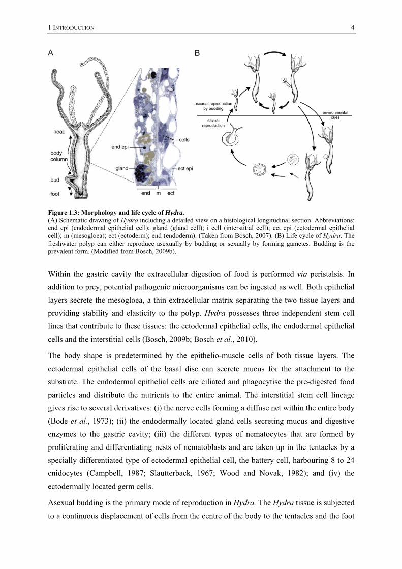
Figure 1.3: Morphology and life cycle of Hydra. (A) Schematic drawing of Hydra including a detailed view on a histological longitudinal section. Abbreviations: end epi (endodermal epithelial cell); gland (gland cell); i cell (interstitial cell); ect epi (ectodermal epithelial cell); m (mesogloea); ect (ectoderm); end (endoderm). (Taken from Bosch, 2007). (B) Life cycle of Hydra. The freshwater polyp can either reproduce asexually by budding or sexually by forming gametes. Budding is the prevalent form. (Modified from Bosch, 2009b).
Within the gastric cavity the extracellular digestion of food is performed via peristalsis. In
addition to prey, potential pathogenic microorganisms can be ingested as well. Both epithelial
layers secrete the mesogloea, a thin extracellular matrix separating the two tissue layers and
providing stability and elasticity to the polyp. Hydra possesses three independent stem cell
lines that contribute to these tissues: the ectodermal epithelial cells, the endodermal epithelial
cells and the interstitial cells (Bosch, 2009b; Bosch et al., 2010).
The body shape is predetermined by the epithelio-muscle cells of both tissue layers. The
ectodermal epithelial cells of the basal disc can secrete mucus for the attachment to the
substrate. The endodermal epithelial cells are ciliated and phagocytise the pre-digested food
particles and distribute the nutrients to the entire animal. The interstitial stem cell lineage
gives rise to several derivatives: (i) the nerve cells forming a diffuse net within the entire body
(Bode et al., 1973); (ii) the endodermally located gland cells secreting mucus and digestive
enzymes to the gastric cavity; (iii) the different types of nematocytes that are formed by
proliferating and differentiating nests of nematoblasts and are taken up in the tentacles by a
specially differentiated type of ectodermal epithelial cell, the battery cell, harbouring 8 to 24
cnidocytes (Campbell, 1987; Slautterback, 1967; Wood and Novak, 1982); and (iv) the
ectodermally located germ cells.
Asexual budding is the primary mode of reproduction in Hydra. The Hydra tissue is subjected
to a continuous displacement of cells from the centre of the body to the tentacles and the foot
1 INTRODUCTION
5
(Campbell, 1967). The amount of new cells is tightly linked to the feeding conditions (Bosch
and David, 1984). The polyp’s cells are permanently in a steady state equilibrium and the
excess of tissue is displaced into the bud that is formed in the lower part of the body column.
Under optimal feeding conditions the polyps can form one bud per three days.
As well as by budding Hydra can also reproduce sexually through the formation of
ectodermally-located testis and oocytes (Bosch and David, 1987; Littlefield, 1985; Littlefield,
1991). Different species can be hermaphroditic or dioecious (Hemmrich et al., 2007a). The
formation of gametes can be induced by environmental changes, e.g. the reduction of the
temperature in Hydra oligactis (Littlefield, 1991) or withholding of food in Hydra vulgaris
(AEP) (Bosch and David, 1987; Sugiyama and Fujisawa, 1977).
In Hydra there is experimental evidence that no separated germ line is present (Bosch and
David, 1987). The multipotent interstitial stem cells that give rise to the somatic derivatives
also give rise to a germline restricted population of stem cells (Bosch and David, 1987;
Littlefield, 1985; Littlefield, 1991). This population is also maintained in sexually
undifferentiated polyps (Holstein and David, 1990) (see Figure 1.4).
Spermatogenesis starts with a local proliferation of the interstitial cells at the area of the
future testis leading to a swelling of the ectodermal tissue and a subsequent lifting of the
covering epithelial cells that will form the outer wall of the testis (Tardent, 1974). The
clusters of accumulated interstitial cells synchronously undergo meiotic divisions. The
subsequent differentiation processes result in cell layers of differentiation stages within the
intercellular space between the mesogloea and the epithelial cells (Munck and David, 1985).
Different Hydra species can form one or several oocytes simultaneously. During oogenesis,
the egg-restricted interstitial cells aggregate and proliferate beneath the ectodermal epithelial
cells giving rise to a cluster of several thousand cells (Honegger et al., 1989). Transplantation
experiments have shown that many of these cells have the potency to differentiate into the
oocyte (Miller et al., 2000), but only one centrally located cell is selected by an unknown
mechanism to become the oocyte. After the determination of the oocyte the surrounding
interstitial cells differentiate into nurse cells. These nurse cells accumulate nutrients, like
lipids and glycogen, and undergo an apoptotic program that is arrested during embryogenesis,
characterized by condensed chromatin and DNA fragmentation into large pieces of 8-15 kb in
length, completed after hatching of the offspring (Honegger et al., 1989; Technau et al.,
2003). This apoptotic program starts during phagocytosis of the nurse cells by the developing
oocyte that dramatically increases in size resulting in a large amoeboid symplastic egg. The
nurse cells are incorporated into the cytoplasm and persist in the developing embryo up to
several months until the new polyp hatches (Technau et al., 2003). Although the nurse cells
undergo an apoptotic program, they are still transcriptional active (Fröbius et al., 2003). The

1 INTRODUCTION
6
mature egg breaks through the covering ectodermal epithelial cells and forms a sphere at the
outer surface of the mother polyp where it is fertilized and embryogenesis is committed. The
embryo develops within the egg without any larval state.
Figure 1.4: The interstitial cell lineage of Hydra. (A) Model of the multipotent interstitial stem cell line; the stem cells give rise to nematocytes, gland cells, nerve cells and germline restricted unipotent stem cells. Environmental stimuli can induce gamete formation. Male differentiation can suppress female differentiation. (Modified after Fraune, 2008; Littlefield, 1991) (B) Oogenesis in Hydra starting with the accumulation of germline restricted female interstitial cells and the determination of the future oocyte. The oocyte (grey) grows and phagocytoses the surrounding cells that differentiate into nurse cells. (Modified from Holstein and Emschermann, 1995).
As a limnic organism Hydra is in permanent contact with its surrounding potential pathogenic
microorganisms like protists, fungi, oomycetes, bacteria and viruses. To inhibit an overgrowth
with these organisms Hydra must have acquired an effective warfare system for its defence.

1 INTRODUCTION
7
1.2 An introduction into immunity
For each living organism it is of tremendous importance to distinguish self from nonself and
to recognize pathogens or degenerated cells to initiate various defence strategies for the
protection of its survival and reproduction (Khalturin and Bosch, 2007; Rosenstiel et al.,
2009).
In Homo sapiens and other jawed vertebrates the adaptive and the innate arm of the immune
system can be distinguished (Khalturin and Bosch, 2007; Rosenstiel et al., 2007). The defence
mechanisms of the adaptive immune response are relatively slow, but highly specific and
mediate an immune memory that protects the organism against attacks of the same pathogen
in the future (Cooper and Alder, 2006). In contrast to that, the innate immune response is
activated fast and therefore the first line of defence confronting the pathogen. The repertoire
of proteins contributing to innate immunity is relatively limited but directed against a broad
spectre of pathogens (Beutler, 2004). The innate immune response does not lead to an
immune memory and relies on germline-encoded receptors (Takeuchi and Akira, 2010).
Many components of the innate immune defence are conserved among all eukaryotes
(Hemmrich et al., 2007b; Hibino et al., 2006; Rosenstiel et al., 2009).
The first line of defence is represented by the epithelia that separate an organism from its
environment (Schreiber et al., 2005). The epithelial cells are tightly interconnected by cellular
junctions and act as a physical barrier. In addition, migrating phagocytes take up invading
pathogens and infected cells. Both cells use receptors that can recognize components that are
conserved in many potentially pathogenic but also commensal microbes and are therefore
called “microbe associated molecular patterns” or MAMPs (Takeuchi and Akira, 2010).
These include for example lipopolysaccharide (LPS), flagellin, CpG-DNA or bacterial cell
wall components like muramyl dipeptide (MDP). Furthermore, these “pattern recognition
receptors” (PRRs) are also capable of recognizing endogenous molecules released from
damaged cells termed damage associated molecular patterns (DAMPs) (Takeuchi and Akira,
2010). The PRRs mediate immune responses through signal transduction cascades leading to
the induction of defence mechanisms such as the onset of inflammation to recruit further
immune cells through the production and release of proinflammatory cytokines, type I
interferons (IFNs) and chemokines or the production and secretion of effector proteins with
an inhibitory or killing activity against microbes, the antimicrobial peptides (AMPs)
(Takeuchi and Akira, 2010).
A persistent inflammatory response may also be the cause for tumour formation and the
development of cancer as a last consequence via an accumulation of gain-of-function and
loss-of-function mutations in proto oncogenes like Kirsten rat sarcoma viral oncogene

1 INTRODUCTION
8
homolog (KRAS) or myelocytomatosis viral oncogene homolog (MYC) or in tumour
suppressor genes like adenomatous polyposis coli (APC) or tumor protein p53 (TP53) through
the production of radical oxygen species (ROS) as a result of inflammation (Grivennikov et
al., 2010).
In addition to the cellular mediated innate immune reactions a humoral innate immunity is
represented by the plasma proteins of the complement system (Beutler, 2004).
1.2.1 The innate pattern recognition receptors
In mammals four families of PRRs have been identified that can be divided into two groups:
the (i) transmembrane proteins of the C-type lectin receptors (CLRs) and the Toll-like
receptors (TLRs); the (ii) intracellular located RIG-I-like receptors (RLRs) and the NOD-like
receptors (NLRs) (Takeuchi and Akira, 2010).
The C-type lectin receptors are characterized by the presence of a carbohydrate-binding
domain. With the help of that domain they recognize carbohydrates on microorganisms, such
as viruses, bacteria and fungi, and stimulate the production of proinflammatory cytokines or
inhibit TLR-mediated immune complexes (Geijtenbeek and Gringhuis, 2009).
The Retinoic acid-inducible gene (RIG)-I-like receptors are composed of two N-terminal
caspase recruitment domains (CARDs), a central DEAD box helicase/ATPase domain and a
C-terminal regulatory domain. Activation by e.g. RNA viruses leads to onset of the
expression of type I IFN genes and to activation of nuclear factor of kappa light polypeptide
gene enhancer in B-cells (NF-κB) via TRADD (Tumor necrosis factor receptor type 1-
associated via DEATH domain), FADD (Fas-associated via DEATH domain) and caspase 8
or 10 (Kawai and Akira, 2006; Takeuchi and Akira, 2009).
The well-characterized Toll-like receptors sense invading pathogens outside of the cell or in
intracellular endosomes and lysosomes (Akira et al., 2006). They are characterized by
N-terminal leucine-rich repeats (LRRs) and a transmembrane domain followed by a
cytoplasmic Toll/ interleukin 1 receptor (IL1R) homology (TIR) domain. Ten TLRs have
been identified in humans; each TLR can recognize particular MAMPs or DAMPs leading to
transcriptional upregulation of distinct genes depending on the TLRs and cell types involved.
Depending on the usage of distinct adaptor molecules TLR signalling can be divided into two
pathways; the MyD88-dependent and the TRIF-dependent signalling pathway (Takeuchi and
Akira, 2010).
MyD88 (myeloid differentiation primary response gene 88) is composed of a DEATH domain
(DD) and a TIR domain and interacts with the DD of the serine/treonine kinase interleukin-1
receptor-associated kinase (IRAK) 4 subsequently leading to the activation of mitogen-
1 INTRODUCTION
9
activated protein (MAP) kinase signalling or the activation of NF-κB via TAK1, the IKK
complex and IκB inducing the expression of cytokine genes and AMPs (Froy, 2005).
TLR3 and TLR4 can use the adaptor protein TRIF (TIR domain-containing adaptor inducing
IFN-β) to activate NF-κB and interferon regulatory factor (IRF) controlling the expression of
proinflammatory cytokines and type-I IFN genes (Takeuchi and Akira, 2010). Via the
cooperative binding of RIPK (receptor interacting serine-threonine protein kinase) 1 (Hsu et
al., 1996; Stanger et al., 1995) and FADD (Chinnaiyan et al., 1995) and the activation of
caspase 8, TRIF can induce apoptosis as well. In addition to the initiation of apoptosis
caspase 8 can also contribute to the TLR-induced NF-κB activation (Maelfait and Beyaert,
2008).
1.2.2 The NOD-like receptors and their signal transduction cascades
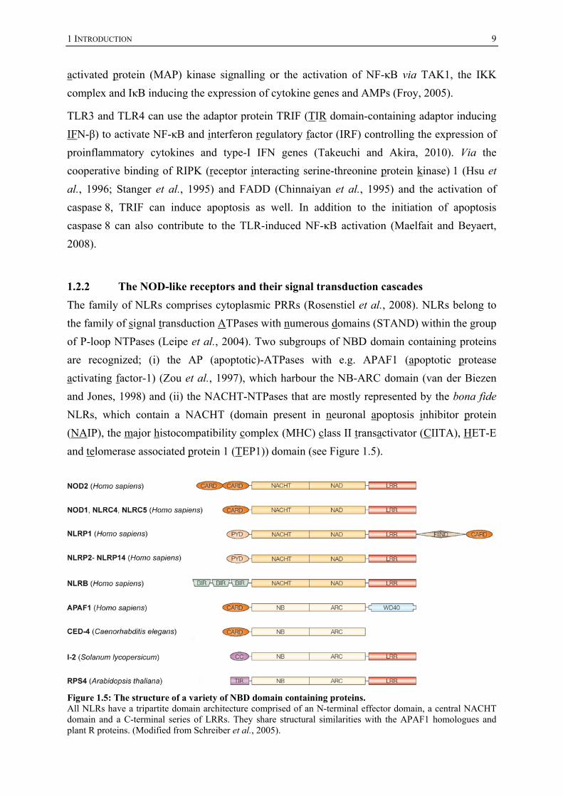
The family of NLRs comprises cytoplasmic PRRs (Rosenstiel et al., 2008). NLRs belong to
the family of signal transduction ATPases with numerous domains (STAND) within the group
of P-loop NTPases (Leipe et al., 2004). Two subgroups of NBD domain containing proteins
are recognized; (i) the AP (apoptotic)-ATPases with e.g. APAF1 (apoptotic protease
activating factor-1) (Zou et al., 1997), which harbour the NB-ARC domain (van der Biezen
and Jones, 1998) and (ii) the NACHT-NTPases that are mostly represented by the bona fide
NLRs, which contain a NACHT (domain present in neuronal apoptosis inhibitor protein
(NAIP), the major histocompatibility complex (MHC) class II transactivator (CIITA), HET-E
and telomerase associated protein 1 (TEP1)) domain (see Figure 1.5).
Figure 1.5: The structure of a variety of NBD domain containing proteins. All NLRs have a tripartite domain architecture comprised of an N-terminal effector domain, a central NACHT domain and a C-terminal series of LRRs. They share structural similarities with the APAF1 homologues and plant R proteins. (Modified from Schreiber et al., 2005).

1 INTRODUCTION
10
NLRs are a complex family of tripartite receptors that have in common the eponymous central
nucleotide-binding domain and oligomerization domain (NOD), synonymously called
NACHT or NBD, and C-terminal LRRs (Schreiber et al., 2005). The mammalian NLRs are
subdivided into five subfamilies based on the type of their N-terminal effector domain (Chen
et al., 2009): NLRA and NLRB each with one human member, the first has a CARD domain
with an acidic domain and the latter has three BIR (baculovirus inhibitor of apoptosis protein
repeat) domains. The third group of NLRC contains five human members, each of them
contains N-terminal CARD domains. To this group belong also the best characterized family
members NOD1 (Bertin et al., 1999; Inohara et al., 1999) and NOD2 (Ogura et al., 2001b).
The 14 members of the fourth and largest group are called NLRPs. All have in common an
N-terminal Pyrin domain. The well characterized NLRP1 and NLRP3 belong to this family as
well. The fifth group called NLRX contains one member with a not yet characterized
N-terminal part.
For some NLRs the minimal elicitors are known. NOD1 and NOD2 recognize peptidoglycan
moieties secreted by bacteria (Chamaillard et al., 2003; Inohara et al., 2003). NLRP3 can
recognize a broad spectrum of ligands including MAMPs, like LPS or MDP, but also
chemicals, like silica or asbestos, and endogenous DAMPs, like uric acid crystals, suggesting
that the agonists might not bind directly to the receptor, but need a co-receptor for recognition
(Chen et al., 2009).
Upon recognition of the elicitor by the LRRs, these proteins form large complexes by a
conformational change of the LRRs that usually block the oligomerization of the receptors by
masking the NACHT domain (Schreiber et al., 2005). The proteins self-oligomerize by the
interactions of their NACHT domains (Inohara et al., 2000; Schroder and Tschopp, 2010).
This brings the effector domains of these proteins in close proximity to each other, activating
the protein complex to recruit downstream proteins via homophilic interactions of the effector
domains, a mechanism that is termed “induced proximity” (Inohara et al., 2000). According
to the identity of the activated NLR different complexes are formed leading to the activation
of different signalling cascades. NOD1 and NOD2 form the NODosome with the CARD-
containing receptor-interacting serine-threonine kinase 2 (RIPK2) (Inohara et al., 2000;
McCarthy et al., 1998; Ogura et al., 2001b). Similar to TLR signalling NF-κB is activated
leading to the transcription of inflammatory cytokines and chemokines such as tumor necrosis
factor (TNF)-α, IL6 and IL8 and antimicrobial peptides (Chen et al., 2009; Voss et al., 2006).
Furthermore, the NODosome can also activate MAP kinase signalling including the p38,
extracellular signal-regulated protein kinase (ERK) and c-Jun N-terminal kinase (JNK)
pathways (Chen et al., 2009) (see Figure 1.6, left panel).

1 INTRODUCTION
11
Figure 1.6: The signal transduction cascades performed by human NLRs. NOD1 or NOD2 sense peptidoglycan (PNG) subunits. Upon activation the proteins form a complex via homotypic binding of CARD domains with RIPK2 leading to the activation of NF-κB or MAP kinase signalling. NLRP1, NLRP3 and NLRC4 can sense a variety of agonists (DAMPs, MAMPs and chemicals) leading to the formation of the inflammasome with ASC and caspase 1. The activation of caspase 1 can lead to pyroptosis and the cleavage of proinflammatory cytokines. Colour code of protein domains: yellow (LRR), dark blue (NACHT), violet (CARD), orange (Pyrin), green (caspase), turquoise (serine/threonine kinase).
Upon activation NLRP1, NLRP3 and NLRC4 form an oligomeric complex termed the
inflammasome (Chen et al., 2009) (Figure 1.6, right panel). The Pyrin domains interact with
the Pyrin domain of the adaptor protein ASC (adaptor protein apoptosis speck protein with
caspase recruitment) (Srinivasula et al., 2002) enabling the recruitment of procaspase 1,
synonymously called ICE (IL1 converting enzyme). The homotypic interaction between the
CARD domains of ASC and procaspase 1 leads to the autoactivation of caspase 1 that
subsequently cleaves pro-IL1β and pro-IL18 resulting in their release (Martinon et al., 2002;
Schroder and Tschopp, 2010).
The structure of the inflammasome shares similarity with the apoptosome formed by APAF1
upon cytochrome c release by damaged mitochondria leading to the recruitment of
procaspase 9 via a CARD-CARD interaction resulting in induction of apoptosis (Zou et al.,
1997; Zou et al., 1999). Indeed, activation of the inflammasome can also lead to a newly
described form of programmed cell death: the pyroptosis, which is definded by the rapid

1 INTRODUCTION
12
formation of plasma membrane pores, cellular lysis and the release of IL1β and IL18
(Bortoluci and Medzhitov, 2010).
Not only in NLR signalling pathways, but also in the other PRR signalling pathways
described above, the homotypic interactions of effector domains made up of the DEATH fold
family, like the CARD, DEATH, DED (DEATH effector domain) and Pyrin domains, play
key roles in the transduction of the signal. Although they share few sequence similarities, they
all have in common the six antiparallel α-helical bundle structure (Liang and Fesik, 1997; Liu
et al., 2003). These domains have been identified in a variety of proteins involved in
apoptosis and immunity (Aravind et al., 1999; Chinnaiyan et al., 1995; Medema et al., 1997;
Schreiber et al., 2005; Zou et al., 1997).
Dysregulated PRR pathways have been implicated in a variety of severe autoinflammatory
diseases (Chen et al., 2009; Rosenstiel et al., 2007; Schreiber et al., 2005). For example,
NOD2 was the first susceptibility gene described for Crohn’s disease, a chronic inflammatory
bowel disease (Hampe et al., 2001; Ogura et al., 2001a). Different single nucleotide
polymorphisms (SNPs) in the part of the NOD2 gene coding for the LRRs, like the
L1007finsC variant leading to a truncated LRR, significantly increase the risk for disease
development (Annese et al., 2005).
1.2.3 Evolution of NLRs
It is known that the TLRs are a group of phylogenetically very ancient receptors (Hibino et
al., 2006; Miller et al., 2007; Wiens et al., 2007). They were originally discovered in
Drosophila melanogaster (Anderson et al., 1985a; Anderson et al., 1985b). The Toll protein
is involved in dorso-ventral patterning and immunity (Lemaitre and Hoffmann, 2007;
Lemaitre et al., 1995). The TLRs and conserved signal transduction components can also be
found in the sponge and in cnidarians at the base of metazoan evolution (Bosch et al., 2009;
Hemmrich et al., 2007b; Miller et al., 2007; Wiens et al., 2007). In contrast, little is known
about the evolution of the NLRs.
Although the described mammalian NLRs share high structural similarities with the resistance
(R) genes of plants it was the general opinion for a long time that these molecules were
acquired independently (Ting and Davis, 2005) (see Figure 1.5). The R proteins are
comprised of a central NB-ARC domain linked to C-terminal LRRs and an N-terminal TIR or
coiled coil domain (Meyers et al., 2003; van der Biezen and Jones, 1998). Unlike vertebrate
NLRs, these proteins do not detect MAMPs, but specifically recognize pathogen-encoded
virulence factors by the avirulence (avr) genes (Meyers et al., 2003). This gene-for-gene
interaction resembles the function of the adaptive immune system of the jawed vertebrates

1 INTRODUCTION
13
and explains the large variety of R proteins. The activation of R proteins leads to the death of
the infected tissue via hypersensitive response (HR) (Ma and Berkowitz, 2007).
A large expansion of NLR orthologues has also been discovered in teleost fishes. 201 NLRs
were detected in the zebrafish and 70 were found in the pufferfish (Stein et al., 2007).
Since except of the structurally related APAF1 orthologues CED4 and DARK of
Caenorhabditis elegans (Yuan and Horvitz, 1990) and Drosophila melanogaster (Rodriguez
et al., 1999) no proteins belonging to the NLR family sensu stricto were present in these
ecdysozoan model organisms, it was assumed that NLRs were vertebrate-specific (Ting and
Davis, 2005).
The first hint for invertebrate NLRs was given by the workgroup of Jonathan Rast in 2006
showing that the sea urchin Strongylocentrotus purpuratus posseses more than 200 NLR
orthologues (Hibino et al., 2006; Rast et al., 2006). Remarkably, the vast majority of these
NLRs have DEATH domains instead of CARD or Pyrin domains. The Pyrin domain still
seems to be vertebrate specific.
In 2008, a large amount of NLR orthologues was also detected in the chordate Branchiostoma
floridae (Huang et al., 2008). The amphioxus genome encodes for more than 100 NLRs.
Although these recent studies have begun to elucidate the origins of NLR signalling, it is still
unclear how early in evolution these receptors have evolved.
1.3 Hydra and other cnidarians provide insight into evolution of innate
immunity
As mentioned above, human chronic inflammatory diseases of epithelial barrier organs are
frequently associated with loss-of-function variations in PRRs (Rosenstiel et al., 2007). But
not only genetic variations, also a change of living conditions within the past century
accompanied by a modified microbial community with new metabolic patterns have been
implicated to contribute to the development of chronic inflammatory diseases (Rosenstiel et
al., 2009; Wen et al., 2008). Basal eumetazoan organisms such as cnidarians can help to
understand the processes leading to the development of these diseases. It was shown that
many genes that have been lost in ecdysozoan model organisms can be found in cnidarians
suggesting the last common ancestor of cnidarians and bilaterians already possessed these
genes (Miller et al., 2005; Technau et al., 2005). Therefore, cnidarians represent a good
model system to understand, how selective pressures contribute to diversity in conserved
immune genes in order to survive a variety of immune challenges (Rosenstiel et al., 2009).

1 INTRODUCTION
14
Eugene Rosenberg proposed in his “coral probiotic hypothesis” that corals can adapt to their
changing environment quickly by changing their bacterial symbionts and therefore gaining
resistance to diseases (Reshef et al., 2006). This “holobiont”-hypothesis gives cues for
understanding the role of a symbiotic microflora of humans in the development of barrier
disorders.
Many facets of immunity are a subject of recent studies in cnidarians. Work on intraspecies
competitions in Anthozoans and the discovery of the allorecognition locus in the colonial
Hydrozoan Hydractinia echinata show that these animals are capable of distinguishing self
from nonself (Cadavid, 2004; Nicotra et al., 2009). Another major field of interest is the
symbiotic association of Cnidarians with photosynthetically active unicellular algae, as in the
extensively studied field of the Anthozoa-Zooxanthellae mutualism and the effect of coral
bleaching due to environmental stressors (Mydlarz et al., 2010; Rosenberg et al., 2009; Weis,
2008). The presence of mannose-binding lectins potentially involved in the recognition of
pathogens and symbionts was reported for the coral Acropora millepora (Kvennefors et al.,
2008). In sea fan corals, it was shown that the attack of a pathogenic fungus leads to tissue
melanisation by the usage of the prophenoloxidase known to be involved in the immune
response of Arthopods (Mydlarz et al., 2008). Furthermore, orthologues of proteins of the
complement system appear to be present in anthozoans and hydrozoans, but so far without
any proven function in immunity (Kimura et al., 2009; Miller et al., 2007).
As mentioned above, Hydra has a very simple body plan. Except for the thin glycocalyx it
does not have any protective layers to separate it from its environment (Bosch et al., 2009).
The freshwater polyp does not possess any migratory immune cells, so it has to rely
exclusively on an epithelial defence making it a useful model system to study epithelial
defence mechanisms (Augustin et al., 2010).
It was demonstrated more than 25 years ago by Rahat and Dimentman that sterile cultivated
polyps have a reduced budding rate indicating a beneficial effect of the microbial community
on their host (Rahat and Dimentman, 1982). In 2003, Kasahara and Bosch showed that
extracts from polyps depleted in interstitial cells have an increased growth inhibition effect
against the Gram-positive bacterium Bacillus subtilis and Gram-negative bacterium E. coli
(Kasahara and Bosch, 2003). Fraune and colleagues could show that these interstitial cell
depleted animals have a different microbial community indicating an effect of the interstitial
cells in shaping the composition of the microbial symbionts (Fraune et al., 2009).
Via 16S rDNA analysis it was detected that different Hydra species support particular
bacterial guilds and that this association of the host with its symbionts is stable over years
(Fraune and Bosch, 2007).

1 INTRODUCTION
15
Effector molecules that might take part in this shaping of Hydra’s microbial community have
also been discovered: Hydra expresses a variety of antimicrobial peptides in its endodermal
layer. Hydramacin-1 and the arminin protein family are expressed by the endodermal
epithelial cells and the expression of the hydramacin-1 protein is inducible via treatment with
LPS (Augustin et al., 2009a; Bosch et al., 2009; Jung et al., 2009). Another peptide, which
shows antimicrobial activity, is kazal2 being strongly expressed by the endodermal gland
cells (Augustin et al., 2009b). Other good candidates for being antimicrobial peptides are the
members of the periculin gene family that are predominantly expressed within the developing
egg and the embryo (Fraune, 2008). One reason for the mainly endodermal expression of
these peptides might be the uptake of food and accompanying putative pathogenic bacteria by
the endodermal epithelial cells (Bosch et al., 2009). Interestingly, many of these peptides do
not show any homology with other peptides and are therefore taxonomically restricted, which
as been observed for many immune effector molecules (Khalturin et al., 2009).
The expression of these taxonomically restricted AMPs may be controlled by conserved
signal transduction pathways (Bosch et al., 2009). The presence of a TLR and the conserved
components of its signalling pathway as well as components of MAP kinase signalling in
Nematostella shows their phylogenetically old roots (Hemmrich et al., 2007b; Miller et al.,
2007). A truncated orthologue for NF-κB was detected in Nematostella as well (Sullivan et
al., 2007; Sullivan et al., 2009).
Hydra does not possess a sensu stricto TLR, but it was shown using a HEK293 cell based in
vitro assay that a TIR-transmembrane domain protein, HyTRR1, can form a functional
subunit with its co-receptor HyLRR2 upon stimulation with flagellin (Bosch et al., 2009) (see
Figure 1.7).
In addition to this unusual TLR, Hydra possesses conserved components of the TLR
signalling cascade, like MyD88, TRAF6, TAK1 and IKK and components of MAP kinase
signalling, like JNK and AP1 (Hemmrich et al., 2007b; Miller et al., 2007; Philipp et al.,
2005). In contrast to previous findings the new transcriptome datasets gave first evidence for
a putative NF-κB orthologue (G. Hemmrich, pers. communication).
Some orthologues of proteins involved in apoptosis were detected in Hydra, like HyBcl-2,
HyBak, HyBax and ten caspases, most of them resembling the human caspase 3 (Böttger and
Alexandrova, 2007; Cikala et al., 1999).
To conclude, Hydra is an important tool for studying a variety of immunological questions
such as the evolution of epithelial defence mechanisms and the development of barrier
disorders.

1 INTRODUCTION
16
Figure 1.7: The epithelial defence in Hydra. (A) Model of the TLR signalling cascade in Hydra. Upon activation with flagellin HyTRR-1 and HyLRR-2 interact. Conserved TLR signalling cascade members are present in Hydra. The activation of TLR signalling may lead to the secretion of effector proteins. (Modified from Bosch et al., 2009). (B) A cross-section of the gastric region of Hydra; (C) A cross-section of the human small intestine. (Taken from Bosch, 2009a).
1.4 Aims of the study
NLRs are a family of intracellular innate immune receptors sensing MAMPs and endogenous
danger signals (Rosenstiel et al., 2008). Upon activation these receptors form multimeric
complexes with downstream signalling components resulting in an immune response
including the release of AMPs and pro-inflammatory cytokines (Chen et al., 2009; Voss et al.,
2006). Genetic variants of NLRs have been associated with the manifestation of chronic
inflammatory diseases of epithelial barrier organs in Homo sapiens (Rosenstiel et al., 2007).
Although these receptors are key players in epithelial defence, the information about their
evolution within the animal kingdom is very limited (Hibino et al., 2006; Huang et al., 2008;
Ting and Davis, 2005). In order to broaden the understanding of NLR evolution, one goal of
the study was to screen the genomes of several animal species with a focus on basal
metazoans and especially on Hydra accompanied by a phylogenetic analysis.

1 INTRODUCTION
17
The freshwater polyp Hydra is continuously surrounded by potential pathogenic microbes and
must have acquired effective epithelial defence mechanisms accompanying its adaption to its
ecological niche (Augustin et al., 2010; Rosenstiel et al., 2009). Using Hydra as model
organism for epithelial defence, the expression of Hydra NLRs should be analysed in more
detail.
Furthermore, putative interaction partners for these receptors should have been identified and
characterized. In order to elucidate the interaction of Hydra NLRs with these proteins and the
result of these interactions, first functional analyses should be performed.
A disturbed function of immunity may lead to a disturbed microflora (Fraune et al., 2009;
Salzman et al., 2010; Zaki et al., 2010). This disturbance has been discovered in a strain of
Hydra oligactis, which accidentally formed tumour-like structures upon isolation from nature
and cultivation under laboratory conditions. The second goal of the study was to characterize
this tumour bearing strain using histological and molecular biological methods in order to get
insight in tumour formation within a basal invertebrate animal.
2 RESULTS
18
2 RESULTS
2.1 The family of NBD domain containing proteins is highly complex in
basal metazoans
Proteins containing NBD domains like NACHT and NB-ARC domains fulfil various
functions in immunity or apoptosis (Rosenstiel et al., 2008; Zou et al., 1997). While a host of
data has been presented on the biological roles of selected members of the family (e.g. NOD2
and NALP3) in Homo sapiens and other vertebrates (Chen et al., 2009; Schroder et al., 2010),
hardly anything is known about the evolution of these gene families. To broaden this
knowledge and ask the question whether NLRs were already present at the base of animal
evolution, a screening for orthologues of NBD domain containing proteins was performed in
the genomes and transcriptomes of various animal species via BLAST (basic local alignment
search tool) (Altschul et al., 1990) searches with the domains of vertebrate NLRs or sequence
analysis using hidden Markov model algorithms (HMMER) (Eddy, 1996). All hits were
subsequently verified by protein domain predictions. As well as basal species such as the
sponge Amphimedon queenslandica, the cnidarian anthozoan species Acropora millepora,
Nematostella vectensis and the hydrozoan species Hydra magnipapillata were investigated,
but also the genomes of one arthropod species, Daphnia pulex, the agnathostome Petromyzon
marinus and three gnathostome species: Oryzias latipes; Xenopus tropicalis; and Gallus
gallus. These organisms were included to elucidate the appearance of NBD-domain
containing proteins at different diverging nodule points of animal evolution.
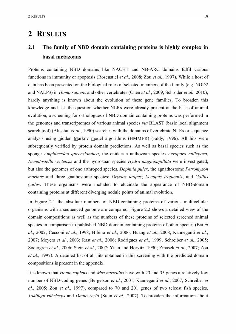
In Figure 2.1 the absolute numbers of NBD-containing proteins of various multicellular
organisms with a sequenced genome are compared. Figure 2.2 shows a detailed view of the
domain compositions as well as the numbers of these proteins of selected screened animal
species in comparison to published NBD domain containing proteins of other species (Bai et
al., 2002; Cecconi et al., 1998; Hibino et al., 2006; Huang et al., 2008; Kanneganti et al.,
2007; Meyers et al., 2003; Rast et al., 2006; Rodriguez et al., 1999; Schreiber et al., 2005;
Sodergren et al., 2006; Stein et al., 2007; Yuan and Horvitz, 1990; Zmasek et al., 2007; Zou
et al., 1997). A detailed list of all hits obtained in this screening with the predicted domain
compositions is present in the appendix.
It is known that Homo sapiens and Mus musculus have with 23 and 35 genes a relatively low
number of NBD-coding genes (Bergelson et al., 2001; Kanneganti et al., 2007; Schreiber et
al., 2005; Zou et al., 1997), compared to 70 and 201 genes of two teleost fish species,
Takifugu rubriceps and Danio rerio (Stein et al., 2007). To broaden the information about

2 RESULTS
19
these genes in vertebrates, the genomes of a bird (Gallus gallus), an amphibian (Xenopus
tropicalis), and another teleost fish (Oryzias latipes) were screened.
Figure 2.1: Number of NBDs in selected multicellular organisms. The NACHT and NB-ARC domains were counted in Gallus gallus, Xenopus tropicalis, Oryzias latipes, Petromyzon marinus, Daphnia pulex, Hydra magnipapillata, Nematostella vectensis and Amphimedon queenslandica at the basis of homology searches via BLAST, HMMER or gene models. The total numbers of NBDs in Amphimedon queenslandica cannot be clearly estimated due to the fact that single genome reads were screened. For Homo sapiens (Schreiber et al., 2005; Zou et al., 1997), Mus musculus (Cecconi et al., 1998; Kanneganti et al., 2007), Takifugu rubripes (Stein et al., 2007), Danio rerio (Stein et al., 2007), Branchiostoma floridae (Huang et al., 2008; Zmasek et al., 2007), Ciona intestinalis (Sodergren et al., 2006), Strongylocentrotus purpuratus (Hibino et al., 2006; Rast et al., 2006; Zmasek et al., 2007), Drosophila melanogaster (Rodriguez et al., 1999), Caenorhabditis elegans (Yuan and Horvitz, 1990), Arabidopsis thaliana (Meyers et al., 2003) and Oryza sativa (Bai et al., 2002) previous publications were taken into consideration.
In the chicken genome, six orthologous gene models for NOD-like receptors and one for
APAF1, including the models published by Hughes et al. (Hughes, 2006), were detected (see
Figures 2.1 and 2.2).
A total of 17 NBD containing gene models were discovered in the genome of Xenopus
tropicalis (see Figure 2.1). Within these gene models a NAIP and an APAF1 orthologue are
present. Most of the gene models encode a NACHT-LRR structure (see Table 9.1 in the
appendix).
2 RESULTS
20
Figure 2.2: Overview of structures of NBD containing proteins in selected animals. (Description: next page)

2 RESULTS
21
Figure 2.2 (continued): Overview of structures of NBD containing proteins in selected animals. For H. sapiens (Schreiber et al., 2005; Zou et al., 1997), B. floridae (Huang et al., 2008; Zmasek et al., 2007), C. intestinalis (Sodergren et al., 2006), S. purpuratus (Hibino et al., 2006; Rast et al., 2006; Zmasek et al., 2007), D. melanogaster (Rodriguez et al., 1999), C. elegans (Yuan and Horvitz, 1990), N. vectensis (2 NB-ARC domain containing models) (Zmasek et al., 2007) previous publications were taken into consideration. Below the predicted protein structure the numbers of proteins are mentioned. Red box indicates the bona fide NLR orthologues in Nematostella and Acropora; Abbreviations: AD (acidic domain), DFD (DEATH-fold domain), TIR (toll-interleukin-1 receptor), TPR (tetratricopeptide repeat), NB-ARC (nucleotide binding adaptor shared by APAF1, R gene products and CED-4), DD (DEATH domain), NACHT (domain present in neuronal apoptosis inhibitor protein (NAIP), the major histocompatibility complex (MHC) transactivator (CIITA), HET-E and TP1), LRR (Leucine rich repeat), DED (DEATH effector domain), CARD (Caspase recruitment domain), PYD (Pyrin domain), BIR (baculovirus inhibitor of apoptosis repeat), CC (coiled coil), ank (ankyrin repeat), RVT (reverse transcriptase), FIIND (domain with function to find), ucd (uncharacterized domain) The lengths of proteins and domains are not to scale. The number of repetitions of a repetitive domain (WD40, LRR, TPR, ank) does not reflect the exact number of repeats.
In the genome of the teleost fish Oryzias latipes 78 loci for NBDs were detected (see Figure
2.1). About one half of the hits are represented by a gene model, some are supported by ESTs.
Some of these NBDs appear to be clustered on different chromosomes, e.g. chromosome 2
(21 NBDs), chromosome 18 (9 NBDs) and ultracontig 205 (6 NBDs) (see Table 9.1 in the
appendix).
It is known that the number of NBD-coding genes in two basal chordate species varies a lot
(Huang et al., 2008; Sodergren et al., 2006). Whereas Branchiostoma floridae NBD-coding
genes underwent a broad expansion, leading to 106 representatives (Huang et al., 2008), the
urochordate Ciona intestinalis does not seem to contain any genes coding for NBDs
connected with other domains (Sodergren et al., 2006). To elucidate the situation in a basal
vertebrate species, the genome of the agnathostome Petromyzon marinus was investigated.
Interestingly, no orthologue for NOD-like receptors was detected here, except of an APAF1
orthologue (see Figure 2.1), represented by four short genome contigs with a size range from
4.6 to 17.9 kb. Due to the short contig length and to the fact that the contigs cover different
parts of the APAF1 gene, these hits are assumed to belong to one locus in the genome.
Because D. melanogaster and C. elegans do not have any NLR orthologues except APAF1
(Rodriguez et al., 1999; Yuan and Horvitz, 1990), it was assumed that this gene family is
absent in all ecdysozoan species (Ting and Davis, 2005). In order to question this assumption,
the genome of the arthropod species Daphnia pulex was investigated. In the genome of the
freshwater crustacean two gene models coding for proteins with a NACHT domain followed
by LRRs were discovered in addition to an APAF1 homologue (see Figure 2.1 and Table 9.1
in the appendix). One of these NLRs gives best BLAST hits to two predicted proteins of the
same domain composition of two mosquito species, Aedes aegypti and Culex
quinquefasciatus (XP_001658101, XP_00184881). Furthermore, a gene model coding for a
NACHT domain and WD40 repeats was detected (see Table 9.1 in the appendix).

2 RESULTS
22
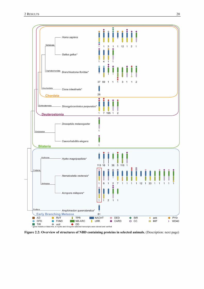
Since nothing was known about the presence of NBD coding genes in basal metazoan species,
it was previously thought that NLRs evolved at the state of the deuterostomes (Ting and
Davis, 2005). The genomes of a demosponge, Amphimedon queenslandica, of two cnidarian
species, the anthozoan Nematostella vectensis, and the hydrozoan Hydra magnipapillata, and
the EST databases of the anthozoan Acropora millepora were investigated to determine
whether these basal animals could use NLRs for their defence against pathogens as well.
It is not yet possible to survey the complete NBD gene repertoire of the sponge Amphimedon
queenslandica because no genome assembly is available. A total of 349 NACHT domains
were identified in the trace archive genomic sequence data, which were assembled to 61
genome contigs (see Figures 2.1 and 2.2). This implied presence of a large number of
NACHT proteins suggests a relatively complex NBD repertoire in early diverging Metazoa.
In collaboration with Yvonne Weiss from the workgroup of David J. Miller the EST
databases of the anthozoan cnidarian species Acropora millepora were screened. One bona
fide NOD-like receptor orthologue consisting of a DED, a NACHT domain and LRRs was
detected and verified by cloning of the entire open reading frame (red box in Figure 2.2). In
addition, another NACHT and LRR containing EST, two NACHT and one NB-ARC
containing ESTs were detected. An orthologue for APAF1 coding for two CARD domains
instead of one was cloned (see Figure 2.2).
In addition to two published NB-ARC domain containing proteins (Zmasek et al., 2007), 70
loci coding for NBD domains were detected in a second anthozoan species, Nematostella
vectensis. These include an orthologue of the Acropora millepora NLR (red box in Figure
2.2), more than 30 NACHT domains linked to LRRs and many other proteins showing a large
variety of domain compositions, e.g. NACHT domains that are connected to WD40 repeats or
ankyrin repeats and eleven NACHT domains connected to coiled coils. Another group of six
proteins link an NB-ARC domain with TPRs. A single bona fide APAF1 homologue is
present as well (see Figure 2.2).
In the genome of Hydra magnipapillata two large groups of in total 290 NBD domain
containing proteins were detected (see Figures 2.1, 2.3).
The first group includes 130 NACHT domain coding loci. Using HMMER analysis in 16 of
them a DEATH domain was found upstream of the NACHT domain on the genomic contig,
in one of them a DED domain (see Figure 2.2). No LRRs appear to be present in Hydra
orthologues for NOD-like receptors, but ESTs coding for LRR containing proteins were
identified (data not shown). Neither by computational analysis methods, nor by attempts to
physically link NACHT-coding with LRR-coding contigs via PCR, could an NLR with a
NACHT-LRR structure be identified. HyNLRs share a high degree of conservation on the
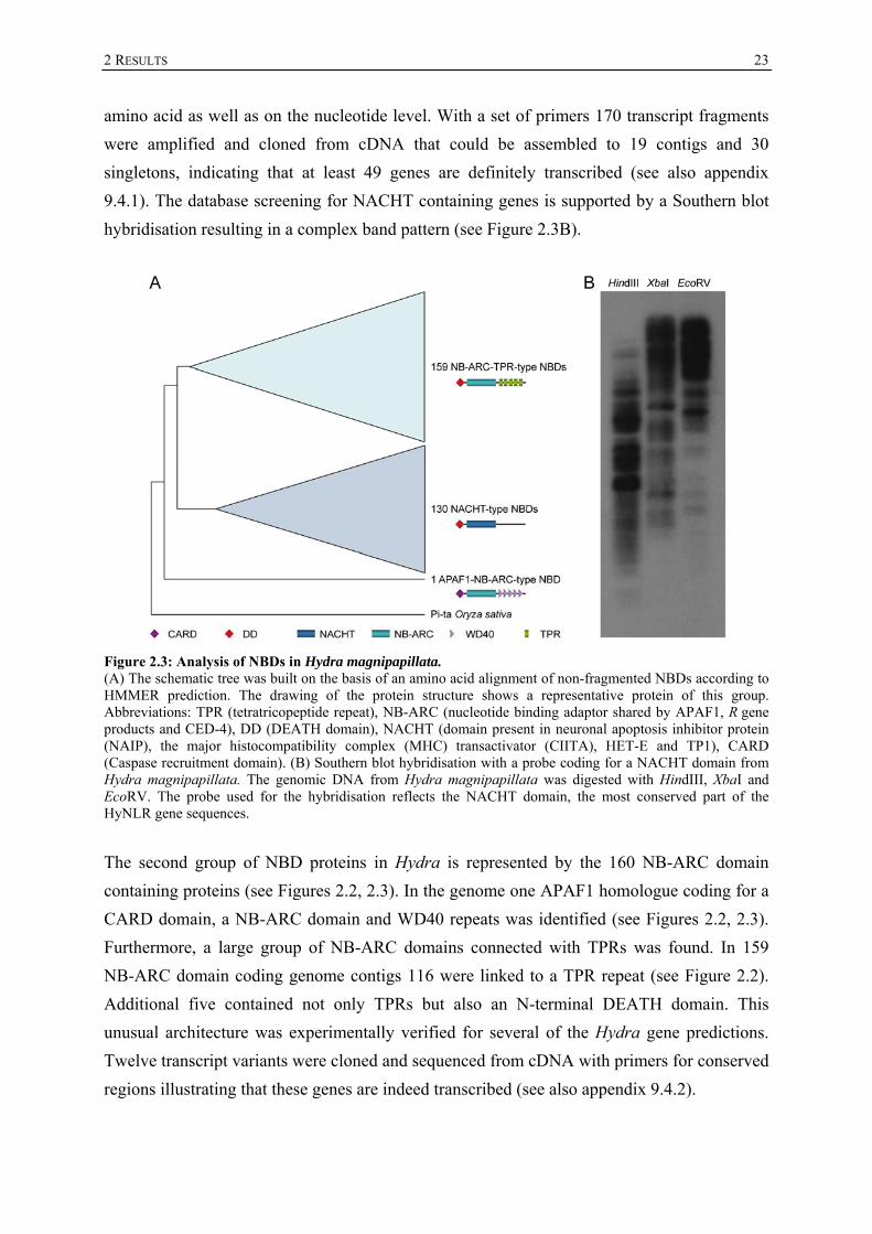
2 RESULTS
23
amino acid as well as on the nucleotide level. With a set of primers 170 transcript fragments
were amplified and cloned from cDNA that could be assembled to 19 contigs and 30
singletons, indicating that at least 49 genes are definitely transcribed (see also appendix
9.4.1). The database screening for NACHT containing genes is supported by a Southern blot
hybridisation resulting in a complex band pattern (see Figure 2.3B).
Figure 2.3: Analysis of NBDs in Hydra magnipapillata. (A) The schematic tree was built on the basis of an amino acid alignment of non-fragmented NBDs according to HMMER prediction. The drawing of the protein structure shows a representative protein of this group. Abbreviations: TPR (tetratricopeptide repeat), NB-ARC (nucleotide binding adaptor shared by APAF1, R gene products and CED-4), DD (DEATH domain), NACHT (domain present in neuronal apoptosis inhibitor protein (NAIP), the major histocompatibility complex (MHC) transactivator (CIITA), HET-E and TP1), CARD (Caspase recruitment domain). (B) Southern blot hybridisation with a probe coding for a NACHT domain from Hydra magnipapillata. The genomic DNA from Hydra magnipapillata was digested with HindIII, XbaI and EcoRV. The probe used for the hybridisation reflects the NACHT domain, the most conserved part of the HyNLR gene sequences.
The second group of NBD proteins in Hydra is represented by the 160 NB-ARC domain
containing proteins (see Figures 2.2, 2.3). In the genome one APAF1 homologue coding for a
CARD domain, a NB-ARC domain and WD40 repeats was identified (see Figures 2.2, 2.3).
Furthermore, a large group of NB-ARC domains connected with TPRs was found. In 159
NB-ARC domain coding genome contigs 116 were linked to a TPR repeat (see Figure 2.2).
Additional five contained not only TPRs but also an N-terminal DEATH domain. This
unusual architecture was experimentally verified for several of the Hydra gene predictions.
Twelve transcript variants were cloned and sequenced from cDNA with primers for conserved
regions illustrating that these genes are indeed transcribed (see also appendix 9.4.2).

2 RESULTS
24
Except from Hydra and Nematostella this new uncharacterized protein family has so far only
been detected in Branchiostoma floridae (Zmasek et al., 2007). Remarkably, these proteins
give best BLAST hits to bacterial proteins also containing NB-ARC domains connected to
TPRs. RT-PCR performed on mRNA isolated from separated ectodermal and endodermal
tissue layers showed that this gene family is exclusively expressed in the ectodermal
epithelium of Hydra (data not shown).
A phylogenetic analysis using Bayesian interference with selected NACHT-domain
representatives (Figure 2.4) resulted in a polytomy and revealed that throughout animal
evolution vast species-specific NLR gene expansions frequently occurred.
These expansions can be observed for A. queenslandica, H. magnipapillata, S. purpuratus
and partially for N. vectensis, B. floridae, X. tropicalis and for the human NLRP genes.
Whereas almost all human NLR genes and their vertebrate orthologues appear to be
vertebrate-specific, NOD1 and NOD2 form a cluster not only with vertebrates, but also with
two cnidarian representatives indicating a putative phylogenetically old origin for NOD1 and
NOD2, at least for their NACHT domains. Remarkably, all Hydra NACHT domains are
located within one group of exclusive vertebrate sequences, indicating a relatively high
sequence similarity between these groups.
An alignment with some representative NACHT domains that were used to build the
phylogenetic tree (see appendix 9.2) shows that the various NACHT-domain motifs, like the
P-loop and the Mg2+-binding site (Koonin and Aravind, 2000), are conserved throughout
evolution, although the different NACHT sequences are very heterogeneous.

2 RESULTS
25
Figure 2.4: Phylogenetic analysis of representative NACHT-domains across the animal kingdom. The tree was built by Bayesian interference of phylogeny. Percentages indicate posterior probabilities. Posterior probabilities below 50 % are not shown; bright yellow box: Hydra NACHT domains are located within one group with vertebrate sequences; grey box: indicates a cluster of vertebrate NOD1 and NOD2 orthologues with similar cnidarian NACHT domains; as outgroup the NB-ARC domain of RPS4 (A. thaliana) was used. Abbreviations: Amil (A. millepora), Atha (A. thaliana), Nvec (N. vectensis), Spur (S. purpuratus), Bflo (B. floridae), Aque (A. queenslandica), Xtro (X. tropicalis), Olat (O. latipes), Hmag (H. magnipapillata), Hsap (H. sapiens), Ggal (G. gallus).
2 RESULTS
26
2.2 The expression profile of HyNLR type 1, a Hydra-NLR representative
gene
In contrast to anthozoans, Hydra lacks a classical NLR with a NACHT-LRR structure, but
contains several proteins with an N-terminal DEATH domain followed by a NACHT domain.
To provide insight into the function of the Hydra NLR-like proteins, the transcript
architecture and expression pattern of one of Hydra NLR-type genes, HyNLR type 1, was
analyzed in detail (Figure 2.5).
Figure 2.5: Expression profiling of HyNLR type 1. (A) The gene structure of HyNLR type 1. The gene consists of five exons of various sizes and encodes a transcript of about 2,417 bases. At the 5’ end the trans-spliced leader B is added. The 688 aa long protein product contains a predicted DEATH and a NACHT domain but lacks LRRs. (B) Semiquantitative reverse transcriptase PCR performed on RNA extracted from the ectodermal and the endodermal cell layers of Hydra after procaine treatment showing a predominant expression of HyNLR type 1 in the endodermal layer. As control genes the housekeeping gene actin, the ectodermally expressed nematoblast gene nb042 (Milde et al., 2009) and the endodermally expressed gland cell gene HyDkk1/2/4 C (Augustin et al., 2006) were used. (C) Splice isoforms of HyNLR type 1. In addition to the most abundant HyNLR type 1 transcript (light blue) three other transcript variants (dark blue, orange and green) that show an alternative splice pattern were cloned and sequenced. HyNLR type 1 var 3 and 4 were cloned partially. (D) Quantitative real time PCR showing a light up-regulation of HyNLR type 1 after stimulation with LPS and flagellin. The fold-change of the expression of HyNLR type 1 is compared with the expression levels of the negative control. Abbreviations: endo (endoderm), ecto (ectoderm).

2 RESULTS
27
The complete transcript of HyNLR type 1 was amplified using 3’RACE (rapid amplification
of cDNA ends)-PCR technique and sequenced subsequently from cDNA. The gene is
represented on the Hydra magnipapillata genome scaffold contig35932
(http://hydrazome.metazome.net) and is transcribed from five exons of various sizes to an
mRNA with a length of about 2,417 bases with a coding sequence of 2,067 bases (Figure
2.5A). The 5’ untranslated region (5’ UTR) contains the Hydra-specific trans-spliced leader B
sequence (Stover and Steele, 2001). Thus, a clear transcription start cannot be estimated
easily. Sequencing of different PCR products revealed that the trans-spliced leader can be
added on two different splice acceptor sites resulting in two different 5’ UTRs varying in their
length (see also appendix 9.4.1.1). The transcript codes for a protein of 688 amino acid (aa)
residues with a predicted DEATH domain and a subsequent NACHT domain. BLAST
searches with the complete protein against the mammalian protein databases result in NLRPs
as best hits, resulting from a similarity in DEATH and NACHT domains. The C-terminal part
that follows the NACHT domain does not contain LRRs and gave no significant matches with
the NCBI non redundant (NR) database, implying that this part of the protein is unique for
Hydra.
Since in situ hybridisation did not lead to interpretable results, the gene’s expression profile
was assessed using semiquantitative reverse transcription (RT)-PCR following a procaine
treatment to separate the two tissue layers of Hydra (Figure 2.5B). The HyNLR type 1 gene is
predominantly expressed in the endodermal tissue layer and to a decreased amount in the
ectodermal layer.
In addition to the most abundant HyNLR type 1 transcript three additional transcript variants
that are transcribed from both alleles were amplified from cDNA as a result of alternative
splicing events (Figure 2.5C, see also appendix 9.4.1.1). The complete transcript of
HyNLR type 1 var 2 was cloned and subsequent sequencing showed that the transcript lacks
80 nucleotides in the third exon. The variants 3 and 4 were partially amplified from cDNA. In
the HyNLR type 1 var 3 the second exon contains twelve additional nucleotides at the 5’ end
and another deletion of 253 bases in the third exon. HyNLR type 1 var 4 contains the same
deletion and in addition the exons I, II and III are not spliced at all. In all cases the canonical
splice sites GU-AG were used.
In order to analyse whether the expression of the HyNLR type 1 transcript is inducible upon
stimulation with bacteria or MAMPs, incubation experiments were performed. In the first
experiment polyps were incubated either with a suspension of the freshwater bacterium
Pseudomonas fluorescens or Escherichia coli, or with a supernatant of the human pathogenic
bacterium Pseudomonas aeruginosa or P. aeruginosa surface components. After the
2 RESULTS
28
incubation, RT-PCRs were performed. The transcription of the HyNLR type 1 gene was not
induced upon treatment with these stimuli (data not shown).
A possible explanation for no observed induction of the HyNLR type 1 transcript might be the
poor accessibility of the endodermal cell layer for the bacterial stimuli. Under normal
conditions, the mouth of Hydra is tightly closed and separates the gastric cavity from the
environment. In order to overcome this problem a second incubation experiment was
performed. Now the MAMPs, LPS and flagellin, were directly injected into the gastric cavity
of Hydra in addition to the incubation of the polyps within these MAMPs. To exclude an
effect due to tissue injury, Hydra medium alone was injected into control animals. A
quantitative real-time (qRT)-PCR was conducted using the extracted mRNA as template. The
fold-changes of the HyNLR type 1 expression were calculated in comparison to the expression
levels of the non treated animals. Upon stimulation with LPS, an upregulation of 1.55-fold
was detected, after challenging the polyps with flagellin a 1.5-fold induction was observed.
2.3 The putative interactome of HyNLR type 1 and other HyNLRs
Upon activation NLRs recruit several supporting proteins that are necessary to form large
molecular complexes, e.g. the inflammasome comprised of NLRP1 or NLRP3 monomers
(Schroder and Tschopp 2010), ASC (apoptosis-associated speck-like protein containing a
CARD) (Masumoto et al., 1999) and caspase 1 (Cerretti et al., 1992; Thornberry et al., 1992)
or the NODosome, formed by NOD1 or NOD2 in association with the serine-threonine kinase
RIPK2 (Inohara et al., 2000; Ogura et al., 2001b). Additionally, chaperones and co-
chaperones are required to foster and modulate the functional state of the complexes (Hahn,
2005; Mayor et al., 2007). To elucidate the conservation of the NLR-associated molecular
pathways, the Hydra magnipapillata genome was screened for genes whose protein products
may interact with the HyNLR type 1 protein in an ancient NLR signalling cascade (Figure
2.5). To further investigate their roles in a HyNLR signalling cascade, selected candidate
genes were subsequently cloned from cDNA using RACE PCR techniques in order to obtain
their entire coding sequence.
Three orthologues of the NLR-chaparone HSP90 (Mayor et al., 2007), and orthologues of the
NLR co-chaperones Chp-1 (Hahn, 2005) and SGT1 (Mayor et al., 2007) were identified
(Figure 2.5B) and the full open reading frame (ORF) of one of the co-chaperones, HySGT1,
consisting of 1,979 bases, was cloned and sequenced.
To identify the initiators of downstream signals elicited by NLR activitation, the EST and
genome databases were screened for genes whose predicted protein products contain an
effector domain of the DEATH-fold domain family for homotypic interactions with HyNLRs.

2 RESULTS
29
A gene encoding a protein consisting of two DEATH domains was identified and the entire
ORF of 915 bases was cloned (Figure 2.6B). This Double-DEATH protein HyDODE could
function as an adaptor protein like ASC in the human NLRP1 and NLRP3 inflammasomes
and could serve as a bridge for the interaction of Hydra NLRs with further effector proteins
like caspases or kinases.
Figure 2.6: Screening for the putative interactome of HyNLRs. (A) A putative model how the signal transduction of an NLR in H. magnipapillata could be performed. The NLR needs a chaperone and a co-chaperone to foster and modulate its functional state. Upon activation it could either activate a caspase or a kinase directly or indirectly via an adaptor protein. The caspase could induce apoptosis or cleave substrate proteins and the kinase could activate transcription factors that may regulate the expression of e.g. antimicrobial peptides. (B) Table of the putative interactome of HyNLRs. The complete ORFs for HySGT1, HyDODE, HyCARD-Caspase, HyDD-Caspase, HyDED-Caspase and HyJUN were cloned. Abbreviations: DD (DEATH domain), DED (DEATH effector domain), CARD (Caspase recruitment domain), CH (CHORD), S-L (SGT1-like domain), S_TKc (serine/threonine protein kinase), RHD (Rel homology domain), IPT (Ig like, plexins, transcription factors), B (basic region leucine zipper); The HyDD-Caspase, the HyDED-Caspase and HyDODE transcripts contain the trans-spliced leader B, the protein structure of HyNF-κB and HyDED-DD-Kinase are derived from transcripts from H. vulgaris (AEP).
Furthermore, the complete transcript sequence consisting of 1,431 bases coding for a caspase
containing a CARD domain at the N-terminus was identified and cloned (Figure 2.6B).
The Hydra magnipapillata orthologues of two suggested transcripts encoding Hydra caspases
that contain an N-terminal DED (HyDED-Caspase, 1,215 bp) or a DEATH domain
(HyDD-Caspase) (Böttger and Alexandrova, 2007) were cloned and the ORF of the
HyDD-Caspase gene was completed from 1,182 to 1,224 bases using trans-spliced leader
PCR. The HyDD-Caspase is unique for Hydra. Thus, HyNLRs could form an ancient
inflammasome with one of these caspases directly or indirectly via homotypic interaction of
DEATH-like domains leading to cell death or the processing of other target proteins.

2 RESULTS
30
Another possibility could be a signal transduction through a kinase that activates subsequent
NF-κB or MAP kinase signalling pathways. A group of kinase coding genes was identified in
the transcriptome database of H. vulgaris (AEP) (3) and in the genome of Hydra
magnipapillata (3) that have a unique protein domain composition consisting of an
N-terminal DED and a DEATH domain followed by a kinase domain not orthologous to
RIPK1 or RIPK2. The DEATH-kinase structure resembles the domain composition of the
interleukin-1 receptor-associated kinase 1 (IRAK1), but these proteins give best BLAST hits
to MAP kinases. A BLAST search without the kinase domain resulted in the apoptosis
signalling adaptor protein FADD (Fas-associated via death domain) as best hit. The unusual
domain composition of these genes was verified by partially amplifying one H. vulgaris
(AEP) orthologue in H. magnipapillata cDNA (Figure 2.6B).
This protein kinase could activate the H. magnipapillata orthologues for NF-κB or JUN. The
Hydra NF-κB orthologue, partially cloned from cDNA, contains a Rel homology domain
necessary for DNA binding and an Ig-like domain, but seems to lack the C-terminal part of
other NF-κB orthologues containing ankyrin repeats and a DEATH domain used for
transactivation (see Figure 2.6B). An orthologue for JUN, HyJUN is present in Hydra as well.
Remarkably, the transcripts for HyDODE, the HyCARD-Caspase, the HyDD-Caspase and the
HyDED-Caspase contain the same trans-spliced leader as HyNLR type 1.
A Southern blot hybridisation performed for HySGT1, HyDODE, the HyDD-Caspase, the
HyDED-Caspase and the HyCARD-Caspase showed that, in contrast to the HyNLRs, these
genes are single or low copy genes (data not shown).
A prerequisite for an interaction of protein products is the co-expression of the coding genes.
Since the expression for HySGT1, HyDODE, the HyCARD-Caspase and the HyDED-Caspase
could not be determined via in situ hybridisation, the same RT-PCR on procaine treated tissue
samples, mentioned in section 2.2, was performed for all candidate genes and in addition, a
primer combination was used to amplify a couple of HyNLRs (pan HyNLR) (Figure 2.7A).
The HyNLR group, HyDODE, the HyCARD-Caspase and the HyDD-Caspase showed a
predominant expression in the endodermal layer and a weaker expression in the ectodermal
tissue, whereas HySGT1, HyDED-Caspase and the HyDED-DD-Kinase are equally expressed
in both layers. HyNF-κB and HyJUN showed an endodermal expression as well, but a
stronger expression level in the ectodermal tissue. The RT-PCR result for the HyDD-Caspase
is supported by the in situ hybridisation, which shows the predominant endodermal
expression (Figure 2.7B). Thus, all genes are co-expressed in the endodermal tissue of Hydra
and most of them are expressed in the ectoderm as well.

2 RESULTS
31
Figure 2.7: Analysis of the gene expression patterns of the putative HyNLR interactome. (A) Semiquantitative RT-PCR performed on RNA extracted from the ectodermal and the endodermal cell layers of Hydra after procaine treatment. As control genes the housekeeping gene actin, the ectodermally expressed nematoblast gene nb042 and the endodermally expressed gland cell gene HyDkk1/2/4 C are used. The HyDD-Caspase, HyDODE and the HyCARD-Caspase are predominantly expressed in the endodermal layer, HySGT1, the HyDED-Caspase and the HyDED-DD-Kinase are equally expressed, HyNF-κB and HyJUN are predominantly expressed in the ectodermal layer. (B) In situ hybridisation performed for the HyDD-Caspase showing an endodermal expression in the whole body column. Abbreviations: ec/ecto (ectoderm), en/endo (endoderm).
The HyNLR group, HyDODE, the HyCARD-Caspase and the HyDD-Caspase showed a
predominant expression in the endodermal layer and a weaker expression in the ectodermal
tissue, whereas HySGT1, HyDED-Caspase and the HyDED-DD-Kinase are equally expressed
in both layers. HyNF-κB and HyJUN showed an endodermal expression as well, but a
stronger expression level in the ectodermal tissue. The RT-PCR result for the HyDD-Caspase
is supported by the in situ hybridisation, which shows the predominant endodermal
expression (Figure 2.7B). Thus, all genes are co-expressed in the endodermal tissue of Hydra
and most of them are expressed in the ectoderm as well.
To investigate the inducibility of the expression of HySGT1, HyDODE, the
HyCARD-Caspase, the HyDD-Caspase, the HyDED-Caspase and HyCARD upon bacteria or
MAMP stimulation the mRNA obtained from the incubation experiment mentioned in section
2.2 served as RT-PCR template for these candidate genes as well. For none of the candidate
genes an alteration of transcription levels was observed (data not shown).

2 RESULTS
32
2.4 The exons of HyNLRs and their putative interaction partners are in
phase
The broad variety of NLR orthologues in Hydra gave rise to speculations about their origins.
They may have arisen from few precursors by gene duplication and exon shuffling events.
Another striking observation was that many putative interacting proteins have a protein
domain composition which is so far unique for Hydra, e.g. HyDODE, the HyDD-Caspase or
the HyDED-DD-Kinase. A prerequisite for successful exon shuffling is that all exons start
and end in the same phase of a base triplet and that an entire domain is encoded by one exon
(Liu and Grigoriev, 2004; Liu et al., 2005). By this it can be assured that the exons do not
destroy the open reading frame upon rearrangement and that the whole domain can be
transferred into other proteins. In most cases across the animal kingdom these exons start and
end in phase 1 of the base triplet (Liu and Grigoriev, 2004). To investigate whether the
HyNLRs and their interaction partners may also have been a target for exon shuffling, the
exon-intron structures of two HyNLR representatives (HyNLR type 1 and the HyNLR coding
for an N-terminal DED-domain), HyDODE as well as the HyDD-Caspase were analysed.
Remarkably, all exons of all investigated genes were in the same phase, i.e. phase 1, of the
coding base triplet indicating that these genes may indeed have arisen by exon shuffling
events (see appendix 9.4.1.1, 9.4.1.2, 9.5.4, 9.5.6).
2.5 Functional characterisation of HyNLR type 1 and its putative
interaction partners
Numerous putative interaction partners were detected in the H. magnipapillata genome. They
seemed to be co-expressed in both epithelial layers of Hydra and therefore could interact in an
ancient NLR signalling cascade. To demonstrate their interactions two different approaches
were performed. In an in vitro assay the direct interaction of HyNLR type 1 and two caspases
was investigated. An unbiased in vivo assay was performed to analyse the effect of the
activation of HyNLR type 1. For both assays the principle described in Figure 2.7 was used.
Since the endogenous ligands for HyNLRs are not known, the C-terminal part of
HyNLR type 1 was replaced with two FKBP subunits that can be artificially linked by the
addition of a small lipophilic ligand (AP20187). This oligomerisation should mimic the
endogenous activation of the HyNLR (Figure 2.8) as it was shown for NOD2 by Ogura et al.
(Ogura et al., 2001b).

2 RESULTS
33
Upon oligomerisation the DEATH domains of the HyNLR type 1::FKBP2 chimera should
recruit the downstream signalling protein through homophilic interaction with its DEATH
fold domain.
Figure 2.8: Principle of activation of the HyNLR chimeric protein. The C-terminal part of the HyNLR type 1 protein was replaced with two FKBP subunits. Upon induced oligomerisation using the small lipophilic ligand AP20187 the DEATH domains of the HyNLR should recruit the downstream proteins via homophilic interactions with the DEATH fold domain. Abbreviations: DD (DEATH domain), DFD (DEATH fold domain), NACHT (domain present in neuronal apoptosis inhibitor protein (NAIP), the major histocompatibility complex (MHC) transactivator (CIITA), HET-E and TP1), FKBP (FK506 binding protein), X (unknown domain).
2.5.1 HyNLR type 1 and two Hydra caspases interact in vitro
The interaction of the HyNLR type 1::FKBP2 protein with the HyDD-Caspase and the
HyDED-Caspase was investigated in vitro using HEK293 cells as heterologous expression
system. The cells were co-transfected with expression-constructs for the Flag-tagged chimeric
HyNLR type 1::FKBP2 and the Myc-tagged HyDD-Caspase or HyDED-Caspase (Figure
2.9A, see also appendix 9.7.1).
The immunoblotting analysis showed that all transfected constructs were similarly expressed
(Figure 2.9B).
Remarkably, the HyNLR type 1::FKBP2 protein co-precipitated with the HyDD-Caspase
upon forced oligomerisation in a ligand concentration-dependent manner. Addition of 100 nM
AP20187 led to a robust co-precipitation of both overexpressed proteins, whereas in the
absence of the ligand a weak interaction was detectable.
Also with the HyDED-Caspase the HyNLR type 1::FKBP2 was co-precipitated. In this case
the interaction was rather constitutive and not depending on the ligand concentration.

2 RESULTS
34
These results give first hints that the HyDD-Caspase, as well as the HyDED-Caspase, may be
direct endogenous interaction partners for the HyNLR type 1 protein and may be involved in
HyNLR-mediated cytoplasmic defence responses.
Figure 2.9: Co-immunoprecipitation experiments of the HyNLR type 1::FKBP2 chimera and two Hydra caspases expressed in HEK293 cells. (A) HEK293 cells were co-transfected with an expression construct coding for the Flag-tagged HyNLR type 1::FKBP2 protein under the control of an CMV promoter, and either an expression construct coding for the Myc-tagged HyDD-Caspase or the Myc-tagged HyDED-Caspase in order to check their interactions and to measure apoptosis. (B) The co-immunoprecipitation experiment performed on the co-expressed HyNLR type 1::FKBP2 protein and the HyDD-Caspase or the HyDED-Caspase with increasing amounts of AP20187. Line 1: Immunoprecipitation was performed using an α-Myc antibody to catch the HyDD-Caspase or the HyDED-Caspase. Western blot was performed using an α-Flag antibody to detect the co-precipitated HyNLR type 1::FKBP2 fusion protein. The amount of the precipitated HyNLR type 1::FKBP2 chimera increases with the ligand concentration in the case of the HyDD-Caspase, whereas the amount of the chimeric protein does not increase in the case of the expression of the HyDED-Caspase. Line 2: Expression control for the Flag-tagged HyNLR type 1::FKBP2 chimeric protein in HEK293 cells. Line 3: Expression control for the Myc-tagged HyDD-Caspase. Line 4: Expression control for the Myc-tagged HyDED-Caspase. (C) Percentage of FITC Annexin V positive HEK293 cells expressing HyNLR type 1::FKBP2 protein and the HyDED-Caspase measured by flow cytometry, (mean ± standard deviation; n = 4; t test was performed for the HyNLR type 1::FKBP2/ HyDED-Caspase expressing cells, P = 0,06). Abbreviations: WB (Western blot), IP (immunoprecipitation), DED (DEATH effector domain), DD (DEATH domain), NACHT (domain present in neuronal apoptosis inhibitor protein (NAIP), the major histocompatibility complex (MHC) transactivator (CIITA), HET-E and TP1), FKBP (FK506 binding protein), Casp (Caspase domain).
In order to analyse whether the interaction of the HyNLR type 1 chimera and one of the
caspases leads to apoptosis, the HEK293 cells co-expressing these contructs were subjected to

2 RESULTS
35
a FITC-Annexin V staining and subsequent counting by flow cytometry after incubation with
and without the ligand (Figure 2.9C). The cells co-expressing HyNLR type 1::FKBP2 and the
HyDD-Caspase did not undergo apoptosis upon ligand addition (data not shown). In contrast
to that, the HyDED-Caspase showed the ability to induce apoptosis upon forced
oligomerization of the HyNLR type 1 resulting in a similar amount of apoptotic cells like in
the staurosporin-treated positive control. This was not the case in the absence of the ligand.
These results indicate that the HyNLR type 1 and the HyDED-Caspase can indeed interact in
vitro and that the activated HyNLR on his part can activate the HyDED-Caspase to induce
apoptosis. This can be seen as a first hint that apoptosis may be used as an innate immune
defence mechanism in Hydra. To prove these results the function of the HyNLR type 1
protein needs to be characterized in vivo.
2.5.2 Characterization of HyNLR type 1 in vivo
To confirm the in vitro findings three different constructs were generated for overexpression
experiments in transfected Hydra (Figure 2.10).
Figure 2.10: Generation of overexpression constructs to elucidate the function of HyNLRs in vivo. Schematic drawing of the three constructs that have been generated. All constructs were double expression constructs using GFP under the expression control of the actin promoter and terminator in an opposite reading frame orientation. HyNLR type 1 long::FKBP2-HA contains the DEATH and NACHT domain of HyNLR type 1 fused to the two FKBP subunits with a C-terminal HA tag under the control of an actin promoter and terminator. The HyNLR type 1 short::FKBP2-HA construct contains the DEATH domain of HyNLR type 1 fused to the FKBP domains. A construct containing only the FKBP subunits (FKBP2-HA) was generated to serve as a negative control.
All expression constructs contained a GFP coding sequence fused to the first nucleotides of
actin to trace the transgenic cells (see also appendix 9.7.2). In the opposite reading frame
direction three different ORFs containing a codon optimized FKBP2 fused to a C-terminal
HA-tag were inserted, that were fused to some actin coding bases as well. The
HyNLR type 1 long::FKBP2-HA construct contained both the DEATH and the NACHT
domain of the HyNLR, whereas the HyNLR type 1 short::FKBP2-HA chimera contained only

2 RESULTS
36
the DEATH domain in case that the long version is constitutively active due to the
oligomerisation of the NACHT domains. The third construct contained no sequence parts of
HyNLR type 1 and was designed as a negative control to exclude unspecific cell signalling
effects due to the oligomerisation of the FKBP domains alone.
In total, six transgenic Hydra lines expressing GFP were obtained by microinjection of the
expression constructs into developing embryos. For none of the three lines transfected with
the long HyNLR construct could an expression of the HA-fused chimera be detected, neither
by western blotting, nor by immunohistochemical approaches (data not shown).
The two lines A6 and B2 showed an expression of the short fusion protein of the expected
size of about 48 kDa in the immunoblot (Figure 2.11A).
The line C10, being transfected with the negative control, also expressed the protein of the
estimated size (26 kDa) (Figure 2.11B). The immunoblot results are supported by the
immunofluorescence staining performed on all three lines (Figure 2.11C-K). Unfortunately,
only a small amount of GFP positive cells additionally expressed the HA-tagged protein
(arrows) in all tested lines. Some HA-positive cells did not express the GFP (arrowheads).
The expression of GFP and the fusion proteins seems to be uncoupled in all tested lines and
GFP cannot be used as a tracer to select for HA-positive cells. Although the expression of
these fusion proteins was successful, living polyps cannot be used for apoptosis assays or
other experiments because a too small amount of GFP-positive cells expresses the chimeric
protein. A possible solution for further experiments might be the enrichment of the GFP and
HyNLR type 1::FKBP2 expressing cells via cell sorting for in vitro assays, e.g. Annexin V
staining, to detect apoptosis. The animals might also be used for co-precipitation experiments
to find the endogenous downstream interacting protein. For future studies new constructs
should be generated, in which the expression of the chimeric HyNLR protein is directly
coupled with a tracer to enrich these cells in vivo.

2 RESULTS
37
Figure 2.11: Detection of the fusion proteins in Hydra. (A) Western blot performed to confirm the expression of HyNLR type 1 short::FKBP2-HA using an α-HA antibody; the detected proteins in the tested lines B2 and A6 had the expected size of 48 kDa; (B) Western blot performed to confirm the expression of FKBP2-HA using an α-HA antibody; the detected protein in the tested line C10 had the expected size of 26 kDa; in (A) and (B) the coomassie-stained gels are shown as loading control; as a negative control, GFP expressing (GFP) and wildtype animals (WT) were used. (D), (E), (F) immunofluorescence staining performed on the line B2 using an α-HA antibody; (G), (H), (I) immunofluorescence staining performed on the line A6 using an α-HA antibody; (J), (K), (L) immunofluorescence staining performed on the line C10 using an α-HA antibody; (C), (F), (I) overlay of all three detected wavelengths, indicating that only a small amount of cells expresses the HA-tagged protein; (D), (G), (J) detection of the HA-tagged protein; (E), (H), (K) detection of the DAPI-stained nuclei and the GFP-positive cells; red (α-HA); blue (DAPI); green (GFP); arrows (cells coexpressing GFP and the HA-tagged protein), arrowheads (cells expressing only the HA-tagged protein), scalebars (50 µm).
2 RESULTS
38
2.6 Tumour bearing polyps provide an example for a disturbed microbial
community
It is known from experiments conducted in mammals that pattern recognition receptors and
their signalling pathways are crucially involved not only in host-commensal mutualism, but
also in intestinal tissue homeostasis and tumourigenesis (Rakoff-Nahoum and Medzhitov,
2007; Rakoff-Nahoum et al., 2004). In Hydra, a tumour bearing strain of Hydra oligactis
showed an imbalanced homeostasis of the associated microbiota (Figure 2.12). This wildlife
strain was isolated from the environment of St. Petersburg by Dr. Boris Anokhin.
In H. oligactis, endosymbiotic bacteria belonging to the group of α-Proteobacteria have been
detected in the endodermal as well as in the ectodermal epithelial cells in an equal distribution
(Figure 2.12A, B) (Fraune and Bosch, 2007). In contrast to that, the fluorescence in situ
hybridisation (FISH) assay performed on the tumour bearing polyps led to a dramatically
different result (Figure 2.12C, D).
Remarkably, as shown in Figure 2.11C, the ectodermal tissue was free of endosymbiotic
bacteria, whereas the endodermal layer contained large clumps of bacteria not belonging to
the same species as the normal endosymbiont of H. oligactis (Figure 2.12C, D). The
transmission electron microscopy picture shows that these bacteria reside within cells. They
have a rod-like shape and a size of about 1 µm in length. Their cytoplasm looks very
heterogeneous with one to two electron-dense areas per cell of different sizes (Figure 2.12E).
A 16S rDNA analysis demonstrated that these bacteria belong to the class of β-Proteobacteria
and with a probability of 56 % to the order of Neisseriales (Figure 2.12F). This group was
never detected in any other H. oligactis microbial community (S. Fraune, personal
communication) indicating that the tumour bearing polyps have a drastic change in their
microbiota.
After isolation of this strain from the environment, it spontaneously developed tumour-like
structures upon cultivation under laboratory conditions for approximately one month.

2 RESULTS
39
A
B D
C E
3µm3µm 1µm
F
Figure 2.12: Analyis of microbiota of the tumour bearing Hydra. (Description: next page)

2 RESULTS
40
Figure 2.12 (continued): Analyis of microbiota of the tumour bearing Hydra. (A, B) FISH performed on control H. oligactis ectodermal (A) and endodermal (B) tissue, showing an equal distribution of bacterial endosymbionts. (C, D) FISH performed on tumour bearing H. oligactis ectodermal (C) and endodermal (D) tissue showing that the endogenous endosymbiotic strain is not present and that clumps of bacteria are located within the endoderm. Green (eubacterial probe), blue (DAPI), red (phalloidin), yellow (probe specific for the endosymbiont of H. oligactis, results in an overlay of the red specific probe and the green eubacterial probe). (E) TEM image showing a clump of bacteria with a heterogenous cytoplasm surrounded by a membrane. (F) Phylogenetic positions (16S rDNA sequences, neighbor-joining tree) of identified bacterial phylotypes in the tumour bearing polyps and in control H. oligactis polyps. Bootstrap values are shown at the corresponding nodes (n = 1000). The branch length indicator displays 0.05 substitutions per site, the gray shadowed phylotype represents the dominant clone isolated from the tumour bearing samples. Light red (phylotypes isolated from the H. oligactis laboratory culture), dark red (phylotypes isolated from H. oligactis samples from the lake Ploen or lake Pohlsee).
Figure 2.13 shows a morphological comparison of a tumour bearing polyp with a normal
undifferentiated H. oligactis polyp, as well as with a male and a female differentiated animal.
A C DB
testes eggs
bud
tumourabortive
eggs
stalk
Figure 2.13: Different phenotypes of Hydra oligactis. (A) A sexually undifferentiated budding polyp with a small insertion of a schematic drawing of a H. oligactis specific holotrichous isorhiza (taken from Holstein and Emschermann, 1995). (B) A male differentiated polyp with shortened tentacles and numerous testes. (C) A female differentiated polyp producing several eggs and abortive eggs simultaneously. (D) A polyp from the tumour bearing H. oligactis strain releasing cells through its mouth (arrow). Insertion (holotrichous isorhiza from the tumour bearing strain indicating that this strain belongs to the species of H. oligactis).
Below the budding zone H. oligactis possesses a stalk region (Hemmrich et al., 2007a). The
colour of H. oligactis is brown to orange due to the inclusion of carotenoids. Its
synapomorphic feature is a special type of holotrichous isorhiza with an unordered
arrangement of the external tubes (Figure 2.13A, see also Figure 1.2) (Hemmrich et al.,
2007a). H. oligactis is strictly dioecious and this species hardly ever switches between the two
sexes (Littlefield, 1991). Under laboratory conditions sexual reproduction can be artificially
2 RESULTS
41
induced by cultivating the animals at 10 °C for approximately two weeks (Littlefield, 1991).
Gametogenesis is accompanied by shortening of the tentacles (Brien, 1965) due to the
consumption of interstitial cells. This leads to the death of the animal if gametogenesis is
proceeded for a long time, a phenomenon called “crise gamétogénique” (Brien and Reniers-
Decoen, 1949). The male polyp contains numerous spirally arranged testes throughout the
entire body column (Figure 2.13B). The female differentiated polyp contains synchronously a
couple of eggs in the distal gastric region and in addition numerous “abortive eggs” (Figure
2.13C) (Brien and Reniers-Decoen, 1949). The tumour bearing polyps have an abnormal body
shape with bulges of tissue in the distal gastric region (Figure 2.13D). By their unordered
arrangement of the external tube within the holotrichous isorhiza they could be identified as
H. oligactis species. The yellowish polyps hardly ever bud and for reproduction of the culture
the polyps had to be cut longitudinally. Remarkably, the animals frequently release cells
through their mouth (arrow).
In the following chapters these tumour bearing polyps are characterised under histological and
biomolecular aspects.
2.6.1 Histological characterisation of the tumour bearing polyps
In order to gain a deeper understanding of the cellular composition of the tumour-like
structures, the animals were cross-sectioned and subjected to light microscopy (Figure 2.14A-
L). The tissue sections show that the tissue layers of the tumour bearing Hydra are thickened
in comparison to the control tissue (Figure 2.14A-H).
The tumour-like structure consists of numerous interstitial cells (Figure 2.14C, D). Interstitial
cells with two nucleoli were detected as well (Figure 2.14G, arrow), indicating a high
synthesis activity of these cells. In addition, numerous rounded cells with a morphology
resembling apoptotic cells were present in the ectodermal tissue layer (arrowheads).
Remarkably, these cells resemble nurse cells in shape and staining characteristics (Figure
2.14F) (see also Fröbius et al., 2003).
Nurse cells are differentiated interstitial cells arrested in apoptosis, which are taken up by the
developing egg during oogenesis (Honegger et al., 1989). The cells lose their nucleolus and
contain condensed chromatin (as shown in Figure 2.15A) (see also Technau et al., 2003).

2 RESULTS
42
Figure 2.14: Histological characterisation of the tumour bearing polyps. (Description: next page)

2 RESULTS
43
Figure 2.14 (continued): Histological characterisation of the tumour bearing polyps. (A-L) Cross-sections of a control polyp (A, E, I), a female polyp (B, C, D) and a tumour bearing polyp (C, D, G, H, K, L) stained with Richardson (A-H) or Sudan Black (I-L). (A) Overview of a cross-section in a control polyp. (B) Cross-section overview in a female differentiated polyp with a developing oocyte (arrowhead). (C) Cross-section overview of a tumour bearing polyp. (D) Enlargement of the cross-section showing that the tumour cells are interstitial cells. (E) Enlargement of the ectodermal and endodermal tissue of the control polyp. (F) Enlargement of the developing oocyte with ingested nurse cells in a female polyp. (G) Enlargement of the ectodermal layer of the tumour bearing polyp showing a cell with two nucleoli (arrow) and rounded cells (arrowheads). (H) Enlargement of the endodermal layer of the tumour bearing polyp. (I) Cross-section of a control polyp showing the endodermally located lipid droplets. (J) Cross-section of a developing oocyte with ingested nurse cells. (K) Cross-section of the ectodermal tissue of a tumour bearing polyp showing small lipid droplets located in the interstitial cells. (L) Cross-section of the endodermal tissue of the tumour bearing polyp showing large lipid droplets. (M) Phalloidin staining of actin filaments in a control polyp and (N) in a tumour bearing polyp indicating that the arrangement of the actin filaments is looser than in the control tissue. Scalebars: A-D (100 µm), E-L (20 µm).
In the case of the tumour bearing polyps, rounded cells containing condensed chromatin can
be found in the ectodermal layer, but these cells are not phagocytosed (Figure 2.15B).
A B
NuNu
Figure 2.15: The rounded cells of the tumour bearing polyps resemble nurse cells. TEM pictures of (A) a nurse cell in a female differentiated H. oligactis polyp. The cell is located within the developing oocyte. (B) A rounded cell of the tumour bearing polyp, that is not phagocytosed by a developing oocyte. Both cells contain nuclei with condensed chromatin. Scalebars (5 µm); abbreviation: Nu (nucleus).
A terminal deoxynucleotidyl transferase dUTP nick end labelling (TUNEL) assay to detect
apoptotic cells within the tumour bearing polyps resulted in no dramatically induced apoptosis
throughout the whole body column in comparison to the control animal, although apoptotic
cells could be detected within the tumour (Figure 2.16B).
The Sudan Black staining performed on the tumour bearing polyps shows a different size and
distribution of lipid droplets in comparison to the sexually undifferentiated control tissue, e.g.
the unusual presence of numerous small lipid droplets in the ectodermal interstitial cells
(Figure 2.14K) and the dramatically enlarged lipid droplets on the endodermal tissue layer
(Figure 2.14L). This feature can also be observed within the developing oocyte (Figure

2 RESULTS
44
2.14J). The nurse cells contain small lipid droplets and large lipid droplets can also be found
in the endoderm.
A B
Figure 2.16: Detection of apoptosis in a tumour bearing polyp in comparison to a control animal. TUNEL staining was performed in a control polyp (A) and in a tumour bearing polyp (B). There is no enlarged apoptosis detectable in the body column of the tumour bearing polyp although there are TUNEL positive cells within the tumour (box).
A phalloidin staining of the actin filaments shows that in comparison to the control animals
the actin filaments of the tumour bearing polyps have a much looser arrangement than the
filaments of the control polyps (Figure 2.14M, N). This may be a result of the large number of
the interstitial cells that intercalate between the epithelial cells.
2.6.2 Gene expression analysis in the tumour bearing polyps
For many examples of cancerogenesis in vertebrates it is known that the oncogene KRAS,
that is involved in the regulation of the cell cycle, can be dysregulated.
Two orthologues genes of the small GTPases KRAS (Der et al., 1982) and RRAS (Lowe et
al., 1987), ras1 and ras2, have been identified in a previous study in H. magnipapillata
(Bosch et al., 1995). In order to analyse a putative involvement of the RAS signalling
pathway in the tumour bearing Hydra, the orthologues were partially amplified from
H. oligactis and their expression levels were determined via RT-PCR (Figure 2.17).

2 RESULTS
45
The expression of the RRAS orthologue ras2 was not altered in the tumour bearing polyps,
whereas the expression level of the KRAS orthologue ras1 was induced in comparison to the
control H. oligactis.
Figure 2.17: Ras1 and Cniwi transcripts are upregulated within the tumour bearing polyps. The RT-PCR was performed on mRNA isolated from tumour bearing polyps and two isolates from control polyps for the transcripts of ras1, ras2 and Cniwi; the housekeeping genes actin and gapdh served as equilibration controls.
Since the tumour-like structures contain large amounts of undifferentiated interstitial cells and
some cells that resemble the morphology of nurse cells, one could hypothesise that the
interstitial cells in the tumours predominantly differentiate into gamete cells. Therefore, the
expression level of Cniwi, a gene being primarily expressed within the interstitial cells
determined to become germline cells (Puchert, 2008; Seipel et al., 2004), was analysed as
well. Indeed, the transcription of this gene was induced in the tumour bearing polyps.
In order to confirm these results, one of the two NANOS1 (Jaruzelska et al., 2003)
orthologues, Cnnos1, that have been identified in H. magnipapillata (Mochizuki et al., 2000),
served as marker for interstitial cells restricted to gametogenesis in an in situ hybridisation
(Figure 2.18).
In sexually undifferentiated polyps of the species H. magnipapillata, this gene is expressed in
few groups of interstitial cells along the body column (Mochizuki et al., 2000). This was also
the case for sexually undifferentiated H. oligactis polyps (data not shown). In male as well as
in female differentiated animals, this gene is expressed on the bases of the testis (Figure
2.18A) and within the developing eggs (Figure 2.18B). In the tumour bearing polyps, the
Cnnos1 gene is expressed in large patches within the tumours, in cells that are located
between the ectodermal epithelial cells and the mesogloea (Figure 2.18C, D).
The induced expression of Cniwi and the large tissue patches of the tumour bearing polyps
expressing Cnnos1, indicate that the dramatically increased interstitial cells forming the
tumour are indeed determined to become germline cells.

2 RESULTS
46
A B
C D
Figure 2.18: Cnnos1 is expressed within the interstitial cells that are located in the tumour. In situ hybridisation with Hydra oligactis was performed on a male differentiated polyp (A), the small insertion showing the sense control, a female differentiated polyp (B) and two tumour bearing polyps (C,D).
2.6.3 The female germline marker periculin 1a is expressed within the tumours
Since the interstitial cells within the tumour-like structure appeared to be cells restricted to the
female germline, an immunofluorescence staining using an antiserum against the cationic part
of the periculin 1a protein (Bosch et al., 2009; Fraune, 2008), which is exclusively expressed
within female germline cells, was performed to confirm the previous results (Figure 2.19).

2 RESULTS
47
CA B
Figure 2.19: Immunofluorescence staining of periculin 1a. Immunofluorescence staining using an antiserum generated against the cationic part of the periculin 1a protein with female control H. oligactis (A) and tumour bearing H. oligactis (B). Red (phalloidin), blue (DAPI), green (α-periculin 1a antiserum). (C) Induction of gametogenesis in a tumour bearing Hydra. Arrow (egg).
In control tissue the antibody stains small nests of interstitial cells (Figure 2.19A). The
periculin 1a protein is located within vesicles. In the tissue of the tumour bearing polyp, large
amounts of periculin 1a-expressing cells were detected (Figure 2.19B), indicating that the
interstitial cells that build up the tumour are female restricted interstitial cells. Like in the
control population of H. oligactis, oogenesis is never performed by the tumour bearing Hydra
under normal culturing conditions. Oogenesis can be induced in these polyps by cultivating
them at 10 °C for about 14 days (Figure 2.19C). Remarkably, the differentiated polyps looked
different than the female control polyps (as shown in Figure 2.19C). They do not contain
abortive eggs and they produce only one egg. This may indicate a disturbance of the
differentiation pathway of the interstitial stem cells or an onset of a loss of interstitial cells.
2.6.4 Dying tumour bearing polyps have lost a large amount of interstitial cells
Regularly tumour bearing polyps died. That led to a limited number of animals and therefore
to a limited number of experiments that could be performed. Two of these dying animals have
been macerated and the amount of interstitial cells and their derivatives has been counted in
relation to the number of epithelial cells in comparison to two macerated control polyps
(Figure 2.20A).

2 RESULTS
48
0
10
20
30
40
50
60
70
80
90
100
interstitial cells(1s and 2s)
nematoblastnests
nematocytes nerve cells gland cells
no
. o
f ce
lls
per
100
ep
ith
elia
l ce
lls
control Hydra
tumour bearing Hydra
A B
Figure 2.20: Analysis of the interstitial cell lines in dying tumour bearing polyps. (A) Counting of interstitial cells and their derivatives in relation to 100 epithelial cells. Two female control polyps and two dying tumour bearing polyps were macerated and cells of two slides per animal were counted. The mean value of the two animals was built. (B) A dying tumour bearing polyp showing a decreased length of tentacles and a smooth surface of the tentacles.
The numbers of interstitial stem cells and all their derivatives, including nematoblasts,
nematocytes, nerve cells and gland cells, were dramatically decreased in the dying tumour
bearing polyps in comparison to the control animals. This finding is additionally supported by
the observation that the dying animals had shortened tentacles with a smooth appearance
indicating that hardly any nematocytes were present in the tentacles of these animals (Figure
2.20B). This indicates a misbalance in the tissue homeostasis in dying tumour bearing polyps.
This misbalance may be due to the enhanced differentiation of interstitial cells into germline-
restricted stem cells. This may lead to a subsequent lack of multipotent stem cells giving rise
to other differentiation products of the interstitial stem cell lineage with the death of the polyp
as a last consequence.

3 DISCUSSION
49
3 DISCUSSION
3.1 Screening for NBD-containing proteins at the base of the animal
kingdom
In the screening for NBD-containing proteins performed as part of this thesis an extraordinary
large number of NACHT and NB-ARC domain containing proteins was identified at the base
of animal evolution (see section 2.1).
The presence of 349 NACHT domains in the raw genome data of Amphimedon queenslandica
that led to 61 assembled contigs was the first hint that this sponge may use a large repertoire
of NLRs for immune defence (see section 2.1). A subsequent screening of the assembled
genome will help to confirm these preliminary results. Little is known about the immune
system of the sponge (Böhm et al., 2001; Wiens et al., 2005). At the molecular level,
components of the TLR signalling pathway, orthologues for one TLR, MyD88, NF-κB and
IRAK-4 have been discovered in the demosponge Suberites domuncula (Wiens et al., 2005;
Wiens et al., 2007). Although these filter-feeders do not possess true tissue, the TLR
orthologue is expressed at the epithelial surfaces that separate it from its external environment
indicating an ancient form of epithelial defence. However, an effective defence cannot be
achieved using only one TLR. It is likely that the sponge uses NLR orthologues for pattern
recognition as well.
Two anthozoan cnidarian species, A. millepora and N. vectensis have one bona fide NLR with
a DED at its N-terminus, a NACHT domain and LRRs (see section 2.1). The expression of
this receptor was confirmed by PCR in Acropora. Unfortunately, the total number of
Acropora NLRs cannot truly be determined due to the lack of a sequenced genome.
In the genome of Nematostella a total of 72 NBD-containing proteins were identified (see
section 2.1). Interestingly, many genome scaffolds in Nematostella contain several NLR
genes indicating a putative transcriptional co-regulation of these genes. Moreover, several of
them contain various C-terminal repetitive domains, including ankyrin repeats, WD40
domains, LRRs and TPRs. Remarkably, 11 gene models were detected displaying structural
similarities to plant R proteins by the presence of an N-terminal coiled coil domain. The
appearance of this domain must be proven by analysing the coding sequence, but it could be
seen as another hint for a last common ancestor of plants and animals that might have used
NBD containing proteins for defence. However, these coiled coil domains could also have
been acquired in an analogous manner to the plant R genes.
It seems that in H. magnipapillata as a representative of the Hydrozoa that branched from the
Anthozoa at least 540 million years ago (Chapman et al., 2010) a vast expansion of the NBD-

3 DISCUSSION
50
containing proteins occurred. 130 NACHT and 160 NB-ARC containing genes were found in
the genome of H. magnipapillata.
Being cnidarians, the body plans of Nematostella and Hydra are relatively simple. Both are
built up of two monolayered epithelia; the ectoderm and the endoderm that are separated by
the mesogloea. Although migratory cells with a putative immune function have been reported
for anthozoans (Ellner et al., 2007; Mydlarz et al., 2008), both animals have to rely on an
effective epithelial defence mechanism (Bosch, 2008; Bosch et al., 2009). Together with
ingested prey, numerous putatively pathogenic microorganisms can be taken up and pass into
the gastric cavities of the animals (Bosch et al., 2009). The phagocytic endodermal epithelial
cells cannot distinguish between food particles and pathogens and as a result, Hydra
individuals could be easily infected. However, in Hydra it was shown that they also live in
close contact to symbiotic bacteria hat may also benefit to their fitness (Fraune and Bosch,
2007). In the case of H. oligactis these symbiotic bacteria are located inside the epithelial
cells surrounded by a membrane (Fraune and Bosch, 2007). Therefore it is important to
differentiate between pathogenic and non-pathogenic organisms. An immune defence reaction
against extracellularly-located pathogens would be detrimental to Hydra, because
antimicrobial peptides that are secreted into the gastric cavity will be released from the cavity
through the mouth. A more effective scenario would be the direct defence against
phagocytosed cytoinvasive bacteria by the release of antimicrobial peptides into the
phagosome or an intracellular defence mechanism performed by the NLRs. This could be a
possible explanation for the high number of intracellular NLRs in Nematostella and in Hydra
compared to a low number of membrane-spanning TLRs (one in Nematostella and two
truncated versions in Hydra) (Bosch et al., 2009; Hemmrich et al., 2007b; Miller et al., 2007).
It is highly likely that the TLRs and the NLRs in these organism function together in a
complex network leading to apoptosis or the production of AMPs to control the microbial
community that surrounds Hydra and Nematostella.
Finding classical NLRs in anthozoan cnidarians directly contradicts the assumption that NLRs
evolved at the level of the teleost fishes (Ting and Davis, 2005). This led to a reinvestigation
of their apparent absence from ecdysozoans.
It was reported that the ecdysozoans D. melanogaster and C. elegans do not contain any
NLRs, only the APAF1 orthologues DARK and CED-4 are present (Rodriguez et al., 1999;
Yuan and Horvitz, 1990). It was shown by Miller, Tenchau and colleagues that these species
are rapidly evolving and divergent from other animal species (Miller et al., 2005; Miller et al.,
2007; Technau et al., 2005). Several gene losses were reported for this group. Nevertheless in
the genome of the arthropod D. pulex four NBD containing genes were identified, with two of
them having orthologs in dipteran insects consisting of a NACHT domain connected with

3 DISCUSSION
51
LRRs. In contrast to previous statements (Ting and Davis, 2005), NLR-resembling proteins
are present in arthropods, but they are very limited in number.
Therefore it is likely that arthropods rely on other proteins for the recognition of pathogens. In
addition to a relatively small number of peptidoglycan recognition proteins (PGRPs) and
Gram-negative binding proteins (GNBPs) that can activate the Toll or the immune deficiency
(IMD) pathway in Drosophila (Lemaitre and Hoffmann, 2007; Pal and Wu, 2009), arthropods
use another highly variable system for pattern recognition. They can express thousands of
isoforms of the immunoglobulin (Ig)-superfamily receptor Down syndrome cell adhesion
molecule (Dscam) that are generated through a mechanism of alternative splicing (Watson et
al., 2005).
In the sea urchin S. purpuratus as well as in the chordate B. floridae, a vast expansion of
NLRs and TLRs has occurred (Hibino et al., 2006; Huang et al., 2008; Rast et al., 2006). In
contrast, the urochordate C. intestinalis does not seem to have any NLRs as only single
NACHT domains were detectable (Sodergren et al., 2006). In the jawless vertebrate
P. marinus no NLRs were detected by the performed screening (see section 2.1).
Interestingly, this species has in addition only two TLR orthologs (Ishii et al., 2007).
However, the lamprey uses a highly variable receptor system; the variable lymphocyte
receptors (VLRs) composed of highly diverse leucine-rich repeats (LRR) that are somatically
rearranged for the specific recognition of pathogens expressed by migratory immune cells
(Pancer et al., 2004).
In order to validate the relatively small amount of NBD containing proteins in jawed
vertebrates the analysis was extended with a screening of a teleost fish species, O. latipes, an
amphibian, X. tropicalis, and G. gallus as a representative of birds. Although all jawed
vertebrates use the somatic recombination of Ig-domain coding gene families within
migratory immune cells to generate highly specific antibodies directed against thousands of
epitopes (Cooper and Alder, 2006), the total numbers of NBD containing genes vary greatly
within this group. In general, the gene family seems to be expanded within the teleost fish,
with more than 200 representatives in the zebrafish D. rerio (Stein et al., 2007), but the
number of NBD coding genes of land living vertebrates is relatively low with 17 orthologs in
the frog, 7 genes in the chicken, 35 representatives in the mouse (Cecconi et al., 1998;
Kanneganti et al., 2007) and 23 in man (Schreiber et al., 2005; Zou et al., 1997).
The phylogenetic analysis shows that in Hydra as well as in many other species throughout
the entire animal kingdom like in A. queenslandica and S. purpuratus, and partially in N.
vectensis, B. floridae, O. latipes, X. tropicalis and also H. sapiens species-specific NLR gene
expansions occurred. The same was reported for two additional bony fish species: D. rerio

3 DISCUSSION
52
and T. rubripes (Stein et al., 2007). With regard to a possible function in immunity, this may
indicate species-specific adaptations in a variety of ecological niches.
Each organism must defend itself effectively against putative pathogens that persist in its
external environment. A prerequisite for an effective defence is a highly specific recognition
of pathogens by the use of a highly variable repertoire of receptor proteins. It is very likely
that different defence strategies have evolved dependent on an organism’s adaptation to its
habitat and corresponding to its phenotype. In addition to species-specific recognition
strategies, conserved pathways such as the TLR signalling pathway including MAP kinase
and NF-κB signalling are highly conserved throughout animal evolution (Hemmrich et al.,
2007b; Hibino et al., 2006; Miller et al., 2007; Wiens et al., 2007).
A good example for a well-adapted immune answer can be found in plants. They are sessile
but have a very high self-regeneration capacity. They use a large repertoire of intracellular
R proteins for the specific recognition of pathogens by their avirulence (avr) factors
(Chisholm et al., 2006; Lukasik and Takken, 2009; Mansfield, 2009). These R proteins share
structural similarities with the NLRs of the animal kingdom (Głowacki et al., 2010; van der
Biezen and Jones, 1998). They contain an N-terminal coiled coil or a TIR domain, a central
NB-ARC domain and C-terminal LRRs. An even higher specificity is achieved through
alternative splicing and a very fast evolution of these R genes (Bergelson et al., 2001; Dinesh-
Kumar and Baker, 2000). The tissue that is affected by pathogen attack dies by a mechanism
called hypersensitive response (HR) to prevent a spreading of the pathogen (Ma and
Berkowitz, 2007). Before this study it was assumed that plants and vertebrates have invented
the tripartite NBD-containing receptors independently because no NLRs have been
discovered in D. melanogaster and C. elegans (Ting and Davis, 2005), but the current thesis
gives the first evidence that a scenario is very likely where the last common ancestor of plants
and animals already had at least the domains that are necessary to built up this kind of
receptors. Indeed, NACHT and NB-ARC domains are already present in some eubacterial
species, where they perform other functions (Koonin and Aravind, 2000; Leipe et al., 2004).
However, these domains might have been acquired through horizontal gene transfer. In
addition, the NBD domains can be connected to repetitive domains like TPRs or WD40
repeats or to TIR domains. In contrast, it seems that the 6-helical DEATH fold which the
DEATH, the DED, the CARD and the Pyrin domain have in common is unique to the animal
kingdom (see also Aravind et al., 1999). It is already present in the sponge (Müller et al.,
2001) and it was proposed that it evolved from ankyrin repeats (Boldin et al., 1995).
One can conclude that the numbers of NBD-coding genes of various species does not follow
any rule, but that each organism needs an effective and highly specific immune response
against invading pathogens. NLRs as well as TLRs are two ancient immune receptor families
3 DISCUSSION
53
and each organism has developed its own pathogen recognition strategy to be best adapted to
its ecological niche and its physiological predispositions.
3.1.1 Hydra uses a complex repertoire of NBD-coding genes
In the genome of H. magnipapillata 130 NACHT and 160 NB-ARC-containing genes have
been identified (see section 2.1). Even on the nucleotide level the NBD coding genes of
Hydra are very similar to each other making it difficult to work specifically on one particular
representative gene. As supported by phylogenetic analysis, it is very likely that this variety
was achieved by gene duplication events and exon shuffling. The Hydra genome contains a
high number of transposable elements (Chapman et al., 2010). In this screening many NLR
like genes were found in close proximity to transposases (data not shown). A prerequisite for
a successful exon shuffling is the location of domains on single exons and that all exons are in
the same phase of the coding base triplets so that the open reading frame is not destroyed as a
result of shuffling (Liu and Grigoriev, 2004; Liu et al., 2005). In the gene HyNLR type 1 and
another screened representative of HyNLRs this is indeed the case (see section 2.4 and
appendix 9.4.1.1, 9.4.1.2, 9.5.4, 9.5.6). All exons are phase 1 exons, so the domain
compositions can be changed easily. Transcripts of other HyNLRs were amplified containing
the second exon two times or containing NACHT domain coding exons sharing a 100 %
identity at the nucleotide level, but having completely different effector domain coding exons
(data not shown). In addition, the gene HyNLR type 1 having one distinct representative in the
genome of H. magnipapillata appears to be a small gene family of 4 slightly differently
expressed genes in the transcriptome of H. vulgaris (AEP), a closely related Hydra species
(data not shown). This can be seen as another hint for the rapid evolution of the HyNLR
genes. These observations lead to the conclusion that in the case of HyNLRs gene duplications
and exon shuffling events seem to be used as a relatively fast and effective mechanism for
genes that need to evolve fast due to their putative role in immune defence. Although many of
these genes were partially amplified from cDNA proofing that they are indeed transcribed,
some of them may also represent pseudogenes. Interestingly, the phylogenetic analysis
showed that Hydra NACHT domains are located within a cluster of exclusively vertebrate
NACHT domains (see Figure 2.4). This may indicate a similar role in immunity and makes
Hydra a good model organism to study NLR function, e.g. the function of human NLRPs.
In contrast to the NLR orthologues detected in Acropora and Nematostella, the surprisingly
large portion of NACHT domain coding genes in Hydra lack C-terminal LRRs (see Figure
2.2). Three different scenarios could be possible: (i) there are NLRs present in the Hydra
genome that contain LRRs, but due to the relatively short average genome scaffold lengths of
63.4 kb in the CA assembly and 92.5 kb in the RP assembly (Chapman et al., 2010), it was
not possible to detect the LRRs. Therefore the presence of LRRs in HyNLRs cannot be

3 DISCUSSION
54
excluded, but if there were NACHT domains connected to LRRs it would be very unlikely
that never a NACHT domain and an LRR are located at the same scaffold accidentally; (ii)
there may be a Hydra-specific domain for the detection of a ligand present at the C-terminal
part of the HyNLRs, this is supported by the finding that the C-terminal part of the
HyNLR type 1 did not have any homologous sequences in other organisms, this
uncharacterized part may represent a taxonomically restricted ligand detection motif. It would
be useful to clarify the tertiary structure of this part of HyNLR type 1; potentially one could
find a repetitive folding element similar to e.g. the LRRs of Toll-like receptors (Bell et al.,
2003). In addition, one could test the ligand recognition ability of the C-terminal part by
generating fusion proteins with the N-terminal part of e.g. the human NOD2 protein in a
HEK293 cell-based assay. An activation of the chimera would lead to an activation of NF-κB
that could be detected using e.g. luciferase assays; (iii) the HyNLRs might need a second
protein encoding LRRs as a co-receptor. In a recent publication it was shown that the Hydra
Toll like receptor resembling protein (HyTRR)-1 lacking the external LRRs of bona fide
TLRs interacts with HyLRR-2 in HEK293 cells upon stimulation with flagellin (Bosch et al.,
2009). This could also be the case for HyNLRs and might be supported by the finding of 26
ESTs encoding LRRs that are not connected to other domains.
Besides an orthologue for APAF1, a large family of proteins with a tripartite domain
composition composed of an N-terminal DEATH domain, a NB-ARC domain and C-terminal
TPRs has been discovered in Hydra (see section 2.1, Figure 2.2). Predicted proteins of similar
domain compositions have so far only been reported for B. floridae and N. vectensis without
any proposed function (Zmasek et al., 2007). As mentioned before, in bacteria and fungi
proteins with a NB-ARC domain connected to TPRs are present as well (Leipe et al., 2004).
And the amplified transcripts from H. magnipapillata give best BLAST hits to bacterial
proteins. But these transcripts could be amplified from H. magnipapillata cDNA, they show
an exon-intron structure on the genome and contain the trans-spliced leader B sequence (data
not shown). Therefore, a contamination from bacteria or fungi is excluded. RT-PCR
performed on mRNA isolated from ectodermal and endodermal tissue samples showed that at
least a group of these genes is exclusively expressed within the ectodermal layer of Hydra.
The presence of an NB-ARC domain is a first hint for a possible function of these proteins in
immunity or cell death in Hydra. To support this idea, e.g. knockout or overexpression
constructs similar to the HyNLR type 1::FKBP2 fusion protein that can be artificially
activated may be used to generate transgenic Hydra in future studies.

3 DISCUSSION
55
3.2 HyNLR type 1, an ancient NOD-like receptor that could build up an
ancient inflammasome
One representative of the discovered Hydra NOD-like receptor gene family, termed
HyNLR type 1, was analysed in detail (see section 2.2). The gene coding for a C-terminal
DEATH domain followed by a NACHT domain and a long part without any domain
predictions is predominantly expressed in the endodermal epithelium and shows a weak
expression in the ectoderm as well (see Figure 2.5). The endoderm is surrounding the gastric
cavity of Hydra, the place where the digestion of food is performed. The endodermal
epithelial cells that phagocyte pre-digested food particles come into close contact to co-
ingested pathogens. This makes the effective protection of the endoderm of great importance
as mentioned before.
Not only one HyNLR type 1 transcript is generated, but 3 additional variants result from
alternative splicing (see Figure 2.5). With a specific antibody targeted against the DEATH
domain of HyNLR type 1 one could detect the resulting shorter proteins via immunoblotting
to check if the alternatively spliced transcripts are indeed translated to regulate NLR
signalling in Hydra. Remarkably, this putative immune-modulatory function of splicing might
be conserved throughout evolution, as a short splice isoform of the NOD2 transcript is found
in H. sapiens that negatively regulates NOD2 signalling (Rosenstiel et al., 2006). Also in
plants, alternative splicing plays an important role in R protein signalling (Dinesh-Kumar and
Baker, 2000; Ferrier-Cana et al., 2005; Gassmann, 2008).
The transcription of HyNLR type 1 can be weakly induced by the injection of flagellin and
LPS into the gastric cavity of Hydra (see Figure 2.5). This induction of gene expression
dependent on MAMP treatment can be seen as a hint for the involvement of this gene in
immunity. A reason for this relatively weak induction could be the fact that this receptor must
be constitutively expressed. A strong expression regulation is not very likely in contrast to the
expression of putative effector genes that might be up-regulated upon pathogen recognition.
On the other hand, Hydra is continuously surrounded by bacteria and their cell components.
This could lead to a constitutive induction of the gene involved in immunity. The
development of a stable gnotobiotic culture could lead to more drastic changes of gene
expression levels upon stimulation with MAMPs.
3.2.1 Characterisation of the HyNLR interactome
An in silico screen for potential interaction partners of Hydra NLRs resulted in several
interesting candidate genes leading to the model shown in Figure 3.1 (see also section 2.3).

3 DISCUSSION
56
Figure 3.1: A model for the HyNLR signalling cascade. Upon activation the HyNLR might recruit the HyDD-Caspase or the HyDED-Caspase resulting in apoptosis or the HyDED-DD-Kinase resulting in the activation of transcription factors like HyNF-κB or HyJUN. This interaction might be performed directly or via the adaptor protein HyDODE. Arrows: supported by experimental evidence; dashed arrows: possible interaction.
Three orthologs for HSP90 and one for SGT1 were identified in the Hydra genome. The
HSP90-SGT1 complex is conserved in plants, fungi and vertebrates and one task is keeping
the NBD containing receptors in a functional state (da Silva Correia et al., 2007; Mayor et al.,
2007). It is known that the SGT1 protein binds the NLRs in their LRR region (da Silva
Correia et al., 2007). Since Hydra NLRs do not contain LRRs, co-immunoprecipitation
experiments could lead to the discovery of an alternative binding site. An orthologue for the
co-chaperone Chp-1 is also present in the Hydra genome. Chp-1 is orthologous to the plant
co-chaperone RAR1 that is implicated in R protein-mediated disease resistance (Hahn, 2005).
In humans the Chp-1 protein is known to form a complex with HSP90 and NOD1 (Hahn,
2005; Wu et al., 2005). The function of this co-chaperone in innate immune signalling seems
to be highly conserved. The detection of these proteins also in Hydra strongly supports the
idea of an ancient phylogenetic origin of the NLR signalling.
Interestingly, the HyNLR type 1 protein shows the highest similarity with proteins of the
NLRP family that form the inflammasome in H. sapiens. Several components of an ancient
inflammasome are present in the Hydra genome. An adaptor protein with two DEATH
domains was identified that could function in analogy to the ASC protein that mediates the
binding of caspase 1 to the human inflammasome complex (Chen et al., 2009; Srinivasula et
al., 2002). Activation of this caspase leads to the cleavage of the cytokine precursor proIL1β
with the subsequent release of IL1β (Cerretti et al., 1992). Three caspases coding for an
N-terminal DEATH like fold are present in the Hydra genome. In contrast to previous studies

3 DISCUSSION
57
there is a caspase with an N-terminal CARD domain present in Hydra. This caspase may
interact with the Hydra orthologue for APAF1 and form the apoptosome. Two additional
caspases, one coding for an N-terminal DEATH domain, the other coding for a DED domain,
that were originally identified by Böttger and Alexandrova in Hydra vulgaris (Böttger and
Alexandrova, 2007) are present in the Hydra magnipapillata genome as well. Interestingly,
the HyDD-Caspase appears to be specific for the genus Hydra. One or both of these caspases
could form an ancient inflammasome with HyNLRs that may activate cleavage of a substrate
protein or induce apoptosis.
Moreover, all exons of HyDODE and the HyDD-Caspase genes code for entire domains and
are in phase 1 which is a hint for exon shuffling events to obtain these Hydra specific genes as
discussed before (see appendix 9.5.4, 9.5.6).
Upon activation the human NOD1 or NOD2 proteins form the NODosome complex with the
RIPK2 to induce NF-κB, JUN or p38 signalling (Chen et al., 2009; Inohara et al., 1999;
Ogura et al., 2001b). No direct orthologue for RIPK2 was detected in the Hydra genome, but
a new Hydra-specific kinase containing all domains necessary for this signal transduction, the
HyDED-DD-Kinase. This might function in analogy to RIPK2 and activate NF-κB or JUN
signalling in Hydra.
NF-κB proteins are a family of Rel-homology domain containing proteins that serve as
transcription factors in a variety of signal transduction cascades (Vallabhapurapu and Karin,
2009). In humans it consists of 5 family members: RelA; c-Rel; RelB; NF-κB1 (p50 and its
precursor p105); and NF-κB2 (p52 and its precursor p100). The Rel proteins code for the Rel
homology domain, an Ig-like domain and a transcription activation domain (TAD), whereas
p100 and p105 do not code for a transcription activation domain, but instead for ankyrin
repeats followed by a DEATH domain. These proteins need the dimerisation with Rel
proteins to activate transcription. Upon activation of these proteins, the ankyrin repeats are
cleaved from the protein resulting in the active protein that can translocate to the nucleus
(Vallabhapurapu and Karin, 2009).
Like the NF-κB orthologue from Nematostella (Sullivan et al., 2007; Sullivan et al., 2009),
the HyNF-κB that is comprised of the Rel homology domain and an Ig-like domain seems to
lack the C-terminal ankyrin repeats as well as the DEATH domain and homology searches
result in the p100/p105 group as best hit as well.
In contrast to this, a NF-κB orthologue was detected in the phylogenetically older
demosponge A. queenslandica coding for all domains found in p100 and p105 (Gauthier and
Degnan, 2008), suggesting that this form is more ancient and that the cnidarian NF-κB
orthologues are secondarily shortened. Since p100 or p105 cannot activate the transcription
3 DISCUSSION
58
alone, they need a Rel protein containing a transcription activation domain to form a
heterodimer. If it does not form a heterodimer, it might serve as a repressor of transcription.
On the other hand, the protein may also contain a not yet identified TAD domain like as in the
case for the Rel proteins and activate transcription without any co-activator. Future sequence
analysis will help to elucidate the protein domain structure.
Furthermore, it may need a protein like IκB containing ankyrin repeats to repress its
autoactive state. An IκB-like protein is present in Nematostella (Sullivan et al., 2007), but was
so far not identified in Hydra. An important task for future work will be to screen for
additional Rel-homology domain containing proteins coding for a transcription activation
domain and for IκB orthologues to elucidate the function of this transcription factor in Hydra.
In addition, it will be of great interest to find target genes of HyNF-κB and to perform
HyNF-κB activation assays using reporter gene constructs.
All investigated putative interaction partners of HyNLRs are co-expressed within both
epithelia of Hydra, most of them predominantly in the endodermal layer, so a pre-requisite for
an interaction is fulfilled (see Figure 2.7). As mentioned above, the endoderm surrounds the
gastric cavity and is exposed to ingested material putatively containing pathogenic
microorganisms. The ectodermal layer must also be protected as it is permanently exposed to
the surrounding environment. The ectodermal epithelial cells secrete the glycocalyx
surrounding the entire animal whose function might be to build up a physical barrier against
pathogens. This glycocalyx may also contain antimicrobial active components and serve as a
chemical barrier as well (R. Augustin, pers. communication). In contrast, the endodermal
epithelium is not protected by an extracellular layer and may therefore require an increased
level of protection leading to an increased expression level of the investigated transcripts.
It seems that the DEATH and DED domains are the folds of the NLR orthologues at the base
of animal evolution. This must also be carried on by their interaction partners: HyDODE is
comprised of two DEATH domains instead of ASC with its Pyrin and CARD domain. The
HyDD-Caspase and the HyDED-Caspase contain DEATH and DED domains instead of the
CARD-domain of the human caspase 1 and the HyDED-DD-Kinase contains a DED and a
DEATH domain instead of the CARD domain in the human RIPK2. It seems that in higher
vertebrates the DEATH domains were replaced by CARD domains (Hibino et al., 2006). The
first CARD domain in a NLR is present in one NLR orthologue in S. purpuratus, but also
here the majority of the NLR gene models contain DEATH domains (Hibino et al., 2006). As
no Pyrin domains were detected neither in Hydra nor in any other invertebrate species by the
performed screenings, these results support the view that the Pyrin domain is vertebrate-
specific (Rosenstiel et al., 2008).
3 DISCUSSION
59
One can conclude that the general mechanisms, such as the activation of a caspase or a kinase
by an NLR and the usage of adaptor proteins as well as alternative splicing in NLRs are
conserved throughout evolution, whereas the domain compositions and sequences of the
proteins that are involved in the signalling cascade are not conserved and are at least in the
case of the HyDD-Caspase and the HyDED-DD-Kinase unique to Hydra and therefore
taxonomically restricted.
3.2.2 Trans-spliced leader additions indicate a putative translational co-regulation of the HyNLR interactome
Remarkably, the transcripts for HyDODE, the HyCARD-Caspase, the HyDD-Caspase and the
HyDED-Caspase contain the same trans-spliced leader as the HyNLRs. The Hydra-specific
trans-spliced leader B is added to all these transcripts (Stover and Steele, 2001). It is
supposed that spliced leaders (SL) are added to at least one third of all Hydra genes
(Chapman et al., 2010). During spliced leader addition the original 5’UTR is replaced by a
short RNA leader sequence commonly transcribed from the spaces in between the 5S rRNA
gene clusters (Drouin and de Sá, 1995) using splice donor and acceptor sites of the leader
sequence and the transcribed gene (Hastings, 2005; Van der Ploeg, 1986). SL trans-splicing
was observed in various animal groups including trypanosomes, dinoflagellates, cnidarians,
rotifers, nematodes, flatworms and urochordates, but not in fungi, plants and vertebrates
(Derelle et al., 2010; Lall et al., 2004; Matsumoto et al., 2010; Van der Ploeg, 1986; Wallace
et al., 2010). In Hydra 10 SL sequences were identified (Chapman et al. 2010). A common
ancient origin of SL addition with independent losses in many lineages and a repeated
evolution of the mechanism are a current subject of discussion (Douris et al., 2010; Hastings,
2005).
The function of SL trans-splicing is not fully understood. It was shown to be involved in
translational regulation in trypanosomes by providing the 5’ cap structure (Lee and Van der
Ploeg, 1997). In flatworms it provides the start codon for the initiation of translation (Cheng
et al., 2006), it can improve translational efficiency (Lall et al., 2004; Maroney et al., 1995;
Zeiner et al., 2003) or it can help to resolve multicistronic transcripts into capped
monocistronic mRNAs (Blumenthal, 2005; Marlétaz et al., 2008; Satou et al., 2006). In H.
magnipapillata 32 putative operons were detected by genome screenings indicating that these
SL additions might play a role in resolving polycistronic mRNAs to functional mRNA
subunits (Chapman et al., 2010). It is tempting to speculate that putative immune relevant
genes might be transcribed simultaneously within one operon and that the SL-B is added to all
these transcripts for a translational coregulation to ensure that all pathway members are
expressed simultaneously within the same cell. Until now the operon structure of these genes
3 DISCUSSION
60
cannot be supported, because no genome scaffold with clustered NLR signalling genes could
be identified in the Hydra genome. In contrast to that, in N. vectensis some NBD-coding
genes were indeed located on the same scaffolds which could be a hint at transcriptional co-
regulation. However, in contrast to Hydra, no trans-spliced leader sequences are present in
Nematostella (Chapman et al., 2010).
3.3 Programmed cell death as an ancient immune answer?
Hydra does not possess migratory immune cells and has an enormous self-renewing capacity
(Bosch, 2008; Bosch, 2007). A mechanism could be conceived in which pathogen-infected
cells could undergo apoptosis and not be ingested by neighbouring cells, because this might
lead to the spreading of the pathogen, and simply be released into the gastric cavity and to the
environment. The damaged tissue could easily regenerate.
To prove this hypothesis it is important to find a Hydra pathogen that can be used in
incubation experiments. It was observed that Hydra can release rounded cells through the
mouth aperture and first incubation experiments with the spores of the oomycete Saprolegnia
sp. have shown that Hydra can lose large amounts of ectodermal and endodermal cells upon
spore attack (R. Augustin, pers. communication).
The experiments conducted in this thesis in a HEK293 cell-based assay have shown that
HyNLR type 1 is able to recruit two Hydra caspases; the Hydra specific HyDD-Caspase and
the HyDED-Caspase as an ortholog of caspase 8 in vitro. This recruitment leads to elevated
apoptosis of HEK293 cells with the HyDED-Caspase. The interaction of these proteins is
most probably performed via the homo- or heterotypic interactions of the DEATH fold
domains of the caspases and the NLR. An interaction of a DEATH domain with a DED
domain is unusual, but cannot be excluded, because both domains belong to the same fold
family. In addition, these domains are only the result of a prediction and future work must be
conducted to elucidate the true domain foldings and their binding capacities, using e.g.
nuclear magnetic resonance (NMR) spectroscopy or surface plasmon resonance (SPR)
technology.
These results can be seen as first hints that apoptosis could be indeed an ancient form of
immune defence in Hydra. Further experiments need to be performed to prove this
hypothesis. The interaction of the proteins must be supported independently by assays such as
yeast two hybrid screens or co-localization experiments. The induction of apoptosis could be
studied in addition by PARP-cleavage detection assays or DNA laddering experiments. Of
course, in vivo studies need to be performed. Transgenic Hydra expressing
HyNLR type 1::FKBP2 fusion constructs should have been used for apoptosis assays. Upon

3 DISCUSSION
61
addition of the ligand AP20187 the green glowing GFP-expressing cells should undergo
apoptosis and disappear. With the transgenic Hydra lines that were created in this thesis it was
not possible to perform reliable apoptosis assays, because the amount of GFP and
HyNLR type 1::FKBP2 co-expressing cells was too low. New constructs must be generated
where the successful expression of the HyNLR can be directly tagged visually in living
animals. For the construction of the present expression vectors the NLR was voluntary not
linked to a fluorescent protein to inhibit its self-assembly. However, with these constructs co-
immunoprecipitation experiments can be conducted via the HA-tag of the HyNLR fusion
protein to identify the endogenous interaction partner. Subsequent assays to detect caspase
activity could be performed in future work.
Nevertheless, it is important to strictly regulate the putative apoptotic immune reaction in
Hydra because it is not feasible to induce apoptosis only upon stimulation with a MAMP.
Hydra is permanently surrounded by bacteria that are not harmful to it and apoptosis can only
be permitted if a real danger threatens the animal. NLRs are good candidates for this
activation of apoptosis because they are located intracellularly and might induce the host
reaction to pathogens only if the pathogen is cytoinvasive similar to the function of the
R genes in plants. A requirement for simultaneous activation of several HyNLRs to induce
downstream signalling can not be excluded as well.
The programmed death of an infected cell is known to play a role in an effective pathogen
defence in many examples. Plants, sessile organisms without migrating immune cells, use a
mechanism of programmed cell death, the HR, leading to apoptosis at the site of infection to
limit the spread of pathogens within the whole plant (Ma and Berkowitz, 2007). HR is
accompanied by the generation of ROS and NO. It is induced by gene-for gene interactions of
the plant R gene with its pathogen encoded avirulence (avr) gene and provides host resistance
to the pathogen (Rafiqi et al., 2009). But not only in plants generation of ROS is an important
protective mechanism. Recent studies using human cell lines showed an increased ROS
generation upon NOD2 stimulation (Lipinski et al., 2009). Furthermore, ROS appear to be
involved in the activation of the NALP3 inflammasome via the thioredoxin-interacting
protein (TXNIP) (Zhou et al., 2010).
In mammals, cytotoxic T-cells recognize virus-infected cells by the presentation of a foreign
antigen and induce their apoptotic program by the release of the pro-apoptotic cytotoxic
proteins granzyme and granulysin that activate apoptosis by the cleavage of the procaspase 3
and BH3 interacting domain death agonist (BID) (Lieberman, 2003).
In invertebrates, caspases are involved in host immune defence as well. In D. melanogaster it
was shown that loss-of-function mutants of dredd, the ortholog of caspase 8, are highly
susceptible to Gram-negative bacteria and are defective in the production of the antimicrobial

3 DISCUSSION
62
peptide drosomycin (Kuranaga and Miura, 2007). Furthermore the caspase inhibitor protein
DIAP2 (Drosophila inhibitor of apoptosis 2) has been reported to be required for the IMD
pathway regulating host defence against Gram-negative bacteria (Leulier et al., 2006).
In C. elegans, an infection of the intestine with Salmonella typhimurium causes an increase in
the gonadal cell death. A loss-of-function mutant of the caspase ced-3 with suppressed cell
death activation is highly susceptible of being killed by Salmonella infections (Aballay and
Ausubel, 2001).
In the demosponge Suberites domuncula the expression of a caspase is highly inducible by the
addition of the synthetic triacyl lipopeptide Pam(3)Cys-Ser-(Lys)(4), a ligand for the
mammalian TLR1 and TLR2 (Wiens et al., 2007).
In mammals, the inflammasome-dependent activation of caspase 1, primarily leading to the
modification of endogenous signalling molecules, may also result in the induction of
pyroptosis, a kind of programmed cell death that is accompanied by the release of the pro-
inflammatory cytokines IL1β, IL18 and IL33 to recruit immune cells (Bortoluci and
Medzhitov, 2010).
Autophagy can also be involved in the control of infections by directing intracellular or
ingested pathogens to lysosomes leading to their destruction and can also lead to apoptotic
death of the cell as a consequence (Bortoluci and Medzhitov, 2010).
Therefore programmed cell death seems to be a conserved mechanism of innate immune
defence.
3.3.1 A putative immune modulatory function of HyNLRs
The HyDD-Caspase that can also interact with the HyNLR type 1 did not show the ability to
induce apoptosis in vitro (see Figure 2.9). This could have many reasons. Firstly, this caspase
is Hydra specific and therefore its substrate proteins might not be present in human cell lines.
The replacement of the caspase domain of the HyDD-Caspase and the HyDED-Caspase with
a human caspase domain could help to compare their activation. Secondly, if the activation of
the caspase may not lead to apoptosis in Hydra the caspase might have another immune
modulatory role. The human caspase 1 e.g. that is activated by the inflammasome converts
pro-inflammatory cytokines into their active released forms (Schroder and Tschopp, 2010). It
is known that caspases in vertebrates and invertebrates have many roles in developmental
processes, such as cell proliferation, cell migration, cell differentiation and cell shaping
depending on the cleavage of different substrate proteins (Kuranaga and Miura, 2007).
Caspases are also reported to modulate immune responses, like caspase 12 that has an
inhibitory role in the antimicrobial response by interacting with RICK (LeBlanc et al., 2008).

3 DISCUSSION
63
Furthermore, caspase 1 is able to cleave the TLR adaptor Mal and can interfere with TLR
signalling (Ulrichts et al., 2010). In a recent study it was shown that mice deficient for
NLRP3 or ASC and caspase 1 were highly susceptible to dextran sodium sulphate (DSS)
induced colitis. This was accompanied by a loss of epithelial integrity and a commensal
overgrowth and bacteremia (Zaki et al., 2010). Thus, the activation of caspase 1 is involved in
the maintenance of intestinal homeostasis and protection against ulcerative colitis. As
discussed above invertebrate caspases also have immune regulatory roles.
The HyDD-Caspase might also be involved in the cleavage of substrate proteins that can
directly mediate defence against pathogens, such as proteins that interfere with immune
signalling or even effector molecules. A genetic knockout of the two caspases in Hydra can
help to elucidate their roles in immunity.
If modulations of an immune answer rather than apoptosis are the result of the activation of
HyNLR type 1, the transgenic polyps expressing the HyNLR type 1::FKBP2 fusion protein
could also have been used for minimum inhibitory concentration (MIC) assays in order to
analyse the activation of antimicrobial peptides as putative effector genes of HyNLR
signalling. This could have been supported by comparative gene expression analyses such as
qRT-PCR or transcriptome comparisons in order to discover putative target genes of HyNLR
signalling that could either be conducted using the caspases or the HyDED-DD-Kinase.
Furthermore, a comparative 16S rRNA gene analysis of the bacterial community might help
to elucidate the role of NLR signalling in Hydra. As the amount of cells expressing the fusion
protein is too low for a reliable conduction of these experiments, new transgenic lines must be
established in the future.
3.4 The tumour bearing polyps have an altered microbial community
Pathogens cannot only cause apoptosis, but they can also induce tumour formation and cancer
(Grivennikov et al., 2010; Hussain and Harris, 2007; Rakoff-Nahoum, 2006). In the tissue of
the tumour bearing Hydra large clumps of intracellularly-located bacteria belonging to the
order of Neisseriales have been detected within the endodermal layer (see section 2.6). This
group cannot be detected in undifferentiated control polyps of the same species.
Representatives of this group, such as Neisseria gonorrhoeae and Neisseria meningitidis,
cause the human diseases gonorrhoea and meningitis (Virji, 2009). This may be a hint that the
bacteria of the tumour bearing Hydra may also have the potential to infect an animal species.
Remarkably, a changed microbial community with a shift to Vibrio spp. has also been
discovered in the tumour harbouring tissue of the coral Porites compressa (Breitbart et al.,
2005).

3 DISCUSSION
64
It is unclear whether the bacteria of the tumour bearing polyps are the cause for or a
secondary effect of the tumour. Due to the low number of animals curing experiments with
antibiotics could not be performed. If this bacterial strain is cultivatable, inoculation
experiments with healthy polyps could be performed to check whether they can induce
tumour growth in Hydra.
An example of a bacterial pathogen that can cause cancer in the human stomach is
Helicobacter pylori. It causes long-term infections of the gastric mucosa accompanied with
chronic inflammation that can lead to the ulceration of the stomach wall and to gastric cancer
and mucosa-associated lymphoid tissue (MALT) lymphoma (Grivennikov et al., 2010;
Hussain and Harris, 2007). It may be possible that the bacteria of the investigated Hydra
strain also cause the tumour formation.
In general, chronic inflammation as a result of infection with viruses or bacteria, because of
inherited diseases such as Crohn’s disease or ulcerative colitis, or as a result of exposure to
chemicals such as asbestos can be linked to tumorigenesis by providing a pro-tumorigenic
microenvironment delivering reactive oxygen species (ROS) or reactive nitrogen
intermediates (RNI) being capable of inducing DNA damage and genomic instability and
delivering cytokines that may promote tumour growth (Hussain and Harris, 2007).
Furthermore, epigenetic changes as a result of inflammation can favour tumour initiation
(Grivennikov et al., 2010; Hussain and Harris, 2007).
The production of ROS to inhibit pathogen growth is an ancient mechanism in immune
defence. In addition to vertebrate immunity it was described in plant defence against insects
(Liu et al., 2010; Moloi and van der Westhuizen, 2006) in C. elegans (Chávez et al. 2009) and
insect immunity (Molina-Cruz et al., 2008). Until now it is not known, whether Hydra
produces ROS upon immune stimulation. Answering this question could form an interesting
part of future work in Hydra immunity.
It was shown by Fraune et al. that the amount of interstitial cells in the tissue of Hydra has a
direct effect on the composition of its microbial community (Fraune et al., 2009). A loss of
the interstitial cell lineage and all its derivatives in the H. magnipapillata strain sf1 resulted in
a shift from the dominating β-Proteobacteria phylotype to a new dominating phylotype of the
Bacteroidetes group. If a loss of a cell lineage can change the microbial community than an
excess of this cell lineage may change the microbial composition as well. The tissue
homeostasis in the tumour bearing polyps is altered as well and because the tumour is made
up of many interstitial cells these may cause the change in the microflora by their expressed
gene products, e.g. antimicrobial peptides. A good candidate gene for shaping the microbial
community expressed by the interstitial cells is periculin 1a. The expression of periculin 1 is
inducible upon several stress stimuli, e.g. monosodium urate, LPS, flagellin or dsRNA (Bosch

3 DISCUSSION
65
et al., 2009) and the protein is located within vesicles. It is built up of an anionic and a
cationic part and it shows antimicrobial activity. The expression of periculin 1a is restricted to
the female germ cells. The tumour consists of female germ cells that express the periculin 1a
protein (see Figure 2.19). Since there is enlarged periculin 1a expression this may cause the
change of the microbial composition.
3.5 The tissue homeostasis is disturbed in the tumour bearing polyps
Tumour formation and cancer, the formation of malign tumours or neoplasia, are a
tremendous problem in the health care system. Statistically, one third of european population
develops cancer (ECCO, 2009). And it is a cause of death for many people. Therefore cancer
research is a main focus in the medical research worldwide (Christofori, 2006; Grivennikov et
al., 2010; Stratton et al., 2009). Mouse models are used to study tumourigenesis but only few
cases of tumours in various invertebrate phyla including annelids, arthropods, molluscs and
ascidians resulting from hereditary or environmental factors including pathogen attack have
been described (Scharrer and Lochhead, 1950).
Uncontrolled growth of hemocytes as a classical neplasia, also called “clam leukemia”, has
been discovered in a variety of bivalve molluscs (Barber, 2004; Delaporte et al., 2008; Farley,
1969a; Farley, 1969b). Few cases of tumour growth have been reported for decapod
crustaceans (Vogt, 2008). Planarians and Drosophila have been used as invertebrate model
organisms to study tumour formation (Jang et al., 2007; Oviedo and Beane, 2009; Rodahl et
al., 2009). Examples for tumour formation have been detected in many coral species from
more than ten families (Breitbart et al., 2005; Peters et al., 1986; Squires, 1965; Yamashiro et
al., 2000). In the case of corals abnormal growth that lead to enlarged skeletal elements are
referred to as tumours (Breitbart et al., 2005).
In this study the first case of a tumour within a hydrozoan species is described. The tumour is
made up of numerous interstitial cells including cells that resemble nurse cells. The cells
express Cnnos1, a marker for germline cells in Hydra (Mochizuki et al., 2000), and
periculin 1a (Fraune, 2008), which validates the assumption that these cells are restricted to
the female germ line. Figure 3.2 shows a model that can be used to understand the abnormal
proliferation of the interstitial stem cells giving rise to the tumour tissue.
It was shown by Littlefield, Bosch and David that Hydra possesses three interstitial stem cell
lines that have the capacity to self-renew (Bosch and David, 1987; Littlefield, 1991); the
multipotent stem cells that give rise to all derivatives of the interstitial cell lineage,
nematocytes, gland cells, nerve cells and gametes. The male- and female-restricted stem cell
lines have the ability to self-renew and give rise to spermatozoa, oocytes and nurse cells.

3 DISCUSSION
66
During oogenesis the interstitial cells start to proliferate and give rise to a large group of cells
(Honegger et al., 1989). One cell that is located in the middle of this group differentiates into
the oocyte and starts to phagocytose the surrounding interstitial cells that differentiate into
nurse cells. Nurse cell differentiation is indicated by the appearance of lipid vesicles in
interstitial cells. H. oligactis is strictly dioecious and gametogenesis can be induced by
environmental stimuli, such as temperature decrease (Littlefield, 1991).
Figure 3.2: Model of the interstitial cell lineage in the tumour bearing Hydra. The multipotent stem cell lineage in Hydra has a self-renewing capacity and gives rise to its derivatives nematocytes, gland cells, nerve cells and germline cells. H. oligactis is strictly dioecious and either male or female gametes are produced upon induction via environmental stimuli. There are sub-populations of gametogenesis restricted stem cell lines that also have the possibility to self-renew. In the case of the tumour bearing polyps, there is an over-proliferation of the female restricted stem cell lines with a blocking of the terminal differentiation of the oocyte, but sometimes nurse cells appear that are not phagocytosed by the oocyte. (Modified after Fraune, 2008; Littlefield, 1991).

3 DISCUSSION
67
The tumour bearing polyps show an over-proliferation of the female restricted germline cells
without the terminal differentiation into the oocyte. Nurse cell resembling cells occur, which
are not phagocytosed by a developing oocyte. In histological sections one can clearly see the
lipid droplets that are stored by the interstitial cells indicating the onset of the differentiation
into nurse cells as shown in Figure 2.13J. Many rounded cells can be seen within the tumour,
but only few apoptotic cells can be found, which fits to the observation that TUNEL positive
nurse cells are only found when the new polyp hatches (Technau et al., 2003). The general
opinion in the literature is that nurse cells start to differentiate after the oocyte is selected
(Honegger et al., 1989; Miller et al., 2000; Technau et al., 2003). In the tumour bearing
polyps there is no sign of oocyte differentiation. Either there is no oocyte selected, or if there
is one, this may have lost the ability to phagocytose. An in situ hybridisation with a marker
gene that is exclusively expressed within the developing oocyte could help to clarify the
situation.
The tumour bearing polyps still have the ability to produce eggs, as it could be shown by
incubating a tumour bearing polyp under decreased temperature conditions. However, only
one egg is produced in contrast to the normal oogenesis in H. oligactis, that is defined by the
production of many eggs simultaneously and the appearance of small abortive eggs (Brien
and Reniers-Decoen, 1949). This supports the findings of the disturbed oogenesis and could
be seen as a hint for a lack of interstitial cells that could differentiate into oocytes.
The lack of interstitial cells fits to the observation that numerous tumour bearing polyps died
due to a decreased amount of interstitial cells that therefore could not give rise to
nematocytes, gland cells and nerve cells as shown in Figure 2.19. This might be due to the
fact that all multipotent stem cells undergo augmented differentiation into the female
restricted stem cells and as a consequence, a too small amount of multipotent stem cells
remains. The frequent longitudinal cutting of the animals for culturing purposes may have
further promoted this situation.
Drosophila and Hydra oogenesis share striking similarities (Alexandrova et al., 2005; Miller
et al., 2000; Technau et al., 2003). In both species, the oocyte and nurse cells arise from
germ-line cells. And in both species apoptosis of germline-derived nurse cells is involved in
oocyte differentiation. Nurse cells enter the apoptotic program, but are prevented from
complete degradation for a defined time frame (Miller et al., 2000). The species orthologues
for caspase 3 are thought to be involved in this different apoptotic processes in Hydra and
Drosophila (Technau et al., 2003). Although the Hydra nurse cells reside in apoptosis, they
are still transcriptionally active and express a large number of genes involved in development
(Fröbius et al., 2003).

3 DISCUSSION
68
Ovarian tumours have also been described for D. melanogaster (Bai et al., 2002; King, 1969;
Smith and King, 1966). Here, mutations in the genes ovarian tumor (otu), Sex-lethal (Sxl) and
sans fille (snf) result in the formation of grossly abnormal egg cysts consisting of hundreds to
thousands of germ line cells surrounded by an unaffected somatic follicle cell layer (King,
1969; King and Riley, 1982). The tumorous germ cells are small and fail to differentiate into
nurse cells and oocytes.
Also, in hard- and soft-shell clams ovarian tumours have been reported (Barry and Yevich,
1972; Darriba et al., 2006; Hesselman et al., 1988; Van Beneden et al., 1993; Yevich and
Berry, 1969). Three types of gonadal neoplasm histotypes have been defined in bivalves: (i)
germinoma (follicular proliferation of immature germ cells that can expand into and beyond
interstitial tissues); (ii) gonadal stromal neoplasms (composed of spindle cells); and (iii)
gonadoblastoma (an intermixture of primitive follicular and stromal elements) (Darriba et al.,
2006). However, these tumours have not been characterized at the molecular level.
3.5.1 Does an increased expression of oncogenes lead to tumour formation in Hydra?
The tumour bearing polyps show an enlarged expression of the KRAS orthologue ras1 (see
section 2.6.3). The oncogene KRAS encodes for a small GTPase involved in many signal
transduction pathways as well as in the control of the cell cycle and is known to play a role in
various cases of cancerogenesis in humans (Friday and Adjei, 2005). In most cases, point
mutations lead to an expression of a constitutively active form of KRAS, but also gene
amplifications or overexpression of KRAS have been reported to be implicated in
tumourigenicity and cancer (Coleman et al., 1994; Galiana et al., 1995). Sequencing of the
ras1 coding sequence of the tumour bearing polyps with a subsequent single nucleotide
polymorphism (SNP) analysis in comparison to the ras1 gene of the control population would
be useful to rule out whether this gene might be a cause for the tumour formation in these
polyps. A generation of overexpression constructs coding for a constitutively active form of
ras1 in Hydra could help to understand tumourigenesis in these animals.
An enlarged expression of ras1 might be a cause for the tumour, but can also be seen as a
result of the increased proliferation of the interstitial cells. One cannot exclude that the
detected increase of the ras1 expression is due to a greater number of ras1 expressing
interstitial cells in the cDNA sample as it is likely the case for the increased expression of
cniwi. To rule out this possibility it will be necessary to use a non-inducible gene exclusively
expressed in interstitial cells as equilibration control.
An orthologue of the human c-myc protooncogene coding for the transcription factor Myc
(Bister and Jansen, 1986) and for its dimerization partner Max (Eisenman, 2001) have been
3 DISCUSSION
69
identified and characterized in Hydra (Hartl et al., 2010). Both are bHLH-Zip DNA binding
proteins controlling the expression of up to 15 % of all human genes and regulate cell growth,
proliferation differentiation metabolism and apoptosis (Eilers and Eisenman, 2008; Eisenman,
2001). A deregulation of c-myc leading to elevated Myc levels leads to tumourigenesis and
characterizes about 30 % of all human cancers (Dang et al., 2008; Nesbit et al., 1999). The
Myc and Max orthologues myc1 and max from Hydra are expressed in fast proliferating stem
cell populations of the interstitial cell lineage in Hydra, such as the multipotent stem cells,
nematoblasts and gland cells, and Myc is able to induce cell transformation in avian fibroblast
assays (Hartl et al., 2010). It would be interesting to analyse the expression levels of myc and
max in the tumour bearing polyps. If these were elevated this could be another reason for the
induction of the tumour.
Another reason for tumour formation in these polyps could be environmental changes. These
Hydra polyps have been collected from the environment and were subsequently cultivated
under laboratory conditions. This represents an extreme change of the environment. Maybe
this was sufficient to incompletely induce oogenesis. Nevertheless, this culture was stable for
years, so it seems unlikely that just one change in the culturing conditions may have caused
this extreme change in tissue homeostasis.
3.6 Tumourigenesis in an immortal animal?
As mentioned above, neoplasia is a burden on healthcare systems worldwide, but relatively
few cases have been described in invertebrate species. A putative reason for this might be the
relatively short lifespan of many invertebrates. Hydra is often considered to be immortal
(Brien, 1951; Tardent, 1978) and can proliferate by budding. All three cell lines of Hydra, the
ectodermal and the endodermal epithelial cells as well as the interstitial cells, are multipotent
stem cells and have the ability to self-renew for a very long period of time or even
perpetually. Therefore they show characteristics which cells of vertebrate species only have in
embryogenesis (Bosch et al., 2010). Aspects of ageing have not been clearly shown for Hydra
and it is proposed that it is immortal (Martínez, 1998), except for the “crise gamétogénique”
that leads to the death of the female H. oligactis polyp after sexual reproduction because of
the consumption of the interstitial cells (Brien and Reniers-Decoen, 1949; Yoshida et al.,
2006). Because Hydra is immortal, it is hard to define tumourigenesis or cancer as most cells
have the ability to self-renew and it was shown that Hydra cells also have the ability to
transdifferentiate (Siebert et al., 2008). In the case of the tumour bearing polyps, the
interstitial cells divide uncontrolled leading to the formation of a tumour and a loss of tissue
homeostasis.

3 DISCUSSION
70
A classification of these tumours from a “classic” medical point-of-view is hard because of
Hydra’s simple body plan. One can clearly say that these tumours are not malignant or
cancerous, because they do not seem to be tissue-invasive, as no clumps of interstitial cells
were detected in the endodermal layer. Furthermore, the histological sections show that the
borders of the neoplasia appear to be well defined. First transplantation experiments show,
that the tumour cannot grow on the tissue recipients (data not shown) but this may also be due
to the fact that the acceptor animal was male. It is known that the male germ line has a
suppressive effect on the female germ line in Hydra (Littlefield, 1991). Therefore it may be
correct to refer to these tumours, in a neutral way speaking, as a result of uncontrolled cell
proliferation without implications of true cancer.
Interestingly, the polyps that carry a tumour cannot produce buds and so the tumour is not
transferred to the next generation under natural conditions. To obtain a clonal culture the
polyps were cut longitudinally which would never occur in nature. Although the tumours
were stable for years, it was not possible to enlarge the culture over a critical number of
animals. Frequently, animals died due to a lack of interstitial cells, so these tumours cannot
affect future generations of H. oligactis.
An immortal animal must have evolved effective mechanisms to protect itself from a variety
of diseases, either caused by pathogens or somatic mutations. These animals need an effective
innate immune defence machinery that is so far characterized by the usage of Toll-like
receptors, a large complexity of NOD-like receptors, conserved and taxonomically restricted
putative signalling pathway components, a variety of antimicrobial peptides, and a conserved
apoptosis machinery. In addition they need an efficient DNA repair mechanisms to protect
themselves from the acquisition of dangerous mutations. The identification of new
components of innate immune defence and the protection of self and the characterization of
their networks in Hydra will be a task of future work.
3.7 Hydra, a valuable model organism to analyse innate immunity and
tissue homeostasis
Taken together, the results of this thesis show that NLRs are conserved throughout animal
evolution and therefore phylogenetically older than previously thought.
The repertoire of Hydra NLRs appears to be very complex. Some mechanisms of NLR
signalling seem to be conserved throughout animal evolution, such as the signal transduction
through DEATH-fold effector domains between the NLR and downstream signalling
components, e.g. adaptor proteins (HyDODE) or caspases (HyDD-Caspase, HyDED-
Caspase), whereas other aspects have been solved in a species-specific way. Examples for the

3 DISCUSSION
71
latter are the species-specific expansions of NLR genes, the unique domain compositions of
NLR signalling components (HyDODE, HyDD-Caspase, HyDED-DD-Kinase), but also
effector proteins, such as AMPs (Augustin et al., 2009a; Jung et al., 2009; Khalturin et al.,
2009).
Furthermore, the analysis of the tumour bearing Hydra strain shows a disturbance in tissue
homeostasis. Through an unknown trigger female germline-restricted interstitial stem cells
over-proliferate and form tumour-like structures. These polyps cannot proliferate by budding
indicating severe defects. These polyps are a valuable tool for further studies of the control of
cell proliferation and apoptosis.
Although Hydra, as a representative of early branching metazoan animals, has a very simple
body plan, it already possesses many mechanisms of innate immunity and maintenance of
tissue homeostasis. All these mechanisms need to be tightly controlled and are interwoven in
a complex regulatory network. Because of its simplicity, Hydra is a useful model organism to
gain insights into these complex networks.
3.8 Future prospects
NLRs are sensors of MAMPs and endogenous “danger” signals (Chen et al., 2009). They are
key regulators of the maintenance of epithelial barrier integrity in mammals (Rosenstiel et al.,
2007). However, these receptors are not only present in vertebrates (Hibino et al., 2006;
Huang et al., 2008). The repertoire of NLR proteins throughout the animal kingdom is very
diverse and especially in early branching metazoans very complex. This rich repertoire was
most likely acquired through species-specific gene amplifications in adaptation to their habitat
and its concomitant pathogens. Not only receptors, but also basic principles of their signal
transduction pathways could be conserved throughout animal evolution. However, species-
specific pathway components may have evolved as well. In Hydra, the activation of NLR
signalling may lead to apoptosis as an ancient form of immune response.
The results of this study may lead to new and interesting questions that may form part of
future investigations:
When in the evolution of Metazoa did the first NLRs appear?
o Did these NLRs already have a role in immunity?
o What is the mechanism of action of the ancestral NLR-signalling cascade?
o How is it regulated?
Does the activation of NLR-signalling in Hydra lead to apoptosis in vivo?
How are the different immune responses integrated in Hydra?

3 DISCUSSION
72
What are the target genes of HyNLRs?
What is the result of their activation?
Furthermore, the analysis of a tumour bearing strain of H. oligactis showed an altered
microbial community. The cells that contribute to the tumour are female germline-restricted
cells and the tumour formation resembles Hydra oogenesis in several aspects.
Questions that arise as a result of these observations include:
What causes tumourigenesis?
o Are bacteria or mutated oncogenes the reason for the tumour-formation?
o Are environmental changes the trigger for the tumour growth?
Which genes are affected by tumour-formation with regard to their expression?
If tumour formation represents an incomplete misguided oogenesis, is there a cell that
differentiates into an oocyte simultaneously with nurse cells?
o Why can oogenesis not be completed?

4 SUMMARY
73
4 SUMMARY
The initial sensing of invading potential pathogenic microorganisms by pattern recognition
receptors (PRRs) of the innate immune system is pivotal for the survival of the host.
Nucleotide binding and oligomerization domain (NOD)-like receptors (NLRs) are a family of
cytoplasmic PRRs that recognize conserved microbe-associated molecular patterns (MAMPs)
or endogenous danger signals. Upon activation, these proteins trigger the release of pro-
inflammatory cytokines and antimicrobial peptides (AMPs) via a variety of signal
transduction cascades in mammals. All pathways have in common the initial formation of a
multimeric complex of the activated NLR and downstream signalling proteins through
homophilic interactions of effector domains. Many genetic variants of NLRs have been linked
to chronic inflammatory disorders of the human epithelial barrier organs.
Although these receptors are of fundamental importance for understanding the maintenance of
the human barrier integrity not much is known about their evolutionary roots.
This thesis involved screening the genomes of a variety of animal species with a focus on
basal Metazoa and particularly on the freshwater polyp Hydra for NLR orthologues. It was
subsequently demonstrated that the family of NLRs is evolutionary conserved and that
frequently throughout animal evolution species-specific gene-amplifications occurred.
Hydra is continually surrounded by potential pathogenic microorganisms and must rely on an
effective epithelial defence machinery. Using Hydra as a model organism for epithelial
defence, the expression profile of one of the identified Hydra NLRs, HyNLR type 1 was
analysed in detail. This gene is predominantly expressed in the endodermal epithelium and is
subject of alternative splicing. A co-expressed putative HyNLR-interactome has been
identified as well. First functional in vitro studies using an artificially activatable chimeric
protein including the N-terminal part of HyNLR type 1 could show that Hydra caspases may
link NLR signalling to apoptosis as an ancient mechanism of innate immune defence.
As well as a genetic background, environmental changes and an altered composition of the
host’s microbiota are proposed to contribute to the development of barrier diseases. A tumour
bearing Hydra strain with an altered microbial composition was analysed using histological
and molecular biological techniques. The tumour was shown to be composed of female
germline-determined interstitial stem cells. The tumour-like structure shows numerous
parallels to oogenesis in Hydra, including lipid storage, the appearance of nurse cells and the
expression of germline-specific genes. This first detailed description of tumour formation in a
basal eumetazoan animal provides new possibilities for the analysis of an imbalanced tissue
homeostasis.

5 ZUSAMMENFASSUNG
74
5 ZUSAMMENFASSUNG
Das initiale Erkennen eindringender potenziell pathogener Mikroorganismen durch Muster-
Erkennungsrezeptoren (pattern recognition receptors (PRRs)) des angeborenen
Immunsystems ist entscheidend für das Überleben des Wirts. Nucleotide binding and
oligomerization domain (NOD)-like Rezeptoren (NLRs) stellen eine Familie von
zytoplasmatischen PRRs dar, die konservierte Mikroben-assoziierte molekulare Muster
(microbe-associated molecular patterns (MAMPs) oder endogene Gefahrsignale erkennen.
Nach ihrer Aktivierung leiten diese Proteine in Säugetieren die Freisetzung
proinflammatorischer Zytokine und antimikrobieller Peptide (antimicrobial peptides (AMPs))
über eine Vielfalt von Signaltransduktionskaskaden ein. Allen Signalwegen gemein ist die
initiale Bildung eines multimeren Komplexes aus dem aktivierten NLR und nachgeschalteten
Signalproteinen durch homophile Interaktionen ihrer Effektordomänen. Viele genetische
Varianten der NLRs sind mit chronisch entzündlichen Erkrankungen der menschlichen
Barriereorgane in Verbindung gebracht worden.
Obwohl diese Rezeptoren von fundamentaler Bedeutung für das Verständnis der
Aufrechterhaltung der humanen Barriere Integrität sind, ist nicht viel über ihre evolutiven
Wurzeln bekannt.
Diese Arbeit umfasste das Überprüfen der Genome zahlreicher Tierarten mit einem
besonderen Fokus auf basalen Metazoen und insbesondere auf dem Süßwasserpolypen Hydra
auf Orthologe für NLRs. Es konnte weiterhin gezeigt werden, dass die Familie der NLRs
evolutionär konserviert ist und dass häufig während der Evolution der Tiere artspezifische
Gen-Amplifikationen aufgetreten sind.
Hydra ist stets von potenziell pathogenen Mikroorganismen umgeben und auf effektive
epitheliale Abwehrmechanismen angewiesen.
Hydra wurde als Modellorganismus für epitheliale Abwehr benutzt und das Expressionsprofil
eines der identifizierten Hydra NLRs, HyNLR type 1, detailliert untersucht. Dises Gen wird
vor allem im endodermalen Epithel exprimiert und unterliegt alternativem Spleißen. Ein
koexprimiertes putatives Interaktom für HyNLRs konnte ebenfalls identifiziert werden. Erste
funktionelle in vitro Studien mit einem künstlich aktivierbaren chimären Protein, das den
N-terminalen Bereich von HyNLR type 1 enthält, konnten zeigen, dass Hydra Caspasen die
NLR Signalgebung mit Apoptose als einer alten Form der angeborenen Immunantwort
verknüpfen können.
Es wird vermutet, dass nicht nur ein genetischer Hintergrund, sondern auch Veränderungen
der Lebensumstände und eine veränderte Komposition der Wirt-Mikrobiota an der Entstehung

5 ZUSAMMENFASSUNG
75
von Barriere-Erkrankungen beteiligt sind. Ein spontan aufgetretener Tumor in einer Hydra-
Linie, die eine veränderte mikrobielle Zusammensetzung aufwies, wurde histologisch und
molekularbiologisch untersucht. Es konnte gezeigt werden, dass der Tumor von interstitiellen
Stammzellen, die auf die weibliche Keimbahn festgelegt sind, gebildet wird. Die Tumor-
artige Struktur zeigt viele Parallelen zur Hydra-Oogenese auf. Dies beinhaltet die
Speicherung von Lipiden, das Auftreten von Nährzellen und die Expression von Keimbahn-
spezifischen Genen. Diese erste detaillierte Beschreibung einer Tumorbildung in einem
basalen Eumetazoen bietet neue Möglichkeiten für die Analyse einer gestörten
Gewebehomöostase.

6 MATERIALS
76
6 MATERIALS
6.1 Organisms and cell lines
Hydra strains: H. magnipapillata (105) H. vulgaris (AEP) H. oligactis Tumour bearing H. oligactis Dr. Boris Anokhin H. oligactis St. Petersburg Dr. Boris Anokhin
Feeding strain: Artemia salina Silver Star Cell line: HEK293 (ACC305) German Collection of
Microorganisms and Cell Cultures (DSMZ, Braunschweig, Germany)
Bacterial strains: E. coli ElectroMAXTM DH5α-ETM invitrogen Pseudomonas fluorescens Department of Dermatology, Kiel P. aeruginosa (ATCC33348) Department of Dermatology, Kiel P. aeruginosa (ATCC33354) Department of Dermatology, Kiel
6.2 Media
Artemia medium 31.8 g sea salt/l Millipore water
DMEM medium (Ready Mix) PAA Laboratories
LB medium 10 g peptone, 5 g yeast extract, 5 g NaCl, water up to 1 l, autoclaved
LB medium with ampicillin LB medium, add 50 µg/ml ampicillin, water up to 1 l, autoclaved
LB agar LB medium, add 15 g agar agar, water up to 1 l, autoclaved
TSB medium 3.0 g Tryptic Soy Broth, 17.0 g peptone, 2.5 g glucose, 5.0 g NaCl, 2.5 g K2HPO4, water up to 1l
MG medium 40 mM K2HPO4, 30 mM glucose, 22 mM KH2PO4, 7 mM (NH4)2SO4, 0.5 mM MgSO4
Hydra medium 1 mM CaCl2, 1 mM NaCl, 0.1 mM MgSO4, 0.1 mM KCl, 1 mM TRIS-HCl (pH 7,8)
Volvic Danone Waters
SOB medium 20 g peptone, 5 g yeast extract, 0.55 g NaCl, 1.86 g KCl, water up to 1l, autoclaved, add 10 ml 1M MgSO4 und 10 ml 1M MgCl2 (both sterile)
SOC medium SOB medium, add 0.02 M glucose (sterile)

6 MATERIALS
77
6.3 Buffers and solutions
6.3.1 General
APS stock solution 10 % in Millipore water
Denhardt´s (50x) 1 % Polyvinylpyrrolidone, 1 % Ficoll, 1 % BSA fraction V in Millipore water (sterile)
DNA loading buffer 50 % glycerol, 10 mM EDTA (pH 8.0), 0.1 % SDS, 0.1 % Bromophenol blue, 0.1 % Xylene cyanol
EDTA solution 0.5M EDTA (pH 8.0)
Hoechst stock solution 1mg/ml in Millipore water
Mowiol 5 g Mowiol 4-88 in 20 ml 100 mM TRIS (pH 8.0), stir for 16h at room temperature, then add 10 ml glycerol, stir for 16h at room temperature. Clarification of the solution by centrifugation (5000 rpm, 20 min). Stored at -20 °C in aliquots (500 μl). Prewarm to room temperature before use.
Mowiol/DABCO 2 ml Mowiol, add some crystals of DABCO
NTM buffer 100 mM NaCl, 100 mM TRIS-HCl, 50 mM MgCl2, pH 9.5
NTMT buffer NTM, add 0.1 % Tween20
Paraformaldehyde stock solution 8 % paraformaldehyde in Millipore water, heat in water bath at 50 °C, add droplets of NaOH until the powder lyses, pH 7.4
PBS buffer 150 mM NaCl, 80 mM K2HPO4, pH 7.34
PBS/ glycerol 9:1 of PBS and glycerol
PBT buffer PBS, add 0.1 % Tween20
Phalloidin stock solution 0.1mg/ml in Millipore water
SDS stock solution 10 % in Millipore water
Sheep serum inactivation at 56 °C for 30 min
SSC buffer (20x) 3M NaCl, 0.3M sodium citrate (pH 7.0)
TAE buffer (50x) 242 g TRIS base, 57.1 ml 100 % acetic acid, 100 ml 0.5M EDTA, Millipore water up to 1l
TBE (10 x) 0.89 M TRIS base, 0.89 M boric acid, 0.02 M EDTA
TBS buffer (10x) 200 mM TRIS, pH 7.6, 1.37 M NaCl
TBST buffer TBS, add 0.1 % Tween20
TE buffer 10 mM TRIS-HCl (pH 7.5), 1 mM EDTA

6 MATERIALS
78
Urethane relaxing solution 2 % urethane in Hydra medium
6.3.2 In situ hybridisation
Blocking Solution 80 % MAB-B, 20 % heat-inactivated sheep serum
Glycine working solution 4 mg glycine per ml PBT
Hybridisation Solution 50 % formamide, 5x SSC, 0.1 % Tween20, 0.1 % CHAPS, 1x Denhardt´s, 100 µg/ml Heparin in Millipore water
MAB 100 mM maleic acid, 150 mM NaCl, pH 7.5
MAB-B MAB plus 1 % BSA fraction V
Triethanolamine 100 mM triethanolamine (pH 7.8)
6.3.3 Southern blot
Sephadex G-50 suspension 5 g Sephadex G-50 in 100 ml TE buffer, autoclaved
Washing solution 1 2x SSC, 0.1 % SDS
Washing solution 2 0.2x (0.5x) SSC, 0.1 % SDS
Denaturating solution 1.5M NaCl, 0.5M NaOH
Neutralisation solution 1M TRIS, pH 7.4, 1.5M NaCl
Hybridisation solution 50 % formamide, 4.8x SSC, 10 mM TRIS-HCl, pH 7.5, 1 % SDS, 1x Denhardt’s, 10 % dextran sulfate
6.3.4 Procaine
Dissociation medium 3.6 mM KCl, 6 mM CaCl2, 1.2 mM MgSO4, 6 mM sodium citrate, 6 mM sodium pyruvate, 12.5 mM TES, pH 6.9, sterile
Solution A 1:1:1 dilution of 1 % Procaine-HCl in Millipore water, Dissociation medium, Hydra medium, pH 4.5
Solution B 1:1:1 dilution of 1 % Procaine-HCl in Millipore water, Dissociation medium, Hydra medium, pH 2.5
6.3.5 TUNEL
Blocking solution 20 % heat-inactivated sheep serum in PBS
TdT staining solution 5 µM DIG-dUTP, 150U/ml TdT in TdT buffer

6 MATERIALS
79
6.3.6 Western blot
Blocking solution TBST, add 5 % milk powder
Coomassie solution 0.05 % Coomassie in decolourising solution
Decolourising solution 50:1:40 of methanol, acetic acid, Millipore water
Gel buffer 1 0.5M TRIS-HCl (pH 6.8)
Gel buffer 2 1.5M TRIS-HCl (pH 8.8)
Loading buffer (2x) 135 mM TRIS-HCl (pH 6.8), 20 % glycerol, 4 % SDS, 2 % β-mercaptoethanol, 0.001 % bromophenol blue
Running buffer 125 mM TRIS, 0.96 M glycine, 0.5 % SDS
Separating gel (12 %, 90 ml) 36 ml 30 % acrylamide/0.8 % bisacrylamide, 22.5 ml gel buffer 2, 900 µl 10 % SDS, 45 µl TEMED, 450 µl 10 % APS, 30.1 ml water
Stacking gel (45 ml) 5.85 ml 30 % acrylamide/0.8 % bisacrylamide, 11.25 ml gel buffer 1 450 µl 10 % SDS, 45 µl TEMED, 225 µl 10 % APS, 27.45 ml water
Transfer buffer 25 mM TRIS, 192 mM glycine, 10 % methanol
6.3.7 Coimmmunoprecipitation
Anode buffer I 30 mM TRIS, 20 % methanol
Anode buffer II 300 mM TRIS, 20 % methanol
Cathode buffer 25 mM TRIS, 20 % methanol, 40 mM 6-aminocaproic acid
RIPA buffer 50 mM TRIS-HCl (pH 7.4), 1 % NP40, 0.25 % sodium deoxycholate, 150 mM NaCl
6.3.8 FISH
Hybridisation solution (4 ml) 360 µl 5M NaCl, 40 µl 1M TRIS-HCl (pH 7.4), 200 µl formamide, 1398 µl Millipore water, 2 µl 10 % SDS
Washing buffer (50 ml) 4.5 ml 5M NaCl, 1 ml 1M TRIS-HCl, 44.45 ml Millipore water, 50 µl 10 % SDS

6 MATERIALS
80
6.3.9 Maceration
Maceration solution 1:1:13 of acetic acid, glycerol, water
6.3.10 DNA extraction
Lysis Buffer 100 mM TRIS-HCl, pH 8.0, 100 mM EDTA, 1 % SDS
6.3.11 Histology
Lead citrate solution 1.33 g Pb(NO3)2, 1.76 g sodium citrate in Millipore water, mix for 30 min, add 8 ml 1M NaOH and fill up to 50 ml with Millipore water
Richardson 0.5 % Methylene blue, 0.5 % borax, 0.5 % Azur II in ddH2O
Sudan Black B Solution 1 g Sudan Black B in 100 ml 100 % ethanol, heat to cooking using a water bath, cool down and filtrate
6.3.12 DNA sequencing
Sequencing gel for LI-COR (41 cm) 10.5 g urea, 14 ml Millipore water, 3.75 ml Rotiphorese® Gel 40, 2.5 ml 10x TBE, 38 µl TEMED, 175 µl 10 % APS
Sequencing gel for LI-COR (66 cm) 21 g urea, 28 ml Millipore water, 5.55 ml Rotiphorese® Gel 40, 5 ml 10x TBE, 76 µl TEMED, 350 µl 10 % APS
6.4 Chemicals
1,2-Propylene oxide Roth 6-Aminocaproic acid Sigma Acetic acid Roth Acetic anhydride Roth Agar 100 resin Agar Scientific Agar agar Roth Agarose Roth Ammonium persulfate (APS) Merck Ammonium sulfate ((NH4)2SO4) Roth Ampicillin Merck AP20187 Ariad Azur II Merck Boraxcarmin solution Chroma-Gesellschaft Boric acid Roth Bovine serum albumine (BSA) fraction V Roth Bromophenol blue Sigma Calcium chloride (CaCl2) Roth

6 MATERIALS
81
CHAPS Roth Chloroform Roth Coomassie Brilliant Blue R-250 Serva DABCO Sigma dCTP (100 mM) Fermentas DEPC Roth Developer Agfa Dextran sulfate Roth dGTP (100 mM) Fermentas DIG-dUTP Roche Dipotassium phosphate (K2HPO4) Merck dNTP mix (10 mM) Fermentas dTTP (100 mM) Fermentas EDTA Roth Ethanol Roth Ethidium bromide Roth Euparal Roth Ficoll Sigma Fixer Tetenal Flagellin Alexis Formamide Roth FuGENE® 6 Transfection Reagent Roche Gelatine Sigma Glass wool Serva Glucose Roth Glutaraldehyde Sigma Glycerol Roth Glycine Roth Heparin Roth Hoechst Calbiochem Hydrochloric acid (HCl) Roth Isopropanol Roth Lead (II) nitrate (Pb(NO3)2) Merck Lead acetate Merck Levamisole Roth Lipopolysaccharide (LPS) Borstel Magnesium chloride (MgCl2) Merck Magnesium sulfate (MgSO4) Merck Maleic acid Roth Methanol Roth Methylene blue Merck Milk powder Roth Monopotassium phosphate (KH2PO4) Merck Mowiol 4-88 Calbiochem TES Sigma NBT/BCIP Roche Nitrogen (N2) Westfalia Nonidet P40 (NP40) Sigma Osmium tetroxide (OsO4) Roth Paraformaldehyde Sigma Peptone Roth

6 MATERIALS
82
Phalloidin-Tetramethylrhodamine B isothiocyanate Sigma Phenol Roth Phosphatase inhibitor cocktail 2 Sigma Pioloform Plano Polyvinylpyrrolidone Sigma Potassium chloride (KCl) Roth Procaine hydrochloride Sigma Protease inhibitor tablets Roche Protein A/G PLUS-Agarose Santa Cruz Biotechnology Rhodamine B Isothiocyanate-Dextran R9379 Sigma Rotiphorese® Gel 30 Roth Rotiphorese® Gel 40 Roth Salmon sperm DNA Invitrogen Sea salt (Reef crystalsTM) Aquarium Systems Sephadex G-50 Pharmacia Sheep serum Sigma Sodium acetate Roth Sodium cacodylate Sigma Sodium chloride (NaCl) Roth Sodium citrate (Na3(C6H5O7)) Roth Sodium deoxycholate Sigma Sodium dodecyl sulfate (SDS) Roth Sodium hydroxide (NaOH) Roth Sodium pyruvate Sigma Sudan Black B Merck Tetramethylethylenediamine (TEMED) Serva Triethanolamine Sigma TRIS base Roth TRIS-HCl Roth TritonX100 Merck TRIzol® Invitrogen tRNA yeast Sigma Tryptic Soy Broth Sigma Tween20 Roth Uranyl acetate Merck Urea Roth Urethane Sigma Volvic water Danone waters Xylene cyanol Sigma Yeast extract Roth β-Mercaptoethanol Sigma
6.5 Kits
Amersham ECL PlusTM Western Blotting Reagent Pack GE Healthcare ARGENT™ Regulated Homodimerization Kit Ariad BigDye® Terminator v1.1 Cycle Sequencing Kit Applied biosystems DIG RNA Labeling Kit (SP6/T7) Roche DNeasy Blood & Tissue Kit Qiagen First Strand cDNA Synthesis Kit Fermentas

6 MATERIALS
83
FITC Annexin V Apoptosis Detection Kit I BD PharmigenTM NucleoSpin® Extract II Macherey Nagel NucleoSpin® Plasmid QuickPure Macherey Nagel pGEM®-T Kit promega QIAfilterTM Plasmid Maxi Kit Qiagen QIAfilterTM Plasmid Midi Kit Qiagen RNeasy® Mini-Kit Qiagen SequiTherm EXCELTM II DNA Sequencing Kit Epicentre SuperSignal West Pico Chemiluminescent Substrate Thermo Scientific
6.6 Enzymes
DNase I Fermentas Gateway® BP Clonase® II enzyme mix Invitrogen Gateway® LR Clonase® II enzyme mix Invitrogen Klenow fragment Fermentas Platinum® Taq Polymerase High Fidelity Invitrogen Proteinase K Sigma RNase A Fermentas T4 ligase Fermentas Taq DNA polymerase GE Healthcare Terminal Deoxynucleotidyl Transferase (TdT) Fermentas
6.6.1 Restriction enzymes
BsuRI Fermentas EcoRI Fermentas EcoRV New England Biolabs FseI New England Biolabs Hin6I Fermentas HindIII Fermentas PsyI Fermentas XbaI Fermentas
6.7 Vectors
pGEM®-T Promega pCEV-GwA provided by G. Jacobs (see appendix) pC4M-Fv2E Ariad pCMV-Myc Clontech pDONR®221 Invitrogen hydeath-fkbp synthesised by GENEART (see appendix) ligR1 provided by K. Khalturin (see appendix)
6.8 Oligonucleotides
A list of the used oligonucleotides can be found in the appendix.

6 MATERIALS
84
6.9 Radioactive nucleotides
α-[32P]-dATP GE Healthcare
6.10 Antibodies
Alexa Fluor® 488 donkey anti-mouse IgG (H+L) Invitrogen Anti HA High Affinity Rat mAB (clone 3F10) Roche Anti-Digoxigenin-alkaline phosphatase linked Fab fragments Roche Anti-FLAG® M2 Antibody Sigma-Aldrich Anti-Periculin antiserum N31 V. Klimovich Cy3 AffiniPure Donkey Anti-Mouse IgG Jackson Immunoresearch ECLTM Mouse IgG Horseradish Peroxidase-linked Whole Ab GE Healthcare ECLTM Rabbit IgG Horseradish Peroxidase-linked Whole Ab GE Healthcare HA-Tag (6E2) mouse monoclonal antibody Cell Signaling Technology Myc-Tag (71D10) Rabbit mAb Cell Signaling Technology Peroxidase AffiniPure Goat Anti-Rat IgG Jackson Immunoresearch
6.11 DNA and protein size standards
GeneRulerTM DNA Ladder Mix Fermentas Precision Plus Protein Dual Color Standard Bio-Rad Prestained Protein Marker, Broad Range New England Biolabs
6.12 Lab equipment and other materials
6.12.1 PCR machines
Cyclone gradient Peqlab Primus 96 plus MWG-Biotech Primus 25 MWG-Biotech 7900HT Fast Real Time PCR-System Applied Biosystems
6.12.2 Power supplies
E835 Consort PP3000 Biometra EPS-500/400 Pharmacia Biotech EPS-3500 Pharmacia Biotech
6.12.3 Gel electrophoresis chambers
MINI SUB-CELL® Bio-Rad Novex Mini-Cell Invitrogen SUB-CELL® GT Bio-Rad SE250 PAGE gel chamber Hoefer Scientific Instr. PerfectBlueTM Twin L Peqlab

6 MATERIALS
85
6.12.4 Incubators/ shakers
CB150 (Cell culture incubator) Binder Certomat® H B. Braun Certomat® R B. Braun HIS25 Grant BOEKEL KS10 (rotation shaker) Edmund Bühler Thermoincubator Heraeus Instruments Thermomixer Compact Eppendorf ThermoStat plus Eppendorf Oven/ incubator Memmert C/R5 (waterbath) Julabo Rotation incubator RM5 Assistant
6.12.5 UV devices
UV lamp Chroma 43 Vetter GmbH ImaGo Compact Imaging System B&L Systems UV-Stratalinker® 1800 Stratagene
6.12.6 Electroporation devices
Gene Pulser II Bio-Rad Pulse Controller II Bio-Rad
6.12.7 Centrifuges
Centrifuge 5415 D Eppendorf Centrifuge 5417 R Eppendorf miniSpin Eppendorf Multifuge 3 S-R Heraeus Speed Vac® Plus Savant Universal Vacuum System Plus Savant
6.12.8 Microscopy
CLSM TCS SP/UV Leica DC300F (digital camera) Leica Transmission electron microscope EM 208 S Philips Axiovert 100 Zeiss Axioskop 2 Zeiss AxioCam (digital camera) Zeiss SZX16 Olympus DP71 (digital camera) Olympus Wild M3C binocular Heerbrugg KL1500 LCD Schott
6.12.9 Photometer
Nanodrop ND1000 photometer Thermo Scientific BioPhotometer Eppendorf

6 MATERIALS
86
6.12.10 Sanger sequencing
3730 DNA Analyzer Applied Biosystems 4300 DNA Analyzer LI-COR
6.12.11 Other devices
AF-10 (ice machine) Scotsman Air pump WISA D.B.G.M. Curix 60 AGFA Eraser (erasing phosphoimager screens) Raytest FACSCalibur flow cytometer BD Biosciences FemtoJet eppendorf Flaming/ Brown Micropipette Puller Model P-97 Sutter instrument Freezer -20 °C Liebherr Freezer -80 °C Forma Scientific LaminAir® HB 2448 (cleanbench) Heraeus Instruments Micromanipulator 5171 Eppendorf Microwave Moulinex Milli-Q Academic System Millipore pH211 Microprocessor pH Meter Hanna Instruments Phosphoimager FLA-5000 Fuji Phosphoimager-screen Fuji Refrigerator Liebherr Scintillation counter Siemens SEDECM Peqlab Ultratome Ultracut S Leica UV-Stratalinker 1800 Stratagene VARIOKLAV Sterilizer Typ 400 EV H+P Labortechnik GmbH Vortex-Genie2® Scientific Industries Wallac WinSpectral Perkin Elmer Weighing machine Sartorius Combimag RTC magnet stirrer IKA Laboratory scales 770 Kern
6.12.12 Expendable materials
Tube (15 ml, 50 ml) Sarstedt 10 ml pipettes Sarstedt 6-well microtiter plates Greiner bio-one 12-well microtiter plates Greiner bio-one 24-well microtiter plates Greiner bio-one 1 ml syringe Braun 20 ml syringe Henke-Sass, Wolf Multiply®-µStrip Pro 8-strip Sarstedt Multiply®- Pro cup 0.2 ml Sarstedt Multiply®- Pro cup 0.5 ml Sarstedt Multiply®-µStrip 0.2 ml chain Sarstedt 8-Lid chain, dome cap Sarstedt Microtube 1.5 ml Sarstedt SafeSeal tube 2 ml Sarstedt Amersham Hybond Protein membrane GE Healthcare

6 MATERIALS
87
Amersham Hyperfilm ECL GE Healthcare Cover slips Roth Membrane filter disc (0.025 µm) Millipore Extra thick filterpaper Invitrogen Amersham HybondTM N+ membrane GE Healthcare UVette® Eppendorf Cuvettes Sarstedt Electroporation cuvettes (1mm) Peqlab NuPAGE® 4-12 % Bis-TRIS Gel Invitrogen Pasteur pipettes (150mm, 230mm) Eydam Petri dishes (35x10mm, 60x15mm, 92x16mm) Sarstedt Plastic dishes Westmark Roti®-PVDF membrane Roth Grid Box Plano Grid G2500 Plano Scalpel blades Merck Pipette tips (2.5 µl, 200 µl, 1000 µl, 5 ml) Sarstedt Gel Blotting Paper GB002 Schleicher & Schuell BioMax MS (x-ray film) Kodak Latex gloves Roth Nitril gloves Roth Microscope Slides Walter-CMP Flat embedding mould G3690 (rubber) Plano Filtropur S (0.2 µm, 0.45 µm) Sarstedt Plastic foil Aldi Micro mortar Fisher Parafilm Pechiney Plastic Packaging
6.12.13 Other materials
Sieves Nuova Glass bottles Schott Pipettes (2-20 µl, 20-200 µl, 0.1-1 ml, 0.5-5 ml) Gilson Pipette (0.1-2.5 µl) Eppendorf Easypet Eppendorf 250 ml tubes Sorval Micro cannulation needle Hamilton Bonaduz AG Beaker glasses Schott BioMax® MS Intensifying Screen Kodak X-ray cassettes Kodak Erlenmeyer flasks Schott Graduate zylinder Schott

6 MATERIALS
88
6.13 URLs
AG Bosch internal BLAST server http://134.245.171.51/blast/ BLAST searches http://blast.ncbi.nlm.nih.gov/Blast.cgi Compagen http://compagen.zoologie.uni-kiel.de/ Compagen_NG http://134.245.171.51/compagen_ng/blast/blast_cs.html Genome Browser http://genome.ucsc.edu
Gallus Oryzias Xenopus Petromyzon
Genome Browser Daphnia http://genome.jgi-psf.org/Dappu1/Dappu1.home.html Genome Browser Hydra http://www.ncbi.nlm.nih.gov/
http://hydrazome.metazome.net Genome browser Nematostella http://genome.jgi-psf.org/Nemve1/Nemve1.home.html Ribosomal database project http://rdp.cme.msu.edu/ SMART http://smart.embl-heidelberg.de/smart/ Stellabase http://evodevo.bu.edu/stellabase/
6.14 Software
Axio Vision 3.1 Zeiss BioEdit http://www.mbio.ncsu.edu/BioEdit/bioedit.html Cell^A Imaging software Olympus DNAMAN4.15 Lynnon Coprporation e-Seq 3.0 LI-COR FigTree1.3.1 http://tree.bio.ed.ac.uk/software/figtree/ HMMER http://hmmer.janelia.org/ IM 50 4.0 Leica InterProScan http://www.ebi.ac.uk/Tools/InterProScan/ Jalview http://www.jalview.org/ MEGA 3.0 http://www.megasoftware.net/index.html MEGA 4.0 http://www.megasoftware.net/index.html MINIMUS http://sourceforge.net/apps/mediawiki/amos/
index.php?title=AMOS MrBayes 3.1.2 http://mrbayes.csit.fsu.edu/ MUSCLE http://www.ebi.ac.uk/Tools/muscle/index.html PhosphoImager AIDA Image Analyser Raytest Photoshop CS4 Adobe Pintail http://www.bioinformatics-toolkit.org/Pintail/ Wallac 1414 WinSpectral v1.30 PerkinElmer
.

7 METHODS
89
7 METHODS
7.1 Cultivation and incubation of Hydra and HEK293 cells
7.1.1 Cultivation of Artemia salina
Artemia salina served as food for all used Hydra species. For hatching, eggs were incubated
overnight at 32 °C in Artemia medium under permanent air supply. Afterwards, nauplii were
kept at room temperature for several days. For feeding the Hydra polyps, nauplii were
collected and, by using a close meshed sieve, washed with water to remove salts. Afterwards,
Artemia were resuspended in Hydra medium.
7.1.2 Cultivation of Hydra
Hydra polyps were kept in plastic dishes at 18 °C in Hydra medium with a 12 h day-night
cycle according to standard methods (Lenhoff and Brown, 1970). All animals were fed two to
four times per week. For comparative incubation experiments all animals were kept under the
same feeding conditions. All experiments for the NOD-like receptor study were performed on
H. magnipapillata. The transgenic polyps were obtained using H. vulgaris (AEP). The tumour
bearing polyps were kept in a petri dish that was replaced after each feeding. Only polyps
with more than five tentacles were cut longitudinally to expand the culture. A laboratory male
strain of H. oligactis and a female strain of H. oligactis isolated by Dr. B. Anokhin from the
surrounding of St. Petersburg served as control strains. In H. oligactis gametogenesis was
induced by incubating polyps at 10 °C for 14 days.
7.1.3 HEK293 cell culture
HEK293 cells were cultivated in 6-well plates containing DMEM medium at 37 °C and 5 %
CO2.
7.1.4 Tissue separation with procaine
A modified protocol from Epp et al. and Bode et al. was used to separate the ectodermal and
endodermal tissue of Hydra magnipapillata (Bode et al., 1987; Epp et al., 1979).
Head and foot were removed; animals were incubated in solution A for 5 min and in solution
B for 1.5 min at 4 °C. Polyps were transferred to dissociation medium at 18 °C. The
ectodermal and endodermal tissues formed a ring- and a rod-like structure respectively and
were separated using needles. The separated tissues were subjected to RNA extraction.

7 METHODS
90
7.1.5 Preparation of Pseudomonas aeruginosa MAMP solutions
P. aeruginosa (ATCC33354) was cultivated in TSB medium over night at 37 °C. The culture
was centrifuged at 6000 rpm for 10 min. The pellet was resuspended in water and heat-
inactivated at 65 °C for 15 min. The sample was centrifuged and the supernatant was sterile-
filtered (0.45 µm).
P.s aeruginosa (ATCC33348) was cultivated in MG medium without shaking. The
supernatant was sterile-filtered.
7.1.6 Incubation of Hydra polyps in bacteria and MAMPs
E. coli DH5α and Pseudomonas fluorescens were cultured in 30 ml LB medium at 37 °C and
room temperature respectively until they reached an OD600 of 5. The cells were centrifuged
for 15 min at 2,500 g, washed three times with sterile Millipore water and resuspended in
Hydra medium to an OD600 of 10. Furthermore, Pseudomonas aeruginosa (ATCC33348)
supernatant in MG medium (cultivated without shaking) and Pseudomonas aeruginosa
(ATCC33354) cell membrane components in Millipore water (bacteria were heat shocked and
sterile filtered) were used as stimuli. Hydra medium and MG medium served as negative
controls. Animals were incubated for 4 h in the different stimuli prior to extraction of RNA
using TRIzol®.
7.1.7 Injection of MAMPs into Hydra
The animals starved for 48 h and were placed in fresh Volvic water over night prior to
injection. LPS (200 ng/ml in Volvic water) or flagellin (2.5 µg/ml in Volvic water) were
injected through the hypostome into the gastric cavity of 20 polyps without buds or gonads
using a 1 ml syringe and a micro cannulation needle (Hamilton Bonaduz AG). Untreated
animals and animals injected with pure Volvic water (wounding control) served as negative
controls. MAMPs were injected and surrounded the animals in the same concentration. Polyps
were incubated for 4 h prior to RNA isolation using TRIzol®.
7.2 General molecular biologic methods
7.2.1 Isolation of RNA from Hydra
Before the isolation of RNA, polyps were starved for at least two days. At least 20 polyps per
approach were collected within a 1.5 ml reaction tube on ice and washed several times with
ice-cold Hydra medium. The supernatant was removed completely and 10 µl TRIzol®
reagent (Invitrogen) per mg tissue were added. The sample was vortexed immediately until
the tissue was completely homogenised. After incubating the samples for 5 min, they were

7 METHODS
91
either stored at -20 °C or the extraction protocol was proceeded. Only RNase-free material
and solutions were used from this point of time on. Per ml TRIzol® 0.2 ml Chloroform were
added, followed by a vigorous shaking for 15 sec. After 3 min of incubation at RT the
samples were centrifuged for 15 min at 4 °C and 12,000 g to separate the aqueous and the
organic phase. The upper aqueous phase was collected and transferred into a fresh tube. Per
ml TRIzol® 0.5 ml Isopropanol were added at room temperature and the tube was shaken
softly for several times followed by incubation at room temperature for 10 min. Afterwards,
the samples were centrifuged for 10 min at 4 °C and 12,000 g. The supernatant was removed
and the pellet was washed in 1 ml 75 % ethanol per ml TRIzol® and centrifuged for 5 min at
4 °C at 7,500 g. After removing the entire supernatant the pellet was air-dried on ice. The
pellet was resuspended within 80 µl Millipore water on ice.
To remove contaminations with genomic DNA the samples were treated with DNase I
according to the pipetting scheme shown in Table 7.1.
Table 7.1: Pipetting scheme for the treatment with DNase I.
Component Volume [µl]10x Buffer 10 DNase I (1 U/µl) 8 RNaseOUT Ribonuclease Inhibitor 2 RNA 80 Final volume 100
The samples were incubated for 1 h at 37 °C. To stop the reaction 2 µl EDTA (25 mM) were
added and the samples were heated to 65 °C for 10 min.
Afterwards the RNA was purified using the “RNeasy® Mini-Kit” from QIAGEN according
to the manufacturer’s “RNA Cleanup” protocol. The RNA was eluted by adding 20 µl
Millipore water to the silica column.
7.2.2 Quantification of nuclear acids
The quantity and quality of isolated nuclear acids were determined at the absorbance at
260 nm using the Nanodrop spectrophotometer.
7.2.3 First strand cDNA synthesis
The synthesis of cDNA was performed using the “First Strand cDNA Synthesis Kit”
(Fermentas) according to the manufacturer’s protocol. For subsequent comparative analyses
of gene expression levels via PCR, equal amounts of RNA were added to the reactions.

7 METHODS
92
For synthesis either the included Oligo(dT)18 primer or sequence specific primers were used.
For subsequent 3’RACE PCRs an adapter primer was used according to the manufacturer’s
instructions. Afterwards the cDNA was diluted with 130 µl Millipore water.
7.2.4 Polymerase chain reaction (PCR)
7.2.4.1 Standard PCR
A standard PCR reaction was set up in a final volume of 20 µl. Table 7.2 contains the
pipetting scheme for the reaction.
Table 7.2: Pipetting scheme for standard PCRs.
Component Volume [µl] DNA + H2O 11.8 Buffer containing MgCl2(10x) 2 Forward primer (10 µM) 2 Reverse primer (10 µM) 2 dNTP mix (1 mM) 2 Taq polymerase (5 U/µl) 0.2 Final volume 20
The used PCR program is shown in Table 7.3.
Table 7.3: Standard PCR program.
Step Temperature Duration Initial Denaturation 94 °C 3 min Denaturation 94 °C 40 sec Hybridisation * 40 sec Elongation
30-40x 72 °C **
Terminal Elongation 72 °C 3 min * Hybridisation temperature was depending on the melting temperature of the used primer pairs. ** Elongation time was depending on the length of the amplified PCR fragment. A synthesis rate of the
Taq polymerase of 1,000 bp per minute was used to estimate elongation time.
The PCR products were analysed via agarose gel electrophoresis.
7.2.4.2 Colony check PCR
A colony check PCR was performed to analyse plasmids for ligated inserts. A standard PCR
reaction of a final volume of 10 µl was set up without template DNA. Single bacterial
colonies carrying plasmids served as template. These were picked from agar plates and
transferred directly to the reaction mix. The PCR was performed using the plasmid-specific
SP6 and T7 primers at a hybridisation temperature of 52 °C for 40 cycles.

7 METHODS
93
7.2.4.3 High Fidelity PCR
To analyse open reading frames of genes and for the amplification of DNA fragments for
expression constructs, a DNA polymerase with proof reading ability (Platinum® Taq
Polymerase High Fidelity, invitrogen) was used. The pipetting scheme for the reaction is
shown in Table 7.4.
Table 7.4: Pipetting scheme for high fidelity PCRs.
Component Volume [µl] DNA + H2O 11 Buffer (10x) 2 MgSO4 (50 mM) 0.8 Forward primer (10 µM) 2 Reverse primer (10 µM) 2 dNTP mix (1 mM) 2 Platinum® Taq Polymerase High Fidelity (5 U/µl) 0.2 Final volume 20
The used PCR program is shown in Table 7.5.
Table 7.5: High fidelity PCR program.
Step Temperature Duration Initial Denaturation 94 °C 2 min Denaturation 94 °C 40 sec Hybridisation * 40 sec Elongation
30-40x 68 °C **
Terminal Elongation 68 °C ** * Hybridisation temperature was depending on the melting temperature of the used primer pairs. ** Elongation time was depending on the length of the amplified PCR fragment. A synthesis rate of the
Taq polymerase of 1,000 bp per minute was used to estimate elongation time.
7.2.4.4 3’ Rapid Amplification of cDNA Ends (RACE) PCR
To elongate the sequence information of transcripts, RACE PCRs were performed. To obtain
more sequence information in the 3’ direction of transcripts, the cDNA synthesis described
before was conducted with an adapter primer that contains a specific sequence 5’ of the
Oligo(dT) sequence. The 3’ RACE PCRs were performed using high fidelity Taq polymerase
in two steps. In the first step a transcript-specific forward primer was used together with a
reverse primer hybridising to the adaptor sequence. In addition, two reactions were prepared
that contained only one of both primers to exclude unspecific amplifications. The products
were separated using agarose gel electrophoresis and specific fragments were excised,
purified and served as templates for the second PCR step. In this semi-nested PCR a transcript
specific primer located in 3’ direction of the first specific primer is used together with the
adaptor primer. The reactions containing one of both primers were prepared as well. The

7 METHODS
94
products were analysed via agarose gel electrophoresis and the specific fragments were
excised, purified, cloned and sequenced.
7.2.4.5 5’ RACE PCR using trans-spliced leader primers
For the amplification of unknown 5’ ends of partially sequenced transcripts, forward primers
hybridising in the trans-spliced leader sequence were used together with transcript-specific
reverse primers in a high fidelity PCR. As described before, two steps with two strand-
specific primers were performed. The products were analysed via agarose gel electrophoresis
and the specific fragments were excised, purified, cloned and sequenced.
7.2.4.6 Semiquantitative reverse transcriptase PCR
In order to compare expression levels of specific genes within different cDNA samples, like
cDNA from MAMP treated animals, tumour bearing polyps or procaine-separated tissue,
semiquantitative reverse transcriptase PCRs were performed using the standard PCR protocol.
Before the first comparison of target gene transcript levels, the amounts of template cDNAs
were equilibrated using primers for housekeeping genes, like actin (M32364) (Fisher and
Bode, 1989) or glyceraldehydes 3-phosphate dehydrogenase (gapdh) (AF307863). Each time
the housekeeping gene reactions were carried out together with the target gene reactions. For
the tissue separation experiment the genes nb042 (FJ232916, expressed in ectodermal
nematoblasts) (Milde et al., 2009) and HyDkk1/2/4 C (DQ127904, expressed in endodermal
gland cells) (Augustin et al., 2006) served as separation controls. Template master mixes were
prepared to ensure an accurate mixture of all samples. The PCR cycles were adjusted to the
different expression levels of the genes taking into account that they represent the exponential
amplification phase. All reactions were conducted twice. The products were analysed using
agarose gel electrophoresis.
7.2.4.7 Quantitative real-time PCR (qRT-PCR)
In order to analyse the expression levels of HyNLR type 1 after MAMP injection, a
quantitative real-time PCR (TaqMan) was performed according to the manufacturer’s
guidelines (Applied Biosystems Inc, USA) using a 7900HT Fast Real Time PCR-System.
Expression levels of HyNLR type 1 were compared to actin. Expression levels were calculated
relative to actin using the standard-curve method (Livak and Schmittgen, 2001) and
performed in duplicate.

7 METHODS
95
7.2.5 Electrophoretic separation of DNA samples
DNA samples were separated using horizontal 1-1.5 % agarose gel electrophoresis in 1x TAE
buffer. The gels contained 0.01 % ethidiumbromide. As size marker the “GeneRulerTM DNA
Ladder Mix” (Fermentas) was used.
7.2.6 Purification or extraction of DNA fragments from agarose gel fragments
The desired PCR products or other DNA fragments were cut out on a UV illumination table
and transferred into 1.5 ml reaction tubes. Afterwards DNA was extracted using the
“NucleoSpin® Extrakt II” Kit by Macherey Nagel. The same kit was used for direct
purification of PCR products. Both were performed according to the manufacturer’s protocol.
DNA was eluted by adding 20 µl NE buffer to the silica column.
7.2.7 Ligation of PCR products into the pGEM®-T vector
For sequencing or probe generation for in situ hybridisation, PCR products were ligated into
the pGEM®-T vector by promega via TA cloning. The Taq-polymerase generates adenosin
(A) overhangs on the synthesised DNA strands. These “sticky ends” are used for the ligation
into the vector carrying thymidin (T) overhangs. Reactions were set up as follows:
Table 7.6: Pipetting scheme for the ligation of DNA fragments into the pGEM®-T vector.
Component Volume [µl]pGEM®-T (50 ng/µl) 0.25 2x Buffer 5 T4-Ligase (3 U/µl) 1 DNA (4-70 ng) 3.75 Final volume 10
Ligations were carried out over night at 4 °C. Afterwards the samples were diluted 1:2 with
Millipore water prior to a dialysis performed on a semi-permeable membrane filter disc on
ice-cold water for 1 h.
7.2.8 Generation of electrocompetent E. coli cells
E. coli ElectroMAX DH5α-E cells (invitrogen) were stored at -80 °C and used as stock
culture for the generation of electrocompetent cells. 1 µl of the stock culture was added to
20 ml SOB medium and cells were cultured overnight at 220 rpm and 37 °C. 6 ml of this
culture were added to 450 ml SOB medium twice in parallel. The culture was grown at
220 rpm and 37 °C until the logarithmic growth phase was reached at OD600 = 0.6. The cells
were harvested by centrifugation for 15 min at 3,000 g and 4 °C. The pellet was washed in

7 METHODS
96
ice-cold Millipore water and centrifuged at 3,000 g for 10 min. This step was repeated.
During resuspension the cells were merged successively. The pellet was re-suspended within
36 ml of 15 % ice-cold glycerol. After a centrifugation step at 3,000 g for 10 min the pellet
was dissolved in 3,600 µl 15 % ice-cold glycerol and aliquots of 40 µl were immediately
frozen in liquid nitrogen prior to storage at -80 °C.
7.2.9 Transformation of E. coli
The cells containing 7µl of the dialysed ligation sample were electroporated with the “Gene
Pulser II” (Bio-Rad) using the following conditions: 1.8 kV, 25 µF. Immediately after
electroporation the cells were rinsed with 1 ml pre-warmed SOC medium and incubated at
37 °C and 220 rpm for 1 h. 100 to 1000 µl were plated on ampicillin-containing LB-agar.
Only cells containing the plasmid providing resistance against the antibiotics were able to
grow over night at 37 °C.
7.2.10 Preparation of plasmids
7.2.10.1 Mini-preparation
For sequencing and probe generation, Mini-preparations were performed using the
“NucleoSpin® Plasmid QuickPure” Kit (Macherey Nagel) according to the manufacturer’s
instructions.
7.2.10.2 Midi-preparation
For subsequent injection of expression constructs into Hydra embryos a Midi-preparation was
performed using the “QIAfilterTM Plasmid Midi Kit” (Qiagen) according to the
manufacturer’s instructions. Afterwards an additional precipitation step was performed by the
addition of 1/10 volume 3 M Sodium Acetate and 2 volumes of Ethanol. The tubes were
inverted and centrifuged for 15 min at 20,000g at 4 °C. The pellet was washed in 300 µl 75 %
ethanol, the centrifugation step was repeated and the pellet was dried on air and dissolved in
75 µl Millipore water over night at 4 °C.
7.2.10.3 Maxi-preparation
For the transfection of Hek293 cells expression constructs were isolated by a Maxi-
preparation using the “QIAfilterTM Plasmid Maxi Kit” (Qiagen) according to the
manufacturer’s instructions.

7 METHODS
97
7.2.11 Sanger DNA sequencing
Two different protocols were used for the method originally published by Sanger et al.
(Sanger et al., 1977). 300 ng of Mini-prepped plasmid DNA served as template for all
reactions.
7.2.11.1 Sequencing using the LI-COR system
For the reaction the “SequiTherm EXCELTM II DNA Sequencing Kit” (Epicentre) was used
with IRD700® and IRD800® labelled 5’ ends of T7 and SP6 primers as shown in Table 7.7:
Table 7.7: Pipetting scheme for the LI-COR sequencing reaction.
Component Volume [µl] DNA (300 ng) + H2O 8 Buffer (3.5x) 7.2 SequiTherm EXCEL II DNA Polymerase (5 U/µl) 0.8 IRD labelled primer (5 µM) 1 Final volume 17
A total of 4 µl of this mixture were added to 2 µl of nucleotide mix containing ddATP,
ddCTP, ddGTP or ddTTP respectively. The samples were subjected to the following PCR
program:
Table 7.8: PCR program for the LI-COR sequencing reaction.
Step Temperature DurationInitial Denaturation 95 °C 5 min Denaturation 95 °C 30sec Hybridisation 50 °C 15sec Elongation
30x 70 °C 1 min
The reaction was stopped by the addition of 3µl loading buffer. Prior to loading 0.7µl of the
samples on a 41 or 66 cm sequencing gel in the “4300 DNA Analyzer” (LI-COR Biosciences)
they were denatured for 10 min at 95 °C. The results were analysed using the e-Seq 3.0
program.
7.2.11.2 Sequencing using the capillary system
For the reaction the “BigDye® Terminator v1.1 Cycle Sequencing Kit” (Applied Biosystems)
was used as shown in Table 7.9:

7 METHODS
98
Table 7.9: Pipetting scheme for the BigDye® sequencing reaction.
Component Volume [µl]DNA (300 ng) + H2O 6 SB buffer (5x) 1 BigDye 2 Primer (5 µM) 1 Final volume 10
The PCR was performed using the following program:
Table 7.10: PCR program for the BigDye® sequencing reaction.
Step Temperature DurationInitial Denaturation 96 °C 1 min Denaturation 96 °C 10 sec Hybridisation * 5 sec Elongation
30x 60 °C 4 min
* depending on melting temperatures of used primers
For visualisation of sequencing results the “3730 DNA Analyzer” (Applied Biosystems) was
used according to the manufacturer’s instructions.
7.2.12 Southern blotting
7.2.12.1 Isolation of genomic DNA from Hydra
For the isolation about 200 polyps were collected. After removing the Hydra medium 500µl
Lysis Buffer were added and the animals were homogenised by using a plastic pistil. 50 µl of
a 1 mg/ml Proteinase K solution were added and the homogenate was incubated at 50 °C for
2 h. An equal volume of phenol was added; the tube was inverted several times and
centrifuged for 15 min at 11,000 g at room temperature. The supernatant was transferred into
a fresh tube and the phenol step was repeated. The supernatant was transferred into a fresh
tube and an equal volume of chloroform was added. The tube was inverted carefully and
centrifuged at 11,000 g for 10 min. This step was repeated as well. The supernatant was
transferred into a fresh tube and sodium chloride was added to a final concentration of 0.2 M
prior to the precipitation of the DNA with two volumes of 96 % ethanol. The tube was
inverted carefully until the DNA formed a white clot. This clot was transferred into a fresh
tube and washed in 70 % ethanol. The ethanol was removed completely and the pellet was air
dried. The DNA was dissolved over night at 4 °C in Millipore water. RNA contaminations
were removed by a digestion with RNase A. 10 mg/ml were added prior to an incubation of
1 h at 37 °C. The quality of the DNA was determined via gel electrophoresis.

7 METHODS
99
7.2.12.2 Restriction digestion of genomic DNA
A total of 20 µg of the isolated DNA were digested using three different restriction enzymes
independently. The pipetting scheme for the reaction is shown in Table 7.11.
Table 7.11: Pipetting scheme for the digestion of genomic DNA.
Component Volume [µl]Enzyme (HindIII/ XbaI/ EcoRV) (10 U/µl) 5 10x Buffer 5 DNA (20 µg) + H2O 40 Final volume 50
The digestion was performed at 37 °C for 2 h.
7.2.12.3 Southern blotting
The digested DNA was separated on a 0.6 % agarose gel using a low voltage. The gel was
photographed and depurinated for 10 min with 0.2 M hydrochloric acid. The gel was briefly
rinsed with water and the DNA was denaturated by incubating the gel twice in denaturating
solution for 20 min. Then the DNA was neutralised by two gel incubations in neutralisation
solution for 20 min each. Afterwards the gel was rinsed with 20x SSC for 10 min. The DNA
was transferred by capillary forces to a Hybond N+ membrane (GE Healthcare) using the
construction shown in Figure 7.1 according to the method published by Southern (Sanger et
al., 1977).
Figure 7.1: Schematic drawing of the Southern blot capillary transfer. (Mikosch, 1999)

7 METHODS
100
The DNA was blotted for 18 h. The filter was equilibrated for 5 min in 6x SSC and dried on
air. The DNA was UV-cross-linked to the membrane using 130 mJ in the stratalinker.
Afterwards the membrane was incubated at 80 °C for 45 min.
7.2.12.4 Radioactive labelling of DNA probes
PCRs with sequence-specific primers were performed with cDNA or plasmids containing the
desired insert to obtain the 300 to 400 bp templates for the labelling reactions. Purified PCR
products served as template. Table 7.12 shows the pipetting scheme for the labelling of DNA
with α-32P-dATP.
Table 7.12: Pipetting scheme for the generation of radioactive DNA probes.
Component Volume [µl]DNA (100/150 ng) + H2O 72/62 10x buffer 10 Random hexamer primer (10 mM) or Sequence specific primers (F+ R) (100 µM)
10 10 each
dCTP/ dGTP/ dTTP mix (0.5 mM) 4 Klenow fragment (10 U/µl) 2 α-32P-dATP 2 Final volume 100
A total of 100 ng DNA (150 ng for the NACHT-probe) were mixed with random hexamer
primers (or sequence specific primers for the NACHT-probe) and denatured at 95 °C for
5 min and cooled down slowly to room temperature. Afterwards the buffer, the dCTP/ dGTP/
dTTP mix, the Klenow fragment and the α-32P-dATP were added. The synthesis of DNA
strands was carried out at 37 °C for 3 h.
7.2.12.5 Purification of radioactive probes
To remove non-integrated α-32P-dATP and primers the probes were purified using Sephadex
G-50 columns. A 1 ml syringe was filled with a plug of glass wool and Sephadex suspended
in 1x TE buffer, after a centrifugation at 194 g for 1 min. After addition of the probe, the
column was centrifuged at 1,700 g for 1 min. For the determination of the integration rate,
1 µl of the probe was measured using a scintillation counter using the Cerenkov-method
(Haberer, 1966) before and after the purification.

7 METHODS
101
7.2.12.6 Hybridisation of Southern blots
The membranes were transferred into 50 ml tubes. Per cm2 of membrane 0.2 ml of pre-heated
hybridisation solution were added, containing denatured salmon sperm DNA (final
concentration 100 µg/ml). The membranes were pre-hybridised for 1 h at 42 °C.
Per ml hybridisation solution one million counts of probe were added to the tube. Prior to the
addition the probe was denatured at 95 °C for 5 min. The hybridisation was performed over
night at 42 °C. Afterwards the excessive probe was removed by washing the membrane twice
in washing solution 1 for 10 min at room temperature and once with washing solution 2 for
30 min at 65 °C. The membrane was shrink-wrapped and exposed to a Phosphoimager-screen
for various incubation times prior to the detection in the “Phosphoimager FLA-5000”. After
the detection of specific bands the membrane was exposed to an X-ray film at -80 °C. After
several time points the films were developed.
7.2.13 Terminal deoxynucleotidyl transferase dUTP nick end labelling (TUNEL)
Polyps were relaxed for 2 min using freshly prepared urethane relaxing solution and fixed
over night at 4 °C by adding an 8 % paraformaldehyde solution. Samples were transferred
into 12-well plates and dehydrated for 2 min in 100 % ethanol and rehydrated with 50 %
ethanol for 2 min. Afterwards they were washed twice in PBS for 5 min, twice in PBT for
15 min and twice in PBS for 10 min. Next, the samples were equilibrated in Terminal
Deoxynucleotidyl Transferase (TdT) buffer (Fermentas) for 30 min prior to the labelling
reaction using 5 µM DIG-dUTP and 150 U/ml TdT in TdT buffer for 2 h at 37 °C. The
samples were washed in TBS for 1 min and incubated in 0.5 M EDTA (pH 8.0) to stop the
labelling. Afterwards all samples were washed twice with PBS containing 1 mM EDTA for
1 min and heated to 65 °C for 30 min in PBS/EDTA to inhibit endogenous alkaline
phosphatases. Animals were washed four times in PBS for 10 min each and once with PBT
for 15 min prior to the incubation with blocking solution for 30 min. The binding of the anti-
Digoxigenin-alkaline phosphatase linked Fab fragments (Roche, 1:4,000 in blocking solution)
was performed over night at 4 °C. Afterwards animals were washed four times in PBT for
15 min to remove excess antibodies. The samples were equilibrated three times in NTMT for
5 min. The colour reaction was performed with NBT/BCIP (1:100 in NTMT) and stopped by
exchanging the solution with water. Animals were dehydrated with increasing ethanol
concentrations and embedded in Euparal.

7 METHODS
102
7.3 In situ hybridisation
7.3.1 Generation of a Digoxigenin-labelled RNA probe
The desired transcript fragments 300 to 400 bp length were cloned from cDNA into the
pGEM®-T vector and sequenced afterwards. Depending on the direction of integration a PCR
was performed with one of the vector-specific primers (T7/SP6) and one gene-specific
primer.
A total of 0.5 µg purified PCR product were used as template for the “DIG RNA Labeling Kit
(SP6/T7)” (Roche) according to the manufacturer’s protocol.
7.3.2 Dot blot with the RNA probes
In order to analyse the accurate labelling of all RNA probes a dot blot was performed. The
probe templates were diluted 1:10, 1:102, 1:103, 1:104, 1:105 and 1:106 and 1 µl was spotted
onto a Hybond N+ membrane. The membrane was dried and the DNA was UV-cross-linked
to the membrane using 120 mJ in the stratalinker. The following steps were carried out at
hybridisation temperature. The prehybridisation was conducted for 30 min with hybridisation
solution prior to the hybridisation with a probe dilution of 1:10,000 for 4 h. Afterwards, the
membrane was washed in 50 % hybridisation solution/50 % 2x SSC for 10 min and twice in
2x SSC containing 0.1 % CHAPS. The following steps were conducted at room temperature.
The membrane was washed twice with MAB for 5 min and once for 10 min with MAB-B
prior to the incubation with anti-Digoxigenin-alkaline phosphatase linked Fab fragments
(dilution 1:2,000 in blocking solution) for 1 h. The excess antibody was removed by washing
the membrane in MAB for 10 min and twice in PBT for 10 min. Afterwards the membrane
was equilibrated in NTMT for 5 min prior to the staining reaction with 10 µl NBT/ BCIP per
ml NTMT. The reaction was stopped in water.
7.3.3 Whole-mount in situ hybridisation with Hydra
The protocol was adapted from previous works (Grens et al., 1996; Philipp et al., 2005).
Day 1
Only polyps that were starved for at least two days were used. Animals were relaxed in
freshly prepared urethane relaxing solution for 2 min followed by a quick addition of a fresh
8 % paraformaldehyde in Hydra medium (pH 7.4). The solutions were replaced by a new 4 %
paraformaldehyde solution prior to the overnight fixation at 4 °C.

7 METHODS
103
Day 2
Only RNase-free materials were used. The following steps were performed at room
temperature. The animals were treated in 99 % ethanol for at least 10 min until the animals
were completely decolourised. The samples were rehydrated by a treatment with 75 %
ethanol, followed by a treatment with 50 % ethanol and a treatment with 25 % ethanol in
PBT. All steps were carried out for 5 min. Afterwards, the samples were washed three times
with PBT for 10 min followed by a treatment with Proteinase K in PBT (end concentration
10 µg/ml) for 20 min. The reaction was stopped by the addition of glycine working solution
and the immediate exchange of this solution with new glycine working solution followed by
10 min of incubation. The samples were washed three times with PBT for 10 min and
afterwards treated twice with triethanolamine for 10 min. The triethanolamine was exchanged
and 2.5 µl/ml acetic anhydride were added. After 5 min additional 2.5 µl/ml acetic anhydride
were added. After three washing steps with PBT for 5 min the animals were refixed with 4 %
paraformaldehyde in PBT for 1 h. The solution was removed by washing the samples three
times with PBT for 7 min. Afterwards the animals were washed twice in 2x SSC for 7 min
followed by a heating step in 2x SSC for 15 min at 70 °C. The following steps were
performed at hybridisation temperature, usually 56 °C. The animals were washed in 50 % 2x
SSC/50 % hybridisation solution for 10 min, followed by a washing step in hybridisation
solution for 10 min. Afterwards the samples were pre-hybridised for at least 2 h in
hybridisation solution containing 20 µl/ml tRNA. Different pre-dilutions of the probes were
used ranging from 1:1 to 1:10. 1 µl probe per ml hybridisation solution was denatured in 50 µl
hybridisation solution at 70 °C for 10 min. The hybridisation solutions of the samples were
replaced with new hybridisation solution with tRNA and the probe was added to a final
concentration of 1:1000 to 1:10,000 and the samples were incubated for 36 h. A sense and an
antisense probe were always used in parallel.
Day 3
The probe was removed by the following 10 min washing steps: 100 % hybridisation solution,
75 % hybridisation solution/25 % 2x SSC, 50 % hybridisation solution/50 % 2x SSC and
25 % hybridisation solution/75 % 2x SSC. Afterwards the animals were incubated twice in 2x
SSC containing 0.1 % CHAPS for 30 min.
The following steps were performed at room temperature. The samples were washed twice in
MAB for 10 min followed by a 1 h treatment in MAB-B. The samples were incubated in
blocking solution for 2 h at 4 °C. The binding of the anti-Digoxigenin-alkaline phosphatase
linked Fab fragments (Roche, dilution 1:2,000) was performed overnight at 4 °C.

7 METHODS
104
Day 4
The samples were washed with blocking solution for 20 min, followed by a washing step with
MAB-B for 30 min. After six additional washing steps with MAB, the samples were rinsed
with NTMT for 5 min and treated with NTMT containing 1 mM Levamisole for 5 min. The
solution was replaced by the staining solution containing 10 µl NBT/BCIP per ml NTMT
with 1 mM Levamisole. The samples were incubated in the dark until a clear staining was
detectable. The reaction was stopped by exchanging the staining solution with Millipore water
twice. The animals were dehydrated with increasing amounts of Ethanol prior to embedding
in Euparal.
7.4 Analysis of Hydra-associated bacteria
7.4.1 Extraction of DNA from Hydra-associated bacteria
Genomic DNA was extracted from 10 Hydra polyps using the “DNeasy Blood & Tissue” Kit
(Qiagen). Prior to DNA extraction, the animals were washed three times with sterile filtered
Hydra medium. The animals were lysed in 180 µl of ATL buffer containing 20 µl
Proteinase K for 3 to 6h at 56 °C. After the addition of 200 µl of AL buffer the samples were
incubated for 10 min at 70 °C. 200 µl of 100 % ethanol were added and the lysates were
purified using DNeasy columns and eluted with 100 µl AE buffer.
7.4.2 Phylogenetic 16S rDNA analysis
Bacterial 16S rRNA gene fragments were amplified via standard PCR (30 cycles) using the
Eub_27F and Eub_1492R primers (Weisburg et al., 1991). The products were cloned into
pGEM®-T vector and transformed into E. coli DH-5α as described before. The inserts were
checked by performing a colony-check PCR. The PCR products were precipitated over night
using sodium acetate and two volumes of ethanol. The samples were centrifuged at 4,000 g
for 30 min and 4 °C. The pellet was dried on air and resuspended to subject them to restriction
fragment length polymorphism (RFLP) analysis using the restriction enzymes BsuRI and
Hin6I. The products were separated on a 1.2 % agarose gel. Representative plasmids were
sequenced as described before. The sequences were subjected to the chimera check program
Pintail to estimate chimeric sequences (Ashelford et al., 2005). Closely related sequences
were identified by using the “Sequence match” of the ribosomal database project. An
alignment was produced using Bioedit (Hall, 1999). The alignment was used to create a
neighbor-joining tree (Saitou and Nei, 1987) by using MEGA4.0 (Kumar et al., 2008; Tamura
et al., 2007) with a bootstrap re-sampling of 1,000 replicates.

7 METHODS
105
7.4.3 Fluorescence in situ hybridisation (FISH)
Macerates were produced as described before. Hybridisations of fixed Hydra cells were done
according to the protocol used by Manz et al. with fluorescent-labelled rRNA oligonucleotide
probes (Manz et al., 1992): a universal eubacterial probe (EUB338, 5’ labelled with
fluorescein) and a probe specific for the Hydra oligactis endosymbiont (HoSym1030 E. coli
positions 1030-1048, 5’ labelled with Cy3) (Fraune and Bosch, 2007). Hybridisations were
performed at 46 °C for 90 min and washed at 48 °C for 15 min. The formamide concentration
in the hybridisation buffer was varied between 0 and 30 %, while the sodium chloride
concentration in the post-hybridisation buffer was adjusted accordingly. The fluorescence
signal by probe HoSym1030 to target cells was stable; the intensity of this signal was stable
between 0 and 20 % formamide and decreased slightly at 30 % formamide. With non-target
cells, there was no signal even under low-stringency conditions (no formamide). Therefore,
10 % formamide was used routinely for single hybridisations and for double hybridisations
with EUB338. Afterwards the samples were stained with Hoechst and mounted with
DABCO/Mowiol. Samples were analysed using the confocal laser scanning microscope.
7.5 Overexpression of HyNLR type 1 in Hydra
7.5.1 Generation of expression constructs for Hydra vulgaris (AEP)
Three expression constructs were generated for Hydra. The first construct (HyNLR type 1
short::FKBP2-HA) contained the DEATH domain coding part of HyNLR type 1 fused to two
FKBP coding subunits fused to a hemagglutinin (HA)-tag coding sequence of the pC4M-Fv2E
of the “ARGENT™ Regulated Homodimerization Kit” (Ariad). The sequences were codon-
optimized for the expression in Hydra by GENEART (hydeath-fkbp, vector backbone pGA4)
and flanked by an FseI and EcoRI cutting site for the site-directed mutagenesis into the ligR1
expression vector provided by Dr. K. Khalturin having the pGEM®-T vector as backbone.
The DEATH and FKBP sequence were separated by a PsyI cutting site for the insertion of the
NACHT-coding part of HyNLR type 1 in order to obtain the HyNLR type 1 long::FKBP2-HA.
The NACHT coding part was amplified from cDNA with primers containing the PsyI cutting
sites. The third construct (FKBP2-HA) contained the two FKBP subunits amplified with FseI
and EcoRI cutting site containing primers from the GENEART vector. The NACHT and
FKBP sequences were sub-cloned into the pGEM®-T vector and electroporated into E. coli.
Afterwards, Mini-preparations were performed. The same was done with the GENEART
vector. The NACHT sequence containing plasmid was cut with PsyI and the NACHT
sequence was inserted into the GENEART vector using T4 ligase (Fermentas). The three
different vectors were cut with FseI and EcoRI and the inserts were ligated into the ligR1
vector thereby exchanging the DsRed coding sequence. Now all coding sequences were under

7 METHODS
106
the control of a Hydra actin promoter followed by the first 10 amino acids of actin and an
actin terminator. All expression vectors were proofed by sequencing. Prior to the injection
into Hydra embryos Midi-preparations of all plasmids were performed as described before.
7.5.2 Generation of transgenic Hydra polyps
Transgenic polyps were generated using the method published by Wittlieb et al. (Wittlieb et
al., 2006). A plasmid concentration of 2 µg/µl was used and 1 % Rhodamine B
Isothiocyanate-Dextran R9379 (Sigma) were added as tracer. The solution was injected into
Hydra embryos within the 1 to 2 cell developmental stage.
7.5.3 Western blotting with Hydra protein extract
7.5.3.1 Sodium dodecyl sulphate polyacrylamide gel electrophoresis (SDS-PAGE)
A total of 10 Hydra polyps of each tested line were lysed in 100 µl 2x loading buffer
containing 20 µl/ml β-mercaptoethanol by denaturing them for 10 min at 99 °C and
subsequent vortexing. Prior to loading them on the 12 % TRIS-glycine gel together with the
broad range “Prestained Protein Marker” (New England Biolabs), the probes were centrifuged
at 10,000 g. 20 µl of each probe were loaded following the method published by Laemmli
(Laemmli, 1970). The run was started at 40 mA, when the bands reached the border between
the stacking and the separating gel the amperage was raised to 100 mA. Two gels were run in
parallel and the first gel was stained in Coomassie solution directly afterwards, whereas the
second gel was used for Western blotting.
7.5.3.2 Western blotting
The gel was placed in transfer buffer for 15 min without shaking. A PVDF-membrane was
rinsed with methanol for 1 min and afterwards placed into the transfer buffer together with the
Whatman papers. Proteins were blotted using a semidry electro blotting device with amperage
of 3 mA/cm2 for 80 min. After blotting the gel was stained with Coomassie to determine the
amount of transferred protein. The membrane was dried. After a treatment with methanol for
1 min and 5 min with transfer buffer the membrane was placed in TBST buffer for 5 min. The
blocking was carried out in TBST containing 4 % milk powder at 4 °C over night. Afterwards
the membrane was washed four times in TBST for 5 min. The membranes were incubated
with the primary antibody (“Anti HA High Affinity” antibody (Roche) diluted 1:1,500 in
blocking solution) for 1 h. By several washing steps (twice for 10 sec, once for 10 min and
tree times for 5 min) with TBST the excess first antibody was removed. Afterwards, the
membrane was exposed to the horseradish peroxidase-conjugated secondary antibody
(“Peroxidase AffiniPure Goat Anti-Rat IgG” (Jackson ImmunoResearch) dilution 1:10,000 in

7 METHODS
107
TBST) for 1 h. This antibody was removed using the same washing steps as before. The
proteins were detected using the “SuperSignal West Pico Chemiluminescent Substrate”
(Thermo Scientific) according to the manufacturer’s instructions. The signal was detected by
exposing the membrane to the “Amersham Hyperfilm ECL” (GE Healthcare) for several time
points ranking from 1 min to 1 h.
7.6 Heterologous expression of Hydra genes in HEK293 cells
7.6.1 Generation of the HEK293 cell expression constructs
For heterologous expression in HEK293 cells, three different expression constructs were
generated. The first expression construct contained the long variant of the HyNLR type 1
fragment of the Hydra expression construct fused to two FKBP subunits (see section 7.5.1).
Via Gateway® recombination cloning the insert should be transferred into the pCEV-GwA
vector. Therefore the insert was amplified with gateway primers and sub-cloned into the
pDONRTM221 vector (Invitrogen) using the “Gateway® BP Clonase® II enzyme mix”
(invitrogen) according to the manufacturer’s protocol. Afterwards the insert was transferred
into the pCEV-GwA expression vector using the “Gateway® LR Clonase® II enzyme mix”
(Invitrogen) according to the manufacturer’s guidelines and thereby fused to an N-terminal
Flag-tag.
The HyDD-Caspase and the HyDED-Caspase were amplified from plasmids containing the
entire open reading frame with restriction site containing primers. The open reading frames
were ligated into the pCMV-Myc vector (clontech) using T4 ligase and thereby fused to
N-terminal Myc-tags using the EcoRI and XhoI cutting sites. A Maxi-preparation was
conducted prior to transfection of the HEK293 cells.
7.6.2 Transfection of HEK293 cells
HEK293 cells were grown for 24 h prior to transfection via lipofection. 3 µg of plasmid DNA
(Flag-HyNLR type 1::FKBP2, Myc-HyDD-Caspase, Myc-HyDED-Caspase) were mixed with
12 µl FuGENE®6-reagent and 100 µl of FCS-free DMEM-medium were added. The
solutions were incubated for 30 min and added to the HEK293 cells after a replacement of the
culture medium. The cells were incubated for 24 h at 37 °C and 5 % CO2.

7 METHODS
108
7.6.3 Coimmunoprecipitation
7.6.3.1 Addition of AP20187 to the HEK293 cells
Various amounts of AP20187 (final concentrations: 1 µM, 0.1 µM, 10 nM and 1 nM) were
added to oligomerize and activate the FKBP-linked proteins. The cells were incubated at
37 °C and 5 % CO2 for 24 h.
7.6.3.2 Coimmunoprecipitation
Transfected HEK293 cells were washed with PBS and rinsed with 500 µl RIPA buffer
containing phosphatase- and protease-inhibitors (phosphatase-inhibitor: 1:100, protease
inhibitor: 1 tablet per 10 ml RIPA buffer). Cells were shaken for 15 min at 4 °C using a plate
shaker. The free-floating cells were transferred into a tube and vortexed tree times for 45 sec.
The samples were centrifuged at 18,500 g and 4 °C for 15 min. The supernatant was
transferred into two fresh tubes; the first sample was used for immunoprecipitation whereas
the other one served as control extract. A preclearance step was performed with one sample
by the addition of 10 µl “Protein A/G PLUS-Agarose” (Santa Cruz Biotechnology) and
subsequent rotation of the probe on a roll-incubator for 90 min at 4 °C to remove the proteins
that bind unspecifically to agarose beads. The samples were centrifuged for 5 min at 425 g
and 4 °C and the supernatant was transferred into a fresh tube. Afterwards the precipitation
antibody (“Myc-Tag Rabbit mAb”, Cell signalling technology, dilution 1:1,000) was added
and the samples were rotated for 1 h at 4 °C. 20 µl Protein A/G PLUS-Agarose were added
and the samples were rotated over night at 4 °C. After a centrifugation step for 5 min at 425 g
the supernatant was discarded and the pellet was washed five times with 500 µl RIPA buffer
for 20 min at 4 °C.
7.6.3.3 SDS-PAGE
The agarose beads were centrifuged for 5 min at 425 g and 4 °C and resuspended in 20 µl
loading buffer, then heated to 100 °C for 10 min. Afterwards they were centrifuged at 425 g
for 1 min and 12 µl supernatant were loaded on the “NuPAGE® 4-12 % Bis-Tris Gel”
(Invitrogen) together with control extracts and the “Precision Plus Protein Dual Color
Standard” (Bio-Rad). The gel electrophoresis was conducted at 50 mA for 90 min.
7.6.3.4 Western blotting
The “Amersham Hybond-P” membrane (GE Healthcare) was rinsed for 10 sec in methanol
and placed in Millipore water for 5 min. Three pieces of “Extra thick filterpaper” (invitrogen)
were placed in cathode buffer, anode buffer I and anode buffer II respectively. The anode

7 METHODS
109
buffer II filter was placed at the base of the semidry blotting aperture, followed by the anode
buffer II filter, the membrane, the separating gel and the cathode buffer filter. Blotting was
conducted with 0.8 mA/cm2 and 300 V for 2 h.
After blotting the membrane was placed into a 50 ml-tube for blocking with TBST containing
5 % milk powder for 1 h prior to the incubation with the primary antibodies (“Myc-Tag
Rabbit mAb” (Cell Signalling Technology), “Anti-FLAG® M2 Antibody” (Sigma-Aldrich))
in a dilution of 1:1,000 in TBST containing 5 % milk powder. One short and three 20 min
washing steps with TBST removed excess antibodies. The incubation with the secondary
antibody (“ECLTM Rabbit IgG Horseradish Peroxidase-linked Whole Ab” or “ECLTM Mouse
IgG Horseradish Peroxidase-linked Whole Ab” (GE Healthcare), dilution 1:2,000) was
conducted for 2 h at room temperature. After the same washing procedure as described
before, the detection was performed using the “Amersham ECL PlusTM Western Blotting
Reagent Pack” and the “Amersham Hyperfilm ECL” (both GE Healthcare) according to the
manufacturer’s instructions for various exposure times.
7.6.4 Annexin V staining and quantification of HEK293 cells
HEK293 cells were co-transfected with the HyDD- or HyDED-Caspase and
HyNLR type 1::FKBP2 expression constructs. Four samples were conducted in parallel within
a 24-well plate. Cells were pre-treated with or without 100 nM AP20187 as described before.
For the detection of apoptosis, the “FITC Annexin V Apoptosis Detection Kit I” (BD
PharmigenTM) was used. Cells incubated in 1 µM Staurosporin for 2-4 h were used as positive
control. Cells were washed in 1 ml PBS and were detached by pipetting up and down and
transferred into a 15 ml-tube. 5 ml PBS were added for washing. Cells were centrifuged for 5-
10 min at 300 g. The supernatant was replaced with the staining solution containing 200 µl
Annexin V binding buffer, 5 µl FITC-Annexin V and 10 µl propidium iodide staining
solution. The samples were transferred into FACS tubes and incubated for 10 min in the dark.
Afterwards 300 µl PBS were added. The amount of Annexin V positive cells was determined
using the “FACSCalibur” flow cytometer (BD Biosciences). First the forward scatter and the
side scatter of the cells were used to gate the main cell population. The total amount of
apoptotic cells per 10,000 showing a FITC-Annexin V and a low propidium iodide staining
was determined. The mean values and standard deviations were calculated. A student’s t test
was performed for the HyNLR type 1::FKBP2/ HyDED-Caspase expressing cells.

7 METHODS
110
7.7 Histological methods
7.7.1 Maceration of Hydra cells
Single dying tumour bearing (2) and control polyps (2) were placed in a tube and the medium
was replaced by 50 µl maceration solution as described previously (David, 1973). They were
incubated in a water bath at 32 °C for 30 min. The cells were dissociated by flipping the tube.
The cells were fixed by the addition of an equal volume of an 8 % paraformaldehyde solution.
Aliquots of the suspension were placed onto gelatine-covered glass-slides with a droplet of a
1 % Tween20 solution. Slides were air-dried over night and embedded with PBS/glycerol for
microscopic analysis. Per animal the amount of interstitial cells per 100 epithelial cells was
counted twice.
7.7.2 Preparation of Hydra tissue for transmission electron microscopy
Polyps were relaxed in freshly prepared urethane relaxing solution and fixed in 3.5 %
glutaraldehyde in 0.05 M cacodylate buffer containing 10 mM MgCl2 (pH 7.4) for 18 h at
4 °C. After a washing step using 0.075 M cacodylate buffer for 30 min the animals were post-
fixed with 1 % OsO4 in 0.075 M cacodylate buffer for 2 h at 4 °C. The animals were washed
for 30 min prior to the 15 min dehydration steps using 30, 50, 60, 70, 80, 90, 100 and 100 %
ethanol. After incubating the samples twice in 1,2-propylene oxide for 20 min they were
infiltrated with agar 100 resin. The components were mixed according to the manufacturer’s
descriptions for the “hard” version. Several 30 min steps of infiltration were performed using
1,2-propylenoxide and agar 100 resin in mix ratios of 1:2, 1:1 and 2:1 followed by an over
night incubation with pure agar resin at room temperature. Afterwards the samples were
placed into rubber forms containing fresh resin and polymerized at 60 °C.
7.7.2.1 Semi-thin sections and staining
Tissue blocks were trimmed using a razor blade prior to cutting with the Ultracut S ultratome
(~ 0.5 µm thickness). The sections were placed on a glass slide and stained as described by
Richardson et al. (Richardson et al., 1960) at 60 °C for 1 to 2 min. Other sections were
stained with Sudan Black B according to Lison for 5 to 10 min and washed with water (Lison,
1934).
7.7.2.2 Ultra-thin sections and contrasting
Ultra-thin sections (thickness 60 to 70 nm) were cut with the Ultracut S ultratome and placed
on round grids pre-treated with 1.2 % pioloform dissolved in chloroform. The sections were
contrasted using 2.5 % uranylacetate for 5 min, rinsed with water and treated with freshly

7 METHODS
111
prepared lead citrate solution for 3 min (Reynolds, 1963) and analysed using the transmission
electron microscope EM 208 S.
7.7.3 Immunohistochemical staining of proteins in Hydra
Animals were relaxed in freshly prepared urethane relaxing solution for 2 min and fixed by
the addition of 8 % paraformaldehyde in Hydra medium for 1 h. During fixation some
animals expressing the HA-tagged protein were cut longitudinally. Afterwards, animals were
washed four times in PBT for 15 min followed by a washing step in PBS with 0.5 %
TritonX100 for 30 min. A blocking was performed in PBT with 1 % bovine serum albumin
(BSA) for 1 h. The incubation with the primary antibody was performed over night at 4 °C.
The following dilutions were used: For the periculin 1a labelling: 1:500 dilution of the
“antiserum N31” (Klimovich); for the detection of the HA-tagged protein: 1:200 dilution of
the “HA-Tag (6E2) mouse monoclonal antibody” (Cell Signaling Technology®). The samples
were washed four times in PBT containing 1 % BSA for 15 min. Afterwards the samples were
treated with the fluorescence-labelled secondary antibody (for the α-periculin 1a antiserum:
Alexa Fluor® 488 donkey anti-mouse IgG: 1:1,000; for the α-HA antibody: Cy3 AffiniPure
Donkey Anti-Mouse IgG, dilution 1:1,000) in PBT with 1 % BSA for 2 h. The samples were
washed four times with PBS containing 0.5 % Tween20 and 1 % BSA for 15 min. In the case
of the periculin 1a staining the animals were treated with phalloidin (1:1,000 in PBS
containing 1 % BSA and 0.5 % TritonX100 and Tween20) for 1 h. Afterwards, these samples
were treated three times with PBS containing 1 % BSA and 0.5 % Tween20 for 10 min. All
samples were treated with Hoechst (dilution 1:500 in PBS containing 1 % BSA and 0.5 %
Tween20) and were directly embedded in DABCO/Mowiol and stored at 4 °C prior to
microscopical analysis using the confocal laser scanning microscope.
7.8 Computational analyses
7.8.1 Analysis of DNA sequences
The program DNAMAN 4.15 was used for general small scale sequence analysis like
assemblies, alignments or the analysis of open reading frames.
7.8.2 Screening for NLR orthologues within annotated genomes
The genomes of Xenopus tropicalis, Oryzias latipes, Gallus gallus, Petromyzon marinus,
Nematostella vectensis and Daphnia pulex were screened by hand via TBLASTN searches
(Altschul et al., 1990) with mammalian NOD-like receptors and APAF1 and their NBD
domains, with the NLR orthologue of Acropora millepora, and by text searches containing

7 METHODS
112
the keywords “NOD”, “NLRC”, “NLRP” or “NLRX”. All hits were controlled manually by
checking their ORFs, domain compositions and best BLAST hits using DNAMAN 4.15,
SMART (Letunic et al., 2006) and BLASTP. In Nematostella the annotated gene models from
Stellabase (Sullivan et al., 2006) were screened for NACHT domains, LRRs and effector
domains in addition. The hits were matched to the genome screening afterwards.
7.8.3 Screening for NACHT domains in the whole genome shotgun (WGS) reads of Amphimedon queenslandica
Using a NACHT domain consensus sequence from Nematostella vectensis a TBLASTN
search was performed on a local installed WGS database of Amphimedon queenslandica using
the compagen server. The hits with sequence identity of more than 94 % were assembled
using the MINIMUS assembler included in the AMOS package (Sommer et al., 2007). Only
contigs consisting of two or more reads were taken into consideration for further analysis. The
ORFs were determined manually using DNAMAN and NACHT domains were predicted
using SMART.
7.8.4 Screening of Acropora millepora ESTs
A local installation of Acropora millepora ESTs on the compagen server and ESTs of the
workgroup of D. J. Miller were screened for NLR orthologues via TBLASTN and HMMER
(Eddy, 1996) searches with the single domains of mammalian NLRs. All hits were screened
for ORFs and the predicted domains. Best BLAST hits were obtained manually using
SMART and BLASTP.
7.8.5 Screening of the Hydra magnipapillata databases for NBD coding genes
Two approaches were performed to detect Hydra NLR orthologues. To obtain a first
overview, the Hydra magnipapillata EST were screened for effector, NACHT and NB-ARC
domains and LRRs via TBLASTN searches with vertebrate NLR and APAF1 domains and
via HMMER analysis. Second, the Hydra magnipapillata hydrazome genome browser was
screened using the identified ESTs for further NBD-domain coding genes. To estimate the
entire NACHT and NB-ARC coding gene repertoire, a TBLASTN screening was performed
on the non-assembled whole genome shotgun reads using the Nematostella NACHT
consensus sequence and one NB-ARC sequence (connected with a TPR) as query using
compagen. All hits were assembled using MINIMUS to obtain haplotype representatives. The
hits were matched to the Hydra genome “Celera Assembler” assembly (Chapman et al., 2010)
via BLAST-searches and all scaffolds were screened for protein domains via InterProScan
(Zdobnov and Apweiler, 2001). All effector domains with the right orientation (5’ of the NBD

7 METHODS
113
domain and encoded by the same strand) and TPRs (3’ of the NB-ARC domain and encoded
by the same strand) were taken into consideration.
Extensive searches for LRRs connected to NACHT domains were performed with different
approaches: (i) the NACHT-coding genome scaffolds were screened for LRRs via
InterProScan, (ii) TBLAST searches on the Hydra genome were performed using all
published Strongylocentrotus purpuratus NLRs clearly connected to LRRs (Hibino et al.,
2006) as query sequence, (iii) PCRs were performed using primers hybridising in weakly
predicted LRR regions in 3’ orientation to NACHT-coding genome sequences and (iv) PCRs
were performed to connect NACHT-encoding ESTs with LRR-encoding EST.
7.8.6 Screening of the Hydra magnipapillata databases for NLR interaction partners
All ESTs coding for effector domains not connected to a NACHT domain were used to find
putative interaction candidates for Hydra NLRs. They were matched against the hydrazome
genome browser to identify putative further domains. Screenings for RIPK1 or 2 orthologs
were performed via TBLASTN searches with the human protein sequences and via screening
all Hydra genome scaffolds for protein kinase and effector domains using InterProScan. No
true orthologs were detected. Similar searches were performed for caspase genes coding for
effector domains. Using the assembled second generation Hydra transriptome assembly of
various species (Compagen-NG), transcripts coding for effector domains connected with
kinase domains were identified via TBLASTN searches with RIPK1 and 2. The sequences of
the HyDD-Caspase and the HyDED-Caspase were provided by Prof. A. Böttger. All other
interaction partners were detected by TBLASTN analysis using the hydrazome genome
browser and human genes as query sequence.
7.8.7 Calculation of phylogenetic trees
The Hydra NBD-domain tree was calculated using MEGA 3.0 (Kumar et al., 2008) using the
neighbor-joining algorithm (Saitou and Nei, 1987) and the amino acid alignments of
completely predicted NBD-domains. These alignments were produced using MUSCLE and
Jalview. Afterwards the branches were replaced manually by triangles to obtain a graphic
overview of the different NBD-containing protein types in Hydra.
For the calculation of the NACHT-domain tree SMART-predictions of rather complete
NACHT-domains were used. For organisms with a large amount of NLR proteins (Hydra
magnipapillata, Amphimedon queenslandica, Nematostella vectensis) previous trees were
calculated on amino acid alignments using the ClustalW Multiple Alignment Tool (Thompson
et al., 1994) of BioEdit (Gap open: 10; Gap extend: 2) (Hall, 1999) and the neighbor-joining

7 METHODS
114
algorithm of MEGA 4.0 (Tamura et al., 2007) (Bootstrap:1,000; Poisson correction model;
complete deletion). Representatives of all groups supported with a bootstrap value > 50 %
were selected. For already published NLRs of Strongylocentrotus purpuratus and
Branchiostoma floridae representatives were selected on the base of the published trees
(Hibino et al., 2006; Huang et al., 2008). These prototype amino acid sequences were aligned
by hand on the basis of the NACHT domain alignment published by Koonin and Aravind
(Koonin and Aravind, 2000). As out-group the NB-ARC domain of an R protein (RPS4) of
Arabidopsis thaliana was selected. The NACHT domains of Daphnia pulex were left out for
the final tree because they appeared to produce too much noise. For the calculation of the tree
a local install of MrBayes 3.1.2 (Huelsenbeck and Ronquist, 2001) was used. 2,600,000
generations were calculated using the GTR model and four chains with a burnin of 25 % and
the invgamma rate variation. Only posterior probabilities > 50 % are shown. The tree vas
visualised using FigTree1.3.1 (Rambaut, 2006).
8 REFERENCES
115
8 REFERENCES
Aballay, A. and Ausubel, F.M. (2001) Programmed cell death mediated by ced-3 and ced-4 protects Caenorhabditis elegans from Salmonella typhimurium-mediated killing. Proceedings of the National Academy of Sciences of the United States of America (98) 2735-2739.
Akira, S., Uematsu, S. and Takeuchi, O. (2006) Pathogen recognition and innate immunity. Cell (124) 783-801. Alexandrova, O., Schade, M., Böttger, A. and David, C.N. (2005) Oogenesis in Hydra: nurse cells transfer
cytoplasm directly to the growing oocyte. Developmental biology (281) 91-101. Altschul, S.F., Gish, W., Miller, W., Myers, E.W. and Lipman, D.J. (1990) Basic local alignment search tool.
Journal of molecular biology (215) 403-410. Anderson, K.V., Bokla, L. and Nüsslein-Volhard, C. (1985a) Establishment of dorsal-ventral polarity in the
Drosophila embryo: the induction of polarity by the Toll gene product. Cell (42) 791-798. Anderson, K.V., Jürgens, G. and Nüsslein-Volhard, C. (1985b) Establishment of dorsal-ventral polarity in the
Drosophila embryo: genetic studies on the role of the Toll gene product. Cell (42) 779-789. Annese, V., Lombardi, G., Perri, F., D'Incà, R., Ardizzone, S., Riegler, G., Giaccari, S., Vecchi, M., Castiglione,
F., Gionchetti, P.et al. (2005) Variants of CARD15 are associated with an aggressive clinical course of Crohn's disease--an IG-IBD study. The American journal of gastroenterology (100) 84-92.
Aravind, L., Dixit, V.M. and Koonin, E.V. (1999) The domains of death: evolution of the apoptosis machinery. Trends in biochemical sciences (24) 47-53.
Ashelford, K.E., Chuzhanova, N.A., Fry, J.C., Jones, A.J. and Weightman, A.J. (2005) At least 1 in 20 16S rRNA sequence records currently held in public repositories is estimated to contain substantial anomalies. Applied and environmental microbiology (71) 7724-7736.
Augustin, R., Anton-Erxleben, F., Jungnickel, S., Hemmrich, G., Spudy, B., Podschun, R. and Bosch, T.C.G. (2009a) Activity of the novel peptide arminin against multiresistant human pathogens shows the considerable potential of phylogenetically ancient organisms as drug sources. Antimicrobial agents and chemotherapy (53) 5245-5250.
Augustin, R., Franke, A., Khalturin, K., Kiko, R., Siebert, S., Hemmrich, G. and Bosch, T.C.G. (2006) Dickkopf related genes are components of the positional value gradient in Hydra. Developmental biology (296) 62-70.
Augustin, R., Fraune, S. and Bosch, T.C.G. (2010) How Hydra senses and destroys microbes. Seminars in immunology (22) 54-58.
Augustin, R., Siebert, S. and Bosch, T.C.G. (2009b) Identification of a kazal-type serine protease inhibitor with potent anti-staphylococcal activity as part of Hydra's innate immune system. Developmental and comparative immunology (33) 830-837.
Bai, J., Pennill, L.A., Ning, J., Lee, S.W., Ramalingam, J., Webb, C.A., Zhao, B., Sun, Q., Nelson, J.C., Leach, J.E.et al. (2002) Diversity in nucleotide binding site-leucine-rich repeat genes in cereals. Genome research (12) 1871-1884.
Barber, B.J. (2004) Neoplastic diseases of commercially important marine bivalves. Aquating Living Resources (17) 449-466.
Barry, M.M. and Yevich, P.P. (1972) Incidence of gonadal cancer in the quahaug. Mercenaria mercenaria. Oncology (26) 87-96.
Bell, J.K., Mullen, G.E.D., Leifer, C.A., Mazzoni, A., Davies, D.R. and Segal, D.M. (2003) Leucine-rich repeats and pathogen recognition in Toll-like receptors. Trends in immunology (24) 528-533.
Bergelson, J., Kreitman, M., Stahl, E.A. and Tian, D. (2001) Evolutionary dynamics of plant R-genes. Science (New York, N.Y.) (292) 2281-2285.
Bertin, J., Nir, W.J., Fischer, C.M., Tayber, O.V., Errada, P.R., Grant, J.R., Keilty, J.J., Gosselin, M.L., Robison, K.E., Wong, G.H.et al. (1999) Human CARD4 protein is a novel CED-4/Apaf-1 cell death family member that activates NF-kappaB. The Journal of biological chemistry (274) 12955-12958.
Beutler, B. (2004) Innate immunity: an overview. Molecular Immunology (40) 845-859. Bister, K. and Jansen, H.W. (1986) Oncogenes in retroviruses and cells: biochemistry and molecular genetics.
Advances in cancer research (47) 99-188. Blumenthal, T. (2005) Trans-splicing and operons. WormBook : the online review of C. elegans biology) 1-9. Bode, H., Berking, S., David, C.N., Gierer, A., Schaller, H. and Trenkner, E. (1973) Quantitative analysis of cell
types during growth and morphogenesis in Hydra. Development Genes and Evolution (171) 269-285. Bode, H.R., Heimfeld, S., Chow, M.A. and Huang, L.W. (1987) Gland cells arise by differentiation from
interstitial cells in Hydra attenuata. Developmental biology (122) 577-585. Böhm, M., Hentschel, U., Friedrich, A.B., Fieseler, L., Steffen, R., Gamulin, V., Müller, I.M. and Müller,
W.E.G. (2001) Molecular response of the sponge Suberites domuncula to bacterial infection. Marine biology (139) 1037-1045.

8 REFERENCES
116
Boldin, M.P., Varfolomeev, E.E., Pancer, Z., Mett, I.L., Camonis, J.H. and Wallach, D. (1995) A novel protein that interacts with the death domain of Fas/APO1 contains a sequence motif related to the death domain. The Journal of biological chemistry (270) 7795-7798.
Bortoluci, K.R. and Medzhitov, R. (2010) Control of infection by pyroptosis and autophagy: role of TLR and NLR. Cellular and molecular life sciences : CMLS.
Bosch, T. (2008) The Path Less Explored: Innate Immune Reactions in Cnidarians. In Heine, H. (ed.), Innate Immunity of Plants, Animals, and Humans. Nucleic Acids and Molecular Biology 21. Springer-Verlag, Berlin, Heidelberg.
Bosch, T.C., Benitez, E., Gellner, K., Praetzel, G. and Salgado, L.M. (1995) Cloning of a ras-related gene from Hydra which responds to head-specific signals. Gene (167) 191-195.
Bosch, T.C.G. (2007) Why polyps regenerate and we don't: towards a cellular and molecular framework for Hydra regeneration. Developmental biology (303) 421-433.
Bosch, T.C.G. (2009a) Evolutionäres Vermächtnis: Stammzellen in Hydra. Biologie in unserer Zeit (39) 114-122.
Bosch, T.C.G. (2009b) Hydra and the evolution of stem cells. BioEssays : news and reviews in molecular, cellular and developmental biology (31) 478-486.
Bosch, T.C.G., Anton-Erxleben, F., Hemmrich, G. and Khalturin, K. (2010) The Hydra polyp: nothing but an active stem cell community. Development, growth & differentiation (52) 15-25.
Bosch, T.C.G., Augustin, R., Anton-Erxleben, F., Fraune, S., Hemmrich, G., Zill, H., Rosenstiel, P., Jacobs, G., Schreiber, S., Leippe, M.et al. (2009) Uncovering the evolutionary history of innate immunity: the simple metazoan Hydra uses epithelial cells for host defence. Developmental and comparative immunology (33) 559-569.
Bosch, T.C.G. and David, C.N. (1984) Growth regulation in Hydra: Relationship between epithelial cell cycle length and growth rate*1. Developmental Biology (104) 161-171.
Bosch, T.C.G. and David, C.N. (1987) Stem cells of Hydra magnipapillata can differentiate into somatic cells and germ line cells. Developmental Biology (121) 182-191.
Böttger, A. and Alexandrova, O. (2007) Programmed cell death in Hydra. Seminars in cancer biology (17) 134-146.
Breitbart, M., Bhagooli, R., Griffin, S., Johnston, I. and Rohwer, F. (2005) Microbial communities associated with skeletal tumors on Porites compressa. FEMS microbiology letters (243) 431-436.
Brien, P. (1951) Contribution a l' étude des Hydres d'eau douce (Hydra fusca, Hydra viridis, Hydra vulgaris): Croissance et reproduction. Bull. Soc. zool. Franc. (76) 277-296.
Brien, P. (1965) L'embryogénèse et la sénescence de l'Hydre de l'eau douce (Hydra fusca (oligactis) Pallas). Mem. Acad. roy. Belg. Cl. Sci. (81) 33-101.
Brien, P. and Reniers-Decoen, M. (1949) La croissance, la blastogénèse, l'ovogénèse chez Hydra fusca (Pallas). Bull. Biol. France et Belgique (83) 295-386.
Cadavid, L.F. (2004) Self-discrimination in colonial invertebrates: genetic control of allorecognition in the hydroid Hydractinia. Developmental and comparative immunology (28) 871-879.
Campbell, R.D. (1967) Tissue dynamics of steady state growth in Hydra littoralis. II. Patterns of tissue movement. Journal of morphology (121) 19-28.
Campbell, R.D. (1987) Organization of the nematocyst battery in the tentacle of hydra: Arrangement of the complex anchoring junctions between nematocytes, epithelial cells, and basement membrane. Cell and Tissue Research (249) 647-655.
Cecconi, F., Alvarez-Bolado, G., Meyer, B.I., Roth, K.A. and Gruss, P. (1998) Apaf1 (CED-4 homolog) regulates programmed cell death in mammalian development. Cell (94) 727-737.
Cerretti, D.P., Kozlosky, C.J., Mosley, B., Nelson, N., Van Ness, K., Greenstreet, T.A., March, C.J., Kronheim, S.R., Druck, T., Huebner, K.et al. (1992) Molecular cloning of the interleukin-1 beta converting enzyme. Science (New York, N.Y.) (256) 97-100.
Chamaillard, M., Hashimoto, M., Horie, Y., Masumoto, J., Qiu, S., Saab, L., Ogura, Y., Kawasaki, A., Fukase, K., Kusumoto, S.et al. (2003) An essential role for NOD1 in host recognition of bacterial peptidoglycan containing diaminopimelic acid. Nature immunology (4) 702-707.
Chapman, J.A., Kirkness, E.F., Simakov, O., Hampson, S.E., Mitros, T., Weinmaier, T., Rattei, T., Balasubramanian, P.G., Borman, J., Busam, D.et al. (2010) The dynamic genome of Hydra. Nature (464) 592-596.
Chen, G., Shaw, M.H., Kim, Y.-G. and Nuñez, G. (2009) NOD-like receptors: role in innate immunity and inflammatory disease. Annual review of pathology (4) 365-398.
Cheng, G., Cohen, L., Ndegwa, D. and Davis, R.E. (2006) The flatworm spliced leader 3'-terminal AUG as a translation initiator methionine. The Journal of biological chemistry (281) 733-743.
Chinnaiyan, A.M., O'Rourke, K., Tewari, M. and Dixit, V.M. (1995) FADD, a novel death domain-containing protein, interacts with the death domain of Fas and initiates apoptosis. Cell (81) 505-512.

8 REFERENCES
117
Chisholm, S.T., Coaker, G., Day, B. and Staskawicz, B.J. (2006) Host-microbe interactions: shaping the evolution of the plant immune response. Cell (124) 803-814.
Christofori, G. (2006) New signals from the invasive front. Nature (441) 444-450. Cikala, M., Wilm, B., Hobmayer, E., Böttger, A. and David, C.N. (1999) Identification of caspases and apoptosis
in the simple metazoan Hydra. Current biology : CB (9) 959-962. Coleman, W.B., Throneburg, D.B., Grisham, J.W. and Smith, G.J. (1994) Overexpression of c-K-ras, c-N-ras
and transforming growth factor beta co-segregate with tumorigenicity in morphologically transformed C3H 10T1/2 cell lines. Carcinogenesis (15) 1005-1012.
Collins, A.G. (2002) Phylogeny of Medusozoa and the evolution of cnidarian life cycles. Journal of Evolutionary Biology (15) 418-432.
Collins, A.G., Schuchert, P., Marques, A.C., Jankowski, T., Medina, M. and Schierwater, B. (2006) Medusozoan phylogeny and character evolution clarified by new large and small subunit rDNA data and an assessment of the utility of phylogenetic mixture models. Systematic biology (55) 97-115.
Cooper, M.D. and Alder, M.N. (2006) The evolution of adaptive immune systems. Cell (124) 815-822. da Silva Correia, J., Miranda, Y., Leonard, N. and Ulevitch, R. (2007) SGT1 is essential for Nod1 activation.
Proceedings of the National Academy of Sciences of the United States of America (104) 6764-6769. Dang, C.V., Kim, J.-w., Gao, P. and Yustein, J. (2008) The interplay between MYC and HIF in cancer. Nature
reviews. Cancer (8) 51-56. Darriba, S., Iglesias, D., Harshbarger, J.C. and López, C. (2006) Germinoma in razor clam Ensis arcuatus
(Jeffreys, 1865) in Galicia (NW Spain). Journal of invertebrate pathology (93) 140-142. David, C.N. (1973) A Quantitative method for maceration of Hydra tissue. Wilhelm Roux' Archiv (171) 259-268. Delaporte, M., Synard, S., Pariseau, J., McKenna, P., Tremblay, R., Davidson, J. and Berthe, F.C.J. (2008)
Assessment of haemic neoplasia in different soft shell clam Mya arenaria populations from eastern Canada by flow cytometry. Journal of invertebrate pathology (98) 190-197.
Der, C.J., Krontiris, T.G. and Cooper, G.M. (1982) Transforming genes of human bladder and lung carcinoma cell lines are homologous to the ras genes of Harvey and Kirsten sarcoma viruses. Proceedings of the National Academy of Sciences of the United States of America (79) 3637-3640.
Derelle, R., Momose, T., Manuel, M., Da Silva, C., Wincker, P. and Houliston, E. (2010) Convergent origins and rapid evolution of spliced leader trans-splicing in metazoa: insights from the ctenophora and hydrozoa. RNA (New York, N.Y.) (16) 696-707.
Dinesh-Kumar, S.P. and Baker, B.J. (2000) Alternatively spliced N resistance gene transcripts: their possible role in tobacco mosaic virus resistance. Proceedings of the National Academy of Sciences of the United States of America (97) 1908-1913.
Douris, V., Telford, M.J. and Averof, M. (2010) Evidence for multiple independent origins of trans-splicing in Metazoa. Molecular biology and evolution (27) 684-693.
Drouin, G. and de Sá, M.M. (1995) The concerted evolution of 5S ribosomal genes linked to the repeat units of other multigene families. Molecular biology and evolution (12) 481-493.
ECCO. (2009) Survival of cancer patients in Europe, 1995-2002: The EUROCARE 4 Study. European Journal of Cancer (45) 901-1094.
Eddy, S.R. (1996) Hidden Markov models. Current opinion in structural biology (6) 361-365. Eilers, M. and Eisenman, R.N. (2008) Myc's broad reach. Genes & development (22) 2755-2766. Eisenman, R.N. (2001) Deconstructing myc. Genes & development (15) 2023-2030. Ellner, S.P., Jones, L.E., Mydlarz, L.D. and Harvell, C.D. (2007) Within-host disease ecology in the sea fan
Gorgonia ventalina: modeling the spatial immunodynamics of a coral-pathogen interaction. The American naturalist (170) E143-161.
Epp, L.G., Tardent, P. and Bänninger, R. (1979) Isolation and Observation of Tissue Layers in Hydra attenuata Pall. (Cnidaria, Hydrozoa). Transactions of the American Microscopical Society (98) 392 - 400.
Farley, C.A. (1969a) Probable neoplastic disease of the hematopoietic system in oysters, Crassostrea virginica and Crassostrea gigas. National Cancer Institute monograph (31) 541-555.
Farley, C.A. (1969b) Sarcomatoid proliferative disease in a wild population of blue mussels (Mytilus edulis). Journal of the National Cancer Institute (43) 509-516.
Ferrier-Cana, E., Macadré, C., Sévignac, M., David, P., Langin, T. and Geffroy, V. (2005) Distinct post-transcriptional modifications result into seven alternative transcripts of the CC-NBS-LRR gene JA1tr of Phaseolus vulgaris. TAG. Theoretical and applied genetics. Theoretische und angewandte Genetik (110) 895-905.
Fisher, D. and Bode, H. (1989) Nucleotide sequence of an actin-encoding gene from Hydra attenuata: structural characteristics and evolutionary implications. Gene (84) 55-64.
Fraune, S. (2008) Towards understanding a holobiont: Host-microbe interactions in Hydra. Zoologisches Institut. Christian-Albrechts-Universität, Kiel.
Fraune, S., Abe, Y. and Bosch, T.C.G. (2009) Disturbing epithelial homeostasis in the metazoan Hydra leads to drastic changes in associated microbiota. Environmental microbiology (11) 2361-2369.

8 REFERENCES
118
Fraune, S. and Bosch, T.C.G. (2007) Long-term maintenance of species-specific bacterial microbiota in the basal metazoan Hydra. Proceedings of the National Academy of Sciences of the United States of America (104) 13146-13151.
Friday, B.B. and Adjei, A.A. (2005) K-ras as a target for cancer therapy. Biochimica et biophysica acta (1756) 127-144.
Fröbius, A.C., Genikhovich, G., Kürn, U., Anton-Erxleben, F. and Bosch, T.C.G. (2003) Expression of developmental genes during early embryogenesis of Hydra. Development genes and evolution (213) 445-455.
Froy, O. (2005) Regulation of mammalian defensin expression by Toll-like receptor-dependent and independent signalling pathways. Cellular microbiology (7) 1387-1397.
Galiana, C., Lozano, J.-C., Bancel, B., Nakazawa, H. and Yamasaki, H. (1995) High frequency of ki-ras amplification andp53 gene mutations in adenocarcinomas of the human esophagus. Molecular Carcinogenesis (14) 286-293.
Gassmann, W. (2008) Alternative splicing in plant defense. Current topics in microbiology and immunology (326) 219-233.
Gauthier, M. and Degnan, B.M. (2008) The transcription factor NF-kappaB in the demosponge Amphimedon queenslandica: insights on the evolutionary origin of the Rel homology domain. Development genes and evolution (218) 23-32.
Geijtenbeek, T.B.H. and Gringhuis, S.I. (2009) Signalling through C-type lectin receptors: shaping immune responses. Nature reviews. Immunology (9) 465-479.
Gierer, A. and Meinhardt, H. (1972) A theory of biological pattern formation. Kybernetik (12) 30-39. Głowacki, S., Macioszek, V.K. and Kononowicz, A.K. (2010) R proteins as fundamentals of plant innate
immunity. Cellular & molecular biology letters. Grens, A., Gee, L., Fisher, D.A. and Bode, H.R. (1996) CnNK-2, an NK-2 homeobox gene, has a role in
patterning the basal end of the axis in hydra. Developmental biology (180) 473-488. Grivennikov, S.I., Greten, F.R. and Karin, M. (2010) Immunity, inflammation, and cancer. Cell (140) 883-899. Haberer, K. (1966) Measurement of beta activities in aqueous samples utilizing Cerenkov radiation. Packard
Technical Bulletin (16) 1-14. Habetha, M., Anton-Erxleben, F., Neumann, K. and Bosch, T.C.G. (2003) The Hydra viridis/Chlorella
symbiosis. Growth and sexual differentiation in polyps without symbionts. Zoology (Jena, Germany) (106) 101-108.
Hahn, J.-S. (2005) Regulation of Nod1 by Hsp90 chaperone complex. FEBS letters (579) 4513-4519. Hall, T. (1999) BioEdit: a user-friendly biological sequence alignment editor and analysis program for Windows
95/98/NT. Nucleic Acids Symposium Series (41) 95 - 98. Hampe, J., Cuthbert, A., Croucher, P., Mirza, M., Mascheretti, S., Fisher, S., Frenzel, H., King, K., Hasselmeyer,
A., MacPherson, A.et al. (2001) Association between insertion mutation in gene and Crohn's disease in German and British populations. The Lancet (357) 1925-1928.
Hartl, M., Mitterstiller, A.-M., Valovka, T., Breuker, K., Hobmayer, B. and Bister, K. (2010) Stem cell-specific activation of an ancestral myc protooncogene with conserved basic functions in the early metazoan Hydra. Proceedings of the National Academy of Sciences of the United States of America (107) 4051-4056.
Hastings, K.E.M. (2005) SL trans-splicing: easy come or easy go? Trends in genetics : TIG (21) 240-247. Hemmrich, G., Anokhin, B., Zacharias, H. and Bosch, T.C.G. (2007a) Molecular phylogenetics in Hydra, a
classical model in evolutionary developmental biology. Molecular phylogenetics and evolution (44) 281-290.
Hemmrich, G., Miller, D.J. and Bosch, T.C.G. (2007b) The evolution of immunity: a low-life perspective. Trends in immunology (28) 449-454.
Hesselman, D.M., Blake, N.J. and Peters, E.C. (1988) Gonadal neoplasms in hard shell clams Mercenaria spp., from the Indian River, Florida: occurrence, prevalence, and histopathology. Journal of invertebrate pathology (52) 436-446.
Hibino, T., Loza-Coll, M., Messier, C., Majeske, A.J., Cohen, A.H., Terwilliger, D.P., Buckley, K.M., Brockton, V., Nair, S.V., Berney, K.et al. (2006) The immune gene repertoire encoded in the purple sea urchin genome. Developmental biology (300) 349-365.
Holstein, T.W. and David, C.N. (1990) Cell cycle length, cell size, and proliferation rate in hydra stem cells. Developmental biology (142) 392-400.
Holstein, T.W. and Emschermann, P. (1995) Cnidaria: Hydrozoa Kamptozoa. Gustav Fischer Verlag, Stuttgart, Jena, New York.
Honegger, T.G., Zürrer, D. and Tardent, P. (1989) Oogenesis in Hydra carnea: A new model based on light and electron microscopic analyses of oocyte and nurse cell differentiation. Tissue & cell (21) 381-393.
Hsu, H., Huang, J., Shu, H.B., Baichwal, V. and Goeddel, D.V. (1996) TNF-dependent recruitment of the protein kinase RIP to the TNF receptor-1 signaling complex. Immunity (4) 387-396.

8 REFERENCES
119
Huang, S., Yuan, S., Guo, L., Yu, Y., Li, J., Wu, T., Liu, T., Yang, M., Wu, K., Liu, H.et al. (2008) Genomic analysis of the immune gene repertoire of amphioxus reveals extraordinary innate complexity and diversity. Genome research (18) 1112-1126.
Huelsenbeck, J.P. and Ronquist, F. (2001) MRBAYES: Bayesian inference of phylogenetic trees. Bioinformatics (17) 754-755.
Hughes, A.L. (2006) Evolutionary relationships of vertebrate NACHT domain-containing proteins. Immunogenetics (58) 785-791.
Hussain, S.P. and Harris, C.C. (2007) Inflammation and cancer: an ancient link with novel potentials. International journal of cancer. Journal international du cancer (121) 2373-2380.
Inohara, N., Koseki, T., del Peso, L., Hu, Y., Yee, C., Chen, S., Carrio, R., Merino, J., Liu, D., Ni, J.et al. (1999) Nod1, an Apaf-1-like activator of caspase-9 and nuclear factor-kappaB. The Journal of biological chemistry (274) 14560-14567.
Inohara, N., Koseki, T., Lin, J., del Peso, L., Lucas, P.C., Chen, F.F., Ogura, Y. and Núñez, G. (2000) An induced proximity model for NF-kappa B activation in the Nod1/RICK and RIP signaling pathways. The Journal of biological chemistry (275) 27823-27831.
Inohara, N., Ogura, Y., Fontalba, A., Gutierrez, O., Pons, F., Crespo, J., Fukase, K., Inamura, S., Kusumoto, S., Hashimoto, M.et al. (2003) Host recognition of bacterial muramyl dipeptide mediated through NOD2. Implications for Crohn's disease. The Journal of biological chemistry (278) 5509-5512.
Ishii, A., Matsuo, A., Sawa, H., Tsujita, T., Shida, K., Matsumoto, M. and Seya, T. (2007) Lamprey TLRs with properties distinct from those of the variable lymphocyte receptors. Journal of immunology (Baltimore, Md. : 1950) (178) 397-406.
Jang, A.C.-C., Starz-Gaiano, M. and Montell, D.J. (2007) Modeling migration and metastasis in Drosophila. Journal of mammary gland biology and neoplasia (12) 103-114.
Jaruzelska, J., Kotecki, M., Kusz, K., Spik, A., Firpo, M. and Reijo Pera, R.A. (2003) Conservation of a Pumilio-Nanos complex from Drosophila germ plasm to human germ cells. Development genes and evolution (213) 120-126.
Jung, S., Dingley, A.J., Augustin, R., Anton-Erxleben, F., Stanisak, M., Gelhaus, C., Gutsmann, T., Hammer, M.U., Podschun, R., Bonvin, A.M.J.J.et al. (2009) Hydramacin-1, structure and antibacterial activity of a protein from the basal metazoan Hydra. The Journal of biological chemistry (284) 1896-1905.
Kanneganti, T.-D., Lamkanfi, M. and Núñez, G. (2007) Intracellular NOD-like receptors in host defense and disease. Immunity (27) 549-559.
Kasahara, S. and Bosch, T.C.G. (2003) Enhanced antibacterial activity in Hydra polyps lacking nerve cells. Developmental and comparative immunology (27) 79-85.
Kawai, T. and Akira, S. (2006) TLR signaling. Cell death and differentiation (13) 816-825. Khalturin, K. and Bosch, T.C.G. (2007) Self/nonself discrimination at the basis of chordate evolution: limits on
molecular conservation. Current opinion in immunology (19) 4-9. Khalturin, K., Hemmrich, G., Fraune, S., Augustin, R. and Bosch, T.C.G. (2009) More than just orphans: are
taxonomically-restricted genes important in evolution? Trends in genetics : TIG (25) 404-413. Kimura, A., Sakaguchi, E. and Nonaka, M. (2009) Multi-component complement system of Cnidaria: C3, Bf,
and MASP genes expressed in the endodermal tissues of a sea anemone, Nematostella vectensis. Immunobiology (214) 165-178.
King, R.C. (1969) Hereditary ovarian tumors of Drosophila melanogaster. National Cancer Institute monograph (31) 323-345.
King, R.C. and Riley, S.F. (1982) Ovarian pathologies generated by various alleles of theotu locus in Drosophila melanogaster. Developmental Genetics (3) 69-89.
Koonin, E.V. and Aravind, L. (2000) The NACHT family - a new group of predicted NTPases implicated in apoptosis and MHC transcription activation. Trends in biochemical sciences (25) 223-224.
Kumar, S., Nei, M., Dudley, J. and Tamura, K. (2008) MEGA: a biologist-centric software for evolutionary analysis of DNA and protein sequences. Briefings in bioinformatics (9) 299-306.
Kuranaga, E. and Miura, M. (2007) Nonapoptotic functions of caspases: caspases as regulatory molecules for immunity and cell-fate determination. Trends in cell biology (17) 135-144.
Kvennefors, E.C.E., Leggat, W., Hoegh-Guldberg, O., Degnan, B.M. and Barnes, A.C. (2008) An ancient and variable mannose-binding lectin from the coral Acropora millepora binds both pathogens and symbionts. Developmental and comparative immunology (32) 1582-1592.
Laemmli, U.K. (1970) Cleavage of structural proteins during the assembly of the head of bacteriophage T4. Nature (227) 680-685.
Lall, S., Friedman, C.C., Jankowska-Anyszka, M., Stepinski, J., Darzynkiewicz, E. and Davis, R.E. (2004) Contribution of trans-splicing, 5' -leader length, cap-poly(A) synergism, and initiation factors to nematode translation in an Ascaris suum embryo cell-free system. The Journal of biological chemistry (279) 45573-45585.

8 REFERENCES
120
LeBlanc, P.M., Yeretssian, G., Rutherford, N., Doiron, K., Nadiri, A., Zhu, L., Green, D.R., Gruenheid, S. and Saleh, M. (2008) Caspase-12 modulates NOD signaling and regulates antimicrobial peptide production and mucosal immunity. Cell host & microbe (3) 146-157.
Lee, M.G. and Van der Ploeg, L.H. (1997) Transcription of protein-coding genes in trypanosomes by RNA polymerase I. Annual review of microbiology (51) 463-489.
Leipe, D.D., Koonin, E.V. and Aravind, L. (2004) STAND, a class of P-loop NTPases including animal and plant regulators of programmed cell death: multiple, complex domain architectures, unusual phyletic patterns, and evolution by horizontal gene transfer. Journal of molecular biology (343) 1-28.
Lemaitre, B. and Hoffmann, J. (2007) The host defense of Drosophila melanogaster. Annual review of immunology (25) 697-743.
Lemaitre, B., Meister, M., Govind, S., Georgel, P., Steward, R., Reichhart, J.M. and Hoffmann, J.A. (1995) Functional analysis and regulation of nuclear import of dorsal during the immune response in Drosophila. The EMBO journal (14) 536-545.
Lenhoff, H.M. and Brown, R.D. (1970) Mass culture of hydra: an improved method and its application to other aquatic invertebrates. Laboratory animals (4) 139-154.
Letunic, I., Copley, R.R., Pils, B., Pinkert, S., Schultz, J. and Bork, P. (2006) SMART 5: domains in the context of genomes and networks. Nucleic acids research (34) D257-260.
Leulier, F., Lhocine, N., Lemaitre, B. and Meier, P. (2006) The Drosophila inhibitor of apoptosis protein DIAP2 functions in innate immunity and is essential to resist gram-negative bacterial infection. Molecular and cellular biology (26) 7821-7831.
Liang, H. and Fesik, S.W. (1997) Three-dimensional structures of proteins involved in programmed cell death. Journal of molecular biology (274) 291-302.
Lieberman, J. (2003) The ABCs of granule-mediated cytotoxicity: new weapons in the arsenal. Nature reviews. Immunology (3) 361-370.
Lipinski, S., Till, A., Sina, C., Arlt, A., Grasberger, H., Schreiber, S. and Rosenstiel, P. (2009) DUOX2-derived reactive oxygen species are effectors of NOD2-mediated antibacterial responses. Journal of cell science (122) 3522-3530.
Lison, L. (1934) Sur des nouveaux colorants histologiques spècifiques des lipides. C R Soc Biol (115) 202-205. Littlefield, C.L. (1985) Germ cells in Hydra oligactis males. I. Isolation of a subpopulation of interstitial cells
that is developmentally restricted to sperm production. Developmental biology (112) 185-193. Littlefield, C.L. (1991) Cell lineages in Hydra: isolation and characterization of an interstitial stem cell restricted
to egg production in Hydra oligactis. Developmental biology (143) 378-388. Liu, M. and Grigoriev, A. (2004) Protein domains correlate strongly with exons in multiple eukaryotic genomes-
-evidence of exon shuffling? Trends in genetics : TIG (20) 399-403. Liu, M., Walch, H., Wu, S. and Grigoriev, A. (2005) Significant expansion of exon-bordering protein domains
during animal proteome evolution. Nucleic acids research (33) 95-105. Liu, T., Rojas, A., Ye, Y. and Godzik, A. (2003) Homology modeling provides insights into the binding mode of
the PAAD/DAPIN/pyrin domain, a fourth member of the CARD/DD/DED domain family. Protein science : a publication of the Protein Society (12) 1872-1881.
Liu, X., Williams, C.E., Nemacheck, J.A., Wang, H., Subramanyam, S., Zheng, C. and Chen, M.-S. (2010) Reactive oxygen species are involved in plant defense against a gall midge. Plant physiology (152) 985-999.
Livak, K.J. and Schmittgen, T.D. (2001) Analysis of relative gene expression data using real-time quantitative PCR and the 2(-Delta Delta C(T)) Method. Methods (San Diego, Calif.) (25) 402-408.
Lowe, D.G., Capon, D.J., Delwart, E., Sakaguchi, A.Y., Naylor, S.L. and Goeddel, D.V. (1987) Structure of the human and murine R-ras genes, novel genes closely related to ras proto-oncogenes. Cell (48) 137-146.
Lukasik, E. and Takken, F.L.W. (2009) STANDing strong, resistance proteins instigators of plant defence. Current opinion in plant biology (12) 427-436.
Ma, W. and Berkowitz, G.A. (2007) The grateful dead: calcium and cell death in plant innate immunity. Cellular microbiology (9) 2571-2585.
Maelfait, J. and Beyaert, R. (2008) Non-apoptotic functions of caspase-8. Biochemical pharmacology (76) 1365-1373.
Mansfield, J.W. (2009) From bacterial avirulence genes to effector functions via the hrp delivery system: an overview of 25 years of progress in our understanding of plant innate immunity. Molecular plant pathology (10) 721-734.
Manz, W., Amann, R., Ludwig, W., Wagner, M. and Schleifer, K. (1992) Phylogenetic oligodeoxynucleotide probes for the major subclasses of Proteobacteria: problems and solutions. System. Appl. Microbiol. (15) 593-600.
Marlétaz, F., Gilles, A., Caubit, X., Perez, Y., Dossat, C., Samain, S., Gyapay, G., Wincker, P. and Le Parco, Y. (2008) Chaetognath transcriptome reveals ancestral and unique features among bilaterians. Genome biology (9) R94.

8 REFERENCES
121
Maroney, P.A., Denker, J.A., Darzynkiewicz, E., Laneve, R. and Nilsen, T.W. (1995) Most mRNAs in the nematode Ascaris lumbricoides are trans-spliced: a role for spliced leader addition in translational efficiency. RNA (New York, N.Y.) (1) 714-723.
Martin, V.J., Littlefield, C.L., Archer, W.E. and Bode, H.R. (1997) Embryogenesis in hydra. The Biological bulletin (192) 345-363.
Martindale, M., Finnerty, J.R. and Henry, J.Q. (2002) The Radiata and the evolutionary origins of the bilaterian body plan. Molecular Phylogenetics and Evolution (24) 358-365.
Martínez, D.E. (1998) Mortality patterns suggest lack of senescence in hydra. Experimental gerontology (33) 217-225.
Martinon, F., Burns, K. and Tschopp, J. (2002) The inflammasome: a molecular platform triggering activation of inflammatory caspases and processing of proIL-beta. Molecular cell (10) 417-426.
Masumoto, J., Taniguchi, S., Ayukawa, K., Sarvotham, H., Kishino, T., Niikawa, N., Hidaka, E., Katsuyama, T., Higuchi, T. and Sagara, J. (1999) ASC, a novel 22-kDa protein, aggregates during apoptosis of human promyelocytic leukemia HL-60 cells. The Journal of biological chemistry (274) 33835-33838.
Matsumoto, J., Dewar, K., Wasserscheid, J., Wiley, G.B., Macmil, S.L., Roe, B.A., Zeller, R.W., Satou, Y. and Hastings, K.E.M. (2010) High-throughput sequence analysis of Ciona intestinalis SL trans-spliced mRNAs: Alternative expression modes and gene function correlates. Genome research.
Mayor, A., Martinon, F., De Smedt, T., Pétrilli, V. and Tschopp, J. (2007) A crucial function of SGT1 and HSP90 in inflammasome activity links mammalian and plant innate immune responses. Nature immunology (8) 497-503.
McCarthy, J.V., Ni, J. and Dixit, V.M. (1998) RIP2 is a novel NF-kappaB-activating and cell death-inducing kinase. The Journal of biological chemistry (273) 16968-16975.
Medema, J.P., Scaffidi, C., Kischkel, F.C., Shevchenko, A., Mann, M., Krammer, P.H. and Peter, M.E. (1997) FLICE is activated by association with the CD95 death-inducing signaling complex (DISC). The EMBO journal (16) 2794-2804.
Meinhardt, H. and Gierer, A. (1974) Applications of a theory of biological pattern formation based on lateral inhibition. Journal of cell science (15) 321-346.
Meinhardt, H. and Gierer, A. (2000) Pattern formation by local self-activation and lateral inhibition. BioEssays (22) 753-760.
Meyers, B.C., Kozik, A., Griego, A., Kuang, H. and Michelmore, R.W. (2003) Genome-wide analysis of NBS-LRR-encoding genes in Arabidopsis. The Plant cell (15) 809-834.
Mikosch, T. (1999) Blottingverfahren und Hybridisierungen. In Gassen, H.G. and Schrimpf, G. (eds.), Gentechnische Methoden: Eine Sammlung von Arbeitsanleitungen für das molekularbiologische Labor. Spektrum Akademischer Verlag, Heidelberg, Berlin.
Milde, S., Hemmrich, G., Anton-Erxleben, F., Khalturin, K., Wittlieb, J. and Bosch, T.C.G. (2009) Characterization of taxonomically restricted genes in a phylum-restricted cell type. Genome biology (10) R8.
Miller, D.J., Ball, E.E. and Technau, U. (2005) Cnidarians and ancestral genetic complexity in the animal kingdom. Trends in genetics : TIG (21) 536-539.
Miller, D.J., Hemmrich, G., Ball, E.E., Hayward, D.C., Khalturin, K., Funayama, N., Agata, K. and Bosch, T.C.G. (2007) The innate immune repertoire in cnidaria--ancestral complexity and stochastic gene loss. Genome biology (8) R59.
Miller, M.A., Technau, U., Smith, K.M. and Steele, R.E. (2000) Oocyte development in Hydra involves selection from competent precursor cells. Developmental biology (224) 326-338.
Mochizuki, K., Sano, H., Kobayashi, S., Nishimiya-Fujisawa, C. and Fujisawa, T. (2000) Expression and evolutionary conservation of nanos-related genes in Hydra. Development genes and evolution (210) 591-602.
Molina-Cruz, A., DeJong, R.J., Charles, B., Gupta, L., Kumar, S., Jaramillo-Gutierrez, G. and Barillas-Mury, C. (2008) Reactive oxygen species modulate Anopheles gambiae immunity against bacteria and Plasmodium. The Journal of biological chemistry (283) 3217-3223.
Moloi, M.J. and van der Westhuizen, A.J. (2006) The reactive oxygen species are involved in resistance responses of wheat to the Russian wheat aphid. Journal of plant physiology (163) 1118-1125.
Müller, W.E., Schröder, H.C., Skorokhod, A., Bünz, C., Müller, I.M. and Grebenjuk, V.A. (2001) Contribution of sponge genes to unravel the genome of the hypothetical ancestor of Metazoa (Urmetazoa). Gene (276) 161-173.
Munck, A. and David, C.N. (1985) Cell proliferation and differentiation kinetics during spermatogenesis inHydra carnea. Wilhelm Rouxs Archives of Developmental Biology (194) 247-256.
Mydlarz, L.D., Holthouse, S.F., Peters, E.C. and Harvell, C.D. (2008) Cellular responses in sea fan corals: granular amoebocytes react to pathogen and climate stressors. PloS one (3) e1811.
Mydlarz, L.D., McGinty, E.S. and Harvell, C.D. (2010) What are the physiological and immunological responses of coral to climate warming and disease? The Journal of experimental biology (213) 934-945.

8 REFERENCES
122
Nesbit, C.E., Tersak, J.M. and Prochownik, E.V. (1999) MYC oncogenes and human neoplastic disease. Oncogene (18) 3004-3016.
Nicotra, M.L., Powell, A.E., Rosengarten, R.D., Moreno, M., Grimwood, J., Lakkis, F.G., Dellaporta, S.L. and Buss, L.W. (2009) A hypervariable invertebrate allodeterminant. Current biology : CB (19) 583-589.
Nielsen, C., Scharff, N. and Eibye-Jacobsen, D. (1996) Cladistic analyses of the animal kingdom. Biological Journal of the Linnean Society (57) 385-410.
Ogura, Y., Bonen, D.K., Inohara, N., Nicolae, D.L., Chen, F.F., Ramos, R., Britton, H., Moran, T., Karaliuskas, R., Duerr, R.H.et al. (2001a) A frameshift mutation in NOD2 associated with susceptibility to Crohn's disease. Nature (411) 603-606.
Ogura, Y., Inohara, N., Benito, A., Chen, F.F., Yamaoka, S. and Nunez, G. (2001b) Nod2, a Nod1/Apaf-1 family member that is restricted to monocytes and activates NF-kappaB. The Journal of biological chemistry (276) 4812-4818.
Oschman, J.L. (1967) Structure and reproduction of the algal symbionts of Hydra viridis. Journal of Phycology (3) 221-228.
Oviedo, N.J. and Beane, W.S. (2009) Regeneration: The origin of cancer or a possible cure? Seminars in cell & developmental biology (20) 557-564.
Pal, S. and Wu, L.P. (2009) Pattern recognition receptors in the fly: lessons we can learn from the Drosophila melanogaster immune system. Fly (3) 121-129.
Pancer, Z., Amemiya, C.T., Ehrhardt, G.R.A., Ceitlin, J., Gartland, G.L. and Cooper, M.D. (2004) Somatic diversification of variable lymphocyte receptors in the agnathan sea lamprey. Nature (430) 174-180.
Peters, E.C., Halas, J.C. and McCarty, H.B. (1986) Calicoblastic neoplasms in Acropora palmata, with a review of reports on anomalies of growth and form in corals. Journal of the National Cancer Institute (76) 895-912.
Philipp, I., Holstein, T.W. and Hobmayer, B. (2005) HvJNK, a Hydra member of the c-Jun NH2-terminal kinase gene family, is expressed during nematocyte differentiation. Gene expression patterns : GEP (5) 397-402.
Philippe, H., Lartillot, N. and Brinkmann, H. (2005) Multigene analyses of bilaterian animals corroborate the monophyly of Ecdysozoa, Lophotrochozoa, and Protostomia. Molecular biology and evolution (22) 1246-1253.
Puchert, M. (2008) Untersuchungen zur Biologie von Stammzellen unter Verwendung transgener Hydren. Zoologisches Intitut. Christian-Albrechts-Universität, Kiel.
Putnam, N.H., Srivastava, M., Hellsten, U., Dirks, B., Chapman, J., Salamov, A., Terry, A., Shapiro, H., Lindquist, E., Kapitonov, V.V.et al. (2007) Sea anemone genome reveals ancestral eumetazoan gene repertoire and genomic organization. Science (New York, N.Y.) (317) 86-94.
Rafiqi, M., Bernoux, M., Ellis, J.G. and Dodds, P.N. (2009) In the trenches of plant pathogen recognition: Role of NB-LRR proteins. Seminars in cell & developmental biology (20) 1017-1024.
Rahat, M. and Dimentman, C. (1982) Cultivation of bacteria-free Hydra viridis: missing budding factor in nonsymbiotic hydra. Science (New York, N.Y.) (216) 67-68.
Rakoff-Nahoum, S. (2006) Why cancer and inflammation? The Yale journal of biology and medicine (79) 123-130.
Rakoff-Nahoum, S. and Medzhitov, R. (2007) Regulation of spontaneous intestinal tumorigenesis through the adaptor protein MyD88. Science (New York, N.Y.) (317) 124-127.
Rakoff-Nahoum, S., Paglino, J., Eslami-Varzaneh, F., Edberg, S. and Medzhitov, R. (2004) Recognition of commensal microflora by toll-like receptors is required for intestinal homeostasis. Cell (118) 229-241.
Rambaut, A. (2006) FigTree: Tree Figure Drawing Tool, Version 1.0. Rast, J.P., Smith, L.C., Loza-Coll, M., Hibino, T. and Litman, G.W. (2006) Genomic insights into the immune
system of the sea urchin. Science (New York, N.Y.) (314) 952-956. Reshef, L., Koren, O., Loya, Y., Zilber-Rosenberg, I. and Rosenberg, E. (2006) The coral probiotic hypothesis.
Environmental microbiology (8) 2068-2073. Reynolds, E.S. (1963) The use of lead citrate at high pH as an electron-opaque stain in electron microscopy. The
Journal of cell biology (17) 208-212. Richardson, K.C., Jarett, L. and Finke, E.H. (1960) Embedding in epoxy resins for ultrathin sectioning in
electron microscopy. Stain technology (35) 313-323. Rodahl, L.M., Haglund, K., Sem-Jacobsen, C., Wendler, F., Vincent, J.-P., Lindmo, K., Rusten, T.E. and
Stenmark, H. (2009) Disruption of Vps4 and JNK function in Drosophila causes tumour growth. PloS one (4) e4354.
Rodriguez, A., Oliver, H., Zou, H., Chen, P., Wang, X. and Abrams, J.M. (1999) Dark is a Drosophila homologue of Apaf-1/CED-4 and functions in an evolutionarily conserved death pathway. Nature cell biology (1) 272-279.
Rosenberg, E., Kushmaro, A., Kramarsky-Winter, E., Banin, E. and Yossi, L. (2009) The role of microorganisms in coral bleaching. The ISME journal (3) 139-146.

8 REFERENCES
123
Rosenstiel, P., Huse, K., Till, A., Hampe, J., Hellmig, S., Sina, C., Billmann, S., von Kampen, O., Waetzig, G.H., Platzer, M.et al. (2006) A short isoform of NOD2/CARD15, NOD2-S, is an endogenous inhibitor of NOD2/receptor-interacting protein kinase 2-induced signaling pathways. Proceedings of the National Academy of Sciences of the United States of America (103) 3280-3285.
Rosenstiel, P., Jacobs, G., Till, A. and Schreiber, S. (2008) NOD-like receptors: ancient sentinels of the innate immune system. Cellular and molecular life sciences : CMLS (65) 1361-1377.
Rosenstiel, P., Philipp, E.E.R., Schreiber, S. and Bosch, T.C.G. (2009) Evolution and function of innate immune receptors--insights from marine invertebrates. Journal of innate immunity (1) 291-300.
Rosenstiel, P., Till, A. and Schreiber, S. (2007) NOD-like receptors and human diseases. Microbes and infection / Institut Pasteur (9) 648-657.
Saitou, N. and Nei, M. (1987) The neighbor-joining method: a new method for reconstructing phylogenetic trees. Molecular biology and evolution (4) 406-425.
Salzman, N.H., Hung, K., Haribhai, D., Chu, H., Karlsson-Sjöberg, J., Amir, E., Teggatz, P., Barman, M., Hayward, M., Eastwood, D.et al. (2010) Enteric defensins are essential regulators of intestinal microbial ecology. Nature immunology (11) 76-83.
Sanger, F., Nicklen, S. and Coulson, A.R. (1977) DNA sequencing with chain-terminating inhibitors. Proceedings of the National Academy of Sciences of the United States of America (74) 5463-5467.
Satou, Y., Hamaguchi, M., Takeuchi, K., Hastings, K.E.M. and Satoh, N. (2006) Genomic overview of mRNA 5'-leader trans-splicing in the ascidian Ciona intestinalis. Nucleic acids research (34) 3378-3388.
Scharrer, B. and Lochhead, M.S. (1950) Tumors in the invertebrates: a review. Cancer research (10) 403-419. Schreiber, S., Rosenstiel, P., Albrecht, M., Hampe, J. and Krawczak, M. (2005) Genetics of Crohn disease, an
archetypal inflammatory barrier disease. Nature reviews. Genetics (6) 376-388. Schroder, K. and Tschopp, J. (2010) The inflammasomes. Cell (140) 821-832. Schroder, K., Zhou, R. and Tschopp, J. (2010) The NLRP3 inflammasome: a sensor for metabolic danger?
Science (New York, N.Y.) (327) 296-300. Seipel, K., Yanze, N. and Schmid, V. (2004) The germ line and somatic stem cell gene Cniwi in the jellyfish
Podocoryne carnea. The International journal of developmental biology (48) 1-7. Siebert, S., Anton-Erxleben, F. and Bosch, T.C.G. (2008) Cell type complexity in the basal metazoan Hydra is
maintained by both stem cell based mechanisms and transdifferentiation. Developmental biology (313) 13-24.
Slautterback, D.B. (1967) The cnidoblast-musculoepithelial cell complex in the tentacles of hydra. Zeitschrift für Zellforschung und Mikroskopische Anatomie (79) 296-318.
Smith, P.A. and King, R.C. (1966) Studies on fused, a mutant gene producing ovarian tumors in Drosophila melanogaster. Journal of the National Cancer Institute (36) 445-463.
Sodergren, E., Weinstock, G.M., Davidson, E.H., Cameron, R.A., Gibbs, R.A., Angerer, R.C., Angerer, L.M., Arnone, M.I., Burgess, D.R., Burke, R.D.et al. (2006) The genome of the sea urchin Strongylocentrotus purpuratus. Science (New York, N.Y.) (314) 941-952.
Sommer, D.D., Delcher, A.L., Salzberg, S.L. and Pop, M. (2007) Minimus: a fast, lightweight genome assembler. BMC bioinformatics (8) 64.
Squires, D.F. (1965) Neoplasia in a coral? Science (New York, N.Y.) (148) 503-505. Srinivasula, S.M., Poyet, J.-L., Razmara, M., Datta, P., Zhang, Z. and Alnemri, E.S. (2002) The PYRIN-CARD
protein ASC is an activating adaptor for caspase-1. The Journal of biological chemistry (277) 21119-21122.
Stanger, B., Leder, P., Lee, T.-H., Kim, E. and Seed, B. (1995) RIP: A novel protein containing a death domain that interacts with Fas/APO-1 (CD95) in yeast and causes cell death. Cell (81) 513-523.
Steele, R. (2002) Developmental Signaling in Hydra: What Does It Take to Build a “Simple” Animal? Developmental Biology (248) 199-219.
Stein, C., Caccamo, M., Laird, G. and Leptin, M. (2007) Conservation and divergence of gene families encoding components of innate immune response systems in zebrafish. Genome biology (8) R251.
Stover, N.A. and Steele, R.E. (2001) Trans-spliced leader addition to mRNAs in a cnidarian. Proceedings of the National Academy of Sciences of the United States of America (98) 5693-5698.
Stratton, M.R., Campbell, P.J. and Futreal, P.A. (2009) The cancer genome. Nature (458) 719-724. Sugiyama, T. and Fujisawa, T. (1977) Genetic Analysis of developmental mechanisms in Hydra I. Sexual
reproduction of Hydra magnipapillata and isolation of mutants. Development Growth & Differentiation (19) 187-200.
Sullivan, J.C., Kalaitzidis, D., Gilmore, T.D. and Finnerty, J.R. (2007) Rel homology domain-containing transcription factors in the cnidarian Nematostella vectensis. Development genes and evolution (217) 63-72.
Sullivan, J.C., Ryan, J.F., Watson, J.A., Webb, J., Mullikin, J.C., Rokhsar, D. and Finnerty, J.R. (2006) StellaBase: the Nematostella vectensis Genomics Database. Nucleic acids research (34) D495-499.

8 REFERENCES
124
Sullivan, J.C., Wolenski, F.S., Reitzel, A.M., French, C.E., Traylor-Knowles, N., Gilmore, T.D. and Finnerty, J.R. (2009) Two alleles of NF-kappaB in the sea anemone Nematostella vectensis are widely dispersed in nature and encode proteins with distinct activities. PloS one (4) e7311.
Takeuchi, O. and Akira, S. (2009) Innate immunity to virus infection. Immunological reviews (227) 75-86. Takeuchi, O. and Akira, S. (2010) Pattern recognition receptors and inflammation. Cell (140) 805-820. Tamura, K., Dudley, J., Nei, M. and Kumar, S. (2007) MEGA4: Molecular Evolutionary Genetics Analysis
(MEGA) software version 4.0. Molecular biology and evolution (24) 1596-1599. Tardent, P. (1974) Gametogenesis, Fertilization, and Embryogenesis: Introductory Remarks. Integrative and
Comparative Biology (14) 443-445. Tardent, P. (1978) Coelenterata, Cnidaria. In Seidel, F. (ed.), Morphogenese der Tiere. Handbuch der
ontogenetischen Morphologie und Physiologie in Einzeldarstellungen. Fischer Verlag, Stuttgart, pp. 73-415.
Tardent, P. (1995) The cnidarian cnidocyte, a hightech cellular weaponry. BioEssays (17) 351-362. Technau, U., Miller, M.A., Bridge, D. and Steele, R.E. (2003) Arrested apoptosis of nurse cells during Hydra
oogenesis and embryogenesis. Developmental biology (260) 191-206. Technau, U., Rudd, S., Maxwell, P., Gordon, P.M.K., Saina, M., Grasso, L.C., Hayward, D.C., Sensen, C.W.,
Saint, R., Holstein, T.W.et al. (2005) Maintenance of ancestral complexity and non-metazoan genes in two basal cnidarians. Trends in genetics : TIG (21) 633-639.
Thompson, J.D., Higgins, D.G. and Gibson, T.J. (1994) CLUSTAL W: improving the sensitivity of progressive multiple sequence alignment through sequence weighting, position-specific gap penalties and weight matrix choice. Nucleic acids research (22) 4673-4680.
Thornberry, N.A., Bull, H.G., Calaycay, J.R., Chapman, K.T., Howard, A.D., Kostura, M.J., Miller, D.K., Molineaux, S.M., Weidner, J.R. and Aunins, J. (1992) A novel heterodimeric cysteine protease is required for interleukin-1 beta processing in monocytes. Nature (356) 768-774.
Ting, J.P.-Y. and Davis, B.K. (2005) CATERPILLER: a novel gene family important in immunity, cell death, and diseases. Annual review of immunology (23) 387-414.
Ulrichts, P., Bovijn, C., Lievens, S., Beyaert, R., Tavernier, J. and Peelman, F. (2010) Caspase-1 targets the TLR adaptor Mal at a crucial TIR-domain interaction site. Journal of cell science (123) 256-265.
Vallabhapurapu, S. and Karin, M. (2009) Regulation and function of NF-kappaB transcription factors in the immune system. Annual review of immunology (27) 693-733.
Van Beneden, R.J., Gardner, G.R., Blake, N.J. and Blair, D.G. (1993) Implications for the presence of transforming genes in gonadal tumors in two bivalve mollusk species. Cancer research (53) 2976-2979.
van der Biezen, E.A. and Jones, J.D. (1998) The NB-ARC domain: a novel signalling motif shared by plant resistance gene products and regulators of cell death in animals. Current biology : CB (8) R226-227.
Van der Ploeg, L.H. (1986) Discontinuous transcription and splicing in trypanosomes. Cell (47) 479-480. van Leeuwenhoek, A. (1702) The Collected Letters of Antoni van Leeuwenhoek Vol. XIV. Swets and Zeitinger,
1996. Virji, M. (2009) Pathogenic neisseriae: surface modulation, pathogenesis and infection control. Nature reviews.
Microbiology (7) 274-286. Vogt, G. (2008) How to minimize formation and growth of tumours: potential benefits of decapod crustaceans
for cancer research. International journal of cancer. Journal international du cancer (123) 2727-2734. Voss, E., Wehkamp, J., Wehkamp, K., Stange, E.F., Schröder, J.M. and Harder, J. (2006) NOD2/CARD15
mediates induction of the antimicrobial peptide human beta-defensin-2. The Journal of biological chemistry (281) 2005-2011.
Wallace, A., Filbin, M.E., Veo, B., McFarland, C., Stepinski, J., Jankowska-Anyszka, M., Darzynkiewicz, E. and Davis, R.E. (2010) The nematode eukaryotic translation initiation factor 4E/G complex works with a trans-spliced leader stem-loop to enable efficient translation of trimethylguanosine-capped RNAs. Molecular and cellular biology (30) 1958-1970.
Watson, F.L., Püttmann-Holgado, R., Thomas, F., Lamar, D.L., Hughes, M., Kondo, M., Rebel, V.I. and Schmucker, D. (2005) Extensive diversity of Ig-superfamily proteins in the immune system of insects. Science (New York, N.Y.) (309) 1874-1878.
Weis, V.M. (2008) Cellular mechanisms of Cnidarian bleaching: stress causes the collapse of symbiosis. The Journal of experimental biology (211) 3059-3066.
Weisburg, W.G., Barns, S.M., Pelletier, D.A. and Lane, D.J. (1991) 16S ribosomal DNA amplification for phylogenetic study. Journal of bacteriology (173) 697-703.
Wen, L., Ley, R.E., Volchkov, P.Y., Stranges, P.B., Avanesyan, L., Stonebraker, A.C., Hu, C., Wong, F.S., Szot, G.L., Bluestone, J.A.et al. (2008) Innate immunity and intestinal microbiota in the development of Type 1 diabetes. Nature (455) 1109-1113.
Wiens, M., Korzhev, M., Krasko, A., Thakur, N.L., Perović-Ottstadt, S., Breter, H.J., Ushijima, H., Diehl-Seifert, B., Müller, I.M. and Müller, W.E.G. (2005) Innate immune defense of the sponge Suberites

8 REFERENCES
125
domuncula against bacteria involves a MyD88-dependent signaling pathway. Induction of a perforin-like molecule. The Journal of biological chemistry (280) 27949-27959.
Wiens, M., Korzhev, M., Perovic-Ottstadt, S., Luthringer, B., Brandt, D., Klein, S. and Müller, W.E.G. (2007) Toll-like receptors are part of the innate immune defense system of sponges (demospongiae: Porifera). Molecular biology and evolution (24) 792-804.
Wittlieb, J., Khalturin, K., Lohmann, J.U., Anton-Erxleben, F. and Bosch, T.C.G. (2006) Transgenic Hydra allow in vivo tracking of individual stem cells during morphogenesis. Proceedings of the National Academy of Sciences of the United States of America (103) 6208-6211.
Wood, R.L. and Novak, P.L. (1982) The anchoring of nematocysts and nematocytes in the tentacles of hydra. Journal of Ultrastructure Research (81) 104-116.
Wu, J., Luo, S., Jiang, H. and Li, H. (2005) Mammalian CHORD-containing protein 1 is a novel heat shock protein 90-interacting protein. FEBS letters (579) 421-426.
Yamashiro, H., Yamamoto, M. and van Woesik, R. (2000) Tumor formation on the coral Montipora informis. Diseases of aquatic organisms (41) 211-217.
Yevich, P.P. and Berry, M.M. (1969) Ovarian tumors in the quahog Mercenaria mercenaria. Journal of invertebrate pathology (14) 266-270.
Yoshida, K., Fujisawa, T., Hwang, J.S., Ikeo, K. and Gojobori, T. (2006) Degeneration after sexual differentiation in hydra and its relevance to the evolution of aging. Gene (385) 64-70.
Yuan, J.Y. and Horvitz, H.R. (1990) The Caenorhabditis elegans genes ced-3 and ced-4 act cell autonomously to cause programmed cell death. Developmental biology (138) 33-41.
Zaki, M.H., Boyd, K.L., Vogel, P., Kastan, M.B., Lamkanfi, M. and Kanneganti, T.-D. (2010) The NLRP3 inflammasome protects against loss of epithelial integrity and mortality during experimental colitis. Immunity (32) 379-391.
Zdobnov, E.M. and Apweiler, R. (2001) InterProScan--an integration platform for the signature-recognition methods in InterPro. Bioinformatics (Oxford, England) (17) 847-848.
Zeiner, G.M., Sturm, N.R. and Campbell, D.A. (2003) The Leishmania tarentolae spliced leader contains determinants for association with polysomes. The Journal of biological chemistry (278) 38269-38275.
Zhou, R., Tardivel, A., Thorens, B., Choi, I. and Tschopp, J. (2010) Thioredoxin-interacting protein links oxidative stress to inflammasome activation. Nature immunology (11) 136-140.
Zmasek, C.M., Zhang, Q., Ye, Y. and Godzik, A. (2007) Surprising complexity of the ancestral apoptosis network. Genome biology (8) R226.
Zou, H., Henzel, W.J., Liu, X., Lutschg, A. and Wang, X. (1997) Apaf-1, a human protein homologous to C. elegans CED-4, participates in cytochrome c-dependent activation of caspase-3. Cell (90) 405-413.
Zou, H., Li, Y., Liu, X. and Wang, X. (1999) An APAF-1.cytochrome c multimeric complex is a functional apoptosome that activates procaspase-9. The Journal of biological chemistry (274) 11549-11556.
9 APPENDICES
126
9 APPENDICES
9.1 NLR orthologues of the screened animal species
9.1.1 Genome hits for NLR orthologues
Table 9.1: List of all NBD coding sequences in the screened genomes.
see CD-ROM
9.1.2 NLR orthologues in Acropora millepora
see CD-ROM
9.1.3 NACHT domains in Amphimedon queenslandica
see CD-ROM
9.1.4 NACHT consensus sequence from Nematostella vectensis
RSVLLEGDSGAGKTTLTKKLASDWAKGVLSGSSTFPEVELLLVLKCSEMKEGNEGGGIFQAIQEQLLPEEITVEERLVIFQYIKHNQGKVMVILDGLDEIPSQANSVKESLNKVISRNALALSYILVTSRPDKTLYRHFVGGNILQIKGLCNVDDYISNFFPSDPDY
9.2 Bayesian inference analysis
9.2.1 NACHT domains used fort the alignments
see CD-ROM
9.2.2 Alignment used fort the phylogenetic tree
see CD-ROM
9.2.3 Representative alignment for the phylogenetic tree
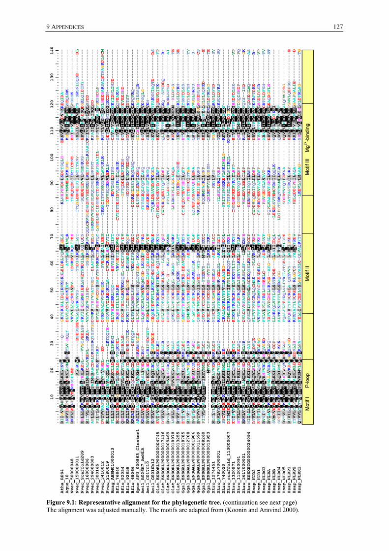
9 APPENDICES
127
Figure 9.1: Representative alignment for the phylogenetic tree. (continuation see next page) The alignment was adjusted manually. The motifs are adapted from (Koonin and Aravind 2000).

9 APPENDICES
128
Figure 9.1 (continued): Representative alignment for the phylogenetic tree. The alignment was adjusted manually. The motifs are adapted from (Koonin and Aravind 2000).
9.3 Hydra NACHT domains used for schematic tree
9.3.1 Alignment of NACHT domains sequences
see CD-ROM

9 APPENDICES
129
9.4 Hydra NBD containing transcripts
9.4.1 Hydra NLR transcripts
9.4.1.1 HyNLR type 1
Coding sequence see CD-ROM Genomic Sequence see CD-ROM Translation 688 AA, MW=81221 1 AGTTAAAATGGATGAAAACATTCTTCAAAATCAACAGAATAGGTATCTATTTAGTCGAAG 1 M D E N I L Q N Q Q N R Y L F S R S 61 TGTCGGAATAGATTGGAAAGACCTTGGACGATGTTTACAAATTTTAGATAGCTATTTAGA 19 V G I D W K D L G R C L Q I L D S Y L D 121 TGTGATAGATATTGAATCTAAAAATTGTTGTGATAAAGCATATCAAATGTTGACCAAATG 39 V I D I E S K N C C D K A Y Q M L T K W 181 GACAGAAAAGGAAAATCCATCATTAAATGAACTAAAAAATGCTTTGCAAACTATGGAGAA 59 T E K E N P S L N E L K N A L Q T M E K 241 AAACGATTTGATGAAAACATTGGAAGATCTTCAAATACGTTCATCAAAAGAAATTGAAAA 79 N D L M K T L E D L Q I R S S K E I E K 301 AAGTAATTTAGTCAAGACAGATTTCTTGAGTGATGCGCTGAAAAATTTTTATCTCGAAAC 99 S N L V K T D F L S D A L K N F Y L E T 361 TTATAAAACAATTGAGGAAATTCAACCACAATTAAACAAACCTTATCAAGTTAACTTTTT 119 Y K T I E E I Q P Q L N K P Y Q V N F L 421 AGATAAATTTGTTGATCTTTATGTTGTTGATGAAGTTGAAGTCCAAAAAGATGCAATTAA 139 D K F V D L Y V V D E V E V Q K D A I N 481 TATTGCAGAGCTAGATCAATTTTTAAAGAAACAAATGAGTTATACATCAATTCTATTTGA 159 I A E L D Q F L K K Q M S Y T S I L F E 541 AAAGCATTTAAAAGATCTGTCTTTATTTCTAATATCTGGTATTGCTGCTATTGGTAAAAC 179 K H L K D L S L F L I S G I A A I G K T 601 ATGGTTGCTTAGAAAATTTTTACTAGATTGGTCAAATGGTTTTATTTGGAAAAATATTGA 199 W L L R K F L L D W S N G F I W K N I D 661 TTTAGTGTTTTACTTGGAGTGTAAACAACTTAATTTGTATGAAAATATTTCTAATATTAA 219 L V F Y L E C K Q L N L Y E N I S N I N 721 TGAATTACTTGATGTTTTCTACAAAGATATTTTAAAAGGTTATAATATTTCTTTAGATTT 239 E L L D V F Y K D I L K G Y N I S L D F 781 CATGCAATCTAAACCATCAATTATGTTTATAATTGATGGCTTAAACGAATTTAAATACTT 259 M Q S K P S I M F I I D G L N E F K Y F 841 TGACCAATTAATAAGCAATACACACTGTAGTTCACAAGAAATTCCTATTCTTAATGTCTT 279 D Q L I S N T H C S S Q E I P I L N V F 901 TACAGAAATTTATAAATACAAAGCTGTGATATCAGGAAGAGTAAATACAATATCACAATA 299 T E I Y K Y K A V I S G R V N T I S Q Y 961 TGAAAATGTGGTCACGCGTTATAAAGATAAGTTAACCATTAGAGTTATGGGTTATAATGA 319 E N V V T R Y K D K L T I R V M G Y N E

9 APPENDICES
130
1021 AAATGGAATAAAATATTATTTGAGGAATAATTTAATAAAAAAAACAAAACGCAGAGTGAA 339 N G I K Y Y L R N N L I K K T K R R V K 1081 AACTATGTTAAAAGAGTCTTCGATTGCAAAAACCATGGCAAAAGTACCCTTTTTTTTATC 359 T M L K E S S I A K T M A K V P F F L S 1141 ATGTATGTGTACAATTGTTGCTGATTTAAAAGTAAAATATAACTTTATAAATCTCACTAT 379 C M C T I V A D L K V K Y N F I N L T M 1201 GACAGATTTGTATGCATATAATTTTTTATACTTTTTTCAAAGACACATTTTTAATGAAAA 399 T D L Y A Y N F L Y F F Q R H I F N E N 1261 TCATGAACCAATTTTCAAAATAATGGAAAATAATCTAAACAAGCAATGTATTTTAAACAT 419 H E P I F K I M E N N L N K Q C I L N I 1321 ATGTAAAATGGCATATATTTTTTTTATTGAAAATAAAACTACTTTTTCCGATATAGAAAT 439 C K M A Y I F F I E N K T T F S D I E I 1381 CCAAACTTTTATCAGTGGTTTTGATCAAATTGAAAGTGGGCTCCTTGAAAAGGTGTTTTT 459 Q T F I S G F D Q I E S G L L E K V F L 1441 AATGAAAAGTGGTTTTATTGAAAAGATTGAAACAAATTTTGGTTATCATTATCGGTTTTG 479 M K S G F I E K I E T N F G Y H Y R F C 1501 TCAGACAGCAATGATAGAGCTATGTGTCTCTGTTCATGCATACTATAGTTATAGTGGGAA 499 Q T A M I E L C V S V H A Y Y S Y S G K 1561 AAAGATTATGGAAAATCAAAAGTTAAAAGACTGTTTTTCAATTATATATGGTTTATTAAA 519 K I M E N Q K L K D C F S I I Y G L L N 1621 TACAAATGAAAATAGTTTTATCAAGTACTTAGCTAACTTGACAAATTCAAATACTGAAAA 539 T N E N S F I K Y L A N L T N S N T E K 1681 GATATCTTTGAATCATGTGATTGATGTGATTCCAAAAGAAATTAACTGTGATCGTGAGTA 559 I S L N H V I D V I P K E I N C D R E Y 1741 TCAAGATAAACAAAAGTTGTTTATGCAATGTTTTTTTGAATCGCAGGCAACTATTACAGA 579 Q D K Q K L F M Q C F F E S Q A T I T D 1801 CGATATTAAAGCATCAATTGATGAAAGAAAGTGGAAGATTTTGATAAATGGTACAAAAAC 599 D I K A S I D E R K W K I L I N G T K T 1861 ATCTTATGAAGTTTTATGTGAAAAATATTTTGTAAACCATCTCGTTAACTCAGGAGGGAA 619 S Y E V L C E K Y F V N H L V N S G G K 1921 GTTGACATCTTTAGAAATTGACAAAAATTTATTAACTGATAAAGAAAAAGATCTTATTAA 639 L T S L E I D K N L L T D K E K D L I K 1981 GCAATGTTCAAGGAATGTAAAAGATATCCGTATAAAGCGGAGTTATCGTTTTTTTATTGA 659 Q C S R N V K D I R I K R S Y R F F I D 2041 TCAATTCACGAAGCGATGGATAAGAAGTGAATAAAGTTGTTTTTATTGGAGGGTATTAAT 679 Q F T K R W I R S E *
Background Colour: Exon structure Red: DEATH domain Blue: NACHT domain Bold: Start and Stop codon Splice variants see CD-ROM
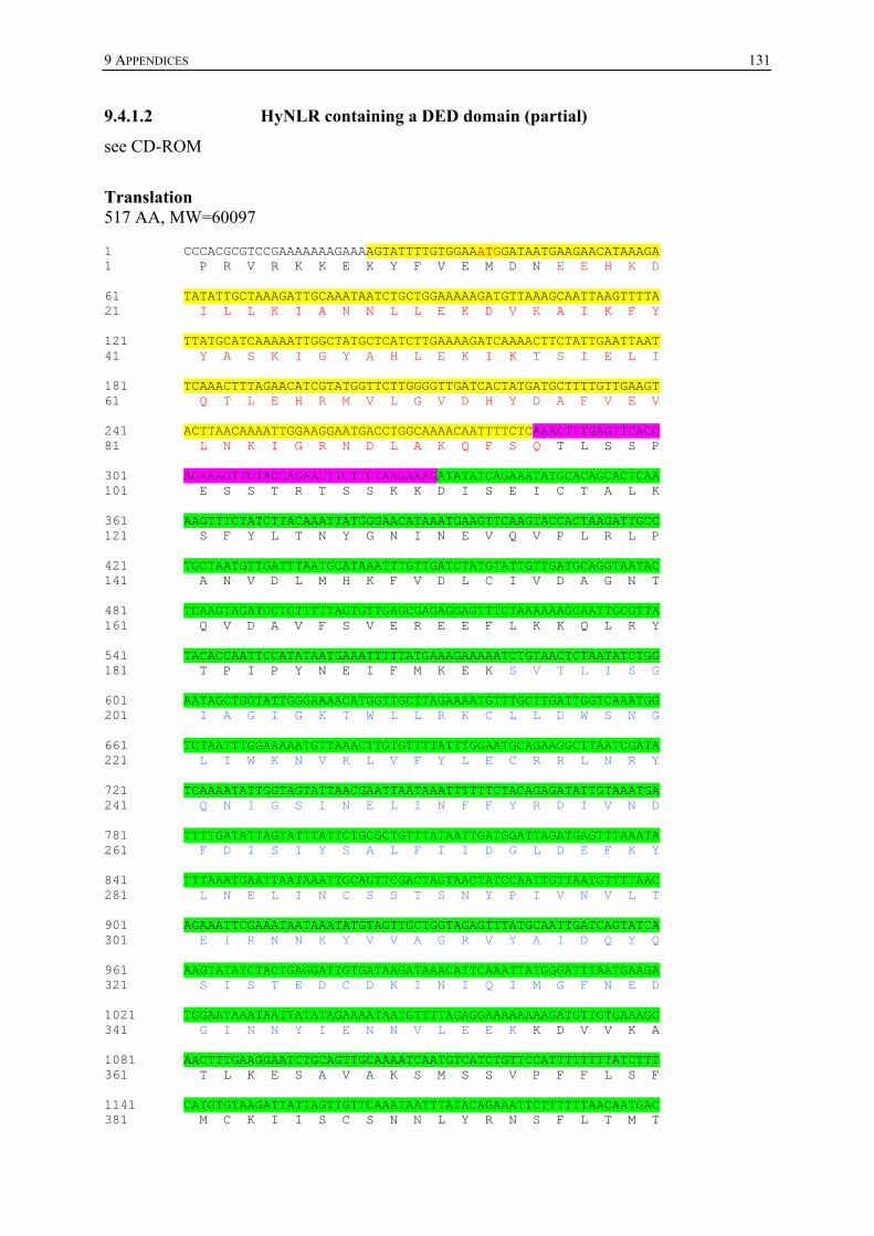
9 APPENDICES
131
9.4.1.2 HyNLR containing a DED domain (partial)
see CD-ROM
Translation 517 AA, MW=60097 1 CCCACGCGTCCGAAAAAAAGAAAAGTATTTTGTGGAAATGGATAATGAAGAACATAAAGA 1 P R V R K K E K Y F V E M D N E E H K D 61 TATATTGCTAAAGATTGCAAATAATCTGCTGGAAAAAGATGTTAAAGCAATTAAGTTTTA 21 I L L K I A N N L L E K D V K A I K F Y 121 TTATGCATCAAAAATTGGCTATGCTCATCTTGAAAAGATCAAAACTTCTATTGAATTAAT 41 Y A S K I G Y A H L E K I K T S I E L I 181 TCAAACTTTAGAACATCGTATGGTTCTTGGGGTTGATCACTATGATGCTTTTGTTGAAGT 61 Q T L E H R M V L G V D H Y D A F V E V 241 ACTTAACAAAATTGGAAGGAATGACCTGGCAAAACAATTTTCTCAAACTTTGAGTTCACC 81 L N K I G R N D L A K Q F S Q T L S S P 301 AGAAAGTTCTACCAGAACTTCTTCTAAGAAAGATATATCAGAAATATGCACAGCACTCAA 101 E S S T R T S S K K D I S E I C T A L K 361 AAGTTTCTATCTTACAAATTATGGGAACATAAATGAAGTTCAAGTACCACTAAGATTGCC 121 S F Y L T N Y G N I N E V Q V P L R L P 421 TGCTAATGTTGATTTAATGCATAAATTTGTTGATCTATGTATTGTTGATGCAGGTAATAC 141 A N V D L M H K F V D L C I V D A G N T 481 TCAAGTAGATGCTGTTTTTAGTGTTGAGCGAGAGGAGTTTCTAAAAAAGCAATTGCGTTA 161 Q V D A V F S V E R E E F L K K Q L R Y 541 TACACCAATTCCATATAATGAAATTTTTATGAAAGAAAAATCTGTAACTCTAATATCTGG 181 T P I P Y N E I F M K E K S V T L I S G 601 AATAGCTGGTATTGGGAAAACATGGTTGCTTAGAAAATGTTTGCTTGATTGGTCAAATGG 201 I A G I G K T W L L R K C L L D W S N G 661 TCTAATTTGGAAAAATGTTAAACTTGTGTTTTATTTGGAATGCAGAAGGCTTAATCGATA 221 L I W K N V K L V F Y L E C R R L N R Y 721 TCAAAATATTGGTAGTATTAACGAATTAATAAATTTTTTCTACAGAGATATTGTAAATGA 241 Q N I G S I N E L I N F F Y R D I V N D 781 TTTTGATATTAGTATTTATTCTGCGCTGTTTATAATTGATGGATTAGATGAGTTTAAATA 261 F D I S I Y S A L F I I D G L D E F K Y 841 TTTAAATGAATTAATAAATTGCAGTTCGACTAGTAACTATCCAATTGTTAATGTTTTAAC 281 L N E L I N C S S T S N Y P I V N V L T 901 AGAAATTCGAAATAATAAATATGTAGTTGCTGGTAGAGTTTATGCAATTGATCAGTATCA 301 E I R N N K Y V V A G R V Y A I D Q Y Q 961 AAGTATATCTACTGAGGATTGTGATAAGATAAACATTCAAATTATGGGATTTAATGAAGA 321 S I S T E D C D K I N I Q I M G F N E D 1021 TGGAATAAATAATTATATAGAAAATAATGTTTTAGAGGAAAAAAAAGATGTTGTGAAAGC 341 G I N N Y I E N N V L E E K K D V V K A 1081 AACTTTGAAGGAATCTGCAGTTGCAAAATCAATGTCATCTGTTCCATTTTTTTTATCTTT 361 T L K E S A V A K S M S S V P F F L S F 1141 CATGTGTAAGATTATTAGTTGTTCAAATAATTTATACAGAAATTCTTTTTTAACAATGAC 381 M C K I I S C S N N L Y R N S F L T M T

9 APPENDICES
132
1201 AGACCTGTATGCAAATATTTTTTTACACTTTTTGCAAAAACACATTATCAAAAACAATGA 401 D L Y A N I F L H F L Q K H I I K N N E 1261 ATCAATTTCTCAATTTATGGAAAACAATTCCAATAAAAAATATGTTCTAAACATTTGTAA 421 S I S Q F M E N N S N K K Y V L N I C K 1321 AATTGCGTACCAATTGTTTGTTGAAAATAAAGTAATTTTTTTCAAAGAAGAAATTCAAAC 441 I A Y Q L F V E N K V I F F K E E I Q T 1381 TTTTGTCAATGAGTTTGATAAAAATGAAGAAACTTTTTTTGGATTTATTGAGAGGATTGA 461 F V N E F D K N E E T F F G F I E R I E 1441 AACAAGTCTAGGTGAATATATTTATCAATTTGCACATTTGACAATAATGGAGTTTTGTGC 481 T S L G E Y I Y Q F A H L T I M E F C A 1501 ATCTATATATGCATATAATTGTTTAAGTAGTGAAGAGATTATGACCAATGAAAA 501 S I Y A Y N C L S S E E I M T N E
Background Colour: Exon structure Red: DEATH domain Blue: NACHT domain Bold: Start codon
9.4.1.3 Other HyNLRs
see CD-ROM
9.4.1.4 Probe for Southern blot hybridisation
TGGTATTGGGAAAACATGGTTGCTTAAAAAATGTTTGCTTGATTGGTCAAATGATATAATTTGGaAAAATGTTGAACTTGTTTTTTATTTGGAATGCAGAAGGATTAATCAATATGAAAATATTTCAAATATTAACGATTTACTAAGCGTTTTCTACAAAGACATTATAAGCAACTTTAATATTAGTAATCATACTGTACTGTTTATAATTGATGGGTTAGATGAGTTTAAATATTTCAATGAATTATCAAACCCAAGATTAAAGTGTAcTATCCCTATTGTTAATGTTTTAGCAGAAATTCAAAGATATAAACATTTAGTTGCTGGTAGAGTTTATGCAATTGATC
9.4.2 Hydra TPR-NLR transcripts
see CD-ROM
9.5 Putative interactome of HyNLRs
9.5.1 HSP90 (gene models)
see CD-ROM
9.5.2 HySGT1
see CD-ROM
9.5.3 HyChp-1
See CD-ROM

9 APPENDICES
133
9.5.4 HyDODE
Coding sequence see CD-ROM Translation 304 AA, MW=35528 1 CCCACGCGTCCGCACATACTGAAACTTTTTAGTCCCTGTGTAATAAGATGACTCAAAATA 1 M T Q N 61 TATCTAGTTTGACAAAGTCAAAACTTGCACAATGTCTCGCTGAAATCTGGAAAGAGTTTG 5 I S S L T K S K L A Q C L A E I W K E F 121 CAGAAGAAATCCCTTTACCAAAATATAAGATAGCTTCTATTGAACAAGAACAAAAACTGT 25 A E E I P L P K Y K I A S I E Q E Q K L 181 TAGTAGAAAGAGCTTATCGTGTACTTACTTTTTGGTACCAATCTGATCCTGATGCATTTA 45 L V E R A Y R V L T F W Y Q S D P D A F 241 ATGAAAACAACATAAAAATGAAGTTAAAAAGTTTAAAGCGGAATGATATAATTAGAGCTG 65 N E N N I K M K L K S L K R N D I I R A 301 TGTTTGGAGAAGAAGATGATCAACCTGCAAAACCCAAAGAAACAAACGTCTCTAAAGTTG 85 V F G E E D D Q P A K P K E T N V S K V 361 AGGACCTTTCTTATCGTCAGATTCGTGTGCTTTCAGACTTCTTAGATCCTGACTATGGGT 105 E D L S Y R Q I R V L S D F L D P D Y G 421 GGGAAGAATTTGCCCATCACCTTTTTGAACATGACAATGAAATGATAAATGAAATACAAA 125 W E E F A H H L F E H D N E M I N E I Q 481 AGCTTCGTCATGAGTATGCTGGAGGTGGTAACCCTGCATACCTGTTCTTAAAACAATTGC 145 K L R H E Y A G G G N P A Y L F L K Q L 541 TTCAAAGAAGGCCAAACATACTTTTGAATGAATTTTTGGATGCTTGTAAGGAAGTAAAGA 165 L Q R R P N I L L N E F L D A C K E V K 601 GAGCAGATATCATACACTATGCAAATGAAAATTTTAGTGGAATTAGTTATATCAGCGAAT 185 R A D I I H Y A N E N F S G I S Y I S E 661 TAGATAAAAACCAACTAAGAGATTTCGCCGATAAGGTACGTGGAAATATAGTTCCAAGTA 205 L D K N Q L R D F A D K V R G N I V P S 721 ATTGGGCTGAAATCGCAAGCGAGTTTGTCGAGTTTTCTTCTGAAGATATAAAAAGAATAG 225 N W A E I A S E F V E F S S E D I K R I 781 AACTTGAAAGGCTGGCTCCAAATAGTTATAGTCCGACGAAAAAATTGTTTGAAAAGCTTA 245 E L E R L A P N S Y S P T K K L F E K L 841 AACAAATTCATCCAAATATGGAGTTAGATAATTTAATAAATGTATGTGAAAAAATCAGGA 265 K Q I H P N M E L D N L I N V C E K I R 901 GGAAAGATGTTGCTAATAAACTTCGTGAATTTGCAATAAATAATAGCAGCATAGCGCAAT 285 R K D V A N K L R E F A I N N S S I A Q 961 AGTTATTTTTTTTAATGTTTTCAAGTTTTTTTATTAATTTTTTTGTATTATTTAATGATT 320 * 1021 TTATTTTTTGAGTTTATTAGGTTTTTATCGGTAGTTGATAATATCTATTCTTTAATTAAA 340 1081 TGTTTAGAATGC 360
Background Colour: Exon structure Red: DEATH domain

9 APPENDICES
134
Bold: Start and Stop codon 9.5.5 HyCARD-Caspase
See CD-ROM
9.5.6 HyDD-Caspase
Coding Sequence See CD-ROM Translation 407 AA, MW=47096 1 TGTAATAAGAAACAACAAAATGGCAGAAGACATTGTTTCTAATGAATCATTCATTTTTAA 1 M A E D I V S N E S F I F K 61 AATGAGCTCTAGTATTGGAACATATTGGGTAACACTTGGACGTGCTCTAGGAATCAAAGA 15 M S S S I G T Y W V T L G R A L G I K D 121 CAGTGTTTTAGAAATGCTTGATAAGGAAAATCCCAAAGTTATAGATAAAGCATATGCTAT 35 S V L E M L D K E N P K V I D K A Y A M 181 GTTAAAACATTGGACTGCAAAGAACACAAATGCAACTATTGACGAGTTGAAAATTGTTTT 55 L K H W T A K N T N A T I D E L K I V L 241 AATGGAATTAGAAAGAAATGATTTAAAAAAAGAAGTAGAAAAAATTTCAAGTACTTTAAT 75 M E L E R N D L K K E V E K I S S T L I 301 TTCAAAAATGACTATTGCTGACAAGAAAGTAAATAGCCAAAATCTATCATTAGGCAATGA 95 S K M T I A D K K V N S Q N L S L G N E 361 ACAGGATTTAAATGTTGAGGAGAAGAAATCTATTCAAGAAACCCTAACCTCAAGTAAACA 115 Q D L N V E E K K S I Q E T L T S S K Q 421 ATCTTTGTCATCAGAACATGAATCAACAGTAACAGATGAAAAACCTAATCAAGCTCAATG 135 S L S S E H E S T V T D E K P N Q A Q C 481 CTATCCTATGACACGTAAAAAACTTGGCAAACTTTTCATTTTTAACAATATGCACAATGA 155 Y P M T R K K L G K L F I F N N M H N D 541 TGAAAAGACTACTCATAGACTTGCAGCAAAATATGATGTCAAAAAAATTAAAGCAACCTT 175 E K T T H R L A A K Y D V K K I K A T F 601 CAGCAAAATAGAATTTGAAGTCGTAGTTTATAAACCGAATAGCTTTGAAGAGATGAAAAA 195 S K I E F E V V V Y K P N S F E E M K K 661 AAATTTAGAAGAAGTTTTAAAGGAACCATATGGCGAGTACAATATTTTTTCAATATTTTT 215 N L E E V L K E P Y G E Y N I F S I F F 721 CATTGCTCATGGACAGGATGGTAAACTATTTGGACCAAATGAAACTAATAACAAAAAATC 235 I A H G Q D G K L F G P N E T N N K K S 781 ATTTTCAATACTTGAATTGAAAAAAATGCTTGAGGATAACTCTTGGCTGAAAAATATTCC 255 F S I L E L K K M L E D N S W L K N I P 841 CAAACTGGTCTTTTGCGAAAGCTGTCGCGGCTTCGATAAAGATTTTCAACAAATGACCCT 275 K L V F C E S C R G F D K D F Q Q M T L 901 AACTAATTTTGAAGCTAAATCTAATGAAAAGAATGTAAATACGGCAAAATTTGCTGACTT 295 T N F E A K S N E K N V N T A K F A D L 961 ATTTATTGGTTTTTCTACTGCTGAGTCTTTTGTATCAATATCTATGCCTTCAGATAATGA 315 F I G F S T A E S F V S I S M P S D N D

9 APPENDICES
135
1021 TGTTCAAATGAAAAACTTTTACAGCCCGTGGGTCATTGAACTTTGCAAACAAATTCAGGA 335 V Q M K N F Y S P W V I E L C K Q I Q E 1081 AAAATACATGACAACTGACTTTCTACAGATTTACCAGGACCTCCAGCGATACATGAGTAG 355 K Y M T T D F L Q I Y Q D L Q R Y M S S 1141 CACACCTTTATTGTCTAATGGCGAAGAAGTTTATTTCCAGATGCCAGAACTTCAATCTAC 375 T P L L S N G E E V Y F Q M P E L Q S T 1201 TTTAAGATATAAACTTTATCTTAATATGGAGAGAAAAACTTAAAAAAAATAATGTTAAAT 395 L R Y K L Y L N M E R K T *
Background Colour: Exon structure Red: DEATH domain Green: Caspase domain Bold: Start and Stop codon
9.5.9 HyDED-Caspase
see CD-ROM
9.5.8 HyDED-DD-Kinases
see CD-ROM
9.5.9 HyJUN
see CD-ROM
9.5.10 HyNF-κB
see CD-ROM

9 APPENDICES
136
9.6 Vectors
9.6.1 pCEV-GwA (constructed and provided by Dr. G. Jakobs)
8.4 kb
CMV IE enhancer
intronintron
SP6 PMCST7 P
insulin poly Atac P
SV40 PSV40 poly A
ampr
intronintron
neor
BamHI
EcoRIEcoRVXhoISalI
pCEV3.1 (+ )
Gat
eway
rea
ding
fram
eA
CMV IE enhancer
intronintron
SP6 PMCST7 P
insulin poly Atac P
SV40 PSV40 poly A
ampr
intronintron
neor
BamHI
EcoRIEcoRVXhoISalI
pCEV3.1 (+ )
Gat
eway
rea
ding
fram
eA
STARTATG GGT ACC TCT GAC TAC AAG GAC GAC GAT GAC AAG GGG CCC AGC GAT TAC
BamHIAAA GAC GAT GAT GAT AAG TCC GAC TAT AAG GAT GAC GAC GAC AAA GGA TCC
EcoRI EcoRVACT AGT CCA GTG TGG TGG AAT TCT GCA GAT
XhoIATC CAG CAC AGT GGC GGC CGC
SalITCG AGT CTA GAG GGC CCG TTT AAA CGT CGA C T7-Promotor-Primer
M G T S D Y K D D D D K G P S D Y
K D D D D K S D Y K D D D D K G S
T S P V W W N S A D
I Q H S G G R
S S L E G P F K R R
STARTATG GGT ACC TCT GAC TAC AAG GAC GAC GAT GAC AAG GGG CCC AGC GAT TAC
BamHIAAA GAC GAT GAT GAT AAG TCC GAC TAT AAG GAT GAC GAC GAC AAA GGA TCC
EcoRI EcoRVACT AGT CCA GTG TGG TGG AAT TCT GCA GAT
XhoIATC CAG CAC AGT GGC GGC CGC
SalITCG AGT CTA GAG GGC CCG TTT AAA CGT CGA C T7-Promotor-PrimerT7-Promotor-Primer
M G T S D Y K D D D D K G P S D Y
K D D D D K S D Y K D D D D K G S
T S P V W W N S A D
I Q H S G G R
S S L E G P F K R R
pCEV-GwA
Figure 9.2: Map of the pCEV-GwA vector including multiple cloning site.
Partial sequence:
see CD-ROM

9 APPENDICES
137
9.6.2 pGA4 including hydeath-fkbp (GENEART)
Figure 9.3: Map of the pGA4 vector including hydeath-fkbp.
Vector sequence:
see CD-ROM
9.6.3 ligR1 (constructed and provided by Dr. K. Khalturin)
Figure 9.4: Map of the ligR1 vector (vector backbone pGEM-T).
ligR1 expression casette sequence:
see CD-ROM

9 APPENDICES
138
9.7 Expression constructs
9.7.1. Expression constructs for HEK293 cell-based assays
9.7.1.1 Flag-HyNLR-FKBP
ATGGGTACCTCTGACTACAAGGACGACGATGACAAGGGGCCCAGCGATTACAAAGACGATGATGATAAGTCCGACTATAAGGATGACGACGACAAAGGATCCACTAGTCCAGTGTGGTGGAATTCTGCAGATATCACAAGTTTGTACAAAAAAGCAGGCTTCGACGAAAATATTCTTCAAAATCAACAAAATAGATATCTTTTTTCAAGATCAGTTGGAATTGATTGGAAAGATCTTGGAAGATGTTTACAAATTCTTGATTCATATCTTGATGTTATTGATATTGAATCAAAAAATTGTTGTGATAAAGCTTATCAAATGTTAACAAAATGGACAGAAAAAGAAAATCCATCATTAAATGAACTTAAAAATGCTTTACAAACAATGGAAAAAAATGATCTTATGAAAACATTAGAAGATCTTCAAATTAGATCATCAAAAGAAATTGAAAAATCAAATCTTGTTAAAACAGATTTTCTTTCAGATGCTTTAAAAAACTTTTATCTTGAAACATATAAAACAATTGAAGAAATTCAACCACAACTTAATAAACCATATCAAGTTAATTTTCTTGATAAATTTGTTGATCTTTATGTTGTTGATGAAGTTGAAGTTCAAAAAGATGCTATTAATATTGCTGAATTAGATCAATTTCTTAAAAAACAAATGTCATATACATCAATTCTTTTTGAAAAACATCTTAAAGATCTTGGACCAGGTCTTTCTTTATTTCTAATATCTGGTATTGCTGCTATTGGTAAAACATGGTTGCTTAGAAAATTTTTACTAGATTGGTCAAATGGTTTTATTTGGAAAAATATTGATTTAGTGTTTTACTTGGAGTGTAAACAACTTAATTTGTATGAAAATATTTCTAATATTAATGAATTACTTGATGTTTTCTACAAAGATATTTTAAAAGGTTATAATATTTCTTTAGATTTCATGCAATCTAAACCATCAATTATGTTTATAATTGATGGCTTAAACGAATTTAAATACTTTGACCAATTAATAAGCAATACACACTGTAGTTCACAAGAAATTCCTATTCTTAATGTCTTTACAGAAATTTATAAATACAAAGCTGTGATATCAGGAAGAGTAAATACAATATCACAATATGAAAATGTGGTCACGCGTTATAAAGATAAGTTAACCATTAGAGTTATGGGTTATAATGAAAATGGAATAAAATATTATTTGAGGAATAATTTAATAAAAAAAACAAAATGCAGAGTGAAAACTGGACCAGGTCCCGGTGTTCAAGTTGAAACAATTTCACCCGGTGATGGAAGAACATTTCCAAAAAGAGGACAAACTTGCGTTGTTCATTATACTGGAATGTTAGAAGATGGTAAAAAAGTTGATTCATCAAGAGATAGAAACAAACCTTTTAAATTTATGTTAGGAAAACAAGAAGTTATTAGAGGTTGGGAAGAAGGTGTTGCTCAAATGTCAGTTGGACAAAGAGCTAAATTAACAATTTCTCCAGATTATGCTTATGGTGCTACAGGACATCCCGGTATTATTCCACCACATGCTACATTAGTTTTTGATGTTGAATTACTTAAATTAGAAACAAGAGGTGTACAAGTAGAAACTATTAGTCCCGGTGATGGTAGAACTTTTCCTAAAAGAGGTCAAACTTGCGTAGTACATTATACTGGAATGCTTGAAGATGGAAAAAAAGTAGATAGTAGCAGAGATAGAAACAAACCTTTTAAATTTATGCTTGGTAAACAAGAAGTAATAAGAGGTTGGGAAGAAGGTGTAGCACAAATGAGTGTAGGTCAAAGAGCAAAACTTACAATTAGCCCTGATTATGCATATGGTGCAACTGGTCATCCCGGAATTATACCACCTCATGCAACTCTTGTATTTGATGTAGAATTGTTAAAACTTGAAACATCATATCCATATGATGTTCCAGATTATGCTTAAGACCCAGCTTTCTTGTACAAAGTGGT Blue: Flag tag Pink: attR1 site Green: attR2 site Grey: PsyI cutting site Bold: Start and Stop codon Red: HyNLR type 1 DEATH domain-coding part Blue: HyNLR type 1 NACHT domain-coding part Light blue: FKBP2 part

9 APPENDICES
139
9.7.1.2 Myc-HyDD-Caspase
ATGGCATCAATGCAGAAGCTGATCTCAGAGGAGGACCTGCTTATGGCCATGGAGGCCCGAATTCTGGCAGAAGACATTGTTTCTAATGAATCATTCATTTTTAAAATGAGCTCTAGTATTGGAACATATTGGGTAACACTTGGACGTGCTCTAGGAATCAAAGACAGTGTTTTAGAAATGCTTGATAAGGAAAATCCCAAAGTTATAGATAAAGCATATGCTATGTTAAAACATTGGACTGCAAAGAACACAAATGCAACTATTGACGAGTTGAAAATTGTTTTAATGGAATTAGAAAGAAATGATTTAAAAAAAGAAGTAGAAAAAATTTCAAGTACTTTAATTTCAAAAATGACTATTGCTGACAAGAAAGTAAATAGCCAAAATCTATCATTAGGCAATGAACAGGATTTAAATGTTGAGGAGAAGAAATCTATTCAAGAAACCCTAACCTCAAGTAAACAATCTTTGTCATCAGAACATGAATCAACAGTAACAGATGAAAAACCTAATCAAGCTCAATGCTATCCTATGACACGTAAAAAACTTGGCAAACTTTTCATTTTTAACAATATGCACAATGATGAAAAGACTACTCATAGACTTGCAGCAAAATATGATGTCAAAAAAATTAAAGCAACCTTCAGCAAAATAGAATTTGAAGTCGTAGTTTATAAACCGAATAGCTTTGAAGAGATGAAAAAAAATTTAGAAGAAGTTTTAAAGGAACCATATGGCGAGTACAATATTTTTTCAATATTTTTCATTGCTCATGGACAGGATGGTAAACTATTTGGACCAAATGAAACTAATAACAAAAAATCATTTTCAATACTTGAATTGAAAAAAATGCTTGAGGATAACTCTTGGCTGAAAAATATTCCCAAACTGGTCTTTTGCGAAAGCTGTCGCGGCTTCGATAAGGATTTTCAACAAATGACCCTAACTAATTTTGAAGCTAAATCTAATGAAAAGAATGTAAATACGGCAAAATTTGCTGACTTATTTATTGGTTTTTCTACTGCTGAGTCTTTTGTATCAATATCTATGCCTTCAGATAATGATGTTCAAATGAAAAACTTTTACAGCCCGTGGGTCATTGAACTTTGCAAACAAATTCAGGAAAAATACATGACAACTGACTTTCTACAGATTTACCAGGACCTCCAGCGATACATGAGTAGCACACCTTTATTGTCTAATGGCGAAGAAGTTTATTTCCAGATGCCAGAACTTCAATCTACTTTAAGATATAAACTTTATCTTAATATGGAGAGAAAAACTTAACTCGAG Blue: Myc tag Pink: EcoRI cutting site Blue: XhoI cutting site Bold: Start and Stop codon
9.7.1.3 Myc-HyDED-Caspase
ATGGCATCAATGCAGAAGCTGATCTCAGAGGAGGACCTGCTTATGGCCATGGAGGCCCGAATTCTGAATCCTCATATCAGTAGTTGGAATCAATTTTTATGTTCATTGGCTGATGAATTATTAAAAGATAATATAAAGAAAATCAAGTTTATTTTGAAAGACTACATACCTGCTAGTGTTCGTGAGACATTAGATGATGGTCATACACTCTTGAATGAATTGGAGAAGCGGAATATAATCTCATGTAATGACCTGTCTAAACTAGGCCAGATTCTTGCTGATATTGGAAGGAAAGACCTTTTATCTAAAATAAAATCTTATATGGAAGAAAAAAGCAAATTAACGAGTGTGAATGAGATATCCCAAAAAAGTGGTTCAACATTCAGTTCAACATTCAATTCAACATTCAATTCAGCTCAAACTTCTGCTTCAGTTTCTGCTTCAACTTCTGTTTTAAATTTGGACTCAATTAAGAAAGAATATTTTGTAGAAAATACTTCAATTGAGTTGGGAATATACAGCATGCACAATGTTCCTAGTGGAATATGTATTATATTCAGCAACTACTTTGATAAAGAAGTAATTATAACTGATGAAAATAAAAAGCTAGAACAGCGCTTAGGAAATGGAGCAGACGAAAGTCAACTAAAGGAGACATTTACTTGGCTGAACTTTGATGTTTTGGTATACAAAAATAAAAGTTCTAAAGATATAGTGCAATTATTATATAGTAATCTCAAAAATAGTTCGATACAAGATTGTTTTGTATGTTGTATTTTGTCACATGGATATAGAAATGGAATATATGGCAGTGATGGATCCAGGCTATCGTTTGAAGAATTGTGGACCACTGTTGAGGACGCATCTTCTGAATCACTTCAGGGTAAACCTAAGTTGTTTTTCATTCAAGCTTGTCAAACCGAAGTAAATGTTAAGGAAATGTCTTTATCAGATATTGGAATAAAAGTTGATTTTGAAGATGTTTTGACATCTTTAGCAACATTACCAGGTAAAGTTGCGTTTCGTGATGTAACTAGAGGCTCATGGTATATTCAAACACTTTGTGAGACTTTGAAAAATCGTGCTTATGATTCTTCATTACTTGATATTTTGACGGAACTTAACTCTAATATGGCGTCGAAAGTAGATTACTACAAAGTATGTTCTGAATATAAAATCGCTACCCAAATGTCTTCAATTCAAAGTTGCACCTTATCAAAAAAATTATTTTTCAAACCATATGACGTAACTGCTCTTTCGCGGTTACGTATTCTTTAACTCGAG Blue: Myc tag Pink: EcoRI cutting site Blue: XhoI cutting site Bold: Start and Stop codon

9 APPENDICES
140
9.7.2 Expression constructs for transgenic Hydra
9.7.2.1 HyNLR type 1 long::FKBP2-HA
GGCCGGCCAGGAATGGACGAAAATATTCTTCAAAATCAACAAAATAGATATCTTTTTTCAAGATCAGTTGGAATTGATTGGAAAGATCTTGGAAGATGTTTACAAATTCTTGATTCATATCTTGATGTTATTGATATTGAATCAAAAAATTGTTGTGATAAAGCTTATCAAATGTTAACAAAATGGACAGAAAAAGAAAATCCATCATTAAATGAACTTAAAAATGCTTTACAAACAATGGAAAAAAATGATCTTATGAAAACATTAGAAGATCTTCAAATTAGATCATCAAAAGAAATTGAAAAATCAAATCTTGTTAAAACAGATTTTCTTTCAGATGCTTTAAAAAACTTTTATCTTGAAACATATAAAACAATTGAAGAAATTCAACCACAACTTAATAAACCATATCAAGTTAATTTTCTTGATAAATTTGTTGATCTTTATGTTGTTGATGAAGTTGAAGTTCAAAAAGATGCTATTAATATTGCTGAATTAGATCAATTTCTTAAAAAACAAATGTCATATACATCAATTCTTTTTGAAAAACATCTTAAAGATCTTGGACCAGGTCTTTCTTTATTTCTAATATCTGGTATTGCTGCTATTGGTAAAACATGGTTGCTTAGAAAATTTTTACTAGATTGGTCAAATGGTTTTATTTGGAAAAATATTGATTTAGTGTTTTACTTGGAGTGTAAACAACTTAATTTGTATGAAAATATTTCTAATATTAATGAATTACTTGATGTTTTCTACAAAGATATTTTAAAAGGTTATAATATTTCTTTAGATTTCATGCAATCTAAACCATCAATTATGTTTATAATTGATGGCTTAAACGAATTTAAATACTTTGACCAATTAATAAGCAATACACACTGTAGTTCACAAGAAATTCCTATTCTTAATGTCTTTACAGAAATTTATAAATACAAAGCTGTGATATCAGGAAGAGTAAATACAATATCACAATATGAAAATGTGGTCACGCGTTATAAAGATAAGTTAACCATTAGAGTTATGGGTTATAATGAAAATGGAATAAAATATTATTTGAGGAATAATTTAATAAAAAAAACAAAATGCAGAGTGAAAACTGGACCAGGTCCCGGTGTTCAAGTTGAAACAATTTCACCCGGTGATGGAAGAACATTTCCAAAAAGAGGACAAACTTGCGTTGTTCATTATACTGGAATGTTAGAAGATGGTAAAAAAGTTGATTCATCAAGAGATAGAAACAAACCTTTTAAATTTATGTTAGGAAAACAAGAAGTTATTAGAGGTTGGGAAGAAGGTGTTGCTCAAATGTCAGTTGGACAAAGAGCTAAATTAACAATTTCTCCAGATTATGCTTATGGTGCTACAGGACATCCCGGTATTATTCCACCACATGCTACATTAGTTTTTGATGTTGAATTACTTAAATTAGAAACAAGAGGTGTACAAGTAGAAACTATTAGTCCCGGTGATGGTAGAACTTTTCCTAAAAGAGGTCAAACTTGCGTAGTACATTATACTGGAATGCTTGAAGATGGAAAAAAAGTAGATAGTAGCAGAGATAGAAACAAACCTTTTAAATTTATGCTTGGTAAACAAGAAGTAATAAGAGGTTGGGAAGAAGGTGTAGCACAAATGAGTGTAGGTCAAAGAGCAAAACTTACAATTAGCCCTGATTATGCATATGGTGCAACTGGTCATCCCGGAATTATACCACCTCATGCAACTCTTGTATTTGATGTAGAATTGTTAAAACTTGAAACATCATATCCATATGATGTTCCAGATTATGCTTAACATTCGTAGAATTC Pink: FseI cutting site Blue: EcoRI cutting site Grey: PsyI cutting site Red: HyNLR type 1 DEATH domain-coding part Blue: HyNLR type 1 NACHT domain-coding part Light blue: FKBP2 part Bold: Stop codon
9.7.2.2 HyNLR type 1 short::FKBP2-HA
GGCCGGCCAGGAATGGACGAAAATATTCTTCAAAATCAACAAAATAGATATCTTTTTTCAAGATCAGTTGGAATTGATTGGAAAGATCTTGGAAGATGTTTACAAATTCTTGATTCATATCTTGATGTTATTGATATTGAATCAAAAAATTGTTGTGATAAAGCTTATCAAATGTTAACAAAATGGACAGAAAAAGAAAATCCATCATTAAATGAACTTAAAAATGCTTTACAAACAATGGAAAAAAATGATCTTATGAAAACATTAGAAGATCTTCAAATTAGATCATCAAAAGAAATTGAAAAATCAAATCTTGTTAAAACAGATTTTCTTTCAGATGCTTTAAAAAACTTTTATCTTGAAACATATAAAACAATTGAAGAAATTCAACCACAACTTAATAAACCATATCAAGTTAATTTTCTTGATAAATTTGTTGATCTTTATGTTGTTGATGAAGTTGAAGTTCAAAAAGATGCTATTAATATTGCTGAATTAGATCAATTTCTTAAAAAACAAATGTCATATACATCAATTCTTTTTGAAAAACATCTTAAAGATCTTGGACCAGGTCCCGGTGTTCAAGTTGAAACAATTTCACCCGGTGATGGAAGAACATTTCCAAAAAGAGGACAAACTTGCGTTGTTCATTATACTGGAATGTTAGAAGATGGTAAAAAAGTTGATTCATCAAGAGATAGAAACAAACCTTTTAAATTTATGTTAGGAAAACAAGAAGTTATTAGAGGTTGGGAAGAAGGTGTTGCTCAAATGTCAGTTGGACAAAGAGCTAAATTAACAATTTCTCCAGATTATGCTTATGGTGCTACAGGACATCCCGGTATTATTCCACCACATGCTACATTAGTTTTTGATGTTGAATTACTTAAATTAGAAACAAGAGGTGTACAAGTAGAAACTATTAGTCCCGGTGATGGTAGAACTTTTCCTAAAAGAGGTCAAACTTGCGTAGTACATTATACTGGAATGCTTGAAGATGGAAAAAAAGTAGATAGTAGCAGAGATAGAAACAAACCTTTTAAATTTATGCTTGGTAAACAAGAAGTAATAAGAGGTTGGGAAGAAGGTGTAGCACAAATGAGTGTAGGTCAAAGAGCAAAACTTACAATTAGCCCTGATTATGCATATGGTGCAACTGGTCATCCCGGAATTATACCACCTCATGCAACTCTTGTATTTGATGTAGAATTGTTAAAACTTGAAACATCATATCCATATGATGTTCCAGATTATGCTTAACATTCGTAGAATTC Pink: FseI cutting site Blue: EcoRI cutting site

9 APPENDICES
141
Grey: PsyI cutting site Red: HyNLR type 1 DEATH domain-coding part Light blue: FKBP2 part Bold: Stop codon
9.7.2.3 FKBP2-HA
GGCCGGCCAGGTGTTCAAGTTGAAACAATTTCACCCGGTGATGGAAGAACATTTCCAAAAAGAGGACAAACTTGCGTTGTTCATTATACTGGAATGTTAGAAGATGGTAAAAAAGTTGATTCATCAAGAGATAGAAACAAACCTTTTAAATTTATGTTAGGAAAACAAGAAGTTATTAGAGGTTGGGAAGAAGGTGTTGCTCAAATGTCAGTTGGACAAAGAGCTAAATTAACAATTTCTCCAGATTATGCTTATGGTGCTACAGGACATCCCGGTATTATTCCACCACATGCTACATTAGTTTTTGATGTTGAATTACTTAAATTAGAAACAAGAGGTGTACAAGTAGAAACTATTAGTCCCGGTGATGGTAGAACTTTTCCTAAAAGAGGTCAAACTTGCGTAGTACATTATACTGGAATGCTTGAAGATGGAAAAAAAGTAGATAGTAGCAGAGATAGAAACAAACCTTTTAAATTTATGCTTGGTAAACAAGAAGTAATAAGAGGTTGGGAAGAAGGTGTAGCACAAATGAGTGTAGGTCAAAGAGCAAAACTTACAATTAGCCCTGATTATGCATATGGTGCAACTGGTCATCCCGGAATTATACCACCTCATGCAACTCTTGTATTTGATGTAGAATTGTTAAAACTTGAAACATCATATCCATATGATGTTCCAGATTATGCTTAACATTCGTAGAATTC Pink: FseI cutting site Blue: EcoRI cutting site Light blue: FKBP2 part Bold: Stop codon
9.8 Bacteria of the tumour bearing Hydra
9.8.1 Bacterial 16S rDNA sequence
see CD-ROM
9.8.2 Alignment of the bacterial 16S rDNA sequences
see CD-ROM
9.9 Hydra oligactis sequences for Ras orthologues
9.9.1 ras1 (partial)
GGTGTTGGAAAAAGTGCTCTAACAATACAACTTATACAGAACCATTTTGTTGAAGATTATGACCCTACTATTGAGGATTCGTATATTAAGCAGGTTGTTGTAGATGGCGCAATATGCATTCTGGATATTCTTGATACAGCAGGTCAAGAAGAGTACAGTGCTATGCGAGAACACTATATGCGTACTGGAGAAGGTTTTTTGTGCATGTTTGCTGTGACAAGTCTTAAATCATTTCAAGAAATTGATAACTTTAGAACACAAGCCCTAAGGGTAAAAGATGCTGACAAAGTA
9.9.2 ras2 (partial)
AAAAAGTGCACTAACCATACAATTCATTCAATCGCATTTTGTCCAAGACTATGATCCAACGATTGAAGATTCTTACAGGAAACAGTGTGTAATTGATGATAAAGTTGCACATTTAGATATATTGGATACAGCTGGGCAAGAGGAGTTTAGTGCTATGCGAGAGCAATACATGAGAACAGGGGAGGGTTTTCTTCT 9.10 Oligonucleotides
Table 9.2: List of the used oligonucleotides.
see CD-ROM

10 ACKNOWLEDGEMENTS
142
10 ACKNOWLEDGEMENTS
Zunächst möchte ich meinen großen Dank an meine beiden Betreuer Prof. Dr. Dr. h.c.
Thomas Bosch und Prof. Dr. Philip Rosenstiel richten. Beide gaben mir die Möglichkeit, an
diesem interessanten Thema zu forschen, und haben mich stets vor allem durch motivierende
und konstruktive Projektbesprechungen unterstützt.
Des Weiteren gilt mein Dank all den Kollegen im Labor, die mich bei meiner Arbeit
unterstützt haben. Insbesondere möchte ich mich bei Frau Dr. Friederike Anton-Erxleben für
ihre hervorragende Hilfe bei bildgebenden Verfahren und anregende Diskussionen bedanken.
Auch Frau Antje Thomas möchte ich meinen Dank für ihre Assistenz bei histologischen
Präparaten aussprechen. Jörg Wittlieb danke ich herzlich für die Hilfe bei der Mikroinjektion
zur Herstellung transgener Hydren. Javier López Quintero möchte ich für die Einführung in
phylogenetische Analysen und anregende Diskussionen danken. Georg Hemmrich danke ich
für die Assistenz bei einigen Sequenz-Analysen am PC. Sebastian Fraune möchte ich für
seine Hilfe bei der Untersuchung Hydra-assoziierter Bakterien danken. Tasja Rahn danke ich
für die Unterstützung bei der Bearbeitung des HyNLR-Interaktoms. Ulrich Klostermeier gilt
mein Dank für die Assistenz bei der Koimmunpräzipitation und Simone Lipinski für die
Unterstützung bei dem Annexin V Assay. Ulrich Knief und Sören Franzenburg danke ich für
ihre Mitarbeit als Forschungspraktikanten bei dem Tumor-Projekt. Mein großer Dank gilt
außerdem René Augustin für seine stets freundliche Hilfe bei allen wissenschaftlichen
Problemen, für das kritische Lesen dieser Arbeit und für konstruktive Diskussionen. Ich
danke Kostya Khalturin für die Bereitstellung des ligR1 Vektors, Joanna Melonek für die
Bereitstellung von HA-Testproteinextrakten und Antikörpern, Gunnar Jakobs für die
Bereitstellung des pCEV-GwA Vektors, Malte Puchert für die Bereitstellung der Cnnos1
Hybridisierungssonde, Ulrich Gerstel für die Bereitstellung der Pseudomonas-Stämme und
Überstände, Vladimir Klimovich und Marina Samoilovich für die Bereitstellung des
verwendeten Antiserums und dem Sequenzierteam des IKMBs. Frau Christa Kuzel danke ich
für ihre herzliche Unterstützung in organisatorischen Angelegenheiten.
Von Herzen möchte ich allen Kollegen und Freunden danken, die zu einer stets angenehmen
Arbeitsatmosphäre im Labor und im Büro beigetragen haben. Dem „native speaker“ Stuart
Hanmer, Felix Sommer und Stephanie Jungnickel möchte ich für das gründliche
Korrekturlesen danken.
Mein großer Dank gilt meiner Familie und Freunden, die mich stets unterstützt haben und
auch in schwierigen Zeiten immer für mich da waren.

11 ERKLÄRUNG
143
11 ERKLÄRUNG
Hiermit erkläre ich, dass ich die vorliegende Dissertation nach den Regeln guter
wissenschaftlicher Praxis eingeständig verfasst und keine anderen als die angegebenen
Hilfsmittel und Quellen benutzt habe. Dabei habe ich keine Hilfe, außer der
wissenschaftlichen Beratung durch meinen Doktorvater Prof. Dr. Dr. h.c. Thomas C. G.
Bosch in Anspruch genommen. Des Weiteren erkläre ich, dass ich noch keinen
Promotionsversuch unternommen habe.
Teile dieser Arbeit wurden bereits zur Publikation eingereicht.
Kiel, den 10.08.2010
Christina Lange