subchapter c—federal hazardous substances act regulations ...
Upload
khangminh22Category
view
0download
0
International Journal of
Molecular Sciences
Review
Importance of Bioactive Substances in Sheep’s Milk inHuman Health
Zuzanna Flis and Edyta Molik *
�����������������
Citation: Flis, Z.; Molik, E.
Importance of Bioactive Substances in
Sheep’s Milk in Human Health. Int. J.
Mol. Sci. 2021, 22, 4364. https://
doi.org/10.3390/ijms22094364
Academic Editor: Michele Navarra
Received: 16 March 2021
Accepted: 21 April 2021
Published: 22 April 2021
Publisher’s Note: MDPI stays neutral
with regard to jurisdictional claims in
published maps and institutional affil-
iations.
Copyright: © 2021 by the authors.
Licensee MDPI, Basel, Switzerland.
This article is an open access article
distributed under the terms and
conditions of the Creative Commons
Attribution (CC BY) license (https://
creativecommons.org/licenses/by/
4.0/).
Department of Animal Nutrition and Biotechnology, and Fisheries, Faculty of Animal Science, University ofAgriculture in Krakow, 31-059 Krakow, Poland; [email protected]* Correspondence: [email protected]
Abstract: Sheep’s milk is an important source of bioactive substances that have health-promotingfunctions for the body. The valuable composition of sheep’s milk is due to the high content of fattyacids, immunoglobulins, proteins, hormones, vitamins and minerals. Many biopeptides found inmilk have antibacterial, antiviral and anti-inflammatory properties. The bioactive substances ofsheep’s milk also show anticancer properties. Sheep’s milk, thanks to its content of CLA and oroticacid, prevents the occurrence of type 2 diabetes, Alzheimer’s disease and cancer. Sheep’s milk, as aproduct rich in bioactive substances, can be used as a medical aid to support the body in the fightagainst neurological and cancer diseases.
Keywords: functional foods; natural bioactive molecules; natural products; sheep; milk
1. Introduction
Nowadays, a sedentary lifestyle, lack of physical activity and an inadequate dietcontribute to the development of many diseases. In recent years, consumer awareness offoods that contain biologically active ingredients and directly affect health has increased.Functional food plays a key role in human health [1]. Sheep’s milk and its productsare an important source of fatty acids, calcium, phosphorus, iron and magnesium [2,3].According to Lordan et al. (2018) and Megalemou et al. (2017), fermented milk productshave a health-promoting effect, especially on the cardiovascular system and civilizationdiseases [4,5]. Consuming yogurt and kefir reduces obesity and the risk of metabolicsyndrome [6] and prevents type II diabetes [7]. Sheep’s milk also contains many biologicallyactive, antibacterial, immunomodulatory and antioxidant substances. The high contentof valuable nutrients and biologically active substances in sheep’s milk prove the dietaryvalue of this milk and products derived from it.
Fermented beverages made from sheep’s milk are characterized by good antibacterialproperties, due to the presence of bioactive compounds such as catechol, vanillin, ferulicacid and salicylic acid. Additionally, kefir is rich in vitamins such as B1, B2, B5 and C;minerals; and amino acids that are essential for the healing process and homeostasis [8].These dairy products can be used as an adjunct therapy in the treatment of neurologicaltumors (glioma) [9]. A study by dos Reis et al. (2019) on Wistar rats has shown thatconsumption of kefir reduced the occurrence of aberrant crypt foci by 36% [10]. Thus, theadministration of kefir to animals helped to reduce the development of lesions, possiblyby increasing short-chain fatty acid production, reducing intestinal permeability andimproving colonic antioxidant activity [10]. Besides anticancer properties, fermenteddrinks (yogurt, kefir) have a positive effect on the wound healing process [11], haveantibacterial properties [12] and modulate the intestinal microflora [13]. Recent studiesshow that kefir consumption also improves exercise capacity and reduces postexerciselactic acid production in humans [14]. Sheep’s milk is used in the production of prebioticsand probiotics and as an ingredient in infant formulas, thus being a suitable alternative
Int. J. Mol. Sci. 2021, 22, 4364. https://doi.org/10.3390/ijms22094364 https://www.mdpi.com/journal/ijms

Int. J. Mol. Sci. 2021, 22, 4364 2 of 14
to mother’s milk and for the production of nutraceuticals [15]. Owing to the content ofbioactive substances, sheep’s milk can be used for the production of medical food.
2. Methodology
At the initial stage of preparing this review, similarly to Guiné et al. (2020) andGuiné et al. (2021), the topic to be addressed was selected [16,17]. After searching availableliterature, it was concluded that there is a need to gather in a review article up-to-dateinformation on the importance of sheep’s milk bioactive substances on human health,which is scattered around the scientific literature. Then, by selecting the appropriatekeywords, the following scientific databases were searched: Web of Science, Science Direct,Scopus and PubMed. For each of the read publications, certain principles were establishedfor their inclusion in this review [16,17]. The most up-to-date scientific articles and thosethat best suited the issues of the review were selected. The final version of the articlepresents references from 1979 to 2021. Particular emphasis was placed on papers publishedwithin the last 5 years, which represent over 50% of the included publications.
3. The Role of Milk Proteins in the Functioning of the Body
Milk proteins include casein complexes (80%) and whey proteins (20%). The wheyprotein fraction contains several extremely important ingredients, including lysozyme, lac-toperoxidase or lactoferrin [18]. Research by Caboni et al. (2019) showed that sheep’s milkcontains the most lactoferrin and proteins that have antibacterial and anti-inflammatoryeffects [19]. Whey proteins exhibit immunoactive properties and include α-lactalbumins, β-lactoglobulins, immunoglobulins, lactoperoxidase (LP), lysozyme and lactoferrin (LF) [20].Lysozyme and α-lactalbumin show reactive oxygen species (ROS)-dependent cytotoxi-city in many tumor cells such as MCF-7, MDA-MB231, HeLa and MG 63 [21]. In vitroand in vivo studies by Li et al. (2020) have shown that lactoferrin, α-lactalbumin andβ-lactoglobulin exhibit neuroprotective effects by increasing bopindolol levels followed byinhibition of the TLR4-related pathway [22].
LF as a sheep’s milk protein has antibacterial, antioxidant, anticancer and anti-inflammatory effects [23,24]. Research by Zheng et al. (2020) proves that LF has theability to alleviate oxidative stress in the hippocampus [25]. Studies have shown thatadministration of LF regenerates cells of the CA1 subregion of the hippocampus in elderlymice. LF has antiviral activity against, among others, rotavirus, HIV and the hepatitisC virus [26,27]. LF has a high affinity for iron ions [28]. According to Sanchez et al.(1992), LF is the most important protein that binds iron, thus blocking the growth of somemicroorganisms and the development of infections [29]. Additionally, LF has the ability toinhibit the proliferation of cancer cells [18]. LF can bind to transferrin receptors (TFR) andLF membrane internalization receptors (LFR) that are characteristic of cancer cells [30]. Theexpression of the LF receptor occurs in the capillaries of the brain; additionally, LF has theability to cross the blood–brain barrier. This suggests that LF is an excellent ligand in thefield of nanomedicine that can deliver substances to the brain [31,32]. These properties ofLF are used in the design of targeted drug delivery systems that can percolate cancer cellsor cross the brain barrier [30,31,33,34]. LF can be used to treat glioblastoma. It has beenshown that bovine LF inhibits the migration of human glioblastoma cell lines by reducingSNAIL and vimentin expression, thereby increasing cadherin levels and inhibiting theIL-6/STAT3 axis [35]. The studies of Eliassen et al. (2002) prove that LF has significantcytotoxic activity against the colon cancer line C26, thus confirming the antitumor activityof this protein [36].
An important endogenous amino acid of sheep’s milk protein is proline, which playsa key role in the synthesis of arginine and polyamines and activates mTOR cell signalingto initiate the process of protein synthesis, especially collagen [37]. Both proline andhydroxyproline are found in the highest amounts in sheep’s milk proteins (Table 1) and incollagen. The demand for this amino acid increases in the prenatal period and just afterbirth, so it is crucial for the proper growth and development of young organisms. Proline
Int. J. Mol. Sci. 2021, 22, 4364 3 of 14
and hydroxyproline account for as much as 12% of proteins in the body of a newborn [38].A study by Singh et al. (2017) showed an indirect effect of proline on the proliferationof MCF-7 and MDA-MB-231 breast cancer cells [39]. Proline plays an important rolein the synthesis of polyamines in the placenta of pregnant sheep [37]. Sheep’s milk ischaracterized by a high content of proline which influences the synthesis of hemoglobin.The proline-rich polypeptide (PRP) participates in the humoral immune response and isresponsible for the maturation of regulatory T lymphocytes (Tregs). PRP can also inhibitthe synthesis of amyloid-beta (Aβ) and reduce its toxic effects on nerve cells [40]. Aβ, apeptide consisting of 40–42 amino acids, is a major component of amyloid plaques in thebrain of patients with Alzheimer’s disease [41]. Aβ molecules, which are formed fromamyloid precursor protein (APP), have the ability to self-aggregate, creating various formsof oligomers that are toxic to nerve cells. Some incorrectly folded oligomers cause a chainreaction and change the degree of the folding of subsequent Aβ molecules. The mostcommon type of neurodegenerative disorder is Alzheimer’s disease, which is characterizedby cognitive decline and memory impairment. This is due to the deposition of Aβ inneuritic plaques and its further effects on microglia, astrocytes, neurons and the post-translational modification of the Tau protein [42,43]. Research by Bharadwaj et al. (2013)has shown that dairy products rich in proline protect against Alzheimer’s disease andother amyloidogenic diseases [41]. In a rat model of Alzheimer’s disease, there is anincreased level of oxidative stress in cells and raised concentration of brain monoamines,which is associated with structural damage to monoaminergic neurons. In animals treatedwith intramuscular injections of PRP, a partial reversal of neurodegenerative changes wasobserved [44].
Table 1. Amino acid composition of sheep’s milk proteins (adapted from [45]).
Amino Acids In g/100 g of Sheep Milk In g/100 g of Casein
Tryptophan 0.084 1.3Threonine 0.268 3.6Isoleucine 0.338 5.1Leucine 0.587 9.0Lysine 0.513 7.3
Methionine 0.155 2.1Cysteine 0.035 0.8
Phenylalanine 0.284 5.2Tyrosine 0.281 5.6
Valine 0.448 6.7Arginine 0.198 3.3Histidine 0.167 3.3Alanine 0.269 3.2
Aspartic acid 0.328 7.7Glutamic acid 1.019 21.1
Glycine 0.041 1.7Proline - 10Serine 0.492 5.0
4. The Importance of Fatty Acids
For many years, attention has been paid to the importance of fatty acids in preventingthe risk of cancer. Fatty acids are one of the most important bioactive components ofmammalian milk. Due to their high nutritional value and influence on the physicochemicalprocesses of the body, they are necessary for the proper development of the nervous systemand the growth of a young organism [46]. Polar lipids (PLs) are an important componentof milk fat with significant pro-health properties. Although the phospholipid fraction ofsheep’s milk is quantitatively a minor component of the overall lipid content (Table 2), itis extremely important as it exhibits anticoagulant activity [47]. Research by Megalemouand Sioriki (2017) and Poutzalis et al. (2016) has shown that the most active inhibitorsof platelet-activating factor (PAF) were found in yogurt made from sheep’s milk and
Int. J. Mol. Sci. 2021, 22, 4364 4 of 14
goat’s milk [5,48]. The processing of milk, especially the fermentation process, enhancesthe antithrombotic properties of PLs against PAF [47] and thrombin-induced plateletaggregation [49]. The antithrombotic activity of PLs was also demonstrated in a study onthe production of traditional Greek sheep cheeses such as Ladotyri and Kefalotyri [50].Due to their properties, polar lipid fractions improve human health by reducing thelevel of atherogenic lipoprotein cholesterol, modulating the intestinal microflora andreducing inflammation in blood serum and liver [51]. Studies on low-density lipoprotein(LDL) receptor knockout mice have shown that the addition of milk to feed reduced thedevelopment of atherosclerosis compared to animals fed a high-fat diet without milk [51].Recently, yogurts with the addition of omega-3 fatty acids (O-3FAs) have been gainingpopularity. Recent studies show that consuming yogurt with O-3FAs significantly reducesthe risk of developing infectious diseases [52]. Sheep’s milk contains short- and medium-chain fatty acids (representing a fraction of about 11%), which are extremely important fora healthy human diet [53,54]. Sheep’s milk has higher concentrations of butyric acid (C4:0),omega-3 fatty acid and conjugated linoleic acid (CLA) than milk from other ruminants [54].
Table 2. Content of total lipids (TLs), total polar lipids (TPLs) and total neutral lipids (TNLs)expressed in grams per 100 g of sheep milk and yogurt (adapted from [47]).
Dairy Products TLs (g/100 g) TNLs (%TL) TPLs (%TL)
Sheep Milk 5.28 ± 0.37 95.15 ± 2.30 3.20 ± 0.56Yoghurt A 8.10 ± 0.43 96.46 ± 1.07 2.45 ± 0.20Yoghurt B 8.23 ± 1.59 97.62 ± 0.22 2.29 ± 0.17Yoghurt C 7.23 ± 0.60 97.47 ± 0.53 2.10 ± 0.37Yoghurt D 7.47 ± 0.36 97.34 ± 0.47 2.25 ± 0.10Yoghurt E 9.20 ± 0.55 97.60 ± 0.38 2.55 ± 0.45
A special role in the regeneration of the nervous system is attributed to conjugatedlinoleic acid dienes [55]. It is believed that the cis-9, trans-11 octadecadienoic acid (rumenicacid) is the major isomer of CLA present in the milk fat of ruminants (Table 3). Its amountcan be up to 90% of total CLA [1,55]. Of all the ruminants, sheep’s milk turns out tobe the richest (1.1%) in CLA, but the concentration of this component in milk dependson the season [56,57]. According to Zervas et al. (2011), sheep’s milk has a higher CLAcontent than goat’s milk, due to differences in the mRNA of the adipocytes of the mammaryglands in both species [58]. In recent years, evidence has been provided largely based onin vitro studies and human clinical studies that CLA, besides its classical mechanism ofaction mediated by nuclear transcription factors, also exhibits a number of interdependentmolecular signaling pathways that are responsible for human health [59]. CLA inhibits bothbenign and malignant tumors by inhibiting cell growth and development [60]. Accordingto reports by Ochoa et al. (2004), the 10-CLA isomer acts peripherally by modulating theapoptosis process and controlling the cell cycle, while the major 9-CLA isomer influencesthe metabolism of arachidonic acid [61]. Cellular mechanisms modulating CLA synthesismay modify cell proliferation, lipid oxidation and vitamin A transformations [62–64]. Ithas been shown that women who consume high-fat dairy products have a lower incidenceof colon cancer, and this may be partly related to the high CLA content in these foods [65].Administration of LF-CLA complex to rats with Alzheimer’s disease resulted in a 2-folddecrease in Aβ in the hippocampus. This effect may be due to the ability of CLA to destroyexisting Aβ and inhibit the formation of new oligomers of this protein. In addition, in thegroup treated with the LF–CLA complex, there was a significant decrease in the levels ofreactive oxygen species (ROS), nitrite (NO) and malondialdehyde (MDA), which indicatesthe antioxidant effect of CLA and the ability to scavenge free radicals. All the obtainedresults of oxidative stress markers showed a high antioxidant capacity of the LF–CLAcomplex due to its active ability to cross the blood–brain barrier (BBB) and target the braintissue. By reducing the level of arachidonic acid (ARA), CLA can influence the growth ofcancer cells. For this reason, rats treated with the LF–CLA complex showed significantly
Int. J. Mol. Sci. 2021, 22, 4364 5 of 14
lower levels of TNF-α [30]. Similar results regarding the effect of CLA on oxidative stress,MDA levels and ARA levels have also been observed in other studies [66,67]. The mother’sdiet during pregnancy and lactation can have a significant impact on the brain developmentof the offspring. CLA is delivered to the fetus through the placenta during pregnancyand through breast milk in the infancy period [68]. Administration of CLA to rats duringpregnancy and lactation caused a reduction in brain lipid peroxidation in the offspring [67].
Table 3. Conjugated linoleic acid (CLA) isomers (% total CLA) in sheep’s milk (adapted from [69]).
Isomer Sheep’s Milk
trans-12, trans-14 1.31–3.47trans-11, trans-13 1.21–5.08trans-10, trans-12 1.17–1.77trans-9, trans-11 1.13–1.99trans-8, trans-10 1.05–1.37trans-7, trans-9 0.48–0.61
12–14 (cis–trans plus trans–cis) 0.52–1.8311–13 (cis–trans plus trans–cis) 0.76–4.2310–12(cis–trans plus trans–cis) 0.28–0.419–11 (cis–trans plus trans–cis) 76.5–82.48–10 (cis–trans plus trans–cis) 0.11–0.717–9 (cis–trans plus trans–cis) 3.31–9.69
Some bioactive substances contained in sheep’s milk have a health-promoting effect onthe body due to their self-healing properties. Genomic and mitochondrial DNA moleculesare continually exposed to damage from endogenous metabolites, environmental carcino-gens, some anti-inflammatory drugs and genotoxic cancer therapeutics [70]. This damagecan cause permanent changes to the DNA molecule that preclude the cell from transcribingthe damaged DNA fragment. When DNA repair processes are not working efficiently,the generation of DNA lesions and mutations leads to carcinogenic transformation [71].The main sources of endogenous DNA damage are ROS and alkyl groups [72]. However,there are mechanisms that correct damaged DNA molecules. The DNA damage responseinvolves the activation of complex signaling networks that repair DNA damage and main-tain genome integrity [70,73]. This is crucial in preventing tumorigenesis [73]. A study byIzzotti et al. (2003) on newborn mice showed that the sudden transition from maternal-mediated respiration to autonomic pulmonary respiration in the fetus causes a significantincrease in extensive DNA adducts and oxidative DNA lesions in the lungs [74]. This DNAdamage was attenuated by the upregulation of many genes involved in oxidative stressand DNA repair. Additionally, prenatal administration of the antioxidant N-acetylcysteineprevented all transcriptional changes in the lungs [74], suggesting links between oxidativestress, DNA damage and tissue function. It is widely accepted that an impaired DNAdamage repair system is a common mechanism in neurodegenerative diseases [75]. A largeaccumulation of DNA damage occurs most often in the central nervous system due to thelow DNA repair capacity in postmitotic brain tissue [76]. Accumulation of DNA damage isa well-known factor of aging and, therefore, is believed to be the main cause of Alzheimer’sdisease [77]. Orotic acid is an important precursor in the biosynthetic pathway of pyrim-idine nucleotides and, therefore, participates in the synthesis of DNA and RNA [78]. Inmammals, it is released from mitochondrial dihydroorotate dehydrogenase (DHODH) bythe enzyme cytoplasmic synthase UMP for conversion to UMP [79]. Cows’ milk containsthe most orotic acid, followed by sheep’s milk and then goat’s milk. In addition, the contentof this component in milk depends on the season of the year, the stage of lactation andthe breed of the animal [80,81]. In ruminants, the highest concentration of orotic acid inmilk is observed in the middle of lactation [78]. Still, little is known about the functionof orotic acid in milk and its impact on human health; it can be assumed that it is neededin developing the microbiome in the stomach of ruminants [78]. It has been shown thatorotic acid improves learning ability in adult rats [82], and it has a neuroprotective effect in
Int. J. Mol. Sci. 2021, 22, 4364 6 of 14
gerbils and cats with transient cerebral ischemia [83]. Additionally, the orotic acid moleculeis thought to be needed to regulate genes that are extremely important in the developmentof cells, tissues and organisms [79].
5. The Importance of Vitamins and Hormones
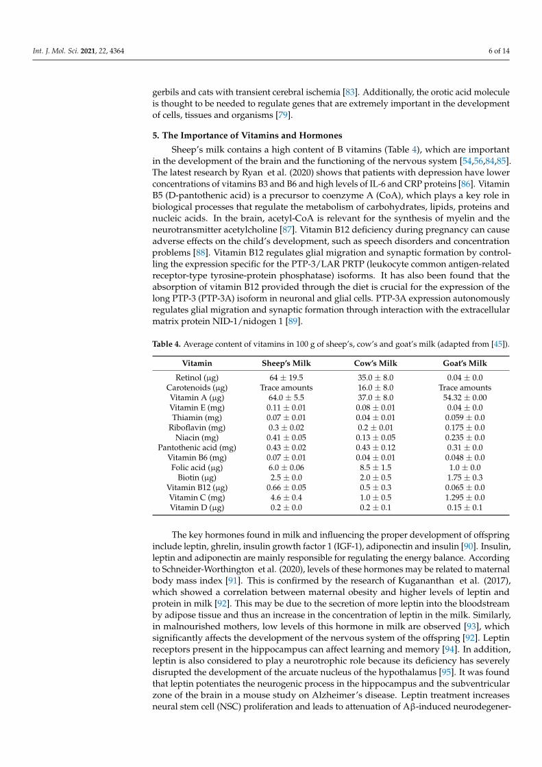
Sheep’s milk contains a high content of B vitamins (Table 4), which are importantin the development of the brain and the functioning of the nervous system [54,56,84,85].The latest research by Ryan et al. (2020) shows that patients with depression have lowerconcentrations of vitamins B3 and B6 and high levels of IL-6 and CRP proteins [86]. VitaminB5 (D-pantothenic acid) is a precursor to coenzyme A (CoA), which plays a key role inbiological processes that regulate the metabolism of carbohydrates, lipids, proteins andnucleic acids. In the brain, acetyl-CoA is relevant for the synthesis of myelin and theneurotransmitter acetylcholine [87]. Vitamin B12 deficiency during pregnancy can causeadverse effects on the child’s development, such as speech disorders and concentrationproblems [88]. Vitamin B12 regulates glial migration and synaptic formation by control-ling the expression specific for the PTP-3/LAR PRTP (leukocyte common antigen-relatedreceptor-type tyrosine-protein phosphatase) isoforms. It has also been found that theabsorption of vitamin B12 provided through the diet is crucial for the expression of thelong PTP-3 (PTP-3A) isoform in neuronal and glial cells. PTP-3A expression autonomouslyregulates glial migration and synaptic formation through interaction with the extracellularmatrix protein NID-1/nidogen 1 [89].
Table 4. Average content of vitamins in 100 g of sheep’s, cow’s and goat’s milk (adapted from [45]).
Vitamin Sheep’s Milk Cow’s Milk Goat’s Milk
Retinol (µg) 64 ± 19.5 35.0 ± 8.0 0.04 ± 0.0Carotenoids (µg) Trace amounts 16.0 ± 8.0 Trace amountsVitamin A (µg) 64.0 ± 5.5 37.0 ± 8.0 54.32 ± 0.00Vitamin E (mg) 0.11 ± 0.01 0.08 ± 0.01 0.04 ± 0.0Thiamin (mg) 0.07 ± 0.01 0.04 ± 0.01 0.059 ± 0.0
Riboflavin (mg) 0.3 ± 0.02 0.2 ± 0.01 0.175 ± 0.0Niacin (mg) 0.41 ± 0.05 0.13 ± 0.05 0.235 ± 0.0
Pantothenic acid (mg) 0.43 ± 0.02 0.43 ± 0.12 0.31 ± 0.0Vitamin B6 (mg) 0.07 ± 0.01 0.04 ± 0.01 0.048 ± 0.0Folic acid (µg) 6.0 ± 0.06 8.5 ± 1.5 1.0 ± 0.0
Biotin (µg) 2.5 ± 0.0 2.0 ± 0.5 1.75 ± 0.3Vitamin B12 (µg) 0.66 ± 0.05 0.5 ± 0.3 0.065 ± 0.0Vitamin C (mg) 4.6 ± 0.4 1.0 ± 0.5 1.295 ± 0.0Vitamin D (µg) 0.2 ± 0.0 0.2 ± 0.1 0.15 ± 0.1
The key hormones found in milk and influencing the proper development of offspringinclude leptin, ghrelin, insulin growth factor 1 (IGF-1), adiponectin and insulin [90]. Insulin,leptin and adiponectin are mainly responsible for regulating the energy balance. Accordingto Schneider-Worthington et al. (2020), levels of these hormones may be related to maternalbody mass index [91]. This is confirmed by the research of Kugananthan et al. (2017),which showed a correlation between maternal obesity and higher levels of leptin andprotein in milk [92]. This may be due to the secretion of more leptin into the bloodstreamby adipose tissue and thus an increase in the concentration of leptin in the milk. Similarly,in malnourished mothers, low levels of this hormone in milk are observed [93], whichsignificantly affects the development of the nervous system of the offspring [92]. Leptinreceptors present in the hippocampus can affect learning and memory [94]. In addition,leptin is also considered to play a neurotrophic role because its deficiency has severelydisrupted the development of the arcuate nucleus of the hypothalamus [95]. It was foundthat leptin potentiates the neurogenic process in the hippocampus and the subventricularzone of the brain in a mouse study on Alzheimer’s disease. Leptin treatment increasesneural stem cell (NSC) proliferation and leads to attenuation of Aβ-induced neurodegener-
Int. J. Mol. Sci. 2021, 22, 4364 7 of 14
ation and the production of superoxide anions. Researchers have emphasized the need forfurther research to elucidate which signaling mechanisms are involved in the neurogenicand neuroprotective effects of this hormone, which could lead to the development of newtherapies for the treatment of Alzheimer’s disease in the future [96]. Sahin et al. (2020)reported that leptin induces synaptogenesis in the developing hippocampus through in-creased expression of KLF4 and cytokine signaling suppressor 3 (SOCS3) in hippocampalneurons [97]. Additionally, it exhibits neuroprotective effects and alleviates spatial memoryimpairment in rat studies of premature brain injury, which may contribute to a betterunderstanding of the protective effect of this hormone in premature infants with cerebralhypoxia [98,99].
In the case of adiponectin, no relationship was found between maternal obesityand the concentration of this hormone in milk [92]. This suggests that adiponectin ismostly synthesized in the mammary gland [100]. It is considered that some bioactivehormones found in milk, including adiponectin, link the metabolic status of the motherwith the metabolic health of the offspring [101]. Milk-fed infants had higher levels ofadiponectin and had less weight gain in the first 6 months of life [102]. It is believedthat adiponectin influences the proper course of fatty acid metabolism and indicates anti-inflammatory properties [103]. Adiponectin may also have neuroprotective effects onoxidative stress-induced brain damage [104]. It is suggested that adiponectin may mediateAdipoR1/APPL1/LKB1/AMPK signaling, because the administration of recombinanthuman exogenous adiponectin (rh-adiponectin) to young mice after cerebral infarctionreduces neuronal apoptosis [105].
Insulin plays an important role in regulating the distribution of nutrients in ruminantsduring lactation [106]. Plasma insulin levels also affect the milk yield of ruminants; namely,low insulin levels are correlated with higher milk production [107]. Li et al. (2020) provedin their study that administering donkey milk powder to rats with type 2 diabetes for4 weeks significantly increases target organ insulin sensitivity, lowers blood glucose levels,improves insulin resistance, increases the ability to capture free radicals and improves thelevel of antioxidants in the body [22]. Wingrove et al. (2019) reported that inhaled insulinadministration uses the nose–brain pathway and delivers the drug directly to the braintissue while limiting systemic exposure [108]. Insulin administered in this way appears toimprove the potential of the brain’s mitochondrial membrane and stimulates the activityof the brain’s mitochondrial complexes in a streptozotocin-induced model of early type2 diabetes [109]. Injections of lipopolysaccharide (LPS) into the brain ventricles of ratscaused inflammation of the nervous system. The cognitive functions were impaired andthe levels of IL-1β and TNF-α increased in the cortex and hippocampus of the animals.Studies have shown that LPS modulates mitochondrial function and induces oxidativestress by reducing the activity of superoxide dismutase and catalase and the levels ofglutathione and sulfhydryl. Treatment with insulin caused a reversal of all effects, whichproves the potential role of insulin as a therapeutic drug in inflammatory diseases relatedto mitochondrial dysfunction in the brain [110].
6. Importance of Milk MicroRNA
Micro-ribonucleic acids (miRNAs) are small non-coding RNA molecules 18–25 nu-cleotides long, responsible for regulating 40% to 60% of gene expression at the post-transcriptional level [111]. The miRNA precursor is transcribed primarily by RNA poly-merase II and then is processed to mature miRNAs [112]. miRNAs are responsible forregulating protein expression by binding to complementary mRNAs and then directingthe mRNA to degrade or inhibit translation [113]. Studies have shown that miRNA-21(a mammalian microRNA encoded by the MIR21 gene) can control the proliferation andapoptosis of many cell types, including cancer cells [114]. As demonstrated by Rasool-nezhad et al. (2021), miR-128-5p inhibits proliferation in breast cancer cells by regulatingthe PI3K/AKT pathway [115]. Moreover, miRNA-138-5p induced apoptosis by increasingthe levels of caspase-9 and caspase-3 and stopping the cell cycle in the sub-G1 phase. It has
Int. J. Mol. Sci. 2021, 22, 4364 8 of 14
been found that miRNA-21 can act on glioblastoma cells. This is due to the inhibition ofcaspase-3 and caspase-7 expression in vitro and in vivo. miRNA plays an important rolein the growth and development of animals and performs a variety of biological functionsin their tissues and organs [116]. miRNAs were detected in saliva [117], urine [118] andmilk [119]. The synthesis of miRNAs takes place in the mammary gland; then, when thecubs drink milk, the miRNAs are transported to the intestine, where they are absorbed byepithelial cells [120]. miRNA molecules enter tissues and organs through the circulatorysystem so that they can perform various functions, e.g., immunoprotection [121]. ThemiRNA in milk is in the form of free molecules packed into extracellular vesicles (EVs) suchas exosomes that protect against degradation [121–123]. According to Zhou et al. (2012),more than 60% of all pre-miRNAs that are associated with resistance are found in breastmilk [119]. miRNA plays a key role in the physiology of the mammary gland and in thelactation of livestock [31,124], but its function in sheep is not fully understood. In sheep, themiRNA is presumed to be responsible for the growth of the fleece [125] and muscle [126].A study by Hao et al. (2021) on Gansu Alpine Merino and Small-Tailed Han sheep showedthat differently expressed miRNA target genes were largely involved in some metabolicand signaling pathways related to mammary gland development and synthesis of milkproteins and fats [127]. Previous studies on the effects of miRNAs on the mammary glandfocused primarily on identifying specific miRNAs as oncogenes and tumor suppressorsthat regulate gene expression by targeting mRNAs in breast cancer [128]. Human milkis a rich source of miRNAs that are specific to lactation, and this is why they began to beused as biomarkers of mammary gland efficiency [121]. Genc et al. (2018) examined thechanges in the expression of miRNAs in subacute sclerosing panencephalitis (SSPE) [129].IL-29 and miR-548 levels were increased in SSPE patients. The increased expression ofmiR-548 may be a compensatory result of an excessive immune system response. Thisproves that IL-29 and miR548 can be involved in the pathogenesis of this disease and canbe used in the diagnosis and treatment of SSPE. Oral administration of milk to mice causesdetectable accumulation of EVs in their tissues, mainly the liver and brain [130]. It has beenfound that miRNA-9-5p is an important regulator of angiogenesis following traumaticbrain injury (TBI). Studies in rats have shown significantly higher levels of miRNA-9-5pand an increased density of vessels and neurons in the damaged areas. Elevated expres-sion of miRNA-9-5p promoted angiogenesis in the injured cerebral cortex and restoredneurological functions by activating the Hedgehog pathway and increasing expression ofp-AKT [131].
7. Summary
Sheep’s milk is a product rich in bioactive substances needed for the proper develop-ment of young organisms. The valuable composition of sheep’s milk is due to the highcontent of fatty acids, immunoglobulins, proteins, hormones, vitamins and minerals. Dueto having the highest linoleic acid content of all ruminants, sheep’s milk is effective inpreventing obesity, type 2 diabetes and cancer. Many active biopeptides found in milkhave proven antiviral, antibacterial and anti-inflammatory properties. In addition, theyalso exhibit cytotoxic activity against cancer cells. A specific feature of sheep’s milk isthe high level of vitamins, especially B vitamins and minerals. The effect of sheep’s milkon the human body is still not fully understood, and the latest research focuses on theimportance of orotic acid and microRNAs that are present in the milk of these ruminants.Undoubtedly, sheep’s milk is a rich source of health-promoting substances for the humanbody, and thanks to this, it can act as an effective functional food.
Author Contributions: Conceptualization and writing—original draft preparation, E.M. and Z.F. Allauthors have read and agreed to the published version of the manuscript.
Funding: This research received no external funding.
Institutional Review Board Statement: Not applicable.
Int. J. Mol. Sci. 2021, 22, 4364 9 of 14
Informed Consent Statement: Not applicable.
Data Availability Statement: Not applicable.
Conflicts of Interest: The authors declare no conflict of interest.
References1. Kumar, A.; Naik, S.; Gandhi, K.; Pandey, V. Functional Lipid Components for Obesity Management: A Review. Int. Food Res. J.
2019, 26, 1111–1122.2. Zhang, R.H.; Mustafa, A.F.; Zhao, X. Effects of Feeding Oilseeds Rich in Linoleic and Linolenic Fatty Acids to Lactating Ewes on
Cheese Yield and on Fatty Acid Composition of Milk and Cheese. Anim. Feed Sci. Technol. 2006, 127, 220–233. [CrossRef]3. Molik, E.; Murawski, M.; Bonczar, G.; Wierzchos, E. Effect of Genotype on Yield and Chemical Composition of Sheep Milk. Anim.
Sci. Pap. Rep. 2008, 26, 211–218.4. Lordan, R.; Tsoupras, A.; Mitra, B.; Zabetakis, I. Dairy Fats and Cardiovascular Disease: Do We Really Need to Be Concerned?
Foods 2018, 7, 29. [CrossRef] [PubMed]5. Megalemou, K.; Sioriki, E.; Lordan, R.; Dermiki, M.; Nasopoulou, C.; Zabetakis, I. Evaluation of Sensory and in Vitro Anti-
Thrombotic Properties of Traditional Greek Yogurts Derived from Different Types of Milk. Heliyon 2017, 3. [CrossRef]6. Sayon-Orea, C.; Martínez-González, M.A.; Ruiz-Canela, M.; Bes-Rastrollo, M. Associations between Yogurt Consumption and
Weight Gain and Risk of Obesity and Metabolic Syndrome: A Systematic Review. Adv. Nutr. 2017, 8, 146S–154S. [CrossRef][PubMed]
7. Gijsbers, L.; Ding, E.L.; Malik, V.S.; de Goede, J.; Geleijnse, J.M.; Soedamah-Muthu, S.S. Consumption of Dairy Foods andDiabetes Incidence: A Dose-Response Meta-Analysis of Observational Studies. Am. J. Clin. Nutr. 2016, 103, 1111–1124. [CrossRef][PubMed]
8. Farag, M.A.; Jomaa, S.A.; Abd El-Wahed, A.; R El-Seedi, H. The Many Faces of Kefir Fermented Dairy Products: QualityCharacteristics, Flavour Chemistry, Nutritional Value, Health Benefits, and Safety. Nutrients 2020, 12, 346. [CrossRef] [PubMed]
9. Fatahi, A.; Soleimani, N.; Afrough, P. Anticancer Activity of Kefir on Glioblastoma Cancer Cell as a New Treatment. Int. J. FoodSci. 2021, 2021, 8180742. [CrossRef] [PubMed]
10. dos Reis, S.A.; da Conceição, L.L.; e Dias, M.M.; Siqueira, N.P.; Rosa, D.D.; de Oliveira, L.L.; da Matta, S.L.P.; Peluzio, M.D.C.G.Kefir Reduces the Incidence of Pre-Neoplastic Lesions in an Animal Model for Colorectal Cancer. J. Funct. Foods 2019, 53, 1–6.[CrossRef]
11. Oryan, A.; Alemzadeh, E.; Eskandari, M.H. Kefir Accelerates Burn Wound Healing Through Inducing Fibroblast Cell MigrationIn Vitro and Modulating the Expression of IL-1ß, TGF-SS1, and BFGF Genes In Vivo. Probiotics Antimicrob. Proteins 2019, 11,874–886. [CrossRef]
12. Miao, J.; Guo, H.; Chen, F.; Zhao, L.; He, L.; Ou, Y.; Huang, M.; Zhang, Y.; Guo, B.; Cao, Y.; et al. Antibacterial Effects of aCell-Penetrating Peptide Isolated from Kefir. J. Agric. Food Chem. 2016, 64, 3234–3242. [CrossRef]
13. Hsu, Y.-J.; Huang, W.-C.; Lin, J.-S.; Chen, Y.-M.; Ho, S.-T.; Huang, C.-C.; Tung, Y.-T. Kefir Supplementation Modifies GutMicrobiota Composition, Reduces Physical Fatigue, and Improves Exercise Performance in Mice. Nutrients 2018, 10, 862.[CrossRef] [PubMed]
14. Lee, M.-C.; Jhang, W.-L.; Lee, C.-C.; Kan, N.-W.; Hsu, Y.-J.; Ho, C.-S.; Chang, C.-H.; Cheng, Y.-C.; Lin, J.-S.; Huang, C.-C. TheEffect of Kefir Supplementation on Improving Human Endurance Exercise Performance and Antifatigue. Metabolites 2021, 11, 136.[CrossRef]
15. Pulina, G.; Milán, M.J.; Lavín, M.P.; Theodoridis, A.; Morin, E.; Capote, J.; Thomas, D.L.; Francesconi, A.H.D.; Caja, G. InvitedReview: Current Production Trends, Farm Structures, and Economics of the Dairy Sheep and Goat Sectors. J. Dairy Sci. 2018, 101,6715–6729. [CrossRef] [PubMed]
16. Guiné, R.P.F.; Florença, S.G.; Barroca, M.J.; Anjos, O. The Link between the Consumer and the Innovations in Food ProductDevelopment. Foods 2020, 9, 1317. [CrossRef]
17. Guiné, R.P.F.; Barroca, M.J.; Coldea, T.E.; Bartkiene, E.; Anjos, O. Apple Fermented Products: An Overview of Technology,Properties and Health Effects. Processes 2021, 9, 223. [CrossRef]
18. Jilo, K. Medicinal Values of Camel Milk. Int. J. Vet. Sci. Res. 2016, 2, 018–025. [CrossRef]19. Caboni, P.; Murgia, A.; Porcu, A.; Manis, C.; Ibba, I.; Contu, M.; Scano, P. A Metabolomics Comparison between Sheep’s and
Goat’s Milk. Food Res. Int. 2019, 119, 869–875. [CrossRef]20. Mohapatra, A.; Shinde, A.K.; Singh, R. Sheep Milk: A Pertinent Functional Food. Small Rumin. Res. 2019, 181, 6–11. [CrossRef]21. Somu, P.; Paul, S. Supramolecular Nanoassembly of Lysozyme and α-Lactalbumin (Apo α-LA) Exhibits Selective Cytotoxicity
and Enhanced Bioavailability of Curcumin to Cancer Cells. Colloids Surf. B Biointerfaces 2019, 178, 297–306. [CrossRef]22. Li, Y.; Fan, Y.; Shaikh, A.S.; Wang, Z.; Wang, D.; Tan, H. Dezhou Donkey (Equus Asinus) Milk a Potential Treatment Strategy for
Type 2 Diabetes. J. Ethnopharmacol. 2020, 246, 112221. [CrossRef]23. Bruni, N.; Capucchio, M.T.; Biasibetti, E.; Pessione, E.; Cirrincione, S.; Giraudo, L.; Corona, A.; Dosio, F. Antimicrobial Activity of
Lactoferrin-Related Peptides and Applications in Human and Veterinary Medicine. Mol. Basel Switz. 2016, 21, 752. [CrossRef]24. Hao, L.; Shan, Q.; Wei, J.; Ma, F.; Sun, P. Lactoferrin: Major Physiological Functions and Applications. Curr. Protein Pept. Sci. 2019,
20, 139–144. [CrossRef] [PubMed]
Int. J. Mol. Sci. 2021, 22, 4364 10 of 14
25. Zheng, J.; Xie, Y.; Li, F.; Zhou, Y.; Qi, L.; Liu, L.; Chen, Z. Lactoferrin Improves Cognitive Function and Attenuates BrainSenescence in Aged Mice. J. Funct. Foods 2020, 65, 103736. [CrossRef]
26. Drago-Serrano, M.E.; Campos-Rodríguez, R.; Carrero, J.C.; de la Garza, M. Lactoferrin: Balancing Ups and Downs of InflammationDue to Microbial Infections. Int. J. Mol. Sci. 2017, 18, 501. [CrossRef]
27. Niaz, B.; Saeed, F.; Ahmad, A.; Imran, M.; Maan, A.; Khan, M.; Tufail, T.; Anjum, F.; Hussain, S.; Suleria, H. Lactoferrin (LF): ANatural Antimicrobial Protein. Int. J. Food Prop. 2019, 22, 1626–1641. [CrossRef]
28. Tanhaeian, A.; Shahriari Ahmadi, F.; Sekhavati, M.H.; Mamarabadi, M. Expression and Purification of the Main ComponentContained in Camel Milk and Its Antimicrobial Activities Against Bacterial Plant Pathogens. Probiotics Antimicrob. Proteins 2018,10, 787–793. [CrossRef] [PubMed]
29. Sánchez, L.; Calvo, M.; Brock, J.H. Biological Role of Lactoferrin. Arch. Dis. Child. 1992, 67, 657–661. [CrossRef] [PubMed]30. Agwa, M.M.; Sabra, S. Lactoferrin Coated or Conjugated Nanomaterials as an Active Targeting Approach in Nanomedicine. Int.
J. Biol. Macromol. 2021, 167, 1527–1543. [CrossRef] [PubMed]31. Cai, Q.; Ruan, C.; Jiang, L.; Ma, Y.; Pan, H. Preparation of Lactoferrin Modified Poly (Vinyl Alcohol) Nanospheres for Brain Drug
Delivery. Nanomedicine Nanotechnol. Biol. Med. 2016, 2, 542–543. [CrossRef]32. Khan, A.I.; Liu, J.; Dutta, P. Bayesian Inference for Parameter Estimation in Lactoferrin-Mediated Iron Transport across Blood-Brain
Barrier. Biochim. Biophys. Acta BBA Gen. Subj. 2020, 1864, 129459. [CrossRef]33. Li, H.; Tong, Y.; Bai, L.; Ye, L.; Zhong, L.; Duan, X.; Zhu, Y. Lactoferrin Functionalized PEG-PLGA Nanoparticles of Shikonin for
Brain Targeting Therapy of Glioma. Int. J. Biol. Macromol. 2018, 107, 204–211. [CrossRef]34. Zhang, M.; Asghar, S.; Tian, C.; Hu, Z.; Ping, Q.; Chen, Z.; Shao, F.; Xiao, Y. Lactoferrin/Phenylboronic Acid-Functionalized
Hyaluronic Acid Nanogels Loading Doxorubicin Hydrochloride for Targeting Glioma. Carbohydr. Polym. 2021, 253, 117194.[CrossRef] [PubMed]
35. Cutone, A.; Colella, B.; Pagliaro, A.; Rosa, L.; Lepanto, M.S.; Bonaccorsi di Patti, M.C.; Valenti, P.; Di Bartolomeo, S.; Musci, G.Native and Iron-Saturated Bovine Lactoferrin Differently Hinder Migration in a Model of Human Glioblastoma by RevertingEpithelial-to-Mesenchymal Transition-like Process and Inhibiting Interleukin-6/STAT3 Axis. Cell. Signal. 2020, 65, 109461.[CrossRef]
36. Eliassen, L.T.; Berge, G.; Sveinbjørnsson, B.; Svendsen, J.S.; Vorland, L.H.; Rekdal, Ø. Evidence for a Direct Antitumor Mechanismof Action of Bovine Lactoferricin. Anticancer Res. 2002, 22, 2703–2710. [PubMed]
37. Wu, G.; Bazer, F.W.; Burghardt, R.C.; Johnson, G.A.; Kim, S.W.; Knabe, D.A.; Li, P.; Li, X.; McKnight, J.R.; Satterfield, M.C.; et al.Proline and Hydroxyproline Metabolism: Implications for Animal and Human Nutrition. Amino Acids 2011, 40, 1053–1063.[CrossRef]
38. Wu, T.; Wang, L.; Hallett, M.; Li, Q.; Chan, P. Neural Correlates of Bimanual Anti-Phase and in-Phase Movements in Parkinson’sDisease. Brain J. Neurol. 2010, 133, 2394–2409. [CrossRef]
39. Singh, J.; Singh, R.; Gupta, P.; Rai, S.; Ganesher, A.; Badrinarayan, P.; Sastry, G.N.; Konwar, R.; Panda, G. Targeting ProgesteroneMetabolism in Breast Cancer with l -Proline Derived New 14-Azasteroids. Bioorg. Med. Chem. 2017, 25, 4452–4463. [CrossRef][PubMed]
40. Janusz, M.; Woszczyna, M.; Lisowski, M.; Kubis, A.; Macała, J.; Gotszalk, T.; Lisowski, J. Ovine Colostrum Nanopeptide AffectsAmyloid Beta Aggregation. FEBS Lett. 2009, 583, 190–196. [CrossRef] [PubMed]
41. Bharadwaj, P.; Head, R.; Martins, R.; Raussens, V.; Sarroukh, R.; Jegasothy, H.; Waddington, L.; Bennett, L. Modulation ofAmyloid-β 1-42 Structure and Toxicity by Proline-Rich Whey Peptides. Food Funct. 2013, 4, 92–103. [CrossRef]
42. Näslund, J.; Haroutunian, V.; Mohs, R.; Davis, K.L.; Davies, P.; Greengard, P.; Buxbaum, J.D. Correlation between Elevated Levelsof Amyloid Beta-Peptide in the Brain and Cognitive Decline. JAMA 2000, 283, 1571–1577. [CrossRef]
43. Walsh, D.M.; Selkoe, D.J. Amyloid β-Protein and beyond: The Path Forward in Alzheimer’s Disease. Curr. Opin. Neurobiol. 2020,61, 116–124. [CrossRef]
44. Yenkoyan, K.; Fereshetyan, K.; Matinyan, S.; Chavushyan, V.; Aghajanov, M. The Role of Monoamines in the Development ofAlzheimer’s Disease and Neuroprotective Effect of a Proline Rich Polypeptide. Prog. Neuropsychopharmacol. Biol. Psychiatry 2018,86, 76–82. [CrossRef] [PubMed]
45. Molik, E.; Bonczar, G.; Misztal, T.; Zebrowska, A.; Zieba, D. The Effect of the Photoperiod and Exogenous Melatonin on theProtein Content in Sheep Milk. Milk Protein 2012, 12, 325–340. [CrossRef]
46. Martysiak-Zurowska, D.; Kiełbratowska, B.; Szlagatys-Sidorkiewicz, A. The Content of Conjugated Linoleic Acid and VaccenicAcid in the Breast Milk of Women from Gdansk and the Surrounding District, as Well as in, Infant Formulas and Follow-upFormulas. Nutritional Recommendation for Nursing Women. Dev. Period Med. 2018, 22, 128–134. [PubMed]
47. Lordan, R.; Walsh, A.M.; Crispie, F.; Finnegan, L.; Cotter, P.D.; Zabetakis, I. The Effect of Ovine Milk Fermentation on theAntithrombotic Properties of Polar Lipids. J. Funct. Foods 2019, 54, 289–300. [CrossRef]
48. Poutzalis, S.; Anastasiadou, A.; Nasopoulou, C.; Megalemou, K.; Sioriki, E.; Zabetakis, I. Evaluation of the in Vitro Anti-Atherogenic Activities of Goat Milk and Goat Dairy Products. Dairy Sci. Technol. 2016, 96, 317–327. [CrossRef]
49. Lordan, R.; Vidal, N.P.; Huong Pham, T.; Tsoupras, A.; Thomas, R.H.; Zabetakis, I. Yoghurt Fermentation Alters the Compositionand Antiplatelet Properties of Milk Polar Lipids. Food Chem. 2020, 332, 127384. [CrossRef] [PubMed]
50. Tsorotioti, S.E.; Nasopoulou, C.; Detopoulou, M.; Sioriki, E.; Demopoulos, C.A.; Zabetakis, I. In Vitro Anti-Atherogenic Propertiesof Traditional Greek Cheese Lipid Fractions. Dairy Sci. Technol. 2014, 94, 269–281. [CrossRef]
Int. J. Mol. Sci. 2021, 22, 4364 11 of 14
51. Millar, C.L.; Jiang, C.; Norris, G.H.; Garcia, C.; Seibel, S.; Anto, L.; Lee, J.-Y.; Blesso, C.N. Cow’s Milk Polar Lipids ReduceAtherogenic Lipoprotein Cholesterol, Modulate Gut Microbiota and Attenuate Atherosclerosis Development in LDL-ReceptorKnockout Mice Fed a Western-Type Diet. J. Nutr. Biochem. 2020, 79, 108351. [CrossRef]
52. Gumus, C.E.; Gharibzahedi, S.M.T. Yogurts Supplemented with Lipid Emulsions Rich in Omega-3 Fatty Acids: New Insightsinto the Fortification, Microencapsulation, Quality Properties, and Health-Promoting Effects. Trends Food Sci. Technol. 2021, 110,267–279. [CrossRef]
53. Sinanoglou, V.J.; Koutsouli, P.; Fotakis, C.; Sotiropoulou, G.; Cavouras, D.; Bizelis, I. Assessment of Lactation Stage and BreedEffect on Sheep Milk Fatty Acid Profile and Lipid Quality Indices. Dairy Sci. Technol. 2015, 95, 509–531. [CrossRef]
54. Revilla, I.; Escuredo, O.; González-Martín, M.I.; Palacios, C. Fatty Acids and Fat-Soluble Vitamins in Ewe’s Milk Predicted bynear Infrared Reflectance Spectroscopy. Determination of Seasonality. Food Chem. 2017, 214, 468–477. [CrossRef] [PubMed]
55. Savoini, G.; Farina, G.; Dell’Orto, V.; Cattaneo, D. Through Ruminant Nutrition to Human Health: Role of Fatty Acids. Adv. Anim.Biosci. 2016, 7, 200–207. [CrossRef]
56. Claeys, W.L.; Verraes, C.; Cardoen, S.; De Block, J.; Huyghebaert, A.; Raes, K.; Dewettinck, K.; Herman, L. Consumption of Rawor Heated Milk from Different Species: An Evaluation of the Nutritional and Potential Health Benefits. Food Control 2014, 42,188–201. [CrossRef]
57. Molik, E.; Błasiak, M.; Pustkowiak, H. Impact of Photoperiod Length and Treatment with Exogenous Melatonin during Pregnancyon Chemical Composition of Sheep’s Milk. Animals 2020, 10, 1721. [CrossRef]
58. Zervas, G.; Tsiplakou, E. Effect of Feeding Systems on the Characteristics of Products from Small Ruminants. Small Rumin. Res.2011, 101, 104–149. [CrossRef]
59. Benjamin, S.; Spener, F. Conjugated Linoleic Acids as Functional Food: An Insight into Their Health Benefits. Nutr. Metab. 2009, 6,36. [CrossRef] [PubMed]
60. Belury, M.; Moya Camarena, S.; Lu, M.; Shi, L.; Leesnitzer, L.; Blanchard, S. Conjugated Linoleic Acid Is an Activator and Ligandfor Peroxisome Proliferator-Activated Receptor-Gamma (PPAR). Nutr. Res. 2002, 22, 817–824. [CrossRef]
61. Ochoa, J.J.; Farquharson, A.J.; Grant, I.; Moffat, L.E.; Heys, S.D.; Wahle, K.W.J. Conjugated Linoleic Acids (CLAs) Decrease ProstateCancer Cell Proliferation: Different Molecular Mechanisms for Cis -9, Trans -11 and Trans -10, Cis -12 Isomers. Carcinogenesis2004, 25, 1185–1191. [CrossRef]
62. Lampen, A.; Leifheit, M.; Voss, J.; Nau, H. Molecular and Cellular Effects of Cis-9, Trans-11-Conjugated Linoleic Acid inEnterocytes: Effects on Proliferation, Differentiation, and Gene Expression. Biochim. Biophys. Acta 2005, 1735, 30–40. [CrossRef]
63. Salas-Salvadó, J.; Márquez-Sandoval, F.; Bulló, M. Conjugated Linoleic Acid Intake in Humans: A Systematic Review Focusingon Its Effect on Body Composition, Glucose, and Lipid Metabolism. Crit. Rev. Food Sci. Nutr. 2006, 46, 479–488. [CrossRef]
64. Gorocica-Buenfil, M.A.; Fluharty, F.L.; Reynolds, C.K.; Loerch, S.C. Effect of Dietary Vitamin A Restriction on Marbling andConjugated Linoleic Acid Content in Holstein Steers. J. Anim. Sci. 2007, 85, 2243–2255. [CrossRef] [PubMed]
65. Bruen, R.; Fitzsimons, S.; Belton, O. Atheroprotective Effects of Conjugated Linoleic Acid. Br. J. Clin. Pharmacol. 2017, 83, 46–53.[CrossRef]
66. Saba, F.; Sirigu, A.; Pillai, R.; Caria, P.; Cordeddu, L.; Carta, G.; Murru, E.; Sogos, V.; Banni, S. Downregulation of InflammatoryMarkers by Conjugated Linoleic Acid Isomers in Human Cultured Astrocytes. Nutr. Neurosci. 2017, 22, 1–8. [CrossRef] [PubMed]
67. Queiroz, M.P.; Lima, M.d.S.; de Melo, M.F.F.T.; Bertozzo, C.C.d.M.S.; de Araújo, D.F.; Guerra, G.C.B.; Queiroga, R.d.C.R.d.E.;Soares, J.K.B. Maternal Suppplementation with Conjugated Linoleic Acid Reduce Anxiety and Lipid Peroxidation in the OffspringBrain. J. Affect. Disord. 2019, 243, 75–82. [CrossRef] [PubMed]
68. Oaks, B.M.; Young, R.R.; Adu-Afarwuah, S.; Ashorn, U.; Jackson, K.H.; Lartey, A.; Maleta, K.; Okronipa, H.; Sadalaki, J.; Baldiviez,L.M.; et al. Effects of a Lipid-Based Nutrient Supplement during Pregnancy and Lactation on Maternal Plasma Fatty AcidStatus and Lipid Profile: Results of Two Randomized Controlled Trials. Prostaglandins Leukot. Essent. Fatty Acids 2017, 117, 28–35.[CrossRef]
69. De la Fuente, M.A.; Mercedes, R.; Isidra, R.; Manuela, J. Sheep Milk. In Milk and Dairy Products in Human Nutrition; Park, Y.W.,Haenlein, G.F.W., Eds.; John Wiley & Sons: Oxford, UK, 2013; pp. 554–577. ISBN 978-1-118-53416-8.
70. Roos, W.P.; Thomas, A.D.; Kaina, B. DNA Damage and the Balance between Survival and Death in Cancer Biology. Nat. Rev.Cancer 2016, 16, 20–33. [CrossRef] [PubMed]
71. Kiwerska, K.; Szyfter, K. DNA Repair in Cancer Initiation, Progression, and Therapy-a Double-Edged Sword. J. Appl. Genet. 2019,60, 329–334. [CrossRef] [PubMed]
72. McCord, J.M. The Evolution of Free Radicals and Oxidative Stress. Am. J. Med. 2000, 108, 652–659. [CrossRef]73. Ali, R.; Rakha, E.A.; Madhusudan, S.; Bryant, H.E. DNA Damage Repair in Breast Cancer and Its Therapeutic Implications.
Pathology 2017, 49, 156–165. [CrossRef]74. Izzotti, A.; Balansky, R.M.; Camoirano, A.; Cartiglia, C.; Longobardi, M.; Tampa, E.; De Flora, S. Birth-Related Genomic and
Transcriptional Changes in Mouse Lung. Modulation by Transplacental N-Acetylcysteine. Mutat. Res. 2003, 544, 441–449.[CrossRef] [PubMed]
75. Shiwaku, H.; Okazawa, H. Impaired DNA Damage Repair as a Common Feature of Neurodegenerative Diseases and PsychiatricDisorders. Curr. Mol. Med. 2015, 15. [CrossRef] [PubMed]
76. Maynard, S.; Fang, E.F.; Scheibye-Knudsen, M.; Croteau, D.L.; Bohr, V.A. DNA Damage, DNA Repair, Aging, and Neurodegener-ation. Cold Spring Harb. Perspect. Med. 2015, 5. [CrossRef] [PubMed]
Int. J. Mol. Sci. 2021, 22, 4364 12 of 14
77. Lin, X.; Kapoor, A.; Gu, Y.; Chow, M.J.; Peng, J.; Zhao, K.; Tang, D. Contributions of DNA Damage to Alzheimer’s Disease. Int. J.Mol. Sci. 2020, 21, 1666. [CrossRef] [PubMed]
78. Zaalberg, R.M.; Buitenhuis, A.J.; Sundekilde, U.K.; Poulsen, N.A.; Bovenhuis, H. Genetic Analysis of Orotic Acid Predicted withFourier Transform Infrared Milk Spectra. J. Dairy Sci. 2020, 103, 3334–3348. [CrossRef]
79. Löffler, M.; Carrey, E.A.; Zameitat, E. Orotate (Orotic Acid): An Essential and Versatile Molecule. Nucleosides Nucleotides NucleicAcids 2016, 35, 566–577. [CrossRef]
80. Wehrmüller, K.; Jakob, E.; Ryffel, S.; Alp, F.A.L.-P. Lebensmi Orotsäuregehalt in Kuh-, Schaf- und Ziegenmilch. Agrarforsch. 2008,15, 356–360.
81. Guler, Z.; Keskin, M.; Dursun, A.; Gül, S.; Gündüz, Z.; Önel, E. Effects of Waiting Period before Milking on Orotic, Uric andHippuric Acid Contents of Milks from Shami and Kilis Goats. Tarim Bilim. Derg. 2018, 24, 170–178. [CrossRef]
82. Rüthrich, H.L.; Wetzel, W.; Matthies, H. Postnatal Orotate Treatment: Effects on Learning and Memory in Adult Rats. Psychophar-macology 1979, 63, 25–28. [CrossRef] [PubMed]
83. Akiho, H.; Iwai, A.; Katoh-Sudoh, M.; Tsukamoto, S.; Koshiya, K.; Yamaguchi, T. Neuroprotective Effect of YM-39558, Orotic AcidEthylester, in Gerbil Forebrain Ischemia. Jpn. J. Pharmacol. 1998, 76, 441–444. [CrossRef]
84. Park, Y.W.; Juárez, M.; Ramos, M.; Haenlein, G.F.W. Physico-Chemical Characteristics of Goat and Sheep Milk. Small Rumin. Res.2007, 68, 88–113. [CrossRef]
85. Zhao, M.; Chen, S.; Yang, M.-L.; Li, S.-Y.; Jiang, W.; Xiao, N. Vitamin A Regulates Neural Stem Cell Proliferation in Rats afterHypoxic-Ischemic Brain Damage via RARα-Mediated Modulation of the β-Catenin Pathway. Neurosci. Lett. 2020, 727, 134922.[CrossRef]
86. Ryan, K.M.; Allers, K.A.; Harkin, A.; McLoughlin, D.M. Blood Plasma B Vitamins in Depression and the Therapeutic Response toElectroconvulsive Therapy. Brain Behav. Immun. Health 2020, 4, 100063. [CrossRef]
87. Ismail, N.; Kureishy, N.; Church, S.J.; Scholefield, M.; Unwin, R.D.; Xu, J.; Patassini, S.; Cooper, G.J.S. Vitamin B5 (d-PantothenicAcid) Localizes in Myelinated Structures of the Rat Brain: Potential Role for Cerebral Vitamin B5 Stores in Local MyelinHomeostasis. Biochem. Biophys. Res. Commun. 2020, 522, 220–225. [CrossRef]
88. Omotoso, G.O.; Abdulsalam, F.A.; Mutholib, N.Y.; Bature, A.I.; Gbadamosi, I.T. Cortico-Hippocampal Morphology and Be-havioural Indices Improved in Maternal Deprivation Model of Schizophrenia Following Vitamin B Complex Supplementation.Neurol. Psychiatry Brain Res. 2020, 38, 74–82. [CrossRef]
89. Zhang, A.; Ackley, B.D.; Yan, D. Vitamin B12 Regulates Glial Migration and Synapse Formation through Isoform-Specific Controlof PTP-3/LAR PRTP Expression. Cell Rep. 2020, 30, 3981–3988.e3. [CrossRef]
90. Mazzocchi, A.; Giannì, M.L.; Morniroli, D.; Leone, L.; Roggero, P.; Agostoni, C.; De Cosmi, V.; Mosca, F. Hormones in Breast Milkand Effect on Infants’ Growth: A Systematic Review. Nutrients 2019, 11, 1845. [CrossRef] [PubMed]
91. Schneider-Worthington, C.R.; Bahorski, J.S.; Fields, D.A.; Gower, B.A.; Fernández, J.R.; Chandler-Laney, P.C. Associations AmongMaternal Adiposity, Insulin, and Adipokines in Circulation and Human Milk. J. Hum. Lact. Off. J. Int. Lact. Consult. Assoc. 2020,890334420962711. [CrossRef] [PubMed]
92. Kugananthan, S.; Gridneva, Z.; Lai, C.T.; Hepworth, A.R.; Mark, P.J.; Kakulas, F.; Geddes, D.T. Associations between MaternalBody Composition and Appetite Hormones and Macronutrients in Human Milk. Nutrients 2017, 9, 252. [CrossRef]
93. Quinn, E.A.; Largado, F.; Borja, J.B.; Kuzawa, C.W. Maternal Characteristics Associated with Milk Leptin Content in a Sample ofFilipino Women and Associations with Infant Weight for Age. J. Hum. Lact. Off. J. Int. Lact. Consult. Assoc. 2015, 31, 273–281.[CrossRef] [PubMed]
94. Hamilton, K.; Harvey, J. Leptin Regulation of Hippocampal Synaptic Function in Health and Disease. Vitam. Horm. 2021.[CrossRef]
95. Bouret, S.G.; Draper, S.J.; Simerly, R.B. Trophic Action of Leptin on Hypothalamic Neurons That Regulate Feeding. Science 2004,304, 108–110. [CrossRef] [PubMed]
96. Calió, M.L.; Mosini, A.C.; Marinho, D.S.; Salles, G.N.; Massinhani, F.H.; Ko, G.M.; Porcionatto, M.A. Leptin Enhances AdultNeurogenesis and Reduces Pathological Features in a Transgenic Mouse Model of Alzheimer’s Disease. Neurobiol. Dis. 2021, 148,105219. [CrossRef] [PubMed]
97. Sahin, G.S.; Dhar, M.; Dillon, C.; Zhu, M.; Shiina, H.; Winters, B.D.; Lambert, T.J.; Impey, S.; Appleyard, S.M.; Wayman, G.A.Leptin Stimulates Synaptogenesis in Hippocampal Neurons via KLF4 and SOCS3 Inhibition of STAT3 Signaling. Mol. Cell.Neurosci. 2020, 106, 103500. [CrossRef] [PubMed]
98. Feng, E.-C.; Jiang, L. Effect of leptin on long-term spatial memory of rats with white matter damage in developing brain. ZhongguoDang Dai Er Ke Za Zhi Chin. J. Contemp. Pediatr. 2017, 19, 1267–1271.
99. Feng, E.-C.; Jiang, L. Effects of Leptin on Neurocognitive and Motor Functions in Juvenile Rats in a Preterm Brain Damage Model.Mol. Med. Rep. 2018, 18, 4095–4102. [CrossRef]
100. Anderson, J.; McKinley, K.; Onugha, J.; Duazo, P.; Chernoff, M.; Quinn, E.A. Lower Levels of Human Milk Adiponectin PredictOffspring Weight for Age: A Study in a Lean Population of Filipinos. Matern. Child. Nutr. 2016, 12, 790–800. [CrossRef]
101. Yu, X.; Rong, S.S.; Sun, X.; Ding, G.; Wan, W.; Zou, L.; Wu, S.; Li, M.; Wang, D. Associations of Breast Milk Adiponectin, Leptin,Insulin and Ghrelin with Maternal Characteristics and Early Infant Growth: A Longitudinal Study. Br. J. Nutr. 2018, 120,1380–1387. [CrossRef] [PubMed]
Int. J. Mol. Sci. 2021, 22, 4364 13 of 14
102. Woo, J.G.; Guerrero, M.L.; Altaye, M.; Ruiz-Palacios, G.M.; Martin, L.J.; Dubert-Ferrandon, A.; Newburg, D.S.; Morrow, A.L.Human Milk Adiponectin Is Associated with Infant Growth in Two Independent Cohorts. Breastfeed. Med. 2009, 4, 101–109.[CrossRef] [PubMed]
103. Newburg, D.S.; Woo, J.G.; Morrow, A.L. Characteristics and Potential Functions of Human Milk Adiponectin. J. Pediatr. 2010, 156,S41–S46. [CrossRef] [PubMed]
104. Wang, S.; Li, D.; Huang, C.; Wan, Y.; Wang, J.; Zan, X.; Yang, B. Overexpression of Adiponectin Alleviates IntracerebralHemorrhage-Induced Brain Injury in Rats via Suppression of Oxidative Stress. Neurosci. Lett. 2018, 681, 110–116. [CrossRef][PubMed]
105. Xu, N.; Zhang, Y.; Doycheva, D.M.; Ding, Y.; Zhang, Y.; Tang, J.; Guo, H.; Zhang, J.H. Adiponectin Attenuates NeuronalApoptosis Induced by Hypoxia-Ischemia via the Activation of AdipoR1/APPL1/LKB1/AMPK Pathway in Neonatal Rats.Neuropharmacology 2018, 133, 415–428. [CrossRef] [PubMed]
106. Sinclair, B.; Back, P.; Davis, S.; Lee, J.; Mackenzie, D.; McNabb, W.; Roy, N.; Tavendale, M.; Harris, P. Insulin Regulation ofAmino-Acid Metabolism in the Mammary Gland of Sheep in Early Lactation and Fed Fresh Forage. Anim. Int. J. Anim. Biosci.2009, 3, 858–870. [CrossRef]
107. Zinicola, M.; Bicalho, R.C. Association of Peripartum Plasma Insulin Concentration with Milk Production, Colostrum InsulinLevels, and Plasma Metabolites of Holstein Cows. J. Dairy Sci. 2019, 102, 1473–1482. [CrossRef]
108. Wingrove, J.; Swedrowska, M.; Scherließ, R.; Parry, M.; Ramjeeawon, M.; Taylor, D.; Gauthier, G.; Brown, L.; Amiel, S.; Zelaya,F.; et al. Characterisation of Nasal Devices for Delivery of Insulin to the Brain and Evaluation in Humans Using FunctionalMagnetic Resonance Imaging. J. Control. Release Off. J. Control. Release Soc. 2019, 302, 140–147. [CrossRef]
109. Torabi, N.; Noursadeghi, E.; Shayanfar, F.; Nazari, M.; Fahanik-babaei, J.; Saghiri, R.; Khodagholi, F.; Eliassi, A. Intranasal InsulinImproves the Structure–Function of the Brain Mitochondrial ATP–Sensitive Ca2+ Activated Potassium Channel and RespiratoryChain Activities under Diabetic Conditions. Biochim. Biophys. Acta BBA Mol. Basis Dis. 2021, 1867, 166075. [CrossRef]
110. Canteiro, P.; Antero, D.; Tramontin, N.; Simon, K.; Mendes, C.; Corrêa, M.E.; Silveira, P.; Muller, A. Insulin Treatment Protects theBrain against Neuroinflammation by Reducing Cerebral Cytokines and Modulating Mitochondrial Function. Brain Res. Bull.2019, 149. [CrossRef]
111. Benmoussa, A.; Provost, P. Milk MicroRNAs in Health and Disease. Compr. Rev. Food Sci. Food Saf. 2019, 18, 703–722. [CrossRef]112. Gebert, L.F.R.; MacRae, I.J. Regulation of MicroRNA Function in Animals. Nat. Rev. Mol. Cell Biol. 2019, 20, 21–37. [CrossRef]113. Bartel, D.P. MicroRNAs: Genomics, Biogenesis, Mechanism, and Function. Cell 2004, 116, 281–297. [CrossRef]114. Hou, N.; Guo, Z.; Zhao, G.; Jia, G.; Luo, B.; Shen, X.; Bai, Y. Inhibition of MicroRNA-21-3p Suppresses Proliferation as Well
as Invasion and Induces Apoptosis by Targeting RNA-Binding Protein with Multiple Splicing through Smad4/Extra CellularSignal-Regulated Protein Kinase Signalling Pathway in Human Colorectal Cancer HCT116 Cells. Clin. Exp. Pharmacol. Physiol.2018, 45, 729–741. [CrossRef] [PubMed]
115. Rasoolnezhad, M.; Safaralizadeh, R.; Hosseinpourfeizi, M.A.; Banan-Khojasteh, S.M.; Baradaran, B. MiRNA-138-5p: A StrongTumor Suppressor Targeting PD-L-1 Inhibits Proliferation and Motility of Breast Cancer Cells and Induces Apoptosis. Eur. J.Pharmacol. 2021, 896, 173933. [CrossRef]
116. Zhai, B.; Zhang, L.; Wang, C.; Zhao, Z.; Zhang, M.; Li, X. Identification of MicroRNA-21 Target Genes Associated with HairFollicle Development in Sheep. PeerJ 2019, 7. [CrossRef] [PubMed]
117. Michael, A.; Bajracharya, S.D.; Yuen, P.S.T.; Zhou, H.; Star, R.A.; Illei, G.G.; Alevizos, I. Exosomes from Human Saliva as a Sourceof MicroRNA Biomarkers. Oral Dis. 2010, 16, 34–38. [CrossRef] [PubMed]
118. Weber, J.A.; Baxter, D.H.; Zhang, S.; Huang, D.Y.; Huang, K.H.; Lee, M.J.; Galas, D.J.; Wang, K. The MicroRNA Spectrum in 12Body Fluids. Clin. Chem. 2010, 56, 1733–1741. [CrossRef]
119. Zhou, Q.; Li, M.; Wang, X.; Li, Q.; Wang, T.; Zhu, Q.; Zhou, X.; Wang, X.; Gao, X.; Li, X. Immune-Related MicroRNAs AreAbundant in Breast Milk Exosomes. Int. J. Biol. Sci. 2012, 8, 118–123. [CrossRef]
120. Melnik, B.C.; Schmitz, G. MicroRNAs: Milk’s Epigenetic Regulators. Best Pract. Res. Clin. Endocrinol. Metab. 2017, 31, 427–442.[CrossRef]
121. Alsaweed, M.; Lai, C.T.; Hartmann, P.; Geddes, D.; Kakulas, F. Human Milk MiRNAs Primarily Originate from the MammaryGland Resulting in Unique MiRNA Profiles of Fractionated Milk. Sci. Rep. 2016, 6, 20680. [CrossRef]
122. Kusuma, R.J.; Manca, S.; Friemel, T.; Sukreet, S.; Nguyen, C.; Zempleni, J. Human Vascular Endothelial Cells Transport ForeignExosomes from Cow’s Milk by Endocytosis. Am. J. Physiol. Cell Physiol. 2016, 310, C800–C807. [CrossRef] [PubMed]
123. Benmoussa, A.; Ly, S.; Shan, S.T.; Laugier, J.; Boilard, E.; Gilbert, C.; Provost, P. A Subset of Extracellular Vesicles Carries the Bulkof MicroRNAs in Commercial Dairy Cow’s Milk. J. Extracell. Vesicles 2017, 6. [CrossRef] [PubMed]
124. Wang, J.; Hao, Z.; Hu, J.; Liu, X.; Li, S.; Wang, J.; Shen, J.; Song, Y.; Ke, N.; Luo, Y. Small RNA Deep Sequencing Reveals theExpressions of MicroRNAs in Ovine Mammary Gland Development at Peak-Lactation and during the Non-Lactating Period.Genomics 2021, 113, 637–646. [CrossRef]
125. Liu, Y.; Zhang, J.; Xu, Q.; Kang, X.; Wang, K.; Wu, K.; Fang, M. Integrated MiRNA-MRNA Analysis Reveals Regulatory PathwaysUnderlying the Curly Fleece Trait in Chinese Tan Sheep. BMC Genom. 2018, 19, 360. [CrossRef] [PubMed]
126. Cao, Y.; You, S.; Yao, Y.; Liu, Z.-J.; Hazi, W.; Li, C.-Y.; Zhang, X.-Y.; Hou, X.-X.; Wei, J.-C.; Li, X.-Y.; et al. Expression Profiles ofCircular RNAs in Sheep Skeletal Muscle. Asian-Australas. J. Anim. Sci. 2018, 31, 1550–1557. [CrossRef]

Int. J. Mol. Sci. 2021, 22, 4364 14 of 14
127. Hao, Z.Y.; Wang, J.Q.; Luo, Y.L.; Liu, X.; Li, S.B.; Zhao, M.L.; Jin, X.Y.; Shen, J.Y.; Ke, N.; Song, Y.Z.; et al. Deep Small RNA-SeqReveals MicroRNAs Expression Profiles in Lactating Mammary Gland of 2 Sheep Breeds with Different Milk Performance.Domest. Anim. Endocrinol. 2021, 74, 106561. [CrossRef]
128. Le Quesne, J.; Caldas, C. Micro-RNAs and Breast Cancer. Mol. Oncol. 2010, 4, 230–241. [CrossRef]129. Cakmak Genc, G.; Dursun, A.; Karakas Celik, S.; Calik, M.; Kokturk, F.; Piskin, I.E. IL28B, IL29 and Micro-RNA 548 in Subacute
Sclerosing Panencephalitis as a Rare Disease. Gene 2018, 678, 73–78. [CrossRef] [PubMed]130. Manca, S.; Upadhyaya, B.; Mutai, E.; Desaulniers, A.T.; Cederberg, R.A.; White, B.R.; Zempleni, J. Milk Exosomes Are Bioavailable
and Distinct MicroRNA Cargos Have Unique Tissue Distribution Patterns. Sci. Rep. 2018, 8. [CrossRef] [PubMed]131. Wu, J.; He, J.; Tian, X.; Li, H.; Wen, Y.; Shao, Q.; Cheng, C.; Wang, G.; Sun, X. Upregulation of MiRNA-9-5p Promotes Angiogenesis
after Traumatic Brain Injury by Inhibiting Ptch-1. Neuroscience 2020, 440, 160–174. [CrossRef] [PubMed]
Copyright © 2022 FDOKUMEN