Milk and Food Technology
76
APRIL, 1972 Vol. 35 P. 189-256 No. 4 Journal of Milk and Food Technology Official 59TH ANNUAL MEETING August 21, 22, 23, 24, 1972 PFISTER HOTEL Milwaukee, Wisconsin Publication
-
Upload
khangminh22 -
Category
Documents
-
view
3 -
download
0
Transcript of Milk and Food Technology

APRIL, 1972
Vol. 35 P. 189-256 No. 4
Journal of
Milk and Food Technology
Official
59TH ANNUAL MEETING August 21, 22, 23, 24, 1972
PFISTER HOTEL Milwaukee, Wisconsin
Publication

The History of Difco
Quality Control • • •
and what it means to you today
Difco was developed by producing better products through control of quality. Now there is a history of more than seventy years of dedication to accuracy and dependability,
, specialized equipment, exacting materials control.
It's a history of a unique kind of control. Difco tests each ingredient before it is accepted for use. Testing throughout preparation assures compatibility.
The finished product is then tested for dependable performance under the conditions for which the product is intended. Result? Perfectly standardized products that give reproducible results every time.
What does it mean to you?
Assured reliability. Savings in time and money. Dependable microbiological results in your laboratory.
DIFCO Difco Laboratories Detroit 48201 USA
• ;
,
•
•

. ·~
i
Your Hosts Wisconsin Association Milk~ Food And
Environmental Sanitarians~ Inc. AND
Wisconsin Association Of Dairy Fieldmen Milwaukee~ Wisconsin
AUGUST 21, 22, 23, 24, 1972 JOINT MEETING
National Association Of Dairy Fieldmen AND
59th Annual Meeting Of International
Association Milk, Food And Environmental Sanitarians, Inc.
(Regional National Mastitis Council Meeting August 21)
PLEASE SEND ALL RESERVATIONS TO
THE PFISTER HOTEL WISCONSIN AVE. AT JEFFERSON, MILWAUKEE, WIS. 53202
IAMFES, INC., NADF & NMC ANNUAL MEETING
Organization ------ - ----- - -- -- - - -- - - - - -------------- - ---- - - - - - -- ---------- --- - - -- - - - - - ---- - ---
Narn e - - ----------------- - --- --- - --- - - --- -- --- - -------------- ------------- - --- - - -- ----- - ----- -
Addre~s - - -- - - - - --- - -- - ---- - - - ---- ---- - - - - - --- - ----- -- - ---- - -- - ------------------- --------- - --
City ----- - - ---- -; - - ----- - ---- - - --- - -- - - - ------ State ------------- - --- - ------- Zip - --------
Arrival Date - ------- - -- - - -- - - - - ---------- - -- - - Departure Date -- - --- - ---- - ----- ----------- - -
D Single $16.50
D Single $22.50
D Double $20.00-Pfister Bldg.
D Double $26.00- Tower Bldg.
D Suite
D Suite
Number Rooms ____ ___ __ _ Number People - ------ - --
( Reservations held beyond 6:00 p .m. requires $20.00 Deposit )

HOTELS/HOTELS
Ambassador Motor Hote l • • •• , •• ... .•• , ...• , • . 72 Antlers Hotel •••••••..••.. .. •••••.....• , ..• 40 Astor !lotel . ......•..... .. . . •• • .. ... . •• , . •. 10 Be l 1110nt Hotel ••••• • . .. . . • •• , •• •• • . , ••• , •.•• SO Down t owner Motor ~nn •••••••• , • •. •• , , • • • . • • • 57 Ho l iday Inn Central ••.• . • . . , • . ••• . .... . ••• . 10 Holiday I nn Hid town .....• ••••• • ••.•.• • , • . . . 74 l:lyatt- Lodge • .. ...•••.••••••• .. ....•••••. .• . 73 Knickerbocker Hotel •..•• . , • . • ••• • . . . , . , • • • . 7 lii..!Waukee In n , . •. .... .. .•.• , • , . • , • . . • • • • . • . 11 Pfis ter Hotel & Tower . .. , • • •• , , • ... , •• , •. .. 23 Planki n torf'House •.•.•..•.. . ...•••• , . •• . •• , , 41 Plaza Motor Hotel .••• , •.••••.•.... , , • ••••. . 14 RAmada Inn . . • . • . • . • . . • • . • . . • . . . • . • • • • • • . . . • 58 Sheraton-Schroeder Hotel .. . .. . •.••• . .. ....• 53 Wisconsin Ho t el .. , .••••• , , • , •• , , . ....•. , ... 44
KECCA, {Milwaukee EXpositiea & Conventioa m ~:~:~ ~ ~~~~ a~ . ~~~~~. ~~ ••••• ..• • • ••••• ~
PFISTER HOTEL AND TOWER
PLACES Of' INTEREST
!'lilwaW:.ee Visitor Information Center . . 25 Milwauk ee Con vention & Visitors &ureau 2S Bank of Ce ramerce . . . . . . . . . . . . • . . . . . . . . . . . 52 Charles Allis Art Library . . . . . . . . . . . . . . . 1 f'irst Wisconsin National Bank . . . . . . . . . . . • . 34 lieri tage Bank .•. , . • . . . . . . . . . . . . . . . . • • • • . . . . 22 Jewish Community Center . . . . . . . . . . • • • • • • . • . 4 Joan of Arc Chapo 1 . . • . . . . . . . . . . . . . . . • • . . . . . 69 The Journal Co~~:pany .. . .... .............. ... 4 7 ~Iarine National Exchange Bank •• , .... , ... . .. )6 Marshall & Ilsley Bank . .. . .. .. . .. .. . .. .. 29 McKinley PuLlic Marina . . . . . . . . . . • . 0 )lidland National Bank . . • . • . • . . . . . . . . . . . . . . 45 Miller Brewing Company . ..... . .. . . , . . ...•.•. 0 Milwaukee County Historical Society .• . ..... 43 Milwaukee Public Library ...........•.•.... 65 Milwaukee Public Museum . • . . . . . . . . 59 Mitcbell Park Conservatory . . 0 Northwestern ~lutual Life Insurance Co. 16 Northwestern National Insurance Co. 19 Old Line Life Insurance Company • , , •• .• , 67 Pabst Brewing Company 62
II
.;•
f'RE tvAY LOOP
- OPEN EXPRESSWAY
J: u
) """"""""
--• trnDER CO.'ISTROCTlON
rcrforming Arts Center . ......... . . • ••••.... 0 Joseph Schlitz Brewing Company , , . . • . •. •... , 42 Scottish Rite Cathed.ral •... . ..•• , .. .. .. . . , . 15 Time Insurance CoiT!pany . . . . . • . . . . . 52 War ~temorial & Art Center . . . . . . . . . E) Wisconsin Electric Po·.o~er Company .. . ... .. • , 46 Wisconsin Gas Company . . . . ... • . , . . 18 Wisconsin Telephone Company . . . . . . 26
PUBLI C BUILDW<:;S
City Hall ... . ...•• , . ... . . . , . , , .. . Milwaukee Auc;l.i tori\111.-Arena •.•.... . . . . .. . . , . Ml.iwaukee County Court 1:1ouse . ....••.. . • , . , . Nilwaukee County Park InfQrtnation . . . .• . . . .. Milwaukee f'ire li'lepartment Headquarters ... . . Municipal Of fi ce Building , •. .. •• , . , • . •.•.. . Safety Building {Police & Sheri ff Depts.) •. U. S . Office 8uilding .. . . •• , •.• , . , • , .• , . . •.
32
" " 66 60 28 64
" 76 u. s. Post Office {Ma i n Station) , , •.• , , • , • . U. S . Post Office (Pede r al Sta tion ) Wisconsin S tate Office Building
20 ...••. 56
( .. I
••
I
·' l
OFFICERS AND EXECUTIVE BOARD President, 0RLOWE M. OsTEN, Minn.
D epL of Agric., 517 State Office Bldg., St. Paul, Minn. 55101.
President-Elect , WALTER F . \tVILSON, County Los Angeles H ealth DepL, 313 N. Figueroa St., Los An geles, Calif. 90012.
First V·ice-P1·esident, EARL 0. \ .YRIGHT, 116 Dairy Industry Bldg., Iowa St21te U., Ames, ln. 50010.
Second Vice-President, P. J . SKULDORSTAD, 2100 South York Rd. , Oakbrook, Ill. 60521.
Secretary-Treasurer, RICHARD P. MARCH, 118 Stocking HRll, Cornell Univ. , Ithaca, N. Y. 14850.
]un·ior Past-PTesident, DICK B. \ VHITEHEAD, Miss. State Board of H ealth, P. 0. Box 1700, Jackson, Miss. 39205.
SenioT Past-PTesident, MrLTON E. HELD, 910 Lupin Way, San Carlos, Calif. 94070.
Editors D11. ELMER H . MAHTH, Editor, D epL
of Food Science, University of \Visconsin, Madison, W is. 53706.
H . L. THOMASSON, Executive Secretary and ii.:Iarwging Edit01·, Box 437, Shelbyville, Indiana 46176.
Editoria l Board H . S. ADAMS __ __ __ Indianapolis, Incl. J. A. ALFOHD ___ __ _____ Beltsville, lvld. E . F. BAEH _______ _ Washington, D. C. F. W. BARBER ______ __ Glenview, Ill. F. L. BRYAN ______ ______ Atlanta, Ga. W. J . DYER _____ _____ Halifax, N. S. J. C. FLAKE ______ Washington, D . C. S. E. GILLILAND _____ _ Raleigh, N. C. H. R. GRONINGER ______ Seattle, Wash. L. G. HARMON ___ _ East Lansing, Mich. N. F. I NSALATA __ __ Battle Creek, Mich. C. K. JoHNS _________ ___ Ottawa, Ont. H . KoREN ___ ______ _ Terre H aute, Ind. R. V. LECHOWICH ____ Blacksburg, Va. R. T. MARSHALL __ ____ Columbia, Mo. S. A. M.nz ----- -----Villa Park, Ill. E. M. MJKOLAJCIK __ __ Columbus, Ohio J. C. OuoN, Jn. ____ Washington, D . C . R. L. OLSON __ ____ ____ Albany, Calif.
I Z. J. On.JJAL ___________ _ Urbana, Ill. J . W. PENCE __ ________ Albany, Calif. H . l PEPPLEH ______ Milwaukee, 'Wis. H. PrvNICK ___ ________ _ Ottawa, Ont . D. S. PosTLE __ ________ Ithaca, N. Y. vV. D. Pownm ____ __ Vancouver, B. C . R. B. READ, Jn. ____ Washington, D . C. G. 'vV. REINBOLD ___ ____ _ Am es, Iowa C . H. RICHAHDSON __ ____ Logan, Utal1 R. L. SAFFLE ____________ Athens, Ga. \V. E. SANDINE ____ Corvallis, Oregon F. M. SAWYEH ______ __ Amherst, Mass. D. F . SPL!TTSTOESSEH __ Geneva, N. Y. C. E. SwiFT ________ Philadelphia, Pa. B. A. TwiGG _____ _ College Park, Mel. C. VAI\'DEHZANT _College Station, T exas 'vV. G. \•VALTER ______ Bozeman , !J:onL H. B. 'vVARHEN ________ Omaha, Nebr. K. G. WECKEL ________ Madison, Wis. J. C. WHITE ______ ____ Ithaca, N. Y. H . WISTREICH __________ Chicago, Ill. E. R. 'vVOLFORD ____ Puyallup , vVash. E. !}. ZoTTOLA ______ __ St. Paul , Minn.
The Journ a l of Hi:!< and F ood Technol ogy is issued monthly beg-inning with the Janu ary number. Each volum e compri ses 12 numbers. Pub lished by the International Association of
]oumal of
MILK and FOOD TECHNOLOGY
INCLUDING MILK AND FOOD SANITATION Official Publication
International Association of lvf ilk, Food and En vironmenta l Sanitarians, Inc.
Reg. U. S. Pat. Off.
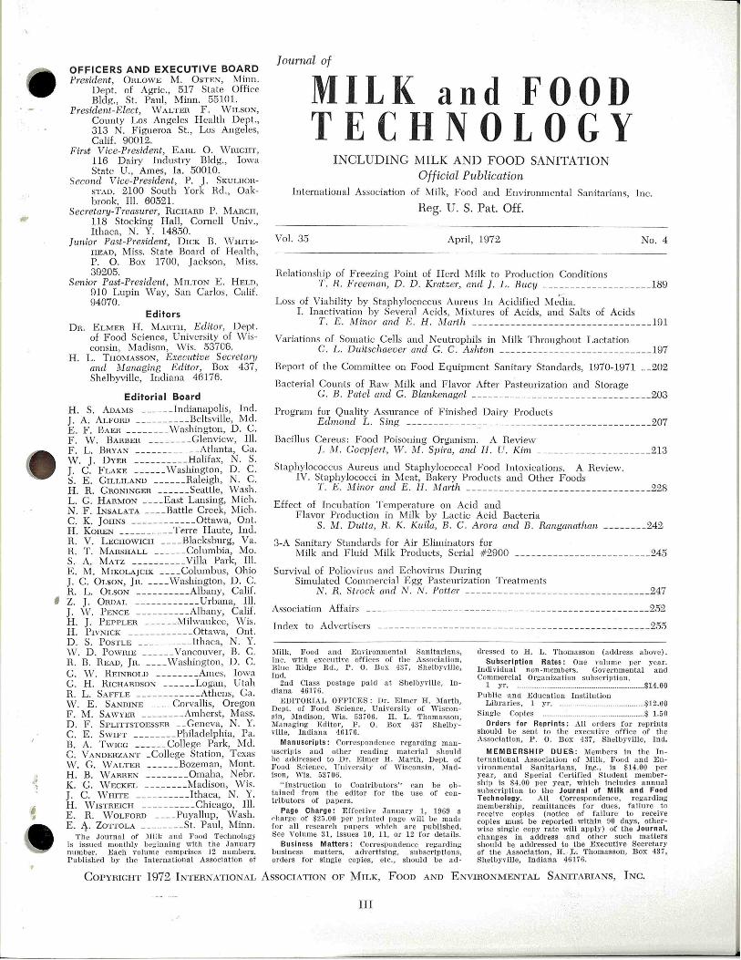
Vol. 35 April , 1972 No.4
Relationship of Freezing Point of H erd Milk to Production Conditions T . R. FTeeman, D. D. KratzeT, t!nd ]. L. Bucy ___ ___ ____ _____ _____ 189
Loss of Viability by Staphylococcus Aureus In Acidified Me.~lia. I. Inactivation by Severa l Acids, Mixtures of Acids, and Salts of Acids
T . E. Minor and E . H. Mwth ---------------------------------191
Variations of Somatic Cells and Neutrophils in ]Vlilk Throughout Lactation C. L. Du:itschaever and G . C. Ashton -- ----- ------- ----- ------- --197
Heport of the Committee on Food Equipment Sanitary Standards, 1970-1971 __ 202
Bacterial Counts of Raw Milk and F lavor After Pasteurization and Storage G. B . Patel and G. Blankenagel --- -- ----- --- - --------------- ----203
Program for Quality Assurance of Finished Dairy Products Edmond L. Sing _____ ___ __ ___ ~ --- ______ ______________________ 207
Bacillus Cereus : Food Poisoning Organism. A Review ]. M. Goepfett, W. M. Sp im , and H . U. Kim ___________ _____ ____ _ 213
Staphylococcus Aureus and Staphylococcal Food Intoxications. A Review. IV. Staphylococci in Meat, Bakery Products and Other Foods
T. E. 1H·inoT and E. H. i\tlarth ---------- ---- --------------------228
Effect of Incubation T emperature on Acid and Flavor Production in Milk by Lactic Acid Bacteria
S. M. Dutta, R. K Kuila, B. C. Amra and B. Ranganathan ________ 242
3-A Sanitary Standards for Air Eliminators for Milk and F luid lv!ilk Products, Serial # 2900 - - ----- -- ----------------245
Survival of Poliovirus and Echovirus Dming Simulated Commercial Egg Pasteuriza tion Treatm ents
N. R. Strock and N . N. Potter ------- -- ---------------- - -- - - --- -247
Association Affairs _____ -------- ----- - - - ----------- --- ______ ___ ________ 252
Index to Advertisers --- __ ---- __ __ ---- -- -- ____ ----- - -- ______ _____ ______ 255
l\Iilk, Food and En vironmenta l SanHarirrns, Inc. with executive offi ces of the Assoc i ation ~ Blue Ridge Rcl. , P . 0. Box 43 7, Shelbyville, Ind.
2nd Class postage pa.icl at Shelbyville, Indiana 46176.
EDITORIAL OFF ICES : Dr. Elmer H. Marth, Dept. of Food Science, Unive rsity of Wiscon!!- in, :Madison , 'Vis . 53i 06. H . L . 'l1homasson , ~Iana ging Editor, F. 0. Box 437 S helbyville, Indiana 46176.
Manuscripts: Correspond ence r ega rding man uscripts and other readin g material should be addressed to Dr. Rimer H. Marth, Dept. of Food Science, Uni,·ersity of \Yi sconsin, Madison, ' Vis. 53 70 6.
" Instruction to Contributors'' ca n be obta ined from t he editor for !he use of contributors of papers.
Page Charge: Effective J anua.ry 1, 1969 a ('ha.r~e of $25.00 per print ed page will be made for a ll research ]lapers whi ch are publ ished. See Volume 31, issues 10, 11, or 12 for deta il s.
Bu sin ess Matters: Co rrespondence regard ing business matters, advertising, subscriptions, orders for s ingle copies, etc., shou ld be a d -
dressed to H. L. Thomasson (address above). Subscription Rates: One volum e per year.
IncUvidual non-members . Governmental and Comm er.cial Organization subscription.
1 yr. . .............. ............ ....... .............. .... ........ $14.00 Publi c ·and Education In stitution
Lib raries, 1 yr . ..... .. ............................ :uz.uo S ingle Copies ... .. ......................... $ 1.50
Orders for Repr ints : All orders for reprints shou ld be sent to t he executire office of the Association, P . 0. Box 437, Shelbyv ille, In cl.
MEMBERSHIP DUES : illembers in the Interna ti onal Associa ti on of :i\Iill\ , Food and Environm ental Sanitarians, In,c., is $14.00 per year, and Special Certifi ed Student membership is $4.00 per year, which includ es annual subscript.ion to t he Journ a l of · Milk a nd Food Tech nology. All Correspondence, r egarding membership, remittances for du es, failure to receive copies (notice of failure to receive copies must be r epor ted within 90 days, otherwise s ingle copy rate will apply) of the Journ a l, cha.nges in address and other such matters should be addressed to tl1e Executive Secretary of the Association, H. ;L. Thomasson, Box ll 37J Shelbyv ille, Inclia na 46176.
CoPYRIGHT 1972 INTERNATIONAL AssociATION OF MILK, Foon AND ENVIRONMENTAL SA.t'ITTARIAN"S, INc.
III
.1'
METHODS FOR PRODUCTION OF HIGH QUALITY RAW MILK
(A Summary of Annual Reports Prepared From 1955 .to 1970 by the IAMFES Dairy Farm Methods Commitee)
COMPILED AND EDITED BY
J. C. FLAKE, A. E. PARKER, J. B. SMATHERS, A. K. SAUNDERS AND E. H. MARTH
PUBLISHED BY
INTERNATIONAL ASSOCIATION OF MILK, FOOD, AND ENVIRONMEN,TAL SANITARIANS, INC.
COPIES OBTAINABLE FROM
International Association of Milk, Food, and Environmental Sanitarians, Inc.
REVISED 19'66
EDITION
Box 437, Shelbyville, Indiana 46176
Prices: Single Copies $2.00 each-25-100 Copies $1.75 each, 100 or More Copies $1.50 each
Procedure for
The Investigation of
Foodborne Disease Outbreal{_s
Recommended By
INTERNATIONAL ASSOCIATION OF MILK, FOOD AND ENVIRONMENTAL SANITARIANS, INC.
COPIES OBTAINABLE FROM
REVISED 19'66
EDITION
International Association of Milk, Food and Environmental Sanitarians, Inc. Box 437, Shelbyville, Indiana 46176
Prices: Single Copies, $1.00 each: 100 or more copies, 65 cents each. 25-100 copies, 75 cents each. Please do not send stamps.
IV
•· I
•
I
·~
]. Milk Food Technol., Vol. 35, No. 4 (1972) 189
RELATIONSHIP OF FREEZING POINT OF HERD MILK TO PRODUCTION CONDITIO~NS'
T. R. FREEMAN, D. D. KRATZER, Ar-.ro J. L . BucY
Department of An imal Sciences University of Kentucky, Lexington 40506
( Heceived for publi cation October 8, 1971 )
ABSTHACT
A statistically significant relationship was found between freezing point and abnospheric temperatme, season, roughage in th e ration, breed of cow, and concentrate in the ration. Because of the narrow range in freezing points, however, the practical importance of these relationsh ips appears questionable.
Results of a comprehensive suTvey of the freezing point of herd milk produced in Kentucky were presented in a previous report (1). Methods of sampling and analysis were described in that report. The average freezing point from 509 herd samples of milk collected from five soil areas of the state over a period of one year was -0.541 C. _ In connection with the above smvey information
was rfcorded at each sampling, to describe environmental, feeding, and other conditions within the herd that might influence freezing point of the milk of that herd. Factors which were analyzed statistically for relationship to milk freezing point included: (a) breed, (b) concenh·ates in the ration, (c) roughages in the ration, (d) environmental temperature, (e) rainfall, (f) level of management employed (above or below average) as judged by the Heldman, (g) fat test of herd milk, and (h) herd milk yield. Temperature and rainfall data were obtained from U. S. weather station records.
RESULTS AND DISCUSSION
Data in Table 1 show correlations of milk freezing point with fat test, milk yield, and previous high temperatme and rainfall. Milk freezing point, fat test, and milk yield were obtained from pooled samples of the evening and morning milk of each herd. Average high temperature and total rainfall were from the 3-day period preceding the morning mi!king.
High temperatme · was positively correlated with milk freezing point because days with higher temperature were associated with higher milk freezing points (nearer zero). A physiological explanation
'The investigation repotted in this paper (No. 71-5-121~ is in connection with a project of the Kentucky Agric,\tltmal Experin1ent Station and is published with approval of the Director.
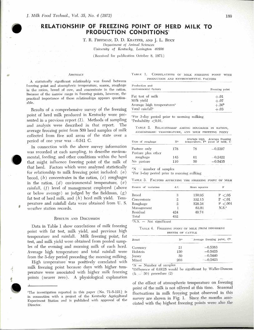
TABLE l. CoRHELA TIONS OF l\IILK FHEEZING POINT WITH
PHODUCTION AND ENVIHONJ\,lENTAL FACTORS
P rodu ction and en\·ironmental factors J?reezing point
Fat t es t of milk Milk yield Average high temperatures" Total rainfall•
"For 3-day period prior to morning milking "Probability <0.01.
+.01 +-07 +.3Qb +.03
TABLE 2. HELATIONSH!P AMONG ROUGHAGE Ul RATION,
ATl\lOSPHEHIC TEl\!PEl1ATUHE, AND MILK FHEEZIN G POINT
Average high A\·erage freezing Type of roughage 1\ tl temperature, Fb po in t of milk, c
Pasture only 178 78 -0.5397 Pasture plus oth er
roughage 145 61 -0.5422. No pasture llO 50 -0.5435
"N = number of samples "For 3-day period prior to morning milking
TABLE 3. FACTOHS AFFECTL'IG THE FHEEZING POL'IT OF MILK
Source of variation d.t'. ~lean squares F
Breed 3 150.95 p < .05 Concentrate 2 332.15 p < .01 Houghage 2 538.34 p <.001 Management 1 63.81 N.S.• Hesidual 424 49.74 Tota l 432.
"N.S . = Not significant
TAilLE 4. FHEEZING POINT OF MILK _FHOM DIFFEHENT
DHEEDS OF CATTLE
B reed N• Average freezing point, Cb
Guernsey 21 -0.5383 Holstein 150 -0.5425 Jersey 59 - 0.5440 Mixed 203 ' -0.5423
Ntunber of samples "Difference of 0.0025 would be significant by ' iValler-Duncan ( k =· 50) procedure (2j
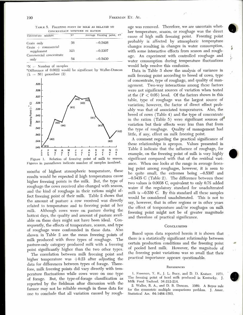
of the effect of atmospheric temperature on freezing point of the milk js not ~~ered at this time. Seasonal fluctuations in milk fre~~ing point observed in this survey are sh_9vvn in Fig. 1. Sinc~ _:;the months associated 'vith thee highest freezing points were also th,e
.J'
190 FI'IEEMAN ET. AL.
TABLE 5. FREEZING POINT OF MILK AS RELATED TO
CONCENTRATE l\HXTURE IN RATION
Concentra te mixture J\:t A \'Crage freezing point,
Grain only 56 -0.5426
Grain + commercial supplement 323 - 0.5397
Commercial concentrate only 54 - 0.5430
" T = lumber of samples
Cb
"Difference of 0.0022 would be significant by Waller-Dtmcan ( k = 50) procedure (2)
.535
.536
.537
u .538
_I_ .539 ~
c: '(5
.540 0..
"' c: ·;;
.541 ., ., u:
.542 (43)
. 543
.544
FMAMJJ ASOND a e a p a uu u e c oe n b r r y nl g p tv c
F igure 1. Relation of freezing point of milk to season. Figures in parentheses indicate number of samples involved.
months of highest atmospheric temperature, these results would be expected if high temperatures cause higher freezing points in the milk. But, the type of roughage the cows received also changed with season, and the kind of roughage in their rations might affect freezing point of their milk. Table 2 shows that the amount of pasture a cow received was directly related to temperahue and to freezing point of her milk. Although cows were on pasture during the hottest days, the quality and amount of pasture available on these days might not have been ideal. Consequently, the effects of temperature, season, and type of roughage were confounded in these data. Also shown in Table 2 are the mean freezing points of milk produced with three types of roughage. The pasture-only category . produced milk with a freezing point significantly higher than the two other types.
The correlation between milk freezing point and higher temperature was +0.23 after adjusting the data for differences between types of forage. Therefore, milk freezing points did vary directly with temperature fluctuations while cows were on one type of forage. But, the type-of-forage classification as reported by the Heldman after discussion with the farmer may not be reliable enough in these data for one to conclude that all vruiation caused by rough-
age was removed. Therefore, we are uncertain whether temperature, season, or roughage was tl1e direct cause of high milk freezing point. Freezing point probably is affected by atmospheric temperr tme changes resulting in changes in water consumption, w!th some interactive effects from season and roughage. An experiment with controlled roughage and water consumption dming temperature fluctuations would help resolve this confusion.
D ata in Table 3 show tl1e analysis of variance in milk freezing point according to breed of cows, type of concentrate, type of roughage, and quality of management. Two-way interactions among tl1ese factors were not significant somces of vru·iation when tested at tl1e (P < 0.05) level. Of the factors shown in this table, type of roughage was the largest source of vru·iation; however, the factor of direct effect probably was that of associated temperatures. Also, the breed of cows (Table 4) and the type of concenh·ate in tl1e ration (Table 5) were significant sources of" vru·iation but their effects were less than that from the type of roughage. Quality of management had little, if any, effect on milk freezing point .
A comment regarding the practical significance of these relationships is apropos. Values presented in Table 2 indicate that the influence of roughage, for example, on tl1e freezing point of milk is very highly significant compared with that of the residual variance. ·when one looks at the range in average freezing point a.mong roughages, however, it is seen to be quite small, tl1e exh·emes being -0.5397 and -0.5435 C (Table 2). The difference between these 1:\vo values is 0.0038 C, equivalent to only 0.7% added water if tl1e regulatory . standard for unadulterated milk is -0.530 C. By tl1is standard all these samples would be considered unadulterated. This is not to say, however, that in other regions or in other years the effect of temp erature and/or roughages on milk freezing point might not be of greater magnitude and therefore of practical significance.
CONCLUSIONS
Based upon data reported herein it is shown that there is a statistically significant relationship between certain production conditions and the freezing point of pooled herd milk. However, the magnitude of the freezing point vru·iations was so small that their practical importance appears questionable.
R EFERENCES
1. Freeman, T. R., J. L. Bucy, and D. D . Kratzer. 1971. The freezing point of herd milk produced in Kentucky. J. Milk Food Techno!. 34:212-214.
2. Waller, R. A., and D . B. Dtmcan. 1969. A Boyes rule for · the symmetric multiple comparisons problem. J. Amer. Statistical Ass. 64:1484-1503.
, , ( .. I

1.
I
•' )
' '
• '
]. Milk Food T echnol., Vol. 35, No. 4 (1972) 191
LOSS OF VIABILITY BY STAPHYLOCOCCUS AUREUS IN ACIDIFIED MEDIA
I. INACTIVATION BY SEVERAL ACIDS, M IXTURES OF ACIDS, AND SALTS OF ACIDS'
T. E. MINOR AND E. H. MARTI!
Department of Food Science and The Food Research Institute Uni:versity of Wisconsin, Mad·ison, Wisconsin 53706
( Received for publication December 10, 1971)
ABSTHACT
Survival of Staphylococcus au1·eus ( 108 cells per milliliter) after 24 lu- of incubation at 37 C in Trypticase Soy broth acidified with acetic, citric, hydrochloric, lactic, and phosphoric acids was investiga ted. V/hen the organism was exposed to the medium adjusted with hydrochloric acid to pH values of 5.2-3.6, 90-99.99% of the cells were inactivated. Acetic, lactic, and phosphoric acids were more active against S. aureus than was hydrochloric, whereas citric was equivalent to hydrochloric. Mixtures of lactic and h ydrochloric acids inactivated more cells than did either acid alone but mixtures of other acids with hydrochloric, whil e superior to hydrochloric acid itself, offered no apparent advantage over use of single acids. The undissociated acid molecule was responsible for enhanced inactiva tion of cells by partially dissociated acids since anions of th ese acids had no effect on cell survival. Cells were more susceptible to inactivation by hydrogens ions at high incubation t emperatures ( 45 C) and when the number of bacteria was low. Cells of S. aureus were most sensitive to the effects of hydrogen ions between the 12th and 24th hr during a 120-hr incubation .
In the last fe"v decades there has been limited interes t in the survival of bacteria in acidic environments . The food indush·y, however, cannot afford to overlook tllis matter, for virtually all foods are more or less acidic in nah1re. Staphylococcus aureus and staphylococcal food intoxications, for example, have been associated with fermented foods (2, 3, 4, 5), particularly when acid production during manufacture was inadequate. The rate and amount of acid development in such foods govems, in part, whether staphylococci, if present, will grow and produce enterotoxin. A direct relationship between subnormal acid development and production of enterotoxin was noted by Zehl'en and Zelu·en (9, 10) when they tested cheeses involved in a rather large outbreak of staphylococcal food poisoning. W e studied the behavior of S. au.reus in pasteurized milks which were gradually adjusted to various levels of acidity (1) and reported substantial differences in growth rates, depending on the rate of acid addition and the type of acid employed . Growth of Salmonella typhi-
'Supported by the Coll ege of Agricultural and Life Sciences, University of Wisconsin, Madison, and by Public Health Service Grant No. FD00009-05 from the Food an d Drug Admini~tration .
muri·um in skimmilks similarly acidified over a period of time with several acids was investigated by Subramanian and Marth (6) who noted that differences in inhibition were dependent on the type of acid used. vVe have also examined the fate of S. aureus when added to market samples of cultured dairy products (unpublished data) and observed their rapid denlise within relatively short periods of time.
The present study is concemed with inactivation of S. atweus in an acidic environment. This communication deals with inactivation of S. aU1'eu.s in a Trypticase Soy broth containing separately each of five different acids, miA·tw:es of these acids, and salts of the acids.
MATERIALS AND METHODS
Media Basal medi.tvm. Dehychated Trypticase Soy (TS) broth
( BBL, BioQuest) was reconstituted according to the manufacturer's directions and autoclaved ( 121 C, 15 min ) in 30 ml quantities in 25 X 150 mm screw capped tubes. The pH of the sterilized mediwn ranged between 7.0 and 7.1.
Treated media. Solutions of the following acids were prepared and autoclaved ( 121 C, 15 min) in 10 ml quantities: 2 M acetic, glacial, A. C. S. reagent (Allied), 1 M citric, granular, analytical reagent ( Mallinckrodt); 2 M hydrochloric, reagent (DuPont); 2 M lactic, USP 85% ( Mall inckrodt) ; 2 M phosphoric ( ortho), A. C. S. reagent (Allied). Solutions of the following salts were prepared in a similar manner: 2 M sodium aceta te, anhydrous ( Mallinckrodt); 1 M sodium citrate (Fisher ); 1 ~·il calcium lactate, NF powder ( Mallinckrodt); 2 M sodium phosphate, monobasic, crystal (Allied); and, 1 M sodium phosphate, dibasic, anhydrous powder (Baker ). Appropriate quantities of the above solutions were aseptically added to the previously prepared 30 ml quantities of TS broth. The volume of solutions added ranged between 0.075 and 1.35 ml per 30 ml of broth.
Culture A cultme of S. aureus strain 100 was obtained from Dr. K.
F. \•Veiss (The Food Research Institute, University of Wisconsin ) . The organism was stocked on TS agar slants, refrigerated tmtil used, and res t0cked every 2 to 3 months. Before each e;~:periment, the organism was transferred from a stock slant to 10 ml of TS broth and incubated 24 hr at 37 C . F inally, a loopful was transferred to 16 X 125 mm screw capped tubes containing 10 ml of TS broth (two tubes were inoculated for each variable under study) and incubated 18 hr at 37 C (resulting in populations of approximately 10" cells per milliliter) .

192 MINOR AND MARTI-I
~4r--------.6-------~-----------------o
I
~
o ACETIC e CITRIC D HYDROCHLORIC 6 LACTIC A PHOSPHORIC
Figure 1. Inactiva tion of S. aum us after 24 hr of iilcilbation at 37 C in a Trypti case Soy broth ( BBL ) acidified with acetjc, citric, hydrochloric, lactic, and phosphoric ( ortho) acids (cell i.J1ac tivation expressed as a function of mrviolar concentration). Initial cell populatiml = 108 p er milliliter.
o ACETIC • CITRIC 0 HYDROCHLORIC 6 LACTIC A PHOSPHORIC
5.5 5 .0 INITIAL pH
4.5 4 .0 3.5
F igure 2. Inactivation of S. awreus after 24 hr of mcubation at 37 C m a Trypticase Soy broth acidified with acetic, cib·ic, hydrochloric, lactic, and phosphoric ( ortho) acids (cell ii1activa tion expressed as a function of pH ). Initial cell population = 108 p er milliliter.
Exposure of staphylococci, to t·reatecl m edia The 18-hr old cultures were cooled to 10 C and then were
centrifuged at about 4000 rpm for 5 mill . Supernatants were removed with sterile Pasteur pipettes and replaced, aseptically, with 10 ml of the appropriately treated broth ( 20 ml of a treated broth were thus used for each variable, leavmg 10 ml for a pH determination ) . Staphylococcal cells were resuspended by means of a Vortex mixer and the tubes were
incubated statically for 24 hr at 37 C. \•Vhen the staphylococci were resuspended in untreated broth (control ) , their numbers remained fairly constant (approximately 108 per milliliter ) during the 24-hr incubation period . Occasionally, some growth was observed in the control and during! one experiment numbers of the organism reached 9 X 10⁸ per milliliter .
Determ i.nation of cell survival A 1.0 ml aliquot was asepticall y removed from each tube
after the contents were mixed with a Vortex mLxer, and pour plates were prepared according to recommendations of Stan danl Methods for the Eunn:i.nation of Da·iry Products (8) . Plates were poured with TS agar and incubated at 37 C for 48 hr. An average count was calculated for the duplicate tubes of each variable and control. Each variable was compared to its control and the mm1 ber of logs difference was determined .
Changes ·in e~per i:mental pammeters Several parameters were altered to test their effect on
survival of S. aureus in acidified TS broths.
Tim e of exposum. An ii1oculum of 108 cells of S. aureus per milliliter was exposed for various time intervals to TS , broth containmg 40 mM hydrochloric acid. Survival of the organism and pH of th e mediwn were determi.J1ed after 0, 4, 8, 12, 24, 48, 72, 96, and 120 hr at 37 C.
In cubation temperatu·re. An inoculum of 108 cells of S. a.ureus per milliliter was treated at 10, 23, 30, 37, and 45 ·c in TS broth for 24 hr with 40 mM h ydrochloric acid.
In oculum. The inoculum was varied by making several 1: 100 dilutions of th e 18. hr broth culture in TS broth ( 0.1 ml of broth culture was added to 10 ml of fresh untreated or treated broth, the contents mixed, and 0.1 ml transfer.:ed to another tube of broth , etc.). Inocula of 108
, 106, 10\ and
102 cells per milliliter were prepared. Two concentrations of hydrochloric acid, 30 and 50 mM, in th e broth were used in these tests.
RESULTS
Inactivat·ion of S. aureus by. single acids Figures 1 and 2 depict inactivation of S. aureus by
five different acids in TS broth as a function of mMolar concentration and pH, respectively.
I-nact-ivat-ion as a function of mM alar concentration. When the acids are ranked in order of decreasing activity (as a function of mMolar concentration), they form the series: lactic > cibic > phosphoric > hydrochloric > - acetic. The acids can be arranged into three groups according to their performance, with lactic and acetic acids at the exb·emes and the remaining tlU"ee acids occupying an intermediate position. A concenb·ation of 10 mM lactic acid caused no detectable change in cell population, whereas an increase to 25 mM resulted in a four-log (99.99%) decrease in number of viable cells. In conb·ast, 30 mM acetic acid had no effeot on cell numbers and a 90 mM concenb·ation was required to achieve a four-log reduction in population. Citric, phosphoric, and hydrochloric acids differed only slightly from one another (the former two were slightly superior to tl1e latter one). A 50 mM concenb·ation of hydro-
\ .. I
'

,
I
·~
Loss OF VIABILITY 193
chloric acid was required to cause a four-log de
crease in staphylococcal population.
Inactivat·ion as a function of pH. Below pH 4.8,
the acids can be arranged into nvo groups according
to their activity (as a function of pH ) against staphy
lococci. Acetic, lactic, and phosphoric acids as a
group were more effective than the other n\'0 acids
(at lower pH values, phosphoric was inferior and
acetic superior to lactic acid). Cih·ic and hydro
chloric acid were almost identical to each other in
perfom1ance across the entire pH spectrum and as
a group were inferior to the other acids (at lower
pH values, hydrochloric was slightly inferior to citric ).
Acetic acid had no demonstrable effect on num
bers of staphylococci at pH 5.3. In conh·ast, cihic
and hydrochloric acids siightly affected cell sur
vival at a pH just below 7, vvhereas tl1e deh·imental
effect of lactic and phosphoric acids was first detect
ed betv.reen pH 6.0 and 6.5. For practical pmposes,
none of the acids substantially inactivated ( > 90%)
S. aum11s above pH 5.7. Above pH 4.7, cell inactiva
tion · did not exceed 99% for any of the acids. Acetic
TABLE l. SURVIVAL OF S. aureus AFTER 24 I·lli OF INCUBATION
AT 37 C I N TRYPTJCASE Soy BHOTH ACIDIFIED WITH MIXTURES
OF HYDHOCHLOIUC AND OTH E H ACIDS. 1
mi\Iolar Other acid Reduction in con en ml\Iolar population H CI Type con en pH (no. logs) 2
20 none 5.8 0.22
20 acetic 15 5.1 1.78 20 5.0 0.67
20 citric 15 4.5 2.44 20 4.3 3.56
20 lactic :!.5 4.4 5.22 20 4.2 5.44
20 phosphoric 15 4.7 1.00 20 4.5 2.33
40 none 4.1 2.22
40 acetic 5 4.0 4.11 10 4.0 3.78 15 3.8 5.11 20 3.9 3.89
40 ci tric 5 3.8 2.89 10 3.6 3.56 15 3.3 5.56 20 3.3 5.56
40 lactic 5 3.8 5.89 10 3.7 7.00 15 3.4 8.00 20 3.3 8.00
40 phosphoric 5 3.9 4.11 10 3.7 4 .11 15 3.4 6.00 20 3.1 7 .00
' Initial cell population = 108 per milliliter 2Compared to a control ( no acids added)
acid inac tiva ted 99.99% of the cells at pH 4.4, where
as a pH of < 3.6 was required to achieve similar re
sults w ith hydrochloric acid.
Inactivation of S. aureus by mixtures of hydrochloric
and otheT acids Inactivation of S. aureus in media acidified with
mixtmes of hydrochloric and other acids is detailed
in Table 1. Mixtures of hydrochloric and lactic
acids were substantially more effective than were
those of other acids combined with hydrochloric
acid . iVIixtures of hydrochloric acid and acetic, cit
h·ic, and phosphoric acids were, for practical purpos
es, equal in effectiveness when compared to each
other on the basis of pH.
All mixtures of hydrochloric and lactic acid were
more effective against staphylococci at a given pH
than either acid alone (see Fig. 2) at the same
pH v:1lue. A 40 mM hydrochloric plus 15 mM
lactic acid mixture (pH 3.4) was completely leth
al (an eight-log decrease in numbers ) to S. aureus.
Mixtures of other acids with hyclrochloric were, for
the most part, .'equivalent to or more -effective than
hydrochloric a·cid alone when compared at the
same pH. In many instances, the data indicated
that effectiveness of acetic, citric, or phosphoric
acids was impaired when combined with hydrochloric
acid. In all instances, the pH of the _ mixh1re of
acids was lower than the pH would have been if
the acids were used alone.
Behavior of S. atl'reus in media contain:ing salts of
acids a·nd ac·icl salts p l11 s hydrochlor-ic acid.
Table 2 provides data on the behavior of S. au-reus
in the presence of acid salts and mixtures of these
salts with hydrochloric acid in TS broth. The salts
were evaluated at concenh·ations equivalent to those
of the resp ~cti ve acids which inactivated S. aureus
(Fig. 2 ) . Acetate and cih·ate had no effect on the
pH of the medium, whereas lactate and monobasic
phosphate h1rned it slightly acidic, and dibasic phos
phate slightly alkaline. Survival of staphylococci was
unaffec ted by either of tl1e phosphate salts. Cih·ate
caused a slight decline in numbers of the organism
and acetate and lactate brou ght about a small in
crease.
Mixtures of acetate, cih·ate, and dibasic phosphate,
with 40 mM hydrochloric acid in TS broth were
higher in pH than media containing just 40 - m :~-.'1
acid ( the ::mions, tl1erefore, must have combined
with some of the hydrogen ions and formed undis
sociated acid ). :Mi>..·tures of lactate and monobasic
phosphate with acid in TS brotl1 were equivalent- in
pH to media containing acid alone. Vilhen. the pH
of the medium was increased by the presence .oj the
salt, cell inactivation was almost completely prevent"
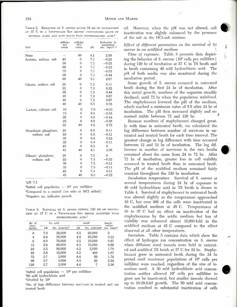
.1'
194 MINOR Al\'D MARTH
TABLE 2. BEHAVIOR OF S. au:reus AFTER 24 HR OF INCUBATION
AT 37 C IN A TRYPTICASE SoY BROTH 1 CONTAINING SALTS OF
SEVERAL ACIDS AND ACID SALTS PLUS HYDHOCHLOHIC ACID!
mMolar mMolar R eduction in salt HCl population
Sa lt con en con en pH (no. logs) 3, 4
None Acetate, sodium salt
Citrate, sodium salt
Lactate, calcitm1 salt
Monobasic phosphate, sodium salt
Dibasic phosphate, sodium ·salt
'pH 7.1
40 50 60 75 90 90
20 25 30 35 40 40
10 15 20 25 25
20 25 30 35 45 45
20 25 30 35 45 45
40 4.1 0 7.1 0 7.1 0 7.1 0 7.1 0 7.1
40 5.1
0 7.2 0 7.2 0 7.2 0 7.2 0 7.2
40 5.5
0 7.0 0 6.9 0 6.9 0 6.8
40 4.2
0 6.5 0 6.5 0 6.4 0 6.4 0 6.3
40 4.1
0 7.2 0 7.3 0 7.3 0 7.3 0 7.4
40 6.3
'Initial cell population = 108 per milliliter 3Compared to a control ( no salts or HCl added) 4Negative no. indicates growth
2.00 - 0.22 -0.22 - 0.22 -0.33 -0.44
0.67
0.11 0.22 0.44 0.67 0.67 0.78
- 0.33 - 0.33 -0.44 -0.56
2.78
0.11 -0.11
0.22 - 0.11
0 2.56
0 - 0.22 -0.11 - 0.11
0.11 -0.22
TABLE 3. SURVTVAL OF S. aureus DURING 120 HR OF INCUBA
TION AT 37 C IN A 'TRYPTICASE SOY BROTH ACIDIFIED W ITH
HYDROCHLORIC ACID.1
Hr of No acid Acid• Inacti-lncu- vation bation pH No. cells/ml3 pH No. cells/ml3 (no. logs)•
0 7.0 20,000 4.2 20,000 0 4 6.6 50,000 4.4 20,000 0.33 8 6.0 70,000 4.2 10,000 0.67
12 5.8 60,000 4.3 10,000 0.56 24 5.5 90,000 4.3 2,000 1.78 48 5.6 10,000 4 .2 400 1.67 72 5.7 3,000 4.4 50 1.78 96 5.7 4,000 4 .4 20 2.22
120 5.7 3,000 4.4 7 2.56
1Initial cell population = 108 per milliliter "40 mM hydrochloric acid 3Divided by 10•
•No. of logs difference between surv:vors in treated and un~ treated broth
ed. However, when the pH was not altered, cell inactivation was slightly enhanced by the presence of the salt in the HCl-salt mixture.
! Effect of diffeTent parmneters on the survival of S.l auTeus in an acidified medit~.m !..
Time of exposure . Table 3 presents data depict~ ing the behavior of S. atiTe'LIS ( 10" cells per milliliter ) dming 120 hr of incubation at 37 C in TS broth and in broth containing 40 mM hydrochloric acid. The pH of both media was also monitored dming the incubation period.
Some growth of S. aureus occmred in untreated broth dming the first 24 hr of incubation. After this initial growth, numbers of the organism steadily declined, until 72 hr when the population stabilized. The staphylococci lowered the pH of the medium, which reached a min·imum value of 5.5 after 24 hr of incubation. The pH then increased slightly and re-mained stable bet\veen 72 and 120 hr. '
Because numbers of staphylococci changed greatly with time in untreated broth, we calculated the log difference bet\veen number of survivors in untreated and treated broth for each time interval. The greate·st change in log difference with time occmred bet\veen 12 and 24 hr of incubation. The log difference in number of suTVivors in the 1:\vo broths remained about the same from 24 to 72 hr. After 72 hr of incubation, greater loss in cell viability occurred in treated broth than in untreated broth. The pH of the acidified medium remained fairly constant throughout the 120 hr incubation.
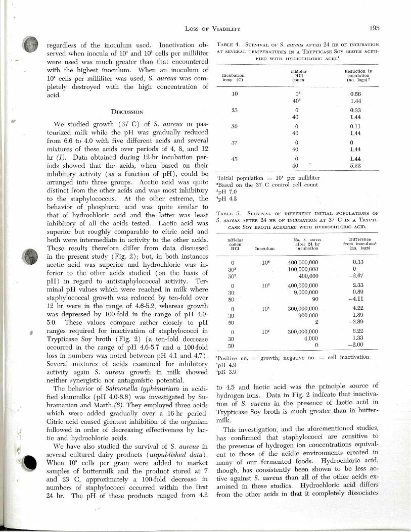
Incubation temperature. Survival of S. aureus at several t emperatures dming 24 hr of exposure to 40 mM hydrochloric acid in TS broth is shown in Table 4. Survival of staphylococci in untreated broth was altered slightly as the temperature approached 10 C, but over 90% of the cells were inactivated in the acidified medium at 45 C. Temperatures of 10 to 37 C had no effect on inactivation of the staphylococcus by tl1e acidic medium but loss of viability was enhanced almost 10,000-fold in the acidified medium a.t 45 C compared to the effect observed at all other temperatures.
Inoculum. Table 5 contains data which show the effect of hydrogen ion concentration on S. aureus when different sized inocula were held in untreated and acidified TS broth at 37 C for 24 hr. Staphylococci grew in unb·eated broth during the 24 hr period unotil maximum populations of 10' cells per milliliter were reached, regardless of the size of inoculum used. A 30 mM hydrochloric acid ccmcentnttion neither allowed 108 cells p er milliliter to grow nor be inactivated, but smaller inocula showed up to 10-20-fold growth. The 50 mM acid concenb·ation resulted in substantial inactivation of cells
, , I .. I
I
·' )
'
Loss OF VIABILITY 195
regardless of the inoculum used. Inactivation observed when inocula of 10• and 10" cells per milliliter were used was much greater than that encountered with the highest inoculum. When an inoculum of 10" cells per milhliter was used, S. aureus was completely destroyed with the high concentration of acid.
DISCUSSION
We studied growth ( 37 C) of S. au reus in pasteurized milk while the pH was gradually reduced from 6.6 to 4.0 with five different acids and several mixtures of these acids over periods of 4, 8, and 12 hr (1). Data obtained during 12-lu· incubation periods showed that the acids, when based on their inhibitory activity (as a :fun:ction of pH), could be an-anged into three groups . Acetic acid was quite distinct from the other acids and was most inhibitory to the staphylococcus. At the other extreme, the behavior of phosphoric acid was quite similar to that of hydrochloric acid and the latter was least inhibitory of all the acids tested. Lactic acid was superior but roughly comparable to cih·ic acid and both were intermediate in activity to the other acids. TI1ese results therefore differ from data discussed in the present study (Fig. 2); but, in both instances acetic acid was superior and hydrochloric was inferior to the other acids studied (on the basis of pH) in regard to antistaphylococcal activity. Terminal pH values which were reached in milk where staphylococcal growth was reduced by ten-fold over 12 hr were in the range of 4.6-5.2, whereas growth was depressed by 100-fold in the range of pH 4.0-5.0. These values compare rather closely to pH ranges required for inactivation of staphylococci in Tryptica:se Soy broth (Fig. 2) (-a ten-fold decrease occun-ed in the range of pH 4.6-5.7 and a 100-fold loss in numbers was noted between pH 4.1 and 4.7). Several mixhrres of acids examined for inhibitory activity again S. aumus growth in milk showed neither synergistic nor antagonistic potential.
The behavior of Salnwnella typhimu1·ium in acidified skimmilks (pH 4.0--6.6) was investigated by Subramanian and Marth (6). They employed three acids which were added gradually over a 16-hr period. Citric acid caused greatest inhibition of the organism followed in order of decreasing effectiveness by lactic and hydrochloric acids .
We have also studied the survival of S. aureus in ~everal culhrred dairy products (unpublished data). When 10' cells per gram were added to market samples of buttermilk and the product stored at 7 and 23 C, approximately a 100-fold decrease in numbers of staphylococci occun-ed "vithin the first 24 hr. The pH of these products ranged from 4.2
• r
TABLE 4. SURVIVAL OF S. a:tt1'eUS AFTER 24 HR OF INCUBATION
AT SEVERAL TEl\•1PEHAT1mES IN A THYPTICASE SoY BHOTH ACIDI
FIED WITH HYDROCHLORIC ACID!
mMolar Incubation HCt temp (C) con en
10 oa 40'
23 0 40
30 0 40
37 0 40
45 0 40
'Initial population = 108 per milliliter 2Based on the 37 C control cell count 3pH 7.0 'pH 4.2
Reduction in population (no . logs) 2
0.56 1.44
0.33 1.44
0.11 1.44
0 1.44
1.44 5.22
TABLE 5. SURVIVAL OF DIFFERENT INITIAL POPULATIONS OF
S . au:reus AFTER 24 HR. OF INCUBATION AT 37 C IN A TRYPTI
CASE SOY DHOTH ACIDIFIED WITH HYDHOCHLORIC ACID.
ml\Iolar No. S. aureus Difference con en after 24 hr from inoculum! HCt Inoculum in cubation (no . togs)
0 108 400,000,000 0.33 302 100,000,000 0 503 400,000 -2.67
0 106 400,000,000 2.33 30 9,000,000 0.89 50 90 -4.11
0 10'' 300,000,000 4.22
30 900,000 1.89 50 2 -3.89
0 10' 300,000,000 6.22
30 4,000 1.33
50 0 -2.00
'Positive no. growth; negative no. = cell inactivation
'pH 4.9 3pli 3.9
to 4.5 and lactic 'acid was the principle source of hydrogen ions. Data in Fig. 2 indicate that inactivation of S. aureus in the presence of lactic acid in Trypticase Soy broth is much greater than il).- buttermilk.
This investigation, and the aforementioned sh1dies, has confirmed that staphylococci are sensitive to the presence of hydrogen ion concentrations equivalent to those of -the acidic environments created in many of our fermented foods. Hydrochloric acid, though, has consistently been shown to be less active ag-ainst S. aU?·eus than all of the other acids examined in these s-tudies. Hydrochloric acid differs from the other acids in that it completely dissociates
.;'
196 Mmon 1\1\'D MARTH
into ionic substances in aqueous media and its anion is a low molecular weight element. Since the anions of the partially dissociatEd acids had little or no effect on sur viva 1 of S. au reus (Table 2), tl1e enhanced antibacterial activity of these weak ~-cids must b e tl1e .consequence of undissociated acid molecules (which confirms information available in tl1e early literature). ·when the strengtl1 (dissociation) and molecular weight of each acid is compared with the data in Fig. 2, it appears that neither property is associated with the ability of an acid to inactivate staphylococci (as a function of pH ) . That some undissociated acid molecules are more detrimental to staphylococci than are others may be related to tl1e inherent physiology of the celL
Since tl1e majority of our fermented foods contain lactic acid as the principal source of acidity and a large number of foods are acidified with acetic acid, it is fortunate that these acids have demonstrated superior antibacterial activity. Food scientists, tl1ough, are working to replace natural food fermentations with direct acidification pmcesses. Out of practical considerations, hydrochloric acid has been used in many of tl1ese applications . In the interest of food safety, it might be well to consider the use of more biologically-active acids. Mixhtres of hydrochloric and lactic acid may b e mmsually effective against bacteria but we have been unable to demonstrate this in a food substance (1) .
Our data suggest that holding foods at temperahues approaching the mELximum for growth of S. a.ureus ( 45 C), e.g. fermentation and cooking temperahtres employed in yogmt and Swiss cheese manufactm·e, respectively, may greatly enhance inactivation of staphylococci by acids. On the other hand, Tatini et aL (7) have speculated that tl1e cooking temperatures reached in Swiss cheese (50 C) may stimulate enterotoxin production by staphylococci.
Data in Table 3 inpicate tl1at there are two factors involved in tl1e inactivation of staphylococci in acidic environments over extended periods of time, i.e. "old age" and the antibacterial aotivity of acid. The effect of the acid, per se, may b e primarily operative only in the initial stage of incubation and be dominated by natm·al causes of deatl1 dming extended storage.
Two phenomena must be considered in any dis-
cussion concerning the behavior of bacteria in acidic environments, i.e. growtl1 inhibition and cell inactivation. We employed very high inocula in this study to severely limit opporhmities for staphy lococcal growth. Levels of acids which substantially inhibit growth of low and moderate cell concenb·ations may be insufficient to inactivate very high populations of organisms. Acidic environments which are not only bacteriostatic but also bactericidal for any population of organisms may be more effective against lower numbers.
These data, it is hoped, will again stimulate interest in the smvival of microorganisms in acidic environments. A number of unanswered questions remain and concern for the safety of om foods demands tl1at they be answered.
REFEHENCES
1. Minor, T. E. , and E. H. Marth . 1970. Growth of Staphylococcus aumus in acidified pasteurized milk. J. Milk Food Teclmol. 33:516-520.
2. Minor, T. E., and E. H. Marth. 1971. Staphylococcus aureus and staphylococcal food intoxications. A review. I. The staphylococci: characteristics, isolation, and behavior in artificial media. J. Milk Food Techno!. 34:557-564.
3. Minor, T. E., and E. H. Marth. 1972. Staphylococcus attreus and staphylococcal food intoxications. A review. II. Entertoxins and epidemiology. J. ]'v[jlk Food Teclmol. 35·: 21-29.
4. Minor, T . E., and E . H. Marth. 1972. Staphylococctts aureus and staphylococcal food intoxications. A review. III. Staphylococci in dairy foods. J. Milk Food Techno!. 35: 77-82.
5. Minor, T. E., and E. H. Marth. 1972. Staphylococcus aureus and staphylococcal food intoxications. A review. IV. Staphylococci in meat, bakery products, and other foods. J . Milk Food Techno!. In pTess.
6. Subramanian, C. S., and E. H. Marth . 1968. Multiplication of Sabmonella typhimtt1'imn in skimmilk with and without added hydrochloric, lactic, and citric acids. J . Milk Food Techno!. 31:323-326.
7. Tatini, S. R., W. D. Wesala, and J. J. Jeseski . 1970. Production of staphylococcal enterotoxin A in blue, brick, mozzarella, and Swiss cheese. Bacterial. Proc. 1970:12.
8. \-\falter, vV. G. (eel.) 1967. Standard methods for the examination of dairy products. 12th eel. Amer. Public Health Ass., Inc. New York. 304 p.
9. Zelu·en, V. L., and V. F. Zehren. 1968. Examination of large quantities of cheese for staphylococcal enterotoxin A. J. Dairy Sci. 51:635-644.
10. Zehren, V. L., and V. F. Zehren. 1968. Relation of acid development during cheesemaking to development of staphylococcal enterotoxin A. J. Dairy Sci. 51:645-649 .
•· I
,
•

I
·~
]. Milk Food Technol. , Vol. 35, No. 4 (1972) 197
VARIATIONS OF SOMATIC CELLS AND NEUTROPHILS IN MILK THROUGHOUT LACTATION'
C. L. D UITSCHAEVER
Department of Food Sdence A.J.'ID
G. C. AsHTON
Department of Math ematios a:nd Statistics Un·i-versity of Guelph
Guelph, Ontario .
( Received for publication October 12, 1971)
AllS'D1ACT
Total and differential cell counts were obtained for alternate weekly morning and evening milk hom 11 Holstein cows in six different lactations. Milk from quatters suspected of mastitis were examined for presence of pathogens. vVeekly cell counts for each cow showed large variations tlu-ou ghout lactation. The neutrophil cou11t closely paralleled tl1e total cell cotmt. The average neutrophil percentage varied from 65 to 96%. No relationship was observed between cell count or type and length of lacta tion, age of cow, and milk yield. In addition to mastitis, unspecified stresses seemed to cause irregular sudden increases in somatic cells. Except during severe stresses, total cell counts were about 200,000' per milliliter, of which 65 to 90% were neutrophils.
Microscopic examination of unprocessed milk always reveals somatic cells. The number of these cells has long been used as an indication of irritation or inflammation of the mammary gland. Somatic cell numbers tend however to vary sharply over short periods depending on several factors, including time of sampling. Marked variations in cell counts have been reported to occur during a single milking (18), at intervals during 24 hr periods (17), and from day-
- to-day and week-to-week (3, 15) within the same cows. Acute infections of the mammary gland are highly associated with an increase in leucocytes. There is, however, little information on the occurence and significance of different cells, particularly of neutrophils for the duration of an entire lactation. It has been suggested (8, 19) that of the somatic cells only the neutrophils should be counted, because they indicate pathological disturbances in the mammary gland.
The purposes of o~,u· sh1dy were: (cb) to amplify earlier findings on cell count variations associated with time of sampling by investigating weekly variation of somatic cells tlu-oughout a complete lactation, and (b) to determine the presence of neub:ophils aqd whether certain numbers of these cells are in-
'Supported in part by a research grant ( A-4423) from the Tational Reseat·ch Council, Canada, and by tl1e Ontario De
parb1lent of Agriculture and Food.
deed indicative of pathological conditions of the mammary gland.
MATEHlALS ANU M ETHODS
Ani:mals - ~ Eleven Holsteins from the University dairy herd were used.
They freshened in late August and early September and represented sLx different lactations. All cows met tl1e health requirements of the veterinat')' control program at ilie start of the experiment. Three cows out of the eleven had been treated for mastitis in previous lactations.
Sampling rout-ine
A drip sample ( 100 ml ) was collected once a week from each cow from the metering device (approved by tl1e D.H. I.A.) on the pipeline milker. Weekly sampling alternated between morning ( 7 a .m .) and evening ( 4 p.m.) milkings. Two tablets of the preservative Lactab (5) were added to tl1e proportionally collected sample and stored at 5 C for 4 to 18 lu· before making cell counts. In a previous publication (5) it was shown tl1at preserved and fresh milk san1ples gave comparable counts. Total milk yield at sampling was also recorded.
Cell counting pmced[{re
Differential somatic cell counts were performed using ilie Millipore membrane technique described previously (4, 5). This procedure was shown to be superior to ilie Breed-type smear method (5) . Thirty fields per satnple were counted. The microscopic factor was 81,000 ( 81,000 X no. of cells per fi eld = no . of cells per milliliter milk).
Baoteriological examinat·ion vVhen there was clinical evidence of inflatrunation of tile
udder, or when there was a two-fold increase in somatic cells for any individual cow, 0.1 ml from 30 ml of aseptically drawn foremilk from each quarter was streaked on blood agar and on phenol red matmitol agm· and incubated for 24 and 48 hr at 37 C. Colonies were selected and divided into tl1ree groups: gram-negative rods, gram-positive rods, and gram-positive cocci. Staphylococcus aumus was identified by growth on phenol red mannitol agar and by ilie coagulase reaction. Streptococcus was identified by lack of catalase activity. Gratnpositive and gram-negative rods were not found.
REsULTS AND DISCUSSION
Weekly cell cotl'nt variation du1·ing a complete lactation
198 DmTSCHAEVER AND AsHTON
The rise and fall in cell numbers during a complete lactation could best be shown by graphs. Total somatic cells and number of neuh·ophils per 30 microscopic fields of the alternate morning and evening milk from each cow were plotted against weeks of lactation (Cow 9, 524, and 552 shown in Fig. 1 ). Large variations in cell counts were evident throughout the lactation in morning and evening mill<. There was no indication of a regular 4-week cyclical rise and fall in cell numbers as was reported by Cullen (3) . The neuh·ophil count showed the same pattern of variation as the total cell count. \ i\Then there was a rise or fall in total cell count, neutrophils generally responded accordingly. We interpreted this to mean that fluctuations in cell cow1t were mainly increases or decreases in neuh·ophils. This was h·ue of all cows except Cows 524 (Fig. 1 ) 72, and 75 (not shown). In those instances, the cells consisted mainly of small acidophilic epithelial cells whose morphology has been described previously (4). Occasionally, these cells appeared also in increased proportions in milk of other cows. It was not known what condition in the mammary gland sometimes caused relative high numbers of these cells. The average sample-to-sample variation around the mean cell count measured as the coefficient of variation ( Ta;ble 1 ) was smaller for the evening milk than for the morning milk which was opposite to what was generally indicated by the peaks and valleys in the graphs (Fig. 1 ) . These two phenomena of variability aTe not contradictory because they represent hvo types of variation, sampleto-sample veTsus peak-to-valley. Whatever the reason for the two types of marked variation, the average cell counts over the complete lactation were higher Jor the evening milk in 9 cows .
... Disparity bet\veen mean cell counts of morning and-·-evening milk did not seem to be explained by differences in yield alone ( Fig. 2). Of the 11 simple correlations computed between cell count and milk yield, eight were negative and not significant. Of the three _positive ones only one was statistically sigIiificarit, -and here yield accounted for less than 25% of the ~ariation in c~ll count. There was thus little evidence ·of a relationship between cell count and milk yield. The pattern of greater pea)<-to-valley variability in evening milk might be related to exposure of tl1e cows to qifferent sh·esses during tl1e day which might influence body cell secretion into the milk. . '
It is generally accepted tha:t milk from cows in tl1e terminal _p,?-rt of . a lactation has more somatic cells, tl1e majorfty of" which are epithelial cells, resulting from th~ noi·mal involution of tl1e udder (2, 3, 7, 10). These observations were not supported
by tl1e present study. Our findings agreed with those of Schipper (15) who reported no change in cell numbers with length of lactation. Although milk yield gradually decreased as tl1e lactation progressed, it was not necessarily accompanied by a concurrent increase in somatic cells (Fig. 2) , nor did a differential cell count show a higher proportion of epithelial cells. The observed irregular changes in cell cow1ts might have been a response to chance infection, physical sh·ess, various environmental conditions and management, particularly milking technique rather than to physiological causes. The invariably high proportion of neutrophils seemed to point to such a response.
Blackburn (2) showed tl1at tl1e somatic cell count increases witl1 tl1e lactation age of a cow. The effect of nwnber of lactations was difficult to assess in our work, since four of the older cows developed mastitis in the course of the experiment and the milJ<, had high cell counts for at least 6 weeks after successful h·eahnent with antibiotics. Except for these periods of severe sh·ess, the cell count was not appreciably higher tl1an in the younger cows, except for Cow- 5$2 which had a long histmy of mastitis. This suggests that increased cell cotmts in older cows are dependent on severity of pathological conditions which may have occurred during successive lactations ratl1er than on a physiological process associated with stage of lactation.
The cell count in some cows was constantly higher or lower than in others under the same conditions of management and hygiene. Such differences were also reported by Schipper (15) and could be related to genetic factors as suggested by Afifi (1). Influence of infection or stress on cell count
Except for Cows 54, 524, and 552, there was no previous history of mastitis in the herd. During this experiment clinical mastitis was diagnosed and h·eated on 6 occasions, once in Cow 9, 74, 539, 553, and twice in 552. Altl1ough clinical evidence of inflammation was present, together with marked increases in neuh·ophils, the causative organism could not always be isolated . Only in four out of tl1e six cases were eitl1er Staphylococcus att1'eus or Streptococcus or both found at tl1e same time, in aseptically drawn foremilk of the affected quarter. Subsequent samples became negative by the 4th milking. A high neuh·ophil count of more than 1 million per milliliter sometimes persisted for 5 to 10 weeks after h·eahnent with antibiotics. On several occasions dur·ing tl1e lactations of any one cow the neuh·ophil count exceeded 1 million per milliliter altl1ough there was no clinical evidence of inflammation of the udder or visible abnormality of the milk, nor could any pathogens be isolated. The massive influx of
;. I
I
•

I
•' l
' '
•
v ABlATIONS OF SOMATIC CELLS :199
"I cow 74 3200 36 cow 75 '60
26 2600 32 '20
2400 360 I- I-z z
~ ::::> ~ ::::> -' 2000 0 :::! 300 0
:E u :E u vl 1600 -' vl 2,0 -' -' a:l w .....
a:l w -' u -' u
12 1200 160
600 120
,00 60
0 0 0 ' 6
WEEKS WEEKS
cow 54 cow 524 '" ,60
I- I-
~ z z -' ::::> ~ ::::>
0 .... 0 :E u :E u
..... vl -' vl .... -' co w co w -' u -' u
WEEKS WEEKS
56 i so
cow 8 I BOO cow 553 4800
I 575 1.120 0
1350 I- 3600 I-
z z ::::> ~ ::::>
1125 0 .... 3000 0 u :E u
900 .... vl 2ll00 ~ ....
w co w u -' u
675 !BOO
450 1200
225 600 '
WEEKS WEEKS
Figure 2. Weekly cell count (number of cells/30 microscopic fields) and milk yield for the whole lactation period. Yield of milk: lines with ( •) = morning milk, lines with diamonds = evening mille. Total cell count: lines with squares = morning milk; lines with ci.!'cles = evening milk.

200
1600
11100
1200
.... z )000
::> 0 BOO u -1 -1 w 600 u
cow 9 MORNING MILK
WEEKS
ijOO cow 524 MORNING MILK
3SO
300
.... z 2SO ::> 0 u 200
-1 -1 LU U ISO
100
3600 .
2700
.... ~ 22SO
0 U !BOO
-1 -1 LU u 13SO
900
WEEKS
cow 552 MORNING MILK
WEEKS
D UITSCHAEVER ru'ffi AsHTON
ijB
ij B
.... z ::>
1600
1400
1200
!000
0 BOO u -1 -1 LU 600 u
ij00
ij00
3SO
300
.... z 2SO ::> 0 u 200
-1 -1 LU U ISO
100
so
3600
cow 9 EVENING MILK
WEEKS
cow 524 EVENING MILK
WEEKS
cow 552 31so EVENING MILK
2700
.... z 22SO ::> 0 u
1600
-1 -1 LU u 13SO
900
WEEKS
_,.
ij B
F igure 1. Weekly cell cotmt (nwnber of cells/ 30 microscopic fields) for the whole lactation period . Lines with ( x) total cell count; lines with diamonds = neutrophil count; M = mastitis diagnosed .
,
,
I
:!

I
•' l
•
vARIATIONS OF SOMATIC CELLS 201
TABLE l. MEAN CELL _ COUNT, COEFFICIENT OF VA1UA'DON (SAMPLE-TO-SAMPLE) AND PERCENTAGE OF :NEUTROPIDLS FOH
EACH COW FOH AM AND P!'vl MILKING.
:: b lllenn
' . . l~ :u· t;ltlnn Cow A~! C.Y.
,I'
-- -.- ( x 103/ ml ) (%) .. '1 72 240 136
1 74' 280 235 1 75 97 84 2 54 190 83 3 524 140 121 3 39 470 81 4 9" 519 104 4 539' 660 146 5 8 110 301 6 552' 2270 103 6 553' 620 102
"Mastitis diagnosed during lactation
neuh·ophils in the lactating mammary gland may have removed the invading infectious agents through phagocytosis before any symptoms of dysfunction of the gland developed. It may also be that the sudden increase in neutrophils resulted from physical stress or h·auma. Usually, such a response was short and rapid recovery and return of the gland to nor
rpal occurred.
Neutrophil count Observations at the extreme peaks in the graphs, ,
whether associated with diagnosed udder infections or not, generally showed an influx of neuh·ophils whi_ch constituted invariably more than 90% of the total - cells. 1 eutrophilia, in these instances, was cle __ arly _ associated with acute abnormalities. Howevh, relative neutrophilia was also observed in normal (or at least presumably nonpathological ) secreting glands during the entire lactation. Only rarely -was' the neutrophil count < 20% of the total, but varied from 50 to > 90% at any time during the lactation. The mean cell count and the percentage of neuh·ophils per cow for their whole lactation period are--m Table l. Cows affected at one time or an9tf1er ·with clinical or subclinical mastitis had the highest averag~: cell count. That such condition had occurred was also refl ected in a higher neuh·o-phil percentage (more than 90%) . _
Blackburn (2) reported an average of 56% of polymorpBs in unit1fected quarters and 61 to 75% in infycted quarters, depending upon the type of organis'm. Paape and T:u.cker (9) found 66 to 69% of granulocyte-s 'in their fraction-collected n1ilk~- These were
lower neuh·ophil percentages than we observed. The invariably high proportion of neuh·ophils in normal milk was inb·iguing, particularly since conceh·a-
cell count Neutrophils
nr c.v. Ai\l Pi\l
( X 103 / ml ) (%) (%) (%) 280 101 73 74 478 79 88 92 110 132 65 66 180 104 79 81 210 86 75 80 750 60 95 97 810 59 93 95 810 78 93 95 130 106 80 82
1700 98 95 96 670 190 93 95
tions of more than 20% were considered by Galli and Guallini (6) as certain infection. A preexisting neuh·ophilia may actually serve the cow well by increasing the resistance against infection as shown by Schalm et al. (12, 13).
CoNCLusiON
Large irregular sample to sample variation occm'ed in the total somatic cells and neuh·ophils of alternate morning and evening milks of these 11 cows. Frequently variability in counts was further reflected in marked peaks and valleys, particularly for the evening milks. There was little evidence of a relationship beh;yeen cell counts and milk yield. The somatic cell count of milk from cows free of clinical mastitis were within the generally accepted levels of 300,000 to 500,000 per milliliter. A high proportion of neub·ophils ( 70 to 95%) was common even in the absence of diagnosed clinical mastitis.
ACKNO\VLEDGMENT
Th e authors express their appreciation to Miss Maureen
S!ade for her capable technical assistance.
REFERENCES
l. Afifi, Y. A. 1968. Genetical influence on : leucocyte
cotm ts in the milk of cows. Netherlands Milk D ah·y J. 22:3. 2. Blackbum, P . S. 1968. The cell cow1t of cow's inilk
and the microorganisms cultured from th e milk. J. Dairy Res. 35:39. . -
3. Cullen, G. A. 1968. Cell cotmt· throughout lactation.
Physiological variation in the cell count of co,v's milk clur- ·
ing lactation. Veterinary Res. 83 ( 5) : 125. 4. Duitschaever, C. L ., and A. G. Leggatt. 1967. Cells·
in bovine milk : Differential staining in suspension; collect
ing, counting and examinin g on millipore membrane~ Stain
Techno!. 42 ( 4): 183.
202 DurrsCHAEVER Al\TD AsiiToN
5. Duitschaever, C. L., and G. C. Ashton. 1968. Comparison of cotmts using a Breed-type smear and millipore membrane methods on fresh and preserved milk samples. J. Dairy Sci. 51:665.
6. Galli, A., and L. Guallini. 1966. Richerche sul valore cliagnostico dei polinucleati neuh·ofili nel latte mastitico. Arch. Vet. I tal. 17 ( 6) :435.
7. Luhtala, A. T., T . Siirtola, and M. Antila. cell count in mille using an electronic cotmter. 75 (3):142.
1969. The Suom. Elain.
8. Munch-Petersen, E., and L. Mulgrave. 1969. Quarter vs. bulk milk samples for the detem1ination of staphylococci and leucocytes. Australian J. Dairy Technol. 24 ( 4): 127.
9. Paape, J. M. , and H. A. Tucker. 1966. Somatic cell content variation in fraction collected milk. J. Dairy Sci. 49:265.
10. Pearson, J. K. L. 1969. The advantages of assessing the somatic cell content of bulle milk in national herd. J. Soc. Dairy Teclmol. 22 (3): 147.
11. Plastridge, W. N. 1958. Bovine mastitis: A review. J. Dairy Sci. 41:1141.
12. Schahn, 0. W., J. Lasmanis, and E. J. Carroll. 1964. Effects of pre-existing leucocytosis on eJ.:perimental coliform
(Aembacter aeroge-nes) mastitis in cattle. Amer. J. Veterinary Res. 25:83.
13. Schalm, 0 . W., J. Lasmanis, and E. J. Carroll. f966 . Significance of leucocytic infiltration into the milk in experimental Streptococcus agalactiae mastitis in cattle. Amer. J . Veterinary Res. 27: 1537.
14. Scham1, 0. W., and J. Lasmanis. 1968. The leucocytes : Origin and function in mastitis. J. Amer. Veterinary Med . Ass. 153:1688.
15. Schipper, C. J. 1963. Het aantal cellen in melk van individuele koeien en enkele methoden voor het opsporen van uierontsteking. Veet. en Zuivelber. 4:165.
16. Schultze, V·l . D., and J. W. Smith. 1964. Cell cotmt variations in milk from bacteria-free and clinically nonnal individual quarters. J. Dairy Sci. 47:696.
17. Smith, J. W., and W. D. Schultze. 1967. Variation in cell content of milk associated with time of sample collection. I. Diurnal variation. J. Dairy Sci. 50:1083.
18. White, F., and E. A. S. Rattray. 1965. Diurnal variation in the cell content of Cow's milk. J. Camp. Pathol. 75:253.
19. Young, B. Y. 1968. The periodic acid-Schiff reaction as an aid in differentiating cell types in mille samples. Veteri- , nary Record 82 ( 8) :226.
REPORT OF THE COMMITTEE ON FOOD EQUIPM ENT -SANITARY STANDARDS,
1970-1971
The IAMFES Committee on Food Equipment Sanitary Standards, known hereafter as the Committee, is chm·ged with the responsibility of cooperating with other interested health organizations and related industries in the formulation of sanitary standards and educational materials for the' .:tabrication, installation, and operation of food equipment arid to present to the membership those standards and educational materials which .the Committee recommends be endorsed by the Association.
TI1e purpose of this cooperative progrmn is to aid industry in improving the design, construction and installation of equipment so that it will lead to easy cleaning and proper functioning when placed into service in food establishments. It is the Committee's further purpose to cooperate with industry in the prepm·ation of standards or guidelines which public health agencies will accept, thereby securing uniformity in the manufacture and natim'lwide acceptance of such equipment.
The follmving report outlines the Committee's activities during the past year in working with tw·o health and industry organizations (National Sanitation Foundation's Joint Committee on Food Equipment Standards and the National Automatic Merchandising Association's Automatic Merchandising Health-Industry Council) and progress in meeting its purposes and objectives. It is expected these organizations will be the two groups that the Committee "viii work with during the coming year.
NATIONAL SANITATION FouN!lATION (NSF)
The Committee was represented at the 1971 m eeting of the National Sanitation Foundation's Joint Committee on Food Equipment Standards, where action was taken on several proposals; and prior to the meeting, the Committee reviewed and submitted comments on each draft of these proposals. Since the meeting, the Committee has also reviewed and sub-mitted comments on proposed changes to standards. .. ·
Standa-rd for soda fou-ntain eq uipment Prior to the recent revision of the Standard for Soda Foun
tain Equipment, properly labeled bobtail soda fountain equipment has been exempted from complying with the requirement for separate drainboards, since this small equipment nonnally is installed in places dispensing food in and with single service mticles and with adequate facilities to wash and sanitize any multi-use equipment or utensils. Consequently, th e following addition to Item 5.21 of Standard No. 1 was approved by the public health representatives :
"TI1is provision for separate drain boards shall not apply to bobtails; provided however a label stating the followin shall be affLxed in a conspicuous position on each unit: This unit is intended for use with single-use service and the sink and drain section DOES NOT COMPLY with Standard No. 1 as it relates to multi-use customer service."
Standard for food service equipment TI1e cw-rent provision limiting the size of cutting boards
to 24 X 36 inches and not heavier than 50 lb. was reviewed by the public health members and was amended to p ennit a 36-inch maxi.Imun dimension in any one . plane' and a weight not to exceed 50 lb.
Standa·rd for spray-type dishwashi-ng machim2s TI1e Foundation staff brought to the Joint Committee's at
tention tlfa t tl1e current eclition of Stanpard No. 3 on Spray~ype Dishwashing Machines fail ed to provide adequate specifications for some machines, as to spray patterns and proportion of spray jets for the lower and upper wash amis. The Joint Committee deemed tl1at more specificity was needed in order to enable the manufacturer and eY.aluator to carry out their responsibilities to · tl1e user and cqnsumer and in-
( Continued on Page 206)
, , ( .. '
I
I
·' l
;
]. Milk Food Technol., Vol. 35, No. 4 (1972) 203
BACTERIAL COUNTS OF RAW MILK AND FLAVOR OF THE MILK AFTER PASTEURIZATION AND STORAGE
G. B. PATEL Ar-..'D G. BLANKENAGEL
Depwtment of DaiTy and Food Science
University of Saskatchewan
Saskatoon, Saskatchewan, Canada
( Received for publication September 20, 1971)
ABSTRACT
A total of 216 raw milk samples with a variety of Standard
Plate Counts and psychrotrophic bacteria counts were labora
tory-pasteurized, stored at 7 C, and then evaluated for
flavor after 1 and 2 weeks. Results showed that milk with
counts of > 1,000,000/ ml before heating frequently develop
eel objectionable flavors after pasteurization and subsequent
storage. The most common defect was a bitter flavor which
appeared within 2 weeks after pasteurization in nearly all
samples which as raw milk had counts exceeding 10,000,000/
mi. This off-flavor developed in spite of small numbers
of organisms in the pastemized product and in the absence
of post-pasteurization contamination.
Alternate day bulk handling of raw milk and less
frequent home deliveries of processed products have
resulted in prolonged storage of milk at low tempera
hJres. Sometimes this causes a build-up of large
bacterial populations from growth of psychTob·ophic
bacteria (3, 4, 11, 17, 20). This group of organisms
has been associated with the deterioration of flavor
in both raw and pasteurized milk (1, 3, 6). Although
in a few instances the existence of heat resistant
psychrotrophs has been reported (5, 12, 16), it is
generally accepted that most of these organisms are
desb·oyed by pasteurization (4, 7, 20). · Flavor de
fects of microbial origin in pasteurized and proper
ly stored milk, therefore, are usually considered to
be _ the . result of post-pasteurization contamination
(2, i, ·13). Jolms (8) has suggested that certain
compounds produced by microorganisms in raw
milk might be responsible for off-flavor develop
m.ent in pasteurized milk. Other authors (9, 10,
i5) have found some fairly heat-stable bacterial en
?Ytnes which were not inactivated by pasteurization
temperatures. _ The purpose of this study was to determine the
effect of large numbers of microorganisms in raw
milk on the flavor of the pasteurized product after
storage for up to 14 days.
MATEHJALS ANTJ METHODS
1· .. .. Initially, 72 raw milk samples were collected from farm
bufk tanks from ston1ge tanks of proces_sing plants, and
fi·tfq1 _ir· fe,: individual cows. All samples wer'e held at 5 C
ot lo,ver and analyzed within 18 hr.
· Each raw milk sample was divided into three portions, re
sulting in a total of 216 $amples. One portion was plated
immediately for Standard Plate Count ( SPC ), coliform plate
count ( CPC), psychrotl'Ophic bacteria cotmt ( PBC) , labora
tory-pastemization count ( LPC), and then laboratory-pas
teurized at 62.3 C for 30 min (18), cooled, and stmed at 7 C.
Portions 2 and 3 of the raw milk samples were held at 7 C for
2 and 4 clays, respectively, before t.he various plate counts
were determined, then laboratory-pasteurized, and stored as
the first portion. The purpose of the additional holding time
in the raw state was to obtain samples with larger bacterial
populations to have a wider range of counts. After pasteurized
milk was stored for 1 week, each sample was plated for
SPC and PBC and the flavor was determined by a panel of
three or four judges. This was repeated once more with
the milk stored for 2 weeks after pastemization.
Bacteriological analyses of all 216 samples were perfonned
according to the procedures recommended by Sta:ndm·d Meth
ods faT the Examination of Da.iTy Products (18). However,
in a few instances it was necessary to count plates with < 30
colonies.
REsULTS Ar-..'D DrscussiON
The SPC of the raw milk samples ranged from
< 100/ml to nearly 100,000,000/ ml . Psychrob·oph
counts were generally higher than SPC, especially
in those samples that were held for 2 or 4 additional
days before pasteurization. This was expected since
any increase in bacterial populations resulted from
growth of psychrotrophs, many of which failed to
form visible colonies on SPC plates incubated at
32 C. Results indicated that additional holding of
raw milk either on the · farm (e.g. every-third-day
pick-up) or in the processing plant (e.g. on week
ends ) may cause significant increases in numbers of
psychrob·ophs. Any unnecessaq delay J:>efore analyz
ing samples would have a similar effec~, possibly re
sulting in coun_ts tha_t may exceed _the __ limit1 of a
given stan_9ard. __ · · __
Coliform counts ranged from < 1/ n:l;l to 20,000,000/
ml. Goliform,_ ]:>ac~e~·ia rrmltiplied rapidly at 7_. C,
and consequently it is reasonable to assume :~hat these
organisms p1ay represent a :~_igni.ficant portion pf the
psyclu·ob·9ph coijnt_ ~f raw milk. · ·
. Thermoduric~counts in most samples were < 1,000/
ml, and genm:ally th.ese numbers remained .at the
same level .. or even decreased slightly during . .:the · 2-
and 4-day holding period before· pastemization. How
ever, there were a few · exceptions where the num
bers of thermodmic bacteria increased in the raw
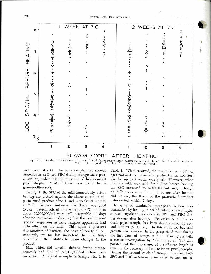
.1'
204 PATEL AND BLA JKENAGLE
WEEK AT 7C 2 WEEKS AT 7C 8 . . . • . . . • ' .
• • I • (: . :·: . • ·.• •I• . (9 .. : a:a ~·· .. ::· . . =~ .. .. . . lo z .. ·:· .. . ..... : :1: • .. . .. ... . 7 ·.:: • .. .... ~~ : . .
~ ~ • a• • . .. • • . . .. • .,. .. • w . . I • . • • . ... • • w • • •i• . • Q:: 6 ·:· 0 • LL .
• • w ... ":\" • co '·\ s, • . . • . . : s . . _J • . -:z ..... L: .. f:: ... . ,. .s , .. -- :~
.. ·'· u . a"!-.
o._ •• • . .. lf) 4 ••• .. ... . . •• . . :: : .. . ... •JJ• . ... I "· (9 • ·:· \" .. ..• • • . .. 0 : . ..
·~'· :;~ ..
' _J . .. . .. . . . • 3 • • . .. •• .. •
~ 2 3 4 2 3 4
FLAVOR SCORE AFTER HEATING Figure l. Standard Plate Count of raw milk and flavor scores after pastemization and storage for 1 and 2 weeks at
7 C. (1 = good; 2 = fair; 3 = · poor; 4 = very poor)
milk stored at 7 C. The same samples also showed increases in SPC and PBC during storage after pasteurization, indicating the presence of heat-resistant psychroh·ophs. Most of these were fotmd to be gram-positive rods,.
In Fig. 1, the SPC of the milk immediately before heating are plotted against the flavor scores of the pasteurized product after 1 and 2 weeks of storage at 7 C. In most instances the flavor was good to fair. Several lots of milk with raw SPC of up to about 50,000,000/ml were still acceptable 14 days after pasteurization, indicating that the predominant types of organisms in these samples apparently had little effect on the milk. This again emphasizes that numbers of bacteria, the basis of nearly all our standards, are far less important than the types present and their ability to cause changes in the product.
Milk which did develop defects dming storage generally had SPC of > 1,000,000/rnl before pasteurization. A ·typical example is Sample No. 2 in
Table 1. When received, the raw milk had a SPC of 6,000/ml and the flavor after pasteurization and storage for up to 2 weeks was good. However, when the raw milk was held for 4 days before heating, the SPC increased to 27,000,000/ml and, although no differences were found in counts after heating and storage, the flavor of the pasteurized product deteriorated within 7 days.
In spite of eliminating post-pasteurization contamination by heating in sealed tubes, a few samples showed significant increases in SPC ru1d PBC during storage after heating. The existen~e of thermoduric psychroh·ophs has been demonsh·ated by several authors (5, 12, 16). In this study no bacterial growth was observed in the pasteurized milk during the first week of storage at 7 C. This agrees with a recent investigation by Wah·ous et al. (19) who pointed out the importance of a sufficient length of time for the recovery of heat-resistant psychroh·ophs, During the _.second week of -storage, however, both SPC and PBC occasionally increased to such an ex-
, , ' . . '
•. '
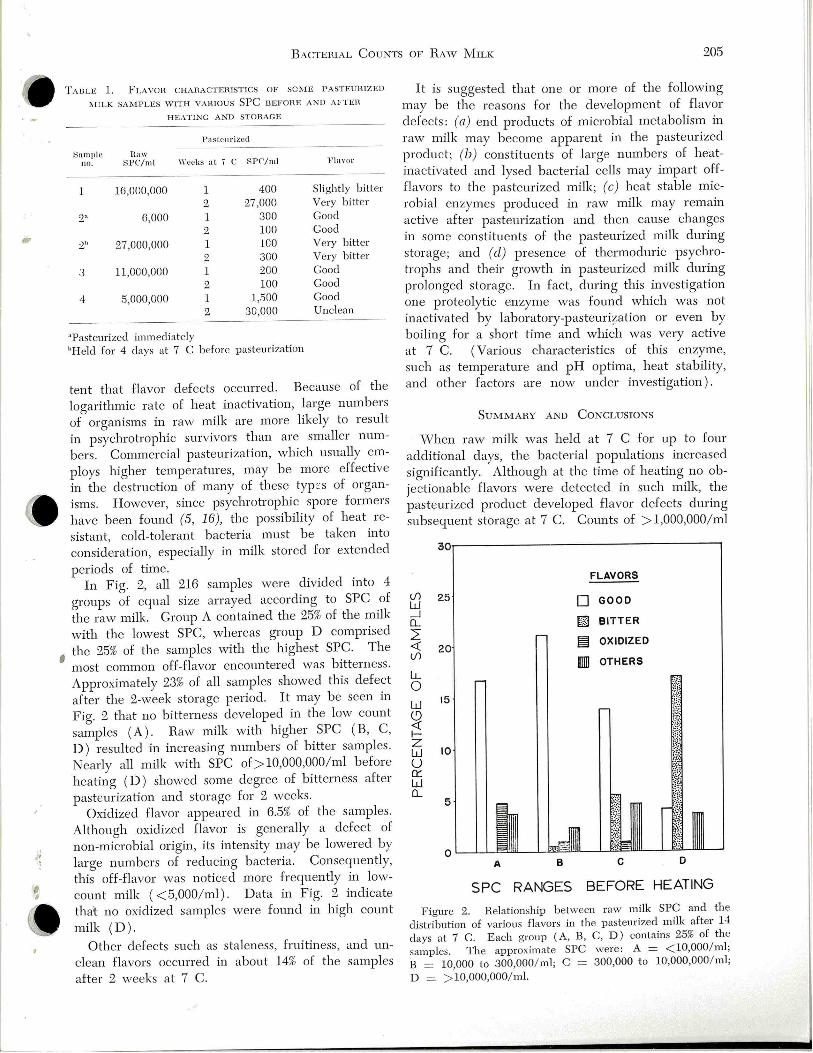
I
·' )
' '
BACTEHIAL CouNTS OF R Aw MILK 205
TAB LE l. F LAVO L\ CHAHA CTEHlSTICS OF SCi\'IE PASTEUHlZED
J\I lLK SAMPLES WITH VA!UOUS SPC UEFOHE AND AFTEH
HEATING AND STOHACE
Pasteur ized
~a mpl e Haw no. SPC/ml \Yeel;s at i C SPC'/ml }' laror
1 16,000,000 l 400 Slig htly bitter
2 27,000 Very bitte r
2:\ 6,000 l 300 Good
2 100 Good
:2h 27,000,000 l 100 Ve ry bitter
2 300 Very bitter
3 11,000,000 1 200 Good
2 100 Good
4 5,000,000 1 1,500 Good
2 30,000 Unclean
"Pasteurized imm ediate ly
"Held for 4 clays a t 7 C b efore pasteuri za tion
tent that flavor defec ts occurred. Because of the
logarithmic rate of heat inactivation, large numbers
of organisms in raw milk are more likely to result
in psychrotrophic survivors than are smaller num
bers. Commercial pasteurization, which usually em
ploys higher temperatures, may be more effective
in the destruction of many of these typ ::s of organ
isms. However, since psychrotrophic spore formers
have been found (5, 16), the possibility of heat re
sistant, cold-tolerant bacteria must be taken into
consideration, especially in milk stored for extended
periods of time. In Fig. 2, all 216 samples were divided into 4
groups of equal size arrayed according to SPC of
the raw milk. Group A contained the 25% of the milk
with the lowest SPC, whereas group D comprised
the 25% of the samples with the highest SPC. The
; most common off-flavor encountered vvas bitterness .
Approximately 23% of all samples showed this defect
after the 2-week storage period. It may b e seen in
Fig. 2 that no bitterness developed in the low count
samples (A) . Raw milk with higher SPC ( B, C,
D) resulted in increasing numbers of bitter samples .
Nearly all milk with SPC of > 10,000,000/ml before
heating (D) showed some degree of bitterness after
pasteurization and storage for 2 weeks.
Oxidized flavor appeared in 6.5% of the samples.
Although oxidized flavor is generally a defect of
non-microbial origin, its intensity may be lowered by
large numbers of reducing bacteria. Consequently,
this off-flavor was noticed more frequently in low
count milk ( <5,000/ ml ). Data in Fig. 2 indicate
that no oxidized samples were found in high count
milk (D).
Other defects such as staleness, fruitiness, and un
clean flavors occurred in about 14% of the samples
after 2 weeks at 7 C.
It is suggested that one or more of the following
may be the reasons for the development of flavor
defects: (a) end products of microbial metabolism in
raw milk may become apparent in the pasteurized
product; (b) constituents of large numbers of heat
inactivated and lysed bacterial cells may impart off
flavors to the pasteurized milk; (c) heat stable mic
robial enzymes produced in raw milk may remain
active after pasteurization and then cause changes
in some constituents of the pasteurized milk during
storage; and (d) presence of thermoduric psychro
trophs and their growth in pasteurized milk during
prolonged storage. In fact, during this investigation
one proteolytic enzyme was found which was not
inactivated by laboratory-pasteuription or even by
boiling for a short time and which was very active
at 7 C. (Various characteristics of this enzyme,
such as temperature and pH optima, heat stability,
and other factors are now under investigation) .
S uMMARY AND CoNCLUSIONS
vVhen raw milk was held at 7 C for up to four
additional days, the bacterial populations increased
significantly. Although at the time of heating no ob
jectionable flavors were detected in such milk, the
pas teurized product developed flavor defects dming
subsequent storage at 7 C. Counts of > 1,000,000/ ml
lf)
w _j Q_
2: <( lf)
LL 0 w ~ fz
25
15
w 10 u 0:: w Q_
FLAVORS
0 GOOD
rn BITTER
§:! OXIDIZED
IIlli OTHERS
A
SPC RANGES BEFORE HEATING
F ig ure 2. Relationship be tween raw milk SPC and the
distribution of various fl avors in the pasteurized milk after 14
d ays at 7 C. Each group (A, B, C, D ) contains 25% of the
samples. The approximate SPC wer e : A = < 10,000/ ml;
B = 10,000 to 300,000/ ml; C = 300,000 t o 10,000,000/ ml;
D = > 10,000,000/ ml.
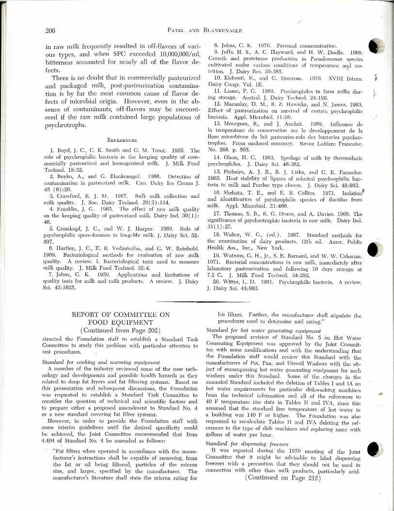
206 P ATEL AND BLA NKENAGLE
in raw milk frequently resulted in off-flavors of various types, and when SPC exceeded 10,000,000/ ml, bitterness accounted for nearly all of the flavor defects.
There is no doubt that in commercially pasteurized and packaged milk, post-pasteurization contamination is by far the most common cause of flavor defects of microbial origin. However, even in the absence of contaminants, off-flavors may be encountered if the raw milk contained large populations of psychrotrophs.
REFERENCES
1. Boyd, J. C., C. K. Smith and G. M. Trout. 1955. The role of psychrophilic bacteria in the keeping quality of commercially pasteurized and homogenized milk. J . Milk Food Teclmol. 18:32.
2. Boyko, A., and G. Blm•kenagel. 1966. Detection of contamination in pasteurized milk. Can. Dairy Ice Cream J . 45 (6):20.
3. Crawford, R. J. M. 1967. Bulk milk collection and milk quality. J. Soc. Dairy Teclmol. 20(3) :114.
4 . Franklin, J . G. 1965. The effect of raw milk quality on the keeping quality of pasteurized milk. Dairy Ind. 30( 1) : 46.
5. Grosskopf, J. C., and W. J . Harper. 1969. Role of psychrophilic spore-form ers in long-life milk. J, Dairy Sci. 52: 897.
6. Hartley, J. C., E> R. Vedamuthu, and G. W. Reinbold. 1969. Bacteriological methods for evaluation of raw milk quality. A review. I. Bacteriological tests used to measure milk quality. J. Milk Food Techno!. 32:4.
7. Jolms, C. K. 1959. Applications and limitations of quality tests for milk and milk products. A review. J. Dairy Sci. 42:1625.
REPORT OF COMMITTEE ON FOOD EQUIPMENT
(Continued from Page 202 ) structed the Foundation staff to establish a Standard Task Committee to study this problem with particular attention to test procedures.
Standm·d for cooking and warming eqttipment A member of the industry reviewed some of the new tech
nology and developments and possible healtl1 hazards as they related to deep fat fryers and fat filtering systems. Based on this presentation and subsequent discussions, the Foundation was requested to establish a Standard Task Conm1ittee to consider the question of technical and scientific factors and to prepare either a proposed amendment to Standard No. 4 or a new standard covering fat filter systems.
However, in order to provide the Foundation staff wiili some interim guidelines until the desired specificity could be achieved, the Joint Committee recommended that Item 4.404 of Standard No. 4 be amended as follows:
"Fat filters when operated in accordance wiili the manufac turer's instructions shall be capable' of removing, from the fat or oil being filtered, particles of the micron size, and larger, specified by the manufacturer. The manufacturer's literature shall state the micron rating for
8. Jolms, C. K. 1970. Personal communication. 9. Juffs, H . S., A. C. Hayward, and H . W. Doelle. 1968.
Growth and proteinase production in cultivated under various conditions of trition. J . Dairy Res . 35 :385.
Pseudomonas species temperature and nu
!
10. Kishonti, E., and G. Sjostrom. 1970. XVIII Intern. Dairy Congr. Vol. lE.
11. Loane, P. C. 1969. Psychrophiles in fann milks during storage. Austral. J . Dairy Techno!. 24: 196.
12. Macaulay, D. M., R. Z. Hawirko, and N. James. 1963. Effect of pasteurization on sun ;ival of certain psychrophilic bacteria. Appl. Micro bioi. 11:90.
13. Mourgues, R. , and J. Auclair. 1969. Influence de Ia temperatme de conservation sur le developpement de Ia flore microbinne du lait pasteurise-role des bacteries psychrotrophes. From enclosed summary. Revue Laitiere Francaise. No. 268. p. 505.
14. Olson, H . C. 1963. Spoilage of milk by thermoduric psychrophiles. J. Dai ry Sci. 46:362.
15. Pinheiro, A. J. R., B. J. Liska, and C. E . Parmelee. 1965. Heat stability of lipases of selected psychrophilic bacteria in milk and Purdue type cheese. J. Dairy Sci. 48:983.
16. Shehata, T. E., and E. B. Collins. 1971. Isolation' and identification of psychrophilic species of BacilLus from milk. Appl. 1icrobiol. 21:466.
17. Thomas, S. B., R. G. Druce, and A. Davies. 1966. The significance of psychroh·ophic bacteria in raw milk. Dairy Incl. 31(1):27.
18. Walter, Vol . G., (eel.). 1967. Standard methods for the examination of dairy products. 12th eel. Amer. Public Health Ass., Inc., New York.
Hl. Watrous, G. H., Jr. , S. E. Barnard, and Vv. ' 'V. Coleman. 1971. Bacterial concentrations in raw milk, immediately after laboratory pasteurization and following 10 days storage at 7.2 C. J. Milk Food Techno!. 34 :282.
20. Witter, L. D . 1961. Psychrophilic bacteria. A review. J. Dairy Sci. 44:983.
his filters. Further, the manufacturer shall stipulate the procedures used to determine said rating."
Sta.ndm·d for hot water generating eqtt·ipment The proposed revision of Standard No. 5 on Hot Water
Generating Equipment was approved by the Joint Committee with some moclifications and with the understanding that the Foundation staff would review this Standard with the manufactmers of Pot, Pan, and Utensil Washers with the object of encompassing hot water generating equipment for such washers tmder this Standard. Some of the changes in the amended Standard included the deletion of Tables I and IA on hot water requirements for particular clishwashing machines from the technical information and all of the references to 40 F temperature rise data in Tables II and IVA, since this assw11ed that the standard line temperatme of hot water in a building was 140 F or higher. The Foundation was also requested to recalculate Tables II and IVA deleting the references to tl1e typ e of clish machines and replacing same with gallons of water p er hour.
Sta.nda:rcl fo1' clispensi:ng freezers It was r·eportecl during the 1970 meeting of the Joint
Committee iliat it might be advisable to label clispensing freezers with a precaution that they should not be used in connection with other than milk products, pariicularly acid-
( Continued on Page 212)
•· I
I
·' l
'
]. Mille Food T echnol. , Vol. 35, No. 4 (1972) 207
PROGRAM FOR QUALITY ASSURANCE OF FINISHED DAIRY PRODUCTS'
EDMOND L. SING
Moseley Laboratories 3862 East Washington Street Indianapolis, Indiana 46201
ABSTRACT
Specifications concerning quality of milk products from raw ingredients to finish ed products are now often required by many distributors and food processers. Problems concerning plant sanitation, post-pasteuriza tion contamination, and product temperatme still persist despite an abundance of practical remedial knowledge available. The manifes tations of many current problems are the same as they were 10 to 15 years ago. Two examples of these problems are induced rancidity in silo tanks as a result of excessive air agitation and thermoduric organisms in pasteurized milk which may proliferate to excessive numbers at refrigeration temperatures .
A program for quality assurance should incluae a raw ingredient program, methods for evaluation, effective plant sanitation, care of finished products, human motivation, and follow through. The area of most n eglect is the last aspect. Plant cleaning and sanitation have improved remarkably in the last few years . Many dairies have capitalized on proven refinement of automated systems and have incorporated fine points of sanitation. The most effective procedure for trouble shooting problem areas is to follow product processing backwards using aseptic sampling techniques. D ata accumulated in successfully pinpointing problem areas the past 5 years h ave demonstrated th e value of this approach.
Quality assurance is a term now frequently encountered in the dairy and food indush-y. It h as a broader meaning to consumers since they feel that quality is assured to them from raw ingredients to the ultimate finished products. In-plant quality assurance, which is synonymous with quality control, will be the main area of discussion.
In tl1e last few years there has been a great increase in the need for chemical and bacteriological analyses on raw and finish ed food products . Results of these tests are intended to provide information to manufacturers , distributors , or buyers concerning the specifications they require. For instance, the meat industry, jointly with u:.s.D.A. is presently engaged in setting bacteriological and chemical standards on ready to eat products such as wieners and bologna. The canning indush·y requires bacteriological specifications on starch which deal with the numbers of tl1ermophilic spores and hydrogen sulfide producing and non-hydrogen sulfide producing anaerobes.
'Presented at the 57th Annu al Meeting of th e International Association of Milk, · Food, and• Environmental Sanitarians, Cedar Rapids, Iowa, August 17-20, •1970
Large super market chains frequently require a specified shelf life for all finished dairy products.
At the I.A.M.F.E.S . conferences of 1963 and 1966 Elliker et al. (1, 2) presented papers which discussed post-pasteurization contaminatio~1 of milk and dairy products. Possible causes and suggested procedures to minimize the problem were mentioned. Attempts to isolate and pinpoint the problem ( 8 or 9 years ago ) were accomplished by aseptic sampling procedures and equipment. The data collected indicated that the major post-pasteurization contamination areas were the fillers. As further effort was made, some contaminating areas were traced back as far as the HTST unit. It was common knowledge that normal pasteurization ( HTST unit) would destroy most organisms able to grow at 45 F . These organisms vvere then referred to as psychTophiles but a more accurate term used today is psychwtrophs. Other areas that were found to be potential sources of contamination included joints. valves, manifolds, pumps, and air supply. Suggested procedmes for correction, such as on-th e-spot sanitizer spraying of the filler area before and during operation, were found to be effective. The culmination of this work realized an increase of 14 to 20 days shelf life for many dairy products.
In the pas t few years there have been many changes in dairy operations throughout the country. It is of interest to visit plants to note new changes and to evaluate them for their contribution to post processing contamination. As a result, we have made several conclusions.
1. 1 he small independents generally lag behind the larger plants (majors, captives, and large coops) with resp :::ct to basic design and plant improvements made. Yet ironically, the small plants may show a better profit picture than the large up-to-date dairies. Being behind oftentimes may· be in their favor since they can update their plants and take advantage of modern technology. They certainly capitalize on all the refinements of automated systems. On the other hand, the CIP systems (in their infancy 15 years ago) are now being converted and brought up to date.
2. Those actively pursuing cleaning and sanitizing
Z08 SING
and quality control programs continue to be on top, saleswise and profitwise. The question most fr equently asked now is-are all plants taking full advantage of the accumulated knowledge in this field? Unfortunately not. Those plants that are more conscious of pricing than of quality will not be in business long. However, those vvho feel that quality has marketable value will continue to grow and make a profit.
3. New equipment and concepts have b een b eneficial in many ways, however, as a result new problems also are introduced. Aseptic systems, silo storage, high speed fillers, and air conh·ol are examples of a few such areas.
In discussing a program for plant quality assurance of finished dairy products, five main points of concentration should be considered. These may be listed as follows:
l. Raw ingredient program. 2. Methods for evaluating quality assmance. 2. Nlethods for evaluating guality assurance of
finished products. 3. Effective plant sanitation procedures . 4. Care of post-processed (finished) products. 5. Human motivation and follow through.
In discussing each of the above points, it is of value to illush'ate or discuss problems that have b een recently encountered . It is surprising that the manifestations of many current problems are the same as they were 10 to 15 years ago.
RAw I NGREDIENT PROGRAM
Important considerations in this program are flavor, bacterial content (raw and pasteurized count), raw storage, adulterants (water and antibiotics), pesticides, and composition (butterfat and solids). Flavor in a raw ingredient program is considered the most important of all the aspects described above.
The most critical problem is developed flavor as the result of excessive bacterial growth. In most instances these flavors are the result of prolonged storage, either on the farm or in the plant, at temperatures which are favorable for proliferation of bacteria. Temperatures in excess of 40 F could cause rapid multiplication of bacteria which will cause flavors such as puh'id, rancid, rotten, or fruity. Most of these developed flavors in the raw product cannot b e eliminated by pasteurization. On the other hand, flavors that develop in the finished, pasteurized product are generally the result of postpasteurization contamination.
In discussing various aspects of the flavoring program, current trends towards large storage capacity such as silo tanks, have been beneficial. The greater commingling of milk has resulted in dilution of un-
d 2sirable flavors. However, one of the problems encountered with large storage capacities is insufficient turn-over of raw product. In many instances,
I
silo tanks for raw milk may not b e washed for 3' to 4 days. If raw product is continuously added at favorable temperatures, psychrotrophic growth will p erp etuate off-flavors.
Another problem that has. b een encountered concerning silo tanks is development of rancidity. The problem is more pronounced in silo tanks with capacities >30,000 gal. Rancidity develops b ecause of excessive air agitation, especially when the tank is partially filled. The smaller silo tanks generally have mechanical impellers that will maintain proper agitation. The larger tanks, by necessity, have relied, primarily, on air agitation. Many of these large tanks are air agitated with constant air pressure rather than constant volume. Data in Table 1 illush·ate what happens in a 40,000 gal silo tank that ' is agitated by constant air pressure. When the tank was full , acid degree values (ADV ) were satisfactory. As the tank was emptied, the ADVs increased. At 18,000 gal the ADV was 0.76, whereas at 2,500 gal it was 1.48. Nlany silo tank installations that are agitated using constant air pressure are finding similar results. A remedy for this situation is the installation of a constant volume air regulator that insures proper agitation at any given volume of liquid in the tank
D espite development or rapid tests and sophisticated instrumentation in the last 10 years to detect raw ingredient problems, these problems are still frequently encow1tered. Some of these problems are induced rancidity, high thermoduric counts, antibiotics, p esticides, added water, and mastitic milk Ironically, these are, basically, the same problems that we faced many years ago.
A suggested in-plant raw milk program is as follows:
l. Grade raw products at the tanker level by taste and smell. It would be desirable that two people be involved, for example, the receiving man and a person in quality control. It is important that people concerned with tasting milk are trained and are able to detect off-flavors. To avoid flavoring raw milk, these samples may b e heat shocked at 150 F for 2 or 3 min and cooled, prior to tasting.
2. All milk in raw storage tanks should be tasted just before processing. This step is to insure that raw milk going to the HTST unit has an acceptable flavor.
3. Laboratory tests should be performed periodically. These should include standard plate counts, laboratory pastetuized counts, leucocyte counts, and tests for adulterants, such as antibiotics, added water, and pesticides. Other quality tests such as flavor
, , ( .. I
•

I
•' l
' '
;
PRoGRAM FOR QuALITY AssuRANCE 209
of milk from individual producers and pre-incuba
tion prior to standard plate counts have proven to be
beneficial. Some plants also conduct culture activity
tests on the milk to make certain that it will support
active growth of lactic acid bacteria in production of
cottage cheese, buttermilk, and yogurt.
To illustrate that some problems which existed in
the past are still persisting today, refer to Table 2
which shows that high thermoduric counts still per
sist in some operations. If you will compare the
fresh count to the shelf life count, you will note
that the high count vvas not as a result of post-pas
temization contamination but a raw ingredient prob
lem. Some thermodurics have been found that will
grow at refrigeration tem perah1res. Recently report
ed was a carton of pasteurized milk exhibiting a very
bitter flavor that had a count of 58,000,000/ ml. These
organisms were not the typical gram-negative con
taminants but were large spore-forming rods. This
product was picked up 7 days after processing and
the milk was held at refrigeration temperah1res. These
large spore-forming rods have often been fatmd to
cause sweet coagulation in finished products that
may be 2 or 3 weeks old. Data as indicated in Table
2 are important from the standpoint that companies
buying on specific standards for pasteurized milk
will not tolerate high thermoduric counts even
though proper processing procedures have been used.
i\'fETHODS FOR EvALUATING QuALITY AssuRANCE OF
FINISHED PRODUCT
This topic concerns procedures for evaluating qual
ity assurance of finished products. These proced
ures are quality tests p~rfonned in the laboratory
and methods for determining in-plant problem areas.
The following discussion will be limited to present
application of prior techniques described in past
publications (1, 2). The fin ished product coming directly from each
packaging lin :! should be tasted at various intervals
during the production day. It would be desirable
that the fina l product be tasted by the operator and
a person in quality control. Finished products taken
from each line for this purpose also may be used for
checking weights and leakage. It must b e pointed
out that tasting the .final product at this point is ex
tremely important since often this is the last op
portunity before the product is distributed.
Randomly selected samples of all pasteurized prod
ucts should b ::! taken for bacteriological and flavor
tests. These samples should be taken at periodic in
tervals during production and the number of samples
taken should be proportional to the quantity of any
given item processed . Each of the samples should
include pertinent information regarding its production
such as HTST unit, tank, line, filler , sequence of
production and time. These samples of finished
products are to be subjected to keeping quality tests.
A number of laboratory tests for determining shelf
life characteristics are available. It is important to
select a test that will provide accurate data concern
ing shelf life characteris tics. One of the most suitable
tests for shelf-life is the Moseley 5-day, 45 F test.
In recent years the test has been made more sensitive
by extending the number of clays at 45 F. In doing
keeping quality tests for either flavor or bacteria
count, it is most important to simulate marketing
conditions to which the product will actually be sub
jected. A minimal test for keeping quality would
be holding the product for a given number of clays
at 45 F and tas ting it to aetect bacteriological
spoilage. If the collected data do not show satis
factory results based on bacteriological counts and
flavor, then spot check procedures should be imple
mented to determine problem areas.
Various sampling devices are available to aseptical
ly obtain line samples. Recommended areas for in
stallation _ of in-line sampling devices are: l. Outlet of each HTST unit 2. Outlet of each pasteurized smge tank 3. Pasteurized surge manifold 4. Above the bowl of each fil ler
A suggested procedure of line sampling to pin
point the area of contamination is to work backwards
from the finished product. Sampling prior to the
filling machine is a good location to monitor. Many
plants will take daily routine samples at this point.
If results are satisfactory here, there is no need to
trace back beyond this point. This approach has
been proven to b e extremely effective.
In collecting data from numerous plants, it has
been found that the major problem areas are as
follows. Filling machines, by far, cause most con
tamination problems. This is followed by the pas
teurized surge line system if it contains many sani
tary thread joints and plug valves. If the smge line
system contains air valves and >velclecl lines, these
areas conh·ibute little contamination if satisfactorily
cleaned and sanitized. Pasteurized smge tanks that
are cleaned by automation cause few problems.
Hand washed storage tanks are still major problem
areas. The HTST unit and related pipe lines often
have been a source of contamination. The specific
defect most often encountered is accumulation of
soil in the cooling section or in dead-end fittings
such as air eliminators and thermometer probes.
A present-day plant containing standard equip
ment should be able to produce products from the outlet side of each HTST unit which have a count
of < 3,000/ ml after 14 to 21 days storage at 45 F.
210 Smc
If the plant has all welded lines and automatic valves, there should be only minimal pick-up of contaminants between the HTST unit and the point above the fill er bowl of any given filling machine.
Lastly, evaluation of any plant requires thorough records concerning the operation. These records should include daily inspection reports, noting cleanliness of equipment and potentially dangerous areas; time and temperature recording charts; and procedural changes in cleaning and sanitizing. }..·Ionitoring data also are important. For example, data should be obtained from the outlet side of each HTST unit and the area above the filler bowl of each filling machine.
EFFECTIVE PLA!\'T SANITATION PROCEDURES
This discussion pertains to procedures used in cleaning and sanitizing and also covers some of the fine points of sanitation. Observations made in the past few years have shown specific points to be important. These are as follows:
l. All product contact surfaces and associated surfaces should be cleaned to the surface of the equipment material. Associated surfaces are areas that may not come in direct contact with the product but may become potential sources of contamination through their relation to the product. Examples of these areas are the interior surface of lids and tops of tanks, and the exterior surface of fillers. There is only one criterion for determination of a clean surface, and that is whether it is bare surface clean. It is also important that we learn to recognize a clean surface and to provide cleaning procedures to insure that this criterion is met.
2. It is necessary for quality control and plant supervisors to be present during the clean-up. As cleaning systems become more complicated and more automation is installed, it becomes increasingly important that highly trained people supervise the clean-up operation. · It is felt that one day cleaning and sani tizing will be accepted as the most important functions in any plant operation.
3. Proper maintenance of all facets of the cleaning system is an absolute necessity. These include periodic changing of all joint gaskets ; insuring proper functioning and s e a tin g of automatic or manual valves and electronic components of the CIP cleaning system; checking proper flow rate of CIP and return pumps; and removal of all extraneous material blocking jets of spray balls.
4. Daily routine checks of concentrations of cleaning agents and sanitizers are required to insure the performance of CIP systems as well as manual cleaning operations.
In sanitizing, we have found procedural methods
to be the most important. l. To insure proper sanitizing, the system should
be, for the most part, closed. By this we mean that all pasteurized surge tanks are closed, jumpers, to the pasteurized manifold in place, jumpers to filler bowls of machines in place, and all fillers assembled. The pasteurized distribution manifold also should be closed with all jumper hook-ups connected to all tanks. It has been found that if spray balls are permanent installations, and sanitizing through spray balls is programmed into the systems, this is the most ideal way to sanitize. Fogging of tanks with high concentration of sanitizers, in conh·ast, is a poor practice.
2. The suggested procedure for sanitizing the HTST unit would be to start vvith a sanitizing solution made up in a processing tank and then pumped to the balance tank. Sufficient sanitizing solution of the proper concentration should be made up to insure proper contact time. The sanitizing solution ' from the HTST unit is then distributed throughout the pasteurized distribution system into the surge tanks. Again, sufficient solution should be available in each surge tank to sufficiently flood the pasteurized surge tank outlets and lines to the machines. To properly flood filler bowls, many plan ts have devised a tool to hold down the floats to insme complete contact. Finally, machines are operated to insure sanitizing of the internal patts of assembled machin es. Prior to sanitizing, filler machines are assembled using a sanitizer while building up the machine. :Most companies now have systems which provide a sanitizer in key areas in the plant for sanitizing on the spot. The most common faul t in most plant operations is the indiscriminate use of untreated water in all post-pasteurized areas. Procedures such as flushing lines with untreated water and rinsing pasteurized surge tanks with unh·eated water are bad practices.
3. Procedures to insme proper contact time, concentration, and pH of sanitizers also are important. The manufacturer's directive for use of various types of germicides should be closely followed.
In discussing fine points of sanitation, some of the points to be stressed have already been mentioned. In most instances, fin e points of sanitation are procedural changes or use of special equipment that may be suitable only to a particular operation. Procedures are implemented before and during the operation. It is important to recognize that equipment does not remain clean and sanitized all day. Present day operations require extremely long runs, therefore, failure to recognize that this equipment will not remain sanitary all during the operation will lead to poor results.
;. I
•
••

•
'
' · ~
PROGRAM FOB Q uALITY AssuRANCE 211
TABLE 1. E F FECTS OF EXCESSiVE Am PRESSURE AGITATJON I N
RAW SILOS ON RANCIDITY
Tnnk
Raw silo #7 ( 18,000 gal ) Raw silo #7 ( 2,500 gal ) Tank #7 to balance tank (recycling)
Normal Unsatisfactory
"ADV = Acid degree value
ADV ADV
ADV•
.76 1.48 1.73
0.4 1.5
TAllLE 2. EFFECTS OF HIGH THEH!VIODURIC COUNTS ON FHESH
AND SHELF LIFE TESTS
Sn mple Fresh Aftel' 5 days at 45 F
Qt. homo 140,000 150,000 ]I d Gal hom o. 24,000 24,000 Half and half 90,000 > 3,000,000
SPC Lab. Past.
Raw tanker 1,200,000 72,000
Two effective procedures have been implemented in the past years. First, be assured that all filling machine parts are sanitized prior to assembly and sanitize while assembling. Secondly, spraying the exterior parts of filling machines with an appropriate sanitizer periodically during the day's operation has helped to reduce post-pasteurization contaminants at these sites. The two most common and effective sanitizers used in the industry today are iodophors and chlorine compounds. Iodophors are non-corrosive acid sanitizers that are most effective at fairly low concentrations. The central dish·ibution iodophor systems that are piped tl1l'oughout the plant also have been extremely beneficial for use as on-thespot sanitizers for filler parts, exterior of fillers, jumper hook-ups, plug valves, valve clusters, and other areas which may contribute p ost-pasteurization contamination . Sodium hydrochlorite solution is still the most effective general sanitizer for tanks, lines, and HTST units. In some instances, consideration must be given to the pH of the water used to make up the sanitizer. It has been found that water with a high pH, in some instances, has negated the sanitizing properties of certain iodophors when used at low concentrations. vVhen low concenh·ations of chlorine are employed for cottage cheese wash water, their activity is enhanced by controlled acidification of the water. Chlorine sa]1itizers are more active if a final acid rinse is used in plant cleaning programs. Acid rinsings also have been beneficial in reducing milk stone formation and hard water and detergent spotting.
' In recent years in-plant environmental conditions have been demonstrated as affecting the finished product. Air and condensate have been found to be sources of post-pasteurization contamination. Imple-
menting fine points of sanitation have helped conh·ol these areas. In addition, manufacturers of air conh·ol systems and dairy equipment have produced new designs with sanitation in mind. Good examples of these concepts are use of HEPA type filters for the air supply for critical filling room areas often combined with a positive pressure air sys tem for specific rooms. Also, the location of laminar flow, filtered air units over filling units insures essentially sterile au: in the filler environments.
CAHE OF PosT-PHocEssEn (FINISHED) PRODUCTS
In view of current dish·ibution systems, it is extremely important to properly care for finished products. Finished products from processing plants, in many instances, are being tr·uckyd or shipped to other distribution complexes. As many as three re-handling procedures could be made from point of origin to final destination. For this reason, close control of storage temperatures is essential to meet the requirement for longer shelf-life. Points to sh·ess are as follows:
l. Processing plants must practice maximum sanitation as discussed earlier. Temperature conh·ol to keep the product as cold as possible will also be necessary. Many plants are putting in additional cooling capacities, for example, glycol systems are becoming more common iiJ. the indush-y. Product leaving the HTST unit may be at temperahues close to freezing. Many plants will conduct their products through insulated lines and store in refrigerated holding tanks.
2. Storage vaults for finished products also should be as cold as possible. Maintenance of temperah1res at 34 to 38 F is not uncommon. The importance of maintaining these temperatures despite re-handling of the product to different coolers cannot be overstressed.
3. Display cases in merchandising areas me areas of temperature abuse. 1 umerous sh1dies have been made on display cases showu1g temperahues in many cases to be far above 45 F. Another poor practice often encountered is stacking of finished products above the recommended load line of display cases .
It has been demonsh·ated that by virhw of temperature control alone, without any changes in plant processing, the shelf-life of finished products can be increased . The point of this discussion is to sh·ess that with the combination of good sanitation practices and temperature control optimum shelf-life characteristics can be attained.
Hu:~~-rA r MoTIVATION A1\'D FoLLOW-TlffiOUGH
Of the five points discussed, the combination of motivation and follow-through is the most important.
.J'
212 Smc
Unfortunately, this also is the area of most neglect. Most dairy plant operations have all the physical needs for producing a long shelf-life product, however, failure to meet this goal is most often ath·ibuted to human weakn ~ ss . Some of the reasons why we do not accomplish the total job of quality assurance may be :
l. Lack of emphasis from management to incorporate improved programs. The success of quality assurance depends upon management passing down or approving the necessary requirements to attain their · goals.
2. Often there is a lack of a properly defin ed quality control department. In many instances this deparhnent is a part of production and not directly under management. Under this arrangement the administrative responsibilities are such that the job vvill never be accomplished . If quality control people are bogged down with production problems, there is no opportunity for success of any quality assurance program.
3. Supervisory people in production or quality control must be trained in human relations otherwise
REPORT OF COJ\IIMITTEE ON FOOD EQUIPMENT
( Continued from Page 206 )
type foods, since such ac tion :11ight produce a toxic product. According to the Foundation staff, currently dispensin g freezers are being manufactured with component parts which have been found to be suitable for use with either dairy products or with slush ice products; thus, any precautionary labelin g for new equipment would be useless. Stanclanl for walk- in and Teach-in refrigerators and storage freezers
Following presentations by various manufacturers and a lengthy discussion by the members of the Joint Committee, a revision in the errata to Item 5.02, as it related to the requirements for joints and seams in walk-in refrigerators, was recommended as follows:
"The provisions of this specification are interpreted to require that gaskets meet the material requirements for splash zone. Interpretation furth er precludes the use of sealing compounds appiied in the field during or after erection of the unit except for effecting r epairs."
Fmther presentations were made regarding the revision of Standard o. 7 to permit the use of plastic caving strips in walk-in refrigerators and freezers to effect the required radii in two and three plane intersections in lieu of the present requirements which specify that the radii shall be smooth and continuous, i .e., of the parent material. After discussing these new materials and techniques, it was concurred that the subject should be sent to the Standard Task Committee for study and recommendations with a subsequent review and ballot by the Joint Committee. A recommendation was made also by members of the industry that the Standard Task Committee review the current provisions which require a flu sh door siE for walk-ins.
they will certainly not be able to motivate others. 4. Careful screening of employees should be done
to obtain personnel that are experienced or that can b e trained to do specific jobs. l
All successful endeavors are the result of people. ~
D espite increased technology, automation, new sophisticated systems, and up-to-date equipment, an ;. operation functions only as well as the individuals ' make it perform. As operations become more complex and additional knowledge is required, it becomes necessary to assist control and plant personnel in every way possible to develop tl1e skill to carry out programs or manipulate costly equipment. Hesults of care are successful endeavor and pride. This will insure continuance of a job well done.
R EFE HENCES
l. Elliker, P. R., E . L. Sing, L. }. Christensen, and W. E . Sandine. 1964. Psychrophilic bac teria and keeping quality of pasteurized dairy products. J. Milk Food Technol. 1:7 : 60-75.
2. Sing, E . L., P. R. Elliker, L. J. Christensen, and '~' · E . Sandine. 1967. Effective tes ting procedures for evaluatin g plant sanitation. J. Milk Food Techno!. 30:103-lll.
Standard for manual dispensing equ·ipm ent The potential h azard of backflow of carbon dioxide and
carbonated water in manual dispensing equipment was discussed. The Foundation staff indicated that it was their plan to propose an amendment to Standard No . 18 (Manual Dispensing Equipment ) in t he same manner as Standard No . 25 (Food and Beverage Vendin g Machines), is being amended to prevent cartonat~d water from coming in contact with copper lines.
Standard for commercial cookin g eq uipm ent exhaust
The public health members briefl y considered th e need for huther study of commercial cookin g equipm ent exh aust systems with particular attention to units other than canopy hoods. They noted that at the present tim e it is strictly a perfonnan ce standard and there are no specifications for exhaust systems other than of the canopy-type, thus making field evaluation of such equipment ·,ery difficult . The NSF Testing Laboratory staff recommended that changes be held pending the results of the Laboratory's testing of a number of these tmits in order to provide a baseline for the design and construction parameters.
Requirem ents for covers over fo od zones
The Foundation staff reviewed a survey of requirements for covers over food zones as now contained in Standards 1, 2 , 4, 6, 7, 8, 12, 18, and 25 and Criteria C-2; and the survey revealed that some required a cover or other protection, some specified that the cover overlap th e opening, some specified protection of port openings, and some required a 3/ 16 inch raised rim for covers over food zones. In view of the survey findin gs, the Joint Committee requested the Foundation to" establish a Standard Task Committee to review the present requirements relative to covers over food zones and to make a recomm endation to the Joint Committee. Specificall y, th e
(Continued on Page 227 )
I
·' l
]. Mi.llc Food Technol., Vol. 35, No. 4 (1972) 213
BACILLUS CEREUS: FOOD POISONING ORGANISM. A REVIEW J. i\11. GoEPFEHT, W. M. SPIRA, Al\TD H. U. KrM
Food Research Institute and Department of Bacteriology Universitu of v\iisconsin
Madis:m , vVisconsin 53706
( Hceeivccl for publi cation October 6, 1971)
ABSTHACT
This paper is devoted to a review of information pertinent to th e role of Bacillus cereus as a food-borne pathogen. Primary emphasis is on the properties of B. cereus and published accounts of its in volvement in animal and human disease. Methods for isolation, identification, and enum eration are discussed. Research needs pertaining to (a) recognition of the potential public health hazard and (b) furth er investigation of th e food poisoning syndrome are presented.
It has b ecome increasingly clear that Bac-ill·us ceretts is involved in foodborne illness. The scope of the problem in this country is unknown . This review of the literature pertinent to B. cereus is presented with the hope that it may serve as a springboard from which investigations into B. cereus food poisoning may be launched . Obviously, the quantity of information that exists on this single microorganism is so voluminous that all could not possibly be reviewed in this limited space. Vlith this in mind, we have based our selection of data to those areas which seem today most pertinent to the problem of B. cereus as a food-born e pathogen.
TI-IE 0RGANISlVI
Taxonomy Before 1946, classification of the genus Bacillus
t ould best be described as chaotic. In that year, the first of two excellent monographs in which a rational and orderly approach to the taxonomy of Bacillus spp. w as published (158). In 1952, the second monograph (largely an expansion of the fir st ) was published and the taxonomic scheme proposed therein is the basis for separation and identification of bacilli today. Smith et al. (158, 159) proposed division of the genus Bacillus into three groups (I, II, and III ) based on spore and sporangium morphology. Group I bacilli consisted of those genera without definitely swollen sporangia (by tbe spore). Group II and III organisms evidenced swollen sporangia and spore mo11Jhology (i.e. oval spores for group li and round spores in group III ) was employed to differentiate the h~!O groups . Within group I, further subdivision was made on the basis of vegetative cell size and presence or absence of vacuoles within vegetative cells grown on glucose-nutrient agar. Thus, the largecelled species (vegetative cell diameter 2:: 0.9 fL ; also
I"
vacuolated) included Bacillus m egaterimn and B. cereus and varieties while the so-called small-celled species (cell diameter < 0.9 fL ; not vacuolated) of group I bacilli included Bac-illus subt-ilis, Bacillus lichenifonrvis, Bacillus pu.milis, Bacillus coagulans, and two rarely encountered species Bacillus f'innus and Bac-illus lentus.
In these extensive treatises, Smith et al. chose to consider Bac-illus mycoides, Bac-illus thwringiensis, and Bacillus anthraC'is as varieties of B. cereus rather than as separate species. This position has evoked favorable response from some (26, 170) and criticism from others (9, 66, 85, 100). In this review, we shall refer to these organisms as separate species purely for simplicity without intent to support either the species or variety taxonomical position. These organisms are very closely related and tests designed to differentiate them have been the subject of numerous papers (see Section on confirmatory tests).
Bacillus ceretts was originally isolated and described by Frankand and Frankland (57) in 1887. It is characterized as a gram-positive and catalase-positive long rod. Various biochemical, serological, toxicological, and bacteriophage susceptibility patterns have been ascribed to the species but, by and large, intensive investigation has shown that great variability in properties is characteristic of this organism. The major properties of the organism are presented , but the reader is vvarned that aberrant and deviant sb:ains are not uncommon .
Biochemical properties As a species, B. ce·reus strains are able to utilize
glucose, fru ctose, and trehalose. Xylose, arabinose, galaotose, sorbose, sorbitol, mannitol, and dulcitol are not attacked (85, 101). Sucrose, salicin, maltose, mannose, glycerol m-inositol, and lactose [sh·ains utilizing lactose are usually named Bac-illus albolact'is (159)] are attacked by some but not all members of the species (26, 85, 101). Urease is produced by few strains (85). Acetylmethylcarbinol ( AMC ) production is characteristic of most B. cereu.s strains (29, 66, 101, 159) but is not produced by all (26, 78, 85). Nitrates are reduced to nitrites (101, 159), but nitrate reductase negative strains have been isolated (26, 29, 78, 85). Niost sh·ains attack soluble starch but a few
.J'
214 GoEPFEHT E-r. AL.
amylase-less mutan ts have been described (78, 85). Most strains produce a neutral metalloprotease (52,
60, 152) and actively dissimilate such substrates as casein and gelatin (26, 78, 85, 101, 159). Certain strains have been reported to synthesize a dehydropeptide reductase that catalyzes cleavage of the antibiotics nisin and subtilin (72, 73 ). Virtually all strains produce a penicillinase (149) which enables them to grow on agar containing 10 International Units of penicillin, a test which aids in differentiating B. cereus from B. anthracis . In contTast to B. megateriu.m, few strains (if any) are sensitive to lysozyme (86). Reportedly, B. cereus is not able to grow in a medium in which ammonium ion is the sole source of nitrogen (83).
Recent reports by Knisely (84) and Mossel et al. (119) have indicated that B. cereus grows well on chloml hydrate agar and phenethylalcohol agar. Kim and Goepfert (78) could not substantiate this with tests of strains freshly isolated from food products.
One of the most important aspects of the biochemical pattern of B. cemus is synthesis and excretion of cert ain toxins. At leas t three important extracellular toxins are produced by B. cereus; these products have received extensive study.
Production of lecithinase by B. cereus was recognized by McGaughey and Chu (109) and Colmer (38) in 1948. Chu (34) later reported that cultme filtrates of B. cereus also possessed hemolytic, dermonecrotic and lethal (for mice) activity. At the time, and for some years after, it was tempting to speculate that all of these activities resided in a single molecular species as was true of Clostridium. perfringens a-toxin (105). But Molnar (117) provided evidence that lethal toxin and phospholipase were sepaTate entities and could be physically separated. Ottolenghi et al. (142) reported that hemolysin and phospholipase could be differentiated on the basis of thermal stability.
There is some evi.Clence that more than one phospholipase is produced by B. cemus. Slein and Logan (156, 157) reported separation of n:vo phospholipolytic enzymes on columns of DEAE-cellulose. The enzymes were different with respect to digestion by trypsin, thermal stability, and substrate specificity. Similar evidence for a multiplicity of phospholipases was presented by Kushner (91), who stated that phospholipase and an egg yolk hubidity-producing factor differed in thermal Sltability. Recent studies on the lecithinase C of B. cereus have shown that it has a molecular weight of 20,000 and an isoeleoh·ic point of 8.0-8.1 (140). , Some argument has arisen as to the requirement for Zn2+ ions for full enzymatic activity (75, 81, 141). In contrast to the substrate specificity of the C. pe1jringens enzyme (104) the lecithinase C
of B. ce·reus is able to hydrolyze phosphatidyl-ethanolamine and phospbatidyl serine (157) . The enzyme is formed and secreted by cells at the end of the log phase (on complex media) and is somewhat lsusceptible to denahu·ation if vigorously shaken (92) . Although there are reports of lecithinase-negative strains of B. cereus, such strains in our hands always produce turbidity on egg yolk agar. For the most part, the typical reaction of · these latter sh·ains on egg yolk agar is the 'restricted' form defined by Proom and Knight (150 ). Synthesis , but not activity of the enzyme from lecithinase positive strains, can be inhibited ·by growing the culture in the presence of C, - C" alcohols (90). Many questions remain regarding the role of the enzyme in pathogenicity, but at present, use of egg yolk containing agar as a presumptive medium for isolation and identification of B. cereus is the most efficient procedure.
Hemolysin, or "cereolysin" as it has been termed ' (11, 12), is similar to strep tolysin 0 in that it is activated by cystine, inhibited by cholesterol, and is neuh·alized by horse serum containing antistreptolysin 0 antibodies (12). Although some reports (21, 75) have suggested that "cereolysin" is heat labile, others (56) have said that the pmified enzyme is relatively stable. Fossum (56) has presented evidence that lability of hemolysin in crude preparations is caused by enhanced activity of proteases as the preparations are heated. Removal of the proteases allowed a more definitive detennination of thermal stability of the hemolysin. Purified hemolysin does not show phospholipase activity (12).
The lethal toxin of B. cereus differs considerably from the toxin formed by B. anthracis (117). Not only is the skin lesion resulting from intradermal inoculation different, but the B. cereus toxin is rapidly lethal for mice ( inh·avenous injection) while the B. anthmC'is toxin is not 1(9). Moreover, the B. cereus toxin rapidly destroys monolayers. of guinea pig spleen cells while the B. a.nthracis toxin is without effect (20 ). J olmson and Bon ventre (75) have obtained partially pmified preparations of lethal toxin which failed to exhibit hemolytic or phospholipolytic activity. Recently, Krieg (88) has reported that B. cereus produces a toxin which is lethal for mice and insects. Properties of this toxin were not described, so a comparison with the lethal toxin sh1died by Jolmson and Bonventre (75) ·was not possible. The possible role of the lethal toxin in food-borne disease is currently under investigation.
Bacteriophage sensitivity A number of bacteriophages have been isolated
which will lyse strains of B. cereus and one or more closely related species (39, 59, 106, 107, 144, 161, 168). Several phages have ath·acted the attention of in-
, , ( .. I
•
I
·~
BACILLUS CEREUS 215
vestigators interested in a definitive diagnostic test for B. cemus and B. anthraC'is. Phage V•/ was isolated by McCloy (106, 107), and its specificity has been examined by various investigators (25, 26, 70, 106, 107). Their findings indicate that all strains of B. anthracis and most strains of B. cereus are susceptible to \V . phage.
Phabe y, a clear plaque mutant of V.l, h as b een reported by many workers (25, 26, 69, 86, 100) to bel specific for B. anthracis. Buck et al. (27) stated that strains of B. subtil-is and B. m egateriu:m were also susceptible to y phage but this has remained unconfirmed. The apparent specificity of this phage resides in the absence of specific receptor sites on the surface of B. cereus and other Bacillus spp. (69).
Smith et al. (158) isolated phage 201 which was purported to b e specific for B. cereus. However, Brown et al. (26) and Kim and Goepfert (78) reported that not all sb·ains of B. cereus were susceptible. The species specificity of 201 is questionable since the phage was able to attack B. thuring'iens·is A TCC 10792 (78). At present, there does not appear to b e a reliable test employing b acteriophages for the identification of B. cereus.
Serology The serological relationships within the genus
Bac'illus are not as well defined as for other genera, e.g., Salmonella . There have been numerous attempts over the years to develop serological procedmes for use as diagnostic aids, but these have met with little success . There are several excellent reviews of the serology of the Bacillaceae and B. cereus in particular (6, 135) The reader is referred to these articles for details additional to those presented below.
\ iVhen considering the antigenic content of B. cereus, it must be kept in mind that, except in occasional mutant strains , fl agellar, vegetative cell somatic, and spore antigens are present.
Flagellar antigens Although it is well documented that B. cereus is
motile by means of perih·ichously arranged flagella (8, 26, 145), some variation in motility (e.g., diminished, absence of, etc. ) of certain sh·ains has been reported (86, 100, 101, 159).
Although flagellar antigen analysis h as resulted in a serotype classification of B. thuring-iensis (8, 9, 17), attempts at such a classification of B. cereus have not met with equal success. One of the most comprehensive investigations was described by Lemille (102). H e was able to group 33 strains of B. cereus lnto 17 H serotypes based on flagellar agglutination patterns . \iVhen sera prepared against these seratypes was tested against 60 strains representing 8 species of Bacillt1s, cross reactions were obtained with
2 strains of B. m egateriu.m. Conversely, a test of the 33 B. cereus sh·ains against flagellar sera prepared against 6 other species of Baci.llus revealed certain cross reactions with B. subtiUs antisera.
Krieg (86) reported that B. cereus sh·ains crossreacted with sera containing antibodies to flagellar antigens of B. thwringiensis. orris and Wolf (135) demonstrated that flagellar antigens of B. cereus were strain rather than species specific.
Somatic antigens An early attempt to determine the specificity of
vegetative somatic antigens of B. cereus was described by Sievers and Zetterberg (155). Cross reactions of B. subtilis and Bac-illus vulgatus in anti-B. cereus sera were observed . Smn e years later, Ivanovics and Foldes (69) evaluated the precipitin test on exh·acted polysaccharides from vegetative B. cereus cells. They concluded that two broad groups of B. cereHs existed based on cross reactions (and lack of such ) with anti-B . anthracis sera. Tomcsik and Baumann Grace (169) evolved 13 0 groups of B. cere·us from 23 sb·ains examined. However, exh·acted polysaccharides from vegetative cells reacted in anti-B . anthracis sera (10). Iorris and Wolf (136) failed to demonstrate the presence of a species-conunon vegetative cell precipitinogen in sb·ains of B. cereus. Presence of vegetative cell somatic antigen in spores has been demonsh·ated (174). However, these antigens are located along the cortical membrane and are not readily available for reaction with immune serum. At present, it seems that serological reactions involving cell wall antigens are of little value as an aid in identifying B. ce·reus.
Spore antigens The antigenic nature of spores was described by
D efalle ( 47) in 1902. Since then, numerous attempts have been made to use the serology of spores as an analytical tool. Much of the older work was beset by teclmical problems involving contamination of sp ore p1·epa:rations with vegetative cell debris or germination of sp ores vvithin the inoculated animal leading to synthesis of anti-vegetative cell immunoglobulins.
In 1940, Lan1anna (94) attempted to differentiate tl1e 'large-celled' b acilli by use of spore agglutinins. Although differentiation from B. m.egaterium was possible, B. m ycoides would cross-react in anti-B. cereus sera. Later, Doak and Lamanna (48) showed tl1at Bacillus brevis and Bacillus sphae·l"icu.s would cross-react in B. cereus sera. The multiplicity of spore surface antigens in a single strain of B. cereus was also demonstrated. Later work by several inves tigators ·(95, 96) revealed tl1at extensive cross agglutination occurred with spores of B. cereus, B. an-
.)'
216 GoEPFERT Kr. AL.
thracis, and B. thu.ring-iensis. In then· comprehensive smvey of Baci.llus spp.,
Norris and Wolf (136) showed that heat resistant agglutinogens and precipitinogens were present in spores but absent in vegetative forms . No sn1gle heat stable agglutinogen could be found in all sh·ains of B. ceretts. However, a spore precipitinogen common to all sh·ains of B. cerett.s was found. Cross-precipitin reactions with B. thu.ringiensis spore exh·acts were not infrequent.
More recently, work has begun on tl1e antigenic struchue of the exosporium of B. ceretts and tl1e potential use of this structure n1 serodiagnostic tests. Results with fluorescein labeled anti-exosporium globulins have indicated that this structure may possess more species-specific antigens than are present in the underlying sh·uctures (H.U. Kim, unpublished data ).
At present, it appears that serological tests are available to differentiate tl1e B. ceretts group from other bacilli but intragroup speciation is not possible with any degree of certainty.
H uMAN Ai'.'l]) ANIMAL P ATHOGENICITY NoT RELATED
TO THE INGESTION OF FooD
Bacillus ceretts is commonly and justifiably considered to be a harmless saprophyte under most circumstances (22), but the number of reports implicating this organism in various human infections is large enough to be significant.
As mentioned above, tl1e em:ly literatme contains a great deal of confusion concerning the taxonomy of tl1e genus Bacillus. Many autl1ors identified then· isolates as B. subtilis, but the published descriptions more closely resemble that of B. cereus. Isolates described as "m1iliracoid" or "antl1rax-like" should also be considered to be B. cereus (22).
The scientific literatt1re before 1920 contains many accow1ts of B. cereus or B. cereus-like organisms causn1g various eye infections, e.g., acute conjunctivitis (5, 53, 112 ); pmulent iridocyclitis (Baenziger and Silberschmidt, ci-ted in ref. 62); and hemorrhagic panoptl1almia (Steuber, cited in xef. 167).
Bronchopneumonia has been associated witl1 B. cereus infection (166, 167). In one case, the pneumonia occurred along witl1 sepsis after a hernia operation, and was ultimately fatal.
Bacilltts cereus is often a secondary invader, commonly associated with h·auma. Sweany and PinneT (167) isolated a Bacillus sp. from most of the lesions and principal body fluids of a patient suffering from tt1berculosis. Bais (7) reported isolating a large, motile Bacilltts sp. from blood of a patient dying from gangrene of tl1e lung. Sheen m1d Klein (154)
described an ulcerated wound from which they isolated motile, UJ1thl'Ux-like bacilli.
Lazar and Jurcsak (99) demonstrated B. cereus in two separate cases of ear infection . It has also beet~ implicated in gall bladder and urinary infections (110), and in five cases of bacteremia associated witl1 intermittent hemodialysis (42) .
Pearson (143) reported on infections in 48 patients, seen in a general hospital over a ·period of two years , which were possibly caused by organisms of the genus Bacillus. Unfortunately, he made no species identification, but from tl1e statements of otl1er authors (36, 110) one is led to believe that a large fraction of the infections were caused by B. cereus.
Reports on naturally occurring infections in animals caused by B. cere·us are rare. Biancardi (cited in ref. 162) described eight cases of bovine mastitis ath·ibuted to this bacterium. Recently, an autopsy following the fatal infection of an adult tiger at the Cincnmati zoo revealed B. ceretts in the blood and viscera of tl1e animal (Bell, cited in ref. 21). But evidence that B. cereus is pathogenic for animals comes chiefly from laboratory investigations.
Confusion in tl1e older literature regm:ding taxonomy of the genus Bacill·us clouds the issue of animal pathogenicity of B. cereus as well.
Grierson (62) demonsh·ated that 30 of 65 isolates of "Bacillus anthracoides" caused death of mice within 24 lu· after intraperitoneal injection. Clark (36) recognized that the bacilli described n1 some earlier accounts were probably B. cereus. In one study, he proved that 10 isolates lethal for guinea pigs, classed as Bacillus siamensis, were in fact culttu·ally indistinguishable from B. cereus.
Both Bmdon (28) and Brown, et al. (26) reported that stock cultmes of B. cereus had low virulence for mice, guinea pigs, and rabbits, but tl1at vn·ulence could be increased by several passages on blood agar plates at 3-lu· intervals.
In a later paper, Bmdon m1d Wende (31) showed that as few as 3 X 10" actively grown1g cells were able to kill mice when injected subcutaneously. They suggested that the ilh1ess produced by virulent B . cereus cultures might be called "cereobacillus disease" (32). In addition to killing mice, vn·ulent cuilures also produced local hemorrhagic and granulomatous lesions in rabbits and guinea pigs.
Using washed vegetative cells from cultures incubated overnight, Lammma and Jones (97) found tl1at tl1e LDso of tlu·ee stran1s of B. cereus n1jected inh·aperitioneally into mice ranged from 1.05 - 3.20 X 10⁷
•
Bmdon, Davis, and Wende (30) confn·med this finding by observing that n1h·aperitoneal injection of 2.2 X 10⁷ brain heart n1fusion grmvn-bacilli caused 84-100% mortality in mice. Others (99, 131), have also
,
\ .. I
I
·' )
'
BACILLUS CEREUS 217
confirmed the mouse lethality of B. cereus vegetative cells. Spore suspensions were lethal for mice, but much higher numbers were required (97).
Several groups have demonsh·ated the toxic nature of sterile culture filtrates for mice, rats, and golden hamsters (18, 19, 75, 87, 162). The primary pathological change that occured after intravenous injection of the toxic filtrates was appearance of many fibrous blood clots in arteries and capillaries of the lungs (1 9, 30). Death occmred rapidly after inh·avenous or intranasal inoculation, usually in less than 2.5 hr (19, 162). Stamatin and Anghelesco (162)
stated that "Bacillus cereus toxin inoculated by respiratory route seem (eel ) to be the strongest of the so far known bacterial toxins."
Nikodemusz and Gonda (132) subjected both dogs and cats to challenge with saline suspensions of vegetative cells of B. cereus (no numbers given). Dogs, injected subcutaneously with 1-4 ml of cell suspension developed fever, tender swelling at the site of injection, and loss of appetite. All symptoms disappeared by the fomth day. Intraperitoneal injection of 5 ml of cell suspension into each of five ca ts led first to vomiting, diarrhea, and weakness; then to shock; and within 12 hr, to death. Autopsy of the cats revealed B. cereus in the spleen and heart blood.
The failure in many cases to isolate B. cereus from the blood or spleen of animals dead from cereobacillus disease (30, 31), suggests that the illness is a toxemia. Lethal toxin is not associated with washed cells at the time they are injected into the test animal (21). Presumably, it is produced i·n vivo before the cells are desh·oyed by the host's defense mechanisms. ·when a generalized infection does occur, it is possible that the host is in a vveakened condition and unable to defend against the invading cells.
OuTBREAKS OF Bac-i.llus cereus Foon PoiSONING
Although there are few reports of B. ce·reus foodborne illness in the United States, the European
literature since the turn of the centmy contains numerous accounts of food poisonings caused by B.
cereus or B. cereus-like sporeformers. In 1906, Lubenau (103) described an outbreak in a sanitorium in which 300 of 400· inmates and staff became ill with profuse diarrhea, stomach cramps, and vomiting. From meatballs remaining after the incriminated meal, he isolated a sporeforming bacillus he called Bacillus pepton·ificans. From his description (strongly hemolytic, rapidly peptonizes milk, rapidly forms spores), the isolate closely resembles B. cereus. Some years later, Seitz (153) reported isolating a cereuslike bacillus from the feces of a young man suffering severe enteritis and profuse diarrhea. Brekenfeld
implicated aerobic sporefonners similar to B. cereus in food poisonings involving vanilla sauce (23) and a jellied meat dish (24); and Trub and Wundram (172),
later Krehnke (cited in ref. 128 ) reported that food contaminated with aerobic sporeformers and stored for long periods of time at improper temperahues was able to cause illness when consumed.
During the period 1936-1943, 117 of 367 cases inves tigated by the Stockholm Board of Health, were suspected of being caused by aerobic sporeformers (148). Foods most often involved were boiled beef or pork, sausage, and meatballs. Meat and meat products were repoPted to be the major vehicles of food borne B. cemus poisoning (77) . E-arly reports on B. cereus food poisoning . shared several features: they rarely gave a complete description of the organism, usually classified it as a member of the subUlismesentericus group or as an "anthracoid" bacillus, and failed to give the num hers of bacteria in the incriminated foods.
Hauge (64, 65) presented the first classic descriptions of B. cereus gash·oenteritis in his discussion of four Torwegian outbreaks involving 600 persons. In all four outbreaks, the food implicated was a vanilla sauce prepared and stored at room temperature for one day before being served. Since the outbreaks were similar, only one, involving the patients and staff of an Oslo hospital, was described in detail. Samples of the sauce t aken after the outbreak contained 25-110 million B. cereus p er milliliter. The average latent period for patients was 10 hr; for the staff, 12-l/2 hr. They suffered abdominal pains; profuse, watery diarrhea; rectal tenesmus; moderate nausea; and, rarely, vomiting. F ever was uncommon. Symptoms did not usually last more than 12 hr.
From 1950 to 1960, reports of B. cereus food-borne illness came from many European countries. A yellow pudding dessert with 1.3 X 10⁷ B. cereus per gram produced abdominal pains and diarrhea in 15 of 18 adults and 106 of 150 children who consumed it (33). Chicken soup containing 6.0 X 10⁷ bacilli caused similar symptoms in six persons in an Italian outbreak (Pisu and Stazzi, cited in ref. 128). Clarenberg and Kampelmacher (cited in ref. 120 ) ath'ibuted B. cereu.s outbreaks in The Netherlands to consumption of contaminated mashed potatoes, vegetables, minced meat, liver sausage, certain rice dishes, puddings, and soups. In eleven specifically cited cases, the level of B. cereus contamination in the incriminated foods ranged from 0.5-200 X 10" cells per gram. Meyer (111) reported two cases of gash·oenteritis blamed on canned herring and liverwurst. Both foods yielded members of the "subUUs-mesente·ricus" group of bacilli which were culhu·ally and biochemically identical to B. cereus.

.J'
218 GoEPFERT ET. AL.
Htmgarian inves tigators , particularly 1 ikodemusz and co-workers, did extensive work on the problem of B. cereus food poisoning. Nikodemusz (120) first described a foodborne illness in which vegetable soup served as a vehicle for the organism. Sixtyfiv e of 175 p -= rsons eating the meal developed the typically mild symptoms. Bacillus cereus was not isolated from the feces of victims. 1 ikodemusz and Csaba later published accounts of seven outbreaks, encompassing 90 individuals in which B. cereus was directly involved ( 40, 121 , 130 ). The incriminated foods w:: re liver sausage, pumpkin greens, cream pash-y, and a mutton and egg dish. Contamination with B. cereus ranged from 1.6-16 million cells per gram. Kiss (80) implicated an improperly stored sausage containing 1 X 10' B. ceTeus p er gram in the illness of three people.
A summaq of food poisoning in Hungary caused by aerobic sporeformers (122, 128) lists 51 outbreaks, 35 attributed soley to B. cereus. These 35 outbreaks affected about 800 people. Foods most often incriminated were sausage, vegetable dishes, cream pastries, and meat or vegetable soups. The level of B. cereus contamination ranged from 3.6 X 10⁴ to 9.5 X 10⁸
cells per gram. Symptoms, identical to those described by Hauge, were generally mild. In at least four outbreaks, examination of feces from victims •revealed B. cereu.s.
In r elation to other food-born e pathogens, B. cereus ranked as the third most common cause of food poisoning in Hungary during the period 1960-1966 (127). This organism was responsible for 88 outbreaks ( 4.4% of total ) and 3,560 sh·icken individuals ( 11.0% of total ). Other statements on the significance of B. cereus food poisoning in Hungary came from Nikodemusz et al. , Novoh1y et al. (134), Csaba and 1 ikodemusz (41 ), Bojan and Lakatos (16), and, more recently, from Ormay and Novotny (138, 139), who reported that b etween 1960 and 1968, B . cereus was responsible 'for 8.2% of all outbreaks and 15.2% of all cases of food poisoning of known etiology. They further stated that three out of every five B. cereus food poisonings involved meat or meat products . This high incidence of meat related illness was explained by the fact that Hungarian meat and meat dishes are highly seasoned with spices that often contain large numbers of aerobic sporeformers. The hea t h·eatment given the food was usually insufficient to kill spores, and improper holding after cooking allowed the spores to germinate and the resulting vegetative cells to multiply. Other authors also recognized the significance of improper holding conditions in outbreaks of the illness (33, 65, 129 ).
Bacillus cereus gastroenteritis is u·aditionally associated with mild symptoms, yet in certain circum-
stances it may be more serious. Heinertz (cited in ref. 137 ) documented the case of a hospitalized patient in a pre-ileus condition who, having eaten a pork casserole containing 20 million B. cereus per gram, developed acute ileus . Three-hundred sixty other patients in the hospital were stricken with gash·oenteritis after eating the same casserole. The possibility that young children may b e more severely affected by B. cereus was raised by Bodnar (15). He described two separate outbreaks in which adults and children ate the same B. cereus-contaminated food. Adults suffered only mild symptoms, but children ( S and 9 years old) required hospitalization for 3 days.
Other reports concerning B. cereus food poisoning have appeared in the European literature in the last few years (51 , 146, 147, 176), including an excellent case-study by Birzu, et al. (14). In contrast, there have been few well-documented outbreaks in the United States (2, 3, 46). The only extensively studied case to be reported thus far (113) occurred in 1969 and affected 15 members of a fraternity who ate meat loaf contaminated with B. cereus. Samples taken from the meat loaf contained 7.0 X 10⁷ B. cereus per gram. The victims suffered the classic symptoms of B. ceTeus food poisoning and recovered within 24 hr.
Bacillus cereus food poisoning remains an unknovvn quantity in the United States. Symptoms are usually not severe enough to force a victim to seek medical attention, so the cause of illness in individual cases may easily be overlooked. Only when several people are sick at the same time do those involved suspect food poisoning.
THE NATURE OF Bacillus cereus PATHOGENICITY
Isolation of large numbers of B. cere·us from foods implicated in gastrointestinal illness is strong presumptive evidence that this organism was the r esponsible agent. At present, its enteropathogenicity can only b e conclusively proven by eliciting symptoms in human volunteers feel pure cultures of the organism (43) .
Hauge (65) inoculated vanilla sauce with a strain of B. cereus isolated from an outbreak of gastrointestinal illness. After incubation at room temperature for 24 hr, the sauce contained 92 million bacilli per milliliter. Hauge consumed 200 ml. Thirteen hours later he experienced severe abdominal pain , diarrhea, :mel rectal tenesmus. The symptoms persisted for 8 l1r. During this time, fecal samples revealed few B. cereus cells. In a previous experiment (64), six human volunteers drank 155-270 ml of vanilla sauce containing 30-60 million B. cereus per milliliter. Four developed symptoms of B. cereus food poisoning.
,
I
I
·' l
' •
BACILLUS CEREUS 219
Dack et al. (45), could not confirm Hauge's findings . Human volunteers who ingested washed cell suspensions, skim milk cultures, or vanilla sauce cultures in 47 test feedings failed to develop any definite symptoms. The number of viable cells administered per volunteer ranged from 0.45-47.6 X 10⁹
•
Dack et al. used four strains isolated from cheese spreads in these experiments since they were unable to obtain Hauge's sh'ains. Though these sh·ains appeared biochemically identical to those isolated by Hauge, the fact that they were not isolated from a food poisoning outbreak limits the significance of these negative results. Using one of Hauge's sh·ains, Johannesen (74) demonstrated that ingestion of cell ex tracts by human volunteers led to headache, dizziness, and meteorism (gas accumulation causing distension of the intestine) .
Nikodemusz et al. (133) had only partial success in eliciting a response in human volunteers . Ingestion of 1.8 X 10'" bacilli washed from a 24-hr nutrient agar slant culhue failed to produce symptoms in any of the four volunteers; broth cultures containing 5.6 X 10⁸ cells were similarly innocuous. ·when the volunteers ate 200 ml of pudding containing 1.4 X 10'" cells, one developed diarrhea lasting 24 hr. In all cases, B . cereus could be isolated from the feces of the volunteers on the second and third day of the test.
'Without question, the need for further human feeding e>qJeriments remains. But the results of the previously described investigations intimate that, given the proper conditions, certain strains of B. cereus~
can elicit gastrointestinal illness in human vohmteers. The enteric response of certain animals to cultures
of B . cemus has been more consistently positive than that of human volunteers. It gives some promise of providing a suitable experimental model for the study of B. cereus gastroenteritis.
Feeding cultures of B. cereus or B. cere·us-like sporefonners to rodents has caused enteritis (153), decreased intestinal passage time (137), or has elicited no symptoms at all (55, 131 ). Mice injected intraperitoneally with sterile culture filtrates (111) and guinea pigs injected subcutaneously or inb:aperitoneally with whole cultures (148) developed severe diarrhea. Korentschewsky (cited in ref. 133) reported that intrarectal doses of B. cereus produced intestinal illness in rabbits. In our laboratory, we have had some prelimimuy successes in attempting to elicit fluid accumulation in tl1e ligated ileal loop of rabbits by injection of whole cultures of B. ce'l'etts (unpublished da.ta.).
Carnivores appear to be quite sensitive to the enteropathogenic activity of B. cereus cultures administered per os. Dogs given milk or other food
. /'
containing large numbers (greater than 1 X 10⁵ cells/ ml ) of B. cereus consistently developed diarrhea (55, 103, 126).
Cats are also sensitive to B. cereus (123), possibly even more so tl1an clogs (124). Nikodemusz (125) gave a striking example of how seriously tl1ese animals are affected by oral administration of the organism. For several weeks, six cats were feel food containing 1 X 10⁵
- 1 X 10⁸ of B. cereus per gran1. All six developed a thin, watery diarrhea and within 3 vveeks, one-half had died. The remaining cats lost over 10% of their original body weight by tl1e end of 30 days.
An important consideration in laboratory attempts to cl2monstrate B . ce·reus enteropatl1ogenicity is whether the syndrome is a result of infection or an intoxication . The relatively sudden onset of symptoms and short duration of illness suggest an intoxication. The characteristic lack of fever in outbreaks supports this suggestion.
Nygren (137) p ostulated tl1at phosphorylcholine, produced from the hydrolysis of lecithin by B. cereus phospholipase C, was the toxicant in B. cemus food poisoning. But otl1er investigators could not confinn his results (44, 175). Hauge (65) demonstrated that though whole cultures of B. cereus produced symptoms in human volunteers, Seitz filtrates of the same culh1res would not. Since phosphorylcholine would be expected to pass this filter, it does not appear to have been the active agent.
In the study by Johannesen (74), two volunteers drank 150 ml of a 48-hr brotl1 cultme of B. cereus (isolated by Hauge) filtered through a Berkefelcl filter. Neitl1er b ecame ill. They tl1en drank 150 ml of a cell extract from a similar culture also sterilized by Berkefelcl filtration. After 6 hr, one volunteer complained of headache and dizziness; after 4 hr the other suffered from meteorism . Neitl1er experienced diarrhea, nausea, or vomiting. The autl1or suggested that the responsible agent for B. cemus food poisoning is cell-associated. His results imply that tl1is agent can be formed outside the intestine, but the premise that growth or sporulation in tl1e intestine must occur in order to produce a food poisoning syndrome cannot be overlooked. Presently, data aTe not sufficient to justify any definite conclusion concerning the nahue of tl1e enteropatl1ogenic factor. The idea of a cell-associated enterotoxin, released upon lysis of the cell, agrees with tl1e other known characteristics of B. cereus food-borne ilh1ess, e.g., (a) the requirement in outbreaks for large numbers of cells to elicit a response ( 44, 65); (b) rapid onset and short duration of the illness; and (c) lack of fever coupled with inability of investigators to .isolate B. cereus from feoal samples in many outbreaks,
220 GoEPFERT Kr. AL.
indicating that neither invas ion of tissue nor multiplication within the intestine occurs to any great exten t.
Bac-illus cereus IN FooDs
Bac-illus cereus strains have been related to food spoilage and to the production of certain types of food. In 1952, Stone and Rowlands (165) and Stone (163, 164) showed that B. cereus and B. mycoides were responsible for a defect in mjlk called 'broken' or 'bitty' cream. Their studies demonstrated that the lecithinase produced by the microorgrmisms disrupted the fat-globule membranes, resulting in the irreversible dispersion of cream into various size particles when shaken. Lecithinase-negative strains of B. ce·reus failed to produce this defect. The defect occurred in both raw and pasteurized milk that vvas held at 15 C or higher. Don ovan (50) and Billing and Cuthbert (13) found that the major source of B. cereus in milk was cans that were rinsed and left standing overnight prior to reuse. Rinsing diluted the milk nuh·ients and thereby enhanced sporulation of vegetative cells of B. cereus that remained in the cans.
Bacillus cereus produces a penicillinase, and this organism has occasionally been used as a commercial source of the enzyme (4). Because the organism produces a protease readily, investigations on the use of this enzyme as a clotting agent in cheese manufacture have been conducted (160). These trials were rather limited since the B. cereus enzyme was not as efficient as certain other microbially derived enzymes.
In the past, at leas t one baking concern has employed B. cereus in the manufacture of certain types o£ bread (Dr. \V. Bradley; personal commu·nication.). In this procedure, flour is fermented with B. albolactis (a lactose fermenting B. cereus) prior to drying, pulverising, and adding to dough before baking. The baking treatment des troys vegetative cells so that the aerobic plate count on the final product rarely exceeds 3 X 10"/g.
There are relatively few papers describing surveys of food products for B. cere·us contamination. In 1957, Kjellander and Nygren (cited in ref. 137 ) examined 514 food samples and found 26% of the meats, 77% of the milk samples, and 51% of the fruits , nuts, and vegetables to contain B. cereus. Two years later, Andersson and Storgards (cited in ref. 137) reported that 129 of 486 pasteurized milk samples, 147 of 161 cream samples, and 157 of 161 whjpping cream samples contained viable B. cereus. The relatively high frequency of B. cereus in milk products was rei terated by a study conducted in Rumania (68) in which 72.4% of the fresh raw milk, 86.7% of bottled pasteurized milk, and 100% of milk samples
taken directly from the pasteurizer were found to contain B. cereus at levels ranging from 1 X 10-' to 1 X 10·• organisms per milliliter.
Perhaps the most comprehensive study of the idcidence of B. cereus in food materials was carried out by Nygren (137). The examination of 3,888 samples revealed that 51.6% of 1,546 food ingredient samples, 43.8% of 1,911 cream and pudding samples, and 52.2% of 431 meat and vegetable products contained B. cereus. In most instances, the level of contamination was less than 1 X 10" organisms per gram .
:More recently, a survey of selected dry foods purchased at retail outlets in Madison, \Visconsin, resulted in isolation of B. cere·us from 25.3% of the products (79 ). Items most frequently containing B. cereus were seasoning mixes, spices, dry potatoes, dry milk, and spaghetti sauces. In a study conducteel in Russia, Akimov (1) reported that 13.6% of canned foods, 7.7% of sausage products, and 6.7% of confectionery items contained B. cereus.
From these repmts, it is evident that B. cereus organisms are widely distributed and better deserve the label 'ubiquitous' than do the salmonellae.
GnowTH A!\:D D ESTHUCTION OF Bacillus cereus
The optimum temperature for growth of B. cemus is reported to be 30 C (22). Tllis same source lists the maximum temperature for growth as between 37 C and 48 C. However. Kim and Goepfert (78) reported that 15 of 51 isolates from food products were able to proliferate at 49 C. Larkin and Stokes (98) described a culh1re of B. cereus that grew at 50 C, whereas Iyer and Bhat (71) stated that 48 C was the upper limit for most strains and that a minority failed to multiply at 40 C. There appears to be more general agreement on the minimum temperature for vegetative cell growth. Larkin and Stokes (98) reported 10 C as the lower limit for a single strain of B. cereus, whereas Mol (115) demonstrated that strains would grow at 12 C in a few days but not at 8 C when held as long as 4 months. Larkin and Stokes (98) described the isolation of several group I bacilli among the 90 cultures of psychrophilic bacilli obtained from soil, mud, and water. Since furth er taxonomic investigation was not attempted, it is not known whether any of these isolates were B. cemus.
Studies by Halvorson, e t a!. (63) and Knaysi (82) have demonstrated that the temperahu·e limits for germination are somewhat different than for vegetative cell growth. Knaysi (82) reported that -1 C, 30 C, and 59 C were the minimum, optimum, and maximum temperatures, respectively, for the 'initial stage' of germination. Halvorson et a!. (63) stated that th e initial stage would proceed at temperah1res
,

I
·' l
;
BACILLUS CEREUS 221
as low as -6 C. Stage two of germination vvas characterized as proceeding be!:'Neen 10 C and 44 C (82) .
The range of pH permitting growth of B. ce reus in laboratory media has b een reported to b e pH 4.9 (78) to 9.3 (54). However, the media were adjusted with mineral acids and alkalis, so the influ ence of organic acids commonly used as food acidulents is unknown. Since the nature of the acid molecule plays a significant role in determining the growth response of other organisms, e.g. , Salmon ella (35), it probably is pertinent to the growth of B. cereu s and should be inves tigated more fu lly.
Data on interaction of B. cereus with other food horne microorganisms are scant. :McCoy an d Faber (1 08) observed that B. cereus significantly stimulated staphylococcal growth and enterotoxin production in inoculated meat slurries. Contrastingly, Troller and Frazier (171) noted that B. cereus produced a substance inhibitory to Staphylococcu s auTeus. More recently, Kafel and Ayres f76) demonstrated an antagonistic effec t on B . cereus by Lancefield Group D sh·eptococci isolated from canned hams. The suggestion has been made that the rather low ( observed ) incidence of B. ceTeus enteritis may be due to the inhibitory action on the growth of B. cereus by common saprophytes in the food (118).
Certainly a major concern to food microbiologists is the des tru ction of microorganisms . As B. ceTeu s forms spores, mos t of the concern revolves about destruction of the spore ra ther than the vegeta tive cells. The read er is referred to the excellent chapter by Roberts and Hitchens (151) for a review of the resistance of bacterial spores to various chemical and physical agents.
Of most prac tical concern is th e hea t resistance of the B. cereus spore. Much of the work on the heat resistance of B. ceTeus in food products has evolved from studies on the defect 'bitty cream' and its preven tion. Clegg et al. (37) showed that hea ting cream to 90 C for 30 min prevented bittiness provided the initial spore level was 20/ ml or less. Obviously, the product could not tolerate such a trea tment. A D ·oo c value of 2.7-3.1 min for B. ceTeus in skim milk was reported (114) . Ingram (67), citing data from various sources, has estimated the D 10o c value in low acid foods (> pH 4.5 ) to be 5 min. D values at 85 C, 90 C, 95 C, and 100 C in phosphate buffer (pH 7.0 ) of 220 min, 71 min, 13 min, and 8 min, respectively, have been published (116). In contrast, the D,., c
vah1es in soybean oil and olive oil were 30 and 17.5 min (116).
In 1957, Vas and Proszt (173) showed that destruction of B. cerev s spores was not strictly logarithmic when plotted against time on a linear scale. They
,r
demonstrated that approximately one sp ore in 107 -
10" possessed an unusually high heat resistance. These findings were reiterated by Franklin (58) who described the isolation of a strain of B . cereus from cream that had b een heated at 140 C for 2 sec. 'When this strain was cultmed and retested , the author demonstrated that one spore in 105
- 106 would smvive 135 C for 4 hr. Observations of such deviations from logarithmic kill curves have in the past largely been ath·ibuted to experimental error or clunping, rather than to a distribution of hea t resistance values in a given population. However, data to the contrary have accumulated and may well necessitate reconsideration of the problem.
It would appear that B . ceTeus possesses h eat and rad iation resistance (61 ) similar to most mesophilic sporeform ers and is not extraordinary in this property.
D ETECTION AND CHAHACTERIZATION OF Bac-illus ceTeus
Several methods have been described to detect and c numerate of B . cereus in food products. These simple and rapid procedures are presumptive in nature, and colonies that appear on the agar media should be confirmed by fur ther testing. The presumptive methods are based on an expression of the hemolytic and lecithinase properties of B. cereu s.
In the initial work on food p oisoning B. cereus, Hauge (65) employed surface plating on blood agar followed by incubation at 37 C for 18 hr . Colonies surrounded by a clear zone were then picked and tested for lecithinase activity.
Some years la ter, D onovan ( 49) devised a peptonebeef extract-egg yolk agar that contained lithium chloride and polymyxin B as selective agents. Typical B. cereus colonies were surrounded by an opaque zone after 18 hr a t 30 C. tviossel et al. (119) •:!xpanded this procedure and added mannitol to aid in differentiating non-B . cereus from the mannitolnega tive B. cereus organisms . The authors reported good recovery of B. cereus from food products, but cautioned that biochemical confirmatory tests should be made in certain instances . H.ecently, Kim and Go zpfert (79) formul ated an equally sensitive egg yolk containing medium that was designed to enhan ce sporulation by B. ce·reus within the 24-lu· incubation period. Colonies would then be ready for serological confirmation ·by using fluores'cein-labeled anti-exosporium sera .
Other quantitative methods e.g., single tube dilution series, millipore filtration, multiple tube dilution (Ivfost Probable Iumber ), and centrifugation follo weel by p la ting (93) have been described, but these procedures are either (a) too sensitive for routine needs, (b) too laborious, or (c) relatively inaccurate.

.J'
222 GoEPFERT ET. AL.
Procedures that rely on lecithinase production as the identifying characteristic of B. ceTeus can be at best presumptive indicators of the B. cere·us population in a given substr·ate. Lecithinase-negative sh·ains of B. cereus have been reported (86, 101 ) and despite the theory of . ygren, these organisms must b e considered potentially pathogenic. In addition, there are other species of bacilli, e.g. B. thuringiensis, B . myco·ides, B. laterosporus, and certain members of the polymyxa-macerans group that are also lecithinasepositive and will evoke a B. ceTeus-1ike reaction on egg yolk agar. For these reasons, confirmatory tests are required to establish the identity of an isolate as B. ceTeus.
It is safe to say that no single biochemical test is 100% effective in identifying a culture as B. cereus. Iviost of the past work has been directed at differentiating B. cereus from the very closely related species B. artthmcis, B. thuringiensis, and B. myco·ides . Procedures evolved for establishing a culture as B. ceTeus may include biochemical tests , cell and sp ore morphology, and animal and insect p athogenicity. Obviously, the typical food laboratory is not equipped to perform all of these tests and the need exists for simple, rapid, and precise methods.
In general, most laboratories are able to examine microscopically for swelling of the sporangium by sporulating cells [not characteristic of B. cere·us (22, 159)] and for the pres':nce of a crystal or parasporal body characteris tic of B. thuringiensis. Rhizoidal growth on solid media is quite readily identified and is a simple test for B. ·mycoides.
Differentiation of B. cereus from B. anthracis is somevvhat more difficult. Various properties such as motility, growth on p enicillin-containing agar, failure to form a capsule on bicarbonate-containing agar, and insensitivity to · y phage have been ascribed to B. cereus. Production of mouse-lethal toxin and failure to cause anthrax ,in animals is also characteristic of B. cereus.
lVlore commonly, biochemical tests are employed. Usually, anaerobic dissimilation of glucose, liquefaction of gelatin, nitr·ate reduction, hemolytic activity, acetylmethylcarbinol production, and failure to utilize mannitol and xylose are characteristic of B. cereus and represent readily testable properties (26, 29, 31, 65, 83, 86, 89, 101, 119, 137). It should be r emembered that these tests are subject to limitation in th at variant strains are relatively common. In doubtful cases, the aid of laboratories able to perform the more definitive tes ts should be sought.
NEEDED I NFORMATION
There are several major needs rela ting to the prob-
lem of B. ceTeus food poisoning. Certainly, in Europe, the role of this organism in food borne disease is well documented. In this counh')', this is not tr·ue. Little information is available concerning the in~/Olvement of B. cereus in the overall pichu·e of food poisoning. 'When the magnitude of the B. cereus problem in eastern Emope is apprecia ted, it seems rather unusual and somewhat surprising that there are virtually no reported occurrences in the United States.
It is conceivable that outbreaks of B. cere·us poisoning are not significant in number in the United States, but this should be established by inves tigation of suspect vehicles rather than by ignoring the possibility and proclaiming its absence. As an example, the Annual Summary of Food-borne Outbreaks for the Year 1969 (3), published by the Center for Disease Contr·ol, lists 16 outbreaks caused by ClostTid·ium. perfr·ingens. The etiology of 19 outbreaks is listed as "probably C. perfringens." But, C. perfringenj was isolated from afflicted individuals in only two of the latter 19 outbreaks and from none of the foods involved. It is known that B. cereus poisoning elicits the same symptoms and disease pattern as C. perfringens. Were these outbreaks investigated in sufficient detail to rule out the possibility of B. cereus prior to being classified as "probable C. peTfrin.gens?" This would seem doubtful since to the authors' knowledge very few clinicians and laboratory people are aware of the potential pathogenicity of B. cereus, and fewer still are cognizant that simple and rapid presumptive methods of detection are available. In addition to the outbreaks just mentioned, the Annual Summary also lists 50 outbreaks as "etiology unknown." Several of these fit the pattern ( i.e. symptoms, incubation period, duration) expected if B. ceTeus were the causative agent. One can only wonder how many outbreaks would be removed from the "probable" and "etiology unknown" classification if a simple quantitative B. cereus examination were added to the battery of tests routinely performed on suspected food poisoning vehicles. If it is decided that incidence of 13. cereus outbreaks is insignificant, it would be comforting to know that this decision was based on investigatory data rather than on speculation. Clearly, one major need is for greater cognizance of the pathogenic nature of the organism and increased surveillance of suspect food vehicles.
Recognizing that a problem exists is a big step, but only a small part of the overall picture. vVe must have a more knowledge of the nahn·e of the disease and the organism so that preventative measures may b e taken. At this writing, the nahue of B. cereus poisoning is a mys tery. Because of its similarity to C. perfringens poisoning, it is attractive to speculate that similar, if not identical , mechanisms of pathogen-
,
I .. '
I
·' )
BACILLus CEREus 223
icity exist. Answers to questions on pathogenicity cannot be obtained until a suitable assay system is developed. Only then can the nature (i.e. intoxication or infection) of the disease be established. Should a toxin be indicated, fmther questions develop: What is the nah1re of the toxin? Do all sltrains produce toxin? Under what conditions is toxin produced? Is there more than one toxin? How can toxin be inactivated or destroyed? These are but a few.
More information is needed on the properties of the organisms themselves with special reference to their behavior in food environments . Conditions whk h support growth and/or toxin production in foods must be delineated so that these may be avoided (if possible ) by food service establishments .
It is our hope that this review has called the at
tention of the appropriate people to a potentially significant problem. Moreover, we hope that the litera ture review presented will provide the infonnational basis from which investigations of B. cereu:s in foods can be initiated.
ACKNOWLEDGiVI ENT
Contribution from the College of Agricultural and Life Sciences. Support from Food and Drug Administration grant FD-00447 is gratefully acknowledged .
H EFEHENCES
l. Akimov, A. M. HJ69. The contamina tion of various food products with Bacillus ceret1s. Gig. Sanit. 34:109-111. ( Bioi. Abstr. 51:6727; 1970') . (In H.ussian) .
2. Anon. 1969. Fooclborne outbreaks. Annual summary, 1968. National Communicable Disease Center. Public H ealth Service, U. S. Dept. Health, Education, and 'Welfare.
3. Anon. 1970. Fooclborne outbreaks. Annual summary, 1969. National Communicable Disease Center. Public H ealth Service, U.S . Dept. Health , Education, and Welfare.
; 4. Arima, K. 1964. Microbia'f enzyme production. pp . 277-294. In M. P. Starr (eel . ) Global impacts of applied micmbiology. J. ' "' iley and Sons, Inc., New York.
5. Axenfeld, T. 19C8 . The bacteriology of the eye. Wi lliam '"'ood and Co., New York. p. 402.
6. Baba, T. 1969. Analytical serology o': Bacillaceae. pp. 6G9-642. In ]. B. G. Kwapinski (eel .) Analytica l serology d microorganisms. Vol. 2. J. \•Vil ey and Sons, Inc. New York.
7. Bais, vV. J. 18•'27. A case of pathogen icity of Bacillus subtilis. J. Infect. Dis . 40:313-315.
8. Barjac, H . de, and A. Bonnefoi. 1962. Essai de classification biochimique et serologique de 24 souches de Bacill·us du type B. tlnl'ringiensis. Entomophaga 7:5-31.
9. Bariac, H . de, and A .. .i3onnefoi. 1968. A classification of strains of Bacillus tlwringiensis Berliner with a key to their differen tiation. J. Invert. Pathol. 11; 335-347.
10. Baumann-Crace, J. B., H . Kovics, and J . Tomcsik. 1959. Extraktion von Polysaccharidhaptenen aus der Zellwancl des B. anthracis und des B. cereus. Schweiz. Z. Pathol. Bakteriol. 22: 15~-176.
11. Bernheim er, A. vV. , and P. Grushoff. 1967. Extracellular hemolysin of aerobic sporogenic bacilli. J. Bacteriol. 93; 1541-1543.
12. Bernh eimer, A. \V., and P. Gnd10ff. 1967. Cereolysin; Production, purification, and partial characteriza tion. J.
r
Gen. Microbial. 46:143-150. 13. Billing, E., and W. A. Cuthbert. 1958. 'Bitty' cream;
The occurrence and significance of Bac-illus cereus spores in raw milk supplies . J . Appl. Bacterial. 21:65-78.
14. Birzu, A., C. F isita, M. Galantean u, C. Onciu, and V. Dinga. 1968. Toxi-infectic:1 alimentaire par B . cereus. Etude de laboratoire. Evol. Mecl. 12 :591-594 ( Biol. Abstr. 51:8999, 1970 ).
15. Bodnar, S. 1962. Ueber durch Bacillus cereus verursachte Lebensmittelvergiftungen. Z. Ges. Hyg. Grenzeb. 8:368 ( Zentralbl. Bakteriol. I Ref. 187; 218, 1963) .
16. Bojan, M., and M. Lakatos. 1961. Bakterum okozta etelmergezesch a Hajclu-Bikar megyeir KOJAL laboratoriumi visgalati anyagaban. Nepegeszsegugy 44:308 ( Zentra lbl. Bakteriol. I H.ef. 193:80, 1964) .
17. Bonnefoi, A., and H . de Earjac. 1963. Classifica tio11 des souches du groupe Bacillus t lwringiensis par la determination de l'antigene flagellaire. Entomophaga 8:223-229.
18. Bonven tre, P . F . 1965. Diffei'ential cytotoxicity of Bacillus anthracis and Bacillus cereus culture filtrates. ]. Bacterial. 90; 284-285.
19. Bonventre, P. F., and . J. Eckert. 1963. The biologic activities of Bacillus antlnacis and Bacillus cereus culture filtrates. Amer. J. Pathol 43:201-211.
20. Bonventre, P. F ., and N. ]. Ecke:t. 1963. Toxin production as a criterion for differentiating B. cereus and B. anthmcis. J. Bacterial. 85:490-491.
21. Bonventre, P. F., and C. E. Johnson. 1970. Bacillus cereus toxin . p. 415-435. In T. C. Montie, S. Kadis, and S. ]. Ajl (eel. ) Microbial toxins. Vo l. III, Academic Press, New York, N.Y.
22. Breed, R. S., G. D . lvlurray, and N. H. Smith . 1957. Bergey's manual of determinative bacteriology. Seventh edition. The Williarns and \~1ilkins Co., Baltimore, Mel.
23. Brekenfeld, H. 1926. Lebensmittelbakterien und Vergiftungen. Zentralbl. Bakterio l. I Orig. 99 :353-385.
24 . Brekenfeld, H. 1929. Eine Presskopfvergiftungen t:ncl ihre Lehren. Zentra lbl. Bakteriol. I Orig. 110 :139-147.
25. Brown, E . R. , and W . B. Cherry. 1955. Specific identification of Bacillus anthraci.s by means of a variant b;1cteriophage. J . Infect. Dis. 96:34-39.
26 . Brown, E. R., M. D. Mood y, E. L. Treece, and C. \V. Smith. 1958. Differential diagnosis of Bac-illus cereus, Bacillus a.ntlu·acis, and Bacillus cerc us var. m.ycoides. J. Bacterial. 7 5; 499-509.
27. Buck, C. A. , H. L. Anacker, F. S. Newman, and A. Eisenstark. 1963. Phage isolated from lysogenic Bacillus anthracis. J. Bacterial. 85:1423-143~.
28. Burdon, K. L. 1947. The potential pathogenicity cf Bacillus cereus and relationship to Bacillus anthracis. ! Bacterial. 54:58.
29. Burdon, K. L. 1956. Usefd criteria for the identific;ltion of Bacillus anthracis and related species. J. Bacterial. 71:25-42.
30. Burdon, K. L., ] . S. Davis, and R. D. Wende. 1967. Experimental infection of mice wi th Bacillus cereus; Studies of pathogenesis and pathogenic changes. ]. Infect. Dis. 117:307-316.
31. Burdon, K. L. , and R. D. W ende. 1960. On the differentiation of anthrax bacilli from B. cereus. J. Infect. Dis. 107:224-234.
32. Burdon, K. L. , H. D. Wende, and J. S. Davis. 1961. Studies on the pathogenesis r.nd pathology of experim ental Bacillus cereus infections. Bact. Pwc. 61st Annual Meet:ng, 1961. p . 119.
33. Christiansen, 0., S. 0. Koch, ~.nd P . Madelung. 1951. Et udbrud af levnedsmicldelforgiftning forarsoget af Bacil~us

.1'
224 GoEPFEHT ET. AL.
cereus. Nord. Veterinar Mecl. 3:194-202. 34. Chu, H. P . 1949. The lecithinase of Bacillus cereus
and its comparison with Clostridium welchi.i 0 Aoxil1. J. Gen. Microbial. 3:255-273.
35. Chw1g, K. C., and J. M. Goepfert. 1970. Growth of Salmonella. at low pH. J. Food Sci. 35:326-328.
36. Clark, F. E. 1937. T l-.e relation of Bacillus siam ensis and similar pathogenic spore-forming bacteria to Bacillus cereus. J. Bacterial 33:435-443.
37. Clegg, L. F . L., V. Sergeant, J. G. Fra!lklin, and J. E. Auclair. 1956. The effect of lactenix on th e growth of thermoduric bacteria in milk, with some observations on 'bitty cream' . Dail·y Ind. 21:128-133.
38. Coh11er, A. R. 1948. The action of Bacillus cereus and related species on the lecithin complex of egg yolk. J. Bacterial. 55:777-785.
39. Cowles, P . B. anthra.cis. J. Bacterial.
40. Csaba, K. , and bakteriwnok ( Bacillus Nepegeszsegugy 42-85 1962 ).
1931. A bacteriophage for Bacillus 21:161-166. I. Nikodemusz. 1961. Aerob sporas genus) jelentosege etelmergezesekben. ( Zentralbl. Bakteriol. I He f. 186: 334,
41. Csaba, K. , and I. Nikodemusz. 1963. Bakterialis eredeht etelm ergezesek etiologiaja. Nepegeszsegugy 44 :25 ( Zentralbl. Bakteriol. I Ref. 189:475, 1963) .
42. Curtis, J. R. , A. ]. vVing, and J . C. Coleman. 1967. Bacillus cereus bacteraemia : A complication of intermittent haemodialysis. The Lancet. Jan. 21, 1967. pp. 136-138.
43. Dack, G. M. 1947. Problems and errors in assigning causes of bacterial food poisoning. Amer. J. Public H ealth 37 :360-364.
44. Dack, G. M. 1964. !viicrobial food poisoning. Food Techno!. 18:1904-1906.
45. Dack, G. M., H. Sugiyama, F. J. Owens, and J. B. Kisner, 1954. Fail ure to produce ill ness in human volunteers fed Bacillus cereus and Clost·rid ium petjrin gens. J. Infect. Di s. 94:34-38.
46. D auer, C. C. 1961. ::.960 Summary of disease outbreaks and a 10-year resume. Public Health Hep. 76:915-922.
47. Defalle, \ V. 1902. Recherches sur les antico:·ps des sporas. Ann . Inst. Pasteur 16:756-774.
48. Doak, B. vV. , and C. Lamanna . 1948. On the antigenic structure of th e bacterial spore. J . Bacterial 55 :373-380.
49. Donovan, K. 0 . 1958. A selective med ium for Bacillus cereus in milk. J. App l. Bacterial. 21: 100-1C3.
50. Donovan, K .0. 1958. The occurrence of Bacillus cereus in milk and on da iry equipm ent. J. Appl. Bacterial. 22:131-137.
51. Duchinski i, B. M. , and L. L. Prokopova. 1970. Ketiologisches koi roli Bac. cereu:; pri pischevykh toksikoinfektsiyakh . Zh. Mikrobiol. Epidemiol. Immunot iol. 47 :141 ( Biol. Abstr. 52:6175, 1971 ) .
52. F eder, J. , and L. R. Garrett. 1971. A rapid method for removal of zinc from the meta llo neu tral proteases . Diochem. Biophys. Hes. Comm. 43 :943-948.
53. Filatow, \•V. 1912. Bacillus subtilis als Erreger von Augenkrankh eiten. Arch . Augenheilk. 70:185-201.
54. Fluer, F . S., and Y. Z. E zepchuk. 1970. Formation of lecithinase by Bacillus cereus. (English translation ) Microbial. 39 :396-401.
55. Flugge, C. 1894. Die Aufga be und Leistingen der Milchsterilisirung gegenuber den Darmkran khciten die Sauglinge. Zeit. Hyg. Infekth . 17:272-342.
56. FossLm1, K. 1964. The h eat sensitivity of B. cereus hemolysin. Acta Pathol. ificrobiol. Scand. 60 :523-527.
57. Frankland, G. C., and F. F. F rankland . 1887. Studies on some new microorganisms obtained from a ir. Hoy. Soc.
London Phil. Trans. Ser. B. 178:257-287. 58. Franklin, J . G. 1970. Spores in milk: problems as
sociated with UHT processing . .T. Appl. Bacteri:Jl. 33:180-191. 59. Gordon, R E. 1940. Dissociation and bacteripphagc
of B. mycokles and B. cereus. J. Bacterial. 39: 98 . · 60. Gorini , L. 1950. Le role du calcium clans lactivite
et Ia stabilite de quelques proteil1ases bactericnnes . Bioch em. Bioph ys. Acta 6:237-255.
61. Gould, G. W., and Z. J. Orela!. spores of Bacill:us cere 11s by '¥-radiation. 50:77-84.
1968. Activation of J. Gen. Microbial.
62. Grierson, A. !vf. i\I. 1928. " Bacillus anthraco ides ." A study of its biological characters and relat ionships and its pathogenic properti es under experim ental conditicJ'S. J. Hyg. Camb. 27:306-320.
63. Halvorson , I-I. 0. , J. Wolf, and V. R Initiation of growth at low temperatures. Low Temperature Mi crobiology Symp.
Srinivasan. 1962. pp. 27-35. Proc. 1961. Campbell
Soup Co., Camden, N.J. 64. Hauge, S. 1950. Matforgiftninger frcmkalt av Bacil
lus cereus. Nordisk. Hyg. Ticlskr. 31: 189-206 ( Bioi. Abstr. 25:1063, 1951 ) .
65. Hauge, S. 1955. spore-formin g bacilli. J .
66. Heimpel, A. M. for the species of th e Pa thol. 9:364-375.
Food poisonin g ca used by aerobi<J App l. Bacteria l. 18:591-595. 1967. A taxonomic key proposed crys tal li ferous bacteria . J. Invert.
67. Ingram , M. 1969. Sporeform ers as food spoilage organisms. pp . 549-610. In G. VV. Gould, and A. Hurst (eel . ) The bacterial spore. Academic Press, London.
68. Ionescu, G., C. Ienistea, and C. Ion escu. 1966. Freeventa B. cereus in laptele crud si in laptele pasteurizat. Mi crobial. Parazitol. Epidemiol. ll :423-430. ( Biol. Abstr. 49 :882; 1968).
69. Ivanovics, G., and J. Foldes. 1958. Problems concerning the ph ylogenesis of B. antltracis. Acta Microbial. Hung. 5:89-109.
70. Ivanovics, G., and J. Lantos. 1962 . Phenocopy of resistan ce to phage \V in B. antltracis. Acta Microbial. Hung. 9:237-246.
71. Iyer, V., and J. V. Bhat. 1955. Observations on the ~axonomy of the gen us Bacillus. Arch. if ikrobiol. 23 :88-97.
72. Jarvis , B. 1967. Resistance to nisin and production of nisin-inactivating enzymes by several Eacillus species . J. Gen. Microbial. 47 :33-48.
73. Jarvis, B., and J . Farr. 1971. Partial purification, specificity, and m echanism of action of the nisin-inactivating enzyme from Bacillus cereus. Biochem . Bioph ys . Acta 227: 232-240.
74. Johannesen, S. 1957. Aerobe sporedannende bakterien fra kolonialvarer. Nordisk, Hyg. Tidskr. 38:231-238.
75. Johnson, C . E ., and P. F. Bonventre. 1967. Lethal toxin of Bacillus cereus. 1. Relationships and nature of toxin , hemolysin, and phospholipase. J. Bacteria l. 94 :306-316.
76. Kafel, S., and J. C. Ay!·es. 1969. The antagonism of en terococci on other bacteria in cann ed hams. J. Appl. Bacteria l. 32 :217-232.
77. Kelch, F. 1957. F leisch als Ursache von Lebensmittelschadigungen. F leischwirtsch aft 9:471. ( Zcntralbl. Bakteriol. I Hef. 166:270, 1958 ).
78. Kim , H . U., and J. M. Goepfert . 1971. Bacillus cereus in selected dry food products. Techno!. 34:12-15.
Occurrence of J. Milk Food
79. Kim , H . U., and J. 1. Coepfert. 1971. En umerati on and identification of Bacilltts cereus in foods. I. 24-hour presumptive test medium. Appl. Microbia l. 22:581-587.
80. Kiss, P. 1961. 13aciilus cereus alta] okozott etelmergez-
.. I
•
I
·' l
BACILLUS CElillUS 225
es . Nepegeszsegugy 42:87 (Zentra lbl. Bakteriol. I Ref. 187: 217, 1963).
81. Kleiman, J. H. , and E. M. Lands. 1969. Purification of a phospholipase from Bacillus cereus. Biochim. Biophys. Acta 187:477-485.
82. Knaysi, G. 1964. Effect of temperature on the rate of germination in Bacillus cereus. J. Bacterial. 87:619-622.
83. Knight, B. C. J . G. , and H. Proom. 1950. A comparative smvey of the nutrition and p hysiology of mesophilic species in the genus Bacillus. J. Gen . Microbial. 4 :508-538.
84. Knisely, R. F . 1965. Differential media for the identification of Bacillus anthracis. J. Bacterial. 90:1778-1783.
85. Kri eg, A. 1968. A taxonomic study of B. thu rin giens is Berliner. J. Invert. Pathol. 12: 366-378.
86 . Krieg, A. 1969. ln v itro determination of Bacillus tlwringiensis, BacilLus cereus, an d related bacilli. J. Invert. Pathol. 15:313-320.
87. Krieg, A. 1970. Dber di e Differenzierung der MauseToxizitat des Bac·illus cereHs und des Bacillus t lttrrin giensis von der Mause-PathogenWit des Bacillus anthracis. Zentralbl. Bakteriol., I Abt. Orig. 215:523-529 .
88. Krieg, A. 1971. Concerning a -exotoxin produced by vegetative cells of Bac illus t lwrin gie11Sis and Bacillus ce·reus. J . Invert. Pathol. 17:134-135.
89. Ktmdrat, V\1. 1963 . Ziir Differenzierung aerober Sporenbildner (genus Bacili.us Cohn) Zentralbl. Veterinar. Med. 10:418-426.
90. Kusher, D. J . 1957. Effect of alcohols on the synthesis of lecithinase by Bacillus cereus. Nature 179:781-782.
91. Kushner. D. J. 1957. An evaluation of the egg yolk reaction as a test for lecithinase activity. }. Bacterial. 73:297-302.
92. Kushner, D. J. 1962. Formation and release of leci thinase activity by growing cultmes of Bac'illus cereus. Can. J. 1icrobiol. 8:673-688.
93. Labots, H ., and G. Hup. 1964 . Bacillus cereus in rauwe en gepasteuriseerde melk. I. Bepali..ng van h et aantal B. cereus-sporen in melk. Neth . Mi lk Dairy J. 18:112-126.
94. Lamanna, C . 1940. Th e taxonomy of the genus Bacillus. III. Differentiatien of the large celled species by means of spore antigens. J. Infect. Dis. 67 :205-212.
95. Lam anna, C ., and D. Eisler. 1960 . Comparative tl:udy of the agglutinogens of the endospores of B. anthrac·is and B. cereus. J. Bacterial. 79 :435-441.
96. Lamanna, C ., and L. Jones. 1961. Antigenic relationship of the endospores of B. cereus-like insect pathogens to B. cereus and B. anthracis . J. Bacterial. 81:622-625.
97. Lamatma, C ., and L. Jones . 1963. Lethality for mice of vegeta tive and spore forms of Bacillus cereus and Bacillus cereus-like insect pathogens injected intraperitoneally <.nd subcutaneously. J. Bacterial. 85 :532-535.
98 . Larkin , J. M., and J. L. Stokes. 1966. Isolation of psychrophilic species of Bacillus. J. Bacterial. 91: 1667-1671.
99. Lazar, J., and L. Jurs~JCk. 1966. Daten zur Pathogenitat des Bacillus cereus. Zentralbl. Bakteriol. , I Orig. 199:59-64.
100. Leise, J. M., C. H. Carter, H. Friedlander, and S. 'N . Freed. 1959. Criteria for the identification of Bacillus anthracis. J. Bacterial. 77:655-660.
101. LeMille, F., H . deBarjac, and A. Bonnefoi. 1969. Essai sur la classification biochimique de 97 bacillus du groupe, I. Appurtenant a 9 espices differente . Annales lnst. Pastem! 116:808-819.
102. LeMille, F., H . de Barjac, and A. Bonnefoi . 1969. Etude serologique de Bacillus. Mise en evidence de divers serotypes bases sur les antigenes f lagellaires. Annales In st. Pastem. 117:31-38.
103. Lubenau, C. 19C6. Bacillus peptonificans als Erreger einer Gastroenteritis-Epidemie. Zentralbl. Bakteriol., I Orig. 40:433-437.
104. Macfarlane, M. C . 1942. Specificity of the lecithinase present in Cl. welchii toxin. Biochem. J. 36 :iii.
105. Macfarlane, M. C ., and B. C. J. G. Knight. 1941. The biochemistry of bacteri al toxins. 1. The lecithinase activity of Cl. welch# toxins. Biochem. J. 35:884-902.
106. McCloy, E. W. 1951. Studies on a lysogenic Bacillus strain. I. A bacteriophage specifi c for Bacillus anthmcis. J. Hyg. 49:114-125. .
107. McCloy, E. W. 1958. Lysogenicity and inununity to Bacillus phage W. J. Gen . Microbial. 18:198-220.
108. McCoy, D. W., and J. E . Faber. 1966. Influence of food microorganisms on staphylococcal growth and enterotoxin production in meat. Appl. Microbial. 14:373-377.
109. l'vl cGaughey, C. A., and H. P. Chu. 1948. The egg yolk reaction of aerobic sporing bacilli . J. Gen. licrobiol. 2:334-340.
llO. Melles, Z., I. 1 ikodemusz, and A. Abel. 1969. Die pathogene \ 'Virkung aerober sporenbildner Bakterien. Zentralbl. Bakteriol., I Ori g. 212:174-176.
lll. Meyer, H. 1951. D~er zwei ahrungsmittelvergifttmgen durch Keime der M esentericus-Subtilis-Gruppe. Zeit. Hyg. 133:211-216.
ll2. Michalski, J. 1904. Bacillus conjunctiv-itis subtiliform ·is . Zenh·albl. Bakteriol., I Orig . 36:212-214.
113. Midura, T., M. Gerber, R. Wood, and A. R. Leonard. 1970. Outbreak of food poisoning caused by Bacillus cereus. Public Health Rep . 85:45-48.
ll4. Mikolajcik, E. M. 1970. Thermodestruction of Bacillus spores in milk. J. Milk Food Techno!. 33:61-63.
115. Mol, J. H. H . 1957. The temperature characteristics of spore germination and growth of Bacillus cereus. J. Appl. Bacterial. 20:454-459.
116. '[olin, N., <mel B. C. Snygg. 1967. Effect of lipid materials on h eat resistance of bacterial spores. Appl. Microbi al. 15:1422-1426.
117. Molnar, D. M. 1962. Separation of the toxin of Bacillus cereus into two components and nonidentity of th e toxin with phospholipase. J. BacteTiol. 84 :147-153.
ll8. Mossel, D . A. A. 1971. Physiological and metabolic attributes of microbial groups associated wi th foods. J. Appl. Bacterial. 34:95-118.
119. 'lvlossel, D . A. A., M. J. Koopman, and E. Jongerius. 1967. Enumeration of Bacillus cereus in foods. Appl. Microbial. 15:650-653.
120. Nikodemusz, I . 19·58. Bacillus cereus als Ursachc von Lebensmittelvergift:mgen. Zeit. Hyg. 145:335-338.
121. ikodemusz, I. 1961. Bacillus cereus ca agent contitionat patogen. Rev. mecl. Chir. ( Ias i ) 45:383 ( Zentralbl. Bakteriol. , I Ref. 186:67, 1962).
122. 1 ikodemusz, I. 1961. Attekintes a Magyarorszagon eddig drognostizalt etelm ergezesekrol amelyben aerob sporasah koroki szerep et jatszanah. Orvosi H etilap Arztl. \ 'Vsch r. 101:1947 ( Zentralbl. Bakteri o1., I Hef. 187 :217, 1963).
123. Nikodemusz, I. 1965. Die Reproduzierbarkeit der von Bacillus cereus verursacteten Lebensmittelvergiftungen bei Katzen. Zentralbl. Bakteriol. , I Orig. 196:81-87 ..
124. Nikodemusz, I. 1966. Comparaison du pouvoir pathogene experimental de Bacillus cereus et de Bacillus laterasporous aclministeres par voie orale. Annales Inst. Pasteur (Li ll e) 17:229-232 ( Zentralbl. Bakteriol. , I He£. 321:547, 1968).
125. Nikodemusz, I. 1966. Die Wirkung langfristigen Verabreichung von Bacillus cereus veru.nreinigten Lebensmitteln b ei Katzen. Zentralbl. Bakteriol. , I Orig. 199:64-67.
.1'
226 GoEPFERT E-r. AL.
126. Nikodemusz, I. von Bacillus ce·reus bei 202: 533-538.
1967. Die enteropathogene \Virkung Hunden. Zentralbl. Bakteriol. , I Orig.
127. Tikodemusz, I. 1968. Die Atiologie der Lebensmittelvergiftungen in Ungarn in den Jahren 1960 bis 1966. Zeit. Hyg. 155: 204-208.
128. Nikodemusz, I. , S. Bodnar, M. Bojan, l'v!. Kiss, P . Kiss, M. Laczko, E. Molnar, and D . Papay. 1962. Aerobe Sporenbildner als · Lebensmittelvergifter. Zentralbl. Bakteriol. , I Orig. 184:462-470.
129. Tikodemusz, I. , and D . Bouquet. 1S6l. H ygienischeepidemiologische und bakteriologische Untersuchungen bei einer durch Bac'ill11s cereus verursachten Lebensmittelvergiftungen. Zeit. Hyg. 147 :327-335.
130. Nikodemusz, I. , and K. Csaba. 1959. Die Bedeutung der aeroben. Sporenbildner bei Lebensmittelvergiftw1gen. Zeit. H yg. 146 :156-160.
131. Nikodemusz, I. , and Gy. Gonda. 1963. Die enterupathogens vVirkun g aerober Sporenbildn er bei Nagetieren. Zentralbl. Bakteriol. , I. Orig. 190:237-242.
132. Nikodemusz, I. , and Gy. Gonda. 1963. Beitrage zur Tierpathogenitat aerober Sporen bildner. Zentralbl. Bakteriol. , I Orig. 189:298-307.
133. Nikoclemusz, I. , Z. M!·dody, A. Szentmihalyi, and ]vi. Dombay. 1969. Experimente i.iber d ie enteropathogene \<Virkung von Bacillus. Zentralbl. Bakterio l. , I Orig. 211:274-279.
134. Tikodemusz, I. , T. Novotny, D. Bouquet, and R. Tarjan. 1967. Dber die Atiologie der Lebensmittelvergiftungen in Ungarn . Zentralbl. Bakteriol. , I Orig. 203:137-142.
135. Noriis, J. R. 1962. Bacterial spore antigens: a n ·view. J. Gen. Microbial. 28:393-408 .
136. Norris, J . H. ., and J. Wolf. 1961. A stud y of the antigens of the aerobic spore-forming bacteria. J. Appl. Bacterial. 24:42-56.
137. Nygren, B. 1962. Phospholipase C-producing bacteria and food poisoning. Acta Pathol. Microbial. Scand . Suppl. 160:1-89.
138. Ormay, L., and T. Novotny. 1968. The significance of Bacillu s cereus food poisoning in Hun gary. Proc. VI. Intern . Symp. Food Microbial. p. 279-285.
139. Ormay, L. , and T. Novotny. 1970. Dber sogenmmte unspezifischen Lebensmittelvergiftungen in Ungarn . Zentralbl. Eakteriol. , I Orig. 215: 84-8&.
140. Ottolenghi, A. C. HJ60. Phospholipase C. pp. 189-197. ln J. M. Lowenstein (eel.) Meth ods in Enzymology, Vol. XIV. Lipids. Academic Press, Inc., New York, 1 . Y.
141. Ottolenghi , A. C. 1965. Phospholipase C from BacilLus cereus. A zinc-reqttiring metalloenzyme. Biochim. Biophys. Acta 106:510-518.
142. Ottolenghi , A. , S. Gollub, and A. Win . 1961. Studies on phospholipase from Bacillus cereus. I. Separation of phospholipolytic and h emo lytic act ivities . Bacterial. Proc. p. 171.
143. Pearson, H . E. 1970. Human infections caused by organisms of th e Bacillus species. A mer. J. Clin. Pathol. 53: 506-515.
144. Pesch, K. L. 1924. ivlilzbrand-Pseudobakteriophagen. Zentralbl. Bakteriol. , I Abt. Orig. 93:525-528.
145. Pijper, A. 1949. Bacterial surface, f lagella, and motility. In The nature of bacterial surface. Symp. Soc. Gen. Microbial. Vol. 1:144-179.
146. Pivovarov, Y. P., and A. M. Akimov. 1969. Varenye i vareno-kopchenye kolbasy kak vozmozhnaya prichina pish chevykh otravleni i, vyzvann ykh Bac. cereus. Vop. Pitan. 28: 77-81. ( Bioi. Abstr. 50:8540, 1969).
147. Pivovarov, Y. P. , ancl G. I. Siderenko . 1969. Bac. cereus i ego rol'v patologii cheloveka. Gig. Sanit. 34 :85-90
(Bioi. Abstr. 51 :5534, 1970). 148. Plazikowski, U. 1947.
ing the cause of food poisoning. 511.
Further investigations regardCongr. Int. Microbial. 4 :510-
149. Pollack, M. R. 1952. Penicillinase adaption in Bacillus cereus: an anal ysis of till'ee phases in th e response of logarithmically growing cultures to induction of penicillinase form ation b y penicillin. Brit. J. Exp. Pathol. 33:587.
150. Proom, H ., and B. C. J. G. Knight. 1955. The minimal nutritional requirements of some species in the genus Bacill11s. J. Gen. Microbial. 13 :474-480.
151. Roberts, T. A., and A. D. Hitchens. 1969. Resistance o~ spores. pp. 611-670. In G. \ '\1. Gould, and A. Hurst ( eel. ) The bacterial spore. Academic Press, London.
152. Schaeffer, P . 1969. Sporulation and the production of antibiotics, exoenzymes, and exotoxins. Bacterial. Rev. 33: 48-71 .
153. Seitz, M. 1913. Pathogener Bacillus su.bt iUs. Zentralbl. Bakteriol., I Orig. 70:113-114.
154. Sheen, A. W ., and B. G. Klein . 1915. A case of war wotmd infection by an anthracoid Bacill-u s. The Lancet i, 1292.
155. Sievers, 0., and B. Zetterberg. 1940. A preliminary investigation into the antigenic character of spore-fmmin g, aerobic bacteria. J . Bacterial. 40:45-56.
156. Slein, M. W., and G. F . Logan, Jr. 1963. Partial purification and properties of two phosphoHpases of Bacillus cereus. J . Bacterial. 85 :369-381.
157. Slein, M. W., and G. F. Logan, Jr. 1965. Characteriza tion of the phospholipases of Bacillus cereus and their effects on erythrocytes, bone and kidney cells. J. Bacterial. 90:69-81.
158. Smith, N. R., R. E. Gordon , and F. E. Clark. 1946. Aerobic mesophilic sporeforming bacteria. USDA Misc. Pub!. 1 o. 559.
159. Smith, N. H. , H. E. Gordon, and F . E. Clark. 1952. Aerobic sporeformin g bacteria. USDA Monograph 1 o. 16.
160. Srinivasan, R. A., S. 7. Anantharamaiah, C. P. Ananthakrishnan, and K. K. lya. 1962. Studies on milk clotting bacterial enzyme for cheese manufacture. I. Some factors affecting the elaboration of the enzyme by bacteria. Proc. XVI Intern. Dairy Congr., Sect. IV. p. 401-409.
161. Stamatin, N ., and S. Anghelesco. 1966. Phage t est for the identification of B. anthra.G'is and B. cereus. Proc. IV Congress of Hung. Ass. Microbial. pp. 359-361.
162. Stamatin, N., and S. Angh elesco. 1969. Pouvoir pathogene et toxicite de Bacillus cereus. Annales Inst. Pasteur (Paris) 116 :210-217.
163. Stone, M. J. 195·'4. The effect of temperature on the development of broken cream. J. Dairy Res. 19:302-310.
164. Stone, M. J. 1952. The action of th e lecithinase of Bacill-us cereus on th e globule m embrane of mi lk fat. J. Dairy Res. 19:311-315.
165. Stone, M. J. , and A. Rowlands. 'bitty' cream in raw and pastemized milk. 51-62.
1952. 'Broken' or J. Dairy H.es. 19 :
166. Stopler, T. , V. Camuescu, and M. voiculescu. 1965. Bronchopneumonie mit letalem Ausgang durch ein Mikroorganismus vom Genus Bazilh:s ( Bac. cereus) Rmnan . Med . Rev. Vol. 2 :7 ( Zentralbl. Bakteriol. , I Ref. 201:278, 1965).
167. Sweany, H. C., and M. Pinner. 1925. A pathogenic Subtilis Bacillus from a patient with chronic tuberculosis. J. Infect. Dis. 37:340-343.
168. Thome, C. B. 1968. Transducing bacteriophage for Bacillus cereus. J. Vi.rol. 2:657-662.
169. Tomcsik, J., and J. B. Baumann-Crace. 1959. Serologische Typen von Bacillus cereus und ihre Verwandtschaft mit Bac-illus anthrads. Schweiz. Zeit. Pathol. Bakteriol. 22:
'
,
( ,. I
I
·' l
BACILLUS CEREUS 227
144-157.
170. Tow11anoff, C., and Y. LeCoroller. 1959. Contribu
tion a I' etude de Bacillus cereus F:·ank. et Frank. crystallo
phores et pathogines pour les larves de lepidopteres. Annales In st. Pasteur 96 :680-688.
171. Troll er, J. A., and \V. C. Frazier. 1968. Repres
sion of Staphylococcus au·reus by food bacteria. II. Causes of inhibition . Appl. Micro bioi. 11:163-165.
172. Trub, C. L. P. , and G. Ww1dram. 1942. Die
Gemeinschaftsverpfl egung in ihrer Bezielnmg zu den un
spezifischen bakteriellen Lebensmittelvergiftungen. Volks
gesundh . Reichminist . Inn ern 56 No. 1 ( Zentralbl. Bakteriol I He£. 143:241, 1943) . .,
REPORT OF COMMITTEE 0 FOOD EQUIPMENT
(Continued from Page 212)
Task Committee will be instructed to consider th e fun ction
as well as the public h ealth aspects. In addition, considera
tion will be given as to whether the equipm ent is designed for
long or short term storage or holding, or for service only, and
wh ether the covers are provided on a protection or performance basis, i .e., pressure vesseis or similar use.
General a:nd futum projects
Standard Task Committees have been form ed and in
structed to review for possible r evision of Standard No. 12
; relating .to Automatic Ice i'v!aking Equipment; Standard No:
20, relatmg to Commercia l Bulk Milk Dispensing Equipment;
and Basic Criteria C-2, relating to th e E valuation of Special Equipment and / or Devices.
The need for in-position cl2aning fo r some NSF Standard
No. 4 equipment was 1·evieweli by th e TSF Testing Laboratory
staff, who noted that several large items of equipm ent had
been encountered which did not lend themselves to cUsas
sembly for cleaning and saniti zing nor were they capable of
being cleaned in-place by normal methods, thus requiring in
position cleaning with special fa cilities and procedures. It
was suggested by th e staff that a provision similar to tl1 e
one in Standard No. 8 which permits in-position cleaning and
sanitizing be added to Standard No. 4. After considerable dis
cussion, it was th e decision of the public h ealth r epresent
at ives that it would be much more logical to develop a new
standard or criterion for large equ ipment which is not capable
of being disassembled for cleaning or cleaned in-pl ace. This
standard or criterion should encompass similiar types of food servine equipm ent covered in all standards.
The Foundation staff advised th e public h ealth represen
tatives that Standard No. 26 covering Pot, Pan , and Utensil
\~'ashers is currently being implemented. Further, there had
been no efforts during the past year to include cart washers or
th e large aircraft contain er washers in this Standard . It was
173. Vas, K. , and G. Proszt. 1957. Observations on the
h eat resistance of spores of Bacillus cereus. J. Appl. Bac
t erial. 20 :431-441. 174. Walker, P. D., A. Baillie, H. 0. Thomson, and I.
Batty. 1966. The use of ferritin-labeled antibodies in the
location of spore and vegetative antigens of B. cereus. J. Appl. Bacteriol. 29 :512-518.
175. Weiss, K. F., D. H. Stwng, and R. A. Groom. 1966.
1ice and monkeys as assay anim f!ls for Clostridium petjri1l
gens food poisonin g. Appl. Microbiol. 14:479-485.
176. Zijl, vV. J. van, and H. L. Wolff. 1968. Bacillus
cereus als o01·zaad van voedselvergiftiging in een bejaarden
centnml. Med . T ijdscher. G2neesk. 112:1369-1371 ( Biol.
Abstra. 50 :9626, 1969).
the staff's opuuon th at it would not be feasible to develop a
standard for aircraft container washers since tl1 ey were of
custom design and made for specific types of containers . How
ever, with r egard to cart washing equipm ent, the r epresenta
tives requested the Foundation to invite manufacturers of such
equipm ent to participate in development of a standard for
cart washers, since it did not seem feasible to include such
equipm ent in tl1e Standard for Pot, Pan, and Utensil \•Vash ers.
According to some of th e pub lic h ealth repreoentatives,
carpeting is being used on an ever increasing basis in food
sen rice es tablishm ents, as well as in institutions; and there
appeared to be a lack of guidelines or standards covering
same. The Foundation indicated th at if a request were re
ceived from either th e manuf<:::turers, user groups, or regula
tory agencies it would be wi lling to explore th e feasibi lity of
developing such a standard .
The NSF is to be comm end ed for setting aside a day for
the public h ealth representatives to meet and discuss agenda
items and other points of interes t and to arrive at a mutual
understandin g on m any matters of public h ealth significan ce
before th e Joint Committee sessions officiall y commenced.
1 ATJONAL AuTOMATic JV!EnCHAN DJSING AssociATION (NAMA)
The National Automatic lv!erchanclising Association's Auto
matic Merchandising H ealth-Industry Council ( AMHIC) held
its 15th annual meeting during Novem ber 1970, and this
Association an d other public h ea lth organizations and tl1e
affected industries were repre:;ented and participated in AM
HIC's discussions.
The afternoon of th e first day was reserved solely for a
meeting of the public l1 ealth representatives and was used
by them to discuss and clarify their view on public h ealth
objectives and policies to be followed in their work with tl1e
entire membership of the 1\MHIC. The Chairman of tl1 e IA rfFES Food Equipment Committee was re-elected Chair-
(Continued on Page 244)
228 .J'
]. Milk. Food T echnol., Vol. 35, No. 4 (1972)
STAPHYLOCOCCUS AUREUS AND STAPHYLOCOCCAL FOOD INTOXICATIONS. A REVIEW
IV. STAPHYLOCOCCI IN MEAT, BAKE RY PRODUCTS, AND OTHER FOODS!. 2
T. E. MINOR AND E. H. MARTI-I
D epartm ent of Food Science and The Food Research I11 stitut:e University of ' 'F·isco11sin
iVIadison, W ·isconsin 53706
( Received for publication August 6, 1971)
AnsTHACT
Staphylococcal intoxications involving foods, other than dairy products, have been associated predominantly with meats and bakery items. Outbreaks involving Genoa sausage, corn ed beef, barbecued chicken, baked ham, fish , pastries, pie fillings, and other foods are reviewed. Because meats have caused food-poi son ing outbreaks, commercial samples have been tested for incidence of staphylococcal contamination. Several surveys have revealed that approximately 40% of the samples tested contained coagulase-positive staphylococci. These and other surveys, including studies of some frozen foods, are discussed. Finall y, results of research concerning the behavior of staph ylococci in several food s are discussed . Investigations on raw, cooked, canned, cured, and smoked meats, and meat pies have been reported in the literature. Studies on pastry and pie fillings have also been conducted and are summarized in this review.
Part I of this review ( 223) discussed the nature of the staphylococci, th e bacterial agents of staphylococcal food intoxication. Part II ( 224) reviewed some properties of the enterotoxins, the toxic moieties of these intoxications, and epidemiology of staphylococcal food poisoning. Part III ( 225) summarized research r esults on staphylococci in dairy foods. In this paper we will discuss studies of staphylococci in meat, bakery products, and other foods; covering, (a) outbreaks of staphylococcal food poisoning traced to such foods , (b) surveys of these foods for staphylococci, and (c) research on behavior of staphylococci in various foods .
O uTBHEAKS
Staphylococcal food poisoning outbreaks with meat as th e vehicle include one in a defense plant traced to corned beef and involving 81 employees ( 305) , another in which barbecued chicken containing 408 million coagulase-positive staphylococci per gram vvas involved (256), and two related incidents involving baked hams ( 104). Fish were involved in an outbreak aboard a I avy ship in which a shrimp salad was implicated (190 ) and in a rather large
'Supported by the College of Agricultural and Life Sciences, University of vVisconsin, Madison and by Public H ealth Service Grant No. FD00009-05 from the Food and Drug Administra-tion. 2Parts I , II, and III appeared in previous issues of this Journal.
outbreak which occurred in a hospital in Puerto Rico, affected 539 persons, and was caused by saltpreserved codfish ( 213).
Several outbreaks of staphylococcal food poisoning were recently reported ( 238) in Colorado and \Visconsin in which Genoa sausage produced by a major manufacturer was implicatr.Q. Analyses revealed 1,000 to 100,000 coagulase-positive staphylococci per, gram in plant samples of the sausage and upwards of 1 million per gram in some of the food-poisoning samples. Enterotoxin A was recovered from two samples. Shortly after this report, another series of outbreaks traced to Genoa sausage were recorded ( 239) but the product in this instance was produced by ano ther company. Some of the samples, which were tested, again contained upwards of 1 million coagulas ~-positive staphylococci per gram.
Bakery products have also been implicated in outbreaks of staphylococcal food poisoning. Coughlin and Johnson (70) reported 17 outbreaks involving 1,246 cases within a 5-year period ( 1935-1939) caused by cream-filled pastries in ew York Stat:: exclusive of New York City. Chocolate eclairs and cream puffs were most commonly involved, rarely creamfilled pies. Other incidents include a 12-case outbreak in which a custard filling was implicated ( 69), two cases caused by butterscotch pie ( 82, 368) , and an outbreak caused by a doughnut filling which contained 215 million staphylococci per gram ( 199) .
Other food-poisoning outbreaks reported in th ~ literature which involved various foods contaminated with staphylococci, include: 38 cases caused by a pineapple pudding ( 97) , 59 cases caused by minced egg and olive sandwiches ( 119 ), and 57 cases which involved potato salad ( 250).
\Ne have not included in this discussion the cases of staphylococcal food poisoning reported in local and state medical jomnals. Those outbreaks which have been cited are sufficient to demonstrate that a variety of foods can, under the right circumstances, b ecome hazardous if they are contaminated with enterotoxigenic staphylococci and if the bacteria grow in the food. The reader who wishes to have
.. I

I
·' ~
•
STAPHYLOCOCCUS AUREUS 229
current information of food poisoning outbreaks is
advised to read the weekly M orb·icUty and Mortality
reports issued by the National Center for Disease Control in Atlanta, Georgia .
S u RVEYS F OR STAPHYLOCOCCI IN FOODS
Meat and fish Jay (166 ) studied four types of market meat and
concluded that both human sources of Staphylococcus
aumus (based on the phage typ es encountered and characteristics of the hemolysins produced) and nonhuman sources of the organism (based on their resistance to several antibiotics ) were involved. In
another study, Jay (167 ) obtained 209 samples of 16 types of unfrozen market meats and found 39% of 173 samples from 27 stores contained coagulasepositive staphylococci, whereas 36 samples from 7 stores were free of staphylococci. :Meats from which
staphylococci were recovered, in decreasing order of frequency, were: chicken, pork liver, fish , spiced ham, round beef steak, hamburger, b eef liver , pork chops, veal steak, and lamb chops . Meats found to be free of the organisms were: b ologna, shucked oysters, olive and pickle loaf, salami, wieners, and
chopped ham. A study of these isolates (168) led the author to conclude that the staphylococci were of animal origin.
Commercial chicken tested by Messer et al. (216 )
contained 2 to 620 S. aureus per milliliter of rinse
solution in 80% ot the samples . t-.1Iarket hamburger tested by these authors contained 50 to 3500 staphylococci per gram in 35% of the samples. Studies by Surkiewicz et a!. ( 332) revealed 20% of samples of
frozen, raw-breaded fish produced under good and poor conditions of sanitation contained coagulase-positive staphylococci and 10% of frozen, fried, breaded fish contained the organisms. Virgilio et al. ( 352) re
ported 140 of 392 samples of precooked frozen shrimp from two Chilean manufacturers contained staphylococci but only one-half of these had populations exceeding 100 cells per gram. Shew an ( 300 ) found up to 29% of the cocci recovered from dehydrated fish were coagulase-positive.
Canale-Parol a and Orela! ( 37 ) encountered 160 to 390,000 coagulase-positive staphylococci per gram in all brands of precooked frozen chicken and turkey pies manufactured by five companies.
Data by Chou and Marth (59) showed that about 40% of samples of frozen meat by-products and liver used in the diets of mink contained coagulase-posi.tive staphylococci. None were recovered from froz~n fish or dried mink feed.
Oth er foods A survey of frozen "TV dinners" conducted by Ross
and Thatcher ( 277) in a Canadian city revealed
• r
that foods contaminated with staphylococci contained
14 to 334,000 coagulase-positive S. aureus per gram . Staphylococci were found in 0 to 60% of samples of a variety of precooked frozen meals tested by Huber
et a!. ( 158). Splittstoesser et al. ( 309) discovered that 31 to 64% of samples from several kinds of
frozen vegetables contained coagulase-positive staphylococci but large numbers were not encountered. The highest counts were obtained from frozen peas (7.3 per gram), whereas the greatest number of positive samples ( 64%) were associated with corn. Casman ( 40 ) isolated staphylococci from frozen foods and determined that 8.5% of these strains were enterotoxigenic.
Green beans were tested by Messer et al. ( 216 )
who found that 10% of the saniples contained staphylococci. Counts of 100 to 105,000 per gram were observed. According to studies b y Davison and D ack ( 84), staphylococci grew in canned corn and elabora ted enterotoxin.
Christiansen and King ( 62 ) 'reported 39% of salads and 60% of sandwiches obtained from commercial outlets contained coagulase-positive staphylococci. Populations were encountered which exceeded 10" organisms per gram for a fevv samples of salads and 10' per gram for one type of sandwich . Jopke and Riley ( 173 ) collected food samples (meat, fruits and vegetables, soups, salads and salad dressings, dairy products, pies, and other foods ) from serving lines in cafes and cafeterias. Bacteriological analyses revealed that most of the foods were free of staphylococci, but the organisms were found in egg salad ( < 10-240 per gram) , chicken salad ( < 10-380 p ·r gram ), potato salad ( < 10-10 ), and carrot and raisen slaw ( 150 per gram ).
R ESEARCH ON BEHAVIOR OF STAPH YLOCOCCI IN
DIFFERENT FOODS
Meat and fish Raw and cooked m eats, and m eat juices. Little
or no growth of S. aureus in raw ground beef was observed by Cas man et al. ( 45), but when surfaces of raw and cooked meats were inoculated ( 250 cells per em•), good staphylococcal growth and enterotoxin A production occurred.
Pivnick et al. ( 257) reported populations of staphylococci inoculated on the surface of barbecued chicken increased 10,000-folcl within 8 hr and maximal numbers were reached in 16 hr. Temperatures in the range of 35.5-42.5 C did not appreciably alter the growth rate or maximum p opulation. At 45 C, numbers of staphylococci increased 1,000-fold within 8 hr, but at 47 C they did not grow. Enterotoxin was detected after 2.5 clays of incubation a t 35 C but none was detected after 24 hr at 40 C and above.
230 MINOR Al\'D MARTH
Luxuriant growth and survival of staphylococci was reported by Surgalla and Dack (327) in canned meats held 60 days at 22 and 37 C when loss of moisture was prevented . 'iVhen the organism was inoculated at a point on the top surface or in the geometTical center, it rapidly spread tbroughout the contents of the can.
Temperatures employed to cook, broil, or bake meats are sufficient to desh·oy any staphylococci which may be present in the meat, provided time is allowed for the interior of the meat to cook. Dack and Surgalla ( 79) determined that holding pork for 2 hr and 40 min in a roaster set for 260 C was sufficient to destroy staphylococci in the meat. The time required for complete destruction of S. aw·eu.s in chicken a la king was reported by Angeletti et al. ( 9 ) to be 40-47 min at 60 C. Heating of enterotoxin in the presence of meat proteins, according to Satterlee and Kraft ( 282) , resulted in a rapid loss of toxic activity. The slope of the thermal inactivation curve ( z value) for enterotoxin A in beef bouillon (initial pH 6.2 ) was reported by D enny et al. ( 86) to be approximately 27.8 C for toxin concentrations in the range of 5 to 60 ~-tg per milliliter.
Vacuum packaging was noted by Christiansen and Foster ( 61) to markedly inhibit growth of staphylococci on sliced ham. Staphylococcal growth was prevented by Greenberg and Silliker ( 132 ) in ham and domestic sausage containing 3 and 5 ppm tylosin, respectively. Gomutputra and Fabian ( 130 ) employed 1% organic acid solutions to sanitize the surface of pork and beef inoculated with S. aureus and reported the follovving effectiveness ( in decreasing order) : acetic > monochloracetic > dehydroacetic (sodium salt ) > dihydroacetic.
Staphylococci can be controlled in meat pot pies by the natural microflora of the pies. Frozen chicken pot pies were selected by Peterson et al. ( 251) for study because of their high total populations of microorganisms (staphylococci mu11 bered between 10 and 39,000 per gram). It was not possible to enhance growth of staphylococci under any conditions of defrost although the pies spoiled under these conditions. Dack and Lipputz ( 78) found the natural microflora of pot pies (chicken, turkey, and beef) exerted an inhibitory effect on growth of staphylococci after incubation at 35 C for 18 hr. The predominating organism isolated from the microflora of the pies was a lactobacillus. When the pH of a meat pie slurry was adjusted with phosphoric acid , staphylococci failed to grow at pH 4.0, 4.1, and 4.3, but grew slightly at pH 4.5. Good growth occurred at pH 5.0 and above.
The minimum temperature for growth of S. aureus in chicken gravy was reported by Kereluk et al. (185)
as between 5 and 10 C. The maximum stationary phase of growth was reached in 48 hr at 37 C, in 6 days at 20 C, and in 15 days at 10 C.
Cured and ferm ented m.eats. Growth of S. a~t.1·eus in 39 different processed meats at different pH levels and with several combinations of NaCl and NaNOo was evaluated by Genigeorgis et al. (124) . The meat environments were more conducive to growth of the staphylococcus than Brain Heart Infusion broth. Elevated yields of enterotoxins B and C were encountered at high pH levels coupled with low brine concentrations.
Food-poisoning staphylococci were discovered to grow vigorously in ground pork containing any permissable and palatable combination of curing ingredients (NaCl, Na Oa, and aNO•) in studies by Lechowich et al. ( 193) . However, if the pH of the meat was lowered to 4.8-5.0, anaerobic growth of the staphylococci was prevented even in the absence of nitrite. The organisms died rather rapidly in curing pickle but ·were protected by addition of meat Jmces . Most, if not all, staphylococci injected into hams with the pickle solution survived normal curing until the smoking operation. Heating meat in a smokehouse to an internal temperature of 58.3 C killed all surviving staphylococci. Heating at 48.9 C for 48 hr slowly killed the organisms in tl1e interior of the meat, but they occasionally multiplied on the surface.
Enterotoxin B was detected by Genigeorgis et al. (123) in laboratory cured hams inoculated with 103
-
10" staphylococci per gram and held anaerobically for up to 16 weeks [initial pH >5.3, up to 9.2% 1 aCl (brine), and 0.54 ppm undissociated nitTous acid]. Greater toxin production occurred at 30 C than at 22 or 10 C. Toxin was detected at 10 C after 2 weeks in a few samples and in most instances after 8 weeks when the pH was >5.6. Toxic hams contained >4 x 10" staphylococci per gram. In contrast, Kross and Vinton (133) could not produce enterotoxin in meat held as long as 2 weeks at 6.1 C.
Staphylococcus aureu.s was reported by Eddy and Ingram ( 100 ) to occur and survive in bacon curing brines and on shop-sliced and vacuum-sliced bacon. Under anaerobic conditions, the staphylococci competed with the natural microflora of the bacon but staphylococcal grovvth was favored by low numbers of th e other organisms and by a high storage temperature.
i\lletcalf and D eibel ( 217) suggested that lysostaphin (an enzyme which specifically lyses staphylococci) might b e an effective control agent for S . aureus in susceptible foods. In a simulated sausage process, the substance appeared to have no practical value though an initial inhibitive effect was noted.
F'ish. Davison and Dack ( 84) reported staphy-
, , ;. I
'
• !
••
I
•' l
STAPHYLOcoccus AunEus 231
lococci could elaborate enterotoxin in cans of oysters but toxin could not be detected in canned salmon. Staphylococci were added by Hall et al. (137) to slurries of shrimp, scallops, lobster, and crabmeat. Enterotoxin was consistently recovered in quantities of 4 to 68 11-g per milliliter and toxin was occasionally found in lake perch similarly inoculated with S. aureus .
Bakery products Commercial dry-mixed puddings reconstituted with
milk were noted by Cathcart et al. (50) to support growth of S. a.ureus but growth was inhibited ·when water was substituted for the milk. Vanilla, pumpkin, squash, sweet potato pie, cheese cake fillings , and whipping-cream mixes supported growth of staphylococci. Addition of sufficient citric acid to vanilla filling to lower the pH to 3.43-3.65 effectively checked growth of S. aureus and a decrease to pH 5.12 gradually retarded growth. Lactic acid vvas effective at pH 4.42 and 4.67.
Crisley et al. (74) studied seven synthetic cream fillings made with water and found that growth of S. aureus decreased slightly in three, decreased markedly in two, and increased significantly in two during 72 hr of incubation ( 22-25 C ). Increasing the number of staphylococci in the inoculum and neutralizing the pH of the fillings did not significantly alter the pattern of growth but increased the ability of the staphylococci to survive in larger numbers. Substitution of milk for water in preparing the fillings , addition of minute quantities of whole egg, and combination with pie crusts increased the ability of fill ings to support staphylococcal growth. Natural chocolate and cocoa fill ings (custard base) were observed by Cathcart and Merz ( 49) to have a defi-
; nite inhibiting action on growth of S. aureus. Ry-· berg and Cathcart ( 280 ) reported that lemon, orange, pineapple, apricot, and strawberry fruit fillings effectively inhibited staphylococcal growth.
Staphylococcus aureus was inoculated by Preona~ et al. ( 259) on the top and on cut surfaces of freshly baked Southern custard pies. The top surface inoculum showed a 24-36 hr lag b ecause of the protective action of a cakelike consistency (acting as a barrier to moisture migrating from the interior to the surface). The cut surfa_ce supported better growth of staphylococci than the top surface.
Staphylococci were reported by Post et al. ( 258) to be inhibited to some extent by naturally-occurring competitors in a whipped cream used in cream puffs.
The heat treatments to which many bakery products are exposed during their preparation are usually sufficient to destroy any staphylococci they may contain. The minimum temperatures for thickening
egg ( 78-86 C ) were found by Kinh1er and 1vlangel (186) as insufficient to desh·oy all S. att.reus inoculated into the dried egg in puddings. However, the organisms were reduced to a minimum and growth was inhibited in all instances for at least 3 hr. vVhm a temperature of 91-93 C was employed to prepare stirred and baked custards, none of the inoculated staphylococci remained viable. Cathcart et al. (51 ) found that the act of merely bringing custards to a second boil after addition of the thickening mix rendered them free of staphylococci. Baked custard pies inoculated with S. au.reus prior to baking were free of the organisms as they left the oven. Staphylococcus aureus ( 105 per gram) cells were destroyed in the custard filling of puffs and eclairs subjected by Stritar et al. ( 317) to an oven tei;nperature of 190.6 C for 25 min. A similar study by Gilcreas and Coleman ( 129) showed that staphylococci could b e destroyed in the custard filling of eclair shells by exposing the product to oven temperatures of 215.6-220 C for 15 min. Angelotti et al. ( 9 ) reported 53-59 min were required to destroy all staphylococci in a custard filling heated at 60 C.
Cathcart et al. (52) effectively reduced staphylococci in the air and on smooth surfaces of custard and cream with ultraviolet irradiation ( 2,000-2,950A) , but the treatment was not as effective on irregular surfaces. Ozone was ineffective. Both potassium sorbate and sodium b enzoate were noted by Schmidt et al. ( 288 ) to be effective against S. aureus at pH values of 4.5-5.0 when these chemicals were incorporated at a concentration of 0.1% in a synthetic cream fi lling or a pie shell or when 0.1% soluticns of the chemicals were sprayed on the surface of the pie shell (and the products incubated at 22 and 37 C) . The amino acids serine, glycine, and cysteine were noted by Castellani ( 48) to have an inhibitory effect on food poisoning staphylococci in a cream pastry.
Other foods According to Segalove et al. ( 293), growth of S.
aureus in canned foods was best in low-acid foods (peas, corn ) but growth was not affected by the kind of food in this category. Growth did not occur in high acid canned foods ( tomato juice, peaches). In almost all instances, growth was better at 22 than at 37 C.
Mayonnaise and salad dressings were noted by ·wethington and Fabian ( 365) to be improbable sources for staphylococcal food poisoning because of their acid content. Decreasing the quantity of acetic acid increased the survival time of staphylococci in these foods. Incomplete des truction of staphylococci inoculated by Kintner and i\llangel ( 187 ) into cooked salad dressing made ·with dried eggs occmred
-- ----- -~---- -------- ~-~~-~~
.1'
232 j\t!INOR Al\'D iARTH
at the pH of the basic formula and any palatable variation, but these pH levels do not provide a favorable medium for growth of the organism.
Smith and Iba ( 307) found S. aureus on Pistachio nut meats held at 4 C die slowly but still survive longer than 141 days . At 16-26 C, S. aureus increased in numbers reaching a maximum count in 9 days . The population then decreased and viable cells disappeared in 99 days. At 37 C, the maximum number of staphylococci was reached within 48 hr after which there was a rapid inactivation which vvas complete in 63 days.
CoNCLUDING REMARKS
Methods of food processing, food distribution, and food preparation are undergoing constant change. Sometimes these changes are made without a full understanding of their impact on the presence and growth of microorganisms in a food product. Most commercial food processors recognize this problem and attempt to institute processes and procedures which will minimize or eliminate potential hazards. That they are largely successful is borne out by the relatively few reported outbreaks of staphylococcal food poisoning caused through mishandling of foods by processors (Table 2, Part I of this review ).
Inspite of this excellent record, processors must be constantly vigilant to identify products and processes with the greatest degree of potential hazard and then take the appropriate action to minimize or eliminate the risks. More often than not critical products are nonsterile foods , frequently of animal origin, which are subjected to substantial handling by people. Although we now have methods to detect staphylococcal enterotoxins in foods , processors and epidemiologists both would benefit if simplified and shorter procedures could be developed.
The real problems with staphylococcal food poisoning are not associated with food processors, but instead are associated with mishandling of food in food service establishments and in the home (Table 2, Part I of this review) . Undoubtedly the true incidence of this disease is far greater than is reflected by the number of outbreaks that are actually r eported. Olson ( 245 ) has speculated that there are several million cases of staphylococcus food poisoning annually in the United States.
We have all the information which is needed to prevent outbreaks of staphylococcal food poisoning that originate in food service establishments and in the home. If these problems areas could be eliminated, the number of persons suffering from this illness would drop to negligible levels. Even though the information is available, it must be applied b efore
it is beneficial. Management, supervisors, and food handlers in food processing plants and in food service establishments must all recognize the situations which can lead to trouble and then must avoid them!. The consumer must be offered better training in the way foods must be handled to keep them safe. Perhaps this should be done in grade schools and high schools. Surely the handling of food to keep it safe must be as important . as driving an automobile. \ i\Te offer high school training in the latter but what about the former?
ACKNO\VLEDGMENT
Professors M. S. Bergdoll and '· F . Olson reviewed the four-part manuscript. The <:uthors thank them for their helpful suggestions which served to improve th e entire review.
REFERENCES
1. Abd-El-Malek, Y. , and T. Gibson . 1948. Studies in tl~ bacteriology of milk. II. The staphylococci and micrococ~i of milk. J. Dairy Res. 15: 249-260.
2. Allen, V. D ., and W. D. Stovall. 1960. Laboratory aspects of staphylococcal food poisoning from Colby cheese. J. Milk Food Technol. 23:271-274.
3. Allwood, M. C., and A. D . Russell. 1967. Mechanism of therm al injmy in Staphylococous au·reus. I. Relationship between viability and leakage. Appl. Microbial. 15:1266-1269.
4. Amin, V. M., and N. F. Olson. 1967. Factors affecting the resistance of Staphylococcus a.t/1'eus to hydrogen peroxide treatments in milk. Appl. Microbiol. 15:97-101.
5. Amin, V. M., and ' · F . Olson. 1968. Influence of catalase activity on resistance of coagulase-positive staphylococci to h ydrogen peroxide. Appl. Microbial. 16 :267-270.
6. Anderson, K. 1953. The effect of staphylococcal filtrates on isolated rabbit small intes tine with special reference to enterotoxic strains of Staphylococcus puogenes. Brit. J. Exptl. Pathol. 34 :548-555.
7. Anderson, P. H . R. , and D. M. Stone. 1955. Staphylococcal food poisonings associated with spray-dried milk. J. H yg. 53:387-397.
8. An gelotti, R. 1969. Staphylococcal intoxications, p . 359 to 393. In H. Riemann ( ed ), Food-borne infections and intoxications. Academic Press, Inc., New York, N. Y.
9. Angelotti, R. , M. J. Foter, and K. H . Lewis. 1961. Time-temperature effects on salmonellae and staph ylococci in foods. II. Thermal death tim e studies. Appl. Microbiol. 9: 308-315.
10. Armijo, R. , D. A. H enderson, R. Timothee, and H. B. Robinson. 1957. Food poisonings outbreaks associated wi th spray-dried milk - an epidemiologic study. Am er. J. Public H ealth 47:1093-1100.
11. Avena, R. M., and M. S. Bergdoll. 1967. Purification and some physiochemical properties of enterotoxin C, Staphulococcus att1'etts strain 361. Biochem. 6:1474-1480.
12. Baer, E. F., M. if. Gilden, and if. K. Franklin. 1971 . Growth inhibition of Staphulocnccus au:reus and other species by staphylococcal isolation media. Bacterial. Proc. 1971:19.
13. Baird-Parker, A. C. 1962. An improved diagnostic and selective medium for isolating coagulase-postive staphylococci. J. Appl. Bacteriol. 25:12-19.
14. Baird-Parker, A. C. 1966. Methods for classifying staphylococci and micrococci, p. 59 to 64. In B. M. Gibbs
;
.. I
l'
' ·' )
' '
STAPHYLococcus AuREus 233
and F. A. Skinner (eel) , Identification methods for microbiologists. Technical series 1. Part A. Academic Press, Inc. London.
15. Barber, M. A. 1914. Milk poisoning clue to a type of St(tphylococcus albus occurring in the udder of a cow. Philippine J. Sci. 9:515.
16. Bell, W. B., and M. 0. Veliz. 1952. Production of enterotoxin by staphylococci recovered from the bovine mammary gland. Veterinary Med. 47:321-322.
17. Bergdoll, M. S. 1956. The chemistry of staphylococcal enterotoxin. Ann . N.Y. Acad. Sci. 65:139-143.
17a. Bergdoll, M. S. 1966. Immunization of Rhesus
monkeys with enterotoxoid B. J. Infect. Dis. 116:191-196. 18. Bergdoll, M. S. 1967. The staphylococcal entero
toxins, p. 1 to 25. In R. I. Mateles and G. N. Wogan (eel), Biochemistry of some foodborne microbial toxins. f. I. T. Press. Cambridge.
19. Bergdoll, M. S. 1970. Enterotoxins, p. 265 to 326. In
T. C. Montie, S. Kadis, and S. J . Ail (eel), Microbial toxins. Vol. 3. Academic Press. 1 ew York and London.
20. Bergdoll, M. S., C. R. Borja, and R. M. Avena. 1965. Identification of a new enterotoxin as enterotoxin C. J . Bacterial. 90:1481-1485.
21. Bergdoll, M. S., H. Sugiyama, and G. M. Dack. 1959. Staphylococcal en terotoxin. I. Purification. Arch. Biochem. Biophys . 85:62-69.
22. Bergdoll, M. S., H. Sugiyama, and G. lvL Dack. 1961. The recovery of staphylococcal enterotoxin from bacterial culture supernatants by ion exchange. J. Biochem. Microbial. Technol. Eng. 3:41-50.
23. Bergdoll, M. S., M. J. Surgalla, and G. M. Dack. 1959. Staphylococcal enterotoxin - identification of a specific precipitating antibody with enterotoxin neutralizing property. J. Immunol. 83:334-338.
24. Bergdoll, M. S., K. F . Weiss, and M. J. Muster. 1967. The production of staphylococcal enterotoxin by a coagulasenegative microorganism . Bacterial. Proc. 1967 :12.
25. Bergdoll, l\11. S., C. R. Borja, R. N. Robbins, and K. F. Weiss. 1971. Identification of enterotoxin E. Infect. Immun. In P1·ess.
25a. Bergdoll, M. S., F. S. Chu, I-Y. Huang, C. Rowe, and T . Shih. 1965. Staphylococcal enterotoxin B. III. The phy
f Sicochemical properties and the N- and C-terminal amino acid sequences. Arch. Biochem. Biophys. 112:104-110.
26. Blair, }. E., and :tv!. Carr. 1953. The bacteriophage typing of staphylococci . J. Infect. Dis. 93: l-13.
27. Bokkenheuser, V. , M. A. Cardella, E. A. Gorzynski, G. G. VVright, and E. Neter. 1963. Effect of staphylococcal enterotoxin on dermal reactivity to epinephrine. Proc. Soc. Exptl. Bioi. Med. 112:18-21.
28. Borja, C. R. , and M. S. Bergdoll. 1967. Purification and partial characterization of enterotoxin C produced by Staphylococcus aureus strain 137. Biochem. 6 :1467-1473.
29. Bothwell, P. \ '\1. 1963. H.eviews on the progress of dairy science, Section F. Milk-borne disease. J. Dairy Res. 30:109-159.
30. Bowden, J. P. 1968. A method for preparing small quantities of C'4 -labelled staphylococcal enterotoxin B. Biochim. Biophys. Acta 168:150-152.
31. Brandish, J. M., and A. T . \;\,Iillis . 1970. Observations
on the coagulase and deoxyribonuclease t ests for staphylococd J. Ivied. Lab. Technol. 27:355-358.
32. Braymen, D . T. 1964. Resistance of bacteria to purified staphylococcus enterotoxin B. J. Bacterial. 87:971.
33. Breed, R. S., E. G. D. Murray, and N. R. Smith. 1957. Bergey's manual of determinative bacteriology. 7th eel. VVilliams and ·wilkins Co., Baltimore. 1094 p.
34. Bryan, F. L. 1968. \ •Vhat the sanitarian should know about staphylococci and salmonellae in non-dau·y products. I. Staphylococci. J. Milk Food Technol. 31:110-116.
35. Burke, V. , and A. M. Kaplan. 1943. Comparison of th e Dolman kitten test, and Stone cultural screen test, and th e Slocum-Linden agglutination tes t for enterotoxic staphylococci. Food Res. 8:243-248.
36. Busta, F. F., and J . J. Jezeski . 1963. Effect of sodium chloride concentration in an agar meditm1 on growth of heat-shocked Staphylococcus au-reus. Appl. Microbial. 11:404-407.
37. Canale-Parola, E ., and Z. J. Ordal. 1957. A survey of the bacteriological quality of frozen poultry pies. Food Technol. 11:578-582.
38. Casman, E . P. 1958. Serological studies of staphylococcal enterotoxin. Public H ealth Rep . 73:599-609.
39. Casman, E . P. 1960. Further serological studies of staphylococcal enterotoxin. J. Bacterial. 79:849-856.
40. Casman, E . P. 1965. Staphyloco~cal enterotoxin. Ann. N. Y. Acad. Sci. 128:124-131.
41. Casman, E . P., and R. \ 'II. Bennett. 1963. Cultnre medium for the production of staphylococcal enterotoxin A. J. Bacterial. 86: 18-23.
42. Casman, E. P., and R. \V. Bennett. 1964. Production of antiserum for staphylococcal enterotoxin. Appl. Microbial. 12:363-367.
43. Casman, E . P ., and R. VV. Bennett. 1965. D etection of staphylococcal enterotoxin in food. Appl. Microbial. 13:181-189.
44. Casman, E. P., M. S. Bergdoll, and J. Robinson . 1963. Designation of staphylococcal enterotoxu1s. J . Bacterial. 85: 715-716.
45. Casman, E. P., D. \ill . McCoy, and P . J. Branclly. 1963. Staphylococcal growth and enterotoxin production in meat. Appl. Microbial. 11:498-500.
46. Casman, E. P., R. \•V. Bennett, A. E. Dorsey, and J. A. Issa. 1967. Identification of a fourth staphylococcal enter
otoxin, enterotoxin D . J. Bacterial. 94:1875-1882. 47. Casman, E. P., R. \V. Bennett, A. E. Dorsey, and J. E.
Stone. 1969. The micro-slide gel double diffusion test for the detection and assay of staphylococcal enterotoxins. Health Lab. Sci.. 6:186-198.
48. Castellani, A. G. 1954. Inhibiting effect of serii1e upon the growth of food poisoning staphylococci. Baker's Digest 28:47-48.
49. Cathcart, W. H., and A. lVIerz. 1942. Staphylococci and salmonella control in fo oclo. III. Effect of chocolate and cocoa fillings on inhibiting growth of staphylococci. Food Res. 7:96-99.
50. Cathcar t, W. H., W. J. Godkin, and G. Barnett. 1947. Growth of Staphylococcus au-reus in various pastry fillings .
Food H.es . 12:142-150. 51. Cathcart, W. H., A. l\llerz, and R. E. Ryberg. 1942.
Staphylococci and salmonella control in foods. IV. Effect of cooking bakery custards. Food Res. 7:100-103.
52. Cathcart, W . H., H. E. Ryberg, and A. Merz. 1942. Staphylococci and sa lmonella control in foods. I. Effect of ultraviolet radiation and ozone on bakery custard and cream
fil lings. Food Res. 7:1-9.
53. Caudill, F. W. , and M. A. Meyer. 1943. An epidemic of food poisoning due to pasteurized milk. J. Milk Techno!.
6 :73-76. 54. Chang, S. L., and H . E. Hall. 1963. Resistance of
free-living nematodes to staphylococcal enterotoxin. Appl. l[icrobiol. 11:365-367.
55. Chapman, G. H . 1944. A suggestion for the rapid presumptive examination of food s suspected of having caused
.1'
234 MINOR A 1D MARTH
~taphylococcal food poisoning. Food H.es. 9:377. 56. Chapman, G. H. 1946. A sin gle culture medium for
selective isolation of plasma- coagulating staphylococci and for improved testing of chromogenesis, plasma coagulation, mannitol ferm entation, and th e Stone reaction. J. Bacterial. 51: 409-410.
57. Chesbro, W. H., and K. Auborn. 1967. Enzymatic detection of 'the growth of Staphyloo:Jccus au:reus in food s. Appl. Microbial. 15:1150-1159.
58. Chinn, B. D. 1936. Observa tions on reactions of staphylococci of th e fo od poison;ng types in gelatin. Food Hes . 1:513-516.
59. Chou, C. C., and E. H . l\1Iarth. 1969. Microbiology of ~ome frozen and dried feedstuffs. J. Milk Food T echno!. 32: 372-378.
60. Chou, C. C. , and E. H. larth. 1969. A comparison of direct plating and enrichm ent methods for detection and enum eration of coagulase-positive staphylococci in frozen feeds of animal origin. J. Milk Food T echno!. 32 :398-403.
61. Christiansen, L. N ., and E . M. Foster. 1965. Effect a:= vacuum packaging on growth of Clostridhmt /;otu.linwn and Staphylococcus aure 11s in cured meats. Appl. l\ licrobiol. 13 :1G-23-1025.
62. Christiansen, L. N., and N. S. King. 1971. The microbial content cf some sa lads and sandwich es at retail ou tlets. J. Milk Fcod Techno!. 34:289-293 .
63. Chu, F. S. 1968. Hydrogen ion equilibria of staphylococcal enterotoxin B. J. Bioi. Chem. 243:4342-4349.
64. Chu, F . S. , K. Thadhani, E. J. Schantz, and l\ 1. S. Bergdoll. 1966. Purification and characterization of staphybcoccal enterotoxin A. Biochem. 5:3281-3289.
65. Clark, \V . G. , and H. L. Borison . 1963. Pyrogeni c effect of purified staphylococcal enterotoxin. J. PharmacaL Exptl. Therapy 142:237-241.
66. Clark, W. C., and J. S. Page. 1968. Pyrogenic responses to staphylccoccal enterotoxins A and B in cats. J. Bacterial. 96 :1940-1946.
67. Clark, vV. S. , Jr. , and F. E. Nelson. 1961. l\lultiplication of coagu lase--posi~ve staphylococci in Grade A raw milk samples. J. Dairy Sci. 44:232-236.
68. Clark, W. S., Jr. , T. D . Moore, and F. E . Nelson. 1961. Characteriza ti :m of coagulase-positive staphylococci isolated from raw mille Appl. Microbial. 9 :195-199.
69. Corpening, A., and E. P. Foxh all. 1935. Outbreak of food poisoning, probab ly due to Staphylococcus aureus. Am er. J. Public Health 25:938-940.
70. Coughlin, F. E. , and 13 . Johnson, Jr. 1941. Gastroenteritis outbreaks fr01 1 cream-fill ed pastry. Amer. J. Public Health 31:245-250.
71. Craig, C. P. , S. J. Norman n, V. McGann , and W. S. Irvin. 1971. Chemotactic activity generated by staphy-lococcal enterotoxin 13. Infect. Immun . 3 :94-99.
72. Crawley, G. J ., J. N . Black, I. Gray, and J. W . Blanchard . 1966. Clinical chemistry of staphylococcal enterotoxin poisoning in monkey . Appl. Microbial. 14:445-450.
73. Crawley, G. J. , I. Gray, 'vV. A. LeBlang, and J. W. Blanch!_lrd. 1966. Blood binding, distribution and excretion of staphylococcal enterotoxin in monkeys. J. Infect. Dis. 116:48-56.
74. Crisley, F. D., H. Angelotti , and M. J. Foter. 1964. Multiplica'tion of Staphylococcus auTeus in synthetic cream fillings and pies. Public H ealth Rep . 79:369-376.
· 75. Crossley, E. L. , and M. Campling. 1957. The influence of certain manufacturing processes on the Sta-phylococcus {ttl-TellS content of spray-dried milk. J. Appl. Bacterial. 20; 615-70.
"7'6.- Crowle, A. J. 1958. A simplified micro double-dif-
fusion agar precipitin technique. J. Lab. Clin. Med. 52 :784-787.
77. Czop, J. K. , and M. S. Bergdoll. 1970. Synthesis of enterotoxin by 1-forms of Staphylococcus au-reus. Intect. Immun. 1:169-173.
78. Dack, G. M. , and G. Lipputz. 1962. Fate of staphylococci and enteri c microorgan i~ms introduced into slurry of frozen pot pies. Appl. Mi crobial. 10:472-479.
79. Dack, G. M., and M. J. Surgalla. 1953. Thermode~truction of staphylococcus entero t~x i n experim entall y inoculated into fresh pork. Food Techno!. 7:307-3C8.
80. Dack, G. M., \V. E. Cary, 0. Woolpert, and H . J. Wiggins. 1930. An outbreak of food poisoning proved to be due to a yellow hemol ytic staph ylococcus. J. Prev. Med . 4:167-175.
81. Dahiya, H. S. , and M. L. Speck. 1968. Hydrogen peroxide formation by lactobacilli and its effect on Staphu lococcus a.u.Teus. J. Dairy Sci. 51:1568-1572.
82. Davey, J . E. , W. J. Deadman, and F . J. Elliott. 1942. A fourth outbreak of staphylococcal food poisoning in Hamilton, Canada . Can. J. Public H ealth 33:82-85.
83. Davison, E., and G. M. Dack. 1939. Some chemical and physical studies of staphylococcus enterotoxin . J. Infect . , Dis. 64:302-306 .
84. Davison, E., and G. 11. Dack. 1942. Production of staphylococcus enterotoxin in canned corn , salmon , and oy.> ters. Food Hes. 7:80-84.
85. Davison, E. , G. M. Dack, and W. E. Cary. 1938. Attempts to assay th e en terotoxic substance produced by staphylococci by parenteral inj ection of monkeys and kittens. Infect. Dis. 62:219-223.
86. Denny, C. B., J. Y. Humber, and C. \V. Bohrer. 1971. Effect of toxin concentration on the heat inactiva tion of staphylococcal enterotoxin A in beef bouillon and in phosphate buffer. Appl. Microbial. 21: 1GG4-1066.
87. Denny, C. B. , P. L. Tan, and C. W. Bohrer. 1966. H eat inactivation of staphylococcal enterotoxin A. J. Food Sci. 31:762-767.
-88. Dolman, C. E. 1934. Ingestion of staphylococcus exotoxin by human volunteers, with special reference to staphylococcic food poisoning. J. Infect. Dis. 55:172-183.
89. Dolman, C. E. 1944. Antigenic properties of staph y-lococcus enterotoxin. Can . J. Public Health. 35: 337-35 l.
90. Dolman, C. E., and H. J. Wilson. 1938. Experim ents with staphylococcal enterotoxin . J. Immunol. 35: 13-3\l.
91. Dolman, C. E., and H. J. Wilson. 1940. Th ~ kitten test for staphylococcus enterotoxin. Can. J. Public H ealth. 31:68-71.
92. Dolman , C. E. , H. J. Wilson, and W. H. Cockcroft. 1936. A new method of detecting staphylococcus enterotoxin. Can. J. Public H ealth. 27:489-493.
93. Donnelly, C. B. , L. A. Black, and K. H. Le\\·h . 19G!. Occurrence of coagulase-positive staph ylococci in Chcdd:tr cheese. Appl. Microbial. 12:311-315.
94. Donnelly, C. B., J. E. Leslie, and L. A. Black. 1968. Production of enterotoxin A in milk. Appl. Microbial. 16 : 917-924.
95. Donnelly, C. B. , J. E. Leslie, L. A. Black, and K . H. Lewis. 1967. Sero logica l identification of enterotoxigenic staphylococci from cheese. Appl. M icrobiol. 15: 1382-1 387.
96. Dornbusch, K. , H. 0. Hallander, and F . Lofquist. 1969. Extrachromosomal control of methicillin resistance and toxin production in Sta7Jltylococcus aureus. J. Bacte;·iol. 98: 351-358. .
97. Doyle, H. S. 1946. An outbreak of food poisoning in Prince Aibert, Saskatchewan. Can. J. Public H ealth . 37 : 65-68.
~ ,. I
'
I
·~
I
STJ\PHYLOcoccus AUREus 235
98. Drysdale, A. 1950. soniog. An account of an Med. Hyg. 53:12-14.
Staphylococcus aureus food poioutbreak in Khartoum . J . Trop.
99. Eddy, C . A. 1951. The frog test for staphylococcal enterotoxin. Proc. Soc. E xptl. 1ed . 78:131-134.
100. Eddy, B. P. , and M. Ingram. 1962. The occurrence and growth of staphylococci on packed bacon, with special reference to Staphylococws ameus. J. Appl. Bacterial. 25:237-24 7.
101. Evans, J. B., and C. F . 1 iven, Jr. 1950. A comparative study of known food poisoning staphylococci and related varieties. J. Bacterial. 59 :545-550.
102. Evaris, J. B., L. G. Beutb1er, and C. 1950. Evaluation of th e coagulase test in staphylococci associated with food poisoning. 60:481-484.
F . 1 iven, Jr. the study of
J. Bacterial.
103. Favorite, G. 0. , and \V. Me D. Hammon. 1941. The production of staphylococcus enterotoxin and alpha h emolysin in a simplified medium. J . Bacterial. 41: 305-316.
104. F eig, M. 1950. Staphylococcal food poisoning. A report of two related outbreaks, and a discussion of the data presented. Amer. J. Public H ealth 40:279-285.
105. Feldman, H . A. 1946. Incidence of certain bio-logical characteristics among food-poisoning "staphylococci." Ainer. J. Public H ealth 36:55-57.
106. Feingold, M. J. 1967. Interstitial puhnonary edema: an electron microscope study of the pathology of staphylococcal enterotoxemia in Rhesus monkeys. Lab . Inves t. 16: 912-924.
107. Foltz, V. D ., R. Mickelsen, \ iV. H. Martin, and C. A. Htmter. 1960. The incidence of potentially pathogenic staphylococci in dairy products at the consum er level. I. Fluid milk and fluid milk by-products. J. Milk Food T echno!. 23:280-284.
108. Forsgren, A. 1970. Significance of protein A production by staphylococci. Infect. Immun. 2 :672-673 .
109. Foster, E. M., R. H . D eibel, C. L. Duncan, J. :1-.'!. Goepfert, and H. Sugiyama. 1970. Food poisoning, p . 624 to 625. In. J. E. Blair, E. H . Lennette, and J.P. Truant (eel ), Manual of clinical microbiology. Amer. Soc. for Microbial. Bethesda, Mel.
110. Frea, J. 1. , E. McCoy, and F. M. Strong. 1963. Purification of type B staphylococcal enterotoxin. J. Bacterial. 86 : 1308-1313.
111. Friedman, M. E . 1966. Inhibition of staph ylococcal enterotoxin B formation in broth cultures. J. Bacterial. 92 : 277-278.
112. Friedman, 1. E . 1968. Inhibition of staphylococcal enterotoxin B form ation by cell wall blocking agents and other compounds. J. Bacterial. 95 :1051-1055.
113. Friedman, M. E ., and M. B. Howard . 1971. Induction of mutants of Staphylococcus aureus 100 with increased ability to produce enterotoxin A. J. Bacteria l. 106 : 289-291.
114. Friedman, M. E., and J. D . White. 1965. Im-munofluorescent clemonsfration of cell-associated staph ylococcal enterotoxin B. J. Bacterial. 89 :1155.
115. Fulton, F . 1943. Staphylococcal enterotoxin-with special reference to the kitten test. Brit. J. Exptl. Pathol. 24:65-72. . 116. Fung, D . Y. C. , and J. W agner. 1971. Capillary
tbbe assay for staphylococcal enterotoxin A, 13; and C. Appl. Micro bioi. 21:559-561.
117. Galton, M. M., and J. H. Steele. 1961. Laboratory and epidemiological aspects of foodborne diseases . J. Milk Food Techno!. 24:104-114.
118. Gandhi, N . R. , and G. H . Rich ardson. 1971. Cap-
illary tube immunological assay for staphylococcal enterotoxins. Appl. Microbial. 21 :626-627 .
119. Geiger, J . C. , and A. B. Crowley. 1940. Illness clue to commercially prepared egg and olive sandwiches contamioatecl by a probable carrier of h emolytic Staphylococcus aureus. Amer. J. Public H ealth 30:1351-1354.
120. Genigeorgis, C., and W. \i\T. Sadler. 1966. Effect of sodiwn chloride and pH on enterotoxin B production. J. Bacterial. 92:1383-1387.
121. Genigeorgis, C., and W. \V. Sadler. 1966. Im-munofluorescent detection of staphylococcal enterotoxin B. I. Detection in culture media. J. Food Sci. 31:441-449.
122. Genigeorgis, C., and \V. \i\T. Sadler. 1966. Immunofluorescent detection of staphylococcal enterotoxin B. 11. Detection in foods. J. Food Sci. 31 :605-609.
123. Genigeorgis, C., H. H.iemann, and vV. vV. Sadler. 1969. Production of enterotoxin B in cured meats. J. Food Sci. 34:62-68.
124. Genigeorgis, C., M. Savoukicfis, and S. Martin. 1971. Initiation of staphylococcal growth in processed meat environments. Appl. Microbial. 21:940-942.
125. Genigeorgis, C. , M. S. Foda, A. Mantis, and W. W. Sadler. 1971. Effect of sodium chloride and pH on enterotoxin C production . Appl. 1icrobiol. 21:862-866.
126. Genigeorgis, C. , S. Martin, C. E. Franti , and H . H.iemann. 1971. Initiation of staphylococcal growth in laboratory media. Appl. Micro bioi. 21:934-939.
127. George, E., Jr. , J . C . Olson, Jr., J. J . Jezeski, and S. T. Coulter. 1959. The growth of staphylococci in condensed skimm.ilk. J . Dairy Sci. 42 :816-823.
128. Gilbert, R. J. , M. Kendall, and B. C. Hobbs. 1969. Media for the isolation and enumeration of coagulase-positive staphylococci from foods, p . 9 to 15. l11 D. A. Shapton and G. W . Gould (eel ), Isolation methods for microbiologists. Technical series 3 . Academic Press, London.
129. G.ilcreas, F. vV., and M. B. Coleman. 1941. Studies of rebaking cream-fill ed pastries. Ainer. }. Public Health 31: 956-958.
130. Gomutputra, C., and F . \V. Fabian. 1953. Acids and chloramphenicol as sanitizing agents for meat contaminated witl1 food poisoning organisms. }. Milk Food Techno!. 16: 220-227.
131. Graves, R. R. , and W . C. Frazier. 1963. Food microorganisms influencing the growth of Staphylococcus au-reus. Appl. Micro bioi. 11: 513-516.
132. Greenberg, R. A. , and }. H. Silliker. 1962. The effect of tylosin on coagulase-positive staphylococci in food products. J. Food Sci. 27:60-63.
133. Gross, C. E ., and C. Vinton. 1947. Th ermal death time of a strain of staphylococcus in meat. Food Res. 12: 188-202.
134. Gruber, J. , and C. C. Wright. 1969. Ammonium sulfate coprecipitation antibody cb termination with purified staphylococcal enterotoxins. J. Bacterial. 99:18-24.
135. Gwatkin, H. 1937. Further observations on staph y-lococcic infections of th e bovine udder. Can. J. Public H ealth 28 : 185-191.
136. Hackler, }. F. 1939. Outbreak of staphylococcus milk poisoning in p as teurized milk. Amer. J. Public Health 29: 1247-1249.
137. H all, H. E ., R. Angelotti, and K. H. Lewis. 1963. The quantitative detection of staphylococcal enterotoxio B in foods b y gel-diffusion methods. Public H ealth H.ep. 78 : 1089-1098.
138. Hall, H. E., H. An gelotti, and K. H. Lewis. 1965. Detection of staphylococcal enterotoxios in food . H ealth Lab. Sci. 2 :179-191.

236
139. H allander, H. 0. 1965. Production of large quant ities of enterotoxin B and other staphylococcal toxins on solid media. Acta Pathol. Micro bioi. Scan d. 63:299-305.
140. H ammon, VV. Me D . 1941. Staphylococcus enterotoxin: an improved cat test, chemical and immunologica l
studies. Amer. J. Public H ealth 31:1191-1198. 141. H ausler, W. J., Jr. , E. J. Byers, Jr. , L. C. Scm·bor
ough, Jr. , and S. L. Hendricks. 1960 . Staphylococcal food intoxication clue to Cheddar cheese. II. Laboratory evalu
ation. J . Milk Food Techno!. 23: 1-6. 142. Hayama, T., and H . Sugiyama. 1964. Diarrhea in
cecectomized rabbits by staphylococcal en terotoxin. Proc.
Soc. Exp. Biol. Med. 117:115-118. 143. H ein emann, B. 1957. Growth and th ermal de-
struction of Micrococous pyogenes var. aureus in h eated and raw milk. J. Dairy Sci. 40:1585-1589.
144. Hendricks, S. L., R. A. Belknap, and \V. J. Hausler, Jr. 1959. Staphylococcal food intoxication due to Cheddar cheese. I. Epidemiology. J. 1ilk Food Techno!. 22:313-
317. 145. Hibnick, H. E., and M. S. Bergdoll. 1959. Staphy
lococcal enterotoxin. II . Chemistry. Arch. Biochem. Biophys. 85:70-73.
146. Hilker, J. S., W . R. Heilman, P. L. Tan, C. B. Denny, and C. \ !l,f , Bohrer. 1968. Heat in acti vation of enterotoxin A from Staphylococcus aureus in Verona] buffer. Appl. Microbiol. 16:308-310.
147. H odoval , L. F., M. I. Rapoport, and \ iV. H. Beisd.
1966. A radiobioassay for staphylococcal enterotoxin B antitoxin . J. Lab. Clin. Med . 68:678-685.
148. Hodoval, L. F., E. L. 1orris, G. J. Crawley, and \!l,f , R. Beisel. 1968. Pathogenesis of lethal sho C'k after intravenous staphylococcal enterotoxin B in monkeys . App l. Microbial. 16:187-192.
149. Hocloval, L. F., E. L. Morris, D . D. E lsberry, and Vl. H. Beisel. 1967. Effect of staphylococcal enterotoxin B on the electroencephalogram of monkeys . Appl. Mi crobioi. 15 :403-406.
150. Hojva t, S. A., and H. Jackson. 1969. Effects of sodium chloride and temperature on the growth and production of enterotoxin B by Sta7Jhybc:Jccus aureus. Can. lnst. Food
Techno!. J . 2:56-59. 151. Hopkins, E. W. , and E. F. Poland. 1942. Young
pigs as test animals for staphylococcus en terotoxin. Food
Res. 7:414-419.
152. Hopper, S. H. 1963. D etection of staph ylococcus enterotoxin. I. Flotation antigen -antibody system. J. Food Sci. 28 :572-577 .
153. Hopton, J. 1961. A selec tive medium fo r the isolation and enum eration of coagul ase-positi ve staph ylococci from foods. J . Appl. Bacteriol. 24: 121-1 24.
154. Huang, I-Y., and M. S. Bergdoll. 1970. The pri
mary structure of staph ylococcal enterotox in B: I. Isolation, composition , and sequence of tryptic peptides from oxidized enterotoxin B. J. Bioi. Chem. 245:3493-3510.
155. Huang, I-Y., and M. S. Bergdoll. 1970. The primary structure of staphylococcal enterotoxin B: II. Isolation, composition, and sequence of chymotryptic peptides. J. Biol. Chem. 245:3511-3517.
156. Huang, I-Y., and M. S. Bergdoll. 1970. The primary structure of staphylococcal enterotoxin B. III. The cyanogen bromide peptides of reduced and aminoethylated
enterotoxin B and th e compl ete amino acid sequence. J . Bioi. Chem. 245:3518-3525.
157. Huang, I-Y., T. Shih, C. H. Borja, R M. Avena, and
M. S. Bergdoll. 1967. Am ino acid composition and termi-
nal amino acids of staph ylococcal en terotoxin C. Biochem. 6 :1480-1484.
158. Huber, D. A. , H . Zaborowski, and M. M. Hayman. 1958. Studies on the microbiological quality of precooked frozen meals. Food Techno!. 12:190-194. !
159. Hucker, G. J. , and vV. C. Haynes. 1937. Certain factors affecting the growth of food poisoning micrococci.
Amer. J. Public Health 27 :590-594. 160. Hussemann, D. L., and F . \V. Tanner. 1949. A com
parison of strains of staphylococci isolated from foods. Food Res. 14:91-97.
161. Iandolo, J. J ., Z. J. Orela!, and L. D . Witter. 1964. The effect of in cubation temperature and controlled pH on th e growth of Staphylococcus aureus MF 31 at various concentrations of NaCl. Can. J. l'vlicrobiol. 10: 808-810.
162. Ianclolo, J. J. , C. W. Clark, L. Bluhm, and Z. J. Or-ela!. 1965. Repressions of Staphylococcus aureus in as-sociative culture. Appl. l\·1icrobiol. 13: 646-649.
163. In~e,rnationa\ subcommittee on staph ylococci and micrococci. 1965. Recommendations of subcom mittee. Int. Bull. Bact. Nomencl. T axon. 15:109.
164. Jarvis, A. \ i\,1., and H. C. Lawrence. 1970. Produc
tion of high titers of enterotoxins for the routine testing of ' staphylococci. Appl. Microbiol. 19:698-699.
165. Jasper, D . E., and N. C. Jain. 1965. Some cultura l characteristics of staphylococci isolated from milk. Can. J. Comp. Med. 29 :118-124.
166. Jay, J . M. 1961. Some charac teristics of coagulaseposi tive staphylococci from market meats relative to th eir origins into the meats. J. Food Sci. 26:631-634.
167. Jay, J. 1. 1962. F mther studies on staphylococ<.:i in m eats. III. Occurrence and characteristics of coagulasepositi ve strains from a var iety of nonfrozen market cuts. Appl. Microbiol. 10: 247-251.
168. Jay, J. M. 1962. Further studies on staphylococci in m eats. IV. The bacteriophage pattern and antibiotic sensitivity of isolates from nonfrozen meats. Appl. Microbiol. 10: 252-257.
169. Jay, J. M. 1970. Effect of borate on the growth of coagulase-positive and coagulase-negat ive staphylococci. Infect. Immun. 1 :78-79.
170. Jay, J . M. 1971. Use of a plating method to es timate extracellular protein production by staph ylococci. Infect. Immun . 3 :544-547.
171. Jezeski, J . J. , S. H. Tatini, P. C. DeGarcia, and J. C. Olson, Jr. 1967. Influence of Streptococcus lactis on growth and enterotoxin A production by Staphylocoo:;us ml1'eus in milk. Bacteriol. Proc. 1967 : 12.
172. Jezeski , J . J. , H. A. Morris, E. A. Zottola, E. George, Jr. , and F. F. Busta. 1961. The effect of penicillin in milk on the growth and survival of Staphylococcu~ aureus
during the manufacture and curing of Cheddar and Co lby cheese. J. Dairy Sci. 44:1160.
173. Jopke, W. H ., and J. R. Riley. 1968. Microbiology of restaurant-cafeteria prepared food dishes . J. Milk Food Techno!. 31:393-397.
174. Johnson , H. M., H. E. Hall , and M. Simon. 1967. Enterotoxin B: serological assay in cultures by passive hemagglutination. Appl. Mi crobiol. 15:815-818.
175. Jordan, E. 0. 1930. The production by staph ylococci of a substance causing food poisoning. J. Atner. Med . Ass. 94:1648-1650.
176. Jorgan, E. 0. 1931. Staphylococcus food poisonin g. J. Amer. Med. Ass. 97:1704-1707.
177. Joshi, ., and D. G. Dale. 1963. Some microbio-logical aspects of staphylococcal mastitis. Can. J. Comp. Med. 27 :61-68.
\ •• I
. :

i.;
I
•' l
STAPHYLococcus AUBEUs 237
178. Joshi, N. , A. C. Blackwood, and D . G. Dale. 1963. Influence of milk on the growth of coagulase-negative staph ylococci on tellurite-glycine agar medium . J. Bacterial. 86: 1119.
179. Kadan, R. S., W . H . Martin, and R. Mickelsen. 1963. Effects of ingredients used in condensed and frozen dairy products on thermal resistance of potentially pathogenic staphylococci. Appl. Micro bioi. 11:45-49.
180. Kao, C. T., and W. C. Fra~ier. 1966. Effect of lactic acid bacteria on growth of Staphylococcus a.ureus. Appl. Microbiol. 14:251-255.
181. Kato, E., M. Khan, L. Kujovich, and M. S. Bergdoll . 1966. Production of enterotoxin A. Appl. Microbiol. 14:966-971 . '
182. Kelsey, J . C. , and B. C. Hobbs. 1954. Studies on the effect of staphylococcal culture filtrates on isolated rabbit gut. J. Hyg. 52:502-509.
183. Kent, T . H. 1966. Staphylococcal enterotoxin gasb·oentertis in rhesus monkeys. Amer . J. Pathol. 48:387-407.
184. Kent, T. H. , H. R. Jervis, and J. G. Kuhns. 1966. Enzyme histochemistry of acute staphylococcal enterotoxin gastroenteritis in rhesus monkeys . Amer. J. Pathol. 48: 667-681. r 185. Kereluk, K., A. C. Peterson, and M. F . Gunderson.
1961. Effect of different temperatures on various bacteria isolated from frozen meat pies. J. Food Sci. 26:21-25.
186. Kintner,' T. C. , aild M. Mangel. 1953. Survival of staphylococci and sah11o~ellae in puddings and custards prepared with experimentally inoculated dried egg. Food Res. 18:492-496.
187. Kinb1er, T. C., and M. Mangel. 1953. Survival of staphylococci experimentally inoculated into salad dressing prepared with dried eggs. Food Res. 18:6-10.
188. Kocandrle, V. , E. Houttuin, and J. V. Prohaska. 1966. Acute hemodynamic and gastrointestinal changes produced by sta1jliylococcal exotoxin and enterotoxin in dogs . J . Sur g. Res. 6 : 50~57.
189. Kupchik, G. J. 1937. Some cultural and biochemical characteristics of enterotoxic staphyloc.occi. J . Infect. Dis. 61 : 320-324.
190. LaChapelle, N. C. , E. H . Joy, and C. W. Halverson. i 1966. A gastroenteritis outbreak of Staphylococcus aureL~~,
type 29. Amer. J . Public H ealth 56:94-96. 191. Lachica, R. V. F :, P. D . Hoeprich , and C. Genigeorgis.
1971. ·Nuclease production and lysostaphin susceptibility of Staphylococcus aumus and other catalase-positive cocci. Appl. Micro bioi. 21 :823-826.
192. Lachica, R. V. F ., K. F. Weiss, and R. H. Deibel. 1969. Relationships among coagulase, enterotoxin, and heatstable deoxyribonuclease production by Staphylococcus awreus. Appl. Micro bioi. 18: 126-127.
193. Lechowich, R. V., J . . B. Evans, and C. F. Niven. 1956. Effect of curing ingredients and procedures on the survival and gro\vth of st aphylococci in . and on cured meats. Appl. Microbiol.' 4:360-363. . ?
194. Lyons, P . R. , and W. · L. Malln1anri. 1954. A bacteriological -'study of cottage cheese· · with p~1:ticular reference to public healtl~ hazard . J . Milk Food . Techno!. 17: 37"2-376. . '
195. : McCoy, D .' W. , and J, E. Faber. 1966. · Influence of fuod microorganisms on staphylococcal growth and entero~ toxin production. in meat. Appl. Microbial. 14:372-377.
196. McDivitt, M. E. , and r. W . Jerom e. 1965. Limitations of fibrinogen-polymyxin medium in de tecting coagulaseposttiv@. staphylococci in raw milk -. Appl. l'l1icrobioL 13: 157-1:59: :: -~ .
197. McDivitt, · .M. ·E:, and E. B. · Topp. 1964. Com pari-
son of several selective media for isolation and differentiation of coagulase-positive strains of Staphylococcus aureus. Appl. Microbial. 12 :169-172.
198. McDivitt, M. E., P. P. Huppler, and A. M. Swanson. 1964. Bacteriological changes dming the manufacture of nonfat dry milk J, Dairy Sci. 47:936-941.
199. McKinley, T. W ., and E . J. Clarke, Jr. 1964. Imitation cream filling as a vehicle of staphylococcal food poisoning. J . Milk Food Techno!. 27:302-304.
200. McLean, R. A., H. D. Lilly, and J . A. Alford. 1968. Effects of meat-curing salts and temperature on production of staphylococcal enterotoxin B. J. Bacteriol. 9-5:1207-1211 .
201. McLeod, R. W., R. J. Roughly, and J. Richards. 1962. Sta:phylococcus a.u:reus in Cheddar cheese : effect of nwnbers on survival. Aust. J. Dairy Techno!. 17 :54-56.
202. McPhillips, J., and E . ·A. Ctmningham. 1962. Coagulase-positive staphylococci in market milk Au st. J . Dairy Tech. 17:95-97.
203. Mah, R. A. , D. Y. C. Fung, and S. A. Morse. 1967. Nub·itional requirements of Staphyloooccus au·reus S6. Bac~ teriol. Proc. 1970:114.
204. Markus, Z. H. 1970. Inhibition of .enterotoxin B production b y nontoxigenic variants of Sta.phylooocc·us aureus S6 Bacteriol. Proc. 1970:114.
205. Markus, Z., and G. Silverman. 1968. Enterotoxin B production by nongrowing cells of Staphylococcus aureus. J. Bacteriol. 96:1446-1447.
206. Markus, Z., and G. J . Silverman. 1969. Enterotoxin B synthesis by replicating and nonreplicating cells of Staphylococcus a.u·reus. J . Bacteriol. 97 :506-512.
207. Markus, Z. H ., and G. J . Silverman. 1970. Factors affecting the secretion of staphylococcal enterotoxin A. Appl. Microbial. 20:492-496.
208. Marshall, R. T. , C. D. Neighbors, and J. E. Edmondson. 1965. Isolation of staphylococci from chied milk. J. Milk Food Techno!. 28:117-120.
209. Marshall, R. T ., B. Singh, S. A. S. Shahidi , and J . E . Edmondson . 1966. Staphylococcus a.t11·eus counts as affected by tin1e and temperature of storage of milk samples . J . Dairy Sci. 49:703-704.
210. Marth, E. H ., and R. V. Hussong. 1963. Effect of skimmilks cultured with different strains of Leuconostoc o:trovomm on growth of some bacteria and yeasts. J. Dairy Sci. 46 :1033-1037.
211 . Martin, W. J ., and S. Marcus. 1964. Relation of pyrogenic and emetic properties of enterobacteriaceal endotoxin and of staphylococcal enterotoxin. J . Bacterial. 87 : 10'19-1026.
212. Martley, F. G., A. W. Jarvis, D . F . Bacon, and R. C. Lawrence. 1970. Typing of coagulase-posi tive staphylococci by proteolytic activity on buffered caseinate-agar with special reference to bacteriophage nontypable strains. Infect. Immun. 2 :439-442.
213. Masi, A. T ., R. A. Timothree, R. Armijo, D . Alonzo, and L. E. lvlainardi. 1959. Two poisoning outbreaks in Puerto Rico from salt preserved codfish . Public Health Rep. 74:265-270.
214. Matheson, B. H ., and F. S. Thatcher. 1955 . . Studies with staphylococcal toxins. I. A reappraisal of the validity of th e "kitten test" as an indication of staphylococcal enterotoxin. Can. J . Microbiol. 1:372-381.
215. Merrill , T . G., and H . Sprinz. 1968. The effect of staphylococcal enterotoxin on the fin e structure of the monkey jejwmm. Lab. Invest. 18:114-123.
216. Messer, J. W., J. T . Peeler, R. B. Read, Jr. , J. E. Can1pbell, H . E. Hall, . and H, H averland; 1970. Microbiological quality survey of .some .selected rhatket.' foods · in two s'ocioeconomic areas. Bacteriol. Proc. · 1970:12.

238 MINOR AND MARTH
217. Metcalf, R. H., and R. H . Deibel. 1969. Staphyloc-occus aureus response to lysostaphin in some fem1ented foods. Appl. Microbial. 17 :63-67 .
218: Mickelsen, R. , V. D. Foltz, W. H. Martin, and C. A. Hunter. 1961. The incidence of potentially pathogenic staphylococci in dairy products at the consumer level. II. Cheese. J, Milk Food Techno!. 24:342-345.
219. Mickelsen, R., V. D. Foltz, W. H. Mmtin, and C. A. Hunter. 1963. Staphylococci in cottage cheese. J. Milk Food Techno!. 26:74-77.
220. Milone, N. A. 1962. Studies on the use of tissue culture for the bioassay of staphylococcal enterotoxin. J. Food Sci. · 27:501-507 .
221. Minett, F. C. 1938. Experiments on staphylococcus food poisoning. J. Hyg. 38:623-637.
222. Minor, T. E ., and E. H . Marth . 1970. Growth of Staphylococcus auretts in acidified pasteurized milk. }. Milk Food Techno!. 33 :516-520.
223. Minor, T . E., and E. H. Marth. 1971. Staphyiococcus aureus and staphylococcal food intoxications. A review. I. The staphylococci: characteristics, isolation, and behavior in mtificial media. J . Milk Food Techno!. In Press.
224. Minor, T. E., and E. H . Marth. 1971. Sta:phylococcus au.reus and staphylococcal food intoxications. A review. II. Enterotoxins and epidemiology. J. l'viilk Food Techno!. In Press-.
225. Minor, T . E., and E. H . Marth. 1972. Staphylococcus aureus and staphylococcal food intoxications. A review. III. Staphylococci in dairy food s. J. Milk Food Techno!. In Press.
226. Minor, T. E., and E . H. Marth . 1972. Staphylococous aureus and staphylococcal food intoxications. A review. IV. Staphylococci in meat, bakery products, and other foods. J. Milk Food Techno!. In P1·ess.
227. Moore, T. D ., and F. E . 1elson. 1962. The enw11-eration of Staphylococcus attreus on several tellurite-glycine media. J. Milk Food Techno!. 25:124-127.
228. Moorehead, S., and H. H . Weiser. 1946. The survival of staphylococci food poisoning strain in the gu t and excreta of the house fly. J. Milk Food T echno!. 9:253-259.
229. ·Morris, E . L. , L. F. Hodoval, and W. R. Beisel. 1967. The unusual role of th e kidney during intoxication of monkeys by intravenous staphylococcal enterotoxin B. J. Infect. Dis. 117:273-284.
230. Morse, S. A., and J . 1 . Baldwin. 1970. Cytochromeindependent and -dependent repression of staphylococcal enterotoxin B. Bacterial. Proc. 19-70:78.
231. Morse, S. A.; and R. A. Mah. 1967. Microtiter hemagglutination-inhibition assay for staphylococcal enterotoxin B. Appl. Microbial. 15 :58-61.
232. Morse, S. A., R. A. Mah, and W. J. Dobrogosz. 1969. Regulation of staphylococcal enterotoxin B. J. Bacterial. 98: 4-9. ,.
233. Munch-Peterson, E. 1960. Food-borne epidemics clue to staphylococci. Aust. J. Dairy T ech. 15:25-34.
234. Munch-Peterson, E. 1963. Staphylococci in food and food intoxiCation. A review and an appraisal of phage typing results. J. Food Sci. 28:692-710.
235. Muth, W. L. 1971. Evidence for a new extrachromosomal elemen t in a high coagulase-producing strain of Staphylococcus aureus. Bacterial. Proc. 1971:45.
236. National Center for Disease Conb·ol. 1970. Center for Disease Control. Fooclborne outbreaks. Annual summary. 1970. Public H ealth Service. U. S. Dept. of H ealth, Education, and Welfare, Washington, D . C.
237. National Center for Disease Control. 1970. Staphylococcal food pdisoning traced to butter - Alabama. Mor-
bidity Mortality 19:271. 238. National Center for Disease Control. 1971. Staphy
lococcal gastroenteritis associated with salami. United States. Morbidity Mortality 20:253, 258. l
239. National Center for isDease Control. · 1971. Castroenteritis associated with Genoa salami. United States . Morbidity Mortality 20:261, 266.
240. Nom1ann, S. J . 1970. Why antibody inhibits ·vascular clearance of staphylococcal entemtoxin B. J. Immunol. 104:673-678.
241. ormann, S. J. , and R. T. Johnsey. 1968. Absence of a circulatory transport protein for staphylococcal enterotoxin B. J. Bacterial. 95:1178-1179.
242. orth, vV. R. , Jr. 1943. Staphylococcus enterotoxin in relation to alpha-hemolysin production in simple media. Food Res. 8:169-178.
243. Nunheimer, T . D., and F. ,;v, Fabian. 1940. Influ ence of organic acids, sugars and sodium chloride upon strains of food poisoning staphylococci. Amer. J. Public Health 30:1040-1049.
244. Oberhofer, T. R., and W. C. Frazier. 1961. Competition of Staphylococcus au1·eus with other organisms. J. Milk Food Techno!. 24:172-175. '
245. Olson, J. C., Jr. 1968. Microbiological considerations in light of today's food processing operations . J. Milk Food Teclmol. 31:335-339.
24.6. Olson, J. C. Jr. , E. P. Casman, E. F. Baer, and J. E. S.toii:e. ,- 1970. Enterotoxigenicity of Staphylococcus ameus cult'u~es. isolated from acute cases of bovine mas titis. Appl. Microbial. 20:605-607.
247.-.0uchterlony, 0 . 1958. Diffusion-in-gel methods for immunological analysis. Progress Allergy 5:1-77.
248. Parfentjev, I. A., and A. R. Catelli. 1964. Tolerance of Staphylococcus aureus to sodium chloride. J. Bacterial. 88:1-3 .
249. Pariza, M. V/., and J . J. Iandolo. 1969. Coagulase production by injured Staphyloooccus au.reus MF-31 during recovery. Appl. Microbial. 17 :836-838.
250. Perriello, R. C. 1944. A gastroenteritis outbreak from food. J. Milk Techno!. 7 :68-72.
251. Peterson, A. C., J . J . Black, and M. F. Gunderson . 1962. Staphylococci in competition . I . Growth of naturally occurring mixed populations in precooked frozen foods during defrost . Appl. Microbial. 10:16-22.
252. Peterson, A. C., J. J . Black, and M. -F. Cundersdn: 1962. Staphylococci in competition. II. Effect of total numbers and proportion of staphylococci in mixed cultures on growth in artificial culture medium. Appl. Microbial. 10: 23-30.
253. Peterson, A. <;:., J. J. Black, and M. F. Gunderson. 1964. Staphylococci in competition. III. Influence of pH and salt on staphylococcal growth in mixed populations. Appl. Microbial. 12: 70-76.
254. Peterson, A. C., }. J. Black, and M. F. Gunderson . 1964. Staphylococci in competition. IV. Effect of starch and kind and concentration of sugar on staphylococcal growth in mixed populations. Appl. Microbial. 12:77-82.
255. Peterson, A. C., J. J. Black, and M. F. Gunderson. 1964. Staphylococc( in competition . V. Effect of eggs, eggs plus carbohydrate, and lipids on staphylococcal growth Appl. Microbial. 12:83-86.
256. Pivnick, H ., T. R. B. Barr, I. E . Erdman, and J. I. Pataki . 1968. Staphylococcus food poisoning from barbecued chicken. Can. J. Public H ealth . 59:30"31.
257. Pivnick, H ., I. E. Erdman, S. Manzatiuk; and E. Pommer. 1968. Growth of food poisoning bacteria on barbecued chicken. J. Milk Food Techno!. 31: 198-201.
, , ~ .. I

STAPI-IYiococcus AUREUs
258. Post, F. J., A. H. Bliss, and W . B. O'Keefe. 1961. Studies on the ecology of selected food poisoning organisms in food. I. Growth of Staphylococcus aureus in cream and a cream product. J. Food Sci. 26:436-441.
259. Preonas, D . L., A. I. Nelson, Z. J. Orela!, M. P. Steinberg, and L. S. \ i\Tei. 1969. Growth of Sta.phylococcus aureus MF-31 on the top and cut surfaces of southern custard pies. Appl. Micro bioi. 18:68-75.
260. Raj, H. D ., and M. S. Bergdoll. 1969. Effect of enterotoxin B on human volunteers. J . Bacterial. 98 :833-834.
261. Raj, H. , and J. Liston. 1962. Fish bioassay for them1ostable toxins of staphylococci. Bacterial. Proc. 1962:65.
262. Rammell, C. G., and J. M. Hawick. 1967. Emm1eration of coagulase-positive staphylococci in cheese. J. Appl. Bacterial. 30:382-388.
263. Rapoport, M. I. , L. E. Hodoval, and \V. R. Beisel. 1967. Influence of thorotrast blockade and acute renal artery ligation on disappearance of staphylococcal enterotoxin B from blood. J. Bacterial. 93:779-783.
264. Rapoport, M. I. , L. F . Hodoval, E. W . Grogan, V. McGann, and \~'. R. Beisel. 1966. The influence of specific antibody on the disappearance of staphylococcal enterotoxin B from the blood . J. Clin. Inves t. 45 :1365-1372.
265. Read, R. B., Jr. , and J. G. Bradshaw. 1966. Staphylococcal enterotoxin B thermal inactivation in milk. J. Dairy Sci. 49:202-203.
266. Read, R. B., Jr. , and J . G. Bradshaw. 1966. Thermal inactivation of staphylococcal enterotoxin B in Verona! buffer. Appl. Microbial. 14:130-132.
267. Read, R. B., Jr. , and J. G. Bradshaw. 1967. Gamma
•
irradiation of staphylococcal enterotoxin B. Appl. Microbial. 15:603-605.
268. Read, R. B., Jr., J. Bradshaw, W. L. Pritchard, and L. A. Black. 1965. Assay of staphylococcal enterotoxin
' ·' )
from cheese. J. Dairy Sci. 48:420-424. 269. Read, R. B., Jr., W. L. Pritchard, J. Bradshaw, and
L. A. Black. 1965. In v-itro assay of staphylococcal enterotoxins A and B from milk. J . Dairy Sci. 48:411-419.
270. Reiser, R. F ., and K. F. 'Neiss. 1969. Production of staphylococcal enterotoxins A, B, and C in various media. Appl. Micro bioi. 18:1041-1043.
271. Reiter, B., B. G. Fewins, T. F . F1·yer, and M. E. l'harpe. 1964. Factors affecting the multiplication and survival of coagulase-positive staphylococci in Cheddar cheese. J. Dairy Res. 31:261-272.
272. Rhoda, D. A., and W. R. Beisel. 1970. Lymph production during staphylococcic B enterotoxemia-induced shock in monkeys. Amer. J. Veterinary Res. 31:1845-1851.
273. Rhoda, D. A., D. D. Elsberry, and W. R. Beisel. 1970. Fluid compartm ent alterations in the monkey with staphylococcic B enterotoxemia . Amer. J. Veterinary Res. 31 :507-514.
274. Richmond, J. J. , C. I. Reed, H. J. Shauglmessy, and V. Michael. 1942. The effect of staphylococcus enterotoxin on isolated rabbit gut segments. J. Bacterial. 44:200-205.
275. Robinton, E. D. 1949. The effect of staphylococcal enterotoxin upon the frog. Proc. Soc. Exptl. Bioi. Mecl. 72: 265-266.
276. Rosenwald, A. J. , and R. E. Lincoln. 1966. Streptomycin inhibition of elaboration of staphylococcal enterotoxic
1 protein . J. Bacterial. 92:279-280.
I' 277 . . Ross, A. D., and F. S. Thatcher. 1958. Bacteriologi
cal coritent of marketed pre-cooked frozen foods in relation to public health. Food Techno!. 12:369-371.
278. Rothschild, L. , Jr. ( ed). 1970. Finn voluntaril y recalls butter because of contamination. Food Chem. News. 12:33.
279. Roughley, R. J. , and R. W . McLeod. 1961. Lon-
gevi ty of Staphylococcus au·re'lts in Australian Cheddar cheese. Aust. J . Dairy Techno!. 16:110-112.
280. Ryberg, R. E ., and W. H. Cathcart. 1942. Staphylococci and sah110nellae in foods. II. Effect of pure fruit fillings . Food Res. 7:10-15.
281. St. George, C., K. E. Russell, and J. B. Wilson. 1962. Characteristics of staphylococci from bovine milk. J. Infect. Dis. 110 :75-79.
281a. Salomon, L. L., and R. vV. Tew. 1968. Assay of Staphylococcal enterotoxin B by latex agglutination. Proc. Soc. Exptl. Bioi. Med. 129:539-542.
282. Satterlee, L. D., and A. A. Kraft. 1969. Effect of meat and isolated meat proteins on the thermal inactivation of staphylococcal enterotoxin B. Appl. Microbial. 17 :906-909.
283. Schaeffer, \ i\1' . I. 1970. Interaction of staphylococcal enterotoxin B with cell cultures . Infect. Immun. 1:455-458.
284. Schaeffer, E. I. , J . Gabliks, and R. Calitis. 1966. Interaction of staphylococcal enterotoxin' B with cultures of human embryonic intes tine. J . Bacterial. 91:21-26.
285. Schaeffer, W. I. , J . Gabliks, and R. Catlitis. 1967. Interference of trypsin in the interaction of staphylococcal enterotoxin B and cell cultures of human embryonic intestine. J. Bacterial. 93:1489-1492.
286. Schantz, E . J ., vV. G. Roessler, J. Wagman, L. Spero, D . A. Dunnery, and M. S. Bergdoll. 1965. Purification of staphylococcal enterotoxin B. Biochem. 4:1011-1016.
287. Scheusner, D . L., and L. G. Hannon. 1971. Temperature range for production of four different enterotoxins by Staphylococcus au.mus in BHI broth. Bacterial Proc. 1971:18.
288. Schmidt, E. W ., Jr. , W. A. Gould, and H. H. Weiser. 1969. Chemical preservatives to inhibit the growth of Staphylococcus au.reus in synthetic cream pies acidified to pH 4.5 to 5.0. Food T echno!. 23:1197-1220.
289. Scott, L. G. , and D. H. Strong. 1964. Effect of sodium alginate on Staphylococcus au.1·eus during mild heating and freezing. Appl. Micro bioi. 12:146-149.
290. Scott, T. J. , R. M. Young, and H. Fanger. 1955. Incidence of staphylococci associated with enteritis and their sensitivity to antibiotics. Amer. J. Clin. Pathol. 25:1315-1320.
291. Scott, VV. J, 1953. Water relations of Staphylococcus au.reus at 30 C. Aust. J . Bioi. Sci. 6:549-564.
292. Segalove, M., and G. :tvl. Dack. 1941. Relation of time and temperature to growth and enterotoxin production of staphylococci. Food Res. 6:127-133.
293. Segalove, if., E. Davison, and G. M. Dack. 1943. Growth of a food-poisoning strain of staphylococcus experimentally inoculated into canned foods. Food Res. 8:54-57.
294 . Seminiano, E . 1 ., and \~' . C. Frazier. 1966. Effect of pseudomonads and A chromobacteriaceae on growth of Staphylococcus aureus. J . Milk Food T echno!. 29:161-164.
295. Sessoms, A. R. , and A. J . Mercuri. 1969. Efficiency of five selective media for recovering coagulase-positive staphylococci from mixed cultures. Poult. Sci. 48:1637-1639.
296. ShaqJe, M. E. , B. G. Fewins, B. Reiter, and W. A. Cuthbert. 1965. A survey of the incidence of coagulasepositive staphylococci in market milk and cheese in England and Wales. J. Dairy Res. 32: 187-192.
297. Shaughnessy, H . J., and T: C. Grubb. 1936. Staphylococcus food poisoning. Report of a small milk-bome epidemic. J. Infect. Dis. 58:318-323.
298. Sheahan , D. G., H. R. Jervis, A. Takeuchi, and H. Sprinz. 1970. Tht. dfect of staphylococcal enterotoxin ' on the epithelial mucosub~tances of the small intestine of Rhesus monkeys . Am er. J. Pathol. 60:1-18. "
299. Shemano, 1., J. T. Hitchens, and J. M. Beiler. 1967. Paradoxical intestinal inhibitory effects of staphylococcal en-
.1'
240 MINOR AND MARTH
terotoxin . Gastroenterology 53:71-77. 300. Shewan, J. M. 1945. The bacteriology of dehyclrat
ed fish. I. Qualitative and quantitative studies of the drying
process. J. Hyg. 44: 193-209. 301. Silverman , S. J., D. A. Espeseth , and E. J. Schantz.
1969. Effect of formald ehyde on the immunochemical and
biological activity of staphylococcal enterotoxin B. J. Bac
terial. 98: 437-442. 302. Silvem1an, S. J ., A. H. Knott, and M. Howard . 1968.
Hapid, sensitive assay for staphylococcal enterotoxin and a
comparison of serological methods. Appl. Micro bioi. 16:1019-
1023. 303. Si lverman, S. J., G. T. Moore, and vV. G. Hoessler.
1969. Effect of formaldehyde on the immunogenici ty of
staphylococcal enterotoxin B for Maoaca mulatto. J . Bac
terial. 98:443-446. 304. Skerman, V. B. D. 1957. A guide to the identifica
tion of the genera of bacteria. 2nd eel. W illi ams and Wil
kins Co., Baltimore, Md. 303 p. 305. Slater, B. J. , and J. L. Norris . 1944. Food poisoning
caused by hemolytic staphylococcus in a defense plant.
Amer. J . Public Health 34:854-856. 306. Smith, P. B., E. McCoy, and J. B. Vlilson. 1962.
Identification of staphylococci in nonfat dry milk by the
fluorescent antibody techn ique. J. Dai ry Sci. 45:729-734.
307. Smith, W. W., and S. Iba. 1947. Survival of food
poisoning staphylococci on nut meats. Food Hes. 12 :400-404.
308. Spero, L. D., Stefanye, P. I. Brecher, H. M. Jacoby,
J. E. Dalidowicz, and E. J. Schantz. 1965. Amino acid
composition and terminal amino acids of staphylococcal en
terotoxin B. Biochem. 4:1024-1030. 309. Splittstoesser, D. F., G. E. H. Hervey II, and W. P.
Wettergreen. 1965. Contamination of frozen vegetables by
coagulase-positive staphylococci. J. Milk Food Techno!. 28:
149-151. 310. Staab, E. V., J. Niederhuber, D. A. Rhoda, C. S. Faulk
ner II, and W. R. Beisel. 1969. Hole of the kidney in
staphylococcal enterotoxemia. Appl. Micro bioi. 17:394-398.
311. Stark, H. L., and P . H. Middaugh. 1969. Immww
fluorescent detection of enterotoxin B in food and a culture
mediuni.. Appl. Microbial. 18:631-635. 312. Stark, H. L. , and P. R. Middaugh. 1970. Immw1o
fluorescence detection of enterotoxin B produced under CO,
and . N2 abnospheres. Appl. Micro bioi. 20:519-520. 313. Stiles, 1. E., and L. D. Witter. 1965. Therm al in
activation, heat injury, and recovery of Staphyloc•occus OUTeliS.
J. ~ Dairy Sci. 48:677-681. 314. Stiles, M. E., L. D . Witter, and S. L. Tuckey. 1962.
Changes in the characteristics of Sta:phylococcus au reus dur
ing cheese ripening. J. Dairy Sci. 45:653. 315. Stone, H. V. , Sr. 1935. A cultural method for classi
fying staphylococci as of the "food poisoning" type. Proc.
Soc. Exptl. Bioi. i'iled. 33: 185. 316. Stone, H. V., Sr. 1943. Staphylococci food-poisoning
with dairy products. J. Milk Technol. 6:7-16. 317. Stritar, J., G. M. Dack, and F. G. Jtmgewaelter.
1936. The control of staphylococci in custard-filled puffs
and eclairs. Food Hes. 1:237-246. 318. · Sugiyama, H. 1966. Endotoxin-like responses in-
duced by staphylococcal enterotoxin . J. Infect. Dis. 116:162-170.
319. Sugiyama, H., and T . Hayama. 1964. Comparative
resistance of vagotomized monkeys to intravenous vs. intragastric · staphylococcal enterotoxin challenge. Proc. Soc.
Ex1>tl. Biol. Med. 115:243-246. 320. Sugiyama, H., and T. Hayama. 1965. Abdominal
viscera as site of emetic action for staphylococcal enterotoxin
in the monkey . J. Infect. Dis. 115:330-336. 321. Suygiyama, H. , and E. M. McKissic, Jr. 1966. Leuko
cytic response in monkeys challenged with staphylococcal en
terotoxin . J. Bacterial. 92:349-352. 322. Sugiyama, H., M. S. Bergdoll , and G. M. Dack. 1958.
Increased serum glutamic-oxalacetic transaminase of monkeys
following oral administration of staphylococcal enterotoxin.
Proc. Soc. Exptl. Bioi. Med. 98:33-36. 323. Sugiyama, H., M. S. Bergdoll, and G. M. Dack.
1960. l n vitro studies on staphylococcal enterotoxin produc
tion . J. Bacterial. 80:265-270. 324. Sugiyama, H., M. S. Bergdoll, and G. M. Dack. 1962.
Early developments of a temporal resistance to the emetic
action of staphylococcal enterotoxin. J. Infect . Dis. 111:233-238.
325. Sugiyama, H., E. M. McKissic, Jr. , M. S. Bergdoll,
and B. Heller. 1964. Enhancement of bacterial endotoxin
lethality by staphylococcal enterotoxin. J. Infect. Dis. 114: 111-118.
326. Smgall a, M. J. 1947. A study of the production of
staphylococcal enterotoxin in chemically defined mediw11s. J. Infect. Dis. 81:97-111.
327. Surgalla, M., and G. lVI. Dack. 1945. Growth ~ Staphylococcus (ltl1'ellS, Salmonella. enter-itis, and alpha-type
streptococcus experimentally inoculated into canned meat products. Food Hes. 10:108-113.
328. Surgalla, !vi. J., M. S. Bergdoll, aqd G. M. Dack. 1952.
Use of antigen-antibody reactions in agar to follow the pro
gress of fractionation of antigenic mixtmes: application · to
purification of staphylococcal enterotoxin. J. Immunol. 69: 357-365.
329. Surgall a, M. J., M. S. Bergdoll, and G. M. Dack. 1953.
Some observations on the assay of staphylococcal enterotoxin
by th e monkey-feeding test. J. Lab. Clin . Med. 41:782-788.
330. Surgalla, M. J., M. S. Bergdoll, and G. M. Dack. 1954.
Staphylococcal enterotoxin: neutralization by rabbit antiserum. J . Immunol. 72:398-403.
331. Surgalla, M. J. , J. L. Kadavy, M. S. Bergdoll, and G.
M. Dack. 1951. Staphylococcal enterotoxin production methods. J. Infect. Dis. 89: 180-184.
332. Sur1.iewicz, B. F., H. J . Groomes, and L. H. Shelton,
Jr. 1968. Bacteriological survey of the frozen prepared
foods industry. IV. Frozen breaded fish. Appl. Microbial. 16 :147-150.
333. Takahashi , I., and C. K. Johns. 1959. Staphylococ
cus au reus in Cheddar cheese. J. Dairy Sci. 42:1032-1037.
334. Tan, P. L., C. B. Denny, W. R. Heilman, and C. W.
Bohrer. 1969. New technique for rapid purification, entrap
ment, and recovery of enterotoxin A from a liquid chamber
by polyacrylamide gel electrophoresis. Appl. Micro bioi. 18: 1089-1090.
335. Tati.ni, S. H., W . D. Wesala, and J. J. Jezeski. 1970.
Production of staphylococcal enterotoxin A in blue brick
mozzarella, and Swiss cheese. Bacterial. Proc. 1970,'12. '
336. Tatini, S. H. , B. H. Cords, L. L. McKay, and H. \ i\1.
Bennett. 1971. Effect of growth temperatures higher than
optimum on production of staphylococcal enterotoxins and
deoxyribonuclease. Bacteriul. Proc. 1971: 17.
337. Tatini, S. H. , J . J . Jezeshi, J. C. Olson, _Jr., and E. P.
Casman. 1971. Factors influencing the production of
staphylococcal enterotoxin A in milk. J. Dairy Sci. 54:312-
320.
338. Tati.ni, S. H. , J. J. Jezeski, H . A. Morris, J. C. Olson,
Jr., and E. P. Casman. 1971. Production oJ staphylococcal
enterotoxin A in Cheddar and Colby Cheeses. J. Dairy Sci. 54 :815-825.
339. Thatcher, F. S., and D . S. Clark (eel). 1968.
, , ' .. I
. :

STAPHYLOCOCCUS AUREUS 241
Microorganisms in foods : their significance and methods of enumeration. Univ of Toronto Press.
340. Thatcher, F. S., and B. H. Matheson . 1955. Studies with Staphylococcal toxins. II. The specificity of enterotoxin. Can. J. Microbial. 1:382-400.
341. 111atcher, F . S., and D. Ross. 1960. Multiplication of staphylococci during cheese making. The influence of milk storage temperature and of antibiotics. Can. J. Public Health . 51:226-234.
342. Thatcher, F. S., and W. A. Simon. .1956. Comparative appraisal of the properti es of "staphylococci" isolated from clinical sites and from dairy products. Can. J. Microbioi. 2:703-714.
343. Thatcher, F . S. , R. D. Comtois, D. Ross, and I. E. Erdman. 1959. Staphylococci in cheese : some public health aspects. Can. J. Public H ealth. 50:497-503.
344. Thomas, C. T ., J. C. vVhite, and K. Longress. 1966. Thermal resistance of sahnonellae and staphylococci in foods. Appl. Microbial. 14 :815-820.
345. Troller, J. A. 1971 . Effect of water activity on enterotoxin B production and growth of Staphylococcus aureus. Appl. Micro bioi. 21:435-439.
346. Troller, J. A. , and W. C. Frazier. 1963. Repression of Sta.phylowccus a:ttreus by food bacteria. I. Effect of environm ental factors on inhibition . Appl. Micro bioi. 11:11-14.
347. Troller, J . A. , and W . C. F razier. 1963. Repression of Staphylococcus a.ureus by food bacteria. II. Causes of inhibition. Appl. Micro bioi. 11:163-165.
348. Twby, C . P ., and E. 0 . Be1mett. 1966. Role of lipid in the protection of Staphylococcus a.u.reus against trichlorophenol in mixed culture. Appl. Microbial. 14:769-773.
• 349. Tuckey, S. L. , M. E. Stiles, Z. J . Orclal, and L. D . Witter. 1964. Relation of cheese-making operations to survival and growth of Staph.ylocoocus attm u.s in different varieties of cheese. J. Dairy Sci. 47:604-611.
I .. l
I
350. U. S. Department of Health, Education, and Welfare. 1969. Bacteriological analytical manual. 2nd eel. U . S. Dept. H. E. \ V., Publ. Health Service, Consumer Protection and Environmental H ealth Service, F. D. A. Division of Microbiology, Washington, D. C.
351. Vaugh, V. C. 1884. Poisonous or sick cheese. Public Health. 10:241-245.
352. Virgilio, R., C. Gonzalez, S. Mendoza, S. Avendano, ; and N. Munoz. 1970. Bacteriological analyses of frozen
shrimp. 2. Staphylococci in precooked frozen Chil ean shrimp . J. Food Sci. 35:845-848.
353. Von Ruden, K. L. , and N. F . Olson . 1966. Media for enumerating Staphylococcus a.ureus treated with hydrogen peroxide in milk. ]. Milk Food Techno!. 29 :77-80.
354. vVadsworth, C. 1957. A slide microtechnique for the analysis of immune precipitates in gel. Int. Arch. Allergy Appl. Immunol. 10:355-360.
355. Wagman, }., R. C. Edwards, and E. J . Schantz. 1965. Molecular size, homogeneity, and hydrodynamic properties of purified staphylococcal enterotoxin B. Biochem. 4:1017-1023.
356. Walker, G. C., and L. G. Harmon. 1965. The growth and per~istence of Staphylococcus a:ureus in milk and broth substrates. J. Food Sci. 30 :351-358.
357. W alker, G. C., and L . G. Harmon. 1965. Hydrogen peroxide as a bactericide for staphylococci in cheese milk. J. Mpk Food Techno!. 28:36-40.
358. Walker, G. C. , and L. G. Hmmon. 1966. Them1al resistance of Staphylococcus a.u.reus in milk, whey, and phos-phate buffer. Appl. Microbial. 14:584-590. '
359. \.Valker, G. C., L. G. Hannon, and C. M. Stine. 1961. Staphylococci in Colby cheese. J. Dairy Sci. 44:1272-
1282. 360. Wallace, C . D., W . B. Quisenberry, and M. A. De
Harne. 1960. Preliminary report of human staphylococci infection associated with mastitis in dairy cattle. Public H ealth Rep. 75:457-460.
361. Walter, W . G. ( eel ). 1967. Standard methods for th e examination of da iry products. 12th eel . Am er. PubHc Health Ass., Inc. New York. 304 p.
362. Warren, H. B., and R. E . Arends. 1967. Staphylococci shed in milk of freshened heifers fo llowing the use of milking machines. J. Milk Food Technol. 30:363-365.
363. \Veirether, F . J. , E. E . Lewis, A. J. Rosenwald, and R. E. Lincoln. 1966. Rapid quantitative serological assay of staphylococcal enterotoxin B. Appl. Microbial. 14 :284-291.
364. W eiss, K. F. , and R. N. Robbins . 1970. Relationship between staphylococcal antiserwn titer and zone development on inunune serum plates . Appl. Microbial. 19 :911-914.
365. Wethington, M. C., and F . W. Fabian. 1950. Viability of food-poisoning staphylococci ··and salmonellae in salad dressing and mayonnaise. Food Res. 15:125-134.
366. \•Villiams, vV. L. 1941. Sta.phylococous a.u.reus contamination of a grade "A" raw milk supply. J. Milk Techno!. 4 :311-313.
367. Wilson, B. J. 1959. Comparative susceptibility of chimpanzees and Ma.caca. mu.l.atta monkeys to oral administration of partially purified staph ylococcal enterotoxin. J. Bacterial. 78:240-242.
368. Wilson, R. J. 1942. Special report on a food-poisoning outbreak in Hami lton , Canada, May 1941. Can. J. Public Health. 33:86-88.
369. Wilson, E. , M. J . Foter, and K. H. Lewis. 1959. A rapid tes t for detecting Staphylococcus au.retts in food. Appl. Micro bioi. 7:22-26.
370. Woodburn, M. }., and D. H . Strong. 1960. Survival of Sa~mon ella. typhimu:rium, Staphylococcus aureus, and Streptococcus fa ecalis in simplified food substrates. Appl. Microbioi. 8:109-113.
371. Wu, C.-H ., and M. S. Bergdoll. 1971. Stimulation of enterotoxin B production . I. Stinllilation by fractions from a pancreatic diges t of casein. Infect. Immun. 3:777-783.
372. Wu, C.-H. , and M. S. Bergdoll. 1971. Stimulation of enterotoxin B production . II. Synthetic medium for staphylococcal growth and enterotoxin B production. Infect. Immun. 3:784-792.
373. Yagasaki, 0., and H . Sugiyama. 1966. Vasoconstrictive action of epinepluine following staphylococcal enterotoxin. Proc. Soc. E xptl. Bioi. Med. 122:459-461.
374. Zebovitz, E. , J. B. E vans, and C. F. Niven, Jr. 1955. Tellurite-glycine agar : selective plating medium for the quantitative detection of coagulase-positive staphylococci. J. Bacterial. 70 :686-690.
375. Zehren, V. L. , and V. F . Zehren. 1968. Examination of large quantities of cheese for staphylococcal enterotoxin A. J. Dairy Sci. 51:635-644.
376. Zehren, V. L., and V. F . Zehren. 1968. Relation of acid development during cheesemaking to development of staphylococcal enterotoxin A. J. Dairy Sci. 51:645-649.
377. Zemelman, R., and L. Longeri. 1965. Characterization of staphylococci isolated from raw milk. Appl. Microbioi. 13 :167-170.
378. Zottola, E. A. , A. N. Al-Dulaimi, and J. J. Jezeski. 1965. Heat-resistance of Staphylococcus au-reus isolated from milk and cheese. J . Dairy Sci. 48:774.
379. Zottola, E. A., J. J. Jezeski, and A. T. Al-Dulain1i. 1969. Effect of short-time subpastemization treabnents on the desb·uction of Sta.phyloooccus a.ureus in milk for cheese mmmfach1re. J. Dairy Sci. 52:1707-1714.

.1'
242
A 'i<e4eMd "itate EFFECT OF INCUBATION TEMPERATURE ON ACID AND FLAVOR
PRODUCTION IN MiLK BY LACTIC ACID BACTERIA' S. M. D uTTA, R. K. KuiLA, B. C. ARORA AND
B. R.ANGANATHAN
National Dai1·y Research Institute, Kamal (Ilaryana), India
(Received for publication September 28, 1971)
ABSTRACT
The effect of incubation temperature on acid and flavor production by 8 lactic starter cultmes was studied. Higher titratable acidity was produced at 22 than at 30 C by Streptococcus cremoris, while a reverse trend was observed with Streptococcus diacetilact-is. '~'ith Streptococcus thermophilus, Lactobacillus casei., and Lactobacillus helveticus, increasing the incubation temperature from 37 to 42 C retarded acid production by 8.54, 31.8, and 37.4%, respectively. Volatile acidity increased at higher incubation temperatmes in some cultures, while in others, a significant decrease was observed. Higher diacetyl production was noted when S. diacetilactis was incubated at 22 C, as compared to 30 C, while the reverse occurred with S. thermophilus. Acetoin production by S. diacetUactis was enhanced 7-fold by increasing the incubation t emperature from 22 to 30 C. o significant difference was noted in proteolytic activity of all cultures, regardless of incubation temperatme.
Among the factors that affect growth and activity of starter cultures, incubation temperatme is known to play an important role (9). Hammer and Babel (8) sh·essed the need of an optimum incubation temperatme for maintaining a proper balance betvveen acid producing and aroma bacteria. The present .communication reports the effect of incubation temperature on biochemical changes produced in milk by selected lactic cultmes.
ExPERIMENTAL
Eight cultures .. including Streptococcus lactis ( C-10 ), Streptococcus cremoris ( C-1) , Streptococcus diacetilactis ( DRC-1), Streptococcus thel'mophihts ( HST) , Lactobacillus bulgm·icus ( LBW) , Lactobac ilLus casei ( L-8) , Lactobacillus helveticus ( L-23) , and Lactobacillus 7Jlantarum ( L-24) were studied.
Reconstituted nonfat milk steamed for 30 min was used. Streptococci were incubated at 22 and 30 C, while lactobacilli were incubated at 37 and 42 C. The incubated milk samples were analysed after 24 and 48 hr. Total acidity was determined by titrating 10· g of sample with 0.1 N NaOH. Volatile acidity was determined by the method of Hempenius and Liska (10) by titrating 100 ml of distillate from 50 g of the sample with 0.01 N NaOH. Diacetyl was determined by Owades and Jacovac's method as modified by Pack et al. (13), while acetoiu. was measured by the method of Anderson and Leesment (1). Proteolytic activity was de-
'N.D.R.I. Publication No. 71-63.
tem1ined by Hull's method (11 ) and expressed as milligrams of tyrosine liberated per gram of the sample.
RESULTS AND DISCUSSION
Although the role of incubation temperatme on acid and flavor production by starter cultmes has been studied by several workers (2, 14, 15) a wi<Je range of incubation temperatures, viz. 22, 30, or 37 C, have been repmted to be optimum for acid production (2, 3, 6, 9). Golding et al. (7) reported that propagation at 21-22 C gave an active starter, though the optimum temperature for acid development by starters was 30 C. Variation in acid production by stmter cultmes in response to incubation temperahue is evident from the data presented in Tables 1 and 2. Whereas S. cremoris produced higher tih·atable acidity at 22 than at 30 C, the reverse occmred with S. cliacetilactis. The higher incubation temperatme of 42 C retarded acid production by S. thennophilus, L. casei, and L. helveticus, while with L. bulgaricus, increased tih·atable acidity was noted at that temperature. More volatile acids were produced at higher incubation temperah1res by S. lactis, S. crenwris, and S. thermophilus. Folda (5) repmted similar observations with single and mixed sh·ain starters. In normal commercial practice in India and elsewhere, the cooking temperature dming cheese manufacture is about 40 C which is appreciably higher than the optirnun1 incubation temperahu·e for growth and activity of struter cultures. This results in a slow rate of acid production dming cooking of . the cheese curd. Fmther, increased runounts of volatile acids ru·e produced at higher incubation temperahu·es. These observations suggest the need for selecting cheese stru·ters with high temperature optima for uniform acid development and improved flavor production dming cheese ripening. In the yogmt struters, nrunely L. bulgaricus and S. thermophilus, the higher tib·atable acidity by the former and · increased total and volatile acid production by the latter art 42 C may be viewed with interest, since the above incubation temperahu·e is normally used dming the manufachu·e of yogurt.
;
.. I
. :
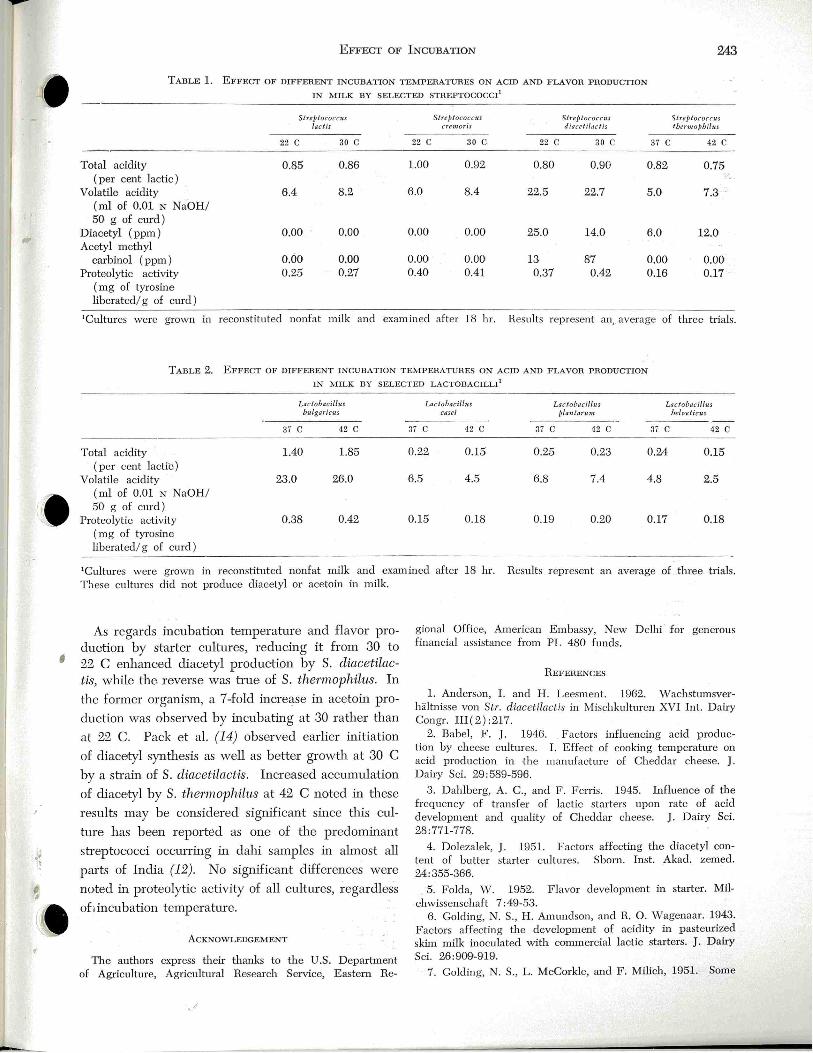
I
·' l
l _
• ;
EFFECT OF INCUBATION 243
TABLE l. EFFECT OF DIFFERENT INCUBATION TEMPERATURES ON ACID AND FLAVOR PRODUCTION IN MILK BY SELECTED STREPTOCOCCI'
Total acidity (per cent lactic)
Volatile acidity · (ml of 0.01 N NaOH/ 50 g of curd)
Diacetyl (ppm) Acetyl methyl
carbinol (ppm) Proteolytic activity
( mg of tyrosine liberated/ g of curd)
Streptococcus /act is
22 c 30 c
0.85 0.86
6.4 8.2
0.00 0.00
0.60 0.00 0.25 0.27
Streplococcm cremoris
22 c 30 c
1.00 0.92
6.0 8.4
0.00 0.00
0.00 0.00 0.40 0.41
Streptococctts !itreptococc1lS diacelilaclis tbennoPbilus
22 c 30 c 37 c 42 c
0.80 0.90 0.82 0.75 -.
22.5 22.7 5.0 7.3 .
25.0 14.0 6.0 12.0
13 87 0.00 0.00 0.37 0.42 0.16 0.17
'Cultures were grown in reconstituted nonfat milk and examined after 18 hr. Results represent an,. average of three trials.
TABLE 2. EFFECT OF DIFFERENT INCUBATION TEMPERATURES ON ACID AND FLAVOR PRODUCTION IN ~~!ILK BY SELECTED LACTOBACILLI'
Lactobacillus Lac tobacillus Lactobacillus Laclobacillus bulgaricus casei plantartwz, belveticus
37 c 42 c 37 c 42 c 37 c 42 c 37 c 42 c
Total acidity 1.40 1.85 0.22 0.15 0.25 0.23 0.24 0.15 (per cent lactic)
Volatile acidity 23.0 26.0 6.5 4.5 6.8 7.4 4.8 2.5 (ml of 0.01 N NaOH/ 50 g of cmd)
Proteolytic activity 0.38 0.42 0.15 0.18 0.19 0.20 0.17 0.18 ( mg of tyrosine liberated/ g of curd)
'Cultures were grown in reconstituted nonfat milk and examined after 18 hr. Results r epresent an average of three trials. These cultures did not produce diacetyl or acetoin in milk.
As regards incubation temperature and flavor production by starter cultures, reducing it from 30 to 22 C enhanced diacetyl production by S. diacetUact·is, willie the reverse was bue of S. thennophilus. In
the former organism, a 7-fold increase in acetoin pro
duction was observed by incubating at 30 rather than
at 22 C. Pack et al. (14) observed earlier initiation
of diacetyl synthesis as well as better growth at 30 C
by a sb·ain of S. diacetila.ctis. Increased accumulation
of diacetyl by S. thennophilus at 42 C noted :in these
results may be considered significant since tltis cul
tme has been repmted as one of tl1e predominant
streptococci occmTing in dahi samples in almost all
parts of India (12). No s.ignificant differences were
noted in proteolytic activity of all cultmes, regardless
of1incubat:ion temperatme.
ACKNOWLEDGEMENT
TI1e authors express their thanks to the U.S. Department of Agriculture, Agricultural Research Service, Eastern Re-
• r
gional Office, American Embassy, New Delhi for generous fin ancial assistance hom PL 480 funds.
REFER);:NCES
l. Andersvn, I. and H. Leesment. 1962. vVachstumsverhaltnisse von Str. diacetilactis in Mischkulturen XVI Int. Dairy Congr. III(2) :217.
2. Babel, F. J. 1946. Factors influencing acid production by cheese cultures. I. Effect of cooking temperature on acid production in the manufactme of Cheddar cheese. J. Dairy Sci. 29:589-596.
3. Dahlberg, A. C., and F . Ferris. 1945. Influence of the frequency of transfer of lactic starters upon rate of acid development and quality of Cheddar cheese. J. Dairy Sci. 28:771-778.
4. Dolezalek, J. 1951. Factors affecting the diacetyl content of butter starter cultmes. Shorn. Inst. Akad. zeroed. 24:355-366.
5. Folda, W. 1952. Flavor development in starter. Milchwissenschaft 7:49-53.
6. Golding, N. S., H. Amundson, and R. 0. Wagenaar. 1943. Factors affecting the development of acidity in pasteurized skim milk inoculated with commercial lactic starters. J. Dairy Sci. 26:909-919.
7. Golding, N. S., L. McCorkle, and F. Milich, 1951. Some

.1'
244 DUTrA ET. AL.
factors which may affect activity tests for lactic starters. Proc. W. Div. Amer. Dairy Sci. Ass. 36-46.
8~ H ammer, B. vV., and F . J . Babel. 1957. Dairy bacteriology. 4th eel. John Wiley & Sons Inc., New York.
9. Harrison, J., and D. V. D earden . 1941. Slowness in cheese making. J. Dairy Res. 12:35-43.
10. Hempenius, W . L., and B. J. Liska. 1968. Method for detenninfug volatile acids in cultured milk products. J. Dairy Sci. 51:221.
ll. Hull, M. E. 1947. Studies on milk proteins. II. Colorimetric determination of the partial hydrolysis of the proteins in milk. J . Dairy Sci. 30:881.
12. Laxminarayana H., V. K. N. Nambudripad, V. Lakshminarasim, S. N. Ananthru:amiah, V. Sreenivasanmrthy, and K. K. Iya. 1952. Studies on dah.i. III. Taxonomy of lactic
REPORT OF COMMITTEE ON FOOD EQUIPMENT
(Continued from Page 227)
man of the P)lblic Health Group and also served as Co-Chairman of AMHIC during 1970-1971.
Ev~tluation ma.mwl fo1· food and beverage vending machines Carbonation backflow pmvent-ion. The form er Item 603.1
( 1)', ( 2) , ( 3), and ( 4) on general requirements to prevent carbonation backflow in post-mix beverage machines is being replaced by the new and more stringent Item 603.1 which is d-escribed in the following three paragraphs:
"In those vending machines in which the accidental backflow of carbon dioxide or carbonated water from the carbonator could come into contact with potentiallytoxic tubing or components upstrerun, an aiJ: gap, atmospherically-vented valve or other positive venting device ( in addition to the check valves or other devices necessary to maintain C02 pressure) shall be installed between the carbonator and any such potentially-toxic tubing or components upstream.
NOTE: A brass ptunp housing shall not be considered 'potentially-toxic' in a system which is otherwise free of copper or other materials listed in 602.3.
ill machines designed to be directly connected to the location water supply (no air-gapped water reservoir ), such supply lines shall be considered to be potentiallytoxic. Machines designed for such direct cmmection shall be equipped with an air gap , atmospherically-vented valve, or other positive venting device between the carbonator and the water connection fitting, even if no potentially-toxic tubing or components are present in the internal water system of the machine."
In addition, a Committee was appointed at the 1970 meeting to explore the need to rule out the use of copper pre-cooler (or other) tubing in the carbonated water systems of postmix beverage vending machines. At the present time this Committee and the Carbonation Backflow Committee are considering the following available options: (a) evaluate the design and operational features of vented solenoid-type valves; (b) recommend tests for such valves to detennine -if they fail safe under all circumstances and, if not, what corrective m_ea~ures are needed; (c) Repeat (a) and (b) for the n ew Watts
acid bacteria of dahi. Ind. J . Vet. Sci. 22(1):27-49. 13. Pack, M. Y., W. E. Sandine, P . R. Elliker, E. A. Day,
'imd R. C. Lindsay. 1964. Owades and J a·covac method -£or diacetyl detem1ination in mixed strain starters . J . Dairy Sci. 47:981. !
14. Pack, M. Y., E. R. Vedanmthu, W . E. Sandine, P. R. Elliker, and H. Leesment. 1968. Effect of t emperature on growth and diacetyl production by aroma bacteria in single and mixed strain starter culture. J. Dairy Sci. 51:339-344.
15. Sasaki, R. , T. Tsugo, T. linan1m·a, and M. Murai. 1953. Studies on the diacetyl production .by lactic acid bacteria. Int. Dairy Congr. 4:611-613.
16. Sasaki, R. , T . Tsugo, T. Imrunura, and M. Murai. 1954. Studies on the diacetyl production by lactic acid bacteria. Jap . J . Zootech Sci. 24:176-179.
o. 9BD non-electric vented valve; and (d) Recommend to the Manual Revision Committee : ( i) adoption of a "no intemal copper tubing" requirement; or, ( ii) new design or , testing standards for vented valves; or, ( iii) recommend no action.
Fmthermore, it is considered commendable that the NSF and 1 AMA have voltmtru:ily agreed to maintaill unifom1 specifi cations for the evaluation of food and beverage vending machines. Furthennore, NSF has aheady arrru1ged to adopt the new provisions of the aforementioned Item 603.1 in NSF Standard No . 25 relating to Food and Beverage Vending Machines.
Gnp tu·rret covers. During the 1969 meeting, Item 505.2 concernillg covers for cup turrets was amended to require that covers be made availabie but not installed on the machine, since in the opinion of several public h ealth representatives. the top of the machines afforded reasonable protection to the cups. Nevertheless, at the 1·equest of the members of th e vending industry, Item 505.2 was runended during the 1970 meeting to require covers on all cup turrets.
lcem.aker standa-rds. The present Evaluat-ion Mmwal has not provided specific items on icemaker design and construction. Icemakers, b y implication, would be required to meet applicable specifications for food and water contact surfaces. This has been deemed to be inadequate, and based on the 1968-1969 NAMA icemaker research project and fi eld observa tions, consideration is being given to adding a new item to the Man-ual on l cem.aker Systems. This new item would include construction and design specifications relating to accessibility, materials and fabrication , water reservoirs, water tu billg, ice storage hopper~ , icemaker harvesters, ice dispensing chutes, and water filters .
l cemaker san-itation bullet i-n. At the request of the members of AMI-IIC, the Committee on Education and Training in cooperation with the NAMA staff has developed a first draft of a bulletin on icemaker sanitation for vending operators. This proposed bulletin is being designed to pose significant questions ru1d -then to provide appropriate answers on subjects such as: Is it algae? Where does growth occur? How should icemakers be cleaned? ' ¥ hat cleaners should be used? What cleaning procedures should be used? How often should icemakers be cleaned? The application of the illformation proposed in this bulletin and in the icemaker
(Continued on Page 251)
]. Milk Food Technol. , Vol. 35, No. 4 (1972) .245
3-A SANITARY STANDARDS FOR AIR ELIMINATORS FOR MILK AND FLUID MILK PRODUCTS
Serial #2900
Formulated by _ Intemational Association of Milk, Food and Environmental Sanitarians
United States Public Health Service The Dairy Industry Committee
It is the purpose of the IAMFES, USPHS, and DIC in connection with the development of the 3-A Sanitary Standards program to allow and encourage full freedom for inventive genius or new developments. Air eliminator specifications heretofore and hereafter developed which so differ in design, material, fabrication, or otherwise as not to conform with the following standards, but which, in the fabricator's opinion are equivalent or better, may be submitted for joint consideration of the IAMFES, USPHS, and DIC, at any time.
A.
A.1
SCOPE
These standards cover the sanitary aspects of air eliminators for milk and fluid milk products.
steel of the AISI 300 series1 or corresponding ACP types (See Appendix, Section E.), or equally corrosion resistant metal that is non-toxic and nonabsorbent, except that:
C.1.1
fli.A.2 ~ In order to conform with these 3-A Sanitary Stan-
dards, air eliminators shall comply with the following design, material, and fabrication criteria.
Rubber and rubber-like materials may be used for gaskets, seals, 'and parts used in similar applications. These materials shall comply with fhe applicable provisions of the "3-A Sanitary Standards for Multiple-Use Rubber and Rubber-Like Materials Used as Product Contact Surfaces in Dairy Equipment, Serial #1800".
I
•' ~
B. DEFINITIONS
8.1 Product: Shall mean milk and fluid milk products.
8.2 i SU1jaces :
8.2.1 Product Contact Surfaces: Shall mean all surfaces which are exposed to the product and surfaces from which liquid may drain, drop, or be drawn into the product.
8.2.2 Non-Product Contact Su1jaces: Shall mean all other exposed surfaces.
8.3
c.
C.1
Mechanical Clean·ing o1· Mechan·ically Cleaning: Shall denote cleaning, solely by circulation and/ or flowing chemical detergent solutions and 'water rinses onto and over the surfaces to be cleaned, by mechanical means.
MATERIALS
All product contact surfaces shall be of stainless
C.1.2
C.2
Plastic materials may be used for inner liners, valve parts, gaskets, seals, and parts used in similar applications. These materials shall comply with the applicable provisions of the "3-A Sanitary Standards for Multiple-Use Plastic Materials Used as Product Contact Surfaces for Dairy Equipment, Serial #2000", as amended.
All non-product contact surfaces shall be corrosion-resistant material or material that is rendered corrosion-resistant. If coated, the coating used shall adhere. All non-product contact surfaces shall be relatively nonabsorbent, durable and cleanable. Parts removable for cleaning having both
1The data for this series are contained in the following reference: AISI Steel Products Manual, Stainless & Heat Resisting Steels, April, 1963, Table 2-1, pp. 16-17. Available from: American Iron and Steel Institute, 633 Third Avenue, New York, New York 10017. 2Alloy Casti~g Institute Division, Steel Founders;' :-Society of America, 21010 Center Ridge Road, Rooky River, OKio 44116.
.J'
246 3-A SANITARY STANDARDS
D.
product contact and non-product contact surfaces shall not be painted.
FABRICATION 0.1
All product contact surfaces shall be at least as smooth as a No. 4 mill finish on stainless steel sheets. (See Appendix, Section F.)
0.2 All permanent joints in product contact surfaces shall be welded. All welded areas of product contact surfaces shall be at least as smooth as the adjoining surfaces.
0.3 All product contact surfaces shall be easily accessible for cleaning, either when in an assembled position or when removed. Removable parts shall be readily demountable.
0.4 Means for mechanically cleaning the air eliminator, when provided, shall clean the product contact surfaces of the air eliminator and all nonremovable appurtenances thereto except those areas requiring manual cleaning.
0.5 All product contact surfaces shan be self draining except for normal clingage.
0.6 All internal angles of 135° or less on product contact surfaces shall have minimum radii of 1/4 inch except the radii of vent seal parts may be 1/8 inch.
0.7 There shall be no threads on product contact surfaces.
0.8 Sanitary pipe and fittings shall conform with "3-A Sanitary Standards for Fittings Used on Milk and Milk Products 'Equipment and Used on Sanitary Lines Conducting Milk and Milk Products, Serial #0809", as amended and supplements thereto and /or the applicable provisions for welded sanitary product pipelines found in the "3-A Accepted Practices for Permanently Installed Sanitary Product-Pipelines and Cleaning Systems", effective June 9, 1966, Serial #60500, as amended.
0 .9 Air vents shall be designed or protection provided to prevent foreign material from entering the air eliminator through the air vent.
0.10 Gaskets shall be removable. Gasket retaining grooves shall be no deeper than their width. The minimum radius of a gasket retaining groove shall
not be less than 1/8 inch, except that the radius may be 3/ 32 inch where a standard 1/4 inch 0 -Ring is used and the radius may be 1/3~ inch where a standard 1/8 inch 0-Ring is used. ·
0.11 A pressure transmitter, if used, shan be installed so that it is relatively flush with the product contact surface if the air ~liminator is designed to be mechanically cleaned.
0.12
If legs are used, they shall be smooth with rounded ends and no exposed threads. Legs made of hollow stock shall be sealed. The clearance between the lowest part of the air eliminator (excluding legs) and the floor shall be one of the following:
0.12.1 Not less than four inches if the horizontal area oj the air eliminator is more than one square foot.
0.12.2 Not less than two inches if the horizontal area of the air eliminator is not more than one square foot and the air eliminator is designed to be portable.
0.13
E.
F.
Non-product contact surfaces shall be free of pockets and crevices and be readily cleanable and those to be coated shall be effectively prepared for coating.
APPENDIX
STAINLESS STEEL MATERIALS Stainless steel conforming to the applicable com
position ranges established by AISP for wrought products, or by ACP for cast products, should be considered in compliance with the requirements of Section C.1 herein. Where welding is involved the carbon content of the stainless steel should not exceed 0.08%. The first reference cited in C.l sets forth the chemical ranges and limits of acceptable stainless steels of the 300 series. Cast grades of stainless steel equivalent to types 303, 304, and 316 are designated CF-16F, CF-8, and CF-8M, respectively. These cast grades are coverer by ASTM' specifications A296-68 and A351-70.
PRODUC T CONTACT SURFACE FINISH Surface finish equivalent to 150 grit or better as obtained with silicon carbide, is considered in compliance with the requirements of Section D.l herein.
These Standards are effective June 23, 1972.
'Available from American Society for Testing and Materials, 1916 Race Street, Philadelphia, Pennsylvania 19103.
•• I

]. Milk Food Technol ., Vol. 35, No. 4 (1972) 247
SURVIVAL OF POLIOVIRUS AND ECHOVIRUS DURING SIMULATED COMMERCIAL EGG PASTEURIZATION TREATMENTS
N. R. SmoCK AND N. N. PoTIER Department of Food Science
Comell Univer;ity Ithaca, New York 14850
(Received for publication September 27, 1971 )
Fresh liquid whole egg and egg white were inoculated with poliovi..Ius and echovirus and subjected to various process conditions of egg pasteurization. The process conditions studied included Salmonella destmctive tin1e-temperature exposm es for whole egg and egg white, and various h eat-peroxide treatments for egg white. Viruses did not survive timetemperature exposures that are effective against Salmonella in whole egg or egg white, but survived at various levels the heat-peroxide treatments applied to egg white. Although commercial equipm ent was not used in these studies, times, temperah1res, and peroxide levels closely approxilnated those currently used commercially.
Currently in the U. S. and certain other countries
• all forms of liquid egg used commercially must receive a pasteurization treatment to render the egg Salmonella-negative. Regulations with respect to time-temperature e>.'Posur~s are not rigid, however,
(11) indicates that 56 C for 5 min is sufficient to kill poliovirus. Infectious hepatitis virus was found to withstand heating at 56 C for 30 min (6). A study on inactivation. of foot and mouth disease virus compared heating of cell-free inoculum and heating in tissue culhue suspensions (4). ' The latter imparted considerable protection to the virus, which was still active within cells after 4 hr at 80 C. Viruses often are more stable in food than in artificial media, however, tl1e effects of food processing on viJ:us destruction have received relatively scant attention (2, 3). A search of the literature has revealed a dearth of information on tl1e viricidal effects of commercial egg pasteurization processes. Treatments used ill the present investigation were modeled after commercial processes with regard to times, temperatures, and p eroxide levels in the case of egg white. One strain of poliovirus and one of echovb·us as inoculum were studied in both whole egg and egg white.
I
·' ~
and the processor currently may choose from several treatments that have been shown to be effective against Salmonella (1). All commercial pasteurization treatments for egg are considerably milder in terms of heat exposures than are used for milk and various other low acid foods because of the high order of sensitivity of the functional properties of
; eggs to heat (5, 7, 8, 10). With egg white, which is still more sensitive than yolk containing products, temperatures as low as 51.7-54.4 C for 3.5 min or less combined with hydrogen peroxide addition constitutes effective pasteurization against Salmonella (1 ).
•
Egg products, of course, are subject to contamination by agents ·other than Salmonella, and among tl1ese are viruses able to cause human infection. In the commercial production of shelled egg in the U. S., egg breaking is mostly done by machine. There are, however, many small operations that still use hand labor to break eggs, thus creating the possibility of human viral contamination. Egg spoilage, even when machine breaking is used, is tested for by sniffing broken out eggs at regular intervals, inh·oducing an-other possible source of human viral contan1ination.
Many studies on heat treatment of viruses and their survival have been made. These are quite often in disagreement because of different measuring techniques and conditions. One report (9) shows that poliovirus survives at . 55 C for 60 min while another
MATERIALS AND METHODS
Vi.ruses Echovirus type 6 strain D' Amari and Poliovirus type 1
strain chat were obtained from tl1e American Type Culture Collection. TI1ey were propagated on green monkey kidney cells obtained from tl1e Veterinary Virus Research Institute at Cornell University.
Media and reagents All media and reagents were obtamed from tl1e Grand Is
land Biological Company, Grand Island, N.Y. The following reagents were used: (a) fetal calf senun (GIBCO 614), (b) gamma globulin-free calf serum (GIBCO 624), (c) minimal essential medium ( MEM) (Eagle) ( 1 X ) with L-glutamine ( GIBCO 109G ) , (d) lactalbumm hydrolysate solution ( GIBCO 164) , (e) penicillin-streptomycin solution ( 10,000 units penicillin and 10,000 meg streptomycm/ml) ( GIBCO 514 ), and (f) trypsm-EDTA solution ( 1 X) ( GIBCO 530).
The above reagents were mixed in solution to fom1 ilie followmg media: Med·ium A: 78% Eagle's MEM, 10% lactalbumm hydrolysate, 10% fetal calf serum, 2% penicillin-streptomycin; Medi.um B: 86% Eagle's MEM, 10% lactalbumi11 hydrolysate, 2% fetal calf serum, 2% p enicillin-streptomycin; Mediu.m C: 86% Eagle's MEM, 10% lactalbumin h ydrolysate, 2% gamma globulin-free calf serum, 2% penicillin-streptomycin; m1d Medium D : 58% Eagle's MEM, 10% lactalbumi.D. .. hydrolysate, 20% fetal calf sermn, 2% penicillin-streptomycin, 10% glycerol.
Cell maintenance Cells were grown at 37 C in glass Roux bottles containing
.J'
248 STROCK AND PorrnR
80 ml of Med ium A. ·when fully sheeted ( 2-3 days after passing), this mediwn was poured off and replaced with 80 ml of Medium B. Cells could be maintained 2-3 weeks on the meditml.
Cell passing The medilllll was poured from the Roux bottles and 40 ml
of trypsin-EDTA added . Bottl es were in cubated at 37 C for 5 min and then shaken to dislodge and separate cells. The trypsinized cells were then centrifuged and the p ellet resuspended in 240 ml of Medium A.
Virus titrations were clone in 16 X 150 mm tissue cultme tubes. One milliliter of cell suspension was added to each tissue culture tube, which was th en stoppered and incubated at a 5o angle.
V-irus propagation Viruses were propagated on cells in disposable, plastic, 75
cm2 surface tissue culture fl asks. The flasks received 30 ml of suspended cells which were grown to a full cell sheet in 2-3 clays at 37 C. The mediwn was pomecl off and 0.2 ml of virus suspension was added. F lasks were incubated at 37 C for 30 min to permit virus adsoqJtion to the cells . Thirty milliliters of Medium C were added and cells were incubated at 37 C.
At the first sign of cytopathic effect ( CPE), flasks and contents were frozen. Generally, CPE appeared after 2 days with echovirus and 4 clays with poliovirus . It was at this point that maximum titers ~vvere obtained. Cells were frozen and thawed tlu·ee times to ruptme them and free virus particles. vVhen allowed to incubate further, viruses were inactivated by metabolic products and low pH produced by the cells.
Vims titration Virus was titrated following dilution in Medium C. Two
tentlls milliliter portions were added to tissue culture tubes (cells growing, medium removed) prepared by the cell passing meiliocl described above. Tubes were then incubated at 37 C for 30 min. Following incubation, 1.5 ml of Mediw11 C was added to each htbe. Tubes were tllen incubated at 37 C and results read after the appt:opriate incubation period ( 2 days for echovirus, 4 days for p oliovirus). Titrations were clone in triplicate using th e most probable number method for enw11 eration. Tlu-ee control tubes (no virus) were used fo r each titration to assist with CPE detection .
Reproducibility of titration m ethod Reproducibility of the titration method was determined in
Medium C and in egg white (pH 8.5 ). In egg white, this 'included preparing a homogeneous virus suspension.
For titration reproducibility in Mediwn C, five identical portions of poliovirus suspension were titrated. 'Vith egg white, 5 ml of poliovirus suspension in Mediwn C were added to 25 ml of egg white in a 250 ml E rlenmeyer flask and stirreel until tl1e pH indicator of Mecliwn C was llllifom1ly spread throughout the mixture. Anotl1er 75 ml of egg white were added and stirred for 10 min. Thick and thin protein were completely intermixed and there was no foaming. Three identical portions were taken, one from the center, one from the top, and one from tile bottom of the flask. Titration results are shown in Table 1.
Heat pasteu:rization treatments Pasteurization studies were carried out in a controlled temp
erature water bath. Vims inoculated fresh shelled egg was injected into stainless steel tubes of 3/16-inch outside diameter witl1 10 mil thick walls with a syringe. Tubes of 8-inch length held about 1 ml of egg. Come up ti.nle to 60.0 C was
15 sec measured by a temperahrre sensor in the tube held in the water bath. These i5 sec were added onto the processing times listed in subsequent tables. Tubes of heated egg white-vims mixtures (pH 8.5) and whole egg-virus mixtures (pH 7.0) were chilled in an ice bath to 10 C in1mediately after processing. Portions of 0.2 ml were then titrated as previously described.
T-l qat-h ydrogen -peroxide pasteu.Tization t·reatm ents ":he combined bactericidal effects of heat and hydrogen
peroxide enable tl1e commercial processor to use lower pasteurization temperatures than would be effective with heat alone. Hydrogen peroxide pasteurization is used commercially for egg white only. The following treatments differed in tile amount of catalase destruction ( natmal catalase of the egg) and the amollllt of peroxide added. In each treatment th e temperatures studied (see Results) were ilie same.
Heat-peroxide pasteuriza tion treatment A consisted of heating 100 ml of egg white at 51.7 C for 30 min to inactivate the natural catalase of the egg. The egg was cooled and 5.0 ml of virus suspension added. The egg-virus mLxtme was made homogeneous and 10 ml portions were heated in test tubes to the pasteurization temperature of 54.4 C or 51.7 C. , Come up tin1e was approximately 2.5 min. Hydrogen peroxide was added at a level of 0.07% ( 0.23 ml of 3% H 202). The egg was held at the respective pasteurization t emperatme for 2.0 min. At tl1e end of the holding period, the egg was inlmediately cooled in an ice bath to 10 C and catalase added to decompose any remaining p eroxide. To observe the effect of the peroxide alone on the viruses, the above procedw·e was also followed using room temperature as the holding (pasteurization) temperatme.
Heat-peroxide pasteurization treatment B was a more realistic test of the egg pastemization method as presently ;,Jracticed commercially. One hundred milliliters of egg white were inoculated witl1 virus and made homogeneous. Ten milliliter portions of egg-virus suspension were heated in test tubes to 54.4 C or 51.7 C and h eld for 1.5 min . This was the catalase destruction period. Peroxide at the 0.07% level was added and egg held at 54.4 C or 51.7 C for 2.0 min of pastemization. Egg was cooled and catalase added. Mter 1.5 min ·of peroxide decomposition, the egg was titrated.
Tllis treatment differed from treab11ent A in two ways. In treatment A, all the catalase in the egg was de~troyed before addition of tl1e virus or peroxide. Thus, there was no peroxide decomposition dming processing from tllis enzyme. In treatment B, however, catalase de~truction may have been incomplete after tl1e 1.5 min allowed for this purpose. Some peroxide might then have been decomposed before being able to fully affect the viruses. Also in treatment B, virus was present during the 1.5 min catalase inactivation period before the pasteurization p eriod, thus receiving h eat for 3.5 min rather than 2.0 min as in treatm ent A.
H eat-peroxide pasteurization treatment C disregarded the natmal catalase of the egg and compensated by using a higher level of peroxide. Ten milliliter portior:s of virus-inoculated, mLxed egg wh ite were heat ed in test tubes to ilie pasteurization temp erature of 54.4 C or 51.7 C and 0.09% hydrogen p eroxide ( 0.3 ml of 3% H202) was added. The egg was h eld at pastemization temperature for 3.5 min. It was then cooled in an ice batl1 alld catalase added. After 1.5 min of peroxide decomposition tl1e egg was .titrated.
RESULTS
Effects of heat pasteurization t1'eatments The heat pastemization of whole egg resulted in
( .. I
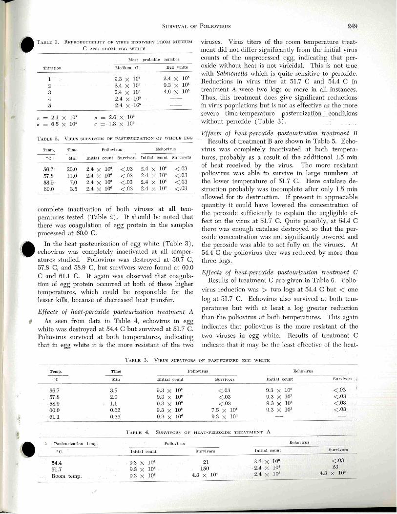
--
I
SunvrvAL oF Pouovmus 249
• TABLE l. R EPRODUCIDIL ITY OF VIRUS RECOVERY FROM MEDIUM
C AND FROJ\•I EGG WHITE
Titration
l 2 3 4 5
J.t - 2 .1 X 10' u 6.5 X 10⁴
f.'=
u
Most probable number
Medium C Egg white
9.3 X 1()4 2.4 X 10⁵
2.4 X 10' 9.3 X 10⁴
2.4 X 10⁵ 4.6 X 10⁵
2.4 X 10' 2.4 X lG5
2.6 X 10⁵
1.8 X 10⁵
TABLE 2. VIRUS SURVI\'ORS OF PASTEURIZATION OF WHOLE EGG
Temp. Time Poliovirus l~c.h ov iru s
·c Min Initi a l count Survivors Initial count Survivors
56.7 20.0 2.4 X 10" <.03 2.4 X 1()4 <.03 57.8 11 .0 2.4 X lOG < .03 2.4 X 10⁴ <.03 58.9 7.0 2.4 X lOG < .03 2.4 X lQ• <.03 60.0 3.5 2.4 X 10" <.03 2.4 X l()'' < .03
complete inactivation of both vimses at all temperatures tested (Table 2). It should be noted that there was coagulation of egg protein in the samples processed at 60.0 C. n. In the heat pasteurization of egg white (Table 3),
\~ echovirus was completely inactivated at all temperatures studied. Poliovirus was desb·oyed at 56.7 C, 57.8 C, and 58.9 C, but survivors were found at 60.0 C and 61.1 C. lt again was observed that coagulation of egg protein occurred at both of these higher temperatures, which could be responsible for the lesser kills, because of .decreased heat h·ansfer .
Effects of heat-peroxide pasteurization treatment A
I As seen from data in Table 4, echovirus in egg white was desb·oyed at 54.4 C but survived at 51.7 C. Poliovirus survived at both temperatures, indicating that in egg white it is the more resistant of the two
viruses. Virus titers of the room temperature b·eatment did not differ significantly from tl1e initial virus counts of the unprocessed egg, indicating that peroxide without heat is not viricidal. This is not tme 'Arith Salmonella which is quite sensitive to peroxide. Reductions in virus titer at 51.7 C and 54.4 C in treatment A were tv,ro logs or more in all instances . Thus, this b·eatment does give significant reductions in virus populations but is not as effective as the more s~vere time-temperature pasteurizatLon - conditions without peroxide (Table 3).
Effects of heat-perox-ide pasteu?"ization treatment B Results of b·eatment B are shown in Table 5. Echo
virus was completely inactivated at both temperatures, probably as a result of tl1~ additional 1.5 min of heat received by tl1e virus. The more resistant poliovirus was able to survive in large numbers at the lower temperature of 51.7 C. Here catalase desh·uction probably was incomplete after only 1.5 min allowed for its desb·uction. If present in appreciable quantity it could have lowered the concentration of the peroxide sufficiently to e;\'plain tl1e negligible effect on the virus at 51.7 C. Quite possibly, at 54.4 C there was enough catalase destroyed so that the peroxide concenb:ation was not significantly lowered and the peroxide was able to act fully on tl1e viruses. At 54.4 C the poliovims titer was reduced by more than three logs.
Effects of heat-peroxide pasteurization t1·eatment C Results of treabnent C are given in Table 6. Polio-
virus reduction was > 1:\vo logs at 54.4 C but < one
log at 51.7 C. Echovirus also survived at both tem
peratures but witl1 at least a log greater reduction
than the poliovirus at both temperatures. This again
indicates that poliovirus is the more resistant of the
1:\;vo viruses in egg white. Results of treabnent C
indicate tl1at it may be the least effective of tl1e heat-
TABLE 3. VIRUS SURVIVORS OF PASTEURIZED EGG WHITE
Temp. Time 1=-oliovirus Echovirus ---
·c Min Initia l count Survivors Initial count Sun·ivor:s }
56.7 3.5 9.3 X lOG <.03 9.3 X HI' <.03 57.8 2.0 9.3 X lOG <.03 9.3 X 10³ <.03 58.9 l.l 9.3 X 10⁶ < .03 9.3 X 10³ < .03 60.0 0.62 9.3 X 10⁶ 7.5 X 10⁴ 9.3 X 10³ <.03
·~ 61.1 0.35 9.3 X lOG 9.3 X 10³
;
Pasteurization temp.
•c
54.4 51.7 Room temp.
·"
TABLE 4. SURVIVORS OF HEAT- PEROXIDE TREATMENT A
Poliovirus
Initial count Survivors Initial count
9.3 X 10⁴ 21 2.4 X 10³
9.3 X 10"' 150 2.4 X 10³
9.3 X 10'' 4.3 X 10"' 2.4 X 10³
Echovirus
Survivors
<.03 23
4.3 X 10³

.J'
250 STRoCK AND PoTIER
TABLE 5. SURVIVORS OF HUT-PEROXIDE TREATMENT B
Pasteurization temp. Poliovirus Echovirus
·c Initia l count Survivors Initial count Sun•ivors
15 54.4 51.7
4.3 X 10• 4.3 X 10⁴ 1.1 X 10⁴
2.9 X 10⁴
2.9 X 10'1
TABLE 6. SURVIVORS OF HUT-PEROXIDE TREATMENT C
Poliovirus Echovirus Pasteurization temp.
·c Initial count Survivors Initial count Survivors
54.4 51.7
4.3 X 10' 4.3 X 10⁴
2.4 X 10" 9.3 X 10³
2.9 X 10⁴
2.9 X 10⁴
9.1 43
peroxide pasteurization h·eahnents studied with respect to the test viruses. This h·eahnent disregarded heat inactivation of the natural catalase present in the egg white before peroxide addition and a higher concenh·ation of peroxide was added to offset decomposition from catalase. However, too much peroxide probably v.ras decomposed before it was able to act on the viruses. Since the heating time was similar to that of heat-peroxide h·eahnent B which destroyed the echovirus, the difference appears to l; e with decomposition of the peroxide.
DISCUSSION
Throughout these studies, eggs were inoculated with high titers of virus to facilitate measurement of the effects of pasteurization h·eahnents on reduction of virus populations. In commercial egg handling where virus infection may occur, it is not likely that levels of contamination would be as high. Further, should virus-contaminated egg be processed indush·ially, viruses would become greatly diluted.
Results of the heat pasteurization of whole egg show that tl1e two viruses selected for this study were completely inactivated over tl1e entire time-temperature range tested. · ·vvitl1 egg white pasteurized by heat alone, poliovirus, which was completely inactivated at temperatures from 56.7 to 58.9 under the test conditions, survived the heat h·eatrnents at the higher temperatures of 60.0 C and 61.1 C. This survival is not ath·ibuted to resistance of the virus but to coagulation of egg white protein tl1at occurred. The coagulation caused a drop in the heat h·ansfer rate which, in turn, minimized tl1e heat ·treatment that was received by the virus. This decreased heat h·ea:hnent, nevertheless, still was effective against the less resistant echovirus.
Although sh·aight heat pasteurization over a range of times and temperatures that span commercial practice appear to be quite adequate for destruction of test viruses studied in both whole egg and egg white,
a similar conclusion cannot be reached with milder heat plus peroxide commonly used for commercial pasteurization of egg white.
The treahnents used in this study were modeled after commercial processes with regard to times, ' temperatures, and peroxide levels. To be sure, tl1e current studies carried out under small batch-heating laboratory conditions only approximate the true exposures encountered by liquid egg in commercial pasteurization equipment. However, it is not unlikely that conditions exist where current commercial pasteurization of egg white with heat plus peroxide are no more severe than in the present studies. Such egg white may be rendered Salmonella-negative but could retain virus. It therefore would appear that virus survival in mildly pasteurized liquid egg, especially egg white, is worthy of additional study. To be most meaningful, such studies should include pasteurization processing with industrial equipment. ·
ACKNOWLEDGEMEi\"T
This sh1dy was supported, in part, by the National Institutes of Health Training Grant FD 00002 from the United States Department of Health, Education, and \Velfare, Public Health Service.
REFERENCES
1. Agricultura l Research Service. 1969. Egg pasteurization manual. A.R.S . Bulletin 74-48.
2. Berg, G. 1964. The food vehicle in virus transmission. Health Lab. Sci. 1:5 1-59•.
3. Cliver, D. 0. 1967. Food associated viruses. Health Lab. Sci. 4:213-221.
4. D emopoullos, G. T., 0. N. Fellowes, J. J. Collis, G. C. Poppensiek, A. G. Edwards, and J. H. Graves. 1959. Thermal inactiviation and antigenicity studies of heated tissue suspensions containing foot and mouth disease virus. Arner. J. Veterinary Res. 20:510-521.
5. Goresline, H . E., K. M. Hayes, R. E. Moser, Jr., M. A. Howe, and E. E . Drewniak. 1951. Pasteurization of liquid whole egg under commercial conditions to eliminate Salmonella . USDA Circular No . 897:1-15.
6. Havens, W. P., Jr. 1945. Properties of the etiological
,
\ ,. '

SURVIVAL OF Pouovmus 251
•
:gent of infectious hepatitis. Proc. Soc. Exp . Bioi. Med. ( u8 :203.
7. Kline, L. T. F., M. L. Sugihara, M. L. Bean, and K. Ljichi. 1965. Heat pasteurization of raw liquid egg white. Food Techno!. 19:1709-1718.
8. Lineweaver, H. 1966. Pasteurization and the func-tional properties of egg products. Proc. Rutgers Liquid Egg Processing Course, Rutgers University, New Brunswick, New Jersey.
REPORT OF COMMITTEE ON FOOD EQUIPMENT
(Continued from Page 244)
standards should assure the production and dispensing of a safe and acceptable ice product.
Cut-off controls. The members of AMHIC received reports on some of the technical problems encountered by industry and public h ealth in obtaining effective and reliable automatic controls to prevent the storage and dispensing of potentially hazardous foods at unsafe temperatures, between 45 F and 140 F, and in obtaining a workable field t est to detem1ine the accuracy and reliability of these controls. To overcome these problems and to accomplish the objectives of the cut-off controls, several major changes in machine de-
lr.ign are presently being considered by the manufacturers ~ , nd the members of the Committee on Cut-Off Controls;
and it is anticipated that within the n ext year appropriate and essential changes will be made in the E valuation Manual to more adequately protect the conswner and to aid the evaluator and reguJatory officials in carrying out their responsibilities.
I
·' )
Remanufactured machines The program for Evaluation of Remanufactured Vending
Machines seemed to have tremendous appeal, during th e early planning stage, to members of the public h ealth agencies and vending machines operating companies; and several
- of the firms rebuilding vending machines requested this program for evaluation of other-than-new machines. However, to date, only three remanufacturers have submitted machines for evaluation and only two have r eceived Letters of Compliance to manufacture machines under tl1is NAMA program even tl10ugh several written invitations to participate in the program h ave been sent by NAMA to all known remanufacturers.
Coin-operated special devices The NAMA staff reported at the 1970 meeting that several
requests had been receivd during tl1e past year from members of tl1e public h ealth agencies and tl1e vending industry to develop criteria for the e.valuation of tl1e small coin-operat-ed office coffee service machines. After thoroughly discussing these requests and recognizing a possible conflict witl1 tl1e current edition of th·e PHS recomm ended ordinance and code, AMHIC appointed a Committee to make an in-depth study of
1 the definition and sanitation requirements pertinent to coin-
•
per:}ted special food and beverage devices and present same o the membership by at least tl1e next AMHIC m eeting and,
. subsequently, to tl1e appropriate Federal agency. Only a few of tl1ese special coffee service devices on the market today could be manufactured to comply with the current Evaluation Manual without some major changes in design.
,I'
9. Medearis, D . N. , Jr ., J. H . Arnold, and J . F. Enders. 1960. Survival of poliovirus at elevated temperatures ( 60-75 C). Proc. Soc. Exp. Bioi. Med. 104 :419-423.
10. Sebring, M., A. B. Rogers, J. C. H eck, and G. Pankey. 1965. Low temperature pasteurization of egg products. Poultry Sci. 44:1414.
11. Stanley, N. F., D. C. Donnan, J. Ponsford, and M. Larkin. 1956. Variants of poliovirus hominus. 4. Inactivation by h eat, ultraviolet light and formaldehyde. Aust. J. Exp. Med. Sci. 34:297.
Commissary gu:ide The second draft of the proposed NAMA V ending Commis
sm·y Guide, which is intended to advise vending machine operators of basic sanitation requirements as to si te, layout, design, construction, and equipment installation for food preparation commissaries, has been reviewed b y tl1is Committee. The tllird draft of tl1is proposal is probably being delayed tmtil questions posed by some state regulatory officials and by the NAMA staff have been adequately resolved concerning the applicability of the Federal meat inspection laws and regulations to tl1e typical vending-catering-food management commissaries.
REcOMl\-IENDA noNs
1. The Association reaffim1 its support of the National Sanitation Foundation and the National Automatic Merchandising Association and continue to work with tllese two organizations in developing acceptable standards and educational materials for ilie food industry and public h ealili.
2. The Association urge all sanitarians to obtain a complete set of tl1e National Sanitation Foundation's Food Equipment Standards and Criteria and a copy of tl1e National Automatic Merchandising Association-Automatic Merchandising H ealthIndustry Council's Vending Maclline Evaluation Manual and related materials; · to evaluate each piece of food equipment and vending machine in tl1e field to determine compliance with the applicable sanitation guidelin es; and to let this Committee and the appropriate evaluation agency know of any manufacturer, installer, or operator failing to comply with these guidelines.
3. The Association urge all sanitarians and regulatory agencies to support tl1e work of the Association's Committee, submit suggestions for developing new guidelines and for an1ending same, and subscribe, by law or administrative policy, to the principles represented by tl1e Standards, Criteria, and Evaluation Manual for Food Equipment and Vending Machines.
This report of the Committee on Food Equipment Sanitary standards respectfully submitted by: Karl K. Jones, Chairman, Purdue University, \Vest Lafayette, Indiana; Irving L . Bell , State Department of H ealth, Frankfort, Kentucky; Glenn Brauner, National Canners Association, vVashirigton, D.C.; Robert R. Dalton, State Department of Public H ealtl1, Lansing, Michigan; Carl H enderson, State D epartment of Healtl1 and Social Services, Santa F e, New Mexico; Howard Hutchings, State D epartm ent of Health, Pierre, South Dakota; 0. Donald Moore, Food & Drug Administration, Atlanta, Georgia; W. Joel Simpson, State D epartment of Environmental Resources, H arrisburg, Pennsylvania; and Harold Wainess, Harold Wainess and Associates, Northfield, Illinois.
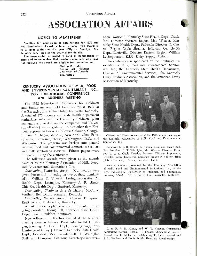
252 AssoCIA noN AFFAIRS
ASSOCIATION AFFAIRS NOTICE TO MEMBERSHIP
·Deadline for submission of nominations for 1972 An· nual Sanita'rians Award is June 1, 1972. The award is to a local sanitarian this year (City or County). See January 1972 issue of the Journal for details.
The membership is urged to send in nominations at one.:! and to remember that previous nominees who have not received the award are eligible for re-nomination.
Melton E. Held, Senior Past President Chairman of Awards Committee
KENTUCKY ASSOCIATION OF MILK, FOOD AND ENVIRONMENTAL SANITARIANS, INC.,
1972 EDUCATIONAL CONFERENCE AND BUSINESS MEETING
The 1972 Educational Conference for Fieldmen and Sanitarians was held F ebruary 22-23, 1972 at the Executive Inn Motor Hotel, Louisville, Kentucky. A total of 275 (county and state health department sanitarians, milk and food industry fieldmen, plant managers and related service companies and University offiCials) were registered. States other than Kenhlcky represented were as follows: Colorado, Georgia, Indiana, ~~lichigan , Missouri, Iew York, Ohio, Pennsylvania, Tennessee, Texas, · ' iVashington, D.C. , and ·wisconsin. The program was broken into general sessions, food and environmental sanitarians sections and milk sanitarians sections. Fifteen papers were presented dui·ing the conference.
The following awards were given at the awards banquet by the Kentucky Association of Milk, Food, and Environmental Sanitarians, Inc.
Outstanding San-itarian Award: (Co. awards were given due to a tie in voting on r,;vo of those nominated). William T'. Vincent, Lexington-Fayette Co. Health D ept. , Lexington, Kentucky A. R. Himes, Ohio Co. Health Dept. , Hartford, Kentucky.
Outstanding Fieldma:n Awanl: Harold McCurry, Southern Bell Dairy, Somerset, Kentucky.
Oustanding SeToice Award: Charles F. Spears , Kraft Foods, Taylorsville, Kentucky.
A past presidents plaque was also presented to out going president, Irving Bell, Kentucky State Health Department, Frankfort, Kenh1cky. · New officers and directors elected at the business
meeting were as follows: President-Donald L. Colgan, Fleming Co. Health Dept., Flemingsburg; President-elect-Dudley J. Conner, Kentucky State Health Dept. , Frankfort; Vice President-R. T . Winbigler, Swift and Company, Glasgow; Secretary-Treasm er-
' Leon Townsend, Kentucky State Health Dept. , Frank-fort ; Director Western Region-Max ·w eaver, Kentucky State Health Dept. , Paducah; Director N. Central Region-Gayle Shrader, Jefferson Co. Health Dept., Louisville; Director ~astern Region-William L. Stephenson, K.I.O. Dairy Supply, Union.
The conference is sponsored by the Kentucky Association of Milk, Food and Environmental Sanitarians Inc., the Kentucky State Health Department, Division of Environmental Services, The Kentucky Dairy Products Association, and the American Dairy Association of Kentucky.
Officers and Directors elected at th e 1972 annual meeting of the Kentucky Association of Milk, Food and Environmental Sanitarians Inc.
Back row L. to R. Donald L. Colgan, President, Irving Bell, Past President, H. T . \ .Yinbigler, Max vVeaver, Director. Front row L. to H. Gayle Shrader, Director, William Stephenson, Director, Leon Townsend, Secretary-Treasurer. (absent from picture Dudley J. Conner, President elect ).
Awards winners, presented by the Kentucky Association of Milk, Food and Environmental Sanitarians, Inc., at the 1972 Educational Conference of Fieldmen and Sanitarians, F ebruary 22-23, 1972, Executive Inn, Louisville, Kentucky.
L. to H. A. H. Himes, and W. T. Vincent, Outstanding Sanitarians Award, Charles F. Spears, Outstanding Service Award, Harold McCurry, Outstanding Fieldmen Award and J. L. Wallace and Louis Smith, Honorary Memberships.
' •· I
! :
AssocrATIO AFFAIRS 253
• THE SANITARIAN: HEALTH CAREER FACTS
. _ Performing preventive medical services to an en-
tire population is quite an extensive assignment. And
it is not unrealistic. This responsibility is carried
out daily by the professional sanitarian. If communi
ties are clean and free of damaging hazards to over
all health it reflects the sanitarian's awareness and
ability to deal with environmental health problems.
Most people assume that the food they eat, the
liquids they drink, the air they breathe, the public
swimming pools they use are safe. The sanitarian
cannot assume any of these, and his investigations
and evaluations assure safety for the general public.
The sanitarian finds and eliminates hazards in order
to make the physical environment safe for everyone.
He is primarily involved with improving man's en
vironment by alleviating metropolitan and rural health
problems. Promoting and securing water supply ex
tension, providing safer recreational areas, and de
veloping better hygienic conditions in nursing and
convalescent homes are several of his many duties .
To meet the demands of many diverse roles the
sanitarian must incorporate the knowledge of medi
cal , engineering, scientific and legal areas into his
occupation. Sanitarians work closely with other
~~·health specialists in the community to prevent out
breaks of disease, to plan for civil defense and emer
gency disaster aid, to make public health surveys,
and to conduct health education seminars. Bacter
iologist, chemist, physicist, engineer, teacher and law
yer-the sanitarian must b e familiar with all these
fi elds to effectively promote environmental health.
' '
Currently sanitarians are fighting to eliminate many
problems of crowded metropolitan areas. Industrial
~xpansion has aggravated the problem of air pollu
tion. The sanitarian assumes the responsibility of
inspecting and attempting to alleviate many of the
mobile and stationary sources of air pollution. He
provides services for protection of ai.r resources and
assists in the planning of new industrial areas.
Proper housing for the American family is an in
tegral part of environmental health. The sanitarian
improves the neighborhood by protecting water re
sources and waste disposal, by promoting home safety
and accident prevention, and by coordinating acti
vities with urban renewal authorities, building in
spection agencies, and public housing agencies.
No one can eliminate human immunity to accidents,
but since the sanitarian is acutely aware of potential
haza,rds he is needed to provide an accident control
program. Professional h·aining enables the sanitarian
to recognize safety hazards and to develop a sense
of diplomacy in working with other professionals to
dear up the problem .
.''
To protect our food supplies the sanitarian closely
checks production and processing techniques . In
spections are made in food and dairy industries to
see that clean, healthful, safe working conditions
exist. The sanitarian works with the problem areas that
affect our health . But a constant watch must be
kept over the potential hazards and developments of
tomorrow. Sanitarians have been vital contributors
to the space program by resolving sanitation prob
lems of space flights. The projected necessity of a
controlled environment for moon dwellers has also
fallen within the sanitarian's area of concern.
EDUCA TIOr AL REQUIREME~ TS: The desir
able minimum qualification for the profess ional sani
tarian is a college degree with a major in physical,
biological, or sanitary science. A graduate degree
in some specialty of public health is usually required
for higher level positions. In ·wisconsin , a person
can b ecome a registered sanitarian by examination
after six years or practical experience following high
school graduation.
Many sanitarians supplement their college back
ground by enrolling in specialized short-term training
courses in such subjects as occupational health, water
supply and pollution control, air pollution, radiologi
cal health, milk and food protection, and metropolitan
planning.
Wisconsin State University, Eau Claire, offers a
B.S. degree in Environmental and Public H ealth.
Universities having Schools of Public Health such as
Minnesota, Michigan, California, North Carolina and
Johns Hopkins offer postgradute degrees in public
health sanitation. The :Milwaukee Technical College
offers a 2-year concenb·ated program in environmen
tal sanitation. A certificate is issued upon comple
tion.
OPPORTUNITIES: In recent years environmental
health has expanded rapidly. And it is currently on
the threshold of more ex tensive development. The
problems of radiological health, occupational health,
food protection, water and air pollution will expand
as a result of population increases, migration to the
cities and adoption of a highly teclmological civili
zation. To offset these problems more highly h·ained
sanitarians will be needed.
Health organizations exis t on city, state, national
levels . International organizations-vVorld Health
Organization, Agency for Intemational D evelopment,
the Peace Corps-have a need for sanitarians to deal
with problems of environmental health-problems
that may be far different and far greater than ours.
In local, state and federal health departments the
254 AssociATION AFFAIRS .1'
sanitarian may specialize in a particular area of work, such as milk and other dairy products, food sanitation, waste conh·ol, air pollution, housing, or insect control. 1n rural areas and small cities they may be responsible for a wide range of environmental health activities.
The added significance of environmental health can be seen in the increasing number of sanitarians being employed by industries. Many sanitarians are employed by lru:ge businesses to conb:ol proper maintenance of buildings, equipment and employee facilities, to eliminate safety hazards, to assure the proper cleaning of plant equipment. The food and dairy industries, have a clu·onic need for qualified sanitarians to make sure that food ingredients and production procedures meet government specifications. Colleges and universities also have positions available to the professional sanitarian who may serve as a faculty member, conduct research, or direct the campus enviJ:onmental health program.
SALARIES: The demand for more qualified individuals and the technological advancements within the field have resulted in in1proved salary scales for the sanitarian. Annual salaries with state and local governments usually ran g e between $7,000 and $12,000. Industrial salary levels will vary according to educational background and the individual sanitarians knovvledge of a particular specialty.
GEORGIA ENVIRONMENTAL HEALTH ASSOCIATION
Persons employed in the various areas of Environmental H ealth in Georgia have an active professional association in their home state. The Georgia Environmental H ealth Association (formerly the Georgia Society of Registered Professional Sanitariam ) is an organization which has as its goal the betterment of society tlu·ough conh·ol of the surroundu1gs. The association brings together in one group all environmentalists who are employed in local and state health departments , the Georgia D eparh11ent of Agriculture, and numerous indusb·y representatives who are employed to protect the environment.
Since its beginning 19 years ago, the organization has be"n concerned with the competence of its members. In the mid-50's, the association was instrumental in devising an evaluation procedure and establishing a review and examining board at tl1e state level to evaluate competence of sanitarians, and to certify them as "registered professional sanitarians" when certain professional qualifications were met. This registration carried with it advancements in pay and promotion to additional areas of responsibility, and
provided an incentive for the enviJ·onmentalist to keep abreast of advancements in his profession.
The annual meeting of the association is designed to provide continuiJ1g education for the memqers . Once each year, members of the Georgia Environmental H ealth Association assemble on Jekyll Island for an educational conference. The theme for the 1971 conference was "A Future in Our Environment." with such outstanding speak~rs as the Deputy Director, Georgia D epa.rhnent of H ealtl1, a Regional Vice-President of a 1 ational Pest Conb:ol Association, the Director of Environmental Con t r o 1 for the Georgia-Pacific Corporation, the President of Acoustic and Vibration Association, Inc. , and the Assis tant Secretary for Health Services, U. S. Deparhnent of H ealth , Education and W elfare.
Also, members of the association saw a need and played an active role in getting the degree program "B. S. in Environmental Health Science" initiated ;~.t
the University of Georgia. Since its beginning in the ' Fall of 1970, this program as developed into a very successful venture with 42 students currently working toward the degree.
The officers for the Georgia Environmental Healtl1 Association for the current year are as follows: President, Harry Forehand, Long County Health Department, Jesup, Georgia; President Elect, Ray N. Bliss, Ai.rwick Indush·ies, Inc., Forest Park, Georgia; VicePresident, Charles D. Stewart, Jr. , Georgia D epartment of Agriculture, Atlanta, Georgia; and SecretaryTreasurer, James H. MartiJ1, University of Georgia, Athens, Georgia .
SURGE ANNOUNCES .RESULTS OF FIELD TESTS ON NEW QTO MILKER
The Surge QTO or Quarter Take-Off milker, a new concept in milking, has been developed by Babson Bros. Co. Research. Built around the proven Surge principle of tug and pull and fast efficient milking, the new unit au tomatically removes the teat cup when each quarter is milked out.
Results from almost one million milkings to date show advantages in labor efficiency while maintaining top milk production and good overall udder health.
Babson Bros. Research and Development D epartment b egan testing their first automatic take-off milker in 1958. ContinuiJ1g research on the concept led to the development of tl1e current model. The milking unit is governed by a solid state electronic control. Individual elech·odes sense the milk flow from each teat. When ·tl1e milk How ceases, the low voltage sensing circuit is broken which signals tl1e re-
' •• I
' . .
I
•' l
' '
I DEX TO ADVERTISERS 255
~· moval of the tea t cup. Actual removal is accomplished when an air powered cylinder draws the teat cup
downward. Vacuum is shut off at the stem of the
inflation allowing the teat cup to drop off. The
operator can check the status of the QTO with the
i
•
help of indicator ligh ts on the conh·ol unit.
FOOD ENGINEERING AWARD
JURY SELECTED
Daily and Food Industries Supply Assn. (DFISA )
and American Society of Agricultural Engineers
( ASAE ) have announced names of a blue-ribbon
jury which will select the recipient of the indush-y's
first Food Engineering Award.
Jury members who will evaluate nominees for the
food processing, science and technology honor are :
R. H. Brown, chaiJ.·man, Agricultural Engineering
D ept. , University of Georgia; D . F . Farkas, chemical
engineer, Engineering and Development Laboratory,
Wes tern Regional Research Laboratory, Agricultural
Research Service, U. S. Dept. of Agriculture; V. A.
Jones, associate professor, Dept. of Food Science,
HARDCHROME PLATE
Old nickel alloy valve bodies for CIP and pasteurized products
More durable and corrosion resistant than even stainless steel. Cleared by USPHS.
and MODERNIZE with Leakproof T ANA CO PLASTIC PLUGS
inert to caustics, acids and live steam . Send iior illustrated brochure on how it's done .
,r
2825 Benedict Street Los Angeles, Calif. 90039
Phone: (213) 661-1222
North Carolina State University; D . E. Kirk, pro
fessor, Agricultural Engineering Dept. , Oregon State
Universi ty; K. H. Lewis, diJ.·ector, Office of Food San
itation, Bureau of Foods, Dept. of Health , E ducation
and Welfare; H . L. Mitten, Jr. , d i1·ector of interna
tional operations, CP Division, St. Regis; and B. S.
Schweiger t, chainnan, D ept. of Food Science and
Technology, University of California.
The Food Engli1eering Award is designed to honor
those who have made outstanding original conh·i
butions in research, development, design or manage
ment of food processing equipment or processes which
have been of significant economic value to the food
industry and the consuming public.
LIQUID SUD'N rinses freely without film or residue even in COLD WATER. TRY SUD 'N.
TODAY it 's free rinsing SUD'N for brush and ultra-sonic washing of al l laboratory glassware. missile space components ... used wherever the ultimate in clean ing is essential ... where anything less would be too expensive.
Wr ite lor details
• Phosphate Free
• Biodegradable
• Non Polluting
SEP-KO CHEMICALS Div. H. B. Fuller Co.
3900 Jackson Street N.E. Minneapolis, Minn . 55421
lNDEX TO ADVERTISERS
Babson Bros, Co. ____________________ Back Cover
Difco Laboratories ____________ Inside Front Cover
IAi'vlFES, Inc. _____ _______________ __ ! , II, IV, 256
Sep-Ko Chemicals _________ __ ________ __ ______ 255
Tanaco Products _________________ ________ ___ 255
The Haynes Mfg. Co. __________ __ Inside Back Cover
CLASSIFIED ADS FOR SALE
Single Service milk sample tubes. For further in-
formation and a catalogue please write, Dairy Technology
Inc., P. 0 . Box 101, Eugene, Oregon 97401.
256
Application for Members hip INTERNATIONAL ASSOCIATION OF MILK, FOOD & ENVIRONMENTAL
SANITARIANS, INC. Box 437, Shelbyville, Indiana 46176
Name __ _____ ________________ ___ __________ ____ _____ __ ____ ________________ ____ - -- Date - ----- --------Please Print
Address -- - -- ------- ------ ------ ---- ---- - - -- --- --- ----- -- ------------ --- ------ [] Renewal ------ ------- - ----------------- ------------ --- Zip Code ---- -- - --- - ---------- [] New Business Affiliation _ __ __ _ _ _ _ __ __ ___ _ __ __ __ __ __ _ ___ __ _ _ __ __ __ __ ____ _ _ __ __ __ __ _ _ [] Re-instatement
Direct Member Annual Dues $14.00 [] Check [] C; sh Membership Through An Affiliate-$12.00 Plus Affiliate Dues
Student Membership $4.00 (Membersh ip Includes Subscription to Journal of Milk & Food Technology.)
Recommended by
Shelbyville, Ind. Box 437
P lease Prin t
Su.bscript·ion Order JOURNAL OF MILK & FOOD TECHNOLOGY
(Monthly Publication) Name __ ______ __ _____ __ __ ___ __ __ ____ ___________________________________________ _ Date _____ __ ______ _
Please P rin t
Address ------------ ---- ------ - -- ----- ----- --- ----- -- - -- - - - --- --- - ------------ [] New
Zip Code - - ---- ----------- - -- - [] Renewal
Educational Institution & Public (Jl.. nnuaHy) $12.00 [] Check
Libraries Individual Non-Member Subscription (Annually) $14.00 [] Cash Government Agencies, Commercial Organizations
I.A.M.F.E.S. & J .M.F.T. Box 437, Shelbyville, Ind. Change of Address
FROM
Name - -- --------------- - -- -- --- ------ ------ -- ------ ---------- - ----------------- Date -- ---- --------Please Print
Address
TO Name -- - - ---- -- -- - ---- - ----- -- ------------------ ------ ----- -------------- ----1-'lease tTint
Address --------------- --- ----- --------------------- -------------- --- ------ ---I.A.M.F.E.S. & J .M.F.T.
Box 437, Shelbyville, Ind. Order fo1· 3A Standards Name _______ ___ ________ ___ _____ ______________ ______ _______ __ ___________ ---- - - -- Date ________ ----- -
P lease Print
Address ---- -- ---- -- - --- ------- ------ ----- --- --------------------- - -------------( ) Complete Set $5.50 = ________ ( ) Complete set bound (durable cover) @ $7.00 =
( ) Revised HTST Std.-without cover = $1.50 F.O.B. Shelbyville, Ind. ( ) Revised HTST Std.-25 or more = $1.00 each F.O.B. Shelbyville, Ind. ( ) E-3-A Sanitary Standards-Set Unbound @ $2.25 _ ( ) Methods For Production of High Quality Raw Milk $2.00
3-A Accepted Practices For Milking Machines 1-100 = 25c ea.; 100-1000 = 20c ea.; 1000 or more = 15c ea. 5 Year Service on Standards as Published = $6.00 addit ional
Order fo1· Reprints of Articles
Amt. ---------- --- --- Title
Schedule of letterpress prices for reprints within 30 days after publication, F.O.B. Shelbyville, 1 Page 2 Pages 3 & 4 Pages 6 & 8 Pages 12 Pages
100 or less $19.50 $23.40 $32.80 $46.80 $78.00 Add'l 100's 2.40 3.00 5.00 6.60 10.80
After 30 Days Offset Reprint prices add $2.00 per page
Indiana
Cover $33.60
5.50
,
. :

I I
~ SANITARY-PURE
~ ODORLESS-TASTELESS
_ NON-TOXIC
Tim Fme U<~t-~ HAYNES-SPRAY 3kwbi be med t4 &Wltit.afe: SANITARY VAL YES HOMOGENIZER PISTONS - RINGS SANITARY SEALS & PARTS CAPPER SLIDES & PARTS POSITIVE PUMP PARIS GLASS & PAPER FILLING MACHINE PARTS and fo, All OTHER SANITARY MACHINE PARTS which o'e clecned doily.
Tke lf(cuUrm HAYNES-SPRAY lf(e&M Db .C~ CM#J&IlMt3 Mtifk Ute lf(ifk 0~ ruuL Ccule R~ luj Ute U. S. P!Cb& Heatfk Sewiu
The Haynes-Spray eliminates the danger af contamination which is possible by old fashioned lubricating methods. Spreading lubricants by the use of the finger method may entirely destroy previous bactericidal treatment of equipment.
PACKED·6-12 oz. CANS PER CARTON SHIPPING WE IGHT- 7 LBS.
Gasket Color ••• slightly off-while
*MADE FROM
TEFLON® " Tke SrJPJmficaW G~ket" THE IDEAL UNION SEAL FOR
BOTH VACUUM AND PRESSURE LINES
SNAP-TITE self-centering gaskets of TEFLON are designed for all standard bevel seat sanitary fittings. They SNAP into place provid· ing self-alignment and ease of assembly and disassembly. HAYNES SNAP-TITES of TEFLON are unaffected by cleaning solu· l ions, steam and solvents. They will not embrittle of temperatures as low as minus 200° F. and ore impervious to heal up to 500° F.
•
fOR lA FITTING GASKET THAT WILL OUT-PERFORM ALL OTHERS.M
S~etijy ..• HAYNES SNAP· liTES of TEFLON e Tf110N ACCE"ED SAFE fOa USE ON FOOD & PaOCESSING
EQUIPMENT 8Y U . S. fOOD AND DaUG ADMINISTaATION
• Gaskets made of DuPont TEFLON® TFE-FLUOROCARBON RESINS
TH E HAYNES MANUFACTURING COMPANY <&1 80 LORAIN AVENUE . ~ . CLEVELAND, OHIO 44113
SNAP INTO FITTINGS
~ LOW COST ••• RE-USABLE
~ LEAK-PREVENTING NEOPRENE GASKET for· Sanitary Fittings
~ ~ $NA1'!J'I~E r/dt-taa~ Tight joints, no leaks, no shrinkage
Sanitary, unaffected by heat or fats
Non·porous, no seams or crevices
Odorless, polished surfaces, easily cleaned
Withstand sterilization
Time-saving, easy ta assemble Self·centering No sticking to fittings
Eliminate line block• Help overcome line vibrations
long life, use over ond over
Ayoiloble for J", Ph", 2", 2~" and 3" fittings. Pocked 100 to the box. Order through your dairy supply house.
THE HAYNES MANUFACTURING CO. 418 0 Lorain Avenue • Cleveland 13, Ohio
.A-valatie in tolh SPRAY AND TUBE
All Lubri- Film ingredients are
approved additives and can be
safely utilized as a lubricant for
food processing equipment when
used in compliance with existing
food additive regulations.
ESPECIALLY DEVELOPED FOR LUBRICATION OF FOOD
PROCESSING AND PACKAGING EQUIPMENT
For Use in Dairies- Ice Cream Plants- Breweries
Beverage Plants- Bakeries- Canneries- Packing Plants
SANITARY • NON TOXIC • ODORLESS • TASTELESS SPRAY- PACKED 6- l6 OZ. CANS PER CARTON TUBES- PACKED 12-4 OZ. TUBES PER CARTON
THE HAYNES MANUFACTURING CO . CLEVELAND . OHIO 44113
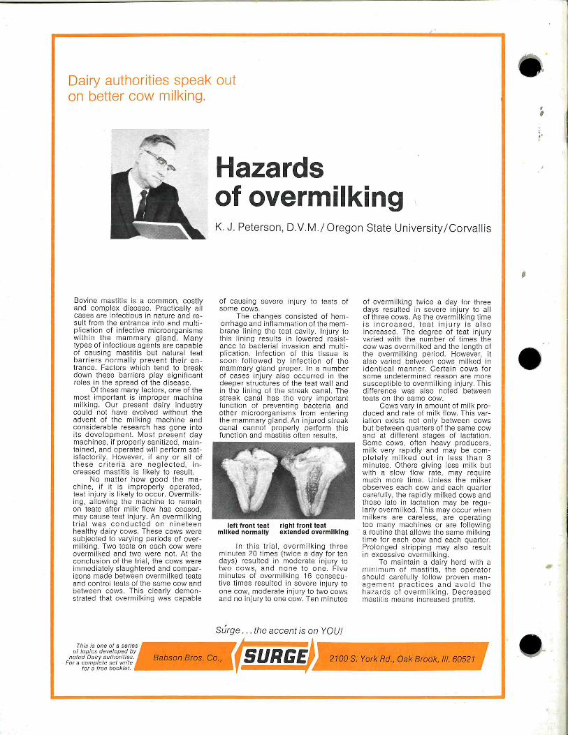
Dairy authorities speak out on better cow mi lking.
Hazards of overmilking
Bovine mastit is is a common, costly and comp lex disease. Practically al l cases are infectious in nature and re sult from the entrance into and mult ip lication of infective microorganisms withi n the mamma ry g land. Many types of infect ious agen ts are capable of causing mastit is but natural teat barriers normall y prevent the ir entrance. Factors which tend to break down these barriers play significant ro les in the sp read of the disease.
Of these many factors, one of the most important is improper machi ne milking. Our present dairy industry cou ld not have evolved without the advent of the mi lking machine and considerab le research has gone into its development. Most present day machines, if properly san it ized, maintai ned, and operated wi ll perform satisfactorily. However, if any or all of these cr i teria are neglec ted , increased masti tis is likely to resu lt.
No matter how good the machine, if it is improperly operated, teat injury is likely to occur. Overmilking, al lowing the machine to remain on teats afte r milk• flow has ceased , may cause teat injury. An overmilking tria l was conducted on n inetee n healthy dairy cows. These cows were subjected to varying periods of overmi lking . Two teats on each cow were overmilked and two were not. At the conclus ion of the trial, the cows were immediate ly slaughtered and comparisons made between overmilked teats and control teats of the same cow and between cows . This clear ly demonstrated that overmilking was capable
K. J. Peterson, D.V.M./ Oregon State University / Corvallis
of causing severe injury to teats of some cows.
The changes consisted of hemorrhage and inflammation of the membrane lin ing the teat cavity. Injury to this lining resu lts in lowered resistance to bacterial invasion and mul tiplication. Infection of this tissue is soon followed by infection of the mammary gland proper. In a number of cases injury also occurred in the deeper structures of the teat wall and in the lining of the streak canal. The streak cana l has the very important funct ion of preventing bacteria and other microorganisms from entering the mammary gland. An injured streak canal cannot properly perform this function and mastit is often results.
left front teat right front teat milked normally extended overmilking
In this tria l, overmilking three minutes 20 times (twice a day for ten days) resulted in moderate injury to two cows, and none to one. Five minutes of overmilking 16 consecutive times resu lted in severe injury to one cow, moderate injury to two cows and no injury to one cow. Ten minutes
Surge ... the accent is on YOU!
of overmilki.ng twice a day for three days resulted in severe injury to all of three cows. As the overmilking time is increased , teat injury is also increased. The deg ree of teat injury varied with the number of times the cow was overmilked and the length of the overmilking period. However, it also varied between cows milked in identical manner. Certai n cows for some undetermined reason are more susceptible to overmilking injury. This difference was also noted between teats on the same cow.
Cows vary in amount of mi lk produced and rate of mi lk flow. This variation exists not only between cows but between quarters of the same cow and at different stages of lactation. Some cows, often heavy producers, milk very rap idly and may be comp lete ly mi lked out in less th an 3 minutes. Others giving less milk but with a slow flow rate , may require much more time. Unless the milker observes each cow and each quarter carefully, the rapidly milked cows and those late in lactation may be regularly overmilked. This may occu r when mi lkers are care less, are operating too many machines or are following
· a routine that al lows the same milki ng time for each cow and each quarter. Prolonged stripping may also resu lt in excessive overmilking.
To maintain a dairy herd with a minimum of mastitis, th e ope rator should carefu lly follow proven management practices and avoid the hazards of overmilking . Decreased mastitis means increased profits.
Th is is one of a series of topics developed by
noted Dairy authorities. For a complete set write
tor a tree booklet.
Babson Bros. Co., ( 5 URGE~ 2100 S. York Rd .. Oak Brook, Ill. 60521
,
,. I