Impaired neurogenesis, neuronal loss, and brain functional deficits in the APPxPS1-Ki mouse model of...
12
Neurobiology of Aging 32 (2011) 407–418 Impaired neurogenesis, neuronal loss, and brain functional deficits in the APPxPS1-Ki mouse model of Alzheimer’s disease A. Faure a , L. Verret b,c , B. Bozon a , N. El Tannir El Tayara d,e , M. Ly a , F. Kober f , M. Dhenain g,h,i , C. Rampon b,c , B. Delatour a,∗ a CNRS, Lab NAMC, UMR8620, Université Paris Sud, 91405, Orsay, France b Université de Toulouse, UPS, Centre de Recherches sur la Cognition Animale, 118 route de Narbonne, F-31062 Toulouse Cedex 9, France c CNRS, Centre de Recherches sur la Cognition Animale, F-31062 Toulouse, France d INSERM, U759, Centre Universitaire, Laboratoire 112, 91405 Orsay Cedex, France e Institut Curie, Centre Universitaire, Laboratoire 112, Orsay 91405, France f Centre de Résonance Magnétique Biologique et Médicale (CRMBM), UMR CNRS 6612, Faculté de Médecine, Université de la Méditerranée, Marseille, France g CEA, DSV, I 2 BM, SHFJ, MIRCen Program, 4 Place du Général Leclerc, 91401 Orsay Cedex, France h CNRS, URA 2210, 4 Place du Général Leclerc, 91401 Orsay Cedex, France i CEA, DSV, I2BM, NeuroSpin, Centre CEA de Saclay, Bât. 145, 91191 Gif sur Yvette, France Received 16 January 2009; received in revised form 17 March 2009; accepted 18 March 2009 Available online 23 April 2009 Abstract Amyloid- peptide species accumulating in the brain of patients with Alzheimer’s disease are assumed to have a neurotoxic action and hence to be key actors in the physiopathology of this neurodegenerative disease. We have studied a new mouse mutant (APPxPS1-Ki) line developing both early-onset brain amyloid- deposition and, in contrast to most of transgenic models, subsequent neuronal loss. In 6-month- old mice, we observed cell layer atrophies in the hippocampus, together with a dramatic decrease in neurogenesis and a reduced brain blood perfusion as measured in vivo by magnetic resonance imaging. In these mice, neurological impairments and spatial hippocampal dependant memory deficits were also substantiated and worsened with aging. We described here a phenotype of APPxPS1-Ki mice that summarizes several neuroanatomical alterations and functional deficits evocative of the human pathology. Such a transgenic model that displays strong face validity might be highly beneficial to future research on AD physiopathogeny and therapeutics. © 2009 Elsevier Inc. All rights reserved. Keywords: Alzheimer’s disease; Amyloid- peptide; Hippocampus; Neurogenesis; Learning and memory; Brain blood perfusion 1. Introduction Alzheimer’s disease (AD) is a progressive neurodegener- ative disorder which accounts for the majority of dementia cases in the elderly population. The neuropathological ∗ Corresponding author. Tel.: +33 1 69 15 49 88; fax: +33 1 69 15 77 26. E-mail addresses: [email protected] (A. Faure), [email protected] (L. Verret), [email protected] (B. Bozon), [email protected] (N. El Tannir El Tayara), [email protected] (F. Kober), [email protected] (M. Dhenain), [email protected] (C. Rampon), [email protected] (B. Delatour). hallmarks of AD include extracellular amyloid- (A) depo- sition, intracellular tau accumulation, neuronal and synaptic losses. Because familial AD is associated with mutations of the amyloid protein precursor (APP) or presenilin (PS) genes, many mouse lines that overexpress human transgenes bearing these mutations have been generated over the two last decades. Whereas most of these transgenic mice develop amyloid deposits, the majority of them fail to exhibit signif- icant neuronal loss. In the few mouse lines showing altered neuronal densities, cell loss remains weak or limited to the vicinity of amyloid deposits and does not reflect the drastic neuronal reduction observed in AD patients (Calhoun et al., 0197-4580/$ – see front matter © 2009 Elsevier Inc. All rights reserved. doi:10.1016/j.neurobiolaging.2009.03.009
Transcript of Impaired neurogenesis, neuronal loss, and brain functional deficits in the APPxPS1-Ki mouse model of...
A
hdopmsf©
K
1
ac
(Nfr
0d
Neurobiology of Aging 32 (2011) 407–418
Impaired neurogenesis, neuronal loss, and brain functional deficits in theAPPxPS1-Ki mouse model of Alzheimer’s disease
A. Faure a, L. Verret b,c, B. Bozon a, N. El Tannir El Tayara d,e, M. Ly a,F. Kober f, M. Dhenain g,h,i, C. Rampon b,c, B. Delatour a,∗
a CNRS, Lab NAMC, UMR8620, Université Paris Sud, 91405, Orsay, Franceb Université de Toulouse, UPS, Centre de Recherches sur la Cognition Animale, 118 route de Narbonne, F-31062 Toulouse Cedex 9, France
c CNRS, Centre de Recherches sur la Cognition Animale, F-31062 Toulouse, Franced INSERM, U759, Centre Universitaire, Laboratoire 112, 91405 Orsay Cedex, France
e Institut Curie, Centre Universitaire, Laboratoire 112, Orsay 91405, Francef Centre de Résonance Magnétique Biologique et Médicale (CRMBM), UMR CNRS 6612,
Faculté de Médecine, Université de la Méditerranée, Marseille, Franceg CEA, DSV, I2BM, SHFJ, MIRCen Program, 4 Place du Général Leclerc, 91401 Orsay Cedex, France
h CNRS, URA 2210, 4 Place du Général Leclerc, 91401 Orsay Cedex, Francei CEA, DSV, I2BM, NeuroSpin, Centre CEA de Saclay, Bât. 145, 91191 Gif sur Yvette, France
Received 16 January 2009; received in revised form 17 March 2009; accepted 18 March 2009Available online 23 April 2009
bstract
Amyloid-� peptide species accumulating in the brain of patients with Alzheimer’s disease are assumed to have a neurotoxic action andence to be key actors in the physiopathology of this neurodegenerative disease. We have studied a new mouse mutant (APPxPS1-Ki) lineeveloping both early-onset brain amyloid-� deposition and, in contrast to most of transgenic models, subsequent neuronal loss. In 6-month-ld mice, we observed cell layer atrophies in the hippocampus, together with a dramatic decrease in neurogenesis and a reduced brain blooderfusion as measured in vivo by magnetic resonance imaging. In these mice, neurological impairments and spatial hippocampal dependantemory deficits were also substantiated and worsened with aging. We described here a phenotype of APPxPS1-Ki mice that summarizes
everal neuroanatomical alterations and functional deficits evocative of the human pathology. Such a transgenic model that displays strongace validity might be highly beneficial to future research on AD physiopathogeny and therapeutics.
2009 Elsevier Inc. All rights reserved.
eywords: Alzheimer’s disease; Amyloid-� peptide; Hippocampus; Neurogenesis; Learning and memory; Brain blood perfusion
hsl
. Introduction
Alzheimer’s disease (AD) is a progressive neurodegener-
tive disorder which accounts for the majority of dementiaases in the elderly population. The neuropathological∗ Corresponding author. Tel.: +33 1 69 15 49 88; fax: +33 1 69 15 77 26.E-mail addresses: [email protected] (A. Faure), [email protected]
L. Verret), [email protected] (B. Bozon),[email protected] (N. El Tannir El Tayara),
[email protected] (F. Kober), [email protected] (M. Dhenain),[email protected] (C. Rampon), [email protected] (B. Delatour).
ogblainvn
197-4580/$ – see front matter © 2009 Elsevier Inc. All rights reserved.oi:10.1016/j.neurobiolaging.2009.03.009
allmarks of AD include extracellular amyloid-� (A�) depo-ition, intracellular tau accumulation, neuronal and synapticosses. Because familial AD is associated with mutationsf the amyloid protein precursor (APP) or presenilin (PS)enes, many mouse lines that overexpress human transgenesearing these mutations have been generated over the twoast decades. Whereas most of these transgenic mice developmyloid deposits, the majority of them fail to exhibit signif-
cant neuronal loss. In the few mouse lines showing alteredeuronal densities, cell loss remains weak or limited to theicinity of amyloid deposits and does not reflect the drasticeuronal reduction observed in AD patients (Calhoun et al.,
4 ogy of A
12til1
msueAp1n(wKitdh
tWpcniswsfr
2
2
(iM(Lm5bNwsiat2
TeK
eCsottaact
2
fipstmt
saa
s1
(nlbs(CFs(mtmp
sCqp
08 A. Faure et al. / Neurobiol
998; Irizarry et al., 1997; Takeuchi et al., 2000; Urbanc et al.,002). These observations challenge the cascade hypothesishat posits A� accretion as a primary neurotoxic event trigger-ng morphological and functional brain anomalies, ultimatelyeading to neuronal death and dementia (Hardy and Higgins,992).
It is only recently that mouse lines harboring multipleutated APP and PS1 transgenes and developing aggres-
ive and early-onset brain amyloidosis, have been reported tondergo brain atrophies and substantial cell loss (e.g. Oakleyt al., 2006; Schmitz et al., 2004). In one of these models, thePPxPS1-knock-in line (Casas et al., 2004), a severe hip-ocampal cell loss (50%) has been reported at the age of0 months. It mimics both the topography and extent of theeuronal loss observed in the hippocampus of AD patientsScher et al., 2007; West et al., 2000). At the behavioral level,orking memory deficits have been described in APPxPS1-i mice (Bayer and Wirths, 2008; Wirths et al., 2007), but
t remains unknown whether these mice also develop medialemporal lobe dysfunction, and particularly hippocampus-ependent learning impairments, as has been reported foruman patients.
In the present study we used a multi-disciplinary approacho characterize in detail the APP/PS-Ki mouse phenotype.
e evaluated the nature and extent of A�-associated mor-hological anomalies and the occurrence of neurological andognitive impairments in this mouse model. Because adulteurogenesis may provide a means for neuronal replacementn the AD brain, we also examined whether the progres-ive neuronal loss displayed by APP/PS1-Ki is associatedith modified hippocampal neurogenic processes. Finally, to
ubstantiate brain dysfunction in APPxPS1-Ki mice, we per-ormed blood perfusion analysis by means of in vivo magneticesonance imaging (MRI) techniques.
. Material and methods
.1. Animals
Transgenic mice were established as previously describedCasas et al., 2004). In short, mice were generated by cross-ng homozygous PS1-Ki (carrying presenilin 1 knock-in
233T and L235P mutations) with hemizygous APPSL micehAPP751 transgene with the Swedish K670N/M671L andondon V717I mutations, under the control of a Thy1 pro-oter) to obtain APPxPS1-Ki transgenics harboring in totalmutations associated with familial AD on a mixed genetic
ackground (C57BL/6 50% – CBA 25% – 129SV 25%).ote that, owing to the use of homozygous PS1-Ki mice, noild-type littermates could be generated from the breeding
cheme. However the PS1-Ki mice are free of neuropatholog-
cal lesions (amyloid deposits, neuronal loss or axonopathy)nd have been considered in previous studies to be good lit-ermate controls for APPxPS1-Ki mice (Wirths et al., 2006,007).uohp
ging 32 (2011) 407–418
Mice were 6 weeks old at their arrival in the laboratory.hey were acclimated during at least 2 weeks to their newnvironment before initiating experiments. A total of 60 PS1-i and 57 APPxPS1-Ki male mice were used.All experiments were conducted in accordance with the
thical standards of French and European laws (Europeanommunities Council Directive of 24 November 1986). The
upervisor of the present study (B. Delatour) had receivedfficial agreements from the French Ministry of Agricultureo carry out research and experiments on animals (authoriza-ion No. 91-282). Experiments were performed in compliancend following approval of the Sanofi-Aventis Animal Carend Use Committee. The general health of mice was regularlyhecked and body weights were assessed weekly throughouthe experimental period.
.2. Histology and biochemistry
Mice (PS1-Ki, n = 30; APPxPS1-Ki, n = 28) were sacri-ced at the age of 6 months with an overdose of sodiumentobarbital and were perfused transcardially with salineolution. After removal, the brains were weighed and pho-ographed. From macro-photographs, the brain length was
easured at the midline level using the measure tool of Pho-oshop CS2 (Adobe, Paris, France).
Left hemispheres were snap-frozen in liquid nitrogen andtored at −80 ◦C. The A� peptides biochemical load (A� 42nd total A�) was determined by electrochemiluminescencessays as previously described (Blanchard et al., 2003).
Right hemispheres were sectioned (frontal 40 �m-thickections) on a freezing microtome after 1-week fixation in0% formalin and subsequent cryoprotection.
Hippocampal tissue loss was evaluated after Nisslthionin) stain by measuring the pyramidal cell layer thick-ess. Although biased, this method allows rapid evaluation ofocal morphological changes. From previous studies, it haseen shown that aged APPxPS1-Ki mice display progres-ive layer thinning in close association with neuronal lossCasas et al., 2004). All slides were digitized using a SuperoolScan 8000 ED scanner (Nikon, Champigny sur Marne,rance) with a 4000 dpi in-plane digitization resolution (pixelize 6.35 �m2). Sections at the level of the dorsal hippocampin = 3–4 sections/mouse) were selected and the CA1-2 regionanually delineated with Photoshop CS2. For each section,
he thickness of the pyramidal cell layer was measured at 3edio-lateral levels so that a total of 9–12 measures were
erformed and averaged for every mouse.Amyloid deposits were labeled by standard Congo red
taining (30 min in a 80% ethanol solution saturated withongo red and sodium chloride). Amyloid loads wereuantified using computer-based thresholding methods asreviously implemented (Le Cudennec et al., 2008). Eval-
ation of amyloid loads was performed in multiple regionsf interest (ROIs): the total hippocampus, the dorsal (septal)ippocampus, the whole cortex (i.e. isocortex plus hippocam-us), and the prefrontal cortex. Quantitative analyses were
ogy of A
mc
wloitDid(NppCrdDfn
tsd
2
pceTtOmab
Bea(mg2glF1w(aoft
isstvl(Cwwca
2
tN1lKca
2
(tarw
1
2
3
A. Faure et al. / Neurobiol
ade on several serial sections to sample the whole rostro-audal extent of each ROI.
Evaluation of neurogenic activity and cell differentiationas performed using antibodies directed against Ki67 (a pro-
iferation marker) or against doublecortin (DCX, a markerf neuronal precursors). Immunolabelings were performedn a subset of the neuropathologically assessed popula-ion (for Ki67: 6 PS1-Ki and 5 APPxPS1-Ki mice; forCX: 15 PS1-Ki and 14 APPxPS1-Ki mice) using classical
mmunoperoxidase methods (Verret et al., 2007). Ki67 wasetected using a primary rabbit anti-human Ki67 antibodyNCL-Ki67p, Novocastra Laboratories/Vision BioSystems,ewcastle Upon Tyne, UK; 1:500 overnight at room tem-erature) while DCX immunolabeling was performed with aolyclonal goat anti-DCX antibody (C-18, Santa Cruz, Santaruz, CA, USA; 1:100 overnight at room temperature). Final
eactions made use of diaminobenzidine or nickel-enhancediaminobenzidine as chromogens. The number of Ki67 andCX-immunostained cells (Ki67+ or DCX+) was evaluated
rom peroxidase-labeled sections spaced at 480 �m and span-ing the whole septo-temporal extent of the hippocampus.
Cell countings and measurement of surfaces areas of den-ate gyrus were performed using the Mercator stereologyystem (Explora Nova, La Rochelle, France) as previouslyescribed (Verret et al., 2007).
.3. MRI-assessed brain perfusion
Fourteen PS1-Ki and 14 APPxPS1-Ki mice received MRIerfusion assessment before sacrifice and neuropathologi-al examination (see above). Cerebral blood flow (CBF) wasvaluated by using a pulsed arterial spin labeling technique.he animals were anesthetized with isoflurane (4% for induc-
ion, 1–1.5% for maintenance) in a mixture of N2 (80%) and2 (20%) administered via a facemask. Respiration rate wasonitored to insure stable physiologic conditions during the
cquisitions. The body temperature of the mice was stabilizedy using a heating blanket.
In vivo MR images were recorded on a 4.7 Tesla Brukeriospec 47/30 system equipped with a 12 cm diameter gradi-nt system (200 mT/m). A surface coil (diameter = 25 mm),ctively decoupled from the transmitting birdcage probeBruker GmbH) was used for signal acquisition. Perfusioneasurements were performed using a FAIR Look-Locker
radient-echo method described elsewhere (Kober et al.,008). Briefly, a series of 50 echoes was acquired afterlobal or slice-selective magnetization inversion with the fol-owing imaging parameters: TE/TR = 1.59/150 ms, α = 12.5◦,OV: 20 mm × 20 mm, slice thickness: 1.5 mm, input matrix:28 × 64; total acquisition time 32 min. Data acquisitionas performed in a tissue slab of the posterior cerebrum
volume acquisition: from Bregma −1.82 to Bregma −3.28
ccording to Paxinos and Franklin, 2001). The thicknessf this sampling slice was too large to measure per-usion in convoluted regions of interest (ROI) such ashe hippocampus. Because of these methodological lim-4
ging 32 (2011) 407–418 409
ts we evaluated perfusion in large ROIs with minimalhape variations across the antero-posterior axis of thetudied slice. Two ROIs were selected: the posterior cor-ex and the thalamus. In these selected ROIs, perfusionalues (P), given in ml g−1 min−1, were calculated as fol-ows: P/λ = [(T1global/T1blood) × (1/T1selective − 1/T1global)]program developed in an IDL environment (RSI, Boulder,O, USA)). T1global was calculated from images recordedith a global inversion pulse while T1selective was calculatedith the selective inversion pulse. The blood/tissue partition
oefficient for water (λ) was assumed to be 0.9 ml/g (Sun etl., 2004), and T1blood was 1.7 s (Williams et al., 1992).
.4. Behavior
Behavioral studies were carried out in 2 additional cohortshat were evaluated longitudinally at 2, 4 and 6 months of age.eurological assessment was performed on 15 PS1-Ki and4 APPxPS1-Ki mice. Another cohort was used to evaluateearning and memory functions (PS1-Ki, n = 15; APPxPS1-i, n = 15; 2 PS1-Ki and 1 APPxPS1-Ki mice died before the
ompletion of the study and received only partial behavioralssessment).
.4.1. Neurological assessmentA primary screen, derived from the SHIRPA test battery
Burguiere et al., 2005; Rogers et al., 1997), was performedo evaluate the general appearance of animals, spontaneousctivity (e.g. jumping, freezing, rearing) and neurologicaleflexes. On the following days secondary/tertiary screensere performed as followed:
. Motor strength was measured with the wire grid test.Strength suspension (four paws) was assessed on a grid.The mouse was placed on the center of the grid that wasslowly turned upside down at a 20 cm-height above thefloor. Latency to fall was recorded (cut off: 1 min).
. Locomotor activity was measured in a squared openfield (50 cm × 50 cm; luminosity: 35 lx) with black walls30-cm high. Each animal was placed in the center ofthe arena and allowed to freely explore it for 10 min.Horizontal activity was monitored using the Any-Mazesoftware (Stoelting, Wood Dale, USA). Time spent in the10-cm wide peripheral zone and in the complementary30 cm × 30 cm central zone was recorded, and the numberof rearings (vertical activity) was scored.
. Locomotion was also assessed by monitoring mice duringa 24-h period. Mice were moved to the experimental roomand placed in a cage (50 cm × 15 cm) with food and wateravailable. Mice were observed during both light and darkphases with a CCTV camera (WV-BP312E, Panasonic,Saint Denis La Plaine, France) equipped with an infra-red
lamp. Tracking was performed using Any-Maze softwarethat recorded horizontal activity across the 24-h period.. Anxiety was evaluated in an elevated plus-maze (length,28 cm; width, 5 cm; walls height, 16 cm; height from floor:
4 ogy of A
2
wwwoat6
atrmfttlrc
2
SS(aagsfts
3
3A
gdc3c
Aara
trata
cWmob
3a
pTAfD
apetgcpbclntgtiKtnKmpcrtgrD
pq
10 A. Faure et al. / Neurobiol
40 cm; overall luminosity in open arms: 70 lx). Mice wereplaced in the central region of the maze and the locomotoractivity and time spent in each arm were measured for a5-min period using Any-Maze software.
.4.2. Cognitive evaluationSpatial memory was evaluated using the standard Morris
ater maze task. The maze was a 150-cm diameter pool filledith opacified water (21–22 ◦C). A 10 cm diameter platformas submerged 0.5 cm below water surface in the center ofne of the pool quadrants. The non-visible platform remainedt constant position across trials but was varied at each ageime points to promote new spatial learning in 2-, 4- and-month-old mice.
Training consisted in 5 consecutive daily sessions (6 tri-ls/sessions; start positions pseudo-randomly varied fromhe four cardinal points). Each trial ended when the animaleached the platform. A 60-s cut off was used, after which theouse was manually guided to the platform. Once on the plat-
orm animals were given a 30-s rest before being replaced inheir cage. The inter-trial interval was approximately 1 h. Onhe 5th training session, a probe trial was performed after theast trial. During this memory retention test, the platform wasemoved and mice allowed to navigate for 60-s. Data wereollected, analyzed and stored using Any-Maze software.
.5. Statistical analyses
Morphological and perfusion data were analyzed withtudent t-tests and Pearson coefficients of correlation usingtatistica v6 (StatSoft, Inc., Tulsa, OK, USA) or Systat v11.0Systat Software Inc., Richmond, CA, USA) software pack-ges. Behavioral data were analyzed mainly with two-factornalyses of variance (ANOVA) with repeated measures (withenotype as the between-subjects factor and age as the within-ubjects factor), using Statistica v6 software. In order toacilitate the presentation of the results, the ANOVA statis-ics have been presented only by providing p-values whenignificant.
. Results
.1. Gross brain morphology and amyloid deposition inPPxPS1-Ki mice
Brain lengths/widths were not different in the twoenotypes (ts(56) < 1.08; ps > .28). However, brain weightsecreased significantly in 6-month-old APPxPS1-Ki miceompared to PS1-Ki controls (mean ± S.E.M. = 464 ±.1 mg for APPxPS1-Ki mice vs. 490 ± 4.7 mg for PS1-Kiontrols; t(56) = 4.96; p < .0001).
Additional neuropathological analyses revealed in
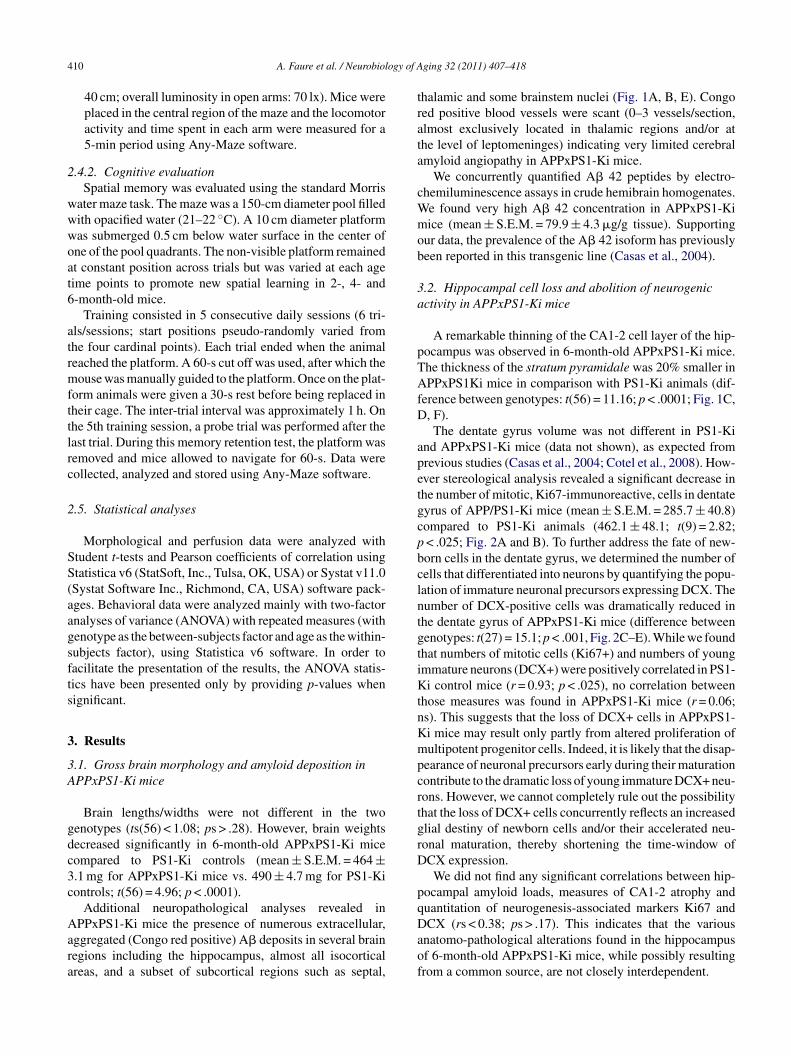
PPxPS1-Ki mice the presence of numerous extracellular,ggregated (Congo red positive) A� deposits in several brainegions including the hippocampus, almost all isocorticalreas, and a subset of subcortical regions such as septal,
Daof
ging 32 (2011) 407–418
halamic and some brainstem nuclei (Fig. 1A, B, E). Congoed positive blood vessels were scant (0–3 vessels/section,lmost exclusively located in thalamic regions and/or athe level of leptomeninges) indicating very limited cerebralmyloid angiopathy in APPxPS1-Ki mice.
We concurrently quantified A� 42 peptides by electro-hemiluminescence assays in crude hemibrain homogenates.e found very high A� 42 concentration in APPxPS1-Kiice (mean ± S.E.M. = 79.9 ± 4.3 �g/g tissue). Supporting
ur data, the prevalence of the A� 42 isoform has previouslyeen reported in this transgenic line (Casas et al., 2004).
.2. Hippocampal cell loss and abolition of neurogenicctivity in APPxPS1-Ki mice
A remarkable thinning of the CA1-2 cell layer of the hip-ocampus was observed in 6-month-old APPxPS1-Ki mice.he thickness of the stratum pyramidale was 20% smaller inPPxPS1Ki mice in comparison with PS1-Ki animals (dif-
erence between genotypes: t(56) = 11.16; p < .0001; Fig. 1C,, F).The dentate gyrus volume was not different in PS1-Ki
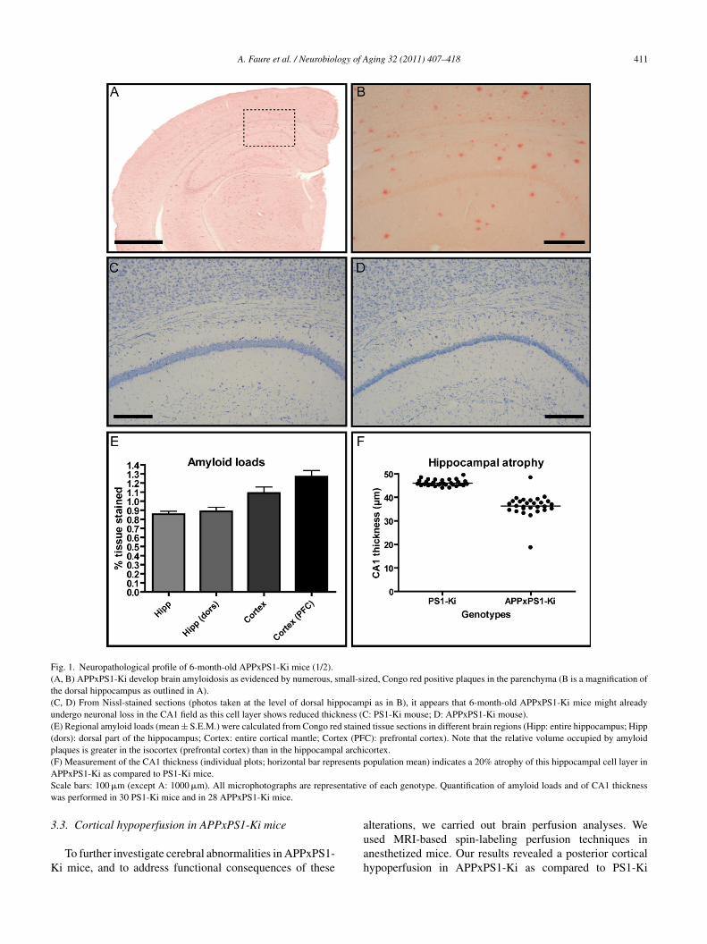
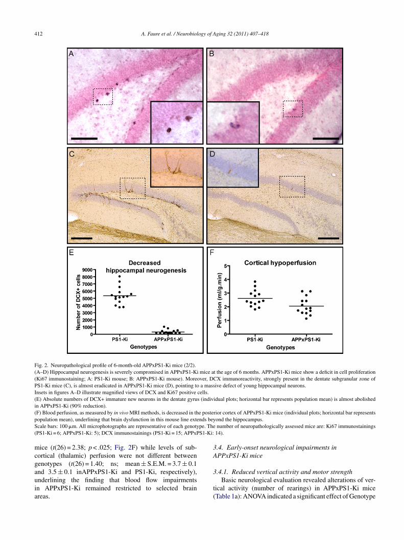
nd APPxPS1-Ki mice (data not shown), as expected fromrevious studies (Casas et al., 2004; Cotel et al., 2008). How-ver stereological analysis revealed a significant decrease inhe number of mitotic, Ki67-immunoreactive, cells in dentateyrus of APP/PS1-Ki mice (mean ± S.E.M. = 285.7 ± 40.8)ompared to PS1-Ki animals (462.1 ± 48.1; t(9) = 2.82;< .025; Fig. 2A and B). To further address the fate of new-orn cells in the dentate gyrus, we determined the number ofells that differentiated into neurons by quantifying the popu-ation of immature neuronal precursors expressing DCX. Theumber of DCX-positive cells was dramatically reduced inhe dentate gyrus of APPxPS1-Ki mice (difference betweenenotypes: t(27) = 15.1; p < .001, Fig. 2C–E). While we foundhat numbers of mitotic cells (Ki67+) and numbers of youngmmature neurons (DCX+) were positively correlated in PS1-i control mice (r = 0.93; p < .025), no correlation between
hose measures was found in APPxPS1-Ki mice (r = 0.06;s). This suggests that the loss of DCX+ cells in APPxPS1-i mice may result only partly from altered proliferation ofultipotent progenitor cells. Indeed, it is likely that the disap-
earance of neuronal precursors early during their maturationontribute to the dramatic loss of young immature DCX+ neu-ons. However, we cannot completely rule out the possibilityhat the loss of DCX+ cells concurrently reflects an increasedlial destiny of newborn cells and/or their accelerated neu-onal maturation, thereby shortening the time-window ofCX expression.We did not find any significant correlations between hip-
ocampal amyloid loads, measures of CA1-2 atrophy anduantitation of neurogenesis-associated markers Ki67 and
CX (rs < 0.38; ps > .17). This indicates that the variousnatomo-pathological alterations found in the hippocampusf 6-month-old APPxPS1-Ki mice, while possibly resultingrom a common source, are not closely interdependent.
A. Faure et al. / Neurobiology of Aging 32 (2011) 407–418 411
Fig. 1. Neuropathological profile of 6-month-old APPxPS1-Ki mice (1/2).(A, B) APPxPS1-Ki develop brain amyloidosis as evidenced by numerous, small-sized, Congo red positive plaques in the parenchyma (B is a magnification ofthe dorsal hippocampus as outlined in A).(C, D) From Nissl-stained sections (photos taken at the level of dorsal hippocampi as in B), it appears that 6-month-old APPxPS1-Ki mice might alreadyundergo neuronal loss in the CA1 field as this cell layer shows reduced thickness (C: PS1-Ki mouse; D: APPxPS1-Ki mouse).(E) Regional amyloid loads (mean ± S.E.M.) were calculated from Congo red stained tissue sections in different brain regions (Hipp: entire hippocampus; Hipp(dors): dorsal part of the hippocampus; Cortex: entire cortical mantle; Cortex (PFC): prefrontal cortex). Note that the relative volume occupied by amyloidplaques is greater in the isocortex (prefrontal cortex) than in the hippocampal archicortex.(F) Measurement of the CA1 thickness (individual plots; horizontal bar represents population mean) indicates a 20% atrophy of this hippocampal cell layer inAS entativew
3
K
PPxPS1-Ki as compared to PS1-Ki mice.cale bars: 100 �m (except A: 1000 �m). All microphotographs are represas performed in 30 PS1-Ki mice and in 28 APPxPS1-Ki mice.
.3. Cortical hypoperfusion in APPxPS1-Ki mice
To further investigate cerebral abnormalities in APPxPS1-i mice, and to address functional consequences of these
auah
of each genotype. Quantification of amyloid loads and of CA1 thickness
lterations, we carried out brain perfusion analyses. Wesed MRI-based spin-labeling perfusion techniques innesthetized mice. Our results revealed a posterior corticalypoperfusion in APPxPS1-Ki as compared to PS1-Ki
412 A. Faure et al. / Neurobiology of Aging 32 (2011) 407–418
Fig. 2. Neuropathological profile of 6-month-old APPxPS1-Ki mice (2/2).(A–D) Hippocampal neurogenesis is severely compromised in APPxPS1-Ki mice at the age of 6 months. APPxPS1-Ki mice show a deficit in cell proliferation(Ki67 immunostaining; A: PS1-Ki mouse; B: APPxPS1-Ki mouse). Moreover, DCX immunoreactivity, strongly present in the dentate subgranular zone ofPS1-Ki mice (C), is almost eradicated in APPxPS1-Ki mice (D), pointing to a massive defect of young hippocampal neurons.Insets in figures A–D illustrate magnified views of DCX and Ki67 positive cells.(E) Absolute numbers of DCX+ immature new neurons in the dentate gyrus (individual plots; horizontal bar represents population mean) is almost abolishedin APPxPS1-Ki (90% reduction).( e postep nds beyS pe. The( S1-Ki:
mcgauia
3A
F) Blood perfusion, as measured by in vivo MRI methods, is decreased in thopulation mean), underlining that brain dysfunction in this mouse line extecale bars: 100 �m. All microphotographs are representative of each genotyPS1-Ki = 6; APPxPS1-Ki: 5); DCX immunostainings (PS1-Ki = 15; APPxP
ice (t(26) = 2.38; p < .025; Fig. 2F) while levels of sub-ortical (thalamic) perfusion were not different betweenenotypes (t(26) = 1.40; ns; mean ± S.E.M. = 3.7 ± 0.1
nd 3.5 ± 0.1 inAPPxPS1-Ki and PS1-Ki, respectively),nderlining the finding that blood flow impairmentsn APPxPS1-Ki remained restricted to selected brainreas.3
t(
rior cortex of APPxPS1-Ki mice (individual plots; horizontal bar representsond the hippocampus.number of neuropathologically assessed mice are: Ki67 immunostainings
14).
.4. Early-onset neurological impairments inPPxPS1-Ki mice
.4.1. Reduced vertical activity and motor strengthBasic neurological evaluation revealed alterations of ver-
ical activity (number of rearings) in APPxPS1-Ki miceTable 1a): ANOVA indicated a significant effect of Genotype

A. Faure et al. / Neurobiology of Aging 32 (2011) 407–418 413
Table 1APPxPS1-Ki mice demonstrate various and gradual neurological impair-ments.
.
PS1-Ki APPxPS1-Ki
a. Primary screen—vertical activity (nb rearings)2 months 16.0 ± 1.7 13.2 ± 2.5 �4 months 12.2 ± 2.4 4.4 ± 1.3 � � �6 months 18.1 ± 2.8 2.9 ± 1.1 � � �Age effect – ↘
b. Wire grid test—latency to fall (s)2 months 56.9 ± 3.1 53.4 ± 3.7 �4 months 60.0 ± 0.0 33.0 ± 6.6 � � �6 months 56.3 ± 3.7 7.1 ± 1.4 � � �Age effect – ↘
c. 24 h actimetry test—locomotion (m)2 months 682.0 ± 45.0 816.2 ± 73.0 �4 months 862.1 ± 89.5 1057.5 ± 96.2 �6 months 718.4 ± 95.8 1253.0 ± 99.0 � � �Age effect – ↗
d. 24 h actimetry test—locomotion, night period (m)2 months 338.8 ± 49.3 492.2 ± 56.7 �4 months 568.9 ± 68.1 828.1 ± 93.0 �6 months 466.7 ± 69.5 868.0 ± 76.4 � � �Age effect ↗ ↗
e. Open-field test—horizontal activity (m)2 months 52.3 ± 3.0 51.4 ± 4.2 �4 months 39.8 ± 3.2 45.8 ± 4.9 �6 months 38.5 ± 2.9 53.4 ± 5.5 �Age effect ↘ –
f. Open-field test—vertical activity (nb rearings)2 months 34.9 ± 6.0 25.9 ± 4.0 �4 months 33.4 ± 5.5 10.7 ± 2.4 � �6 months 14.2 ± 3.2 2.3 ± 0.6 � �Age effect ↘ ↘
g. Open-field test—Ratio Center/Periphery2 months 0.21 ± 0.5 0.17 ± 0.6 �4 months 0.13 ± 1.4 0.21 ± 0.8 �6 months 0.09 ± 2.2 0.34 ± 0.7 � �Age effect ↘ ↗
h. Elevated plus maze test—Open/Closed ratio2 months 0.33 ± 0.06 0.51 ± 0.08 �4 months 0.09 ± 0.07 0.17 ± 0.04 �6 months 0.03 ± 0.01 1.28 ± 0.59 �Age effect ↘ ↗
i. Elevated plus maze test—horizontal activity (m)2 months 9.3 ± 0.6 8.8 ± 0.6 �4 months 4.8 ± 0.8 6.9 ± 0.7 �
Table 1 (Continued )
PS1-Ki APPxPS1-Ki
6 months 3.8 ± 0.5 10.6 ± 1.5 � � �Age effect ↘ ↗
Mice were evaluated at different ages in a neurological battery. Primaryscreening allowed the measurement of vertical activity (a). Motor strengthwas assessed using the wire grid test (b). During actimetry (c, d) and openfield (e, f) tests, various aspects of locomotor behavior were measured.Anxiety-related behaviors were quantified in the open field (g) and elevatedplus-maze (h, i) tests.For each test, the measured dependent variables are represented asmean ± S.E.M. Effects of the Age factor are symbolized for each geno-type by upward/downward arrows (p < .05). Effects of the Genotype factorare also illustrated, by white and black squares, at each age (see caption).APPxPS1-Ki mice show various abnormal neurological traits, with vari-ada
(iAagne
AAAa
3
eoAcaitbpm
tbiehmcob
3
i
ble onset and progression. Major neurological impairments involve: motorefects (loss of motor strength and of vertical activity), increased locomotorctivity, and decreased anxiety-related behaviors.
p < .001) and Age (p < .005) as well as an Age × Genotypenteraction (p < .005). In comparison to PS1 control mice,PPxPS1-Ki mice presented reduced vertical activity at 4
nd 6 months of age. There were no differences betweenenotypes on the other measurements provided during basiceurological evaluation (i.e. reflexes, spontaneous behaviors,tc.).
The wire grid test revealed significant effects of Genotype,ge and an interaction between the two factors (all ps < .001).large reduction of motor strength was hence detected in
PPxPS1-Ki transgenic as compared to PS1-Ki mice at 4nd 6 months of age (Table 1b).
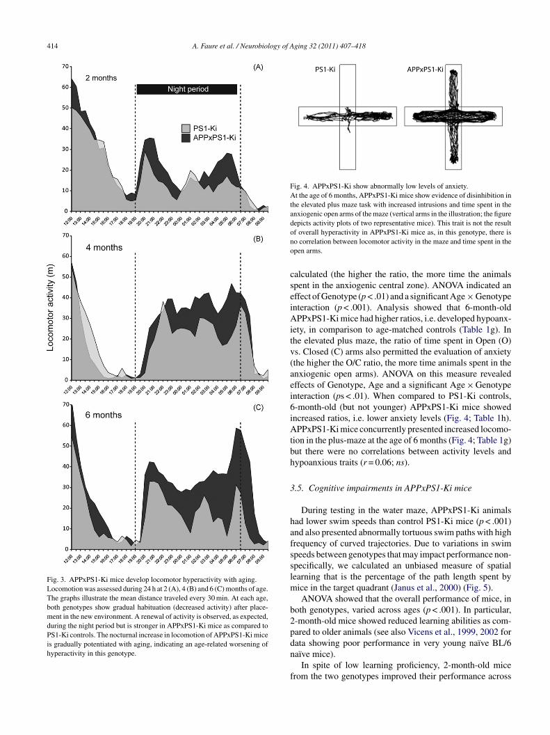
.4.2. Increased locomotor activityHyperactivity was observed in APPxPS1-Ki mice when
valuated during the 24-h actimetry test (Fig. 3). An effectf the Genotype factor was noted (p < .005) and the effect ofge and the Age × Genotype interaction were also signifi-
ant (ps < .05), owing to a gradual potentiation of locomotorctivity with aging in APPxPS1-Ki mice that reached signif-cance at 6 months of age (Table 1c). Refining the analyseso the nocturnal time window revealed stronger differencesetween groups (p < .001; Table 1d). Indeed, during the nighteriod, APPxPS1-Ki animals were more active than PS1-Kiice at 4 and 6 months of age.In the open-field task, no global effect of the Geno-
ype factor was observed on horizontal activity. However,oth Age and the Age × Genotype interaction were signif-cant (ps < .05). A significant difference between genotypesmerged at the age of 6 months with APPxPS1-Ki mice beingyperactive when compared to PS1 controls. APPxPS1-Kiice, while displaying enhanced locomotion, showed a con-
urrent reduced vertical activity (Table 1f), confirming databtained from neurological primary screen (decreased num-er of rearings, see above).
.4.3. Reduced anxiety-related behaviorsTo assess anxiety-related behaviors during open field test-
ng, a Center-to-Periphery ratio on exploration times was
414 A. Faure et al. / Neurobiology of Aging 32 (2011) 407–418
Fig. 3. APPxPS1-Ki mice develop locomotor hyperactivity with aging.Locomotion was assessed during 24 h at 2 (A), 4 (B) and 6 (C) months of age.The graphs illustrate the mean distance traveled every 30 min. At each age,both genotypes show gradual habituation (decreased activity) after place-ment in the new environment. A renewal of activity is observed, as expected,during the night period but is stronger in APPxPS1-Ki mice as compared toPS1-Ki controls. The nocturnal increase in locomotion of APPxPS1-Ki miceis gradually potentiated with aging, indicating an age-related worsening ofhyperactivity in this genotype.
Fig. 4. APPxPS1-Ki show abnormally low levels of anxiety.At the age of 6 months, APPxPS1-Ki mice show evidence of disinhibition inthe elevated plus maze task with increased intrusions and time spent in theanxiogenic open arms of the maze (vertical arms in the illustration; the figuredepicts activity plots of two representative mice). This trait is not the resultono
cseiAitv(aei6iAtbh
3
hafsslm
b2pdn
f
f overall hyperactivity in APPxPS1-Ki mice as, in this genotype, there iso correlation between locomotor activity in the maze and time spent in thepen arms.
alculated (the higher the ratio, the more time the animalspent in the anxiogenic central zone). ANOVA indicated anffect of Genotype (p < .01) and a significant Age × Genotypenteraction (p < .001). Analysis showed that 6-month-oldPPxPS1-Ki mice had higher ratios, i.e. developed hypoanx-
ety, in comparison to age-matched controls (Table 1g). Inhe elevated plus maze, the ratio of time spent in Open (O)s. Closed (C) arms also permitted the evaluation of anxietythe higher the O/C ratio, the more time animals spent in thenxiogenic open arms). ANOVA on this measure revealedffects of Genotype, Age and a significant Age × Genotypenteraction (ps < .01). When compared to PS1-Ki controls,-month-old (but not younger) APPxPS1-Ki mice showedncreased ratios, i.e. lower anxiety levels (Fig. 4; Table 1h).PPxPS1-Ki mice concurrently presented increased locomo-
ion in the plus-maze at the age of 6 months (Fig. 4; Table 1g)ut there were no correlations between activity levels andypoanxious traits (r = 0.06; ns).
.5. Cognitive impairments in APPxPS1-Ki mice
During testing in the water maze, APPxPS1-Ki animalsad lower swim speeds than control PS1-Ki mice (p < .001)nd also presented abnormally tortuous swim paths with highrequency of curved trajectories. Due to variations in swimpeeds between genotypes that may impact performance non-pecifically, we calculated an unbiased measure of spatialearning that is the percentage of the path length spent by
ice in the target quadrant (Janus et al., 2000) (Fig. 5).ANOVA showed that the overall performance of mice, in
oth genotypes, varied across ages (p < .001). In particular,-month-old mice showed reduced learning abilities as com-ared to older animals (see also Vicens et al., 1999, 2002 for
ata showing poor performance in very young naïve BL/6aïve mice).In spite of low learning proficiency, 2-month-old micerom the two genotypes improved their performance across

A. Faure et al. / Neurobiology of Aging 32 (2011) 407–418 415
Fig. 5. APPxPS1-Ki mice develop a gradual impairment in the acquisitionof spatial information.Spatial reference memory in the water maze task was evaluated in 2-, 4-and 6-month-old mice using an unbiased measure (% of distances traveledin the target quadrant: mean ± S.E.M.). At 2 months of age, both genotypesdemonstrate difficulties in learning the task although their performancesimprove across training sessions. Acquisition proficiency largely increaseswal
ltGwtropPi(gea
s(ptParatsiahsm(o
Fig. 6. APPxPS1-Ki mice develop a gradual impairment in the retention ofspatial information.From the age of 4 months, APPxPS1-Ki mice display spatial memory impair-ments in the water maze task. (A) During probe tests, APPxPS1-Ki micenavigate at larger distance (mean ± S.E.M.) from the spatial goal location(platform position) than do PS1-Ki control mice. The lack of finely tunedspatial representations in APPxPS1-Ki mice was observed at 4 and 6 monthsof age, but not in younger mice. (B) Reduced spatial bias in APPxPS1-Kimice was also directly evidenced by their occupancy plots (here illustratedin 6-month-old animals) that show a less narrowed exploration pattern. Tobuild the occupancy plots, the surface of the maze was divided in small tilesof equal sizes and the time spent in each tiles was color-coded accordingto its value (lookup table with white, yellow and red colors correspondingrstv
ioof
4
utAb
hen the mice are re-tested 2 months later and, at that age, no differencesre observed between the two genotypes. Finally, at 6 months of age, a clearearning deficit is observed in APPxPS1-Ki mice (p < .01).
earning sessions (all ps < .001). There was neither a Geno-ype effect nor a significant interaction between Session andenotype factors. When re-tested at 4 months of age, the miceere able to learn a new platform location with no Geno-
ype effect, but a Session × Genotype interaction (p < .05)eflecting a slight, nonsignificant, decrement in performancef APPxPS1-Ki mice. Both group of mice showed clearrogress in their search accuracy with training (p < .001 forS1-Ki and APPxPS1-Ki mice). At 6 months of age, learn-
ng was also evidenced, as reflected in an effect of Sessionp < .001) that reached statistical significance for the tworoups (ps < .001), but at this age APPxPS1-Ki mice clearlyxhibited impaired performance compared to control PS1-Kinimals (p < .01; see Fig. 5).
Memory for the platform location was evaluated duringingle probe trials (no platform) performed at each time point2, 4 and 6 months). From visual inspection of occupancylots (Wolfer et al., 2001), it appeared that spatial bias forhe target quadrant was lower in APPxPS1-Ki mice than inS1-Ki mice. This was particularly evident at 6 months ofge (Fig. 6B). To compare quantitatively the search accu-acy for the platform location in the two genotypes, welso calculated a proximity measure (Gallagher et al., 1993),hat is, the mouse-to-platform distance averaged across allampled points of the swim path. ANOVA on this proxim-ty measure indicated a significant effect of Genotype andn Age × Genotype interaction (ps < .05; see Fig. 6A). Postoc analysis showed that 4- and 6-month-old PS1-Ki mice
wam in closer vicinity to the platform, indicating betteremory proficiency, than age-matched APPxPS1-Ki miceps < .05). Comparison of performance at different ages inur longitudinal design indicated that PS1-Ki mice gradually
t
ai
espectively to low, medium and high durations; the occupancy plots repre-ent data cumulated and averaged for each genotype). (For interpretation ofhe references to color in this figure legend, the reader is referred to the webersion of the article.)
mproved their memory abilities between 2 and 6 monthsf age (p < .001), whereas APPxPS1-Ki mice showed thepposite tendency, i.e. a progressive decline in memory per-ormance (p < .001; see Fig. 6A).
. Discussion
The research effort to fight Alzheimer’s disease and tonderstand its physiopathogeny is largely dependent uponhe development of valid animal models. To this extend, thePPxPS1-Ki model is among the rare transgenic mouse linesearing brain amyloidosis together with neuronal loss, and
herefore is of particular interest.The present study was undertaken to extensively char-cterize the neuropathological alterations and functionalmpairments in this mouse model.

4 ogy of A
tancrolo
vaCaArcraoagcKisicnpohdpbascpmaaAho2iWmrAcmrCi
lsfeohealdt
bbwhttfnbddmoaoKtsarlc2rbmiirc
ppeEa2tis
16 A. Faure et al. / Neurobiol
We found that APPxPS1-Ki mice display a significanthinning of the hippocampal pyramidal cell layer, occurrings early as 6 months of age and that the production of neweurons in the dentate gyrus of these mice is also dramati-ally reduced. Using in vivo MRI measurements, our studyevealed cerebral capillary blood flow reduction in the cortexf 6-month-old APPxPS1-Ki mice that is paralleled by neuro-ogical impairments and memory deficits strongly evocativef hippocampal dysfunction.
As expected, extracellular A� deposits were detected inarious brain regions of 6-month-old animals. At this earlyge, we observed in addition a remarkable thinning of theA1-2 cell layer, evocative of local neuronal loss (Casas etl., 2004). From initial reports it appears that 2-month-oldPPxPS1-Ki mice do not show significant hippocampal neu-
onal loss whereas 10-month-old transgenics display a severeell loss in this region (Casas et al., 2004). Combined with ouresults, these data suggest that APPxPS1-Ki mice undergon age-related loss of neurons in the hippocampus, the onsetf which is between 2 and 6 months of age. When evalu-ting potential neuronal replacement based on new neuronsenerated in the dentate gyrus, we found that hippocampalell proliferation was reduced by nearly 40% in APPxPS1-i mice. Moreover, the fewer cells produced also showed
mpaired neuronal differentiation, as illustrated by the mas-ive (>90%) reduction of the number of DCX-expressingmmature neurons in APPxPS1-Ki mice. Interestingly, Li andollaborators (Li et al., 2008) reported that differentiation ofewly born cells into mature neurons is also severely com-romised in AD patients, resulting into a dramatic decreasef hippocampal neurogenesis. The mechanisms leading toippocampal neuronal loss and decreased neurogenic pro-uction in APPxPS1-Ki mice remain hypothetical. Despitearenchymal accumulation of A�, there were no correlationsetween hippocampal amyloid loads and neuronal loss (seelso Casas et al., 2004; Schmitz et al., 2004 for similar conclu-ions) or density of new neurons. It is possible that other A�onformations or localizations may have a higher pathogenicotential. A�42 is a prevalent A� species in APPxPS1-Kiice brain tissue (see biochemical results and also Casas et
l., 2004) and is considered to be a very toxic species closelyssociated with neuronal injury (Walsh and Selkoe, 2007).lso, and more importantly, intracellular A� accumulationas been reported in the APPxPS1-Ki line starting at the agef 2 months in the CA1-2 hippocampal region (Casas et al.,004). Although still debated, the exquisite pathogenicity ofntracellular A� is repeatedly reported (Laferla et al., 2007;
irths and Bayer, 2008a). In particular, intracellular A� isostly detected in brain regions that show subsequent neu-
onal loss (Christensen et al., 2008a; Oakley et al., 2006).bnormally up-regulated proteins involved in apoptosis and
ell survival have recently been identified in APPxPS1-Ki
ice (Damjanac et al., 2007; Page et al., 2006) and this up-egulation may result from intracellular accumulation of A�.oncerning the observed decreased neurogenic production,
t is also possible that highly vulnerable neuronal popu-
dsa2
ging 32 (2011) 407–418
ations, like new immature neurons, display an increasedensitivity to the toxic effects of A�. This would accountor the near complete absence of hippocampal neurogen-sis observed in 6-month-old APPxPS1-Ki mice. To date,ur report is the first to reveal such a massive reduction ofippocampal adult neurogenesis in transgenic animals mod-ling AD brain amyloidosis (Verret et al., 2007). Whetherbolished hippocampal neurogenesis is linked to CA1-2 celloss and/or to the impaired hippocampal plasticity previouslyescribed in APPxPS1-Ki mice (Bayer et al., 2008) remainso be determined.
In addition to hippocampal pathology, we showed reducedlood perfusion in the cortex of APPxPS1-Ki mice. Cere-ral hypoperfusion has also been reported in AD patientsith the same arterial spin labeling MRI methods as usedere (Asllani et al., 2008; Johnson et al., 2005), suggestinghat the in vivo measure of perfusion in transgenic mice is aranslational marker of the pathology. Interestingly, hypoper-usion in APPxPS1-Ki mice was not detected in subcorticaluclei (thalamus), generally considered to be less affectedy AD pathology. As suggested by its restricted topography,ecreased blood perfusion may promote or reflect local brainysfunction and behavioral impairments in APPxPS1-Kiice. Cortical hypoperfusion has previously been reported in
ther transgenic lines (Niwa et al., 2002), however the mech-nisms responsible for decreased blood flow, in the absencef robust amyloid angiopathy as in the case of the APPxPS1-i mice, remain elusive. Cerebral hypoperfusion might be
he consequence of the presence of soluble A� as recentlyhown (Luo et al., 2008). Decreased cortical perfusion mightlso result from local cortical cell loss and/or abnormal neu-onal activity. Indeed, recent observations underlined that aoss of neurons can be detected in isocortical regions (frontalortex) of 6-month-old APPxPS1-Ki mice (Christensen et al.,008b). Also, neural network dysfunctions involving aber-ant GABAergic inhibition of local circuitries have recentlyeen demonstrated in the brain of hAPP mice carrying ADutations (Palop et al., 2007). In the future, it would be
nteresting to examine whether cortical blood hypoperfusionn APPxPS1-Ki mice originates from cell loss and/or neu-onal dysfunction and whether it is related to behavioral andognitive defects.
Finally, we demonstrated that APPxPS1-Ki mice dis-lay neurological and cognitive symptoms often seen in ADatients and sometimes described in transgenic mouse mod-ls (Janus and Westaway, 2001; Pietropaolo et al., 2008).xtending previous findings in APPxPS1-Ki mice (Bayernd Wirths, 2008; Wirths and Bayer, 2008b; Wirths et al.,007), we reported several abnormal early onset behavioralraits such as decreased motor strength, reduced vertical activ-ty, and altered swimming behaviors. Interestingly, motorymptoms, although modest, have also been depicted in
emented patients (see Wirths and Bayer, 2008a for discus-ion). In APPxPS1-Ki mice, these symptoms are presumablyssociated with peripheral axonopathy (Bayer and Wirths,008; Wirths and Bayer, 2008b; Wirths et al., 2006). More
ogy of A
iAbfiab(Kapnpc
imhBbttnbnodprp
D
bfao
eCsottaa
A
Gstt
rihB
R
A
B
B
B
B
C
C
C
C
C
C
D
G
H
I
A. Faure et al. / Neurobiol
mportantly, we further described new phenotypic traits ofPPxPS1-Ki mice including: (1) a robust hyperlocomotorehavior that was confirmed using three different tests (openeld, actimetry and elevated-plus maze), and (2) abnormalnxiety-related behaviors together with signs of disinhi-ition that are also symptoms developed by AD patientsCummings, 1997). Lastly, at the cognitive level, APPxPS1-i mice exhibited deficits of acquisition and retention of fine
llocentric spatial information. These latter symptoms, notreviously described, are fully evocative of spatial disorga-ization and medial temporal lobe impairment as seen in ADatients. Their onset at 6 months of age in APP/PS1-Ki miceoincides with severe hippocampal pathology (see above).
In summary, APPxPS1-Ki mice develop early neurolog-cal deficits and signs of hippocampal dysfunction. At 6
onths of age, they display a neuronal loss and an abolishedippocampal neurogenesis, in addition to brain amyloidosis.rain functional anomalies in this model appear to extendeyond the hippocampus as evidenced by altered isocor-ical blood perfusion. Further work is needed to explorehe exact relationships between A� accumulation, alteredeuronal densities, impaired adult neurogenesis, reducedrain perfusion, and associated cognitive deficits. To date,o mouse model exhibits every neuropathological featuref Alzheimer’s disease. Therefore, APPxPS1-Ki mice thatisplay concomitant appearance of behavioral, cellular, andathological markers reminiscent of Alzheimer’s disease mayepresent a valuable tool to evaluate therapies against AD inreclinical research.
isclosure statement
B. Delatour received funding from Sanofi-Aventisetween 2005 and 2007 to carry out this research (salaryor post-doctoral fellowships). The authors are not aware ofny biases that might be perceived as affecting the objectivityf the present research work.
All experiments were conducted in accordance with thethical standards from French and European laws (Europeanommunities Council Directive of 24 November 1986). The
upervisor of the present study (B. Delatour) had receivedfficial agreements from the French Ministry of Agricultureo carry out research and experiments on animals (authoriza-ion No. 91-282). Experiments were performed in compliancend following approval of the Sanofi-Aventis Animal Carend Use Committee.
cknowledgements
We thank the Sanofi-Aventis Neurodegenerative Disease
roup for the generous gift of the animals involved in thistudy. This work was supported by the Del Duca Founda-ion (BD), the ACI Neurosciences program (MD & BD) andhe ANR Young Researcher program (CR, LV, BD). BD also
J
ging 32 (2011) 407–418 417
eceived funding from Sanofi-Aventis through an academic-ndustrial partnership. We are grateful to M. Guégan forelpful assistance in histological studies and to Dr. Brucerown for his help with the English text.
eferences
sllani, I., Habeck, C., Scarmeas, N., Borogovac, A., Brown, T.R., Stern,Y., 2008. Multivariate and univariate analysis of continuous arterial spinlabeling perfusion MRI in Alzheimer’s disease. J. Cereb. Blood FlowMetab. 28 (4), 725–736.
ayer, T.A., Breyhan, H., Duan, K., Rettig, J., Wirths, O., 2008. Intra-neuronal beta-amyloid is a major risk factor—novel evidence from theAPP/PS1KI mouse model. Neurodegener. Dis. 5 (3–4), 140–142.
ayer, T.A., Wirths, O., 2008. Review on the APP/PS1KI mouse model:intraneuronal Abeta accumulation triggers axonopathy, neuron loss andworking memory impairment. Genes Brain Behav. 7 (Suppl. 1), 6–11.
lanchard, V., Moussaoui, S., Czech, C., Touchet, N., Bonici, B., Planche,M., Canton, T., Jedidi, I., Gohin, M., Wirths, O., Bayer, T.A., Langui, D.,Duyckaerts, C., Tremp, G., Pradier, L., 2003. Time sequence of matura-tion of dystrophic neurites associated with Abeta deposits in APP/PS1transgenic mice. Exp. Neurol. 184 (1), 247–263.
urguiere, E., Arleo, A., Hojjati, M.R., Elgersma, Y., Zeeuw, C.I., Berthoz,A., Rondi-Reig, L., 2005. Spatial navigation impairment in mice lack-ing cerebellar LTD: a motor adaptation deficit? Nat. Neurosci. 8 (10),1292–1294.
alhoun, M.E., Wiederhold, K.H., Abramowski, D., Phinney, A.L., Probst,A., Sturchler-Pierrat, C., Staufenbiel, M., Sommer, B., Jucker, M., 1998.Neuron loss in APP transgenic mice. Nature 395 (6704), 755–756.
asas, C., Sergeant, N., Itier, J.M., Blanchard, V., Wirths, O., van der Kolk,N., Vingtdeux, V., van de Steeg, E., Ret, G., Canton, T., Drobecq, H.,Clark, A., Bonici, B., Delacourte, A., Benavides, J., Schmitz, C., Tremp,G., Bayer, T.A., Benoit, P., Pradier, L., 2004. Massive CA1/2 neuronalloss with intraneuronal and N-terminal truncated Abeta42 accumula-tion in a novel Alzheimer transgenic model. Am. J. Pathol. 165 (4),1289–1300.
hristensen, D.Z., Bayer, T.A., Wirths, O., 2008a. IntracellularAbeta triggers neuron loss in the cholinergic system of theAPP/PS1KI mouse model of Alzheimer’s disease. Neurobiol. Aging,doi:10.1016/j.neurobiolaging.2008.07.022.
hristensen, D.Z., Kraus, S.L., Flohr, A., Cotel, M.C., Wirths, O., Bayer,T.A., 2008b. Transient intraneuronal Abeta rather than extracellularplaque pathology correlates with neuron loss in the frontal cortex ofAPP/PS1KI mice. Acta Neuropathol. 116 (6), 647–655.
otel, M.C., Bayer, T.A., Wirths, O., 2008. Age-dependent loss of dentategyrus granule cells in APP/PS1KI mice. Brain Res. 1222, 207–213.
ummings, J.L., 1997. The Neuropsychiatric Inventory: assessing psy-chopathology in dementia patients. Neurology 48 (5 Suppl. 6), S10–S16.
amjanac, M., Rioux Bilan, A., Paccalin, M., Pontcharraud, R., Fauconneau,B., Hugon, J., Page, G., 2007. Dissociation of Akt/PKB and ribosomalS6 kinase signaling markers in a transgenic mouse model of Alzheimer’sdisease. Neurobiol. Dis. 29 (2), 354–367.
allagher, M., Burwell, R., Burchinal, M., 1993. Severity of spatial learningimpairment in aging: development of a learning index for performancein the Morris water maze. Behav. Neurosci. 107 (4), 618–626.
ardy, J.A., Higgins, G.A., 1992. Alzheimer’s disease: the amyloid cascadehypothesis. Science 256 (5054), 184–185.
rizarry, M.C., Soriano, F., McNamara, M., Page, K.J., Schenk, D., Games,D., Hyman, B.T., 1997. Abeta deposition is associated with neuropil
changes, but not with overt neuronal loss in the human amyloid pre-cursor protein V717F (PDAPP) transgenic mouse. J. Neurosci. 17 (18),7053–7059.anus, C., D’Amelio, S., Amitay, O., Chishti, M.A., Strome, R., Fraser, P.,Carlson, G.A., Roder, J.C., St George-Hyslop, P., Westaway, D., 2000.

4 ogy of A
J
J
K
L
L
L
L
N
O
P
P
P
P
R
S
S
S
T
U
V
V
V
W
W
W
W
W
W
W
18 A. Faure et al. / Neurobiol
Spatial learning in transgenic mice expressing human presenilin 1 (PS1)transgenes. Neurobiol. Aging 21 (4), 541–549.
anus, C., Westaway, D., 2001. Transgenic mouse models of Alzheimer’sdisease. Physiol. Behav. 73 (5), 873–886.
ohnson, N.A., Jahng, G.H., Weiner, M.W., Miller, B.L., Chui, H.C., Jagust,W.J., Gorno-Tempini, M.L., Schuff, N., 2005. Pattern of cerebral hypop-erfusion in Alzheimer disease and mild cognitive impairment measuredwith arterial spin-labeling MR imaging: initial experience. Radiology234 (3), 851–859.
ober, F., Duhamel, G., Cozzone, P.J., 2008. Experimental comparison offour FAIR arterial spin labeling techniques for quantification of mousecerebral blood flow at 4.7 T. NMR Biomed. 21 (8), 781–792.
aferla, F.M., Green, K.N., Oddo, S., 2007. Intracellular amyloid-beta inAlzheimer’s disease. Nat. Rev. Neurosci. 8 (7), 499–509.
e Cudennec, C., Faure, A., Ly, M., Delatour, B., 2008. One-year longitudi-nal evaluation of sensori-motor functions in APP751SL transgenic mice.Genes Brain Behav. 7 (Suppl. 1), 83–92.
i, B., Yamamori, H., Tatebayashi, Y., Shafit-Zagardo, B., Tanimukai, H.,Chen, S., Iqbal, K., Grundke-Iqbal, I., 2008. Failure of neuronal matu-ration in Alzheimer disease dentate gyrus. J. Neuropathol. Exp. Neurol.67 (1), 78–84.
uo, F., Seifert, T.R., Edalji, R., Loebbert, R.W., Hradil, V.P., Harlan, J.,Schmidt, M., Nimmrich, V., Cox, B.F., Fox, G.B., 2008. Non-invasivecharacterization of beta-amyloid(1–40) vasoactivity by functional mag-netic resonance imaging in mice. Neuroscience 155 (1), 263–269.
iwa, K., Kazama, K., Younkin, S.G., Carlson, G.A., Iadecola, C., 2002.Alterations in cerebral blood flow and glucose utilization in mice over-expressing the amyloid precursor protein. Neurobiol. Dis. 9 (1), 61–68.
akley, H., Cole, S.L., Logan, S., Maus, E., Shao, P., Craft, J., Guillozet-Bongaarts, A., Ohno, M., Disterhoft, J., Van Eldik, L., Berry, R., Vassar,R., 2006. Intraneuronal beta-amyloid aggregates, neurodegeneration,and neuron loss in transgenic mice with five familial Alzheimer’s diseasemutations: potential factors in amyloid plaque formation. J. Neurosci. 26(40), 10129–10140.
age, G., Rioux Bilan, A., Ingrand, S., Lafay-Chebassier, C., Pain, S., PeraultPochat, M.C., Bouras, C., Bayer, T., Hugon, J., 2006. Activated double-stranded RNA-dependent protein kinase and neuronal death in modelsof Alzheimer’s disease. Neuroscience 139 (4), 1343–1354.
alop, J.J., Chin, J., Roberson, E.D., Wang, J., Thwin, M.T., Bien-Ly, N.,Yoo, J., Ho, K.O., Yu, G.Q., Kreitzer, A., Finkbeiner, S., Noebels, J.L.,Mucke, L., 2007. Aberrant excitatory neuronal activity and compen-satory remodeling of inhibitory hippocampal circuits in mouse modelsof Alzheimer’s disease. Neuron 55 (5), 697–711.
axinos, G., Franklin, K.B.J., 2001. The Mouse Brain in Stereotaxic Coor-dinates, second ed. Academic Press, San Diego.
ietropaolo, S., Feldon, J., Yee, B.K., 2008. Age-dependent phenotypic char-acteristics of a triple transgenic mouse model of Alzheimer disease.Behav. Neurosci. 122 (4), 733–747.
ogers, D.C., Fisher, E.M., Brown, S.D., Peters, J., Hunter, A.J., Martin, J.E.,
1997. Behavioral and functional analysis of mouse phenotype: SHIRPA,a proposed protocol for comprehensive phenotype assessment. Mamm.Genome 8 (10), 711–713.cher, A.I., Xu, Y., Korf, E.S., White, L.R., Scheltens, P., Toga, A.W.,Thompson, P.M., Hartley, S.W., Witter, M.P., Valentino, D.J., Launer,
W
ging 32 (2011) 407–418
L.J., 2007. Hippocampal shape analysis in Alzheimer’s disease: apopulation-based study. Neuroimage 36 (1), 8–18.
chmitz, C., Rutten, B.P., Pielen, A., Schafer, S., Wirths, O., Tremp, G.,Czech, C., Blanchard, V., Multhaup, G., Rezaie, P., Korr, H., Steinbusch,H.W., Pradier, L., Bayer, T.A., 2004. Hippocampal neuron loss exceedsamyloid plaque load in a transgenic mouse model of Alzheimer’s disease.Am. J. Pathol. 164 (4), 1495–1502.
un, Y., Schmidt, N.O., Schmidt, K., Doshi, S., Rubin, J.B., Mulkern, R.V.,Carroll, R., Ziu, M., Erkmen, K., Poussaint, T.Y., Black, P., Albert, M.,Burstein, D., Kieran, M.W., 2004. Perfusion MRI of U87 brain tumorsin a mouse model. Magn. Reson. Med. 51 (5), 893–899.
akeuchi, A., Irizarry, M.C., Duff, K., Saido, T.C., Hsiao Ashe, K.,Hasegawa, M., Mann, D.M., Hyman, B.T., Iwatsubo, T., 2000. Age-related amyloid beta deposition in transgenic mice overexpressing bothAlzheimer mutant presenilin 1 and amyloid beta precursor proteinSwedish mutant is not associated with global neuronal loss. Am. J.Pathol. 157 (1), 331–339.
rbanc, B., Cruz, L., Le, R., Sanders, J., Ashe, K.H., Duff, K., Stanley, H.E.,Irizarry, M.C., Hyman, B.T., 2002. Neurotoxic effects of thioflavin S-positive amyloid deposits in transgenic mice and Alzheimer’s disease.Proc. Natl. Acad. Sci. U.S.A. 99 (22), 13990–13995.
erret, L., Jankowsky, J.L., Xu, G.M., Borchelt, D.R., Rampon, C., 2007.Alzheimer’s-type amyloidosis in transgenic mice impairs survival ofnewborn neurons derived from adult hippocampal neurogenesis. J. Neu-rosci. 27 (25), 6771–6780.
icens, P., Bernal, M.C., Carrasco, M.C., Redolat, R., 1999. Previous train-ing in the water maze: differential effects in NMRI and C57BL mice.Physiol. Behav. 67 (2), 197–203.
icens, P., Redolat, R., Carrasco, M.C., 2002. Effects of early spatial trainingon water maze performance: a longitudinal study in mice. Exp. Gerontol.37 (4), 575–581.
alsh, D.M., Selkoe, D.J., 2007. Abeta Oligomers—a decade of discovery.J. Neurochem. 101 (5), 1172–1184.
est, M.J., Kawas, C.H., Martin, L.J., Troncoso, J.C., 2000. The CA1 regionof the human hippocampus is a hot spot in Alzheimer’s disease. Ann. N.Y. Acad. Sci. 908, 255–259.
illiams, D.S., Detre, J.A., Leigh, J.S., Koretsky, A.P., 1992. Magnetic-resonance-imaging of perfusion using spin inversion of arterial water.Proc. Natl. Acad. Sci. U.S.A. 89 (1), 212–216.
irths, O., Bayer, T., 2008a. Early intraneuronal �-amyloid pathology:do transgenic mice represent valid model systems? Open Aging J. 2,7–12.
irths, O., Bayer, T.A., 2008b. Motor impairment in Alzheimer’s diseaseand transgenic Alzheimer’s disease mouse models. Genes Brain Behav.7 (Suppl. 1), 1–5.
irths, O., Breyhan, H., Schafer, S., Roth, C., Bayer, T.A., 2007. Deficitsin working memory and motor performance in the APP/PS1ki mousemodel for Alzheimer’s disease. Neurobiol. Aging 29 (6), 891–901.
irths, O., Weis, J., Kayed, R., Saido, T.C., Bayer, T.A., 2006.
Age-dependent axonal degeneration in an Alzheimer mouse model. Neu-robiol. Aging 28 (11), 1689–1699.olfer, D.P., Madani, R., Valenti, P., Lipp, H.P., 2001. Extended analysis ofpath data from mutant mice using the public domain software Wintrack.Physiol. Behav. 73 (5), 745–753.