Impact of the ion transportome of chloroplasts on the ...
14
REVIEW PAPER Impact of the ion transportome of chloroplasts on the optimization of photosynthesis Ildikò Szabò 1, * and Cornelia Spetea 2 1 Department of Biology, University of Padova, Italy; CNR Institute of Neuroscience, Padova, Italy 2 Department of Biological and Environmental Sciences, University of Gothenburg, 40530 Gothenburg, Sweden * Correspondence: [email protected] Received 21 November 2016; Editorial decision 9 February 2017; Accepted 9 February 2017 Editor: Angus Murphy, University of Maryland Abstract Ions play fundamental roles in all living cells, and their gradients are often essential to fuel transport, regulate enzyme activities, and transduce energy within cells. Regulation of their homeostasis is essential for cell metabolism. Recent results indicate that modulation of ion fluxes might also represent a useful strategy to regulate one of the most impor- tant physiological processes taking place in chloroplasts, photosynthesis. Photosynthesis is highly regulated, due to its unique role as a cellular engine for growth in the light. Controlling the balance between ATP and NADPH synthesis is a critical task, and availability of these molecules can limit the overall photosynthetic yield. Photosynthetic organ- isms optimize photosynthesis in low light, where excitation energy limits CO 2 fixation, and minimize photo-oxidative damage in high light by dissipating excess photons. Despite extensive studies of these phenomena, the mechanism governing light utilization in plants is still poorly understood. In this review, we provide an update of the recently iden- tified chloroplast-located ion channels and transporters whose function impacts photosynthetic efficiency in plants. Key words: Chloroplast, ion transport, photosynthesis, proton motive force. Introduction Photosynthesis Chloroplasts are the site of several metabolic processes, the most relevant being photosynthesis. During the light-depend- ent reactions of photosynthesis, taking place in the thylakoid membrane, photons are absorbed by the antenna pigments and the excitation energy is transferred to the reaction centers of photosystems, where photochemistry allows generation of reducing equivalents and a proton (H + ) gradient across the thylakoid membrane. In eukaryotic photosynthetic organ- isms, including algae and higher plants, thylakoids are sur- rounded by soluble stroma and two envelope membranes, together forming the chloroplast as the bioenergetic orga- nelle. Thylakoids, where primary photosynthetic reactions take place, are organized in stacked membranes called grana and unstacked stroma-exposed membranes. In prokaryotic photosynthetic organisms such as cyanobacteria, thylakoid membranes enclosing a soluble lumen compartment are not isolated from the cytosol and grana are not present. The sites of photon absorption are the light-harvesting complexes in the thylakoid membrane, which contain pro- tein-bound chlorophyll and carotenoids, and in the case of photosynthetic prokaryotes, the phycobilisomes containing phycobilins as pigments. In the reaction center of the photo- systems, charge separation drives electron flow from PSII to PSI via the cytochrome b 6 f (cyt b 6 f) complex. The net result of the light-driven electron flow is the oxidation of water to © The Author 2017. Published by Oxford University Press on behalf of the Society for Experimental Biology. All rights reserved. For permissions, please email: [email protected] Journal of Experimental Botany, Vol. 68, No. 12 pp. 3115–3128, 2017 doi:10.1093/jxb/erx063 Advance Access publication 13 March 2017 Downloaded from https://academic.oup.com/jxb/article/68/12/3115/3067753 by guest on 02 February 2022
-
Upload
khangminh22 -
Category
Documents
-
view
0 -
download
0
Transcript of Impact of the ion transportome of chloroplasts on the ...
REVIEW PAPER
Impact of the ion transportome of chloroplasts on the optimization of photosynthesis
Ildikò Szabò1,* and Cornelia Spetea2
1 Department of Biology, University of Padova, Italy; CNR Institute of Neuroscience, Padova, Italy2 Department of Biological and Environmental Sciences, University of Gothenburg, 40530 Gothenburg, Sweden
* Correspondence: [email protected]
Received 21 November 2016; Editorial decision 9 February 2017; Accepted 9 February 2017
Editor: Angus Murphy, University of Maryland
Abstract
Ions play fundamental roles in all living cells, and their gradients are often essential to fuel transport, regulate enzyme activities, and transduce energy within cells. Regulation of their homeostasis is essential for cell metabolism. Recent results indicate that modulation of ion fluxes might also represent a useful strategy to regulate one of the most impor-tant physiological processes taking place in chloroplasts, photosynthesis. Photosynthesis is highly regulated, due to its unique role as a cellular engine for growth in the light. Controlling the balance between ATP and NADPH synthesis is a critical task, and availability of these molecules can limit the overall photosynthetic yield. Photosynthetic organ-isms optimize photosynthesis in low light, where excitation energy limits CO2 fixation, and minimize photo-oxidative damage in high light by dissipating excess photons. Despite extensive studies of these phenomena, the mechanism governing light utilization in plants is still poorly understood. In this review, we provide an update of the recently iden-tified chloroplast-located ion channels and transporters whose function impacts photosynthetic efficiency in plants.
Key words: Chloroplast, ion transport, photosynthesis, proton motive force.
Introduction
Photosynthesis
Chloroplasts are the site of several metabolic processes, the most relevant being photosynthesis. During the light-depend-ent reactions of photosynthesis, taking place in the thylakoid membrane, photons are absorbed by the antenna pigments and the excitation energy is transferred to the reaction centers of photosystems, where photochemistry allows generation of reducing equivalents and a proton (H+) gradient across the thylakoid membrane. In eukaryotic photosynthetic organ-isms, including algae and higher plants, thylakoids are sur-rounded by soluble stroma and two envelope membranes, together forming the chloroplast as the bioenergetic orga-nelle. Thylakoids, where primary photosynthetic reactions
take place, are organized in stacked membranes called grana and unstacked stroma-exposed membranes. In prokaryotic photosynthetic organisms such as cyanobacteria, thylakoid membranes enclosing a soluble lumen compartment are not isolated from the cytosol and grana are not present.
The sites of photon absorption are the light-harvesting complexes in the thylakoid membrane, which contain pro-tein-bound chlorophyll and carotenoids, and in the case of photosynthetic prokaryotes, the phycobilisomes containing phycobilins as pigments. In the reaction center of the photo-systems, charge separation drives electron flow from PSII to PSI via the cytochrome b6f (cyt b6f) complex. The net result of the light-driven electron flow is the oxidation of water to
© The Author 2017. Published by Oxford University Press on behalf of the Society for Experimental Biology. All rights reserved. For permissions, please email: [email protected]
Journal of Experimental Botany, Vol. 68, No. 12 pp. 3115–3128, 2017doi:10.1093/jxb/erx063 Advance Access publication 13 March 2017
Dow
nloaded from https://academ
ic.oup.com/jxb/article/68/12/3115/3067753 by guest on 02 February 2022
3116 | Szabò and Spetea
molecular oxygen and the reduction of NADP+ to NADPH. Electron transport is coupled to the translocation of H+ into the lumen. The pH in the lumen has been estimated to reach values as low as 5.5–5.7 (Zaks et al., 2012). However, pH probably becomes even more acidic, as the activation maxi-mum of the violaxanthin de-epoxidase (VDE) that is crucial for the photoprotective mechanism NPQ (see below) is at pH 5.2 (Fufezan et al., 2012). Furthermore, using electron paramagnetic resonance (EPR) spin probes designed specifi-cally for measuring the intrathylakoid pH in chloroplasts, it has been demonstrated that the lumenal pH in Vicia faba can drop to values as low as pH 5.4 (Tikhonov et al., 2008) (in the state of photosynthetic control, characterized by an exhausted pool of ADP) when the efflux of H+ from the lumen through the ATP synthase is reduced. Tikhonov and colleagues, using the same experimental approach, also provided experimental data suggesting a lateral heteroge-neity in the transthylakoid pH difference. Their model pre-dicts significant alkalinization of the interthylakoid gap (the space between adjacent thylakoids of grana) and the establishment of non-uniform lateral profiles of ΔpH under photophosphorylation conditions (Vershubskii et al., 2017). Such lateral variation in the proton motive force (pmf) has also been observed in mitochondria, using ratiometric fluo-rescent pH-sensitive green fluorescent protein (GFP) fused to specific components of the respiratory chain complex (Rieger et al., 2014).
The linear electron flow is strictly correlated with the evo-lution of molecular oxygen, the rate of which can be taken as a measure of photosynthetic efficiency. However, a light-driven cyclic electron transport around PSI and cyt b6f is also operative, which does not evolve oxygen, or induce NADP+ reduction, but only contributes to H+ translocation into the thylakoid lumen. Most components of these macromolecular complexes are highly conserved between eukaryotic photo-synthetic organisms and cyanobacteria, where the thylakoid membrane is also the site of respiration. Successful acclima-tion of photosynthetic organisms to variable environments requires a tight management of source (light harvesting) and sink (CO2 assimilation) relationships.
Light-driven transthylakoid proton motive force
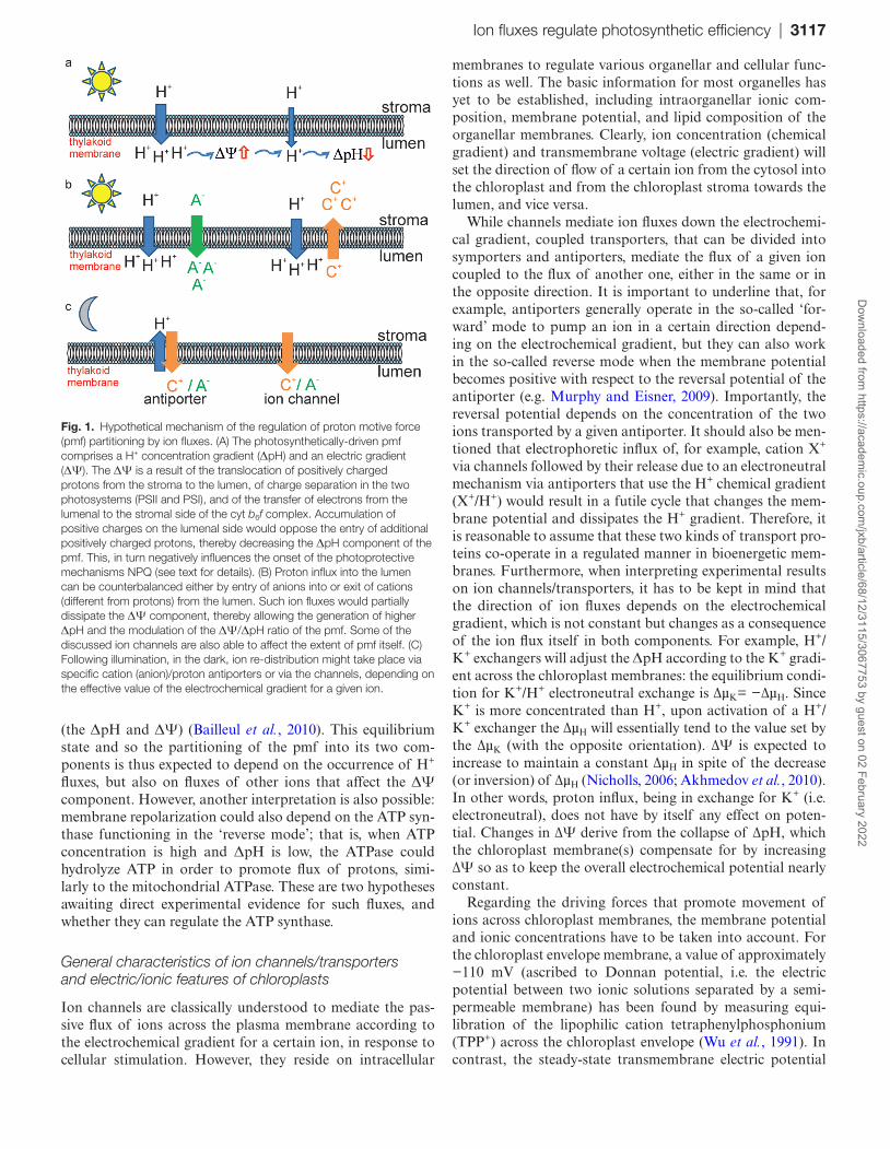
During the linear electron flow, H+ concentration increases in the lumen due to water splitting at PSII and plastoquinol oxidation at the cyt b6f complex. Conversely, H+ ions are transported back to the stroma by the ATP synthase, which converts the energy of the H+ gradient into chemical energy in the form of ATP. The photosynthetically-driven pmf com-prises a H+ concentration gradient (the ΔpH) and an electric field (the ΔΨ). The ΔΨ is produced by charge separation in the two photosystems (PSII and PSI) and by the transfer of electrons from the lumenal to the stromal side of the cyt b6f complex during the so-called Q cycle. The ΔΨ is initially a localized dipole field (positive at the lumenal and negative at the stromal side of the thylakoid membrane), but it is rapidly delocalized (within ~0.1 µs; Witt, 1979) by ion redistribution within the hydrophilic compartments of the chloroplast (the
stroma and the lumen). Conversely, ion flux between these two compartments (i.e. across the thylakoid membrane) leads to relaxation of the pmf. This relaxation is mostly attributed to H+ translocation through the ATP synthase CF0F1 com-plex (Joliot and Delosme, 1974). This enzyme translocates H+ and thus modifies the ΔΨ and ΔpH at the same time. CF0F1 activity can modulate the size, but cannot change the relative composition of the pmf. Therefore, the existence and implica-tion of other ion channels/pumps in regulation of the pmf via ion exchange was conceived since the formulation of the chemiosmotic theory: this would explain the finding that, although the pmf is generated by a similar process in chloro-plasts and mitochondria, it is mostly composed of a ΔpH in the photosynthetic organelle and of a ΔΨ in the respiratory organelle (see, for example, the reviews by Bernardi, 1999; Finazzi et al., 2015).
The ΔpH component of the pmf is crucial for initiating photoprotection of the photosynthetic apparatus through energy-dependent non-photochemical quenching (qE), a process that thermally dissipates the excess absorbed light energy, thereby limiting the production of reactive oxygen species (ROS) (e.g. Szabó et al., 2005; Niyogi and Truong, 2013; Ruban, 2016; Shikanai, 2016). qE, a component of non-photochemical quenching (NPQ), is also called energy-dependent excitation quenching, because thermal dissipation is stimulated by ΔpH, which builds up across the thylakoid membrane during photosynthetic electron transport. Since qE involves the de-excitation of singlet excited chlorophyll, it is also called feedback de-excitation. Thus, ΔpH specifically regulates photosynthetic electron flow and photoprotection. This calls for a tight regulation of the two components. Since the ΔpH/ΔΨ ratio is constant during electron flow, other mechanisms must exist to modify this ratio and also the par-titioning between ΔpH and ΔΨ in the light. In particular, ion channels and transporters in both thylakoids and envelope membranes have been hypothesized already decades ago to modify the thylakoid ΔΨ/ΔpH ratio, by varying the electric field without affecting the H+ gradient across thylakoids or impacting the stromal H+ concentration (Berkowitz and Wu, 1993; Cruz et al., 2001; Kramer et al., 2003) (Fig. 1), but the molecular nature of these ion flux-mediating pathways has remained unknown until recently. Since the ΔΨ modifies the spectra of photosynthetic pigments in the thylakoid mem-brane, resulting in the so-called electrochromic shift (ECS; Witt 1979), the pmf induced in the light and the thylakoid ΔΨ/ΔpH ratio can be estimated with this non-invasive tech-nique, even in vivo. In a typical ECS measurement, a fast relaxation phase, almost certainly reflecting H+ flux via the ATP synthase, is followed by a slower ECS increase, which has been interpreted as a signal due to charge redistribution across the membranes. According to this interpretation, flux of ions (different from H+) across the thylakoid membrane would be responsible for the observable partial membrane repolarization; that is, for a decrease of lumenal ΔΨ from positive values towards more negative values (that are still positive, see below) (Cruz et al., 2001). After a while, the ECS signal becomes relatively constant, reflecting the attainment of an equilibrium state between both components of the pmf
Dow
nloaded from https://academ
ic.oup.com/jxb/article/68/12/3115/3067753 by guest on 02 February 2022
Ion fluxes regulate photosynthetic efficiency | 3117
(the ΔpH and ΔΨ) (Bailleul et al., 2010). This equilibrium state and so the partitioning of the pmf into its two com-ponents is thus expected to depend on the occurrence of H+ fluxes, but also on fluxes of other ions that affect the ΔΨ component. However, another interpretation is also possible: membrane repolarization could also depend on the ATP syn-thase functioning in the ‘reverse mode’; that is, when ATP concentration is high and ΔpH is low, the ATPase could hydrolyze ATP in order to promote flux of protons, simi-larly to the mitochondrial ATPase. These are two hypotheses awaiting direct experimental evidence for such fluxes, and whether they can regulate the ATP synthase.
General characteristics of ion channels/transporters and electric/ionic features of chloroplasts
Ion channels are classically understood to mediate the pas-sive flux of ions across the plasma membrane according to the electrochemical gradient for a certain ion, in response to cellular stimulation. However, they reside on intracellular
membranes to regulate various organellar and cellular func-tions as well. The basic information for most organelles has yet to be established, including intraorganellar ionic com-position, membrane potential, and lipid composition of the organellar membranes. Clearly, ion concentration (chemical gradient) and transmembrane voltage (electric gradient) will set the direction of flow of a certain ion from the cytosol into the chloroplast and from the chloroplast stroma towards the lumen, and vice versa.
While channels mediate ion fluxes down the electrochemi-cal gradient, coupled transporters, that can be divided into symporters and antiporters, mediate the flux of a given ion coupled to the flux of another one, either in the same or in the opposite direction. It is important to underline that, for example, antiporters generally operate in the so-called ‘for-ward’ mode to pump an ion in a certain direction depend-ing on the electrochemical gradient, but they can also work in the so-called reverse mode when the membrane potential becomes positive with respect to the reversal potential of the antiporter (e.g. Murphy and Eisner, 2009). Importantly, the reversal potential depends on the concentration of the two ions transported by a given antiporter. It should also be men-tioned that electrophoretic influx of, for example, cation X+ via channels followed by their release due to an electroneutral mechanism via antiporters that use the H+ chemical gradient (X+/H+) would result in a futile cycle that changes the mem-brane potential and dissipates the H+ gradient. Therefore, it is reasonable to assume that these two kinds of transport pro-teins co-operate in a regulated manner in bioenergetic mem-branes. Furthermore, when interpreting experimental results on ion channels/transporters, it has to be kept in mind that the direction of ion fluxes depends on the electrochemical gradient, which is not constant but changes as a consequence of the ion flux itself in both components. For example, H+/K+ exchangers will adjust the ΔpH according to the K+ gradi-ent across the chloroplast membranes: the equilibrium condi-tion for K+/H+ electroneutral exchange is ∆µK= −∆µH. Since K+ is more concentrated than H+, upon activation of a H+/K+ exchanger the ∆µH will essentially tend to the value set by the ∆µK (with the opposite orientation). ∆Ψ is expected to increase to maintain a constant ∆µH in spite of the decrease (or inversion) of ∆µH (Nicholls, 2006; Akhmedov et al., 2010). In other words, proton influx, being in exchange for K+ (i.e. electroneutral), does not have by itself any effect on poten-tial. Changes in ∆Ψ derive from the collapse of ∆pH, which the chloroplast membrane(s) compensate for by increasing ∆Ψ so as to keep the overall electrochemical potential nearly constant.
Regarding the driving forces that promote movement of ions across chloroplast membranes, the membrane potential and ionic concentrations have to be taken into account. For the chloroplast envelope membrane, a value of approximately −110 mV (ascribed to Donnan potential, i.e. the electric potential between two ionic solutions separated by a semi-permeable membrane) has been found by measuring equi-libration of the lipophilic cation tetraphenylphosphonium (TPP+) across the chloroplast envelope (Wu et al., 1991). In contrast, the steady-state transmembrane electric potential
Fig. 1. Hypothetical mechanism of the regulation of proton motive force (pmf) partitioning by ion fluxes. (A) The photosynthetically-driven pmf comprises a H+ concentration gradient (ΔpH) and an electric gradient (ΔΨ). The ΔΨ is a result of the translocation of positively charged protons from the stroma to the lumen, of charge separation in the two photosystems (PSII and PSI), and of the transfer of electrons from the lumenal to the stromal side of the cyt b6f complex. Accumulation of positive charges on the lumenal side would oppose the entry of additional positively charged protons, thereby decreasing the ΔpH component of the pmf. This, in turn negatively influences the onset of the photoprotective mechanisms NPQ (see text for details). (B) Proton influx into the lumen can be counterbalanced either by entry of anions into or exit of cations (different from protons) from the lumen. Such ion fluxes would partially dissipate the ΔΨ component, thereby allowing the generation of higher ΔpH and the modulation of the ΔΨ/ΔpH ratio of the pmf. Some of the discussed ion channels are also able to affect the extent of pmf itself. (C) Following illumination, in the dark, ion re-distribution might take place via specific cation (anion)/proton antiporters or via the channels, depending on the effective value of the electrochemical gradient for a given ion.
Dow
nloaded from https://academ
ic.oup.com/jxb/article/68/12/3115/3067753 by guest on 02 February 2022
3118 | Szabò and Spetea
across thylakoids has been estimated to be small, ~10 mV positive in the lumen (Rottenberg and Grunwald, 1972), but values ranging between +20 mV and +50 mV have also been proposed (Joliot and Joliot, 1989). These early works sug-gested that given the relatively low value of the membrane potential, basically almost all pmf is stored as ΔpH, implying a high H+ concentration in the lumen. However, more recent studies employing non-invasive in vivo measurements indicate that the lumenal pH is only moderately acidic (pH ~6) under optimal conditions (Takizawa et al., 2007). The same authors also suggest that depending on the conditions, ΔΨ contrib-utes substantially and in a variable way to pmf in order to sustain a sufficient electrochemical gradient for H+ for the synthesis of ATP. Since both components of pmf can be exploited for ATP synthesis, a considerable contribution of the electric gradient to the pmf would allow the maintenance of a moderate lumen pH in the range that is not harmful to the photosynthetic electron transfer.
Regarding ionic composition, the chloroplast stroma contains ~150 mM K+ and 50–90 mM Cl− (Demmig and Gimmler, 1983; Robinson and Downton, 1984) as the main ions (Neuhaus and Wagner, 2000). Sodium is present in the stroma at 40–70 mM depending on the plant species (Robinson and Downton, 1984) and on salt stress conditions (Pottosin and Shabala, 2016). Concentrations of free Mg2+ in the stroma range between 0.5–1 mM in the dark and 2–3 mM in the light, as determined using an Mg2+-sensitive fluores-cent indicator (Ishijima et al., 2003). The concentration of K+ in the cytosol varies from 60 mM to 200 mM, and the opti-mal concentration is 100 mM (Dreyer and Uozumi, 2011), while Mg2+ is present at 1 mM concentration, for example in root epidermal cells (Bose et al., 2013). To our knowledge, much less is known about the ionic composition and dynamic changes in ion concentrations in the thylakoid lumen under physiological conditions, but it is well known that Ca2+ and Cl−, for example, are essential for the correct functioning of the oxygen-evolving complex (Vander Meulen et al., 2002), and calcium plays multiple roles in the context of photosyn-thesis regulation (Hochmal et al., 2015). Furthermore, ionic strength may impact thylakoid structure and grana stacking (Spetea and Schoefs, 2010), which in turn affect photosyn-thetic efficiency (Kirchhoff, 2014; Yoshioka-Nishimura et al., 2014; Pottosin and Shabala, 2016).
Ion channels and transporters in chloroplast envelope membranes
Similarly to mitochondria, chloroplasts are metabolically active organelles, and therefore endowed with numerous metabolite and ion transporters that connect the chloroplast milieu with the cytoplasm in a controlled way. Several trans-porters in the outer envelope membrane have been identified, namely outer envelope proteins (OEPs) 16, 21, 23, 24, and 37 (Gutierrez-Carbonell et al., 2014), the newly characterized solute channel OEP40 (Harsman et al., 2016), and an ABC transporter with unknown function. The different OEPs dis-play selectivity towards a certain set of metabolites, including
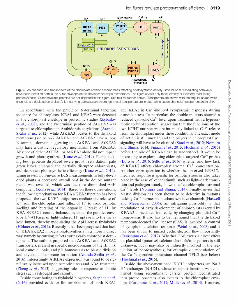
photosynthetic metabolites such as glucose, phosphorylated glucose, ATP, triose phosphates, and polypeptides (Pottosin and Shabala, 2016). Thereby, the OEPs act as barriers, avoid-ing an uncontrolled metabolite flux across the outer envelope, and are also good candidates for indirect regulation of pho-tosynthesis and related processes such as photorespiration (Linka and Weber, 2005). Analysis of Arabidopsis knock-out lines for OEP21, OEP24, and OEP37, however, did not reveal any photosynthetic phenotype under standard growth conditions, and knock-out plants for the glucose gate, OEP40, displayed an early flowering phenotype at cold temperature (Harsman et al., 2016). Regarding the chloroplast inner enve-lope (IE), a large number of metabolite and ion transporters have been identified by the combination of different methods ranging from classical biochemistry to proteomics and reverse genetics. The reader is advised to consult excellent reviews on this topic (Neuhaus and Wagner, 2000; Weber et al., 2005; Weber and Linka, 2011; Finazzi et al., 2015; Pottosin and Shabala, 2016). Among all IE transporters, we focus in the section below exclusively on those transporting inorganic ions and indirectly affecting photosynthetic efficiency (Fig. 2).
Monovalent cation–proton antiporters KEA1, KEA2, and NHD1
A K+ transport system consisting of K+-selective channels (Heiber et al., 1995) and/or of K+/H+ exchangers across the IE has been proposed to be crucial for chloroplast function because even small imbalance in the osmotic pressure or pH led to a dramatic decrease in photosynthesis. Indeed, long-lasting light-induced stromal alkalinization is important for optimal functioning of the Calvin–Benson cycle and has been proposed to be maintained through K+/H+ counterflux across the chloroplast envelope (Wu and Berkowitz, 1992). The pH gradient created during illumination by active H+ export across the IE membrane (~7.5 in the cytosol and 8.0 in the stroma) represents a driving force for H+ uptake into the stroma, which in turn is exploited for extrusion of K+ and Na+ via monovalent cation/K+ exchangers. The Arabidopsis proteome contains three homologs of the bacterial KefC, named AtKEA1–AtKEA3, and another three proteins of the NHX subgroup, named AtKEA4–AtKEA6. The full-length AtKEA1 was found to be inactive in yeast, whereas a short form lacking the N-terminal domain was functional, suggest-ing that this part of the protein may correspond to an auto-inhibitory domain controlling the transport activity (Zheng et al., 2013). A fragment of AtKEA2 was shown to comple-ment a yeast mutant deficient in the endosomal Na+(K+)/H+ exchanger (NHX1p) and to mediate monovalent cation/H+ antiport with preference for K+ when inserted into liposomes (Aranda-Sicilia et al., 2012). In mammals, one of the basic features of the so-called mitochondrial K+/H+ exchanger is the low selectivity for the species transported in exchange for H+, since the transporter does not discriminate between K+, Na+, and Li+, and it also transports organic cations such as tetrapropylammonium (Bernardi, 1999). It would therefore be important to explore whether AtKEA1–AtKEA3 share the permeability properties of AtKEA2.
Dow
nloaded from https://academ
ic.oup.com/jxb/article/68/12/3115/3067753 by guest on 02 February 2022
Ion fluxes regulate photosynthetic efficiency | 3119
In accordance with the predicted N-terminal targeting sequence for chloroplasts, KEA1 and KEA2 were detected in the chloroplast envelope in proteomic studies (Zybailov et al., 2008), and the N-terminal peptide of AtKEA2 was targeted to chloroplasts in Arabidopsis cotyledons (Aranda-Sicilia et al., 2012), while AtKEA3 locates to the thylakoid membrane (see below). AtKEA1 and AtKEA2 have a long N-terminal domain, suggesting that AtKEA1 and AtKEA2 may have a distinct regulatory mechanism from AtKEA3. Absence of either AtKEA1 or AtKEA2 alone did not impact growth and photosynthesis (Kunz et al., 2014). Plants lack-ing both proteins displayed severe growth retardation, pale green leaves, enlarged and partially disrupted chloroplasts, and decreased photosynthetic efficiency (Kunz et al., 2014). Using in vivo, non-invasive ECS measurements in fully devel-oped plants, a decreased overall pmf in the double mutant plants was revealed, which was due to a diminished ΔpH component (Kunz et al., 2014). Based on these observations, the following mechanism for KEA1/KEA2 function has been proposed: the two K+/H+ antiporters mediate the release of K+ from the chloroplast and influx of H+ to avoid osmotic swelling and bursting of the organelle. Uptake of H+ by KEA1/KEA2 is counterbalanced by either the putative enve-lope H+-ATPases or light-induced H+ uptake into the thyla-koid lumen, thereby modulating the ΔpH across thylakoids (Höhner et al., 2016). Recently, it has been proposed that lack of KEA1/KEA2 impacts photosynthesis in a more indirect way, namely by causing defects during early chloroplast devel-opment. The authors proposed that AtKEA1 and AtKEA2 transporters, present in specific microdomains of the IE, link local osmotic, ionic, and pH homeostasis to plastid division and thylakoid membrane formation (Aranda-Sicilia et al., 2016). Interestingly, AtKEA2 expression was found to be sig-nificantly increased upon osmotic stress and ABA treatment (Zheng et al., 2013), suggesting roles in response to abiotic stress such as drought and salinity.
Beside contributing to thylakoid biogenesis, Stephan et al. (2016) provided evidence for involvement of both KEA1
and KEA2 in Ca2+-induced cytoplasmic responses during osmotic stress. In particular, the double mutants showed a reduced cytosolic Ca2+ level upon treatment with a hyperos-motic sorbitol solution, suggesting that the functions of the two K+/H+ antiporters are intimately linked to Ca2+ release from the chloroplast under these conditions. The exact mode of action is still unclear, and the players in chloroplast Ca2+ signaling will have to be clarified (Stael et al., 2012; Nomura and Shiina, 2014; Finazzi et al., 2015; Hochmal et al., 2015) before the role of KEA1/2 can be understood. It would be interesting to explore using chloroplast-targeted Ca2+ probes (Loro et al., 2016; Sello et al., 2016) whether and how lack of KEA1/2 affects chloroplast stromal Ca2+ concentration. Another open question is whether the observed KEA1/2-mediated response is specific for osmotic stress or also takes place in the case of other stimuli, such as light–dark transi-tion and pathogen attack, shown to affect chloroplast stromal Ca2+ levels (Nomura and Shiina, 2014). Finally, given that plastid division has been shown to be defective in mutants lacking Ca2+-permeable mechanosensitive channels (Haswell and Meyerowitz, 2006), an intriguing possibility is that modulation of early development of chloroplasts exerted by KEA1/2 is mediated indirectly, by changing plastidial Ca2+ homeostasis. It also has to be mentioned that the thylakoid membrane-located Ca2+ sensor (CAS) is involved in control of cytoplasmic calcium response (Weinl et al., 2008) and it has been shown to impact cyclic electron flow importantly (Terashima et al., 2012). Whether CAS exerts a direct effect on plastidial (putative) calcium channels/transporters is still unknown, but it may also be indirectly involved in the reg-ulation of photosynthesis, for example via modulation of the Ca2+-dependent potassium channel TPK3 (see below) (Hochmal et al., 2015).
Beside the above-mentioned K+/H+ antiporters, an Na+/H+ exchanger (NHD1), whose transport function was con-firmed using recombinant carrier protein reconstituted into proteoliposomes, also locates to the chloroplast enve-lope (Furumoto et al., 2011; Müller et al., 2014). However,
Fig. 2. Ion channels and transporters of the chloroplast envelope membranes affecting photosynthetic activity. Several ion flux-mediating pathways have been identified both in the outer envelope and in the inner envelope membranes. The figure shows only those directly or indirectly modulating photosynthesis. Outer envelope proteins are not depicted in this figure. See text for further details. Transporters are shown with rectangular shape while channels are depicted as circles. Anion-carrying pathways are in orange, metal transporters are in blue, while cation channels/transporters are in pink.
Dow
nloaded from https://academ
ic.oup.com/jxb/article/68/12/3115/3067753 by guest on 02 February 2022
3120 | Szabò and Spetea
proteomic analysis also detected the protein in stroma lamel-lae (Tomizioli et al., 2014). An Arabidopsis T-DNA insertion mutant nhd1-1 showed compromised quantum efficiency of PSII and a higher NPQ in response to NaCl shock treatment (Müller et al., 2014). The chloroplast from salt-stressed nhd1 knock-out plants exhibited an increased Na+ concentration in the stroma in comparison with wild-type plants, suggesting that NHD1 is required for the export of Na+ from the chloro-plasts. The same study concluded that high stromal Na+ levels in nhd1 mutants result in impaired photosynthesis.
In addition, recent experiments showed that a decline in PSII efficiency in isolated chloroplasts from crop species kept under low cytosolic K+ conditions (concentration decreased from 50 mM to 10 mM) may be recovered by addition of substantial amounts of Na+ to the medium (Percey et al., 2016a, b), suggesting that ion transport across the envelope and thylakoid membranes also plays a critical role in leaf photosynthetic performance under salinity. Sodium might affect the function of the thylakoid membrane-located anion transporter 1 (ANTR1) that functions as an Na+-dependent Pi transporter when expressed in Escherichia coli (but as an H+-dependent Pi transporter when expressed in yeast). This protein is also called PHT4;1 (see below), and has been pro-posed to export Pi produced during nucleotide metabolism from the thylakoid lumen to the stroma (Pavón et al., 2008). Interestingly, halophyte species benefit from a relatively high level of sodium in chloroplasts (Cosentino et al., 2010; for a review, see, for example, Flowers et al., 2015). In the salt-tolerant plant Mesembryanthemum crystallinum, NaCl stress induced accumulation of Na+ in the chloroplasts via the Na+/H+ antiporter McNhaD. Its bacterial homolog NhaA of E. coli is involved in bacterial H+ homeostasis and is directly regulated by pH (Padan and Landau, 2016). The similarity of the pH sensitivities of NhaA and McNhaD suggests that this latter antiporter may also play a role in pH homeostasis of chloroplasts (Cosentino et al., 2010).
Proton/phosphate transporter family members PHT4;4 and PHT2;1
Members of the SLC17 transporter family were long thought to function as Na+ or H+ phosphate (Pi) symporters (PHT4). In Arabidopsis, the six genes coding for PHT4 proteins are differentially expressed in the various tissues and respond to distinct stimuli. Five out of the six proteins were initially proposed to be targeted to the plastid envelope, while the sixth resides in the Golgi apparatus. Complementation of the growth defect of a yeast Pi uptake-defective mutant demonstrated that all six isoforms can mediate Pi trans-port (Guo et al., 2008). On the basis of similarities with the mammalian SLC17 transporters which catalyze ∆Ψ- and Cl−-dependent organic anion transport, a role for PHT4;4 in ascorbate transport has also been hypothesized. Moriyama and colleagues then provided direct evidence using puri-fied AtPHT4;4 incorporated into proteoliposomes that the protein indeed mediates ∆Ψ- and Cl−-dependent ascor-bate uptake (Miyaji et al., 2015). In this work, AtPHT4;4 was shown to be located in the chloroplast envelope, and
plants lacking this isoform displayed decreased levels of the reduced form of ascorbate and a decrease in the xantho-phyll cycle pigment zeaxanthin (Zea). Zea forms from vio-laxanthin upon high light stress by the action of the enzyme VDE, which requires ascorbate as coenzyme. In accord-ance with the reduced amount of Zea, the photoprotective mechanism NPQ (non-photochemical quenching) known to require Zea for activation was decreased. Therefore, photo-oxidative stress was less tolerated by the knock-out plants (Miyaji et al., 2015).
In addition to PHT4;4, other members such as PHT4;1, PHT4;2, PHT4;5, and PHT2;1 are also located in plastids, as assessed using transiently expressed GFP fusions (Guo et al., 2008). PHT2;1 was found to function as a low-affinity H+ Pi symporter located in the chloroplast IE membrane where the pH difference might energize Pi import into the stroma. The observed overall reduction in growth of knock-out plants was in agreement with the diminished Pi content in the leaf, which might limit photosynthesis (Versaw and Harrison, 2002). Instead, the root plastid PHT4;2, lack of which does not impact photosynthesis, seems to play a key role in co-ordinat-ing metabolic signals throughout the whole plant (Irigoyen et al., 2011).
Metal transporters HMA1, HMA6, and PIC1
Metal ions within chloroplasts are necessary for the cor-rect function/assembly of proteins involved in photosynthe-sis, such as Fe–S proteins, plastocyanin, and cytochromes. However, when present in their free ionic forms, they may induce toxic oxidative stress; therefore, the transport of these metal ions has to be tightly regulated across plastid mem-branes (Bashir et al., 2016; López-Millán et al., 2016). Copper is pivotal for photosynthesis, since plastocyanin, one of the most abundant plant copper-binding proteins, is a lumen-located mobile electron carrier between cyt b6f and PSI. In addition, Cu transport into chloroplasts also provides the cofactor for the stromal enzyme copper/zinc superoxide dis-mutase (Cu/ZnSOD) which serves to reduce oxidative stress (Pilon et al., 2011; Aguirre and Pilon, 2015). Copper delivery to apo-plastocyanin involves at least two PIB-type ATPases, namely HMA6, a chloroplast envelope-located high-affinity transporter, and HMA8 in the thylakoid membrane (see below). Therefore, photosynthetic electron transport and plant growth were significantly reduced in mutants lacking either HMA6 or HMA8 (Shikanai et al., 2003; Abdel-Ghany et al., 2005). A recent work identified a cysteine- and histi-dine-rich motif in HMA6/8 that is crucial for copper release (Sautron et al., 2016). Another family member with plastidial localization is AtHMA1, which is able to transport copper but also other metal ions such as Zn. Although the plasto-cyanin content and function were not reduced in HMA1-less mutants, the photosensitivity was accentuated due to reduced chloroplast superoxide dismutase activity (Seigneurin-Berny et al., 2006). Interestingly, lack of HMA1 was shown to rein-force growth delay and reduced Cu/ZnSOD activity of the HMA6 mutant, and HMA1 overexpression in HMA6-less mutants resulted in Cu-dependent photosensitivity and Cu
Dow
nloaded from https://academ
ic.oup.com/jxb/article/68/12/3115/3067753 by guest on 02 February 2022
Ion fluxes regulate photosynthetic efficiency | 3121
accumulation in the chloroplast (Seigneurin-Berny et al., 2006; Boutigny et al., 2014).
Chloroplasts have a high Fe requirement, owing to their metabolism of photosynthetic pigments and photosynthetic complexes (Briat et al., 2015). Several families of proteins may play a role in Fe transport in chloroplasts, PIC1 (per-mease in chloroplasts1) in Arabidopsis being the first iden-tified molecular component involved in plastid Fe transport (López-Millan et al., 2016). AtPIC1 is a homolog of Sll1656 in Synechocystis sp. PCC6803, and was reported to interact with the NiCo protein, a member of the Ni2+–Co2+ trans-porter family. Therefore, the two proteins may function together in the transport of Fe into plastids for Fe–S cluster biogenesis, thus promoting the assembly of Fe–S clusters into new proteins upon translocation (Duy et al., 2011). Nicotiana tabacum PIC1 (NtPIC1) was shown to regulate chloroplast development, and pic1 mutants exhibited an albino pheno-type, demonstrating the important role of PIC1 in the Fe homeostasis of plastids (Gong et al., 2015).
Putative ion transporters affecting photosynthesis
There are a number of proteins that have been found in envelope membranes for which no direct evidence in favor of their ion-transporting ability in native membranes is available. GLR3.4 was localized to the IE membrane in spinach chloroplasts, and Arabidopsis knock-out plants showed a slight decrease in photosynthetic yield (Teardo et al., 2011). Although GLR3.4 has been proposed to mediate Ca2+ flux, based on electrophysiological evidence obtained upon heterologous expression (Vincill et al., 2012), its function as a Ca2+ channel in native membranes has not been proven. Recently, another GLR3 family member has been localized to chloroplasts, in particular one splicing variant of GLR3.5 is targeted to mitochondria, while the other variant is plastidial (Teardo et al., 2015). Whether a double knock-out would have an altered Ca2+ homeostasis and more severe photosynthetic phenotype remains to be explored.
Another putative channel of the ClC chloride channel fam-ily, ClC-f, has also been identified in the chloroplast envelope membrane of spinach by MS (Teardo et al., 2005), but was later proposed to target instead to the Golgi, since it func-tionally complemented the yeast gef1 mutant disrupted in the single CLC gene encoding a Golgi-associated protein (Monachello et al., 2009). Given that a synapse-like inter-action between the chloroplast and Golgi body has been observed and proposed to account, for example, for the transport of Golgi-synthesized plastoquinone to chloroplasts (Selga and Selga, 2000), these two findings may not be con-tradictory. The slight effect of ClC-f function on oxygen evo-lution was found only in isolated chloroplasts incubated with a mammalian ClC inhibitor, p-chlorophenoxy-acetic acid (Teardo et al., 2005). Thus, further work is required to under-stand whether AtClC-f knock-out plants have reduced pho-tosynthetic efficiency or they show a phenotype compatible with the knock-out for another Golgi-located ClC, AtCLC-d (von der Fecht-Bartenbach et al., 2007).
Recently, a divalent cation transporter, PAM71, has been discovered in the thylakoid membrane (Schneider et al., 2016), and its homolog, PAM71HL, was found in the envelope membrane in proteomic studies (Tomizioli et al., 2014). Both PAM71HL and PAM71 display sequence simi-larity with TMEM165 (Schneider et al., 2016). TMEM165 has been proposed to impact manganese homeostasis in the Golgi (Potelle et al., 2016), while evidence for its function as a Ca2+/H+ exchanger has previously been provided (Reinhardt et al., 2014). Direct evidence for a similar transport function of PAM71HL is still missing.
The non-intrinsic ABC protein 14 (AtNAP14) is a candi-date for the transport of Fe into the chloroplast, since the shoots of AtNAP14-less plants contain ~18 times more Fe than those of wild-type plants (Shimoni-Shor et al., 2010).
Other ion channels shown to regulate some aspects of chloroplast function are mechanosensitive MSL2 and MSL3, which are related to the small conductance mechanosensi-tive channel (MscS) of E. coli. These putative channels are required for normal plastid size and shape, since msl2 msl3 double mutants have enlarged chloroplasts and leaves with a lobed periphery and disturbed mesophyll cell organiza-tion (Haswell and Meyerowitz, 2006; Hamilton et al., 2015). Although MSL3 was proposed to be a channel based on its ability to rescue an E. coli strain lacking key mechanosen-sitive channels (Haswell and Meyerowitz, 2006), and a pref-erence for Cl− has been hypothesized (Veley et al., 2014), to our knowledge an in-depth analysis of the ion selectivity of MSL2/3 has not been performed. Similarly, determination of photosynthetic parameters would add to our understanding of the role of these channels for chloroplast function.
Ion channels and transporters in the thylakoid membrane
Channel activities in thylakoid membranes have been inves-tigated during the last decades by either the technically chal-lenging direct patch-clamping of isolated swollen thylakoid membranes (Schonknecht et al., 1988; Hinnah and Wagner, 1998), incorporating membrane vesicles (Tester and Blatt, 1989), or using purified native/recombinant proteins in pla-nar lipid bilayers (Carraretto et al., 2016). Unfortunately, the pharmacological characterization of these channels is still in its infancy. Furthermore, the technical difficulty of patch-clamping thylakoids from the completely sequenced Arabidopsis model plant persists, rendering it difficult to put forward hypotheses about the proteins responsible for most activities. Nonetheless, combination of multiple strategies including forward and reverse genetics, proteomics, and bio-informatic predictions has recently led to the discovery of a number of proteins with proven or hypothetical channel activ-ity. One of these putative channels for which no functional analysis is available up to now is the cyclic nucleotide-gated channel CNGC13, identified by proteomics in the thylakoid membrane (Yin et al., 2015). In the section below, we give a detailed description of the channel/transporter activities identified from the molecular point of view, and we discuss
Dow
nloaded from https://academ
ic.oup.com/jxb/article/68/12/3115/3067753 by guest on 02 February 2022
3122 | Szabò and Spetea
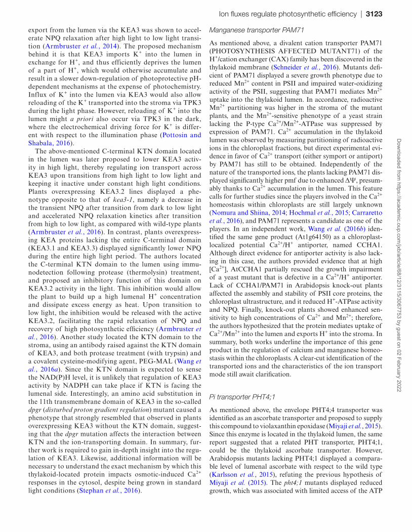
their putative or proven role in the regulation of photosyn-thesis (Fig. 3).
Potassium channel TPK3
The first genetic evidence for pmf regulation by a channel protein in higher plants has been obtained for the two-pore potassium channel TPK3. Experimentally, TPK3 has been located to the thylakoid stromal membranes in Arabidopsis plants using a specific monoclonal antibody and chloroplast membrane fractionation as well as confocal microscopic anal-ysis of GFP-tagged AtTPK3 expressed in Arabidopsis plants (Carraretto et al., 2013), although an earlier study located the protein to the vacuole and to other undefined intracellular structures (Voelker et al., 2006). Regarding the possible tar-geting of AtTPK3 to thylakoids, it is interesting to note that this protein harbors a diacidic motif at the C-terminus, typi-cal of cargo proteins that are transported by the vesicle trans-port system (Khan et al., 2013). Indeed, vesicular transport has been proposed also to take place within the chloroplasts, from the envelope membrane to the thylakoid via the COPII-like system (Khan et al., 2013; Karim and Aronsson, 2014).
Similarly to a K+ channel located to thylakoids in cyano-bacteria (Checchetto et al., 2012), TPK3 was found to act as a regulator of the ΔpH/ΔΨ ratio in vivo, apparently via ion counterbalancing. Activity of the recombinant AtTPK3 channel was found to be regulated by H+ and Ca2+ (Carraretto et al., 2013). The physiological significance of the regulation by H+ is clear, since H+ concentration increases in the lumen during the light phase and the channel is highly activated upon acidification of the recording solution. A fine analy-sis of the change of open probability in function of the H+ concentration, however, is still lacking. Similarly interesting questions to be resolved are whether the channel is oriented
in a way that the Ca2+-binding motif, the EF hand, faces the lumen and whether post-translational modifications modu-late its activity.
Silenced plants lacking AtTPK3 grown at 40 µmol pho-tons m−2 s−1 showed no difference with respect to wild-type plants, but when the light intensity was increased to 90 µmol photons m−2 s−1, they exhibited a decreased rosette size and an enhanced photosensitivity indicated by an increased anthocyanin content. This phenotype is in accordance with the observed higher ΔΨ and lower ΔpH in the knock-down plants, probably due to the lack of counterbalancing flux of positively charged K+ ions. The observed altered pmf partitioning results in reduced CO2 assimilation, reduced growth, and also in a deficient non-photochemical dissipa-tion (NPQ) of excess absorbed light, since NPQ onset is controlled by ΔpH. More recent results indicate that not only diminished ΔpH, but in general also elevated ΔΨ leads to increased photodamage (Davis et al., 2016), in accord-ance with the observations in TPK3 knock-down plants. Recently, tpk3 T-DNA insertion lines became available in the seed banks. Therefore, it will be important to explore whether these plants show exactly the same phenotype as the knock-down plants. This is an interesting and impor-tant issue in general, since in many cases (also in animals) in knock-out organisms a functional complementation of a given protein takes place. For example, another member of the same family can overtake the function of the miss-ing protein. This complementation is less likely to occur in organisms where protein expression is regulated via post-transcriptional silencing, and, interestingly, in several cases the phenotype of knock-out organisms does not perfectly match that of silenced plants/animals for the same protein (e.g. Kunz et al., 2014; Wagner et al., 2016).
Potassium/proton antiporter KEA3
As mentioned above, three members of the K+/H+ exchange family (KEA) have been described in Arabidopsis chlo-roplasts, namely KEA1 and KEA2 in the envelope and KEA3 in thylakoids (Kunz et al., 2014). Similarly to the situation with AtClC-f (see above), a study using stably transformed Arabidopsis seedlings expressing Cauliflower mosaic virus 35S-driven AtKEA3–GFP located the protein to Golgi membranes and found no fluorescence in chloro-plasts (Zheng et al., 2013), while in other works (includ-ing proteomic studies) KEA3 was found in thylakoids, in particular in the stroma lamellae subcompartment (Ferro et al., 2010; Armbruster et al., 2014; Tomizioli et al., 2014). Three splice forms of KEA3 (KEA3.1, KEA3.2, and KEA3.3), all displaying a chloroplast-targeting sig-nal and a cation/H+ antiporter domain, are annotated in Arabidopsis (Armbruster et al., 2016). KEA3.2, which is the major isoform in leaves, encodes a C-terminal K+ transport/nucleotide-binding (KTN) domain, which is truncated in KEA3.1 and is completely missing in KEA3.3 (Armbruster et al., 2014).
For AtKEA3, a role in photosynthetic acclimation under fluctuating light was reported; in particular, proton
Fig. 3. Ion channels and transporters of the thylakoid membrane. The figure shows ion flux-mediating pathways with a proven effect on photosynthetic efficiency. Changes of the ΔΨ/ΔpH ratio and of the pmf measured in knock-out or knock-down plants are shown, where available. The indicated changes in pmf partitioning are derived from ECS measurements. As in Fig. 2, anionic channels/transporters are shown in orange, metal transporters are in blue, while potassium channels/transporters are in pink.
Dow
nloaded from https://academ
ic.oup.com/jxb/article/68/12/3115/3067753 by guest on 02 February 2022
Ion fluxes regulate photosynthetic efficiency | 3123
export from the lumen via the KEA3 was shown to accel-erate NPQ relaxation after high light to low light transi-tion (Armbruster et al., 2014). The proposed mechanism behind it is that KEA3 imports K+ into the lumen in exchange for H+, and thus efficiently deprives the lumen of a part of H+, which would otherwise accumulate and result in a slower down-regulation of photoprotective pH-dependent mechanisms at the expense of photochemistry. Influx of K+ into the lumen via KEA3 would also allow reloading of the K+ transported into the stroma via TPK3 during the light phase. However, reloading of K+ into the lumen might a priori also occur via TPK3 in the dark, where the electrochemical driving force for K+ is differ-ent with respect to the illumination phase (Pottosin and Shabala, 2016).
The above-mentioned C-terminal KTN domain located in the lumen was later proposed to lower KEA3 activ-ity in high light, thereby regulating ion transport across KEA3 upon transitions from high light to low light and keeping it inactive under constant high light conditions. Plants overexpressing KEA3.2 lines displayed a phe-notype opposite to that of kea3-1, namely a decrease in the transient NPQ after transition from dark to low light and accelerated NPQ relaxation kinetics after transition from high to low light, as compared with wild-type plants (Armbruster et al., 2016). In contrast, plants overexpress-ing KEA proteins lacking the entire C-terminal domain (KEA3.1 and KEA3.3) displayed significantly lower NPQ during the entire high light period. The authors located the C-terminal KTN domain to the lumen using immu-nodetection following protease (thermolysin) treatment, and proposed an inhibitory function of this domain on KEA3.2 activity in the light. This inhibition would allow the plant to build up a high lumenal H+ concentration and dissipate excess energy as heat. Upon transition to low light, the inhibition would be released with the active KEA3.2, facilitating the rapid relaxation of NPQ and recovery of high photosynthetic efficiency (Armbruster et al., 2016). Another study located the KTN domain to the stroma, using an antibody raised against the KTN domain of KEA3, and both protease treatment (with trypsin) and a covalent cysteine-modifying agent, PEG-MAL (Wang et al., 2016a). Since the KTN domain is expected to sense the NAD(P)H level, it is unlikely that regulation of KEA3 activity by NADPH can take place if KTN is facing the lumenal side. Interestingly, an amino acid substitution in the 11th transmembrane domain of KEA3 in the so-called dpgr (disturbed proton gradient regulation) mutant caused a phenotype that strongly resembled that observed in plants overexpressing KEA3 without the KTN domain, suggest-ing that the dpgr mutation affects the interaction between KTN and the ion-transporting domain. In summary, fur-ther work is required to gain in-depth insight into the regu-lation of KEA3. Likewise, additional information will be necessary to understand the exact mechanism by which this thylakoid-located protein impacts osmotic-induced Ca2+ responses in the cytosol, despite being grown in standard light conditions (Stephan et al., 2016).
Manganese transporter PAM71
As mentioned above, a divalent cation transporter PAM71 (PHOTOSYNTHESIS AFFECTED MUTANT71) of the H+/cation exchanger (CAX) family has been discovered in the thylakoid membrane (Schneider et al., 2016). Mutants defi-cient of PAM71 displayed a severe growth phenotype due to reduced Mn2+ content in PSII and impaired water-oxidizing activity of the PSII, suggesting that PAM71 mediates Mn2+ uptake into the thylakoid lumen. In accordance, radioactive Mn2+ partitioning was higher in the stroma of the mutant plants, and the Mn2+-sensitive phenotype of a yeast strain lacking the P-type Ca2+/Mn2+-ATPase was suppressed by expression of PAM71. Ca2+ accumulation in the thylakoid lumen was observed by measuring partitioning of radioactive ions in the chloroplast fractions, but direct experimental evi-dence in favor of Ca2+ transport (either symport or antiport) by PAM71 has still to be obtained. Independently of the nature of the transported ions, the plants lacking PAM71 dis-played significantly higher pmf due to enhanced ΔΨ, presum-ably thanks to Ca2+ accumulation in the lumen. This feature calls for further studies since the players involved in the Ca2+ homeostasis within chloroplasts are still largely unknown (Nomura and Shiina, 2014; Hochmal et al., 2015; Carraretto et al., 2016), and PAM71 represents a candidate as one of the players. In an independent work, Wang et al. (2016b) iden-tified the same gene product (At1g64150) as a chloroplast-localized potential Ca2+/H+ antiporter, named CCHA1. Although direct evidence for antiporter activity is also lack-ing in this case, the authors provided evidence that at high [Ca2+], AtCCHA1 partially rescued the growth impairment of a yeast mutant that is defective in a Ca2+/H+ antiporter. Lack of CCHA1/PAM71 in Arabidopsis knock-out plants affected the assembly and stability of PSII core proteins, the chloroplast ultrastructure, and it reduced H+-ATPase activity and NPQ. Finally, knock-out plants showed enhanced sen-sitivity to high concentrations of Ca2+ and Mn2+; therefore, the authors hypothesized that the protein mediates uptake of Ca2+/Mn2+ into the lumen and exports H+ into the stroma. In summary, both works underline the importance of this gene product in the regulation of calcium and manganese homeo-stasis within the chloroplasts. A clear-cut identification of the transported ions and the characteristics of the ion transport mode still await clarification.
Pi transporter PHT4;1
As mentioned above, the envelope PHT4;4 transporter was identified as an ascorbate transporter and proposed to supply this compound to violaxanthin epoxidase (Miyaji et al., 2015). Since this enzyme is located in the thylakoid lumen, the same report suggested that a related PHT transporter, PHT4;1, could be the thylakoid ascorbate transporter. However, Arabidopsis mutants lacking PHT4;1 displayed a compara-ble level of lumenal ascorbate with respect to the wild type (Karlsson et al., 2015), refuting the previous hypothesis of Miyaji et al. (2015). The pht4;1 mutants displayed reduced growth, which was associated with limited access of the ATP
Dow
nloaded from https://academ
ic.oup.com/jxb/article/68/12/3115/3067753 by guest on 02 February 2022
3124 | Szabò and Spetea
synthase to stromal Pi, impacting carbon fixation and sugar accumulation in leaves (Karlsson et al., 2015). Therefore, PHT4;1 was proposed to play a role in Pi homeostasis in thy-lakoids as a local Pi supplier to the ATP synthase during the photobiochemical reactions. Due to reduced ATP synthase activity, an excess of H+ in the lumen of pht4;1 was indicated by a higher pmf partitioning to ΔpH in the first minute of illumination, resulting in a more efficient activation of NPQ (Karlsson et al., 2015).
Anion channels VCCN1, VCCN2, and ClC-e
Duan et al. (2016) localized two bestrophin-like proteins to the thylakoid membrane in Arabidopsis and named them AtBest1 and AtBest2. The bestrophin family is diverse in terms of substrate and includes members such as bestro-phin-1 and KpBest from Klebsiella pneumoniae, that are a Ca2+-activated Cl− channel and an Na+ channel, respec-tively. Although no electrophysiological experiments were performed by Duan et al., the AtBest1 was suggested to be a Cl− channel involved in reduction of pmf partitioning to ∆Ψ, resulting in enhanced ∆pH and fast activation of NPQ during photosynthesis, especially under fluctuating light con-ditions. As compared with altered photosynthesis (reduced NPQ) in atbest1 mutants, atbest2 behaved like the wild type, whereas the atbest1atbest2 mutant resembled the atbest1 mutant. Transcript analyses revealed AtBest1 as the major form expressed in leaves, whereas AtBest2 was only 10% of AtBest1. The authors suggested that the two proteins have redundant functions in leaves since the NPQ phenotype of the double mutant was fully restored by complementation with either AtBest1 or AtBest2. Transmission electron micros-copy of the double mutant (not indicated whether they were dark- or light-adapted samples) revealed a larger thylakoid lumen and loose grana stacks, and the data were interpreted as showing that other ions entered the lumen instead of Cl−, and promoted the observed effect. It is concluded that AtBest is critical for thylakoid ultrastructure. Outdoor experiments revealed an enhanced sensitivity of the double mutant to high light and attributed it to the reduced NPQ capacity.
The same proteins were also studied by Herdean et al. (2016a). The authors came to the same conclusions regarding the thylakoid location, and in addition showed that AtBest2 is mostly expressed in flowers, where it may play an impor-tant role worthy of further investigations. Based on electro-physiology of the recombinant protein, AtBest1 was found to be a voltage-dependent anion channel (with preference for Cl− over nitrate), and was therefore named AtVCCN1, whereas AtBest2 was named AtVCCN2. Detailed ECS and NPQ analyses indicated differences between transgenic lines overexpressing either AtVCCN1 or AtVCCN2, and it was concluded that AtVCCN2 triggered a similar response to overexpressed AtVCCN1 but with different kinetics in leaves. The reasons behind the different conclusions concerning redundancy of the two proteins in this study and the one by Duan et al. (2016) are not clear at present. One possible expla-nation could be the different mutant backgrounds used in the two studies. Another discrepancy is that the ultrastructure of
thylakoids from vccn1 in Herdean et al. (2016a) differs from that of atbest1atbest2 in Duan et al. (2016), namely the vccn1 in the dark-adapted state has wider grana with smaller repeat distance which could be due to smaller lumen size. In the light, a large proportion of the thylakoids displayed altered shape (‘banana-like’) of thylakoids. The ultrastructure of the dou-ble mutant has not been analyzed by Herdean et al. (2016a).
In addition to the above anion channels, ClC-e of the ClC family has been located to the thylakoid membrane in differ-ent species (Marmagne et al., 2007). As in the case of ClC-f, direct evidence in favor of this protein working as a chloride (nitrate) channel or as a chloride(nitrate)/proton antiporter is lacking. Herdean et al. (2016b) reported that the size and composition of the pmf were mildly altered in two independ-ent Arabidopsis clc-e mutant lines. In particular, the pho-tosynthetic electron transport of dark-adapted plants was affected, and pre-treatment with KCl but not with KNO3 restored the wild-type phenotype. Based on these data, the authors proposed that AtCLC-e functions in Cl– rather than in nitrate homeostasis and is especially important after tran-sition from light to dark, which affects chloroplast ultrastruc-ture and regulation of photosynthetic electron transport.
Future outlook
Besides the above-discussed Cl−, K+, Fe2+, and Mn2+ ions, other cations including Mg2+ and Ca2+ could have major roles in regulating (directly or indirectly) photosynthetic efficiency. Indeed Mg2+ is also a well-established candidate for counterbalancing H+ entry into the lumen (Hind et al., 1974), and changes in stromal Mg2+ concentration are known to affect chloroplast photosynthetic capacity by controlling H+ movement across the envelope and activation of RuBisCo (Berkowitz and Wu, 1993). Ca2+ is necessary not only for the correct function of the oxygen-evolving complex (Kessler, 1955; Boussac et al., 1989) but also for the regulation of Calvin cycle enzymes, and recent data highlight an important role for this cation in the control of cyclic electron flow (CEF) as well (Terashima et al., 2012; Hochmal et al., 2015, 2016). Thus, research should substantially focus on the identifica-tion of the molecular players involved in the flux of these ions (Nouet et al., 2011). Moreover, the pathways allowing their entry to the correct site of action within the chloroplast should be addressed, to complement their biochemical and electro-physiology studies in vitro. Identification of post-translational modifications affecting channel/transporter activities as well as of interacting partners (possibly in vivo) is also of utmost importance. Whether, similarly to mitochondrial ion chan-nels that have been shown to be physically and functionally coupled to respiratory chain complexes (Bednarczyk et al., 2013), thylakoid ion channels/transporters directly interact with stroma lamellae-located components of the photosyn-thetic electron transport chain, is also an exciting possibility to explore. Finally, in vivo studies are needed to establish the link between the roles of each ion in photosynthesis. Further studies should clarify to what extent the different ion chan-nels/transporters are involved in the regulation of the pmf
Dow
nloaded from https://academ
ic.oup.com/jxb/article/68/12/3115/3067753 by guest on 02 February 2022
Ion fluxes regulate photosynthetic efficiency | 3125
by comparing single/double/triple mutants and whether their relative contribution depends on specific environmental con-ditions (e.g. continuous or intermittent light stress). In addi-tion, as pointed out by Tikhonov and colleagues (Vershubskii et al., 2017), direct measurements of pH inside the lumen and in the interthylakoid gap with pH-sensitive fluorescent probes fused to proteins involved in photosynthesis would be very useful. If measurements with these probes (still to be gener-ated) were combined with those obtained using ion-specific probes [to be generated for most ions, but see, for example, Hong-Hermesdorf et al. (2015) for copper distribution], a more complete picture could be achieved about the relation-ship between ion and proton activities and pmf in vivo. The observation that most of the so far identified players in ion homeostasis across thylakoids profoundly alter (indirectly) the NPQ (see above), gains further importance in light of the recent discovery that photosynthetic efficiency and dry mass could be increased by 15% in fluctuating light through acceleration of NPQ relaxation in tobacco plants express-ing Arabidopsis VDE, zeaxanthin epoxidase, and PsbS (i.e. known players in the of NPQ; Kromdijk et al., 2016).
AcknowledgementsThe authors are grateful to Dr Giovanni Finazzi for critical reading of the manuscript, and to all collaborators who contributed to the works described in this review. Work in the authors’ laboratories was supported by a grant from the Human Frontiers Science Program (HFSP 0052, to IS) and PRIN 2015795S5W of the Italian Ministry (to IS), the Swedish Research Council, the Royal Society of Arts and Sciences in Gothenburg, and Donationsfonderna at University of Gothenburg (CS).
ReferencesAbdel-Ghany SE, Müller-Moulé P, Niyogi KK, Pilon M, Shikanai T. 2005. Two P-type ATPases are required for copper delivery in Arabidopsis thaliana chloroplasts. The Plant Cell 17, 1233–1251.
Aguirre G, Pilon M. 2015. Copper delivery to chloroplast proteins and its regulation. Frontiers in Plant Science 6, 1250.
Akhmedov D, Braun M, Mataki C, Park KS, Pozzan T, Schoonjans K, Rorsman P, Wollheim CB, Wiederkehr A. 2010. Mitochondrial matrix pH controls oxidative phosphorylation and metabolism–secretion coupling in INS-1E clonal beta cells. FASEB Journal 24, 4613–4626.
Aranda-Sicilia MN, Aboukila A, Armbruster U, et al. 2016. Envelope K+/H+ antiporters AtKEA1 and AtKEA2 function in plastid development. Plant Physiology 172, 441–449.
Aranda-Sicilia MN, Cagnac O, Chanroj S, Sze H, Rodríguez-Rosales MP, Venema K. 2012. Arabidopsis KEA2, a homolog of bacterial KefC, encodes a K(+)/H(+) antiporter with a chloroplast transit peptide. Biochimica et Biophysica Acta 1818, 2362–2371.
Armbruster U, Carrillo LR, Venema K, et al. 2014. Ion antiport accelerates photosynthetic acclimation in fluctuating light environments. Nature Communications 5, 5439.
Armbruster U, Leonelli L, Correa Galvis V, Strand D, Quinn EH, Jonikas MC, Niyogi KK. 2016. Regulation and levels of the thylakoid K+/H+ antiporter KEA3 shape the dynamic response of photosynthesis in fluctuating light. Plant and Cell Physiology 57, 1557–1567.
Bailleul B, Cardol P, Breyton C, Finazzi G. 2010. Electrochromism: a useful probe to study algal photosynthesis. Photosynthesis Research 106, 179–189.
Bashir K, Rasheed S, Kobayashi T, Seki M, Nishizawa NK. 2016. Regulating subcellular metal homeostasis: the key to crop improvement. Frontiers in Plant Science 7, 1192.
Bednarczyk P, Wieckowski MR, Broszkiewicz M, Skowronek K, Siemen D, Szewczyk A. 2013. Putative structural and functional coupling of the mitochondrial BKCa channel to the respiratory chain. PLoS One 8, e68125.
Berkowitz GA, Wu W. 1993. Magnesium, potassium flux and photosynthesis. Magnesium Research 6, 257–265.
Bernardi P. 1999. Mitochondrial transport of cations: channels, exchangers, and permeability transition. Physiological Reviews 79, 1127–1155.
Bose J, Babourina O, Shabala S, Rengel Z. 2013. Low pH and aluminum resistance in Arabidopsis correlates with high cytosolic magnesium content and increased magnesium uptake by plant roots. Plant and Cell Physiology 54, 1093–1104.
Boussac A, Zimmermann JL, Rutherford AW. 1989. EPR signals from modified charge accumulation states of the oxygen evolving enzyme in Ca2+-deficient photosystem II. Biochemistry 28, 8984–8989.
Boutigny S, Sautron E, Finazzi G, Rivasseau C, Frelet-Barrand A, Pilon M, Rolland N, Seigneurin-Berny D. 2014. HMA1 and PAA1, two chloroplast-envelope PIB-ATPases, play distinct roles in chloroplast copper homeostasis. Journal of Experimental Botany 65, 1529–1540.
Briat JF, Rouached H, Tissot N, Gaymard F, Dubos C. 2015. Integration of P, S, Fe, and Zn nutrition signals in Arabidopsis thaliana: potential involvement of PHOSPHATE STARVATION RESPONSE 1 (PHR1). Frontiers in Plant Science 6, 290.
Carraretto L, Formentin E, Teardo E, Checchetto V, Tomizioli M, Morosinotto T, Giacometti GM, Finazzi G, Szabó I. 2013. A thylakoid-located two-pore K+ channel controls photosynthetic light utilization in plants. Science 342, 114–118.
Carraretto L, Teardo E, Checchetto V, Finazzi G, Uozumi N, Szabo I. 2016. Ion channels in plant bioenergetic organelles, chloroplasts and mitochondria: from molecular identification to function. Molecular Plant 9, 371–395.
Checchetto V, Segalla A, Allorent G, La Rocca N, Leanza L, Giacometti GM, Uozumi N, Finazzi G, Bergantino E, Szabo I. 2012. Thylakoid potassium channel is required for efficient photosynthesis in cyanobacteria. Proceedings of the National Academy of Sciences, USA 109, 11043–11048.
Cosentino C, Fischer-Schliebs E, Bertl A, Thiel G, Homann U. 2010. Na+/H+ antiporters are differentially regulated in response to NaCl stress in leaves and roots of Mesembryanthemum crystallinum. New Phytologist 186, 669–680.
Cruz JA, Sacksteder CA, Kanazawa A, Kramer DM. 2001. Contribution of electric field (Delta psi) to steady-state transthylakoid proton motive force (pmf) in vitro and in vivo. Control of pmf parsing into Delta psi and Delta pH by ionic strength. Biochemistry 40, 1226–1237.
Davis GA, Kanazawa A, Schottler MA, et al. 2016. Limitations to photosynthesis by proton motive force-induced photosystem II photodamage. eLife 5, e16921.
Demmig B, Gimmler H. 1983. Properties of the isolated intact chloroplast at cytoplasmic K concentrations: I. Light-induced cation uptake into intact chloroplasts is driven by an electrical potential difference. Plant Physiology 73, 169–174.
Dreyer I, Uozumi N. 2011. Potassium channels in plant cells. FEBS Journal 278, 4293–4303.
Duan Z, Kong F, Zhang L, Li W, Zhang J, Peng L. 2016. A bestrophin-like protein modulates the proton motive force across the thylakoid membrane in Arabidopsis. Journal of Integrative Plant Biology 58, 848–858.
Duy D, Stübe R, Wanner G, Philippar K. 2011. The chloroplast permease PIC1 regulates plant growth and development by directing homeostasis and transport of iron. Plant Physiology 155, 1709–1722.
Ferro M, Brugière S, Salvi D, et al. 2010. AT_CHLORO, a comprehensive chloroplast proteome database with subplastidial localization and curated information on envelope proteins. Molecular and Cellular Proteomics 9, 1063–1084.
Finazzi G, Petroutsos D, Tomizioli M, Flori S, Sautron E, Villanova V, Rolland N, Seigneurin-Berny D. 2015. Ions channels/transporters and chloroplast regulation. Cell Calcium 58, 86–97.
Flowers TJ, Munns R, Colmer TD. 2015. Sodium chloride toxicity and the cellular basis of salt tolerance in halophytes. Annals of Botany 115, 419–431.
Dow
nloaded from https://academ
ic.oup.com/jxb/article/68/12/3115/3067753 by guest on 02 February 2022
3126 | Szabò and Spetea
Fufezan C, Simionato D, Morosinotto T. 2012. Identification of key residues for pH dependent activation of violaxanthin de-epoxidase from Arabidopsis thaliana. PLoS One 7, e35669.
Furumoto T, Yamaguchi T, Ohshima-Ichie Y, et al. 2011. A plastidial sodium-dependent pyruvate transporter. Nature 476, 472–475.
Gong X, Yin L, Chen J, Guo C. 2015. Overexpression of the iron transporter NtPIC1 in tobacco mediates tolerance to cadmium. Plant Cell Reports 34, 1963–1973.
Guo B, Jin Y, Wussler C, Blancaflor EB, Motes CM, Versaw WK. 2008. Functional analysis of the Arabidopsis PHT4 family of intracellular phosphate transporters. New Phytologist 177, 889–898.
Gutierrez-Carbonell E, Takahashi D, Lattanzio G, et al. 2014. The distinct functional roles of the inner and outer chloroplast envelope of pea (Pisum sativum) as revealed by proteomic approaches. Journal of Proteome Research 13, 2941–2953.
Hamilton ES, Schlegel AM, Haswell ES. 2015. United in diversity: mechanosensitive ion channels in plants. Annual Review of Plant Biology 66, 113–137.
Harsman A, Schock A, Hemmis B, et al. 2016. OEP40, a regulated glucose-permeable β-barrel solute channel in the chloroplast outer envelope membrane. Journal of Biological Chemistry 291, 17848–17860.
Haswell ES, Meyerowitz EM. 2006. MscS-like proteins control plastid size and shape in Arabidopsis thaliana. Current Biology 16, 1–11.
Heiber T, Steinkamp T, Hinnah S, Schwarz M, Flügge UI, Weber A, Wagner R. 1995. Ion channels in the chloroplast envelope membrane. Biochemistry 34, 15906–15917.
Herdean A, Nziengui H, Zsiros O, Solymosi K, Garab G, Lundin B, Spetea C. 2016b. The Arabidopsis thylakoid chloride channel AtCLCe functions in chloride homeostasis and regulation of photosynthetic electron transport. Frontiers in Plant Science 7, 115.
Herdean A, Teardo E, Nilsson AK, et al. 2016a. A voltage-dependent chloride channel fine-tunes photosynthesis in plants. Nature Communications 7, 11654.
Hind G, Nakatani HY, Izawa S. 1974. Light-dependent redistribution of ions in suspensions of chloroplast thylakoid membranes. Proceedings of the National Academy of Sciences, USA 71, 1484–1488.
Hinnah SC, Wagner R. 1998. Thylakoid membranes contain a high-conductance channel. European Journal of Biochemistry 253, 606–613.
Hochmal AK, Schulze S, Trompelt K, Hippler M. 2015. Calcium-dependent regulation of photosynthesis. Biochimica et Biophysica Acta 1847, 993–1003.
Hochmal AK, Zinzius K, Charoenwattanasatien R, et al. 2016. Calredoxin represents a novel type of calcium-dependent sensor-responder connected to redox regulation in the chloroplast. Nature Communications 7, 11847.
Höhner R, Aboukila A, Kunz HH, Venema K. 2016. Proton gradients and proton-dependent transport processes in the chloroplast. Frontiers in Plant Science 7, 218.
Hong-Hermesdorf A, Miethke M, Gallaher SD, et al. 2015. Subcellular metal imaging identifies dynamic sites of Cu accumulation in Chlamydomonas. Nature Chemical Biology 11, 235.
Irigoyen S, Karlsson PM, Kuruvilla J, Spetea C, Versaw WK. 2011. The sink-specific plastidic phosphate transporter PHT4;2 influences starch accumulation and leaf size in Arabidopsis. Plant Physiology 157, 1765–1777.
Ishijima S, Uchibori A, Takagi H, Maki R, Ohnishi M. 2003. Light-induced increase in free Mg2+ concentration in spinach chloroplasts: measurement of free Mg2+ by using a fluorescent probe and necessity of stromal alkalinization. Archives of Biochemistry and Biophysics 412, 126–132.
Joliot P, Delosme R. 1974. Flash-induced 519 nm absorption change in green algae. Biochimica et Biophysica Acta 357, 267–284.
Joliot P, Joliot A. 1989. Characterization of linear and quadratic electrochromic probes in Chlorella sorokiniana and Chlamydomonas reinhardtii. Biochimica et Biophysica Acta 975, 355–360.
Karim S, Aronsson H. 2014. The puzzle of chloroplast vesicle transport—involvement of GTPases. Frontiers in Plant Science 5, 472.
Karlsson PM, Herdean A, Adolfsson L, et al. 2015. The Arabidopsis thylakoid transporter PHT4;1 influences phosphate availability for ATP synthesis and plant growth. The Plant Journal 84, 99–110.
Kessler E. 1955. On the role of manganese in the oxygen-evolving system of photosynthesis. Archives of Biochemistry and Biophysics 59, 527–529.
Khan NZ, Lindquist E, Aronsson H. 2013. New putative chloroplast vesicle transport components and cargo proteins revealed using a bioinformatics approach: an Arabidopsis model. PLoS One 8, e59898.
Kirchhoff H. 2014. Diffusion of molecules and macromolecules in thylakoid membranes. Biochimica et Biophysica Acta 1837, 495–502.
Kramer DM, Cruz JA, Kanazawa A. 2003. Balancing the central roles of the thylakoid proton gradient. Trends in Plant Science 8, 27–32.
Kromdijk J, Glowacka K, Leonelli L, Gabilly ST, Iwai M, Niyogi KK, Long SP. 2016. Improving photosynthesis and crop productivity by accelerating recovery from photoprotection. Science 354, 857–861.
Kunz HH, Gierth M, Herdean A, Satoh-Cruz M, Kramer DM, Spetea C, Schroeder JI. 2014a. Plastidial transporters KEA1, -2, and -3 are essential for chloroplast osmoregulation, integrity, and pH regulation in Arabidopsis. Proceedings of the National Academy of Sciences, USA 111, 7480–7485.
Linka M, Weber AP. 2005. Shuffling ammonia between mitochondria and plastids during photorespiration. Trends in Plant Science 10, 461–465.
López-Millán AF, Duy D, Philippar K. 2016. Chloroplast iron transport proteins—function and impact on plant physiology. Frontiers in Plant Science 7, 178.
Loro G, Wagner S, Doccula FG, Behera S, Weinl S, Kudla J, Schwarzländer M, Costa A, Zottini M. 2016. Chloroplast-specific in vivo Ca2+ imaging using yellow cameleon fluorescent protein sensors reveals organelle-autonomous Ca2+ signatures in the stroma. Plant Physiology 171, 2317–2330.
Marmagne A, Vinauger-Douard M, Monachello D, et al. 2007. Two members of the Arabidopsis CLC (chloride channel) family, AtCLCe and AtCLCf, are associated with thylakoid and Golgi membranes, respectively. Journal of Experimental Botany 58, 3385–3393.
Miyaji T, Kuromori T, Takeuchi Y, et al. 2015. AtPHT4;4 is a chloroplast-localized ascorbate transporter in Arabidopsis. Nature Communications 6, 5928.
Monachello D, Allot M, Oliva S, Krapp A, Daniel-Vedele F, Barbier-Brygoo H, Ephritikhine G. 2009. Two anion transporters AtClCa and AtClCe fulfil interconnecting but not redundant roles in nitrate assimilation pathways. New Phytologist 183, 88–94.
Müller M, Kunz HH, Schroeder JI, Kemp G, Young HS, Neuhaus HE. 2014. Decreased capacity for sodium export out of Arabidopsis chloroplasts impairs salt tolerance, photosynthesis and plant performance. The Plant Journal 78, 646–658.
Murphy E, Eisner DA. 2009. Regulation of intracellular and mitochondrial sodium in health and disease. Circulation Research 104, 292–303.
Neuhaus HE, Wagner R. 2000. Solute pores, ion channels, and metabolite transporters in the outer and inner envelope membranes of higher plant plastids. Biochimica et Biophysica Acta 1465, 307–323.
Nicholls DG. 2006. Simultaneous monitoring of ionophore- and inhibitor-mediated plasma and mitochondrial membrane potential changes in cultured neurons. Journal of Biological Chemistry 281, 14864–14874.
Niyogi KK, Truong TB. 2013. Evolution of flexible non-photochemical quenching mechanisms that regulate light harvesting in oxygenic photosynthesis. Current Opinion In Plant Biology 16, 307–314.
Nomura H, Shiina T. 2014. Calcium signaling in plant endosymbiotic organelles: mechanism and role in physiology. Molecular Plant 7, 1094–1104.
Nouet C, Motte P, Hanikenne M. 2011. Chloroplastic and mitochondrial metal homeostasis. Trends in Plant Science 16, 395–404.
Padan E, Landau M. 2016. Sodium–proton (Na(+)/H(+)) antiporters: properties and roles in health and disease. Metal Ions in Life Sciences 16, 391–458.
Pavón LR, Lundh F, Lundin B, Mishra A, Persson BL, Spetea C. 2008. Arabidopsis ANTR1 is a thylakoid Na+-dependent phosphate transporter: functional characterization in Escherichia coli. Journal of Biological Chemistry 283, 13520–13527.
Percey WJ, McMinn A, Bose J, Breadmore MC, Guijt RM, Shabala S. 2016a. Salinity effects on chloroplast photosystem II performance in glycophytes and halophytes. Functional Plant Biology 43, 1003–1015.
Dow
nloaded from https://academ
ic.oup.com/jxb/article/68/12/3115/3067753 by guest on 02 February 2022
Ion fluxes regulate photosynthetic efficiency | 3127
Percey WJ, Shabala L, Wu Q, Su N, Breadmore MC, Guijt RM, Bose J, Shabala S. 2016b. Potassium retention in leaf mesophyll as an element of salinity tissue tolerance in halophytes. Plant Physiology and Biochemistry 109, 346–354.
Pilon M, Ravet K, Tapken W. 2011. The biogenesis and physiological function of chloroplast superoxide dismutases. Biochimica et Biophysica Acta 1807, 989–998.
Potelle S, Morelle W, Dulary E, et al. 2016. Glycosylation abnormalities in Gdt1p/TMEM165 deficient cells result from a defect in Golgi manganese homeostasis. Human Molecular Genetics 25, 1489–1500.
Pottosin I, Shabala S. 2016. Transport across chloroplast membranes: optimizing photosynthesis for adverse environmental conditions. Molecular Plant 9, 356–370.
Reinhardt TA, Lippolis JD, Sacco RE. 2014. The Ca(2+)/H(+) antiporter TMEM165 expression, localization in the developing, lactating and involuting mammary gland parallels the secretory pathway Ca(2+) ATPase (SPCA1). Biochemical and Biophysical Research Communications 445, 417–421.
Rieger B, Junge W, Busch KB. 2014. Lateral pH gradient between OXPHOS complex IV and F(0)F(1) ATP-synthase in folded mitochondrial membranes. Nature Communications 5, 3103.
Robinson SP, Downton WJ. 1984. Potassium, sodium, and chloride content of isolated intact chloroplasts in relation to ionic compartmentation in leaves. Archives of Biochemistry and Biophysics 228, 197–206.
Rottenberg H, Grunwald T. 1972. Determination of pH in chloroplasts. 3. Ammonium uptake as a measure of pH in chloroplasts and sub-chloroplast particles. European Journal of Biochemistry 25, 71–74.
Ruban AV. 2016. Nonphotochemical chlorophyll fluorescence quenching: mechanism and effectiveness in protecting plants from photodamage. Plant Physiology 170, 1903–1916.
Sautron E, Giustini C, Dang T, Moyet L, Salvi D, Crouzy S, Rolland N, Catty P, Seigneurin-Berny D. 2016. Identification of two conserved residues involved in copper release from chloroplast PIB-1-ATPases. Journal of Biological Chemistry 291, 20136–20148.
Schneider A, Steinberger I, Herdean A, et al. 2016. The evolutionarily conserved protein PHOTOSYNTHESIS AFFECTED MUTANT71 is required for efficient manganese uptake at the thylakoid membrane in Arabidopsis. The Plant Cell 28, 892–910.
Schonknecht G, Hedrich R, Junge W, Raschke K. 1988. A voltage-dependent chloride channel in the photosynthetic membrane of a higher plant. Nature 336, 589–592.
Seigneurin-Berny D, Gravot A, Auroy P, et al. 2006. HMA1, a new Cu-ATPase of the chloroplast envelope, is essential for growth under adverse light conditions. Journal of Biological Chemistry 281, 2882–2892.
Selga T, Selga M. 2000. The synapse-like interactions between chloroplast, dictyosome, and other cell compartments during increased ethylene production in leaves of winter rye (Secale cereale L.). Photosynthetica 38, 433–441.
Sello S, Perotto J, Carraretto L, Szabò I, Vothknecht UC, Navazio L. 2016. Dissecting stimulus-specific Ca2+ signals in amyloplasts and chloroplasts of Arabidopsis thaliana cell suspension cultures. Journal of Experimental Botany 67, 3965–3974.
Shikanai T. 2016. Regulatory network of proton motive force: contribution of cyclic electron transport around photosystem I. Photosynthesis Research 129, 253–260.
Shikanai T, Müller-Moulé P, Munekage Y, Niyogi KK, Pilon M. 2003. PAA1, a P-type ATPase of Arabidopsis, functions in copper transport in chloroplasts. The Plant Cell 15, 1333–1346.
Shimoni-Shor E, Hassidim M, Yuval-Naeh N, Keren N. 2010. Disruption of Nap14, a plastid-localized non-intrinsic ABC protein in Arabidopsis thaliana results in the over-accumulation of transition metals and in aberrant chloroplast structures. Plant, Cell and Environment 33, 1029–1038.
Spetea C, Schoefs B. 2010. Solute transporters in plant thylakoid membranes: key players during photosynthesis and light stress. Communicative and Integrative Biology 3, 122–129.
Stael S, Wurzinger B, Mair A, Mehlmer N, Vothknecht UC, Teige M. 2012. Plant organellar calcium signalling: an emerging field. Journal of Experimental Botany 63, 1525–1542.
Stephan AB, Kunz HH, Yang E, Schroeder JI. 2016. Rapid hyperosmotic-induced Ca2+ responses in Arabidopsis thaliana exhibit sensory potentiation and involvement of plastidial KEA transporters.
Proceedings of the National Academy of Sciences, USA 113, E5242–E5249.
Szabó I, Bergantino E, Giacometti GM. 2005. Light and oxygenic photosynthesis: energy dissipation as a protection mechanism against photo-oxidation. EMBO Reports 6, 629–634.
Takizawa K, Cruz JA, Kanazawa A, Kramer DM. 2007. The thylakoid proton motive force in vivo. Quantitative, non-invasive probes, energetics, and regulatory consequences of light-induced pmf. Biochimica et Biophysica Acta 1767, 1233–1244.
Teardo E, Carraretto L, De Bortoli S, Costa A, Behera S, Wagner R, Lo Schiavo F, Formentin E, Szabo I. 2015. Alternative splicing-mediated targeting of the Arabidopsis GLUTAMATE RECEPTOR3.5 to mitochondria affects organelle morphology. Plant Physiology 167, 216–227.
Teardo E, Formentin E, Segalla A, Giacometti GM, Marin O, Zanetti M, Lo Schiavo F, Zoratti M, Szabò I. 2011. Dual localization of plant glutamate receptor AtGLR3.4 to plastids and plasmamembrane. Biochimica et Biophysica Acta 1807, 359–367.
Teardo E, Frare E, Segalla A, De Marco V, Giacometti GM, Szabò I. 2005. Localization of a putative ClC chloride channel in spinach chloroplasts. FEBS Letters 579, 4991–4996.
Terashima M, Petroutsos D, Hudig M, et al. 2012. Calcium-dependent regulation of cyclic photosynthetic electron transfer by a CAS, ANR1, and PGRL1 complex. Proceedings of the National Academy of Sciences, USA 109, 17717–17722.
Tester M, Blatt MR. 1989. Direct measurement of K channels in thylakoid membranes by incorporation of vesicles into planar lipid bilayers. Plant Physiology 91, 249–252.
Tikhonov AN, Agafonov RV, Grigor’ev IA, Kirilyuk IA, Ptushenko VV, Trubitsin BV. 2008. Spin-probes designed for measuring the intrathylakoid pH in chloroplasts. Biochimica et Biophysica Acta 1777, 285–294.
Tomizioli M, Lazar C, Brugière S, et al. 2014. Deciphering thylakoid sub-compartments using a mass spectrometry-based approach. Molecular and Cellular Proteomics 13, 2147–2167.
Vander Meulen KA, Hobson A, Yocum CF. 2002. Calcium depletion modifies the structure of the photosystem II O(2)-evolving complex. Biochemistry 41, 958–966.
Veley KM, Maksaev G, Frick EM, January E, Kloepper SC, Haswell ES. 2014. Arabidopsis MSL10 has a regulated cell death signaling activity that is separable from its mechanosensitive ion channel activity. The Plant Cell 26, 3115–3131.
Versaw WK, Harrison MJ. 2002. A chloroplast phosphate transporter, PHT2;1, influences allocation of phosphate within the plant and phosphate-starvation responses. The Plant Cell 14, 1751–1766.
Vershubskii AV, Trubitsin BV, Priklonskii VI, Tikhonov AN. 2017. Lateral heterogeneity of the proton potential along the thylakoid membranes of chloroplasts. Biochimica et Biophysica Acta 1859, 388–401.
Vincill ED, Bieck AM, Spalding EP. 2012. Ca(2+) conduction by an amino acid-gated ion channel related to glutamate receptors. Plant Physiology 159, 40–46.
Voelker C, Schmidt D, Mueller-Roeber B, Czempinski K. 2006. Members of the Arabidopsis AtTPK/KCO family form homomeric vacuolar channels in planta. The Plant Journal 48, 296–306.
von der Fecht-Bartenbach J, Bogner M, Krebs M, Stierhof YD, Schumacher K, Ludewig U. 2007. Function of the anion transporter AtCLC-d in the trans-Golgi network. The Plant Journal 50, 466–474.
Wagner S, De Bortoli S, Schwarzländer M, Szabò I. 2016. Regulation of mitochondrial calcium in plants versus animals. Journal of Experimental Botany 67, 3809–3829.
Wang C, Yamamoto H, Narumiya F, Munekage YN, Finazzi G, Szabo I, Shikanai T. 2016a. Fine-tuned regulation of the K+/H+ antiporter KEA3 is required to optimize photosynthesis during induction. The Plant Journal (in press).
Wang C, Xu W, Jin H, et al. 2016b. A putative chloroplast-localized Ca(2+)/H(+) antiporter CCHA1 is involved in calcium and pH homeostasis and required for PSII function in Arabidopsis. Molecular Plant 9, 1183–1196.
Weber AP, Linka N. 2011. Connecting the plastid: transporters of the plastid envelope and their role in linking plastidial with cytosolic metabolism. Annual Review of Plant Biology 62, 53–77.
Dow
nloaded from https://academ
ic.oup.com/jxb/article/68/12/3115/3067753 by guest on 02 February 2022
3128 | Szabò and Spetea
Weber AP, Schwacke R, Flügge UI. 2005. Solute transporters of the plastid envelope membrane. Annual Review of Plant Biology 56, 133–164.
Weinl S, Held K, Schlücking K, Steinhorst L, Kuhlgert S, Hippler M, Kudla J. 2008. A plastid protein crucial for Ca2+-regulated stomatal responses. New Phytologist 179, 675–686.
Witt HT. 1979. Energy conversion in the functional membrane of photosynthesis. analysis by light pulse and electric pulse methods. The central role of the electric field. Biochimica et Biophysica Acta 505, 355–427.
Wu W, Berkowitz GA. 1992. Stromal pH and photosynthesis are affected by electroneutral K and H exchange through chloroplast envelope ion channels. Plant Physiology 98, 666–672.
Wu W, Peters J, Berkowitz GA. 1991. Surface charge-mediated effects of Mg on K flux across the chloroplast envelope are associated with regulation of stromal pH and photosynthesis. Plant Physiology 97, 580–587.
Yin L, Vener AV, Spetea C. 2015. The membrane proteome of stroma thylakoids from Arabidopsis thaliana studied by successive in-solution and in-gel digestion. Physiologia Plantarum 154, 433–446.
Yoshioka-Nishimura M, Nanba D, Takaki T, Ohba C, Tsumura N, Morita N, Sakamoto H, Murata K, Yamamoto Y. 2014. Quality control of photosystem II: direct imaging of the changes in the thylakoid structure and distribution of FtsH proteases in spinach chloroplasts under light stress. Plant and Cell Physiology 55, 1255–1265.
Zaks J, Amarnath K, Kramer DM, Niyogi KK, Fleming GR. 2012. A kinetic model of rapidly reversible nonphotochemical quenching. Proceedings of the National Academy of Sciences, USA 109, 15757–15762.
Zheng S, Pan T, Fan L, Qiu QS. 2013. A novel AtKEA gene family, homolog of bacterial K+/H+ antiporters, plays potential roles in K+ homeostasis and osmotic adjustment in Arabidopsis. PLoS One 8, e81463.
Zybailov B, Rutschow H, Friso G, Rudella A, Emanuelsson O, Sun Q, van Wijk KJ. 2008. Sorting signals, N-terminal modifications and abundance of the chloroplast proteome. PLoS One 3, e1994. D
ownloaded from
https://academic.oup.com
/jxb/article/68/12/3115/3067753 by guest on 02 February 2022