Impact of personality on the cerebral processing of emotional prosody
10
Impact of personality on the cerebral processing of emotional prosody Carolin Brück a, ⁎, Benjamin Kreifelts a , Evangelia Kaza b , Martin Lotze b , Dirk Wildgruber a a Department of Psychiatry and Psychotherapy, University of Tübingen, Calwerstraße 14, 72076 Tübingen, Germany b Functional Imaging Group, Department for Diagnostic Radiology and Neuroradiology, University of Greifswald, Friedrich-Loeffeler-Straße 23a, 17487 Greifswald, Germany abstract article info Article history: Received 24 December 2010 Revised 27 May 2011 Accepted 3 June 2011 Available online 13 June 2011 Keywords: Interindividual differences Neuroticism Extraversion Emotional prosody fMRI While several studies have focused on identifying common brain mechanisms governing the decoding of emotional speech melody, interindividual variations in the cerebral processing of prosodic information, in comparison, have received only little attention to date: Albeit, for instance, differences in personality among individuals have been shown to modulate emotional brain responses, personality influences on the neural basis of prosody decoding have not been investigated systematically yet. Thus, the present study aimed at delineating relationships between interindividual differences in personality and hemodynamic responses evoked by emotional speech melody. To determine personality-dependent modulations of brain reactivity, fMRI activation patterns during the processing of emotional speech cues were acquired from 24 healthy volunteers and subsequently correlated with individual trait measures of extraversion and neuroticism obtained for each participant. Whereas correlation analysis did not indicate any link between brain activation and extraversion, strong positive correlations between measures of neuroticism and hemodynamic responses of the right amygdala, the left postcentral gyrus as well as medial frontal structures including the right anterior cingulate cortex emerged, suggesting that brain mechanisms mediating the decoding of emotional speech melody may vary depending on differences in neuroticism among individuals. Observed trait-specific modulations are discussed in the light of processing biases as well as differences in emotion control or task strategies which may be associated with the personality trait of neuroticism. © 2011 Elsevier Inc. All rights reserved. Introduction Displays of emotions serve as a valuable behavioral guide in any social situation. Whether it is a smile on the face or tears in the eyes of a person, expressions of emotions not only allow insight into the minds of our communication partners but also regulate social interaction by providing crucial information that helps to adapt one's own behavior to the demands of a given social environment (Van Kleef, 2009). Besides body gestures, posture and facial signals, human beings rely on speech to infer the emotional messages sent by their com- munication partners. The words we use allow access to our thoughts and inner feelings. However, often the manner in which we say something appears to “speak louder than words”. Particularly information about a speaker's current affective state might predom- inantly be conveyed by modulations of speech melody (emotional or affective prosody) rather than word content (Mehrabian and Ferris, 1967; Mehrabian and Wiener, 1967). Over the past years several studies have addressed the neurobi- ological mechanisms underlying speech melody perception and have thereby identified a widespread network of cortical and subcortical structures contributing to the decoding of prosodic information (e.g. reviews in Ackermann et al., 2004; Schirmer and Kotz, 2006; Wildgruber et al., 2006; Wildgruber et al., 2009). Aside the mid and posterior superior temporal cortex, particularly the inferior frontal cortex and orbitofrontal cortex, as well as the basal ganglia and limbic regions have frequently been implicated in the processing of emotional speech melody (e.g. Dara et al., 2008; Ethofer et al., 2006b, 2009; Grandjean et al., 2005; Kotz et al., 2003; Mitchell et al., 2003; Morris et al., 1999; Wiethoff et al., 2008, 2009; Wildgruber et al., 2006; Wildgruber et al., 2002). With respect to hemispheric lateralization, numerous lesion studies indicate a predominant role of the right cerebral hemisphere in the processing of emotional prosody (Borod et al., 2002; Heilman et al., 1984; Ross, 1981; Ross and Monnot, 2008; Starkstein et al., 1994). However, an increasing number of patient data (Adolphs et al., 2002; Cancelliere and Kertesz, 1990; Hornak et al., 2003; Peper and Irle, 1997) as well as neuro- imaging data challenge the view of a strict right-ward lateralization of prosody comprehension (Kotz et al., 2006). Considering, for instance, superior temporal structures implicated in prosody processing a recruitment of right (Grandjean et al., 2005; Wildgruber et al., 2005), as well as left (Bach et al., 2008) or both left and right aspects of the superior temporal cortex (Ethofer et al., 2006c, 2009; Mitchell et al., 2003; Wiethoff et al., 2008) has been described in the neuroimaging NeuroImage 58 (2011) 259–268 ⁎ Corresponding author. Fax: + 49 7071 294141. E-mail address: [email protected] (C. Brück). 1053-8119/$ – see front matter © 2011 Elsevier Inc. All rights reserved. doi:10.1016/j.neuroimage.2011.06.005 Contents lists available at ScienceDirect NeuroImage journal homepage: www.elsevier.com/locate/ynimg
-
Upload
independent -
Category
Documents
-
view
0 -
download
0
Transcript of Impact of personality on the cerebral processing of emotional prosody

NeuroImage 58 (2011) 259–268
Contents lists available at ScienceDirect
NeuroImage
j ourna l homepage: www.e lsev ie r.com/ locate /yn img
Impact of personality on the cerebral processing of emotional prosody
Carolin Brück a,⁎, Benjamin Kreifelts a, Evangelia Kaza b, Martin Lotze b, Dirk Wildgruber a
a Department of Psychiatry and Psychotherapy, University of Tübingen, Calwerstraße 14, 72076 Tübingen, Germanyb Functional Imaging Group, Department for Diagnostic Radiology and Neuroradiology, University of Greifswald, Friedrich-Loeffeler-Straße 23a, 17487 Greifswald, Germany
⁎ Corresponding author. Fax: +49 7071 294141.E-mail address: [email protected]
1053-8119/$ – see front matter © 2011 Elsevier Inc. Aldoi:10.1016/j.neuroimage.2011.06.005
a b s t r a c t
a r t i c l e i n f oArticle history:Received 24 December 2010Revised 27 May 2011Accepted 3 June 2011Available online 13 June 2011
Keywords:Interindividual differencesNeuroticismExtraversionEmotional prosodyfMRI
While several studies have focused on identifying common brain mechanisms governing the decoding ofemotional speech melody, interindividual variations in the cerebral processing of prosodic information, incomparison, have received only little attention to date: Albeit, for instance, differences in personality amongindividuals have been shown to modulate emotional brain responses, personality influences on the neuralbasis of prosody decoding have not been investigated systematically yet.Thus, the present study aimed at delineating relationships between interindividual differences in personalityand hemodynamic responses evoked by emotional speech melody. To determine personality-dependentmodulations of brain reactivity, fMRI activation patterns during the processing of emotional speech cues wereacquired from 24 healthy volunteers and subsequently correlated with individual trait measures ofextraversion and neuroticism obtained for each participant.Whereas correlation analysis did not indicate any link between brain activation and extraversion, strongpositive correlations between measures of neuroticism and hemodynamic responses of the right amygdala,the left postcentral gyrus as well as medial frontal structures including the right anterior cingulate cortexemerged, suggesting that brain mechanisms mediating the decoding of emotional speech melody may varydepending on differences in neuroticism among individuals. Observed trait-specific modulations arediscussed in the light of processing biases as well as differences in emotion control or task strategies whichmay be associated with the personality trait of neuroticism.
e (C. Brück).
l rights reserved.
© 2011 Elsevier Inc. All rights reserved.
Introduction
Displays of emotions serve as a valuable behavioral guide in anysocial situation. Whether it is a smile on the face or tears in the eyes ofa person, expressions of emotions not only allow insight into theminds of our communication partners but also regulate socialinteraction by providing crucial information that helps to adaptone's own behavior to the demands of a given social environment(Van Kleef, 2009).
Besides body gestures, posture and facial signals, human beingsrely on speech to infer the emotional messages sent by their com-munication partners. The words we use allow access to our thoughtsand inner feelings. However, often the manner in which we saysomething appears to “speak louder than words”. Particularlyinformation about a speaker's current affective state might predom-inantly be conveyed by modulations of speech melody (emotional oraffective prosody) rather than word content (Mehrabian and Ferris,1967; Mehrabian and Wiener, 1967).
Over the past years several studies have addressed the neurobi-ological mechanisms underlying speech melody perception and have
thereby identified a widespread network of cortical and subcorticalstructures contributing to the decoding of prosodic information (e.g.reviews in Ackermann et al., 2004; Schirmer and Kotz, 2006;Wildgruber et al., 2006; Wildgruber et al., 2009). Aside the mid andposterior superior temporal cortex, particularly the inferior frontalcortex and orbitofrontal cortex, as well as the basal ganglia and limbicregions have frequently been implicated in the processing ofemotional speech melody (e.g. Dara et al., 2008; Ethofer et al.,2006b, 2009; Grandjean et al., 2005; Kotz et al., 2003; Mitchell et al.,2003; Morris et al., 1999; Wiethoff et al., 2008, 2009; Wildgruberet al., 2006; Wildgruber et al., 2002). With respect to hemisphericlateralization, numerous lesion studies indicate a predominant roleof the right cerebral hemisphere in the processing of emotionalprosody (Borod et al., 2002; Heilman et al., 1984; Ross, 1981; Ross andMonnot, 2008; Starkstein et al., 1994). However, an increasingnumber of patient data (Adolphs et al., 2002; Cancelliere and Kertesz,1990; Hornak et al., 2003; Peper and Irle, 1997) as well as neuro-imaging data challenge the view of a strict right-ward lateralization ofprosody comprehension (Kotz et al., 2006). Considering, for instance,superior temporal structures implicated in prosody processing arecruitment of right (Grandjean et al., 2005; Wildgruber et al., 2005),as well as left (Bach et al., 2008) or both left and right aspects of thesuperior temporal cortex (Ethofer et al., 2006c, 2009; Mitchell et al.,2003; Wiethoff et al., 2008) has been described in the neuroimaging

260 C. Brück et al. / NeuroImage 58 (2011) 259–268
literature. Similarly both an involvement of the right inferior frontalcortex (Buchanan et al., 2000) as well as a bilateral activation ofinferior frontal structures (Ethofer et al., 2006b; Ethofer et al., 2009;Wildgruber et al., 2004) have been associated with the processing ofprosodic information.
Beyond themere identification of neural substrates, imaging studieshave shed light on the functional properties of several brain regionsimplicated in prosody processing and in doing so have helped to formhypotheses about the nature of contributions made by these structuresto the process of speech melody decoding (review inWildgruber et al.,2009). For instance: Whereas hemodynamic responses of the midsuperior temporal cortex (m-STC) have been characterized to beprimarily stimulus-driven (e.g. Ethofer et al., 2009; Wildgruber et al.,2006) and modulated by basic acoustic properties of prosodic stimuli(Wiethoff et al., 2008), activations of the posterior superior temporalcortex (p-STC) as well as inferior frontal areas have frequently beendescribed to be task-dependent and linked to task instructions requiringindividuals to focus attention on the classification of vocally expressedemotions (e.g. Ethofer et al., 2006a, 2009; Quadflieg et al., 2008;Wildgruber et al., 2005). Building on such observations, current modelsof prosody comprehension (Wildgruber et al., 2009) propose hemody-namic activity of the m-STC to reflect processing stages involvingacoustic analysis, whereas the p-STC and frontal structures have beenimplicated in sub-processes concerned with the identification andevaluation of emotions expressed by affective prosody.
However, the ways in which human beings perceive and processtheir emotional environments tend to differ tremendously acrossindividuals, and the same emotional stimuli may often evoke verydifferent responses among subjects (Hamann and Canli, 2004).Discrepancies in howwe perceive and respond to the emotional signalsaround us most often appear to be determined by differences inpersonality. A notionwhich is further supported by personality theoriesassuming personality styles to modulate an individual's susceptibilityto emotional stimulation (Costa and McCrae, 1980; Gray, 1970).
In recent years, functional imaging studies have begun to explorehow personality-dependent variations in emotional responsivenessare paralleled by differences in the cerebral processing of emotionalcues. Ever since, compelling evidence suggesting a modulatinginfluence of personality on emotional brain responses has steadilyaccumulated in the literature (Brühl et al., 2011; Canli, 2004; Canliand Amin, 2002; Frühholz et al., 2010; Hamann and Canli, 2004;Hooker et al., 2008).
One of the first reports to relate personality differences tovariations in emotional brain reactivity was published by Canli et al.(2001). Using functional magnetic resonance imaging (fMRI), Canliet al. (2001) obtained activation patterns evoked by positive andnegative visual emotional stimuli in a sample of fourteen healthywomen and correlated those brain responses with individual scoreson measures of extraversion and neuroticism, two personality traitswhich not only have been assumed to influence emotional respon-siveness (Costa and McCrae, 1980) but also have been shown tomodulate brain activity (e.g. Deckersbach et al., 2006; Fischer et al.,1999; Herrmann et al., 2001; Johnson et al., 1999; O'Gorman et al.,2006). Results indicated a strong association between measures ofextraversion and brain responses to positive stimuli within frontaland temporal cortical regions as well as the basal ganglia andamygdala. Furthermore, a significant correlation betweenmeasures ofneuroticism and hemodynamic activity to negative pictures wasobserved within the left temporal and left frontal cortex.
However, considering the cerebral basis of prosody decoding,interindividual differences in brain reactivity have largely been over-looked, and modulations of brain activation by basic traits of humanpersonality, to date, have not been investigated systematically.Accordingly, the present study aimed at examining the relationshipbetween measures of personality and hemodynamic response patternsevoked by emotional speech melody. More precisely, we intended to
investigate the influence of personality (particularly the traits ofextaversion and neuroticism) on task- and stimulus-driven activityassociated with prosody processing. To this end, we presented a set ofstimuli comprising digital recordings ofwords spoken either in a happy,a neutral or an angry tone of voice under three different task conditions:vowel identification, word content identification and prosody identifi-cation. During each task, brain activation patterns were obtained usingfMRI. Contrasts between task conditions allowed to delineate task-driven activity. Aiming at disentangling activation patterns specific tothe identification of affective prosody, we contrasted prosody identifi-cation with two further tasks a) vowel identification and b) wordcontent identification, in order to control for (unspecific) hemodynamicresponses associated with the processing of a) phonetic or b) semanticaspects of presented speech stimuli. Moreover, contrasts betweenstimuli spoken in either anemotional or a neutral toneof voice helped toidentify stimulus-driven activity. Based on previous neuroimagingstudies (e.g. Ethofer et al., 2009; Mitchell et al., 2003; Quadflieg et al.,2008; Wildgruber et al., 2005; Wiethoff et al., 2008), we assumed task-related effects to present as strong hemodynamic responses within thep-STC and frontal cortical regions, whereas stimulus-driven activationwas expected within the m-STC. Finally, to investigate personality-dependent modulation of brain responses, activation patterns werecorrelated with individual measures of personality. We focused ouranalysis ondeterminingeffects of neuroticismand extraversion as thosetwo traits have repeatedly been linked to emotional experience andemotion processing (e.g. Costa and McCrae, 1980; Knyazev et al., 2008;Larsen and Ketelaar, 1991).
While previous studies have targeted the relationships betweenpersonality and stimulus-driven emotional brain responses (e.g. Canliet al., 2001; Cremers et al., 2010; Suslow et al., 2010), personality-dependent modulations of task-related activation patterns, in com-parisons, have received far less attention. However with respect toreports revealing a strong impact of personality on stimulus-drivenemotional brain responses and empirical evidence demonstrating astrong influence of neuroticism and extraversion on brain activationassociated with various task conditions unrelated to emotionprocessing (Haier et al., 1987; Kumari et al., 2004; Stenberg et al.,1993) or even on brain responses at rest (Deckersbach et al., 2006;Fischer et al., 1999; Johnson et al., 1999; O'Gorman et al., 2006), weassumed that both task - and stimulus-driven activation patternswould be modulated by individual differences in personality.Moreover, based on the literature reviewed above, we predictedthat such personality-dependent modulationwould take place both incortical, especially frontal and temporal cortical areas as well assubcortical structures, particularly the basal ganglia and amygdalae.
Methods
Participants
Twenty-four volunteers (12 female, 12 male, mean age: 23.3 year,range: 19–33 years) participated in this study. All participants werenative speakers of German and right- handed as determined by theEdinburgh Handedness Inventory (Oldfield, 1971). None of thesubjects reported any current or past psychiatric or neurologicalillness, nor indicated any hearing impairments.
Prior to their enrolment in this study all volunteers receiveddetailed information about purpose and procedure of the investiga-tion as well as a thorough screening concerning standard MRIcontraindications. Participation was based upon written consent.
Ethics statement
The present study was conducted in accordance with the ethicalprincipals expressed in the Declaration of Helsinki, and the studyprotocol was approved by the local ethics committee.

261C. Brück et al. / NeuroImage 58 (2011) 259–268
Tasks and stimulus material
During scanning, all participants were instructed to completethree tasks: 1) identification of vowels 2) identification of emotionalprosody 3) identification of emotional semantics (i.e. word content).During vowel identification (VI), each individual was asked to indicatewhether a spoken word contained the letter “a”, or the letter “o”, orwhether neither “a”, nor “o” were present. Prosody identification (PI)required participants to label emotions conveyed by the speakers’tone of voice choosing one of three alternatives: happy, neutral orangry. Similarly, the semantics identification (SI) task asked to classifyeach stimulus according to its emotional word content using one ofthree categories: positive, neutral or negative word meaning. Eachtask was presented twice, with the same set of stimuli used through-out all tasks. Stimulus order within tasks was fully randomized andtask order was balanced among individuals and repetitions.
Stimulusmaterial comprised short digital recordings of sixty singleGerman words (30 adjectives, 30 nouns) spoken by six professionalactors (three female, three male) either in a happy, an angry or aneutral tone of voice. Stimulus material was balanced with respect toprosodic category (20 words spoken in a happy, 20 in a neutral and 20in an angry tone of voice), word content (20 words with positive, 20with neutral, and 20 with negative meaning; a-priori categorizationbased on previous ratings obtained by Herbert and collaborators(2006)) and occurrence of vowels (20 words containing the vowel“a”, 20 words containing the vowel “o”, 20 word with neither “a”nor “o”). To ensure that emotional prosody was manipulated asintended, stimulus material was pretested in two pilot studies (pilotstudy 1: 20 subjects, 10 male, 10 female; pilot study 2: 10 subjects,5 male, 5 female) and stimuli were only considered for use in thismain experiment if they were correctly classified by at least 70% of theparticipants involved in both pilot studies.
All auditory stimuli were normalized to the same mean intensityand presented binaurally viaMR compatible headphones (MRIconfon;Innomed, Magdeburg, Germany). Stimulus duration ranged from480 ms to 1440 ms, with a mean duration of 994 ms (standarddeviation: ±244 ms) obtained for stimuli spoken in an angry tone ofvoice; 853 ms (±146 ms) for stimuli spoken in a happy tone of voiceand 760 ms (±230 ms) for stimuli spoken in a neutral tone of voice.
During each task, participants indicated their answers by pressingone of three buttons on a fiber optic response system (LumiTouch,Photon Control, Burnaby, Canada) with their right hand. Responseswere required within a time frame of three seconds followingstimulus onset. Handling of response equipment was explainedimmediately before starting each task and visual aid concerningresponse categories was provided throughout the whole scanningsession. To avoid effects attributable to the positioning of categorieson the response pad, arrangement of response categories wasreversed for half of the participants.
Personality measures
To evaluate personality, a German version (Borkenau andOstendorf, 2008) of the NEO-Five Factor Inventory (NEO-FFI; Costaand McCrae, 1991) was employed. The NEO-FFI is a widely acceptedself-report questionnaire that allows to assess five central traits ofadult personality: neuroticism, extraversion, openness, conscientious-ness, agreeableness. Measures for each personality dimension areobtained using 60 statements (12 items per trait subscale) paraphras-ing cognition and behavior typical of the traits of interest. Participantsare required to indicate the extent of their agreement with eachstatement using a 5-point rating scale ranging from strongly disagreeto strongly agree. To determine individual scores for each personalitydimension, ratings are averaged among items within each subscaleresulting in scores ranging from 0 to 4 per trait with higher scoresindicating higher degrees of trait manifestation. Of those traits
assessed by the questionnaire, only measures of neuroticism andextraversion were regarded in this study.
Within our sample, trait scores obtained for extraversion rangedfrom 1.08 to 3.42 with a mean total score of 2.39±0.52 determinedfor this personality dimension. Neuroticism scores ranged from 0.25to 2.67 averaging to a mean total score of 1.69±0.62.
Magnetic resonance imaging: Data acquisition
MR imageswere acquired using a 3Twhole body scanner (SiemensVERIO, Siemens Erlangen, Germany) equipped with a standard 32channel head coil. Functional images were obtained with a 2-dimensional echo- planar imaging sequence covering the wholebrain (EPI: TR=2000 ms, TE=30 ms, flip angle=90°, field of view(FOV)=192 mm, 64×64 matrix, 34 slices, 3 mm thickness, 1 mmgap, oriented along AC-PC plane). Functional image acquisition andstimulus presentation were synchronized. Stimulus onset was jitteredrelative to scan onset in steps of 500 ms and inter-stimulus-intervalsranged from 7 to 9 s. Null-events were randomly interspersedthroughout stimulus presentation (10% null events within one run).Functional images for each task and task repetition were obtainedwithin separate runs with 279 volumes acquired per run. Thus, theexperiment incorporated six functional runs (three tasks repeatedtwice) adding up to a total scan time of approximately 60 min.Additionally, high-resolution structural images were acquired using amagnetization prepared rapid acquisition gradient echo (MPRAGE)sequence (TR=1900 ms, TE=2.52 ms, 176 slices with 1 mm thick-ness). Finally, to allow offline correction of EPI distortion a static fieldmap (TR=488 ms, TE(1)=4.92 ms, TE(2)=7.38 ms) was obtainedfor each subject prior to functional measurements.
Data analysis
Behavioral dataTo assure constant attention to and correct understanding of task
requirements, the performance of each participant was monitoredand logged during scanning. For each subject, mean reaction times aswell as mean accuracy rates (= number of stimuli correctly identifieddivided by the number of stimuli presented multiplied with 100)computed for each task and prosodic category were regarded asmeasures of general performance. In order to infer differences inperformance among tasks and prosodic categories, obtained meanaccuracy rates and reaction times were subjected to two separaterepeated-measures analyses of variance (ANOVA)with task (VI, PI, SI)and prosodic category (happy/neutral/angry) defined as within-subject factors. To account for violations of sphericity, p-values wereGreenhouse–Geisser corrected.
Moreover, to investigate possible relationships between personal-ity and performance, correlations between trait scores (extraversion,neuroticism) and individual performance measures (accuracy rates,reactions times) were assessed using Pearson's coefficient.
MRI dataImaging data was analyzed using SPM5 software (Wellcome
Department of Imaging Neuroscience, London, UK; http://www.fil.ion.ucl.ac.uk/spm). To exclude measurements preceding T1 equilibri-um, the first five EPI images obtained within each run were discardedfrom further analysis. All images were realigned to the first volume,unwarped using a static field maps (Andersson et al., 2001), correctedfor slice timing, coregistered with anatomical images, normalized toMNI space (Collins et al., 1994) and finally smoothed using a isotropicGaussian kernel of 10 mm full-width half-maximum. Statisticalanalysis was based on a general linear model (Friston et al., 1994)with a separate regressor defined for each event using a stick func-tion convolved with the hemodynamic response function. Eventswere time-locked to stimulus onset. Low frequency components were

262 C. Brück et al. / NeuroImage 58 (2011) 259–268
removed from the data by applying a high-pass filter with a cut-offfrequency of 1/128 Hz. To account for serial autocorrelation withinthe data, the error term was modelled as an autoregressive process(Friston et al., 2002).
For each subject, individual activation maps reflecting task- andstimulus-driven effects were calculated using t-contrasts of param-eter estimates (β-weights): To examine task- specific effects,activation during prosody identification was compared to activationobserved during both word content identification and vowelidentification [PIN(SI, VI)]. Moreover, to evaluate stimulus-driveneffects, hemodynamic activity to emotionally spoken stimuli wascontrasted to cerebral responses obtained for neutrally spoken ones[(happy,angry)Nneutral]. In order to further delineate stimulus-driven effects, additionally comparisons between happy and angryprosody were computed (happyNangry; angryNhappy).
Statistical evaluation of group data was based on a second levelrandom-effects analysis. Results are reported at a height thresholdp≤0.001 (T≥3.48), corrected for multiple comparisons across thewhole brain at cluster level (p≤0.05). For activations withinpredefined region of interest (ROI), the search volume was restrictedto these regions, and corrections for multiple comparisons wereperformed using a small volume correction (Worsley et al., 1996). Asdescribed in the Introduction, we expected stimulus-driven activationwithin the left and right m-STC, whereas task-specific responses wereexpected within the right p-STC and right IFC. Thus ROIs comprisedthe right and left superior temporal gyrus as well as the inferiorfrontal cortex (pars opercularis, pars triangularis, pars orbitalis of theinferior frontal gyrus). Each ROI was defined using the automaticanatomic labeling (Tzourio-Mazoyer et al., 2002) tool implemented inthe WFU pickatlas (Maldjian et al., 2003). Moreover, to assure thatobserved stimulus-driven effects were not solely attributable to slightdiscrepancies in stimulus duration among prosodic categories, meanparameter estimates for each stimulus event were extracted fromeach cluster showing significant stimulus-driven activation. Subse-quently mean parameter estimates obtained for each participantwere subjected to simple regression analyses with stimulus durationsdefined as independent variable and mean parameter estimates foreach corresponding stimulus event as dependent variable. Regressionresiduals were averaged among prosodic categories (happy, angry,neutral) and compared among categories using paired- sample t-test((happy,angry) vs. neutral, happy vs. angry) to assess whether ob-tained stimulus-driven effects remained significant after controllingfor differences in stimulus duration.
Finally, to evaluate brain activity varying with personality, cor-relations between brain activity and trait scores of neuroticism andextraversion were determined using regression analyses on contrast
ME
AN
AC
CU
RA
CY
RA
TE
S [
%]
0
10
20
30
40
50
60
70
80
90
100A B
VISIPI
Fig. 1. Behavioural data: Mean accuracy rates (left panel) and mean responses times (right pSI=semantics identification) for stimuli spoken in either a happy (dark grey bars), neutrdeviations from the mean.
images with trait scores entered as covariates into the regressionmodel. Correlations were assessed separately for each trait. Resultsare reported at a height threshold of p≤0.001 (T≥3.48), correctedfor multiple comparisons across the whole brain at cluster level(p≤0.05). Again, for activationwithin predefined ROIs, search volumewas restricted to these regions and corrections for multiple com-parisons were performed using a small volume correction (Worsleyet al., 1996). ROIs comprised the basal ganglia (putamen, pallidus,caudate), amygdala, frontal cortex (superior, mid and inferior frontalgyrus) and temporal cortex (superior, mid and inferior temporalgyrus) of both hemispheres.
Results
Behavioral data
Accuracy rates (Fig. 1A)Data analysis revealed a significant main effect of task [F(1.66,
38.11)=12.81, p=0.000] as well as a significant interaction of taskand prosodic category [F(3.17, 72.95)=4.14, p=0.008], whereas themain effect of prosodic category failed to reach statistical significance[F(1.62, 37.16)=3.33, p=0.056]. To further delineate observed taskeffects, mean accuracy scores were compared among tasks usingpaired samples t-tests. Results revealed significant accuracy differ-ences between VI and PI (t(23)=−5.03, p=0.000) as well asbetween VI and SI (t(23)=−4.69, p=0.000) with higher propor-tions of correct responses obtained for VI (mean±standard deviation:96%±4%) as compared to both PI (88%±9%) and SI (88%±8%).Moreover, in order to explore the task×prosodic category interaction,differences among prosodic categories were analyzed separately foreach task using repeated-measures ANOVAs with prosodic category(happy/neutral/angry) treated as within-subject factor. Pair-wisecomparisons among prosodic categories within each task helped todetail the finding. A significant effect of prosodic category wasrevealed only for PI [F(1.68, 38.66)=4.96, p=0.016] with higheraccuracy in judging neutral (91%±10%; t(23)=−2.53, p=0.016)and angry (89%±10%; t(23)=−2.214, p=0.037) prosody ascompared to happy (84%±13%) prosody. During SI and VI, however,all stimuli, regardless of whether they were spoken with an angry, ahappy or a neutral tone of voice, were judged with similar precision(Fig. 1A).
Reactions times (Fig. 1B)Concerning reaction times, no significant main effect of task
[F (1.60, 36.50)=1.71, p=0.198] or interaction of task and prosodiccategory [F (2.81, 64.85)=0.62, p=0.597] was revealed. However,
0
200
400
600
800
1000
1200
1400
1600
1800
ME
AN
RE
AC
TIO
N T
IME
[m
s]
VISIPI
anel) obtained within each task (VI=vowel identification, PI=prosody identification,al (light grey bars) or angry tone (white bars) of voice. Error bars indicate standard

Table 1Task-specific activation: significant results obtained from contrasting prosodyidentification (PI) against both vowel identification (VI) and word content identifica-tion (SI).
Contrast Anatomicaldefinition
Clustersize
T p Cluster peak(corr.)
x y z
PIN(SI, VI) R: IFC 586 6.31 0.000 54 33 18R: p-STC 18 4.39 0.035 60 −48 15 *
Height threshold T≥3.48, p≤0.05 corrected for multiple comparisons at cluster-levelacross the whole brain, *corrected for multiple comparisons within the respective ROI;R: right cerebral hemisphere / L: left cerebral hemisphere; IFC: inferior frontal cortex /p-STC: posterior superior temporal cortex.
263C. Brück et al. / NeuroImage 58 (2011) 259–268
data analysis did indicate a significantmain effect of prosodic category[F (1.94, 44.63)=47.83, p=0.000]. Paired samples t-test amongprosodic categories revealed significant response time differences forcomparisons computed between happy and angry (t(23)=−8.33,p=0.001) as well as between neutral and angry (t(23)=−8.60,p=0.000). Corresponding mean response times indicated slowerresponses following angrily (mean±standard deviation: 1582 ms±157 ms) as compared to happily (1480 ms±146 ms) or neutrally(1461 ms±138 ms) spoken stimuli. To control for effects attributableto differences in stimulus durations, the computed analyses wererepeated using measures of reaction time from which all variancecorrelated with stimulus duration had been removed. To this end,reaction times obtained for each stimulus were entered into a simpleregression analysis with stimulus duration modelled as regressorand the resulting regression residuals were subsequently subjectedto an analysis of variance with task and prosodic category defined aswithin- subject factor, as well as post-hoc t-test to further evaluatethe findings. Results indicated that, irrespective of task condition, 49%of the variance in response times was explained by differencesin stimulus durations (R2=.485, p=0.000). However, the reportedmain effect of prosodic category remained significant [F (1.95, 44.93)=3.67, p=0.034] even after controlling for differences in stimulusduration, with statistics computed on regression residual indicating asignificant response time difference between angry and happy prosody(t(23)=−2.54, p=0.018) (Fig. 1B).
Relationship between trait scores and performance measuresCorrelation analyses revealed significant associations between
trait scores for extraversion and accuracy rates obtained during SI(r=0.63, p=0.001) as well as between extraversion and accuracyscores computed for the judgment of neutrally (r=0.51, p=0.012)and angrily (r=0.46, p=0.022) spoken stimulus material. However,no further significant correlations did emerge between measures ofperformance and personality variables.
Fig. 2. Task-related activation patterns obtained by contrasting brain responses during prosvowel identification (PIN(SI,VI); p≤0.001 (T≥3.48), corrected formultiple comparisons at c
Imaging data
Task-related activity (Fig. 2)To evaluate activation patterns specific to prosody judgment,
hemodynamic responses obtained during PI were compared toresponses obtained both during SI and VI [PIN(SI, VI)]. Results aresummarized in Table 1. Data analysis yielded enhanced activation ofthe right inferior frontal cortex as well as the right posterior superiortemporal cortex during prosody judgement as compared to wordcontent and vowel identification (Fig. 2).
Stimulus-driven activity (Fig. 3)To determine stimulus-driven effects, activation patterns to words
spokenwith emotional (i.e. happy and angry) prosodywere comparedto brain activity evoked by neutrally spoken stimulus material[(happy, angry)Nneutral]. Results are displayed in Table 2. Strongerresponses to emotional as compared to neutral prosodywere observedwithin the left and right superior temporal gyrus, the left pre- andpostcentral cortex as well as the medial frontal cortex. Aiming tofurther delineate stimulus-driven effects, comparisons betweenhappyand angry stimulusmaterial were computed. Relative to angry stimuli,enhanced activation of the right anterior cingulate cortex wasobserved during the perception of happily spoken stimuli. Viceversa, relative to happy stimuli, the presentation of angry prosodywas associated with increased responses of the left and right superiortemporal gyrus. Reported stimulus-driven effects on brain activationcould not be explained by discrepancies in stimulus duration amongcompared prosodic categories, as described activation differences (i.e.differences in parameter estimates) remained significant after con-trolling for effects of stimulus duration by means of regressionanalyses and statistics computed on resulting residuals (please referto Table 2, column residual statistics for details on results) (Fig. 3).
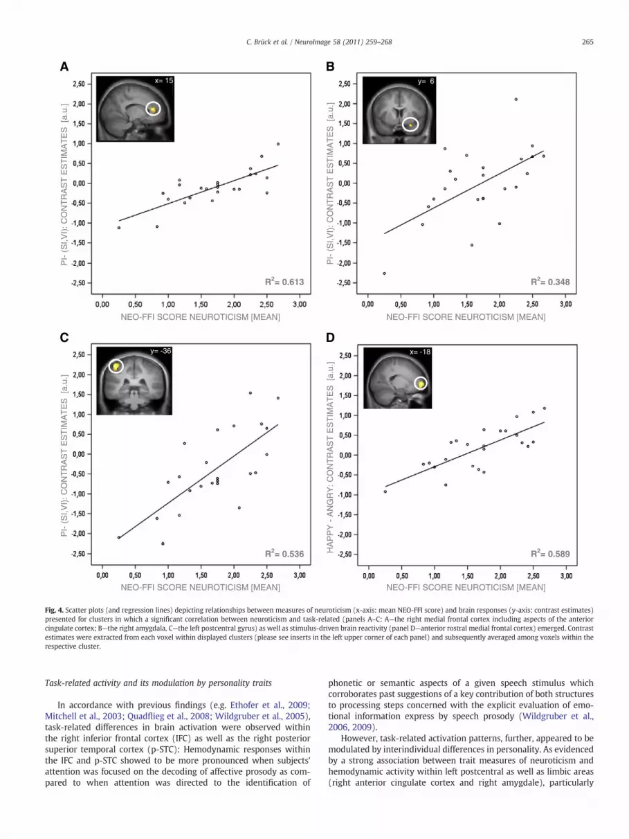
Correlations between brain activity and measures of personality (Fig. 4)To investigate personality-dependent modulation of task- and
stimulus-driven activation patterns, brain responses obtained foreach contrast were correlated with individual measures of personal-ity. The employed correlation analysis failed to reveal a significantassociation between brain activity and measures of extraversion.However, the analysis did indicate a significant positive correlationbetween trait scores for neuroticism and neuronal responses withinthe right amygdala, the right medial frontal cortex including aspectsof the anterior cingulate cortex as well as the left postcentral cortexduring prosody judgment as relative to word content and vowelevaluation (Table 3). Moreover, a positive correlation between neu-roticism scores and cerebral activity to happy prosody as comparedto angry prosody was observed within the medial frontal cortex(Table 3) (Fig. 4).
ody identification against brain responses during both word content identification andluster- level). Activationmaps are rendered on to the cortical surface of a template brain.
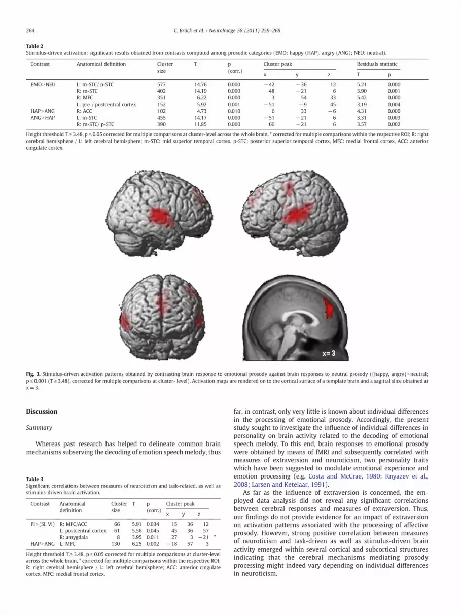
Table 2Stimulus-driven activation: significant results obtained from contrasts computed among prosodic categories (EMO: happy (HAP), angry (ANG); NEU: neutral).
Contrast Anatomical definition Clustersize
T p Cluster peak Residuals statistic(corr.)
x y z T p
EMONNEU L: m-STC/ p-STC 577 14.76 0.000 −42 −36 12 5.21 0.000R: m-STC 402 14.19 0.000 48 −21 6 3.90 0.001R: MFC 351 6.22 0.000 3 54 33 5.42 0.000L: pre-/ postcentral cortex 152 5.92 0.001 −51 −9 45 3.19 0.004
HAPNANG R: ACC 102 4.73 0.010 6 33 −6 4.31 0.000ANGNHAP L: m-STC 455 14.17 0.000 −51 −21 6 3.31 0.003
R: m-STC/ p-STC 390 11.85 0.000 66 −21 6 3.57 0.002
Height threshold T≥3.48, p≤0.05 corrected for multiple comparisons at cluster-level across the whole brain, * corrected for multiple comparisons within the respective ROI; R: rightcerebral hemisphere / L: left cerebral hemisphere; m-STC: mid superior temporal cortex, p-STC: posterior superior temporal cortex, MFC: medial frontal cortex, ACC: anteriorcingulate cortex.
Fig. 3. Stimulus-driven activation patterns obtained by contrasting brain response to emotional prosody against brain responses to neutral prosody ((happy, angry)Nneutral;p≤0.001 (T≥3.48), corrected for multiple comparisons at cluster- level). Activation maps are rendered on to the cortical surface of a template brain and a sagittal slice obtained atx=3.
264 C. Brück et al. / NeuroImage 58 (2011) 259–268
Discussion
Summary
Whereas past research has helped to delineate common brainmechanisms subserving the decoding of emotion speechmelody, thus
Table 3Significant correlations between measures of neuroticism and task-related, as well asstimulus-driven brain activation.
Contrast Anatomicaldefinition
Clustersize
T p Cluster peak(corr.)
x y z
PIN(SI, VI) R: MFC/ACC 66 5.91 0.034 15 36 12L: postcentral cortex 61 5.56 0.045 −45 −36 57R: amygdala 8 3.95 0.011 27 3 −21 *
HAPNANG L: MFC 130 6.25 0.002 −18 57 3
Height threshold T≥3.48, p≤0.05 corrected for multiple comparisons at cluster-levelacross the whole brain, * corrected for multiple comparisons within the respective ROI;R: right cerebral hemisphere / L: left cerebral hemisphere; ACC: anterior cingulatecortex, MFC: medial frontal cortex.
far, in contrast, only very little is known about individual differencesin the processing of emotional prosody. Accordingly, the presentstudy sought to investigate the influence of individual differences inpersonality on brain activity related to the decoding of emotionalspeech melody. To this end, brain responses to emotional prosodywere obtained by means of fMRI and subsequently correlated withmeasures of extraversion and neuroticism, two personality traitswhich have been suggested to modulate emotional experience andemotion processing (e.g. Costa and McCrae, 1980; Knyazev et al.,2008; Larsen and Ketelaar, 1991).
As far as the influence of extraversion is concerned, the em-ployed data analysis did not reveal any significant correlationsbetween cerebral responses and measures of extraversion. Thus,our findings do not provide evidence for an impact of extraversionon activation patterns associated with the processing of affectiveprosody. However, strong positive correlation between measuresof neuroticism and task-driven as well as stimulus-driven brainactivity emerged within several cortical and subcortical structuresindicating that the cerebral mechanisms mediating prosodyprocessing might indeed vary depending on individual differencesin neuroticism.
A B
PI-
(S
I,VI)
: CO
NT
RA
ST
ES
TIM
AT
ES
[a.
u.]
HA
PP
Y -
AN
GR
Y: C
ON
TR
AS
T E
ST
IMA
TE
S [
a.u.
]
PI-
(S
I,VI)
: CO
NT
RA
ST
ES
TIM
AT
ES
[a.
u.]
PI-
(S
I,VI)
: CO
NT
RA
ST
ES
TIM
AT
ES
[a.
u.]
NEO-FFI SCORE NEUROTICISM [MEAN] NEO-FFI SCORE NEUROTICISM [MEAN]
C D
R2= 0.613
NEO-FFI SCORE NEUROTICISM [MEAN] NEO-FFI SCORE NEUROTICISM [MEAN]
R2= 0.536 R2= 0.589
R2= 0.348
x= 15 y= 6
y= -36 x= -18
Fig. 4. Scatter plots (and regression lines) depicting relationships between measures of neuroticism (x-axis: mean NEO-FFI score) and brain responses (y-axis: contrast estimates)presented for clusters in which a significant correlation between neuroticism and task-related (panels A–C: A—the right medial frontal cortex including aspects of the anteriorcingulate cortex; B—the right amygdala, C—the left postcentral gyrus) as well as stimulus-driven brain reactivity (panel D—anterior rostral medial frontal cortex) emerged. Contrastestimates were extracted from each voxel within displayed clusters (please see inserts in the left upper corner of each panel) and subsequently averaged among voxels within therespective cluster.
265C. Brück et al. / NeuroImage 58 (2011) 259–268
Task-related activity and its modulation by personality traits
In accordance with previous findings (e.g. Ethofer et al., 2009;Mitchell et al., 2003; Quadflieg et al., 2008; Wildgruber et al., 2005),task-related differences in brain activation were observed withinthe right inferior frontal cortex (IFC) as well as the right posteriorsuperior temporal cortex (p-STC): Hemodynamic responses withinthe IFC and p-STC showed to be more pronounced when subjects’attention was focused on the decoding of affective prosody as com-pared to when attention was directed to the identification of
phonetic or semantic aspects of a given speech stimulus whichcorroborates past suggestions of a key contribution of both structuresto processing steps concerned with the explicit evaluation of emo-tional information express by speech prosody (Wildgruber et al.,2006, 2009).
However, task-related activation patterns, further, appeared to bemodulated by interindividual differences in personality. As evidencedby a strong association between trait measures of neuroticism andhemodynamic activity within left postcentral as well as limbic areas(right anterior cingulate cortex and right amygdale), particularly

266 C. Brück et al. / NeuroImage 58 (2011) 259–268
differences in neuroticisms appeared to exert great influence on brainresponses associated with speech prosody judgment.
Although limbic regions have frequently been recognized as keystructures of the “emotional brain” (Dalgleish, 2004), their role in thedecoding of emotional speech melody remains controversial: Forinstance, considering the contribution of the amygdala, previousstudies suggest increased (Wiethoff et al., 2009) as well as decreased(Morris et al., 1999) or no “critical” (Adolphs and Tranel, 1999)amygdalar involvement during the processing of vocally expressedemotions. Such inconsistencies in the literature have partly beenresolved by applying a dual-process concept to prosody comprehen-sion positing explicit and implicit processing mechanisms each ofwhich in turn are assumed to be differentially represented in the brain(Wildgruber et al., 2006, 2009). Observed involvement of limbicstructures, thus, might depend uponwhether employed tasks addresscognitive-controlled (explicit) or rather incidental (implicit) modes ofprosody processing. As far as the neural substrates of both types ofprocessing are concerned, experimental data highlight a predominantrole of limbic structures during implicit processing, whereas explicitprocessing has been associated with frontal cortical regions (Bachet al., 2008; Hariri et al., 2003; Lange et al., 2003; Sander et al., 2005;Tamietto and de Gelder, 2010; Wildgruber et al., 2006, 2009).Moreover, recent neuroimaging studies have suggested that frontalcortical areas might also contribute to the inhibition of limbicactivation when individuals actively attend to emotional signals(Blair et al., 2007; Mitchell et al., 2007). Considering the functionalrole of those observations, suppression of limbic activity might reflecta recruitment of emotion regulation processes during task perfor-mance (Blair et al., 2007) which in turnmight attenuate the automaticinduction of specific emotional reactions associated with limbicactivation during implicit prosody processing (Wildgruber et al.,2009). In keeping with the idea that emotional responses may impairour ability to perform goal-directed task (Blair et al., 2007), the valueof described adaptations of limbic activation thus appears to lie inaiding task performance.
However, in light of a strong association between neuroticismand limbic brain responses under explicit processing conditions (asrequired by the PI task), proposed emotional regulation processes andthus the suppression of predominant emotional responses appears tobe altered in individuals reporting higher degrees of neuroticism.Various features associated with “neurotic” personality profiles lendthemselves to the assumption of altered emotional regulation. Forexample, affective instability, a “trademark” of the neuroticism con-cept incorporated in the NEO-FFI (Borkenau and Ostendorf, 2008)might itself reflect a failure to modulate emotional responses appro-priately. Altered emotional regulations might in turn render “neurot-ic” subjects more sensitive to emotional (particularly aversive)stimulation as documented for instance by a variety of psychophys-iological studies conducted in clinical (e.g. Lewinsohn et al., 1973;Marissen et al., 2010; Sigmon and Nelson-Gray, 1992) as well asnonclinical samples (Norris et al., 2007) scoring high on measures ofneuroticism. As for the case of prosody processing, such heightenedsusceptibility to emotional stimulation associated with alterations inregulatory capacities might be reflected in observed increased limbicactivation yielding an automatic induction of emotional responses topresented speech cues.
With reference to the idea that emotional control might aid taskperformance, one would expect proposed alterations in emotionregulation to be accompanied by deficits in accuracy or speed of taskexecution. However, behavioral data obtained within this study didnot indicate any link between neuroticism and measures of taskperformance.
Rather than alterations in emotional regulation, observed differ-ences in activation patterns could also reflect discrepant, nonethelessequally efficient approaches taken to tasks requiring the decoding ofemotional cues. Bearing in mind that observed limbic activation has
frequently been linked to the induction of emotional responses“neurotic” individualsmight rely on feeling rather than thinkingwhenfaced with emotional judgments. Similar ideas may be derived con-sidering differences in post-central activation observed in this study.
Aside limbic brain structures, trait-specific modulations of activa-tion were revealed within the left postcentral cortex with enhancedpostcentral activity observed in subjects scoring higher in neuroti-cism. Although primarily recognized for their role in somatosensoryprocessing, left postcentral structures have previously been reportedto be involved in emotional task (Canli et al., 2002; Deeley et al., 2006)and activation of somatosensory-related cortices observed duringemotion recognition has been associated with simulation processesthat aid emotion decoding by generating somatosensory representa-tions that mimic body states corresponding to the respective affectsa person is presented with (Adolphs, 2003; Adolphs et al., 2000).Stronger activation of somatosensory areas observed for individualshigh in neuroticism might, thus, reflect a greater reliance of neuroticindividuals on “simulation” when faced with tasks of emotionjudgment.
Stimulus-driven activity and its modulation by personality traits
Aside from delineating task-related effects, data analysis furthersought to evaluate differences in the processing of emotional asrelative to neutral prosody as well as processing differences amongvarying qualities of prosodic emotional information particularlyexpressions of joy or anger.
Comparisons computed with respect to behavioral measuresrevealed significantly higher rates of correct identification for neutraland angry as compared to happyprosody, replicatingpreviousfindingsof an advantage for decoding angry prosody over other emotionalcategories such as fear, love or happiness (Juslin and Laukka, 2003).
However, arguing against the assumptions of a processing ad-vantage, analyses computed on measures of response times indicatedthat the processing of angry prosody resulted in significantly slowerresponses compared to both happy and neutral prosody. In an attemptto explain observed effects, response time differences were re-evaluated using an approach that controlled for differences in stimulusduration among the three prosodic categories. Results revealedthat, although effects proved in parts driven by stimulus durations,differences between happy and angry prosody remained significant,suggesting an emotion-specific facilitation of responding that war-rants further investigation.
Considering stimulus effects at brain level, enhanced hemody-namic responses to stimuli spoken in an emotional rather than aneutral tone of voice were observed bilaterally within the midsuperior temporal cortex (m-STC) with the strongest responsesobtained for angry prosody within these structures. Additionallyenhanced reactivity to emotional as compared to neutral prosody wasrevealedwithin themedial frontal cortex (MFC) aswell as the left pre-and postcentral cortex. Increased responsiveness of the m-STC toemotional speech melody has been documented in a variety ofprevious neuroimaging studies (Ethofer et al., 2009; Grandjean et al.,2005;Wiethoff et al., 2008), and given its stimulus-drivenmodulationof hemodynamic responses independent of task demands, aninvolvement of the m-STC particularly in the acoustic analysis ofvocal emotional stimuli has been assumed (Ethofer et al., 2009;Wildgruber et al., 2009). Enhanced responses of the MFC, on the otherhand, have been related to various aspects of social cognition, a set ofprocesses contributing to successful social interaction (Amodio andFrith, 2006). One such fundamental component of social cognitionassociated particularly with the activation of anterior subdivisions ofthe MFC has been described to be “mentalizing” (Amodio and Frith,2006), a process by which inferences about the mental state of othersare made (Frith and Frith, 2006). Reading the minds of our partnersof interaction, grasping their desires, feelings and believes, proves to

267C. Brück et al. / NeuroImage 58 (2011) 259–268
be crucial to effective social functioning as those mental statesdetermine behavior (Frith and Frith, 2006). Emotional speech melodyprovides valuable cues as to the psychological states of our fellowhuman beings and activation of the anterior MFC observed inresponse to emotional speech melody thus might reflect mentalizingprocesses triggered by the perception of vocal emotional cues.
Similar to the results obtained for task-related brain activationpatterns, stimulus-driven activity appeared to be further modulatedby individual differences in neuroticism as personality measures ofneuroticism were found to correlate with activity of the left anteriorMFC to positive as compared to negative prosody. Bearing in mindthe involvement of the anterior MFC in social-cognitive processes,particularly in inferring the mental states of our partners of inter-action, observed activation patterns may reflect greater effort to“mentalize” positive emotional states in subjects scoring high onmeasures of neuroticism. Neuroticism has frequently been associatedwith advantages in the processing of negative emotional information(Chan et al., 2007; Derryberry and Reed, 1994; Gomez et al., 2002;Reed and Derryberry, 1995). Assuming such an affective bias toselectively facilitate the processing of negative vocal emotionalstimuli, positive prosodic stimuli may in comparison impose greaterdemands on social–cognitive processing. In turn, greater demands onsocial–cognitive processes in relation with the decoding of emotionalvocalizations have been associated with enhanced activation ofmedial frontal brain areas very similar in location to activationpatterns observed in the present study (Szameitat et al., 2010).
However, one would expect the proposed disadvantage in theprocessing of positive emotions to reveal itself, for instance, in slowedresponse times to happy as compared to angry stimuli or a responsebias towards judging emotions as negative. Yet, behavioral dataobtained in the present study reveal no such patterns of performance.Employed correlation analyses yielded no significant relationshipbetween neuroticism and measures of performance. One possibleexplanation for those negative findings might be that categoricaljudgment tasks (such as those employed in this study) are notsensitive enough to evaluate biases in the processing of different typesof emotions. In order to further explore the idea of a neuroticism-linked processing bias and its cerebral underpinnings, it appearsworthwhile to re-evaluate the respective hypothesis using, forinstance, tasks that require a continuous rating of the emotionalvalence of a given speech stimulus.
Particularly in light of lacking behavioral data to support the as-sumption of a personality-dependent processing bias in the decodingof emotional speech melody, the interpretation of the observedcerebral response patterns at this point remains hypothetical innature and require further elaboration.
Conclusion
Irrespective of whether observed personality-dependent varia-tions in activation patterns may reflect processing biases, differencesin emotion control or task strategies, reported findings, first andforemost, evidence a clear influence of personality on brain responsesrelated to the processing of emotional speech melody.
In highlighting such personality-dependent modulations of brainactivation, the present study, again underlines the importance ofconsidering individual differences in the study of prosody processingand encourages to further elaborate the effects of such differencesin order to broaden our insight into the neurobiology of prosodyperception.
Acknowledgments
We would like to thank the Werner Reichardt Centre forIntegrative Neuroscience and the Deutsche Forschungsgemeinschaft(EXC 307) for supporting this study.
References
Ackermann, H., Hertrich, I., Grodd, W., Wildgruber, D., 2004. Das Hören von Gefühlen:Funktionell-neuroanatomische Grundlage der Verarbeitung affektiver Prosodie.Aktuelle Neurol. 31, 449–460.
Adolphs, R., 2003. Cognitive neuroscience of human social behaviour. Nat. Rev.Neurosci. 4, 165–178.
Adolphs, R., Damasio, H., Tranel, D., 2002. Neural systems for recognition of emotionalprosody: a 3-D lesion study. Emotion 2, 23–51.
Adolphs, R., Damasio, H., Tranel, D., Cooper, G., Damasio, A.R., 2000. A role forsomatosensory cortices in the visual recognition of emotion as revealed by three-dimensional lesion mapping. J. Neurosci. 20, 2683–2690.
Adolphs, R., Tranel, D., 1999. Intact recognition of emotional prosody followingamygdala damage. Neuropsychologia 37, 1285–1292.
Amodio, D.M., Frith, C.D., 2006. Meeting of minds: the medial frontal cortex and socialcognition. Nat. Rev. Neurosci. 7, 268–277.
Andersson, J.L., Hutton, C., Ashburner, J., Turner, R., Friston, K., 2001. Modelinggeometric deformations in EPI time series. Neuroimage 13, 903–919.
Bach, D.R., Grandjean, D., Sander, D., Herdener, M., Strik, W.K., Seifritz, E., 2008. Theeffect of appraisal level on processing of emotional prosody in meaningless speech.Neuroimage 42, 919–927.
Blair, K.S., Smith, B.W., Mitchell, D.G., Morton, J., Vythilingam, M., Pessoa, L., Fridberg, D.,Zametkin, A., Sturman, D., Nelson, E.E., Drevets, W.C., Pine, D.S., Martin, A., Blair, R.J.,2007. Modulation of emotion by cognition and cognition by emotion. Neuroimage35, 430–440.
Borkenau, P., Ostendorf, F., 2008. NEO- FFI NEO-Fünf-Faktoren-Inventar nach Costa undMcCrae. 2. neu normierte und vollständig überarbeitet Auflage. Hogrefe, Göttingen.
Borod, J.C., Bloom, R.L., Brickman, A.M., Nakhutina, L., Curko, E.A., 2002. Emotionalprocessing deficits in individuals with unilateral brain damage. Appl. Neuropsy-chol. 9, 23–36.
Brühl, A.B., Viebke, M.C., Baumgartner, T., Kaffenberger, T., Herwig, U., 2011. Neuralcorrelates of personality dimensions and affective measures during the anticipationof emotional stimuli. Brain Imaging Behav. 5, 86–96.
Buchanan, T.W., Lutz, K., Mirzazade, S., Specht, K., Shah, N.J., Zilles, K., Jancke, L., 2000.Recognition of emotional prosody and verbal components of spoken language: anfMRI study. Brain Res. Cogn. Brain Res. 9, 227–238.
Cancelliere, A.E., Kertesz, A., 1990. Lesion localization in acquired deficits of emotionalexpression and comprehension. Brain Cogn. 13, 133–147.
Canli, T., 2004. Functional brain mapping of extraversion and neuroticism: learningfrom individual differences in emotion processing. J. Pers. 72, 1105–1132.
Canli, T., Amin, Z., 2002. Neuroimaging of emotion and personality: scientific evidenceand ethical considerations. Brain Cogn. 50, 414–431.
Canli, T., Desmond, J.E., Zhao, Z., Gabrieli, J.D., 2002. Sex differences in the neural basis ofemotional memories. Proc. Natl. Acad. Sci. U.S.A. 99, 10789–10794.
Canli, T., Zhao, Z., Desmond, J.E., Kang, E., Gross, J., Gabrieli, J.D., 2001. An fMRI study ofpersonality influences on brain reactivity to emotional stimuli. Behav. Neurosci.115, 33–42.
Chan, S.W., Goodwin, G.M., Harmer, C.J., 2007. Highly neurotic never-depressed studentshave negative biases in information processing. Psychol. Med. 37, 1281–1291.
Collins, D.L., Neelin, P., Peters, T.M., Evans, A.C., 1994. Automatic 3D intersubjectregistration of MR volumetric data in standardized Talairach space. J. Comput.Assist. Tomogr. 18, 192–205.
Costa Jr., P.T., McCrae, R.R., 1980. Influence of extraversion and neuroticism onsubjectivewell-being: happy and unhappy people. J. Pers. Soc. Psychol. 38, 668–678.
Costa, P.T., McCrae, R.R., 1991. NEO Five- Factor Inventory (NEO-FFI) professionalmanual. Psychological Assessment Resources, Odessa, FL.
Cremers, H.R., Demenescu, L.R., Aleman, A., Renken, R., van Tol, M.J., van der Wee, N.J.,Veltman, D.J., Roelofs, K., 2010. Neuroticism modulates amygdala-prefrontal connec-tivity in response to negative emotional facial expressions. Neuroimage 49, 963–970.
Dalgleish, T., 2004. The emotional brain. Nat. Rev. Neurosci. 5, 583–589.Dara, C., Monetta, L., Pell, M.D., 2008. Vocal emotion processing in Parkinson's disease:
reduced sensitivity to negative emotions. Brain Res. 1188, 100–111.Deckersbach, T., Miller, K.K., Klibanski, A., Fischman, A., Dougherty, D.D., Blais, M.A.,
Herzog, D.B., Rauch, S.L., 2006. Regional cerebral brain metabolism correlates ofneuroticism and extraversion. Depress. Anxiety 23, 133–138.
Deeley, Q., Daly, E., Surguladze, S., Tunstall, N., Mezey, G., Beer, D., Ambikapathy, A.,Robertson, D., Giampietro, V., Brammer, M.J., Clarke, A., Dowsett, J., Fahy, T., Phillips,M.L., Murphy, D.G., 2006. Facial emotion processing in criminal psychopathy.Preliminary functional magnetic resonance imaging study. Br. J. Psychiatry 189,533–539.
Derryberry, D., Reed, M.A., 1994. Temperament and attention: orienting toward andaway from positive and negative signals. J. Pers. Soc. Psychol. 66, 1128–1139.
Ethofer, T., Anders, S., Erb, M., Droll, C., Royen, L., Saur, R., Reiterer, S., Grodd, W.,Wildgruber, D., 2006a. Impact of voice on emotional judgment of faces: an event-related fMRI study. Hum. Brain Mapp. 27, 707–714.
Ethofer, T., Anders, S., Erb,M., Herbert, C.,Wiethoff, S., Kissler, J., Grodd,W.,Wildgruber, D.,2006b. Cerebral pathways in processing of affective prosody: a dynamic causalmodeling study. Neuroimage 30, 580–587.
Ethofer, T., Anders, S.,Wiethoff, S., Erb,M., Herbert, C., Saur, R., Grodd,W.,Wildgruber, D.,2006c. Effects of prosodic emotional intensity on activation of associative auditorycortex. Neuroreport 17, 249–253.
Ethofer, T., Kreifelts, B., Wiethoff, S., Wolf, J., Grodd, W., Vuilleumier, P., Wildgruber, D.,2009. Differential influences of emotion, task, and novelty on brain regionsunderlying the processing of speech melody. J. Cogn. Neurosci. 21, 1255–1268.
Fischer, H., Wik, G., Fredrikson, M., 1999. Extraversion, neuroticism and brain function:A PET study of personality. Pers. Individ. Differ. 23, 345–352.

268 C. Brück et al. / NeuroImage 58 (2011) 259–268
Friston, K.J., Glaser, D.E., Henson, R.N., Kiebel, S., Phillips, C., Ashburner, J., 2002. Classicaland Bayesian inference in neuroimaging: applications. Neuroimage 16, 484–512.
Friston, K.J., Holmes, A.P., Worsley, K.J., Poline, J.P., Frith, C.D., Frackowiak, R.S.J., 1994.Statistical parametric maps in functional imaging: a general linear approach. Hum.Brain Mapp. 2, 189–210.
Frith, C.D., Frith, U., 2006. The neural basis of mentalizing. Neuron 50, 531–534.Frühholz, S., Prinz, M., Herrmann, M., 2010. Affect-related personality traits and
contextual interference processing during perception of facial affect. Neurosci. Lett.469, 260–264.
Gomez, R., Gomez, A., Cooper, A., 2002. Neuroticism and extraversion as predictors ofnegative and positive emotional information processing: comparing Eysenck's,Gray's, and Newman's theories. Eur. J. Pers. 16, 333–350.
Grandjean,D., Sander,D., Pourtois,G., Schwartz, S., Seghier,M.L., Scherer,K.R., Vuilleumier, P.,2005. Thevoices ofwrath: brain responses to angryprosody inmeaningless speech.Nat.Neurosci. 8, 145–146.
Gray, J.A., 1970. The psychophysiological basis of introversion–extraversion. Behav. Res.Ther. 8, 249–266.
Haier, R.J., Sokolski, K., Katz, M., Buchsbaum, M.S., 1987. The study of personality withpositron emission tomography. In: Strelau, J., Eysenck, H.J. (Eds.), Personalitydimensions and arousal. Plenum, New York, pp. 251–267.
Hamann, S., Canli, T., 2004. Individual differences in emotion processing. Curr. Opin.Neurobiol. 14, 233–238.
Hariri, A.R., Mattay, V.S., Tessitore, A., Fera, F., Weinberger, D.R., 2003. Neocorticalmodulation of the amygdala response to fearful stimuli. Biol. Psychiatry 53, 494–501.
Heilman, K.M., Bowers, D., Speedie, L., Coslett, H.B., 1984. Comprehension of affectiveand nonaffective prosody. Neurology 34, 917–921.
Herbert, C., Kissler, J., Junghofer,M., Peyk, P., Rockstroh, B., 2006. Processing of emotionaladjectives: evidence from startle EMG and ERPs. Psychophysiology 43, 197–206.
Herrmann, M.J., Unterecker, S., Eisenack, S., Fallgatter, A.J., 2001. P300 and personality—a correlational analysis in healthy subjects. Acta Psychiatr. Belg. 101, 202–212.
Hooker, C.I., Verosky, S.C., Miyakawa, A., Knight, R.T., D'Esposito, M., 2008. The influenceof personality on neural mechanisms of observational fear and reward learning.Neuropsychologia 46, 2709–2724.
Hornak, J., Bramham, J., Rolls, E.T., Morris, R.G., O'Doherty, J., Bullock, P.R., Polkey, C.E.,2003. Changes in emotion after circumscribed surgical lesions of the orbitofrontaland cingulate cortices. Brain 126, 1691–1712.
Johnson,D.L.,Wiebe, J.S., Gold, S.M., Andreasen,N.C.,Hichwa,R.D.,Watkins,G.L., Boles Ponto,L.L., 1999. Cerebral blood flow and personality: a positron emission tomography study.Am. J. Psychiatry 156, 252–257.
Juslin, P.N., Laukka, P., 2003. Emotional expression in speech and music: evidence ofcross-modal similarities. Ann. N. Y. Acad. Sci. 1000, 279–282.
Knyazev, G.G., Bocharov, A.V., Slobodskaya, H.R., Ryabichenko, T.I., 2008. Personality-linked biases in perception of emotional facial expressions. Pers. Individ. Differ. 44,1093–1104.
Kotz, S.A., Meyer, M., Alter, K., Besson, M., von Cramon, D.Y., Friederici, A.D., 2003. Onthe lateralization of emotional prosody: an event-related functional MR investi-gation. Brain Lang. 86, 366–376.
Kotz, S.A., Meyer, M., Paulmann, S., 2006. Lateralization of emotional prosody in thebrain: an overview and synopsis on the impact of study design. Prog. Brain Res. 156,285–294.
Kumari, V., ffytche, D.H., Williams, S.C., Gray, J.A., 2004. Personality predicts brainresponses to cognitive demands. J. Neurosci. 24, 10636–10641.
Lange, K., Williams, L.M., Young, A.W., Bullmore, E.T., Brammer, M.J., Williams, S.C.,Gray, J.A., Phillips, M.L., 2003. Task instructions modulate neural responses tofearful facial expressions. Biol. Psychiatry 53, 226–232.
Larsen, R.J., Ketelaar, T., 1991. Personality and susceptibility to positive and negativeemotional states. J. Pers. Soc. Psychol. 61, 132–140.
Lewinsohn, P.M., Lobitz, W.C., Wilson, S., 1973. "Sensitivity" of depressed individuals toaversive stimuli. J. Abnorm. Psychol. 81, 259–263.
Maldjian, J.A., Laurienti, P.J., Kraft, R.A., Burdette, J.H., 2003. An automated method forneuroanatomic and cytoarchitectonic atlas-based interrogation of fMRI data sets.Neuroimage 19, 1233–1239.
Marissen,M.A.,Meuleman, L., Franken, I.H., 2010. Altered emotional information processingin borderline personality disorder: an electrophysiological study. Psychiatry Res. 181,226–232.
Mehrabian, A., Ferris, S.R., 1967. Inference of attitudes from nonverbal communicationin two channels. J. Consult. Psychol. 31, 248–252.
Mehrabian, A., Wiener, M., 1967. Decoding of inconsistent communications. J. Pers. Soc.Psychol. 6, 109–114.
Mitchell, D.G., Nakic, M., Fridberg, D., Kamel, N., Pine, D.S., Blair, R.J., 2007. The impact ofprocessing load on emotion. Neuroimage 34, 1299–1309.
Mitchell, R.L., Elliott, R., Barry, M., Cruttenden, A.,Woodruff, P.W., 2003. Theneural responseto emotional prosody, as revealed by functional magnetic resonance imaging.Neuropsychologia 41, 1410–1421.
Morris, J.S., Scott, S.K., Dolan, R.J., 1999. Saying it with feeling: neural responses toemotional vocalizations. Neuropsychologia 37, 1155–1163.
Norris, C.J., Larsen, J.T., Cacioppo, J.T., 2007. Neuroticism is associatedwith larger andmoreprolonged electrodermal responses to emotionally evocative pictures. Psychophys-iology 44, 823–826.
O'Gorman, R.L., Kumari, V., Williams, S.C., Zelaya, F.O., Connor, S.E., Alsop, D.C., Gray, J.A.,2006. Personality factors correlate with regional cerebral perfusion. Neuroimage31, 489–495.
Oldfield, R.C., 1971. The assessment and analysis of handedness: the Edinburgh inventory.Neuropsychologia 9, 97–113.
Peper, M., Irle, E., 1997. Categorical and dimensional decoding of emotional intonationsin patients with focal brain lesions. Brain Lang. 58, 233–264.
Quadflieg, S., Mohr, A., Mentzel, H.J., Miltner, W.H., Straube, T., 2008. Modulation of theneural network involved in the processing of anger prosody: the role of task-relevance and social phobia. Biol. Psychol. 78, 129–137.
Reed, M.A., Derryberry, D., 1995. Temperament and attention to positive and negativetrait information. Pers. Individ. Differ. 18, 135–147.
Ross, E.D., 1981. The aprosodias. Functional-anatomic organization of the affectivecomponents of language in the right hemisphere. Arch. Neurol. 38, 561–569.
Ross, E.D., Monnot, M., 2008. Neurology of affective prosody and its functional–anatomic organization in right hemisphere. Brain Lang. 104, 51–74.
Sander, D., Grandjean, D., Pourtois, G., Schwartz, S., Seghier, M.L., Scherer, K.R.,Vuilleumier, P., 2005. Emotion and attention interactions in social cognition: brainregions involved in processing anger prosody. Neuroimage 28, 848–858.
Schirmer, A., Kotz, S.A., 2006. Beyond the right hemisphere: brain mechanismsmediating vocal emotional processing. Trends Cogn. Sci. 10, 24–30.
Sigmon, S.T., Nelson-Gray, R.O., 1992. Sensitivity to aversive events in depression:antecedent, concomitant, or consequent? J. Psychopathol. Behav. Assess. 14, 225–246.
Starkstein, S.E., Federoff, J.P., Price, T.R., Leiguarda, R.C., Robinson, R.G., 1994.Neuropsychological and neuroradiologic correlates of emotional prosody compre-hension. Neurology 44, 515–522.
Stenberg, G., Wendt, P.E., Risberg, J., 1993. Regional cerebral blood flow and extraversion.Pers. Individ. Differ. 15, 547–554.
Suslow, T., Kugel, H., Reber, H., Bauer, J., Dannlowski, U., Kersting, A., Arolt, V., Heindel, W.,Ohrmann, P., Egloff, B., 2010. Automatic brain response to facial emotion as a functionof implicitly and explicitly measured extraversion. Neuroscience 167, 111–123.
Szameitat, D.P., Kreifelts, B., Alter, K., Szameitat, A.J., Sterr, A., Grodd, W., Wildgruber, D.,2010. It is not always tickling: distinct cerebral responses during perception ofdifferent laughter types. Neuroimage 53, 1264–1271.
Tamietto, M., de Gelder, B., 2010. Neural bases of the non-conscious perception ofemotional signals. Nat. Rev. Neurosci. 11, 697–709.
Tzourio-Mazoyer, N., Landeau, B., Papathanassiou, D., Crivello, F., Etard, O., Delcroix, N.,Mazoyer, B., Joliot, M., 2002. Automated anatomical labeling of activations in SPMusing a macroscopic anatomical parcellation of the MNI MRI single-subject brain.Neuroimage 15, 273–289.
Van Kleef, G.A., 2009. How emotions regulate social life: the Emotions as SocialInformation (EASI) model. Curr. Direct. Psychol. Sci. 18, 184–188.
Wiethoff, S., Wildgruber, D., Grodd, W., Ethofer, T., 2009. Response and habituation ofthe amygdala during processing of emotional prosody. Neuroreport 20, 1356–1360.
Wiethoff, S., Wildgruber, D., Kreifelts, B., Becker, H., Herbert, C., Grodd, W., Ethofer, T.,2008. Cerebral processing of emotional prosody—influence of acoustic parametersand arousal. Neuroimage 39, 885–893.
Wildgruber, D., Ackermann, H., Kreifelts, B., Ethofer, T., 2006. Cerebral processing oflinguistic and emotional prosody: fMRI studies. Prog. Brain Res. 156, 249–268.
Wildgruber, D., Ethofer, T., Grandjean, D., Kreifelts, B., 2009. A cerebral network modelof speech prosody comprehension. Int. J. Speech Lang. Pathol. 11, 277–281.
Wildgruber, D., Hertrich, I., Riecker, A., Erb, M., Anders, S., Grodd, W., Ackermann, H.,2004. Distinct frontal regions subserve evaluation of linguistic and emotionalaspects of speech intonation. Cereb. Cortex 14, 1384–1389.
Wildgruber, D., Pihan, H., Ackermann, H., Erb, M., Grodd, W., 2002. Dynamic brainactivation during processing of emotional intonation: influence of acoustic param-eters, emotional valence, and sex. Neuroimage 15, 856–869.
Wildgruber, D., Riecker, A., Hertrich, I., Erb, M., Grodd, W., Ethofer, T., Ackermann, H.,2005. Identification of emotional intonation evaluated by fMRI. Neuroimage 24,1233–1241.
Worsley, K.J., Marrett, S., Neelin, P., Vandal, A.C., Friston, K.J., Evans, A.C., 1996. A unifiedstatistical approach for determining significant signals in imagesof cerebral activation.Hum. Brain Mapp. 4, 58–73.