Immune responsiveness and parasite-specific antibody levels in human hepatobiliary disease...
6
Clin. exp. Immunol. (1991) 84, 213-218 Immune responsiveness and parasite-specific antibody levels in human hepatobiliary disease associated with Opisthorchis viverrini infection M. R. HASWELL-ELKINS*¶, P. SITHITHAWORN*, E. MAIRIANGt, D. B. ELKINS*¶, S. WONGRATANACHEEWIN$, S. KAEWKES*! & P. MAIRIANG§ Departments of *Parasitology, t Radiology, T Microbiology and § Medicine, Faculty of Medicine, Khon Kaen University, Thailand, and The Tropical Health Program, Queensland Institute of Medical Research, Brisbane, Australia (Acceptedfor publication 1 December 1990) SUMMARY Opisthorchis viverrini infection is associated with human hepatobiliary disease and cholangiocarci- noma, but the role of the immune response in the pathogenesis of infection is unclear. Here ultrasonography was used to examine the biliary tracts of residents from an endemic community. Delayed-type hypersensitivity responses to unrelated antigens, and fluke-specific IgG and IgA levels in serum of this group were also examined. Relationships between immunological parameters, intensity of infection and radiologically measured variables are reported. Immune responsiveness to unrelated antigens did not vary with intensity of parasite infection or disease status. Of all the variables, IgG levels were most markedly elevated in disease cases compared with normal subjects and were closely associated with gall bladder size and dysfunction. This is consistent with the hypothesis that an immunopathologic mechanism is involved in opisthorchiasis and suggests that antibody levels may be useful in screening populations for fluke-associated hepatobiliary disease. Keywords parasite-specific antibodies gall bladder Opisthorchis viverrini ultrasonography ELISA INTRODUCTION The liver fluke, Opisthorchis viverrini is a major public health problem in North East Thailand where it is estimated 7 million people are infected (Preuksaraj, 1984). Humans acquire this parasite by eating raw fish containing infective metacercariae. The flukes reside in the small bile ducts and gall bladder and do not undergo systemic migration. Eggs exit the biliary tract and are excreted in faeces. The most serious manifestation of liver fluke infection is an increased susceptibility to cholangiocarcinoma (CHCA), ade- nocarcinoma of the biliary tract (Hou, 1956; Flavell, 1981; Harinasuta, Riganti & Bunnag, 1984; Kim, 1984; Elkins et al., 1990). CHCA is the leading cancer in North East Thailand, but it is rare in areas where the fluke is absent (Vatanasapt et al., 1990a, b). Several less severe diseases, e.g. cholelithiasis, cholecystitis and cholangitis, are also associated with Opisthor- chis (Harinasuta et al., 1984; Dhiensiri et al., 1984; Elkins et al., 1990); these are risk factors of CHCA (Fraumeni & Kantor, 1982; Nagorney & McPherson, 1988). Correspondence: Dr Melissa Haswell-Elkins, P.O. Box 108, Khon Kaen 40000, Thailand. The role of the liver fluke in the aetiology of biliary disease and carcinoma remains uncertain. During infection, desquama- tion of the bile duct epithelium can occur, leading to over- reactive repair, including hyperplasia, fibrosis and goblet cell metaplasia (Hou, 1956; Bhamarapravati, Thamavit & Vajras- thira, 1978; Flavell, 1981; Harinasuta et al., 1984; Kim, 1984). The fluke may release toxins which cause direct damage (Hou, 1956; Kim, 1984; Harinasuta et al., 1984), and/or the changes may be immunopathological (Bhamapravati et al., 1978; Fla- vell, 1981). Published reports describe mainly damage to the biliary epithelium; the pathogenesis of gall bladder disease and its role in carcinogenesis is unknown. Wongratanacheewin et al. (1987) have shown that Opisthor- chis infection causes a generalized suppression of immunologi- cal responses in hamsters. The survival of malignant cells may be enhanced by parasite-induced impairment of immunosurveil- lance mechanisms. However, it is not known whether infection suppresses immunity in humans. Our recent studies demonstrated a high frequency of ultrasound-diagnosed hepatobiliary disease associated with heavy Opisthorchis infection in a sample group drawn from a small Northeast Thai village (Elkins et al., 1990). Here we demonstrate close relationships between radiological and immunological data from this group. 213 ADONIS 00099104910013 1 Z
-
Upload
independent -
Category
Documents
-
view
0 -
download
0
Transcript of Immune responsiveness and parasite-specific antibody levels in human hepatobiliary disease...
Clin. exp. Immunol. (1991) 84, 213-218
Immune responsiveness and parasite-specific antibody levelsin human hepatobiliary disease associated with
Opisthorchis viverrini infection
M. R. HASWELL-ELKINS*¶, P. SITHITHAWORN*, E. MAIRIANGt, D. B. ELKINS*¶,S. WONGRATANACHEEWIN$, S. KAEWKES*! & P. MAIRIANG§ Departments of *Parasitology,
t Radiology, T Microbiology and § Medicine, Faculty of Medicine, Khon Kaen University, Thailand, andThe Tropical Health Program, Queensland Institute of Medical Research, Brisbane, Australia
(Acceptedfor publication 1 December 1990)
SUMMARY
Opisthorchis viverrini infection is associated with human hepatobiliary disease and cholangiocarci-noma, but the role of the immune response in the pathogenesis of infection is unclear. Hereultrasonography was used to examine the biliary tracts of residents from an endemic community.Delayed-type hypersensitivity responses to unrelated antigens, and fluke-specific IgG and IgA levelsin serum of this group were also examined. Relationships between immunological parameters,intensity of infection and radiologically measured variables are reported. Immune responsiveness tounrelated antigens did not vary with intensity of parasite infection or disease status. Of all thevariables, IgG levels were most markedly elevated in disease cases compared with normal subjectsand were closely associated with gall bladder size and dysfunction. This is consistent with thehypothesis that an immunopathologic mechanism is involved in opisthorchiasis and suggests thatantibody levels may be useful in screening populations for fluke-associated hepatobiliary disease.
Keywords parasite-specific antibodies gall bladder Opisthorchis viverrini ultrasonographyELISA
INTRODUCTION
The liver fluke, Opisthorchis viverrini is a major public healthproblem in North East Thailand where it is estimated 7 millionpeople are infected (Preuksaraj, 1984). Humans acquire thisparasite by eating raw fish containing infective metacercariae.The flukes reside in the small bile ducts and gall bladder and donot undergo systemic migration. Eggs exit the biliary tract andare excreted in faeces.
The most serious manifestation of liver fluke infection is anincreased susceptibility to cholangiocarcinoma (CHCA), ade-nocarcinoma of the biliary tract (Hou, 1956; Flavell, 1981;Harinasuta, Riganti & Bunnag, 1984; Kim, 1984; Elkins et al.,1990). CHCA is the leading cancer in North East Thailand, butit is rare in areas where the fluke is absent (Vatanasapt et al.,1990a, b). Several less severe diseases, e.g. cholelithiasis,cholecystitis and cholangitis, are also associated with Opisthor-chis (Harinasuta et al., 1984; Dhiensiri et al., 1984; Elkins et al.,1990); these are risk factors of CHCA (Fraumeni & Kantor,1982; Nagorney & McPherson, 1988).
Correspondence: Dr Melissa Haswell-Elkins, P.O. Box 108, KhonKaen 40000, Thailand.
The role of the liver fluke in the aetiology of biliary diseaseand carcinoma remains uncertain. During infection, desquama-tion of the bile duct epithelium can occur, leading to over-reactive repair, including hyperplasia, fibrosis and goblet cellmetaplasia (Hou, 1956; Bhamarapravati, Thamavit & Vajras-thira, 1978; Flavell, 1981; Harinasuta et al., 1984; Kim, 1984).The fluke may release toxins which cause direct damage (Hou,1956; Kim, 1984; Harinasuta et al., 1984), and/or the changesmay be immunopathological (Bhamapravati et al., 1978; Fla-vell, 1981). Published reports describe mainly damage to thebiliary epithelium; the pathogenesis of gall bladder disease andits role in carcinogenesis is unknown.Wongratanacheewin et al. (1987) have shown that Opisthor-
chis infection causes a generalized suppression of immunologi-cal responses in hamsters. The survival ofmalignant cells may beenhanced by parasite-induced impairment of immunosurveil-lance mechanisms. However, it is not known whether infectionsuppresses immunity in humans.
Our recent studies demonstrated a high frequency ofultrasound-diagnosed hepatobiliary disease associated withheavy Opisthorchis infection in a sample group drawn from asmall Northeast Thai village (Elkins et al., 1990). Here wedemonstrate close relationships between radiological andimmunological data from this group.
213
ADONIS 00099104910013 1Z

M. R. Haswell-Elkins et al.
Table 1. Summary of the radiological findings for the village residents selected to undergo ultrasonography
Mean ageDiagnosis n (years) Worm load IgG*
Normal, no radiological abnormalities 41 40 10 0 (2.8) 0 33 (0.04)Mild gall bladder disease, thickened wall or enlarged gall bladder, good contraction 15 49 117 3 (56) 0-44 (0 04)
Cholelithiasis 6 51 303 4 (199) 0 59 (0 13)
Chronic cholecystitis, thickened wall, enlarged or contracted organ, poor contraction 12 51 251 5 (80 2) 0 71 (0-09)
Cholangiocarcinoma, suspected if hydrops gall bladder, a mass, dilated common 8 50 NA 0 93 (0 09)and/or intrahepatic bile ducts
Parenchymal disease, increased echoes in the liver 11 49 165 (128) 0 60 (0-12)
* Expressed as the mean (s.e.m.).NA, Praziquantel contraindicated. The mean egg output of this group was the highest of all groups (Elkins et al., 1990).
MATERIALS AND METHODS
Study design and ultrasonographyThe procedures used in the field program and details of thesample group are described elsewhere (Elkins et al., 1990). Theproject was carried out in Bahn Huai Matho, ChangwatKalasin, North East Thailand, beginning in March 1989. Alladults over 19 years of age (total 87) with negative (n = 19) orhigh (>10000 eggs/g; n = 22) Opisthorchis egg counts or withclinical indications (palpable liver, history of jaundice) under-went ultrasonography. The purpose of the study was explainedand informed consent obtained from the participants. Subjectsfasted at least 6 h before examination using a Toshiba SonolayerLS-SAL-55AS with a 3-5 mHz linear array probe. Radiologicaldiagnoses and the number ofcases observed are given in Table 1.Gall bladder size was defined as its greatest length observedfrom all planes of view (Everson et al., 1980). Contraction wasassessed by remeasuring the organ 30 min after consumption ofa fatty meal (Dhiensiri et al., 1984; Hederstrom et al., 1988).Portal vein radicle echoes (PVRE) were scored according to theprominence of echoes along the portal triad (Homeida et al.,1988). All examinations were performed by one experiencedradiologist who was unaware of the clinical, parasitological orimmunological status of the subjects. Suspected cases ofCHCAwere investigated further using endoscopic retrograde cholan-giopancreatography and computed tomography.
ParasitologyEgg counts were performed using a quantitative formalin ethertechnique. Worm burdens were determined by recovery offlukes from stools passed during 48 h after treatment withpraziquantel (40 mg/kg body weight) and magnesium sulphatepurgative (Radomyos, Bunnag & Harinasuta, 1984; Ramsey etal., 1989; Elkins et al., 1991).
Skin testsThe cell-mediated immune responsiveness of 44 of the subjects(all consenting CHCA cases plus randomly selected normal andgall bladder disease cases). was assessed by measuring theinduration diameters 48 h after application of seven commonantigens and glycerin alone (negative control) to the forearm(Multitest CMI; Institut Merieux). A total skin test index wascalculated as the sum of induration diameters to all antigenswhose diameters exceeded 2 mm.
Laboratory investigationsVenous blood was collected from each participant, and serumprepared by centrifugation was frozen at - 20'C until required.Relative levels of parasite-specific antibodies among subjectswere quantified by ELISA as previously described (Wongrata-nacheewin et al., 1988; Elkins et al., 1991). For the determina-tion of IgG levels, Immulon 2 microtitre plates (Dynatech) werecoated overnight with 0-625 Mg/ml of somatic antigen extract ofadult Opisthorchis and test sera were diluted 1/800. To estimateIgA levels, 5 jg of the same antigen was used, and sera werediluted 1/200. After incubation and washing of test sera,peroxidase-conjugated anti-human IgG and IgA (Dakopatts,Glostrup, Denmark) was added for I h, then OPD substrate(Sigma) incubated for 30 min. Absorbance at 490 nm wasdetermined by spectrophotometry. The mean absorbance of thenegative control serum pool drawn from Thai laboratory staffwas 0 17 for IgG and 0-38 for IgA.
Statistical analysisAnalysis of variance (ANOVA) and x2 tests were used to test thesignificance of variation in means (e.g. immunoglobulin levels,egg counts) or frequencies (e.g. increased PVRE) between thedisease groups. Correlations between quantitative variableswere determined by calculating Pearson's correlation coefficientafter data transformation to achieve normality (assessed byKolmogorov-Smirnov test, P < 0 05). Multiple regression wasused for multivariate examination. SPSS/PC + software wasused for all analyses.
RESULTS
As can be seen from Table 1, many cases of hepatobiliarydisease, including suspected early cholangiocarcinoma, wereobserved by ultrasonography. For further details of thesefindings and their relationship to the intensity of liver flukeinfection, see Elkins et al., (1990). Because of its association withother biliary and renal diseases, parenchymal disease is notincluded in the statistical analyses, and data are shown separa-tely.
Cell-mediated immune response, fluke infection and biliarydiseaseCutaneous reactivity to the seven skin-test antigens followed anormal distribution in the sample group (n = 44). The propor-
214
Immunology of liverfluke-associated hepatobiliary disease 215
-.030
20-
10
0
a b c d e f g
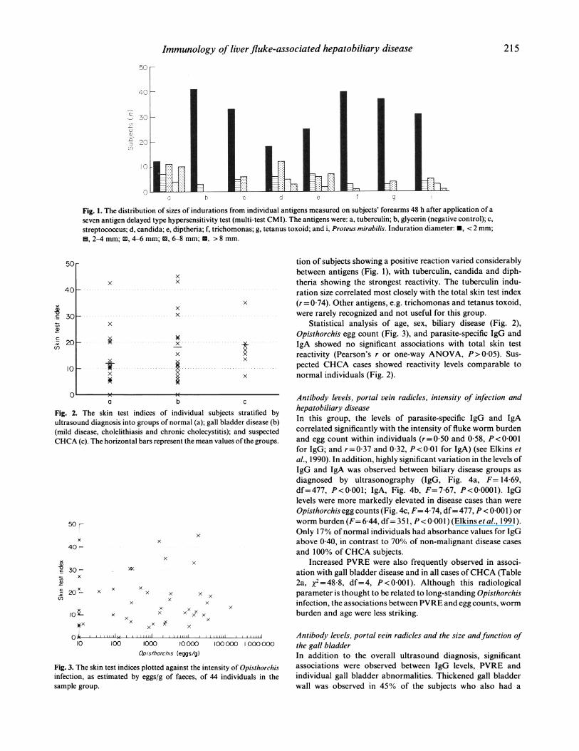
Fig. 1. The distribution of sizes of indurations from individual antigens measured on subjects' forearms 48 h after application of aseven antigen delayed type hypersensitivity test (multi-test CMI). The antigens were: a, tuberculin; b, glycerin (negative control); c,streptococcus; d, candida; e, diptheria; f, trichomonas; g, tetanus toxoid; and i, Proteus mirabilis. Induration diameter: *, < 2 mm;
, 2-4 mm; CM, 4-6 mm; 0, 6-8 mm;E, > 8 mm.
x
x
x
I~ ~~~~ E
tion of subjects showing a positive reaction varied considerablybetween antigens (Fig. 1), with tuberculin, candida and diph-theria showing the strongest reactivity. The tuberculin indu-ration size correlated most closely with the total skin test index(r= 0 74). Other antigens, e.g. trichomonas and tetanus toxoid,were rarely recognized and not useful for this group.
Statistical analysis of age, sex, biliary disease (Fig. 2),Opisthorchis egg count (Fig. 3), and parasite-specific IgG andIgA showed no significant associations with total skin testreactivity (Pearson's r or one-way ANOVA, P> 0-05). Sus-pected CHCA cases showed reactivity levels comparable tonormal individuals (Fig. 2).
' -a b c
Fig. 2. The skin test indices of individual subjects stratified byultrasound diagnosis into groups of normal (a); gall bladder disease (b)(mild disease, cholelithiasis and chronic cholecystitis); and suspectedCHCA (c). The horizontal bars represent the mean values of the groups.
50%x
40 -x
xx
020
30Vi x
Q)
' 20X~n.I
x x /x
x
x
x x xx
xx xxx x x
x
x
x
Antibody levels, portal vein radicles, intensity of infection andhepatobiliary diseaseIn this group, the levels of parasite-specific IgG and IgAcorrelated significantly with the intensity of fluke worm burdenand egg count within individuals (r=0 50 and 0-58, P<0.001for IgG; and r=0-37 and 0 32, P<O 01 for IgA) (see Elkins et
al., 1990). In addition, highly significant variation in the levels ofIgG and IgA was observed between biliary disease groups as
diagnosed by ultrasonography (IgG, Fig. 4a, F= 14 69,df=477, P<0001; IgA, Fig. 4b, F=7-67, P<0O0001). IgGlevels were more markedly elevated in disease cases than were
Opisthorchis egg counts (Fig. 4c, F= 4 74, df= 477, P < 0-001) or
worm burden (F= 6A44, df= 351, P < 0 001 ) (Elkins et al., 1991).Only 17% of normal individuals had absorbance values for IgGabove 040, in contrast to 70% of non-malignant disease cases
and 100% of CHCA subjects.Increased PVRE were also frequently observed in associ-
ation with gall bladder disease and in all cases ofCHCA (Table2a, X2=48-8, df=4, P<0 001). Although this radiologicalparameter is thought to be related to long-standing Opisthorchisinfection, the associations between PVRE and egg counts, worm
burden and age were less striking.
0o ,, a, I II t II ,1 I,,,a1 Antibody levels, portal vein radicles and the size andfunction ofl0 00 1000 0000 00000 1000000 the gall bladder
Opis/horchls (eggs/g) In addition to the overall ultrasound diagnosis, significant
Fig. 3. The skin test indices plotted against the intensity of Opisthorchisinfection, as estimated by eggs/g of faeces, of 44 individuals in thesample group.
associations were observed between IgG levels, PVRE andindividual gall bladder abnormalities. Thickened gall bladderwall was observed in 45% of the subjects who also had a
50 F
40 -
xxx
a)_0c
V)a)'CCI)
301-
20 _
xX.
1O0_ tx x
x x

M. R. Haswell-Elkins et al.
x
Table 2. Associations between the intensity of portal vein radicle echoes(PVRE) and hepatobiliary abnormalities
Portal vein radicles
(a) Relationship with hepatobiliary diagnosis
xx
xx
.0
DiagnosisNormalMild diseaseCholelithiasisChronic cholecystitisCHCA
04010340
3
0
244
6
410
3 4I
D Parenchymal disease2 3 4 5 PDUltrasound diagnosis
..... ...
(b) Relationship with gall bladder wall thicknessGall bladder wall 0Normal 43Thickened 16
x (c) Relationship with contraction0 x~~~~~
°-.' '*8 > zx x x
*6 .. .. ...
0 2~~~~~2 ..........Ultrasoun d sx
O 2 3 4 5 PIDUltrasound diagnosis
x
0 2 3 4Ultrasound diagnosis
x
x xx
ContractionNormal response< 50% reductionNo change
PVRE ranked as: 0, normal;severe increase.
I1- -
xx
> 0-8a)
x H0
x
0.41
0-2
PD/ (
5 PD
x
x
0960
1, mild increase; or 2, moderate to
x
x
1-x
0 0-5
.4.
'X'
xxx
1-5Gall bladder contraction
Fig. 4. Parasite-specific antibody levels and intensity of infection inhepatobiliary disease. Absorbance values for IgG (a) and IgA (b) inELISA and Opisthorchis egg counts (c) of individual subjects stratifiedby ultrasound diagnosis. The diagnoses are coded as: 1, normal; 2, mildgall bladder disease; 3, cholelithiasis; 4, chronic cholecystitis; 5,suspected cholangiocarcinoma; and PD, parenchymal liver disease. Thehorizontal bars represent the mean values of the groups and s.e.m. forIgG are given in Table 1. Numbers are shown inside (c) to indicate howmany cases were negative for fluke eggs, where there were more than 1.
significantly higher frequency of increased PVRE (x2 = 24 44,df= 1, P < 0 001, Table 2b). Mean IgG levels varied significantlywith severity of gall bladder dysfunction (ANOVA, F= 73,df= 233, P < 0 005, Fig. 5). All subjects with no contraction hadincreased PVRE, but the frequency of increased echoes differedonly slightly between the normal and poor contraction groups
(X2= 8 52, df= 2, P < 0-025, Table 2c).
Fig. 5. Parasite-specific IgG levels and gall bladder function. IgG levelsof individuals in groups stratified by the degree of gall bladdercontraction after fatty meal. Contraction is coded as: 0 5, group notexamined because the gall bladder was considered normal (thin walledand not enlarged or contracted) by ultrasonography; 1, good contrac-tion (> 50% reduction in size); 1 5, poor contraction (< 50% contrac-tion); and 2, no contraction (no difference in pre- and post-meal size ofthe organ). The horizontal bars represents the mean values of thegroups.
The length of the gall bladder showed similar correlationswith egg count, worm burden and IgG levels (r=0 41-0-48).Multiple-regression analysis was performed incorporating eggcount and worm burden, age, sex, IgG and IgA levels as
independent variables and gall bladder size as the dependentvariable. This analysis indicated that IgG level accounted for the
( a )
x
xx
xx
x
I- 8
I1*6
I.4
a)a> * °
CD 088
0-6-
0-2-
0
IO8
0-6
1-4
6
4
x
x
.X.
..x ............
x 0
x..x ...........
( b )
I2h_IV)a)
c-
244144
I .
0.
0
0'
0.
21
13
6
5
(c )
7
2525
u0
uJ1-1LL)
4
3
2
-Itx
x
_x
x
xx
2103) 416 I 4l(2) -
216
I-.4r
.x .............x 4-
1-

Immunology of liver fluke-associated hepatobiliary disease
tory antigens may adsorb to host cell surfaces leading toX Xx antibody binding, complement fixation and chronic inflamma-
X X x tion. The proposed immunopathology may involve antigen orx X3 X isotype-specific IgG, as suggested in filariasis (Hussain, Grogl &
--xx x/. x-- Ottesen, 1987), Sowda-type onchocerciasis (Parkhouse& Harri-x x xx son, 1989) and inflammatory bowel disease (Kett, Rognum &
->K_ X X X x Brandtzaeg, 1987).o>x xX x The relationship between IgG levels and enlargement and
_ >x''xa -* .... poor function of the gall bladder may be directly causative (asx
x \\\\\\\\\\suggested above), but it is equally possible that enlargement ofthe gall bladder results from other pathological mechanisms andsecondarily enhances the leakage of parasite antigens into
| ((( §circulation. This could result in proportionately higher antibody0.2 0-4 0 6 0-8 1l0 1-2 4 6 levels, which may or may not narticinate in the pathologicalIgG levels
Fig. 6. The relationship between Opisthorchis-specific IgG levels andmaximum length of the gall bladder as measured radiologically. Linearregression generates the equation y=51 65x+43 2, where y=theabsorbance value for IgG in ELISA and x =gall bladder length. Theregression is highly significant (ANOVA, F= 81 44, df= 70, P< 0-0001).
most variability in gall bladder size (r= 0 48), and inclusion ofother variables did not improve the model significantly.
There are four obvious outlying points in Fig. 6; one ofwhich (length, 26 cm) represents a contracted gall bladder, an
abnormal state after fasting indicating chronic cholecystitis. Ofthe three remaining outliers, all had light worm burdens andradiologically normal gall bladders (can also be seen on Figs 4aand 5). Two received anti-helminthic treatment at least twicebefore this study, which may have influenced both theirantibody level and gall bladder status. If these points are
excluded, the correlation coefficient between IgG levels and gallbladder length rises from 0-48 to 0 73.
DISCUSSION
Several chronic inflammatory diseases of the gastroentericmucosa are associated with an increased risk of carcinoma(Fraumeni & Kantor, 1982; Christie, 1987; Petras, Mir-Madj-lessi & Farmer, 1987; Nagorney & McPherson, 1988). However,an aetiological agent is established only in a few; this precludesexamination of the role of specific responses in pathogenesis.Schistosoma haematobium in bladder carcinoma and liver flukesin CHCA provide good models for studying the role ofimmunopathology in chronic disease and carcinogenesis (Chris-tie, 1987).
A close relationship was observed between parasite-specificantibodies, PVRE and hepatobiliary disease in Opisthorchisviverrini infection. More specifically, IgG levels were highlypredictive ofgall bladder size and contraction, while PVRE were
closely linked to gall bladder wall thickness. The intensity ofcurrent fluke infection showed weaker associations with theseabnormalities.
One possible interpretation of the close relationshipreported between IgG levels and the size and function of the gallbladder is that these antibodies have a causative role in thepathophysiology of gall bladder disease. Two mechanisms are
proposed. The antibodies may bind host antigens on the gallbladder, eliciting autoimmune damage, as suggested by some
investigators for chronic myocardial damage in Trypanosomacruzi (Hudson, 1985). Alternatively, parasite excretory/secre-
-I ------ --v -- r ---.--_-r---. -- -- ----.. .. - --- cw
process. Further studies are needed to clarify these possiblemechanisms.
Increased echoes along the portal vein radicles were notstrongly associated with current intensity of infection or age(duration of infection). However, their increased frequency indisease cases and cases of gall bladder wall thickening supportstheir association with pathogenesis. Increased PVRE mayrepresent fibrotic change resulting from chronic inflammation,perhaps initiated by specific T cell responses to Opisthorchisantigens. Interestingly, thickening of the gall bladder wall andincreased PVRE are also observed in Symmers' periportalfibrosis in S. mansoni infection (Homeida et al., 1988). Studiesare in progress to determine the histopathological equivalent toPVRE in Opisthorchis infection and their association with cell-mediated responses.
The lack of a detectable influence ofeither heavy infection ordisease status on cutaneous delayed-type hypersensitivity re-sponses suggests that Opisthorchis infection does not inducegeneralized immunosuppression in humans. These findings,however, should be followed up on larger populations.
The observation that 83% of the normal group and only27% of the total diseased group had antibody titres less than 04suggests that this assay may be a useful screening tool for thediagnosis of fluke-associated disease. ELISA is quicker andcheaper to perform than the conventional stool examination forfluke eggs. Where the prevalence of infection is high, thepresence of eggs may be an incidental finding rather than anindication of involvement in disease. High IgG levels, however,appear to be more predictive of disease than egg counts, exceptin some recently treated people.
These data are consistent with the hypothesis that biliarytract and gall bladder abnormalities associated with Opisthor-chis infection, which may create favorable conditions forcarcinogenesis, are at least in part immunopathological. IgGmay contribute to bile stasis (as risk factor for CHCA) if it playsan active role in the enlargement and malfunction of the gallbladder, while inflammatory responses may elicit chronic cellproliferation and fibrotic damage. Our results do not indicatethat parasite-induced immunosuppression is a major factor intumour development.
ACKNOWLEDGMENTSThis research was generously supported by the Tropical HealthProgram, Commonwealth Government, Australia. We express appre-ciation to P. Awacharagan for excellent technical assistance in theELISA, and to P. Treesarawat, S. Phinlaor, S. Phisaipan, C. Phisaipan,P. Sirisatchang and L. Chaturat for field and laboratory work. The
120
100
C3C)
a)
='a0
0
80
60
40
20
0
217

218 M. R. Haswell-Elkins et al.
study would not have been possible without the advice and support oflocal health personnel, including Dr V. Wechosotsakda, Dr S. Khan-thai, S. Ponrueng, B. Singawn and T. Poopahngern. We also thank P.Graves, A. Saul, M. Good, D. McManus and G. Mitchell for helpfulcomments on the manuscript, D. Battistuta for statistical advice and DrD. Moss for suggesting the multi-test CMI.
REFERENCES
BHAMARAPRAVATI, N., THAMAVIT, V. & VAJRASTHIRA, S. (1978) Liverchanges in hamsters infected with a liver fluke of man, Opisthorchisviverrini. Am. J. trop. Med. Hyg. 27, 787.
CHRISTIE, J.D. (1987) Patterns of Schistosoma haematobium egg distri-bution in human lower urinary tract. Cancerous lower urinary tracts.Am. J. trop. Med. Hyg. 35, 759.
DHIENSIRI, T., EUA-ANANTA, Y., BUNNAG, D., HARINASUTA, T. &SCHELP, P.F. (1984) Roentgenographically controlled healing of gallbladder lesions in opisthorchiasis after praziquantel treatment. DrugRes. 34, 1175.
ELKINS, D.B., HASWELL-ELKINS, M.R., MAIRIANG, E., MAIRIANG, P.,SITHITHAWORN, P., KAEWKES, S., BHUDHISAWASDI, V. & UTTARAVI-CHIEN, T. (1990) A high frequency of hepatobiliary disease andcholangiocarcinoma associated with heavy Opisthorchis viverriniinfection in a small community in Northeast Thailand. Trans. R. Soc.trop. Med. Hyg. 84, 715.
ELKINS, D.B., SITHITHAWORN, P., HASWELL-ELKINS, M.R., KAEWKES,S., AWACHARAGAN, P. & WONGRATANACHEEWIN, S. (1991) Opisthor-chis viverrini: relationships between egg counts, worms recovered andantibody levels within an endemic community in Northeast Thailand.Parasitology (In press).
EVERSON, G.T., BRAVERMAN, D.Z., JOHNSON, M.L. & KERN, F. (1980) Acritical evaluation of real-time ultrasonography for the study ofgallbladder volume and contraction. Gastroenterology, 79, 40.
FLAVELL, D.J. (1981) Liver fluke infection as an aetiological factor inbiler duct carcinoma of man. Trans. R. Soc. trop. Med. Hyg. 75, 815.
FRAUMENI, J.F. & KANTOR, A.F. (1982) Cancers of the biliary tract. InCancer Epidemiology and Prevention (ed. by D. Schottenfeld & J.F.Fraumeni) p.683. WB Saunders, Philadelphia.
HARINASUTA, T., RIGANTI, M. & BUNNAG, D. (1984) Opisthorchisriverrini infection: pathogenesis and clinical features. Drug Res. 34,1167.
HEDERSTROM, E., FORSBERG, L., HERLIN, P. & HOLMIN, T. (1988) Fattymeal provocation monitored by ultrasonography: a method to
diagnose ambiguous gallbladder disease. Acta radiol. 29, 207.HOMEIDA, M., ABDEL-GADIR, A.F., CHEEVER, A.W., BENNETT, J.L.,ARBAB, B.M.O., IBRAHIUM, S.Z., ABDEL-SALAM, I.M., DAFALLA, A.A.& NASH, T.E. (1988) Diagnosis of pathologically confirmed Symmers'
periportal fibrosis by ultrasonography: a prospective blinded study.Am. J. trop. Med. Hyg. 38, 86.
Hou, P.C. (1956) The relationship between primary carcinoma of theliver and infestation with Clonorchis sinensis. J. Pathol. Bacteriol. 72,239.
HUDSON, L. (1985) Autoimmune phenomona in chronic Chagasiccardiopathy. Parasitol. Today, 1, 6.
HUSSAIN, R., GROGL, M. & OTTESEN, E.A. (1987) IgG antibodysubclasses in human filariasis. Differential subclass recognition ofparasite antigens correlates with different clinical manifestations ofinfection. J. Immunol. 139, 2794.
KETT, K., ROGNUM, T.O. & BRANDTZAEG, P. (1987) Mucosal subclassdistribution of immunoglobulin G-producing cells is different inulcerative colitis and Crohn's disease of the colon. Gastroenterology,93, 919.
KIM, Y.I. (1984) Liver carcinoma and liver fluke infection. Drug Res. 34,1121.
NAGORNEY, D.M. & MCPHERSON, G.A.D. (1988) Carcinoma of thegallbladder and extrahepatic bile ducts. Semin. Oncol. 15, 106.
PARKHOUSE, R.M.E. & HARRISON, L.J.S. (1989) Antigens of parasitichelminths in diagnosis, protection and pathology. Parasitology, 99,S5.
PETRAS, R.E., MIR-MADJLESSI, S.H. & FARMER, R.G. (1987) Crohn'sdisease and intestinal carcinoma. A report of 11 cases with emphasison associated epithelial dysplasia. Gastroenterology, 93, 1307.
PREUKSARAJ, S. (1984) Public health aspects of opisthorchiasis inThailand. Drug Res. 34, 1119.
RADOMYOS, P., BUNNAG, D. & HARINASUTA, T. (1984) Worms recoveredin stools following praziquantel treatment. Drug Res. 34, 1215.
RAMSEY, R.J., SITHITHAWORN, P., PROCIV, P., MOORHOUSE, D.E. &METHAPHAT, C. (1989) Density dependent fecundity of Opisthorchisviverrini in humans, based on faecal recovery of flukes. Trans. R. Soc.trop. Med. Hyg. 83, 241.
VATANASAPT, V., TANGVORAPHONKCHAI, V., TITAPANT, V., PIPITGOOL,V., VIRIYAPAP, D. & SRIAMPORN, S. (1990a) A high incidence of livercancer in Khon Kaen Province, Thailand. Southeast Asian. J. trop.Med. Public Health, 21, 382.
VATANASAPT, V., UTTARAVICHIEN, T., MAIRIANG, E., PAIROJKUL, C.,CHARTBANCHACHAI, V. & HASWELL-ELKINS, M.R. (1990b) NortheastThailand: A region with a high incidence of cholangiocarcinoma.Lancet, 335, 116.
WONGRATANACHEEWIN, S., BUNNAG, D., VAEUSORN, N. & SIRISINHA, S.(1988) Characterization of humoral immune response in the serum
and bile of patients with opisthorchiasis and its application inimmunodiagnosis. Am. J. trop. Med. Hyg. 38, 356.
WONGRATANACHEEWIN, S., RATTANASIRIWILAI, W., PRIWAN, R. &SIRISINHA, S. (1987) Immunodepression in hamsters experimentallyinfected with Opisthorchis viverrini. J. Helminthol. 61, 151.