Post-Discharge Effects and Parents' Opinions of Intranasal ...
Upload
khangminh22Category
view
2download
0
www.amegroups.com
amegroups.com
2A005
AME Medical Review 005
Key Leaders' Opinions onPrecision Medicine in Hepatobiliary Cancer
Key Lead
ers' Opin
ion
s on
Prec
ision
M
edic
ine in
Hepato
biliary Can
cer
Irene Oi-Lin Ng, Diego F. Calvisi, Xuehao WangHaitao Zhao, Ralf Weiskirchen, Ling Lu, Bryan C. FuchsYaqing Zhu, Francesco Feo, Sherry X. Yang, Helen L. Reeves
Honorary Editors:
Editors:
Associate Editors:
Editors: H
aitao Zhao, Ralf W
eiskirchen,
Ling Lu, Bryan C
. Fuchs

AME Medical Review 005
Key Leaders' Opinions onPrecision Medicine in Hepatobiliary Cancer
Irene Oi-Lin Ng, Diego F. Calvisi, Xuehao WangHaitao Zhao, Ralf Weiskirchen, Ling Lu, Bryan C. FuchsYaqing Zhu, Francesco Feo, Sherry X. Yang, Helen L. Reeves
Honorary Editors:
Editors:
Associate Editors:

AME Publishing Company
Room C 16F, Kings Wing Plaza 1, NO. 3 on Kwan Street, Shatin, NT, Hong Kong
Information on this title: www.amegroups.comFor more information, contact [email protected]
Copyright © AME Publishing Company. All rights reserved.
This publication is in copyright. Subject to statutory exception and to the provisions of relevant collective licensing agreements, no reproduction of any part may take place without the written permission of AME Publishing Company.
First published in 2017Printed in China by AME Publishing Company
Editors: Haitao Zhao, Ralf Weiskirchen, Ling Lu, Bryan C. FuchsCover Image Illustrator: Zhijing Xu, Shanghai, China
Key Leaders' Opinions on Precision Medicine in Hepatobiliary Cancer(Hard Cover)
ISBN: 978-988-77841-6-6AME Publishing Company, Hong Kong
AME Publishing Company has no responsibility for the persistence or accuracy of URLs for external or third-party internet websites referred to in this publication, and does not guarantee that any content on such websites is, or will remain, accurate or appropriate.
The advice and opinions expressed in this book are solely those of the authors and do not necessarily represent the views or practices of the publisher. No representation is made by the publisher about the suitability of the information contained in this book, and there is no consent, endorsement or recommendation provided by the publisher, express or implied, with regard to its contents
© AME Publishing Company. All rights reserved. www.amegroups.com
I
Precision Medicine in HePatobiliary cancer(FIRST EDITION)
Honorary Editors
Irene Oi-Lin NgDepartment of Pathology, and State Key Laboratory for Liver Research, The University of Hong Kong, Hong Kong, China
Diego F. CalvisiDepartment of Clinical and Experimental Medicine, University of Sassari, Sassari, Italy
Xuehao WangAcademician of the Chinese Academy of Engineering, Liver Transplantation Center, First Affiliated Hospital of Nanjing Medical University, Nanjing, China
Editors
Haitao ZhaoDepartment of Liver Surgery, Peking Union Medical College Hospital, Chinese Academy of Medical Sciences, Beijing, China
Ralf WeiskirchenInstitute of Molecular Pathobiochemistry, Experimental Gene Therapy, and Clinical Chemistry, RWTH University Hospital Aachen, Aachen, Germany
Ling Lu Liver Transplantation Center, First Affiliated Hospital of Nanjing Medical University, Nanjing, China
Bryan C. FuchsDivision of Surgical Oncology, Massachusetts General Hospital Cancer Center, Harvard Medical School, Boston, Massachusetts, USA
Associate Editors
Yaqing ZhuDepartment of Hepatobiliary Surgery, The First Affiliated Hospital, Guangzhou University of Chinese Medicine, Guangzhou 510405, China
Francesco FeoProfessor Emeritus of Experiential Pathology, Department of Clinical and Experimental Medicine, Division of Experimental Pathology and Oncology, University of Sassari, Italy
Sherry X. YangNational Clinical Target Validation Laboratory, Division of Cancer Treatment and Diagnosis, National Cancer Institute, National Institutes of Health, Bethesda, Maryland, USA
Helen L. ReevesNorthern Institute for Cancer Research, Newcastle University, Newcastle upon Tyne, UK; Newcastle upon Tyne Hospitals NHS Foundation Trust, Newcastle upon Tyne, UK
Authors
Ivana AkrapMolecular Biology Unit, Interfaculty Institute of Cell Biology, Tubingen University, Tubingen, Germany
Gyorgy BaffyDepartment of Medicine, VA Boston Healthcare System and Brigham and Women’s Hospital, Harvard Medical School, Boston, MA 02130, USA
Simonetta BandieraInserm, U1110, Institut de Recherche sur les Maladies Virales et Hepatiques, Strasbourg, France; Universite de Strasbourg, Strasbourg, France

© AME Publishing Company. All rights reserved. www.amegroups.com
II
Savio G. BarretoDepartment of Gastrointestinal Surgery, Gastrointestinal Oncology and Bariatric Surgery, Medanta Institute of Digestive and Hepatobiliary Sciences, Medanta-The Medicity, Gurgaon, Haryana, India
Christopher Taylor BarryKIMS Institute of Organ Transplantation, Secunderabad, 500003 Telangana, India
Cecilia BattistelliIstituto Pasteur Italia-Fondazione Cenci Bolognetti, Department of Cellular Biotechnologies and Haematology, Sapienza University of Rome, Rome, Italy
Thomas F. BaumertInserm, U1110, Institut de Recherche sur les Maladies Virales et Hepatiques, Strasbourg, France; 2Universite de Strasbourg, Strasbourg, France; Institut Hospitalo-Universitaire, Pole hepato-digestif, Nouvel Hopital Civil, Strasbourg, France
Kai BreuhahnInstitute of Pathology, University Hospital Heidelberg, Heidelberg, Germany
Diego F. CalvisiDepartment of Clinical and Experimental Medicine, University of Sassari, Sassari, Italy
Alfred S. L. ChengSchool of Biomedical Sciences, State Key Laboratory of Digestive Disease and Institute of Digestive Disease, The Chinese University of Hong Kong, Hong Kong SAR, China; Shenzhen Research Institute, The Chinese University of Hong Kong, Shenzhen 518057, China
Su Pin ChooDivision of Medical Oncology, National Cancer Centre Singapore, Singapore
Carla CicchiniIstituto Pasteur Italia-Fondazione Cenci Bolognetti, Department of Cellular Biotechnologies and Haematology, Sapienza University of Rome, Rome, Italy
Ruixia CuiDepartment of Hepatobiliary Surgery, the First Affiliated Hospital of Xi’an Jiaotong University, Xi’an 710061, China
Amit DuttThe Advanced Centre for Treatment, Research and Education in Cancer, Tata Memorial Center, Kharghar, Navi Mumbai, Maharashtra, India
Francesco FeoDivision of Experimental Pathology and Oncology, Department of Clinical and Experimental Medicine, University of Sassari, Sassari, Italy
Puri FortesDepartment of Gene Therapy and Hepatology, Center for Applied Medical Research (CIMA), IdiSNA, Navarra Institute for Health Research, University of Navarra. Pio XII 55, 31008 Pamplona, Spain
Bryan C. FuchsDivision of Surgical Oncology, Massachusetts General Hospital Cancer Center, Harvard Medical School, Boston, Massachusetts, USA
Sarani GhoshalDivision of Surgical Oncology, Massachusetts General Hospital Cancer Center, Harvard Medical School, Boston, Massachusetts, USA
Margarita Gonzalez-VallinasInstitute of Pathology, University Hospital Heidelberg, Heidelberg, Germany
Ghassan M. HammoudDivision of Gastroenterology and Hepatology, Department of Internal Medicine, School of Medicine, University of Missouri, One Hospital Drive, Columbia, MO 65212, USA
Ruben Hernandez-AlcocebaDepartment of Gene Therapy and Hepatology, Center for Applied Medical Research (CIMA), IdiSNA, Navarra Institute for Health Research, University of Navarra. Pio XII 55, 31008 Pamplona, Spain
© AME Publishing Company. All rights reserved. www.amegroups.com
III
Jamal A. IbdahDivision of Gastroenterology and Hepatology, Department of Internal Medicine, School of Medicine, University of Missouri, One Hospital Drive, Columbia, MO 65212, USA
Yoshinori InagakiHepato-Biliary-Pancreatic Surgery Division, Department of Surgery, Graduate School of Medicine, the University of Tokyo, Tokyo, Japan
Keita KaiDepartment of Pathology, Saga University Hospital, Saga 849-8501, Japan
Shuichi KanekoDepartment of Gastroenterology, Kanazawa University Graduate School of Medical Science, Kanazawa, Ishikawa, Japan
Norihiro KokudoHepato-Biliary-Pancreatic Surgery Division, Department of Surgery, Graduate School of Medicine, the University of Tokyo, Tokyo, Japan
Dragana KopanjaDepartment of Biochemistry and Molecular Genetics (M/C 669), University of Illinois at Chicago, College of Medicine, Chicago, IL 60607, USA
Masatoshi KudoDepartment of Gastroenterology and Hepatology, Kinki University Faculty of Medicine, 377-2 Ohno-higashi, Osaka-sayama, Osaka 589-8511, Japan
Justina Yick Ching LamDivision of Medical Oncology, National Cancer Centre Singapore, Singapore
Man LiuSchool of Biomedical Sciences, The Chinese University of Hong Kong, Hong Kong SAR, China
Chang LiuDepartment of Hepatobiliary Surgery, the First Affiliated Hospital of Xi’an Jiaotong University, Xi’an 710061, China
Thomas LongerichInstitute of Pathology, University Hospital RWTH Aachen, Aachen, Germany
Hiroyuki MarusawaDepartment of Gastroenterology and Hepatology, Graduate School of Medicine, Kyoto University, Kyoto, Japan
Vivian McAlisterDepartment of Surgery, University of Western Ontario, London, ON, Canada
Balraj MittalDepartment of Medical Genetics, Sanjay Gandhi Postgraduate Institute of Medical Sciences, Lucknow 226014, UP, India
Myth T. S. MokSchool of Biomedical Sciences, The Chinese University of Hong Kong, Hong Kong SAR, China; Shenzhen Research Institute, The Chinese University of Hong Kong, Shenzhen 518057, China
Irene Oi-Lin NgDepartment of Pathology, State Key Laboratory for Liver Research, The University of Hong Kong, Hong Kong, China
Kouki NioDepartment of Gastroenterology, Kanazawa University Graduate School of Medical Science, Kanazawa, Ishikawa, Japan; Lineberger Comprehensive Cancer Center, The University of North Carolina at Chapel Hill, Chapel Hill, North Carolina, USA
Naoshi NishidaDepartment of Gastroenterology and Hepatology, Kinki University Faculty of Medicine, 377-2 Ohno-higashi, Osaka-sayama, Osaka 589-8511, Japan
Alfred NordheimMolecular Biology Unit, Interfaculty Institute of Cell Biology, Tubingen University, Tubingen, Germany
Takahiro OchiyaDivision of Molecular and Cellular Medicine, National Cancer Center Research Institute, Tokyo, Japan

© AME Publishing Company. All rights reserved. www.amegroups.com
IV
Hideya OnishiDepartment of Cancer Therapy and Research, Graduate School of Medical Sciences, Kyushu University, Fukuoka, Japan
Rosa M. PascaleDepartment of Clinical and Experimental Medicine, Division of Experimental Pathology and Oncology, University of Sassari, Sassari, Italy
Martin PichlerDivision of Oncology, Department of Internal Medicine, Medical University of Graz, Austria; Department of Experimental Therapeutics, The University of Texas MD Anderson Cancer Center, Houston, USA
Karen Pineda-SolisDepartment of Surgery, University of Western Ontario, London, ON, Canada
Kai Qu Department of Hepatobiliary Surgery, the First Affiliated Hospital of Xi’an Jiaotong University, Xi’an 710061, China
Pradip RaychaudhuriDepartment of Biochemistry and Molecular Genetics (M/C 669), University of Illinois at Chicago, College of Medicine, Chicago, IL 60607, USA; Jesse Brown VA Medical Center, Chicago, IL 60612, USA
Helen L. ReevesNorthern Institute for Cancer Research, Newcastle University, Newcastle upon Tyne, UK; Newcastle upon Tyne Hospitals NHS Foundation Trust, Newcastle upon Tyne, UK
Anna Lena RessDivision of Oncology, Department of Internal Medicine, Medical University of Graz, Austria
Ismael RiquelmeMolecular Pathology Laboratory, Department of Pathology, CEGIN-BIOREN, Universidad de La Frontera, Temuco, Chile
Juan Carlos RoaDepartment of Pathology, UC Centre for Investigational Oncology (CITO), Advanced Centre for Chronic Diseases (ACCDiS), Ponti cia Universidad Catolica de Chile, Santiago, Chile
Maria M. SimileDepartment of Clinical and Experimental Medicine, Division of Experimental Pathology and Oncology, University of Sassari, Sassari, Italy
Peipei SongHepato-Biliary-Pancreatic Surgery Division, Department of Surgery, Graduate School of Medicine, the University of Tokyo, Tokyo, Japan
Atsushi TakaiDepartment of Gastroenterology and Hepatology, Graduate School of Medicine, Kyoto University, Kyoto, Japan
Naoko TakebeCancer Therapy Evaluation Program, National Cancer Institute, National Institutes of Health, Bethesda, Maryland, USA
Haruhiko TakedaDepartment of Gastroenterology and Hepatology, Graduate School of Medicine, Kyoto University, Kyoto, Japan
Kenneth K. TanabeDivision of Surgical Oncology, Massachusetts General Hospital Cancer Center, Harvard Medical School, Boston, Massachusetts, USA
Wei TangHepato-Biliary-Pancreatic Surgery Division, Department of Surgery, Graduate School of Medicine, the University of Tokyo, Tokyo, Japan
Abhishek ThavamaniMolecular Biology Unit, Interfaculty Institute of Cell Biology, Tubingen University, Tubingen, Germany
Bin Tean TheNCCS-VARI Translational Research Laboratory, National Cancer Centre Singapore, Singapore; DUKE-NUS (National University of Singapore) Graduate Medical School, Singapore
Marco TripodiIstituto Pasteur Italia-Fondazione Cenci Bolognetti, Department of Cellular Biotechnologies and Haematology, Sapienza University of Rome, Rome, Italy

© AME Publishing Company. All rights reserved. www.amegroups.com
V
Rishi WagleDepartment of Experimental Therapeutics, The University of Texas MD Anderson Cancer Center, Houston, USA
Ralf WeiskirchenInstitute of Molecular Pathobiochemistry, Experimental Gene Therapy, and Clinical Chemistry, RWTH University Hospital Aachen, Aachen, Germany
Catherine E. WilloughbyNorthern Institute for Cancer Research, Newcastle University, Newcastle upon Tyne, UK
Carmen Chak-Lui WongDepartment of Pathology, State Key Laboratory for Liver Research, The University of Hong Kong, Hong Kong, China
Chun-Ming WongDepartment of Pathology, State Key Laboratory for Liver Research, The University of Hong Kong, Hong Kong, China
Saurabh YadavDepartment of Medical Genetics, Sanjay Gandhi Postgraduate Institute of Medical Sciences, Lucknow 226014, UP, India
Taro YamashitaDepartment of Gastroenterology, Kanazawa University Graduate School of Medical Science, Kanazawa, Ishikawa, Japan
Sherry X. YangNational Clinical Target Validation Laboratory, Division of Cancer Treatment and Diagnosis, National Cancer Institute, National Institutes of Health, Bethesda, Maryland, USA
Mirjam B. ZeiselInserm, U1110, Institut de Recherche sur les Maladies Virales et Hepatiques, Strasbourg, France; Universite de Strasbourg, Strasbourg, France
Xing ZhangDepartment of Hepatobiliary Surgery, the First Affiliated Hospital of Xi’an Jiaotong University, Xi’an 710061, China
Cover Image Illustrator
Zhijing Xu, Shanghai, China
Executive Typesetting Editor
Beibei Chen, AME Publishing Company

© AME Publishing Company. All rights reserved. www.amegroups.com
VIForeword
Will scholarly journals perish?
Will scholarly journals perish? This is a question that has puzzled me for years.
The introduction of online journals results in the inevitable recession of print journals. The uprise of the open access journals has been changing the structure of scholarly journals ceaselessly. What keeps me thinking is the open access of clinical trials data. What would be the bigger picture if open access to clinical trials data becomes the mainstream?
It is interesting that with the primary bottleneck lying in the availability of open data, the Big-data Clinical Trial (BCT) seems to stay where it was in spite of the increasingly popularity of “Big Data” among scientists. It has to be the fact that without open data, a statistical analysis is restricted to a particular area (or several areas). Even with big enough data, the study can only be termed as “research with big data sets” rather than “big data research”, which are totally different concepts. Big Data is constituted by a plurality of dimensions. On one hand, for an individual (e.g., a patient), the relevant data covering his/her disease course is big enough; on the other hand, for the entire population, as more as individuals (e.g., patients) are expected to be included, to contains all the elements just like the “universe set” in set theory; by doing so, scientists expect to carry out the so-called clinical studies in real-world settings.
Why do the real-world-based clinical trials so appealing? It is understandable that the results and conclusions are likely to be altered in studies targeting the same issue using the same research method with sample size changed. In addition, the probability of such a “likely” is quite high. In many top journals, it is a common phenomenon that some authors tend to validate the results of one study in another population using the same research method. However, if the results are “validated” in one population, it only means that they are “repeatable”. Will the results also be repeatable in the second, third, and more populations? If the attempts are not continuing, which should be, the “validation” is equivalent to “self-deception” in a sense.
When clinical research data is open accessed, we can easily integrate data from multiple centers for statistical analysis and meanwhile “validate” the results in multiple populations. If this is the case, then another question arise: can everyone easily publish his/her results/papers in high-profile journals such as the New England Journal of Medicine? My answer is NO.
When the open access to clinical research data becomes mainstream, we can easily find the constant update of database on the Internet. Simply by clicking on a button, we obtain the statistical results of the most current data. A further button click would display the validation results based on a specific population. The database would be updated at a certain period of time (e.g., 1 month or 1 day), and the statistical results would “likely” also be changed accordingly. At that time, the questions may change to “would any researchers publish their findings in a journal?” Well, even if someone is still keen to write such articles, journals may be reluctant to publish them because of the indefiniteness of the findings with the risk of being overturned at anytime.
Eventually here it comes the serious question: will scholarly journals perish? My answer is still NO. Then in what way the scholarly journals would probably lead to?
During my Business Administration course, my teacher distributed to us an article from the Case Study column of the Harvard Business Review. In this highly respected journal, articles in this column often present one case first, followed by the comments from two experts. These comments could either support or oppose each other. My teacher asked us to study the case, read through the comments and then form our own point of views on the case. He encouraged us to interpret the case from different perspectives independently in what form that I found pretty practical.
The course brought a possible answer to me. When the open access to clinical research data becomes mainstream, the entire publishing industry, especially the publication of “scholarly journals”, would eventually experience revolutionary change. It may no longer focus on the rigid and cold outcomes but it would definitely cares more about the reflection on the problems, update of insights, and integration of science and arts.
AME Medical Review Series is a production of the above thinking. As an attempt, we decided to invite experts internationally to provide their views on a specific topic to share their insights with more clinicians and thus benefit more patients. The first chosen topic for the series is the currently controversial one: conventional surgery versus stereotactic body radiotherapy for

© AME Publishing Company. All rights reserved. www.amegroups.com
VII
the early stage lung cancer. As the first book to the series, we hope it would give you a glance at the coming changes. The book series will be written by a group of individual experts who are willing to contribute medical reviews and
comments to individuals who are interested in clinical research and medical reviews specifically. The book in your hand may possibly be on a heavy subject but we do hope it is presented in an easier way. It will be more than great if it brings you some thoughts and inspire you in some way.
Stephen D. Wang Founder and CEO,
AME Publishing Company

© AME Publishing Company. All rights reserved. www.amegroups.com
VIII
Hepatobiliary cancers are among the most prevalent cancers worldwide. With an increasing trend of the incidence of these diseases, there has been a persistent focus in developing new cutting-edge knowledge for the molecular pathogenesis, diagnosis and treatment. In order to understand the advances of research and achieve translational applications in clinical use and precision medicine, a good grasp of the recent advances of research, both basic and clinical, in these cancers is a must.
This book is a fine collection of opinions in the form of commentaries on important topics published in various journals of the AME Publishing Company. The hepatobiliary cancers covered consist of hepatocellular carcinoma, cholangiocarcinoma, and gallbladder cancer. The areas of the commentaries and opinions are on the current knowledge of multidisciplinary research topics ranging from cancer stem cells, signaling pathways, cancer metabolism, epigenetics, microRNAs, to identifying novel gene targets and inhibitors for treatment. New technologies such as ‘omics’ and gene signature approaches are often used in those original papers. These are important tools and technologies in precision medicine.
I would like to thank the Editors and the AME Publishing Company for their putting together this book with special important topics in hepatobiliary cancers. This book is co-edited by Dr. Haitao Zhao, Dr./Prof. Ralf Weiskirchen, Dr. Ling Lu, and Dr. Bryan C. Fuchs from three countries, and represent the experience of a group of dedicated and well-informed physician-scientists. The authors of the commentaries in this book are renowned researchers in their own fields. Hence, their opinions represent updated perspectives and key opinions based on their expertise. This book, with the commentaries in these important areas, should be valuable to basic scientists, practitioners and oncologists in hepatobiliary cancers and serve as a concise but a significant source of updated knowledge on the molecular pathogenesis, strategic target identification and new treatment for hepatobiliary cancers.
Irene Oi-Lin Ng, MD, PhDDepartment of Pathology, and State Key Laboratory for Liver Research,
The University of Hong Kong, Hong Kong, China.(Email: [email protected])
Preface
© AME Publishing Company. All rights reserved. www.amegroups.com
IX
Hepatobiliary cancer is a major health concern worldwide, being the second most common cause of cancer-related death and the fifth most frequent tumor entity globally. Hepatobiliary cancer comprises a group of highly aggressive tumors, with heterogeneous etiological and histopathological features. The differences in the etiology are presumably the major factor responsible for the diverse incidence trend characterizing these malignancies. Indeed, while the most frequent forms of primary liver cancer, namely hepatocellular carcinoma (HCC) and intrahepatic cholangiocarcinoma (iCCA) are rapidly rising in incidence and mortality in the world, extrahepatic cholangiocarcinoma (eCCA) shows a progressively decreasing tendency. Due to the paucity of specific symptoms, most hepatobiliary tumors are identified at advanced stage and only a small percentage of patients can be subjected to tumor resection at the time of diagnosis. For patients with inoperable disease, treatment options are inadequate and mostly ineffective. In particular, only the multikinase inhibitor Sorafenib has shown some limited anti-tumoral activity in advanced HCC in terms of patients’ survival, whereas the other targeted therapies employed so far in hepatobiliary tumors have been proven unsatisfactory.
In order to significantly improve the prognosis of patients affected by hepatobiliary cancers, a better understanding of their molecular pathogenesis is highly required. In recent years, the advent of sequencing, transcriptomic, and proteomic technologies has substantially increased the investigative potential of scientists on hepatobiliary cancers. On the one hand, these high-throughput analysis approaches have significantly improved our knowledge on the molecular events occurring in these malignancies. On the other hand, these technologies have revealed the remarkable complexity and the assorted molecular features underlying these tumor entities. Such heterogeneity is presumably the consequence of the functional interaction among genetic and epigenetic alterations, risk factors, and causative events. In light of these findings, it is clear that numerous and highly diverse hepatobiliary tumor subsets exist, with peculiar molecular characteristics. Thus, it is not surprising that molecularly-targeted therapies against hepatobiliary tumors have been largely unsuccessful to date.
To significantly improve their effectiveness, several aspects of tumor biology should be better clarified. For instance, comprehensive investigations should be conducted to elucidate the functional consequences of specific molecular alterations and their eventual crosstalk. In addition, mechanisms of drug resistance to targeted therapies cannot be excluded and should be identified. Furthermore, reliable biomarkers should be discovered and validated in order to allow the selection of patient subsets who will presumably benefit from a given treatment.
In the present book, the opinions of a number of key international experts on hepatobiliary cancers are reported. These detailed opinions focus on various aspects of the molecular pathogenesis of these highly malignant diseases. By commenting on recently published landmark research articles on this topic, the authors of the book provide a detailed and up-to-date overview of both the established and emerging pathways associated with hepatobiliary tumors, their interplay, and the effect of their inhibition in experimental in vitro and in vivo models. In particular, the role and the mode of action of newly-discovered oncogenes and tumor suppressor genes in hepatobiliary malignancies are described and thoroughly discussed. Suggestions on future experiments to be conducted are also given to the readers. Moreover, the possible therapeutic implications of innovative drugs are critically analyzed and evaluated. Thus, the book overall covers important topics of hepatobiliary carcinogenesis, ranging from the molecular bases of the disease to their clinical repercussions. In a comprehensive, yet concise way, the book in fact emphasizes the challenges, barriers, and solutions that have been, or are being, brought forward to enable translation of the current knowledge into health care.
Together with providing a broad landscape of the molecular features of hepatobiliary cancers, the present book drives the readers to the selection of the specific genes and/or molecular events whose suppression or reactivation might be deleterious for the growth and survival of distinct subsets of hepatobiliary tumors. Thus, the book ultimately envisages the implementation of “Precision Medicine” ("an emerging approach for disease treatment and prevention that takes into account individual variability in genes, environment, and lifestyle for each person"; Precision Medicine Initiative, US National Institutes of Health) to hepatobiliary cancers.
Although preliminary, I believe that the body of information provided by the present opinion collection is an invaluable source for the elucidation and understanding of the molecular pathogenesis of hepatobiliary cancers and may indeed contribute to the design of innovative, effective and tailored therapies against these deadly diseases.
Diego F. Calvisi, MDDepartment of Clinical and Experimental Medicine,
University of Sassari, Sassari, Italy.(Email: [email protected])
Preface

© AME Publishing Company. All rights reserved. www.amegroups.com
X
It is an honor to write the preface to this fascinating edition on Key Leaders' Opinion on Precision Medicine in Hepatobiliary Cancer. The perspectives from many countries like China, USA, Germany, France, Italy, Spain and the rest of the world provide an easy and impressive introductory course to the diagnosis and treatment of liver cancer and gallbladder cancer. Most importantly, it allows us to further focus on the untapped potential of precision medicine in hepatobiliary cancer.
As one of the leading causes of cancer-related mortality worldwide in man and lack of consistent outcome for conventional therapies, liver cancer is a heterogeneous malignant disease which calls for immunotherapy and metabolism therapy in the future studies. As science and next-generation sequencing technologies advance, the molecular diagram has profoundly changed in recent years. However, the knowledge has lagged behind the technical improvements and the studies have not yet been fully applied into clinical practice.
The book divided into two sections of liver cancer and gallbladder cancer, covers a wide range of hot topics in precision medicine: hepatic epithelial transforming growth factor-β signaling, Vps4A-mediated tumor suppression, SETDB1, multi-omics strategy, decoding multifocal hepatocellular carcinoma, Sulfatase 1, STAT3, Chromodomain-helicase-DNA-binding protein 4, combination PARP and HDAC inhibition, hedgehog signaling pathway, etc. All these topics are thought-provoking and will help the clinicians to apply precision medicine in daily work in a personalized manner.
Drawing on the experience of international experts in the field, the edition is an extraordinary work and it is well worth a read for a comprehensive understanding of the field. There is no question that clinicians and healthcare professionals reading this book will benefit from its wisdom and gain the knowledge needed in providing the highest quality care to their patients.
Xuehao Wang, MDAcademician of the Chinese Academy of Engineering,
Liver Transplantation Center, First Affiliated Hospital of Nanjing Medical University, Nanjing, China.
Preface

© AME Publishing Company. All rights reserved. www.amegroups.com
XI
As the leading cause of cancer death in the world wide, hepatobiliary cancer includes primary liver cancer, cholangiocarcinoma as well gallbladder cancer. The morbidity of hepatobiliary cancer is relatively high in most Asia countries, while this kind of cancer is traditionally viewed as a rare cancer in some western countries. Potent therapeutic methods for hepatobiliary cancer are very limited. Surgical resection or liver transplantation is offering the only hope for cure, nevertheless most patients were diagnosed at advanced stage and the propensity of liver or biliary tract cancer possess early metastasis and high recurrent. The efficacy of chemotherapy for hepatobiliary cancer is far from satisfactory. Targeted therapy or immunotherapy for hepatobiliary cancer is also insufficient and ineffective, which possible results from complicated genomic profiling or extensive intratumor heterogeneity. Therefore, there is an urgent need for development of more effective and novel adjuvant therapeutic options for patients with hepatobiliary cancer.
Precision medicine, currently a hotspot in mainstream medicine, has been strongly promoted in recent years. It is expected that in addition to conventional symptoms and signs, precision medicine will define disease in terms of the underlying molecular characteristics and other environmental susceptibility factors. With rapid technological development, such as next-generation sequencing, and fierce competition in molecular targeted drug exploitation, precision medicine represents an advance in science and technology. Among precision medicine in hepatobiliary cancer, several significant progressions have been achieved in recent years, numerous innovative biomarkers were discovered to indicate patients’ prognosis, to assist early diagnose, and some genomic targets have been determined to translate to clinical therapy.
The present synopsis contains 33 short editorials, commentaries, and correspondences previously published in journals of the AME Publishing Company. These attractive writings mainly focus on precision medicine in hepatobiliary cancer, which discuss and highlight latterly published significant articles that make prominent progression of hepatobiliary cancer researches on the pathogenesis, carcinogenesis, heterogeneity, cancer metastasis, diagnosis or treatment. The individual contributions were written by prominent key leaders in the field of hepatobiliary cancer.
We are considerably confident that this synopsis of short writings consisted of editorials, commentaries, and correspondences will present and discuss numerous crucial discovery and hot issues in hepatobiliary cancer research both in basic medicine and clinical translation or application. Particularly, this compilation focuses on precision medicine in hepatobiliary cancer and the contents enable readers to quickly identify key advances and update their knowledge in the field of hepatobiliary cancer.
We sincerely thank the experts that contributed to this synopsis and the professional editorial team of the AME Publishing Company assistance in organizing this amazing compilation. Moreover, we are grateful to Xiaoyue Xu and Jianzhen Lin for their remarkable editing support throughout the compose of this textbook.
Haitao Zhao, MD, ProfessorDepartment of Liver Surgery, Peking Union Medical College Hospital,
Center of Translational Medicine, Peking Union Medical College Hospital,Chinese Academy of Medical Sciences and Peking Union Medical College, China.
(Email: [email protected])
Preface

© AME Publishing Company. All rights reserved. www.amegroups.com
XII
Tumors of the liver, gallbladder, and biliary tract are among the most common tumors worldwide. Neoplasms of the hepatobiliary system are classified as primary tumors, such as hepatocellular carcinoma and cholangiocarcinoma, or secondary lesions that result from the metastatic spread of malignant cells of nonhepatobiliary origin. Actually, there is only limited ability to reliably detect such lesions at early stages. Therefore, the clinical outcome of all these malignancies remains poor because patients usually present with advanced, often unresectable neoplasms. Consequently, hepatobiliary cancers impose a major socioeconomic burden on modern societies. However, during the last decades our understanding of the pathogenetic events underlying formation of liver cancer and gallbladder outgrowth has improved considerably. In particular, novel insights in the functional role of molecular mediators driving hepatobiliary cancerogenesis and the advances in understanding of the contribution of different cell subpopulations in cancer biology rose incredibly. Based on this knowledge, numerous novel potential biomarkers were discovered that will help to decrease the gap between the time points from initiation and detection of cancer.
The present synopsis contains 33 short editorials, commentaries, and correspondences previously published in journals of the AME Publishing Company. These contributions discuss or highlight recent articles that significantly contributed to the progression of knowledge on the pathogenesis or diagnosis of hepatobiliary cancer. In particular, the focus of these contributions are research topics and clinical contributions investigating issues contributing to the aggressiveness, heterogeneity, and tumorigenicity during initiating and progression of hepatocellular carcinoma and development of gallbladder cancer (Figure 1). The individual contributions were written by outstanding key leaders in their field.
Preface
Figure 1 New perspectives in liver and gallbladder cancerogenesis. This book contains expert commentaries on mediators and signalling pathways
contributing to the pathogenesis of hepatic and biliary tract neoplasms. The individual contributions discuss research highlights and provide short
updates on new progress of specific fields that are of current interest in pathogenesis, diagnostic, and therapy of hepatobiliary cancer. Abbreviations
used are: APC, adenomatous-polyposis-coli; BAP1, breast cancer 1 gene-associated protein-1; CD133, cluster of differentiation 133 (prominin-1);
CDKN2A, cyclin-dependent kinase Inhibitor 2A; CHD4, chromodomain-helicase-DNA-binding protein 4; CRP, C-reactive protein; EDIL3,
Epidermal growth factor-like repeats and discoidin domains 3; EMT, epthelial-mesenchymal transiton; EpCAM, epithelial cell adhesion molecule;
GBC, gallbladder cancer; HCC, hepatocellular carcinoma; HDAC, histone deacetylase; HER-2, human epidermal growth factor receptor 2; IL-1/6,
interleukin-1/6; KRAS, Kirsten rat sarcoma viral oncogene; lncRNA-PRAL, long non-coding RNA-p53 regulation-associated lncRNA; miRNA,
micro RNA; PARP, poly(ADP-ribose) polymerase; PIK3CA, phosphatidylinositol-4,5-bisphosphate 3-kinase catalytic subunit α; Sox12, SRY-box 12;
TGF-β, transforming growth factor-β; TP53, tumor protein 53; TTK, dual-specificity protein kinase.

© AME Publishing Company. All rights reserved. www.amegroups.com
XIII
Ralf Weiskirchen, PhDProfessor of Pathobiochemistry and Molecular Biology
Institute of Molecular Pathobiochemistry, Experimental Gene Therapy and Clinical ChemistryUniversity Hospital Aachen, Aachen, Germany.
(Email: [email protected])
I think that this synopsis of short contributions will provide a good overview of current “hot topics” presently taking the attention in basic science and clinical practice. In addition, this compilation could serve as a possible starting point for those readers attending to increase their knowledge by further readings of up-to-date references cited in the individual contributions of this book.
I cordially thank the experts that contributed to this synopsis and the highly efficient editorial team of the AME Publishing Company helping in realizing this marvellous compilation. In particular, I am grateful to Elva S. Zheng for the extraordinary editorial support throughout the preparation of this textbook.

© AME Publishing Company. All rights reserved. www.amegroups.com
XIV
Hepatobiliary cancers, comprising those of the liver and biliary tract, are highly lethal conditions with increasing incidence worldwide. In fact, hepatocellular carcinoma (HCC), the most common primary liver malignancy, is now the fastest growing cause of cancer-related death. Surgical resection and liver transplantation remain the only curative therapies, though recurrence rates are high even after an R0 resection. Unfortunately, most patients present with inoperable, advanced disease at diagnosis. For these reasons, better screening strategies and more effective therapies are urgently needed.
While precision medicine with targeted therapies has been successful for several tumor types, its potential for hepatobiliary cancer has yet to be recognized. Over the past decade, several large omic-based studies have been completed in an attempt to identify actionable genetic drivers and molecular subtypes of hepatobiliary cancers. Although intratumor heterogeneity remains a challenge for large lesions, these studies should now pave the way for precision-guided, patient-selected trials over the next few years. For example, dysregulation of several signaling pathways including MET, ERK, PI3K, WNT, HDAC, and SHH seem to be common themes in hepatobiliary cancers along with activation of several miRNAs. In addition, good response rates have been observed with immunotherapy in a subset of hepatobiliary cancers, and studies aimed at characterizing the tumor microenvironment should allow for more appropriate patient selection for therapy.
Underlying liver disease is a major risk factor for hepatobiliary cancers and an obstacle for treatment. While alcohol excess and viral hepatitis infection have historically been the most common causes of liver disease, fatty liver disease is becoming increasingly prevalent as a result of obesity, diabetes, and the metabolic syndrome. Over the past few years, the mechanisms involved in the progression of various etiologies of liver disease have been elucidated and new treatment strategies are starting to emerge. Chemoprevention after successful treatment of the underlying liver disease also has great potential for improving the dismal prognosis of hepatobiliary cancers.
In this book, we explore several of these topics including the effect of intratumor heterogeneity on HCC chemoresistance, the analysis of omics data to predict new prognostic biomarkers and therapeutic targets for HCC and gallbladder cancer, the role of cancer stem cells in the treatment of HCC, and the emergence of SHH inhibitors for the treatment of gallbladder cancer. While much work is still needed to be done, precision medicine may finally offer some hope for the prevention and treatment of these highly lethal cancers.
Preface
Bryan C. Fuchs, PhDAssistant Professor of Surgery,
Harvard Medical School, Boston, Massachusetts, USA.(Email: [email protected])

© AME Publishing Company. All rights reserved. www.amegroups.com
XV
Liver cancer, including hepatocellular carcinoma (HCC) and intrahepatic cholangiocarcinoma (ICC), is one of the most frequent human cancers. Highest frequencies of HCC occur in sub-Saharan Africa and eastern Asia regions, where hepatitis B virus (HBV) and hepatitis C virus (HCV) infections are endemic, and in regions where mycotoxin contamination of foodstuffs, stored grains, drinking water, and soil occurs. Other etiologic factors include chronic hepatitis and cirrhosis induced by excessive alcohol consumption, autoimmune chronic active hepatitis; and cryptogenic cirrhosis with unknown origin, metabolic disorders, including hemochromatosis, glycogen storage diseases, Wilson's disease, and galactosemia. ICC constitutes the second most common primary liver tumor and its incidence is increasing in Western countries. The most known risk factors for ICC are primary sclerosing cholangitis, hepatobiliary flukes, hepatolithiasis, and biliary malformations. In addition, cirrhosis, mainly secondary to chronic infection with HCV, represents an important risk factor for ICC.
Liver cancer is a fatal disease. Partial liver resection and liver transplantation are potentially curative. Ultrasonography is sufficiently sensitive to detect small liver lesions, which may be efficiently treated by resection or radiofrequency ablation. However, only a minority of cases is open to these treatments. Moreover, therapies with pharmacological agents (i.e. Sorafenib alone or in combination with other signaling inhibitors) or trans-arterial chemo-embolization or yttrium-90 microspheres, and percutaneous ethanol injection, do not improve substantially the prognosis of patients with locally advanced disease.
This situation arouses the interest of many researchers, in several countries, to the evaluation of the individual genetic predisposition to liver cancer, the molecular mechanisms involved, and the new treatments. Increasing efforts are devoted to “precision oncology” perspectives to identify personalized treatments taking into account individual genetic variability, environment, and lifestyle. A panomic approach to molecular biology analyses is necessary to discover the genetic content of individual patient's disease and then to utilize targeted treatments based on the context of patient’s characteristics. To the pursuit of these goals is currently directed a large part of the research on liver cancer in various laboratories.
A peculiarity of the present book is the extensive collection of editorials and commentaries, made by experts, on a series of recent articles on the main aspects of research on HCC and ICC. Thus, various contributions, dealing with some new approaches to alterations of signal transduction in liver cancer, consider the conditions determining the double role of TGFβ, as inhibitor or stimulator in these tumors, the dysregulation of the epigenetic regulator SETDB1 in human HCC, the role of EDFIL3 protein in the determination of HCC prognosis. Of particular interest the analysis of a “gene cloud” constituted by Sox12 transcription factor together different other genes to realize a gene signaling network in HCC. A multi-omic approach for the identification of prognostic biomarkers and for the management of HCC is also considered.
Different comments are reserved to microRNAs as regulators of HCC and ICC cell dissemination, as markers and targets of HCC or, in the case of circulating microRNAs, for early detection of HBV-related HCCs. Interestingly, the focal loss of long non-coding RNA-PRAL, is considered as a determinant of HCC cell function and phenotype. Finally, some contributions are specifically dedicated to ICCs, their preneoplastic manifestations, the signaling pathways involved and their role as targets for ICC therapy.
The complexity of studies on the different aspects of liver cancer, and the vastness of the literature dedicated to HCC and ICC cannot be included in a single treatise. However, this volume deals critically with many researches in this field and can be considered a valid means of spreading some excellent recent contributions to various aspects of liver cancer.
Francesco Feo, MDProfessor Emeritus of Experiential Pathology,
Department of Clinical and Experimental Medicine,Division of Experimental Pathology and Oncology,
University of Sassari, Italy.(Email: [email protected])
Preface

© AME Publishing Company. All rights reserved. www.amegroups.com
XVI
Table of Contents
Liver Cancer
1 TGFβ signaling: a friend or a foe to hepatic fibrosis and tumorigenesisDragana Kopanja, Pradip Raychaudhuri
5 A novel role of hepatic epithelial transforming growth factor-β signaling in cholangiocarcinogenesisMan Liu, Myth T. S. Mok, Alfred S. L. Cheng
8 Vps4A-mediated tumor suppression upon exosome modulation?Ivana Akrap, Abhishek Thavamani, Alfred Nordheim
11 SETDB1 is a new promising target in HCC therapyCarla Cicchini, Cecilia Battistelli, Marco Tripodi
14 Dysregulation of the epigenetic regulator SETDB1 in liver carcinogenesis—more than one way to skin a catThomas Longerich
17 Intratumor heterogeneity, variability and plasticity: questioning the current concepts in classification and treatment of hepatocellular carcinomaRalf Weiskirchen
22 Are we getting closer to understanding intratumor heterogeneity in hepatocellular carcinoma?Ghassan M. Hammoud, Jamal A. Ibdah
25 Beginnings of a “gene cloud” definition in hepatocellular carcinomaChristopher Taylor Barry
27 Multi-omics in prognosis of hepatocellular carcinoma Anna Lena Ress, Rishi Wagle, Martin Pichler
30 A bridge between multi-omics data and the management of hepatocellular carcinomaNaoshi Nishida, Masatoshi Kudo
32 Comprehensive characterization of hepatitis B virus-associated multifocal hepatocellular carcinoma using a multi-omics strategyHaruhiko Takeda, Atsushi Takai, Hiroyuki Marusawa
35 New ‘multi-omics’ approach and its contribution to hepatocellular carcinoma in ChinaYoshinori Inagaki, Peipei Song, Norihiro Kokudo, Wei Tang
37 Decoding multifocal hepatocellular carcinoma: an opportune pursuitGyörgy Baffy
42 Wading through the noise of “multi-omics” to identify prognostic biomarkers in hepatocellular carcinomaKaren Pineda-Solis, Vivian McAlister
44 Sulfatase 1: a new Jekyll and Hyde in hepatocellular carcinoma?Rosa M. Pascale, Diego F. Calvisi, Francesco Feo
49 Epidermal growth factor-like repeats and discoidin I-like domains 3: a multifaceted oncoprotein at the crossroad of MAPK and TGF-beta pathways in human hepatocellular carcinomaDiego F. Calvisi, Rosa M. Pascale, Francesco Feo

© AME Publishing Company. All rights reserved. www.amegroups.com
XVII56 STAT3 is a key transcriptional regulator of cancer stem cell marker CD133 in HCC
Sarani Ghoshal, Bryan C. Fuchs, Kenneth K. Tanabe
59 Circulating microRNAs for early detection of hepatitis B-related hepatocellular carcinomaSimonetta Bandiera, Thomas F. Baumert, Mirjam B. Zeisel
62 MicroRNAs are key regulators of hepatocellular carcinoma (HCC) cell dissemination—what we learned from microRNA-494Margarita González-Vallinas, Kai Breuhahn
67 Focal loss of long non-coding RNA-PRAL, as determinant of cell function and phenotype of hepatocellular carcinomaFrancesco Feo, Maria M. Simile, Rosa M. Pascale
70 Cancer stem cell-associated microRNAs: searching for markers and targets in hepatocellular carcinomaRuben Hernandez-Alcoceba, Puri Fortes
74 Hormonal control of the metabolic machinery of hepatocellular carcinomaCarmen Chak-Lui Wong, Chun-Ming Wong, Irene Oi-Lin Ng
77 Novel therapeutic strategies targeting liver cancer stem cellsTakahiro Ochiya
79 Combination PARP and HDAC inhibition as a therapeutic strategy targeting liver cancer stem cells?Catherine E. Willoughby, Helen L. Reeves
83 Response: Chromodomain-helicase-DNA-binding protein 4: a novel therapeutic target in liver cancer stem cellsKouki Nio, Taro Yamashita, Shuichi Kaneko
Gallbladder Cancer
86 Organ-specific concept and controversy for premalignant lesions and carcinogenesis of gallbladder cancerKeita Kai
89 Response: To improve outcomes of gallbladder cancer we need to better understand it!Savio G. Barreto, Amit Dutt
92 Meta-signature of mutated genes in gallbladder cancer: evidence based high throughput screening assaysKai Qu, Xing Zhang, Ruixia Cui, Chang Liu
96 Targeting the hedgehog pathway for gallbladder cancer therapy?Balraj Mittal, Saurabh Yadav
99 Sonic hedgehog signaling pathway and gallbladder cancer: targeting with precision medicine approachNaoko Takebe, Sherry X. Yang
102 Future prospect of gallbladder therapy using Hedgehog signaling inhibitorHideya Onishi
104 HER2 as a therapeutic target in gallbladder cancer—aye or nay?Justina Yick Ching Lam, Su Pin Choo, Bin Tean Teh
108 Activated status of Hedgehog pathway in oral squamous cell carcinoma (OSCC): the door is still openIsmael Riquelme, Juan Carlos Roa


© AME Publishing Company. All rights reserved. www.amegroups.com
Liver Cancer
TGFβ signaling: a friend or a foe to hepatic fibrosis and tumorigenesis
Dragana Kopanja1, Pradip Raychaudhuri1,2
1Department of Biochemistry and Molecular Genetics (M/C 669), University of Illinois at Chicago, College of Medicine, Chicago, IL 60607, USA; 2Jesse Brown VA Medical Center, Chicago, IL 60612, USA
Correspondence to: Dragana Kopanja. Department of Biochemistry and Molecular Genetics (M/C 669), University of Illinois at Chicago, College of
Medicine, 900 S. Ashland Ave., Chicago, IL 60607, USA. Email: [email protected].
Provenance: This is a Guest Commentary commissioned by Guest Editor Haitao Zhao, MD, PhD (Associate Professor, Department of Liver Surgery,
Peking Union Medical College Hospital, Chinese Academy of Medical Sciences and Peking Union Medical College, Beijing, China).
Comment on: Mu X, Pradere JP, Affò S, et al. Epithelial Transforming Growth Factor-β Signaling Does Not Contribute to Liver Fibrosis but Protects
Mice From Cholangiocarcinoma. Gastroenterology 2016;150:720-33.
Submitted Feb 01, 2016. Accepted for publication Mar 09, 2016.
doi: 10.21037/atm.2016.03.22
View this article at: http://dx.doi.org/10.21037/atm.2016.03.22
Transforming growth factor-β (TGFβ) signaling pathway is an important regulator of cell survival, proliferation, differentiation, migration and immunosurveillance (1). It translates extracellular cues into appropriate gene expression response. It possesses relatively simple machinery, but it is finely tuned to a variety of processes, both temporally and spatially at different levels, including ligand expression and activation, receptor complex formation, effector activation, modification and translocation and availability of transcriptional partners in the nucleus. Therefore, the readout of TGFβ signals strongly depends on the cellular context.
The TGFβ family consists of large number structurally and functionally related cytokines, all grouped in following subfamilies: TGFβ, BMPs, AMH, GDFs, activins and inhibins (2). There are three largely homologous TGFβ isoforms in humans: TGFβ1, TGFβ2 and TGFβ3. All TGFβ isoforms bind transmembrane receptor TGFβ receptor type II (TBR2), which leads to the recruitment of TGF-β receptor type I (TBR1) to the complex. Both receptors have serine/threonine kinase activity. Canonical TGFβ signaling propagates intracellular signal by the SMAD family of proteins. Upon ligand activation, the TBR1 phosphorylates SMAD2/3 at a serine-rich C-terminal motif, and the phospho-SMAD2/3 associates with SMAD4, subsequently being shuttled into the nucleus to regulate transcription. Availability of phospho-SMAD partners establishes the final output of the pathway. It determines which genes will be targeted, as well as will their expression
be activated or repressed. In non-canonical pathway, TBR2 interacts with TGFβ-activated kinase 1 (TAK1), tumor necrosis factor receptor-associated factor 6 (TRAF6), phosphoinositide 3-kinase (PI3K), Akt, mitogen-activated protein kinase (MAPK), and integrin (3-5). Additional layer of complexity to the TGFβ pathway is related to its cooperation with other signaling pathways, including Wnt and Ras pathways (6,7).
TGFβ pathway has dual role in cancer progression. It has been shown that malignant cells have to avoid cytostatic effect of exogenous TGFβ for hepatocellular carcinoma (HCC) to develop. TBR2 expression and phosphorylation of SMAD3 were found to be down-regulated in human HCCs compared to adjacent, normal liver tissues (2). With the autonomous TGFβ pathway eliminated in malignant cells, cancer cells can generate TGFβ-rich tumor microenvironment that can favor tumor progression through its effect on tumor stroma. Moreover, residual epithelial TGFβ can additionally promote tumor progression by stimulating epithelial to mesenchymal transition (EMT).
A new study, published in Gastroenterology by Mu et al. offers insights into our understanding of hepatic epithelial TGFβ signaling pathway in hepatic fibrosis and during liver carcinogenesis. Using mice with cell-specific deletions of TBR2, the authors found that TGFβ signaling in the liver epithelial cells does not contribute to the liver fibrosis or to the development of DEN-induced HCC in mice. However, it constrains proliferation of cholangiocytes and prevents

2 Kopanja and Raychaudhuri. TGFβ in fibrosis and liver cancer
© AME Publishing Company. All rights reserved. www.amegroups.com
cholangiocarcinoma development in the context of hepatic PTEN deletion (8).
Using double transgenic mice expressing floxed TBR2 and Albumin-Cre to inhibit TGFβ signaling in the liver epithelium and mouse models of toxicity-induced fibrosis (CCl4 injections) and cholestatic liver fibrosis (common bile
duct ligation and Mdr2 knockout mice), Mu et al. found that epithelial TGFβ signaling does not contribute to liver injury and does not contribute to biliary liver fibrosis. These observations somewhat contradict previously published studies (9,10). Dooley et al. (9) used SMAD7 overexpression to inhibit TGFβ signaling in liver epithelial cells, and observed decreased liver damage and fibrosis after CCl4 treatment. Possible reasons for conflicting results could be different mouse strains used (FVB vs. C57BL/6), different CCl4 treatment (3 times a week for 8 weeks vs. 8 injections total), or TGFβ-independent function of SMAD7. However, both studies do agree on the activation of TGFβ signaling in the liver epithelial cells after liver injury in the mouse models and in the patients with chronic liver disease.
Going forward, Mu et al. found that epithelial TGFβ signaling does not affect DEN induced HCCs. Mice lacking TBR2 developed tumors at the same rate as control mice when injected with DEN. In contrast to genotoxic hepatocarcinogenesis, Mu et al. observed significant role of TBR2 in tumorigenesis caused by PTEN loss. They generated a triple transgenic strain where Albumin-Cre drives loss of TBR2 and PTEN simultaneously in liver epithelium. Interestingly, the mice deficient for both TBR2 and PTEN develop tumors and die at 5–7 months of age, while the PTEN deficient mice are tumor free at that age. As expected, the tumors that developed in older single PTEN knockout mice were HCCs (Figure 1A), however, the tumors developed in double knockout background had characteristics of cholangiocarcinomas (Figure 1B). The tumors were keratin positive and had high expression of cholangiocyte and cholangiocarcinoma markers. Because Albumin-Cre causes deletion of TBR2 in both cholangiocytes and hepatocytes, they investigated which type of epithelial cells was affected by inhibition of TGFβ signaling in PTEN knockout background. They found that epithelial TGFβ signaling controls proliferation of cholangiocytes, but not hepatocytes.
To distinguish between TGFβ signaling in hepatocytes vs. cholangiocytes, they used cell type specific ablation strategies to delete TBR2. For cholangiocyte-specific deletion of TBR2 and PTEN, they used two different types of triple-transgenic mice: one strain co-expressing Prom-1-CreERT2 with floxed PTEN and TBR2, and the other co-expressing K19-CreERT with floxed PTEN and TBR2. Because keratin19 and prominin are not specific only for cholangiocytes, they put mice on DDC diet to trigger cholestatic liver injury before tumors develop in the other organs, for example in pancreas. Mice without TBR2
Figure 1 Epithelial TGFβ signaling protects livers from cholangiocarcinoma development. Deletion of PTEN in liver epithelial cells causes hepatocellular carcinoma (HCC) development (A). Mu et al. (8) demonstrated that deletion of TBR2 in PTEN-deficient hepatocytes and cholangiocytes promotes cholangiocarcinoma (CCA) development (B). Simultaneous deletion of PTEN and TBR2 only in cholangiocytes triggers CCA (C), as well as hepatocyte-specific deletion of PTEN and TBR2 (D), but with longer latency period. TGFβ, transforming growth factor-β.
PTEN −/−TBR2 −/−
PTEN +/+TBR2 +/+
PTEN −/−TBR2 +/+
HCC CCA
CCA
Hepatocyte Cholangiocyte
CCA
PTEN −/−TBR2 −/−
PTEN −/−TBR2 −/−
PTEN −/−TBR2 −/−
PTEN −/−TBR2 +/+
PTEN +/+TBR2 +/+
A
C
B
D

3Precision Medicine in Hepatobiliary Cancer
© AME Publishing Company. All rights reserved. www.amegroups.com
in cholangiocytes developed cholangiocarcinomas faster (Figure 1C). They demonstrated that TGFβ signaling in cholangiocytes restricts their proliferation, protecting them from tumorigenesis. Using reporter mouse strain, Mu et al. showed that cell origin of cholangiocarcinoma in this mouse models are cholangiocytes.
To investigate role of TGFβ signaling in hepatocytes, they used double transgenic TBR2 and PTEN floxed mice and AAV8-TBG-Cre virus infection. In contrast to Albumin-Cre mice, all mice with hepatocyte-specific deletion of TBR2 and PTEN survived for 1 year. Surprisingly, when sacrificed at that age, they harbored tumors with characteristics of cholangiocarcinoma (Figure 1D). Interestingly, by co-labeling the Cre with GFP, they demonstrated that developed cholangiocarcinomas have hepatocyte origin. In non-tumor region of AAV8-TBG-Cre infected PTEN and TBR2 floxed mice, they identified, at low numbers, hepatocyte-derived progenitors that were GFP and keratin or osteopontin positive, suggesting that loss of TBR2 in PTEN-deficient hepatocytes results in cholangiocarcinoma development through an increase in proliferation of hepatocyte-derived cholangiocyte-like cells. Microarray studies where Mu et al., compared expression profile of these murine tumors with human cholangiocarcinomas, confirmed that this indeed are true cholangiocarcinomas and that they cluster with “proliferation” class human cholangiocarcinomas.
The results obtained by Mu et al. contrast data published by Yang et al. (10) in which TGFβ signaling in TAK-1 deleted livers contributes to liver fibrosis and tumorigenesis. Because the output of TGFβ signaling is highly cell context dependent, it is easy to imagine that different liver injury drivers will utilize TGFβ pathway differently. Furthermore, TAK-1 is a part of non-canonical TGF-β pathway hence inhibiting TGFβ can affect the driver of liver injury in this mouse model.
It is important to note that mutations in SMAD4 are prevalent in human cholangiocarcinomas (11,12), but not in human HCCs, which increases significance of this study. Cholangiocarcinomas are very aggressive form of human liver cancers, second by the frequency of incidence in liver cancer patients. However, a therapy does not exist. There is a genuine need for more extensive research in this area as well as a need for better animal models to aid the research. The murine models developed by Mu et al., can significantly contribute to our understanding of this type of liver cancer.
Additionally, Mu et al. suggested that hepatocytes could be the origin of cholangiocarcinoma in PTEN deficient
livers, after they transdifferentiate into progenitors with cholangiocyte characteristics and succumb to unrestricted proliferation due to the lack of TGFβ control. How hepatocytes obtain cholangiocytes characteristics is an exciting question raised by this study.
Because TGFβ signaling in HSCs is the key pathway of liver fibrosis, targeting it holds a big promise in anti-fibrosis therapy. Indeed, there are several clinical trials for TGFβ inhibitory drugs. However, as suggested by Mu et al., one can cure fibrosis but inhibition of that signaling could increase risk of cholangiocarcinoma development.
Acknowledgements
Funding: P Raychaudhuri is supported by a grant (CA175380) from the National Cancer Institute and also by a Merit Review Grant (BX000131) from the Veteran Administration.
Footnote
Conflicts of Interest: The authors have no conflicts of interest to declare.
References
1. Massagué J. TGFβ signalling in context. Nat Rev Mol Cell Biol 2012;13:616-30.
2. Zhang S, Sun WY, Wu JJ, et al. TGF-β signaling pathway as a pharmacological target in liver diseases. Pharmacol Res 2014;85:15-22.
3. Javelaud D, Mauviel A. Crosstalk mechanisms between the mitogen-activated protein kinase pathways and Smad signaling downstream of TGF-beta: implications for carcinogenesis. Oncogene 2005;24:5742-50.
4. Landström M. The TAK1-TRAF6 signalling pathway. Int J Biochem Cell Biol 2010;42:585-9.
5. Margadant C, Sonnenberg A. Integrin-TGF-beta crosstalk in fibrosis, cancer and wound healing. EMBO Rep 2010;11:97-105.
6. Remy I, Montmarquette A, Michnick SW. PKB/Akt modulates TGF-beta signalling through a direct interaction with Smad3. Nat Cell Biol 2004;6:358-65.
7. Minoo P, Li C. Cross-talk between transforming growth factor-beta and Wingless/Int pathways in lung development and disease. Int J Biochem Cell Biol 2010;42:809-12.
8. Mu X, Pradere JP, Affò S, et al. Epithelial Transforming Growth Factor-β Signaling Does Not Contribute to Liver

4 Kopanja and Raychaudhuri. TGFβ in fibrosis and liver cancer
© AME Publishing Company. All rights reserved. www.amegroups.com
Fibrosis but Protects Mice From Cholangiocarcinoma. Gastroenterology 2016;150:720-33.
9. Dooley S, Hamzavi J, Ciuclan L, et al. Hepatocyte-specific Smad7 expression attenuates TGF-beta-mediated fibrogenesis and protects against liver damage. Gastroenterology 2008;135:642-59.
10. Yang L, Inokuchi S, Roh YS, et al. Transforming growth factor-β signaling in hepatocytes promotes hepatic fibrosis and carcinogenesis in mice with hepatocyte-specific deletion
of TAK1. Gastroenterology 2013;144:1042-1054.e4.11. Ong CK, Subimerb C, Pairojkul C, et al. Exome
sequencing of liver fluke-associated cholangiocarcinoma. Nat Genet 2012;44:690-3.
12. Chan-On W, Nairismägi ML, Ong CK, et al. Exome sequencing identifies distinct mutational patterns in liver fluke-related and non-infection-related bile duct cancers. Nat Genet 2013;45:1474-8.
Cite this article as: Kopanja D, Raychaudhuri P. TGFβ signaling: a friend or a foe to hepatic fibrosis and tumorigenesis. Ann Transl Med 2016;4(6):122. doi: 10.21037/atm.2016.03.22

© AME Publishing Company. All rights reserved. www.amegroups.com
Liver Cancer
A novel role of hepatic epithelial transforming growth factor-β signaling in cholangiocarcinogenesis
Man Liu1, Myth T. S. Mok1,3, Alfred S. L. Cheng1,2,3
1School of Biomedical Sciences, 2State Key Laboratory of Digestive Disease and Institute of Digestive Disease, The Chinese University of Hong
Kong, Hong Kong SAR, China; 3Shenzhen Research Institute, The Chinese University of Hong Kong, Shenzhen 518057, China
Correspondence to: Prof. Alfred S. L. Cheng. School of Biomedical Sciences and State Key Laboratory of Digestive Disease, The Chinese University of
Hong Kong, Shatin, Hong Kong SAR, China. Email: [email protected].
Provenance: This is a Guest Commentary commissioned by Guest Editor Haitao Zhao, MD, PhD (Associate Professor, Department of Liver Surgery,
Peking Union Medical College Hospital, Chinese Academy of Medical Sciences and Peking Union Medical College, Beijing, China).
Comment on: Mu X, Pradere JP, Affò S, et al. Epithelial Transforming Growth Factor-β Signaling Does Not Contribute to Liver Fibrosis but Protects
Mice From Cholangiocarcinoma. Gastroenterology 2016;150:720-33.
Submitted Feb 07, 2016. Accepted for publication Feb 18, 2016.
doi: 10.21037/atm.2016.03.01
View this article at: http://dx.doi.org/10.21037/atm.2016.03.01
Transforming growth factor-β (TGF-β) s ignaling regulates a broad range of cellular processes including cell proliferation, differentiation and apoptosis (1). Based on the current knowledge, TGF-β is the main pro-fibrogenic cytokine in the liver that induces fibrosis by activating the hepatic stellate cells (HSCs) (2). However, the role of TGF-β in hepatocarcinogenesis is not as clear as in hepatic fibrogenesis because of the dual functions of TGF-β as both a tumor suppressor and promoter (3). In tumor microenvironment, many cell types are responsive to TGF-β signaling leading to complex effects on cancer initiation and progression. It is now generally accepted that TGF-β acts as a tumor suppressor at early stage of cancer development by inhibiting cell cycle progression and inducing malignant cell apoptosis. However, in late stage, TGF-β acts as a tumor promoter by increasing tumor invasiveness and metastasis. The pro-tumorigenic effect of TGF-β is evident by the induction of a mesenchymal phenotype in epithelial tumor cells, also known as epithelial-to-mesenchymal transition (EMT) after prolonged exposure to TGF-β (4). Indeed, overexpressed TGF-β has been related to increased tumor progression and poor clinical outcomes in different types of cancers (5). Given the critical role of TGF-β in tumor progression, TGF-β has been regarded as a promising target for cancer therapy (6).
Liver is a multicellular organ composed of diverse cell types, including epithelial cells (e.g., hepatocytes, cholangiocytes, etc.) and mesenchymal cells (e.g., HSCs, liver macrophages
(Kupffer cells), sinusoidal endothelial cells, etc.) (7). Among these cells, HSCs can be directly stimulated by TGF-β, in which the TGF-β signaling is propagated by TGF-β receptors, Smad2/3/4 and miRNAs, leading to transcriptional changes for fibrogenic phenotype (8). The resulting fibrosis can further progress to cirrhosis, and eventually hepatocellular carcinoma (HCC) (8). Of note, Smad7 is an antagonist of this TGF-β-Smad pathway through a negative feedback mechanism (9). In addition to fibrosis, TGF-β has also been implicated in liver cancer development. TGF-β is an immune regulator that takes part in innate and adaptive immune response (10). Elevated TGF-β in tumor microenvironment is widely reported to impair cancer immune surveillance by induction of M2 macrophage polarization (11), inhibition of NK cell maturation (12), impairment of antigen presenting function of dendritic cells (13), and induction of regulatory T cell (Treg) and myeloid derived suppressive cell (MDSC) expansion (14), which all contribute to immune tolerance and promote tumor immune escape and progression. Despite the diverse effects of TGF-β, its exact roles in individual hepatocellular compartments have not been clearly distinguished. To evaluate the therapeutic values of hepatic TGF-β-targeted drugs, it is necessary to characterize the TGF-β functions in context- and cell-specific manners.
In a recent issue of Gastroenterology, Mu et al. reported a comprehensive in vivo study on the functions of TGF-β in the epithelial compartment of injured liver (15). They first

6 Liu et al. Hepatic TGF-β signal in cholangiocarcinogenesis
© AME Publishing Company. All rights reserved. www.amegroups.com
confirmed the activation of TGF-β signaling in epithelial cells (hepatocytes and cholangiocytes) and mesenchymal cells (HSCs) in both human cirrhotic liver and murine injured livers [treated with carbon tetrachloride (CCl4), bile duct ligation (BDL) or Mdr2 knockout]. To dissect the cell-specific roles of TGF-β, the authors generated double transgenic mice devoid of epithelial TGF-β receptor II (TBR2lko) and compared them with controls for liver fibrosis development. Surprisingly, they found that epithelial TBR2 affected neither liver injury nor fibrosis development in all three CCl4, BDL and Mdr2 knockout mouse models. Moreover, expression of epithelial TBR2 is not related to the formation of diethylnitrosamine (DEN)-induced HCCs and the associated expression of Afp, Cd133, and mKi67. These results contradict with a previous finding that reported the positive regulation of TGF-β on liver fibrosis and HCC development (16), though in which a different knockout mouse model deficient in Tak1 (a downstream mediator of TGF-β) was used and the results might not be as directly relevant to TGF-β as those from TBR2lko mouse model.
To further investigate the functional role of epithelial TGF-β signaling in liver carcinogenesis, Mu et al. generated more knockout models including PTENlko and TBR2 PTENlko. Both single and double knockout mice were born normally. Intriguingly, all the TBR2 PTENlko mice developed cholangiocarcinomas and died around age 5-7 months, whereas PTENlko mice displayed no tumors or mortality at the same ages. Consistent with the phenotype, cholangiocyte- and cholangiocarcinoma-related markers such as Ehf, Reg1 and Dmbt1 were also up-regulated in TBR2 PTENlko mice compared with PTENlko controls. In addition, considerable expansion of cholangiocytes was found in TBR2 PTENlko mice. These findings suggest that epithelial TGF-β signaling has a protective role against cholangiocarcinoma formation, which contrasts with the previous results from Shuang group that TGF-β can promote EMT in human cholangiocarcinoma cell line TFK-1, resulting in the acquisition of cancer stem cell traits, and increased invasiveness and metastasis of cholangiocarcinoma (17).
To determine whether the TGF-β s ignaling in cholangiocytes and/or hepatocytes contributes to the cholangiocarcinogenesis in TBR2 PTENlko mice, Mu et al. generated more mouse models for cholangiocyte-specific knockout [TBR2 PTENΔChol(Prom1) and TBR2 PTENΔChol(K19)] and hepatocyte-specific knockout (TBR2 PTENΔHep). After treatment with DDC diet, rapid development of
cholangiocarcinoma (<20 weeks) was evident in TBR2 PTENΔChol(Prom1) and TBR2 PTENΔChol(K19) mice, wherein cholangiocytes expanded in the absence of TBR2 and PTEN, and were regarded as the major cell type responsible for cholangiocarcinogenesis. Similar to the cholangiocyte-specific knockout models, TBR2 PTENΔHep mice also developed cholangiocarcinoma , but in a significantly lower rate (>52 weeks), of which tumors exhibited comparable gene expression patterns to those of human cholangiocarcinoma. Based on these results, the authors concluded that TBR2 ablation in hepatocyte-derived cholangiocytes, rather than hepatocytes, promotes cholangiocarcinoma development.
TGF-β-dependent pathways are among the most complex molecular signaling cascades that can exert pleiotropic effects in a broad range of cell types in multiple organs. Numerous studies have reported the functional roles of TGF-β signaling in liver pathogenesis, particularly fibrogenesis and carcinogenesis. Nevertheless, the consensus is mainly confined to the pro-fibrogenic role of TGF-β in HSCs. The recent study by Mu et al. comprehensively proved that epithelial TGF-β signaling has insignificant effects on both liver fibrogenesis and carcinogenesis, but it can suppress cholangiocarcinoma formation by inhibiting the proliferation of hepatocyte-derived cholangiocytes. These results clearly demonstrate the cell-specific and opposite actions of TGF-β in the liver. However, it should be noted that all cholangiocarcinoma data in the Mu study were derived from mouse models that are devoid of not only TBR2, but also PTEN. It is unclear why the TBR2lko group was omitted in all in vivo cholangiocarcinoma experiments, therefore it is hard to interpret whether the observed phenotypic changes primarily resulted from the loss of TBR2, or both TBR2 and PTEN. Another shortcoming of this study is the lack of mechanistic characterizations and validations in relevant cell models, particularly those related to PTEN pathways, which would otherwise help address the relationship of PTEN and TBR2 in cholangiocarcinoma development, and resolve the discrepancies among different studies. In addition to the liver-residential cells, infiltrating immunoregulatory cells are also susceptible to TGF-β actions and can potentially react in different manners. Moreover, the TGF-β-Smad pathway can be epigenetically regulated in the gastrointestinal system (18). Continuous studies of the regulation of TGF-β pathway and its effects on distinct cell types in the liver will provide more specific insights on the therapeutic potential and delivery approach of TGF-β-targeted inhibitors in treating liver diseases.

7Precision Medicine in Hepatobiliary Cancer
© AME Publishing Company. All rights reserved. www.amegroups.com
Acknowledgements
Funding: This work was supported by the Collaborative Research Fund (C4017-14G) of the Hong Kong Research Grants Council and the National Natural Science Foundation of China (373492, 81302167).
Footnote
Conflicts of Interest: The authors have no conflicts of interest to declare.
References
1. Ikushima H, Miyazono K. TGFbeta signalling: a complex web in cancer progression. Nat Rev Cancer 2010;10:415-24.
2. Gressner AM, Weiskirchen R, Breitkopf K, et al. Roles of TGF-beta in hepatic fibrosis. Front Biosci 2002;7:d793-807.
3. Derynck R, Akhurst RJ, Balmain A. TGF-beta signaling in tumor suppression and cancer progression. Nat Genet 2001;29:117-29.
4. Miettinen PJ, Ebner R, Lopez AR, et al. TGF-beta induced transdifferentiation of mammary epithelial cells to mesenchymal cells: involvement of type I receptors. J Cell Biol 1994;127:2021-36.
5. Pickup M, Novitskiy S, Moses HL. The roles of TGFβ in the tumour microenvironment. Nat Rev Cancer 2013;13:788-99.
6. Yingling JM, Blanchard KL, Sawyer JS. Development of TGF-beta signalling inhibitors for cancer therapy. Nat Rev Drug Discov 2004;3:1011-22.
7. Ramadori G, Saile B. Mesenchymal cells in the liver--one cell type or two? Liver 2002;22:283-94.
8. Fabregat I, Moreno-Càceres J, Sánchez A, et al. TGF-β signaling and liver disease. FEBS J 2016. [Epub ahead of print].
9. Achyut BR, Yang L.. Transforming growth factor-β in the gastrointestinal and hepatic tumor microenvironment. Gastroenterology 2011;141:1167-78.
10. Yang L, Pang Y, Moses HL. TGF-beta and immune cells: an important regulatory axis in the tumor microenvironment and progression. Trends Immunol 2010;31:220-7.
11. Gong D, Shi W, Yi SJ, Chen H. TGFβ signaling plays a critical role in promoting alternative macrophage activation. BMC Immunol 2012;13:31.
12. Marcoe JP, Lim JR, Schaubert KL, et al. TGF-β is responsible for NK cell immaturity during ontogeny and increased susceptibility to infection during mouse infancy. Nat Immunol 2012;13:843-50.
13. Tanaka H, Shinto O, Yashiro M, et al. Transforming growth factor β signaling inhibitor, SB-431542, induces maturation of dendritic cells and enhances anti-tumor activity. Oncol Rep 2010;24:1637-43.
14. Ryzhov SV, Pickup MW, Chytil A, et al. Role of TGF-β signaling in generation of CD39+CD73+ myeloid cells in tumors. J Immunol 2014;193:3155-64.
15. Mu X, Pradere JP, Affò S, et al. Epithelial transforming growth factor-β signaling does not contribute to liver fibrosis but protects mice from cholangiocarcinoma. Gastroenterology 2016;150:720-33.
16. Yang L, Inokuchi S, Roh YS, et al. Transforming growth factor-β signaling in hepatocytes promotes hepatic fibrosis and carcinogenesis in mice with hepatocyte-specific deletion of TAK1. Gastroenterology 2013;144:1042-54.e4.
17. Shuang ZY, Wu WC, Xu J, et al. Transforming growth factor-β1-induced epithelial-mesenchymal transition generates ALDH-positive cells with stem cell properties in cholangiocarcinoma. Cancer Lett 2014;354:320-8.
18. Yang W, Mok MT, Li MS, et al. Epigenetic silencing of GDF1 disrupts SMAD signaling to reinforce gastric cancer development. Oncogene 2015. [Epub ahead of print].
Cite this article as: Liu M, Mok MT, Cheng AS. A novel role of hepatic epithelial transforming growth factor-β signaling in cholangiocarcinogenesis. Ann Transl Med 2016;4(5):101. doi: 10.21037/atm.2016.03.01

© AME Publishing Company. All rights reserved. www.amegroups.com
Liver Cancer
Vps4A-mediated tumor suppression upon exosome modulation?
Ivana Akrap, Abhishek Thavamani, Alfred Nordheim
Molecular Biology Unit, Interfaculty Institute of Cell Biology, Tübingen University, Tübingen, Germany
Correspondence to: Prof. Alfred Nordheim. Chair, Molecular Biology, Interfaculty Institute for Cell Biology, Tübingen University, Auf der
Morgenstelle 15, D-72076 Tübingen, Germany. Email: [email protected].
Provenance: This is a Guest Editorial commissioned by Guest Editor Haitao Zhao, MD, PhD (Associate Professor, Department of Liver Surgery,
Peking Union Medical College Hospital, Chinese Academy of Medical Sciences and Peking Union Medical College, Beijing, China).
Comment on: Wei JX, Lv LH, Wan YL, et al. Vps4A functions as a tumor suppressor by regulating the secretion and uptake of exosomal microRNAs
in human hepatoma cells. Hepatology 2015;61:1284-94.
Submitted Apr 04, 2016. Accepted for publication Apr 09, 2016.
doi: 10.21037/atm.2016.04.18
View this article at: http://dx.doi.org/10.21037/atm.2016.04.18
In cancer, communication between tumor cells and components of its surrounding microenvironment is critical for tumor growth, progression and metastatic potential (1). Components of the tumor microenvironment include extracellular matrix structures, fibroblasts (myofibroblasts), immune-reactive and inflammatory cells, and endothelial cells. Importantly, therapeutic opportunities may be opened up by combined targeting of tumor cells and their microenvironment (2). In this regard, better understanding of the cellular mechanisms mediating the communication between tumor cells and their microenvironment is urgently required.
Extracellular vesicles, e.g., exosomes, together with their protein and nucleic acid cargo enable an intriguing form of cell communication in paracrine and endocrine fashions and, accordingly, considerable research effort is currently focused on the detailed role of exosomes in shaping the tumor microenvironment (2). A recent publication highlights an interesting finding of a novel candidate tumor suppressor protein, Vps4A, to influence exosomal functions involving the loading and delivery of microRNA cargo (3).
Extracellular vesicles can be divided into three main classes: exosomes (20–100 nm in size), microvesicles (100–1,000 nm in size) and apoptotic bodies (1–5 μm in size). These vesicles differ among themselves not only by size, but also by origin and composition (4). Microvesicles are formed through outward budding of the plasma membrane and intracellular space. In contrast, exosomes are actively packed in intracellular endosomes, which progress to multivesicular bodies as a consequence of inwards budding of the plasma
membrane, and then are targeted to either lysosomes or are released to the extracellular space through fusion with the plasma membrane. Extracellular release of exosomes can then lead to endocytosis by other cells and cargo molecules become effective inside these recipient cells (5).
The composition of the exosomes displays enrichment for specific proteins, lipids and RNAs, while other macromolecules appear absent. This indicates the presence of a regulatory mechanism controlling the sorting of cargo into exosomes (6). So far, 4,563 different proteins, 1,639 different mRNAs, and 764 different microRNAs (miRNAs) have been identified in exosomes originating from various tissues (7).
MicroRNAs are 21–23 nucleotide long RNAs that act as important regulators of gene expression (8). Mature miRNAs associate with Argonaute (Ago) protein and together form the RNA-induced silencing complex (RISC), a ribonucleoprotein complex effecting posttranscriptional gene silencing. Complementary base pairing of the miRNA with target messenger RNA serves as a guide for the Ago protein and directs degradation, destabilization or translational repression (9). In mammals, more than 60% of protein-coding genes are believed to be under the control of miRNAs (10). Functional studies suggest that almost every cellular process investigated to date is under miRNA influence. Aberrant miRNA expression contributes to a range of human pathologies, including cancer (11,12). The selective packaging of macromolecules into exosomes is a topic of major interest. The presence of non-random miRNAs in tumor cell-derived exosomes raises the possibility of miRNA-mediated gene regulation in proximal

9Precision Medicine in Hepatobiliary Cancer
© AME Publishing Company. All rights reserved. www.amegroups.com
recipient cells to modulate the local microenvironment and in distant recipient cells to possibly help the formation of a metastatic niche (13).
The protein Vps4A is a part of the Endosomal Sorting Complexes Required for Transport (ESCRT) machinery, which consists of five different protein complexes: ESCRT-0, ESCRT-I, ESCRT-II, ESCRT-III and Vps4 itself (14). Protein and nucleic acid sorting to exosomes can be ESCRT-dependent or ESCRT-independent (using tetraspanins or employing a lipid-dependent mechanism) (5).
The study by Wei et al. (3) shows that Vps4A can act as tumor suppressor in hepatocellular carcinoma (HCC), possibly through regulation of exosomal miRNA sorting. According to the study, Vps4 is significantly down-regulated in primary human HCCs and, furthermore, low Vps4 expression is correlated with hepatitis B viral infection, increased tumor size, reduced tumor capsule integrity, and regional lymph node metastasis. Incubation of HCC cells (SMMC-7721) with self–derived exosomes caused a notable increase in cell growth, migration and invasion. Accordingly, ectopic expression of Vps4 in SMMC-7721 cells represses their growth, colony formation, migration and invasion. Of interest, transfection of HCC cells toward overexpression of Vps4A repressed the tumor growth of these cells in subcutaneous murine xenograft experiments. The authors propose that Vsp4a acts by weakening the cell response to exosomes. Furthermore, they demonstrate that Vps4A facilitates the secretion into exosomes of oncogenic miRNAs (miR-27b-3p and miR-92a-3p) and the cellular retention of tumor suppressor miRNas (miR-193-39, miR-320a, miR-132-3p). Additionally, incubation of Vps4A-transfected HCC cells with exosomes originating from control HCC cells showed cellular accumulation of tumor suppressor miRNAs (miR-122-5p, miR-33a-5p, miR-34a-5p, miR-193a-3p, miR-16-5p and miR-29b-3p). We note that miR-16-5p, which is found upregulated after transfection of Vps4a expressing cells with control HCC cell exosomes, is reported to regulate Vps4A expression itself (15). This finding may suggest the existence of a feedback loop leading to downregulation of Vps4A upon incubation with control cell exosomes. In this light, it is not clear what would be the outcome of prolonged incubation of Vps4A overexpressing cells with control cell exosomes. Therefore, we feel that it would be interesting to revisit this experiment and address both short-term and long-term effects of exosomal incubation.
Previous studies have shown that early ESCRT complexes, ESCRT-I and ESCRT-II, are involved in
cargo sorting while ESCRT-III complex together with Vps4A is necessary for scission of the membrane neck that connects the bud to the parental membrane during exosome biogenesis (14,16). The study by Wei et al. (3) raises therefore the intriguing possibility that Vps4A may have an additional role in the earlier events of cargo sorting to exosomes. Clearly, more work is necessary to fully clarify the role of Vps4A complex in exosome biogenesis.
The key finding of Wei et al. is the discovery that Vps4A acts as HCC tumor suppressor. At the same time, the detailed mechanism of Vps4A tumor suppressive activity is not fully elucidated, particularly its effect on selective packaging of microRNAs in tumor exosomes. It would be highly interesting to deduce how Vps4A allows secretion of oncogenic miRNAs and selective retention or uptake of tumor suppressive ones.
The detailed mechanism of miRNA packing into exosomes is still unknown. According to the earlier studies, subsets of miRNAs containing the EXOmotif (GGAG) sequence are loaded into exosomes by binding to the heterogeneous ribonucleoprotein A2B1 (hnRNPA2B1) (5). However, the oncogenic microRNAs described by Wei et al., as found to be specifically enriched in the exosomes, do not contain the EXOmotif. This suggests that there are alternative mechanisms for miRNA sorting into exosomes. Further, Wei et al. point out that in humans there are two paralogs of Vps4, namely Vps4A and Vps4B. Vps4B is also found downregulated in human HCC. It would be interesting to see whether Vps4B acts as tumor suppressor as well and if it also has an impact on miRNA sorting.
In conclusion, considering that exosomes are important for shaping the tumor microenvironment and tumor progression toward metastatic spread, it would be highly desirable to understand the exact role of Vps4A in exosome formation and how Vps4A directs miRNA sorting to the exosomes. Since exosomes not only have a substantial impact on tumor development but also promise to serve as targets in tumor therapy, it would be of considerable significance to describe the mechanism of Vps4A downregulation in human HCC and to assess if restoration of Vps4A expression could be used in tumor therapy.
Acknowledgements
Funding: Work of the authors is funded by the German Cancer Aid (project 109886). I. Akrap is supported by a fellowship of Tübingen University. Both I. Akrap and A. Thavamani are members of the International Max

10 Akrap et al. Vps4A-mediated tumor suppression upon exosome modulation?
© AME Publishing Company. All rights reserved. www.amegroups.com
Planck Research School Tübingen (“From Molecules to Organisms”).
Footnote
Conflicts of Interest: The authors have no conflicts of interest to declare.
References
1. Quail DF, Joyce JA. Microenvironmental regulation of tumor progression and metastasis. Nat Med 2013;19:1423-37.
2. Yu S, Cao H, Shen B, Feng J. Tumor-derived exosomes in cancer progression and treatment failure. Oncotarget 2015;6:37151-68.
3. Wei JX, Lv LH, Wan YL, et al. Vps4A functions as a tumor suppressor by regulating the secretion and uptake of exosomal microRNAs in human hepatoma cells. Hepatology 2015;61:1284-94.
4. Sato K, Meng F, Glaser S, et al. Exosomes in liver pathology. J Hepatol 2016. [Epub ahead of print].
5. Villarroya-Beltri C, Baixauli F, Gutiérrez-Vázquez C, et al. Sorting it out: regulation of exosome loading. Semin Cancer Biol 2014;28:3-13.
6. Valadi H, Ekström K, Bossios A, et al. Exosome-mediated transfer of mRNAs and microRNAs is a novel mechanism
of genetic exchange between cells. Nat Cell Biol 2007;9:654-9.
7. Zhao L, Liu W, Xiao J, et al. The role of exosomes and "exosomal shuttle microRNA" in tumorigenesis and drug resistance. Cancer Lett 2015;356:339-46.
8. Ghildiyal M, Zamore PD. Small silencing RNAs: an expanding universe. Nat Rev Genet 2009;10:94-108.
9. Bartel DP. MicroRNAs: target recognition and regulatory functions. Cell 2009;136:215-33.
10. Pasquinelli AE. MicroRNAs and their targets: recognition, regulation and an emerging reciprocal relationship. Nat Rev Genet 2012;13:271-82.
11. Ha M, Kim VN. Regulation of microRNA biogenesis. Nat Rev Mol Cell Biol 2014;15:509-24.
12. Lujambio A, Lowe SW. The microcosmos of cancer. Nature 2012;482:347-55.
13. Kosaka N, Yoshioka Y, Hagiwara K, et al. Trash or Treasure: extracellular microRNAs and cell-to-cell communication. Front Genet 2013;4:173.
14. Henne WM, Buchkovich NJ, Emr SD. The ESCRT pathway. Dev Cell 2011;21:77-91.
15. Adhikari N, Guan W, Capaldo B, et al. Identification of a new target of miR-16, Vacuolar Protein Sorting 4a. PLoS One 2014;9:e101509.
16. Hurley JH, Yang B. Making sense of Vps4. J Mol Biol 2014;426:503-6.
Cite this article as: Akrap I, Thavamani A, Nordheim A. Vps4A-mediated tumor suppression upon exosome modulation? Ann Transl Med 2016;4(9):180. doi: 10.21037/atm.2016.04.18

© AME Publishing Company. All rights reserved. www.amegroups.com
Hepatocellular carcinoma (HCC), a major form of adult primary liver cancer, is the third cause of cancer-related deaths worldwide. The development of this tumor has been associated with various risk factors, mostly chronic viral hepatitis, chronic alcohol consumption and aflatoxin-B contaminated food, and shows higher incidence in cirrhotic patients. Current therapy is as yet very limited, thus American and European guidelines recommend implementation of surveillance programs in high risk patients. Liver transplant and interventional radiology are still the most efficient treatments while chemoembolization represents the choice for unresectable HCC. The sole drug currently showing some survival benefits in HCC patients in advanced stages and preserved liver function is sorafenib, an inhibitor of tyrosine kinases affecting proliferation and angiogenesis (1). Novel pharmacological approaches against HCC are strongly needed.
Along with various genetic causes, recent findings described HCC onset and progression deeply correlated to epigenetic modifications [reviewed by (2)] of both DNA and histones (i.e., methylation and hydroxymethylation of DNA cytosine residues and acetylation, ribosylation, phosphorylation, ubiquitination, sumoylation, methylation, deamination and proline isomerization of histone tails). These local modifications control gene specific transcriptional activity and often, in HCC as well as other tumor types, aberrant patterns of epigenetic marks cause the silencing of tumor suppressor genes (3). Notably, epigenetic modifications are reversible, thus representing attractive
targets in therapeutic approaches. For these reasons, each enzyme that bears activity of chromatin modifier and appears correlated to tumor onset and progression holds promise for therapeutic targeting in cancer treatment. Of note, some drugs against epigenetic modifiers have already been used in clinical trials with interesting results. For example, the histone deacetylase inhibitors belinostat and, more recently, resminostat have been assessed in the treatment of HCC patients and have shown signs of efficacy (4,5). In particular, resminostat treatment resulted in a control rate of the disease close to 90% in patients with confirmed progression on prior sorafenib treatment (5).
Notably, Wong and colleagues (6) correlated the upregulation of the methyltransferase SETDB1 with HCC progression, aggressiveness and poor prognosis; moreover they also provided evidence regarding the functional role of SETDB1 in tumoral cell proliferation and invasiveness.
SETDB1 [SET domain, bifurcated 1/ESET/KMT1E (7)] catalyzes the methylation of lysine 9 of histone H3 (H3K9), a well-conserved mark for transcriptional silencing [also catalyzed by other methyltransferases (HMTs), including suppressor of variegation 3-9 homolog 1 (SUV39H1) and SUV39H2 (8), G9a (9), Riz1/PRDM2 (10), CLLD8/KMT1F (11)].
In particular SETDB1, that methylates lysine 9 up to trimethylation (H3K9me3), is responsible for the silencing of heterochromatin (12,13) and euchromatin sequences (7) and has a critical role in early embryonic development. Among the genes, those identified as targets of SETDB1-
Liver Cancer
SETDB1 is a new promising target in HCC therapy
Carla Cicchini, Cecilia Battistelli, Marco Tripodi
Istituto Pasteur Italia-Fondazione Cenci Bolognetti, Department of Cellular Biotechnologies and Haematology, Sapienza University of Rome, Rome,
Italy
Correspondence to: Marco Tripodi. Department of Cellular Biotechnologies and Haematology, Sapienza University of Rome, Viale Regina Elena 324,
Rome 00161, Italy. Email: [email protected].
Provenance: This is a Guest Editorial commissioned by Guest Editor Haitao Zhao, MD, PhD (Associate Professor, Department of Liver Surgery,
Peking Union Medical College Hospital, Chinese Academy of Medical Sciences and Peking Union Medical College, Beijing, China).
Comment on: Wong CM, Wei L, Law CT, et al. Up-regulation of histone methyltransferase SETDB1 by multiple mechanisms in hepatocellular
carcinoma promotes cancer metastasis. Hepatology 2016;63:474-87.
Submitted Apr 14, 2016. Accepted for publication Apr 20, 2016.
doi: 10.21037/cco.2016.05.04
View this article at: http://dx.doi.org/10.21037/cco.2016.05.04

12 Cicchini et al. New perspective for HCC treatment
© AME Publishing Company. All rights reserved. www.amegroups.com
mediated repression are tumor suppressors RASSF1A and P53BP2 (14). As a parallel activity, SETDB1 acts as protein methylase and, interestingly, a recent report pointed to a role in HCC for this enzyme in the methylation of the tumor suppressor p53 (15).
Wong and colleagues started their study by whole-transcriptome sequencing (RNA-seq) comparing the expression levels of a large set of the known chromatin modifiers in HBV-associated primary HCC to correspondent NT livers. Overall, several epigenetics regulators were found modulated, highlighting the relevance of epigenetic mechanisms in controlling aberrant HCC gene expression. In particular, SETDB1 was found significantly upregulated at both RNA and protein levels; this result was confirmed in several in vitro models of HCC. Notably, SETDB1 expression levels were also associated to clinicopathological features and survival rates of patients and this analysis suggested its overexpression to have a prometastatic role and a correlation to poor prognosis. To investigate the possible direct functional role of SETDB1 in HCC, these authors knocked-down the expression of this enzyme in two cell models (Hep3B and MHCC97L) and tested the effects of its silencing; SETDB1 knockdown was found to be able to suppress proliferation in vitro and reduce tumor size in in vivo orthotopic livers. Moreover, coherently with the finding of an up-regulation of SETDB1 in patients’ metastases, its knock-down was found to attenuate HCC lung metastasis in orthotopic implants in nude mice. RNA-seq was then used to investigate the global transcriptional modulation in SETDB1 knock-down cells and, as expected, the enriched target genes identified by these analyses belong to multiple pathways often deregulated in cancer, including those involved in the control of cell-cell adhesion. Data were further validated by ChIP and RT-qPCR analysis.
This study also focused on mechanisms of SETDB1 upregulation in HCC and, of note, multiple levels of control have been identified. Firstly, the SETDB1 gene copy gain was found at chromosome 1q21 (this chromosomal region is frequently amplified in human HCC). Secondly, aiming at the identification of putative consensus binding site for transcriptional regulators on SETDB1 gene they performed an in silico analysis that allowed for identification of the specificity protein 1 transcription factor (SP1); its role as transcriptional activator of SETDB1 gene was further confirmed by luciferase reporter assay and inactivation by mithramycin A treatment or siRNA silencing. Finally, the post-transcriptional regulation of SETDB1 was found to be mediated by the down-regulation of the microRNA-29.
Overall these data are of interest not only for the clarification of the oncogenic role of SETDB1 in HCC development but also because these results integrate with other recent findings contributing to the identification of SETDB1 as a new relevant marker of HCC. In fact, recently Fei and colleagues (15) identified in the SETDB1-mediated di-methylation of the tumor suppressor p53 a mechanism by which this methyltransferase executes its role in HCC.
Moreover, SETDB1 has been found to interact with the DNA methyltransferase DNMT3A (14). Interestingly, miR-29 family members are also known to target DNMT3s in HCC cells (16): low levels of miR-29 and DNMT3A modulation have been correlated to HCC aggressiveness (17) and the treatment with a DNMTs inhibitor is able to impair metastasis (18). On the other hand, miRs-29 levels are controlled to maintain the differentiated hepatocyte phenotype (19). Thus, miR-29 family members control methylation activity on both histones and DNA.
In our view, while more efforts are needed to better clarify the role of SETDB1 in all HCC stages, particularly with respect to specific cancer-related targets, the provided evidence indicates this enzyme as a promising target in future therapy. Therefore, further development of specific inhibitors suitable to clinical use is opportune.
Acknowledgements
Funding: The authors have received funding from MIUR Progetti Ateneo Sapienza (C26H15TXNL).
Footnote
Conflicts of Interest: The authors have no conflicts of interest to declare.
References
1. Pascual S, Herrera I, Irurzun J. New advances in hepatocellular carcinoma. World J Hepatol 2016;8:421-38.
2. Amicone L, Citarella F, Cicchini C. Epigenetic regulation in hepatocellular carcinoma requires long noncoding RNAs. Biomed Res Int 2015;2015:473942.
3. Calvisi DF, Ladu S, Gorden A, et al. Mechanistic and prognostic significance of aberrant methylation in the molecular pathogenesis of human hepatocellular carcinoma. J Clin Invest 2007;117:2713-22.

13Precision Medicine in Hepatobiliary Cancer
© AME Publishing Company. All rights reserved. www.amegroups.com
4. Yeo W, Chung HC, Chan SL, et al. Epigenetic therapy using belinostat for patients with unresectable hepatocellular carcinoma: a multicenter phase I/II study with biomarker and pharmacokinetic analysis of tumors from patients in the Mayo Phase II Consortium and the Cancer Therapeutics Research Group. J Clin Oncol 2012;30:3361-7.
5. Bitzer M, Horger M, Giannini EG, et al. Resminostat in combination with sorafenib as second-line therapy of advanced hepatocellular carcinoma - The SHELTER Study. J Hepatol 2016. [Epub ahead of print].
6. Wong CM, Wei L, Law CT, et al. Up-regulation of histone methyltransferase SETDB1 by multiple mechanisms in hepatocellular carcinoma promotes cancer metastasis. Hepatology 2016;63:474-87.
7. Schultz DC, Ayyanathan K, Negorev D, et al. SETDB1: a novel KAP-1-associated histone H3, lysine 9-specific methyltransferase that contributes to HP1-mediated silencing of euchromatic genes by KRAB zinc-finger proteins. Genes Dev 2002;16:919-32.
8. O'Carroll D, Scherthan H, Peters AH, et al. Isolation and characterization of Suv39h2, a second histone H3 methyltransferase gene that displays testis-specific expression. Mol Cell Biol 2000;20:9423-33.
9. Tachibana M, Matsumura Y, Fukuda M, et al. G9a/GLP complexes independently mediate H3K9 and DNA methylation to silence transcription. EMBO J 2008;27:2681-90.
10. Steele-Perkins G, Fang W, Yang XH, et al. Tumor formation and inactivation of RIZ1, an Rb-binding member of a nuclear protein-methyltransferase superfamily. Genes Dev 2001;15:2250-62.
11. Falandry C, Fourel G, Galy V, et al. CLLD8/KMT1F is a
lysine methyltransferase that is important for chromosome segregation. J Biol Chem 2010;285:20234-41.
12. Rowe HM, Jakobsson J, Mesnard D, et al. KAP1 controls endogenous retroviruses in embryonic stem cells. Nature 2010;463:237-40.
13. Sarraf SA, Stancheva I. Methyl-CpG binding protein MBD1 couples histone H3 methylation at lysine 9 by SETDB1 to DNA replication and chromatin assembly. Mol Cell 2004;15:595-605.
14. Li H, Rauch T, Chen ZX, et al. The histone methyltransferase SETDB1 and the DNA methyltransferase DNMT3A interact directly and localize to promoters silenced in cancer cells. J Biol Chem 2006;281:19489-500.
15. Fei Q, Shang K, Zhang J, et al. Histone methyltransferase SETDB1 regulates liver cancer cell growth through methylation of p53. Nat Commun 2015;6:8651.
16. Kogure T, Kondo Y, Kakazu E, et al. Involvement of miRNA-29a in epigenetic regulation of transforming growth factor-β-induced epithelial-mesenchymal transition in hepatocellular carcinoma. Hepatol Res 2014;44:907-19.
17. Parpart S, Roessler S, Dong F, et al. Modulation of miR-29 expression by α-fetoprotein is linked to the hepatocellular carcinoma epigenome. Hepatology 2014;60:872-83.
18. Ding W, Dang H, You H, et al. miR-200b restoration and DNA methyltransferase inhibitor block lung metastasis of mesenchymal-phenotype hepatocellular carcinoma. Oncogenesis 2012;1:e15.
19. Cicchini C, de Nonno V, Battistelli C, et al. Epigenetic control of EMT/MET dynamics: HNF4α impacts DNMT3s through miRs-29. Biochim Biophys Acta 2015;1849:919-29.
Cite this article as: Cicchini C, Battistelli C, Tripodi M. SETDB1 is a new promising target in HCC therapy. Chin Clin Oncol 2016. doi: 10.21037/cco.2016.05.04

© AME Publishing Company. All rights reserved. www.amegroups.com
Hepatocellular carcinoma (HCC) is one of the most prevalent human cancers worldwide (1). The most prevalent etiological factors are chronic hepatitis B virus (HBV) and hepatitis C virus (HCV) infections, chronic alcohol consumption and, in certain geographical areas, aflatoxin B1 exposure (2).
Human hepatocarcinogenesis is considered a step-wise process in which genetic and epigenetic alterations lead to the activation of oncogenes and the inactivation
of tumor suppressor genes. In contrast to genetic alterations, epigenetic changes that include aberrant methylation, histone modification and RNA interference do not alter the genetic information, but affect the level of mRNA transcripts. Importantly, epigenetic alterations may influence each other. For instance, methylation of microRNA (miRNA) genes may affect their expression level (3), while genes affecting the chromatin structure (e.g., DNMT1, DNMT3a/b, HDAC4 etc.) may be targeted by
Liver Cancer
Dysregulation of the epigenetic regulator SETDB1 in liver carcinogenesis—more than one way to skin a cat
Thomas Longerich
Institute of Pathology, University Hospital RWTH Aachen, Aachen, Germany
Correspondence to: Thomas Longerich, MD. Institute of Pathology, University Hospital RWTH Aachen, Pauwelsstr. 30, 52074 Aachen, Germany.
Email: [email protected].
Provenance: This is a Guest Editorial commissioned by Guest Editor Haitao Zhao, MD, PhD (Associate Professor, Department of Liver Surgery,
Peking Union Medical College Hospital, Chinese Academy of Medical Sciences and Peking Union Medical College, Beijing, China).
Comment on: Wong CM, Lai W, Law CT, et al. Up-regulation of histone methyltransferase SETDB1 by multiple mechanisms in hepatocellular
carcinoma promotes cancer metastasis. Hepatology 2016;63:474-87.
Abstract: Hepatocellular carcinoma (HCC) is one of the most prevalent human cancers worldwide. Its development is considered a step-wise process in which genetic and epigenetic alterations lead to the activation of oncogenes and the inactivation of tumor suppressor genes. In contrast to genetic alterations, epigenetic changes that include aberrant methylation, histone modification and RNA interference do not alter the genetic code, but affect the level of mRNA transcripts. In addition, these epigenetic alterations may influence each other. In their elegant study, Wong et al. analyzed the expression of 591 known epigenetic regulators in human HBV-induced HCC by transcriptome sequencing. They identified SETDB1 as the most significantly up-regulated epigenetic regulator in human HCC. In their cohort SETDB1 overexpression was associated with metastasis formation and poorer prognosis of HCC patients. Interestingly, the authors observed several complementary mechanisms contributing to the upregulation of SETDB1 in HCC cells. Besides copy number gains at the SETDB1 gene locus at chromosome 1q21 enhanced SETDB1 transcription mediated by the transcription factor SP1 could be detected. Finally, Wong and colleagues showed that SETDB1 is a target of miR-29, which is frequently downregulated in human HCCs. Taken together, SETDB1 overexpression is mediated by several complementary acting mechanisms suggesting that upregulation of SETDB1 may be a hallmark of HCC progression. This study warrants for independent validation, analyses of a larger series of non-HBV-associated human HCCs, and for further testing of methyltransferase inhibitors as well as molecules targeting SETDB1 in (pre-)clinical studies.
Keywords: Hepatocellular carcinoma (HCC); RNA sequencing; microRNA (miRNA); histone; methylation
Submitted Feb 12, 2016. Accepted for publication Feb 17, 2016.
doi: 10.21037/cco.2016.03.18
View this article at: http://dx.doi.org/10.21037/cco.2016.03.18

15Precision Medicine in Hepatobiliary Cancer
© AME Publishing Company. All rights reserved. www.amegroups.com
miRNAs (4).Covalent modification of specific residues within amino
terminal tails of histones alters chromatin structure and function. The unique combination of certain modifications has been described as the histone code. In principal two groups of multiprotein complexes that affect this code can be differentiated: the Polycomb (PcG) and the Thritorax group (TrxG). PcG proteins establish histone modifications that repress transcription, whereas TrxG proteins establish histone modifications that activate transcription (5).
The SET domain, bifurcated 1 (SETDB1) gene is located at chromosome 1q21 and encodes a 143-kDa protein with multiple functional domains. The C-terminal SET domain is responsible for H3K9-specific lysine methylation (6). SETDB1 was linked to transcriptional repression of euchromatin (7) and has been shown to be important for the maintenance of ES cell state by repressing lineage specific gene expression (8,9). A body of evidence indicates that ‘miswriting’, ‘misreading’, or ‘mis-erasing’ of histone modifications contributes to the initiation and development of human cancer (10).
In their study, Wong et al. analyzed the expression of 591 known epigenetic regulators in HBV-induced human HCCs by transcriptome sequencing (11). They observed that upregulation of epigenetic modulators (341/351 deregulated modulators) is a common event in human HCC and identified SETDB1 as the most significantly up-regulated epigenetic regulator in this type of liver cancer. SETDB1 overexpression was significantly associated with HCC progression, cancer aggressiveness (e.g., formation of tumor microsatellites and metastasis), and poorer prognosis of HCC patients. In particular, SETDB1 was upregulated in all metastatic lesions analyzed and inactivation of SETDB1 reduced the proliferative and migratory capacity of HCC cells, suppressed orthotopic tumorigenicity, and abolished the formation of lung metastasis, suggesting that SETDB1 is a bona fide oncogene that is important for HCC growth and metastasis. Depletion of SETDB1 reduced global H3K9 trimethylation level leading to transcriptional reactivation of 828 genes, while the levels of H3K27 trimethylation and H3K4 trimethylation remained unaffected. Consistently, the expression level of these SETDB1 target genes was downregulated in human HCC and negatively correlated with the SETDB1 expression levels.
The second important finding of Wong et al. is the identification, that several complementary mechanisms contribute to the SETDB1 upregulation in HCC cells (11).
Besides copy number gains at the SETDB1 gene locus
at chromosome 1q21 enhanced SETDB1 transcription mediated by the transcription factor SP1 could be detected. Finally, Wong and colleagues showed that SETDB1 is a target of miR-29, which is frequently downregulated in human HCCs (11) . Taken together, SETDB1 overexpression is mediated by several complementary acting mechanisms suggesting that upregulation of SETDB1 may be a hallmark of HCC progression.
We recently reported a similar multi-layer dysregulation of the Mouse double minute homolog 4 (MDM4) in human HCC, which leads to functional inactivation of p53 signalling, another hallmark of cancer (12). Thus, the present study by Wong et al. underscores that hallmarks of HCC development and progression are dysregulated by several different, but co-acting mechanisms. Furthermore, the miR-29 supported reactivation of SETDB1 expression leads to epigenetic silencing of numerous target genes suggesting the potential presence of an epigenetic boost mechanism that may constitute a switch for the development of HCC metastases.
In summary, this elegant study by Wong et al. warrants for independent validation, analyses of a larger series of non-HBV-associated human HCCs, and further testing of methyltransferase inhibitors as well as molecules directly targeting SETDB1 in (pre-)clinical studies. Considering that SETDB1 is reported as commonly upregulated in human cancers, the findings by Wong et al. may have importance beyond liver cancer.
Acknowledgements
None.
Footnote
Conflicts of Interest: The author has no conflicts of interest to declare.
References
1. Jemal A, Bray F, Center MM, et al. Global cancer statistics. CA Cancer J Clin 2011;61:69-90.
2. Badvie S. Hepatocellular carcinoma. Postgrad Med J 2000;76:4-11.
3. Anwar SL, Albat C, Krech T, et al. Concordant hypermethylation of intergenic microRNA genes in human hepatocellular carcinoma as new diagnostic and prognostic marker. Int J Cancer 2013;133:660-70.

16 Longerich. SETDB1 in liver carcinogenesis
© AME Publishing Company. All rights reserved. www.amegroups.com
4. Saito Y, Hibino S, Saito H. Alterations of epigenetics and microRNA in hepatocellular carcinoma. Hepatol Res 2014;44:31-42.
5. Mills AA. Throwing the cancer switch: reciprocal roles of polycomb and trithorax proteins. Nat Rev Cancer 2010;10:669-82.
6. Wang H, An W, Cao R, et al. mAM facilitates conversion by ESET of dimethyl to trimethyl lysine 9 of histone H3 to cause transcriptional repression. Mol Cell 2003;12:475-87.
7. Schultz DC, Ayyanathan K, Negorev D, et al. SETDB1: a novel KAP-1-associated histone H3, lysine 9-specific methyltransferase that contributes to HP1-mediated silencing of euchromatic genes by KRAB zinc-finger proteins. Genes Dev 2002;16:919-32.
8. Bilodeau S, Kagey MH, Frampton GM, et al. SetDB1 contributes to repression of genes encoding developmental
regulators and maintenance of ES cell state. Genes Dev 2009;23:2484-9.
9. Yuan P, Han J, Guo G, et al. Eset partners with Oct4 to restrict extraembryonic trophoblast lineage potential in embryonic stem cells. Genes Dev 2009;23:2507-20.
10. Chi P, Allis CD, Wang GG. Covalent histone modifications--miswritten, misinterpreted and mis-erased in human cancers. Nat Rev Cancer 2010;10:457-69.
11. Wong CM, Wei L, Law CT, et al. Up-regulation of histone methyltransferase SETDB1 by multiple mechanisms in hepatocellular carcinoma promotes cancer metastasis. Hepatology 2016;63:474-87.
12. Pellegrino R, Calvisi DF, Neumann O, et al. EEF1A2 inactivates p53 by way of PI3K/AKT/mTOR-dependent stabilization of MDM4 in hepatocellular carcinoma. Hepatology 2014;59:1886-99.
Cite this article as: Longerich T. Dysregulation of the epigenetic regulator SETDB1 in liver carcinogenesis—more than one way to skin a cat. Chin Clin Oncol 2016. doi: 10.21037/cco.2016.03.18

© AME Publishing Company. All rights reserved. www.amegroups.com
Hepatocellular carcinoma (HCC) is the most frequent primary liver cancer diagnosed worldwide and a prominent source of mortality (1). It develops on the background of many etiologies (chronic hepatitis B and C, non-alcoholic steatohepatitis, gene mutations) that either trigger hepatocytes to replicate at higher rate or by inducing a cellular phenotype that is resistant to apoptosis. Current pre-clinical research is focussed on genes that
are deregulated during HCC development and predictive biomarkers that may lead to the identification of novel pharmacological relevant target structures. Prototypically, somatic mutations of the β-catenin gene (CTNNB1) leading to aberrant nuclear expression of β-catenin and activation of the Wnt/β-catenin pathway in HCC promote tumor progression by stimulating tumor cell proliferation (2). Likewise, there is a large mutational spectrum within the
Liver Cancer
Intratumor heterogeneity, variability and plasticity: questioning the current concepts in classification and treatment of hepatocellular carcinoma
Ralf Weiskirchen
Institute of Molecular Pathobiochemistry, Experimental Gene Therapy, and Clinical Chemistry, RWTH University Hospital Aachen, Aachen,
Germany
Correspondence to: Ralf Weiskirchen. Institute of Molecular Pathobiochemistry, Experimental Gene Therapy, and Clinical Chemistry (IFMPEGKC),
RWTH University Hospital Aachen, D-52074 Aachen, Germany. Email: [email protected].
Provenance: This is a Guest Editorial commissioned by the Deputy Editor-in-Chief Haitao Zhao (Associate Professor, Department of Liver Surgery,
Peking Union Medical College Hospital, Chinese Academy of Medical Sciences and Peking Union Medical College, Beijing, China).
Comment on: Friemel J, Rechsteiner M, Frick L, et al. Intratumor heterogeneity in hepatocellular carcinoma. Clin Cancer Res 2015;21:1951-61.
Abstract: In the classical view, the formation of a primary tumor is the consequence of a mutational event that first affects a single cell that subsequently passes through a multitude of consecutive hyperplastic and dysplastic stages. At the end of this pathogenetic sequence a cell arises that is potentially able to expanse infinitely having capacity to form a homogenous tumor mass. In contrary to this clonal expansion concept, the majority of primary human tumors display already a startling heterogeneity that can be reflected in different morphological features, physiological activities, and genetic diversity. In the past it was speculated that this cancer cell plasticity within a tumor is the result of an adaptive process that is induced by specific inhibiting therapies. In regard to the formation of hepatocellular carcinoma (HCC) this dogma was once challenged in a recent study that analysed tumor areas that were taken from HCC patients without medical pretreatment. Most of the analyzed samples showed highly significant intratumor heterogeneity. This affected morphological attributes, immunohistochemical stainability of five tumor-associated markers [α-fetoprotein (AFP), EpCAM, CK7, CD44 and glutamine synthetase], and integrity of genes (β-catenin and p53) that are critically involved in the pathogenesis of HCC. Altogether, this study showed that intratumor heterogeneity is a frequent finding in HCC that may contribute to treatment failure and drug resistance in HCC patients.
Keywords: Cancer stem cell model; clonal evolution model; stochastic model; hepatocellular carcinoma (HCC);
subclone; tumor diversification
Submitted Dec 26, 2015. Accepted for publication Dec 30, 2015.
doi: 10.3978/j.issn.2304-3881.2016.02.04
View this article at: http://dx.doi.org/10.3978/j.issn.2304-3881.2016.02.04

18 Weiskirchen. Intratumor heterogeneity, a challenge in HCC classification
© AME Publishing Company. All rights reserved. www.amegroups.com
TP53 gene encoding the tumor suppressor p53. Several p53 mutations have profound effects on its protective activities towards DNA-damaging agents, chronic hepatitis virus infection, and during the molecular pathogenesis of HCC (3). Therefore, genetic testing for respective alterations is diagnostically widely applied. In addition, elevated expression of biliary/progenitor cell markers (e.g., cytokeratin 7, CK7; cytokeratin 19, CK19), cancer stem cell surface markers (CD34, CD44, EpCAM), α-fetoprotein (AFP), and other proteins that become differentially expressed in the tumor were introduced in diagnosing HCC. These immunohistochemical markers are widely used to classify HCC into different prognostic subclasses sharing similar characteristics or to guide therapeutic decision-making for personalized treatment in HCC. However, on the observed lack of consistent therapeutic outcome it was recognized during the last years that the histology-based definition of the morphological heterogeneity of HCC needs critical refinement (4).
Beside the observed variability among patients, a recent study systematically characterized intratumor heterogeneity in HCC in regard to morphology, immune phenotype, and mutational status within the CTNNB1 and TP53 genes (5). In the mentioned study, the authors analyzed 120 tumor areas taken from 23 patients suffering from HCC without medical pretreatment. In particular, the samples were analysed for cell and tissue morphologies, expression of tumor-associated markers (CK7, CD44, AFP, EpCAM and glutamine synthetase) and for gene mutations affecting the TP53 or CTNNB1 genes. In most of the cases, the authors noticed intratumor heterogeneity that either affected the morphology alone, the morphology and immunohistochemical characteristics, or pertained morphology, exposed antigens and mutational status of the CTNNB1 and TP53 genes (Figure 1). Only three patients showed homogenous tumors lacking the morphologic and immunohistochemical intratumor heterogeneity.
Although the analyzed patient cohort in this study is rather small, the study unequivocally shows that intratumor heterogeneity is a frequent finding in HCC. Furthermore, the morphological and immunophenotypical heterogeneity within the tissue was associated with variable somatic TP53 and CTNNB1 gene mutations suggesting that the observed endogenous tumor cell plasticity and tumor cell subclonality in the affected liver tissue is crucially triggered by genetic factors.
The observed intratumor heterogeneity in the tumorigenic livers has major implications for diagnosis and therapy of
HCC. In light of the present study, actual classification criteria and scoring systems that are presently used in prognostic staging of hepatic tumors are challenged by the finding of intratumor heterogeneity. The TNM system for example that is maintained by the American Joint Committee on Cancer (AJCC) and the International Union for Cancer Control (UICC) is widely used among clinicians for tumor classification, determination of a targeted therapy and assessment of the chance of a successful treatment outcome (6). However, criteria of intratumor heterogeneity are not included in this scoring system.
Since the study by Friemel and colleagues enrolled patients without medical pretreatment, the findings further confirm previous results that have shown that intratumor heterogeneity is an intrinsic property of primary tumors in which chemotherapy only promotes the dominance of existing previously minor or dormant lineages (7). Therefore, the imprinted heterogeneity of a primary tumor might be one of the driving forces predicting clonal evolution, tumor progression, and resistance to chemotherapy.
There is clear evidence from many other tumors that the phenotypic and functional heterogeneity hierarchically arise among cancer cells as a consequence of genetic drift and epigenetic environment differences (8). Based on this assumption, HCC tumor diversification is a highly dynamic process that might offer some new diagnostic avenues with prognostic value. It also implies that in the development of novel drugs or definition of therapeutic targets, the occurrence of intratumor heterogeneity in HCC has to be considered. As discussed above, well established HCC staging systems such as the TNM classification (6) incorporates only information about the characteristics of the original primary tumor (T), the involved regional lymph nodes (N), and the occurrence of distant metastasis (M). Data on intratumorigenic heterogeneity might on long-term added to these scoring systems to better support the requested personalization in HCC therapy and outcome prediction. In this regard, the development of novel single-cell Western blotting techniques (9), innovative mass spectrometric imaging techniques designed for detection of tumor heterogeneity (10) and single-cell imaging techniques that have diagnostic capacity to unravel different cell populations in a tumor (11) might offer new diagnostic options to early track down such imprinted intratumorigenic heterogeneities at single cell resolution.
During the last years several models were discussed that should explain tumor heterogeneity (12). Currently there

19Precision Medicine in Hepatobiliary Cancer
© AME Publishing Company. All rights reserved. www.amegroups.com
are two models that are favoured (Figure 2). In the “cancer stem cell model”, it is supposed that within a population of tumor cells, there is a distinct subset of cells with self-renewal capacity that are potentially tumorigenic (13). These cells can drive tumor growth and intratumor heterogeneity might result from differences in the stem cells which contributed to the pathogenetic event. In the “clonal evolution model” that was already proposed in 1976 it is assumed that the primary tumor arises from a single mutated cell that accumulates additional mutations
during its uncontrolled multiplication (14). The resulting heterogenic subclones in turn have also potential to form further subclones that have reproductive or survival advantages in the tumor environment. This hypothesis is also compatible with the establishment of a mosaic tumor that has the observed variations in genotype and phenotype. Certainly, these two models are not mutually exclusive and it is not excluded that they both cooperate or synergistically act in establishing intratumor heterogeneity during neoplastic transformation and HCC.
Figure 1 Intratumor heterogeneity. (A) Friemel and coworkers analysed 23 HCC patients without medical pretreatment. In most cases (n=20), intratumor heterogeneity was observed solely on the level of morphology (n=6), on the level of morphology combined with immunohistochemical heterogeneity (n=9), and heterogeneity in regard to morphology, immunohistochemistry and mutational status of the tumor suppressor p53 (TP53) and β-catenin (CTNNB1) (n=5). Only three tumors were phenotypically homogenous, meaning that there was no morphological or immunohistochemical variation observed. The authors concluded that this intratumor heterogeneity is a challenge for the establishment of a robust HCC classification and a critical factor that contributes to treatment failure and the development of drug resistance; (B) the analyzed morphological characteristics, immunohistochemical parameters as well as the detected TP53 and CTNNB1 gene mutations are depicted. More details on this study are given elsewhere (5). HCC, hepatocellular carcinoma.
Intratumor heterogeneity
HCC Tumor
TNM classfication?
Morphology
Immunohistochemistry
Mutational status
INTRATUMOR
HETEROGENEITY
Tumor area with morphologic &
immunohistochemical heterogenity
Tumor area with morphologic, genetic & immunohistochemical
heterogenity
Region 1 Region 2
TP5317p13.1
CTNNB1
3p22.1
CGC → CAC (R175H)GAA → GCA (E258G)
CTG → CCG (L31P)GAC → AAC (D32N)TCT → GCT (S33A)ATC → AGC (I35S)TCT → GCT (S37A)TCT → TAT (S37Y)TCT → CCT (S37P)ACC → ATC (T41I)ACC → GAC (T41A)TCT → CCT (S45P)c.352−367 (∆15 bp)
chr 17 chr 3
p
p
q
q
Sta
in 2
Sta
in 1
Sta
in 3
- Intratumor heterogeneity? morphology immunohistochemistry molecular characteristics
- Multinodularity?- Satellite nodules?- Vascular invasion?
- Glutamine synthetase (GS)- Cytokeratin 7 (CK7)- Cytokeratin 19 (CK19)- Hematopoietic progenitor cell
antigen (CD34)- CD44 antigen- Nuclear β-catenin- α-Fetoprotein (AFP)- Epithelial cellular adhesion
molecule (EpCAM)
Tumor area with morphologic & heterogenity
Homogenous tumor area

20 Weiskirchen. Intratumor heterogeneity, a challenge in HCC classification
© AME Publishing Company. All rights reserved. www.amegroups.com
To emphasize it again, the observed intratumorigenic heterogeneity has wide implication in HCC therapy. It is obvious that the different clonal subpopulation within the tumor may exhibit different sensitivities to drugs and causative involved in mediating drug resistance. Moreover, since the epigenetic and genetic factors that provoke the formation of different tumor cell subclones is nearly infinitely, it can be assumed that each patient acquires a highly individually mixture of subtumors that is unique in regard to genetic, immunologic and clinico-pathological phenotype. This diversity is further modulated by patient's specific tumor microenvironment consisting of different numbers and amounts of soluble factors, signalling molecules, extracellular matrix components and many other factors.
Consequently, each patient needs a highly personalized
therapy targeting its individual divergent cancer entity. The complexity in elaborating such sophisticated treatment regimens is a scary clinical challenge that will require new diagnostic approaches for definition of intratumorigenic diversity. It was recently proposed that a computationally predictive combination therapy in the context of intratumoral diversity is a chance to maximize tumor cell death and to minimize the outgrowth of clonal subpopulations (15).
In regard to HCC, it is now first necessary to estimate the potential relevance of intratumorigenic diversity for the pathogenesis and outcome prediction in larger patient cohorts. It is also required to dissect if the observed spatial and temporal alterations during the initiation and progression of HCC are dependent on the etiology of the tumor and to dissect the genetic or epigenetic factors
Figure 2 Models of tumor growth. The cancer stem cell model (A) suggests a hierarchy of cells in which only a small subset of tumorigenic cells exists. These tumor-forming cancer stem cells (CSC) have self-renewal capacity (SR) and potential to differentiate into non-tumorigenic cells. As a consequence, a neoplasm contains cancer stem cells that feed the abnormal growth of the tissue, cells that divide a few times before they differentiate into specialized tumor cells, and inactive tumor cells. The clonal evolution theory (B) that is a stochastic model suggests that a tumor is the result of a single mutated somatic cell that acquires a highly proliferative phenotype and accumulates additional mutations during repeated divisions. There is no hierarchy during tumorigenesis and the resulting subpopulations have different potential to grow and divide. The resulting subclones can independently choose between self-renewal and differentiation and during time the tumor environment create dominant cell variants that have acquired growth advantages. While in the cancer stem cell model individual CSCs are therapeutic targets, individual somatic cells with unwanted reproductive or survival properties must be tackled therapeutically according to the clonal evolution model.
Cancer stem cell
(CSC)
Tumor subclones arise from:
or
or
or orTherapeutic target cell:
CSC
Time
Cancer stem cell model Clonal evolution model
Stochastic model
Single mutated
cell
Tumor subclones arise from:
Therapeutic target cells:
Additional mutation and and
Time
Hierarchical model
SR
SR

21Precision Medicine in Hepatobiliary Cancer
© AME Publishing Company. All rights reserved. www.amegroups.com
that influence generation of intratumor heterogeneity. Unravelling of inter-individual differences in susceptibility for intratumor heterogeneity will possibly allow on long-term to establish novel personalized treatments designed for specific subsets of HCC patients that carry similar combinations of heterogenic morphological, immunohistochemical, immunologic or mutations.
Acknowledgements
The author is grateful to Sabine Weiskirchen for help in preparing the final figures.Funding: Author’s laboratory is sponsored by the German Research Foundation (DFG, SFB TRR57 P13) and the Interdisciplinary Centre for Clinical Research within the Faculty of Medicine at the RWTH Aachen University (IZKF, project E7-6).
Footnote
Conflicts of Interest: The author has no conflicts of interest to declare.
References
1. Waller LP, Deshpande V, Pyrsopoulos N. Hepatocellular carcinoma: A comprehensive review. World J Hepatol 2015;7:2648-63.
2. Nhieu JT, Renard CA, Wei Y, et al. Nuclear accumulation of mutated beta-catenin in hepatocellular carcinoma is associated with increased cell proliferation. Am J Pathol 1999;155:703-10.
3. Hussain SP, Schwank J, Staib F, et al. TP53 mutations and hepatocellular carcinoma: insights into the etiology and pathogenesis of liver cancer. Oncogene 2007;26:2166-76.
4. Li L, Wang H. Heterogeneity of liver cancer and
personalized therapy. Cancer Lett 2015. [Epub ahead of print]. doi: 10.1016/j.canlet.2015.07.018.
5. Friemel J, Rechsteiner M, Frick L, et al. Intratumor heterogeneity in hepatocellular carcinoma. Clin Cancer Res 2015;21:1951-61.
6. Edge SB, Compton CC. The American Joint Committee on Cancer: the 7th edition of the AJCC cancer staging manual and the future of TNM. Ann Surg Oncol 2010;17:1471-4.
7. Kreso A, O'Brien CA, van Galen P, et al. Variable clonal repopulation dynamics influence chemotherapy response in colorectal cancer. Science 2013;339:543-8.
8. Meacham CE, Morrison SJ. Tumour heterogeneity and cancer cell plasticity. Nature 2013;501:328-37.
9. Kang CC, Lin JM, Xu Z, et al. Single-cell Western blotting after whole-cell imaging to assess cancer chemotherapeutic response. Anal Chem 2014;86:10429-36.
10. Abramowski P, Kraus O, Rohn S, et al. Combined Application of RGB Marking and Mass Spectrometric Imaging Facilitates Detection of Tumor Heterogeneity. Cancer Genomics Proteomics 2015;12:179-87.
11. Di Palma S, Bodenmiller B. Unraveling cell populations in tumors by single-cell mass cytometry. Curr Opin Biotechnol 2015;31:122-9.
12. Shackleton M, Quintana E, Fearon ER, et al. Heterogeneity in cancer: cancer stem cells versus clonal evolution. Cell 2009;138:822-9.
13. Kreso A, Dick JE. Evolution of the cancer stem cell model. Cell Stem Cell 2014;14:275-91.
14. Nowell PC. The clonal evolution of tumor cell populations. Science 1976;194:23-8.
15. Zhao B, Pritchard JR, Lauffenburger DA, et al. Addressing genetic tumor heterogeneity through computationally predictive combination therapy. Cancer Discov 2014;4:166-74.
Cite this article as: Weiskirchen R. Intratumor heterogeneity, variability and plasticity: questioning the current concepts in classification and treatment of hepatocellular carcinoma. HepatoBiliary Surg Nutr 2016;5(2):183-187. doi: 10.3978/j.issn.2304-3881.2016.02.04

© AME Publishing Company. All rights reserved. www.amegroups.com
Hepatocellular carcinoma (HCC) is a primary malignant hepatocellular epithelial tumor that frequently occurs in the setting of chronic liver disease and in the background of cirrhosis. The carcinogenesis of HCC is a complex process and is associated with multiple risk factors. HCC may present as a solitary liver nodule or multinodular disease. This may involve one hepatic lobe or scattered throughout the liver. HCC involves multistep carcinogenesis in which dysplastic nodules are precursors to the development of HCC (1,2).
HCC is a highly heterogenous disease and intratumor heterogeneity is a well-known fact within each individual tumor. The pathologic classification of HCC is based on the degree of cellular differentiation. This includes well-differentiated, moderately differentiated, poorly
differentiated, and undifferentiated tumors. The cancerous tissue of two different histological grades may be present in a single tumor. Immunohistochemistry may aid in the diagnosis of HCC; staining for pCEA or CD10 is diagnostic of HCC (3,4). The tumor may also stain for other markers such as alpha-fetoprotein (AFP), hepatocyte paraffin 1 (HepPar1), cytoplasmic thyroid transcription factor-1 (TTF1), glutamine synthetase, GPC3, CK8 and CK18. However, not all HCC cells express the tumor marker AFP. Therefore, AFP is insensitive for diagnosis of HCC (5). Immunohistochemical staining showed that p53 and beta-catenin overexpression was significantly related to the histological differentiation of HCC. However, tissue obtained from HCC may exhibit different immunohistochemical characteristics in the same tumor.
Liver Cancer
Are we getting closer to understanding intratumor heterogeneity in hepatocellular carcinoma?
Ghassan M. Hammoud, Jamal A. Ibdah
Division of Gastroenterology and Hepatology, Department of Internal Medicine, School of Medicine, University of Missouri, One Hospital Drive,
Columbia, MO 65212, USA
Correspondence to: Jamal A. Ibdah, MD, PhD. Division of Gastroenterology and Hepatology, Department of Internal Medicine, School of Medicine,
University of Missouri, One Hospital Drive, Columbia, MO 65212, USA. Email: [email protected].
Provenance: This is a Guest Editorial commissioned by the Deputy Editor-in-Chief Haitao Zhao (Associate Professor, Department of Liver Surgery,
Peking Union Medical College Hospital, Chinese Academy of Medical Sciences and Peking Union Medical College, Beijing, China).
Comment on: Friemel J, Rechsteiner M, Frick L, et al. Intratumor heterogeneity in hepatocellular carcinoma. Clin Cancer Res 2015;21:1951-61.
Abstract: Hepatocellular carcinoma (HCC) is a highly heterogenous disease and intratumor heterogeneity is a well-known fact within each individual tumor, and may involve morphological, immunohistochemical, and molecular heterogeneities. Understanding of intratumor heterogeneity of HCC should provide critical knowledge about prognosis of the disease and response to therapy. In a recent article by Friemel and colleagues, the investigators utilized a comprehensive approach in linking immunohistochemical markers and molecular changes to morphological intratumor heterogeneity in HCC. The study found that intratumor heterogeneity was detectable in 87% of HCC cases. Combined heterogeneities with respect to morphologic, immunohistochemical, and mutational status of the two most important driver mutations CTNNB1 and TP53 were seen in 22% of HCC cases. The study demonstrates the challenges facing therapeutic strategies targeting single molecules and may explain the limited success so far in developing molecular targeted therapy for HCC.
Keywords: Hepatocellular carcinoma (HCC); intratumor heterogeneity; targeting therapy
Submitted Jan 21, 2016. Accepted for publication Jan 26, 2016.
doi: 10.3978/j.issn.2304-3881.2016.01.06
View this article at: http://dx.doi.org/10.3978/j.issn.2304-3881.2016.01.06

23Precision Medicine in Hepatobiliary Cancer
© AME Publishing Company. All rights reserved. www.amegroups.com
Furthermore, a wide array of genetic heterogeneity has been described in HCC. It is clear that HCC is less likely to be caused by a single driver mutation.
Intratumor heterogeneity of HCC plays an important role in the prognosis of the disease. The prognosis of HCC is dependent on four important clinical criteria. These include the severity of underlying liver disease, the size of tumor, lymphovascular invasion and distant metastasis. The intratumor heterogeneity plays a key role in tumor size, cellular differentiation and lymphovascular spread. Well-differentiated small HCC tends to be less aggressive, exhibits a gene expression of hepatocyte function-related genes, and carries a better prognosis while moderately/poorly-differentiated larger tumors tend to have a worse prognosis. These tumors exhibit gene expression through TGF-beta pathway, Akt and Myc pathway (6). Aggressive tumors are characterized by TP53 inactivation mutations, which can be observed in up to 50% of HCC cases. Activation of the pro-oncogenic signaling pathway β-catenin is found in 20–40% of HCC.
Detection of HCC intratumor heterogeneity is important for development of effective targeted therapies. While liver transplantation, surgical resection and radiofrequency ablation (RFA) offer a curative treatment for HCC, it is not an option for patients with intermediate/advanced stage HCC. Sorafenib, a multikinase inhibitor of several tyrosine protein kinases [VEGF receptors and the platelet-derived factor beta (PDGFB) receptor], is implicated in the current treatment of patients with intermediate/advanced HCC. Sorafenib has shown a modest increase in median survival in clinical trial (7). Other anti-VEGFR pathway therapies such as sunitinib, vandetanib, brivanib and bevacizumab have been tested. The vascular heterogeneity within the tumor prevents the acquisition of adequate drug concentration and reduces response to therapy. Furthermore, intratumor heterogeneity plays a role in drug resistance.
Thus, better understanding of intratumor heterogeneity of HCC should provide critical knowledge about prognosis of the disease and response to potential future therapy. By identifying the underlying molecular drivers of heterogenous tumor, specific or combined therapy targeting groups of cells within the tumor may provide therapeutic efficacy.
The recent study by Friemel et al. (8), aimed at making a link between morphologic intratumor heterogeneity, immune phenotypes and genetic heterogeneity of the two most important driver mutations in HCC β-catenin 1 (CTNNB1) and tumor protein p53 (TP53) sheds
more light on HCC intratumor heterogeneity. A notable strength in this study is the comprehensive approach in linking immunohistochemical markers and molecular changes to morphological intratumor heterogeneity. The study found that intratumor heterogeneity was detectable in 87% of HCC cases. The frequency of morphological intratumor heterogeneity was associated with larger tumor size and higher tumor stage, although it did not reach statistical significance, most likely due to a small sample size. The morphological heterogeneity combined with immunohistochemical heterogeneity was noted in 39% of the cases. Further, combined heterogeneities with respect to morphologic, immunohistochemical, and mutational status of the two most important driver mutations CTNNB1 and TP53 were seen in 22% of HCC cases indicating that these driver mutations are not uniformly present in all tumor regions within the same tumor.
Although the sample size in the study by Friemel and Colleagues was small, the study was powerful in utilizing the combined morphological, immunohistochemical, and molecular approaches to comprehensively document intratumor heterogeneity. The study clearly demonstrates the challenges facing future therapies by targeting single molecules and may explain the limited success so far in developing molecular targeted therapy for HCC. Future studies may improve therapeutic efficacy by identifying the underlying molecular drivers of heterogenous tumors, which in turn may lead to development of specific or combined therapeutic strategies targeting groups of cells within the tumor.
Acknowledgements
None.
Footnote
Conflicts of Interest: The authors have no conflicts of interest to declare.
References
1. Nam SW, Park JY, Ramasamy A, et al. Molecular changes from dysplastic nodule to hepatocellular carcinoma through gene expression profiling. Hepatology 2005;42:809-18.
2. Libbrecht L, Desmet V, Roskams T. Preneoplastic lesions in human hepatocarcinogenesis. Liver Int 2005;25:16-27.
3. Wang L, Vuolo M, Suhrland MJ, et al. HepPar1,

24 Hammoud and Ibdah. Intratumor heterogeneity in liver cancer
© AME Publishing Company. All rights reserved. www.amegroups.com
MOC-31, pCEA, mCEA and CD10 for distinguishing hepatocellular carcinoma vs. metastatic adenocarcinoma in liver fine needle aspirates. Acta Cytol 2006;50:257-62.
4. Lin F, Abdallah H, Meschter S. Diagnostic utility of CD10 in differentiating hepatocellular carcinoma from metastatic carcinoma in fine-needle aspiration biopsy (FNAB) of the liver. Diagn Cytopathol 2004;30:92-7.
5. Snowberger N, Chinnakotla S, Lepe RM, et al. Alpha fetoprotein, ultrasound, computerized tomography and magnetic resonance imaging for detection of hepatocellular carcinoma in patients with advanced cirrhosis. Aliment
Pharmacol Ther 2007;26:1187-94.6. Mínguez B, Lachenmayer A. Diagnostic and prognostic
molecular markers in hepatocellular carcinoma. Dis Markers 2011;31:181-90.
7. Llovet JM, Ricci S, Mazzaferro V, et al. Sorafenib in advanced hepatocellular carcinoma. N Engl J Med 2008;359:378-90.
8. Friemel J, Rechsteiner M, Frick L, et al. Intratumor heterogeneity in hepatocellular carcinoma. Clin Cancer Res 2015;21:1951-61.
Cite this article as: Hammoud GM, Ibdah JA. Are we getting closer to understanding intratumor heterogeneity in hepatocellular carcinoma? HepatoBiliary Surg Nutr 2016;5(2):188-190. doi: 10.3978/j.issn.2304-3881.2016.01.06

© AME Publishing Company. All rights reserved. www.amegroups.com
Hepatocellular carcinoma (HCC) is a major worldwide health burden with over 700,000 cases diagnosed annually (1). Proven risk factors include Hepatitis B and Hepatitis C infections, alcoholic liver disease and, more recently, non-alcoholic fatty liver disease (2,3). HCC is the major cause of death in patients with cirrhosis (2) and in most countries, the mortality rate nearly equals the incidence rate (4). To date, the definition of adequately sensitive and specific HCC biomarkers and signaling pathways has proven to be extremely complicated, in large part due to this cancer’s genetic heterogeneity [both between patients and within individual patients (5,6)] as well as patient heterogeneity depending on the underlying risk factor(s) present and other patient background genetic determinants. As such, the future of improved HCC diagnosis, prognosis and treatment (or most any cancer for that matter) will likely rely on a combination of multiple biomarkers, enhanced imaging technologies and accurate clinical assessment of the patient (2,7).
Limin Xia’s group report in this issue of Chinese Clinical Oncology (CCO) that Sox12, a transcription factor containing the SRY (sex determining region Y) related high mobility group box DNA binding domain, positively correlates with human HCCs that display aggressive clinical characteristics, including poor overall survival and higher recurrence rates post-surgical resection (8). Importantly, they demonstrate upstream (FoxQ1) and downstream (Twist1 and FGFBP1) effectors in Sox12-associated HCCs and thus begin to identify a “gene cloud” for HCCs with a poor prognosis.
I define a gene cloud as a group of genes that work
together to realize a particular biological process, such as a biochemical pathway or a gene signaling network. For example, the Wnt/β-catenin signaling pathway involves a number of genes (c-Myc, Cyclin D1, Frizzled, Sox1, etc.) that can be thought of as comprising a “cloud”. Which genes are active or inactive in the cloud will determine the cloud’s function, i.e., differentiation versus proliferation versus homeostasis (9,10). Clouds driving processes such as proliferation, EMT activation and dedifferentiation will differ between normal cells, premalignant cells, cancer cell tissues of origin, cancer subtypes (e.g., benign versus malignant) and even between different tumor nodules within the same patient. Our current understanding of gene ontology is vexed by the fact that most any given gene’s function is entirely contextual: the same gene can have one particular function in a given cell type or disease state and an entirely different function in another. The great challenge of biology today is to comprehensively understand how each gene functions depending on its interaction with other genes, the cell differentiation environment and extracellular milieu. The “cloud” concept attempts to incorporate the contextuality of gene function beyond our currently simple gene ontology lists.
So it is important to define entire gene clouds to learn how each individual gene is functioning relative to the remaining gene cloud members. The size of a cloud will depend on the complexity of the process. For example, a “simple” biologic process such as initiation of cell proliferation may involve hundreds of genes whereas a complicated process such as malignant cancer potential
Liver Cancer
Beginnings of a “gene cloud” definition in hepatocellular carcinoma
Christopher Taylor Barry
KIMS Institute of Organ Transplantation, Secunderabad, 500003 Telangana, India
Correspondence to: Christopher Taylor Barry, MD, PhD, FACS. KIMS Institute of Organ Transplantation, 1-8-31/1, Minister Rd, Krishna Nagar
Colony, Begumpet, Secunderabad, 500003 Telangana, India. Email: [email protected].
Provenance: This is a Guest Editorial commissioned by Guest Editor Haitao Zhao, MD, PhD (Associate Professor, Department of Liver Surgery,
Peking Union Medical College Hospital, Chinese Academy of Medical Sciences and Peking Union Medical College, Beijing, China).
Comment on: Huang W, Chen Z, Shang X, et al. Sox12, a direct target of FoxQ1, promotes hepatocellular carcinoma metastasis through up-regulating
Twist1 and FGFBP1. Hepatology 2015;61:1920-33.
Submitted Apr 30, 2016. Accepted for publication May 11, 2016.
doi: 10.21037/cco.2016.05.07
View this article at: http://dx.doi.org/10.21037/cco.2016.05.07

26 Barry. Gene clouds in HCC
© AME Publishing Company. All rights reserved. www.amegroups.com
may involve thousands of genes. Xia’s group has begun to scratch the surface of understanding an “HCC metastasis gene cloud” in demonstrating the synergy of Sox12/Twist1, Sox12/FGFBP1 and Sox12/FoxQ1 co-expression in more biologically aggressive tumors. Clearly, a single gene or even two genes together do not tell the whole story. The task of completely defining an entire gene cloud even in the simplest of biological processes is protean, but Xia and colleagues are on the right path.
HCC recurrence is known to be associated with many clinical factors, especially vascular invasion, tumor grade, tumor size/burden and, more often than not, Alpha Fetoprotein levels. Sox12 overexpression correlates with tumor encapsulation loss, microvascular invasion and higher TNM staging as per Xia’s investigations, but how is Sox12 actually functioning? From what is known from other Sox gene family members in their regulation of specification and differentiation in various cell types (11), Sox12 may simply enable metastatic potential in HCC by enacting or participating in a dedifferentiation program. But might Sox12 be more specifically involved in cell growth deregulation or vascular invasion? Existing data and materials are in hand to address these questions and hopefully shed more light on Sox12’s exact function in hepatocellular carcinogenesis (12).
Acknowledgements
Funding: This work was supported by the Roche Organ Transplant Research Fund (# 871634732) and the Wilmot Cancer Institute (University of Rochester Medical Center Rochester, New York, USA).
Footnote
Conflicts of Interest: The author has no conflicts of interest to declare.
References
1. Forner A, Llovet JM, Bruix J. Hepatocellular carcinoma. Lancet 2012;379:1245-55.
2. Bruix J, Gores GJ, Mazzaferro V. Hepatocellular carcinoma: clinical frontiers and perspectives. Gut 2014;63:844-55.
3. White DL, Kanwal F, El-Serag HB. Association between nonalcoholic fatty liver disease and risk for hepatocellular cancer, based on systematic review. Clin Gastroenterol Hepatol 2012;10:1342-1359.e2.
4. Dhanasekaran R, Limaye A, Cabrera R. Hepatocellular carcinoma: current trends in worldwide epidemiology, risk factors, diagnosis, and therapeutics. Hepat Med 2012;4:19-37.
5. Barry CT, D'Souza M, McCall M, et al. Micro RNA expression profiles as adjunctive data to assess the risk of hepatocellular carcinoma recurrence after liver transplantation. Am J Transplant 2012;12:428-37.
6. Xie QY, Almudevar A, Whitney-Miller CL, et al. A microRNA biomarker of hepatocellular carcinoma recurrence following liver transplantation accounting for within-patient heterogeneity. BMC Med Genomics 2016;9:18.
7. Schütte K, Schulz C, Link A, et al. Current biomarkers for hepatocellular carcinoma: Surveillance, diagnosis and prediction of prognosis. World J Hepatol 2015;7:139-49.
8. Huang W, Chen Z, Shang X, et al. Sox12, a direct target of FoxQ1, promotes hepatocellular carcinoma metastasis through up-regulating Twist1 and FGFBP1. Hepatology 2015;61:1920-33.
9. Kim M, Lee HC, Tsedensodnom O, et al. Functional interaction between Wnt3 and Frizzled-7 leads to activation of the Wnt/beta-catenin signaling pathway in hepatocellular carcinoma cells. J Hepatol 2008;48:780-91.
10. Tsao CM, Yan MD, Shih YL, et al. SOX1 functions as a tumor suppressor by antagonizing the WNT/β-catenin signaling pathway in hepatocellular carcinoma. Hepatology 2012;56:2277-87.
11. Kamachi Y, Kondoh H. Sox proteins: regulators of cell fate specification and differentiation. Development 2013;140:4129-44.
12. Chuang KH, Whitney-Miller CL, Chu CY, et al. MicroRNA-494 is a master epigenetic regulator of multiple invasion-suppressor microRNAs by targeting ten eleven translocation 1 in invasive human hepatocellular carcinoma tumors. Hepatology 2015;62:466-80.
Cite this article as: Barry CT. Beginnings of a “gene cloud” definition in hepatocellular carcinoma. Chin Clin Oncol 2016. doi: 10.21037/cco.2016.05.07

© AME Publishing Company. All rights reserved. www.amegroups.com
Multi-omics in prognosis of hepatocellular carcinoma
Anna Lena Ress1, Rishi Wagle2, Martin Pichler1,2
1Division of Oncology, Department of Internal Medicine, Medical University of Graz, Austria; 2Department of Experimental Therapeutics, The
University of Texas MD Anderson Cancer Center, Houston, USA
Correspondence to: Dr. Martin Pichler. Division of Oncology, Department of Internal Medicine, Medical University of Graz, Augenbruggerplatz 15,
8010 Graz, Austria. Email: [email protected].
Comment on: Miao R, Luo H, Zhou H, et al. Identification of prognostic biomarkers in hepatitis B virus-related hepatocellular carcinoma and
stratification by integrative multi-omics analysis. J Hepatol 2014;61:840-9.
Submitted Nov 19, 2014. Accepted for publication Nov 20, 2014.
doi: 10.3978/j.issn.2305-5839.2014.12.07
View this article at: http://dx.doi.org/10.3978/j.issn.2305-5839.2014.12.07
The hepatocellular carcinoma (HCC), a malignant tumor of liver parenchymal cells, is one of the most common malignances in the world. Currently, it ranks among the top ten leading cancer types for estimated deaths in both sexes and its incidence continues to increase. In the majority of all liver cancer cases (three-quarters), a chronic infection of hepatitis B virus (HBV) or hepatitis C virus (HCV) leads to chronic liver damage and plays an important role in hepatocarcinogenesis (1-3).
The current therapy and management of HCC follows staging, grounded on clinical practical guidelines. The management of local diseases varies from resection, with local ablation and liver transplantation dependent upon other terms from the number and size of lesions and clinical performance status. Unfortunately, most HCC patients attain locally advanced or metastatic diseases and reduced liver function due to the underlying cirrhosis, which commonly impairs curative treatment considerations. The only currently approved palliative agent, sorafenib, targets different growth signals and angiogenesis by blocking RAF and other kinases. Despite the effects of this approved drug, some HCC cells are initially resistant to it (primary resistance) or become resistant (secondary resistance) after long-term exposure to the drug. The need for sufficiently therapeutic options is highlighted by the prognosis, which drops from ≥36 to ≤16 months of median survival for patients with advanced or metastatic diseases (4-6). The ongoing clinical development of new targeted agents along with the identification of clinically relevant biomarkers might provide further advances (7). Biomarkers are a helpful tool to prognosticate patients’ clinical outcome and
might help to improve the stratification of patients with similar clinical or pathological stages. The commonly used and established HCC tumor marker alpha-fetoprotein is rather unspecific and often results in false positives or positives due to known prepositions (liver cirrhosis, chronically hepatitis) (8). This shows the urgent need for specific and significant markers and a screening test for HCC including the individuality and complexity of every patient and HCC (9-11). In this context, a recently published paper by Aleksandrova et al. illustrates the complexity and range of this topic. The authors showed that a higher risk of HCC is also associated with elevated levels of biomarkers of inflammation and hyperinsulinemia (interleukin-6, CRP, Adiponektin, C-Peptid) independent of obesity and established liver cancer risk factors (12). Recent technological advances, especially next generation sequencing (NGS) strategies, have enabled a completely new view of the underlying molecular mechanisms in cancer genomics, bringing the level of information from the single parametric level to the multi-genomic area (13,14).
Since the genomics era, our understanding of cancer biology has greatly improved, while simultaneously the complexity of the cancer genome was pictured. The success in translating cancer genomics offered potential targets, which can extends the lives of many cancer patients. In the context of HCC, frequently diagnosed as multifocality tumors, the prognosis is quite different for patients. One reason for this disparity is that the differentiation between synchronous developed multi-focal lesions and intra-hepatic metastatic spread was nearly impossible to observe with classical diagnostic tools. The latter has a significantly poor
Liver Cancer

28 Ress et al. Multi-omics in HCC
© AME Publishing Company. All rights reserved. www.amegroups.com
clinical outcome, making a stratification of these two multi-focal events important. Recently, a very interesting paper by Miao and colleagues showed the power and impact of this possible approach prior the above discoveries. Through the use of multi-omics profiling of HCC tissue specimen (tumour and normal) and integration with detailed clinico-pathological information the HCC type, clonality and aggressiveness could be described and promising prognostic markers were found (15). In their study, they selected two multifocal HBV-related HCC patients as following: patient I (PI) with cirrhosis and poorly differentiated HCC died of recurrences three month after resection and patient II (PII) was non-cirrhotic and well-differentiated multifocal HCC, no recurrences appeared within 2 years after surgery. Based on this clinical presentation, they hypothesized that PI had intrahepatic metastases in contrast to PII whom they assumed had synchronous primary tumour development, without spread or metachronous lesions. NGS was carried out for each patient with different tissues from multiple lesions, non-cancerous liver controls and peripheral blood, and the results were validated by independent PCR analysis.
The different manifestations of the multiple tumours in these two patients were first explained with the HBV integration data that suggested different pattern of tumour clonality. Whereas the HBV integration was associated with a 3,209 bp event in the intergenic region of 3q26.1 in all tumours of PI (monoclonal origin of metastases), the PII tumours had completely different HBV integration sites (different tumour-initiating clones). This finding was validated by the following four different experiments. Somatic mutations including substitutions and small insertions/or deletions were studied. Through analysis of whole-genome sequencing and SNP genotyping data, copy number variations were assessed. Additionally, genomic structural variations were analyzed and a phylogenetic tree was constructed. Together, these findings clearly indicate the genomic similarities of all PI tumours and distinct mutation profiles of PII (13,14).
The transcriptomic analyses supports the genetic alterations identified at the genomic level.
Moreover, based on the multi-omic results, potential biomarkers for prognosis were validated in an independent cohort of 174 HBV-related HCC patients. Genes were evaluated for pathway enrichment and in parallel associated with clinico-pathological characteristics. In the correlation of gene expression with postsurgical prognosis, Miao et al. found out that TKK expression might be an independent prognostic indicator for metastatic potential, postsurgical
recurrence, and survival of HCC patients (15). Interestingly, the median recurrence-free survival was 3.53 months in TKK-high group compared to 12.48 months in TKK-low group (P=0.0122). In spite of this encouraging data, independent confirmation in different cohorts should be the next steps to enable a generalizability of this result. For instance, a further independent evaluation of TKK expres-sion and prognostic relevance in HCC can be done by using publicly available data and also by comparing their findings in non-HBV-related HCC.
Additionally, the HBV X protein (HBx), one of four overlapping open-reading frames of the double-stranded DNA genome of HBV, is known to influence the development of HCC in different processes including metastasis and involvement in p53 signaling (16). While performing the transcriptomic analyses and validation of biomarkers of multifocal HCC, it would have been of interest to connect the generated data with HBx expression (17).
For the first time, the authors were able to explain and define on the levels of genomic and transcriptomic studies the different multi-focal tumour development models in HCC (metastatic versus synchronous primary). With this work, new mechanisms in HCC development were found and consequently HCC biomarkers could be identified and validated in HCC patient cohorts, which offers new and helpful therapy planning options and may influence clinical decision making.
Acknowledgements
The research on hepatocellular carcinoma in Dr. Martin Pichler lab is supported by a project of the Österreichische Krebshilfe Steiermark.
Footnote
Conflicts of Interest: The authors have no conflicts of interest to declare.
References
1. Center MM, Jemal A. International trends in liver cancer incidence rates. Cancer Epidemiol Biomarkers Prev 2011;20:2362-8.
2. Siegel R, Naishadham D, Jemal A. Cancer statistics, 2013. CA Cancer J Clin 2013;63:11-30.
3. Bosetti C, Turati F, La Vecchia C. Hepatocellular carcinoma epidemiology. Best Pract Res Clin

29Precision Medicine in Hepatobiliary Cancer
© AME Publishing Company. All rights reserved. www.amegroups.com
Gastroenterol 2014;28:753-70.4. Zhai B, Sun XY. Mechanisms of resistance to sorafenib and
the corresponding strategies in hepatocellular carcinoma. World J Hepatol 2013;5:345-52.
5. Bagrodia S, Smeal T, Abraham RT. Mechanisms of intrinsic and acquired resistance to kinase-targeted therapies. Pigment Cell Melanoma Res 2012;25:819-31.
6. Verslype C, Rosmorduc O, Rougier P, et al. Hepatocellular carcinoma: ESMO-ESDO Clinical Practice Guidelines for diagnosis, treatment and follow-up. Ann Oncol 2012;23 Suppl 7:vii41-8.
7. Finn RS. Development of molecularly targeted therapies in hepatocellular carcinoma: where do we go now? Clin Cancer Res 2010;16:390-7.
8. Marrero JA. Screening tests for hepatocellular carcinoma. Clin Liver Dis 2005;9:235-51.
9. Aigelsreiter A, Haybaeck J, Schauer S, et al. NEMO expression in human hepatocellular carcinoma and its association with clinical outcome. Hum Pathol 2012;43:1012-9.
10. Aigelsreiter A, Ress AL, Bettermann K, et al. Low expression of the putative tumour suppressor spinophilin is associated with higher proliferative activity and poor prognosis in patients with hepatocellular carcinoma. Br J Cancer 2013;108:1830-7.
11. Aigelsreiter A, Janig E, Sostaric J, et al. Clusterin
expression in cholestasis, hepatocellular carcinoma and liver fibrosis. Histopathology 2009;54:561-70.
12. Aleksandrova K, Boeing H, Nöthlings U, et al. Inflammatory and metabolic biomarkers and risk of liver and biliary tract cancer. Hepatology 2014;60:858-71.
13. Heitzer E, Auer M, Hoffmann EM, et al. Establishment of tumor-specific copy number alterations from plasma DNA of patients with cancer. Int J Cancer 2013;133:346-56.
14. Heitzer E, Auer M, Gasch C, et al. Complex tumor genomes inferred from single circulating tumor cells by array-CGH and next-generation sequencing. Cancer Res 2013;73:2965-75.
15. Miao R, Luo H, Zhou H, et al. Identification of prognostic biomarkers in hepatitis B virus-related hepatocellular carcinoma and stratification by integrative multi-omics analysis. J Hepatol 2014;61:840-9.
16. Tarocchi M, Polvani S, Marroncini G, et al. Molecular mechanism of hepatitis B virus-induced hepatocarcinogenesis. World J Gastroenterol 2014;20:11630-40.
17. Xia L, Huang W, Tian D, et al. Upregulated FoxM1 expression induced by hepatitis B virus X protein promotes tumor metastasis and indicates poor prognosis in hepatitis B virus-related hepatocellular carcinoma. J Hepatol 2012;57:600-12.
Cite this article as: Ress AL, Wagle R, Pichler M. Multi-omics in prognosis of hepatocellular carcinoma. Ann Transl Med 2015;3(1):2. doi: 10.3978/j.issn.2305-5839.2014.12.07

© AME Publishing Company. All rights reserved. www.amegroups.com
A bridge between multi-omics data and the management of hepatocellular carcinoma
Naoshi Nishida, Masatoshi Kudo
Department of Gastroenterology and Hepatology, Kinki University Faculty of Medicine, 377-2 Ohno-higashi, Osaka-sayama, Osaka 589-8511, Japan
Correspondence to: Naoshi Nishida, MD. Department of Gastroenterology and Hepatology, Kinki University Faculty of Medicine, 337-2 Ohno-
higashi, Osaka-sayama, Osaka 589-8511, Japan. Email: [email protected].
Comment on: Miao R, Luo H, Zhou H, et al. Identification of prognostic biomarkers in hepatitis B virus-related hepatocellular carcinoma and
stratification by integrative multi-omics analysis. J Hepatol 2014;61:840-9.
Submitted Nov 19, 2014. Accepted for publication Nov 20, 2014.
doi: 10.3978/j.issn.2305-5839.2014.12.01
View this article at: http://dx.doi.org/10.3978/j.issn.2305-5839.2014.12.01
Recent technological advancements in comprehensive genome and transcriptome analyses have clarified the molecular pathways underlying the development of human hepatocellular carcinomas (HCCs). However, there is still a gap between the results of multi-omics analyses and their clinical implications. Because of the large quantity of data obtained through these types of analyses, identifying target molecules important for clinical uses is difficult.
Miao et al . l inked multi-omics results with the management of HCC (1). They performed whole genome sequencing of noncancerous liver samples and multiple HCC nodules of the same patients. They distinguished two types of nodules—metastatic nodules derived from a primary tumor and multicentric nodules that occur synchronously—and successfully clarified the clonality and aggressiveness of multifocal HCCs. For example, metastatic nodules showed a sequential progression of genetic alterations from the primary tumor to the portal vein thrombus and metastatic satellite metastatic lesions. Previously, Tao et al. also analyzed mutations in multiple nodules of the same patients using whole genome data; they elucidated cancer growth dynamics and the associated mutations (2). It is possible that comprehensive analyses of genetic alterations should be a powerful tool to distinguish metastatic lesions from the multicentric occurrence of HCCs, and to manage HCCs. For example, the recent development of direct-acting antiviral agents for hepatitis C has enabled the eradication of the virus even in patients with advanced liver cirrhosis and HCC (3). It is also known that a sustained virologic response after treatment of
hepatitis can decrease the emergence of HCC and mortality. Therefore, if it could be demonstrated that nodules were not metastatic but instead originated from independent tumors, such patients would be suitable for antiviral therapies after the curative treatment of HCC, preventing recurrence. Moreover, the indication of liver transplantation for patients with HCC could be expanded by this type of molecular analysis. Typically, the Milan criteria are applied for selecting cases with HCC that are appropriate for liver transplantation. However, it is possible that the risk of recurrence differs for patients with and without metastatic lesions. From this point of view, the clonality of multifocal nodules should be considered for the indication of liver transplantation in HCC patients.
Using a large patient cohort, Miao et al. also identified the key mitotic checkpoint regulator TTK as a promising overall prognostic marker for HCC (1). Based on the transcriptome analysis, more molecules responsible for cellular function were found to be deregulated to a greater extent in metastatic lesions than in primary tumors. On the other hand, gene expression alterations in non-metastatic nodules resulting from multicentric occurrences were trivial. TTK expression was significantly correlated with tumor grade in the expression analysis using a large cohort of HBV-positive HCC cases. Importantly, TTK mRNA expression levels were inversely correlated with the recurrence-free survival and overall survival of these patients. The group with high TTK expression showed shorter times to HCC recurrence than the group with low TTK expression. This finding could
Liver Cancer

31Precision Medicine in Hepatobiliary Cancer
© AME Publishing Company. All rights reserved. www.amegroups.com
also have clinical importance because it affects the HCC management strategy; the selection of HCC cases for invasive treatment including liver transplantation, and the need for antiviral treatment for HCV-positive cases after curative treatment of HCC (4). Further validation using HCV-related and non-viral HCC patients is necessary because the mutational profile might differ between HBV-positive and -negative HCCs (5,6). Nevertheless, it is possible that “omics” analyses will be a powerful tool for the development of a cure for liver disease including HCC in the near feature.
Acknowledgements
None.
Footnote
Conflicts of Interest: The authors have no conflicts of interest to declare.
References
1. Miao R, Luo H, Zhou H, et al. Identification of prognostic biomarkers in hepatitis B virus-related hepatocellular carcinoma and stratification by integrative multi-omics analysis. J Hepatol 2014;61:840-9.
2. Tao Y, Ruan J, Yeh SH, et al. Rapid growth of a hepatocellular carcinoma and the driving mutations revealed by cell-population genetic analysis of whole-genome data. Proc Natl Acad Sci U S A 2011;108:12042-7.
3. Liang TJ, Ghany MG. Therapy of hepatitis C--back to the future. N Engl J Med 2014;370:2043-7.
4. Kwo PY, Mantry PS, Coakley E, et al. An Interferon-free Antiviral Regimen for HCV after Liver Transplantation. N Engl J Med 2014;371:2375-82.
5. Huang J, Deng Q, Wang Q, et al. Exome sequencing of hepatitis B virus-associated hepatocellular carcinoma. Nat Genet 2012;44:1117-21.
6. Totoki Y, Tatsuno K, Covington KR, et al. Trans-ancestry mutational landscape of hepatocellular carcinoma genomes. Nat Genet 2014;46:1267-73.
Cite this article as: Nishida N, Kudo M. A bridge between multi-omics data and the management of hepatocellular carcinoma. Ann Transl Med 2015;3(1):1. doi: 10.3978/j.issn.2305-5839.2014.12.01

© AME Publishing Company. All rights reserved. www.amegroups.com
Comprehensive characterization of hepatitis B virus-associated multifocal hepatocellular carcinoma using a multi-omics strategy
Haruhiko Takeda, Atsushi Takai, Hiroyuki Marusawa
Department of Gastroenterology and Hepatology, Graduate School of Medicine, Kyoto University, Kyoto, Japan
Correspondence to: Hiroyuki Marusawa, MD, PhD. Department of Gastroenterology and Hepatology, Graduate School of Medicine, Kyoto University,
54 Shogoin-Kawahara-cho, Sakyo-ku, Kyoto 606-8507, Japan. Email: [email protected].
Comment on: Miao R, Luo H, Zhou H, et al. Identification of prognostic biomarkers in hepatitis B virus-related hepatocellular carcinoma and
stratification by integrative multi-omics analysis. J Hepatol 2014;61:840-9.
Submitted Nov 25, 2014. Accepted for publication Nov 26, 2014.
doi: 10.3978/j.issn.2305-5839.2014.12.10
View this article at: http://dx.doi.org/10.3978/j.issn.2305-5839.2014.12.10
Hepatocellular carcinoma (HCC) is the third most common cause of cancer-related death worldwide (1). Although numerous studies have examined the molecular mechanisms involved in hepatocarcinogenesis, powerful diagnostic and/or prognostic factors as well as an efficient therapeutic target for HCC have yet to be developed.
In the recent investigative study by Miao et al. (2), “Identification of prognostic biomarkers in hepatitis B virus (HBV)-related HCC and stratification by integrative multi-omics analysis”, the authors addressed the characteristics of multifocal HCCs using a global genome and transcriptome analyses approach, and identified novel biomarkers predicting survival duration. In their study, the authors compared the genetic profiles of multifocal HCCs from two patients with different outcomes after surgery. Patient I (PI) had multiple liver tumors (poorly differentiated HCC) with portal vein tumor thrombus and a very short survival, whereas patient II (PII) had multi-centric nodules in the liver (well-differentiated HCC) and achieved a long survival.
Miao et al. (2) reported differences in the HCC features between the two patients in several aspects. They found that the HBV genome was integrated in a particular region in all tumors in PI, but the integration sites differed between nodules in PII. Whole-genome sequencing analysis demonstrated differences in the pattern of copy-number variations between the two patients; that is, some amplifications or deletions in particular regions were detected in all nodules in PI, whereas the alteration patterns varied between nodules in PII. According to the phylogenetic tree that was established based on the analyses of gene mutations, copy number variations, and structure
variations, in PI the intrahepatic metastatic tumors were most distant from the putative germline compared to portal invasion or primary tumors, whereas in PII multiple tumors located the same distance from the germline and each other. These findings suggest that all of the nodules that formed in the liver of PI originated from the primary tumor, whereas in PII the liver tumors developed in an independent molecular process.
Next, the authors analyzed the gene expression profiles of the PI and PII tumors. Every tumor in PI shared a similar gene expression pattern, consistent with findings from the genomic approach. Pathway analysis clarified that all the tumors in PI had a deregulated function in common, whereas each of the tumors in PII had a distinct transcriptomic dysregulation pattern. In addition, the functional changes essential for metastasis, including cell migration and proliferation, were remarkable in PI tumors compared with PII tumors, suggesting an association with tumor aggressiveness and patient prognosis.
Lastly, the authors selected seven candidate genes with highly differential expression between the two patients, followed by validation studies using paired tumor/non-tumor tissues of 174 HBV-HCC patients to confirm the specificity of the gene expression in HCC tissue. Among the candidate genes, they identified the expression of TKK, a dual-specific protein kinase participating in the p53 pathway, as being significantly correlated with tumor grade, recurrence-free survival, and overall survival; emphasizing the possible applicability of TKK as a novel adverse prognostic factor of HBV-HCC.
In their article, the Miao et al. (2) described the different
Liver Cancer

33Precision Medicine in Hepatobiliary Cancer
© AME Publishing Company. All rights reserved. www.amegroups.com
molecular features of tumors that developed in the two patients with multiple HCCs using various methods, including identification of the HBV integration sites, somatic mutation pattern, copy-number variations, and phylogenetic analysis. Especially, in patients with aggressive HCC having a poor prognosis, analyses of vascular invasion and metastatic nodules might enable us to speculate the types of genetic alterations necessary for the primary tumor to acquire the ability to metastasize. The authors demonstrated that aggressive tumors acquired malignant potential, including functional somatic mutations and gene signatures related to functional changes. Through their multi-omics analyses, the authors identified the contribution of a novel prognostic biomarker, TKK, to tumorigenesis in various human cancers by modulating the mitotic checkpoint, which is worthy of special mention.
Recently, omics technologies driven by advanced mass spectrometry have been applied as powerful tools to improve the understanding of the pathogenesis and treatments of various diseases, including human cancers (3,4). In general, omics technologies are defined as concurrent high-throughput methods that are specifically utilized for global profiling of in vivo molecules (DNAs, RNAs, proteins, and metabolites) present in various biologic matrices. Introducing omics technologies to analyses of the pathogenesis or treatments of human diseases might contribute not only to the identification of molecular biomarkers used in a clinical context, but also to exploration of the pathogenesis of various diseases in an experimental setting. Indeed, multiple omics technologies have been broadly utilized in cancer research. In particular, these technologies are often used to dissect different biologic aspects of tumors for the purpose of biomarker discovery, developing a better understanding of the pathogenesis, and for therapeutic discovery.
Recent outstanding advances in analysis technology, including ultra-deep sequencing, enabled us to perform multi-omics analyses more rapidly, precisely, and easily. Several studies have examined exome-sequencing or whole genome sequencing to identify driver gene mutations in HCC (5-7). In addition, several gene-expression profiling studies have suggested a variety of multi-gene scoring systems that are useful for classifying HCC and predicting overall survival and/or recurrence-free survival (8,9). These previous studies focused on various genes as putative prognostic markers or predictors of the malignant potential of HCCs. In the near future, additional cohort studies on prognostic factors of HCC patients will be conducted
using omics technologies and it is expected that potential candidate genes associated with prognosis or recurrence of patients with HCC will be identified.
Although the perspectives mentioned above contribute to elucidating the pathogenesis of HCC, the abundant data obtained using those strategies could be confusing due to the magnitude and complexity of the information. In Miao et al. (2), the authors focused on TTK as a representative adverse prognostic marker, but the relationship between high expression of the gene TTK and the poor prognosis of HCC patients was demonstrated only by univariate analysis, such as Kaplan-Meier curves and log-rank test. It is important to note that several other factors could also be associated with a patient’s prognosis, including other genomic and transcriptomic information as well as the clinicopathologic background. Multivariate analyses of these other co-factors, such as by the Cox proportional hazard model, might further clarify the significance of TKK as a prognostic factor. In the post-genome era, integration of the information obtained from global genetic analysis and understanding how to incorporate the large amount of data into the clinical data, including survival information, is extremely complex, but important. Furthermore, quality control and validation studies of the results obtained from ultra-deep sequencing are critical (10).
HCC is an extremely heterogeneous tumor. To date, only a couple of studies have classified patients with HCC into several subgroups based on their gene expression profiles. The molecular features of HCC, such as driver mutations, genetic profiles, and prognostic biomarkers differ between subgroups. On the other hand, HCC etiology could also influence the molecular profiles of the tumor. It is a challenging task to identify the prognostic biomarkers from multi-omics analyses that can be applied to other cohorts with other etiologies, such as hepatitis C virus infection, alcoholic liver disease, and nonalcoholic steatohepatitis (11).
To benefit the various populations of HCC patients, the establishment of diagnosis and treatment methodologies according to the pathogenesis and tumor status of each patient is warranted. Application of the currently rapidly progressing omics technologies will facilitate the development of strategies for diagnosis and treatment based on multi-omics rather than clinicopathology. In this new paradigm, an important topic will be how we will utilize significant information among the enormous amounts of omics data, so-called “big data”, and how we will apply the large amounts of information to the advancement of clinical practice.

34 Takeda et al. Multi-omics analysis on HBV-associated HCC
© AME Publishing Company. All rights reserved. www.amegroups.com
Acknowledgements
None.
Footnote
Conflicts of Interest: The authors have no conflicts of interest to declare.
References
1. Forner A, Llovet JM, Bruix J. Hepatocellular carcinoma. Lancet 2012;379:1245-55.
2. Miao R, Luo H, Zhou H, et al. Identification of prognostic biomarkers in hepatitis B virus-related hepatocellular carcinoma and stratification by integrative multi-omics analysis. J Hepatol 2014;61:840-9.
3. Kim MK, Lun DS. Methods for integration of transcriptomic data in genome-scale metabolic models. Comput Struct Biotechnol J 2014;11:59-65.
4. Goldenberg NA, Everett AD, Graham D, et al. Proteomic and other mass spectrometry based "omics" biomarker discovery and validation in pediatric venous thromboembolism and arterial ischemic stroke: Current state, unmet needs, and future directions. Proteomics Clin
Appl 2014;8:828-36.5. Cleary SP, Jeck WR, Zhao X, et al. Identification of driver
genes in hepatocellular carcinoma by exome sequencing. Hepatology 2013;58:1693-702.
6. Totoki Y, Tatsuno K, Covington KR, et al. Trans-ancestry mutational landscape of hepatocellular carcinoma genomes. Nat Genet 2014;46:1267-73.
7. Ikeda A, Shimizu T, Matsumoto Y, et al. Leptin receptor somatic mutations are frequent in HCV-infected cirrhotic liver and associated with hepatocellular carcinoma. Gastroenterology 2014;146:222-32.e35.
8. Boyault S, Rickman DS, de Reyniès A, et al. Transcriptome classification of HCC is related to gene alterations and to new therapeutic targets. Hepatology 2007;45:42-52.
9. Nault JC, De Reyniès A, Villanueva A, et al. A hepatocellular carcinoma 5-gene score associated with survival of patients after liver resection. Gastroenterology 2013;145:176-87.
10. Zhou Q, Su X, Ning K. Assessment of quality control approaches for metagenomic data analysis. Sci Rep 2014;4:6957.
11. Kim SK, Marusawa H, Eso Y, et al. Clinical characteristics of non-B non-C hepatocellular carcinoma: a single-center retrospective study. Digestion 2011;84 Suppl 1:43-9.
Cite this article as: Takeda H, Takai A, Marusawa H. Comprehensive characterization of hepatitis B virus-associated multifocal hepatocellular carcinoma using a multi-omics strategy. Ann Transl Med 2015;3(1):3. doi: 10.3978/j.issn.2305-5839.2014.12.10

© AME Publishing Company. All rights reserved. www.amegroups.com
Hepatocellular carcinoma (HCC) is one of the most common liver neoplasms worldwide, and 70-80% cases are accounted in Asian countries (1). Etiological background of HCC patients is different in each country or area. In China, infection of hepatitis B virus (HBV) is a main etiological factor of increased incidence of HCC. In fact, 93 million HBV carriers are Chinese, accounting for 2/3 of such patients worldwide, and about 20 million of these people have chronic HBV infection (2). Chronic HBV infection is a high risk factor for development of HCC. Therefore, the follow-up of those chronic viral hepatitis type B patients and the early-detection of HCC in those patients are pressing tasks to reduce the incidence of HCC in China (3).
Recent years, various omics analyses have rapidly advanced with the development of next generation sequencing technology. Those omics analyses including genomic, transcriptomic and proteomic analyses can provide the huge amount of data regarding genetic alteration and gene or protein expression level. The combination of those omics analyses can overview the perturbed systems in the cell or tissue. Furthermore, the advanced technologies of bioinformatics enable construction of reliable and significant dataset. The combination of omics analyses and bioinformatics can contribute to the personalized medicine and the discovery of new diagnostic or therapeutic target, but the difficulty still remains in integration of those dataset, delineation of physiological pathway that affect significantly in disordered specimen (4,5).
The study of multi-omics analysis performed by Miao et al., entitled “Identification of prognostic biomarkers in hepatitis B virus-related hepatocellular carcinoma and stratification by integrative
multi-omics analysis” can provide the foundation of genetic and transcriptomic analyses against individual patients’ HCC tissues (6). Whole-genome sequencing analysis of HBV-related HCC patients revealed the different HBV integration pattern and mutations in coding sequence, suggesting the different tumor clonality in the primary-metastatic tumor tissues or the synchronous tumor tissues. This analysis can be used for the evaluation of HCC characteristics from the genomic similarities of all tumors in the individual patient and contribute to the decision-making of treatment strategy. They also perform the transcriptomic analysis and revealed that genes related to cytoskeleton organization and extracellular matrix organization were up-regulated in patient who had cirrhosis and multifocal, poorly differentiated HCC (died of recurrence) but not in patient who had non-cirrhosis and multifocal, well differentiated HCC (no recurrence). In addition, 21 genes related to cell cycle, p53 signaling pathway and histidine metabolism were found to be enriched in HCC of patient who had bad prognosis. Comparative analysis of gene expression level to clinicopathological characteristics in 174 HBV-related HCC patients showed expression level of SFN, TTK, BUB1 and MCM4 were significantly related to Edmondson tumor grade. Although further validation study is necessary, these results suggested that multi-mics approach can contribute to the characterization of individual HCC and the discovery of clinicopathologically significant genes.
Altered expression of those identified genes had partly studied and suggested the relationship with the role of carcinogenesis and cancer progression in HCC or other cancers (7-9). In the study of drug resistance using HCC
Liver Cancer
New ‘multi-omics’ approach and its contribution to hepatocellular carcinoma in China
Yoshinori Inagaki, Peipei Song, Norihiro Kokudo, Wei Tang
Hepato-Biliary-Pancreatic Surgery Division, Department of Surgery, Graduate School of Medicine, the University of Tokyo, Tokyo, Japan
Correspondence to: Dr. Wei Tang, MD, PhD. Hepato-Biliary-Pancreatic Surgery Division, Department of Surgery, Graduate School of Medicine, the
University of Tokyo, Hongo 7-3-1, Bunkyo-ku, Tokyo, 113-8655, Japan. Email: [email protected].
Comment on: Miao R, Luo H, Zhou H, et al. Identification of prognostic biomarkers in hepatitis B virus-related hepatocellular carcinoma and
stratification by integrative multi-omics analysis. J Hepatol 2014;61:840-9.
Submitted Nov 12, 2014. Accepted for publication Nov 21, 2014.
doi: 10.3978/j.issn.1000-9604.2014.11.07
View this article at: http://dx.doi.org/10.3978/j.issn.1000-9604.2014.11.07

36 Inagaki et al. New ‘multi-omics’ approach in hepatocellular carcinoma
© AME Publishing Company. All rights reserved. www.amegroups.com
cell lines, increased TTK expression induced the sorafenib-resistance as well as up-regulation of cell proliferation in HCC cells (8). In addition, TTK overexpression was detected in 86.8% (46/53) of HCC tissue specimens. This rate coincides with the rate of high TTK gene expression in the result of transcriptomic analysis performed by Miao et al. (6). To perform further biological study to clarify the functional role of TTK in HCC, TTK can be developed as a diagnostic marker and a therapeutic target.
Serological detection of tumor marker is easy and effective as a diagnostic and follow-up method of HCC. Currently, simultaneous evaluation of two tumor markers [e.g., alpha-fetoprotein (AFP) and des-gamma-carboxyprothrombin (DCP)] is recommended in J-HCC guideline (10,11). In contrast, only AFP has been recommended and widely used for the diagnosis of HCC in China. Our research group demonstrated a multi-center case-controlled study in China to investigate the clinical utility of simultaneous evaluation of AFP and DCP (12). As results, we found that simultaneous measurement of AFP and DCP could achieve a better sensitivity in diagnosing Chinese HCC patients, even for small tumors. We consider improvement of the diagnostic ability of serum biomarkers for HCC contributes to reduce the current high incidence of HCC patients in China.
Systematic medical care for HCC is being advanced in China. Introduction of effective tools (e.g., tumor marker) and the standardization of medical care (e.g., construction of guideline) are considered to be important for improving HCC patients’ prognosis (13). Novel factors discovered by multi-omics analysis of HBV-related HCC specimens are expected to develop new effective method of diagnosis and therapeutics for HCC.
Acknowledgements
None.
Footnote
Conflicts of Interest: The authors have no conflicts of interest to declare.
References
1. Huang J, Zeng Y. Current clinical uses of the biomarkers for hepatocellular carcinoma. Drug Discov Ther 2014;8:98-9.
2. Zhang K, Song P, Gao J, et al. Perspectives on a combined test of multi serum biomarkers in China: Towards
screening for and diagnosing hepatocellular carcinoma at an earlier stage. Drug Discov Ther 2014;8:102-9.
3. Song P, Feng X, Zhang K, et al. Screening for and surveillance of high-risk patients with HBV-related chronic liver disease: promoting the early detection of hepatocellular carcinoma in China. Biosci Trends 2013;7:1-6.
4. Rosenblum D, Peer D. Omics-based nanomedicine: the future of personalized oncology. Cancer Lett 2014;352:126-36.
5. Weaver JM, Ross-Innes CS, Fitzgerald RC. The ‘-omics’ revolution and oesophageal adenocarcinoma. Nat Rev Gastroenterol Hepatol 2014;11:19-27.
6. Miao R, Luo H, Zhou H, et al. Identification of prognostic biomarkers in hepatitis B virus-related hepatocellular carcinoma and stratification by integrative multi-omics analysis. J Hepatol 2014;61:840-9.
7. Shiba-Ishii A, Noguchi M. Aberrant stratifin overexpression is regulated by tumor-associated CpG demethylation in lung adenocarcinoma. Am J Pathol 2012;180:1653-62.
8. Liang XD, Dai YC, Li ZY, et al. Expression and function analysis of mitotic checkpoint genes identifies TTK as a potential therapeutic target for human hepatocellular carcinoma. PLoS One 2014;9:e97739.
9. Ricke RM, Jeganathan KB, van Deursen JM. Bub1 overexpression induces aneuploidy and tumor formation through Aurora B kinase hyperactivation. J Cell Biol 2011;193:1049-64.
10. Song P, Tang W, Hasegawa K, et al. Systematic evidence-based clinical practice guidelines are ushering in a new stage of standardized management of hepatocellular carcinoma in Japan. Drug Discov Ther 2014;8:64-70.
11. Song P, Gao J, Inagaki Y, et al. Biomarkers: evaluation of screening for and early diagnosis of hepatocellular carcinoma in Japan and china. Liver Cancer 2013;2:31-39.
12. Song P, Feng X, Inagaki Y, et al. Clinical utility of simultaneous measurement of alpha-fetoprotein and des-γ-carboxy prothrombin for diagnosis of patients with hepatocellular carcinoma in China: A multi-center case-controlled study of 1,153 subjects. Biosci Trends 2014;8:266-73.
13. Song P. Standardizing management of hepatocellular carcinoma in China: devising evidence-based clinical practice guidelines. Biosci Trends 2013;7:250-2.
Cite this article as: Inagaki Y, Song P, Kokudo N, Tang W. New ‘multi-omics’ approach and its contribution to hepatocellular carcinoma in China. Chin J Cancer Res 2014;26(6):639-640. doi: 10.3978/j.issn.1000-9604.2014.11.07

© AME Publishing Company. All rights reserved. www.amegroups.com
Hepatocellular carcinoma (HCC) is the fifth most common cancer in men, accounting for more than 500,000 deaths per year worldwide (1). Approximately 70% to 90% of all cases of HCC occur in cirrhosis due to chronic infection by hepatitis B virus (HBV) and hepatitis C virus (HCV), toxic injury from excessive alcohol consumption, or metabolic liver disease primarily associated with obesity and diabetes (2). Long-term prognosis of HCC remains dire with 5-year survival rates hovering around 12% for all stages combined, although treatment interventions applied at early stages of HCC provide dramatically better results and justify regular surveillance and aggressive therapy (3). Accordingly, HCC staging has been linked to specific treatment strategies such as liver transplantation, surgical resection, and locoregional therapies in order to optimize clinical outcomes (4).
Liver resection is one of the most efficient interventions for the treatment of HCC, with 5-year survival rates
ranging between 38% and 61% (5). Regrettably, about 75% of all HCC cases present as multiple intrahepatic tumors at the time of initial diagnosis, which may preclude surgical interventions with curative intent due to insufficient functional reserve of the remaining liver (6). Even if surgery can be safely performed, however, therapeutic success is limited by postoperative recurrence of HCC, which may reach 70% to 80% within 5 years. This is perhaps not surprising since cirrhosis is associated with a high risk for developing HCC and the chronically diseased residual liver tissue continues to have a malignant potential.
Postoperative recurrences of HCC can be addressed by complex surgical strategies that include repeated hepatic resection or salvage liver transplantation, but success for these interventions remains variable and difficult to predict (7). A likely reason for this heterogeneity is that the pathogenesis of multiple HCC includes at least 2 very
Liver Cancer
Decoding multifocal hepatocellular carcinoma: an opportune pursuit
György Baffy
Department of Medicine, VA Boston Healthcare System and Brigham and Women’s Hospital, Harvard Medical School, Boston, MA 02130, USA
Correspondence to: Dr. György Baffy, MD, PhD. VA Boston Healthcare System, 150 S. Huntington Ave., Room 6A-46, Boston, MA 02130, USA.
Email: [email protected].
Comment on: Miao R, Luo H, Zhou H, et al. Identification of prognostic biomarkers in hepatitis B virus-related hepatocellular carcinoma and
stratification by integrative multi-omics analysis. J Hepatol 2014;61:840-9.
Abstract: Hepatocellular carcinoma (HCC) is a malignancy with major worldwide prevalence and a poor overall prognosis. About 75% of all HCC cases are initially diagnosed as multiple tumors, presenting a particular challenge for aggressive surgical therapy. Multiple HCC may result from multicentric occurrence (MO-HCC) or intrahepatic metastases (IM-HCC), corresponding to highly dissimilar clinical outcomes. Reliable distinction of these two mechanisms is therefore paramount in optimizing the management of multiple HCC. In a recent work, Miao et al. adopted a multi-omics approach to find key parameters of different clonality in MO-HCC vs. IM-HCC and link these data to tumor behavior and prognosis in a cohort of patients with HBV-related HCC. The mitotic checkpoint regulator TTK has emerged from this analysis as a novel biomarker that may predict aggressive behavior and early postoperative recurrence of HCC.
Keywords: Hepatocellular carcinoma (HCC); intrahepatic metastasis (IM-HCC); multicentric occurrence (MO-
HCC); tumor clonality; whole genome sequencing
Submitted Nov 26, 2014. Accepted for publication Dec 03, 2014.
doi: 10.3978/j.issn.2304-3881.2014.12.05
View this article at: http://dx.doi.org/10.3978/j.issn.2304-3881.2014.12.05
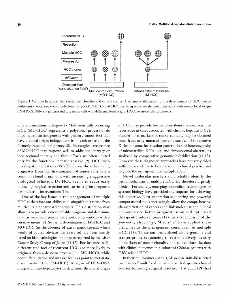
38 Baffy. Multifocal hepatocellular carcinoma
© AME Publishing Company. All rights reserved. www.amegroups.com
different mechanisms (Figure 1). Multicentrically occurring HCC (MO-HCC) represents a polyclonal process of de novo hepatocarcinogenesis with primary tumor foci that have a clonal origin independent from each other and the formerly resected malignancy (8). Postsurgical recurrence of MO-HCC may respond well to additional surgery or loco-regional therapy and these efforts are often limited only by the functional hepatic reserve (9). HCC with intrahepatic metastases (IM-HCC), on the other hand, originates from the dissemination of tumor cells with a common clonal origin and with increasingly aggressive biological behavior. IM-HCC seems to recur early following surgical resection and carries a grim prognosis despite heroic interventions (10).
One of the key issues in the management of multiple HCC is therefore our ability to distinguish metastatic from multicentric hepatocarcinogenesis. This distinction may allow us to provide a more reliable prognosis and determine how far we should pursue therapeutic interventions with a curative intent (9). So far, differentiation of IM-HCC and MO-HCC (in the absence of extrahepatic spread, which would of course obviate this exercise) has been mostly based on histopathological findings as reported by the Liver Cancer Study Group of Japan (11,12). For instance, well-differentiated foci of recurrent HCC are more likely to originate from a de novo process (i.e., MO-HCC), while poor differentiation and invasive features point to metastatic dissemination (i.e., IM-HCC). Analysis of HBV-DNA integration into hepatocytes to determine the clonal origin
of HCC may provide further clues about the mechanism of recurrence in cases associated with chronic hepatitis B (12). Furthermore, markers of tumor clonality may be obtained from frequently mutated proteins such as p53, selective X-chromosome inactivation pattern, loss of heterozygosity of microsatellite DNA loci, and chromosomal aberrations analyzed by comparative genomic hybridization (11,13). However, these diagnostic approaches have not yet yielded sufficient knowledge to become routine clinical practice and to guide the management of multiple HCC.
Novel molecular markers that reliably identify the pathomechanism of multiple HCC are therefore urgently needed. Fortunately, emerging biomedical technologies of systems biology have provided the impetus for achieving this objective. Next-generation sequencing and powerful computational tools increasingly allow the comprehensive characterization of cancers and link molecular and clinical phenotypes to better prognostication and optimized therapeutic interventions (14). In a recent issue of the Journal of Hepatology, Miao et al. have applied these principles to the management conundrum of multiple HCC (15). These authors utilized whole-genome and transcriptome sequencing to retrospectively identify biomarkers of tumor clonality and to associate the data with clinical outcomes in a cohort of Chinese patients with HBV-related HCC.
In their multi-omics analysis, Miao et al. initially selected two cases of multifocal hepatoma with disparate clinical courses following surgical resection. Patient I (PI) had
Figure 1 Multiple hepatocellular carcinoma: clonality and clinical course. A schematic illustration of the development of HCC due to multicentric occurrence with polyclonal origin (MO-HCC) and HCC resulting from intrahepatic metastases with monoclonal origin (IM-HCC). Different patterns indicate tumor cells with different clonal origin. HCC, hepatocellular carcinoma.
Recurrent HCC
HCC clones
Diseased liver(‘cancerization field’)
Multicentric occurrence(MO-HCC)
Intrahepatic metastasis(IM-HCC)
Multiple HCC
Resection
Progression
Initiation

39Precision Medicine in Hepatobiliary Cancer
© AME Publishing Company. All rights reserved. www.amegroups.com
multiple foci of poorly differentiated HCC in a cirrhotic liver and died 3 months after surgery. By contrast, patient II (PII) had multifocal, well-differentiated HCC in a non-cirrhotic liver and remained symptom-free at 2 years after surgery. Whole-genome sequencing revealed different patterns of HBV integration in these patients as all tumors in PI had a single HBV integration site, while two distinct tumor-initiating clones were identified in PII. Differences in clonality of PI and PII tissues were further confirmed by analysis of somatic mutation profiles and genomic structural variations that were validated by PCR and Sanger sequencing. Phylogenetic trees of PI tumors constructed from these data were similar to each other but far removed from the germ line, while the genetic variations found in PII tumors were best explained by the synchronous development of distinct clones. From these findings, Miao et al. concluded that PI and PII were prototype cases of IM-HCC and MO-HCC, respectively (15).
Miao et al. subsequently performed transcriptome analysis to characterize protein-coding gene expression in PI and PII tumors. As expected, similarities between the mRNA sets were more pronounced in PI than in PII tissue samples, further suggesting that multiple foci of HCC in PI were indeed the result of a monoclonal (i.e., metastatic) process. Subsequent functional enrichment mapping of differentially expressed genes of PI and PII indicated important topological differences in various gene function modules. According to this network analysis, essential changes in all PI tumors were comparable with upregulation of genes involved in cytoskeletal remodeling and extracellular matrix organization in PI satellite tumors, consistent with a metastatic signature. On the other hand, PII tumors displayed two distinct transcriptome patterns with some overlaps for tumorigenesis hallmarks such as negative regulation of apoptosis (15).
As a next step, Miao et al. utilized their transcriptome data to find genes with markedly different gene expression in PI and PII tumor tissues. Expression patterns of six genes with the most pronounced alterations (HAL, SFN, KIF15, TTK, BUB1, and MCM4) were analyzed against various clinico-pathological characteristics and postsurgical HCC recurrence in a cohort of 174 patients with HBV-related single or multifocal HCC. Interestingly, expression of TTK was found to have a strong reverse association with favorable postoperative prognosis in this cohort (15). The TTK gene encodes a dual-specificity kinase (also known as monopolar spindle 1 or Mps1 kinase) that phosphorylates serine, threonine, and tyrosine residues with a critical role in
the regulation of cell division in normal and cancer cells (16). Mps1/TTK is required for normal chromosomal segregation and may serve a particular role in cancer by allowing sustained cell proliferation in the presence of aneuploidy (17). Consequently, Mps1/TTK is more than just a biomarker and has become a promising target in cancer therapy (18).
Miao et al. respectively linked the highly dissimilar clinical outcomes of HBV-related HCC in PI and PII to monoclonal and polyclonal cancer growth (15). Genomic integration of HBV increases the risk of hepatocarcinogenesis regardless of the presence of cirrhosis, and the authors reasonably assumed that HCC nodules in the non-cirrhotic liver of PII resulted from multiple occurrence, contrasted with a metastatic process in PI. However, multiple regenerative nodules in the remodeling cirrhotic liver may also serve as simultaneous sites of tumor initiation (19). This notion was corroborated by combined clinicopathological and genetic evaluation that distinguished IM-HCC vs. MO-HCC in 160 Chinese patients with HBV-related HCC and repeated surgical resection (9). Even though cirrhosis was more severe in the group of MO-HCC, patients in this earlier study had a significantly better disease-free survival, indicating that IM-HCC is not necessarily linked to the severity of underlying liver disease. Indeed, the combined effects of HBV integration and cirrhosis provide an intriguing example for the concept of ‘field cancerization’ in which multiple independent tumors may rise within a specific environment (20).
Could these observations provide new strategies with regards to the management of multiple HCC? If reliable biomarkers become available for the distinction of MO-HCC vs. IM-HCC, they may affect treatment algorithms for intermediate stage HCC with having multiple tumor foci in the liver. At the same time, there are issues that may limit enthusiasm for the surgical management of recurrent HCC. Repeated and generous resections may promote pro-oncogenic mechanisms associated with increased rates of liver regeneration whether or not cirrhosis has been established, accelerating further recurrence of HCC (12). Also, HCC may recur as a combination of multi-occurrence and intrahepatic metastasis, possibly calling for even more sophisticated biomarkers to guide clinical management.
Can we extrapolate the findings of Miao et al. to non-HBV-related HCC? There is evidence that the incidence of MO-HCC is significantly higher in HCV-positive patients compared to those with chronic HBV infection (9). These observations may reflect an accelerated rate of tumor

40 Baffy. Multifocal hepatocellular carcinoma
© AME Publishing Company. All rights reserved. www.amegroups.com
progression due to concurrent oncogenic processes and a higher proportion of IM-HCC in HBV-associated cirrhosis. The findings that increased TTK mRNA levels predict a more aggressive course of HCC in patients with HBV-associated liver disease indicate that TTK may become a new biomarker for the presence of IM-HCC with a more aggressive clinical course, which includes a shorter interval of postoperative HCC recurrence. Importantly, Miao et al. also found a highly significant association between TTK gene expression and HBsAg-positivity (15). Future studies will determine whether upregulation of the mitotic checkpoint regulator TTK is a useful parameter in predicting the biological behavior of non-HBV-related HCC.
The quest to find biomarkers that reliably identify MO-HCC vs. IM-HCC is also about defining the biological characteristics of tumor initiation vs. progression. Clinical experience indicates that making this distinction has tremendous implications for the affected individuals. Miao et al. have taken a significant step towards applying the methods of multi-omics analysis and network medicine to track changes in the genome and transcriptome of liver cells linked to these two different aspects of tumorigenesis (15). Extrapolation of functional gene enrichment analysis has yielded a promising biomarker (TTK) to assist prognostication and guide the management of multifocal HCC. Our hope is that increasingly applying the tools of systems biology to the problem of HCC clonality will bring precision and efficacy beyond the current state of the art.
Acknowledgements
None.
Footnote
Conflicts of Interest: The author has no conflicts of interest to declare.
References
1. Jemal A, Bray F, Center MM, et al. Global cancer statistics. CA Cancer J Clin 2011;61:69-90.
2. El-Serag HB. Hepatocellular carcinoma. N Engl J Med 2011;365:1118-27.
3. Forner A, Reig ME, de Lope CR, et al. Current strategy for staging and treatment: the BCLC update and future prospects. Semin Liver Dis 2010;30:61-74.
4. Pons F, Varela M, Llovet JM. Staging systems in
hepatocellular carcinoma. HPB (Oxford) 2005;7:35-41.5. Morise Z, Kawabe N, Tomishige H, et al. Recent advances
in the surgical treatment of hepatocellular carcinoma. World J Gastroenterol 2014;20:14381-92.
6. Jelic S, Sotiropoulos GC; ESMO Guidelines Working Group. Hepatocellular carcinoma: ESMO Clinical Practice Guidelines for diagnosis, treatment and follow-up. Ann Oncol 2010;21 Suppl 5:v59-64.
7. Fuks D, Dokmak S, Paradis V, et al. Benefit of initial resection of hepatocellular carcinoma followed by transplantation in case of recurrence: an intention-to-treat analysis. Hepatology 2012;55:132-40.
8. Forner A, Gilabert M, Bruix J, et al. Treatment of intermediate-stage hepatocellular carcinoma. Nat Rev Clin Oncol 2014;11:525-35.
9. Li Q, Wang J, Juzi JT, et al. Clonality analysis for multicentric origin and intrahepatic metastasis in recurrent and primary hepatocellular carcinoma. J Gastrointest Surg 2008;12:1540-7.
10. Bruix J, Sherman M; American Association for the Study of Liver Diseases. Management of hepatocellular carcinoma: an update. Hepatology 2011;53:1020-2.
11. Morimoto O, Nagano H, Sakon M, et al. Diagnosis of intrahepatic metastasis and multicentric carcinogenesis by microsatellite loss of heterozygosity in patients with multiple and recurrent hepatocellular carcinomas. J Hepatol 2003;39:215-21.
12. Yamamoto T, Kajino K, Kudo M, et al. Determination of the clonal origin of multiple human hepatocellular carcinomas by cloning and polymerase chain reaction of the integrated hepatitis B virus DNA. Hepatology 1999;29:1446-52.
13. Kawai S, Imazeki F, Yokosuka O, et al. Clonality in hepatocellular carcinoma: analysis of methylation pattern of polymorphic X-chromosome-linked phosphoglycerate kinase gene in females. Hepatology 1995;22:112-7.
14. Fujimoto A, Totoki Y, Abe T, et al. Whole-genome sequencing of liver cancers identifies etiological influences on mutation patterns and recurrent mutations in chromatin regulators. Nat Genet 2012;44:760-4.
15. Miao R, Luo H, Zhou H, et al. Identification of prognostic biomarkers in hepatitis B virus-related hepatocellular carcinoma and stratification by integrative multi-omics analysis. J Hepatol 2014;61:840-9.
16. Janssen A, van der Burg M, Szuhai K, et al. Chromosome segregation errors as a cause of DNA damage and structural chromosome aberrations. Science 2011;333:1895-8.

41Precision Medicine in Hepatobiliary Cancer
© AME Publishing Company. All rights reserved. www.amegroups.com
17. Bursavich MG, Dastrup D, Shenderovich M, et al. Novel Mps1 kinase inhibitors: from purine to pyrrolopyrimidine and quinazoline leads. Bioorg Med Chem Lett 2013;23:6829-33.
18. Aarts EO, Dogan K, Koehestanie P, et al. Long-term results after laparoscopic adjustable gastric banding: a mean fourteen year follow-up study. Surg Obes Relat Dis 2014;10:633-40.
19. Piao Z, Park YN, Kim H, et al. Clonality of large regenerative nodules in liver cirrhosis. Liver 1997;17:251-6.
20. Slaughter DP, Southwick HW, Smejkal W. Field cancerization in oral stratified squamous epithelium; clinical implications of multicentric origin. Cancer 1953;6:963-8.
Cite this article as: Baffy G. Decoding mult i focal h e p a t o c e l l u l a r c a r c i n o m a : a n o p p o r t u n e p u r s u i t . HepatoBiliary Surg Nutr 2015;4(3):206-210. doi: 10.3978/j.issn.2304-3881.2014.12.05

© AME Publishing Company. All rights reserved. www.amegroups.com
A little over a decade ago, we reviewed the potential for new techniques in the basic science of genetics to influence clinical care (1). We found the task faced by scientists to be daunting and the prospect of success seemed distant. Since then techniques advanced from gene mapping to include transcription, protein and other characteristics of cell function encompassed in the neologism “multi-omics”. Information available is increasing exponentially. If it was hard to pick a signal out of the noise 10 years ago, it is many times harder now. Using a deceptively simple experimental design, Miao and colleagues at the Peking Union Medical College, have cracked the nut (2).
By comparing two patients with hepatitis B virus (HBV) and multifocal hepatocellular carcinoma (HCC), they pulled out seven candidate genes that may be related to the capability of the tumour to metastasize. Examining the candidates in a cohort of patients with HCC associated
with hepatitis C virus (HCV). They reduced the group to six. By looking at progression of HCC, they isolated TTK, a protein kinase which disrupts the interaction of the tumour suppressor p53 with the oncogene MDM2. TTK-high tumours recurred 3 times faster than TTK-low tumours.
A variety of risk factors have been associated with HCC (3). The prognosis after the proper treatment in HCC (either surgical or local treatment) depends on intrinsic factors of the tumour (4). The current guidelines for the diagnosis of HCC recommend liver biopsy for hepatic nodules with atypical features of imaging (5). For HCC, there has been increasing demand for classifications to predict the biological behaviour and prognosis of the cancer. Most of these classifications are morphological (6).
It has long been the goal of research to refine histology by examining cellular pathways, particularly those related to the
Liver Cancer
Wading through the noise of “multi-omics” to identify prognostic biomarkers in hepatocellular carcinoma
Karen Pineda-Solis, Vivian McAlister
Department of Surgery, University of Western Ontario, London, ON, Canada
Correspondence to: Vivian McAlister, Co-editor of the Canadian Journal of Surgery. Department of Surgery, University of Western Ontario, 339
Windermere Road, London, ON, Canada. Email: [email protected].
Comment on: Miao R, Luo H, Zhou H, et al. Identification of prognostic biomarkers in hepatitis B virus-related hepatocellular carcinoma and stratification by integrative multi-omics analysis. J Hepatol 2014;61:840-9.
Abstract: Multi-omics, the molecular analysis of genes, transcriptional RNA and proteins, allows researchers document the mechanism of action of a target gene. However multi-omics may result in an avalanche of information when used to screen a population. It is very difficult to discern a pattern or signal related to a disease or its progression. Differential multi-omics exploits our ability to see differences between subjects who are similar in all respects except for the outcome being tested. Twin studies are an example of this. Miao and colleagues compared two patients who had diverse outcomes following treatment of multi-focal hepatocellular carcinoma (HCC) to identify seven candidates as the responsible genes. In a larger cohort of patients with HCC they narrowed the field down to a single target down. By looking at progression of HCC, they isolated TTK, a protein kinase which disrupts the interaction of the tumour suppressor p53 with the oncogene MDM2. TTK-high tumours recurred 3 times faster than TTK-low tumours.
Keywords: Hepatitis C virus (HCV); hepatitis B virus (HBV); hepatocellular carcinoma (HCC); genomics;
multi-omics
Submitted Mar 16, 2015. Accepted for publication Mar 23, 2015.
doi: 10.3978/j.issn.2304-3881.2015.04.05
View this article at: http://dx.doi.org/10.3978/j.issn.2304-3881.2015.04.05

43Precision Medicine in Hepatobiliary Cancer
© AME Publishing Company. All rights reserved. www.amegroups.com
cell cycle. In order to separate cancers with high malignant potential from those less likely to metastasize or recur.
In 2004, Lee and colleagues used gene sequencing to identify two gene predictors of a likelihood of HCC recurrence and suggested that JAK/STAT and NOTCH1 pathway inhibitors may have a role in preventing this outcome (7). On the other hand in 2007, Boyault and colleagues found a diverse array of signals when they perfomed global transcriptome analyses on 57 HCC and attempted validation in another cohort of 63 patients (8). This has not stopped others from developing strategems for “genomics-driven oncology” (9,10).
Miao and colleagues need to test their hypothesis in a second cohort of patients with HCC in order to determine the magnitude of its effect. The mechanism is probably shared with other cancers that may be tested as well. Fruitful areas of investigation will be to understand the effect on clinically used tyrosine kinase inhibitors of TTK function. Specific TTK inhibition is a therapeutic option but its effect on hepatocyte function will have to be understood. The excellent paper by the Peking Union Medical College team, which reads like an exciting detective story, may well lead to a happy ending, progress in treating a difficult cancer that affects millions of patients worldwide.
Acknowledgements
None.
Footnote
Conflicts of Interest: The authors have no conflicts of interest to declare.
References
1. McAlister V. Pharmacogenomics in liver transplantation. Liver Transpl 2003;9:1114-5.
2. Miao R, Luo H, Zhou H, et al. Identification of prognostic biomarkers in hepatitis B virus-related hepatocellular carcinoma and stratification by integrative multi-omics analysis. J Hepatol 2014;61:840-9.
3. Forner A, Llovet JM, Bruix J. Hepatocellular carcinoma. Lancet 2012;379:1245-55.
4. Morimoto O, Nagano H, Sakon M, et al. Diagnosis of intrahepatic metastasis and multicentric carcinogenesis by microsatellite loss of heterozygosity in patients with multiple and recurrent hepatocellular carcinomas. J Hepatol 2003;39:215-21.
5. European Association For The Study Of The Liver; European Organisation For Research And Treatment Of Cancer. EASL-EORTC clinical practice guidelines: management of hepatocellular carcinoma. J Hepatol 2012;56:908-43.
6. Parfitt JR, Marotta P, Alghamdi M, et al. Recurrent hepatocellular carcinoma after transplantation: use of a pathological score on explanted livers to predict recurrence. Liver Transpl 2007;13:543-51.
7. Lee JS, Chu IS, Heo J, et al. Classification and prediction of survival in hepatocellular carcinoma by gene expression profiling. Hepatology 2004;40:667-76.
8. Boyault S, Rickman DS, de Reyniès A, et al. Transcriptome classification of HCC is related to gene alterations and to new therapeutic targets. Hepatology 2007;45:42-52.
9. Garraway LA. Genomics-driven oncology: framework for an emerging paradigm. J Clin Oncol 2013;31:1806-14.
10. Finn RS. Emerging targeted strategies in advanced hepatocellular carcinoma. Semin Liver Dis 2013;33:S11-9.
Cite this art ic le as : P ineda-Sol i s K, McAl i s ter V. Wading through the noise of “multi-omics” to identify prognost ic biomarkers in hepatocel lular carcinoma. HepatoBiliary Surg Nutr 2015;4(4):293-294. doi: 10.3978/j.issn.2304-3881.2015.04.05

© AME Publishing Company. All rights reserved. www.amegroups.com
Introduction
Hepatocellular carcinoma (HCC) is a frequent and deadly human disease (1). Several lines of evidence indicate that the gradual accumulation of genomic alterations, leading to progressive deregulation of different signaling pathways, induces the progressive evolution of initiated liver cells to dysplastic nodules and malignant lesions (1). Molecular events leading to cell cycle deregulation in HCC include up-regulation of RAS/ERK, PI3K/AKT, IKK/NF-kB, WNT, TGF-β, NOTCH, HEDGEHOG, and HIPPO signaling pathways, and genes involved in DNA repair process (2). Better understanding of the molecular mechanisms underlying hepatocarcinogenesis may hasten the identification of novel molecular HCC progression markers and development of new diagnostic and therapeutic strategies.
Heparan-sulfate glycosaminoglycans (HSGAGs)
The HSGAGs are linear polysaccharides constituted by 50–200 disaccharide repeats, with regions of glucuronic acid-N-acetylglucosamine and regions of 2-O-sulfated uronic acid/N-glucosamine sulfated at 3-O and 6-O positions interspaced by transition areas in which both sulfoglucosamine and N-acetylglucosamine are present. The N-position of glucosamine can be sulfated, acetylated, or unmodified (3). The polysaccharides, covalently attached to a polypeptide core, form heparan sulfate proteoglycans (HSPGs) located at the cell surface (Figure 1A) and also present in the extracellular matrix.
HSGAGs bind and interact with chemokines, enzymes, and growth factors involved in tumor development. The latter include fibroblast growth factors (FGF1, FGF2), vascular endothelial growth factor (VEGF), hepatocyte growth factor (HGF), transforming growth factor-β (TGF-β), and platelet-derived growth factor (PDGF) (4). Predominantly, extracellular proteins such as FGFs bind to a tetra- or hexasaccharide motif within an HSGAG chain (5). Long oligosaccharide sequences can bridge protein-protein complexes, thus favoring the homo-oligomerization and/or bridging a ligand to its receptor (Figure 1B). Thus, HSGAG chains of proteoglycans function as binding sites for signaling molecules. The sequence and conformational plasticity of the HSGAG polymer determine the binding specificity with consequent regulation of different biological processes.
HSGAGs may play important roles in cancer initiation and progression by modulating tumor cell growth, invasiveness, and metastatic potential (6). Different observations indicate that changes in expression level and oligosaccharide sequence of cell-surface HSPGs might contribute to cell transformation (7-9).
Heparin-degrading sulfatases
The heparin sulfate 6-O-endosulfatases 1 and 2, designated as sulfatase 1 (SULF1) and sulfatase 2 (SULF2), respectively, hydrolyze the sulfate ester bonds of HSGAGs. SULF1 and SULF2 show the same in vitro specificity to trisulfated disaccharides, but the two sulfatases display structural and
Liver Cancer
Sulfatase 1: a new Jekyll and Hyde in hepatocellular carcinoma?
Rosa M. Pascale, Diego F. Calvisi, Francesco Feo
Division of Experimental Pathology and Oncology, Department of Clinical and Experimental Medicine, University of Sassari, Sassari, Italy
Correspondence to: Francesco Feo. Division of Experimental Pathology and Oncology, Department of Clinical and Experimental Medicine, University
of Sassari, Sassari, Italy. Email: [email protected].
Provenance: This is a Guest Commentary commissioned by Guest Editor Haitao Zhao, MD, PhD (Associate Professor, Department of Liver Surgery,
Peking Union Medical College Hospital, Chinese Academy of Medical Sciences and Peking Union Medical College, Beijing, China).
Comment on: Dhanasekaran R, Nakamura I, Hu C, et al. Activation of the transforming growth factor-β/SMAD transcriptional pathway underlies a
novel tumor-promoting role of sulfatase 1 in hepatocellular carcinoma. Hepatology 2015;61:1269-83.
Received: 12 April 2016; Accepted: 18 April 2016; Published: 18 May 2016.
doi: 10.21037/tgh.2016.05.04
View this article at: http://dx.doi.org/10.21037/tgh.2016.05.04

45Precision Medicine in Hepatobiliary Cancer
© AME Publishing Company. All rights reserved. www.amegroups.com
Figure 1 Structure of HSGAGs and proposed model of SULFs in heparin bindings growth factors. (A) Heparan sulfate proteoglycans on cell surface showing heparan-sulfate glycosaminoglycans (HSGAGs) attached to a protein core. Enlarged detail: 2-O-sulfated uronic acid-N-glucosamine sulfated at the 3-O and 6-O positions. NHX on glucosamine: N position sulfated, acetylated, or unchanged; (B) sulfate residues on HSGAG (blue circles) can favor ligand bridging (i.e., FGF) to its receptor, and signaling pathways activation. HSGAG desulfation by sulfatases can impede the link of ligands to their receptors.
functional differences in mice and humans (10). Lai and coworkers (11) reported that SULF1 desulfates HSPGs on cells surface and inhibits HCC tumor cell growth in vitro and in nude mice, partially through effects on gene expression mediated through histone H4 acetylation. SULF1 is downregulated in most HCC cell lines and approximately 30% of primary HCCs. SULF1 transfection in HCC cells reduces proliferation rate by suppressing heparin-binding to growth factor signaling. In contrast, SULF2 promotes hepatocarcinogenesis in nude mice and
its expression is associated with more rapid recurrence and shorter survival of HCC patients, after surgical resection (11). Furthermore, Shire and coworkers (12) showed SULF1 mRNA down-regulation in 9/11 HCC cell lines and in only 6/10 primary tumors. They found that SULF1 promoter acquires a silenced chromatin state in low SULF1-expressing cells, through an increase in d i / t r imethyl-K9H3 and t r imethyl-K27H3 and a concomitant loss of activating acetyl K9, K14H3. Restoration of SULF1 mRNA expression by 5-Aza-dC sensitized HCC cells to drug-induced apoptosis.
The oncosuppressor effect of SULF1 was confirmed by the recent observation that microRNA-21, a suppressor of PTEN and hSulf-1 expression, promotes HCC progression through the AKT/ERK pathways (13). It was hypothesized that the inhibition of histone deacetylase by SULF1 induces a rise in acetylated histone H4, which leads to inhibition of RAS/ERK and PI3K/AKT signaling (13). A recent contribution (14) provides a hypothetical mechanism whereby cancer cells could evade SULF1 suppressor action. The removal by sulfatases of the sulfate moiety from 6-O of heparan sulfate on HSPGs, should result in decreased FGF binding sites on HSPG that should disfavor FGF bridging to its receptor (Figure 1B). However, HIF-1α stabilization, under low oxygen conditions prevailing in solid tumors, shuts down the transcription of sulfatases, which may results in sulfation of 6-O of heparan sulfate on HSPGs. This would favor FGF signaling, cell migration, and invasion (14).
In apparent contradiction with above reports, gene expression analysis of human HCCs showed that high SULF1 overexpression is associated with poor survival, suggesting that SULF1 is oncogenic in most HCC in vivo (15). This conclusion is supported by Dhanasekaran and coworkers, who in a recent study (16) provided convincing data in support of the oncogenic role of SULF1 in hepatocarcinogenesis and unraveled some of the molecular mechanisms involved. These authors used a transgenic mouse model overexpressing SULF1 (Sulf1-Tg) to evaluate the effects of SULF1 on the diethylnitrosamine (DENA) model of hepatocarcinogenesis. They showed a higher incidence of large and multifocal HCCs in DENA-treated Sulf1-Tg mice, compared to wild-type (WT) mice. They also found that lung metastases were present in 75% of Sulf1-Tg mice but not in WT mice. These in vivo experiments clearly indicated that SULF1 overexpression enhances liver tumor progression and strongly support a tumor promoter role for SULF1.
In order to identify the molecular players responsible
A
B

46 Pascale et al. Sulfatase 1 in liver cancer
© AME Publishing Company. All rights reserved. www.amegroups.com
for the oncogenic role of SULF1, Dhanasekaran and coworkers (16) evaluated by transcriptome analysis of non-DENA treated liver tissues the pathways and biological processes activated in Sulf1-Tg compared to WT mice. They observed the up-regulation of biological processes involving cytoskeletal remodeling, cell adhesion, and muscle development in Sulf1-Tg mice. Noticeably, they also found the preferential activation of the epithelial mesenchymal transition (EMT) process in Sulf1-Tg mice. EMT is a process implicated in tumor progression and development of metastases (17) and its activation is in line with the presence of larger tumors and lung metastases in Sulf1-Tg mice.
TGF-β is known to be implicated in EMT of cancer cells (18,19). The analysis by Dhanasekaran and coworkers (16)
of genes involved in TGF-β signaling showed higher expression of Smad2 and Smad6 in Sulf1-Tg than in WT mice. Also, an increase in phosphorylation of Smad2/3 in peritumoral liver tissues and, at a higher extent, in tumors of Sulf1-Tg mice than WT mice occurred. These changes were associated with a lower expression of the epithelial marker E-cadherin, and an increase in the mesenchymal markers N-cadherin and vimentin in tumors of Sulf1-Tg mice when compared to HCC of WT mice. Further, the authors provided the immunohistochemical evidence of cytoplasmic expression of the mesenchymal proteins in tumor cells of Sulf1-Tg mice suggesting the acquisition of the mesenchymal traits by the epithelial cells (Figure 2).
The results in mice were confirmed in a series of experiments with various human HCC cell lines. In particular, it was observed that the overexpression of SULF1 increased the phosphorylation of both SMAD2 and SMAD3, whereas the suppression of SULF1 expression led to the opposite effects. Moreover, it was clearly shown a link between SULF1 up-regulation and EMT: SULF1-transfected cells, treated with TGF-β1, exhibited a decrease in the tight junction protein Zona occludens protein 1 (Zo-1) and in E-cadherin, as well as an increase in the mesenchymal markers N-cadherin, vimentin, and α-smooth muscle actin (αSMA). Opposite changes occurred when SULF1 expression was suppressed.
Some elegant experiments were devoted to the analysis of the role of SULF1 catalytic activity. For this aim, a SULF1 mutant with loss of catalytic activity was created. It was thus demonstrated that the stimulation of cell migration and invasiveness and SMAD2/3 phosphorylation, present in cell transfected with SULF1 and treated with TGF-β1, was lost in cells transfected with the mutant SULF1 devoid of sulfatase activity.
The classification of HCC patients into two prognostic clusters, according the microarray expression profile, showed that the majority of patients with high SULF1 expression (76%) belonged to the poor prognosis cluster and gene expression correlation analysis confirmed the association between SULF1, TGF-β activation, EMT and five EMT driver genes (vimentin, SNAI1, COL1A2, TGF-β1, SPARC) in human HCC. The association between high SULF1 and high phospho-SMAD2/3 expression, decreased expression of E-cadherin and increased that of vimentin and αSMA was confirmed in the tumor specimens.
Importantly, the authors presented evidence that Hep3B and PLC/PRF5PRF/5 cell lines, used in the SULF1 experiments, do not express SULF2. Thus, it is unlike that
Figure 2 A possible mechanism responsible for the oncogenic role of SULF1. TGF-β1, bound to the sulfate moiety from 6-O of heparan sulfate on HSPGs, is released following the desulfation of 6-O of heparan sulfate on HSPGs by SULF1. This allows TGF-β1 binding to the receptor system. After binding and phosphorylation, the receptor activates by phosphorylation the SMAD2 and SMAD3 effectors. This is followed by the formation of heteromeric complexes of SMAD2 and SMAD3 with SMAD4, which then translocate to the nucleus and activate specific DNA sequences involved in EMT.

47Precision Medicine in Hepatobiliary Cancer
© AME Publishing Company. All rights reserved. www.amegroups.com
the observed results are a consequence of overexpression of the SULF2 gene, which is known to possess an oncogenic activity.
Conclusions
According to the results of Dhanasekaran and coworkers, SULF1 is a potential biomarker of tumor progression and thus a novel target for drug development. At present, there are no sufficient elements to understand the conflictual results about the role of SULF1 in tumorigenesis. TGF-β is known to behave as an oncogene or an oncosuppressor gene in cancer (20). The effect and function of genes can be opposite and adaptable in cells with different genomes or in different contexts and the response to the same protein could be cellular genetic/context-dependent (20). However, a link between SULF1 overexpression and the oncosuppressor role of TGF-β has not yet been demonstrated. Further research is needed to confirm a possible antagonistic role of SULF1 in liver carcinogenesis and solve this dilemma.
Acknowledgements
None.
Footnote
Conflicts of Interest: The authors have no conflicts of interest to declare.
References
1. Frau M, Biasi F, Feo F, et al. Prognostic markers and putative therapeutic targets for hepatocellular carcinoma. Mol Aspects Med 2010;31:179-93.
2. Calvisi DF, Frau M, Tomasi ML, et al. Deregulation of signalling pathways in prognostic subtypes of hepatocellular carcinoma: novel insights from interspecies comparison. Biochim Biophys Acta 2012;1826:215-37.
3. Kreuger J, Spillmann D, Li JP, et al. Interactions between heparan sulfate and proteins: the concept of specificity. J Cell Biol 2006;174:323-7.
4. Forsten-Williams K, Chu CL, Fannon M, et al. Control of growth factor networks by heparan sulfate proteoglycans. Ann Biomed Eng 2008;36:2134-48.
5. Morimoto-Tomita M, Uchimura K, Werb Z, et al. Cloning and characterization of two extracellular heparin-degrading endosulfatases in mice and humans. J Biol Chem
2002;277:49175-85.6. Chen Z, Fan JQ, Li J, et al. Promoter hypermethylation
correlates with the Hsulf-1 silencing in human breast and gastric cancer. Int J Cancer 2009;124:739-44.
7. Ai X, Do AT, Lozynska O, et al. QSulf1 remodels the 6-O sulfation states of cell surface heparan sulfate proteoglycans to promote Wnt signaling. J Cell Biol 2003;162:341-51.
8. Delehedde M, Lyon M, Gallagher JT, et al. Fibroblast growth factor-2 binds to small heparin-derived oligosaccharides and stimulates a sustained phosphorylation of p42/44 mitogen-activated protein kinase and proliferation of rat mammary fibroblasts. Biochem J 2002;366:235-44.
9. Wang S, Ai X, Freeman SD, et al. QSulf1, a heparan sulfate 6-O-endosulfatase, inhibits fibroblast growth factor signaling in mesoderm induction and angiogenesis. Proc Natl Acad Sci U S A 2004;101:4833-8.
10. Wei G, Bai X, Gabb MM, et al. Location of the glucuronosyltransferase domain in the heparan sulfate copolymerase EXT1 by analysis of Chinese hamster ovary cell mutants. J Biol Chem 2000;275:27733-40.
11. Lai JP, Thompson JR, Sandhu DS, et al. Heparin-degrading sulfatases in hepatocellular carcinoma: roles in pathogenesis and therapy targets. Future Oncol 2008;4:803-14.
12. Shire A, Lomberk G, Lai JP, et al. Restoration of epigenetically silenced SULF1 expression by 5-aza-2-deoxycytidine sensitizes hepatocellular carcinoma cells to chemotherapy-induced apoptosis. Med Epigenet 2015;3:1-18.
13. Bao L, Yan Y, Xu C, et al. MicroRNA-21 suppresses PTEN and hSulf-1 expression and promotes hepatocellular carcinoma progression through AKT/ERK pathways. Cancer Lett 2013;337:226-36.
14. Khurana A, Beleford D, He X, et al. Role of heparan sulfatases in ovarian and breast cancer. Am J Cancer Res 2013;3:34-45.
15. Yang JD, Sun Z, Hu C, et al. Sulfatase 1 and sulfatase 2 in hepatocellular carcinoma: associated signaling pathways, tumor phenotypes, and survival. Genes Chromosomes Cancer 2011;50:122-35.
16. Dhanasekaran R, Nakamura I, Hu C, et al. Activation of the transforming growth factor-β/SMAD transcriptional pathway underlies a novel tumor-promoting role of sulfatase 1 in hepatocellular carcinoma. Hepatology 2015;61:1269-83.
17. Li Y, Ma J, Qian X, et al. Regulation of EMT by Notch signaling pathway in tumor progression. Curr Cancer

48 Pascale et al. Sulfatase 1 in liver cancer
© AME Publishing Company. All rights reserved. www.amegroups.com
Drug Targets 2013;13:957-62.18. Morrison CD, Parvani JG, Schiemann WP. The relevance
of the TGF-β Paradox to EMT-MET programs. Cancer Lett 2013;341:30-40.
19. Lamouille S, Xu J, Derynck R. Molecular mechanisms of
epithelial-mesenchymal transition. Nat Rev Mol Cell Biol 2014;15:178-96.
20. Stepanenko AA, Vassetzky YS, Kavsan VM. Antagonistic functional duality of cancer genes. Gene 2013;529:199-207.
doi: 10.21037/tgh.2016.05.04Cite this article as: Pascale RM, Calvisi DF, Feo F. Sulfatase 1: a new Jekyll and Hyde in hepatocellular carcinoma? Transl Gastroenterol Hepatol 2016;1:43.

© AME Publishing Company. All rights reserved. www.amegroups.com
Introduction
Hepatocellular carcinoma (HCC) is a frequent human cancer with 0.25–1 million of newly diagnosed cases each year (1-3). Major risk factors associated with the development of HCC are chronic hepatitis B virus (HBV) and hepatitis C virus (HCV) infections, alcoholic hepatitis, aflatoxin B1 (3-5), and some inherited diseases (6). HCC is a fatal disease, with a life expectancy of about 6 months from the time of diagnosis (6). Early liver lesions could be detected by ultrasonography and efficiently treated by resection or radiofrequency ablation (7). However, only a minority of cases is eligible to these treatment modalities due to the late diagnosis of the disease (2,7,8). In addition, therapies with pharmacological agents or alternative approaches, including percutaneous ethanol injection, trans-arterial chemo-embolization or yttrium-90 microspheres, do not improve significantly the prognosis of patients with advanced disease (2,7,8).
The evaluation of the molecular mechanisms and the identification of prognostic categories of HCC are difficult due to HCC heterogeneity, which results from complex relationships between genetic, etiologic, and environmental risk factors (6). A better understanding of HCC molecular pathogenesis may hasten the identification of new prognostic markers and the development of novel diagnostic and therapeutic strategies against this disease (6,9).
Biological ad clinical behavior of HCC may be largely influenced by both genetic and epigenetic alterations of a number of genes and signaling pathways (6,10). The remodeling of microenvironment (11,12) surrounding HCC may also affect HCC biological behavior, thus influencing patients’ outcome (13). This is an important facet of the complex mechanisms involved in tumor progression. Different proteins of the extracellular matrix (ECM) may affect cell growth, migration, invasion, anoikis and metastasis (13-17) by binding to specific receptors of cancer cells or interfering with the binding of specific cytokines (18).
The epidermal growth factor-like repeats and discoidin I-like domains 3 (EDIL3) protein
EDIL3, also known as endothelial cell locus (DEL-1), is a secreted ECM protein isolated and identified from embryonic mouse lung in 1998 (19). EDIL3, secreted by embryonic endothelial cells and hypertrophic chondrocytes (20), was firstly characterized in vascular morphogenesis (21).
EDIL3 is a glycoprotein composed of five domains: three epidermal growth factor (EGF)-like repeats (E1, E2, E3), and two discoidin I-like domains (C1, C2). In particular, the second EGF repeat contains an Arg-Gly-Asp (RGD) motif (Figure 1) (19,20). It has been shown that the C-terminus of the C1 domain is essential for the organization of EDIL3 into the ECM and that all the E repeat domains and the
Liver Cancer
Epidermal growth factor-like repeats and discoidin I-like domains 3: a multifaceted oncoprotein at the crossroad of MAPK and TGF-beta pathways in human hepatocellular carcinoma
Diego F. Calvisi, Rosa M. Pascale, Francesco Feo
Department of Clinical and Experimental Medicine, University of Sassari, Sassari, Italy
Correspondence to: Diego Calvisi, MD. Department of Clinical and Experimental Medicine, University of Sassari, via P. Manzella 4, 07100 Sassari,
Italy. Email: [email protected].
Provenance: This is a Guest Editorial commissioned by Guest Editor Haitao Zhao, MD, PhD (Associate Professor, Department of Liver Surgery,
Peking Union Medical College Hospital, Chinese Academy of Medical Sciences and Peking Union Medical College, Beijing, China).
Comment on: Xia H, Chen J, Shi M, et al. EDIL3 is a novel regulator of epithelial-mesenchymal transition controlling early recurrence of
hepatocellular carcinoma. J Hepatol 2015;63:863-73.
Submitted Mar 11, 2016. Accepted for publication Mar 17, 2016.doi: 10.21037/tcr.2016.03.09
View this article at: http://dx.doi.org/10.21037/tcr.2016.03.09

50 Calvisi et al. Role of EDIL3 in hepatocellular carcinoma
© AME Publishing Company. All rights reserved. www.amegroups.com
N-terminus of the C1 domain play supportive roles for this organization (20).
At the cellular level, EDIL3 exerts numerous, important roles. Through the interaction of the Arg-Gly-sp tripeptide with the αvβ3 integrin, EDIL3 induces clustering of integrin receptors, endothelial attachment, and migration as well as focal contact and phosphorylation of different molecules involved in cell signaling, including p125FAK and MAP kinase (22). Moreover, EDIL3 plays a pivotal role in inflammatory and immune responses, where leukocyte adhesion to endothelium, crucial for leukocyte recruitment, requires numerous adhesion molecules expressed on leukocytes and endothelial cells. Indeed, EDIL3 acts an anti-adhesive factor that interferes with the integrin LFA-1-dependent leukocyte-endothelial adhesion, thus preventing leukocyte adhesion to the endothelium (23,24).
EDIL3 is also a potent pro-angiogenic factor, as it significantly contributes to vessel wall remodeling and development during angiogenesis (25), mediates endothelial cell attachment and migration (26), and induces mesentery and cerebral angiogenesis in mice (27,28). Both animal experiments and clinical studies have demonstrated that EDIL3 gene therapy is effective in the presence of an ischemic disease (29-31).
EDIL3 is expressed in brain, heart, small intestine and kidney tissues, but not in colon, liver, or lung of human adults (26). In addition, EDIL3 is expressed in primary human tumors, such as lung (32), bladder (33), pancreas (34), liver (35), breast, and colon cancer, and melanomas (26), and in many tumor cell lines (28). Furthermore, EDIL3 levels have been associated with the progression and prognosis of lung cancer (32), bladder cancer (33), and pancreatic ductal adenocarcinoma (35). Interestingly, EDIL3 was recently shown to be a novel biomarker for early breast cancer detection (36).
EDIL3, epithelial-to-mesenchymal transition (EMT), and integrin signaling
EMT is involved in different physiological events, including
blastocyst implantation, generation of the neural crest, normal wound healing (37,38) as well as in pathological events such as pathological wound healing, tissue fibrosis and carcinogenesis (39,40).
EMT consists of the loss of typical epithelial features, such as cell polarity, intercellular junctions, and ability to synthesize basement membranes, associated with the development of a fibroblastic morphology with rearrangement of the actin cytoskeleton and changes in cell surface matrix receptors, such as integrins. As a consequence, cells form filopodia, migrate, and synthesize ECM (39,41). Three types of EMT have been described: (I) type 1, which occurs during earliest stages of development; (II) type 2, occurring in mature epithelial tissues, generally triggered by inflammation or wound-healing responses, which may induce fibrosis; (III) type 3, which is associated with cancer progression (38).
Tumors contain a subpopulation of cells characterized by the loss of epithelial features and the acquisition of the mesenchymal-like migratory phenotype. These cells, known as cancer stem cells (CSCs), are able to self-renew and regenerate the tumor mass. CSCs are crucial to the development of invasive carcinomas and metastasis (38-41). Nonetheless, tumor cells disseminated into target organs may undergo mesenchymal-epithelial transition (MET), which would also favor metastasis formation (42,43).
EMT is regulated by TGF-β through different mechanisms. Nuclear translocation of SMAD complexes, formed in the canonical TGF-β cascade (6), stimulates the expression of different mesenchymal genes, while repressing epithelial gene transcription (Figure 2). Furthermore, TGF-β signaling activates integrin-linked kinase (ILK), which phosphorylates GSK-3β and AKT (serine/threonine protein kinase), with consequent nuclear translocation of β-Catenin and activation of different transcription factors involved in EMT (44). EMT is also induced trough the ERK/MAP kinase, Rho GTPase and the PI3 kinase/AKT pathways following TGF-β receptor activation (Figure 2) (45,46).
TGF-β is synthesized in a complex pathway: precursor forms of TGF-β1 and TGF-β3 are linked to a latency-associated peptide (LAP), containing an RGD motif that may be activated by αvβ1, 3, 5, 6, and 8 integrins and interacts with RGD (Figure 2) (47-50). This is followed by the interaction of mature TGF-β with its receptor and the activation of different signals that, at DNA level, induce the activation of mesenchymal markers (i.e., integrins, N-Cadherin, fibronectin, collagen) and inhibition of
Figure 1 Schematic representation of the EDIL3 glycoprotein showing three epidermal growth factor (EGF)-like repeats (E1, E2, E3) and two discoidin I-like domains (C1, C2). The second EGF repeat contains an Arg-Gly-Asp (RGD) motif.
RGD
E1 E2 E3 C1 C2

51Precision Medicine in Hepatobiliary Cancer
© AME Publishing Company. All rights reserved. www.amegroups.com
epithelial markers (i.e., CDH1, claudins, occludins, desmoplakin), and integrin activation (38).
EDIL3 binding to αvβ3 integrin by the RGD motif (Figure 2) prevents apoptosis of endothelial cells, thus favoring cancer vascularization and potentiating cancer cell proliferation and invasion (51). This effect is mediated specifically through the crosstalk with FAK/ERK and AKT signaling (51). Integrins, as primary receptors involved in cell-matrix adhesion, may strongly influence the ability of cancer cells to survive in specific sites. Interestingly, it has been observed that in some cases integrin receptors can also function in the absence of ligand binding to promote stemness and survival (52). Thus, the interplay between TGF-β and integrin signaling, occurring downstream of initial TGF-β receptor activation, regulates various cellular processes (53), including different signaling pathways that are able to override the tumor suppressing functions of TGF-β (54-56).
EDIL3 and HCC
Mounting evidence supports an important role of EDIL3 in HCC. According to recent data, indeed, EDIL3 activity is crucial for the interaction between HCC cells and endothelial cells (28), and may accelerate tumor growth by stimulating angiogenesis (57). EDIL3 gene is overexpressed in HCC (35) and predicts poor prognosis of HCC patients (13,35,58). Interestingly, recent studies suggest that autocrine EDIL3 may contribute to a receptive microenvironment for the survival of detached HCC cells by promoting anoikis resistance (13). This intriguing finding suggests that activation of integrin signaling pathways by EDIL3 may contribute to HCC cell spreading. Furthermore, the accumulation of tumor-produced EDIL3 in the microenvironment represents an advantage for anchorage-independent growth of tumor cells.
These observations have been confirmed and extended
LTBP
αV
EDIL3RGD
TGF-β
I II
SMADCASCADE
PI3K/AKT
LAPRGD
Mesenchymal markers
Epithelialmarkers
STEMNESS
RAS/MAPK
Rho-GTPases
APOPTOSIS
βαV β
Figure 2 The regulatory circuitry of αvβ integrins. The interaction of ECM protein, EDIL3, and the LAP protein of the TGF-β-immature complex with αvβ integrin is followed by activation of TGF-β, RAS/ERK, PI3K/AKT and Rho/GTPases signaling pathways. This leads to the up-regulation of the mesenchymal markers, the down-regulation of the epithelial markers, and the up-regulation of integrins, with consequent decrease in cell death and acquisition of the molecular and morphologic changes of stemness and EMT. Arrows indicate activation; blunt arrows indicate inhibition. LAP, latency activated peptide; LTBP, latent transforming growth factor β binding protein; MMP, metalloproteinase; EMT, epithelial-to-mesenchymal transition.

52 Calvisi et al. Role of EDIL3 in hepatocellular carcinoma
© AME Publishing Company. All rights reserved. www.amegroups.com
in an interesting publication by Xia and coworkers (59). As a first approach to establish the role of EDIL3 as regulator of EMT in HCC, the authors evaluated the correlation of EDIL3 expression with that of mesenchymal and epithelial markers, using independent published microarray data for liver cancer cell lines. Noticeably, the authors found a positive correlation between EDIL3 levels and the expression of the mesenchymal marker vimentin (VIM), and a negative correlation with the epithelial marker E-cadherin (CDH1). Accordingly, forced EDIL3 expression in Huh7 cells led to the acquisition of a fibroblastic elongated phenotype associated with a fall in the expression of the epithelial marker CDH1 and up-regulation of the mesenchymal marker VIM. The opposite occurred when EDIL3 expression was inhibited by specific siRNA in HLE cells. In the latter case, morphologic changes indicative of MET were found. Further support to the role of EDIL3 as regulator of EMT in HCC was obtained by the evaluation of different phenotypic properties linked to EMT. Indeed, the migration and invasion properties of Huh7 cells, characterized by lower EDIL3 expression and an epithelial phenotype, were significantly lower than that of HLE cells, which exhibit high EDIL3 expression and a mesenchymal phenotype. The modulation of EDIL3 expression strongly influenced HCC cell migration, invasion, and HCC angiogenesis in the same cells, as evaluated by in vitro endothelial recruitment and capillary tube formation assays.
Interestingly, an epigenetic mechanism was found to be responsible for EDIL3 deregulation in HCC. Specifically, the authors identified microRNA (miR)-137 as a critical, negative regulator of EDIL3. In particular, Xia et al. observed the downregulation of miR-137 in HCC samples from patients exhibiting early recurrent disease, when compared to samples from patients with non-recurrent HCC. The decrease in miR-137 expression was correlated with the up-regulation of EDIL3 expression. Subsequent in vitro experiments showed that miR-137 triggers EDIL3 downregulation, inhibits HCC cell invasion, and induces endothelial cell capillary tube formation.
In accordance with previous studies on the relationships between TGF-β an integrin expression (60), TGF-β1 levels were found to be significantly increased in HuH7 and PLC/PRF/5 HCC cells stably transfected with EDIL3. Using the data reported in the Cancer Cell Line Encyclopedia dataset (http://www.broadinstitute.org/ccle), the authors compared two groups of liver cancer cells displaying high and low EDIL3 expression, respectively. This allowed the study of the correlation of differentially
expressed genes with EDIL3 expression levels. Significant correlation was observed for the expression of TGFβ1I1 and TGFβ2, suggesting a regulation of TGF-β signaling through binding to αvβ3 integrin in liver cancer cells. Further analysis showed that pseudopodium-enriched atypical kinase 1 (PEAK1)-associated regulatory signaling interacts with EDIL3 through the SRC family kinases. Importantly, overexpression of EDIL3 not only significantly enhanced the expression of PEAK1, but also induced the phosphorylation of SRC, ERK and SMAD2, suggesting the activation of ERK and TGF-β signaling.
These important observations by Xia et al. confirm and extend to the HCC field previous observations (38) indicating the existence of a regulatory circuitry for EMT (Figure 2). In this circuitry, the ECM protein EDIL3 interacts with αvβ integrin, thus inducing the activation of TGF-β and RAS/ERK cascades. Once activated, the TGF-β and RAS/ERK pathways trigger the up-regulation of mesenchymal marker and integrins, while promoting the down-regulation of epithelial markers. These molecular events are associated with cell death decrease and acquisition of the molecular and morphologic changes of stemness and EMT by cancer cells.
Concluding remarks
A growing body of experimental and clinical observations points to a pivotal role of EDIL3 protein in HCC progression and patient’s prognosis. The study by Xia et al., in particular, indicates that EDIL3 significantly contributes to many traits of HCC cells, namely uncontrolled growth, resistance to apoptosis, migration, invasion, and angiogenesis. At the clinical level, EDIL3 up-regulation results in early tumor recurrence and poor outcome. Intriguingly, it has been demonstrated that EDIL3 lies at the crossroad of numerous oncogenic pathways, including the ERK/MAPK, TGF-β, and integrin signaling cascades. Consequently, EDIL3 suppression might result in the concomitant inhibition of multiple oncogenic stimuli, whose inactivation could be highly deleterious for the survival of HCC cells. Based on these important findings, additional efforts should be devoted to elucidate the function of EDIL3 in liver cancer as well as to develop novel therapeutic approaches aimed at suppressing EDIL3 activity for the treatment of this pernicious disease.
Acknowledgements
None.

53Precision Medicine in Hepatobiliary Cancer
© AME Publishing Company. All rights reserved. www.amegroups.com
Footnote
Conflicts of Interest: The authors have no conflicts of interest to declare.
References
1. Bosch FX, Ribes J, Díaz M, et al. Primary liver cancer: worldwide incidence and trends. Gastroenterology 2004;127:S5-S16.
2. Llovet JM, Bruix J. Molecular targeted therapies in hepatocellular carcinoma. Hepatology 2008;48:1312-27.
3. Feo F, Pascale RM, Simile MM, et al. Genetic alterations in liver carcinogenesis: implications for new preventive and therapeutic strategies. Crit Rev Oncog 2000;11:19-62.
4. Bruix J, Boix L, Sala M, et al. Focus on hepatocellular carcinoma. Cancer Cell 2004;5:215-9.
5. McGlynn KA, London WT. Epidemiology and natural history of hepatocellular carcinoma. Best Pract Res Clin Gastroenterol 2005;19:3-23.
6. Calvisi DF, Frau M, Tomasi ML, et al. Deregulation of signalling pathways in prognostic subtypes of hepatocellular carcinoma: novel insights from interspecies comparison. Biochim Biophys Acta 2012;1826:215-37.
7. Sherman M. Modern approach to hepatocellular carcinoma. Curr Gastroenterol Rep 2011;13:49-55.
8. Calvisi DF, Pascale RM, Feo F. Dissection of signal transduction pathways as a tool for the development of targeted therapies of hepatocellular carcinoma. Rev Recent Clin Trials 2007;2:217-36.
9. Villanueva A, Newell P, Chiang DY, et al. Genomics and signaling pathways in hepatocellular carcinoma. Semin Liver Dis 2007;27:55-76.
10. Frau M, Feo CF, Feo F, et al. New insights on the role of epigenetic alterations in hepatocellular carcinoma. Journal of Hepatocellular Carcinoma 2014;1:65-83.
11. Farazi PA, DePinho RA. Hepatocellular carcinoma pathogenesis: from genes to environment. Nat Rev Cancer 2006;6:674-87.
12. Yang JD, Nakamura I, Roberts LR. The tumor microenvironment in hepatocellular carcinoma: current status and therapeutic targets. Semin Cancer Biol 2011;21:35-43.
13. Feng MX, Ma MZ, Fu Y, et al. Elevated autocrine EDIL3 protects hepatocellular carcinoma from anoikis through RGD-mediated integrin activation. Mol Cancer 2014;13:226.
14. Ye QH, Qin LX, Forgues M, et al. Predicting hepatitis B
virus-positive metastatic hepatocellular carcinomas using gene expression profiling and supervised machine learning. Nat Med 2003;9:416-23.
15. Bergamini C, Sgarra C, Trerotoli P, et al. Laminin-5 stimulates hepatocellular carcinoma growth through a different function of alpha6beta4 and alpha3beta1 integrins. Hepatology 2007;46:1801-9.
16. Li H, Ge C, Zhao F, et al. Hypoxia-inducible factor 1 alpha-activated angiopoietin-like protein 4 contributes to tumor metastasis via vascular cell adhesion molecule-1/integrin β1 signaling in human hepatocellular carcinoma. Hepatology 2011;54:910-9.
17. Fu Y, Feng MX, Yu J, et al. DNA methylation-mediated silencing of matricellular protein dermatopontin promotes hepatocellular carcinoma metastasis by α3β1 integrin-Rho GTPase signaling. Oncotarget 2014;5:6701-15.
18. Hynes RO. The extracellular matrix: not just pretty fibrils. Science 2009;326:1216-9.
19. Hidai C, Zupancic T, Penta K, et al. Cloning and characterization of developmental endothelial locus-1: an embryonic endothelial cell protein that binds the alphavbeta3 integrin receptor. Genes Dev 1998;12:21-33.
20. Hidai C, Kawana M, Kitano H, et al. Discoidin domain of Del1 protein contributes to its deposition in the extracellular matrix. Cell Tissue Res 2007;330:83-95.
21. Hidai C, Kitano H, Kokubun S. The Del1 deposition domain can immobilize 3alpha-hydroxysteroid dehydrogenase in the extracellular matrix without interfering with enzymatic activity. Bioprocess Biosyst Eng 2009;32:569-73.
22. Penta K, Varner JA, Liaw L, et al. Del1 induces integrin signaling and angiogenesis by ligation of alphaVbeta3. J Biol Chem 1999;274:11101-9.
23. Choi EY. Inhibition of leukocyte adhesion by developmental endothelial locus-1 (del-1). Immune Netw 2009;9:153-7.
24. Choi EY, Chavakis E, Czabanka MA, et al. Del-1, an endogenous leukocyte-endothelial adhesion inhibitor, limits inflammatory cell recruitment. Science 2008;322:1101-4.
25. Zhong J, Eliceiri B, Stupack D, et al. Neovascularization of ischemic tissues by gene delivery of the extracellular matrix protein Del-1. J Clin Invest 2003;112:30-41.
26. Zou X, Qiao H, Jiang X, et al. Downregulation of developmentally regulated endothelial cell locus-1 inhibits the growth of colon cancer. J Biomed Sci 2009;16:33.
27. Aoki M, Kanamori M, Ohmori K, et al. Expression of developmentally regulated endothelial cell locus 1 was

54 Calvisi et al. Role of EDIL3 in hepatocellular carcinoma
© AME Publishing Company. All rights reserved. www.amegroups.com
induced by tumor-derived factors including VEGF. Biochem Biophys Res Commun 2005;333:990-5.
28. Niu JX, Zhang WJ, Ye LY, et al. The role of adhesion molecules, alpha v beta 3, alpha v beta 5 and their ligands in the tumor cell and endothelial cell adhesion. Eur J Cancer Prev 2007;16:517-27.
29. Brooks PC, Clark RA, Cheresh DA. Requirement of vascular integrin alpha v beta 3 for angiogenesis. Science 1994;264:569-71.
30. Rezaee M, Penta K, Quertermous T. Del1 mediates VSMC adhesion, migration, and proliferation through interaction with integrin alpha(v)beta(3). Am J Physiol Heart Circ Physiol 2002;282:H1924-32.
31. Ma L, Luo L, Qiao H, et al. Vasostatin synergizes with B7H3-mediated immunotherapy to eradicate hepatocellular carcinomas. J Hepatol 2007;46:98-106.
32. Lee SH, Kim DY, Jing F, et al. Del-1 overexpression potentiates lung cancer cell proliferation and invasion. Biochem Biophys Res Commun 2015;468:92-8.
33. Beckham CJ, Olsen J, Yin PN, et al. Bladder cancer exosomes contain EDIL-3/Del1 and facilitate cancer progression. J Urol 2014;192:583-92.
34. Damhofer H, Medema JP, Veenstra VL, et al. Assessment of the stromal contribution to Sonic Hedgehog-dependent pancreatic adenocarcinoma. Mol Oncol 2013;7:1031-42.
35. Luo JH, Ren B, Keryanov S, et al. Transcriptomic and genomic analysis of human hepatocellular carcinomas and hepatoblastomas. Hepatology 2006;44:1012-24.
36. Moon PG, Lee JE, Cho YE, et al. Identification of Developmental Endothelial Locus-1 on Circulating Extracellular Vesicles as a Novel Biomarker for Early Breast Cancer Detection. Clin Cancer Res 2016;22:1757-66.
37. Hay ED. An overview of epithelio-mesenchymal transformation. Acta Anat (Basel) 1995;154:8-20.
38. Mamuya FA, Duncan MK. aV integrins and TGF-β-induced EMT: a circle of regulation. J Cell Mol Med 2012;16:445-55.
39. Chaffer CL, Thompson EW, Williams ED. Mesenchymal to epithelial transition in development and disease. Cells Tissues Organs 2007;185:7-19.
40. López-Novoa JM, Nieto MA. Inflammation and EMT: an alliance towards organ fibrosis and cancer progression. EMBO Mol Med 2009;1:303-14.
41. Thiery JP, Acloque H, Huang RY, et al. Epithelial-mesenchymal transitions in development and disease. Cell 2009;139:871-90.
42. van der Pluijm G. Epithelial plasticity, cancer stem cells and bone metastasis formation. Bone 2011;48:37-43.
43. Dave B, Mittal V, Tan NM, et al. Epithelial-mesenchymal transition, cancer stem cells and treatment resistance. Breast Cancer Res 2012;14:202.
44. Delcommenne M, Tan C, Gray V, et al. Phosphoinositide-3-OH kinase-dependent regulation of glycogen synthase kinase 3 and protein kinase B/AKT by the integrin-linked kinase. Proc Natl Acad Sci U S A 1998;95:11211-6.
45. Zavadil J, Bitzer M, Liang D, et al. Genetic programs of epithelial cell plasticity directed by transforming growth factor-beta. Proc Natl Acad Sci U S A 2001;98:6686-91.
46. Xu J, Lamouille S, Derynck R. TGF-beta-induced epithelial to mesenchymal transition. Cell Res 2009;19:156-72.
47. Mu D, Cambier S, Fjellbirkeland L, et al. The integrin alpha(v)beta8 mediates epithelial homeostasis through MT1-MMP-dependent activation of TGF-beta1. J Cell Biol 2002;157:493-507.
48. Annes JP, Rifkin DB, Munger JS. The integrin alphaVbeta6 binds and activates latent TGFbeta3. FEBS Lett 2002;511:65-8.
49. Annes JP, Chen Y, Munger JS, et al. Integrin alphaVbeta6-mediated activation of latent TGF-beta requires the latent TGF-beta binding protein-1. J Cell Biol 2004;165:723-34.
50. Ludbrook SB, Barry ST, Delves CJ, et al. The integrin alphavbeta3 is a receptor for the latency-associated peptides of transforming growth factors beta1 and beta3. Biochem J 2003;369:311-8.
51. Wang Z, Kundu RK, Longaker MT, et al. The angiogenic factor Del1 prevents apoptosis of endothelial cells through integrin binding. Surgery 2012;151:296-305.
52. Seguin L, Desgrosellier JS, Weis SM, et al. Integrins and cancer: regulators of cancer stemness, metastasis, and drug resistance. Trends Cell Biol 2015;25:234-40.
53. Cary LA, Han DC, Guan JL. Integrin-mediated signal transduction pathways. Histol Histopathol 1999;14:1001-9.
54. Wendt MK, Schiemann WP. Therapeutic targeting of the focal adhesion complex prevents oncogenic TGF-beta signaling and metastasis. Breast Cancer Res 2009;11:R68.
55. Sieg DJ, Hauck CR, Ilic D, et al. FAK integrates growth-factor and integrin signals to promote cell migration. Nat Cell Biol 2000;2:249-56.
56. Playford MP, Schaller MD. The interplay between Src and integrins in normal and tumor biology. Oncogene 2004;23:7928-46.
57. Aoka Y, Johnson FL, Penta K, et al. The embryonic angiogenic factor Del1 accelerates tumor growth by enhancing vascular formation. Microvasc Res 2002;64:148-61.

55Precision Medicine in Hepatobiliary Cancer
© AME Publishing Company. All rights reserved. www.amegroups.com
58. Sun JC, Liang XT, Pan K, et al. High expression level of EDIL3 in HCC predicts poor prognosis of HCC patients. World J Gastroenterol 2010;16:4611-5.
59. Xia H, Chen J, Shi M, et al. EDIL3 is a novel regulator of epithelial-mesenchymal transition controlling early
recurrence of hepatocellular carcinoma. J Hepatol 2015;63:863-73.
60. Sarrazy V, Koehler A, Chow ML, et al. Integrins αvβ5 and αvβ3 promote latent TGF-β1 activation by human cardiac fibroblast contraction. Cardiovasc Res 2014;102:407-17.
Cite this article as: Calvisi DF, Pascale RM, Feo F. Epidermal growth factor-like repeats and discoidin I-like domains 3: a multifaceted oncoprotein at the crossroad of MAPK and TGF-beta pathways in human hepatocellular carcinoma. Transl Cancer Res 2016;5(2):103-109. doi: 10.21037/tcr.2016.03.09

© AME Publishing Company. All rights reserved. www.amegroups.com
According to the world cancer report 2014, liver cancer is the second leading cause of cancer related death worldwide. Hepatocellular carcinoma (HCC) is the most common primary liver cancer representing 80–90% of cases and is the final endpoint of chronic liver inflammation caused by viruses, toxins, metabolic liver diseases or autoimmune hepatitis. It is alarming that in the United States, the incidence of HCC has doubled over the past two decades. Due to the late presentation of HCC, a large number of patients are ineligible for potentially curative surgical resection or liver transplantation and have limited chemotherapeutic options. HCC is a heterogeneous complex disease, highly resistant in nature and a significant number of patients experience recurrence after treatment. In recent years, the concept of cancer stem cells (CSCs) has emerged and enabled a better understanding of how tumors successfully evade chemotherapy (1,2).
CSCs are a subpopulation of tumor cells which resemble normal stem cells with respect to their ability to self-renew and differentiate indefinitely. Though CSCs are extremely
low in number within a tumor and the specific origin of CSCs remains elusive, growing evidence suggests that CSCs are the tumor initiating cells that play key roles in tumor growth, survival and resistance to chemotherapy and radiation therapy. In recent years, CD133, a CSC marker, has gained significant attention due to its high expression in various human cancers including HCC. CD133 expression was found to be positively correlated with HCC tumorigenicity and chemoresistance (3,4). However, the underlying molecular mechanism of CD133 regulation was not clear. In a recent issue of Hepatology (Hepatology, Vol. 62, No. 4, 2015), Won and colleagues reported a previously unknown mechanism by which the expression of CD133 is increased in the liver tumor microenvironment and promotes the progression of liver carcinogenesis (5). Using human HCC cell lines, they demonstrated that CD133 is encoded by an inducible gene and that its expression at the transcriptional level is directly regulated by interleukin-6 (IL-6) mediated activation of signal transducer and activator of transcription factor 3 (STAT3). Binding of IL-6 to its
Liver Cancer
STAT3 is a key transcriptional regulator of cancer stem cell marker CD133 in HCC
Sarani Ghoshal, Bryan C. Fuchs, Kenneth K. Tanabe
Division of Surgical Oncology, Massachusetts General Hospital Cancer Center, Harvard Medical School, Boston, Massachusetts, USA
Correspondence to: Kenneth K. Tanabe, MD. Division of Surgical Oncology, Massachusetts General Hospital Cancer Center, Harvard Medical School,
Boston, Massachusetts, USA. Email: [email protected].
Provenance: This is a Guest Commentary commissioned by the Deputy Editor-in-Chief Haitao Zhao (Associate Professor, Department of Liver Surgery,
Peking Union Medical College Hospital, Chinese Academy of Medical Sciences and Peking Union Medical College, Beijing, China).
Comment on: Won C, Kim BH, Yi EH, et al. Signal transducer and activator of transcription 3-mediated CD133 up-regulation contributes to
promotion of hepatocellular carcinoma. Hepatology 2015;62:1160-73.
Abstract: Cancer stem cell (CSC) marker CD133 was found to be upregulated in many cancers including hepatocellular carcinoma (HCC). However, the molecular mechanism of CD133 regulation in the liver tumor microenvironment has remained elusive. In this study Won and colleagues report that interleukin-6 (IL-6) mediated signal transducer and activator of transcription factor 3 (STAT3) signaling and hypoxia enhance the expression of CD133 and promote the progression of HCC.
Keywords: CD133; hepatocellular carcinoma (HCC); signal transducer and activator of transcription factor 3
(STAT3); interleukin-6 (IL-6)
Submitted Feb 06, 2016. Accepted for publication Feb 29, 2016.
doi: 10.21037/hbsn.2016.03.02
View this article at: http://dx.doi.org/10.21037/hbsn.2016.03.02

57Precision Medicine in Hepatobiliary Cancer
© AME Publishing Company. All rights reserved. www.amegroups.com
receptor gp130, causes receptor dimerization and subsequent activation of associated janus kinases (JAKs). Activated JAKs, in turn, phosphorylate the receptor which now serves as the docking site for STAT3 which is also phosphorylated by JAKs. The activated STAT3 translocates into nucleus and directly binds to the promoter of CD133 leading to increased histone acetylation and subsequent transcription of CD133. The authors concluded that IL-6/STAT3-mediated histone modification was therefore key for upregulating CD133 expression under the chronic inflammation conditions leading to HCC. To further validate the finding in vivo, they used a DEN-induced HCC model in TLR4/IL-6 double knockout mice. In absence of IL-6 signaling, STAT3 was not activated and CD133 was not induced.
Hypoxia is a common hallmark of solid tumors, and hypoxia-inducible factor-1 alpha (HIF-1α) is a transcription factor which activates multiple genes in response to hypoxia. In this study, Won and colleagues further demonstrated that the upregulation of CD133 expression was also HIF-1α dependent and that the hypoxic microenvironment contributed to STAT3 mediated upregulation of CD133 expression in cooperation with NFκB-p65. Using chromatin immunoprecipitation (ChIP) assays they observed high enrichment of the active STAT3/NF-κB p65 dimer to the promoter region of HIF-1α. Interestingly, they also observed a synergistic effect of IL-6 and hypoxia on STAT3 activation suggesting that chronic inflammation and
hypoxic microenvironment both promote HCC formation by synergistically activating CD133 expression.
Finally, the authors examined the biological role of CD133 in tumorigenesis in vitro and in vivo. Using shRNA mediated gene silencing, they showed that inhibition of CD133 resulted in reduced growth rate of HCC cell lines due to cell cycle arrest and that silencing of CD133 expression also suppressed tumor formation in an HuH-7 xenograft mouse model. Sorafenib, a multi-kinase inhibitor, is the only FDA approved drug currently available for the treatment of HCC, and the authors showed that both sorafenib and nifuroxazide, a STAT3 inhibitor, inhibited tumor growth in vivo by reducing the expression of CD133.
In summary, both IL-6 and hypoxia increase activation of STAT3, a key regulator of CD133 expression and an important mediator in the maintenance of stemness of HCC cells. These observations represent a step forward in understanding the molecular mechanism underlying the enhanced expression of CSC marker CD133 and its role in HCC formation. Interestingly, a previous report had shown that a noncanonical frizzled 2 (FZD2) pathway could also activate STAT3 in HCC, independent of IL-6, and that FZD2-STAT3 signaling induced epithelial to mesenchymal transition (EMT) and was associated with metastasis (6). Thus, it appears that multiple signaling pathways in HCC can activate STAT3 and these events may lead to different phenotypic outcomes (Figure 1). More studies are needed to better understand whether there is any overlap between these signaling mechanisms and how they contribute to EMT, tumor metastasis and stemness.
The results of this study suggest that targeting the immune microenvironment by reducing IL-6 mediated inflammation could be another potential therapeutic strategy for HCC, especially for tumors arising in the setting of cirrhosis (7). And in fact a recent study showed that tumor-associated macrophages (TAMs) promote the expansion of CSCs in HCC. Tocilizumab is a recently FDA approved drug that targets IL-6 receptor, and treatment of HCC cells with tocilizumab or STAT3 knockdown reduced the ability of TAMs to not only promote CSC expansion but also growth of xenograft tumors (8). Thus, STAT3 signaling appears to be a promising therapeutic target for HCC treatment.
Acknowledgements
None.
Activation of STAT3 in HCC
Stemness EMT, metastasis
IL-6
gp130
pJAK
pSTAT3 pSTAT3
pFyn
pFzd2
Wnt5a/b
Figure 1 Activation of STAT3 in HCC. Two different pathways have been reported to activate STAT3 in HCC. Fzd2-STAT3 signaling has been shown to lead to EMT and metastasis independent of IL-6 (6). Whereas, IL-6-STAT3 signaling has been shown to increase HCC stemness as assessed by CD133 expression (5). STAT3, signal transducer and activator of transcription factor 3; HCC, hepatocellular carcinoma; IL-6, interleukin-6.

58 Ghoshal et al. STAT3 and CD133 in hepatocellular carcinoma
© AME Publishing Company. All rights reserved. www.amegroups.com
Footnote
Conflicts of Interest: The authors have no conflicts of interest to declare.
References
1. Magee JA, Piskounova E, Morrison SJ. Cancer stem cells: impact, heterogeneity, and uncertainty. Cancer Cell 2012;21:283-96.
2. Yamashita T, Wang XW. Cancer stem cells in the development of liver cancer. J Clin Invest 2013;123:1911-8.
3. Ma S, Lee TK, Zheng BJ, et al. CD133+ HCC cancer stem cells confer chemoresistance by preferential expression of the Akt/PKB survival pathway. Oncogene 2008;27:1749-58.
4. Yin S, Li J, Hu C, et al. CD133 positive hepatocellular
carcinoma cells possess high capacity for tumorigenicity. Int J Cancer 2007;120:1444-50.
5. Won C, Kim BH, Yi EH, et al. Signal transducer and activator of transcription 3-mediated CD133 up-regulation contributes to promotion of hepatocellular carcinoma. Hepatology 2015;62:1160-73.
6. Gujral TS, Chan M, Peshkin L, et al. A noncanonical Frizzled2 pathway regulates epithelial-mesenchymal transition and metastasis. Cell 2014;159:844-56.
7. Wiest R, Weigert J, Wanninger J, et al. Impaired hepatic removal of interleukin-6 in patients with liver cirrhosis. Cytokine 2011;53:178-83.
8. Wan S, Zhao E, Kryczek I, et al. Tumor-associated macrophages produce interleukin 6 and signal via STAT3 to promote expansion of human hepatocellular carcinoma stem cells. Gastroenterology 2014;147:1393-404.
Cite this article as: Ghoshal S, Fuchs BC, Tanabe KK. STAT3 is a key transcriptional regulator of cancer stem cell marker CD133 in HCC. HepatoBiliary Surg Nutr 2016;5(3):201-203. doi: 10.21037/hbsn.2016.03.02

© AME Publishing Company. All rights reserved. www.amegroups.com
Hepatocellular carcinoma (HCC) is a major challenge for global health. Indeed, HCC represents the third leading and fastest rising cause of cancer death worldwide (1). About 90% of HCC cases can be associated with a well-characterized underlying risk factor including chronic hepatitis B, hepatitis C, ethanol consumption, aflatoxin exposure, non-alcoholic fatty liver disease, metabolic diseases like diabetes, and hereditary liver disease (1-3). Although the risk of developing HCC can be reduced in patients by treatment of the underlying cause, e.g., by viral clearance, strategies to prevent cancer development in patients with advanced fibrosis and established cirrhosis are still lacking (4). Furthermore, despite the recent improvements, treatment options for HCC remain largely unsatisfactory (5). Early diagnosis through surveillance of at-risk patients increases the chance of effective therapy. Clinical practice guidelines recommend periodic ultrasound-based surveillance of patients with cirrhosis (6). However, ultrasound detection of small liver tumors can be challenging and depends on the expertise of the operator. Therefore, several non-invasive biomarkers have been assessed for their utility in determining HCC risk and/or detecting HCC at early stages. Alpha-fetoprotein (AFP) is the most widely used diagnostic serum marker for HCC. However, since its diagnostic and predictive value is largely limited by the presence of AFP-negative HCC as well as the
impact of the underlying liver disease (2,6), its use for HCC surveillance is currently not recommended by American and European guidelines. Thus there is an unmet need for novel serum biomarkers for HCC diagnosis capable of increasing the diagnostic and predictive power in surveillance programs.
Non-coding RNAs including microRNAs (miRNAs) provide a complex layer of the control of gene expression in virtually every biological process including development, immune response, aging and cell death (7). Accumulating evidence shows that altered regulation of miRNA expression contributes to disease pathogenesis including cancer (7,8). Moreover, miRNAs circulating in body fluids including blood have been suggested to hold promise as sensitive non-invasive biomarkers in clinical settings. However, only few clinical trials have been launched to validate the functional relevance of miRNAs as biomarkers (9). This paucity of clinical data also concerns the study of the involvement of miRNAs in HCC. Indeed, miRNA deregulation was indicated to contribute to the development of this disease by imparing key regulatory pathways in the tumor microenvironment (10). Additionally, circulating miRNAs were suggested as potential biomarkers useful for HCC diagnosis and prognosis (10,11). However, no previous study has evaluated whether circulating miRNAs could have a diagnostic performance in detecting early-stage HCC using prospectively collected HCC samples.
In their multicentre, longitudinal biomarker identification
Liver Cancer
Circulating microRNAs for early detection of hepatitis B-related hepatocellular carcinoma
Simonetta Bandiera1,2, Thomas F. Baumert1,2,3, Mirjam B. Zeisel1,2
1Inserm, U1110, Institut de Recherche sur les Maladies Virales et Hépatiques, Strasbourg, France; 2Université de Strasbourg, Strasbourg, France; 3Institut Hospitalo-Universitaire, Pôle hépato-digestif, Nouvel Hôpital Civil, Strasbourg, France
Correspondence to: Thomas F. Baumert. Inserm U1110, Institut de Recherche sur les Maladies Virales et Hépatiques, 3 rue Koeberlé, F-67000
Strasbourg, France. Email: [email protected]; Mirjam B. Zeisel. Inserm U1110, Institut de Recherche sur les Maladies Virales et
Hépatiques, 3 rue Koeberlé, F-67000 Strasbourg, France. Email: [email protected].
Provenance: This is a Guest Commentary commissioned by the Deputy Editor-in-Chief Haitao Zhao, MD, PhD (Associate Professor, Department of
Liver Surgery, Peking Union Medical College Hospital, Chinese Academy of Medical Sciences and Peking Union Medical College, Beijing, China).
Comment on: Lin XJ, Chong Y, Guo ZW, et al. A serum microRNA classifier for early detection of hepatocellular carcinoma: a multicentre,
retrospective, longitudinal biomarker identification study with a nested case-control study. Lancet Oncol 2015;16:804-15.
Submitted Jan 29, 2016. Accepted for publication Mar 02, 2016.
doi: 10.21037/hbsn.2016.03.08
View this article at: http://dx.doi.org/10.21037/hbsn.2016.03.08

60 Bandiera et al. Serum miRNAs and HCC
© AME Publishing Company. All rights reserved. www.amegroups.com
study including retrospective studies as well as a prospective nested case-control study, Lin and co-workers identified a novel miRNA classifier for hepatitis B virus (HBV)-associated HCC referred to as Cmi (12). This biomarker contains seven miRNAs (miR-29a, miR-29c, miR-133a, miR-143, miR-192 and miR-505) that display differential levels in the serum of Asian HCC patient cohorts versus non-HCC controls including healthy individuals, inactive HBV surface antigen (HBsAg) carriers, subjects with chronic hepatitis B and those with HBV-induced liver cirrhosis. Clinical diagnosis of HCC was based on at least two imaging techniques and most of the cases were confirmed by histopathology. The Cmi was identified as follows: by TaqMan miRNA array, a pool of 754 miRNAs were screened in the discovery cohort that allowed detecting 19 differentially regulated miRNAs according to four distinct normalization methods. These candidate miRNAs were further assessed in the training cohort in order to establish the best miRNA classifier, whose sensitivity, specificity and performance to predict/diagnose HCC were validated in two independent cohorts and a nested case-control study with prospectively collected samples in a third validation cohort (12). By taking advantage of these large independent cohorts and robust statistical data analysis, the authors revealed that Cmi has greater sensitivity than AFP
at a cutoff of 20 ng/mL (AFP20) in detecting HCC at the time of clinical diagnosis, while both biomarkers displayed similar sensitivity. Importantly, Cmi was shown to distinguish HCC 12 months before clinical diagnosis including small size, early-stage and AFP-negative HCC. Given that miRNA assessment in the serum is a pretty straight-forward non-invasive test that can be employed in clinical settings, the authors suggest Cmi as a tool for surveillance of HCC development in HBV-infected at-risk patients (12) (Figure 1).
This well conducted study that included several independent cohorts and thereby enabled large sample size provides a significant advancement in the early detection of HBV-related HCC. Given that quantitative detection of miRNAs by RT-PCR can be easily implemented in diagnostic laboratories, using Cmi for prediction and diagnosis of HCC could become a promising approach for routine application in clinics. In contrast to previous studies having assessed the correlation between circulating miRNAs and HCC, Lin and co-workers tested the diagnostic potential of Cmi by performing a nested case-control study with prospectively collected specimens in addition to the retrospectively assessed training and validation cohorts (12). Data from this prospective validation study showed that Cmi was more sensitive than AFP20 and could identify HCC irrespective of the presence of AFP several months earlier than imaging-based clinical diagnosis. This indicates that Cmi may be a valuable tool to identify at-risk patients developing HCC in order to complement and help interpretation of imaging-based surveillance of the liver. By increasing the chance of detecting early stage HCC and thus of successful curative surgery, the use of Cmi may enhance the overall survival for HCC.
The current study only focused on Asian patients and HBV-related HCC (12). In the future, it would be interesting to test whether Cmi can also be of relevance for other HCC etiologies as well as cohorts of non-Asian patients. A piece of increasing evidence likely supports this possibility. Indeed, several miRNAs among those of Cmi
have been reported as being deregulated in HCC tissues from different etiologies including patients with hepatitis C (13-16) and/or have been suggested as potential biomarkers for HCC (17-19). In this picture Cmi holds potential as a universal miRNA classifier for HCC. Another unanswered question is about the potential role of Cmi in surveillance of HCC recurrence. Indeed, given the high recurrence rate of HCC following surgery, it would be very interesting to assess the ability of Cmi to detect HCC recurrence at an early stage in future studies.
In conclusion, the validation of the potential of Cmi for
Figure 1 Schematic representation of the potential use of Cmi
to complement current imaging-based surveillance for the management of HCC. HCC, hepatocellular carcinoma; HBV, hepatitis B virus.
Liver imaging
HBV-related HCC Cmi profiling in serum

61Precision Medicine in Hepatobiliary Cancer
© AME Publishing Company. All rights reserved. www.amegroups.com
detection of early stage HCC using additional independent cohorts of patients with HCC related to chronic hepatitis C as well as non-alcoholic and alcoholic liver disease may ultimately provide a tool to improve the management of HCC patients by enabling detection of early stage, small size tumors irrespective of the presence of AFP. This may eventually increase the chance of successful curative operation and longer survival. Further studies are needed to define the role of Cmi in the management of patients at risk to develop HCC including HCC recurrence following surgery.
Acknowledgements
Funding: The authors acknowledge grant support of the European Union (ERC-2014-AdG-671231-HEPCIR, EU FP7 HepaMab, EU-Infect-ERA, Interreg IV-Rhin Supérieur-FEDER-Hepato-Regio-Net 2012, ANRS) and ANR (ANR-10-LABX-0028_HepSYS, IHU TheraHCC).
Footnote
Conflicts of Interest: The authors have no conflicts of interest to declare.
References
1. El-Serag HB. Hepatocellular carcinoma. N Engl J Med 2011;365:1118-27.
2. van Meer S, de Man RA, Siersema PD, et al. Surveillance for hepatocellular carcinoma in chronic liver disease: evidence and controversies. World J Gastroenterol 2013;19:6744-56.
3. Zeisel MB, Lucifora J, Mason WS, et al. Towards an HBV cure: state-of-the-art and unresolved questions--report of the ANRS workshop on HBV cure. Gut 2015;64:1314-26.
4. Hoshida Y, Fuchs BC, Bardeesy N, et al. Pathogenesis and prevention of hepatitis C virus-induced hepatocellular carcinoma. J Hepatol 2014;61:S79-90.
5. Sangiovanni A, Colombo M. Treatment of hepatocellular carcinoma: beyond international guidelines. Liver Int 2016;36 Suppl 1:124-9.
6. Della Corte C, Triolo M, Iavarone M, et al. Early diagnosis of liver cancer: an appraisal of international recommendations and future perspectives. Liver Int 2016;36:166-76.
7. Mendell JT, Olson EN. MicroRNAs in stress signaling and human disease. Cell 2012;148:1172-87.
8. Bandiera S, Pfeffer S, Baumert TF, et al. miR-122 - a key factor and therapeutic target in liver disease. J Hepatol
2014;62:448-57.9. Keller A, Meese E. Can circulating miRNAs live up to the
promise of being minimal invasive biomarkers in clinical settings? Wiley Interdiscip Rev RNA 2016;7:148-56.
10. Giordano S, Columbano A. MicroRNAs: new tools for diagnosis, prognosis, and therapy in hepatocellular carcinoma? Hepatology 2013;57:840-7.
11. Arrese M, Eguchi A, Feldstein AE. Circulating microRNAs: emerging biomarkers of liver disease. Semin Liver Dis 2015;35:43-54.
12. Lin XJ, Chong Y, Guo ZW, et al. A serum microRNA classifier for early detection of hepatocellular carcinoma: a multicentre, retrospective, longitudinal biomarker identification study with a nested case-control study. Lancet Oncol 2015;16:804-15.
13. Wong CM, Wong CC, Lee JM, et al. Sequential alterations of microRNA expression in hepatocellular carcinoma development and venous metastasis. Hepatology 2012;55:1453-61.
14. Gramantieri L, Ferracin M, Fornari F, et al. Cyclin G1 is a target of miR-122a, a microRNA frequently down-regulated in human hepatocellular carcinoma. Cancer Res 2007;67:6092-9.
15. Yang XW, Zhang LJ, Huang XH, et al. miR-145 suppresses cell invasion in hepatocellular carcinoma cells: miR-145 targets ADAM17. Hepatol Res 2014;44:551-9.
16. Shi KQ, Lin Z, Chen XJ, et al. Hepatocellular carcinoma associated microRNA expression signature: integrated bioinformatics analysis, experimental validation and clinical significance. Oncotarget 2015;6:25093-108.
17. Motawi TK, Shaker OG, El-Maraghy SA, et al. Serum MicroRNAs as Potential Biomarkers for Early Diagnosis of Hepatitis C Virus-Related Hepatocellular Carcinoma in Egyptian Patients. PLoS One 2015;10:e0137706.
18. Duan X, Hu J, Wang Y, et al. MicroRNA-145: a promising biomarker for hepatocellular carcinoma (HCC). Gene 2014;541:67-8.
19. Zhang ZQ, Meng H, Wang N, et al. Serum microRNA 143 and microRNA 215 as potential biomarkers for the diagnosis of chronic hepatitis and hepatocellular carcinoma. Diagn Pathol 2014;9:135.
Cite this article as: Bandiera S, Baumert TF, Zeisel MB. Circulating microRNAs for early detection of hepatitis B-related hepatocellular carcinoma. HepatoBiliary Surg Nutr 2016;5(3):198-200. doi: 10.21037/hbsn.2016.03.08

© AME Publishing Company. All rights reserved. www.amegroups.com
Hepatocellular carcinoma (HCC) is the second leading cause of cancer-related death worldwide, and it is well accepted that the poor outcome of HCC patients among others is caused by metastasis and tumor cell dissemination. Early tumor recurrence due to intrahepatic micro-metastases predominantly occurs in early phases of hepatocarcinogenesis (often within the first 2 years after treatment), whereas new primary lesions are observed after longer periods (1). Importantly, metastasis is mainly detectable within the diseased liver itself, with new tumors invading into the portal vein (2).
Many aspects of liver tumor cell migration and invasion are well understood. The cellular mechanisms necessary for the initiation and maintenance of a mobile phenotype includes for example cessation of cell polarity (which is of special importance for highly polarized hepatocytes), cytoskeletal reorganization, re-connection with the microenvironment, and activation of pro-migratory intracellular molecules. However, the upstream regulatory mechanisms leading to a highly invasive cellular phenotype are not completely understood. In the recent study Kuang-Hsiang Chuang and colleagues illustrated that the dysregulation of a single microRNA (miRNA), miR-494, supports HCC invasiveness through the epigenetic regulation of a miRNA network (3).
miRNAs are single-stranded non-coding RNAs, 19− 25 nucleotides long, obtained from endogenous hairpin transcripts. They negatively regulate gene expression by
binding to target mRNAs, usually in the 3’UTR, which subsequently leads to target degradation or translational repression. A single miRNA may target more than 100 transcripts, and it is estimated that more than 60% of human protein-coding genes are modulated by miRNAs, pointing at them as master modulators of gene expression (4). Indeed, many aspects of tumor cell biology including stemness and epithelial-to-mesenchymal transition (EMT) are modulated by miRNAs (5). Dysregulation of miRNA expression in cancer, as it occurs with other genes, may be due to genetic alterations, modification of epigenetic patterns, transcriptional control, and post-transcriptional regulation (e.g., alteration of the miRNA processing machinery). It is a general characteristic of cancer cells that the expression of most miRNAs decreases in tumor tissue compared to normal tissue. However, some miRNAs are increased in malignantly transformed cells and facilitate oncogenic properties (4).
Few miRNAs such as miR-122 account for >80% of around 300 miRNAs expressed in healthy liver tissue (6). miRNA dysregulation is already detectable in early stages of liver tumorigenesis and a number of studies illustrated the relevance of aberrant miRNAs on different aspects of hepatocarcinogenesis and HCC cell biology. For example, a specific set of miRNAs has been demonstrated to regulate lipid synthesis, fatty acid oxidation, as well as lipoprotein production and therefore is involved in the development of metabolic syndrome (7) and non-alcoholic fatty liver
Liver Cancer
MicroRNAs are key regulators of hepatocellular carcinoma (HCC) cell dissemination—what we learned from microRNA-494
Margarita González-Vallinas, Kai Breuhahn
Institute of Pathology, University Hospital Heidelberg, Heidelberg, Germany
Correspondence to: Kai Breuhahn, PD, PhD. Institute of Pathology, University Hospital Heidelberg, Im Neuenheimer Feld 224, 69120 Heidelberg,
Germany. Email: [email protected].
Provenance: This is a Guest Editorial commissioned by the Deputy Editor-in-Chief Haitao Zhao, MD, PhD (Associate Professor, Department of Liver
Surgery, Peking Union Medical College Hospital, Chinese Academy of Medical Sciences and Peking Union Medical College, Beijing, China).Comment on: Chuang KH, Whitney-Miller CL, Chu CY, et al. MicroRNA-494 is a master epigenetic regulator of multiple invasion-suppressor
microRNAs by targeting ten eleven translocation 1 in invasive human hepatocellular carcinoma tumors. Hepatology 2015;62:466-80.
Submitted Apr 14, 2016. Accepted for publication Apr 19, 2016.
doi: 10.21037/hbsn.2016.05.07
View this article at: http://dx.doi.org/10.21037/hbsn.2016.05.07

63Precision Medicine in Hepatobiliary Cancer
© AME Publishing Company. All rights reserved. www.amegroups.com
disease (NAFLD) (8). In addition, hepatitis B and hepatitis C viruses (HBV and HCV), which are major risk factors for the development of HCC, not only alter the miRNA profile in HCC cells and patients but also exploit the host miRNAs to improve viral replication and tumor-supporting mechanisms (9,10). Importantly, up- or down-regulation of individual miRNAs and miRNA signatures have been used to further classify HCCs according to specific biological or clinical parameters (11). For example, a signature consisting of 20 miRNAs discriminated between HCC with and without venous metastasis (12). The presence of this signature correlated with poor outcome and tumor relapse, illustrating that a set of miRNAs and a network of probably hundreds of target transcripts may define a migratory phenotype. In addition, several individual miRNAs have been reported to be involved in HCC cell dissemination. For example, reduced expression of miR-122, which regulates hepatocyte differentiation, increased the metastatic potential of HCC cells. In addition, miR-34a showed tumor suppressor effects by reducing cell migration and invasion via targeting the hepatocyte growth factor (HGF) receptor c-MET. In contrast, miR-21 represented an oncogenic miRNA in HCC that induce cell growth, invasion, and metastasis by inhibiting PTEN gene activity (13).
The study by Kuang-Hsiang Chuang and colleagues identified miR-494 as an oncogenic miRNA in liver cancer, which predominantly mediates its pro-migratory effects through the reduction of an enzyme regulating epigenetic tags, followed by the inhibition of several invasion-suppressor miRNAs (3). Thus, the dysregulation of miR-494 might function as an initial kick-off for a cascade driving HCC cell dissemination. Based on miRNA profiling of HCC patients with and without vascular invasion combined with data derived from HCC cell lines (with low or high invasive capacity) the authors identified a set of significantly up- or down-regulated miRNAs. Highly expressed miR-494 in tumor nodules correlated with poor patient survival. In vitro, miR-494 supported migration as well as invasion associated with the induction of EMT-related genes. Interestingly, miR-494 overexpression induced the hypermethylat ion of proximal CpG-islands—and therefore reduction—of miRNAs that are known suppressors of cell invasiveness. The authors hypothesized that miR-494 affected these miRNAs via inhibition of promoter demethylation. By using different bioinformatic tools, the TET family of methylcytosine dioxygenases was identified as possible miR-494 target. This family of enzymes converts 5-methylcytosine (5mC)
to 5’-hydroxymethylcytosine (5hmC) and therefore initiates the removal of the epigenetic tag. Indeed, miR-494 negatively regulated three TET isoforms (TET1-3) and diminished global 5hmC levels. Genetic experiments revealed that the inhibition of TET1 resembles the effects observed after miR-494 overexpression. Importantly, in a rescue experiment, the authors showed that reduced cell mobility after miR-494 inhibition was partly compensated after simultaneous TET1 knockdown. Indeed, HCCs with high miR-494 amounts (associated with vascular invasion) showed increased levels of EMT markers, and reduced 5hmC abundance and TET1 expression. Lastly, reduction of miR-494 amounts diminished the ability of HCC cells to form lung metastases in an orthotopic HCC xenograft model.
Besides the recent study by Chuang et al., other publications described the oncogenic role of miR-494 in HCC. For example, miR-494 was reported to induce proliferation, migration, and invasion, as well as Sorafenib resistance, by targeting the phosphatase PTEN (14). Moreover, miR-494 increased HCC cell proliferation and G1/S cell cycle transition through targeting the tumor suppressor gene mutated in colorectal cancers (MCC), and its inhibition decreased transformation in both human HCC cell lines and de novo tumor formation (15). miR-494 has also been discussed as biomarker since circulating miR-494 in sera distinguished cirrhotic patients with and without HCC (16). However, the study of Chuang et al. for the first time suggested that miR-494 facilitates its pro-migratory properties through modulation of TET1 and probably inactivation of further tumor suppressive miRNAs.
Interestingly, the role of miR-494 is not consistent across different tumor types. miR-494 overexpression found in HCC has also been reported for non-small cell lung cancer (NSCLC) (17), acute myeloblastic leukemia, and retinoblastoma (18). In contrast, miR-494 expression was reduced in many other tumor entities including breast cancer (19), ovarian cancer, prostate cancer, gastrointestinal stromal tumor, pancreatic cancer, and cholangiocarcinoma (18). These data clearly indicate that the tumor-suppressive or oncogenic function of miR-494 is tissue and cell type dependent. This dual role of miRNAs is frequently reported in the literature and reflects the pleiotropic character of miRNAs, which preferentially target distinct mRNA sets according to the genomic background and microenvironment. Reported targets of miR-494 include the oncogene c-MYC in ovarian, gastric (20), and pancreatic cancers (21) as well as the tumor suppressor

64 González-Vallinas and Breuhahn. What we learned from microRNA-494
© AME Publishing Company. All rights reserved. www.amegroups.com
genes PTEN and MCC in HCC. The molecular mechanism described by Chuang et al. might also be relevant for those cancer types where miR-494 acts as an oncogene (e.g., NSCLC); however, this mechanism is probably inactivated or functionally compensated in tumors where miR-494 showed tumor suppressor properties.
Another interesting aspect is that miR-494 belongs to a miRNA megacluster on chromosome 14q32 that has been reported to play an important role in cancer. In HCC, overexpression of this cluster has been associated with cell stemness and poor survival rates (22). Likewise, the expression of miRNAs located on this cluster was reported to drive aggressiveness in lung adenocarcinoma (23). These results are in agreement with the overexpression of miR-494 in the same tumor type. On the contrary, the 14q32 miRNA cluster has been shown to be repressed in other human cancers, such as glioblastoma, ovarian cancer, breast invasive carcinoma, kidney renal clear cell carcinoma, stomach adenocarcinoma, prostate adenocarcinoma, and bladder urothelial cancer (24). These findings not only support the dichotomous character of miR-494 but also indicate that other 14q32 cluster miRNAs differentially affect tumor cell properties in different cancer types.
A recent study by Lim et al. showed an induction of miR-494 as part of the miRNA cluster 12qF1 (mouse orthologue of human 14q32) in three transgenic liver cancer models driven by c-MYC, RAS, and c-MYC+RAS oncogenes. This correlation was confirmed in human HCC samples (15), suggesting that c-MYC and RAS represent potential upstream modulators of miR-494 and the human miRNA cluster 14q32 in general. Moreover, an independent study demonstrated that miR-494 was downregulated after the inhibition of ERK1/2 nuclear activity in 293A cells (17), further supporting the involvement of the Ras/ERK pathway in the modulation of miR-494 expression.
Besides miR-494, only few studies have demonstrated the modulation of DNA methylation levels by miRNAs through the repression of distinct miRNA target genes. For example, the miR-29 family promotes DNA demethylation and consequent reactivation of tumor-suppressor genes by inhibiting the de novo DNA methyltransferases DNMT3A and DNMT3B (4). Common epigenetic changes occurring in cancer cells include global hypomethylation but also hypermethylation of tumor suppressor genes (4). The study on miR-494 now suggested that dysregulation of specific miRNAs may act as initial events that drive tumor-supporting imbalance of other miRNAs depending on an epigenetic mechanism.
The overall 5-year survival rate of HCC is still very low, partly due to the unsatisfactory power of conventional HCC biomarkers (e.g., DPC, AFP, and AFP-L3), which are often unable to distinguish between cancer and inflammatory diseases such as chronic hepatitis or liver cirrhosis (22). Unlike currently used biomarkers, miRNAs have a high specificity in cancer detection and classification. In addition to the examples mentioned above, a seven miRNA signature is suitable to differentiate HCC patients from healthy volunteers, patients with cirrhosis, and patients with chronic HBV infection (13). Since highly stable miRNAs can be accurately detected in a wide variety of body fluids (4) even under extreme conditions (13), they represent ideal non-invasive biomarkers that can help physicians with patient evaluation and therapy. Importantly, differential miRNA expression in serum has been detected even at early cancer stages (5). However, there is poor consensus regarding circulating miRNA profiles in patients with HCC (16). The major reasons for this limitation are probably differences in the miRNA isolation protocols, cohort specifications, varying technical detection platforms, and to certain extent tumor heterogeneity in human HCCs. Therefore, additional studies are needed to achieve consistent results on potential miRNA biomarkers. The study of Chuang et al. indicates that miR-494 levels might be used to identify highly aggressive HCCs. Moreover, it is possible to analyze the methylation status of the downstream tumor-suppressive miRNAs repressed by miR-494 as indicator of HCC aggressiveness. Indeed, DNA methylation levels have been previously proposed to be useful as markers for cancer prognosis (25).
Besides their potential role as biomarkers, miRNAs represent novel targets for therapeutic intervention, which may include the administration of drugs that modulate upstream regulators of miRNA expression, inhibition of oncogenic miRNAs or reintroduction of tumor-suppressive miRNAs (13). According to the results of Chuang et al., the therapeutic inhibition of one individual miRNA might lead to the consequent upregulation of a set of tumor-suppressive miRNAs, which would inhibit tumor development. Since miRNAs are master coordinators of multiple cellular pathways, it is assumed that miRNA-directed therapies will be less liable to the development of resistance.
One of the main challenges of miRNA-based therapy is to reach the required drug levels in the tumor. However, chemical modifications of the therapeutic miRNA joined with the fact that the liver has a unique affinity for small nucleic acids (15), allowed to demonstrate the efficacy

65Precision Medicine in Hepatobiliary Cancer
© AME Publishing Company. All rights reserved. www.amegroups.com
of a miRNA-based therapy in primates infected with HCV. In this study, locked nucleic acid (LNA)-modified anti-miR-122 was administered to chronically infected chimpanzees. Results demonstrated that the treatment induced a long-term suppression of HC-viremia with no evidence of unwanted effects (4). This example shows that miRNA-based therapies might be applicable in the near future. Nevertheless, as shown before, miRNAs may exert both oncogenic and tumor suppressor activities depending on the tissue/cancer type. Therefore, the selection of the miRNA/s to be targeted or replaced in each specific tumor type must be carefully defined.
Acknowledgements
K Breuhahn is supported by the Deutsche Krebshilfe (111524). M González-Vallinas is supported by the Alfonso Martin Escudero Foundation (Spain).
Footnote
Conflicts of Interest: The authors have no conflicts of interest to declare.
References
1. Imamura H, Matsuyama Y, Tanaka E, et al. Risk factors contributing to early and late phase intrahepatic recurrence of hepatocellular carcinoma after hepatectomy. J Hepatol 2003;38:200-7.
2. Cha C, Fong Y, Jarnagin WR, et al. Predictors and patterns of recurrence after resection of hepatocellular carcinoma. J Am Coll Surg 2003;197:753-8.
3. Chuang KH, Whitney-Miller CL, Chu CY, et al. MicroRNA-494 is a master epigenetic regulator of multiple invasion-suppressor microRNAs by targeting ten eleven translocation 1 in invasive human hepatocellular carcinoma tumors. Hepatology 2015;62:466-80.
4. Di Leva G, Garofalo M, Croce CM. MicroRNAs in cancer. Annu Rev Pathol 2014;9:287-314.
5. Acunzo M, Romano G, Wernicke D, et al. MicroRNA and cancer--a brief overview. Adv Biol Regul 2015;57:1-9.
6. Gamazon ER, Innocenti F, Wei R, et al. A genome-wide integrative study of microRNAs in human liver. BMC Genomics 2013;14:395.
7. Yang Z, Cappello T, Wang L. Emerging role of microRNAs in lipid metabolism. Acta Pharm Sin B 2015;5:145-50.
8. Baffy G. MicroRNAs in nonalcoholic fatty liver disease. J Clin Med 2015;4:1977-88.
9. Li H, Jiang JD, Peng ZG. MicroRNA-mediated interactions between host and hepatitis C virus. World J Gastroenterol 2016;22:1487-96.
10. Sun X, Zhang S, Ma X. Prognostic Value of MicroRNA-125 in various human malignant neoplasms: a meta-analysis. Clin Lab 2015;61:1667-74.
11. Anwar SL, Lehmann U. MicroRNAs: emerging novel clinical biomarkers for hepatocellular carcinomas. J Clin Med 2015;4:1631-50.
12. Budhu A, Jia HL, Forgues M, et al. Identification of metastasis-related microRNAs in hepatocellular carcinoma. Hepatology 2008;47:897-907.
13. Wang L, Yue Y, Wang X, et al. Function and clinical potential of microRNAs in hepatocellular carcinoma. Oncol Lett 2015;10:3345-53.
14. Liu K, Liu S, Zhang W, et al. miR-494 promotes cell proliferation, migration and invasion, and increased sorafenib resistance in hepatocellular carcinoma by targeting PTEN. Oncol Rep 2015;34:1003-10.
15. Lim L, Balakrishnan A, Huskey N, et al. MicroRNA-494 within an oncogenic microRNA megacluster regulates G1/S transition in liver tumorigenesis through suppression of mutated in colorectal cancer. Hepatology 2014;59:202-15.
16. Fornari F, Ferracin M, Trerè D, et al. Circulating microRNAs, miR-939, miR-595, miR-519d and miR-494, Identify Cirrhotic Patients with HCC. PLoS One 2015;10:e0141448.
17. Romano G, Acunzo M, Garofalo M, et al. MiR-494 is regulated by ERK1/2 and modulates TRAIL-induced apoptosis in non-small-cell lung cancer through BIM down-regulation. Proc Natl Acad Sci U S A 2012;109:16570-5.
18. Li N, Zhao X, Wang L, et al. miR-494 suppresses tumor growth of epithelial ovarian carcinoma by targeting IGF1R. Tumour Biol 2016;37:7767-76.
19. Song L, Liu D, Wang B, et al. miR-494 suppresses the progression of breast cancer in vitro by targeting CXCR4 through the Wnt/β-catenin signaling pathway. Oncol Rep 2015;34:525-31.
20. Yuan J, Wang K, Xi M. MiR-494 inhibits epithelial ovarian cancer growth by targeting c-Myc. Med Sci Monit 2016;22:617-24.
21. Liu Y, Li X, Zhu S, et al. Ectopic expression of miR-494 inhibited the proliferation, invasion and chemoresistance of pancreatic cancer by regulating SIRT1 and c-Myc.

66 González-Vallinas and Breuhahn. What we learned from microRNA-494
© AME Publishing Company. All rights reserved. www.amegroups.com
Gene Ther 2015;22:729-38. 22. Wang Y, Tian Y. miRNA for diagnosis and clinical
implications of human hepatocellular carcinoma. Hepatol Res 2016;46:89-99.
23. Nadal E, Zhong J, Lin J, et al. A MicroRNA cluster at 14q32 drives aggressive lung adenocarcinoma. Clin Cancer Res 2014;20:3107-17.
24. Laddha SV, Nayak S, Paul D, et al. Genome-wide analysis
reveals downregulation of miR-379/miR-656 cluster in
human cancers. Biol Direct 2013;8:10.
25. Lujambio A, Esteller M. How epigenetics can explain
human metastasis: a new role for microRNAs. Cell Cycle
2009;8:377-82.
Cite this article as: González-Vallinas M, Breuhahn K. MicroRNAs are key regulators of hepatocellular carcinoma (HCC) ce l l d i s s emina t ion—what we l e a rned f rom microRNA-494. HepatoBiliary Surg Nutr 2016;5(4):372-376. doi: 10.21037/hbsn.2016.05.07

© AME Publishing Company. All rights reserved. www.amegroups.com
Liver Cancer
Focal loss of long non-coding RNA-PRAL, as determinant of cell function and phenotype of hepatocellular carcinoma
Francesco Feo, Maria M. Simile, Rosa M. Pascale
Department of Clinical and Experimental Medicine, Division of Experimental Pathology and Oncology, University of Sassari, Sassari, Italy
Correspondence to: Rosa M. Pascale. Department of Clinical and Experimental Medicine, Division of Experimental Pathology and Oncology,
University of Sassari, Sassari, Italy. Email: [email protected].
Provenance: This is a Guest Commentary commissioned by Guest Editor Haitao Zhao, MD, PhD (Associate Professor, Department of Liver Surgery,
Peking Union Medical College Hospital, Chinese Academy of Medical Sciences and Peking Union Medical College, Beijing, China).
Comment on: Zhou CC, Yang F, Yuan SX, et al. Systemic genome screening identifies the outcome associated focal loss of long noncoding RNA PRAL
in hepatocellular carcinoma. Hepatology 2016;63:850-63.
Submitted Mar 14, 2016. Accepted for publication Mar 22, 2016.
doi: 10.21037/atm.2016.03.47
View this article at: http://dx.doi.org/10.21037/atm.2016.03.47
Hepatocarcinogenesis is a long process characterized by the progressive development of preneoplastic and neoplastic lesions, and the acquisition of multiple genetic and epigenetic events contributing to the biochemical and molecular heterogeneity of the disease (1,2). Hepatocellular carcinoma (HCC) constitutes the second/third cause of cancer-related deaths in the world (3). Curative strategies of HCC, such as liver transplantation, radiofrequency ablation, alcoholization, or sorafenib (a multikinase inhibitor) are available (4). Unfortunately, the majority of patients are not candidates for these therapies, due to the delay of HCC diagnosis. In addition, the survival benefit of sorafenib is still modest. Therefore, there is an urgent need to develop molecular tools and guarantee patients stratification on the basis of the molecular and clinical features, and to identify new strategies to prevent relapse and prolong patient survival (5). Although significant progress has been made in the knowledge of HCC pathogenesis, the information on molecular mechanisms underlying HCC development and progression is still incomplete (1,2).
Copy number changes, in a part or entire chromosomes, or of specific genes can have a dramatic impact on the fitness of an organism (6). Cancer is an excellent example of amplifications and deletions driving disease (7). Chromosome rearrangements are a hallmark of most solid tumors. Cytogenetic studies allowed the identification of Rb suppressor gene as a consequence of chromosome deletion del[13] (q14) in retinoblastoma, and the translocation t(8;14) of the proto-oncogene c-myc in human Burkett’s
lymphoma (8). Additionally, cytogenetic studies associated to molecular analyses of recurring chromosome changes has greatly improved the identification of oncogenes and tumor suppressor genes that could play a critical role in tumor development.
Genes located in chromosomal regions frequently amplified or deleted are often not expressed, either in normal or tumors tissues, making unclear the functional role of these alterations. The identification of chromosomal regions containing DNA copy number or specific genes alterations, consequently associated to transcriptional deregulation, might offer a promising strategy to identify driver genes accounting for cancers aggressivity and/or functioning as biomarkers (9). Second generation sequencing, and comparative genomic hybridization array allow the characterization of somatic copy number alterations/variations (SCNA/SCNV) in cancer samples (10,11).
In addition to small regulatory RNAs (e.g., microRNA and siRNA), mammalian genome transcribes other non-coding RNAs as long non-coding RNA (lncRNA) (12). LncRNA have been implicated in embryogenesis, gene dosage compensation, invasiveness, metastasis, and other biological processes (13). LncRNA regulate gene expression serving as repressor or activator of transcription process, lncRNA-p21 acts as repressor of p53. LncRNA are recently associated to a variety of diseases, for example neurodegenerative diseases and cancers (14,15).
Recently Dr. Zhou and collaborators published an interesting paper in which they consider the opportunity

68 Feo et al. Role of long non-coding RNA-PRAL on HCC diagnosis and therapy
© AME Publishing Company. All rights reserved. www.amegroups.com
to evaluate the outcome of HCC based on the loss of the lncRNA-PRAL, a p53 regulation associated lncRNA (16).
SCNA may lead to the identification of new cancer-causing genes suggesting specific therapeutic approaches. Using the genome-wide chromosomal copy number analysis, it was found that more than 80% of pancreatic intraepithelial neoplasms and pancreatic intraductular papillary mucinous neoplasms, from patients with a familial history of pancreatic cancer, do not show detectable SCNA. Approximately 95% of familial pancreatic precancerous lesions harbored K-RAS codon 12/13 mutations. However, a small percentage of pancreatic preneoplastic lesions showed SCNA and, in some samples, the SCNA preceded the K-RAS mutations (17).
Zhou and Coworkers underline the consistent challenge to identify oncogenes and tumor suppressor genes targeted by SCNA and to elucidate SCNA functions affecting HCC phenotype. They refer to several sophisticated methodologies, cytogenetic studies, array-based profiling and the more recently targeted exome capture, as tools to identify recurrent SCNA associated to HCC. They emphasize, however, the difficulty to link directly SCNA (localized often in intergenic regions) to proteins content and/or function.
The authors suggest the possible role of lncRNA as tumor suppressors or oncogenes drivers, involved in HCC and other cancers development. The observations that lncRNA are located in genomic fragile sites or in genomic abnormal regions, associated to cancer phenotype, strongly supports Zhou and Coworkers hypothesis. To better understanding cancer pathogenesis, the authors focus their attention to the link of SCNA and lncRNA. Zhou and Coworkers performed a data mining process on published data (GSE38323), and evaluated the frequency of DNA amplifications or deletions on HCCs samples and matched non-tumor liver tissues. By integrating SCNA profiles with lncRNA expression signatures, 11 lncRNA within SCNA regions, up to 73 lncRNA previously isolated, were identified by the authors. Among the 11 lncRNA, lncRNA-PRAL was significantly underexpressed and recurrently deleted in HCC. The genomic lncRNA-PRAL alteration was highly correlated with poor prognosis of HCC bearing patients. Markedly, lncRNA-PRAL exhibited greatest reductions of both DNA copy number and RNA transcript levels, and Kaplan-Meier analysis demonstrated that the low genomic level of lncRNA-PRAL in HCC was significantly correlated with reduced tumor-free survival and overall survival of HCC bearing patients. With respect to the
etiology, the presence of SCNA and genome instability was significantly more remarkable in HBV- than in HCV- or alcohol-related HCC. In all these HCC subgroups, more than 43% of SCNA were located in human genome intergenic regions.
By functional experiments, Zhou and Coworkers show that lncRNA-PRAL was localized into both cytoplasmic and nuclear compartments and that its expression was significantly lower in several hepatoma cell lines compared to immortalized hepatocytes. LncRNA-PRAL knock-down by siRNA, led to increase in cell proliferation of hepatoma cell lines and to lower apoptosis, compared to control cells. In contrast, HCCLME and SMMC-7721 cells, forced to overexpress lncRNA-PRAL, showed lower proliferation and higher apoptosis.
The potential biological therapeutic relevance of lncRNA-PRAL was evaluated by delivering adenovirus vector-lncRNA-PRAL (AV-PRAL) in nude mice, pre-injected with human HCC cell lines. This treatment induced a significant inhibition of tumor growth. This experiment supports the suggestion that AV-PRAL may have considerable potential as HCC gene therapy.
The molecular basis linking lncRNA-PRAL deletion with HCC poor prognosis is explained by Zhou and coworkers showing that lncRNA-PRAL enhances p53 stability, in vitro and in vivo, favoring the formation of HSP90-p53 complex and apoptosis, and inhibiting MDM2-dependent p53 ubiquitination and degradation. In addition, lncRNA-PRAL directly binds to HSP-90.
The experiments published by Zhou and coworkers, clearly demonstrate that lncRNA-PRAL down-regulation may be responsible for p53 inactivation in p53-wild type HCC. Indeed, the Authors state that apoptosis is absent in p53-deficient (Hep3B) or p53-mutant (Huh7) cells cultured in presence of lncRNA-PRAL.
It must be noted, however, that HSP90 is a chaperone molecule for over 100 client proteins, several of which are involved in signaling pathways (18), and contributes to modify chromatin conformation and to the expression of numerous genes. HSP90 favors also the stability and function of HMGA2 (19), a non-histone protein acting as a transcriptional regulating factor. Zhou and coworkers suggest a more complex role of HSP90 in modulating cancer development and progression, beyond to be a simple apoptosis inhibitor. These considerations are in line with the proposal that HSP90-associated lncRNAs may provide new and ideal cancer therapeutic tools (19).
The results and conclusions published by Zhou and

69Precision Medicine in Hepatobiliary Cancer
© AME Publishing Company. All rights reserved. www.amegroups.com
coworkers on Hepatology paper (16) may help planning future actions in the fight against liver cancer, either through the detection of early cancer lesions or improving diagnosis and therapy.
Acknowledgements
None.
Footnote
Conflicts of Interest: The authors have no conflicts of interest to declare.
References
1. Thorgeirsson SS, Grisham JW. Molecular pathogenesis of human hepatocellular carcinoma. Nat Genet 2002;31:339-46.
2. Frau M, Biasi F, Feo F, et al. Prognostic markers and putative therapeutic targets for hepatocellular carcinoma. Mol Aspects Med 2010;31:179-93.
3. Mazzanti R, Arena U, Tassi R. Hepatocellular carcinoma: Where are we? World J Exp Med 2016;6:21-36.
4. Granito A, Marinelli S, Negrini G, et al. Prognostic significance of adverse events in patients with hepatocellular carcinoma treated with sorafenib. Therap Adv Gastroenterol 2016;9:240-9.
5. Yamashita T, Forgues M, Wang W, et al. EpCAM and alpha-fetoprotein expression defines novel prognostic subtypes of hepatocellular carcinoma. Cancer Res 2008;68:1451-61.
6. Tang YC, Amon A. Gene copy-number alterations: a cost-benefit analysis. Cell 2013;152:394-405.
7. Lau SH, Guan XY. Cytogenetic and molecular genetic alterations in hepatocellular carcinoma. Acta Pharmacol Sin 2005;26:659-65.
8. Beroukhim R, Mermel CH, Porter D, et al. The landscape
of somatic copy-number alteration across human cancers. Nature 2010;463:899-905.
9. Vadaie N, Morris KV. Long antisense non-coding RNAs and the epigenetic regulation of gene expression. Biomol Concepts 2013;4:411-5.
10. Adler AS, Lin M, Horlings H, et al. Genetic regulators of large-scale transcriptional signatures in cancer. Nat Genet 2006;38:421-30.
11. De S, Michor F. DNA secondary structures and epigenetic determinants of cancer genome evolution. Nat Struct Mol Biol 2011;18:950-5.
12. Rosa A, Ballarino M. Long Noncoding RNA Regulation of Pluripotency. Stem Cells Int 2016;2016:1797692.
13. Guttman M, Amit I, Garber M, et al. Chromatin signature reveals over a thousand highly conserved large non-coding RNAs in mammals. Nature 2009;458:223-7.
14. Wapinski O, Chang HY. Long noncoding RNAs and human disease. Trends Cell Biol 2011;21:354-61.
15. Huarte M, Rinn JL. Large non-coding RNAs: missing links in cancer? Hum Mol Genet 2010;19:R152-61.
16. Zhou CC, Yang F, Yuan SX, et al. Systemic genome screening identifies the outcome associated focal loss of long noncoding RNA PRAL in hepatocellular carcinoma. Hepatology 2016;63:850-63.
17. Hong SM, Vincent A, Kanda M, et al. Genome-wide somatic copy number alterations in low-grade PanINs and IPMNs from individuals with a family history of pancreatic cancer. Clin Cancer Res 2012;18:4303-12.
18. Pascale RM, Simile MM, Calvisi DF, et al. Role of HSP90, CDC37, and CRM1 as modulators of P16(INK4A) activity in rat liver carcinogenesis and human liver cancer. Hepatology 2005;42:1310-9.
19. Kao CY, Yang PM, Wu MH, et al. Heat shock protein 90 is involved in the regulation of HMGA2-driven growth and epithelial-to-mesenchymal transition of colorectal cancer cells. PeerJ 2016;4:e1683.
Cite this article as: Feo F, Simile MM, Pascale RM. Focal loss of long non-coding RNA-PRAL, as determinant of cell function and phenotype of hepatocellular carcinoma. Ann Transl Med 2016;4(9):183. doi: 10.21037/atm.2016.03.47

© AME Publishing Company. All rights reserved. www.amegroups.com
Rational design of treatments with increasing selectivity for tumor cells has been the goal of cancer research for many decades. Advances in the knowledge of cell composition, function and regulation have been rapidly applied in the search for differences between normal and cancer cells that could provide new targets for treatment or diagnosis. Scientific and technological innovations are now opening unprecedented opportunities in this field.
The concept of cancer stem cells (CSC) has changed the way many scientist address this issue (1). Instead of studying differences between normal and tumoral tissues, the most relevant comparisons are now established between normal and CSC. The traditional stochastic model of cancer assumed that virtually all cancer cells have the capacity to sustain tumor growth, and explained the heterogeneity of cancer cells as a reflection of the genetic instability coupled with the selective pressure imposed by the host. In contrast, the CSC hypothesis proposes a hierarchical organization resembling normal tissues. According to this model, only a subset of phenotypically identifiable cancer cells (CSC) is able to sustain unlimited proliferation though asymmetrical division, resulting in self-renewal plus a different cell lineage that undergoes partial differentiation before becoming mitotically inactive. This concept, initially described in hematopoietic malignancies, was then applied to breast cancer (2), and now CSCs have been described in virtually all solid tumors, including hepatocellular carcinoma (HCC) (3). In practical terms, a dogmatic
view of the stochastic and hierarchical models of cancer is not useful, because both concepts are important for the understanding of tumor biology (4). It is now clear that not all the cells have the same tumorigenic potential in a specific moment, but the extraordinary plasticity of cancer cells can blur this hierarchy (5), and the CSC phenotype could be considered a functional state in response to stimuli from the microenvironment, rather than a lineage attribute. In agreement with this idea, the amazing field of cell reprogramming has recently illustrated the capacity of somatic cells to acquire pluripotency.
As their normal counterparts, CSCs are very resilient cells, equipped with efficient detoxifying and drug efflux systems, as well as mechanisms that protect them from pro-apoptotic stimuli and oxidative stress (1). The unlimited proliferation capacity and the resistance to conventional radio/chemotherapies point to CSCs as the main responsible for tumor relapse and metastasis formation. Therefore, identification of characteristics associated with CSC properties is crucial not only for the design of targeted therapies, but also for the development of new diagnostic and stratification algorithms to guide cancer treatment. The abundance of CSCs in tumors, based on molecular profiling, has been associated with bad prognosis and risk of relapse in a variety of cancers, including HCC (6). In addition, microarray-derived gene-expression signatures from stem cells can be used to identify single biomarkers that can be detected by clinically validated immunohistochemistry
Liver Cancer
Cancer stem cell-associated microRNAs: searching for markers and targets in hepatocellular carcinoma
Ruben Hernandez-Alcoceba, Puri Fortes
Department of Gene Therapy and Hepatology, Center for Applied Medical Research (CIMA), IdiSNA, Navarra Institute for Health Research,
University of Navarra. Pio XII 55, 31008 Pamplona, Spain
Correspondence to: Puri Fortes. Department of Gene Therapy and Hepatology, Center for Applied Medical Research (CIMA), IdiSNA, Navarra
Institute for Health Research, University of Navarra. Pio XII 55, 31008 Pamplona, Spain. Email: [email protected] or [email protected].
Provenance: This is a Guest Commentary commissioned by Guest Editor Haitao Zhao, MD, PhD (Associate Professor, Department of Liver Surgery,
Peking Union Medical College Hospital, Chinese Academy of Medical Sciences and Peking Union Medical College, Beijing, China).
Comment on: Ji J, Zheng X, Forgues M, et al. Identification of microRNAs specific for epithelial cell adhesion molecule-positive tumor cells in
hepatocellular carcinoma. Hepatology 2015;62:829-40.
Received: 17 February 2016; Accepted: 22 February 2016; Published: 16 March 2016.
doi: 10.21037/tgh.2016.03.11
View this article at: http://dx.doi.org/10.21037/tgh.2016.03.11
71Precision Medicine in Hepatobiliary Cancer
© AME Publishing Company. All rights reserved. www.amegroups.com
techniques, as recently described for the transcription factor CDX2 in colorectal cancer (7).
In the case of HCC, several markers have been proposed to identify CSCs, including epithelial cell adhesion molecule (EpCAM), CD13, CD24, CD47, CD90 and CD133 (reviewed in reference 8) (8). Although combinations of these markers could aid in the discrimination of normal versus cancer SCs for diagnostic purposes, definition of therapeutic targets requires the discovery of unique features in CSC that are not shared by normal SCs or differentiated tissues. The chances of finding a particular protein or a cellular function with such specificity seem to be scarce, taking into account the similarity between physiological and carcinogenic self-renewal pathways. Encouraged by the fast development of high-throughput technologies, researchers are addressing this challenge by scrutinizing the vast diversity of cellular non-coding RNAs.
The first transcriptome analyses, carried out a decade ago, led to the amazing discovery that most of the genome is transcribed to express noncoding RNAs (9). A particularly abundant family among the non-coding RNA genes that populate the genome is involved in gene expression regulation. Regulatory non-coding genes have been divided into those that express transcripts longer than 200 nucleotides (long non-coding RNAs, lncRNAs) and those that result in short RNA molecules called microRNAs (miRs). While lncRNAs have multiple functions, the major role of miRs is to guide the RNA interference machinery to target transcripts. This results in decreased stability and reduced translation of the target gene. Interestingly, the non-coding transcriptome expressed in the cell is particularly involved in the fine tuning of cellular processes and provides new opportunities to define cell-specific patterns of expression.
In a recent issue of Hepatology (10), Ji and coworkers used small RNA deep sequencing to compare the miR transcriptome of EpCAM+ (putative CSC) and EpCAM−
HCC cells from the same patients, and then contrasted the data with normal EpCAM+ hepatic SCs isolated from fetal livers and adult liver donors. Analysis of the results showed expression of 600 known miRs with a median of reads higher than 3. As many as 99 out of the 600 miRs were differentially expressed more than 2-fold between EpCAM+ and EpCAM− HCC cells. The authors selected those showing drastic changes between EpCAM+ and EpCAM− cells (more than 5-fold) which were not changed in normal SCs compared to hepatocytes (less than 2-fold). Among those, miR-155, miR-150 and miR-223 seemed especially
relevant: they were significantly upregulated in HCCs from patients with high levels of the AFP marker and EpCAM, which correspond to patients with short survival and HCCs with strong metastatic features. Furthermore, they found a signature of 511 transcripts whose expression significantly correlated with the expression of miR-155, miR-150 and miR-223. This signature was able to discriminate EpCAM+ AFP+ HCCs from EpCAM− AFP− HCCs and predicted overall survival and time to recurrence.
miR-155 was chosen for further analysis based on the strong and specific expression in EpCAM+ AFP+ HCCs compared with EpCAM− AFP− HCCs, adult and fetal livers, and normal hepatic stem cells. miR-155 is not the first miRNA marker described in HCC CSCs. Several authors have shown that HCC CSCs have decreased levels of miR148a, miR-142-3p, miR-150, miR-145, miR-612, miR-200a and miR-200c and increased levels of the miR-181 family and miR-21 (11-13). What seems unique for miR-155 is the exquisite specificity of expression in HCC CSCs compared to hepatocytes and normal hepatic stem cells. Similar to the other miRs deregulated in CSCs from HCC, miR-155 could be not only a marker but a driver for HCC. Supporting this possibility, silencing of miR-155 resulted in decreased levels of EpCAM+ cells and inhibition of malignant features such as migration, invasion, spheroid formation or colony formation. Finally, 27 transcripts predicted to be regulated by miR-155 and downregulated in EpCAM+ AFP+ HCCs compared to EpCAM− AFP− HCCs served to build a signature that discriminates survival and time to recurrence in patients.
The oncogenic functions of miR-155 were described before the discovery of miRs. At that time it was identified as an oncogene called B-cell integration cluster (BIC) which induced B-cell leucosis in chickens (14). Further work demonstrated that transgenic mice overexpressing miR-155 developed lymphomas, and clinical studies found an association between miR-155 expression and bad prognosis in several human malignancies (15,16).
The findings now described by Ji and coworkers (10) pave the way for new therapies targeting HCC CSCs. As miR-155 is simultaneously a marker of CSCs and a regulatory factor, it can be used to develop new gene therapy approaches against HCC. For instance, transfer of genes encoding drug-detoxifying enzymes to the liver could protect normal hepatocytes and SCs from chemotherapeutic agents, whereas CSCs would remain sensitive to the drugs if miR-155 target sites are incorporated in the expression cassette.

72 Hernandez-Alcoceba and Fortes. Specific miRNAs for hepatic cancer stem cells
© AME Publishing Company. All rights reserved. www.amegroups.com
Furthermore, therapies that block miR-155 should decrease cell proliferation and could be beneficial for the treatment of HCCs and other tumors whose growth is driven by miR-155 expression. However, a potential drawback of this approach stems from the physiological role of miR-155 in normal cells. Although miR-155 is absent in hepatocytes, it is expressed in the thymus and the spleen, and plays a fundamental role in immune cell functionality (17). This includes antibody-mediated signaling in B cells and inflammatory cytokine production in macrophages and dendritic cells, where miR-155 expression increases in response to interferon and the toll-like receptor pathway (18). Therefore, therapies targeting miR-155 or other oncogenic miRs should reach most tumor or CSCs while sparing normal cells.
Another issue for the therapeutic application of miR-155 is the possibility that it only identifies a subset of HCC CSCs, specifically those expressing EpCAM. This is relevant because several biological markers of hepatic CSCs different from EpCAM have been identified, such as CD133 or CD90 (8,19,20). Due to the heterogeneity of hepatic CSCs with different CSC markers and their clinical significance, common miRNA profiles could be difficult to find. Nevertheless, progress in the characterization of HCC drivers provides new candidate targets for the development of combined therapies with increased levels of safety and efficacy.
Acknowledgements
Funding: This work was supported by grants from the Spanish Department of Science (SAF2012-40003, SAF2015-65157-R, SAF2015-70971-R), grant Ortiz de Landazuri from the Government of Navarra, Fundacion La Marato de TV3 (20132130-31-32), and European FEDER funding.
Footnote
Conflicts of Interest: The authors have no conflicts of interest to declare.
References
1. Clarke MF, Fuller M. Stem cells and cancer: two faces of eve. Cell 2006;124:1111-5.
2. Al-Hajj M, Wicha MS, Benito-Hernandez A, et al. Prospective identification of tumorigenic breast cancer
cells. Proc Natl Acad Sci U S A 2003;100:3983-8.3. Chiba T, Iwama A, Yokosuka O. Cancer stem cells in
hepatocellular carcinoma: Therapeutic implications based on stem cell biology. Hepatol Res 2016;46:50-7.
4. Valent P, Bonnet D, De Maria R, et al. Cancer stem cell definitions and terminology: the devil is in the details. Nat Rev Cancer 2012;12:767-75.
5. Doherty MR, Smigiel JM, Junk DJ, et al. Cancer Stem Cell Plasticity Drives Therapeutic Resistance. Cancers (Basel) 2016;8.pii:E8.
6. Yamashita T, Forgues M, Wang W, et al. EpCAM and alpha-fetoprotein expression defines novel prognostic subtypes of hepatocellular carcinoma. Cancer Res 2008;68:1451-61.
7. Dalerba P, Sahoo D, Paik S, et al. CDX2 as a Prognostic Biomarker in Stage II and Stage III Colon Cancer. N Engl J Med 2016;374:211-22.
8. Zhu CP, Wang AQ, Zhang HH, et al. Research progress and prospects of markers for liver cancer stem cells. World J Gastroenterol 2015;21:12190-6.
9. ENCODE Project Consortium, Birney E, Stamatoyannopoulos JA, et al. Identification and analysis of functional elements in 1% of the human genome by the ENCODE pilot project. Nature 2007;447:799-816.
10. Ji J, Zheng X, Forgues M, et al. Identification of microRNAs specific for epithelial cell adhesion molecule-positive tumor cells in hepatocellular carcinoma. Hepatology 2015;62:829-40.
11. Li L, Liu Y, Guo Y, et al. Regulatory MiR-148a-ACVR1/BMP circuit defines a cancer stem cell-like aggressive subtype of hepatocellular carcinoma. Hepatology 2015;61:574-84.
12. Oishi N, Kumar MR, Roessler S, et al. Transcriptomic profiling reveals hepatic stem-like gene signatures and interplay of miR-200c and epithelial-mesenchymal transition in intrahepatic cholangiocarcinoma. Hepatology 2012;56:1792-803.
13. Ji J, Yamashita T, Budhu A, et al. Identification of microRNA-181 by genome-wide screening as a critical player in EpCAM-positive hepatic cancer stem cells. Hepatology 2009;50:472-80.
14. Tam W, Ben-Yehuda D, Hayward WS. bic, a novel gene activated by proviral insertions in avian leukosis virus-induced lymphomas, is likely to function through its noncoding RNA. Mol Cell Biol 1997;17:1490-502.
15. Eis PS, Tam W, Sun L, et al. Accumulation of miR-155 and BIC RNA in human B cell lymphomas. Proc Natl Acad Sci U S A 2005;102:3627-32.

73Precision Medicine in Hepatobiliary Cancer
© AME Publishing Company. All rights reserved. www.amegroups.com
16. Cui B, Chen L, Zhang S, et al. MicroRNA-155 influences B-cell receptor signaling and associates with aggressive disease in chronic lymphocytic leukemia. Blood 2014;124:546-54.
17. Rodriguez A, Vigorito E, Clare S, et al. Requirement of bic/microRNA-155 for normal immune function. Science 2007;316:608-11.
18. O'Connell RM, Taganov KD, Boldin MP, et al. MicroRNA-155 is induced during the macrophage
inflammatory response. Proc Natl Acad Sci U S A 2007;104:1604-9.
19. Ma S, Lee TK, Zheng BJ, et al. CD133+ HCC cancer stem cells confer chemoresistance by preferential expression of the Akt/PKB survival pathway. Oncogene 2008;27:1749-58.
20. Yang ZF, Ho DW, Ng MN, et al. Significance of CD90+ cancer stem cells in human liver cancer. Cancer Cell 2008;13:153-66.
doi: 10.21037/tgh.2016.03.11Cite this article as: Hernandez-Alcoceba R, Fortes P. Cancer stem cell-associated microRNAs: searching for markers and targets in hepatocellular carcinoma. Transl Gastroenterol Hepatol 2016;1:16.

© AME Publishing Company. All rights reserved. www.amegroups.com
Hepatocellular carcinoma (HCC) remains one of the most fatal malignancies worldwide. High death rate in HCC is mostly attributed to the lack of curative therapy and late symptom presentation. Only a minority of HCC patients are eligible for surgical resection or liver transplantation due to poor liver functions or presence of metastasis. Furthermore, HCC has a high recurrence rate and is highly resistant to conventional chemotherapies. So far, there is only one FDA-approved targeted therapy for advanced HCC patients, but its effect is only modest (1). Better knowledge regarding the molecular and metabolic alterations in HCC will be instrumental to the development of novel therapeutic interventions against HCC.
Warburg effect and hepatocellular carcinoma (HCC)
Liver is a center that coordinates the major metabolic events in our body. During the development of HCC, in the cancer cells, the normal hepatocytic functions are lost, accompanied by the acquisition of new metabolic traits that support the increased nutrient requirement for HCC cells. HCC cells prefer to metabolize glucose by glycolysis over oxidative phosphorylation to produce energy even in the presence of O2, a cancer hallmark which is also named the Warburg Effect (2). Although less energy efficient, this metabolic shift maximizes the production of anti-oxidants
Liver Cancer
Hormonal control of the metabolic machinery of hepatocellular carcinoma
Carmen Chak-Lui Wong1,2, Chun-Ming Wong1,2, Irene Oi-Lin Ng1,2
1Department of Pathology, 2State Key Laboratory for Liver Research, The University of Hong Kong, Hong Kong, China
Correspondence to: Irene Oi-Lin Ng. State Key Laboratory for Liver Research, The University of Hong Kong, Hong Kong, China. Email: [email protected].
Provenance: This is a Guest Editorial commissioned by the Deputy Editor-in-Chief Haitao Zhao, MD, PhD (Associate Professor, Department of Liver
Surgery, Peking Union Medical College Hospital, Chinese Academy of Medical Sciences and Peking Union Medical College, Beijing, China).
Comment on: Nie H, Li J, Yang XM, et al. Mineralocorticoid receptor suppresses cancer progression and the Warburg effect by modulating the miR-
338-3p-PKLR axis in hepatocellular carcinoma. Hepatology 2015;62:1145-59.
Abstract: Hepatocellular carcinoma (HCC) is one of the most fatal malignancies worldwide. It is an aggressive cancer with low cure rate, frequent metastasis, and highly resistant to conventional chemotherapies. Better knowledge regarding the molecular and metabolic alterations in HCC will be instrumental to the development of novel therapeutic interventions against HCC. In the August 2015 issue of Hepatology, Nie et al. reports an important molecular pathway that contributes to the Warburg Effect in HCC. They have beautifully demonstrated that the loss of a component of a hormonal system, the mineralocorticoid receptor (MR), reprogrammed the metabolic machinery of HCC cells to aerobic glycolysis through the miR-338-3p-PKL/R axis. The implication could be that in addition to drugs that directly target the metabolic enzymes in cancer cells, more translational efforts could be focused on the development of drugs that involve the activation of the MR-aldosterone system or other hormonal systems to target the Warburg effect.
Keywords: Mineralocorticoid receptor (MR); aerobic glycolysis; miR-338-3p-PKL/R axis
Submitted Feb 10, 2016. Accepted for publication Feb 19, 2016.
doi: 10.21037/hbsn.2016.03.07
View this article at: http://dx.doi.org/10.21037/hbsn.2016.03.07

75Precision Medicine in Hepatobiliary Cancer
© AME Publishing Company. All rights reserved. www.amegroups.com
and building blocks for rapid cell division (3). In the August 2015 issue of Hepatology, an elegant article by Nie et al. reports an important molecular pathway that contributes to the Warburg Effect in HCC (4). Nie et al. demonstrated that down-regulation of the mineralocorticoid receptor (MR) in HCC led to down-regulation of its transcriptional target, miR-338-3p, which resulted in the up-regulation of pyruvate kinase (PK) L/R and subsequently increased glycolysis (4). PK is a glycolytic enzyme which catalyzes the last step of glycolysis, transferring a phosphate group from ADP to phosphoenolpyruvate forming pyruvate and ATP. As pyruvate diverges into glycolysis and TCA cycle, PK determines the metabolic flux into glycolysis and oxidative phosphorylation. PK has 4 isoforms which are derived from 2 genes, the PKL and PKM. The PKL gene produces the PKL and PKR isoforms, the transcriptions of which are initiated from two different tissue-specific promoters. The PKM gene produces PKM1 and PKM2 by alternative splicing and resulting in a 9th and 10th exon-containing PKM isoforms. PKL is highly expressed in liver and kidney. PKR is highly expressed in red blood cells. PKM1 is highly expressed in muscle, brain, and bladder. PKM2 is particularly abundant in cancer cells. PKM2, a less active isoform as compared to PKM1, favors tumor growth as PKM2 channels glucose intermediates from the TCA cycle to glycolysis (5,6). Most studies in the field compare the biochemical and oncogenic properties of PKM2 and PKM1 without taking into account of the PKL and PKR isoforms. Nie et al. beautifully showed that PKL/R isoforms enhanced the glycolytic flux of HCC cells and promoted the Warburg Effect (4). The deregulation of PKL is particularly important in the context of HCC and liver, as PKL is highly expressed in liver but not in other tissues. Of note, this study did not distinguish the roles of PKL and PKR. Our previous study showed that PKR expression was barely detectable in HCC and normal liver tissues (7), suggesting that effects observed by Nie et al. should be mediated mostly by PKL but not PKR. A long-standing question as to why cells of different tissue contexts express and require different PK isoforms is yet to be addressed.
MiRNAs and pyruvate kinase (PK)
As PKL/R and PKM1/2 isoforms are derived from PKL and PKM genes, respectively, PKL/R and PKM1/2 share different 3’ untranslated regions (3’UTR). 3’UTR is recognized and bound by the miRNAs with complementary
seed sequences, mediating degradation of the target mRNAs or translational repression (8). Therefore, PKL/R and PKM1/2 are regulated by different sets of microRNAs (miRNAs). Mounting evidence has documented those miRNAs that interfere with the 3’UTR of PKM1/2. MiR-122, miR-let-7a, and miR-29b have been shown to directly interact and suppress PKM2 expression in various cancer models (7,9,10). PKM2 is known to be a transcriptional target of c-myc (11). MiR-290/371 cluster represses a transcriptional repressor of c-myc, Mdb2, thereby promoting c-myc-induced PKM2 expression and glycolysis in embryonic stem cells (11). While most studies tilt to reveal the miRNA regulation on PKM2, Nie et al. provides the first report to establish the link between miRNA and PKL/R (4). In HCC cells, Nie et al. showed that miR-338-3p suppressed PKL/R and confirmed that miR-338-3p inhibited glycolytic flux (4). They also demonstrated that miR-338-3p and PKL/R expression levels were inversely correlated in human HCC samples (4). Intriguingly, they showed that miR-338-3p expression was controlled by a transcription factor, MR in human HCC (4).
Mineralocorticoid receptor (MR) system in liver
MR is also known as the aldosterone receptor as it is activated by its ligand, aldosterone, a steroid hormone produced by the adrenal gland. The MR-aldosterone system is particularly important to the kidney (12). Upon stimulation by aldosterone, MRs of the renal cells are translocated into the nucleus and bind to promoters of genes to activate their transcription to promote sodium and water retention and reduce potassium concentration in the blood, thereby increasing blood pressure. When blood flow in the kidney is decreased, renal cells produce renin which converts the angiotensinogen which is generated by the liver to angiotensin I and subsequently angiotensin II. Angiotensin II in turn stimulates renal cells to secrete aldosterone. The renin-angiotensin-aldosterone system is mainly regulated by the kidney and liver and plays an essential role in blood pressure maintenance. Increasing evidence has shown that MR expression is not restricted to renal cells but in different types of cells in the central nervous system, heart, blood vessels, sweat glands, brown adipose tissue, and colon (13). Nie et al. documented that MR could be detected in normal liver and was under-expressed in around 80% of HCC cases (4). This important clinical observation suggests that there may be

76 Wong et al. Hormonal control of the metabolic machinery of HCC
© AME Publishing Company. All rights reserved. www.amegroups.com
some unknown functions of MR in the liver and HCC patients might have impairment in the renin-angiotensin-aldosterone system.
Taken altogether, Nie et al. have beautifully disclosed that the loss of a component of a hormonal system, the MR, reprogrammed the metabolic machinery of HCC cells to aerobic glycolysis through the miR-338-3p-PKL/R axis (4). In the coming future, in addition to drugs that directly target the metabolic enzymes in cancer cells, more translational efforts should be focused on the development of drugs that involve the activation of the MR-aldosterone system or other hormonal systems to target the Warburg Effect.
Acknowledgements
None.
Footnote
Conflicts of Interest: IO Ng is Loke Yew Professor in Pathology. The other authors have no conflicts of interest to declare.
References
1. Llovet JM, Ricci S, Mazzaferro V, et al. Sorafenib in advanced hepatocellular carcinoma. N Engl J Med 2008;359:378-90.
2. Warburg O. On the origin of cancer cells. Science 1956;123:309-14.
3. Vander Heiden MG, Cantley LC, Thompson CB. Understanding the Warburg effect: the metabolic requirements of cell proliferation. Science 2009;324:1029-33.
4. Nie H, Li J, Yang XM, et al. Mineralocorticoid receptor
suppresses cancer progression and the Warburg effect by modulating the miR-338-3p-PKLR axis in hepatocellular carcinoma. Hepatology 2015;62:1145-59.
5. Christofk HR, Vander Heiden MG, Harris MH, et al. The M2 splice isoform of pyruvate kinase is important for cancer metabolism and tumour growth. Nature 2008;452:230-3.
6. Christofk HR, Vander Heiden MG, Wu N, et al. Pyruvate kinase M2 is a phosphotyrosine-binding protein. Nature 2008;452:181-6.
7. Wong CC, Au SL, Tse AP, et al. Switching of pyruvate kinase isoform L to M2 promotes metabolic reprogramming in hepatocarcinogenesis. PLoS One 2014;9:e115036.
8. Filipowicz W, Bhattacharyya SN, Sonenberg N. Mechanisms of post-transcriptional regulation by microRNAs: are the answers in sight? Nat Rev Genet 2008;9:102-14.
9. Tang R, Yang C, Ma X, et al. MiR-let-7a inhibits cell proliferation, migration, and invasion by down-regulating PKM2 in gastric cancer. Oncotarget 2016;7:5972-84.
10. Teng Y, Zhang Y, Qu K, et al. MicroRNA-29B (mir-29b) regulates the Warburg effect in ovarian cancer by targeting AKT2 and AKT3. Oncotarget 2015;6:40799-814.
11. Cao Y, Guo WT, Tian S, et al. miR-290/371-Mbd2-Myc circuit regulates glycolytic metabolism to promote pluripotency. EMBO J 2015;34:609-23.
12. Hellal-Levy C, Fagart J, Souque A, et al. Mechanistic aspects of mineralocorticoid receptor activation. Kidney Int 2000;57:1250-5.
13. Zennaro MC, Farman N, Bonvalet JP, et al. Tissue-specific expression of alpha and beta messenger ribonucleic acid isoforms of the human mineralocorticoid receptor in normal and pathological states. J Clin Endocrinol Metab 1997;82:1345-52.
Cite this article as: Wong CC, Wong CM, Ng IO. Hormonal control of the metabolic machinery of hepatocellular carcinoma. HepatoBiliary Surg Nutr 2016;5(3):195-197. doi: 10.21037/hbsn.2016.03.07

© AME Publishing Company. All rights reserved. www.amegroups.com
Novel strategies against treatment-resistant tumor cells remain a major challenge but a promising therapeutic method. Over the past decade, despite accumulated evidence suggesting the presence of highly malignant cell populations within tumors, the issues such as in vivo targeting and clinical relevance remain unsolved. In liver cancer, which is the 5th most common cancer in worldwide, several hepatic stem/progenitor markers are found for isolating a subset of liver cells with stem cell features, such as cancer stem cells (CSCs) which are responsible for tumor drug resistance, relapse, and metastasis (1). Currently, Yamashita’s group focused on chromodomain helicase DNA binding protein 4 (CHD 4), a component of the histone deacetylase NuRD complex which participates in the remodeling of chromatin by deacetylating histones (2). They found that CHD4, which is specifically expressed in CSC fractions with [epithelial cell adhesion molecule (EpCAM)]+, could be a therapeutic approach against liver CSCs.
Among primary liver cancers, hepatocellular carcinoma (HCC) represents the major histological subtype, accounting for around 80% of cases of primary liver cancer (3). The poor prognosis of patients with HCC is credited to recurrence of the disease after treatment and the emergence of chemoresistance, which may be explained partly by the existence of liver CSCs. Liver CSCs have been recognized as an important therapeutic target against HCC. Several liver CSCs markers identified include EpCAM, CD133, CD90, CD44, CD24, CD13, oval cell markers (OV6, A6, and OV1), cytokeratin 7, CK19, fetal hepatocytes (alpha-fetoprotein), as well as aldehyde dehydrogenase activities (4).
Those liver CSC markers may functionally support their malignant phenotypes with highly invasiveness and chemoresistance (1,5). Therefore, these surface markers serve not only as tools for identifying liver CSCs but also as therapeutic targets for eradicating these cells (6,7). Although numerous therapeutic agents have been developed targeting liver CSC markers, their clinical significance have not been confirmed. Other possible approaches for targeting liver CSCs examine CSC-specific molecular signatures that are involved in high therapeutic resistance. In the current publication by Nio et al., the authors highlighted chromatin remodeling enzyme CHD 4 (1). This unique molecule is known for their roles in DNA-damage response and cell cycle progression (8). Furthermore, as part of NuRD, it participates in regulating p53 acetylation status, thereby indirectly regulating the G1/S cell cycle checkpoint. Nio et al. surveyed large HCC samples and found that CHD4 was abundantly expressed in cell fraction with EpCAM+ HCC CSCs. It was also identified that the patients with CHD4-high EpCAM+ HCCs showed worse prognosis in two independent cohort analyses. Most importantly, the authors conducted in vitro and in vivo model studies that assessed the efficacy of the histone deacetylase inhibitors such as suberohydroxamic acid and poly(ADP-ribose) polymerase inhibitor and found that the combination of these two inhibitors effectively inhibited tumor growth in a mouse xenograft model. They also indicated the reduction of EpCAM+ CSCs after the treatment of these inhibitors, thus suggesting that the CHD4 targeting agents can be a promising new molecular
Liver Cancer
Novel therapeutic strategies targeting liver cancer stem cells
Takahiro Ochiya
Division of Molecular and Cellular Medicine, National Cancer Center Research Institute, Tokyo, Japan
Correspondence to: Takahiro Ochiya. Chief, Division of Molecular and Cellular Medicine, National Cancer Center Research Institute, Tokyo, Japan.
Email: [email protected].
Provenance: This is a Guest Editorial commissioned by Guest Editor Haitao Zhao, MD, PhD (Associate Professor, Department of Liver Surgery,
Peking Union Medical College Hospital, Chinese Academy of Medical Sciences and Peking Union Medical College, China).
Comment on: Nio K, Yamashita T, Okada H, et al. Defeating EpCAM(+) liver cancer stem cells by targeting chromatin remodeling enzyme CHD4 in
human hepatocellular carcinoma. J Hepatol 2015;63:1164-72.
Submitted Feb 25, 2016. Accepted for publication Feb 29, 2016.
doi: 10.21037/cco.2016.03.10
View this article at: http://dx.doi.org/10.21037/cco.2016.03.10

78 Ochiya. Targeting liver cancer stem cells
© AME Publishing Company. All rights reserved. www.amegroups.com
therapy in HCC.
Acknowledgements
This work was supported in part by Japan Agency for Medical Research and Development (AMED).
Footnote
Conflicts of Interest: The author has no conflicts of interest to declare.
References
1. Yamashita T, Wang XW. Cancer stem cells in the development of liver cancer. J Clin Invest 2013;123:1911-8.
2. Nio K, Yamashita T, Okada H, et al. Defeating EpCAM(+) liver cancer stem cells by targeting chromatin remodeling enzyme CHD4 in human hepatocellular carcinoma. J
Hepatol 2015;63:1164-72.3. Altekruse SF, Henley SJ, Cucinelli JE, et al. Changing
hepatocellular carcinoma incidence and liver cancer mortality rates in the United States. Am J Gastroenterol 2014;109:542-53.
4. Zhou G, Wilson G, George J, et al. Targeting cancer stem cells as a therapeutic approach in liver cancer. Curr Gene Ther 2015;15:161-70.
5. Romano M, De Francesco F, Pirozzi G, et al. Expression of cancer stem cell biomarkers as a tool for a correct therapeutic approach to hepatocellular carcinoma. Oncoscience 2015;2:443-56.
6. Oishi N, Yamashita T, Kaneko S. Molecular biology of liver cancer stem cells. Liver Cancer 2014;3:71-84.
7. Mishra L, Banker T, Murray J, et al. Liver stem cells and hepatocellular carcinoma. Hepatology 2009;49:318-29.
8. O'Shaughnessy A, Hendrich B. CHD4 in the DNA-damage response and cell cycle progression: not so NuRDy now. Biochem Soc Trans 2013;41:777-82.
Cite this article as: Ochiya T. Novel therapeutic strategies targeting liver cancer stem cells. Chin Clin Oncol 2016. doi: 10.21037/cco.2016.03.10

© AME Publishing Company. All rights reserved. www.amegroups.com
Advances in imaging, surgery and medical therapy over the last few decades have resulted in steadily-declining cancer mortality rates across the globe. Mortality attributed to primary liver cancer, however, continues to rise (1). Liver cancer is responsible for over 700,000 deaths per year and is the second highest cause of cancer-related deaths worldwide (2).
Hepatocellular carcinoma (HCC) is the commonest primary liver cancer and geographical variations in HCC incidence and mortality largely reflect the prevalence of hepatitis B and C viral infections, which predispose to chronic liver disease (CLD) and HCC. In countries where the prevalence of viral hepatitis is low, however, the incidence of HCC continues to rise, attributed to the prevalence of alcoholic and obesity related liver diseases (3). Over the last two decades, several life prolonging advances have been introduced for the management of patients with early and intermediate stage HCC (4). Unfortunately, despite these advances and irrespective of etiology, surveillance strategies to detect early cancers are largely ineffective, resulting in late stage presentation for the vast majority. Options for these patients are limited and HCC incidence and annual mortality data remain remarkably similar.
There is an urgent need, therefore, to improve palliative treatment options for patients with advanced HCC. Cytotoxic therapies such as chemo or radiotherapy are poorly tolerated in patients with CLD and a major focus over the last few years has been on candidate targeted
medical therapies. The multikinase inhibitor sorafenib is a cytostatic agent targeting RAF kinase and VEGFR signalling in the tumour cells and their microenvironment and following landmark trials published in 2008 and 2009, sorafenib became the standard of care for patients with advanced HCC (5,6). Although its survival benefit was a modest median of 6–10 weeks, its introduction was accompanied with enthusiasm and the hope that following this small but major step forward, second line therapies targeting alternative pathways would follow.
In fact, for a number of reasons as recently reviewed (7), this has not yet happened. Toxicity is partly to blame, but in addition has come the realisation that we need to understand better the key drivers of hepatocarcinogenesis, as well as how to block them effectively with emerging novel therapies. Biomarkers guiding treatment stratification may well be essential to guiding their use more effectively and we have entered a second phase of ‘enrichment’ trials in patients with HCC—treating individuals with upregulation of a targeted pathway, for example, rather than all comers. In addition to targeting oncogenic drivers more effectively, we have realised the need to improve our understanding of HCC therapy resistance and how to overcome it.
HCC has always been regarded as a notoriously treatment resistant cancer. Traditional cytotoxic therapies are not just poorly tolerated in cirrhotic patients—they are also largely ineffective. Recognised mechanisms of
Liver Cancer
Combination PARP and HDAC inhibition as a therapeutic strategy targeting liver cancer stem cells?
Catherine E. Willoughby1, Helen L. Reeves1,2
1Northern Institute for Cancer Research, Newcastle University, Newcastle upon Tyne, UK; 2Newcastle upon Tyne Hospitals NHS Foundation
Trust, Newcastle upon Tyne, UK
Correspondence to: Dr. Helen L. Reeves. Senior Lecturer and Honorary Consultant Gastroenterologist, Northern Institute for Cancer Research, The
Medical School, Newcastle University, Framlington Place, Newcastle upon Tyne, NE2 4HH, UK. Email: [email protected].
Provenance: This is a Guest Editorial commissioned by Guest Editor Haitao Zhao, MD, PhD (Associate Professor, Department of Liver Surgery,
Peking Union Medical College Hospital, Chinese Academy of Medical Sciences and Peking Union Medical College, Beijing, China).
Conflicts of Interest: The authors have no conflicts of interest to declare.
Comment on: Nio K, Yamashita T, Okada H, et al. Defeating EpCAM(+) liver cancer stem cells by targeting chromatin remodeling enzyme CHD4 in
human hepatocellular carcinoma. J Hepatol 2015;63:1164-72.
Submitted Feb 29, 2016. Accepted for publication Feb 29, 2016.
doi: 10.21037/cco.2016.03.21
View this article at: http://dx.doi.org/10.21037/cco.2016.03.21

80 Willoughby and Reeves. Therapeutic targeting of DNA repair in EpCAM positive HCC
© AME Publishing Company. All rights reserved. www.amegroups.com
resistance include the upregulation of ABC transporters or pathways exporting or metabolising drugs in HCC cells. Strategies to target these pathways therapeutically have proved disappointing thus far. More recently has come the realisation that while an impairment of DNA damage repair can cause cancer, up-regulated DNA damage repair activity is often evident in established cancers (8). Both radiotherapy and cytotoxic drugs act by causing DNA damage, to which the cell mounts a DNA damage response (DDR) to signal and repair the damage. Increased DNA damage repair activity can therefore contribute both to tumour survival and progression, as well as therapeutic resistance. For cancers whose survival is dependent on the DDR, there is hope that inhibition of the DDR may result in tumour death—with little damage to non-tumour tissues. In parallel is the hope that DDR inhibition may render traditional cytotoxic therapies more effective, at lower and better tolerated doses. Therapeutic targeting of DDR pathways may include treatments that inhibit DNA single-strand break (SSB) or double strand break (DSB) repair pathways. For example, base-excision repair of SSBs is dependent on the enzyme poly(ADP-ribose) polymerase (PARP). PARP inhibition is non-toxic and results in conversion of SSBs to DSBs. Trials suggest benefit in individuals who develop cancer as a result of a defect in DSB repair—namely those with germline BRCA1 or BRCA2 mutations (9). In these patients, cancer develops when a cell acquires a second mutation in the DSB DDR, but the cancer specific defect in DSB break repair becomes the cancer’s ‘achilles heel’, as the cancer is consequently unable to repair the damage induced by PARP inhibition (10). In patients with HCC, germline BRCA1/2 mutations are rare, but PARP expression may be increased and have a role in HCC progression (11,12). Furthermore, PARP inhibition—possibly in combination with an agent promoting SSBs—may have therapeutic potential (13,14). Similarly, recent studies suggest that activity of the non-homologous end-joining pathway of DSB repair is upregulated in HCC, through increased expression and activity of the DNA-dependent protein kinase catalytic subunit (DNA-PKcs), and that this is a poor prognostic indicator contributing to the innate resistance of HCCs to cytotoxic agents (15,16). Inhibitors of DSB repair may therefore also have therapeutic potential in patients with HCC, if not as single agents, perhaps in combination with PARP inhibitors, or as potentiators of tumour directed cytotoxicity of lower dose chemotherapy or selective internal radiotherapy.
The paper by Nio et al. recently published in Journal
of Hepatology (17), compliments the emerging theme of exploiting DDR inhibition for patients with HCC, but also sets this in the context of another proposed mechanism of resistance to cancer treatments—namely that of the so called ‘cancer stem cell’ (CSC). Within cancers it is proposed that a small minority of cells—CSCs—possess the characteristics of normal stem cells, retaining the ability to self-renew and differentiate into the multiple cell types present in a particular cancer. Of key importance is that CSCs often lack the particular characteristic targeted by a traditional or novel anti-cancer therapy. It is hypothesised therefore, that this small sub-population of cells are a distinct population that survive treatment and cause relapse as well as promoting metastatic disease. Strategically, therapeutic approaches specifically targeting CSCs may have the potential to treat cancers more effectively, reducing recurrence and metastatic spread.
Nio and the team lead by Taro Yamashita have previously shown that the stem cell marker EpCAM can be used to classify HCC subtypes with stem cell features, with distinct gene expression profiles and patient prognosis (18,19). They have also shown that cells sharing this phenotype exhibit resistance to chemotherapeutic agents (19,20) and have gone on to explore candidate underlying mechanisms. Using gene expression profiling approaches, they identified activation of the transcription factor Sal-like protein 4 (SALL4) in EpCAM positive HCC cells. SALL4 reportedly interacts with other stem cell transcription factors (e.g., Oct4 and Nanog), in addition to interacting directly with the epigenetic modulator and nucleosome remodelling and histone deacetylase (NuRD) complex—regulating histone modifications which maintain stemness. The NuRD complex is a chromatin remodelling complex, made up of chromodomain-helicase-DNA-binding proteins (CHDs), metastases-associated proteins and histone deacetylases (HDACs).
The authors have now highlighted the role played by chromodomain-helicase-DNA-binding protein 4 (CHD4)—a DNA-binding protein recruited to DNA damage sites in a PARP dependent manner—in the NuRD complex, exploring its contribution to chemoresistance in EpCAM positive HCC. Studying gene and protein expression profiles in vivo in 245 and 144 patients respectively, they have confirmed that CHD4 is abundantly expressed in EpCAM positive HCC in association with a poorer prognosis. Furthermore, they have manipulated CDH4 levels in EpCAM positive HuH7 HCC cells in vitro , showing that CHD4 knockdown increased

81Precision Medicine in Hepatobiliary Cancer
© AME Publishing Company. All rights reserved. www.amegroups.com
chemosensitivity to epirubicin, with reduced cell viability, while CHD4 overexpression induced resistance, with increased cell viability in the presence of epirubicin. Having established a key functional role for CHD4, the authors have subsequently inhibited those functions of CHD4 that are mediated through HDAC and PARP, with specific respective inhibitors suberohydroxamic acid and AG-014699. Treatment with either agent reduced the numbers of EpCAM positive liver cancer cells in vitro, while having no impact on EpCAM negative HCC cell lines. Limited inhibitor effects were observed in vivo in Huh7 EpCAM positive tumour xenograft growth in a mouse model, but the combination of HDAC and PARP inhibitor successfully inhibited xenograft growth, without any reported toxicity.
These data support an earlier study reporting synergy between inhibitors of chromatin modifying enzymes and PARP (21), but have taken a significant step forward in our mechanistic understanding of their effects and interaction. While SALL4 and the NuRD complex are clearly important in maintaining stem cells, these data suggest that in the presence of DNA damage in EpCAM positive HCC, SALL4 recruits CHD4 to the NuRD complex in a PARP dependent manner, promoting repair and chemotherapy resistance. Furthermore, inhibition of HDAC and PARP restores sensitivity to chemotherapy in EpCAM positive cells. These are promising data, presenting a therapeutic strategy to target chemoresistance in EpCAM positive HCC or EpCAM positive liver CSCs, potentially offering hope to a growing group of patients with a particularly poor prognosis. As stated by Nio et al. the safety, tolerability and efficacy of this or similar combinations for HCC patients warrants further investigation.
Acknowledgements
None.
Footnote
Conflicts of Interest: The authors have no conflicts of interest to declare.
References
1. AACR Cancer Progress Report Writing Committee, Sawyers CL, Abate-Shen C, et al. AACR Cancer Progress Report 2013. Clin Cancer Res 2013;19:S4-98.
2. World Health Organaization. GLOBOCAN 2012:
Estimated Cancer Incidence, Mortality and Prevalence Worldwide in 2012. Available online: http://globocan.iarc.fr/Pages/fact_sheets_cancer.aspx
3. Dyson J, Jaques B, Chattopadyhay D, et al. Hepatocellular cancer: the impact of obesity, type 2 diabetes and a multidisciplinary team. J Hepatol 2014;60:110-7.
4. European Association For The Study Of The Liver; European Organisation For Research And Treatment Of Cancer. EASL-EORTC clinical practice guidelines: management of hepatocellular carcinoma. J Hepatol 2012;56:908-43.
5. Llovet JM, Ricci S, Mazzaferro V, et al. Sorafenib in advanced hepatocellular carcinoma. N Engl J Med 2008;359:378-90.
6. Cheng AL, Kang YK, Chen Z, et al. Efficacy and safety of sorafenib in patients in the Asia-Pacific region with advanced hepatocellular carcinoma: a phase III randomised, double-blind, placebo-controlled trial. Lancet Oncol 2009;10:25-34.
7. Llovet JM, Hernandez-Gea V. Hepatocellular carcinoma: reasons for phase III failure and novel perspectives on trial design. Clin Cancer Res 2014;20:2072-9.
8. Curtin NJ. DNA repair dysregulation from cancer driver to therapeutic target. Nat Rev Cancer 2012;12:801-17.
9. Kaufman B, Shapira-Frommer R, Schmutzler RK, et al. Olaparib monotherapy in patients with advanced cancer and a germline BRCA1/2 mutation. J Clin Oncol 2015;33:244-50.
10. Benafif S, Hall M. An update on PARP inhibitors for the treatment of cancer. Onco Targets Ther 2015;8:519-28.
11. Nomura F, Yaguchi M, Togawa A, et al. Enhancement of poly-adenosine diphosphate-ribosylation in human hepatocellular carcinoma. J Gastroenterol Hepatol 2000;15:529-35.
12. Shimizu S, Nomura F, Tomonaga T, et al. Expression of poly(ADP-ribose) polymerase in human hepatocellular carcinoma and analysis of biopsy specimens obtained under sonographic guidance. Oncol Rep 2004;12:821-5.
13. Guillot C, Favaudon V, Herceg Z, et al. PARP inhibition and the radiosensitizing effects of the PARP inhibitor ABT-888 in in vitro hepatocellular carcinoma models. BMC Cancer 2014;14:603.
14. Luo Q, Li Y, Deng J, et al. PARP-1 inhibitor sensitizes arsenic trioxide in hepatocellular carcinoma cells via abrogation of G2/M checkpoint and suppression of DNA damage repair. Chem Biol Interact 2015;226:12-22.
15. Evert M, Frau M, Tomasi ML, et al. Deregulation of DNA-dependent protein kinase catalytic subunit

82 Willoughby and Reeves. Therapeutic targeting of DNA repair in EpCAM positive HCC
© AME Publishing Company. All rights reserved. www.amegroups.com
contributes to human hepatocarcinogenesis development and has a putative prognostic value. Br J Cancer 2013;109:2654-64.
16. Cornell L, Munck JM, Alsinet C, et al. DNA-PK-A candidate driver of hepatocarcinogenesis and tissue biomarker that predicts response to treatment and survival. Clin Cancer Res 2015;21:925-33.
17. Nio K, Yamashita T, Okada H, et al. Defeating EpCAM(+) liver cancer stem cells by targeting chromatin remodeling enzyme CHD4 in human hepatocellular carcinoma. J Hepatol 2015;6:1164-72.
18. Yamashita T, Forgues M, Wang W, et al. EpCAM and alpha-fetoprotein expression defines novel prognostic subtypes of hepatocellular carcinoma. Cancer Res
2008;68:1451-61.
19. Yamashita T, Ji J, Budhu A, et al. EpCAM-positive
hepatocellular carcinoma cells are tumor-initiating cells
with stem/progenitor cell features. Gastroenterology
2009;136:1012-24.
20. Yamashita T, Budhu A, Forgues M, et al. Activation of
hepatic stem cell marker EpCAM by Wnt-beta-catenin
signaling in hepatocellular carcinoma. Cancer Res
2007;67:10831-9.
21. Zhang JX, Li DQ, He AR, et al. Synergistic inhibition of
hepatocellular carcinoma growth by cotargeting chromatin
modifying enzymes and poly (ADP-ribose) polymerases.
Hepatology 2012;55:1840-51.
Cite this article as: Willoughby CE, Reeves HL. Combination PARP and HDAC inhibition as a therapeutic strategy targeting liver cancer stem cells? Chin Clin Oncol 2016. doi: 10.21037/cco.2016.03.21

© AME Publishing Company. All rights reserved. www.amegroups.com
To the editor:
First of all, we appreciate the precious comments written by Professors Dr. Ochiya and Dr. Willoughby to our study (1,2). As commented, hepatocellular carcinoma (HCC) is one of the most common cancers with poor outcome worldwide, partly due to the lack of effective treatment options for patients with advanced-stage disease (3,4). Treatment with cytotoxic reagents did not show clear survival benefit in advanced HCC patients. Although a receptor tyrosine kinase inhibitor sorafenib, mainly targeting the vascular endothelial growth factor receptor 2 (VEGFR2) signaling in vascular endothelial cells, is the current standard therapy for advanced HCC, its effect is modest (5). The novel therapeutic strategy is clearly required to prolong the survival in advanced HCC patients.
We have been exploring the malignant nature of HCCs based on the stem/maturational status of the tumors by
evaluating the expression of stem cell and hepatocyte markers such as epithelial cell adhesion molecule (EpCAM), alpha-fetoprotein (AFP), Sal-like protein 4 (SALL4), organic anion transporter polypeptides 1B3 (OATP1B3), and hepatocyte nuclear factor 4 alpha (HNF4α) (6-9). We found that the expression of stem cell markers is very heterogeneous even in established HCC cell lines, and these cells show the feature of so-called “cancer stem cells” (CSCs) in terms of self-renewal and differentiation capacity, tumorigenic capacity, and chemoresistance against cytotoxic reagent 5-fluorouracil (10,11). Our previous studies indicated that HCCs with stem cell features [hepatic stem cell-like HCC (HpSC-HCC)] show poor prognosis after surgery, suggesting the requirement to develop novel adjuvant therapy effective to treat CSCs as well as non-CSCs population in HpSC-HCC.
We have made a concentrated effort on clarifying the molecular events activated in HCC CSCs. SALL4 is known
Liver Cancer
Response: Chromodomain-helicase-DNA-binding protein 4: a novel therapeutic target in liver cancer stem cells
Kouki Nio1,2, Taro Yamashita1, Shuichi Kaneko1
1Department of Gastroenterology, Kanazawa University Graduate School of Medical Science, Kanazawa, Ishikawa, Japan; 2Lineberger
Comprehensive Cancer Center, The University of North Carolina at Chapel Hill, Chapel Hill, North Carolina, USA
Correspondence to: Taro Yamashita, MD, PhD. Department of General Medicine/Gastroenterology, Kanazawa University Graduate School of Medical
Science, 13-1 Takara-Machi, Kanazawa, Ishikawa 920-8641, Japan. Email: [email protected].
Provenance: This is an invited article commissioned by Guest Editor Haitao Zhao, MD, PhD (Associate Professor, Department of Liver Surgery
Peking Union Medical College Hospital, Chinese Academy of Medical Sciences and Peking Union Medical College, Beijing, China).
Response to: Ochiya T. Novel therapeutic strategies targeting liver cancer stem cells. Chin Clin Oncol 2016. [Epub ahead of print].
Willoughby CE, Reeves HL. Combination PARP and HDAC inhibition as a therapeutic strategy targeting liver cancer stem cells? Chin Clin Oncol
2016. [Epub ahead of print].
Abstract: The novel therapeutic strategy is required to prolong the survival in advanced hepatocellular carcinoma (HCC) patients. In our current study, we found that chromodomain-helicase-DNA-binding protein 4 (CHD4) plays a crucial role in chemoresistance and the maintenance of stemness in liver cancer stem cells (CSCs) and its targeting therapy suppresses the HCC growth. CHD4 is therefore a novel therapeutic target in liver CSCs.
Keywords: Chemoresistance; chromodomain-helicase-DNA-binding protein 4 (CHD4); liver cancer stem cell
(liver CSC)
Submitted Jun 01, 2016. Accepted for publication Jun 01, 2016.
doi: 10.21037/cco.2016.07.01
View this article at: http://dx.doi.org/10.21037/cco.2016.07.01

84 Nio et al. CHD4 a novel therapeutic target in liver cancer stem cells
© AME Publishing Company. All rights reserved. www.amegroups.com
as a recruiter of nucleosome remodeling and deacetylase (NuRD) complex as well as a transcription factor activating the genes regulating the stemness (12). NuRD complex contains histone deacetylases (HDACs) to regulate the histone modification. Indeed, our previous study indicated that SALL4-positive HCCs have high HDAC activity and are chemosensitive to an HDAC inhibitor SBHA (9). However, SBHA treatment alone had a limited efficacy to suppress the tumorigenesis in patient-derived xenograft (PDX) mouse model (unpublished data), suggesting the need to search additional targets activated in HCC CSCs. Since NuRD complex is composed of HDACs, chromodomain-helicase-DNA-binding proteins (CHDs), and metastasis-associated proteins (MTAs), we focused on the characterization of these protein expressions in HCC CSCs.
In our current study, we evaluated the expression of chromodomain helicase DNA-binding protein 4 (CHD4) in HCC. Although CHD4 is known as relatively ubiquitous protein detected in proliferating cells, we found that CHD4 is highly expressed in EpCAM-positive HCCs compared with -negative HCCs, and the abundant expression of CHD4 correlates with poor prognosis in HCC patients. Interestingly, forced expression of CHD4 conferred chemoresistance against epirubicin, consistent with the previous studies suggesting the role of CHD4 on DNA double strand break repair through interaction with poly (ADP-ribose) polymerase (PARP) (13-15). Sorted EpCAM-positive CSCs showed the strong expression of CHD4, suggesting that CHD4 plays a crucial role in chemoresistance as a core member of NuRD complex and may be a potential therapeutic target in HCC CSCs. We tried to suppress the molecular activity of CHD4 as a regulator of HDAC and PARP by combination of an HDAC inhibitor (SBHA) and a PARP inhibitor (AG-014699) in PDX mouse model, and demonstrated the utility of the combination of HDAC and PARP inhibitor to suppress the HCC growth in vitro and in vivo (16).
Since several evidence have demonstrated that the aberrant expression of HDACs is associated with poor prognosis and survival rates in HCC (17), HDAC inhibitor alone or in combination with sorafenib has been recently tested in some clinical trials (18,19). Our findings offer new mechanistic insights into the chemoresistance of HCC CSCs and suggest clinical utility of HDAC/PARP inhibitors combination therapy. We hope that our findings will provide a novel therapeutic option for patients with advanced HCC in near future.
Acknowledgements
None.
Footnote
Conflicts of Interest: The authors have no conflicts of interest to declare.
References
1. Ochiya T. Novel therapeutic strategies targeting liver cancer stem cells. Chin Clin Oncol 2016. [Epub ahead of print].
2. Willoughby CE, Reeves HL. Combination PARP and HDAC inhibition as a therapeutic strategy targeting liver cancer stem cells? Chin Clin Oncol 2016. [Epub ahead of print].
3. Jemal A, Bray F, Center MM, et al. Global cancer statistics. CA Cancer J Clin 2011;61:69-90.
4. de Lope CR, Tremosini S, Forner A, et al. Management of HCC. J Hepatol 2012;56 Suppl 1:S75-87.
5. Llovet JM, Ricci S, Mazzaferro V, et al. Sorafenib in advanced hepatocellular carcinoma. N Engl J Med 2008;359:378-90.
6. Yamashita T, Budhu A, Forgues M, et al. Activation of hepatic stem cell marker EpCAM by Wnt-beta-catenin signaling in hepatocellular carcinoma. Cancer Res 2007;67:10831-9.
7. Yamashita T, Forgues M, Wang W, et al. EpCAM and alpha-fetoprotein expression defines novel prognostic subtypes of hepatocellular carcinoma. Cancer Res 2008;68:1451-61.
8. Yamashita T, Honda M, Nakamoto Y, et al. Discrete nature of EpCAM+ and CD90+ cancer stem cells in human hepatocellular carcinoma. Hepatology 2013;57:1484-97.
9. Zeng SS, Yamashita T, Kondo M, et al. The transcription factor SALL4 regulates stemness of EpCAM-positive hepatocellular carcinoma. J Hepatol 2014;60:127-34.
10. Yamashita T, Ji J, Budhu A, et al. EpCAM-positive hepatocellular carcinoma cells are tumor-initiating cells with stem/progenitor cell features. Gastroenterology 2009;136:1012-24.
11. Yamashita T, Wang XW. Cancer stem cells in the development of liver cancer. J Clin Invest 2013;123:1911-8.
12. Lu J, Jeong HW, Kong N, et al. Stem cell factor SALL4 represses the transcriptions of PTEN and SALL1 through

85Precision Medicine in Hepatobiliary Cancer
© AME Publishing Company. All rights reserved. www.amegroups.com
an epigenetic repressor complex. PLoS One 2009;4:e5577. 13. Larsen DH, Poinsignon C, Gudjonsson T, et al. The
chromatin-remodeling factor CHD4 coordinates signaling and repair after DNA damage. J Cell Biol 2010;190:731-40.
14. Pan MR, Hsieh HJ, Dai H, et al. Chromodomain helicase DNA-binding protein 4 (CHD4) regulates homologous recombination DNA repair, and its deficiency sensitizes cells to poly(ADP-ribose) polymerase (PARP) inhibitor treatment. J Biol Chem 2012;287:6764-72.
15. Polo SE, Kaidi A, Baskcomb L, et al. Regulation of DNA-damage responses and cell-cycle progression by the chromatin remodelling factor CHD4. EMBO J 2010;29:3130-9.
16. Nio K, Yamashita T, Okada H, et al. Defeating EpCAM(+) liver cancer stem cells by targeting chromatin remodeling enzyme CHD4 in human hepatocellular carcinoma. J
Hepatol 2015;63:1164-72. 17. Rikimaru T, Taketomi A, Yamashita Y, et al. Clinical
significance of histone deacetylase 1 expression in patients with hepatocellular carcinoma. Oncology 2007;72:69-74.
18. Bitzer M, Horger M, Giannini EG, et al. Resminostat plus sorafenib as second-line therapy of advanced hepatocellular carcinoma - The SHELTER study. J Hepatol 2016. [Epub ahead of print].
19. Yeo W, Chung HC, Chan SL, et al. Epigenetic therapy using belinostat for patients with unresectable hepatocellular carcinoma: a multicenter phase I/II study with biomarker and pharmacokinetic analysis of tumors from patients in the Mayo Phase II Consortium and the Cancer Therapeutics Research Group. J Clin Oncol 2012;30:3361-7.
Cite this article as: Nio K, Yamashita T, Kaneko S. Chromodomain-helicase-DNA-binding protein 4: a novel therapeutic target in liver cancer stem cells. Chin Clin Oncol 2016. doi: 10.21037/cco.2016.07.01

© AME Publishing Company. All rights reserved. www.amegroups.com
Gallbladder cancer (GBC) is generally considered to be a rare malignant neoplasm, and there has been no definitive definition to date of its premalignant lesions, risk factors or models of carcinogenesis. Barreto et al. recently reviewed models of the pathways of carcinogenesis of GBC (1). Step-wise development via adenoma/dysplasia to carcinoma is the most classical model of GBC carcinogenesis. Adenoma of the gallbladder typically presents as well-demarcated polypoid lesions. It is classified into three histological subtypes: the pyloric gland type, the intestinal type and the foveolar type (2). Pyloric gland-type adenoma is the most common variant. In colorectal cancer, the adenoma-carcinoma sequence proposed by Fearon and Vogelstein (3) is widely accepted as a major model of carcinogenesis, while in GBC, the malignant transformation of adenoma or the co-existence of adenoma with GBC is very rare (4). Therefore, the dysplasia-carcinoma sequence (5) or the metaplasia-dysplasia-carcinoma sequence (6) is considered
to be more important models of gallbladder carcinogenesis than the adenoma-carcinoma sequence. Generally, a pathological diagnosis of dysplasia depends primarily on nuclear atypia, and a diagnosis of metaplasia depends primarily on features of cytoplasm.
The decision for or against surgical intervention is important in the clinical management of gallbladder polyps, and pathologists agree that, after cholecystectomy, routine pathological examination or extensive pathological examination of whole sections of the resected gallbladder is important. A consensus meeting of the Americas Hepato-Pancreato-Biliary Association (AHPBA) recommends surgery for gallbladder polyps ≥1.0 cm because polyps of this size are more frequently associated with cancer than smaller ones. The AHPBA also recommends extensive pathological examination of the remaining whole gallbladder when high-grade dysplasia is pathologically found in the polyp because carcinomatous changes frequently occur in
Gallbladder Cancer
Organ-specific concept and controversy for premalignant lesions and carcinogenesis of gallbladder cancer
Keita Kai
Department of Pathology, Saga University Hospital, Saga 849-8501, Japan
Correspondence to: Keita Kai. Department of Pathology, Saga University Hospital, Nabesima 5-1-1, Saga City, Saga 849-8501, Japan.
Email: [email protected].
Provenance: This is a Guest Commentary commissioned by the Deputy Editor-in-Chief Haitao Zhao, MD, PhD (Associate Professor, Department of
Liver Surgery, Peking Union Medical College Hospital, Chinese Academy of Medical Sciences and Peking Union Medical College, Beijing, China).
Comment on: Barreto SG, Dutt A, Chaudhary A. A genetic model for gallbladder carcinogenesis and its dissemination. Ann Oncol 2014;25:1086-97.
Abstract: An analysis of premalignant lesions, risk factors and models of carcinogenesis of gallbladder cancer (GBC) involves the concept of organ specificity. In GBC, the dysplasia-carcinoma sequence and metaplasia-dysplasia-carcinoma sequence are considered to be more important models of carcinogenesis than the adenoma-carcinoma sequence. Cholecystectomy is recommended for gallbladder polyps ≥1.0 cm, and all pre-invasive adenomas and papillary neoplasms ≥1.0 cm are defined as intracholecystic papillary-tubular neoplasms (ICPTNs). Although adenomyomatosis (ADM) and xanthogranulomatous cholecystitis (XGC) are controversial lesions, a knowledge of their clinicopathological features would help clinicians to manage gallbladder lesions associated with ADM or XCG.
Keywords: Gallbladder cancer (GBC); premalignant lesion; adenoma; intracholecystic papillary-tubular
neoplasms (ICPTNs); adenomyomatosis (ADM); xanthogranulomatous cholecystitis (XGC)
Submitted Dec 01, 2015. Accepted for publication Dec 04, 2015.
doi: 10.3978/j.issn.2304-3881.2016.01.03
View this article at: http://dx.doi.org/10.3978/j.issn.2304-3881.2016.01.03
87Precision Medicine in Hepatobiliary Cancer
© AME Publishing Company. All rights reserved. www.amegroups.com
the background gallbladder in that situation (7). Adsay et al. propose the term “intracholecystic papillary-tubular neoplasms” (ICPTNs) for all pre-invasive adenomas and papillary neoplasms of the gallbladder that are ≥1.0 cm, regardless of the phenotype of tumor cells (8). By definition, ICPTNs embrace all subtypes of adenomas and intracystic papillary neoplasms in the WHO-2010 classification (2). Although intracholecystic papillary-tubular neoplasm was originally abbreviated as “ICPN” (8), “ICPTN” was used in the report on the AHPBA consensus meeting (7), probably to clearly distinguish “ICPTNs” from “intracystic papillary neoplasms”.
Adenomyomatosis (ADM) and xanthogranulomatous cholecystitis (XGC) are controversial lesions. Although ADM is not generally considered a premalignant lesion, previous studies and case reports suggest the malignant potential of ADM, and segmental-type ADM is known to have an increased risk of carcinogenesis (9-12). It is difficult to prove whether a cancer truly arose from rokitansky-aschoff sinus (RAS) or surface in situ cancer extended into RAS. In addition, as the carcinogenesis of GBC correlates with the presence of gallstones and/or inflammation, the accompanying gallstones and/or inflammation tend to be considered responsible for the carcinogenesis rather than the presence of ADM itself. Although the malignant potential of ADM remains unclear, the clinician should keep in mind that a diagnosis of early GBC is very difficult in the context of preceding ADM (13). Actually, in our previous series of invasive GBCs, approximately 25% were grossly associated with ADM and all of these cases were diagnosed at the advanced (≥ T2) stage (14).
As XGC often coexists with GBC, the malignant potential of XGC is disputed. One study suggests the malignant potential of XGC for its upregulated oncogenes (BCL-2, c-Myc) (15), while another study suggests the inflammatory nature of XGC through the expression of p53, proliferating cell nuclear antigen (PCNA) and beta-catenin (16). It is of clinical importance that XGC often mimics GBC and rarely involves adjacent organs (17). Therefore, clinicians should include XGC among the possible differential diagnoses of masses in the liver hilum.
In summary, a thorough understanding of precancerous lesions of the gallbladder, adenoma, dysplasia, and ICPTNs requires organ specificity. Although ADM and XGC remain controversial, a knowledge of their clinicopathological features would help clinicians to better manage gallbladder lesions associated with ADM or XCG.
Acknowledgements
None.
Footnote
Conflicts of Interest: The author has no conflicts of interest to declare.
References
1. Barreto SG, Dutt A, Chaudhary A. A genetic model for gallbladder carcinogenesis and its dissemination. Ann Oncol 2014;25:1086-97.
2. Bosman FT, Carnerio F, Hruban RH, et al, editors. WHO Classification of Tumors of the Digestive System. Lyon: IARC Press, 2010:269-70.
3. Fearon ER, Vogelstein B. A genetic model for colorectal tumorigenesis. Cell 1990;61:759-67.
4. Goldin RD, Roa JC. Gallbladder cancer: a morphological and molecular update. Histopathology 2009;55:218-29.
5. Wistuba II, Albores-Saavedra J. Genetic abnormalities involved in the pathogenesis of gallbladder carcinoma. J Hepatobiliary Pancreat Surg 1999;6:237-44.
6. Yamagiwa H, Tomiyama H. Intestinal metaplasia-dysplasia-carcinoma sequence of the gallbladder. Acta Pathol Jpn 1986;36:989-97.
7. Adsay V, Jang KT, Roa JC, et al. Intracholecystic papillary-tubular neoplasms (ICPN) of the gallbladder (neoplastic polyps, adenomas, and papillary neoplasms that are ≥1.0 cm): clinicopathologic and immunohistochemical analysis of 123 cases. Am J Surg Pathol 2012;36:1279-301.
8. Aloia TA, Járufe N, Javle M, et al. Gallbladder cancer: expert consensus statement. HPB (Oxford) 2015;17:681-90.
9. Ootani T, Shirai Y, Tsukada K, et al. Relationship between gallbladder carcinoma and the segmental type of adenomyomatosis of the gallbladder. Cancer 1992;69:2647-52.
10. Nabatame N, Shirai Y, Nishimura A, et al. High risk of gallbladder carcinoma in elderly patients with segmental adenomyomatosis of the gallbladder. J Exp Clin Cancer Res 2004;23:593-8.
11. Albores-Saavedra J, Shukla D, Carrick K, et al. In situ and invasive adenocarcinomas of the gallbladder extending into or arising from Rokitansky-Aschoff sinuses: a clinicopathologic study of 49 cases. Am J Surg Pathol 2004;28:621-8.

88 Kai. Carcinogenesis of gallbladder cancer
© AME Publishing Company. All rights reserved. www.amegroups.com
12. Funabiki T, Matsumoto S, Tsukada N, et al. A patient with early gallbladder cancer derived from a Rokitanski-Aschoff sinus. Surg Today 1993;23:350-5.
13. Kai K, Irie H, Ide T, et al. Actual status of clinical diagnosis in patients with primary gallbladder cancer associated with adenomyomatosis. Indian J Gastroenterol 2013;32:386-91.
14. Kai K, Ide T, Masuda M, et al. Clinicopathologic features of advanced gallbladder cancer associated with adenomyomatosis. Virchows Arch 2011;459:573-80.
15. Zhuang PY, Zhu MJ, Wang JD, et al.
Xanthogranulomatous cholecystitis: a clinicopathological study of its association with gallbladder carcinoma. J Dig Dis 2013;14:45-50.
16. Ghosh M, Sakhuja P, Agarwal AK. Xanthogranulomatous cholecystitis: a premalignant condition? Hepatobiliary Pancreat Dis Int 2011;10:179-84.
17. Spinelli A, Schumacher G, Pascher A, et al. Extended surgical resection for xanthogranulomatous cholecystitis mimicking advanced gallbladder carcinoma: A case report and review of literature. World J Gastroenterol 2006;12:2293-6.
Cite this article as: Kai K. Organ-specific concept and controversy for premalignant lesions and carcinogenesis of gallbladder cancer. Hepatobiliary Surg Nutr 2016;5(1):85-87. doi: 10.3978/j.issn.2304-3881.2016.01.03

© AME Publishing Company. All rights reserved. www.amegroups.com
The famous and most revered Chinese Philosopher, Lao Tzu once said, ‘A journey of a thousand miles begins with a single step’. The words of this wise man hold true for every sphere of life, and certainly medicine is not excluded.
Long-term survival in gallbladder cancer remains poor and there is much to be achieved in terms of improving survival (1) in addition to surgery (2). However, following the advice of Lao Tzu, the search for an overall improvement in survival of gallbladder cancer requires that we take small, but definite steps forward in our quest to understand this disease in its totality before we consider novel therapies. One such path to achieving this dream has been the concerted efforts of oncologists to delve deeper into the genetic changes that accompany the process of carcinogenesis from normal epithelium (3). A couple of years ago, while reviewing the published evidence on the genetic landscape in the progression of gallbladder cancer, we developed a carcinogenesis model for tumours evolving by the dysplasia—carcinoma cascade (3). The reason for choosing the dysplasia—carcinoma cascade was the simple fact that it is the predominant pathway involved in gallbladder carcinogenesis the world over (4).
We thank Dr. Kai for his interest in our work (5). We agree with him that in order to achieve the eventual dream of completely understanding gallbladder cancer so as to develop treatments for it would require us to consider
every pathway involved in its pathogenesis, including the adenoma-carcinoma cascade (6) and possibly even xanthogranulomatous cholecystitis as suggested by him (5).
Recently, Yoshida and colleagues (7) attempted to investigate the expression of human epidermal growth factor receptor 2 (HER-2) in an unselected population of gallbladder cancer patients to clarify if anti HER-2 therapy can be justified in gallbladder cancer. Based on a combination of immunohistochemistry and fluorescence in situ hybridisation (FISH) the authors identified a 17% HER-2 positive expression in their cohort.
Based on the available literature coming from studies using only immunohistochemistry, we realised that there was a lack of concurrence between studies from the Far East and from India and the West in terms of HER-2 expression. The study from the Far East (8) suggested an increased expression in advanced cancer while those from India and the West indicated the maximal expression of HER-2 in the premalignant and carcinoma in situ stages. In fact, the study by Kim and colleagues (8) further demonstrated a correlation between HER-2 expression and survival (HER-2 positive tumours had a significantly poorer survival). The study by Yoshida and colleagues (7) while supporting the previous findings of increased HER-2 expression (though not reaching statistical significance P<0.055) in advanced cancers based on a thoroughly conducted analysis likely
Gallbladder Cancer
Response: To improve outcomes of gallbladder cancer we need to better understand it!
Savio G. Barreto1, Amit Dutt2
1Department of Gastrointestinal Surgery, Gastrointestinal Oncology and Bariatric Surgery, Medanta Institute of Digestive and Hepatobiliary
Sciences, Medanta-The Medicity, Gurgaon, Haryana, India; 2The Advanced Centre for Treatment, Research and Education in Cancer, Tata
Memorial Center, Kharghar, Navi Mumbai, Maharashtra, India
Correspondence to: Dr. Amit Dutt. Wellcome Trust/DBT India Alliance Int Fellow, Tata Memorial Centre, ACTREC, 410210 Navi Mumbai, India.
Email: [email protected].
Provenance: This is an invited article commissioned by the Deputy Editor-in-Chief Haitao Zhao, MD, PhD (Associate Professor, Department of Liver
Surgery, Peking Union Medical College Hospital, Chinese Academy of Medical Sciences and Peking Union Medical College, Beijing, China).
Response to: Kai K. Organ-specific concept and controversy for premalignant lesions and carcinogenesis of gallbladder cancer. Hepatobiliary Surg
Nutr 2016;5:85-7.
Submitted Apr 12, 2016. Accepted for publication Apr 19, 2016.
doi: 10.21037/hbsn.2016.05.06
View this article at: http://dx.doi.org/10.21037/hbsn.2016.05.06
90 Barreto and Dutt. Improving outcomes of gallbladder cancer
© AME Publishing Company. All rights reserved. www.amegroups.com
provide the ‘missing link’ between the aforementioned divergent findings namely, tumour heterogeneity. They found HER-2 positive cells in mucosal lesions rather than invasive areas. This aspect certainly warrants further investigation.
In the past we have been unsuccessful in our attempts to extrapolate the role of estrogen and progesterone receptors in gallbladder cancer from breast cancer (9). However, this study serves as an important benchmark for future studies looking to analyse the expression of HER-2 in gallbladder cancer and certainly provides an impetus to further explore the role of anti HER-2 therapy in gallbladder cancer.
We have often relied on extrapolating ideas from one cancer to another owing to the success achieved in the former thereby overlooking the sheer complexity of carcinogenesis at its very core (10). In the present context, it is likely the female predilection of gallbladder cancer and breast cancer that has driven the exploration of the expression and therapeutic role of HER-2. Although expressed in barely 20% of women with breast cancer, monoclonal antibodies targeting HER-2 have become the standard of care in patients expressing this protein (11). Considering the use of monoclonal antibodies targeting HER-2 in gallbladder cancer represents a promising therapeutic strategy.
In our proposed carcinogenesis model (3), there remained one important lacuna that need to be clarified. This deficiency in our understanding was termed ‘inflammatory stimulus’ by us given that it appeared to drives the initial cascade of an upregulation of inflammatory markers characterised by an increase in protective mucins as well as a strange divergence of inflammatory markers thereafter from the stage of in situ to invasive cancer. While there previously existed epidemiological evidence to support the association of typhoidal Salmonella typhi and S. paratyphi with the risk of gallbladder cancer (12-15), we have now uncovered the first evidence to support the association of even non-typhoidal Salmonella with gallbladder cancer (16). Owing to the ability of Salmonella infection to stimulate a host response and non typhoidal species (S. typhimurium, S choleraesuis) to elicit an even stronger host immune response compared to the typhoidal species, it is likely that these bacteria are able to provide the continued ‘inflammatory stimulus’ necessary for carcinogenesis. Salmonella isolates in the chronic carrier state thus fits the role of the ‘inflammatory stimulus’ in the genetic model for gallbladder carcinogenesis and its dissemination cascade, which may trigger transformation through chronic inflammation, but not for maintenance of
tumourigenesis (3). The importance of this f inding (16) cannot be
understated. The current focus of treatment in typhoid-endemic countries has traditionally been to eliminate typhoidal Salmonella species often underestimating the contribution of the non-typhoidal isolates that show an inherent higher resistance to the standard antibiotics (17) resulting in their ability to lead to chronic carrier state in humans. The finding of non-typhoidal Salmonella species in our study (16) brings to light the fact that in typhoid-, as well as gallbladder cancer-endemic countries, efforts must be directed not only at treating typhoid fever, but also diagnosing and appropriately managing non-typhoidal Salmonella species. Such an approach may help reduce the chronic carrier state of these species in humans, and the resultant chronic inflammatory stimulus driving gallbladder carcinogenesis hypothesized by us. Thus, such a simple, yet effective, strategy may help reduce in the incidence of gallbladder cancer.
Thus, in conclusion, we concur with Dr. Kai that every effort must be made to completely understand gallbladder carcinogenesis taking into account every known precursor lesion (18,19). Knowledge gained through such an exercise will only help us develop better and more effective treatment strategies for gallbladder cancer.
Acknowledgements
Funding: A Dutt is supported by an Intermediate Fellowship from the Wellcome Trust/DBT India Alliance (IA/I/11/2500278), by a grant from DBT (BT/PR2372/AGR/36/696/2011), and intramural grants (IRB project 108, 92 and 55).
Footnote
Conflicts of Interest: The authors have no conflicts of interest to declare.
References
1. Shukla PJ, Barreto SG. Gallbladder cancer: we need to do better! Ann Surg Oncol 2009;16:2084-5.
2. Barreto SG, Pawar S, Shah S, et al. Patterns of failure and determinants of outcomes following radical re-resection for incidental gallbladder cancer. World J Surg 2014;38:484-9.

91Precision Medicine in Hepatobiliary Cancer
© AME Publishing Company. All rights reserved. www.amegroups.com
3. Barreto SG, Dutt A, Chaudhary A. A genetic model for gallbladder carcinogenesis and its dissemination. Ann Oncol 2014;25:1086-97.
4. Goldin RD, Roa JC. Gallbladder cancer: a morphological and molecular update. Histopathology 2009;55:218-29.
5. Kai K. Organ-specific concept and controversy for premalignant lesions and carcinogenesis of gallbladder cancer. Hepatobiliary Surg Nutr 2016;5:85-7.
6. Kozuka S, Tsubone N, Yasui A, et al. Relation of adenoma to carcinoma in the gallbladder. Cancer 1982;50:2226-34.
7. Yoshida H, Shimada K, Kosuge T, et al. A significant subgroup of resectable gallbladder cancer patients has an HER2 positive status. Virchows Arch 2016;468:431-9.
8. Kim YW, Huh SH, Park YK, et al. Expression of the c-erb-B2 and p53 protein in gallbladder carcinomas. Oncol Rep 2001;8:1127-32.
9. Barreto SG, Haga H, Shukla PJ. Hormones and gallbladder cancer in women. Indian J Gastroenterol 2009;28:126-30.
10. Hanahan D, Weinberg RA. Hallmarks of cancer: the next generation. Cell 2011;144:646-74.
11. Cardoso AT, Nanji L, Costa J, et al. Analysis of the Cochrane Review: Vitamin D supplementation for prevention of cancer in adults. Cochrane Database Syst Rev. 2014, 6:CD007469. Acta Med Port 2014;27:411-3.
12. Nagaraja V, Eslick GD. Systematic review with meta-
analysis: the relationship between chronic Salmonella typhi carrier status and gall-bladder cancer. Aliment Pharmacol Ther 2014;39:745-50.
13. Dutta U, Garg PK, Kumar R, et al. Typhoid carriers among patients with gallstones are at increased risk for carcinoma of the gallbladder. Am J Gastroenterol 2000;95:784-7.
14. Caygill CP, Hill MJ, Braddick M, et al. Cancer mortality in chronic typhoid and paratyphoid carriers. Lancet 1994;343:83-4.
15. Mager DL. Bacteria and cancer: cause, coincidence or cure? A review. J Transl Med 2006;4:14.
16. Iyer P, Barreto SG, Sahoo B, et al. Non-typhoidal Salmonella DNA traces in gallbladder cancer. Infect Agent Cancer 2016;11:12.
17. Crump JA, Sjölund-Karlsson M, Gordon MA, et al. Epidemiology, Clinical Presentation, Laboratory Diagnosis, Antimicrobial Resistance, and Antimicrobial Management of Invasive Salmonella Infections. Clin Microbiol Rev 2015;28:901-37.
18. Barreto SG, Shukla PJ. Pancreatobiliary malignancies--an appreciation of the "field cancerization theory". Arch Pathol Lab Med 2009;133:850.
19. Shrikhande SV, Barreto SG, Singh S, et al. Cholelithiasis in gallbladder cancer: coincidence, cofactor, or cause! Eur J Surg Oncol 2010;36:514-9.
Cite this article as: Barreto SG, Dutt A. To improve outcomes of gallbladder cancer we need to better understand it! HepatoBiliary Surg Nutr 2016;5(4):379-381. doi: 10.21037/hbsn.2016.05.06

© AME Publishing Company. All rights reserved. www.amegroups.com
Gallbladder Cancer
Meta-signature of mutated genes in gallbladder cancer: evidence based high throughput screening assays
Kai Qu*, Xing Zhang*, Ruixia Cui, Chang Liu
Department of Hepatobiliary Surgery, the First Affiliated Hospital of Xi’an Jiaotong University, Xi’an 710061, China
*These authors contributed equally to this work.
Correspondence to: Chang Liu, MD, PhD; Kai Qu, MD, PhD. Department of Hepatobiliary Surgery, the First Affiliated Hospital of Medical College,
Xi’an Jiaotong University, West Yanta Road 277, Xi’an 710061, China. Email: [email protected]; [email protected].
Provenance: This is a Guest Commentary commissioned by Guest Editor Haitao Zhao, MD, PhD (Associate Professor, Department of Liver Surgery,
Peking Union Medical College Hospital, Chinese Academy of Medical Sciences and Peking Union Medical College, Beijing, China).
Comment on: Javle M, Rashid A, Churi C, et al. Molecular characterization of gallbladder cancer using somatic mutation profiling. Hum Pathol
2014;45:701-8.
Submitted May 15, 2016. Accepted for publication May 23, 2016.
doi: 10.21037/atm.2016.05.57
View this article at: http://dx.doi.org/10.21037/atm.2016.05.57
Background
Gallbladder carcinoma (GBC) is the fifth most common carcinoma of gastrointestinal tract, and represents 80–95% of biliary tract cancers. It is relatively an uncommon malignant disease with a poor prognosis. According to previous reports (1), GBC has a low incidence rate (<2/100,000). Reid et al. (2) found that the worldwide incidence of GBC correlates with the prevalence of gallstone disease. The high-incidence areas of GBC are Poland (14/100,000), Northern India (21.5/100,000), south Pakistan (11.3/100,000), Israel (5/100,000) and Japan (7/100,000) (1). Besides, GBC is more common in females. Stinton et al. (1) demonstrated that the incidence rate was high in South American females, 15.5 per 100,000 in Bolivia (vs. 7.5/100,000 in male), and 11.3 per 100,000 in New Mexico (vs. 4/100,000 in male).
A satisfied outcome depends on the early diagnosis and appropriative treatment. Up to date, the most effective treatment for GBC patients is surgery. However, mainly due to their occult symptoms, less than 10% of GBC patients have the opportunities to receive surgery, and nearly 50% of them already had lymph node metastasis at first diagnosis. Because of the difficulties in early diagnosis, the prognosis of GBC is so poor. The overall 5-year survival rate of GBC patients is less than 5% (3). A thorough understanding of the underlying mechanism is critical for
exploring potential diagnostic biomarker and developing effective therapeutic approach for GBC patients.
High-throughput genetic mutation profiling in GBC
Grateful thanks to the decades of relevant studies, a numerous molecular mechanisms involved in GBC were unveiled. Recently, molecular testing in multiple solid tumors has become standard practice. Newer molecular tests are focusing on mutation detection as a diagnostic biomarker of GBC. High-throughput genetic mutation profiling provided the possibility to do the comprehensive examination of the cancer genome. It has undoubted advances in the characterization and quantification of genomes, epigenomes and transcriptomes. High-throughput genetic mutation profiling is being widely applied in mutation detection. Today, several commercial platforms are available, including SNaPshot multiplex system, next generation sequencing (NGS) and massARRAY platform technics. Among of them, NGS technology is widely applied high-throughput genetic mutation detection method since 2006. NGS technology is free from many of the confines dictated by previous technologies, such as the bias due to the probe selection in array technology, cross-hybridization background, and signal saturation-induced detection dynamic range limitation.

93Precision Medicine in Hepatobiliary Cancer
© AME Publishing Company. All rights reserved. www.amegroups.com
Recently, Javle et al. (4) performed mass spectroscopy-based and next-generation sequencing profiling in GBC samples. By hotspot mutations analysis, they found 14 hotspot mutations from 11 different genes, included IDH1, KRAS, NRAS, PIK3CA and MET. Among of them, mutations in IDH1 are the most recurrent (36.4%). They also detected 26 mutations by targeted NGS, and identified TP53 as the most common mutated gene. They further conducted a multivariate analysis and found mutated IDH and KRAS were associated with poorer overall survival. Their results provided evidence that high-throughput mutation profiling may be a useful platform for identifying novel mutations for targeted therapy of GBC.
Meta-signature of mutated genes in GBC
Nowadays, increasing groups are focusing on mutated genes in GBC. However, due to small sample size and different technological platforms between above studies, the mutated gene profiling effort in GBC led to inconsistent results. To overcome the limitations, we conducted a meta-signature of mutated genes in GBC based on six studies (4-9) including 232 subjects receiving high-throughput genetic mutation profiling (Table 1). Totally 43 mutated genes were detected in 232 GBC patients. Among of them, six genes (TP53, KRAS, PIK3CA, CDKN2A, BAP1 and APC) were reported in more than three studies (Figure 1). Our meta-analysis further revealed that three mutated genes (TP53, KRAS, PIK3CA) were significantly associated with GBC (Table 2). In the following aspect, we will discuss the three recurrent mutated genes.
TP53 contains 34,453 mutations, including 1,311 hot-spot mutations (10). Increasing evidence suggest that mutated TP53 plays important role in multiple tumors.
Table 1 Characteristics of analyzed datasets
Author (year) Country Ethnicity No. of patients Sample type Assay platform Refs.
Borger et al. [2012] USA Caucasion 25 FFPE SNaPshot multiplex system (5)
Jiao et al. [2013] USA Caucasion 9 Fresh-frozen Whole-exome sequencing (6)
Javle et al. [2014] USA Caucasion 72 FFPE Targeted sequencing (4)
Kumari et al. [2014] India Asian 49 FFPE MassARRAY platform (7)
Simbolo et al. [2014] Italy Caucasion 26 FFPE Targeted sequencing (8)
Li et al. [2014] China Asian 51 Fresh-frozen Whole-exome sequencing (9)
FFPE, formalin-fixed paraffin-embedded.
Figure 1 Meta-signature of mutated genes in gallbladder carcinoma (GBC).
TP53KRASPIK3CACTNNB1CDKN2AARID1APBRM1TGFBR2PTENKDRKITMETAKT1APCBAP1STK11AURKAPRKAR1ASMARCA4EGFRERBB2RB1SMAD4SMARCB1
Bor
ger
et a
l. [2
012]
Jiao
et a
l. [2
013]
Javl
e et
al.
[201
4]
Kum
ari e
t al.
[201
4]
Sim
bolo
et a
l. [2
014]
Li e
t al.
[201
4]
6 studies included in
meta-analysis
Mut
ated
gen
es w
hich
wer
e de
tect
ed in
ga
llbla
dder
cha
ncer
tiss
ues
(tota
l n=
32)
Table 2 Meta-signature mutations in gallbladder cancer
Genes Studies P value Corrected P value
TP53 5 1.44×10⁻⁶ 1.00×10⁻³
KRAS 5 3.37×10⁻⁶ 2.34×10⁻²
PIK3CA 4 1.10×10⁻⁴ 7.65×10⁻²

94 Qu et al. Mutated genes in gallbladder cancer
© AME Publishing Company. All rights reserved. www.amegroups.com
Cardesa et al. (11) represented that TP53 gene mutations were observed in up to 50% of head and neck squamous-cell carcinomas and approximately 65% of them have aberrant expression of TP53. Szymańska et al. (12) also reported that TP53 was the most frequently mutated gene in human cancer, such as hepatocellular carcinoma and oesophagus carcinoma. Asai et al. (13), exploredTP53 mutations in GBC patients, and found nearly half of GBC patients have TP53 mutations. In our meta-analysis, we also found that TP53 was the most recurrent mutated gene in GBC (crude P value =1.44×10⁻⁶, corrected P value =1.00×10⁻³, Table 2).
There are more than 3,000 in KRAS, and 90% of them are located in exon 2 and 10% in exons 3 and 4 (www.sanger.ac.uk/genetics/CGP/cosmic/). KRAS has been considered as one of the most frequently mutated genes in multiple tumors. Therkildsen et al. (14) meta-analyzed 22 studies with 2,395 patients with different tumors, and found that KRAS mutations might be implemented for prediction of clinical benefit from anti-EGFR antibodies in metastatic colorectal cancer. Eirini et al. (15) explored KRAS mutations in non-small-cell lung cancer patients, and represented that KRAS exon 2 mutation was observed in 18.89% (106/561) patients. Reid et al. (2) reported that KRAS mutations were associated with GBC in patients with anomalous junction of the pancreaticobiliary duct (AJPBD), suggesting that KRAS mutation might serve as a useful tool in screening early GBC in patients with AJPBD. Our data also revealed that mutated KRAS was associated with GBC (crude P value =3.37×10⁻⁶, corrected P value =2.34×10⁻², Table 2), consistent with previous studies.
PIK3CA is located on 3q26.3, whose mutations were also associated with multiple malignancies. Dey et al. (16) found that PIK3CA mutations were detected in 35% patients with breast cancer, which were associated with deregulation of PI3K pathway and contributed to carcinogenesis of breast cancer. Yip et al. (17) also reported the relationship between mutated PIK3CA and nasopharyngeal carcinoma (NPC). They performed qRT-PCR and immunohistochemical staining in 74 patients with NPC, and demonstrated that aberrant expression of PIK3CA was detected in 68.9% (51/74) patients with NPC. In GBC, Deshpande et al. (18) found PIK3CA mutations in 12.5% patients and suggested PIK3CA mutations as diagnostic biomarkers and therapy targets. In the present study, we also found that mutated PIK3CA was associated with GBC, although the corrected P-value was not significant mainly due to small number of studies (crude P value =1.10×10⁻⁴, corrected P value =7.65×10⁻², Table 2).
Summary and prospect
Overall, our meta-analysis data strongly suggested that mutated TP53, KRAS, PIK3CA were associated with GBC, and it may be a potential diagnostic and prognostic biomarker for GBC patients. However, nowadays, the limited number of studies cannot supply sufficient evidence for further analysis. Therefore, large, multi-center and well-performed studies are warranted to confirm above findings. In future, GBC patients harboring mutations of TP53, KRAS, PIK3CA may benefit from target therapies available or in development.
Acknowledgements
Funding: This study was supported by National Science Foundation of China (No. 81071876, 81201549, 81272644 and 81472247), and the Project of Innovative Research for Key Science and Technology in Xi’an Jiaotong University (2013KCJ-23).
Footnote
Conflicts of Interest: The authors have no conflicts of interest to declare.
References
1. Stinton LM, Shaffer EA. Epidemiology of gallbladder disease: cholelithiasis and cancer. Gut Liver 2012;6:172-87.
2. Reid KM, Ramos-De la Medina A, Donohue JH. Diagnosis and surgical management of gallbladder cancer: a review. J Gastrointest Surg 2007;11:671-81.
3. Rakić M, Patrlj L, Kopljar M, et al. Gallbladder cancer. Hepatobiliary Surg Nutr 2014;3:221-6.
4. Javle M, Rashid A, Churi C, et al. Molecular characterization of gallbladder cancer using somatic mutation profiling. Hum Pathol 2014;45:701-8.
5. Borger DR, Tanabe KK, Fan KC, et al. Frequent mutation of isocitrate dehydrogenase (IDH)1 and IDH2 in cholangiocarcinoma identified through broad-based tumor genotyping. Oncologist 2012;17:72-9.
6. Jiao Y, Pawlik TM, Anders RA, et al. Exome sequencing identifies frequent inactivating mutations in BAP1, ARID1A and PBRM1 in intrahepatic cholangiocarcinomas. Nat Genet 2013;45:1470-3.
7. Kumari N, Corless CL, Warrick A, et al. Mutation profiling in gallbladder cancer in Indian population. Indian

95Precision Medicine in Hepatobiliary Cancer
© AME Publishing Company. All rights reserved. www.amegroups.com
J Pathol Microbiol 2014;57:9-12. 8. Simbolo M, Fassan M, Ruzzenente A, et al. Multigene
mutational profiling of cholangiocarcinomas identifies actionable molecular subgroups. Oncotarget 2014;5:2839-52.
9. Li M, Zhang Z, Li X, et al. Whole-exome and targeted gene sequencing of gallbladder carcinoma identifies recurrent mutations in the ErbB pathway. Nat Genet 2014;46:872-6.
10. Edlund K, Larsson O, Ameur A, et al. Data-driven unbiased curation of the TP53 tumor suppressor gene mutation database and validation by ultradeep sequencing of human tumors. Proc Natl Acad Sci U S A 2012;109:9551-6.
11. Cardesa A, Nadal A. Carcinoma of the head and neck in the HPV era. Acta Dermatovenerol Alp Pannonica Adriat 2011;20:161-73.
12. Szymańska K, Hainaut P. TP53 and mutations in human cancer. Acta Biochim Pol 2003;50:231-8.
13. Asai T, Loza E, Roig GV, et al. High frequency of TP53 but not K-ras gene mutations in Bolivian patients with gallbladder cancer. Asian Pac J Cancer Prev
2014;15:5449-54.14. Therkildsen C, Bergmann TK, Henrichsen-Schnack T, et
al. The predictive value of KRAS, NRAS, BRAF, PIK3CA and PTEN for anti-EGFR treatment in metastatic colorectal cancer: A systematic review and meta-analysis. Acta Oncol 2014;53:852-64.
15. Papadopoulou E, Tsoulos N, Tsirigoti A, et al. Determination of EGFR and KRAS mutational status in Greek non-small-cell lung cancer patients. Oncol Lett 2015;10:2176-84.
16. Dey N, Leyland-Jones B, De P. MYC-xing it up with PIK3CA mutation and resistance to PI3K inhibitors: summit of two giants in breast cancers. Am J Cancer Res 2014;5:1-19.
17. Yip WK, He PY, Abdullah MA, et al. Increased Expression of Phosphatidylinositol 3-Kinase p110α and Gene Amplification of PIK3CA in Nasopharyngeal Carcinoma. Pathol Oncol Res 2016;22:413-9.
18. Deshpande V, Nduaguba A, Zimmerman SM, et al. Mutational profiling reveals PIK3CA mutations in gallbladder carcinoma. BMC Cancer 2011;11:60.
Cite this article as: Qu K, Zhang X, Cui R, Liu C. Meta-signature of mutated genes in gallbladder cancer: evidence based high throughput screening assays. Ann Transl Med 2016;4(11):229. doi: 10.21037/atm.2016.05.57

© AME Publishing Company. All rights reserved. www.amegroups.com
Gallbladder carcinoma (GBC) is a highly aggressive malignancy of the hepatobiliary tract, the fifth most common gastrointestinal tumor. However, most patients with GBC are presented at the advanced stage carcinoma because of its asymptomatic nature (1). The aggressive tumor spreads in anatomically neighbouring areas, making it unresectable and “incurable”. In advanced GBC cases, the standard care involves combination chemotherapy with gemcitabine and cisplatin, it does not have a significant impact on the median overall survival which is less than 6 months after diagnosis. Long-term survival in small proportion of cases is primarily seen in those detected incidentally during routine cholecystectomy for gallstones (GS) (2). Poor understanding of the molecular pathogenesis, aberrant signaling pathways and effect of targeted therapeutic agents on this tumor type has hampered our ability to devise effective strategies to deal with this disease.
Advances in our understanding of activation/deregulation of different signaling pathways in various cancers have resulted in the identification of new drug targets. These aberrant signaling pathways include hedgehog, wnt, notch, TGF-beta pathway etc. The Hedgehog (Hh) gene was first
discovered by Christiane Nusslein-Volhard and Eric F. Weischaus in 1980 and the term hedgehog was coined because the mutations in Hh gene caused hedgehog like spikes on the cuticle of Drosophila larvae (3). Subsequently, the Hedgehog pathway has been recognised as one of the major regulators of cell growth and differentiation during embryogenesis and early development of vertebrates. Generally, it is inactivated in adults but reactivation via inappropriate mutation or deregulation of this pathway may play a crucial role in tumor development. In addition, Hedgehog pathway is being investigated as a potential therapeutic target for various cancers (4). Many inhibitors of hedgehog pathway have been discovered especially Erivedge (vismodegib) and Odomzo (sonidegib) are the centre of attraction since they have been approved by the U.S. FDA to be used in treatment of basal cell carcinoma.
In vertebrates, the hedgehog pathway consists of Patched receptor (PTCH) that is a membrane protein receptor and Smoothened (SMO) which is a member of 7-transmembrane G protein-coupled receptors family of proteins. In mammals, three families of hedgehog genes exist, namely Indian (Ihh), Desert (Dhh) and Sonic (Shh) hedgehog. Shh
Gallbladder Cancer
Targeting the hedgehog pathway for gallbladder cancer therapy?
Balraj Mittal, Saurabh Yadav
Department of Medical Genetics, Sanjay Gandhi Postgraduate Institute of Medical Sciences, Lucknow 226014, UP, India
Correspondence to: Balraj Mittal, PhD. Department of Medical Genetics, Sanjay Gandhi Postgraduate Institute of Medical Sciences, Lucknow 226014,
UP, India. Email: [email protected] or [email protected].
Provenance: This is a Guest Editorial commissioned by Guest Editor Haitao Zhao, MD, PhD (Associate Professor, Department of Liver Surgery
Peking Union Medical College Hospital, Chinese Academy of Medical Sciences and Peking Union Medical College, Beijing, China).
Comment on: Matsushita S, Onishi H, Nakano K, et al. Hedgehog signaling pathway is a potential therapeutic target for gallbladder cancer. Cancer
Sci 2014;105:272-80.
Abstract: Gallbladder carcinoma is a fatal malignancy of hepatobiliary tract that is generally diagnosed at advanced stages of cancer because of its asymptomatic nature. Advanced GBC tumors are unresectable with poor prognosis. Improvement in GBC patient care requires better understanding of the biological signaling pathways and application of newly discovered drugs for cancer therapy. Herein, we discuss the possibilities and challenges in targeting the hedgehog pathway in gallbladder cancer therapy based on recent developments in the area.
Keywords: Gallbladder neoplasms; molecular targeted therapy; biliary tract cancer; hedgehog pathway
Submitted Nov 13, 2015. Accepted for publication Nov 29, 2015.
doi: 10.3978/j.issn.2304-3865.2015.12.03
View this article at: http://dx.doi.org/10.3978/j.issn.2304-3865.2015.12.03

97Precision Medicine in Hepatobiliary Cancer
© AME Publishing Company. All rights reserved. www.amegroups.com
is the best-studied ligand of the hedgehog pathway (3). Downstream signaling of SMO in mammals is known as Glioma-associated oncogenes-GLI 1, GLI 2 and GLI 3. GLI 1 is a transcriptional activator (5). Matsushita et al. (6) for the first time assessed the status of hedgehog pathway in gallbladder cancer. First, the researchers evaluated the expression of pathway components of sonic hedgehog in GBC tissues and normal gallbladder. They observed the presence of Gli1 in the nucleus of GBC cells and its absence in normal gallbladder cells. At the same time, enhanced levels of Smoothened (Smo) and Sonic Hh (Shh) were detected in GBC as compared to normal tissue. The GLI has been reported to play a crucial role in development and progression of many cancers. To understand the role of Smo and Shh in GBC oncogenesis, the researchers carried out in vitro studies using two GBC cell lines (GBd15 and TGBC2TKB). They turned the “switch off and On” of hedgehog pathway by inhibiting Smo and activating Shh signaling. The inhibition of the effector Smo by Cyclopamine decreased the proliferation and invasiveness of cultured GBC cell lines on the contrary, addition of exogenous recombinant Shh augmented their oncogenic phenotypes. Further, researchers observed that the decrease in GBC cell invasiveness by inhibition of Smo may be as a result of inhibited the epithelial—mesenchymal transition and down expression of MMP-2 and MMP-9. Finally, to check the effect of Smo inhibition on tumor growth, a xenograft model of GBC was used where Smo inhibition by siRNA resulted in the significantly lower size of the tumors than in controls.
To explain the role of hedgehog pathway in carcinogenesis, three mechanisms have been put forward in various types of cancers (5). First, in type 1, the ligand-independent signaling is driven by mutations mainly in PTCH1, PTCH2, SMO and SUFU in the hedgehog pathway component as observed in basal cell carcinoma (BCC) and medulloblastoma. However, based on recent studies using next generation sequencing, this mechanism is unlikely to play a role in GBC oncogenesis (7,8). Whole exome and transcriptome sequencing studies have reported a central role of ERBB pathway in GBC on the basis of somatic mutation profile. Such studies have been carried out with limited number of samples. Therefore, more research is needed with the larger number of cases to comprehensive characterize the somatic mutational landscape and check hedgehog pathway specific mutations in subsets of GBC patients.
Unlike BCC or meduloblastoma, most tumors such as
lung, stomach, esophagus, pancreas, prostate, breast, liver and brain also do not harbour recurrent driver somatic mutations in the Hh signaling pathway (9). Rather, these cancers demonstrate activation of ligand-dependent signaling in an autocrine/juxtacrine (type 2) or paracrine (type 3) manner. In the type 2 activation, most of tumors express all the members of hedgehog signaling pathway and require direct hedgehog ligands and may be inhibited by PTCH 1 antagonistic drugs. To target the type III signaling pathway, there will be requirement of the drugs that control the stromal hedgehog signals though they may not have a complete beneficial therapeutic response as the tumors have variable needs depending on the activation of stromal components induced by hedgehog pathway. Hence, combination therapy is required in these types of cancers.
The findings by Matsushita et al. suggest activation of hedgehog pathway in gallbladder cancer and raise the possibility of targeting its components to improve the prognosis in GBC (5). But there are many questions which should be resolved before considering potential drugs/inhibitors and designing therapeutic interventions. First, the findings related to hedgehog pathway in GBC based on classical immunohistochemistry should be validated using alternative modern and robust high-throughput proteomic and genomic approaches. Second, effort should be made to solve the questions which still remain unanswered like whether Hh ligand expression occurs in all the tumor cells or in a small number of tumor stem cells and whether the hedgehog signaling is autocrine or juxtacrine (type 2) or paracrine (type 3) in GBC? Also, targeting a single pathway in cancer may improve prognosis in most of the molecularly recruited patients but after some time the cancer fights back with the help of resistant subclones present in the tumor mass (10). Hence, in addition to hedgehog pathway, it will be important to explore other potential targetable pathways such as wnt, erbb and notch etc. to deal with biological complexities of gallbladder cancer.
Acknowledgements
None.
Footnote
Conflicts of Interest: The authors have no conflicts of interest to declare.

98 Mittal and Yadav. Targeting the hedgehog pathway for gallbladder cancer therapy?
© AME Publishing Company. All rights reserved. www.amegroups.com
References
1. Wistuba II, Gazdar AF. Gallbladder cancer: lessons from a rare tumour. Nat Rev Cancer 2004;4:695-706.
2. Lamarca A, Hubner RA, David Ryder W, et al. Second-line chemotherapy in advanced biliary cancer: a systematic review. Ann Oncol 2014;25:2328-38.
3. Varjosalo M, Taipale J. Hedgehog: functions and mechanisms. Genes Dev 2008;22:2454-72.
4. Xie J, Bartels CM, Barton SW, et al. Targeting hedgehog signaling in cancer: research and clinical developments. Onco Targets Ther 2013;6:1425-35.
5. Gupta S, Takebe N, Lorusso P. Targeting the Hedgehog pathway in cancer. Ther Adv Med Oncol 2010;2:237-50.
6. Matsushita S, Onishi H, Nakano K, et al. Hedgehog
signaling pathway is a potential therapeutic target for gallbladder cancer. Cancer Sci 2014;105:272-80.
7. Li M, Zhang Z, Li X, et al. Whole-exome and targeted gene sequencing of gallbladder carcinoma identifies recurrent mutations in the ErbB pathway. Nat Genet 2014;46:872-6.
8. Nakamura H, Arai Y, Totoki Y, et al. Genomic spectra of biliary tract cancer. Nat Genet 2015;47:1003-10.
9. Ruch JM, Kim EJ. Hedgehog signaling pathway and cancer therapeutics: progress to date. Drugs 2013;73:613-23.
10. McGranahan N, Swanton C. Biological and therapeutic impact of intratumor heterogeneity in cancer evolution. Cancer Cell 2015;27:15-26.
Cite this article as: Mittal B, Yadav S. Targeting the hedgehog pathway for gallbladder cancer therapy? Chinese Clinical Oncology 2016;5(1):2. doi: 10.3978/j.issn.2304-3865.2015.12.03

© AME Publishing Company. All rights reserved. www.amegroups.com
Gallbladder cancer is a rare disease with only about 20% of cases diagnosed in early stages. According to the American College of Surgeons/American Cancer Society National Cancer Database in the AJCC Cancer Staging Manual [2010], 5-year survival rate is only 28% for patients with stage II gallbladder cancer and single digit in stage III/IV gallbladder cancers (http://www.cancer.org/cancer/gallbladdercancer/detailedguide/gallbladder-survival-rates). It is managed by radiation therapy and chemotherapy for patients with unresectable disease in late stages and no targeted therapy are available. Thus, novel treatment approaches are needed in gallbladder cancer.
The importance of sonic hedgehog (sHh) signaling has been widely recognized among oncologists since the 2012 approval by the US Food and Drug Administration (FDA) of vismodegib (GDC0449; Roche), a small molecule anti-smoothened (SMO), for the treatment of advanced basal cell carcinoma (BCC) (1). Besides, vismodegib has been shown activity in a subset of medulloblastoma (2). In 2015, another SMO antagonist sonidegib (LDE225; Novartis) was approved for use in the treatment of locally advanced BCC that has recurred following surgery or radiation therapy, or those who are not candidates for surgery or radiation therapy. The success was largely based on the genetic alterations of Patched (PTCH) 1 gene, a tumor suppressor, in sporadic BCCs. Notably, sporadic BCCs were shown to carry a greater mutational burden including PTCH1
mutations in 75% of tumors, which are nonsynonymous alterations with the predominance of nonsense and splice site mutations, as well as frameshift deletions and missense mutations. These mutations caused a complete/partial loss of function of one copy of the PTCH1 gene (3). Consequently, the suppression of PTCH1 on SMO is lost, leading to the constitutive activation of the Hh signaling via unleashing and accumulation of SMO and activation of its downstream transcription factor Gli1.
In addition to the mutation-driven ligand-independent Hh pathway activation, the ligand-dependent signaling by both autocrine and paracrine mechanisms are important to the sHh signaling-mediated tumor growth (4,5). The autocrine signaling refers to the mode that Hh ligand produced by tumor cells stimulates the Hh signaling in tumor cells; and the paracrine signaling is regarded as the one that tumor cell produced-Hh ligand activates stromal and endothelial cells, which produce growth factors in microenvironment to support tumor growth and survival. Mainly based on the paracrine mechanism of action, clinical trials were conducted in a randomized fashion—standard of care (SOC) as a control arm versus experimental treatment with SOC plus vismodegib in advanced colorectal carcinoma (6,7). The US National Cancer Institute (NCI) sponsored two other randomized trials based on the autocrine mechanism of action in pancreatic and gastric cancer (8,9). However, none of these trials met the clinical
Gallbladder Cancer
Sonic hedgehog signaling pathway and gallbladder cancer: targeting with precision medicine approach
Naoko Takebe1, Sherry X. Yang2
1Cancer Therapy Evaluation Program, 2National Clinical Target Validation Laboratory, Division of Cancer Treatment and Diagnosis, National
Cancer Institute, National Institutes of Health, Bethesda, Maryland, USA
Correspondence to: Naoko Takebe, MD, PhD. 9609 Medical Center Drive, MSC9739, Bethesda, Maryland 20852, USA. Email: [email protected];
Sherry X. Yang, MD, PhD. 37 Convent Drive, Bldg. 37/Rm.1048, Bethesda, MD 20892, USA. Email: [email protected].
Provenance: This is a Guest Editorial commissioned by Guest Editor Haitao Zhao, MD, PhD (Associate Professor, Department of Liver Surgery,
Peking Union Medical College Hospital, Chinese Academy of Medical Sciences and Peking Union Medical College, Beijing, China).
Comment on: Matsushita S, Onishi H, Nakano K, et al. Hedgehog signaling pathway is a potential therapeutic target for gallbladder cancer. Cancer
Sci 2014;105:272-80.
Submitted Dec 05, 2015. Accepted for publication Dec 13, 2015.
doi: 10.3978/j.issn.2304-3865.2016.01.02
View this article at: http://dx.doi.org/10.3978/j.issn.2304-3865.2016.01.02

100 Takebe and Yang. Hedgehog signaling and gallbladder cancer
© AME Publishing Company. All rights reserved. www.amegroups.com
endpoints. There may be multiple reasons for the negative results, with the most reasonable explanation of the lack of predictive biomarkers. Wadhwa et al. have looked at the predictive biomarkers to trimodality therapy in esophageal cancer. Pretreatment nuclear Gli1 labeling index (Gli-1 Lis) was significantly associated with pathological stage progression (10). They hypothesized that Gli-1 Lis can be explored as a predictive biomarker for targeting the Hh pathway and other treatment approaches.
In gallbladder cancer, Hh pathway activation was confirmed by Matsushita et al. examining 37 gallbladder cancer specimens, in which SMO, sHh, or Gli1 expression was detected in the cytoplasm/nucleus of the cancer cells by immunohistochemistry (11). Their results were consistent with the findings by Li and colleagues (12), which first reported the Hh signaling activation in gallbladder cancer. Matsushita et al. further studied the effect of pharmacological inhibition of Hh pathway using cyclopamine and small interfering RNA (siRNA) on the Hh signaling to inhibit tumor invasion and epithelia-mesenchymal transition (EMT) (13). Unfortunately, the results using this pharmacological approach were not compelling possibly due to cyclopamine being less potent than vismodegib or sonidegib and having off-target effects. Additionally, Xie et al. have reported that the Hh pathway activation was important in the initiation of gallbladder cancer (14), and strong Gli1 expression was associated with poor survival in patients.
The clinical role of Hh pathway inhibitors in biliary tract cancers has not been evaluated. The experience from gastrointestinal malignancies, including pancreatic, colorectal and gastric cancers would argue that without using a predictive biomarker for patient selection, targeting Hh pathway is less likely to be successful in gallbladder cancer. Nevertheless, it warrants preclinical testing using SMO inhibitors other than cyclopamine in gallbladder cancer models in vitro and in vivo. It would be interesting to conduct a proof of principle clinical trial using Gli-1 as a biomarker for patient selection. Li et al. recently reported the genomic landscape of gallbladder cancer, and revealed that the ErbB family signaling is altered frequently (36.8%) and no PTCH alterations were detected from 57 tumor samples (15). The data may suggest that genetic profile of gallbladder cancer is different from gastric cancer which harbors 16% of PTCH1 and 12% SMO genetic alterations (16). Therefore, consideration should be given from multiple angles that tackle more than two activating pathways in gallbladder cancer.
In summary, the findings by Matsushita and colleagues demonstrated the role of Hh signaling pathway on the invasion and proliferation of gallbladder cancer cells in addition to expression of Gli1 and other key molecules of the pathway in human gallbladder cancer specimens. The preclinical findings seem to justify the Hh pathway as a potential therapeutic target in gallbladder cancer. However, in our view, it may increase the odds of success for clinical targeting the Hh signaling pathway in patient population with Gli1-expressing tumors as a precision medicine approach. It warrants further development and establishment of Gli-1 immunohistochemistry suitable to clinical use.
Acknowledgements
The authors would like to thank Richard D. Swerdlow, PhD, PSI International, Inc. for editorial assistance.
Footnote
Conflicts of Interest: The authors have no conflicts of interest to declare.
References
1. Takebe N, Miele L, Harris PJ, et al. Targeting Notch, Hedgehog, and Wnt pathways in cancer stem cells: clinical update. Nat Rev Clin Oncol 2015;12:445-64.
2. Robinson GW, Orr BA, Wu G, et al. Vismodegib Exerts Targeted Efficacy Against Recurrent Sonic Hedgehog-Subgroup Medulloblastoma: Results From Phase II Pediatric Brain Tumor Consortium Studies PBTC-025B and PBTC-032. J Clin Oncol 2015;33:2646-54.
3. Jayaraman SS, Rayhan DJ, Hazany S, et al. Mutational landscape of basal cell carcinomas by whole-exome sequencing. J Invest Dermatol 2014;134:213-20.
4. Amakye D, Jagani Z, Dorsch M. Unraveling the therapeutic potential of the Hedgehog pathway in cancer. Nat Med 2013;19:1410-22.
5. Takebe N, Hunsberger S, Yang SX. Expression of Gli1 in the hedgehog signaling pathway and breast cancer recurrence. Chin J Cancer Res 2012;24:257-8.
6. Berlin J, Bendell JC, Hart LL, et al. A randomized phase II trial of vismodegib versus placebo with FOLFOX or FOLFIRI and bevacizumab in patients with previously untreated metastatic colorectal cancer. Clin Cancer Res 2013;19:258-67.

101Precision Medicine in Hepatobiliary Cancer
© AME Publishing Company. All rights reserved. www.amegroups.com
7. Yauch RL, Gould SE, Scales SJ, et al. A paracrine requirement for hedgehog signalling in cancer. Nature 2008;455:406-10.
8. Catenacci DV, Junttila MR, Karrison T, et al. Randomized Phase Ib/II Study of Gemcitabine Plus Placebo or Vismodegib, a Hedgehog Pathway Inhibitor, in Patients With Metastatic Pancreatic Cancer. J Clin Oncol 2015;33:4284-92.
9. Cohen DJ, Liebes L, Xu R, et al. A randomized, double-blind placebo-controlled phase II study of FOLFOX with or without GDC-0449 (vismodegib) in patients with advanced gastric and gastroesophageal junction carcinoma (NCI 8376). J Clin Oncol 2011;29:abstr TPS173.
10. Wadhwa R, Wang X, Izzo J, et al. Correlation of pretreatment nuclear GLI-1 labeling indices (LIs) with initial SUV (iSUV) in esophageal cancer (EC) patients undergoing trimodality therapy (TMT): A potential pathway to esophageal preservation. J Clin Oncol 2014;32:abstr 18.
11. Matsushita S, Onishi H, Nakano K, et al. Hedgehog signaling pathway is a potential therapeutic target for gallbladder cancer. Cancer Sci 2014;105:272-80.
12. Li J, Wu T, Lu J, et al. Immunohistochemical evidence of the prognostic value of hedgehog pathway components in primary gallbladder carcinoma. Surg Today 2012;42:770-5.
13. McMillan R, Matsui W. Molecular pathways: the hedgehog signaling pathway in cancer. Clin Cancer Res 2012;18:4883-8.
14. Xie F, Xu X, Xu A, et al. Aberrant activation of Sonic hedgehog signaling in chronic cholecystitis and gallbladder carcinoma. Hum Pathol 2014;45:513-21.
15. Li M, Zhang Z, Li X, et al. Whole-exome and targeted gene sequencing of gallbladder carcinoma identifies recurrent mutations in the ErbB pathway. Nat Genet 2014;46:872-6.
16. Wadhwa R, Song S, Lee JS, et al. Gastric cancer-molecular and clinical dimensions. Nat Rev Clin Oncol 2013;10:643-55.
Cite this article as: Takebe N, Yang SX. Sonic hedgehog signaling pathway and gallbladder cancer: targeting with precision medicine approach. Chin Clin Oncol 2016;5(1):1. doi: 10.3978/j.issn.2304-3865.2016.01.02

© AME Publishing Company. All rights reserved. www.amegroups.com
Research into Hedgehog (Hh) signaling in gallbladder cancer is in its infancy, and clinical trials are now only being conducted. Importantly, Hh signaling inhibitors work very well in vitro and in vivo mice models; however, as described by Takebe et al. (1), clinical trials of vismodegib, a Smoothened (SMO) inhibitor, has never met the clinical endpoint (2), and thus we must consider this discrepancy.
To account for this anomaly, we must study the tumor microenvironment; in particular, hypoxia. The in vivo environment, especially at local tumor sites, exists under hypoxic conditions (3,4). The oxygen tension in local tumor sites is <1.3% O2 compared with 5.3% in mixed venous blood and 3.3–7.9% in well-vascularized organs (4,5). Therefore, different modes of signaling transduction may occur under hypoxic conditions.
Hh signaling is activated under hypoxic conditions through upregulation of SMO transcription (6). As shown in our previous study (7), inhibition of transcription using siRNA was significantly more effective than suppression of SMO by cyclopamine treatment in gallbladder cancer, demonstrating the importance of its regulation at the transcription level. Many Hh inhibitors are protein or antibody inhibitors, and they may not regulate Hh signaling at the transcriptional level. Under hypoxic conditions, SMO may be constitutively supplied, overwhelming the activity of Hh inhibitors. We believe that analysis of the mechanism of
SMO transcription upregulation under hypoxia is seriously required. Recently, it has been shown that transcriptional regulator, recombination signal binding protein for immunoglobulin-kappa-J region (RBPJ), and transcriptional co-activator, mastermind-like 3 (MAML3), contribute to hypoxia-induced upregulation of SMO (8). Such a study may improve the efficacy of Hh inhibitors in clinical use.
As described in Mittal et al. (9), whether Hh signaling acts in an autocrine or in a paracrine manner is also an important issue. In addition, we must consider cross-talk signaling. GLI1 is located downstream of Hh signaling as a target gene and transcriptional factor, and therefore, it is a milestone of Hh signaling activation. It has been reported that GLI1 is also activated through other signaling cascades such as the EWS/FLI1 (10), PKC-delta (11), PI3k-AKT (12), and RAS-MEK1 pathways (13), but not through canonical Hh signaling. This infers that SMO inhibition alone is not sufficient to suppress Hh signaling.
GLI2 and GLI3 are also members of the GLI transcription factor family (9). Both occur as inhibitory truncated forms and activated full-length forms. It is difficult for us to discriminate between the two clinically. Previously, we have shown that GLI3 but not GLI1 has a pivotal role in inducing the tumorigenicity of colon cancer (14). Therefore, studies examining the roles of GLI2 and GLI3 in cancer should be conducted in the future.
Gallbladder Cancer
Future prospect of gallbladder therapy using Hedgehog signaling inhibitor
Hideya Onishi
Department of Cancer Therapy and Research, Graduate School of Medical Sciences, Kyushu University, Fukuoka, Japan
Correspondence to: Hideya Onishi. Department of Cancer Therapy and Research, Graduate School of Medical Sciences, Kyushu University, 3-1-1
Maidashi, Higashi-ku, Fukuoka, 812-8582, Japan. Email: [email protected].
Provenance: This is an invited article commissioned by Guest Editor Haitao Zhao, MD, PhD (Associate Professor, Department of Liver Surgery
Peking Union Medical College Hospital, Chinese Academy of Medical Sciences and Peking Union Medical College, Beijing, China).
Response to: Mittal B, Yadav S. Targeting the hedgehog pathway for gallbladder cancer therapy? Chinese Clinical Oncology 2016;5:2.
Takebe N, Yang SX. Sonic hedgehog signaling pathway and gallbladder cancer: targeting with precision medicine approach. Chin Clin Oncol
2016;5:1.
Submitted Apr 19, 2016. Accepted for publication Apr 20, 2016.
doi: 10.21037/cco.2016.05.05
View this article at: http://dx.doi.org/10.21037/cco.2016.05.05
103Precision Medicine in Hepatobiliary Cancer
© AME Publishing Company. All rights reserved. www.amegroups.com
Acknowledgements
The author thanks Ms Kaori Nomiyama for skillful technical assistance. Funding: This study was supported by Japan Society for the Promotion of Science KAKENHI grant number 26293289.
Footnote
Conflicts of Interest: The author has no conflicts of interest to declare.
References
1. Takebe N, Yang SX. Sonic hedgehog signaling pathway and gallbladder cancer: targeting with precision medicine approach. Chin Clin Oncol 2016;5:1.
2. Catenacci DV, Junttila MR, Karrison T, et al. Randomized Phase Ib/II Study of Gemcitabine Plus Placebo or Vismodegib, a Hedgehog Pathway Inhibitor, in Patients With Metastatic Pancreatic Cancer. J Clin Oncol 2015;33:4284-92.
3. Koong AC, Mehta VK, Le QT, et al. Pancreatic tumors show high levels of hypoxia. Int J Radiat Oncol Biol Phys 2000;48:919-22.
4. Caldwell CC, Kojima H, Lukashev D, et al. Differential effects of physiologically relevant hypoxic conditions on T lymphocyte development and effector functions. J Immunol 2001;167:6140-9.
5. Höckel S, Schlenger K, Vaupel P, et al. Association between host tissue vascularity and the prognostically relevant tumor vascularity in human cervical cancer. Int J
Oncol 2001;19:827-32.6. Onishi H, Kai M, Odate S, et al. Hypoxia activates the
hedgehog signaling pathway in a ligand-independent manner by upregulation of Smo transcription in pancreatic cancer. Cancer Sci 2011;102:1144-50.
7. Matsushita S, Onishi H, Nakano K, et al. Hedgehog signaling pathway is a potential therapeutic target for gallbladder cancer. Cancer Sci 2014;105:272-80.
8. Onishi H, Yamasaki A, Kawamoto M, et al. Hypoxia but not normoxia promotes Smoothened transcription through upregulation of RBPJ and Mastermind-like 3 in pancreatic cancer. Cancer Lett 2016;371:143-50.
9. Mittal B, Yadav S. Targeting the hedgehog pathway for gallbladder cancer therapy? Chin Clin Oncol 2016;5:2.
10. Joo J, Christensen L, Warner K, et al. GLI1 is a central mediator of EWS/FLI1 signaling in Ewing tumors. PLoS One 2009;4:e7608.
11. Riobo NA, Lu K, Emerson CP Jr. Hedgehog signal transduction: signal integration and cross talk in development and cancer. Cell Cycle 2006;5:1612-5.
12. Riobó NA, Lu K, Ai X, et al. Phosphoinositide 3-kinase and Akt are essential for Sonic Hedgehog signaling. Proc Natl Acad Sci U S A 2006;103:4505-10.
13. Nolan-Stevaux O, Lau J, Truitt ML, et al. GLI1 is regulated through Smoothened-independent mechanisms in neoplastic pancreatic ducts and mediates PDAC cell survival and transformation. Genes Dev 2009;23:24-36.
14. Iwasaki H, Nakano K, Shinkai K, et al. Hedgehog Gli3 activator signal augments tumorigenicity of colorectal cancer via upregulation of adherence-related genes. Cancer Sci 2013;104:328-36.
Cite this article as: Onishi H. Future prospect of gallbladder therapy using Hedgehog signaling inhibitor. Chin Clin Oncol 2016. doi: 10.21037/cco.2016.05.05

© AME Publishing Company. All rights reserved. www.amegroups.com
Gallbladder cancer (GBC) is an uncommon cancer. Prevalence varies across different geographical locations with Chile having the highest incidence at 9.7 per 100,000 age-standardised rate followed by Bolivia and the Republic of Korea (South Korea) (1). Most patients present in advanced stages of the disease which is associated with a poor prognosis. Even with palliative chemotherapy, the median overall survival in advanced disease is less than 12 months (2,3). As such, the search for new therapeutic targets with the hope of improving survival in patients with this disease is much needed.
The molecular biology behind the role of HER family of tyrosine kinases playing an important role in the pathogenesis of cancer is well established. Among the four HER family proteins, HER2 has the strongest catalytic kinase activity and is the preferred partner for dimerization with other HER family proteins (4). Even though the pathogenesis of GBC is not well-understood, the pathogenic ability of HER2 overexpression has been clearly demonstrated in animal models in which overexpression of HER2 in the basal layer of biliary tract epithelium led to the development of gallbladder adenocarcinoma in 100% of transgenic mice by 3 months of age (5).
In 2014, Roa et al. (6) published a study conducted with the aim to determine the frequency of HER2 overexpression in GBC and to identify a subgroup of patients who would benefit from targeted therapy. Specimens of 187 patients with GBC and 75 normal controls were tested for HER2 overexpression using immunohistochemical technique.
HER2 positivity was defined according to the CAP/ASCO (College of American Pathologists/American Society of Clinical Oncology) criteria for breast cancer. The study reported prevalence of HER2 overexpression in 12.8% of the GBC cases and there was a trend towards a worse 5-year overall survival in patients with HER2 overexpression although this was not statistically significant. More recently, Yan et al. (7) examined more than 37,000 tumour specimen across different histology for HER2 overexpression and found it to be present in 9.8% of 194 GBC specimens in the study. GBC was in fact the fourth highest in terms of frequencies of HER2 overexpression after bladder cancer, oesophageal and gastroesophageal junction cancers and breast cancer. Where do the findings of studies such as Roa et al.’s take us in our understanding of the relevance of HER2 in GBC and our pursuit of novel therapeutic targets for this disease?
Logically, in looking for a new therapeutic target for any disease, it would first involve determining the presence as well as prevalence of the molecular target in that specific disease. In this light, studies such as the one conducted by Roa et al. (6) support the relevance of studying HER2 as a target in GBC by informing us that a significant proportion of GBC demonstrates HER2 overexpression. Compared to earlier studies (8-16) on the prevalence of HER2 overexpression in GBC, the strength of Roa et al.’s study lies in its large sample size. However, examining his results together with these earlier studies, it is notable that the reported prevalence of HER2 overexpression ranged
Gallbladder Cancer
HER2 as a therapeutic target in gallbladder cancer—aye or nay?
Justina Yick Ching Lam1, Su Pin Choo1, Bin Tean Teh2,3
1Division of Medical Oncology, 2NCCS-VARI Translational Research Laboratory, National Cancer Centre Singapore, Singapore; 3DUKE-NUS
(National University of Singapore) Graduate Medical School, Singapore
Correspondence to: Dr. Su Pin Choo, Senior Consultant. Division of Medical Oncology, National Cancer Centre Singapore, 11 Hospital Drive,
Singapore 169610, Singapore. Email: [email protected].
Provenance: This is a Guest Editorial commissioned by Guest Editor Haitao Zhao, MD, PhD (Associate Professor, Department of Liver Surgery,
Peking Union Medical College Hospital, Chinese Academy of Medical Sciences and Peking Union Medical College, Beijing, China).
Comment on: Roa I, de Toro G, Schalper K, et al. Overexpression of the HER2/neu Gene: A New Therapeutic Possibility for Patients With Advanced
Gallbladder Cancer. Gastrointest Cancer Res 2014;7:42-8.
Submitted Dec 23, 2015. Accepted for publication Dec 25, 2015.
doi: 10.3978/j.issn.2218-676X.2015.12.12View this article at: http://dx.doi.org/10.3978/j.issn.2218-676X.2015.12.12

105Precision Medicine in Hepatobiliary Cancer
© AME Publishing Company. All rights reserved. www.amegroups.com
widely across different studies. This wide variability may be explained by the use of different criteria to define HER2 overexpression in each study and highlights the challenges we face in determining HER2 positivity in GBC.
Currently, there is no guideline on a standardised algorithm for testing and defining HER2 positivity for GBC. HER2 overexpression may be tested for by immunohistochemical techniques or FISH for gene amplification. It is uncertain if it is valid to apply the same criteria used in defining HER2 positivity in breast or gastric cancer for GBC too. As we learnt from experience in studying HER2 in breast and gastric cancer, a clinically relevant definition of HER2 positivity may differ in different diseases at least in part due to intrinsic biological differences. It is hence plausible that GBC being a distinct disease entity also has its unique HER2 positivity definition still unbeknownst to us at the moment.
Following the study by Roa et al., further insights into the relevance of HER2 in GBC have been gained using a combination of exome sequencing and ultra-deep sequencing of cancer-related genes on 57 GBC samples. Li et al. (17) reported that the ErbB signalling pathway is the most extensively mutated pathway occurring in 36.8% of the samples examined. A total of 37 non-silent somatic mutations of 15 genes in the ErbB pathway (ERBB3, ERBB2, ERBB4, EGFR, KRAS, NRAS, HRAS, PIK3CA, BRAF, MAP2K4, MAPK10, SRC, MYC, NRG1 and SOS2) were detected. To determine the oncogenic effects of ERBB3 and ERBB2, Li et al. overexpressed the mutants of ERBB3 and ERBB2 in GBC cell lines (GBC-SD, NOZ and OCUG-1) and found that overexpression of each ERBB3 or ERBB2 mutant resulted in a significant increase in proliferation in at least one cell line compared with mock transfection or expression of wild-type ERBB3 or ERBB2. To further confirm these findings, ERBB3 and ERBB2 expression were tested in a non-malignant, ERBB2 and ERBB3 non-expressing cell line (HEK293T). It was noted that expression of ERBB3 alone had no effect on cell proliferation and expression of wild-type or mutant ERBB2 resulted in only a slight increase in proliferation of HEK293T cells, whereas co-expression of either ERBB3 mutants with wild-type ERBB2 or ERBB2 mutants with wild-type ERBB3 significantly enhanced proliferative effect. On the other hand, by using RNA interference to mediate loss of function, the study also found that silencing of EGFR, ERBB2 and ERBB3 inhibited growth of four lines of GBC cells. These data demonstrate the oncogenic potential and role of ErbB alterations in GBC pathogenesis
and support the rationale for further exploration of HER2 as a therapeutic target in this disease.
HER2 is an attractive target also because we now have several drugs which could inhibit HER2 for anti-cancer effect with successes seen in the treatment of breast and gastric cancer. An anti-HER2 agent, lapatinib, when combined with gemcitabine, had a synergistic anti-proliferative effect on a GBC cell line (TGBC1-TKB) in vitro (18). Kiguchi et al. (19), by studying a transgenic mouse model of GBC, gave us in vivo data of the chemopreventive and therapeutic efficacy of agents targeting the EGFR and HER2 pathway in this disease. Javle et al. (20) did a retrospective analysis of nine GBC patients treated with HER2 directed agents either as monotherapy or in combination with chemotherapy. Eight of these patients had either HER2 gene amplification or overexpression, of which three had stable disease, four achieved partial responses and one complete response. One patient with HER2 mutation experienced mixed response after lapatinib treatment. Thus, there appears to be some activity with HER2 inhibition even as a single agent in the treatment of GBC.
However, despite these results , there has been disappointingly little progress made clinically in targeting HER2 for the treatment of GBC. There is no clinical trial conducted to evaluate the efficacy of anti-HER2 therapy specifically in GBC. Rather, GBC is often recruited under the umbrella of biliary tract cancer in clinical trials. Ramanathan et al. (21) conducted a phase II study of oral lapatinib dosed at 1,500 mg per day continuously in patients with advanced biliary tree and hepatocellular cancer previously treated with no more than one line of chemotherapy. Among the 17 patients with biliary tract cancer, including 5 GBC patients, response rate was 0%, progression free survival 1.8 months and median survival 5.2 months. Another trial attempted to evaluate the activity of trastuzumab as a single agent in HER2 positive advanced GBC and cholangiocarcinoma. This trial was closed prematurely after screening 53 patients with only four being enrolled onto study and three of these patients developed progressive disease while on treatment (https://clinicaltrials.gov/show/NCT00478140).
In face of limited and negative early clinical trial results thus far, one may wonder if HER2 is indeed a therapeutic target worth pursuing for GBC, but importantly we also need to explore and understand the possible reasons behind these failures. Firstly, the low prevalence rate of GBC in general makes it difficult to accrue patients onto clinical trial of adequately powered sample size. Furthermore,

106 Lam et al. HER2 as a therapeutic target in gallbladder cancer
© AME Publishing Company. All rights reserved. www.amegroups.com
when the target population with HER2 overexpression may consist of only about 12% of all GBC (6), accrual onto trial for targeted therapy becomes even more challenging. The strategy used to overcome this by recruiting and studying patients under the umbrella of biliary tract cancers is not ideal given the different disease biology between GBC and cholangiocarcinoma. Secondly, as mentioned earlier and similar to observations made in breast and gastric cancer trials, defining what is a clinically significant HER2 positive cut-off is crucial and may make a difference in anti-HER2 therapeutic outcomes but this cut-off in the setting of GBC is still unclear. Thirdly, single agent anti-HER2 therapy may have limited activity in GBC and further studies should examine its combination either with chemotherapy or other targeted agents.
In conclusion, the treatment of GBC is an area of unmet need and this tumour is often regarded as an orphan cancer. While there is rationale to further explore the use of anti-HER2 therapy in GBC, this will be difficult to do without collaborative efforts. The key challenges to making progress with HER2 therapy in GBC lie in designing better trials that require smaller numbers, selecting the right group of GBC patients and conducting these trials at multiple sites in countries with a higher prevalence of GBC.
Besides HER2, there have been a variety of other therapeutic targets which are currently also being intensively studied and investigated in a series of clinical trials (22). Genome wide analysis and scientific research to further our understanding of GBC pathogenesis together with a strong collaboration between scientists and clinicians from different centres to collectively evaluate drugs in clinical trials for this uncommon disease will likely help us to systematically establish more novel therapeutic targets for this disease in the future.
Acknowledgements
None.
Footnote
Conflicts of Interest: The authors have no conflicts of interest to declare.
References
1. Ferlay J SI, Ervik M, et al. international Agency for Research on Cancer. GLOBOCAN 2012 v1.0, Cancer Incidence and Mortality Worldwide: IARC CancerBase
No. 11. Available online: http://globocan.iarc.fr/old/summary_table_site-html.asp?selection=7080&title=Gallbladder&sex=0&type=0&window=1&africa=1&america=2&asia=3&europe=4&oceania=5&build=6&sort=0&submit=%C2%A0Execute%C2%A0
2. Valle J, Wasan H, Palmer DH, et al. Cisplatin plus gemcitabine versus gemcitabine for biliary tract cancer. N Engl J Med 2010;362:1273-81.
3. Sharma A, Dwary AD, Mohanti BK, et al. Best supportive care compared with chemotherapy for unresectable gall bladder cancer: a randomized controlled study. J Clin Oncol 2010;28:4581-6.
4. Yan M, Parker BA, Schwab R, et al. HER2 aberrations in cancer: implications for therapy. Cancer Treat Rev 2014;40:770-80.
5. Kiguchi K, Carbajal S, Chan K, et al. Constitutive expression of ErbB-2 in gallbladder epithelium results in development of adenocarcinoma. Cancer Res 2001;61:6971-6.
6. Roa I, de Toro G, Schalper K, et al. Overexpression of the HER2/neu Gene: A New Therapeutic Possibility for Patients With Advanced Gallbladder Cancer. Gastrointest Cancer Res 2014;7:42-8.
7. Yan M, Schwaederle M, Arguello D, et al. HER2 expression status in diverse cancers: review of results from 37,992 patients. Cancer Metastasis Rev 2015;34:157-64.
8. Matsuyama S, Kitajima Y, Sumi K, et al. Gallbladder cancers rarely overexpress HER-2/neu, demonstrated by Hercep test. Oncol Rep 2004;11:815-9.
9. Kim YW, Huh SH, Park YK, et al. Expression of the c-erb-B2 and p53 protein in gallbladder carcinomas. Oncol Rep 2001;8:1127-32.
10. Nakazawa K, Dobashi Y, Suzuki S, et al. Amplification and overexpression of c-erbB-2, epidermal growth factor receptor, and c-met in biliary tract cancers. J Pathol 2005;206:356-65.
11. Harder J, Waiz O, Otto F, et al. EGFR and HER2 expression in advanced biliary tract cancer. World J Gastroenterol 2009;15:4511-7.
12. Puhalla H, Wrba F, Kandioler D, et al. Expression of p21(Wafl/Cip1), p57(Kip2) and HER2/neu in patients with gallbladder cancer. Anticancer Res 2007;27:1679-84.
13. Chaube A, Tewari M, Garbyal RS, et al. Preliminary study of p53 and c-erbB-2 expression in gallbladder cancer in Indian patients manuscript id: 8962091628764582. BMC Cancer 2006;6:126.
14. Kumari N, Kapoor VK, Krishnani N, et al. Role of C-erbB2 expression in gallbladder cancer. Indian J Pathol
107Precision Medicine in Hepatobiliary Cancer
© AME Publishing Company. All rights reserved. www.amegroups.com
Microbiol 2012;55:75-9.15. Kawamoto T, Krishnamurthy S, Tarco E, et al. HER
Receptor Family: Novel Candidate for Targeted Therapy for Gallbladder and Extrahepatic Bile Duct Cancer. Gastrointest Cancer Res 2007;1:221-7.
16. Toledo C, Matus CE, Barraza X, et al. Expression of HER2 and bradykinin B1 receptors in precursor lesions of gallbladder carcinoma. World J Gastroenterol 2012;18:1208-15.
17. Li M, Zhang Z, Li X, et al. Whole-exome and targeted gene sequencing of gallbladder carcinoma identifies recurrent mutations in the ErbB pathway. Nat Genet 2014;46:872-6.
18. Pignochino Y, Sarotto I, Peraldo-Neia C, et al. Targeting EGFR/HER2 pathways enhances the antiproliferative effect of gemcitabine in biliary tract and gallbladder
carcinomas. BMC Cancer 2010;10:631.19. Kiguchi K, Ruffino L, Kawamoto T, et al. Chemopreventive
and therapeutic efficacy of orally active tyrosine kinase inhibitors in a transgenic mouse model of gallbladder carcinoma. Clin Cancer Res 2005;11:5572-80.
20. Javle M, Churi C, Kang HC, et al. HER2/neu-directed therapy for biliary tract cancer. J Hematol Oncol 2015;8:58.
21. Ramanathan RK, Belani CP, Singh DA, et al. A phase II study of lapatinib in patients with advanced biliary tree and hepatocellular cancer. Cancer Chemother Pharmacol 2009;64:777-83.
22. Bizama C, García P, Espinoza JA, et al. Targeting specific molecular pathways holds promise for advanced gallbladder cancer therapy. Cancer Treat Rev 2015;41:222-34.
Cite this article as: Lam JY, Choo SP, Teh BT. HER2 as a therapeutic target in gallbladder cancer–aye or nay? Transl Cancer Res 2016;5(1):7-10. doi: 10.3978/j.issn.2218-676X.2015.12.12

© AME Publishing Company. All rights reserved. www.amegroups.com
The Hedgehog (Hh) signaling pathway is essential for regulation of cell differentiation and organ formation in a concentration-dependent manner during embryonic development. Also, this pathway is important to the maintenance of adult stem cells. Studies have shown that three members of this family are present in mammals: Sonic hedgehog (SHH), Desert hedgehog (DHH) and Indian hedgehog (IHH), all of which encode secreted proteins (1).
In the absence of a ligand, the Hh signaling pathway is inactive in those Hh responsive cells. In this case, the transmembrane protein receptor Patched (PTCH1) inhibits the activity of Smoothened (SMO), a transmembrane protein. The transcription factor GLI1, a downstream component of Hh signaling, is prevented from entering the nucleus through interactions with cytoplasmic proteins, including Fused (FU) and Suppressor of fused (SUFU), resulting in a repression of the transcriptional activation of Hh target genes. Activation of the pathway is initiated through binding of any of the three mammalian ligands (SHH, DHH or IHH) to PTCH. Ligand binding results in de-repression of Smo, thereby activating a cascade that leads to the translocation of the active form of the transcription factor GLI into the nucleus. Nuclear GLI activates target gene expression, including PTCH1 and GLI1 itself, as well as HHIP, an Hh binding protein that attenuates ligand diffusion (2). Other target genes important for the
oncogenic function of the Hh pathway are genes involved in controlling cell proliferation [CCND1, CCNE1, MYC and components of the epidermal growth factor (EGF) pathway], in angiogenesis [components of the platelet-derived growth factor (PDGF) and vascular epithelial growth factor (VEGF) pathways] and in cell migration and invasion (activation of SHH signaling pathway is directly involved in lymphangiogenesis, activation of EMT through MMP-9) (3). This regulation of migration and invasion has been found in pancreatic cancer, lung cancer, gastric cancer and hepatocarcinoma (4). This feature has been strengthened with in vitro and in vivo experiments using GLI inhibitors and/or SHH transfection in various cell lines to study adhesion, migration, and invasion of tumor cells.
Oral squamous cell carcinoma (OSCC) is the most prevalent epithelial tumor of the head and neck region, and is characterized by a high occurrence of locally invasive growth and cervical lymph node metastasis, important factors for oral cancer patient prognosis. Fan et al. studied the SHH and GLI1 expression in OSCC by immunohistochemistry (IHC) and correlated this expression with clinical parameters, with EMT markers such as MMP-9 and E-cadherin, and prognosis (4). This article constitutes the first evidence that describes the role of Hh signaling pathway in migration and invasion for OSCC.
Gallbladder Cancer
Activated status of Hedgehog pathway in oral squamous cell carcinoma (OSCC): the door is still open
Ismael Riquelme1, Juan Carlos Roa2
1Molecular Pathology Laboratory, Department of Pathology, CEGIN-BIOREN, Universidad de La Frontera, Temuco, Chile; 2Department of
Pathology, UC Centre for Investigational Oncology (CITO), Advanced Centre for Chronic Diseases (ACCDiS), Pontificia Universidad Católica de
Chile, Santiago, Chile
Correspondence to: Juan Carlos Roa, MD, MSc. Professor and Chairman, Department of Pathology, UC Centre for Investigational Oncology (CITO),
Advanced Centre for Chronic Diseases (ACCDiS), Pontificia Universidad Católica de Chile, 377 Marcoleta, 7th Floor, Postal Code 8330024,
Santiago, Chile. Email: [email protected].
Provenance: This is a Guest Editorial commissioned by Guest Editor Haitao Zhao, MD, PhD (Associate Professor, Department of Liver Surgery,
Peking Union Medical College Hospital, Chinese Academy of Medical Sciences and Peking Union Medical College, Beijing, China).
Comment on: Fan HX, Wang S, Zhao H, et al. Sonic hedgehog signaling may promote invasion and metastasis of oral squamous cell carcinoma by
activating MMP-9 and E-cadherin expression. Med Oncol 2014;31:41.
Submitted Dec 21, 2015. Accepted for publication Dec 25, 2015.doi: 10.3978/j.issn.2218-676X.2016.01.01
View this article at: http://dx.doi.org/10.3978/j.issn.2218-676X.2016.01.01

109Precision Medicine in Hepatobiliary Cancer
© AME Publishing Company. All rights reserved. www.amegroups.com
Both SHH and GLI1 were found with a significantly higher expression in OSCC than in non-cancerous oral mucosa. There was a significant positive correlation between SHH and GLI1 overexpression in OSCC with lymph node metastasis, among other associations. MMP-9 and E-cadherin are EMT-related marker proteins. The MMP-9 overexpression and E-cadherin downregulation have previously been associated with an increase in tumor invasion and metastasis. A similar expression pattern of E-cadherin and MMP-9 expression was consistently found in OSCC tissues, with a high correlation with lymph node metastasis (P<0.05 for both), among other clinical features. Survival analyses also made sense because patients with high GLI1 and MMP-9 protein expression had lower 5-year survival rates than those with low levels of these proteins (P<0.05). Those with low levels of E-cadherin protein expression had a lower 5-year survival rate than those with high levels (P<0.05). Spearman’s correlation test revealed that E-cadherin expression had a significant negative correlation with SHH and GLI1 protein expression, and that, conversely, GLI1 expression was positively correlated with MMP-9 protein expression, suggesting that abnormal activation of Hh pathway (via SHH and GLI1) has a putative role in MMP-9 and E-cadherin in order to induce EMT in OSCC. This effect can be caused by GLI1, which can increase expression of the transcription factor SIPI, promoting expression of two EMT-related transcription factors, TWIST2 and SNAI2 (5).
Recent reports have stated that SHH signaling mediates invasion and metastasis through its interaction with the ERK and PI3K/AKT pathways. Hyperactivity of the EGFR/(MAPK) ERK and PI3K/AKT/mTOR pathways are reported abnormalities of advanced oral and oropharyngeal SCC (6), but the relationship of these pathways specifically with the angiogenesis process is not fully understood, suggesting that this is a very interesting approach to be studied in OSCC in order to clarify the activation status of all proteins related to ERK and PI3K/AKT pathways and to assess whether the downstream effector of these pathways such as mTOR, P70S6K1, 4E-BP1 or hypoxia-inducible factor (HIF) are then triggering these angiogenesis processes. Data from the Fan group’s indicate that MMP-9 expression may be induced by the PI3K/AKT pathway to cause angiogenesis in OSCC; thus, subsequently, this SHH signaling activation pathway may also contribute to angiogenesis in OSCC.
The increase in GLI1 expression was accompanied in many cases by an elevation of SHH; however, whether this enhanced expression of GLI1 was entirely caused by
abnormal activation of SHH signaling is unclear because some studies have suggested that GLI1 protein changes were not immediate outcomes of SHH signal transduction, but rather were subsequent events mediated by GLI1-driven transcription (4,7). Therefore, in order to elucidate this issue, other molecules or upstream modulators such as PTCH1, SMO, DHH or IHH could be the focus of future analyses.
As GLI1 seems to be a key effector in Hh signaling, this molecule or a member of its family, constitute promising prognostic markers and potential therapeutic targets in OSCC. In 2011, Yan et al. offered an interesting model for studying GLI signaling inhibitors. They found that GLI2 expression, another transcriptional activator of Hh/GLI signaling, was present in 44% of samples (60) and was significantly associated with poor clinical outcomes. Only 44% of the patients whose tumors expressed GLI2 survived at 5 years after surgery compared to 77% of those whose tumors lacked the GLI2 expression (P<0.0001). They also established a model based on two Hh/Gli inhibitors, cyclopamine and GANT61, which could effectively inhibit GLI expression, decrease cell growth, promote G1 arrest, increase apoptosis, and inhibit migration of OSCC cell lines, demonstrating not only that activation of this pathway is important in OSCC progression, but also that a subset of OSCC patients may benefit from anti-Hh/GLI therapies (7). These drugs could be studied to determine their effects on the expression of proteins in the Hh pathway or another pathway, or to determine their results in other cell features in either in vitro or in vivo models.
In addition, recent findings suggest that Hh signaling may also promote tumorigenesis in a paracrine manner from the tumor to the surrounding stroma, or in cancer stem cells (CSCs) (8). As oral mucosa is continuously exposed to environmental forces and needs to be constantly renewed, its epithelium contains a large reservoir of epithelial stem cells, which can withstand strong stress mechanisms. Better purification of the stem-like cell population in oral carcinomas is necessary to clarify which metastatic characteristics are indeed unique to these cells. Taking advantage of this, some research groups have designed in vitro and in vivo models of metastasis to study the behavior of this unique tumor cell subpopulation in head and neck squamous carcinoma (HNSCC). As CSCs possess a greater capacity for tumor growth and metastasis compared to non-CSCs, it is thought that CSCs may be those mainly responsible for the development of metastasis in HNSCC. There is growing evidence that CSCs behavior is orchestrated in vivo

110 Riquelme and Roa. Hedgehog pathway in OSCC
© AME Publishing Company. All rights reserved. www.amegroups.com
in tissue-specific, “niche” microenvironment that supports stem cell maintenance and resistance to anoikis, suggesting that targeting the crosstalk between CSCs and other cells from their supportive niche may provide an effective way to abrogate the tumorigenic function of these cells and to trigger EMT. However, it remains unclear how CSCs carry out the metastatic process in these carcinomas and how the metastatic behavior of OSCC is modulated by CSC phenotypic characteristics (9,10). Here is where paracrine secretion of Hh pathway modulators might be important in the understanding of these tumorigenic or spreading mechanisms. Whether Hh pathway regulators have such an implication in OSCC, the inhibitors against these modulators could be tested on these OSCC models in order to create novel therapeutic approaches that will result in significant improvement for the management and outcome of patients with this disease.
In conclusion, the door remains open for further advances in the knowledge of the role of the Hh signaling pathway in OSCC, considering at least four points. First, the implication of other pathways or other members of the Hh pathway (i.e., DHH, IHH, among others) in the activation of either GLI1 or other transcription factors in OSCC. Second, acquiring a better understanding of the relationship between the Hh and angiogenesis pathways (mTORC1/HIF/VEGF). Third, the use of new chemical inhibitors against the Hh pathway on in vivo and in vitro models in order to accelerate the potential treatment with these drugs in OSCC patients. Fourth, elucidating the potential relationship between the paracrine secretion of Hh ligands into the niche microenvironment of CSCs and the subsequent result of an invasive OSCC. Any progress in these areas could be useful for enhancing the current treatment protocols in OSCC and likely improving patient prognosis.
Acknowledgements
None.
Footnote
Conflicts of Interest: The authors have no conflicts of interest to declare.
References
1. Rubin LL, de Sauvage FJ. Targeting the Hedgehog pathway in cancer. Nat Rev Drug Discov 2006;5:1026-33.
2. Pasca di Magliano M, Hebrok M. Hedgehog signalling in cancer formation and maintenance. Nat Rev Cancer 2003;3:903-11.
3. Yoo YA, Kang MH, Lee HJ, et al. Sonic hedgehog pathway promotes metastasis and lymphangiogenesis via activation of Akt, EMT, and MMP-9 pathway in gastric cancer. Cancer Res 2011;71:7061-70.
4. Fan HX, Wang S, Zhao H, et al. Sonic hedgehog signaling may promote invasion and metastasis of oral squamous cell carcinoma by activating MMP-9 and E-cadherin expression. Med Oncol 2014;31:41.
5. Ohta H, Aoyagi K, Fukaya M, et al. Cross talk between hedgehog and epithelial-mesenchymal transition pathways in gastric pit cells and in diffuse-type gastric cancers. Br J Cancer 2009;100:389-98.
6. Degen M, Natarajan E, Barron P, et al. MAPK/ERK-dependent translation factor hyperactivation and dysregulated laminin γ2 expression in oral dysplasia and squamous cell carcinoma. Am J Pathol 2012;180:2462-78.
7. Yan M, Wang L, Zuo H, et al. HH/GLI signalling as a new therapeutic target for patients with oral squamous cell carcinoma. Oral Oncol 2011;47:504-9.
8. Lee SY, Han HS, Lee KY, et al. Sonic hedgehog expression in gastric cancer and gastric adenoma. Oncol Rep 2007;17:1051-5.
9. Papagerakis S, Pannone G, Zheng L, et al. Oral epithelial stem cells-implications in normal development and cancer metastasis. Exp Cell Res 2014;325:111-29.
10. Shah A, Patel S, Pathak J, et al. The evolving concepts of cancer stem cells in head and neck squamous cell carcinoma. ScientificWorldJournal 2014;2014:842491.
Cite this article as: Riquelme I, Roa JC. Activated status of Hedgehog pathway in oral squamous cell carcinoma (OSCC): the door is still open. Transl Cancer Res 2016;5(1):14-16. doi: 10.3978/j.issn.2218-676X.2016.01.01


Copyright © 2022 FDOKUMEN