Immature granulocyte count on the new Sysmex XN9000
10
Immature granulocyte count on the new Sysmex XN9000: performance and diagnosis of sepsis in the intensive care unit BY SABRINA BUORO, TOMMASO MECCA, MAURO VAVASSORI, GIOVANNA AZZARÀ, SARA APASSITI ESPOSITO, PAOLA DOMINONI, GABRIELE PAPAGNI, GIANMARIANO MARCHESI, COSIMO OTTOMANO, ALBERTO CRIPPA, GIUSEPPE LIPPI Abstract Introduction. The amount of immature leukocytes reflects marrow response to bacterial infection, and this may be quantified as the band or immature granulocyte (IG) count. The aim of this study was to analyze the IG count performance of the Sysmex XN-9000 hematology analyzer in intensive care unit (ICU) patients. Methods. 480 peripheral blood samples from adult patients admitted to the ICU (301 control, 119 sepsis and 60 septic shock) were analyzed with Sysmex XN-9000. Serum C reactive protein (CRP) was measured on Siemens ADVIA 2400. IG count in peripheral blood was determined either by XN-9000 or optical microscopy (OM). Agreement between the two methods was assessed with Pearson’s correlation, Passing-Bablok regression and Bland Altman bias. Diagnostic accuracy was estimated through ROC curves analysis. Sysmex XN-9000 imprecision and within-run precision were also evaluated. Results. Pearson’s correlation (r) relative to IG count, as absolute and percentage values, was 0.89 (p <0.0001) and 0.74 (p <0.0001), respectively, with a Bias of 0.22 and 1.69 respectively. The Area Under the curve (AUC) for the IG count for diagnosing sepsis was greater on XN-9000 than OM and equal to the serum CRP. The diagnostic accuracy of IG counts improves when taking into account the conventional criteria for diagnosing sepsis. Conclusion. IG count appears suitable and reliable when performed using XN-9000. Even if a modest overestimation was found, the diagnostic accuracy showed by IG analysis on XN-9000 may represent a valid alternative to OM count for diagnosing sepsis in ICU patients. Key words: immature granulocyte, Sysmex XN, sepsis, automated cell count Introduction Sepsis is conventionally defined as a generalized immune response triggered by bacterial, viral, fungal or parasitic infections. (1) Despite remarkable technological advances and improvements in therapeutic management, the high mortality from sepsis in intensive care units (ICUs) has not declined in recent years. As many as 750,000 new cases of severe sepsis are recorded each year in the US, with an overall hospital mortality rate which approximates 30%. (1) Unfortunately, it has been anticipated that these concerning figures will even worsen in the forthcoming years, due to the ageing of the population, the increased number of immunocompromised patients and resistant microorganisms, the larger use of invasive procedures, as well as the introduction of more accurate diagnostic parameters that will ultimately enhance the number of diagnoses of this condition. (2,3) The accepted criteria for the diagnosis of sepsis, approved by the International Sepsis Definitions Conference, (4) include: documented or suspect infection plus ≥ 1 of the different variables: general, inflammatory, hemodynamic, organ dysfunction and tissue dysfunction. Notably, the enumeration and differentiation of peripheral blood leukocytes (WBC) (leukocytosis with WBC> 12.00 x10 9 L, or leukopenia with WBC <4.00 x10 9 L or normal WBC count with 10% immature WBC), increased values of C reactive protein (CRP) or procalcitonin (> 2SD) are included among the inflammatory variables. The criteria for the diagnosis include sepsis plus organ dysfunction for severe sepsis, and sepsis plus either hypotension (refractory to intravenous fluids) or increased lactate levels for septic shock, respectively.
-
Upload
khangminh22 -
Category
Documents
-
view
0 -
download
0
Transcript of Immature granulocyte count on the new Sysmex XN9000

Immature granulocyte count on thenew Sysmex XN9000: performanceand diagnosis of sepsis in the intensivecare unit
BY SABRINA BUORO, TOMMASO MECCA, MAURO VAVASSORI, GIOVANNA AZZARÀ, SARA APASSITI ESPOSITO, PAOLA
DOMINONI, GABRIELE PAPAGNI, GIANMARIANO MARCHESI, COSIMO OTTOMANO, ALBERTO CRIPPA, GIUSEPPE LIPPI
Abstract
Introduction. The amount of immature leukocytes reflects marrow response to bacterial infection, and this may bequantified as the band or immature granulocyte (IG) count. The aim of this study was to analyze the IG countperformance of the Sysmex XN-9000 hematology analyzer in intensive care unit (ICU) patients.
Methods. 480 peripheral blood samples from adult patients admitted to the ICU (301 control, 119 sepsis and 60 septicshock) were analyzed with Sysmex XN-9000. Serum C reactive protein (CRP) was measured on Siemens ADVIA2400. IG count in peripheral blood was determined either by XN-9000 or optical microscopy (OM). Agreementbetween the two methods was assessed with Pearson’s correlation, Passing-Bablok regression and Bland Altman bias.Diagnostic accuracy was estimated through ROC curves analysis. Sysmex XN-9000 imprecision and within-runprecision were also evaluated.
Results. Pearson’s correlation (r) relative to IG count, as absolute and percentage values, was 0.89 (p <0.0001) and0.74 (p <0.0001), respectively, with a Bias of 0.22 and 1.69 respectively. The Area Under the curve (AUC) for the IGcount for diagnosing sepsis was greater on XN-9000 than OM and equal to the serum CRP. The diagnostic accuracyof IG counts improves when taking into account the conventional criteria for diagnosing sepsis.
Conclusion. IG count appears suitable and reliable when performed using XN-9000. Even if a modest overestimationwas found, the diagnostic accuracy showed by IG analysis on XN-9000 may represent a valid alternative to OM countfor diagnosing sepsis in ICU patients.
Key words: immature granulocyte, Sysmex XN, sepsis, automated cell count
Introduction
Sepsis is conventionally defined as a generalized immune response triggered by bacterial, viral, fungal or parasiticinfections. (1) Despite remarkable technological advances and improvements in therapeutic management, the highmortality from sepsis in intensive care units (ICUs) has not declined in recent years. As many as 750,000 new cases ofsevere sepsis are recorded each year in the US, with an overall hospital mortality rate which approximates 30%. (1)Unfortunately, it has been anticipated that these concerning figures will even worsen in the forthcoming years, due tothe ageing of the population, the increased number of immunocompromised patients and resistant microorganisms,the larger use of invasive procedures, as well as the introduction of more accurate diagnostic parameters that willultimately enhance the number of diagnoses of this condition. (2,3)
The accepted criteria for the diagnosis of sepsis, approved by the International Sepsis Definitions Conference, (4)include: documented or suspect infection plus ≥ 1 of the different variables: general, inflammatory, hemodynamic,organ dysfunction and tissue dysfunction. Notably, the enumeration and differentiation of peripheral blood
leukocytes (WBC) (leukocytosis with WBC> 12.00 x10⁹L, or leukopenia with WBC <4.00 x10⁹L or normal WBC countwith 10% immature WBC), increased values of C reactive protein (CRP) or procalcitonin (> 2SD) are included amongthe inflammatory variables. The criteria for the diagnosis include sepsis plus organ dysfunction for severe sepsis, andsepsis plus either hypotension (refractory to intravenous fluids) or increased lactate levels for septic shock,respectively.

According to these criteria, an immature leukocyte count should be regarded as one of the leading diagnostic criteriafor the diagnosis of sepsis. (3,4) In the presence of 10% of more immature forms of WBCs, that are usually reported asimmature leukocytes (ILs), the typical picture that emerges is a “left shift” in the leukocyte histogram, whichprevalently encompasses a population of “band” neutrophils (NEUT). Unfortunately, the enumeration of bandneutrophils by means of Optical Microscopy (OM) in a peripheral blood smear is plagued by limits of applicabilityand obsolescence compared to the use of automated hematology instrumentation. In fact, the traditional blood smearreview requires skilled personnel, is characterized by high imprecision and inter-observer variability, is timeconsuming and is also plagued by elevated turnaround time (TAT). (5) Unlike OM, the modern generation ofhematological analyzers enables fully automated IL count, both in absolute numbers or percentages. (6-10)
The new Sysmex XN-9000 hematological analyzer can operate up to 9 analytical modules in a single system, withvarious combinations of analyzers and slide preparation units. The fully automated system provides the completeblood cell count (CBC) along with a number of specific hematological parameters, such as Immature Granulocyte (IG)and nucleated red blood cell (NRBC) count with every differential and a throughput that is several times of magnitudehigher than that of OM (i.e., each single module cam perform as many as 90 whole blood samples per hour). (11,12)
As such, the aim of this study was to evaluate the performance of automatic IG counting on XN-9000 compared withOM, and evaluate the diagnostic performance of these parameters in the diagnosis of sepsis.
Materials and methodsSubject population
This study was carried out in accordance with the Declaration of Helsinki, and under the terms of all relevant locallegislation.
The study included 480 samples obtained from patients admitted to the adult ICU of the General Hospital ofBergamo, Italy (Hospital Papa Giovanni XXIII), between February and March 2014. According to the criteria definedby the International Sepsis Definitions Conference, (4) which takes into account the daily reassessment of the clinicalcondition of the patients until their discharge from ICU, 301 samples (i.e., 63%) had no sepsis and were hence definedas controls (CT), 119 (i.e., 25%) had sepsis (SE) and 60 (12%) had septic shock (SS). The presence of hematologicaldisorders was excluded in all patients.
Sample preparation and methods
The CBC was performed on 5.4 mg 2.0 mL K3EDTA tubes (Becton Dickinson, Franklin Lakes, NJ) whole blood
specimens using XN-9000 (Sysmex Co., Kobe, Japan), within 1 hour from collection. The blood smears were alsoautomatically prepared with Autoslider SP-10 (Sysmex Co., Kobe, Japan) and May-Grünwald-Giemsa stained (CarloErba Reagents S.p.A. Milano, Italy), and the blood smear review process was performed with DI60 (Sysmex Co.,Kobe, Japan). Both Autoslider SP-10 and DI60 were physically connected with the XN-9000 analyzer. The digitalimages were then reevaluated by a specialist in laboratory hematology, according to the CLSI standard H20-A2 (13)and the criteria defined by the College of American Pathologists (CAP). (14,15)
The parameters evaluated in this study included:
1. The neutrophil count, both absolute (NEUT#) and percentage (NEUT%) obtained with XN-9000, along with theneutrophil count, both absolute (OM-NEUT#) and percentage (OM- NEUT%), obtained by OM as previouslydescribed;
2. The Immature Granulocyte (IG) count, both absolute (IG#) and percentage (IG%) obtained with XN-9000, alongwith the IG count (i.e., promyelocytes, myelocytes and metamyelocytes), both absolute (OM-IG#) and percentage(OM-IG%), obtained by OM as previously describedThe within-run imprecision of XN-9000 was evaluated using 10 replicates of 5 fresh whole blood routine samples,obtained after routine testing had been completed. The imprecision of Sysmex XN was also assessed according to theCLSI EP5-A2 guideline, (16) by analyzing for 40 consecutive working days, in duplicate, three different levels (1, 2 and3) of control material (XN-CHECK; Streck Laboratories Inc., Omaha, NE, USA).
The concentration of C reactive protein (CRP) was also measured on patients’ paired serum specimens, using animmunoturbidimetric assay on ADVIA 2400 (Siemens Healthcare Diagnostics USA). The total imprecision of thisassay is <3.4%, as quoted by the manufacturer.
Statistical analysis
The correlation between the counts obtained with XN-9000 and OM was assessed using Pearson’s correlation andPassing & Bablock regression, after verification of normal distribution of values by Kolmogorov-Smirnov test. The

bias between automatic and manual counting was also assessed with Bland and Altman plots.
The normality distribution of values within the three different classes of subjects (i.e., CT, SE and SS) was thenevaluated with Shapiro-Wilk test. The preliminary analysis of data revealed a non-normal distribution of values, sothat results were reported as median for each class of subjects. The statistical difference was then evaluated with thenonparametric Kruskal-Wallis and Steel-Dwass-Critchlow-Fligner (pair comparison).
The diagnostic accuracy of the hematological parameters was compared with the criteria defined by the InternationalSepsis Definitions Conference (4) by means of receiver operating characteristics (ROC) curves, with calculation ofsensitivity (SN), specificity (SP) and Correct Classification (CD). The statistical analysis was performed with Analyse-it (Analyse-it Software Ltd, Leeds, UK).
ResultsCorrelation between XN9000 and optical microscopy
The Pearson’s correlation, Passing & Bablock regression and Bland Altman bias for IG and NEUT showed betterresults for XN-9000 than for OM (table 1). The Pearson’s correlation (r) for the neutrophil count, between XN-9000and OM, further increased to 0.99 (p <0.0001) for NEUT # and 0.84 (p <0.0001) for NEUT%, respectively, thusconfirming the high reliability of the IG count with XN-9000.
After establishing that values were normally distributed with Kolmogorov-Smirnov CUSUM test, the resultingPassing and Bablok regressions were calculated and showed a better correlation for NEUT# rather than OM-NEUT#(y=0.99x-0.19, table 1). The bias, calculated with Bland and Altman plots, was 0.31 for NEUT# and -3.08 for NEUT%,respectively.
The Pearson’s correlation (r) of IG count between XN-9000 and OM was 0.89 (p <0.0001) for IG# and 0.74 (p<0.0001) for IG%. After establishing that values were not normally distributed with the Kolmogorov-SmirnovCUSUM test, the Passing and Bablok regressions could not be calculated. Nevertheless, the bias calculated with Blandand Altman plots was 0.22 for IG# and 1.69 for IG%, which is a sign of overestimation of IG count, both as IG# andIG%, even if of modest entity.
Imprecision
The within-run imprecision on XN-9000 was comprised between 2.2% (mean value 1.58 x10⁹/L) and 24% (mean
value, 0.10 x10⁹/L) for IG#, and between 2.9% (mean value 21.5%) and 25.1% (mean value 4.2%) for IG%,respectively. It is noteworthy that the sample with the largest imprecision was flagged with the alarm “Abnscattergram”, which clearly reflects the unsuitability of XN-9000 for identifying IGs at these threshold values (table2).
Moreover, the imprecision calculated as CV on the average of 40 consecutive working days for IG# and IG% rangedbetween 3.2% and 4.3% (table 3), thus proving that the automated IG count on XN-9000 is robust and precise.
Diagnostic accuracy
The age of the three patient groups was not significantly different, although CT (65±17 years) were basically youngerthan both SE (66±12 years) and SS (64±18 years). The sex distribution was 27% females and 73% males in CT, 27%females and 73% males in SE, and 100% males in SS, respectively. As shown in table 4, the median values of WBC,NEUT#, NEUT%, OM-NEUT#, OM-NEUT%, IG#, IG%, OM-IG#, OM-IG% and CRP progressively increased inparallel with the severity of disease that is from CT to SE and SS.
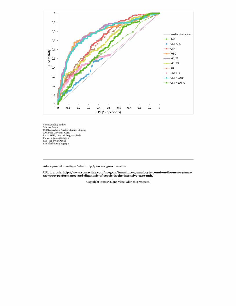
The AUC for diagnosing SE and SS versus CT (figure 1) was always greater for WBC, NEUT# and NEUT% than for theother parameters (table 5). Moreover, the Area Under the curve (AUC) of IG# was also greater than that of OM-IG#(p<0.0001) and equal to that of CRP. In particular, although this biomarker exhibited a remarkably high SN (i.e.,0.98), its diagnostic usefulness may be strongly impaired by the very modest SP (i.e., 0.12)
Interestingly, combined with the conventional diagnostic criteria for sepsis (i.e., leukocytosis with WBC> 12.00
x109/L, or leukopenia with WBC <4.00 x10⁹/L, or normal WBC count with 10% immature WBC) 4, a 10% cut-off for
IG% was characterized by a CD of 74%, with 0.72 SN and 0.75 SP for diagnosing sepsis, whereas a 0.150 x10⁹/L cut-off for IG# was characterized by a CD of 61%, with 0.86 SN and 0.46 SP.
With a normal WBC count, another option may be combining the NEUT # count at 9.38×10⁹L cut-off : this set up wascharacterized by a CD of 72%, with 0.75 SN and 0.46 SP. Instead, with an 80% NEUT% cut off we obtained a CD of80%, with 0.90 SN and 0.42 SP.

An interesting clinical consideration emerges from the analysis of data in table 5, wherein the diagnostic concordance
between laboratory criteria and clinical diagnosis was 61% and 74% using 10% or 0.150 x10⁹/L IG cut-offs,respectively.
Therefore, the good correlation with OM, the low imprecision and the excellent diagnostic accuracy support theconclusion that XN-9000 may be regarded as a reliable approach for IG counting.
Discussion
Despite important therapeutic improvement, sepsis remains a major healthcare issue, due to the high mortalityworldwide. The prognosis of this condition is strongly influenced by timely diagnosis, so that the introduction of easy,inexpensive and rapid tests should be regarded as an appealing perspective.
Overall, the results of our study show that the new Sysmex XN-9000 displays optimal performance for IG counting,in terms of both imprecision and correlation with OM, which still remains the reference method. Interestingly, theimprecision of IG on the XN-9000 seems even better than that of IG compared with the previous generation Sysmexanalyzers and OM. (6,7,10) The analysis of bias shows that the XN-9000 exhibits a trend towards overestimation ofboth IG# and IG% compared to OM, an aspect that was previously described by Maenhout et al. (9) However, unlikedata reported by Maenhout, in our study the diagnostic accuracy of IG# and IG% on XN-9000 was characterized bysignificantly better AUC (p <0.0001) than those exhibited by the same parameters obtained with OM (OM- IG # andOM-IG% ) (table 4 and 5).
In a previous paper by Ansari-Lari et al., (8) the observation of increased IG measurements on the Sysmex XE-2100was shown for infection and sepsis, as well as the biological and clinical relevance of this phenomenon even if IGcount lacked sensitivity, as confirmed by our study. Indeed, the diagnostic performance measured in the presentstudy was lower than that reported by Ansari-Lari and others, (8,17) and this is clearly attributable to the fact that weused a more suitable control population (i.e., ICU patients) rather than a group of healthy controls. Unlike in ourstudy, moreover, Ansari-Lari did not use the validated criteria of the International Sepsis Definition Conference (4)for diagnosing sepsis.
Interestingly, the adoption of a 10% cut-off for IG% on XN-9000 rather than for IL on OM in patients with a normalWBC count was characterized by satisfactory diagnostic performance for the diagnosis of sepsis (i.e., 0.72 SN and
0.75 SP, respectively). The SE could also be further improved by using a threshold of 0.150 x10⁹/L for IG# (i.e., 0.86SE), which was however counterbalanced by a much lower SP (i.e., 0.46). According to the AUC, the diagnosticperformance of both IG# was comparable to that of CRP (i.e., AUCs of 0.70 versus 0.72), but the CD of the formerparameter was even better (i.e., 61% versus 44%). Interestingly, in the same condition, the use of NEUT# andNEUT% parameters showed an AUC and CD of 0.79 versus 0.69 and 72% versus 80% respectively.
We can conclude that the assessment of both IG# and IG% appears suitable and reliable using XN-9000. Despite amodest overestimation, the excellent diagnostic accuracy of IGs analysis on XN-9000 may represent a valid andreliable alternative to assessment of IL with OM for diagnosing sepsis in the ICU. This approach also carries somereal benefits, such as the lower cost, high throughput, and shorter turnaround time compared to OM. Even moreimportantly, the opportunity to include novel hematological parameters such as IGs or NEUT in the diagnosticalgorithms may help overcome the inherent limits of those currently employed (e.g., ILs), thus helping to developnewer score systems that may ultimately improve the diagnosis of sepsis.
References
1. Nguyen HB, Rivers EP, Abrahamian FM, Moran GJ, Abraham E, Trzeciak S, et al. Severe sepsis and septic shock:rewiew of the literature and emergency department management guidelines”. Ann Emerg Med 2006;48:28-54.
2. Martin GS, Mannino DM, Eaton S, Moss M. The epidemiology of sepsis in the United States from 1979 through 2000.N Engl J Med 2003;348:1546-54.
3. Angus DC, van Der Poll T. Severe sepsis and septic shock. N Engl J Med 2013;369:840-51.4. Levy MM, Fink MP, Marshall JC, Abraham E, Angus D, Cook D, et al. International Sepsis Definitions Conference.
Crit Care Med 2003;4:1250-6.5. van der Meer W, van Gelder W, de Keijzer R, Willems H. Does the band cell survive the 21st century? Eur J Haematol
2006:76:251–4.6. Fernandes B, Hamaguchi Y. Automated enumeration of immature granulocytes. Am J Clin Pathol 2007;128:454-63.7. Nigro KG, O’Riordan MA, Molloy EJ, Walsh MC, Sandhaus LM. Performance of an automated immature granulocyte
count as a predictor of neonatal sepsis. Am J Clin Pathol 2005;123:618-24.

8. Ansari-Lari MA, Kickler TS, Borowitz MJ. Immature granulocyte measurement using the Sysmex XE-2100relationship to infection and sepsis. Am J Clin Pathol 2003;120:795-9.
9. Maenhout TM, Marcelis L. Immature granulocyte count in peripheral blood by the Sysmex haematology XN SeriesCompared to microscopic differentiaton. J Clin Pathol 2014; 67:648-50.
10. Bernstein LH, Rucinsky J. Measurement of granulocyte maturation may improve the early diagnosis of the septicstate. Clin Chem Lab Med 2011;49:2089-95.
11. Briggs C, Longair I, Kumar P, Singh D, Machin SJ. Performance evaluation of the Sysmex haematology XN modularsystem. J Clin Pathol 2012;65:1024-30.
12. Tantanate C. Spurious white blood cell count from a new automated Sysmex XN hematology analyzer. Int J LabHematol 2014;36(6): 86-7.
13. Koepke JA, van Assendelft OW, Brindza LJ, Davis BH, Fernandes BJ, Gewirtz AS, et al. Reference Leukocyte (WBC)Differential Count (Proportional) and Evaluation of instrumental Methods – Approved Standard, second ed. CLSIdocument H20A2, Wayne, PA, 2010.
14. College of American Pathologists. Hematology and Clinical Microscopy Glossary, Illinois, 2012, pp. 3–5.15. Barnes PW, McFadden L, Machin SJ, Simson E. The international consensus group for hematology review: suggested
criteria for action following automated CBC and WBC differential analysis. Lab Hematol 2005;11:83-90.16. McEnroe RJ, Durham AP, Goldford MD, Kondratovich MV, Lababidi S, Magari R, Middle JG, Pierson-Perry JF, Vaks
JE. Evaluation of Precision of Quantitative Measurement Procedures – Approved Guideline, third edition. CLSIguideline EP05-A3E, Wayne, PA, 2014.
17. Park SH, Park CJ, Lee BR, Nam KS, Kim MJ, Han MY, et al. Sepsis affects most routine and cell population data(CPD) obtained using the Sysmex XN-2000 blood cell analyzer: neutrophil-related CPD NE-SFL and NE-WY provideuseful information for detecting sepsis. Int J Lab Hematol 2014 May 28. doi: 10.1111/ijlh.12261Table 1. Pearson’s correlation, Passing & Bablock regression and Bland Altman bias for different cell categories inoptical microscopy (OM) versus XN-9000 automated count of immature granulocytes (IG) and neutrophils (NEUT).Values in percentage (%) and absolute number (#).
Test PearsonCorrelation
KolmogorovSmirnov CUSUMtest
Passing&
Bablockregression
Slope
(95%CI)
Intercept
(95%CI)
BiasBlandAltman
(95% CI)
OMNEUT#vs NEUT#
r= 0.99p<0.0001
Normal
p =0.4y=0.99x-0.19
0.99
(0.98 to1.00)
-0.19
(-0.27to-0.095)
-0.31
(-0.37 to-0.25)
OMNEUT% vsNEUT%
r= 0.84
p<0.0001
Normal
p =0.5y=1.1x-11.85
1.1
(1.0to1.2)
-11.85
(-17.00to-6.81)
-3.08
(-3.52 to-2.68)
OMIG# vsIG#
r= 0.89
p<0.0001 Not normal NA NA NA
0.22
(0.19 to0.25)
OMIG% vsIG%
r= 0.74
p<0.0001 Not normal NA NA NA
1.69
(1.49 to1.89)
Table 2. The within-run imprecision (expressed as coefficient of variation(CV) and standard deviation (SD))assessed on five blood samples using XN-9000 for white blood cell (WBC), immature granulocyte (IG) andneutrophils (NEUT) count. Values in percentage (%) and absolute number (#).
MeanMean
MeanMean Mean
Morphological

WBC x109/L(SD e CV%)
NEUTx109/L
(SD eCV%)
NEUT %
(SD eCV%)
IG x109/L
(SD eCV%)
IG %
(SD eCV%)
Flags by
XN9000
Sample1
3.61
(SD:0.053;CV:1.5)
2.14
(SD: 0.066
CV:3.1)
59.41
(SD:1.60;CV1.7)
0.77
(SD 0.017;CV: 2.2)
21.45
(SD:0.62;CV: 2.9)
1. WBC Abnormal1. IG Present?1. Left Shift?1. Fragments?
Sample2
13.28
(SD:0.296;CV:2.2)
9.62
(SD:0.233; CV:2.4)
72.4
(SD:0.34;CV: 0.05)
0.59
(SD 0.016;CV: 2.6)
4.49
(SD: 0.17;CV :3.7)
1. AtypicalLympho?
1. RBC Abnormal
Sample3
2.41
(SD:0.065;CV:2.7)
1.29
(SD:0.043;CV 3.4)
53.43
(SD:1.37;
CV: 2.6)
0.100
(SD 0.024;
CV: 24.0)
4.16
(SD:1.04;
CV : 25.1)
1. Left Shift?1. Abn Lympho?1. PLT Abn
Distribution1. Abn Scattergram1. RBC Abnormal
Sample4
13.63
(SD:0.208;CV:1.5)
11.59
(SD:0.193;CV: 1.7)
85.10
(SD:0.48;
CV: 0.6)
0.890
(SD: 0.042;CV:4.7)
6.51
(SD: 0.29;CV:4.5)
1. IG Present1. RBC Abnormal1. Leucopenia1. Anemia
Sample5
8.46
(SD: 0.083; CV:1.0)
7.27
(SD:0.093; CV:1.3)
85.94
(SD:0.47;
CV: 0.5)
1.576
(SD: 0.063;CV: 4.0)
18.65
(SD:0.80;CV:4.3)
1. Left Shift?1. Abn Lympho?1. PLT Abnormal
RBC, red blood cells; PLT, platelets.
Table 3. The imprecision (expressed as a coefficient of variation (CV) and standard deviation (SD) assessed on threecontrol levels XN-CHECK using XN-9000 for white blood cell (WBC), immature granulocyte (IG) andneutrophil(NEUT) count. Values in percentage (%) and absolute number (#).
Mean
WBC x109/L
(SD e CV%)
Mean NEUT#x109/L
(SD e CV%)
MeanNEUT%
(SD e CV%)
Mean
IG# x109/L
(SD e CV%)
Mean IG %
(SD e CV%)
XNCHECKLevel 1
2.91
(SD: 0.07;CV:2.5)
1.14
(SD: 0.04;CV:3.6)
38.8
(SD: 0.03;CV:3.5)
0.29
(SD: 0.03;CV:4.3)
10
(SD: 0.37;CV:3.7)
XNCHECK
Level 2
6.74
(SD:0.104;CV:2.1)
2.89
(SD: 0.09;CV:2.8)
43.4
(SD: 0.02;CV:4.9)
0.73
(SD: 0.03;CV:3.5)
11
(SD: 0.35;CV:3.2)
XNCHECK
Level 3
15.91
(SD:0.25;CV:1.6)
7.59
(SD: 0.19;CV:2.5)
47.8
(SD: 1.08;CV:2.3)
1.92
(SD: 0.07;CV:3.7)
12.1
(SD: 0.41;CV:3.4)

Table 4. Diagnostic accuracy of three different cell categories: leucocyte (WBC), neutrophils (NEUT) and immaturegranulocyte (IG) count using XN-9000, of two cell category: neutrophils (OM-NEUT) and immature granulocyte(OM-IG) in optical microscopy (OM) and concentration of C reactive protein (CRP).
Parameters
MedianControls(CT)
(95%CI)
Median
Sepsis(SE)
(95%CI)
Median
SepticShock(SS)
(95%CI)
KruskalWallistest
X²approximation and
pvalue
AUC
(95% CI)
and pvalue
Cutoff
Sensitivity(SN)
Specificity(SP)
CorrectClassification(CD)
WBCx109/L
9.13 & §
(8.83-9.78)
13.88
(13.11-14.57)
13.74
(9.43-17.50)
116.93
<0.0001
0.79
(0.74-0.83)p<0.0001
>12x109/L
<4x10⁹/L
0.71 0.77 75%
NEUTx109/L
7.30 & §
(6.87-7.73)
11.50
(11.02-12.4)
10.98
(7.92-15.78)
120.41
<0.0001
0.79
(0.73-0.84)
p<0.0001
≥9.38x10⁹/L
0.73 0.75 74%
OM-NEUTx109/L
7.68 & §
(7.31-8.05)
12.07
(11.34–12.67)
11.02
(8.06-15.66)
112.87
<0.0001
0.78
(0.74-0.83)
p<0.0001
≥9.70x10⁹/L
0.72 0.72 72%
NEUT%
80.1 & §
(0.78.9-81.0)
84.7 £
(81.5-86.5)
87.6
(83.5-92.9)
51.11
<0.0001
0.69
(0.64-0.74)
p<0.0001
≥80% 0.73 0.49 58%
OM-NEUT%
84.2 & §
(83.1-85.43)
87.5
(85.6-88.7)
87.5
(84.0-91.0)
20.55
<0.0001
0.62
(0.57-0.67)
p<0.0001
≥85% 0.62 0.53 56%
*IGx109/L
0.130 &§
(0.120-0.150)
0.240
(0.180-0.310)
0.280
(0.180-0.740)
53.48
<0.0001
0.70
(0.65-0.75)
p<0.0001
≥0.150x10⁹/L
0.73 0.54 61%
OM-IGx109/L
0.026§
(0.000-0.045)
0.001£
(0.000-0.0723)
0.135
(0.053-0.274)
21.55
<0.0001
0.57
(0.52-0.63)
p<0.01
≥0.110x10⁹/L
0.32 0.85 62%
1.4§ 1.6£2.8
19.850.59
*IG% (1.3- 1.6) (1.4- 2.1) (2.0-5.6)
<0.0001 (0.54 –0.64)
p<0.0005
≥10 % 0.10 0.98 65%
OM-IG%
0.5§
(0.0-0.5)
0.1£
(0.0-0.5)
1.1
(1.0-2.0)
19.02
<0.0001
0.53
(0.48-0.58)
p=0.2
// //
CRPmg/dL
4.8 & §
(4.0-5.8)
11.0
(9.4-12.6)
13.3
(8.8-16.0)
63.73
<0.0001
0.72
(0.67-0.76)
p<0.0001
≥1.2 0.98 0.12 44%
&, §, and £ statistically significant difference by Steel-Dwass-Critchlow-Fligner test for CT versus SE, CT versus SSand SE versus SS respectively.
*The data refer to all the samples examined, while in subjects 285 samples with a normal WBC count: IG#, IG% to cutoff indicated showed a SN equal to 0.52 to 0.4, SP equal to 0.59 to 0.98 and finally DC of 58%, the 80% respectively.
%: Values in percentage;
#: absolute number;
AUC, Area Under the curve; CI, Confidence Interval.
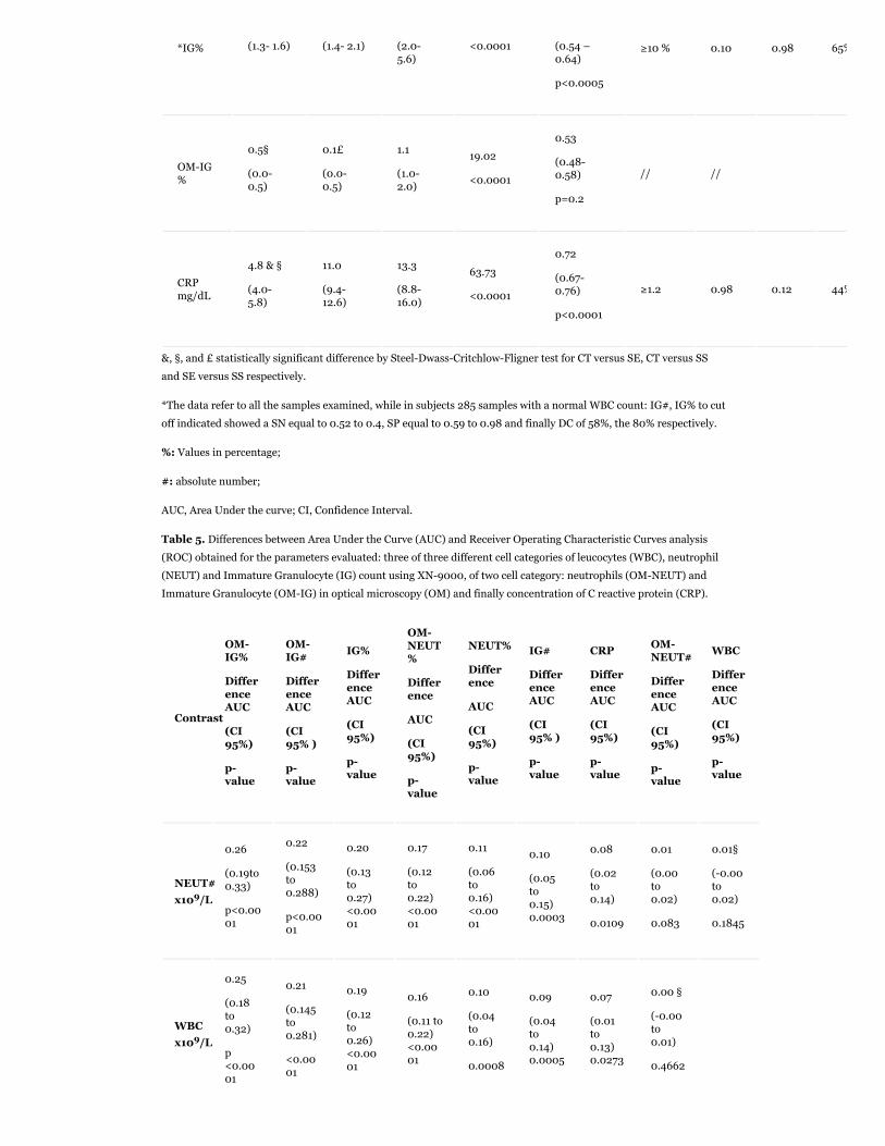
Table 5. Differences between Area Under the Curve (AUC) and Receiver Operating Characteristic Curves analysis(ROC) obtained for the parameters evaluated: three of three different cell categories of leucocytes (WBC), neutrophil(NEUT) and Immature Granulocyte (IG) count using XN-9000, of two cell category: neutrophils (OM-NEUT) andImmature Granulocyte (OM-IG) in optical microscopy (OM) and finally concentration of C reactive protein (CRP).
Contrast
OMIG%
DifferenceAUC
(CI95%)
pvalue
OMIG#
DifferenceAUC
(CI95% )
pvalue
IG%
DifferenceAUC
(CI95%)
pvalue
OMNEUT%
Difference
AUC
(CI95%)
pvalue
NEUT%
Difference
AUC
(CI95%)
pvalue
IG#
DifferenceAUC
(CI95% )
pvalue
CRP
DifferenceAUC
(CI95%)
pvalue
OMNEUT#
DifferenceAUC
(CI95%)
pvalue
WBC
DifferenceAUC
(CI95%)
pvalue
NEUT#x109/L
0.26
(0.19to0.33)
p<0.0001
0.22
(0.153to0.288)
p<0.0001
0.20
(0.13to0.27)<0.0001
0.17
(0.12to0.22)<0.0001
0.11
(0.06to0.16)<0.0001
0.10
(0.05to0.15)0.0003
0.08
(0.02to0.14)
0.0109
0.01
(0.00to0.02)
0.083
0.01§
(-0.00to0.02)
0.1845
WBCx109/L
0.25
(0.18to0.32)
p<0.0001
0.21
(0.145to0.281)
<0.0001
0.19
(0.12to0.26)<0.0001
0.16
(0.11 to0.22)<0.0001
0.10
(0.04to0.16)
0.0008
0.09
(0.04to0.14)0.0005
0.07
(0.01to0.13)0.0273
0.00 §
(-0.00to0.01)
0.4662

OMNEUT#x109/L
0.25
(0.18to0.32)
p<0.0001
0.21
(0.141to0.278)<0.0001
0.19
(0.12to0.26)<0.0001
0.160
(0.11 to0.21)<0.0001
0.10§
(0.04to0.15)
0.0003
0.08
(0.03to0.14)
0.0010
0.07
(0.01to0.13)0.0320
CRP
0.18
(0.12to0.25)<0.0001
0.14
(0.079to0.207)<0.0001
0.12
(0.057to0.1889)0.0003
0.093
(0.03to0.16)0.0033
0.03§
(-0.03to0.09)0.3040
0.02§
(-0.05to0.08)0.5813
IG#x109/L
0.166
(0.11to0.22)<0.0001
0.13
(0.07to0.18)<0.0001
0.10
(0.078to0.131)
<0.0001
0.076
(0.01to0.15)0.0306
0.012§
(-0.05to0.08)0.7207
NEUT%
0.15
(0.087to0.221)<0.0001
0.11
(0.05to0.18)0.0008
0.09
(0.023to0.163)0.0093
0.064
(0.03to0.10)<0.0001
OMNEUT%
0.09
(0.01to 0.17)
0.0212
0.049§
(-0.03to0.13)0.1992
0.03§
(-0.05to0.10)0.4575
IG%
0.061
(0.01to 0.11)0.0122
0.021§
(-0.03to0.07)
0.4111
OMIG#x109/L
0.040
(0.03to0.05)
<0.0001
§: Not statistically significant difference
CI, Confidence Interval.
Figure 1. ROC curve analysis in all samples analyzed for leucocyte (WBC), neutrophil count, both absolute (NEUT#)and percentage (NEUT%) immature granulocyte (IG) count, both absolute (IG#) and percentage (IG%) obtained withXN-9000. The neutrophil count, both absolute (OM-NEUT#) and percentage (OM- NEUT%) and the IG count (i.e.,promyelocytes, myelocytes and metamyelocytes), both absolute (OM-IG #) and percentage (OM-IG%). Finally, theconcentration of C reactive protein (CRP).
Corresponding author Sabrina Buoro USC Laboratorio Analisi Chimico Cliniche A.O. Papa Giovanni XXIII Piazza OMS, 1 -24128 Bergamo, Italy Phone: + 39 0352674550 Fax + 39 035 2674939 E-mail: [email protected]
Article printed from Signa Vitae: http://www.signavitae.com
URL to article: http://www.signavitae.com/2015/12/immaturegranulocytecountonthenewsysmexxn9000performanceanddiagnosisofsepsisintheintensivecareunit/
Copyright © 2015 Signa Vitae. All rights reserved.