Illustrated Guide to the Immature Lepidoptera on Oaks in ...
382
Illustrated Guide to the Immature Lepidoptera on Oaks in Missouri Robert J. Marquis, Steven C. Passoa, John T. Lill, James B. Whitfield, Josiane Le Corff, Rebecca E. Forkner, and Valerie A. Passoa United States Department of Agriculture Forest Health Assessment and Applied Sciences Team FHAAST-2018-05 June 2019
-
Upload
khangminh22 -
Category
Documents
-
view
2 -
download
0
Transcript of Illustrated Guide to the Immature Lepidoptera on Oaks in ...

Illustrated Guide to the Immature Lepidoptera
on Oaks in MissouriRobert J. Marquis, Steven C. Passoa, John T. Lill, James B. Whitfield,
Josiane Le Corff, Rebecca E. Forkner, and Valerie A. Passoa
United States Department of Agriculture
Forest Health Assessment and Applied Sciences Team
FHAAST-2018-05June 2019

THIS PAGE INTENTIONALLY LEFT BLANK

Illustrated Guide to the Immature Lepidoptera
on Oaks in Missouri
Cover Photos. Clockwise from top left: Quercus alba, Acronicta (subgenus Lepitoreuma, increta group), Malacosoma americana, Pseudotelphusa sp., Erynnis brizo, Eacles imperialis, Euclea delphinii (gray form). Photos by R.J. Marquis. Bottom left: Antheraea polyphemus pupal brain window. Photo by S.C. Passoa. All photos used with permission.
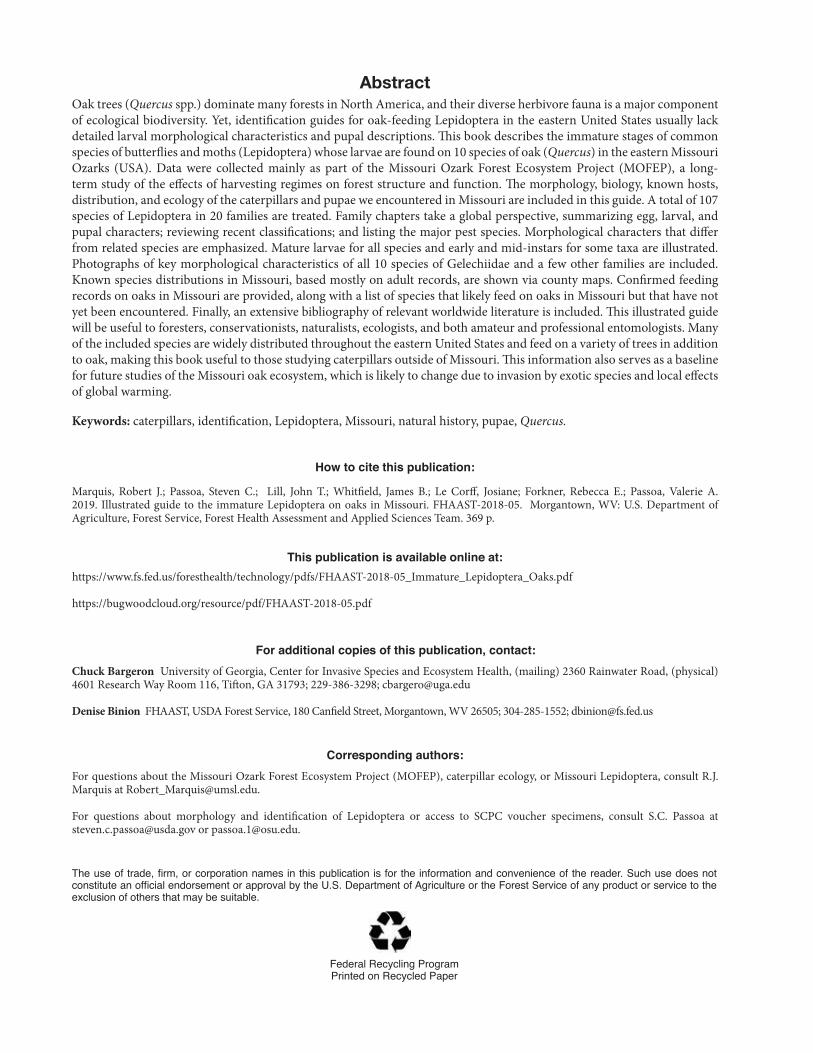
AbstractOak trees (Quercus spp.) dominate many forests in North America, and their diverse herbivore fauna is a major component of ecological biodiversity. Yet, identification guides for oak-feeding Lepidoptera in the eastern United States usually lack detailed larval morphological characteristics and pupal descriptions. This book describes the immature stages of common species of butterflies and moths (Lepidoptera) whose larvae are found on 10 species of oak (Quercus) in the eastern Missouri Ozarks (USA). Data were collected mainly as part of the Missouri Ozark Forest Ecosystem Project (MOFEP), a long-term study of the effects of harvesting regimes on forest structure and function. The morphology, biology, known hosts, distribution, and ecology of the caterpillars and pupae we encountered in Missouri are included in this guide. A total of 107 species of Lepidoptera in 20 families are treated. Family chapters take a global perspective, summarizing egg, larval, and pupal characters; reviewing recent classifications; and listing the major pest species. Morphological characters that differ from related species are emphasized. Mature larvae for all species and early and mid-instars for some taxa are illustrated. Photographs of key morphological characteristics of all 10 species of Gelechiidae and a few other families are included. Known species distributions in Missouri, based mostly on adult records, are shown via county maps. Confirmed feeding records on oaks in Missouri are provided, along with a list of species that likely feed on oaks in Missouri but that have not yet been encountered. Finally, an extensive bibliography of relevant worldwide literature is included. This illustrated guide will be useful to foresters, conservationists, naturalists, ecologists, and both amateur and professional entomologists. Many of the included species are widely distributed throughout the eastern United States and feed on a variety of trees in addition to oak, making this book useful to those studying caterpillars outside of Missouri. This information also serves as a baseline for future studies of the Missouri oak ecosystem, which is likely to change due to invasion by exotic species and local effects of global warming.
Keywords: caterpillars, identification, Lepidoptera, Missouri, natural history, pupae, Quercus.
How to cite this publication:
Marquis, Robert J.; Passoa, Steven C.; Lill, John T.; Whitfield, James B.; Le Corff, Josiane; Forkner, Rebecca E.; Passoa, Valerie A. 2019. Illustrated guide to the immature Lepidoptera on oaks in Missouri. FHAAST-2018-05. Morgantown, WV: U.S. Department of Agriculture, Forest Service, Forest Health Assessment and Applied Sciences Team. 369 p.
This publication is available online at: https://www.fs.fed.us/foresthealth/technology/pdfs/FHAAST-2018-05_Immature_Lepidoptera_Oaks.pdf
https://bugwoodcloud.org/resource/pdf/FHAAST-2018-05.pdf
For additional copies of this publication, contact: Chuck Bargeron University of Georgia, Center for Invasive Species and Ecosystem Health, (mailing) 2360 Rainwater Road, (physical) 4601 Research Way Room 116, Tifton, GA 31793; 229-386-3298; [email protected]
Denise Binion FHAAST, USDA Forest Service, 180 Canfield Street, Morgantown, WV 26505; 304-285-1552; [email protected]
Corresponding authors: For questions about the Missouri Ozark Forest Ecosystem Project (MOFEP), caterpillar ecology, or Missouri Lepidoptera, consult R.J. Marquis at [email protected].
For questions about morphology and identification of Lepidoptera or access to SCPC voucher specimens, consult S.C. Passoa at [email protected] or [email protected].
The use of trade, firm, or corporation names in this publication is for the information and convenience of the reader. Such use does not constitute an official endorsement or approval by the U.S. Department of Agriculture or the Forest Service of any product or service to the exclusion of others that may be suitable.
Federal Recycling ProgramPrinted on Recycled Paper

Illustrated Guide to the Immature Lepidoptera on Oaks in Missouri
Robert J. Marquis, Department of Biology and the Whitney R. Harris World Ecology Center, University of Missouri-St. Louis, 1 University Boulevard, St. Louis, MO 63121-4400
Steven C. Passoa, USDA-APHIS-PPQ, 1315 Kinnear Road, Columbus, OH 43212
John T. Lill, Department of Biological Sciences, The George Washington University, 800 22nd Street NW, Suite 6000, NW, Washington, DC 20052
James B. Whitfield, Department of Entomology, University of Illinois at Urbana-Champaign, 320 Morrill Hall, 505 South Goodwin Avenue, Urbana, IL 61801
Josiane Le Corff, Institut de Recherche en Horticulture et Semences, UMR 1345, INRA-Université d’Angers-Agrocampus Ouest, 42 rue Georges Morel-CS 60057, 49071 Beaucouzé Cedex - France
Rebecca E. Forkner, Department of Biology, George Mason University, David King Hall MSN 3E1, 4400 University Drive, Fairfax, VA 22030-4444
Valerie A. Passoa, The Ohio Lepidopterists, 1315 Kinnear Road, Columbus, OH 43212
Contributors/Collaborators
The following organizations provided support for the production of this book in the form of space (Ohio State University, University of Missouri-St. Louis), funding for data collection (Missouri Department of Conservation, National Science Foundation, University of Missouri-St. Louis), editing and production (USDA Forest Service), and funding for printing (USDA Forest Service; USDA APHIS).
Forest ServiceAPHIS
FHAAST-2018-05

II
ILLUSTRATED GUIDE TO THE IMMATURE LEPIDOPTERA ON OAKS IN MISSOURI
AcknowledgmentsWe thank the Missouri Department of Conservation, including Randy Jensen, Terry Robison, Brian Brookshire, Robert Lawrence, DeeCee Darrow, and David Gwaze, for their support. The Missouri Department of Conservation, USDA Cooperative Land, Prevention and Suppression Grant 02DG 1124225 430 and the National Science Foundation Division of Biological Sciences Grant 6164397 provided financial support. Phillip Koenig digitized Richard Heitzman’s card files to create an electronic list of Missouri Lepidoptera. Estefania Fernandez provided technical expertise to Alaa Saffaf and Omar Saffaf, who created the distribution maps for all species reported in this book. Thank you to the Missouri Department of Natural Resources and Bruce Schuette for permission to conduct sampling at Cuivre River State Park and to Washington University for access to the Tyson Research Center.
The following specialists either made or confirmed identifications, answered questions, provided specimens, or sent publications: John Rawlins, Jim Young, David Wagner, James Appleby, Don Davis, Marc Epstein, Ron Hodges, Tim McCabe, Terry Harrison, George Godfrey, Phil DeVries, Alma Solis, Jim Hayden, Robert Robbins, John Brown, Richard Brown, Jason Dombroskie, Jerry Powell, Paul Goldstein, and Eric Metzler. Specimens of Tischeriidae were provided by Terry Harrison; David Wagner donated a small but very useful synoptic collection of moths that helped with our morphological studies. Early drafts of the manuscript were reviewed by Mark Epstein (Limacodidae and Megalopygidae), George Godfrey (Notodontidae), James Hayden (Pyralidae), Ron Hodges (Gelechiidae), Tim McCabe (Noctuidae, Erebidae, and Geometridae), and Richard Peigler (Saturniidae). Christopher Ernst and Zachary Weber compiled the Appendix 1 species list. Terry Harrison, Dave Wagner, and Chris Schmidt reviewed parts of the completed manuscript. We appreciate their efforts, which improved this book tremendously.
Robert Marquis thanks Phil DeVries for introducing him to the natural history of caterpillars, and May Berenbaum and Jack Schultz for training and encouragement. Steven Passoa thanks Jack Schultz, May Berenbaum, and George Godfrey for explaining caterpillar basics, and to Valerie Passoa for showing him the beauty of caterpillars as living things.
Steven Passoa also thanks Jim Slavicek and John Freudenstein who gave him office space to work on this book. It was very helpful to have access to the Charles A. Triplehorn insect collection at Ohio State University, through the never-ending kindness of Charles Triplehorn, Norman Johnson, and Luciana Musetti.
Valerie Passoa has had the good fortune to communicate over the years with like-minded individuals with a fascination for Lepidoptera. Their generosity with advice, literature, plant materials, and/or livestock has made her adventures with life history and its offshoots more rewarding. She thanks James Adams, Carter Bays, Roger Bernhardt, Arthur Bürger, Michael Collins, Christopher Conlan, Vittorio Giammarrusco, Michael Gilligan, Leroy Koehn, Paul Latham, George Leslie, James Mouw, Wolfgang Nässig, Stefan Naumann, Donald Olhausen, Ulrich and Laela Paukstadt, Richard Peigler, Kevin Phipps, Mark Pickup, Paul Plasmeijer, John Rawlins, Eric Rudge, Gene Shatrowsky, Michael Soukup, Bert Strik, Stephen Stone, Paul Tuskes, Robert Weast, and Kirby Wolfe. A handful of special people have shared her life-long affinity for rearing Lepidoptera: Olivia Moses, Steven Passoa, Ethan Passoa, and Tom Roberts.
A few people have passed away but would have enjoyed using or seeing our book: Dale Habeck, Ellis MacLeod, Jim Sternburg, Noel McFarland, Roy Kendall, Frank Sala, Ron Hodges, and Caleb Passoa.
This publication would not have been completed were it not for Richard Reardon and Susan Wright of the USDA Forest Service. Scott Neitch and Clarissa Maroon-Lango provided significant APHIS funding. John Kabrick and Daniel Dey also provided funds. Rachel Winston provided outstanding book design skills, but also located numerous technical and style inconsistencies that, once corrected, improved the final product. An equally great job of editing was done by Amy Scherzer. Dan Kucera and Noel Schneeberger supported us from the start.
Collectively, the authors thank those who were not mentioned because of space limitations. These include an army of APHIS identifiers, USDA cooperators, and MOFEP insect samplers. The authors acknowledge Mary Murtfeldt (see Bailey 1998) and Richard Heitzman for their pioneering studies of the Missouri fauna.

III
ILLUSTRATED GUIDE TO THE IMMATURE LEPIDOPTERA ON OAKS IN MISSOURI
ContentsIntroduction ................................................................................................................1Data Basis for this Book .............................................................................................4Lepidopteran Life Cycle and Life History Variation ...................................................7Caterpillar Morphology .............................................................................................10
General Body Plan ................................................................................................................... 10Setae........................................................................................................................................ 13Illustrating Caterpillar Morphology ........................................................................................ 14
Pupal Morphology .....................................................................................................15How to Collect, Rear, and Preserve Caterpillars ......................................................18How to Document a Caterpillar Fauna .....................................................................21Parasites (Parasitoids) .............................................................................................23How to Use this Book ...............................................................................................25Species Descriptions ................................................................................................29
Tischeriidae ............................................................................................................................. 29Coptotriche citrinipennella (Clemens) .........................................................................................30
Gracillariidae ........................................................................................................................... 32Subfamily Lithocolletinae ............................................................................................................................34
Phyllonorycter fitchella (Clemens) ...............................................................................................34Depressariidae ........................................................................................................................ 36
Subfamily Stenomatinae..............................................................................................................................41Antaeotricha humilis (Zeller) ........................................................................................................41Antaeotricha osseella (Walsingham) ...........................................................................................42Antaeotricha schlaegeri (Zeller) ...................................................................................................44Menesta melanella (Murtfeldt) .....................................................................................................46Rectiostoma xanthobasis (Zeller) ................................................................................................47
Unplaced Depressariidae .............................................................................................................................48Psilocorsis cryptolechiella (Chambers) .......................................................................................48Psilocorsis quercicella Clemens ..................................................................................................50Psilocorsis reflexella Clemens .....................................................................................................51Machimia tentoriferella Clemens .................................................................................................53
Gelechiidae .............................................................................................................................. 55Subfamily Dichomeridinae ..........................................................................................................................59
Dichomeris georgiella (Walker) ....................................................................................................59Dichomeris ligulella Hübner .........................................................................................................61
Subfamily Gelechiinae .................................................................................................................................62Arogalea cristifasciella (Chambers) .............................................................................................62Chionodes adamas Hodges ........................................................................................................64Chionodes formosella complex (Murtfeldt) ................................................................................66Chionodes fuscomaculella (Chambers) ......................................................................................68Chionodes pereyra Clarke ...........................................................................................................69Pseudotelphusa complex ............................................................................................................71Pubitelphusa latifasciella (Chambers) .........................................................................................73Trypanisma prudens Clemens .....................................................................................................75

Iv
ILLUSTRATED GUIDE TO THE IMMATURE LEPIDOPTERA ON OAKS IN MISSOURI
Tortricidae ............................................................................................................................... 77Subfamily Tortricinae ...................................................................................................................................80
Choristoneura rosaceana (Harris) ................................................................................................80Cenopis directana (Walker) ..........................................................................................................84Cenopis pettitana (Robinson) ......................................................................................................85Pandemis limitata (Robinson) ......................................................................................................88
Subfamily Olethreutinae ..............................................................................................................................89Ancylis divisana (Walker) ..............................................................................................................89
Hesperiidae ............................................................................................................................. 92Subfamily Pyrginae ......................................................................................................................................93
Erynnis brizo (Boisduval & Leconte) ............................................................................................93Erynnis juvenalis (Fabricius) .........................................................................................................94
Lycaenidae .............................................................................................................................. 97Subfamily Theclinae .....................................................................................................................................98
Satyrium calanus (Hübner) .........................................................................................................98Nymphalidae ......................................................................................................................... 100
Subfamily Limenitidinae ............................................................................................................................101Limenitis arthemis astyanax (Fabricius) ....................................................................................101
Megalopygidae ...................................................................................................................... 104Subfamily Megalopyginae .........................................................................................................................105
Megalopyge crispata (Packard) .................................................................................................105Limacodidae .......................................................................................................................... 108
Subfamily Limacodinae .............................................................................................................................109Acharia stimulea (Clemens) .......................................................................................................109Adoneta spinuloides (Herrich-Schäffer) ....................................................................................111Apoda biguttata (Packard) .........................................................................................................113Euclea delphinii (Boisduval) .......................................................................................................114Isa textula (Herrich-Schäffer) .....................................................................................................117Isochaetes beutenmuelleri (Edwards) ......................................................................................118Natada nasoni (Grote) ...............................................................................................................119Packardia geminata (Packard) ...................................................................................................121Parasa chloris (Herrich-Schäffer) .............................................................................................122Parasa indetermina (Boisduval) .................................................................................................124Prolimacodes badia (Hübner) ...................................................................................................125Tortricidia flexuosa (Grote) .........................................................................................................127Phobetron pithecium (J.E. Smith) .............................................................................................130
Pyralidae ............................................................................................................................... 133Subfamily Epipaschiinae ...........................................................................................................................136
Pococera expandens (Walker) ...................................................................................................136Oneida lunulalis (Hulst) .............................................................................................................139
Subfamily Phycitinae .................................................................................................................................140Salebriaria engeli (Dyar) .............................................................................................................140Salebriaria tenebrosella (Hulst) ..................................................................................................142Quasisalebria fructetella (Hulst) .................................................................................................143
Geometridae .......................................................................................................................... 144Subfamily Ennominae ................................................................................................................................151
Besma quercivoraria (Guenée) ..................................................................................................151Campaea perlata (Guenée) ........................................................................................................154Eutrapela clemataria (J.E. Smith) ..............................................................................................156

v
ILLUSTRATED GUIDE TO THE IMMATURE LEPIDOPTERA ON OAKS IN MISSOURI
Erannis tiliaria (Harris) ................................................................................................................160Lambdina fervidaria athasaria (Walker) .....................................................................................163Melanolophia canadaria (Guenée) .............................................................................................166Plagodis alcoolaria (Guenée) .....................................................................................................168Selenia kentaria (Grote & Robinson) .........................................................................................171Phigalia strigataria (Minot) ..........................................................................................................173Ectropis crepuscularia (Denis & Schiffermüller) ........................................................................175Glena cribrataria (Guenée) .........................................................................................................176Phaeoura quernaria (J.E. Smith) ...............................................................................................178Hypagyrtis unipunctata (Haworth) .............................................................................................179Alsophila pometaria (Harris) ......................................................................................................181
Subfamily Geometrinae .............................................................................................................................183Nemoria bistriaria Hübner ..........................................................................................................183
Mimallonidae ........................................................................................................................ 185Lacosoma chiridota Grote .........................................................................................................186
Lasiocampidae ...................................................................................................................... 188Subfamily Lasiocampinae .........................................................................................................................189
Malacosoma americana (Fabricius) ..........................................................................................189Malacosoma disstria Hübner .....................................................................................................191Phyllodesma americana (Harris) ...............................................................................................193
Subfamily Macromphaliinae ......................................................................................................................195Tolype velleda (Stoll) ..................................................................................................................195
Saturniidae ............................................................................................................................ 197Subfamily Hemileucinae ............................................................................................................................200
Automeris io (Fabricius) ............................................................................................................200Subfamily Saturniinae ...............................................................................................................................202
Antheraea polyphemus (Cramer) .............................................................................................202Subfamily Ceratocampinae .......................................................................................................................205
Anisota senatoria (Smith) ...........................................................................................................205Anisota virginiensis (Drury) ........................................................................................................207Eacles imperialis (Drury) ............................................................................................................209
Sphingidae ............................................................................................................................ 212Subfamily Smerinthinae ............................................................................................................................213
Paonias excaecata (J.E. Smith) .................................................................................................213Notodontidae ......................................................................................................................... 216
Subfamily Heterocampinae .......................................................................................................................218Cecrita guttivitta Walker .............................................................................................................218Lochmaeus manteo Doubleday ...............................................................................................221Macrurocampa marthesia (Cramer) .........................................................................................223Oedemasia semirufescens (Walker) ..........................................................................................225Ianassa lignicolor (Walker) .........................................................................................................226Schizura ipomaeae Doubleday ................................................................................................227
Subfamily Notodontinae ............................................................................................................................229Paraeschra georgica (Herrich-Schäffer) ....................................................................................229
Subfamily Nystaleinae ...............................................................................................................................230Symmerista spp. ........................................................................................................................230
Subfamily Phalerinae .................................................................................................................................233Nadata gibbosa (Smith) .............................................................................................................233Peridea angulosa (Smith) ..........................................................................................................235

vI
ILLUSTRATED GUIDE TO THE IMMATURE LEPIDOPTERA ON OAKS IN MISSOURI
Nolidae .................................................................................................................................. 237Subfamily Nolinae ......................................................................................................................................239
Meganola phylla (Dyar) ..............................................................................................................239Noctuidae .............................................................................................................................. 242
Subfamily Pantheinae ................................................................................................................................251Charadra deridens (Guenée) ....................................................................................................251
Subfamily Acronictinae ..............................................................................................................................254Acronicta (subgenus Lepitoreuma, increta group) ...................................................................254Acronicta impleta (Walker) .........................................................................................................256
Subfamily Amphipyrinae ...........................................................................................................................259Amphipyra pyramidoides (Guenée) ..........................................................................................259
Subfamily Noctuinae ..................................................................................................................................261Cosmia calami (Harvey) .............................................................................................................261Lithophane antennata (Walker) ..................................................................................................263Achatia distincta (Hübner) ........................................................................................................265Morrisonia confusa (Hübner) .....................................................................................................267Orthosia rubescens (Walker) ....................................................................................................269
Erebidae ................................................................................................................................ 272Subfamily Lymantriinae .............................................................................................................................279
Dasychira obliquata (Grote & Robinson) ...................................................................................279Dasychira tephra Hübner ...........................................................................................................281Orgyia leucostigma (J.E. Smith) ...............................................................................................282Lymantria dispar dispar (Linnaeus) ...........................................................................................285
Subfamily Arctiinae ...................................................................................................................................289Halysidota tessellaris (J.E. Smith) ..............................................................................................289
Subfamily Herminiinae ..............................................................................................................................291Palthis angulalis (Hübner) ..........................................................................................................291
Subfamily Boletobiinae ..............................................................................................................................292Hyperstrotia secta (Grote) & H. pervertens (Barnes & McDunnough) .....................................292
Subfamily Erebinae ....................................................................................................................................295Catocala amica (Hübner) ...........................................................................................................295Phoberia atomaris (Hübner) .......................................................................................................297Zale minerea (Guenée) ...............................................................................................................299
Subfamily Eulepidotinae ............................................................................................................................301Panopoda rufimargo (Hübner) ..................................................................................................301
References ..............................................................................................................303Glossary ..................................................................................................................341Appendix .................................................................................................................350
Appendix 1: Quercus-feeding species found as adults but not larvae in Missouri. .....................................350Appendix 2: Key to the counties of Missouri ..............................................................................................355Appendix 3: Images of early instars of select species .................................................................................356Appendix 4: Images of diagnostic traits of Gelechiidae and other species included in this book ...............358
Index .................................................................................................... 368

ILLUSTRATED GUIDE TO THE IMMATURE LEPIDOPTERA ON OAKS IN MISSOURI
1
IntroductionOak trees (Quercus spp.) dominate many forests in North America, reach their highest diversity in Mexico, and extend all the way through Central America to the highland regions of Colombia. In many North American locations, oaks make up the majority of the canopy biomass and often are represented by five or more species in a single location (Stein et al. 2003). Oaks occur in alluvial to upland forests, from closed canopy to savannah, and in scrublands.
The herbivore fauna of oaks is unusually diverse compared to those of many other tree species, and as such, these insects represent a major component of the biodiversity in their associated ecological communities. Documenting native faunas is the first step toward protecting them. For instance, Robinson et al. (2002) listed 724 species of Lepidoptera that feed on oaks in North America, and 36 families of Lepidoptera were recorded from California oaks by Swiecki et al. (1997). Numerous sawflies, beetles, and 485 species of North American cynipid wasps (Burks 1979, Griffin et al. 1987) also attack oak trees. We have reared or observed more than 150 oak-feeding species of Lepidoptera in Missouri, and we expect there to be at least 200 more species in the state based on collections of adults and their known host plants from other regions (Appendix 1). Because there is such a large insect fauna associated with oaks, the potential loss of biodiversity and ecosystem function would be catastrophic if the oak ecosystem were to be damaged. Besides their contribution in terms of numbers to the biodiversity count, these insects provide food for birds and mammals, pollinate plants, serve as hosts for the native parasitoid community (see Parasites, page 23), and contribute detritus and nutrients that support additional microarthropod and microbial communities.
The other side of the coin is that oak herbivores are members of the second trophic level (herbivores). They potentially influence tree production by reducing leaf area and damaging woody tissue. Missouri is an example where adding basic information on the oak insect fauna is essential to help protect a valuable timber resource. For example, oak trees in Missouri provide $5.9 billion in revenue (2016 estimate) annually from timber harvests and associated primary processors (like sawmills), secondary processors (furniture, pallets, paper, etc.), associated industries, and all their spending (T. Treiman, Missouri Department of Conservation, pers. comm.). Oaks also provide shelter and food for wildlife; for example, oak mast represents a major resource for rodents, birds, and ungulates (Brezner 1960, McKinley 1960: 41, White 1995).
One of our goals with this book is to help protect a valuable timber resource by providing basic information on the lepidopteran fauna of Missouri oaks. The detrimental effects of these insects become most apparent when factors that typically keep insect populations in check fail. For example, a few native species (e.g., Cecrita [Heterocampa] guttivitta, Lochmeaus manteo, Phoberia atomaris, and Archips semifuranus) have undergone widespread outbreaks in Missouri or elsewhere, causing extensive damage to native trees (Nothnagle and Schultz 1987, Mattson et al. 1991). Even so-called “chronic” levels of leaf area loss in “typical” years by the entire community of insects seem to be sufficient to affect tree sapling growth (Marquis and Whelan 1994) and

ILLUSTRATED GUIDE TO THE IMMATURE LEPIDOPTERA ON OAKS IN MISSOURI
2 INTRODUCTION
acorn production of mature trees (Hochwender et al. 2003) in Missouri Ozark forests. Leaf-tying caterpillars alone often remove as much as 30 percent of the leaf area of understory saplings of white oak in Missouri (R.J. Marquis and J.T. Lill, unpublished data). Exotic species of insects that are introduced to North America without their arthropod natural enemies represent an important additional threat. Oaks are known to harbor at least 33 introduced insect species in North America (Mattson et al. 1994). The economic impact of the European gypsy moth, Lymantria dispar dispar, on northeastern U.S. forests (Kegg 1973, Goebl 1987, Dreistadt and Weber 1989) clearly demonstrates the damage caused by exotic insect introductions. Unfortunately, there have been few studies documenting the ecological impacts of these invasive pests on native herbivore communities (Ghandi and Herms 2010). Control measures themselves may affect nontarget species, either negatively by poisoning them (Rastall et al. 2003, Boulton et al. 2007) or positively by reducing the impact defoliators like gypsy moth have on leaf tissue quality (Manderino et al. 2014). This lack of information highlights the importance of documenting intact oak herbivore faunas in places like the Ozarks, where the gypsy moth has yet to establish. There is an increasing need to document exotic insect distributions (Liebhold et al. 1995b), but such documentation depends on our ability to discriminate between native species and newly introduced exotic pests. Furthermore, there is increasing evidence that both biologically- and chemically-based efforts to control exotic species, particularly the European gypsy moth, may have detrimental effects on native insects found on oaks (Peacock et al. 1993, Walters 1995). Mattson et al. (1994) predicted that new North American introductions of exotic insects on woody plants would likely come from Europe and central Asia because all three regions have similar floras. Given that European and Asian oaks harbor diverse herbivore faunas (600 arthropod species in China, including seven species of Lymantria related to the gypsy moth; Zheng et al. 2006), it is especially important that North American foresters monitor the oak ecosystem to allow for the quick detection of potentially new invasive species.
Another reason why the Missouri oak fauna merits study is that increasing attention is being paid to insect populations impacted by habitat destruction and global climate change. Lepidoptera, and butterflies in particular, have come under scrutiny because the abundance of a number of species has declined (e.g., New 2004, Schweitzer et al. 2011). Lepidoptera are excellent indicator species for changes in habitat quality because their populations are intimately tied to the abundance and seasonality of their host plants, and host plant decimation is often synonymous with habitat destruction. The abundance of Lepidoptera is linked to leaf quality (e.g., Forkner et al. 2004), which in turn can be influenced by environmental changes associated with climate change (Zavala et al. 2013).
The starting point for all ecology, conservation, and management is identification of the relevant organisms. Despite the dominance of oaks and the importance of their insect herbivores, available identification guides for oak-feeding Lepidoptera in the eastern United States usually lack detailed larval morphological characteristics (Bray and Triplehorn 1953; Hitchcock 1961; USDA 1985, 1987; Wagner 2005; Wagner et al. 1995b, 1997, 2001, 2005). Moreover, they completely lack pupal descriptions. Both types of information are also lacking in guides for other parts of North America (Brown and Eads 1965, Ives and Wong 1988, Miller 1995, Rose et al. 1997, Solomon et al. 1999). A further hurdle exists for microlepidopteran species. Although they can be abundant and cause severe damage to their host plants (e.g., Ives and Wong 1988), most guides cover them only sparingly if at all. In many families, identification is complicated by the lack of experts and a shortage of named voucher material in collections.
This book arose from an ongoing study of the impact of forest management on biodiversity and long-term productivity of forests in the Missouri Ozarks (Brookshire et al. 1997). The Missouri Ozark Forest Ecosystem Project (MOFEP) is a 100+ year experiment, sponsored by the Missouri Department of Conservation, whose goal is to compare the effects of alternative harvesting regimes on forest structure and function. One of the focal groups for this study is oak-feeding insects. In a sense, this book represents part of our initial findings. It is the first documentation of the identity and local abundance of the species that occur on oaks in the study region. From the very onset of our ecological studies, we found that our difficulty and sometimes inability to identify caterpillars hampered our understanding of the system. This book represents our effort to rectify this situation for future ecologists, entomologists, foresters, conservation biologists, and natural historians. Complementing

ILLUSTRATED GUIDE TO THE IMMATURE LEPIDOPTERA ON OAKS IN MISSOURI
INTRODUCTION 3
these results, we have published on the role of leaf chemistry (Forkner et al. 2004), seasonality (Forkner et al. 2008), host effects and differences among strata (Le Corff and Marquis 1999, Forkner et al. 2004), interactions among species (Wold and Marquis 1997, Le Corff et al. 2000, Lill 2001, Lill and Marquis 2003, Baer and Marquis 2014), initial responses of the fauna to timber extraction (Forkner et al. 2006), and the effects of forest age on insect herbivore species richness and community composition (Jeffries et al. 2006). Other ecological studies of North American oak lepidopteran herbivore communities outside of Missouri include those by Opler (1974), Carroll (1977), Futuyma and Gould (1979), Linit et al. (1986), Wagner et al. (1995a), Butler and Strazanac (2000), Abrahamson et al. (2003), Summerville et al. (2003), and Pearse and Hipp (2009).
The main goal of this book is to assist in the identification of the larval stages (caterpillars) of Lepidoptera that feed on oaks in the Missouri Ozarks. We provide ecological information when possible and review some of the technical literature for the included species and families. We feel that this book will also be of great service to stakeholders interested in oak caterpillars outside of Missouri. Our descriptions and photographs are broadly applicable to the eastern United States and adjacent Canada because most of the included species are found throughout this region. Many occur not only on oak, but also on a wide variety of woody plant species outside of the Fagaceae (the oak and beech family). Conversely, specific information on the Missouri oak fauna complements more broadly focused publications (e.g., Ives and Wong 1988, Wagner 2005) because details of caterpillar life cycles and morphology can be geographically variable.
We provide detailed morphological characteristics for most of the species represented. These detailed descriptions of the caterpillars themselves, combined with the family-level descriptions, provide a more accurate method of identification beyond simply matching specimens with photographs on Internet sites or in a pictorial guide. Moreover, identification is faster if one learns the proper identification characters instead of randomly thumbing through possibly hundreds of pages “looking for a match.” If enough taxonomic details are provided, rearing the immature stages may be unnecessary. Morphological descriptions of larvae accompanied by reared adult voucher specimens represent the first step toward building a library of DNA barcodes required to name unknown samples. Finally, a guide to Missouri oak feeders will allow researchers to compare our fauna to those covered in publications from Europe or Asia (e.g., Patočka 1980). For all these reasons, we feel that there is a need for a book on the caterpillar fauna of Missouri oaks.

ILLUSTRATED GUIDE TO THE IMMATURE LEPIDOPTERA ON OAKS IN MISSOURI
4
Data Basis for this BookThe data for this book are based on 30 years of sampling oak herbivores throughout central and southern Missouri. This field work included: 1 year of intensive sampling in the St. Louis area (Tyson Research Center, Little Lost Creek Conservation Area, and Daniel Boone Conservation Area) on white (Quercus alba), post (Q. stellata), northern red (Q. rubra), black (Q. velutina), and scarlet (Q. coccinea) oak (R.J. Marquis and J.B. Whitfield, unpublished data); 20 years of sampling white and black oaks associated with the Missouri Ozark Forest Ecosystem Project in Reynolds, Carter, and Shannon Counties of the Current River watershed (Marquis and Le Corff 1997, Le Corff and Marquis 1999); 20 years of sampling various oak species at Cuivre River State Park, Lincoln County (Marquis and Lill 2010); and one season of sampling at Big Spring Pine Natural Area (Ozark National Scenic Riverways), Mudlick Mountain Natural Area, Sam Baker State Park, the Current River Natural Area, and the Mark Twain National Forest, Potosi District (Jeffries et al. 2006). For the MOFEP studies, trees were sampled a minimum of four times (early May, June, July, and August–September) each growing season to capture seasonal turnover in the oak herbivore fauna. Additional sampling sites in the St. Louis area included Young Conservation Area, Forest 44 Conservation Area, LaBarque Creek Conservation Area, and Emmenegger Nature Park. Data on insect parasitoids reared during this study were published previously (Le Corff et al. 2000, Whitfield et al. 1999). Consult Heitzman and Heitzman (1987) for maps of Missouri showing the major ecological divisions. A brief history of Lepidoptera studies in Missouri was given by Meiners (1949).
We have sampled eight oak species common to our region (Figure 1). These include black, northern red, blackjack (Quercus marilandica), and shingle (Q. imbricaria) of the red oak subgenus, and white, post, bur (Q. macrocarpa), and chinkapin (Q. muehlenbergii) of the white oak subgenus. In addition, we sampled scarlet oak (Q. coccinea) in the MOFEP, and pin oak (Q. palustris) (both in the red oak group) at Cuivre River State Park and in the St. Louis city region.
We provide detailed descriptions for 107 species of Lepidoptera. The species descriptions include the following sections: larval description, pupal description, natural history, distribution, host plants, and comments. We chose species that are common in the Missouri oak ecosystem and those for which little scientific literature has been published. At the end of each family description, we list all species we have encountered but not illustrated. There are an additional 191 species reported to feed on oaks as larvae (Robinson et al. 2002) and that occur in Missouri based on collections of adults (J.R. Heitzman and P.E. Koenig, unpublished data) that we have not yet encountered (Appendix 1). Perhaps these species occur in parts of the state that we have not sampled, are on oak species we have not investigated, or are exceedingly rare.
Even after rearing and comparison to the available guides, we recommend identifying certain caterpillars only to genus or a species complex. For these difficult taxa, the best methods for obtaining a species name include rearing every specimen to an adult or obtaining DNA barcode data. Morphological identification to species of these enigmatic taxa may be possible in the future, but presently we cannot confirm the value of our suspected taxonomic characters. There is still much to learn about the Missouri oak lepidopteran fauna.

ILLUSTRATED GUIDE TO THE IMMATURE LEPIDOPTERA ON OAKS IN MISSOURI
DATA BASIS FOR THIS BOOK 5
a b c
d e
g
f
h i
Figure 1. Mature leaf morphology (insets show young leaves) of the main oak species sampled in this study. (a) Quercus alba, white oak; (b) Q. imbricaria, shingle oak; (c) Q. macrocarpa, bur oak; (d) Q. marilandica, blackjack oak; (e) Q. muehlenbergii, chinkapin oak; (f) Q. palustris, pin oak; (g) Q. rubra, northern red oak; (h) Q. stellata, post oak; (i) Q. velutina, black oak, mature shade leaf (top), mature sun leaf (bottom left). Q. coccinea, scarlet oak, not shown. Photos by R.J. Marquis, used with permission.
ILLUSTRATED GUIDE TO THE IMMATURE LEPIDOPTERA ON OAKS IN MISSOURI
6 DATA BASIS FOR THIS BOOK
Classification of the Lepidoptera is in a state of flux. Zimmerman (1978: 201) stated “taxonomy of Lepidoptera is largely in chaos. It is the most confused and unsatisfactory of any order of insects.” Little has changed. There is still little stability in some superfamilies such as the Gelechioidea or Noctuoidea; each new paper seems to bring a new scheme. This is exciting to specialists because molecular methods and cladistic methodology have challenged traditional groupings. However, from a practical standpoint, it is difficult to decide which scheme to follow for our book. We have chosen to follow Pohl et al. (2016) for most of the generic combinations. Any deviations in the higher classification (family and subfamilies) are discussed and explained when warranted. A true consensus for Lepidoptera phylogeny will come only when researchers develop a global outlook and can agree on a single tree-building methodology using the same character matrix.
Because Gelechiidae are among the most common and ecologically important caterpillars on oak and their immature stages are virtually impossible to identify using current field guides, we cover them in significantly greater detail than other families. We also provide details on geometrid and owlet moth subfamilies because both groups contain a large number of polyphagous and oak-feeding species that potentially could be found in our study sites. Voucher specimens collected during our Missouri oak project are deposited in the collections of coauthors R.J. Marquis (University of Missouri-St. Louis, St. Louis, MO) and S.C. Passoa (Columbus, OH) and are available upon request. Specimens from the Steven C. Passoa collection specifically mentioned in this book are marked with the four-letter codon SCPC.
The MOFEP study requires a broad understanding of the biological components on many levels. Therefore, we often reference morphological characteristics of caterpillars that do not feed on oak if these species provide examples needed to define a taxon that does include oak feeders. The same is true of the literature. We cite publications from other states, or even other countries, when the morphology and biology contained in these works contribute to an understanding of the Missouri oak fauna. Although this book provides more detail than most popular guides, this treatment is far from comprehensive for the voluminous literature about Lepidoptera on oak. No attempt was made to duplicate topics already well covered in the literature (rearing, photography, etc.), although we do cite sources to consult. An effort was made not to cite facts without including sources; “personal observation” was used to document new records. We feel that a sound grasp of the relevant scientific literature, on oak caterpillars and Lepidoptera in general, is an important building block to the best possible oak management and regeneration practices leading to long-term sustainability in Missouri.
Literature citations were chosen to support the goals of the book and were not meant to be comprehensive. For illustrations of the common species, such as the imperial moth, numerous choices were available. We generally chose standard taxonomic publications over popular guides, and all other things being equal, did not emphasize Web sites or other nonpeer-reviewed publications, especially if they lacked larval morphology. We de-emphasized nonpeer-reviewed Web sites and regional butterfly guides with no larval morphology, but did try to cite unpublished dissertations covering identification of immature Lepidoptera, such as Minno’s treatment of North American Hesperiidae (Minno 1994), especially if they cover a number of the species we treat in this book. As a rule, we chose diverse sources instead of a series of repetitive citations of a few general works that could be cited under almost every species. Interested readers should consult each of the general works in our bibliography to get a more complete understanding of the species than we give in our summary.
The Missouri maps throughout this book that show species distributions are based on Richard Heitzman’s original records that have been compiled and updated by Phillip E. Koenig. The records are of specimens deposited in a museum or private collection, including both adults and larvae reared to adults (see Appendix 2 for a key of Missouri county names). Our citation of distribution records outside of Missouri was mostly a function of the available literature and published state lists. For example, state lists exist for Kentucky (Covell 1999) and Florida (Heppner 2003).

ILLUSTRATED GUIDE TO THE IMMATURE LEPIDOPTERA ON OAKS IN MISSOURI
7
Lepidopteran Life Cycle and Life History VariationSo that our readers are familiar with the basic biology of Lepidoptera, we begin with a section summarizing lepidopteran ecology (from Sbordoni and Forestiero 1985, Scoble 1992) and caterpillar life histories (Stehr 1987, Stamp and Casey 1993). In particular, we highlight ecological observations based on the Missouri oak fauna covered in this book.
Larvae of the Lepidoptera are popularly known as caterpillars. Their life cycle begins as eggs laid by an adult female. These eggs may be laid singly, in clusters, or in a single mass. Eggs usually change color during incubation (Peterson 1962b). A special feature of the Missouri oak ecosystem is that shelters built by Pseudotelphusa and other leaf tying species are important oviposition sites for future generations of caterpillars and other arthropods (Lill and Marquis 2003). In other cases, eggs are laid on undamaged leaves or stems.
The first instar (first stage) caterpillar emerges from the egg. Before feeding on the host plant, the caterpillar usually eats the eggshell (chorion); it is unclear if this is a nutritional requirement (Barros-Bellanda and Zucoloto 2001) or important for passing on symbiotic microbes from generation to generation as has been demonstrated in other insect taxa (Iverson et al. 1984). The recent discovery of the plant hormones jasmonic and salicylic acids in eggs suggests a potential role for the chorion in helping developing larvae avoid plant defenses (Tooker and De Moraes 2005, 2007). After a period of eating and growth, the caterpillar sheds its head capsule and skin (cuticle), entering the next instar. Often, one instar can differ dramatically from the next in appearance, resting substrate, or feeding behavior.
At the end of the last instar, the caterpillar enters a prepupal stage where it empties its gut and shrinks in length. Frequently, the larva also loses previous markings and changes color to yellow, pink, or green. The head capsule and skin are split one final time to produce a pupa (in moths) or chrysalis (in butterflies). The pupa may be naked in the ground or may be exposed but covered with a cocoon formed of silk, leaves, caterpillar hairs, caterpillar excrement (“frass” in the literature, more correctly called fecula according to Frost [1959]), or some combination of these items. Butterflies often use a silken girdle to support the chrysalis and attach it to a twig. While the pupal stage appears to be inactive, it is actually an intense period of physiological development where most larval structures are destroyed and new pupal or adult ones are formed. Most of the species covered in this book overwinter as pupae or as eggs, more rarely as adults or caterpillars.
Four basic life history types occur in Missouri oak caterpillars. The first includes spring and early summer species, which most often emerge as adults in early to mid-summer. We presume that these species mostly pass the winter as eggs cemented to persistent structures, such as buds or stems on trees. Species with this life history, such as Malacosoma disstria (Lasiocampidae), typically synchronize egg hatch with budburst, feed exclusively on the tender, nutritionally superior young leaves present in the spring, and generally are univoltine. A second group includes species that feed on summer foliage and then pass the winter in a prepupal or pupal stage. These species may pass through two generations during the summer, or may require part of or the full growing season to

ILLUSTRATED GUIDE TO THE IMMATURE LEPIDOPTERA ON OAKS IN MISSOURI
8 LEPIDOPTERAN LIFE CYCLE AND LIFE HISTORY vARIATION
complete one generation. Species such as Acronicta spp. (Noctuidae), and most (if not all) Arctiinae of the Erebidae, Geometridae, Notodontidae, and Limacodidae typify this life cycle. The third life history type includes a relatively few species that overwinter as larvae. These include Erynnis juvenalis and E. brizo (Hesperiidae), Zanclognatha cruralis (Erebidae), which eats dead leaves over the winter, Meganola minuscula (Nolidae), and Limenitis arthemis astyanax (Nymphalidae). The mimallonid, Lacosoma chiridota, also overwinters as a larva inside its shelter. Finally, the fourth life history type includes those species whose adults overwinter. A number of oak-feeding species in the Xylenini (Noctuidae), such as Lithophane antennata, L. querquera, Eupsilia vinulenta (Schweitzer 1974), and sometimes Xystopeplus rufimargo (Wagner 2005), overwinter as moths.
Oak leaf quality is one of the major factors that affects these cycles (Feeny 1970, Forkner et al. 2006). Temperature, rainfall, and natural enemies (Law and Gott 1987, Lill and Marquis 2004) in the environment are other important factors affecting the caterpillar’s life cycle and foraging. A number of lepidopteran species have more than one generation per growing season, often determined by the climate and quality and availability of food (Hunter and McNeil 1997). In some years, some species may be able to complete three generations.
The larvae of most species of Lepidoptera feed on green plants. Many species are cryptically colored (brown and green) and further, some are shaped with appendages to blend into their background. In contrast, others are brightly colored (warning or aposematic coloration) to warn potential predators of their unpalatability or toxicity. The variety of feeding habits that have evolved in the Lepidoptera is spectacular, ranging from feeding on a single plant part of a single plant species to feeding on hundreds of plant species, depending on the caterpillar’s preferences. Various caterpillars have the ability to feed within plant tissue, while others feed externally. All plant parts, including roots, can be consumed. Leaves may be rolled to make tight tubes or cases in which the caterpillar can hide, tied together to make flat leaf packs, or sewed into tents of silk (Figure 2). Perhaps the quintessential specialized lifestyle for leaf-feeding Lepidoptera is that of the leaf miner. For these species, an egg is laid within the leaf tissue, and the caterpillar spends part or all of its life feeding between the upper and lower epidermal layers of the leaf. The exact pattern of feeding is often characteristic, resulting in a particular kind of leaf “mine” attributable to a particular species of insect (e.g., blotch versus linear or serpentine mines). A few Missouri oak caterpillars (Menesta, Paraclemensia, Psychidae, Coleophoridae) make cases of plant fragments and silk. Lacosoma chiridota (Mimallonidae) creates woven shelters or cases using excrement and silk.
The cocoon, a protective covering around the pupa, if present, is often used for identification. It can consist of silk, or silk and other materials, including body setae, leaves, or frass. Plusiine noctuids have a dense silken cocoon without larval setae, the only noctuid subfamily to pupate in this manner. The “boat-shaped” or, more accurately, tent-shaped nolid cocoon is also characteristic. A final example is the ribbed cocoon of Bucculatrix, the genus that includes the oak leaf skeletonizer. Characters of the cocoon are largely ignored in keys. There is a great need to include the cocoon variation and to document larval chaetotaxy that is visible on the pupal cuticle. For example, the enlarged larval SD1 pinaculum of Pseudotelphusa appears as a large red spot above the pupal spiracle. It is unknown how many other characters await documentation in this poorly studied stage.
The adult emerges from the pupa (which may be extruded from the cocoon) or chrysalis to complete the life cycle. Adults feed on sugars (nectar), nitrogen compounds (dung), or salts (urine), or do not feed at all if they lack functional mouthparts (e.g., Saturniidae and most Limacodidae). Many adult Lepidoptera species rest in a characteristic position with regard to their wings, antennae, and choice of resting substrate.

ILLUSTRATED GUIDE TO THE IMMATURE LEPIDOPTERA ON OAKS IN MISSOURI
LEPIDOPTERAN LIFE CYCLE AND LIFE HISTORY vARIATION 9
a b c
d e f
Figure 2. Examples of structures built by lepidopteran larvae on Missouri oaks. (a) Erynnis leaf tent on black oak; (b) Stigmella mine on northern red oak; (c) web by Machimia tentoriferella on white oak; (d) leaf tie on white oak; (e) “composite” shelter on white oak by an unidentified Gelechiidae; (f) leaf roll on shingle oak. Photos by R.J. Marquis, used with permission.

ILLUSTRATED GUIDE TO THE IMMATURE LEPIDOPTERA ON OAKS IN MISSOURI
10
Caterpillar Morphology
General Body Plan
Numerous publications contain general introductions (e.g., Scoble 1992, Miller 1995, Wagner 2005, Sogaard 2009) or technical treatments (Razowski 1973, Stehr 1987, Common 1990, Nielsen and Common 1991, Kristensen 1998) of caterpillar or pupal morphology. This section, which is largely compiled from these works, provides an introduction to the external morphology of lepidopteran immature stages with special emphasis on important taxonomic characters useful for the identification of the Missouri oak fauna. A few examples of families containing these characters are given, but this often represents just one of many cases that could be cited. Many words likely to be unfamiliar to our readers are defined in a glossary at the end of this book. Suggested future
research on caterpillar morphology is also discussed. Consult Nielsen and Common (1991), Eaton (1988), and Kristensen (2003) for information on caterpillar internal anatomy.
The caterpillar body is composed of a head, thorax, and abdomen (Figure 3a). The three thoracic segments are abbreviated T1-3; T1 is the prothorax, T2 is the mesothorax, and T3 is the metathorax. Abdominal segments are abbreviated with a capital “A” and the segment number; thus A3 refers to the third abdominal segment.
Caterpillars have a sclerotized head capsule of two epicranial lobes and a frontal area (Figure 4a, b). The labrum, stemmata, antennae, and three pairs of mouthparts (mandibles, maxillae, and labial palpi) are all located near the oral cavity. Because larvae in several other insect orders produce silk and rarely possess crochets, the presence of
a
b
Figure 3. Major body parts and primary setae of a typical caterpillar. (a) segments and body parts, lateral view (A1-10=abdominal segments 1-10); (b) setal map, lateral view (ac=anal comb, as=anal shield, co=coxa, D1-2=dorsal setae, L1-3=lateral setae, pl=proleg, SD1-2=subdorsal setae, sp=spiracle, SV1-3=subventral setae, T1-3=thoracic segments, V1=ventral seta). Images from Passoa (2008), used with permission.

ILLUSTRATED GUIDE TO THE IMMATURE LEPIDOPTERA ON OAKS IN MISSOURI
CATERPILLAR MORPHOLOGY 11
a b
Figure 4. (a) Front view of the head of Chloridea virescens (mandibles, hypopharyngeal complex, and spinneret removed); (b) side view of the head and thorax of Diaphania nitidalis. Setae and structures are labeled according to Stehr (1987): (a) A=anterior setae, AF=adfrontal setae, C=clypeal setae, F=frontal setae, L=lateral setae, P=posterior setae, S=stemmatal setae); (b) D=dorsal setae, L=lateral setae, SD=subdorsal setae, SV=subventral setae, T1=prothorax, T2=mesothorax, XD=XD setae). Photos from Gilligan and Passoa (2014), used with permission.
adfrontal sutures and a tubular projecting spinneret on the head capsule are the most reliable characters to distinguish lepidopteran larvae from those of other orders. Noting the presence of adfrontal sutures is especially helpful when the spinneret is reduced, the stemmata number less than six, or the caterpillar is grublike with reduced or absent legs. The head of slug caterpillars (Limacodidae) and flannel moth caterpillars (Megalopygidae) is retracted into the thorax, so removal of the head may be needed for careful study of the sutures and mouthparts in species of these families. The spinneret is the external opening of the silk glands, and with the labial palpi, is fused into a structure called the hypopharyngeal complex.
Most structures of the head are taxonomically important. The most commonly used characters are the color and markings, head shape, orientation of the head (projecting horizontally or vertically), chaetotaxy, labral notch depth, arrangement of the stemmata, spinneret shape, mandibular morphology, and height of the frontal area. One common color pattern is a dark spot or line on the side of the head; this mark or line is called a genal spot or dash. Noctuids, erebids, and notodontids often have a reticulated or spotted pattern that may include vertical lines or arcs. The head also has pores that are important in helping to identify cossid larvae. Saturniidae and Nymphalidae have similar caterpillars, but the shape of the epicrania can separate these two families. Lepidopteran mandibles, if teeth are present, have scissorial (cutting) teeth, and sometimes either an outer (marginal) tooth or inner tooth (retinaculum). Two or more mandibular setae are present above the condyle. The teeth and shape of the mandible are useful for identifying gelechiid oak leaf tying caterpillars. Noctuid and tortricid larvae are often partially identified by the spinneret, which is usually pointed, blunt, or slitlike. Future research on the antennae, maxillae, spinneret in lateral view, labral setae, and epipharyngeal spines will likely provide additional characters useful at the species level.
The second body region, or thorax, consists of three segments (prothorax, mesothorax, and metathorax), each with a pair of segmented legs, with each leg ending in a claw. The prothorax normally has a dorsal sclerotized plate called the prothoracic shield, and unlike other lepidopteran thoracic segments, always possesses

ILLUSTRATED GUIDE TO THE IMMATURE LEPIDOPTERA ON OAKS IN MISSOURI
12 CATERPILLAR MORPHOLOGY
a spiracle. Prothoracic glands are found either dorsally (osmetaria of swallowtail larvae) or ventrally (Noctuidae, Notodontidae). In Hesperiidae (skipper) larvae, the prothorax is narrowed so the caterpillar appears to have a “neck.” Modifications of the thoracic cuticle (spines, tubercles, dense setae, etc.) often cover the abdomen as well. Important thoracic characters include the shape and markings on the prothoracic shield, chaetotaxy, relative size of the prothoracic spiracle, and relative spacing of the coxae. Virtually no one has studied chaetotaxy of the thoracic legs, but the setae dorsal to the tarsal claws, and the shape of the claws themselves, are often taxonomically useful.
The third body region, the abdomen, consists of ten segments, usually with non-segmented appendages called prolegs on A3-6 and A10. Because the size and number of lepidopteran prolegs are variable, they are often taxonomically important at the family level. Gracillariid leaf miners and nolid caterpillars only have three pairs of prolegs instead of the usual four pairs on A3-A6. Ignoring a few exceptions, larval Geometridae (“true” loopers) have prolegs only on A6 and A10. Some noctuid larvae also move in a looping fashion (semi-loopers) because of a reduction or loss of the anterior prolegs on A3 and A4. In the Zygaenoidea, megalopygid larvae have extra peglike prolegs on A2 and A7, whereas all the prolegs are lost in the Limacodidae. Some microlepidopteran larvae (e.g., Yponomeutoidea, some of which are oak feeders) have elongated thin prolegs. Lycaenid caterpillars are recognized by a fleshy lobe at the end of each abdominal proleg. Some groups (e.g., Gelechioidea) have a sclerotized band at the apex of the proleg that is of unknown taxonomic value and function.
The overall shape of the abdomen is sometimes distinctive. Some Geometrinae larvae have flattened platelike expansions, or hooks that are used to attach to flower parts, which give the larva a characteristic appearance. Mimallonid larvae that live in cases have a swollen abdomen, sometimes truncated, compared to the thorax and head. Some root- or stem-feeding gelechioid larvae are elongated and thin, appearing like wireworms (the larvae of click beetles from the family Elateridae).
In most lepidopteran larvae, small hooks (or curved spines) called crochets occur at the apex of each proleg. Crochets can be characterized by the number of rows (uniserial to multiserial), their length (uniordinal to triordinal), or their arrangement (circle, penellipse, or mesoseries). Sometimes gelechiid and geometrid larvae have the crochets broken into two or more groups. Although clearly defined in introductory texts, application of these categories to actual specimens is sometimes frustrating if the larva dies with the prolegs retracted. A sclerotized anal shield is often present on A10, which in a few Cossidae (Comadia) have a single curved horn. In some families (Tortricidae, Gelechiidae), a spinelike anal comb (=fork) lies below the anal shield. The anal comb of the Hesperiidae and the Megalopygidae is fanlike. Unlike the abdominal prolegs, crochets on the anal prolegs are rarely arranged in a circle; instead they form an arc, sometimes broken into two groups. The anal prolegs of the Drepanidae (e.g., Drepana binaria on oak in Europe) and many Notodontidae are reduced to elongate peglike structures, often lacking crochets. As a result of their non-functioning anal prolegs, drepanid and notodontid caterpillars often rest with the rear end of the body raised in the air.
All abdominal segments except A9 and A10 have a lateral spiracle. Spiracles are useful for identification because they are often colored white, red, or black, or more rarely blue or orange. Additionally, they serve as landmarks that help describe the position of any markings. The relative size and shape of the spiracles to each other, and to setae, is often taxonomically important. Megalopygid larvae have fleshy projections called spiracular spurs next to their spiracles.
In addition to prolegs, the lepidopteran larval abdomen has a vast array of other modifications and appendages. The most obvious is the horn or tubercle on A8 of most sphingid larvae (Paonias in our study), Pheosia (Notodontidae), and a few other tropical taxa. Many geometrids have dorsal to lateral ridges, tubercles, or warts, and sometimes even fleshy filaments, on their abdomen. Minute platelets called lenticles are taxonomically important in skipper larvae. Middorsal abdominal glands on A6 and usually A7 are diagnostic for lymantriid larvae. Lycaenid larvae use “honey glands” to feed ants, whereas skipper larvae have glands that produce wax. Ventral fringes are found in many Catocala (underwing) larvae and the genus Campaea (Geometridae). Many of the modifications in the caterpillar body plan are geared to avoid a particular class of natural enemy (e.g., shape and color for visually orienting predators, glands for chemical defense).

ILLUSTRATED GUIDE TO THE IMMATURE LEPIDOPTERA ON OAKS IN MISSOURI
CATERPILLAR MORPHOLOGY 13
SetaeSetae cover the caterpillar to various degrees, with the pattern of setae characteristic of a given family (Figure 3b). The study of the form and positon of setae is called chaetotaxy. True setae (as opposed to spines that resemble setae) have a socket at the base and often arise from a sclerotized plate called a pinaculum (pl., pinacula). If the pinaculum is elevated, and there is a single seta, this is called a chalaza (pl., chalazae). In other cases (many butterflies, limacodid, and saturniid larvae), setae arise from an outgrowth of the body wall that is often branched. This is called a scolus (pl., scoli). Scoli are often associated with glands that are generally assumed to be defensive. Another common modification has the setae pointing in all directions from a base that looks like a pincushion. This is called a verruca (pl., verrucae). Verrucae are common on hairy caterpillars, for example, in the Zygaenidae and some Erebidae. First instar larvae of some groups are adorned with clubbed setae, spines, or secondary setae, whereas others are relatively unspecialized with simple setae.
There are three types of setae on a caterpillar body. Primary setae have a definite location and number. Two types of primary setae are recognized: long (macroscopic) and tiny (microscopic). Long tactile primary setae are often used in keys and are named according to the position on the body. The most important head setae are probably the anterior (A) and posterior (P) setal groups. The major setal groups of the body include: dorsal (D), subdorsal (SD), lateral (L), subventral (SV), and ventral (V). Although the chaetotaxy of the anal proleg has been studied in the Gelechioidea and Geometridae, homologizing and naming setae on this segment is still difficult. The lateral setal group of the prothorax, because of its taxonomic importance, is often called the prespiracular group. Major groups of Lepidoptera are partially defined by the number of prespiracular setae present. Numerous exceptions occur, but in general, microlepidoptera have three prespiracular setae, whereas macrolepidoptera have only two setae (if not obscured by secondary setae). In a few cases, larvae do not fit either division; a few noctuids apparently have only a single prespiracular seta, and there are four prespiracular setae in a Neotropical cossid (Langsdorfia) (SCPC). Most families of Lepidoptera are recognized by a combination of setal characteristics, but in a few cases only one feature is needed. For example, in tischeriid leaf miners, the two abdominal D setae touch each other on the same pinaculum, which is a unique and diagnostic arrangement.
Several modifications of the long tactile primary setae are known. Gelechioid larvae, many butterflies, and noctuids have certain setae on the thorax or A9 that are thinner than normal; these are called hairlike setae. Hairlike setae are very likely sound or vibration receptors. The majority of all other body setae are more stout and spinelike; these are described as being setaform. A second modification occurs at the base of the seta. Most, but not all, pyralid caterpillars have a sclerotized ring surrounding at least SD1 of A8. Sometimes additional sclerotized rings are found on SD1 of the mesothorax (Phycitinae), the D setae of the metathorax (Chrysauginae), or SD1 of A1 (Galleriinae). Some scythridids, blastobasids, and a few phycitines have sclerotized rings surrounding SD1 on all abdominal segments. Finally, in the Symmocinae, there is a minute pit close to the setae at the base. The function of the minute pore or the sclerotized ring is not known. Clubbed or flattened setae are common throughout the macrolepidoptera. Lycaenid larvae probably have the most complicated set of modified setae, including those that are branched, clubbed, pointed, bent, or short and blunt. Several species of Missouri oak-feeding caterpillars have urticating setae or spines, e.g., some Limacodidae, Megalopyge crispata (Megalopygidae), and Automeris io (Saturniidae).
Microscopic setae, the second type of primary setae, have the letter “M” as a prefix (e.g., MD2). Although microscopic setae are less frequently studied, they are important in identification of families of Noctuoidea.
The second class, subprimary setae, also has a definite position and number. Subprimary setae are absent in the first instar, but then develop on the second or succeeding instars. To the naked eye, subprimary setae and long tactile primary setae look identical. The difference is only apparent by comparing first and late instar setal patterns. See Figure 3b for a setal map illustrating the main primary and subprimary setae.
The third class of setae are secondary setae. These setae have no definite position and number. Many caterpillars with secondary setae have a hairy or spiny appearance; examples are found in some butterfly and owlet moth caterpillars. However, in some Gelechioidea, secondary setae are restricted to the ventral areas and anal prolegs. The secondary setae usually, but not always, hide any pattern of primary setae that may be present.
ILLUSTRATED GUIDE TO THE IMMATURE LEPIDOPTERA ON OAKS IN MISSOURI
14 CATERPILLAR MORPHOLOGY
There are some setae that do not fit the above classification. For example, some Olethreutinae and Geometridae have extra setae on the abdomen near or included in the SD, L, or SV groups. These setae have a definite location and number, and thus do not fit the definition of secondary setae. Yet, they cannot be placed into any of the primary setal groups and are too long to be microscopic setae. These setae are rarely named, but in notodontids one is called “seta X.” More study is needed to understand these accessory setae.
Setal positions are described using a series of compound words that sound complicated, but are easily understood if dissected into two parts. Anterior refers to being in front of a structure; the opposite is posterior or behind a reference point. Dorsal is above a structure whereas ventral is the lower region. Antero-ventrad, written without the dash (anteroventrad), means a structure is in front of (anterior) and below (ventral) another structure; for example, L1 is anteroventrad of the spiracle on A3. A second example is D2 posterodorsad of D1 on A9. This means D2 is above (dorsal) and behind (posterior) the D1 seta. These terms are not standardized and some workers use dorsoposterior instead of posterodorsad, etc. The meaning is usually clear upon reference to Figure 3 and the text. When dorsal, ventral, anterior, or posterior are used alone, this refers to a single orientation. If SD1 is dorsal (or dorsad) of the spiracle, this means that SD1 is directly above the spiracle, not off to the side. These terms can also refer to pinacula or scoli. For example, SV pinaculum ventrad (ventral) to the L pinaculum on T2 means the SV pinaculum is directly below the L pinaculum on that segment. It is normal to have to read these phrases a few times slowly at first before the meaning is clear. With experience, however, setal terminology is easier to understand.
Spines are sometimes confused with setae. A spine is an outgrowth of the body wall and lacks a socket at the base that characterizes true setae. Caterpillar spines are often large and obvious, but sometimes the cuticle is covered with minute spines (e.g., Heliothinae). In other cases, the cuticle is smooth, granular, or some combination of these textures. Cutworms are often partially identified by the cuticular texture. Muscles cannot attach directly to soft cuticle. Instead they are joined to a minute hardened plate called a tonofibrillary platelet. These have been named and are useful to identify some Pyralidae.
Illustrating Caterpillar MorphologyCaterpillar morphology is variously illustrated by photographs through a camera with a macro lens, scanning electron micrographs (SEMs), photomicrographs (photographs through a microscope), setal maps (a rectangular box with the setae marked), and/or habitus (general body form) drawings. Each method has advantages and disadvantages. Camera photographs document color patterns and leaf damage, but minute details are often not obvious. SEM studies often reveal useful details, especially in the mouthparts; unfortunately, the color is unnatural, sutures are often faint, and distortion or debris can be a problem under higher magnifications. Photomicrographs give a natural magnified image, but cameras usually perform better than microscopes at low powers. Setal maps show the relationship of all setae in one view, but it takes experience to “read the map” on an actual caterpillar. Habitus drawings are the most natural and easy to understand. The main disadvantage is that no one view shows the complete setal pattern. In practice, publications use a mixture of the above techniques. We have chosen photographs through the camera supplemented with occasional photomicrographs when necessary. Although illustrations exist for many common species, the percentage of caterpillars worldwide with published setal descriptions is quite low; thus, any illustration made using any technique is usually a valuable scientific contribution.

ILLUSTRATED GUIDE TO THE IMMATURE LEPIDOPTERA ON OAKS IN MISSOURI
15
Pupal MorphologyLepidopteran pupae are classified based on the mandibles and fusion of their appendages to the body. The most primitive Lepidoptera have functional mandibles, such as Eriocraniidae, which includes blotch leaf miners on oak. Most other Lepidoptera have pupae with small nonfunctional mandible remnants. These Lepidoptera are divided into two groups. The more primitive (generally microlepidoptera) have the appendages (wings and legs) free and exposed; these are called exarate pupae. The more derived Lepidoptera (macrolepidoptera) have the appendages fused to the body; these pupae are described as being obtect. Pupal mobility also varies throughout the order. The general trend is for primitive Lepidoptera to be more mobile, whereas derived taxa have fused segments, resulting in loss of mobility. Pupal morphology varies greatly for those species that feed on Missouri oaks (Figure 5), with many characters unique to each species.
The morphology of a typical pupa combines larval remnants (setae, proleg scars), unique features of the pupa that aid in eclosion (e.g., dorsal spines), and characteristics of the developing adult (legs, wings) (Figure 6). Some butterfly pupal structures (long spines on the head or lobes on the abdomen) are of unknown function
a b c
d e
Figure 5. Pupa of (a) Pseudotelphusa species complex with silk pad and antenna meeting at the meson; (b) Antaeotricha sp.; (c) Lambdina fervidaria; (d) Cosmia calami, showing a waxy bloom; (e) Anisota virginiensis with a bifurcated cremaster. Photos a-d by R.J. Marquis, e by S.C. Passoa, used with permission.
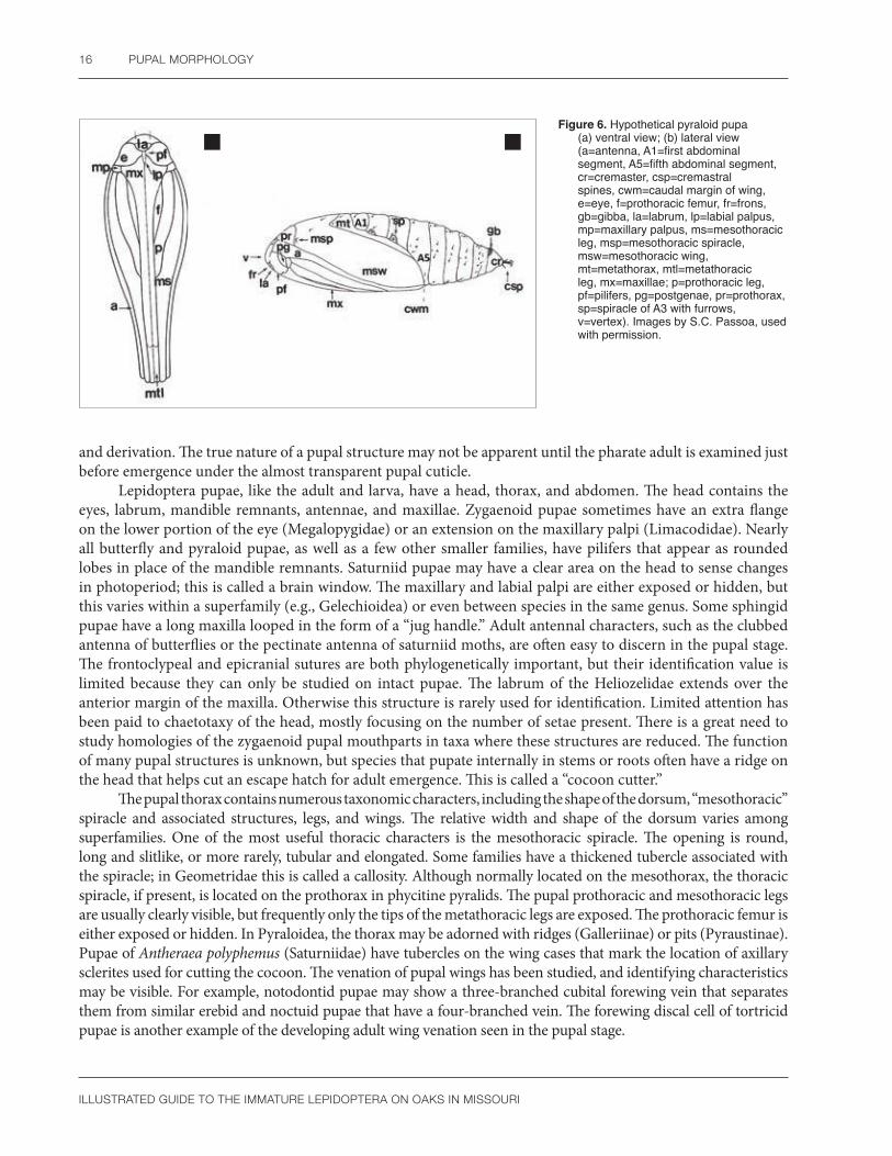
ILLUSTRATED GUIDE TO THE IMMATURE LEPIDOPTERA ON OAKS IN MISSOURI
16 PUPAL MORPHOLOGY
a bFigure 6. Hypothetical pyraloid pupa
(a) ventral view; (b) lateral view (a=antenna, A1=first abdominal segment, A5=fifth abdominal segment, cr=cremaster, csp=cremastral spines, cwm=caudal margin of wing, e=eye, f=prothoracic femur, fr=frons, gb=gibba, la=labrum, lp=labial palpus, mp=maxillary palpus, ms=mesothoracic leg, msp=mesothoracic spiracle, msw=mesothoracic wing, mt=metathorax, mtl=metathoracic leg, mx=maxillae; p=prothoracic leg, pf=pilifers, pg=postgenae, pr=prothorax, sp=spiracle of A3 with furrows, v=vertex). Images by S.C. Passoa, used with permission.
and derivation. The true nature of a pupal structure may not be apparent until the pharate adult is examined just before emergence under the almost transparent pupal cuticle.
Lepidoptera pupae, like the adult and larva, have a head, thorax, and abdomen. The head contains the eyes, labrum, mandible remnants, antennae, and maxillae. Zygaenoid pupae sometimes have an extra flange on the lower portion of the eye (Megalopygidae) or an extension on the maxillary palpi (Limacodidae). Nearly all butterfly and pyraloid pupae, as well as a few other smaller families, have pilifers that appear as rounded lobes in place of the mandible remnants. Saturniid pupae may have a clear area on the head to sense changes in photoperiod; this is called a brain window. The maxillary and labial palpi are either exposed or hidden, but this varies within a superfamily (e.g., Gelechioidea) or even between species in the same genus. Some sphingid pupae have a long maxilla looped in the form of a “jug handle.” Adult antennal characters, such as the clubbed antenna of butterflies or the pectinate antenna of saturniid moths, are often easy to discern in the pupal stage. The frontoclypeal and epicranial sutures are both phylogenetically important, but their identification value is limited because they can only be studied on intact pupae. The labrum of the Heliozelidae extends over the anterior margin of the maxilla. Otherwise this structure is rarely used for identification. Limited attention has been paid to chaetotaxy of the head, mostly focusing on the number of setae present. There is a great need to study homologies of the zygaenoid pupal mouthparts in taxa where these structures are reduced. The function of many pupal structures is unknown, but species that pupate internally in stems or roots often have a ridge on the head that helps cut an escape hatch for adult emergence. This is called a “cocoon cutter.”
The pupal thorax contains numerous taxonomic characters, including the shape of the dorsum, “mesothoracic” spiracle and associated structures, legs, and wings. The relative width and shape of the dorsum varies among superfamilies. One of the most useful thoracic characters is the mesothoracic spiracle. The opening is round, long and slitlike, or more rarely, tubular and elongated. Some families have a thickened tubercle associated with the spiracle; in Geometridae this is called a callosity. Although normally located on the mesothorax, the thoracic spiracle, if present, is located on the prothorax in phycitine pyralids. The pupal prothoracic and mesothoracic legs are usually clearly visible, but frequently only the tips of the metathoracic legs are exposed. The prothoracic femur is either exposed or hidden. In Pyraloidea, the thorax may be adorned with ridges (Galleriinae) or pits (Pyraustinae). Pupae of Antheraea polyphemus (Saturniidae) have tubercles on the wing cases that mark the location of axillary sclerites used for cutting the cocoon. The venation of pupal wings has been studied, and identifying characteristics may be visible. For example, notodontid pupae may show a three-branched cubital forewing vein that separates them from similar erebid and noctuid pupae that have a four-branched vein. The forewing discal cell of tortricid pupae is another example of the developing adult wing venation seen in the pupal stage.
ILLUSTRATED GUIDE TO THE IMMATURE LEPIDOPTERA ON OAKS IN MISSOURI
PUPAL MORPHOLOGY 17
The abdomen consists of 10 segments (A1 through A10). Mobility of these segments varies between superfamilies and sometimes between the sexes (e.g., Tortricidae), but as a rule, the abdomen moves in a more or less circular motion. The family Depressariidae is a rare exception where lateral condyles between some abdominal segments restrict the pupal movement to a vertical plane in an up or down motion. Perhaps the most obvious modification of the pupal abdomen is the presence of spines. Spined pupae occur throughout the microlepidoptera (e.g., Tortricidae, Cossidae, Tineidae) and in some Saturniidae. Smooth pupae are characteristic of the Noctuidae, Geometridae, and Notodontidae. Sometimes scars of the larval verrucae are visible in the Lymantriinae and Arctiinae. Modifications of the thorax, such as the middorsal ridge of some Pyralidae (e.g., Galleriinae), often continue onto the abdomen. Spiracles, and any associated structures such as furrows, tubercles, or modifications of the peritreme, are also important for identification.
The terminal abdominal segments contain a wealth of information. A raised smooth area, called the gibba in phycitine pyralids, is often elongate and oval. Geometrid pupae often have a dorsal groove, with or without a toothed margin, in this area. However, for identification purposes, the most important structure on the abdomen is the cremaster. No standard terminology exists for this complex structure. In the strict sense, from a morphological standpoint, a cremaster is an additional “appendage” added onto the posterior portion of A10. Oecophorinae or Tortricinae pupae have a long tubular structure at the end of the abdomen that clearly illustrates this definition. In some Crambinae, the cremaster is broad and flattened. Conversely, in the broad sense, a cremaster can be considered as any structure that holds the pupa to the substrate, be it an “appendage,” a spine, or a seta. Thus, if a pupa has only hooked setae at the end of the abdomen (as in some phycitines associated with stored grains), the cremaster can be considered absent (if defined as an appendage) or present (if defined as the setae holding the pupa to the cocoon). Most Lepidoptera have a cremaster in the strict sense bearing spines, setae, or some combination of these structures. Blastobasid pupae, like that of the acorn moth, are unusual in that they lack any support mechanism on A10. Many noctuid pupae have a cremaster with only two long thin spines, whereas some Olethreutinae have a cremaster with lateral spines and medial setae. The resolution of this confusion is to illustrate any structure if the meaning from text descriptions alone would be unclear.
Both the genital pore and the anal opening are located ventrally on the fused terminal segments of pupae. Sex is determined by the position of the genital pore, which is located on segment A8 in females and on segment A9 in males. The anal opening is on A10 in both sexes. The raised area surrounding the anus is called the anal rise in olethreutine pupae, and the number of setae in this area is taxonomically significant.
Most collectors encounter pupae by rearing larvae or by accidental discovery in the field. If the pupa is still alive, every attempt should be made to rear the specimen because adults usually can be identified whereas most pupae are undescribed and cannot be named. If a pupa fails to eclose, sometimes identifications can be made by dissecting the genitalia from the pharate adult or by preparing a slide of the larval exuvia if present. If a parasitoid emerges, preserve the lepidopteran pupal and larval exuviae because these are the only clues for identification of the host. Parasitoids without host data are usually more difficult to identify. Mosher (1916a), Nakamura (1981), and Patočka and Turčáni (2005) have the best keys for identifying pupae. These works and Kristensen (1998) provided most of the information used in the previous summary.

ILLUSTRATED GUIDE TO THE IMMATURE LEPIDOPTERA ON OAKS IN MISSOURI
18
How to Collect, Rear, and Preserve CaterpillarsPreserved caterpillar specimens are routinely acquired by confining field collected gravid females in oviposition cages under laboratory conditions that favor egg production (typical lepidopteran eggs are shown in Figure 7). This method is excellent for morphological studies, but it gives little information on the true biology of the species. For our faunal survey of Missouri oak-feeding Lepidoptera, the emphasis was on finding leaf-feeding caterpillars on their natural host plants and rearing them to adults for identification. The following summary is based on our survey experience and the works of Eliot and Soule (1902), Collins and Weast (1961), Friedrich (1986), Holland (1968), Crotch (1969), Villiard (1975), Dickson (1976), Gardiner (1982), Wagner (2005), Wagner et al. (2001, 2011), and Winter (2000). The major kinds of caterpillar damage on midwestern hardwood trees were illustrated by MacAloney and Ewan (1964).
Externally feeding caterpillars are found simply by a careful search of the appropriate host plants. Fresh leaf rolls, leaf ties, tents, and webs will harbor larvae, while many truly external feeding species are found on the undersides of leaves, on the leaf edge, on the petiole, hiding in shelters, or cryptically resting on stems or bark. One soon learns to favor the inspection of leaves showing crisply cut areas of feeding, indicating a nearby caterpillar, rather than leaves with no damage or with damage that is rounded and browning from wound repair. Frass on leaves or on the ground is another sign of caterpillar activity. Certain species (Geometridae and the caterpillars of the underwing moths) can be found on twigs, stems, or the bark. Burlap tied around the trunk of a tree can be used to find species that regularly move off the tree during the day and back on at night. Insects can be collected by placing a beating umbrella (an umbrella frame covered in white muslin or light canvas) under vegetation and shaking or beating the vegetation with a stick. This latter method allows for the collection of insects that typically fall to the ground when disturbed, but has the disadvantage of not providing any information on the resting place of the caterpillar or the kind of leaf damage it creates. Periodic checks of acorns and logs that have been enclosed in mesh or screening can reveal emerging adults for internal feeders.
Certain times of the year are more conducive to finding caterpillars. In Missouri, caterpillars are particularly abundant in early spring before leaves toughen, and again in late summer (August–September). Because the caterpillar fauna is continually changing in a predictable manner throughout the oak’s growing season (e.g., Marquis and Whelan 1994, Le Corff et al. 2000), visits staggered over time will offer new species for rearing. Several oak species should be searched because abundance varies by oak species (Marquis and Lill 2010). Though logistically difficult, canopy sampling is also worthwhile because abundances vary by forest strata (Le Corff and Marquis 1999, Forkner et al. 2004, Murakami et al. 2005). Searching for caterpillars at night with a flashlight is often productive.
To collect uncommon species, a diverse range of habitats must be sampled throughout the year, sometimes using specialized techniques. Digging for pupae in the winter months has been suggested, but few species are easily collected this way. Another option is to cage plant parts (acorns, branches, root balls) in the hope that

ILLUSTRATED GUIDE TO THE IMMATURE LEPIDOPTERA ON OAKS IN MISSOURI
HOW TO COLLECT, REAR, AND PRESERvE CATERPILLARS 19
a b
c d
e f
g h
Figure 7. Eggs of (a) Antaeotricha schlaegeri; (b) Choristoneura rosaceana; (c) Megalopyge crispata; (d) Phobetron pithecium; (e) Eutrapela clemataria; (f) Automeris io; (g) Nadata gibbosa; (h) Charadra deridens. Photos a, b, f by S.C Passoa; c, d, e, h by A. Peterson; g by G. Godfrey; all used with permission.

ILLUSTRATED GUIDE TO THE IMMATURE LEPIDOPTERA ON OAKS IN MISSOURI
20 HOW TO COLLECT, REAR, AND PRESERvE CATERPILLARS
something of interest will be captured and emerge in the container. Other less frequently collected substrates that may harbor caterpillars include moss, lichens, or fungi on the tree trunk. Peeling dead bark can also be productive in some ecosystems. Trunk, stem, or twig borers can be recognized by exit holes (that sometimes contain protruded pupae), galls, swellings in the stem, or by the wilting and eventual death of the apical buds and leaves.
Once found, caterpillars can be reared in captivity to observe all life stages. Clean plastic containers with lids work the best for most moth species. Saturniidae and Sphingidae, because of their large size, usually require larger containers or sleeving on outdoor branches of the host plant. Leaf miners should be reared in plastic bags to maintain high humidity and prevent leaf desiccation before the life cycle is completed. Free-feeding caterpillars need fresh leaves from their host plant every few days and frass emptied from the bottom of the container daily. Species that overwinter as larvae can be reared in outdoor containers, with a bit of moistened litter or soil. As soon as leaves appear in the spring, larvae should be provided with food once again. Success is greater when only a few individuals of a species are kept in a single container because some species (e.g., noctuids such as Lithophane antennata) are cannibalistic (R.J. Marquis and J.T. Lill, pers. obs.; Wagner 2005). Also, diseases often spread more rapidly in higher density rearings. Larvae should be handled as little as possible, and never during a molt. Leaf miners and stem borers need to be left in their feeding site as long as the plant quality is acceptable. Many early instar caterpillars are easily moved if offered a fresh piece of their host plant; they often crawl on the plant gently pushed below them, and this is safer than trying to pick them up with a brush or forceps. Containers should not be used twice if resources allow. If reused, wash with soap and water and soak in a 10 percent solution of bleach for a minimum of 20 minutes to kill pathogens, then rinse once more with water. Overwintering pupae can be kept in a plastic container with a slightly moistened paper towel or peat moss, placed outdoors away from direct sunlight, to allow the pupa to experience natural fluctuations in temperature and light throughout the winter. As spring returns, the adults will emerge. Some species of Noctuidae and Notodontidae pupate best in a loose soil substrate mixed with leaf litter or peat moss. Other species pupate in dead wood (e.g., most oak dagger moth caterpillars, Acronicta spp.), so providing moist, decaying wood will aid in pupation. Many microlepidoptera need only a few pieces of paper towel for a pupation substrate.

ILLUSTRATED GUIDE TO THE IMMATURE LEPIDOPTERA ON OAKS IN MISSOURI
21
How to Document a Caterpillar FaunaDocumenting a caterpillar fauna, particularly for an entire plant community, can be a daunting task. For most plant communities or even individual plant species, the local caterpillar fauna is poorly known. Sampling at multiple times during the year is essential because composition can change with leaf age. Samples also should be taken across years because species abundance can be so low in a given year that they can be easily missed in that year’s sample, even with an intensive sampling effort. Sampling at multiple sites is also important since presence and abundance varies between locations, particularly by forest age (Jeffries et al. 2006). Sampling should take place on multiple tree species; host plant lists for most caterpillar species are woefully incomplete, and even closely related tree species may not share caterpillar species. Finally, plant development stage (seedling, sapling, or mature tree) also may influence the associated insect species (Boege and Marquis 2005).
Larvae should be photographed at various instars to document morphological and color changes throughout development. A unique identification code or locality data should be associated with these photographs. For extremely accurate reproduction of larval colors, it is best to photograph the larva on a standard photographic gray card. However, it is almost always more aesthetically pleasing to place the larva on a natural background of stems and leaves, ideally from its host plant. Especially active larvae can be placed in a refrigerator for a short time to slow them, making photographing more tractable, but not so long that the natural resting posture is disrupted.
Basic techniques for working with lepidopteran immature stages are described by Passoa (2008 and references cited within), using Olethreutinae as a representative taxon. Topics include slide mounting (eggshells, larval mouthparts, and skins) and preservation of intact larvae, pupae, and their exuvia. Techniques for identifying and preserving pest Lepidoptera can be found in Passoa (2009).
When collecting caterpillars, notes should be made on the host plant species, feeding mode (e.g., in leaf shelters, leaf mines, or free-feeding), position on the plant (top or bottom of leaf, twig, or bark), type of damage (e.g., “shot holes,” skeletonization, edge feeding, and the nature of any shelter that has been constructed), and a physical description of the larva. Whether the caterpillar is feeding singly or in groups should be noted, and if the caterpillar is actively feeding. Unfortunately, from practical experience, it seems that logbooks or data notebooks are often lost or rarely consulted unless the author is a famous world authority on an insect group. However, this information must be saved because details of collection trips are impossible to remember over the long term. The best solution is to post data on the Web or place critical biological information in a very short phrase on a specimen label.
It is important to note the collection location, collection date, and emergence date if the specimen was reared. Host plant information requires special care to avoid confusion. The following terminology is suggested: “on” signifies a resting substrate (on flowers of Quercus); “ex” means the larva was reared from that host in nature (ex Quercus leaves); and “ex” a plant name “in the lab as a test” indicates a rearing in the laboratory and not a
ILLUSTRATED GUIDE TO THE IMMATURE LEPIDOPTERA ON OAKS IN MISSOURI
22 HOW TO DOCUMENT A CATERPILLAR FAUNA
natural ecological association (ex Quercus in the lab as a test). Certainly, other terminology is equally good as long as the actual meaning of the rearing is clear. One final caution: pay attention to whether the caterpillar has actually fed on the plant material and ideally molted at least once before pupation. In some cases, larvae wander off their host plant, and if collected at this time, will pupate without feeding. This can cause an erroneous host plant record.
Rearing to adulthood is often essential. Even though a number of guides to caterpillar faunas are becoming available, geographic and local variation in larval coloration and morphology make it essential that larval characteristics are matched to adult vouchers. Once emerged, adults can be prepared as described in many field guides to butterflies and moths (e.g., Covell 1984, Heitzman and Heitzman 1987, Winter 2000). Even more information is available when the larval exuvia (molted skin) and pupal case are associated with the emerged adult. Pupal cases, larval skins, and head capsules should all be preserved in gelatin (pill) capsules pinned with the adult. Pressed leaf mines or silk shelters can also be mounted in a collection to demonstrate larval damage and to associate such damage with reared adults. Ideally, additional larvae collected at the same time and deemed to be identical to those reared and photographed should be preserved in 80 percent ethanol in containers sealed with rubber stoppers. Intact pupae (as opposed to exuvia) are often preserved in alcohol to prevent the decay of internal organs that occurs when specimens are pinned.
It is important to realize that many microlepidopteran caterpillars cannot be identified to species with the naked eye; this is especially true of the early instars. In contrast, some adults are superficially indistinguishable but their caterpillars are markedly different in coloration and morphology (e.g., Acronicta and some Lymantriinae). If a large series of similar looking larvae can be collected, it is best to preserve a few representatives of each instar and rear the rest of the brood to adults, especially if the identity of the larva is unknown. Because pupal mortality can be high, it rarely pays to kill unknown pupae before rearing the adult. If multiple larvae are available, different instars can be preserved as caterpillars develop.
Dissection of pupae containing a pharate adult was used by Mosher (1916a) to homologize and identify pupal characters. This technique is also useful for extracting genitalia from adults within dead pupae for possible identifications. Molecular methods are now frequently utilized for identification of Lepidoptera. Caterpillars for such analyses are usually frozen in a -85 ºC freezer or preserved in absolute (100 percent) ethanol (e.g., Regier et al. 2009). Molecular analyses are increasingly important for resolving issues of species boundaries, in addition to their relevance to broad-scale community studies (Janzen et al. 2009).
Although the above synopsis represents a program with unlimited time and resources, important scientific contributions to lepidopteran biology and morphology are still possible even if the effort is less than ideal. Faunal studies of moths that confirm historical distribution or host records, even if these are previously published, are needed for nearly all regions of North America. A fact is only as good as the number of accurate records supporting it. Single observations that are never duplicated or confirmed will always be doubted.

ILLUSTRATED GUIDE TO THE IMMATURE LEPIDOPTERA ON OAKS IN MISSOURI
23
Parasites (Parasitoids)In almost any field collection of Lepidoptera larvae, it is inevitable that some individuals will be parasitized by other insects or nematodes (Figure 8). These natural enemies help keep populations of caterpillars in check. When a particular species of caterpillar is locally abundant, it is not unusual for upwards of 80 percent of the larvae to be attacked. In other cases the percent parasitized can be extremely low (e.g., Le Corff et al. 2000).
Figure 8. Parasitoid pupa and the remaining exoskeleton of its caterpillar host from which the parasitic wasp larva emerged. Photo by R.J. Marquis, used with permission.
Most parasitized caterpillars are destined to die prematurely, as a result of either physiological effects caused by their natural enemies, or by being fed upon by the parasitoid itself. Parasites that typically cause the death of their hosts have been termed “parasitoids” (Reuter 1913, Eggleton and Gaston 1990), although the term parasite is still often used interchangeably with parasitoid. In most cases, parasitic insects or nematodes will emerge from their host caterpillar(s) just before the host dies. They then complete their life cycle away from the host, sometimes in the soil. In some cases, the parasitoids (especially wasps of the families Braconidae and Ichneumonidae) “mummify” the host caterpillar and pupate within the inflated and hardened shell of the larva (Shaw 2006). Finally, a few species will emerge as adults directly from the parasitized caterpillar.
Several parasitoid wasp groups (especially Platygastridae, Trichogrammatidae, and Encyrtidae) are known to attack the eggs of Lepidoptera. Most will kill the eggs they parasitize and then emerge as adults, but a few (Braconidae wasps of the subfamilies Adeliinae, Cheloninae, and rarely Microgastrinae) postpone their

ILLUSTRATED GUIDE TO THE IMMATURE LEPIDOPTERA ON OAKS IN MISSOURI
24 PARASITES (PARASITOIDS)
development until the host egg hatches and the caterpillar reaches a later stage of development (Ruberson and Whitfield 1996). These resemble the more typical larval parasitoids.
Most parasitoids reared from Lepidoptera attack during the larval stage. Usually an adult female parasitoid wasp or fly finds a caterpillar and lays an egg near, or on the body. In the case of wasps, this may be after paralyzing the caterpillar with a venomous sting from its ovipositor. These parasitoid eggs then hatch, and the wasp or fly larvae either feed inside the host (endoparasitoids) or attached to the host’s exterior (ectoparasitoids). Sometimes the ectoparasitoids wait to attack relatively fully grown caterpillars, paralyze them, and the young develop rather quickly on their moribund host. These species are known as idiobionts (Askew and Shaw 1986, Quicke 1997). The endoparasitoids typically take longer to develop, and their host caterpillars are often able to regain activity after parasitization and may appear normal when collected. The parasitized caterpillar and parasitoid then have an extended period of complex physiological interaction, sometimes involving symbiotic viruses (Stoltz and Whitfield 1992), before the host eventually succumbs. These latter parasitoids are known as koinobionts, and it has been suggested they generally possess more narrowly defined host ranges than the idiobionts (Askew and Shaw 1986, Quicke 1997). The boundary between idiobiosis and koinobiosis generally follows the split between endo- and ectoparasitoids, but there are a number of exceptions. For instance, there are a few ectoparasitoids of the wasp family Eulophidae that are able to remain fixed on mobile hosts, and there are many egg and some pupal parasitoids that feed internally but have a very short interaction with their host (Mills 1992). With both ecto- and endoparasitoids, the host is usually killed before it is able to pupate. In some cases, however, the caterpillar is able to start spinning a cocoon, and in a few groups the parasitoid emerges from the host pupa (these are known as larval-pupal parasitoids).
Our understanding of the taxonomy and natural history of the parasitoids of Lepidoptera is far from complete, and any efforts to collect, save, and report information concerning parasitoids reared from caterpillars may be valuable. In most cases the emerged adult parasitoid is a fly (especially Tachinidae, Sarcophagidae) or a wasplike insect (especially Braconidae, Ichneumonidae, various Chalcidoidea, and Bethylidae). Emerged adult parasitoids should be associated with their own cocoons or pupal cases (if these can be found), as well as with the remains of the host. As much information about the times of collection and emergence, and any unusual features of the parasitized caterpillar, should be noted on a label with the emerged parasitoid. In most cases, the parasitoid and associated materials can be preserved in 70–95 percent ethanol, point-mounted, or pinned as typical insect specimens (best only for larger parasitoids). Additional useful information on the collecting, rearing, mounting, and preservation of parasitoids can be found in Ford (1943), Smith (1974), Noyes (1982), Shaw and Askew (1983), and Shaw (1997). Photographs of the hosts and parasitoids and preservation of skins, head capsules, and pupal cases can prove to be invaluable for host documentation when parasitism prevents moth or butterfly emergence. Preliminary summaries of the composition of the parasitoid communities on Ozark oak-feeding caterpillars were published by Whitfield et al. (1999) and Le Corff et al. (2000).
To correctly associate any parasitoid with its host and pupal cases, it is important that caterpillars be reared either isolated from one another (best), or sorted to species with extreme care and monitored frequently for emergence. A number of incorrect host records exist in the literature because such care was not taken. It is especially important to avoid introducing insects that feed internally, such as leaf miners and gall makers, into the rearing container with other caterpillars, as each caterpillar may host its own specific parasitoid(s).
In some cases, what appears to be a parasitized larva instead will be one that is diseased. In many cases the caterpillar will become visibly moldy or stiff, or its bodily contents will liquefy. Occasionally, elaborate fungal fruiting bodies may also sprout from diseased caterpillars. Photographs should be taken of any diseased caterpillar to aid in identification since decomposition of the host is often rapid. Diseased caterpillars and their containers should be isolated from other caterpillars. A number of viral, bacterial, and fungal diseases of caterpillars are known, but much remains to be understood about these natural enemies as well. Rivers (1983) provides an excellent introduction to caterpillar diseases. A guide to gypsy moth diseases and parasitoids (Blackburn and Hajek 2018) is another excellent resource to help distinguish these two mortality factors.

ILLUSTRATED GUIDE TO THE IMMATURE LEPIDOPTERA ON OAKS IN MISSOURI
25
How to Use this BookAlthough long technical descriptions and keys are more accurate, no doubt some readers will find the following “simplified” guide to major caterpillar families that we present in Table 1 more helpful. Using ecology and a few morphological characters at the family level, many unknown caterpillars can be placed in a provisional genus or group of families for further analysis. The family introductions, species descriptions, and photographs in this book are designed to provide additional clues that hopefully will lead to at least a confirmed genus name.
Most scientific names listed in Table 1 represent taxa that are associated with oaks in Missouri. For this table, we define the oak tree as an ecosystem, with both living and dead components and the potential of being covered with moss, lichens, or fungi.
With regard to host plants listed in our species descriptions, we only cite the genus of the plant unless the host is oak, but references are given so that the reader can find further details. For oaks, we cite all the oak species that the caterpillar feeds on, either published or from our own observation. If the host list was too long, or references were too scattered to easily compile, we list the species as “polyphagous” and try to cite the major literature sources. We generally used Tietz (1972) and Robinson et al. (2002) to compile a preliminary list, then cite other publications only if they provide additional new records. We agree with Ferguson (1975) who stated that if the plants are well known it is a “useless redundancy” and “saves space” not to include the author names. This is the case for plants of the eastern United States, especially trees, which are well-documented.
Size is an important factor to consider when identifying a caterpillar. For example, very large caterpillars are likely to be either Sphingidae or Saturniidae. At the other extreme, most species are difficult to distinguish from each other in the first or second instar, thus identifying smaller larvae is more difficult. Descriptions and/or photographs of distinctive early instar larvae are included in this book when available (see Appendix 3), but this information is often lacking even for the common species. Because certain caterpillars are limited to a particular time of year, it is often possible to eliminate candidates by combining date of occurrence with each caterpillar description.
The names in Table 1 are arranged by either feeding ecology or morphology, based on the character that allows for the fastest recognition. If an unknown larva cannot be identified by using our photographs or descriptions, consult the following publications that include some sources for Table 1: USDA (1985) and Furniss and Carolin (1992), which both describe species on trees; Chambers (1878), Edwards (1889), Tietz (1972), Vozzo (1984), and Robinson et al. (2002), which contain lists of species feeding on oak; Wagner (2005), which is a pictorial guide to the more showy species; or Stehr (1987), which contains technical morphological descriptions. The final step would be to consult foreign literature such as Porter (1997), Sugi (1987), or Gómez de Aizpúrua (1985–1992, 2002–2008) to determine whether a species similar to the unknown larva has been illustrated for another faunal region. With the notable exception of the Web site microleps.org or the Moth Photographer’s Group Web site (http://mothphotographersgroup.msstate.edu/), the Internet contains little

ILLUSTRATED GUIDE TO THE IMMATURE LEPIDOPTERA ON OAKS IN MISSOURI
26 HOW TO USE THIS BOOK
information on caterpillar morphology or biology not contained in the technical literature from peer-reviewed books and journals. Users should remember that many common species still have undescribed larvae, unknown life histories, or unknown early life stages. In a few cases, especially with microlepidoptera, the species may be new to science. Most of the large showy macrolepidopteran caterpillars can be easily identified using the Discover Life nontechnical interactive caterpillar guide (http://www.discoverlife.org/mp/20q?guide=Caterpillars) and bugguide.net.
Table 1. The feeding habits and distinguishing traits of taxa likely to be found on Missouri oak species.1
Feeding Guild Distinguishing Traits2 Taxon
Leaf Miners
Serpentine (long and narrow) mine with uninterrupted trail of frass following the mine, often ending in a blotch; empty mines with a semicircular exit slit. Deep epicranial notch; no thoracic legs, prolegs, or crochets.
Nepticulidae [can be confused with leaf mining flies].
Trumpet-shaped (expanding apically like a horn) or blotch mine, often without frass. D1 and D2 very closely spaced on A3-6; no thoracic legs, crochets present on A3-6 and A10.
Tischeriidae (p. 29).
Blotch or tent-shaped mine. Larva hypermetamorphic, early instars with shallow epicranial notch, no legs or crochets; later instars with crochets on A3-5 and A10, but absent on A6.
Gracillariidae (p. 32) [morphology of species on hosts other than oak sometimes more diverse than stated here, especially in the tropics].
Blotch mines with stringy frass trails. Deep epicranial notch; no thoracic legs, prolegs or crochets; mandible with a lateral tuft of setae.
Eriocraniidae.
Case Makers on Leaves, Branches or Trunk
Oval case of circular leaf fragments. Abdominal crochets in a circle with more than seven crochets. Menesta (Depressariidae) (p. 46).
Oval case of circular leaf fragments. Prolegs vestigial with a single transverse row of 4–7 crochets.
Paraclemensia (Incurvariidae) [unlike in most other Incurvariidae, crochets are present on A10 in Paraclemensia and Vespina].
Silken cases of leaf fragments, twigs and/or frass. Prothoracic spiracle horizontal, prothoracic shield and prespiracular group fused.
Psychidae [smaller species, if found on bark, are possibly confused with Tineidae that are associated with shelf fungi growing on the trunk].
Case open at both ends. Posterior of abdomen wide and truncated, anal crochets in an unbroken oval. Mimallonidae (p. 185).
Very small cases (1–4 mm long) shaped like a pistol with a short, blunt handle.
Coleophora (Coleophoridae) [best identified by larval case morphology; no setal characters are known to define the pistol case species group in North America].
Trunk or Twig Borers and Miners
Mesothorax and metathorax with D setae fused on dorsal sclerotized plates.
Prionoxystus (Cossidae) [cossids can be confused with Enarmonia (Tortricidae), Chrysoclista (Elachistidae), and Sesiidae, all of which bore into tree trunks].
Moss FeedersVery small abdominal spiracles, their diameter on A3-6 smaller than the corresponding SD1 setal base above them.
Probably Scopariinae (Crambidae) [group poorly studied, normally considered to be moss feeders].
Lichen, Algae Feeders
Mandible with enlarged lateral lobe, setae barbed. Various Arctiinae (Erebidae).
No enlarged lateral lobe on mandible, setae simple. Various Noctuidae.
Case opening with a circle of silk and octagonal in cross section. Homaledra octagonella (Batrachedridae).

ILLUSTRATED GUIDE TO THE IMMATURE LEPIDOPTERA ON OAKS IN MISSOURI
HOW TO USE THIS BOOK 27
Feeding Guild Distinguishing Traits2 Taxon
Web or Tent Makers on Multiple Leaves
Head red or black, hairy larva, scattered long hairs. Hyphantria cunea (Erebidae) [sometimes confused with tent caterpillars, Malacosoma spp.].
D2 of A9 joined on a single pinaculum. SV group bisetose on T2 and T3. Anal fork absent. Archips fervidana (Tortricidae).
A8 with a sclerotized ring around SD1, coloration variable. Pococera expandens (Pyralidae) (p. 136).
In AcornsSubmental pit present. Blastobasis spp. (Blastobasidae).
D2 of A9 joined on a single pinaculum. Cydia "latiferreana" complex (Tortricidae).
Dead TissueAssociated with dead oak leaves, secondary setae present. Pyromorpha dimidiata (Zygaenidae).
Feeds in dead, decayed logs. Scolecocampa liburna (Erebidae).
Under Bark In long cryptic webs of frass and silk.Probably Oecophorinae (Oecophoridae) or Tineidae (both groups are poorly studied with many life histories unknown).
Gall FeedersFeeds on oak galls. Bondia comonana (Carposinidae), Dichomeris
ligulella (Gelechiidae) (p. 61).
Feeds on Kermes scales in oak galls. Euclemensia bassettella (Cosmopterigidae).
Leaf Tier, Leaf Roller, Web Maker or Leaf Folder (1–2 Leaves)
D2 of A9 not joined on a single pinaculum. Anal comb straight or curved.
Gelechiidae (p. 55) [larva easily confused with Tortricidae or other Gelechioidea; see text for a more complete diagnosis].
D2 of A9 joined on a single pinaculum. Anal comb straight.
Tortricidae (p. 77) [larva easily confused with other Gelechioidea].
Bisetose prespiracular group on prothorax. No sclerotized ring surrounding SD1 on A8. One L seta on A9. No anal comb.
Crambidae [larva easily confused with Pyralidae].
Bisetose prespiracular group on prothorax. At least a sclerotized ring surrounding SD1 on A8. Three L setae on A9. No anal comb.
Pyralidae (p. 133) [larva easily confused with Crambidae].
Ventral prothoracic gland slit present. L1 behind spiracle on A1-8. Abdominal crochets in a uniordinal mesoseries.
A few leaf tying noctuids, e.g., Morrisonia confusa (p. 267).
Dorsal pinacula of mesothorax and metathorax enlarged. No anal comb. Stenomatinae (Depressariidae) (p. 41).
Head reticulated with punctures. SV setae on A10 almost form a horizontal line. Psilocorsis (Depressariidae) (p. 48).
Several larvae under thin strands of silk. Anal crochets in an unbroken circle. Early instar Mimallonidae (p. 185).
Not as above, trisetose prespiracular group on prothorax. No anal comb.
Miscellaneous microlepidoptera especially Gelechiidae (p. 55), Tortricidae (p. 77), and possibly Yponomeutoidea.
Prolegs on A6 and A10. Hydriomena transfigurata (Geometridae) makes a loose web.
Free (Exposed) Feeders
Horn or tubercle present on A8. Sphingidae (p. 212), some Notodontidae (p. 216), Pheosia, Symmerista (p. 230).
Hairy larvae. Heteroideous crochets (ends shorter than middle). Arctiinae (Erebidae) (p. 289).
Hairy larvae. Dorsal abdominal glands on A6 and usually A7. Lymantriinae (Erebidae) (p. 279).
Hairy larvae. Secondary setae on head. Prothorax with large lateral verruca.
Pantheinae (Noctuidae) (p. 251) [easily confused with Arctiinae].
Hairy larvae.Secondary setae almost always absent on head. Cuticle not smooth (granules or microspines present).
Acronictinae (Noctuidae) (p. 254).

ILLUSTRATED GUIDE TO THE IMMATURE LEPIDOPTERA ON OAKS IN MISSOURI
28 HOW TO USE THIS BOOK
Feeding Guild Distinguishing Traits2 Taxon
Free (Exposed) Feeders
Hairy larvae.Labrum with v-shaped notch and usually a median groove. Anal legs stubby, often lifted at rest.
Some Notodontidae (p. 216).
Hairy larvae, biordinal crochets.
Colored with middorsal white markings or thoracic red/black intersegmental bands when disturbed.
Lasiocampidae (p. 188).
Hairy larvae, biordinal crochets.
Middorsal abdominal tufts. Apex of abdominal legs is red. Apatelodes torrefacta (Apatelodidae).
Spiny larvae.Head angulate with scoli and tubercles. Middorsal scoli on A7 but not A9.
Some Nymphalidae.
Spiny larvae. Head smooth and rounded. Middorsal scoli on A9 but not A7. Some Saturniidae (p. 197).
Sluglike caterpillars (head hidden in dorsal view, body not eruciform).
Hairy larva. Prolegs on A2 and A7. Megalopygidae (p. 104).
Spiny or smooth larva. No prolegs present. Limacodidae (p. 108).
Crochets on A3-6 in a mesoseries interrupted by a fleshy lobe Lycaenidae (p. 97).
Tubercles present on body. Head smooth and rounded. Middorsal scoli on A9 but not A7. Some Saturniidae (p. 197).
Non-anal prolegs variously modified, ventral filaments sometimes present.
Prolegs only on A6 and A10. Most Geometridae (p. 144).
Prolegs on A5 smaller than A6. Subventral area with fringe of thick filaments.
Campaea perlata (Geometridae) (p. 154).
Prolegs on A5 and A6 equal in length. Often lack subventral filaments.
Semiloopers and some early instars of Noctuidae (p. 242), Erebidae (p. 272).
No prolegs on A3. Nolidae (p. 237).
Anal prolegs modified.
Head with vertical stripes. No long process from last abdominal segment. Many Notodontidae (p. 216).
Head mottled or banded. Long process from last abdominal segment. Drepaninae (Drepanidae).
Smooth skinned larva.
Prothoracic ventral gland slit present. L1 behind spiracle. Crochets in mesoseries. SV group of A1 bisetose. SD1 on A9 often hairlike.
Many Noctuidae (p. 242).
Prothoracic ventral gland slit present. L1 behind spiracle. Crochets in mesoseries. SV group of A1 trisetose. SD1 on A9 often setalike (not thin and hairlike).
Many Erebidae (p. 272).
Stipular lobe present. Less modified species of Notodontidae (p. 216).
A3-6 with two extra setae behind the spiracle. Thyatirinae (Drepanidae).
1 Most scientific names listed in Table 1 represent taxa that are associated with oaks in Missouri. A few names are taken from Robinson et al. (2002). Names in bold with a page number are treated in our book. If the name is underlined, the taxon is known to feed on oaks in the eastern United States, but this group is not treated in our book. Because these latter taxa are somewhat similar to the species on oak, we include them to prevent confusion, although they are not diagnosed. For this table, we define the oak tree as an ecosystem, either living or dead, with the potential of being covered with moss, lichens, or fungi.
2 See Figures 3 and 4 for definitions of abbreviations.

ILLUSTRATED GUIDE TO THE IMMATURE LEPIDOPTERA ON OAKS IN MISSOURI
29
Species Descriptions
TischeriidaeTrumpet leaf miner moths
Tischeriidae are a small family of monotrysian moths containing over 100 species worldwide (Nieukerken et al. 2011). Previously, Braun (1972) placed all the North American species in one genus, Tischeria, which was later split by Puplesis and Diškus (2003) into Astrotischeria, Coptotriche, and a restricted concept of Tischeria with fewer species. Their phylogeny defined the family based on adult characters, and considered Astrotischeria and Tischeria to be the sister group to Coptotriche. For more information on the systematics of Tischeriidae, consult the references listed by Davis (1987), Puplesis and Diškus (2003), Kobayashi et al. (2016), Stonis et al. (2017), and Xu et al. (2017). Braun (1972: 6) described the tischeriid egg as elliptical and encircled by a broad band of adhesive. Eggs are usually laid on the upperside of the leaf.
Larval tischeriids are easy to recognize. The bases of the D setae of A3-6 are usually touching, and the thoracic legs are greatly reduced or absent. The prolegs are poorly developed, but crochets are present (Davis 1987, 1998). The presence of a ventral prothoracic plate, similar in appearance to the prothoracic shield, is also unusual. Puplesis and Diškus (2003) stated that the larval and pupal gonads of Astrotischeria are distally rounded, but this character is not practical for identification at present. Worldwide, tischeriids feed on 17 host families (Xu et al. 2017). Braun (1972) divided the North America fauna into five groups based on foodplants. These divisions are: Quercus and Castanea; Rosaceae (one exception on Vaccinium); Asteraceae; Ceanothus; and Malvaceae. The mines form a blotch, or may slowly enlarge in a single direction as the larva matures (“trumpet mine”) (Braun 1972). There may be a circular silken structure inside the mine called a nidus, but typically no frass is present in the mine (Davis 1987). Larvae pupate in the mine (Davis 1998).
Braun (1972) characterized tischeriid pupae, in part, as having a rounded vertex sometimes with a tubercle, patches or bands of dorsal abdominal spines, forked or pointed abdominal setae, and a hooked cremaster. The characteristic close spacing of the larval abdominal D setae is present in the pupa of C citrinipennella, some European species (Patočka and Turčáni 2005: Plate 2), and one species illustrated by Mosher (1916a: Fig. 54). Although Tischeria and Coptotriche were separated in her key, Mosher (1916a) stated that no well-defined characters exist for these genera. Patočka and Turčáni (2005) used setal length to define Emmetia (now a synonym of Coptotriche) and Tischeria. Clearly, pupae have potential phylogenetic value and could support the splitting of Tischeria if setal length and other characters do not vary when more species are examined.
Although many tischeriids feed on oaks in both Europe and North America (Braun 1972, Patočka 1980), they are generally of little economic importance to these host plants. Tischeria ekebladella is a secondary pest of oak in parts of Russia (Gerasimov 1952, listed as T. complanella).

30 SPECIES DESCRIPTIONS
ILLUSTRATED GUIDE TO THE IMMATURE LEPIDOPTERA ON OAKS IN MISSOURI
Coptotriche citrinipennella (Clemens)
Larval DescriptionHead dorsoventrally flattened, not as wide as prothorax; prothoracic shield divided in two sclerotized bands of irregular width; cuticular texture of prothoracic shield and ventral plate rugose, with ridgelike microgranules (similar to Davis 1987: Fig. 26: 35D), these microgranules especially obvious on the mesothorax and metathorax; ambulatory warts present on thorax; thoracic legs vestigial; crochets in multiserial bands of differing size; each anal proleg with more than 15 crochets in two bands, the bands and crochets equal in size (Figure 9a).
Characters that separate species of Tischeria in North America are not available. Patočka (1980) used mine characteristics, the prothoracic shield texture, and the number of anal crochets to separate species of Tischeria in Europe. A similar set of characters was used by Gerasimov (1952) for the Russian species. The first couplet of his key divided species that feed on Rosaceae with crochets on A3-6 from feeders on oak and chestnut with crochets only on the anal prolegs. Coptotriche citrinipennella has two bands of crochets on A3-6 and A10, unlike some European oak feeders. This may be unusual, but the crochet arrangement on other North American oak feeders is unknown. Mine characters of Missouri oak tischeriids are given below (see Comments).
According to Braun (1972), the mine of C. citrinipennella starts out as a slightly narrow blotch. Later it enlarges and curls so that the mine hides part of the leaf edge (Figure 9b).
a b
Figure 9. Coptotriche sp. (a) mature larva on Quercus alba; C. citripennella (b) mature mine on Q. rubra. Photo a by R.J. Marquis and photo b by S.C. Passoa, both used with permission.
Pupal DescriptionVertex rounded, without a pointed tubercle; maxillae extend slightly less than one-half the distance to caudal margin of the wing; maxillary palpi absent; labial palpi and prothoracic femur exposed; metathoracic leg exposed; abdominal segments with fine spicules, those on the anterior margin of the third abdominal segment largest; abdominal D setae on A3-6 closely spaced, as in the larva; the longest body setae stout and minutely forked at the tip, the shorter ones are fine and pointed; dorsum of A10 has a pair of elongated setose knobs with two setae, and the cremaster is composed of two stout curved hooks.
North American species of Tischeriidae are poorly known as pupae. The two setose knobs on A10 of C. citrinipennella may be unusual (Appendix 4, Plate 9a). Neither Mosher (1916a) nor Patočka and Turčáni (2005) illustrate anything similar on the species they examined. The forked tip of the body setae breaks off readily and the same is true of the setae on the knobs on A10. This should be kept in mind, especially when viewing dry cast pupal exuvia.
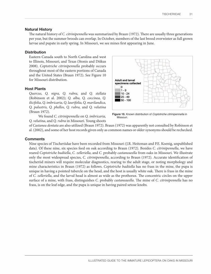
TISCHERIIDAE 31
ILLUSTRATED GUIDE TO THE IMMATURE LEPIDOPTERA ON OAKS IN MISSOURI
Natural HistoryThe natural history of C. citrinipennella was summarized by Braun (1972). There are usually three generations per year, but the summer broods can overlap. In October, members of the last brood overwinter as full grown larvae and pupate in early spring. In Missouri, we see mines first appearing in June.
DistributionEastern Canada south to North Carolina and west to Illinois, Missouri, and Texas (Stonis and Diškus 2008). Coptotriche citrinipennella probably occurs throughout most of the eastern portions of Canada and the United States (Braun 1972). See Figure 10 for Missouri distribution.
Host PlantsQuercus, Q. nigra, Q. rubra, and Q. stellata (Robinson et al. 2002); Q. alba, Q. coccinea, Q. ilicifolia, Q. imbricaria, Q. laurifolia, Q. marilandica, Q. palustris, Q. phellos, Q. rubra, and Q. velutina (Braun 1972).
We found C. citrinipennella on Q. imbricaria, Q. velutina, and Q. rubra in Missouri. Young shoots of Castanea dentata are also utilized (Braun 1972). Braun (1972) was apparently not consulted by Robinson et al. (2002), and some of her host records given only as common names or older synonyms should be rechecked.
CommentsNine species of Tischeriidae have been recorded from Missouri (J.R. Heitzman and P.E. Koenig, unpublished data). Of these nine, six species feed on oak according to Braun (1972). Besides C. citrinipennella, we have reared Coptotriche badiiella, C. zelleriella, and C. probably castaneaeella from oaks in Missouri. We illustrate only the most widespread species, C. citrinipennella, according to Braun (1972). Accurate identification of tischeriid miners will require molecular diagnostics, rearing to the adult stage, or noting morphology and mine characteristics in Braun (1972) as follows. Coptotriche badiiella has no frass in the mine, the pupa is unique in having a pointed tubercle on the head, and the host is usually white oak. There is frass in the mine of C. zelleriella, and the larval head is almost as wide as the prothorax. The concentric circles on the upper surface of a mine, with frass, distinguishes C. probably castaneaeella. The mine of C. citrinipennella has no frass, is on the leaf edge, and the pupa is unique in having paired setose knobs.
Figure 10. Known distribution of Coptotriche citrinipennella in Missouri.

32 SPECIES DESCRIPTIONS
ILLUSTRATED GUIDE TO THE IMMATURE LEPIDOPTERA ON OAKS IN MISSOURI
GracillariidaeLeaf blotch miner moths
Gracillariidae are a large, worldwide family of approximately 2,000 species that were previously classified into three subfamilies: Gracillariinae, Lithocolletinae, and Phyllocnistinae (Davis and Robinson 1998, de Prins and de Prins 2005). Phyllocnistinae is sometimes treated as a separate family, although early workers such as Chapman noted their morphological similarity to gracillariids (Emmet et al. 1985). Other classifications have suggested Gracillariidae should contain four to six subfamilies (summarized by de Prins and de Prins 2005). The most recent molecular and morphological phylogeny continues the trend of adding subfamilies for a total of eight. They are Acrocercopinae, Gracillariinae, Lithocolletinae, Marmarinae, Oecophyllembiinae, Parornichinae, Ornixolinae, and Phyllocnistinae (Kawahara et al. 2017). We use this classification for our diagnosis of the larvae that follows below. For more information on the Gracillariidae, consult Davis (1987), Davis and Robinson (1998), de Prins and de Prins (2005), Kawahara et al. (2017), and references cited by these authors. The best Internet resource is de Prins and de Prins (2018).
Eggs of the Gracillariidae are inconspicuous compared to other leaf miners like Nepticulidae (Emmet et al. 1985). Davis and Deschka (2001) illustrated some typical Phyllonorycter eggs from species feeding on Salicaceae. They were oval, reticulated to rugose on the dorsal half, and smooth ventrally. The micropyle may be reduced in size (Davis and Robinson 1998).
Gracillariid larval habits and morphology are both diverse (discussed by Davis 1987, Davis and Robinson 1998, Kawahara et al. 2017). Species of Phyllonorycter and Phyllocnistis are leaf miners. Late instar Caloptilia roll leaves. Mamara includes fruit and stem miners. Other species bore in fruit or form galls (Kawahara et al. 2017). Larvae undergo hypermetamorphosis; depending on the subfamily or species, this can include larvae feeding on sap, plant tissue, or not feeding at all. Holloway et al. (1987) summarized typical life histories for the three subfamilies of Gracillariidae in the old sense. Gracillariinae leave the mine after the sap-feeding stage and live externally in a folded or rolled leaf. Lithocolletinae remain in the mine as sap or tissue feeders. The hypermetomorphosis of Phyllocnistis includes a final cocoon-spinning instar that does not feed, and its only function is to spin a cocoon (Emmet et al. 1985, Holloway et al. 1987). Most gracillariids are host specific (Maier and Davis 1989) or sometimes oligophagous (Holloway et al. 1987).
Mine morphology is equally distinct. Specific characters include the shape of the mine itself (serpentine or blotch), location (leaf upper side or underside), texture (folds present or absent), frass characteristics (in relation to the pupa), size, host, and biology (Emmet et al. 1985: 248–252, Davis 1987). Thus, it is important to save the mine when preserving immature leaf miners (Passoa 2009).
Gracillariid larvae are usually easy to recognize. According to Davis (1987), larvae have a bisetose prespiracular group, and in those North American genera with prolegs, they occur on A3-5, A10, but not A6. With one exception from Brazil (Kawahara et al. 2017), hypermetomorphosis is also a unique feature of gracillariid larvae. Sap-feeding instars are dorsoventrally flattened with modified mandibles, but lack a functional spinneret or legs (Davis 1987, Kawahara et al. 2017). Except for Phyllocnistinae, the antennae have several fingerlike projections in addition to the normal sensory cones (Holloway et al. 1987: 198). Davis and Robinson (1998) and Kawahara et al. (2017) characterized the subfamilies of Gracillariidae by their mouthparts and chaetotaxy. The subfamilies Marmarinae, Oecophyllembiinae, and Phyllocnistinae all have larvae that construct slender, subepidermal, serpentine mines but lack tissue-feeding instars (Kawahara et al. 2017). Two genera of Marmarinae (Dendrorycter and Marmara) have larvae with a “pharate phase” that sits within the exoskeleton of the preceding instar (Davis and Robinson 1998, Wagner et al. 2000). Their larvae were characterized by Kawahara et al. (2017) as follows. The head and spinneret of the pharate phase is much reduced, and it lacks both stemmata and mandibles. The last spinning instar of this subfamily has five to six stemmata and three pairs of labral setae. There is a single lateral seta on the mesothorax and metathorax. Oecophyllembiinae contains six genera with very unusual larvae (summary by Kawahara et al. 2017). With one exception, an extra thoracic spiracle is present on the mesothorax in addition to the one normally found on the prothorax. The last instar

ILLUSTRATED GUIDE TO THE IMMATURE LEPIDOPTERA ON OAKS IN MISSOURI
has one or two lateral setae on the mesothorax and metathorax. The number of abdominal prolegs is also unusual. For instance, Metriochroa and Prophyllocnistis have prolegs on A3-6 as do most species of Lepidoptera. Cryphiomystis has an extra anterior proleg on A2 in addition to those on A3-6. The last instar head of both Metriochroa and Prophyllocnistis lacks stemmata and has only two to three pairs of labral setae. Larvae of the Phyllocnistinae (summary by Kawahara et al. 2017) lack a spiracle on the mesothorax and have early sap-feeding instars with a flat head, broad, flat mandibles, and no stemmata. The last, nonfeeding stage has a greatly reduced head morphology with small antennae, no mandibles, and no stemmata. The only well-developed head structure is a spinneret. There are two D setae on A9 (Davis and Robinson 1998). There is only one genus, Phyllocnistis.
The remaining five subfamilies form blotch mines, roll leaves, or feed on flowers or fruits (Kawahara et al. 2017: Fig. 3). Parornichinae has four genera. The last instar has six stemmata, six labral setae, and three lateral setae on all body segments except the prothorax (Kawahara et al. 2017). There are five coxal setae, and the D group of A9 is bisetose (Davis and Robinson 1998). There are 28 genera in the subfamily Ornixolinae. The last instar larva has five to six stemmata, four to six labral setae, and two lateral setae on the mesothorax and metathorax (Kawahara et al. 2017). This subfamily also has five coxal setae and a bisetose D group on A9 (Davis and Robinson 1998). Lithocolletinae has 11 genera. The mouthparts were not described by Kawahara et al. (2017), but Davis and Robinson (1998) noted that there are four coxal setae, and the D group of A9 is unisetose. Acrocercopinae is another large group with 28 genera. The final instar has two lateral setae on all segments, five pairs of stemmata, and four to five pairs of labral setae. The last subfamily, Gracillariinae, has 25 genera. Mature larvae roll leaves and have six stemmata, six labral setae, three lateral setae on the mesothorax and metathorax, and two lateral setae on each abdominal segment. Sap-feeding forms have sawlike mandibles (Common 1990: Fig. 72:5), although the last instar hypopharyngeal complex and mandible are more typical of lepidopteran larvae (e.g., Passoa 1980).
According to Mosher (1916a), the pupae of gracillariids have antennae and metathoracic legs longer than the wings. In addition, abdominal segments A4 to A6 (females) or A4 to A7 (males) are not fused, and patches of spines are present on the abdominal dorsum, usually in two sizes. A similar diagnosis was given by Patočka and Turčáni (2005: 23–24) for the European species. They described gracillariid pupae as “semiliberae” (not obtect), having the vertex longer than the pronotum, the wings long and pointed, and the head often with a frontal projection. A cremaster is present or absent, at most there are two pairs of hooklike setae, or the end of the abdomen has small spines. Nakamura (1981) had a simpler diagnosis for Japanese species. Gracillarioidea was defined by having the hindwing covered by the forewing at either the first, second, or third abdominal segment. For Gracillariidae, maxillary palpi were present, or if they were absent, the antennae were at least as long as the body. “Phyllocnistidae,” considered a separate family by Nakamura (1981), lacks maxillary palpi with antennae that do not reach the caudal end of the body. In addition, Kawahara et al. (2017) noted that pupal Phyllocnistinae lack lateral processes or setae on the frontal projection of the vertex, the abdominal dorsum has a pair of dorsal setae and dorsal hooks, and the cremaster has only one pair of caudal processes. In spite of these varying diagnoses, typical gracillariid pupae present a characteristic appearance with their pointed head, free appendages, long antennae, and spined abdomen. Depending on the genus, good characters exist for identification of pupal gracillariids (e.g., Emmet et al. 1985: 302).
Most pest gracillariids are associated with trees and shrubs. Some examples mentioned by Holloway et al. (1987) include Conopomorpha on cocoa, Phyllonorycter on apple, and Phyllocnistis on citrus. Cameraria cincinnatiella (USDA 1985) and C. hamadryadella (Solomon et al. 1999) occasionally damage oak in the central United States. Phyllonorycter messaniella is a serious pest of oak in Europe and New Zealand (Browne 1968). The horse chestnut leaf miner, Cameraria ohridella, is another pest gracillariid in Europe (de Prins and de Prins 2018).
GRACILLARIIDAE 33

34 SPECIES DESCRIPTIONS
ILLUSTRATED GUIDE TO THE IMMATURE LEPIDOPTERA ON OAKS IN MISSOURI
Subfamily Lithocolletinae
Phyllonorycter fitchella (Clemens)
Larval DescriptionBased on several head capsules dissected from larval exuviae associated with reared adults: Mature larva with three stemmata of almost equal size on each side of the head; labrum deeply divided with a v-shaped notch, epipharynx with a dense patch of setae forming a vertical row on each side of the cleft, joined medially at the base of the labrum; spinneret approximately as long as the labial palpi, the proximomedial region spined, and mandible with four or five blunt teeth (depending on if the smallest lobe is counted as a tooth).
The lack of morphological information and preserved larvae of North American oak-feeding Phyllonorycter prevent a meaningful diagnosis of P. fitchella. Davis (1987) illustrated the stemmatal number, crochet arrangement, and mandibles as useful taxonomic characters for gracillariids. Using Davis and Deschka (2001), the stemmatal arrangement of P. fitchella most closely resembles Fig. 182, the epipharyngeal spine pattern as in Fig. 124, and the mandible is similar to Fig. 185, except that the teeth are smaller and rounded. Sap-feeding instars of P. fitchella could not be accurately identified and thus were not described. Lack of material and extreme morphological similarity were problems encountered by Davis and Deschka (2001: 5) in their study of Phyllonorycter on Salicaceae. Larvae should be reared to adult for accurate identifications (see Comments).
Pupal DescriptionBased on a single damaged pupal exuvia associated with a reared adult: Vertex with projection present; paired clypeal setae on each side that almost touch each other; labial palpi partially exposed; A7 with a wide flat ridge on each side; the abdominal dorsum covered with minute spines of two sizes, the largest spines on A8 are at the anterior margin, and cremaster with large dorsal spines and four hooked setae.
The pupa of P. fitchella is suspended in a slight web of silk (Braun 1908) unlike P. basistrigella, which pupates in a cocoon lined with frass. Maier and Davis (1989) considered this frass pattern diagnostic for P. basistrigella. Otherwise, mines of P. fitchella can be confused with P. basistrigella until the prepupa constructs a cocoon. As with the larva, the pupa of P. fitchella can be confused with several other oak-feeding species.
The spined cremastral texture of P. fitchella most closely resembles Fig. 220 of Davis and Deschka (2001). Phyllonorycter fitchella lacks an accessory cremaster (defined by Davis and Deschka 2001: 26); instead there is a pair of flat ridges on the venter of A7. These are present in P. basistrigella and mentioned by Davis and Deschka (2001: 7) for some Phyllonorycter feeding on Salicaceae.
Natural HistoryThe life history of P. fitchella was studied by Fitch (1859: 827 as P. quercifoliella) in New York State. The larva is pale white with brown mandibles. There is also a red-brown middorsal stripe formed from the gut contents showing through the pale body. The pupa is initially pale yellow, very mobile, and able to spin and wriggle if disturbed. Mines are distributed throughout the host, from near the ground up to the tops of tall trees. Moths were found in the spring and autumn.
In Missouri, there are two generations per year. The first begins in late May to early June. The upper surface of the mine is a single to triple series of brown dots outlining the circular to oval blotch mine (Figure 11a, see also Harrison 2017). The undersurface epidermis is wrinkled with one to two ridges, turning from white to whitish brown as the mine matures (Figure 11b). The prepupa is pink (Figure 11c). Pupation occurs in the mine. According to Maier and Davis (1989), P. fitchella has two, possibly three, generations per year.

GRACILLARIIDAE 35
ILLUSTRATED GUIDE TO THE IMMATURE LEPIDOPTERA ON OAKS IN MISSOURI
a b c
Figure 11. Phyllonorycter fitchella (a) mature mine on Quercus alba, leaf top side; (b) mature mine on Q. alba, leaf bottom side; (c) prepupal larva. Photos by R.J. Marquis, used with permission.
DistributionEastern Canada (Quebec), Maine to Florida, then west to Colorado, Texas, and California (Maier and Davis 1989, de Prins and de Prins 2005), including Missouri. Although P. fitchella has a wide distribution in North America, there are only scattered records from well-collected areas of the United States. See Figure 12 for Missouri distribution.
Host PlantsQuercus alba, Q. ilicifolia, Q. macrocarpa, Q. prinoides, Q. prinus, and Q. stellata (Robinson et al. 2002); Q. bicolor and Q. castanea (de Prins and de Prins 2005).
Phyllonorycter fitchella is common on members of the white oak group, but the record on Robinia was considered an error by Maier and Davis (1989). We have found this species to be very common on Quercus alba, Q. macrocarpa, Q. muehlenbergii, and Q. stellata in Missouri.
CommentsPhyllonorycter basistrigella was recorded from Missouri by J.R. Heitzman and P.E. Koenig (unpublished data); this is an example of an oligophagous oak feeder (Maier and Davis 1989) and as such is expected on any of the Missouri oaks. We have collected mines with a characteristic circle of frass around the pupa similar to the description in Braun (1908), thus confirming this record.
Maier and Davis (1989) noted that six species of Cameraria and seven species of Phyllonorycter mine the oaks of southern New England. We illustrate only one species in this book, but warn that identifications ideally should be based on reared adults, molecular methods, or larvae with associated preserved mines at a minimum. Two species of oak, Q. bicolor and Q. macrocarpa, had eight gracillariid species associated with each plant in southern New England (Maier and Davis 1989). Phyllonorycter aeriferella, for example, occurs with other species of Gracillariidae on oak (Maier and Davis 1989); thus, it would be an error to assume that every mine on a single oak leaf is the same species. Given the potential for errors, especially on the two oak species mentioned above, gracillariid identifications should be done with care.
Figure 12. Known distribution of Phyllonorycter fitchella in Missouri.

36 SPECIES DESCRIPTIONS
ILLUSTRATED GUIDE TO THE IMMATURE LEPIDOPTERA ON OAKS IN MISSOURI
DepressariidaeFlat-bodied moths
Depressariidae contain 2,300 species worldwide (Sohn et al. 2016). Based on several morphological and molecular analyses, a number of classifications of this family have been proposed. As is the case with many Lepidoptera, family composition of the Depressariidae will depend on the data matrix and methods used by researchers to form their classification. Bucheli (2009) summarized most of the morphological characters used in traditional gelechioid phylogenies, including those that apply to the Depressariidae. Consult Hodges (1998) for the core literature on biology and systematics of the Gelechioidea. For more information on depressariid immatures, including chaetotaxy, consult Hodges (1974), Stehr (1987), Fetz (1994), and Passoa (1995). The most recent molecular classifications can be found in Heikkilä et al. (2014) and Sohn et al. (2016).
Until 1989, Depressariidae was a subfamily (Depressariinae) in the Oecophoridae (Hodges et al. 1983). Using morphology of the pupa, Minet (1989: 248) enlarged and redefined the Elachistidae to include those oecophorid species formally placed in the Depressariinae, plus the Agonoxenidae, Stenomidae (including Aeolanthinae), Elachistidae (in the traditional sense), and Ethmiidae. To this group he added two newly defined subfamilies, Cryptolechiinae and Hypertrophinae. Passoa (1995) did not formally use the name Elachistidae in the broad sense. He did include Stenomatinae (“Stenomidae”), Ethmiinae, Depressariinae, Elachistinae, Amphisbatinae, Peleopodinae, Agonoxeninae, Hypertrophinae, and Xyloryctinae in “Oecophoridae 1” (nomenclature followed Fetz 1994) of his series Oecophoriformes (paralleling nomenclature in the Pyraloidea, see Munroe 1972). Hodges (1998) accepted the definition of Minet (1989) and separated Elachistidae as the sister group to the other Gelechioidea. Elachistidae, as defined by Hodges (1998), contained the Stenomatinae (“Stenomidae”), Ethmiinae, Depressariinae, Elachistinae, Deuterogoniinae, Aeolanthinae, Agonoxeninae, and Hypertrophinae. A recent morphological analysis by Kaila (2004) also placed depressariid subfamilies in what was called an oecophorid lineage, but he too uniquely defined the family. His grouping contained the Hypertrophinae, Depressariinae, Ethmiinae (with Orophia), Aeolanthinae, Parametriotinae, Agonoxeninae, and Elachistinae, plus miscellaneous “amphisbatids and depressariids” of various authors. Lvovsky (2012) divided the Gelechioidea into several superfamilies, but this classification is generally not followed.
With the development of molecular phylogenies, several authors turned their attention to the Gelechioidea. Recent molecular analyses have confirmed some morphological hypotheses, but also created novel relationships, some of which will likely need to be reconsidered (Powell and Opler 2009: 60). Bucheli and Wenzel (2005) confirmed a close relationship between Ethmiinae and Depressariinae, but placed Stenomatinae near the Coleophoridae. Kaila et al. (2011) also did not recover the enlarged concept of Elachistidae as a monophyletic unit. Their elachistid sensu lato assemblage contained the Aeolanthinae, Depressariinae (sensu lato, sensu stricto), Peleopodidae (with Carcina), Lecithoceridae, Hypercalliinae, Oecophoridae, Hypertrophinae, Ethmiinae, Cryptolechiinae, and Stenomatinae. Separated from the above clade, a second section called Elachistidae sensu stricto, included only Elachistidae in the traditional sense of Braun (1948). The final section called Elachistidae sensu lato had “Stathmopodidae” and Parametriotinae. Recent molecular phylogenies accepted the definition of Elachistidae in the narrow sense while transferring the remaining subfamilies to a new family called Depressariidae. Heikkilä et al. (2014: 585) defined Depressariidae to include 11 taxa: Acriinae, Aeolanthinae, Cryptolechiinae, Depressariinae, Ethmiinae, Hypercalliinae, Hypertrophinae, Oditinae, Peleopodinae, Stenomatinae, and several genera of uncertain placement (Carcina, Gonionota, Machimia, Himmacia sensu stricto, Psilocorsis, etc.). Sohn et al. (2016: 17) sampled some of the above taxa, and although they confirmed a core group of Depressariidae, they could not confirm monophyly of this family, perhaps due to a lack of material.
For our morphological introduction to the classification of the Depressariidae, we use the revision by Heikkilä et al. (2014: 585) because it has the most complete taxon sampling. Depressariidae was also the name used by Pohl et al. (2016) for the species we treat in our book. They recognized five groups of North American Depressariidae: Depressariinae, Ethmiinae, Peleopodinae, Stenomatinae, and a group called “unplaced Depressariidae.”

DEPRESSARIIDAE 37
ILLUSTRATED GUIDE TO THE IMMATURE LEPIDOPTERA ON OAKS IN MISSOURI
Larvae of the Depressariidae are diverse and are best recognized by studying the individual groups since there appears to be no single larval character to define the family. A large percentage of the larvae seem to have secondary setae on at least the anal prolegs. As mentioned with regard to the systematic position of Machimia, a group of subfamilies lack L3 on A8 and/or have a bisetose L group on A9. These subfamilies are discussed below. This group includes species of Acria (Acriinae) (Chen and Wu 2011) and Hypercallia citrinalis (Hypercalliinae) (Fetz 1994: 135, 229). Acria feeds on a wide variety of plants, including some of economic importance, such as coffee, mulberry, castor bean (Robinson et al. 2001), and coconut (Chen and Wu 2011). Hypercallia citrinalis eats Polygala (Parenti 2000). Jinbo et al. (2004) suggest that Anchinia belongs in the Hypercalliinae, but L3 is present on A9 in Anchinia (Fetz 1994: 135). It feeds on Daphne (Jinbo et al. 2004). Komai et al. (2011: Fig. 3: 31–34) partially illustrated the larva of four species of Scythropiodes (Oditinae). At least one species has a bisetose L group of A8 and A9. In other cases the distance between P1 setae is equal to that separating the P2 setae, the mandible appears to have short heavily sclerotized scissorial teeth, and the D2 setae of the anal shield are short. A wide variety of plant species are eaten by members of this subfamily (Robinson et al. 2001).
Depending on how the subfamily is defined, Hypertrophinae have secondary setae in the abdominal SV groups, the D and SD setae on A9 form a vertical line, the L group on A9 has secondary setae, and the crochets of A10 are sometimes broken into two groups (Common 1980; Fetz 1994: 135, 137; Hodges 1998). They contain species that feed on plants from Polygalaceae, Thymelaeaceae (Parenti 2000), and Myrtaceae (Hodges 1998).
Host plant associations are very helpful for identification of the subfamily Ethmiinae. Most feed on plants from Boraginaceae and Hydrophyllaceae (Hodges 1998). Unusual characteristics of ethmiine larvae are: a greater distance between P2 setae than the distance separating the P1 setae; abdominal SV pinacula and/or anal proleg with secondary setae; crochets in a mesoseries or penellipse; D setae of A9 closely spaced in an almost horizontal or slanted line; and in Pyramidobela, the mandible has a notch on the outer margin (MacKay 1972: Figs. 23, 24; Powell 1973: 25; Passoa 1995: 26, 27).
Stenomatinae and Depressariinae contain more species than other subfamilies of Depressariidae. As a result of this worldwide diversity, caterpillars from these two subfamilies are recorded feeding on leaves, flowers, stems, or seeds of plants from 16–17 plant families (Hodges 1998). To the naked eye, Stenomatinae larvae on Missouri oaks are easily confused with Gelechiidae because both families have leaf-tying caterpillars of approximately the same body size (10–23 mm long) with similar ecology. Stenomatinae are distinguished by having an enlarged D or SD pinaculum on at least the mesothorax, a characteristic never found in the Gelechiidae (Passoa 1995: 27). In addition, there is usually a close association of SD1 setae with the L setae on A9 in Stenomatinae (MacKay 1972: 17). Some Stenomatinae have a membranous pad on the abdominal prolegs, and similar to the Gelechiidae, the D and SD setae on A9 may form a vertical line (Stehr 1987: 381, 382; Passoa 1995: 233).
Depressariinae lack many of the morphological specializations seen in other Gelechioidea and thus are difficult to define (Fetz 1994, Passoa 1995, Hodges 1998). Typically, in North America (Passoa 1995) stemma 1 is larger than stemma 2, the distance between the P2 setae is greater than the distance separating the P1 setae, secondary setae are absent except for a few scattered hairs on the anal proleg (Apachea is an exception), A9 has SD1 hairlike with D1 anteroventrad of D2, the abdominal crochets are in a circle, and the anal region is spinose.
One species of Peleopodinae (Durrantia) has a ring on the D2 seta of the metathorax (Passoa, 1995: 252). Depressariinae feed on 17 plant families worldwide (Hodges 1998) but are often associated with Apiaceae and Asteraceae in the Holarctic region. Host records are from Malpighiaceae, Verbenaceae, Polygonaccae, and Platanaceae (Duckworth 1970) but most members of the Peleopodinae have unknown life histories.
Three species of Cryptolechiinae in the genera Orophia (Fetz 1994: Fig. 150) and Eutorna (Komai et al. 2011: Figs. III: 18, 19) seem unusual in that A9 has a patch of small secondary setae below SV1. It is apparently present in Cacochroa permixtella as well (Fetz 1994: 190), although it was not shown by Huertas Dionisio (2003). In addition, L1 and L2 are joined on a single pinaculum separate from L3 that may sometimes include SD1 (Komai et al. 2011: Figs. III: 19). Like Machimia, Orophia has a reduced D1 seta on the mesothorax (Fetz 1994:
38 SPECIES DESCRIPTIONS
ILLUSTRATED GUIDE TO THE IMMATURE LEPIDOPTERA ON OAKS IN MISSOURI
Fig. 148). The arrangement of D1 and D2 on A9 is similar to the Ethmiinae (Heikkilä et al. 2014). Larvae of the Cryptolechiinae feed on a wide variety of hosts (Medvedev 1989: 848). Larvae of the Aeolanthinae are poorly known. A species in India is a leaf roller on Meliaceae (Hodges 1998).
Pupae of Depressariidae can be divided into several groups. Some subfamilies have lateral condyles that give them a unique morphology and movement (Hodges 1998: 133). They may or may not have a pubescent pupal texture. In other cases, these condyles are secondarily lost or absent. A third division are those that have sparse to dense setal/spine patches above the anal rise, on the anal rise, or at the apex of the abdomen on the cremaster. Finally, a few subfamilies pupate exposed and oriented perpendicular to the substrate. Hypertrophinae are interesting in that the pupae fit all these categories except they have a smooth texture.
Lateral condyles are a morphological specialization that appears as points of articulation between abdominal segments, often involving A5/A6 and A6/A7 (Hodges 1998: 136, Berenbaum and Passoa 1999: Fig. 2A). These condyles restrict lateral movement of the abdomen, instead forcing an up and down “clicking” motion (Passoa 1995: Figs. 103, 104). When sclerotized ridges are present, this movement could pinch natural enemies (S.C. Passoa, pers. obv.). Hinton (1955) called these gin traps in beetle and moth pupae. Lateral condyles are clearly present in the Stenomatinae, Hypertrophinae, Depressariinae, Ethmiinae (Passoa 1995: 250), and Cryptolechiinae (Huertas Dionisio 2003). Pupae of the Depressariinae, Cryptolechiinae, and primitive Ethmiinae are pubescent. Some Ethmiinae have spiracular pits (Powell 1973). Depressariine pupae are characterized by their tubular mesothoracic spiracle (Passoa 1995: 254, 255). Lateral condyles are apparently absent in the Peleopodinae (Passoa 1995: 250, Lvovsky 2012: 189) and Aeolanthinae (Fletcher 1933: Plate X). These pupae also have a smooth texture.
Another depressariid pupal specialization, exemplified by the Hypertrophinae, involves a mode of pupation in which the pupa sits exposed, erect, and almost perpendicular to the substrate (e.g., Common 1980). These pupae may be camouflaged as a branch, bud, or other inedible object; another possibility is that their position may be related to their temperature and position to the sun (Passoa 1989, Minet 1989: 242). Experimental evidence is needed to test these hypotheses. Gonionota, considered unplaced by Heikkilä et al. (2014), would seem to belong here based on mode of pupation. Jinbo et al. (2004) stated that erect pupation was not a characteristic of Anchinia, a genus normally assumed to be in this category (Minet 1990), because they hang upside down (Lvovsky 2012).
Three subfamilies of Depressariidae have pupal spines or setal patches on the last abdominal segments. These include Hypercalliinae, Oditinae, and Acriinae (Common 1980, 1990; Minet 1989; Fetz 1994: 135; Komai et al. 2011: Figs. 3: 34, 18: 18 J; SCPC). The pupal characteristics of these taxa are discussed below with regard to the systematic position of Machimia.
A large group of Missouri oak-feeding depressariids are placed in the Stenomatinae, and their classification within this subfamily seems stable. Two other oak-feeding depressariids, Machimia and Psilocorsis, have a more doubtful systematic position and nomenclature. Both were previously placed in a taxon that included Amphisbatis, which depending on the author, was called a tribe (Amphisbatini), subfamily (Amphisbatinae), or family (Amphisbatidae) (Hodges 1974, Passoa 1995, Hodges 1998, and see Heikkilä and Kaila 2010: 79–80). When a phylogenetic analysis by Heikkilä and Kaila (2010) suggested that Lypusa was related to Amphisbatis and that both belonged in the same family, Amphisbatidae became a junior synonym of Lypusidae (Nieukerken et al. 2011). Thus, genera formally in the Amphisbatidae, but not related to Lypusa or Amphisbatis in the Lypusidae, all of a sudden lacked a familial placement. To resolve this problem, Nieukerken et al. (2011: Footnote 34) informally transferred “most of the Amphisbatidae” back to the Depressariinae without specifically listing which genera were moved. This implied that Machimia and Psilocorsis are currently in the Depressariinae (e.g., Kaila et al. 2011). Although convenient, no morphological or molecular data support this change (Urra 2014), which makes the classification unacceptable even to our conservative treatment. Thankfully, Pohl et al. (2016) placed Machimia and Psilocorsis in a category called unplaced Depressariidae. Because these two genera of oak leaf tiers are both common and sometimes pests, we compare their morphology to the Depressariinae and selected gelechioid relatives to justify our classification and acceptance of the atypical category of unplaced Depressariidae

DEPRESSARIIDAE 39
ILLUSTRATED GUIDE TO THE IMMATURE LEPIDOPTERA ON OAKS IN MISSOURI
among the several choices available in the literature. Understanding the phylogenetic position also emphasizes characters that help with their identification.
Larval characters used to recognize Psilocorsis include a punctate head texture; a mesothorax, metathorax, and A8 with the D1 seta reduced; and SV setae of A10 that form, or nearly form, a horizontal line (MacKay 1972, Passoa 1995: 30). All of these features are found in the genus Doina (Hormazábal et al. 1994) that was placed in the Amphisbatini by Becker (1984). Unfortunately, workers have not been able to acquire material of this genus for inclusion in molecular studies. The similar larval morphology suggests Psilocorsis is probably related to Neotropical genera near Doina. Other subfamilies of Depressariidae do not have these three larval characters (head texture, reduced D1 setae, and SV setae in a line). However, as explained above, there is no family name available for the genera formally placed in the Amphisbatini. Thus, Doina also is probably “unplaced.”
Passoa (1995: 254, 255) defined the Depressariinae by having a tubular pupal mesothoracic spiracle. He placed them as the sister group to the Ethmiinae based on spacing of the larval P setae and a pubescent pupal cuticle. Psilocorsis and other North American Amphisbatini were excluded from the Depressariinae-Ethmiinae clade because they have a smooth, not pubescent, pupal cuticle, and the arrangement of the P setae is different. In addition, Psilocorsis lacks a tubular mesothoracic spiracle, which excludes it from placement in the Depressariinae. This characterization also applies to the pupa of Doina because it also lacks a tubular mesothoracic spiracle, and the texture is not pubescent (Hormazábal et al. 1994). A phylogenetic analysis by Hodges (1998) suggested an even wider separation between Psilocorsis and Depressariinae; he recognized Amphisbatidae as a family. Kaila’s (2004) morphological analysis rooted Psilocorsis in the “unplaced elachistids.” However, he could not code the P setae, so this important character was not included in his phylogeny. Molecular studies such as those by Bucheli and Wenzel (2005), Kaila et al. (2011), Heikkilä et al. (2014), and Sohn et al. (2016) also failed to link Psilocorsis with a traditional definition of Depressariinae. When Psilocorsis was added to a data matrix used for a phylogeny of Depressaria, again it did not root in the Depressariinae, even when genitalia were emphasized instead of the immature stages (Bucheli et al. 2010). However, the pupal movement of Psilocorsis is similar to some Gelechioidea with obvious lateral condyles (Minet 1989). This suggests that their position in, or close to, the family Depressariidae is justified (Powell 1973: 26) and that the lateral condyles may have been lost secondarily (Passoa 1995: 250). But see Heikkilä et al. (2014: 577) for a discussion of this character; losses and independent gains of this modification caused the authors to conclude that lateral condyles may have limited phylogenetic value.
Psilocorsis does root with Phaeosaces in the molecular/morphological study by Heikkilä et al. (2014). Unfortunately there is no published larval information on Phaeosaces, and it is placed in Oecophoridae sensu lato. This analysis suggests Psilocorsis is related to the Oecophoridae. It is very interesting that Psilocorsis also links with Oecophorinae in the study by Bucheli and Wenzel (2005). However, the relationship is hard to accept considering that Psilocorsis has very different male genitalia than the Oecophorinae (Hodges 1974), the pupa places Psilocorsis in the Depressariidae, and the larvae of the Oecophorinae do not resemble Psilocorsis in morphology or biology (SCPC).
Because Psilocorsis did not root in any clearly defined subfamily of Gelechioidea (including Depressariinae) based on morphology of any stage or in molecular studies, this taxon likely needs a new family or at least subfamily by itself. Kaila (2004: 324) recommended that no new names be coined for the Gelechioidea without a strong phylogenetic justification. We agree and leave Psilocorsis in unplaced Depressariidae following Pohl et al. (2016), pending a further survey of taxa (e.g., Doina) possibly related to Psilocorsis (Passoa, in prep.).
Machimia tentoriferella has a similar taxonomic history to Psilocorsis and was previously placed, depending on the author, in the Amphisbatini, the Amphisbatinae, or the Amphisbatidae (Hodges 1974, Passoa 1995, Hodges 1998). Leraut (1992: 131) first noticed Machimia was not morphologically similar to other Amphisbatinae, but he retained the genus in that subfamily based on adult characters. Kaila (2004) joined Machimia near genera normally placed in the Hypertrophinae using characters of all stages. Using morphological and molecular data, Heikkilä et al. (2014: Figs. 2, 4) placed Machimia in a clade with Oditinae (Scythropiodes), two Peleopodinae (Acriinae and Carcina), Hypercalliinae, Aeolanthinae, Depressariinae, Hypertrophinae, Cryptolechiinae,

40 SPECIES DESCRIPTIONS
ILLUSTRATED GUIDE TO THE IMMATURE LEPIDOPTERA ON OAKS IN MISSOURI
and Ethmiinae. This was similar to the results of Kaila et al. (2011), but with more taxa as possible relatives. Sohn et al. (2016) did not study Machimia. Here we look at the similarities of Machimia to Hypertrophinae, Hypercalliinae, Cryptolechiinae, Oditinae, Acriinae, Ethmiinae, Depressariinae, and Carcina as suggested by the above phylogenies. Larva of the Aeolanthinae are too poorly known for a comparison.
The most important larval characteristics of Machimia tentoriferella are SD1 thinner than SD2 on T2 and T3, L3 absent on A8 and A9, SD1 is hairlike on A9, and the absence of secondary setae on the anal proleg. In the field, the larva lives in a characteristic web that often has alternating bands of dark and light silk (Passoa 1995).Larvae of the Hypercalliinae (Hypercallia citrinalis, see Fetz 1994: 135, 229) and Machimia both have the L group bisetose on A9 and secondary setae present on the anal proleg. But the L seta is present on A8 in Hypercallia and the thoracic SD2 seta are not thin (Fetz 1994: 137). A species of Acria (Acriinae) has a bisetose L group on A8 and A9 (Chen and Wu 2011), but otherwise the chaetotaxy is different from Machimia (e.g., secondary setae are found on the margin of the prothoracic shield instead of the anal prolegs and SD2 is not hairlike on the thorax). In contrast, an Acria in Japan does have secondary setae on the proleg (Komai et al. 2011: 634), but the other characters of Machimia listed above are not discussed. A third potential relative, Carcina quercana, seems to be quite similar in chaetotaxy and biology to Machimia. Both genera have a bisetose L group on A8 and A9 (Fetz 1994: 130) with secondary setae on the anal proleg (SCPC ). Like Machimia, the larva of Carcina makes a web and feeds on many trees including oak (Alford 1984). Lvovsky (2012: 202) placed Carcina near the Amphisbatidae in his phylogenetic study based on morphology. Limited information is available on the larvae of the Oditinae. Komai et al. (2011: Fig. 3: 31–34) partially illustrated the larvae of four species of Scythropiodes. At least one species has the L group of A8 and A9 bisetose. There is little information on other body chaetotaxy except for the head. This loss of L setae in these taxa on A8 and/or A9 is unusual and is likely to be phylogenetically significant. Unfortunately, it is unknown how widespread this character is in the Gelechioidea. A bisetose L group on A9 also occurs in some gelechiids (Fetz 1994: 185), which are unrelated to Depressariidae, a probable example of homoplasy. Interestingly, larvae of Machimia (Passoa 1995: Fig. 19), Acria (Shon 2006: 155), and many Hypertrophinae (Hodges 1998), including one Australian species of Hypertropha (Common 1980: Fig. 38), all feed inside a silken tent. The phylogenetic value of this unusual larval behavior merits study. Otherwise the chaetotaxy in the Hypertrophinae is not similar to Machimia.
The subfamilies Depressariinae, Peleopodinae (sensu stricto), Stenomatinae, Ethmiinae, and Cryptolechiinae all have immature stages that seem to indicate little or no relationship to Machimia (e.g., L3 setae present on A8, L group trisetose on A9). Their morphological characteristics were discussed above in more detail.
With regard to the pupa, the systematic position of Machimia is more ambiguous. Machimia has a patch of hooked setae near the anal slit on a raised area called the anal rise in tortricid pupae (see Passoa 2008: Fig. 29). In addition, there is a second pair of smaller setal patches dorsally, although all of these setae are relatively poorly developed. They were diagrammatically illustrated by Passoa (1995: Fig. 2). Three species of Anchinia (Fetz 1994: 135, SCPC), one Scythropiodes (Komai et al. 2011: Fig. 3: 34), Acria (Komai et al. 2011: Fig. 18: 18J), Hypertropha (Common 1980, 1990), and Hypercallia (Minet 1989: Fig. 6) all have patches of hooked setae on the last abdominal segments. Other patches are located dorsally to the anal slit, in small groups or bands, or are ventral near the apex of the abdomen. In some Australian species, the hooked setae are on lobes (Common 1990). Carcina quercana has setae on the anal rise and another pair dorsally (Fetz 1994, Patočka and Turčáni 2005). It may be premature to suggest these setal groups are all homologous because of differences in the location and density. Nevertheless, all these pupae show additional setal patches normally absent from typical gelechioid pupae.
There are, however, clear differences between Machimia and the Old World taxa mentioned above. Machimia does not pupate in an upright exposed manner characteristic of the Hypertrophinae (Common 1980, Minet 1989). All four species of Scythropiodes illustrated by Komai et al. (2011: Fig. 3: 31–34) have prothoracic processes that are absent in Machimia. Perhaps the pupa of Acria (Komai et al. 2011: Fig. 18: 18 J, Chen and Wu 2011) is most similar to Machimia although the distribution of hooked setae is slightly different. Carcina has a band of setae on A7 that is similar to some Gelechiinae (Patočka and Turčáni 2005) but not Machimia.

DEPRESSARIIDAE 41
ILLUSTRATED GUIDE TO THE IMMATURE LEPIDOPTERA ON OAKS IN MISSOURI
Pupae of the Cryptolechiinae, Depressariinae, Ethmiinae, and Aeolanthinae are all significantly different from Machimia. Cryptolechiinae (Huertas Dionisio 2003), Depressariinae, and primitive Ethmiinae (Passoa 1995) are pubescent unlike the smooth texture of Machimia. The pupa of one species of Aeolanthinae has anal legs (Fletcher 1933: Plate X). These are lacking in Machimia.
Like Psilocorsis, Machimia is clearly misplaced as part of the core Depressariinae, but more study is needed. There is some support to link Machimia with Carcina, but the systematic position of both genera is not clear. There is also some justification to place Machimia with Acria. Larval biology weakly implies a relationship with Hypertrophinae. Without suggesting changes in the classification of the Gelechioidea, we place Machimia in unplaced Depressariinae following Pohl et al. (2016).
Depressariidae contains subfamilies with diverse larval habits, but only a few are forest pests. Psilocorsis attacks beech (Fagus), oak (Quercus), and hickory (Carya) in the southern United States (Stehr 1987). Otherwise, most pest species belong to the Stenomatinae, feeding on, for example, tropical fruits, coconut, or coffee (Holloway et al. 1987).
Subfamily Stenomatinae
Antaeotricha humilis (Zeller)Dotted Antaeotricha
Larval DescriptionHead copper colored, unmarked except for a broad dark band of variable intensity from the gena to the stemmatal area; prothoracic shield black; mesothorax pigmented partially or entirely chocolate brown, dorsal pinacula enlarged, contrasting, pigmented black, and greater in diameter than metathoracic dorsal pinacula; A1-8 with dorsal pinacula enlarged to form two pairs of spots per segment, each subdorsal area with two thick longitudinal stripes that surround a pale white marking; anal shield with subdorsal patches (Figure 13).
The larva of A. humilis was illustrated by Carroll (1977: Fig. 19) and Stehr (1987: Fig. 26: 55). Important morphological characteristics (D. Habeck, S.C. Passoa unpublished keys) are: head tan, with only one genal stripe; mesothoracic D1-D2 pinaculum large; L3 minute, shorter than D1 on T2 and T3; crochets in a circle on A3-6, with the mesal crochets smallest and D1 anteroventrad of D2 on A9, with SD1 closer to the L group than to D1. The lack of markings on the dorsal surface of the head, combined with the reduced mesothoracic and metathoracic L3 seta, will distinguish A. humilis from related eastern United States Antaeotricha species.
Pupal DescriptionAntennal scape unmodified, without raised ridges; labial palpi minutely exposed; maxillary palpi small and triangular; prothoracic femur hidden; maxillae extend approximately two-thirds the distance to caudal margin of wings; metathoracic legs exposed; lateral condyles present on A4/5, A5/6, and A6/7, the sclerotized
Figure 13. Antaeotricha humilis mature larva. Photo by R.J. Marquis, used with permission.

42 SPECIES DESCRIPTIONS
ILLUSTRATED GUIDE TO THE IMMATURE LEPIDOPTERA ON OAKS IN MISSOURI
margins of the segments without teeth; venter of A9 with a paired patch of curved setae and cremaster absent; the end of abdomen with several curved setae.
The pupa of A. humilis was partially described and illustrated by Carroll (1977). The antennal scape of A. humilis lacks ridges characteristic of A. schlaegeri (Carroll 1977: 82) and A. osseella (SCPC).
Natural HistoryThe life history of A. humilis in central Missouri was studied and illustrated by Carroll (1977). Elongated eggs are laid singly on leaves in July. Early instars are gregarious. There are six instars and two generations per year. Carroll and Kearby (1978) collected larvae from May to July, then again from late August to November. Adults fly from May to June and July to September.
Duckworth (1964) reared A. humilis on oak in North Carolina from a larva collected during July. Pupation occurred inside several tied leaves. There are larval records for A. humilis in September from Ohio (SCPC) and Florida (Stehr 1987). An overwintering larva emerged in June in Illinois (Godfrey et al. 1987a). These records seem consistent with the life cycle given above. Larvae are leaf rollers in spring but later form winding tunnels of frass between leaves during mid- to late summer (R.J. Marquis, pers. obs.).
DistributionNew Jersey and Maryland to North Carolina and Florida, west to Missouri and Texas (Duckworth 1964, Carroll 1977). See Figure 14 for Missouri distribution.
Host PlantsQuercus, Q. virginiana (Robinson et al. 2002), Q. nigra (Stehr 1987), Q. ilicifolia, Q. laurifolia, and Q. laevis (Heppner 2003).
Missouri records include A. humilis on Q. rubra seedlings (Linit et al. 1986), and larvae were commonly found on Q. velutina and Q. marilandica, as did Carroll (1977), and on Q. imbricaria, Q. muehlenbergii, and Q. rubra. It is common on Q. alba in Illinois (T. Harrison, pers. comm.).
Figure 14. Known distribution of Antaeotricha humilis in Missouri.
Antaeotricha osseella (Walsingham)
Larval DescriptionHead capsule spotted gray and black, the black spots sometimes coalesce to almost form a curved band across the epicranium; prothoracic shield spotted black and gray with two large lateral contrasting black dots; mesothoracic and metathoracic pinacula enlarged, black, and contrasting; mesothorax, metathorax, and abdominal segments with a pair of thin middorsal and two thick subdorsal pale gray stripes on a blue-white ground color; anal shield concolorous with body (Figure 15).
Important morphological characters are: head spotted or at most with a broken band across the epicranium; mesothoracic D pinaculum large and pigmented; L3 seta longer than D1 on T2 and T3; crochets in a circle, weakly triordinal laterally and biordinal mesially, and D1 anteroventrad of D2 on A9, with SD1 closer to the L group than to D1.
The extensive head spotting of A. osseella readily separates it from A. humilis that has the head unmarked dorsally. Antaeotricha schlaegeri generally lacks large pigmented spots on the prothoracic shield

DEPRESSARIIDAE 43
ILLUSTRATED GUIDE TO THE IMMATURE LEPIDOPTERA ON OAKS IN MISSOURI
and mesothoracic D pinaculum, two characters that distinguish it from A. osseella. This agrees with Carroll (1977: 158) who suggested the pattern of markings is a good way to distinguish these two species. Some well pigmented specimens of A. leucillana, a species that occurs in Missouri (see Comments under Antaeotricha schlaegeri), could be confused with A. osseella. Based on a small series of specimens, the two species can be distinguished by the head pattern and coloration of the thoracic legs. The head of A. osseella has a broken band across the epicranium with unpigmented or lightly spotted thoracic legs. According to Chapman and Lienk (1971: Plate 20, Fig. D), A. leucillana has a well-defined contrasting head band and darkly pigmented thoracic legs.
Figure 15. Antaeotricha osseella mature larva. Photo by R.J. Marquis, used with permission.
Pupal DescriptionAntennal scape with raised ridges; labial palpi minutely exposed; maxillary palpi small and triangular; prothoracic femur hidden; maxillae extend approximately two thirds the distance to caudal margin of wings; metathoracic legs exposed; lateral condyles present A4/5, A5/6, and A6/7, the sclerotized margins of these segments finely serrated dorsally; venter of A9 with a paired patch of curved setae and cremaster absent, curved setae present around the anal slit and at the end of abdomen.
Antaeotricha humilis lacks antennal scape ridges that are present in A. osseella. Because the pupal stage of other Antaeotricha species is very similar to that of A. osseella, it is difficult to identify pupae of this genus to species unless the cast larval exuvia is present to provide additional clues on head color and other characteristics.
Natural HistoryCarroll (1977: 158) suggests A. osseella overwinters as a larva, feeds in mid-May, and pupates from late May to mid-June. Adults emerge 3 to 10 days later. Duckworth (1964) recorded adults from May to September.
We have found larvae tying oak leaves from April through September, including some overwintering in the larval stage. Larvae in Kentucky collected during mid-May emerged by late July (Covell et al. 2000). Development in a rearing from Illinois was faster, when a larva collected in early May emerged at the end of the same month (SCPC).
DistributionNew York and Massachusetts to South Carolina, west to Texas, Arkansas, and Missouri (Duckworth 1964), also including Florida (Heppner 2003). See Figure 16 for Missouri distribution.
Host PlantsQuercus alba, Q. coccinea/rubra, Q. muehlenbergii (Robinson et al. 2002); and Q. montana (Covell et al. 2000).
Figure 16. Known distribution of Antaeotricha osseella in Missouri.
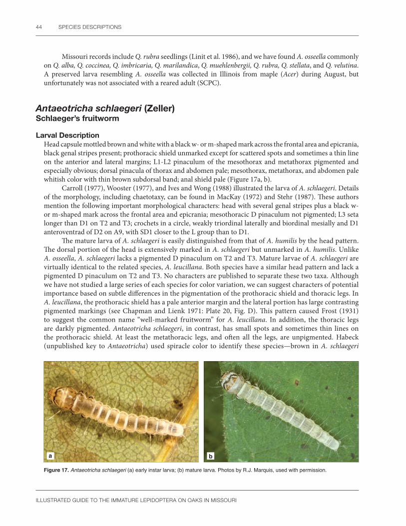
44 SPECIES DESCRIPTIONS
ILLUSTRATED GUIDE TO THE IMMATURE LEPIDOPTERA ON OAKS IN MISSOURI
Missouri records include Q. rubra seedlings (Linit et al. 1986), and we have found A. osseella commonly on Q. alba, Q. coccinea, Q. imbricaria, Q. marilandica, Q. muehlenbergii, Q. rubra, Q. stellata, and Q. velutina. A preserved larva resembling A. osseella was collected in Illinois from maple (Acer) during August, but unfortunately was not associated with a reared adult (SCPC).
Antaeotricha schlaegeri (Zeller)Schlaeger’s fruitworm
Larval DescriptionHead capsule mottled brown and white with a black w- or m-shaped mark across the frontal area and epicrania, black genal stripes present; prothoracic shield unmarked except for scattered spots and sometimes a thin line on the anterior and lateral margins; L1-L2 pinaculum of the mesothorax and metathorax pigmented and especially obvious; dorsal pinacula of thorax and abdomen pale; mesothorax, metathorax, and abdomen pale whitish color with thin brown subdorsal band; anal shield pale (Figure 17a, b).
Carroll (1977), Wooster (1977), and Ives and Wong (1988) illustrated the larva of A. schlaegeri. Details of the morphology, including chaetotaxy, can be found in MacKay (1972) and Stehr (1987). These authors mention the following important morphological characters: head with several genal stripes plus a black w- or m-shaped mark across the frontal area and epicrania; mesothoracic D pinaculum not pigmented; L3 seta longer than D1 on T2 and T3; crochets in a circle, weakly triordinal laterally and biordinal mesially and D1 anteroventrad of D2 on A9, with SD1 closer to the L group than to D1.
The mature larva of A. schlaegeri is easily distinguished from that of A. humilis by the head pattern. The dorsal portion of the head is extensively marked in A. schlaegeri but unmarked in A. humilis. Unlike A. osseella, A. schlaegeri lacks a pigmented D pinaculum on T2 and T3. Mature larvae of A. schlaegeri are virtually identical to the related species, A. leucillana. Both species have a similar head pattern and lack a pigmented D pinaculum on T2 and T3. No characters are published to separate these two taxa. Although we have not studied a large series of each species for color variation, we can suggest characters of potential importance based on subtle differences in the pigmentation of the prothoracic shield and thoracic legs. In A. leucillana, the prothoracic shield has a pale anterior margin and the lateral portion has large contrasting pigmented markings (see Chapman and Lienk 1971: Plate 20, Fig. D). This pattern caused Frost (1931) to suggest the common name “well-marked fruitworm” for A. leucillana. In addition, the thoracic legs are darkly pigmented. Antaeotricha schlaegeri, in contrast, has small spots and sometimes thin lines on the prothoracic shield. At least the metathoracic legs, and often all the legs, are unpigmented. Habeck (unpublished key to Antaeotricha) used spiracle color to identify these species—brown in A. schlaegeri
a b
Figure 17. Antaeotricha schlaegeri (a) early instar larva; (b) mature larva. Photos by R.J. Marquis, used with permission.

DEPRESSARIIDAE 45
ILLUSTRATED GUIDE TO THE IMMATURE LEPIDOPTERA ON OAKS IN MISSOURI
but cream or pale in A. leucillana. The larva of A. leucillana is illustrated by Frost (1931) under the name Stenoma algidella.
MacKay (1972) lists several distinctive characters for recognizing A. schlaegeri. The presence of a retinaculum on the mandible appears to be especially significant. It is present in A. schlaegeri, A. leucillana, and A. osseella, but absent in A. humilis and the A. vestalis-floridella complex (SCPC).
Pupal DescriptionAntennal scape with raised ridges; labial palpi minutely exposed; maxillary palpi small and triangular; prothoracic femur hidden; maxillae often pigmented and extending approximately two-thirds the distance to caudal margin of wings; metathoracic legs exposed; lateral condyles present on A4/5, A5/6, and A6/7, the sclerotized margins of the segments finely serrated; venter of A9 with a paired patch of curved setae; cremaster absent, the end of abdomen often darkly pigmented with several curved setae.
Excluding A. humilis, which lacks antennal ridges, A. schlaegeri is most likely to be confused with A. osseella and A. leucillana. Coloration and size are important clues. Many specimens of A. schlaegeri have the maxillae and the end of the abdomen darkly pigmented. This gives the pupa a characteristic appearance. In addition, A. schlaegeri is the largest stenomatine species treated in this book, with the pupal stage measuring 8–9 mm in length. Antaeotricha osseella and A. leucillana are smaller, usually under 7 mm (measurements from Carroll 1977). The size, characteristic pupal pigmentation when present, and larval skin lacking a pigmented prothoracic shield and thoracic D pinacula are the best characteristics for identifying A. schlaegeri on oaks in Missouri.
Natural HistoryThe eggs of A. schlaegeri are illustrated by Peterson (1967) and Carroll (1977). They are oval, slightly pointed at one end, and laid either singly or in a small group on the edge of a leaf (Figure 7a). According to Wooster (1977: 45) and Carroll et al. (1979), A. schlaegeri is a solitary leaf-tying species that occasionally is found in leaf ties mixed with Psilocorsis species. First instar larvae skeletonize leaves, while later instars form a flat leaf tie with silken bands. Second generation larvae overwinter in matted leaves on the forest floor, then pupate in the spring (Carroll 1977, Wooster 1977). Wooster (1977) states that A. schlaegeri is univoltine in Massachusetts, in contrast to Missouri where this species is at least double-brooded with adults from April to September (Heitzman and Heitzman 1987). We consider A. schlaegeri to be bivoltine in central Missouri, with larvae first appearing in June.
DistributionQuebec throughout the eastern United States to North Carolina and west to Missouri, Texas, and Arizona (Duckworth 1964).
According to Heitzman and Heitzman (1987), A. schlaegeri occurs throughout Missouri. See Figure 18 for Missouri distribution.
Host PlantsQuercus alba, Q. coccinea/rubra (Robinson et al. 2002), and Q. rubra (Wooster 1977). We have found A. schlaegeri on Q. alba, Q. coccinea, Q. imbricaria, Q. rubra, and Q. stellata, and Q. velutina in Missouri. Linit et al. (1986) collected it on Q. rubra seedlings. There is a record for A. schlaegeri on birch in Canada (Prentice 1965).
Figure 18. Known distribution of Antaeotricha schlaegeri in Missouri.

46 SPECIES DESCRIPTIONS
ILLUSTRATED GUIDE TO THE IMMATURE LEPIDOPTERA ON OAKS IN MISSOURI
CommentsAntaeotricha leucillana occurs in Missouri (Duckworth 1964) but is apparently not as common as A. schlaegeri (Carroll 1977: 158). We have not reared A. leucillana from Missouri oaks. Our observations for A. schlaegeri are based on reared adults.
Menesta melanella (Murtfeldt)
Larval DescriptionMature larva dorsoventrally flattened, with a pale yellow head, bluish green body, and a broad white to purplish-colored band running longitudinally from T2-A9; frontal area does not reach epicranial notch; the spinneret is long and pointed; mandible lacks a retinaculum and has only one pointed scissorial tooth; thoracic D pinacula pigmented and relatively large; the crochets of A3-6 form a circle (Figure 19a).
a b c
Figure 19. Menesta melanella (a) mature larva; (b) web; (c) pupal chamber. Photos by R.J. Marquis, used with permission.
The larva of M. melanella was described by Murtfeldt (1890a). It may be confused with Paraclemensia acerifoliella, which also sometimes forms a case on oak (Nielsen 1982). However, M. melanella feeds under a surface web (Figure 19b) and only forms a flattened case as a pupation chamber. Paraclemensia acerifoliella, in contrast, forms a flattened case between two cut leaf pieces, feeding from the case by partially exiting it. The two species also differ in crochet arrangement on A3-6; in M. melanella they form a circle whereas Paraclemensia has a small number of crochets in a band. Preserved specimens of the early instars of either case maker are not available. It is possible they have a different morphology than the mature larva.
Pupal DescriptionThe pupa of M. melanella is very unusual. The spiracles of A2 and A3 are enlarged and located more dorsally than normal. More typical of the Stenomatinae, lateral condyles are present, and venter of A9 has paired lateral patches of curved setae. Mosher (1916a) noted the similarity of Menesta to Antaeotricha schlaegeri.
Natural HistoryThis species was first described from Kirkwood, MO, by Murtfeldt (1890a) who illustrated the characteristic damage. Early instars are leaf miners. Later they feed externally under a v- to square-
Figure 20. Known distribution of Menesta melanella in Missouri.

DEPRESSARIIDAE 47
ILLUSTRATED GUIDE TO THE IMMATURE LEPIDOPTERA ON OAKS IN MISSOURI
shaped sheet web formed on the leaf underside at the intersection of a main and secondary vein (Figure 19b). Skeletonization of the leaf proceeds from the distal edge of the web where frass is ejected. At pupation, the larva cuts an oval portion of the leaf under the web, then sews the edges of the web to the leaf fragment (Figure 19c). Sometimes the silken case may be moved in its entirety to a different location (Murtfeldt 1890a). Menesta albaciliella makes a similar silken case on blackberry (Braun 1915).
DistributionMassachusetts south to Florida and west to Arizona (Duckworth 1964). See Figure 20 for Missouri distribution.
Host PlantsQuercus stellata (=Quercus obtusifolia) (Murtfeldt 1890a, Duckworth 1964). We have found M. melanella commonly on Q. velutina, less so on Q. alba, Q. stellata, and Q. muehlenbergii.
Rectiostoma xanthobasis (Zeller)Yellow-vested moth
Larval DescriptionHead light brown to light orange; the prothoracic and anal shields either unpigmented, spotted, or solid brown to black; all thoracic segments pigmented and contrasting reddish brown to orange, the enlarged D2 pinaculum and thoracic legs conspicuous; abdomen pale with irregular reddish spots in the subdorsal and lateral regions, the D pinacula appearing as four elongated dorsal dots on each segment (Figure 21).
The larva of R. xanthobasis is illustrated by Stehr (1987). The short frontal area, enlarged D2 pinaculum, D and SD setae in a line on A9, and anal crochets broken into two groups are distinctive for R. xanthobasis.
Because of the pigmented thorax, R. xanthobasis resembles Psilocorsis quercicella in the field, but the latter lacks the reddish patterning on the abdomen and blackened pinacula on the thorax. The two taxa also differ in the head texture. The head is smooth in R. xanthobasis but punctate in P. quercicella. Preserved alcoholic specimens of R. xanthobasis can lose the pigmentation of the thorax (Stehr 1987: Fig. 26: 52, SCPC). These are easily confused at the family level with both Gelechiidae (because the D and SD setae form a line on A9 and the anal crochets are broken into two groups) and Tortricidae (because the D2 setae on A9 are closely spaced). Characters to separate these groups are given by Stehr (1987) and in the introductions to these families in this book.
Pupal Description Modified from Duckworth (1971): Head, thorax, appendages, and wing cases with finely ridged texture; labrum u-shaped; labial palpi minutely exposed; maxillary palpi and prothoracic femur hidden; maxillae extends about two-thirds the distance to the caudal margin of the wings; metathoracic legs hidden; mesothoracic spiracle slitlike; prothorax rugose; abdomen shagreened; abdomen damaged but condyles present; venter of A7 with a truncate plate bearing six widely spaced downward pointing curved spines that are easily broken.
Figure 21. Rectiostoma xanthobasis mature larva. Photo by R.J. Marquis, used with permission.

48 SPECIES DESCRIPTIONS
ILLUSTRATED GUIDE TO THE IMMATURE LEPIDOPTERA ON OAKS IN MISSOURI
The pupa of R. xanthobasis is easily recognized by having six curved spines on the venter of A7 (Duckworth 1971: 5, Figs. 8, 9). Keifer (1936b: Plate III) illustrated similar spines on the pupa of a related western species, Rectiostoma fernaldella. The cuticular texture of R. xanthobasis is also unusual. As is typical for the Stenomatinae, lateral condyles are present in both species of Rectiostoma mentioned here (SCPC).
Natural HistoryAccording to Duckworth (1971), the larva of R. xanthobasis lives between two oak leaves tied with silk. We have seen larvae folding over leaf lobes of white oak and attaching them to the main portion of the leaf, especially during August and September. Larvae were collected in June in Florida (Stehr 1987). Information on the pupation site and number of generations per year is conflicting (Duckworth 1971), perhaps due to geographical variation. We expect the pupa of R. xanthobasis to overwinter in the last generation, with adults emerging the following spring.
DistributionMassachusetts south to Florida and west to Illinois, Missouri and Texas (Duckworth 1964, 1971). See Figure 22 for Missouri distribution.
Host PlantsQuercus (Robinson et al. 2002); Q. nigra, Q. stellata (Duckworth 1971); and Q. laurifolia (Stehr 1987). We have found R. xanthobasis on Q. alba, Q. muehlenbergii, Q. rubra, and Q. velutina in Missouri.
CommentsThis species was previously placed in Setiostoma (Duckworth 1971), then was moved to Rectiostoma in Becker (1984) and Pohl et al. (2016).
Figure 22. Known distribution of Rectiostoma xanthobasis in Missouri.
Unplaced Depressariidae
Psilocorsis cryptolechiella (Chambers)Black-fringed leaf tier
Larval DescriptionModified from Passoa (1995): Head punctate with a network of raised ridges; head capsule, prothoracic shield, prespiracular pinaculum, SD and L thoracic pinacula, thoracic legs, and sometimes the SD1 pinacula on A1-8, all light to dark reddish brown; anal shield usually pale, more rarely tan or reddish brown in color (Figure 23).
The larva of P. cryptolechiella is illustrated by Carroll (1977), Carroll et al. (1979), Stehr (1987), and in color by Passoa (1995). Wooster (1977) and Passoa (1995) include drawings of the mouthparts and chaetotaxy. Important morphological features are: mandible longer than wide, with three to four scissorial teeth; frontal area extending more than half the distance to the epicranial notch; all L setae located on the prespiracular pinaculum; XD1 at least three-fourths as long as XD2, and D1 posterodorsad of SD2 on the anal shield (Wooster 1977: 37, Passoa 1995).

DEPRESSARIIDAE 49
ILLUSTRATED GUIDE TO THE IMMATURE LEPIDOPTERA ON OAKS IN MISSOURI
Figure 23. Psilocorsis cryptolechiella mature larva. Photo by R.J. Marquis, used with permission.
Mature larvae of P. cryptolechiella can be confused with Psilocorsis quercicella, P. reflexella, or any one of several gelechiid species on oak in Missouri. Unlike P. reflexella, P. cryptolechiella has the frontal area extending more than half the distance to the epicranial notch, all L setae are on the prespiracular pinaculum, and the anal shield is unpigmented. Most specimens of P. quercicella have an intensely pigmented thorax. The mesothorax and metathorax are unpigmented in P. cryptolechiella. Psilocorsis has all the SV almost in a horizontal row on A10; this arrangement is absent in gelechiids on oak in Missouri.
Pupal DescriptionModified from Passoa (1995): Antennal scape with a longitudinal groove; labial palpi hidden; maxillae extend three-fourths the distance to caudal margin of wings; prothoracic femur and metathoracic legs exposed; prothorax with a patch of punctures; mesothoracic spiracle a minute spiny protuberance; A1-4 smooth except for a row of punctures at the anterior margin; cremaster u-shaped with seven to eight curved setae.
The pupa of P. cryptolechiella is illustrated by Carroll (1977), Carroll et al. (1979), and Passoa (1995). This species is easily recognized by the grooved antennal scape, presence of abdominal punctures in a row on the anterior margin of A1-4, and the u-shaped cremaster. Unlike the Stenomatinae, lateral condyles are absent in all Psilocorsis species.
Natural HistoryThe life cycle of P. cryptolechiella is summarized by Carroll (1977), Carroll et al. (1979), and Passoa (1995). Eggs of P. cryptolechiella are mottled with red. In Missouri, except for mid-July, oviposition occurs from May through September, with larval feeding from May through October. Flight time and larval feeding period were longer in Missouri than in Massachusetts. There are two generations per year, and pupation occurs in the leaf litter. Adults fly from May through September, with a small lapse in mid-July.
DistributionMaine to northern Florida, west to Texas and Arkansas (Hodges 1974), including Illinois (Godfrey et al. 1987a) and Missouri (Carroll et al. 1979). See Figure 24 for Missouri distribution.
Host PlantsQuercus, Q. rubra (Robinson et al. 2002); Q. laurifolia, Q. laevis, Q. nigra, Q. phellos (Passoa 1995), and Q. marilandica (Heppner 2003).
Quercus is the primary host of P. cryptolechiella (Hodges 1974). Other hosts (Acer, Betula, Carya, Castanea, Fagus, Ilex, Robinia, and Myrica) reported in Robinson et al. (2002) might need to be confirmed. Linit et al. (1986) reported P. cryptolechiella on Q. rubra seedlings in Missouri. We have found P. cryptolechiella on Q. alba, Q. imbricaria, Q. muehlenbergii, Q. stellata, Q. rubra, and Q. velutina in Missouri.
Figure 24. Known distribution of Psilocorsis cryptolechiella in Missouri.

50 SPECIES DESCRIPTIONS
ILLUSTRATED GUIDE TO THE IMMATURE LEPIDOPTERA ON OAKS IN MISSOURI
Psilocorsis quercicella ClemensOak leaf tier
Larval DescriptionModified from Passoa (1995): Head capsule punctate with a network of raised ridges; head and prothoracic shield shiny black; mesothorax and metathorax intensely pigmented pale gray, brown, or black; abdominal segments cream, yellowish brown, green, or pink (Figure 25).
The larva of P. quercicella is illustrated by Carroll (1977), Wooster (1977), Carroll et al. (1979), and in color by Passoa (1995). Wooster (1977) and Passoa (1995) include drawings of the mouthparts and chaetotaxy. Important morphological features of P. quercicella are: mandible longer than wide, triangular, with four scissorial teeth that may be worn smooth; frontal area extending more than half the distance to the epicranial notch; usually all L setae are on the prespiracular pinaculum; all thoracic segments normally intensely pigmented; XD1 approximately half as long as XD2, and D1 posterodorsad of SD2 on the anal shield (Wooster 1977: 10, 15; Passoa 1995).
Psilocorsis quercicella is the only species of the genus in North America with an intensely pigmented thorax. For identification characteristics of specimens with light thoracic pigmentation, consult Passoa (1995: 49). Psilocorsis quercicella could be confused with Rectiostoma xanthobasis, which also feeds on oak in Missouri (Carroll 1977: Table V), because T2 and T3 are pigmented in both species. The head of P. quercicella is punctate, the abdominal D pinacula are round, and the crochets of A10 are unbroken (Passoa 1995). In contrast, R. xanthobasis has a smooth head, the abdominal D pinacula are elongated, and the crochets of A10 are divided into two groups (Stehr 1987: Fig. 26.52). The photograph published by Cochaux (1969) suggests the study organism was P. reflexella and not P. quercicella.
Pupal DescriptionModified from Passoa (1995): Antennal scape with a longitudinal groove; labial palpi hidden; maxillae extend three-fourths the distance to caudal margin of wings; prothoracic femur and metathoracic legs exposed; prothorax lacks a patch of punctures; mesothoracic spiracle a minute spiny protuberance; A1-4 smooth, without any punctures; cremaster u-shaped with eight curved setae.
The pupa of P. quercicella is illustrated by Mosher (1916a), Carroll (1977), Carroll et al. (1979), and Passoa (1995). This species is separated from other eastern United States Psilocorsis by the presence of a mesothoracic spiracle and the lack of dorsal abdominal punctures. Pupae of P. reflexella and P. quercicella can be confused because the minute mesothoracic spiracle, which is absent in P. reflexella, is easily overlooked. Size can be a clue. As a rule, large pupae over 8 mm are usually P. reflexella whereas small pupae (6–8 mm) are often P. quercicella. The cast larval exuvia, if present, also furnishes clues to help identify the specific Psilocorsis species because it is possible to see the length of the front and mandible morphology (see sections on larval description).
Natural HistoryThe life cycle of P. quercicella is summarized by Carroll (1977), Wooster (1977), Carroll et al. (1979), and Passoa (1995). Lill and Marquis (2001) studied the effects of leaf quality on P. quercicella. Natural enemies of P. quercicella are listed by Lill (1999).
Figure 25. Psilocorsis quercicella mature larva. Photo by R.J. Marquis, used with permission.
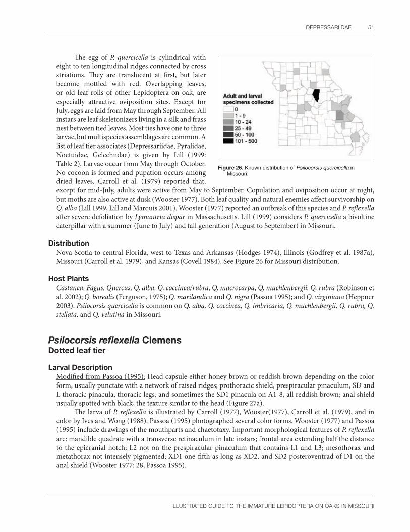
DEPRESSARIIDAE 51
ILLUSTRATED GUIDE TO THE IMMATURE LEPIDOPTERA ON OAKS IN MISSOURI
The egg of P. quercicella is cylindrical with eight to ten longitudinal ridges connected by cross striations. They are translucent at first, but later become mottled with red. Overlapping leaves, or old leaf rolls of other Lepidoptera on oak, are especially attractive oviposition sites. Except for July, eggs are laid from May through September. All instars are leaf skeletonizers living in a silk and frass nest between tied leaves. Most ties have one to three larvae, but multispecies assemblages are common. A list of leaf tier associates (Depressariidae, Pyralidae, Noctuidae, Gelechiidae) is given by Lill (1999: Table 2). Larvae occur from May through October. No cocoon is formed and pupation occurs among dried leaves. Carroll et al. (1979) reported that, except for mid-July, adults were active from May to September. Copulation and oviposition occur at night, but moths are also active at dusk (Wooster 1977). Both leaf quality and natural enemies affect survivorship on Q. alba (Lill 1999, Lill and Marquis 2001). Wooster (1977) reported an outbreak of this species and P. reflexella after severe defoliation by Lymantria dispar in Massachusetts. Lill (1999) considers P. quercicella a bivoltine caterpillar with a summer (June to July) and fall generation (August to September) in Missouri.
DistributionNova Scotia to central Florida, west to Texas and Arkansas (Hodges 1974), Illinois (Godfrey et al. 1987a), Missouri (Carroll et al. 1979), and Kansas (Covell 1984). See Figure 26 for Missouri distribution.
Host PlantsCastanea, Fagus, Quercus, Q. alba, Q. coccinea/rubra, Q. macrocarpa, Q. muehlenbergii, Q. rubra (Robinson et al. 2002); Q. borealis (Ferguson, 1975); Q. marilandica and Q. nigra (Passoa 1995); and Q. virginiana (Heppner 2003). Psilocorsis quercicella is common on Q. alba, Q. coccinea, Q. imbricaria, Q. muehlenbergii, Q. rubra, Q. stellata, and Q. velutina in Missouri.
Figure 26. Known distribution of Psilocorsis quercicella in Missouri.
Psilocorsis reflexella ClemensDotted leaf tier
Larval DescriptionModified from Passoa (1995): Head capsule either honey brown or reddish brown depending on the color form, usually punctate with a network of raised ridges; prothoracic shield, prespiracular pinaculum, SD and L thoracic pinacula, thoracic legs, and sometimes the SD1 pinacula on A1-8, all reddish brown; anal shield usually spotted with black, the texture similar to the head (Figure 27a).
The larva of P. reflexella is illustrated by Carroll (1977), Wooster(1977), Carroll et al. (1979), and in color by Ives and Wong (1988). Passoa (1995) photographed several color forms. Wooster (1977) and Passoa (1995) include drawings of the mouthparts and chaetotaxy. Important morphological features of P. reflexella are: mandible quadrate with a transverse retinaculum in late instars; frontal area extending half the distance to the epicranial notch; L2 not on the prespiracular pinaculum that contains L1 and L3; mesothorax and metathorax not intensely pigmented; XD1 one-fifth as long as XD2, and SD2 posteroventrad of D1 on the anal shield (Wooster 1977: 28, Passoa 1995).

52 SPECIES DESCRIPTIONS
ILLUSTRATED GUIDE TO THE IMMATURE LEPIDOPTERA ON OAKS IN MISSOURI
a b
Figure 27. Psilocorsis reflexella (a) mature larva; (b) mature larva in silk tube found within leaf ties. Photo a by S.C. Passoa; b by R.J. Marquis; both used with permission.
Psilocorsis reflexella may be distinguished from other eastern United States Psilocorsis species by the frontal area, which extends only one-half the distance to the epicranial notch. In addition, the mature larva has a quadrate blunt mandible, not triangular and pointed as in related species. L2 is separated from the prespiracular pinaculum in P. reflexella, but joined with L1 and L3 in both P. cryptolechiella and P. quercicella. The anal shield chaetotaxy of P. reflexella is distinctive.
Pupal DescriptionModified from Passoa (1995): Antennal scape with a longitudinal groove; labial palpi hidden; maxillae extend three-fourths the distance to caudal margin of wings; prothoracic femur and metathoracic legs exposed; prothorax lacks a patch of punctures; mesothoracic spiracle absent; A1-4 smooth, without any punctures; cremaster u-shaped with five to seven, or more often eight, curved setae.
The pupa of P. reflexella is illustrated by Carroll (1977), Carroll et al. (1979), and Passoa (1995). Unlike P. cryptolechiella and P. quercicella, the mesothoracic spiracle of P. reflexella is absent. Carroll (1977) used size to distinguish P. reflexella in central Missouri; its mean size of 8 mm is larger than that of other Psilocorsis he collected. The larval exuvia, if present, will contain the characteristic mandible and short frontal area.
Natural HistoryThe life cycle of P. reflexella is summarized by Carroll (1977), Wooster (1977), Carroll et al. (1979), and Passoa (1995). Psilocorsis reflexella is univoltine in the northern United States; there may be an extra generation in the Gulf States. The eggs are illustrated by Carroll (1977), Wooster (1977), and Carroll et al. (1979). They are laid singly or in small groups. Overlapping leaves are common oviposition sites. In Missouri, oviposition occurs from May through August, larval development from June through October, and pupation in the leaf litter throughout the year except for the summer months. Adults fly from May to August. Wooster (1977) reported an outbreak of P. reflexella and P. quercicella after severe defoliation by Lymantria dispar in Massachusetts in 1975. We have collected P. reflexella in narrow tunnels of frass and silk up to 5 cm long between tightly spun leaves (Figure 27b) from June through October.
Figure 28. Known distribution of Psilocorsis reflexella in Missouri.

DEPRESSARIIDAE 53
ILLUSTRATED GUIDE TO THE IMMATURE LEPIDOPTERA ON OAKS IN MISSOURI
DistributionSouthern Quebec to northern Wisconsin and south to central Florida and eastern Texas (Hodges 1974), including Illinois (Godfrey et al. 1987a) and Missouri (Lill 1999). See Figure 28 for Missouri distribution.
Host PlantsAcer, Betula, Carya, Corylus, Fagus, Populus, Quercus, Q. coccinea/rubra, Q. rubra, Salix, Tilia (Robinson et al. 2002); Q. borealis (Ferguson 1975); Celtis, Ilex, Ostrya, Q. chapmanii, Q. michauxii, and Q. nigra (Passoa 1995).
Missouri records for P. reflexella include Quercus rubra seedlings (Linit et al. 1986), and we have found this species on Q. alba, Q. palustris, Q. rubra, Q. stellata, Q. velutina, and Q. muehlenbergii. Hodges (1974) noted that Canadian individuals were frequently reared from birch and poplar, whereas specimens from Florida and Texas are common on hickory, thus representing the distribution of the host plants.
CommentsWe agree with Hodges (1974) and Passoa (1995) in regarding P. reflexella as a single but highly variable species. Passoa (1995) documents three larval color forms of P. reflexella, but due to rare intergrades among these groups and no pupal morphological differences, he was unwilling to name taxa formally. Hodges (1974) also noted variation in wing color and size, but found genital characters intergraded among the forms he recognized. The larval color forms of P. reflexella on their usual hosts are: anal shield darkly spotted (on oak and other trees), the head and anal shield honey colored (on beech,) or the head capsule reddish brown with no anal shield markings (on hickory). Passoa (1995) suggests this is an example of a larva’s ability to modify its appearance based on the food plant, similar to the study and examples given by Collins (1999).
Machimia tentoriferella ClemensGold-striped leaf tier
Larval DescriptionModified from Passoa (1995): Head pale green, smooth; body first pale green, then older instars turn yellowish green with wide white subdorsal stripes running the length of the body; dorsal setae of abdominal segments arise from white pinacula (Figure 29a).
The larva of M. tentoriferella is illustrated by Chapman and Lienk (1971). They show the prothoracic shield with a small black dot, an unusual coloration. Passoa (1995) illustrates the larva and describes the mouthparts and chaetotaxy. Important morphological features of M. tentoriferella are: head without punctures, mandible with four scissorial teeth and no retinaculum; SD1 thinner than SD2 on T2 and T3; L3 absent on A8
a b
Figure 29. Machimia tentoriferella (a) mature larva; (b) early instar larva in sheet web. Photos by R.J. Marquis, used with permission.

54 SPECIES DESCRIPTIONS
ILLUSTRATED GUIDE TO THE IMMATURE LEPIDOPTERA ON OAKS IN MISSOURI
and A9; and the SV setae of A9 not in a line. The combination of thin SD1 setae and lack of L3 on A8-9 is not found in any other Missouri oak-feeding caterpillar we studied. All Psilocorsis have a punctate head and the SV setae in a horizontal line on A9. In the field, the characteristic web of M. tentoriferella often has alternating bands of dark and light silk. Only Semioscopis, which has different hosts and morphology, can be confused with M. tentoriferella (see Passoa 1995).
Pupal DescriptionModified from Passoa (1995): Antennal scape unmodified; labial palpi, prothoracic femur, and metathoracic legs all hidden; maxillary palpi present; maxillae extend slightly more than half the distance to caudal margin of wings; mesothoracic spiracle ridgelike; scattered curved setae in two diffuse patches above anal slit, more densely packed in area surrounding the anal slit and in a group of five setae at the end of abdomen.
The pupa of M. tentoriferella is illustrated by Passoa (1995). Among Missouri oak feeders, the pupa of M. tentoriferella can be identified by the unmodified antennal scape, smooth cuticular texture, lack of lateral condyles, lack of dorsal abdominal setose knobs, hidden metathoracic legs, lack of a fringe of setae on A7, and the characteristic distribution of curved setae on the posterior abdominal segments. The distribution of these setae is shown online at https://bugtracks.wordpress.com/2016/12/19/quality-control/ and by Passoa (1995: Fig. 2). In our study area, species of Stenomatinae have lateral condyles, the metathoracic legs are exposed in Psilocorsis, Dichomeridinae have dorsal abdominal paired setose knobs, and the other genera of oak-feeding gelechiids have a fringe of setae on the posterior margin of A7.
Natural HistoryThe life history of M. tentoriferella is reviewed by Passoa (1995). There is one generation per year, at least in New York. Larvae are found in June and July. Pupation occurs in a folded leaf during July and August. Adults can be collected from August to September. Either the adult or egg is the overwintering stage. Chapman and Lienk (1971) coined the common name “gold-striped leaf tier” for this species.
In our experience, M. tentoriferella is a pale green caterpillar that makes a flat surface web on the underside of a leaf; the web is open at either end and is usually built from one vein to raise the web off the leaf surface (Figure 29b). At first, early instars skeletonize the leaf at the head end of the web, unlike later instars that eat the whole leaf. New, increasingly larger webs are built as the caterpillar grows. When disturbed, the larva wriggles backward to escape and drops through the rear slit in the web.
DistributionNova Scotia south to Virginia, west to Iowa and Mississippi (Covell 1984), including Missouri (Le Corff et al. 2000).
Hodges (1974) suspects that M. tentoriferella will eventually be recorded from South Carolina and northern Georgia. It is currently not recorded from Florida (Heppner 2003). See Figure 30 for Missouri distribution.
Host PlantsAcer, Betula, Carya, Castanea, Cephalanthus,
Cornus, Corylus, Crataegus, Fagus, Fraxinus, Juglans, Populus, Prunus, Pyrus, Quercus alba, Q. rubra, Sorbus, Syringa, Tilia, Ulmus, Viburnum (Robinson et al. 2002); and Salix (Passoa 1995).
Le Corff et al. (2000) collected M. tentoriferella on Q. alba in Missouri.
Figure 30. Known distribution of Machimia tentoriferella in Missouri.

GELECHIIDAE 55
ILLUSTRATED GUIDE TO THE IMMATURE LEPIDOPTERA ON OAKS IN MISSOURI
GelechiidaeGelechiid moths
Gelechiidae are a large family of microlepidoptera with approximately 4,700 described species (Nieukerken et al. 2011), although many more undescribed taxa are waiting to be named (Hodges 1986, 1998). Based on morphology, Hodges (1998) recognized four subfamilies: Gelechiinae, Dichomeridinae, Physoptilinae, and Pexicopiinae. Ponomarenko (2006) added a subfamily while recognizing three of the four subfamily names used by Hodges (1998). Her groupings were Physoptilinae, Anomologinae, Gelechiinae, Anacampsinae, and Dichomeridinae. The most recent molecular phylogeny by Karsholt et al. (2013) redefined these groups and suggested a division with seven subfamilies including Physoptilinae, Anacampsinae, Dichomeridinae, Apatetrinae, Thiotrichinae, Anomologinae, and Gelechiinae. The Pexicopiinae of Hodges (1998) was placed as a tribe (Pexicopiini) within the Apatetrinae. The distribution of these subfamilies is variable. Gelechiinae and Dichomeridinae are broadly distributed throughout the world but the Physoptilinae contains only one genus found from India to Australia (Hodges 1998). All of the Missouri oak-feeding genera of Gelechiidae covered in this book belong to the Gelechiinae and Dichomeridinae.
For more information on systematics of the Gelechiidae, consult Busck (1903a, 1903b, 1939), Benander (1937), Janse (1949–1954, 1958–1963), Hodges (1966, 1986, 1998, 1999), Povolny (1967, 1990, 1991, 1994, 2002), Clarke (1969a, 1969b), Fetz (1994), Park (1995), Passoa (1995), Elsner et al. (1999), Huemer and Karsholt (1999), Powell and Povolny (2001), Emmet and Langmaid (2002), Lee and Brown (2008), Lee et al., (2009), Ponomarenko (2009), Nazari and Landry (2012), and references cited in these monographs. Useful information on Gelechiidae is also available on Web sites by Lee (2015) and Hayden et al. (2017).
Gelechiid larvae are difficult to recognize. No one single unique trait defines the family; instead, a combination of characters needs to be examined. The most significant ones are the mandible, chaetotaxy, crochets, and form of the anal comb. In gelechiid larvae, the distance between L1 and A3 on the head is greater than the distance separating A3 and A2 (Stehr 1987: Fig. 129). The mandible often has a tooth, or notch, on the outer margin above the condyle (Benander 1937; Passoa 1995: 20, 237), but this modification also occurs in other Gelechioidea such as Apachea (Depressariinae) (Passoa 1995: Fig. 36). The hypopharyngeal complex may have a series of platelike projections called blades or a sclerotized ridge on the proximolateral portion in some Missouri species (Appendix 4, Plate 4). Keifer (1936a) and MacKay (1972: 14) both noted that gelechiids have one outer tarsal claw seta that is spinelike while the other is scalelike. A bisetose SV group on A1 is another characteristic of most gelechiid larvae (Stehr 1987). At least in Missouri, oak-feeding gelechiids usually have the SD1 seta posterodorsad of spiracle on A1, but anterodorsad of spiracle on A2-7, and anterior to spiracle on A8. The posterodorsad position of SD1 on A1 in some gelechiids was considered unusual by MacKay (1972). Secondary setae are almost always absent, two exceptions being the secondary setae found on the anal prolegs of Dichomeris marginella (Stehr 1987) and the anal shield of Anarsia lineatella (MacKay 1972). In the Dichomeridinae and some Gelechiinae, the setae of the posterior abdominal segments may be darker and thicker than the other body setae. Larval gelechiids have the crochets of A3-6 either in a circle, penellipse, or two bands (often formed by mesial or lateral gaps in a circle). A gap may also occur in the crochets of the anal prolegs. In Sitotroga, Metzneria, and Isophrictis, the crochets are reduced or absent (Stehr 1987). Stehr (1987) stated that gelechiids are the only members of the Gelechioidea to have an anal comb, at least in North America. Reports of an anal comb in the Oecophoridae (Stehr 1987: 295) probably refer only to Amphisbatis (Minet 1990, larval exuviae in Cornell University collection examined by S.C. Passoa) and Pseudatemelia (Fetz 1994) (both genera now in the Lypusidae). In larval gelechiids, the prongs of the anal comb are either scalelike or spinelike. Generally, one prong is curved and the medial prongs are largest (Benander 1937).
One of the most significant features of gelechiid chaetotaxy is the arrangement of the D and SD setae on A9. In many gelechiid genera, D2, D1, and SD1 form a vertical line (MacKay 1972). In other cases, D2 is posterodorsad of vertical line connecting D1 and SD1, or more rarely, D1 is very slightly anterior of a line connecting D2 and SD1 (Passoa 1995, figures in Stehr 1987, Hodges 1999: Fig. 5). However, gelechiid larvae

56 SPECIES DESCRIPTIONS
ILLUSTRATED GUIDE TO THE IMMATURE LEPIDOPTERA ON OAKS IN MISSOURI
almost never have D1 clearly anteroventrad of D2 and anterodorsad of SD1 so that the D and SD setae of A9 form an equilateral triangle (Stehr 1987). This difference in the chaetotaxy of A9 was used by Passoa (1995) to divide larvae of the Gelechioidea into two groups. Although most gelechiid larvae have D1 approximately dorsad of SD1, this pattern is not unique to the Gelechiidae; therefore, it must be used with the other characters previously mentioned. There also appears to be considerable individual variation in the chaetotaxy of A9, which requires examination of both sides of the larva, and if possible, a series of larvae, to be sure of the correct character state.
When the world fauna is considered, numerous exceptions become apparent, especially at the subfamily level. For example, larvae of the Gelechiinae usually have a bisetose SV group on A1, crochets that are either in circles or two rows or rarely absent, SD1 on A9 is either hairlike or setiform, and usually no anal comb is present (Hodges 1998). Dichomeridinae usually have a bisetose SV group on A1, biordinal crochets in two groups on elongate prolegs, SD1 hairlike on A9, and an anal comb is usually present (Hodges 1998). In addition, the anal shield may taper to a blunt point (SCPC). Several of the remaining subfamilies can be divided into tribes. Apatetrinae has two tribes. Apatetrini contains the genus Chrysoesthia that forms a blotch mine on Chenopodium and related plants (Emmet and Langmaid 2002). The larva has elongate abdominal prolegs and no anal comb (SCPC). The Pexicopiini usually have a unisetose or bisetose SV group on A1, crochets in a lateral penellipse except for Sitotroga (Stehr 1987), SD1 usually setiform, and no anal comb is present (Hodges 1998). There are also two tribes of Anacampsinae. Anarsia is the best known member of the tribe Chelariini. According to the illustrations in MacKay (1972: Fig. 15, Stehr 1987, Gilligan and Passoa 2014), Anarsia lineatella has a bisetose SV group on A1, biordinal abdominal crochets, A9 with SD1 hairlike and L3 absent, and an anal comb is present. The other tribe, Anacampsini (based on Anacampsis and Syncopacma), has the SV group of A1 bisetose, crochets in two groups, SD1 hairlike on A9 with a somewhat enlarged D2 pinaculum, and no anal comb (SCPC). The only representative of the Thiotrichinae in the United States is Polyhymno (Pohl et al. 2016). Busck (1903a) records two species of this genus feeding on legumes in the southern United States, but no detailed morphological information is readily available on the immature stages. A Japanese species of Thiotrichinae was studied by Ueda and Fujiwara (2005). They found that the larva of Thiotricha makes cases on the flowers of Symplocos; has a bisetose SV group on A1; D1 and D2 of A9 joined on the same pinaculum; and a bilobed broad flat anal comb. Physoptilinae is another poorly known subfamily in the larval stage. There is one genus, Physoptila, with seven species all in the Indo-Australian region that are stem-borers on the Lecythidaceae (Karsholt et al. 2013). The final subfamily, Anomologinae, contains several very unusual larvae from a morphological standpoint. Metzneria and Isophrictis, considered sister groups by Karsholt et al. (2013), both lack crochets (Stehr 1987: 394). Stereomita andropogonis, which has a grublike larva with a roughened cuticle, is another genus in this subfamily with reduced crochets and prolegs (SCPC). The larva of Monochroa is more normal in structure. The crochets are in an ellipse, the prolegs are well developed, and an anal comb is absent (Stehr 1987).
Considering the lack of unique characters and the variability of those that are useful (noted in our descriptions), Stehr (1987) predicted that recognition of larval Gelechiidae in North America is likely to remain difficult for many years to come. Benander (1937) and Fetz (1994), both working on the European fauna, also were unable to find unique larval characters to define the Gelechiidae. These results suggest the subfamilies may not be monophyletic or more detailed morphological work is needed with the immature stages. Having a stable higher classification and a greater percentage of described larvae would be especially helpful. No doubt the above summary will need to be greatly modified after more information is eventually published. Hodges (1966: 5, 1986) and Povolny (2002) have discussed many problems relating to the recognition of tribal and subfamily groups in the Gelechiinae.
In Missouri, oak-feeding larvae of the Gelechiidae either are leaf tiers and leaf rollers on tender foliage in the spring or leaf tiers on mature foliage later in the year. Species feeding on mature leaves skeletonize them. Arogalea cristifasciella forms a silk tent (R.J. Marquis, pers. obs.), and in the case of Chionodes fuscomaculella, a cottony mass of silk is included in the feeding tunnel (Carroll and Kearby 1978). When disturbed, many species crawl or wriggle rapidly across the leaf. Most species in Missouri have a pigmented prothoracic shield and a variety of color patterns on the thorax or abdomen. These patterns frequently include contrasting bands or longitudinal stripes. Single or multiple generations occur in a season depending on the species. Oak-feeding

GELECHIIDAE 57
ILLUSTRATED GUIDE TO THE IMMATURE LEPIDOPTERA ON OAKS IN MISSOURI
gelechiids in Missouri are small (less than 12 mm in the last larval instar), and therefore, a hand lens is needed to appreciate their color patterns and strategies for protection.
With regard to the Missouri oak fauna, gelechiid larvae are most likely to be confused with Depressariidae or Tortricidae. All of these taxa cause similar damage, and larvae of several of these families may occupy the same leaf shelter. Larval Stenomatine have only the thoracic pinacula enlarged, which separates them from all Missouri oak-feeding Gelechiidae except Pseudotelphusa. In Pseudotelphusa, both the thoracic L and abdominal SD pinacula are enlarged. Larvae of Psilocorsis have a rugose punctate head, the SV setae on A10 almost in a horizontal line, no anal comb, and D1 clearly anteroventrad of D2 and anterodorsad of SD1 on A9. The head of Missouri oak-feeding gelechiids is usually smooth, an anal comb is usually present, and at least D1 and SD1 are in a vertical line on A9. Most tortricid larvae are easily separated from the Gelechiidae by having D2 of A9 joined on a common middorsal pinaculum, or if on separate pinacula, then the D2 of A9 setae are spaced closer to each other than to their corresponding D1 setae on that segment (Brown 1987b). In doubtful cases where the larva is not clearly a Tortricidae or Gelechioidea, several backup characters may prove helpful. Gelechiid larvae never have D1 and SD1 of A9 joined on the same pinaculum, a characteristic of the Olethreutinae (Brown 1987b). Horak and Brown (1991: 24) diagnosed tortricid larvae, in part, by the straight prongs on their anal comb. Although many gelechiid larvae have curved prongs, straight prongs do occur on some Missouri oak-feeding Dichomeridinae. Tortricids never have SD1 hairlike on A9; this seta may be hairlike or setiform in the Gelechiidae. Pending further study, the outer tarsal claw setae also may differ in the two taxa. Keifer (1936a) suggested that outer tarsal claw setae are equal in tortricid larvae, whereas in gelechiid larvae, one seta is short and spinelike while the other seta is longer and flattened.
Missouri oak-feeding gelechiid genera can be separated into several groups based on the crochet pattern on A3-6. Arogalea has these crochets in a penellipse that is open mesially. All three species of Dichomeris (D. georgiella, D. ventrella, and D. ligulella) have the crochets arranged in two groups. All four species of Chionodes (the formosella complex, C. fuscomaculella, C. pereyra, and C. adamas) and Pubitelphusa have the crochets on A3-6 in a circle. In Pseudotelphusa and Trypanisma, the crochets are in a mesial penellipse closed by a few scattered uniordinal crochets. Pseudotelphusa and Chionodes bicostomaculella are the only Missouri oak-feeding gelechiids we studied that lack an anal comb. Using the crochet arrangement, form of the mandible, presence or absence of anal comb, and color pattern, it should be possible to correctly name most commonly encountered gelechiid larvae on Missouri oaks. The length of the frontal area, details of the crochet length (uniordinal, biordinal, or triordinal), position of SD1 on A8 relative to the spiracle, and close association of SD1 with the L group on A9 may also be important identification characters as more Missouri oak-feeding gelechiids are described and diagnosed.
Gelechiid pupae are equally difficult to recognize because no single character exists to define the family. The antennae usually meet at the meson, as is typical for members of the Gelechioidea, then diverge apically to expose the metathoracic legs (Hodges 1998). However, exceptions to this generalization exist. For example, the antennae do not meet at the meson in two European species of Aristotelia (Patočka 1988a). In the genus Sitotroga, the antennae do not diverge apically (Patočka 1987d). Many Holarctic genera of Gelechiidae have a complete frontoclypeal suture, but often this suture is not evenly curved across the vertex (Mosher 1916a; Patočka 1987a, 1987b, 1987c, 1987d, 1989; Patočka and Turčáni 2005). The antennal scape is constricted at the base in Metzneria from Europe (Patočka 1987a) and often in Chionodes from Missouri. Labial palpi are not exposed, or at most only a small triangular portion is visible (Mosher 1916a, Nakamura 1981, Passoa 1995, Patočka and Turčáni 2005). The cuticular texture is usually smooth, but in some genera such as Anacampsis and Aristotelia (Mosher 1916a, Patočka 1988a, 1988b) and one undetermined genus from Honduras (SCPC), the pupa is pubescent. The prothoracic femur is hidden in the Holarctic genera examined by Mosher (1916a) and Patočka (1987a, 1987b, 1987c, 1987d, 1989). Maxillary palpi are present (Nakamura 1981), except for one species of Chrysoesthia (Patočka 1987a) and some North American Dichomeris (SCPC). Nakamura (1981) stated that Japanese species of Gelechiidae never have the mesothoracic spiracle protruded like a small cone. Lateral condyles are absent (Passoa 1995).

58 SPECIES DESCRIPTIONS
ILLUSTRATED GUIDE TO THE IMMATURE LEPIDOPTERA ON OAKS IN MISSOURI
In many Gelechiidae, the pupal dorsum shows a wide array of morphological specializations. A Dichomeris from Europe has two rows of punctures on the thorax. North American and European species of Dichomeridinae often have paired setose protuberances on A2-4 (Patočka 1987d, Hodges 1986). Modifications of the Gelechiinae include a fringe of setae on the posterior margin of A7 (Hodges 1998), and in Arogalea, paired subdorsal oval to rectangular sclerotized raised areas on the dorsum of A5. Small depressions occur dorsal to the spiracle of the anterior abdominal segments of some Gelechiinae (Chionodes and Telphusa). The pupa of Thiotricha has a row of minute spines on the dorsum of A6 and A7 (Ueda and Fujiwara 2005). With some exceptions (Patočka 1987d, 1988b), gelechiid pupae usually do not have a well-developed tubular cremaster (Patočka 1987a, 1987b, 1987c, 1987d, 1989; Patočka and Turčáni 2005). Instead, setae (either curved or straight) are present. Pupation often occurs in a folded leaf, usually with a silken cocoon. For more information on pupae of the Gelechiidae, consult Mosher (1916a), Patočka (1987a, 1987b, 1987c, 1987d, 1988b, 1989), and Patočka and Turčáni (2005).
A few broad generalizations are possible with regard to gelechiid pupae, but like the larvae, more examples need to be examined. Two genera in the Anacampsinae (Anarsia, Anacampsis), but not Syncopacma, have pubescent pupae (Keifer 1935, SCPC). Dichomeridinae (in the restricted sense) is readily identified by having paired setose knobs on the dorsum of A2-4 (Hodges 1986). Patočka and Turčáni (2005: 145) characterized Thiotricha (Thiotrichinae) as having the metathoracic legs five times longer than wide. This is also true of an Asian species in this genus (Ueda and Fujiwara 2005). Representatives of the two tribes in the Apatetrinae (Chrysoesthia and Sitotroga) are somewhat different. Chrysoesthia has hidden maxillary palpi and long wings, whereas the pupa of Sitotroga is more typical in having exposed palpi and wings that extend to A5 (Patočka and Turčáni 2005: Plates 82, 91). Pupae of the Anomologinae are diverse (see Patočka and Turčáni 2005: Plates 82, 83). Metzneria and Isophrictis have wings that extend almost to the end of the abdomen, while in Monochroa they stop at A4. In addition, they noted the vertex of Isophrictis has tubercles with setae, and the maxillary palpi of Metzneria are somewhat enlarged. The pupa of Physoptila pinguivora (Physoptilinae), like some Thiotrichinae, has minute spines on the last few abdominal segments (Hodges 1998). Many, but not all Gelechiinae (in the broad sense), have a fringe of setae in a groove on the posterior margin of A7 (Mosher 1916a, Patočka and Turčáni 2005).
With regard to the Missouri oak fauna, several species in this book belong to genera that have modified pupae. Hodges (1986) recognized three genera of Dichomeridinae in North America and characterized the known pupae. Helcystogramma differs from Dichomeris in having a single mesial knob on the anterior abdominal segments. Scodes has paired setose knobs like Dichomeris, but this genus occurs in Arizona and is not known from Missouri. Therefore, Dichomeris is the only member of this subfamily in Missouri to have paired setose knobs on the dorsum of A2-4. Hodges (1998) noted that Dichomeridinae pupae often lack a cremaster, but all three species of Missouri oak-feeding Dichomeris are unusual in that the cremaster is well developed and tubular. Many Gelechiinae have a fringe of setae in a groove on the posterior margin of A7 (Mosher 1916a, Patočka and Turčáni 2005), including all the oak-feeding Gelechiinae treated in this book (Arogalea, Chionodes, Pseudotelphusa, Telphusa, and Trypanisma). The length of the groove and number of lobes present were used by Mosher (1916a) to diagnose genera of Gelechiidae. Oak-feeding stenomatines have lateral condyles, but these are lacking in all Missouri oak-feeding gelechiids. Also, Missouri oak-feeding gelechiids never have rows of dorsal abdominal spines that are always present in the Tortricidae. When trying to identify an unknown pupa, it is helpful to examine the cast larval skin because important clues to larval color patterns and morphology are often obvious, even without slide mounting the exuvia.
It is important to remember that gelechiids are poorly known in the immature stages; thus, characters mentioned in this book need to be used with extreme caution outside Missouri. Our book treats the most common and easily recognized members of a complex fauna. In addition to the species covered here, we have reared the following species on oak in Missouri: Aristotelia sp., Chionodes bicostomaculella, Coleotechnites quercivorella, Dichomeris ventrella, and Telphusa longifasciella.
There is a single record for Chionodes suasor that was reared on Q. imbricaria in Missouri by R. Lawrence (T. Harrison, pers. comm.). Five of the above taxa (A. cristifasciella, C. fuscomaculella and its synonym C.

GELECHIIDAE 59
ILLUSTRATED GUIDE TO THE IMMATURE LEPIDOPTERA ON OAKS IN MISSOURI
maculimarginella, Chionodes sp., Pseudotelphusa spp., and Pubitelphusa latifasciella) were previously recorded on oak in Missouri by Carroll and Kearby (1978). One unidentified gelechiid species infrequently occurs in high numbers on Q. imbricaria, but it is unclear if one or a complex of species are responsible for the widespread outbreaks documented in 2010 and 2011 on this host in Missouri and Illinois (USDA 2011; R. J. Marquis, pers. obs.).
A few Gelechiidae are serious pests in the forests of North America. Young seedlings and ornamental plantings of various conifers are sometimes damaged by Coleotechnites and Exoteleia (USDA 1985). Serious outbreaks of Dichomeris ligulella (the palmer worm) have been noted since 1791 at long, irregular intervals on trees such as apple (Chapman and Lienk 1971) and in northern Minnesota, on oak and hazel (Hodges 1986). For a list of gelechiid pests on other crops besides forest trees, consult Hodges (1978: 6), Hill (1975, 1987), and Carter (1984). Zhang (1994) listed 258 species of Gelechiidae with some degree of economic importance on crops worldwide.
Besides their occasional economic importance, gelechiids can have a significant ecological impact on oak forests, at least in Missouri. Lill and Marquis (2003) showed that removal of Pseudotelphusa shelters from white oak decreased the diversity of the insect herbivore fauna later in the year. This result suggests that Pseudotelphusa is an important ecosystem engineer whose survival is critical to preserving the biodiversity of the oak forest. Other oak-feeding species may play a similar role (see Carroll 1977, Wooster 1977), but experimental evidence is lacking for these taxa.
Subfamily Dichomeridinae
Dichomeris georgiella (Walker)
Larval DescriptionMature larva with head capsule slightly flattened dorsoventrally; head and prothoracic shield shiny black to reddish brown; prothoracic legs darker than the mesothoracic and metathoracic legs; mesothorax pale green; metathorax and abdominal segments light green with several dorsal longitudinal white stripes; body pinacula pale, anal shield pale green (Figure 31).
Morphological characters of the larva of D. georgiella are: mandible rectangular (wider than long), with four cutting teeth, an outer tooth, and a well-developed outer lobe above the condyle (Appendix 4, Plate 1a); proximolateral area of hypopharyngeal complex with a sclerotized ridge; SV group bisetose on A1 and A7, trisetose on A2, and unstable on A8 (either one or two setae on the single specimen available in alcohol); crochets of A3-6 in two groups, crochets of A10 in a broken mesoseries; A9 with D2 posterodorsad of line connecting D1 and the hairlike SD1 seta; and anal comb with eight to ten straight prongs, the two middle ones scalelike and longer than the others.
Dichomeris georgiella is recognized by the characteristic mandible, crochets arranged in two groups, and body coloration. In particular, the longitudinal stripes, pinacula, and anal shield are all pale. Among Missouri oak-feeding gelechiids, D. georgiella is most likely to be confused with Chionodes adamas because
Figure 31. Dichomeris georgiella mature larva. Photo by R.J. Marquis, used with permission.
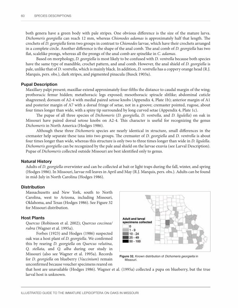
60 SPECIES DESCRIPTIONS
ILLUSTRATED GUIDE TO THE IMMATURE LEPIDOPTERA ON OAKS IN MISSOURI
both genera have a green body with pale stripes. One obvious difference is the size of the mature larva. Dichomeris georgiella can reach 12 mm, whereas Chionodes adamas is approximately half that length. The crochets of D. georgiella form two groups in contrast to Chionodes larvae, which have their crochets arranged in a complete circle. Another difference is the shape of the anal comb. The anal comb of D. georgiella has two flat, scalelike prongs, whereas all the prongs of the anal comb are spinelike in C. adamas.
Based on morphology, D. georgiella is most likely to be confused with D. ventrella because both species have the same type of mandible, crochet pattern, and anal comb. However, the anal shield of D. georgiella is pale, unlike that of D. ventrella, which is mainly black. In addition, D. ventrella has a coppery orange head (R.J. Marquis, pers. obs.), dark stripes, and pigmented pinacula (Busck 1903a).
Pupal DescriptionMaxillary palpi present; maxillae extend approximately four-fifths the distance to caudal margin of the wing; prothoracic femur hidden; metathoracic legs exposed; mesothoracic spiracle slitlike; abdominal cuticle shagreened; dorsum of A2-4 with medial paired setose knobs (Appendix 4, Plate 1b); anterior margin of A1 and posterior margin of A7 with a dorsal fringe of setae, not in a groove; cremaster pointed, rugose, about four times longer than wide, with a spiny tip surrounded by long curved setae (Appendix 4, Plate 1c).
The pupae of all three species of Dichomeris (D. georgiella, D. ventrella, and D. ligulella) on oak in Missouri have paired dorsal setose knobs on A2-4. This character is useful for recognizing the genus Dichomeris in North America (Hodges 1986).
Although these three Dichomeris species are nearly identical in structure, small differences in the cremaster help separate these taxa into two groups. The cremaster of D. georgiella and D. ventrella is about four times longer than wide, whereas this structure is only two to three times longer than wide in D. ligulella. Dichomeris georgiella can be recognized by the pale anal shield on the larvae exuvia (see Larval Description). Pupae of Dichomeris collected outside Missouri are best identified only to genus.
Natural HistoryAdults of D. georgiella overwinter and can be collected at bait or light traps during the fall, winter, and spring (Hodges 1986). In Missouri, larvae roll leaves in April and May (R.J. Marquis, pers. obs.). Adults can be found in mid-July in North Carolina (Hodges 1986).
DistributionMassachusetts and New York, south to North Carolina, west to Arizona, including Missouri, Oklahoma, and Texas (Hodges 1986). See Figure 32 for Missouri distribution.
Host PlantsQuercus (Robinson et al. 2002); Quercus coccinea/rubra (Wagner et al. 1995a).
Forbes (1923) and Hodges (1986) suspected oak was a host plant of D. georgiella. We confirmed this by rearing D. georgiella on Quercus velutina, Q. stellata, and Q. alba during our study in Missouri (also see Wagner et al. 1995a). Records for D. georgiella on blueberry (Vaccinium) remain unconfirmed because voucher specimens reared on that host are unavailable (Hodges 1986). Wagner et al. (1995a) collected a pupa on blueberry, but the true larval host is unknown.
Figure 32. Known distribution of Dichomeris georgiella in Missouri.

GELECHIIDAE 61
ILLUSTRATED GUIDE TO THE IMMATURE LEPIDOPTERA ON OAKS IN MISSOURI
Dichomeris ligulella HübnerPalmer worm
Larval DescriptionModified from Singerland and Crosby (1924), Chapman and Lienk (1971): Mature larva with head and anterior portion of prothoracic shield light brown; lateral and posterior margins of prothoracic shield black, often forming a pair of u-shaped markings; dorsal and subdorsal pinacula black, those on the thorax most obvious; thorax and abdomen either dark green with paired white middorsal longitudinal stripes, or white with thick green subdorsal longitudinal stripes and a thinner, sometimes broken, middorsal line; anal shield with two pigmented longitudinal bands (Figure 33).
Morphological characters of the larva of D. ligulella are: hypopharyngeal complex with a sclerotized ridge; mandible quadrate (as high as wide) with four cutting teeth, an outer tooth, and a moderately developed lobe above the condyle (Appendix 4, Plate 1d); SV group bisetose on A1 and A7, trisetose on A2, and unisetose on A8; crochets of A3-6 in two groups (Appendix 4, Plate 1e), crochets of A10 in a broken mesoseries; A9 with D2 posterodorsad of D1 and the hairlike SD1; and anal comb with six straight prongs, the two middle ones scalelike (Appendix 4, Plate 1f).
The larva of D. ligulella is illustrated by Singerland and Crosby (1924) and in color by Chapman and Lienk (1971). Dichomeris ligulella is recognized by the characteristic mandible, divided crochets, and distinctive body coloration. Typically, the prothoracic shield has two black u-shaped marks, the body pinacula are pigmented, and two wide subdorsal stripes surround a thinner middorsal line. However, Chapman and Lienk (1971) noted the larva of D. ligulella is extremely variable in color. The black prothoracic markings are commonly reduced from u-shaped marks to lines on the lateral and posterior margins of the prothoracic shield. In addition, the body stripes may fade on the posterior abdominal segments. Unlike D. georgiella, D. ligulella has only a moderately developed mandibular lobe (Appendix 4, Plate 1f).
Pupal DescriptionMaxillary palpi present; maxillae extend approximately four-fifths the distance to caudal margin of the wing; prothoracic femur hidden; metathoracic legs exposed; mesothoracic spiracle slitlike; abdominal cuticle shagreened, rarely somewhat smooth on the terminal abdominal segments; dorsum of A2-4 with medial paired setose knobs; anterior margin of A1 with a fringe of setae; cremaster blunt, rugose, about two times longer than wide, and surrounded by long curved setae.
Singerland and Crosby (1924) photographed the pupa of D. ligulella. The paired setose knobs on A2-4 and blunt cremaster that is two to three times longer than wide separate D. ligulella from other oak-feeding Gelechiidae treated in this book. If the larval exuvia is present, look for u-shaped markings on the prothoracic shield that are characteristic of many specimens of D. ligulella.
Natural HistoryLife cycle summarized from Chapman and Lienk (1971) and Hodges (1986): Dichomeris ligulella is univoltine with eggs laid in early spring. Larvae complete development from May to early July and feed on leaves, fruit, or oak-apple galls. The pupal stage lasts 10 days. Adults emerge in late June or early July, except in Florida where specimens have been captured in March. The adult is the overwintering stage.
Figure 33. Dichomeris ligulella mature larva. Photo by R.J. Marquis, used with permission.

62 SPECIES DESCRIPTIONS
ILLUSTRATED GUIDE TO THE IMMATURE LEPIDOPTERA ON OAKS IN MISSOURI
In Missouri, the larva of D. ligulella is a leaf roller found most frequently in April and May, rarely in June (R.J. Marquis, pers. obs.).
DistributionSouthern Canada south throughout the eastern United States to Virginia and Florida, east to Texas (Hodges 1986, Wagner et al. 1995a), including Missouri (Carroll and Kearby 1978).
According to Hodges (1986), no records were available from New Brunswick, Quebec, Maine, Vermont, Rhode Island, or Nebraska. Recently, D. ligulella was recorded from Quebec (Handfield 1999). See Figure 34 for Missouri distribution.
Host PlantsAcer, Betula, Carya, Castanea, Corylus, Juglans, Malus, Oxydendrum, Populus, Pyrus, Quercus, Q. alba, Q. coccinea, Q. macrocarpa, Q. rubra, Tilia, Vaccinium (Robinson et al. 2002); Cydonia, Rhododendron, and doubtfully Lactuca (Hodges 1986); Q. stellata and Q. velutina (Whitfield et al. 1999, Le Corff et al. 2000).
Oaks are the preferred food plants of D. ligulella, otherwise it is a generalist on trees and shrubs (Hodges 1986). In Missouri, D. ligulella feeds on Q. rubra seedlings (Linit et al. 1986) and is common on Q. alba and Q. velutina (LeCorff et al. 2000). We also found it on Q. coccinea, Q. imbricaria, Q. rubra (adults), Q. stellata, and Q. muehlenbergii.
CommentsDichomeris ligulella is often the most common species of gelechiid at blacklight traps in midsummer (Hodges 1986).
Figure 34. Known distribution of Dichomeris ligulella in Missouri.
Subfamily Gelechiinae
Arogalea cristifasciella (Chambers)Stripe-backed moth
Larval DescriptionMature larva with a copper-colored head capsule; prothoracic shield black along the lateral and posterior margins, otherwise concolorous with head; mesothorax and metathorax completely pigmented chocolate brown or red; abdomen banded with anterior half cream-colored and posterior half either chocolate brown or red; anal shield with posterior margin lined in black (Figure 35a).
Morphological characters of the larva of A. cristifasciella are: mandible with the cutting teeth elongated and fingerlike, lacking an outer tooth (Appendix 4, Plate 2a); proximolateral region of hypopharyngeal complex with a sclerotized ridge; SV group bisetose on A1 and A2 and unisetose on A7 (bisetose in one specimen on one side) and A8; crochets of A3-6 in a penellipse open mesially (Appendix 4, Plate 2b); D2 usually posterodorsad to a line joining D1 and the hairlike SD1 on A9; D1 is slightly anteroventrad of a

GELECHIIDAE 63
ILLUSTRATED GUIDE TO THE IMMATURE LEPIDOPTERA ON OAKS IN MISSOURI
a b
Figure 35. Arogalea cristifasciella (a) mature larva; (b) mature larva in silken nest, edged by frass. Photos by R.J. Marquis, used with permission.
line joining D2 and SD1 in some individuals; anal crochets divided into two groups; and a small anal comb present with two straight prongs (Appendix 4, Plate 2c).
The first instar is pale without obvious markings (Carroll 1977). The ground color of this species is variable. Dried larvae of A. cristifasciella at the United States National Museum of Natural History are a bright reddish color. In Missouri, the ground color is often reddish chocolate brown, and bright red in middle instars. Wooster (1977) illustrated the larva of A. cristifasciella and stated that the posterior abdominal segments were green or white. The green color may represent mature individuals about to pupate (Carroll 1977), although, it is more typical of moth larvae to turn pinkish or red at this time.
Among Missouri oak-feeding gelechiids, the larva of A. cristifasciella is easily recognized by its characteristic markings (see Carroll 1977: Fig. 25). In particular, the banded posterior margin of the prothorax and anal shield, unicolorous mesothorax and metathorax, and banded abdomen are unusual features when observed together. Several unrelated microlepidoptera, for example Agonopterix pulvipennella (Depressariidae) and Cenopis reticulatana (Tortricidae), have similarly marked anal shields (SCPC). However, both lack the banded abdomen of A. cristifasciella, and each has a different morphology and chaetotaxy than the Gelechiidae (see Stehr 1987). Also, Agonopterix pulvipennella does not feed on oak. At present, A. cristifasciella appears to be one of the few oak-feeding gelechiids with a distinctive coloration.
Pupal DescriptionMaxillary palpi present; maxillae extend approximately one-half the distance to caudal margin of the wings; prothoracic femur hidden; metathoracic legs exposed; mesothoracic spiracles slitlike; texture of abdominal cuticle shagreened; dorsum of A5 with paired subdorsal oval to rectangular sclerotized raised areas (Appendix 4, Plate 2d); fringe of setae in a groove present on A7; cremaster with scattered long, curved setae.
Carroll (1977: Fig. 28) photographed the pupa of A. cristifasciella. The subdorsal sclerotized raised areas on A5 will easily distinguish A. cristifasciella from all other gelechiid pupae that feed on oak in Missouri. No European species studied by Patočka (1987a, 1987b, 1987c, 1987d, 1988b, 1989; Patočka and Turčáni 2005) have a similar modification on the abdominal dorsum.
Natural HistoryAccording to the illustration in Carroll (1977), the egg of A. cristifasciella is translucent with a finely sculptured cuticle. Carroll and Kearby (1978) studied the life history of A. cristifasciella in Missouri. Adults first fly from March to May. Larvae occur from April to October. Adults of the second generation are found in July and August. The pupa of the second generation overwinter.
Larvae were collected in Missouri from April to October in small rhomboid-shaped tents of frass with a thick white silk roof (R.J. Marquis, pers. obs.; Figure 35b).
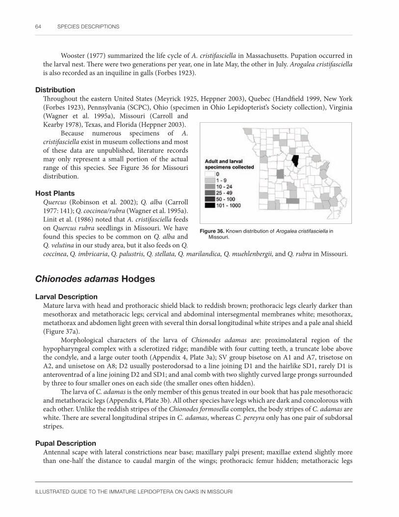
64 SPECIES DESCRIPTIONS
ILLUSTRATED GUIDE TO THE IMMATURE LEPIDOPTERA ON OAKS IN MISSOURI
Wooster (1977) summarized the life cycle of A. cristifasciella in Massachusetts. Pupation occurred in the larval nest. There were two generations per year, one in late May, the other in July. Arogalea cristifasciella is also recorded as an inquiline in galls (Forbes 1923).
DistributionThroughout the eastern United States (Meyrick 1925, Heppner 2003), Quebec (Handfield 1999, New York (Forbes 1923), Pennsylvania (SCPC), Ohio (specimen in Ohio Lepidopterist’s Society collection), Virginia (Wagner et al. 1995a), Missouri (Carroll and Kearby 1978), Texas, and Florida (Heppner 2003).
Because numerous specimens of A. cristifasciella exist in museum collections and most of these data are unpublished, literature records may only represent a small portion of the actual range of this species. See Figure 36 for Missouri distribution.
Host PlantsQuercus (Robinson et al. 2002); Q. alba (Carroll 1977: 141); Q. coccinea/rubra (Wagner et al. 1995a). Linit et al. (1986) noted that A. cristifasciella feeds on Quercus rubra seedlings in Missouri. We have found this species to be common on Q. alba and Q. velutina in our study area, but it also feeds on Q. coccinea, Q. imbricaria, Q. palustris, Q. stellata, Q. marilandica, Q. muehlenbergii, and Q. rubra in Missouri.
Figure 36. Known distribution of Arogalea cristifasciella in Missouri.
Chionodes adamas Hodges
Larval DescriptionMature larva with head and prothoracic shield black to reddish brown; prothoracic legs clearly darker than mesothorax and metathoracic legs; cervical and abdominal intersegmental membranes white; mesothorax, metathorax and abdomen light green with several thin dorsal longitudinal white stripes and a pale anal shield (Figure 37a).
Morphological characters of the larva of Chionodes adamas are: proximolateral region of the hypopharyngeal complex with a sclerotized ridge; mandible with four cutting teeth, a truncate lobe above the condyle, and a large outer tooth (Appendix 4, Plate 3a); SV group bisetose on A1 and A7, trisetose on A2, and unisetose on A8; D2 usually posterodorsad to a line joining D1 and the hairlike SD1, rarely D1 is anteroventrad of a line joining D2 and SD1; and anal comb with two slightly curved large prongs surrounded by three to four smaller ones on each side (the smaller ones often hidden).
The larva of C. adamas is the only member of this genus treated in our book that has pale mesothoracic and metathoracic legs (Appendix 4, Plate 3b). All other species have legs which are dark and concolorous with each other. Unlike the reddish stripes of the Chionodes formosella complex, the body stripes of C. adamas are white. There are several longitudinal stripes in C. adamas, whereas C. pereyra only has one pair of subdorsal stripes.
Pupal DescriptionAntennal scape with lateral constrictions near base; maxillary palpi present; maxillae extend slightly more than one-half the distance to caudal margin of the wings; prothoracic femur hidden; metathoracic legs

GELECHIIDAE 65
ILLUSTRATED GUIDE TO THE IMMATURE LEPIDOPTERA ON OAKS IN MISSOURI
a b
Figure 37. Chionodes adamas (a) mature larva; (b) hiding in leaf roll and silken nest of deciduous leaf hairs of Quercus alba. Photos by R.J. Marquis, used with permission.
exposed; mesothoracic spiracle slitlike; abdominal cuticle with scattered groups containing two to three fine spicules (Appendix 4, Plate 3c); fringe of setae in a groove present on A7; cremaster consists of curved setae in a semicircle around dorsal and lateral portions of anal slit.
The pupa of C. adamas is easily recognized by the characteristic cuticular texture composed of scattered clumps of spicules and the larval exuvia with pale legs. Unlike C. fuscomaculella, the antennal scape of C. adamas has constrictions. The C. formosella complex lacks spicules on the medial portion of A1, which separates it from C. adamas. The cuticular texture of C. pereyra is densely punctate without clumps of spicules. Pupae of Pseudotelphusa have clumped punctures, but the antennal scape is not constricted. The enlarged SD1 pinacula and row of punctures on the abdominal segment margins that are present in Pseudotelphusa are lacking in Chionodes.
Natural HistoryLarvae feed only in the spring (April–May) within small leaf rolls started from the leaf edge (R.J. Marquis, pers. obs.; Figure 37b). Adults have been collected in all months of the year, except February, with peak abundance in July and August (Hodges 1999).
DistributionSouthern Quebec to Manitoba, south to Mississippi and Texas, including Missouri (Hodges 1999). See Figure 38 for Missouri distribution.
Host PlantsQuercus alba, Q. ilicifolia, Q. laurifolia, Q. prinus, and Q. rubra (Hodges 1999). We have found larvae of C. adamas to be common on Q. alba and Q. velutina in Missouri, but also on Q. coccinea, Q. imbricaria, Q. rubra, and Q. stellata.
Figure 38. Known distribution of Chionodes adamas in Missouri.

66 SPECIES DESCRIPTIONS
ILLUSTRATED GUIDE TO THE IMMATURE LEPIDOPTERA ON OAKS IN MISSOURI
Chionodes formosella complex (Murtfeldt)
Larval DescriptionMature larva with head capsule and prothoracic shield either shiny black or reddish brown with some black shading; prothoracic legs as dark as the mesothorax and metathoracic legs; anterior margin of mesothorax white, the rest of segment chocolate brown; anterior half of metathorax white; posterior portion of metathorax and all abdominal segments concolorous, each with six broad dorsal and subdorsal red to reddish brown longitudinal stripes on a bright white to gray background that merges at the anal shield (Figure 39a).
Confirmed preserved larvae of the Chionodes formosella complex are not available. Our descriptions are based on larval exuviae associated with reared adults. Morphological characters of the larva of the C. formosella complex are: proximolateral region of hypopharyngeal complex with a sclerotized ridge; mandible with four cutting teeth and a large outer tooth (Appendix 4, Plate 3d); SV group bisetose on A1 and trisetose on A2; and anal comb with two large stout curved middle prongs surrounded by three to four smaller ones on each side (the smaller ones may be hidden) (Appendix 4, Plate 3e). The chaetotaxy of A9 is hard to evaluate on the available preparations, but apparently D2 is posterodorsad to a line joining D1 and the setiform SD1.
a b
Figure 39. Chionodes formosella complex (a) mature larva; (b) mature larva in silken nest of deciduous hairs of Quercus alba. Photos by R.J. Marquis, used with permission.
Among the few Chionodes species treated in this book, the C. formosella complex is most likely to be confused with C. fuscomaculella because these two species have the head and thorax similarly colored. However, the dorsal abdominal coloration is different. There are several parallel stripes in the C. formosella complex, whereas C. fuscomaculella has a w-shaped marking on the abdominal dorsum and only two thin subdorsal stripes. Another undetermined Chionodes species has faint cream longitudinal stripes in contrast to the C. formosella complex, whose stripes are red or reddish brown.
The larva of the C. formosella complex is poorly known; thus, it is difficult to predict how much color variation to expect. Hodges (1999) reproduced Murtfeldt’s larval notes on C. formosella. Our specimens generally agree with her description. We have seen the shiny black head and prothoracic shield, chocolate brown mesothorax, black legs, red longitudinal stripes, and gray body color. We have not seen purple body stripes. Murtfeldt reported eight longitudinal stripes, but the exact number of stripes present in our material is difficult to determine with the few larval photographs available.
Outside of Missouri, larvae colored like the C. formosella complex are best identified only to genus because other species of Chionodes with undescribed immature stages are known to feed on oak in the United States (Hodges 1999).

GELECHIIDAE 67
ILLUSTRATED GUIDE TO THE IMMATURE LEPIDOPTERA ON OAKS IN MISSOURI
Pupal DescriptionAntennal scape with lateral constrictions near the base; maxillary palpi present; maxillae extend slightly more than one-half the distance to the caudal margin of the wing; prothoracic femur hidden; metathoracic legs exposed; mesothoracic spiracle slitlike; A4 with dorsal punctures more widely spaced than the lateral punctures (those located below the spiracle); rest of abdominal cuticle with scattered fine punctures; fringe of setae in a groove present on A7; cremaster consists of curved setae in semicircle around dorsal and lateral portions of anal slit.
The modified antennal scape in the C. formosella complex distinguishes this species from C. fuscomaculella, which has an unmodified scape. The C. formosella complex and C. pereyra can be separated by differences in the anterior abdominal segments. Chionodes pereyra has both the dorsal and lateral punctures approximately equal in density and spacing. The dorsal punctures of the C. formosella complex are more widely spaced than the lateral punctures below the spiracle.
Natural HistoryThis species occurs almost exclusively during the spring (April–May) before the new leaves toughen (Figure 39b) and is sometimes found hiding in small portions of leaves that have died and turned brown (R.J. Marquis, pers. obs.). Hodges (1999) and Forbes (1923) state that the larva of C. formosella occurs in May, the pupa is found in a folded leaf, and adults were collected in July. Moths can also be found from April (Heppner 2003) to September (Hodges 1999).
DistributionNova Scotia to southwestern Manitoba and south to North Carolina and eastern Missouri (Hodges 1999); Illinois (Godfrey et al. 1987a); Florida and possibly Texas (Heppner 2003) (based on records for C. formosella). See Figure 40 for Missouri distribution.
Host PlantsQuercus alba, Q. macrocarpa, Q. rubra (Robinson et al. 2002); Ostrya virginiana, Q. velutina, and probably Q. imbricaria (Hodges 1999); Q. laurifolia (Forbes 1923, Heppner 2003).
The Chionodes formosella complex was most frequently found by us on Q. alba and Q. velutina in Missouri. Most rearings for C. formosella seen by Hodges (1999) were from the red oak group, e.g., Q. rubra, Q. velutina, and probably Q. imbricaria. Records in Robinson et al. (2002) for C. formosella found on Q. douglasii and Q. lobata likely refer to C. powelli (see Hodges 1999: 52). We have seen larvae on Q. alba, Q. coccinea, Q. marilandica, Q. rubra, Q. stellata, and Q. velutina.
Figure 40. Known distribution of the Chionodes formosella complex in Missouri.
CommentsChionodes formosella was known as Gelechia vernella Murt. and has also been confused with C. powelli in the western United States (Hodges 1999). Given our lack of material and the difficult identification problems in this complex (Hodges 1999: 50), we have chosen to be tentative in our species determination. Our voucher specimen (91-4C-B-6) was reared from Q. velutina and was identified by R. W. Hodges as C. formosella. Further support for our identification comes from a dried larva preserved in the U.S. National Museum of Natural History determined as “Gelechia vernella.” This specimen closely matches our larval photographs of the C.

68 SPECIES DESCRIPTIONS
ILLUSTRATED GUIDE TO THE IMMATURE LEPIDOPTERA ON OAKS IN MISSOURI
formosella complex. More work is needed to ensure that larvae colored like C. formosella are always associated with reared adults of this species. Related species may have identical larvae. Carroll and Kearby (1978: 460) also pointed out that associating immatures with adults was a challenge in the Missouri oak ecosystem.
Chionodes fuscomaculella (Chambers)
Larval DescriptionMature larva with head and prothoracic shield black; mesothorax chocolate brown; metathorax with anterior portion white, contrasting with the chocolate brown posterior portion; prothoracic legs as dark as the mesothorax and metathoracic legs; A1-8 with chocolate brown, w-shaped dorsal markings and a thin subdorsal line of the same color; anal shield usually solid chocolate brown, sometimes spotted black (Figure 41).
Morphological characters of the larva of C. fuscomaculella include: proximolateral region of hypopharyngeal complex with a sclerotized ridge (Appendix 4, Plate 4a); mandible with four cutting teeth and a small outer tooth that may be worn smooth, lobe above condyle poorly developed (Appendix 4, Plate 4b); SV group bisetose on A1 and A7, trisetose on A2, and unisetose on A8; crochets in a circle (Appendix 4, Plate 4c); D2 usually posterodorsad to a line joining D1 and the SD1 hairlike on A9, rarely D1 is anteroventrad of a line joining D2 and SD1; and anal comb present with two large curved prongs surrounded by three to four smaller ones on each side if not hidden (Appendix 4, Plate 4d).
Among the few Chionodes treated in this book, C. fuscomaculella is the only species with a contrastingly white anterior portion of the metathorax, dorsal w-shaped abdominal markings, and a completely pigmented anal shield. Hodges (1999: 21) noted that the larva of C. fuscomaculella has SD1 hairlike on A9 and an anal comb. Our specimens agree with this description.
Chionodes fuscomaculella might be confused with D. ventrella because both species have a pigmented anal shield. However, the crochets on A3-6 of C. fuscomaculella are in a circle, whereas D. ventrella has these crochets in two groups.
One of about 10 preserved larvae of C. fuscomaculella lacks an anal comb. This may be an anomaly or perhaps a rarely encountered sibling species of Chionodes. Several species of this genus are known to lack an anal comb (Hodges 1999), including C. bicostomaculella, which also has been reared from oak in Missouri.
Pupal DescriptionAntennal scape unmodified, at most with faint traces of a constriction; maxillary palpi present; maxillae extend slightly more than one-half the distance to caudal margin of the wings; prothoracic femur hidden; metathoracic legs exposed; mesothoracic spiracle slitlike; texture of abdominal cuticle smooth except for scattered fine spicules; fringe of setae in a groove present on A7; cremaster consists of curved setae in a semicircle around the dorsal and lateral portions of the anal slit.
The pupa of C. fuscomaculella differs from that of C. pereyra, C. adamas, and the C. formosella complex by having an unmodified scape.
Figure 41. Chionodes fuscomaculella mature larva. Photo by R.J. Marquis, used with permission.
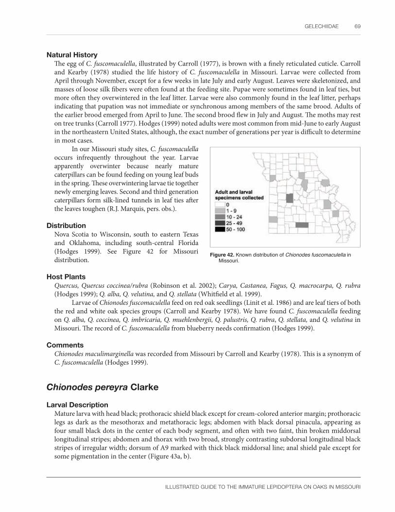
GELECHIIDAE 69
ILLUSTRATED GUIDE TO THE IMMATURE LEPIDOPTERA ON OAKS IN MISSOURI
Natural HistoryThe egg of C. fuscomaculella, illustrated by Carroll (1977), is brown with a finely reticulated cuticle. Carroll and Kearby (1978) studied the life history of C. fuscomaculella in Missouri. Larvae were collected from April through November, except for a few weeks in late July and early August. Leaves were skeletonized, and masses of loose silk fibers were often found at the feeding site. Pupae were sometimes found in leaf ties, but more often they overwintered in the leaf litter. Larvae were also commonly found in the leaf litter, perhaps indicating that pupation was not immediate or synchronous among members of the same brood. Adults of the earlier brood emerged from April to June. The second brood flew in July and August. The moths may rest on tree trunks (Carroll 1977). Hodges (1999) noted adults were most common from mid-June to early August in the northeastern United States, although, the exact number of generations per year is difficult to determine in most cases.
In our Missouri study sites, C. fuscomaculella occurs infrequently throughout the year. Larvae apparently overwinter because nearly mature caterpillars can be found feeding on young leaf buds in the spring. These overwintering larvae tie together newly emerging leaves. Second and third generation caterpillars form silk-lined tunnels in leaf ties after the leaves toughen (R.J. Marquis, pers. obs.).
DistributionNova Scotia to Wisconsin, south to eastern Texas and Oklahoma, including south-central Florida (Hodges 1999). See Figure 42 for Missouri distribution.
Host PlantsQuercus, Quercus coccinea/rubra (Robinson et al. 2002); Carya, Castanea, Fagus, Q. macrocarpa, Q. rubra (Hodges 1999); Q. alba, Q. velutina, and Q. stellata (Whitfield et al. 1999).
Larvae of Chionodes fuscomaculella feed on red oak seedlings (Linit et al. 1986) and are leaf tiers of both the red and white oak species groups (Carroll and Kearby 1978). We have found C. fuscomaculella feeding on Q. alba, Q. coccinea, Q. imbricaria, Q. muehlenbergii, Q. palustris, Q. rubra, Q. stellata, and Q. velutina in Missouri. The record of C. fuscomaculella from blueberry needs confirmation (Hodges 1999).
CommentsChionodes maculimarginella was recorded from Missouri by Carroll and Kearby (1978). This is a synonym of C. fuscomaculella (Hodges 1999).
Figure 42. Known distribution of Chionodes fuscomaculella in Missouri.
Chionodes pereyra Clarke
Larval DescriptionMature larva with head black; prothoracic shield black except for cream-colored anterior margin; prothoracic legs as dark as the mesothorax and metathoracic legs; abdomen with black dorsal pinacula, appearing as four small black dots in the center of each body segment, and often with two faint, thin broken middorsal longitudinal stripes; abdomen and thorax with two broad, strongly contrasting subdorsal longitudinal black stripes of irregular width; dorsum of A9 marked with thick black middorsal line; anal shield pale except for some pigmentation in the center (Figure 43a, b).

70 SPECIES DESCRIPTIONS
ILLUSTRATED GUIDE TO THE IMMATURE LEPIDOPTERA ON OAKS IN MISSOURI
a b
Figure 43. Chionodes pereyra (a) early instar larva; (b) mature larva. Photos by R.J. Marquis, used with permission.
Morphological characters of C. pereyra larvae are: proximolateral region of hypopharyngeal complex with a sclerotized ridge; mandible with four cutting teeth, an outer tooth, and well developed lobe above the condyle (Appendix 4, Plate 5a); SV group bisetose on A1 and A7, trisetose on A2, and unisetose on A8; in our single specimen, D2 posterodorsad of a line joining D1 and the hairlike SD1 on one side, the other has D1 anteroventrad of a line joining D2 and SD1; and anal comb with two slightly curved large prongs surrounded by two smaller ones on each side if not hidden.
Of the Chionodes treated in our book, C. pereyra is recognized by the contrasting subdorsal longitudinal stripes, relatively large pale dorsal pinacula, and the lack of w-shaped abdominal markings. Unlike C. fuscomaculella, the mandibular lobe above the condyle of C. pereyra is well developed. Instead of only subdorsal stripes, some Chionodes species have dorsal and lateral longitudinal stripes.
Pupal DescriptionAntennal scape with lateral constrictions near base (Appendix 4, Plate 5b); maxillary palpi present; maxillae extend slightly more than one-half the distance to the caudal margin of the wings; prothoracic femur hidden; metathoracic legs exposed; mesothoracic spiracle slitlike; dorsal and lateral punctures of A4 approximately equal in density and spacing, rest of abdominal cuticle with scattered fine punctures (Appendix 4, Plate 5c); fringe of setae in a groove present on A7 (Appendix 4, Plate 5d); cremaster consists of curved setae in a semicircle around dorsal and lateral portions of anal slit.
The pupa of C. fuscomaculella differs from that of C. pereyra in having an unmodified scape. Chionodes pereyra has both the dorsal and lateral punctures of A4 approximately equal in density and spacing, unlike the C. formosella complex, which has the dorsal punctures more widely spaced than the lateral punctures below the spiracle.
Natural HistoryThe larvae tie leaves in spring and early summer (R.J. Marquis, pers. obs.). Adults fly mainly in June and July. They may be collected in light traps, especially in rainy weather, and in flight intercept traps designed for horseflies (Tabanidae) (Hodges 1999). Wagner et al. (1995a) collected numerous C. pereyra from the oak canopy in Virginia. Figure 44. Known distribution of Chionodes pereyra in Missouri.

GELECHIIDAE 71
ILLUSTRATED GUIDE TO THE IMMATURE LEPIDOPTERA ON OAKS IN MISSOURI
DistributionMassachusetts to Michigan south to Florida and Texas (Hodges 1999). See Figure 44 for Missouri distribution.
Host PlantsQuercus spp. (Hodges 1999); and Q. coccinea/rubra (Robinson et al. 2002).
We have found C. pereyra on Q. alba, Q. imbricaria, Q. muehlenbergii, Q. rubra, Q. stellata, and Q. velutina in Missouri.
Pseudotelphusa complex
Larval DescriptionYoung larvae pale green to pale yellow. Mature larvae of two types. First type (gray to cream form, Figure 45a) with a black genal band, black lateral and posterior margins on the prothorax, pigmented body pinacula, and a pale anal shield (Appendix 4, Plate 6a). Second type (banded form, Figure 45b) with the head capsule spotted on the vertex; prothoracic shield with posterior margins lined in black; anterior half of mesothorax, metathorax, and anterior abdominal segments cream to white; rest of thorax and abdomen with solid red to chocolate brown bands alternating with white bands; body pinacula pigmented, and an anal shield with the posterior margin black (Appendix 4, Plate 6b).
Both forms have large spots in the lateral area of the thorax and subdorsal area of the abdomen. Morphological characters of the mature larva of the first type are: head lacking spots on vertex; mandible square-shaped with a small outer tooth and four cutting teeth (Appendix 4, Plate 6c); proximolateral region of hypopharyngeal complex with a sclerotized ridge; SV group bisetose on A1 and A7, trisetose on A2 (rarely bisetose on one side), and unisetose on A8; diameter of L pinacula of thorax and SD1 pinacula of abdomen three to five times the diameter of the abdominal spiracles; crochets of A3-6 in a mesal penellipse, closed by a few scattered small crochets forming a circle; anal crochets in a broken mesoseries closed by a few small crochets in the center; A9 with D2 posterodorsad to a line joining D1 and SD1, or sometimes D1 slightly anteroventrad of a line joining D2 with SD1, SD1 hairlike; and anal comb absent.
Morphological characters of the mature larva of the second type are: head with spots on vertex; mandible square-shaped with a small outer tooth and four cutting teeth; proximolateral region of hypopharyngeal complex with a sclerotized ridge; SV group bisetose on A1 and A7, trisetose on A2, and unisetose on A8; diameter of the L pinacula of thorax and SD1 pinacula of abdomen three to five times the diameter of the abdominal spiracles; crochets of A3-6 in a mesial penellipse, closed by a few scattered small crochets forming
a b
Figure 45. Pseudotelphusa complex (a) mature nonbanded form larva; (b) mature banded form larva. Photos by R.J. Marquis, used with permission.

72 SPECIES DESCRIPTIONS
ILLUSTRATED GUIDE TO THE IMMATURE LEPIDOPTERA ON OAKS IN MISSOURI
a circle; anal crochets in a broken mesoseries closed by a few small crochets in the center; A9 with D2 posterodorsad to a line joining D1 and SD1, or sometimes D1 slightly anteroventrad of a line joining D2 with SD1, SD1 hairlike; and anal comb absent.
Both larval phenotypes of Pseudotelphusa are easily recognized by the enlarged L and SD1 pinacula, characteristic crochet pattern on A3-6, and lack of an anal comb. Stenomatine depressariids have enlarged thoracic pinacula, but unlike Pseudotelphusa, the abdominal SD1 pinacula are much smaller than the ones on the thorax. The lack of an anal comb easily separates Pseudotelphusa from both Chionodes and Dichomeris. Arogalea has a reduced anal comb, but unlike Pseudotelphusa, the SD1 pinacula are not enlarged compared to the spiracles.
Pupal Description Banded type larva: Antennal scape unmodified; maxillary palpi present; maxillae extend one half the distance to caudal margin of the wings; prothoracic femur hidden; metathoracic legs exposed; mesothoracic spiracle slitlike; spiracle and the SD1 seta of A1-4 fused on a low rounded dull reddish brown tubercle (Appendix 4, Plate 6d); abdominal cuticle with scattered groups of two to five spicules and a dorsal row of fine punctures on both the anterior and posterior margins of A1-4 (Appendix 4, Plate 6d); fringe of setae in a groove present on A7; cremaster consists of curved setae around the anal slit.
The presence of a fine row of punctures on the dorsal surface of the anterior and posterior margins of A1-4 will separate the banded form of Pseudotelphusa from other oak-feeding gelechiids treated in this book. Gray to cream form larva: We need more material to better associate the pupa of the nonbanded gray form with reared adults and larval skins.
Pseudotelphusa is most likely to be confused with Chionodes species in the pupal stage because both taxa have the punctures arranged in clumps. The antennal scape of Pseudotelphusa lacks lateral constrictions, which often are present in Chionodes. Also, Pseudotelphusa has a row of fine punctures on the margins of A1-4, which are absent in Chionodes. Pseudotelphusa is the only genus of Missouri oak-feeding gelechiids that has the spiracle and the SD1 seta of A1-4 fused on a low, rounded, dull reddish brown tubercle, although sometimes this feature is absent or difficult to observe.
Mosher (1916a) diagnosed P. quercinigracella (as Telphusa) in the following way: abdomen not pubescent, forked body setae absent, and A7 with a bilobed row of dense setae in a groove that does not extend around the whole segment. This diagnosis also applies to Pseudotelphusa in Missouri except that the groove is only weakly bilobed.
Patočka (1987a, 1987b) separated European species of Pseudotelphusa from Chionodes using the condition of the labial palpi. In Chionodes the labial palpi is minutely exposed, whereas in Pseudotelphusa the palpi are hidden. This difference does not apply in Missouri, where both genera have their labial palpi completely hidden. After further study, Patočka and Turčáni (2005) lumped Pseudotelphusa with four other related genera in a combined species key. There was no mention of enlarged abdominal subdorsal tubercles characteristic of species in the Missouri oak fauna.
Natural HistoryThe egg of Pseudotelphusa is illustrated by Carroll (1977) and is light gray at first, later turning brown with spots. The life history of Pseudotelphusa was studied by Carroll and Kearby (1978) in Missouri. Eggs and first instar larvae were collected in late April on the middle and lower portions of the tree. Larvae fed in silk-lined pockets underneath tied leaves. The pupation site was not noted. Adult flight started in March and peaked by April. The moths may rest on tree trunks (Carroll 1977).
Pseudotelphusa are very common leaf tiers in Missouri with two generations per year from June through September (R.J. Marquis, pers. obs.). Leaf ties made by Pseudotelphusa on Q. alba are sites of colonization by many other herbivore species. This results in increased insect herbivore diversity on trees with leaf ties compared to trees in which early leaf tiers (mostly Pseudotelphusa) have been removed (Lill and Marquis 2003).

GELECHIIDAE 73
ILLUSTRATED GUIDE TO THE IMMATURE LEPIDOPTERA ON OAKS IN MISSOURI
DistributionAny distribution is speculative until accurate identifications are made for the Pseudotelphusa complex. Pseudotelphusa quercinigracella is generally distributed in the eastern United States (Forbes 1923, Meyrick 1925) but is absent from Florida (Heppner 2003). Wagner et al. (1995a) did not list any Pseudotelphusa from oak in Virginia. See Figure 46 for Missouri distribution.
Host PlantsQuercus sp., Q. nigra (Robinson et al. 2002 for P. quercinigracella); and Q. alba (Lee 2011 for P. ontariensis).
We have found Pseudotelphusa on Quercus alba, Q. coccinea, Q. imbricaria, Q. palustris, Q. rubra, Q. stellata, Q. muehlenbergii, and Q. velutina in Missouri. The nonbanded form is more common than the banded form (R. J. Marquis and J. B. Whitfield, unpublished data).
CommentsWe tentatively regard Pseudotelphusa as a complex of two or more species based on the widely different larval coloration. Reared adults from our study, as determined by R. W. Hodges, included P. quercinigracella and another different but unknown Pseudotelphusa species. There may also be color differences between instars that further complicate identification. Larval rearing of adults of P. quercinigracella for this study represents a new state record for this species.
We agree with Carroll and Kearby (1978) that more work is needed to associate reared adults with their larval stages in Pseudotelphusa. These authors also suggested two species were associated with oaks in Missouri, one being either P. fuscopunctella or P. quercinigracella, the other being a new species (citing R. W. Hodges as the source of these names). The larval coloration of both these species is similar, causing Forbes (1923) to warn that the complex is “practically indistinguishable in the larval stage without breeding [to adults].” As of 2018, no molecular studies have been attempted on Pseudotelphusa.
Figure 46. Known distribution of the Pseudotelphusa complex in Missouri.
Pubitelphusa latifasciella (Chambers)White-banded leaf roller
Larval DescriptionMature larva with head capsule pale brown; all thoracic legs pale; prothoracic shield and abdomen yellow, the digestive tract showing through the body as an internal medial dark streak; anal shield pale (Figure 47).
Morphological characters of the larva of P. latifasciella are: mandible with four cutting teeth, an outer tooth, and well developed lobe above the condyle (Appendix 4, Plate 7a); proximolateral region of the hypopharyngeal complex with a sclerotized ridge; SV group bisetose on A1 and A7, trisetose on A2, and unisetose on A8; crochets of A3-6 in a circle (Appendix 4, Plate 7b) and anal crochets in a mesoseries; A9 with D2 posterodorsad to a line joining D1 and SD1; or sometimes D1 slightly anteroventrad of a line joining D2 with SD1, SD1 hairlike; anal comb with two slightly curved large prongs (Appendix 4, Plate 7c) surrounded by a smaller pair on each side.
The larva of P. latifasciella is separated from other Missouri oak-feeding gelechiids by the pale body color, distinctive mandible, and crochet arrangement. It is most likely to be confused with Chionodes adamas

74 SPECIES DESCRIPTIONS
ILLUSTRATED GUIDE TO THE IMMATURE LEPIDOPTERA ON OAKS IN MISSOURI
Figure 47. Pubitelphusa latifasciella mature larva. Photo by R.J. Marquis, used with permission.
and T. prudens, which also have a pale body color and crochets in a circle. However, only the mesothoracic and metathoracic legs of C. adamas are unpigmented, whereas all three thoracic legs are pale in P. latifasciella. The well-developed outer lobe on the mandible of P. latifasciella is absent from T. prudens.
The curved anal comb and widely separated D2 setae on A9 will separate P. latifasciella from several pale colored oak-feeding tortricids that might occur on Missouri oaks.
Pupal DescriptionAntennal scape without lateral constrictions near base; maxillary palpi present; maxillae extend three-quarters the distance to caudal margins of the wing; prothoracic femur hidden; metathoracic legs exposed; mesothoracic spiracle slitlike; abdominal cuticle finely shagreened with minute tubercles (Appendix 4, Plate 7d); fringe of setae in a groove present on A7; cremaster consists of curved setae in a semicircle around dorsal and lateral portions of anal slit.
The pupa of P. latifasciella is the only Missouri oak-feeding gelechiid that lacks antennal scape constrictions and modifications of the abdominal dorsum, in combination with a shagreened abdominal cuticle. Pseudotelphusa and Chionodes are similar, but these genera have punctures, and in the case of some Chionodes, antennal constrictions.
Natural HistoryWe observed the larva of P. latifasciella rolling one-third to one-half of a leaf and also tying young leaves together, especially of Q. velutina, in April–May. Carroll and Kearby (1978) also observed this species making leaf ties.
DistributionQuebec and southern Ontario to Wisconsin then south to Florida, Texas, and Oklahoma (Lee and Brown 2013), including the eastern United States (Massachusetts, Virginia, Kentucky, North Carolina, and Missouri) (Forbes 1923, Meyrick 1925, Wagner et al. 1995a), Illinois (Heppner 2003) and Ohio (specimen in The Ohio Lepidopterists Society collection). See Figure 48 for Missouri distribution.
Host PlantsQuercus coccinea/rubra, Vaccinium (Robinson et al. 2002); Q. alba (Whitfield et al. 1999), Quercus sp., Q. rubra, Q. velutina (Lee and Brown 2013).
Missouri records include Q. rubra seedlings (Linit et al. 1986), and we have found it on Q. alba, Q. coccinea, Q. imbricaria, Q. marilandica, Q. muehlenbergii, Q. rubra, Q. stellata, and Q. velutina as well. Lee and Brown (2013) noted that there are no voucher specimens to confirm the record for P. latifasciella on Vaccinium.
Figure 48. Known distribution of Pubitelphusa latifasciella in Missouri.

GELECHIIDAE 75
ILLUSTRATED GUIDE TO THE IMMATURE LEPIDOPTERA ON OAKS IN MISSOURI
CommentsPubitelphusa latifasciella was recently moved from the genus Telphusa (Lee and Brown 2013) after being initially described in Gelechia (Chambers 1875). Busck (1903a: 785) noted that adults come in two color forms, both of which were bred from similar larvae by Murtfeldt. We have also bred two color forms of this species (with and without a white fascia, see Forbes 1923) and can confirm that both pupae have a similar shagreened cuticular texture. Lee and Brown (2013) illustrate three adult color forms.
Trypanisma prudens Clemens
Larval DescriptionEarly instar with pale yellow head capsule and body. Mature larva with pale yellow-green head and brown mouth parts, a green prothoracic shield with a broad pale anterior transverse band, green mesothorax and metathorax; and a green abdomen with cream intersegmental areas on most segments (Figure 49a).
Morphological characters of the larva of T. prudens are: mandible with three cutting teeth, the third tooth strongly diverging from the second, without an outer tooth or lobe above the condyle (Appendix 4, Plate 8a); proximolateral region of hypopharyngeal complex with a sclerotized ridge; SV group bisetose on A1 and A7, trisetose on A2, and unisetose on A8; crochets of A3-6 in a mesial penellipse, closed by a few scattered small crochets forming a circle (Appendix 4, Plate 8b); anal crochets in a broken mesoseries connected by small crochets (Appendix 4, Plate 8c); A9 with D2 posterodorsad of D1 and the hairlike SD1, or the D2, D1, and SD1 setae are a straight vertical line; and anal comb with two slightly curved large prongs surrounded by four smaller pairs on each side (Appendix 4, Plate 8d).
Because of the pale coloration, T. prudens is most likely to be confused with Chionodes adamas and P. latifasciella. The mandible of T. prudens lacks both a well-developed lobe above the condyle and an outer tooth; at least one of these characters is present in other similarly colored gelechiids. In addition, T. prudens is the only species of Missouri oak-feeding gelechiid treated in this book to make an “escape hole” in the leaf shelter (see Natural History). The long anal setae (if not broken) are also unusual.
Many microlepidopteran larvae in several families have pale coloration. Therefore, careful attention must be paid to the family characteristics of the Gelechiidae listed in the introduction when trying to identify T. prudens.
Pupal DescriptionAntennal scape with lateral constrictions near base; maxillary palpi present; maxillae extend slightly more than half the distance to the caudal margin of the wings; wings extend to A7; prothoracic femur hidden; metathoracic legs exposed at tips; mesothoracic spiracle slitlike; entire cuticle of body covered with scattered fine spicules, clumped in groups of two to three on the anterior abdominal dorsum; fringe of setae in a groove
a b
Figure 49. Trypanisma prudens (a) mature larva; (b) mature larva in sheet web near "escape hole." Photos by R.J. Marquis, used with permission.

76 SPECIES DESCRIPTIONS
ILLUSTRATED GUIDE TO THE IMMATURE LEPIDOPTERA ON OAKS IN MISSOURI
present on A7 with two large plates dorsally (Appendix 4, Plate 8e); cremaster consists of curved setae in a semicircle around dorsal and lateral portions of anal slit.
Mosher (1916a) diagnosed the pupa of T. prudens as follows: abdomen not pubescent, forked body setae absent, and A7 with a wavy fringe of setae that includes two large lobes on the dorsum.
Among Missouri oak-feeding gelechiids, T. prudens is most likely to be confused with Chionodes because both taxa have a constricted antennal scape and spicules in clumps on the cuticle. Unlike Chionodes, T. prudens has spicules all over the body, even including the appendages, mouthparts, and wing cases. Also, Chionodes lacks dorsal lobes on the groove of A7, which are present in T. prudens.
Natural HistoryThe life history of T. prudens was described by Clemens (1860). The larva lives in a web on the underside of oak leaves. This web includes an escape hole (Figure 49b; Appendix 4, plate 8f) formed by cutting an opening through the leaf just large enough to allow the larva to pass from one surface to another. Feeding occurs on both sides of the leaf, but if disturbed, the larva returns to the web through the escape hole. Pupation is on the same leaf that supports the larval feeding.
DistributionEastern United States from Pennsylvania south to Florida, west to Illinois and perhaps Mississippi (Heppner 2003), and Ohio (Forbes 1923). See Figure 50 for Missouri distribution.
Host PlantsAcer (T. Harrison, pers. comm.); Fagus, Quercus, Q. velutina (Robinson et al. 2002); Q. prinus (Stainton 1872). We have found T. prudens on Q. alba, Q. coccinea, Q. imbricaria, Q. palustris, Q. rubra, and Q. velutina in Missouri.
CommentsRota and Wagner (2008) listed examples of other microlepidopteran larvae using the “escape hole” defense strategy. Our collections represent a new state record for this species.
Figure 50. Known distribution of Trypanisma prudens in Missouri.

TORTRICIDAE 77
ILLUSTRATED GUIDE TO THE IMMATURE LEPIDOPTERA ON OAKS IN MISSOURI
TortricidaeLeaf roller moths
The family Tortricidae currently contains approximately 10,000 species worldwide in three subfamilies: Tortricinae, Chlidanotinae, and Olethreutinae (Horak 1998, Regier et al. 2012a). Ten tortricid taxa have been considered families in the past, the most common examples being Tortricidae, Olethreutidae, Cochylidae (=Phalonidae), or Ceracidae (e.g., MacKay 1959, 1962; Covell 1984; Regier et al. 2012a: Table 1). However, more recent phylogenetic studies have supported combining these groups under Tortricidae (Horak and Brown 1991, Horak 1998, Regier et al. 2012a). Although tortricids are distributed worldwide, the tribes often show geographical specialization. For example, the Sparganothini are more diverse in the New World, whereas the Ceracini are confined to Asia (Horak 1998). There are eleven tribes of Tortricinae, three tribes in the Chlidanotinae, and seven tribes of Oleuthreutinae. Freeman (1958: 6) provided a historical review of Tortricinae classification. Diakonoff (1973: XI) published a similar review of the Olethreutinae. Recent publications treat the Olethreutinae of Australia (Horak 2006) and the midwestern United States (Gilligan et al. 2008). For a summary of important literature on the Tortricidae and detailed morphological notes on the tribes, consult the world catalog by Brown et al. (2005) and the references listed in the information sources section of Horak (1998). Regier et al. (2012a) compared historical morphological phylogenies to molecular results. Powell and Brown (2012) revised the Sparganothini and Atteriini of North America. Three of the most useful Web sites for Tortricidae are: Gilligan (2019), which includes a synopsis of the tribes with host plant and literature information; Gilligan and Epstein (2012), which provides fact sheets and information about agriculturally important species, and Gilligan and Epstein’s (2009) light brown apple moth identification guide. Arguably, these Web sites make the Tortricidae the best documented family of Lepidoptera online. The guide to New Zealand species by Dugdale et al. (2005) is an excellent source of information on Asian species, including those intercepted during quarantine surveys.
Tortricid eggs are of the flat type, with the long axis and micropyle parallel to the substrate. The lemon-shaped eggs of the Phricanthini possibly represent the primitive condition, but most species have naked eggs that are flat, scalelike, or dome-shaped (Horak 1998). In the genus Archips, females cover their eggs with scales (A. semiferanus) (Chapman and Lienk 1971) or a black secretion (A. fuscocupreanus) (SCPC). Two Tortricinae genera from Australia, Isotenes and Cryptoptilia, encircle the eggs with a “fence” of scales from the female’s body (Powell and Common 1985). In his survey of North American tortricid species, Peterson (1965a) noted that olethreutine eggs are sometimes translucent, or are colored milky white to light green to yellow. This coloration somewhat overlaps with Tortricinae eggs, which are white, yellow, red, or light green. At least in North America, tortricid eggs are deposited singly (some Olethreutinae, Tortricini, Cochylini, and some Cnephasiini) or in small clusters that may overlap (Sparganothini, Archipini, some Cnephasiini, and Olethreutinae) (Brown 1987b, Horak 1998, Passoa 2008: Fig. 16). The chorion either is granulated, reticulated, ridged, dimpled, or smooth (Peterson 1965a).
Tortricid larvae can tie or roll leaves, bore into stems or roots, feed on flowers or fruits, induce galls, or rarely mine leaves (Patočka and Turčáni 2005). Most tortricid larvae can be recognized and segregated into subfamilies by a few characters. Typically, the prespiracular group is trisetose; L1 and L2 are adjacent on A1-8; SD1 is in front of the spiracle on A8; the D2 setae of A9 are joined on the same pinaculum; and the crochets are in a circle. An anal comb with straight prongs may be present. In the Tortricinae, SD1 and D1 of A9 are on separate pinacula. This is not the case for the Olethreutinae and Cochylini, in which the SD1 and D1 pinacula of A9 are fused (MacKay 1959, 1962; Brown 1987b; Horak and Brown 1991; Horak 1998). Dugdale et al. (2005) added that tortricid larvae usually have V1 close to or fused with the T2 and T3 coxae.
When the world fauna is considered, scattered exceptions to the above generalizations have led to three problems in tortricid larval identification. First, neither MacKay (1962) in North America nor Swatschek (1958) in Europe were able to separate all larval Tortricinae from all Olethreutinae (including Cochylini). Swatschek (1958) developed a key to tribes without defining the subfamiles. MacKay (1962: 7) suggested that if the larva ties leaves or feeds externally among the flowers and fruit or lives in a nest of silk and frass, and has a rounded

78 SPECIES DESCRIPTIONS
ILLUSTRATED GUIDE TO THE IMMATURE LEPIDOPTERA ON OAKS IN MISSOURI
spinneret, it is better to guess Tortricinae. When the larva is a true borer (internal feeder) and the spinneret either is tapered, bifurcate, or spatulate, it is better to guess Olethreutinae. Adding the condition of SD1 and D1 of A9 to the above guidelines is helpful, but there is still no known way to separate these two subfamilies with complete certainty. Outside of North America, especially in tropical regions, it is often impossible even to guess past the family level. The second problem is that existing keys to tribes are often of limited use, as noted by Brown (1987a). For example, some members of the tortricine tribe Euliini (Seticosta, Proeulia, and Anopina) from Latin America have SD1 and D1 fused on the same pinaculum (Brown and Nishida 2003). This condition is typical for the Olethreutinae but not the Tortricinae (MacKay 1959). Unfortunately, the European study of Tortricidae larvae by Swatschek (1958) did not emphasize the same characters as MacKay (1959, 1962). This further complicates identification of tribes at the world level and makes it difficult to compare taxonomic characters between regions.
The third problem in tortricid larval identification is that, in rare cases, it may be hard to recognize Tortricidae at the family or superfamily level. For example, a bisetose prespiracular group is found in a few species of four Old World tortricid genera (Hendecaneura, Pammene, Thaumatographa, and Ochetarcha) (Horak and Brown 1991, Nasu and Komai 1997). These genera would not easily key to Tortricidae using existing literature. In North America, Stehr (1987: Couplet 108) was not able to separate all larvae of the Gelechioidea from all larvae of the Tortricoidea. Instead, the user has to compare five characters and choose the best alternative that describes the unknown larva. Important features to define Tortricidae are: SD1 pinaculum of A8 anterior to the spiracle (Stehr 1987: Figs. 124, 125); D2 setae of A9 often on common pinaculum with the D2 setae closer to each other than to the corresponding D1 of A9 (Stehr 1987: Figs. 52, 56), except in Ceracini, some Cochylini, and Tracholena according to Horak (1998); distance between the D2 setae of A9 smaller than the distance separating the D1 setae of A8; A9 with D1 closer to SD1 than to D2, with SD1 and D1 often on the same pinaculum (Stehr 1987: Fig. 52); and SD1 on A9 never hairlike. Swatschek (1958) and Horak (1998) diagnosed tortricid larvae, in part, by the straight prongs on their anal comb. As pointed out in the section on Gelechiidae and shown by Gerasimov (1952: Fig. 67), gelechiid larvae have either straight or curved anal combs. Thus, a larva with a straight anal comb could be either family, but if the prongs are curved, it cannot be a tortricid. The same logic also applies to secondary setae, presence of a sclerotized ring around the SD1 seta, and hairlike SD1 setae on A9. Although a few species of Olethreutinae may have an extra abdominal seta (MacKay 1959), tortricid larvae never have dense secondary setae (the figure by Swatschek 1958: 104 is an error) (Komai 1999: 93). Dense secondary setae occur sporadically in various gelechioid families (Stehr 1987). In Tortricidae, the SD1 seta of A9 is as thick as the other body setae (not hairlike), and sclerotized rings around the SD1 are always absent. Some gelechioid families have SD1 hairlike on A9 and sclerotized rings around the SD1 are sometimes present (Stehr 1987).
Work by Rota (2005) has shown that Choreutidae and Tortricidae larvae could be confused because both families usually have a trisetose prespiracular group, and the D2 setae of A9 is joined on a single pinaculum. Unlike tortricids, choreutid larvae have peglike abdominal prolegs on A3-6 that are closely approximate or almost joined medially at their bases (Rota 2005). The two families also differ in details of the chaetotaxy on A9. Tortricid larvae always have D1 present (MacKay 1959, 1962), but this seta is lacking in some choreutid genera (Rota 2005). Finally, some choreutids have D1, SD1, L1, and L2 joined on the same pinaculum (Rota 2005). When fusion of these setae occurs in Tortricidae, only D1 and SD1 are fused on a pinaculum separate from the L setae (MacKay 1959). Dugdale et al. (2005: 16) noted that a few tortricids in New Zealand may have what is perhaps a SD2 seta on A9. We have not seen this in our New World examples, nor do we see an SD2 in New World Choreutidae (SCPC). The presence of SD2 on A9 was supposed to separate these two families, in part (Dugdale et al. 2005: 16).
Accurate identification of tortricid larvae may require a phenetic approach (see Brown 2011) where the family is divided into Olethreutinae and Tortricinae “types” based on the fusion of the SD1 and D1 pinacula on A9. This approach may serve better than forcing a key to reflect a classification in which there are numerous exceptions and the smaller tribes, especially in the Chlidanotinae, are poorly known. More study of the SV group on A1 is also needed because variation in this setal pattern is rarely emphasized in keys (e.g., MacKay 1959).

TORTRICIDAE 79
ILLUSTRATED GUIDE TO THE IMMATURE LEPIDOPTERA ON OAKS IN MISSOURI
According to Adler (1991), eastern North American tortricid pupae on apple (Malus) can be recognized by the antennae extending almost to the wing tip; a well-developed maxilla; broad wings, not pointed at the apex, that extend to A3; and two rows of dorsal spines on at least A4-7. This diagnosis will probably serve equally well for identifying Tortricidae on oak in Missouri. Unlike the Sesioidea, pupae of the Tortricidae lack large spines on the ventral portion of A10 (Mosher 1916a). This general body plan may be modified to include projections on the head, furrows on the mesothorax, dorsal pits on the abdomen, and in the case of the Ceracini, loss of both rows of dorsal abdominal spines (Horak 1991). Both the prothoracic and mesothoracic coxae are exposed. The cremaster is either u-shaped or broader than long, depending on the tribe (see review in Horak and Brown 1991). In the case of the Tortricinae, a number of genera including Pandemis, Choristoneura, Clepsis, Archips, Argyrotaenia, Sparganothis, Platynota, and Amorbia have a characteristic setal arrangement of two pairs of lateral setae on each side of the cremaster and four others located apically (SCPC, see Adler 1991). More rarely, a cremaster is absent (Horak and Brown 1991). Setae may surround the anal opening (anal rise) (Mosher 1916a). For more information on tortricid pupae, consult Honma (1970), Adler (1991), Horak and Brown (1991), Passoa (2008), and keys to the European species in Patočka and Turčáni (2005).
A number of forest pests in North America belong to the Tortricidae. MacLean (1985) considered management of the spruce budworm complex (Choristoneura fumiferana and C. occidentalis) on fir and spruce trees to be the number one forest insect problem in some parts of the eastern United States. Frequent outbreaks of Archips goyerana on bald cypress in Louisiana often kill wide swaths of trees (Kruse 2000). With regard to oak trees, some tortricids, such as Archips fervidanus, cause very little economic damage, whereas other species, such as Archips “argyrospila” or A. semiferanus, have defoliated tens of thousands of hectares of oak during their worst outbreaks (USDA 1985). The oak leaf tier, Acleris semipurpurana, killed red oak trees in Pennsylvania during 1964 and 1965 (USDA 1985). Other important genera of tortricid forest pests in North America are Cydia, Eucosma, Epinotia, Rhyacionia, and Sparganothis (USDA 1985, Otvos 1991). Together these genera are a threat to many hardwood and conifer species, attacking both mature trees and seedlings in nurseries (USDA 1985). For more information on tortricid forest pests, consult Browne (1968), Furniss and Carolin (1992), van der Geest and Evenhuis (1991), and USDA (1985).
Outside of forest ecosystems, tortricid pests of economic importance occur in a wide variety of habitats (see review by van der Geest and Evenhuis 1991). Fruits are attacked by Cydia pomonella, Spilonota ocellana, and Grapholita molesta. Three genera (Paralobesia, Lobesia, and Eupoecilia) are pests in vineyards. Citrus, coffee, and tea are other crops known to have tortricid pests. Not all tortricids damage woody plants. Legumes such as soybeans or peas are also a frequent host of tortricids. The nature of the damage is as varied as the crops attacked. Besides their direct feeding on crops in the field, tortricid defoliation can negatively affect other wildlife living in the forest and reduce the aesthetic value of the forest to humans (MacLean 1985). Some tortricids are a concern in greenhouses (van der Geest and Evenhuis 1991), and a few are significant quarantine pests (Crytophlebia, Thaumatotibia) (Gilligan and Epstein 2012).
Numerous tortricid species have been recorded on oaks in North America (Robinson et al. 2002). We have also collected and reared the following species of Tortricidae on oak in Missouri: Acleris semipurpurana (on Quercus alba, Q. rubra, and Q. velutina), A. subnivana (on Q. alba), Argyrotaenia quercifoliana (on Q. alba, Q. stellata, and Q. velutina), A. alisellana (on Q. alba and Q. velutina), the Archips argyrospila complex (on Q. alba, Q. coccinea, Q. stellata, and Q. velutina), A. grisea (on Q. imbricaria and Q. stellata), A. semiferanus (on Q. alba, Q. imbricaria, and Q. rubra), Choristoneura fractivittana (on Q. alba and Q. velutina), Olethreutes appendicea (on Q. alba), O. atrodentata (on Q. alba and Q. coccinea), Sparganothis diluticostana (on Q. rubra), and S. saracana (on Q. alba). Two common species, Pseudexentera faracana (collected and reared on Q. alba and Q. velutina) and P. spoliana (collected and reared on Q. alba, Q. coccinea, Q. rubra, Q. stellata, and Q. velutina), which could not be reared to adults, were identified via DNA barcoding (C. Baer, pers. comm.). The larval stages of many of these listed species are described and illustrated by MacKay (1959, 1962).

80 SPECIES DESCRIPTIONS
ILLUSTRATED GUIDE TO THE IMMATURE LEPIDOPTERA ON OAKS IN MISSOURI
Subfamily Tortricinae
Choristoneura rosaceana (Harris)Oblique-banded leaf roller
Larval DescriptionModified from Chapman and Lienk (1971): Mature larva with a brown to black head; prothoracic shield with a variable amount of pigmentation, entirely black, only the lateral margins black, or black present only at the corner of the anterior margins; rest of body light green without obvious pinacula (Figure 51a, b).
The larva of C. rosaceana is illustrated in color by Chapman and Lienk (1971), Béïque and Bonneau (1979), Ives and Wong (1988), and Miller (1995). MacKay (1962) describes the morphology of C. rosaceana in detail and includes a chaetotaxy map
Structural characters (modified from MacKay 1962: 30, 54) for Choristoneura group 3 are: D1 and SD1 of A9 on separate pinacula; SD2 fused to the SD1 pinaculum of A1-8; SV group unisetose on the mesothorax and metathorax; mesothoracic, and often the metathoracic, dorsal and subdorsal pinacula elongated; P1 closer to AF2 than P2, or equidistant from both setae; V1 on A9 separated by a distance equal to, or only rarely much farther apart than, the V1 setae of A7 and A8; spiracles of A1-7 more than two times larger than the diameter of the corresponding SD1 pinaculum, and spiracles of the prothorax and A8 equal in diameter; prothoracic shield lacking anteromedial spots; and head with dark genal dash. The mandible has four scissorial teeth and a broad thin retinaculum on the first molar ridge (SCPC).
a b
Figure 51. Choristoneura rosaceana (a) brown head capsule form, mature larva; (b) black head capsule form, mature larva. Photo a by R.J. Marquis; b by S.C. Passoa; both used with permission.
Under normal circumstances, most specimens of C. rosaceana on oaks in Missouri can be recognized by having the SD2 pinaculum fused to SD1 on A1-8; elongated mesothoracic, and often metathoracic, dorsal and subdorsal pinacula; and spiracles of A1-8 that are more than twice the diameter of the corresponding SD1 pinaculum. These characteristics define a group of larva MacKay (1962) called Choristoneura group 3 that include C. parallela, C. obsoletana, and C. rosaceana. However, because of color and morphological variation, it may not always be possible to identify all specimens of C. rosaceana on all their hosts (MacKay 1962: 54, Chapman and Lienk 1971: 87).
When the thoracic pinacula are elongated, C. rosaceana is most likely to be confused with C. parallela or C. obsoletana. The presence of two anteromedial dark spots on the prothoracic shield and brown pigment on the anal shield will separate C. parallela from C. rosaceana. Although the prothoracic shield markings on C. rosaceana are highly variable, two anteromedial dark spots are never present. Unlike C. parallela, the anal shield of C. rosaceana lacks dark pigmentation. The coloration of C. rosaceana and C. obsoletana are

TORTRICIDAE 81
ILLUSTRATED GUIDE TO THE IMMATURE LEPIDOPTERA ON OAKS IN MISSOURI
very similar, but many specimens of C. obsoletana lack a genal dash, which is present in many C. rosaceana. It appears that unless a large series of determined specimens are available, inexperienced users will have difficulty using the color differences given by MacKay (1962). However, the host plant may provide an important clue. Choristoneura obsoletana is a generalist feeder, but at this time is not known to feed on oak (MacKay 1962, Prentice 1965). In contrast, C. rosaceana is widespread on more than 20 tree species, including oak. This suggests that oak-feeding Choristoneura are most likely to be C. rosaceana rather than C. obsoletana.
On rare occasions when C. rosaceana has round thoracic D and SD pinacula, this species is most likely to be confused with A. griseus and the Archips argyrospila (Walker) complex (Chapman and Lienk 1971). Choristoneura rosaceana can be told from A. griseus by the color of the anterior margin of the prothoracic shield. Archips griseus has a completely black prothoracic shield, while the anterior margin of the prothoracic shield of C. rosaceana is white. The pigmentation of the prothoracic shield is often limited to a spot or band around the lateral margins in Archips “argyrospila,” whereas C. rosaceana has this pigmentation covering at least the posterior half of the prothoracic shield, including the meson. Another difference is in the spacing of the Dl setae on A1-7. In C. rosaceana, D1 setae on A1-7 are separated from each other by three to five times the diameter of the D1 pinaculum. These two setae are more widely spaced in Archips “argyrospila,” being separated from each other by four to five times the diameter of the D1 pinaculum. Lastly, if live mature larvae are available, C. rosaceana is larger (28 mm), with the body uniform in color on the dorsal and ventral region. The mature larva of Archips “argyrospila” is smaller (20 mm) and has the dorsum darker than the contrasting paler ventral region (Strand 1999: 72). The mature larva of A. griseus is also smaller on average (15–23 mm) than that of C. rosaceana (Chapman and Lienk 1971). Overwintering larvae of C. rosaceana are darker than the summer forms (Weires and Riedl 1991).
Pupal DescriptionPupa uniformly light tan to dark red to black brown with relatively long setae; vertex with two pairs of setae and a striated transverse ridge above an inverted y-shaped ridge; labrum trapezoidal; labial palpi exposed, about one third the length of the maxillae; maxillary palpi minutely exposed; maxillae extend about one half the distance to the caudal margin of the wings; both the prothoracic and mesothoracic coxae are exposed; prothoracic leg slightly longer than the prothoracic coxae; mesothoracic leg and antennae about the same length; metathoracic legs exposed; mesothoracic spiracle long and slitlike; A1 and A2 unmodified; anterior and posterior spine rows about equal in size; cremaster elongate, with faint striations, with eight curved spines in three groups of four, two, and two.
Among oak-feeding tortricids in Missouri, the pupa of C. rosaceana is readily recognized by the characteristic vertex and cremaster as illustrated by Adler (1991: Fig. 1.3.1.4.f), but identification is complicated if rarer species are considered. No keys exist to identify all tortricine pupae in North America. However, Adler (1991) published a key to tortricids feeding on apple that includes representatives of many of the common genera likely to be confused with C. rosaceana, including Aphelia, Archips, Pandemis, Sparganothis, and Xenotemna. Based on Adler (1991), C. rosaceana is most likely to be confused with Aphelia because both taxa share a modified front with ridges that form an inverted “Y.” The two genera can be separated by their general coloration when alive, and by the relative width of the vertex. For pupa of C. rosaceana, the vertex is six times longer than wide on the midline, whereas the vertex of A. alleniana is not more than five times longer than wide at the same point. In addition, the living pupa of C. rosaceana is colored uniformly greenish to dark brown compared to the dark brown to black coloration of A. alleniana. However, these color differences will not work for pupal exuvia. Perhaps the easiest difference to use is in the abdominal spine size. The abdominal spines of C. rosaceana are almost equal in size (Appendix 4, Plate 9d), whereas the anterior row is much larger than the posterior one in A. alleniana (SCPC). Mosher (1916a) lumped Archips and a part of Choristoneura together in a group called Archips a, which she defined as having spines in the intersegmental membrane, a longer than broad cremaster with four apical setae, and in agreement with Adler (1991), an unmodified dorsum on A2. Our specimens of C. rosaceana agree
82 SPECIES DESCRIPTIONS
ILLUSTRATED GUIDE TO THE IMMATURE LEPIDOPTERA ON OAKS IN MISSOURI
with this diagnosis, but the presence of spines in the intersegmental area can be difficult to observe without practice. They are found below the posterior row of dorsal spines, more easily seen in profile along the lateral margins, on an area concolorous with the rest of the abdomen. In Platynota, this region is lighter and the texture is not coarsely spinose. Another problem is that these spines are not visible if the abdominal segments are retracted. The diagnosis of C. rosaceana will no doubt change as more pupae of the Tortricinae are described.
Little published information exists to separate C. rosaceana from other species of Choristoneura. However, the head shape does vary between species. For example, the vertex of C. fractivittana (Adler1991: Fig. 1.3.1.5.i) is relatively unmodified, with perhaps only very faint traces of ridges or a depression. Choristoneura fumiferana also lacks a projection on the vertex; although, the slight enlargement of A1-3 and dark transverse abdominal stripes are unusual (Mosher 1916a). Choristoneura parallela and C. rosaceana are most similar in that the ridges are well developed and shaped like an inverted “Y” (Adler 1991, Sandberg and Passoa 1988). Again, the abdominal spine size of C. rosaceana seems unusual. The anterior row of C. parallela is larger than the posterior row in contrast to C. rosaceana that has both rows subequal in size.
Patočka (1958) utilized structures of the pupal abdominal dorsum and cremaster to identify European members of the Archipini. He also found that two species of Choristoneura could be separated by their body color and by differences between the basal and distal row of pupal spines (Patočka 1958: 189). Although color characters are included in keys to tortricid pupae for both North America and Europe (Adler 1991, Patočka 1958), this feature needs to be used with caution. For example, the pupal exuvia of C. murinana is lighter than the intact pupa (Patočka 1958: 189). Thus, it may be difficult to identify empty pupal skins based on color as noted above. There are also developmental differences. Newly formed pupae of C. rosaceana are greenish brown but turn reddish brown as they mature (Weires and Riedl 1991). In partial agreement with Adler (1991), pupae of European Choristoneura also are colored shades of brown (Patočka 1958).
Natural HistoryThe biology of C. rosaceana was well summarized by Chapman and Lienk (1971), Weires and Riedl (1991), and Powell (1964). These authors do not agree on all details of the life cycle because each brood, in each geographical region, has its own specialized life history traits (primary hosts and number of broods, for example).
The egg mass of C. rosaceana is illustrated by Peterson (1965a) and in two stages of development by Chapman and Lienk (1971). We illustrate eggs of this species in Figure 7. The mass contains flattened oval, scalelike eggs that are yellowish in color (Peterson 1965a), with anywhere from 48 (Powell 1964) to 900 (Weires and Riedl 1991) eggs per mass. Powell (1964) suggested that caged females in a protected environment are more likely to produce larger egg masses than moths under field conditions. Unlike the eggs of some other Tortricinae studied by Peterson (1965a), the eggs of C. rosaceana lack hexagonal reticulations. Powell and Common (1985) also noted variation in the chorion of the Australian Archipini they examined.
Although tortricids will oviposit on a wide variety of substrates when confined in the laboratory (Peterson 1965a), under natural conditions C. rosaceana prefers to lay eggs on the upperside of the older leaves (Powell 1964). Newly hatched larvae may use silk threads to drop from the egg mass, landing either on other parts of the host plant, or in some cases with the help of the wind, on new hosts (Powell 1964). Early instar larvae skeletonize the lower leaf surface; later instars form a characteristic leaf roll with parts of the dorsal surface cut out (Powell 1964, Chapman and Lienk 1991: Fig. 42) or chew holes in the leaves (Valley 1979: Fig. 3). Leaf quality is an important factor controlling larval growth. Development is most rapid on young tender foliage, whereas high levels of cyanogenic compounds in peach leaves deter feeding (Weires and Riedl 1991). Besides feeding on leaves, larvae may also damage the flowers and fruit (Weires and Riedl 1991). This can cause shallow channels or pits in the skin (Strand 1999), disfigure fruit, or reduce yield (Chapman and Lienk 1991). Feeding habits of other generations are similar to that of the spring brood. Pupation occurs in the last larval shelter (Powell 1964).

TORTRICIDAE 83
ILLUSTRATED GUIDE TO THE IMMATURE LEPIDOPTERA ON OAKS IN MISSOURI
Depending on the length of the growing season and host plant (Hunter and McNeil 1997), C. rosaceana has a variable number of broods. It is univoltine in Nova Scotia, bivoltine in New York State (Chapman and Lienk 1971), and has two or three broods in parts of California (Strand 1999). The third instar larva overwinters in a silken hibernaculum (Chapman and Lienk 1971).
DistributionSouthern Canada and most areas of the continental United States except for high elevations (Powell and Opler 2009) and the arid regions of the southwestern states (Weires and Riedl 1991).
Powell (1964) stated the distribution of C. rosaceana was poorly known in the midwestern United States. Pheromone trap surveys done by USDA/APHIS/PPQ have captured adults in Ohio, Indiana, Illinois, and Michigan as a nontarget for other exotic pest Lepidoptera (Passoa 1990). See Figure 52 for Missouri distribution.
Host PlantsFeeds on more than 79 unrelated plant species, including deciduous trees, conifers, shrubs, annual crops, and weeds (Prentice 1965, Chapman and Lienk 1971, Robinson et al. 2002); Quercus agrifolia, Q. alba, Q. coccinea/rubra, Q. macrocarpa, Q. rubra (Robinson et al. 2002); and Q. laevis (SCPC).
Chapman and Lienk (1971) considered rosaceous plants such as hawthorn (Crataegus), Prunus, Rubus, rose (Rosa), and apple (Malus) to be the primary hosts of C. rosaceana. However, they noted that other authors have added poplar (Populus), white birch (Betula), willow (Salix), and buckeye (Aesculus) as possible non-rosaceous alternatives. Powell (1964) suggested Aesculus, Prunus, and Ceanothus were the most frequently utilized native hosts in California. Linit et al. (1986) collected C. rosaceana from Q. rubra seedlings and we have collected and reared it on Q. alba, Q. rubra, Q. stellata, and Q. velutina in Missouri. Oak also is a host for C. rosaceana in New Hampshire, Illinois, and Florida (SCPC). These rearings confirm literature records listed in Robinson et al. (2002).
Chapman and Lienk (1971) also noted that C. rosaceana is polyphagous, although not totally omnivorous. Some weeds were ignored during an outbreak of this species in raspberry plantations. Choristoneura parallela was sometimes common on the toxic Hypericum perforatum (Sandberg and Passoa 1988), while only a single specimen of C. rosaceana was reared on that host (SCPC).
CommentsThe morphological variation in C. rosaceana needs detailed study. Powell (1964) noted that two adults,
one with a dark female hindwing and the other lacking a male costal fold, were otherwise identical to C. rosaceana based on genitalia as he defined the species. MacKay (1962), working with the larvae, noted that some specimens apparently lacked elongated thoracic pinacula. Either C. rosaceana is highly variable in key taxonomic features, or a sibling species is mixed in our concept of this taxon. Until this morphological variation is understood, occasional specimens of C. rosaceana will remain difficult to identify. The close relationship of C. rosaceana and C. parallela, both in Dang’s (1992) “species group 8,” is supported by their similar larvae.
Larvae of C. rosaceana are rarely pests in California orchards. Sometimes apricots, peaches, plums, and fresh market prunes are damaged (Strand 1999). Outbreaks have also been reported on apple, rose, dewberry (but see Powell 1964), red raspberry, filberts (Weires and Riedl 1991), and more recently, rhododendrons (Valley 1979) and perhaps pistachios (Rice et al. 1988).
Figure 52. Known distribution of Choristoneura rosaceana in Missouri.

84 SPECIES DESCRIPTIONS
ILLUSTRATED GUIDE TO THE IMMATURE LEPIDOPTERA ON OAKS IN MISSOURI
Because of two invasion pathways, C. rosaceana has quarantine significance. Early instar larvae may contaminate stone fruit exports from the United States to Mexico (Cave et al. 2003). Moreover, as an occasional pest in greenhouses, C. rosaceana has the potential to be exported along with flowers and vegetables grown indoors. For more information on C. rosaceana, see Gilligan and Epstein (2012).
Cenopis directana (Walker)Chokecherry leaf roller
Larval DescriptionHead capsule sometimes faintly marked, usually completely shiny black with a contrasting genal dash; prothoracic shield brownish black with a white anterior edge or dark posterior margin; dorsum olive green with a contrasting white subdorsal stripe from T2-A8; L, SV pinaculum, and leg of prothorax dark; subdorsal abdominal pinacula, dorsal pinacula of A9, and the anal shield white and contrasting; ventral area pale and especially contrasting with the darker dorsum (Figure 53).
Structural characters of Sparganothis group 1 (modified from MacKay 1962: 75) are: P1 closer to P2 than AF2; V1 on A9 one and one-half to two times as far part as those of A7; stemmata 3 and 4 not ringed with brown pigment; prothoracic spiracle slightly larger than the spiracle of A8; anal shield tapered posteriorly; L1 about 1.5 to 2 times the length of the anal segment with D2 about half as long as L1; coxae of mesothorax not close together; and anal comb well developed.
The larva of C. directana is described and illustrated by MacKay (1962). She noted the head may be faintly marked instead of solid black. Neither MacKay (1962) nor Chapman and Lienk (1971) mentioned the obvious white subdorsal stripe in their descriptions, but both authors emphasized the obviously darker dorsum. These subdorsal stripes are an important field character that helps identify this species in Missouri (R.J. Marquis, pers. obsv.).
Chapman and Lienk (1971) observed that the prothoracic shield may be paler medially. Because larval color variation is so poorly known and C. directana may represent a species complex based on adult morphology (Powell and Brown 2012), we record it from oak in Missouri but prefer not to attempt a detailed diagnosis of the larva at this time. The contrasting row of white subdorsal pinacula also seem unusual. However, the larva of C. directana closely resembles that of Sparganothis umbrana (Chapman and Lienk 1971), and therefore we must be cautious. We suspect the undescribed larva of C. sarcana, a closely related species to C. directana, is also similar.
Pupal DescriptionPupa uniformly red-brown; vertex with two pairs of setae and a broad, smooth mesal ridge extending from above the labrum to behind the antenna without branching like an inverted “Y”; labrum u-shaped; labial palpi exposed, about three fourths the length of the maxillae and lacking a patch of small round convex granules at the base; maxillary palpi apparently present but minute (based on a study of exuvia only); maxillae extend about one third the distance to the caudal margin of the wings; both the prothoracic and mesothoracic coxae are exposed; mesothoracic legs almost as long as the antennae; metathoracic legs exposed; mesothoracic
Figure 53. Cenopis directana mature larva. Photo by R.J. Marquis, used with permission.

TORTRICIDAE 85
ILLUSTRATED GUIDE TO THE IMMATURE LEPIDOPTERA ON OAKS IN MISSOURI
spiracle long and slitlike; A1 and A2 unmodified; anterior row of spines much larger than the posterior ones; cuticular texture of abdominal segments shagreened; cremaster longer than broad without any points at the tip and eight setae in groups of four, two, and two.
The pupae of C. directana and C. pettitana are very similar, with both species having a mesal ridge on the vertex. However, they differ in abdominal spine size and the cremaster. In particular, C. directana has a much larger anterior row compared to the posterior one, whereas in C. pettitana the anterior and posterior spine rows are almost equal in size. The cremaster of C. directana lacks two points at the apex as shown for C. pettitana in Adler (1991: Fig. 1.3.1.4.k). Although difficult to see, C. directana lacks convex granules at the base of the labial palpi, but the granules are present on C. pettitana.
Natural HistoryAccording to Chapman and Lienk (1971), C. directana is univoltine. Eggs hatch in May, and larvae are fully grown by June. Pupation occurs on the plant, in the shelter of the last instar, and the eggs overwinter. Cenopis directana is an early season leaf roller on oaks in Missouri, but is also a pest in orchards (Bjostad et al. 1980).
DistributionWidespread in Canada (Quebec to Saskatchewan) and the eastern United States, south to Florida and the Gulf States, then west to Colorado, Montana, Utah, and northern California (MacKay 1962, Powell and Opler 2009, Powell and Brown 2012). See Figure 54 for Missouri distribution.
Various races of C. directana were described by Powell and Brown (2012).
Host PlantsGeneralist on trees and shrubs (Lambert 1950, Powell and Opler 2009); Amelanchier, Betula, Gossypium, Juglans, Malus, Picea, Prunus, Quercus, Rubus, Vaccinium, Vitis (Robinson et al. 2002); and Carya (Powell and Brown 2012). Records for C. directana on conifers need confirmation (Powell and Brown 2012).
We have collected and reared this species on Quercus alba, Q. coccinea, Q. rubra, Q. stellata, and Q. velutina in Missouri. Florida oak species may not be ideal hosts for C. directana, as attempts to rear this species on soft leaves in Florida were barely successful (Powell and Brown 2012).
CommentsSeveral authors (Lambert 1950, MacKay 1962, Powell and Opler 2009) agree that Cenopis is a valid genus. MacKay (1962) lists this species in Sparganothis. Here we follow the checklist by Pohl et al. (2016) and the most recent revision by Powell and Brown (2012). Our voucher adults were identified by J. Brown.
Figure 54. Known distribution of Cenopis directana in Missouri.
Cenopis pettitana (Robinson)Maple-basswood leaf roller
Larval DescriptionMature larva (modified from MacKay 1952) with a black to brown head; pigmentation of prothoracic shield varies from having a thick band on the lateral margins to having only a medial v-shaped mark on the posterior

86 SPECIES DESCRIPTIONS
ILLUSTRATED GUIDE TO THE IMMATURE LEPIDOPTERA ON OAKS IN MISSOURI
margin; thoracic legs either spotted or entirely dark; body green with contrasting white body pinacula (Figure 55).
Structural characters (modified from MacKay, 1952, 1962: 30 74) are: apex of the adfrontal area pointed; P1 closer to P2 than AF2; all stemmata easy to observe; mandible with four scissorial teeth and a thin retinaculum on the first molar ridge in later instars; prothoracic shield with a v-shaped dark marking on the posterior margin near the midline, sometimes with dark lateral and posterior markings as well; SV group unisetose on the mesothorax and metathorax; mesothoracic and metathoracic D and SD pinacula not elongated; spiracles of A2-7 larger than the corresponding SD1 setal base; SD2 fused to the SD1 pinaculum of A1-8; D1 of anal shield closer to SD1 than to each other; anal shield pale, rounded posteriorly, with all setae approximately equal in length; D1 and SD1 of A9 on separate pinacula and the anal comb well developed.
The larva of C. pettitana is illustrated in color by Rose et al. (1997). MacKay (1952, 1962) describes the morphology of C. pettitana in detail, including chaetotaxy. Cenopis pettitana, when synonymized with C. acerivorana (see Comments), is variable in the color of prothoracic shield and the body spicules. MacKay (1952: Figs. 1–6; 1962: Fig. 69) illustrated and discussed the following characters used to define each species. The prothoracic shield of C. pettitana has a pair of dorsal v-shaped brown or black markings, and sometimes lateral and posterior dark markings as well. The body spicules are pale. Cenopis acerivorana, in contrast, has either an entirely dark brown prothoracic shield or a broad dark lateral band. In both cases, the v-shaped markings characteristic of C. pettitana are absent. The body spicules of C. acerivorana are dark, not pale.
Certain tortricine genera are morphologically similar to C. pettitana. Based on MacKay (1962), the following characters seem especially significant to help recognize this species: no thoracic D or SD pinacula elongated; P1 closer to P2 than AF2; all stemmata developed and almost equal in size; spiracles of A2-7 larger than a setal base; D1 setae of anal shield closer to SD1 than to each other; and larva feeding on oak, not cedar, rabbit brush, grass, or goldenrod.
On rare occasions when Choristoneura rosaceana has round thoracic D and SD pinacula, this species can also be confused with C. pettitana. Based on the figures in Chapman and Lienk (1971: Plate 17 a, e), C. rosaceana always lacks v-shaped dark markings on the prothoracic shield, or if markings are present (SCPC), they are elongated like a thin oval. In addition, C. pettitana has P1 closer to P2 than AF2, whereas in C. rosaceana P1 is closer to AF2 than P2, or P1 is equidistant from both setae.
Figure 55. Cenopis pettitana mature larva. Photo by R.J. Marquis, used with permission.
Pupal DescriptionPupa uniformly red-brown; vertex with one pair of setae and a broad, smooth mesal ridge extending from above the labrum to behind the antenna without branching like an inverted “Y”; labrum trapezoidal; labial palpi exposed, about two thirds the length of the maxillae, and with a patch of small round convex granules at the base; maxillary palpi apparently present but minute (based on a study of exuvia only); maxillae extend about one third the distance to the caudal margin of the wings; both the prothoracic and mesothoracic coxae are exposed; mesothoracic legs almost as long as the antennae; metathoracic legs exposed; mesothoracic spiracle long and slitlike; A1 and A2 unmodified; anterior row of spines similar in size to the posterior ones; cuticular texture of abdominal segments shagreened; cremaster longer than broad with two points at the tip and eight setae in groups of four, two, and two.

TORTRICIDAE 87
ILLUSTRATED GUIDE TO THE IMMATURE LEPIDOPTERA ON OAKS IN MISSOURI
The pupa of C. pettitana is very similar to C. directana because both species have a mesal ridge on the vertex. However, they differ in abdominal spine size and the cremaster. In particular, C. pettitana has the anterior and posterior spine rows almost equal in size, whereas C. directana has a much larger anterior row compared to the posterior one. The cremaster of C. pettitana has two points at the apex and is longitudinally striated (see Adler 1991: Fig. 1.3.1.4.k). This is unlike C. directana that has almost smooth texture and a rounded apex without points. A final difference, although subtle, is the presence of convex granules at the base of the labial palpi, which are present in C. pettitana but absent in C. directana.
No keys exist to identify all tortricine pupae in North America. However, Adler (1991) published a key to tortricids feeding on apple that included many of the common species in, or related to, Sparganothis. Based on Adler (1991), C. pettitana is most likely to be confused with Platynota and other Sparganothis because of the similar mesal ridge on the vertex (see illustration in Adler 1991: Fig. 1.3.1.5). Sparganothis is a large genus with many undescribed pupae, some of which are no doubt quite similar to C. pettitana, and may possibly feed on oak. Thus, unless the fauna is well known, it is better to identify these tortricine pupae only to genus. Mosher (1916a) studied Sparganothis (=Epagoge) sulfureana and compared this species to Platynota flavedana. She noted that, unlike Platynota, Sparganothis had a dorsal cephalic row of spines on A2 in the female and cremastral spines that were not flattened. Our specimens of C. pettitana agree with Mosher’s (1916a) diagnosis of Sparganothis, but in P. flavedana there is a cephalic row of spines on A2 in the female, although it is poorly developed, and the cremastral setae are laterally flattened.
Natural HistoryA generalized life history for C. pettitana was given by Rose et al. (1997). Eggs are laid on the twigs and small branches of the host plant. After hatching in early spring, young larvae feed in the buds, later forming leaf rolls by May and June. The fully grown larva is 23 mm long and pupates in a folded leaf. Adults fly and mate in late June and early August. Eggs overwinter, and there is one generation per year.
DistributionSoutheastern Canada and the eastern United States (Chapman and Lienk 1991), south to Florida and west to the Mississippi Valley (USDA 1985), including the midwestern United States (Illinois, Ohio) and Texas (Lambert 1950, Powell and Brown 2012). See Figure 56 for Missouri distribution.
Host PlantsPolyphagous on eight families of woody plants; Quercus alba, Q. laevis, Q. macrocarpa, Q. rubra (Robinson et al. 2002); and Q. velutina (Lambert 1950).
Basswood and maple appear to be the preferred host of C. pettitana, especially in Canada (Chapman and Lienk 1971, Rose et al. 1997). Conifers are not eaten (Powell and Brown 2012). Quercus rubra seedlings were infested in Missouri (Linit et al. 1986). We have collected and reared C. pettitana from Q. alba, Q. coccinea, Q. rubra, Q. stellata, and Q. velutina in Missouri.
Figure 56. Known distribution of Cenopis pettitana in Missouri.
CommentsWe follow Powell and Brown (2012) and Pohl et al. (2016) who considered C. pettitana and C. acerivorana to be synonyms. This treatment agrees with Grehan et al. (1995) who were unable to find consistent differences
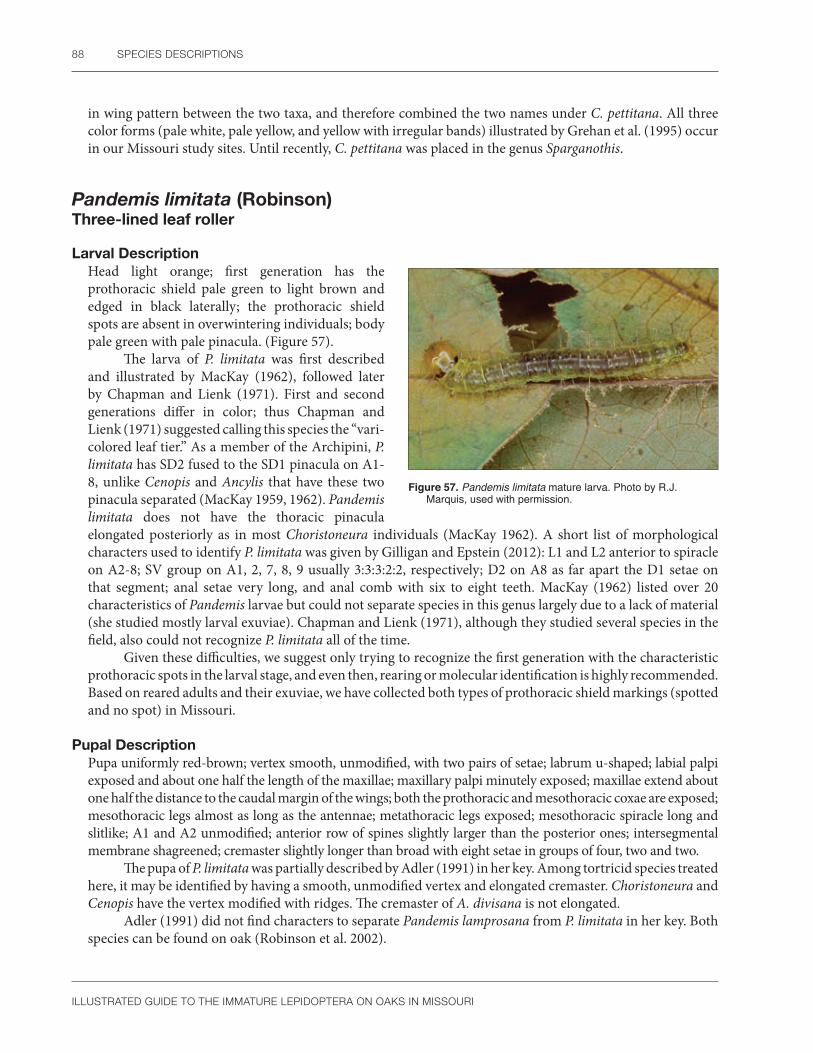
88 SPECIES DESCRIPTIONS
ILLUSTRATED GUIDE TO THE IMMATURE LEPIDOPTERA ON OAKS IN MISSOURI
in wing pattern between the two taxa, and therefore combined the two names under C. pettitana. All three color forms (pale white, pale yellow, and yellow with irregular bands) illustrated by Grehan et al. (1995) occur in our Missouri study sites. Until recently, C. pettitana was placed in the genus Sparganothis.
Pandemis limitata (Robinson)Three-lined leaf roller
Larval DescriptionHead light orange; first generation has the prothoracic shield pale green to light brown and edged in black laterally; the prothoracic shield spots are absent in overwintering individuals; body pale green with pale pinacula. (Figure 57).
The larva of P. limitata was first described and illustrated by MacKay (1962), followed later by Chapman and Lienk (1971). First and second generations differ in color; thus Chapman and Lienk (1971) suggested calling this species the “vari-colored leaf tier.” As a member of the Archipini, P. limitata has SD2 fused to the SD1 pinacula on A1-8, unlike Cenopis and Ancylis that have these two pinacula separated (MacKay 1959, 1962). Pandemis limitata does not have the thoracic pinacula elongated posteriorly as in most Choristoneura individuals (MacKay 1962). A short list of morphological characters used to identify P. limitata was given by Gilligan and Epstein (2012): L1 and L2 anterior to spiracle on A2-8; SV group on A1, 2, 7, 8, 9 usually 3:3:3:2:2, respectively; D2 on A8 as far apart the D1 setae on that segment; anal setae very long, and anal comb with six to eight teeth. MacKay (1962) listed over 20 characteristics of Pandemis larvae but could not separate species in this genus largely due to a lack of material (she studied mostly larval exuviae). Chapman and Lienk (1971), although they studied several species in the field, also could not recognize P. limitata all of the time.
Given these difficulties, we suggest only trying to recognize the first generation with the characteristic prothoracic spots in the larval stage, and even then, rearing or molecular identification is highly recommended. Based on reared adults and their exuviae, we have collected both types of prothoracic shield markings (spotted and no spot) in Missouri.
Pupal DescriptionPupa uniformly red-brown; vertex smooth, unmodified, with two pairs of setae; labrum u-shaped; labial palpi exposed and about one half the length of the maxillae; maxillary palpi minutely exposed; maxillae extend about one half the distance to the caudal margin of the wings; both the prothoracic and mesothoracic coxae are exposed; mesothoracic legs almost as long as the antennae; metathoracic legs exposed; mesothoracic spiracle long and slitlike; A1 and A2 unmodified; anterior row of spines slightly larger than the posterior ones; intersegmental membrane shagreened; cremaster slightly longer than broad with eight setae in groups of four, two and two.
The pupa of P. limitata was partially described by Adler (1991) in her key. Among tortricid species treated here, it may be identified by having a smooth, unmodified vertex and elongated cremaster. Choristoneura and Cenopis have the vertex modified with ridges. The cremaster of A. divisana is not elongated.
Adler (1991) did not find characters to separate Pandemis lamprosana from P. limitata in her key. Both species can be found on oak (Robinson et al. 2002).
Figure 57. Pandemis limitata mature larva. Photo by R.J. Marquis, used with permission.
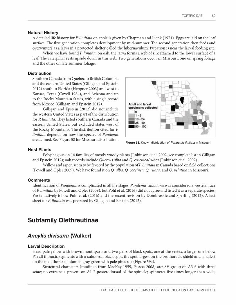
TORTRICIDAE 89
ILLUSTRATED GUIDE TO THE IMMATURE LEPIDOPTERA ON OAKS IN MISSOURI
Natural HistoryA detailed life history for P. limitata on apple is given by Chapman and Lienk (1971). Eggs are laid on the leaf surface. The first generation completes development by mid-summer. The second generation then feeds and overwinters as a larva in a protected shelter called the hibernaculum. Pupation is near the larval feeding site.
When we have found P. limitata on oak, the larva forms a web of silk attached to the lower surface of a leaf. The caterpillar rests upside down in this web. Two generations occur in Missouri, one on spring foliage and the other on late summer foliage.
DistributionSouthern Canada from Quebec to British Columbia and the eastern United States (Gilligan and Epstein 2012) south to Florida (Heppner 2003) and west to Kansas, Texas (Covell 1984), and Arizona and up to the Rocky Mountain States, with a single record from Mexico (Gilligan and Epstein 2012).
Gilligan and Epstein (2012) did not include the western United States as part of the distribution for P. limitata. They listed southern Canada and the eastern United States, but excluded states west of the Rocky Mountains. The distribution cited for P. limitata depends on how the species of Pandemis are defined. See Figure 58 for Missouri distribution.
Host PlantsPolyphagous on 14 families of mostly woody plants (Robinson et al. 2002, see complete list in Gilligan
and Epstein 2012); oak records include Quercus alba and Q. coccinea/rubra (Robinson et al. 2002).Willow and aspen seem to be favored by the population of P. limitata in Canada based on field collections
(Powell and Opler 2009). We have found it on Q. alba, Q. coccinea, Q. rubra, and Q. velutina in Missouri.
CommentsIdentification of Pandemis is complicated in all life stages. Pandemis canadana was considered a western race of P. limitata by Powell and Opler (2009), but Pohl et al. (2016) did not agree and listed it as a separate species. We tentatively follow Pohl et al. (2016) and the recent revision by Dombroskie and Sperling (2012). A fact sheet for P. limitata was prepared by Gilligan and Epstein (2012).
Figure 58. Known distribution of Pandemis limitata in Missouri.
Subfamily Olethreutinae
Ancylis divisana (Walker)
Larval DescriptionHead pale yellow with brown mouthparts and two pairs of black spots, one at the vertex, a larger one below P1; all thoracic segments with a subdorsal black spot, the spot largest on the prothoracic shield and smallest on the metathorax; abdomen gray green with pale pinacula (Figure 59a).
Structural characters (modified from MacKay 1959, Passoa 2008) are: SV group on A3-6 with three setae; no extra seta present on A1-7 posterodorsad of the spiracle; spinneret five times longer than wide;
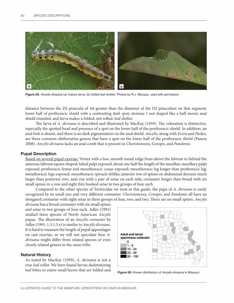
90 SPECIES DESCRIPTIONS
ILLUSTRATED GUIDE TO THE IMMATURE LEPIDOPTERA ON OAKS IN MISSOURI
a b
Figure 59. Ancylis divisana (a) mature larva; (b) folded leaf shelter. Photos by R.J. Marquis, used with permission.
distance between the D2 pinacula of A8 greater than the diameter of the D2 pinaculum on that segment; lower half of prothoracic shield with a contrasting dark spot; stemma 1 not shaped like a half moon; anal shield rounded; and larva makes a folded, not rolled, leaf shelter.
The larva of A. divisana is described and illustrated by MacKay (1959). The coloration is distinctive, especially the spotted head and presence of a spot on the lower half of the prothoracic shield. In addition, an anal fork is absent, and there is no dark pigmentation on the anal shield. Ancylis, along with Evora and Hedya, are three common olethreutine genera that have a spot on the lower half of the prothoracic shield (Passoa 2008). Ancylis divisana lacks an anal comb that is present in Choristoneura, Cenopis, and Pandemis.
Pupal DescriptionBased on several pupal exuviae: Vertex with a low, smooth mesal ridge from above the labrum to behind the antenna; labrum square shaped; labial palpi exposed, about one half the length of the maxillae; maxillary palpi exposed; prothoracic femur and mesothoracic coxae exposed; mesothoracic leg longer than prothoracic leg; metathoracic legs exposed; mesothoracic spiracle slitlike; anterior row of spines on abdominal dorsum much larger than posterior row; anal rise with a pair of setae on each side; cremaster longer than broad with six small spines in a row and eight thin hooked setae in two groups of four each.
Compared to the other species of Tortricidae we treat in this guide, the pupa of A. divisana is easily recognized by its small size and very different cremaster. Choristoneura, Cenopis, and Pandemis all have an elongated cremaster with eight setae in three groups of four, two, and two. There are no small spines. Ancylis divisana has a broad cremaster with six small spines and setae in two groups of four each. Adler (1991) studied three species of North American Ancylis pupae. The illustration of an Ancylis cremaster by Adler (1991: 1.3.1.3.e) is similar to Ancylis divisana. It is hard to measure the length of pupal appendages on cast exuviae, so we will not speculate how A. divisana might differ from related species or even closely related genera in the same tribe.
Natural HistoryAs noted by MacKay (1959), A. divisana is not a true leaf roller. We have found larvae skeletonizing leaf lobes or entire small leaves that are folded and Figure 60. Known distribution of Ancylis divisana in Missouri.

TORTRICIDAE 91
ILLUSTRATED GUIDE TO THE IMMATURE LEPIDOPTERA ON OAKS IN MISSOURI
tied down at the edges with silk (Figure 59b). Adults fly from late May until mid-August (Gilligan et al. 2008). We have collected larvae during mid- to late summer in Missouri.
DistributionEastern North America (Gilligan et al. 2008); Nova Scotia to Florida, west to Wisconsin and Texas (Heppner 2003), including Ohio (SCPC) and Missouri (Heinrich 1923). See Figure 60 for Missouri distribution.
Host PlantsPlatanus, Quercus, Quercus coccinea/rubra (Robinson et al. 2002). We have collected A. divisana on Q. alba in Missouri.

92 SPECIES DESCRIPTIONS
ILLUSTRATED GUIDE TO THE IMMATURE LEPIDOPTERA ON OAKS IN MISSOURI
HesperiidaeSkippers
Hesperiidae contain about 4,000 species distributed worldwide, except for New Zealand (Ackery et al. 1998, Nieukerken et al. 2011). Seven subfamilies are recognized by Warren et al. (2008, 2009) and Toussaint et al. (2018) largely based on adult characters. Both authors include references to the more important taxonomic literature in their publications, and Toussaint et al. (2018) summarizes many important ecological studies involving skippers. Keys to eastern United States hesperiid eggs, larvae, and pupae can be found in Scudder (1899), Scott (1986a), and Minno (1994). Common hesperiid larvae have been photographed by several authors (Allen et al. 2005, Minno et al. 2005, Wagner 2005); nevertheless identification of these caterpillars is often difficult. For more information on the Hesperiidae, consult the bibliographies by Beattie (1976), Pelham (2008: 497), and Lamas (2018). The most useful Web site for the New World fauna is https://www.butterfliesofamerica.com/. Host plants for New World hesperiids were compiled by Beccaloni et al. (2008). The life histories of many Old World species are treated by Igarishi and Fukada (1997, 2000) and in a supplementary volume by Igarashi and Harada (2015).
Traditionally, skippers are defined by their narrow thorax, giving the larva a unique appearance of having a “neck” (Ackery et al. 1998: 269). According to Minno (1994: 79), this is generally true for the Hesperiinae and Pyrginae, but not for the Pyrrhopyginae, which have a prothorax and head that are about the same size. Megathyminae, often considered a separate family in older literature but now treated as a tribe of the Hesperiinae (Warren et al. 2009), has the prothorax larger than the head. Skipper larvae have a ventral prothoracic gland and anal comb, but their most distinctive feature is the presence of lenticles (Minno et al. 2005: 24). Lenticles, as defined by Minno (1994: 52), are “small sclerotized plates that seem to represent modified setae.” Similar tiny plates also occur in some African Saturniidae, such as the mopani worm (SCPC), but it is unclear if the two situations are homologous. They are also found in lycaenids (Ballmer and Pratt 1988: 17). Some hesperiids bore into Agavaceae (Ackery et al. 1998), but most skipper larvae make shelters, usually by cutting a semicircle from the leaf edge, folding it over, and tying it to the adjoining leaf surface (Greeney and Jones 1998). Dethier (1942) studied abdominal glands of some larval Hesperiinae. These glands become active in the last instar and produce a waxy secretion that apparently repels water that could drown the pupa. Representative wax glands were illustrated by Minno (1994: 84). Both of our Missouri oak-feeding skippers are in the genus Erynnis. Many Missouri distribution records for this genus were given by Burns (1964).
Mosher (1916a) characterized hesperiid pupae (including “Megathymidae”) by their pilifers and maxillae. The lateral corner of the maxilla extends to the eye, but the relative length of the maxilla compared to the wing can vary. Other important taxonomic characters include the vertex being pointed or rounded, the length of antenna, prothoracic texture (crenulations present or absent), thoracic spiracle morphology, and the form of the cremaster. An epicranial suture is present and pupation occurs within a larval shelter (Brock 1990). Hesperiinae are the only subfamily to have lenticles in the pupal stage (Minno: 1994: 463, Couplet 1a). The cremaster of Erynnis has a shallow dorsal groove that is marked by a clear, smooth area in the cuticle. This contrasts to the rest of the cremaster that has a striated texture. Oak-feeding hesperiids are not economically important in the United States (USDA 1985), but a few tropical skippers are pests on rice, grasses (Hill 1975, 1987), palms, banana, or legumes (Holloway et al. 1987).

HESPERIIDAE 93
ILLUSTRATED GUIDE TO THE IMMATURE LEPIDOPTERA ON OAKS IN MISSOURI
Subfamily Pyrginae
Erynnis brizo (Boisduval & Leconte)Sleepy duskywing
Larval DescriptionHead capsule black in the first instar, copper brown with light orange spots laterally in subsequent instars; body light green, densely white speckled, with subdorsal white or yellow stripes (Figure 61a,b). The larva of E. brizo was partially described by Minno (1994) and is smaller than E. juvenalis. Unlike E. juvenalis and E. horatius, the stemmata are unequal in size (Minno 1994: Fig. 40 F). Stemma 5 is smaller than all other stemmata and stemma 6 is larger than all the rest. The rounded head and single spot also appear to be unusual (Minno 1994: Fig. 26 C).
a b
Figure 61. Erynnis brizo (a) mid-instar larva; (b) mature larva. Photos by R.J. Marquis, used with permission.
Pupal DescriptionModified from Minno (1994: 416): Head rounded; labial palpi exposed; antenna shorter than mesothoracic legs and clubbed; maxillae extends to caudal margins of wings; maxillary palpi apparently absent; prothoracic femur hidden; mesothoracic legs longer than prothoracic legs; metathoracic legs hidden; thoracic “spiracle guard” a small dark spot; body setae short; cremaster with numerous curved setae at the tip, no constriction, and with a short dorsal groove.
At the genus level, the absence of lenticles, short body setae, maxillae that extend to the caudal margin of the wings, and blunt cremaster with hooked setae all group the pupae of Erynnis with several other genera (Minno 1994: 465). The cremaster of various Erynnis species is illustrated by Minno (1994: 445). The constriction of the cremaster in E. juvenalis, absent from E. brizo, separates these two species if only the Missouri oak fauna is considered. Another difference is that the dorsal groove of E. juvenalis is longer than half the length of the cremaster. The dorsal groove of E. brizo is much shorter and is barely present. The top portion of the maxillae of E. horatius are pigmented and the cremaster is broad, u-shaped, and has a long dorsal groove. The maxillae of E. brizo is unpigmented and the dorsal groove is short. It is possible that a single head spot on the larval cast exuvia can help to identify E. brizo pupae. In addition, the stemmata of E. brizo are unequal in size. In contrast, there are three head spots on E. juvenalis and the stemmata are equal in size.
Natural HistoryThe life history of E. brizo is summarized by Minno et al. (2005). Pale green eggs are laid on oak leaves. The larva ties leaves into a shelter with silk (see Minno 1994: Fig. 1). Fully grown larvae overwinter as part of a single generation per year (Minno et al. 2005).

94 SPECIES DESCRIPTIONS
ILLUSTRATED GUIDE TO THE IMMATURE LEPIDOPTERA ON OAKS IN MISSOURI
In the lab, Erynnis brizo pupates during July or August in Missouri, with adults emerging in the fall or spring (R.J. Marquis, pers. obs.). Allen et al. (2005) states that E. brizo tends to use 1–2 m tall oak seedlings instead of mature trees; this would be another unusual characteristic of the life cycle.
DistributionSouthern Ontario, southern Manitoba, and southeastern Saskatchewan (Layberry et al. 1998), virtually found throughout eastern North America (Opler and Krizek 1984) and west to Colorado and Utah; the southern portion of the range includes Florida to California (Minno et al. 2005) along the Gulf States. See Figure 62 for Missouri distribution.
Host PlantsRed and white oaks (Opler and Krizek 1984, Heitzman and Heitzman 1987); Quercus durata, Q. myritifolia (Robinson et al. 2002); Castanea dentata (Shapiro 1966); Quercus chapmanii, Q. dumosa, Q. fusiformis, Q. gambelli, Q. harvardii, Q. ilicifolia, Q. inopina, Q. laevis, Q. lobata, Q. macrocarpa, Q. turbinella, and Q. undulata (Minno 1994). Quercus macrocarpa is the only host plant in Manitoba (Layberry et al. 1998). We have found E. brizo on Q. alba and Q. velutina in Missouri.
Figure 62. Known distribution of Erynnis brizo in Missouri.
Erynnis juvenalis (Fabricius)Juvenile duskywing
Larval DescriptionHead capsule sharply lobed, golden in the first instar, and then black, subsequently copper brown with three pairs of orange spots laterally in later instars; body light green, densely white speckled, with thin subdorsal white to yellow stripes (Figure 63).
The larva of E. juvenalis is partially described by Minno (1994). Compared to E. brizo, E. juvenalis is distinguished by having the stemmata subequal in diameter (Minno 1994: Fig. 40 G). In addition, the head of E. brizo is more rounded than the lobed head of E. juvenalis (Minno 1994: Figs. 26: C, D). Minno (1994) also shows that E. juvenalis has three pale spots on the head compared to the single spot of E. brizo.
Heitzman and Heitzman (1987) recorded E. horatius from Missouri. The larva feeds on oak and is similar to E. juvenalis. According to Minno et al. (2005: 198, fig. 19H), the larva of E. horatius lacks the subdorsal longitudinal stripe that is present in E. juvenalis (see Minno et al. 2005: 199). Although a wide range of oaks are eaten, E. horatius favors red oaks (Allen et al. 2005: 165).
Figure 63. Erynnis juvenalis mature larva. Photo by R.J. Marquis, used with permission.

HESPERIIDAE 95
ILLUSTRATED GUIDE TO THE IMMATURE LEPIDOPTERA ON OAKS IN MISSOURI
Pupal Description Modified from Minno (1994: 416): Head rounded; labial palpi exposed; antenna shorter than mesothoracic legs and clubbed; maxillae extends to caudal margins of wings; maxillary palpi apparently absent; prothoracic femur hidden; mesothoracic legs longer than prothoracic legs; metathoracic legs hidden; thoracic “spiracle guard” a small dark spot; body setae short; cremaster with numerous curved setae at the tip, constricted about halfway down its length, and with a dorsal groove longer than one-half the length of the cremaster.
According to Minno (1994: 465), the absence of lenticles, short body setae, maxillae that extend to the caudal margin of the wings, and blunt cremaster with hooked setae all group the pupae of Erynnis with several other genera. The cremaster of various Erynnis species is illustrated by Minno (1994: 445). He shows a constriction on the cremaster in E. juvenalis that is absent from E. brizo. Another difference is that the dorsal groove of E. juvenalis is longer than half the length of the cremaster. The dorsal groove of E. brizo is much shorter than half the length of the cremaster. The top portion of the maxillae of E. horatius is pigmented, and the cremaster is broad, u-shaped, and has a long dorsal groove. The maxillae of E. juvenalis are unpigmented, the cremaster is narrower than in some Erynnis species, and like E. horatius, there is also a long dorsal groove. The larval head on the exuvia of E. juvenalis likely has three spots and all stemmata of equal size. These characters can be used to distinguish from E. brizo, which has a single head spot and stemmata of unequal size.
Natural HistoryThe life cycle of E. juvenalis is illustrated in O’Donnell et al. (2007). They noted that eggs are laid singly on the young foliage or stem of the host. Fully grown larvae overwinter and can make a scraping sound with their mandibles by rubbing them on a leaf (Minno et al. 2005: 36).
In Missouri, the larva cuts a piece of the leaf, then forms a tent by folding the leaf piece down and holding it with silk. This species has a single generation, beginning on nearly fully expanded leaves in the spring (May) and not completing development until late August or September. Adults often emerge in late summer or fall in the lab. Population numbers vary from year to year (R.J. Marquis, pers. obs.).
Adults visit flowers in open woodlands, congregate at mud puddles, and often fly with E. horatius (Heitzman and Heitzman 1987). Although similar in wing pattern, E. juvenalis and E. horatius are easily separated by male genitalia (Bouseman et al. 2006: 8).
DistributionNova Scotia, Canada, west to Manitoba and the Dakotas, south through New England, the Midwest, Florida, and the Gulf States, including Mexico (Opler and Krizek 1984, Bouseman et al. 2006). See Figure 64 for Missouri distribution.
Host PlantsFagus, Quercus alba, Q. arizonica, Q. coccinea/rubra, Q. emoryi, Q. geminata, Q. hypoleucoides, Q. ilicifolia, Q. macrocarpa, Q. marilandica, Q. mohriana, Q nigra, Q. prinus, Q. rubra, Q. stellata, and Q. velutina (Robinson et al. 2002); usually found on Quercus, possibly also Corylus, Quercus agrifolia in the lab, Q. chapmanii, Q. falcata, Q. fusiformis, Q. gambelli, Q. inopina, Q. laevis, Q. lobata, Q. muehlenbergii, and Q. phellos (Minno 1994).
Minno (1994) critically classified the known food plants of E. juvenalis as either erroneous, laboratory hosts, or valid field records. We emphasized the confirmed and probable records for E. juvenalis on oak.
Figure 64. Known distribution of Erynnis juvenalis in Missouri.

96 SPECIES DESCRIPTIONS
ILLUSTRATED GUIDE TO THE IMMATURE LEPIDOPTERA ON OAKS IN MISSOURI
Wagner (2005) states that E. juvenalis frequently uses hickory (Carya) in New Jersey. Butler and Strazanac (2000) report this species on Carya, Acer, various red oak species, Q. alba, and Q. prinus. There is also one report of this species on Castanea dentata (Opler and Krizek 1984, Heitzman and Heitzman 1987). We have found E. juvenalis on Quercus velutina (Whitfield et al. 1999) and Q. alba, Q. coccinea, Q. rubra, and Q. stellata in Missouri.
CommentsWagner (2005) notes that variable head and body markings prevent accurate larval identifications of Erynnis skippers based on color. We agree that larval identifications in this genus are tentative and reared adults are needed for positive identifications. Burns (1964: 61, 67) considered E. brizo and E. juvenalis each to be a species complex. This may account for some of the variation and identification difficulty. Another problem is that, given the erroneous foodplant records, it would be unwise to base identification exclusively on the food plant associations. Eight species of Erynnis are recorded from oak in North America (Allen et al. 2005).

LYCAENIDAE 97
ILLUSTRATED GUIDE TO THE IMMATURE LEPIDOPTERA ON OAKS IN MISSOURI
LycaenidaeHairstreak butterflies
The family Lycaenidae contains approximately 5,000 species worldwide (Nieukerken et al. 2011) and is most diverse in tropical regions (Scoble 1992). Ackery et al. (1998) recognized five lycaenid subfamilies, including Riodininae. Other studies suggest that Riodinidae and Lycaenidae are separate families (Wahlberg et al. 2005). The latest North American checklist by Pohl et al. (2016) recognizes the family Riodinidae while dividing lycaenids into four subfamilies (Miletinae, Lycaeninae, Theclinae, and Polyommatinae). North American taxa are called metalmarks, harvesters, blues, or hairstreaks. Part of the Missouri lycaenid fauna was treated by Heitzman and Heitzman (1987). Keys to eastern United States lycaenid eggs, larvae, and pupae are available in Scudder (1899) and Scott (1986a). Several authors have photographed common lycaenid larvae (Allen et al. 2005, Minno et al. 2005, Wagner 2005); nevertheless, larval identification is often difficult. For more information on the Lycaenidae, consult the bibliographies by Beattie (1976), Pelham (2008: 497), and Lamas (2018). The most useful Web site for the New World faunas is https://www.butterfliesofamerica.com/. Host plants for New World lycaenids were compiled by Beccaloni et al. (2008).
Lycaenid larvae are easily recognized by their sluglike appearance, concealed head, and spatulate membranous lobe on the abdominal prolegs (Downey 1987). Ackery et al. (1998) listed four body forms: onisciform, carapaced, tuberculate, and a “Lymantriinae type.” North American Lycaenidae and Riodinidae are usually separated by their mandible and spiracles. Riodinidae have more than two mandibular setae and the spiracles are positioned more ventrad on A1 than A2. In Lycaenidae, only two mandibular setae are present, and the spiracles of A1 and A2 are aligned horizontally (Downey 1987). However, exceptions to these generalizations do occur (Duarte et al. 2005). Cuticular morphology is highly variable in the Lycaenidae. Ballmer and Pratt (1988) illustrate examples of recurved, stellate, clubbed, and branched setae. In general, under magnification, Polyommatinae (blues) have star shaped setae, whereas Lycaeninae (coppers) have mushroom-shaped setae (Ballmer and Pratt 1988: 15, Wagner 2005: 105). Specialized glands are also common, with recent examples illustrated in Duarte et al. (2005), Ballmer and Wright (2008), and Kaminski et al. (2013). Ballmer and Wright (2008) discuss setal terminology in lycaenid caterpillars, including the presence of lenticles similar to those of skipper larvae (Ballmer and Pratt 1988: 17). Lycaenids are associated with ants and homopterans in a wide range of obligate to facultative interactions (Ackery et al. 1998, Pierce et al. 2002). For more information on lycaenid immature stages, consult Clark and Dickson (1971), Ballmer and Pratt (1988), Scott (1986a), Harvey (1987a, 1987b), Igarashi and Fukada (1997, 2000), Igarashi and Harada (2015), Allen et al. (2005), and Minno et al. (2005).
Lycaenid pupae have maxillae that never extend to touch the eyes, no epicranial suture, and maxillae that are never as long as the wings (Mosher 1916a). In addition, the head is in a more ventral position than other butterflies, a cremaster is usually absent, and a silk girdle is present (Scott 1986a). Genus and species identification often depends on setal location, density, and shape. Stellate and mushroom-shaped setae, characteristic of lycaenid larvae, are also found on the pupae (Patočka and Turčáni 2005: Plate 163).
Lycaenids are associated with oak (Quercus) throughout the Holarctic region (Higgins and Riley 1970, Scott 1986a), but they are not economically important on North American forest trees (USDA 1985). Tropical species are pests of fruits and seeds, especially on legumes (Holloway et al. 1987).
In addition to Satyrium calanus (see below), we have found Fixsenia (Satyrium) flavonius on Missouri oaks. Satyrium edwardsii, S. caryaevorus, S. liparops, and Calycopis cecrops also are reported to feed on oaks, and are recorded as adults in Missouri (J.R. Heitzman and P.E. Koenig, unpublished list) (Appendix 1).

98 SPECIES DESCRIPTIONS
ILLUSTRATED GUIDE TO THE IMMATURE LEPIDOPTERA ON OAKS IN MISSOURI
Subfamily Theclinae
Satyrium calanus (Hübner) Banded hairstreak
Larval DescriptionModified from Wagner (2005): Ground color varies from white to green to brown to pink; secondary setae and white speckles cover the body; usually the front and rear ends are marked with dark patches; oblique subdorsal patches usually present; white subspiracular stripe well developed on the last segments, including the anal shield (Figure 65a, b).
According to Opler and Krizek (1984), the larva of S. calanus is green with white oblique lines and brown pubescence. A brown form with trapezoidal markings is also known. Scott (1986a) describes the larva as “grass-green, with a dorsal line of dark triangles, the body with light-green (or whitish) and dark green longitudinal lines.” A second form of S. calanus has a “green or greenish brown head, usually a pinkish brown body without markings, although sometimes heavy, dark markings occur on the front and the rear ends” of the larva (Scott 1986a).
According to Allen et al. (2005), S. calanus is recognized by having dark markings on the front and rear ends and a ground color from white to green to brown. Wagner (2005) gives color characters to separate S. calanus from other oak-feeding lycaenids and has illustrated four of the common color forms. Fracker (1915) examined S. calanus and grouped it under the older genus name Thecla. He defined this taxon by having the head about one-third the diameter of the body, the prothorax much smaller than the mesothorax, and a usual lack of subdorsal markings. The latter character does not seem to apply to S. calanus, which often has subdorsal markings. Given that the genus Satyrium contains several species with similar larvae, it would be better to identify larvae only to genus unless they are reared or barcoded.
a b
Figure 65. Satyrium calanus (a) mature larva; (b) mature larvae tended by Crematogaster ants. Photos by R.J. Marquis, used with permission.
Pupal DescriptionModified from Mosher (1916a): Pupa hourglass shaped with a convex dorsum, colored brown with small black markings; labrum u-shaped; pilifers not lobed, but square at the apex and joined at the meson; maxillae extend one half the distance to the caudal margins of the wings; maxillary palpi and prothoracic femur both hidden; prothoracic leg extends cephalad to lower margin of eye; mesothoracic leg about as long as the prothoracic leg; metathoracic legs hidden; mesothoracic spiracle a contrasting white patch; abdomen covered with minutely barbed setae almost as long as the height of a segment; no cremaster or hooked setae present on the terminal abdominal segments.

LYCAENIDAE 99
ILLUSTRATED GUIDE TO THE IMMATURE LEPIDOPTERA ON OAKS IN MISSOURI
The pupa of S. calanus is illustrated by Scudder (1899). Scott (1986a) stated it was colored light to dark brown. Mosher (1916a) grouped S. calanus with Thecla, as did Fracker (1915), but neither had many lycaenid species to study for their diagnoses. Morphological identification of North American lycaenid pupae is very uncertain. The larvae exuvia of S. calanus lacks spatulate or star shaped setae; this is an important clue for identification.
Natural HistoryPale green eggs are laid on host twigs in the summer (Scott 1986a) but do not hatch until the following spring (Minno et al. 2005). We find larvae are rare in Missouri, as they are in Florida (Minno et al. 2005), and are found only in the spring on very young leaves. Caterpillars in the canopy feed first on catkins then switch to leaves. Wagner (2005) reported larvae resting on the leaf undersides; these were frequently collected using beating sheets. In other cases, larvae hide in curled leaves (Minno et al. 2005). An adult was reared from a pupa inside a dried pecan husk in Florida during April (SCPC).
Satyrium calanus is univoltine according to Opler and Krizek (1984). The adult flight period depends on latitude. In the northern United States, butterflies emerge from July to August. In Florida, S. calanus flies in April. Males perch until dusk on trees and shrubs, watching for females.
DistributionNova Scotia, west to central Manitoba, Canada, south to Florida, the Gulf Coast, and Texas (Opler and Krizek 1984) with an isolated population in western Colorado and northern New Mexico (Minno et al. 2005).
According to Heitzman and Heitzman (1987), S. calanus is found in “virtually any brush field or open forest in Missouri from late May to early June.” It is the most common eastern United States hairstreak, although population levels vary widely (Opler and Krizek 1984). See Figure 66 for Missouri distribution.
Host PlantsCastanea, Carya, Juglans, Quercus alba, Q. coccinea, Q. falcata, Q. gambelii, Q. incana, Q. laevis, Q. macrocarpa, Q. prinus, and Q. rubra (Robinson et al. 2002). We have collected and reared S. calanus on Q. alba, Q. stellata, and Q. velutina in Missouri. Satyrium calanus has been reared from Carya, Juglans, and Quercus (Scudder 1899). Records for Malus mixed with Prunus branches, Acer, and Fraxinus listed by Scott (1986a) probably need confirmation since none of these hosts are listed by Wagner (2005) as common foodplants. Hawthorn is also doubtful (Opler and Krizek 1984).
Figure 66. Known distribution of Satyrium calanus in Missouri.
100 SPECIES DESCRIPTIONS
ILLUSTRATED GUIDE TO THE IMMATURE LEPIDOPTERA ON OAKS IN MISSOURI
NymphalidaeBrush-footed butterflies
The family Nymphalidae contains approximately 6,000 species (Nieukerken et al. 2011) representing nearly one-third of all known butterfly taxa (Scoble 1992). The Morphini are among the world’s most beautiful Lepidoptera because of their brilliant, often metallic, wing colors. At the opposite extreme, Satyrinae adults are usually brown and cryptic. Collectively, nymphalids are called “brush-footed” butterflies because of their reduced forelegs. Another important specialization is the presence of three ventral carinae on the antenna, a character apparently unique to Nymphalidae (Ackery et al. 1998: 285). The most recent nymphalid classification and phylogeny is posted on nymphalidae.net following Wahlberg et al. (2009). Twelve subfamilies are recognized: Libytheinae, Danainae, Charaxinae, Satyrinae, Calinaginae, Heliconiinae, Limenitidinae, Pseudergolinae, Biblidinae, Apaturinae, Cyrestinae, and Nymphalinae. Previously, many of these subfamilies were given family ranking (Holloway et al. 1987).
Nymphalids are important model systems for the study of butterfly biology. Knowledge gained from research on heliconiines and ithomiines has contributed to a better understanding of insect-plant interaction (Brown 1981, Brown 1987a). Classical papers on migration (Brower 1995), mimicry (see review by Ackery and Vane-Wright 1984), and butterfly evolution at the species level (Platt 1983) have utilized members of the Danainae and Limenitidinae. Many subfamilies specialize on a narrow group of plants, especially Passiflora, milkweeds, or monocotyledons (Holloway et al. 1987). Nymphalids are readily recognized by the public, a fact leading to the selection of the monarch butterfly (Danaus plexippus) as the United States of America’s “National Insect” (Entomological Society of America 1989).
Nymphalid larvae are morphologically diverse. Based on the North American fauna, nymphalid larvae are identified by having the stemmata enlarged or elevated on papillae, the presence of numerous short secondary setae, crochets in uniordinal to triordinal mesoseries (rarely a pseudocircle), and by having a middorsal scolus on A7 but not on A9 (Toliver 1987). The head often has horns (Holloway et al. 1987) or scoli (Toliver 1987). Harvey (1991) noted that nymphalids are the only butterfly larvae to have a filiform (thin and hairlike) seta on A9, but this statement is not universally accepted (Ackery et al. 1998: 286). Toliver (1987) recognized four main types of nymphalid caterpillars. More study is needed to determine whether the newly recognized subfamilies correctly fit into these categories. Larvae with head projections, either smooth or spiny, and a body sometimes with branched or clubbed scoli, characterize the Heliconiinae, Nymphalinae, Limenitidinae, Biblidinae, Cyrestinae, and some Pseudergolinae. These nymphalid subfamilies are sometimes confused with saturniids, but differ in the arrangement of the scoli. Saturniids have a middorsal scolus on A9, whereas this structure is present on A7 in the Nymphalidae (Toliver 1987). A second nymphalid larval phenotype occurs in the Danainae. These larvae usually have one or more pairs of fleshy filaments on the thorax and/or abdomen. Frequently the larvae are brightly colored. The Calinaginae, Charaxinae, Satyrinae, and Apaturinae comprise the third class of nymphalid larvae. Some species in this group have a head larger than the prothorax, but a forked or truncate anal shield is the most characteristic larval feature. Morpho larvae, currently placed as a tribe of the Satyrinae, are covered with dense tufted hair and could almost be considered a separate grouping. The last group, Libytheinae, contains larvae without filaments, scoli, or modifications of the head and anal shield. Libytheana have crochets in a pseudocircle, unlike other nymphalid subfamilies (Toliver 1987). For more information on nymphalid immatures, consult Scott (1986a), Toliver (1987), Harvey (1991), Igarashi and Fukada (1997, 2000), Igarashi and Harada (2015), Allen et al. (2005), Wagner (2005), Minno et al. (2005), and especially the keys in Scudder (1899) and the illustrated data matrix by Freitas and Brown (2004).
According to Mosher (1916a), pupae of the Nymphalidae lack an epicranial suture. In addition, the prothoracic leg extends cephalad to the eye, and the mesothoracic leg lies between the eye and the antennae. At least in Europe, Libythea is an exception in that the prothoracic legs do not reach the eyes (Patočka and Turčáni 2005: 322). The vertex is often angled, pointed, or deeply forked, and the body may have tubercles or projections. Nymphalid pupae can be mottled, colored with metallic spots (Patočka and Turčáni 2005), or cryptic. Usually

NYMPHALIDAE 101
ILLUSTRATED GUIDE TO THE IMMATURE LEPIDOPTERA ON OAKS IN MISSOURI
the pupae hang from a silken pad, without a girdle, with the head facing down (Scott 1986a, Patočka and Turčáni 2005). In other cases nymphalids pupate on the ground or on top of a leaf (Scott 1986a). As with the larvae, nymphalid pupae are morphologically diverse. Six pupal types are illustrated by Freitas and Brown (2004) using Libytheana, Tithorea, Memphis, Adelpha, Callicore, and Heliconius as exemplars. It is not immediately clear how all the subfamilies of Nymphalidae would fit into this scheme. Heliconiinae, Nymphalinae, and Limenitidinae usually have a more or less developed concave (cuplike) dorsal margin, often with dorsal projections, such as tubercles or a keel (median ridge) (Scott 1986a: Fig. 52; Penz 1999: P63, P64; Freitas and Brown 2004). The pupal similarity of Limenitidinae and Heliconiinae might help support the surprising sister group relationship of these two subfamilies based on molecular data (see nymphalidae.net). Other nymphalid pupae are more rounded or bent (Freitas and Brown 2004: Fig. A6). Some Satyrinae (Oeneis and relatives) have lost the cremaster (Scott 1986a: 233) and thus resemble lycaenid pupae to some degree (Mosher 1916a: 95).
Nymphalids are rarely important as pests of temperate forest trees, although Asterocampa clyton sometimes defoliates hackberry (Celtis) in the Great Lakes states, and Nymphalis antiopa is a pest on ornamental trees along fencerows (USDA 1985). In the New World tropics, Brassolini (Satyrinae) are important banana pests (Ostmark 1974).
Many common nymphalids of Missouri are illustrated in Heitzman and Heitzman (1987). We include Limenitis arthemis astyanax in our guide because of the association with oaks in parts of Canada (Layberry et al. 1998), New England (O’Donnell et al. 2007), and Missouri. The caterpillar is a conspicuous element of the eastern forest fauna (e.g., USDA 1985).
Subfamily Limenitidinae
Limenitis arthemis astyanax (Fabricius)Red-spotted purple
Larval DescriptionModified from Scott (1986a): Ground color a mixture of brown, green, or whitish green; head pale olive green to brownish red with two small scoli on vertex; thorax with white patches and two long horns which are not strongly clubbed, small paired abdominal middorsal scoli present on A7, a larger pair on A8; abdomen with a white or reddish- cream colored saddle and white to pink lateral line (Scott 1986a); anal shield with tubercles (Figure 67a, b).
a b
Figure 67. Limenitis arthemis astyanax (a) early instar larva on frass chain; (b) mature larva. Photos by R.J. Marquis, used with permission.

102 SPECIES DESCRIPTIONS
ILLUSTRATED GUIDE TO THE IMMATURE LEPIDOPTERA ON OAKS IN MISSOURI
The life cycle of the red-spotted purple is illustrated by Hilton and Heppner (1995). The caterpillar has been illustrated many times; recent examples in color are Allen et al. (2005), Minno et al. (2005), Wagner (2005), and O’Donnell et al. (2007). Limenitis caterpillars with long scoli on T2 but no well-developed middorsal spines are easy to recognize to genus (see key in Forbes 1960). However, it is rather difficult to separate the viceroy from the red-spotted purple (Scudder 1899: 255). According to Scott (1986a), larvae of the red-spotted purple have long mesothoracic horns with slender warts that are not clubbed. Opler and Krizek (1984) stated that the red-spotted purple may be recognized by a larger number of “spiracular tubercles,” a character which refers to Scudder’s (1899) observation that the number of tubercles above the spiracular line differs among Limenitis species. Minno et al. (2005) used the number of “small spines” on the thorax and anterior abdominal segments; these were present in the viceroy but absent in the red-spotted purple. Forbes (1960) emphasized differences in the head spines. Wagner (2005) summarized these differences by stating the red spotted purple is less spiny than the viceroy. In particular, the “scolus on the head and spine clusters of T2, A2, A7, and A8 are proportionally smaller” than on the viceroy. In the red-spotted purple, the hump over A2 is larger and the scolus of A2 is more clubbed than in the viceroy. Many of these characters require reference specimens to fully appreciate the subtle differences, some of which are illustrated by Scudder (1899). Wagner (2005) and Scott (1986a) give conflicting diagnoses using the mesothoracic scoli, suggesting that the variation of these characters needs further study in each instar.
Biology and host data are helpful in separating the viceroy and red-spotted purple. Both the viceroy and red-spotted purple eat poplar and aspen (Wagner 2005). The viceroy is restricted to Salicaceae and the adults prefer meadows and streams (Platt 1983). Thus, we expect the red-spotted purple to be the most likely choice for all Limenitis larvae on oak in the forests of Missouri.
Pupal DescriptionLabrum small, rectangular, and notched on ventral surface where it joins the minutely exposed labial palpi; maxillae extend to caudal margins of wings; prothoracic legs approximately half the length of maxillae, the basal portion extending cephalad past maxillae to touch the ventral portion of the eye; mesothoracic legs about three-fourths the length of maxillae, the basal portion extending cephalad past the maxillae to lie between the eye and the antenna; metathoracic legs hidden; mesothoracic spiracle shelflike, oval, and covered with pale, dense, minute spines; abdomen smooth except for a median, slightly flattened, oval keel on the dorsum of the second segment; cremaster with a dense pad of stout, short curved spines.
According to Scudder (1899), the pupa of the red-spotted purple differs from that of the viceroy, with the size of the keel on A2 and characteristics of the cremaster varying between the two species.
Natural HistoryAccording to Scott (1986a), eggs are laid on the upper side of leaf tips. The larvae are foliage feeders and do not construct a nest (hibernaculum) until the third instar, which is the overwintering stage. There are one to several broods a year, depending on the latitude. In Missouri, adults occur from May through October and feed on a wide variety of sweets (nectar, sap, or honeydew) or sources of nitrogenous compounds (dung, carrion). They may also puddle in damp places (Heitzman and Heitzman 1987). Early larvae are frass chain builders (Figure 67a) that construct extensions of the leaf midrib by gluing together frass pellets to the end of the leaf vein, forming a chain on which they rest, head first towards the leaf (DeVries 1987, Machado and Freitas 2001). We have seen one frass chain and associated caterpillar on a Quercus alba leaf in Missouri. Presumably, the majority of larvae found on oaks in our area arise from eggs laid on other host plants, Prunus in particular, and the caterpillars have secondarily colonized oak trees.
DistributionCentral New England and the Great Lakes states of the Midwest, south to Arizona, New Mexico, central Texas, and central Florida (Opler and Krizek 1984, Scott 1986a). Heitzman and Heitzman (1987) considered

NYMPHALIDAE 103
ILLUSTRATED GUIDE TO THE IMMATURE LEPIDOPTERA ON OAKS IN MISSOURI
the red-spotted purple to be common throughout Missouri. See Figure 68 for Missouri distribution.
Host PlantsCarpinus, Celtis, Crataegus, Cydonia, Malus, Ostrya, Populus, Prunus, Pyrus, Quercus ilicifolia, Q. palustris, Q. phellos, Ribes, Rosa, Salix, Tilia, Ulmus, Vaccinium (Robinson et al. 2002); Q. alba, Q. coccinea, Q. rubra, and Q. velutina (Butler and Strazanac 2000).
Wild cherry (Prunus) is the most common host of the red-spotted purple. Records on Rosa, Celtis, Salix, Ostrya, and Ribes require confirmation (Opler and Krizek 1984; Scott 1986a). During our study, however, we have only encountered it as a caterpillar on oak four times, three times on Q. alba and once on Q. imbricaria.
CommentsOpler and Krizek (1984) noted that the red-spotted purple is a Batesian mimic of Battus philenor. In spite of their distinctive appearances as adults, the white admiral and the red-spotted purple are classified by most authorities as the same species (Opler and Krizek 1984, Scott 1986a, Pohl et al. 2016).
Figure 68. Known distribution of Limenitis arthemis astyanax in Missouri.

104 SPECIES DESCRIPTIONS
ILLUSTRATED GUIDE TO THE IMMATURE LEPIDOPTERA ON OAKS IN MISSOURI
MegalopygidaeFlannel moths
Megalopygidae are a small New World family of 232 species (Nieukerken et al. 2011) generally placed with the “limacodid-group” of the Zygaenoidea (Epstein et al. 1998). Two genera, Norape and Megalopyge (=Lagoa), are found in the eastern United States (Covell 1984, Poole and Gentili 1996, Heppner 1997). Profant et al. (2010) summarize the Ohio fauna but do not address taxonomic issues or provide technical descriptions. Epstein and Adams (2009) list all the megalopygid species from the western United States with representative genitalia and an up-to-date summary of their systematics. For more information on megalopygids, consult Bourquin (1945), Stehr (1987a), Epstein (1996, 1997), and Epstein et al. (1998). Lepesqueur (2012) studied the ecology of megalopygids in Brazil.
Megalopygid eggs are considered to be of the “flat type” (Hinton 1981) because the micropyle is oriented parallel to the substrate. Megalopyge and Trosia have eggs shaped like an elongate oval that is much longer than wide (SCPC, Baerg 1924, Peterson 1967, Epstein 1996). Most species lay their eggs in rows and cover them with scales from a specialized pouch located at the end of the female’s abdomen, between A7 and A8 (Scoble 1992).
Stehr (1987a) divided megalopygid larvae into two groups, “puss type” and “arctiid type,” based on general appearance. This distinction is obvious in the eastern United States fauna and megalopygid larvae from Latin America (SCPC). The “puss type” is represented in Missouri by Megalopyge, which has soft dense hairs covering the stinging spines. Norape is an example of the “arctiid” type because the long hairs and obvious verrucae resemble the body form seen in some tiger moth caterpillars of the subfamily Arctiinae. However, this division may not have phylogenetic significance because both types of larvae occur in the Trosiinae, for example (Epstein, 1996: 74). Larvae of the Megalopygidae are hairy and sluglike, with the head concealed under the body (Appendix 4, Plate 9i). They can be recognized by their seven pairs of prolegs on abdominal segments A2-7 and A10, two more than is typical for most lepidopteran larvae (Stehr 1987). Most megalopygid species have crochets on A3-6 and A10; in a few tropical genera they are present on A2 and A7 as well (Epstein, 1996: 74). Additionally, larvae have a “fleshy protuberance,” termed the spiracular sensilla (Epstein 1996: 68), on the prothorax and abdomen near the spiracle (Peterson 1962a: L23). The relative position of A9 to A10 (Stehr 1987a) and presence of a membranous pad on the proleg (Epstein 1996: 68) are two other unusual larval modifications of this family. A broad, flattened anal comb is present in Megalopyge but absent in Norape (SCPC).
Megalopygid pupae are easily recognized, in part, by two distinctive features. The compound eye has a lateral flange similar to that seen in the Limacodidae (Mosher 1916a), and there is a conical lobe, representing the larval spiracular sensilla, present near the abdominal spiracles of A1-6 (Epstein 1996). The distribution of these lobes sometimes varies with the sex of the individual. Epstein (1996: 74) stated that males have a lobe only on abdominal segment 1 whereas females show this structure on the spiracles of A2-6. A longitudinal furrow is sometimes present on the mesonotum (Epstein 1996: Fig. 300), and the abdominal dorsum has both stout abdominal setae and spines in patches (Mosher 1916a: 42, Epstein 1996: Fig. 311). The nature of the cocoon’s outer coating and escape hatch are other variable characteristics of megalopygid pupae (Stehr 1987a, Epstein 1996: 9). Megalopyge crispata and M. opercularis incorporate finely barbed setae in the covering of their hard smooth cocoon. The covering of N. ovina is more papery with no setae and a less obvious escape hatch (SCPC).
Larvae of Megalopyge are infamous for their urticating spines (Baerg 1924, Bishopp 1923, Heppner 1997). Megalopyge crispata has damaged oak in western Oklahoma (Lamdin et al. 2000) and has caused severe defoliation to shin oak in Texas (USDA 1985). A related species, M. opercularis, has defoliated turkey oak in Florida (USDA 1985).

MEGALOPYGIDAE 105
ILLUSTRATED GUIDE TO THE IMMATURE LEPIDOPTERA ON OAKS IN MISSOURI
Subfamily Megalopyginae
Megalopyge crispata (Packard)Puss caterpillar
Larval DescriptionModified from Packard (1894), Baerg (1924), Stehr (1987), and Epstein (1996: Figs. 119, 131, 132): Frontal area reaching one quarter the distance to epicranial notch; spinneret rounded in dorsal view; cutting edge of mandible with a broad retinaculum but no teeth; stemma one larger than the others, stemmata 1-4 and 6 form an arc, stemma 5 lower and widely separated from the others; prothoracic shield thin and bandlike; prolegs of A3-6 with the crochets arranged in a v-shaped uniordinal mesoseries; prolegs of A2 and A7 peglike and without crochets; fourth abdominal segment with elongated dorsal and subdorsal verrucae above the spiracle; V setae more widely spaced on A10 than A9; anal shield thin and bandlike; anal comb bilobed with serrated edges; anal proleg with crochets in a uniordinal mesoseries.
Early instars are covered in long, white to cream-colored hairs, later instars are darker with dense hairs pointing upward and a poorly defined tail of long, thin setae (see Wagner 2005: 53, Profant et al. 2010: Plate 3, Fig. 5). The structure of the venomous hairs is described and illustrated by Lamdin et al. (2000).
The larva of Megalopyge crispata is illustrated by Packard (1894), Eliot and Soule (1902), and Baerg (1924). Short et al. (1997) and Wagner (2005) illustrate the mature larva in color. These figures, and our own experience in Missouri, indicate that the larva of M. crispata is either a solid color or contrastingly bicolored, most often in shades of cream to brown to gray (Figure 69a, b). A few of these color forms are shown in Profant et al. (2010).
Structural features that separate species of Megalopyge in the larval stages are generally lacking. Fracker (1915) states that the distance between stemmata 4 and 5 is greater in M. opercularis than M. crispata, but the validity of this character was questioned by Epstein (1996). The best way to separate M. crispata from M. opercularis is by noting the form of the posterior setae. In M. opercularis, setae at the rear end of the abdomen form a dense “tail” (see Stehr 1987a, Wagner 2005). The posterior setae in M. crispata never form a tail; at most only a few scattered long setae are present (Appendix 4, Plate 9i).
The larvae of M. laceyi and M. pyxidifera are described by Klots (1966) and Dyar (1897d), respectively. Both species occur in the southern United States and feed on oak, but identification characteristics to separate them from M. crispata need further study. Megalopyge laceyi is white at maturity, with long and short hairs dorsally, that almost form several tufts (Klots 1966: Fig. 1). This is unlike the smooth, dense appearance of M. crispata that has darker hairs in the later instars. Forbes (1906: Couplet 172) stated that early instars of M.
a b
Figure 69. Megalopyge crispata (a) mid-instar larva; (b) mature larva. Photos by R.J. Marquis, used with permission.

106 SPECIES DESCRIPTIONS
ILLUSTRATED GUIDE TO THE IMMATURE LEPIDOPTERA ON OAKS IN MISSOURI
crispata are cream white instead of gray white as in M. pyxidifera. Dyar (1897d) gave a different description. Perhaps the red or orange anal shield of M. pyxidifera is the most diagnostic character. Currently neither M. laceyi nor M. pyxidifera have been found in Missouri (J.R. Heitzman and P.E. Koenig unpublished).
Pupal DescriptionModified from Mosher (1916a): Posterior margin of vertex with a broad depression and slight ridges, antennae not reaching caudal margin of wings; sculptured portion of eye covers tip of prothoracic femur and does not reach mesothoracic spiracle; maxillae hidden except for rectangular lateral portions; prothoracic coxa and femur exposed; mesothorax without central dorsal ridge (alar furrow); mesothoracic spiracle covered by large opening formed by prothorax; spiracular lobes present on spiracles of A2-6 in both sexes; dorsum of A1-8 with bands of stout spines and sparse patches of small setae; cremaster consists of two verrucae with long spines; anal slit surrounded by a few long spines.
The pupa of M. crispata was illustrated by Mosher (1916a) and by Lamdin et al. (2000). Megalopyge crispata and M. opercularis are almost identical as pupae; minor differences in the shape of the vertex show promise for identification, but these were not consistent in our small series of specimens. There are differences between M. crispata and M. lanata (a tropical species illustrated by Epstein 1996: 61) that may be useful when other North American megalopygid pupae are described. Unlike M. lanata, there is no median alar furrow in M. crispata. Another difference is that M. crispata has spiracular lobes on A2-6 in both sexes (SCPC, Lamdin et al. 2000: Fig. 22). The males of M. lanata have a spiracular lobe only on A1. We did notice a sclerotized area on the ventral margin of the spiracle on A1 in M. crispata; this may or may not represent a vestigial spiracular lobe. We tentatively follow Mosher’s (1916a) terminology and thus consider the thoracic spiracle to be on the mesothorax. However, the dorsal position of this structure in M. crispata and the large prothoracic opening suggests the actual position of the thoracic spiracle in Megalopygidae needs further study. Pupae of M. laceyi and M. pyxidifera are undescribed.
Natural HistoryThe eggs of M. crispata are light green, then bright yellow, and laid in rows covered with hairs from the female’s body (Packard 1894, Eliot and Soule 1902, Figure 7c). According to the study by Lamdin et al. (2000) in Oklahoma, masses of 18 to 48 eggs are laid in June or July. Early instars are gregarious (R.J. Marquis, pers. obs.). Six instars were inferred using Dyar’s law. Larvae were collected from early June to mid-October. The prepupa overwinters inside a cocoon on the ground. The transformation to a pupa is completed in April or early May within the cocoon. Baerg (1924) reported seven instead of six instars, feeding for slightly over a month, then reaching 25 mm long at maturity. Megalopyge crispata is bivoltine in Missouri (Heitzman and Heitzman 1987), but only 15 individuals have been collected by us on Quercus, limiting our ability to confirm the number of generations per year in our area. Stehr (1987) reported one generation in the north, possibly two in the southern United States. Larvae are common on scattered cherry saplings (Wagner 2005) late in the year (Stehr 1987).
DistributionMaine (Brower 1983) and New Hampshire south to Florida, then west to Louisiana, Missouri (Covell 1984), and Oklahoma (Lamdin et al. 2000), including Kentucky (Covell 1999), Ohio (specimens in the Charles A. Triplehorn insect coll.), and Illinois (SCPC). The Oklahoma record, Figure 70. Known distribution of Megalopyge crispata in Missouri.

MEGALOPYGIDAE 107
ILLUSTRATED GUIDE TO THE IMMATURE LEPIDOPTERA ON OAKS IN MISSOURI
although outside the normal range of this species, is based on identifications by Don Davis. These adults were illustrated by Lamdin et al. (2000). See Figure 70 for Missouri distribution.
Host PlantsMany tree and shrub species, including Acer, Amelanchier, Cydonia, Gaylussacia, Malus, Myrica, Populus, Prunus, Pyrus, Salix, Sassafras, Ulmus, Vaccinium (Robinson et al. 2002); Alnus, Betula, Corylus, Lindera (listed as Benzoin), Lyonia (listed as Xolisma), Rubus, Tilia (Tietz 1952); Castanea (Murtfeldt 1891); Robinia (USDA 1985); Celtis, Diospyros, and Hamamelis (Wagner 2005). Specific records for oak include Quercus alba, Q. coccinea, Q. macrocarpa, Q. marilandica, Q. nana, Q. palustris, Q. rubra (Tietz 1952); Q. mohriana, and Q. havardii (Lamdin et al. 2000).
In Missouri, we have found and collected M. crispata on Q. alba, Q. imbricaria, Q. marilandica, Q. stellata, Q. velutina, and Diospyros virginiana. The record of M. crispata on a fern (Pteridium) (Tietz 1952) needs confirmation because this plant is outside the expected host range. Megalopyge crispata larvae search for a pupation site on the ground (Lamdin et al. 2000: 1186), which could explain this abnormal host association.

108 SPECIES DESCRIPTIONS
ILLUSTRATED GUIDE TO THE IMMATURE LEPIDOPTERA ON OAKS IN MISSOURI
LimacodidaeSlug caterpillar moths
There are approximately 1,600 species of Limacodidae worldwide (Nieukerken et al. 2011). Epstein et al. (1998) placed them in the “limacodid-group” of the Zygaenoidea, although other workers have suggested an association with the Cossoidea (Heppner 2003). Larval and pupal characters do not support removing Limacodidae from the Zygaenoidea (Epstein et al. 1998), but molecular data do indicate that Cossoidea and Zygaenoidea are close relatives (Mutanen et al. 2010). The male genitalia of the Cossidae and Limacodidae are also similar (Eyer 1924: 317).
Because of their bizarre shapes and bright colors, limacodid larvae are charismatic (Murphy et al. 2011) and a favorite of caterpillar collectors (Wagner 2005: 43). Their coloration varies from aposematic (Lill et al. 2006) to cryptic against the spotted older leaves of their hosts (Dyar 1897b). Other species may be mimics of plant galls or possibly spiders (Dyar 1935). Understandably, limacodids have attracted attention in several faunal regions. These include the northeastern United States (Wagner 2005), Washington, DC (Murphy et al. 2011), Ohio (Profant et al. 2010), Missouri (Lill et al. 2006), and the western United States (Epstein and Adams 2009). All of these studies, except Profant et al. (2010), contain insights into the systematics of the Limacodidae or extensive biological observations on North American species. Consult Profant et al. (2010) and Heppner (1995) for a summary of Dyar’s classic papers on limacodid life histories and illustrations from his works. It is helpful to understand Dyar’s numbering of caterpillar segments (his joint system) when trying to read his papers. He numbered the caterpillar segments from 1 to 14 with “joint 1” being the head. For example, “joint 5” is another name for A1. Epstein et al. (1998) and Profant et al. (2010: 43) cited many of the more important works on taxonomy of the Zygaenoidea, with emphasis on the Limacodidae. There appears to be no recent molecular phylogeny of the group.
Limacodid eggs are unusual in several ways; the chorion is extremely thin, and in those species with spiny larvae, the first instar remains in the egg without feeding (Zaspel et al. 2016). The relationship between the chorion, clutch size, and larval morphology is discussed by Murphy et al. (2011: 149). According to the summary by Dyar (1899b), all eastern United States species of Limacodidae have similar eggs. They are elliptical, flat, and very thin with a reticulated chorion. Usually the eggs are colorless, orange, or yellow. He considered the brown eggs of P. pithecium an exception, although Peterson (1967) photographed an egg that looked green because it was on a green leaf. Most hatch in 7 to 19 days, but some take as long as 2 weeks. Peterson (1967) and Epstein et al. (1998) describe limacodid eggs as being flat or oval, arranged singly or in overlapping rows or groups.
According to Epstein (1996: 81) and Epstein et al. (1998), limacodid larvae can be classified into one of four types. They are smooth with simple setae, spiny with scoli, hairy with tubercles or verrucae, and in some Asian species, the cuticle has gelatinous tubercles. This difference in larval morphology was the basis for Dyar’s (1899b) classification of eastern United States species into four sections. Except for one African genus, limacodids lack crochets and instead have ventral suckerlike appendages on A1-7 or A1-8 (Epstein et al. 1998). Due to their characteristic gliding movement (Epstein 1997) and body shape, limacodids are often called slug caterpillars. In Missouri, limacodid larvae are readily recognized by their retracted head, sluglike body, and presence of suckerlike oval structures (without crochets) on the abdomen. The lack of crochets distinguishes North American limacodids from both the Megalopygidae and Lycaenidae, which share the same sluglike body form but have crochets on the prolegs of at least A3-6 (Stehr 1987, Epstein 1996). The cup shaped frass of limacodids is also unusual (Frost 1959, Wagner 2005) (see Appendix 4, Plate 9b), but it is not known which species have this character or if it occurs in other moth families.
Most limacodid larvae are polyphagous feeders on woody plants (Holloway et al. 1987). In the northeastern United States (Wagner 2005: 47) and Missouri (Lill et al. 2006: 803), limacodid larvae are rarely seen in the spring; their peak abundance occurs in August or September. Except for early instars of Acharia stimulea (Dyar and Morton 1896), most species are solitary feeders. Based on Dyar’s observations (1899b), limacodid larvae are microclimate and habitat specialists, where canopy openness, position off the ground, and texture of the host leaf seem to be more important than leaf chemistry. Subsequent sampling in Missouri, coupled with feeding trials, demonstrates that leaf pubescence is more important than microclimate (Lill et al. 2006). Limacodid caterpillars

LIMACODIDAE 109
ILLUSTRATED GUIDE TO THE IMMATURE LEPIDOPTERA ON OAKS IN MISSOURI
are more likely to be found on glabrous than pubescent plant species and are more common in the canopy of black oak (Q. velutina) (where leaves are nearly glabrous) than in the understory (where leaves are pubescent). There is no difference in the abundance of limacodid larvae on canopy versus understory white oak, for which leaves are both glabrous. These results lend support to Epstein’s “glabrous host hypothesis” (Epstein 1988).
Limacodid pupae were characterized by Mosher (1916a) as having a thin cuticle, exposed spiracle on the first abdominal segment, and finely spined abdominal dorsum. These characters are present in the available Missouri species, but because we examined cast pupal exuviae, we assumed the spiracle of A1 was exposed when it was visible. Intact pupae may have the spiracles covered by the wings, a possibility that should be kept in mind. Limacodids are further distinguished by their exposed labial palpi, short maxillae less than half the length of the wings, lack of spiracular sensilla, and long mesal lobe of the mesothorax that extends to the first abdominal segment. All these characters are also present in Apoda from Europe (Patočka and Turčáni 2005: 168). Members of the Prolimacodes and Phobetron complexes (Epstein 1996: 82, Fig. 314), such as Prolimacodes badia (SCPC), have maxillae that do not reach the antenna. In contrast, except for a few independent losses, species in the Apoda, Parasa, and Natada complexes (Epstein 1996: 61, 82, Fig. 316) have a digitate process that allows the maxillae to touch the antenna. North American examples include Acharia, Euclea (Mosher 1916a), Parasa chloris, and the introduced Cnidocampa flavescens (SCPC). In Mosher (1916a) and the reprint edition of her work (Entomological Reprint Specialists 1969), the zygaenoid family key lists Pyromorphidae twice due to a misprint. The first choice (Couplet b) refers to Limacodidae (=Eucleidae), whereas Couplet bb designates the true Pyromorphidae (now called Zygaenidae). Depending on the species, limacodid pupae are enclosed in either a hard, oval cocoon formed of silk, saliva, and other compounds, or in larval structures (Epstein 1996: 10). The adult emerges through a circular lid, supposedly cut by the larval mandibles (Holloway et al. 1987, Scoble 1992), although Epstein (1996) stated that the cap actually represents an area of weakness produced during the formation of the cocoon. Pupae of the Missouri species of Limacodidae have a small frontal ridge that might function as a “cocoon cutter” to further help the adult open the lid. No species we have examined has a cremaster or cremastral spines/setae.
Limacodids are minor pests of forest (USDA 1985) and apricot trees (Murtfeldt 1891) in the United States. Some tropical species damage woody plants, especially palms (Cock et al. 1987). A few limacodids are medically important because contact with their larvae causes dermatitis in humans (Delgado Quiroz 1978, Heppner 1995). There are 23 species of limacodids in Missouri (Heitzman and Heitzman (1987), several of which are urticating (Riley 1873). It is beyond the scope of this work to unscramble the taxonomic uncertainties of eastern U.S. (D. Wagner, unpublished data) and Florida limacodids (see Kimball 1965). Resolution of these problems potentially could impact any future Missouri faunal list. In addition to the species listed below, we have also collected Lithacodes fasciola (on Q. alba) and Monoleuca semifascia (on Q. alba and Q. velutina) on Missouri oaks.
Subfamily Limacodinae
Acharia stimulea (Clemens)Saddleback caterpillar
Larval DescriptionMature larva brownish purple with a broad, white-edged green saddle on the mid-abdominal segments; the saddle enclosing an oval spot concolorous with the ground color, which is ringed by white and a thin black line; subdorsal scoli present on T2, T3, A1, A7, A8, and A9, the scoli of A1 and A7 larger than the others; a row of lateral scoli also present on T2, T3, and A2-8; all scoli concolorous with the ground color except for the tips which are pink, gray, or black; caltrope spines present on A2 to A7 and the posterior portion of
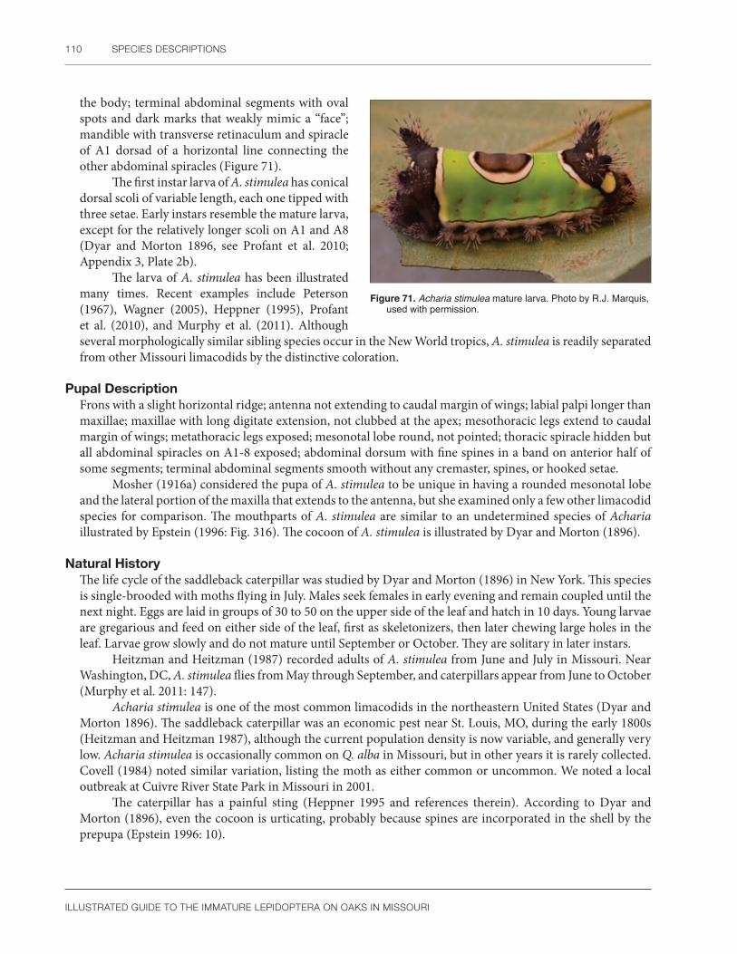
110 SPECIES DESCRIPTIONS
ILLUSTRATED GUIDE TO THE IMMATURE LEPIDOPTERA ON OAKS IN MISSOURI
the body; terminal abdominal segments with oval spots and dark marks that weakly mimic a “face”; mandible with transverse retinaculum and spiracle of A1 dorsad of a horizontal line connecting the other abdominal spiracles (Figure 71).
The first instar larva of A. stimulea has conical dorsal scoli of variable length, each one tipped with three setae. Early instars resemble the mature larva, except for the relatively longer scoli on A1 and A8 (Dyar and Morton 1896, see Profant et al. 2010; Appendix 3, Plate 2b).
The larva of A. stimulea has been illustrated many times. Recent examples include Peterson (1967), Wagner (2005), Heppner (1995), Profant et al. (2010), and Murphy et al. (2011). Although several morphologically similar sibling species occur in the New World tropics, A. stimulea is readily separated from other Missouri limacodids by the distinctive coloration.
Figure 71. Acharia stimulea mature larva. Photo by R.J. Marquis, used with permission.
Pupal DescriptionFrons with a slight horizontal ridge; antenna not extending to caudal margin of wings; labial palpi longer than maxillae; maxillae with long digitate extension, not clubbed at the apex; mesothoracic legs extend to caudal margin of wings; metathoracic legs exposed; mesonotal lobe round, not pointed; thoracic spiracle hidden but all abdominal spiracles on A1-8 exposed; abdominal dorsum with fine spines in a band on anterior half of some segments; terminal abdominal segments smooth without any cremaster, spines, or hooked setae.
Mosher (1916a) considered the pupa of A. stimulea to be unique in having a rounded mesonotal lobe and the lateral portion of the maxilla that extends to the antenna, but she examined only a few other limacodid species for comparison. The mouthparts of A. stimulea are similar to an undetermined species of Acharia illustrated by Epstein (1996: Fig. 316). The cocoon of A. stimulea is illustrated by Dyar and Morton (1896).
Natural HistoryThe life cycle of the saddleback caterpillar was studied by Dyar and Morton (1896) in New York. This species is single-brooded with moths flying in July. Males seek females in early evening and remain coupled until the next night. Eggs are laid in groups of 30 to 50 on the upper side of the leaf and hatch in 10 days. Young larvae are gregarious and feed on either side of the leaf, first as skeletonizers, then later chewing large holes in the leaf. Larvae grow slowly and do not mature until September or October. They are solitary in later instars.
Heitzman and Heitzman (1987) recorded adults of A. stimulea from June and July in Missouri. Near Washington, DC, A. stimulea flies from May through September, and caterpillars appear from June to October (Murphy et al. 2011: 147).
Acharia stimulea is one of the most common limacodids in the northeastern United States (Dyar and Morton 1896). The saddleback caterpillar was an economic pest near St. Louis, MO, during the early 1800s (Heitzman and Heitzman 1987), although the current population density is now variable, and generally very low. Acharia stimulea is occasionally common on Q. alba in Missouri, but in other years it is rarely collected. Covell (1984) noted similar variation, listing the moth as either common or uncommon. We noted a local outbreak at Cuivre River State Park in Missouri in 2001.
The caterpillar has a painful sting (Heppner 1995 and references therein). According to Dyar and Morton (1896), even the cocoon is urticating, probably because spines are incorporated in the shell by the prepupa (Epstein 1996: 10).

LIMACODIDAE 111
ILLUSTRATED GUIDE TO THE IMMATURE LEPIDOPTERA ON OAKS IN MISSOURI
DistributionMassachusetts south to Florida, west to Texas (Covell 1984), including Ohio (Profant et al. 2010), Illinois (Godfrey et al. 1987a), Missouri (Lill et al. 2006), and Mexico (Heppner 2003).
Heitzman and Heitzman (1987) noted the saddleback caterpillar is more common in eastern Missouri than other parts of the state. See Figure 72 for Missouri distribution.
Host PlantsPolyphagous on 20 plant families (Robinson et al. 2002). Specific records for oak include Q. alba, Q. coccinea, Q. macrocarpa, Q. palustris, Q. prinus, Q. rubra, and Q. velutina (Tietz 1952).
Large host lists for A. stimulea were given by Tietz (1952), Heppner (2003), and Robinson et al. (2002). Canna, Castanea, Prunus, Quercus, Tilia, and Zea are favored foodplants (Dyar and Morton 1896). In Missouri, besides on Q. alba and Q. velutina (Lill et al. 2006), we have found A. stimulea on Sassafras albidum, Cornus florida, and Carya ovata.
CommentsUntil recently, A. stimulea was placed in the genus Sibine (Becker and Miller 1989).
Figure 72. Known distribution of Acharia stimulea in Missouri.
Adoneta spinuloides (Herrich-Schäffer)Purple-crested slug
Larval DescriptionEarly instar larvae pale green with a few small dorsal red irregular patches. Middle instars with an irregular dorsal purple band, the D1 pinacula contrasting as a small pair of white dots within this band; ground color of dorsal area pale yellow; subdorsal and lateral scoli pale white or yellow, the anterior and posterior scoli longer than the others; lateral area green. Mature larva variable in color; normal coloration (form A of Dyar 1897a) with the middorsal area deep purple; subdorsal area defined by a sinuate, black-edged yellow band, subdorsal
Figure 73. Adoneta spinuloides (a) mid-instar larva; (b) mature larva. Photos by R.J. Marquis, used with permission.
a b

112 SPECIES DESCRIPTIONS
ILLUSTRATED GUIDE TO THE IMMATURE LEPIDOPTERA ON OAKS IN MISSOURI
scoli either reddish (T2 to A1, A4, A7, A8) or yellow (A2, A3, A5, A6); lateral area green with a broken yellow or white longitudinal stripe; the posterior scoli may be slightly longer and diverging relative to earlier instars; mandible with transverse retinaculum and spiracle of A1 dorsad of a horizontal line connecting the other abdominal spiracles; caltrope spines present only in the last instar. In other forms (B–D of Dyar 1897a), the subdorsal scoli can be either mostly red or mostly yellow, the subdorsal area has a variable amount of yellow, and the lateral stripe may be broken or solid (Figure 73a, b).
The larva of A. spinuloides is illustrated by Dyar (1897a), Wagner (2005), Profant et al. (2010), and Murphy et al. (2011). Dyar’s (1897a) figure was reproduced in color by Profant et al. (2010: Plate 8). First instar larvae have large and small conical dorsal scoli, similar to the arrangement in Euclea (Dyar 1897a). The purple dorsal area surrounded by yellow (Wagner 2005) and short knoblike scoli readily separate A. spinuloides from most other eastern United States species. The larva of Adoneta bicaudata is similar except for having the last pair of posterior scoli elongated and about four times longer than wide (Profant et al. 2010). This contrasts with A. spinuloides, which has scoli that are about two times longer than wide and only slightly larger than the other body scoli.
Pupal DescriptionFrons with a slightly curved horizontal ridge; antenna extends to caudal margin of wings; labial palpi longer than maxillae; maxillae lack a digitate extension and do not reach the antenna; mesothoracic legs extend to caudal margin of wings; metathoracic legs exposed; mesonotal lobe round, not pointed; thoracic spiracle hidden but all abdominal spiracles on A1-8 exposed; abdominal dorsum with faint, very fine spine patches; terminal abdominal segments smooth without any cremaster, spines, or hooked setae. The long antennae, lack of maxillary digitate extension, and round mesonotal lobe are significant, but too few North American limacodid pupae are described to diagnose A. spinuloides at this time.
Natural HistoryThe life cycle of A. spinuloides is described in Dyar (1897a). Elliptical, flat, milky white eggs are laid during July either singly or in small clumps. Larvae feed on low branches (but see Lill et al. 2006) and mature in September. The pupation site is not mentioned. Neither Dyar (1897a) nor Wagner (2005) reported the spines as very painful.
Covell (1984) considered A. spinuloides to be more common in the northern portions of its range. Murphy et al. (2011) reported caterpillars from June to October, whereas moths fly from May through September (Covell 1984). A more restricted flight time from June to July was given by Profant et al. (2010) and Murphy et al. (2011).
DistributionQuebec, Canada, south to North Carolina, west to Mississippi, Missouri (Covell 1984), Minnesota (Lugger 1898), Illinois (Godfrey et al. 1987a), Ohio (Profant et al. 2010), Florida (Heppner 2003), and Texas (Dyar 1935). See Figure 74 for Missouri distribution.
Host PlantsVarious deciduous trees and shrubs (Dyar 1897a, Covell 1984); Malus, Prunus, Quercus (Robinson et al. 2002); Amelanchier, Betula, Castanea, Fagus, Hamamelis, Myrica, Nyssa, Robinia, Salix, Tilia (Tietz 1952); and
Figure 74. Known distribution of Adoneta spinuloides in Missouri.
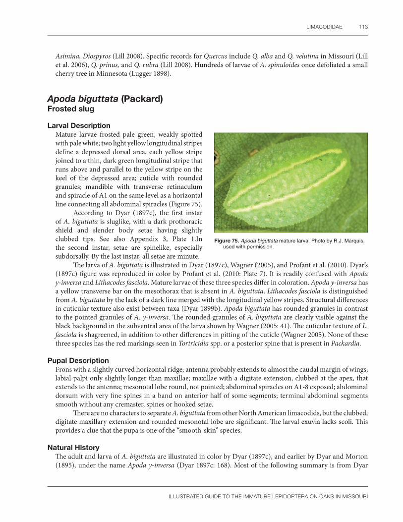
LIMACODIDAE 113
ILLUSTRATED GUIDE TO THE IMMATURE LEPIDOPTERA ON OAKS IN MISSOURI
Asimina, Diospyros (Lill 2008). Specific records for Quercus include Q. alba and Q. velutina in Missouri (Lill et al. 2006), Q. prinus, and Q. rubra (Lill 2008). Hundreds of larvae of A. spinuloides once defoliated a small cherry tree in Minnesota (Lugger 1898).
Apoda biguttata (Packard)Frosted slug
Larval DescriptionMature larvae frosted pale green, weakly spotted with pale white; two light yellow longitudinal stripes define a depressed dorsal area, each yellow stripe joined to a thin, dark green longitudinal stripe that runs above and parallel to the yellow stripe on the keel of the depressed area; cuticle with rounded granules; mandible with transverse retinaculum and spiracle of A1 on the same level as a horizontal line connecting all abdominal spiracles (Figure 75).
According to Dyar (1897c), the first instar of A. biguttata is sluglike, with a dark prothoracic shield and slender body setae having slightly clubbed tips. See also Appendix 3, Plate 1.In the second instar, setae are spinelike, especially subdorsally. By the last instar, all setae are minute.
The larva of A. biguttata is illustrated in Dyar (1897c), Wagner (2005), and Profant et al. (2010). Dyar’s (1897c) figure was reproduced in color by Profant et al. (2010: Plate 7). It is readily confused with Apoda y-inversa and Lithacodes fasciola. Mature larvae of these three species differ in coloration. Apoda y-inversa has a yellow transverse bar on the mesothorax that is absent in A. biguttata. Lithacodes fasciola is distinguished from A. biguttata by the lack of a dark line merged with the longitudinal yellow stripes. Structural differences in cuticular texture also exist between taxa (Dyar 1899b). Apoda biguttata has rounded granules in contrast to the pointed granules of A. y-inversa. The rounded granules of A. biguttata are clearly visible against the black background in the subventral area of the larva shown by Wagner (2005: 41). The cuticular texture of L. fasciola is shagreened, in addition to other differences in pitting of the cuticle (Wagner 2005). None of these three species has the red markings seen in Tortricidia spp. or a posterior spine that is present in Packardia.
Pupal DescriptionFrons with a slightly curved horizontal ridge; antenna probably extends to almost the caudal margin of wings; labial palpi only slightly longer than maxillae; maxillae with a digitate extension, clubbed at the apex, that extends to the antenna; mesonotal lobe round, not pointed; abdominal spiracles on A1-8 exposed; abdominal dorsum with very fine spines in a band on anterior half of some segments; terminal abdominal segments smooth without any cremaster, spines or hooked setae.
There are no characters to separate A. biguttata from other North American limacodids, but the clubbed, digitate maxillary extension and rounded mesonotal lobe are significant. The larval exuvia lacks scoli. This provides a clue that the pupa is one of the “smooth-skin” species.
Natural HistoryThe adult and larva of A. biguttata are illustrated in color by Dyar (1897c), and earlier by Dyar and Morton (1895), under the name Apoda y-inversa (Dyar 1897c: 168). Most of the following summary is from Dyar
Figure 75. Apoda biguttata mature larva. Photo by R.J. Marquis, used with permission.

114 SPECIES DESCRIPTIONS
ILLUSTRATED GUIDE TO THE IMMATURE LEPIDOPTERA ON OAKS IN MISSOURI
(1897c). The eggs of A. biguttata are elliptical, whitish, with fine rectangular reticulations (Dyar 1897c; Peterson 1967). They are laid singly on the lower branches of oak. First instar larvae skeletonize the lower leaf surface. There is one generation per year. Moths occur at the end of June, with larvae appearing later in the same year during August and September. The flight period is longer in Ohio, extending from May to July (Profant et al. 2010).
Unlike other limacodid species in Missouri, A. biguttata is more common in the canopy of white oak than in the understory (Lill et al. 2006: 802). Early instars make unusual feeding tracts on the host leaves (Wagner 2005), as do some other eastern species (SCPC).
Heitzman and Heitzman (1987) reported that A. biguttata has two generations per year in Missouri, in contrast to Dyar (1897c) who documented one generation in the eastern United States. In Missouri, adults were collected from mid-May to August, and are common at lights. This is somewhat longer than the June to August flight period given by Murphy et al. (2011) based on collections near Washington, DC. The distribution of A. biguttata in Missouri is restricted to woodlands, where it can be common (Heitzman and Heitzman 1987).
DistributionEastern Canada to Florida, west to Texas, Missouri (Covell 1984), Ohio (Profant et al. 2010), Michigan (spms. in Charles A. Triplehorn coll.), and Illinois (Godfrey et al. 1987a). Profant et al. (2010) recorded A. biguttata from Kansas without a source for the record. See Figure 76 for Missouri distribution.
Host PlantsQuercus (Robinson et al. 2002); Carpinus (Covell 1984); Carya (Tietz 1952); Fagus (Wagner 2005); Quercus, usually Q. alba (Dyar 1897c); Q. coccinea (Whitfield et al. 1999), and Q. velutina (Lill et al. 2006).
In addition to Q. velutina, we have collected A. biguttata on Q. muehlenbergii in Missouri. Dyar (1897c) did not mention Carya as a host in his revision of the description of A. biguttata (Dyar and Morton 1895). It is unknown if Dyar omitted the record by accident or on purpose. Another member of the genus, A. y-inversa, also feeds on hickory (Dyar 1935).
CommentsWe find the common name shagreened slug (Wagner 2005, Profant et al. 2010) a poor choice for A. biguttata because this cuticular texture is an identification characteristic of L. fasciola. Instead, the sparkling appearance of A. biguttata would be emphasized by using the common name frosted slug.
Figure 76. Known distribution of Apoda biguttata in Missouri.
Euclea delphinii (Boisduval)Spiny oak slug
Larval DescriptionMature larva variable in coloration; ground color may be a shade of green, yellow, pink, orange-brown, steel blue, or gray; dorsal area slightly depressed, usually with a white to pale green middorsal longitudinal line, a pair of white dots on each segment, and a pair of thin, black, sinuate longitudinal stripes around the spots; subdorsal area cream to orange, with a variable amount of red to brown patches sometimes forming a line, and

LIMACODIDAE 115
ILLUSTRATED GUIDE TO THE IMMATURE LEPIDOPTERA ON OAKS IN MISSOURI
a row of pointed green, orange, or yellow colored scoli on each segment, these scoli white or black tipped and longer on the anterior and posterior abdominal segments; lateral area with white specks and black markings that vary from a line to almost a circle, with scoli present on each segment; mandible with transverse retinaculum and spiracle of A1 dorsad of a horizontal line connecting the other abdominal spiracles (Figure 77).
According to Dyar (1896a), the first instar larva of E. delphinii has conical dorsal scoli of variable length, each one tipped with three setae. There is also a lateral row of scoli. Dyar (1897b) characterized five larval color variations of E. delphinii, four of which were illustrated in color by Profant et al. (2010: Plate 8). Late instars may have one or two pairs of detachable spines on the last abdominal segments that may not be homologous to the caltrope spines of other limacodid larvae (Dyar 1897b: 58). Unlike the midwestern specimens examined in this study, the mandible of the Florida population appears to lack a transverse retinaculum (Dyar 1896a). See also Appendix 3, Plate 1.
The larva of E. delphinii has been illustrated many times. A few examples include Peterson (1962a: L22), Stehr (1987), Heppner (1995), Wagner (2005), Sogaard (2009), Profant et al. (2010), and Murphy et al. (2011). Being a popular photographic subject, an Internet Web search produces many examples, including eggs, first instars, and many color forms of the adult and larva. Although the coloration is highly variable, no other limacodid larva on Missouri oaks has one or two pairs of contrasting, short, black scoli on the terminal abdominal segments. When these patches are absent, E. delphinii can be recognized by characteristics of the coloration and body shape. In particular, the scoli are all approximately equal in size and four dorsal patches of reddish markings are present in most of the common color forms.
Figure 77. Euclea delphinii mature larva. Photo by R.J. Marquis, used with permission.
Pupal DescriptionFrons with a slightly curved horizontal ridge; labial palpi longer than maxillae; all abdominal spiracles on A1-8 exposed; abdominal dorsum with fine spines in a band on anterior half of some segments; terminal abdominal segments smooth without any cremaster, spines, or hooked setae.
According to Mosher (1916a), the lateral portion of the maxilla extends to the antenna, and the mesonotal lobe is pointed in E. delphinii. We have only three pupal exuviae of this species, and neither character is obvious on our specimens. This may represent variation or the poor condition of our specimens.
Natural HistoryThe life cycle of E. delphinii is illustrated in color and summarized by Dyar (1896a, 1897b). Other aspects of the natural history were studied in more detail by Murphy et al. (2011). The eggs of E. delphinii are irregularly shaped, flattened, and transparent (Epstein 1996: 13). The surface has indistinct reticulations. Unlike most other species of eastern United States limacodids, eggs are laid in clusters instead of singly (Murphy et al. 2011). According to Dyar (1897b), first instar larvae hatch in about 7 days and pass through eight or nine instars before maturing in September. Wagner (2005) and Murphy et al. (2011) also reported caterpillars in the fall. Larvae prefer to hide by day in curled leaves or other suitable sites in Florida, but this behavior was not common in New York and has not been observed in Missouri (R. J. Marquis, pers. obs.). The coloration of E. delphinii appears to be aposematic, but the mild sting (Wagner 2005) seems to argue against warning coloration. We have seen green, orange, yellow, and gray forms in Missouri (R. J. Marquis, pers. obs.). Another hypothesis is that the larvae are cryptic against the dry, spotted, brilliantly-colored fall leaves (Dyar 1897b).

116 SPECIES DESCRIPTIONS
ILLUSTRATED GUIDE TO THE IMMATURE LEPIDOPTERA ON OAKS IN MISSOURI
Heitzman and Heitzman (1987) reported that E. delphinii has two generations per year in Missouri in contrast to one generation in New York (Dyar 1897b). In Missouri, adults were collected from early May to late August and are common at lights, but they were rarely seen around lights in Minnesota by early collectors (Lugger 1898). Murphy et al. (2011) gave a similar flight period of May until late July around Washington, DC. This species is abundant in Ohio, and adults are found from May to September (Profant et al. 2010). It is also widespread throughout Missouri and breeds in most parts of the state (Heitzman and Heitzman 1987).
DistributionMaine and southern Quebec, Canada, to Florida and west to Missouri and Louisiana (Covell 1984). See Figure 78 for Missouri distribution.
Host PlantsAcer, Alnus, Betula, Carya, Coccoloba, Crataegus, Fagus, Malus, Pyrus, Prunus, Rhizophora, Salix (Robinson et al. 2002); Andromeda, Carpinus, Castanea, Chrysobalanus, Gleditsia, Magnolia, Myrica, Nyssa, Rubus, Smilax, Tilia (Tietz 1952); Diospyros (Lill 2008); Celtis, Fraxinus, Platanus, Populus, and Vaccinium (Wagner 2005). Specific records from oak include Quercus ilicifolia, Q. rubra (Robinson et al. 2002); Q. alba, and Q. velutina (Lill et al. 2006).
Euclea delphinii is polyphagous on forest trees and shrubs (Robinson et al. 2002), especially Quercus (Wagner et al. 1997). We have found E. delphinii on Q. alba, Q. imbricaria, Q. rubra, and Q. velutina in Missouri. Tietz (1952) noted that northern and southern populations use different hosts. Larvae in Florida feed on sea grape and mangrove (Kimball 1965).
CommentsEuclea delphinii is easily confused with E. nanina. The latter species occurs in Pennsylvania (Tietz 1952) and Florida (Kimball 1965) but is not yet recorded from Missouri (J.E. Heitzman and P.E. Koenig, unpublished data). According to Kimball (1965), E. nanina has a round discal dot in contrast to the elongate discal dot characteristic of E. delphinii. We are not sure of the status of E. nanina in Missouri and how the larva could be separated from E. delphinii if both species were present in our study sites. Euclea delphinii form “paenulata” is the most common form in Missouri according to Heitzman and Heitzman (1987).
Dyar (1897a) notes that E. delphinii has a “tendency toward local forms” in several geographical regions of the eastern United States. Kimball (1965) raised the possibility of an unrecognized species in Florida that potentially could occur in Missouri. Epstein and Adams (2009) suspect that E. delphinii forms a hybrid zone with a western species in the Midwest. These observations suggest more study is needed on the systematics of E. delphinii in Missouri and elsewhere.
Figure 78. Known distribution of Euclea delphinii in Missouri.

LIMACODIDAE 117
ILLUSTRATED GUIDE TO THE IMMATURE LEPIDOPTERA ON OAKS IN MISSOURI
Isa textula (Herrich-Schäffer)Crowned slug
Larval DescriptionMature larva dorsoventrally flattened with head, prothorax, and mesothorax hidden from above; anterior margin of metathorax with two short scoli projecting forward, these often colored rust to red; dorsum with pair of pale yellow longitudinal stripes and usually two red dots on a pale green ground color; lateral margin of body surrounded by a row of elongated scoli; mandible with transverse retinaculum and spiracle of A1 dorsad of the lateral row of scoli so that it is the only abdominal spiracle visible from above, other abdominal spiracles visible in ventral view below the scoli (Figure 79).
According to Dyar (1896b, 1898c), the row of subdorsal spines on A2-8 is reduced in the first instar larva of I. textula. The mature larva of I. textula is illustrated by Dyar (1896b), Stehr (1987), and in color by Wagner (2005), Profant et al. (2010), and Murphy et al. (2011). This species is easily recognized, although there is variation in the amount of red coloration present on the dorsal area. The scoli of I. textula are spinelike, not plumose as in Isochaetes beutenmuelleri.
Pupal DescriptionBased on damaged pupal exuvia: Frons with a slightly curved horizontal ridge; antenna probably does not extend to caudal margin of wings; labial palpi only slightly longer than maxillae; maxillae with a digitate extension, not clubbed at the apex that extends to the antenna; mesonotal lobe round, not pointed; abdominal spiracles on A1-8 exposed; abdominal dorsum with very fine spines in a band on anterior half of some segments; terminal abdominal segments smooth without any cremaster, spines or hooked setae. With only a single, crumpled pupal exuvia, we can only partially describe the pupa of I. textula but not diagnose it.
Natural HistoryDyar (1896b) studied the life cycle of I. textula in New York. Eggs are laid singly during July. First instar larvae immediately molt before feeding. There are eight to nine larval instars. Pupation occurs in a cocoon on the ground during September.
Adults of I. textula fly from May through August (Covell 1984). Larvae can be found very late in the fall around Washington, DC, often from October until the leaves drop, even on foliage that is changing color (Murphy et al. 2011). They prefer the canopy, at least in the case of Q. velutina (Lill et al. 2006).
DistributionSouthern Ontario, Canada, to Florida and west to Mississippi, Missouri (Covell 1984), Illinois (Godfrey et al. 1987a), and Ohio (Profant et al. 2010). See Figure 80 for Missouri distribution.
Figure 79. Isa textula mature larva. Photo by R.J. Marquis, used with permission.
Figure 80. Known distribution of Isa textula in Missouri.

118 SPECIES DESCRIPTIONS
ILLUSTRATED GUIDE TO THE IMMATURE LEPIDOPTERA ON OAKS IN MISSOURI
Host PlantsPolyphagous on trees and shrubs, including Acer, Aesculus, Castanea, Myrica, Prunus, Tilia (Robinson et al. 2002); Carya, Fagus, Ostrya, and Ulmus (Tietz 1952). Specific records for oak include Quercus alba (Robinson et al. 2002); Q. velutina (Lill et al. 2006); Q. prinus, and Q. rubra (Lill 2008). We have frequently collected Isa textula on Q. alba, Q. rubra, and Q. velutina in Missouri.
CommentsIsa textula was included in a group of seven North American limacodid genera whose larvae are urticating (Wagner et al. 1997, Wagner 2005: 46). This species was also seen stinging a person in Missouri (R. Forkner, pers. obs.).
Isochaetes beutenmuelleri (Edwards) Spun glass slug
Larval Description Mature larva dorsoventrally flattened, and except for the middorsal region, covered with dense green-white plumose scoli, the subdorsal ones from T3 to A8 with a bulblike modification; mandible with transverse retinaculum and spiracle of A1 dorsad of the lateral row of scoli so that it is the only abdominal spiracle visible from above, other abdominal spiracles visible in ventral view below the scoli (Figure 81).
According to Dyar (1907), the first instar larva of I. beutenmuelleri has long, tapered spines on the thorax and abdomen. The mature larva is illustrated in Dyar (1907), Wagner (2005), Profant et al. (2010), and Murphy et al. (2011). This species is easily recognized by the characteristic bulblike modification on the lateral scoli and, in the field, by the pale green, unmarked plumose setae.
Pupal DescriptionBased on one damaged pupal exuviae: Frons with a curved horizontal ridge; labial palpi slightly longer than the maxillae; maxillae lack a long digitate extension; mesothoracic legs probably extend to caudal margin of wings; mesonotal lobe long, rounded, and extending to first abdominal segment; abdominal dorsum with fine spines in a band on anterior half of most segments; terminal abdominal segments smooth without any cremaster, spines, or hooked setae.
There are no characters to separate I. beutenmuelleri from other North American limacodids. The larval exuvia, normally an excellent source of clues for pupal identification, is misleading in this species. The overwintering larva sheds its scoli (see below), which can lead to incorrectly concluding the pupa must belong to the “smooth-skin” species group, when in fact scoli were originally present.
Natural HistoryDyar (1907) studied the life history of I. beutenmuelleri in New York. Eggs are laid singly on the leaf undersurface. Young larvae skeletonize the epidermis; older individuals consume the whole leaf starting from the edge. Lill et al. (2006) found equal numbers of I. beutenmuelleri in the canopy compared to the understory
Figure 81. Isochaetes beutenmuelleri mature larva. Photo by R.J. Marquis, used with permission.
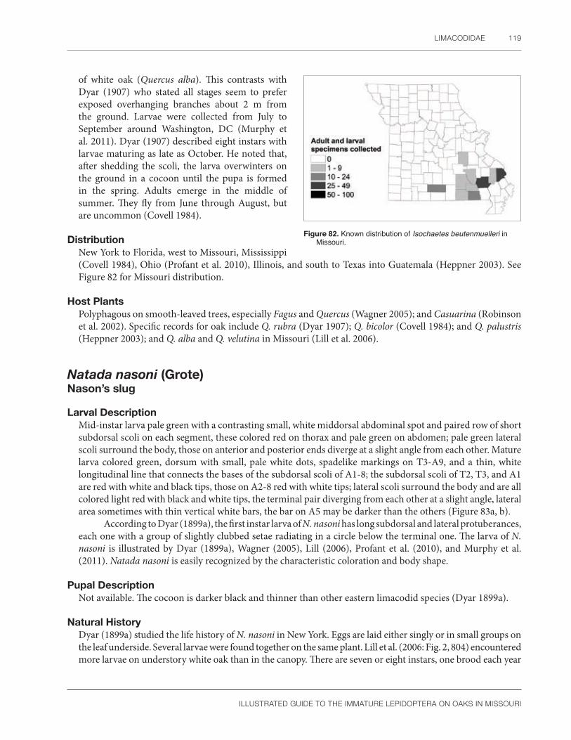
LIMACODIDAE 119
ILLUSTRATED GUIDE TO THE IMMATURE LEPIDOPTERA ON OAKS IN MISSOURI
of white oak (Quercus alba). This contrasts with Dyar (1907) who stated all stages seem to prefer exposed overhanging branches about 2 m from the ground. Larvae were collected from July to September around Washington, DC (Murphy et al. 2011). Dyar (1907) described eight instars with larvae maturing as late as October. He noted that, after shedding the scoli, the larva overwinters on the ground in a cocoon until the pupa is formed in the spring. Adults emerge in the middle of summer. They fly from June through August, but are uncommon (Covell 1984).
DistributionNew York to Florida, west to Missouri, Mississippi (Covell 1984), Ohio (Profant et al. 2010), Illinois, and south to Texas into Guatemala (Heppner 2003). See Figure 82 for Missouri distribution.
Host PlantsPolyphagous on smooth-leaved trees, especially Fagus and Quercus (Wagner 2005); and Casuarina (Robinson et al. 2002). Specific records for oak include Q. rubra (Dyar 1907); Q. bicolor (Covell 1984); and Q. palustris (Heppner 2003); and Q. alba and Q. velutina in Missouri (Lill et al. 2006).
Figure 82. Known distribution of Isochaetes beutenmuelleri in Missouri.
Natada nasoni (Grote) Nason’s slug
Larval Description Mid-instar larva pale green with a contrasting small, white middorsal abdominal spot and paired row of short subdorsal scoli on each segment, these colored red on thorax and pale green on abdomen; pale green lateral scoli surround the body, those on anterior and posterior ends diverge at a slight angle from each other. Mature larva colored green, dorsum with small, pale white dots, spadelike markings on T3-A9, and a thin, white longitudinal line that connects the bases of the subdorsal scoli of A1-8; the subdorsal scoli of T2, T3, and A1 are red with white and black tips, those on A2-8 red with white tips; lateral scoli surround the body and are all colored light red with black and white tips, the terminal pair diverging from each other at a slight angle, lateral area sometimes with thin vertical white bars, the bar on A5 may be darker than the others (Figure 83a, b).
According to Dyar (1899a), the first instar larva of N. nasoni has long subdorsal and lateral protuberances, each one with a group of slightly clubbed setae radiating in a circle below the terminal one. The larva of N. nasoni is illustrated by Dyar (1899a), Wagner (2005), Lill (2006), Profant et al. (2010), and Murphy et al. (2011). Natada nasoni is easily recognized by the characteristic coloration and body shape.
Pupal DescriptionNot available. The cocoon is darker black and thinner than other eastern limacodid species (Dyar 1899a).
Natural HistoryDyar (1899a) studied the life history of N. nasoni in New York. Eggs are laid either singly or in small groups on the leaf underside. Several larvae were found together on the same plant. Lill et al. (2006: Fig. 2, 804) encountered more larvae on understory white oak than in the canopy. There are seven or eight instars, one brood each year

120 SPECIES DESCRIPTIONS
ILLUSTRATED GUIDE TO THE IMMATURE LEPIDOPTERA ON OAKS IN MISSOURI
a b
Figure 83. Natada nasoni (a) early instar larva; (b) mature larva. Photos by R.J. Marquis, used with permission.
(Dyar 1899a) and larvae were collected from June to October by Murphy et al. (2011). Pupation is on the ground, and adults emerge in June and early July, with each sex having a different resting posture (Dyar 1899a). Covell (1984) recorded moths in August, a little later than in Profant et al. (2010) and Murphy et al. (2011).
DistributionMaine to Florida, west to Missouri and Mississippi (Covell 1984) including Illinois (Godfrey et al. 1987a) and Ohio (Profant et al. 2010).
The relationship of N. daona to N. nasoni requires comment. Dyar (1899a) reported that Perola daona, at that time considered a synonym of N. nasoni, occurs in Mexico and Costa Rica. This synonymy was used in the North American checklists by Hodges et al. (1983) and Pohl et al. (2016). In his later review of the American fauna (Dyar 1935), Dyar changed his mind and treated N. daona and N. nasoni as two distinct species, as did Epstein and Becker (1994). We restrict the distribution of N. nasoni to the United States in agreement with the most recent taxonomists to have studied the genus (Epstein and Becker 1994). See Figure 84 for Missouri distribution.
Host PlantsCastanea, Fagus, Ostrya, Quercus velutina (Robinson et al. 2002); Carya, (Covell 1984); Carpinus (Wagner 2005); and Q. rubra (Lill 2008). We have found N. nasoni on Q. muehlenbergii in Missouri (Lill et al. 2006).
CommentsIn contrast to Covell (1984), we consider N. nasoni as one of the most common limacodids on oak in Missouri. Wagner (2005) and Profant et al. (2010) mentioned that it can be common in parts of the Appalachian Region.
Figure 84. Known distribution of Natada nasoni in Missouri.

LIMACODIDAE 121
ILLUSTRATED GUIDE TO THE IMMATURE LEPIDOPTERA ON OAKS IN MISSOURI
Packardia geminata (Packard)Jeweled tailed slug
Larval Description Mid-instar larva turquoise with two thin, pale yellow longitudinal stripes on the dorsal keel; dorsal area with short scoli ending in paired black tipped spines and posterior abdominal segments drawn to narrow tip. Mature larva as above except scoli are represented by minute tubercles, the skin is granulated, a contrasting white subdorsal stripe is present, the mandible has a transverse retinaculum, and the spiracle of A1 is dorsad of a horizontal line connecting the other abdominal spiracles (Figure 85a, b).
According to Dyar (1898a), the first instar of P. geminata has clubbed setae, the second instar has long pointed spines, and later instars have minute tubercles. The larva of P. geminata is illustrated by Wagner (2005) and Profant et al. (2010: Plate 4). Dyar’s (1898a: fig. 6) drawing of P. geminata was reproduced in color by Profant et al. (2010: Plate 7: Fig. 3) under the name P. elegans instead of P. geminata due to a misprint. Larvae of Packardia are easily recognized to genus by the smooth cuticle, lack of red circular dorsal markings, and presence of a spine on the terminal abdominal segment. Species identification is more difficult. The mature larva of P. elegans is recognized by the wavy subdorsal line and closely spaced skin granules that almost touch each other (Dyar 1898a: Figs. 5, 9–10, 1899b). Packardia geminata has straight subdorsal lines and cuticular granules that are separated from each other by approximately their diameter (Dyar 1898a: Figs. 4 and 6, 1899b). It is unknown if these texture differences between species are valid for all instars. In addition, the subdorsal stripe of P. geminata is wider and more contrasting than in P. elegans. Wagner (2005) noted that the tail can break off in Packardia larvae, thus it is important to pay attention to the cuticle texture in doubtful cases. Given how subtle these color and cuticular texture differences are to evaluate, it would be best to rear adults or barcode larvae to be absolutely certain of the species identity.
a b
Figure 85. Packardia geminata (a) early instar larva; (b) mature larva. Photos by R.J. Marquis, used with permission.
Pupal DescriptionFrons with a slightly curved horizontal ridge; labial palpi as long as the maxillae; maxillae with a digitate extension; abdominal spiracles on A1-8 exposed; abdominal dorsum with very fine spines in a band on anterior half of some segments; terminal abdominal segments smooth without any cremaster, spines, or hooked setae. The labial palpi and maxillae being equal in length seems unusual, but a lack of material prevents any significant pupal diagnosis of P. geminata.
Natural HistoryDyar (1898a) studied the life history of P. geminata in New York and New Hampshire. The eggs are laid singly
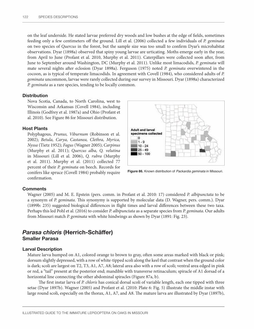
122 SPECIES DESCRIPTIONS
ILLUSTRATED GUIDE TO THE IMMATURE LEPIDOPTERA ON OAKS IN MISSOURI
on the leaf underside. He stated larvae preferred dry woods and low bushes at the edge of fields, sometimes feeding only a few centimeters off the ground. Lill et al. (2006) collected a few individuals of P. geminata on two species of Quercus in the forest, but the sample size was too small to confirm Dyar’s microhabitat observations. Dyar (1898a) observed that spiny young larvae are urticating. Moths emerge early in the year, from April to June (Profant et al. 2010, Murphy et al. 2011). Caterpillars were collected soon after, from June to September around Washington, DC (Murphy et al. 2011). Unlike most limacodids, P. geminata will mate several nights after eclosion (Dyar 1898a). Ferguson (1975) noted P. geminata overwintered in the cocoon, as is typical of temperate limacodids. In agreement with Covell (1984), who considered adults of P. geminata uncommon, larvae were rarely collected during our survey in Missouri. Dyar (1898a) characterized P. geminata as a rare species, tending to be locally common.
DistributionNova Scotia, Canada, to North Carolina, west to Wisconsin and Arkansas (Covell 1984), including Illinois (Godfrey et al. 1987a) and Ohio (Profant et al. 2010). See Figure 86 for Missouri distribution.
Host PlantsPolyphagous, Prunus, Viburnum (Robinson et al. 2002); Betula, Carya, Castanea, Clethra, Myrica, Nyssa (Tietz 1952); Fagus (Wagner 2005); Carpinus (Murphy et al. 2011); Quercus alba, Q. velutina in Missouri (Lill et al. 2006), Q. rubra (Murphy et al. 2011). Murphy et al. (2011) collected 77 percent of their P. geminata on beech. Records for conifers like spruce (Covell 1984) probably require confirmation.
CommentsWagner (2005) and M. E. Epstein (pers. comm. in Profant et al. 2010: 17) considered P. albipunctata to be a synonym of P. geminata. This synonymy is supported by molecular data (D. Wagner, pers. comm.). Dyar (1899b: 235) suggested biological differences in flight times and larval differences between these two taxa. Perhaps this led Pohl et al. (2016) to consider P. albipunctata as a separate species from P. geminata. Our adults from Missouri match P. geminata with white hindwings as shown by Dyar (1891: Fig. 23).
Parasa chloris (Herrich-Schäffer) Smaller Parasa
Larval Description Mature larva humped on A1, colored orange to brown to gray, often some areas marked with black or pink; dorsum slightly depressed, with a row of white-tipped scoli along the keel that contrast when the ground color is dark; scoli are largest on T2, T3, A1, A7, A8; lateral area also with a row of scoli; ventral area edged in pink or red, a “tail” present at the posterior end; mandible with transverse retinaculum; spiracle of A1 dorsad of a horizontal line connecting the other abdominal spiracles (Figure 87a, b).
The first instar larva of P. chloris has conical dorsal scoli of variable length, each one tipped with three setae (Dyar 1897b). Wagner (2005) and Profant et al. (2010: Plate 6: Fig. 5) illustrate the middle instar with large round scoli, especially on the thorax, A1, A7, and A8. The mature larva are illustrated by Dyar (1897b),
Figure 86. Known distribution of Packardia geminata in Missouri.

LIMACODIDAE 123
ILLUSTRATED GUIDE TO THE IMMATURE LEPIDOPTERA ON OAKS IN MISSOURI
a b
Figure 87. Parasa chloris (a) early instar larva; (b) mature larva. Photos by R.J. Marquis, used with permission.
Wagner (2005), and Murphy et al. (2011). Five color variants are shown in Profant et al. (2010). Parasa chloris is easily recognized by the humped body with a “tail” at the posterior end. Although early instars of P. chloris somewhat resemble Euclea, they show the characteristic body shape of the mature larva (Wagner 2005).
Based on ontogenetic studies, Dyar (1897b) states that the “tail” of P. chloris probably represents a fusion of scoli. The scoli of early instars are partially fused, but later merge into a flaplike appendage.
Pupal DescriptionModified from Mosher (1916a): Frons with a small horizontal ridge; antenna not extending to caudal margin of wings; labial palpi longer than maxillae; maxillae with long digitate extension; neither antennae or mesothoracic legs extend to caudal margin of wings; metathoracic legs exposed; mesonotal lobe pointed, not rounded; thoracic spiracle hidden but all abdominal spiracles on A1-8 exposed; abdominal dorsum with fine spines in a band on anterior half of some segments; terminal abdominal segments smooth without any cremaster, spines, or hooked setae.
Mosher (1916a) illustrates the pupa of P. chloris and diagnosed it by the pointed mesonotal lobe and the lateral portion of the maxilla that extends to the antenna, but she examined only three genera. The long labial palpi of P. chloris are also unusual. The mesothoracic legs on our single specimen of P. chloris are slightly shorter than in Mosher’s (1916a) illustration.
Natural HistoryThe life cycle of P. chloris was published in Dyar (1897b), based on observations in New York. Almost transparent eggs are laid singly from mid- to late July and cohorts of different ages may occur together on the same leaf. Dyar (1897b) wrote that larvae prefer lower branches of trees, especially in shady locations, where they feed on the undersides of leaves. This was not confirmed by Lill et al. (2006: 802) because densities of P. chloris on the understory and canopy of black oak did not differ statistically. Larval development is completed by October (Murphy et al. 2011). Lugger (1898) noted the caterpillar hibernates in the cocoon, not pupating until spring. Adults fly from May through August (Covell 1984). Dark forms of the larvae are cryptic on browned leaves, and the spines of P. chloris are extremely urticating (R. Marquis, pers. obs.).
DistributionNew York to Florida, west to Missouri and Mississippi (Covell 1984), including Illinois (Godfrey et al. 1987a) and Ohio (Profant et al. 2010). Parasa chloris is uncommon in Missouri (R.J. Marquis, pers. obs.). See Figure 88 for Missouri distribution.

124 SPECIES DESCRIPTIONS
ILLUSTRATED GUIDE TO THE IMMATURE LEPIDOPTERA ON OAKS IN MISSOURI
Figure 88. Known distribution of Parasa chloris in Missouri.
Host PlantsPolyphagous on trees and shrubs, Myrica, Populus, Prunus, Ulmus, Vaccinium (Robinson et al. 2002); Betula, Carpinus, Carya, Castanea, Celastrus, Cornus, Lysimachia, Malus, Pyrus, Rosa, Salix, Tilia, (Tietz 1952); Lindera (Lill 2008); and Fagus (Wagner 2005). Specific records for oak include Quercus alba, Q. velutina (Lill et al. 2006); Q. rubra (Lill 2008); and Q. muehlenbergii in Missouri.
CommentsThe record for P. chloris in Arizona (Epstein and Adams 2009) may refer to another closely related species of Parasa (D. Wagner, pers. comm.).
Parasa indetermina (Boisduval)Stinging rose caterpillar
Larval Description Early instar larva dark maroon-brown with three thin, faint middorsal longitudinal stripes and six pairs of stout conical yellow-orange dorsal scoli, with smaller scoli also present subdorsally; ground color of mature larva yellow or red to orange with six pairs of long dorsal scoli on T2, T3, A1, A4, A7, and A8, those on the thorax shorter than the others; dorsum of the remaining body segments with knoblike short scoli; all segments with short subdorsal scoli; middorsal area with four thin, black longitudinal lines on a blue-gray background; subdorsal area white closest to the midline, the remainder with orange, white, or black longitudinal stripes; lateral and subventral area also striped as above; ventral area light purple; mandible with transverse retinaculum and spiracle of A1 dorsad of a horizontal line connecting the other spiracles (Figure 89a, b).
According to Dyar (1897a), the first instar larva of P. indetermina has three setae at the tip of stout conical scoli. The fourth instar of P. indetermina is illustrated by Dyar (1897a) and Profant et al. (2010). Wagner (2005) and Profant et al. (2010) illustrated the mature larva. In addition, P. indetermina is illustrated on the cover of Stehr (1987) and the American Entomologist (Grantham 2003). See also Appendix 3, Plate 1.
Figure 89. Parasa indetermina (a) early instar larva; (b) mature larva. Photos by R.J. Marquis, used with permission.
a b

LIMACODIDAE 125
ILLUSTRATED GUIDE TO THE IMMATURE LEPIDOPTERA ON OAKS IN MISSOURI
Parasa indetermina is easily recognized by its coloration, although there is some minor variation in the length of the dorsal scoli. Dyar (1897a) reported a yellow color form that is illustrated by Wagner (2005); this form is also found in Missouri.
Pupal DescriptionNot available. The figure by Chittenden (1913) lacks morphological detail but is similar in appearance to limacodids illustrated by Mosher (1916a). He noted the pupa has a frontal ridge (“cocoon cutter”), which is typical of limacodid species in Missouri.
Natural HistoryDyar (1897a) and Chittenden (1913) studied the life history of P. indetermina in the northeastern United States. Overlapping eggs are laid singly or in small groups during July. The larval stage consists of eight or nine instars, although the first instar does not feed. They seem to prefer low woody shrubs as hosts. This was the case in Missouri where P. indetermina was more common on understory leaves of white oak than in the canopy (Lill et al. 2006).
According to Chittenden (1913), caterpillars usually mature in September, although last instar larvae can be collected from August to October. The cocoon is formed on the ground or on the host plant if larvae are caged. An adult emerged during June in Illinois (Godfrey et al. 1987a), consistent with a flight period of June to August (Covell 1984, Murphy et al. 2011). The life cycle is illustrated by Chittenden (1913).
DistributionNew York to Florida, west to Missouri and Texas (Covell 1984) including Illinois (Godfrey et al. 1987a), Ohio, and an undocumented citation for Michigan (Profant et al. 2010).
Bess (2005) considered P. indetermina to be locally common in scattered populations throughout its current range. See Figure 90 for Missouri distribution.
Host PlantsAsimina, Carya, Castanea, Cornus, Myrica, Malus, Prunus, Pyrus, Rosa (Robinson et al. 2002); Acer, Platanus, Populus, Rubus, Salix, Vaccinium, Viburnum (Tietz 1952); Cercis (Appleby 1987). Reports for oak in Missouri include Quercus alba and Q. velutina (Lill et al. 2006).
Parasa indetermina has caused serious damage to rosebushes (Chittenden 1913) and can be commonly collected on redbud in August (Appleby 1987). In addition to Quercus and Cercis, we have found it on Cornus, Asimina, and Prunus in Missouri.
Prolimacodes badia (Hübner) Skiff moth
Larval Description Mature larva with a flattened expanded dorsal region colored either green or brown, with darker brown, white, or more rarely, purple markings; a thin, yellow middorsal longitudinal line sometimes present; dorsal
Figure 90. Known distribution of Parasa indetermina in Missouri.

126 SPECIES DESCRIPTIONS
ILLUSTRATED GUIDE TO THE IMMATURE LEPIDOPTERA ON OAKS IN MISSOURI
a b
Figure 91. Prolimacodes badia (a) mature larva; (b) another color form of the mature larva. Photos by R.J. Marquis, used with permission.
area widest on fourth abdominal segment where the margin sometimes forms one or two pointed tubercles; lateral region green with abdomen marked in white or brown spots of various sizes and shapes; anal region tapering posteriorly to a point; mandible with transverse retinaculum; spiracle of A1 in line with a horizontal line connecting the other spiracles.
The first instar larva of P. badia was described by Dyar (1896b) as having scoli with two spines at the tip, which he considered a specialization. See also Appendix 3, Plate 1. Dyar (1896b), Eliot and Soule (1902), Stehr (1987) and Wagner (2005) illustrate the mature larva. Profant et al. (2010) reproduced Dyar’s (1896b) drawings of P. badia in color; additional photographs show variations in larval color, markings, and dorsal spines. This species is easily recognized by the characteristic body shape that is rounded, flattened dorsally, with two longitudinal ridges, and no long scoli (Dyar 1896b: Plate 7: Fig. 6). Eliot and Soule (1902) agreed with Dyar (1896b) that “no two larvae looked alike,” but green with dorsal patches or brown with small markings appear to be the most common forms (Figure 91a, b). The cuticular texture, composed of overlapping scalelike plates, is very distinctive.
Pupal DescriptionFrons with a slight horizontal ridge; antenna extending almost to caudal margin of wings; labial palpi as long as maxillae; maxillae lack a digitate extension; mesothoracic legs extend to caudal margin of wings; metathoracic legs exposed; mesonotal lobe round, not pointed; thoracic spiracle hidden, but all abdominal spiracles on A1-8 exposed; abdominal dorsum with fine spines in a band on anterior half of some segments; terminal abdominal segments smooth without any cremaster, spines, or hooked setae. Mosher (1916a) stated the maxilla of P. badia lacks a lateral extension and thus does not reach the antenna. In addition, the mesonotal lobe is rounded.
Natural HistoryAccording to Dyar (1896b), the eggs of P. badia are laid singly and the caterpillars live on the underside of leaves. At least on Q. velutina, larvae are more common in the canopy than in the understory (Lill et al. 2006: 802). Eliot and Soule (1902) reared a brood from eggs to cocoon in 53 days, noting that late instars eat the leaf edge. Larvae occur from June to October in the eastern United States (Dyar 1896b, Murphy et al. 2011). Adults fly from June to August in Washington, DC (Murphy et al. 2011) and Ohio (Profant et al. 2010). They mate in early evening (Eliot and Soule 1902).
Heitzman and Heitzman (1987) considered P. badia to be “apparently bivoltine” in Missouri where adults are found from late May to late August, especially at lights. Covell (1984) extended the flight period to September, noting that P. badia may be abundant in Florida. There is only one generation per year in the northeast (Wagner 2005).
LIMACODIDAE 127
ILLUSTRATED GUIDE TO THE IMMATURE LEPIDOPTERA ON OAKS IN MISSOURI
Figure 92. Known distribution of Prolimacodes badia in Missouri.
DistributionNew Hampshire to Florida, west to Missouri (Covell 1984), including Illinois (Godfrey et al. 1987a) and Ohio (Profant et al. 2010). Heitzman and Heitzman (1987) stated P. badia is locally common in Missouri. See Figure 92 for Missouri distribution.
Host PlantsPolyphagous on trees and shrubs, Acer, Carya, Castanea, Citrus, Crataegus, Ilex, Juglans, Myrica, Nyssa, Ostrya, Platanus, Prunus, Pyrus, Quercus rubra, Rhododendron, Ulmus (Robinson et al. 2002); Amelanchier, Betula, Hamamelis, Liquidambar, Malus, Populus, Salix, Tilia, Vaccinium (Tietz 1952). Specific reports on oak include Quercus alba and Q. velutina in Missouri (Lill et al. 2006).
Heitzman and Heitzman (1987) mentioned Crataegus as a host in Missouri. According to Eliot and Soule (1902), bayberry (Myrica sp.) is preferred over a selection of 10 other trees, with Populus being the least favored. Holland (1968) recorded sycamore as a common host.
CommentsProlimacodes scapha is a synonym of P. badia (Hodges et al. 1983).
Tortricidia flexuosa (Grote)
Larval DescriptionMature larva pale green, and if defined in the narrow sense of the species (see Comments section), with a variable dorsal red marking that is either a small rectangle at the anterior end (Dyar 1898b: Plate 6: Fig. 8, Profant et al. 2010: Plate 7: Fig. 2), two small markings on the dorsum (Dyar 1898b: Plate 6: Fig. 9) or a circle to triangle that does not reach the sides of the body (Dyar 1898b: Plate 6: Figs. 5 and 6, Wagner 2005, Profant et al. 2010: Plate 7: 2); spiracle of A1 is very slightly dorsad of a horizontal line connecting the other spiracles; setae are absent after first molt and the cuticle lacks micropapillae (Figure 93a).
Figure 93. Tortricidia flexuosa (a) larva; T. pallida (b) mature larva. Photo a by D.L. Wagner, b by R.J. Marquis, used with permission.
a b
128 SPECIES DESCRIPTIONS
ILLUSTRATED GUIDE TO THE IMMATURE LEPIDOPTERA ON OAKS IN MISSOURI
Dyar (1896b; 1898b, c) describes the instars of three Tortricidia that were considered separate species at that time of his writing (T. testacea, T. pallida [Figure 93b], and T. flexuosa). He noted the first instar of Tortricidia has two longitudinal rows of y-shaped dorsal setae. Late instar T. pallida and T. flexuosa can be separated from T. testacea by differences in the color pattern, cuticular texture, and presence of setae after the first instar (see Dyar 1899b: 238). Unlike T. testacea, many forms of T. flexuosa have markings that do not extend to the sides (apparent subdorsal area) of the body. The markings of T. pallida do extend to the sides of the body, often forming a cross or a spade, but are asymmetrical because the anterior portion is larger and the posterior margin is excavated (curved inward) (Dyar 1898c: Plate 8: Figs 8, 10, and 11; Wagner 2005; Profant et al. 2010: Plate 3: 8, 9, 11; Plate 7: 1). Tortricidia testacea tends to have a more symmetrical pattern with the posterior margin not clearly curved inward. In addition, the lateral arms of the cross in T. testacea can be narrow and almost rectangular (Dyar 1896b, Profant et al. 2010: Plate 8: 8, Wagner 2005). Only T. flexuosa has color forms with a single, small anterior spot, or two small spots; these patterns are not present in T. testacea. Larvae with a dorsal circle are problematical. Larvae with larger circles are more likely to be T. testacea, but the presence of setae, form of the cuticular granules, or times of collection are other important clues. Dyar (1899b: 241) noted some temporal isolation among larvae of Tortricidia species. Tortricidia testacea is found in July and completes development by August. In contrast, T. pallida and T. flexuosa often mature later in the fall (Dyar 1898c: 152). The lack of micropapillae and setae after the first molt will separate some T. flexuosa from T. testacea (Dyar 1899b: 252). Dyar (1898c) was originally confused by the complexity of species identification in Tortricidia. Wagner (2005) agreed, pointing out that larval color will not work for all individuals (an example being Murphy et al. 2011: Fig. 1K), and rearing to the adult stage will not always resolve the doubt. Using the above criteria, T. pallida in Profant et al. (2010: Plate 3: 10) is more likely T. testacea. The figure of T. testacea (Profant et al. 2010: Plate 4: 1) could well be T. flexuosa, but no morphological or biological information is presented with the photograph. There is a need to confirm and illustrate differences in the cuticle texture and setal development among species and to preserve a large series of known larvae for further morphological study. Identifications are more accurate with mature larvae, molecular methods, or sometimes reared adults.
Pupal DescriptionFrons with a slight horizontal ridge; antenna extends to caudal margin of wings; labial palpi as long as maxillae; maxillae with long digitate extension that reaches the antenna; mesothoracic legs extend to caudal margin of wings; metathoracic legs exposed; mesonotal lobe round, not pointed; thoracic spiracle hidden but all abdominal spiracles on A1-8 present, perhaps more ventral than normal; abdominal dorsum with fine spines in a band on anterior half of some segments; terminal abdominal segments smooth without any cremaster, spines, or hooked setae.
Although it is not possible to diagnose pupal Tortricidia at this time, the overall small size, relatively long antennae, and lack of scoli on the larval exuvia are significant. The abdominal spiracles are not obvious at first glance, and the wings tend to cover those on A1 and A2. Other spiracles appear to be more ventrally located than is normal.
Natural HistoryThe translucent eggs of T. flexuosa are laid singly according to Peterson (1967). The life history of T. flexuosa is outlined by Dyar (1898b). Eggs are laid on the undersides of leaves. At least with chestnut, larvae prefer young leaves on the lower main branches. The larvae mature in September. Dyar’s (1898b, 1898c) notes on T. pallida and T. flexuosa do not differ greatly.
Lill et al. (2006) did not try to distinguish species of Tortricidia in the field but assumed most of their records were for T. pallida (broadly defined) based on the collection dates. Larvae were more common on the understory than the canopy of white oak, but there was no difference in distribution within black oak trees. We have found larvae on both sides of the leaves in Missouri where their coloration mimics the spotted diseased leaves of their host.

LIMACODIDAE 129
ILLUSTRATED GUIDE TO THE IMMATURE LEPIDOPTERA ON OAKS IN MISSOURI
DistributionNova Scotia, Canada, to Virginia and west to Missouri and Mississippi (Covell 1984 as flexuosa), including Illinois (Godfrey et al. 1987a) and Ohio (Profant et al. 2010).
We have found T. flexuosa to be uncommon on oak in Missouri. See Figure 94 for Missouri distribution.
Host PlantsPolyphagous on trees/shrubs, Acer, Betula, Quercus rubra, Prunus (Robinson et al. 2002); Carya, Castanea, Corylus, Hamamelis, Malus, Myrica, Nyssa, Platanus, Salix (Tietz 1952); Q. alba, and Q. coccinea (Dyar 1898b). Specific records on Quercus in Missouri include Q. alba and Q. velutina (Lill et al. 2006).
We have cited the records from Tietz (1952) and Robinson et al. (2002) but recognize that both the documented hosts and host range need confirmation given the problems in species identification of Tortricidia. Murphy et al. (2011) found larvae of Tortricidia almost equally split between Fagus, Q. alba, and Q. rubra.
CommentsMurphy et al. (2011) and Epstein (in Wagner 2005: 36 and Profant et al. 2010: 15) suggest that T. pallida and T. flexuosa represent extremes of a single species that intergrade in wing color and biology. Larval coloration is variable, but if one looks at all the color forms of T. pallida and T. flexuosa combined, the dorsal patterns vary from an anterior marking to a central spot to a cross that in the last two morphs are connected to anterior markings by a thin line. It is easy to view these as a continuum in coloration. In addition, there are no genital differences between adults of T. pallida and T. flexuosa (Profant et al. 2010: 15). The situation in Tortricidia is very similar to Platt’s (1983) study on admiral butterflies that look different except for the blend zone where intergrades show the relationship of the two forms as a single species.
Dyar’s (1899b: 252) observations that setae are absent after the first instar in T. flexuosa but present in T. pallida and T. testacea, along with other cuticle differences, would argue there are three species in his complex. In addition, larvae of Tortricidia pallida and T. flexuosa are distinct and immediately diagnosable as larvae in the later instars (D. Wagner, pers. comm.). The most recent checklist by Pohl et al. (2016) also considers T. pallida and T. flexuosa to be separate species. We follow this nomenclature here. According to Wagner (pers. comm.), there appears to be five species of North American Tortricidia.
We have reared 11 larvae of Tortricidia, and some adults are marked with two forewing oblique lines seen in T. flexuosa according to the diagnosis of Forbes (1923). We also see the unmarked forewings expected for T. pallida in this series, and therefore conclude these species are the most common Tortricidia in our study area. We do separate the larvae in our diagnosis.
One unidentified larva collected during our survey has a dorsal cross with broad lateral arms that reach the sides of the body. This coloration resembles “T. pallida form,” but no adult was reared. Other forms of Tortricidia collected during our survey remain unidentified.
In both Tortricidia pallida and T. flexuosa, the mandible has a transverse retinaculum, sometimes with scissorial teeth. The mandibular morphology of Tortricidia needs further study. This is the only genus of Limacodidae examined during our survey that has mandibular teeth, but it is unclear if the teeth can be worn smooth or if there is variation among species, instars, or within a molt. Nevertheless, this character has a potential use for identification.
Figure 94. Known distribution of Tortricidia pallida in Missouri.
130 SPECIES DESCRIPTIONS
ILLUSTRATED GUIDE TO THE IMMATURE LEPIDOPTERA ON OAKS IN MISSOURI
The status of Kronaea minuta requires a comment as it could be confused with T. pallida. Both species have red dorsal markings. The type of K. minuta is lost and no further specimens are known (Forbes 1923), although Dyar (1899b) suspected it was collected a second time in New York. If K. minuta is not a valid species awaiting rediscovery, it may be a form of Heterogenea shurtleffi (Forbes 1923) or possibly an exotic species that failed to establish. The supposed type examined by Dyar (1905) was a European species, Heterogenea asella, that Dyar assumed was mislabeled. The larva of H. asella does have a dorsal red cross, the arms sometimes equal to each other, and the body folds thrown into an anterior ridge (Porter 1997: Plate 2B). The cross and ridge were mentioned by Reakirt (1864) in his larval description of K. minuta. The black-brown hindwing is also consistent with H. asella, but not all details of the original description match a known species. Dyar (1899b) did mention a yellow line on the larval prothorax, but such a marking is not specifically included in the description of K. minuta by Reakirt (1864). Heterogenea shurtleffi does have a yellow line on the larval prothorax (Wagner 2005) and a dark hindwing, but the dorsal marking is circular instead of being a cross. In any case, we have not seen any larvae or reared adults that clearly fit K. minuta, and therefore assume it is absent from our study sites.
Phobetron pithecium (J.E. Smith)Monkey slug
Larval Description Larva pale and unmarked, the dorsal surface hidden by tan to purple-brown curved, fleshy scoli; subdorsal area with a row of white bulblike setae on a spined conical base; mandible with no teeth and a transverse retinaculum; spiracle of A1 dorsad of other abdominal segments (Figure 95a, b).
The larva of P. pithecium has been illustrated many times and is easily recognized. Dyar’s (1896b) drawings were reproduced in color by Profant et al. (2010 Plate 8: Fig. 2); more recent figures include Covell (1984), Stehr (1987), Heppner (1995), Cranshaw (2004), Wagner (2005), and Profant et al. (2010). There are one to nine pairs of deciduous scoli, most often on the third, fifth, and seventh abdominal segments, each densely covered with fine hair and a variable amount of plumose setae (Dyar 1896b, Peterson 1962a: L22, Cranshaw 2004). Phobetron pithecium can also be recognized by the row of white bulblike setae (Dyar, 1896b:
a b
Figure 95. Phobetron pithecium (a) early instar larva; (b) mature larva. Photos by R.J. Marquis, used with permission.

LIMACODIDAE 131
ILLUSTRATED GUIDE TO THE IMMATURE LEPIDOPTERA ON OAKS IN MISSOURI
181, Plate 8, Fig. 8). These were called “lateral warts” by Dyar (1896b), but as they are dorsal to the spiracle, we consider them to be subdorsal appendages. Scoli are easily knocked off if the caterpillars are handled roughly (Lugger 1898), but the bulblike setae are always present. We have not seen adults or immatures of Phobetron dyari, a species recorded from Texas by Dyar (1935).
Pupal DescriptionFrons with a slight horizontal ridge; antenna short, not extending to caudal margin of wings; labial palpi as long as maxillae; maxillae lacks a digitate extension; mesonotal lobe round, not pointed; thoracic spiracle hidden but all abdominal spiracles on A1-8 present; abdominal dorsum with fine spines in a band on anterior half of some segments; terminal abdominal segments smooth without any cremaster, spines, or hooked setae. The cocoon of P. pithecium is illustrated by Lugger (1898) and Profant et al. (2010). The lack of a maxillary extension is unusual; most other limacodid species in Missouri have this modification. According to Dyar (1896b), the cocoon of P. pithecium is readily recognized by the fleshy subdorsal scoli which are loosely attached to the outer covering of the pupal case. Normally, eastern United States limacodids have a smooth pupal covering with no scoli.
Natural HistoryBoth Dyar (1896b) and Peterson (1967) describe the eggs of P. pithecium as flat and scalelike. Peterson (1967) said they were translucent to the point of showing the background through the egg. Dyar (1899b) describes the eggs as brown. Unlike some other limacodids, they are laid singly (Peterson 1967, Murphy et al. 2011).
The life cycle of P. pithecium was studied by Dyar (1896b) in New York. Larvae rest on the underside of leaves and move to the upper leaf surface at maturity. There are nine larval instars. The caterpillars occur from June to November (July to September around Washington, DC), and there is one generation in the northern United States, more in the southern areas (Wagner 2005, Murphy et al. 2011).
Caterpillar density did not differ on understory compared to canopy leaves of two oak species (Lill et al. 2006). The prepupa overwinters in the cocoon and pupates in the spring (Schaffner 1959). Adults fly from June to August (Murphy et al. 2011) or September (Profant et al. 2010).
DistributionQuebec, Canada, south to Florida, west to Nebraska, Arkansas, and Mississippi (Covell 1984), including Illinois (Godfrey et al. 1987a) and Ohio (Profant et al. 2010). See Figure 96 for Missouri distribution.
Host PlantsPolyphagous on many woody plants including Acer, Amelanchier, Betula, Carya, Castanea, Citrus, Fraxinus, Juglans, Malus, Populus, Prunus, Pyrus (Robinson et. al 2002); Celastrus, Cephalanthus, Cornus, Corylus, Diospyros, Hamamelis, Rubus, Salix, Sassafras, Syringa, Tilia (Tietz 1952); Quercus alba and Q. velutina (Lill et al. 2006), and Q. coccinea and Q. velutina in Missouri. Nearly 80 percent of the larvae found by Murphy et al. (2011) were on Fagus.
CommentsSeveral interesting ecological questions remain about this species. Wagner (2005) did not find the hairs painful when placed on his forearm. Stehr (1987) also noted the larvae were “more interesting than irritating.”
Figure 96. Known distribution of Phobetron pithecium in Missouri.

132 SPECIES DESCRIPTIONS
ILLUSTRATED GUIDE TO THE IMMATURE LEPIDOPTERA ON OAKS IN MISSOURI
Short et al. (1997) and Cranshaw (2004) did consider this species urticating. The variation in the sting has not been studied.
Dyar (1896b) considered the larva to be a dead leaf mimic. This is more likely a characteristic of early instars, which do not greatly resemble a spider. Wagner (2005) thought the larva resembles a tarantula cast skin that would contain irritating hairs. Larvae of Phobetron from Latin America (Honduras, Ecuador) (SCPC) are very similar to P. pithecium in general appearance; this suggests predators will be exposed to almost the same image throughout the New World. More studies with birds and other vertebrates are needed to determine the models and the basis of the mimicry.
Lugger (1898) suggested that larvae of P. pithecium sometimes chew through leaves, killing them to make a pupation site that will hide the pupa in a brown or dark background. If confirmed, this behavior would be very unusual for the Limacodidae or Lepidoptera in general.
PYRALIDAE 133
ILLUSTRATED GUIDE TO THE IMMATURE LEPIDOPTERA ON OAKS IN MISSOURI
PyralidaeSnout moths
The Pyraloidea contains approximately 16,000 species distributed throughout the world (Munroe and Solis 1998, Nieukerken et al. 2011). Munroe (1972) originally divided Pyralidae into three series named Crambiformes, Pyraliformes, and Midiliformes. Some authors (e.g., Shaffer et al. 1996, Heppner 2003) still follow this broad definition of the family. More recent pyraloid classifications (Minet 1982, Munroe and Solis 1998, Nieukerken et al. 2011, Regier et al. 2012b) elevated Crambiformes and Pyraliformes to family status so that the current concept of Pyralidae is similar to Munroe’s (1972) definition of the Pyraliformes. The current concept of the Crambidae includes the series Crambiformes and, based on adult morphology, immature stages, and molecular data, the Midiliformes as well (Regier et al. 2012). In our book, we follow a restricted definition of Pyralidae that includes 4,000 species worldwide in five subfamilies: Chrysauginae, Galleriinae, Pyralinae, Epipaschiinae, and Phycitinae. As in Passoa (1985), the Pyralinae and Epipaschiinae are considered sister groups (Regier et al. 2012b).
For general information on subfamilies of North American Pyralidae, consult the following sources: Phycitinae (Heinrich 1956, Shaffer 1968, Neunzig 1986, 1990, 1997, 2003, and papers cited in these works); Epipaschiinae (Holland and Schaus 1925, Solis 1993); Pyralinae (Dyar 1908, Solis and Shaffer 1999); Chrysauginae (Hampson 1897, Cashatt 1968); Galleriinae (Hampson 1917, Whalley 1964, Kuchlein 1978); and Epipaschiinae, “Endotrichinae," and Pyralinae (Hampson 1896). Useful regional studies for the eastern United States are Forbes (1923), Solis (2008), and Heppner (2003). Powell and Opler (2009) treated the western United States species. Selected faunal studies on pyralids from various parts of the world include: Korea (Bae et al. 2008); Borneo (Sutton et al. 2015), Hawaii (Zimmerman 1958b), Marquesas (Clarke 1986), Sumatra (Roesler 1983), Belize (Neunzig and Dow 1993), Honduras (Passoa 1985), England (Goater 1986), Denmark (Palm 1986), and Africa (Balinsky 1994). Consult Munroe and Solis (1998) for a historical review and world bibliography of previous publications on Pyraloidea. A useful Web site on this superfamily is Nuss et al. (2003–2013).
Eggs of the Pyraloidea are poorly known. A useful summary of egg types in this superfamily is given by Neunzig (1987). Pyraloid eggs are laid on or near the host substrate or prey (Neunzig 1987). At least in sod webworms and stored product phyctines, the eggs appear white, yellow, red, or orange through their transparent chorion (Matheny and Heinrichs 1972, LeCato and Flaherty 1974, Neunzig 1987). Flat scalelike eggs, sometimes laid in a mass that overlap like shingles, are recorded from the Chrysauginae, Epipaschiinae, and many Crambidae (Peterson 1963a, McFarland 1972, Neunzig 1987). Peterson (1963a) considered flat eggs to be the most common and widespread type in the pyraloids he studied. Species of Galleriinae, Epipaschiinae, and Phycitinae lay eggs singly or in small clusters (Neunzig 1987). Phycitine eggs are generally ovoid, their chorion is reticulated, and in some cactus-feeding species, the eggs are glued together in a long threadlike stick (Neunzig 1986). Egg morphology of some Galleriinae associated with stored products is variable (Arbogastet al. 1980). Galleria and Achroioa lack a nipplelike projection, and the chorion is marked with straight-sided polygons. This distinguishes them from Corcyra, which has a nipplelike projection at one end of the egg and sinuous reticulations on the chorion. Two genera of Pyralinae, Aglossa and Pyralis, have ovoid eggs that are not scalelike (Arbogast and van Byrd 1981). As in the Galleriinae, there is variation in the chorion. Aglossa has a reticulated pattern with large cells compared to Pyralis, which has a smaller more sinuous texture. This tends to support Peterson’s (1963a) observation that pyraloid eggs are hard to characterize at the subfamily level. Modifications in oviposition characters (e.g., adhesive coating lost, eggs covered with scales from the female’s body), shape (scalelike or oval) (Peterson 1963a), and chorion texture (cells large or not) (Arbogast et al. 1980) appear to be common. Some of these modifications are likely to be phylogenetically significant.
Pyralid larvae occupy a wide variety of habitats. Some tie or fold leaves; others bore into stems, shoots, seeds, or fruits of the host. Three subfamilies (Phycitinae, Galleriinae, and Pyralinae) are associated with stored products (Hinton 1943, Neunzig 1987,). The subfamily Galleriinae (Neunzig 1987), and more rarely Phycitinae (Vitula) (Okumura 1955), are associated with bee or wasp nests. Some of the more specialized species eat scale
134 SPECIES DESCRIPTIONS
ILLUSTRATED GUIDE TO THE IMMATURE LEPIDOPTERA ON OAKS IN MISSOURI
insects (Neunzig 1987) or the egg masses of notodontids (Shaffer et al. 1996). Unlike the Crambidae, feeding on dead plant material is widespread in the Pyralidae (Powell et al. 1998).
Larvae of the Pyraloidea (Pyralidae and Crambidae) are characterized by having a bisetose prespiracular group on the prothorax; L1 and L2 of A1-8 closely associated or joined on the same pinaculum; three SV setae on A3-6; and from one to three L setae on A9. The crochets form mesal or lateral penellipse, circle, or transverse bands (Neunzig 1987). Secondary setae are absent, except on the prothorax of Munroeodes (Allyson 1981: 467) and anal prolegs of Pediasia (Hasenfuss 1960: Fig. 160). While none of these characters is unique, they all help define the superfamily (Passoa 1985, Weisman 1986, Neunzig 1987, Solis 1999). Some aquatic species of Acentropinae (Nymphulinae) are reported to have a unisetose prespiracular group (Peterson 1962a: 99). Actually, two L setae are present in the Acentropinae, but one is reduced and easily overlooked (Neunzig 1987: 463).
When the world fauna is considered, many Lepidoptera have a bisetose prespiracular group on the prothorax. Any of them could potentially be confused with the Pyraloidea. Examples of a bisetose prespiracular group are found in larvae of the Tineidae (Scardiinae) (Davis 1987), Gelechioidea (Momphidae) (Stehr 1987), and several Gelechiidae, including Monochroa and Frumenta (SCPC), Yponomeutoidea (almost all Glyphipterigidae, a few Yponomeutidae, and the Ypsolophidae) (Dugdale et al. 1998b), Tortricidae (Horak and Brown 1991, Nasu and Komai 1997), Copromorphoidea, Millieriidae, Epermeniidae, Alucitidae, Thyrididae, Hyblaeidae (Dugdale et al. 1998a), Tineodidae, Brachodidae, and macrolepidoptera with primary setae (such as Noctuidae and others) (Carter and Kristensen 1998: 37–39). Therefore, it is important not to assume all caterpillars with a bisetose prespiracular group are pyraloids. Consult Hasenfuss (1960), Stehr (1987), and Carter and Kristensen (1998) for keys to separate the above superfamilies from the Pyraloidea. The spacing of the L setae on A1-8, form of the spiracles (tubular or not), chaetotaxy of A9, and crochet arrangement are especially significant.
Several characters can be used to distinguish larvae of the Crambidae from the Pyralidae. Crambid larvae lack a sclerotized ring around the body setae. Pyralid larvae usually have a sclerotized ring around SD1 of A8 and often another sclerotized ring on either the thorax or abdomen (Neunzig 1987). In a few Phycitinae (Mussidia, Amyelois, and Ectomyelois), all segments from A1-A8 have a sclerotized ring or ring fragments (Solis 1999). Practically speaking, identifying pyralid larvae requires some care due to variation in ring morphology as discussed by Neunzig (1979: 3). Sclerotized rings should not be confused with the much smaller ringlike alveoli that surround all caterpillar setae. In some lightly pigmented species, the sclerotized rings may be extremely pale so that clearing the larva and examining the slide-mounted cuticle under darkfield microscopy is the best way to confirm whether rings are present. In rare cases, for example Aphomia (Galleriinae) in diapause (SCPC), only the rodlike neural connection (see Neunzig 1979: Fig. 12, 1987) is visible, and the rings are extremely reduced.
When present, sclerotized rings are found on SD1 of the mesothorax and usually A8 in the Phycitinae (Neunzig 1979). Larvae of the Galleriinae have a sclerotized ring surrounding SD1 of A1 and A8 (Allyson 1977), except for the tribe Megarthridiini (Passoa 1985). Epipaschiinae and Pyralinae have only one sclerotized ring surrounding A8 (not A9, a misprint in Solis and Shaffer 1999: 7), but differ in the spacing of the V setae on A7 and A9 (Allyson 1977). The Chrysauginae have a ring surrounding SD1 (Munroe and Solis 1998) or D2 (Galasa) (Neunzig 1987) of the metathorax in addition to SD1 of A8 (Munroe and Solis 1998). Two pyralid subfamilies contain species lacking sclerotized rings on all segments. Heinrich (1956: 1) mentioned six New World genera of the Phycitinae in this category. Bonchis (Passoa 1985) and Azamora (Munroe and Solis 1998) of the Chrysauginae are two more exceptions.
A few families of Gelechioidea can be confused with the Pyralidae because they also have a similar (perhaps not homologous) sclerotized ring around the SD1 seta. Examples include the Blastobasidae, Autostichidae, Xyloryctidae, and Scythrididae (Passoa 1995: 237). The trisetose prespiracular group of these Gelechoiodea with sclerotized rings easily separates them from similar Pyralidae that are bisetose on that pinaculum.
If sclerotized rings are absent, pyralid larvae can be separated from Crambidae by the number of L setae on A9. Crambid larvae have either one or two L setae on A9 (Passoa 1988). This contrasts with Pyralidae in which three L setae are present, except for a single unconfirmed case of Pelasgis in the Chrysauginae (Passoa 1985: 205). The example of Etiella having a bisetose L group on A9 (Solis 1999: 650) is probably atypical because

PYRALIDAE 135
ILLUSTRATED GUIDE TO THE IMMATURE LEPIDOPTERA ON OAKS IN MISSOURI
other workers have considered the L group of this genus to be trisetose (Passoa 1985, Neunzig 1987: 487). The L group on A9 may contain two or three setae on a given side in some Chrysauginae such as Bonchis (Passoa 1985: 207), where two setae are sometimes present. Hasenfuss (1960: 35, 137), studying the fauna of Europe, noted that in pyralid larvae the SV group of A8, and often A9, are bisetose. This differs from crambid larvae that usually have a single SV seta on the same segments. Another difference is that L1 is posteroventrad of L2 on A1-7 in Pyralidae, but in Crambidae L1 is posterodorsad of L2. Neither character has been evaluated in other faunal regions, but both may prove useful if the sclerotized rings are hard to see or are absent.
Pupae of the Pyraloidea can be recognized by a combination of features. Mosher (1916a) separates Pyraloidea (except Galleria), Atteva (in “Attevidae”), and Papilionioidea from the rest of the Lepidoptera by the presence of pilifer lobes (see Passoa 1988: Fig. 4). Unlike the butterflies, pyraloids never have clubbed antennae. Atteva differs from the Pyraloidea in having a well-developed frontoclypeal suture, the prothoracic femur divided by a suture, the mesothoracic spiracle ridgelike (not rounded) with an opening on the prothorax, and the host plant being Ailanthus, at least in the eastern United States. Pyraloid pupae are most likely to be confused with the families Thyrididae, Hyblaeidae, and Pterophoridae. The apex of the forewing extends to A5 in thyridids but only to A4 in pyraloids (Nakamura 1981). This difference, based on Asian genera, needs to be confirmed in New World species of thyridids and pyraloids. Patočka and Turčáni (2005) used a lack of maxillary palpi and the presence of a row of punctures between A9 and A10 to separate Thyrididae from the Pyraloidea in Europe. Other significant features of thyridid pupae include the deep, pitlike mesothoracic spiracle with an opening on the prothorax (see Aiello 1980: Fig. 15L), a pair of deep, curved metathoracic pits on each side of the midline, and a row of ridges or punctures on the terminal abdominal segments (see Patočka and Turčáni 2005: Plate 140). One or all of these modifications were seen on three genera of New World thyridids (Dysodia, Meskea, and an undetermined species from Brazil) (SCPC). A large number of pyralid pupae in our collection and in other collections we have visited are missing pieces of the head that include the pilifers. This makes identification at the family level especially difficult, and care must be taken to preserve these parts after adult emergence.
The absence of L2 on the abdomen of Pyralidae was used to separate this family from pupae of the Hyblaeidae in Japan (Nakamura 1981: 8). This character does not hold in the Nearctic region, where many phycitines have the L2 seta present (Neunzig 1979: Fig. 19). The pupa of Hyblaea puera from the southern United States is unusual in that the anterior part of the peritreme of each abdominal spiracle is four to five times as thick as the posterior portion. Hyblaeids also have relatively short antennae, about two-thirds the length of the wings, whereas pyralids have antennae longer than that measure (Forbes 1933).
Pterophorid pupae in Europe and North America are separated from all pyraloids, except Nymphulinae and Schoenobiinae, by having prothoracic and mesothoracic legs that project cephalad of the maxillae. Pyralid pupae never possess a covering of spines or long setae on the abdomen that are common in pterophorids (Mosher 1916a, Passoa 1985, Patočka and Turčáni 2005).
No characters exist to separate pupal Pyralidae and Crambidae. Some generalizations can be made about each pyralid subfamily (see Passoa 1985), which may serve as a guide to recognize the family. For many Phycitinae pupae, the mesothoracic spiracle, if present, opens on the prothorax, anterior of the normal position for the Pyraloidea. Another characteristic of many phycitine pupae is the gibba (see Neunzig 1979: Fig. 19). Apparently, a gibba is always absent in the Crambidae. Pyralinae and Epipaschiinae often have a dorsal groove, another structure rare in Crambidae. Following Mosher (1916a), we consider the dorsal groove to be between A9 and A10, but additional study in a larger series of specimens and taxa is needed to be sure of the true location of this groove. Galleriinae have short maxillae, no mesothoracic spiracle, and often a middorsal abdominal projection or ridge. Long maxillae and a mesothoracic spiracle are common in the Crambidae. Some Galleriinae (Aphomia and Galleria) pupate in a mass of silken cocoons (Goater 1986), but this biology is unusual. Chrysauginae pupae (four genera examined) and a few Epipaschiinae lack maxillary palpi, but this is uncommon in Pyraloidea.
Pyralidae attack a wide variety of commodities in almost every conceivable agroecosystem; therefore, it is no surprise that numerous pest species could be listed (see Balachowsky 1972; Carter 1984; references cited in Passoa 1985; USDA 1985; Zhang 1994; and pest lists for the tropics and temperate regions in Hill 1975, 1987).

136 SPECIES DESCRIPTIONS
ILLUSTRATED GUIDE TO THE IMMATURE LEPIDOPTERA ON OAKS IN MISSOURI
With regard to New World forest pests, the fauna is more restricted. In the eastern United States, most species of concern belong to the Pyraustinae, Epipaschiinae, and Phycitinae. Pococera robustella can kill pine trees, many species of Dioryctria damage seeds or cones of conifers, and some Euzophora bore in tree trunks, which sometimes results in death of the host (USDA 1985). Phycitinae and Pyralinae in the western United States, such as Dioryctria and Herculia, can damage forest and ornamental trees (Furniss and Carolin 1992). Mexico has a pyralid pest complex similar to that in the United States, including species of Phycitinae and Epipaschiinae (Dioryctria, Hypsipyla, and Pococera) (Cibrián Tovar et al. 1995).
Besides the pest species listed above, many pyralids are associated with woody plants, including oaks (see list in Robinson et al. 2002). Missouri oak-feeding pyralids usually belong to the subfamilies Epipaschiinae and Phycitinae. We have collected and reared the gregarious Hypsopygia olinalis, a member of the Pyralinae, only once on oak (Quercus velutina) in Missouri.
Subfamily Epipaschiinae
Pococera expandens (Walker)Striped oak webworm
Larval Description Modified from Allyson (1977): Head with dark brown reticulation; mandible with three blunt teeth (if not worn) and large transverse retinaculum; hypopharyngeal complex with nine blades on each side of the proximolateral region; ground color of larva varies from yellow-brown to dark chocolate brown, rarely pale yellow; anterior margin of prothoracic shield often white with two dark spots on each side of the midline; rest of prothoracic shield has a variable number of scattered spots in the lateral area with middorsal, addorsal, subdorsal, and sometimes lateral, longitudinal stripes, the subdorsal stripe either absent or composed of a series of loosely connected patches; abdomen with faint thin and paired middorsal stripes, a thick subdorsal stripe, and a thinner lateral longitudinal stripe present from the mesothorax to A9; SV group trisetose on A1; setal bases small but strongly pigmented, tonofibrillary platelets strongly pigmented; sclerotized ring around A8 more strongly pigmented anteriorly; anal shield yellow-brown with dark markings (Figure 97a, b). See also Appendix 3, Plate 1.
Other morphological characters listed by Allyson (1977) for P. expandens and related species in Pococera include: integument granulose; prothorax with D1 anterodorsad of D2 and closer to XD1 than D2; SV group
a b
Figure 97. Pococera expandens (a) early instar larva in silk web; (b) mature larva. Photos by R.J. Marquis, used with permission.

PYRALIDAE 137
ILLUSTRATED GUIDE TO THE IMMATURE LEPIDOPTERA ON OAKS IN MISSOURI
unisetose on the mesothorax and metathorax; L2 anterodorsad of L1 on A1-7; and L setae of A9 near one another. Our diagnosis agrees with the published description of P. expandens by Allyson (1977), except that we have seen more variation in the prothoracic shield markings.
On Missouri oaks, P. expandens is most likely to be confused with two other genera of Epipaschiinae (Tallula and Oneida) associated with oak in the southeastern United States (Solis 1993: Table 2). Tallula has a roughened granular head, a dark prothoracic shield with no pattern, long and thin tarsal claws that are more than one-third the length of the distal tarsal segment, and tubular protruding spiracles (SCPC). This differs greatly from P. expandens, which has a smooth head, striped and spotted prothoracic shield, tarsal claws that do not exceed one-third the length of the distal tarsal segment, and spiracles that are not tubular. Oneida lunulalis is more similar to P. expandens in larval morphology, but the two taxa differ in head markings, spiracles, and biology. The head of P. expandens is uniformly marked with a series of patches, and the larvae are gregarious. This contrasts with O. lunulalis that has a faint series of head patches and a contrasting dark longitudinal stripe passing from the gena through stemmata 1, 2, and 3. Unlike P. expandens, larvae of O. lunulalis are solitary. The abdominal spiracles of P. expandens have the anterior and posterior rims equal in thickness. Larvae of O. lunulalis have the posterior rim much thicker than the thin anterior portion.
The larva of P. expandens is partially illustrated by Allyson (1977). Among known United States species of Pococera (=Tetralopha of Allyson 1977), P. expandens may be recognized by dark patches on the upper portion of the head; a prothoracic shield with the middorsal, addorsal, and lateral margins dark; abdomen with solid, not mottled, subdorsal stripes; and the host plant being Quercus. According to the drawings in Neunzig (1987), a transverse retinaculum is present on the mandible of P. scortealis but lacking in P. robustella. The mandible of P. expandens has a transverse retinaculum, but mandibles of other Pococera species have not been examined.
Pupal DescriptionCondition of vertex, maxillary palpi, and labrum unknown; labial palpi shaped like an inverted teardrop; maxillae and antenna extend to the caudal margin of the wings; prothoracic femur exposed but short; prothoracic leg about 0.85 as long as mesothoracic leg; metathoracic leg hidden; mesothoracic spiracle indicated by two curved sclerotized depressions connected by a dark bar; abdominal texture punctate; intersegmental membrane of A9 and A10 with a dorsal groove lined with fine golden setae, the posterior margin of the groove wavy; A10 dorsum with poorly defined “gibba”composed of a thin black band; cremaster with eight curved spines.
Pococera expandens has a mesothoracic spiracle that is lacking on the two genera of phycitine pupae we treat here (Salebriaria and Quasisalebria). No published information exists to separate genera of oak-feeding Epipaschiinae in the pupal stage. The mesothoracic spiracle of O. lunulalis has three curved sclerotized depressions connected by a black bar, and the gibba is well defined with a bulge at the middle. In contrast, the mesothoracic spiracle of P. expandens has only two sclerotized depressions and the “gibba” is a poorly defined thin, black band of even thickness. Tallula has golden brown abdominal segments with shagreened cuticle between the punctures. This differs from P. expandens and O. lunulalis because the latter two species have shiny reddish brown abdominal segments between the punctures. In addition, the spiracles of Tallula are strongly conical, projecting, and contrastingly darkly colored compared to the abdomen. The spiracles of P. expandens and O. lunulalis are not on a conical base, and they are colored like the rest of the abdomen.
No published key is available to separate species of Pococera in the pupal stage. Pococera robustella is separated from P. expandens by having a shorter prothoracic leg (about 0.70 the length of the mesothoracic leg), a row of deep, round punctures posterior to the gibba, and a different host (Pinus instead of Quercus). The relatively short prothoracic leg is also found in P. probably scortealis and P. militella, but otherwise both these species closely resemble P. expandens in the pupal stage. Accurate identification of Pococera pupae will require examination of the larval skin (see Allyson 1977 for clues), knowledge of the host, and pupal morphology.

138 SPECIES DESCRIPTIONS
ILLUSTRATED GUIDE TO THE IMMATURE LEPIDOPTERA ON OAKS IN MISSOURI
Our pupal description of P. expandens is based on only three empty exuviae. As a result, the condition of the vertex, labrum, and maxillary palpi is unknown. Preserved pupae of a related species, P. robustella, have the following features: vertex smooth, labrum triangular, and maxillary palpi hidden. It is unknown if these characteristics will be found on P. expandens.
Natural HistoryLittle information exists on the biology of P. expandens. Larvae are colonial web builders in August and September, according to Allyson (1977). In Missouri, young caterpillars can be found in June and July, and older larvae occur as late as October. Pupation occurs in the leaf litter, with adults emerging the following summer (Rose et al. 1997).
Rose et al. (1997) stated that P. expandens often uses leaves damaged by other caterpillars. Lill and Marquis (2004) confirmed this experimentally. Females preferentially oviposit in previously occupied leaf ties over previously unoccupied leaves that have been clipped together (Lill and Marquis 2004).
Detailed life history information of P. “asperatella” on oak can be found in USDA (1985). It is likely that some of these details actually refer to P. expandens because the two were once considered synonyms (see Comments). Eggs were laid on partially rolled leaves. Young larvae are skeletonizers, while later instars form a web that includes the leaves and stem. Exposed trees and open portions of the crown are more frequently attacked. Prepupae overwinter as a cocoon on the ground. Apparently the feeding of P. asparatella on sugar maple (Acer saccharum) can facilitate infestations of maple blight. It is unknown if the feeding of P. expandens could leave oaks susceptible to diseases. The illustration of a nest called P. “asparatella” on oak (USDA 1985: Fig. 69) is probably that of P. expandens.
DistributionNova Scotia to southern Quebec and Ontario, Canada (Allyson 1977), south into the eastern United States from Maine to South Carolina and west to Mississippi (Solis 2008) and Missouri (Le Corff et al. 2000), including West Virginia (Albu and Metzler 2004) and Kentucky (Covell 1999).
Heppner’s (2003) records for P. “asparatella” on oak could potentially apply to P. expandens. Solis (2008) listed several isolated records for P. expandens from Arizona, Utah, and Colorado. The exact distribution of P. expandens in North America requires clarification.
Le Corff et al. (2000) considered P. expandens to be one of the 11 most common species collected on Missouri oak from 1993 to 1995. See Figure 98 for Missouri distribution.
Host PlantsQuercus alba, Q. macrocarpa, Q. muehlenbergii, Q. palustris, Q. rubra, Q. velutina (Allyson 1977); and Q. prinus (Lill 2008). We have found P. expandens on Q. alba, Q. imbricaria, Q. muehlenbergii, Q. rubra, Q. palustris, Q. stellata, and Q. velutina in Missouri.
CommentsPococera expandens was originally known as Tetralopha nephelotella until the two taxa were synonymized by Munroe (1963). Other authors, such as Holland and Schaus (1925), considered P. expandens to be a synonym
Figure 98. Known distribution of Pococera expandens in Missouri.

PYRALIDAE 139
ILLUSTRATED GUIDE TO THE IMMATURE LEPIDOPTERA ON OAKS IN MISSOURI
of P. asperatella. This was not followed by Solis (1993) or Pohl et al. (2016) who considered both P. expandens and P. asperatella to be valid species.
Oneida lunulalis (Hulst)
Larval DescriptionHead capsule and prothoracic shield copper brown; four thin copper brown stripes run the length of the thorax and abdomen, alternating with five lighter yellow-brown stripes; subdorsal area with a dark brown stripe; thorax and abdomen both yellow laterally (Figure 99).
As is typical for Epipaschiinae, O. lunulalis and P. expandens lack a sclerotized ring around SD1 of the mesothorax. This separates them from most Phycitinae such as Salebriaria. Oneida lunulalis has faint head patches and a contrasting dark, longitudinal stripe passing from the gena through stemmata 1, 2, and 3. The head of P. expandens is uniformly marked with a series of dark patches and no genal spot. Larvae of O. lunulalis are solitary instead of being gregarious like P. expandens. The abdominal spiracles of O. lunulalis have the posterior rim much thicker than the thin anterior portion; in P. expandens the anterior and posterior rims are of equal thickness. See also Appendix 3, Plate 1. No information is available for related species of Oneida that potentially may have similar larvae to O. lunulalis.
Pupal DescriptionVertex smooth; labrum u-shaped; labial palpi shaped like an inverted tear drop; maxillary palpi present only as a thin sclerite above the prothoracic leg; maxillae and antennae extend to caudal margin of the wings; prothoracic femora exposed; metathoracic leg hidden; mesothoracic spiracle with three curved, sclerotized depressions connected by a black bar; abdominal texture punctate; A10 dorsum has a well-defined gibba with a bulge at the middle; cremaster with probably six curved setae.
The mesothoracic spiracle of O. lunulalis is distinctive (Appendix 4, Plate 9c). The maxillary palpi, which are partially hidden, are also very unusual, but we only had one intact pupa to study. This and the number of cremastral setae need confirmation.
Natural HistoryThe biology of O. lunulalis has not been studied. We found larvae on the upper leaf surface in a web of silk with the edges of the leaf bent inward to form a shallow bowl. Adults fly from May to August in Missouri (Solis 1993).
Distribution Nova Scotia and southern Ontario south to Florida and Texas (Solis 1991). See Figure 100 for Missouri distribution.
Figure 99. Oneida lunulalis mature larva. Photo by R.J. Marquis, used with permission.
Figure 100. Known distribution of Oneida lunulalis in Missouri.

140 SPECIES DESCRIPTIONS
ILLUSTRATED GUIDE TO THE IMMATURE LEPIDOPTERA ON OAKS IN MISSOURI
Host PlantsQuercus rubra (Robinson et al. 2002); Fagus (Solis 1991). We have collected O. lunulalis on Q. alba in Missouri.
Subfamily Phycitinae
Salebriaria engeli (Dyar)
Larval description Modified from Neunzig (1988, 2003): Late instar larvae yellowish white to green with a cream head covered by light brown patches; mandible triangular, cuplike, with two ridgelike retinacula, one on the oral surface and another ridge traversing the cutting margin; spinneret approximately three times longer than the basal segment of the labial palpus; prothoracic shield with a black spot surrounding the SD1 seta; prothoracic spiracle larger than the others; mesothoracic sclerotized ring surrounding the SD1 seta either unpigmented or with a small amount of pigment ventrally; two thick pale white dorsal longitudinal stripes, broken or variable in thickness, present from the mesothorax to A9; a white subdorsal stripe parallel to the middorsal stripe; sclerotized ring of A8 usually unpigmented; and A9 with the dorsal pinacula darker than the other abdominal D setae (Figure 101).
The larva of S. engeli was described and illustrated in detail by Neunzig (1988), then later diagnosed more briefly by Neunzig (2003). It may be separated from other known Salebriaria by the pale body coloration without dark markings on the abdomen; lack of dark setal bases on the head and prothoracic shield; lack of a spot surrounding the P1 seta; presence of a retinaculum and ridge on the mandible; presence of a dark spot (equal in size to the prothoracic spiracle) surrounding the prothoracic SD1 seta; an unpigmented, or at most slightly pigmented, mesothoracic sclerotized ring; and a lack of a silk and frass shelter covering the larva. No characters exist to separate Salebriaria from other North American phycitine genera. However, the triangular mandible with two retinacula, long spinneret, large prothoracic spiracle, and prothoracic shield spot seem unusual at the generic level. The frontal area of Salebriaria only reaches about 0.6 the distance to the epicranial notch (Neunzig 1988).
Salebriaria tenebrosella has also been collected on Missouri oaks. In contrast to the pale coloration of S. engeli, S. tenebrosella has dark spots on the thorax and abdomen.
Pupal Description Modified from Neunzig (1988): Vertex unmodified; labrum u-shaped; labial palpi shaped like an inverted tear drop; maxillary palpi present; maxillae and antennae extend to caudal margin of the wings; prothoracic femur exposed; prothoracic leg about half as long as mesothoracic leg; metathoracic leg hidden; mesothoracic spiracle absent; abdominal texture punctate; A10 dorsum with a long, thin oval gibba, the anterior portion of which is ridgelike, reddish brown, and strongly curved medially, the posterior margin is lined with a single row of punctures; cremaster with four long medial setae curved at the tip and two smaller lateral spines.
Figure 101. Salebriaria engeli mature larva. Photo by R.J. Marquis, used with permission.

PYRALIDAE 141
ILLUSTRATED GUIDE TO THE IMMATURE LEPIDOPTERA ON OAKS IN MISSOURI
Neunzig (1988) describes and illustrates the pupa of S. engeli in part. Pupae of Salebriaria and Quasisalebria both lack a mesothoracic spiracle, which separates them from the two Epipaschiinae (P. expandens and O. lunulalis) we treat in this book. No key to North American phycitine pupae is available. In Mosher’s (1916a) key, S. engeli will key to a group of Phycitinae that has a cremaster with hooked setae of equal length and a pair of shorter straight lateral spines. Psorosina, included in this group, has four setae of two sizes (Mosher 1916a), whereas Salebriaria has four hooked setae of equal length (Neunzig 1988). This pupal similarity supports the statement that Salebriaria and Psorosina are closely related based on adult characters (Neunzig 2003: 72). The pupa of S. engeli is separated from other Missouri oak-feeding Phycitinae by the gibba and more easily by the larval exuvia. The anterior margin of the gibba is evenly curved in Q. fructetella, not strongly curved medially as in S. engeli (see Neunzig 1988: plate 30). The gibba of S. tenebrosella has lateral curves (see Neunzig 1988: plate 36). The larval exuvia of S. engeli is pale without black markings on the epicranium, and the single spot on prothoracic shield is readily visible. Salebriaria tenebrosella and Q. fructetella both have black markings on the head and body, again easily visible on the larval exuviae.
The apex of the maxillary palpus is in broad contact with the maxilla in S. engeli. Patočka and Turčáni (2005: Couplet 1) use this character to help separate phycitine genera from other Pyraloidea. Their illustration of a phycitine pupa from Europe (Plate 144: Fig. 20) agrees well with S. engeli.
Natural HistoryThe life history and damage of S. engeli to oak is described and illustrated by Neunzig (1988). Early instar larvae feed more or less gregariously on the leaf undersides. Later they tend to be solitary. All instars skeletonize the leaves, forming white patches. Scattered frass may be present near the feeding sites, but the larvae do not make silk shelters. Adults usually are found in July and August, although there are scattered records from May to September, depending on the locality.
DistributionNova Scotia to Ontario, Canada, throughout the eastern United States to South Carolina and west to Illinois and Texas (Neunzig 2003). See Figure 102 for Missouri distribution.
Salebriaria engeli is the most commonly collected member of the genus in eastern North America (Neunzig 2003).
Host PlantsQuercus alba, Q. rubra, Q. marilandica (Neunzig 2003). We have collected S. engeli on Q. velutina in Missouri.
CommentsSalebriaria larvae sometimes cause obvious damage to ornamental plantings of oak (Neunzig 1988).
Figure 102. Known distribution of Salebriaria engeli in Missouri.
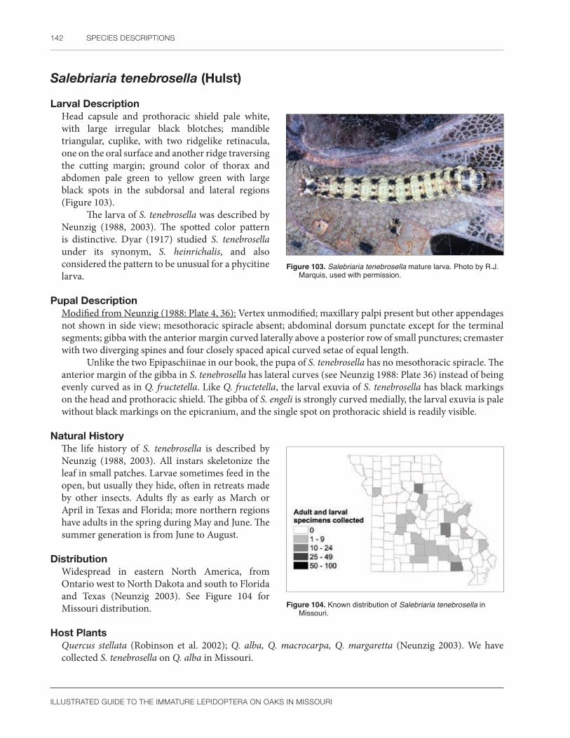
142 SPECIES DESCRIPTIONS
ILLUSTRATED GUIDE TO THE IMMATURE LEPIDOPTERA ON OAKS IN MISSOURI
Salebriaria tenebrosella (Hulst)
Larval DescriptionHead capsule and prothoracic shield pale white, with large irregular black blotches; mandible triangular, cuplike, with two ridgelike retinacula, one on the oral surface and another ridge traversing the cutting margin; ground color of thorax and abdomen pale green to yellow green with large black spots in the subdorsal and lateral regions (Figure 103).
The larva of S. tenebrosella was described by Neunzig (1988, 2003). The spotted color pattern is distinctive. Dyar (1917) studied S. tenebrosella under its synonym, S. heinrichalis, and also considered the pattern to be unusual for a phycitine larva.
Pupal DescriptionModified from Neunzig (1988: Plate 4, 36): Vertex unmodified; maxillary palpi present but other appendages not shown in side view; mesothoracic spiracle absent; abdominal dorsum punctate except for the terminal segments; gibba with the anterior margin curved laterally above a posterior row of small punctures; cremaster with two diverging spines and four closely spaced apical curved setae of equal length.
Unlike the two Epipaschiinae in our book, the pupa of S. tenebrosella has no mesothoracic spiracle. The anterior margin of the gibba in S. tenebrosella has lateral curves (see Neunzig 1988: Plate 36) instead of being evenly curved as in Q. fructetella. Like Q. fructetella, the larval exuvia of S. tenebrosella has black markings on the head and prothoracic shield. The gibba of S. engeli is strongly curved medially, the larval exuvia is pale without black markings on the epicranium, and the single spot on prothoracic shield is readily visible.
Natural HistoryThe life history of S. tenebrosella is described by Neunzig (1988, 2003). All instars skeletonize the leaf in small patches. Larvae sometimes feed in the open, but usually they hide, often in retreats made by other insects. Adults fly as early as March or April in Texas and Florida; more northern regions have adults in the spring during May and June. The summer generation is from June to August.
DistributionWidespread in eastern North America, from Ontario west to North Dakota and south to Florida and Texas (Neunzig 2003). See Figure 104 for Missouri distribution.
Host PlantsQuercus stellata (Robinson et al. 2002); Q. alba, Q. macrocarpa, Q. margaretta (Neunzig 2003). We have collected S. tenebrosella on Q. alba in Missouri.
Figure 103. Salebriaria tenebrosella mature larva. Photo by R.J. Marquis, used with permission.
Figure 104. Known distribution of Salebriaria tenebrosella in Missouri.

PYRALIDAE 143
ILLUSTRATED GUIDE TO THE IMMATURE LEPIDOPTERA ON OAKS IN MISSOURI
Quasisalebria fructetella (Hulst)
Larval DescriptionHead capsule and prothoracic shield with irregular black spots; ground color of body brown, yellow, or yellow-green, the dorsal area speckled with black and paler than the other regions; irregular subdorsal black dashes connect to form a longitudinal broken line; spiracular area white and contrasting (Figure 105).
The body coloration will separate Q. fructetella from both species of Salebriaria included in our study. The mandible of Q. fructetella has two retinacula and is similar to both oak-feeding Salebriaria.
Pupal DescriptionVertex unmodified; pilifers present, labial palpi minutely exposed, shaped like an inverted teardrop; maxillary palpi present; maxillae extend to caudal margin of wings; prothoracic leg half as long as maxillae; prothoracic femur exposed; mesothoracic legs extends to caudal margin of wings; metathoracic legs hidden; mesothoracic spiracle absent; abdominal dorsum punctate; gibba dark, slightly curved anteriorly, above an irregular row of punctures; cremaster a rounded lobe with two lateral spines and four apical setae of equal length.
The pupa of Q. fructetella lacks a mesothoracic spiracle, unlike the Epipaschiinae oak feeders we studied. The anterior margin of the gibba is evenly curved in Q. fructetella, but not as strongly curved as in S. tenebrosella or S. engeli. The larval exuvia has dark markings on the epicranium and prothoracic shield, distinguishing it from S. engeli, which lacks these markings.
Natural HistoryThe life cycle of Q. fructetella is poorly known. We found it on summer foliage as a solitary surface web builder. Neunzig (1988) illustrated a similar feeding habit for Quasisalebria atratella.
DistributionSouth central United States from Missouri to Oklahoma to Texas (Neunzig 2003). See Figure 106 for Missouri distribution.
Host PlantsQuercus (Robinson et al. (2002). We have reared Q. fructetella from Q. alba in Missouri.
CommentsNeunzig (2003) placed this species in Quasisalebria instead of Salebriaria because of differences in adult morphology.
Figure 105. Quasisalebria fructetella mature larva. Photo by R.J. Marquis, used with permission.
Figure 106. Known distribution of Quasisalebria fructetella in Missouri.

144 SPECIES DESCRIPTIONS
ILLUSTRATED GUIDE TO THE IMMATURE LEPIDOPTERA ON OAKS IN MISSOURI
GeometridaeInchworm moths
The family Geometridae contains approximately 23,000 species distributed worldwide (Nieukerken et al. 2011), mostly concentrated in the Neotropical and Oriental Regions (Scoble 1999). Although much taxonomic work is needed on geometrid moths, large monographs exist for several groups. McGuffin (1967, 1972, 1977, 1987, 1988) completed a series of guides to the Geometridae of Canada with keys to the immature stages when possible. Rindge (1961, 1975, 1983, 1986, 1990) and Pitkin (2002) reviewed or revised New World tribes and genera of Ennominae. New World Geometrinae are also well known thanks to the works of Ferguson (1985b) and Pitkin (1996). Wagner et al. (2001), Wagner (2005), and Ferguson (2008) illustrated the larvae of many eastern United States species. Host records for Florida were compiled by Matthews et al. (2014). Porter (1997), Gómez de Aizpúrua (2002a, 2012), and Skou (1986) reviewed the European members of the Geometroidea. McFarland (1988) illustrated many Australian species with an emphasis on biology, rearing, and resting postures. The geometrid fauna of Borneo was partially reviewed by Holloway (1993, 1996, 1997). A world catalogue of Geometridae published by Scoble (1999) included citations to original descriptions and host plants in some cases. For a more complete introduction to literature on the Geometridae, consult Ferguson (1985b), Minet and Scoble (1998), Wagner et al. (2001), and the historical review of geometrid larval studies in Young (2008). Both Abraham et al. (2001) and Sihvonen et al. (2011) produced a molecular phylogeny of geometrid subfamilies. Forum Herbulot (2013) is a Web site devoted to gathering historical and recent information on the Geometridae. We generally follow the higher classification used on that site.
Most geometrid eggs are laid on their side, and thus are of the flat type (called horizontal by Salkeld 1983), defined by having the micropyle lying parallel to the substrate. However, some species have upright eggs, and in a few genera, such as Sterrha and Biston, both types of ova are present (Scoble 1992). In lateral view, geometrid eggs can be described as elliptical, ovoid, or rectangular. Sometimes they are truncated at one end and/or laterally compressed (Syme 1961). McFarland (1988: 224) characterized typical geometrine eggs from Australia as being “lozenge-shaped.” The chorion of most Geometridae is either sculptured, smooth, or rarely granular (Peterson 1962b, 1968). When sculpturing is present, the pattern is often hexagonal without obvious ribbing (Syme 1961: 39, McFarland 1988: 231). In some Ennominae, there is a collar around the micropyle (Salkeld 1983: 150). The pattern of oviposition is highly variable; eggs may be laid singly, in a linear strand, or in a sometimes dense, single layered mass. Some Australian geometrid eggs have a thin chorion and are placed in crevices or other protected locations (McFarland 1988: 231). A few American species lay their eggs on bark (Wagner et al. 2001), but the oviposition site of many North American species has never been documented. Geometrid eggs have a uniform color that is some shade of white, light yellow, beige, cream, orange, or yellow-green. This may change to orange, red, or deep brown before eclosion (Peterson 1962b). For more information on eggs of the Geometridae, consult Doring (1955), Syme (1961), Peterson (1962b, 1968), Salkeld (1983), McFarland (1988), and McGuffin (1988).
No characters exist to distinguish geometrid eggs from other Lepidoptera (McGuffin 1988). However, a few generalizations may be helpful in separating them from other common oak-feeding Lepidoptera in Missouri. Geometrid eggs are rarely spherical (Peterson 1962b) or have a smooth chorion, two features that are characteristic of Notodontidae and Sphingidae (Syme 1961). The typical hemispherical shape so common in noctuoid eggs (Noctuidae, Erebidae) is rarely, if ever, seen in the Geometridae (McFarland 1988: 231). Most noctuids have well-developed longitudinal ribs and cross striations, a pattern uncommon in geometrids (Salkeld 1984: 4). Geometrid eggs are rarely scalelike (Peterson 1968), which separates them from some families of tree-feeding microlepidoptera (Tortricidae, Limacodidae) that are common on oaks.
Geometrid larvae are usually easy to recognize by their looping gait and reduced proleg number. Larvae of the Geometridae typically have prolegs just on A6 and A10 (Stehr 1987), but there are several exceptions to this generalization. Geometrid larvae, assumed to be primitive, have abdominal prolegs on A3-6, with the anterior ones on A3-5 smaller than the others (Common 1990, Minet and Scoble 1998, Wagner et al. 2001). Other geometrids have abdominal prolegs only on A4 to A6 (Common 1990, Staude 2001), having lost the one pair on A3. In North

GEOMETRIDAE 145
ILLUSTRATED GUIDE TO THE IMMATURE LEPIDOPTERA ON OAKS IN MISSOURI
American species of Alsophila and Campaea, there is one vestigial proleg on A5 in addition to the well-developed prolegs of A6 and A10 (Stehr 1987). There is an ennomine genus in California with rudimentary prolegs on A3-5 (Pterotaea) (D. Wagner, pers. comm.). Interestingly, most of these proleg combinations are present in larvae of the Australian Nacophorini where vestigial prolegs are present only on A3-5 or just on A5 (Young 2008).
When prolegs are found only on A6 and A10 as is typical, geometrid larvae are most likely to be confused with noctuid larvae that move in a similar fashion. Noctuid loopers have prolegs of equal size on A5 and A6, whereas North American geometrid larvae lack a proleg on A5, or if one is present, it is smaller than the A6 proleg (Stehr 1987). However, in some African Geometridae, the prolegs of A5 and A6 are equal in size (Staude 2001); thus other characters are needed for recognizing unusual geometrid larvae.
Accurate identification of geometrid larvae with prolegs on A6 and A10 depends on chaetotaxy, the shape of the hypopharyngeal complex, and the relative length of the abdominal segments. Two modifications of the normal lepidopteran setal pattern characterize the Geometroidea (including Geometridae) and Drepanoidea. At least in North America, L3 is below L2 on A6, anterior of its normal position (Stehr 1987: 500, 502). Except for Sematuriidae, most abdominal segments of most species in these two superfamilies have an extra seta cephalad of L3 (Stehr 1987: 500, Minet and Scoble 1998: 301). Knight (1941: 64) noticed this on the first seven abdominal segments of the geometrids he studied and wrote that it was a distinctive characteristic of the family. Following the suggestion of C. Heinrich, Knight (1941) called this extra seta VIa. Later workers agreed and placed this seta in the L group (as LD or L3), whereas others associated it with the SV setae (as SV5 or fourth SV seta) (Singh 1953: 68, Heitzman 1985: 10, Stehr 1987, Minet and Scoble 1998: 312). Young (2008) reviewed the issue and called the seta L4, as did Dugdale (1961: 219), suggesting it is an apomorphy of the Geometridae. It will be necessary to understand the homology of this seta to decide if it is an apomorphy of the Geometridae (Knight 1941, Young 2008), a characteristic of the superfamilies Geometroidea and Drepanoidea (Minet and Scoble 1998: 301), or if it defines a group of geometroids and bombycoids (Fracker 1915: 94, 95).
Geometrid larvae appear superficially very similar, but they are in fact morphologically quite diverse. Geometrid larvae are only rarely covered with secondary setae (Young 2008), but it is common to have extra setae in addition to L4. A seta may be present above or below the spiracle (Stehr 1987: 502). Anywhere from 4 to 20 setae may be present on the proleg of A6, depending on the species of Geometridae (Knight 1941: 64). A short spinneret, not longer than the prementum at the midline, is characteristic of most Geometroidea (Minet and Scoble 1998: 301). Geometrid larvae also appear unique in that the anterior abdominal segments are much longer than the posterior ones (Wagner et al. 2001: 23), and the proleg of A6 projects posteriorly at an acute angle (Common 1990: 362). However, other members of the Geometroidea are poorly known as larvae (Uraniidae, Sematuridae) (see Minet and Scoble 1998). It is not clear which setal pattern, spinneret, or body shape will be seen in rarer members of the Geometroidea. Consult Minet and Scoble (1998) for further information on these taxa. Both the head and anal shield of geometrid larvae can be rounded, triangular, rectangular, or bifid with pointed tips (Minet and Scoble 1998, Singh 1953). The mandible may lack teeth or have a retinaculum. The spinneret can be short and broad or long and pointed, and might be fringed (Singh 1953). Surprisingly, identification of geometrid larvae at the generic level can involve characters not used in other lepidopteran families. Ridges, humps, tubercles (with setae or not), and rarely filaments, adorn the body (Heitzman 1985, Wagner et al. 2001). Small pockets of modified cuticle, called blisters (Heitzman 1985: 13), are unique to the larvae of the Geometridae. The crochets are either biordinal or triordinal, and they are arranged in one or two groups (Stehr 1987). There may be a membranous pad separating the groups of crochets (Singh 1953). The last abdominal segment is modified into two lateral paraprocts and a medial hypoproct (Nakamura 1993, Wagner et al. 2001: 9). Although we do describe the mandible morphology of our oak-feeding species, Heitzman (1985) warned that they are subject to variation and, therefore, must be used with caution. We have especially seen wear, in which the smaller scissorial teeth disappear. The presence of an outer tooth or retinaculum seems more reliable. For more information on larvae of the Geometridae, consult Baker (1972), Evans (1968), Dugdale (1961), Dyar (1899–1904), Ferguson (1985b, 2008), Gómez de Aizpúrua (1987, 1989, 2002c), Heitzman (1985), Knight (1941), McFarland (1988), Porter (1997), Singh (1953, 1957), Sugi (1987), Wagner et al. (2001), and Wagner (2005).

146 SPECIES DESCRIPTIONS
ILLUSTRATED GUIDE TO THE IMMATURE LEPIDOPTERA ON OAKS IN MISSOURI
Although based on Canadian species, the last version of McGuffin’s larval key to subfamilies of Geometridae (McGuffin 1988) is useful for the Missouri oak fauna, especially when combined with the photos in Wagner et al. (2001) or Wagner (2005). Below is a diagnosis for common North American geometrid subfamilies. The Archiearinae (=Brephinae) have prolegs on A3-6 and A10, a very unusual combination for Geometridae. However, correctly identifying this taxon to family is most of the battle because, in at least one case, the larva resembles any one of several green fruitworms in the Noctuidae (see Wagner et al. 2001: 24). The fall cankerworm, Alsophila pometaria, which used to be in the Oenochrominae but is now an Ennominae, is another example of a geometrid with an extra proleg, in this case on A5 (McGuffin 1988). The crochets on A6 are in two groups, and the larva varies in color from green to almost black, often with longitudinal stripes, yellow intersegmental membranes, or a black spot near the spiracle (Ives and Wong 1988, McGuffin 1988, Wagner et al. 2001).
Perhaps the next most distinctive subfamily of the Geometridae is the Geometrinae, which are almost worldwide in distribution and include five recognized tribes in North America (Ferguson 1985b). Larvae of the Geometrinae are relatively sedentary compared to other related subfamilies (McFarland 1988: 226), but they do “quiver from side to side” when disturbed or moving (Wagner et al. 2001), and, at least for the early instars, attach plant material to modified setae on large dorsolateral projections (see bibliography in Comstock 1960, Ferguson 1985b: 14). Typically, geometrine larvae have stemmata 1 and 2 closely spaced; a body with short setae and a granular or beadlike cuticle due to having fine micropapillae; SV4 absent on A1 but present on A3-5; MD1 present on the anterior abdominal segments when the dorsolateral processes are absent; crochets divided into two groups; and CD2 of the anal proleg above a horizontal line drawn through L2 (Ferguson 1985b: 9, 79; McFarland 1988; McGuffin 1988). Knight (1941: 75, Fig. 132) illustrates an extra seta on the thorax for the few species he examined. The North American fauna has three main larval types in the subfamily, each described by Ferguson (1985b: 9, 12, 77, 81, 101, 103). Nemoriini are solitary foliage feeders with a variable number of dorsolateral processes and crochets divided into two groups. Except when very young, they do not attach plant fragments to their body. In addition, the spiracle of A1 is lower than the other abdominal spiracles. A single species of Dichordophorini is similar to Nemoriini but differs in having the crochets in a single row. It feeds on Rhus. A second larval type is found in Synchlorini. They feed on flowers, usually in the Asteraceae, and bear dorsolateral projections with an apical seta and spinules. All instars attach plant fragments to their body with silk. The Hemitheini represents the third larval type in the Geometrinae. This tribe includes foliage and flower feeders and, unlike other North American tribes, occurs on all continents. Flower feeders tend to favor the Asteraceae. The head is bifid, smaller projections occur on the prothorax, dorsolateral projections are absent, the crochets are in two groups, and MD1 is conspicuous on the anterior abdominal segments. Lophochoristini contains a few species in Arizona and southern Florida. A single known example feeds on Krugiodendron and has a larval type more similar to the Hemitheini than to other North American tribes.
In theory, Larentiinae are a distinctive group having a characteristic chaetotaxy. In practice, this subfamily is an excellent example of the morphological confusion facing anyone trying to understand larval Geometridae. Again, authors do not agree on the nomenclature, especially with regard to the L and SV groups on the abdomen. Studying Indian species, Singh (1953: 11) considered that SV3 was lost in Larentiinae and thus called the SV group bisetose. McGuffin (1958) defined the subfamily in Canada by the absence of L4 on A2-5. Later, McGuffin (1964, 1988) wrote that SV4 is absent on all abdominal segments. Wagner et al. (2001) followed McGuffin (1958) by noting that a single L seta (L4) was lost. We feel the best solution is to count setae. Typically, geometrids have 3 L setae, 4 SV setae, and a single V seta on A1, for a total of eight (McGuffin 1946). McGuffin (1958: 12–13) divided the Larentiinae into two groups depending on whether six or seven setae were present in the L, SV, and V groups on that segment. Six setae are present in the Asthenini, Eudilini, and part of the Hydriomenini. The Lobophorini, Operophterini, Eupitheciini, and some Hydriomenini have seven setae in the L, SV, and V groups. Additionally, they are defined by having six or fewer secondary setae on the proleg of A6. The Mesoleucini, Stamnodini, Xanthorhoini, and the remainder of the Hydriomenini also have seven setae on the L, SV, and V groups of A1, but their proleg on A6 has eight or more secondary setae. Thus, if a geometrid larva from Missouri has six or seven setae below the spiracle in the L, SV, or V groups, even if the missing seta cannot be named,

GEOMETRIDAE 147
ILLUSTRATED GUIDE TO THE IMMATURE LEPIDOPTERA ON OAKS IN MISSOURI
Larentiinae is a better possibility than Ennominae (McGuffin 1946: 161). Other important clues for Larentiinae are the lack of abdominal ridges, tubercles or protuberances that are rarely present on the thorax (McGuffin 1958), the ability to tie leaves to form shelters (Wagner et al. 2001), and the presence of secondary setae on the prolegs. Larvae also produce a characteristic damage in that they chew holes in the middle of the leaf instead of starting their feeding on the edge. One species of Larentiinae from the western United States is easily identified; Perizoma is a leaf miner on Primula, a unique feeding habit for the family (Powell and Opler 2009).
Sterrhinae is a small subfamily with diverse feeding habits. Some species are polyphagous, others feed on herbaceous plants or trees, and a few eat dead foliage, including herbarium specimens (Sihvonen and Kaila 2004). Knight (1941) noted that the F1 seta is dorsal of a horizontal line drawn through A2 on the head, but only one species in this subfamily was examined. McGuffin (1988) used the SV4 seta (or called an L seta, see previous discussion) to separate the Sterrhinae from other subfamilies. This seta is absent on A1 but present on A3-5. The Sterrhinae have MD1 inconspicuous on the anterior abdominal segments, lack dorsolateral projections as in the Geometrinae, and generally have SV4 below the level of SV1 on A3. There are two exceptions to the position of the SV setae on A3 that need further discussion, Haematopis and Pleuroprucha. These two genera both have SV4 and SV1 at the same level on A3, and along with Cyclophora, have a bisetose SV group on A1 (McGuffin 1967). Haematopis and Pleuroprucha were described and illustrated by Wagner (2005). Haematopis is a brown larva with a dark, thick arc on each side of the head and a lighter subdorsal area. Pleuroprucha has a rough cuticle, peglike setae, and a characteristic pattern of stripes on the prothoracic shield. Singh (1953), based on Indian species of Sterrhinae, stated that crochets on the larval prolegs are interrupted medially in combination with seta CD2 on the anal proleg below the level of seta SV2. At least for the Scopulini, larvae are typically narrow, sticklike, and rest at an angle of 45 degrees (Sihvonen, 2005).
The final subfamily of Geometridae in Missouri, Ennominae, is the largest. In the eastern United States, except for Campaea, prolegs are present only on A6 and A10. According to McGuffin (1988: 7), part of the subfamily has SV4 present on A1, including for example, the fall cankerworm (Alsophila). Most other Ennominae lack SV4 on A1. These ennomine genera that lack SV4 on A1 have this seta present on A3-5, unlike the Larentiinae that lack SV4 on all abdominal segments. Warts and tubercles may be present in the Ennominae, but there are no broad segmental dorsolateral flangelike projections as in the Geometrinae, and the MD1 seta is inconspicuous on the anterior abdominal segments. SV4 on A3 is normally at the level of SV1, or higher, in Ennominae except for Hesperumia and Orthofidonia. This separates Ennominae from the Sterrhinae (except for Haematopis and Pleuroprucha) that have SV4 below the level of SV1 on A3. Haematopis and Pleuroprucha were described above. Miller (1995) and Wagner et al. (2001) published illustrations of Hesperumia and Orthofidonia. Orthofidonia feeds on viburnum and has one or more of the following: a lime green body, a broad vertical bar on the gena, a white subdorsal stripe, thin lateral cuticle, or a red middorsal line that is especially widened posteriorly. McGuffin (1987: 14) characterized Hesperumia as having a tubercle without setae on A2; this was used as a field recognition character by Miller and Hammond (2003). Miller (1995) and Wagner et al. (2001) also noted this feature in their descriptions. This genus is polyphagous.
Dugdale (1961) compared the morphological larval characteristics of the Ennominae faunas of New Zealand, India, and Canada. His diagnosis of this subfamily for all three regions included the following shared characters: SV group of A3-6 with three to six setae (some North American species have more, SCPC); four to eight annulets on each of the first five abdominal segments; hypoproct developed with a fine point; and the anal shield with SD1 anterior to D1.
McGuffin (1987) and Heitzman (1985) wrote the only two keys that exist for determining tribes of Ennominae in North America. Heitzman (1985: 129) suggests an enlarged concept of Ennomini that includes the Ourapterygini and Cingiliini, because all three taxa have two V setae on A6. This chaetotaxy is not known in other Geometridae. He defined the Boarmiini and related tribes by having SV1 on line with, or posterior to, a line connecting SV3 and V1 on A5. In addition, there are usually five SV setae on A6. Other characters of the Boarmiini mentioned by McGuffin (1987) include a rugose head texture, crochets of the prolegs in one group, and if the color is green, they lack double grey lines. A few tribes have extra setae in the subdorsal or lateral

148 SPECIES DESCRIPTIONS
ILLUSTRATED GUIDE TO THE IMMATURE LEPIDOPTERA ON OAKS IN MISSOURI
regions of the anterior abdominal segments. These are the Nacophorini, Melanolophiini, and the genus Ectropis (McGuffin 1987: 14). Lithinini are unusual because many species are fern feeders; they have V1 anteroventrad of SV1 on A1 (McGuffin 1987); the abdominal L setae are three times the corresponding spiracle height; and seven setae are present on the A6 proleg (Heitzman 1985: 45).
Besides Boarmiini, the Macariini (=Semiothisini), Bistonini, and Angeronini also have the crochets in a single group (McGuffin 1987). In general, Macariini have green, brown, or purplish larvae with longitudinal lines or bars and spots (Ferguson 2008: 17). McGuffin (1972: 16, 1987: 14) noted that green forms may have paired lines, the tribe has four setae on A6, and sometimes the larvae are dimorphic in color. Macaria (in the strict sense) is the largest genus with mostly conifer feeders, especially on Pinaceae, but legumes and Ribes are two other common hosts of the tribe (Ferguson 2008: 49, 179). McGuffin (1972: 16) described the larvae of Macaria (in the broad sense) as having four to eight annulets on the anterior abdominal segments, a smooth to granulose cuticle, and from 20 to 30 crochets on the proleg in one group. However, a few members of the Macariini do have crochets in two groups (Ferguson 2008: 179, McGuffin 1972: 13). Bistonini have four setae and 22 or more crochets on the proleg of A6. Angeronini have five (McGuffin 1987) or six (Heitzman 1985:17) setae on the proleg of A6. The abdominal L setae are less than half the height of the spiracles, and two of three genera in the Angeronini have the D2 seta of A5 on conical tubercles (Heitzman 1985: 22, 23). These larvae feed on trees and are twig mimics (McGuffin 1981: 56).
Ferguson (2008) redefined the Abraxini for North America and moved some genera to the Cassymini that resulted in this Old World tribe now being part of the Nearctic fauna (see suggestion by Holloway 1993: 10, 118). We follow the more traditional definitions of these tribes by McGuffin (1987) until Ferguson’s changes are more carefully studied (see warning by R. W. Hodges in Ferguson 2008: 16), although Pohl et al. (1916) did accept these changes. McGuffin (1987: 14) characterized the Abraxini by their rugose body cuticle and proleg on A6 with four or fewer setae and 10 to 16 crochets in two groups.
Heitzman (1985: 16, 27) treated the Baptini and Caberini as a unit and did not consider the two tribes distinct. The SV setal count on A6 was listed as five or rarely four by Heitzman (1985: 27) compared to four or fewer by McGuffin (1987: 14). SV1 lies slightly anterior to a line passing through SV3 and V1 on A5 (Heitzman 1985: 16, 27). Lomographa is the only genus of Baptini studied by Heitzman (1985) and McGuffin (1981: 16). The ground color is green (Wagner et al. 2001), and the body setae are sometimes thin, short, and colorless (Heitzman 1985: 26). The 10 to 16 crochets on the proleg of A6 are in two groups, and the D setae of A3 are at least twice as long as the corresponding spiracle on that segment (McGuffin 1987: 14). Larvae of the Caberini generally feed on Salix, Populus, Betula, or Ceanothus (McGuffin 1981: 25). The head is smooth, and there are 17 to 20 crochets in two groups on A6 (McGuffin 1987: 14).
Heitzman (1985: 16, 18) characterizes the Anagogini by having V1 posterior to a transverse line through the spiracle on A1, V2, and SV5 but both absent on A5 and A6; the abdominal L setae are equal to or greater than the height of corresponding spiracle; and six or more setae present on the proleg of A6. Ridges and tubercles are often present, and although this group is normally polyphagous (D. Wagner, pers. comm.), one subtribe seems to favor birch (McGuffin 1987: 34).
Several smaller tribes complete this summary of the Ennominae. Wagner et al. (2001: 115) illustrated Epirranthis of the Epirranthini, the lone member of the tribe in North America (Heitzman 1985). It feeds on aspen, does poorly on willow, and is not likely associated with conifers (McGuffin 1981). There are eight SV setae on the A6 proleg (including SV5 which is unusual) (Heitzman 1985: 44). Campaea was placed in the tribe Campaeini, defined by the extra proleg on A5 and the abdominal fringe setae. The larva has been illustrated many times (see species page below), and the example by Sogaard (2009) shows how cryptic the caterpillar can be when resting on a twig. A very common genus is Pero of the Azelinini. Larvae have a cleft head, SV5 on A4 and A5 only, 40–60 crochets in one group, and 15 or more SV setae on the proleg of A6 (McGuffin 1987: 16, Heitzman 1985: 24). This chaetotaxy is unusual (Poole 1987: 2).
The above summary is certainly tentative, and several caveats are worth emphasizing. Some tribes that do not occur in Canada were omitted, and not all specialists may classify the family in the same way.

GEOMETRIDAE 149
ILLUSTRATED GUIDE TO THE IMMATURE LEPIDOPTERA ON OAKS IN MISSOURI
Geometrid larval coloration and morphology are both highly variable (Wagner 2005: 143). The setae may be hard to find (Heitzman 1985: 26, Wagner et al. 2003) or name (see above examples) because the literature is scattered and confusing. If reared under stress, atypical morphology (setae, crochets, protuberances) may be produced (Heitzman 1985: 14). Modifications of the body such as warts, tubercles, swellings, or ridges are not clearly defined for this family and are usually used loosely and interchangeably. For all these reasons, larval identification of geometrids to species or even subfamily is likely to be difficult for at least the short term.
Geometrid caterpillars protect themselves with a wide array of specialized morphological and behavioral defenses. With a few exceptions (McFarland 1988, Wagner et al. 2001), geometrid larvae usually feed exposed and do not live in rolled leaves or silk nests, although they may be found feeding in cones or fruit (D. Wagner, pers. comm.). Young caterpillars may hang from silk threads, which probably allows for wind dispersal (Hill 1987, Sogaard 2009: 37) and perhaps protects them from some invertebrate predators (Wagner et al. 2001: 88). Mature larvae of the Ennominae often anchor themselves to the substrate with strands of silk for support (Wagner et al. 2001: 25). However, geometrid larvae are best known for their excellent camouflage (crypsis or masquerade). This visual effect is largely due to their resting posture, cryptic coloration, and morphology (reduced proleg number, warts and protuberances, etc.). For example, the tubercles of E. clemataria seem to mimic buds of maple trees (Mosher 1917). In some cases (Ferguson 1985b), larvae completely blend into their environment by attaching bits of food plant to their body using modified hooked setae. Once discovered, geometrid larvae have an equally impressive array of escape mechanisms. Depending on the species, they can drop and squirm, drop and curl, drop and run, twist, and even produce an alarm display (McFarland 1988). Heitzman (1982: 113) suggested geometrid larvae have a defensive gland on A2, but no other details are known. The interesting morphology and behavior of larval geometrids remain largely unstudied, and careful study would surely produce more examples of defensive behaviors.
According to Mosher (1917: 34), pupae of the Geometridae can be recognized by their normally hidden labial palpi (except in some Archiearinae, see Holloway 1997: 13). Their maxillae extend at least three-fifths the distance to the caudal margin of the wings, or if the maxillae are shorter, then the spiracles of A3 are hidden, the spiracles of A6 are more ventrad than normal, or there are hooked setae on the terminal segments. Sometimes the prothoracic femur is exposed and the mesothorax lacks a row of pits, although there may be a dorsal and/or lateral furrow between A9 and A10. In addition, Forbes (1948) noted that the maxillary palpi are absent and the three-branched cubital vein of the forewing is often visible under certain lighting conditions (probably oblique illumination) on the pupal wing case. Other important characters of geometrid pupae mentioned by Mosher (1917) include the hidden metathoracic wings on the ventral body surface and often a specialized cremaster. The legs are longer than normal; on the prothorax, they are usually three-quarters the length of the wing while the mesothoracic legs usually reach or come close to the caudal margin of the wings. Most species Mosher studied were about 1–2 cm long, but there is a huge size range in the family worldwide. Geometrid pupae are most likely to be confused with Notodontidae (Forbes 1948) and certain Noctuidae with hidden labial palpi. Pupae of the Geometridae lack a row of pits on the mesothoracic dorsum; these pits are present on notodontid pupae with long maxillae. With one exception (Datana), notodontid pupae lack a dorsal furrow between A9 and A10 that is frequently present in geometrids. Geometrid pupae have only the prothoracic legs extending cephalad of the ventral eye margin. This separates them from similar noctuid pupae, where both the prothoracic and mesothoracic legs extend cephalad of the eye (Mosher 1917). Forbes (1948: 12) stated that “geometrid pupae in America” lack secondary setae or warts. This implies that some tropical species may have these structures although no examples were cited.
As with the larva, McGuffin’s “Guide to the Geometridae of Canada” (McGuffin 1988: 7–8) is the best resource for identifying a geometrid pupa on oak in Missouri to the subfamily level. Unfortunately, it is difficult to characterize geometrid subfamilies in the pupal stage. Existing keys (Mosher 1916a, 1917, Forbes 1945, Evans 1966, Patočka 1994, Patočka and Turčáni 2005) tend to reflect the distribution of critical characters instead of organizing the key phylogenetically or characterizing a subfamily. This does not mean pupal characters are unimportant. For example, they are used in classification of North American Ennominae (Forbes 1948) and

150 SPECIES DESCRIPTIONS
ILLUSTRATED GUIDE TO THE IMMATURE LEPIDOPTERA ON OAKS IN MISSOURI
in the genus Eupithecia (Bolte 1990). The form of the mesothoracic spiracle (callosity), presence or absence of flanges or furrows on the abdominal spiracles, and presence or absence of grooves between A9 and A10 were the most important features. A good illustration of the dorsal and lateral grooves is found in Bolte (1990: Fig. 226). The following summary, mostly from McGuffin (1967, 1972, 1977, 1987, 1988), will provide some clues for identification of pupal Geometridae.
Archiearinae have a brown pupa with a hidden prothoracic femur and two stout cremaster setae (McGuffin 1988: 8, 11). The cuticle of the Archiearinae is heavily sclerotized (Forbes 1948: 13). Ferguson (1985b: 10) described pupae of the Geometrinae as light colored, usually brown, with a rugose and/or tuberculate texture instead of being glossy and smooth. McGuffin (1988: 13) added that the color is often brown or green, the prothoracic femur is hidden, a dorsal groove between A9 and A10 is never present, and the cremaster has four pairs of recurved setae. Ferguson (1985b) noted the terminal pair of setae is the largest. At least one tribe of Sterrhinae has a silken girdle on A3 as in some butterflies (Wagner et al. 2001, Sihvonen and Kaila 2004: 335). Otherwise the subfamily was described as lacking spiracular furrows but having a cremaster with eight hooked setae and both a dorsal and lateral groove between A9 and A10 (Forbes 1948: 118). McGuffin (1967: 9, 1988: 8) added that the cremaster has D1 dorsad of SD1, and the terminal setae (which he named D2) are thicker than the other setae. Some species are truncated at the anterior end and have a pair of hornlike protuberances.
Pupae of the Larentiinae are diverse and difficult to separate from the Ennominae. According to McGuffin (1988: 7, 8), both subfamilies have species with hooked terminal setae/spines on the cremaster. If a dorsal groove between A9 and A10 is absent and there are four cremastral setae of the same size, it is an Ennominae. Larentiinae always have a dorsal groove on A9 and A10. However, if a dorsal groove between A9 and A10 is present, the cremastral setae are of different sizes, and there is no epicranial suture, then it is an Ennominae. Some species of the Larentiinae have an epicranial suture, a furrow on the anterior margin of A5, and a finely toothed dorsal groove between A9 and A10. Another group of Larentiinae lacks a furrow on A5, has a coarsely serrate dorsal groove between A9 and A10 and a cremaster with D1 not dorsad of SD1, and hooked setae of different sizes, with the terminal ones being thinner than the others.
Distinguishing Larentiinae from Ennominae when the terminal setae of the cremaster are straight is somewhat less complicated. If the epicanial suture is absent and there are no cremastral setae, it is an Ennominae. Larentiinae have an epicranial suture and vestigial short setae on the cremaster.
Ennominae pupae were divided into two groups by Forbes (1945: 206, text partially reprinted in 1948 with a table of tribes). The boarmiine series has two terminal spines at the apex and a flange plate on A5; it includes the Abraxini, Macariini (=Semiothisini), Boarmiini, Melanolophiini, and Bistonini. The remaining tribes of Ennominae are in the ennomine series. They have four pairs of hooked setae, usually the prothorax femur hidden, and no flange plate on A5. There are exceptions (Forbes 1945, Ferguson 2008: 46), but the two groupings do offer important clues for identification. The keys by Forbes (1945, 1948), McGuffin (1987), or Evans (1966) can be used to try for an identification to genus, but it is difficult to extract any generalizations past those of Forbes (1945). The pupal key in Forbes (1948) is probably the easiest to use of the available options for the Missouri fauna because it is not necessary to decide if the cremastral setae are hooked or not, the number of antennal segments are not used, and the key is relatively short. He used the term flange groove which appears to be what McGuffin (1967, 1972, 1977, 1987, 1988) called a spiracular furrow.
Several caveats are important to note when trying to study geometrid pupae. A few characters, such as the number of rows of segments on the antenna (McGuffin’s 1988: 7, 8) need clarification and are difficult to interpret. It is usually impossible to evaluate the epicranial suture on a cast pupal exuvia. A better choice is to use an intact pupa if possible. McGuffin (1981: 17, Fig. 160e; 1987: 22) sometimes cited the maxillary palpi in his descriptions as if they were exposed; most other workers considered these structures to be hidden in geometrid pupae (Mosher 1916a, 1917; Forbes 1945; Patočka and Turčáni 2005). Perhaps McGuffin meant to reference the maxillae instead of the palpi. Ferguson (2008: 17) used “foretibia” instead of “prothoracic femur” in his work, but this terminology has never been confirmed. Patočka and Turčáni (2005) noted that in some specimens of Geometridae the pupal abdominal segments contract, which hides key taxonomic features, a problem also noted by Ferguson (2008: 17).

GEOMETRIDAE 151
ILLUSTRATED GUIDE TO THE IMMATURE LEPIDOPTERA ON OAKS IN MISSOURI
The abdomen needs to be pulled apart in these cases. Cremastral setae often break (Ferguson 2008: 17). Passoa (2008: 314) suggested pinning the pupal exuvia instead of placing it in a vial as a way to protect the cremaster. Sometimes the cremaster is said to have terminal hooks, spines, or setae. Technically, setae are socketed but spines are not (Stehr 1987: 719). Calling them D2 implies they are all setae (McGuffin 1988). This needs to be more carefully documented. It may be the dichotomy is D2 setae present versus spines present.
Geometrids have a significant impact on the forest ecosystem as pests, as food for birds and mammals, and as an indicator species due to their tendancy toward habitat specificity (Minet and Scoble 1998). Alsophila, Ennomos, Erannis, Lambdina, Macaria, Operophtera, Paleacrita, and Rheumaptera are common pests of the eastern United States and Canada (Wagner et al. 2001, Asaro and Chamberlin 2015), and in several cases geometrids have been responsible for oak mortality in the eastern United States (Millers et al. 1989). Carter (1984) listed ten geometrid pest species for Europe, including several genera that also are pests in North America (Alsophila, Erannis, and Operophtera). Not all geometrids are pests of trees; in Brazil and Japan they can damage soybeans (see Passoa 1983). Hill (1987) provided a list of geometrid pests of the world.
The geometrid fauna of Missouri was catalogued by Heitzman (1973), and later studies focused on the Larentiinae (Heitzman and Enns 1978). Besides the species described below, we have also encountered the following geometrid species on Missouri oaks: Anacamptodes ephyraria (on Q. alba and Q. velutina), Cleora sublunaria (on Q. alba and Q. velutina), Cyclophora packardi (on Q. alba and Q. velutina), Eupithecia miserulata (on Q. alba and Q. velutina), Hydromena transfigurata (on Q. alba), Hypomecis umbrosaria (on Q. alba and Q. velutina), Lytrosis unitaria (on Q. alba), Nematocampa resistaria (on Q. alba), Nemoria lixarea (on Q. alba), Phigalia titea (on Q. alba), Plagodis fervidara (on Q. alba), and Tetracis cachexiata (on Q. alba). Illustrations of most of these species can be found in Wagner (2005) or Wagner et al. (2011).
Subfamily Ennominae
Besma quercivoraria (Guenée)Oak Besma
Larval Description Modified from Heitzman (1985), McGuffin (1987), Wagner et al. (2001), and Wagner (2005): Mature larva brown to gray to yellow with a brown- and black-spotted head, sometimes with a black bar at the vertex; mandible with no scissorial teeth on cutting margin, but a broad retinaculum and a small outer tooth above the mandibular setae; the mesothorax sometimes enlarged, especially in the subdorsal or lateral regions; metathorax enlarged, especially ventrally; first and third tarsal setae scalelike; body with over ten annulets per segment; A2 and A3 with a lateral protuberance and another one located dorsally or subdorsally, those on A3 much larger, and all protuberances either black, red, or a mixture of these two colors; D setae of A1, A5, A6, and A8 often enlarged, sometimes a lateral protuberance present on A8; body blisters present; posterior abdominal segments (A6-9) often darker than the anterior abdominal segments (Figure 107).
Figure 107. Besma quercivoraria mature larva. Photo by R.J. Marquis, used with permission.

152 SPECIES DESCRIPTIONS
ILLUSTRATED GUIDE TO THE IMMATURE LEPIDOPTERA ON OAKS IN MISSOURI
Ives and Wong (1988), Wagner et al. (2001), and Wagner (2005) photographed the larva of B. quercivoraria. Mature larvae are typically recognized by having a lateral tubercle on the mesothorax, a tubercle on A2 behind the spiracle, and a dorsal tubercle on A6 according to the diagrammatic drawing of this species in Rupert (1944). Both Forbes (1906: 94) and Wagner (2005) called attention to the thoracic swellings for identification of B. quercivoraria. This is one of the few geometrid species that we treat with a retinaculum on the mandible. The setal pattern of A1 and A2 in the first instar, and A3 of the last instar, was figured by McGuffin (1987) for B. quercivoraria. Heitzman (1985: Figs. 53, 124–126) illustrated the tarsal setae and chaetotaxy of A1-3 of the related species, B. endropiaria. Because the color and number of tubercles are so variable, one must consult a series of morphological details by several authors to make a correct identification.
According to McGuffin (1987), the first instar of B. quercivoraria is gray to grayish green, with a light brown head, and sometimes a lateral brown spot on the anterior abdominal segments. The second instar is similar to the first except that the ground color may be brown and the ventral region has light lines. The head color of the third instar larva is variable, either light yellow to brown with darker markings, or light green with a reddish brown lateral spot. Like the previous instars, a lateral spot may be present on the anterior abdominal segments. The fourth instar has a light brown or light green head marked with a herringbone pattern. The body is either green with reddish lateral spots or yellowish with brown markings. The mature larva is described above, but more variation was seen by Sugden (1968) who described two color morphs (green to reddish purple) with intermediate forms in British Colombia. We have seen early instar larvae of B. quercivoraria with dark reddish brown lateral spots (tubercles) on A1-6 similar to the photograph in Wagner (2005: 185). Our specimen also has the spiracle of A8 more dorsad than the one on A7 (Appendix 3, Plate 1).
According to the key in McGuffin (1987), B. quercivoraria may be recognized by the following features: no prolegs present on A3, A4, or A5; SV4 present on A1; no extra setae present in the subdorsal or lateral regions of the anterior abdominal segments; V1 ventrad of SV1 on A1; five or more setae on outer side of the proleg on A6; crochets in two groups; and body with tubercles or ridges except for the subdorsal region of A2. McGuffin (1987) was unable to separate the tribes Anagogini, Cingiliini, and Ourapterygini as defined in his work. As a result, he suggested using each tribal key in turn until a likely genus was obtained. Under the tribe Cingiliini, B. quercivoraria is characterized by the lack of a middorsal tubercle on A3 and A5; body with tubercles bearing setae; and host plants including deciduous trees (not conifers or Ribes).
Larvae of B. quercivoraria are most likely to be confused with Caripeta and Neoterpes. McGuffin (1987: 56) noted that Caripeta feeds on conifers, Neoterpes on Ribes, and Besma on deciduous forest trees. No morphological characters were given to separate these genera. Because records exist for Besma on conifers (see Host Plants) and the host range of Caripeta in the western United States needs study (D. Wagner pers. comm.), host plant data cannot be used for accurate identifications.
Heitzman (1985) enlarged the tribe Ennomini to include the Cingiliini (with Besma) and Ourapterygini. This group was apomorphically defined by the presence of V2 on A6. Using the keys in Heitzman (1985: 16, 32), B. quercivoraria can be identified by the following features: A5 with prolegs absent and SV1 anterior to a line connecting SV3 and V1; A6 with V2 present and proleg with six or more SV setae; protuberances present on one or more segments on the anterior abdominal segments, those on A2 and A3 the largest; thoracic legs with contrasting dark pigmentation; body blisters present; 30 crochets present on A6 with the medial ones being slightly curved and not hooked; D2 of A2 without a protuberance; spiracles black; tarsal setae 1 and 3 scalelike; and L setae of A1-5 about equal to the height of the spiracle. These characters will distinguish B. quercivoraria from related taxa regardless of the host association. Some caution is required when using the color characters. At least in early and middle instars, the thoracic legs may lack dark pigmentation. Destutia is a western United States genus whose species also feed on oak, but the body lacks blisters. Otherwise, the larva is similar to Besma (Heitzman 1985).
No good characters exist to separate B. quercivoraria from B. endropiaria. However, B. endropiaria tends to have a green and reddish head, a red tinted body, relatively small body protuberances, and a preference for maple. In contrast, B. quercivoraria tends to have a blackish head, less red on the body, larger

GEOMETRIDAE 153
ILLUSTRATED GUIDE TO THE IMMATURE LEPIDOPTERA ON OAKS IN MISSOURI
body protuberances, and is more frequently collected on oak (Wagner et al. 2001, Wagner 2005). Other than foodplant preferences, Rupert (1944) used a different set of characters to separate Besma species. In particular, he illustrated a thoracic ridge on B. endropiaria that is lacking on B. quercivoraria. Larvae are best identified only to genus unless the local fauna is well known through rearing.
Pupal DescriptionVertex rugose; labrum u-shaped; labial palpi hidden, except for a minute triangular area; maxillary palpi absent; maxillae extend almost to the caudal margin of the wings; prothoracic femur hidden; mesothoracic leg not reaching lower eye margin; metathoracic legs exposed at their tips; mesothoracic callosity slitlike; abdominal dorsum with dark punctures; dorsal furrow with posterior margin deeply serrated and covered with minute white setae; lateral furrow present; cremaster rugose, with four pairs of curved setae, the apical pair larger than the rest.
McGuffin (1987) used the following characters to identify the pupa of B. quercivoraria: cremaster with four pairs of setae that are not hooked; prothoracic femur hidden; two rows of segments present on the antenna; some areas of A5-A7 rugose dorsally; mesothoracic callosity vestigial; pupa light brown with small brown spots; conspicuous setae absent on the posterior abdominal segments; and hosts include deciduous forest trees, not Ribes.
With several exceptions, it is possible to key the pupa of B. quercivoraria correctly in McGuffin (1987). The cremasteral setae of B. quercivoraria are clearly hooked (McGuffin 1987: Fig. 249t), which will cause the pupa to pass the tribe Cingiliini in McGuffin (1987: 15). We do not understand “two rows of segments” on the antenna, as there appears to be only one row on our specimens. Finally, the setae are conspicuous on the posterior abdominal segments, although they are short. Evans (1966) used color, form of the mesothoracic spiracle, and the hooked setae on the cremaster in his key as well. Besma endropiaria (as Therina) does key more easily in Forbes (1948: 32–34). The important features are cremaster with hooked setae; no flange plate is present; dorsal and lateral grooves are present; prothoracic femur is concealed; pupa is colored brown; the mesothoracic callosity is not well developed; head and thorax lacks a longitudinal keel; and the presence of a series a ridges in front of the [abdominal] spiracles suggesting a furrow.
Natural HistoryThe egg of B. quercivoraria is described by Salkeld (1983). Newly laid eggs are shiny green, but later turn dull red. The micropyle is surrounded by a short rim composed of domed aeropyles. Compared to the related B. endropiaria, the chorion of B. quercivoraria has relatively small polygonal cells.
The life history of B. quercivoraria was briefly described by McGuffin (1987), Wagner et al. (2001), and Wagner (2005). Up to 50 eggs were laid singly, or in groups, on the underside of an oak leaf (McGuffin 1987). Young larvae are green without protuberances, but as they mature these swellings become more prominent. There are two generations in the northeastern United States. Thus, larvae can be found from May to July, and again from September to November. The pupa overwinters (Wagner 2005). Moths fly from May to September (Forbes 1948).
DistributionAcross Canada from Newfoundland to British Columbia and south throughout the eastern United States to Florida and Texas (McGuffin
Figure 108. Known distribution of Besma quercivoraria in Missouri.

154 SPECIES DESCRIPTIONS
ILLUSTRATED GUIDE TO THE IMMATURE LEPIDOPTERA ON OAKS IN MISSOURI
1987, Wagner et al. 2001, Wagner 2005), including Missouri (Heitzman 1985). See Figure 108 for Missouri distribution.
Besma quercivoraria is common throughout its range (Covell 1984, Wagner et al. 2001). Adults were frequently collected during a survey of moths in a New Jersey oak forest (Moulding and Madenjian 1979).
Host PlantsPolyphagous on conifers, Acer, Alnus, Betula, Picea, Populus, Quercus alba, Q. coccinea/rubra, Q. rubra, Q. nigra, Salix, Ulmus, Uvularia (Robinson et al. 2002); Fagus (Wagner 2005).
In Canada, B. quercivoraria is found mainly on white birch and willow (Ives and Wong 1988). We have found this species on Q. alba and Q. stellata in Missouri. Records for B. quercivoraria on conifers, Uvularia, and Picea (Robinson et al. 2002) need to be confirmed, especially because these hosts are more characteristic of Caripeta or Neoterpes according to the key by McGuffin (1987: 56).
CommentsOur species concept of B. quercivoraria follows recent workers (Heitzman 1973, Covell 1984, Handfield 1999, Wagner 2005, Pohl et al. 2016) who consider B. quercivoraria and B. endropiaria to be separate species. McGuffin (1987) considered B. quercivoraria and B. endropiaria to be synonyms. Reared adults from our study sites have the dark black discal dot on the forewing used by Forbes (1948) and Covell (1984) to characterize B. quercivoraria. Both B. quercivoraria and B. endropiaria occur in Missouri (Heitzman 1973).
Campaea perlata (Guenée)Fringed looper
Larval DescriptionModified from Heitzman (1985), McGuffin (1987), Wagner et al. (2001), and Wagner (2005): Mature larva gray to brick red, or less frequently, a shade of olive green; head with dark markings and an oblique line from the vertex to the stemmatal area; mandible with a small outer tooth, three blunt scissorial teeth, and no retinaculum; body usually unmarked, but sometimes one or more faint longitudinal lines are present, the abdominal segments often with the posterior margin pale; vestigial prolegs present on A5; subventral area with a fringe of light colored setae; dorsal tubercles of A8 larger than the others; venter green becoming lighter toward the subventral area (Figure 109a, b).
The larva of C. perlata was recently figured by McGuffin (1981), Heitzman (1985: Fig 108), Ives and Wong (1988), Miller (1995), Rose et al. (1997), Landry et al. (2000), Wagner et al. (2001), Miller and Hammond (2003), Wagner (2005), and Sogaard (2009). McGuffin (1981) illustrated the head of the mature larva and the first instar chaetotaxy. The setal pattern of the third abdominal segment was mapped by Heitzman (1985).
a b
Figure 109. Campaea perlata (a) mature larva, lateral view; (b) mature larva, dorsal view. Photos by R.J. Marquis, used with permission.

GEOMETRIDAE 155
ILLUSTRATED GUIDE TO THE IMMATURE LEPIDOPTERA ON OAKS IN MISSOURI
According to Forbes (1948) and McGuffin (1981), the first instar has a light brown head; yellowish body; dorsal, subdorsal, and lateral brown to reddish-brown longitudinal stripes; and spots below the spiracles of the abdominal segments. Forbes (1948) stated that the spiracular spots were located on the middle abdominal segments, whereas McGuffin (1981) mentioned spots on the anterior portion of the abdomen. A small series of larvae from New Hampshire had spots on A1-5 and, even at this stage, the small proleg on A5 was present (SCPC). Later instars are light green with brown markings and are similar to the mature larva described above (McGuffin 1981).
The larva of C. perlata is easily recognized by the fringe of subventral pale setae and vestigial prolegs on A5 (Wagner et al. 2001). Several other geometrid genera (Brephos and Alsophila) also have larvae with prolegs on A5 (McGuffin 1988); thus, C. perlata is not unique with regard to this character. However, both Brephos and Alsophila lack the row of subventral setae that is present in C. perlata.
In the key to subfamilies of geometrid larvae (McGuffin 1987), C. perlata will key to Ennominae because prolegs are absent on A3 and A4; vestigial prolegs are present on A5; SV4 is absent on A1 but present on A3-5; seta MD1 is minute on the anterior abdominal segments; and SV4 is above level of SV1 on A3. To separate Campaea from other Ennominae genera, the following characters are useful: extra setae absent in the SD and L regions of the anterior abdominal segments; V1 anteroventrad of SV1 on A1; and body color not green with longitudinal lines. However, we have seen some forms of C. perlata that are faintly mottled with broken lines, and the ground color of the body may be shades of green (Wagner 2005). Thus, body color is a character that must be used with caution.
Pupal Description Modified from McGuffin (1987): Vertex with several ridges and large setae; maxillae extend to caudal margin of the wings; labial palpi hidden except for a minute triangular area; prothoracic femur hidden; mesothoracic leg longer than the prothoracic leg; metathoracic legs exposed at their tips; mesothoracic callosity elongate; abdominal dorsum faintly punctate, becoming shagreened toward the posterior segments; posterior margin of dorsal furrow irregularly toothed and covered with minute cream-colored setae; lateral groove present; cremaster with eight curved setae, the ones at the apex thicker than the other six.
McGuffin (1987) used the following characters to identify the pupa of C. perlata: cremaster with eight, not hooked, setae; prothoracic femur hidden; two rows of segments present on the antenna; some areas of A5-7 rugose dorsally; mesothoracic callosity elongate; and epicranial suture absent.
Our pupal exuviae of C. perlata will sometimes key out correctly in McGuffin (1987), but the characters are subtle and difficult. The cremasteral setae of C. perlata are often hooked, which will cause the pupa to pass C. perlata in the key. The two “rows of segments” on the antenna more closely resemble two longitudinal rows of raised dots, but they are obviously in rows. The rugose areas on the dorsum of A5-7 are, at best, extremely faint on some individuals. The punctate and shagreened terminal abdominal segments may be helpful identification characters for C. perlata. Forbes (1948) noted in his key that the cremaster has hooked setae, with the two terminal ones enlarged; no flange plate is present; dorsal and lateral grooves are present; the prothoracic femur is concealed; pupa is colored brown; and the mesothoracic callosity is well developed.
Natural HistoryThe egg of C. perlata is illustrated by Syme (1961: Figs. 61, 309), McGuffin (1981: Fig. 154), and Salkeld (1983: 99). Unlike other Ennominae eggs that are elongated and covered with ridges, the egg of C. perlata is almost circular with faint reticulations (McGuffin 1981). Thus, it is most similar to eggs of the genus Pero (Salkeld 1983). The most unusual modification in the egg of C. perlata involves the cells surrounding the micropyle. Because these cells are faint, the whole area appears smooth, and the rosette is isolated from the surrounding chorion (Salkeld 1983). Recently laid eggs are pale green or yellow, but later turn red (Forbes 1948, Syme 1961). Oviposition usually occurs on the undersides of leaves or other flat surfaces (McGuffin 1981), with eggs oriented either singly or in small clumps, with each egg lined up in a row (Syme 1961).

156 SPECIES DESCRIPTIONS
ILLUSTRATED GUIDE TO THE IMMATURE LEPIDOPTERA ON OAKS IN MISSOURI
McGuffin (1981), Landry et al. (2000), and Wagner et al. (2001) described the life history of C. perlata. Eggs require 11 days to hatch (McGuffin 1981). In New England, larvae of the first generation overwinter as partially grown caterpillars that mature by April. Individuals of the second generation occur from late June to September (Wagner et al. 2001). A similar phenology is reported in Canada, where caterpillars can be collected between May and September (Rose et al. 1997). Larvae rest on leaves or twigs and are 30–40 mm when fully grown (Rose et al. 1997, Wagner et al. 2001). The pupa is formed in a slight cocoon (McGuffin 1981), usually on foliage (Evans 1966), and adults fly from June to September (Landry et al. 2000).
Adults of C. perlata are usually nocturnal, but they become diurnal flyers in arctic North America, where there is 24 hours of daylight during their flight period (McGuffin 1981).
DistributionEastern Canada to Alaska and south throughout the United States reaching Georgia, Arkansas, and Arizona (McGuffin 1981, Wagner et al. 2001), including the Midwest (Ohio) (SCPC) and Missouri. See Figure 110 for Missouri distribution.
Campaea perlata is a common species throughout its range (Covell 1984, Miller 1995, Wagner 2005). Strangely, Heitzman (1973) did not record it from Missouri.
Host PlantsFeeds on over 65 species of trees and woody shrubs, including conifers. The list includes: Abies, Acer, Alnus, Amelanchier, Betula, Corylus, Larix, Picea, Pinus, Populus, Pseudotsuga, Quercus, Rosa, Salix, Shepherdia, Tilia, Thuja, Tsuga, Ulmus (Robinson et al. 2002); Fagus, Fraxinus, Prunus, and Vaccinium (Handfield 1999). We have found C. perlata on Q. alba and Q. velutina in Missouri. Campaea perlata is most commonly found on aspen (Rose et al. 1997), or in the western United States on serviceberry, alder, and hazel (Miller 1995).
CommentsA related European species, C. margaritata, also has fringes of setae in the subventral region (Porter 1997). Patočka and Turčáni (2005: Plate 174) illustrated two species of Campaea with cremasters that are somewhat similar to C. perlata in that they are hooked.
Eutrapela clemataria (J.E. Smith)Purplish-brown looper
Larval DescriptionModified from Heitzman (1985), McGuffin (1987), Wagner et al. (2001), and Wagner (2005): Mature larva gray to brown to purplish brown with a brown patch posterior to the prothoracic spiracle that may extend to the mesothorax; mandible with seven scissorial teeth but no outer tooth or retinaculum; body pinacula may be marked with orange; dorsum of mesothorax with a long thin ridge, this ridge with a dark-pigmented band and usually orange or yellow spots; V1 pinacula of A2 slightly conical; D2 pinacula of A4 greatly enlarged, conspicuous, and at least four times the height of D2 on A5; V2 present on A6; D1 pinacula of A8 slightly enlarged; proleg of A6 with 5 setae on outer surface and one on inner side; crochets in two groups,
Figure 110. Known distribution of Campaea perlata in Missouri.

GEOMETRIDAE 157
ILLUSTRATED GUIDE TO THE IMMATURE LEPIDOPTERA ON OAKS IN MISSOURI
a b
Figure 111. Eutrapela clemataria (a) mid-instar larva; (b) mature larva. Photos by R.J. Marquis, used with permission.
sometimes separate from each other; hypoproct large and paraprocts extend beyond the anal shield (Figure 111a, b).
The larva of E. clemataria is illustrated by Béïque and Bonneau (1979), Heitzman (1985), McGuffin (1987), Ives and Wong (1988), Rose et al. (1997), Landry et al. (2000), Wagner et al. (2001), and Wagner (2005). Heitzman (1985) and McGuffin (1987) described the larva in detail. Typically, it may be recognized by having a dorsal ridge on the mesothorax, large dorsal tubercles on A4, and smaller dorsal tubercles on A8 (Wagner 2005).
According to McGuffin (1987), the first instar has a black head, gray body, and light gray, lateral stripes. The body is black in the second and third instars (see Landry et al. 2000). See also Appendix 3, Plate 1. Also in the third instar, the thorax becomes swollen. Large tubercles on the D2 pinacula of A4 are present in the fourth instar. The ground color of the body also changes in the fourth instar to purplish brown or gray-brown with orange spots. The mature larva (fifth or sixth instar depending on the sex) is described above.
Eutrapela clemataria is most likely to be confused with Prochoerodes lineola (=transversata) because both genera have a swollen thorax, large dorsal tubercle on A4, and a smaller dorsal tubercle on A8. According to Heitzman (1985: 35), E. clemataria has a dark band on the mesothorax that is lacking in P. lineola. In addition, the V1 setae of A2 are slightly conical in E. clemataria but flat in P. lineola (Heitzman 1985: 110). Wagner et al. (2001) noted that Antepione thisoaria and Tetracis are both similar to P. lineola, which implies that these two genera could potentially be confused with E. clemataria. Heitzman (1985) used the size of the tubercles on A5 to separate these taxa. In Antepione and Tetracis the tubercles of A4 and A5 are nearly equal in height, compared to E. clemataria and P. lineola, which have the tubercles of A5 present but clearly smaller than those of A4. Wagner (2005) did not mention a thin middorsal stripe on the last abdominal segments in his description of E. clemataria. This stripe is present in Antepione and Tetracis and in some P. lineola (Wagner 2005: 190, 193).
According to the key in McGuffin (1987), E. clemataria may be recognized by the following features: no prolegs present on A3, A4, or A5; SV4 present on A1; no extra setae present in the subdorsal or lateral regions of the anterior abdominal segments; V1 ventrad of SV1 on A1; five setae on outer side of the proleg on A6; crochets in two groups; and body with tubercles or ridges except for the subdorsal region of A2. McGuffin (1987) was unable to separate the tribes Anagogini, Cingiliini, and Ourapterygini as defined in his work. As a result, he suggested using each tribal key in turn until a likely genus is obtained. Under the tribe Ourapterygini, E. clemataria keys out rapidly due to the presence of a silver crescent-shaped spot anterior to the tubercles of A4, but we have not used this character (see below).
Heitzman (1985) enlarged the tribe Ennomini to include the Cingiliini and Ourapterygini. This group was apomorphically defined by the presence of V2 on A6. Using the keys in Heitzman (1985), E. clemataria can be identified by the following features: prolegs absent and SV1 anterior to a line connecting SV3 and V1 on A5; A6 with six or more SV setae on the proleg and V2 present; dorsal tubercle of A4 enlarged to at least

158 SPECIES DESCRIPTIONS
ILLUSTRATED GUIDE TO THE IMMATURE LEPIDOPTERA ON OAKS IN MISSOURI
four times the height of the dorsal tubercle on A5; mesothorax swollen in the dorsal and lateral regions that includes a dark pigmented band; and mature larva not over 55 mm long. Except for size, Heitzman (1985) was not able to separate the genera Eutrapela and Oxydia (see Comments).
Several authors (USDA 1985, Ives and Wong 1988) note tubercles on A5 and A9 in E. clemataria, but at least in the material we have seen, the tubercles are largest on A4 and A8, and there are no tubercles on A9. The red tubercles on the thorax mentioned by Heitzman and Heitzman (1987) are probably unusual, given that tubercles are normally orange or yellow.
Although McGuffin (1987) called attention to a silver crescent anterior to the tubercles of A4, we have not used this character in our diagnosis because neither our photographs nor those of Wagner et al. (2001) or Wagner (2005) show this feature. The tubercles are bicolored, however, and that is unusual. Perhaps this was the coloration McGuffin (1987) had noticed.
Pupal DescriptionModified from Mosher (1917) and McGuffin (1987): Vertex with three small tubercles; maxillae extend to caudal margin of the wings; labial palpi hidden except for a minute triangular area; prothoracic femur hidden; mesothoracic leg longer than the prothoracic leg; metathoracic legs exposed at their tips; abdominal dorsum rugose and covered with minute tubercles, except for the posterior half of A4 which is shagreened; D setae of A1-8 set in small black pinacula; abdominal spiracles oval with black peritremes, contrasting against the brown cuticle; dorsal furrow finely spined, with prominent round projections present on the posterior margin; lateral groove present; cremaster with four pairs of setae, the basal three pairs hooked and thinner than the slightly curved terminal pair.
Mosher (1917) included E. clemataria in a study of geometrid pupae of Maine. She noted that the cremaster has hooked setae and is not bifurcate; the prothorax lacks lateral projections; a dorsal furrow with prominent projections is present between A9 and A10; the abdomen is densely punctate; the prothoracic femur is hidden; and three small tubercles are present on the vertex. Compared to other geometrid pupae in Maine, E. clemataria is larger (18 to 23 mm) and less heavily sclerotized so that the cuticle “yields to the touch” (Mosher 1917). Our specimens agree with this diagnosis except for the abdominal cuticular texture that is rugose instead of being punctate.
McGuffin (1987) used the following characters to identify the pupa of E. clemataria: cremaster lacks longitudinal ridges on the dorsum, four pairs of cremasteral setae are present in two sizes, all hooked except for the longer, curved terminal pair; dorsal groove present between A9 and A10; epicranial suture absent; prothoracic femur hidden; mesothoracic callosity an elongated ridge or slight thickening; one row of segments present on the antenna; A5-7 rugose; pupa light brown and sometimes with darker markings; conspicuous setae present on the posterior abdominal segments; no dark line running across vertex to the antenna; and length 16 to 20 mm.
Forbes (1948: 33–34) used the following characters to identify E. clemataria in his key to geometrid pupa: cremaster with hooked setae; no flange plate is present; dorsal and lateral grooves are present; prothoracic femur is concealed; pupa is colored brown; the mesothoracic callosity is not well developed; and both the head and thorax have a longitudinal keel.
With two exceptions, the above features can be used to identify E. clemataria. One inconsistency relates to cremaster in McGuffin’s (1987: 15) key to tribes of Ennominae. The figure given by McGuffin (1987: 249d) clearly shows all setae are hooked on the cremaster of E. clemataria, except for the larger curved terminal pair. If these setae are characterized as all hooked when using the pupal key to tribes (Couplet 1), then the pupa will not reach Ourapterygini (Couplets 18–24), which includes Eutrapela. In order to recognize Eutrapela with McGuffin (1987), the larger cremasteral setae must be coded as curved and not hooked, and the smaller hooked setae surrounding the cremaster must be ignored. Another conflict involves the number of rows of segments on the antenna. Eutrapela clemataria is characterized as having two rows of antennal segments in the key to tribes of Ennominae, but only one row in the key to genera of Ourapterygini (McGuffin 1987: p. 15, Couplets 2–17; p.

GEOMETRIDAE 159
ILLUSTRATED GUIDE TO THE IMMATURE LEPIDOPTERA ON OAKS IN MISSOURI
80, Couplet 1). Extreme care is needed when trying to identify E. clemataria in McGuffin (1987). Except for size (16–20 mm in E. clemataria), McGuffin (1987) was unable to separate Eutrapela from Antepione.
Because the vertex and epicranial suture were damaged during adult emergence, we could not evaluate the presence of tubercles on the vertex or the condition of the epicranial suture.
Natural HistoryThe egg of E. clemataria was illustrated by Syme (1961), Peterson (1962b), and Salkeld (1983). When freshly laid they are bright green, later turning reddish brown during incubation (Peterson 1962b). Except for small aeropyles, the chorion is smooth (Salkeld 1983). Seven to ten cells surround the micropyle in several rows (Syme 1961, Salkeld 1983). Up to 491 eggs were laid singly or in a mass during a 6-day period (McGuffin 1987).
Mosher (1917), McGuffin (1987), Landry et al. (2000), and Wagner et al. (2001) described the life history of E. clemataria. Larvae have been found from June to September in Canada (McGuffin 1987) but are most common during July in Maine (Mosher 1917). There are two resting postures (Wagner et al. 2001). The first position is characterized by the legs held tightly against the body. In the second position, the head and legs are pressed together to resemble a fist at the anterior end. Although the leaves of the host are usually eaten, caterpillars feeding on cranberry plants also consume the flower buds (Landry et al. 2000). Larvae mature in 47 days and pupate between two leaves or in a slight cocoon at the soil surface (McGuffin 1987). In Maine, pupation starts in August with adults emerging in the spring of the following year (Mosher 1917). There are commonly two generations from Pennsylvania southward (Wagner et al. 2001). Adults fly from March to October in Missouri.
This species overwinters as an adult or pupa (Heitzman and Heitzman 1987). In Missouri, we have found larvae E. clemataria only on spring (April–May) foliage.
DistributionNewfoundland, Canada, to Minnesota and south to Florida and Texas (Wagner 2005), including the Midwest (Ohio) (SCPC), Kentucky (Covell 1999), and Missouri (Heitzman and Heitzman 1987).
Eutrapela clemataria is common in Missouri (Heitzman and Heitzman 1987) and other parts of its range throughout the eastern United States (Covell 1984; Figure 112). Moths are frequently collected at light traps in an oak forest in New Jersey (Moulding and Madenjian 1979).
Host PlantsMany trees and woody shrubs, including softwoods: Abies, Acer, Betula, Carya, Castanea, Celastrus, Clematis, Cornus, Fraxinus, Juglans, Malus, Picea, Populus, Prunus, Pycnanthemum, Pyrus, Quercus alba, Q. coccinea/rubra, Q. velutina, Q. virginiana, Salix, Sassafras, Tilia, Tsuga, Ulmus, Vaccinium (Robinson et al. 2002); Myrica, Physocarpus, Viburnum (Handfield 1999, 2011); Amelanchier (McGuffin 1987); Liquidambar, Ribes, Sambucus (Wagner et al. 2001); and Smilax, Toxicodendron (Matthews et al. 2014). We have found E. clemataria on Q. alba, Q. coccinea, Q. marilandica, Q. stellata, and Q. velutina in Missouri.
CommentsAccording to Heitzman (1985), E. clemataria is the most common geometrid submitted for identification, probably because of the abundance and relatively large size of the caterpillar.
Figure 112. Known distribution of Eutrapela clemataria in Missouri.

160 SPECIES DESCRIPTIONS
ILLUSTRATED GUIDE TO THE IMMATURE LEPIDOPTERA ON OAKS IN MISSOURI
No larval characters are available to separate Oxydia from E. clemataria (Heitzman 1985). Oxydia is larger (over 70 mm at maturity) and occurs from Florida south throughout the Neotropical Region. Eutrapela clemataria is widespread in the eastern and midwestern United States (Heitzman 1985), where it grows to about 60 mm when mature (Wagner et al. 2001). Due to their similar morphology, Heitzman (1985: 138) suggests that Oxydia should be a junior synonym of Eutrapela.
Eutrapela clemataria was placed in the genus Abbotana by Forbes (1948).
Erannis tiliaria (Harris) Linden looper
Larval Description Modified from Wagner et al. (2001) and Wagner (2005): Mature larva with an unmarked, or slightly reticulated, pale orange head having a rugose texture; the reddish brown dorsum contains up to ten thin, often irregularly fused, black longitudinal stripes alternating with white strips running from the prothorax to A9, the lowest subdorsal stripe usually thicker than the others; mandible with cutting edge smooth and lacking another tooth and retinaculum; body covered with a mixture of blunt or sharp, sometimes recurved, granules; lateral region bright yellow with contrasting black-rimmed spiracles on A1-8; ventral region white to cream (Figure 113).
The larva of E. tiliaria was recently illustrated by Heitzman (1985), McGuffin (1987), Ives and Wong (1988), Wagner et al. (2001), and Wagner (2005). Knight (1941) and McGuffin (1977) describe the mature larva in detail. Other larval descriptions are listed by Tietz (1972). Knight (1941, citing Dyar) and McGuffin (1977) described the various instars of E. tiliaria. First instar larvae are olive to brown-grey with lighter middorsal and lateral stripes. Unlike the later stages, the cuticle is smooth and there are only two setae on the outer side of the prolegs. The second to fifth instars have traces of black dorsal markings, which increase to ten stripes at maturity. Markings may be present or absent in the ventral region.
Although the coloration of E. tiliaria is variable (Mosher 1917), Wagner (2005) noted that the brown dorsum with black stripes and contrasting yellow lateral area are especially distinctive. Sometimes these black stripes vary in intensity and thickness (Rose et al. 1997). In other cases, the dorsum may be all yellow (Forbes 1948) or the ventral regions may be marked with light lines (Knight 1941), but these variations probably refer to early instars. In alcoholic specimens that have lost their color, the presence of a rugose head combined with a mixture of sharp and blunt granules on the body also help distinguish this species (Knight 1941). Other morphological features of E. tiliaria used by McGuffin (1987) in his key include: no prolegs present on A3, A4, or A5; SV4 present on A1; no extra setae present in the subdorsal or lateral regions of the anterior abdominal segments; V1 ventrad of SV1 on A1; proleg of A6 with four setae on outer side and 22 crochets in one group; head rounded without spots; no prominent tubercles or swellings present on the anterior abdominal segments; anal plate rounded or broadly truncate at the posterior end; color pattern of A1 similar to the other anterior abdominal segments; and D1 on A8 not on a prominent tubercle.
Except for a discrepancy in the condition of the crochets, E. tiliaria is easily keyed with McGuffin (1977, 1987). Using the key to tribes (McGuffin 1987: 14), the crochets must be in two groups to reach Bistonini, the
Figure 113. Erannis tiliaria mature larva. Photo by R.J. Marquis, used with permission.

GEOMETRIDAE 161
ILLUSTRATED GUIDE TO THE IMMATURE LEPIDOPTERA ON OAKS IN MISSOURI
tribe containing Erannis. This contrasts with the key to genera in McGuffin (1977: 13), which states that the genus Erannis has crochets in a single group. Our specimens from Missouri have crochets in one group and thus agree with McGuffin (1977).
Pupal DescriptionModified from Mosher (1917) and McGuffin (1987): Vertex rounded; labial palpi hidden or minutely exposed; maxillae extend to caudal margin of the wings; prothoracic femur hidden; mesothoracic leg shorter than the prothoracic leg; metathoracic legs barely exposed at their tips; mesothoracic spiracle adjacent to a large callosity covered with fine hairs; abdominal dorsum of A1-8 punctate; A5 with a spiracular furrow; dorsal groove between A9 and A10 finely serrate; cremaster bifurcate with one, rarely two, pairs of thin spines that are easily broken.
The pupa of E. tiliaria is illustrated by Mosher (1917), Singerland and Crosby (1924), and McGuffin (1977). Mosher (1917) stated that the presence of a bifurcated cremaster, hidden prothoracic femur, dorsal groove between A9 and A10, and mesothoracic callosity would separate E. tiliaria from other Geometridae she studied in Maine. These characters were also used by McGuffin (1977, 1987), who added the following additional features based on his study of the Canadian fauna: prothoracic femur hidden; mesothoracic callosity raised and rounded; A5 with a spiracular furrow; abdominal spiracles not elevated; cremaster stout, with a rounded base; and prothoracic leg extending to the tip of the antenna.
Erannis tiliaria will key to the tribe Bistonini in McGuffin (1987: 15) if the cremasteral setae are characterized as hooked. However, we cannot find hooked setae on any of the several illustrations of cremaster of E. tiliaria given by that author (McGuffin 1977: Fig. 164). McGuffin (1987) did not mention differences in pupal morphology between the species of Erannis, nor were sexual differences mentioned even though the female is wingless. The presence of a spiracular furrow on A5 (McGuffin 1977: Fig. 164e), flange plate on the abdomen, and bifid cremaster (Forbes 1948) is an unusual combination among geometrid species on oak in Missouri.
Natural HistoryThe eggs of E. tiliaria were illustrated by Syme (1961: Figs. 55, 302), McGuffin (1977), and Salkeld (1983), and in color by Rose et al. (1997). Syme (1961) stated the eggs of E. tiliaria are greenish in contrast to Salkeld (1983), who described the freshly laid eggs as orange that later turn dark brown. The color photograph by Rose et al. (1997) and descriptions given by McGuffin (1977) agree with Salkeld (1983). The egg of E. tiliaria is oval, the micropyle is surrounded by six cells, and hexagonal reticulations cover the finely wrinkled chorion (Syme 1961, Salkeld 1983). Except for the genus Hypagyrtis, the egg morphology of E. tiliaria is similar to other members of the tribe Bistonini.
Mosher (1917), Knight (1941), McGuffin (1977), and Wagner et al. (2001) described the life history of E. tiliaria. Each female moth lays up to 500 eggs during October and early November (Knight 1941). They are laid singly or in groups of 3–12 eggs each, usually in crevices formed by the twigs and bark of the host plant (McGuffin 1977), but other oviposition sites may be used (Rose et al. 1997). When the eggs are laid on branches, the higher and more exposed ones are preferred (McGuffin 1977). The eggs overwinter, and young larvae feed on the buds of their host. Each leaf is only partially consumed, then the larva searches for another meal (Rose et al. 1997). Caterpillars of E. tiliaria are found from April to July (Wagner et al. 2001). On oaks in Missouri, larvae are only found on young leaves; they rest in a position in which the head is reflexed back, the prothoracic legs splayed and not in contact with the leaf surface, and the abdomen arched (as shown in Wagner 2005). McGuffin (1977) documented five larval instars, but he noted that Dyar (1903) described a sixth instar in his rearings. Pupation occurs at the soil surface or up to 12 cm underground. Females are wingless, but males have wings and fly to lights (Knight 1941). Rarely, adults of E. tiliaria are migratory (Handfield 2011: 1163). In Missouri there is only a single generation.

162 SPECIES DESCRIPTIONS
ILLUSTRATED GUIDE TO THE IMMATURE LEPIDOPTERA ON OAKS IN MISSOURI
DistributionNewfoundland to central Alberta, Canada, south to Georgia, Missouri, and northeastern Texas, then west to the Rocky Mountains (Utah) (Rindge 1975, McGuffin 1977, Wagner et al. 2001). See Figure 114 for Missouri distribution.
Host PlantsMany broadleaf trees and shrubs, Acer, Alnus, Amelanchier, Betula, Carpinus, Carya, Crataegus, Cornus, Corylus, Fagus, Fraxinus, Gleditsia, Humulus, Juglans, Liriodendron, Malus, Ostrya, Populus, Prunus, Pyrus, Quercus alba, Q. coccinea/rubra, Q. macrocarpa, Q. rubra, Ribes, Salix, Sorbus, Tilia, Ulmus, and Viburnum (Robinson et al. 2002). We have found E. tiliaria on Q. alba, Q. coccinea, Q. marilandica, Q. muehlenbergii, Q. rubra, Q. stellata, and Q. velutina in Missouri.
Maple is the preferred host of both E. tiliaria and E. vancouverensis (McGuffin 1977). Linit et al. (1986) collected E. tiliaria on Q. rubra seedlings in Missouri. Although E. tiliaria was frequently called the lime looper (Wagner et al. 2001), this species is not associated with citrus. Linden trees were sometimes called lime trees in New England (Mosher 1917: 57), hence the common name lime tree spanworm. The accepted Entomological Society of America common name is linden looper (Bosik 1997).
CommentsErannis tiliaria is a sporadic pest, capable of causing serious localized defoliation of short duration (Rose et al. 1997). Fruit trees and hardwoods, especially apple, elm, linden, and maple are occasional attacked in the United States (Forbes 1948) and Canada (Belton 1988, Rose et al. 1997).
Unlike Rindge (1975) and McGuffin (1977), we have treated E. tiliaria and E. vancouverensis as separate species following Pohl et al. (2016). The Missouri population would be E. tiliaria. Erannis vancouverensis is found from British Columbia to Alberta, south to northern California (McGuffin 1977) and Idaho (Rindge 1975). Evans (1973) described the larva of E. vancouverensis. It differs slightly from E. tiliaria in hosts and coloration (McGuffin 1977: 40). The most obvious difference is the presence of reddish spots around the spiracles (Miller 1995, Miller and Hammond 2003), although some variation in color pattern is expected for this species. Rindge (1975) and McGuffin (1977) added Pinus and Tsuga as hosts of E. vancouverensis. These are not recorded hosts of E. tiliaria. Miller and Hammond (2003) noted that E. vancouverensis is very common on broadleaved trees and shrubs such as serviceberry, white oak, and hazel during May and June.
Rindge (1975) suggested that E. tiliaria and the European E. defoliaria might not be separate species. McGuffin (1977) also agreed that these two taxa may be conspecific, although differences exist in wing coloration and genitalia. Recent genetic data for Erannis in the Barcodes of Life Project (BOLD; http://www.boldsystems.org) suggest that E. defoliaria and E. tiliaria are distinct taxa (Ratnasingham and Hebert 2007). Erannis defoliaria was brought into Rye, NY, during the 1930s for breeding experiments (Rindge 1975). This population apparently never escaped or established in the United States. Singerland and Crosby (1924) stated that E. defoliaria was established in British Columbia, but these records are not considered valid (Rindge 1975, McGuffin 1977). The larva of E. defoliaria is illustrated by Porter (1997: Plate 23) and Soria (1987: 127).
Figure 114. Known distribution of Erannis tiliaria in Missouri.

GEOMETRIDAE 163
ILLUSTRATED GUIDE TO THE IMMATURE LEPIDOPTERA ON OAKS IN MISSOURI
Lambdina fervidaria athasaria (Walker)Curved-lined looper
Larval DescriptionModified from Heitzman (1985): Mature larva with a highly variable ground color from dark bluish gray to light gray to dark brown; head reticulated with light spots overlaid by large contrasting irregular black spots; mandible with an outer tooth, retinaculum, and a series of several large, and maybe smaller teeth on the cutting margin; dorsum with a faint white to gray middorsal stripe of irregular thickness, four conspicuous small black spots marking the location of the D pinacula, and a faint thin longitudinal line connecting the D pinacula from segment to segment; subdorsal and lateral area also striped with mixture of black, gray, cream, and/or pink lines or dashes, the subdorsal markings often wider than the others; venter sparsely marked with black (Figure 115a, b).
Ives and Wong (1988) photographed the larva of L. f. athasaria. The tarsal setae and chaetotaxy of A1-3 are illustrated by Heitzman (1985: Figs. 68, 170–172). Early instar larvae of L. f. athasaria are gray with dark gray bands on each of the middle abdominal segments. A similar coloration was reported by Forbes (1948) for L. athasaria and McGuffin (1987) for L. fiscellaria. See also Appendix 3, Plate 1.
Both McGuffin (1987) and Wagner et al. (2001) studied L. athasaria, which may be the same as the Missouri population (see Comments). The related L. fiscellaria was treated by Knight (1941). Several authors have reported a dark color phase, where only cream or greenishwhite dorsal and/or subdorsal lines are present (Forbes 1948, Ives and Wong 1988). This form is uncommon in Missouri at normal population levels. The larva of L. f. athasaria is most likely to be confused with Nepytia and Cingilia because these genera have larvae with a black-spotted head. However, Cingilia caterpillars have two black spots above the abdominal spiracles (Wagner 2005) that separate them from the various color forms of L. f. athasaria. Unlike Nepytia, L. f. athasaria lacks a broad, yellow or white spiracular stripe (Heitzman 1985).
According to the keys in McGuffin (1987: 9, 14, 56), L. f. athasaria may be recognized by the following features: no prolegs present on A3, A4, or A5; SV4 present on A1; no extra setae present in the subdorsal or lateral regions of the anterior abdominal segments; V1 ventrad of SV1 on A1; five or more setae on outer side of the proleg on A6; crochets in two groups, and the body with tubercles or ridges except for the subdorsal region of A2. This generally describes Lambdina caterpillars except that tubercles and ridges are both absent in this genus. McGuffin (1987) was unable to separate the tribes Anagogini, Cingiliini, and Ourapterygini as defined in his work. As a result, he suggested using each tribal key in turn until a likely genus was obtained. Under the tribe Cingiliini, L. f. athasaria is characterized by the lack of a middorsal tubercle on A3 and A5, body without tubercles bearing setae, head with contrasting dark spots, and a dark spot present at the base of the abdominal D setae.
a b
Figure 115. Lambdina fervidaria athasaria (a) dorsal view mature larva; (b) lateral view mature larva. Photos by R.J. Marquis, used with permission.

164 SPECIES DESCRIPTIONS
ILLUSTRATED GUIDE TO THE IMMATURE LEPIDOPTERA ON OAKS IN MISSOURI
Heitzman (1985) enlarged the tribe Ennomini to include the Cingiliini (with Lambdina) and Ourapterygini. This group was apomorphically defined by the presence of V2 on A6. Using the keys in Heitzman (1985: 16, 32), L. f. athasaria can be identified by the following features: A5 with prolegs absent and SV1 anterior to a line connecting SV3 and V1; A6 with V2 present, the proleg with six or more SV setae; and protuberances absent on one or more anterior abdominal segments. Based on the peritreme color, Lambdina will key out in two places. If the peritreme is brown, the larva will have no spinules on the cuticle; no contrasting pigmentation on the thoracic legs; third tarsal seta scalelike; no ridge on D2 of A2; six setae on the proleg of A6; no wide light spiracular line; and body with brown stripes and patches above the abdominal spiracles. If the spiracle is black, the specimen will have no contrasting pigmentation on the thoracic legs; third tarsal seta scalelike; medial crochets on A6 straight, not hooked; no ridges on the thorax or anterior abdomen; no wide light spiracular line; and body with brown stripes and patches above the abdominal spiracles. We have found the spiracle color difficult to evaluate on preserved specimens, but the characters needed to identify Lambdina are very similar in both cases. In live larvae, the spiracles are sometimes clearly black.
The best color characters to recognize the genus Lambdina are the spotted head (Wagner 2005), dark dashes on A1-6 or at least A1 (Heitzman 1985), and a pale abdominal dorsum with the D pinacula pigmented so that they form a square. No good characters exist to separate the species of Lambdina in the larval stage (Wagner 2005), but Maier et al. (2004: 66) suggested the oak-feeding “fervidaria” was lighter in color than the pine-feeding “athasaria.” Larvae found on hosts other than oak are best identified only to genus unless the local fauna is well known through rearing or there is a molecular identification.
Pupal DescriptionVertex without projections; labrum u-shaped; labial palpi hidden except for a small triangular area; maxillary palpi absent; maxillae extend almost to the caudal margin of the wings; prothoracic femur hidden; mesothoracic leg not reaching lower eye margin; metathoracic legs exposed at their tips; mesothoracic callosity absent, the spiracle slitlike; abdominal dorsum punctate; lateral groove present; dorsal furrow with posterior margin deeply serrated and covered with minute white setae; cremaster rugose, with four pairs of curved setae, the apical pair much larger and thicker than the others.
McGuffin (1987) used the following characters to identify the pupa of L. f. athasaria: cremaster with four pairs of hooked setae, the apical pair larger than the rest; dorsal groove present between A9 and A10; epicranial suture absent; prothoracic femur hidden; two rows of segments present on the antenna; some areas of A5-A7 rugose dorsally; mesothoracic callosity vestigial; pupal colored light brown with small brown spots; and conspicuous setae absent on the posterior abdominal segments.
With several exceptions, it is possible to key the pupa of L. f. athasaria correctly in McGuffin (1987). The cremasteral setae of L. f. athasaria are clearly hooked, which will cause the pupa to pass the tribe Cingiliini in McGuffin (1987: 15). It is difficult to find “two rows of segments” on the antenna; there appears to be only one row on our specimens. Finally, the setae are conspicuous on the posterior abdominal segments, although they are short. Evans (1966: 9) also noted the spotted pupa is helpful for identifying L. f. athasaria.
In the key by Forbes (1948: 33–34), Lambdina will key out with Besma under the name Therina. The important characters were listed above (see Besma, Pupal Description).
Natural HistoryThe egg of L. fervidaria is partially described by Dyar (1900a) and Forbes (1948). Freshly laid eggs are dark green, later changing to gray-green and then pink. Salkeld (1983: 134, 150) noted that L. fiscellaria belongs to a group of Ennominae geometrids in which the anterior end of the egg is sloped. Weak chorionic ridges and dome-shaped aeropyles are other characteristics of this species that may apply to L. fervidaria.
The life history of Lambdina fervidaria athasaria is briefly described by Franclemont (1950), McGuffin (1987), and Wagner et al. (2001). Eggs laid during the spring hatch into larvae, which pupate in late June. Adults emerge by July of the same year (Franclemont 1950).
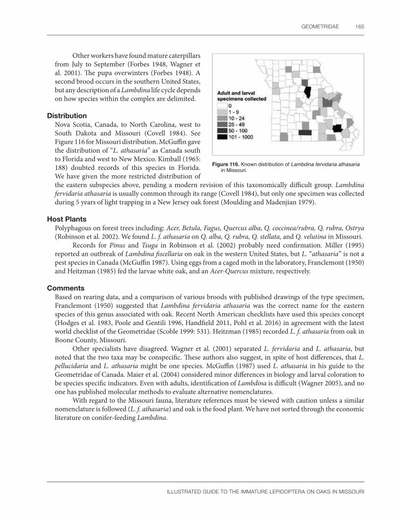
GEOMETRIDAE 165
ILLUSTRATED GUIDE TO THE IMMATURE LEPIDOPTERA ON OAKS IN MISSOURI
Other workers have found mature caterpillars from July to September (Forbes 1948, Wagner et al. 2001). The pupa overwinters (Forbes 1948). A second brood occurs in the southern United States, but any description of a Lambdina life cycle depends on how species within the complex are delimited.
DistributionNova Scotia, Canada, to North Carolina, west to South Dakota and Missouri (Covell 1984). See Figure 116 for Missouri distribution. McGuffin gave the distribution of “L. athasaria” as Canada south to Florida and west to New Mexico. Kimball (1965: 188) doubted records of this species in Florida. We have given the more restricted distribution of the eastern subspecies above, pending a modern revision of this taxonomically difficult group. Lambdina fervidaria athasaria is usually common through its range (Covell 1984), but only one specimen was collected during 5 years of light trapping in a New Jersey oak forest (Moulding and Madenjian 1979).
Host PlantsPolyphagous on forest trees including: Acer, Betula, Fagus, Quercus alba, Q. coccinea/rubra, Q. rubra, Ostrya (Robinson et al. 2002). We found L. f. athasaria on Q. alba, Q. rubra, Q. stellata, and Q. velutina in Missouri.
Records for Pinus and Tsuga in Robinson et al. (2002) probably need confirmation. Miller (1995) reported an outbreak of Lambdina fiscellaria on oak in the western United States, but L. “athasaria” is not a pest species in Canada (McGuffin 1987). Using eggs from a caged moth in the laboratory, Franclemont (1950) and Heitzman (1985) fed the larvae white oak, and an Acer-Quercus mixture, respectively.
CommentsBased on rearing data, and a comparison of various broods with published drawings of the type specimen, Franclemont (1950) suggested that Lambdina fervidaria athasaria was the correct name for the eastern species of this genus associated with oak. Recent North American checklists have used this species concept (Hodges et al. 1983, Poole and Gentili 1996, Handfield 2011, Pohl et al. 2016) in agreement with the latest world checklist of the Geometridae (Scoble 1999: 531). Heitzman (1985) recorded L. f. athasaria from oak in Boone County, Missouri.
Other specialists have disagreed. Wagner et al. (2001) separated L. fervidaria and L. athasaria, but noted that the two taxa may be conspecific. These authors also suggest, in spite of host differences, that L. pellucidaria and L. athasaria might be one species. McGuffin (1987) used L. athasaria in his guide to the Geometridae of Canada. Maier et al. (2004) considered minor differences in biology and larval coloration to be species specific indicators. Even with adults, identification of Lambdina is difficult (Wagner 2005), and no one has published molecular methods to evaluate alternative nomenclatures.
With regard to the Missouri fauna, literature references must be viewed with caution unless a similar nomenclature is followed (L. f. athasaria) and oak is the food plant. We have not sorted through the economic literature on conifer-feeding Lambdina.
Figure 116. Known distribution of Lambdina fervidaria athasaria in Missouri.

166 SPECIES DESCRIPTIONS
ILLUSTRATED GUIDE TO THE IMMATURE LEPIDOPTERA ON OAKS IN MISSOURI
Melanolophia canadaria (Guenée)Canadian melanolophia
Larval DescriptionModified from McGuffin (1944, 1987), and Wagner et al. (2001): Mature larva pale green with yellow or red intersegmental bands, the body often marked by cream to yellow faint subdorsal and spiracular stripes, more rarely with red middorsal and supraspiracular spots; head prognathous, yellowish green to green, sometimes with a yellow stripe; mandible with three large and several smaller scissorial teeth but without an outer tooth or retinaculum; anterior abdominal segments divided by 5–9 annulets; abdominal cuticle with a wavy pink subspiracular flange running parallel to the body, which may be tinted cream, pink, or red; venter green sometimes suffused with reddish to purplish brown (Figure 117).
The larva of M. canadaria is illustrated by Ives and Wong (1988), Rose et al. (1997), Wagner et al. (2001), and Wagner (2005). The example shown by Ives and Wong (1988) is heavily striped, whereas Wagner (2005) photographed a solid green individual. Sometimes the larva is heavily spotted with red (Rose et al. 1997: 227). McGuffin (1977) illustrated the head and chaetotaxy of the mature larva of M. signataria, a related species.
The first instar larva of M. canadaria has a yellow to reddish brown head with a yellow body and gray stripes. Except for the green anterior abdominal segments, the second instar is similar to the first (McGuffin 1977). In the middle instars, the head tends to be more yellow or orange and the body is more conspicuously striped, especially in the subdorsal and lateral regions (Wagner et al. 2001). This suggests that the individual illustrated by Ives and Wong (1988) was not mature. Based on the lack of distinctive features, Wagner (2005) recommended recognizing M. canadaria by first eliminating other more readily identifiable species. However, two helpful field identification characters are the prognathous head and pink body stripe (D. Wagner, pers. comm.).
According to McGuffin (1977, 1987), M. canadaria can be recognized by the following characters: prolegs absent on A3 and A4; SV4 absent on A1 but present on A3-5; MD1 inconspicuous on anterior abdominal segments and abdomen without lateral projections; SV4 either in a horizontal line with, or dorsal to, SV1 on A3; an extra seta present (SDX2) on the anterior abdominal segments; mesothorax not swollen; frons without brown at the apex, and larva colored green, often with lighter lines; host not pine; and thick red spiracular stripe not present. Extra SD setae (XSD1, SDX2) are found on the anterior abdominal segments in the tribe Melanolophiini and Ectropis (Boarmiini) (McGuffin 1977: 114: Fig. 199). These extra setae are easily seen between the D and SD setae, making them a good character that points to M. canadaria and its relatives.
Using morphology, M. canadaria is most likely to be confused with Eufidonia discospilata, but the two species differ in body shape and in their mandibles. The larva of E. discospilata is somewhat flattened (Wagner et al. 2001) with a mandible that has a small tooth on the outer margin above the mandibular setae (McGuffin 1945). In contrast, M. canadaria has a more cylindrical body shape (Wagner et al. 2001) and a mandible that lacks a tooth on the outer margin (McGuffin 1944).
McGuffin (1944) gave characters to separate species of Melanolophia in Canada based on the amount of red in the spiracular line. We have not found this reliable, in agreement with Wagner (2005).
Figure 117. Melanolophia canadaria mature larva. Photo by R.J. Marquis, used with permission.

GEOMETRIDAE 167
ILLUSTRATED GUIDE TO THE IMMATURE LEPIDOPTERA ON OAKS IN MISSOURI
Pupal DescriptionModified from McGuffin (1987): Vertex smooth; labrum u-shaped; maxillae nearly reach the caudal margin of the wings; labial palpi hidden except for a minute triangular area; prothoracic femur hidden; mesothoracic leg does not extend to lower eye margin; metathoracic legs exposed at their tips; mesothoracic callosity rounded and raised; abdominal dorsum more strongly punctate on the anterior, compared to posterior, segments; fifth abdominal segment with deep toothed prespiracular groove; dorsal furrow absent; cremaster bifurcate with two thin spines.
McGuffin (1977, 1987) used the following characters to identify the pupa of M. canadaria: cremaster with hooked setae; prothoracic femur hidden; mesothoracic callosity rounded and raised; anterior edge of spiracular furrow toothed; and posterior margin of spiracular furrow curved away from the spiracle.
Except for the condition of the cremastral spines, our pupal exuviae of M. canadaria have the morphological features listed by McGuffin (1977, 1987). To key a specimen correctly to Melanolophiini in McGuffin (1987), the pupa must have hooked cremastral setae. However, the figure by McGuffin (1977: Fig. 200), as well as our specimens, demonstrate that the cremaster has only two thin straight spines, not hooked setae. Therefore, Couplet 1 of McGuffin (1977: 15) is best ignored. In the key by Forbes (1948: 32–33), the presence of a bifid cremaster; flange plate; weak dorsal furrow; hidden prothoracic femur; prespiracular furrow; and sparsely punctate abdominal texture were used to recognize the genus Melanolophia.
The presence of a wide toothed prespiracular furrow on A5 is very unusual and will help identify the pupa of M. canadaria (see illustration in McGuffin 1977: Fig. 199: l). Unfortunately, like the larva, no characters exist to separate species of Melanolophia.
Natural HistoryThe egg of M. canadaria was illustrated by Syme (1961: Figs. 61, 309), Peterson (1962b: Fig. 16), and Salkeld (1983: 55). There are 6–7 cells surrounding the micropyle (Syme 1961). The chorion is covered with ridges in hexagonal pattern and aeropyles that are larger on the truncated end (Salkeld 1983). Eggs are laid in small groups, sometimes inserted into cracks (Salkeld 1983). They are initially yellow to green, later turning red (McGuffin 1977). All species of Melanolophiini examined by Salkeld (1983) have similar eggs.
McGuffin (1977) and Wagner et al. (2001) described the life history of M. canadaria. Eggs take from 10 to 13 days to hatch (McGuffin 1977). Larvae rest on the undersides of leaves during the day. At night they either hang from short silken threads or feed on foliage (Wagner et al. 2001). There are 5–7 larval instars (McGuffin 1977), and caterpillars can be found from June to October. The pupa overwinters, and there are two generations or more per year (Wagner 2005).
DistributionNova Scotia to Manitoba in Canada and south to Florida and Texas, including Ohio (Rindge 1964), Kentucky (Covell 1999), and Missouri (Heitzman 1973). See Figure 118 for Missouri distribution.
Melanolophia canadaria is common throughout the eastern United States (Covell 1984) and is one of the most frequently seen caterpillars in northeastern United States forests (Wagner et al. 2001).
Host PlantsMany woody trees and shrubs including: Abies, Acer, Alnus, Amelanchier, Betula, Cornus, Epilobium, Fagus, Fraxinus, Jacaranda, Larix, Malus, Myrica, Figure 118. Known distribution of Melanolophia canadaria in
Missouri.

168 SPECIES DESCRIPTIONS
ILLUSTRATED GUIDE TO THE IMMATURE LEPIDOPTERA ON OAKS IN MISSOURI
Pinus, Polygonum, Populus, Prunus, Quercus, Rhus, Salix, Spiraea, Tilia, Tsuga, Ulmus, Vaccinium, Viburnum (Robinson et al. 2002); Corylus (Rindge 1964); Cercis, Juglans (Wagner et al. 2001); Carya, Celtis, Gleditsia, Magnolia, Nyssa, Picea, Platanus, Rhamnus, Rubus, Sassafras (Wagner 2005); Q. alba, and Q. prinus (Butler and Strazanac 2000); found on Q. alba, Q. imbricaria, Q. stellata, and Q. velutina in Missouri.
Larvae of M. canadaria will eat Myrica under laboratory conditions (McGuffin 1977). Some food plant records need confirmation because of potential confusion with M. signataria (Rindge 1964). Examples include Epilobium, Polygonum (Tietz 1972), conifers, and Impatiens (Rindge 1964). The host range of M. canadaria seems to occasionally include herbaceous plants.
CommentsTwo species of Melanolophia occur in Missouri, M. canadaria and M. signataria (Heitzman 1973), both with nearly identical larvae (Wagner 2005). Without a large series of reared adults, we are unable to determine which species is dominant on Missouri oaks. However, it seems likely that M. canadaria is part of our complex because both Wagner et al. (2001) and Covell (1984) consider this species to be very common. The situation is similar in Canada, where M. canadaria is more common than M. signataria (Handfield 1999). Rindge (1964) divided M. canadaria and M. signataria into several subspecies based on adult morphology.
Larvae of M. canadaria once seriously damaged a stand of black ash in Ontario, Canada (Rose et al. 1997). Two South American species of Melanolophia are defoliators of introduced ornamental trees such as Eucalyptus, Pinus, and Cupressus (Rindge 1990: 12).
Plagodis alcoolaria (Guenée)Hollow-spotted plagodis
Larval DescriptionModified from McGuffin (1950, 1987) and Wagner et al. (2001): Mature larva with a mottled gray and light brown head and pale lateral patch that may extend to the prothorax; mandible with several large scissorial teeth and smaller teeth on the cutting margin that may be worn smooth, retinaculum absent, outer tooth minute or absent; mesothorax swollen with a broad hump; body pale green, brown, or tan and may be marked with lateral to subventral cream patches and/or red dorsally; thin red spiracular line sometimes present; spiracles orange or yellow, with a black or brown peritreme, and sometimes surrounded by a lighter patch; A5 with a large dorsal transverse hump; A8 sometimes with dorsal ridge; midventral stripe composed of a series of broken lines; hypoproct and paraproct conspicuous (Figure 119a, b).
a b
Figure 119. Plagodis alcoolaria (a) mid-instar larva; (b) mature larva. Photos by R.J. Marquis, used with permission.

GEOMETRIDAE 169
ILLUSTRATED GUIDE TO THE IMMATURE LEPIDOPTERA ON OAKS IN MISSOURI
The larva of P. alcoolaria is illustrated by Rose et al. (1997), Ives and Wong (1988), Wagner et al. (2001), and Wagner (2005). McGuffin (1950) described the larva in detail. The thoracic and abdominal chaetotaxy of the related species, P. phlogosaria, were given by Heitzman (1985: Figs. 42, 91).
No information is available on the first instar of P. alcoolaria. McGuffin (1987) described the later instars, but with doubt about his identifications. The second instar has a cream colored head with brown markings. The body is pale gray with dark green-gray stripes (McGuffin 1950). The probable third instar has a light grey head marked with brown, a light gray body, gray longitudinal lines, and a green spiracular stripe. The probable fourth instar has the head and body marked as in the third stage except for additional reddish markings on the dorsum and spiracular stripe. Early instars of Plagodis lack the characteristic dorsal hump on A5 (McGuffin 1987, Wagner et al. 2001), the probable fourth instar has humps only on the mesothorax, A2, and A8 (McGuffin 1987). The ultimate and penultimate instars are similar in color (McGuffin 1950). See also Appendix 3, Plate 1.
Plagodis belongs to the tribe Anagogini. This group was characterized by Heitzman (1985) as having V1 posterior to a vertical line drawn through the spiracle of A1; six or more setae present on A6 proleg, and V2 and SV5 absent on the abdomen. McGuffin (1987) also listed characters to help recognize the Anagogini. They included: prolegs absent on A3 and A4; SV4 present on A1; no extra setae present in the SD or L region of the anterior abdominal segments; V1 ventrad of SV1 on A1; crochets on proleg of A6 in two groups; five or more setae on each proleg; and A2 without a ridge or hump lacking setae, although ridges are present elsewhere on the body. Unfortunately, McGuffin (1987) was unable to separate members of the tribes Anagogini, Cingiliini, and Ourapterygini. Instead, he suggested that it was easier to learn a few key characters for each genus instead of trying to use a “cumbersome key that might not work” for these tribes (McGuffin 1987: 14).
Plagodis alcoolaria is most likely to be confused with Selenia and Probole because all three genera share a hump on A5 that bears the D2 seta (McGuffin 1987). However, only Plagodis has SV4 present on A1. This seta is lacking in the two related genera mentioned above (McGuffin 1987: 34). In addition, Plagodis alcoolaria has a dorsal hump on the mesothorax that is lacking in Plagodis pulveraria (previously Anagoga occiduaria) (McGuffin 1950: 210). No good larval characters exist to separate all species of Plagodis (see Comments), although the diagnoses and images in Wagner (2005) can be used to make tentative identifications. McGuffin (1987) mentioned that a dorsal ridge was often present on A8 in Plagodis. This ridge sometimes occurs on P. alcoolaria as well.
Pupal DescriptionModified from McGuffin (1987): Vertex smooth; labrum rectangular; maxillae extend to caudal margin of the wings; labial palpi hidden except for a small triangular area; prothoracic femur hidden; mesothoracic leg shorter than the prothoracic leg because it does not extend to lower eye margin; metathoracic legs exposed at their tips; mesothoracic callosity elliptical; abdominal dorsum punctate on the anterior half; dorsal furrow almost absent, at most a thin suture without teeth; cremaster a slight hump, with four pairs of setae, the three basal pairs thinner than the slightly curved terminal pair.
McGuffin (1987) used the following characters to identify the pupa of P. alcoolaria: cremaster with the lateral margins strongly curved and four pairs of hooked setae in two sizes; epicranial suture absent; prothoracic femur hidden; mesothoracic callosity raised and elliptical; two rows of segments present on the antenna; A5-A7 rugose dorsally; pupa brown; conspicuous setae absent on the posterior abdominal segments; dorsal groove not toothed; and pupal face smooth.
We have found it difficult to key our pupal exuviae of P. alcoolaria correctly using McGuffin (1987). One inconsistency relates to cremastral setae in the key to tribes of Ennominae. The figure given by McGuffin (1987: 249s) clearly shows all setae are hooked on the cremaster of Plagodis. However, if these setae are characterized as hooked when using the key (Couplet 1), then the route will go past Anagogini (Couplet 15), which includes Plagodis. Another conflict involves the number of rows of segments on the antenna and the pupal dorsal texture. With the punctate dorsal surface, and apparent single row of segments on the antenna, P. alcoolaria best fits characters of the tribe Angeronini (Couplet 11). The “face” is not smooth.

170 SPECIES DESCRIPTIONS
ILLUSTRATED GUIDE TO THE IMMATURE LEPIDOPTERA ON OAKS IN MISSOURI
In the key by Forbes (1948: 32–34), the following characters were used to recognize the genus Plagodis: the presence of hooked setae on the cremaster, with the two terminal pairs of setae equal in length; no flange plate; absence of dorsal and lateral grooves; maxillae extending to caudal margin of the wings; and a well developed callosity. Because the vertex and epicranial suture were damaged during adult emergence, we could not evaluate the condition of the epicranial suture.
As with the larvae, no good characters exist to separate the species of Plagodis as pupae. We only examined two pupae, and although they are associated with adults that fit the P. acoolaria phenotype well (see Comments), they are slightly different from each other and the description by McGuffin (1987). Pupal morphology of this species seems to vary in color and texture.
Natural HistoryThe egg of P. alcoolaria changes color three times during incubation. Newly hatched eggs are yellow, then later orange, and finally become spotted with red. They are laid along the leaf edge (Rupert 1949, Wagner 2005) or petiole, either singly, or in a clump containing up to 20 eggs (McGuffin 1987). Both Peterson (1962b) and Salkeld (1983) described the eggs of several Plagodis species, but did not include P. alcoolaria. Four Canadian species of Plagodis have eggs that are bluntly elliptical, with one end wider than the other and a slightly compressed lateral profile. Unlike other Ennominae, the eggs of Plagodis lack obvious ridges (Salkeld 1983).
Rupert (1949), McGuffin (1987), and Wagner et al. (2001) briefly described the life history of P. alcoolaria: The larva rests with the head withdrawn in the thorax (Rupert 1949). Caterpillars can be found in June and July, and again from August to October. The pupa overwinters (Wagner et al. 2001). Moths occur from April to August (Heitzman 1973) but are most common from May to July (McGuffin 1987).
DistributionSouthern Canada south to Georgia and Texas (Wagner et al. 2001), including Kentucky (Covell 1999) and Missouri (Heitzman 1973). See Figure 120 for Missouri distribution.
Host PlantsMany trees and woody shrubs: Acer, Alnus, Betula, Castanea, Fagus, Populus, Quercus alba, Q. rubra, Salix, Tilia, Viburnum (Robinson et al. 2002); found on Q. stellata and Q. velutina in Missouri.
Rupert (1949) thought oak is probably the food plant of P. alcoolaria under natural conditions. He found middle instar larvae did poorly after being switched from oak to beech in the laboratory.
CommentsLarge numbers of P. alcoolaria have been associated with outbreaks of the saddled prominent, Cecrita guttivitta, in Ontario, Canada (Rose et al. 1997). Although identification of the immature stages is tentative in this genus, we have included P. alcoolaria in our book because reared adults match the wing pattern (slanted postmedial line and hollow discal dot) of this species as defined by Forbes (1948: 100). Plagodis fervidaria has also been collected in Missouri in smaller numbers.
Figure 120. Known distribution of Plagodis alcoolaria in Missouri.
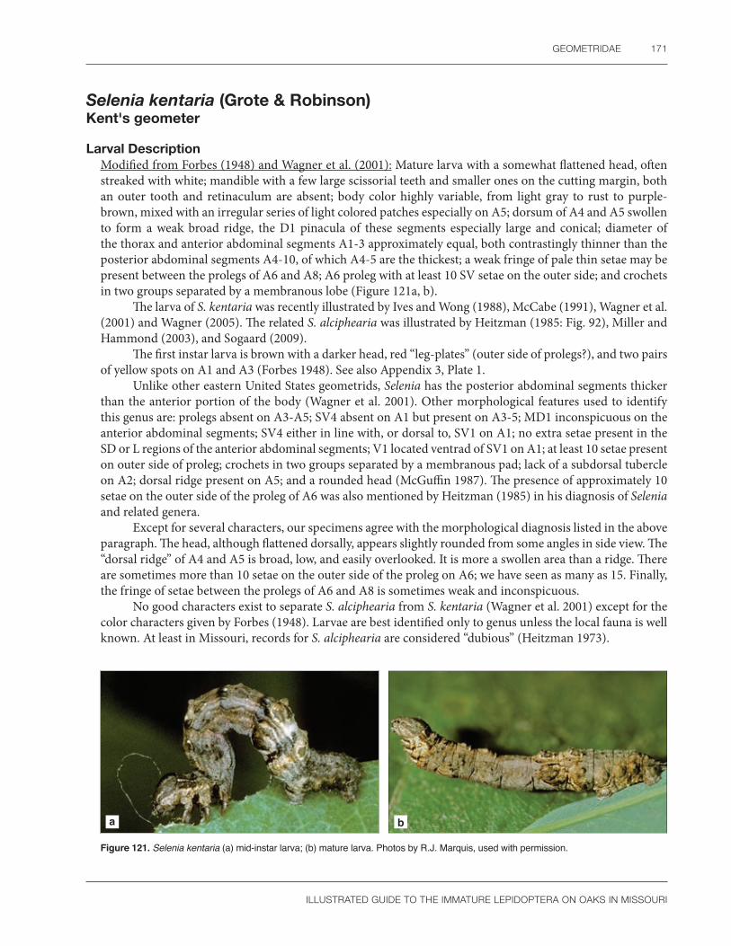
GEOMETRIDAE 171
ILLUSTRATED GUIDE TO THE IMMATURE LEPIDOPTERA ON OAKS IN MISSOURI
Selenia kentaria (Grote & Robinson)Kent's geometer
Larval DescriptionModified from Forbes (1948) and Wagner et al. (2001): Mature larva with a somewhat flattened head, often streaked with white; mandible with a few large scissorial teeth and smaller ones on the cutting margin, both an outer tooth and retinaculum are absent; body color highly variable, from light gray to rust to purple-brown, mixed with an irregular series of light colored patches especially on A5; dorsum of A4 and A5 swollen to form a weak broad ridge, the D1 pinacula of these segments especially large and conical; diameter of the thorax and anterior abdominal segments A1-3 approximately equal, both contrastingly thinner than the posterior abdominal segments A4-10, of which A4-5 are the thickest; a weak fringe of pale thin setae may be present between the prolegs of A6 and A8; A6 proleg with at least 10 SV setae on the outer side; and crochets in two groups separated by a membranous lobe (Figure 121a, b).
The larva of S. kentaria was recently illustrated by Ives and Wong (1988), McCabe (1991), Wagner et al. (2001) and Wagner (2005). The related S. alciphearia was illustrated by Heitzman (1985: Fig. 92), Miller and Hammond (2003), and Sogaard (2009).
The first instar larva is brown with a darker head, red “leg-plates” (outer side of prolegs?), and two pairs of yellow spots on A1 and A3 (Forbes 1948). See also Appendix 3, Plate 1.
Unlike other eastern United States geometrids, Selenia has the posterior abdominal segments thicker than the anterior portion of the body (Wagner et al. 2001). Other morphological features used to identify this genus are: prolegs absent on A3-A5; SV4 absent on A1 but present on A3-5; MD1 inconspicuous on the anterior abdominal segments; SV4 either in line with, or dorsal to, SV1 on A1; no extra setae present in the SD or L regions of the anterior abdominal segments; V1 located ventrad of SV1 on A1; at least 10 setae present on outer side of proleg; crochets in two groups separated by a membranous pad; lack of a subdorsal tubercle on A2; dorsal ridge present on A5; and a rounded head (McGuffin 1987). The presence of approximately 10 setae on the outer side of the proleg of A6 was also mentioned by Heitzman (1985) in his diagnosis of Selenia and related genera.
Except for several characters, our specimens agree with the morphological diagnosis listed in the above paragraph. The head, although flattened dorsally, appears slightly rounded from some angles in side view. The “dorsal ridge” of A4 and A5 is broad, low, and easily overlooked. It is more a swollen area than a ridge. There are sometimes more than 10 setae on the outer side of the proleg on A6; we have seen as many as 15. Finally, the fringe of setae between the prolegs of A6 and A8 is sometimes weak and inconspicuous.
No good characters exist to separate S. alciphearia from S. kentaria (Wagner et al. 2001) except for the color characters given by Forbes (1948). Larvae are best identified only to genus unless the local fauna is well known. At least in Missouri, records for S. alciphearia are considered “dubious” (Heitzman 1973).
a b
Figure 121. Selenia kentaria (a) mid-instar larva; (b) mature larva. Photos by R.J. Marquis, used with permission.

172 SPECIES DESCRIPTIONS
ILLUSTRATED GUIDE TO THE IMMATURE LEPIDOPTERA ON OAKS IN MISSOURI
Pupal DescriptionFront bulging outward; labrum trapezoidal; maxillae almost extend to caudal margin of the wings; labial palpi hidden except for a minute triangular area; prothoracic femur hidden; mesothoracic leg not reaching lower eye margin; metathoracic legs exposed at their tips; mesothoracic callosity elongated and covered with pale cream setae; abdominal dorsum smooth (especially on the anterior segments) to very faintly punctate or ridged; posterior margin of dorsal furrow irregularly toothed and covered with minute cream-colored setae; lateral groove present; cremaster rugose, with apparently eight curved setae, but the exact number is not clear in our two examples.
McGuffin (1987) used the following characters to identify the pupa of Selenia: cremaster with four pairs of setae that are not hooked; prothoracic femur hidden; two rows of segments present on the antenna; some areas of A5-7 rugose dorsally; mesothoracic callosity elliptical; pupa brown in color; conspicuous setae absent on the posterior abdominal segments; face of pupa smooth; pupa rounded; one pair of cremasteral setae larger than the rest; and dorsal groove toothed.
Identification of pupal Selenia is difficult at all taxonomic levels. No characters exist to separate Anagogini (with Selenia) from all Cingiliini, Selenia from Metarranthis, or S. alciphearia from S. kentaria (McGuffin 1987: 15, 35). Moreover, even with the incomplete resolution of the available keys, our pupal exuvia of S. kentaria will not key correctly. The cremasteral setae of S. kentaria are clearly hooked, which will cause the pupa to pass the tribe Anagogini in McGuffin (1987: 15). It is difficult to find “two rows of segments” on the antenna, a situation further complicated by the faint segmentation of the antenna. Rugose areas are present on the dorsum of the anterior abdominal segments, but they are faint. Nevertheless, the presence of a slight frontal tubercle, relatively smooth abdominal dorsum, and pubescent mesothoracic callosity are somewhat unusual among Missouri oak Geometridae. Evans (1966) used the vertex, smooth texture of A1, and size (9–16 mm) to define S. alciphearia in his key to British Columbia geometrids. The key by Forbes (1948: 32–33) used the following characters to recognize Selenia: cremaster with hooked setae, the four terminal pairs equal in length; flange plate is absent; dorsal and lateral grooves are both present; prothoracic femur is concealed; the pupa is cylindrical and brown; and spiracular furrows are absent on A5.
Natural HistoryThe egg of S. kentaria is elliptical with one end wider than the other. It is green when first laid, but later changes from red to black (Forbes 1948).
McGuffin (1987) and Wagner et al. (2001) briefly described the life history of S. kentaria. Larvae occur from June to July, but there is a partial second generation from August to October as well. When at rest, larvae hold their metathoracic legs perpendicular to the body (Wagner et al. 2001), sometimes with the head bent upwards (McCabe 1991). The pupa overwinters, and the moths fly from May to July in Canada (McGuffin 1987).
DistributionSaskatchewan to Nova Scotia, Canada, and south throughout the eastern United States to Alabama, Mississippi, and Arkansas (McGuffin 1987, Wagner et al. 2001), including Missouri (Heitzman 1973). See Figure 122 for Missouri distribution.
Although frequently collected in Missouri (R.J. Marquis, pers. obs.), Selenia kentaria is considered an uncommon species throughout its range (Wagner et al. 2001).
Figure 122. Known distribution of Selenia kentaria in Missouri.

GEOMETRIDAE 173
ILLUSTRATED GUIDE TO THE IMMATURE LEPIDOPTERA ON OAKS IN MISSOURI
Host PlantsAcer, Betula, Fagus, Populus, Quercus, Tilia, and Ulmus (Robinson et al. 2002); and Juglans (Wagner 2005). We have found S. kentaria on Q. alba, Q. rubra, and Q. velutina in Missouri.
CommentsOur larval identifications for this book were based on reared adults.
Phigalia strigataria (Minot)Small phigalia
Larval DescriptionModified from Butler (1986), Wagner et al. (2001), and Wagner (2005): head weakly bifid; mandible with a few large scissorial teeth and smaller ones on the cutting margin, both an outer tooth and retinaculum absent; body color greatly variable, almost all yellow streaked with black, or a mixture of orange and either black, yellow, brown or gray; spiracular stripe solid or broken into patches; body cuticle covered with fine setae; A2 and A3 with D2 and L1 set on conical pinacula; A8 with D1 on a conical pinaculum; A6 with five to six SV setae on the outer side; crochets in a single band (Figure 123).
The larva of P. strigataria was described by Butler (1986) and recently illustrated by Sogaard (2009), Wagner et al. (2001), and Wagner (2005). Several larval color forms were described by Butler (1986). The covering of fine setae on the cuticle and conical D pinacula on A2, A3, and A8 help separate the genus Phigalia from other oak-feeding ennomine geometrids in Missouri. However, larvae of P. denticulata are extremely similar to P. strigataria (Wagner 2005), which complicates species identification of these two species in most of the eastern United States. On the other hand, Butler (1986) noted that P. titea and P. strigataria have a completely different larval coloration and are easily distinguished (see photographs in Wagner 2005).
In rare cases, the D pinacula of P. strigataria may be flat. Gilligan and Passoa (2014) observed similar variation in pinacula height on some Heliothinae (Noctuidae). Individuals of P. strigataria with flat pinacula can still be recognized by their cuticle and the characters in McGuffin (1977: 12, 13). In particular, there are no extra setae on the anterior abdominal segments, A2 and A3 are somewhat swollen, SV4 is present on A2, the proleg of A6 has six or less setae with crochets in one group, and the D2 setae of A2 and A8 are on prominent tubercles.
Large D pinacula on A2, A3, and A8 are also found on P. plumogeraria (=plumigeraria) from the western United States (Miller and Hammond 2003). The similar chaetotaxy of P. denticulata, P. strigataria, and P. plumogeraria supports the placement of these taxa in group II of Phigalia as defined by Rindge (1975).
Pupal DescriptionModified from Butler (1986): Body shagreened except for abdominal dorsum; vertex rounded; maxillae extend to caudal margin of the wings; maxillary and labial palpi hidden; prothoracic femur hidden; mesothoracic leg thin, much shorter than prothoracic leg, with the apex extending well below the lower eye margin; metathoracic legs hidden; mesothoracic callosity an oval slit; abdominal dorsum punctate; spiracular furrow
Figure 123. Phigalia strigataria mature larva. Photo by R.J. Marquis, used with permission.

174 SPECIES DESCRIPTIONS
ILLUSTRATED GUIDE TO THE IMMATURE LEPIDOPTERA ON OAKS IN MISSOURI
of A5 forms a deep toothed depression; dorsal and lateral grooves absent; cremaster a long thin rod with two spines at the apex.
The pupa of P. strigataria is partially illustrated by Butler (1986) and is very similar to the illustrations of P. titea and P. plumogeraria in McGuffin (1977: Plates 157, 159). The hidden prothoracic femur, mesothoracic leg thinner than the antenna, poorly developed mesothoracic callosity, toothed spiracular furrow on A5, and bifid cremaster are all unusual (McGuffin 1977: 14, 15). The presence of fine setae covering the larval exuvia is a good clue for identifying this genus.
The species of Phigalia may be separated by using McGuffin (1977) and Butler (1986). The spiracular furrow on A5 is not toothed anteriorly in P. plumogeraria, unlike both P. titea and P. strigataria that have well developed teeth on the anterior margin (McGuffin 1977: 27). The pupa of P. strigataria has a smooth frontoclypeal area and a rugose vertex. The vertex is smooth in P. titea and the frontoclypeal area is rugose (Butler 1986). We do not know if the female pupa is different from the male. Forbes (1948) did not include Phigalia in his key to geometrid pupae.
Natural HistoryAspects of the life history of P. strigataria are discussed by Rindge (1975), Butler (1986), and Wagner (2005). Eggs were laid on loose bark or dead twigs. Newly hatched larvae dispersed by ballooning on silk threads. There were five larval instars (Butler 1986). Larvae will starve if offered old tough leaves (Wagner 2005). Adults fly from December to March in the southern United States and from March into June in northern localities (Rindge 1975). The females have underdeveloped wings (Rindge 1975), and there is one generation per year (Wagner 2005). We have found larvae only during April and May in Missouri.
DistributionQuebec to Ontario in Canada and south to northern Florida, then west to the eastern edge of North Dakota and Texas (Rindge 1975), including Ohio (SCPC), Kentucky (Covell 1999), and Missouri (Heitzman 1973). See Figure 124 for Missouri distribution.
Phigalia strigataria is common throughout eastern North America (Covell 1984) but highly variable in yearly abundance in Missouri. It is not widespread in Canada (McGuffin 1977).
Host PlantsPolyphagous on forest trees and shrubs, Quercus coccinea/rubra (Robinson et al. 2002); Q. alba, Q. prinus, Q. stellata, Q. velutina (Butler 1986); Castanea, Liquidambar, and Salix (Wagner 2005).
Butler (1986) listed 32 species of woody plants eaten by of P. strigataria. She noted that oaks (Quercus), hickories (Carya), and hackberry were the preferred hosts. We have found P. strigataria on Q. alba, Q. coccinea, Q. imbricaria, Q. macrocarpa, Q. palustris, Q. rubra, Q. stellata, and Q. velutina in Missouri.
CommentsNot all authors agree on the generic placement or spelling of the species included in Phigalia. We conservatively use P. strigataria following Rindge (1975) and Pohl et al. (2016). We had difficulty rearing adults, so identification of this species was confirmed by DNA barcoding.
Figure 124. Known distribution of Phigalia strigataria in Missouri.

GEOMETRIDAE 175
ILLUSTRATED GUIDE TO THE IMMATURE LEPIDOPTERA ON OAKS IN MISSOURI
Ectropis crepuscularia (Denis & Schiffermüller)Saddleback looper
Larval DescriptionModified from McGuffin (1977) and Wagner et al. (2001): Stout caterpillar highly variable in color from golden yellow to gray to dark brown or black, often with a dark brown, v-shaped mark in the adfrontal area or apex of the frons; mandible with several large scissorial teeth and a few smaller ones on the cutting margin; mesothorax swollen; crochets of A6 in one group; prolegs of A6 often lighter in color than the rest of the body, with four setae on the outer side; and usually a low ridge edged with black on A8 (Figure 125a, b).
The larva of E. crepuscularia is described by Knight (1941) and illustrated in color by Béïque and Bonneau (1979), Sugi (1987), Porter (1997), Wagner et al. (2001), Gómez de Aizpúrua (2002c), and Wagner (2005). Knight (1941:185) and McGuffin (1977: 12, Fig. 190a) diagnosed the larva of E. crepuscularia by having a dark brown adfrontal area, a swollen mesothorax, and especially the extra seta (SDX1) present on the anterior abdominal segments. Wagner (2005) called attention to a diagonal marking on the dorsum A8. Although frequently present, this marking is absent on some color forms in Europe (Gómez de Aizpúrua 2002c). A saddlelike marking may be present on A4 (see Béïque and Bonneau 1979), or there may be black lines below the spiracles (Wagner 2005). Early instars may have subdorsal dashes. See also Appendix 3, Plate 1.
Pupal DescriptionModified from McGuffin (1977): Vertex with fine ridges; labial palpi minutely exposed; maxillae extend to the caudal margin of wings; prothoracic leg longer than mesothoracic leg; prothoracic femur exposed; mesothoracic leg does not reach lower eye margin; metathoracic legs exposed at the tips; mesothoracic callosity a small oval tubercle; abdominal dorsum punctate; spiracular furrow of A5 an oval pit; dorsal groove present, sometimes finely dentate; cremaster with a broad base ending in two diverging straight spines.
The pupa of E. crepuscularia was described and illustrated by McGuffin (1977) and by Patočka and Turčáni (2005). The exposed prothoracic femur, oval spiracular furrow on A5, punctate abdominal dorsum, and cremaster with straight spines are helpful for separating E. crepuscularia from other Missouri oak geometrids. The spiracular furrow is sometimes hidden if A5 is withdrawn within A4, or it can be easily overlooked if the pupa is darkly colored. The dark frons of the larva is frequently present on the cast larval exuvia; this is another identification clue.
Forbes (1948: 32–33) used several of the above characters in his key including: cremaster bifid; prothoracic femur exposed; the flange plate and callosity both present; and the dorsal groove is weak and finely dentate.
a b
Figure 125. Ectropis crepuscularia (a) mid-instar larva; (b) mature larva. Photos by R.J. Marquis, used with permission.
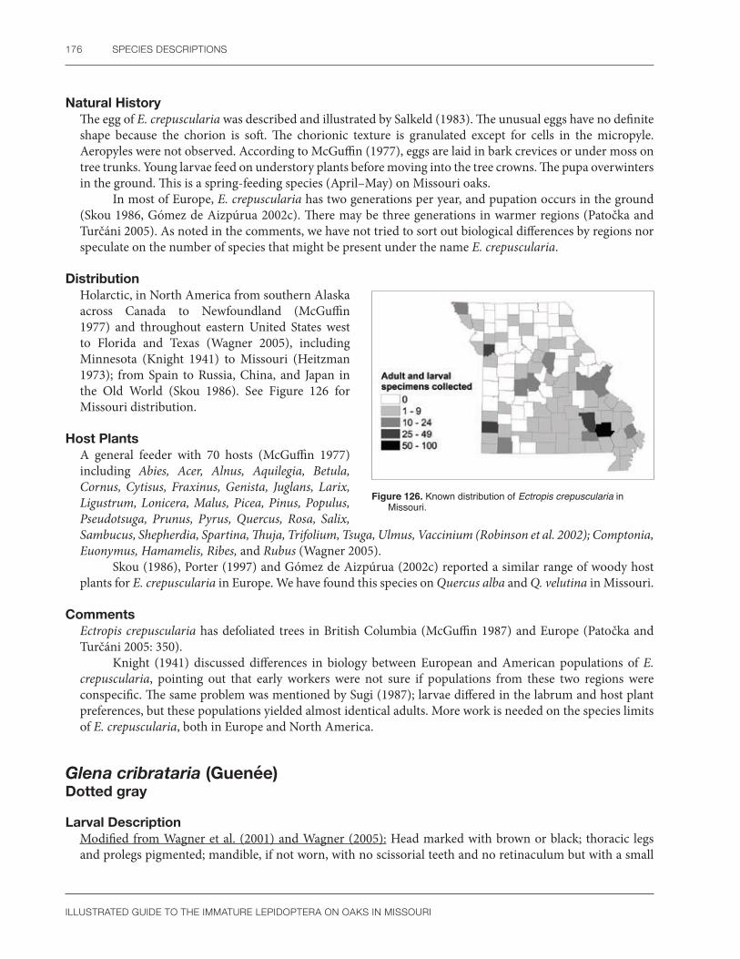
176 SPECIES DESCRIPTIONS
ILLUSTRATED GUIDE TO THE IMMATURE LEPIDOPTERA ON OAKS IN MISSOURI
Natural HistoryThe egg of E. crepuscularia was described and illustrated by Salkeld (1983). The unusual eggs have no definite shape because the chorion is soft. The chorionic texture is granulated except for cells in the micropyle. Aeropyles were not observed. According to McGuffin (1977), eggs are laid in bark crevices or under moss on tree trunks. Young larvae feed on understory plants before moving into the tree crowns. The pupa overwinters in the ground. This is a spring-feeding species (April–May) on Missouri oaks.
In most of Europe, E. crepuscularia has two generations per year, and pupation occurs in the ground (Skou 1986, Gómez de Aizpúrua 2002c). There may be three generations in warmer regions (Patočka and Turčáni 2005). As noted in the comments, we have not tried to sort out biological differences by regions nor speculate on the number of species that might be present under the name E. crepuscularia.
DistributionHolarctic, in North America from southern Alaska across Canada to Newfoundland (McGuffin 1977) and throughout eastern United States west to Florida and Texas (Wagner 2005), including Minnesota (Knight 1941) to Missouri (Heitzman 1973); from Spain to Russia, China, and Japan in the Old World (Skou 1986). See Figure 126 for Missouri distribution.
Host PlantsA general feeder with 70 hosts (McGuffin 1977) including Abies, Acer, Alnus, Aquilegia, Betula, Cornus, Cytisus, Fraxinus, Genista, Juglans, Larix, Ligustrum, Lonicera, Malus, Picea, Pinus, Populus, Pseudotsuga, Prunus, Pyrus, Quercus, Rosa, Salix, Sambucus, Shepherdia, Spartina, Thuja, Trifolium, Tsuga, Ulmus, Vaccinium (Robinson et al. 2002); Comptonia, Euonymus, Hamamelis, Ribes, and Rubus (Wagner 2005).
Skou (1986), Porter (1997) and Gómez de Aizpúrua (2002c) reported a similar range of woody host plants for E. crepuscularia in Europe. We have found this species on Quercus alba and Q. velutina in Missouri.
CommentsEctropis crepuscularia has defoliated trees in British Columbia (McGuffin 1987) and Europe (Patočka and Turčáni 2005: 350).
Knight (1941) discussed differences in biology between European and American populations of E. crepuscularia, pointing out that early workers were not sure if populations from these two regions were conspecific. The same problem was mentioned by Sugi (1987); larvae differed in the labrum and host plant preferences, but these populations yielded almost identical adults. More work is needed on the species limits of E. crepuscularia, both in Europe and North America.
Glena cribrataria (Guenée)Dotted gray
Larval DescriptionModified from Wagner et al. (2001) and Wagner (2005): Head marked with brown or black; thoracic legs and prolegs pigmented; mandible, if not worn, with no scissorial teeth and no retinaculum but with a small
Figure 126. Known distribution of Ectropis crepuscularia in Missouri.

GEOMETRIDAE 177
ILLUSTRATED GUIDE TO THE IMMATURE LEPIDOPTERA ON OAKS IN MISSOURI
Figure 127. Glena cribrataria mature larva. Photo by R.J. Marquis, used with permission.
outer tooth very close to the cutting edge by the molar ridge; body gray to green with faint, thin, small wavy lines; spiracle of A1 ventrad of the other abdominal spiracles; bicolored swelling on A2 surrounds spiracle and is darker posteriorly; sometimes smaller middorsal black dots present or they may be dots by spiracle on A3-4 and A6-7 (Figure 127).
The larva of G. cribrataria was illustrated by Wagner et al. (2001) and Wagner (2005). Glena is easily confused with other Boarmiini that have a tubercle on A2 (examples in McGuffin 1977: 13, 114, Heitzman 1982). The tubercle on G. cribrataria surrounds the spiracle (Wagner 2005), and L1 is probably included on the swelling as well. Some other features mentioned by McGuffin (1977) for Glena are SV4 present on A2 and the proleg of A6 with 30–38 crochets and four setae on the outer side. We did not study young larvae of G. cribrataria, and therefore, the condition of the tubercle in early instars is unknown. Characters to recognize larval Glena and its included species need more study. The mandible is unusual, but we only examined a single exuvia of G. cribrataria and did not compare to other closely related species of Glena to see if their mandibles are similar.
Pupal DescriptionVertex rounded; labial palpi minutely exposed; maxillae extend to the caudal margin of wings; prothoracic leg longer than mesothoracic leg; prothoracic femur exposed; mesothoracic leg does not reach lower eye margin; metathoracic legs exposed at the tips; mesothoracic callosity a small oval tubercle; abdominal dorsum punctate; spiracular furrow on A5 present as a shallow oval groove near a faint ridge with the L setae; dorsal groove on A9 absent; cremaster with a broad base ending in two diverging straight spines.
The above description is similar to other Glena studied by McGuffin (1977). He noted the exposed prothoracic femur, A5 with a spiracular furrow, with a ridge bearing the L setae, and the two spines on the cremaster will allow recognition of the genus. The ridge takes practice to recognize, as it is faint.
In the key by Forbes (1948: 32–33), Glena and Ectropis were similar in pupal morphology. Characters to recognize Glena included: cremaster bifid; the dorsal groove is absent; prothoracic femur exposed; the flange plate and callosity are present; and the abdominal cuticular texture is rough between the punctures.
Natural HistoryLittle information is available on the life history of G. cribrataria. Larvae are found from July–October in Missouri. There are one or two generations per year and this species is more likely to be seen in dry oak woodlands than other areas (Wagner et al. 2001).
DistributionOntario, Canada, to Florida, then west to Minnesota, Mississippi (McGuffin 1977), and Texas (Wagner 2005). See Figure 128 for Missouri distribution. Figure 128. Known distribution of Glena cribrataria in Missouri.

178 SPECIES DESCRIPTIONS
ILLUSTRATED GUIDE TO THE IMMATURE LEPIDOPTERA ON OAKS IN MISSOURI
Host PlantsPicea, Populus, Salix (Robinson et al. 2002); probably a wide variety of woody plants, Acer, Amelanchier, Betula, Prunus, Quercus, Salix, and Vaccinium (Wagner 2005). We have found G. cribrataria on Q. rubra and Q. velutina in Missouri.
Phaeoura quernaria (J.E. Smith) Oak beauty
Larval Description Modified from McGuffin (1981), Carmichael and Vance (2004), and Wagner et al. (2001): Stout, robust green to brown to gray caterpillar, irregularly streaked with black longitudinally; head bifid, cleft at vertex, flat, and held inward; mandible, when not worn, with approximately eight small scissorial teeth, no retinaculum, and a small outer tooth; mesothorax and A2 swollen dorsally, a swelling also present on the venter of A3; a fringe of simple to stellate hairs subventrally between the abdominal and anal prolegs; A6 with 16–18 setae on the outer side; 50–64 crochets in one group (Figure 129a, b).
The larva of P. quernaria was described by McGuffin (1981) and illustrated in color by Ives and Wong (1988), Wagner et al. (2001), Carmichael and Vance (2004), and Wagner (2005). According to these authors, late instars are easily recognized by the bifid head, swollen mesothorax, ridge on A2, swelling on the venter of A3, hairs between the prolegs, and body setae arising from yellow to green pinacula. In addition, the larva of P. quernaria is instantly recognizable by the characteristic shagreened integument (D. Wagner, pers. comm.). The setal count and crochet number on A6 might need to be confirmed in this species as McGuffin (1981) implied these characters in his introduction to the genus without specifying the species examined. See also Appendix 3, Plate 1.
Pupal DescriptionModified from McGuffin (1981): Body rough and punctate on the abdomen; vertex and glazed eyepiece with several tubercles; labial palpi minutely exposed; maxillae extends about three-fourths the distance to the caudal margin of wings; prothoracic leg longer than mesothoracic leg; prothoracic femur hidden; mesothoracic leg does not reach lower eye margin; metathoracic legs exposed at the tips; mesothoracic callosity absent, spiracle forms an oval pit; spiracular furrow on A5 absent; dorsal groove on A9 serrated; lateral grooves present; cremaster with a broad base ending in two thick spines and six thinner curved setae.
The pupa of P. quernaria was partially illustrated by McGuffin (1981). The combination of tubercles on vertex and glazed eyepiece (not shown by McGuffin 1981), rough body texture, lack of both the mesothoracic callosity and spiracular furrow, pitlike mesothoracic spiracle and setae of the cremaster are especially unusual.
a b
Figure 129. Phaeoura quernaria (a) mid-instar larva; (b) mature larva. Photos by R.J. Marquis, used with permission.

GEOMETRIDAE 179
ILLUSTRATED GUIDE TO THE IMMATURE LEPIDOPTERA ON OAKS IN MISSOURI
Forbes (1948: 32–33) found the pupa of P. quernaria to be distinctive. It keys out rapidly by having a cremaster with eight hooked setae, the terminal pair of which are thicker than the others; the prothoracic femur is hidden; a dorsal groove is present; and the antenna has four or five rows of tubercles.
Natural HistoryAccording to McGuffin (1981), the egg of P. quernaria is yellowish green with metallic reflections. The caterpillar occurs from August to October (Wagner et al. 2001), the pupa usually overwinters, and adults are found most often in June or July (McGuffin 1981). Apparently there is a single generation in Missouri, where we collected larvae from May to June. The phenology of P. quernaria needs more study in Missouri.
DistributionNova Scotia, Canada, south to Florida and west to Saskatchewan, Arkansas, and Texas, including Missouri (Heitzman 1973). See Figure 130 for Missouri distribution.
Host PlantsMany trees and shrubs such as Betula, Crataegus, Populus, Prunus, Quercus alba, Q. coccinea/rubra, Q. macrocarpa Q. robur, Q. rubra, Salix, Tilia, Ulmus (Robinson et al. 2002); Fraxinus (Wagner et al. 2001); and Hamamelis (Wagner 2005). Phaeoura quernaria is said to favor oak (Rindge 1961), but despite the common name, birch is the preferred host in Canada (McGuffin 1981). It is found on Q. alba and Q. velutina in Missouri.
Hypagyrtis unipunctata (Haworth)One-spotted variant
Larval DescriptionModified from McGuffin (1977), Wagner et al. (2001), and Wagner (2005): Larva variably colored from brown to reddish to gray to yellow-green, without pronounced ridges or warts; head and body cuticle shagreened with clear to frosty convex granules under high magnification; mandible without scissorial teeth or a retinaculum but
Figure 130. Known distribution of Phaeoura quernaria in Missouri.
Figure 131. Hypagyrtis unipunctata (a) early instar larva; (b) mature larva. Photos by R.J. Marquis, used with permission.
a b
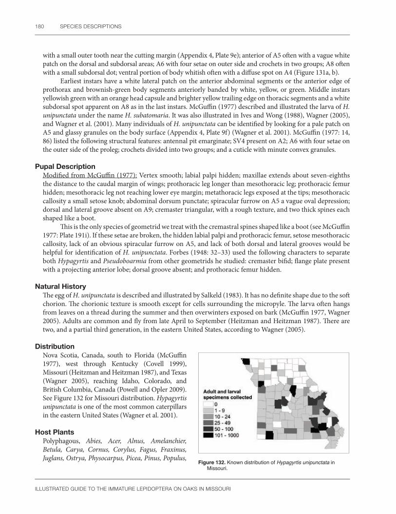
180 SPECIES DESCRIPTIONS
ILLUSTRATED GUIDE TO THE IMMATURE LEPIDOPTERA ON OAKS IN MISSOURI
with a small outer tooth near the cutting margin (Appendix 4, Plate 9e); anterior of A5 often with a vague white patch on the dorsal and subdorsal areas; A6 with four setae on outer side and crochets in two groups; A8 often with a small subdorsal dot; ventral portion of body whitish often with a diffuse spot on A4 (Figure 131a, b).
Earliest instars have a white lateral patch on the anterior abdominal segments or the anterior edge of prothorax and brownish-green body segments anteriorly banded by white, yellow, or green. Middle instars yellowish green with an orange head capsule and brighter yellow trailing edge on thoracic segments and a white subdorsal spot apparent on A8 as in the last instars. McGuffin (1977) described and illustrated the larva of H. unipunctata under the name H. subatomaria. It was also illustrated in Ives and Wong (1988), Wagner (2005), and Wagner et al. (2001). Many individuals of H. unipunctata can be identified by looking for a pale patch on A5 and glassy granules on the body surface (Appendix 4, Plate 9f) (Wagner et al. 2001). McGuffin (1977: 14, 86) listed the following structural features: antennal pit emarginate; SV4 present on A2; A6 with four setae on the outer side of the proleg; crochets divided into two groups; and a cuticle with minute convex granules.
Pupal DescriptionModified from McGuffin (1977): Vertex smooth; labial palpi hidden; maxillae extends about seven-eighths the distance to the caudal margin of wings; prothoracic leg longer than mesothoracic leg; prothoracic femur hidden; mesothoracic leg not reaching lower eye margin; metathoracic legs exposed at the tips; mesothoracic callosity a small setose knob; abdominal dorsum punctate; spiracular furrow on A5 a vague oval depression; dorsal and lateral groove absent on A9; cremaster triangular, with a rough texture, and two thick spines each shaped like a boot.
This is the only species of geometrid we treat with the cremastral spines shaped like a boot (see McGuffin 1977: Plate 191i). If these setae are broken, the hidden labial palpi and prothoracic femur, setose mesothoracic callosity, lack of an obvious spiracular furrow on A5, and lack of both dorsal and lateral grooves would be helpful for identification of H. unipunctata. Forbes (1948: 32–33) used the following characters to separate both Hypagyrtis and Pseudoboarmia from other geometrids he studied: cremaster bifid; flange plate present with a projecting anterior lobe; dorsal groove absent; and prothoracic femur hidden.
Natural HistoryThe egg of H. unipunctata is described and illustrated by Salkeld (1983). It has no definite shape due to the soft chorion. The chorionic texture is smooth except for cells surrounding the micropyle. The larva often hangs from leaves on a thread during the summer and then overwinters exposed on bark (McGuffin 1977, Wagner 2005). Adults are common and fly from late April to September (Heitzman and Heitzman 1987). There are two, and a partial third generation, in the eastern United States, according to Wagner (2005).
DistributionNova Scotia, Canada, south to Florida (McGuffin 1977), west through Kentucky (Covell 1999), Missouri (Heitzman and Heitzman 1987), and Texas (Wagner 2005), reaching Idaho, Colorado, and British Columbia, Canada (Powell and Opler 2009). See Figure 132 for Missouri distribution. Hypagyrtis unipunctata is one of the most common caterpillars in the eastern United States (Wagner et al. 2001).
Host PlantsPolyphagous, Abies, Acer, Alnus, Amelanchier, Betula, Carya, Cornus, Corylus, Fagus, Fraxinus, Juglans, Ostrya, Physocarpus, Picea, Pinus, Populus,
Figure 132. Known distribution of Hypagyrtis unipunctata in Missouri.

GEOMETRIDAE 181
ILLUSTRATED GUIDE TO THE IMMATURE LEPIDOPTERA ON OAKS IN MISSOURI
Prunus, Quercus alba, Q. coccinea/rubra, Q. macrocarpa, Rosa, Salix, Tilia, Ulmus, Vaccinium (Robinson et al. 2002); Platanus (Wagner 2005); Crataegus, Liquidambar, Myrica, Q. nigra, and Q. laevis (Matthews et al. 2014).
We have found H. unipunctata on Q. coccinea, Q. imbricaria, Q. rubra, A. stellata, and Q. velutina in Missouri. Records for conifers may need confirmation.
CommentsBased on their similar egg morphology, Salkeld (1983: 53) suggests E. crepuscularia and H. unipunctata are closely related and should be in the same tribe. Wagner (2005) noted the genus Hypagyrtis is in need of modern revision. Our observations are based on reared adults, but even with them, molecular confirmation would have been helpful.
Alsophila pometaria (Harris) Fall cankerworm
Larval Description Modified from McGuffin (1988), Wagner (2005), and Ciesla and Asaro (2013): Head pale green to black, sometimes mottled; mandible with a few large and several smaller teeth on the cutting margin, small outer tooth present but retinaculum absent; body pale green to dark brown to black, with or without three lateral white stripes, one subdorsal (the widest), one supraspiracular, and one subspiracular; a black spot may be present behind the spiracle, especially on the anterior abdominal segments; A5 with a small proleg; anal shield concolorous with head.
Even though the body color is quite variable, the larva of A. pometaria is easily recognized by having a small proleg on A5 (Wagner 2005). The dorsum varies from light green to black as a function of population density (Ciesla and Asaro 2013). Dark and green color forms are illustrated by Ives and Wong (1988). See Figure 133a, b).
a b
Figure 133. Alsophila pometaria (a) mature larva, black form; (b) mature larva, green form. Photos by R.J. Marquis, used with permission.
Pupal DescriptionModified from Mosher (1916a) and Forbes (1948: 15): Vertex smooth; labial palpi hidden; maxillae extends to the caudal margin of wings; prothoracic leg longer than mesothoracic leg; prothoracic femur hidden; mesothoracic leg does not reach lower eye margin; metathoracic legs hidden; mesothoracic callosity absent; abdominal dorsum punctate; spiracular furrow on A5 absent; dorsal and lateral groove absent on A9; cremaster with two thick spines converging at a 45 or 180 degree angle.

182 SPECIES DESCRIPTIONS
ILLUSTRATED GUIDE TO THE IMMATURE LEPIDOPTERA ON OAKS IN MISSOURI
This is the only species we treat that has the cremastral spines straight (not hooked or bent) and diverging at a 45 to 180 degree angle. This arrangement caused Mosher (1916a) to put A. pometaria in her group C with only one other species. McGuffin (1988) also noted the unusual cremaster.
Although our single pupal exuvia was associated with a reared adult, and the genitalia matched the female illustrated by McGuffin (1988), we were unable to confirm several important characters mentioned by Forbes (1948) and Mosher (1916a). The prothoracic legs do not meet at the meson in our specimen, and they should according to Mosher (1916a: 118). Forbes (1948) stated that the tongue (maxillae) is shorter than the fore (prothoracic) legs, agreeing with Mosher (1916a). He also stated that the third abdominal spiracle is hidden; ours is fully exposed. Perhaps there is sexual dimorphism in A. pometaria. Besides our confirmation of our identification with genitalia, there is a photograph online that is very similar to our specimen, and it, too, has the third abdominal spiracle clearly evident (https://bugguide.net/node/view/604915).
The pupa of the spring cankerworm, Paleacrita vernata, is similar to A. pometaria except for the cremaster that has two parallel thin spines on a short stalk instead of being thick and diverging (SCPC).
Natural HistoryThe life history of A. pometaria has been discussed many times. Tietz (1972) and McGuffin (1988) list most of the important earlier publications. A recent synopsis is given by Van Driesche et al. (2013). The eggs overwinter and hatch at the same time that leaf buds are starting to open. Early instars are leaf skeletonizers, but later they eat everything except the larger veins. Pupation occurs in the soil and adults emerge in November or December. Eggs are laid on the trunk or small branches. Females can reproduce by sexual means or parthenogenesis.
In Missouri, as elsewhere, this species feeds on spring foliage. A population increasing in number over the years 2005–2006 at the Tyson Research Center in Eureka, MO, was decimated in 2007 by an early leaf flush in combination with a late spring frost that killed all emerging leaves, thus eliminating the food source for the young caterpillars (R.J. Marquis, pers. obs.). However, recent fall cankerworm outbreaks on oak in Virginia appear to be getting worse (Asaro and Chamberlin 2015). A summary of outbreaks by A. pometaria (Ciesla and Asaro 2013) indicate the earliest one was in 1661. Since then, outbreaks have been reported in Georgia, New York, Pennsylvania Massachusetts, North Carolina, Virginia, Washington DC, and the Rocky Mountain States.
DistributionMaritime Provinces of Canada west to Alberta, eastern United States south to Florida and west to Missouri, New Mexico, Utah, Colorado, and California (Ciesla and Asaro 2013). See Figure 134 for Missouri distribution.
Host PlantsPolyphagous on forest trees and shrubs, Acer, Betula, Caragana, Carya, Castanea, Celtis, Crataegus, Cydonia, Fagus, Fraxinus, Juglans, Malus, Ostrya, Populus, Prunus, Pyrus, Quercus alba, Q. coccinea, Q. macrocarpa, Q. rubra, Robinia, Salix, Solidago, Sorbus, Tilia, Ulmus, and Vaccinium (Robinson et al. 2002). We have found A. pometaria on Quercus alba, Q. imbricaria, Q. rubra, Q. stellata, and Q. velutina in Missouri.
Figure 134. Known distribution of Alsophila pometaria in Missouri.

GEOMETRIDAE 183
ILLUSTRATED GUIDE TO THE IMMATURE LEPIDOPTERA ON OAKS IN MISSOURI
CommentsAlthough usually placed in Oenochrominae, the fall cankerworm has also been segregated as its own subfamily (Alsophilinae) (Minet and Scoble 1998). McGuffin (1964: 849) noted a similarity in larval chaetotaxy between this species and the Ennominae. Wagner et al. (2001) agreed and placed A. pometaria in the Ennomiinae as did Pohl et al. (2016) in their checklist.
Subfamily Geometrinae
Nemoria bistriaria HübnerRed-fringed emerald
Larval Description Modified from Ferguson (1985b) and Wagner (2005): Head capsule tan brown; mandible of mature larva not available, early instar has an outer tooth and five scissorial teeth; body flattened dorsoventrally, covered with patches of spines, and with alternating black, rust, dark brown, or yellow-brown stripes and spots; paired processes arising subdorsally on several body segments at a 45° angle; processes on the thorax, A1, and A8 short, usually with a terminal spine, those on A2-5 largest and terminating in two diverging spines; spiracle of A6 clearly below spiracle of A5 (Figure 135a, b).
a b
Figure 135. Nemoria bistriaria (a) early instar larva; (b) mature larva. Photos by R.J. Marquis, used with permission.
The larva of N. bistriaria was illustrated by Ferguson (1985b: 8). Members of the tribe Nemoriini are identified by their general appearance and habits, in particular, they fed on foliage instead of flowers, and have large flat triangular projections on the thorax and/or abdomen (Ferguson 1985b: 14). Unfortunately, no keys exist to separate species of Nemoria in the larval stage, and several sympatric species are known to feed on oak (Ferguson 1985b, Wagner 2005). Ferguson (1985b) and Wagner (2005) both suggested that the projections on A2 and A3 are longer on N. bistriaria than in related species, but this should be tested with a large series of examples. At present, identifications of larval Nemoria need to be based on reared adults or molecular methods (Wagner et al. 2001: 158, 160).
According to the drawing in Ferguson (1985b), the abdominal flanges seem to arise below the spiracles. This would make them lateral processes morphologically speaking, even though they are located subdorsally.

184 SPECIES DESCRIPTIONS
ILLUSTRATED GUIDE TO THE IMMATURE LEPIDOPTERA ON OAKS IN MISSOURI
Pupal DescriptionBody shagreened, with a middorsal stripe; vertex roughened; maxillae extend to caudal margin of the wings; labial palpi minutely exposed; maxillary palpi hidden; prothoracic femur hidden; mesothoracic leg thin, much shorter than prothoracic leg, with the apex below the lower eye margin; metathoracic legs exposed; mesothoracic callosity absent; peritreme of spiracles on A4-7 surrounded by a pale thick rim, the spiracle of A6 located ventrad of the other spiracles; dorsal and lateral grooves absent; cremaster rounded with eight curved setae.
The unusual ventral location of the spiracle on A6 and the thick rim surrounding the peritremes on the spiracles of A4-7 help separate N. bistriaria from other Missouri oak-feeding geometrids. However, the pupa of N. lixaria (SCPC) is quite similar to N. bistriaria, which implies that these features may only be useful for characterizing the tribe or genus.
Natural HistoryThe egg of N. bistriaria was illustrated by McGuffin (1988). It is a round cylinder with a flattened top that is yellow at first but later turns brick red. We see caterpillars most often from May to June in Missouri. Adults follow from April until August (Heitzman 1973) suggesting two broods per year (Ferguson 1985b, McGuffin 1988). More generations per year were listed by Wagner (2005), especially in the southern United States. Adults come to puddles of mud (McGuffin 1988).
DistributionOntario and Quebec, Canada, south to Georgia, and west to Illinois, Missouri, and Texas (Ferguson 1985b). See Figure 136 for Missouri distribution.
Host PlantsBetula, Juglans (Robinson et al. 2002); Quercus (Ferguson 1985b); Comptonia, and Myrica (Wagner 2005). We have found N. bistriaria on Q. alba and Q. velutina in Missouri.
CommentsTwo subspecies of N. bistriaria were described in Ferguson (1985).
The color and morphology of some larval Nemoria are affected by diet (Comstock 1960: 425, Greene 1989, Wagner 2005), at least for N. bistriaria (http://moths.friendscentral.org).
Figure 136. Known distribution of Nemoria bistriaria in Missouri.

MIMALLONIDAE 185
ILLUSTRATED GUIDE TO THE IMMATURE LEPIDOPTERA ON OAKS IN MISSOURI
MimallonidaeSack bearer moths
Mimallonidae is a small family of New World moths containing about 300 species in six subfamilies (St Laurent et al. 2018). Most authors (Franclemont 1973, Lemaire and Minet 1998, Nieukerken et al. 2011) classify them in or near the Bombycoidea based at least in part on pupal morphology. Given that Mimallonidae and Bombycoidea both have reduced adult mouthparts (Lemaire and Minet 1998) and the pupa reflects adult structures, it is no surprise that the pupal appendages look similar in the two taxa. Using larval morphology, Scott (1986b) placed Mimallonidae in the Pyraloidea, perhaps following Fracker (1915: 81). This similarity probably involves plesiomorphies, and thus workers have generally ignored the suggestion. Mimallonid larvae do have the abdominal L setae closely spaced (Forbes 1942), as do pyraloids and gelechioids (Stehr 1987), but no other evidence linking Mimallonidae to the microlepidoptera is obvious (Scoble 1992). Recent molecular data place Mimallonidae in or near the macrolepidoptera (see Timmermans et al. 2014) or butterflies (Regier et al. 1998). Mimallonids were previously known as Lacosomidae (e.g., Peterson 1966) or Perophoridae (e.g., Dyar 1900b).
Lemaire and Minet (1998) described mimallonid eggs as flat, elongate, subcylindrical, and often asymmetrical. The tough, sometimes transparent, chorion may have ridges. Females of Mimallo amilia typically lay four to five masses in their lifetime, each containing about 20 bright yellow, oval eggs (Zanuncio et al. 2005). When magnified under low power, eggs of M. amilia show longitudinal rows of small nodules (SCPC). Their yellow color and general shape is similar to the egg of Lacosoma chiridota described by Dyar (1900b) and Peterson (1966). A photograph by Mota et al. (2014), although determined only by probable association with a nearby caterpillar, seems typical of the family. With more study, mimallonid eggs may prove to be relatively unusual in shape and texture.
Larvae of the Mimallonidae are unique in having the anal crochets arranged in an unbroken oval with the posterior row much smaller than the anterior portion (Stehr 1987). There are two caveats to this statement. First, it might only apply to later instars because the first instar of M. amilia and early instars of L. chiridota have the anal crochets arranged in an arc, as is typical for most caterpillars (SCPC). Second, Scoble (1992: 328) stated that the circular arrangement on A3-6 is the distinctive character for Mimallonidae. This is a misprint and the reference should have been to the anal crochets and not those on A3-6. Another unusual feature of mimallonid larvae is that they live in cases made of plant parts, silk, and often frass. There appear to be several types (see Monte 1934 or Costa Lima 1949). Some larvae form a hard, smooth case (e.g., Cicinnus packardii), whereas the covering in other species is a loose mixture of frass and leaves (e.g., M. amilia) or several leaves sewn together with silk (e.g., Lacosoma). The case may be erect on a stem, or more commonly, hanging from the host. Larvae that block the openings of their case with body parts typically have a heavily sclerotized rugose head and flat, tough anal region (Wagner 2005, Chacón and Montero 2007: Plate 27). In other species, such as M. amilia and an undetermined larva from Honduras (SCPC), the body form is unmodified and more typical of caterpillars. The head setae may be spatulate (Dyar 1896c), the number of SV setae can vary on A3-6 (Fracker 1915, Stehr 1987, SCPC), and SD1 on A8 may be dorsal or to the side of the spiracle (Stehr 1987). The outer surface of the mandibles of M. amilia, Cicinnus melsheimeri, and L. chiridota has an irregular, rough texture. Typically, this portion of the mandible is smooth in caterpillars (SCPC). Lemaire and Minet (1998) called attention to a “ventral protuberance” behind the coxae on T2 and T3 that is single, paired, spiny, or smooth; this is another distinctive character of the few Mimallonidae that we examined (Lacosoma, Cicinnus, Mimallo). It is absent in the undetermined Honduran mimallonid (SCPC). Some species of Mimallonidae are brightly colored, especially the thorax (Forbes 1942, Lemaire and Minet 1998, Chacón and Montero 2007).
Dyar (1900b) suggested there are two types of life cycles in the family. Sometimes mimallonids skeletonize the leaves as young larvae, then live in a case as late instars. Other species live in a case throughout their larval life. The host is often a tree species (Lemaire and Minet 1998).
Most pupae of the Mimallonidae are readily recognized by having dorsal abdominal grooves lined with large, blunt teeth. These resemble a structure that Hinton called a gin trap (see Hinton 1955: Fig. 13). It is present in both Missouri species C. melsheimeri (Harris 1863: 417) and L. chiridota (SCPC), as well as M. amilia, but
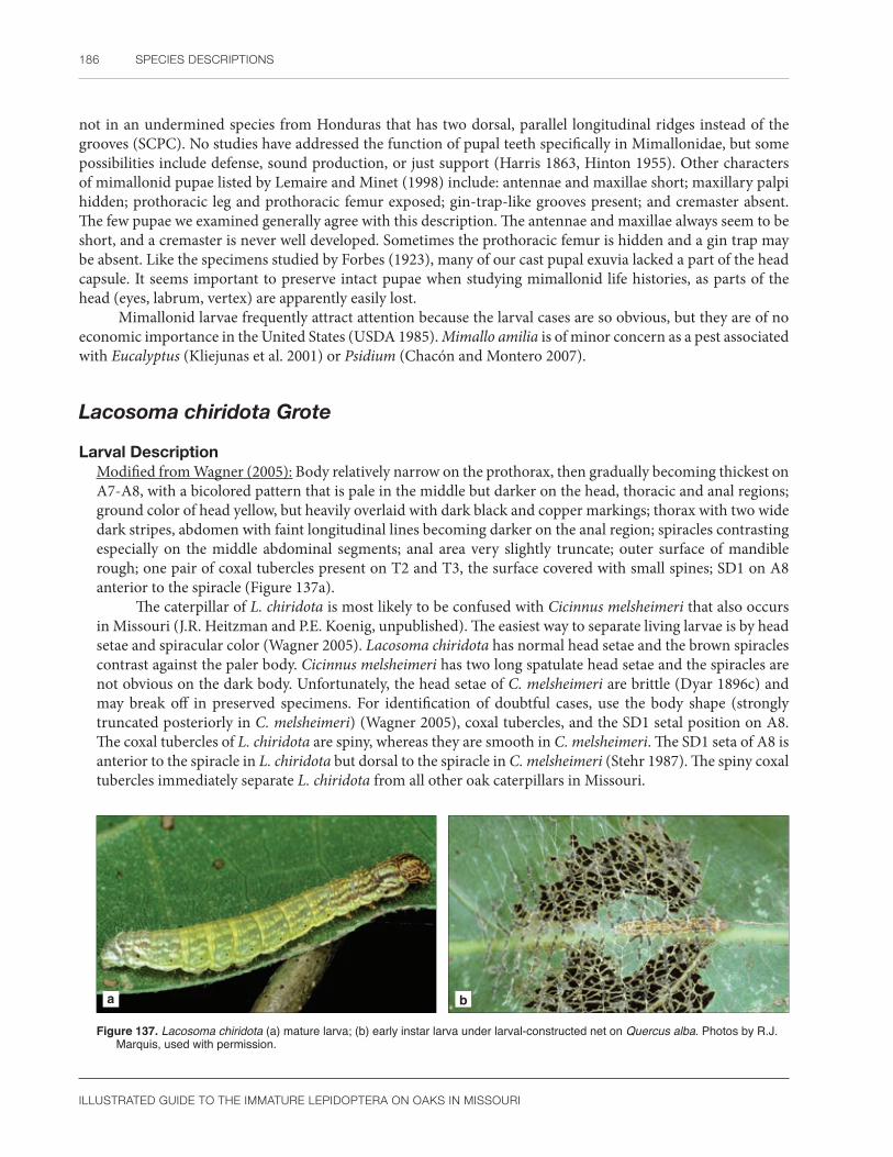
186 SPECIES DESCRIPTIONS
ILLUSTRATED GUIDE TO THE IMMATURE LEPIDOPTERA ON OAKS IN MISSOURI
not in an undermined species from Honduras that has two dorsal, parallel longitudinal ridges instead of the grooves (SCPC). No studies have addressed the function of pupal teeth specifically in Mimallonidae, but some possibilities include defense, sound production, or just support (Harris 1863, Hinton 1955). Other characters of mimallonid pupae listed by Lemaire and Minet (1998) include: antennae and maxillae short; maxillary palpi hidden; prothoracic leg and prothoracic femur exposed; gin-trap-like grooves present; and cremaster absent. The few pupae we examined generally agree with this description. The antennae and maxillae always seem to be short, and a cremaster is never well developed. Sometimes the prothoracic femur is hidden and a gin trap may be absent. Like the specimens studied by Forbes (1923), many of our cast pupal exuvia lacked a part of the head capsule. It seems important to preserve intact pupae when studying mimallonid life histories, as parts of the head (eyes, labrum, vertex) are apparently easily lost.
Mimallonid larvae frequently attract attention because the larval cases are so obvious, but they are of no economic importance in the United States (USDA 1985). Mimallo amilia is of minor concern as a pest associated with Eucalyptus (Kliejunas et al. 2001) or Psidium (Chacón and Montero 2007).
Lacosoma chiridota Grote
Larval DescriptionModified from Wagner (2005): Body relatively narrow on the prothorax, then gradually becoming thickest on A7-A8, with a bicolored pattern that is pale in the middle but darker on the head, thoracic and anal regions; ground color of head yellow, but heavily overlaid with dark black and copper markings; thorax with two wide dark stripes, abdomen with faint longitudinal lines becoming darker on the anal region; spiracles contrasting especially on the middle abdominal segments; anal area very slightly truncate; outer surface of mandible rough; one pair of coxal tubercles present on T2 and T3, the surface covered with small spines; SD1 on A8 anterior to the spiracle (Figure 137a).
The caterpillar of L. chiridota is most likely to be confused with Cicinnus melsheimeri that also occurs in Missouri (J.R. Heitzman and P.E. Koenig, unpublished). The easiest way to separate living larvae is by head setae and spiracular color (Wagner 2005). Lacosoma chiridota has normal head setae and the brown spiracles contrast against the paler body. Cicinnus melsheimeri has two long spatulate head setae and the spiracles are not obvious on the dark body. Unfortunately, the head setae of C. melsheimeri are brittle (Dyar 1896c) and may break off in preserved specimens. For identification of doubtful cases, use the body shape (strongly truncated posteriorly in C. melsheimeri) (Wagner 2005), coxal tubercles, and the SD1 setal position on A8. The coxal tubercles of L. chiridota are spiny, whereas they are smooth in C. melsheimeri. The SD1 seta of A8 is anterior to the spiracle in L. chiridota but dorsal to the spiracle in C. melsheimeri (Stehr 1987). The spiny coxal tubercles immediately separate L. chiridota from all other oak caterpillars in Missouri.
a b
Figure 137. Lacosoma chiridota (a) mature larva; (b) early instar larva under larval-constructed net on Quercus alba. Photos by R.J. Marquis, used with permission.
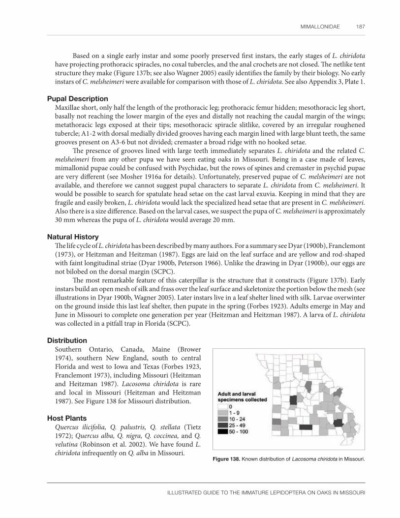
MIMALLONIDAE 187
ILLUSTRATED GUIDE TO THE IMMATURE LEPIDOPTERA ON OAKS IN MISSOURI
Based on a single early instar and some poorly preserved first instars, the early stages of L. chiridota have projecting prothoracic spiracles, no coxal tubercles, and the anal crochets are not closed. The netlike tent structure they make (Figure 137b; see also Wagner 2005) easily identifies the family by their biology. No early instars of C. melsheimeri were available for comparison with those of L. chiridota. See also Appendix 3, Plate 1.
Pupal DescriptionMaxillae short, only half the length of the prothoracic leg; prothoracic femur hidden; mesothoracic leg short, basally not reaching the lower margin of the eyes and distally not reaching the caudal margin of the wings; metathoracic legs exposed at their tips; mesothoracic spiracle slitlike, covered by an irregular roughened tubercle; A1-2 with dorsal medially divided grooves having each margin lined with large blunt teeth, the same grooves present on A3-6 but not divided; cremaster a broad ridge with no hooked setae.
The presence of grooves lined with large teeth immediately separates L. chiridota and the related C. melsheimeri from any other pupa we have seen eating oaks in Missouri. Being in a case made of leaves, mimallonid pupae could be confused with Psychidae, but the rows of spines and cremaster in psychid pupae are very different (see Mosher 1916a for details). Unfortunately, preserved pupae of C. melsheimeri are not available, and therefore we cannot suggest pupal characters to separate L. chiridota from C. melsheimeri. It would be possible to search for spatulate head setae on the cast larval exuvia. Keeping in mind that they are fragile and easily broken, L. chiridota would lack the specialized head setae that are present in C. melsheimeri. Also there is a size difference. Based on the larval cases, we suspect the pupa of C. melsheimeri is approximately 30 mm whereas the pupa of L. chiridota would average 20 mm.
Natural HistoryThe life cycle of L. chiridota has been described by many authors. For a summary see Dyar (1900b), Franclemont (1973), or Heitzman and Heitzman (1987). Eggs are laid on the leaf surface and are yellow and rod-shaped with faint longitudinal striae (Dyar 1900b, Peterson 1966). Unlike the drawing in Dyar (1900b), our eggs are not bilobed on the dorsal margin (SCPC).
The most remarkable feature of this caterpillar is the structure that it constructs (Figure 137b). Early instars build an open mesh of silk and frass over the leaf surface and skeletonize the portion below the mesh (see illustrations in Dyar 1900b, Wagner 2005). Later instars live in a leaf shelter lined with silk. Larvae overwinter on the ground inside this last leaf shelter, then pupate in the spring (Forbes 1923). Adults emerge in May and June in Missouri to complete one generation per year (Heitzman and Heitzman 1987). A larva of L. chiridota was collected in a pitfall trap in Florida (SCPC).
DistributionSouthern Ontario, Canada, Maine (Brower 1974), southern New England, south to central Florida and west to Iowa and Texas (Forbes 1923, Franclemont 1973), including Missouri (Heitzman and Heitzman 1987). Lacosoma chiridota is rare and local in Missouri (Heitzman and Heitzman 1987). See Figure 138 for Missouri distribution.
Host PlantsQuercus ilicifolia, Q. palustris, Q. stellata (Tietz 1972); Quercus alba, Q. nigra, Q. coccinea, and Q. velutina (Robinson et al. 2002). We have found L. chiridota infrequently on Q. alba in Missouri.
Figure 138. Known distribution of Lacosoma chiridota in Missouri.

188 SPECIES DESCRIPTIONS
ILLUSTRATED GUIDE TO THE IMMATURE LEPIDOPTERA ON OAKS IN MISSOURI
LasiocampidaeLappet moths
Lasiocampidae contain 2,000 species (Nieukerken et al. 2011) worldwide and are most diverse in tropical regions (Franclemont 1973). Several morphologically based classifications are presented by Franclemont (1973), Holloway (1987), and most recently in Lemaire and Minet (1998). Zwick et al. (2010) and Zolotuhin et al. (2012) used molecular methods to study the placement of Lasiocampidae within the phylogeny of the Bombycoidea. Unlike other macrolepidoptera (e.g., Noctuidae, Geometridae, or Erebidae), the lasiocampid fauna of Missouri oaks is small, readily recognized, and easily characterized at the subfamily or tribal levels using Franclemont (1973). For convenience, we follow the higher classification of Franclemont (1973) in Pohl et al. (2016) but understand that the world classification of Lasiocampidae is tentative and in need of updating, pending study of the Neotropical fauna, additional immature stages, and a consensus of the molecular data. Consult Zolotuhin et al. (2012) and Regier et al. (2000) for a historical review of various subfamilies and tribes in the Lasiocampidae and Franclemont (1973) for issues and classification of the North American species. Fitzgerald (1995) studied the physiology and behavior of Malacosoma, whereas Stehr and Cook (1968) revised this genus for North America. Both publications contain detailed information on the biology of all the stages.
Lemaire and Minet (1998) considered lasiocampid eggs to be of the flat type and described them as oval, subcylindrical, with rounded edges, and often a flat top. They further noted that the chorion is tough, smooth, and often patterned. Many North American species were described and illustrated by Peterson (1966). According to his summary, Malacosoma is unusual in that the eggs encircle a twig and are covered with a frothy secretion from the female’s body called spumaline. Syme (1961) provided detailed drawings of the micropyle of Malacosoma americana, but that was the only species he studied. Stehr and Cook (1968) provide a key to egg masses of North American Malacosoma, and Swaine (1913) illustrated some egg variation in M. disstria and M. americanum. Tolype cover the eggs with hairs from the female’s body (Peterson 1966). Species that lay naked eggs deposit them singly, in small rows, or in a few layers. Lasiocampid eggs studied by Peterson (1966) were black, white, or a mixture of the two colors. Eggs of European lasiocampids were illustrated by Doring (1955: Plate 21). Those in Russia were studied by Dolinskaya and Pljushch (2000). Lasiocampid eggs differ from each other by the number and size of the cells around the micropyle, as well as the texture of the chorion.
First instar lasiocampid larvae are covered with secondary setae (Lemaire and Minet 1998). Later instar larvae are of two types (Stehr 1987). The first type has a cylindrical body shape, and in the case of tent caterpillars (e.g., Malacosoma), lives socially in large nests of silk. The second type, called lappet caterpillars (members of the Macromphaliinae such as Tolype species), are solitary feeders with a flattened body shape that enables them to rest closely pressed to their host plant (see Wagner 2005). The setae are either spinelike, spatulate, or scalelike, but never barbed or plumose (Lemaire and Minet 1998). Larval dorsal coloration is often bright, including one or two contrasting thoracic transverse bands that may be hidden at rest (Lemaire and Minet 1998, Wagner 2005). Following Stehr (1987: 511), we tentatively consider these markings to be segmental and not located on the intersegmental membrane except in the case of Artace. There may also be ventral patches on each segment (e.g., orange with black in Tolype).
Lasiocampids are easily confused with Apatelodidae, Arctiinae, and Lymantriinae of the Erebidae, a few Notodontidae, and Noctuidae (especially Acronictinae) (Stehr 1987). However, lasiocampid larvae in the Nearctic Region can be separated from related taxa by their biordinal crochets and a shallow u-shaped labral notch (Stehr 1987). Lasiocampid larvae also have an anal projection. According to Stehr (1987), it is present in the Lasiocampidae but absent in the Apatelodidae he examined, and is of unknown distribution in the Noctuoidea and other bombycoids. Based on specimens in the SCPC, we have seen an anal projection in Tolype (T. nana and T. velleda), a larva determined as probably Phyllodesma americana, Gloveria, Artace, Eutachyptera psidii, Dendrolimus, Dicogaster, and four species of Malacosoma (M. neustria, M. americana, M. californica, and M. disstria). The form of this projection varies from a small triangular point to a large, flat or oval lobe. If the larva is poorly preserved or the anal region is dirty, the projection can be difficult to see. An anal projection is absent
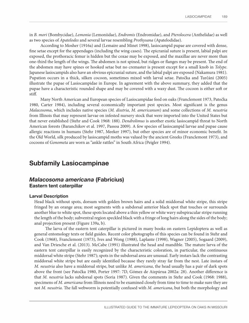
LASIOCAMPIDAE 189
ILLUSTRATED GUIDE TO THE IMMATURE LEPIDOPTERA ON OAKS IN MISSOURI
in B. mori (Bombycidae), Lemonia (Lemoniidae), Endromis (Endromidae), and Pterolocera (Anthelidae) as well as two species of Apatelodes and several larvae resembling Prothysana (Apatelodidae).
According to Mosher (1916a) and (Lemaire and Minet 1998), lasiocampid pupae are covered with dense, fine setae except for the appendages (including the wing cases). The epicranial suture is present, labial palpi are exposed, the prothoracic femur is hidden but the coxae may be exposed, and the maxillae are never more than one-third the length of the wings. The abdomen is not spined, but ridges or flanges may be present. The end of the abdomen may have spines or hooked setae but no cremaster is present except for a small knob in Tolype. Japanese lasiocampids also have an obvious epicranial suture, and the labial palpi are exposed (Nakamura 1981). Pupation occurs in a thick, silken cocoon, sometimes mixed with larval setae. Patočka and Turčáni (2005) illustrate the pupae of Lasiocampidae in Europe. In agreement with the above summary, they added that the pupae have a characteristic rounded shape and may be covered with a waxy dust. The cocoon is either soft or stiff.
Many North American and European species of Lasiocampidae feed on oaks (Franclemont 1973, Patočka 1980, Carter 1984), including several economically important pest species. Most significant is the genus Malacosoma, which includes native species (M. disstria, M. americanum) and some collections of M. neustria from Illinois that may represent larvae on infested nursery stock that were imported into the United States but that never established (Stehr and Cook 1968: 188). Dendrolimus is another exotic lasiocampid threat to North American forests (Baranchikov et al. 1997, Passoa 2009). A few species of lasiocampid larvae and pupae cause allergic reactions in humans (Stehr 1987, Meeker 1997), but other species are of minor economic benefit. In the Old World, silk produced by lasiocampid moths was valued by the ancient Greeks (Franclemont 1973), and cocoons of Gonometa are worn as “ankle rattles” in South Africa (Peigler 1994).
Subfamily Lasiocampinae
Malacosoma americana (Fabricius)Eastern tent caterpillar
Larval Description Head black without spots, dorsum with golden brown hairs and a solid middorsal white stripe, this stripe fringed by an orange area; most segments with a subdorsal anterior black spot that touches or surrounds another blue to white spot, these spots located above a thin yellow or white wavy subspiracular stripe running the length of the body; subventral region speckled black with a fringe of long hairs along the sides of the body; anal projection present (Figure 139a, b).
The larva of the eastern tent caterpillar is pictured in many books on eastern Lepidoptera as well as general entomology texts or field guides. Recent color photographs of this species can be found in Stehr and Cook (1968), Franclemont (1973), Ives and Wong (1988), Laplante (1998), Wagner (2005), Sogaard (2009), and Van Driesche et al. (2013). McCabe (1991) illustrated the head and mandible. The mature larva of the eastern tent caterpillar is easily recognized by the characteristic coloration, in particular, the continuous middorsal white stripe (Stehr 1987); spots in the subdorsal area are unusual. Early instars lack the contrasting middorsal white stripe but are easily identified because they rarely stray far from the nest. Late instars of M. neustria also have a middorsal stripe, but unlike M. americana, the head usually has a pair of dark spots above the front (see Patočka 1980, Porter 1997: 7D, Gómez de Aizpúrua 2002a: 28). Another difference is that M. neustria lacks subdorsal spots (Soria 1987). Given the comments in Stehr and Cook (1968: 1988), specimens of M. americana from Illinois need to be examined closely from time to time to make sure they are not M. neustria. The fall webworm is potentially confused with M. americana, but both the morphology and
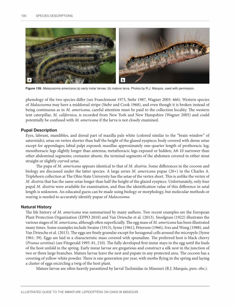
190 SPECIES DESCRIPTIONS
ILLUSTRATED GUIDE TO THE IMMATURE LEPIDOPTERA ON OAKS IN MISSOURI
a b
Figure 139. Malacosoma americana (a) early instar larvae; (b) mature larva. Photos by R.J. Marquis, used with permission.
phenology of the two species differ (see Franclemont 1973, Stehr 1987, Wagner 2005: 466). Western species of Malacosoma may have a middorsal stripe (Stehr and Cook 1968), and even though it is broken instead of being continuous as in M. americana, careful attention must be paid to the collection locality. The western tent caterpillar, M. californica, is recorded from New York and New Hampshire (Wagner 2005) and could potentially be confused with M. americana if the larva is not closely examined.
Pupal DescriptionEyes, labrum, mandibles, and dorsal part of maxilla pale white (colored similar to the “brain window” of saturniids); setae on vertex shorter than half the height of the glazed eyepiece; body covered with dense setae except for appendages; labial palpi exposed; maxillae approximately one-quarter length of prothoracic leg; mesothoracic legs slightly longer than antenna; metathoracic legs exposed or hidden; A8-10 narrower than other abdominal segments; cremaster absent, the terminal segments of the abdomen covered in either stout straight or slightly curved setae.
The pupa of M. americana appears identical to that of M. disstria. Some differences in the cocoon and biology are discussed under the latter species. A large series M. americana pupae (20+) in the Charles A. Triplehorn collection at The Ohio State University has the setae of the vertex short. This is unlike the vertex of M. disstria that has the same setae longer than half the height of the glazed eyepiece. Unfortunately, only four pupal M. disstria were available for examination, and thus the identification value of this difference in setal length is unknown. An educated guess can be made using biology or morphology, but molecular methods or rearing is needed to accurately identify pupae of Malacosoma.
Natural HistoryThe life history of M. americana was summarized by many authors. Two recent examples are the European Plant Protection Organization (EPPO 2010) and Van Driesche et al. (2013). Snodgrass (1922) illustrates the various stages of M. americana, although often superficially. The egg mass of M. americana has been illustrated many times. Some examples include Swaine (1913), Syme (1961), Peterson (1966), Ives and Wong (1988), and Van Driesche et al. (2013). The eggs are finely granular except for hexagonal cells around the micropyle (Syme 1961: 39). Eggs are laid in a characteristic mass covered with spumaline. The preferred host is black cherry (Prunus serotina) (see Fitzgerald 1995: 81, 210). The fully developed first instar stays in the egg until the buds of the host unfold in the spring. Early instar larvae are gregarious and construct a silk nest in the junction of two or three large branches. Mature larvae leave the nest and pupate in any protected area. The cocoon has a covering of yellow-white powder. There is one generation per year, with moths flying in the spring and laying a cluster of eggs encircling a twig of the host plant.
Mature larvae are often heavily parasitized by larval Tachinidae in Missouri (R.J. Marquis, pers. obs.).
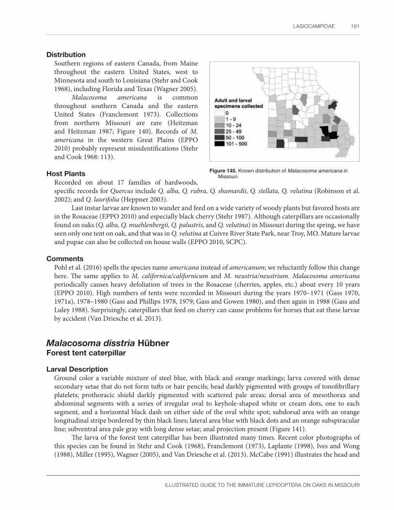
LASIOCAMPIDAE 191
ILLUSTRATED GUIDE TO THE IMMATURE LEPIDOPTERA ON OAKS IN MISSOURI
DistributionSouthern regions of eastern Canada, from Maine throughout the eastern United States, west to Minnesota and south to Louisiana (Stehr and Cook 1968), including Florida and Texas (Wagner 2005).
Malacosoma americana is common throughout southern Canada and the eastern United States (Franclemont 1973). Collections from northern Missouri are rare (Heitzman and Heitzman 1987; Figure 140). Records of M. americana in the western Great Plains (EPPO 2010) probably represent misidentifications (Stehr and Cook 1968: 113).
Host PlantsRecorded on about 17 families of hardwoods, specific records for Quercus include Q. alba, Q. rubra, Q. shumardii, Q. stellata, Q. velutina (Robinson et al. 2002); and Q. laurifolia (Heppner 2003).
Last instar larvae are known to wander and feed on a wide variety of woody plants but favored hosts are in the Rosaceae (EPPO 2010) and especially black cherry (Stehr 1987). Although caterpillars are occasionally found on oaks (Q. alba, Q. muehlenbergii, Q. palustris, and Q. velutina) in Missouri during the spring, we have seen only one tent on oak, and that was in Q. velutina at Cuivre River State Park, near Troy, MO. Mature larvae and pupae can also be collected on house walls (EPPO 2010, SCPC).
CommentsPohl et al. (2016) spells the species name americana instead of americanum; we reluctantly follow this change here. The same applies to M. californica/californicum and M. neustria/neustrium. Malacosoma americana periodically causes heavy defoliation of trees in the Rosaceae (cherries, apples, etc.) about every 10 years (EPPO 2010). High numbers of tents were recorded in Missouri during the years 1970–1971 (Gass 1970, 1971a), 1978–1980 (Gass and Phillips 1978, 1979; Gass and Gowen 1980), and then again in 1988 (Gass and Luley 1988). Surprisingly, caterpillars that feed on cherry can cause problems for horses that eat these larvae by accident (Van Driesche et al. 2013).
Malacosoma disstria HübnerForest tent caterpillar
Larval Description Ground color a variable mixture of steel blue, with black and orange markings; larva covered with dense secondary setae that do not form tufts or hair pencils; head darkly pigmented with groups of tonofibrillary platelets; prothoracic shield darkly pigmented with scattered pale areas; dorsal area of mesothorax and abdominal segments with a series of irregular oval to keyhole-shaped white or cream dots, one to each segment, and a horizontal black dash on either side of the oval white spot; subdorsal area with an orange longitudinal stripe bordered by thin black lines; lateral area blue with black dots and an orange subspiracular line; subventral area pale gray with long dense setae; anal projection present (Figure 141).
The larva of the forest tent caterpillar has been illustrated many times. Recent color photographs of this species can be found in Stehr and Cook (1968), Franclemont (1973), Laplante (1998), Ives and Wong (1988), Miller (1995), Wagner (2005), and Van Driesche et al. (2013). McCabe (1991) illustrates the head and
Figure 140. Known distribution of Malacosoma americana in Missouri.

192 SPECIES DESCRIPTIONS
ILLUSTRATED GUIDE TO THE IMMATURE LEPIDOPTERA ON OAKS IN MISSOURI
mandible. Stehr and Cook (1968) give a detailed description of all stages of M. disstria. They noted that the D1 verruca has four setae and the L1 verruca is bisetose on A3-6, characters shared with the European pest M. neustria, but no other North American species. The easiest way to identify M. disstria is by the characteristic coloration that includes a series of anterior oval or keyhole-shaped, white or silver dots dorsally (Stehr 1987).
Pupal DescriptionModified from Mosher (1916a): Eyes, labrum, mandibles, and dorsal part of maxilla pale white (colored similar to the “brain window” of saturniids); setae on vertex longer than half the height of the glazed eyepiece; body covered with dense setae except for appendages; labial palpi exposed; maxillae approximately one-quarter length of prothoracic leg; mesothoracic legs slightly longer than antenna; metathoracic legs exposed or hidden; abdominal segments A8-10 narrower than other abdominal segments; cremaster absent, the terminal segments of the abdomen covered in stout straight or slightly curved setae.
The pupa of M. disstria is illustrated and diagnosed by Mosher (1916a). Differences between male and female pupae are shown by Muggli (1974). Malacosoma pupae are covered with a dense coating of setae that is lacking in Tolype. In addition, segments A8-10 of Malacosoma are noticeably narrower than the rest of the abdomen. No one has studied the pupal morphology of Malacosoma, but Stehr and Cook (1968: 89) pointed out how M. disstria and M. americana differ in their cocoon and biology. The cocoon of M. disstria has more loose silk on the outside than the relatively naked M. americana. Also, M. disstria tends to spin a cocoon between two leaves, unlike M. americana that pupates in protected situations or on the ground. Another difference is that the cocoon of M. disstria has less yellow powder than M. americana (Wagner 2005). However, exceptions are known for all these differences. As noted under M. americana, the setal length on the vertex of M. disstria is longer than the glazed eyepiece. Using the diagrams in Peterson (1962a) and McCabe (1991), additional clues are possible from the larval mandibles that seem to differ slightly in shape.
Natural HistoryThe life cycle of the forest tent caterpillar was recently summarized by Ives and Wong (1988), USDA (1985), and Van Driesche et al. (2013). Females lay from 100–300 eggs in a band that encircles a twig. Eggs overwinter and hatch in spring of the following year. Swaine (1913) and Peterson (1966) illustrate the egg mass or a few isolated eggs from a confined female in a plastic bag. First instar larvae are colored black with dense white hairs, and their food consists of tender foliage or expanding buds. There are five instars with one generation per year. Young larvae are gregarious. They maintain contact by using silk trails or forming pads of silk as a molting substrate. Unlike other eastern species of lasiocampids, adults vary in wing color (Franclemont 1973).
In University City, MO, we have seen many of the life history traits mentioned above. Larvae descend from the canopy en masse to molt, forming groups containing hundreds of caterpillars. After molting, they return to the canopy to feed. Later instars disperse farther apart and remain separated without constructing a nest common to other Malacosoma species. The preferred pupation site is among leaves, although any site will do if no leaves are available.
DistributionWidely distributed throughout North America, from the Atlantic to Pacific coasts in southern Canada and the entire United States, except for Nevada and Arizona (Franclemont 1973).
Figure 141. Malacosoma disstria mature larva. Photo by R.J. Marquis, used with permission.

LASIOCAMPIDAE 193
ILLUSTRATED GUIDE TO THE IMMATURE LEPIDOPTERA ON OAKS IN MISSOURI
Malacosoma disstria no doubt occurs in both Arizona and Nevada, but records have not been formally published. See Figure 142 for Missouri distribution.
Host PlantsRecorded from 15 families of hardwoods; also conifers and herbaceous plants when other food is not available (Stehr and Cook 1968, USDA, 1985, Robinson et al. 2002); specific records for oaks include Quercus alba, Q. aquatica, Q. garryana, Q. macrocarpa, Q. marilandica, Q. rubra, Q. stellata,Q. velutina, Q. virginiana (Robinson et al. 2002); Q. coccinea/rubra (Wagner et al. 1995a); Q. laevis, Q. laurifolia, Q. nigra (Meeker 1997); Q. coccinea and Q. palustris in Missouri.
In the northern and western portions of North America, quaking aspen (Populus tremuloides) is preferred. Quercus, Liquidambar styraciflua, and Nyssa are the most common hosts in the southern states (Stehr and Cook 1968). We find last instar larvae on oaks during April and May in Missouri, but rarely so. According to Stehr and Cook (1968), red maple (Acer rubrum) and pear (Pyrus) trees were least favored by the forest tent caterpillar, as were a few other understory shrubs. Malacosoma disstria has the widest host range of any Malacosoma species in North America.
CommentsThe forest tent caterpillar can cause serious damage to the host because defoliation often lasts for more than two consecutive years (Duncan and Hodson 1958, USDA 1985, Ives and Wong 1988). Malacosoma disstria is the most destructive species of the genus in the United States (Stehr and Cook 1968). Damage is common on aspen (Populus) in the northern United States and Canada, maple (Acer) in New England, Nyssa and sweetgum (Liquidambar) in the southern United States, and on oaks in Texas (Stehr 1987). Outbreaks occur every 6 to 16 years and have included several species of oaks in Florida (Meeker 1997). Elms (Ulmus) are eaten in the Mississippi valley (Belzer and Morris 1978). Gass (1971a) reports that M. disstria was documented in Missouri in 1971 for the first time in over 75 years. Large swarms were seen on Q. palustris and L. styraciflua in University City, MO, during 2011.
Phyllodesma americana (Harris) American lappet moth
Larval Description Modified from Stehr (1987): Larva varies from uniform gray to blue-gray to black, with orange to red black spotted segmental transverse bands on T2 and T3 that are hidden at rest; markings, when present, consist of faint, thin orange or yellow middorsal stripes lined or mottled with black; the dark color morph has A1 unmarked, a dark gray middorsal line, and scattered subdorsal white markings on the rest of the abdomen; A8 of all forms with single middorsal hump; anal projection probably present (Figure 143a, b).
The larva of P. americana is illustrated in color by Laplante (1998), Ives and Wong (1988), Miller and Hammond (2003), Wagner (2005), and Sogaard (2009). It is identified by the orange segmental membranes usually near three black dots, presence of lateral tufts (lappets), and the middorsal hump on A8 (Wagner 2005). In addition, unlike Tolype or Artace, there are no fanlike setae (Stehr 1987). Early instars of P. americana
Figure 142. Known distribution of Malacosoma disstria in Missouri.

194 SPECIES DESCRIPTIONS
ILLUSTRATED GUIDE TO THE IMMATURE LEPIDOPTERA ON OAKS IN MISSOURI
a b
Figure 143. Phyllodesma americana (a) mid-instar larva; (b) mature larva. Photos by R.J. Marquis, used with permission.
are superficially similar to M. disstria because they have white slightly teardrop-shaped patches subdorsally on T2, T3, and A2, and well as smaller ones on A5 and A6. Malacosoma species lack a midorsal hump on A8 that is present in P. americana.
Pupal DescriptionBody cylindrical, not dorsoventrally flattened, with long setae and a waxy covering; labial palpi exposed; maxillae approximately two thirds the length of the prothoracic leg; prothoracic coxae barely exposed, the femur hidden; mesothoracic legs approximately as long as prothoracic legs; metathoracic legs hidden; mesothoracic spiracle slitlike; abdominal flanges have a thin circle of setae; cremaster absent, last segment with curved setae.
The pupa of P. americana lacks the knoblike cremaster present in T. velleda. The head of P. americana is concolorous with the body and the abdominal setae surround the segmental flanges in a circle. The setae in both Missouri species of Malacosoma evenly cover the abdomen. In addition, in Malacosoma, the eyes, labrum, mandibles, and the dorsal part of the maxillae are contrastingly pale white.
Natural HistoryThe egg of P. americana was illustrated by Peterson (1966). It is oval, white, and has blue-black bands. Larvae are found from May to October (Wagner 2005). There are two broods in Missouri; the first has adults from April to early June, whereas the second brood is in July (Franclemont 1973). Other areas have one generation per year (Wagner 2005). The pupa overwinters in a tough cocoon on tree bark (USDA 1985). Last instar larvae are often found, well concealed, resting on branches. We rarely find this species on Missouri oaks.
DistributionSouthern Canada and the United States except for costal South Carolina, Georgia, Florida, and eastern Texas (Franclemont 1973). See Figure 144 for Missouri distribution.
Host PlantsMany trees and shrubs including Alnus, Betula, Populus, Malus, Ostrya, Prunus Quercus, Salix (Wagner 2005); Quercus garryana, Q. ilicifolia, and Q. rubra (Robinson et al. 2002). We have found P. americana on Q. alba and Q. imbricaria in Missouri.
Figure 144. Known distribution of Phyllodesma americana in Missouri.

LASIOCAMPIDAE 195
ILLUSTRATED GUIDE TO THE IMMATURE LEPIDOPTERA ON OAKS IN MISSOURI
CommentsThe larva of Phyllodesma occidentis is undescribed (Franclemont 1973, Wagner 2005) and likely very similar to P. americana. It is recorded around, but not from, Missouri (Franclemont 1973, J.R. Heitzman and P.E. Koenig, unpublished). Heppner (2003) lists oak as one of many host plants for P. occidentalis. Rearing to adult or using molecular identification is recommended for members of this genus, especially in southern Missouri.
Subfamily Macromphaliinae
Tolype velleda (Stoll)
Larval DescriptionModified from Stehr (1987) and Wagner (2005): Larva dorsoventrally flattened; uniformly gray, with or without irregular dorsal white to orange patches; metathorax with enlarged D pinacula and a black transverse segmental bar concealed at rest; A5 lacks enlarged dorsal verrucae; A8 with two dorsal verrucae; all segments with fleshy lateral lappets, often largest on the thorax; anal projection present (Figure 145a, b).
The larva of T. velleda is illustrated by Peterson (1962a) and in color by Ives and Wong (1988), Wagner (2005), and Sogaard (2009). Although the coloration is somewhat variable, T. velleda is identified by the large metathoracic dorsal verrucae, transverse segmental bars, and paired dorsal verrucae on A8. In addition, the dorsal verrucae of A5 are not enlarged compared to the verrucae on the other abdominal segments.
a b
Figure 145. Tolype velleda (a) mid-instar larva; (b) mature larva. Photos by R.J. Marquis, used with permission.
Pupal DescriptionModified from Mosher (1916a): Body dorsoventrally flattened; body without long setae but scars of verrucae present on abdomen; labial palpi exposed; maxillae approximately one-half length of prothoracic leg; prothoracic coxae exposed, the femur hidden; mesothoracic legs slightly shorter than antennae; metathoracic legs exposed; mesothoracic spiracle slitlike; cremaster a very small knob with irregular texture of ridges or teeth.
Mosher (1916a) studied the pupa of T. velleda. The knoblike cremaster and lack of body setae will separate T. velleda from Malacosoma in Missouri. The pupa of T. velleda, like the larva, has two separate dorsal verrucae on A8.
Natural HistoryThe egg of T. velleda is illustrated by Peterson (1966). The chorion is reddish brown and the eggs are laid in short rows covered by hairs from the female’s body. The life cycle is summarized by USDA (1985). Moths

196 SPECIES DESCRIPTIONS
ILLUSTRATED GUIDE TO THE IMMATURE LEPIDOPTERA ON OAKS IN MISSOURI
appear in the fall and lay their eggs at that time. Larvae appear from June to August. Pupation occurs on the twigs or bark of the host in a tough, flat, tapered cocoon (Sogaard 2009). The egg overwinters (Wagner 2005).
This caterpillar is very difficult to find when it is hiding on branches and twigs because the hairs conceal the body form (Lugger 1898). It may be much more abundant than our three larval records suggest. There is one generation in Missouri (Heitzman and Heitzman 1987).
DistributionSouthern Canada to central Florida and west to Minnesota, Nebraska, and Texas (Franclemont 1973). See Figure 146 for Missouri distribution.
Host PlantsAcer, Betula, Diospyros, Fraxinus, Malus, Populus, Prunus, Pyrus, Quercus alba, Q. phellos, Salix, Syringa, Tilia, Ulmus (Robinson et al. 2002); Ilex (USDA 1985); and Fagus (Wagner 2005). We have found T. velleda on Q. alba, Q. imbricaria, and Q. velutina in Missouri.
CommentsFranclemont (1973) pointed out that the caterpillar of Artace cribrarius is very similar to Tolype and both species fed on oak. He stated the dorsum of A8 has two verrucae in Tolype but only a single verruca in Artace. However, this diagnosis is tentative for several reasons. Larvae of Artace (reared from eggs of a female moth collected in Maryland and matching the photographs of the genus in Wagner 2005) have paired verrucae on A8, not just a single dorsal one. As a final caution, other species of Tolype with poorly known larvae are possible in Missouri. For now, T. velleda is separated from Artace by the lack of enlarged verrucae on A5 (Wagner 2005). Unlike T. velleda, the pupa of Artace lacks a knoblike cremaster and instead has an upturned ridge with minute serrated teeth in a row (SCPC).
Figure 146. Known distribution of Tolype velleda in Missouri.

SATURNIIDAE 197
ILLUSTRATED GUIDE TO THE IMMATURE LEPIDOPTERA ON OAKS IN MISSOURI
SaturniidaeWild silk moths
The family Saturniidae consists of approximately 2,350 species worldwide (Nieukerken et al. 2011), including most of the world’s largest moths. Among lepidopterists and the general public, saturniids are a favorite group for breeding, photographing, and collecting due to their large size, beauty, and spectacular caterpillars (Pinratana and Lampe 1990, Tuskes et al. 1996, Passoa 1999, Rougerie and Estradel 2008). This attention has generated a voluminous literature for all stages in many faunal regions. Some of the larger multivolume works include Packard’s (1905, 1914) life history studies emphasizing the North American fauna, d’Abrera’s (1995, 1998, 2012) three volume adult pictorial guide that includes holotype illustrations, and Lemaire’s (1978, 1980, 1988, 2002) revisions of the New World fauna (originally under the name Attacidae) with drawings of genitalia. Regional works on Saturniidae exist for parts of Africa (see reviews in Oberprieler 1995, Rougerie and Estradel 2008) and several Asian countries (Pinratana and Lampe 1990, Nässig et al. 1996, Holloway 1987). Revisions exist for some of the more commonly encountered genera including Anisota (Riotte and Peigler 1981), Attacus (Peigler 1989), Automeris (Lemaire 1971, 1973, 1974), Copaxa (Wolfe 1993), and Samia (Peigler and Naumann 2003). Several handbooks have discussed rearing saturniids (Crotch 1956, Gardiner 1982, Baxter 1992). Lampe (2010) photographed the life histories of many species from egg to adult. Stone (1991) and Santin (2004) listed host plants for saturniids of the world. Peigler (1996) cataloged the parasitoids of this family. A useful Web site is http://www.silkmoths.bizland.com/indexos.htm, especially for Neotropical species.
Although popular with lepidopterists, a rigorous cladistic classification for the Saturniidae was still lacking until recently (Deml and Dettner 2002: 90). Michener (1952) published a morphological study on New World species, whereas Oberprieler (1997) studied the African fauna. Phylogenies exist for the Arsenurinae (Peigler 1993a), the Ceratocampinae (Balcazar-Lara and Wolfe 1997), and the moon moths (Yllas et al. 2005). The latest molecular phylogeny of the Saturniidae is by Regier et al. (2002, 2008). Based on morphology, Lemaire and Minet (1998) recognized nine saturniid subfamilies, including the Oxyteninae and Cercophaninae, both usually considered separate families (reviewed by Jordon 1924). Regier et al. (2008) suggested eight subfamilies based on molecular data.
Several important books on the North American fauna have been published since Packard (1905, 1914). Ferguson (1971, 1972) treated the family in the traditional sense, emphasizing morphology and keys. The book by Tuskes et al. (1996) has more biological details and excellent photographs of the immature stages. Photographs of the eastern United States saturniid species are shown in Wagner (2005).
Studies of the Saturniidae, and to some extent other macrolepidoptera in Europe, are hampered by the recent trend of some authors publishing new species descriptions in monographs that are difficult to obtain or extremely expensive. Without easy access to these revisions, identification of some saturniid taxa, especially from the Old World, is not always accurate.
Saturniid eggs are of the flat type, with a chorion that appears smooth and thick (Lemaire and Minet 1998), but that is pitted under magnification (Syme 1961). Depending on the species, they are shades of white, yellow, green, brown, beige, or gray and may be banded, spotted, or solid (Lampe 2010). Saturniinae lay eggs singly or in small groups that are attached to leaves or twigs with brownish or reddish cement (Tuskes et al. 1996). Arsenurinae eggs are usually brown and white (Lemaire and Minet 1998). Hemileucinae eggs have a conspicuous black spot indicating the micropyle (Lemaire and Minet 1998) and are laid in clusters or rings (Tuskes et al. 1996) that may encircle a twig similar to egg masses of Malacosoma. Females of Hylesia cover the eggs with scales from their body (Janzen 1984a, Lemaire and Minet 1998, Lemaire 2002: Plate ES14). In at least one species of Cercophaninae (Cercophana), the eggs are green with a white band (Wolfe and Balcázar-Lara 1994). A species in the Oxyteninae (Oxytenis) has green eggs at first that turn transparent at maturity (Aiello and Balcázar-Lara 1997). This development is similar to some Ceratocampinae (=Citheroniinae) that also have a clear chorion (Gardiner 1982: 54). Lampe (2010) photographed saturniid eggs of the world. Syme (1961) and Peterson (1965b) focused on North American species. The few species of Saturniidae known from Europe were included in Doring (1955) and photographed by Pljushch and Dolinskaya (2001).

198 SPECIES DESCRIPTIONS
ILLUSTRATED GUIDE TO THE IMMATURE LEPIDOPTERA ON OAKS IN MISSOURI
Saturniid larvae are relatively well known (Rougerie and Estradel 2008). First instar larvae show a wide range of morphological specializations illustrated by Pease (1960), Balcázar-Lara and Wolfe (1997) and Lampe (2010). Pease (1960: 107) divided the Saturniidae into two groups based on first instar larval morphology, providing a key to species with emphasis on Nearctic genera. Body setae may be simple or modified into scoli that are branched or clubbed. Old World species often have secondary setae on wart-like scoli and some or all of their prolegs. New World species rarely have secondary setae. Another difference is that New World saturniid species have the D2 scoli fused on A9 (Balcázar-Lara and Wolfe 1997: 21). The first instar of Oxytenis differs from the species of Ceratocampinae studied by Balcázar-Lara and Wolfe (1997) because it lacks primary setae on the thoracic D and SD scoli and the D2 scoli of A9 are not fused. They also noted the tip of the metathoracic dorsal scolus seems especially variable in the Ceratocampinae. Automeris and related genera have eversible glands on A1 and A7 (Pease 1960). The first instar of Cercophana does not fit any of Pease’s (1960) groups (Wolfe and Balcázar-Lara 1994) and the presence of secondary setae is unusual for New World saturniids. First instar larvae of the Salassinae lack scoli and instead have protuberances on the prothoracic and anal shields (Lemaire and Minet 1998). Characters from first instar saturniid larvae merit study and have proved important in several phylogenetic analyses at higher levels (Pease 1960, Peigler 1993a, Balcázar-Lara and Wolfe 1997, Lemaire and Minet 1998) and as a character to group tribes of the Saturniinae (Rougerie and Estradel 2008: 230).
Depending on the species, at maturity saturniid larvae have several body forms and a range of typical color patterns, including stripes, bands, or spots (Lampe 2010). Silver markings are found in Syssphinx and relatives (Balcázar-Lara and Wolfe 1997: 23), some African genera, and Antheraea (Nässig et al. 1996: 123). Some African saturniids have colored body platelets (Oberprieler 1995) resembling the lenticles of skipper larvae. More rarely larvae are almost a solid color with reduced markings (Lampe 2010). Waxy coatings are present in Attacus and Samia (Deml and Dettner 2002). Thoracic coxae are fused in all members of the African tribe Bunaeini as well as other saturniids (Rougerie and Estradel 2008: 224). The subfamilies Arsenurinae, Agliinae, Oxyteninae (Balcázar-Lara and Wolfe 1997: 22), and Salassinae (Deml and Dettner 2002) lack scoli. When scoli are present, they vary in both morphology and function. Deml and Dettner (2002: 89) recognized several types of scoli in the family Saturniidae, including some that are flat, branched and erect, thorny, or in a rosette (“star warts”). Some are secretory, whereas others are urticating. The scoli of Oxyteninae are sticky (Aiello and Balcázar-Lara 1997) and represent yet another type.
Many saturniid larvae show characteristic resting postures or defensive behaviors in a manner analogous to the adults (see Blest 1957). For example, cecropia larvae “tense up” when disturbed (V. Passoa, pers. obs.). This increase in hemolymph pressure is critical to the release of defensive chemicals that are surprisingly different for each colored scolus and instar (Deml and Dettner 2003). The target organisms of scoli defensive secretions are likely microorganisms or arthropod enemies (Deml and Dettner 1997). All Hemileucinae are gregarious in at least the first instar, and although this behavior also occurs in other subfamilies (Lemaire and Minet 1998), the vast majority of saturniids are solitary. Some Hylesia live in silk pouches (Wolfe 1988).
At least in North America, saturniid larvae are recognized by their smooth head, presence of at least rudimentary scoli, and biordinal crochets arranged in a homoideous mesoseries (Ferguson 1971: 8). The front normally extends less than half the distance to the epicranial notch (Lemaire and Minet 1998). The scissorial teeth on saturniid mandibles have two ontogenetic patterns; these teeth are either lost or retained at maturity (Passoa and Passoa 1999). Bernays and Janzen (1988), based on Costa Rican species, characterized late instar saturniid mandibles as “short, with a broad base, and without obvious teeth” in contrast to the complicated mandibles of the Sphingidae. However, this distinction disappears outside of Central America because both types of mandibles (sphingid and saturniid type) occur in the Attacini (Passoa and Passoa 1999).
Saturniids are most likely to be confused with nymphalid butterfly larvae, which also often have well-developed scoli. However, unlike saturniids, nearly all nymphalids have an angular and/or spined head (Godfrey et al. 1987b, a few exceptions in Fracker 1915: 125). Outside North America, some saturniids (e.g., Arsenurinae from Mexico, SCPC) resemble lasiocampids, but presumably they lack the anal protuberance (“point”) characteristic of larval Lasiocampidae. More rarely, saturniids are confused with early instar swallowtails

SATURNIIDAE 199
ILLUSTRATED GUIDE TO THE IMMATURE LEPIDOPTERA ON OAKS IN MISSOURI
(Papilionidae) that may have scoli. Saturniid larvae lack an osmetarium that is present in Papilionidae. The possession of sticky glands, habit of resting in a “J” position, utilization of rubiaceous food plants, and the common presence of eyespots and a tail on A8 help define the Oxyteninae (Aiello and Balcázar-Lara 1997). Although their body shape is unusual, the larva of Cercophana (Wolfe and Balcázar-Lara 1994) at first glance could be confused with a notodontid caterpillar. When mature, saturniid larvae can be identified by their large size, but early instars are easily confused with several poorly known smaller Old World families of Bombycoidea (see Lemaire and Minet 1998).
Saturniid larvae form cocoons that may be double or single walled, tightly or loosely spun, or lacelike (see Gardiner 1982: Figs. 15–26). Pupal Saturniidae are difficult to characterize because they display a wide range of morphological specializations. According to Mosher (1916b), the antennae usually show a pectinate condition, the sutures of the head and legs are poorly developed, the labial palpi are rarely exposed, and the maxillae rarely extend more than one-sixth the distance to the caudal margin of the wings. The pupa of Antheraea polyphemus is sexually dimorphic: the male antenna covers all the thoracic legs, whereas the female antenna is not as broad (Hall 2012). There may be tubercles on the mesonotum, metanotum, and even minute ones on either side of the labrum (SCPC). Pupae of the luna and polyphemus moths have a well-developed mesothoracic tubercle (Mosher 1916b) that covers a spine used by the adult to cut the cocoon upon emergence (Hilton 1965) after secreting an enzyme (Tuskes et al. 1996: 176). Other species spin a cocoon with an “escape valve” that allows the moth to exit without using cutting tubercles or enzymes (Sogaard 2009, SCPC). Saturniid pupae rarely have visible setae, and the abdomen either is spiny, smooth (Ferguson 1971: 7), or granular (Oberprieler 1995). Some abdominal intersegmental membranes of Bunaeopsis (Rougerie and Estradel 2008: Fig. 67) and Gonimbrasia tyrrhea (SCPC) have a large flange lined with vertical grooves visible posteriorly. Albertoni and Duarte (2015) noted that pupal Ceratocampinae in the New World tropics have metanotal tubercles, a spiny abdomen, and bifurcated cremaster. In some members of the tribe Saturniini, there is a clear area of transparent cuticle called the brain window, which may play a role in controlling diapause (Tuskes et al. 1996). The dorsum of A10 in Caio (Lemaire and Minet 1998), Bunaeopsis (Rougerie and Estradel 2008: Fig. 67), and other African saturniids (Oberprieler 1995, SCPC) has two deep pits called caudal cavities. Saturniid pupae have hooked or stiff setae on their cremaster, unlike the related families Endromidae and Lemoniidae that have a cremaster covered with spines (Patočka and Turčáni 2005: Plate 158). Balcázar-Lara and Wolfe (1997: 23) considered the long bifurcated cremaster of the Ceratocampinae to be an apomorphy of the subfamily. A cremaster is absent in other taxa, including Hylesia (Wolfe 1988), Copaxa (Wolfe 1993), Cercophana (Wolfe and Balcázar-Lara 1994), and most North American Saturniinae (Mosher 1916b). Saturniid pupae can be found in the soil, leaf litter, or on the host plant (Godfrey et al. 1987b). As with the larvae, separation of saturniid pupae from related Bombycoidea in the Old World can be difficult.
For more information on North American saturniid moths, consult Ferguson (1971, 1972) and Tuskes et al. (1996) and the references contained in these works. Mosher (1914, 1916a, 1916b) published keys to the North American pupae. Janzen (1984b) studied saturniid ecology in Costa Rica. Saturniids have long been valued as a source of food and silk and as symbols in art and religion (Peigler 1993b, Tuskes et al. 1996). A few saturniids are also medically important. The larvae of some species contain urticating spines that are capable of causing rashes, or very rarely in some parts of South America, death (Kelen et al. 1995, Diaz 2005). Saturniids are rare pests of forest trees in the United States (Tuskes et al. 1996), Latin America (Hilje et al. 1992, Cibrián Tovar et al. 1995), and the Old World (Evans 1952, Zhang 1994). With reference to oak in the United States, two species of Anisota (A. senatoria and A. virginiensis) and Dryocampa rubicunda can cause serious defoliation (Ferguson 1971, Wilson 1971, USDA 1985, Stimmel 1988). All three of these species occur in Missouri. Most other saturniids are not common on oak in Missouri, but when they are found they usually attract attention. Therefore, we include them in our book. Surprisingly, we did not encounter the buck moth, Hemileuca maia, during our survey. This species is restricted to the southern part (Ozark Region) of Missouri (Heitzman and Heitzman 1987) and apparently is not common in our study area. Consult Ferguson (1971), Tuskes et al. (1996), and Wagner (2005) for information on H. maia. Besides the species listed below, we have encountered and reared Dryocampa rubicunda on Q. alba in Missouri.

200 SPECIES DESCRIPTIONS
ILLUSTRATED GUIDE TO THE IMMATURE LEPIDOPTERA ON OAKS IN MISSOURI
Subfamily Hemileucinae
Automeris io (Fabricius) Io moth
Larval DescriptionModified from Packard (1914) and Hall (2014b): First instar larva with light brown to dark red to black head capsule, light red to orange thorax and abdomen, sometimes with faint longitudinal stripes or contrasting scoli; middle instars uniformly light yellow to orange to brown with faint longitudinal stripes and black tipped contrasting scoli (see Appendix 3, Plate 1). Last instar with a light green head capsule, thorax, and abdomen; all scoli black tipped, approximately equal in size, and of the rosette type; spiracles white with a black peritreme; secondary setae numerous on the prolegs; a red stripe connecting the spiracles, with a thicker white subspiracular stripe and an additional broken thin red line sometimes present below the white line; a subventral light patch behind each abdominal proleg (Figure 147).
The chaetotaxy of A. io is partially illustrated by Packard (1914), Fracker (1915), and Peterson (1962a). The mature larva has been photographed many times; some recent examples are Heitzman and Heitzman (1987), Heppner (1994), Tuskes et al. (1996), Lemaire (2002), Wagner (2005), Lampe (2010), and Hall (2014). Variation in larval coloration has been documented, especially for the earlier instars. Elliot and Soule (1902) noted that the first instar had an almost black head. Preserved larvae from several clutches of A. io from eastern United States localities had a light brown to dark red first instar head color (V.A. Passoa, pers. obs.). Middle instars are sometimes dark brown (Lampe 2010: Plate 106: L2) to black (Lemaire 2002: Plate ES7: 2). The ground color of A. io is usually green at maturity, but rarely individuals may be golden yellow or bluish green (Heppner 1994, Tuskes et al. 1996).
The caterpillar of A. louisiana is similar to that of A. io (Wagner 2005). However, A. louisiana has only been found in Louisiana and restricted coastal areas of Mississippi and Texas (Tuskes et al. 1996) and has not been collected in Missouri (J.R. Heitzmand and P.E. Koenig, unpublished). Although Tuskes et al. (1996) and Lemaire (2002) suggest using color differences in the lateral stripe to separate A. io from A. louisiana, the best way to separate these two species is by host plant and locality. Automeris louisiana is not found on oak under natural conditions, and its preferred host plant is cordgrass (Tuskes et al. 1996). Automeris io has a distinctive appearance unlike any other oak-feeding caterpillars in Missouri. However, sibling species of Automeris with similar larvae in Latin America can complicate identifications outside of Missouri.
The larva of A. io has eversible spiracular glands on A1 and A7 (see illustrations in Packard 1914 and Peterson 1962a). These glands are not restricted to A. io, but their exact distribution in Automeris or the rest of the Saturniidae remains unstudied. Their function is also unknown. The scoli of A. io may cause a painful sting followed by swelling (Heppner 1994, Wagner 2005, references in Hall 2014b).
Pupal DescriptionModified from Mosher (1916b): Dark red-brown pupa with conspicuous sparse, small setae; body cuticle shagreened; brain window absent; maxillae small; labial palpi hidden; prothoracic legs exposed; prothoracic femur hidden; mesothoracic legs partially hidden; metathoracic legs completely hidden; mesothoracic
Figure 147. Automeris io mature larva. Photo by R.J. Marquis, used with permission.

SATURNIIDAE 201
ILLUSTRATED GUIDE TO THE IMMATURE LEPIDOPTERA ON OAKS IN MISSOURI
dorsum with transverse striations; mesothoracic spiracle deep and pitlike; A5-7 with flange plates, anterior to the flange plates are transverse striations; cremaster buttonlike, not bifurcated, with recurved spines.
The pupa of A. io was diagnosed and illustrated by Mosher (1914, 1916a). Ferguson (1972: 156) states that the pupa of A. io and A. pamina differ from other North American hemileucines by their sparse covering of contrasting red-brown hairs on the vertex, thorax, and last abdominal segment. Automeris io has striations on the mesothoracic dorsum that are absent in A. pamina (Mosher 1914). Automeris io, unlike the related oak-feeding Hemileuca, forms a cocoon (Mosher 1914, see photographs in Villiard 1975, Hall 2014b). A few specimens of A. io had three pairs of tiny tubercles on the metathorax and A1 (SCPC), but the significance of this variation is not known.
Natural HistoryThe egg of A. io is illustrated by Syme (1961), Peterson (1965b), Tuskes et al. (1996), and Hall (2014). Mature fertilized eggs are yellow with a black micropylar area and are laid in clusters on stems or leaves (Hall 2014b, also see Figure 7f). The natural history of A. io was summarized by Tuskes et al. (1996) and Hall (2014). Larvae are gregarious when young and follow silk trails to new feeding or resting sites until they later become solitary feeders (Tuskes et al. 1996). Larvae molt in a group with the head facing outward (Hall 2014b). At maturity, they rest without release of the thoracic legs, but curl and drop when disturbed (V.A. Passoa, pers. obs.). Unlike other saturniids, this behavior makes them hard to rear in sleeve cages (Collins and Weast 1961). Pupation occurs in papery cocoons among debris. There are one to four generations per year depending on the geographical region (Tuskes et al. 1996).
DistributionSouthern portions of eastern Canada to Manitoba, throughout the eastern United States and west to Colorado, the tip of southeastern Utah, and New Mexico (Tuskes et al. 1996: Map 25), then south to Costa Rica, including the West Indies (Bahamas) (Lemaire 2002: Map 91). See Figure 148 for Missouri distribution.
In the United States, Automeris io does not range west of the Rocky Mountains, but it is one of the few saturniids known from the West Indies (Lemaire 2002).
Host PlantsPolyphagous on grasses, herbaceous plants, shrubs, deciduous trees, and conifers (Heitzman and Heitzman 1987) of over 100 plant genera (Heppner 1994); specific records for oak include Quercus alba, Q. cerris, Q. coccinea, Q. emoryi, Q. gambelii, Q. kelloggi, Q. lobata, Q. macrocarpa, Q. palustris, Q. rubra, Q. stellata, Q. velutina (Robinson et al. 2002); Q. acuttisima, Q. laevis, Q. laurifolia, Q. myrtifolia, Q. virginiana (Heppner 2003); and Q. nigra (V.A. Passoa, pers. obs.).
According to Tuskes et al. (1996), the most common host plants are Cercis, Celtis, Hibiscus, Prunus, Pyrus, Ribes, Rubus, Salix, Sassafras, and Wisteria, but local host plant preferences can vary regionally or even within a state. For example, they noted Piscidia, Delonix, and occasionally Hibiscus are some common hosts in the Florida Keys. Prosopis is the favorite host in costal Texas (Collins and Weast 1961), but around Houston, larvae can be collected on Cercis, Ligustrum, and Cynodon dactylon (Tuskes et al. 1996). In the vicinity of Houston, caterpillars were often found on Q. nigra and ornamental Ligustrum (V.A. Passoa, pers. obs.). Betula and Populus are the most common hosts in Canada (Ives and Wong 1988). Conifers are usually
Figure 148. Known distribution of Automeris io in Missouri.

202 SPECIES DESCRIPTIONS
ILLUSTRATED GUIDE TO THE IMMATURE LEPIDOPTERA ON OAKS IN MISSOURI
avoided (Godfrey et al. 1987b). Consult Hall (2014) for a larger list of preferred hosts for A. io compiled from recent literature.
Automeris io larvae are found on Chamaecrista fasciculata in Missouri (R.J. Marquis, pers. obs.) and are commonly found on Sassafras albidum in the Missouri Ozarks (Heitzman and Heitzman 1987). We have found A. io on Q. alba in Missouri, but it is generally rare, even on Q. alba.
CommentsConsult Lemaire (2002) and Hall (2014) for a review of literature relating to A. io.
Subfamily Saturniinae
Antheraea polyphemus (Cramer) Polyphemus moth
Larval DescriptionModified from Pease (1960), Jolly et al. (1970), Godfrey et al. (1987b), Wagner (2005), and Lampe (2010): First instar transversely banded, secondary setae on prolegs of A3-6, D2 absent on A9; second instar pale green with faint bands and yellow tipped scoli that are slightly larger anteriorly (see Appendix 3, Plate 2); later instars with head capsule always brown, labrum almost completely divided into two parts, mandible with a series of complex teeth and ridges; prothorax edged in yellow anteriorly; body color translucent pale to yellow-green with constrictions at the intersegmental membranes, giving the impression that the larva has a serrated dorsal margin; dorsal, subdorsal, lateral, and subventral scoli wartlike, with two to five setae, colored yellow orange to red, often mixed with silver; spiracles orange, the posterior margin of those on A2-7 touched by a pale, slanted line; prolegs on A3-6 with 9–12 secondary setae; anal shield with a brown, rarely white, stripe at the rim extending to A9 (Figure 149a, b).
Peterson (1962a) and Jolly et al. (1974) illustrate the chaetotaxy of A. polyphemus. The life stages of A. polyphemus are illustrated by Packard (1914) and Lampe (2010). The mature larva has been photographed many times. Some examples in color are Heitzman and Heitzman (1987), Ives and Wong (1988), Miller (1995), Tuskes et al. (1996), Wagner (2005), Brown et al. (2007), and Lampe (2010). The illustration of A. polyphemus by Villiard (1975: 150) is incorrect. It clearly shows a spotted head capsule and longitudinal spiracular stripe,
a b
Figure 149. Antheraea polyphemus (a) mid-instar larva; (b) mature larva. Photo a by R.J. Marquis; b by V.A. Passoa; both used with permission.

SATURNIIDAE 203
ILLUSTRATED GUIDE TO THE IMMATURE LEPIDOPTERA ON OAKS IN MISSOURI
both features of which are characteristic of certain Asiatic Antheraea species (Nässig et al. 1996: 134). Instead, A. polyphemus and A. oculea both have an unmarked head and slanted lines that touch the spiracles on A2-7 (Tuskes et al. 1996). These distinctive slanted lines are even absent on related species such as A. godmani or A. montezuma (Nässig et al. 1996: 125). Another difference separating the polyphemus moth from other Asian relatives is the chaetotaxy of A9. The lateral scoli has moved subdorsally in A. polyphemus. The opposite is true for Asian species studied by Jolly et al. (1944), for which the scoli on A9 are located subdorsally rather than laterally.
The caterpillar of the polyphemus moth is sometimes confused with the luna moth caterpillar (Tuskes et al. 1996), but there are numerous color differences in live larvae. The head color is always brown in the polyphemus moth but either green or brown in the luna moth (V.A. Passoa, pers. obs.). Spiracular slanted lines are present in the polyphemus moth, whereas the luna moth has a longitudinal spiracular line. Tuskes et al. (1996) mentioned that the slanted stripes in the polyphemus moth touch the spiracle, whereas in the luna moth these stripes, if present, are intersegmental (also see Sogaard 2009). Preserved larvae can be identified by the number of SV setae on A3-6 as in Ferguson (1972), or more easily by noting the presence of spatulate or thickened dorsal setae in the luna moth that are lacking in the polyphemus moth. A final difference is in the mandibles. The mandible of the luna moth is simple and toothed compared to the polyphemus moth that has a complex series of small teeth and ridges (similar to illustrations in Passoa and Passoa 1999: Figs.7–9).
Tuskes et al. (1996) could not find larval characters to separate A. polyphemus from A. oculea, but the latter species is restricted to Arizona, New Mexico, and parts of Mexico. Based on illustrations in Tuskes et al. (1996), Nässig et al. (1996: Fig. 59), and Lampe (2010), the slanted lines in A. oculea are twice as thick as those in A. polyphemus.
Pupal DescriptionModified from Mosher (1916b): Pupa yellowish brown; body smooth, shagreened, or with fine striations; a long brain window extends from the top of the vertex to the labrum running between the eyes; antennae sexually dimorphic, in the male covering all the appendages except the maxillae and labrum, in the female, thinner with parts of the prothoracic and mesothoracic legs exposed; labrum with 3–4 small tubercles, the bottom pair largest; maxillae very short, as long as the brain window (female) or partially hidden by the antenna (male); prothoracic femur hidden; metathoracic legs usually hidden; mesothoracic spiracle a deep pit, the anterior margin convex; mesothoracic tubercle well developed; abdominal spiracles large and oval; anterior margins of A5-7 without transverse furrows; cremaster composed of curved spines in two groups.
The pupa of A. polyphemus is illustrated by Mosher (1916b) and photographed in color by Heitzman and Heitzman (1987), Bouseman and Sternburg (2002), Lampe (2010), and Hall (2012). The presence of a brain window, large mesothoracic tubercle, body that tapers posteriorly, two groups of slightly curved spines instead of a well-developed cremaster, and cocoon lacking an escape valve help identify this species. It is possible to confuse polyphemus and luna moth pupae, but the two species differ in characteristics of the cremastral spines and the texture of A10 (Mosher 1916a). The spines of the luna moth are recurved and A10 is rugose, whereas the polyphemus moth has a smoother cuticle on A10 and the spines are only slightly curved. Larval mandibles can be dissected and used in pupal identifications; see the differences mentioned above for these two species. A final difference is in the cocoon. Antheraea polyphemus has a hard, oval cocoon, whereas the luna moth has a more papery one (Tuskes et al. 1996: Figs. 31, 32).
Natural HistoryThe egg of the polyphemus moth is illustrated by Syme (1961), Peterson (1965b), Lampe (2010), and Hall (2012). The eggs are cream to light tan with a wide brown to reddish brown band (Tuskes et al. 2006). Based on a scanning electron microscope study, Regier et al. (1980) recognized four regions in the egg: an aeropyle crown, a flat area, a striped area, and the micropyle. The aeropyle crown appears to be a modification restricted to Antheraea and was not found in other related bombycoids sampled by Regier et al. (2005).

204 SPECIES DESCRIPTIONS
ILLUSTRATED GUIDE TO THE IMMATURE LEPIDOPTERA ON OAKS IN MISSOURI
The natural history of A. polyphemus was summarized by Tuskes et al. (1996) and Hall (2012). All instars are solitary feeders, and there are usually one to two generations per year. Larvae of A. polyphemus often rest with their anterior end raised in a sphinx-like posture (Packard 1914: 208). When disturbed, larvae swing their head from side to side, tense up, or produce a clicking sound with their mandibles (V.A. Passoa, pers. obs.). The mandible clicks probably function as a warning signal to natural enemies that regurgitation of digestive fluids will follow (Brown et al. 2007). In captivity, mandible clicking by one caterpillar seems to encourage others to follow (Wagner 2005), but a physiological mechanism to explain communication between individuals is not obvious (Brown et al. 2007).
Several kinds of cocoons are formed by A. polyphemus, and each has its own mortality factors (Hall 2012). Some surround a twig longitudinally. A second type uses a silk strand to attach the cocoon to the host. Both of these cocoons normally pass the winter off the ground. A third type of cocoon is attached to leaves that eventually fall to the ground. Lastly, the larva may spin the cocoon among dead leaves; this is the most common situation according to Peigler (1999). However, if given a choice of pupation sites in captivity, the caterpillar will routinely spin a peduncle around the petiole and stem to hang instead of walking off the branch and pupating on the ground (V.A. Passoa, pers. observ.) Adults fly from mid-April through August in Missouri (Heitzman and Heitzman 1987), with up to three generations per year (Wagner 2005).
DistributionAntheraea polyphemus is found in southern Canada, all of the United States except Arizona and Nevada (Tuskes et al. 1996: Map 31), and Mexico (Lemaire 1978).
According to Peigler (1999), A. polyphemus is very common California, Colorado, Texas, Florida, and New York. It is also abundant in and near the forests of Missouri (Heitzman and Heitzman 1987; Figure 150). Other habitat preferences were discussed in Tuskes et al. (1996).
Host PlantsPolyphagous on over 50 deciduous trees and shrubs (see lists in Ferguson 1972, Robinson et al. 2002); recorded on over 20 trees and shrubs in Missouri (Heitzman and Heitzman 1996). Specific records for oak include Quercus agrifolia, Q. alba, Q. coccinea, Q. falcata, Q. gambelii, Q. garryana, Q. kelloggii, Q. lyrata, Q. palustris, Q. phellos, Q. robur, Q. rubra, Q. stellata, Q. virginiana (Robinson et al. 2002); Q. imbricaria and Q. laurifolia (Heppner 2003).
Records for A. polyphemus on Q. x bebbiana, Q. bicolor, Q. macrocarpa, Q. nigra, and Q. velutina were listed as errors in Robinson et al. (2002), although they seem reasonable. Using our sampling methods, A. polyphemus was rarely collected on Missouri oaks and was only found twice on Q. alba. Ferguson (1972) considered birch (Betula) to be the most common host for A. polyphemus, but Acer, Alnus, Cornus, Quercus, and Salix are also frequently utilized (Peigler 1999). Tuskes et al. (1996) reported a similar diet for the midwestern and eastern United States. Heitzman and Heitzman (1987) considered Corylus one of several favorite hosts in Missouri.
CommentsConsult Lemaire (1978) and Hall (2014) for a review of literature relating to A. polyphemus.
Figure 150. Known distribution of Antheraea polyphemus in Missouri.

SATURNIIDAE 205
ILLUSTRATED GUIDE TO THE IMMATURE LEPIDOPTERA ON OAKS IN MISSOURI
Subfamily Ceratocampinae
Anisota senatoria (Smith)Orange-striped oakworm
Larval DescriptionModified from Packard (1905), Pease (1960), Riotte and Peigler (1981), and Lampe (2010): All instars with a black head, two long mesothoracic scoli, and pairs of shorter scoli on the metathorax to ninth abdominal segment; first instar initially green with a black head and faint black dots, later becoming light yellow, with long setae on the mesothoracic subdorsal scoli, a granulated prothoracic shield, and the dorsal scoli of A8 not fused; color of second instar similar to the first but with faint longitudinal lines; third instar yellow to olive green with light yellow stripes and no setae on the mesothoracic subdorsal scoli; fourth and fifth instars similar, both have yellow to orange unbroken longitudinal stripes with black prothoracic and anal shields (Figure 151).
The life cycle of Anisota senatoria was photographed by Lampe (2010) and Van Driesche et al. (2013). Packard (1905) and Peterson (1962a) illustrate some morphological features. The first two instars are not distinctive even though the mesothoracic scoli are large. A characteristic striped color pattern develops by the third instar. Mature larvae of A. senatoria have orange stripes and small, inconspicuous subdorsal scoli, unlike A. peigleri, which has yellow stripes and obvious subdorsal scoli (Tuskes et al. 1996: 79). However, this difference in scoli length was not confirmed by Burke and Peigler (2009: Table 1), nor was the difference in stripe coloration confirmed by Riotte and Peigler (1981: 155). The latter authors thought the stripe coloration of A. senatoria varied from yellow to orange, depending on the population. There also appear to be differences in anal shield morphology between A. senatoria and A. peigleri. However, for most of their range, locality seems to be the best clue for identification because these two species do not overlap except in parts of southern Appalachia (Tuskes et al. 1996: Map 5). The mesothoracic scoli are longer than the head in A. senatoria but reduced and short in A. finlaysoni (Riotte and Peigler 1981, Tuskes et al. 1996: Plate 2).
Pupal DescriptionModified from Mosher (1914): Pupa brown to black; brain window absent; stout spines present from base of antenna near the scape to slightly above the labrum; labial palpi hidden; eyes smooth only at the margin, otherwise with texture of cuticle; antenna short with rows of stout spines; labrum oval and spiny; maxillae very short, only one-fourth the length of wings; prothoracic femur hidden; prothoracic and mesothoracic legs almost equal in length; mesothoracic legs hidden; metathoracic spiracle oval and pitlike; prothorax and mesothorax with small, curved spines; dorsum of metathorax with a pair of tubercles; first four abdominal segments lack spines at the margins, other segments with small spines on the cephalic margin and larger ones on the caudal margin; cremaster bifurcated, one-eighth or more of total body length and ending in two spines that form a v-shaped notch, the notch occupying less than one-fourth the cremaster’s length.
The pupa of A. senatoria is described by Mosher (1914) and was photographed by Hitchcock (1958), Villiard (1975), and Lampe (2010). Riotte and Peigler (1981) illustrate the cremaster. The pupae of A. senatoria and A. virginiensis are virtually identical. According to Riotte and Peigler (1981: Plate VII), the cremastral spines of A. senatoria diverge, causing them to form a v-shaped notch at their base unlike A. virginiensis
Figure 151. Anisota senatoria mature larva. Photo by R.J. Marquis, used with permission.
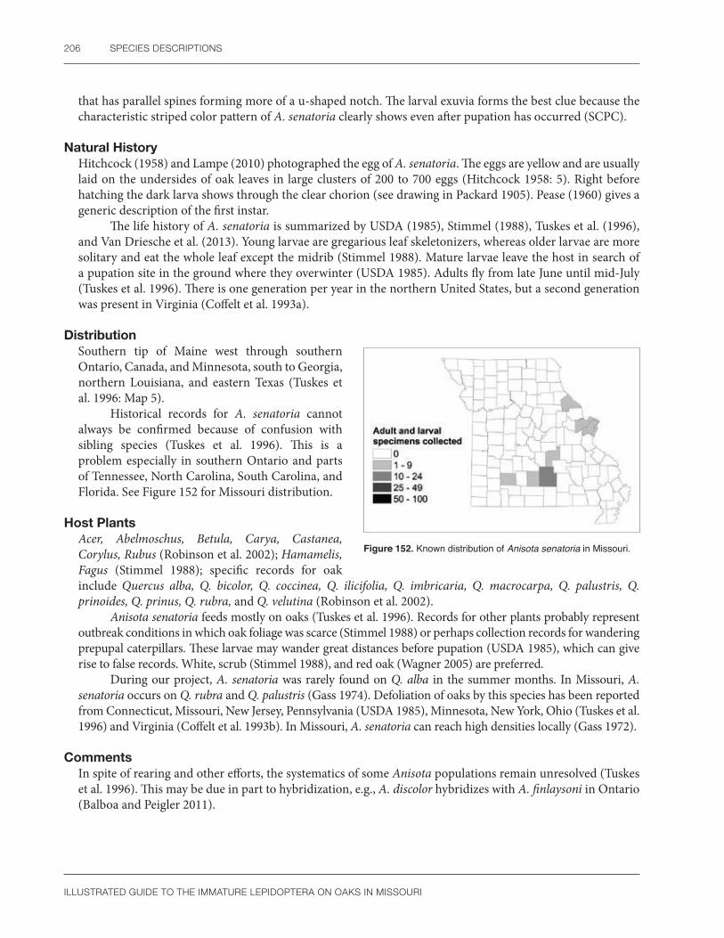
206 SPECIES DESCRIPTIONS
ILLUSTRATED GUIDE TO THE IMMATURE LEPIDOPTERA ON OAKS IN MISSOURI
that has parallel spines forming more of a u-shaped notch. The larval exuvia forms the best clue because the characteristic striped color pattern of A. senatoria clearly shows even after pupation has occurred (SCPC).
Natural HistoryHitchcock (1958) and Lampe (2010) photographed the egg of A. senatoria. The eggs are yellow and are usually laid on the undersides of oak leaves in large clusters of 200 to 700 eggs (Hitchcock 1958: 5). Right before hatching the dark larva shows through the clear chorion (see drawing in Packard 1905). Pease (1960) gives a generic description of the first instar.
The life history of A. senatoria is summarized by USDA (1985), Stimmel (1988), Tuskes et al. (1996), and Van Driesche et al. (2013). Young larvae are gregarious leaf skeletonizers, whereas older larvae are more solitary and eat the whole leaf except the midrib (Stimmel 1988). Mature larvae leave the host in search of a pupation site in the ground where they overwinter (USDA 1985). Adults fly from late June until mid-July (Tuskes et al. 1996). There is one generation per year in the northern United States, but a second generation was present in Virginia (Coffelt et al. 1993a).
DistributionSouthern tip of Maine west through southern Ontario, Canada, and Minnesota, south to Georgia, northern Louisiana, and eastern Texas (Tuskes et al. 1996: Map 5).
Historical records for A. senatoria cannot always be confirmed because of confusion with sibling species (Tuskes et al. 1996). This is a problem especially in southern Ontario and parts of Tennessee, North Carolina, South Carolina, and Florida. See Figure 152 for Missouri distribution.
Host PlantsAcer, Abelmoschus, Betula, Carya, Castanea, Corylus, Rubus (Robinson et al. 2002); Hamamelis, Fagus (Stimmel 1988); specific records for oak include Quercus alba, Q. bicolor, Q. coccinea, Q. ilicifolia, Q. imbricaria, Q. macrocarpa, Q. palustris, Q. prinoides, Q. prinus, Q. rubra, and Q. velutina (Robinson et al. 2002).
Anisota senatoria feeds mostly on oaks (Tuskes et al. 1996). Records for other plants probably represent outbreak conditions in which oak foliage was scarce (Stimmel 1988) or perhaps collection records for wandering prepupal caterpillars. These larvae may wander great distances before pupation (USDA 1985), which can give rise to false records. White, scrub (Stimmel 1988), and red oak (Wagner 2005) are preferred.
During our project, A. senatoria was rarely found on Q. alba in the summer months. In Missouri, A. senatoria occurs on Q. rubra and Q. palustris (Gass 1974). Defoliation of oaks by this species has been reported from Connecticut, Missouri, New Jersey, Pennsylvania (USDA 1985), Minnesota, New York, Ohio (Tuskes et al. 1996) and Virginia (Coffelt et al. 1993b). In Missouri, A. senatoria can reach high densities locally (Gass 1972).
CommentsIn spite of rearing and other efforts, the systematics of some Anisota populations remain unresolved (Tuskes et al. 1996). This may be due in part to hybridization, e.g., A. discolor hybridizes with A. finlaysoni in Ontario (Balboa and Peigler 2011).
Figure 152. Known distribution of Anisota senatoria in Missouri.
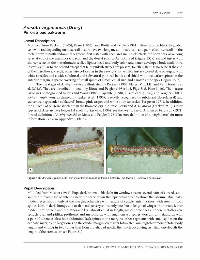
SATURNIIDAE 207
ILLUSTRATED GUIDE TO THE IMMATURE LEPIDOPTERA ON OAKS IN MISSOURI
Anisota virginiensis (Drury) Pink-striped oakworm
Larval DescriptionModified from Packard (1905), Pease (1960), and Riotte and Peigler (1981): Head capsule black to golden yellow to red depending on instar; all instars have two long mesothoracic scoli and pairs of shorter scoli on the metathorax to ninth abdominal segment; first instar with head and anal shield black, the body dark olive, long setae at end of the mesothoracic scoli and the dorsal scoli of A8 not fused (Figure 153a); second instar with shorter setae on the mesothoracic scoli, a lighter head and body color, and better developed body scoli; third instar is similar to the second except that faint pinkish stripes are present; fourth instar has no setae at the end of the mesothoracic scoli, otherwise colored as in the previous instar; fifth instar colored slate blue-gray with white speckles and a wide subdorsal and subventral pink-red band; anal shield with two darker spines on the anterior margin, a sparse covering of small spines of almost equal size, and a notch at the apex (Figure 153b).
The life stages of A. virginiensis are illustrated by Packard (1905: Plates IV, L, LII) and Van Driesche et al. (2013). They are described in detail by Riotte and Peigler (1981: 145, Figs. 3, 5, Plate 1: 10). The mature larva was photographed by Ives and Wong (1988), Laplante (1998), Tuskes et al. (1996), and Wagner (2005). Anisota virginiensis, as defined by Tuskes et al. (1996), is readily recognized by subdorsal (dorsolateral) and subventral (spiracular, sublateral) broad, pink stripes and white body tubercles (Ferguson 1971). In addition, the D1 scoli of A1-6 are shorter than the thoracic legs in A. virginiensis and A. senatoria (Fracker 1930). Other species of Anisota have longer D1 scoli (Tuskes et al. 1996). See the keys to larval Anisota by Ferguson (1971) (broad definition of A. virginiensis) or Riotte and Peigler (1981) (narrow definition of A. virginiensis) for more information. See also Appendix 3, Plate 2.
a b
Figure 153. Anisota virginiensis (a) mid-instar larva; (b) mature larva. Photos by R.J. Marquis, used with permission.
Pupal DescriptionModified from Mosher (1914): Pupa dark brown to black; brain window absent; several pairs of curved, stout spines run from base of antenna near the scape down the “epicranial area” to above the labrum; labial palpi hidden; eyes smooth only at the margin, otherwise with texture of cuticle; antenna short with rows of stout spines; labrum dark, bumpy and oval; maxillae very short, only one fourth length of wings; prothoracic femur hidden; prothoracic and mesothoracic legs almost equal in length; mesothoracic legs hidden; metathoracic spiracle oval and pitlike; prothorax and mesothorax with small curved spines; dorsum of metathorax with a pair of tubercles; first four abdominal lack spines at the margins, other segments with small spines on the cephalic margin and larger ones on the caudal margin; cremaster bifurcated, one-eighth or more of total body length and ending in two spines that form a u-shaped notch, the notch occupying less than one-fourth the length of the cremaster (see Figure 5e).

208 SPECIES DESCRIPTIONS
ILLUSTRATED GUIDE TO THE IMMATURE LEPIDOPTERA ON OAKS IN MISSOURI
The pupa of A. virginiensis is described by Mosher (1914) and illustrated by Van Driesche et al. (2013). Riotte and Peigler (1981) illustrate the cremaster. Both publications use minor differences in the cremaster to separate A. virginiensis from related species. These differences are at best very subtle or perhaps not valid in a large series of specimens. The larval exuvia of A. virginiensis lacks stripes and has short scoli. These characters can be important clues for identification. Otherwise, characters used by Mosher (1914), like the thoracic spines or pupal length, can be difficult to interpret.
Natural HistoryPeterson (1965b) photographed the egg of A. virginiensis. He describes them as yellow at first, then turning reddish brown before hatching. Tuskes et al. (1996) uses the phrase “more orange than yellow.” The parental female of A. virginiensis that laid the eggs studied by Peterson (1965b) is at the Charles A. Triplehorn collection in Columbus, Ohio. It was collected in Minnesota, confirming that it is the true A. virginiensis and not a related taxon.
The biology of A. virginiensis is summarized by Tuskes et al. (1996) and Van Driesche et al. (2013). Eggs are laid in clusters on the underside of oak leaves. Early instars are gregarious and there are one to three generations per year. The pupal stage overwinters in the northern United States; it only lasts a few weeks in warmer climates where development is continuous. Adults are dimorphic in color and males are active during the day.
Larvae have pronounced “head twitching” when disturbed (R.J. Marquis, pers. obs.), a behavior in which the heads of one or more larvae rapidly move from side to side. This is presumably defensive in function.
DistributionNova Scotia, Canada, south to northern Florida and west to a line connecting southeastern Manitoba, Canada, to eastern Texas (Tuskes et al. 1996: Map 4).
Ferguson (1971) stated that A. virginiensis should occur wherever oak is present, although at that time, it was considered rare in Missouri. Later, Riotte and Peigler (1981: 147) examined specimens from 15 counties in Missouri, and suggest that this species is currently more widespread than previously thought. Packard (1905) originally recorded it from St. Louis. James (2017) reported an outbreak in Manitoba in the late 1980s but considers this species to currently be in decline. See Figure 154 for Missouri distribution.
Host PlantsAcer, Amelanchier, Betula, Carpinus, Castanea, Celtis, Cornus, Corylus, Fagus, Prunus, Salix; specific records for oak include Quercus alba, Q. falcata, Q. ilex, Q imbricaria, Q. lyrata, Q. macrocarpa, Q. marilandica, Q. myrtifolia, Q. nigra, Q. palustris, Q. petraea, Q. prinus, Q. pubescens, Q. robur, Q. rubra, Q. stellata, Q. texana, Q. velutina, Q. virginiana (Robinson et al. 2002); Q. laurifolia, and Q. nuttallii (Heppner 2003).
The preferred host of A. virginiensis is Q. rubra (Riotte and Peigler 1981). However, it is common on Q. alba in Missouri (R.J. Marquis pers. obs.) in contrast to Riotte and Peigler (1981: 171) who stated that Anisota rarely use this plant species in the southern states. Black oaks (Q. velutina group) are utilized in the eastern United States (Wagner et al. 1997).
Riotte and Peigler (1981: 113) provided a discussion on the use of oaks by Anisota throughout the United States, pointing out differences in usage by each moth species.
Figure 154. Known distribution of Anisota virginiensis in Missouri.

SATURNIIDAE 209
ILLUSTRATED GUIDE TO THE IMMATURE LEPIDOPTERA ON OAKS IN MISSOURI
CommentsOur species concept of A. virginiensis follows Ferguson (1971), Tuskes et al. (1996), and the new checklist by Pohl et al. (2016), who synonymized A. pellucida and A. discolor under A. virginiensis. Larval and egg coloration seem to support recognition of a species complex as suggested by Riotte and Peigler (1981: 146), but grouping based on external morphology could not be correlated with adult genitalia (Lemaire 1988: 327). Tuskes et al. (1996) doubted the taxonomic value of these color differences. According to Ferguson (1971: 80), Riotte and Peigler (1981: 147), and Lemaire (1988: Fig. 263), the Missouri population falls under A. virginiensis in the strict sense. We have tried to restrict our comments to this taxon when possible.
Eacles imperialis (Drury) Imperial moth
Larval DescriptionModified from Packard (1905), Pease (1960), Lampe (2010), and Wagner (2005): First instar with an orange head capsule, transverse black stripes and long bifurcated non-clubbed scoli on the mesothorax, metathorax, and A8; second instar lacks the transverse striped pattern and adds secondary setae; third instar is similar to the second except the scoli are shorter compared to the body length and color tends to start becoming variable; fourth instar larva is similar to the third except the longer scoli are not forked at the tip; mature larva generally shades of brown, peach, salmon, crimson, or green with long fine hairs, head capsule usually with a pair of black vertical stripes in the frontal area, and vertical lateral epicranial stripes, mesothorax, and metathorax with stout pointed scoli, abdominal spiracles white, conspicuous due to their large size, with a black peritreme; last two thoracic segments and abdomen with a small subdorsal to dorsal scolus on the side of each segment, a longer middorsal scolus present on A8; anal prolegs spotted black and yellow to pale green or brown, depending on the color form; anal shield normally colored as the anal prolegs (Figure 155a, b).
The life cycle of Eacles imperialis is illustrated by Packard (1905), Lampe (2010), and Hall (2014a). The mature larva has been illustrated many times, and recent examples are Tuskes et al. (1996), Bouseman and Sternburg (2002), Sogaard (2009), and several color variations shown by Wagner (2005). Packard (1905: 123) illustrates the scoli in detail, and Peterson (1962a) illustrates the larva in side view.
According to Ferguson (1971: 23), Eacles is the only member of the subfamily Ceratocampinae in the United States to have long setae. Although variable in color, most instars are easily recognized by their general appearance. Sibling species of Eacles with similar larvae, found in Latin America, complicate identification of this genus throughout Latin America.
a b
Figure 155. Eacles imperialis (a) mid-instar larva; (b) mature larva. Photos by R.J. Marquis, used with permission.
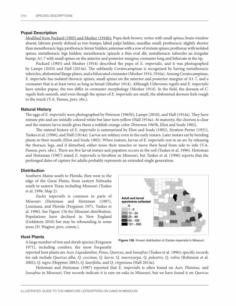
210 SPECIES DESCRIPTIONS
ILLUSTRATED GUIDE TO THE IMMATURE LEPIDOPTERA ON OAKS IN MISSOURI
Pupal DescriptionModified from Packard (1905) and Mosher (1916b): Pupa dark brown; vertex with small spines; brain window absent; labrum poorly defined as two bumps; labial palpi hidden; maxillae small; prothoracic slightly shorter than mesothoracic legs; prothoracic femur hidden; antennae with a row of minute spines; prothorax with isolated spines; metathoracic legs hidden; mesothoracic spiracle a thin oval slit; metathoracic tubercles an irregular bump; A1-7 with small spines on the anterior and posterior margins; cremaster long and bifurcate at the tip.
Packard (1905) and Mosher (1914) described the pupa of E. imperialis, and it was photographed by Lampe (2010) and Hall (2014a). The subfamily Ceratocampinae is recognized by having metathoracic tubercles, abdominal flange plates, and a bifurcated cremaster (Mosher 1914, 1916a). Among Ceratocampinae, E. imperialis has isolated thoracic spines, small spines on the anterior and posterior margins of A1-7, and a cremaster that is at least twice as long as broad (Mosher 1914). Although Citheronia regalis and E. imperialis have similar pupae, the two differ in cremaster morphology (Mosher 1914). In the field, the dorsum of C. regalis feels smooth, and even though the spines of E. imperialis are small, the abdominal dorsum feels rough to the touch (V.A. Passoa, pers. obs.).
Natural HistoryThe eggs of E. imperialis were photographed by Peterson (1965b), Lampe (2010), and Hall (1914a). They have minute pits and are initially colored white but later turn yellow (Hall 1914a). At maturity, the chorion is clear and the mature larva inside gives them a reddish orange color (Peterson 1965b, Eliot and Soule 1902).
The natural history of E. imperialis is summarized by Eliot and Soule (1902), Stratton-Porter (1921), Tuskes et al. (1996), and Hall (1914a). Larvae are solitary even in the early instars. Later instars eat by bending plants to their mouth (Eliot and Soule 1902). When mature, larvae of E. imperialis rest in an arc by releasing the thoracic legs, and if disturbed, either tense their muscles or move their head from side to side (V.A. Passoa, pers. obs.). There are five larval instars and pupation occurs in the soil (Tuskes et al. 1996). Heitzman and Heitzman (1987) stated E. imperialis is bivoltine in Missouri, but Tuskes et al. (1996) reports that the prolonged dates of capture for adults probably represents an extended single generation.
DistributionSouthern Maine south to Florida, then west to the edge of the Great Plains, from eastern Nebraska south to eastern Texas including Missouri (Tuskes et al. 1996: Map 2).
Eacles imperialis is common in parts of Missouri (Heitzman and Heitzman (1987), Louisiana, and Florida (Ferguson 1971, Tuskes et al. 1996). See Figure 156 for Missouri distribution. Populations have declined in New England (Goldstein 2010) but may be rebounding in some areas (D. Wagner, pers. comm.).
Host PlantsA large number of tree and shrub species (Ferguson 1971), including conifers; the most frequently reported host plants are Acer, Liquidambar, Pinus, Quercus, and Sassafras (Tuskes et al. 1996); specific records for oak include Quercus alba, Q. coccinea, Q. laevis, Q. macrocarpa, Q. palustris, Q. rubra (Robinson et al. 2002); Q. nigra (Heppner 2003); Q. laurifolia, and Q. virginiana (Hall 2014a).
Heitzman and Heitzman (1987) reported that E. imperialis is often found on Acer, Platanus, and Sassafras in Missouri. Our records indicate it is rare on oaks in Missouri, but we have found it on Quercus
Figure 156. Known distribution of Eacles imperialis in Missouri.

SATURNIIDAE 211
ILLUSTRATED GUIDE TO THE IMMATURE LEPIDOPTERA ON OAKS IN MISSOURI
alba and Q. velutina. There are likely regional preferences in host plant usage (Ferguson 1971: 26), but these have not always been clearly documented. Pine is “extensively used” on Martha’s Vineyard (Goldstein 2010) and in the subspecies E. imperialis pini (Ferguson 1971).
CommentsBoth Ferguson (1971) and Goldstein (2010) suggested a need to study subspecies of E. imperialis to see if they are valid species. Tuskes et al. (1996) expressed more doubt on this issue and recognized one less subspecies in the United States than the above two authors. Lemaire (1988) used a broad concept of E. imperialis. We restrict our comments in this section to E. imperialis imperialis, the subspecies that occurs in Missouri, as defined by Tuskes et al. (1996).

212 SPECIES DESCRIPTIONS
ILLUSTRATED GUIDE TO THE IMMATURE LEPIDOPTERA ON OAKS IN MISSOURI
SphingidaeHawkmoths
Sphingidae contain approximately 1,400 species worldwide (Nieukerken et al. 2011). The most recent checklist of the family is by Kitching and Cadiou (2000), which is valuable because they critically review earlier publications such as Bridges (1993), the pictorial worldwide guide by d’Abrera (1986), and classical works like Rothschild and Jordan (1903). A summary of sphingid systematics based on morphology was published by Lemaire and Minet (1998). Kawahara et al. (2009) suggested a molecular phylogeny for this family. Being popular with collectors, several good Web sites exist for Sphingidae. Adult sphingid photographs can be found at http://en.sphingidae-museum.com/database/. Janzen and Hallwachs studied the family in Costa Rica, and host records with photographs of the immature stages are available at http://janzen.sas.upenn.edu/caterpillars/checklists/sphingidaelist.htm. Other New World Sphingidae are included in the Web site by Bill Oehlke (http://www.silkmoths.m.bizland.com/Sphinx/danjansphinx.htm). Sphingidae of the Western Palearctic are reviewed by Pittaway (1997–2015). Larval and pupal keys to the European species were published by Pelzer (1991, 1995, 1996), whereas Nakamura (1976a, 1977) studied the pupal Sphingidae of Japan. Consult the bibliographies or summaries in the above works and the Web site http://sphingidae.myspecies.info/ for a more complete listing of basic biology, morphology, and literature on Sphingidae. For North America, Hodges (1971) contains a traditional morphological treatment of the adults with genitalia and partial keys to the immature stages. The most recent treatment of the North American fauna is by Tuttle (2007) with an emphasis on biology, larval photographs, and pupal drawings. Wagner (2005) illustrated most of the eastern United States species.
According to Kitching and Cadiou (2000), sphingid eggs are of the flat type with the micropyle at one end. They are elliptical to round (Syme 1961) and colored green to white to yellow (Peterson 1965b). The chorion is shiny and smooth to the naked eye, but may be finely sculptured under higher magnification (Doring 1955, Peterson 1965b). Some species have spotted (Doring 1955: Plate 24) or patterned (Kitching and Cadiou 2000) eggs. Eggs of the Sphinginae are flattened, elliptical, and unusual because one side is concave in some North American (Syme 1961) and European species (Doring 1955). At least in North America, most sphingids do not eat the eggshell (Tuttle 2007), although some African species do so commonly (Kawahara et al. 2009). Eggs are laid singly or rarely in a cluster on leaves or flower heads (Tuttle 2007). As is typical for Lepidoptera, the chorion is clear before hatching, and as a result, dark developing larvae can be seen inside the egg (Peterson 1965b, Kitching and Cadiou 2000). This is especially obvious in Sphingidae because most species have large eggs.
Sphingid larvae, usually called hornworms, are relatively large, naked caterpillars, sometimes exceeding 15 cm in length (Wagner 2005). Most species are oligophagous or rarely polyphagous (Regier et al. 2001). They often betray their presence by scattering conspicuous feces below their feeding site (Wagner 2005). Sphingids are easy to recognize in North America but less clearly defined if the world fauna is considered. They are normally identified in all instars by having a long horn (the D1 scolus according to Lemaire and Minet 1998) on A8, but there are several exceptions to this generalization. First, other families have such a horn, and in fact, the presence of this structure is an apomorphy of the Bombycoidea and not just the Sphingidae (Kitching and Cadiou 2000). Second, not all sphingid larvae have a horn, but in some species there is a buttonlike tubercle present in late instars (Stehr 1987). The horn itself is subject to variation in color and texture (Wagner 2005), and the structure can be stout, flaplike, or long and thin (Kitching and Cadiou 2000). Only rarely does this family have species without either a horn or a button (Stehr 1987). Sphingid larvae usually have a smooth cuticle with short secondary setae, crochets in a biordinal mesoseries, and three lobes on A10 (Stehr 1987). In other taxa, the cuticle has minute tubercles or a fine pubescence (Kitching and Cadiou 2000). Head shape is highly variable, ranging from round to oval to triangular (Forbes 1911). Sphingid mandibles are of two types, either with complex ridges (sphingid type) (see Bernays and Janzen 1988) or simple and smooth (“saturniid type”), such as Pachysphinx modesta (McCabe 1991) or Paonias excaecata (SCPC). Stehr (1987) used the presence of 6–8 annulets per segment to help define sphingid larvae. He noted that other larvae with horns or buttons that are likely to be confused with sphingids (e.g., Bombyx mori, the true silkworm, and a few Notodontidae) lack annulets on their cuticle.

SPHINGIDAE 213
ILLUSTRATED GUIDE TO THE IMMATURE LEPIDOPTERA ON OAKS IN MISSOURI
Sphingid larvae sometimes have a characteristic coloration. In many Sphinginae, and some Macroglossinae, there are lateral, slanted lines pointing upward toward the rear of the body (Lemaire and Minet 1998). Other species have eyespots, even mimicking snakes and snake heads. A few may be aposematically colored or cryptic to match the color of their host (Kitching and Cadiou 2000). All sphingid larvae feed externally.
Sphingid pupae are relatively well studied, and specific keys exist for North America (Mosher 1916a, 1918; Hodges 1971), Europe (Pelzer 1996, Patočka and Turčáni 2005), and Japan (Nakamura 1976a, 1977). Mosher (1916a, 1918) characterized sphingid pupae by their distinctive fusiform shape, usually having long, pointed forewings, a scar on A8 representing the larval horn, and a triangular pointed to bifurcated cremaster generally without hooked setae. The scar of the larval horn is either a small, smooth area or a slit (SCPC), but being easily overlooked, perhaps is of limited value for identification. There are four unusual sphingid pupal characters: the lack of body setae; spiracular furrows (Mosher 1918); various metathoracic modifications such as ridges, plates or reticulations (Kitching and Cadiou 2000); and a specialized notched cremaster in some species (Kawahara et al. 2009). There is great variation in the maxillae leading to three basic body types. The maxillae may be short, only about half the forewing, so that the pupa resembles other bombycoid families, such as the Saturniidae (Mosher 1918: Fig 5). When it is longer, the maxillae form a flap or loop, and this is the most common concept of sphingid pupae because it characterizes the tomato and tobacco hornworms (Mosher 1918: Figs. 2, 3). The last type is similar to some species of Pyraustinae (e.g., Diaphania, see Passoa 1985) in that the head is moved back so that the labial palpi point upward and the vertex is not on the top of the head (Mosher 1918: Fig. 7). Other examples of these types are illustrated by Tuttle (2007) and Villard (1975). These basic divisions are seen in the fauna of Europe as well (Pelzer 1996). Mosher (1918: 275) suggested that North American sphingid pupae conform to the classification of Rothschild and Jordan (1903) in “practically every detail,” but her characters were not studied by Kawahara et al. (2009). It is unknown how useful they might be phylogenetically or for identification in other faunal regions. Sphingid pupae can be black to brightly colored, sometimes with stripes (Kitching and Cadiou 2000). They usually pupate in the ground or rarely construct a cocoon (Tuttle 2007).
Kitching and Cadiou (2000) discussed the worldwide economic importance of the Sphingidae. Both trees and annual crops are attacked, often in Asia. Manduca on solanaceous crops and Erinnyis on cassava or papaya are perhaps the best known New World pests. Two examples of sphingid defoliation in North American forests are the ash sphinx (Sphinx chersis) on roadside young ash trees and the catalpa sphinx (Ceratomia catalpa) on catalpa trees (USDA 1985). In other species (Hyles spp.), the populations can be quite high even if no economic damage to agroecosystems is present (Kitching and Cadiou 2000). A few species are beneficial as food for humans while others were released as biological control agents of weeds (Kitching and Cadiou 2000: 11).
Subfamily Smerinthinae
Paonias excaecata (J.E. Smith)Blinded sphinx
Larval DescriptionBody color varies from blue to pale yellow-green, sometimes spotted with red; cuticle with blunt to pointed granules; head with a thin vertical stripe defining a contrasting frontal area; mandible smooth, lacking complex ridges, with no scissorial teeth but having a transverse retinaculum; thorax with a subdorsal stripe; seven oblique lines on the abdomen slant posteriorly and touch the dorsal surface of the spiracles on A1-7, the one on A7 thicker than the others and extending to the base of the horn on A8; spiracles may be surrounded by red spots; horn on A8 arises from broad base, points upward, and is red- or black-banded to solid green, and only rarely blue (Figure 157a, b).
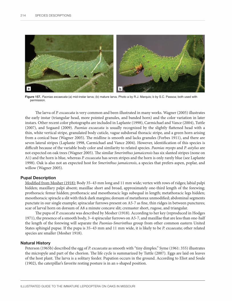
214 SPECIES DESCRIPTIONS
ILLUSTRATED GUIDE TO THE IMMATURE LEPIDOPTERA ON OAKS IN MISSOURI
a b
Figure 157. Paonias excaecata (a) mid-instar larva; (b) mature larva. Photo a by R.J. Marquis; b by S.C. Passoa; both used with permission.
The larva of P. excaecata is very common and been illustrated in many works. Wagner (2005) illustrates the early instar (triangular head, more pointed granules, and banded horn) and the color variation in later instars. Other recent color photographs are included in Laplante (1998), Carmichael and Vance (2004), Tuttle (2007), and Sogaard (2009). Paonias excaecata is usually recognized by the slightly flattened head with a thin, white vertical stripe, granulated body cuticle, vague subdorsal thoracic stripe, and a green horn arising from a conical base (Wagner 2005). The midline is smooth and lacks granules (Forbes 1911), and there are seven lateral stripes (Laplante 1998, Carmichael and Vance 2004). However, identification of this species is difficult because of the variable body color and similarity to related species. Paonias myops and P. astylus are not expected on oak trees (Wagner 2005). The similar Smerinthus jamaicensis has six slanted stripes (none on A1) and the horn is blue, whereas P. excaecata has seven stripes and the horn is only rarely blue (see Laplante 1998). Oak is also not an expected host for Smerinthus jamaicensis, a species that prefers aspen, poplar, and willow (Wagner 2005).
Pupal DescriptionModified from Mosher (1918): Body 35–43 mm long and 11 mm wide; vertex with rows of ridges; labial palpi hidden; maxillary palpi absent; maxillae short and broad, approximately one-third length of the forewing; prothoracic femur hidden; prothoracic and mesothoracic legs subequal in length; metathoracic legs hidden; mesothoracic spiracle a slit with thick dark margins; dorsum of metathorax unmodified; abdominal segments punctate in our single example; spiracular furrows present on A5-7 as fine, thin ridges in between punctures; scar of larval horn on dorsum of A8 a minute concave slit; cremaster short, rugose, and triangular.
The pupa of P. excaecata was described by Mosher (1918). According to her key (reproduced in Hodges 1971), the presence of a smooth body, 3–4 spiracular furrows on A5-7, and maxillae that are less than one-half the length of the forewing will separate the Paonias-Smerinthus group from other common eastern United States sphingid pupae. If the pupa is 35–43 mm and 11 mm wide, it is likely to be P. excaecata; other related species are smaller (Mosher 1918).
Natural HistoryPeterson (1965b) described the egg of P. excaecata as smooth with “tiny dimples.” Syme (1961: 355) illustrates the micropyle and part of the chorion. The life cycle is summarized by Tuttle (2007). Eggs are laid on leaves of the host plant. The larva is a solitary feeder. Pupation occurs in the ground. According to Eliot and Soule (1902), the caterpillar’s favorite resting posture is in an s-shaped position.

SPHINGIDAE 215
ILLUSTRATED GUIDE TO THE IMMATURE LEPIDOPTERA ON OAKS IN MISSOURI
DistributionFrom Labrador across southern Canada to British Columbia, then Maine south to Florida and west throughout the eastern United States, Great Plains, and Rocky Mountains, from Florida, eastern Texas, Arizona to northern California (Tuttle 2007).
A distribution map for P. excaecata was published by Tuttle (2007). Heitzman and Heitzman (1987) consider P. excaecata to be one of the most common sphinx moths in Missouri, but we have encountered only five larvae on oaks. (See Figure 158 for Missouri distribution.) This suggests that it prefers hosts other than oak in our region. Paonias excaecata is rarer in the southern portions of its range (Hodges 1971) but is extremely common in eastern United States forests (Wagner 2005). Caterpillars are common in the Pacific Northwest, particularly on oak (Miller and Hammond 2003).
Host PlantsAcer, Amelanchier, Betula, Carpinus, Chaenomeles, Corylus, Crataegus, Fagus, Fraxinus, Malus, Ostrya, Populus, Prunus, Pyrus, Quercus, Rosa, Rubus, Salix, Spiraea, Tilia, Ulmus, Viburnum, Wisteria (Robinson et al. 2002); and Holodiscus (Miller and Hammond 2003). We have found larvae of P. excaecata on Quercus alba in Missouri.
CommentsWe follow the recent revision of Tuttle (2007), Kitching and Cadiou (2000), and the checklist by Pohl et al. (2016) that use P. excaecata instead of P. excaecatus. Eliot and Soule (1902) mention several larval host preferences for different rearings of P. excaecata such as oak, Pyrus, birch, and poplar. Mosher (1918) found the pupa “so variable it was impossible to write a detailed description.” These observations suggest subspecies or even a cryptic species complex.
Figure 158. Known distribution of Paonias excaecata in Missouri.

216 SPECIES DESCRIPTIONS
ILLUSTRATED GUIDE TO THE IMMATURE LEPIDOPTERA ON OAKS IN MISSOURI
NotodontidaeProminent moths
The family Notodontidae contains approximately 3,800 (Nieukerken et al. 2011) to 4,412 (Schintlmeister 2013) species distributed throughout the world except for New Zealand and some Pacific Islands (Kitching and Rawlins 1998). Nine (Miller 1991) to twelve (Kobayashi and Nonaka 2016) to eighteen (Schintlmeister 2013) subfamilies are recognized depending on the author. Some of these were considered separate families in older literature (e.g., Thaumetopoeidae, Dioptidae). For more information on notodontid moths, consult the phylogenetic analysis and pupal studies by Miller (1991, 1992). Schintlmeister (2013) published a world catalog of the Notodontidae. Packard (1895) and Wagner (2005) illustrated North American notodontid caterpillars and discussed their biology. Becker (2014) reclassified the New World species. The first of two monographs on the Notodontidae of North America was published by Miller et al. (2018). This resulted in several name changes for the Missouri fauna. Forbes (1939) surveyed notodontids found in the Canal Zone of Panama, whereas Miller and Thiaucourt (2011) listed and described new species from Ecuador. Miller (2009a, 2009b) published a generic revision of the Dioptinae. Dolinskaya and Pljushch (2003) give a summary of Palearctic notodontid caterpillar morphology, and adults of this region were treated by Schintlmeister (2008). The notodontid fauna of Borneo was treated by Holloway (1983).
Notodontid eggs are of the upright type (the micropylar axis is perpendicular to the substrate). The number of micropyles varies from four to twenty depending on the species (Hinton 1979). Syme (1961) illustrated the micropyles of several North American notodontid genera. In profile, notodontid eggs are usually hemispherical, circular (Syme 1961, Peterson 1963b, Kitching and Rawlins 1998), or more rarely flattened (Dolinskaya 1990). Although the chorion appears smooth to the naked eye, faint reticulations and small depressions are obvious at magnifications above 90x (Syme 1961, Peterson 1963b, Dolinskaya 1990). At least in North America, notodontid eggs are uniformly colored: white, cream, green, yellow, light blue, or rarely black (Peterson 1963b). A few European notodontid genera have patterned eggs, and most species undergo color changes during incubation (Dolinskaya 1990).
North American notodontid larvae can be confused with several other macrolepidopteran families (see Godfrey and Appleby 1987) and are difficult to characterize when all known exceptions are incorporated into their diagnosis. Superficially, most species are recognized by their unusual anal prolegs, termed stemapoda, which are either well developed, shortened, and peglike, or modified into elongate rods with few or no crochets. The head is often striped with two closely-spaced, parallel bands of different colors, the P setae are equally spaced from the epicranial suture, and the vertex may be pointed (Miller 1991). Secondary setae are absent on the head except for Scevesia (as Skewesia), Dunama (Godfrey and Appleby 1987), Stauropus, Dicranura, Phalera, Pygaera (Dolinskaya 2013), and Clostera examined by both studies (Godfrey and Appleby 1987, Dolinskaya 2013). Dolinskaya (2011) recognized 12 ontogenetic scenarios that measured the difference in notodontid head textures from the first to last instar. She noted the head may be smooth (under 2000x) or textured (with pits, microtrichia, tubercles, faint reticulations, or a mixture of these modifications). Except for Crinodes, Cargida, Theroa, some species of Dioptini (Godfrey et al. 1989), and Nerice (Dolinskaya 2008), the mandibles of last instar notodontid larvae lack teeth and have a smooth cutting margin. A retinaculum is sometimes present (Dolinskaya 2008). Thaumetopoeinae and the genus Dicranura are the only taxa with secondary setae on the mandibles (Godfrey et al. 1989, Dolinskaya 2008). Early instar Heterocampinae may have branched spines resembling antlers on the prothorax (Miller 1991: 138). Notodontid larvae can have modified tarsal setae that are rarely lost (Miller 1991: 150). The body has either primary or dense secondary setae, but they never form long hair pencils. Larval coloration is highly variable in some species (Godfrey and Appleby 1987), and caterpillars in the Hapigiini are counter shaded for disruptive coloration (Miller et al. 1997). Fleshy humps on the abdominal dorsum, including a small knob or horn on A8, are two other common modifications (Miller et al. 1997, Wagner 2005). Many notodontids rest with their body arched upward and the stemapoda held off the substrate. Unlike most Noctuidae or Erebidae, they usually have secondary setae in at least the SV group of A3-

NOTODONTIDAE 217
ILLUSTRATED GUIDE TO THE IMMATURE LEPIDOPTERA ON OAKS IN MISSOURI
6, and in the case of the Heterocampinae and a few Notodontinae, the SV group of the thorax is bisetose (Miller 1991: 141). Notodontid larvae lack both verrucae (except Scevesia and Dunama) (Godfrey and Appleby 1987) and middorsal abdominal glands that are characteristic of the erebid subfamilies Arctiinae and Lymantriinae, respectively. Crochets of the Notodontidae are arranged in a homoideous uniordinal mesoseries. This separates them from other families with similar larvae (characteristics distinguishing these taxa from Notodontidae are listed in parentheses): Apatelodidae (crochets biordinal); Doidae (crochets biordinal); Drepanidae (single anal projection, crochets in two lateroseries); Nolidae (only three abdominal prolegs); Lasiocampidae (crochets biordinal); and even Saturniidae (Agapema) (crochets biordinal) or Sphingidae (each segment with 6–8 annulets and crochets biordinal). For a more complete diagnosis of the North American fauna using these characters, consult Stehr (1987).
Accurate identification of larval Notodontidae requires detailed examination of mouthparts and chaetotaxy. Three unique characteristics can be cited for this family: two MD setae present on A1, seta X present on A10, and maxillae with stipital lobes (Kitching and Rawlins 1998). Although some exceptions are listed below, even the presence of one of these three features will define larval Notodontidae. First instar Acronicta (Noctuidae) have two MD setae on A1 as do notodontids (Godfrey and Appleby 1987: 524). The Dioptinae and all other Noctuidae and Erebidae have one MD seta on A1 (Kitching and Rawlins 1998). Seta X is present in all Notodontidae, except for the subfamily Dudusinae, and in those genera where dense secondary setae obscure this condition (Kitching and Rawlins 1998). Excluding processionary caterpillars (Thaumetopoeinae) and five apparently unrelated genera in Africa, Australia, and the Neotropics, a stipital lobe occurs in all notodontid larvae (Godfrey et al. 1989). Therefore, this is probably the best single feature that can be cited to authoritatively identify notodontid larvae in Missouri and throughout eastern North America.
Workers in both North America (Mosher 1916a) and Asia (Nakamura 1981) have pointed out the close morphological similarity of notodontid and geometrid pupae, even putting them in the same superfamily. This seems logical because a recent molecular phylogeny showed Noctuoidea, Bombycoidea, and Geometroidea are all related (Regier et al. 2013). However, adult characters (Miller 1991: 4) firmly place notodontids in the Noctuoidea. Minet (1991) noticed that certain macrolepidopteran taxa are defined by hidden (or minutely exposed) pupal labial palpi. This clade includes at least the Geometroidea, Papilionioidea, and related groups, but the Noctuoidea were originally excluded because of their exposed labial palpi. Because Notodontidae are considered primitive members of the Noctuoidea (Weller et al. 1994: Fig. 1, Zahiri et al. 2010: Fig. 1, Regier et al. 2017), hidden (or minutely exposed) labial palpi must represent the primitive condition for this superfamily.
No one morphological feature will identify notodontid pupae. Mosher (1917) separated notodontids from noctuids and erebids by their hidden labial palpi and mesothoracic legs that do not extend cephalad (above) of the ventral eye margin. Noctuid and erebid pupae have exposed labial palpi, or if they are hidden, then both the prothoracic and mesothoracic legs extend cephalad of the eyes. The forewing venation of notodontids is also different from noctuids and erebids. Forbes (1948) suggested that notodontid pupae can be partially recognized by examining the pupal wing pads, which show a three-branched cubital vein (except for one subfamily in Japan that has four branches [see Miller 1992: 253]). In noctuids and erebids, this vein is four-branched. A few Arctiinae have hidden labial palpi as well (Miller 1992), but unlike notodontids, many arctiines have a concave dorsum that gives the pupa a characteristic hourglass shape (Mosher1916a). The metathoracic legs are usually hidden in Notodontidae (Patočka and Turčáni 2005). Notodontid pupae also have reduced body setae, distinguishing them from most Arctiinae, Lymantriinae, and Lasiocampidae in which the setae are well developed. Secondary setae, when present in notodontid pupae, are short and almost microscopic (Miller 1992: 241). Many notodontids have either a row of deep pits on the posterior margin of the mesothorax, a pit on A6, and/or a punctate abdominal cuticle (Miller 1992). When modified, there is a wide variety of structures on the cremaster including straight or curved setae, blunt fanlike projections, or pointed spines (Dolinskaya 1986, 1989, 1993; Miller 1992). Notodontid and geometrid pupae can be distinguished by a combination of several characters (modified from Mosher 1917). In geometrids, the maxillae are at least three-fifths as long as the wing, and the mesothorax always lacks a row of pits. Except for Heterocampinae and Nystaleinae, the maxillae of notodontids

218 SPECIES DESCRIPTIONS
ILLUSTRATED GUIDE TO THE IMMATURE LEPIDOPTERA ON OAKS IN MISSOURI
are shorter than three-fifths the length of the wing. Members of the Heterocampinae are easily recognized by having a row of pits on the dorsum of the mesothorax, a character absent in geometrids. In addition, notodontids rarely have the prothoracic femur exposed (Miller 1992) and, except for the genus Datana, a dorsal furrow on the terminal abdominal segments is absent. Some geometrids, on the other hand, have the prothoracic femur exposed, and a dorsal furrow is often present on the terminal abdominal segments. Notodontid pupae are solid chestnut brown to black; geometrid pupae may be light or dark colored and sometimes are patterned. Unlike Drepanidae, pupae of the Notodontidae never have the metathoracic wings exposed. A few notodontids with long maxillae and no mesothoracic row of pits (Symmerista) can be confused with Arctiinae. Notodontidae may be separated from Ctenuchina arctiines by the cocoon. Pupae of Ctenuchina are covered with a dense mass of barbed larval hairs. The cocoons of Symmerista and other notodontids are either void of hairs or sparsely covered with hairs (Packard 1895), or if hairs are present, they are not barbed. The phylogenetic value of pupal morphology in notodontids was studied by Miller (1992).
Several economically important forest pests in Europe and North America are classified in the notodontid subfamilies Thaumetopoeinae, Heterocampinae, and Phalerinae (Carter 1984, Kitching and Rawlins 1998). The most important genera are Cecrita, Thaumetopoea, Lochmaeus, Heterocampa, Symmerista, Schizura, and Phalera. In general, early instar notodontids skeletonize leaves, whereas mature larvae eat from the leaf edge. Damage to forest trees by notodontid caterpillars can be insignificant, sporadic, or serious enough to cause mortality if the defoliation lasts several years (USDA 1985). Attacks by processionary caterpillars sometimes weaken the host, leading to invasion by secondary pests and pathogens (Carter 1984). Consult USDA (1985), Ives and Wong (1988), and Furniss and Carolin (1992) for more information on the economic importance of North American notodontids. Most Asian and Latin American notodontid species are of minor economic importance (Holloway 1983, Hilje et al. 1992). Most oak-feeding notodontids in Missouri belong to the subfamilies Heterocampinae, Notodontinae, and Phalerinae. In addition to the species described in more detail below, we have rarely collected Cecrita biundata, Heterocampa obliqua, and H. umbrata on Quercus alba, and Datana ministra on Q. velutina in Missouri. Biological information and color photographs of these species can be found in Packard (1895: 241, 246), Miller (1992), and Wagner (2005).
Subfamily Heterocampinae
Cecrita guttivitta WalkerSaddled prominent
Larval DescriptionMature larva highly variable in ground color from green to brown to light pink; head with four closely spaced parallel yellow, black, pink to red-brown and white stripes that meet at the epicranial notch to form an arch, the pink to red-brown stripe often contrasting; prothoracic appendages absent, or at most two small bumps are present; dorsal markings on middle abdominal segments continuously variable, forming an X, U, heart, or saddle-shaped band, or sometimes these markings reduced to small, elongate subdorsal dashes; a thin middorsal line present on the thorax that widens on the abdomen to merge with the dorsal markings, often breaking into several parallel longitudinal bands that may merge before ending on the anal shield; subdorsal stripe present from A1-A6, sometimes including the metathorax; lateral and ventral areas often mottled with red, only rarely forming small spots or patches (Figure 159a, b).
Various larval color forms of C. guttivitta are illustrated by Packard (1895), Patch (1908), USDA (1985), and Rush and Allen (1987). Individual larval photographs of C. guttivitta are available in Laplante (1998), Godfrey and Appleby (1987), Ives and Wong (1988), McCabe (1991), Wagner et al. (1997), and Wagner (2005).

NOTODONTIDAE 219
ILLUSTRATED GUIDE TO THE IMMATURE LEPIDOPTERA ON OAKS IN MISSOURI
a b
Figure 159. Cecrita guttivitta (a) mid-instar larva; (b) mature larva. Photos by R.J. Marquis, used with permission.
Patch (1908) illustrates the variation in dorsal markings. The first instar of C. guttivitta has large branched spines on the prothorax (“antlers”) and smaller straight spines on the abdomen. As the larva matures, these spines are lost. Several authors have provided characters to distinguish the different instars of this species based on color (head stripes and prothoracic horns), abdominal projections (Packard 1895, Patch 1908, Allen and Grimble 1970) or mandible morphology (Dockter 1993). See also Appendix 3, Plate 2.
The larva of C. guttivitta is easily confused with Lochmaeus and Heterocampa species. According to Godfrey and Appleby (1987), the short prothoracic dorsal tubercles and head markings consisting of a thin black line adjacent to a thin white line, followed by thicker red and yellow stripes, are distinctive. These markings are well illustrated by Wagner et al. (1997: 59) and Wagner (2005), although the thin white stripe adjacent to the black line is sometimes less intense in larvae from Missouri. The body color (saddle markings and subdorsal stripe) of C. guttivitta is also unusual. Other eastern Heterocampa (H. umbrata, H. obliqua, H. varia, and H. astarte) have no stripes on the head, have well-developed prothoracic knobs, or lack the dorsal saddle markings and subdorsal stripe (Wagner 2005, Schweitzer et al. 2011). The larva of H. biundata is most similar to C. guttivitta; it has a pointed head with a dark knob and a more or less obvious x-shaped mark on the dorsum between A4 and A5 (Forbes 1906: 117, Wagner 2005). Species of Lochmaeus (L. manteo and L. bilineata) have the D pinacula of A1 and A8 enlarged, whereas in C. guttivitta the D pinacula are equal in size. Another difference of potential importance between genera of Heterocampinae was noted by Miller (1991: 29, 157). He found an extra SD seta on A2-7 in H. biundata and M. marthesia but not in L. bilineata. However, he only examined these three species.
Pupal DescriptionModified from Mosher (1917): Labial palpi hidden; maxillae extend almost to caudal margin of wings; prothoracic legs approximately half as long as maxillae; prothoracic femur hidden; mesothoracic legs do not reach lower eye margin; metathoracic legs hidden; antennae slightly shorter than maxillae; mesothorax with a smooth area enclosing a small depression below the mesothoracic spiracle; eight, occasionally seven, large deep pits present on the mesonotum; and a rugose base on a boot-shaped, bifurcated cremaster.
The pupa of C. guttivitta is illustrated by Mosher (1917), Van Driesche et al. (2013), and partially described by Packard (1895) and Miller (1992). Mosher (1917) noted that the number of mesothoracic pits and length of the maxillae varied between C. guttivitta and L. bilineata. Packard (1895: 222) found differences in the cremaster among species of Heterocampa in the broad sense (including Lochmaeus) but did not provide a key.
Natural HistoryThe life cycle of C. guttivitta is summarized by Packard (1895), Patch (1908), Allen and Grimble (1970), Martinat and Allen (1987), Van Driesche et al. (2013), and in detail by Rush and Allen (1987). The egg of C.

220 SPECIES DESCRIPTIONS
ILLUSTRATED GUIDE TO THE IMMATURE LEPIDOPTERA ON OAKS IN MISSOURI
guttivitta was photographed by Peterson (1963b). It is pale green and appears smooth to the naked eye. Before hatching, healthy eggs become reddish. Parasitized eggs, in contrast, turn gray and black. Early instar larvae are skeletonizers, resting on the underside of leaves. Mature caterpillars eat all leaf tissue except the major veins, and when populations are high, can litter the forest floor with leaf debris. When disturbed, larvae often jerk their head from side to side (Packard 1895). Pupation occurs in the leaf litter.
Authors disagree on the number of generations per year in C. guttivitta. According to Wagner (2005), caterpillars are found from May through November and there are two broods per year. Patch (1908) provided literature citations describing C. guttivitta as possibly having two generations per year, although her own laboratory studies demonstrated that C. guttivitta must be univoltine in Maine. Other authors (Forbes 1948, Godfrey and Appleby 1987, Rush and Allen 1987) also consider C. guttivitta to be univoltine with larvae feeding until the middle of September. Rush and Allen (1987) noted that adult emergence and oviposition occur over an extended period of time. This would explain why some authors have observed several overlapping stages as if there were two broods. Alternatively, the life cycle may vary among geographical regions throughout the United States as documented for other Lepidoptera (Covell 1984, USDA 1985).
DistributionNova Scotia to southeastern Manitoba, Canada, and south throughout the eastern United States to Florida and Texas, west to Colorado (Godfrey and Appleby 1987), including Kentucky (Covell 1999), Ohio (specimens in the Charles A. Triplehorn collection), and Illinois (specimens in Illinois Natural History Survey collection).
Forbes (1948) noted that C. guttivitta is a pest in the northern United States but rare in the southern states. See Figure 160 for Missouri distribution.
Host PlantsPolyphagous on shrubs and trees including Acer, Amelanchier, Betula, Carpinus, Carya, Castanea, Cornus, Corylus, Crataegus, Fagus, Fraxinus, Hamamelis, Juglans, Litchi, Malus, Ostrya, Platanus, Populus, Prunus, Pyrus, Quercus alba, Q. rubra, Rubus, Salix, Spiraea, Tilia, Ulmus, Viburnum (Robinson et al. 2002); Aesculus, Diospyros, Nyssa, and Rhus (Wagner 2005). In Missouri, we have seen and reared C. guttivitta on Q. alba, Q. rubra, Q. stellata, and Q. velutina.
First instar larvae prefer sugar maple and apple over red maple or Spiraea (McCabe 1991). When mature, beech, sugar maple, and birch are favored, but most broad-leaved trees will be eaten if they are surrounded by a preferred host (Rush and Allen 1987).
Red maple, striped maple, and white ash are the trees most often avoided (Rush and Allen 1987). Forbes (1948) stated that C. guttivitta avoided birch and sumac, although larvae reared on yellow birch had a higher fecundity than siblings reared on beech or maple (Allen 1973).
CommentsOutbreaks of C. guttivitta have occurred in the eastern United States, or more rarely in the midwestern states, every decade since 1907 (summary by Allen and Grimble 1970, Rush and Allen 1987). An outbreak in New Hampshire in 1969 resulted in increased populations of insectivorous birds (Holmes et al. 1986). Severe damage results in tree mortality, invasion of secondary pest insects and diseases, or lowered value of forests for shade, recreation, and timber. In the case of sugar maple, syrup production is reduced. Outbreaks can affect up to a million and a half acres.
Figure 160. Known distribution of Cecrita guttivitta in Missouri.

NOTODONTIDAE 221
ILLUSTRATED GUIDE TO THE IMMATURE LEPIDOPTERA ON OAKS IN MISSOURI
Rush and Allen (1987) noted that C. guttivitta is often associated with other species of Notodontidae (Heterocampa, Symmerista, Datana ministra) and Saturniidae (Dryocampa rubicunda). We have noted these species in Missouri forests as well, although not all are treated in this book.
Lochmaeus manteo Doubleday Variable oakleaf caterpillar
Larval DescriptionMature larva green with longitudinal white stripes and a variable amount of dorsal red to purple markings; head with three parallel stripes that meet at the epicranial notch to form an arch, the anterior one a thin white stripe, variable in thickness, or sometimes absent; the middle one a thick black stripe; and most posteriorly, a thick white stripe which is usually thinner than the black one; middorsal line solid white, yellow, or sometimes partially yellow only on the mid-abdominal segments; a pair of thin white or purple lines sometimes flanks the middorsal line on the thorax; dorsal area either solid red, red on the thorax followed by green on the abdomen, or all green with patches of red on the thorax and abdomen that extend into the subdorsal region; dorsal pinacula of A1 and A8 largest, those on A3 and A4 somewhat larger than the rest, often the enlarged pinacula are surrounded by red; subdorsal line either solid white or yellow, with red patches either reduced to a thin dash, or enlarged to interrupt the stripe by forming a patch extending to the spiracle; lateral area with a thin yellow spiracular stripe running below the spiracles; lateral and ventral regions speckled with small black spots (Figure 161a, b).
a b
Figure 161. Lochmaeus manteo (a) second instar larva; (b) mature larva. Photo a by G.L. Godfrey; b by R.J. Marquis; both used with permission.
Early instars of L. manteo have black, and sometimes red, markings on the head. The dorsal pinacula are colored white, black, red, or yellow. White markings either cover the dorsal area or are restricted to subdorsal stripes running the body length. According to Forbes (1948), the first two instars of L. manteo have slightly enlarged dorsal pinacula on some segments, but unlike C. guttivitta, thoracic “antlers” are absent. Head capsule widths of parasitized and healthy larvae are given by Surgeoner and Wallner (1975). An early instar is illustrated by Laplante (1998). See also Appendix 3, Plate 2.
The larva of L. manteo is illustrated by Packard (1895), Ives and Wong (1988), Laplante (1998), Carmichael and Vance (2004), and Wagner (2005). It is most similar to L. bilineata except for slight differences in coloration and host plants (Ives and Wong 1988, Godfrey and Appleby 1987: 26.349, 26.350). Typically, the black head stripe tends to be wider than the posterior white stripe, and a middorsal line is present in the various color forms of L. manteo. The opposite is true for L. bilineata; a middorsal line is often absent

222 SPECIES DESCRIPTIONS
ILLUSTRATED GUIDE TO THE IMMATURE LEPIDOPTERA ON OAKS IN MISSOURI
and the black stripe is more narrow, usually equal to, or thinner than, the posterior white stripe. Host plants also furnish an important clue (Godfrey and Appleby 1987). Lochmaeus bilineata is most common on elm, although occasionally birch, beech, and cherry are recorded as host plants. Many broad-leaved tree species are eaten by L. manteo, but oak and basswood are the main host plants. However, specimens of Lochmaeus on oak from Colorado and Ohio (SCPC), presumed to be L. manteo based on the host, have a middorsal line, but their thin black head stipe better fits L. bilineata. This has caused some workers (Forbes 1948, Wagner 2005) to lack confidence in any character separating these two species. Larvae of Lochmaeus are easily separated from Heterocampa by the size of the dorsal pinacula. The D pinacula of Lochmaeus (L. manteo and L. bilineata) on A1 and A8 are enlarged, whereas all D pinacula in Cecrita guttivitta are equal in size. Sometimes late instars of Lochmaeus have D pinacula that are only slightly enlarged. In doubtful cases, the thick, white subdorsal longitudinal stripe of L. manteo is another useful feature for identification (Packard 1895) because some Heterocampa lack this marking (see illustrations in Wagner 2005).
Pupal DescriptionModified from Packard (1895) and Miller (1992): Labial palpi hidden; maxillae extend to caudal margin of wings; prothoracic legs approximately half as long as maxillae; prothoracic femur hidden; mesothoracic legs do not reach lower eye margin; metathoracic legs hidden; antennae slightly shorter than maxillae; 12 pits present on the mesonotum; and cremaster bifurcated without ridges at the base.
The pupa of L. manteo is illustrated by Miller (1992) and partially described by Packard (1895). It is easily confused with other Heterocampa and Lochmaeus species. The base of the cremaster is rugose in C. guttivitta but smooth in both L. manteo (Packard 1895: Fig. 80, SCPC) and L. bilineata (Mosher 1917). There are 12 mesothoracic pits in L. manteo (Packard 1895: 229) but 11, occasionally 10, in L. bilineata (Mosher 1917). We do not have enough pupal material to know the significance of this variation or to further compare L. manteo to other related species.
Natural HistoryThe life cycle of L. manteo was summarized by Packard (1895), USDA (1985), and in detail by Wilson and Surgeoner (1979). The egg of L. manteo was photographed by Peterson (1963b) and Wilson and Surgeoner (1979) and is described by Syme (1961). It is spherical and pale cream at first, later turning clear with purple spots at maturity (Wilson and Surgeoner 1979). Each female lays up to 500 eggs on the leaf surface (USDA 1985). Early instars are gregarious skeletonizers, whereas mature larvae eat the entire leaf except for the larger veins (Wilson and Surgeoner 1979). Larvae overwinter as prepupae, and individuals either complete pupation during early spring of the following year or maintain prepupal diapause for a second season (Wilson and Surgeoner 1979). Adults emerge during May and June in most geographical regions (USDA 1985). The caterpillars of L. manteo occur from May through November, and there are 1–2 broods each year (Wagner 2005).
DistributionNova Scotia, Canada, to Florida then west to Colorado and Texas (Forbes 1948, Covell 1984), including Kentucky (Covell 1999), Ohio (Packard 1895), and Illinois (specimens in the Illinois Natural History Survey collection); Mexico (Anzures-Hernandez et al. 2009).
Lochmaeus manteo is common in the eastern United States (Covell 1984). It is found throughout Missouri (Figure 162) and is especially abundant in the Ozark forest region (Heitzman and Heitzman 1987) and southern New Mexico (D. Wagner, pers. comm.).
Host PlantsPolyphagous on trees and shrubs, Betula, Crataegus, Diospyrus, Fagus, Juglans, Malus, Ostrya, Pinckneya, Quercus alba, Q. imbricaria, Q. macrocarpa, Q. rubra, Q. stellata, Tilia, Ulmus (Robinson et al. 2002); Q.

NOTODONTIDAE 223
ILLUSTRATED GUIDE TO THE IMMATURE LEPIDOPTERA ON OAKS IN MISSOURI
falcata, Q. laurifolia, Q. palustris, and Q. phellos (Wilson and Surgeoner 1979).
We have collected L. manteo on Q. alba, Q. imbricaria, Q. muehlenbergii, and Q. rubra in Missouri. It also occurs on Q. rubra seedlings (Linit et al. 1986). White oak is the preferred host of L. manteo (USDA 1985).
Figure 162. Known distribution of Lochmaeus manteo in Missouri.
CommentsAccording to Heitzman and Heitzman (1987), adults of L. manteo fly from May to October in Missouri, and there is some evidence for a fall migration. Larvae have a secretory gland on the ventral side of the prothorax that produces formic acid and acyclic ketones that can produce skin lesions (Kearby 1975), although these secretions have proved to be nontoxic to us (R.J. Marquis, pers. obs.). Gass (1971a) reports that larvae must be handled for at least an hour before secretions are released. Two major population outbreaks occurred in Missouri in recent years. The first outbreak occurred in south-central and southeast portions of the state during the years 1969–1971 (Gass 1971b), and a second occurred during 1980–1982 in south-central and southwest Missouri (Gass and Gowen 1980, 1981, 1982). In the first outbreak, 2–3 million acres were defoliated as much as 89–100 percent, and 5 million acres were defoliated by 20–79 percent (Gass 1971b), while thousands of acres were defoliated in 21 counties during the second outbreak (Gass and Gowen 1980). Gass (1973) attributes the collapse of the outbreak in 1969–1971 to high levels of egg parasitism in 1972 by Telenomus (Platygastridae: Hymenoptera).
Lochmaeus manteo has been associated with a notodontid (Cecrita guttivitta) and several genera of Saturniidae (Dryocampa, Anisota, and Actias) in Maryland oak forests (Staines 1977). Ives and Wong (1988) listed L. manteo with Symmerista and Ianassa (Oligocentria) as members of a guild defoliating Q. muehlenbergii in Manitoba, Canada. We have also collected these taxa in Missouri forests.
Macrurocampa marthesia (Cramer) Mottled prominent
Larval DescriptionMature larva with two dorsal red tubercles on the prothoracic shield and the anal prolegs modified into long filaments; head green except for a wide vertical mottled red and white stripe on the posterior portion of the epicranium; middorsal area with a wide longitudinal stripe colored in varying amounts of red and white; approximately seven thin, white diagonal lateral stripes present on the thorax and abdomen, these stripes passing through the spiracle on the abdominal segments; subventral area of A7-10 with a longitudinal white band that ends at the white anal prolegs (filaments). See Figure 163a, b.
Early instars of Macrurocampa marthesia are similar to the mature larva except for differences in the length of the prothoracic tubercles and anal filaments. Both these structures are relatively longer in young larvae, and the anal filaments are often curved at the tips.
The larva of M. marthesia is illustrated by Packard (1895), Laplante (1998), Carmichael and Vance (2004), and Wagner (2005). It is most likely to be confused with Misagoda unicolor and several Furcula or Cerura species. Macrurocampa marthesia differs from M. unicolor in coloration, host plants, the mandible, and relative length of the body armature. Specifically, M. unicolor is restricted to sycamore (Platanus), whereas M. marthesia prefers other trees and only rarely feeds on sycamore (Godfrey and Appleby 1987). Prothoracic

224 SPECIES DESCRIPTIONS
ILLUSTRATED GUIDE TO THE IMMATURE LEPIDOPTERA ON OAKS IN MISSOURI
a b
Figure 163. Macrurocampa marthesia (a) early instar larva; (b) mature larva. Photos by R.J. Marquis, used with permission.
tubercles, a retinaculum on the mandible, and lateral body stripes are absent on mature M. unicolor caterpillars. The prothorax of M. marthesia has either spines or prominent tubercles, lateral body stripes are present, and the mandible has a shelf-like retinaculum. Finally, the anal filaments of M. unicolor are stout and cylindrical, not tapering as in M. marthesia. Although larvae of Cerura and Furcula also have anal prolegs modified into filaments, they are easily separated from M. marthesia because their dorsal markings resemble a saddle instead of a stripe (see Wagner 2005). In addition, lateral spiny prothoracic warts are present in Cerura and Furcula (especially in early instars) (Forbes 1948), but are absent in M. marthesia.
Pupal DescriptionBased on a single damaged exuvia: Labial palpi hidden; prothoracic leg wider than the mesothoracic leg; prothoracic femur hidden; maxillae apparently not extending to the caudal margin of the wings; metathoracic legs hidden; mesothoracic spiracle with a small, dark thin curved ridge; 10 mesothoracic pits present on the posterior margin; dorsum of A1-4 faintly punctate but A5-9 with obvious dark punctures on the anterior margin; base of cremaster rugose with two stout, outward pointing spines.
The pupa of M. marthesia can be identified by having a series of mesothoracic pits and two stout spines on the cremaster. Packard (1895) partially describes the pupa, stating that the head has “two parallel ridges between the eyes.” He illustrates the bifurcated cremaster with two curved spines on a broad base (Packard 1895: Figs. 87, 88). Large deep pits on the mesothorax are typical for members of the Heterocampinae (Miller 1992).
Natural HistoryThe life cycle of M. marthesia is summarized by Packard (1895). The egg is flattened, much broader than high. Four cells are present in the micropylar region. The mature larva rests on the underside of leaves, especially near a vein, with the anal prolegs touching one another. When disturbed, the tails are waved over the abdomen, and liquid is sprayed from the cervical gland. Pupation occurs between leaves using a thin layer of silk. The caterpillars of M. marthesia occur from May through November, and there are at least two broods each year (Wagner 2005).
DistributionQuebec, Canada, west to Wisconsin and south to Florida (Forbes 1948), Texas, and Mexico (Packard 1895), including Kentucky (Covell 1999), Ohio, and Illinois (SCPC).
Macrurocampa marthesia is common in the eastern United States (Covell 1984). Records of M.
Figure 164. Known distribution of Macrurocampa marthesia in Missouri.
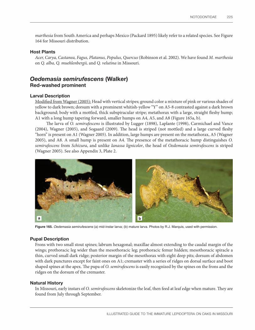
NOTODONTIDAE 225
ILLUSTRATED GUIDE TO THE IMMATURE LEPIDOPTERA ON OAKS IN MISSOURI
marthesia from South America and perhaps Mexico (Packard 1895) likely refer to a related species. See Figure 164 for Missouri distribution.
Host PlantsAcer, Carya, Castanea, Fagus, Platanus, Populus, Quercus (Robinson et al. 2002). We have found M. marthesia on Q. alba, Q. muehlenbergii, and Q. velutina in Missouri.
Oedemasia semirufescens (Walker)Red-washed prominent
Larval DescriptionModified from Wagner (2005): Head with vertical stripes; ground color a mixture of pink or various shades of yellow to dark brown; dorsum with a prominent whitish-yellow “Y” on A5-8 contrasted against a dark brown background; body with a mottled, thick subspiracular stripe; metathorax with a large, straight fleshy hump; A1 with a long hump tapering forward, smaller humps on A4, A5, and A8 (Figure 165a, b).
The larva of O. semirufescens is illustrated by Lugger (1898), Laplante (1998), Carmichael and Vance (2004), Wagner (2005), and Sogaard (2009). The head is striped (not mottled) and a large curved fleshy “horn” is present on A1 (Wagner 2005). In addition, large humps are present on the metathorax, A5 (Wagner 2005), and A8. A small hump is present on A4. The presence of the metathoracic hump distinguishes O. semirufescens from Schizura, and unlike Ianassa lignicolor, the head of Oedemasia semirufescens is striped (Wagner 2005). See also Appendix 3, Plate 2.
a b
Figure 165. Oedemasia semirufescens (a) mid-instar larva; (b) mature larva. Photos by R.J. Marquis, used with permission.
Pupal DescriptionFrons with two small stout spines; labrum hexagonal; maxillae almost extending to the caudal margin of the wings; prothoracic leg wider than the mesothoracic leg; prothoracic femur hidden; mesothoracic spiracle a thin, curved small dark ridge; posterior margin of the mesothorax with eight deep pits; dorsum of abdomen with dark punctures except for faint ones on A1; cremaster with a series of ridges on dorsal surface and boot shaped spines at the apex. The pupa of O. semirufescens is easily recognized by the spines on the frons and the ridges on the dorsum of the cremaster.
Natural HistoryIn Missouri, early instars of O. semirufescens skeletonize the leaf, then feed at leaf edge when mature. They are found from July through September.

226 SPECIES DESCRIPTIONS
ILLUSTRATED GUIDE TO THE IMMATURE LEPIDOPTERA ON OAKS IN MISSOURI
DistributionNova Scotia and Quebec, Canada (Forbes 1948), south throughout the eastern United States (Packard 1895) to Florida (Wagner 2005), and west to Minnesota (Lugger 1898), Kentucky, Illinois (Packard 1895), Missouri, and eastern Texas (Wagner 2005). A race in the western United States is recorded from Pacific Northwest (Miller and Hammond 2000), specifically from British Columbia, Canada, Washington State (Packard 1895), and Colorado (Forbes 1948). Our records from Missouri are within the expected range of this species (Figure 166).
Host PlantsGeneral feeder on trees and shrubs (Miller and Hammond 2000) including Acer, Alnus Amelanchier, Betula, Cercis, Crataegus, Fagus, Malus, Populus, Prunus, Quercus, Q. garryana, Rosa, Salix, possibly Ulmus (Robinson et al. 2002); and Q. rubra (Handfield 2011). We have found O. semirufescens on Q. alba and Q. velutina in Missouri.
CommentsUntil Becker (2014), Oedemasia semirufescens was most commonly placed in the genus Oligocentria. Early literature (Packard 1895) uses the species name O. eximia, now a synonym of O. semirufescens.
Figure 166. Known distribution of Oedemasia semirufescens in Missouri.
Ianassa lignicolor (Walker)Lace capped caterpillar
Larval DescriptionModified from Packard 1895: Mature larva with head mottled between darker vertical stripes on white background; metathoracic hump absent; long, tapering hump on first abdominal segment and smaller hump on eighth abdominal segment; thorax green, a brown patch extends from the hump on A1 to the proleg of A6, rest of abdomen green except for a white to brown marking that goes from the proleg of A6 subventrally
Figure 167. Ianassa lignicolor (a) early instar larva; (b) mature larva. Photos by R.J. Marquis, used with permission.
a b

NOTODONTIDAE 227
ILLUSTRATED GUIDE TO THE IMMATURE LEPIDOPTERA ON OAKS IN MISSOURI
through the spiracle of A8 and ends at the anal proleg; no dorsal white v-shaped mark is present. Early instars are golden yellow and shiny, with the posterior end lifted from the leaf surface (Figure 167a, b).
The larva of I. lignicolor is illustrated by Packard (1895), Ives and Wong (1988), Laplante (1998), Wagner (2005), and Sogaard (2009). It is recognized by the characteristic head markings (see Wagner 2005: 308). In addition, there is no hump on the metathorax or A5 and the dorsum of the posterior abdominal segments is green with no white v-markings (Forbes 1948: 227).
Pupal DescriptionThe distinctive larval head markings are likely visible on the exuvia and would be very helpful in pupal identification of I. lignicolor.
Natural HistoryThe life cycle of I. lignicolor is partially described by Dyar (1889). The first two instars feed on the leaf upper side but later rest on the edge where they consume the whole leaf except for the larger veins. The prepupa stays in a tough cocoon until the adult emerges in the spring.
DistributionQuebec to southeastern Manitoba, Canada (Godfrey and Appleby 1987), and south throughout the eastern United States to Florida then west to South Dakota, Missouri, Arkansas, and Texas (Packard 1895). See Figure 168 for Missouri distribution.
Host PlantsPolyphagous on trees and shrubs, Acer, Betula, Fagus, Prunus, Quercus alba, Q. rubra (Robinson et al. 2002) and Castanea (Wagner 2005).
We have found I. lignicolor on Q. alba, Q. coccinea, Q. stellata, and Q. velutina in Missouri. Wagner (2005) stated the primary hosts are beech, chestnut and oak.
CommentsWe follow Becker (2014) and Miller et al. (2018) who placed O. lignicolor in Ianassa. Both Pohl et al. (2016) and Packard (1895: 189) prefer the generic name Oligocentria.
Figure 168. Known distribution of Ianassa lignicolor in Missouri.
Schizura ipomaeae Doubleday Morning-glory prominent
Larval DescriptionHead with three dark frontal stripes; the last two thoracic segments all green except for dorsal longitudinal stripes; A1-3 and A6-7 with white dorsal markings, lateral abdominal segments with swirling brown and red lines; A1 with a red tubercle that is long and spike-like or smaller and paired, A2-4 with yellow dorsal knobs; humps and tubercles present on A5 and A8 (Figure 169a, b). See also Appendix 3, Plate 2.
The larva of S. ipomaeae was recently illustrated in color by Laplante (1998), Miller and Hammond (2003), Carmichael and Vance (2004), and Wagner (2005). It is best identified by the characteristic head pattern, relatively long body setae, and presence of a hump with tubercle on A5 (Wagner 2005: 313).

228 SPECIES DESCRIPTIONS
ILLUSTRATED GUIDE TO THE IMMATURE LEPIDOPTERA ON OAKS IN MISSOURI
a b
Figure 169. Schizura ipomaeae (a) mid-instar larva; (b) mature larva. Photos by R.J. Marquis, used with permission.
Pupal DescriptionModified from Mosher (1917): Vertex with slight projection; antennae and prothoracic legs almost as long as mesothoracic legs; prothoracic femur hidden; maxillae almost as long as wings, more than seven-eighths their length, but stopping just above where the wings meet at the meson; caudal margin of the mesonotum with seven pits; cremaster bifurcated with boot-shaped projections.
The pupal dorsum of S. ipomaeae is illustrated by Mosher (1917); other details are given in the text (Mosher 1917: 35, 69, 70). The cremaster is shown by Packard (1895: 199). The long maxillae, smooth cuticular texture of the appendages on the “face," wings that meet at the meson, row of pits on the mesothoracic dorsum, and boot-shaped cremaster all help separate S. ipomaeae from related species studied by Mosher (1917). However, these characters have not been or compared to other related species in eastern North America.
Natural HistoryThe egg of S. ipomaeae is illustrated by Peterson (1963b). It is light yellow green, shiny, and smooth under the naked eye. Eggs are laid in a cluster of 20 to 30. The larvae feed singly and overwinter as a prepupa in the ground (Carmichael and Vance 2004). There is a single generation per year, with caterpillars first appearing from May to October near St. Louis according to Packard (1895). We see them from July to August in Missouri.
DistributionWidespread North America (Miller and Hammond 2003) from Newfoundland to British Columbia in Canada (Godfrey and Appleby 1987), throughout the eastern United States south to Florida and Texas (Wagner 2005), including the Midwest (Packard 1895), but absent from a large part of the western United States except for the Pacific Coast, also found in parts of Mexico (Powell and Opler 2009). See Figure 170 for Missouri distribution.
Host PlantsPolyphagous on trees and shrubs including Acer, Alnus, Amelanchier, Betula, Carya, Ceanothus, Cercis, Convolvulus, Crataegus, Fagus, Fraxinus, Ipomoea, Gleditsia, Hamamelis, Juglans, Malus,
Figure 170. Known distribution of Schizura ipomaeae in Missouri.

NOTODONTIDAE 229
ILLUSTRATED GUIDE TO THE IMMATURE LEPIDOPTERA ON OAKS IN MISSOURI
Populus, Prunus, Quercus, Q. alba, Rosa, Rubus, Salix, Tilia, Ulmus, Vaccinium (Robinson et al. 2002); Arbutus, and Cornus (Miller and Hammond 2003).
We have collected S. ipomaeae on Q. alba in Missouri during this study. Wagner (2005) doubts the records for morning glory.
Subfamily Notodontinae
Paraeschra georgica (Herrich-Schäffer)Georgian prominent
Larval DescriptionMature larva blue-green with black spotted thoracic legs and a yellow longitudinal stripe, irregularly edged in red, that extends from the mandible to the abdominal segments, passing through or below the spiracles, before ending at the anal proleg; dorsal and subdorsal region with white spots and sometimes a thin white subdorsal line; dorsum of A8 with a fused pair of small orange and yellow tubercles (Figure 171a, b). See also Appendix 3, Plate 2.
The larva of P. georgica is illustrated by Wagner (2005) and described by Dyar (1899c) and Klots (1956). Among eastern United States notodontids, P. georgica is most likely to be confused with Peridea angulosa because both species have a genal dash and wide spiracular stripe. The presence of a dorsal tubercle on A8 in P. georgica will separate the two taxa (Dyar 1899c). This tubercle is lacking in P. angulosa. There are slight food plant differences as well (see Host Plants). Dyar (1899c) and Klots (1956) both pointed out that Packard’s (1895) larval description of P. georgica represents a misidentification. A few early instar sphingid larvae with dorsal tubercles on A8 are potentially similar enough to P. georgica to cause confusion. The two families are easily separated by characters mentioned in the above introduction to the Notodontidae.
a b
Figure 171. Paraeschra georgica (a) mid-instar larva; (b) mature larva. Photos by R.J. Marquis, used with permission.
Pupal DescriptionLabial palpi hidden; anterior margins of maxillae extend to eyes, the suture with a weak arch compared to other notodontid genera; prothoracic femur hidden, prothoracic and mesothoracic legs not meeting at the meson, metathoracic legs hidden; mesothoracic caudal margin lacking a row of deep punctures; abdomen punctate; dorsal margin of segments 5–7 heavily sclerotized, with paired pits present on the intersegmental membrane of those segments; dorsum of A10 lacks a dorsal groove; cremaster bifurcate with one or two stout, diverging blunt spines.

230 SPECIES DESCRIPTIONS
ILLUSTRATED GUIDE TO THE IMMATURE LEPIDOPTERA ON OAKS IN MISSOURI
The pupa of P. georgica is partially described by Dyar (1899c) and Miller (1992). It is easily recognized by the distinctive paired pits present on the intersegmental membrane of A5-7 (see Miller 1992: Fig. 24).
Natural HistoryThe biology of P. georgica is described by Dyar (1899c) and Klots (1956). Based on his dissection of a female abdomen, Dyar (1899c) describes the egg as probably white with a coarse granular texture. Young larvae eat the leaf except for the midrib (Wagner 2005). The mature larva rests on the midrib, upside down, where it eats one side of the leaf at a time, starting first at the base (Klots 1956). The pupa overwinters (Wagner 2005) and occurs in the soil (Klots 1956) or between leaves (Dyar 1899c), but these sites may represent artificial substrates presented to confined larvae. There are two broods per year with adults flying from May through July (Packard 1895). The caterpillars of P. georgica occur from May through November (Wagner 2005).
DistributionThroughout the eastern United States (Covell 1984), from Quebec, Canada, to central Minnesota then south to Florida and Texas (Wagner 2005).
Paraeschra georgica is common in eastern North America (Covell 1984). See Figure 172 for Missouri distribution.
Host PlantsQuercus velutina, Q. minor (Dyar 1899c), Q. rubra (Klots 1956), Fraxinus, Prunus, Q. alba, and Q. coccinea/rubra (Robinson et al. 2002). We have seen P. georgica on Q. alba in Missouri.
Unlike Peridea angulosa, Paraeschra georgica will feed on "rough-leaved" oaks (Dyar 1899c). Records from tree species other than oak may be in error for P. tortuosa (Powell and Opler 2009: 248).
CommentsUnlike Becker (2014), P. georgica was placed in the genus Hyperaeschra by Pohl et al. (2016). We follow Pohl et al. (2016) in treating P. georgica and P. tortuosa as separate species in contrast to the treatment in Powell and Opler (2009) who considered P. tortuosa as a western subspecies of P. georgica. We use the generic name Paraeschra following the revision by Miller et al. (2018).
Figure 172. Known distribution of Paraeschra georgica in Missouri.
Subfamily Nystaleinae
Symmerista spp.
Larval DescriptionMature larva with a shiny red head and large red to orange tubercle on dorsum of A8; body with one to five black longitudinal stripes alternating with red, yellow, or white stripes, the exact arrangement varies depending on instar and species; lateral area also striped with black, red, yellow, or white; anal prolegs red (Figure 173a, b).

NOTODONTIDAE 231
ILLUSTRATED GUIDE TO THE IMMATURE LEPIDOPTERA ON OAKS IN MISSOURI
a b
Figure 173. Symmerista albifrons (a) second instar larvae; (b) mature larva. Photo a by G.L. Godfrey; b by R.J. Marquis; both used with permission.
Larvae of Symmerista canicosta and S. leucitys are illustrated by Laplante (1998), Ives and Wong (1988), McCabe (1991), Carmichael and Vance (2004), and Wagner (2005). Godfrey and Appleby (1987) illustrate the larvae of Symmerista albifrons. Sogaard (2009) photographed S. canicosta. Middle and late instar larvae of Symmerista are easily recognized by the bright red head, thick subdorsal stripe, and large, contrasting red tubercle on A8. Species identification is more difficult, and only last instar larvae can be accurately named. According to Forbes (1948), S. leucitys is distinguished by having three dorsal black stripes. Symmerista albifrons and S. canicosta are similar but have five dorsal stripes. All dorsal stripes are of equal thickness in S. canicosta, whereas the middle stripes are thicker and wavier in S. albifrons. See also Appendix 3, Plate 2.
Pupal DescriptionThe pupa of S. albifrons is described by Mosher (1917). The maxillae extend to the caudal wing margin, a row of pits are absent on the mesothorax, and the cuticle is punctate even on the mouthparts and legs. No detailed descriptions are available for related Symmerista except for Miller’s (1992) illustrations of the abdominal pits and cremaster of S. leucitys.
The subfamily Nystaleinae is recognized, in part, by a distinctive row of lunate pits on the anterior margin of the movable abdominal segments (see Miller 1992: Figs. 11, 19, 22). These are present in Symmerista. The punctate cuticle of the mouthparts and legs is also unusual, as most other notodontids have a smooth cuticle covering these appendages.
Natural HistoryThe eggs of S. albifrons and S. canicosta were photographed by Peterson (1963b) and Millers and Wallner (1975), respectively. Syme (1961) describes the egg of S. canicosta. The biology of various species of Symmerista is given by Millers and Wallner (1975), Godfrey and Appleby (1987), Donahue (1993), and Wagner (2005). Biological information published before Franclemont (1948) is of limited value because this sibling species complex was not recognized by early workers (e.g., Packard 1895).
A typical life history for a species of Symmerista is given by Millers and Wallner (1975). Eggs are laid in a mass. Early instars skeletonize leaves (R.J. Marquis, pers. obs.), which turns the leaf tips brown. Later instars consume most of the leaf tissue. Mature larvae tend to be solitary defoliators. When disturbed, early instars drop from the leaf on silken threads, later returning to their original substrate. Pupation occurs in the leaf litter. The pupa, or more rarely the prepupa, is the overwintering stage. Adults emerge in the spring of the following year; although, a small number will pass a second season in diapause. Symmerista spp. have one to two generations per year, and caterpillars occur from July to September (USDA 1985, Wagner et al. 1997).
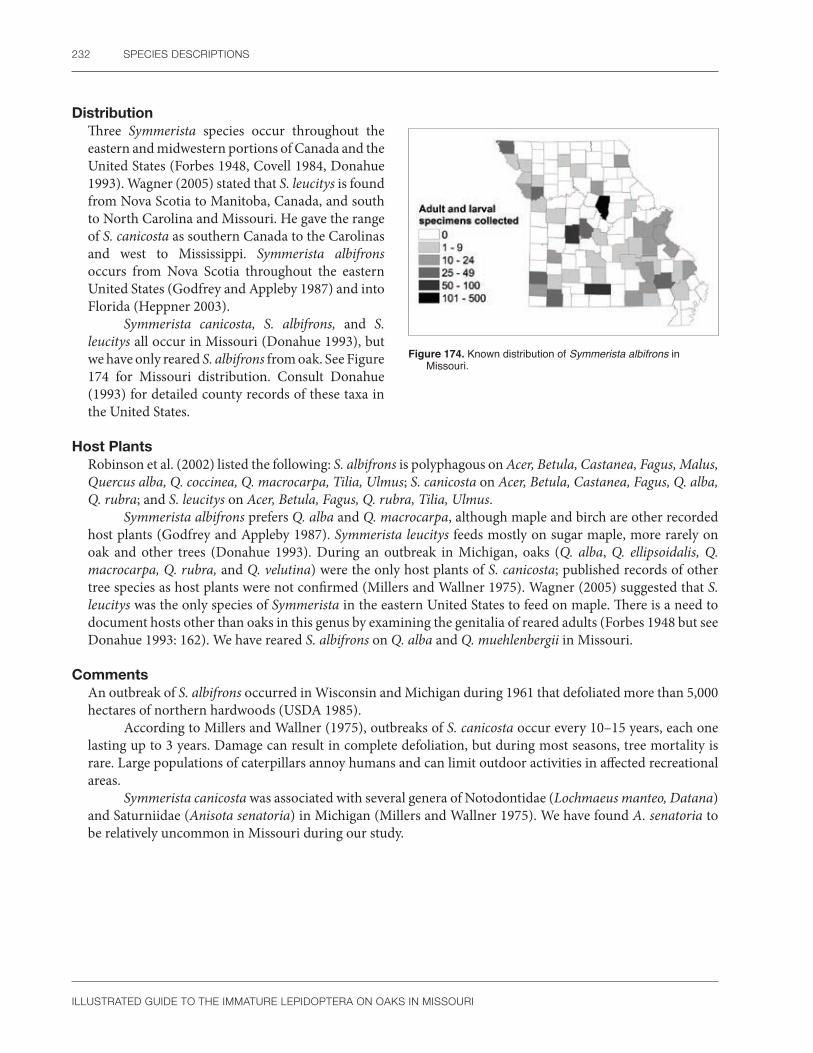
232 SPECIES DESCRIPTIONS
ILLUSTRATED GUIDE TO THE IMMATURE LEPIDOPTERA ON OAKS IN MISSOURI
DistributionThree Symmerista species occur throughout the eastern and midwestern portions of Canada and the United States (Forbes 1948, Covell 1984, Donahue 1993). Wagner (2005) stated that S. leucitys is found from Nova Scotia to Manitoba, Canada, and south to North Carolina and Missouri. He gave the range of S. canicosta as southern Canada to the Carolinas and west to Mississippi. Symmerista albifrons occurs from Nova Scotia throughout the eastern United States (Godfrey and Appleby 1987) and into Florida (Heppner 2003).
Symmerista canicosta, S. albifrons, and S. leucitys all occur in Missouri (Donahue 1993), but we have only reared S. albifrons from oak. See Figure 174 for Missouri distribution. Consult Donahue (1993) for detailed county records of these taxa in the United States.
Host PlantsRobinson et al. (2002) listed the following: S. albifrons is polyphagous on Acer, Betula, Castanea, Fagus, Malus, Quercus alba, Q. coccinea, Q. macrocarpa, Tilia, Ulmus; S. canicosta on Acer, Betula, Castanea, Fagus, Q. alba, Q. rubra; and S. leucitys on Acer, Betula, Fagus, Q. rubra, Tilia, Ulmus.
Symmerista albifrons prefers Q. alba and Q. macrocarpa, although maple and birch are other recorded host plants (Godfrey and Appleby 1987). Symmerista leucitys feeds mostly on sugar maple, more rarely on oak and other trees (Donahue 1993). During an outbreak in Michigan, oaks (Q. alba, Q. ellipsoidalis, Q. macrocarpa, Q. rubra, and Q. velutina) were the only host plants of S. canicosta; published records of other tree species as host plants were not confirmed (Millers and Wallner 1975). Wagner (2005) suggested that S. leucitys was the only species of Symmerista in the eastern United States to feed on maple. There is a need to document hosts other than oaks in this genus by examining the genitalia of reared adults (Forbes 1948 but see Donahue 1993: 162). We have reared S. albifrons on Q. alba and Q. muehlenbergii in Missouri.
CommentsAn outbreak of S. albifrons occurred in Wisconsin and Michigan during 1961 that defoliated more than 5,000 hectares of northern hardwoods (USDA 1985).
According to Millers and Wallner (1975), outbreaks of S. canicosta occur every 10–15 years, each one lasting up to 3 years. Damage can result in complete defoliation, but during most seasons, tree mortality is rare. Large populations of caterpillars annoy humans and can limit outdoor activities in affected recreational areas.
Symmerista canicosta was associated with several genera of Notodontidae (Lochmaeus manteo, Datana) and Saturniidae (Anisota senatoria) in Michigan (Millers and Wallner 1975). We have found A. senatoria to be relatively uncommon in Missouri during our study.
Figure 174. Known distribution of Symmerista albifrons in Missouri.

NOTODONTIDAE 233
ILLUSTRATED GUIDE TO THE IMMATURE LEPIDOPTERA ON OAKS IN MISSOURI
Subfamily Phalerinae
Nadata gibbosa (Smith)White-dotted prominent
Larval DescriptionMature larva with an enlarged head, often wider than the prothorax; mandibles contrasting yellow (or orange according to McCabe 1991) with black tips; head, thorax, and abdomen pale green to turquoise with a faint yellow or white subdorsal longitudinal stripe; spiracles red; subventral area spotted white; anal shield with yellow margin (Figure 175a, b).
a b
Figure 175. Nadata gibbosa (a) mid-instar larva; (b) mature larva. Photos by R.J. Marquis, used with permission.
Early instars of Nadata gibbosa are colored light green and yellow with the digestive system showing through the pale cuticle. They have few white spots, but the characteristic large head and yellow margined anal shield are obvious even on middle instar larvae (Miller 1995). See also Appendix 3, Plate 2.
The larva of N. gibbosa is illustrated by Packard (1895), Godfrey and Appleby (1987), Ives and Wong (1988), Miller (1995), Carmichael and Vance (2004), and Wagner (2005). Superficially, N. gibbosa resembles a complex of noctuid genera collectively named “green fruitworms” because of the similar green and white color pattern. The two taxa are easily separated by morphological characters listed in the introduction above. For example, green fruitworm noctuids lack a stipital lobe and secondary setae in the SV group of A3-6.
Among notodontids, N. gibbosa is similar to Peridea angulosa and Paraeschra georgica in body shape. However, N. gibbosa lacks the genal dash and wide spiracular stripe that characterizes P. angulosa and P. georgica. According to photographs by Ives and Wong (1988) and McCabe (1991: 27), except for a smaller head, the larva of Gluphisia septentrionis is also similar in color to N. gibbosa. Forbes (1948: 209) partially separated Gluphisia and Nadata by their cuticle thickness. This character will not work with early instars of N. gibbosa, which share a thin cuticle with Gluphisia. Preserved larvae of the two species, which generally fade and lose details of the color pattern, are best separated by the shape of the tarsal setae and crochet arrangement. The tarsal setae of Gluphisia are spatulate (Miller 1991: 472), whereas these setae are thinner and more lanceolate in N. gibbosa. The abdominal crochets of early instars of Gluphisia are divided, unlike N. gibbosa, which has the crochets undivided in all instars. Further differences in the P setal spacing and mandibles were illustrated by McCabe (1991).

234 SPECIES DESCRIPTIONS
ILLUSTRATED GUIDE TO THE IMMATURE LEPIDOPTERA ON OAKS IN MISSOURI
Pupal DescriptionLabial palpi hidden; anterior margins of maxillae barely extend to eyes, the suture with a weak arch compared to other notodontid genera; prothoracic femur hidden, prothoracic and mesothoracic legs not meeting at the meson, metathoracic legs hidden; mesothoracic caudal margin lacking a row of deep punctures; abdomen finely punctate; dorsal margin of segments 5–7 heavily sclerotized, the intersegmental membrane of those segments with short, scalloped ridges medially and a few small punctures laterally; dorsum of A10 lacks a dorsal groove; cremaster a long thin rod with two diverging curved spines.
The pupa of N. gibbosa is partially described by Packard (1895) who illustrated an unusual cremaster with four spines. In six examples of pupal skins associated with reared adults, we are not able to find a second pair of spines on any specimen of N. gibbosa from Missouri. Nadata gibbosa is classified in the Phalerinae using Miller’s (1992) key to notodontid subfamilies, although it is an exception because it lacks a dorsal groove on A10.
Natural HistoryThe biology of N. gibbosa is summarized by Packard (1895). The egg of N. gibbosa was studied by Packard (1895), Peterson (1963b), and Syme (1961). They are shiny white with a yellow or greenish tint and laid in a mass (Peterson 1963b). The chorion has mostly hexagonal reticulations (Syme 1961, Peterson 1963b). When threatened, larvae curl up and expose the mandibles (Wagner 2005). Normally they rest on the “back” (underside) of the leaf (Packard 1895: 149). The pupation site needs documentation. Packard (1895) stated that N. gibbosa pupates underground, whereas Godfrey and Appleby (1987) gave the pupation site as leaf litter without a cocoon. The caterpillars of N. gibbosa occur from May through November (Wagner 2005). Godfrey and Appleby (1987) considered N. gibbosa to be bivoltine, with broods in June–July and August–September. Adults fly from early April to October in Missouri, suggesting a possible third brood in our area (Heitzman and Heitzman 1987).
DistributionCoast to coast throughout North America (Powell and Opler 2009) from Newfoundland west to British Columbia, Canada, south to Florida, Texas, and California (Godfrey and Appleby 1987).
Nadata gibbosa is abundant in Missouri forests (Heitzman and Heitzman 1987; Figure 176), common in the eastern United States (Covell 1984), and common on oak in the western United States (Miller 1995).
Host PlantsAcer, Alnus, Amelanchier, Betula, Castanea, Corylus, Fagus, Fraxinus, Populus, Prunus, Quercus alba, Q. garrayana, Q. kelloggii, Q. macrocarpa, Q. rubra, Rosa, and Salix (Robinson et al. 2002). We have collected N. gibbosa on Q. alba, Q. macrocarpa, Q. stellata, and Q. velutina in Missouri.
Newly hatched larvae from New York rejected Acer, Alnus, Betula, Prunus, and Salix and only partially fed upon Amelanchier (McCabe 1991). Because some of these genera are known host plants, these data suggest that N. gibbosa may have regional host preferences or the ability to enlarge its diet with age, assuming that all variables in the above feeding trials were controlled (see Marquis and Braker 1987, Waller and Jones 1989).
Figure 176. Known distribution of Nadata gibbosa in Missouri.

NOTODONTIDAE 235
ILLUSTRATED GUIDE TO THE IMMATURE LEPIDOPTERA ON OAKS IN MISSOURI
Peridea angulosa (Smith) Angulose prominent
Larval DescriptionMature larva light green with a longitudinal stripe, colored red or pink dorsally and yellow or white ventrally, widest at the anterior end, that extends from the mandible through the spiracles of A1-7, including a paler portion that passes below the spiracle on A8 before ending at the base of the anal proleg; dorsum with two parallel, white middorsal lines above an irregularly spotted subdorsal region; abdominal prolegs striped with red on the outer surface (Figure 177). Earlier instars have several white longitudinal body stripes that are lost at maturity. The characteristic genal band and wide spiracular stripe are obvious even in young larvae.
The larva of P. angulosa is illustrated by Packard (1895), Miller (1991: Fig. 502), Carmichael and Vance (2004), and Wagner (2005). It is easily confused with several genera of Noctuidae if careful attention is not paid to details of the distinctive coloration. Structural characters, as mentioned in the introduction (presence of a stipital lobe, secondary setae in the SV group of A4-8, etc.), will easily separate the two families. Among notodontids, P. angulosa is superficially similar to Nadata gibbosa and Paraeschra georgica. Unlike P. angulosa, N. gibbosa lacks a genal dash and spiracular stripe. The presence of a dorsal tubercle on A8 readily separates P. georgica from P. angulosa.
Most misidentifications of P. angulosa are likely to occur at the species level. In particular, P. angulosa can be confused with the related P. ferruginea and P. basitriens. These species are best separated by the color of the prothoracic spiracle and characters of the epicranium in dorsal view (Riotte 1969). In P. angulosa, the prothoracic spiracle is white with a black peritreme, not black or yellow as in P. ferruginea and P. basitriens, respectively. Unlike P. ferruginea, the epicranium of P. angulosa has only a weak pattern of markings. Furthermore, P. angulosa lacks a median depression on the vertex, which is otherwise characteristic of P. basitriens. For other differences among Peridea species, consult Riotte (1969). The hosts are also different. The host of P. angulosa is oak, not birch as is common for P. ferruginea or maple as is normal for P. basitriens (Wagner 2005).
Pupal DescriptionModified from Packard 1895: Labial palpi hidden; maxillae short, the anterior margin reaches the eye; prothoracic femur hidden; the antennae, but not the prothoracic or mesothoracic legs, meet at the meson; abdomen sparsely punctate; cremaster represented by a small tubercle.
The pupa was partially described and illustrated by Packard (1895). According to Riotte (1969), the pupa of P. angulosa is a lighter mahogany brown than P. ferruginea and P. basitriens. In addition, the terminal abdominal segment of P. angulosa has a larger tubercle then the other two related species.
Natural HistoryThe biology of P. angulosa is summarized by Packard (1895). The egg of P. angulosa was photographed by Peterson (1963b). They are chalky white with faint hexagonal reticulations and are laid singly or in small clusters. Larvae rest on the leaf edge with the head turned toward the base (Packard 1895). According to Wagner (2005), caterpillars of P. angulosa occur from May through November and curl when disturbed in a similar manner to N. gibbosa. Pupation occurs underground in a weak cocoon of silk and soil (Packard 1895). There are at least two generations per year in the eastern United States (Wagner 2005).
Figure 177. Peridea angulosa mature larva. Photo by R.J. Marquis, used with permission.

236 SPECIES DESCRIPTIONS
ILLUSTRATED GUIDE TO THE IMMATURE LEPIDOPTERA ON OAKS IN MISSOURI
DistributionNova Scotia to Manitoba, Canada, and south to Florida and Texas (Covell 1984), including Kentucky (Covell 1999), West Virginia (Albu and Metzler 2004), Ohio (Teraguchi and Lublin 1999), Illinois, and Missouri (Packard 1895). See Figure 178 for Missouri distribution.
Peridea angulosa is common to rare depending on the region (Covell 1984, Handfield 1999). Teraguchi and Lublin (1999) collected 290 specimens of P. angulosa in a 4-year period in Ohio. Carmichael and Vance (2004) considered P. angulosa to be locally common in Ontario, Canada.
Host PlantsCarya, Castanea, Quercus alba, Q. rubra, Q. macrocarpa (Robinson et al. 2002). We have collected P. angulosa on Q. alba in Missouri. Records for P. angulosa on Eremochloa (Robinson et al. 2002) and Acer (Handfield 1999) need confirmation.
Figure 178. Known distribution of Peridea angulosa in Missouri.

NOLIDAE 237
ILLUSTRATED GUIDE TO THE IMMATURE LEPIDOPTERA ON OAKS IN MISSOURI
NolidaePunkies
As defined by Zahiri et al. (2013), there are approximately 1,740 species of Nolidae worldwide. Historically, lepidopterists have disagreed on the composition and systematic position of the family. Their early history is treated by Kitching (1984) and later more completely defined by Kitching and Rawlins (1998). Most genera were either placed with the Arctiidae, as a subfamily of the Noctuidae, or as a separate family. Recent morphological (Beck 2009: 455) and molecular studies (Zahiri et al. 2013) suggest Nolidae should be a separate family. In spite of the morphological diversity of the immature stages, the subfamilies seem monophyletic. Lafontaine and Schmidt (2010) recognized five subfamilies of Nolidae in the North American fauna: Afridinae, Risobinae, Chloephorinae, Collomeninae, and Nolinae. The latest definition by Zahiri et al. (2013) has 10 subfamilies worldwide: Risobinae, Collomeninae, Beaninae, Eligminae, Westermanniinae, Nolinae, Chloephorinae, Afridinae, Camptolominae, and surprisingly, the Diphtherinae. Beck (2009) would place some of these subfamilies back in the Noctuidae. The following subfamilies are recorded from North America: Diphtherinae, Nolinae, Chloephorinae, Eligminae, Risobinae, Collomeninae, and Afridinae (Pohl et al. 2016). For more information on this family, see Kitching and Rawlins (1998), Holloway (1998, 2003), Beck (1999, 2009), and Zahiri et al. (2013). Wagner et al. (2011) discussed the biology of most eastern United States species and illustrated many larvae and cocoons.
Detailed egg descriptions are available for a few species of Nolidae. The subfamily Chloephorinae has at least two different types. Pseudoips from Russia has a flat ribbed egg similar to the Acronictinae (Dolinskaya 2014). Spiny bollworms in the genus Earias have a rounded ribbed egg with a circle of blunt tubercles surrounding the micropyle (Balachowsky 1972: Fig. 469). The same seems to be true of the Nolinae. The egg of Uraba is round and ribbed with a central transparent area (McFarland 1972), whereas the Nola egg photographed by Peterson (1961) is of the flat ribbed type. Peterson (1961) also studied Diphthera festiva (Diphtherinae). The egg is rounded, not flattened, and is covered with hooked setae. The ribbed egg morphology of nolids is more similar to the majority of Noctuidae and Erebidae than to Arctiinae that have smooth eggs.
As currently defined, no one character separates all larval nolid subfamilies from all other Noctuiodea worldwide. Beck (2009: 7) noted that the fusion of the L1 and L2 abdominal verrucae is characteristic of the Nolidae he studied, and even the first instar of Nola lacks prolegs on A3. Some nolids have a bisetose thoracic SV group and heteroideous crochets as do the Arctiinae (Holloway 1998: 260, 273). A prothoracic gland (adenosma) is absent in a few nolid subfamilies (Ahola and Silvonen 2005). The caterpillar of D. festiva (Diphtherinae) has fully developed prolegs on A3-6 and looks like a typical member of the Noctuidae or Erebidae as illustrated by Wagner et al. (2011). The ability of some Old World species to stack cast head capsules from previous instars onto the larval body (McFarland 1978) and the use of rolled leaves as shelters by nolids in the eastern United States (Wagner et al. 2011) are two unusual aspects of nolid larval biology. The life history of one eastern United States Nola even includes miners and borers, depending on the instar (Wagner et al. 2011).
In North America, two subfamilies have only a single genus: Afrida in the Afridinae and Baileya of the Risobinae. Afrida is a lichen feeder, cryptically colored green and black, with long and sometimes glandular setae (Wagner et al. 2008, 2011). Wagner et al. (2011) illustrates several Baileya caterpillars. They are green with a thin to thick white subdorsal stripe and sometimes have a few white spots below this line. Hosts are in the Juglandaceae or Betulaceae, depending on the species. Crumb (1956: 346) noted his preserved specimens of Baileya lost their coloration. He characterized the larva as having an inner tooth on the mandible, unmodified tarsal setae, a bisetose SV group on A1, SD1 seta-like on A9, and a coarsely granulose anal shield.
Chloephorinae is currently divided into several tribes with extremely diverse larvae (Zahiri et al. 2013). Perhaps the most common eastern United States genus of Chloephorinae is Nycteola, a feeder on willow (Salix) and poplar (Populus) that has the F setae well above the frontal pores, spatulate tarsal setae, and very long, thin D setae (Gardner 1948a, Crumb 1956). In addition, the prolegs are all developed on A3-6, the SV group is bisetose on A1, and the spinneret has lateral flaps (Wagner et al. 2011). This subfamily also includes Garella nilotica, a cosmopolitan species that is an internal or external feeder on a wide variety of plants (Wagner et al. 2011). The

238 SPECIES DESCRIPTIONS
ILLUSTRATED GUIDE TO THE IMMATURE LEPIDOPTERA ON OAKS IN MISSOURI
larva has a characteristic pattern on the prothoracic shield, with frontal pores in a more normal position near the F setae, short dorsal setae, and unmodified tarsal setae that are not spatulate (Crumb 1956). A third type of larva included in this subfamily is represented by Earias, the spiny bollworms of the Old World (see Gómez de Aizpúrua 2002b for a typical example). These larvae are consistently misidentified by inexperienced United States port identifiers because of their spiny skin, bisetose thoracic SV group, and fleshy filaments that resemble scoli, each with single terminal spine (SCPC, Gilligan and Passoa 2014). They feed most often on cotton, okra, hibiscus, and willow (Zhang 1994).
Three genera of Collomeninae occur in the United States, two of which are illustrated by Wagner et al. (2011). The larva has reduced prolegs on A3-4, with an enlarged planta on the prolegs of A5-6 (Zahiri et al. 2013). Both A1 and A2 have the SV group trisetose, and in the New World they have been reared on Malpighiaceae (Wagner et al. 2011).
Diphthera festiva (Diphtherinae) is a black, transversely striped caterpillar with an open apical silk pore on the spinneret and bisetose SV group on A1. This species is usually found on Malvaceae, although other hosts are also reported (Crumb 1956, Passoa 1984, Dunford and Barbara 2004, Wagner et al. 2011).
Missouri oak-feeding nolids are classified in the Nolinae, a subfamily having very distinctive caterpillars. All North American Nolinae lack prolegs on A3 and have verrucae covering the body (Stehr 1987). Nolinae, like the related genus Nycteola, have long setae, especially on A9 (Wagner et al. 2011). The body form of most eastern United States species is similar and colored with shades of brown to gray with dots, patches, or rarely a longitudinal line. Hosts are oak (Quercus) or other woody plants (Wagner et al. 2011).
Limited morphological information exists on the larvae of the other four subfamilies of Nolidae, especially for those that do not occur in North America. The Beaninae has only one genus, Beana, which contains larvae with reduced prolegs on A3 and a horn on the anal shield (Zahiri et al. 2013). A European species of Beana illustrated by Gómez de Aizpúrua (2002b) is green with two small dorsal tubercles on the thorax. As defined by Zahiri et al. (2013), Eligminae includes larvae that resemble arctiines. Selepa has gregarious larvae with large eversible glands of unknown function above the SD seta on segments A2-A7 (Kitching and Rawlins1998). Gadirtha has spinose patches on the thorax and is a potential biological control agent of Chinese tallow (Pogue 2014). Another example of a similar larva is Iscadia (https://bugguide.net/node/view/1164098). Eligma has long primary setae arising from chalaza and feeds on Ailanthus (Holloway 2003). Larvae of the Westermanniinae occur in the Old World and often feed on Terminalia (Holloway 2003). Their morphology is poorly known. Camptolominae is an Oriental group (Zahiri et al. 2013) with larvae that have a minute L2 seta on the prespiracular pinaculum, a bisetose SV group on the thorax, and two curved setae on A10 that form an anal comb (Holloway 1998). At least one species from Japan illustrated by Issiki (1971) has long setae and is longitudinally striped. The gregarious larvae feed in a web on Fagaceae (Holloway 1998).
Nolid pupal morphology is frequently cited as an apomorphy that unites the various subfamilies into a monophyletic unit. Perhaps the loudest critic of this classification is Beck (2009: 7), who describes this character as “very dubious, putative, and unsuitable.” There are three components to their specialized pupation: the cocoon, in profile, resembles the keel of an upside-down boat; the cocoon has two layers (thus is double walled); and the anterior end has a ridgelike vertical exit slit for adult emergence (Franclemont 1960, McFarland 1978, Stehr 1987, Butler 1989, Common 1990, McCabe 1996, Holloway 1998, Kitching and Rawlins 1998). Although some nolid cocoons fit the “boatlike” shape (Gardner 1948b: Figs. 11, 12; Butler 1989; Alford 1995; McCabe 1996; Kitching and Rawlins 1998: Fig. 19.17 X, Y; Nycteola in SCPC), others are better described as oval, elongated, and hardened (often with pieces of plant material included) (Reinhard 1938: Fig. 2, McFarland 1978, Wagner 2005: 456, pupa of D. festiva in SCPC). Unlike most nolids, the cocoon of D. festiva is single walled, and that of Beana is composed of leaves in a cell of silk (Holloway 1998). This variability seems to confirm Beck’s (2009) objections, but much of the evidence characterizing nolid cocoons as boatlike in the Old World is based on Bell’s unpublished manuscript (see Holloway 1998). Publication of this work, and especially the figures, is needed to help resolve the controversy. The few nolids available for our study (Nola, Meganola, and Earias, SCPC) have the prothoracic femur exposed and mesothoracic leg widely separated from the ventral margin of the eye. This differs from Mosher’s (1916a)

NOLIDAE 239
ILLUSTRATED GUIDE TO THE IMMATURE LEPIDOPTERA ON OAKS IN MISSOURI
characterization of Nolidae because she noted that the prothoracic femur is either hidden or exposed. The species that Mosher examined and Nola pustulata (McCabe 1996) have mesothoracic legs that extend to at least the eyes. The D verrucae may be fused and there may be secondary setae around the scars of the verrucae (Holloway 1998). Pupae of the Nolinae may have long secondary setae on the cuticle, including the terminal abdominal segments (Patočka and Turčáni 2005). The integument of Meganola lacks the dense hairs found on Nola lagunculariae (SCPC). Longitudinal ridges or a series of beads may be present on A10 (Holloway 1998, Kitching and Rawlins 1998) that produce sound by being rubbed against the cocoon (Hinton 1948). All genera examined by Mosher (1916a), McCabe (1996), and us lack a cremaster, but again the situation is more complex. Species of Earias (Patočka and Turčáni 2005, SCPC) and D. festiva (SCPC) have lateral spines on the last abdominal segment.
Despite the variation mentioned above, pupae of the Nolidae, at least for those on Missouri oak trees, can be identified by their hard, elongated, boat-shaped cocoon and lack of a cremaster. Some Notodontidae (Cerura) and Lasiocampidae (Tolype) also form elongated cocoons, but details of the appendages for these families generally differ from those of the Nolidae (Mosher 1916a). In contrast to the Noctuidae and Erebidae, the Euteliidae and Nolidae lack a cremaster (Mosher 1916a, Gardner 1948b, Holloway 1998, Kitching and Rawlins 1998). However, euteliids do not pupate in a boatlike cocoon.
Only a few Nolidae are important pests. Examples include Earias (on cotton), Nola cereella (on sorghum), and Uraba (on Eucalyptus) (Kitching and Rawlins 1998, Common 1990). A more complete list is given by Zahiri et al. (2013).
Subfamily Nolinae
Meganola phylla (Dyar)
Larval DescriptionEarly instar larva pale green or yellow, flattened, with dense hairs arising from verrucae; some dorsal verrucae of the thorax and abdomen with faint, paired black spots, those of A3 darker than the rest. See also Appendix 3, Plate 2. The mature larva is yellow to tan, densely hairy, with a faint, thin, brown middorsal stripe, mottled white and brown integument, and a contrasting black spot on A3 that interrupts the middorsal stripe (Figure 179a, b).
As noted by Franclemont (1960), Franclemont (1985), and Wagner et al. (2011), three closely related oak-feeding species of Meganola occur in the northeastern United States. The larva of M. minuscula is illustrated by Ives and Wong (1988) and Wagner et al. (2011). Each author figured a larva with three black
a b
Figure 179. Meganola phylla (a) early instar larva; (b) mature larva. Photos by R.J. Marquis, used with permission.

240 SPECIES DESCRIPTIONS
ILLUSTRATED GUIDE TO THE IMMATURE LEPIDOPTERA ON OAKS IN MISSOURI
dorsal patches and a cream-colored dorsum. A second Meganola larva was originally described by Dyar as M. minuscula, then listed by Franclemont (1960: 53) as a new species, and finally given the name M. spodia (Franclemont 1985). This larva can be distinguished by the dark brown subdorsal stripe and only two dorsal patches on A4 and A8 (Franclemont 1960). However, Butler (1989) doubted this association and described a larva with a red dorsal stripe and verrucae as in the true M. spondia. This better fits the illustration of this species in Wagner et al. (2011). The Meganola larva illustrated in this work has only a single dark patch on A3 at maturity, suggesting a third species, M. phylla (see Comments). Finally, Nola ovilla is recorded on oak, but we agree with Wagner et al. (2011) that confusion with M. phylla is unlikely. Although differences in larval coloration apparently exist among these taxa, we consider larval identifications tentative until more rearing and life history data are available. Nevertheless, based on the reared adult and larval coloration, our study organism best fits M. phylla.
A preserved mid-instar larva of M. phylla from Missouri had the following unusual morphological characters: second condyle of the mandible enlarged with an oval depression equal to twice the area of the knoblike first condyle; SV group of the thorax multisetose; and tarsal setae thin, flattened, and much longer than the tarsal claw. Kitching and Rawlins (1998: 388) noted that most nolids have a unisetose or bisetose SV group on the thorax, thus the multisetose verrucae of Meganola is atypical. The same multisetose condition is found in M. spondia and Nola (Butler 1989, McCabe 1996).
Pupal DescriptionLabrum rectangular; labial palpi exposed; maxillae extend approximately one half the distance to caudal margin of the wings; prothoracic leg slightly longer than maxillae; prothoracic femur exposed; mesothoracic leg not touching the lower eye margin; metathoracic legs exposed well past the caudal margin of the wings; metathoracic spiracle small and slitlike; texture of abdominal cuticle very finely reticulated, appearing shagreened from a distance; terminal abdominal segments bluntly rounded, without any trace of a cremaster.
The few nolids available for our study (Nola, Meganola, and Earias [SCPC]) have the prothoracic femur exposed and mesothoracic leg widely separated from the ventral margin of the eye. Unlike Meganola, the integument of Nola lagunculariae is densely hairy (SCPC). At present, the pupa of Meganola is tentatively recognized by host data (associated with oak), the wide separation of the mesothoracic legs from the ventral margin of the eye, the finely reticulated abdominal texture, and the somewhat boat-shaped cocoon (see Kitching and Rawlins 1998: 19.17Y). The arrangement of appendages of a Meganola species in Europe (see Patočka and Turčáni 2005: 261, Fig. 25) is very similar to our Meganola specimen from Missouri. More species of Nolidae need to be examined before an accurate diagnosis of pupal Meganola is possible.
Natural HistoryThe life history of Meganola phylla is described by Wagner et al. (2011). Eggs are laid on the underside of leaves near the midrib. Larvae are found in abandoned leaf shelters of other caterpillars, near dead or discolored leaf spots or along the midrib. The cocoon is formed of plant material mixed with silk and is higher at one end. The usual location is a petiole or twig. Adults of M. phylla have been collected in southern Ohio from March through August, with a peak in abundance during May and July (Rings et al. 1992). A similar flight period (May through August) was given by Franclemont (1960), who noted that M. phylla is double-brooded, with melanic forms commonly found in Virginia. There may be additional broods in the south (Wagner et al. 2011). The larva (R.J. Marquis, pers. obs.) or pupa (Wagner et al. 2011) overwinters. In Missouri, early instars of M. phylla are often found on the underside of a leaf appressed to the midrib (R.J. Marquis, pers. obs.).
DistributionSouthern Ontario, Canada, through New England, west to Wisconsin, and south to Florida, Texas (Wagner et al. 2011), and Alabama (Franclemont 1960), including Ohio (Rings et al. 1992). See Figure 180 for Missouri distribution of M. phylla.

NOLIDAE 241
ILLUSTRATED GUIDE TO THE IMMATURE LEPIDOPTERA ON OAKS IN MISSOURI
HostsFagus, red and white oaks, Quercus alba, Q. marilandica, and Q. prinoides (Wagner et al. 2011). In Missouri, M. phylla is most common on Q. velutina but was also reared from Q. alba and Q. stellata. There is a possible record of M. phylla on “live oak” in Florida (Wagner et al. 2011).The larva of M. minuscula and M. spodia both feed on oak (Franclemont 1960, Butler 1989, Wagner 2005). Some Meganola also are recorded from apple (Stehr 1987) and willow (Covell 1984).
CommentsFranclemont (1960, 1985) gives characters to separate the three species of Meganola in the eastern United States. Females of M. spodia have two small signa whereas both M. minuscula and M. phylla have a single large signum. Only one signum is present in our reared Meganola from Missouri, thus eliminating M. spodia from consideration. Details of the wing pattern can also be used to separate M. minuscula and M. phylla. In the Missouri specimen, the basal portion of the costa is black with a well-defined black central costal spot. In combination with the genitalia, these features best fit M. phylla (Franclemont 1960: 53; see adult photographs in Wagner et al. 2011). However, M. minuscula was recently collected from and reared on Q. alba at Cuivre River State Park near Troy, MO (2017) by R.J. Marquis, which shows that both species do occur in Missouri.
Covell (1984) considered M. phylla to be subspecies of M. minuscula, but Rings et al. (1992) suggested that both were valid species. Our larval studies and illustrations in Wagner et al. (2011) favor the view that M. minuscula and M. phylla are separate species, in agreement with Lafontaine and Schmidt (2010) who cited differences in mitochondrial DNA for this distinction. Meganola minuscula and M. phylla are also listed as separate species in Pohl et al. (2016).
Figure 180. Known distribution of Meganola phylla in Missouri.

242 SPECIES DESCRIPTIONS
ILLUSTRATED GUIDE TO THE IMMATURE LEPIDOPTERA ON OAKS IN MISSOURI
NoctuidaeNoctuid owlet moths
Noctuidae, with over 11,000 species distributed worldwide, are the second largest family of Lepidoptera (Nieukerken et al. 2011). Several workers have studied noctuid classification in the traditional sense (Kitching 1984, Poole 1989, Speidel et al. 1996, Kitching and Rawlins 1998). Good summaries exist on the biology of some Old World species (Mathur 1942, Sugi 1987) and the phylogeny of several large subfamilies worldwide (Kitching 1987, Matthews 1991). In addition to general works that include Noctuidae, some publications focus specifically on noctuid egg (Salkeld 1984), larval (Ripley 1923; Crumb 1956; Godfrey 1972a; Beck 1999, 2000; Ahola and Silvonen 2005, 2008, 2011), pupal (Gardner 1948b; Khotko 1968; Patočka 1995, 1996; Patočka and Turčáni 2005), and adult (Ryabov 1988) morphology from several faunal regions. For a more complete introduction to classical noctuid literature, consult the reference lists in Holloway et al. (1987), Kitching (1987), Scoble (1992), Speidel et al. (1996), and Kitching and Rawlins (1998), as well as the appropriate volumes of the “Moths of North America” series (http://wedgefoundation.org/home.html) or the 13 volumes of “Noctuidae Europaeae" (https://www.nhbs.com/3/series/noctuidae-europaeae).
After years of forced stability under the “Hampson system” (see Kitching 1984), noctuid classification has undergone an extensive reorganization using cladistic methodology, larval characters, and molecular data, with emphasis on the Holarctic region (Weller et al. 1994; Mitchell et al. 2000; Fibiger and Lafontaine 2005; Lafontaine and Fibiger 2006; Lafontaine and Schmidt 2010; C. Mitter in Wagner et al. 2011: 32, 33; Zahiri 2013; Regier et al. 2017). Initially, Euteliidae, Micronoctuidae, and Nolidae were removed from the Noctuidae and given family status within the Noctuoidea. The “Catocalinae” and their relatives are combined with more traditional taxa like the Lymantriidae and Arctiidae to form a new family Erebidae. Many of the remaining noctuid subfamilies of Hampson form the new restricted definition of the Noctuidae. At lower taxonomic levels, many tribes were moved, renamed or created, especially in the Acronictinae (Rota et al. 2016) and Erebinae (including taxa formerly in the Catocalinae) (Lafontaine and Schmidt 2010). Recent molecular data from Zahiri et al. (2012) suggest the Micronoctuidae should be a tribe of the Hypenodinae and not a separate family.
No doubt most of this reclassification was long overdue, and the new insights show much promise, but there is still a need for some caution (Beck 2009). Egg morphology (ribbed or smooth chorion) and mode of oviposition need further study. The ribbed egg and presence of a ventral larval prothoracic gland are two strong apomorphies for recognizing Noctuidae in the broad sense of Hampson. The data matrix on noctuid larval characters is weak because the distribution of critical features is poorly documented across the subfamilies. For example, one critical character that is often overlooked is the sclerotized bar connected to the thoracic SD setae. A few phylogenetic studies lack a character matrix and a list of material examined, instead relying on ground plan coding without terminal taxa. Pupal morphology is diverse in noctuids, yet only a few characters are used in the phylogenies. The head, length of the appendages, and details of the cremaster seem especially neglected. Perhaps most important, the polarity and homology of morphological characters used in noctuid classification need more justification (Beck 2009), and sometimes the adult and larval characters seem to conflict (Cordero et al. 1999).
Because noctuoid larvae are currently very hard to define at the subfamily level, it appears we have exchanged forcing the classification to fit the adult morphology of Hampson for a new scheme forced to fit current molecular results. Nevertheless, pheromone chemistry (Kitching and Rawlins 1998: 370) and the pupal mesothoracic leg length (Mosher 1916a) seem to support the division of the Noctuidae into two groups. Therefore, we follow the classification by Lafontaine and Schmidt (2010) and other additions summarized in Pohl et al. (2016) for the North American taxa mentioned in our book. Fibiger and Hacker (2005) provided a similar revised list for the Noctuoidea of Europe. The most recent phylogeny of the Noctuoidea is summarized by Regier et al. (2017).
According to Salkeld (1984), noctuid eggs are of the upright type, characterized by the micropylar axis being perpendicular to the substrate. Their shape either is hemispherical, flattened (Salkeld 1984), oval, conical (Peterson 1964), or very rarely cylindrical (Syme 1961: 45). Vertical and horizontal ribs usually cover the

NOCTUIDAE 243
ILLUSTRATED GUIDE TO THE IMMATURE LEPIDOPTERA ON OAKS IN MISSOURI
chorion; in this respect they differ from the Arctiinae, Lymantriinae, and Notodontidae, all of which have a smooth chorion (Syme 1961, Tutt 1994). Most noctuid eggs have 12 or more cells surrounding the micropyle (Syme 1961), and they often change color during development (Rings et al. 1992). Oviposition occurs either on or within plant parts, in the soil, or on paper towels and other substrates when females are confined in the laboratory (Godfrey 1972a, 1987). Noctuid eggs are laid singly or in clusters several layers deep. A few genera cover their eggs with scales from the posterior portion of the female’s body (Peterson 1964). For more information on noctuid eggs, consult Doring (1955), Peterson (1964), Godfrey (1987: 549), Salkeld (1984), and references cited within these works.
Noctuid larvae are diverse in both morphology and behavior. Their diet preference is highly variable. For example, in the subfamily Plusiinae, Exyra is monophagous on pitcher plants (Sarracenia), whereas Trichoplusia ni is a cosmopolitan pest eating many families of plants (Lafontaine and Poole 1991). Because of a wide range of feeding habits and their economic importance, many common names are associated with noctuid larvae. Some examples include bollworm, earworm, borer, cutworm, climbing cutworm, armyworm, semilooper, looper, and green fruitworm (Godfrey 1987). These names emphasize differences in feeding sites, larval locomotion, and resting behavior. Any plant part (alive or dead) may be eaten by noctuid larvae. If not associated with living plants, other unusual feeding habits include being facultatively predaceous (even cannibalistic), an inquiline in squirrel nests or turtles burrows, and more commonly, feeding on lichens, fungi, or dead wood (Wagner et al. 2011).
The majority of noctuids feed exposed on their host plant, although several subfamilies include taxa that remain hidden in plant parts, soil tunnels, or even leaf mines (Godfrey 1987). When disturbed, noctuid larvae wriggle, or curl up and remain motionless, or assume a defensive posture and/or regurgitate liquids from their mouth. Most species have four pairs of prolegs and move in a manner typical for caterpillars, but there are some exceptions as listed below. Other noctuids loop like geometrids. A few noctuids are beneficial as biological control agents of weeds (Holloway et al. 1987); a recent example is Tyta luctuosa that was introduced as a control agent of field bindweed (Convolvulus arvensis) in the western United States (Lafontaine and Schmidt 2010).
Larvae of Noctuidae are most likely to be confused with other Noctuoidea, Geometroidea (Godfrey 1987), and certain microlepidopteran families with similar prothoracic chaetotaxy (e.g., Pyralidae). Typically, larvae of the Noctuidae, Erebidae, and Euteliidae all have a bisetose prespiracular group, four pairs of abdominal prolegs of equal size, no secondary setae, L1 directly behind the spiracle on A1-6, SD1 of A9 similar in thickness to other body setae, and uniordinal crochets arranged in a mesoseries. A ventral gland, called the adenosma, is present on the prothorax (Speidel et al. 1996). However, numerous exceptions exist for each of these characters, and this variation is evident even for Noctuidae in small geographical regions such as Missouri. For example, the anterior abdominal prolegs are reduced in the subfamilies Plusiinae and Acontiinae. Secondary setae are present in the Acronictinae and Pantheinae. The SD1 seta on A9 is hairlike in some Noctuidae, including the Plusiinae (Lafontaine and Poole 1991), Heliothinae (see Appendix 4, Plate 9g), and members of the Noctuinae as defined by Lafontaine and Fibiger (2006). Biordinal crochets are found in the Oncocnemidinae, Cucullinae, Plusiinae, Heliothinae, and a few genera of Xylenini (Fibiger and Lafontaine 2005: 39). More exceptions could be listed, especially if the scope were enlarged to include rare or tropical taxa.
Because of similar chaetotaxy in the prespiracular pinaculum on T1, noctuids can be confused with some of the larger species of Pyraloidea. Both families usually have a bisetose condition (Stehr 1987), but in some noctuids (e.g., Sesamia, Busseola or related genera) and Crambidae (Nymphulinae), the prespiracular group appears to be unisetose (SCPC). Larvae of Noctuidae have the L setae of A3-6 widely separated with crochets arranged in a mesoseries that is usually uniordinal, or sometimes biordinal, but never triordinal. This contrasts with Crambidae and Pyralidae in which the L setae are closely spaced on the same pinaculum and the crochets are frequently biordinal or triordinal, less commonly uniordinal. The crochets of Crambidae and Pyralidae are arranged in a circle, penellipse (Solis 1999), or elongate ellipse open at each end like transverse bands (Neunzig 1987). When crochets are absent in noctuids (Godfrey 1987), characters of the L setae given above will separate these taxa. Two other smaller families of Pyraloidea, the Hyblaeidae and Thyrididae, are also easily confused with larval Noctuidae. Hyblaeid larvae have triordinal crochets, whereas thyridid larvae differ from the Noctuidae
244 SPECIES DESCRIPTIONS
ILLUSTRATED GUIDE TO THE IMMATURE LEPIDOPTERA ON OAKS IN MISSOURI
because one L seta is lacking on the mesothorax and metathorax (Neunzig 1987). Noctuid larvae never have triordinal crochets and always have three L setae on the mesothorax and metathorax.
Noctuid larvae are separated from the Geometroidea (Geometridae and relatives) by the position of the SV setae. When secondary setae do not obscure the chaetotaxy, noctuids have the SV group of A6 visible on the proleg. This contrasts with Geometroidea where one SV seta has moved dorsally off the proleg closer to the L group (Stehr 1987: 502). Thyatirinae larvae resemble noctuids but differ in having two extra setae on the posterior portion of A3-6 (Stehr 1987). Noctuid loopers can be separated from true measuring worms (Geometridae) by comparing the size of the abdominal prolegs. Noctuid larvae have the prolegs of A5 and A6 subequal in size, whereas the proleg on A5 is usually absent or at least much smaller than the proleg on A6 in geometrid larvae (Godfrey 1987). Because some noctuid larvae can move with a looping pattern even though well-developed prolegs are present on A3-A6 (e.g., some early instar Noctuinae), it is important to pay attention to the proleg size instead of relying on gross features of caterpillar locomotion for identifications. Geometrid larvae are never covered with dense secondary setae as are some noctuid caterpillars.
Larvae of the Doidae, now placed in the Drepanoidea (Mitter et al. 2017) also resemble the Noctuidae. Doid larvae feed on Euphorbiaceae and have a swollen thorax, biordinal crochets, and a few secondary setae in the L and SV group of the abdomen (Stehr 1987); this combination of characters does not occur in the Noctuidae. Within the Noctuoidea, noctuid larvae can be confused with Oenosandridae, Nolidae, Euteliidae, Notodontidae, and especially Erebidae. Oenosandridae are Old World taxa with poorly known immature stages and are absent from Missouri. One example, Oenosandra boisduvalii, feeds on Eucalyptus and has a reddish head, white spots, and a black body (Common 1990: Plate 30: Fig. 6). Euteliid larvae resemble the Noctuidae, but the two families differ in morphology and color pattern, and host plants can be an important clue when identifying larvae. Crumb (1956: 4) defined the North American euteliids by their spinneret and presence of a mandibular retinaculum in the two species he examined. A serrated retinaculum also occurs in Marathyssa basalis (SCPC). Many, but not all, noctuid larvae have a retinaculum. Paectes, a common large North American euteliid genus, has broad tarsal setae and abdominal prolegs bearing heteroideous crochets with lateral fleshy lobes (Crumb 1956: 75, Godfrey 1987: 549, Wagner et al. 2011). Gardner’s (1948a) investigation of the Indian fauna also documented heteroideous crochets in three euteliid genera, including Paectes. Forbes (1954) noted variation in the number of SV setae on A1 and used the anterodorsad position of SD1 on A8 as part of the larval diagnosis for the family. Noctuidae never have prolegs with both fleshy lobes and heteroideous crochets. At least in eastern North America, euteliid larvae usually feed on sweetgum (Liquidambar) or members of the Anacardiaceae (Forbes 1954, Wagner et al. 2011, Powell and Opler 2009) and are colored with white or yellow or black spots (Wagner et al. 2011). Noctuid larvae do not have this combination of hosts and coloration. Finally, Paectes and M. basalis both have a lateral membranous extension on the cardo and a blunt spinneret (SCPC). These merit attention as family characteristics of the Euteliidae. A blunt spinneret was mentioned by Gardner (1948a) and illustrated by Crumb (1956: Plate 3J). When secondary setae are lacking, and the prolegs of A10 are unmodified, noctuid larvae are readily confused with some genera of Notodontidae. With a few exceptions, Noctuidae have one MD setae on A1, whereas two MD setae are found on that segment in the Notodontidae (Godfrey and Appleby 1987, Kitching and Rawlins 1998). The Dioptinae (Notodontidae) have a single MD seta on A1, but they do not occur in Missouri. Acronicta (Noctuidae) have two MD setae on A1, but they lack a stipital lobe (see below). Seta X is present in all Notodontidae, except for the subfamily Dudusinae and those genera in which dense secondary setae obscure this condition (Kitching and Rawlins 1998). Larvae of the Noctuidae lack seta X. In addition, some notodontids have a bisetose SV group on all thoracic segments or an extra SD seta on A2-7 (Miller 1991: 141, 157). These extra setae are normally lacking in noctuids.
If secondary setae are present, noctuid larvae can be confused with other Noctuoidea such as Erebidae or Notodontidae. Unlike Lymantriinae, noctuid larvae lack middorsal abdominal glands. Excluding several taxa not found in North America, a stipital lobe occurs in all notodontid larvae (Godfrey et al. 1989). Noctuid larvae lack a stipital lobe, and although this character requires dissection, it will distinguish larvae of the Noctuidae and Notodontidae when secondary setae can hide other features of the chaetotaxy (MD setae and seta X).

NOCTUIDAE 245
ILLUSTRATED GUIDE TO THE IMMATURE LEPIDOPTERA ON OAKS IN MISSOURI
The separation of the Arctiinae (as Arctiidae) from the Noctuidae (including Erebidae) was discussed by Beck (2009), Wagner (2009), and in our introduction to the Erebidae. Although it is very complicated, we attempt to separate larval Erebidae, Nolidae and Noctuidae in the introduction to these families. Poole (1995: 14) noted that “quadrifid noctuids” (mostly Erebidae) differ from “trifid noctuids” (Noctuidae in the strict sense) by the position of the silk pore. Unfortunately, this character is difficult to use for routine identifications. A bisetose SV group on A1 is listed as an apomorphy of the Noctuidae by Fibiger and Lafontaine (2005: 34), but there are numerous exceptions.
Note that the above diagnosis, although complicated, only separates typical members of the Noctuidae from other caterpillars. Many noctuid subfamilies have specialized larval characters. Their reported distribution in the Noctuidae is listed below by subfamily. In order to understand the systematic position of oak-feeding noctuid caterpillars in Missouri, we need to examine a wide range of Noctuidae, many of which are not associated with trees.
The first category includes subfamilies with reduced prolegs. Plusiinae are defined by the presence of a raduloid on the hypopharyngeal complex (Yela and Zahiri 2011). In addition, the prolegs of A3 and A4 can be fully developed, vestigial, or completely absent (Lafontaine and Poole 1991). The crochets are considered biordinal, rarely uniordinal, but may actually be uniordinal with alternating insertions (Wagner et al. 2011: 212). Plusiinae feed on low plants or trees (Lafontaine and Poole 1991). The Eustrotiinae have a projecting tubular spinneret (Godfrey 1971, 1972c). The proleg size varies on A1 and A2 (Wagner et al. 2011), and the SV group is either bisetose or trisetose on A1 (Godfrey 1971, 1972c). They usually feed on grasses or sedges (Wagner et al. 2011). The spinneret of the Acontiinae is appressed to the labium and barely projects past it, the prolegs of A3 and A4 are absent, and the anal setae are at least somewhat thickened (Crumb 1956: 4). Only two SV setae are present on A1 (Kitching and Rawlins 1998). Acontiine larvae are variable in color. Many have an irregular black spot on at least A1, and often the body is a mixture of a light (yellow or white) and dark (brown or gray) markings (Wagner et al. 2011). They are frequently associated with the Malvales and Asteraceae (Kitching and Rawlins 1998), especially in the eastern United States (Wagner et al. 2011). Larvae of the Balsinae have a tubular spinneret, SD1 on A9 of normal thickness, prolegs on A3, and A4 reduced (Crumb 1956, Lafontaine and Fibiger 2006: 628). They are green with white spots and feed on trees in the Rosaceae (Sogaard 2009, Wagner et al. 2011).
Two subfamilies of Noctuidae, Pantheinae and Acronictinae, often have secondary setae covering the body. Some members of both groups have an elongate second stemma and a silk pore located behind the apex of the spinneret (Crumb 1956: 17). As defined by Kitching and Rawlins (1998), larvae of the Pantheinae have secondary setae on the head and body in all instars, a prothoracic gland that is present or absent, enlarged prespiracular verruca on the prothorax, and L1 below the spiracle on A7. They are generally white or brown and may have some red coloration (see Wagner et al. 2011). Hosts are usually trees, including conifers (Kitching and Rawlins 1998). As defined by Wagner (2007b) and Wagner et al. (2011) to include the genus Cerma, larval Acronictinae have only primary setae on the head (one exception), secondary setae that may form tufts or pencils, a cuticle that is spiny or granulose, and two or more setae present on the L3 pinaculum on A3-6 (except in Cerma) (Wagner 2007a, Wagner et al. 2011). Acronictinae larvae also lack the galeal lobe normally present in other noctuid larvae (Miller 1991: 128; Kitching and Rawlins 1998: 366). With a few exceptions, like Pantheinae, they feed on woody plants.
A few of the smaller subfamilies have distinctive morphology. Raphiinae (Raphia) has the D1 seta of T2 on fleshy conical processes, the SV group is bisetose, and the hosts are aspen, poplar and willow (Crumb 1956, Wagner et al. 2011). Dilobinae (Diloba) feeds on Rosaceae, and the D1 and D2 pinacula of T2 and T3 are fused, the L3 pinaculum of A3-6 is bisetose (Kitching and Rawlins 1998), and there are secondary setae on the larval prolegs (Merzheevskaya 1988 as Episema). The larva of Tyta (Metoponiinae) has an exceptionally short spinneret and labial palpi, SV group trisetose on A1, SD1 not hairlike on A9 (Ahola and Silvonen 2005), and feeds on bindweed (Convulvus arvensis) in the western United States. However, Tyta may not be a true Metoponiinae (D. Wagner, pers. comm.).

246 SPECIES DESCRIPTIONS
ILLUSTRATED GUIDE TO THE IMMATURE LEPIDOPTERA ON OAKS IN MISSOURI
Cydosiinae are a New World group with the SV group of A1 bisetose and secondary setae present on the prolegs of A3-6 (SCPC). D2 and SD1 on A1 and SD1 on A2-3 are modified into long rods with a palmate seta at the apex (Lafontaine and Schmidt 2010). Hosts are in the Loganiaceae and Verbenaceae (Wagner et al. 2011). This taxon should be reduced to a tribe within Metoponiinae according to D. Wagner (pers. comm.).
The subfamily Dyopsinae as defined by Zahiri et al. (2013) includes three clades of mostly tree feeders. The Arcte clade feeds on both Urticaceae and Ulmaceae (Zahiri et al. 2013). One pest species, A. coerula, has the spinneret five times longer than the basal segment of the labial palpi, a mandible with four scissorial teeth and a retinaculum, a trisetose SV group on A1, long, white body setae, reduced prolegs on A3 and A4 in the early instars (Gardner 1948a: 304), and variable black, red, and yellow markings on the body (Sugi 1987). The second group is the Ceroctena clade, which feeds on Malvaceae, palm trees, and Meliaceae. The final group includes the Dyopsini, which are associated with Cecropia (Urticaceae) in the Neotropics. Larvae of these last two groups are poorly known. Litoprosopus is the only member of the subfamily Dyopsinae in North America, but it was not placed in a tribe by Pohl et al. (2016). This species feeds on palms and has 3–4 setae in the prothoracic SV group (Crumb 1956: 325). Depending on the species or instar, there may be long setae on the mesothorax, A8, and A9 (Wagner et al. 2001); a spiny cuticle; and sometimes conical pinacula that appear to be rings because the central area is pale (SCPC).
For at least one species of Aediinae that feeds on Convolvulus and Calystegia in Europe, the larva has a short, broad spinneret and labial palpi, two long, thin parallel rows of microspines on the hypopharyngeal complex, and a trisetose SV group on A1 (Ahola and Silvonen 2005). Characteristics of the Diphtherinae are given in our section on Nolidae, following the most recent molecular studies that consider this genus to be related to the nolids.
The remaining subfamilies of Noctuidae can be sorted into loosely defined groups. Bagisarinae (as defined in Ferguson 1997), Agaristinae, and a few unrelated genera, all have a nonhomologous bisetose SV group on A7 (Godfrey 1987: 550, Rawlins 1992). This separates them from other noctuid larvae that have a unisetose SV group on A7 (Godfrey 1987: 550). In the Erebidae, a bisetose SV group on A7 is known only from the Rivulinae (Godfrey 1987: 550), but these larvae are unusual in lacking a prothoracic gland that is normally present in the Noctuidae (Godfrey 1987: 550, Kitching and Rawlins 1998). Amyna axis has only one SV seta on A7, but it is placed in Bagisarinae by Wagner and Binns (2010) based on morphology and behavior. Characteristics of the Bagisarinae include long body setae that are thick and curved upward on A10, a trisetose SV group on A1, no prolegs on A3 and A4, and crochets with a subapical tooth, known as the appendiculate (Kitching and Rawlins 1998, Wagner et al. 2011). Bagisarinae (except Amyna) often feed on Malvaceae, and Agaristinae are normally associated with Onagraceae and Vitaceae (Kitching and Rawlins 1998).
Eriopinae include a single North American genus of fern-feeding caterpillars with at least one species having 12 teeth on the cutting margin of the mandible and very weakly biordinal crochets on A3-6 (Crumb 1956). The spinneret has a dorsal groove and paired apical flaps, the SV group of A1 is bisetose, and SD1 on A9 is hairlike. Their coloration is diverse. The anterior margin of the prothoracic shield is sometimes edgeed in black, the head is spotted or has two pairs of longitudinal stripes, and the body may have large segmental patches (McCabe 1991, Wagner et al. 2011, SCPC). Most Bryophilinae occur in the western United States (Powell and Opler 2009) and have larvae that are associated with lichens (Kitching and Rawlins 1998). Related Old World species of this subfamily have a triangular mandible with three to four teeth (Ahola and Silvonen 2005: 493), the spinneret is flattened, and SD1 is hairlike on A9 (Yela and Zahiri 2011).
A few characters define the remaining subfamilies of Noctuidae. One group of subfamilies has the “lateral line,” when present, bordering the anal shield; this is characteristic of the Cuculliinae, Oncocnemidinae, and Amphipyrinae (Lafontaine and Fibiger 2006: 629, illustrated by Merzheevskaya 1988: Fig. 62.2, Beck 1992: Fig. 3). Unfortunately, this color pattern may be lost in preserved larvae. Cuculliinae are usually brightly colored flower or seed feeders with the following characteristics: pavement granules on the head and a shiny cuticle, the apical seta of the labial palpi is as long as the second segment, a mandibular retinaculum, a setaform SD1 on A9, and biordinal crochets (Crumb 1956, Poole 1995: 16, Fibiger and Lafontaine 2005: 39). The Oncocnemidinae
NOCTUIDAE 247
ILLUSTRATED GUIDE TO THE IMMATURE LEPIDOPTERA ON OAKS IN MISSOURI
are defined by having a tubular spinneret, uniordinal to slightly biordinal crochets, and an apical seta on the labial palpi that is longer than the second segment (Fibiger and Lafontaine 2005: 41). They tend to feed on herbaceous species in the Caprifoliaceae, Oleaceae, and Scrophulariaceae. Amphipyrinae include three tribes with diverse morphology according to Regier et al. (2017), but work is in progress at this time to reclassify taxa in this subfamily (Keegan et al., 2019). Stiriini (considered a subfamily by Wagner et al. 2011) have a scalelike spinneret and are associated with seeds of the Asteraceae (Crumb 1956, Fibiger and Lafontaine 2005: 40). The SV group is bisetose on A1 (Poole 1995: 78) and SD1 is hairlike on A9 (Wagner et al. 2011). The Amphipyrini and Psaphidini often have a humped eighth abdominal segment and the crochets are uniordinal. Many Amphipyrini and Psaphidini have white or yellow dorsal pinacula (Wagner et al. 2011). Miracavira is unusual because SD2 almost touches the peritreme of the spiracle on several abdominal segments (Wagner et al. 2008: Fig. 1). The Amphipyrini and Psaphidini usually feed on trees, including conifers (Kitching and Rawlins 1998). Keegan et al. (2019) also propose the subfamily Grotellinae that includes 23 described species feeding on leaves, flower, or seeds of Nyctaginaceae in the western United States deserts.
The last section of the Noctuidae has a lateral line, that when present, extends down the anal proleg instead of across the anal shield (Fibiger and Lafontaine 2005: 39; illustrated by Merzheevskaya 1988: Fig. 62.1 and Beck 1992: Fig. 3). Again, this color pattern can be lost in preserved larvae and not all species have a line. Three subfamilies belong in this group: Condicinae, Heliothinae, and a greatly enlarged concept of the Noctuinae. Condicinae feed mostly on Asteraceae and have a scalelike spinneret with a bisetose or trisetose SV group on A1 (Neil 1988; Kitching and Rawlins 1998: 370, 381; Poole 1995: 20; Fibiger and Lafontaine 2005) and a hairlike SD1 on A9 (Kitching and Rawlins 1998: 381). In addition, the prolegs of A3 and A4 are reduced but functional, the pinacula are often white, and A8 may be humped (Wagner et al. 2011). Mature larvae of the Heliothinae are readily recognized by the horizontal arrangement of the prothoracic L setae, usually densely spined cuticle, and biordinal crochets (Matthews 1991: 11). However, there are several exceptions to this generalization. Early instar Heliothinae have the normal vertical orientation of other Noctuidae, and some heliothines have the prothoracic L setae slanted from 15 to 45 degrees instead of being horizontal. The biordinal crochet pattern may not be easy to evaluate, and, in one genus (Schinia), the cuticular spines are restricted to the anal shield (Matthews 1991: 11, 12, 39). A few noctuids that are not in the Heliothinae may also have cuticular spines (some Acronicta, rarely the Plusiinae, rarely the Agrotis/Feltia complex in Chile, some Herminiinae and Litoprosopus) (Gilligan and Passoa 2014, SCPC).
The Noctuinae in the broad sense are defined by their dorsally grooved spinneret (see Wagner et al. 2011: 378) and a thin, hairlike SD1 seta on A9 (Lafontaine and Fibiger 2006: 627, 629). Although this seta is easy to recognize because it vibrates in air and liquid, some species do have an SD1 seta with an intermediate thickness between normal and hairlike (Kitching and Rawlins 1998). This subfamily contains 18 tribes (Lafontaine and Schmidt 2010); only the most common tribes are diagnosed in this summary and not all authors agree with this classification (see Regier et al. 2017).
Noctuinae contain approximately 25 species that may occur on Missouri oak (see Appendix 1), therefore we examine them in some detail. One large section is from the traditional concept of the Noctuinae by Lafontaine (1998, 2004). The Noctuini (subtribe Agrotina) has a spinneret that is wider than long and sometimes the body has a granular cuticle (Lafontaine 2004: 18, 30). If the cuticle is smooth, then SD1 on the metathorax is connected to a muscle attachment by a sclerotized bar (Lafontaine 2004: 30). The Noctuini (subtribe Noctuina) often have a retinaculum on the mandible and the frontal pores located below the frontal setae; their spinneret is usually longer than wide (Lafontaine 2004: 18). A fringed spinneret is common throughout the Noctuini (Lafontaine 2004: 14, Fibiger and Lafontaine 2005: 49).
A second portion of the enlarged concept of the Noctuinae contains six tribes traditionally placed in the Hadeninae (Orthosiini, Tholerini, Hadenini, Leucaniini, Eriopygini, Glottulini) (Fibiger and Lafontaine 2005: 48). According to the figures in Godfrey (1972a), these tribes often have a short frontal area that reaches from half to three-fourths the distance to the epicranial notch. Their hypopharyngeal complex may have a deep transverse cleft or brush of setae attached to a tubular and sometimes fringed spinneret. The mandible usually lacks a retinaculum

248 SPECIES DESCRIPTIONS
ILLUSTRATED GUIDE TO THE IMMATURE LEPIDOPTERA ON OAKS IN MISSOURI
and may lack cutting teeth as well. A diagnosis of the above six tribes is given by Fibiger and Lafontaine (2005). Wagner et al. (2011) noted Orthosiini are especially common in the spring on many trees. They have a deep transverse cleft on the hypopharyngeal complex, but the posterior spines of the distal portion are coarse (Godfrey 1972a, Fibiger and Lafontaine 2005). Tholerini contains the bronzed cutworm (Nephelodes minians). It has an unusual coloration because the middorsal and subdorsal longitudinal stripes are of equal thickness (Wagner et al. 2011). According to Godfrey (1972a), the short P1 seta, lack of a transverse cleft on the hypopharyngeal complex, and lack of SD2 on the prothoracic shield are all distinctive morphological characters. Hadenini are recognized by having a hypopharynx with short, fine spines on the distal area and a transverse cleft, a mandibular retinaculum with one or two teeth on the inner surface, and a spinneret that is usually approximately one to two times as long as wide with a minute fringe in the middle of the upper margin (Fibiger and Lafontaine 2005). Some species are brightly colored. A wide range of hosts are eaten including grass, herbaceous species, and woody plants (Wagner et al. 2011). The Leucaniini have a modified mandible that lacks scissorial teeth, and a hypopharynx that is densely covered with long hairlike spines forming a “brush” (Fibiger and Lafontaine 2005). The normal color pattern includes longitudinal stripes as is typical for grass and sedge feeders (Wagner et al. 2011). The Eriopygini are difficult to define by adults or larvae. Larval characters given by Fibiger and Lafontaine (2005) include a hypopharynx without a transverse cleft, a mandible without a retinaculum, and a long and narrow spinneret, usually two to three times as long as wide, with a small fringe at the apex. Larvae are dull colored and feed near the ground (Wagner et al. 2011). European species are even more difficult to define. Members of this tribe have a tonofibrillary platelet below SD1 on T2 and T3, SD1 is hairlike, and fringes may be lacking on the spinneret (Ahola and Silvonen 2008). Glottulini have a short, flat spinneret, bisetose SV group on T2 and T3, and a hairlike SD1 seta on A9 (Fibiger and Lafontaine 2005). The single eastern United States species is black with a spotted head, transverse white bands, and feeds on Amaryllidaceae (Wagner et al. 2011).
The third and final portion of the enlarged concept of the Noctuinae includes 11 tribes removed from the Amphipyrinae and Cuculliinae (Lafontaine and Fibiger 2006: 614; Lafontaine and Schmidt 2010). The Apameini feed on monocotyledons and are defined by the presence of tonofibrillary platelets between XD2 and D2 on the prothoracic shield (Fibiger and Lafontaine 2005: 45). Some members appear to have only a single prothoracic L seta (Sesamia, SCPC). A great many species are unpigmented borers (Wagner et al. 2011). Actinotiini are associated with Hypericaceae and have a row of peglike spines on the hypopharyngeal complex that resembles the raduloid of larval Plusiinae (Fibiger and Lafontaine 2005: 44). The two eastern species are brown with a wide, white spiracular stripe below the spiracles (Wagner et al. 2011). Two tribes, Prodeniini and Elaphriini, usually have white adfrontal sutures, both the thorax and first two abdominal segments swollen in at least the early instars, one sclerotized bar on the thoracic SD1 seta, and sometimes a dorsal (some Elaphria) or lateral spot (most Spodoptera) on A1 (Crumb 1929: 147, 1956: 219; Godfrey 1981a; Passoa 1991, 2011; Wagner et al. 2011: 380, 386). Host information is rarely a good identification aid for either tribe because their diet includes algae to forbs to woody plants (Wagner et al. 2011). Members of the Arzamini bore in aquatic and semi-aquatic monocots and have the spiracle of A8 pointing backwards (Fibiger and Lafontaine 2005: 47). They are dull colored with slightly contrasting transverse bands encircling most segments (Wagner et al. 2011). The Caradrinini are defined by their spinneret that is six times as long as the width of the middle portion (Fibiger and Lafontaine 2005). One species feeds on dead oak leaves, another is more polyphagous and has biordinal crochets (Wagner et al. 2011: 378, 396). The remaining tribes of the Noctuinae are more difficult to define as larvae. The Xylenini (subtribe Xylenina) includes Lithophane and Eupsilia, both of which are common spring-feeding caterpillars. Members of this subtribe feed on trees and often have round, white contrasting dorsal pinacula (Chapman and Lienk 1974, Wagner et al. 2011) or a solid brown to black rectangular prothoracic shield. A few are marked with pale yellow (Wagner et al. 20111). Caterpillars of the subtribe Cosmiina make shelters of folded leaves on poplar, willow, oak, and other trees (Fibiger and Lafontaine 2005: 44). They tend to be stout, green and usually have a middorsal stripe. A few can almost be considered predaceous because they seek out other caterpillars as food (Wagner et al. 2011). The Antitypina are caterpillars with cryptic markings (Forbes 1954). They probably feed on low plants or shrubs and have white dorsal pinacula, but the prothoracic shield is not contrastingly rectangular (Wagner et

NOCTUIDAE 249
ILLUSTRATED GUIDE TO THE IMMATURE LEPIDOPTERA ON OAKS IN MISSOURI
al. 2011). Members of the Ufeina are defined by having two L setae on A9 and more than 50 crochets on a proleg (Crumb 1956, Kitching and Rawlins 1998: 384). The eastern species are brown caterpillars that feed on poplar (Wagner et al. 2011).
Any noctuine larva that does not fit the above diagnosis and does not feed on woody plants, may be in the next four tribes. Larvae of the Phlogophorini and Dypterygiini have cryptic markings and feed near the soil (Fibiger and Lafontaine 2005: 44, 45). In Europe, Phlogophorini are defined by subtle characters of the hypopharyngeal complex and a retinaculum (inner tooth) on the mandible (Ahola and Silvonen 2008). They are green or brown caterpillars, often with a thin, white middorsal stripe, that are polyphagous, especially on low plants including ferns (Wagner et al. 2011). Dypterygiini have a hump on A8, and the few eastern United States species feed on Polygonum or probably members of the Rhamnaceae. Phosphilini typically feed on Smilacaceae and have the prolegs of A2 and A3 slightly reduced (Poole 1995: 22). Two common species in Phosphila are either green with white pinacula or are contrastingly striped. Both have a hump on A8 (Wagner et al. 2011). The final tribe of Noctuinae, Pseudeustrotiini, is a small group with only a few North American species (Fibiger and Lafontaine 2005). The larva of P. carneola feeds on Polygonum and is described by Godfrey (1971). The labial palpi are very short, there are only a few fine spines on the hypopharyngeal complex, and the mandible lacks a retinaculum. The coloration is variable from brown to green with a red tint, and there may be a contrasting white spiracular stripe (Wagner et al. 2011).
Some noctuid species with atypical morphology cannot be recognized by subfamily characteristics. The best strategy for identifying unknowns is to read the above generalizations carefully in the hope of finding a series of morphological characterizations or host plant associations that suggests a particular taxon. We have no larval information for the subfamily Eucocytinae, but there is only a single species, and it does not occur in Missouri.
According to Mosher (1916a), most Erebidae and Noctuidae pupae have the maxillae extending to, or very near, the caudal margin of the wings. In addition, both the labial palpi and prothoracic femur are exposed. The appendages are fused to the body wall, and no moveable abdominal segments are present cephalad of the fourth segment. The wings extend no farther than A4, a dense covering of setae is absent, and a cremaster is usually present. This diagnosis also fits many European species (Patočka 1995, Patočka and Turčáni 2005), although exceptions exist for each generalization in any faunal region.
Because of their morphological diversity, no single pupal character can separate the Noctuidae and Erebidae. Kitching and Rawlins (1998: 374) characterized the mesothoracic leg reaching the eye as an “apomorphy largely restricted to quadrifine noctuids” (the Erebidae) (Appendix 4, Plate 9h). Thus, noctuid pupae will have the alternative character state, a mesothoracic leg that does not reach the lower eye margin. However, there are numerous exceptions. Another helpful clue is the lack of waxy bloom in noctuid pupae. This covering is sometimes present in the Erebidae. More details on these two features are discussed in our Erebidae introduction. Pupae of the Euteliidae lack a cremaster (Kitching and Rawlins 1998) in contrast to noctuid pupae that usually have spines or setae. The pupa of Doa (Doidae) resembles the Noctuidae but has a unique set of characters, including exposed labial palpi, short maxillae that do not reach the eyes laterally, antennae that are barely shorter than the mesothoracic legs, the latter reaching the lower margin of the eye, and a cremaster with numerous short, curved setae (Miller 1992: Fig. 2). Separation of pupal Geometridae, Notodontidae, and Nolidae from Noctuidae is discussed under the introduction to these families.
It is possible to list pupal characters that define parts of the Noctuidae. For example, pupae of the Plusiinae have a silken cocoon, a labrum positioned cephalad of the normal position, characteristic dorsal ridges, and a lobe (composed of the maxillae and costal margin of the forewing) that extends slightly past the posterior margin of A4 (Lafontaine and Poole 1991: 20). A similar modification of the maxillae and forewing occurs in the Oncocnemidinae and Cuculliinae (Lafontaine and Poole 1991: 20, Kitching and Rawlins 1998). In at least some Acontiinae (Ponometia) and the genus Metaponpneumata (“Amphipyrinae," but misplaced there according to Keegan et al., 2019), the frons bulges slightly, the labrum is trapezoidal and truncated posteriorly, the elongated mesothoracic spiracle is pigmented, and the cremaster has either two spines or two setae (Cordero et al. 1999, SCPC). Pupae of the Pantheinae usually have hidden labial palpi, maxillae that do not reach the caudal margin
250 SPECIES DESCRIPTIONS
ILLUSTRATED GUIDE TO THE IMMATURE LEPIDOPTERA ON OAKS IN MISSOURI
of the wings, sometimes minute secondary setae, and a tubular cremaster with curved setae at the apex (Mosher 1916a, Patočka and Turčáni 2005, SCPC). Many, but not all Acronictinae pupae have one or two projections on the head or prothorax (Patočka and Turčáni 2005, SCPC). Their cremastral setae are all the same length (Mosher 1916a).
Four subfamilies share a broad cremaster. One example is the Dilobinae (see Patočka and Turčáni 2005: Plate 222: 18, 19). The pupa of Tyta (Metoponiinae) has the prothoracic femur exposed, minute setae, and a broad cremaster with two curved spines bent inward (Patočka and Turčáni 2005: Plate 229, Fig. 5). Pupae of the Agaristinae have a rugose body, hidden prothoracic femur, spiny abdominal dorsum, and a characteristic broad and truncate cremaster (Mosher 1916a, Rawlins 1992). The Raphiinae are also similar to the Agaristinae because they are dorsally rugose with a broad cremaster (Kitching and Rawlins 1998: 372).
As currently defined in Pohl et al. (2016), Amphipyrinae includes several tribes. Psaphidini are partially defined by the paired dorsal pits on A10 (see Wagner et al. 2008, Fig. 30). The pupa of Amphipyra is rather nondescript; the abdominal dorsum is smooth; A10 lacks paired pits: and the cremaster is triangular with paired stout spines (Patočka and Turčáni 2005: Fig. 237: 38; SCPC). Species of Stiriini (considered a subfamily by Wagner et al. 2011) have poorly known immature stages (Matthews 1991), and when pupal characters are known, they seem to conflict. Rindge and Smith (1952) stated the cremaster of Annaphila lacks projections, but Patočka and Turčáni (2005) illustrate the cremaster of Panemeria with obliquely diverging spines. Another example is the prothoracic femur being exposed in Cirrhophanus (Kitching and Rawlins 1998) but hidden in Annaphila (Rindge and Smith 1952). Pupae of the Grotellinae, a subfamily proposed by Keegan et al. (2019), were not seen by us.
Three remaining subfamilies of the Noctuidae (Condicinae, Heliothinae, and the Noctuinae in the broad sense) often have straight, spinelike cremastral setae. Only a few examples of the Condicinae were studied (Condica, SCPC); these pupae have an exposed prothoracic femur and a sclerotized ridge above the labrum.
Only one genus of Heliothinae was studied by Mosher (1916a). She placed it in her broadly defined concept of the Hadeninae. Further study of the subfamily by Hardwick has shown that pupal Heliothinae have a relatively uniform morphology. The maxillary palpi are absent, the antennae are shorter than the mesothoracic legs, the prothoracic femur is exposed, and the tips of the metathoracic legs are visible (Hardwick 1958: 33). With regard to the Schinia he studied, the abdominal spiracles were not in a pit, there was a row of punctures on A5-7, and the cremaster had from two to four straight spines. Members of the corn earworm complex are similar to Schinia (Hardwick 1965: 29), but the mesothoracic legs are longer (than the antennae), some pitting is present on A4-7, and there are two straight spines on the cremaster. Perhaps most significant is Hardwick’s (1965: 29) listing of L2 being absent on A4 in the corn earworm complex. However, this character awaits further study in other subfamilies of Noctuidae.
The merging of Hadeninae with Noctuinae, supported by larval morphology, also seems justified based on the pupae. Mosher (1916a: 110) noted that both taxa share a long prothorax. In her concept of the Hadeninae, the abdominal segments are punctate, the cremastral spines are straight, and only rarely are other curved setae present. The Noctuinae (listed as Agrotinae), and some of what she considered Hadeninae, have a row of large circular pits on the dorsum of the moveable abdominal segments. The few Noctuinae she studied had a bifurcated cremaster. Many European Noctuinae (in the broad sense) have a sclerotized area above the labrum and sometimes capitate cremastral spines (Patočka and Turčáni 2005: Plates 251–261). The taxonomic value of labral modification is unknown. The characteristic cremaster, normally with two straight spines, also merits study to define noctuid clades that include Noctuinae (broad sense), Heliothinae, and other smaller groups.
However, this does not mean pupae in the current concept of the Noctuinae are not morphologically diverse. The pupa of Bellura (SCPC), for example, is very unusual. The vertex has two knobs with small tubercles and a similar knob on the frons. The prothoracic femur is minutely exposed, the maxillae do not reach the caudal margins of the wings, the mesothoracic spiracle is a deep pit, the exposed abdominal segments have a flange plate and a large patch of striated cuticle ventrally, the spiracles are a thin oval, and the cremaster is knoblike with four diverging spines. The pupa has the appearance of a hepialid rather than a noctuid.

NOCTUIDAE 251
ILLUSTRATED GUIDE TO THE IMMATURE LEPIDOPTERA ON OAKS IN MISSOURI
Information on the remaining subfamilies of Noctuidae is very scarce. Pupae of the Cydosiinae of Pohl et al. (2016) are so poorly known that we cannot attempt to describe them. Mosher (1916a) noted that in Balsa (Balsinae) the mesothoracic leg reaches the maxillary palpi, the prothoracic femur is exposed, the mesothorax has a row of shallow depressions on the caudal margin, and the cremaster has two long and six short setae. Eustrotiinae in Europe (Patočka and Turčáni 2005: Plate 232) have a cremaster with two large, curved spines and two smaller setae. Amyna axis was placed in the Bagisarinae by Wagner and Binns (2010) following Holloway (2009). The pupa has the labial palpi, maxillary palpi, and prothoracic femur all hidden, the anterior abdominal segments smooth, and two spines on the cremaster. Comstock (1933) photographed the pupa of Litoprosopus coachella (Dyopsinae). The mesothoracic legs do not reach the lower eye margin as is typical for the Noctuidae, the prothoracic femur is hidden, and the cremaster has two thin, straight spines. Pupae of the Eriopinae are unusual because the mesothoracic legs reach the lower margin of the eye, the intersegmental membrane of A9 and A10 has a series of deep pits with sclerotized ridges between them, and a cremaster has two curved spines (SCPC). Lastly, Cryphia is an example of the subfamily Bryophilinae. The pupa has mesothoracic legs that reach tiny maxillary palpi, the prothoracic femur is exposed, and the several cremastral spines are straight (Patočka and Turčáni 2005: 223).
In all noctuid subfamilies, even when a diagnosis is attempted, it must be remembered that the percentage of described pupae is very low, and no doubt, exceptions await further documentation. Moreover, it is very likely that better characters will be discovered when more taxa are known.
Our book treats nine species in four subfamilies of the most common oak-feeding Noctuidae in Missouri, but this is only a small fraction of the expected fauna. Other species that we have encountered and reared include Acronicta afflicta on Q. alba; A. funeralis on Q. alba; A. lithospila mostly on Q. velutina, much less so on Q. alba; A. ovata on Q. alba; A. lobelia, including the gray stage, on Q. alba and Q. velutina; Chaetaglaea sericea; Himella fidelis on Q. alba, Q. imbricaria, Q. marilandica, Q. muehlenbergii, Q. rubra, and Q. velutina; Lithophane amanda; L. innominate; L. querquera; Orthosia alurina on Q. velutina; Polypogon curalis; Psaphida styracis; P. thaxterianus on Q. alba; and Xystopeplus rufago on Q. alba. At least some of the species not covered in our book will likely be recognized by the characters given in the introduction to this section or were illustrated by Wagner et al. (2011). Some taxa belong to several sibling species complexes, which demonstrates that alpha level taxonomy is still needed in the Noctuidae. Compared to the Notodontidae and Lymantriinae, few noctuids are forest pests in the eastern (USDA 1985) or western United States (Furniss and Carolin 1992). Their damage to forest ecosystems usually involves seedlings or young trees in nurseries (USDA 1985, Linit et al. 1986). Lithophane is perhaps the most economically important genus on forest or fruit trees (USDA 1985).
Subfamily Pantheinae
Charadra deridens (Guenée) The laugher
Larval Description Early instar larva with yellowish-green head capsule and body; black and white secondary setae cover the head, thorax, and abdomen but do not form hair tufts; a solid white middorsal stripe, thickest on the middle abdominal segments, extends from the thorax to A9; subdorsal area of A1-3, and usually the mesothorax and metathorax as well, with black verrucae and dark spinelike setae. See also Appendix 3, Plate 2.
Middle instars with a pale green head marked with two black bands across the frontal area, one lies above the apex of the front, the other extends from stemma to stemma above the labrum, adfrontal sutures also outlined with black; long thin secondary setae present on the head, anterior verrucae of prothorax

252 SPECIES DESCRIPTIONS
ILLUSTRATED GUIDE TO THE IMMATURE LEPIDOPTERA ON OAKS IN MISSOURI
a b
Figure 181. Charadra deridens (a) early instar larva; (b) mature larva. Photos by R.J. Marquis, used with permission.
with black hairs, rest of thorax and abdomen covered with white hairs arising from pale verrucae; a solid white middorsal stripe, thickest on the middle abdominal segments, extends from the thorax to A9; thinner subdorsal and lateral stripes run parallel to the middorsal line.
Mature larvae with a shiny black head and three yellow spots running across the middle of the frontal area in a broken band, the labrum and antennal base contrastingly white; prothoracic shield sometimes marked with a pair of thin black markings; prothoracic verruca enlarged, often with a few black hairs; thorax and abdomen covered with long, soft white hairs, and a few inconspicuous black hairs on the last few abdominal segments, arising from pale verrucae; no hair tufts present (Figure 181a, b).
The larva of Charadra deridens is illustrated by McCabe (1991), Wagner et al. (2011), and described in detail by Smith and Dyar (1898) and Crumb (1956). According to Crumb (1956), the mature larva of C. deridens can be recognized by the following features: secondary setae present on head and inner face of femur, legs and labrum of normal size (compared to Panthea spp.), and body hairs soft without hair pencils or tufts of setae. In addition, Kitching and Rawlins (1998) noted that Charadra lacks a prothoracic cervical gland.
The larva of C. deridens is easily confused with related genera such as Panthea and Colocasia. However, both Panthea and Colocasia have setal tufts or hair pencils (Crumb 1956) and a prothoracic cervical gland (Kitching and Rawlins 1998). A few Acronicta larvae superficially resemble C. deridens, but nearly all Acronicta lack secondary setae on the head. The single exception to this rule is A. fragilis (Crumb 1956). However, the larvae of C. deridens and A. fragilis are very different in appearance (see McCabe 1991: 36 and Wagner et al. 2011: 268 for an illustration of the latter species).
The head color of C. deridens is variable depending on the instar (Smith and Dyar 1898), and the integument varies from black to pale (McCabe 1991).
Pupal DescriptionLabrum u-shaped; labial palpi hidden; maxillae extend almost to caudal margin of wings; prothoracic leg slightly enlarged at base; prothoracic femur hidden; mesothoracic legs do not reach lower eye margin; metathoracic legs exposed; mesothoracic spiracle oval; abdomen smooth except for inconspicuous thin secondary setae, shagreened intersegmental membranes, and a punctate cephalic margin of the anterior segments; cremaster an elongate ridged process with curved setae of several sizes.
The pupa of C. deridens is partially described by Smith and Dyar (1898) and Mosher (1916a). The mesothoracic legs do not reach the lower eye margin, both the labial palpi and prothoracic femurs are hidden, and the abdomen is covered in short, inconspicuous secondary setae. Curved setae of two sizes are present on the elongated, roughened cremaster.
Pupae of the Pantheinae are easily separated from most other noctuids by the hidden labial palpi and secondary setae on the abdomen, even though these features are also characteristic of arctiine pupae. The two

NOCTUIDAE 253
ILLUSTRATED GUIDE TO THE IMMATURE LEPIDOPTERA ON OAKS IN MISSOURI
taxa are distinguished by the cremaster (Mosher 1916a). Arctiines have no cremaster, or if one is present, the setae are not hooked. The pupae of C. deridens, Panthea acronyctoides, and Lichnoptera decora (SCPC) all have a cremaster with hooked setae. Few North American Pantheinae pupae have been described (see Smith and Dyar 1898). Therefore, the best clues for identification below subfamily will probably be found on the larval exuvia (legs, head color, etc.). The head markings of C. deridens are obvious on the larval head in contrast to Colocasia, which has the epicranium unmarked or with small red spots (Crumb 1956).
Patočka (1996) studied the pupae of European Pantheinae. He noted that Panthea and Colocasia were separated by the cremaster length and shape of the body and spiracle. Unfortunately, these characters cannot be applied to the North American fauna due to lack of material.
Natural HistoryThe life history of C. deridens is only partially known. Each female lays up to 250 pale white eggs, which after 72 hours develop a circle of brown to black pigment surrounding, and sometimes including, the micropyle (Peterson 1964). There are 26–30 longitudinal ribs and eight to nine rosette cells visible under high magnification (Salkeld 1984).
Larvae live between folded leaves, which may be dead (SCPC), that are formed by C. deridens itself, or that were originally constructed by another species (Wagner et al. 2011). Pupation occurs within the larval nest (Smith and Dyar 1898). Caterpillars are present from June to October, and there may be more than one generation per year, depending on the locality (Wagner et al. 2011). Adults fly from April to early September in southern Ohio (Rings et al. 1992) and May through August in New York (Lienk et al. 1991). Two broods occur in Missouri (Heitzman and Heitzman 1987).
DistributionWidely distributed from Nova Scotia to British Columbia, Canada, and south to Florida, Texas, and northeastern New Mexico (Schmidt and Anweiler 2010). See Figure 182 for Missouri distribution.
Host PlantsAcer, Betula, Fagus, Quercus macrocarpa, Q. rubra, and Ulmus (Robinson et al. 2002). Reported from beech (Fagus) and oak (Quercus) most commonly (Wagner et al. 2011).
CommentsKitching and Rawlins (1998) removed Pantheinae from the Noctuidae and raised this taxon to a family, Pantheidae. They noted that Pantheinae lack a cylindrical larval galeal lobe, a counter-tympanal hood, and sometimes a prothoracic cervical gland. These features are normally present in other noctuid subfamilies. On the other hand, characters of immatures and male genitalia suggest a close relationship between Pantheinae and Acronicta.
Because the absence of a structure may sometimes be due to a secondary loss, and because morphology of the tympanum is coded differently by various authors (Speidel et al. 1996: 245), we conservatively conclude that there are no strong apomorphies to exclude Pantheinae from Noctuidae. Molecular phylogenetic studies and other morphological features seem to support this association (Speidel et al. 1996, Mitchell et al. 2000, Regier et al. 2017). This classification is used in Pohl et al. (2016). We follow the species concept of C. deridens in Schmidt and Anweiler (2010).
Figure 182. Known distribution of Charadra deridens in Missouri.

254 SPECIES DESCRIPTIONS
ILLUSTRATED GUIDE TO THE IMMATURE LEPIDOPTERA ON OAKS IN MISSOURI
Subfamily Acronictinae
Acronicta (subgenus Lepitoreuma, increta group)
Larval DescriptionMature larva colored a shade of either brown, orange, or pink; the head light yellow to red-brown with a network of fine, black lines that may almost form two vertical bars on either side of the vertex; dorsal area with a thin, broken, yellow to white middorsal line extending from the prothoracic shield to A9, and from one to four large, contrasting yellow to white spots that can be equal in size or two spots may be much larger than the others; all D, SD, and L pinacula sometimes with a small, contrasting white or yellow spot to mark the insertion of white setae; A9 and the anal shield with or without white markings (Figure 183a, b).
Middle instar larva white to light yellow with the setae modified into small scoli; the dorsal and subdorsal setae of the anterior body segments sometimes black, but most often the setae are white; body unmarked except for a white to brown, sometimes broken, middorsal line that varies in thickness. Young larvae are pale green with pale scoli.
Species of Acronicta feeding on oak have been called various names by different workers (see Comments). Forbes (1954) used roman numerals for species groups as did Smith and Dyar (1898). Wagner et al. (2011) suggests the name “oak dagger complex” for this taxon. Schmidt and Anweiler (in prep.) recognize an “increta group” within the subgenus Lepitoreuma for their revision of Acronicta. This is the nomenclature we use here. However, we must refer to the older groupings to discuss important larval characters for the Acronicta we treat in our book.
Species of the increta group lack large verrucae and dense secondary setae. These characters are used by Smith and Dyar (1898: 34) and Forbes (1954) to define their hamamelis and persuasa groups (groups III and IV). Other related species groups of Acronicta have a dense coat of secondary setae and/or large verrucae. Another important difference is that members of the increta group lack spatulate setae (Forbes 1954: 236, Schmidt and Anweiler, in prep.). Additional unusual features of the increta group include: a hypopharyngeal complex with stout spines in the distal region and a long blade on the proximolateral region, a body with fine microspines, small scoli sometimes present, and secondary setae present in the SV group of the abdomen (SCPC). Smith and Dyar (1898) and Wagner et al. (2011) illustrate examples of larvae in the increta group.
The spiny cuticle, secondary setae in the SV group of the abdomen but not on the head, small scoli in early instars, and a long blade on the proximolateral region of the hypopharyngeal complex will separate members of the increta group from other oak-feeding noctuids in Missouri.
First instar Acronicta larvae have A7 paler and more narrow than other abdominal segments (Kitching and Rawlins 1998), but we have not investigated this character in the Missouri oak fauna.
a b
Figure 183. Acronicta haesitata (a) mature larva; A. increta (b) mature larva. Photos by R.J. Marquis, used with permission.

NOCTUIDAE 255
ILLUSTRATED GUIDE TO THE IMMATURE LEPIDOPTERA ON OAKS IN MISSOURI
Pupal DescriptionLabrum hexangular; labial palpi with only the posterior portion exposed; maxillae extend to the caudal margin of the wings; maxillary palpi hidden; prothoracic, but not the mesothoracic, legs extend cephalad to the lower margin of the eye; prothoracic femur barely exposed; metathoracic legs minutely exposed at their tips; mesothoracic spiracle a semicircular slit; dorsum of the thorax finely striated; dorsum of A1-8 with inconspicuous punctures, dorsum of A8-10 smoother; cremaster rugose with eight spines, six of which radiate outward from the cremaster in an arc, the remaining two are on the dorsal surface of the cremaster.
The above description is based on a pupa associated with an adult of A. haesitata. The mesothoracic spiracle was damaged, and the condition of the cremaster spines is also unclear. If not broken, they appear to be slightly curved at the tip.
The pupa of the increta group that we examined from Missouri agrees with Mosher’s (1916a) diagnosis of the subfamily because only the prothoracic leg reaches the lower margin of the eye and the cremaster spines are all roughly the same size. Other important clues can be found in the cocoon and the larval skin. Unlike related noctuoids with secondary setae, larval setae are not incorporated into the cocoons of Acronicta (Forbes 1954). The lack of secondary setae on the head of Acronicta distinguishes them from the head of the Pantheinae, which are covered with extra setae.
The cremaster of the few increta group pupae we examined are similar to the type illustrated by Patočka (1995: Fig. 121) in that there are two rows of setae with the distal row forming an arc. Smith and Dyar (1898) also noted that the presence of rows of cremaster spines, one above the other, will separate some species from the rest of the genus.
Natural HistoryNo detailed life history exists for any member of the increta group. Peterson (1964) studied the eggs of six Acronicta from North America and characterized this genus by their chorion, which lacks cross striae connecting the longitudinal ribs. Unfortunately, he did not list which species of Acronicta he studied. Salkeld (1984) studied several Acronicta, but none were in the increta group. Syme (1961) studied only one species in the genus.
Most species of Acronicta have six to eight instars; early instars skeletonize leaves but later feed near the midrib (Wagner et al. 2011). Forbes noted that the larva of A. ovata lives in a nest of curled leaves, where it rests with the head bent to one side. This behavior was also seen in other members of the oak dagger complex as defined by Wagner et al. (2011). In Missouri, our members of the increta group skeletonize leaves as early instars, rest with the head bent in a “C,” and are almost always found on the underside of leaves (R.J. Marquis, pers. obs.). This resting posture was noted by Wagner et al. (2011) for some but not all Acronicta. Acronicta lithospila, which is not in the increta group, is an example of a species found on Missouri oaks that invariably rests on the top of leaves, unlike most other caterpillars we observed, which are mostly on the leaf underside.
Adults of Acronicta fly from spring to fall in Ohio, with June and July frequently cited as peak flight periods for many species (Rings et al. 1992). There is one generation per year, at least in the northern United States (Wagner et al. 1997).
DistributionQuebec to Manitoba, Canada, south to Mississippi and Florida, west to Ohio, Texas, and New Mexico (Forbes 1954, Covell 1984, Rings et al. 1992, Handfield 1999, Wagner et al. 2011), including Missouri. See Figure 184a, b for Missouri distribution.
The distribution listed here is a general summary for Acronicta increta group from the literature. However, local strains also exist, and the taxonomic status of these populations is unknown (Forbes 1954, Wagner et al. 2011). With further study, the distribution of some taxa may prove to be more restricted.

256 SPECIES DESCRIPTIONS
ILLUSTRATED GUIDE TO THE IMMATURE LEPIDOPTERA ON OAKS IN MISSOURI
a b
Figure 184. Known distribution of (a) Acronicta haesitata; (b) A. increta in Missouri.
Host PlantsBetula, Castanea, Corylus, and Fagus for some species (Forbes 1954, Crumb 1956, Rings et al. 1992, Handfield 1999, Wagner et al. 2011). Schmidt and Anweiler (in prep.) considered the increta group to be specialists on Fagaceae, except for A. increta, which includes Betulaceae in its diet. Members of the increta group complex usually feed on Quercus (especially Q. alba and Q. rubra) in Missouri although there are scattered records from other oak groups.
CommentsWithin Acronicta, Smith and Dyar (1898: 32) recognized the hamamelis species group based on forewing shape and male genitalia. Forbes (1954) also considers the hamamelis species group to be a valid informal taxon, but he moved A. hamamelis, as defined by Smith and Dyar (1898), to his Group II and recognized some of Smith and Dyar’s synonyms as valid species. The remaining members of this complex listed by Smith and Dyar (1898) were placed in Forbes’ Group IV. They included A. albarufa, A. ovata, A. exilis, A. modica, A. haesitata, A. increta, A. inclara, A. tristis, and A. retardata. Rings et al. (1992) did not formally name any groups in Acronicta, but they warned that the adults of A. ovata, A. modicata, A. haesitata, A. tristis, and A. increta are difficult to identify. Wagner et al. (2011) placed A. albarufa, A. exilis, A. haesitata, A. increta, A. modicata, A. ovata, and A. tristis in the “oak dagger complex” called the A. haesitata-A. increta group, while noting that all these species feed on oak and have very similar larvae. Rota et al. (2016) placed A. retardata species in the “leporina clade” based on a molecular phylogeny. Schmidt and Anweiler (in prep.) listed A. increta, A. tristis, A. haesitata, A. ovata, A. albarufa, A. exilis, and A. modica as part of the increta species group. Their revision will include a new synonymy, confirmation of the removal of A. hamamelis from the increta group, and the description of a new species.
We agree with Wagner et al. (2011) that the oak-feeding Acronicta increta group are best treated as a complex. Problems with the identification of adults also support the need for caution (Forbes 1954: 245; Schmidt and Anweiler, in prep.). Identification of our illustrated larva of A. increta was confirmed by T. McCabe (pers. comm.).
Acronicta impleta (Walker)Yellow-marked dagger
Larval Description Mature larva with a light brown to black head, verrucae composed of black to brown or white spiny or feathery setae, and a black and white mottled cuticle; dorsal verrucae of A1 large, black to brown, contrasting against

NOCTUIDAE 257
ILLUSTRATED GUIDE TO THE IMMATURE LEPIDOPTERA ON OAKS IN MISSOURI
the white to tan dorsal verrucae of the metathorax and A2; subdorsal verrucae of the metathorax, A1, A2, and A8 white to brown; remaining verrucae of A3-7 light brown and much smaller than the verrucae of the anterior portions of the body; dorsal verrucae of A8 with paired black pencils; subdorsal region with a thin red stripe from A2 to A9 that widens posteriorly; lateral area with a thick red line passing under white spiracles; SV setae long and projecting outward (Figure 185a, b).
The larva of A. impleta is illustrated by Smith and Dyar (1898), McCabe (1991), Miller and Hammond (2003), and Wagner et al. (2011). Smith and Dyar (1898), Forbes (1954), and Crumb (1956) provide keys to help identify species of Acronicta and related genera. According to Forbes (1954), the larva of A. impleta may be recognized by the dark body color in combination with the greatly enlarged dorsal verrucae with tufts on the metathorax, A1, A2, and A8 that include both spines and feathery setae. Crumb (1956) noted that the larval color of A. impleta is variable and listed several morphological features to recognize this species in his key. The most important are: cuticular texture of head smooth, dense secondary setae not obscuring the dorsal verrucae, cuticular texture of body spiny, SV group and inner surface of legs on the thorax with many setae, an emarginated labrum that forms an acute angle, black head markings present, and anterior segments of the body (metathorax, A1, A2) and A8 with tufts of feathery setae. Smith and Dyar (1898) placed A. impleta in their “auricoma group,” using several of the characters mentioned above. See also Appendix 3, Plate 2.
With a few caveats, larvae of A. impleta readily can be identified with the keys of Forbes (1954) and Crumb (1956). Forbes (1954) mentioned “twisted white hair pencils on the dorsum of A8” in his description for the larva of A. impleta. However, the dorsal hair pencils of A8 are dark gray or black on this species. The white tufts on A8 mentioned by Forbes are subdorsal. Crumb (1956) describes the head of A. impleta as black or marked with black. We have found the head color is sometimes light reddish brown, which could cause confusion with A. sperata in Couplet 25 of Crumb (1956). Unlike A. impleta, A. sperata has reddish setae and a mostly reddish cuticle. Crumb (1956) mentions that the spiracle color of his specimens varies from gray to white. Wagner et al. (2011) photographed several color variations of A. impleta. He shows a dark dorsal tuft of hairs on A1 encircled by a ring of lighter tufts on T3 and A2.
Two other characters, stemma 6 located far ventrad of stemma 5 and long, lateral clypeal setae, may also help define the larva of A. impleta after other Acronicta larvae are more carefully studied. The mandibles illustrated by McCabe (1991) are slightly different from each other in the species he studied.
Pupal DescriptionLabrum rectangular; labial palpi exposed; maxillae extend almost to the caudal margin of the wings; prothoracic femur exposed; maxillary palpi hidden; prothoracic, but not the mesothoracic, leg extends cephalad to the lower margin of the eye; metathoracic legs exposed at their tips; mesothoracic spiracle a semicircular slit; dorsum of mesothorax rugose; dorsum of A1-8 with small rounded conical projections that cause the pupa to appear
a b
Figure 185. Acronicta impleta (a) mature larva, black form; (b) mature larva, tan form. Photos by R.J. Marquis, used with permission.
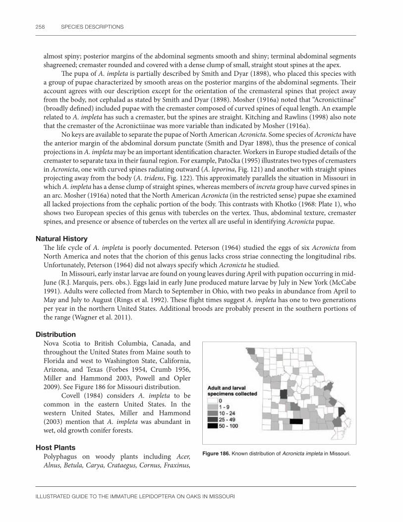
258 SPECIES DESCRIPTIONS
ILLUSTRATED GUIDE TO THE IMMATURE LEPIDOPTERA ON OAKS IN MISSOURI
almost spiny; posterior margins of the abdominal segments smooth and shiny; terminal abdominal segments shagreened; cremaster rounded and covered with a dense clump of small, straight stout spines at the apex.
The pupa of A. impleta is partially described by Smith and Dyar (1898), who placed this species with a group of pupae characterized by smooth areas on the posterior margins of the abdominal segments. Their account agrees with our description except for the orientation of the cremasteral spines that project away from the body, not cephalad as stated by Smith and Dyar (1898). Mosher (1916a) noted that “Acronictiinae” (broadly defined) included pupae with the cremaster composed of curved spines of equal length. An example related to A. impleta has such a cremaster, but the spines are straight. Kitching and Rawlins (1998) also note that the cremaster of the Acronictiinae was more variable than indicated by Mosher (1916a).
No keys are available to separate the pupae of North American Acronicta. Some species of Acronicta have the anterior margin of the abdominal dorsum punctate (Smith and Dyar 1898), thus the presence of conical projections in A. impleta may be an important identification character. Workers in Europe studied details of the cremaster to separate taxa in their faunal region. For example, Patočka (1995) illustrates two types of cremasters in Acronicta, one with curved spines radiating outward (A. leporina, Fig. 121) and another with straight spines projecting away from the body (A. tridens, Fig. 122). This approximately parallels the situation in Missouri in which A. impleta has a dense clump of straight spines, whereas members of increta group have curved spines in an arc. Mosher (1916a) noted that the North American Acronicta (in the restricted sense) pupae she examined all lacked projections from the cephalic portion of the body. This contrasts with Khotko (1968: Plate 1), who shows two European species of this genus with tubercles on the vertex. Thus, abdominal texture, cremaster spines, and presence or absence of tubercles on the vertex all are useful in identifying Acronicta pupae.
Natural HistoryThe life cycle of A. impleta is poorly documented. Peterson (1964) studied the eggs of six Acronicta from North America and notes that the chorion of this genus lacks cross striae connecting the longitudinal ribs. Unfortunately, Peterson (1964) did not always specify which Acronicta he studied.
In Missouri, early instar larvae are found on young leaves during April with pupation occurring in mid-June (R.J. Marquis, pers. obs.). Eggs laid in early June produced mature larvae by July in New York (McCabe 1991). Adults were collected from March to September in Ohio, with two peaks in abundance from April to May and July to August (Rings et al. 1992). These flight times suggest A. impleta has one to two generations per year in the northern United States. Additional broods are probably present in the southern portions of the range (Wagner et al. 2011).
DistributionNova Scotia to British Columbia, Canada, and throughout the United States from Maine south to Florida and west to Washington State, California, Arizona, and Texas (Forbes 1954, Crumb 1956, Miller and Hammond 2003, Powell and Opler 2009). See Figure 186 for Missouri distribution.
Covell (1984) considers A. impleta to be common in the eastern United States. In the western United States, Miller and Hammond (2003) mention that A. impleta was abundant in wet, old growth conifer forests.
Host PlantsPolyphagus on woody plants including Acer, Alnus, Betula, Carya, Crataegus, Cornus, Fraxinus,
Figure 186. Known distribution of Acronicta impleta in Missouri.

NOCTUIDAE 259
ILLUSTRATED GUIDE TO THE IMMATURE LEPIDOPTERA ON OAKS IN MISSOURI
Hamamelis, Juglans, Malus, Oxydendron, Populus, Prunus, Q. robur, Salix, Tilia, Ulmus and Vaccinium (Robinson et al. 2002); Fagus (Handfield 1999); Liquidambar, Nyssa, Sassafras, and Sorbus (Wagner et al. 2011).
CommentsAcronicta impleta was formally known as A. luteicoma (Smith and Dyar 1898, Hodges et al. 1983).
Subfamily Amphipyrinae
Amphipyra pyramidoides (Guenée)Copper underwing
Larval Description Mature larva light green; head unmarked, concolorous with the body; dorsum with a thin white middorsal line that extends from the mesothorax through A8; lateral area with a white and yellow spiracular stripe, often colored deep yellow anteriorly, that passes under the prothoracic spiracle to the mesothorax, then extends through the abdominal spiracles of A2-7 and below the spiracle on A8, before encircling the anal shield margin; spiracles black; dorsal, subdorsal, and subventral areas speckled with large white dots, the subdorsal spots of A8 often merging into an inverted v-shaped marking; A8 with large dorsal conical hump (Figure 187a, b).
The larva of Amphipyra pyramidoides is illustrated by Rings (1968) and Rings et al. (1992) and in color by Chapman and Lienk (1974), Béïque and Bonneau (1979), Ives and Wong (1988), Miller (1995), and Wagner et al. (2008, 2011). At first glance, the large dorsal hump on A8 appears to be a unique identification characteristic for A. pyramidoides. In reality, a similar dorsal hump occurs on less frequently collected noctuids, such as the Psaphidini (Miller 1995: Fig. 57, Kitching and Rawlins 1998). Miracavira brillians, found in the southwestern United States, is especially similar to A. pyramidoides (Wagner et al. 2008). Moreover, even though instars three to six of A. pyramidoides have a dorsal hump, this feature is lacking in young larvae (Rings 1968). The combination of a mandibular retinaculum, an interrupted spiracular line absent on metathorax and A1, the large size of the spiracle on A8 compared to A7, a spiracular line that encircles the margin of the anal shield (Crumb 1956: 174, 225; Miller 1995; Wagner et al. 1997), and the SD1 seta of normal thickness (not hairlike) (Kitching and Rawlins 1998) will accurately distinguish A. pyramidoides. The larval morphology of A. pyramidoides is summarized by Crumb (1956). See also Appendix 3, Plate 2.
a b
Figure 187. Amphipyra pyramidoides (a) mid-instar larva; (b) mature larva. Photos by R.J. Marquis, used with permission.

260 SPECIES DESCRIPTIONS
ILLUSTRATED GUIDE TO THE IMMATURE LEPIDOPTERA ON OAKS IN MISSOURI
Pupal DescriptionLabial palpi exposed; maxillae extend to caudal margin of the wings; prothoracic femur exposed; maxillary palpi small, triangular; prothoracic, but not the mesothoracic, legs extend cephalad to the lower margin of the eye; metathoracic legs exposed at their tips; mesothoracic spiracle slitlike; dorsum of thorax and anterior abdominal segments finely punctate, almost shagreened, in contrast to the shiny texture of the posterior abdominal segments; cremaster rugose, with two stout, slightly curved spines at the apex surrounded by several thinner curved setae.
Mosher (1916a) noted that all Amphipyrinae (broadly defined) have the cremasteral setae of equal length, but she only had a few species of this diverse taxon available for study. The pupa of A. pyramidoides does not fit the above diagnosis because the cremaster has two stout spines and several thin, curved setae. Interestingly, because of the position of the legs and exposed prothoracic femur, the pupa of A. pyramidoides will key with the Cucullinae in Mosher (1916a). This seems to parallel recently published phylogenetic studies (see Comments).
Amphipyra tragopoginis, a European moth recorded from North America (Hodges et al. 1983), has a similar pupa to A. pyramidoides. Characters separating the two species are not available, but the thoracic texture of A. tragopoginis illustrated by Khotko (1968) appears rugose, not punctate as in A. pyramidoides. The pupal pits on A10 illustrated by Wagner et al. (2008) for M. brillians are absent in A. pyramidoides.
Natural HistoryThe egg of A. pyramidoides is illustrated by Rings (1968), Salkeld (1984), and Rings et al. (1992). Twenty to twenty-two longitudinal ridges, each with a thin crest, cover the chorion. The micropylar area has 10 to 11 cells.
The life cycle of A. pyramidoides is summarized by Rings (1968) for Ohio and Chapman and Lienk (1974) for New York State. Adults emerge in July, aggregate in protected areas, and start to oviposit in September, probably on twigs or leaves of their host (Rings 1968, Wagner et al. 2011). Eggs overwinter and then hatch in early spring of the following year when young larvae can feed on tender buds and small leaves. Mature larvae sometimes feed on apple or peach fruit (Rings 1968). Pupation occurs in a cocoon at the soil surface during mid-June. There is one generation per year (Wagner et al. 2011). In Missouri, caterpillars of A. pyramidoides occur only in early spring. This phenology agrees with biological observations of authors throughout the United States (Rings 1968, Miller 1995, Wagner et al. 2011).
DistributionWidely distributed in North America, from Quebec, Canada, (Handfield 1999) throughout the eastern and midwestern United States, west to British Columbia and California, then south to Arizona, Texas (Chapman and Lienk 1974), and Florida (Wagner et al. 2011). See Figure 188 for Missouri distribution. Amphipyra pyramidoides is common in the northeastern United States (Chapman and Lienk 1974).
Host PlantsA generalist feeder on 17 families of deciduous trees, shrubs, and woody vines (Robinson et al. 2002).
According to Chapman and Lienk (1974), Betula, Quercus, Salix, Tilia, and Ulmus appear to be the favored hosts. In the western United States, Acer
Figure 188. Known distribution of Amphipyra pyramidoides in Missouri.

NOCTUIDAE 261
ILLUSTRATED GUIDE TO THE IMMATURE LEPIDOPTERA ON OAKS IN MISSOURI
and Arctostaphylos are frequently utilized (Miller 1995). The unusual record of A. pyramidoides feeding on rhododendron (Crumb 1956), an evergreen, needs confirmation.
CommentsUsing larval morphology, Crumb (1956) and Beck (1999) associate Amphipyra with genera traditionally placed in the Cucullinae or what used to be called the Psaphidinae. Mitchell et al. (2000) agreed with this hypothesis, which considers Amphipyra and Psaphidinae to be sister taxa based on molecular phylogenetic data. Wagner et al. (2008) regarded Psaphidinae and Amphipyrinae to be synonyms. This was followed by Pohl et al. (2016) who place Psaphidini in the Amphipyrinae with Amphipyra.
Subfamily Noctuinae
Cosmia calami (Harvey)American dun-bar
Larval Description Mature larva with an unmarked pale green head and prothoracic shield; middorsal stripe white or rarely yellow, variable in thickness and extending from the prothorax to the posterior margin of the anal shield; dorsal setae sometimes on conspicuous white pinacula; spiracles orange with a brown rim; body speckled with white spots that often form a weak, broken subdorsal line; subventral area with a thin irregular line passing below all spiracles (Figure 189a, b).
The larva of Cosmia calami was described by Crumb (1956) and is illustrated in color by Ives and Wong (1988), Miller (1975), and Wagner et al. (2011). This species is difficult to identify because the coloration and morphology are not distinctive. Nevertheless, the presence of a middorsal line crossing the prothoracic and anal shields, in addition to a thin subventral stripe passing below brown spiracles, helps separate C. calami from other similarly colored noctuids. The shape is also distinctive (see Wagner et al. 2011). In his key to North American noctuids, Crumb (1956) notes the following morphological features that are characteristic of C. calami: mandible with retinaculum; sclerotized bar connecting SD seta to ventral muscle attachment absent on mesothorax; prolegs on A3-6 without a fleshy lobe; height of spiracles on prothorax and A1 subequal; and host plants exclusively oaks (but see section on Hosts, Natural History). Some individuals of C. calami have dark green reticulation on the epicranium (Crumb 1956).
a b
Figure 189. Cosmia calami (a) early instar larva; (b) mature larva. Photos by R.J. Marquis, used with permission.

262 SPECIES DESCRIPTIONS
ILLUSTRATED GUIDE TO THE IMMATURE LEPIDOPTERA ON OAKS IN MISSOURI
Pupal DescriptionLabrum u-shaped; labial palpi minutely exposed; maxillae extend to caudal margin of the wings; maxillary palpi exposed; prothoracic femur exposed; prothoracic, but not mesothoracic, legs extend to lower margin of eye; tips of metathoracic legs exposed; mesothoracic spiracle slitlike; dorsum of thorax nearly smooth, dorsum of abdominal segments sparsely punctate, the anterior margins of A4 and A5 with a row of small vertical striations; cremaster with two stout curved spines surrounded by approximately eight thinner curved seta; base of cremaster rugose.
Mosher (1916a) noted that, except for the Pantheinae, all noctuid pupae have fully exposed labial palpi. The pupa of C. calami is an exception to this generalization because the labial palpi are barely exposed in this species. Another distinctive feature is the vertical striations on the dorsum of A4 and A5. Unlike pantheines, which have traces of verrucae on the abdomen (Mosher 1916a) and a cremaster composed of small curved setae (Kitching and Rawlins 1998: 366), the pupa of C. calami has no verrucae and two stout spines on the cremaster, which are much thicker than the others.
Gardner (1948b) studied several noctuid pupae from India with hidden labial palpi, suggesting that this character state may be more common in noctuids than indicated by the literature. A more complete survey of North American noctuid pupae is needed to evaluate the value of hidden labial palpi for identification. Several species of Cosmia were diagnosed by Patočka (1980, 1995, 1996) in his keys to European noctuid pupae. The labial palpi, prothoracic femur, and cremaster are all important identification features. The cremaster of C. trapezina is illustrated in Khotko (1968) where it has two stout setae as does C. calami.
Natural HistoryLittle is known about the life history of C. calami. Early instar larvae roll leaves, but mature individuals feed exposed on the leaf surface (R.J. Marquis, pers. obs.). Several questions relating to diet breadth and host range need further study. Larvae of other Cosmia species are cannibalistic (Porter 1997), as are larvae of C. calami in Missouri (R.J. Marquis, pers. obs.). However, larvae of C. calami can complete development by feeding only on oak leaves (R.J. Marquis pers. obs.; Wagner et al. 2011); therefore, we conclude this species is a facultative feeder on other caterpillars. Until the prey is accurately identified, we cannot distinguish between cannibalism and true predation. Forbes (1954) stated that C. calami is oligophagous on various tree species, in contrast to Crumb (1956: 193) who considers this species monophagous on oaks.
Adults fly from May to August (Covell 1984), but are most common in July (Lienk et al. 1991). Moths have also been collected during September in Ohio (Rings et al. 1992), suggesting there might be a partial second generation in some areas. In Missouri, there is a single spring generation of caterpillars on oaks.
DistributionNova Scotia, Canada, to northern Florida, west to Manitoba, Canada, and Texas (Covell 1984).
Cosmia calami can be locally common (Covell 1984). See Figure 190 for Missouri distribution.
Host PlantsPolyphagous on trees and shrubs including Quercus agrifolia, Q. alba, Q. coccinea, Q. kelloggi, Q, macrocarpa, and Vaccinium (Robinson et al. 2002).
Larvae occur on various subgenera of oak (Q. alba, Q. rubra, and Q. virginiana), where they also prey
Figure 190. Known distribution of Cosmia calami in Missouri.

NOCTUIDAE 263
ILLUSTRATED GUIDE TO THE IMMATURE LEPIDOPTERA ON OAKS IN MISSOURI
on caterpillars (Covell 1984). Although considered to be polyphagous (Forbes 1954), published host records to support this claim are lacking. Cosmia calami is likely an oak specialist (D. Wagner, pers. comm.).
CommentsAccording to Porter (1997), several European Cosmia spp. are cannibalistic, and C. trapezina will attack other caterpillars and even bite humans when handled. Cosmia ochreimargo defoliated oak trees in India (Mathur 1942). Cosmia canescens is considered a synonym of C. calami (Pohl et al. 2016).
Lithophane antennata (Walker)Green fruitworm
Larval Description Mature larva with pale green head and solid, white to yellow middorsal line, which is thinner than the spiracular stripe; dorsal setae on contrasting white pinacula; subdorsal area with irregular white spots that form a thin broken subdorsal line; spiracles white to yellow with a pale brown rim; spiracular stripe thick and passing below the spiracles of A1-8; ventral area concolorous with body (Figure 191a, b).
The larva of Lithophane antennata is described by Crumb (1956) and illustrated by Chapman and Lienk (1974), Rings (1973), and Wagner et al. (2011). According to keys by Crumb (1956) and Forbes (1954), the following morphological features help define L. antennata: oral face of mandible with two teeth on the retinaculum; subdorsal line broken; abdominal spiracles yellow or white (with a pale brown rim); height of prothoracic spiracle greater than height of spiracle on A1; length of labial palpi not more than two-thirds the length of the long pointed spinneret; and blade of hypopharyngeal complex without well-developed teeth. Unlike other Lithophane (L. baileyi and L. pexata) illustrated by McCabe (1991), the mandible of L. antennata has two large teeth on the retinaculum.
Lithophane antennata has contrasting white dorsal pinacula that appear as several paired dots along each side of the dorsum. The presence of two or three paired dots on either side of the midline is used by Wagner et al. (2011:480) to diagnose the genus Lithophane. There is a smaller white dot on the anterior margin of each abdominal segment when not hidden by a fold (Wagner et al. 2011: 426). Although this pattern is not present in all green fruitworm genera, conspicuous dorsal pinacula are characteristic of many Lithophane larvae (Crumb 1956: 184, Chapman and Lienk 1974, Miller 1995: 40, Wagner et al. 2011). According to Chapman and Lienk (1974), the broken subdorsal line, pale abdominal spiracles, and wide spiracular stripe that equals three times the spiracle height, will separate L. antennata from other green fruitworms. In addition, the thoracic legs are frequently black (Wagner et al. 2011). Because many noctuid genera include larvae with green and white stripes, it is especially important to pay attention to details in color pattern and mouthparts when identifying L. antennata. The color pattern will fade in preserved larvae, complicating identification of specimens in museums.
a b
Figure 191. Lithophane antennata (a) early instar larva; (b) mature larva. Photos by R.J. Marquis, used with permission.

264 SPECIES DESCRIPTIONS
ILLUSTRATED GUIDE TO THE IMMATURE LEPIDOPTERA ON OAKS IN MISSOURI
Pupal DescriptionLabrum u-shaped; labial palpi exposed; maxillae extend to the caudal margin of the wings; maxillary palpi hidden; prothoracic femur exposed; prothoracic, but not the mesothoracic, leg extends to the lower margin of the eye; tips of metathoracic legs exposed; mesothoracic spiracle slitlike; dorsum of abdominal segments smooth except for A7, which has a thin row of punctures; cremaster striated at the base with two thick, curved spines surrounded by a pair of thinner, curved setae.
The pupa of L. antennata is described by Rings (1973) as shiny red brown and 12–14 mm long. The labial palpi, prothoracic femur, and tips of the metathoracic legs are exposed. The maxillae extend to the caudal margin of the wings. Three pairs of setae are located on the apex of the cremaster. The medial setae are thicker than the lateral two pairs. This description agrees with our specimens from Missouri.
Mosher (1916a) studied two species of Lithophane, including L. antennata, under the subfamily Cucullinae, where this genus was usually placed until recently. She noted these distinguishing characters: mesothoracic legs fail to reach the eye, the cremaster with curved setae of two sizes, scars of the larval verrucae are absent, the prothoracic femur and labial palpi are both exposed, and the wings or maxillae never extend past the caudal margin of A4. Our specimens of L. antennata agree with this diagnosis, but Kitching and Rawlins (1998) state that some Cucullinae have pupae with wings and maxillae extending past A4, a feature shared with the Plusiinae. The lack of this character in Lithophane seems to support the current placement in Noctuinae instead of Cucullinae. In Eupsilia devia, a northeastern United States species related to Lithophane, the cremaster has four straight stout spines. This is another exception to the key presented by Mosher (1916a). Patočka (1996) illustrated many European species of Cucullinae, but most belong to the large genus Cucullia, a genus not related to Lithophane. None had curved cremastral setae. The single row of abdominal punctures in L. antennata is another character that may prove distinctive after more genera of Cucullinae and Noctuinae are described.
Natural HistoryThe life history of L. antennata was studied by Rings (1973) in the midwestern United States. Eggs are laid between March and April, either singly or in a mass. During incubation, the egg color changes from lemon yellow to red-brown. Early instar larvae feed on leaf or flower buds, whereas mature larvae eat foliage or the fruit surface. Damage to apple and pear fruit causes feeding scars or premature fruit drop. Wagner et al. (2011) do not feel fruit is a normal part of the diet of L. antennata. Pupation occurs in the soil, with moths emerging from September to November. The adults overwinter and can be collected with fermenting syrup bait traps.
Wagner et al. (2011) and Rings (1973) state that L. antennata has only one generation per year. This contrasts with Lienk et al. (1991: 111) who postulate two generations in New York. Wagner et al. (2011) collected larvae of L. antennata mostly in the late spring (June). We agree with this phenology and have found larvae in Missouri during May only, sometimes in leaf rolls made by other caterpillars. Larvae of L. antennata are facultatively predaceous (Wagner et al. 2011; R.J. Marquis, pers. obs.).
DistributionEastern Canada to the midwestern United States, south to Mississippi (Wagner et al. 2011).
Except for portions of northeastern Canada, L. antennata is common to abundant throughout its range (Rings 1973, Covell 1984). Rings (1973) published a distribution map for L. antennata that includes a single record from California that likely is a misidentification (D. Wagner, pers. comm.). See Figure 192 for Missouri distribution.
Figure 192. Known distribution of Lithophane antennata in Missouri.

NOCTUIDAE 265
ILLUSTRATED GUIDE TO THE IMMATURE LEPIDOPTERA ON OAKS IN MISSOURI
Host PlantsPolyphagous on woody plants including Acer, Carya, Fraxinus, Malus, Populus, Prunus, Pyrus, Q. borealis, Q. coccinea, Q.rubra, Rosa, Vaccinium (Robinson et al. 2002); Ribes, and Ulmus (Rings 1973). We have found L. antennata on Q. alba, Q. coccinea, Q. rubra, Q. stellata, and Q. velutina in Missouri.
Lithophane antennata prefers oak, hickory, apple, crabapple, and cherry (Rings 1975, Rings et al. 1992). The record for “quince” cited by Rings (1973) probably refers to “flowering quince” (Chaenomeles) instead of the tropical fruit tree (Cydonia) because the larva was found in New York. Although there is some doubt about the caterpillar identification, Althea, Cercis, Cornus, Fagus, Forsythia, Kalmia, Morus, and Rhododendron are probably also hosts of L. antennata (Rings 1973). Wagner et al. (2011) listed other woody plants and feverwort as possibilities. We have been conservative in accepting only two classic studies for our host list. Further study may show L. antennata is truly polyphagous.
CommentsDepending on the geographical region, L. antennata comprises from 10 to 75 percent of the green fruitworm complex on apple (Rings 1973).
Achatia distincta (Hübner) Distinct Quaker
Larval Description Mature larva with a light green head, dark green body, and white to yellow markings composed of numerous minute spots and longitudinal stripes; dorsum with a thin middorsal line and an inconspicuous lateral spot on each segment; subdorsal stripe thick and contrasting; spiracles white with pale orange to brown rim; two thin and almost entirely solid spiracular stripes present, one passing through or barely above the spiracles of A1-7, then below the spiracle on A8, while the second stripe passes below the spiracles; subventral stripe thin and broken, similar in thickness to the spiracular stripe; prolegs of A3-6 with a white patch above the crochets; ventral area green with minute white spots (Figure 193).
The larva of Achatia distincta is illustrated in color by Ives and Wong (1988) and Wagner et al. (2011). Crumb (1956), Godfrey (1972a), and McCabe (1991) studied the larval morphology with emphasis on the mouthparts. As a member of the green fruitworm complex, A. distincta is most likely to be confused with other green and white-striped caterpillars such as Orthosia, Himella, and Lithophane. Chapman and Lienk (1974) listed color characteristics of these genera. The presence of a wide, contrasting subdorsal stripe above two more or less continuous, thin spiracular stripes will separate A. distincta from related taxa. Ives and Wong (1988) also illustrate other green and white striped noctuids found on hardwood trees that could potentially be confused with A. distincta. Wagner et al. (2011) pointed out Himella is especially similar to A. distincta.
Although color is a good identification feature in the field, preserved specimens of A. distincta usually lose their markings. Compared to other members of his concept of the subfamily Hadeninae, Crumb (1956) diagnosed the larva of A. distincta using the following characters: mandible with an inner tooth having an
Figure 193. Achatia distincta mature larva. Photo by R.J. Marquis, used with permission.

266 SPECIES DESCRIPTIONS
ILLUSTRATED GUIDE TO THE IMMATURE LEPIDOPTERA ON OAKS IN MISSOURI
apical molar area (grinding surface); prothoracic spiracle and spiracle of A8 both larger than the spiracle on A1; spiracles colored brown with a brown rim (but see note below); and length of the labial palpi about equal to the length of the spinneret. Several of these features were also used by Godfrey (1972a), but many new couplets were added. In particular, A. distincta belongs to Group 8G, which was characterized by: one SV seta on the mesothorax; prothorax with SD2 present; length of P1 equal to or greater than width of the front; food plants not in Caryophyllaceae; proximal region of hypopharyngeal complex with a single row of large spines; and mandible with inner teeth. In addition, the distal region of hypopharyngeal complex is bare above spinneret, and there is no dense brush of long, thin spines. Among members of group 8G, A. distincta is morphologically similar to Morrisonia confusa, another species common on oak in Missouri. Unlike M. confusa, A. distincta has the head unmarked, and there are never wine-colored spots on the body.
Crumb (1956) describes the larval spiracles of A. distincta as brown with a brown rim, although he speculates his specimens were not mature. We have found the spiracles of A. distincta to be white with a brown rim, at least in older larvae.
Pupal DescriptionLabial palpi exposed; maxillae extend to the caudal margin of the wings; maxillary palpi hidden; prothoracic femur hidden; the prothoracic, but not the mesothoracic, leg extends cephalad to the lower margin of the eye; metathoracic leg barely exposed at their tips; mesothoracic spiracle an inconspicuous, elongated oval spot; dorsum of abdominal segments finely punctate except for A4-7 that have a row of deep punctures on their anterior margin, the most distal of these punctures outlined in black; cremaster consists of two stout, curved setae surrounded by a pair of thinner curved setae on each side.
The pupa of A. distincta could be confused with O. hibisci, Mythimna unipuncta (Mosher 1916a), and Morrisonia confusa because all four genera have deep pits on the anterior margin of the middle abdominal segments. Unlike Morrisonia, the pupa of A. distincta lacks a row of punctures on the dorsum of A3. The prothoracic femur of A. distincta is hidden, but it is exposed in M. unipuncta. All of the cremasteral setae of A. distincta are hooked in contrast to O. hibisci, which has only straight setae on the cremaster. There are no keys to separate genera of North American Hadeninae pupae, but, based on the characters used by Patočka (1996) for the European fauna, tentative identifications are possible if careful attention is paid to the prothoracic femur, abdominal texture, and spines on the cremaster.
As noted under M. confusa, it is important to pay attention to the subfamily characteristics in Mosher (1916a) when trying to identify A. distincta because pupae of the Noctuinae and Hadeninae (in the sense of Mosher 1916a) have a similar morphology.
Natural HistorySalkeld (1984) illustrated the egg of A. distincta and describes it as having approximately 48 longitudinal ridges connected by numerous cross ribs that give the egg a pitted appearance under low magnifications. The micropylar area has 13 to 14 rosette cells.
Wagner et al. (2011) summarized the life history of A. distincta. There are 30 eggs in a cluster. Larvae are common in the spring and feed mostly at night, often in a shelter of leaves. When disturbed, the caterpillar rolls in a ball. Prepupae are dull reddish green.
Wagner et al. (2011) suggest one generation per year. Caterpillars are only found during May in Missouri (R.J. Marquis, pers. obs.). The pupa probably overwinters (Chapman and Lienk 1974), and in New York adults fly from April to May (Lienk et al. 1991). The flight period is slightly longer in southern Ohio where adults were collected from March to May (Rings et al. 1992).
DistributionQuebec to Manitoba, Canada, south to Florida, and west to Colorado and Texas, including Missouri (Crumb 1956, Covell 1984). See Figure 194 for Missouri distribution.

NOCTUIDAE 267
ILLUSTRATED GUIDE TO THE IMMATURE LEPIDOPTERA ON OAKS IN MISSOURI
Figure 194. Known distribution of Achatia distincta in Missouri.
Covell (1984) considered A. distincta to be common throughout its range, which might include most of the United States except the southwestern region (Chapman and Lienk 1974).
Host PlantsAcer, Alnus, Betula, Fraxinus, Juglans, Malus, Quercus alba, Q. coccinea, Q. macrocarpa, Q. rubra, Tilia, and Vaccinium (Robinson et al. 2002).
Grape is considered a doubtful host by some (Godfrey 1972b), but not all authors agree (Rings et al. 1992).
CommentsAchatia distincta was previously placed in either Polia or Morrisonia by most workers (Forbes 1954, Crumb 1956, Chapman and Lienk 1974).
Morrisonia confusa (Hübner)Confused woodgrain
Larval Description Early instar larva with solid orange head and thin, white middorsal, subdorsal and spiracular lines; dorsal, subdorsal and lateral areas speckled with small white dots; subdorsal area of thorax and A1-8 with large, contrasting wine-colored spots on each segment; sometimes a small rectangular wine-colored spot present near the prothoracic SV pinaculum as well; spiracular stripe passes above spiracles of A1-7 but below spiracle on A8; ground color of abdomen green or blue-green; ventral region pale green.
Mature larva with a tan head and usually a pair of large, dark spots on the epicranium; thorax and abdomen greenish white with a thin, sometimes inconspicuous, middorsal, and subdorsal white stripe; dorsal, subdorsal, and lateral areas speckled with white (Figure 195a, b).
The larva of M. confusa is described or illustrated by Crumb (1956), Godfrey (1972a), McCabe (1991), Wood and Butler (1991), and Wagner et al. (2011), although not all authors have clearly differentiated between early and late instar larval coloration in these descriptions. Young larvae (instars 2–6) are readily identified by their segmental wine-colored spots surrounding the SD setae (Wood and Butler 1991, Wagner et al. 2011). In addition, unlike many noctuid larvae that feed exposed and unprotected, mature M. confusa larvae form a leaf
Figure 195. Morrisonia confusa (a) early instar larva; (b) mature larva. Photos by R.J. Marquis, used with permission.
a b

268 SPECIES DESCRIPTIONS
ILLUSTRATED GUIDE TO THE IMMATURE LEPIDOPTERA ON OAKS IN MISSOURI
shelter. For more information on identification of young M. confusa, consult Crumb (1956), who diagnosed this species based on early instar larvae only, and the illustration in Béïque and Bonneau (1979). Each instar of M. confusa is described by Wood and Butler (1991).
Mature larvae of M. confusa are more difficult to characterize. According to Godfrey (1972a), M. confusa belongs to group 8G, which he defined as follows: mesothorax with one SV seta; prothorax with SD2 present; length of P1 greater than width of front; food plants not in Caryophyllaceae; proximal region of hypopharyngeal complex with a single row of large spines and mandible with inner teeth. In addition, the distal region of hypopharyngeal complex is bare above spinneret and there is no dense brush of long thin spines. Instead, the anterior portion of the distal region is covered with short, fine spines and the posterior half has short, thick spines (see Godfrey 1972a: Fig. 190). Among members of group 8G, M. confusa is morphologically similar to Morrisonia (=Polia) latex. However, M. latex has a reticulated head pattern, whereas in M. confusa there are two large spots or no head markings at all.
Pupal DescriptionLabrum rectangular; labial palpi exposed; maxillae extend to the caudal margin of the wings; maxillary palpi hidden; prothoracic femur exposed; prothoracic, but not mesothoracic, leg extends to lower margin of the eye; tips of metathoracic legs hidden; mesothoracic spiracle present, but damaged; dorsum of thorax and abdominal segments smooth, the anterior margin of A3-7 with a row of black sclerotized pits; abdominal spiracles elliptical, about three times taller than wide; cremaster with two stout setae, thin, and curved at the tips, with a smaller pair of curved setae on each side of the larger pair.
Mosher (1916a) noted that pupae in her concept of the Hadeninae have two straight, stout setae on the cremaster, and only the prothoracic leg extends to the lower margin of the eye. The pupa of M. confusa agrees with this diagnosis, although the cremasteral setae of genera that used to be in the Hadeninae (now classified as Noctuinae) are more variable than indicated in Mosher (1916a). The tips of the stout cremastral setae are slightly curved in M. confusa, strongly curved in Leucania latiuscula from Honduras, but blunt and flattened in Trichordestra legitima (SCPC).
Except for Mosher (1916a), no keys are available to identify genera of North American noctuid pupae. Most pupae that she included in her concept of the Hadeninae lack deep rows of sclerotized pits on the abdominal dorsum, but pits are present in both M. confusa and Mythimna unipuncta. These two genera may be separated by the number of rows. Morrisonia confusa has an extra row of pits on A3 that is lacking in M. unipuncta. Rows of sclerotized pits also occur on the abdominal dorsum of some (Mosher 1916a: Fig. 101), but not all pupal Noctuinae (Noctua pronuba lacks pits) (SCPC). Therefore it is important to pay attention to the characteristics in Mosher (1916a) when trying to identify M. confusa.
Patočka (1996) studied the pupa of European species that used to be in the Hadeninae and related taxa. He also noted variation in the cremastral spine tips, including genera with straight, blunt, or curved spines. Some European noctuid pupae also have rows of deep, sclerotized abdominal pits. Several examples are illustrated by Khotko (1968).
Because the mesothoracic spiracle was damaged in the two cast pupal exuviae of M. confusa that were available for study, the shape of the pupal mesothoracic spiracle has not been determined. A larger series of specimens is needed to fully evaluate this species.
Natural HistoryThe life history of M. confusa was studied by Wood and Butler (1991). Females lay up to 316 eggs in a single layered mass. The dorsal portion of the egg has from 34–37 longitudinal ribs, numerous cross striations connecting the ribs, and from 9 to 13 rosette cells in the micropyle (Salkeld 1984, Wood and Butler 1991). The ventral portion of the egg has a smooth chorion (Salkeld 1984).
Early instars commonly hide in leaf rolls made by other species, but mature larvae form their own leaf shelters (R.J. Marquis, pers. obs.) where they rest by day (Wagner et al. 2011). This shelter may be a webbed
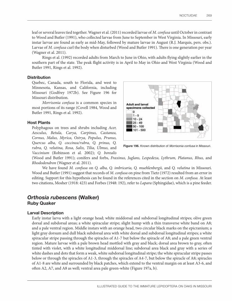
NOCTUIDAE 269
ILLUSTRATED GUIDE TO THE IMMATURE LEPIDOPTERA ON OAKS IN MISSOURI
leaf or several leaves tied together. Wagner et al. (2011) recorded larvae of M. confusa until October in contrast to Wood and Butler (1991), who collected larvae from June to September in West Virginia. In Missouri, early instar larvae are found as early as mid-May, followed by mature larvae in August (R.J. Marquis, pers. obs.). Larvae of M. confusa curl the body when disturbed (Wood and Butler 1991). There is one generation per year (Wagner et al. 2011).
Rings et al. (1992) recorded adults from March to June in Ohio, with adults flying slightly earlier in the southern part of the state. The peak flight activity is in April to May in Ohio and West Virginia (Wood and Butler 1991, Rings et al. 1992).
DistributionQuebec, Canada, south to Florida, and west to Minnesota, Kansas, and California, including Missouri (Godfrey 1972b). See Figure 196 for Missouri distribution.
Morrisonia confusa is a common species in most portions of its range (Covell 1984, Wood and Butler 1991, Rings et al. 1992).
Host PlantsPolyphagous on trees and shrubs including Acer, Aesculus, Betula, Carya, Carpinus, Castanea, Cornus, Malus, Myrica, Ostrya, Populus, Prunus, Quercus alba, Q. coccinea/rubra, Q. prinus, Q. rubra, Q. velutina, Rosa, Salix, Tilia, Ulmus, and Vaccinium (Robinson et al. 2002); Q. borealis (Wood and Butler 1991); conifers and forbs, Fraxinus, Juglans, Lespedeza, Lythrum, Platanus, Rhus, and Rhododendron (Wagner et al. 2011).
We have found M. confusa on Q. alba, Q. imbricaria, Q. muehlenbergii, and Q. velutina in Missouri. Wood and Butler (1991) suggest that records of M. confusa on pine from Tietz (1972) resulted from an error in editing. Support for this hypothesis can be found in the references cited in the section on M. confusa. At least two citations, Mosher (1918: 423) and Forbes (1948: 192), refer to Lapara (Sphingidae), which is a pine feeder.
Figure 196. Known distribution of Morrisonia confusa in Missouri.
Orthosia rubescens (Walker) Ruby Quaker
Larval Description Early instar larva with a light orange head; white middorsal and subdorsal longitudinal stripes; olive green dorsal and subdorsal areas; a white spiracular stripe; slight hump with a thin transverse white band on A8; and a pale ventral region. Middle instars with an orange head, two circular black marks on the epicranium; a light gray dorsum and dull black subdorsal area with white dorsal and subdorsal longitudinal stripes; a white spiracular stripe passing through the spiracles of A1-7 but below the spiracle of A8; and a pale green ventral region. Mature larvae with a pale brown head mottled with gray and black; dorsal area brown to gray, often tinted with violet, with a white longitudinal middorsal line; subdorsal area black and gray with a series of white dashes and dots that form a weak, white subdorsal longitudinal stripe; the white spiracular stripe passes below or through the spiracles of A1-3, through the spiracles of A4-7, but below the spiracle of A8; spiracles of A1-8 are white and surrounded by black patches, which extend to the ventral margin on at least A3-6, and often A2, A7, and A8 as well; ventral area pale green-white (Figure 197a, b).

270 SPECIES DESCRIPTIONS
ILLUSTRATED GUIDE TO THE IMMATURE LEPIDOPTERA ON OAKS IN MISSOURI
a b
Figure 197. Orthosia rubescens (a) mature larva, dorsal view; (b) mature larva, lateral view. Photos by R.J. Marquis, used with permission.
The larva of Orthosia rubescens is illustrated by Godfrey (1972a), McCabe (1991), and Wagner et al. (2011). When darkly marked, it is easily recognized by the wide spiracular stripe that is dark above and pale below, and the black patches surrounding the spiracles on A3-7 (Wagner et al. 2011). Rarely the black patches are reduced to a small spot posterior to the spiracle, and in those cases, O. rubescens is best identified by using a combination of morphological features. According to Godfrey (1972a), O. rubescens belongs to group 8G, which he defined as follows: mesothorax with one SV seta; prothorax with SD2 present; length of P1 equal to or greater than width of the frontal area; food plants not in Caryophyllaceae; proximal region of hypopharyngeal complex with a single row of large spines; and mandible with inner teeth. In addition, the distal region of hypopharyngeal complex is bare above spinneret, and there is no dense brush of long thin spines. Among members of group 8G, O. rubescens is morphologically similar to M. confusa, another species common on oak in Missouri. Young larvae of Morrisonia confusa have wine-colored lateral spots that are lacking in O. rubescens. At maturity, the head markings of O. rubescens include a pair of vertical stripes and a reticulated pattern of black lines (McCabe 1991: Fig. 10487). This contrasts with the two rounded spots characteristic of the epicranium of M. confusa.
Among other Orthosia, O. rubescens is most similar to the dark forms of O. alurina, but the latter species lacks black patches that interrupt the spiracular stripe (see Godfrey 1972a). Middle instars of O. rubescens can be confused with O. hibisci and Lithophane (Wagner et al. 2011) but differ in morphology (length of spinneret compared to labial palpi, see Godfrey 1972a) and coloration (Lithophane has more than a pair of white dorsal spots on each side of the midline per segment).
Pupal DescriptionLabrum u-shaped with a dorsal dark area having two pits; labial palpi exposed; glazed eyepiece smooth; maxillae extends to caudal margin of wings; prothoracic leg about half the length of the maxillae; prothoracic femur thinly exposed; mesothoracic legs do not touch the lower margin of the eye; metathoracic legs exposed; mesothoracic spiracle absent; A1-3 very finely punctate, A4-8 with an anterior row of dark pits; cremaster with two thick parallel spines.
The pupa of O. rubescens is recognized by a smooth glazed eyepiece, a thin prothoracic femur, no mesothoracic spiracle, a row of dark pits on A4-8, and a cremaster with two parallel thick spines. Patočka (1996) studied the pupae of several Orthosia from Europe. In his characterization of this genus, species were illustrated with a textured compound eye; a patch of punctures on the metanotum; except for one species, a thin partially exposed prothoracic femur; partially exposed metathoracic legs; and cremaster spines which are at least somewhat diverging. In O. hibisci, the eye is partially rugose, the metanotum is relatively unmodified, the prothoracic femur is thin and exposed, a row of deep abdominal pits are present on the middle abdominal segments, and the cremaster spines are diverging (Mosher 1916a as Monima alia). At least some of the above features characterize O. rubescens as well. The characters we give will help identify

NOCTUIDAE 271
ILLUSTRATED GUIDE TO THE IMMATURE LEPIDOPTERA ON OAKS IN MISSOURI
O. rubescens on Missouri oaks, but too few noctuid pupae are described to know which characters are found in related species.
Natural HistoryIn spite of the fact that O. rubescens is a common moth, little is known about its life history. Eggs are lemon yellow at first, but later change to rust red. The micropylar area has 14–18 rosette cells and the chorion is covered with ribs forming either oval or rectangular cells (Salkeld 1984). Larvae are found in late April to early July (Wagner et al. 2011) as is typical for related species where feeding is completed by the spring or early summer (Godfrey 1972a: 54). The last instar rests on bark (Wagner et al. 2011). Adults fly during warm winter days and are especially common at sugar baits during March and April (Covell 1984). Sometimes moths are collected in May (Lienk et al. 1991), June (Rings et al. 1992), or rarely July (Covell 1984).
DistributionNova Scotia, Canada, south to Virginia and west to Wisconsin, Arkansas (Forbes 1954, Covell 1984), and Kansas (Godfrey 1972). See Figure 198 for Missouri distribution.
Host PlantsPolyphagous on trees and shrubs including Acer, Alnus, Malus, Prunus, Pyrus, Quercus coccinea/rubra, Q. rubra, Rubus, Populus, Tsuga, Typha, Vaccinium (Robinson et al. 2002); Castanea, Fagus, Ilex, Ostrya, Ribes, Sambucus, Smilax, Viburnum (Wagner et al. 2011).
Orthosia rubescens feeds on Quercus alba, Q. imbricaria, Q. muehlenbergii, Q. rubra, and Q. velutina in Missouri.
CommentsIn larvae of O. rubescens, the SD setae of the mesothorax and metathorax is connected to a tonofibrillary platelet. This character occurs in some pest Noctuinae (Mamestra, Spodoptera) (Weisman 1986, Passoa 1991). Mamestra brassicae, a quarantine significant species not known to be in North America, is somewhat similar to O. rubescens in having black around the spiracles (see Gilligan and Passoa 2014).
Figure 198. Known distribution of Orthosia rubescens in Missouri.

272 SPECIES DESCRIPTIONS
ILLUSTRATED GUIDE TO THE IMMATURE LEPIDOPTERA ON OAKS IN MISSOURI
ErebidaeErebid owlet moths
Erebidae are a newly defined family of Noctuiodea that contains the older concepts of the “quadrifine noctuids” (those with a four branched cubital vein in the hindwing) (Lafontaine and Fibiger 2006), the Arctiidae, and the Lymantriidae (=Liparidae). These three major subfamilies of Erebidae exhibit great diversity: “Catocalinae” (now included in the Erebinae) has at least 10,000 species (Kitching 1984), Arctiinae has 11,000 species, and there are 2,700 species in the Lymantriinae (Zahiri et al. 2012, Weller et al. 2009). For a review of the literature on quadrifine noctuids, see our introduction to the Noctuidae. Until very recently, all taxonomic publications treated quadrifine noctuids under Noctuidae. The biology and systematics of the Arctiinae (at that time a family), including the immature stages and a critical review of their updated classification, were published in Conner (2009). There are currently four tribes in the subfamily Arctiinae according to the latest molecular phylogeny by Zaspel et al. (2014). Three works deal with Lymantriinae (under the name Lymantriidae) in general: Wallner and McManus (1989), Kitching and Rawlins (1998), and for many economic species, Stibick (2000). Ferguson (1978) reviewed the North American lymantriine fauna. The current molecular phylogeny of the Lymantriinae recognizes seven tribes in the subfamily (Wang et al. 2015). There are 18 subfamilies of Erebidae according to the most recent treatment of that group (Zahiri et al. 2012). Lafontaine and Schmidt (2013) discuss the differences between European and North American erebid classifications.
Erebid eggs are morphologically diverse. Eggs that appear smooth to the naked eye are found in the Lymantriinae, Arctiinae, and Herminiinae (Syme 1961, Peterson 1963b, Salkeld 1984). Lymantriinae eggs have an opaque chorion and are often associated with a characteristic oviposition behavior by the females (Peterson 1966, Schaefer 1989). Grass feeders lay eggs singly, whereas tree feeders lay eggs in a mass (Schaefer 1989). The eggs are naked or covered with either scales or a secretion from the female’s body (Dolinskaya and Pljushch 1999). Another strategy is to insert the eggs into a protected crevice such as tree bark, sometimes covering the eggs for further protection (Schaefer 1989). Arctiine eggs are generally round, oval, or cone-shaped, often with a translucent chorion (Peterson 1963b). Most of the chorion or only the apical portion may be patterned (see Pljushch and Dolinskaya 2000a, 2000b). Oviposition behavior in Arctiidae also varies; eggs are usually naked, more rarely covered with scales from the female’s body, with each species having a particular number of eggs per cluster (Wagner 2009).
According to Dolinskaya’s (2014) study of Russian species, erebid eggs may be divided into two types, those that have a cellular sculpture and those that have ridges. She noted that the first type has cells in an irregular, “chaotic” pattern more typical of the Notodontidae, Lymantriinae, and Arctiinae. Erebid eggs with ridges resemble those of the Noctuidae, at least in the species she studied (Dolinskaya 2016). It is unknown if these two erebid egg types will be seen in other faunal regions. There is some information on erebid eggs of Canada. Salkeld (1984) and Syme (1961) described herminiine eggs (except for Palthis) as relatively smooth with a faint hexagonal pattern. This texture is similar to the Lymantriinae, and according to Syme (1961: 104), shows a close relationship of these two groups. Dolinskaya (2014) also noted the similarity of Herminiinae and Lymantriinae eggs in Russia. Syme (1961: 48) did not use the adult classification to name the six egg group types he recognized in the Catocalinae-Erebinae complex as tribes. A similar lack of concordance between the noctuid adult classification and egg stage was noted by Seamans (1933: 18). Variability in erebid eggs does support changes in the adult classification in some cases. Lafontaine and Schmidt (2010) recognize Pangraptinae as a valid subfamily using distinctive adult characters. Earlier, Syme (1961: 48) isolated this subfamily in a group called “miscellaneous series II” based on their unusual eggs. This example suggests a need to address characters of the egg stage in future noctuid classifications.
As might be expected from their morphological diversity, larvae of the Erebidae are difficult to define and cannot be easily separated from related noctuoid families. Most of the characters that separate Oenosandridae, Doidae, Nolidae, Euteliidae, and Notodontidae from the Noctuidae will also apply to the Erebidae (see introduction to the Noctuidae for this discussion).

EREBIDAE 273
ILLUSTRATED GUIDE TO THE IMMATURE LEPIDOPTERA ON OAKS IN MISSOURI
The presence of homoideous crochets distinguishes the Noctuidae from most Arctiinae that have heteroideous crochets. Those arctiines with homoideous crochets (Lithosiini and others) (Jacobson and Weller 2002: 5, Wagner 2009: 34) sometimes have an enlarged molar lobe on the mandible that is absent from noctuid larvae. Integument spicules are often present in noctuids with secondary setae, but the cuticle of arctiines never has such a texture (Habeck 1987). When visible, the presence of an adenosma also separates Arctiinae from Noctuidae. All known Arctiinae larvae lack this gland, which is almost universally present in noctuids (Kitching and Rawlins 1998: 391, Lafontaine and Fibiger 2006: 629). Beck (2009) noted that the number of L setae on the anal prolegs of Arctiinae and Noctuidae differ; in noctuids there are three setae compared to four setae in arctiines. He did not list any exceptions and used this character to distinguish Arctiidae from Noctuidae in the classical sense. Unfortunately, the chaetotaxy of the anal proleg has been poorly studied worldwide, and naming setae on this structure is often difficult in practice. The simplest generalization for the Missouri oak fauna is that Arctiinae can be identified by their barbed secondary setae and heteroideous crochets. If the crochets are homoideous, then the mandible has a molar lobe (Lithosiinae) or the “SD2” (=mSD2 or MSD) (terminology from Miller 1991: 151–152) is absent on A1 and A3 (Kitching and Rawlins 1998, Jacobson and Weller 2002: 244). Consult Jacobson and Weller (2002) and Beck (2009) for a discussion of the problems defining the Arctiinae.
Like the Arctiinae, larvae of the Lymantriinae are covered in secondary setae. Most species, including those in North America, are distinguished by having dorsal glands on A6 and A7 (Ferguson 1978). However, one Indian species apparently lacks glands, a few species have only a single gland on A6 (Ferguson 1978: 21), and in rare cases, six gland slits may be present (Gardner 1941, Kitching and Rawlins 1998).
The remaining Erebidae with primary setae are most likely to be confused with the Noctuidae. Although there are many exceptions, the following characters generally suggest an erebid larva: spinneret with silk pore concealed by apical flaps (illustrated by Crumb 1956: Plate 3 I–R); SV group trisetose on A1; SD1 not hairlike on A9 (a few Erebinae are the exception); and after excluding the Arctiinae and Lymantriinae, secondary setae are absent (Fibiger and LaFontaine 2005: 23, 32). When a larva is associated with fungi, algae, or decaying plant matter, it is more likely in Erebidae than Noctuidae (Wagner et al. 2008: Table 2). Erebids often have some reduction in the abdominal prolegs, especially in the Erebinae (Holloway 2005). The position of SD2 off the prothoracic shield merits attention as another trait of scattered erebid lineages.
Larval Erebidae with primary setae are best characterized by sorting their subfamilies into groups. Fibiger and Lafontaine (2005: 27, 28) partially divided the Erebidae based on host relationships; the Hypenodinae, some Herminiinae, most Boletobiinae, and “Scolecocampinae” (broad sense) often feed on fungi or lichens, whereas the Hypeninae, Calpinae, a few Herminiinae, and Erebinae are associated with vascular plants. Two species of Rivulinae in North America feed on grass (Dethier 1941) or a legume (Godfrey 1972b, Wagner et al. 2011). Other subfamilies associated with vascular plants include the Pangraptinae (Robinson et al. 2002), Tinoliinae (Holloway 2005), Scoliopteryginae (Kitching and Rawlins 1998), Hypocalinae (Robinson et al. 2002), Eulepidotinae (Pogue and Aiello 1999), and Toxocampinae (Gardner 1948a, Holloway 2005). Borneo species of Anobinae lack prolegs on A3, have the proleg of A4 reduced, and feed on Fabaceae (Holloway 2005).
Some erebid subfamilies have distinctive larvae. The Aganainae (formally Hypsidae) lack a prothoracic gland; have long, sometimes plumose, primary setae arising from chalazae; have one or rarely two thoracic SV setae; lack secondary setae; have abdominal prolegs of normal size with uniordinal crochets in a mesoseries; and feed on Moraceae, Apocynaceae, or Asclepiadaceae (Kitching and Rawlins 1998). One species of Rivulinae (Oxycilla) feeds on Desmodium and has stout spines on the hypopharyngeal complex, three teeth and a retinaculum on the mandible, a bisetose SV group on A1 because SV2 is lacking, and no prolegs on A3 (Godfrey 1972b). Another member of the subfamily (Rivula) has long, minutely barbed setae; no prothoracic gland; four well-developed abdominal prolegs; and an apparently bisetose SV group on A1 because the SV3 seta has moved dorsolaterally to join the L3 pinaculum (Dethier 1941, Fibiger and Lafontaine 2005: 24, Wagner et al. 2011). Rivula feeds on grasses, sedges, or other low plants (Kitching and Rawlins 1998, Wagner et al. 2011).
A similar movement of the SV seta is seen in the Hypeninae, again giving the larva an apparently bisetose condition on A1 (Fibiger and Lafontaine 2005: 28, Wagner et al. 2011). Living larvae are bright green, often

274 SPECIES DESCRIPTIONS
ILLUSTRATED GUIDE TO THE IMMATURE LEPIDOPTERA ON OAKS IN MISSOURI
with yellow intersegmental membranes, light longitudinal stripes, and a tapering abdomen with outstretched anal prolegs (McCabe and Vargas 1998, Wagner et al. 2011). Other characteristics of the Hypeninae are head wider than the prothorax, mandible sometimes with an inner tooth, prolegs either present or absent on A3 and sometimes reduced on A4 as well, and the presence on the posterior abdominal segments of pigmented red or black, often raised, pinacula having long setae (Hill 1925, Godfrey 1972, McCabe and Vargas 1998, Wagner et al. 2011).
Anomini and Scoliopterygini (Scoliopteryx), the two tribes of Scoliopteryginae, are unique in having a subprimary seta directly below D2 on A1-6 (Kitching and Rawlins 1998: 375, Ahola and Silvonen 2005: 240, Zahiri et al. 2012). According to Kitching and Rawlins (1998: 375), Scoliopteryx have heteroideous crochets. Some genera of Scoliopteryginae have a faint halo around the pinacula (see Wagner et al. 2011). Anomini often feed on Malvales (Zahiri et al. 2012), whereas Scoliopteryx feed on willow (Wagner et al. 2011).
As defined for North America, the Calpinae includes several genera of sometimes stunning larvae, e.g., Calyptra, Gonodonta, and Eudocima (Kitching and Rawlins 1998: 387; Chacón and Montero 2007: Plates 239, 241; Wagner et al. 2011). Calyptra can be recognized by the bright yellow coloration and the host being Thalictrum (Wagner et al. 2011). Some Gonodonta have heteroideous crochets, another exception to the usual crochet pattern in the Erebidae (Kitching and Rawlins 1998: 375). Although Eudocima adults can be captured in North America, finding larvae is unlikely unless the populations establish (in the sense of Passoa et al. 2008) and breed. Their large caterpillars usually have several eyespots. At least in the eastern United States, larvae of the Calpinae have reduced or no prolegs on A1 and A2, and A8 is possibly humped (Wagner et al. 2011). All three tribes of Calpinae show a preference for Menispermaceae, especially in the tropics (Zahiri et al. 2012).
Tinoliinae have brightly colored larvae with long spatulate setae, no prolegs on A3 or A4, and a preference for Acanthaceae (Chu et al. 1979: 164, Holloway 2005). Based on a study of Abablemma, Wagner et al. (2008) noted a similarity in larval morphology between representative genera of the subfamilies “Scolecocampinae” (Abablemma, Nigetia), Hypenodinae, and Boletobiinae (Metalectra). These larvae (except for Scolecocampa) share a papillated integument, elongated anterior abdominal segments, and lack of prolegs on A3 and A4. This clarified the description of Metalectra as having no anterior prolegs and a “beaded body” (Forbes 1954: 374). The exact relationship of Scolecocampinae to certain lichen feeders is unclear (Wagner et al. 2011, Zahiri et al. 2012), but the larva of Scolecocampa has no abdominal prolegs on A3 and sometimes fused D pinacula on A9 (Crumb 1956). They feed on dead wood (Wagner et al. 2011), sugarcane (Pogue 2002), or possibly bromeliads based on larvae tentatively determined as this genus from Mexico and Peru (SCPC).
North American Hypenodinae larvae studied by Wagner et al. (2011) are elongate, have curved, tilted D setae, and prolegs that are absent on A3 and A4 but reduced in size on A5. European and North American species may have a swollen thorax and a unique SV setal count of 3:2 on A1 and A2, respectively (Fibiger and Lafontaine 2005: 25, Wagner et al. 2011: 87). Zahiri et al. (2012) suggest that the Micronoctuidae should be a tribe (Micronoctuini) of the Hypenodinae. Larval characters of the Micronoctuini (D1 absent on the metathorax, L2 on T2 and T3 greatly reduced) (Fibiger 2011) do not fit with the species studied by Wagner et al. (2008, 2011). In addition, inclusion of the Micronoctuini in the Hypenodinae makes the latter subfamily difficult to define, and the venation of the Micronoctuini is atypical among Erebidae. Perhaps the best morphological support for merging the two groups is from Yela and Zahiri (2011) who claimed both the Micronoctuidae and Hypenodinae (both listed as tribes) have lost one SV seta on A2. North American Hypenodinae feed on lichens or less commonly, on dead or living grass (Wagner et al. 2011). Micronoctuini occur only in the Old World and are tiny fungus-feeding semiloopers (Fibiger and Lafontaine 2005).
The molecular work of Zahiri et al. (2012) also changed the concept of the Boletobiinae to include larvae that are often short and stout. Hyperstrotia is the most common Phytometrini likely to be encountered in the eastern United States; it feeds on trees, including oak (Quercus). This genus can be identified by the lack of prolegs on A3, the slightly reduced prolegs on A4, the host (oak and not maple [Acer] or dead plant material), and the trisetose SV group on A1. Other Phytometrini are often colored green and have fine setae and short prolegs (Wagner et al. 2011). Another tribe in the Boletobiinae is Eublemmini (formerly Eublemminae). The
EREBIDAE 275
ILLUSTRATED GUIDE TO THE IMMATURE LEPIDOPTERA ON OAKS IN MISSOURI
caterpillars have long body setae (Gómez de Aizpúrua 2007), are thickest on A5 and A6, then taper on each end. Some larvae have the abdominal MD1 and MSD2 setae enlarged (Fibiger and Lafontaine 2005: 26). Larvae of the Aventiini are poorly known. The latter subfamily was listed as a lichen feeder by Wagner et al. (2008: Table 2). Members of the Boletobiini are similar to larvae of the Hypenodinae (Wagner et al. 2011).
Herminiinae is another subfamily without a smooth cuticle. The head and body are granulose (spiny in Palthis), several genera have a “pseudospiracle” (a dark spot on T2 or T3), the setae are sometimes peglike or spatulate, the spiracles may be emarginated, and the anterior prolegs may be reduced (Crumb 1934, 1956; Godfrey 1980, 1981b; Fibiger and Lafontaine 2005; Wagner et al. 2011). Numerous North American species illustrated by Wagner et al. (2011) document a wide range of feeding on organic matter such as dried plants, dung, dead insects, leaf litter, or fungi. Their illustrations show larvae colored from light gray to dark brown, often with reddish brown tints, but rarely green.
The remaining subfamilies of Erebidae are more difficult to characterize. Two species of Pangraptinae in North America have reduced prolegs on A3 and A4 with a vertical black head stripe, at least in the mature larva (Wagner et al. 2011). This head coloration was noted by Sugi (1987: 298) for Japanese species of this subfamily. Larvae of the Pangraptinae often feed on Rubiaceae or Ericaceae (Zahiri et al. 2012), but Sugi (1987) listed a wider variety of trees as host plants in Japan. The Hypocalinae includes seven North American species; two of them in the genus Hypsoropha have spotted or mottled caterpillars that feed on persimmon (Wagner et al. 2011). Another common species in the New World is Hypocala andremona. The larva has an enlarged spiracle on A8 (the vertical diameter is more than half the width of the segment) and is highly variable in color (SCPC). It feeds on Diospyros (Robinson et al. 2002) as do its Old World relatives (Forbes 1954, Holloway 2005). Other characters of the subfamily include a short frons, SD1 on A8 anterior to the spiracle (Gardner 1948a), and the spacing of the SV setae on A1 (Crumb 1956).
Two erebid subfamilies, Erebinae and Eulepidotinae, were previously classified in the Catocalinae (now part of the Erebinae). Erebinae are a large group that includes six tribes in North America that have been studied by Wagner et al. (2011) and 12 tribes that were placed in a “core Catocalinae group” by Holloway (2005) based on species from Borneo. The classification of the Erebinae was reviewed by Homziak et al. (2016). Crumb (1956: 5), and later Godfrey (1987: 560), defined the older concept of Catocalinae by having SD2 located off the prothoracic shield. The taxonomic value of this modification is limited because other Erebidae (e.g., Boletobiinae, Hypeninae, Eulepidotinae, and Anomini in the Scoliopteryginae) (Crumb 1956: 7, McCabe and Vargas 1998, Pogue and Aiello 1999) also have this modification. In addition, the character has not been studied in erebid species outside of the Holarctic Region or throughout the Noctuidae. Nevertheless, the position of SD2 on T1 will likely be important in partially defining the Erebinae. Many larvae are cryptic on bark or twigs, have reduced or absent prolegs on A3 and/or A4, a tubercle or ridge on A8, and contrasting ventral patches between the prolegs on each segment (Holloway 2005). The head is usually marked, especially with longitudinal stripes (Wagner et al. 2011) or a v-shaped epicranial spot. Another trend in the Erebinae is to have the V setae on A1 either reduced to a small cone or absent (Crumb 1956: 269, groups 1, 2, 3). Erebinae move in a looping pattern regardless of the development of the prolegs (Holloway 2005). First instar larvae are highly mobile, at least in Zale (Wagner et al. 2011). Crumb (1956: 287, Plate 7Z) noted that Catocala often have an inner tooth on the mandible, a projection on A5, subventral fringes, and sometimes the thoracic L setae are reduced. In some genera there is a colored intersegmental membrane or black bar that is exposed when the larva is extended (Crumb 1956: 288, Wagner et al. 2011).
The larvae of three species of Eulepidotinae were described by Pogue and Aiello (1999), all in the genus Eulepidotis. Their morphology is not unusual; the mandibles have an inner tooth, three SV setae are similar in position on A1 and A2, the V setae on A1 are normal in length, and all four abdominal prolegs are developed. In addition, the SD setae are off the prothoracic shield, and in two cases, either the SV1 or L2 pinacula are enlarged. Larvae of Panopoda are unusual in having a small lobe on the outer face of the abdominal proleg (Crumb 1956: Plate 10 N). The proleg development varies in the Eulepidotinae: Eulepidotis has four normal legs, Anticarsia has the proleg on A3 reduced, and Panopoda has the prolegs on A3 and A4 smaller than the others (Godfrey 1987, Pogue and Aiello 1999, Wagner et al. 2011).

276 SPECIES DESCRIPTIONS
ILLUSTRATED GUIDE TO THE IMMATURE LEPIDOPTERA ON OAKS IN MISSOURI
The final subfamily of Erebidae, according to the classification of Zahiri et al. (2012), is the Toxocampinae. They seem to feed on the bark of Fabaceae, and in some cases the mandibles have a complex “basal tooth” formed of ridges and sharp tubercles (Gardner 1948a, Holloway 2005). Presumably these modified mandibles help grind the tough bark.
Pupae of the Erebidae are poorly studied in North America. Because Erebidae were previously classified in the Noctuidae, characters that separate Noctuidae from other Noctuoidea also apply to the Erebidae. According to Mosher (1916a), most “Noctuidae” (including Erebidae) have the maxillae extending to, or very near, the caudal margin of the wings; both the labial palpi and prothoracic femur are exposed; the appendages are fused to the body wall and no moveable abdominal segments are present cephalad of the fourth segment. In addition, the wings extend no farther than A4, a dense covering of setae is absent (except for Pantheinae, Arctiinae and Lymantriinae), and a cremaster is usually present. This diagnosis also fits many European species (Patočka 1995; Patočka and Turčáni 2005), although exceptions exist for each generalization in any faunal region. With regard to the Erebidae, some significant exceptions to Mosher (1916a) include hidden labial palpi in the Herminiinae and hidden prothoracic femur of some “Catocalinae” (Mosher 1916a, Patočka and Turčáni 2005). Kitching and Rawlins (1998: 374) characterized the mesothoracic leg reaching the lower margin of the eye as an “apomorphy largely restricted to quadrifine noctuids” (the Erebidae). This is the most significant difference separating pupae of the Noctuidae and Erebidae. The mesothoracic leg does not reach the eye in most noctuids (Mosher 1916a).
Seven subfamilies of Erebidae have distinctive pupae because of a white to pale blue to gray waxy layer or “bloom” covering the body (Holloway 2005). Mosher (1916a) used this character to define several genera of “Catocalinae” (now Erebinae) in her key to “noctuid” subfamilies. A similar pupal appearance occurs in the Herminiinae (Kitching and Rawlins 1998) and some Arctiinae (Smith 1938, Wagner 2009). According to Holloway (2005: 23), a pupal bloom is present in the following erebid subfamilies: some Hypocalinae, some Erebinae (Catocalini/Erebini, most Ophiusini, Amphigoniini, Pericymini in Borneo but not India, Catephiini, Ercheiini, and a few other Old World genera), Toxocampinae, Calpinae (Calpini, Phyllodini), and Scoliopteryginae (Scoliopterygini). The phylogenetic value of this pupal character at the subfamily level is unclear (Rawlins in Holloway 2005: 24), but Herminiinae and Erebinae both have a wax bloom and are possibly related based on their pheromone chemistry (Kitching and Rawlins 1998: 370). Another hypothesis is that the bloom is irregularly scattered throughout Erebidae subfamiles based on unknown ecological factors. For skipper pupae, it has been suggested that the presence of a wax layer may help repel water or could even be a protective coloration that mimics the fungal growth of a diseased individual (Minno 1994: 30). At least the first function might apply to erebids, the second is less likely because the pupae in this group are usually hidden. Here we will discuss six of the subfamilies with a waxy bloom and will then group Arctiinae with Lymantriinae since both of these taxa have secondary setae.
In contrast to nearly all other Erebidae and Noctuidae, pupae of the Herminiinae are relatively easy to recognize by their hidden labial palpi (Idia and Palthis are exceptions) (Patočka and Turčáni 2009, Jacobson and Weller 2002: 43, SCPC). In this respect they are similar to the Geometridae and Notodontidae (Mosher 1916a, Patočka and Turčáni 2009). The maxillae of pupal Herminiinae are longer than three-fifths the length of the wing (Gardner 1948); this separates them from all Notodontidae except the Heterocampinae and Nystaleinae. Unlike the Herminiinae, which have a shallow, irregularly pitted abdomen (Patočka and Turčáni 2005: Plates 224, 225), those notodontid subfamilies have a row of deep pits on the dorsum of either the mesothorax or abdomen (Miller 1992). Another way to separate the two families was suggested by Forbes (1948), who noted that notodontid pupal wing pads show a three-branched cubital vein in the forewing. Erebids and noctuids have a four-branched cubital vein.
Herminiinae and Geometridae pupae can be partially separated by their cremaster. The cremaster of the Herminiinae has hooked setae and is not shaped like a “T” (Gardner 1948). This contrasts with geometrid pupae placed in Groups C and D by Mosher (1916a) in which the setae are never hooked and the cremaster sometimes is shaped like an inverted “T.” Herminiinae pupae also lack long appendages or horns from the anterior end of the body as mentioned by Mosher (1916a) in her geometrid Group B. No single character will separate all members

EREBIDAE 277
ILLUSTRATED GUIDE TO THE IMMATURE LEPIDOPTERA ON OAKS IN MISSOURI
of Mosher’s (1916a) geometrid Group A from the Herminiinae. Pupae of Group A often have the prothoracic femur exposed, the antennae elevated with rows of tubercles, and/or a dorsal furrow present between A9 and A10. Herminiinae pupae lack these modifications. When more geometrid and herminiine pupae are described, separation of the Herminiinae and Geometridae may become increasingly difficult. If a larval skin is available, most geometrids can be identified by having prolegs only on A6 and A10. Except for Indian species (Gardner 1948), Herminiinae pupae often have a whitish bloom (Kitching and Rawlins 1998), which was not mentioned as a character of geometrid pupae by either Mosher (1916a) or McGuffin (1987).
Genera of Erebinae often have a bloom or some morphological specializations. We have seen the bloom in Allotria, Parallelia (Poaphilini), Catocala (Catocalini), Zale (Ophiusini), and Mocis and Caenurgina (Euclidiini) from the United States. Kitching and Rawlins (1998) added Ischyja and Ercheia from the Old World to the above list. There is no bloom in Melipotis (Melipotini), but the mesothoracic legs touch the eye and the prothoracic femur is hidden. Unlike most erebids, however, the cremastral spines are straight in this genus (SCPC, Angulo et al. 2006). Some straight spines are also found in the related genus, Phoberia (SCPC). Another example of an unusual cremaster is seen in the Euclidiini; in both Caenurgina (Mosher 1916a) and Mocis (SCPC), cremastral spines are subequal in thickness. Other erebid pupae often have curved spines, with the center spines larger or thicker than the other ones on the cremaster (Mosher 1916a, Gardner 1948). Caenurgina and Mocis also have the mesothoracic legs touching the eyes, a bloom, and a hidden prothoracic femur (Mosher 1916a, SCPC). Euparthenos nubilis (Ophiusini) has an unusual vertex with two rounded tubercles on the posterior portion (SCPC). Mosher (1916a) said a bloom is present in this genus. Erebinae is a large taxon with a relatively unstudied pupal morphology.
Among the other four erebid subfamilies with a waxy bloom, pupae of the Hypocalinae possess abdominal punctures, four curved cremastral spines (the two inner ones being thicker than the others), and sometimes a metathoracic protuberance (Zimmerman 1958a). Pupae of the Calpinae (Calpini) and Scoliopteryginae (Scoliopterygini) are similar; in both cases the mesothoracic legs do not reach the eyes, the prothoracic femur is hidden, the abdomen is punctate, and the cremaster has curved spines (Patočka and Turčáni 2005: Plates 226). As in the Erebinae, a waxy bloom is present or absent and sometimes there are morphological specializations in certain tribes. One example is Eudocima of the Ophiderini (Calpinae) (SCPC, Comstock 1963). The cremaster consists of a few hooked setae, although there are ridges at the apex of the last abdominal segment. The eye, normally called the glazed eyepiece by Mosher (1916a) because it is smooth, has a deeply rugose texture in Eudocima as does the rest of the head and thorax. Unlike other Calpinae (Holloway 2005), Eudocima lacks a waxy bloom (SCPC, Comstock 1963).
The pupae of Anomis and Alabama (Anomini) differ from other Scoliopteryginae studied by Holloway (2005) because they lack a bloom (SCPC; Riley 1885: Fig. 4, 348; Mosher 1916a: 115). Both genera have a hidden prothoracic femur, an abdominal dorsum with faint round granules, and longitudinal ridges at the base of the cremaster. They differ in that the mesothoracic legs extend to the eyes in Alabama, but not in Anomis, and the cremaster is broad with lateral points in Anomis, instead of being rounded as in Alabama.
Little information is available on pupae of the Toxocampinae. A few of the species with known life histories in Borneo have a bloom, and in one case the pupa is enclosed in a “papery white silk cocoon” (Holloway 2005: 104).
If no bloom is present, the remaining subfamilies of erebid pupae can often be defined by the mesothoracic legs reaching the eyes or the maxillary palpi if they are present. Mosher (1916a) used this feature to separate the Hypeninae and “Catocalinae” (and Nolidae) from other Erebidae/Noctuidae. Gardner (1948b) also divided the Erebidae/Noctuidae by the position of the mesothoracic leg using species from India. He noted the mesothoracic legs reach the eyes in the Catocalinae, Herminiinae, and Nolidae but also found this character in some Noctuidae (Amphipyrinae, Noctuinae, and a few other groups). Like many characters of the immature stages in the Erebidae/Noctuidae, numerous exceptions, conflicting statements, and the lack of a good data matrix prevent a concise diagnosis of the subfamilies.
Erebids lacking a bloom and secondary setae can be partially described if not completely diagnosed. There seems to be no obvious way to group them. A single Hypeninae pupa (Hypena) studied by Mosher (1916a) had
278 SPECIES DESCRIPTIONS
ILLUSTRATED GUIDE TO THE IMMATURE LEPIDOPTERA ON OAKS IN MISSOURI
the epicranial suture present (present or absent in the Erebinae), a median thoracic ridge, and curved setae on the cremaster, with two of them being longer than the others. Hypenodine pupae in North America are poorly known. The pupa of Hypenodes also has the mesothoracic leg touching the eye and a very characteristic spined texture on a reduced cremaster (Koponen and Peltonen 1976: Fig. 3, Kitching and Rawlins 1998: 374). A North American species of Rivula has the mesothoracic legs touching the eyes and is easily recognized by having two tubercles on the vertex, each bearing two long setae (Dethier 1941). Similar tubercles are absent on this genus in Europe (Patočka and Turčáni 2005: Plate 225). Three species of Eulepidotis (Eulepidotinae) have mesothoracic legs touching the eyes, a prothoracic femur that is hidden or exposed depending on the species, punctate abdominal segments, and a cremaster with curved setae that have the center two spines thicker than the others (Pogue and Aiello 1999). Anticarsia, in the other North American tribe of Eulepidotinae (Panopodini), has a similar pupa to Eulepidotis except that the prothoracic femur is hidden. In Scolecocampa, a representative of the Scolecocampinae, the mesothoracic legs do not touch the eyes, the prothoracic femur is hidden, there is a shagreened to smooth abdominal cuticular texture, and the cremaster is reduced (SCPC). Two species of Hyperstrotia (Boletobiinae) examined in this study have a horizontal row of pits on the posterior margin of the mesothorax, an unusual pupal texture for the Erebidae. Another example of this subfamily is Eublemma (Boletobiinae: Eublemmini). The mesothoracic legs reach the eyes, the prothoracic femur is hidden, and the cremaster is unusual in being broad with several lateral spines, sometimes with additional hooked setae in both European (Patočka and Turčáni 2005: Plate 233, Gómez de Aizpúrua 2007) and North American (SCPC) species. The pupa of Homodes (Tinoliinae), according to the illustration in Leong and D’Rozario (2012), also has a broad cremaster with lateral spines. In addition, the prothoracic femur appears to be hidden, and the abdominal dorsum has small faint punctures. Other details are not as obvious from their photograph. A pupa of the Aganainae illustrated by Kitching and Rawlins (1998 Fig. 19.18) has a hidden prothoracic femur, the mesothoracic leg extending to the eye, a transverse row of ridges on the mesothorax (not metathorax), and terminal segments with curved setae.
Pupae of the Pangraptinae and Anobinae are too poorly known to describe in this summary. Of three groups of Anobinae recognized by Holloway (2005) in Borneo, only one has a wax bloom.
There seem to be few characters in erebid immature stages that support molecular phylogenies at the subfamily level. This is especially obvious with regard to the wax bloom and the mesothoracic leg length in pupal Erebidae that appear to have evolved several times. But it is also true that even common or showy species have unknown life histories. For example, it is shocking that Thysania agrippina (Erebinae) is among the largest moths in the world, yet the morphology of the larvae and pupae of this species remains undescribed even though the moth is frequently photographed and collected. The unknown larva is likely similar to a huge Catocala that feeds in woody legumes, perhaps high in the canopy. It awaits discovery and might shed light on the tribal classification of the Erebinae.
Secondary setae are seen in pupae of two erebid subfamilies, Lymantriinae and Arctiinae. Both were formally classified as separate families in the Noctuoidea. Because of their secondary setae, they are most likely to be confused with the Nolidae and the Pantheinae (Noctuidae). Nolids can be identified by their hardened “boatlike” cocoon and lack of a cremaster (see our introduction to the Nolidae). Pupae of the Pantheinae differ from arctiines in having a well-developed cremaster with curved setae, from the Lymantriinae in their maxillae that are longer than two-fifths the wing length, and from both the Arctiinae and Lymantriinae in sometimes having a silken cocoon without larval hairs (Mosher 1916a, Patočka and Turčáni 2005, SCPC).
Mosher (1916a: 107) diagnosed pupal Arctiinae by their hidden labial palpi, hidden prothoracic femur, maxillae equal to two-thirds the length of the wings or longer, minute secondary setae surrounding the larval verrucae, and a cremaster, if present, lacking strongly hooked setae. Most typical arctiines can be recognized by this set of characters, but there are some exceptions (Patočka and Turčáni 2005: 524, Wagner 2009). For example, Halysidota and their relatives have the labial palpi exposed (Jacobson and Weller 2002: 43). Geometridae, Notodontidae, and Herminiinae also have hidden labial palpi (Mosher 1917: 34, Jacobson and Weller 2002: 43), but secondary setae surrounding the larval verrucae are absent in these taxa. Two unusual features in some tribes

EREBIDAE 279
ILLUSTRATED GUIDE TO THE IMMATURE LEPIDOPTERA ON OAKS IN MISSOURI
of pupal Arctiinae include the prespiracular carina (i.e., flange plate) (Mosher 1916a: Fig. 104, Wagner 2009: Fig. 3-6 D) and the nail-shaped setae of the cremaster (Patočka and Turčáni 2005: 524, Wagner 2009: Figs. 3–6 A, B). Ferguson (1985a: 195) noted that Arctiinae related to Grammia have a bloomlike covering as do some Erebinae. Lymantriinae can be identified by their short maxillae, never more than two-fifths the wing length, and relatively long secondary setae on the body. This is true for Lymantriinae in the United States (Mosher 1916a) and Japan (Nakamura 1976b). Some specializations of pupal Lymantriinae include the characteristic triangular shape of the prothoracic coxa, if exposed, and sometimes secondary setae on the eye and gena (Mosher 1916a: Fig. 105, Nakamura 1976b). Pupae of the Lymantriinae are often sexually dimorphic (Mosher 1916a).
Erebids are rarely forest pests in the eastern United States (USDA 1985), although there are economically important species in the family that feed on pastures, corn, cotton, and legumes (Godfrey 1987). Several outbreaks of Phoberia atomaris are discussed in the species description section, including one in Missouri. Except for a few studies, the status of Cissusa spadix as a pest is not clear because vouchers or even photos are rarely saved from the outbreaks and identifications “from memory” are not reliable. No erebids are listed as serious forest pests in Europe (Carter 1984). Zanclognatha, Palthis, and Idia are associated with conifers in North America (Wagner et al. 2011), but they are not economically important. There are some Melipotini and Eulepidotis species capable of defoliating woody plants. Each has been associated with a massive outbreak in either the United States (Wagner et al. 2011), Puerto Rico, Panama, or Brazil (Pogue and Aiello 1999).
Subfamily Lymantriinae
Dasychira obliquata (Grote & Robinson)Streaked tussock moth
Larval Description Larva covered with brown to gray dense setae and black hair pencils arising from the first thoracic segment that project forward on each side of the head; dorsal tufts present on A1-4, the one on A1 darker and slightly larger than the others; plumose setae absent; light dorsal glands present on A6 and A7; dorsal tuft of A8 brushlike and posterior lateral hair pencils are either absent or very short (Figure 199a, b).
The larva of D. obliquata is described by Ferguson (1978) and illustrated by Laplante (1998), Wagner (2005), and Sogaard (2009). It is identified by having a slightly enlarged brushlike tuft on A1, no plumose hairs, a brushlike tuft on A8 instead of a long thin hair pencil and reduced posterior lateral hair pencils (Ferguson
a b
Figure 199. Dasychira obliquata (a) early instar larva; (b) mature larva. Photos by R.J. Marquis, used with permission.

280 SPECIES DESCRIPTIONS
ILLUSTRATED GUIDE TO THE IMMATURE LEPIDOPTERA ON OAKS IN MISSOURI
1978) that are no more than half the length of the anterior ones (Wagner 2005). The hosts are not conifers (Ferguson 1978). Dasychira obliquata faintly resembles an arctiine, especially a brown Halysidota tessellaris, because it lacks plumose setae so typical of other Dasychira. However, the pale dorsal glands of D. obliquata, lacking in Arctiinae, are obvious on A6 and A7. It is not likely to be confused with other Lymantriinae larvae in the United States (see key in Ferguson 1978).
Pupal DescriptionBased on a single male pupal exuvia: Body red-brown with long setae; vertex rounded; male antennae pointed at the apex; labrum u-shaped and located between the eyes; labial palpi exposed; maxillae approximately one half the length of the prothoracic legs; mesothoracic leg does not reach the lower eye margin; metathoracic legs exposed; mesothoracic spiracle slitlike; dorsum of A1-3 with scars of the larval setal brushes; spiracular furrows absent; terminal abdominal segments tapering; cremaster longer than broad, smooth, and with curved setae at the apex.
According to Mosher (1916a), the pupa of D. obliquata can be recognized to genus, in part, by having a red-brown body with long setae, the labial palpi exposed and equal in width to the maxillae, no spiracular furrows, and no setae on the top of the head, labrum, or eyes. She examined one European species (“Dasychira” pudibunda) and one North American species (“Olene” manto). The pupa of D. obliquata has a pointed male antenna and tapering terminal abdominal segments, and based on these two characters, it groups with the European example. Pupae of Japanese species of “Dasychira” and “Olene” have the abdomen pointed backward when alive (Nakamura 1976b). We did not check this trait on the pupa of D. obliquata. Species identification of North American Dasychira pupae is not possible at this time, but the cocoon of D. obliquata is unusual. Dasychira tephra, and presumably other North American species in this genus, incorporate some plumose setae in their cocoon. The cocoon of D. obliquata lacks plumose setae because none are present in the larva.
Natural HistoryThe life cycle of D. obliquata is described in Seifert (1887). Approximately 250 eggs were laid in several triangular patches. The eggs appeared smooth, whitish green, and were laid on a leaf covered with a few hairs from the female’s body. Later the eggs developed a dark spot in the center. Upon hatching, the first instar larva ate the eggshell. Larvae skeletonize the leaves and drop on a silken thread if disturbed. Later, this behavior changes, and the mature larvae roll up in a ball if threatened. Pupation occurs in a thin cocoon incorporating some larval hairs.
Larvae were collected from June–September in Missouri and probably overwinter in this stage (R.J. Marquis, pers. obs.). Seifert (1887) found that some larvae overwintered in New York but others did not. Dyar (1894) suspected most individuals hibernate as fourth or fifth instar larvae and the rearing by Seifert (1887) is atypical.
DistributionSouthern Quebec, Canada, south to the mountains in Georgia, west to Minnesota, Canada, and Arkansas (Ferguson 1978, Covell 1984).
Ferguson (1978) considered D. obliquata to be the most common species of the genus in the northeastern United States. It was found infrequently during our study in Missouri (R.J. Marquis, pers. obv.; Figure 200).
Figure 200. Known distribution of Dasychira obliquata in Missouri.
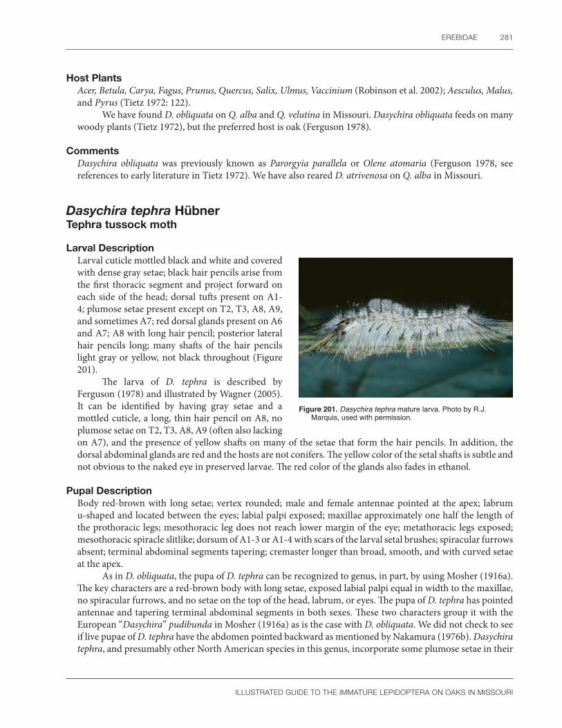
EREBIDAE 281
ILLUSTRATED GUIDE TO THE IMMATURE LEPIDOPTERA ON OAKS IN MISSOURI
Host PlantsAcer, Betula, Carya, Fagus, Prunus, Quercus, Salix, Ulmus, Vaccinium (Robinson et al. 2002); Aesculus, Malus, and Pyrus (Tietz 1972: 122).
We have found D. obliquata on Q. alba and Q. velutina in Missouri. Dasychira obliquata feeds on many woody plants (Tietz 1972), but the preferred host is oak (Ferguson 1978).
CommentsDasychira obliquata was previously known as Parorgyia parallela or Olene atomaria (Ferguson 1978, see references to early literature in Tietz 1972). We have also reared D. atrivenosa on Q. alba in Missouri.
Dasychira tephra HübnerTephra tussock moth
Larval Description Larval cuticle mottled black and white and covered with dense gray setae; black hair pencils arise from the first thoracic segment and project forward on each side of the head; dorsal tufts present on A1-4; plumose setae present except on T2, T3, A8, A9, and sometimes A7; red dorsal glands present on A6 and A7; A8 with long hair pencil; posterior lateral hair pencils long; many shafts of the hair pencils light gray or yellow, not black throughout (Figure 201).
The larva of D. tephra is described by Ferguson (1978) and illustrated by Wagner (2005). It can be identified by having gray setae and a mottled cuticle, a long, thin hair pencil on A8, no plumose setae on T2, T3, A8, A9 (often also lacking on A7), and the presence of yellow shafts on many of the setae that form the hair pencils. In addition, the dorsal abdominal glands are red and the hosts are not conifers. The yellow color of the setal shafts is subtle and not obvious to the naked eye in preserved larvae. The red color of the glands also fades in ethanol.
Pupal DescriptionBody red-brown with long setae; vertex rounded; male and female antennae pointed at the apex; labrum u-shaped and located between the eyes; labial palpi exposed; maxillae approximately one half the length of the prothoracic legs; mesothoracic leg does not reach lower margin of the eye; metathoracic legs exposed; mesothoracic spiracle slitlike; dorsum of A1-3 or A1-4 with scars of the larval setal brushes; spiracular furrows absent; terminal abdominal segments tapering; cremaster longer than broad, smooth, and with curved setae at the apex.
As in D. obliquata, the pupa of D. tephra can be recognized to genus, in part, by using Mosher (1916a). The key characters are a red-brown body with long setae, exposed labial palpi equal in width to the maxillae, no spiracular furrows, and no setae on the top of the head, labrum, or eyes. The pupa of D. tephra has pointed antennae and tapering terminal abdominal segments in both sexes. These two characters group it with the European “Dasychira” pudibunda in Mosher (1916a) as is the case with D. obliquata. We did not check to see if live pupae of D. tephra have the abdomen pointed backward as mentioned by Nakamura (1976b). Dasychira tephra, and presumably other North American species in this genus, incorporate some plumose setae in their
Figure 201. Dasychira tephra mature larva. Photo by R.J. Marquis, used with permission.

282 SPECIES DESCRIPTIONS
ILLUSTRATED GUIDE TO THE IMMATURE LEPIDOPTERA ON OAKS IN MISSOURI
cocoon (SCPC). This suggests that cocoon morphology will not help identify this species. Because of similar pupal morphology, species identification of North American Dasychira pupae is not possible at this time.
Natural HistoryBecause D. tephra was only recently recognized by Ferguson (1978), little of the life history has been formally documented. The eggs are chalky white with a dark spot in the center (SCPC). Larvae eat leaves and can be found throughout the growing season (R.J. Marquis, pers. obs.; SCPC). Wagner (2005) reported larval captures from April to September. The pupa is enclosed in a cocoon with some plumose larval hairs (SCPC).
DistributionMaryland to Florida, west to Missouri and Texas (Ferguson 1978). See Figure 202 for Missouri distribution.
Host PlantsQuercus, Q. laurifolia, and Q. nigra (Robinson et al. 2002).
Dasychira tephra is an oak specialist (Ferguson 1978). We have found it on Q. alba in Missouri, thus confirming this plant as a host in nature (see Ferguson 1978: 27).
Figure 202. Known distribution of Dasychira tephra in Missouri.
Orgyia leucostigma (J.E. Smith) White-marked tussock moth
Larval Description Modified from Ferguson (1978) and Wagner (2005): Head and prothoracic shield contrastingly orange to red; two anterior thin, black hair pencils arise from the prothorax; A1-4 with light dorsal setal tufts in front of a black middorsal and a yellow subdorsal longitudinal stripe; supraspiracular and subspiracular verrucae subequal in size; dorsal glands of A6 and A7 red; and dorsum of A8 with a thin black hair pencil (Figure 203a, b). The larva of O. leucostigma has been illustrated numerous times (see list in Tietz 1972). Color photographs can be found in Béïque and Bonneau (1979), Covell (1984), Ives and Wong (1988), Laplante (1998), Carmichael and Vance (2004), Wagner (2005), and Van Driesche et al. (2013). Howard (1899) illustrated each larval instar. The first and second instars have enlarged prothoracic verrucae but no lateral hair pencils. The long anterior and posterior hair pencils do not appear until the mid to late instars (the anterior ones somewhat hidden in Wagner [2005]).
Orgyia leucostigma is most likely to be confused with O. detrita, which has two color forms (Hall and Buss 2014: Fig. 4). The gray form of O. detrita has a dark body that differs from the pale body of O. leucostigma. Another difference is in the color and size of verrucae above (supraspiracular) and below (subspiracular) the spiracle on A3-6. Orgyia leucostigma has both these structures white or yellow in life and subequal in size (Peterson 1962a: L24, Ferguson 1978: 60). The supraspiracular verruca in O. detrita is often contrastingly red (gray form) and usually larger than the subspiracular one. A final difference is in the size of the dorsal setal tufts on A1-4 (Ferguson 1978: 77). These tend to be larger in O. detrita (Hall and Buss 2014: Fig. 4). Although O. detrita is recorded from New York, south to Florida and east to Texas (Ferguson 1978), it has not been recorded from Missouri (J.R. Heitzman and P.E. Koenig, unpublished list).

EREBIDAE 283
ILLUSTRATED GUIDE TO THE IMMATURE LEPIDOPTERA ON OAKS IN MISSOURI
a b
Figure 203. Orgyia leucostigma (a) mid-instar larva; (b) mature larva. Photos by R.J. Marquis, used with permission.
Pupal DescriptionBody translucent white with long setae, contrasting black spiracles, and brown dorsal markings; vertex rounded; male and female antennae rounded at the apex; labrum u-shaped and located between the eyes; labial palpi exposed; maxillae approximately one-fourth the length of the prothoracic legs; prothoracic coxa is exposed but the femur is hidden; mesothoracic leg does not reach the lower margin of the eye; metathoracic legs exposed; wings of the male longer than the female; mesothoracic spiracle slitlike; dorsum of A1-3 with scars of the larval setal brushes; spiracular furrows absent; terminal abdominal segments tapering; cremaster longer than broad, smooth, and with curved setae at the apex.
The pupa of O. leucostigma is illustrated by Mosher (1916a) and Howard (1899: Fig 4, both sexes). According to Mosher (1916a), the pupa of O. leucostigma can be recognized to genus, in part, by having a white (translucent) body marked in brown without dense setae; exposed labial palpi equal in width to the maxillae; the antennal flagellum not elevated, with the apex of the antenna rounded in both sexes; no spiracular furrows; and no setae on the top of the head, labrum, or eyes. There are two sources of confusion. The deep red head color so obvious in living larvae is faded on the larval exuvia, which could suggest incorrectly that O. leucostigma is not a good possibility. Furthermore, the pupa of O. leucostigma is covered in long setae, which are almost as dense as those in Dasychira (SCPC). However, if eggs are present on the cocoon and they are covered by a frothy coating, it must be O. leucostigma (Ferguson 1978: 78). Other eastern species of Orgyia do not cover their eggs in this way. Pupal identification of male Orgyia to species is not possible at this time. Male pupae are recognized by their large antennae and long wings (Howard 1899).
Natural HistoryThe egg or egg mass of O. leucostigma has been illustrated many times, e.g., Syme (1961: Figs. 128, 376), Peterson (1966), Wagner (2005), and Van Driesche et al. (2013). The female lays from 50 to 150 eggs in a mass on the cocoon or some nearby object (Peterson 1966). The chorion appears to be pitted (Syme 1961: 70, Fig. 376). The most obvious feature of the egg mass is the frothy covering produced by the female (Ferguson 1978). Felt (1912a) illustrated egg masses on an elm trunk during an outbreak in New York State.
There is a large amount of general information on the life cycle of O. leucostigma (see Tietz 1972). Many books on economic entomology, as well as introductory works on Lepidoptera for the eastern United States, treat this species. We have selected Howard (1899), Felt (1912a), and USDA (1985) for a summary. Eggs overwinter and hatch from April to June (USDA 1985). Females are wingless, and thus young larvae must hang from silk treads until they are blown by the wind to a new location. Older caterpillars, especially females, can also wander off their original host (Felt 1912a: 16). Early instar larvae skeletonize leaves, middle instars chew holes in leaves, while later instars eat the entire leaf except for the larger veins (see Howard 1899:

284 SPECIES DESCRIPTIONS
ILLUSTRATED GUIDE TO THE IMMATURE LEPIDOPTERA ON OAKS IN MISSOURI
Fig. 7). The cocoon is formed of silk and larval hairs and hatches in about 2 weeks (USDA 1985). The cocoon is normally found on plant parts such as the trunk, branches, twigs, or more rarely half eaten leaves (Howard 1899). There are two or more generations per year (Wagner 2005). The sexual dimorphism is well-known; only males are capable of flight (Ferguson 1978).
We have found O. leucostigma infrequently on Missouri oaks. Caterpillars first appear in the April and pupate in early June. There are two broods per year on Missouri oaks.
DistributionMaine west to Alberta, Canada, and then south to Kansas, Missouri and Virginia (Ferguson1978 for O. leucostigma intermedia). See Figure 204 for Missouri distribution.
When all four subspecies are combined, O. leucostigma is the most widely distributed lymantriid in the eastern half of North America. It occurs from Newfoundland to Alberta in Canada, throughout the eastern United States, then south to Florida, and west to Colorado (Ferguson 1978).
Host PlantsFeeds on over 140 deciduous or coniferous trees and shrubs (Ferguson 1978); Abies, Acer, Alnus, Betula, Fagus, Larix, Malus, Picea, Populus (Robinson et al. 2002); Prunus, and Ulmus (Rose et al. 1997). We have found O. leucostigma on Quercus alba, Q. velutina, and Ulmus rubra in Missouri.
The most recent host list for O. leucostigma intermedia is from Robinson et al. (2002) who, unlike many other authors, segregate records by subspecies. Still, this is no doubt a conservative list simply because few studies have documented food choice below the species level. Host preferences for O. leucostigma seem to vary by region or ecosystem. Orgyia leucostigma is a pest of urban areas and less of a problem in forests (USDA 1985). In addition, each subspecies has its own preferences (Ferguson 1978). In general, apple, basswood, elm, poplar, certain maples, sycamore, birch, larch, and balsam fir are preferred (USDA 1985). Orgyia leucostigma can cause economic damage to orchard trees in Missouri (Heitzman and Heitzman 1987). Canadian outbreaks have occurred on birch, cherry, alder, apple, beech, maple, elm, and other species in Ontario and the Maritime Provinces (Rose et al. 1997). Other Canadian records for O. leucostigma were from white and yellow birch during outbreak periods (Prentice 1962). Otherwise, it is common on conifers in the eastern but not the western parts of its range. During a study in New York, linden and horse chestnut were preferred (Felt 1912a: 16). Around Washington, D.C., the favored hosts were poplar, certain maples, alder, birch, and willow (Howard 1899). Surprisingly, Villiard (1975) claimed oak was a good choice for rearing O. leucostigma in captivity, noting that the caterpillars could survive better on drying leaves than other species. He illustrated a noctuid instead of O. leucostigma, but the text seems to apply to the latter species.
CommentsMissouri specimens of O. leucostigma were classified as O. leucostigma intermedia by Ferguson (1978). When possible, the sections on distribution and hosts emphasize data relating to this subspecies. In theory, one could gather publications and guess at the subspecies by the locality. We have not done this. For more information on all subspecies of O. leucostigma, consult Ferguson (1978), Robinson et al. (2002), and references cited in these works. Previously, O. leucostigma was placed in Notolophus or Hemerocampa (Forbes 1948).
Some people are allergic to the larval hairs of O. leucostigma. This was discussed by (Hall and Buss 2014).
Figure 204. Known distribution of Orgyia leucostigma in Missouri.

EREBIDAE 285
ILLUSTRATED GUIDE TO THE IMMATURE LEPIDOPTERA ON OAKS IN MISSOURI
Lymantria dispar dispar (Linnaeus) European gypsy moth
Larval Description Modified from Dyar (1896d), Ferguson (1978), and Pogue and Schaefer (2007): Mature larva with two vertical bands on head that extend past the F1 setae to below the frons, top and sides of the head with scattered small dots that sometimes merge but never form six large dark spots; body mottled with a thin, light middorsal line but without large dorsal white patches; prothoracic lateral verrucae enlarged with long setae but no hair pencils or dorsal tufts on the body; dorsal verrucae with straight, rigid needlelike spinules, colored blue on the thorax and A1-2, colored red on the verrucae of A3-8; the dorsal glands of A6-7 also red; A1-8 with the D1 seta minute and black; the SD1 and L verrucae fused posterodorsad to the spiracle (Figure 205).
The larva of the gypsy moth has been illustrated numerous times, and color photographs of L. d. dispar in North America can be found in Béïque and Bonneau (1979), Ives and Wong (1988), Carmichael and Vance (2004), Laplante (1998), Wagner (2005), Pogue and Schaefer (2007: 43), Sogaard (2009), and Van Driesche et al. (2013). Examples of European books with color photos of L. d. dispar include Porter (1997) and Gómez de Aizpúrua (2002b). Many of the economic entomology books also treat this species, and more information is included in references sited in this section.
According to the key in Ferguson (1978: 14), the larva of L. d. dispar is identified by the lack of large dorsal tufts on the first four abdominal segments; by the presence of straight, rigid needlelike spinules on the dorsal verrucae; by the “lateral” verrucae on A3-6 being one behind the other and the mottled body cuticle with a characteristic coloration of blue; and by red dorsal verrucae without conspicuous dorsal white patches. However, the bright blue and red verrucae can fade in preserved specimens (Schnitzler et al. 2012). Fracker (1915) considered the fusion of the SD1 and L verrucae on A3-6 to be unique among North American Lymantriinae he studied. This character would be useful in pale specimens with faded colors.
Detailed larval morphological studies of L. d. dispar include Dyar (1896d: 142), Schierbeek (1917), Kozhanchikov (1950), Peterson (1962a), Traxler (1977), Patočka (1980: 741), and Pogue and Schaefer (2007). Kozhanchikov (1950: Fig. 180) contains an almost complete setal map of the first and late instar larva. Perhaps the best readily available illustrations are found in Peterson (1962a). His drawings show the verrucae and body coloration, two characteristics that are less obvious in scanning electron micrographs produced by other workers. Deml and Dettner (2001) studied the dorsal glands of various Lymantriinae, including the gypsy moth.
The first four instars of the gypsy moth are illustrated by McManus and Twardus (1989). Laplante (1998) has a magnified view of a “young” larva. The first three stages are mostly black and gray. Only the fourth to last instars have the characteristic head pattern with obvious blue and red dorsal verrucae. Thus, early instars of the gypsy moth would be easy to overlook or misidentify. Fortunately, there is at least one unusual morphological feature on the first instar gypsy moth and related species (e.g., nun moth [L. monacha]) to aid recognition. It the presence of a bulbous enlargement on the barbed, spinelike setae of the D, SD, and L verrucae (Burgess 1913; Pogue and Schaefer 2007: 9, Plates 33–37). Early workers called the bulbous portion an aerophore and the setae themselves, aerostatic hairs (Burgess 1913: 16). In addition, the first instar gypsy moth has only five abdominal crochets divided into two groups. The first group of three hooks is on the anterior portion of the proleg, whereas the second group of two hooks is located behind them (Pogue and Schaefer 2007: Plate 37, Fig. 5).
Figure 205. Lymantria dispar dispar mature larva. Photo by S.C. Passoa, used with permission.

286 SPECIES DESCRIPTIONS
ILLUSTRATED GUIDE TO THE IMMATURE LEPIDOPTERA ON OAKS IN MISSOURI
The color variation of L. dispar is described by Goldschmidt (1934) and documented by Keena (1994) in a key. With fourth or fifth instars, Keena (1994) noted that L. d. dispar is usually colored grey (most common) or yellow-grey (in the southern United States). In contrast, L. dispar asiatica has five color forms: grey, yellow-grey, yellow, an especially bright yellow ground color, and one with a black dorsal stripe (also see Pogue and Schaefer 2007: Fig. 9). The presence of the five color forms in a gypsy moth population can be cause for concern when identifying strains, so we do not recommend trying to identify L. dispar by color. However, Keena’s key might be a useful screening tool to select individuals for molecular diagnostics. Morphological and color characters to separate L. d. dispar from related species and subspecies are given by Pogue and Schaefer (2007: 9, 10, 13). Perhaps the most useful character is the D1 setal color on A1-8 that is black in L. d. dispar but white in L. d. asiatica. Wallner et al. (1994) studied the differences between gypsy moth strains by head color patterns. Color forms from Japan are illustrated by Goldschmidt (1934: 146–157). Molecular identification of gypsy moth strains is possible (Garner and Slavicek 1996, Islam et al. 2015, Wu et al. 2018). Thirty-six species of Lymantria were barcoded by deWaard et al. (2010), including those intercepted from domestic surveys in Canada.
Pupal DescriptionModified from Mosher (1916a) and Nakamura (1976b): Pupa dark brown covered with long, finely barbed setae; head with two F setae; labial palpi variable, usually concealed, but sometimes exposed; eyes partially covered with setae; cuticle pale white near base of antenna, apex of antenna pointed in both sexes; maxillae triangular and short; prothoracic coxae and femur hidden; mesothoracic leg does not reach the lower eye margin; metathoracic legs exposed; mesothoracic spiracle a small pit on the prothorax above an oval darkened area on the mesothorax; abdominal segments punctate with spiracular furrows on A5-7; cremaster longer than wide with stout, curved setae at the apex and thinner, curved setae above them.
The pupal morphology of L. d. dispar is described or illustrated by Mosher (1916a), Goldschmidt (1934), Nakamura (1976b), and Patočka (1980). The pupa has been photographed many times in the economic entomology literature, e.g., Van Driesche et al. (2013: 565, Fig. 6). Their illustration is particularly helpful because the female pupa shows spiracular furrows on two abdominal segments. All other North American lymantriid pupae studied by Mosher (1916a) lack these furrows. This character was also used to partially diagnose Lymantria in Japan, although Nakamura (1976b) called them “conjunctiva furrows.” The separation of L. monacha from L. dispar is discussed in Nakamura (1976b).
Natural HistoryBecause regulatory officials often need accurate identification of eggs, it is important to recognize the morphology of gypsy moth eggs and the associated scale covering. Peterson (1966) described gypsy moth eggs as being smooth with faint reticulations. He wrote that newly laid eggs were salmon colored but later darken as the chorion becomes transparent. The egg of L. d. dispar is illustrated by Doring (1955: Plate 20, Fig. 289) and the micropyle is shown in Forbush and Fernald (1896: Plate 58) and Felt (1912b: Plate 9). Dolinskaya and Pljushch (1999) studied the egg of L. d. asiatica with a scanning electron microscope. They describe the eggs of that subspecies as yellow to yellow-grey, with a white chorion that has faint cells and aeropyles. This description is similar to the European race.
Gypsy moth females cover their eggs with scales (often called hairs) from their body (McManus et al. 1992b). Peterson (1966) described these scales as light yellow at first, then fading to beige or dirty white. Horak et al. (http://pbt.padil.gov.au/index.php?q=node/123&pbtID=115) studied them with a scanning electron microscope and compared them to other Australian Lymantriidae and one Notodontidae. No key was given, but close study of the individual photographs reveals L. dispar has a finely serrated scale margin with a relatively smooth surface compared to other related species. Roonwal (1954) compared the female scales of several Indian Lymantria, but his single example of L. dispar was only tentatively identified.

EREBIDAE 287
ILLUSTRATED GUIDE TO THE IMMATURE LEPIDOPTERA ON OAKS IN MISSOURI
The life cycle of the gypsy moth has been described in varying degrees by numerous authors and in many outreach documents. Perhaps the most detailed treatments are Forbush and Fernald (1896), Doane and McManus (1981), Elkinton and Liebhold (1990), USDA (1995), and Pogue and Schaefer (2007). Here we provide a brief summary of only the main points for L. d. dispar. Females on average lay 750 eggs in a mass that is usually located on trunks and branches, but because the female does not fly, despite having wings, the eggs may be found on the ground or any other sheltered place, including on human constructed structures (Leonard 1981). The larvae hatch in the spring and hang from a thread to disperse (a process called ballooning) (see McManus et al. 1992b: Fig. 4, USDA 1995: Chap. 1: 9). First instars chew small holes in the leaves, but large numbers of late instar larvae can completely strip a tree of its foliage (McManus et al. 1992b). There are five instars in the male and six or seven in the female (USDA 1995). Like oviposition, pupation can take place on or off the host (McManus et al. 1992b). Adults emerge during the summer, there is one generation per year, and the first instars overwinter in the egg until spring (USDA 1995).
DistributionThroughout most of Europe, parts of the Middle East, North Africa, several Mediterranean islands, and east to the Ural Mountains (Pogue and Schaefer 2007), but currently not established in England (Carter 1984); introduced and established in eastern North American forests from Quebec to Ontario, Canada, south to North Carolina and west to parts of Wisconsin, northern Illinois, northern Indiana, Ohio, and West Virginia (Liebhold et al. 1997, Pogue and Schaefer 2007). The most recent survey results for L. d. dispar in North American can be found on the Cooperative Agricultural Survey’s Pest Tracker Web site (https://pest.ceris.purdue.edu/pest.php?code=ITAXQNA).
Pogue and Schaefer (2007) discussed the distribution of all subspecies of L. dispar. Liebhold et al. (1997) documented the spread of the gypsy moth throughout North America with a series of maps. The European gypsy moth is established in the northeastern North America but can be sporadically captured in many states even as far west as California (Pogue and Schaefer 2007). There are no established populations of L. dispar in Missouri at this time (see Figure 206), although there have been a few local outbreaks associated with egg clusters entering the state on nursery stock. A few males are caught in pheromone traps each year, e.g., 11 in almost 6,000 traps in 2011 (Missouri Department of Conservation 2011).
Host PlantsExtremely polyphagous (Pogue and Schaefer 2007), specific records from oak include Quercus alba, Q. bicolor, Q. coccinea, Q. ilicifolia, Q. imbricaria, Q. macrocarpa, Q. palustris, Q. prinoides, Q. prinus, Q. rubra, Q. stellata, and Q. velutina (Robinson et al. 2002).
The exact host range of the gypsy moth is difficult to determine because authors do not always distinguish field from laboratory hosts. Liebhold et al. (1995) ranked the native or commonly planted tree species in the United States with regard to their suitability to gypsy moth larvae. They found that 449 tree species could potentially be attacked and 51 oak species were considered susceptible (Pogue and Schaefer 2007). A short list of the main hosts of L. d. dispar are oak, birch, poplar, willow, and alder (USDA 1995: Chapter 1: 12). Ferguson (1978) gives a longer list of principal hosts as do Orozumbekov et al. (2009). Larval age, the exact tree species tested, and stage of the outbreak all affect suitability; this complicates listing of the
Figure 206. Known distribution of Lymantria dispar in Missouri.

288 SPECIES DESCRIPTIONS
ILLUSTRATED GUIDE TO THE IMMATURE LEPIDOPTERA ON OAKS IN MISSOURI
common hosts. Consult USDA (1995: Appendix D) for the rank of 700 tree species to the European gypsy moth larvae. Three hundred species were reported as hosts in Europe (Pogue and Schaefer 2007: 20). The main hosts of the Asian strain are oak, larch, birch, and willow (USDA 1995: Chapter 1: 12). Lechowicz and Mauffette (1986) compared the hosts of the European gypsy moth in North America and Europe. An effort to compare the hosts of L. dispar by region was given by Orozumbekov et al. (2009: Table 2).
During an outbreak of the gypsy moth, almost all vegetation is consumed (USDA 1995, Asaro and Chamberlin 2015). As an example, larvae ate all woody canopy and understory species in an oak-maple forest, with the sole exception of spicebush (Lindera benzoin), during localized outbreaks in Virginia in 2008 (R. Forkner, pers. obs.). For more information on gypsy moth outbreaks, consult Davidson et al. (1999, 2001).
CommentsLymantria dispar was previously placed in the genus Porthetria, and more rarely in Ocneria. The correct nomenclature of the gypsy moth is discussed by Ferguson (1976). Ferguson (1978) noted that male L. d. dispar can be separated from other North American lymantriids by the genitalia alone, even if no other part of the moth is available. Males of the Asian strain differ from the European strain in both size and color. Lymantria d. dispar has a gray light brown ground color and the wingspan is 14–22 mm whereas L. dispar asiatica is larger (wing span 23–28 mm) and colored either a shade of dark or light brown with no gray tint (Pogue and Schaefer 2007: 13, 23).
Missouri forests are highly susceptible to defoliation by the gypsy moth due to the high oak dominance in its wooded areas (Liebhold et al. 2004). Many other states or countries also recognize this threat and have developed a series of action plans with protocols for the recognition and control of L. dispar and related species. Some examples are Stibick (2000), Glare et al. (2003) and USDA (1992, 2013).
Some workers have spent years studying gypsy moth ecology and control. A good example of such efforts can be found at http://www.sandyliebhold.com/pubs/pubs.htm and https://www.fs.fed.us/ne/morgantown/4557/gmoth/world/. Here we do not summarize all the information about life history details and control methods, or cite numerous Web pages, but instead we have compiled a list of citations for the major bibliographies that introduce interested parties to what is known about this important pest. We focused on the European strain of L. dispar (L. d. dispar) and not any of the related species, although we do compare it to the Asian strain (L. d. asiatica) when appropriate. A comparison of the biological characteristics of the Asian and European strains is given by USDA (1995: Chapter 1: 12). A bibliography of the gypsy moth published by Griffiths (1980) contains over 4,000 entries. Rafats (1992) emphasizes control strategies. An attempt to collate literature for Russia and Ukraine was published by Baranchikov et al. (1998). Orozumbekov et al. (2009) review the situation in Central Asia. Schaefer et al. (2014) covers the gypsy moth in China. The classic work on the gypsy moth for the eastern United States is Forbush and Fernald (1896). Another useful work is Gerhardi and Grimm (1979). A nontechnical treatment of the gypsy moth history, biology, and control was published by McClung (1974). Several guidebooks aimed at homeowners have also been published, including Grafton and Web (1990) and McManus et al. (1992a).
Although there are some old records from New York City, the nun moth is not currently established anywhere in North America (Ferguson 1978). The early instars of the nun moth could easily be confused with those of the gypsy moth. The differences were illustrated by Keena et al. (1998).

EREBIDAE 289
ILLUSTRATED GUIDE TO THE IMMATURE LEPIDOPTERA ON OAKS IN MISSOURI
Subfamily Arctiinae
Halysidota tessellaris (J.E. Smith)Banded tussock moth
Larval Description Modified from Dyar (1891): Last instar larva has a shiny black to rarely brown head with a contrasting white intersegmental membrane above the labrum and at the antennal base; the body covered with either white, yellow, amber, or whitish gray hair; prothorax and mesothorax each with two pairs of hair pencils, one white, the other black; midorsum with clumped darker setae that form a line down the back; crochets heteroideous; another pair of black hair pencils above the spiracle on A8 (Figure 207a, b).
The larva of H. tessellaris is described by Dyar (1891). The first instar has primary setae and the head, prothoracic shield, and anal shield are all black. As is typical for many macrolepidopterans, secondary setae appear after the first molt. Later, the larva has sparse, long hairs arising from prominent black verrucae. Middle instars are densely hairy but the characteristic hair pencils do not appear until the fifth instar. There is little change from the fifth through ninth instars. See also Appendix 3, Plate 2.
Color illustrations of H. tessellaris have been published in Ives and Wong (1988), Rose et al. (1997), Wagner (2005), Nagle and Wagner (2009), and Sogaard (2009). Three color forms are shown by Carmichael and Vance (2004). An early instar is illustrated by Laplante (1998). The head and mandible are illustrated by McCabe (1991). Halysidota tessellaris is recognized by the black head, presence of paired black and white hair pencils on the prothorax and mesothorax, presence of a hair pencil from the SD1 verruca on A8, and the unmarked body without contrasting black spots on the cuticle (Forbes 1960). It is most likely to be confused with other caterpillars that have hair pencils, that is, some species of Lymantriinae, Arctiinae, Noctuidae (Forbes 1906: 88), and Apatelodes torrefacta (see Wagner 2005 for differences). Lymantriinae have dorsal abdominal glands, and the Noctuidae have homoideous crochets. Halysidota tessellaris lacks abdominal glands and has heteroideous crochets. The characters listed above will separate H. tessellaris from the few other Arctiinae with hair pencils. Most genera of Arctiinae studied by Forbes (1906: 83) were placed in his grouping of the (woolly) bears, all of which lack hair pencils. However, the hair pencils of H. tessellaris can be lost both in nature (Wagner 2005) and in preserved specimens. In those cases, recognition of H. tessellaris would be more difficult. Jacobson and Weller (2002: 13) note that members of the Halysidota generic group have two subdorsal verrucae on the mesothorax and metathorax.
a b
Figure 207. Halysidota tessellaris (a) mature larva, white form; (b) mature larva, yellow form. Photos by R.J. Marquis, used with permission.

290 SPECIES DESCRIPTIONS
ILLUSTRATED GUIDE TO THE IMMATURE LEPIDOPTERA ON OAKS IN MISSOURI
Pupal DescriptionPupa unmarked, the dorsum not strongly convex; labrum u-shaped; maxillae extend to caudal margins of the wings; maxillary palpi hidden; prothoracic leg about three-fourths as long as maxillae; prothoracic femur hidden; mesothoracic leg does not reach the lower eye margin; only tips of metathoracic legs slightly exposed; mesothoracic spiracle unknown; abdominal segments finely punctate and without flange plates; cremaster absent, end of abdomen rounded with a few slightly curved, thin setae.
The pupa of H. tessellaris was studied by Mosher (1916a). It is easily recognized by these characters: the maxillae as long as the wings, lacking flange plates on the abdominal segments, relatively flattened dorsum and rounded last abdominal segments, unicolorous instead of contrastingly marked with black, and exposed labial palpi that are approximately one-sixth the length of the wings. In addition, the thin cremastral setae are slightly curved but not hooked. The pencil hair tufts are not visible on the larval exuvia (SCPC).
The pupa of H. tessellaris is very similar, if not identical, to that of H. harrisii, including the exposed labial palpi in both species. They can be differentiated by the host if known, larval head, and hair color of the cocoon. Halysidota harrisii feeds on sycamore (Platanus), has a brown head and no black hairs on the cocoon because the larva lacks black hair pencils. In contrast, H. tessellaris is associated with oak (Quercus) and other trees, has a black larval head, and black hairs on the cocoon from the larval hair pencils that are visible under low magnification.
Natural HistoryThe egg of H. tessellaris is illustrated by Peterson (1963b). They are pearl white, shiny, and laid in a flat mass containing about 100 eggs.
The life cycle of H. tessellaris is discussed by Nagle and Wagner (2009). Eggs are laid on the undersides of leaves, but the larva usually rests on the upper surface. Caterpillars are found from July to September (Laplante 1998) and overwinter in a cocoon of silk and hairs (USDA 1985).
Heitzman and Heitzman (1987) considered H. tessellaris to be double-brooded in Missouri, but on oaks there appears to be only a single brood, with first instar larvae appearing in July (R.J. Marquis, pers. obs.).
DistributionQuebec and Ontario, Canada, south to Florida and Texas then west to Wisconsin (Watson 1980), Wyoming, Utah, and Arizona (Powell and Opler 2009).
Halysidota tessellaris is common throughout most of the eastern United States (Covell 1984), including all regions of Missouri (Heitzman and Heitzman 1987; Figure 208).
Host PlantsPolyphagous on 19 families of woody plants; specific records for Quercus include Q. alba, Q. coccinea, Q. palustris, Q. velutina, Q. macrocarpa, and Q. rubra (Robinson et al. 2002).
In Missouri, Halysidota tessellaris was collected from six oaks (Q. alba, Q. coccinea Q. muehlenbergii, Q. rubra, Q. stellata, and Q. velutina), and Heitzman and Heitzman (1987) specifically recorded it from hackberry and grape.We have also encountered and reared the arctiine Haploa contigua on Q. alba in Missouri.
Figure 208. Known distribution of Halysidota tessellaris in Missouri.

EREBIDAE 291
ILLUSTRATED GUIDE TO THE IMMATURE LEPIDOPTERA ON OAKS IN MISSOURI
Subfamily Herminiinae
Palthis angulalis (Hübner)Dark-spotted palthis
Larval Description Mature larva with a granulose head; mandible without a retinaculum; larva colored steel gray to light brown to black with dark dorsal chevrons on the abdominal dorsum and slanted dashes subdorsally; a hump present on A1 and a larger one on A8; a contrasting white to yellow diagonal, lateral line travels from the subventral area of A7 to the dorsal ridge of A8; abdominal spiracles faintly emarginated; cuticle covered with long microspines (Figure 209).
The first instar larva of P. angulalis was superficially described by Dyar (1904). It is nondescript with pointed setae. Later instars were described and partially illustrated by Crumb (1934, 1956) and Brown and McGuffin (1942). Ives and Wong (1988), Sogaard (2009), and Wagner et al. (2011) published color photographs of P. angulalis.
Palthis angulalis might be confused with the Heliothinae because both taxa have spiny cuticles. The presence of a granulose head, uniordinal crochets on A3-6, and a faintly emarginated spiracle (see illustration in Crumb 1956) and hump on A8 will separate P. angulalis from North American heliothines. In addition, heliothines are normally flower or seed feeders on low growing plants (only Pyrrhia are found on shrubs and trees), and their crochets are biordinal (Wagner et al. 2011). There are other differences between P. angulalis and Heliothinae, but the above list includes the most obvious ones. There are also other Erebidae/Noctuidae with a spiny cuticle, but they are not likely to be confused with P. angulalis. Except for Lascoria, other Herminiinae do not have a hump on A8 (Crumb 1956). The coloration of Lascoria is not similar to Palthis (Wagner et al. 2011), and the cuticle is not spiny (Crumb 1956: 335).
Pupal DescriptionLabrum semicircular; labial palpi composed of a triangular portion basally and a long thin distal portion that together extends for about one-third the length of the maxillae; maxillary palpi hidden; maxillae extend to caudal margin of the wings; prothoracic leg wider than other legs; prothoracic femur hidden; mesothoracic leg does not reach lower margin of the eye; metathoracic legs hidden; mesothoracic spiracle slitlike; abdominal segments finely punctate; abdominal spiracles a thin oval, not rounded; cremaster with two thick, curved spines surrounded by thinner ones.
No pupal keys are available for identification of North American Herminiinae. The shape of the labial palpi (triangular base and thinner extension), thin oval abdominal spiracles, and if present, spinose larval cuticle, would suggest a species of Palthis. Many of the noctuids with spiny skin (Heliothinae, some Plusiinae) have a mesothoracic leg that does not extend to the lower eye margin (Gardner 1948). Too few genera of Herminiinae have been examined to allow more definitive identification aids.
Forbes (1954) noted the male prothoracic leg of Palthis was enlarged. This also is true of the female, but there appears to be no sexual dimorphism in pupal prothoracic leg width (SCPC).
Figure 209. Palthis angulalis mature larva. Photo by R.J. Marquis, used with permission.
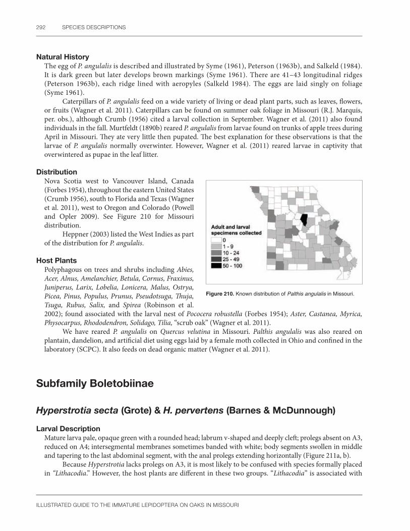
292 SPECIES DESCRIPTIONS
ILLUSTRATED GUIDE TO THE IMMATURE LEPIDOPTERA ON OAKS IN MISSOURI
Natural HistoryThe egg of P. angulalis is described and illustrated by Syme (1961), Peterson (1963b), and Salkeld (1984). It is dark green but later develops brown markings (Syme 1961). There are 41–43 longitudinal ridges (Peterson 1963b), each ridge lined with aeropyles (Salkeld 1984). The eggs are laid singly on foliage (Syme 1961).
Caterpillars of P. angulalis feed on a wide variety of living or dead plant parts, such as leaves, flowers, or fruits (Wagner et al. 2011). Caterpillars can be found on summer oak foliage in Missouri (R.J. Marquis, per. obs.), although Crumb (1956) cited a larval collection in September. Wagner et al. (2011) also found individuals in the fall. Murtfeldt (1890b) reared P. angulalis from larvae found on trunks of apple trees during April in Missouri. They ate very little then pupated. The best explanation for these observations is that the larvae of P. angulalis normally overwinter. However, Wagner et al. (2011) reared larvae in captivity that overwintered as pupae in the leaf litter.
DistributionNova Scotia west to Vancouver Island, Canada (Forbes 1954), throughout the eastern United States (Crumb 1956), south to Florida and Texas (Wagner et al. 2011), west to Oregon and Colorado (Powell and Opler 2009). See Figure 210 for Missouri distribution.
Heppner (2003) listed the West Indies as part of the distribution for P. angulalis.
Host PlantsPolyphagous on trees and shrubs including Abies, Acer, Alnus, Amelanchier, Betula, Cornus, Fraxinus, Juniperus, Larix, Lobelia, Lonicera, Malus, Ostrya, Picea, Pinus, Populus, Prunus, Pseudotsuga, Thuja, Tsuga, Rubus, Salix, and Spirea (Robinson et al. 2002); found associated with the larval nest of Pococera robustella (Forbes 1954); Aster, Castanea, Myrica, Physocarpus, Rhododendron, Solidago, Tilia, “scrub oak” (Wagner et al. 2011).
We have reared P. angulalis on Quercus velutina in Missouri. Palthis angulalis was also reared on plantain, dandelion, and artificial diet using eggs laid by a female moth collected in Ohio and confined in the laboratory (SCPC). It also feeds on dead organic matter (Wagner et al. 2011).
Figure 210. Known distribution of Palthis angulalis in Missouri.
Subfamily Boletobiinae
Hyperstrotia secta (Grote) & H. pervertens (Barnes & McDunnough)
Larval DescriptionMature larva pale, opaque green with a rounded head; labrum v-shaped and deeply cleft; prolegs absent on A3, reduced on A4; intersegmental membranes sometimes banded with white; body segments swollen in middle and tapering to the last abdominal segment, with the anal prolegs extending horizontally (Figure 211a, b).
Because Hyperstrotia lacks prolegs on A3, it is most likely to be confused with species formally placed in “Lithacodia.” However, the host plants are different in these two groups. “Lithacodia” is associated with

EREBIDAE 293
ILLUSTRATED GUIDE TO THE IMMATURE LEPIDOPTERA ON OAKS IN MISSOURI
a b
Figure 211. Hyperstrotia secta (a) mature larva; H. pervertens (b) mature larva. Photos by R.J. Marquis, used with permission.
grasses, sedges, or low plants (Crumb 1956, Wagner et al. 2011: 236, 238, 379), whereas Hyperstrotia usually feeds on oak (Wagner et al. 2011). North American larvae of “Lithacodiinae” (now placed with other Eustrotiinae and Noctuinae) were treated by Crumb (1956), Godfrey (1971, 1972b), and various places in Wagner et al. (2011). Some Hypeninae and Herminiinae also have the prolegs absent on A3 (Wagner et al. 2011). In general, these species do not feed on fresh oak leaves (Crumb 1956: 5, Couplet 15 , 77). Because of the similar color pattern, the larva of Hypena (=Bomolocha) (see Wagner et al. 2011) resembles Hyperstrotia. They are best separated by the development of proleg on A3 and chaetotaxy of the SV group on A1. Hyperstrotia has the proleg completely absent on A3 and the SV group on A1 appears trisetose. In contrast, the Hypeninae have the prolegs absent or reduced on A3 (McCabe and Vargas 1998) and the SV group on A1 appears bisetose because of the dorsal movement of SV3 nearer to L3 (Fibiger and LaFontaine 2005: 28, Wagner et al. 2011).
The larva of Spargaloma sexpunctata, in the same tribe as Hyperstrotia, has a similar green, unmarked caterpillar; it feeds on dogbane and is not found on oaks (Wagner et al. 2011).
No larval characters are available to separate the two species of Hyperstrotia collected in Missouri. We have reared about 10 larvae each of H. secta (Figure 211a) and H. pervertens (Figure 211b). Based on our experience, H. pervertens always has red pinacula, which separate it from H. secta. It is unclear how useful this difference is in other regions (Wagner et al. 2011: 96).
Pupal DescriptionLabrum semicircular, labial palpi exposed; maxillae extend to caudal margin of the wings; prothoracic femur hidden; maxillary palpi hidden; prothoracic and mesothoracic legs both extend to the lower margin of the eye; metathoracic legs hidden; mesothoracic spiracle slitlike; posterior margin of mesothorax with a horizontal row of pits connected to each other and less sclerotized at the meson; abdominal segments punctate, the punctures more dense at the anterior margins of A5-7; cremaster with slightly curved, stout spines at the apex surrounded by much shorter and thinner curved setae.
Mosher (1916a) did not study pupae of the Phytometrini. In her work, Hyperstrotia will key with the Hypeninae because the prothoracic and mesothoracic legs both extend to the lower margin of the eye; the cremaster has hooked or curved setae and there is no whitish bloom covering the body. A European species of Phytometra illustrated by Patočka and Turčáni (2005: Fig. 226) also has the prothoracic and mesothoracic legs extending to the lower margin of the eye, but the cremaster lacks any hooked or curved setae that are found in Hyperstrotia. Another unusual feature of Phytometra is that only a very small portion of the prothoracic femur is exposed.
Hyperstrotia pervertens also has a horizontal row of pits on the posterior margin of the mesothorax. The presence of these pits will separate Hyperstrotia from other oak-feeding noctuids we studied.

294 SPECIES DESCRIPTIONS
ILLUSTRATED GUIDE TO THE IMMATURE LEPIDOPTERA ON OAKS IN MISSOURI
Natural HistoryThe life cycle of H. secta is poorly known. The egg is apparently undescribed. A typical life cycle for Hyperstrotia is discussed by Wagner et al. (2011). Early instars are skeletonizers (Wagner et al. 2011); we have seen this for H. secta and H. pervertens (R.J. Marquis, pers. obs.). All instars rest on the leaf underside (R.J. Marquis, pers. obs.; Wagner et al. 2011). Adults of Hyperstrotia fly from May to August in Ohio, with peak abundance during midsummer (Rings et al. 1992).
In Missouri, larvae of Hyperstrotia secta occur from May to August, with adults emerging in the same year (R.J. Marquis, pers. obs.).
DistributionSouthern Ontario, Canada, to Massachusetts and south to Florida (Wagner et al. 2011), west to Ohio (Rings et al. 1992), Missouri, and Texas (Covell 1984). See Figure 212a, b for Missouri distribution.
Forbes (1954) noted that the distribution of H. secta is somewhat uncertain due to confusion with related species such as H. villificans or H. flaviguttata that have overlapping ranges. Hyperstrotia secta can be locally common (Wagner et al. 2011).
a b
Figure 212. Known distribution of (a) Hyperstrotia secta; (b) H. pervertens in Missouri.
Host PlantsRed and white oaks (Forbes 1954); Quercus alba, Q. rubra (Robinson et al. 2002); and Q. falcata (Heppner 2007).
Hyperstrotia secta has a possible preference for white oaks (Wagner et al. 2011). He noted other Hyperstrotia can be found on elm (Ulmus), beech (Fagus), chestnut, and rarely hickory (Carya). We have reared H. pervertens on both Q. alba and Q. velutina in Missouri.
CommentsHodges et al. (1983) listed Hyperstrotia in the Eustrotiini of the Acontiinae (Noctuidae). Wagner et al. (2011) placed Hyperstrotia in the Phytometrinae with doubt (Erebidae). Pohl et al. (2016) followed this placement although they recognized Phytometrini as a tribe in the Boletobiinae, also in the Erebidae.
A superficial glance at the pupal morphology suggests Hyperstrotia is related to the Hypeninae or some other Erebidae, but not the Acontiinae (Noctuidae). The prothoracic and mesothoracic leg of Hyperstrotia reach the lower margin of the eye as they do in the Hypeninae (Mosher 1916a, see discussion under Pupal Description).
We focused on H. secta and H. pervertens for our book because adults of Hyperstrotia can be difficult to identify, and these species seem to be the most readily identified by wing pattern (e.g., Beadle and Leckie 2012).

EREBIDAE 295
ILLUSTRATED GUIDE TO THE IMMATURE LEPIDOPTERA ON OAKS IN MISSOURI
Subfamily Erebinae
Catocala amica (Hübner)Girlfriend underwing
Larval DescriptionMature larva with a slightly flattened body, black and orange markings at the vertex, and a faintly striped head with irregular, brown vertical lines that do not form a solid black contrasting vertical stripe; thorax and abdomen faintly mottled with a mixture of fine white and black markings on a gray background, which appears whitish gray from a distance; thoracic and abdominal pinacula raised and either orange or black; dorsal pinacula of A5 and A8 conical, larger than the others, the dorsum of A5 often with a black patch; abdominal prolegs of A3 and A4 slightly smaller than those on A5 and A6; pale white subventral filaments present on the thorax and abdomen; ventral region pale with a central large black spot on each anterior segment; anal prolegs elongate (Figure 213a, b).
Compared to later instars, young larvae of C. amica have the proleg on A3 reduced and the anal proleg relatively more elongated. In addition, early instars of C. amica lack orange pinacula and have the ground color brown, not gray. Unfortunately, only a single early instar was available so variation in markings could not be studied.
The larva of C. amica is illustrated by Beutenmuller (1902), Barnes and McDunnough (1918), Sargent (1976), and Wagner et al. (2011: 102, 128). Recognition of species in the C. amica complex is difficult (Sargent 1976, Rings et al. 1992, Wagner et al. 2011); therefore, identification of larval C. amica will remain tentative until more systematic studies are published. At present, the only alternative is to utilize the keys to Catocala larvae by Forbes (1954) and Crumb (1956) in combination with host plant data and photographs in Wagner et al. (2011). By inserting C. amica into the key by Crumb (1956), the following list of identification characters is developed: mandible with retinaculum, a fringe of subventral fleshy filaments present, dorsum of A5 with a tubercle and not a horn, and head very faintly granulose. At Couplet 21, C. amica agrees with other oak-feeding Catocala in having L3 reduced to a ring on T2 and T3, but differs in chaetotaxy on A1 and A2, where three unmodified SV setae are present. This may be a significant exception because other oak feeders studied by Crumb (1956) have two SV setae on A1 absent or modified into a short fleshy cone. Forbes (1954) relied extensively on color to distinguish species, but unfortunately, C. amica was also absent from his key. Important identification characters of C. amica from Forbes (1954) are: A5 with enlarged dorsal tubercles and dark saddlelike marking; subventral fleshly filaments present; underside light with middorsal patches and vertex bluntly pointed. At Couplet 12, C. amica and C. amatrix will key out together because both are considered gray caterpillars. They may be distinguished by their head markings and hosts. Catocala amatrix has a thick, vertical head stripe that is lacking in C. amica (Barnes and McDunnough 1918). In addition, C. amatrix feeds on popular and willow, not oak (Crumb 1956).
a b
Figure 213. Catocala amica (a) mid-instar larva; (b) mature larva. Photos by R.J. Marquis, used with permission.

296 SPECIES DESCRIPTIONS
ILLUSTRATED GUIDE TO THE IMMATURE LEPIDOPTERA ON OAKS IN MISSOURI
No morphological characters are available to separate C. amica from a related sibling species, C. lineella, but each taxon may prefer a different oak subgenus. Catocala amica feeds principally on white oaks, whereas C. lineella most often occurs on red or black oaks (Gall 1990b, Wagner et al. 2011). Little is known about the life cycle of C. jair, but it also feeds on oaks (Wagner et al. 2011). Unlike C. amica, some Catocala larvae lack fleshy subventral projections on their body (Forbes 1954, Crumb 1956, Wagner et al. 2011).
Pupal DescriptionPupa irregularly covered with a whitish blue bloom; labrum u-shaped; labial palpi exposed; maxillary palpi hidden; maxillae extend to caudal margin of the wings; prothoracic femur hidden; prothoracic and mesothoracic legs extend to lower margin of eye; metathoracic legs hidden; mesothoracic spiracle slitlike; dorsum of thorax faintly rugose; abdominal segments finely punctate; abdominal spiracles shaped like a narrow oval, about three times taller than wide; cremaster with longitudinal ridges and eight stout, curved spines at the apex, six spines form a semicircle above a thicker center pair.
Mosher (1916a) noted that all Catocalinae she examined had a whitish bloom, prothoracic and mesothoracic legs that extend to the lower margin of the eye, and usually a hidden prothoracic femur. Catocala amica has these three features. In addition, at least some European Catocala have a similar leg arrangement (Patočka 1995: Fig. 80). Unlike most genera of Catocalinae, Catocala pupae are unusual because the epicranial suture is present (Mosher 1916a). Unfortunately, this character could not be evaluated on C. amica because only pupal exuviae were available, and the epicranial suture is destroyed upon adult emergence.
Mosher (1916a) divided the genus Catocala into two parts based on the structure of the cremaster. Catocala amica has long, hooked setae of two sizes in contrast to members of “Eunetis” (now a synonym of Catocala) in which the setae are short and slightly curved. Except for Mosher (1916a), no other key to Nearctic Catocala pupae is available.
Natural HistoryBarnes and McDunnough (1918: 45) placed Catocala amica in their group 20, a taxon with the egg stage undescribed, and speculated that eggs of this group would be similar to other Catocala. Peterson (1964) illustrates the eggs of 11 species of Catocala, but C. amica was not included.
Although good biological studies exist of the genus Catocala (Sargent 1976; Gall 1990a, b, c; Wagner et al. 2011), the life cycle of C. amica has not been extensively studied. Gall (1990a: 174) and Wagner et al. (2011) summarized the life cycle of a typical Catocala species. The eggs overwinter. Feeding occurs at night and larvae hide during the day. Larvae wiggle vigorously when disturbed. Approximately 40–70 eggs were laid by C. amica in captivity (Gall 1990c). Larvae occur from May to early July (Wagner et al. 2011), where they may be eaten from small or medium-sized oak trees in the eastern United States (Gall 1990b). Adults are most common from July to August where they rest on tree trunks with their head facing downward (Sargent 1976, Lienk et al. 1991).
In Massachusetts, larvae of C. amica hatched in May, pupated in June, and emerged in July (Sargent 1976). There is one generation per year (Wagner et al. 2011).
In Missouri, we found that C. amica occurs only in early spring (April to May) on newly emerged oak leaves. Caterpillars often hide on twigs during the day.
DistributionSouthern Ontario, Canada, south to Maine and Florida, west to South Dakota and Texas (Covell 1984), including Ohio (Rings et al. 1992) and Missouri. See Figure 214 for Missouri distribution.
Catocala amica is commonly collected as an adult (Covell 1984) or larva (Gall 1990b: 209) and is one of the most common underwings in eastern North America (Wagner et al. 2011).
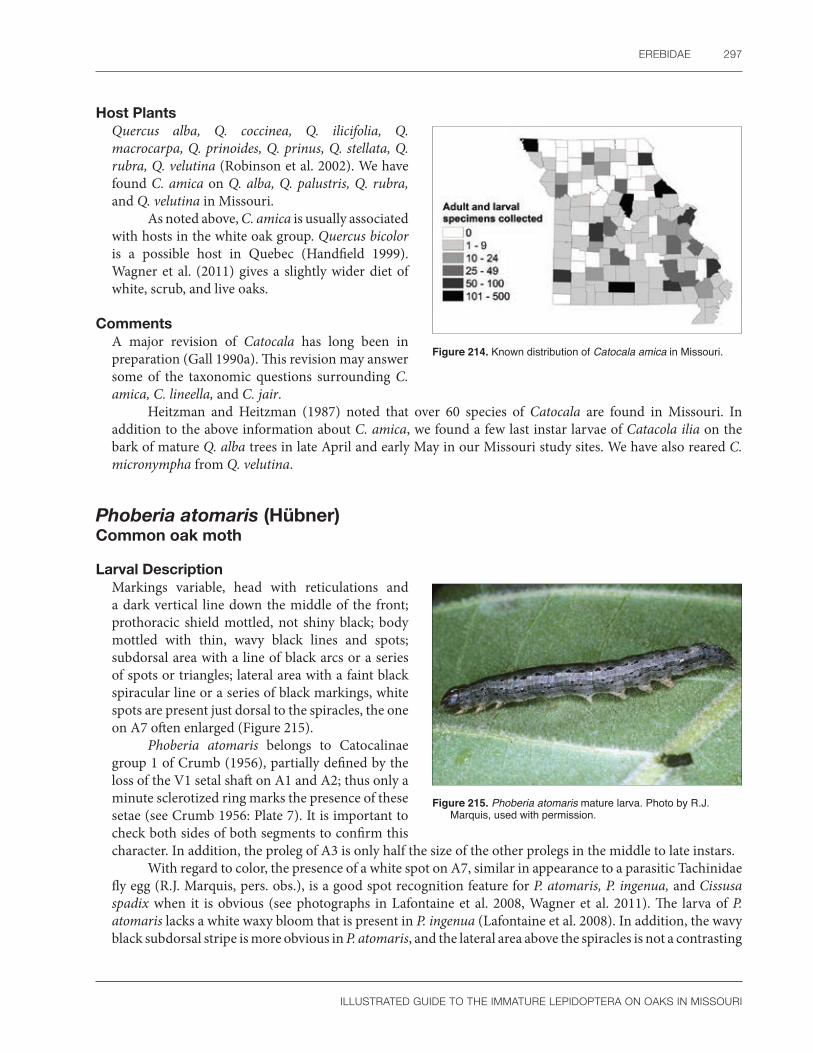
EREBIDAE 297
ILLUSTRATED GUIDE TO THE IMMATURE LEPIDOPTERA ON OAKS IN MISSOURI
Figure 214. Known distribution of Catocala amica in Missouri.
Host PlantsQuercus alba, Q. coccinea, Q. ilicifolia, Q. macrocarpa, Q. prinoides, Q. prinus, Q. stellata, Q. rubra, Q. velutina (Robinson et al. 2002). We have found C. amica on Q. alba, Q. palustris, Q. rubra, and Q. velutina in Missouri.
As noted above, C. amica is usually associated with hosts in the white oak group. Quercus bicolor is a possible host in Quebec (Handfield 1999). Wagner et al. (2011) gives a slightly wider diet of white, scrub, and live oaks.
CommentsA major revision of Catocala has long been in preparation (Gall 1990a). This revision may answer some of the taxonomic questions surrounding C. amica, C. lineella, and C. jair.
Heitzman and Heitzman (1987) noted that over 60 species of Catocala are found in Missouri. In addition to the above information about C. amica, we found a few last instar larvae of Catacola ilia on the bark of mature Q. alba trees in late April and early May in our Missouri study sites. We have also reared C. micronympha from Q. velutina.
Phoberia atomaris (Hübner)Common oak moth
Larval Description Markings variable, head with reticulations and a dark vertical line down the middle of the front; prothoracic shield mottled, not shiny black; body mottled with thin, wavy black lines and spots; subdorsal area with a line of black arcs or a series of spots or triangles; lateral area with a faint black spiracular line or a series of black markings, white spots are present just dorsal to the spiracles, the one on A7 often enlarged (Figure 215).
Phoberia atomaris belongs to Catocalinae group 1 of Crumb (1956), partially defined by the loss of the V1 setal shaft on A1 and A2; thus only a minute sclerotized ring marks the presence of these setae (see Crumb 1956: Plate 7). It is important to check both sides of both segments to confirm this character. In addition, the proleg of A3 is only half the size of the other prolegs in the middle to late instars.
With regard to color, the presence of a white spot on A7, similar in appearance to a parasitic Tachinidae fly egg (R.J. Marquis, pers. obs.), is a good spot recognition feature for P. atomaris, P. ingenua, and Cissusa spadix when it is obvious (see photographs in Lafontaine et al. 2008, Wagner et al. 2011). The larva of P. atomaris lacks a white waxy bloom that is present in P. ingenua (Lafontaine et al. 2008). In addition, the wavy black subdorsal stripe is more obvious in P. atomaris, and the lateral area above the spiracles is not a contrasting
Figure 215. Phoberia atomaris mature larva. Photo by R.J. Marquis, used with permission.

298 SPECIES DESCRIPTIONS
ILLUSTRATED GUIDE TO THE IMMATURE LEPIDOPTERA ON OAKS IN MISSOURI
light color as in some forms of P. ingenua. The larva of C. spadix has a solid black, shiny prothoracic shield, dark dorsum, a contrasting light lateral area above the spiracles, and sometimes a wavy black subdorsal stripe over A8 and A9 (Wagner et al. 2011). The prothoracic shield of both Phoberia species is mottled, not solid black (SCPC, Lafontaine et al. 2008).
The waxy bloom present in P. ingenua will likely dissolve in alcohol, thus preserved specimens must be identified with caution.
Pupal DescriptionBased on a partially damaged exuvia missing the head, mouthparts and part of the cremaster: Mesothoracic spiracle slitlike; abdominal segments punctate with modified spiracles having 4–5 round tubercles either in the opening or associated with the peritreme.
The abdominal spiracles of P. atomaris are modified with a series of tubercles that are of unclear location and unknown function. They are either part of the peritreme or sit in the opening of the spiracle. A brief survey of common erebine genera shows that these structures are absent in Alabama, Anticarsia, Catocala, Mocis, Panopoda, and Zale but something similar is present in Melipotis. This suggests the character may help define a group of genera or perhaps a tribe. Crumb (1956) placed Melipotis and Phoberia together in his “Catocalinae group 1” as both have very short labial palpi that are not longer than half the length of the spinneret.
According to Lafontaine et al. (2008), the pupa of P. atomaris has no waxy bloom and the cremaster has two pairs of spines. The inner ones are recurved and the other ones are straight. The inner pair is much thicker than the other setae in our single specimen. A pupal photograph of C. spadix (Pickering 2016) has a very different cremaster than P. atomaris because there are four curved setae of equal thickness on a broad cremaster. It has no bloom.
Natural HistoryThe biology of P. atomaris was reviewed by Coyle et al. (2013) and the following is from their summary. Females oviposit on bark and larvae hatch after the buds open in the spring. They rest on the bark or in leaf litter during the day and climb trees to feed at night. Homeowners reported that caterpillars were attracted to lights and even entered homes. Their defensive behavior is typical of most erebids. This includes regurgitation of gut contents, playing dead, jerking their bodies rapidly from side to side, or folding the head under the body. Caterpillars pupate in the soil or any other protected substrate, including human constructed materials. There is one generation per year and the pupa overwinters.
We have frequently found caterpillars of P. atomaris resting in the crevices of tree trunks during the day from April to June in Missouri. They can also be found in abandoned leaf rolls. There is one generation per year in Missouri. In our experience, larvae do not readily pupate in captivity.
This species reached outbreak proportions in Kentucky (Covell 1999), in south central Missouri on Quercus stellata during the spring of 2001 and 2012 (R. Lawrence, pers. comm.), and in Ross County, Ohio, during 2002 on white oak (ODNR 2002). It is commonly found under burlap bands used to trap gypsy moth larvae (Wagner et al. 2011).
DistributionSoutheastern Canada to Florida then west to Kansas and Texas (Lafontaine et al. 2008). See Figure 216 for Missouri distribution.
Phoberia atomaris can “be expected in any habitat with oaks” (Lafontaine et al. 2008). Western United States populations of Phoberia found in parts of Texas, Colorado, New Mexico, and California (Lafontaine et al. 2008, Powell and Opler 2009) were assigned to P. ingenua by Lafontaine et al. (2008) based on molecular data that found only slight differences in eastern and western haplotypes.

EREBIDAE 299
ILLUSTRATED GUIDE TO THE IMMATURE LEPIDOPTERA ON OAKS IN MISSOURI
Figure 216. Known distribution of Phoberia atomaris in Missouri.
Host PlantsQuercus rubra, Q. coccinea (Robinson et al.2002); and Q.alba (Coyle et al. 2013). We have found P. atomaris on Q. alba, Q. stellata, and Q. velutina in Missouri. Oviposition occurs on Q. alba, and larvae can be reared on Q. ilicifolia in the laboratory (Lafontaine et al. 2008).
CommentsThis species presents taxonomic problems at all levels. Crumb (1956) warned that larval Catocalinae are difficult to identify. Wagner (2005) noted that larvae of Phoberia and the related genus Cissusa are similar. We found C. spadix on Q. alba in Missouri, but very rarely. At least during the span of recent studies, C. spadix has been much less abundant than P. atomaris. At the species level, the adult and larva of P. atomaris are similar to the related P. ingenua (Lafontaine et al. 2008).
High populations of semi-loopers on oaks are likely to be P. atomaris, but rearing to the adult stage or molecular analysis will be needed to confirm the identity of caterpillars collected during non-outbreak years where similar, more rarely encountered taxa are possible. The lack of voucher specimens for any of the outbreaks mentioned above prevents us from knowing if the damage was from a single species or multiple species acting together.
Zale minerea (Guenée)Colorful zale
Larval Description Modified from MacKay (1951) and Wagner et al. (2011): Mature larva mottled with black, brown, gray, or white markings and faint dorsal longitudinal lines; A1 sometimes slightly swollen, with either the D setae, or just the D2 setal base, surrounded by inner white and outer black pigmentation, the D2 setae often connected by a transverse ridge; proleg of A3 reduced; D2 pinaculum of A8 enlarged (Figure 217a, b).
Early instars are dark brownish green with a more contrasting dark dorsal line. The larva of Z. minerea is described or illustrated by Crumb (1956), MacKay (1951), and Wagner et al. (2011). A slightly enlarged A1 segment with at least a white D2 setal base surrounded by black pigmentation, a transverse ridge (when present) connecting the D2 seta on A1, the reduced prolegs of A3, and the D2 setae of A8 on large
Figure 217. Zale minerea (a) mid-instar larva; (b) mature larva. Photos by R.J. Marquis, used with permission.
a b

300 SPECIES DESCRIPTIONS
ILLUSTRATED GUIDE TO THE IMMATURE LEPIDOPTERA ON OAKS IN MISSOURI
tubercles (chalazae) are good field characters to suggest this complex. In addition, larvae of Zale usually have a midventral stripe and a thin lateral black line on the anal prolegs (Wagner et al. 2011). MacKay (1951) illustrates the western subspecies Z. minerea norda and recognized three color forms. One has the ground color olive gray, the second form is mouse gray, and the last is almost uniformly blackish brown. We illustrate a typical grayish form (Figure 217a, b).
Pupal DescriptionPupa covered with a whitish blue bloom; labrum semicircular; labial palpi exposed; maxillary palpi hidden; maxillae extend to caudal margin of the wings; prothoracic femur hidden; prothoracic and mesothoracic legs extend to lower margin of the eye; metathoracic legs barely exposed; mesothoracic spiracle a faint oval smooth area; dorsum of thorax and abdominal segments shagreened; abdominal spiracles shaped like a narrow oval, about three times taller than wide; cremaster not differentiated from last abdominal segment, with eight stout, curved spines at the apex, with six spines forming a semicircle above a thicker center pair.
The pupae of Z. lunata, which is almost identical to Z. minerea (SCPC), was studied by Mosher (1916a). She noted the epicranial suture is absent, there are no tubercles at the base of the antenna, the texture of the thorax and appendages are relatively smooth, the metathoracic legs are exposed, and the cremaster has two setae larger than the others with none of them enlarged at the tip. Our pupae of Z. minerea and Z. lunata fit this characterization except the metathoracic legs are barely exposed in both cases.
Natural HistorySalkeld (1984) illustrates the egg of Z. minerea and describes it as “muddy green” with 49–52 longitudinal ridges. The life history of a typical Zale, including Z. minerea, was discussed by Wagner et al. (2011). The following is a brief summary from their work. Females lay about 200 relatively large eggs. At least for the oak feeders, the caterpillars are active and seek out young leaves. Later instars rest on branches and may consume slightly older foliage. Larvae feed at night and take 4 to 5 weeks to mature. Pupation occurs on the soil surface or in debris such as leaf litter.
Wagner et al. (2011) stated that some Z. minerea caterpillars are found under loose bark. A single adult reared from a pupa on oak bark in Gainesville, FL (S.C. Passoa, pers. obs.), suggests that some larvae do not always pupate at ground level.
DistributionNova Scotia and southern Canada south to Florida (Forbes 1954) and west to Illinois, Colorado, and Montana along the coast from British Columbia, Canada, to California (Crumb 1956), including Missouri. See Figure 218 for Missouri distribution.
Host PlantsPolyphagous on deciduous trees including Acer, Alnus, Betula, Carpinus, Carya, Corylus, Fagus, Quercus alba, Ostrya, Picea, Populus, Pseudotsuga, Salix, Tilia, Ulmus (Robinson et al. 2002); and Crataegus (Wagner et al. 2011).
The western subspecies, Zale minerea norda, has a similar diet to the eastern population (Robinson et al. 2002). Crumb (1956) has a record for Z. minerea on rose, but the subspecies to which this record applies is not clear from his citation. We found larvae of Z. minerea on young oak (Q. alba, Q. coccinea, and Q. velutina) foliage during April and May in Missouri.
Figure 218. Known distribution of Zale minerea in Missouri.

EREBIDAE 301
ILLUSTRATED GUIDE TO THE IMMATURE LEPIDOPTERA ON OAKS IN MISSOURI
CommentsWagner et al. (2011) noted that separating caterpillars of Z. lunata and Z. minerea is difficult. Food plant records for these species may be confused, but recent literature suggests that Z. lunata is common on willow and occasionally uses oak, whereas Z. minerea is common on oak but uses other plants as well (Miller and Hammond 2003, Wagner et al. 2011). To date, the majority of the larvae reared from our study sites in Missouri have become Z. minerea adults. We have also encountered and reared Z. lunata on Quercus alba.
Larvae of Z. minerea are also similar to Z. undularis (see MacKay 1951) but the hosts are different. Locust (Robinia pseudoacacia), not a common host of Z. minerea, is the host of Z. undularis.
Subfamily Eulepidotinae
Panopoda rufimargo (Hübner) Red-lined panopoda
Larval DescriptionMature larva with the head and body green, both speckled with darker spots; a thin white or yellow line transverses the anterior margin of the prothorax; A1-8 has a slanted line running under the spiracles of each segment; the prolegs of A3 and A4 are reduced but the anal prolegs are slightly enlarged; all prolegs colored bright red at the tip (Figure 219a, b).
Crumb (1954) diagnosed Panopoda by the presence of a minute fleshy cone instead of normal setae on V1 of A1 and A2, a process at the base of the abdominal proleg shield, and inconspicuous D2 pinacula on A8. We have found several of these characters to be subtle and difficult to see on our single preserved larva. The minute fleshy cone is easier to see if the larva is partially dried for a minute or two. It is not clear to us what Crumb (1956) meant by the process on the proleg. However, the mandible, with a basal tooth and two transverse ridges at the cutting margin, appears quite unusual and obvious. A similar tooth was seen on the cast exuvia of our reared adults, confirming the character is typical for P. rufimargo. The small, inconspicuous D2 pinacula on A8 readily distinguish P. rufimargo from Zale minerea. Unlike Catocala, there are no pale subventral setae fringes in P. rufimargo. Phoberia and Cissusa are not colored green as is P. rufimargo. Preserved specimens of P. rufimargo lose their color and fade to white, making identification more difficult. See also Appendix 3, Plate 2.
a b
Figure 219. Panopoda rufimargo (a) mid-instar larva; (b) mature larva. Photos by R.J. Marquis, used with permission.

302 SPECIES DESCRIPTIONS
ILLUSTRATED GUIDE TO THE IMMATURE LEPIDOPTERA ON OAKS IN MISSOURI
Pupal DescriptionPupa may or may not be covered with a faint, whitish bloom; labrum semicircular; labial palpi exposed; maxillary palpi hidden; maxillae extend to caudal margin of the wings; prothoracic femur exposed but very thin; prothoracic and mesothoracic legs extend to lower margin of the eye; metathoracic legs barely exposed; mesothoracic spiracle a faint oval smooth area; thorax and abdominal segments punctate; abdominal spiracles a narrow oval; cremaster not differentiated from last abdominal segment, with four stout, curved spines at the apex, the center pair being thicker.
Mosher (1916a) did not study the genus Panopoda. She did note that a few Catocalinae have an exposed prothoracic femur. Panopoda rufimargo is the only erebid species we studied with an exposed prothoracic femur, although it is very thin and barely one-half the width of the labial palpi. In addition, the four relatively thick cremastral spines also are unusual among our oak feeders.
Natural HistoryThe egg of P. rufimargo is described and illustrated by Salkeld (1984). It has 9–10 longitudinal ridges, is green at first, and then later turns reddish brown. Wagner et al. (2011) describes the life history. Larvae sit on the upper side of leaves, often with the anterior half curved, but wriggle violently if disturbed. Pupae overwinter in leaf litter within a thin cocoon covered with detritus. Two or more broods occur in the south.
We have found larvae of P. rufimargo in Missouri from late spring to summer, often on the top of the leaf surface, in their characteristic arched position.
DistributionQuebec, Canada, to Maine and south to Florida, west to Minnesota and Texas (Covell 1984), including Illinois, Missouri (Crumb 1956), Ohio (Rings et al. 1992), and Kentucky (Covell 1999). See Figure 220 for Missouri distribution.
Host PlantsFagus, Quercus alba, and Q. rubra (Robinson et al. 2002). We have found P. rufimargo on Q. velutina in Missouri.
CommentsHere we follow Pohl et al. (2016) and spell the genus as Panopoda instead of Panapoda. Likewise, the common name is then red-lined panopoda and not red-lined panapoda (Wagner et al. 2011). Both spellings are common on the Web.
Figure 220. Known distribution of Panopoda rufimargo in Missouri.
ILLUSTRATED GUIDE TO THE IMMATURE LEPIDOPTERA ON OAKS IN MISSOURI
303
ReferencesAbraham, D.; Ryrholm, N.; Wittzell, H.; Holloway, J.D.; Scoble, M.J.; Löfstedt, C. 2001. Molecular phylogeny of the
subfamilies in Geometridae (Geometroidea: Lepidoptera). Molecular Phylogenetics and Evolution. 20: 65–77.Abrahamson, W.G.; Hunter, M.D.; Melika, G.; Price, P.W. 2003. Cynipid gall-wasp communities correlate with oak
chemistry. Journal of Chemical Ecology. 29: 209-233.Ackery, P.R.; de Jong, R.; Vane-Wright, R.L. 1998 (1999). The butterflies: Hedyloidea, Hesperoidea and Papilionoidea.
In: Handbook of Zoology, Part 35. Kristensen, N.P., ed. Lepidoptera, moths and butterflies, vol. 1: Evolution, systematics, and biogeography. Berlin: Walter de Gruyter: 263–300.
Ackery, P.R.; Vane-Wright, R.I. 1984. Milkweed butterflies, their cladistics and biology: being an account of the natural history of the Danainae, a subfamily of the Lepidoptera, Nymphalidae. London: British Museum (Natural History). 425 p.
Adler, C.R.L. 1991. Identification of pupae on apple in eastern North America. In: van der Geest, L.P.S.; Evenhuis, H.H., eds. Tortricid pests. Their biology, natural enemies and control. Amsterdam, Netherlands: Elsevier. 802 p.
Ahola, M.; Silvonen, K. 2005. Larvae of northern European Noctuidae. Vaasa, Finland: Kuva Seppälä Group Limited. 657 p. Vol. 1.
Ahola, M.; Silvonen, K. 2008. Larvae of northern European Noctuidae. Vaasa, Finland: Kuva Seppälä Group Limited. 672 p. Vol. 2.
Ahola, M.; Silvonen, K. 2011. Larvae of northern European Noctuidae. Vaasa, Finland: Kuva Seppälä Group Limited. 599 p. Vol. 3.
Aiello, A. 1980. Life history of Dysodia sica (Lepidoptera: Thyrididae) in Panama. Psyche. 87: 121–130.Aiello, A.; Balcazar, L.M. 1997. The immature stages of Oxytenis modestia, with comments on the larvae of Asthenidia
and Homoeopteryx (Saturniidae: Oxyteninae). Journal of the Lepidopterists’ Society. 51: 105–118.Albertoni, F.F.; Duarte, M. 2015. Immature stages of Adeloneivaia fallax (Lepidoptera: Saturniidae). Florida
Entomologist. 98: 178–185.Albu, V.; Metzler, E. 2004. Lepidoptera of North America 5. Contributions to the knowledge of southern West Virginia
Lepidoptera. Contributions of the C.P. Gillette Museum of arthropod diversity. Fort Collins, CO: Colorado State University. 82 p.
Alford, D.V. 1984. A color atlas of fruit pests; their recognition, biology and control. London: Wolfe Publishing Co. 320 p.Alford, D.V. 1995. A color atlas of pests of ornamental trees, shrubs and flowers. New York, NY: Wiley and Sons. 448 p.Allen, D.C. 1973. Fecundity of the saddled prominent, Heterocampa guttivitta. Annals of the Entomological Society of
America. 66: 1181–1183.Allen, D.C.; Grimble, D.G. 1970. Identification of the larval instars of Heterocampa guttivitta with notes on their feeding
behavior. Journal of Economic Entomology. 63: 1201–1203.Allen, T.J.; Brock, J.P.; Glassberg, J. 2005. Caterpillars in the field and garden: a field guide to the butterfly caterpillars of
North America. Oxford, England: Oxford University Press. 232 p.Allyson, S. 1977. A study of some North American larvae of the genus Tetralopha Zeller (Lepidoptera: Pyralidae:
Epipaschiinae). Canadian Entomologist. 109: 329–336.Allyson, S. 1981. Last instar larvae of Pyraustini of America north of Mexico (Lepidoptera: Pyralidae). Canadian
Entomologist. 113: 463–518.

ILLUSTRATED GUIDE TO THE IMMATURE LEPIDOPTERA ON OAKS IN MISSOURI
304 REFERENCES
Angulo, A.O.; Olivares, T.S.; Weigert, G.T. 2006. Estados inmaduros de lepidópteros nóctuidos de importancia económica, agrícola y forestal en Chile (Lepidoptera: Noctuidae). Concepción, Chile: Universidad de Concepción y Corporación Nacional Forestal (CONAF). 154 p.
Anzures-Hernandez, Y.; Torres-Acosta, R.I.; Sanchez-Peña, S.R. 2009. Enemigos naturales del defoliador del encino, Heterocampa manteo (Dbldy.) en el Area Natural protegida Sierra de Zapalinamé. XXIII Congreso Nacional de la Sociedad Mexicana de Control Biológico, Villahermosa, Tabasco, México. 4 p.
Appleby, J.E. 1987. Cover [caption]. In: Stehr, F.W. Immature insects. Dubuque, IA: Kendall/Hunt Publishing Co. Vol. 1. 754 p.
Arbogast, R.T.; LeCato, G.L.; van Byrd, R. 1980. External morphology of some eggs of stored-product moths (Lepidoptera: Pyralidae, Gelechiidae, Tineidae). International Journal of Insect Morphology and Embryology. 9: 165–177.
Arbogast, R.T.; van Byrd, R. 1981. External morphology of the eggs of the meal moth, Pyralis farinalis (L.), and the murky meal moth, Aglossa caprealis (Hübner) (Lepidoptera: Pyralidae). International Journal of Insect Morphology and Embryology. 10: 419–423.
Asaro, C.; Chamberlin, L.A. 2015. Outbreak history (1953–2014) of spring defoliators impacting oak-dominated forests in Virginia, with emphasis on gypsy moth (Lymantria dispar L.) and fall cankerworm ( Alsophila pometaria Harris). American Entomologist. 61: 174–185.
Askew, R.R.; Shaw, M.R. 1986. Parasitoid communities: their size, structure and development. In: Waage, J.; Greathead, D., eds. Insect parasitoids. London: Academic Press: 225–264.
Bae, Y.S.; Byun, B.K.; Paek, M.K. 2008. Pyralid moths of Korea (Lepidoptera: Pyraloidea). Seoul: Korea National Arboretum. 426 p.
Baer, C.S.; Marquis, R.J. 2014. Native leaf-tying caterpillars increase local abundance of the invasive Asiatic oak weevil on deciduous trees through ecosystem engineering. Ecology. 95: 1472–1478.
Baerg, W.J. 1924. On the life history and the poison apparatus of the white flannel moth, Lagoa crispata Packard. Annals Entomological Society of America. 17: 403–411.
Bailey, M.J. 1998. American women in science, a biographical dictionary. Denver, CO: ABC-CLIO. 463 p.Baker, W.L. 1972. Eastern forest insects. Misc. Publ. 1175. Washington, DC: U.S. Department of Agriculture, Forest
Service. 642 p.Balachowsky, A.S. 1972. Entomologie appliquee a L’Agriculture. Tome II. Lepidopteres. Paris : Masson et Cie. 1634 p.Balboa, A.A.; Peigler, R.S. 2011. Artificial hybridization between Anisota finlaysoni from Ontario and Anisota discolor
from Texas (Lepidoptera: Saturniidae). The European Entomologist. 3: 121–131.Balcázar-Lara, M.; Wolfe, K.L. 1997. Cladistics of the Ceratocampinae (Lepidoptera: Saturniidae). Tropical Lepidoptera.
8(supplement 2): 1–53.Baldizzone, G.; van der Wolf, H.; Landry, J.-F. 2006. World catalog of insects, Volume 8. Coleophoridae, Coleophorinae
(Lepidoptera). Stenstrup, Denmark: Apollo Books. 215 p.Balinsky, B.I. 1994. A study of the African Phycitinae in the Transvaal Museum. [Published by the author]. 208 p.Ballmer, G.R.; Pratt, G.F. 1988. A survey of the last instar larvae of the Lycaenidae (Lepidoptera) of California. Journal of
Research on the Lepidoptera. 27: 1–81.Ballmer, G.R.; Wright, D.M. 2008. Life history and larval chaetotaxy of Ahmetia achaja (Lepidoptera, Lycaenidae,
Lycaeninae, Theclini, Cheritrina). Zootaxa. 1845: 47–59.Baranchikov, Y.N.; Montgomery, M.; Kucera, D. 1997. Siberian moth: potential new pest. NE/NA-INF-134-97. Radnor,
PA: U.S. Department of Agriculture, Forest Service Northeastern Forest Experiment Station and Northeastern Area State and Private Forestry 2 p.
Baranchikov, Y.N.; Nikitenko, G.N.; Montgomery, M.E. 1998. Russian and Ukrainian literature on the gypsy moth: an annotated bibliography. Gen. Tech. Rep. NE-253. Radnor, PA: U.S. Department of Agriculture, Forest Service, Northeastern Research Station. 166 p.
Barnes W.B.; McDunnough J. 1918. Illustrations of the North American species of the genus Catacola. Memoirs of the American Museum of Natural History New Series. 47 p. New series vol. III, part I.
Barros-Bellanda, H.C.H.; Zucoloto, F.S. 2001. Influence of chorion ingestion on the performance of Ascia monuste and its association with cannibalism. Ecological Entomology. 26: 557–561.
Baxter, R.N. 1992. Rearing wild silkmoths. Essex, England: Chudleigh Publishing. 72 p.Beadle, D.; Leckie, S. 2012. Petersen field guide to moths of northeastern North America. New York, NY: Houghton
Mifflin. 611 p.Beattie, J.R. 1976. The Rhopalocera directory. Berkeley, CA: J.B. Indexes. 365 p.

ILLUSTRATED GUIDE TO THE IMMATURE LEPIDOPTERA ON OAKS IN MISSOURI
REFERENCES 305
Beccaloni G.W; Viloria A.L.; Hall, S.K.; Robinson, G.S. 2008. Catalogue of the hostplants of the Neotropical butterflies. Zaragoza, Spain: Sociedad Entomologica Aragonesa. 536 p.
Beck, H. 1992. A new view of the higher classification of the Noctuidae (Lepidoptera). Nota Lepidopterologica. 15: 3–28.Beck, H. 1999. Die larven der Europaischen Noctuidae. Herbipoliana. 5: 1, 2.Beck, H. 2000. Die larven der Europaischen Noctuidae. Herbipoliana. 5: 3, 4.Beck, H. 2009. The larval characterization of the Noctuidae sensu Hampson and of the Nolidae sensu stricto,
sensu Hampson and its influence on phylogenetical systematics (Lepidoptera: Noctuidae). SHILAP Revista de Lepidopterología. 37: 449–461.
Becker, V.O. 1984. Oecophoridae. In: Heppner, J.B., ed. Atlas of neotropical Lepidoptera. Checklist: Part I. Micropterigoidea–Immoidea. Hague, Netherlands: W. Junk Publishers: 44–52.
Becker, V.O. 2014. Checklist of New World Notodontidae (Lepidoptera: Noctuoidea). Lepidoptera Novae. 7: 1–40. Becker, V.O.; Miller, S.E. 1989. The identity of Sphinx brunnus Cramer and the taxonomic position of Acharia Huebner
(Lepidoptera: Limacodidae). Journal of Research on the Lepidoptera. 26: 219–214.Béïque, R.; Bonneau, G. 1979. Les principaux insectes défoliateurs des arbres du Québec. ER1-3294-8. Quebec:
Ministère de L’énergie et des ressources. 188 p.Belton, E.M. 1988. Lepidoptera on fruit crops in Canada. Pest Management Paper No. 30. Burnaby, BC: Simon Fraser
University. 105 p.Belzer, H.O.; Morris, R.C. 1978. Forest tent caterpillar. Forest Insect and Disease Leaflet 9. Broomall, PA: U.S.
Department of Agriculture, Forest Service, Northeastern Area State and Private Forestry. 4 p.Benander, P. 1937. Die Gelechiiden-Raupen. Eine vergleichend-morphologische Untersuchung. Opuscula Entomologica.
2: 49–109.Berenbaum, M.R.; Passoa, S. 1999. Generic phylogeny of North American Depressariinae (Lepidoptera: Elachistidae)
and hypotheses about coevolution. Annals of the Entomological Society of America. 92: 971–986.Bernays, E.A.; Janzen, D.H. 1988. Saturniid and sphingid caterpillars: two ways to eat leaves. Ecology. 69: 1153–l160.Bess, J. 2005. Conservation assessment for the stinging rose caterpillar (Parasa indetermina Biosduval). Milwaukee, WI:
U.S. Department of Agriculture, Forest Service, Eastern Region. 37 p.Beutenmuller, W. 1902. Descriptions of some larvae of the genus Catacola. Bulletin of the American Museum of Natural
History. 16: 381–394.Bishopp, F.C. 1923. The puss caterpillar and the effects of its sting on man. Depart. Circ. 288. Washington, DC: U.S.
Department of Agriculture. 14 p.Bjostad, L.B.; Taschenberg, E.F.; Roelofs, W.L. 1980. Sex pheromone of the chokecherry leafroller moth, Sparganothis
directana. Journal of Chemical Ecology. 6: 487–498.Blackburn, L.M.; Hajek, A.E. 2018. Gypsy moth larval necropsy guide. Gen. Tech. Rep. NRS-179. Newtown Square, PA:
U.S. Department of Agriculture, Forest Service, Northern Research Station. 30 p. Blest A.D. 1957. The evolution of protective displays in the Saturnioidea and Sphingidae (Lepidoptera). Behaviour. 11:
257–309.Boege, K.; Marquis, R.J. 2005. Facing herbivory as you grow up: the ontogeny of resistance in plants. Trends in Ecology
and Evolution. 20: 441–448.Bolte, K.B. 1990. Guide to the Geometridae of Canada (Lepidoptera) VI. Subfamily Larentiinae 1. Revision of the genus
Eupithecia. Memoirs of the Entomological Society of Canada. 122: 3–253.Bosik, J.J. 1997. Common names of insects and related organisms. Annapolis, MD: Entomological Society of America. 232 p.Boulton, T.J.; Otvos, I.S.; Halwas, K.L.; Rohlfs, D.A. 2007. Recovery of nontarget Lepidoptera on Vancouver Island,
Canada: one and four years after a gypsy moth eradication program. Environmental Toxicology and Chemistry. 26: 738–748.
Bourquin, F. 1945. Mariposas argentinas. Buenos Aires, Argentina: Distribuidores El Ateneo. 209 p.Bouseman, J.K.; Sternburg, J.G. 2002. Field guide to silkmoths of Illinois. Manual 10. Urbana-Champaign, IL: Illinois
Natural History Survey. 97 p.Bouseman, J.K.; Sternburg, J.G.; Wiker, J.R. 2006. Field guide to the skipper butterflies of Illinois. Manual 11.
Champaign, IL: Illinois Natural History Society. 200 p.Braun, A.F. 1908. Revision of the North American species of the genus Lithocolletis Hübner. Transactions of the
American Entomological Society. 34: 269–357.Braun, A.F. 1915. Life history of Menesta albaciliella Chambers (Lep.). Entomological News. 26: 160–161.Braun, A.F. 1948. Elachistidae of North America (Microlepidoptera). Memoirs of the American Entomological Society.
13: 1–110.
ILLUSTRATED GUIDE TO THE IMMATURE LEPIDOPTERA ON OAKS IN MISSOURI
306 REFERENCES
Braun, A.F. 1972. Tischeriidae of America north of Mexico (Microlepidoptera). Memoirs of the American Entomological Society. 28: 1–148.
Bray, D.F.; Triplehorn, C.A. 1953. Survey of the insect fauna of red and pin oaks in Delaware. Bulletin 297. Newark, DE: University of Delaware Agricultural Experiment Station.
Brezner, J. 1960. Biology, ecology, and taxonomy of insects infesting acorns. Res. Bull. 726. Columbia, MO: Missouri Agricultural Experiment Station. 40 p.
Bridges, C.A. 1993. Catalog of the family-group, genus-group and species-group names of the Sphingidae of the world. Urbana, IL: [published by the author]. 282 p.
Brock, J.P. 1990. Early stages. In: Kudrna, O., ed. Butterflies of Europe. Volume 2. Introduction to lepidopterology. Wiesbaden, Germany: Aula-Verlag: 78–106.
Brookshire, B.L.; Jensen, R.; Dey, D.C. 1997. The Missouri Ozark forest ecosystem project: past, present, and future. In: Brookshire, B.L.; Shifley, S.R., eds. Proceedings of the Missouri Ozark Forest Ecosystem Project symposium: an experimental approach to landscape research. Gen. Tech. Rep. NC-193. St. Paul, MN: U.S. Department of Agriculture, Forest Service, North Central Forest Experiment Station: 1–25.
Brower, A.E. 1974. A list of the Lepidoptera of Maine. Part 1. The Macrolepidoptera. Tech. Bull. 66. Orono, ME: Maine Agricultural Experiment Station. 136 p.
Brower, A.E. 1983. A list of the Lepidoptera of Maine. Part 2. The Microlepidoptera. Section 1. Limacodidae through Cossidae. Tech. Bull. 109. Orono, ME: Maine Agricultural Experiment Station. 60 p.
Brower, L.P. 1995. Understanding and misunderstanding the migration of the monarch butterfly (Nymphalidae) in North America: 1857–1995. Journal of the Lepidopterists’ Society. 49: 304–385.
Brown, A.; McGuffin, W. 1942. New descriptions of larvae of forest insects, III, Zanclognatha, Palthis, and Autographa, (Lepidoptera, Phalaenidae). The Canadian Entomologist. 74: 52–56.
Brown, J.W. 2011. Tools for identifying the larvae of leafrollers (Lepidoptera: Tortricidae) frequently intercepted at U.S. ports of entry. Washington, DC: U.S. Department of Agriculture, Agriculture Research Service, Systematic Entomology Laboratory. 19 p. http://idtools.org/id/leps/lepintercept/Brown_2011_Tortricidae.pdf (accessed December 2017).
Brown, J.W.; Baixeras, J.; Brown, R.; Horak, M.; Komai, F.; Metzler, E.; Razowski, J.; Tuck, K. 2005. World catalogue of insects - Tortricidae (Lepidoptera). Stensrup, Denmark: Apollo Books. 741 p.
Brown, J.W; Nishida, K. 2003. First record of larval endophagy in Eulini (Tortricidae): a new species of Seticosta from Costa Rica. Journal of the Lepidopterist Society. 57: 113–120.
Brown J.W.; Powell J.A. 2012. The moths of America, north of Mexico. Tortricidae (part), Tortricinae (part), Sparganothini and Atteriini. In: Hodges, R.W., ed. Moths of North America, Fascicle 8.1. Washington, DC: Wedge Entomological Research Foundation. 230 p.
Brown, K.S., Jr. 1981. The biology of Heliconius and related genera. Annual Review of Entomology. 26: 427–456.Brown, K.S., Jr. 1987a. Chemistry at the Solanaceae/Ithomiinae interface. Annals of the Missouri Botanical Gardens. 74:
359–397.Brown, L.R.; Eads, C.O. 1965. A technical study of insects affecting the oak tree in Southern California. Bulletin 810.
Berkeley, CA: California Agricultural Experimental Station. 105 p.Brown, R.L. 1987b. Tortricidae (Tortricoidea). In: Stehr, F.W., ed. Immature insects. Dubuque IA: Kendall/Hunt: 419–
433.Brown, S.G.; Boettner, G.H.; Yack, J.E. 2007. Clicking caterpillars: acoustic aposematism in Antheraea polyphemus and
other Bombycoidea. The Journal of Experimental Biology. 210: 993–1005.Browne, F.G. 1968. Pests and diseases of forest plantation trees. Oxford, England: Clarendon Press. 1330 p.Bucheli, S.R. 2009. Annotated review and discussion of phylogenetically important characters for families and
subfamilies of Gelechioidea (Insecta: Lepidoptera). Zootaxa. 2261: 1–23.Bucheli, S.R.; Passoa, S.C.; Wenzel, J.W. 2010. A phylogenetic test of Ehrlich and Raven’s theory of escape and radiation
in insects that feed on toxic plants, based on nearctic Depressaria moths (Gelechioidea: Elachistidae: Depressariinae), with discussion of the evolution of genitalia. Entomologica Americana. 116: 1–24.
Bucheli, S.R.; Wenzel, J. 2005. Gelechioidea (Insecta: Lepidoptera) systematics: A reexamination analysis using combined morphology and mitochondrial DNA data. Molecular Phylogenetics and Evolution. 35: 380–394.
Burgess, A.F. 1913. The dispersion of the gipsy moth. Bulletin 119. Washington, DC: U.S. Department of Agriculture, Bureau of Entomology. 62 p.
Burke, J.T.; Peigler, R.S. 2009. Phylogenetic analysis of Anisota (Insecta: Lepidoptera: Saturniidae) based on scolus size and structure of mature larvae. Southeastern Naturalist. 8: 739–745.
ILLUSTRATED GUIDE TO THE IMMATURE LEPIDOPTERA ON OAKS IN MISSOURI
REFERENCES 307
Burks, B.D. 1979. Family Eulophidae. In: Krombein, K.V.; Hurd, P.D. Jr.; Smith, D.R., Burks, B.D. (eds.). Catalog of Hymenoptera in America North of Mexico, volume 1, Symphyta and Apocrita (parasitica). Washington, DC: Smithsonian Institution Press: 967-1022.
Burns, J.M. 1964. Evolution in skipper butterflies of the genus Erynnis. Publications in Entomology 37. Berkeley, CA: University of California. 217 p.
Busck, A. 1903a. A revision of the American moths of the family Gelechiidae, with descriptions of new species. Proceedings of the United States National Museum. 25: 767–930.
Busck, A. 1903b. Supplement to the revision of American Gelechiidae. Proceedings of the United States National Museum. 25: 931–938.
Busck, A. 1939. Restriction of the genus Gelechia (Lepidoptera: Gelechiidae), with descriptions of new genera. Proceedings of the United States National Museum. 86: 563–593.
Butler, L. 1986. Biology and description of immature stages of Phigalia strigatoria (Minot) (Geometridae). Journal of the Lepidopterists' Society. 40: 289-297.
Butler, L. 1989. Observations on Meganola spodia Franclemont (Lepidoptera: Noctuidae) with description of the mature larva. Proceedings of the Entomological Society of Washington. 91: 615–619.
Butler, L.; Strazanac, J. 2000. Occurrence of Lepidoptera on selected host trees in two central Appalachian national forests. Annals of the Entomological Society of America. 93: 500–511.
Carmichael, I.; Vance, A. 2004. Photo field guide to some caterpillars of southern Ontario. St. Thomas, ON, Canada: St. Thomas Field Naturalist Club Inc. 72 p.
Carroll, M.R. 1977. Observations on microlepidopterous oak leaf tiers (Lepidoptera: Gelechioidea) in Central Missouri. Columbia MO: University of Missouri. 178 p. Master’s thesis.
Carroll, M.R.; Kearby, W.H. 1978. Microlepidopterous oak leaftiers (Lepidoptera: Gelichioidea) in central Missouri. Journal of the Kansas Entomological Society. 51: 457–471.
Carroll, M.R.; Wooster, M.T.; Kearby, W.H.; Allen, D.C. 1979. Biological observations on three oak leaftiers: Psilocorsis quercicella, P. reflexella, and P. cryptolechiella in Massachusetts and Missouri. Annals Entomological Society of America. 72: 441–447.
Carter, D.J. 1984. Pest Lepidoptera of Europe with special reference to the British Isles. Dordrecht, Netherlands: W. Junk Publishers. 431 p.
Carter, D.J.; Kristensen, N.P. 1998 (1999). Classification and keys to higher taxa. In: Handbook of zoology, Part 35. Kristensen, N.P., ed. Lepidoptera, moths and butterflies, vol. 1: Evolution, systematics, and biogeography. Berlin: Walter de Gruyter: 27–40.
Cashatt, E.D. 1968. Revision of the Chrysauginae of North America (Lepidoptera: Pyralidae). Biological Studies Number 127. Washington, DC: Catholic University of America. 186 p. Ph.D. dissertation.
Cave, G.L.; Hennessey, M.K.; Redlin, S.C.; Robertson, S.P. 2003. Harmonization of trade of California stone fruit, (Prunus spp.), between the United States and Mexico. Washington, DC: U.S. Department of Agriculture, Animal and Plant Health Inspection Service, Plant Protection and Quarantine. 52 p. Unpublished document. On file with S. Passoa.
Chacón Gamboa, I.A.; Montero Ramírez, J.J. 2007. Mariposas de Costa Rica. San José, Costa Rica: National Biodiversity Institute [INBio]. 366 p.
Chambers, V.T. 1875. Tineina of the United States. Cincinnati Quaterly Journal of Science. 2: 226–259.Chambers, V.T. 1878. Tineina and their food-plants. Bulletin of the United States Geological and Geographical Survey of
the Territory. 4: 107–123.Chapman, P.J.; Lienk, S.E. 1971. Tortricid fauna of the apple in New York (Lepidoptera: Tortricidae). Special Publication.
Geneva, NY: New York State Agricultural Experiment Station and Cornell University. 122 p.Chapman, P.J.; Lienk, S.E. 1974. Green fruitworms. Food and Life Science Bulletin 50. Geneva, NY: New York State
Agricultural Experiment Station and Cornell University. 15 p.Chen, F.; Wu, C. 2011. A new Cocos-eating moth of Acria Stephens, 1834 (Lepidoptera, Peleopodidae) from China, with
descriptions of its adult, larva and pupa. Zootaxa. 2882: 51–58.Chittenden, F.H. 1913. The rose slug-caterpillar. Bulletin 124. Washington, DC: U.S. Department of Agriculture, Bureau
of Entomology. 9 p.Chu, H.F.; Wang, L.Y.; Fang, C.L. 1979. Atlas of moth larvae. In: Atlas of Chinese insects 4. Beijing: Science Press. 86 p. Cibrián Tovar, D.; Méndez Montiel, J.T.; Campos Balanos, R.; Yates, H.O., III; Flores Lara, J.E. 1995. Insectos
forestales de México. North American Forestry Commission/FAO Publ. 6. Chapingo, Mexico: Universidad Autónoma Chapingo. 453 p.

ILLUSTRATED GUIDE TO THE IMMATURE LEPIDOPTERA ON OAKS IN MISSOURI
308 REFERENCES
Ciesla, W.M; Asaro, C. 2013. Fall Cankerworm. Forest Insect and Disease Leaflet 182. Pacific Northwest Region. U.S. Department of Agriculture, Forest Service. 8 p.
Clark, G.C.; Dickson, C.G.C. 1971. Life histories of the South African lycaenid butterflies. Capetown, South Africa: Purnell. 272 p.
Clarke, J.F.G. 1969a. Catalogue of the type specimens of microlepidoptera in the British Museum (Natural History) described by Edward Meyrick. Vol. VI. London: British Museum. 573 p.
Clarke, J.F.G. 1969b. Catalogue of the type specimens of microlepidoptera in the British Museum (Natural History) described by Edward Meyrick. Vol. VII. London: British Museum. 531 p.
Clarke, J.F.G. 1986. Pyralidae and microlepidoptera of the Marquesas Archipelago. Contributions to Zoology number 416. Washington, DC: Smithsonian Institution. 485 p.
Clemens, B. 1860. Contributions to American lepidopterology. Philadelphia, PA: Proceedings of the Academy of Natural Sciences. X: 156–174.
Cochaux, P. 1969. Observations sur un groupe de défoliateurs du Chêne rouge dans la région de Québec cont Psilocorsis quercicella Clem. (Lepidoptera: Oecophoridae). Annals Entomological Society Quebec. 14: 69–82.
Cock, M.J.W.; Godfray, H.C.J.; Holloway, J.D. 1987. Slug and nettle caterpillars. Wallingford, England: CAB International. 270 p.
Coffelt, M.A.; Schultz, P.B.; Banko, T.J. 1993a. Tree growth regulator influences orangestriped oakworm (Lepidoptera: Saturniidae) development and survival. Journal of Economy Entomology. 86: 1446–1452.
Coffelt, M.A.; Schultz, P.B.; Wolf, D.D. 1993b. Impact of late-season orangestriped oakworm (Lepidoptera: Saturniidae) defoliation on oak growth and vigor. Environmental Entomology. 22: 1318–1324.
Collins, M.M. 1999. A hostplant-induced larval polyphenism in Hyalophora euryalus (Saturniidae). Journal of the Lepidopterists’ Society. 53: 22–28.
Collins, M.M.; Weast, R.D. 1961.Wild silk moths of the United States: Saturniinae. Cedar Rapids, IA: Collins Radio Company. 138 p.
Common, I.F.B. 1980. The systematic position of Hypertropha (Lepidoptera) and related Australian genera. Entomologica Scandinavia. 11: 17–31.
Common, I.F.B. 1990. Moths of Australia. Leiden, Netherlands: EJ Brill. 535 p.Comstock, J.A. 1933. Studies in Pacific coast Lepidoptera (continued). Bulletin of Southern California Academy of
Science. 32: 113–120.Comstock, J.A. 1960. Inherent and applied camouflage in the subfamily Geometrinae (Lepidoptera), including three new
life history studies. Transactions of the San Diego Society of Natural History. 12: 421–440.Comstock, J.A. 1963. A fruit-piercing moth of Samoa and the South Pacific Islands.The Canadian Entomologist. 95:
218–222.Conner, W.E., ed. 2009. Tiger moths and woolly bears: Behavior, ecology, and evolution of the Arctiidae. New York, NY:
Oxford University Press. 303 p.Cordero, R.J.; Brown, R.L.; Pitre, H.N. 1999. Description of life stages and distribution of Metaponpneumata rogenhoferi
(Lepidoptera: Noctuidae). Tropical Lepidoptera. 10: 59–67.Costa Lima, A.D. 1949 (1950). Insetos do Brasil. Lepidopteros. Tomo 6, parte 2. Serie Didática 8. Rio de Janeiro, Brazil:
Escola Nacional de Agronomia. 420 p.Covell, C.V., Jr. 1984. A field guide to moths of eastern North America. Boston, MA: Houghton Mifflin. 496 p.Covell, C.V., Jr. 1999. The butterflies and moths (Lepidoptera) of Kentucky. Scientific and Technical Series number 6.
Frankfurt, KY: Kentucky State Nature Preserves Commission. 220 p.Covell, C.V., Jr.; Gibson, L.D.; Wright, D.J. 2000. New state records and new available names for species of Kentucky
moths (Insecta: Lepidoptera). Journal of the Kentucky Academy of Science. 61: 105–107.Coyle, D.R.; Pickering, J.; Dyer, K.A.; Lehman, F.R.; Mohan, J.E.; Gandhi, K.J.K. 2013. Dynamics of an unprecedented
outbreak of two native moth species, Cissusa spadix and Phoberia atomaris (Lepidoptera: Noctuidae), on oak trees (Quercus spp.) in the southeastern United States. American Entomologist. 59: 83–94.
Cranshaw, W. 2004. Garden insects of North America. Princeton, NJ: Princeton University Press. 704 p. Crotch, W.J.B. 1951. A glossary for the young lepidopterist. London: Amateur Entomologists’ Society. 4 p.Crotch, W.J.B. 1956. A silkmoth rearer’s handbook. London: Amateur Entomologists’ Society. 165 p. Vol. 12.Crumb, S.E. 1929. Tobacco cutworms. Tech. Bull. 88. Washington, DC: U.S. Department of Agriculture. 179 p.Crumb, S.E. 1934. A classification of some noctuid larvae of the subfamily Hypeninae. Entomologica Americana. 14:
133–197.Crumb, S.E. 1956. The larvae of the Phalaenidae. Tech. Bull. 1135. Washington, DC: U.S. Department of Agriculture. 356 p.
ILLUSTRATED GUIDE TO THE IMMATURE LEPIDOPTERA ON OAKS IN MISSOURI
REFERENCES 309
Dang, P.T. 1992. Morphological study of male genitalia with phylogenetic reference of Choristoneura Lederer (Lepidoptera: Tortricidae). Canadian Entomologist. 124: 7–48.
d’Abrera, B. 1986. Sphingidae mundi. Hawkmoths of the world, Faringdon, England: EW Classey. 226 p.d’Abrera, B. 1995. Saturniidae mundi. Saturniid moths of the world. Part I. Keltern, Germany: Automeris Press. 177 p.d’Abrera, B. 1998. Saturniidae mundi. Saturniid moths of the world. Part III. Keltern, Germany: Goecke and Evers. 171 p. d’Abrera, B. 2012. Saturniidae mundi. Saturniid moths of the world. Part II. Melbourne, Australia: Hill House. 174 p.Davidson, C.B.; Gottschalk, K.W.; Johnson, J.E. 1999. Tree mortality following defoliation by the European gypsy moth
(Lymantria dispar L.) in the United States: a review. Forest Science. 45: 74–84.Davidson, C.B.; Gottschalk, K.W.; Johnson, J.E. 2001. European gypsy moth (Lymantria dispar L.) outbreaks: a review
of the literature. General Technical Report NE-278. Newtown Square, PA: U.S Department of Agriculture, Forest Service, Northeastern Research Station. 15 p.
Davis, D.R. 1987. Gracillariidae (Tineoidea). In: Stehr, F.W., ed. Immature insects. Dubuque, IA: Kendall/Hunt: 327–378.Davis, D.R. 1998. The Monotrysian Heteroneura. In: Handbook of zoology, Part 35. Kristensen, N.P., ed. Lepidoptera,
moths and butterflies, vol. 1: Evolution, systematics, and biogeography. Berlin: Walter de Gruyter: 65–89.Davis, D.R.; Deschka, G. 2001. Biology and systematics of the North American Phyllonorycter leafminers on Salicaceae,
with a synoptic catalog of the Palearctic species. Contributions to Zoology No. 614. Washington, DC: Smithsonian Institution. 89 p.
Davis, D.R.; Robinson, G.S. 1998. The Tineoidea and Gracillarioidea. In: Handbook of zoology, Part 35. Kristensen, N.P., ed. Lepidoptera, moths and butterflies, vol. 1: Evolution, systematics, and biogeography. Berlin: Walter de Gruyter: 91–117.
Delgado Quiroz, A. 1978. Venoms of Lepidoptera. In: Bettini, S, ed. Arthropod venoms. Berlin: Springer-Verlag: 555–611.
Deml, R.; Dettner, K. 1997. Chemical defense of emperor moths and tussock moths (Lepidoptera: Saturniidae, Lymantriidae). Entomologia Generalis. 21: 225–251.
Deml, R.; Dettner, K. 2001. Comparative morphology and evolution of the funnel warts of larval Lymantriidae (Lepidoptera). Arthropod Structure & Development. 30: 15–26.
Deml, R.; Dettner, K. 2002. Morphology and classification of larval scoli of Saturniinae and Hemileucinae (Lepidoptera: Saturniidae). Journal of Zoological Systematics and Evolutionary Research. 40: 82–91.
Deml, R.; Dettner, K. 2003. Comparative morphology and secretion chemistry of the scoli in caterpillars of Hyalophora cecropia, Naturwissenschaften. 90: 460–463.
De Prins, W.; De Prins, J. 2005. Gracillariidae (Lepidoptera). World catalogue of insects. Stenstrup, Denmark: Apollo Books. 502 p. Vol. 6.
De Prins, J.; De Prins, W. 2018. Global taxonomic database of Gracillariidae (Lepidoptera). Brussels, Belgium: Royal Belgian Institute of Natural Sciences. http://www.gracillariidae.net (accessed January 2018).
Dethier, V.G. 1941. The immature stages of Rivula propinqualis Gn. American Midland Naturalist. 25: 450–453.Dethier, V.G. 1942. Abdominal glands of Hesperiinae. Journal of the New York Entomological Society. 50: 203–206.DeVries, P.J. 1987. The butterflies of Costa Rica and their natural history. Princeton, NJ: University Press. 327 p. deWaard, J.R.; Mitchell, A.; Keena, M.A.; Gopurenko, D.; Boykin, L.M. [et al.]. 2010. Towards a global barcode library
for Lymantria (Lepidoptera: Lymantriinae) tussock moths of biosecurity concern. PLoS ONE. 5: e14280.Diakonoff, A. 1973. The south Asiatic Olethreutini. Zoologische monographieen van het Rijksmuseum van natuurlijke
historie number 1. Leiden, Netherlands: EJ Brill. 700 p.Diaz, J.H. 2005. The evolving global epidemiology, syndromic classification, management, and prevention of caterpillar
envenoming. The American Journal of Tropical Medicine and Hygiene. 72: 347–357.Dickson, R.D. 1976. A lepidopterist’s handbook. London: Amateur Entomologists’ Society. 165 p. 1st edition.Doane, C.C.; McManus, M.L. 1981. The gypsy moth: Research toward integrated pest management. Tech. Bull. 1584.
Washington, DC: U.S. Department of Agriculture. 757 p.Dockter, D.E. 1993. Developmental changes and wear of larval mandibles in Heterocampa guttivitta and H. subrotata
(Notodontidae). Journal of the Lepidopterists’ Society. 47: 32–48.Dolinskaya, I.V. 1986. Chrysalid morphology of certain notodontid moth species (Lepidoptera, Notodontidae) of the
fauna of the USSR. Vestnik Zoologii. 2: 59–66.Dolinskaya, I.V. 1989. Morphology of the pupae of the Notodontidae (Lepidoptera) from Russian Far East. Vestnik
Zoologii. 5: 60–67.Dolinskaya, I.V. 1990. Egg morphology of notodontid moths (Lepidoptera, Notodontidae) of the USSR.
Entomologicheskoye Obozreniye. 2: 361–368.
ILLUSTRATED GUIDE TO THE IMMATURE LEPIDOPTERA ON OAKS IN MISSOURI
310 REFERENCES
Dolinskaya, I.V. 1993. Characteristics of the notodontid moth (Lepidoptera, Notodontidae) based on pupal characters. Communication 1. Key to genera. Vestnik Zoologii. 5: 39–43.
Dolinskaya, I.V. 2008. Taxonomic variation in larval mandibular structure in Palaearctic Notodontidae. Nota Lepidopterologica. 2: 165–177.
Dolinskaya, I.V. 2011. Larval head microsculpture in Palearctic Notodontidae (Noctuoidea) and its significance for the systematics of the family. Nota Lepidopterologica. 34: 11–28.
Dolinskaya, I.V. 2013. Diversity of the larval cranial setae in Palearctic Notodontidae (Noctuoidea) and their taxonomic distribution. Vestnik Zoologii. 47: 24–34.
Dolinskaya, I.V. 2014. Egg morphology of some Nolidae and Erebidae (Lepidoptera, Noctuoidea). Vestnik Zoologii. 48: 553–561.
Dolinskaya, I.V. 2016. Comparative morphology on the egg chorion characters of some Noctuidae (Lepidoptera). Zootaxa. 4085: 374–392.
Dolinskaya I.V.; Pljushch, I.G. 1999. Surface structure of the eggshell of some vapourer moths (Lepidoptera, Lymantriidae). Lambillionea. 99: 489–502 .
Dolinskaya, I.V.; Pljushch, I.G. 2000. External morphology of the eggs of some lappet moths (Lepidoptera, Lasiocampidae). Vestnik Zoologii. 34: 49–60.
Dolinskaya, I.V.; Pljushch, I.G. 2003. Review of the main morphological characters of the Palearctic notodontid larvae (Lepidoptera, Notodontidae). Communication 1. Lambillionea. 103: 607–624.
Dombroskie, J.J.; Sperling, F.A. 2012. Phylogeny of Nearctic Pandemis (Lepidoptera: Tortricidae), with focus on species boundaries in the P. limitata group. Annals of the Entomological Society of America. 105: 768-80.
Donahue, J.P. 1993. The distribution of three broadly sympatric species of Symmerista moths (Lepidoptera: Notodontidae) in the Great Lakes and Midwest regions of the United States. The Great Lakes Entomologist. 26: 161–171.
Doring, E.K.H. 1955. Zür Morphologie der Schmetterlingseier. Berlin, Germany: Akademie Verlag. 154 p.Downey, J.C. 1987. Lycaenidae (Papilionoidea). In: Stehr, F.W., ed. Immature insects. Dubuque, IA: Kendall/Hunt
Publishing Company: 443–446.Dreistadt, S.H.; Weber, D.C. 1989. Gypsy moth in the Northeast and Great Lakes states. In: Dahlsten, D.L.; García, R.,
eds. Eradication of exotic pests; Analysis with case histories. New Haven, CT: Yale University Press: 229–256. Duarte, M.; Robbins, R.K.; Mielke, O.H.H. 2005. Immature stages of Calycopis caulonia (Hewitson, 1877) (Lepidoptera,
Lycaenidae, Theclinae, Eumaeini), with notes on rearing detritivorous hairstreaks on artificial diet. Zootaxa. 1063: 1–31.
Duckworth, W.D. 1964. North American Stenomidae (Lepidoptera: Gelechioidea). Proceedings of the United States National Museum. 116: 23–94.
Duckworth, W.D. 1970. Neotropical microlepidoptera XVIII: revision of the genus Peleopoda (Lepidoptera: Oecophoridae). Smithsonian Contributions to Zoology. 48: 1–30.
Duckworth, W.D. 1971. Neotropical microlepidoptera XX: Revision of the genus Setiostoma (Lepidoptera: Stenomidae). Smithsonian Contributions to Zoology. 106: 1–45.
Dugdale, J.S. 1961. Larval characters of taxonomic significance of New Zealand ennomines (Lepidoptera: Geometridae).Transactions of the Royal Society of New Zealand. 1: 215–233.
Dugdale, J.S.; Gleeson, D.; Clunie, L.H.; Holder, P.W. 2005. A diagnostic guide to Tortricidae encountered in field surveys and quarantine inspections in New Zealand: morphological and molecular characters. National Plant Pest Reference Laboratory. 161 p.
Dugdale, J.S.; Kristensen, N.P.; Robinson, G.S.; Scoble, M.J. 1998a. The smaller microlepidpteran grade superfamilies. In: Handbook of zoology, Part 35. Kristensen, N.P., ed. Lepidoptera, moths and butterflies, vol. 1: Evolution, systematics, and biogeography. Berlin: Walter de Gruyter: 217–232.
Dugdale, J.S.; Kristensen, N.P.; Robinson, G.S.; Scoble, M.J. 1998b. The Yponomeutoidea. In: Handbook of zoology, Part 35. Kristensen, N.P., ed. Lepidoptera, moths and butterflies, vol. 1: Evolution, systematics, and biogeography. Berlin: Walter de Gruyter: 119–130.
Duncan, D.P.; Hodson, A.C. 1958. Influence of the forest tent caterpillar upon the aspen forests of Minnesota. Forest Science. 4: 71–93.
Dunford, J.C.; Barbara, K.A. 2004. Hieroglyphic moth, Diphthera festiva (Fabricius) (Insecta: Lepidoptera: Noctuidae). Publ. #EENY326. Gainesville, FL: University of Florida, Extension. http://edis.ifas.ufl.edu/in601 (accessed December 2017).
Dyar, H.G. 1889. Preparatory stages of Janassa lignicolor, Walker. Entomologica Americana. 5: 91–92.

ILLUSTRATED GUIDE TO THE IMMATURE LEPIDOPTERA ON OAKS IN MISSOURI
REFERENCES 311
Dyar, H.G. 1891. A revision of the species of Euclea, Parasa and Packardia, with notes on Adoneta, Monoleuca and Varina ornata Neum. Transactions of the American Entomological Society. 18: 149–158.
Dyar, H.G. 1894. Notes on bombycid larvae. Psyche. 7: 135–138.Dyar, H.G. 1896a. The life-history of the Florida form of Euclea delphinii. Journal of the New York Entomological Society.
4: 125–129. Plate IV.Dyar, H.G. 1896b. Life-histories of the New York slug caterpillars, III–VI. Journal of the New York Entomological Society.
4: 167–190.Dyar, H.G. 1896c. Note on the head setae of lepidopterous larvae, with special reference to the appendages of Perophora
melsheimerii. Journal of the New York Entomological Society. 4: 92–93.Dyar, H.G. 1896d. On the larvae of the higher bombyces (Agrotides Grote). Proceedings of the Boston Society of Natural
History. 27: 127–147.Dyar, H.G. 1897a. Life-histories of the New York slug caterpillars, VII–IX. Journal of the New York Entomological
Society. 5: 1–14.Dyar, H.G. 1897b. Life-histories of the New York slug caterpillars, X–XI. Journal of the New York Entomological Society.
5: 57–66.Dyar, H.G. 1897c. Life-histories of the New York slug caterpillars, XII. Journal of the New York Entomological Society. 5:
167–170.Dyar, H.G. 1897d. Notes on the larva of Lagoa pyxidifera. Journal of the New York Entomological Society. 5: 160.Dyar, H.G. 1898a. The life-histories of the New York slug caterpillars, XIII–XIV. Journal of the New York Entomological
Society. 6: 1–9.Dyar, H.G. 1898b. The life-histories of the New York slug caterpillars, XV. Journal of the New York Entomological
Society. 6: 95-98.Dyar, H.G. 1898c. The life-histories of the New York slug caterpillars, XVI, with certain additions and corrections.
Journal of the New York Entomological Society 6: 151–158.Dyar, H.G. 1899a. The life-histories of the New York slug caterpillars, XVIII. Journal of the New York Entomological
Society. 7: 61–67.Dyar, H.G. 1899b. The life-histories of the New York slug caterpillars.–(conclusion.) Journal of the New York
Entomological Society. 7: 234–253. Dyar, H.G. 1899c. Life history of Notodonta georgica-H.S. Entomological News. 10: 202–204.Dyar, H.G. 1899–1904. Life histories of North American Geometridae, I-LVI. Pysche. 8–11.Dyar, H.G. 1900a. Life histories of North American Geometridae, XIII. Psyche. 9: 92–94.Dyar, H.G. 1900b. Notes on the larval-cases of Lacosomidae (Perophoridae) and life-history of Lacosoma chiridota Grt.
Journal of the New York Entomological Society. 8: 177–180.Dyar, H.G. 1903. Life histories of North American Geometridae, XLI. Psyche. 10: 116–117.Dyar, H.G. 1904. The Lepidoptera of the Kootenai district of British Columbia. Proceedings of the United States National
Museum. 27: 779–938.Dyar, H.G. 1905. A few notes on the Strecker collection. Proceedings of the Entomological Society of Washington. 7:
92–94.Dyar, H.G. 1907. The life histories of the New York slug caterpillars, XIX. Journal of the New York Entomological Society.
15: 219–226.Dyar, H.G. 1908. A review of the North American Pyralinae (Lepidoptera: Pyralidae). Proceedings Entomological
Society of Washington. 10: 96–102.Dyar, H.G. 1917. Three new North American Phycitinae (Lepidoptera, Pyralidae). Insecutor Inscitiae Menstruus. 5: 45–46.Dyar, H.G. 1935. Limacodidae. In: Seitz, A., ed. The microlepidoptera of the American region. Vol. 6. The American
Bombyces and Sphinges. Stuttgart, Germany: Alfred Kernen: 1103–1139.Dyar, H.G.; Morton E.L. 1895. The life-histories of the New York slug caterpillars, I. Journal of the New York
Entomological Society. 3:145–157.Dyar, H.G.; Morton E.L. 1896. The life-histories of the New York slug caterpillars, II. Journal of the New York
Entomological Society. 4: 1–9.Eaton, J.L. 1988. Lepidopteran anatomy. New York, NY: Wiley and Sons. 257 p.Edwards, H. 1889. Bibliographical catalogue of the described transformations of North America Lepidoptera. U.S.
National Museum Bulletin 35. Washington, DC: Smithsonian Institution. 147 p.Eggleton, P.; Gaston, K.J. 1990. “Parasitoid” species and assemblages; convenient definitions or misleading compromises?
Oikos. 59: 417–421.

ILLUSTRATED GUIDE TO THE IMMATURE LEPIDOPTERA ON OAKS IN MISSOURI
312 REFERENCES
Eliot, I.M.; Soule, C.G. 1902. Caterpillars and their moths. New York, NY: The Century Co. 302 p.Elkinton, J.S.; Liebhold, A.M. 1990. Population dynamics of gypsy moth in North America. Annual Review of
Entomology. 35: 571–596.Elsner, G.; Huemer, P.; Tokar, Z. 1999. Die palpenmotten (Lepidoptera, Gelechiidae) Mitteleuropas. Bratislava, Slovakia:
Slamka. 208 p.Emmet, A.M.; Langmaid, J.R. 2002. The moths and butterflies of Great Britain and Ireland. Volume 4 part 2:
Gelechiidae. London: Harley Books. 277 p.Emmet, A.M.; Watkinson, I.A.; Wilson, M.R. 1985. Gracillariidae. In: Heath, J.; Emmet, A.M., eds. The moths and
butterflies of Great Britain and Ireland. Volume 2, Cossidae–Heliodinidae. London: Harley Books: 244–362.Entomological Reprint Specialists. 1969. Lepidoptera pupae, five collected works on the pupae of North American
Lepidoptera. Los Angeles, CA: Entomological Reprint Specialists. 323 p.Entomological Society of America. 1989. National insect. The monarch butterfly. Annapolis, MD: Entomological Society
of America. 8 p.Epstein, M.E. 1988. An overview of slug caterpillar moths Lepidoptera: Limacodidae with emphasis on genera in the new
world Parasa group. St. Paul, MN: University of Minnesota. 163 p. Ph.D. dissertation.Epstein, M.E. 1996. Revision and phylogeny of the limacodid-group families, with evolutionary studies on slug
caterpillars (Lepidopatera: Zygaenoidea). Contributions to Zoology 582. Washington, DC: Smithsonian Institution. 102 p.
Epstein, M.E. 1997. Evolution of locomotion in slug caterpillars (Lepidoptera: Zygaenoidea: limacodid group). Journal of Research on the Lepidoptera. 34: 1–13.
Epstein, M.E.; Adams, J.K. 2009. Superfamily Zygaenoidea. In: Powell J.A.; Opler, P.A., eds. Moths of western North America. Berkeley, CA: University of California Press: 160–165.
Epstein, M.E.; Becker, V.O. 1994 (1993). Combinations and synonymies in new world Limacodidae, Megalopygidae, Lasiocampidae and Arctiidae (Lepidoptera). Revista Brasileira de Zoologia. 10: 289–319.
Epstein, M.E.; Geertsema H, Naumann CM, Tarmann GM. 1998. The Zygaenoidea. In: Handbook of zoology, Part 35. Kristensen, N.P., ed. Lepidoptera, moths and butterflies, vol. 1: Evolution, systematics, and biogeography. Berlin: Walter de Gruyter: 159–180.
European Plant Protection Organization [EPPO]. 2010. Data sheets on quarantine pests. Malacosoma americanum. 4 p.Evans, D. 1966. Key to geometrid pupae of the British Columbia coast forest. Information Report BC-X-10. Victoria, BC:
Department of Forestry and Rural Development. 19 p.Evans, D. 1968 (revised). Field key to geometrid larvae of the British Columbia coast forest. Interim research report BC-
7. Victoria, BC: Department of Forestry and Rural Development. 15 p.Evans, D. 1973. The stages of Erannis vancouverensis (Lepidoptera: Geometridae) and life history notes. The Canadian
Entomologist. 105: 605–612.Evans, J.W. 1952. The injurious insects of the British Commonwealth. London: Commonwealth Institute of Entomology.
242 p.Eyer, J.R. 1924. The comparative morphology of the male genitalia of the primitive Lepidoptera. Annals of the
Entomological Society of America. 17: 275–328.Feeny, P. 1970. Seasonal changes in oak leaf tannins and nutrients as a cause of spring feeding by winter moth caterpillars.
Ecology. 51: 565–581.Felt, E.P. 1912a. Elm leaf beetle and white-marked tussock moth. Bulletin 156. Albany, NY: New York State Museum. 17
p.Felt, E.P. 1912b. Twenty-seventh report of the state entomologist on injurious and other insects of the State of New York.
Bulletin 155. Albany, NY: New York State Museum. 198 p.Ferguson, D.C. 1971. Bombycoidea. Saturniidae (Part). Citheroniinae. Hemileucinae. In: Dominick, R.B.; Ferguson,
D.G.; Franclemont, J.G., [et al.], eds. The moths of America north of Mexico. Fascicle 20.2A. London: EW Classey, Ltd. and RBD Publications. 154 p.
Ferguson, D.C. 1972. Bombycoidea. Saturniidae (Part). Hemileucinae (conclusion). Saturniinae. In: Dominick, R.B.; Ferguson, D.G.; Franclemont, J.G., [et al.], eds. The moths of America north of Mexico. Fascicle 20.2B. London: EW Classey Ltd. and RBD Publications: 155–275.
Ferguson, D.C. 1975. Host records for Lepidoptera reared in eastern North America. Tech. Bull. 1521. Washington, DC: U.S. Department of Agriculture, Agricultural Research Service. 49 p.
Ferguson, D.C. 1976. The correct name for the gypsy moth. United States Department of Agriculture Cooperative Plant Pest Report. 1: 83-84.

ILLUSTRATED GUIDE TO THE IMMATURE LEPIDOPTERA ON OAKS IN MISSOURI
REFERENCES 313
Ferguson, D.C. 1978. Noctuoidea. Lymantriidae. In: Dominick, R.B.; Ferguson, D.G.; Franclemont, J.G., [et al.], eds. Fascicle 22.2. London: EW Classey, Ltd. and The Wedge Entomological Research Foundation. 110 p.
Ferguson, D.C. 1985a. Contributions toward reclassification of the world genera of the tribe Arctiini, Part 1 – introduction and a revision of the Neoarctia-Grammia group (Lepidoptera: Arctiidae; Arctiinae). Entomography. 3: 181–275.
Ferguson, D.C. 1985b. Geometroidea. Geometridae (part). In: Dominick, R.B.; Ferguson, D.C.; Franclemont, J.G. [et al.], eds. The moths of America north of Mexico. Fascicle 18.1. Washington, DC: Wedge Entomological Research Foundation. 131 p.
Ferguson, D.C. 1997. Review of the new world Bagisarinae with description of two new species from the southern United States (Noctuidae). Journal of the Lepidopterists’ Society 51: 344–357.
Ferguson, D.C. 2008. Geometroidea: Geometridae (part): Ennominae part—Abraxini, Cassymini, Macariinae. In: Hodges, R.W. [et al.], eds.The moths of North America, including Greenland. Fascicle 17.2. Washington, DC: Wedge Entomological Research Foundation. 431 p.
Fetz, R. 1994. Larvalmorphologische Beiträge zum phylogenetischen System der ehemaligen Oecophoridae (Lepidoptera, Gelechioidea). Neue Entomologische Nactrichten. 33: 270 p.
Fibiger, M. 2011. Revision of the Micronoctuidae (Lepidoptera: Noctuoidea). Part 4, taxonomy of the subfamilies Tentaxinae and Micronoctuinae. Zootaxa. 2842: 1–188.
Fibiger, M.; Hacker, H.H. 2005. Systematic list of the Noctuoidea of Europe (Notodontidae, Nolidae, Arctiidae, Lymantriidae, Erebidae, Micronoctuidae, and Noctuidae). Esperiana. 11: 93–182.
Fibiger, M.; Lafontaine, J.D. 2005. A review of the higher classification of the Noctuoidea (Lepidoptera) with special reference to the Holarctic fauna. Esperiana. 11: 1–92.
Fitch, A. 1859. Fifth report on the noxious and other insects of the state of New York. Transactions of the New York State Agricultural Society. 18: 781–854.
Fitzgerald, T.D. 1995. The tent caterpillars. Ithaca, NY: Cornell University Press. 303 p.Fletcher, T.B. 1933. Life-histories of Indian microlepidoptera (second series). Cosmopterygidae to Neopseustidae.
Scientific Monograph 4. Delhi, India: Imperial Council of Agricultural Research. 85 p.Forbes, W.T.M. 1906. Field tables of Lepidoptera. Published by the author. Worcester, MA: [published by the author]. 141 p.Forbes, W.T.M. 1911. A structural study of the caterpillars II. The Sphingidae. Annals of the Entomological Society of
America. 4: 261–279.Forbes, W.T.M. 1923. The Lepidoptera of New York and neighboring states. Memoir 68. Ithaca, NY: Cornell University
and New York Agricultural Experimental Station. 729 p.Forbes, W.T.M. 1933. The pupa of Hyblaea (Lepidoptera, Hyblaeidae). Annals of the Entomological Society of America.
26: 490.Forbes, W.T.M. 1939. The Lepidoptera of Barro Colorado Island, Panama. Bulletin of the Museum of Comparative
Zoology. 85: 97–322.Forbes, W.T.M. 1942. The Lepidoptera of Barro Colorado Island, Panama 2. Bulletin of the Museum of Comparative
Zoology. 90: 265–406.Forbes, W.T.M. 1945. The ennomid pupa (Lepidoptera, Geometridae). Journal of the New York Entomological Society.
53: 177–210.Forbes, W.T.M. 1948. The Lepidoptera of New York and neighboring states. Part II. Memoir 274. Ithaca, NY: Cornell
University and New York Agricultural Experimental Station. 263 p.Forbes, W.T.M. 1954. The Lepidoptera of New York and neighboring states. Part III. Memoir 329. Ithaca, NY: Cornell
University and New York Agricultural Experimental Station. 433 p.Forbes, W.T.M. 1960. Lepidoptera of New York and neighboring states. Part IV. Memoir 371. Ithaca, NY: Cornell
University and New York Agricultural Experimental Station. 188 p.Forbush, E.H. Fernald, C.H. 1896. The gypsy moth: Porthetria dispar (Linn.) a report of the work of destroying
the insect in the Commonwealth of Massachusetts, together with an account of its history and habits both in Massachusetts and Europe. Boston: Massachusetts State Board of Agriculture. 495 p.
Ford, R.L.E. 1943. On collecting and rearing parasitic Hymenoptera, with special reference to the genus Apanteles (Hym. Braconidae). Proceedings of the Royal Entomological Society of London (A). 18: 88–94.
Forkner, R.E.; Marquis, R.J.; Lill, J.T. 2004. Feeny revisited: Condensed tannins as anti-herbivore defences in leaf-chewing herbivore communities of Quercus. Ecological Entomology. 29: 174–187.
Forkner, R.E.; Marquis, R.J.; Lill, J.T.; Le Corff, J. 2006. Impacts of forest management on oak-feeding caterpillar populations in the Missouri Ozarks. Conservation Biology. 20: 429–440.

ILLUSTRATED GUIDE TO THE IMMATURE LEPIDOPTERA ON OAKS IN MISSOURI
314 REFERENCES
Forkner, R.E.; Marquis, R.J.; Lill, J.T.; Le Corff, J. 2008. Timing is everything? Phenological synchrony and population variability in leaf-chewing herbivores of Quercus. Ecological Entomology. 33: 276–285.
Forum Herbulot. 2013. Forum Herbulot. Web page of the international scientific community with research focused on Geometridae. http://www.herbulot.de/ (accessed December 2017).
Fracker, S.B. 1915. The classification of lepidopterous larvae. Illinois Biological Monographs. 2: 161 p.Franclemont, J.G. 1948. Symmerista. In: Forbes, W.T.M. The Lepidoptera of New York and Neighboring States. Part II.
Memoir 274. Ithaca, NY: Cornell University and New York Agricultural Experiment Station: 222–224.Franclemont, J.G. 1950. On the identity of Therina fervidaria Hübner (Lepidoptera, Geometridae, Ennominae). Bulletin
of the Brooklyn Entomological Society. 45: 90.Franclemont, J.G. 1960. Nolidae. In: Forbes, W.T.M. The Lepidoptera of New York and Neighboring States. Part IV.
Memoir 371. Ithaca, NY: Cornell University and New York Agricultural Experiment Station: 50–55.Franclemont, J.G. 1973. Mimallonoidea; Bombycoidae (in part). In: Dominick, R.B.; Ferguson, D.G.; Franclemont,
J.G. [et al.], eds. The moths of North America north of Mexico. Fascicle 20.1. London: EW Classey, Ltd. and RBD Publications. 86 p.
Franclemont, J.G. 1985. A new species of Meganola Dyar from eastern North America (Lepidoptera: Noctuidae: Nolinae). Proceedings Entomological Society of Washington. 87: 871–874.
Freeman, T.N. 1958. The Archipinae of North America (Lepidoptera: Tortricidae). The Canadian Entomologist. Supplement 7. 89 p.
Freitas, A.V.L.; Brown, K.S., Jr. 2004. Phylogeny of the Nymphalidae (Lepidoptera). Systematic Biology. 53: 363–383.Friedrich, E. 1986. Breeding butterflies and moths: a practical handbook for British and European species. London:
Harley. 176 p.Frost, S.W. 1931. The well-marked fruit-worm, Stenoma algidella Wlk. Journal Economic Entomology. 24: 1208–1213.Frost, S.W. 1959. Insect life and insect natural history. New York, NY: Dover Publications. 526 p.Furniss, R.L.; Carolin, V.M. 1992. Western forest insects. Misc. Publ. 1339. Washington, DC: U.S. Department of
Agriculture. 654 p.Futuyma, D.J.; Gould, F. 1979. Associations of plants and insects in a deciduous forest. Ecological Monographs. 49: 33–50.Gall, L.F. 1990a. Evolutionary ecology of sympatric Catacola moths (Lepidoptera: Noctuidae) I. Experiments on larval
foodplant specificity. Journal of Research on the Lepidoptera. 29: 173–194.Gall, LF. 1990b. Evolutionary ecology of sympatric Catacola moths (Lepidoptera: Noctuidae) II. Sampling for wild larvae
on their foodplants. Journal of Research on the Lepidoptera. 29: 195–216.Gall, L.F. 1990c. Evolutionary ecology of sympatric Catacola moths (Lepidoptera: Noctuidae) III. Experiments on female
oviposition preference. Journal of Research on the Lepidoptera. 29: 217–233.Gardiner, B.O.C. 1982. Silkmoth rearer’s handbook. Amateur Entomologist Vol. 12. London: Amateur Entomologists’
Society. 255 p.Gardner, J.C.M. 1941. Immature stages of Indian Lepidoptera (3). Indian Forest Records. 6: 299–313.Gardner, J.C.M. 1948a. On larvae of the Noctuidae (Lepidoptera) - IV. Transactions Royal Entomological Society
London. 99: 291–318.Gardner, J.C.M. 1948b. Notes on pupae of the Noctuidae. Proceedings Royal Entomological Society London B. 17: 84–92.Garner, K. J.; Slavicek, J. M. 1996. Identification and characterization of a RAPD-PCR marker for distinguishing Asian
and North American gypsy moths. Insect Molecular Biology 5: 81–91.Gass, R.D. 1970. Missouri forest insect and disease status report, April–May. Jefferson City, MO: Missouri Department of
Conservation, Forestry Division. 2 p.Gass, R.D. 1971a. Missouri forest insect and disease status report, May. Jefferson City, MO: Missouri Department of
Conservation, Forestry Division. 3 p.Gass, R.D. 1971b. Life history and ecology of the variable oak leaf caterpillar, Heterocampa manteo (Dbldy.), in Missouri
(Lepidoptera: Notodontidae). Jefferson City, MO: Missouri Department of Conservation, unpublished report. 19 p.Gass, R.D. 1972. Missouri forest pest report. Jefferson City, MO: Missouri Department of Conservation, Forestry
Division. 15 p.Gass, R.D. 1973. Missouri forest pest report. Jefferson City, MO: Missouri Department of Conservation, Forestry
Division. 19 p.Gass, R.D. 1974. Missouri forest pest report. Jefferson City, MO: Missouri Department of Conservation, Forestry
Division. 20 p.Gass, R.D.; Gowen, P.D. 1980. Missouri forest pest report. Jefferson City, MO: Missouri Department of Conservation,
Forestry Division. 36 p.

ILLUSTRATED GUIDE TO THE IMMATURE LEPIDOPTERA ON OAKS IN MISSOURI
REFERENCES 315
Gass, R.D.; Gowen, P.D. 1981. Missouri forest pest report. Jefferson City, MO: Missouri Department of Conservation, Forestry Division. 32 p.
Gass R.D., Gowen P.D. 1982. Missouri forest pest report. Jefferson City, MO: Missouri Department of Conservation, Forestry Division. 42 p.
Gass, R.D.; Luley, C.J. 1988. Missouri forest pest report. Jefferson City, MO: Missouri Department of Conservation, Forestry Division. 38 p.
Gass, R.D.; Phillips, S.O. 1978. Missouri pest forest report. Jefferson City, MO: Missouri Department of Conservation, Forestry Division. 30 p.
Gass, R.D.; Phillips, S.O. 1979. Missouri forest pest report. Jefferson City, MO: Missouri Department of Conservation, Forestry Division. 31 p.
Gerasminov, A.M. 1952. Lepidoptera. Part 1. Caterpillars. Fauna of the USSR. Insects: Lepidoptera 2: 1–338. Nauka Publishers. Leningrad, Moscow. English translation by R. Ericson 1954.
Gerhardi, M.H.; Grimm, J.K. 1979. The history, biology, damage, and control of the gypsy moth Porthetria dispar (L.). Rutherford, NJ: Fairleigh Dickinson Univ. Press. 233 p.
Ghandi, J.K.; Herms, D.A. 2010. Direct and indirect effects of alien insect herbivores on ecological processes and interactions in forests of eastern North America. Biological Invasions. 12: 389–405.
Gilligan, T. 2019. Tortricid.net. Tortricidae Resources on the web. Version 2.0. http://www.tortricidae.com/default.aspGilligan, T.M.; Epstein, M.E. 2009. Tools for diagnosing light brown apple moth and related western U.S. leafrollers
(Tortricidae: Archipini). http://itp.lucidcentral.org/id/lep/lbam/copyright.html (accessed December 2017). Gilligan, T.M.; Epstein, M.E. 2012. Tortricids of agricultural importance. Ft. Collins, CO: Colorado State University.
http://idtools.org/id/leps/tortai/index.html (accessed December 2017).Gilligan, T.M.; Passoa, S.C. 2014. Noctuidae: Chloridea virescens (Fabricius). Fact Sheet. Ft. Collins, CO: U.S.
Department of Agriculture, Animal and Plant Health Inspection Service, Plant Protection and Quarantine, Center for Plant Health Science and Technology. http://idtools.org/id/leps/lepintercept/virescens.html (accessed December 2017).
Gilligan, T.M.; Wright, D.J.; Gibson, L.D. 2008. Olethreutine moths of the Midwestern United States, an identification guide. Bulletin Vol. XVI New Series. Columbus, OH: Ohio Biological Survey. 334 p.
Glare, T.R.; Walsh, P.J.; Kay, M.; Barlow, N.D. 2003. Strategies for the eradication or control of gypsy moth in New Zealand. AgResearch. Forest Research. New Zealand. 165 p.
Goater, B. 1986. British pyralid moths. London: Harley Books. 175 p.Godfrey, G.L. 1971. The larvae of Lithacodia muscosula, L. carneola, and Neoerastria apicosa. Journal of the Kansas
Entomological Society. 44: 390–397.Godfrey, G.L. 1972a. A review and reclassification of larvae of the subfamily Hadeninae (Lepidoptera: Noctuidae) of
America north of Mexico. Tech. Bull. 1450. Washington, DC: U.S. Department of Agriculture, Agriculture Research Service. 265 p.
Godfrey, G. L. 1972b. The last instar caterpillars of Oxycilla tripla Grote, and Colobochyla interpuncta Grote (Lepidoptera: Noctuidae). Proceedings of the Entomological Society of Washington. 74: 78–85.
Godfrey G.L. 1972c. The last larval instar of Lithacodia albidula (Lepidoptera: Noctuidae). Journal of the Kansas Entomological Society. 45: 427–430.
Godfrey, G.L. 1980. Larval descriptions of Renia hutsoni, Renia rigida, and Renia mortualis with a key to larvae of Renia (Lepidoptera, Noctuidae). Proceedings of the Entomological Society of Washington. 82: 457–468.
Godfrey, G.L. 1981a. An Oxalis (Oxalidaceae) feeding larva, Galgula partita (Noctuidae). Journal of the Lepidopterists' Society. 35: 132–136.
Godfrey, G.L. 1981b. Description of the larva of Phalaenophana extremalis with notes on Phalaenophana pyramusalis (Noctuidae). Journal of the Lepidopterists’ Society. 35: 61–65.
Godfrey, G.L. 1987. Noctuidae (Noctuoidea). In: Stehr, F.W., ed. Immature insects. Dubuque, IA: Kendall/Hunt Publishing Co.: 549–578.
Godfrey G.L.; Appleby, J.E. 1987. Notodontidae (Noctuoidea). In: Stehr, F.W., ed. Immature insects. Dubuque, IA: Kendall/Hunt Publishing Co.: 524–533.
Godfrey, G.L.; Cashatt, E.D.; Glenn, M.O. 1987a. Microlepidoptera from the Sandy Creek and Illinois River Region: an annotated checklist of the suborders Dacnonypha, Monotrysia, and Ditrysia (in part) (Insecta). Special Publ. 7: Urbana- Champaign, IL: Illinois Natural History Survey. 44 p.
Godfrey, G.L.; Jeffords, M.; Appleby, J.E. 1987b. Saturniidae (Bombycoidea). In: Stehr, F.W., ed. Immature insects. Dubuque, IA: Kendall/Hunt Publishing Co.: 513–521.
ILLUSTRATED GUIDE TO THE IMMATURE LEPIDOPTERA ON OAKS IN MISSOURI
316 REFERENCES
Godfrey, G.L.; Miller, J.S.; Carter, D.J. 1989. Two mouthpart modifications in larval Notodontidae (Lepidoptera): their taxonomic distributions and putative functions. Journal of the New York Entomological Society. 97: 455–470.
Goebl, M. 1987. Gypsy moth infestation on the Allegheny National Forest has direct impacts on recreationists. In: Fosbroke, S.; Hicks, R.R., Jr., eds. Coping with the gypsy moth in the New Frontier. Morgantown, WV: West Virginia University: 59–64.
Goldschmidt, R. 1934. Lymantria. Bibliographia Genetica. 11: 1–185.Goldstein, P.Z. 2010. Life history of the imperial moth Eacles imperialis (Drury) (Saturniidae: Ceratocampinae) in New
England, U.S.A.: distribution, decline, and nutritional ecology of a relictual islandic population. Journal of Research on the Lepidoptera. 42: 34–49.
Gómez de Aizpúrua, C. 1987. Biologia y morfologia de las orugas. Tomo III. Geometridae. Boletín de Sanidad Vegetal. Fuera de serie no. 8. Ministerio de Agricultura, Pesca y Alimentación. Madrid: Grafur S.A. 230 p.
Gómez de Aizpúrua, C. 1989. Biologia y morfologia de las orugas. Tomo VII. Geometridae. Boletín de Sanidad Vegetal. Fuera de serie no. 11. Ministerio de Agricultura, Pesca y Alimentación. Madrid: Grafur SA. 224 p.
Gómez de Aizpúrua, C. 1985–1992. Biologia y morfologia de las orugas. Tomo I-X. Boletín de Sanidad Vegetal. Fuera de serie. Ministerio de Agricultura, Pesca y Alimentación. Madrid, Spain.
Gómez de Aizpúrua, C. 2002a. Orugas y mariposas de Europa. Tomo III. Heterocera. Ministerio de Medio Ambiente. Parques Nacionales. 321 p.
Gómez de Aizpúrua, C. 2002b. Orugas y mariposas de Europa. Tomo IV. Heterocera. Ministerio de Medio Ambiente, Parques Nacionales. 237 p.
Gómez de Aizpúrua, C. 2002c. Orugas y mariposas de Europa. Tomo V. Heterocera. Ministerio de Medio Ambiente. Parques Nacionales. 352 p.
Gómez de Aizpúrua, C. 2007. Orugas y mariposas de Europa. Tomo VI. Heterocera. Ministerio de Medio Ambiente. Parques Nacionales. 281 p.
Gómez de Aizpúrua, C. [2002–2012]. Orugas y mariposas de Europa. Tomo I-VII. Ministerio de Medio Ambiente, Organsimo Autónomo de Parques Nacionales. Madrid, Spain.
Gómez de Aizpúrua, C. 2012. Orugas y mariposas de Europa. Tomo VII. Heterocera. Ministerio de Medio Ambiente. Parques Nacionales. 252 p.
Grafton, E.; Webb, R.E. 1990. Homeowner's guide to gypsy moth management. Morgantown, WV: West Virginia University Extension Service. 19 p.
Grantham, R.A. 2003. Front cover: stinging rose caterpillar, Parasa indetermina (Boisduval). American Entomologist. 49: 1.Greene, E. 1989. A diet-induced developmental polymorphism in a caterpillar. Science 243: 643–645.Greeney, H.F.; Jones, M.T. 1998. Shelter building in the Hesperiidae: a classification scheme for larval shelters. Journal of
Research on the Lepidoptera. 37: 27–36.Grehan, J.R.; Sabourin, M.; Hanson, P.M. 1995. Maple-feeding Tortricidae of the northeastern United States. Guide to
identification of adults. Misc. Publ. 117 and Coop. Res. Rep. 11. Burlington, VT: Vermont Agricultural Experiment Station. 47 p.
Griffin, J.R.; McDonald, P.M.; Muick, P.C. 1987. California oaks: a bibliography. Gen. Tech. Rep. PSW-GTR-96. Berkeley, CA: U.S. Department of Agriculture, Forest Service, Pacific Southwest Forest and Range Experiment Station. 38 p.
Griffiths, K.J. 1980. A bibliography of gypsy moth literature. Department of the Environment Report O-X-312. Sault Sainte Marie, Ontario, Canada: Canadian Forestry Service, Great Lakes Forest Research Centre. Two volumes. 350 p.
Habeck, D.H. 1987. Arctiidae (Noctuoidea). In: Stehr, F.W., ed. Immature insects. Dubuque, IA: Kendall/Hunt Publishing Co.: 538–542.
Hall, D.W. 2012. Featured creatures. Antheraea polyphemus (Cramer) (Insecta: Lepidoptera: Saturniidae: Saturniinae). Publ. EENY-531. Gainesville, FL: University of Florida, Entomology and Nematology Department. 17 p.
Hall, D.W. 2014a. Featured creatures. Eacles imperialis imperialis (Drury, 1773) (Insecta: Lepidoptera: Saturniidae: Ceratocampinae). EENY-602. Gainesville, FL: University of Florida, Entomology and Nematology Department. 20 p.
Hall, D.W. 2014b. Featured creatures. Automeris io (Fabricius) (Insecta: Lepidoptera: Saturniidae). EENY-608. Gainesville, FL: University of Florida, Entomology and Nematology Department. 23 p.
Hall, D.W.; Buss, L. 2014. Orgyia detrita Guérin-Méneville, 1831 (Lepidoptera: Erebidae: Lymantriinae), Orgyia leucostigma (J.E. Smith, 1797) (Lepidoptera: Erebidae: Lymantriinae) and Orgyia definita Packard, 1864 (Lepidoptera: Erebidae: Lymantriinae). Featured creatures EENY-588. Unnumbered.
Hampson, G.F. 1896. On the classification of three subfamilies of moths of the family Pyralidae: the Epipaschiinae, Endotrichinae, and Pyralinae. Transactions of the Entomological Society of London. 4: 451–550.

ILLUSTRATED GUIDE TO THE IMMATURE LEPIDOPTERA ON OAKS IN MISSOURI
REFERENCES 317
Hampson, G.F. 1897. On the classification of the Chrysauginae, a subfamily of moths of the family Pyralidae. Proceedings of the Zoological Society of London. 1897: 633–692.
Hampson, G.F. 1917. A classification of the Pyralidae, subfamily Gallerianae. Novitates Zoologicae. 24: 17–58.Handfield, L. 1999. Le guide des papillons du Québec. Boucherville, Québec, Canada: Broquet. 982 p.Handfield, L. 2011. Le guide des papillons du Québec. Les Papillons du Québec. Version scientifique. Ottawa, Canada:
Broquet. 1352 p.Hardwick, D.F. 1958. Taxonomy, life history and habits of the elliptoid-eyed species of Schinia (Lepidoptera: Noctuidae),
with notes on the Heliothidinae. Canadian Entomologist. Supplement 6:1–116.Hardwick, D.F. 1965. The corn earworm complex. Memoirs of the Entomological Society of Canada. 40: 1–247.Harris, T.W. 1863. A treatise on some of the insects injurious to vegetation. Boston, MA: Crosby and Nichols. 640 p.Harrison, T. 2017. Microleps.org. http://www.microleps.org (accessed December 2017).Harvey, D.J. 1987b. Riodinidae (Papilionoidea). In: Stehr, F.W., ed. Immature insects. Dubuque, IA: Kendall/Hunt
Publishing Co.: 446–448.Harvey, D.J. 1987a. The higher classification of the Riodinidae (Lepidoptera). Austin, TX: University of Texas. 216 p.
Ph.D. dissertation.Harvey, D.J. 1991. Higher classification of the Nymphalidae, Appendix B. In: Nijhout, H.F., ed. The development and
evolution of butterfly wing patterns. Washington D.C.: Smithsonian Institution Press: 255–273Hasenfuss, I. 1960. Die larvalsystematik der Zunsler (Pyralidae). Abhandlungen zur Larvalsystematik der Insekten nr. 5.
Berlin, Germany: Akademie-Verlag. 263 p.Hayden, J.E.; Lee, S.; Passoa, S. C; Young, J; Landry, J-F.; Nazari, V.; Mally, R.; Somma, L. A.; Ahlmark, K. M. 2013.
Digital identification of Microlepidoptera on Solanaceae. USDA-APHIS-PPQ Identification Technology Program (ITP). Fort Collins, CO. http://idtools.org/id/leps/micro/ (accessed December 2017).
Heinrich, C. 1923. Revision of the North American moths of the subfamily Eucosminae of the family Olethreutidae. Bulletin United States National Museum. 123: 1–298.
Heinrich, C. 1956. American moths of the subfamily Phycitinae. U.S. National Museum Bulletin 207. Washington, DC: Smithsonian Institution. 581 p.
Heikkilä, M.; Kaila, L. 2010. Reassessment of the enigmatic lepidopteran family Lypusidae (Lepidoptera: Tineoidea; Gelechioidea). Systematic Entomology. 35: 71–89.
Heikkilä, M.; Mutanen, M.; Kekkonen, M.; Kaila, L. 2014. Morphology reinforces proposed molecular phylogenetic affinities: a revised classification for Gelechioidea (Lepidoptera). Cladistics. 30: 563–589.
Heitzman, R.L. 1973. An annotated checklist of the Missouri Geometridae (Lepidoptera). Journal of Research on the Lepidoptera. 12: 169–179.
Heitzman, R.L. 1982. Descriptions of the mature larva and pupa of Hypomecis umbrosaria (Hübner) (Lepidoptera Geometridae). Proceedings of the Entomological Society of Washington. 84: 111–116.
Heitzman, R.L. 1985. Systematic study of the mature larvae of the Nearctic Ennomini and related tribes (Lepidoptera: Geometridae). University Park, MD: University of Maryland. 363 p. Ph.D. dissertation.
Heitzman, R.L.; Enns, W.L. 1978. Annotated list and keys to the geometrid subfamily Larentiinae (Lepidoptera: Geometridae) of Missouri. Transactions of the Missouri Academy of Sciences. 12: 47–63.
Heitzman, J.R.; Heitzman, J.E. 1987. Butterflies and moths of Missouri. Jefferson City, MO: Missouri Department of Conservation. 385 p.
Heppner, J.B. 1994. Urticating caterpillars in Florida: 1. Io moth, Automeris io (Lepidoptera: Saturniidae). Entomology Circular No. 362. Tallahassee, FL: Florida Department of Agriculture and Consumer Services. 2 p.
Heppner, J.B. 1995. Urticating caterpillars in Florida: 2. Slug caterpillars (Lepidoptera: Limacodidae). Entomology Circular No. 372. Tallahassee, FL: Florida Department of Agriculture and Consumer Services. 4 p.
Heppner, J.B. 1997. Urticating caterpillars in Florida: 3. Puss caterpillars and flannel moths (Lepidoptera: Megalopygidae). Entomology Circular No. 381. Tallahassee, FL: Florida Department of Agriculture and Consumer Services. 2 p.
Heppner, J.B. 2003 (2007). Lepidoptera of Florida: Introduction and catalog. Vol. 17. Part 1. Arthropods of Florida and neighboring land areas. 7th printing. Gainesville FL: Florida Department of Agriculture and Consumer Sciences. 670 p.
Higgins, L.G.; Riley, N.D. 1970. A field guide to the butterflies of Britain and Europe. London: Collins Publishers. 380 p. Hilje, Q.L.; Araya, F.C.; Scorza, R.F. 1992. Forest pests in Central America: field guide. Turrialba, Costa Rica: Tropical
Agriculture Research and Training Center. 261 p.Hill, C.C. 1925. Biological studies on the green cloverworm. Farmers’ Bulletin 892. Washington, DC: U.S. Department of
Agriculture. 17 p.

ILLUSTRATED GUIDE TO THE IMMATURE LEPIDOPTERA ON OAKS IN MISSOURI
318 REFERENCES
Hill, D. 1975. Agricultural insect pests of the tropics and their control. London: Cambridge University Press. 516 p.Hill, D. 1987. Agricultural insect pests of temperate regions and their control. London: Cambridge University Press. 659 p.Hilton, H.O. 1965. Pupal escape mechanism of certain saturniid moths. Florida Entomologist. 48: 239–244.Hilton, H.O.; Heppner, J.B. 1995. Red-spotted purple metamorphosis (Lepidoptera: Nymphalidae). Holarctic
Lepidoptera. 2: 75–76Hinton, H.E. 1943. The larvae of the Lepidoptera associated with stored products. Bulletin of Entomological Research.
34:163–212.Hinton, H.E. 1948. Sound production in lepidopterous pupae. The Entomologist. 81: 254–269.Hinton, H.E. 1955. Protective devices of endopterygote pupae. Transactions of the British Entomological Society. 12:
49–92.Hinton, H.E. 1979. Biology of insect eggs. Volumes 1–3. New York, NY: Pergamon Press. 1125 p.Hinton, H.E. 1981. Biology of insect eggs: Lepidoptera. Vol. 2. Oxford, England: Pergamon Press: 712–721.Hitchcock, S.W. 1958. The orange-striped oakworm. Circular 202. New Haven, CT: Connecticut Agricultural Experiment
Station. 8 p.Hitchcock, S. 1961. Caterpillars on oaks. Bulletin 641. New Haven CT: Connecticut Agricultural Experiment Station
Bulletin 641. 15 p.Hochwender, C.G.; Sork, V.L.; Marquis, R.J. 2003. Fitness consequences of herbivory on Quercus alba. American
Midland Naturalist. 150: 246–253. Hodges, R.W. 1966. Revision of nearctic Gelechiidae, I. The Lita group (Lepidoptera: Gelechioidea). Proceedings of the
United States National Museum 119. 66 p.Hodges, R.W. 1971. Sphingoidea. In: Dominick, R.B.; Ferguson, D.G.; Franclemont, J.G. [et al.], eds. The moths of
America north of Mexico. Fascicle 21. London: EW Classey, Ltd and RBD Publications. 158 p.Hodges, R.W. 1974. Gelechioidea. Oecophoridae. In: Dominick, R.B.; Ferguson, D.G.; Franclemont, J.G. [et al.], eds. The
moths of America north of Mexico, including Greenland. Fascicle 6.2. London: EW Classey and RBD Publications. 142 p.
Hodges, R.W. 1978. Gelechioidea. Cosmopterigidae. In: Dominick, R.B.; Ferguson, D.G.; Franclemont, J.G. [et al.], eds. The moths of America north of Mexico. Fascicle 7.1. London: EW Classey, Ltd. and Wedge Entomological Research Foundation. 166 p.
Hodges, R.W. 1986. Gelechioidea. Gelechiidae (Part). Dichomeridinae. In: Dominick, R.B.; Ferguson, D.G.; Franclemont, J.G. [et al.], eds. The moths of North America north of Mexico. Fascicle 7.1. Washington, DC: Wedge Entomological Research Foundation. 195 p.
Hodges, R.W. 1998 (1999). The Gelechioidea. In: Handbook of zoology, Part 35. Kristensen, N.P., ed. Lepidoptera, moths and butterflies, vol. 1: Evolution, systematics, and biogeography. Berlin: Walter de Gruyter: 131–158.
Hodges, R.W. 1999. Gelechioidea. Gelechiidae (part Chionodes). In: Dominick, R.B.; Ferguson, D.G.; Franclemont, J.G. [et al.], eds. The moths of North America north of Mexico. Fascicle 7.6. Washington, DC: Wedge Entomological Research Foundation. 339 p.
Hodges, R.W.; Dominick, T.; Davis, D.R.; Ferguson, D.C.; Franclemont, J.G. [et al.]. 1983. Check list of the Lepidoptera of America north of Mexico including Greenland. London: EW Classey and Wedge Entomological Research Foundation. 284 p.
Holland, W.J. 1968. The moth book. Dover Reprint edition. New York: Doubleday. 274 p.Holland, W.J.; Schaus, W. 1925. The Epipaschiinae of the Western Hemisphere: a synonymic catalog of the species
hitherto described, with figures of many, which have not heretofore been depicted. Annals of the Carnegie Museum. 16: 49–130.
Holloway, J.D. 1983. The moths of Borneo. Malayan Nature Journal. 37: 1–107.Holloway, J.D. 1987. The moths of Borneo. Part 3: Superfamily Bombycoidea: families Lasiocampidae, Eupterotidae,
Bombycidae, Brahmaeidae, Saturniidae, Sphingidae. Kuala Lumpur, Malaysia: Southdene. 199 p.Holloway, J.D. 1993 (1994). The moths of Borneo. Geometridae (Ennominae). Malayan Nature Journal. 47: 1–309.Holloway, J.D. 1996. The moths of Borneo. Geometridae (incl. Orthostixini, Oenochrominae, Desmobathrinae,
Geometrinae, Ennominae, addenda). Malayan Nature Journal. 49: 147–326.Holloway, J.D. 1997. The moths of Borneo. Geometridae (incl. Sterrhinae, Larentiinae, addenda). Malayan Nature
Journal. 51: 1–242.Holloway, J.D. 1998. The classification of the Sarrothripinae, Chloephorinae, Camptolominae and Nolinae as the Nolidae
(Lepidoptera: Noctuoidea). Quadrifina. 1: 247–276.Holloway, J.D. 2003. The moths of Borneo. Part 18. Kuala Lumpur, Malaysia: Southdene. 279 p.

ILLUSTRATED GUIDE TO THE IMMATURE LEPIDOPTERA ON OAKS IN MISSOURI
REFERENCES 319
Holloway, J.D. 2005. The moths of Borneo. Family Noctuidae, subfamily Catocalinae. Malayan Nature Journal. 58: 1–529.Holloway, J.D. 2009. The moths of Borneo. Family Noctuidae: Pantheinae, Bagisarinae, Acontiinae, Aediinae,
Eustrotiinae, Bryophilinae, Araeopteroninae, Aventiinae, Eublemminae and further miscellaneous genera. Malayan Nature Journal. 62: 1–240.
Holloway, J.D.; Bradley, J.D.; Carter, D.J. 1987. CIE guides to insects of importance to man 1. Lepidoptera. London: British Museum of Natural History. 262 p.
Holmes, R.T.; Sherry, T.W.; Sturges, F.W. 1986. Bird community dynamics in a temperate deciduous forest: long-term trends at Hubbard Brook. Ecological Monographs. 56: 201–220.
Homziak, N.T.; Breinholt, J.W.; Kawahara, A.Y. 2016. A historical review of the classification of Erebinae (Lepidoptera: Erebidae). Zootaxa. 4189: 516–542.
Honma, K. 1970. A classification of pupae of important species of Tortricidae on apple in northern Japan. Bulletin of the Horticultural Research Station, Series C. 6: 39–63.
Horak, M. 1991. Morphology. In: van der Geest, L.P.S.; Evenhuis, H.H., eds. Tortricid pests. Their biology, natural enemies and control. Amsterdam, Netherlands: Elsevier: 1–22 p.
Horak, M. 1998 (1999). The Tortricoidea. In: Handbook of zoology, Part 35. Kristensen, N.P., ed. Lepidoptera, moths and butterflies, vol. 1: Evolution, systematics, and biogeography. Berlin: Walter de Gruyter: 199–215.
Horak, M. 2006. Olethreutine moths of Australia (Lepidoptera: Tortricidae). Monographs on Australian Lepidoptera. Vol. 10. 522 p.
Horak, M.; Brown, R.L. 1991. Taxonomy and phylogeny. In: van der Geest, L.P.S.; Evenhuis, H.H., eds. Tortricid pests. Their biology, natural enemies and control. Amsterdam, Netherlands: Elsevier: 23–48.
Hormazábal, M.; Parra, L.E.; Ibarra-Vidal, H. 1994. Biología reproductiva y morfología de Doina collimamolae, nueva especie de esqueletizador del arrayán (Luma apiculata) (Lepidoptera: Oecophoridae). Tropical Lepidoptera. 5: 109–116.
Howard, L.O. 1899. Three insect enemies of shade trees. Farmers’ Bulletin 99. Washington, DC: U.S. Department of Agriculture. 29 p.
Huemer, P.; Karsholt O. 1999. Teleiodini. In: Huemer, P.; Karsholt, O.; Lyneborg, L., eds. Microlepidoptera of Europe Vol. 3. Stenstrup, Denmark: Apollo Books: 35–104.
Huertas Dionisio, M. 2003. Immature states of Lepidoptera (XIX): Cacochroa permixtella (Herrich-Schaeffer, 1855) in Huelva, Spain (Lepidoptera: Oecophoridae, Cryptolechiinae, Cacochroini). SHILAP (Sociedad Hispano-Luso- Americana de Lepidopterología) Revista de Lepidopterología. 31: 367–371.
Hunter, M.R.; McNeil, J.N. 1997. Host-plant quality influences diapause and voltinism in a polyphagous insect herbivore.Ecology. 78: 977–986
Igarashi, S.; Fukada, H. 1997. The life histories of Asian butterflies Vol. 1. Tokai University Press, Japan. 578 p.Igarashi, S.; Fukada, H. 2000. The life histories of Asian butterflies Vol. 2. Tokai University Press, Japan. 742 p.Igarashi, S.; Harada, M. 2015. The sequel to "the life histories of Asian butterflies Vols. I–II". Igarashi and Hirada
(privately published). 352 p.Islam M.S.; Barr, N.B.; Braswell, W.E.; Martinez, M.; Ledezma, L.A.; Molongoski, J.; Mastro, V.; Schuenzel, E.L. 2015.
A multiplex real-time PCR assay for screening gypsy moths (Lepidoptera: Erebidae) in the United States for evidence of an Asian genotype. Journal of Economic Entomology. 108: 2450–2457.
Issiki, S. 1971. Early stages of Japanese moths in color. Osaka, Japan: Hoikusha Publishing Co. 238 p. Volume I.Iverson, K.L.; Bromel, M.C.; Anderson, A.W.; Freeman, T.P. 1984. Bacterial symbionts in the sugar beet root maggot,
Tetanops myopaeformis (von Röder). Applied and Environmental Microbiology. 47:22–27.Ives, W.G.H.; Wong, H.R. 1988. Tree and shrub insects of the prairie provinces. Information Rep. NOR-X-292.
Edmonton, AB: Canadian Forest Service, Northern Forest Center. 327 p.Jacobson, N.L.; Weller, S.J. 2002. A cladistic study of the Arctiidae (Lepidoptera) by using characters of immatures and
adults. Thomas Say Monograph Series, Vol. 25. Annapolis, MD: Entomological Society of America. 98 p.James, D.G. (ed.). 2017. The book of caterpillars. A life-size guide to six hundred species from around the world
University of Chicago Press. Chicago, Il. 656 p. Janse, A.J.T. 1949–1954. Gelechiadae. Moths South Africa. 5: 1–464.Janse, A.J.T. 1958–1963. Gelechiadae. Moths South Africa. 6: 1–284.Janzen, D.H. 1984a. Natural history of Hylesia lineata (Saturniidae: Hemileucinae) in Santa Rosa National Park, Costa
Rica. Journal of the Kansas Entomological Society. 57: 490–514.Janzen, D.H. 1984b. Two ways to be a tropical big moth: Santa Rosa saturniids and sphingids. Oxford Surveys in
Evolutionary Biology. 1: 85–140.

ILLUSTRATED GUIDE TO THE IMMATURE LEPIDOPTERA ON OAKS IN MISSOURI
320 REFERENCES
Janzen, D.H.; Hallwachs, W.; Blandin, P.; Burns, J.M.; Cadiou, J.-M. [et al.] 2009. Integration of DNA barcoding into an ongoing inventory of complex tropical biodiversity. Molecular Ecology Resources. 9: 1–26.
Jeffries, J.; Marquis, R.J.; Forkner, R.E. 2006. Community composition, species richness, and abundance of oak herbivore insects in a chronoseries of temperate forest. Ecological Applications. 16: 901–912.
Jinbo, U.; Sugisi, K.; Kogi, H. 2004. Redescription and life history of Anchinia cristalis (Lepidoptera, Gelechioidea), a poorly known moth in Japan, and a historical review on the family-group placement of the genus Anchinia. Transactions of the Lepidopterological Society of Japan. 55: 315–323.
Jolly, M.S.; Narasimhanna, M.N.; Sinha, S.; Kaul, M.N. 1970. Arrangement of tubercular setae in Antheraea species (Lepidoptera: Saturniidae). Entomologist 103: 18–23.
Jolly, M.S.; Sen, S.K.; Maqbool Ahsan, M. 1974. Tasar culture. Bombay, India: Ambika Publishers. 266 p.Jordon, K. 1924. On the saturnoidean families Oxytenidae and Cercophanidae. Novitates Zoologicae. 31: 135–193.Kaila, L. 2004. Phylogeny of the superfamily Gelechioidea (Lepidoptera: Ditrysia): an exemplar approach. Cladistics. 20:
303–340.Kaila, L.; Mutanen, M.; Nyman, T. 2011. Phylogeny of the mega-diverse Gelechioidea (Lepidoptera): adaptations and
determinants of success. Molecular Phylogenetics and Evolution. 61: 801–809.Kaminski, L.A.; Mota, L.L.; Freitas, A.V.L.; Moreira, G.R.P. 2013. Two ways to be a myrmecophilous butterfly: natural
history and comparative immature-stage morphology of two species of Theope (Lepidoptera: Riodinidae). Biological Journal of the Linnaean Society. 108: 844–870.
Karsholt, O.; Mutanen, M.; Sangmi, L.; Kaila, L. 2013. A molecular analysis of the Gelechiidae (Lepidoptera, Gelechioidea) with an interpretative grouping of its taxa. Systematic Entomology. 38: 334–348.
Kawahara, A.Y.; Mignault, A.A.; Regier, J.C.; Kitching, I.J.; Mitter, C. 2009. Phylogeny and biogeography of hawkmoths (Lepidoptera: Sphingidae): evidence from five nuclear genes. PLoS ONE. 4: e5719.
Kawahara, A.Y.; Plotkin, D.; Ohshima, I.; Lopez-Vaamonde, C.; Houlihan, P., Breinholt, J.W.; Kawakita, A.; Xiao, L.; Regier, J.C.; Davis, D.R.; Kumata, T.; Sohn, J-C.; De Prins, J.; Mitter, C. 2017. A molecular phylogeny and revised higher-level classification for the leaf-mining moth family Gracillariidae and its implications for larval host use evolution. Systematic Entomology. 42: 60–81.
Kearby, W.H. 1975. Variable oakleaf caterpillar larvae secret formic acid that causes skin lesions (Lepidoptera: Notodontidae). Journal of the Kansas Entomological Society. 48: 280–282.
Keegan, K.L.; Lafontaine, J.D.; Wahlberg, N.; Wagner, D.L. 2019. Towards resolving and redefining Amphipyrinae: a massively polyphyletic taxon (Lepidoptera, Noctuoidea, Noctuidae). Systematic Entomology. 44: 451–464.
Keena, M.A. 1994. Identification of gypsy moth larval color forms. NE/NA INF 123-94. Radnor, PA: U.S. Department of Agriculture, Forest Service, Northeastern Forest Experimental Station and Northeastern Area State and Private Forestry. 2 p.
Keena, M.; Shields, K.; Torsello, M. 1998. Nun moth: potential new pest. NA-PR-95-98. Radnor, PA: U.S. Department of Agriculture, Forest Service, Northeastern Forest Experimental Station and Northeastern Area State and Private Forestry. 2 p.
Kegg, J.B. 1973. Oak mortality caused by repeated gypsy moth defoliations in New Jersey. Journal of Economic Entomology. 66: 639–641.
Keifer, H. H. 1935. California microlepidoptera VII. The monthly bulletin of the Department of Agriculture, State of California (Sacramento). 24: 195–218.
Keifer, H.H. 1936a. California microlepidoptera VIII. Bulletin of the Southern California Academy of Science. 35: 9–29.Keifer, H.H. 1936b. California microlepidoptera X. The Bulletin of the Department of Agriculture, State of California. 25:
349–359.Kelen, E.M.A.; Picarelli, Z.P.; Duarte, A.C. 1995. Hemorrhagic syndrome induced by contact with caterpillars of the
genus Lonomia (Saturniidae, Hemileucinae). Journal of Toxicology-Toxin Reviews. 14: 283–308.Khotko, E.N. 1968. Determination of pupae of Noctuidae. Minsk, Belarus: Izdatelstvo Nauka i Tekhnika [in Russian]. 125 p.Kimball, C.P. 1965. The Lepidoptera of Florida, an annotated checklist. Gainesville, FL: Florida Department Agriculture,
Division Plant Industry. 363 p.Kitching, I.J. 1984. An historical review of the higher classification of the Noctuidae (Lepidoptera). Bulletin of the British
Museum of Natural History (Entomology). 49: 153–234.Kitching, I.J. 1987. Spectacles and silver Ys: A synthesis of the systematics, cladistics, and biology of the Plusiinae
(Lepidoptera: Noctuidae). Bulletin of the British Museum of Natural History. 54: 75–261.Kitching, I.J.; Cadiou, J.-M. 2000. Hawkmoths of the world; an annotated and illustrated revisionary checklist
(Lepidoptera: Sphingidae). Ithaca, NY: Cornell University Press. 227 p.

ILLUSTRATED GUIDE TO THE IMMATURE LEPIDOPTERA ON OAKS IN MISSOURI
REFERENCES 321
Kitching, I.J.; Rawlins, J.E. 1998 (1999). The Noctuoidea. In: Handbook of zoology, Part 35. Kristensen, N.P., ed. Lepidoptera, moths and butterflies, vol. 1: Evolution, systematics, and biogeography. Berlin: Walter de Gruyter: 355–401.
Kliejunas, J.T.; Tkacz, B.M.; Burdsall, H.H., Jr.; DeNitto, G.A.; Eglitis, A. [et al.]. 2001. Pest risk assessment of the importation into the United States of unprocessed Eucalyptus logs and chips from South America. Gen. Tech. Rep. FPL-124. Madison, WI: U.S. Department of Agriculture, Forest Service, Forest Products Laboratory. 134 p.
Klots, A.B. 1956. The larvae of Hyperaeschra georgica (Notodontidae). The Lepidopterists’ News. 10: 203–204.Klots, A.B. 1966. Life history notes on Lagoa laceyi (Barnes & McDunnough) (Lepidoptera: Megalopygidae). New York
Entomological Society. 74: 140–142.Knight, K.L. 1941. The morphology and taxonomy of the mature larvae of the family Geometridae (Lepidoptera).
Champaign, IL: University of Illinois. 310 p. Ph.D. dissertation.Kobayashi, H.; Nonaka, M. 2016. Molecular phylogeny of the Notodontidae: subfamilies inferred from 28S rRNA
sequences (Lepidoptera, Noctuoidea, Notodontidae). Tinea 23 (supplement 1). 83 pages.Kobayashi S.; Sato H.; Hirano N.; Yamada K.; Hirowatari T. 2016. A review of the Japanese species of the family
Tischeriidae (Lepidoptera). ZooKeys. 601: 127–151.Komai, F. 1999. A taxonomic review of Grapholita and allied genera (Lepidoptera: Tortricidae) in the Palearctic Region.
Entomologica Scandinavica, supplement 55. 226 p.Komai, F.; Yoshiyasu, Y.; Nasu, Y.; Saito, T. 2011. A guide to the Lepidoptera of Japan. Tokyo, Japan: Tokai University
Press. 1305 p.Koponen, M.; Peltonen, E.O. 1976. Larval and pupal stages of Hypenodes turfosalis (Wocke) (Lep., Noctuidae). Annales
Entomologici Fennici. 42: 188–190.Kozhanchikov, I.V. 1950. Insecta Lepidoptera. Vapourer moths (Orgyidae). Fauna of the USSR 12. Moscow, USSR:
Academy of Sciences of the USSR. 582 p.Kristensen, N., ed. 1998 (1999). Lepidoptera, moths and butterflies. volume 1: Evolution, systematics, and biogeography.
In: Handbook of zoology, part 35. Berlin, Germany: Walter de Gruyter. 491 p.Kristensen, N., ed. 2003. Lepidoptera, moths and butterflies. volume 2: Morphology, physiology, and development. In:
Handbook of zoology, part 36. Berlin, Germany: Walter de Gruyter. 564 p.Kruse, J.J. 2000. Archips goyerana, n. sp. (Lepidoptera: Tortricidae) an important pest of baldcypress (Taxodiaceae) in
Louisiana and Mississippi. Proceedings of the Entomological Society of Washington. 102: 741–746.Kuchlein, J.H. 1978. Synopsis of the NW European Microlepidoptera with special reference to the ecology and taxonomy
of the Dutch species. Part 1. Introduction and Pyralidae (Galleriinae). Zoologische Bijdragen. 24: 3-53. Lafontaine, J.D. 1998. Noctuoidea, Noctuidae (part): Noctuinae (Noctuini). In: Dominick, R.B.; Ferguson, D.C.;
Franclemont, J.G. [et al.], eds. The moths of North America. Fascicle 27.3. Washington, DC: The Wedge Entomological Research Foundation: 348 p.
Lafontaine, J.D. 2004. Noctuoidea, Noctuidae (part): Noctuinae (Agrotini). In: Hodges, R.W., ed. The moths of North America. Fascicle 27.1. Washington, DC: The Wedge Entomological Research Foundation. 385 p.
Lafontaine, J.D.; Dickel, T.S.; Schweitzer, D.F.; McCabe, T.L.; Metlevski, J. 2008. Taxonomy and identification of Phoberia species (Lepidoptera: Noctuidae: Catocalinae). Lepidoptera Novae. 1: 103–108.
Lafontaine, J.D.; Fibiger, M. 2006. Revised higher classification of the Noctuiodea (Lepidoptera). Canadian Entomologist. 138: 610–635
Lafontaine, J.D.; Poole, R.W. 1991. Noctuoidea. Noctuidae (Part Plusiinae). In: Moths of North America including Greenland. Fascicle 25.1. Washington, DC: The Wedge Entomological Research Foundation. 182 p.
Lafontaine, J.D.; Schmidt, B.C. 2010. Annotated check list of the Noctuoidea (Insecta, Lepidoptera) of North America north of Mexico. ZooKeys. 40: 1–239.
Lafontaine, J.D.; Schmidt, B.C. 2013. Additions and corrections to the check list of the Noctuoidea (Insecta, Lepidoptera) of North America north of Mexico. ZooKeys. 264: 227–236.
Lamas, G. 2018. An annotated bibliography of the Neotropical butterflies and skippers (Lepidoptera: Papilionoidea) revised electronic edition. http://www.butterfliesofamerica.com/docs/Neotropical-Bibliography-2017.pdf.
Lambert, R. 1950. Revision of the moths of the subfamily Sparganothidinae (Lepidoptera: Tortricidae). Ithaca, NY: Cornell University. 502 p. Ph.D. dissertation.
Lamdin, J.M.; Howell, D.E.; Kocan, K.M.; Murphey, D.R.; Arnold, D.C. [et al.]. 2000. The venomous hair structure, venom and life cycle of Lagoa crispata, a puss caterpillar of Oklahoma. Toxicon. 38: 1163–1189.
Lampe, R.E.J. 2010. Saturniidae of the world. Pfauenspinner der welt. Their life stages from the eggs to the adults. Ihre Entwicklungsstadien vom Ei zum Falter. München, Germany: Verlag. 368 p.

ILLUSTRATED GUIDE TO THE IMMATURE LEPIDOPTERA ON OAKS IN MISSOURI
322 REFERENCES
Landry, J.F.; Roy, M.; Turcotte, C. 2000. Insectes ravageurs de la canneberge au Québec. Montreal, Québec, Canada: CETAQ [Cranberry Environmental and Technical Club of Quebec]. 118 p.
Laplante, J.-P. 1998. Papillons et chenilles du Québec et de l’est du Canada. Montreal, Québec, Canada: Les Éditions de l’Homme. 279 p.
Law, J.R.; J.D. Gott. 1987. Oak mortality in the Missouri Ozarks. In: Hay, R.L.; Woods, F.W.; DeSelm, H., eds.Proceedings of the 6th annual central hardwood forest conference. February 24–26, 1987. Knoxville, TN: 427–436.
LeCato, G.L.; Flaherty, B.R. 1974. Description of eggs of selected species of stored-products insects (Coleoptera and Lepidoptera). Journal of the Kansas Entomological Society. 47: 308–317.
Lechowicz, M.J.; Mauffette, Y. 1986. Host preferences of the gypsy moth in eastern North American versus European forests. Revue d’Entomologie du Quebec. 31: 43–51.
Le Corff, J.; Marquis, R.J. 1999. Difference between understorey and canopy in herbivore community composition and leaf quality for two oak species in Missouri. Ecological Entomology. 24: 46–58.
Le Corff, J.; Marquis, R.J.; Whitfield, J.B. 2000. Temporal and spatial variation in a parasitoid community associated with the herbivores that feed on Missouri Quercus. Environmental Entomology. 29: 181–194.
Lee, S. 2011. Three new species of the genus Pseudotelphusa Janse, 1958 (Lepidoptera: Gelechiidae) from North America. Journal of Asia-Pacific Entomology. 14: 299–303.
Lee, S. 2015. Gelechiidae. https://mississippientomologicalmuseum.org.msstate.edu/Researchtaxapages/Lepidoptera/Gelechiidaehome.html (accessed May 30, 2019).
Lee, S.; Brown, R.L. 2008. Revision of Holarctic Teleiodini (Lepidoptera: Gelechiidae) Zootaxa. 1818: 1–55.Lee, S.; Brown, R.L. 2013. Pubitelphusa (Lepidoptera: Gelechiidae: Litini) – A new genus for two species assigned to
Telphusa and Concubina. Proceedings of the Entomological Society of Washington. 115: 70–74.Lee, S.; Hodges, R.W.; Brown, R.L. 2009. Checklist of Gelechiidae (Lepidoptera) in America North of Mexico. Zootaxa.
2231: 1–39.Leftwich, A.W. 1976. A dictionary of entomology. New York, NY: Crane Russak & Co. 360 p.Lemaire, C. 1971. Révision du genre Automeris Hübner et des genres voisins. Biogéographie, éthologie, morphologie,
taxonomie (Lep. Attacidae). Memoires du Museum National d’Histoire Naturelle (n.s.). Série. A, Zoologie. 68: 1–232.Lemaire, C. 1973. Révision du genre Automeris Hübner et des genres voisins. Biogéographie, éthologie, morphologie,
taxonomie (Lep. Attacidae). Memoires du Museum National d’Histoire Naturelle (n.s.). Série. A, Zoologie. 79: 233–422.Lemaire, C. 1974. Révision du genre Automeris Hübner et des genres voisins. Biogéographie, éthologie, morphologie,
taxonomie (Lép. Attacidae). Memoires du Museum National d’Histoire Naturelle (n.s.). Série A, Zoologie. 92: 423–576.Lemaire, C. 1978. Les Attacidae Americains. The Attacidae of America (=Saturniidae). Attacinae. Neuilly-sur-Seine,
France: [published by the author]. 229 p.Lemaire C. 1980. Les Attacidae Americains. The Attacidae of America (=Saturniidae). Arsenurinae. Neuilly-sur-Seine,
France: [published by the author]. 199 p.Lemaire C. 1988. Les Saturniidae Americains, the Saturniidae of America. Los Saturniidae americanos (=Attacidae).
Ceratocampinae. San José, Costa Rica: Museo Nacional de Costa Rica. 480 p.Lemaire, C. 2002. The Saturniidae of America. Les Saturniidae Americains (=Attacidae). Hemileucinae. Keltern,
Germany: Goecke and Evers. 1338 p.Lemaire, C.; Minet, J. 1998 (1999). The Bombycoidea and their relatives. In: Handbook of zoology, Part 35. Kristensen,
N.P., ed. Lepidoptera, moths and butterflies, vol. 1: Evolution, systematics, and biogeography. Berlin: Walter de Gruyter: 321–353.
Leonard, D.E. 1981. Bioecology of the gypsy moth. In: Doane, C.C.; McManus, M.L., eds. The gypsy moth: Research toward integrated pest management. Tech. Bull. 1584. Washington, DC: U.S. Department of Agriculture. 757 p.
Leong, T.M.; D’Rozario, V. 2012. Mimicry of the weaver ant, Oecophylla smaragdina by the moth caterpillar, Homodes bracteigutta, the crab spider, Amyciaea lineatipes, and the jumping spider, Myrmarachne plataleoides. Nature in Singapore. 5: 39–56.
Lepesqueur, C. 2012. Megalopygidae (Lepidoptera, Zygaenoidea): biologia, diversidade e biogeografia. Universidade de Brasília. 120 p. Ph.D. dissertation.
Leraut, P. 1992. Redéfinition de certains taxa du groupe-famille appartenant aux Gelechioidea (Lep.). Entomologica Gallica. 3: 129–138.
Liebhold, A.M.; Gottschalk, K.W.; Guldin, J.M.; Muzika, R.-M. 2004. Gypsy moth defoliation potential in the Ouachita/Ozark Highlands region. In: Guldin, J.M., Tech. Comp., Ouachita and Ozark Mountains symposium: ecosystem management research. Gen. Tech. Rep. SRS-74. Asheville, NC: U.S. Department of Agriculture, Forest Service, Southern Research Station: 267–272.
ILLUSTRATED GUIDE TO THE IMMATURE LEPIDOPTERA ON OAKS IN MISSOURI
REFERENCES 323
Liebhold, A.M.; Gottschalk, K.W.; Luzader, E.R.; Mason, D.A.; Bush, R.; Twardus, D.B. 1997. Gypsy moth in the United States: an atlas. Gen. Tech. Rep. NE-233. Radnor, PA: U.S. Department of Agriculture, Forest Service, Northeastern Forest Experimental Station. 36 p.
Liebhold, A.M.; Gottschalk, K.W.; Muzika, R-M; Montgomery, M.E.; Young, R. [et al.]. 1995a. Suitability of North American tree species to the gypsy moth: a summary of field and laboratory tests. Gen. Tech. Rep. NE-211. Radnor, PA: U.S. Department of Agriculture, Forest Service, Northeastern Forest Experimental Station. 34 p.
Liebhold, M.; MacDonald, W.L.; Bergdahl, D.; Mastro, V.C. 1995b. Invasion by exotic forest pests: a threat to forest ecosystems. Forest Science Monograph. 30: 1–49.
Lienk, S.E.; Chapman, P.J.; Webb, D.R. 1991. Flight period(s) of the larger species of moths (macrolepidoptera) that occur in western New York. Food and Life Science Bulletin 137. Geneva, NY: New York State Agricultural Experiment Station and Cornell University. 152 p.
Lill, J.T. 1999. Structure and dynamics of a parasitoid community attacking larvae of Psilocorsis quercicella (Lepidoptera: Oecophoridae). Environmental Entomology. 28: 1114–1123.
Lill, J.T. 2001. Selection on herbivore life-history traits by the first and third trophic levels: the devil and the deep blue sea revisited. Evolution. 55: 2236–2247.
Lill, J.T. 2006. Featured insect. Nason’s slug, Natada nasoni. American Entomologist. 52: 239.Lill, J.T. 2008. Caterpillar-host plant relationships recorded from Plummers Island, Maryland (Insecta: Lepidoptera).
Bulletin of the Biological Society of Washington. 15: 75–79.Lill, J.T.; Marquis, R.J. 2001. The effects of leaf quality on herbivore fitness and attack from natural enemies. Oecologia.
126: 418–428.Lill, J.T.; Marquis, R.J. 2003. Ecosystem engineering by caterpillars increases insect herbivore density on white oak.
Ecology. 84: 682–690.Lill, J.T.; Marquis, R.J. 2004. Leaf ties as colonization sites for forest arthropods. Ecological Entomology. 29: 300–308.Lill, J.T.; Marquis, R.J.; Forkner, R.E.; Le Corff, J.; Holmberg, N.; Barber, N. 2006. Leaf pubescence affects distribution
and abundance of generalist slug caterpillars (Lepidoptera: Limacodidae). Environmental Entomology. 35: 795–806.Linit, M.J.; Johnson, P.S.; McKinney, R.A.; Kearby, W.H. 1986. Insects and leaf area losses of planted northern red oak
seedlings in an Ozark forest. Forest Science. 32: 11–20.Lugger, O. 1898. Butterflies and moths injurious to our fruit-producing plants. Bulletin 61. Minneapolis, MN: University
of Minnesota, Agricultural Experiment Station, Division of Entomology. 334 p.Lvovsky, A.L. 2012. Comments on the classification and phylogeny of broad-winged moths (Lepidoptera, Oecophoridae
sensu lato). Entomological Review. 92: 188-205. MacAloney, H.J.; Ewan, H.G. 1964. Identification of hardwood insects by type of tree injury, north-central region. Res.
Pap. LS-11. St. Paul, MN: U.S. Department of Agriculture, Forest Service, Lake States Forest Experiment Station. 71 p.Machado, B.; Freitas, A.V.L. 2001. Larval defense against ant predation in the butterfly Smyrna blomfildia. Ecological
Entomology. 26: 436–439.MacKay, M.R. 1951. Descriptions of larvae of several species of the genus Zale (Lepidoptera: Phalaenidae) Canadian
Entomologist. 83: 245–261.MacKay, M.R. 1952. A new species of Sparganothis allied to S. pettitana (Rob.), with descriptions of the larvae and adults
of both species (Lepidoptera: Tortricidae). Canadian Entomologist. 84: 233–242.MacKay, M.R. 1959. Larvae of the North American Olethreutidae (Lepidoptera). The Canadian Entomologist.
Supplement 10. 338 p.MacKay, M.R. 1962. Larvae of the North American Tortricinae (Lepidoptera: Tortricidae). The Canadian Entomologist.
Supplement 28. 182 p.MacKay, M.R. 1972. Larval sketches of some Microlepidoptera, chiefly North American. Memoirs of the Entomological
Society of Canada. Vol. 88. 83 p.MacLean, D.A. 1985. Effects of spruce budworm outbreaks on forest growth and yield. In: Sanders, C.J.; Stark, R.W.;
Mullins, E.J.; Murphy, J., eds. Recent advances in spruce budworms research, proceedings of the CANUSA spruce budworm research symposium; 1984 Sept. 16–20; Bangor, ME. Ottawa, ON, Canada: Canadian Forest Service: 148–175.
Maier, C.T.; Davis, D.R. 1989. Southern New England host and distributional records of lithocelletine Gracillariidae (Lepidoptera) with comparison of host specificity in temperate regions. Miscellaneous Publications of the Entomological Society of America. Number 70. 23 p.
Maier, C.T.; Lemmon, C.R.; Fengler, J.M.; Schweitzer, D.F.; Reardon, R.C. 2004. Caterpillars on the foliage of conifers in the Northeastern United States. FHTET-2004-01. Morgantown, WV: U.S. Department of Agriculture, Forest Service, Forest Health Technology Enterprise Team and New Haven, CT: Connecticut Agricultural Experiment Station. 151 p.

ILLUSTRATED GUIDE TO THE IMMATURE LEPIDOPTERA ON OAKS IN MISSOURI
324 REFERENCES
Manderino, R.; Crist, T.O.; Haynes, K.J. 2014. Lepidoptera-specific insecticide used to suppress gypsy moth outbreaks may benefit non-target forest Lepidoptera. Agricultural and Forest Entomology. 16: 359–368.
Mansingh, A. 1971. Physiological classification of dormancies in insects. Canadian Entomologist. 103: 983–1009.Marquis, R.J.; Braker, H.E. 1987. The effect of leaf presentation method on host plant choice in experimental feeding
trials. Entomologia Experimentalis et Applicata. 44: 59–63.Marquis, R.J.; Le Corff, J. 1997. Estimating pre-treatment variation in the oak leaf-chewing insect fauna of the Missouri
Ozark Forest Ecosystem Project (MOFEP). In: Brookshire, B.L.; Shifley, S.R., eds. Proceedings of Missouri Ozark Forest Ecosystem Project symposium: an experimental approach to landscape research. General Technical Report NC-193. St. Paul, MN: U.S. Department of Agriculture, Forest Service, North Central Research Station: 332–346.
Marquis, R.J.; Lill, J.T. 2010. Impact of plant architecture versus leaf quality on attack by leaftying caterpillars on five oak species. Oecologia. 163:203–213.
Marquis, R.J.; Whelan, C.J. 1994. Insectivorous birds increase plant growth through their impact on herbivore communities of white oak. Ecology. 75: 2007–2014.
Martinat, P.J.; Allen, D.C. 1987. Relationship between outbreaks of saddled prominent, Heterocampa guttivitta (Lepidoptera: Notodontidae), and drought. Environmental Entomology. 16: 246–249.
Matheny, E.L., Jr.; Heinrichs, E.A. 1972. Chorion characteristics of sod webworm eggs. Annals Entomological Society of America. 65: 238–246.
Mathur, R.N. 1942. On the biology of the Noctuidae. Indian Forest Records. 7: 74–154.Matthews, D.L.; Covell, C.V., Jr.; Lane, K.M.; Miller, J.Y. 2014. Larval hostplants of Geometridae (Lepidoptera) collected
by Dale H. Habeck in Florida. Proceedings of the Entomological Society of Washington. 116: 36–68.Matthews, M. 1991. Classification of the Heliothinae. Bulletin No. 44. Greenwich, England: University of Greenwich,
Natural Resources Institute. 195 p.Mattson, W.J.; Herms, D.A.; Witter, J.A.; Allen, D.C. 1991. Woody plant grazing systems: North American outbreak
folivores and their host plants. In: Baranchikov, Y.N.; Mattson, W.J.; Hain, F.P.; Payne, T.L., eds. Forest insect guilds: patterns of interaction with host trees. Gen. Tech. Rep. NE-153. Radnor, PA: U.S. Department of Agriculture, Forest Service, Northeastern Forest Experiment Station: 53–84.
Mattson, W.J.; Niemela, P.; Millers, I.; Inguanzo, Y. 1994. Immigrant phytophagous insects on woody plants in the United States and Canada: an annotated list. Gen. Tech. Rep. NC-169. St. Paul, MN: US Department of Agriculture, Forest Service, North Central Research Station. 27 p.
McCabe, T.L. 1991. Atlas of Adirondack caterpillars. New York State Museum Bulletin No. 470. Albany, NY: University of the State of New York. 114 p.
McCabe, T.L. 1996. The biology of Nola pustulata (Walker) (Lepidoptera: Noctuidae; Nolinae). Journal of the New York Entomological Society. 104: 79–82.
McCabe, T.L.; Vargas, V. 1998. The larva of Hypena manalis (Lepidoptera: Noctuidae: Hypeninae). Journal of the New York Entomological Society. 106: 109–113.
McClung, R.M. 1974. Gypsy moth: its history in America. New York, NY: William Morrow Co. 96 p.McFarland, N. 1972. Egg photographs depicting 40 species of southern Australian moths. Journal of Research on the
Lepidoptera. 10: 215–247.McFarland, N. 1978 (1980). Retention of cast head capsules by some nolid immatures in four old world countries. The
Journal of Research on the Lepidoptera. 17: 209–217.McFarland, N. 1988. Portraits of South Australian geometrid moths. Lawrence, KS: Allen Press. 400 p.McGuffin, W.C. 1944. New descriptions of larvae of forest insects, VIII. Larvae of the genus Melanolophia (Lepidoptera,
Geometridae). Canadian Entomologist. 76: 124–126.McGuffin, W.C. 1945. New descriptions of larvae of forest insects: Nyctobia, Eufidonia (Lepidoptera, Geometridae).
Canadian Entomologist. 77: 197–199.McGuffin, W.C. 1946. Larvae of some Canadian geometrids. The Canadian Entomologist. 78: 160–162.McGuffin, W.C. 1950. Descriptions of larvae of forest insects. Plagodis, Anagoga, Hyperetis (Lepidoptera: Geometridae).
Canadian Entomologist. 82: 205–210.McGuffin, W.C. 1958. Larvae of the Nearctic Larentiinae (Lepidoptera: Geometridae). Canadian Entomologist.
Supplement 8. 90: 1–104.McGuffin, W.C. 1964. Setal patterns of the anterior abdominal segments of larvae of the Geometridae (Lepidoptera).
Canadian Entomologist. 96: 841–849.McGuffin, W.C. 1967. Guide to the Geometridae of Canada (Lepidoptera). I. Subfamily Sterrhinae. Memoirs of the
Entomological Society of Canada. No. 50. 167 p.

ILLUSTRATED GUIDE TO THE IMMATURE LEPIDOPTERA ON OAKS IN MISSOURI
REFERENCES 325
McGuffin, W.C. 1972. Guide to the Geometridae of Canada (Lepidoptera). II. Subfamily Ennominae, 1. Memoirs of the Entomological Society of Canada. No. 86. 159 p.
McGuffin, W.C. 1977. Guide to the Geometridae of Canada (Lepidoptera). II. Subfamily Ennominae, 2. Memoirs of the Entomological Society of Canada. No. 101. 191 p.
McGuffin, W.C. 1981. Guide to the Geometridae of Canada (Lepidoptera). II. Subfamily Ennominae, 3. Memoirs of the Entomological Society of Canada. No. 117. 153 p.
McGuffin, W.C. 1987. Guide to the Geometridae of Canada (Lepidoptera). II. Subfamily Ennominae, 4. Memoirs of the Entomological Society of Canada. No. 138. 182 p.
McGuffin, W.C. 1988. Guide to the Geometridae of Canada (Lepidoptera). III, IV, and V. Subfamilies Archiearinae, Oenochrominae, and Geometrinae. Memoirs of the Entomological Society of Canada. No. 145. 56 p.
McKinley, D. 1960. Chronology and bibliography of wildlife in Missouri. Bulletin 61 number 13. Library series number 26. Columbia, MO: University of Missouri. 128 p.
McManus, M.L.; Houston, D.R.; Wallner, W.E. 1992a. The homeowner and the gypsy moth: Guidelines for control. Home and Garden Bulletin 227. Washington, DC: U.S. Department of Agriculture. 34 p.
McManus, M.; Schneeberger, N.; Reardon, R.; Mason, G. 1992b. Gypsy moth. Forest Insect and Disease Leaflet 162. Washington, DC: U.S. Department of Agriculture, Forest Service. 13 p.
McManus, M.L.; Twardus, D.B. 1989. Identifying gypsy moth early larval instars. NA-FB/P-32. Radnor, PA: U.S. Department of Agriculture, Forest Service, Northeastern Forest Experiment Station, Northeastern Area State and Private Forestry. 1 p.
Medvedev, GS. 1989. Keys to the insects of the European part of the USSR. Lepidoptera. Vol. 4, part 2. New Delhi, India: Oxonian Press. 1092 p. [English translation by B.R. Sharma].
Meeker, J.R. 1997. The forest tent caterpillar, Malacosoma disstria Hübner (Lepidoptera: Lasiocampidae). Entomology circular 385. Tallahassee, FL: Florida Department of Agriculture and Consumer Services, Division of Plant Industry. 4 p.
Meiners, E.P. 1949. A brief history of lepidopterology in Missouri. The Lepidopterists’ News. 3: 51–52.Merzheevskaya, O.I. 1988. Larvae of owlet moths (Noctuidae). Biology, morphology, and classification. Washington, DC:
Smithsonian Institution and National Science Foundation. 419 p.Meyrick, E. 1925. Lepidoptera Heterocera. Family Gelechiadae. Genera Insectorum. Fascicle 184. Bruxelles, Belgium:
Louis Desmet-Verteneuil. 290 p.Michener, C.D. 1952. The Saturniidae (Lepidoptera) of the western hemisphere. Morphology, phylogeny, and
classification. Bulletin of the American Museum of Natural History. 98: 341–501.Miller, J.C. 1995. Caterpillars of Pacific Northwest forests and woodlands. FHM-NC-06-95. Washington, DC: U.S.
Department of Agriculture, Forest Service, National Center of Forest Health Management Technology Transfer. 80 p.Miller, J.C.; Hammond, P.C. 2000. Macromoths of Northwest forests and woodlands. FHTET-98-18. Washington, DC:
U.S. Department of Agriculture, Forest Service, Forest Health Technology Enterprise Team. 133 p.Miller, J.C.; Hammond, P.C. 2003. Lepidoptera of the Pacific Northwest: caterpillars and adults. FHTET-2003-03.
Washington, DC: U.S. Department of Agriculture, Forest Service, Forest Service, Forest Health Technology Enterprise Team. 324 p.
Miller, J.S. 1991. Cladistics and classification of the Notodontidae (Lepidoptera: Noctuoidea) based on larval and adult morphology. Bulletin of the American Museum of Natural History. 204: 1–230.
Miller, J.S. 1992. Pupal morphology and the subfamily classification of the Notodontidae (Lepidoptera: Noctuoidea). Journal of the New York Entomological Society. 100: 228–256.
Miller, J.S. 2009a. Generic revision of the Dioptinae (Lepidoptera: Noctuoidea: Notodontidae). Part 1: Dioptini. Bulletin of the American Museum of Natural History. 321: 1–674.
Miller, J.S. 2009b. Generic revision of the Dioptinae (Lepidoptera: Noctuoidea: Notodontidae). Part 2: Josiini. Bulletin of the American Museum of Natural History. 321: 675–1022.
Miller, J.S.; Janzen, D.H.; Franclemont, J.G. 1997. New species of Euhapigiodes, new genus, and Hapigiodes in Hapigiini, new tribe, from Costa Rica, with notes on their life history and immatures (Lepidoptera: Notodontidae). Tropical Lepidoptera Research. 8: 81–99.
Miller, J.S.; Thiaucourt, P. 2011. Diversity of prominent moths (Lepidoptera: Noctuoidea: Notodontidae) in the cloud forest of northeastern Ecuador, with descriptions of 27 new species. Annals of the Entomological Society of America. 104: 1033–1077.
Miller, J.S.; Wagner, D.L.; Opler, P.A.; Lafontaine, J.D. 2018. Drepanoidea, Doidae, and Noctuoidea, Notodontidae (part): Pygaerinae, Notodontinae, Cerurinae, Phalerinae, Periergosinae, Dudusinae, Hemiceratinae. In Lafontaine, J.D. [et al.]. The moths of North America. Fascicle 22.1A. 348 p.

ILLUSTRATED GUIDE TO THE IMMATURE LEPIDOPTERA ON OAKS IN MISSOURI
326 REFERENCES
Millers, I.; Wallner, W.E. 1975. The redhumped oakworm. Forest Pest Leaflet 153. St. Paul, MN: U.S. Department of Agriculture, Forest Service. 6 p.
Millers, I.; Shriner, D.S.; Rizzo, D. 1989. History of hardwood decline in the eastern United States. Gen. Tech. Rep. NE-126. Radnor, PA: U.S. Department of Agriculture, Forest Service, Northeastern Forest Experiment Station. 75 p.
Mills, N.J. 1992. Parasitoid guilds, life-styles, and host ranges in the parasitoid complexes of tortricid hosts (Lepidoptera: Tortricoidea). Environmental Entomology. 21: 230–239.
Minet, J. 1982. Les Pyraloidea et leurs principales divisions systématiques. Bulletin de la Société entomologique de France. 86: 262–280.
Minet, J. 1989 (1990). Remaniement partiel de la classification des Gelechioidea, essentiellement en fonction de caractères prè- imaginaux (Lepidoptera Ditrysia). Alexanor. 16: 239–255.
Minet, J. 1991. Tentative reconstruction of the ditrysian phylogeny (Lepidoptera: Drepanoidea). Entomologica Scandinavica. 22: 69–95.
Minet, J.; Scoble, M.J. 1998 (1999). The drepanoid/geometroid assemblage. In: Handbook of zoology, Part 35. Kristensen, N.P., ed. Lepidoptera, moths and butterflies, vol. 1: Evolution, systematics, and biogeography. Berlin: Walter de Gruyter: 301–320.
Minno, M.C. 1994. Immature stages of the skipper butterflies (Lepidoptera: Hesperiidae) of the United States: biology, morphology, and descriptions. Gainesville, FL: University of Florida. 541 p. Ph.D. dissertation.
Minno, M.C. Butler, J.F.; Hall, D.W. 2005. Florida butterfly caterpillars and their host plants. Gainesville, FL: University Press of Florida. 341 p.
Missouri Department of Conservation. 2011. Missouri forest health update. Jefferson City, MO: Missouri Department of Conservation. 10 p. December 2011.
Mitchell, A.C.; Mitter, C.; Regier, J.C. 2000. More taxa or more characters revisited: combining data from nuclear protein-encoding genes for phylogenetic analyses of Noctuoidea (Insect: Lepidoptera). Systematic Biology. 49: 202– 224.
Mitter C.; Davis, D.R.; Cummings, M.P. 2017. Phylogeny and evolution of Lepidoptera. Annual Review of Entomology. 62: 265–283.
Monte, O. 1934. Borboletas que vivem em plantas cultivadas. Secretaria de Agricultura de Minas Gerais, Série Agrícola. Vol. 21. 221 p.
Mosher, E. 1914. The classification of the pupae of the Ceratocampidae and Hemileucidae. Annals of the Entomological Society of America. 7: 277–300.
Mosher, E. 1916a. A classification of the Lepidoptera based on characters of the pupa. Bulletin of the Illinois State Laboratory of Natural History. 12: 17–159.
Mosher, E. 1916b. The classification of the pupae of the Saturniidae. Annals of the Entomological Society of America. 9: 136–158.
Mosher, E. 1917. Pupae of some Maine species of Notodontidea. Bulletin of the Maine Experimental Station. 259: 29–84.Mosher, E. 1918. Pupae of common Sphingidae of Eastern North America. Annals of the Entomological Society of
America. 11: 403–442.Mota, L.L.; Tacioli, A.; Sendoya, S.F.; Vidal, M.C. 2014. Mites inhabiting a lepidopteran egg. Journal of the
Lepidopterists’ Society. 68: 141–142.Moulding, J.D.; Madenjian, J.J. 1979. Macrolepidopteran moths light-trapped in a New Jersey oak forest (Lepidoptera).
Proceedings of the Entomological Society of Washington. 81: 135–144.Muggli, J.M. 1974. Sex identification of Malacosoma disstria pupae (Lepidoptera: Lasiocampidae). Annals of the
Entomological Society of America. 67: 521–522.Munroe, E. 1963. A new species of Tetralopha (Lepidoptera: Pyralidae) from Vaccinium. Canadian Entomologist, 95: 677–679.Munroe, E. 1972. Pyraloidea. Pyralidae (part). In: Dominick, R.B.; Ferguson, D.G.; Franclemont, J.G. [et al.], eds. The moths
of America north of Mexico. Fascicle 13.1A. Washington, DC: Wedge Entomological Research Foundation. 134 p.Munroe, E.; Solis, M.A. 1998 (1999). The Pyraloidea. In: Handbook of zoology, Part 35. Kristensen, N.P., ed.
Lepidoptera, moths and butterflies, vol. 1: Evolution, systematics, and biogeography. Berlin: Walter de Gruyter: 233–256.
Murakami, M.; Yoshida, K.; Hara, H.; Toda, M.J. 2005. Spatio-temporal variation in Lepidopteran larval assemblages associated with oak, Quercus crispula: the importance of leaf quality. Ecological Entomology. 30: 521–531.
Murphy, S.M.; Lill, J.T., Epstein, M.E. 2011. Natural history of limacodid moths (Zygaenoidea) in the environs of Washington, D.C. Journal of the Lepidopterists’ Society. 65: 137–152.
Murtfeldt, M.E. 1890a. An interesting tineid. Washington, DC: U.S. Department of Agriculture, Division of Entomology. Insect Life. 2: 303–305.

ILLUSTRATED GUIDE TO THE IMMATURE LEPIDOPTERA ON OAKS IN MISSOURI
REFERENCES 327
Murtfeldt, M.E. 1890b. Entomological notes from Missouri for the season of 1889. Washington, DC: U.S. Department of Agriculture. Entomology Bulletin. 22: 79–80.
Murtfeldt, ME. 1891. Entomological notes from Missouri for the season of 1890. Washington, DC: U.S. Department of Agriculture. Entomology Bulletin. 23: 45–56.
Mutanen, M.; Wahlberg, N.; Kaila, L. 2010. Comprehensive gene and taxon coverage elucidates radiation patterns in moths and butterflies. Proceedings of the Royal Society B. 277: 2839–2848.
Nagle, R.B.; Wagner, D.L. 2009. Sample species illustrating diversity within the Arctiidae. In: Conner, W.E., ed. Tiger moths and woolly bears: behavior, ecology, and evolution of the Arctiidae. Oxford, England: Oxford University Press: 233–250.
Nakamura, M. 1976a. An inference on the phylogeny of Sphingidae in relation to habits and the structures of their immature stages. Yugato. 63: 19–28.
Nakamura, M. 1976b. Pupae of Japanese Lymantriidae (Lepidoptera). Kontyû. 44: 411–434.Nakamura, M. 1977. Supplement to the pupae of Japanese Sphingidae (Lepidoptera). New Entomologist. 26: 1–13.Nakamura, M. 1981. Key to the classification of the Japanese lepidopterous pupae. Tyô to Ga. 32: 1–12.Nakamura, M. 1993. Venter of abdominal extremity in the lepidopterous larvae, and its singular structure of the
geometrid larva. Tyô to Ga. 43: 263–269. Nässig, W.A.; Lampe, R.E.J.; Kager, S. 1996. The Saturniidae of Sumatra (Lepidoptera). Heterocera Sumatrana. 10:
3–110.Nasu, Y.; Komai, F. 1997. A new species of the genus Hendecaneura Walsingham (Lepidoptera: Tortricidae) injurious to
the twigs of Hirado azalea hybrids from Japan. Japanese Journal of Entomology. 65: 413–420.Nazari, V.; Landry, J.-F. 2012. Gnorimoschemini fauna of Alberta (Lepidoptera: Gelechiidae). Ottawa, ON, Canada:
Alberta Lepidopterists’ Guild. 129 p.Neil, K.A. 1988. Larvae of North American Leuconycta (Noctuidae). Journal of the Lepidopterists’ Society. 42: 285–290.Neunzig, H.H. 1979. Systematics of immature phycitines (Lepidoptera: Pyralidae) associated with leguminous plants in
the southern United States. Tech. Bull. 1589. Washington, DC: U.S. Department of Agriculture, Science and Education Administration. 119 p.
Neunzig, H.H. 1986. Pyraloidea: Pyralidae (in part). In: Dominick, R.B.; Ferguson, D.G.; Franclemont, J.G. [et al.], eds. The moths of America north of Mexico. Fascicle 15.2. Washington, DC: Wedge Entomological Research Foundation: 113 p.
Neunzig, H.H. 1987. Pyralidae (Pyraloidea). In: Stehr, F.W., ed. Immature insects. Dubuque, IA: Kendall/Hunt Publishing Company: 464–494.
Neunzig, H.H. 1988. A taxonomic study of the genus Salebriaria (Lepidoptera: Pyralidae: Phyctinae) in America North of Mexico. Tech. Bull. 287. Raleigh, NC: North Carolina Agricultural Research Service. 95 p.
Neunzig, H.H. 1990. Pyraloidea: Pyralidae (in part). In: Dominick, R.B.; Ferguson, D.G.; Franclemont, J.G. [et al.], eds. The moths of America north of Mexico. Fascicle 15.3. Washington, DC: Wedge Entomological Research Foundation. 165 p.
Neunzig, H.H. 1997. Pyraloidea: Pyralidae (in part). In: Dominick, R.B.; Ferguson, D.G.; Franclemont, J.G. [et al.], eds. The moths of America north of Mexico. Fascicle 15.4. Washington, DC: Wedge Entomological Research Foundation. 157 p.
Neunzig, H.H. 2003. Pyraloidea. Pyralidae (Part). In: Dominick, R.B.; Ferguson, D.G.; Franclemont, J.G. [et al.], eds. The moths of America north of Mexico. Fascicle 15.5. Washington, DC: Wedge Entomological Research Foundation. 338 p.
Neunzig, H.H.; Dow, L.C. 1993. The Phycitinae of Belize (Lepidoptera: Pyralidae). Tech. Bull. 304. Raleigh, NC: North Carolina Agricultural Research Service. 131 p.
New, T.R. 2004. Are Lepidoptera an effective ‘umbrella group’ for biodiversity conservation? Journal of Insect Conservation. 1: 5–12.
Nichols, S.W. 1989. The Torre-Bueno glossary of entomology. New York, NY: The New York Entomological Society. 840 p.
Nielsen, E.S. 1982. The maple leaf-cutter moth and its allies: a revision of Paraclemensia (Incurvariidae s.str.). Systematic Entomology. 7: 217–238.
Nielsen, E.S.; Common, I.F.B. 1991. Lepidoptera (moths and butterflies). In: Naumann, I.D., ed. The insects of Australia. 2nd edition. Ithaca, NY: Cornell University Press: 817–915.
Nieukerken, E.J.; Kaila, L.; Kitching, I.J.; Kristensen, N.P.; Lees, D.C. [et al.]. 2011. Order Lepidoptera Linnaeus, 1758. In: Zhang, Z.-Q, ed. Animal biodiversity: An outline of higher-level classification and survey of taxonomic richness. Zootaxa. 3148: 212–221.

ILLUSTRATED GUIDE TO THE IMMATURE LEPIDOPTERA ON OAKS IN MISSOURI
328 REFERENCES
Nothnagle, P.J.; Schultz, J.C. 1987. What is a forest pest? In: Barbosa, P.; Schultz, J.C., eds. Insect outbreaks. New York, NY: Academic Press: 59–79.
Noyes, J.S. 1982. Collecting and preserving chalcid wasps (Hymenoptera: Chalcidoidea). Journal of Natural History. 16: 315–334.
Nuss, M.; Landry, B.; Vegliante, F.; Tränkner, A.; Mally, R., [et al.]. 2003–2013. Global information system on Pyraloidea. www.pyraloidea.org (accessed December 2017).
Oberprieler, R. 1995. The emperor moths of Namibia. Hartbeespoort, South Africa: Ekogilde. 91 p.Oberprieler, R.G. 1997. Classification of the African Saturniidae (Lepidoptera) - the quest for natural groups and
relationships. Metamorphosis. Supplement 3: 142–155.O’Donnell, J.E.; Gall, L.F.; Wagner, D.L. 2007. The Connecticut butterfly atlas. Hartford, CT: Connecticut Department
of Environmental Protection, State Geological and Natural History Survey. 376 p.Ohio Department of Natural Resources [ODNR]. 2002. Common oak moth. Columbus, OH: Ohio Department of
Natural Resources, Division of Forestry. http://archive.is/O0c7 (accessed December 2017).Okumura, G.T. 1955. Illustrated key to lepidopterous larvae associated with honeybee combs in California. Publication
T-4. Sacramento, CA: California Department of Agriculture. 3 p.Opler, P.A. 1974. Biology, ecology, and host specificity of Microlepidoptera associated with Quercus agrifolia (Fagaceae).
Berkeley, CA: University of California Press. 83 p.Opler, P.A.; Krizek, G.O. 1984. Butterflies east of the Great Plains: an illustrated natural history. Baltimore MD: Johns
Hopkins University Press. 294 p.Orozumbekov, A.A.; Liebhold, A.M.; Ponomarev, V.I.; Tobin, P.C. 2009. Gypsy moth (Lepidoptera: Lymantriidae) in
Central Asia. American Entomologist. 55: 258–264.Ostmark, H.E. 1974. Economic insect pests of bananas. Annual Review of Entomology. 19: 161–176.Otvos, L.S. 1991. North American species in forestry. In: van der Geest, L.P.S.; Evenhuis, H.H., eds. Tortricid pests. Their
biology, natural enemies and control. Amsterdam, Netherlands: Elsevier: 719–756.Packard, A.S. 1894. A study of the transformations and anatomy of Lagoa crispata, a bombycine moth. Proceedings of
the American Philosophical Society. 32: 275–292.Packard, A.S. 1895. Monograph of the bombycine moths of America north of Mexico, including their transformations
and origin of the larval markings and armature. Part I. Family I.-Notodontidae. Memoirs of the National Academy of Sciences. 7:1–291.
Packard, A.S. 1905. Monograph of the bombycine moths of North America, including their transformations and origin of the larval markings and armature. Part 2: Family Ceratocampidae, subfamily Ceratocampinae. Memoirs of the National Academy of Sciences. 9: 1–149.
Packard, A.S. 1914. Monograph of the bombycine moths of America north of Mexico, including their transformations and origin of the larval markings and armature. Part 3. Families Ceratocompidae (exclusive of Ceratocampinae), Saturniidae, Hemileucidae, and Brahmaidae. Memoir of the National Academy of Sciences. 12: 1–276.
Palm, E. 1986. Nordeuropas pyralider. Copenhagen, Denmark: Fauner Boger. 287 p. Bind 3.Parenti, U. 2000. A guide to the microlepidoptera of Europe. Stenstrup, Denmark: Apollo Books. 426 p.Park, K.T. 1995. Gelechiidae of Taiwan III. Insecta Koreana. 12: 1–101.Passoa, S. 1980. Notes on Gracillaria elongella (Gracillariidae) with a description of the larval mouthparts. Journal of the
Lepidopterists' Society. 34: 63–65.Passoa, S. 1983. Immature stages of Anacamptodes herse (Schaus) (Geometridae) on soybean in Honduras. Journal of the
Lepidopterists’ Society. 37: 217–223.Passoa, S. 1984. Larval foodplants of a common tropical noctuid, Diphthera festiva. Bulletin Southern Lepidopterists’
Society. 1: 1–3.Passoa, S. 1985. Taxonomy of the larvae and pupae of economically important Pyralidae in Honduras. Gainesville, FL:
University of Florida. 486 p. Master’s thesis.Passoa, S. 1988. Systematic positions of Acentria ephemerella (Denis and Schiffermuller), Nymphulinae, Schoenobiinae
based on morphology of immature stages (Pyralidae). Journal of the Lepidopterists’ Society. 42: 247–262.Passoa, S. 1989. Strange oecophorid pupa from Ecuador. Journal of the Lepidopterists’ Society. 43: 155. [Feature photograph.]Passoa, S. 1990. Northeastern regional pest identification center annual report. Ithaca, NY: U.S. Department of
Agriculture, Animal and Plant Health Inspection Service, Plant Protection and Quarantine, Northeastern Regional Pest Identification Center. 66 p.
Passoa, S. 1991. Color identification of economically important Spodoptera larvae in Honduras (Noctuidae). Insect Mundi. 5: 185–196.

ILLUSTRATED GUIDE TO THE IMMATURE LEPIDOPTERA ON OAKS IN MISSOURI
REFERENCES 329
Passoa, S. 1995. Larval and pupal systematics of Nearctic Amphisbatinae and Depressariinae (Lepidoptera: Oecophoridae). Champaign-Urbana, IL: University of Illinois. 299 p. Ph.D. dissertation.
Passoa, S. 2008. Immature stages. In: Gilligan, T.M.; Wright, D.J.; Gibson, L.D., eds. Olethreutine moths of the midwestern United States. An identification guide. Columbus, OH: Ohio Biological Survey. New series, 16: 1–334 .
Passoa, S. 2009. Characters and techniques for identifying Lepidoptera of quarantine significance to the southern United States from USDA/APHIS/PPQ CAPS programs. In: Hodges, A.C.; Morse, J.C., eds. Southern plant diagnostic network invasive arthropod workshop. Journal of Insect Science. 9: 10–11. [Abstract].
Passoa, S. 2011. Field key to late instars of Spodoptera of America North of Mexico. In: Wagner, D.L.; Schweitzer, D.F.; Sullivan, J.B.; Reardon, R.C., eds. Owlet caterpillars of eastern North America. Princeton, NJ: Princeton University Press: 386–387.
Passoa, S.; Balogh, G.; Solis, M.A. 2008. Sitochroa palealis: a Palearctic pyraustine moth (Lepidoptera: Pyraloidea: Crambidae) newly introduced to North America. Proceedings of the Entomological Society of Washington. 110: 504– 515.
Passoa, V.A. 1999. Magnificent wild silk moths. Carolina Tips. 62: 15–17.Passoa, V.A.; Passoa, S. 1999. Distribution and hostplant records for Eupackardia calleta from southeastern Texas with
notes on the mandibular morphology of the Attacini (Saturniidae). Journal of the Lepidopterists’ Society. 53: 133–137.Patch, E.M. 1908. The saddled prominent Heterocampa guttivitta (Walker). Bulletin of the Maine Experimental Station.
61: 311–350.Patočka, J. 1958. Bemerkugen zur morphologie der puppen und bionomie einiger wickler aus dem tribus Archipsini
(Lepidoptera: Tortricidae). Casopis Ceskoslovenske Spolecnosti Entomologicke. 55: 185–197.Patočka, J. 1980. Die Raupen und Puppen der Eichenschmetterlinge Mitteleuropas. Monographien zur angewandten
Entomologie 23. 188 p.Patočka, J. 1987a. Über die Puppen der mitteleuropäischen Gelechiidae (Lepidoptera), 1. Teil. Biológia (Bratislava). 42:
571–580.Patočka, J. 1987b. Über die Puppen der mitteleuropäischen Gelechiidae (Lepidoptera) 2. Teil, Tribus Gelechiini. Acta
Entomologica Bohemoslov. 84: 452–468.Patočka, J. 1987c. Über die Puppen der mitteleuropäischen Gelechiiidae (Lepidoptera) 3. Teil. Tribus Teleiodini und
benachbarte Gattungen. Vĕstník Československé Společnosti Zoologické. 51: 286–299.Patočka, J. 1987d. Über die Puppen der mitteleuropäischen Gelechiiidae (Lepidoptera) 4. Teil, unterfamilien Chelariinae
und Dichomerinae. Biológia (Bratislava). 42: 965–976.Patočka, J. 1988a. Die Puppen der Gattung Anacampsis Curtis (Lepidoptera, Gelechiidae). Stapfia. 16: 199–207.Patočka, J. 1988b. Über die Puppen der mitteleuropäischen Gelechiiidae (Lepidoptera) Nachtrag zum teil 1–4. Biológia
(Bratislava). 43: 889–901.Patočka, J. 1989. Über die Puppen der mitteleuropäischen Gelechiiidae (Lepidoptera) 5. Teil, Tribus Gnorimoschemini.
Vĕstník Československé Společnosti Zoologické. 53: 123–140.Patočka, J. 1994. Die Puppen der Spanner Mitteleuropas (Lepidoptera, Geometridae): Charakteristik und
Bestimmungstabelle der Gattungen. Tijdschrift voor Entomologie. 137: 27–56.Patočka, J. 1995. Die puppen der mitteleuropäischen eulen: charakteristik und bestimmungstabelle der gattungen.
Annalen des Naturhistorischen Museums Wien. 97(B): 209–254.Patočka, J. 1996. Die puppen der mitteleuropäischen eulen: Cuculliinae, Heliothinae, Ipimorphinae, Hadeninae,
Agrotinae (Lepidoptera: Noctuidae). Nova Supplementa Entomologica. 8: 1–232.Patočka, J.; Turčáni, M. 2005. Lepidoptera pupae. Central European species. Stenstrup, Denmark: Apollo Books. 863 p.Patočka, J.; Turčáni, M. 2009. Contribution to the knowledge of pupae of the Western Palaearctic erebids (Lepidoptera:
Erebidae: Herminiinae) and noctuids (Lepidoptera: Noctuidae: Acronictinae and Bryophilinae). Biologia. 64: 143–150.Peacock, J.W.; Wagner, D.L.; Schweitzer, D.F. 1993. Impacts of Bt on non-target Lepidoptera. Newsletter of the Michigan
Entomological Society. 38: 1–5.Pearse, I.S.; Hipp, A.L. 2009. Phylogenetic and trait similarity to a native species predict herbivory on non-native oaks.
Proceedings of the National Academy of Sciences. 106: 18097–18102.Pease, R.W. 1960. A study of first instar larvae of the Saturniidae, with special reference to Nearctic genera. Journal of the
Lepidopterists’ Society. 14: 89–111.Peigler, R.S. 1989. A revision of the Indo-Australian genus Attacus. Beverly Hills, CA: Lepidoptera Research Foundation.
167 p.Peigler, R.S. 1993a. Cladistic analysis of the genera of the subfamily Arsenurinae (Saturniidae). Journal of the
Lepidopterists’ Society. 47: 211–228.Peigler, R.S. 1993b. Wild silks of the world. American Entomologist. 39: 151–161.

ILLUSTRATED GUIDE TO THE IMMATURE LEPIDOPTERA ON OAKS IN MISSOURI
330 REFERENCES
Peigler, R.S. 1994. Non-sericultural uses of moth cocoons in diverse cultures. Proceedings of the Denver Museum of Natural History. Series 3: 1–20.
Peigler, R.S. 1996. Catalog of parasitoids of Saturniidae of the world. The Journal of Research on the Lepidoptera 33: 1–121.
Peigler, R.S. 1999. Taxonomy, distribution, and sericultural potential of the American species of Antheraea (Saturniidae). Bulletin of the Indian Academy of Sericulture. 3: 1–9.
Peigler, R.S.; Naumann, S. 2003. A revision of the silkmoth genus Samia. San Antonio, TX: University of the Incarnate Word. 288 p.
Pelham, J.P. 2008. A catalogue of the butterflies of the United States and Canada with a complete bibliography of the descriptive and systematic literature. The Journal of the Research on Lepidoptera. 40: 1–653.
Pelzer, A. 1991. Illustrierter Bestimmungsschlüssel für die Präimaginalstadien der Schwärmer Europas und Nordafrikas (Lepidoptera: Sphingidae). Teil I: Erwachsene Raupen. Nota Lepidopterologica. 14: 220–233.
Pelzer, A. 1995. Illustrierter Bestimmungsschlüssel für die Präimaginalstadien der Schwärmer Europas und Nordafrikas (Lepidoptera: Sphingidae). Teil II: Eilarven. Nota Lepidopterologica. 17 (3/4): 141–154.
Pelzer, A. 1996. Illustrierter Bestimmungsschlüssel für die Präimaginalstadien der Schwärmer Europas und Nordafrikas (Lepidoptera: Sphingidae). Teil III: Puppen. Nota Lepidopterologica. 19: 230–242.
Penz, C.M. 1999. Higher level phylogeny for the passion-vine butterflies (Nymphalidae, Heliconiinae) based on early stage and adult morphology. Zoological Journal of the Linnaean Society. 127: 277–344.
Peterson, A. 1961. Some types of eggs deposited by moths, Heterocera-Lepidoptera. Florida Entomologist. 44: 107–114.Peterson, A. 1962a. Larvae of insects. An introduction to Nearctic species. Part I. Lepidoptera and plant infesting
Hymenoptera. Ann Arbor, MI: Edwards Brothers. 315 p.Peterson, A. 1962b. Some eggs of moths among the Geometridae-Lepidoptera. The Florida Entomologist. 45: 109–119.Peterson, A. 1963a. Egg types among moths of the Pyralidae and Phycitidae-Lepidoptera. Florida Entomologist.Supplement 1. 9 p.Peterson, A. 1963b. Some eggs of moths among the Amatidae, Arctiidae, and Notodontidae – Lepidoptera. Florida
Entomologist. 46: 169–182.Peterson, A. 1964. Egg types among moths of the Noctuidae (Lepidoptera). Florida Entomologist. 47: 71–91.Peterson, A. 1965a. Some eggs of moths among the Olethreutidae and Tortricidae. Florida Entomologist. 48: 1–8.Peterson A. 1965b. Some eggs of moths among the Sphingidae, Saturniidae, and Citheroniidae (Lepidoptera). Florida
Entomologist. 48: 213–219.Peterson, A. 1966. Some eggs of moths among the Liparidae, Lasiocampidae, and Lacosomidae (Lepidoptera). Florida
Entomologist. 49: 35–42.Peterson, A. 1967. Some eggs of moths from several families of Microlepidoptera. Florida Entomologist. 50: 125–132.Peterson, A. 1968. Eggs of moths from additional species of Geometridae – Lepidoptera. Florida Entomologist. 51:
83–94.Pickering, J. 2016. Cissusa spadix, pupa. http://www.discoverlife.org/mp/20p?see=I_JP38369&res=640 (accessed
December 2017).Pierce, N.E.; Braby, M.F.; Heath A.; Lohman, D.J.; Mathew, J. [et al.]. 2002. The ecology and evolution of ant association
in the Lycaenidae (Lepidoptera). Annual Review of Entomology. 47: 733–771.Pinratana, A.; Lampe, R.E.J. 1990. Moths of Thailand. Vol. 1. Saturniidae. Bangkok, Thailand: Bosco Offset. 47 p.Pitkin, L.M. 1996. Neotropical emerald moths: a review of the genera (Lepidoptera: Geometridae: Geometrinae).
Zoology Journal of the Linnean Society. 118: 309–440.Pitkin, L.M. 2002. Neotropical ennomine moths: a review of the genera (Lepidoptera: Geometridae). Zoological Journal
of the Linnean Society. 135: 121–401.Pittaway, A.R. 1997–2015. Sphingidae of the western Palearctic. http://tpittaway.tripod.com/sphinx/list.htm (accessed
December 2017).Platt, A.P. 1983. Evolution of North American admiral butterflies. Bulletin of the Ecological Society of America. 29:
11–22.Pljushch, I.G.; Dolinskaya, I.V. 2000a. Eggshell fine structure of some species of Lithosiinae (Arctiidae) of Far East
Russia. Nota Lepidopterologica. 23: 50–63.Pljushch, I.G.; Dolinskaya, I.V. 2000b. External morphology of the eggs of some tiger-moths (Lepidoptera, Arctiidae).
Lambillionea. 100: 33–41.Pljushch, I.G.; Dolinskaya, I.V. 2001. The features of chorionic surface structures of some species of Saturniidae,
Brahmaeidae and Sphingidae of Far East Russia (Lepidoptera, Bombycoidea). Lambillionea. 101: 11–22.

ILLUSTRATED GUIDE TO THE IMMATURE LEPIDOPTERA ON OAKS IN MISSOURI
REFERENCES 331
Pogue, M.G. 2002. Identity of a sugar cane pest, Scolecocampa mochisa (Schaus), in Mexico, and a new generic synonym (Lepidoptera: Noctuidae). Annals of the Entomological Society of America. 95: 653–657.
Pogue, M.G. 2014. A new species of Gadirtha Walker (Nolidae, Eligminae): a proposed biological control agent of Chinese tallow (Triadica sebifera (L.) Small) (Euphorbiaceae) in the United States. ZooKeys. 382: 13–25.
Pogue, M.; Aiello, A. 1999. Description of the immature stages of three species of Eulepidotis Guenée (Lepidoptera: Noctuidae) with notes on their natural history. Proceedings of the Entomological Society of Washington. 101: 300–311.
Pogue, M.G.; Schaefer, P.W. 2007. A review of selected species of Lymantria Hübner [1819] including three new species (Lepidoptera: Noctuidae: Lymantriinae) from subtropical and temperate regions of Asia, some potentially invasive to North America. FHTET 2006-07. Washington, DC: U.S. Department of Agriculture, Forest Service, Forest Health Technology Enterprise Team. 223 p.
Pohl, G.R.; Patterson, B.; Pelham, J.B. 2016. Annotated taxonomic checklist of the Lepidoptera of North America, North of Mexico. Working paper published online at ResearchGate.net. 766 p. https://doi.org/10.13140/RG.2.1.2186.3287 (accessed December 2017).
Ponomarenko, M.G. 2006. Subfamily Dichomeridinae (Lepidoptera, Gelechiidae): Phylogeny, classification, and position in the system of gelechiid moths. Entomological Review. 86: 449–456.
Ponomarenko, M.G. 2009. Gelechiid moths of the subfamily Dichomeridinae (Lepidoptera: Gelechiidae) of the world fauna. Vladivostok, Russia: Russian Academy of Sciences, Far Eastern Branch, Institute of Biology and Soil Science. 389 p.
Poole, R.W. 1987. A taxonomic review of the new world moth genus Pero (Lepidoptera: Geometridae). Technical Bulletin 1698. Washington, DC: U.S. Department of Agriculture. 257 p.
Poole, R.W. 1989. Lepidopterorum catalogus. Fascicle 118. Noctuidae. New York, NY: EJ Brill/Flora and Fauna Publications. 1314 p.
Poole, R.W. 1995. Noctuoidea: Noctuidae (part). In: Dominick, R.B.; Ferguson, D.G.; Franclemont, J.G. [et al.], eds. The moths of America north of Mexico. Fascicle 26.1. 249 p.
Poole, R.W.; Gentili, P. 1996. Nomina insecta nearctica. A check list of the insects of North America. Vol. 3: Diptera, Lepidoptera, Siphonaptera. Rockville, MD: Entomological Information Services. 1143 p.
Porter, J. 1997. The colour identification guide to caterpillars of the British Isles. London: Viking. 275 p.Povolny, D. 1967. Genitalia of some Nearctic and Neotropical members of the tribe Gnorimoschemini (Lepidoptera,
Gelechiidae). Acta Entomologica Musei Nationalis Pragae (Prague). 37: 51–127.Povolny, D. 1990. Zür heutigen Kenntnis von Nahrungspflanzen der Tribus Gnorimoschemini (Lepidoptera,
Gelechiidae). Acta Universitatis Agriculturae (Brné). 38: 191–204.Povolny, D. 1991. Morphologie, systematik und phylogenie der tribus Gnorimoschemini (Lepidoptera, Gelechiidae).
Acta Scientiarum Naturalium–Academiae Scientiarum Bohemoslovacae Brno. 25: 1–61.Povolny, D. 1994. Gnorimoschemini of southern South America VI: identification keys, checklist of Neotropical taxa and
general considerations (Insecta, Lepidoptera, Gelechiidae). Steenstrupia (Copenhagen). 20: 1–42.Povolny, D. 2002. Synopsis of the genera of the tribe Gnorimoschemini (Lepidoptera: Gelechiidae). Lepidoptera News.
1-2: 37–48.Powell, J.A. 1964. Biological and taxonomic studies on tortricine moths, with reference to the species in California.
Berkeley, CA: University of California Publications in Entomology. 317 p.Powell, J.A. 1973. A systematic monograph of new world ethmiid moths (Lepidoptera: Gelechioidea). Smithsonian
Contributions to Zoology 120. Washington, DC: Smithsonian Institution. 302 p.Powell J.A.; Brown, J.W. 2012. The moths of America, north of Mexico. Tortricidae (part), Tortricinae (part),
Sparganothini and Atteriini. In: Hodges, R.W., ed. Moths of North America, Fascicle 8.1. Washington, DC: Wedge Entomological Research Foundation. 230 p.
Powell, J.A.; Common, I.F.B. 1985. Oviposition patterns and egg characteristics of Australian tortricine moths (Lepidoptera: Tortricidae). Australian Journal Zoology. 33: 179–216.
Powell, J.A.; Mitter, C.; Farrell, B. 1998 (1999). Evolution of larval food preferences in Lepidoptera. In: Handbook of zoology, Part 35. Kristensen, N.P., ed. Lepidoptera, moths and butterflies, vol. 1: Evolution, systematics, and biogeography. Berlin: Walter de Gruyter: 403–423.
Powell, J.A.; Opler, P.A. 2009. Moths of western North America. Berkeley, CA: University of California Press. 369 p.Powell, J.A.; Povolny, D. 2001. Gnorimoschemini moths of coastal dune and scrub habitats in California (Lepidoptera:
Gelechiidae). Holarctic Lepidoptera 8, Supplement 1. 53 p.Prentice, R.M. 1962. Forest Lepidoptera of Canada. Vol. 2. Nycteolidae, Notodontidae, Noctuidae, Liparidae. Ottawa,
ON: Canada Department of Forestry. Publication 128: 77–281.
ILLUSTRATED GUIDE TO THE IMMATURE LEPIDOPTERA ON OAKS IN MISSOURI
332 REFERENCES
Prentice, R.M. 1963. Forest Lepidoptera of Canada. Vol. 3. Lasiocampidae, Drepanidae, Thyatiridae, Geometridae. Ottawa, ON: Canada Department of Forestry. Publication 1013: 283–543.
Prentice, R.M. 1965. Forest Lepidoptera of Canada. Vol. 4. Microlepidoptera. Ottawa, ON: Canada Department of Forestry. Publication 1142: 545–840.
Profant, D.; Metzler, E.H.; Passoa, S. 2010. The slug caterpillar moths (Lepidoptera: Limacodidae) and other Zygaenoidea of Ohio. Bulletin of the Ohio Biological Survey. New Series 16: 1–66.
Puplesis, R.; Diškus, A. 2003. The Nepticuloidea and Tischerioidea (Lepidoptera) – a global review, with strategic regional revisions. Kaunas, Lithuania: Lututė Publishers. 552 p.
Quicke, D.L.J. 1997. Parasitic wasps. London: Chapman & Hall. 470 p.Rafats, J. 1992. Gypsy moth (Lymantria dispar) and its control: January 1979 – October 1991. Beltsville, MD: National
Agricultural Library. 57 p.Rastall, K.; Kondo, V.; Strazanac, J.S.; Butler, L. 2003. Lethal effects of biological insecticide applications on nontarget
lepidopterans in two Appalachian forests. Environmental Entomology. 32:1364–1369.Ratnasingham S.; Hebert P.D. 2007. BOLD: The barcode of life data system. (http://www.barcodinglife.org). Molecular
Ecology Resources. 7: 355–364.Rawlins, J.E. 1992. Life history and systematics of the West Andean moth Aucula franclemonti with description of a new
species from Ecuador (Lepidoptera: Noctuidae: Agaristinae). Journal of the New York Entomological Society. 100: 286–310.
Razowski, J. 1973. Lepidoptera of Poland. 1-General part. Monografie Fauny Polski 2: 1–125. Warsaw, Poland: National Center for Scientific, Technical and Economic Information. 156 p. [Translated from Polish to English by K. Jaworski, 1976].
Reakirt, T. 1864. Descriptions of three new species of Limacodes. Proceedings of the Entomological Society of Philadelphia. 3: 250–252.
Regier, J.C.; Brown, J.W.; Mitter, C.; Baixeras, J.; Cho, S.; Cummings, M.P.; Zwick, A. 2012a. A molecular phylogeny for the leaf roller moths (Lepidoptera: Tortricidae) and its implications for classification and life history evolution. PLoS ONE 7, e35574.
Regier, J.C.; Fang, Q.Q.; Mitter, C.; Peigler, R.S.; Friedlander, T.P. Solis, M.A. 1998. Evolution and phylogenetic utility of the period gene in Lepidoptera. Molecular Biology and Evolution. 15: 1171–1182.
Regier, J.C.; Grant, M.C.; Mitter, C.; Cook, C.P.; Peigler, R.S.; Rougerie, R. 2008. Phylogenetic relationships of wild silkmoths (Lepidoptera: Saturniidae) inferred from four protein-coding nuclear genes. Systematic Entomology. 33: 219–228.
Regier, J.C.; Mazur, G.D.; Kafatos, F.C. 1980. The silkmoth chorion: morphological and biochemical characterization of four surface regions. Developmental Biology. 76: 286–304.
Regier, J.C.; Mitter, C.; Friedlander, T.P.; Peigler, R.S. 2001. Phylogenetic relationships in Sphingidae (Insecta: Lepidoptera): initial evidence from two nuclear genes. Molecular Phylogenetics and Evolution. 20: 311–325.
Regier, J.C.; Mitter, C.; Mitter, K.; Cummings, M.P.; Bazinet, A.L. [et al.]. 2017. Further progress on the phylogeny of Noctuoidea (Insecta: Lepidoptera) using an expanded gene sample. Systematic Entomology. 42: 82–93.
Regier, J.C.; Mitter, C.; Peigler, R.S.; Friedlander, T.P. 2000. Phylogenetic relationships in Lasiocampidae (Lepidoptera): initial evidence from elongation factor-1alpha sequences. Insect Systematics and Evolution. 31: 179–186.
Regier, J.C.; Mitter, C.; Peigler, R.S.; Friedlander, T.P. 2002. Monophyly, composition, and relationships within Saturniinae (Lepidoptera: Saturniidae): evidence from two nuclear genes. Insect Systematics and Evolution. 33: 9–21.
Regier, J.C.; Mitter, C.; Solis, M.A.; Hayden, J.E.; Landry, B.; Nuss, M.; Simonsen, T.J.; Yen, S.H.; Zwick, A.; Cummings, M.P. 2012b. A molecular phylogeny for the pyraloid moths (Lepidoptera: Pyraloidea) and its implications for higherlevel classification. Systematic Entomology. 37: 635–656.
Regier J.C.; Mitter, C.; Zwick, A.; Bazinet, A.L.; Cummings, M.P. [et al.]. 2013. A large-scale, higher-level, molecular phylogenetic study of the insect order Lepidoptera (moths and butterflies). PLoS One. 8: e58568.
Regier, J.C.; Paukstadt, U.; Paukstadt, L.H.; Mitter, C.; Peigler, R.S. 2005. Phylogenetics of eggshell morphogenesis in Antheraea (Lepidoptera: Saturniidae): unique origin and repeated reduction of the aeropyle crown. SystematicBiology. 54: 254–267.
Regier, J.C.; Zwick, A.; Cummings, M.P.; Kawahara, A.Y.; Cho, S. [et al.]. 2009. Toward reconstructing the evolution of advanced moths and butterflies (Lepidoptera: Ditrysia): an initial molecular study. BMC Evolutionary Biology. 9: 280.
Reinhard, H.J. 1938. The sorghum webworm (Celama sorghiella Riley). Bulletin 559. College Station, TX: Texas Agricultural Experimental Station. 35 p.
Reuter, O.M. 1913. Lebensgewohnheiten und Instinkte der Insekten. Berlin, Germany: Friedlander. 448 p.

ILLUSTRATED GUIDE TO THE IMMATURE LEPIDOPTERA ON OAKS IN MISSOURI
REFERENCES 333
Rice, R.E.; Flaherty, D.L.; Jones, R.A. 1988. The obliquebanded leaf roller: a new pest in pistachios? California Agriculture. 42: 27–29.
Riley, C.V. 1873. Fifth annual report on the noxious, beneficial and other insects, of the state of Missouri, made to the State board of agriculture, pursuant to an appropriation for this purpose from the Legislature of the state. 160 p. [Republished in 2017 by Forgotten Books, London].
Riley, C.V. 1885. Fourth report of the United States Entomological Commission on the cotton leafworm, together with a chapter on the boll worm. Washington, DC: U.S. Government Printing Office. 399 p.
Rindge, F.H. 1961. A revision of the Nacophorini (Lepidoptera, Geometridae). Bulletin of the American Museum of Natural History. 123: 91–153.
Rindge, F.H. 1964. A revision of the genera Melanolophia, Pherotesia, and Melanotesia (Lepidoptera, Geometridae). Bulletin of the American Museum of Natural History. 126: 235–434.
Rindge, F.H. 1975. A revision of the new world Bistonini (Lepidoptera Geometridae). Bulletin of the American Museum of Natural History. 156: 71–155.
Rindge, F.H. 1983. A generic revision of the new world Nacophorini (Lepidoptera, Geometridae). Bulletin of the American Museum of Natural History. 175: 1–262.
Rindge, F.H. 1986. Generic descriptions of new world Lithini (Lepidoptera, Geometridae). American Museum Novitates. 2838: 1–68.
Rindge, F.H. 1990. A revision of the Melanolophiini (Lepidoptera, Geometridae). Bulletin of the American Museum of Natural History. 199: 1–148.
Rindge, F.H.; Smith, C.I. 1952. A revision of the genus Annaphila Grote (Lepidoptera: Phalaenidae). Bulletin of the American Museum of Natural History. 98: 187–256.
Rings, R.W. 1968. Contributions to the bionomics of the pyramidal fruitworm, Amphipyra pyramidoides. Journal of Economic Entomology. 61: 174–179.
Rings, R.W. 1973. Contributions to the bionomics of green fruitworms: The life history of Lithophane antennata. Journal of Economic Entomology. 66: 364–368.
Rings, R.W. 1975. Faunal composition of the greenfruitworm complex. Journal of Economic Entomology 68: 178–180.
Rings, R.W.; Metzler, E.H.; Arnold, F.J.; Harris, D.H. 1992. The owlet moths of Ohio. Ohio Biological Survey Bulletin. 9: 1–219.
Riotte, J.C.E. 1969. Rearing and description of the early stages of the Nearctic species of Peridea, with special reference to P. basitriens (Lepidoptera: Notodontidae). The Michigan Entomologist. 1: 351–356.
Riotte, J.C.E.; Peigler, R.S. 1981. A revision of the American genus Anisota (Saturniidae). Journal of the Research on the Lepidoptera. 19: 101–180.
Ripley, L.B. 1923. The external morphology and postembryology of noctuid larvae. Illinois Biological Monographs. 8: 243–344.
Rivers, C.F. 1983. Diseases. In: Heath, J., ed. The moths and butterflies of Great Britain and Ireland, volume I. Micropterigidae-Heliozelidae. Colchester, England: Harley: 57–70.
Robinson, G.S.; Ackery, P.R.; Kitching, I.J.; Beccaloni, G.W.; Hernández, L.M. 2001. Hostplants of the moth and butterfly caterpillars of the Oriental region. London, England: Natural History Museum. 734 p.
Robinson, G.S.; Ackery, P.R.; Kitching, I.J.; Beccaloni, G.W.; Hernández, L.M. 2002. Hostplants of the moth and butterfly caterpillars of America north of Mexico. Memoirs of the American Entomological Institute. Vol. 69. 824 p.
Roesler, R.U. 1983. Die Phycitinae von Sumatra (Lepidoptera: Pyralidae). Heterocera Sumatrana. 3: 1–136.Roonwal, M.L. 1954. Structure of egg-masses and their hairs in some species of Lymantria of importance to forestry
(Insecta: Lepidoptera: Lymantriidae). Indian Forest Records (Entomology). 8: 265–276. [reprinted from Indian Forester. 80: 553–559].
Rose, A.H.; Lindquist, O.H.; Nystrom, K. 1997. Insects of eastern hardwood trees. Forestry Technical Report 29. Ottawa, ON: Canadian Forest Service. 304 p.
Rota, J. 2005. Larval and pupal descriptions of the Neotropical choreutid genera Rhobonda Walker and Zodia Heppner (Lepidoptera: Choreutidae). Annals of the Entomological Society of America. 98: 37–47.
Rota, J.; Wagner, D.L. 2008. Wormholes, sensory nets, and hypertrophied tactile setae: the extraordinary defense strategies of Brenthia caterpillars. Animal Behaviour. 76: 1709–1713.
Rota, J.; Zacharczenko, B.V.; Wahlberg, N.; Zahiri, R.; Schmidt, B.C.; Wagner, D.L. 2016. Phylogenetic relationships of Acronictinae with discussion of the abdominal courtship brush in Noctuidae (Lepidoptera). Systematic Entomology. 41: 416–429.

ILLUSTRATED GUIDE TO THE IMMATURE LEPIDOPTERA ON OAKS IN MISSOURI
334 REFERENCES
Rothschild, W.; Jordan, K. 1903. A revision of the lepidopterous family Sphingidae. Novitates Zoologicae 9 (supplement): 1–972.
Rougerie, R.; Estradel, Y. 2008. Morphology of the preimaginal stages of the African emperor moth Bunaeopsis licharbas (Maassen and Weyding): phylogenetically informative characters within the Saturniinae (Lepidoptera: Saturniidae). Journal of Morphology. 269: 207–232.
Ruberson, J.R.; Whitfield, J.B. 1996. Facultative egg-larval parasitism of the beet armyworm, Spodoptera exigua (Lepidoptera: Noctuidae) by Cotesia marginiventris (Hymenoptera: Braconidae). Florida Entomologist. 79: 296–302.
Rupert, L.R. 1944. A new species of Lambdina, and notes on two species of Besma (Lepidoptera, Geometridæ, Ennominae). Journal of the New York Entomological Society. 52: 329–333.
Rupert, L.R. 1949. A revision of the North American species of the genus Plagodis (Lepidoptera, Geometridae, Ennominae). Journal of the New York Entomological Society. 57: 19–39.
Rush, P.A.; Allen, D.C. 1987. Saddled prominent. Forest and Insect Disease Leaflet 167. St. Paul, MN: U.S. Department of Agriculture, Forest Service, Northeastern Area State and Private Forestry. 7 p.
Ryabov, M.A. 1988. Review of the morphology of noctuid moths. In: Kryzhanovskii, O.L.; Davis, D.R., eds. Lepidopterous fauna of the USSR and adjacent countries. Washington, DC: Smithsonian Institution Librairies and National Science Foundation: 348–387. [English language edition].
Salkeld, E.H. 1983. A catalogue of the eggs of some Canadian Geometridae (Lepidoptera), with comments. Memoirs of the Entomological Society of Canada. Vol. 126. 271 p.
Salkeld, E.H. 1984. A catalogue of the eggs of some Canadian Noctuidae (Lepidoptera). Memoirs of the Entomological Society of Canada. Vol. 127. 167 p.
Sandberg, S.; Passoa, S. 1988 (1989). New host records and morphological notes on four tortricines (Tortricidae). Journal of Research on the Lepidoptera. 27:104–108.
Santin, A. 2004. Répertoire des plantes-hôtes de substitution des chenilles du monde. Guyancourt, France: Office pour les insectes et leur environnement. 2488 p.
Sargent, T.D. 1976. Legion of night: The underwing moths. Amherst, MA: University of Massachusetts Press. 222 p.Sbordoni, V.; Forestiero, S. 1995. Butterflies of the world. New York, NY: Times Books. 312 p.Schaefer, P.W. 1989. Diversity in form, function, behavior and ecology: an overview of the Lymantriidae (Lepidoptera)
of the world. In: Wallner, W.E.; McManus, K.A. Lymantriidae: a comparison of features of new and old world tussock moths. Gen. Tech. Rep. NE-123. Broomall, PA: U.S. Department of Agriculture, Forest Service, Northeastern Forest Service Experiment Station: 1–19.
Schaefer, P.W.; Weseloh, R.M.; Sun, X.; Wallner, W.E.; Yan, J. 2014. Gypsy moth, Lymantria (=Ocneria) dispar (L.) (Lepidoptera: Lymantriidae), in the People’s Republic of China. Environmental Entomology. 13: 1535–1541.
Schaffner, J.V., Jr. 1959. Microlepidoptera and their parasites reared from field collections in the northeastern United States. Misc. Publ. 767. Washington, DC: U.S. Department of Agriculture. 97 p.
Schierbeek, A. 1917. On the setal pattern of caterpillars and pupae. Groningen, Netherlands: University of Groningen. 152 p. Ph.D. dissertation.
Schintlmeister, A. 2008. Palaearctic macrolepidoptera. Vol. 1: Notodontidae. Apollo Books. Stenstrup, Denmark: Apollo Books. 480 p.
Schintlmeister, A. 2013. World catalogue of insects: Notodontidae and Oenosandridae (Lepidoptera). Vol. 11: 1–608.Schmidt. B.C.; Anweiler, G.G. 2010. The North American species of Charadra Walker, with a revision of the Charadra
pata (Druce) group (Noctuidae, Pantheinae). In: Schmidt, B.C. Lafontaine, J.D., eds. Contributions to the systematics of new world macro-moths II. ZooKeys. 39: 161–181.
Schmidt. B.C.; Anweiler, G.G. In prep. Noctuoidea, Noctuidae (part): Pantheinae, Raphiinae, Balsinae and Acronictinae In: Hodges, R.W. et al., The Moths of North America, Fascicle 25.4
Schnitzler, F.R.; Haw, J.W.; Kumarasinghe, L.; George, S. 2012. Identification guide to Lepidoptera larvae intercepted on trade pathways. Bulletin of the Entomological Society of New Zealand. Vol. 15. 105 p.
Schweitzer, D. 1974. Notes on the biology and distribution of the Cuculliinae (Noctuidae). Journal of the Lepidopterists’ Society. 28: 5–21.
Schweitzer, D.F.; Minno, M.C.; Wagner, D.L. 2011. Rare, declining, and poorly known butterflies and moths (Lepidoptera) of forests and woodlands in the Eastern United States. FHTET-2011-01. Washington, DC: U.S. Department of Agriculture, Forest Service, Forest Health Technology Enterprise Team. 517 p.
Scoble, M.J. 1992. The Lepidoptera, form, function and diversity. London, England: Oxford University Press. 404 p.Scoble, M.J. 1999. Geometrid moths of the world. A catalogue. Collingwood, Victoria, Australia: CSIRO Publishing.
482 p.

ILLUSTRATED GUIDE TO THE IMMATURE LEPIDOPTERA ON OAKS IN MISSOURI
REFERENCES 335
Scott, J.A. 1986a. The butterflies of North America. A natural history and field guide. Stanford CA: Stanford University Press. 583 p.
Scott, J.A. 1986b. On the monophyly of the Macrolepidoptera, including a reassessment of their relationship to Cossoidea and Castnioidea, and a reassignment of Mimallonidae to Pyraloidea. Journal of Research on the Lepidoptera. 25: 30–38.
Scudder, S.H. 1899. Butterflies of the eastern United States and Canada with special reference to New England. Cambridge, MA: [published by author]. 1953 p.
Seamans, H.L. 1933. A preliminary study of the chorion design in the micropyle area of noctuid eggs. Saskatoon, Saskatchewan, Canada: University of Saskatchewan. 59 p. Master’s thesis.
Seifert, O. 1887. Parorgyia parallela Grote and its variations. Entomologica Americana. 3: 93–96.Shaffer, J.C. 1968. A revision of the Peoriinae and Anerastiinae (auctorum) of America north of Mexico (Lepidoptera:
Pyralidae). U.S. National Museum Bulletin 280. Washington, DC: Smithsonian Institution. 124 p.Shaffer, M.; Nielsen, E.S.; Horak, M. 1996. Pyralidae. In: Nielsen, E.S.; Edwards, E.D.; Rangsi, T.V., eds. Checklist of the
Lepidoptera of Australia. Monographs on Australian Lepidoptera. Vol. 4. Collingwood, Victoria, Australia: CSIRO Australia. 529 p.
Shapiro, A.M. 1966. Butterflies of the Delaware Valley. Annapolis, MD: American Entomological Society, Special Publication. 79 p.
Shaw, M.R. 1997. Rearing parasitic Hymenoptera. Amateur Entomologists’ Society Vol. 25. London. 46 p.Shaw, M.R.; Askew, R.R. 1983. Parasites. In: Heath, J., ed. The moths and butterflies of Great Britain and Ireland, Volume
I. Micropterigidae-Heliozelidae. Colchester, England: Harley Books: 24–56.Shaw, S.R. 2006. Aleiodes wasps of eastern forests: a guide to parasitoids and associated mummified caterpillars.
FHTET-2006-8. Washington, DC: U.S. Department of Agriculture, Forest Service, Forest Health Technology Enterprise Team. 121 p.
Short, D.E.; Habeck, D.H.; Castner, J.L. 1997. Stinging and venomous caterpillars. SP 107. Gainesville, FL: University of Florida, Institute of Food and Agricultural Science. 2 p.
Sihvonen, P. 2005. Phylogeny and classification of the Scopulini moths (Lepidoptera: Geometridae, Sterrhinae). Zoological Journal of the Linnaean Society. 143: 473–530.
Sihvonen, P.; Kaila, L. 2004. Phylogeny and tribal classification of Sterrhinae with emphasis on delimiting Scopulini (Lepidoptera: Geometridae). Systematic Entomology. 29: 324–358.
Sihvonen, P.; Mutanen, M.; Kaila, L.; Brehm, G.; Hausmann, A.; Staude, H. 2011. Comprehensive molecular sampling yields a robust phylogeny for geometrid moths (Lepidoptera: Geometridae). PLoS ONE. 6: 1–11.
Singerland, M.V.; Crosby, C.R. 1924. Manual of fruit insects. New York, NY: MacMillan. 503 p.Singh, B. 1953. Immature stages of Indian Lepidoptera. Indian Forest Records (New Series) Entomology. 8: 67–138.Singh, B. 1957. Some more Indian geometrid larvae (Lepidoptera), with a note on the identity of components of various
groups of setae. Indian Forest Records (New Series) Entomology. 9: 131–161.Skou, P. 1986. The geometrid moths of north Europe. Leiden, Netherlands: EJ Brill. 348 p.Smith, J.B.; Dyar, H.G. 1898. Contributions toward a monograph of the lepidopterous family Noctuidae of boreal North
America. A revision of the species of Acronycta (Ochsenheimer) and of certain allied genera. Proceedings of the United States National Museum. 21: 1–194.
Smith, K.G.V. 1974. Rearing the Hymenoptera parasitica. Leaflets of Amateur Entomologists Society. 35: 1–15.Smith, M.E. 1938. A revision of the genus Apantesis Walker (Lepidoptera, Arctiidae). Urbana-Champaign, IL: University
of Illinois. 183 p. Ph.D. dissertation.Snodgrass, R.E. 1922. The tent caterpillar. In: Annual Report of the Smithsonian Institution for the Year 1922.
Washington, DC: Smithsonian Institution: 328–362.Sogaard, J. 2009. Moths and caterpillars of the North Woods. Duluth, MN: Kollath-Stensaas Publishers. 276 p.Sohn, J.-C. 2006. Illustrated guide for the Korean insect larvae. Series 2. Seoul, Korea. 455 p. Sohn, J.-C.; Regier, J.C.; Mitter, C.; Adamski, D.; Landry, J.-F. [et al.]. 2016. Phylogeny and feeding trait evolution of the
mega-diverse Gelechioidea (Lepidoptera: Obtectomera): new insight from 19 nuclear genes. Systematic Entomology. 41: 112–132.
Solis, M.A. 1991. Revision and phylogenetic analysis of the new world genus Oneida Hulst (Lepidoptera: Pyralidae: Epipaschiinae), description of a new genus and comments on the coding of scale color characters. Proceedings of the Entomological Society of Washington. 93: 808–827.
Solis, M.A. 1993. A phylogenetic analysis and reclassification of the genera of the Pococera complex (Lepidoptera: Pyralidae: Epipaschiinae. Journal of the New York Entomological Society. 101: 1–83.

ILLUSTRATED GUIDE TO THE IMMATURE LEPIDOPTERA ON OAKS IN MISSOURI
336 REFERENCES
Solis, M.A. 1999. Key to selected Pyraloidea (Lepidoptera) larvae intercepted at U.S. ports of entry: revision of Pyraloidea in keys to some frequently intercepted lepidopterous larvae by D. M. Weisman 1986. Proceedings of the Entomological Society of Washington. 101: 645–686.
Solis, M.A. 2008. Pyraloidea and their known hosts (Lepidoptera: Insecta) from Plummers Island. Bulletin of the Biological Society of Washington. 15: 88–106.
Solis, M.A.; Shaffer, M. 1999. Contributions toward the study of the Pyralinae (Pyralidae): historical review, morphology, and nomenclature. Journal of the Lepidopterists’ Society. 53: 1–10.
Solomon, J.D.; McCracken, F.I.; Anderson, R.L.; Lewis, R. Jr.; Oliveria, F.L.; Filer, T.H.; Barry, P.J. 1999. Oak pests: a guide to major insects, diseases, air pollution and chemical injury. Protection Report R8-PR7. New Orleans, LA: U.S. Department of Agriculture, Forest Service, Southern Forest Experiment Station. 69 p.
Soria, S. 1987. Lepidopteros defoliadores de Quercus pyrenaica Willdenow, 1805. Boletín. Sanidad Vegetal, Ministerio de Agricultura, Pesca y Alimentación. Fuera de Serie 7. 302 p.
Soria, S. 1988. Relación de lepidópteros paleárticos defoliadores del género Quercus L. Boletín de sanidad vegetal, Plagas. 14: 11–26.
Speidel, W.; Fänger, H.; Naumann, C.M. 1996. The phylogeny of the Noctuidae (Lepidoptera). Systematic Entomology. 21: 219–251.
Staines, C.L., Jr. 1977. Observations on the variable oak leaf caterpillar, Heterocampa manteo (Doubleday) (Lepidoptera: Notodontidae) in Maryland. Proceedings of the Entomological Society of America. 79: 343–345.
Stainton, H.T. 1872. Tineina of North America by (the late) Dr. Brackenridge Clemens. London, England: John Van Voorst, Patnoster Row. 282 p.
Stamp, N.E.; Casey, T.M., eds. 1993. Caterpillars. Ecological and evolutionary constraints on foraging. New York, NY: Chapman and Hall. 587 p.
Staude, H.S. 2001. A revision of the genus Callioratis Felder (Lepidoptera: Geometridae: Diptychinae). Metamorphosis. 12: 125–156.
Stehr, F.W. ed. 1987. Immature insects. Dubuque IA: Kendall/Hunt Publishing Co. 754 p.Stehr, F.W.; Cook, E.F. 1968. A revision of the genus Malacosoma Hübner in North America (Lepidoptera:
Lasiocampidae): Systematics, biology, immatures, and parasites. U.S. National Museum Bulletin 276. Washington, DC: Smithsonian Institution. 321 p.
Stein, J.; Finion, D.; Acciavatti, R. 2003. Field guide to native oak species of eastern North America. FHTET-2003-01. Washington, DC: U.S. Department of Agriculture, Forest Service, Forest Health Technology Enterprise Team.161 p.
Stibick, J.N.L. 2000. New pest response guidelines. Lymantriidae. Washington, DC: U.S. Department of Agriculture, Animal and Plant Health Inspection Service, Plant Protection and Quarantine.
Stimmel, J.F. 1988. Orangestriped oakworm, Anisota senatoria (J.E. Smith). Pennsylvania Department Agriculture Entomological Circular 127. Regulatory Horticulture. 14: 23–24.
St Laurent, R.A., Hamilton, C.A., Kawahara, A.Y. 2018. Museum specimens provide phylogenomic data to resolve relationships of sack-bearer moths (Lepidoptera, Mimallonidae). Systematic Entomology. 43: 729–761.
Stoltz, D.B.; Whitfield, J.B. 1992. Viruses and virus-like entities in the parasitic Hymenoptera. Journal of Hymenoptera Research. 1: 125–139.
Stone, S.E. 1991. Foodplants of world Saturniidae. The Lepidopterists’ Society Memoir 4. 186 p.Stonis, J.R.; Diškus, A. 2008. Checklist of American Coptotriche (Insecta: Lepidoptera: Tischeriidae) with descriptions of
two new species from the tropical forest of Belize (Central America). Zoological Science. 25: 99–106.Stonis, J.R.; Diškus, A.; Paulavičiūtė, B.; Monro, A.K. 2017. Urticaceae-feeders from the family Tischeriidae:
descriptions of two new species and new genus Paratischeria gen. nov. Biologija. 63: 1–22.Strand, L.L. 1999. Integrated pest management for stone fruits. Publication 3389. Berkeley, CA: University of California,
Division of Agriculture and Natural Resources. 264 p.Stratton-Porter, G. 1921. Moths of the Limberlost. Garden City, NJ: Doubleday. 369 p.Sugden, B.A. 1968. Annotated list of forest insects of British Columbia part XIV, Ennominae (Geometridae). Journal of
the Entomological Society of British Columbia. 65: 24–33.Sugi, S. 1987. Larvae of larger moths in Japan. Tokyo, Japan: Kodanshu Co. 453 p.Summerville, K.S.; Crist, T.O.; Kahn, J.K.; Gering, J.C. 2003. Community structure of arboreal caterpillars within and
among four tree species of the eastern deciduous forest. Ecological Entomology. 28: 747–757.Surgeoner, G.A.; Wallner, W.E. 1975. Determination of larval instars of Heterocampa manteo and reduction of larval
head capsule size by the parasitoid Diradops bethunei. Annals of the Entomological Society of America. 68: 1061–1062.

ILLUSTRATED GUIDE TO THE IMMATURE LEPIDOPTERA ON OAKS IN MISSOURI
REFERENCES 337
Sutton, S.; Barlow, H.; Whitaker, T. 2015. A preliminary guide to the pyralid moths of Borneo, Part 1: Thyridoidea and Pyraloidea: Pyralidae. Natural History Publications and Southdene. Kuala Lumpur, Malaysia. 89 p.
Swaine, J.M. 1913. Tent caterpillars. Entomological Circular 1. Ottawa, ON: Canada Department of Agriculture, Division of Entomology. 14 p.
Swatschek, B. 1958. Die larvalsystematik der Wickler (Tortricidae und Carposinidae). Abhandlungen Zur Larvalsystematik der Insekten nr. 3. Berlin, Germany: Akademie-Verlag. 269 p.
Swiecki, T.J.; Bernhardt, E.A.; Arnold, R.A. 1997. The California oak disease and arthropod (CODA) database, In: Pillsbury, N.H.; Verner, J.; Tietje, W.D., eds. Proceedings of the symposium on oak woodlands: ecology, management, and urban interface issues. Gen. Tech. Rep. PSW-160. Albany, CA: U.S. Department of Agriculture, Forest Service, Pacific Southwest Forest and Range Experimental Station: 543–552.
Syme, P.D. 1961. The morphology of lepidopterous eggs with descriptions of two hundred and forty-eight species. Toronto, ON, Canada: University of Toronto. 170 p. Ph.D. dissertation.
Teraguchi, S.E.; Lublin, K.J. 1999. Checklist of the moths of Pallister State Nature Preserve, Ashtabula County, Ohio (1988–1992) with analyses of abundance. Kirtlandia. 51: 3–18.
Tietz, H.M. 1952. The Lepidoptera of Pennsylvania, a manual. State College, PA: Pennsylvania State College. 194 p.Tietz, H.M. 1972. An index to the described life histories, early stages, and hosts of the macrolepidoptera of the
Continental United States and Canada. Sarasota, FL: Allyn Museum of Entomology. 1041 p. 2 volumes.Timmermans, M.J.T.N.; Lees, D.C.; Simonsen, T.J. 2014. Towards a mitogenomic phylogeny of Lepidoptera. Molecular
Phylogeny and Evolution. 79: 169–178.Toliver, M. 1987. Nymphalidae (Papilionoidea). In: Stehr, F.W., ed. Immature insects. Dubuque, IA: Kendall/Hunt
Publishing Company: 448–453.Tooker, J.F.; De Moraes, C.M. 2005. Jasmonate in lepidopteran eggs and neonates. Journal of Chemical Ecology. 31:
2753–2759.Tooker, J.F.; De Moraes, C.M. 2007. Jasmonate, salicylate, and benzoate in insect eggs. Journal of Chemical Ecology. 33:
331–343. Toussaint, E.A.; Breinholt, J.W.; Earl, C.; Warren, A.W.; Brower, A.V.Z.; Yago, M.; Dexter, K.M.; Espeland, M.; Pierce,
N.E.; Lohman, D.J.; Kawahara, A.K. 2018. Anchored phylogenomics illuminates the skipper butterfly tree of life. BMC Evolutionary Biology. 18. 101.
Traxler, F.E. 1977. General anatomical features of the gypsy moth larva Lymantria dispar (Linnaeus) (Lepidoptera: Lymantriidae). Journal of the New York Entomological Society. 85: 71–97.
Tuskes, P.M.; Tuttle, J.P.; Collins, M.M. 1996. The wild silk moths of North America. Ithaca, NY: Cornell University Press. 250 p.
Tutt, J.W. 1994. Practical hints for the field lepidopterist. London, England: Amateur Entomologists’ Society. 166 p. [Republished from 1901 original version].
Tuttle, J. P. 2007. The hawk moths of North America: a natural history of the Sphingidae of the United States and Canada. Washington, DC: The Wedge Entomological Research Foundation. 253 p.
Ueda, T.; Fujiwara, Y. 2005. A new species of the genus Thiotricha (Lepidoptera, Gelechiidae) associated with Symplocos prunifolia (Symplocaceae) from Japan, with a biological note on the immature stages and a taxonomic comment on the Japanese Thiotricha species. Transactions of the Lepidopterological Society of Japan. 56: 73–84.
Urra, F. 2014. Un nuevo género chileno de Depressariidae (Lepidoptera: Gelechioidea). Boletín del Museo Nacional de Historia Natural [Chile]. 63: 101–110.
U.S. Department of Agriculture [USDA]. 1985. Insects of eastern forests. Misc. Publ. 1426. Washington, DC: U.S. Department of Agriculture. 608 p.
U.S. Department of Agriculture [USDA]. 1987. Oak pests: a guide to major insects, diseases, air pollution and chemical injury. Protection Report R8-PR7. New Orleans, LA: U.S. Department of Agriculture, Forest Service, Southern Forest Experiment Station and Southern Region.
U.S. Department of Agriculture [USDA]. 1992. Asian gypsy moth emergency program manual. Second edition. Washington, DC: Animal and Plant Health Inspection Service, Plant Protection and Quarantine.
U.S. Department of Agriculture [USDA]. 1995. Gypsy moth management in the United States: a cooperative approach: final environmental impact statement. Volume 2. Washingotn, DC: U.S. Department of Agriculture, Forest Service, and Animal and Plant Health Inspection Service.
U.S. Department of Agriculture [USDA]. 2011. Missouri Forest Health Highlights–2011. Forest Service Forest Health Protection Northeastern Area, State and Private Forestry. 10 p. Available at https://www.fs.fed.us/foresthealth/protecting-forest/forest-health-monitoring/monitoring-forest-highlights.shtml (accessed August 31, 2018).

ILLUSTRATED GUIDE TO THE IMMATURE LEPIDOPTERA ON OAKS IN MISSOURI
338 REFERENCES
U.S. Department of Agriculture [USDA]. 2013. Gypsy moth program manual. Washington, DC: Animal and Plant Health Inspection Service, Plant Protection and Quarantine.
Valley, K. 1979. Obliquebanded leafroller as a pest of rhododendrons in Pennsylvania. Regulatory Horticulture. 5: 17–18.
Van der Geest, L.P.S.; Evenhuis, H.H., eds. 1991. Tortricid pests. Their biology, natural enemies and control. Amsterdam, Netherlands: Elsevier. 802 p.
Van Driesche, R.G.; LaForest, J.H.; Bargeron, C.T.; Reardon, R.C.; Herlihy, M. 2013. Forest pest insects in North America: a photographic guide. FHTET 2012-02. Washington, DC: U.S. Department of Agriculture, Forest Service, Forest Health Technology Enterprise Team. 702 p.
Villiard, P. 1975. Moths and how to rear them. Dover, DE: Dover Publications. 242 p.Vozzo, J. A. 1984. Insects and fungi associated with acorns of Quercus sp. In: Proceedings of the cone and seed insects
working party conference 2.07-01. Athens, Georgia. United States Department of Agriculture Forest Service, Southeastern Forest Experiment Station: 40–43.
Wagner, D.L. 2005. Caterpillars of eastern North America. Princeton, NJ: Princeton University Press. 512 p.Wagner, D.L. 2007a. The larva of Cerma Hübner and its enigmatic linkages to the Acronictinae (Lepidoptera: Noctuidae).
Proceedings of the Entomological Society of Washington. 109: 198–207.Wagner, D.L. 2007b. Barking up a new tree: Ancient pupation behavior suggests Cerma Hübner is an acronictine noctuid
(Lepidoptera). Systematic Entomology. 32: 407–419.Wagner, D.L. 2009. The immature stages: structure, function, behavior, and ecology. In: Conner, W.E., ed. Tiger
moths and woolly bears: behavior, ecology, and evolution of the Arctiidae. New York, NY: Oxford University Press: 31–55.
Wagner, D.L.; Binns, S. 2010. Larva and pupa of Amyna axis (Guenée, 1852) and affirmation of its taxonomic placement in Bagisarinae (Lepidoptera, Noctuidae). In: Schmidt, B.D.; Lafontaine, J.D. eds. Contributions to the systematics of new world macro-moths II. ZooKeys. 39: 107–116.
Wagner, D.L.; Ferguson, D.C.; McCabe, T.L.; Reardon, R.C. 2001. Geometroid caterpillars of Northeastern and Appalachian Forests. FHTET-2001-10. Morgantown, WV: U.S. Department of Agriculture, Forest Service, Forest Health Technology Enterprise Team. 239 p.
Wagner, D.L.; Giles, V.; Reardon, R.C.; McManus, M.L. 1997. Caterpillars of eastern forests. FHTET-96-34. Washington, DC: U.S. Department of Agriculture, Forest Service, Forest Health Technology Enterprise Team. 113 p.
Wagner, D.L.; Henry, J.J.; Peacock, J.W.; McManus, M.L.; Reardon, R.C. 1995b. Common caterpillars of eastern deciduous forests. Report FHM-NC-04-95. Morgantown, WV: U.S. Department of Agriculture, Forest Service. 31 p.
Wagner, D.L.; Hossler, E.W.; Hossler, F.E. 2003. The larva and pupa of Lytrosis permagnaria Pack. (Geometridae). Journal of the Lepidopterists’ Society. 57: 107–112.
Wagner, D.L.; Lafontaine, J.D.; McFarland, N. Connolly, B.A. 2008. Early stages of Miracavira brillians (Barnes) and reassignment of the genus to the Amphipyrinae: Psaphidini: Feraliiina (Noctuidae). Journal of the Lepidopterists’ Society. 62: 121–132.
Wagner, D.L.; Loose, J.L.; Fitzgerald, T.D.; De Benedictis, J.A.; Davis, D.R. 2000. A hidden past: the hypermetamorphic development of Marmara arbutiella (Lepidoptera: Gracillariidae). Annals of the Entomological Society of America. 93: 59–64.
Wagner, D.L.; Peacock, J.W.; Carpenter, J.L.; Talley, S.E. 1995a. Spring caterpillar fauna of oak and blueberry in a Virginia deciduous forest. Annals of the Entomological Society of America. 88: 416–426.
Wagner, D.L.; Rota, J.; McCabe, T.L. 2008. Larva of Abablemma (Noctuidae) with notes on algivory and lichenivory in macrolepidoptera. Annals of the Entomological Society of America. 101: 40–52.
Wagner, D.L.; Schweitzer, D.F.; Sulivan, J.B.; Reardon, R.C. 2011. Owlet caterpillars of eastern North America. Princeton, NJ: Princeton University Press. 576 p.
Wahlberg, N.; Braby, M.F.; Brower, A.V.; de Jong, R.; Lee, M.M.; Nylin, S.; Pierce, N.E.; Sperling, F.A.; Vila, R.; Warren, A.D.; Zakharov, E. 2005. Synergistic effects of combining morphological and molecular data in resolving the phylogeny of butterflies and skippers. Proceedings of the Royal Society Series B Biological Sciences. 272: 1577–86.
Wahlberg, N.; Leneveu, J.; Kodandaramaiah, U.; Peña, C.; Nylin, S.; Freitas, A.V.L.; Brower, A.V.Z. 2009. Nymphalid butterflies diversify following near demise at the Cretaceous/Tertiary boundary. Proceedings of the Royal Society Series B Biological Sciences. 276: 4295–4302.
Waller, D.A.; Jones, C.G. 1989. Measuring herbivory. Ecological Entomology. 14: 479–481.Wallner, W.E.; Grinberg, P.S.; Walton, G.S. 1994. Differentiation between gypsy moth (Lepidoptera: Lymantriidae)
populations by spectral color discrimination of head capsules. Environmental Entomology. 23: 659–664.

ILLUSTRATED GUIDE TO THE IMMATURE LEPIDOPTERA ON OAKS IN MISSOURI
REFERENCES 339
Wallner, W.E.; McManus, K.A. 1989. Lymantriidae: a comparison of features of new and old world tussock moths. Gen. Tech. Rep. NE-123. Broomall, PA: U.S. Department of Agriculture, Forest Service, Northeastern Forest Service Experiment Station. 554 p.
Walters, M.J. 1995. The dubious war on gypsy moths. Nature Conservancy. May/June: 8–9.Wang, H.; Wahlberg, N.; Holloway, J.D.; Bergsten, J.; Fan, X.; Janzen, D.H.; Hallwachs, W.; Wen, L.; Wang, M.; Nylin,
S. 2015. Molecular phylogeny of Lymantriinae (Lepidoptera, Noctuoidea, Erebidae) inferred from eight gene regions. Cladistics. 31: 579–592.
Warren, A.D.; Ogawa, J.R.; Brower, A.V.Z. 2008. Phylogenetic relationships of subfamilies and circumscription of tribes in the family Hesperiidae (Lepidoptera: Hesperiodea). Cladistics. 24: 642–676.
Warren, A.D.; Ogawa, J.R.; Brower, A.V.Z. 2009. Revised classification of the family Hesperiidae (Lepidoptera: Hesperioidea) based on combined molecular and morphological data. Systematic Entomology. 34: 467-523.
Warren, A.D.; Zakharov, E. 2005. Synergistic effects of combining morphological and molecular data in resolving the phylogeny of butterflies and skippers. Proceedings of the Royal Society of London. Series B Biological Sciences. 272: 1577–1586.
Watson, A. 1980. A revision of the Halysidota tessellaris species-group (Halysidota sensu stricto) (Lepidoptera: Arctiidae). Bulletin of the British Museum (Natural History) Entomology. 40: 1–65.
Weires, R.; Riedl, H. 1991. Other tortricids on pome and stone fruits. North American species. In: Van der Geest, L.P.S.; Evenhuis, H.H., eds. Tortricid pests. Their biology, natural enemies and control. Amsterdam, Netherlands: Elsevier: 413–434.
Weisman, D.M. 1986. Keys for the identification of some frequently intercepted lepidopterous larvae. APHIS 81-47. Washington, DC: U.S. Department of Agriculture, Animal and Plant Health Inspection Service, Plant Protection and Quarantine. 64 p.
Weller, S.; DaCosta, M.; Simmons, R.; Dittmar, K.; Whiting, M. 2009. Evolution and taxonomic confusion in Arctiidae. In: Conner, W.E., ed. Tiger moths and woolly bears. Behavior, ecology, and evolution of the Arctiidae. New York, NY: Oxford University Press: 11–30.
Weller, S.J.; Pashley, D.P.; Martin, J.A.; Constable, J.L. 1994. Phylogeny of noctuoid moths and the utility of combining independent nuclear and mitochondrial genes. Systematic Biology. 43: 194–211.
Whalley, P.E.S. 1964. Catalogue of the Galleriinae (Lepidoptera, Pyralidae) with descriptions of new genera and species. Acta Zoologica Cracoviensia. 9: 561-618.
White, J. 1995. Urbanization crowds out oaks. California Agriculture. 49: 5–6.Whitfield, J.B.; Marquis, R.J.; Le Corff, J. 1999. Host associations of braconid parasitoids (Hymenoptera: Braconidae)
reared from Lepidoptera feeding on oaks (Quercus spp.) in the Missouri Ozarks. Entomological News. 110: 225–230.Wilson, L.F. 1971. The green-striped mapleworm. Forest Pest Leaflet 77. St. Paul, MN: U.S. Department of Agriculture,
Forest Service, Lake States Forest Experiment Station. 4 p.Wilson, L.F.; Surgeoner, G.A. 1979. Variable oak leaf caterpillar. Forest Insect and Disease Leaflet 67. St. Paul, MN: U.S.
Department of Agriculture, Forest Service, Lake States Forest Experiment Station. 4 p.Winter, W.D, Jr. 2000. Basic techniques for observing and studying moths and butterflies. Memoirs of the Lepidopterists’
Society. Memoir 5. 444 p.Wold, E.N.; Marquis, R.J. 1997. Induced responses to early-season herbivory in white oak: effects on late-season
herbivores and benefits to the plant. Ecology. 78: 1356–1369.Wolfe, K.L. 1988. Hylesia acuta (Saturniidae) and its aggregate pupal pouch. Journal of the Lepidopterists Society. 42:
132–137.Wolfe, K.L. 1993. The Copaxa of Mexico and their immature stages (Lepidoptera: Saturniidae). Tropical Lepidoptera. 4
(supplement 1): 1–26.Wolfe, K.L.; Balcázar-Lara, M.A. 1994. Chile’s Cercophana venusta and its immature stages (Lepidoptera:
Cercophanidae). Tropical Lepidoptera. 5: 35–42.Wood, P.S.; Butler, L. 1991. Biology of Morrisonia confusa (Noctuidae). Journal of the Lepidopterists’ Society. 41: 197–203.Wooster, M.T. 1977. The bionomics and larval morphology of Psilocorsis quercicella and Psilocorsis reflexella in western
Massachusetts (Lepidoptera: Oecophoridae). Syracuse, NY: State University of New York, College of Environmental Science and Forestry. 62 p. Master’s thesis.
Xu, J.; Dai, X.; Liu, P.; Bai, H.; Diškus, A.; Stonis, J.R. 2017. First report on Paratischeria from Asia (Lepidoptera: Tischeriidae). Zootaxa. 4350: 331–344.
Yela, J.L.; Zahiri, R. 2011. Phylogenetic overview of Noctuidae sensu lato. In: Witt, T.J.; Ronkay, L., eds. Noctuidae Europaeae. Volume 13. Sorø, Denmark: Entomological Press: 17–22.

ILLUSTRATED GUIDE TO THE IMMATURE LEPIDOPTERA ON OAKS IN MISSOURI
340 REFERENCES
Ylla, J.; Peigler, R.S.; Kawahara, A.Y. 2005. Cladistic analysis of moon moths using morphology, molecules, and behaviour: Actias Leach, 1815; Argema Wallengren, 1858; Graellsia Grote, 1896 (Lepidoptera: Saturniidae). Revista de Lepidopterologia (Madrid). 33: 299–317.
Young, C.J. 2008. Characterization of the larvae of Australian Nacophorini. Zootaxa. 1862: 1–74.Zahiri, R.; Holloway, J.D.; Kitching, I.J.; Lafontaine, J.D.; Mutanen, M.; Wahlberg, N. 2012. Molecular phylogenetics
of Erebidae (Lepidoptera, Noctuoidea). Systematic Entomology. 37: 102–124.Zahiri, R.; Kitching, I.J.; Lafontaine, J.D.; Mutanen, M.; Kaila, L.; Holloway, J.D.; Wahlberg, N. 2010. A new molecular
phylogeny offers hope for a stable family-level classification of the Noctuoidea (Insecta: Lepidoptera). Zoologica Scripta. 40: 158–173
Zahiri, R.; Lafontaine, J.D.; Holloway, J.D.; Kitching, I.J.; Schmidt, B.C.; Kaila, L.; Wahlberg, N. 2013. Major lineages of Nolidae (Lepidoptera, Noctuoidea) elucidated by molecular phylogenetics. Cladistics. 29: 337–359.
Zanuncio, T.V.; Zanuncio, J.C.; Zanuncio, J.S., Jr.; Santos, G.P.; Fialho, M.C.Q.; Bernardino, A.S. 2005. Aspectos biológicos e morfológicos de Mimallo amilia (Lepidoptera: Mimallonidae) em folhas de Eucalyptus urophylla. Revista Árvore. 29: 321–326.
Zaspel, J.M.; Weller, S.J.; Epstein, M.E. 2016. Origin of the hungry caterpillar: Evolution of fasting in slug moths (Insecta: Lepidoptera: Limacodidae). Molecular Phylogenetics and Evolution. 94: 827-832.
Zaspel, J.M.; Weller, S.J.; Wardwell, C.T.; Zahiri, R.; Wahlberg, N. 2014. Phylogeny and evolution of pharmacophagy in tiger moths (Lepidoptera: Erebidae: Arctiinae). PLoS ONE. 9: e101975.
Zavala, J.A.; Nabity, P.D. DeLucia, E.H. 2013. An emerging understanding of mechanisms governing insect herbivory under elevated CO2. Annual Review of Entomology. 58: 79–97.
Zhang, B.-C. 1994. Index of economically important Lepidoptera. Wallingford, England: CAB International. 599 p.Zheng, H.; Wu, Y.; Ding, J.; Binion, D.; Fu, W.; Reardon, R. 2006. Invasive plants established in the United States that
are found in Asia and their associated natural enemies. Volume 2. FHTET-2005-15. Beijing, China: Chinese Academy of Agricultural Sciences and Morgantown, WV: U.S. Department of Agriculture, Forest Service, Forest Health Technology Enterprise Team. 175 p.
Zimmerman, E.C. 1958a. Insects of Hawaii. Volume 7. Macrolepidoptera. Honolulu, Hawaii: University of Hawaii Press. 542 p.
Zimmerman, E.C. 1958b. Insects of Hawaii. Volume 8. Lepidoptera: Pyraloidea. Honolulu, Hawaii: University of Hawaii Press. 456 p.
Zimmerman, E.C. 1978. Insects of Hawaii. Volume 9. Part 1. Microlepidoptera. Honolulu, Hawaii: University of Hawaii Press. 881 p.
Zolotuhin, V.V.; Efimov, R.V.; Anikin, V.V.; Demin, A.G.; Knushevitskaya, M.V. 2012. Changes in the suprageneric classification of Lasiocampidae (Lepidoptera) based on the nucleotide sequence of gene EF-1α. Entomological Review. 92: 531–547.
Zwick A.; Regier J.C.; Mitter C.; Cummings M.P. 2010. Increased gene sampling yields robust support for higher-level clades within Bombycoidea (Lepidoptera). Systematic Entomology. 36: 31–43.

ILLUSTRATED GUIDE TO THE IMMATURE LEPIDOPTERA ON OAKS IN MISSOURI
341
GlossaryDefinitions are after, or modified from, Crotch (1951), Mansingh (1971), Leftwich (1976), Covell (1984), Stehr (1987), and Nichols (1989) unless otherwise indicated. Some words have multiple meanings or different uses depending on the taxon or author. To help clarify our intent, we sometimes specify a particular situation where our definition is most useful. For example, the tarsus may have one or two claws depending on the order of insects. We define it with one claw, noting that our definition applies to caterpillars.
abdomen: the last of the three body regions of an insect, located behind the head and thorax, respectively.
abdominal prolegs: prolegs of a caterpillar on A3-6.
addorsal: slightly to either side of middorsal region.
adenosma: reversible prothoracic gland, often projecting as a tube in preserved specimens (Wagner et al. 2011: 552).
adfrontal area: the sclerotized plate on either side of the frons extending from the antennal base to the epicranial suture if they meet, or to the epicranial notch if they do not meet.
aeropyle: the breathing pore of an insect egg.
anal comb: a mesal set of sclerotized prongs (teeth) that sits below the anal plate, its purpose being to eject frass. Also called the anal fork.
anal prolegs: prolegs of a caterpillar on A10.
anal shield: the plate-like dorsal area of the last abdominal segment. Also called the anal plate.
annulet: a subdivision of a body segment, demarcated by transverse creases in the integument.
antennal scape: the basal antennal segment, which articulates with the head capsule.
anterior: located in the front (head) portion of a structure.
anterodorsad: located in the area in front of and above a reference point.
anteromedial: located in the area in front of, and at the center of, a reference point.
anteroventrad: located in the area in front of and below a reference point.
apomorphy: a specialized trait unique to a particular taxon and all its descendants that can be used to define a group in phylogenetic terms.

ILLUSTRATED GUIDE TO THE IMMATURE LEPIDOPTERA ON OAKS IN MISSOURI
342 GLOSSARY
asetose: without setae.
bifurcated: divided or forked into two parts at one end only.
biordinal crochets: crochets in two alternating lengths.
bisetose: bearing two setae.
bivoltine: two generations per season.
brain window: in pupae, a clear area of transparent cuticle on the head, which may play a role in controlling diapause (Tuskes et al. 1996).
callosity: in the Lepidoptera pupa, a swollen, elongate or rounded elevated area usually associated with the mesothoracic spiracle.
caltrope: a structure made up of the specialized deciduous, and often urticating, spines of limacodid larvae (Dyar and Morton 1896: 2).
cardo: basal segment of the maxilla.
caterpillar: in the strict sense, the larval stage of the Lepidoptera, usually soft-bodied with a well-developed head capsule, six true legs on the thorax, and up to five pairs of prolegs (false legs) on the abdomen. In the broad sense, the larva of any order of insects with a larval stage as described above (e.g., sawflies in the Hymenoptera).
caudal: toward the tail or posterior end.
CD2 seta: one of the setae on the anal prolegs of Geometriate (see McGuffin 1967, Fig. 82i)
cephalad (cephalic): toward the head or anterior end.
cervical: relating to the cervix, e.g., the membranous region between the head and prothorax; the neck.
chaetotaxy: the system of nomenclature of the setae based on their form, arrangement, and other characteristics.
chaetotaxy map: a drawing showing the distribution of setae for a particular caterpillar species. Also called a setal map.
chalaza (plural chalazae): in Lepidoptera, a protrusion of the body wall usually bearing a single seta.
chitin: a major substance forming the cuticle of an insect or other arthropod.
chorion: outer shell of an insect egg.
chrysalis: the third stage in the life cycle of the butterfly, after the egg and larva but before the adult.
clypeus: the sclerite between the frons and the labium on the head of an insect.
cocoon: protective covering composed of silk, plant matter, and/or other materials made by the caterpillar prior to pupation.
concolorous: similar in color.
condyle: in some gelechioid moth pupae, a structure at the lateral margins of certain abdominal segments, which restricts the movement of these segments to the dorsal-ventral plane.
counter-tympanal hood: a covering that overlaps the posterior portion of the metathoracic tympanum in moths of the Noctuoidea.
cremaster: typically a structure with hooked setae or spines on the posterior tip of the pupa in Lepidoptera, used for attachment to the substrate or inside of the cocoon.
ILLUSTRATED GUIDE TO THE IMMATURE LEPIDOPTERA ON OAKS IN MISSOURI
GLOSSARY 343
crochet: a curved spine or hook on the proleg of a caterpillar.
crypsis: camouflage by imitating certain environmental background features.
cubital veins: the longitudinal veins of an insect wing found between the medial and anal veins (see Covell 1984: 4).
cuticle: the outer surface of an insect, often with hardened (sclerotized) plates and an outer waxy layer to prevent desiccation.
diagnosis: a brief description of distinguishing characters that differentiate an organism from similar organisms.
diapause: a delay in development to avoid a period of environmental stress.
dimorphic: having two distinctive forms within the same species.
diurnal: active during the day.
dorsad: toward the top or the back of an organism. dorsum: the upper surface of a structure or organism.
eclosion: hatching from the egg.
ectoparasitoid: a parasitoid that lives and feeds externally on its host.
endoparasitoid: a parasitoid that lives and feeds inside its host prior to pupation.
epicranial suture: a y-shaped suture behind the head in lepidopteran pupae (Mosher 1916a), or a y-shaped suture that separates the two epicrania on the head of a caterpillar.
epicranium: a fused structure that includes the top and sides of the head of a lepidopteran larva.
epidermis: in plants, the outer cell layers of a leaf.
eruciform: of or related to a larva that has the form of a caterpillar.
eversible: capable of being turned inside out, for example, a gland.
exuvia (plural exuviae): the cast skin of an insect larva or pupa.
eyepiece: in the pupa, the region that includes the eye, which is composed of a glazed portion and a sculptured portion. In the Zygaenoidea, this region is sometimes enlarged to cover mesothoracic spiracles (Mosher 1916a: 24–25).
femur: the third and usually largest segment of an insect leg.
filament: a long, slender structure.
frass: typically, caterpillar excrement, often in the form of ridged green or black pellets. Some authors (Wagner 2005) define frass more broadly to include both feculae (the excrement) and other waste material (plant fragments).
frenulum: a set of spines or bristles on the humeral angle of the hindwing that connects the forewings and hindwings in flight.
frons: the top portion of the head, delimited by the frontoclypeal suture above and the epicranial arms below.
frontal area (or front): in caterpillars, a triangular or rectangular area defined by the adfrontal area above and the clypeus below that may or may not reach the epicranial notch (see Figure 4). In pupae (Mosher 1916a: 23), the sclerite to which the antennae attach, often with two conspicuous setae (see Figure 6b). Many authors dealing with lepidopteran larval and pupal morphology use the term frontal area (front) as if it was morphologically equal to the frons.

ILLUSTRATED GUIDE TO THE IMMATURE LEPIDOPTERA ON OAKS IN MISSOURI
344 GLOSSARY
frontoclypeal suture: the suture separating the frons from the clypeus.
galea: the outer portion of the maxilla.
galeal lobe: in larvae of the Noctuidae, a modification of the maxilla in which the sensilla arise from a cylindrical base similar in shape to the base of the maxillary palpi (see Kitching and Rawlins 1998: Fig. 13D, Miller 1991: Fig. 399).
gall: an abnormal growth of plant tissues caused by various organisms that irritate the plant, possibly associated with the induced production of plant growth hormones.
gena: in caterpillars, the part of the head above the submentum and posterior to the stemmata; the “cheek.”
genal dash or band: a line on the gena; a thin line is called a dash, a thick line is called a band.
genus (plural genera): an assemblage of species defined by a character or series of characters that are common to all members of the assemblage.
gibba: in pupae of the Pyralidae, especially the Phycitinae, a raised area on the dorsum of A9 that is often oblong or rectangular in shape, with a texture that is different from the surrounding cuticle (e.g., Neunzig 1988: Plate 4).
girdle: in Lepidoptera, a thin silk band used to support the pupa; usually associated with butterflies.
gland: an organ or structure used to synthesize chemicals.
granular: a roughened texture, similar to grains of sand, on the surface of the cuticle.
gregarious: composed of small to large groups.
ground color: basic body color.
hairlike setae: setae which are thin, uniform in diameter, and often more curved than adjacent setae on the same segment. Hairlike setae often wiggle more vigorously than normal setae, perhaps because they need to respond to air currents or sound. Being somewhat fragile, they may break off (Stehr 1987: 326, Couplet 119).
hair pencils: a tuft of hairs.
heteroideous: referring to crochets that vary in length, especially in Arctiinae (Erebidae).
holometabolous: with complete metamorphosis.
homoideous: referring to crochets that are all of equal length.
host plant: the plant the caterpillar ingests to complete its larval development.
hypermetamorphosis: a metamorphosis in which a larva passed through two distinct stages.
hypopharyngeal complex: in caterpillars, the median mouthpart structure on the underside of the head that includes the hypopharnyx, spinneret, labial palpi, and stipular setae.
hypopharynx: an inner median mouthpart structure that is fused with the labial palpi, spinneret, and other structures.
hypoproct: in larvae of the Geometridae, a fleshy pointed extension of the ventral posterior margin of A10 under the anus (Wagner et al 2001: Fig. 7).
idiobiont: referring to a parasitoid that attacks its host at a late stadium and thus matures quickly.
ILLUSTRATED GUIDE TO THE IMMATURE LEPIDOPTERA ON OAKS IN MISSOURI
GLOSSARY 345
instar: properly, the form assumed by an insect between two molts (e.g., this is a second instar larva or this larva is in the second instar). Often, however, the word ”instar” is used to mean the same as, the interval rather than the form (e.g., this larva is a second instar).
integument: outer body wall.
koinobiont: referring to a parasitoid that attacks its host in an early stadium, resulting in a long development time.
L setae: in larval Lepidoptera, L setae constitute one of the groups of primary setae found on the head and body.
labial palpus (plural palpi): in caterpillars, the highly modified, two–segment appendage of the labium, borne on the hypopharyngeal complex. In pupae, when exposed, the labial palpi lie ventral to the labrum.
labral notch: a medial groove in the ventral margin of the labrum.
labrum: the upper lip of caterpillars and pupae.
larva (plural larvae): immature stage of an insect with complete metamorphosis, which corresponds to the second stage of the life cycle of a moth or butterfly.
lateroseries: a longitudinal row of crochets on the lateral aspect of the proleg in Drepanidae (Stehr 1987: 501).
leaf-roller: referring to an insect, usually a caterpillar, that rolls either a portion of or an entire leaf and then lives within the roll.
leaf-tying: referring to an insect, usually a caterpillar, that ties together two or more flattened leaves with silk and then lives between the two leaves.
Lepidoptera: the insect order encompassing moths, butterflies, and skippers.
lunate: shaped like a quarter moon.
macrolepidoptera: an informal term loosely referring to the larger species of moths and butterflies, most of which are advanced groups within Lepidoptera.
mandible: one of a pair of horizontally-moving mouthparts below the labrum, specialized for cutting and grinding food; the jaw.
maxilla (plural maxillae): one of a pair of mouthparts below the mandibles, specialized for taste.
maxillary palpus (plural palpi): in the lepidopteran larva, a three–segmented sensory appendage of the maxilla. In the pupa, a rectangular or triangular sclerite lying between the eye and the top of the prothoracic and mesothoracic legs (Mosher 1916a).
mesal: relating to the meson.
mesal penellipse: an arrangement in which crochets form an incomplete circle that is closed mesally but open laterally.
meson: the imaginary vertical middle longitudinal plane of the body.
mesoseries: a longitudinal row of crochets in the middle of a proleg, or if curved, less than two-thirds of a circle.
mesothorax: the second or middle thoracic segment bearing the second pair of true legs.

ILLUSTRATED GUIDE TO THE IMMATURE LEPIDOPTERA ON OAKS IN MISSOURI
346 GLOSSARY
metamorphosis: with reference to Lepidoptera, a life cycle consisting of four stages (ova, larva, pupa, adult), each very different from the other.
metathorax: the third or last thoracic segment bearing the third pair of true legs.
microlepidoptera: an informal term loosely referring to the smaller species of moths, most of which represent the primitive groups within Lepidoptera.
micropyle: opening in the insect egg chorion through which the sperm enter the egg.
middorsal: on or relating to the middle area of the upper body.
molar lobe: in larvae of some Arctiinae (Erebidae), an enlarged lobe of the mandible used to grind algae and lichens.
molt: the periodic shedding of the hard outer cuticle or exoskeleton of an immature insect, to allow growth.
monophagous: feeding on one species of host plant.
monotrysian: pertaining to the suborder Montrysia of the Lepidoptera; females of this suborder have a single genital opening for mating and laying eggs.
multisetose: bearing many setae.
oligophagous: feeding on a number of host plant species, but all from closely related plant families.
ovipositor: the egg-laying organ of adult female insects.
ovum (plural ova): egg.
palpus (plural palpi): a segmented sensory structure arising from the labium or maxilla.
paraproct: in larvae of the Geometridae, a fleshy pointed extension bordering the anus laterally (Wagner et al. 2001: Fig. 7).
parasitoid: an internal or external parasite that kills its host.
penellipse: with regard to crochets, a pattern forming an incomplete circle or ellipse opened either laterally or mesally.
peritreme: a sclerotized rim surrounding a spiracle.
pharate: the condition in which the next stage of the life cycle is enclosed by the cuticle of the existing one (Stehr 1987).
phenetic: pertaining to the classification of taxa on the basis of overall similarity of observable morphological characters.
pilifers: lateral lobed projections of the labrum in lepidopteran pupae (Mosher 1916a).
pinaculum (plural pinacula): in larval Lepidoptera, a small, flat, or slightly elevated sclerotized area bearing from one to many setae.
plesiomorphic: a shared trait tht is not necessarily due to having a shared common ancestor.
polyphagous: feeding a wide variety of unrelated host plants.
posterodorsal: located in the area behind and above a reference point.
prementum: in larval Lepidoptera, the lower portion of the hypopharyngeal complex, often sclerotized and black (Stehr 1987: Fig. 264).

ILLUSTRATED GUIDE TO THE IMMATURE LEPIDOPTERA ON OAKS IN MISSOURI
GLOSSARY 347
prepupa: the stage in the life cycle of a caterpillar between the last instar and its final molt to the pupa, varying in length from a few hours to a number of months. Prepupae often are colored with a reddish or pinkish hue.
prespiracular: anterior to a spiracle.
prognathous: projecting forward, as in mouthparts.
proleg: ventral, abdominal outgrowths used in caterpillar locomotion; typically bearing crochets. Unlike true legs, the proleg arises from the abdomen (not the thorax) and lacks both segments and a claw.
prong: in the anal comb, one of several sclerotized pointed projections.
prothoracic shield: a sclerotized, dorsal plate on the prothorax, bearing the XD, SD, and D primary setae.
prothorax: the first thoracic segment, bearing the first pair of true legs.
proximolateral region: in larval Lepidoptera, the posterior lateral portion of the hypopharyngeal complex (see Stehr 1987: Fig. 264).
P setae: in larval Lepidoptera, P setae constitute one of the groups of primary setae found on the head.
punctate: having punctures (small pits).
pupa (plural pupae): third stage in the life cycle of insects that undergo complete metamorphosis, during which adult features are formed.
pupal wing pads: that portion of the pupa that contains the developing adult wings.
reticulation: lines that form a regular or irregular netlike color pattern.
retinaculum: in larval Lepidoptera, a projection on the oral face of the mandible, also called a basal or inner tooth.
rugose: roughly wrinkled.
scalelike seta: a seta flattened like a moth scale.
scape: the first or basal segment of the antenna.
sclerite: hardened body plate.
sclerotization: hardening of the cuticle through cross-linking of protein chains.
sclerotized: hardened through sclerotization.
scolus (plural scoli): an elongated projection from the body wall, often branched, with spines and a seta at the tip (see Stehr 1987: Figs. 26 12 and 26 13).
SCPC: Steven C. Passoa collection.
SD setae: abbreviation for the subdorsal setae, a group of primary setae between the top of the caterpillar and the spiracles.
sensillum (plural sensilla): a sense organ.
sensu lato: Latin for in the “broad sense," used to signify a wide definition of a taxon is being used. It is often abbreviated s.l., e.g., Noctuidae (s.l.).
sensu stricto: Latin for “in the strict sense,” used to signify a taxon is defined in a restricted manner. It is often abbreviated s.s., e.g., Noctuidae (s.s.).

ILLUSTRATED GUIDE TO THE IMMATURE LEPIDOPTERA ON OAKS IN MISSOURI
348 GLOSSARY
seta (plural setae): a thin projection arising from the body, defined in part by the fact that it is articulated in a socket.
setaform setae: setae that taper from the base toward the tip in a manner similar to adjacent setae (Stehr 1987: 326, Couplet 119). Unlike hairlike setae, setaform setae are apparently unmodified.
setose: bearing a seta.
shagreened: a texture roughened with numerous minute bumps.
signum (plural signa): one or more heavily sclerotized structures found in female moth genitalia on the membrane of the sac that stores the sperm protein capsule transferred by the male at mating:
sinuate: wavelike.
skeletonize: to consume all leaf tissue but the veins.
spicule: a minute, pointed spine.
spine: a non-moveable thorn-like process without a socket.
spinneret: the organ near the mouth of caterpillars through which the liquid precursor of silk is forced.
spiracle: external lateral opening (often in the shape of an oval), through which respiration takes place.
spiracular: pertaining to or connecting the spiracles.
spiracular furrows: in pupae of Lepidoptera, a series of curved ridges adjacent to the anterior abdominal spiracles (Mosher 1916a).
spumaline: frothy substance sometimes covering egg clusters.
socket: invagination of the insect body wall.
stellate: star-shaped.
stemapoda: shortened or peglike prolegs of A10 modified into elongate rods with few or no crochets. Characteristic of the Notodontidae and a few Noctuidae.
stemma (plural stemmata): simple larval eye of the holometabolous insects. Lepidoptera larvae typically have six stemma on each side of the head.
stemmatal area: head region where the stemmata are located.
stipital lobe: in notodontid larvae, a membranous lobe arising from the stipes (a part of the maxilla) (Godfrey et al. 1989: Figs. 3–8).
stria (plural striae): in eggs and pupae of Lepidoptera, a fine line or ridge.
subdorsal: the region below the dorsal line.
submentum: a ventral section attaching the labium to the head.
subprimary setae: setae having a definite position, present in the second to last instars but absent in the first.
subspiracular: area below the spiracular stripe.
subventral: region between the spiracles and the ventral area.
suture: a groove between two sclerities.
SV: abbreviation for subventral setae, a group of primary setae located above ventral areas, but below the spiracles.

ILLUSTRATED GUIDE TO THE IMMATURE LEPIDOPTERA ON OAKS IN MISSOURI
GLOSSARY 349
tarsus: the fifth and last segment of a thoracic leg; in caterpillars the tarsus bears one claw.
thorax: in caterpillars, the middle portion of the body between the head and abdomen, consisting of three segments (prothorax, mesothorax, and metathorax), each of which usually bears a pair of articulated legs ending in a claw. In pupae, the thorax has both legs and the adult wing pads.
tonofibrillary platelet: sclerotized area of the cuticle used for muscle attachment.
triordinal: referring tocrochets that are arranged so the tips are of three alternating lengths.
trisetose: bearing three setae.
tubercle: in caterpillars, a small knoblike or rounded protuberance.
tympanum: a vibrating membrane used to perceive sound; an “ear.”
unicolorous: of a single color.
uniordinal: of larval crochets, arranged so that they are of a single length.
univoltine: having a single generation during a growing season.
unstable: with regard to chaetotaxy, a variable or unpredictable arrangement or number.
urticating: causing itching, stinging, or a burning condition, and often a rash, after contact.
venter: the under surface of the body.
ventral: pertaining to the underside of the caterpillar.
verruca (plural, verrucae): elevated portion of the cuticle, bearing setae pointing in many directions.
vertex: the top of the head.
vestigial: much reduced or barely developed.

ILLUSTRATED GUIDE TO THE IMMATURE LEPIDOPTERA ON OAKS IN MISSOURI
350
Appendix
Appendix 1: Species of Lepidoptera known to feed on Quercus (Tietz 1972; Robinson et al. 2002; Wagner 2005; Wagner et al. 2001, 2011; Schweitzer et al. 2011), known to occur in Missouri based on collections of adults (P.E. Koenig and J.R. Heitzman, Missouri state list), but not yet found by the authors as larvae feeding on Missouri Quercus. Pohl et al. (2016) did not provide a subfamily for the genera Bucculatrix and Coptotriche, nor did the other checklists examined. Our subfamily classification for the Coleophoridae follows Baldizzone et al. (2006). Our subfamily classification of the Mimallonidae follows St Laurent et al. (2018). Although recorded in Missouri, we omitted Actebia fennica, Archips rosana, Archips franciscana, Chytolita morbidalis, Ematurga amitaria, Leptarctia californiae, Orgyia vetusta, and Seirarctia echo because Missouri falls far outside the typical range of these species, literature records are doubtful, or both. We list Lochmaeus bilineata, but there is some doubt as to whether records for this species feeding on oak are correct (Wagner 2005).
More than 450 lepidopteran species in 37 families were recorded on oak by Soria (1988) in the Palearctic Region, including many of the genera we list below. The overlap of genera between Missouri and the Palearctic Region supports our expectation that many of the taxa listed here will eventually be collected on oak in Missouri.
Family Subfamily Genus Species
BUCCULATRICIDAE Bucculatrix quinquenotellaBUCCULATRICIDAE Bucculatrix recognitaCOLEOPHORIDAE BLASTOBASINAE Blastobasis glandulellaCOLEOPHORIDAE COLEOPHORINAE Coleophora atromarginataCOSSIDAE COSSINAE Prionoxystus macmurtreiCOSSIDAE COSSINAE Prionoxystus robiniaeCRAMBIIDAE SPILOMELINAE Pantographa limataDEPRANIDAE THYATIRINAE Euthyatira pudensDEPRESSARIIDAE STENOMATINAE Antaeotricha leucillanaEREBIDAE ARCTIINAE Haploa clymeneEREBIDAE ARCTIINAE Hypercompe scriboniaEREBIDAE ARCTIINAE Lophocampa caryaeEREBIDAE EREBINAE Catocala andromedaeEREBIDAE EREBINAE Catocala coccinataEREBIDAE EREBINAE Catocala connubialisEREBIDAE EREBINAE Catocala dejecta
ILLUSTRATED GUIDE TO THE IMMATURE LEPIDOPTERA ON OAKS IN MISSOURI
APPENDIX 1: QUERCUS-FEEDING SPECIES FOUND AS ADULTS BUT NOT LARvAE IN MISSOURI 351
Family Subfamily Genus Species
EREBIDAE EREBINAE Catocala delilahEREBIDAE EREBINAE Catocala epioneEREBIDAE EREBINAE Catocala gryneaEREBIDAE EREBINAE Catocala lacrymosaEREBIDAE EREBINAE Catocala lineellaEREBIDAE EREBINAE Catocala neogamaEREBIDAE EREBINAE Catocala palaeogamaEREBIDAE EREBINAE Catocala relictaEREBIDAE EREBINAE Catocala robinsoniiEREBIDAE EREBINAE Catocala similisEREBIDAE EREBINAE Catocala ultroniaEREBIDAE EREBINAE Catocala viduaEREBIDAE EREBINAE Melipotis jucundaEREBIDAE EREBINAE Zale aeruginosaEREBIDAE EREBINAE Zale calycanthataEREBIDAE EREBINAE Zale luniferaEREBIDAE HERMINIINAE Idia aemulaEREBIDAE HERMINIINAE Palthis asopialisEREBIDAE HERMINIINAE Tetanolita floridanaEREBIDAE HERMINIINAE Zanclognatha cruralisEREBIDAE HERMINIINAE Zanclognatha protumnusalisEREBIDAE LYMANTRIINAE Dasychira leucophaeaEREBIDAE LYMANTRIINAE Dasychira meridionalisEREBIDAE LYMANTRIINAE Orgyia definitaEREBIDAE SCOLECOCAMPINAE Scolecocampa liburnaGELECHIIDAE GELECHIINAE Chionodes gilvomaculellaGELECHIIDAE GELECHIINAE Chionodes thoraceochrellaGELECHIIDAE GELECHIINAE Pseudotelphusa palliderosacellaGEOMETRIDAE ENNOMINAE Aethalura intertextaGEOMETRIDAE ENNOMINAE Anavitrinella pampinariaGEOMETRIDAE ENNOMINAE Besma endropiariaGEOMETRIDAE ENNOMINAE Biston betulariaGEOMETRIDAE ENNOMINAE Cepphis armatariaGEOMETRIDAE ENNOMINAE Euchlaena johnsonariaGEOMETRIDAE ENNOMINAE Iridopsis humariaGEOMETRIDAE ENNOMINAE Lomographa vestaliataGEOMETRIDAE ENNOMINAE Lycia ursariaGEOMETRIDAE ENNOMINAE Lycia ypsilonGEOMETRIDAE ENNOMINAE Lytrosis permagnariaGEOMETRIDAE ENNOMINAE Metarranthis duariaGEOMETRIDAE ENNOMINAE Metarranthis obfirmariaGEOMETRIDAE ENNOMINAE Paleacrita vernataGEOMETRIDAE ENNOMINAE Plagodis phlogosariaGEOMETRIDAE ENNOMINAE Plagodis serinariaGEOMETRIDAE ENNOMINAE Probole amicariaGEOMETRIDAE ENNOMINAE Prochoerodes lineola

ILLUSTRATED GUIDE TO THE IMMATURE LEPIDOPTERA ON OAKS IN MISSOURI
352 APPENDIX 1: QUERCUS-FEEDING SPECIES FOUND AS ADULTS BUT NOT LARvAE IN MISSOURI
Family Subfamily Genus Species
GEOMETRIDAE ENNOMINAE Protoboarmia porcelariaGEOMETRIDAE ENNOMINAE Stenoporpia polygrammariaGEOMETRIDAE GEOMETRINAE Hethemia pistasciariaGEOMETRIDAE GEOMETRINAE Nemoria mimosariaGEOMETRIDAE LARENTIINAE Eulithis diversilineataGEOMETRIDAE LARENTIINAE Operophtera bruceataGEOMETRIDAE LARENTIINAE Triphosa haesitataGEOMETRIDAE STERRHINAE Cyclophora pendulinariaGEOMETRIDAE STERRHINAE Idaea eremiataGEOMETRIDAE STERRHINAE Pleuroprucha insulsariaGRACILLARIIDAE GRACILLARIINAE Acrocercops albinatellaGRACILLARIIDAE GRACILLARIINAE Marmara fasciellaGRACILLARIIDAE GRACILLARIINAE Neurobathra strigifinitellaGRACILLARIIDAE LITHOCOLLETINAE Cameraria conglomeratellaGRACILLARIIDAE LITHOCOLLETINAE Cameraria hamadryadellaGRACILLARIIDAE LITHOCOLLETINAE Cameraria macrocarpellaGRACILLARIIDAE LITHOCOLLETINAE Phyllonorycter rileyellaHESPERIIDAE PYRGINAE Erynnis horatiusLASIOCAMPIDAE MACROMPHALIINAE Artace cribrariusLIMACODIDAE LIMACODINAE Packardia elegansLYCAENIDAE THECLINAE Calycopis cecropsLYCAENIDAE THECLINAE Parrhasius m-albumLYCAENIDAE THECLINAE Satyrium caryaevorusLYCAENIDAE THECLINAE Satyrium favoniusLYCAENIDAE THECLINAE Satyrium liparopsMEGALOPYGIDAE MEGALOPYGINAE Megalopyge opercularisMEGALOPYGIDAE TROSIINAE Norape ovinaMIMALLONIDAE CICINNINAE Cicinnus melsheimeriNOCTUIDAE ACRONICTINAE Acronicta albarufaNOCTUIDAE ACRONICTINAE Acronicta brumosaNOCTUIDAE ACRONICTINAE Acronicta exilisNOCTUIDAE ACRONICTINAE Acronicta griseaNOCTUIDAE ACRONICTINAE Acronicta inclaraNOCTUIDAE ACRONICTINAE Acronicta interruptaNOCTUIDAE ACRONICTINAE Acronicta lanceolariaNOCTUIDAE ACRONICTINAE Acronicta longaNOCTUIDAE ACRONICTINAE Acronicta modicaNOCTUIDAE ACRONICTINAE Acronicta noctivagaNOCTUIDAE ACRONICTINAE Acronicta oblinitaNOCTUIDAE ACRONICTINAE Acronicta tristisNOCTUIDAE BOLETOBIINAE Hyperstrotia flaviguttataNOCTUIDAE CUCULLIINAE Psaphida grandisNOCTUIDAE CUCULLIINAE Psaphida resumensNOCTUIDAE NOCTUINAE Abagrotis alternataNOCTUIDAE NOCTUINAE Agnorisma badinodisNOCTUIDAE NOCTUINAE Crocigrapha normani
ILLUSTRATED GUIDE TO THE IMMATURE LEPIDOPTERA ON OAKS IN MISSOURI
APPENDIX 1: QUERCUS-FEEDING SPECIES FOUND AS ADULTS BUT NOT LARvAE IN MISSOURI 353
Family Subfamily Genus Species
NOCTUIDAE NOCTUINAE Egira alternansNOCTUIDAE NOCTUINAE Epiglaea declivaNOCTUIDAE NOCTUINAE Eupsilia morrisoniNOCTUIDAE NOCTUINAE Eupsilia sidusNOCTUIDAE NOCTUINAE Eupsilia tristigmataNOCTUIDAE NOCTUINAE Eupsilia vinulentaNOCTUIDAE NOCTUINAE Euxoa scandensNOCTUIDAE NOCTUINAE Lithophane groteiNOCTUIDAE NOCTUINAE Lithophane heminaNOCTUIDAE NOCTUINAE Lithophane laticinereaNOCTUIDAE NOCTUINAE Lithophane unimodaNOCTUIDAE NOCTUINAE Metaxaglaea viaticaNOCTUIDAE NOCTUINAE Morrisonia evictaNOCTUIDAE NOCTUINAE Morrisonia latexNOCTUIDAE NOCTUINAE Morrisonia mucensNOCTUIDAE NOCTUINAE Orthodes detractaNOCTUIDAE NOCTUINAE Orthosia alurinaNOCTUIDAE NOCTUINAE Orthosia garmaniNOCTUIDAE NOCTUINAE Phosphila miselioidesNOCTUIDAE NOCTUINAE Pseudeustrotia carneolaNOCTUIDAE NOCTUINAE Sericaglaea signataNOCTUIDAE NOCTUINAE Spiramater lutraNOCTUIDAE NOCTUINAE Sunira bicoloragoNOCTUIDAE NOCTUINAE Ulolonche culeaNOCTUIDAE PANTHEINAE Colocasia propinquilineaNOLIDAE NOLINAE Meganola spodiaNOLIDAE NOLINAE Nola ovillaNOTODONTIDAE HETEROCAMPINAE Heterocampa variaNOTODONTIDAE HETEROCAMPINAE Hyparpax auroraNOTODONTIDAE HETEROCAMPINAE Lochmaeus bilineataNOTODONTIDAE HETEROCAMPINAE Schizura concinnaNOTODONTIDAE HETEROCAMPINAE Schizura leptinoidesNOTODONTIDAE NYSTALEINAE Symmerista canicostaNOTODONTIDAE NYSTALEINAE Symmerista leucitysNOTODONTIDAE PHALERINAE Datana angusiiNOTODONTIDAE PHALERINAE Datana contractaNOTODONTIDAE PHALERINAE Datana integerrimaNOTODONTIDAE PYGAERINAE Clostera inclusaNYMPHALIDAE LIMENITIDINAE Limenitis archippusPSYCHIDAE OIKETICINAE Thyridopteryx ephemeraeformisPSYCHIDAE OIKETICINAE Thyridopteryx rileyiPYRALIDAE EPIPASCHIINAE Pococera asperatellaPYRALIDAE EPIPASCHIINAE Pococera subcanalisPYRALIDAE PHYCITINAE Eulogia ochrifrontellaPYRALIDAE PHYCITINAE Moodna ostrinellaPYRALIDAE PHYCITINAE Oreana unicolorella

ILLUSTRATED GUIDE TO THE IMMATURE LEPIDOPTERA ON OAKS IN MISSOURI
354 APPENDIX 1: QUERCUS-FEEDING SPECIES FOUND AS ADULTS BUT NOT LARvAE IN MISSOURI
Family Subfamily Genus Species
PYRALIDAE PHYCITINAE Psorosina hammondiPYRALIDAE PHYCITINAE Salebriaria rufimaculatellaSATURNIIDAE CERATOCAMPINAE Anisota stigmaSATURNIIDAE CERATOCAMPINAE Citheronia regalisSATURNIIDAE HEMILEUCINAE Hemileuca maiaSATURNIIDAE SATURNIINAE Actias lunaSATURNIIDAE SATURNIINAE Callosamia prometheaSESIIDAE SESIINAE Paranthrene pellucidaSESIIDAE SESIINAE Synanthedon pictipesSESIIDAE SESIINAE Synanthedon scitulaSPHINGIDAE SMERINTHINAE Smerinthus jamaicensisSPHINGIDAE SPHINGINAE Ceratomia amyntorSPHINGIDAE SPHINGINAE Ceratomia undulosaTISCHERIIDAE Coptotriche fuscomarginellaTISCHERIIDAE Tischeria quercitellaTORTRICIDAE OLETHREUTINAE Ancylis burgessianaTORTRICIDAE OLETHREUTINAE Ancylis fuscocilianaTORTRICIDAE OLETHREUTINAE Ancylis lacinianaTORTRICIDAE OLETHREUTINAE Ancylis platananaTORTRICIDAE OLETHREUTINAE Catastega aceriellaTORTRICIDAE OLETHREUTINAE Catastega timidellaTORTRICIDAE OLETHREUTINAE Chimoptesis pennsylvanianaTORTRICIDAE OLETHREUTINAE Cydia latiferreanaTORTRICIDAE OLETHREUTINAE Grapholita prunivoraTORTRICIDAE OLETHREUTINAE Gretchena watchunganaTORTRICIDAE OLETHREUTINAE Hedya chionosemaTORTRICIDAE OLETHREUTINAE Olethreutes inornatanaTORTRICIDAE OLETHREUTINAE Pandemis lamprosanaTORTRICIDAE OLETHREUTINAE Pseudexentera cressonianaTORTRICIDAE OLETHREUTINAE Pseudexentera haracanaTORTRICIDAE TORTRICINAE Acleris chalybeanaTORTRICIDAE TORTRICINAE Amorbia humerosanaTORTRICIDAE TORTRICINAE Archips cerasivoranaTORTRICIDAE TORTRICINAE Archips georgianaTORTRICIDAE TORTRICINAE Argyrotaenia marianaTORTRICIDAE TORTRICINAE Cenopis diluticostanaTORTRICIDAE TORTRICINAE Cenopis reticulatanaTORTRICIDAE TORTRICINAE Decodes basiplaganaTORTRICIDAE TORTRICINAE Sparganothis unifasciana

ILLUSTRATED GUIDE TO THE IMMATURE LEPIDOPTERA ON OAKS IN MISSOURI
APPENDIX 2: MAP KEY OF MISSOURI COUNTIES 355
Appendix 2: Key to the counties of Missouri, as depicted in the species distribution maps
1 Atchison2 Nodaway3 Worth4 Harrison5 Mercer6 Putnam7 Schuyler8 Scotland9 Clark10 Holt11 Andrew12 Gentry13 DeKalb14 Daviess15 Grundy16 Sullivan17 Adair18 Knox19 Lewis20 Buchanan21 Clinton22 Caldwell23 Livingston24 Linn
25 Macon26 Shelby27 Marion28 Platte29 Clay30 Ray31 Carroll32 Chariton33 Randolph34 Monroe35 Ralls36 Pike37 Jackson38 Lafayette39 Saline40 Howard41 Boone42 Audrain43 Callaway44 Montgomery45 Lincoln46 Warren47 St. Charles48 Cass
49 Johnson50 Pettis51 Cooper52 Moniteau53 Cole54 Osage55 Gasconade56 Franklin57 St. Louis58 Jefferson59 Bates60 Henry61 Benton62 Morgan63 Miller64 Maries65 St. Clair66 Hickory67 Camden68 Pulaski69 Phelps70 Crawford71 Washington72 St. Francois
73 Ste. Genevieve74 Perry75 Vernon76 Cedar77 Barton78 Dade79 Polk80 Dallas81 Laclede82 Dent83 Iron84 Madison85 Jasper86 Lawrence87 Greene88 Webster89 Wright90 Texas91 Shannon92 Reynolds93 Wayne94 Bollinger95 Cape Girardeau96 Newton
97 Christian98 Douglas99 Carter100 Stoddard101 McDonald102 Barry103 Stone104 Taney105 Ozark106 Howell107 Oregon108 Ripley109 Butler110 Scott111 Mississippi112 New Madrid113 Dunklin114 Pemiscot St. Louis City

ILLUSTRATED GUIDE TO THE IMMATURE LEPIDOPTERA ON OAKS IN MISSOURI
356 APPENDIX 3: EARLY INSTARS OF SELECT SPECIES
Appendix 3: Images of early instars of select species included in this book
Plate 1. Early instars of the following species: (a) Apoda biguttata; (b) Acharia stimulea; (c) Parasa indetermina; (d) Prolimacodes badia; (e) Pococera expandens; (f) Oneida lunulalis; (g) Besma quercivoraria; (h) Eutrapela clemataria; (i) Lambdina fervidaria; (j) Plagodis alcoolaria; (k) Selenia kentaria; (l) Ectropis crepuscularia; (m) Phaeoura quernaria; (n) Lacosoma chiridota; (o) Automeris io. Photos a–n by R.J. Marquis, o by S.C. Passoa, all used with permission.
a b c
d e f
g h i
j k l
m n o

ILLUSTRATED GUIDE TO THE IMMATURE LEPIDOPTERA ON OAKS IN MISSOURI
APPENDIX 3: EARLY INSTARS OF SELECT SPECIES 357
Plate 2. Early instars of the following species: (a) Antheraea polyphemus; (b) Anisota virginiensis; (c) Cecrita guttivitta; (d) Lochmaeus manteo; (e) Oedemasia semirufescens; (f) Schizura ipomaeae; (g) Paraeschra georgica; (h) Symmerista albifrons; (i) Nadata gibbosa; (j) Meganola phylla; (k) Charadra deridens; (l) Acronicta impleta; (m) Amphipyra pyramidoides; (n) Halysidota tessellaris; (o) Panopoda rufimargo. Photos a, e, f, i–o by R.J. Marquis; b by S.C. Passoa; c, d, g, h by G.L. Godfrey; all used with permission.
a b c
d e f
g h i
j k l
m n o

ILLUSTRATED GUIDE TO THE IMMATURE LEPIDOPTERA ON OAKS IN MISSOURI
358 APPENDIX 4: DIAGNOSTIC TRAITS OF SELECT SPECIES
Appendix 4: Images of diagnostic traits of Gelechiidae and other species included in this book
Plate 1. Dichomeris georgiella (a) mandible; (b) pupal abdominal setose knobs; (c) cremaster. Dichomeris ligulella (d) mandible; (e) crochet; (f) anal comb. Photos by S.C. Passoa, used with permission.
a b
c d
e f

ILLUSTRATED GUIDE TO THE IMMATURE LEPIDOPTERA ON OAKS IN MISSOURI
APPENDIX 4: DIAGNOSTIC TRAITS OF SELECT SPECIES 359
Plate 2. Arogalea cristifasciella (a) mandible; (b) crochet; (c) anal comb; (d) pupal abdomen. Photos by S.C. Passoa, used with permission.
a b
c d
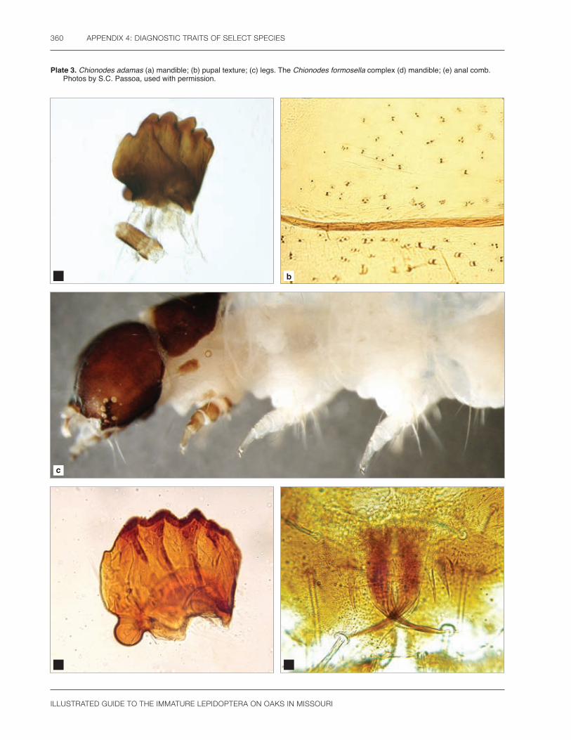
ILLUSTRATED GUIDE TO THE IMMATURE LEPIDOPTERA ON OAKS IN MISSOURI
360 APPENDIX 4: DIAGNOSTIC TRAITS OF SELECT SPECIES
Plate 3. Chionodes adamas (a) mandible; (b) pupal texture; (c) legs. The Chionodes formosella complex (d) mandible; (e) anal comb. Photos by S.C. Passoa, used with permission.
a b
c
d e

ILLUSTRATED GUIDE TO THE IMMATURE LEPIDOPTERA ON OAKS IN MISSOURI
APPENDIX 4: DIAGNOSTIC TRAITS OF SELECT SPECIES 361
Plate 4. Chionodes fuscomaculella (a) hypopharyngeal complex; (b) mandible; (c) crochets; (d) anal comb. Photos by S.C. Passoa, used with permission.
c d
a b

ILLUSTRATED GUIDE TO THE IMMATURE LEPIDOPTERA ON OAKS IN MISSOURI
362 APPENDIX 4: DIAGNOSTIC TRAITS OF SELECT SPECIES
Plate 5. Chionodes pereyra (a) mandible; (b) antennal scape; (c) closeup of pupal punctures; (d) spacing of abdominal pupal punctures. Photos by S.C. Passoa, used with permission.
c d
a b

ILLUSTRATED GUIDE TO THE IMMATURE LEPIDOPTERA ON OAKS IN MISSOURI
APPENDIX 4: DIAGNOSTIC TRAITS OF SELECT SPECIES 363
Plate 6. Pseudotelphusa nr. quercinigracella (a) larval posterior, nonbanded form; (b) larval posterior, banded form; (c) mandible; (d) pupa with the SD1 pinaculum enlarged. Photos by S.C. Passoa, used with permission.
c d
a b

ILLUSTRATED GUIDE TO THE IMMATURE LEPIDOPTERA ON OAKS IN MISSOURI
364 APPENDIX 4: DIAGNOSTIC TRAITS OF SELECT SPECIES
Plate 7. Pubitelphusa latifasciella (a) mandible; (b) crochets; (c) anal comb; (d) pupal texture. Photos by S.C. Passoa, used with permission.
c d
a b

ILLUSTRATED GUIDE TO THE IMMATURE LEPIDOPTERA ON OAKS IN MISSOURI
APPENDIX 4: DIAGNOSTIC TRAITS OF SELECT SPECIES 365
Plate 8. Trypanisma prudens (a) mandible; (b) crochets A3-A6; (c) anal crochets; (d) anal comb; (e) pupal dorsum with lobes and a fringe of setae; (f) pupal spicules surrounded by silk from the cocoon. Photos by S.C. Passoa, used with permission.
a b
c d
e f

ILLUSTRATED GUIDE TO THE IMMATURE LEPIDOPTERA ON OAKS IN MISSOURI
366 APPENDIX 4: DIAGNOSTIC TRAITS OF SELECT SPECIES
Plate 9. Diagnostic traits: (a) two setose knobs on A10 of pupal Coptotriche citrinipennella; (b) dried frass of the Limacodidae showing cuplike depression; (c) pupal mesothoracic spiracle of Oneida lunulalis showing three curved depressions; (d) pupal abdominal spines of Choristoneura rosaceana showing the anterior and posterior rows approximately equal in size; (e) mandible of Hypagyrtis unipunctata showing small lobe-like outer tooth above the two mandibular setae; (f) larval exuvia of H. unipunctata showing a region with clear round microgranules that give the cuticle a glassy appearance; (g) typical noctuid larva showing SD1 on A9 hairlike and much thinner than a normal seta that crosses it in this photo; (h) pupa of Panopoda rufimargo showing the mesothoracic leg reaching the lower eye margin; (i) ventral view of Megalopyge crispata showing the head retracted into the prothorax and the diffuse setae at the end of the abdomen not forming a dense tuft. Photos a-h by S.C. Passoa, i by V.A. Passoa, used with permission.
c d
a b

ILLUSTRATED GUIDE TO THE IMMATURE LEPIDOPTERA ON OAKS IN MISSOURI
APPENDIX 4: DIAGNOSTIC TRAITS OF SELECT SPECIES 367
e f
g
h i
ILLUSTRATED GUIDE TO THE IMMATURE LEPIDOPTERA ON OAKS IN MISSOURI
368
Index
AAcharia stimulea 108, 109, 356Achatia distincta 265Acronicta impleta 256, 357Acronicta (subgenus Lepitoreuma,
increta group) 254, 258Adoneta spinuloides 111Alsophila pometaria 146, 181American dun-bar 261American lappet moth 193Amphipyra pyramidoides 259, 357Ancylis divisana 88, 89Angulose prominent 235Anisota senatoria 199, 205, 207, 232Anisota virginiensis 15, 199, 205, 207,
357Antaeotricha humilis 41, 42, 43, 44, 45Antaeotricha osseella 42, 44, 45Antaeotricha schlaegeri 19, 42, 43,
44, 46Antheraea polyphemus 16, 199, 202,
357Apoda biguttata 113, 356Arogalea cristifasciella 56, 58, 62, 359Automeris io 13, 19, 200, 356
BBanded hairstreak 98Banded tussock moth 289Besma quercivoraria 151, 356Black-fringed leaf tier 48Blinded sphinx 213
CCampaea perlata 28, 154
Canadian melanolophia 166Catocala amica 295Cecrita guttivitta 218, 221, 222, 223,
357Cenopis directana 84, 87Cenopis pettitana 85Charadra deridens 19, 251, 357Chionodes adamas 57, 59, 60, 64, 68,
73, 74, 75, 360Chionodes formosella complex 57, 64,
65, 66, 68, 70, 360Chionodes fuscomaculella 56, 57, 58,
65, 66, 67, 68, 70, 361Chionodes pereyra 57, 64, 65, 67, 68,
69, 362Chokecherry leaf roller 84Choristoneura rosaceana 19, 80, 86,
366Colorful zale 299Common oak moth 297Confused woodgrain 267Copper underwing 259Coptotriche citrinipennella 29, 30, 366Cosmia calami 15, 261Crowned slug 117Curved-lined looper 163
DDark-spotted palthis 291Dasychira obliquata 279, 281Dasychira tephra 280, 281Dichomeris georgiella 57, 59, 61, 358Dichomeris ligulella 27, 57, 59, 60,
61, 358Distinct Quaker 265Dotted Antaeotricha 41
Dotted gray 176Dotted leaf tier 51
EEacles imperialis 209Eastern tent caterpillar 189Ectropis crepuscularia 175, 181, 356Erannis tiliaria 160Erynnis brizo 93, 94, 95, 96Erynnis juvenalis 93, 94Euclea delphinii 114European gypsy moth 285Eutrapela clemataria 19, 149, 156, 356
FFall cankerworm 181Forest tent caterpillar 191Fringed looper 154Frosted slug 113
GGeorgian prominent 229Girlfriend underwing 295Glena cribrataria 176Gold-striped leaf tier 53Green fruitworm 263
HHalysidota tessellaris 280, 289, 357Hollow-spotted plagodis 168Hypagyrtis unipunctata 179, 366Hyperstrotia pervertens 292Hyperstrotia secta 292

ILLUSTRATED GUIDE TO THE IMMATURE LEPIDOPTERA ON OAKS IN MISSOURI
INDEX 369
IIanassa lignicolor 225, 226Imperial moth 209Io moth 200Isa textula 117Isochaetes beutenmuelleri 117, 118
JJeweled tailed slug 121Juvenile duskywing 94
KKent's geometer 171
LLace capped caterpillar 226Lacosoma chiridota 8, 185, 186, 356Lambdina fervidaria 356Lambdina fervidaria athasaria 163Limenitis arthemis astyanax 101Linden looper 160Lithophane antennata 20, 263Lochmaeus manteo 219, 221, 232, 357Lymantria dispar dispar 2, 51, 52, 285
MMachimia tentoriferella 9, 39, 40, 53Macrurocampa marthesia 219, 223Malacosoma americana 188, 189, 192Malacosoma disstria 188, 189, 190,
191, 194Megalopyge crispata 13, 19, 104, 105,
366Meganola phylla 239, 357Melanolophia canadaria 166Menesta melanella 46Monkey slug 130Morning-glory prominent 227Morrisonia confusa 27, 266, 267, 270Mottled prominent 223
NNadata gibbosa 19, 233, 235, 357Nason’s slug 119Natada nasoni 119Nemoria bistriaria 183
OOak beauty 178Oak Besma 151Oak leaf tier 50
Oblique-banded leaf roller 80Oedemasia semirufescens 225, 357Oneida lunulalis 137, 141, 144, 356,
366One-spotted variant 179Orange-striped oakworm 205Orgyia leucostigma 282Orthosia rubescens 269
PPackardia geminata 121Palmer worm 61Palthis angulalis 291Pandemis limitata 92Panopoda rufimargo 301, 357, 366Paonias excaecata 212, 213Paraeschra georgica 229, 233, 235, 357Parasa chloris 109, 122Parasa indetermina 124, 356Peridea angulosa 229, 230, 233, 235Phaeoura quernaria 178, 356Phigalia strigataria 173Phoberia atomaris 1, 279, 297Phobetron pithecium 19, 108, 130Phyllodesma americana 188, 193Phyllonorycter fitchella 34Pink-striped oakworm 207Plagodis alcoolaria 168, 356Pococera expandens 27, 136, 139, 141,
356Polyphemus moth 202Prolimacodes badia 109, 125, 356Pseudotelphusa complex 15, 71, 363Psilocorsis cryptolechiella 48, 52Psilocorsis quercicella 47, 49, 50, 52Psilocorsis reflexella 49, 50, 51Pubitelphusa latifasciella 59, 73, 75,
364Purple-crested slug 111Purplish-brown looper 156Puss caterpillar 105
QQuasisalebria fructetella 141, 142, 143
RRectiostoma xanthobasis 47, 50Red-fringed emerald 183Red-lined panopoda 301Red-spotted purple 101Red-washed prominent 225Ruby Quaker 269
SSaddleback caterpillar 109Saddleback looper 175Saddled prominent 218Salebriaria engeli 140, 142, 143Salebriaria tenebrosella 140, 141, 142,
143Satyrium calanus 97, 98Schizura ipomaeae 227, 357Schlaeger’s fruitworm 44Selenia kentaria 171, 356Skiff moth 125Sleepy duskywing 93Smaller Parasa 122Small phigalia 173Spiny oak slug 114Spun glass slug 118Stinging rose caterpillar 124Streaked tussock moth 279Stripe-backed moth 62Striped oak webworm 136Symmerista spp. 27, 218, 221, 223,
230, 357
TTephra tussock moth 281The laugher 251Three-lined leaf roller 88Tolype velleda 188, 194, 195Tortricidia flexuosa 127Trypanisma prudens 74, 75, 365
VVariable oakleaf caterpillar 221
WWhite-banded leaf roller 73White-dotted prominent 233White-marked tussock moth 282
YYellow-marked dagger 256Yellow-vested moth 47
ZZale minerea 299, 301

THIS PAGE INTENTIONALLY LEFT BLANK

The Forest Health Technology Enterprise Team (FHTET) was created in 1995 by the Deputy Chief for State and Private Forestry, USDA Forest Service, to develop and deliver technologies to protect and improve the health of American forests.
FHTET became Forest Health Assessment and Applied Sciences Team (FHAAST) in 2016. This book was published by FHAAST as part of the technology transfer series.
http://www.fs.fed.us/foresthealth/technology/
In accordance with Federal civil rights law and U.S. Department of Agriculture (USDA) civil rights regulations and policies, the USDA, its Agencies, offices, and employees, and institutions participating in or administering USDA programs are prohibited from discriminating based on race, color, national origin, religion, sex, gender identity (including gender expression), sexual orientation, disability, age, marital status, family/parental status, income derived from a public assistance program, political beliefs, or reprisal or retaliation for prior civil rights activity, in any program or activity conducted or funded by USDA (not all bases apply to all programs). Remedies and complaint filing deadlines vary by program or incident.
Persons with disabilities who require alternative means of communication for program information (e.g., Braille, large print, audiotape, American Sign Language, etc.) should contact the responsible Agency or USDA’s TARGET Center at (202) 720-2600 (voice and TTY) or contact USDA through the Federal Relay Service at (800) 877-8339. Additionally, program information may be made available in languages other than English.
To file a program discrimination complaint, complete the USDA Program Discrimination Complaint Form, AD-3027, found online at http://www.ascr.usda.gov/complaint_filing_cust.html and at any USDA office or write a letter addressed to USDA and provide in the letter all of the information requested in the form. To request a copy of the complaint form, call (866) 632-9992. Submit your completed form or letter to USDA by: (1) mail: U.S. Department of Agriculture, Office of the Assistant Secretary for Civil Rights, 1400 Independence Avenue, SW, Washington, D.C. 20250-9410; (2) fax: (202) 690-7442; or (3) email: [email protected].
USDA is an equal opportunity provider, employer, and lender.

THIS PAGE INTENTIONALLY LEFT BLANK