IL-17A-dependent giant cells in human tuberculosis granulomas
224
THESE en vue d’obtenir le grade de Docteur de l’Ecole Normale Supérieure de Lyon - Université de Lyon Spécialité: Sciences de la Vie – Immunologie Laboratoire CNRS UMR5239 « Dendritic cells & immune plasticity » Ecole doctorale de Biologie Moléculaire Intégrative et Cellulaire Présentée et soutenue publiquement le 24/09/2012 par Mohamad Bachar ISMAIL IL-17A-dependent giant cells in human tuberculosis granulomas: mechanisms of formation, survival and functions Directrice de Thèse Pr Christine DELPRAT Jury Dr Olivier NEYROLLES Rapporteur Dr Geneviève MILON Rapporteur Dr Nathalie WINTER Examinateur Dr Glaucia PARANHOS-BACCALA Examinateur Dr Pierre JURDIC Examinateur et président Pr Christine DELPRAT Directrice de thèse
-
Upload
khangminh22 -
Category
Documents
-
view
1 -
download
0
Transcript of IL-17A-dependent giant cells in human tuberculosis granulomas

0
THESE
en vue d’obtenir le grade de
Docteur de l’Ecole Normale Supérieure de Lyon - Université de Lyon
Spécialité: Sciences de la Vie – Immunologie
Laboratoire CNRS UMR5239 « Dendritic cells & immune plasticity »
Ecole doctorale de Biologie Moléculaire Intégrative et Cellulaire
Présentée et soutenue publiquement le 24/09/2012 par
Mohamad Bachar ISMAIL
IL-17A-dependent giant cells in human tuberculosis granulomas: mechanisms of formation, survival and functions
Directrice de Thèse Pr Christine DELPRAT
Jury
Dr Olivier NEYROLLES Rapporteur
Dr Geneviève MILON Rapporteur
Dr Nathalie WINTER Examinateur
Dr Glaucia PARANHOS-BACCALA Examinateur
Dr Pierre JURDIC Examinateur et président
Pr Christine DELPRAT Directrice de thèse

1
Acknowledgements
In the first place, I would like to express my gratitude and my sincere thanks to my research supervisor, Pr Christine Delprat. Thank you for everything you taught me. Thank you for your understanding, patience, expertise, extraordinary dynamism and your passion for immunology. Thank you also for your advices and useful remarks which guided my work. I am thankful that you used a part of your precious time to read this thesis and give your critical comments about it.
Very special thanks go out to my team members: Alexandre and Carine which help me from the beginning of my project, Nathalie and Giulia for their continuous assistance and Karène for her great advices and fruitful scientific discussions as well as for her assistance with her vast knowledge and skills in many areas. It was a pleasure to work with you and it is a pleasure to convey my gratitude to you in my humble acknowledgment. Thanks also for Emilien and Hélène who have recently joined the team.
I would like to thank professors Françoise Berger, Abdellatif Tazi, Maurizio Aricò and
Jan-Inge Henter for providing us with biopsies from patients with Tuberculosis, pLCH and LCH. The technical assistance of Béatrice Bancel is also gratefully acknowledged.
I gratefully acknowledge Dr Nathalie Winter and her team members Emilie and Florence for CFU studies and Dr Daniel Hanau for electron microscopy.
Many thanks go in particular to the members of my thesis committee: Thanks for Dr Olivier Neyrolles and Dr Geneviève Milon who evaluated my work and provided beneficial, useful and constructive comments. Big thanks also to Dr Pierre Jurdic, Dr Nathalie Winter and Dr Glaucia Paranhos-Baccalà who in the midst of all their activity, they accepted to be members of the reading committee.
At the personal level, I wish to express my gratitude, my deep acknowledgment and my sincere thanks to my family. Thanks for my parents, Khaled and Samar, who have always encouraged me to continue my education and helped me to achieve this goal. Thank you for your inspiration, support and sacrifices. Thanks also to my brothers Baraa and Baligh and my sister Elee who supported me during this time.
Thank you my little angel Samar. You are a very special gift that illuminate my road, encourage me and give my life a great and fantastic meaning. With you it was impossible to work at home!!
Finally, I will be eternally grateful to my wife, Rawaa, for her patience and support. You sustained me in all my difficult moments and helped me anytime I needed it. Thank you for your understanding, endurance and encouragement. This work would not have been possible without your permanent assistance and enthusiasm. To you go my most grateful acknowledgments.

2
Sommaire 1 Part I: Immune response to Mycobacteria with focus on Tuberculosis .......................................... 7
1.1 Main species and general characteristics of Mycobacteria .................................................... 8
1.1.1 Main pathogenic and opportunistic Mycobacterium species ......................................... 8
1.1.2 M. tuberculosis consists of six lineages with differential geographic distribution .......... 9
1.1.3 General microbiologic properties of Mycobacteria ...................................................... 11
1.1.4 M. tuberculosis activates a specific latent genetic program during hypoxic conditions 11
1.1.5 Intracellular growth of dormant M. tuberculosis bacilli relies on lipid catabolism ....... 12
1.1.6 Mycobacteria are characterized by an unusual cell wall structure ............................... 13
1.2 Tuberculosis disease .............................................................................................................. 14
1.2.1 The natural history of tuberculosis ............................................................................... 14
1.2.2 M. tuberculosis and HIV: dangerous liaison for a lethal combination .......................... 16
1.2.3 Tuberculosis histopathology ......................................................................................... 19
1.2.4 Tuberculosis symptoms and diagnosis .......................................................................... 19
1.2.5 Tuberculosis treatment ................................................................................................. 21
1.2.6 Tuberculosis vaccination: current challenges and future strategies ............................. 22
1.3 Innate response can be not sufficient against M. tuberculosis ............................................. 26
1.3.1 Host innate receptors involved in mycobacterial recognition. ..................................... 26
1.3.2 Mycobacterium-receptor interactions determine infection outcomes ........................ 27
1.3.3 Mycobacteria resist phagocytosis-induced destruction ............................................... 27
1.3.4 Innate cells involved in anti-mycobacterial responses .................................................. 33
1.3.5 Processing of intracellular mycobacterial antigens ....................................................... 38
1.4 The anti-mycobacterial adaptive immunity relies on a granulomatous response ................ 41
1.4.1 Structure and cellular composition of granulomas ....................................................... 41
1.4.2 Granuloma myeloid cells are long-lived and express destructive enzymes .................. 42
1.4.3 Different phases of granuloma formation ..................................................................... 46
1.4.4 Pivotal regulators of the granulomatous immune response in tuberculosis ................ 50
1.5 Origins and key mediators of giant cell formation ................................................................ 59
1.5.1 Physiological and pathological giant cells ..................................................................... 59
1.5.2 Osteoclast: the bone-resorbing giant cell ..................................................................... 59
1.5.3 The myeloid giant cells formed in chronic inflammation .............................................. 61
1.5.4 IL-17A characteristics and biological functions ............................................................. 62

3
2 Part II: RESULTS ............................................................................................................................. 71
2.1 Manuscript 1: BFL1, a new biomarker of long lifespan and chemoresistant human dendritic cells: application to Langerhans cell histiocytosis treatment ........................................................... 73
2.2 Manuscrit 2: IL-17A-dependent maintenance of human tuberculosis granuloma is mediated by BFL1, CCL20 and CCL2 ................................................................................................................. 107
2.3 Manuscript 3: (in preparation) Giant Myeloid Inflammatory Cell: a new anti-Mycobacterium effector of the human immune system .......................................................................................... 135
2.4 Manuscrit 4: (in preparation) IL-17A+MMP-12+CSTD+ DC accumulation leads to bronchoepithelium destruction: relevance in pulmonary LCH ....................................................... 159
3 Part III: DISCUSSION .................................................................................................................... 173
3.1 Differential physiopathological roles of granulomas among species & diseases ............... 173
3.1.1 Evolution of the granuloma role in vertebrates .......................................................... 173
3.1.2 Contributions and limitations of the murine models to understand granulomatogenesis .................................................................................................................... 175
3.1.3 Emergence of the role of granulomas in the chronic control of cancer cells ............. 176
3.2 IL-17A-induced genetic program promotes granulomatogenesis ...................................... 178
3.2.1 Is IL-17A a constitutive molecular player in granulomatogenesis? ............................. 178
3.2.2 The role of IL-17A-induced CCL20 and CCL2 in granulomatogenesis .......................... 179
3.2.3 The role of IL-17A-induced CCL20 and CCL2 in cell-cell fusion ................................... 181
3.2.4 The role of IL-17A-induced BFL1 in myeloid cell maintenance ................................... 183
3.2.5 Multiple stimuli for NF-κB -dependent activation of BFL1 in granuloma, in vivo ....... 184
3.3 The IL-17A-induced Giant Myeloid Inflammatory Cell (GMIC) ............................................ 185
3.3.1 GMIC: a novel multinucleated cell type of the immune system characterized by MMP12 & CTSD co-expression .................................................................................................... 185
3.3.2 Comparison of GMICs with the giant cells of LCH and TB granulomas ....................... 187
3.3.3 Microbicidal role of GMIC against Mycobacteria, in vitro ........................................... 189
3.3.4 The destructive role of GMIC against human broncho-epithelial cells ....................... 191
3.3.5 Regulation of IL-17A-mediated responses .................................................................. 193
4 Part IV: CONCLUSIONS & PERSPECTIVES ..................................................................................... 197
5 REFERENCES ................................................................................................................................ 201

4
ABBREVIATIONS
A
APC: Antigen-presenting cell
B
BCG: Bacille de Calmette et Guérin
BCL-2: B-cell lymphoma 2
C
CCL: Chemokine (C-C motif) ligand
CCR: Chemokine (C-C motif) receptor
CR: Complement receptor
CTS: Cathepsin
D
DC: Dendritic cell
DC-SIGN: DC-specific intracellular adhesion molecule grabbing non integrin
DC-STAMP: DC -specific transmembrane protein
E
EEA1: Early endosomal antigen
ER: Endoplasmic reticulum
ESAT-6: Early secreted antigenic target-6
F
FasL: Fas ligand
FBGC: Foreign body giant cell
G
GM-CSF: granulocyte macrophage colony-stimulating factor
GMIC: giant myeloid inflammatory cell
H
HIV: Human immunodeficiency virus
I
ICL: isocitrate lyase

IFN-γ: interferon-gamma
IL: interleukin
iNKT: Invariant natural killer T
iNOS: Inducible nitric oxide synthase
L
LAM: Lipoarabinomannan
LCH: Langerhans cell histiocytosis
LSP: large sequence polymorphism
LXA4: lipoxin A4
M
M.: Mycobacterium
ManLAM: Mannosylated lipoarabinomannan
M-CSF: Macrophage colony-stimulating factor
MDR-TB: Multidrug-resistant tuberculosis
MGC: Multinucleated giant cell
MHC: Major histocompatibility complex
MMP: Matrix metalloproteinase
MP: Macrophage
Mtb: Mycobacterium tuberculosis
N
NADPH ox: nicotinamide adenine dinucleotide phosphate-oxidase
NEMO: NF-κB essential modulator
NF-κB: nuclear factor-kappa B
NK: Natural Killer
NLR: Nucleotide–binding oligomerization domain (NOD)-like receptor
NO: nitric oxide
NOD: Nucleotide–binding oligomerization domain
O
OC: osteoclast

6
P
PG: Peptidoglycans
PGE2: prostaglandin E2
PI3P: Phosphatidylinositol 3-phosphate
PIM: Phosphatidyl inositol mannosides
PMN: polymorphonuclear neutrophil
PRR: Pathogen receptor recognition
R
RANKL: Receptor activator of nuclear factor kappa-B ligand
RD1: Region of deletion 1
RNI: Reactive nitrogen intermediates
ROI: Reactive oxygen intermediates
T
TB: Tuberculosis
TCR: T cell receptor
TGF-β: Transforming growth factor beta
Th: T helper lymphocyte
TLR: Toll-like receptor
TNF-α: Tumor necrosis factor-alpha
TRAF: TNF receptor-associated factor
TRAIL: TNF-related apoptosis-inducing ligand
TST: Tuberculin skin test
V
V-ATPase: Vacuolar ATPase
W
WHO: World health organization
WT: wild-type
X
XDR-TB: Extensive drug resistant tuberculosis

7
1 Part I: Immune response to Mycobacteria with focus on Tuberculosis
Mycobacteria belong to the Mycobacterium genus, the single genus within the family of
Mycobacteriaceae in the Corynebacterineae suborder of the Actinomycetales order. They fall
into two main groups: slow and fast growers. Slow-growers include several pathogens (strict
and opportunistic) while most of the fast growers are non-pathogenic Mycobacteria. Among
strict pathogens, two species, Mycobacterium tuberculosis (Mtb) and Mycobacterium leprae,
cause two of the world's oldest diseases: tuberculosis (TB) and leprosy, respectively.
TB is mainly a pulmonary disease characterized by the formation of small inflammatory
nodules formed by a collection of immune cells in lungs. The formation of these structures
requires cell recruitment and clustering and is driven by several cytokines and chemokines.
TB granuloma consists of a myeloid cell core surrounded by T lymphocytes. A cellular
hallmark of granulomas is the presence of multinucleated giant cells (MGCs) formed by the
fusion of several myeloid cells. In humans, mechanisms of granuloma and MGC formation
and their functions are largely unclear.
Recent data showed that the cytokine IL-17A is involved in the formation of TB
granulomas in mice. Moreover, our group discovered that this cytokine induces the fusion of
human myeloid dendritic cells (DCs) into MGCs in vitro. Based on these data, we suggested a
potential role of IL-17A in the formation of human giant cell-associated granulomas.
During this thesis, we investigated the molecular mechanisms which regulate the
survival, clustering and MGC formation in the IL-17A-dependent pathway. We also searched
if similar mechanisms are involved in the formation and maintenance of granulomas in TB as
well as in another giant cell-associated granulomatous disease called pulmonary Langerhans
cell histiocytosis (pLCH). Then, we investigated the phenotype, the immune functions and the
anti-mycobacterial abilities of the IL-17A-induced MGCs that we called GMICs (giant
myeloid inflammatory cells).
To introduce this work in its scientific context, we first present Mycobacteria and their
characteristics with a focus on TB disease. Then, we describe the innate immune response and
its main effectors against Mycobacteria and show how pathogenic strains can evade this
response. Afterwards, we present the adaptive granulomatous response to Mycobacteria and
we document essential host factors required for granuloma formation and maintenance.
Finally, we describe the different types of myeloid giant cells with a focus on the IL-17A-
dependent pathway as well as the characteristics and biological functions of IL-17A.

8
1.1 Main species and general characteristics of Mycobacteria
1.1.1 Main pathogenic and opportunistic Mycobacterium species
Mycobacterium is a highly diverse genus [1] in which we distinguish the following
species:
-The Mtb complex: this complex is formed by species closely related to Mtb at the genetic
level, but with distinct host tropisms. Mtb is the main etiologic agent of most cases of TB in
humans who are reservoir of this pathogen. In addition to Mtb, Mtb complex contains M.
bovis, M. africanum, M. microti, M. canettii, M. caprae and M. pinnipedi [2] [3]. M. Bovis
causes TB in cattle and humans which serve as reservoirs. An attenuated strain of M. Bovis is
called Bacille de Calmette et Guérin (BCG) and is used as a TB vaccine. M. africanum
specifically infects humans and is a significant cause of TB in the western countries of Africa.
M. canettii is a rare strain of the Mtb complex and was isolated from TB patients mostly in the
horn of Africa. M. microti specifically causes disease in small rodents; M. caprae infects
goats, while M. pinnipedii primarily affects seals.
-M. leprae: a slow growing intracellular pathogen and the etiologic agent of leprosy, a
chronic but curable human disease affecting the skin, peripheral nerves, eyes and mucosa of
the upper respiratory tract. Leprosy is a granulomatous inflammation driven by mycobacterial
antigens that activate a destructive immune response [4]. Even though its prevalence has
decreased dramatically, 228 474 new cases of leprosy were detected worldwide in 2010, thus
indicating active M. leprae transmission. Human beings are the principal reservoir of
infection. However, the exact mechanism of transmission is not fully clear although the
aerosol spread of nasal secretions is considered as the predominant transmission mode [4].
-M. ulcerans: behind Mtb and M. leprae, M. ulcerans is the third most common
mycobacterial pathogen of humans. M. ulcerans is a slow-growing pathogen and the causative
organism of Buruli ulcer disease characterized by devastating necrotic lesions called ulcers
and resulting in extensive destruction of the skin and soft tissues [5] [6]. Infection by M.
ulcerans seems to occur in or near stagnant water and slow flowing rivers in humid tropical
regions [5] [6]. The mode of transmission in humans is unknown, but it was suggested that
aquatic insects and salt marsh mosquitoes may be involved in transmission [5] [6].

9
-M. avium complex: This complex consists of a heterogeneous group of Mycobacteria
including, among others, M. avium and M. intracellulare. M avium is further divided into
various subspecies including M. avium avium and M. avium paratuberculosis. All these
species are slowly-growing environmental Mycobacteria [7] considered as opportunistic
microorganisms that mostly induce diseases in immuno-compromised patients. In humans, M.
avium infection is widely associated with disseminated infections immuno-compromised
human beings. M. intracellulare is more common among immuno-competent individuals. M
avium paratuberculosis is a well-known causative agent of paratuberculosis (Johne's disease),
a chronic granulomatous enteric disease of ruminants. In addition an etiologic link between
this Mycobacterium and Crohn’s disease in humans was proposed but remains controversial
and to be proven.
-M. marinum: an environmental/atypical Mycobacterium which was first discovered in salt
water fish. M. marinum was lately identified as an opportunistic pathogen in humans in whom
infection may cause localized nodular skin lesions called “swimming pool granuloma” or
“fish tank granuloma”.
1.1.2 M. tuberculosis consists of six lineages with differential geographic
distribution
Traditionally, it was thought that the Mtb complex is a highly homogeneous clonal
population which displays limited genetic variability [8] [9]. However, recent advances in
mycobacterial comparative genomics revealed that substantial genetic variation exists at the
whole-genome level of this complex, suggesting that it is more heterogeneous than
appreciated initially [10]. Using large sequence polymorphisms (LSPs) genetic markers,
Gagneux and colleagues found that Mtb consists of six main strain lineages, designated
Lineage 1 through Lineage 6, two of which were initially designated M. africanum (Fig.1,
Top) [10]. They can be further divided into “ancient” and “modern” lineages based on the
presence or absence of a genomic deletion known as TbD1 [11]. Importantly, the six
identified Mtb strain lineages show biogeographic specificities as they are associated with
particular geographic regions and human populations (Fig.1, bottom) [10].

10
Figure 1: Global phylogenetic classification and phylogeographic distribution of Mtb. Adapted from ref [10]. Top: The global population structure of Mtb consists of six main strain lineages (1 to 6). Each lineage is symbolized by a colored circle. Numbers in rectangles refer to LSP markers. Pks: polyketide synthase gene. (A): Ancient lineage. (M) Modern lineage. Bottom: Global phylogeography of Mtb. Colored circles indicate the dominant lineage in country.

11
1.1.3 General microbiologic properties of Mycobacteria
Mycobacteria are thin and long bacilli that measure about 1 to 10 µm. A distinctive
characteristic of Mycobacterium species is the property of acid-fastness as they resist
decolorization with a mixture of acid and alcohol due to their specific cell wall composition.
However, they are routinely stained with the Ziehl-Neelsen colorization, a basic carbofuchsin
staining method. Mycobacterium slow-growers need prolonged incubation periods. For
example, Mtb divides every 15–20 hours, which is a particularly slow generation time
compared to other bacteria. Mycobacterial growth requires selective solid or liquid culture
media including the commonly used Middlebrook media. Cultures of slow-growing
Mycobacteria including Mtb require four to eight weeks of incubation to grow on solid media
while liquid media allow more rapid mycobacterial growth. In vitro-grown Mtb colonies
appear as small, friable, rough and white to light-yellow colored colonies (Fig. 2) [12].
Figure 2: Mtb Colonies on a solid culture medium. Centers for Disease Control and Prevention (CDC).
1.1.4 M. tuberculosis activates a specific latent genetic program during hypoxic
conditions
In response to environmental stresses such as nutrient deprivation and hypoxia inside
host cells, Mtb bacilli precede to a dormant state defined as a stable but reversible non-
replicating state [13]. In this dormancy phase, bacilli persist with a reduced metabolic activity
that facilitates their survival under hypoxic conditions [14]. However, dormant bacilli keep
their ability to resume growth when conditions become favorable [15]. Mtb ability to
terminate replication and shut down its own central metabolism renders these bacilli
extremely resistant to both host defense and drugs [14].
Voskuil et al. revealed that hypoxic microenvironment activates a specific genetic
program leading to latent Mtb survival [16]. They showed that inhibition of respiration by NO

12
(nitric oxide) production and O2 limitation, constrains Mtb replication rates. This non
replicating state is driven by a 48-gene regulon under the control of a transcription factor
named DosR [16]. DosR adapts the organism for survival during prolonged periods of in vitro
dormancy. It is also required for maintaining energy levels and redox balance for Mtb survival
during anaerobic dormancy and ensures rapid Mtb recovery and optimal transition from an
anaerobic or nitric oxide-induced nonrespiring state to aerobic growth [17].
1.1.5 Intracellular growth of dormant M. tuberculosis bacilli relies on lipid
catabolism
Several lines of evidence indicate that, in hypoxic conditions, Mycobacteria switch to
lipid catabolism to ensure their survival. Genomic studies and transcriptomic analysis of
entirely sequenced Mtb genome show that this bacillus displays a wide range of diverse
lipophilic molecules involved in lipogenesis and lipolysis [18]. For example, there are 250
distinct enzymes related to fatty acid metabolism in Mtb compared with only 50 in
Escherichia coli [18]. Among them, several enzymes could catalyze the first step in fatty acid
degradation providing thus different metabolites and fuel for the bacteria [18]. Functional
studies of some of these enzymes have revealed that they play an important role in the
persistence of Mtb in their hosts. For example, isocitrate lyase (ICL), an enzyme induced by
oxygen limitation and essential for the metabolism of fatty acids, has been reported as
required for persistence and virulence of Mtb [19]. ICL is a key component of the glyoxylate
shunt, by which microorganisms bypass the tricarboxylic acid cycle to incorporate carbon
from acetate and fatty acids into carbohydrates. ICL regulates Mtb long-term survival in the
murine lung while its deletion attenuates Mtb virulence as ICL-deficient mutant survived in
resting but not in activated macrophages (MPs) contrary to the Wild-type (WT) strain [19].
Mtb persists in a non-replicating state inside adipocytes and accumulates lipid droplets
inside these cells [20]. This suggests that the adipose tissue might constitute one important
cellular reservoir in which Mtb could persist in a dormancy-like state. Consistent with these
findings are data showing that Mtb metabolize host-derived cholesterol and that disruption of
the mce4 gene, encoding a cholesterol transporter, results in the failure of Mtb to maintain
chronic infection in mice [21]. A recent report showed also that Mtb residing within the
phagosomes of hypoxic human MPs utilizes, accumulates and stores host triacylglycerol in
the form of intracellular lipid droplets [22]. In these conditions, Mtb replication is severely
inhibited and bacilli acquire a dormancy-like phenotype [22]. In mice, BCG infection induces
time- and dose-dependent lipid body formation in infected MPs [23]. More recently, a study

13
revealed that Mtb, namely through its oxygenated mycolic acids, triggered the differentiation
of human MPs into lipid laden foamy cells in which Mycobacteria switched to dormant non-
replicative bacilli and accumulate host cell lipids [24].
Taken together, these data suggest that, during dormancy, Mtb intracellular growth relies on
the lipid catabolism as the main carbon source. This ensures not only survival in hypoxic
conditions, but also its adaptation to carbohydrate starvation.
1.1.6 Mycobacteria are characterized by an unusual cell wall structure
Mycobacteria have a specific cell wall structure which distinguishes species of the
Mycobacterium genus from other prokaryotes [25]. this unusual cell wall structure is
responsible of the acid fastness property of Mycobacteria and is related to mycobacterial
resistance to drying, alkali, many chemical disinfectants and therapeutic agents [26]. It
consists of three major parts: the plasma membrane, the cell wall core and the capsule-like
outermost layer (Fig. 3) [25]. The cell wall core is close to the plasma membrane and consists
of a mycolyl-arabinogalactane-peptidoglycan (mAGP) complex. This core unit is an insoluble
complex formed by peptidoglycans (PG) covalently attached to arabinogalactans (AG) which
in turn covalently bound mycolic acids [25] [27]. These latter, for which the Mycobacteria are
named, constitute ~ 60% of the bacillus total weight. They are very long chain (C60-90) α-
branched, β -hydroxy fatty acids and are considered as the most distinctive feature of the
mycobacterial cell wall.
The capsule-like outermost layer of the cell wall is composed of a variety of free
lipids, lipoglycans, and proteins including porins (pore-forming proteins) [25]. Lipoglycans,
including lipoarabinomannan (LAM) and phosphatidylinositol mannosides (PIMs), are
prevalent components of the mycobacterial cell wall. In contrast to the insoluble cell wall
core, free lipids, proteins, LAM and PIMs are solubilized and form signaling,
immunologically active effector molecules which play important biological functions in the
pathogenesis of Mtb [25] [27].

14
Figure 3: Schematic representation of the unique wax-rich cell wall of Mycobacteria. Adapted from ref [28]. Close to the plasma membrane is the (mAGP) complex. It forms the core unit of the mycobacterial cell wall and consists of PG connected to AG which are covalently linked to mycolic acids. The upper segment is the capsule-like outermost layer which includes a variety of free lipids. The mycobacterial cell wall contains many signaling and effector molecules including LAM and PIM which support mycobacterial survival and virulence. Porins cross the cell wall to allow molecule diffusion into Mycobacteria.
1.2 Tuberculosis disease
1.2.1 The natural history of tuberculosis
In 1882, the German scientist Robert Koch identified Mtb as the etiologic agent of
human TB disease. In 1998, the complete genome sequence of the well characterized H37Rv
strain of Mtb was determined. It comprises 4,411,529 base pairs, contains approximately 4047
predicted genes, and has a very high guanine + cytosine (G+C) content [18]. Although Mtb
was identified more than 130 years ago, it remains currently one of the most pernicious of
human pathogens, and TB disease is more prevalent in the world today than at any other time
in human history. In 2010, the world health organization (WHO) reported an annual burden of
~ 8.8 million cases of disease resulting in ~1.4 million deaths which mostly occurs in poor
world countries [29].
Mtb infection occurs by an efficient person-to-person transmission mode and the main
infection route is the respiratory tract (Fig. 4). Bacilli are transmitted via airborne droplets

15
expelled into the atmosphere by patients with active pulmonary TB most commonly during
coughing or sneezing. These droplets can be later inhaled by healthy individuals in who they
proceed distally to the lung where Mtb can induce the infection. However, exposure to Mtb
bacilli results in a wide spectrum of outcomes. It is estimated that only 30% of exposed
individuals get infected [30]. This suggests that innate immune responses in exposed but not
infected persons may rapidly clear the inhaled bacilli, although this has not been formally
proven. Among infected individuals, a minority (around 10% - most commonly infants or
children of young age) progress to primary active TB disease with clinical symptoms within
1-2 years following infection. In contrast, the majority (90%) of infected individuals remains
asymptomatic and free of tissue damage. However, they carry viable but dormant bacilli
which can persist for decades, thereby developing latent TB infection [14] [30] [31].
Individuals with latent TB don’t transmit Mtb bacilli to other persons but are the largest
reservoir for potential subsequent transmission [30]. They show an equilibrium state in which
they develop an effective acquired immune response that controls but not entirely eradicate
Mtb bacilli. Unfortunately, they have a subsequent 10% risk of reactivation and development
of post-primary (secondary) TB throughout their life time [30] [31].
TB is a multifactorial disorder: in addition to pathogen, several environmental and
host risk factors are associated with Mtb reactivation and disease development. The most
potent risk factor is HIV but other factors including anti-TNF therapy, host genetic factors,
malnutrition, smoking, alcohol and bad life conditions are also involved in this process [31]
[32]. TB is mainly a pulmonary disease: in 85% of immuno-competent infected individuals
disease preferentially occur in lungs [33]. However, Mtb can also spread to any part of the
body and TB can develop in different organs resulting in extrapulmonary TB [34].
extrapulmonary TB develop most commonly in lymph nodes (tuberculous Lymphadenitis),
pleura, bones and joints (with a form affecting the spine, called Pott’s disease) and central
nervous system (the most common presentation is tuberculous meningitis affecting meninges)
[34]. Other sites include pericardium (tuberculous pericarditis), the gastrointestinal and
genitourinary tracts [34].

16
Figure 4: Natural history and outcome spectrum of TB.
TB is a contagious disease caused by Mtb infection. Bacilli are acquired by the inhalation of airborne droplets containing Mtb. Infection occurs in ~30% of exposed individuals. Only ~10% of infected individuals develop active disease: ~5% at primoinfection and ~5% following reactivation after latent infection. TB disease mainly affects lungs, but can also develop in other organs (extrapulmonary TB). If treated, the majority of patients with drug-susceptible-TB are cured while a high mortality occurs in untreated patients. Several risk factors increase the risk of TB development (orange box). Smokers, alcoholics and individuals having weak immune systems, genetic impairment, unfavorable living conditions or compromised by some diseases, malnutrition, or medical intervention show a higher risk of Mtb reactivation and TB development.
1.2.2 M. tuberculosis and HIV: dangerous liaison for a lethal combination
HIV-infected persons are more likely to develop active TB, and clinical studies
suggest that HIV leads to an increased risk of developing TB shortly after HIV infection.
Moreover, while healthy persons are mostly resistant to M. avium complex infections, patients
with AIDS (acquired immunodeficiency syndrome) are susceptible to M. avium complex and
develop disseminated disease upon infection with these opportunistic species. Progression to
active TB can in turn increase HIV replication and accelerate the progression to AIDS [35].
Additionally, while TB is restricted to the lungs in most cases, Mtb/HIV co-infected persons

17
easily develop systemic and more quickly lethal TB [36]. Indeed, HIV+ TB, opposed to HIV-
TB patients, include a higher proportion of cases with extra-pulmonary or disseminated
disease involving multiple organs. Referring to WHO, people immuno-compromised by HIV
and infected with Mtb are up to 20-37 times more likely to develop active TB in their life-
time than people who are HIV–. Recent WHO report revealed that in the 8.8 million new
persons infected with Mtb in 2010, 12.5% were co-infected by HIV [29]. In addition, of the
1.4 million people who died of TB in 2010, 25% were co-infected by HIV [29]. Both diseases
are associated to poverty and unfavorable low-social life conditions and their highest
prevalence are found in the same regions of the world (Fig. 5).
HIV infects CD4+ cells which are not only T cells, but also alveolar MPs, the primary
target of Mtb [37] [38]. HIV and Mtb can therefore share the same host cell. Several studies
showed that these two pathogens potentiate one another to increase and exacerbates its risk.
Several hypotheses were proposed to explain how this occurs and a recent paper summarizes
these hypotheses in four main points: (i) HIV replication is increased at sites of Mtb infection,
leading to increased pathology, (ii) HIV induces primary or reactivated TB through killing of
CD4+ T cells (mandatory for TB control), (iii) HIV manipulation of MP function prevents
Mtb killing, and (iv) HIV induces functional changes in Mtb-specific T cells that decrease
their ability to contain Mtb [39].
Overall, Mtb-induced TB and HIV-induced AIDS form together a lethal combination
in which each speeding the other's progress.

18
Figure 5 : Estimations of: TB incidence rates in 2010 (top), HIV prevalence rate in individuals aged 15-49 in 2009 (middle) and HIV prevalence in new TB cases in 2010 (bottom). Note that both diseases are highly prevalent in the same countries of the World. Sources: Top and bottom: WHO report, global tuberculosis control, 2011. Middle: UNAIDS, report on the global AIDS epidemic, 2010.

19
1.2.3 Tuberculosis histopathology
Typically, Mtb infection induces a specific type of inflammation characterized by the
formation of granulomas, small focal inflammatory nodules formed by a collection of
immune cells. In humans, TB granulomas are highly organized with specific architectural
structures forming an interface between bacteria, host immune response and host tissues (See
1.4.1).
Many of TB granulomas persist as solid structures showing an equilibrium state which
is generally successful in containing, although not eliminating, Mtb bacilli. However,
granuloma can proceed either to localized sterilization of the infection and mineralization of
the lesion or to localized central necrosis [40]. Necrosis induces the extracellular release of
Mtb bacilli and results in characteristic necrotic centers called caseum, essentially formed by
host cell debris. Granulomas with caseating material are known as caseous granulomas and
their development characterize the progression toward disease [41]. Caseating material
greatly supports the extracellular growth and replication of Mtb. It has a yellow-white color
with an initial solid cheesy texture. In advanced stages, it liquefies and cavitates the lung
causing ruptures into the lung airway and release of infectious Mtb bacilli. In patients with
active TB, all granuloma forms (solid, necrotic and caseous) can coexist.
1.2.4 Tuberculosis symptoms and diagnosis
(Fig. 6, left)
TB patients may show several symptoms which are often vague and non specific
including persistent cough (pulmonary TB), anorexia, weight loss, chest pain, hemoptysis,
and night sweats. In extra-pulmonary TB, symptoms can vary according to the affected
organ(s). However, symptoms might be absent in some TB patients especially in the early
stages of disease [32].
Most common tests used for TB diagnosis include cultures, sputum smear microscopy,
nucleic acid amplification tests, radiology, the tuberculin skin test (TST) and IFN-γ release
assays (IGRAs) [42]. In contrast to other methods which detect active TB cases, TST and
IGRAs are used for the immuno-diagnosis of latent TB by identifying the existence of an
adaptive immune response to mycobacterial antigens [42].
TB can be difficult to diagnose, especially in children, HIV-infected individuals and
patients developing extrapulmonary forms [32]. Moreover, different diagnostic tests cited
above provide information only on the state of the disease in a precise single time point
(active or latent TB). Therefore, a major obstacle to TB control is the lack of reliable

20
biomarkers for different stages of infection [43]. Such biomarkers are needed to help to diag-
nose TB, to provide correlates of risk of TB and correlates of protection against active
disease, and to determine the response to therapy [44]. Moreover, TB biomarkers may also
accelerate screening and early selection of potential TB drug and vaccine candidates [43].
Figure 6: Current situation, obstacles and challenges in TB diagnosis, treatment and vaccination

21
1.2.5 Tuberculosis treatment
(Fig. 6, middle)
TB is considered as a treatable disease provided that a full course of anti-tubercular
drugs is regularly taken. However, treatments of TB are very long, difficult, expensive,
complex and have a risk of toxic side effects. Essential anti-TB drugs, discovered in 50’s and
60’s, are isoniazid (INH), ethambutol (EMB), rifampicin (RIF), and pyrazinamide (PZA).
Isoniazid and ethambutol interfere with the biosynthesis of the mycobacterial cell wall by
targeting mycolic acids and arabinogalactan, respectively. Rifampicin is an inhibitor of
transcription which blocks the synthesis of Mtb messenger RNA by inhibiting bacterial RNA
polymerase. Pyrazinamide is a prodrug converted by bacterial pyrazinamidase into its active
form: the pyrazinoic acid. Recently, Shi et al. found that pyrazinoic acid binds to the
ribosomal protein S1 (RpsA) in the ribonucleoprotein complex realizing trans-translation, a
key process that ensures high fidelity protein translation. Following error detection, this
complex releases ribosomes (scarce in dormant bacteria), while tagging abnormal bacterial
proteins for their downstream degradation. Inhibition of trans-translation control by
pyrazinoic acid may thus explain the ability of this drug to eradicate persisting Mtb bacilli
[45]. These cited four standard anti-TB drugs form the first-line treatment of TB. In drug-
sensitive TB patients, treatment involves a combination regimen with an initial phase with all
four drugs for the first two months followed by a continuation phase of four months of
isoniazid and rifampicin [46] [47] [48]. Such combination is efficacious and up to 95% of
people with drug-sensitive TB can be cured in six months with this four-drug regimen [48].
However, several problems may occur including interactions between different TB drugs and
interference with the efficacy of HIV-1 antiretrovirals in Mtb/HIV-1 co-infected individuals
[46]. Moreover, Zhang et al. showed that the standard four-drug therapy results in drug
resistance in immune-deficient mice [49]. This may present serious implications for the
treatment of immuno-compromised individuals such as those with AIDS [46].
Misuse of anti-TB drugs and/or lack of adherence to the treatment regimen allow the
emergence of drug resistant Mtb strains such as MDR-TB (multidrug-resistant TB) and lately
XDR-TB (extensive drug resistant TB). Drug resistant TB treatment requires second-line
drugs and is longer, more expensive and more complex than the standard first line with higher
toxic side effects. Both MDR-TB and XDR-TB patients can be cured with this second line of
treatment, but the likelihood of success of XDR-TB is much smaller than in patients with
MDR-TB. Unfortunately, recent last years witnessed the development of strains that are
resistant to all anti-TB drugs (totally drug-resistant TB) as reported in Italy, Iran South Africa

22
and more recently in India [50]. Currently, a pipeline for new anti-TB drugs is advancing with
several candidates under clinical investigation [29]. Development and validation of new anti-
TB drugs may result in shorter and more effective treatments not only in drug-susceptible but
also drug-resistant TB patients. For example, a very recent clinical trial reports positive
results for one of these new drugs, delamanid, in people with MDR-TB [51]. Delamanid, also
termed OPC-67683 is an inhibitor of mycolic acid synthesis.
1.2.6 Tuberculosis vaccination: current challenges and future strategies
1.2.6.1 Bacille de Calmette et Guérin : success and limitations
(Fig. 6, right)
Currently, BCG is the only available vaccine against TB. This vaccine is a live
attenuated strain of M. bovis which has lost its virulence, but not antigenicity, by continual
passaging in artificial media for years. It was first administered to humans in 1921 in France.
Dissemination of the BCG vaccine over many years and geographic regions has led to the
derivation of multiple sub-strains.
The attenuation of BCG compared to Mtb or M. bovis may be explained by genetic
deletions. Behr et al. showed that 11 regions (encompassing 91 open reading frames) of Mtb
H37Rv were absent from one or more M. bovis strains and that in addition to these deletions,
BCG isolates uniformly lack one region and are polymorphic for four other deletions [52]. An
important differentially expressed region identified by comparative genomic analyses is a 9.5-
kb DNA region called region of deletion 1 (RD1). RD1 is present in virulent Mtb and M.
bovis strains but is deleted in all attenuated BCG vaccine strains suggesting that its deletion
was an original attenuating mutation which arose in the derivation of BCG [52] [53]. Deletion
of RD1 region from Mtb results in attenuation similar to BCG [54]. RD1 encodes components
of the mycobacterial ESX-1 specialized protein secretion system. This latter, is responsible
for the secretion of two proteins ESAT-6 (early secreted antigenic target-6) and CFP-10
(culture filtrate protein-10) which are important immuno-dominant Mtb antigens which elicit
potent immune responses.
BCG vaccination consists of a single intradermal dose delivered soon after birth.
Important characteristics of BCG are its protective effect, low cost and high safety. BCG
confers consistent protection against severe forms of childhood TB including TB meningitis
and disseminated TB. Unfortunately, BCG is not able to prevent the establishment of
persistent latent TB, and fails to afford protection against the predominant adolescent and
adult pulmonary form of TB which accounts for the major burden of global TB mortality and

23
morbidity worldwide. Different trials showed that BCG has a protective efficacy of between 0
and 80% against pulmonary TB [55]. BCG vaccine may also present additional
complications. For example, HIV-infected infants have a higher risk of disseminated BCG
disease compared to their uninfected counterparts [56], and HIV infection in infants severely
impairs the BCG-induced immune response [57]. BCG may be therefore unsafe and provide
little, if any, vaccine-induced benefit in HIV-infected infants. Consequently, BCG vaccination
in HIV-infected infants is no longer recommended by WHO.
1.2.6.2 New candidates and strategies for tuberculosis vaccination
Regarding BCG limitations and its safety issues in infants with HIV infection, more
effective and safer TB vaccines are urgently needed. 15 candidates are currently evaluating in
clinical trials (Table I). Most of them aim to over-express immunodominant Mtb antigens in
(i) attenuated Mycobacteria (e.g. recombinant BCG), (ii) live, non-replicating viral vectors or
(iii) protein/adjuvant formulations [58] [59] [60]. Some of these vaccines (e.g.
rBCGΔureC:Hly) aim to induce a broad immune response which includes not only CD4+ but
also CD8+ T cell activation. This occurs by the expression of a protein which facilitates the
passage of mycobacterial antigens to the cell cytosol, thereby inducing cross-priming (class I
antigen presentation). Other vaccines (e.g. Hybrid 56-IC31) express antigens of the
dormancy phase of Mtb and may thus protect against reactivation of this pathogen. Other TB
vaccination strategies rely on the use of detoxified and fragmented Mtb bacilli or atypical
Mycobacteria (e.g. M. vaccae) which provide cross-reactive antigens (shared with Mtb) [58].
Additional vaccine candidates in preclinical trials are based on live attenuated Mtb
strains lacking virulence genes. Examples include MTBVAC01 and MtbΔRD1ΔpanCD.
MTBVAC01 vaccine is based on attenuation of Mtb by inactivation of phoP (a virulent
transcription factor) and fadD26 (required for the biosynthesis of phthiocerol
dimycocerosates) genes [61]. MtbΔRD1ΔpanCD strain lacks RD1 region and has two
mutated genes, panC and panD, required for the synthesis of pantothenate, essential for Mtb
virulence [62]. Finally a recent report highlights a new vaccine candidate termed IKEPLUS
based on the fast grower M. smegmatis strain in which the esx-3 locus encoding for a
secretion system was replaced with that of Mtb [63].

24
Vaccine Status Description
Recombinant live vaccines
rBCG30 Phase I (completed) rBCG expressing the 30 kDa Mtb Antigen 85B
rBCGΔureC:Hly (VPM1002)
Phase II rBCG expressing the lysterial protein listeriolysin (to perforate the phagosomal membrane) and carries a urease deletion (to ensure an
acidic pH required for listeriolysin activity in phagosomes)
Aeras-422 Phase I rBCG expressing perfringolysin and Ag85A, 85B and Rv3407
Subunit and live vector-based vaccines
Fusion proteins
M72 Phase II Recombinant fusion of Mtb antigens Rv1196 and Rv0125 and
adjuvant AS01 or AS02
Hybrid1-IC31 Phase II Recombinant fusion of Ag85B-ESAT-6 in IC31 adjuvant
Hybrid 1-CAF01 Phase I Recombinant fusion of Ag85B-ESAT-6 in CAF01 adjuvant
HyVac4/Aeras-404-IC31
Phase I Recombinant fusion of Ag85B-TB10.4 in IC31 adjuvant
Hybrid 56-IC31 Phase I Fusion of Ag85B, ESAT-6, and the dormancy antigen Rv2660 in
IC31 adjuvant
Viral vectors-based vaccines
MVA85A Phase IIb Modified vaccinia Ankara vector expressing Mtb Ag85A
AERAS 402 Phase IIb Replication-deficient adenovirus 35 vector expressing Mtb antigens
85A, 85B and TB10.4
AdAg85A Phase I Replication-deficient adenovirus 5 vector expressing Mtb Ag85A
Inactivated whole-cell mycobacterial vaccines
M. vaccae Phase III (completed) Heat-Inactivated M. vaccae
(M. indicuspranii) Phase III Whole cell saprophytic Mycobacterium
M. smegmatis Phase I (completed) Whole cell extract
RUTI Phase II Detoxified and fragmented Mtb in liposomes
Table I: New TB vaccine candidates in clinical trials currently. Adapted from:
Tuberculosis vaccine candidates-2011 (Stop TB Partnership Working Group on New TB Vaccines)
http://www.tbvi.eu/fileadmin/user_upload/Documenten/News/TB_Vaccine_Pipeline_2011.pdf

25
Future vaccination strategies must follow two different approaches: pre-exposure and
post-exposure vaccination [58] [64] (Fig.7). The former aims at prevent disease in uninfected-
individuals while the second intend to inhibit disease reactivation in latent infected-
individuals. Moreover, some emerged vaccines (e.g. detoxified and fragmented Mtb bacilli or
atypical Mycobacteria) may be used as therapeutic vaccines for application after disease
development [59] [64] . Pre-exposure vaccines will most likely prevent TB disease, without
achieving sterile eradication of Mtb bacilli [59] [64]. They contain either recombinant live
mycobacterial vaccines such as genetically modified BCG which aim to replace the current
BCG or subunit and live vector-based vaccines consisting of recombinant Mtb derived-
antigens. Importantly, Pre-exposure TB vaccine development is currently focused on the
consecutive use of these two types in a so-called “heterologous prime-boost strategy” which
combine both formulations. In this strategy, newborns are “primed” with BCG or
recombinant/genetically modified BCG and then “boosted” with subunit vaccines delivered in
a different way, hence “heterologous” [65].
Figure 7: A global view of future TB vaccination strategies. Adapted from ref [60]. Soon after birth, BCG or BCG replacement vaccine (e.g. rBCG) will be given then boosted by viral vectors or protein/adjuvant formulations. Upon infection, latent infected-individuals must be vaccinated with post-exposure vaccines in which it might be an advantage to include dormancy antigens. In persons who develop active TB, inactivated whole-cell mycobacterial vaccines can be used as immuno-therapeutics along with anti-TB drugs.

26
1.3 Innate response can be not sufficient against M. tuberculosis
After inhalation, infectious Mycobacteria are recognized by several host innate receptors
known as pattern recognition receptors (PRRs). PRRs are expressed in phagocytes including
MPs, DCs and polymorphonuclear neutrophils (PMNs), but also in non immune cells (e.g.
lung epithelial cells) which can be also infected. Here we focused on mycobacterial
interaction with phagocyte PRRs.
1.3.1 Host innate receptors involved in mycobacterial recognition.
Specific receptors involved in Mycobacterium recognition include TLRs (Toll-like receptors),
C-type lectin receptors, NLRs (nucleotide–binding oligomerization domain (NOD)-like
receptors), CRs (complement receptors), SRs (scavenger receptors) and other receptors such
as FcγR (Fc receptor γ chain) and CD14 [66] [67] [68] [69]. This diversity of PRRs involved
in mycobacterial infections may be explained by the complexity of the mycobacterial cell
wall.
This panel of PRRs interacts with both opsonized and non opsonized Mycobacteria and some
of these interactions regulate bacterial internalization into phagocytes. For example, Bacilli
opsonized with complement molecules is internalized via the complement receptor 3 (CR3)
[70] while IgG-opsonized Mycobacteria may be internalized through the FcγR (Fc receptor γ
chain) [71]. Other receptors such as the mannose receptor (MR) mediate direct uptake of non
opsonized Mycobacteria after a direct interaction with their cognate mycobacterial ligands
expressed essentially in the cell wall. Receptors of the TLR family are considered as signaling
receptors rather than phagocytic receptors and are essentially involved in the modulation of
the immune response through the induction of signaling cascades. Several TLR receptors
including TLR2, TLR4 and TLR9 were involved in the recognition of mycobacterial ligands.
However, whether TLRs are host protective in mycobacterial infection in vivo remains
unresolved [72].
Mtb infects MPs and DCs by using different receptors of these cells. While CR3 and MR are
the main Mtb receptors on MPs, this pathogen infects DCs essentially via ligation of the C-
type lectin receptor DC-SIGN (dendritic cell-specific intercellular adhesion molecule-3
grabbing nonintegrin) [73]. DC-SIGN was found expressed in freshly isolated human lung
DCs. Importantly, mycobacterial antigens were detected within DC-SIGN-expressing cells,
possibly DCs, within the lymph nodes of TB patients, suggesting that Mtb effectively
interacts with DC-SIGN in vivo during TB [73]. Additional studies showed that DC-SIGN is

27
also induced in alveolar MPs of TB patients and constitutes an important receptor for the
bacillus in these cells [74]. DC-SIGN interact with mannose-containing motifs of several
mycobacterial ligands including ManLAM and α-glucan [75] [73]. Internalized Mycobacteria
and their derived-components can also interact with endosomal receptors such as TLR9 and
cytosolic receptors such as the NLR family member NOD2. TLR9 may recognize CpG motifs
from mycobacterial DNA [76] while NOD2 senses muramyl dipeptide, a component of
bacterial peptidoglycan [77].
1.3.2 Mycobacterium-receptor interactions determine infection outcomes
The signaling pathways activated by innate receptors direct host responses and
cytokine secretion, thereby dictating the outcome of infection. Although recognition of Mtb
may be beneficial for the host in that it activates innate immune responses, it may also allow
mycobacterial persistence and development within host phagocytes. Indeed, while receptors
such as TLRs may elicit pro-inflammatory signals, others such as the mannose receptor
repress inflammatory signals and may thus contribute to bacterial persistence [67]. Early
studies suggested that DC-SIGN might enable Mtb to escape the immune system [78].
However, recent in vivo findings in the murine model suggest that this receptor
mediates protection against Mtb, possibly through the secretion of pro-inflammatory
cytokines including tumor necrosis factor (TNF) [79].
1.3.3 Mycobacteria resist phagocytosis-induced destruction
Phagocytosis of a pathogen results normally in its destruction and clearance. However,
Mtb has developed several strategies to escape phagocytosis-mediated destruction.
1.3.3.1 Phagosome maturation Phagocytosis is the cellular engulfment of large particles (≥ 0.5 μm in diameter) into a
phagosome, generated from the cell plasma membrane [80]. The phagosome undergoes then
sequential fusion and fission events with components of the endocytic pathway. This process,
known as phagosome maturation, leads to the modification of the composition of the
phagosomal membrane and contents [80]. During maturation, phagosome progress in three
essential stages: early phagosome, late phagosome and the phagolysosome (Fig. 8).
Nascent phagosome interacts firstly with early endosome to form an early phagosome
expressing Rab5, a member of the Rab GTPases (guanosine triphosphate phosphohydrolase).
Rab5 recruits additional effectors such as VPS34 and EEA1 (early endosomal antigen).
VPS34 is a kinase which catalyzes the generation of phosphatidylinositol 3-phosphate (PI3P)

28
on endosomal membranes [81]. PI3P promotes proper membrane trafficking events within the
endosomal system and ablation of VPS34 kinase activity dramatically impairs phagosome
maturation [81]. Phagocytosis is accompanied by the increase in cytosolic Ca2+ required to the
activation of VPS34 and subsequent PI3P production [82]. This occurs through the Ca2+-
binding protein calmodulin which associates with various effector proteins including CaMKII
(Ca2+/calmodulin kinase II). EEA1, which promotes the fusion of cellular organells [83] is
recruited to endosomal membranes by both Rab5 and PI3P [84].
Early phagosome progresses to a more mature stage leading to the formation of late
phagosome. These organelles interact with late endosomes and are characterized by the
acquisition of distinct markers including lysosomal-associated membrane proteins (LAMPs) 1
and 2, the small GTPase Rab7 and the Rab7-interacting lysosomal protein (RILP), which
replace the early phagosomal markers such as Rab5 [80].
The terminal step of phagosome maturation is the biogenesis of the phagolysosome,
formed as a result of the fusion of late phagosome with lysosomes. phagolysosomes showed
a highly acidic and oxidative milieu and contain destructive lysosomal enzymes necessary for
target degradation [80].
Figure 8: Essential steps of phagosome maturation and phagolysosome formation. Each step is characterized by specific membrane markers. Accumulation of V-ATPases gradually acidifies phagosomal organelles. Within phagolysosomes, low pH, cathepsins, hydrolases, ROI and RNI induce collectively pathogen degradation and destruction.
1.3.3.2 Acidification and microbicidal properties of phagolysosomes
phagosome maturation is associated with a gradual acidification of these organelles due to
delivery of H+ protons into the phagosomal lumen via the multi subunit protein-pump
complex vacuolar ATPase (V-ATPase) (Fig. 8). The acidic milieu promotes phagosomal

29
destructive properties by restricting microbial growth and activating proteolytic enzymes such
as cathepsins. Moreover, mature phagosomes produce reactive oxygen and nitrogen
intermediates (ROI and RNI) which play important roles in phatogen destruction [80]. ROI
are produced by the NADPH oxidase (nicotinamide adenine dinucleotide phosphate oxidase)
enzyme. NADPH oxidase is composed of a membrane-bound catalytic core comprised of
gp91phox/NOX2 and p22phox , and four cytosolic proteins (p40phox , p47phox , p67phox and Rac2)
[85]. In unstimulated cells this complex is unassembled. However, once phagocytes are
activated, cytosolic proteins translocate and associate with the catalytic core resulting in a
fully functional enzyme which catalyzes the formation of superoxide anions (O2-). Then O2
-
dismutates H2O into hydrogen peroxide (H2O2), a reactive component that generates toxic
hydroxyl radicals (ROI). RNI production requires iNOS (inducible nitric oxide synthase)
which generate nitric oxide (NO), a toxic product per se, which can also react with ROI to
form additional toxic RNI. ROI and RNI act in synergy to damage vital microbial molecules
and impair bacterial growth [86].
Overall, phagosomes undergo maturation and acidification to convert into potent
microbicidal organelles central to pathogen clearance and host protective responses.
1.3.3.3 M. tuberculosis blocks phagosome maturation and acidification
Mtb exploits phagocytosis through a variety of effector molecules which alter
phagosome maturation and ensure bacterial persistence (Fig. 9) and (Table II) [87]. Mtb-
carrying phagosome expresses the early Rab5 but not the late Rab7 phagosomal marker
suggesting that it is arrested at the early stage of maturation [88]. The recruitment of Rab5
effectors, EEA1 and VPS34 to the mycobacterial phagosomes is also greatly impaired [89].
Consequently, VPS34-dependent PI3P generation and accumulation are reduced. A recent
report showed that the mycobacterial nucleoside diphosphate kinase (Ndk) inhibits
phagosome maturation through the inactivation of both Rab5 and Rab7 and the inhibition of
the recruitment of their respective effectors EEA1 and RILP [90].
Mycobacterial LAM inhibits the activity of VPS34 which generates PI3P most likely
by preventing the increase in Ca2+ fluxes and by interfering with the Ca2+/calmodulin/CaMKII
pathway within the infected cells [82]. Another mycobacterial product called SapM (secreted
acid phosphatase of Mtb) dephosphorylates and removes PI3P from the phagosome, thus
inhibiting fusion with late endosomes/lysosomes [91]. By preventing PI3P accumulation on
the phagosomal membrane, LAM and SapM also alter the PI3P-dependent recruitment of
EEA1 which is strengthened by its binding to PI3P [89] [84].

30
Mtb possesses the protein tyrosine phosphatase (PtpA) which interferes with host
trafficking processes by dephosphorylating VPS33B (vacuolar protein sorting 33B), a
regulator of membrane fusion [92]. PtpA is also involved in the blockage of phagosome
acidification by binding to subunit H of V-ATPases, thus excluding these proton pumps from
the phagosome during infection [93].
Mycobacterial lipoamide dehydrogenase C (LpdC) interacts with a host actin-binding
protein called coronin-1 /TACO [94]. In normal conditions, coronin-1 associates transiently
with normal phagosomes. Strikingly, it remains retained by Mycobacterial phagosomes, and
this prevents phagosome maturation and cargo delivery to lysosomes [95]. It was proposed
that Coronin-1 retention on phagosomes activates the Ca2+-dependent phosphatase
calcineurin, which blocks phagosome-lysosome fusion [96]. Retention of coronin-1 by
mycobacterium containing phagosome was identified in murine MPs. However, a report
showed that this is not the case in human MPs and authors suggest that other proteins or lipids
are responsible for the block in phagosome maturation in these cells [97].
Mtb also uses a secreted serine/threonine protein kinase G (PknG) to mediate
phagosome maturation inhibition. In MPs, PknG is secreted within phagosomes, accesses the
cytosol and inhibit phagosome-lysosome fusion [98]. The mechanism of cytosolic
translocation of PknG and its precise action on host trafficking machinery are unclear.
However, it was proposed that PknG may act through the phosphorylation of an unknown
host protein thereby preventing its activity in carrying out phagosome-lysosome fusion [96].
In addition to studies that identify individual bacterial components which block
phagosome maturation and acidification, other studies used various genome-wide approaches
to identify mycobacterial virulence factors involved in this blockade process. Such studies
mainly rely on genetic screens that facilitate the isolation of mutants defective in arresting the
maturation of their phagosomes [99] [100] [101]. For example, a recent study which
combines such screening technologies with automated confocal microscopy analyzed an
11,180-member mutant library and identifies ten mutants that had lost their ability to resist
phagosome acidification [101]. Importantly, molecular characterization of these mutants
revealed that they carry genetic disruption in genes involved in cell envelope biogenesis,
ESX-1 secretion system, molybdopterin biosynthesis and production of acyltrehalose-
containing glycolipids. Such approaches which investigate microbial virulence genes involved
in phagosome maturation arrest are useful for the study of intracellular parasitism by different
pathogenic microorganisms, to identify new targets for vaccines as well as to discover new
anti-microbial drug.

31
Figure 9: Effector mycobacterial molecules (green) involved in the arrest of phagosome maturation and acidification with indication of their host targets (blue).
effector Mechanism Ref
Ndk Inactivates Rab5 and Rab7, thereby inhibiting their respective effectors recruitment
[90]
LAM Interferes with the Ca2+/calmodulin/CaMKII pathway, suppressing VPS34 activation
[82]
SapM Hydrolyzes PI3P, inhibiting phagosome-late endosome fusion [91]
PtpA Dephosphorylates VPS33B, arresting phagolysosome fusion [92]
Blocks V-ATPase trafficking and phagosome acidification [93]
LpdC Retains coronin-1 on the phagosomal membrane, arresting phagosome maturation possibly via calcineurin activation
[94] [96]
PknG Possibly phosphorylates a host molecule, preventing its activity in mediating phagosome-lysosome fusion
[96, 98]
Table II: Effector mechanisms suggested to be used by pathogenic Mycobacteria to block phagosome maturation and acidification

32
1.3.3.4 Mtb counteracts the toxic microbicidal effects of ROI and RNI
Activation of infected phagocytes by inflammatory signals such as IFN-γ enables them
to overcome the inhibition of phagosome maturation and promote RNI and ROI production.
However, Mtb has evolved several strategies to detoxify and scavenge ROI and RNI (Fig. 10)
[102].
Mycobacterial LAM is a potent scavenger of toxic oxygen free radicals [103]. Mtb
produces also the catalase peroxidase KatG, which can deactivate ROI by decomposing
hydrogen peroxide (H2O2) into water and oxygen [104]. Mtb lacking katG displayed no
catalase activity and was hyper susceptible to H2O2 in culture [104]. A variety of mechanisms
contribute also to Mtb resistance to toxic effects of RNI and nitrosative stress. Mtb truncated
hemoglobin, HbN, is a nitric oxide scavenger which, due to its nitric oxide dioxygenase
activity, very efficiently converts NO into harmless nitrate [105]. Mtb encodes also a
proteasome which promotes its defense against RNI possibly by functioning in the
elimination or refolding of proteins damaged by RNI [106] [96]. More recently, it was shown
that mycobacterial coenzyme F420 reduced and converted NO2 back to NO and might thus
protect Mtb from nitrosative damage as this pathogen is more sensitive to NO2 than NO under
aerobic conditions [107].
Finally, it is important to note that Mtb may block the initial event which promotes
phagocyte activation and phagosome maturation such as IFN-γ stimulation. Indeed, in vitro,
Mtb uses two distinct components, the 19-kDa lipoprotein and the cell wall PG, to inhibit the
IFN-γ signaling pathway in human and murine MPs at a transcriptional level [108].
Figure 10: Mycobacterial molecules involved in the blockage of IFN-γ signaling and the
counteraction of ROI and RNI toxic effects.

33
1.3.4 Innate cells involved in anti-mycobacterial responses
(Fig. 11)
1.3.4.1 Differential growth of M .tuberculosis in resident phagocytes
In the alveolar lung space, Mtb bacilli are rapidly surrounded by resident airway myeloid
phagocytes such as alveolar MPs and DCs. Alveolar MPs have been considered as the first
immune cells to encounter and engulf the bacilli, and the vast majority of TB literature has
focused on the interactions of Mtb with this cell type. However, DCs are present as a dense
network in the airway mucosa and several in vitro and in vivo studies showed that they
internalize and respond to Mycobacteria. In vitro, uptake of Mtb and BCG activate human
monocyte-derived DCs [109] [110]. In vivo, using green fluorescent protein (GFP)-labeled
BCG, it was shown that bacilli not only infect alveolar MPs but also DCs in the murine lungs
[111]. Myeloid DCs are one of the major cell populations infected with Mtb in mouse lungs
and lymph nodes [112]. Importantly, in addition to lung resident DCs, murine monocyte-
derived DCs are recruited to the lung interstitium from the bloodstream and take up live GFP-
labeled BCG bacilli within 48 h of intranasal infection [113].
In their resting state, infected MPs form the primary host cell for Mtb replication. In such
cells, Mtb blocks phagosome maturation and acidification to evade killing. Moreover, Mtb-
containing phagosomes in MPs showed permanent fusion events with host cell endosomes,
thereby ensuring continuous access of the pathogen to extracellular nutrients [114]. In
contrast, studies with both murine and human DCs showed that these cells are not permissive
for intracellular mycobacterial growth, even though bacilli are not killed [115] [116]. Tailleux
et al. showed that, as observed in MPs, Mtb-containing vacuoles are not acidic and don’t fuse
with lysosomes in human DCs [116]. However, these vacuoles have no access to DC
recycling endosomes and biosynthetic pathways in contrast to that observed within MPs.
Authors suggest that this process impairs access of intracellular Mycobacteria to host
molecules including possible essential nutrients such as iron and cholesterol, resulting in
constrained intracellular survival of Mtb in DCs [116]. More recently, transcriptomic
approaches which study simultaneous gene expression of both the host and the pathogen
showed that Mtb induces differential responses in human MPs and DCs, and respond
differently to phagocytosis by these two cell types [117]. These studies suggest that, in
comparison to MPs, DCs restrict access of intracellular Mycobacteria to important nutrients.
On the pathogen side, many mycobacterial genes overexpressed in DCs are known to be
induced during dormancy in vivo, during nutrient starvation and in limiting O2 conditions.

34
Therefore, Mtb perceives the DC phagosome as a well constraining environment in which it
develops a clear mycobacterial stress response signature and probably a dormancy genetic
program. In contrast, Mtb gene expression inside MPs reflects a profile of replicating bacteria.
1.3.4.2 Resident phagocytes initiate inflammation and recruit neutrophils and innate
lymphocytes
Innate interaction and internalization of Mtb by resident MPs and DCs, results in the
secretion of several pro-inflammatory cytokines and chemokines [118] (Fig. 11). Among
others, the wide range of pro-inflammatory mediators induced after Mtb-interaction with MPs
and DCs include TNF-α, IL-12, IL-15, IL-18 and IL-23 cytokines as well as CXCL8/IL-8,
CCL2, CCL3 and CCL5 chemokines [118] [119] [120]. Consequently, focal infected
phagocytes attract and activate additional innate inflammatory cells. TNF-α orchestrates
early induction of chemokines which recruit additional leukocytes to control mycobacterial
infection as shown in the murine model [121]. CXC chemokines such as CXCL8 sustain the
intense recruitment of PMNs to the site of infection. At this early stage, IL-12, IL-15 and IL-
18 stimulate IFN-γ-production by γδ T, NK (natural killer) and NKT (natural killer T) cells
while IL-23 stimulate IL-17A secretion by γδ T cells [122]. However, Mtb may also induce
the production of anti-inflammatory mediators which impair the host response but also limit
tissue destruction [118]. For example several studies showed that the anti-inflammatory
cytokine IL-10 was produced by murine and human MPs after phagocytosis of Mtb [123]
[124]. IL-10 can promote Mtb survival by blocking phagosome-lysosome fusion in infected
phagocytes, down-regulating IL-12 secretion and antagonizing MP activation [125] [126].
1.3.4.3 Cooperation between resident and recruited phagocytes
Although both clinical and experimental studies have shown that acute pulmonary TB
is accompanied by an influx of PMNs, the exact role of these cells in host defense against Mtb
remains conflicting and poorly understood [127]. Recruited PMNs internalize Mycobacteria,
and were recently shown to be the predominant cell type infected with Mtb in the airways of
TB patients [128]. While some studies using antibody-mediated depletion of murine PMNs
advocate a role for these cells in TB control, others do not [72]. Moreover, whether PMNs
play a role in Mtb killing remains controversial. Indeed, while some reports suggest that these
cells kill or restrict the growth of Mtb in both mouse and humans, other studies showed that
they form a permissive site for active replication of the bacilli (reviewed in [127]). However,
PMNs recruited to the infection sites produce TNF-α and additional chemokines, amplifying
thus the initial innate response [119].

35
DCs infected with Mycobacteria undergo maturation and travel to lymph nodes where
they activate host naïve lymphocytes, thus linking innate and adaptive immune responses. In
vivo, mice with deficient DC migration from the lungs to the lung draining lymph nodes fail
to induce naïve T cell activation [129]. However, Mtb may also use DCs as a vehicle to
spread inside the host, as migration of Mtb-containing DCs to lymph nodes may contribute to
its extra-pulmonary dissemination. Importantly, in a study using a murine model of
intradermal infection with BCG expressing enhanced GFP, the group of N. Winter showed
that PMNs carried BCG from the skin into the draining lymph nodes [130]. BCG-infected
PMNs activated DCs via physical interactions and this cooperation promoted human and
mouse T cell responses in vitro [131]. More recently, a study showed that murine PMNs are
mandatory for efficient DC migration from the lung to mediastinal lymph nodes in vivo [132].
The authors suggest that PMNs deliver Mtb to DCs and this process promote DC migration
and T cell responses.
Previous in vitro studies showed that human MPs and PMNs infected with Mtb undergo
apoptosis [133] [134] [135]. In this context, it has been observed that apoptotic vesicles
containing mycobacterial antigens can be engulfed by bystander DCs facilitating thus their
presentation through MHC-I and CD1 molecules, a process referred to as cross-presentation
[133] [136]. Phagocytosis of Mtb-induced apoptotic PMNs by other phagocyte can also
promote inflammatory responses. For example, it up-regulates the production of TNF-α by
human MPs [134] [135] and induced functional maturation and activation of human DCs
[137].
1.3.4.4 The role of innate lymphocytes
Innate NK, γδ T and NKT are involved in the activation of Mtb-infected phagocytes,
essentially through IFN-γ secretion, thereby promoting their bactericidal functions (e.g. ROI
and RNI production).
Human NK cells can be activated and produce IFN-γ by direct binding of their NKp44
receptor to the mycobacterial cell wall, although the ligand remains undetermined [138]. In
Mtb-infected mice, NK cells increased in the lungs over the first 21 days and produce IFN-γ
[139]. In the same way, the pleural fluid of patients with TB pleurisy is enriched for NK cells
which form the predominant source of IFN-γ [140].
In healthy humans, the majority of γδ T cells express Vγ9Vδ2 TCRs. They recognize
phosphorylated antigens (phosphoantigens), and mycobacterial phosphoantigens were
identified as potent stimulators of Vγ9Vδ2 T cell functions [141]. In vitro, human γδ T cells

36
are an important source of IFN-γ production in the presence of Mtb-infected monocytes [142]
and γδ T cells from BCG-infected mice proliferate and produce IFN-γ upon stimulation with
Mtb [143]. More recently, it was shown that murine γδ T cells are an important source of the
pro-inflammatory cytokine IL-17A following mycobacterial infections in both mice and
humans [122] [144] [145]. In vitro, murine γδ T cells produce IL-17A facing to DCs provided
that they were infected with Mtb and produced IL-23 [122]. IL-17A induces the release of
CXC chemokines especially from epithelial cells, thereby promoting PMN recruitment [120].
NKT cells recognize glycolipid antigens presented by CD1d molecule. They are divided
into two subsets: diverse NKT with diverse TCR rearrangement, and invariant NKT
(iNKT) showing a highly conserved TCR rearrangement. Mycobacterial PIM is recognized in
a CD1d-restricted fashion by murine and human NKT cells resulting in IFN-γ release [146].
In vitro, Mtb-infected murine MPs activate iNKT cells, which in turn produce IFN-γ that
stimulate NO production by infected-MPs, thus suppressing Mtb growth [147].
In vitro studies showed that innate lymphocytes play also a direct role in the destruction
of Mtb-infected cells. In vitro, human NK cells were able to directly lyse Mtb-infected
monocytes and alveolar MPs. This depends on the NK receptors NKG2D and NKp46, which
respectively bind to stress-induced ligand protein and vimentin expressed by infected
mononuclear phagocytes [148]. However, the mechanism by which NK cells lyse Mtb-
infected cells is unclear, and it was proposed that it is independent from both cytotoxic
granule release (perforin/granzyme/granulysin system) and Fas/Fas ligand-dependent
mechanisms [149]. Human Vγ9Vδ2 T cells kill Mtb-infected MPs and intracellular bacilli
through perforin and granulysin, in vitro [150] [151]. These cells were also able to kill
extracellular Mtb, in a perforin-independent, but granulysin-dependent mechanism [151]. In
the same way, in vitro, human activated NKT cells exerted lytic activity and restricted the
growth of intracellular Mtb within phagocytic cells in a mechanism which potentially depends
on granulysin secretion [152].
Despite in vitro evidences of a role of innate lymphocytes against Mtb, in vivo studies
suggest that innate lymphocytes may not be required for optimum anti-mycobacterial
responses. Indeed, mouse models in which NK cells are defective or are depleted in vivo
showed that NK cells are not essential for immunity to TB [139]. In the same way, although
mice lacking γδ T cells were more susceptible to Mtb in high dose intravenous infection
models [153], they showed similar survival and bacterial containment to that of WT animals
in low dose aerosol infection models [154]. Previous studies suggest also that mice deficient
in NKT cells or CD1d molecule required for NKT cell antigen presentation were not

37
significantly different in their susceptibility to infection than control mice. However more
recent studies showed that adoptively transferred iNKT cells mediate protection against Mtb
infection in mice, suggesting an in vivo contribution of these cells to host defense [147]. The
cytotoxic activity of innate lymphocytes may be also impaired in vivo. For example, the lytic
function of NK cells isolated from TB patients was reduced and may be modulated by
monocyte-secreted regulatory cytokines such as IL-10 [155]. Moreover, although in vitro
studies showed that perforin is required for Mtb killing by some innate lymphocytes, in vivo
studies reported that the course of Mtb infection in perforin deficient mice is the same as
reported in WT animals [156] [157]
Figure 11: Innate host effectors and responses against Mtb.
In response to Mtb, alveolar MPs and resident DCs are rapidly activated and release several pro-inflammatory cytokines and chemokines leading to the recruitment and activation of both PMN and innate lymphocytes (γδ T, NK and iNKT cells). Mtb-infected phagocytes produce also the anti-inflammatory cytokine IL-10 which can circumvent the protective host response. PMN amplify the anti-mycobacterial response by the release of several inflammatory mediators including TNF-α. γδ T cells produce IL-17A which activates epithelial cells to release CXC chemokines, thus increasing PMN recruitment. Innate lymphocytes are an important early source of IFN-γ which synergizes with TNF-α to promote the bactericidal functions of phagocytes through the induction of ROI and RNI.

38
1.3.5 Processing of intracellular mycobacterial antigens
Phagocytes are antigen-presenting cells (APCs) which can present Mtb-derived
antigenic peptides to adaptive T lymphocytes. The pivotal role in this process is played by
DCs which travel to lymph nodes where they encounter and stimulate naïve T cells. APCs
present Mtb peptides by several pathways involving major histocompatibility complex class II
(MHC-II), MHC-I and CD1 molecules. This leads to the activation of different T cell
populations.
1.3.5.1 Classical MHC-II direct presentation is biased by Mtb
MHC-II molecules are synthesized and assembled in the endoplasmic reticulum (ER) then
transported to endosomes and phagosomes. During phagosome maturation, proteins from
captured pathogens are degraded into peptides able to associate with MHC-II molecules. The
resulting peptide-MHC-II complexes are then transported to the cell surface for presentation
to αβ CD4+ T cells.
Following phagocytosis, residence of Mtb within the phagosomal compartment can
ensure that mycobacterial antigens have access to the MHC-II antigen-
processing/presentation machinery [158]. However, Mtb may block the presentation of its
peptide antigens by MHC-II molecules using different mechanisms including reduced
transcription and surface expression of these molecules [159]. Mtb interferes also with
phagosome maturation and functions, essential for MHC-II-dependent presentation [159].
Nevertheless, arrest of phagosomal maturation by Mtb is incomplete and some phagosomes
mature to form phagolysosomes, especially in activated phagocytes (e.g. IFN-γ-stimulated
cells) [158]. The presentation of Mtb antigens by MHC-II molecules is reflected by the
activation of αβ CD4+ T cell which play important protective roles against Mtb (see 1.4.3.3).
1.3.5.2 Presentation of M. tuberculosis antigens by MHC-I and CD1a molecules
MHC-I and CD1 molecules share structural similarities in that they are formed of a
heavy α chain non-covalently associated with a light β2-microglobulin (β2m) chain. Both
chains are synthesized and assembled in the ER.
MHC I-restricted T cells are CD8+ lymphocytes while the CD1-restricted T cells
express either CD4, CD8, both or none of these co-receptors. MHC-I molecules bind peptides
that come from cytosolic antigens degraded through the proteolytic activity of the cell
proteasome and transported into the ER by transporter proteins called TAP (transporter
associated with antigen processing). In contrast, CD1 molecules bind self or foreign lipids for
presentation to T cells. As described before, the mycobacterial cell wall contains abundant

39
and diverse lipids. A variety of these molecules can be processed and presented by both group
I (CD1a, CD1b and CD1c) and group II (CD1d) CD1 molecules (Table III). CD1 molecules
may bind mycobacterial lipid-derived antigens in different endosomal and phagosomal
compartments because they have contact with the phagosomal continuum at different stages
of its maturation [158] [160].
Mycobacterial antigen CD1 isoform
Mycolic acids CD1b
Glucose monomycolate (GMM) CD1b
Sulpholipid (diacylated sulphoglycolipid) CD1b
Phosphatidylinositol mannosides (PIMs) CD1b, CD1d
Mannosylated lipoarabinomannan (ManLAM) CD1b
Mannosyl-β1-phosphomycoketides (MPM) CD1c
Didehydroxymycobactin (DDM) CD1a
Table III: Mycobacterial lipid antigens recognized and presented by CD1 molecules. From ref [161].
CD8+ T cells specific for mycobacterial antigens are detected in TB patients,
indicating active MHC-I-dependent presentation of Mtb peptides [162]. However, as MHC-I
molecules present only cytosolic-derived peptides, it was unclear how Mtb, believed to be
restricted to phagosomes, could stimulate these cells. Currently, several findings may explain
how this occurs. Among them, the process of cross-presentation is strongly believed to be
relevant to Mtb infection [163]. Cross-presentation allows the presentation of antigens from
phagosome-enclosed pathogens (e.g. Mycobacteria) on MHC class I molecules and stimulate
CD8+ T-cell immunity. This may occur through a direct passage of mycobacterial antigens
from the phagosome to the cytosol. Indeed, it was suggested that Mycobacteria permeabilize
the phagosomal membrane allowing bacterial proteins < 70 kDa in molecular size to cross the
phagosomal membrane [164] [165]. This process may involve mycobacterial secretion
systems and their proteins such as ESX-1 and ESAT-6 in Mtb [166] or other ESX complexes
and ESAT-6 homologues in other Mycobacteria [163]. Another possibility relies on a role of
apoptotic bodies generated by Mtb-infected MPs or PMNs and captured by bystander DCs to
facilitate the cross-presentation of Mtb antigens [133] [136]. Within DCs, Mtb antigens loaded
in apoptotic bodies may be released into the cytosol through several mechanisms. One

40
possibility is the fusion between the apoptotic body and ER membrane resulting in the
transport of Mtb antigens by TAP into the cytosol. Peptide antigens may thus gain access to
the proteasome and are then processed and presented by the MHC-I machinery. Apoptotic
bodies from BCG-infected murine MPs injected in mice cross-prime CD8 T cells in vivo, and
this process requires homing of DCs to draining lymph nodes [167]. Remarkably, vaccination
with these apoptotic bodies generated immunity that protected mice against challenge with
virulent Mtb.
As host cell apoptosis promotes cross-presentation and protective anti-mycobacterial
responses, virulent Mtb might inhibits apoptosis and triggers necrosis in a process which
depends on prostaglandin E2 (PGE2) inhibition. Prostaglandin induce plasma membrane
repair and prevent mitochondrial damage; together these events protect infected macrophages
against necrosis and instead promote apoptosis. Recent findings showed that
virulent Mtb strongly induces the production of lipoxin A4 (LXA4), which prevents PGE2
biosynthesis [168]. Without the protective actions of PGE2, the infected MP is more likely to
undergo necrosis which allows for the bacteria to escape into the extracellular space and
infect surrounding MPs. Necrosis-induced death prevents the cross-presentation of Mtb
antigens by DCs and alters the initiation of T cell immunity [169]. However, viable
Mycobacteria released after necrosis can be also phagocytosed by DCs. In these experiments,
cross-presentation through apoptotic bodies was not possible since necrosis of infected cells
replaced apoptosis. Source of Mycobacterium antigen for cross-presentation can only come
from phagosomes. Therefore, absence of cross-priming may signify that Mtb antigens cannot
cross the phagosomal membrane to be cross-presented, apoptosis of infected cells is required
for efficient cross-presentation.
Importantly, in addition to mycobacterial proteins, apoptotic bodies contain glycolipid
antigens of these bacilli. In general, group I CD1 molecules are only highly expressed on
DCs. Therefore, transfer of mycobacterial lipid antigens from infected MPs to DCs through
apoptotic bodies may represent a key mechanism in promoting cross presentation of CD1-
dependent antigens. This process is involved in the presentation of Mtb -derived lipids by
CD1b [133]. However, Mtb infection can cause a reduced CD1 transcription and cell surface
expression, thus interfering with this pathway [159].

41
1.4 The anti-mycobacterial adaptive immunity relies on a granulomatous response
Innate immunity to Mtb represents the initial stage of the host response which may be
successful in the people who are not latently infected. However, it is not sufficient to fully
control Mtb in infected individuals and a next stage of host response is therefore required.
This second stage is characterized by the emergence of cell-mediated adaptive immunity and
the formation of specific histological structures called granulomas. Classically, these
structures are considered as protective structures which imprison Mycobacteria and prevent
further dissemination of the disease in the short term.
1.4.1 Structure and cellular composition of granulomas
Granulomas are small nodules classically described as compact collections of mature
mononuclear phagocytes which may or may not be accompanied by accessory features such
as infiltration of other inflammatory leukocytes or necrosis [170]. In addition to TB,
granulomas are described in several inflammatory disorders. Part of them is formed following
other infections such as schistosomiasis granulomas, others appears after foreign body
reaction, and finally some granulomas of unknown etiologies such as crohn’s disease,
sarcoidosis and Langerhans cell histiocytosis (LCH) destroy the host tissues through an
immunopathological process. In TB granulomas, myeloid cells exhibit several phenotypes
including DCs [171], epithelioid MPs, foamy MPs, and MGCs [170] [171] (Fig. 12).
Epithelioid showed a morphology which differs from MPs and DCs and resembles to
epithelial cells. Foamy MPs contain high amounts of lipid and may constitute a nutrient-rich
reservoir used by Mtb for long-term persistence [40]. MGCs result from the fusion of multiple
mononuclear phagocytes and form the cellular hallmark of a variety of granulomatous
structures. Myeloid/epithelioid cells of TB granuloma are surrounded by T lymphocytes.
An important physical characteristic of the caseous granulomatous lesions in which Mtb
resids is hyoxia, as demonstrated in human samples of tuberculous lung tissues as well as in
several animal models including guinea pigs, rabbits, and nonhuman primates [172] [173].
This implies that in vitro studies should mimic this physical feature.

42
Figure 12: Schematic representation of TB granuloma.
TB granuloma is the archetype of granulomatous structures with a well-organized texture. Building blocks of this structure are essentially mononuclear phagocytes which can be present under different forms including epithelioid MPs (large mature cells), foamy MPs (lipid laden cells) and MGCs (fused cells with several nuclei). DCs are also present within these structures. Mononuclear phagocytes form the myeloid/epithelioid core of the granuloma which is surrounded by T lymphocytes.
1.4.2 Granuloma myeloid cells are long-lived and express destructive enzymes
In a mouse model of infection with BCG, Egen et al. reported that myeloid MPs which
capture and internalize BCG persist at least several weeks after infection [174]. Indeed,
myeloid cells in the infection site at three weeks post infection represent the same population
that was present prior to infection [174]. This showed that MP persistence did not result from
cell proliferation, but rather from long-term survival. These findings are in line with
histological observations which reflected the absence of mitosis and division of myeloid cells
in human TB granulomas. Moreover, in their study, Egen et al. showed that persistent
myeloid cells nucleate the formation of granulomas through the attraction of uninfected MPs
and blood monocytes [174]. Therefore, myeloid cells of the granuloma can survive for long
periods and are supplemented by additional myeloid cells recruited to these sites. Together,
these two features ensure granuloma maintenance. Although myeloid cell recruitment to
granulomas is largely studied, myeloid cell survival within these structures is less understood.
Increased myeloid cell survival may be explained by two mechanisms: the effect of

43
exogenous activators of cell survival (i.e. hematopoietic growth factors) and the blockade of
apoptosis. Here we focus on the inhibition of apoptosis of myeloid cells after Mtb infection, in
vitro, and within mycobacterial granulomas.
1.4.2.1 Extrinsic and intrinsic pathways of apoptosis
Apoptosis is a natural process of programmed cell death. It can be initiated by two main
pathways: the death receptor or ‘extrinsic’ and the mitochondria-mediated or ‘intrinsic’
pathways (Fig. 13). Apoptosis involves the activation of a group of cysteine-proteases called
caspases which are constitutively expressed in their inactive form but become proteolytically
active upon apoptosis induction.
Figure 13: Schematic illustration of the extrinsic and intrinsic pathways of apoptosis.
In the extrinsic pathway, the binding of extracellular death ligands to their receptors results in the activation of caspase 8 which subsequently activate the effector caspase 3. In the intrinsic pathway, BAX-BAK channels mediate the mitochondrial release of Cytochrome c. Anti-survival BH3 only proteins of the BCL-2 family enhance this effect while pro-survival BCL-2 proteins inhibit it. Cytochrome c promotes apoptosome assembly and caspase 9 activation leading to the activation of caspase 3. Caspase 3 activation by both extrinsic and intrinsic pathways culminates in apoptosis.

44
The extrinsic pathway of apoptosis is activated by transmembrane death receptors
belonging to the TNF receptor superfamily such as Fas receptor (Fas/CD95), TNF receptor 1
(TNFR1) and TNF-related apoptosis-inducing ligand (TRAIL) receptors (TRAIL-R1) and
(TRAIL-R2). Interaction of these receptors with their cognate ligands (FasL, TNF-α and
TRAIL, respectively) results in the recruitment and activation of procaspase-8. This leads to
the activation of additional effector or executioner caspases such as caspase-3 which cleave
numerous cellular proteins resulting in cell death. Apoptosis mediated by death receptors can
be prevented by the recruitment of the degenerated caspase homologue c-FLIP (cellular
FLICE-inhibitory protein) which modulate activation of procaspase-8 [175]. In some
instances, this pathway can be also blocked by TNFR associated factors (TRAFs). These
proteins are intracellular adaptors which can initiate signaling pathways that promote cell
survival [176].
Mitochondria-mediated or intrinsic pathway of cell death is triggered by several stimuli
including DNA damage, inhibition of DNA repair and cellular stress. It involves proteins of
the BCL-2 (B-cell lymphoma 2) family which regulate cytochrome c release from
mitochondria into cytoplasm leading to caspase-9 activation. This leads to the activation of
other caspases including caspase-3, culminating in apoptosis. BCL-2 proteins form a family
of cell death regulators which can be either pro-apoptotic such as BAX, BAK, BAD, BID
and BIM or anti-apoptotic such as BCL-2, BCL-XL, BCL-w, MCL1 and BFL1(also termed
A1) [177]. BCL-2family members share sequence homology in their BCL-2 homology (BH)
domains. Some as Bad, Bid and Bim possess only the BH3 motif and are known as BH3-only
proteins. The pro-apoptotic proteins BAX and BAK have an essential role in mediating
cytochrome c release from mitochondria as they can oligomerize to form pores in the outer
mitochondrial membrane. The BH3-only proteins can activate BAX and BAK, thereby
triggering the release of cytochrome c. In contrast, anti-apoptotic BCL-2 proteins such as
MCL1 and BFL1counteract the activation of BAX and BAK and maintain cell survival.
1.4.2.2 Mycobacterial infection inhibits apoptosis of myeloid cells
Although some studies have suggested that under some conditions Mycobacteria may
induce apoptosis of infected cells, several others showed that these intracellular bacilli
stimulate and support strong anti-apoptotic mechanisms.
Mycobacteria are able to interfere with the extrinsic apoptosis pathway. For example,
infection of human MPs with virulent Mtb enhanced the release of soluble TNF-α receptor
class 2 (TNFR2), leading to the inactivation of TNF-α [178]. This results in less stimulation

45
of the TNFR1, thereby reducing apoptosis signaling. In addition, Mtb-infected human MPs
showed reduced susceptibility to FasL-induced apoptosis, potentially through bacilli-mediated
down-regulation of Fas expression on these cells [179]. Down-regulation of Fas mRNA
expression was also reported in Mtb-infected murine macrophagic cells of the J774 cell line
[180]. In THP-1 cells, lipoglycans of the Mtb cell wall stimulated the activation of NF-κB via
TLR-2 leading to the up-regulation of cFLIP and resulting in inhibition of FasL-mediated
apoptosis [181]. In situ, Ordway et al. showed that during the chronic stage of Mtb infection,
myeloid cells of the granuloma express high levels of several anti-apoptotic proteins of the
TRAF family (TRAF-1, TRAF-2 and TRAF-3) [182]. This was associated with resistance to
apoptosis, and authors conclude that anti-apoptotic mechanisms are more predominant than
apoptosis within the granuloma environment.
Mycobacteria interfere also with the intrinsic apoptosis pathway. In vitro studies
showed that BCG infection prevents apoptosis of resting human monocytes and murine MPs
and demonstrated that this was associated with the up-regulation of BFL1 mRNA levels [183]
[184]. BCG-mediated inhibition of apoptosis was abrogated in murine MPs derived from
BFL1-deficient mice, indicating a requirement of this anti-apoptotic protein for the survival of
infected-cells [184]. J774 murine MP cell line infected with virulent Mtb was found to
express BCL-2 mRNA which leads to anti-apoptotic activity [180]. Mtb infection of the
human promonocytic cell line THP-1 or human monocyte-derived MPs induces the
expression of MCL1, and knocking down MCL1 expression using anti-sense oligonucleotides
increases apoptosis of infected cells [185]. Similarly, it was suggested that M. leprae restricts
apoptosis in THP-1 cells by up-regulating MCL1 and down-regulating BAD and BAK mRNA
expression [186]. In lung tissues of Mtb-infected mice, Mogga et al. reported an increased
expression of BCL-2 and reduced expression of BAX in MPs containing Mtb [187].
Overall, Mycobacteria able to inhibit induction of host cell apoptosis via multiple
mechanisms which interfere with both intrinsic and extrinsic pathways of this process.
1.4.2.3 Mycobacterium-infected myeloid cells express proteolytic enzymes
Mtb-infected myeloid cells express a variety of proteases, especially of matrix
metalloproteinases (MMP) and cathepsin (CTS) families.
MMPs are zinc-dependent proteases which consist of two conserved domains, a
prodomain and a catalytic domain. They are essentially involved in the degradation of the
extracellular matrix components. MMPs are not stored in cells, except PMNs, and their
expression is tightly regulated at the transcription level [188]. Several studies reported that

46
Mtb drives the expression of some MMPs in infected-myeloid cells. In vitro, Mtb-dependent
expression of MMP9 is described in murine and human myeloid cells [189] [190] [191]. In
the same way, Mtb infection of human MPs induces gene expression and secretion of MMP1
and MMP7 in vitro [192]. In situ, immunohistochemical staining revealed that MMP9, MMP1
and MMP7 were highly expressed by the myeloid cells in the center of granulomas in Mtb-
infected patients [193] [192].
CTSs are a large family of lysosomal serine, cysteine or aspartic proteases. They function
in intra-lysosomal protein degradation and participate in tissue remodeling responses by
degrading extracellular matrix proteins. Mtb-infection up-regulates the expression of several
CTS proteases in myeloid cells in vitro and within TB granulomas. For example, the
expression of lysosomal CTSB and D was up-regulated in THP-1 cells upon Mtb infection
[194]. CTSB, D and H were increased in lung granulomas of Mtb-infected mice and their
expression was associated with MP within these structures [195]. CTSG expression was also
detected in MPs of hypoxic lung granulomas in mice as well as in caseous human granulomas
of patients with active pulmonary TB [196]. Similarly, CTSK was highly expressed in
myeloid mono-and multinucleated giant cells within human TB granuloma [197].
Therefore, Mtb-infection up-regulates the expression of several proteolytic enzymes of the
MMP and CTS families, and TB granuloma myeloid cells express these proteases in situ.
Such proteases participate in the formation of granulomatous structures [198].
1.4.3 Different phases of granuloma formation
Granuloma formation is a dynamic process based on a series of different steps which can
be schematically divided into four stages: initiation, maturation/maintenance, effector, and
resolution or calcification [199].
1.4.3.1 Initiation
In this initial step, mononuclear phagocytes are the first cells which attempt to contain the
danger (resistant pathogen, foreign body or unknown stimulus). This allows the basic
formation of the granulomatous structure and depends on the sequential production of
chemokines and expression of adhesion molecules for the spatial organization of granuloma.
Initiation of granuloma formation by mononuclear phagocytes (e.g MPs) can occur without
the need of specific antigen recognition as in foreign body granulomatous reactions. The
contribution of other cells such as T lymphocytes at this stage seems less required and some
previous reports demonstrated the initiation of granuloma formation in a T-cell independent
manner [200] [201]. However, it was proposed that non-granuloma T cells specific for other

47
antigens and present in the organism may infiltrate and participate but still play an auxiliary
role in this phase [199].
1.4.3.2 Maturation/maintenance
This stage of granuloma formation is characterized by an intense cell accumulation and
organization. After initiating the granulomatous response, mononuclear phagocytes continue
to play important roles in granuloma maturation through the induction of cell-mobilizing
molecules. However, the dominant role in this phase is played by CD4+ T lymphocytes [199].
For example, mice with deleted genes for CD4 or MHC-II molecules (necessary to CD4+ T
cell activation) are markedly susceptible to Mycobacteria and show disorganized
granulomatous lesions [202] [203] [204]. Similarly, failure of granuloma formation after
infection with Schistosoma mansoni, was observed in mice lacking CD4+ T cells after
depletion or due to deficiency in MHC-II molecules [205] [206]. In humans, evidence of the
role of CD4+ T cells in granuloma maturation comes from HIV patients having a marked
quantitative drop in this cell type and showing high susceptibility to pathogens that induce a
granulomatous response including Mycobacteria (Mtb, M. avium) [207] and Fungi
(Histoplasma capsulatum, Pneumocystis carinii). In HIV/Mtb co-infected patients, it has been
suggested that HIV-induced depletion of CD4+ T cells within granulomas leads to a direct
disruption of the containment of Mtb infection [39].
Overall, CD4+ T cells appear to play a central role in this maturation/maintenance phase
of granuloma formation, and are mandatory in organizing the cell infiltrate into a well-
structured granuloma.
1.4.3.3 Effector
Events occurring during the effector phase are highly variable depending on the type of
granuloma and its inciting event. This phase involves an intense communication and
interaction between different granuloma components especially mononuclear phagocytes and
lymphocytes. Here again, CD4+ T cells play important roles through the secretion of key
inflammatory cytokines. However, the nature of the cytokine response depends on the
granuloma type [208]. For example, TB granuloma is considered as a Th1-type granuloma, as
Th1 related cytokines such as IL-12, IFN-γ, and TNF-α are essential players against Mtb. In
contrast, helminth-induced granulomas such as schistosomiasis granulomas are Th2-type
granulomas in which Th2 related cytokines (IL-4, IL-5 and IL-13) predominate and play
pivotal roles. Cytokines produced in the granuloma micro-environment regulate cellular
interactions and functions. In TB granuloma, IL-12 primarily produced by phagocytes is

48
essential for the induction and the maintenance of CD4+ Th1 cells [209] which in turn
produce IFN-γ. IFN-γ synergizes with TNF-α to promote phagocyte bactericidal functions,
especially through RNI and ROI production. RNI are mandatory for Mtb control in mice.
Animals unable to generate RNI show rapid mycobacterial growth with high burdens in
affected organs associated with necrotic granulomas and rapid death [210]. However, mice
unable to express ROI are slightly susceptible to Mtb infection and show only a transient loss
of resistance to pulmonary TB (Table IV) [211]. In contrast, in humans, it is unclear if RNI
play a protective role against Mycobacteria, while a critical protective role of ROI is well
established. This is reflected by the phenotype of individuals deficient in NADPH oxidase
who develop chronic granulomatous disease (CGD) and are highly susceptible to TB [212].
Moreover, in these patients, disseminated BCG-osis, local disease (BCG-itis) and disease
caused by M. avium pneumonitis have been reported.
In addition to CD4+ T cells, other T cell subsets are also involved in the effector phase
during TB. αβ CD8+ MHC-I-restricted T cells are required for the optimal host resistance to
Mtb. They can produce IFN-γ but also play cytolytic roles to kill infected MPs and
Mycobacteria [158] [162]. Mice lacking the T-cell accessory molecule CD8 were unable to
control a chronic pulmonary infection with Mtb (Table IV) [213] [214] . CD1-restricted T
cells may also contribute in effector granuloma mechanisms by producing IFN-γ and
expressing cytolytic activity [158]. Importantly, mice deficient for β2m (shared by MHC-I and
CD1 molecules) thus lacking both CD8+ and CD1-restricted T cell activities were more
susceptible than CD8-deficient mice to Mtb [215], suggesting an important role of CD1-
restricted T cells. However, several reports showed that CD1d-deficient mice were not
significantly different in their susceptibility to Mtb infection than control mice [213] [216].
Therefore, further investigation is required to clarify the important role of β2m in
mycobacterial infections. β2m may participate to a new unknown pathway of antigen
presentation, important for the processing of mycobacterial antigens, and involving other
molecules than MHC-I and CD1.

49
KO strain
Survival (days)
effects Ref
CD4 150 Increased bacterial growth, poor mononuclear cell recruitment, increased PMN, the normal granulomatous response did not occur.
[204]
CD8 >150 Increased bacterial growth, increased PMN, Normal granuloma development initially apart from increased lymphocytes
[214]
β2m ~85 High susceptibility, extensive pathology, diffused histiocytic infiltration and lower numbers of lymphocytes
[213]
CD1 Normal No increased susceptibility, normal granuloma formation [213] [217]
iNOS 33–45 No RNI, high susceptibility, rapid bacterial growth, necrotic granulomatous pneumonitis
[210]
NADPH oxidase
Normal No ROI, normal control of infection, normal granuloma formation with some showing increased PMN numbers.
[211]
Table IV: Effects on the anti-mycobacterial host response in mice lacking different T cell subsets, β2m, iNOS (RNI) or NADPH oxidase (ROI).
1.4.3.4 Resolution or calcification
As the essential function of granulomas is the containment and clearance of the inciting
agent, these structures may resolve if this aim is achieved. An example of this process is the
response to BCG vaccine. Indeed, cutaneous inflammatory papules and granulomas are
common within the first few weeks after vaccination. This then develops into an ulcer healing
within three months of onset to leave a small flat scar.
Previous studies suggested that γδ T lymphocytes may down-modulate the granulomatous
inflammation. In mice with Mtb-induced granulomas, it was proposed that γδ T cells may
limit the access of inflammatory cells that do not contribute to protection but may cause tissue
damage [154]. Similarly, a previous report suggested that γδ T cells down-regulate αβ T cell
responses during Listeria monocytogenes-induced granulomatous inflammation [218]. Instead of resolution which doesn’t alter the tissue in which they are embedded,
granulomas may progress toward fibrosis. This process involves the accumulation of
fibroblasts and extracellular matrix around and within the granulomas resulting in formation

50
of fibrotic visible scares which may undergo subsequent calcification. This can cause
progressive organ damage and is a significant part of the pathology associated with
granulomatous diseases. Cytokines such as IL-4, IL-13 and TGF-β regulate fibrosis, activate
fibroblasts and promote collagen formation and deposition [219] [220] [221].
1.4.4 Pivotal regulators of the granulomatous immune response in tuberculosis
Formation of mycobacterial granuloma is a complex process involving several cellular
and molecular effectors which interact to coordinate cell recruitment, stabilize granuloma
architecture and control the danger.
1.4.4.1 GM-CSF is mandatory for granulomatous control of Mycobacteria in mouse
GM-CSF (granulocyte macrophage colony-stimulating factor), a monomeric protein, is a
hematopoietic growth factor produced by different cell types including T cells, MPs,
endothelial cells and fibroblasts in response to cytokine or inflammatory stimuli. Its receptor,
expressed essentially on myeloid cells, is a heterodimeric complex composed of α and β
subunits. GM-CSF stimulates survival, proliferation, differentiation, and functional activation
of myeloid cells, including monocytes, MPs, PMNs and DCs.
Facing to Mycobacteria, GM-CSF facilitate containment of these pathogens in granulomas
and preserves alveolar structure [222] [223]. Mtb-infected mice lacking GM-CSF showed
lower numbers of monocytes, MPs, and DCs in their lungs compared to infected WT mice
[222]. Concomitantly, they had impaired ability of controlling aerosol Mtb infection as
reflected by their high bacterial load and their rapid death [222]. Compared to WT mice, those
lacking GM-CSF also showed defective and markedly attenuated granuloma formation facing
to Mycobacteria [222] [223] (Table V). This was associated with the development of
extensive lesions with severe necrosis, edema and alveolar epithelial destruction in the Mtb-
infected lungs [222] [223]. More recently, a study showed that delivery of GM-CSF to the
lungs, expressed by recombinant BCG vaccine, resulted in increased pulmonary DC numbers
and IL-12 secretion and accelerated CD4+ T cell priming and recruitment, leading to an
increased protection against Mtb infection [224].
In humans, GM-CSF mRNA levels were significantly more abundant in TB granulomas than
in control biopsies, and expression of this cytokine was detected in both epithelioid cells and
lymphocytes surrounding and infiltrating the lesions [225]. In vitro, GM-CSF contributes to
the up-regulation of the T cell-stimulating activity of Mycobacterium-infected human MPs by
promoting Th1 cell-mediated immunity [226] [227]. Recent studies showed that GM-CSF is
essential for the successful control of Mtb replication in human MPs in vitro [228]. The

51
dramatic susceptibility to Mtb in GM-CSF-deficient mice is similar to that observed in mice
lacking IFN-γ (see 1.4.4.2). Nevertheless, while deficiency in IFN-γ signaling is associated
with human susceptibility to Mycobacteria, there are no clinical case reports on GM-CSF/
GM-CSF receptor deficiency. Consequently, the exact role of GM-CSF in the anti-
mycobacterial response in humans in vivo is not clearly defined
1.4.4.2 IL-12/IFN-γ axis is critical in mouse and human anti-mycobacterial responses
IL-12 is a pro-inflammatory heterodimeric cytokine which share structural similarities
with another cytokine called IL-23 (Fig. 14). Due to this, and before the characterization of
IL-23, some functions of this latter were attributed to IL-12. Both cytokines share a common
p40 subunit that is linked either to a p35 subunit to form IL-12 or to a p19 subunit to form IL-
23 [229]. Receptors of these two cytokines also shared a common IL-12Rβ1chain paired to
IL-12Rβ2 to form IL-12 receptor or to IL-23R chain to form the receptor of IL-23 [229]. IL-
12Rβ2 chain is expressed on naive T cells whereas IL-23R is expressed on activated memory
T cells [230].
Figure 14: Schematic illustration of the molecular structure of IL-12, IL-23 and their receptors. Adapted from ref [229] Phagocytic cells and APCs, including monocytes, MPs and DCs are the primary producers of
IL-12. This cytokine induces IFN-γ production by NK and NKT cells in the early phases of
the immune response, and induces the differentiation of naive CD4+T cells into IFN-γ-
producing Th1 cells [229]. IFN-γ is a dimeric prototypic cytokine which signals through a

52
ubiquitously expressed heterodimeric receptor (IFN-γR) formed by two chains: IFN-γR1 and
IFN-γR2. IFN-γ retro-activates IL-12 secretion by APCs.
Deficiencies in IL-12 or IFN-γ signaling leads to a similar phenotype after mycobacterial
infections in both mouse and human: they are highly susceptible to mycobacterial infections.
Mice lacking IL-12p40 or having a disrupted IFN-γ gene were unable to contain Mtb
infection. They showed higher bacterial burden, reduced survival and defective granuloma
formation compared to WT mice [231, 232] [233] (Table V). The maintenance of a protective
Th1 host response as well as granuloma formation and bacterial control in Mtb-infected mice
require a continuous and persistent IL-12 production [234]. In humans, gene mutations in IL-
12p40, IL-12Rβ1, IFN-γR1 or IFN-γR2 are associated with increased susceptibility to
mycobacterial infections [235] (Fig. 15). Disseminated BCG and M. avium infections were
reported in individuals with such mutations. More recently, IL-12Rβ1 chain deficiency was
also shown associated with Mtb infection and TB development in both children and adults
[236] [237]. Human studies revealed also that other elements of the IL-12/IFN-γ axis are
required for anti-mycobacterial responses. Indeed, IL-12 production by APCs is regulated by
two signaling pathways: the first involves IFN-γ, STAT1 (signal transducer and activator of
transcription 1) and IRF8 (Interferon regulatory factor 8) while the second is dependent of
CD40, NEMO (NF-κB essential modulator) and NF-κB (nuclear factor-kappa B). Human
mutations in genes encoding STAT-1, NEMO and more recently IRF8 were found associated
with disseminated BCG and M. avium infections [238].
Altogether, data presented above emphasize the critical role of the IL-12/IFN-γ axis in the
control of mycobacterial infections. Importantly, a very recent study identified ISG15 as a
novel player in the IFN-γ-dependent immunity to Mycobacteria [239]. ISG15 is an
intracellular IFN-α/β-inducible protein that conjugates to proteins in an ubiquitin-like fashion.
Humans with inherited deficiency in ISG15 had reduced IFN-γ production and showed
enhanced susceptibility to mycobacterial disease. In the same way, mice lacking ISG15
encoding gene succumbed earlier than WT counterparts upon Mtb infection. Authors found
that the lack of mycobacterium-induced ISG15 secretion by leukocytes, mainly granulocytes,
reduced the lymphocytic IFN-γ production, especially by NK cells [239]. Consequently, they
proposed that the ISG15/IFN-γ axis, operating between granulocytes and NK, may be an
“innate” complement to the more “adaptive” IL-12/IFN-γ axis, operating between
mononuclear phagocytes and T cells.

53
Figure 15: Mutations in seven molecules (pink) of the IL-12/IFN-γ axis are associated with human genetic predisposition to mycobacterial infections. Adapted from ref [238].
1.4.4.3 Recent implication of the IL-23/1L-17A axis in mycobacterial infections
At the beginning of this thesis, there was accumulating evidence for a role of the pro-
inflammatory cytokine, IL-17A, in mycobacterial infections in animal models.
IL-17A is mostly, but not exclusively, produced by CD4+ Th17 cells whose expansion
and maintenance depends of the cytokine IL-23 produced by MPs and DCs (See 1.5.4.3).
Previous studies documented the production of IL-23 by Mtb-infected murine DCs, and
showed that Th17 cells are present and expanded in the lungs during infection [122] [240]. In
the aerosol model of Mtb infection, the absence of IL-23 resulted in a profound reduction in
the frequency and number of Th17 cells and in local IL-17A mRNA production in the lung.
This revealed that IL-23 is essential for the expression of Th17 cells and IL-17A response
during Mtb infection in mice.
Additional studies showed that IL-17A production occurs rapidly upon mycobacterial
infection. IL-17A mRNA was detected in the lungs of BCG-infected animals as early as one
day after infection [144]. Importantly, these studies reported that IL-17A was mainly
produced by γδ T cells rather than CD4+ T lymphocytes in both Mtb- and BCG-infected
animals[122] [144]. IL-23 is also mandatory for IL-17A production by γδ T cells. Indeed,
supernatants from Mtb-infected DCs that contain IL-23 could induce IL-17A secretion by γδ
T cells purified from naïve or Mtb-infected mice [122].
Compared to the IL-12/IFN-γ axis, the IL-23/IL-17A pathway is less important in the
control of mycobacterial infections than the IL-12/IFN-γ axis [240]. Indeed, mice lacking IL-

54
23 or deficient for IL-17A signaling (IL-17AR-deficient mice) were not more susceptible than
WT control mice to Mtb [240] [241]. However, in the absence of functional IL-12, IL-23
provide some protective immunity by promoting the generation of IFN-γ producing CD4+ T
cells, thus compensating IL-12 effects. These results obtained in mice with low dose aerosol
Mtb infection suggest that IL-17A is not essential to anti-mycobacterial responses. However,
subsequent studies in mice infected intratracheally with a high dose of BCG showed that IL-
17A produced by γδ T cells is directly involved in the regulation of mycobacterial granuloma
formation (Table V) [144]. BCG-infected mice lacking IL-17A showed impaired granuloma
formation as reflected by the reduced number and size of granulomas in their lungs compared
to WT control animals. Granulomas in IL-17A-deficient mice were also less densely packed
with mononuclear cells. A role of the IL-23 and IL-17A was also demonstrated in
vaccination-induced protection against Mtb in mice. In this model, vaccination induces a
population of antigen-specific IL-17A producing memory T cells which populate the lungs
[242]. IL-17A produced by this population of induced several chemokines which promote the
recruitment of memory Th1 cells, thus enhancing protective responses [242]. Taken together,
data obtained from murine models revealed that in the presence of functional IL-12/IFN-γ
axis, IL-23/IL-17A axis is not crucial for primary protection against mycobacterial infections,
but play important roles in vaccine-induced immunity and granuloma formation.
In humans, the role of the IL-23/IL-17A pathway in mycobacterial infections is less clear.
In response to Mtb, human DCs preferentially produce IL-23 [243]. Mycobacteria specific
human Th17 cells have been described, in peripheral blood of persons exposed to or diseased
by Mycobacteria [244]. Additional report showed that peripheral blood human γδ T cells are
an important source of IL-17A, and the subset of IL-17A-producing γδ T cells increased in
TB patients [145]. However, if IL-17A is essential to human anti-mycobacterial responses
remains largely unclear.
1.4.4.4 Antagonistic roles of TNF-α and IL-10 in mycobacterial responses
TNF-α versus IL-10 play opposite roles during mycobacterial infections: TNF-α improves
granuloma formation and maturation while IL-10 antagonizes these effects.
TNF-α is a multifunctional cytokine which mediates its pleiotropic actions through two
distinct cell surface receptors: TNFR1 and TNFR2. During Mycobacterial infections, TNF-α
is produced by phagocytes and synergizes with IFN-γ for the maximal activation of their
bactericidal mechanisms (NO production) as well as for granuloma formation [245] [246].
TNF-α regulate both formation and maintenance of granulomas by inducing several

55
chemokines including CCL3, CCL4, CCL5, CXCL9 and CXCL10 thus attracting T and
myeloid cells [121] [247]. In BCG-infected mice, TNF-α neutralization increases bacterial
numbers and dramatically impairs with the development of granulomas both in number and
size [248]. During Mtb infection, reduced survival time associated with higher bacterial
burden and granuloma necrosis were observed in mice lacking TNF-α or TNFR1 but not in
control animals [249]. In humans, the use of Infliximab, a monoclonal antibody against TNF-
α utilized for the treatment of rheumatoid arthritis and Crohn’s disease, reactivates latent TB
in infected patients causing progression to active TB disease. Same effects were also reported
in a mouse model of latent TB [250]. These data highlight the key role of TNF-α in promoting
granuloma maturation and maintenance of their integrity (Table V).
IL-10 is a homodimeric cytokine generally considered as an anti-inflammatory
cytokine with a broad spectrum of immunosuppressive and anti-inflammatory effects. Many
cell types can produce IL-10, including phagocytes, T, B, and NK cells. The IL-10 receptor is
composed of two subunits, IL-10R1 and IL-10R2. Both chains of the IL-10 Receptor are
expressed on many hematopoietic cells, including lymphocytes, monocytes and PMNs.
During Mtb infection, IL-10 is produced early by myeloid phagocytes and later by CD4+ T
lymphocytes [125]. Transgenic mice constitutively expressing IL-10 showed large numbers of
Mycobacteria and were less capable of clearing BCG infection [251]. Similarly, mice
overexpressing IL-10 were more susceptible to Mtb and showed evidence of TB reactivation
with a highly significant increase in bacterial numbers during the chronic phase of the
infection [252]. Concomitantly, such mice showed decreased mRNA production for TNF-α
and IL-12p40 [252]. In contrast, IL-10 deficient mice infected with Mtb showed enhanced
control of the infection with reduced bacterial pulmonary load, accelerated Th1 responses in
lungs and decreased dissemination to the spleen [253]. IL-10 negatively influences granuloma
formation during mycobacterial infection. In the absence of this cytokine, BCG-infected mice
had an enhanced granulomatous response characterized by an increased number of
granulomas showing higher cellularity and increased size as compared to WT mice [254]
(Table V). In humans, patients with active TB have reduced IFN-γ levels compared to healthy
individuals, and this effect may depend on IL-10 [255]. Indeed, studies on PBMCs (peripheral
blood mononuclear cells) obtained from such patients have shown that neutralization of
endogenous IL-10 increased IFN-γ release by enhancing monocyte IL-12 production
[255].Therefore, IL-10 counteracts the inflammatory protective anti-mycobacterial response
and limits granuloma formation. However, by dampening the Th1 inflammatory response, IL-
10 may also limit fatal host-mediated immunopathology as reported recently [256].

56
Mouse Myco
strain Bacterial growth
Mouse
survival (days) IFN-γ
production Granuloma
formation Ref
WT Mtb BCG Controlled yes +++ Normal
GM-CSF-KO Mtb ++ No (35) + Highly
impaired [222]
IL-12p40-KO Mtb +++ No (40-45) +/- Defective [231]
IFN-γ-KO Mtb +++ No (15-32) Absent Defective [232] [233] [257]
TNF-α-KO Mtb ++ No (28-35) +++ Impaired [258]
IL-17A-KO BCG Controlled yes ++ Impaired
[144] [259]
Mtb ++ ND ND Impaired [259]*
IL-10-KO BCG Decreased yes
Slightly increased
Increased [254]
Mtb Decreased yes Increased ND [253]
Table V: Survival and immune responses upon mycobacterial (Mtb, BCG) infection of mice with transgenic gene inactivation of selected cytokines. ND: not determined. *: recent paper published during the thesis, discussed later in the discussion part.
1.4.4.5 Cellular recruitment to granuloma depends on chemokines
Chemokines are a class of small proteins that play essential roles in immune responses.
Specifically, they mediate constitutive and inflammatory recruitment of leukocytes such as
lymphocytes and phagocytic cells from the blood into tissues by directing their trafficking and
organizing their migration [260]. Chemokines exert their biological effects through seven-
transmembrane domain/G protein-coupled receptors. Released chemokines form
concentration gradients, thereby recruiting cells expressing their cognate receptors. The
human chemokine system is complex, with approximately 50 chemokines and 20 receptors
identified currently [260]. Chemokines and their receptors are classified into four structurally
related families designated CC, CXC, C, or CX3C. This classification is defined on the basis
of the position of the cysteine residues at their amino terminus (C represents the cysteine
amino acid; X/X3, one or three non-cysteine amino acids) [260]. Chemokine ligands are
named with an ‘L’ and the receptors with an ‘R’.
57
During the host response to mycobacterial infections, the establishment of chemokine
gradients is crucial for the recruitment of different type of inflammatory cells and their
aggregation to form granulomas [261]. In response to Mycobacteria or their derived antigens,
host cells produce a wide range of both CC (CCL2, CCL3, CCL4, CCL5, CCL19, CCL20,
CCL21, and CCL22) and CXC (CXCL1, CXCL2, CXCL7, CXCL8, CXCL9 and CXCL10)
chemokines in vitro [262] [263]. Compared to healthy controls, elevated levels of CCL2,
CCL3, CCL5, CCL20, CXCL8 and CXCL10 have also been reported in the serum and/or
bronchoalveolar lavage fluid of TB patients [261] [264]. However, chemokine/chemokine
receptor system shows an elevated degree of redundancy with the possibility that a chemokine
binds different chemokine receptors, and one receptor may be the target of several
chemokines [260]. This forms an important difficulty to dissect the exact role of each of these
factors in host anti-mycobacterial response and granuloma formation. In this way, mice
deficient for one chemokine or chemokine receptor were generally no more susceptible to
Mycobacterial infections than control mice. Indeed, deficient mice showed a transiently
impaired response at early stage but were finally able to control infection and form granuloma
as well as WT mice at more advanced times (Table VI). For example, BCG infection of mice
deficient in CCR6, the unique receptor of CCL20 involved in immature DC and memory T
cell recruitment, showed impaired clearance of Mycobacteria at early stage. Nevertheless,
CCR6 was not required for the establishment of T cell-mediated adaptive immunity and
CCR6-deficient mice eliminated Mycobacteria [265]. Similarly, mice deficient for CCL2 or
CCL5 chemokines showed an early reduced recruitment of immune cells to the lungs after
Mtb infection. However, later, they were able to establish normal responses and granuloma
formation, most likely as a result of compensation by other chemokines [266] [267]. In the
case of CCL5-deficient mice, it was shown that the CCL5-related ligand CCL4, but not
CCL3, partially compensates for the lack of CCL5 during early Mtb infection. Therefore, the
chemokine/chemokine receptor network is working with a high redundancy and the lack of
one element is compensated by others to ensure optimal host responses. Notably, it was
shown that after high dose intravenous Mtb infection, CCR2-deficiency markedly impairs MP
recruitment to sites of inflammation, inducing the mouse death early after infection with 100-
fold more Mycobacteria in their lungs compared to WT mice [268]. However, subsequent
studies showed that this effect was dose dependent: after low-dose Mtb infection, mice
lacking CCR2 did not have increased bacterial loads in the lungs compared to WT mice and
successfully formed granulomas [269].

58
Chemokine or receptor
strain Effects compared to WT control animals Ref
CCR1 Mtb No effect on bacterial growth or granuloma formation [261]
CCR2 Mtb
high dose: mice succumb rapidly, defects in early MP and later DC and T cell accumulation in lungs
[268]
Low dose: normal immunity and granuloma formation [269]
CCR4 BCG Impaired early bacterial clearance, normal bacilli elimination later, reduced late stage inflammation, abrogated recall granuloma formation (in response to myco antigen-coated beads)
[270]
CCR5 Mtb Control of bacterial growth, normal granuloma formation [271]
CCR6 BCG Early bacterial clearance defect – Normal T cell-mediated adaptive immunity and bacilli elimination later.
[265]
CCR7 Mtb
Impaired DC migration from lungs to lymph nodes, higher bacterial burden and compromised resistance to high dose infection, generation of adaptive immunity and almost similar survival as WT animals following low dose infection.
[272]
CXCR2 M. avium No difference in bacterial growth , normal PMN recruitment [273]
CXCR3 Mtb Early and transient defect in granuloma formation, normal granulomas development later
[274]
CX3CR1 Mtb Normal bacterial load and granuloma formation [275]
CCL2 Mtb Minor and transient increase in bacterial load, fewer mononuclear cells in granulomas by day 70 post infection
[266]
CCL5 Mtb Early and transient increase in bacterial burden and delayed T cell migration, control of bacterial growth and normal granuloma formation at more advanced times
[267]
Table VI: Anti-mycobacterial responses and granuloma formation in chemokine- or
chemokine receptor-deficient mice.

59
1.5 Origins and key mediators of giant cell formation
1.5.1 Physiological and pathological giant cells
Giant cells are observed in both physiological and pathological conditions. Physiological
giant cells include syncytial trophoblast in placenta, syncytial myofibers in skeletal muscles,
the bone marrow megakaryocytes responsible for the production of blood platelets and OCs,
the bone-resorbing cells critical for bone homeostasis. Pathological MGCs are described in
chronic inflammatory granulomatous conditions including already cited diseases: TB,
schistosomiasis, foreign body reaction sarcoidosis, crohn’s disease and LCH. Giant cells
originate from both non hematopoietic (syncytial trophoblast and syncytial myofibers) and
hematopoietic (megakaryocytes, OCs, pathological MGCs) precursor cells. Except of
megakaryocytes formed by a process called endomitosis (repeated incomplete mitosis without
cell division), giant cells generally results from cell-cell fusion. In inflammatory
granulomatous conditions, MGCs arise from the fusion of myeloid cells at the granulomatous
site [276] [277]. In this part we will focus on giant cells which originate from cell-cell fusion
of hematopoietic myeloid cells: OCs and pathological MGCs.
Among the different myeloid giant cells, OCs were the most studied. In contrast, precise
phenotype, mechanisms of cell fusion and functions of pathological MGCs are less known.
1.5.2 Osteoclast: the bone-resorbing giant cell
OC formation, also termed osteoclastogenesis, is important for the homeostasis of the bone
tissue. Indeed, the maintenance of bone remodeling is tightly controlled by a fine balance
between bone formation by osteoblasts and bone resorption by OCs. OCs are large giant cells
which contain 15 to 20 nuclei. Three different origins of OC formation were identified so far:
bone-marrow progenitors [278], monocytes [279] and immature DCs [280]. Differentiation of
OC precursors into mature OCs involves an extrinsic pathway which requires two cytokines:
M-CSF (macrophage colony stimulating factor) and RANKL (receptor activator for nuclear
factor κB ligand) [281-284] (Table VII). M-CSF is a growth factor required for the survival,
proliferation and differentiation of precursors of the monocyte/macrophage lineage. RANKL
(also designated TRANCE, OPGL or ODF) is a homotrimeric member of the TNF receptor
ligand family. The critical role these two cytokines in osteoclastogenesis was confirmed in
deficient mice which lack osteoclasts and have a profound defect in bone resorption resulting
in osteopetrosis, characterized by high bone density [284-286].

60
Chemotactic stimuli are also required for OC formation. In this context, it was shown that
CCL2 is mandatory for the fusion of both human and murine myeloid cells into giant OCs
[287-290]. Neutralization of CCL2 significantly reduced human osteoclastogenesis in vitro
[289], and OC formation was notably inhibited in cells derived from CCL2-deficient mice
[290].
The mechanisms that allow myeloid cells to fuse into OCs remain poorly understood,
although some molecular mediators were identified. One of these mediators is a membrane
protein called DC-STAMP (DC -specific transmembrane protein). Yagi et al. found that DC-
STAMP was highly expressed in OCs but not in MPs, and showed that cell fusion into OC
was completely abrogated in DC-STAMP-deficient mice [291]. The ligand for DC-STAMP
involved in cell-cell fusion is not known. Additional molecule belonging to the
immunoglobulin superfamily such as MFR/SIRPα (macrophage fusion receptor) was strongly
expressed in MPs at the onset of fusion and is involved in myeloid cell fusion leading to
multinucleation [292]. MFR regulates this process by virtue of interacting with its ligand
CD47 which also belongs to the immunoglobulin superfamily [293].
OCs are responsible for the degradation of the bone matrix. During activation towards bone
resorption, OC cytoskeleton undergoes extensive reorganization. These cells are characterized
by the presence of podosomes composed of a central actin-bundle core surrounded by
surrounded by a ring of by a ring of vinculin and talin and stabilized by microtubules [294].
Mature OCs plated on glass organize their podosomes into a precisely defined circle at the
cell periphery (large belt-like structure). Podosome belt is thought to evolve into the sealing
zone in actively resorbing OCs [295]. The sealing zone delineates an isolated extracellular
compartment known as resorption lacuna between bone surface and the plasma membrane of
OC [296]. Within the resorption lacuna, the bone matrix is degraded by an intense
acidification and secretion of proteolytic enzymes including CTSK, TRAP (tartrate resistant
acid phosphatase) and MMP9 [297].
A growing body of evidences has indicated that the skeletal and immune systems are closely
related through cellular and molecular interactions, and the term osteoimmunology is
currently used to describe the interface between bone biology and immunology [298] [299].
Immune function played by bone cells such as OCs represents an example of the cross-talk
between bone and the immune system. Indeed, as OCs are differentiated from the same
precursor of MPs and DCs, it is reasonably possible that OC themselves have immune

61
functions. In this context, a recent study showed that mature bone-resorbing OCs activated
with lipopolysaccharide (LPS) or IFN-γ up-regulated their of expression both MHC-I and
MHC-II molecules as well as the cofactors CD80, CD86 and CD40, necessary for antigen
presentation [300]. Importantly, the study argued that OCs are also capable of presenting
antigens to stimulate both CD4+ and CD8+ T cells. These results revealed that OCs can act as
functional APCs in vitro.
Several cytokines are positively or negatively involved in the regulation of OC
formation (Table VII) [298]. For example, cytokines such as IL-17A and TNF-α activate
osteoclastogenesis through RANKL induction on mesenchymal cells (IL-17A and TNF-α)
and synergy with RANKL effects (TNF-α). In contrast, others such as IFN-γ and GM-CSF
inhibit RANKL signaling and negatively regulate OC formation.
Cytokine Effect on OC formation Mechanism
M-CSF Activation Survival and RANK induction
RANKL Activation OC differentiation induction
TNF-α Activation RANKL induction on mesenchymal cells; synergy with RANKL
IL-17A Activation RANKL induction on osteoblastes
IFN-γ Inhibition RANKL signaling inhibition
GM-CSF Inhibition RANKL signaling inhibition
Table VII: effect of some cytokines on osteoclastogenesis. Adapted from ref [298].
1.5.3 The myeloid giant cells formed in chronic inflammation
While OC formation occurs in a well defined localization, granuloma-associated MGCs can
be found in a wide range of tissues. They were firstly identified in TB granuloma by
Langhans in 1868 and lately described in several granulomatous diseases of known or
unknown etiologies. Stimuli which induce MGC formation are largely unclear. MGCs were
historically classified into two major types: (i) Langhans’ giant cells showing a relatively
small number of nuclei distributed in a peripheral circular fashion and (ii) foreign body giant
cells (FBGCs) with much larger numbers of nuclei scattered in an irregular central manner
[301]. Similarly to OCs whose extrinsic cytokine stimuli regulate their formation, several
cytokine including IL-4 and IL-13 as well as IFN-γ and GM-CSF were proposed to be

62
involved in MGC formation [302] [303]. However the in vivo relevance of these factors was
only confirmed for IL-4 involved in the formation of FBGCs. Indeed, injection of IL-4
neutralizing antibodies reduced the FBGC density while its exogenous administration
increased it in mice with implanted-biomaterials [304].
Some mycobacterial components also promote MGC formation. Recent research using
an in vitro human model of granuloma induced by mycobacterial antigen-coated beads
showed that some of the major lipoglycans from Mtb cell wall are responsible for the
aggregation and fusion of MPs in MGC [305]. In particular, mycobacterial PIMs, lipomannan,
and trehalose 6,6'-dimycolate (TDM) promote MGC formation in a TLR-2-dependent
pathway, whereas phosphatidylinositol and ManLAM do not [305]. However, if this
promotion of MGC formation occurs through the production of extrinsic factors is unclear.
In 2008, our group identified a novel extrinsic pathway of myeloid cell fusion. The
group demonstrated that in vitro, IL-17A induces the fusion of human monocyte-derived DCs
resulting in MGC formation [306]. IL-17A-treated monocytes were unable to undergo cell
fusion. However, when treated with a combination of both GM-CSF and IL-17A, monocytes
become able to fuse in MGCs. IFN-γ potentiates this IL-17A-dependent pathway and
significantly increases MGC size and nuclear content. These data identified IL-17A as a novel
fusion mediator. In the next part we describe the properties and functions of this cytokine.
1.5.4 IL-17A characteristics and biological functions
1.5.4.1 Molecular characteristics of IL-17A
IL-17A was firstly described in 1993 after the characterization of its encoding gene in a
rodent T cell library by subtractive hybridization [307]. This cytokine has ~58% of homology
to an open reading frame encoded within Herpesvirus saimiri and was first called CTLA‑8
(cytotoxic T lymphocyte antigen 8 (CTLA-8)). The human IL-17A was later characterized in
T lymphocytes [308]. Subsequent genomic sequencing studies and database searches led to
the characterization of several IL-17A homologue cytokines, forming thus the IL-17 cytokine
family. In addition to IL-17A, this family includes IL-17B, IL-17C, IL-17D, IL-17E (also
designated IL-25), and IL-17F [309].
IL-17A is the prototype and the best characterized member of the IL-17 cytokine family. In
humans its encoding gene is located on chromosome 6p12 and is composed of three exons
and two introns extending on 4252 bp of the human genome. The amino acid sequence of
human IL-17A shares ~ 55% of homology with IL-17F which is also encoded by a gene
closely located to the IL-17A gene in the same chromosomal region. Both IL-17A and IL-17F

63
can be secreted as homodimers, but they can also form a heterodimeric cytokine called IL-
17A/F [310]. The human IL-17A chain is composed of 155 amino acids. A disulfide bond
links two chains in the glycosylated homodimer [309].
1.5.4.2 IL-17A Receptor and signal transduction
Members of the IL-17 family signal through IL-17 receptors (IL-17R) that also form a
family of five subunit members (IL-17RA to IL-17RE) [311]. IL-17A and IL-17F
homodimers as well as IL-17A/F heterodimer signal through the same heteromeric complex
receptor composed by IL-17RA and IL-17RC subunits. Both IL-17RA and IL-17RC are type
I transmembrane proteins. IL-17RA is ubiquitously expressed, with particularly high levels in
immune cells while IL-17RC is preferentially expressed in non-immune cells [311].
Therefore, it is not clear how myeloid cells which are IL-17RA+/IL-17RC- bind IL-17A. Like
other members of the IL-17R family, IL-17RA and IL-17RC have cytoplasmic motifs with
homology to a TIR domain (TLR/IL-1 receptor like signaling domain), termed ‘‘SEFIR’’ (for
similar expression of fibroblast-growth-factor genes, and IL-17Rs) [312].
IL-17A binding to its receptor induces the recruitment of the adaptor molecule Act1
which also contains the conserved SEFIR domain, thus interacting with IL-17R through
homotypical interaction via their respective SEFIR domains [313]. Interaction between Act1
and IL-17R is followed by the recruitment of another adaptor molecule, TRAF6 then the
kinase TAK1 (transforming growth factor β-activated kinase 1). The formation of the Act1/
TRAF6/TAK1 complex activates the transcription factors NF-κB, C/EBP (CCAAT/Enhancer
binding proteins) and AP-1 [309].
In 2011, two different studies characterized an original TRAF6-independent axis of IL-
17R signaling. This pathway involves the inducible kinase IKKi (inducible inhibitor of NF-
κB (IκB) kinase) which is recruited to the IL-17R-Act1 complex and mediates Act1
phosphorylation [314] [315].
1.5.4.3 Discovery and characterization of CCR6+ Th17 lymphocytes
Activation and differentiation of naive CD4+ T cells is directed by APCs and leads to the
development and expansion of different Th cell subsets depending of the “local cytokine
environment” (Fig. 16). Previously, two distinct effector Th subsets, termed Th1 and Th2,
were identified by Mosmann & Coffman based on their different patterns of cytokine
production [316] [317]. More recently, researchers characterized a distinct Th subset called
Th17 because they form an essential source of IL-17A production [318] [319] [320].

64
However, more recent studies showed that IL-17A cytokine is also secreted by other cell
types than Th17 [321].
Figure 16: Schematic representation of the differentiation pathways of naive CD4 T cells with their molecular requirements in mice.
Although IL-17A was characterized in the 90’s, Th17 were only identified in 2005. In
addition to IL-17A, they produce a variety of cytokines including IL-17F, IL-21, IL-22, TNF-
α and IL-6 in mice and IL-26 in humans. In mouse, several studies showed that Th17 cells
originate mainly in the presence of TGF-β and IL-6 [322]. However, more recent reports
challenged the requirement of these cytokines in Th17 polarization [323] [324]. In humans,
the exact combination of cytokines that stimulate Th17 cell differentiation is a matter of
debate, although several studies underline an important role of TGF-β, IL-1β and IL-6
cytokines in this process [325]. Several cytokines, such as IL-23, IL-21, IL-1β and TNF-α
amplify Th17 cell differentiation and responses. IL-23 is necessary for expansion,
maintenance and stabilization of the Th17 phenotype and may also serve as a survival factor
for Th17 cells [326]. In vivo, both human and mouse Th17 cells characteristically express the
chemokine receptor CCR6 [327] [328]. CCR6 forms an important functional marker for Th17
cells and contributes to their preferential migration to inflammation sites (See 1.5.4.4). Since
discovery, Th17 cells have received considerable attention regarding their dual role in

65
protective and pathological host immune responses. Indeed, IL-17A produced by these cells
plays a crucial protective role in host immunity against a wide range of pathogens but is also
major effector actor which drive chronic inflammation and autoimmune disorders [309] [329]
[330] [331].
1.5.4.4 Immunological functions of IL-17A
IL-17A plays pleiotropic biological activities essentially by inducing or up-regulating the
expression of several genes. IL-17A target genes include cytokines, chemokines, adhesion
molecules, enzymes, matrix metalloproteinases and anti-microbial molecules [332] [333].
Through such induced-molecules, IL-17A regulates several host responses.
Early studies showed that, in vitro, IL-17A induces the production of G-CSF and CXCL8
by human stromal cells such as fibroblasts, epithelial and endothelial cells, [334]. IL-17A also
up-regulates the expression of CXCL1, CXCL2 and CXCL5 chemokines in several stromal
cell types including epithelial cells [333]. G-CSF sustains PMN production and survival and
CXC chemokines are essential for their recruitment. Both in vitro and in vivo studies showed
that IL-17A promotes granulopoiesis and PMN recruitment through these molecules [334]
[335] [336]. For example, over-expression of IL-17A in mice induced a marked expansion of
PMN progenitors in bone marrow and spleen and increase mature PMNs in peripheral blood,
and this effect was markedly attenuated by neutralization of G-CSF [335]. Similarly, in vivo
administration of IL-17A induced a selective recruitment of PMN which was blocked by anti-
CXC chemokine antibodies [337] [338].
Through the induction of other CXC chemokines such as CXCL9, CXCL10 and CXCL11,
IL-17A participates also in the recruitment of Th1 cells as cited previously upon BCG
vaccination [242]. IL-17A also induces the expression of CCL20 in fibroblasts, epithelial cells
and synoviocytes [333]. This cytokines mediate recruitment of immature DCs and CCR6+ T
cells. IL-17A response creates a positive loop that further attractsIL-17A-producing CCR6+
cells through CCL20 induction [339]. Moreover, activated human tissue-infiltrating Th17
themselves express CCL20, thus creating a particular inflammatory environment favoring
their own recruitment and sequestration [340].
IL-17A is also involved in host defense mechanisms to infectious pathogens. Rapid IL-
17A production has been documented during the early stages of bacterial infection suggesting
a role of this cytokine in host anti-bacterial immune responses [341] [342]. This was
demonstrated by showing that IL-17A neutralization or abrogation of its signaling increases
the susceptibility to several pathogens. Indeed, neutralization of IL-17A in mice infected with

66
Escherichia coli [341] or Pseudomonas aeruginosa [342] led to impaired bacterial clearance.
In the same way, mice lacking IL-17RA showed enhanced susceptibility to infection with
extracellular bacteria (Klebsiella pneumonia) [343], fungi (Candida albicans) [344] and
parasites (Toxoplasma gondii) [345]. Impaired host immunity in mice lacking IL-17A
responses was essentially due to impaired production of CXC chemokines, resulting in a
defective recruitment of PMNs. The role of IL-17A against intracellular bacteria is less
pivotal but this cytokine may contribute in the induction and maintenance of protective Th1
responses against some of such pathogens [346] [347].
In vitro, IL-17A potentiated the killing ability of human PMN and mouse MP facing to
Streptococcus pneumoniae and Bordetella pertussis respectively [348] [349]. IL-17A also
induces epithelial cells and keratinocytes to produce anti-bacterial peptides such as β-
defensins, mucins, calgranulins and lipocalines required for host responses against infectious
microorganisms [333]. Importantly, β-defensins can also play important chemotactic roles as
shown for human β-defensins 1 and 2 which bind to CCR6, inducing chemotaxis in immature
DCs and memory T lymphocytes [350]. In humans, numerous genetic diseases resulting in
infectious phenotypes and linked to abnormalities in IL-17A responses have been recently
identified (Table VIII). Patients with these genetic defects in the Th17 axis suffer from
recurrent infections with Candida albicans and Staphylococcus aureus.
Therefore, IL-17A is involved in protective responses against a variety of pathogens being
required for some of them and helpful for others such as Mycobacteria [144]

67
Disease Infectious manifestations Mechanism Affected gene Ref
hyper-IgE syndrome (HIES)
mucocutaneous candidiasis staphylococcal abscesses
impairment in Th17 cells STAT3 [351]
Dectin-1 deficiency mucocutaneous candidiasis impairment in
Th17 cells Dectin-1
[352]
CARD9 deficiency mucocutaneous candidiasis impairment in Th17 cells
CARD9
[353]
autoimmune polyendocrinopathy-
candidiasis-ectodermal dystrophy
(APECED)
mucocutaneous candidiasis auto-antibodies
against IL-17A, IL-17F, and/or IL-22
AIRE
[354]
IL-17RA deficiency mucocutaneous candidiasis
staphylococcal infections defects in IL-17A & IL-17F signaling
IL-17RA
[355]
IL-17F deficiency mucocutaneous candidiasis
staphylococcal infections
defects in IL-17A/F & IL-17F
signaling IL-17F [355]
Table VIII: Human genetic diseases characterized by recurrent fungal (C. albicans) and bacterial (S. aureus) infections involving IL-17A responses
Dectin-1 is involved in the development of Th17 responses; CARD9 is an adaptor molecule for dectin-1; Mutations in AIRE gene cause defects in thymic self-tolerance induction.
1.5.4.5 Immunopathological role of IL-17A
IL-17A is linked to the pathogenesis of a variety of inflammatory and autoimmune diseases.
IL-17A concentrations are elevated in multiple human autoimmune disorders including
multiple sclerosis, rheumatoid arthritis, psoriasis and inflammatory bowel disease [356].
Studies conducted in animal models revealed that IL-17A participates in the pathogenesis of
these disorders. For example, IL-17A-deficient mice exhibited a delayed onset and
diminished disease severity in EAE, while passive transfer of IL-17A-producing cells induced
severe EAE in recipient mice [319]. Similarly, mice lacking IL-17A are resistant to collagen-
induced arthritis (the animal model of rheumatoid arthritis) whereas local adenoviral IL-17A
injection in mice accelerated the disease onset [357] [358]. In humans, polymorphisms in the
IL-23R gene, necessary to maintain IL-17A responses, are associated with inflammatory
bowel disease [359] and psoriasis [360]. IL-17A promotes inflammation by a variety of

68
mechanisms and acts in synergy with other pro-inflammatory mediators and cytokines. For
example, in arthritis, it was proposed that IL-17A stimulates inflammatory mediators such as
IL-1, IL-6, TNF-α and CXCL8 in a variety of local cells, thus promoting inflammation [361].
IL-17A may also enhance the development of this disease by inducing MMPs and RANKL
which can respectively degrade cartilage matrix and stimulate osteoclastogenesis and bone
erosion [361].
1.5.4.5.1 The role of IL-17A in langerhans cell histiocytosis
In 2008, our group demonstrated that IL-17A is also involved in langerhans cell
histiocytosis (LCH), a rare inflammatory granulomatous disease of unknown etiology.
LCH belongs to the histiocytic disorders characterized by abnormal infiltration of
certain organs by immune cells derived from monocyte/macrophage or dendritic cell lineage.
In affected tissues, LCH is characterized by an abnormal accumulation of DCs in non-well
organized and poorly defined granulomas containing MGCs [362]. Pathological DCs present
in LCH lesions show several characteristics of the epidermal langerhans cells (LCs)
distinguished by the presence of intracellular Birbeck granules, the organelles that are formed
upon ligand binding to surface Langerin/CD207 [362]. In addition, these pathological cells
express CD1a which, in humans, is a marker of LC but also of monocyte-derived DCs. LCs
have many of the features of DCs, including morphology and capacity to stimulate allogeneic
T-cell proliferation in vitro after activation. They develop from embryonic CX3CR1+CD45+
myeloid precursor cells that populate the epidermis before birth. However, unlike DCs,
epidermal LCs are selfrenewing in situ as they are able to repopulate locally independently of
circulating precursors [363]. It has been shown that LCs are present in CSF1/M-CSF-deficient
mice but absent from CSF1 receptor-deficient mice suggesting the existence of another ligand
for this receptor. Importantly, a recent work by Colonna et al. identified IL-34 as an
alternative ligand of CSF1 receptor that can partially compensate for the absence of CSF1
[364]. Through the use of IL-34-deficient reporter mice, authors found that keratinocytes were
the main sources of IL-34 and showed that such mice selectively lacked LCs, thus identifing
this cytokine as a growth factor that direct the differentiation of LC progenitors in the skin
epidermis. An epidermal LC, stimulated by either an inflammatory agonists or an oncogenic
process might be capable of leaving the epidermis and migrating to skin-distant tissues to
fund an extra-skin LCH lesion. Indeed, in normal conditions, maturation signal induces the
egress of LCs from the epidermis to the skin-draining lymph node in a manner that is dictated
by patterns of chemokine receptor expression. However, additional possible origins of

69
pathological cells in LCH were also proposed including dermal Langerin+ DCs, lymphoid
tissue-resident Langerin+ DCs and mononuclear phagocyte precursors [362].
LCH disease occurs predominantly in children and may be classified as active or
inactive. Clinical manifestations are heterogeneous and can vary from a single self-resolving
lesion to a severe life-threatening systemic form. Multiple organs may be affected including
skin, bone, lymph nodes, endocrine glands and central nervous system. LCH cumulates
symptoms that are found separately in various IL-17A–related diseases, such as aggressive
chronic granuloma formation, bone resorption and soft tissue lesions with occasional
neurodegeneration. Our group showed that serum samples of LCH patients revealed a
significantly higher IL-17A content in patients with active LCH, than in patients with inactive
LCH and controls. Working on biopsies of LCH patients, the group demonstrated that IL-17A
is strikingly produced by DC and MGCs inside LCH granuloma [306]. Based on these in situ
findings and in vitro observations that IL-17A induces DC fusion in MGCs, it was suggested
that, in LCH granulomas, IL-17A produced by DCs induce their fusion in MGCs. However,
there is a controversy around IL-17A production in LCH as a recent paper reveals the absence
of this cytokine in LCH biopsies [365]. Nevertheless, several technical problems were not
screened by the reviewers in this alternative report including the negativity of positive
controls (tonsils), or the use of IL-17A antibodies in inadequate applications such as western
blot instead of ELISA. Therefore we maintain that IL-17A is produced in LCH. It was
decided in 2008 to investigate IL-17A expression in another form of LCH: pLCH which
occurs in adults [366]. pLCH mainly affects lungs of smokers, with an incidence peak at 20–
40 years of age. In pLCH, DC granulomas destroy distal bronchioles then cavitate the lungs
and form thick- and thin-walled cysts, which enlarge airway lumina resulting in respiratory
function invalidation. Although childhood and pulmonary adult LCH display similar
histopathological signs with granuloma of DCs and DC-derived MGCs, involvement of IL-
17A in adult pLCH is not yet documented.
In conclusion, TB and LCH (childhood or pulmonary) are granulomatous diseases that
share similar clinical and histological features such as the destructive ability, the possible
dissemination to all organs and the chronic course. However, they differ in some histological
details inside the granulomatous structures as well as in their etiology which is infectious for
TB and unknown for LCH. Consequently, the treatments are also different: antibiotics in the
case of TB and chemotherapy associated to immune suppressors in the case of LCH. For each
disease, the pathways regulating recruitment, phenotype, survival and functions of the mono

70
and multinucleated myeloid cells are a challenge to limit associated immunopathology.
Elucidating the molecular mechanisms that shape the myeloid cell fate and functions may
preside not only to better understand the myeloid cell-mycobacterium interaction, but also to
progress on the pathophysiological mechanisms associated to granuloma formation. Such
scientific breakthroughs may lead to the discovery of new therapies to improve the treatment
of the granulomatous diseases.

71
2 Part II: RESULTS
Our work during this thesis led to four manuscripts, two submitted and two in preparation:
I- BFL1, a new biomarker of long lifespan and chemoresistant human dendritic cells:
application to Langerhans cell histiocytosis treatment. (Submitted)
II- IL-17A-dependent maintenance of human tuberculosis granuloma is mediated by BFL1,
CCL20 and CCL2. (Submitted)
III- Giant Myeloid Inflammatory Cell: a new anti-Mycobacterium effector of the immune
system. (In preparation)
IV- IL-17A+MMP-12+CSTD+ DC accumulation leads to bronchoepithelium destruction:
relevance in pulmonary LCH. (In preparation)

72

73
2.1 Manuscript 1: BFL1, a new biomarker of long lifespan and chemoresistant human dendritic cells: application to Langerhans cell histiocytosis treatment
by Selma OLSSON ÅKEFELDT1-6*, Carine MAISSE2-6*, Alexandre BELOT2-6,8,
Marlène MAZZORANA 3-6,9, Giulia SALVATORE2-6,10-11, Mohamad Bachar ISMAIL
*Contributed equally as first authors. †Deceased
2-6,
Nathalie BISSAY2-6, Béatrice BANCEL7, Françoise BERGER2-7, Pierre JURDIC 3-6,9,
Maurizio ARICÒ 11, Chantal RABOURDIN-COMBE 4-6,12†, Jan-Inge HENTER1 and
Christine DELPRAT2-6,13
Affiliations 1 Childhood Cancer Research Unit, Department of Women's and Children's Health,
Karolinska Institutet, Karolinska University Hospital Solna, S-171 76 Stockholm, Sweden; 2 CNRS, UMR5239, Laboratoire de Biologie Moléculaire de la Cellule, Lyon, F-69007,
France ; 3 Ecole Normale Supérieure de Lyon, Lyon, F-69007, France ; 4 Université de Lyon, Lyon, F-69003, France ; 5 Université de Lyon 1, Villeurbanne, F-69622, France ; 6 UMS3444, Lyon, F-69007, France ; 7 Hospices Civils de Lyon, Department of Pathology, Centre Hospitalier Lyon Sud, Pierre
Bénite, F-69310, France ; 8 Hospices Civils de Lyon, Hôpital Femme Mère Enfant, Bron, F-69500, France ; 9 CNRS, UMR5242, Institut de Génomique Fonctionnelle de Lyon, Lyon, F-69007, France 10 Université de Florence, Florence, I-50134, Italy ; 11 Department Pediatric Hematology Oncology, Azienda Ospedaliero-Universitaria Meyer
Children Hospital , Florence, I-50139, Italy; 12 INSERM, U851, 21 Avenue Tony Garnier, Lyon, F-69007, France ; 13 Institut Universitaire de France, 103 bd St Michel, Paris, F-75005, France
Contact information for correspondence
Christine DELPRAT
CNRS 5239-FACULTE DE MEDECINE LYON SUD
165 Chemin du Grand Revoyet : BP12

74
69921 OULLINS CEDEX
Phone : +33 426 235 980
Fax : +33 426 235 900
e-mail :
Acknowledgments
We would like to thank UMS3444 for the L3 security laboratory and the PLATIM
imaging platform, D. Gavhed and E. Sieni for help with patient data and samples.
Statement of equal author’s contribution
SOA and CM contributed equally to this manuscript.
Funding
This work was supported by grants from (F) CNRS, INSERM, Université de Lyon,
Institut Universitaire de France, Fondation Innovations en Infectiologie, Fondation de France
2008002100, Agence Nationale de la Recherche 08-MIEN-001-02; (USA) Histiocytosis
Association of America 2008; (I) Ministero Sanità, Progetto di Ricerca Finalizzata 2008:
“Getting deeper in histiocytosis", Regione Toscana, Progetto di Ricerca Malattie Rare 2008,
Associazione Italiana Ricerca Istiocitosi (AIRI); (S) the Swedish Children's Cancer
Foundation, the Swedish Research Council, Märta och Gunnar V Philipson’s Foundation, the
Cancer and Allergy Foundation of Sweden, Karolinska Institutet (KID project) and the
Stockholm County Council (ALF project).
Abbreviations used in this paper:
2CdA, cladribine; AraC, cytarabine; CIS, cisplatin; DOX, doxorubicine; DC, dendritic
cell; LCH, Langerhans Cell Histiocytosis; MFI, mean fluorescence intensity; MGC,
multinucleated giant cells; VBL, vinblastine; VCR, vincristine.

75
ABSTRACT (244 / 250 words)
Background
Langerhans cell histiocytosis (LCH) ranges from a self-resolving to a fatal disease following
tissue damage by granulomas, enriched in dendritic cells (DC) and derived giant cells. We
investigated Bcl-2 family member expressions, lifespan and chemoresistance of DC in LCH.
Design and Methods
DC were derived from peripheral blood monocytes of 13 LCH patients and compared to
healthy donors. Bcl-2 family members were studied by transcriptome, flow cytometry and
immunohistology. Flow cytometry quantification of viable versus apoptotic cells was
performed following DiOC6 and propidium iodide stainings. We also investigated the
sensitivity of BFL1-expressing DC to 17 chemotherapy agents in vitro.
Results
Healthy DC express one pro-survival Bcl-2 member named MCL1 and display a short 2-day
lifespan. IL-17A induces NF-kappaB translocation, transcription of the Bcl2 family pro-
survival member BCL2A1/BFL1 and increase of the DC lifespan beyond 14 days. Monocyte-
derived DC of LCH patients constitutively express both MCL1 and BFL1 and exhibit a long
lifespan. BFL1 is also expressed in LCH lesions, thus providing a new molecular link
between monocyte-derived and lesional LCH DC. BFL1-expressing DC are broadly
chemoresistant but apoptosis was induced by vinblastine following the decrease of MCL1
expression or by exposure to anti-IL-17A following BFL1 inhibition. Vinblastine and anti-IL-
17A synergize to induce LCH DC death.
Conclusions
Expression of BFL1 induced by IL-17A enhances DC survival. Exposure to a low dose of
vinblastine, combined with anti-IL-17A, in vitro, inhibits MCL1 and BFL1 expressions, and
induces apoptosis in LCH DC. These results suggest a novel therapeutic approach for patients
with LCH.

76
INTRODUCTION
Since more than twenty years, immunologists have studied the biology of immature
human dendritic cells (DC) generated from monocytes in the presence of GM-CSF and IL-4,
in vitro. The in vivo relevance of monocyte-derived DC was recently established in the
mouse.1 Monocytes and DC share phagocyte functions, the ability to differentiate into
macrophages when cultured with M-CSF and to fuse into multinucleated giant cells (MGC)
which are authentic osteoclasts when cultured with M-CSF and RANKL.2 Two functional
properties discriminate DC from monocytes. First, DC initiate adaptive immune responses
versus tolerance, as demonstrated in mouse models of DC short-term ablation, in vivo.3
Second, DC but not monocytes undergo cell fusion in the presence of IL-17A, a mechanism
highly potentiated by IFN- γ.4 MGC survive long term, thus demonstrating that survival
pathways are activated along the DC fusion process, in vitro. Proteins of the Bcl-2 family
regulate survival and sensitivity to apoptosis by governing mitochondrial outer membrane
permeabilization.5 They are divided into pro-apoptotic and pro-survival proteins. Each cell
type expresses a specific subset of these proteins. MCL1 was first discovered as an early
induction gene during the differentiation of a human myeloblastic leukemia cell line6 and is a
pro-survival member as well as BCL2A1/BFL1, discovered in 1995 in B cell lymphoma.7,8
Inhibition of pro-survival Bcl-2 proteins in cancer cells counteracts chemoresistance and
cures cancer in a high percentage of mice.9
In the rare disease called Langerhans Cell Histiocytosis (LCH), IFN-γ -expressing
DC,10 form pathogenic granulomas following their accumulation and survival without
proliferation (index <2%).11 We have previously reported a new IL-17A-dependent pathway
of DC fusion in LCH4. We detected IL-17A (by Elisa, flow cytometry and
immunohistochemistry, and six different commercial antibodies) in serums from LCH
patients, intracellularly in their monocyte-derived DC and in most of the lesional DC and
MGC, in situ.4 Recently, Allen et al12 did not detect IL-17A mRNA in sorted DC from the
LCH lesions, thus raising a controversy on the presence of IL-17A or IL-17A-like molecules
which was recently discussed by Hogarty13. Thus, further studies on IL-17A and IL17A-like
molecules appear warranted in oder to define the possible indication to their clinical
application in prospective trials.
Killing the lesional DC in LCH may be achieved in most patients by chemotherapy
regimens containing the combination of prednisone and vinblastine (VBL) or, in salvage
settings, cladribine (2CdA) and cytarabine (AraC).14-16 Yet, 20% of patients with
disseminated disease affecting vital organs remain at risk of death. Most of them are included

77
in the one third of patients who fail to respond promptly to chemotherapy. LCH-induced
morbidity is characterized by liver and pulmonary fibrosis, multiple hormone deficiencies,
bone deformities and progressive neurodegeneration.17,18 Thus, novel therapeutic approaches
are warranted with the aims of improving survival in patients at higher risk of early death and
also reducing the number of disease reactivations and late sequelae in the remaining patients.
In order to identify novel therapeutic pathways for LCH, we investigated the role of Bcl-2
family gene expressions and associated chemoresistance in monocyte-derived DC from
patients and healthy donors, in vitro.13

78
DESIGN AND METHODS
Patients
We obtained blood samples from 23 healthy adult volunteers (Etablissement Français
du Sang, Lyon, France) and from 13 patients with LCH, from Sweden (n = 8) and Italy (n =
5) (Table 1). We carried out immunohistological studies on LCH bone lesions from Sweden
(n = 2). The local ethics committees (Karolinska Institutet, Stockholm; AOU Meyer,
Florence; Research Committee for the Hospices Civils de Lyon) approved this study and we
obtained informed consents.
Reagents
We purchased recombinant human GM-CSF, IFN-γ, IL-4 and IL-17A from
PeproTech. Flow cytometry: CD14, CD1a, HLA-DR, CD40 and isotype controls were
purchased from Becton Dickinson, anti-BFL1 (3401 anti- A1) from BioVision; anti-MCL1
(Y37) from Abcam. Biological assays: neutralizing anti-IL-17A (eBio64CAP17) from
eBioscience. Toxic compounds: dexamethasone, 6-mercaptopurine and fludarabine were
purchased from Sigma Aldrich and the remaining drugs were kindly provided by the
Karolinska University Hospital pharmacy. The magnitude of the microenvironment
concentration around cells, in vivo, following clinical dose administration, was calculated by
approximating that the drug could be distributed in half of the body aqueous volume (30L)
with the formula: [(injected concentration) x injected volume] / 30. The results are in the
range of those indicated by pharmacokinetics studies. The following list includes class, target:
name (abbreviation), in vitro range (optimal dose to kill IL-17A and IFN-γ -stimulated DC
when efficient) in µM, and clinical dose in µM, respectively. Glucocorticoids, immune
system: hydrocortisone (HC), 0.01-100, 10; methylprednisolone (MP), 0.01-100, 50;
prednisolone (P), 0.01-100, 5; betamethasone (BM), 0.01-100, 1; dexamethasone (DEX),
0.01-100, 0.5. 11-aminoacid cyclic peptide, calcineurin and immune system: cyclosporine
(CSA), 0.008-80, 0.5. Macrolide, calcineurin and immune system: tacrolimus (FK-506),
0.0001-1, 0.02. Purine analogues, DNA synthesis: cladribine (2CdA), 0.00035-3.5 (3), 0.02;
6-mercaptopurine (6-MP), 0.05-500, 5; fludarabine (FLU), 0.01-10, 5. Pyrimidin analogue,
DNA synthesis: cytarabine (AraC), 0.8-800 (40), 14-140. Folate acid antagonist, DNA
synthesis: methotrexate (MTX), 5-5000, 1.5-75. Organomettalic complex and purine linker,
DNA synthesis: cisplatin (CIS), 0.17-170 (100), 20. Alkaloid, topoisomerase II: etoposide
(ETO), 0.1-100, 12. Anthracycline antibiotic and intercalating agent, topoisomerase II:

79
doxorubicin (DOX), 0.001-10 (1), 0.2. Alkaloids, microtubule functions: vinblastine (VBL),
0.06-60 (0.6), 1.5; vincristine (VCR), 0.0001-1 (1), 0.2.
Monocyte purification, dendritic cell differentiation
CD14+ monocytes were purified (>95% CD14+) from the peripheral blood by ficoll
and percoll gradients, followed by negative magnetic depletion of cells expressing CD3 or
CD56 or CD19. HLA-DR+low CD1a+ CD83– immature monocyte-derived DC (>98%) were
generated, in vitro, after 6 days of culture with 50 ng/ml GM-CSF and 500 U/ml IL-42.
Cultures
Cells were seeded in RPMI (Life Technologies) supplemented with 10% FCS, 10mM Hepes,
2 mM L-glutamine, 40 µg/mL gentamicin (Life Technologies) in the presence of IL-17A (2
ng/mL) and IFN-γ (2 ng/mL), at 4,800 cells/mm2 replenished every week. We added
neutralizing antibodies at 15 µg/ml.
Affymetrix genechip study
RNA were purified from DC, either untreated, or cultured for 12 days with the above
mentioned cytokines: after cell lysis, extraction in Trizol (Invitrogen) and purification on
MEGAclear column (Ambion) to reach an RNA integrity number > 9 with Agilent
bioanalyser, "ProfileXpert" (www.profilexpert.fr) performed the chip study (see
supplementary methods).
Flow cytometry
Immunostaining of cells were performed in 1% BSA and 3% human serum-PBS, then
quantified on a LSRII (Becton Dickinson) and analyzed using CellQuest Pro software. We
used 2 µg/ml of primary or secondary (PE-F(ab')2 goat to mouse IgG, 115-086-062, Jackson
Immunoresearch) antibodies. For intracytoplasmic staining, we blocked the Golgi apparatus
with BD GolgiStopTM, fixed and permeabilized the cells with the Cytofix/Cytoperm reagents
according to procedures from the manufacturer (Becton Dickinson).
Flow cytometry quantification of viable versus dead cells
DiOC6(3) (3,39-diexyloxacarbocyanine)-propidium iodide (PI) double staining was
performed to detect apoptotic cells by flow cytometry until day 7 of culture. Cells were
incubated 15 min at 37°C with 40nM DiOC6 (Molecular Probes) in culture medium to
evaluate mitochondrial transmembrane potential (∆Ψm). Viable cells have stable ∆Ψm
whereas ∆Ψm decreases with cell commitment to apoptosis. PI (0.5 µg/ml) was added before
FACS analysis of the cells and incorporates into DNA of dead cells whose membrane is
permeabilized. Living cells remain DiOC6+PI– whereas apoptotic cells are DiOC6
–PI+. Cells

80
were numbered by a time-monitored flow cytometry analysis during 2 min at high speed (1
µl/s). After day 7, cells strongly attached to the plastic and undergo cell fusion in the presence
of IL-17A, therefore flow cytometry were replaced by TRAP/Hoechst staining as described
below.
Tartrate resistant acidic phosphatase (TRAP) and Hoechst staining
We assessed TRAP activity using the Leukocyte acid phosphatase kit (Sigma-
Aldrich). We stained DNA of the nuclei with 10 μg/ml of Hoechst 33342 (Sigma) for 30 min
at 37°C and fixed with 1% formaldehyde. Following this staining, we counted the total
number of active nuclei inside mononucleated and multinucleated cells to calculate the
percentage of nuclei still active which reflects the percentage of viable DC. Counts were
reported for 1 million of DC put in culture at day 0.
Immunocytofluorescence labeling
Cells were fixed, then labeled with anti-p65 / RelA (C-20, Santa Cruz Biotechnology,
California, USA). We observed them using a Leica TCS-SP5 laser scanning confocal
microscope (Leica, Wetzlar, Germany). Fixed bone biopsies were deparaffinized, rehydrated,
labeled with mouse anti CD1a, (Acris Antibodies, DM363, 1:20 dilution) and rabbit anti-
BFL1 (Biovision,A1/3401-100, 4µg/mL) and then analyzed by confocal microscopy using a
Carl Zeiss MicroImaging Inc. LSM 510 confocal microscope. Image acquisition was
performed using MetaMorph 7.0 Software (Molecular Devices).
Statistical analysis
Polynomial statistical analysis and Mann-Whitney U test from GraphPad Prism 5
software was applied to detect differences between subgroups; the cutoff level of p < 0.05 was
significant.

81
RESULTS
IL-17A induced NF-κB translocation and BCL2A1 / BFL1 in human dendritic cells from
healthy donors
We cultured monocyte-derived DC from healthy donors with or without IL-17A and
IFN-γ, in vitro. mRNA studies performed at day 12 indicated a major impact of IL-17A on
BCL2A1 expression, a pro-survival member of the Bcl2 family genes (Figure 1A). MCL1,
another pro-survival member, was also highly expressed but already present in untreated DC
from healthy donors. Addition of IFN-γ did not strongly affect Bcl2 family gene expression.
RelA is a known regulator of BCL2A1 gene expression.19 We investigated NF-κB nuclear
translocation in DC by immunofluorescence detection of the nuclear factor NF-κB p65 / RelA
subunit (Figure 1B). In untreated immature DC, RelA was located in the cytoplasm, as
demonstrated by fluorescent cytoplasm and black nuclei analyzed by confocal microscopy.
One hour after IL-17A-stimulation, fluorescence stained nuclei in about 90% of the DC.
Moreover, the NF-κB inhibitor Bay-11-7085 blocked IL-17A-dependent BCL2A1 mRNA
induction in healthy DC (data not shown). Translocation indicates that IL-17A induces NF-
κB activation leading to BCL2A1 induction. Quantitative RT-PCR confirmed induction of
BCL2A1 mRNA by IL-17A as soon as day 2 (data not shown) and intracellular flow
cytometry demonstrated the detection of the related protein, called BFL1, in three healthy
donors (Figure 1C). Dose responses of IL-17A (eight points from 2 to 0.016 ng/ml) were
performed to quantify MCL1 and BFL1 expressions in DC. All cells expressed MCL1 whose
intensity remained stable and independent of the dose of IL-17A introduced in DC culture. By
contrast, BFL1 percentages increased, according to the dose of IL-17A provided, with a
plateau at 1-2 ng/mL, depending on the donors.
In conclusion, healthy untreated human DC constitutively express MCL1, but IL-17A
stimulation induces NF-κB p65 / RelA subunit translocation and a strong and stable
expression of an additional pro-survival member of the Bcl2 family gene called
BCL2A1/BFL1.
BCL2A1 / BFL1 expression induced by IL-17A enhances dendritic cell lifespan
Healthy untreated DC, which only express MCL1, survived for 48h (Figure 2A),
indicating that MCL1 alone is associated to a short lifespan. Exposure to IL-17A increased
DC survival and converted short into long lifespan as demonstrated by the survival of about
50% of DC after two weeks. Dose responses of IL-17A (eight points from 2 to 0.016 ng/ml)
were performed on DC from three healthy donors and showed that IL-17A sustained DC

82
survival in a dose-dependent manner, optimally at 1-2 ng/mL, depending on the donors
(Figure 2B). Introduction of sh/siRNA by lipofection or nucleofection affected survival and
phenotype of stimulated DC in the control experiment so that we could not directly
investigate the consequences of BCL2A1 mRNA blockade (data not shown). Therefore, we
used a statistical approach to study the putative link introduced by IL-17A treatment on the
three parameters that we quantified: MCL1 intensity, BFL1 induction and DC survival
(Figures 1C and 2B). For three independent healthy donors and cultures, we separately
plotted eight couples of two data (BFL1, survival), (MCL1, survival) and (MCL1, BFL1),
corresponding to each of the eight doses of IL-17A put in culture (Figure 2C). The statistical
analysis showed that regardless of whether the donor was a moderate (donor C) or a good
(donors A and B) responder to IL-17A (Figure 2B), the ability of DC to survive was linked to
BFL1 expression by a two parameter polynomial statistical analysis with a good correlation
factor (Figure 2C). Although MCL1 intensity was not modified by IL-17A treatment, we
noticed that healthy donors with higher MCL1 intensity (donor A and B), survived better
when BFL1 was induced by IL-17A.
In conclusion, MCL1 is sufficient for short-term DC survival whereas additional
strong and stable expression of BFL1, induced by IL-17A, establishes a long-term lifespan of
DC.
BCL2A1 / BFL1 is expressed in monocyte-derived dendritic cell and delineates
pathogenic myeloid cells in biopsies from patients affected by Langerhans cell
histiocytosis
We performed mRNA studies at day six of DC differentiation from peripheral blood
monocytes. Contrary to healthy donors, monocyte-derived DC from LCH patients (Table 1)
showed a high constitutive expression of the BCL2A1 gene (Figure 3A). We studied survival,
BFL1 and MCL1 expressions of monocyte-derived DC from 11 patients with LCH by flow
cytometry. As documented by lines that did not cross on the figure (Figure 3B), the better the
DC survived, the more they expressed BFL1. We previously documented that IL-17A is
expressed by DC and MGC inside LCH lesions.4 We hypothesized that some pathogenic DC
from LCH lesions may come from accumulation of CD1a+ BFL1+ monocyte-derived DC that
survive long-term under the influence of IL-17A. We looked for intralesional BFL1
expression with double staining (CD1a, BFL1) on LCH tissue sections from two patients (P7,
P8) (Figure 3 C-D). In both cases, green color stained CD1a+ DC and red color stained
BFL1+ cells. Zoom showed that CD1a and BFL1 were co-expressed in DC, at the membrane

83
and in the cytoplasm, respectively (Figure 3C). Strikingly, the BFL1 staining delineated LCH
lesions better than CD1a (Figure 3D) and was expressed not only by CD1a+ DC but also by
MGC (asterisk) and CD1a myeloid cells inside the granuloma.
This is the first demonstration that monocyte-derived DC from LCH patients as well
as aggressive DC accumulated in LCH lesions, actually strongly express BFL1, an unusual
pro-survival Bcl2 family member for the myeloid cells.
BFL1-expressing dendritic cells develop chemoresistance
BFL1 is known to confer chemoresistance in B cell leukemia.20 Therefore, we next
investigated the ability of drugs to affect BFL1+ DC and MGC survival. IFN-γ was added to
increase MGC formation, as previously documented4. We evaluated the resistance of IL-17A
and IFN-γ-treated DC from healthy donors to chemotherapy agents targeting glucocorticoid
receptors, calcineurin, DNA synthesis, topoisomerase II or microtubules (Figure 4A). At
optimal culture conditions, we observed no cytotoxic effect of four glucocorticoids,
fludarabine or etoposide, and unexpected pro-survival effects of dexamethasone, both
calcineurin inhibitors, 6-mercaptopurine and methotrexate. Cyclosporine A has been
clinically evaluated in LCH and was ineffective.21 On the contrary, 2CdA, AraC, cisplatin
(CIS), doxorubicin (DOX), VBL and vincristine (VCR) killed cytokine-stimulated DC. VBL
and DOX were effective already at four hours, VCR and CIS at 24 hours, and AraC and
2CdA at 72 hours (Figure 4B). Dose response studies showed that CIS, DOX and 2CdA
killed only at high doses, exceeding the therapeutic doses (Figures 4 C-D, H) while,
interestingly, VBL, VCR and AraC killed at low doses (Figures 4 E-G). We observed that
24h of pre-incubation with the cytokines facilitated DC killing by CIS while, conversely, it
protected DC from death induced by low doses of VBL or AraC. Altogether, these data
demonstrate that IL-17A and IFN-γ-stimulated DC are chemoresistant to 11 of the 17
chemotherapy agents tested but highly sensitive to VBL and AraC, at concentrations used in
clinical settings.
Anti-IL-17A neutralizing antibodies increase chemosensitivity of dendritic cells from
patients with Langerhans cell histiocytosis
High but not low doses of VBL strongly decreased MCL1 expression and survival
(Figure 5A) of IL-17A and IFN-γ-treated healthy DC. As expected, we also documented that
VBL disorganized microtubules (data not shown). VBL did not affect BFL1 expression.
Therefore BFL1 alone is unable to maintain healthy DC alive. We studied survival, MCL1

84
and BFL1 expressions of monocyte-derived DC from 11 patients with LCH. In the attempt to
mimic current LCH therapies, we also added VBL. High doses of VBL decreased MCL1
expression and killed the DC (Figure 5B), while BFL1 expression was not affected. DC from
one patient (P5) were resistant to high doses of VBL. Working with sub-lethal doses of toxic
compounds, we added anti-IL-17A neutralizing antibodies, in vitro. As measured by flow
cytometry, we observed that addition of anti-IL-17A alone impaired LCH DC survival and
strongly decreased BFL1 expression (Figure 5C). Combination of VBL to anti-IL-17A
neutralizing antibodies offered the best conditions to kill LCH DC, in vitro. We found similar
results with the combination of AraC and 2CdA, the salvage therapy used in LCH.
Interestingly, addition of anti-IL-17A biotherapy overcame the VBL resistance of patient P5.
In Figure 5D, we calculated the specific anti-IL-17A-dependent cytotoxicity for each of the
11 patients. Anti-IL-17A licensed DC killing with sub-optimal doses of chemotoxic
compounds and adding VBL was statistically more efficient than AraC and 2CdA.
As a conclusion, the combination of moderate chemotherapy with inhibition of IL-17A
activity so far appears to be the best solution to kill DC from LCH patients, in vitro.

85
DISCUSSION
We demonstrated that while MCL1 sustains a 2-day-short DC lifespan, additional
BFL1 expression induced by IL-17A increases DC lifespan beyond 14 days. Monocyte-
derived DC from patients with LCH, as well as DC and MGC in LCH lesions, abnormally
constitutively express BFL1. This extended survival of DC and MGC is also associated with
resistance to most chemotherapy agents. They are nevertheless sensitive in vitro to VBL and
AraC, two drugs revealed by clinical studies to be effective in LCH. The blockade of IL-17A
activity results in a dramatic increase of VBL efficacy to kill LCH DC, in vitro. These
findings help in the understanding of LCH and may provide the rationale for novel therapeutic
approaches, targeting IL-17A, to be considered for future clinical trials.
Nuclear translocation of NF-κB provides the basis for up-regulation of BFL1 by
dendritic cells in response to IL-17A
When IL-17A interacts with its receptor chain IL-17RA, Act1 and TRAF6 are
recruited and further activate NF-κB (reviewed in22). In mastocytes, NFAT1 is alternatively
used downstream IL-17RA.23 We studied translocation of RelA because NFAT1 mRNA was
undetectable in the transcriptomes of IL-17A-stimulated DC, and among the five NF-κB
proteins known in mammals, only RelA was expressed. Furthermore, a RelA responsive
element is located in the promoter of BCL2A1 gene and positively regulates BFL1
expression.19 We demonstrate that NF-κB is activated downstream IL-17A-stimulation in DC
and activates BCL2A1 transcription, as assessed by the shut off operated by NF-κB inhibitor.
Thus, nuclear translocation of NF-κB provides the basis for up-regulation of BFL1 by IL-17A
in DC.
Towards a monocytic origin for dendritic cells in Langerhans cell histiocytosis
In 2008, we proposed that LCH is a DC-related disease rather than an LC-related
disease. We designed a model for LCH pathogenesis where monocyte-derived DC drive LCH,
then leading to an uncontrolled accumulation process of long-term surviving aggressive DC,
resistant to apoptosis4. This may explain why LCH lesions include various myeloid cells:
mostly DC of the CD1a+ families as Langerhans cell and monocyte-derived DC, but also
macrophages. Monocytes may provide a continuous large source of DC precursors thus
suggesting how sometimes large granulomatous LCH lesions are built in a very short time by
their recruitment.

86
Following analysis of phenotype (IL-17A, mixed macrophage–DC markers, MMP-9
and MMP-12) and DC fusion, we have also proposed that IL-17A combined to IFN-γ is a
potential driver of LCH-associated pathology.
To advance on the biology of LCH DC, we decided to study expression of the Bcl2
family molecules, in parallel with survival and chemoresistance of LCH DC. Interestingly,
BFL1, a pro-survival member, was induced by recombinant IL-17A in healthy DC and
constitutively produced by LCH DC. The molecule that stimulates BFL1 expression in LCH
DC was blocked by the neutralizing anti-IL-17A antibody from e-biosciences
(eBio64CAP17). This molecule may come from canonical but possibly transient or unstable
human IL-17A mRNA; alternatively, it is transcribed from a different (genetically mutated or
infection-derived) sequence encoding an IL-17A-receptor binding domain. BFL1 provides a
new molecular link between monocyte-derived and lesional DC in LCH. This observation is
consistent with the involvement of monocytes as lesional DC precursors.
Reconciliation of malignancy and reactive immune response as the etiology of
Langerhans cell histiocytosis
Although BFL1 expression and survival are correlated as a function of IL-17A
concentration in healthy DC, other signals may increase DC lifespan, in vivo. In addition,
expansion of the DC number may be related to other processes than the increase of the DC
lifespan in some LCH patients. A high prevalence of a single BRAF missense mutation has
been identified in LCH lesions of half of the patients studied25. The authors suggested an
inhibition of the RAF pathway as a treatment option. This finding awaits validation but is
interesting because the BRAF pathway is used downstream of receptors of cytokines such as
M-CSF or GM-CSF, expressed at the DC surface.26,27 The association of IL-17A signaling
and the MAP pathway activation closely resembles a co-treatment of DC by IL-17A and GM-
CSF, which is a powerful cytokine combination to greatly expand DC, as recently
demonstrated in mouse, in vivo.28
Refining therapy of Langerhans cell histiocytosis by a combination of vinblastine
chemotherapy and anti-IL-17A biotherapy
By targeting LCH DC survival with chemotherapy agents, we could not achieve a
complete DC apoptosis: at least 6% of the DC survived. This finding might be related to the
high frequency of reactivation of LCH in patients with multisystem disease despite that they
had a good response to the current standard chemotherapy regimens.29 Although anti-IL-17A

87
biotherapy alone was less efficient than chemotherapy agents, the combination of
chemotherapy and anti-IL-17A biotherapy synergized in achieving DC killing, in vitro. Our
work provides clues to approach the mechanisms of VBL-induced DC death in LCH. VBL
was first known as an agent targeting the microtubule network, increasing the ratio between
free tubulin fragments and the centrosome anchored, polymerized tubulin forming
microtubules.30 Interestingly, recent results documented other activity of two microtubule-
targeted agents: VCR and Taxol.31 In response to these toxic compounds, kinases
phosphorylate MCL1, thus licensing the recruitment of an ubiquitine-ligase complex that
degrades MCL1 and induces cell death. We actually found that VBL, whose formula is close
to VCR, also decreases amount of MCL1 when DC are killed.
Most LCH reactivations occur in patients soon after completion of 6-12 months of
standard VBL therapy, possibly suggesting that a minority of pathogenic DC may escape
VBL-mediated killing, as in cancer cell lines32. The combination of AraC and 2CdA is
effective as a rescue therapy for patients with LCH refractory to steroids and VBL. However,
this therapy is associated with a life-threatening toxicity in children. BFL1 expression was not
impaired by these toxic compounds. Adding anti-IL-17A biotherapy to VBL let us target both
MCL1 and BFL1 leading to the most powerful BFL1+ DC death, in vitro. Our interpretation is
that (i) MCL1 is required for short-term DC survival but unable to sustain DC survival longer
than 48h ; (ii) BFL1 is required for long-term DC survival, in parallel with MCL1. This would
explain why it is possible to recover LCH DC death by inhibiting BFL1 expression with anti-
IL-17A antibodies and why it is so efficient to combine toxic compounds degrading MCL1
with anti-IL-17A antibodies preventing BFL1 induction. The role of other molecules in
induction of BFL1 in LCH should also be investigated further.13
In conclusion, data obtained with LCH DC and human in vitro models of primary DC
cultures offer interesting hints to devise novel therapeutic strategies for LCH patients. Our
work supports the view that accumulated DC, which are the effectors for tissue damage in
LCH and express BFL1 could be targeted by the combination of lower VBL chemotherapy
and neutralization of IL-17A binding domain with anti-IL-17A biotherapy, now in preclinical
development, internationally, for use in inflammatory diseases. Alternatively, development of
BFL1 inhibitors may provide a future treatment opportunity.
Authorship contributions
CD, JIH, CRC and MA conceived and wrote the initial study proposal and contributed
substantially to conception and design of the research study. SOA, CM, AB, MM, GS, MBI,

88
NB, BB and CD made substantial contributions to acquisition of data. CD, SOA, CM and AB
performed data analysis and drafted the manuscript. FB, MA, and JIH managed clinical
issues. CD, JIH, MA, PJ and CRC participated in interpretation of the results and editing of
the manuscript. All authors approved the final version of the manuscript.
Disclosure of conflicts of interest
The authors have declared that no competing interests exist.
Running head
BFL1, a biomarker for long-lived dendritic cells
References:
1. Cheong C, Matos I, Choi JH, et al. Microbial stimulation fully differentiates monocytes to DC-SIGN/CD209+ dendritic cells for immune T Cell areas. Cell. 2010;143(1): 416–429. 2. Rivollier A, Mazzorana M, Tebib J, et al. Immature dendritic cell transdifferentiation into osteoclasts: a novel pathway sustained by the rheumatoid arthritis microenvironment. Blood. 2004;104(13):4029-4037. 3. Bar-On L, Jung S. Defining dendritic cells by conditional and constitutive cell ablation. Immunol Rev. 2010;234(1):76-89. 4. Coury F, Annels N, Rivollier A, et al. Langerhans cell histiocytosis reveals a new IL-17A-dependent pathway of dendritic cell fusion. Nat Med. 2008;14(1):81-87. 5. Frenzel A, Grespi F, Chmelewskij W, Villunger A. Bcl2 family proteins in carcinogenesis and the treatment of cancer. Apoptosis. 2009;14(4):584-596. 6. Kozopas KM, Yang T, Buchan HL, Zhou P, Craig RW. MCL1, a gene expressed in programmed myeloid cell differentiation, has sequence similarity to BCL2. Proc Natl Acad Sci U S A. 1993;90(8):3516-3520. 7. Feuerhake F, Kutok JL, Monti S, et al. NFkappaB activity, function, and target-gene signatures in primary mediastinal large B-cell lymphoma and diffuse large B-cell lymphoma subtypes. Blood. 2005;106(4):1392-1399. 8. Monti S, Savage KJ, Kutok JL, et al. Molecular profiling of diffuse large B-cell lymphoma identifies robust subtypes including one characterized by host inflammatory response. Blood. 2005;105(5):1851-1861. 9. Oltersdorf T, Elmore SW, Shoemaker AR, et al. An inhibitor of Bcl-2 family proteins induces regression of solid tumours. Nature. 2005;435(7042):677-681. 10. Egeler RM, Favara BE, van Meurs M, Laman JD, Claassen E. Differential In situ cytokine profiles of Langerhans-like cells and T cells in Langerhans cell histiocytosis: abundant expression of cytokines relevant to disease and treatment. Blood. 1999;94(12):4195-4201.

89
11. Senechal B, Elain G, Jeziorski E, et al. Expansion of regulatory T cells in patients with Langerhans cell histiocytosis. PLoS Med. 2007;4(8):e253. 12. Peters TL, McClain KL, Allen CE. Neither IL-17A mRNA Nor IL-17A Protein Are Detectable in Langerhans Cell Histiocytosis Lesions. Mol Ther. 2011;19(8):1433-1439. 13. Hogarty MD. IL-17A in LCH: Systemic Biomarker, Local Factor, or None of the Above? Mol Ther. 2011;19(8):1405-1406. 14. Bernard F, Thomas C, Bertrand Y, et al. Multi-centre pilot study of 2-chlorodeoxyadenosine and cytosine arabinoside combined chemotherapy in refractory Langerhans cell histiocytosis with haematological dysfunction. Eur J Cancer. 2005;41(17):2682-2689. 15. Gadner H, Grois N, Arico M, et al. A randomized trial of treatment for multisystem Langerhans' cell histiocytosis. J Pediatr. 2001;138(5):728-734. 16. Gadner H, Grois N, Potschger U, et al. Improved outcome in multisystem Langerhans cell histiocytosis is associated with therapy intensification. Blood. 2008;111(5):2556-2562. 17. Bernstrand C, Sandstedt B, Ahstrom L, Henter JI. Long-term follow-up of Langerhans cell histiocytosis: 39 years' experience at a single centre. Acta Paediatr. 2005;94(8):1073-1084. 18. Laurencikas E, Gavhed D, Stalemark H, et al. Incidence and pattern of radiological central nervous system Langerhans cell histiocytosis in children: A population based study. Pediatr Blood Cancer. 2011;56(2):250-257. 19. D'Souza BN, Edelstein LC, Pegman PM, et al. Nuclear factor kappa B-dependent activation of the antiapoptotic bfl-1 gene by the Epstein-Barr virus latent membrane protein 1 and activated CD40 receptor. J Virol. 2004;78(4):1800-1816. 20. Morales AA, Olsson A, Celsing F, Osterborg A, Jondal M, Osorio LM. High expression of bfl-1 contributes to the apoptosis resistant phenotype in B-cell chronic lymphocytic leukemia. Int J Cancer. 2005;113(5):730-737. 21. Minkov M, Grois N, Braier J, et al. Immunosuppressive treatment for chemotherapy-resistant multisystem Langerhans cell histiocytosis. Med Pediatr Oncol. 2003;40(4):253-256. 22. Chang SH, Dong C. Signaling of interleukin-17 family cytokines in immunity and inflammation. Cell Signal. 2011;23(7):1069-1075. 23. Ulleras E, Karlberg M, Moller Westerberg C, et al. NFAT but not NF-kappaB is critical for transcriptional induction of the prosurvival gene A1 after IgE receptor activation in mast cells. Blood. 2008;111(6):3081-3089. 24. Yao Z, Fanslow WC, Seldin MF, et al. Herpesvirus Saimiri encodes a new cytokine, IL-17, which binds to a novel cytokine receptor. Immunity. 1995;3(6):811-821. 25. Badalian-Very G, Vergilio JA, Degar BA, et al. Recurrent BRAF mutations in Langerhans cell histiocytosis. Blood. 2010;116(11):1919-1923. 26. Baccarini M, Sabatini DM, App H, Rapp UR, Stanley ER. Colony stimulating factor-1 (CSF-1) stimulates temperature dependent phosphorylation and activation of the RAF-1 proto-oncogene product. EMBO J. 1990;9(11):3649-3657. 27. Carroll MP, Clark-Lewis I, Rapp UR, May WS. Interleukin-3 and granulocyte-macrophage colony-stimulating factor mediate rapid phosphorylation and activation of cytosolic c-raf. J Biol Chem. 1990;265(32):19812-19817.
90
28. Liu B, Tan W, Barsoum A, et al. IL-17 is a potent synergistic factor with GM-CSF in mice in stimulating myelopoiesis, dendritic cell expansion, proliferation, and functional enhancement. Exp Hematol. 2010;38(10):877-884 e871. 29. Minkov M, Steiner M, Potschger U, et al. Reactivations in multisystem Langerhans cell histiocytosis: data of the international LCH registry. J Pediatr. 2008;153(5):700-705, 705 e701-702. 30. Yang H, Ganguly A, Cabral F. Inhibition of cell migration and cell division correlate with distinct effects of microtubule inhibiting drugs. J Biol Chem. 2010. 31. Wertz IE, Kusam S, Lam C, et al. Sensitivity to antitubulin chemotherapeutics is regulated by MCL1 and FBW7. Nature. 2011;471(7336):110-114. 32. Kavallaris M. Microtubules and resistance to tubulin-binding agents. Nat Rev Cancer. 2010;10(3):194-204.

91
TABLE 1. Main features of the patients with Langerhans cell histiocytosis.
Case
Sex / Age at
diagnosis Organs involved during
disease course LCH chemo-immunotherapy
Received Age at study Disease activity
at evaluation
Disease activity class¤
Ongoing LCH chemo-
immunotherapy at samplings Sequelae
1 (ab)
M / 6 yr Bone*, ears*, pituitary*, skin, nd
Local (extirpation) + VBL + CST + 6-MP + MTX
2CdA 6-MP + MTX + CST
18 20 yr AD, chronic 2 1 6-MP + MTX + CST
6-MP + MTX
Panhypopituitarism CNS-ND
2 (ab)
M / 4 yr Bone*, pituitary*, nd Local (steroids) 13 15 yr NAD, sequelae 0 None DI, GHD
3 M/ 14 mo Bone*, skin*, spleen* VBL+CST 2CdA +ARAC 24 mo NAD 0 None None 4 F / 2 mo Skin*, spleen Local (steroids)
VBL + CST + 6-MP + MTX
6 yr AD, chronic 1 6-MP + MTX None
5 F / 5 mo Bone*, skin, spleen, liver, bone marrow, thym,
pituitary, nd
CST VBL + MTX + 6-MP
Etanercept + 2CdA + IVIG
VBL + 6-MP + MTX + CST 6-MP + CST VBL
11 yr AD, Chronic 2 None DI, GHD, CNS-ND
6 M / 2.4 yr Bone*, mm, lungs, pituitary
VBL + CST 2.6 yr AD, better 1 VBL + CST DI
7 M / 19 yr
Bone * untreated 19 yr
Active 2 None
Walking impairment
8 F / 4 yr Bone* None 5 yr NAD 0 None None 9 M / 8 m Skin*, lymph node*,
liver*, ears*, spleen, bone marrow, intestines, bone
VBL+CST, MTX, VP-16 2CdA +ARAC
VBL+MTX+6-MP+CST MTX+6-MP
5 yr AD, better 2 6-MP + MTX None
10 M / 2.8yr Skin* untreated 2.8yr Active, diagnosis 2 None 11 M / 3 yr Bone* VBL + CST 3.6yr AD, better 1 VBL + CST 12 F / 7.6yr Bone* untreated 7.6yr Active, diagnosis 2 None 13 M / 2 yr Bone* VBL + CST 3 yr AD, better 2 VBL + CST None
2CdA, Cladribine; 6-MP, 6-mercaptopurine; AD, active disease (persistence of signs and symptoms; no new lesions); ARAC, cytarabine; Chronic, Chronic disease; CNS-ND, symptomatic CNS neurodegeneration; CST, corticosteroids; DI, Diabetes insipidus; GHD, Growth hormone deficiency; IVIG, intravenous immunoglobulin; local, local corticosteroid injection; Mm, mucous membranes; MTX, methotrexate; NAD, no active disease, resolution of all clinical signs and symptoms; nd, CNS involvement with neurodegeneration evidenced by MRI; Progression, progressive disease (progression of signs and symptoms and/or appearance of new lesions).; VBL, vinblastine; VP-16, etoposide; second (or further) line treatment. Disease activity classes: , sampled two or three times, a, b and c; 0, resolution (no signs of active disease); 1, mild (regression of active disease or mild chronic disease; no hypoalbuminemia or ESR elevation); 2, moderate (moderately active disease; mild thrombocytosis, hypoalbuminemia, or ESR elevation); * indicates organ involved at diagnosis.

92

93
Figure Legends
Figure 1. Study of the Bcl-2 family gene expression and immunocytology of p65 / RelA
in monocyte-derived DC from healthy donors
(A) Bcl-2 family mRNA relative expression in DC at day 0 (None) and after 12 days of
culture with IL-17A or IL-17A and IFN-γ, transcriptome data representative of n = 3 donors.
Left horizontal bar separates pro-apoptotic members (top) from pro-survival members
(down). (B) Nuclear translocation of p65/RelA before and after 1h of IL-17A treatment on
monocyte-derived DC from healthy donors. Right panel is a zoomed view of the left panel,
representative of n = 3 donors. Scale bars, 10µm. (C) Dose response studies of IL-17A
treatment (eight points from 2 to 0.016 ng/ml) on DC from three healthy donors. Mean
fluorescence intensity (MFI) of MCL1 (all cells express MCL1) and percentages of BFL1
intracellular expressions in DC, quantified by flow cytometry.
Figure 2. Study of survival in parallel to MCL1 and BFL1 expressions in IL-17A and
IFN-γ-treated DC from healthy donors
(A) Kinetic study of DC survival with indicated cytokines, percentages of viable DC were
quantified by TRAP/ Hoechst staining. Mean and SD of n > 5. (B, C) Dose response studies
of IL-17A treatment (eight points from 2 to 0.016 ng/ml) on DC from three healthy donors.
(B) Percentages of DiOC6+PI– viable DC at day 7 relative to day 0, quantified by flow
cytometry. (C) Two parameter polynomial statistical analysis of the three couples of data
measured for each value of IL-17A concentration (Figure 1C and 2B) : percentages of BFL1+
DC and viable DC (left) or MCL1 intensity and viable DC (center) or MCL1 intensity and
percentages of BFL1+ DC (right) were plotted as x and y, respectively. The equations of the
statistical curves of tendency (left) are: donor A, y= -0.0023x2+0.7211x+0.3834; donor B, y=
-0.0041x2+0.8444x+1.0192; donor C, y= -0.0024x2+0.4677x+0.9366 with a correlation factor
of 0.9975, 0.9937 and 0.9789, respectively.
Figure 3. Study of the Bcl-2 family gene expression, survival, MCL1 and BFL1
expressions in monocyte-derived and lesional DC from patients with LCH
(A) Bcl-2 family mRNA relative expression in monocyte-derived DC at day 0 from three
LCH patients (p1a, p2a, p3) and one healthy donor representative of n = 4. Left horizontal bar
separates pro-apoptotic members (top) from pro-survival members (down). (B) DC survival
(DiOC6+ PI– viable DC at day 7 relative to day 0), percentages of BFL1 intracellular
expression and MFI of MCL1 expression in DC from 11 LCH patients (p1b, p2b, p4-p13),
quantified by flow cytometry. (C, D) Representative confocal microscopy images of

94
immunofluorescence staining of bone lesions from patients (C) p8 and (D) p7 with LCH.
CD1a (DC, green) and BFL1 (red) are stained. * indicates MGC. Scale bars, 50µm (5 x
10µm), n = 3.
Figure 4. Chemoresistance of IL-17A and IFN-γ-treated DC from healthy donors in the
presence of 17 chemotherapy agents
(A-H) DiOC6– PI+ dead cells were quantified by flow cytometry. Percentages of dead DC
relative to day 0 were calculated at 4 h, 24 h or 72 h. (A) In gray, untreated monocyte-derived
DC cultured 72 h in medium alone. In black, IL-17A and IFN-γ-stimulated DC cultured with
or without chemotherapy agents. Results of the screening are presented at optimal killing
effect (see the “Design and method” section for full names of toxic compounds and optimal
dose) then (B to H) detailed results are shown for the six toxic compounds that killed
cytokine-stimulated DC. (B) Kinetic study at optimal concentration according to C to H.
Mean of a triplicate experiment representative of n = 3, SD were below 10%. (C to H) Dose
response study at optimal time, 24 or 72 h after addition of toxic compounds, according to B.
Toxic compounds were added in DC cultures either concomitantly (black) or 24 h later (gray,
preincubation) stimulation with IL-17A and IFN-γ. Mean and SD of a triplicate experiment
representative of (A) n = 5, (C to H) n = 3. p-values : #, not significant: *, significant P <
0.05; **, very significant P < 0.01; ***, highly significant P < 0.001.
Figure 5. Study of BFL1 and MCL1 expressions and survival of monocyte-derived DC
cultured with toxic compounds and neutralizing anti-IL-17A antibodies
(A-D) “High” doses were 0.6, 40 and 3 µM of VBL, AraC and 2CdA, respectively. “Low”
doses were ten time less. DC survival (DiOC6+ PI– viable DC at day 7 relative to day 0), MFI
of MCL1 and percentages of BFL1 intracellular expressions were quantified by flow
cytometry in (A) IL-17A and IFN-γ -stimulated DC from healthy donors and in (B-D) DC
from LCH patients. Intracellular stainings of MCL1 and BFL1 were measured 12h after toxic
compound treatment, prior DiOC6 PI staining which was performed at optimum death: 24h
and 48h for VBL and AraC+2CdA, respectively. One symbol per patient; monocyte-derived
DC from 11 LCH patients (p1b, p2b, p4-p13) were analyzed. (C, D) neutralizing anti-IL-17A
or isotype control was added 24h before toxic compounds. (D) Specific anti-IL-17A-
dependent cytotoxicity was calculated with [survival without anti-IL-17A – survival with
anti-IL-17A] / survival without anti-IL17A x 100 for each 11 patients from the data shown in
(C). Bars represent the mean for all 11 patients. Statistical significance was determined by the
Mann-Whitney test. *, significant P < 0.05; **, very significant P < 0.01.

95
Supplementary methods
Affymetrix Genechip study, microarray analysis:
Target labelling: Microarray analysis was performed using a high-density
oligonucleotide array (Genechip human genome U133 Plus 2.0, Affymetrix, Santa Clara, CA,
USA). Labeled target for microarray hybridization was prepared using the Genechip
expression 3’ Amplification One-cycle target labeling (Affymetrix). Briefly, total RNA (2
microg) was converted into double stranded cDNA with a modified oligo(dT)24-T7 promoter
primer. After purification, cDNA was converted into cRNA and biotinylated using the IVT
labeling kit (Affymetrix). Reaction was carried out for 16 hours at 37°C then at the end of
incubation biotin-labeled cRNA was purified by the Genechip sample clean up module
(Affymetrix). cRNA quantification was performed with a nanodrop and quality checked with
the bioanalyzer 2100 (Agilent technologies, Inc, Palto Alto, CA, USA).
Arrays hybridization and scanning: Hybridization was performed following
Affymetrix protocol (http://www.affymetrix.com). Briefly, 20 microg of labeled cRNA was
fragmented, mixed in hybridization buffer (50 pM control oligo B2, 1X eukaryotic
hybridization controls, 0,1mg/ml Herring sperm DNA, 0.5 mg/ml BSA and 1x hybridization
buffer, 10% DMSO for a total volume of 300 ul), denaturated during 5 minutes at 95°C and
hybridized on chip during 16 hours at 45°C with constant mixing by rotation at 60 rpm in an
Genechip hybridization oven 640 (Affymetrix). After hybridization, arrays were washed and
stained with streptavidin-phycoerythrin (Invitrogen Corporation, CA, USA) in a fluidic 450
(Affymetrix) according to the manufacturer’s instruction. The arrays were read with a
confocal laser (Genechip scanner 3000, Affymetrix) and analyzed with GCOS software.
Absolute expression transcript levels were normalized for each chip by globally scaling all
probe sets to a target signal intensity of 500. The detection metric (presence, absence or
marginal) for a particular gene was determined by means of default parameters in the GCOS v
1.4 software (Affymetrix). Quality of RNA amplification and labeling were checked by using
B.subtilis poly adenylated RNA spikes-in controls (Lys, phe, thr, dap) mixed to RNA sample
before performing reverse transcription. Hybridization quality was checked by using E.coli
biotinylated target (Bio B, BioC, BioD and CRE). Filtering of results was performed using
Genespring ver 7.0 software (Agilent).

96

97

98

99

100

101

102

103

104

105

106

107
2.2 Manuscrit 2: IL-17A-dependent maintenance of human tuberculosis granuloma is mediated by BFL1, CCL20 and CCL2
by Mohamad Bachar ISMAIL
*Contributed equally as first authors. †Deceased
1-4*, Alexandre BELOT1-5*, Selma OLSSON-ÅKEFELDT1-
4,6, Carine MAISSE1-4, Giulia SALVATORE1-4,7-8, Nathalie BISSAY1-4, Béatrice BANCEL9,
Olga AZOCAR10, Franck BREYSSE9, Maurizio ARICÒ8, Jan-Inge HENTER6, Chantal
RABOURDIN-COMBE3-4,10†, Monique CHOMARAT9, Françoise BERGER1-4,9 and
Christine DELPRAT1-4
1 CNRS, UMR5239, Laboratoire de Biologie Moléculaire de la Cellule, Lyon, F-69007, France ;
2 Ecole Normale Supérieure de Lyon, Lyon, F-69007, France ;
3 Université de Lyon, Lyon, F-69003, France ;
4 Université de Lyon 1, Villeurbanne, F-69622, France ;
5 Hospices Civils de Lyon, Hôpital Femme Mère Enfant, Bron, F-69500, France ;
6 Childhood Cancer Research Unit, Department of Women's and Children's Health, Karolinska Institutet, Karolinska University Hospital Solna, S-171 76 Stockholm, Sweden;
7 Université de Florence, Florence, I-50134, Italy ;
8 Department of Pediatric Hematology Oncology, Azienda Ospedaliero-Universitaria Meyer Children Hospital, Florence, I-50139, Italy;
9 Hospices Civils de Lyon, Department of Pathology, Department of Microbiology, Centre Hospitalier Lyon Sud, Pierre Bénite, F-69310, France ;
10 INSERM, U851, 21 Avenue Tony Garnier, Lyon, F-69007, France ;
Corresponding author:
Pr Christine DELPRAT
CNRS 5239-FACULTE DE MEDECINE LYON SUD
165 Chemin du Grand Revoyet : BP12
69921 OULLINS CEDEX
FRANCE Phone : +33 426 235 980
Fax : +33 426 235 900
e-mail :
Short Title: Granuloma maintenance by interleukine-17A

108
Abbreviations used in this paper: BCG, Mycobacterium bovis strain Bacille de Calmette et
Guérin ; BFL1, B-cell lymphoma 2-related protein A1; CCL, chemokine ligand; CCR,
chemokine (C-C motif) receptor; CXCL, Chemokine (C-X-C motif) ligand; Mtb,
Mycobacterium tuberculosis; RANKL, Receptor Activator for NF-κB Ligand.
Total character counts: 31,191 (text 19,316 + figures 11,875)
ABSTRACT
In tuberculosis, granulomas are constituted by a collection of myeloid cells, including giant
cells, surrounded by a ring of lymphoid cells. In humans, the mechanisms of granuloma
formation and maintenance are largely unknown. We characterized the molecular profile of
granulomas from ten tuberculosis patients. We detected the pro-survival Bcl-2 family gene
BFL1, CCL20 and CCL2 in CCR6+CD68+ myeloid cells and GM-CSF and IL-17A in
lymphoid cells. In vitro, we generated immature dendritic cells from monocytes with GM-
CSF and IL-4. IL-17A induced long-term survival, clustering and fusion of either GM-CSF-
treated monocytes or dendritic cells. IFN-γ only potentiated clustering and fusion without
affecting survival. BFL1 induced by IL-17A correlated with long-term survival. CCL20 and
CCL2 chemokine expression, induced by IL-17A, were mandatory for clustering and fusion,
without affecting survival. Blocking CCL2 resulted in epithelioid morphology, suggesting
that morphological changes, controlled by CCL2, authorized clustering and fusion. We show
that in addition to GM-CSF, IL-17A potentially promotes tuberculosis granuloma
maintenance through myeloid cell survival driven by the BFL1 gene, and through myeloid
cell recruitment and fusion mediated by CCL20 and CCL2. These findings are at the interface
of innate and adaptive immunity and will open novel strategies to control granuloma
formation.

109
INTRODUCTION
The hallmark of tuberculosis (TB) is the formation of granulomas consisting of
accumulated immune cells under the control of chemokines which direct cell migration.
Human TB granulomas are composed of a core made up of pauci-bacillary multi and
mononucleated myeloid cells, referred to as Langhans and epithelioid cells, respectively. A
coat of lymphocytes and a tight external shell of fibroblasts surround this myeloid core.
Chronic granulomatous responses will arise from antigens resisting immune elimination. And,
if uncontrolled, can damage host tissues. Thus, it is crucial to understand the mechanisms how
they are formed, maintained and resolved.
Differences in the role granulomas play have been reported in several animal models:
in the embryos of the inferior vertebrate Zebrafish, the early spreading of Mycobacterium
marinum-infected macrophages from primary granulomas (Davis and Ramakrishnan, 2009)
suggests that granulomas favor mycobacterial expansion and dissemination. Conversely, in
mice, infected macrophages are retained within the granulomas (Egen et al., 2008) thus
protecting the host from bacterial spreading (Flynn and Chan, 2001; Lawn et al., 2002;
Ulrichs and Kaufmann, 2006). Likewise, in humans, disruption of TB granuloma by TNF-α-
neutralization induces reactivation of latent tuberculosis (Gardam et al., 2003), therefore
demonstrating that granulomas control Mycobacteria.
Scientists have identified that TNF-α, GM-CSF, IFN-γ and IL-17A control murine
granulomas. TNF-α affects both formation and maintenance by inducing CCL5, CXCL9 and
CXCL10 thus attracting T and myeloid cells (Algood et al., 2005; Bean et al., 1999; Kindler
et al., 1989). GM-CSF or IFN-γ-deficient mice, infected by Mycobacterium tuberculosis
(Mtb) cannot form granulomas and rapidly succumb (Cooper et al., 1993; Gonzalez-Juarrero
et al., 2005). GM-CSF deficiency prevents induction of CCL4 and CCL5 while IFN-γ
contributes to CXCL9 and 10 chemokine productions (Fenton et al., 1997; Gonzalez-Juarrero
et al., 2005). IL-17A plays a critical anti-Mycobacterium role through the induction of
granuloma formation and maintenance (Lockhart et al., 2006; Okamoto Yoshida et al., 2010;
Umemura et al., 2007) and is known to induce production of CXCL9, CXCL10 and CXCL11,
in vivo (Khader et al., 2007).
Very little is known on the functional and sequential role of the chemokines in
granuloma maintenance. Notably, CCR2-deficiency markedly impairs macrophage
recruitment to sites of inflammation, inducing the mouse death early after infection with 100-

110
fold more Mycobacteria in their lungs compared to wild-type mice (Peters et al., 2001a).
However, mice lacking CCL2, a CCR2 ligand, form granulomas and control Mycobacterium
infection, suggesting that additional ligands contribute to the macrophage recruitment (Kipnis
et al., 2003; Vesosky et al., 2010).
In humans, predisposition to Mycobacteria infections is associated to a deficient IL-12/
IFN-γ /Th1 axis (Casanova and Abel, 2002). Circulating Mtb-specific memory T cells express
CCR6, the only known receptor for CCL20 (Acosta-Rodriguez et al., 2007b). Twenty per cent
of these Mtb-specific CD4+ T cells produce IL-17A (Scriba et al., 2008), consistent with the
identification of CCR6 as a marker for IL-17A-producing lymphocytes (Annunziato et al.,
2007). However, in situ detection and the role of GM-CSF, IL-17A and CCL20/CCR6 remain
to be investigated in human TB chronic granuloma as well as the mechanisms ensuring
myeloid cell survival.
Here, we localized cells (CD3, CD68 and CCR6), cytokines (GM-CSF, IL-17A),
chemokines (CCL20, CCL2) and the pro-survival protein BFL1, from the Bcl-2 family in the
granulomas of ten TB patients. We determined that IL-17A was a potent inducer of BFL1,
CCL20 and CCL2 in myeloid cells. We studied consequences on their survival, clustering and
fusion. Altogether, our results support that GM-CSF combined with IL-17A maintain the
human myeloid core of the TB granulomas by regulating BFL1, CCL20 and CCL2
expressions of CCR6+CD68+ myeloid cells.

111
RESULTS AND DISCUSSION
BFL1, CCL20 and CCL2 are expressed by myeloid cells in the heart of tuberculosis
granuloma
We performed immunostainings on granulomas from ten TB patients. The GM-CSF-
producing lymphoid cells (red, Fig. 1 A) localized in the T cell-rich granuloma periphery
(Fig. 1 B, C, D red CD3). A T cell sub-population expressed CCR6 and IL-17A (Fig. 1 C-
inset white). In the myeloid heart of TB granuloma, mono- and multinucleated giant cells
expressed the CD68 macrophage marker (Fig. 1 A brown, B green and E blue) and the CCR6
chemokine receptor (Fig. 1 C, D blue). CD68+ mono- and multinucleated cells also expressed
BFL1 (Fig. 1 B yellow) and CCL2 (Fig. 1 E light blue). The CCL20+ myeloid cells expressed
CCR6 (Fig. 1 D light blue) but also co-expressed CCL2 (Fig. 1 F ~70% yellow) previously
demonstrated to characterize CD68+ cells. Therefore, most of the myeloid cells express
CD68, CCR6 and co-expressed the BFL1 survival protein, and the CCL20 and CCL2
chemokines.
First, this study revealed BFL1 expression in myeloid cells of TB granulomas. As
other members of the Bcl-2 family, BFL1 regulates sensitivity to apoptosis by governing
mitochondrial outer membrane permeabilization (Frenzel et al., 2009). Expression of this pro-
survival molecule is common in B cell lymphoma, but unusual in primary myeloid cells
which only express the pro-survival member MCL1 at steady-state (Feuerhake et al., 2005;
Monti et al., 2005). However, BFL1 can be induced by BCG in human monocytes (Kremer et
al., 1997) and by the virulent Mtb strain H37Rv in a human monocytic cell line (Dhiman et
al., 2007; Pattingre et al., 2005). Therefore BFL1 induction may result from Mtb infection.
Secondly, we found a homogeneous phenotype between most of the mononucleated and the
giant myeloid cells, suggesting that myeloid cells which undergo cell fusion inside TB
granuloma express CCR6, CD68, BFL1, CCL20 and CCL2.
GM-CSF combined with IL-17A licenses long-term survival and human myeloid cell
fusion, in vitro
We have previously demonstrated that DC undergo cell fusion in the presence of IL-
17A, whereas monocytes cannot (Coury et al., 2008). To further understand the role of GM-
CSF, IL-17A and IFN-γ in myeloid cell survival, clustering and fusion, independently of
Mycobacterium stimulation, we purified human monocytes and generated immature DC in the

112
presence of GM-CSF and IL-4. Monocytes or DC were cultured for 14 days in the presence of
none, GM-CSF, IL-17A, IFN-γ alone or in combination. DC underwent cell fusion in the
presence of IL-17A (Fig. 2 A). When treated with GM-CSF, 20% of monocyte survived (Fig.
2 B), contrary to IFN-γ or IL-17A alone. In GM-CSF-stimulated monocytes like in immature
DC, IL-17A dose-dependently induced long-term survival, cell clustering and fusion (Fig. 2
B-D). IFN-γ accelerated clustering which was visible at 4h (Fig. 2 A), but did not affect
myeloid cell survival (Fig. 2 B). IFN-γ also increased the number of cell clusters (Fig. 2 C)
and enhanced fusion efficiency (Fig. 2 D) without affecting the kinetics of the fusion process
(data not shown).
Murine myeloid cells within chronic Mtb granulomas highly express CD40, MHCII,
CD11c and CD11b, which are markers typically expressed on DC (Ordway et al., 2005).
Pathologists call the myeloid cells of TB granuloma: epithelioid cells, because their
morphology resembles epithelial cells (Adams, 1976). In terms of phenotype, epithelioid cells
co-express macrophage and DC markers. The control of Mtb replication realized by
epithelioid myeloid cells is achieved in vitro by either monocytes or macrophages treated with
GM-CSF (Denis and Ghadirian, 1990; Vogt and Nathan, 2011) or by DC, derived from
monocytes in the presence of GM-CSF and IL-4 or IL-13 (Tailleux et al., 2003b). Moreover,
a recent report confirmed that M-CSF, the cytokine used for canonical macrophage
differentiation, in vitro, failed to control virulent Mtb contrary to GM-CSF (Vogt and Nathan,
2011). Here we show that GM-CSF conditioned monocytes to respond to IL-17A, similarly to
the DC. Overall, the biology of epithelioid myeloid cells appears closely related to DC or
GM-CSF-treated monocytes regarding their ability not only to control Mtb growth, but also to
undergo cell fusion in the presence of IL-17A and IFN-γ, two cytokines highly expressed in
TB granuloma.
IL-17A induces BFL1, CCL20 and CCL2 in human DC
We studied the expression of BFL1, CCL20 and CCL2 in IL-17A-stimulated DC.
Quantitative mRNA and protein detection established that IL-17A induced BFL1 in DC (Fig.
3 A, B). IFN-γ did not affect BFL1 expression. We measured that exposure to IL-17A
increased the percentage of BFL1+ DC in a dose-dependent manner (Fig. 3 C). We studied
the statistical correlation between DC survival and BFL1 induction (Fig. 3 D). We found that
the ability of DC to survive was statistically linked to BFL1 expression and in agreement with
good (donors A and B) or moderate (donor C) response to IL-17A.

113
IL-17A signaling activates NF-κB (reviewed in(Chang and Dong, 2011)) and NF-κB
p65 / RelA subunit is a known regulator of the BFL1 gene (D'Souza et al., 2004). In Mtb-
infected THP1 epithelioid cell line, BFL1 transcription occurs downstream of NF-κB nuclear
translocation. Therefore myeloid cells in TB granuloma may receive two available signals to
induce BFL1: Mtb detection (Dhiman et al., 2007) and IL-17A signal transduction. According
to our results, induction of BFL1 may favor long-term survival of myeloid cells inside
granulomas.
In IL-17A-treated DC, the quantification of CCL20 and CCL2 chemokine expressions
indicated early induction of ccl20 mRNA in the first 12 hours then decreased while ccl2
mRNA increased (Fig. 3 E). Intracellular CCL20 and CCL2 proteins were detected by flow
cytometry (Fig. 3 F) and immunocytology (Fig. 1 G) showing that CCL20 was induced by
IL-17A stimulation contrary to our observation that CCL2 was already expressed in immature
DC then up-regulated by IL-17A. This may explain the difference in chemokine secretions
quantified by ELISA, showing much more CCL2 than CCL20 in supernatants of IL-17A-
stimulated DC (Fig. 3 G). The two chemokines were stored in distinct cytoplasmic vesicles
(Fig. 1 G-inset). Their expressions and secretion, were not affected by adding IFN-γ (Fig. 3
E, G). Overall, IL-17A changed the phenotype of BFL1─CCL20─CCL2+ immature DC in
BFL1+CCL20+CCL2high myeloid cell, comparable to the main phenotype we previously
observed in TB granuloma.
The sequential induction of ccl20 then ccl2 mRNA may orchestrate the anti-
Mycobacterium immune response. Contrary to CCL2/CCR2, little knowledge is available
concerning the role of CCR6 and its ligand CCL20, in Mycobacterium infections. CCL20
production has been documented in TB patients (Lee et al., 2008). In mice, following
intratracheal infection with BCG, CCL20 was induced during the early innate stage of
infection (Stolberg et al., 2011), maybe in agreement with the IL-17A production by γδ T
cells (Umemura et al., 2007). In sarcoidosis, another disease with structured granulomas,
CCL20 is also expressed by epithelioid and multinucleated cells infiltrating the granulomas,
surrounded by IL-17A-producing T cells (Facco et al., 2007; Facco et al., 2011). It would be
interesting to quantify CCL20-expressing mono and multinucleated myeloid cells in
Mycobacterium-infected IL-17A-deficient mice and in IL-17A-transgenic mice which
spontaneously form multinucleated myeloid cells in lungs after three month of age (Park et
al., 2005).
114
CCR6 is expressed by myeloid immature DC, mature B cells and subpopulations of
memory/effector T cells (Kucharzik et al., 2002). Intratracheal infection of CCR6-deficient
mice with BCG recently established that, despite its purported role in DC function, CCR6 was
not required for the establishment of T cell-mediated adaptive immunity against Mycobacteria
: the CCR6-/- mice eliminate BCG. However, in these mice, an impaired early stage
mycobacterial clearance was observed and associated to a 90% decrease in the recruitment of
CD1d-restricted T cells known as iNKT cells (Stolberg et al., 2011), which provided innate
resistance to Mtb (Sada-Ovalle et al., 2008). Thus, murine data only established a role of
CCL20 in the early phase of Mtb infection. But murine granulomas are not structured like
human granulomas and the CCL20/CCR6 co-expression we observed in myeloid cells
suggests an original role for this interaction in the TB granuloma cohesion.
IL-17A-dependent expression of CCL20 and CCL2 are necessary for DC clustering and
fusion
To evaluate the respective role of CCL20 and CCL2 in IL-17A-dependent survival,
clustering and fusion, we conducted inhibitory assays using neutralizing antibodies on DC
cultured with IL-17A and IFN-γ. Isotype control antibodies had no effect on the previous
documented observations. Blocking CCL20 and CCL2 did not impact on the number of viable
cells (Fig. 4 A, B). However, CCL20 inhibition prevented clustering and giant cell formation
(Fig. 4 C, D). In addition, CCL2 blockade not only inhibited DC fusion, but also reproducibly
modified the DC morphology which became elongated (Fig. 4 F, A, E). However, when we
treated DC with recombinant CCL20 and/or CCL2, DC clustering occurred without cell
fusion (data not shown). This indicated that additional unknown molecular modifications,
under the control of IL-17A are mandatory for the fusion process. Therefore both CCL20 and
CCL2 are necessary but not sufficient to trigger IL-17A-dependent DC fusion.
Early innate IL-17A production (Lockhart et al., 2006) may switch on CCL20
secretion thus attracting available CCR6+ myeloid cells such as immature lung DC and
memory IL-17A-producing T cells (Acosta-Rodriguez et al., 2007b; Khader et al., 2007;
Scriba et al., 2008) (Fig. 5). Then IL-17A-mediated CCL2 secretion may recruit peripheral
blood monocytes to differentiate into Mtb controller phagocytes in the presence of GM-CSF,
IL-17A and IFN-γ. In vitro, we showed that a massive CCL2 production, driven by IL-17A,
regulates myeloid cell morphology, favoring clustering and fusion. Thus, intra-granuloma IL-
17A-dependent CCL2 production may represent an important player in the maintenance of

115
human granulomatous structure as well as CCL20 which is also maintained, in vivo, in
chronic TB granuloma. In mice, IL-17A drives mature granuloma formation (Umemura et al.,
2007) while IFN-γ-producing Th1 cells limit the IL-17A-producing T cell population and the
subsequent myeloid cell recruitment, granuloma size and granulomatous immunopathology
(Cruz et al., 2010; Cruz et al., 2006). IFN-γ also inhibits IL-17A production by human
peripheral blood mononuclear cells of TB patients (Scriba et al., 2008). Therefore, in parallel
to its role in stimulation of phagocyte microbicidal activities, IFN-γ may limit granuloma size
and number while potentiating cell fusion of BFL1+CD68+CCR6+CCL20+CCL2+ long-term
surviving epithelioid cells.
As previously discussed, the crucial role of CCR2, the early impact of CCR6 and the
redundant role of CCL2 have been documented in murine Mycobacterium granuloma
formation. However, analysis of CCR6 expression inside granuloma is lacking in mouse
models. For the first time, we document expressions of CCR6/CCL20 inside human TB
granulomas and the requirement of IL-17A-dependent CCL20 induction for in vitro myeloid
cell clustering and fusion, thus opening new strategies to monitor granuloma maintenance.
Another myeloid cell fusion process is under the control of M-CSF and RANKL to form the
physiological giant bone-resorbing cell called osteoclasts (Gallois et al., 2010; Rivollier et al.,
2004b). IL-17A up-regulates RANKL production by murine osteoblasts thus promoting bone
resorption following overproduction of osteoclasts (Kotake et al., 1999). Conversely, IFN-γ
completely abrogates osteoclast formation (Takayanagi et al., 2000) and GM-CSF blocks
RANKL-induced CCL2 expression and osteoclast formation (Kim et al., 2005). CCL2 is a
common secondary messenger for cell fusion to generate osteoclasts as well as IL-17A-
dependent giant myeloid inflammatory cells. Thus CCL20 but not CCL2 blockers may
represent interesting therapeutics in granulomatous diseases to decrease inflammatory
granulomatosis without lowering physiological osteoclastogenesis. IFN-γ inhibition is
harmful in Mycobacterium infection. We ignore if deficiency in GM-CSF or GM-CSFR could
account for Mycobacterium sensitivity in humans as is the case in mice. Finally, in
granulomatous diseases, blocking IL-17A appears the best strategy to limit tissue damages.

116
MATERIALS AND METHODS
Tuberculosis tissue samples
We selected formalin-fixed, paraffin-embedded tissue of lung (n=5), pulmonary lymph node
(n=4) and pleura (n=1) from 10 Tuberculosis patients (Centre Hospitalier Lyon Sud, France).
Diagnosis of Mtb infection was confirmed by culture and genome PCR detection. Sections
were routinely stained with haematoxylin-phloxine-safran to identify granuloma area
including giant cells. The local ethics committees (Research Committee for the Hospices
Civils de Lyon) approved this study, informed consent were obtained from each subject for
the research use of their biopsy.
Reagents. To perform flow cytometry, we purchased the following antibodies from Becton
Dickinson : PE-CD1a (HI149), FITC-CD3 (UCHT1), FITC-CD14 (M5E2), PE-CD196
(CCR6) (11A9), PerCP-Cy5.5-MHC II (HLA-DR) (G46-6), from R&D Systems: PE-CCL20
(67310) and IL-17A (41802) and from Beckman Coulter : isotype controls. To perform
biological assays, we used anti-human CCL20 (MAB360, R&D System) and anti-human
CCL2 (500-P34, Peprotech). Recombinant human M-CSF, GM-CSF, IL-4, IL-17A, IFN-γ,
CCL2 and CCL20 were purchased from PeproTech.
Immunochemistry, triple colour immunofluorescence and confocal microscopy
4-µm paraffin-embedded TB biopsies were cut, deparaffinized and subjected to heat-mediated
antigen-retrieval in a microwave using citrate buffer (10mM, pH6.0). The tissue sections were
incubated 30 minutes in PBS-1% BSA with 3% human serum to block Fc receptors. For
immunochemistry, primary monoclonal biotinylated antibodies directed against CD68 (clone
KP1, Dako) were detected with avidin-peroxidase revealed by its substrate, the
diaminobenzidine (Serotec), which gives a brown color to the positive cells, counterstained
with Mayer’s hematoxylin. For immunofluorescence, we incubated tissue sections overnight
at 37°C with primary antibodies to CD68 (KP1, 2 µg/ml) and CD3 (A0452, 12 µg/ml) from
DAKO, to GM-CSF (FL-144, 4 µg/ml) from Santa Cruz, to BFL1 (3401-100, 4 µg/ml) from
Biovision and to CCR6 (53103, 25 µg/ml), IL-17A (41802, 10 µg/ml), CCL20 (67310.111,
10 µg/ml), and CCL2 (23002, 25 µg/ml) from R&D Systems or to isotype controls (mouse
IgG1 X0931 from DAKO, mouse IgG2b MOPC-195 from Immunotech-Beckman Coulter and
rabbit IgG (AB105-C from R&D Systems). After three washes in PBS-1% BSA, detection of
the primary antibodies was performed with isotype-specific secondary Alexa Fluor 488, 546

117
and 647-conjugated antibodies (Invitrogen) at 10µg/mL for 30min. After 3 washes in PBS-
1%BSA, the sections were mounted using Mowiol and then analyzed by confocal microscopy
using a Carl Zeiss MicroImaging Inc. LSM 510 confocal microscope.
Cells: cells cultured in Lab-Tek Chamber Slide™ System were first fixed for 20 minutes with
2% paraformaldehyde in PBS. Epitope retrieval was performed twice in 0,1% glycine for 10
minutes each time. Cells cultured were permeabilized with 0.1% Triton X-100 in PBS for 5
minutes then stained for CCL20 and CCL2 as described above.
Monocyte purification and dendritic cell differentiation.
Blood was obtained from healthy adult volunteer donors (Etablissement français du
sang, Lyon Gerland, France). CD14+ monocytes were purified (>95% CD14+) from the
peripheral blood by ficoll and percoll gradients, followed by negative magnetic depletion of
cells expressing CD3 or CD56 or CD19. HLA-DR+low CD1a+ CD83– immature monocyte-
derived DC (>98%) were generated, in vitro, after 6 days of monocyte culture seeded at
0,8.106 cells/mL with 50 ng/ml GM-CSF and 500 U/ml IL-4 (Rivollier et al., 2004b) in RPMI
1640 (Gibco) supplemented with 10 mM Hepes (N-2-hydroxyethylpiperazine-N’-2-
ethanesulfonic acid), 2 mM L-glutamine, 40 µg/mL gentamicin (Gibco), 10% heat-inactivated
FCS (Boehringer Mannheim)
Cell culture, giant cell formation, time lapse study, cluster counts, TRAP assay and
Hoechst DNA staining
DC were seeded at 4800 cells/mm2 in RPMI supplemented with 10% FCS, 2 mM L-
glutamine, 10 mM HEPES, 40 µg/mL gentamicin, in the presence of IL-17A (2 ng/mL)
without or with IFN-γ (2 ng/ml). Cytokines were added at the beginning of the culture and
then replenished every week. We added neutralizing antibodies at indicated concentrations.
For time lapse study, the plate was placed at 37°C and heated in a 5% CO2 atmosphere and
cells were imaged by Metamorph software v6 with a Coolsnap HQ monochrome camera
associated with a time lapse microscope (Axiovert 100 M) and a 10× (numerical aperture,
0.25) Plan-Apochromat objective (Zeiss). Meta Imaging Series 4.5 (Universal Imaging, West
Chester, PA) was used to make Quick-Time movies from image stacks from metamorph
software. One picture was made every 10 min for 96h, and every second of movie represents
235.4 min (3.92 h) of culture. Images extracted from stacks were processed with Adobe
Photoshop 6.0 (Adobe Systems, San Jose, CA). Cell clusters (strictly bigger than 50 nm) were

118
counted at day 5 with Image_J freeware. At day 12 of culture, TRAP (tartrate resistant acidic
phosphatase) activity was assessed using the Leukocyte acid phosphatase kit (Sigma-Aldrich).
DNA of the nuclei were then stained with 10 μg/mL of Hoechst 33342 (Sigma) for 30 min at
37°C. The staining was fixed with 1% formol. Counts were made of the total number of
nuclei and the number of nuclei included in giant cells (strictly more than two nuclei) to
calculate the percentage of nuclei included in giant cells among total nuclei which quantifies
the fusion efficiency. Culture and TRAP/hoechst pictures were analyzed using a Leica
DMiRB microscope equipped with x40/0.30 NA or x40/0.55 NA objective lenses (Leica) a
Leica DC300F camera and the Leica FW400 software.
Flow cytometry quantification of viable versus dead cells
DiOC6(3) (3,39-diexyloxacarbocyanine)-propidium iodide (PI) double staining was
performed to detect apoptotic cells by flow cytometry until day 7 of culture. Cells were
incubated 15 min at 37°C with 40nM DiOC6 (Molecular Probes) in culture medium to
evaluate mitochondrial transmembrane potential (ΔΨm). Viable cells have stable ΔΨm
whereas ΔΨm decreases with cell commitment to apoptosis. PI (0.5 µg/ml) was added before
FACS analysis of the cells and incorporates into DNA of dead cells whose membrane is
permeabilized. Living cells remain DiOC6+PI– whereas apoptotic cells are DiOC6
–PI+. Cells
were numbered by a time-monitored flow cytometry analysis during 2 min at high speed (1
µl/s).
Real-time quantitative PCR
Total RNA from 2 millions of cells was extracted using Trizol® (Invitrogen) and
RNeasy Mini Kit® (Qiagen) to reach an RNA integrity number >9 with Agilent bioanalyzer.
RT-PCR reactions were performed with SuperScript® II Reverse Transcriptase (Invitrogen).
One µg total RNA was reverse-transcribed using oligo(dT)12-18 Primers (Invitrogen). For
expression studies, 25ng of cDNA were amplified in Stratagene Mx3000P apparatus (Agilent
Technologies), using the QuantiTect® SYBR®Green PCR Kit (QIAGEN). Primer sequences
were as follows: BFL1/Bcl2A1, ACAGGCTGGCTCAGGACTATCT (forward),
CTCTGGACGTTTTGCTTGGAC (reverse); GAPDH, CACCCACTCCTCCACCTTTGAC
(forward), GTCCACCACCCTGTTGCTGTAG (reverse); Ccl2, QuantiTect® primers specific
Hs_CCL2_1_SG QuantiTect Primer Assay (Qiagen) ; Ccl20, QuantiTect® primers specific
Hs_CCL20_1_SG QuantiTect Primer Assay (Qiagen) ; TBP, QuantiTect® primers specific
Hs_TBP_1_SG QuantiTect Primer Assay (Qiagen). All samples were normalized to

119
expression of GAPDH or TBP.
Western blot analysis
Three millions of cells were harvested, sonicated and lysed 1h at 4°C with RIPA buffer
(50mM Tris-HCl, pH7.5, 150mM NaCl, 1% NP-40, 0.5% sodium deoxycholate, 0.1% SDS,
and 1mM DTT) containing protease inhibitor cocktail (Roche). Cellular debris were pelletted
by centrifugation (10,000g 15 min at 4°C) and protein extracts (100µg per lane) were loaded
onto a 12% SDS-polyacrylamide gel and blotted onto PVDF sheet (Bio-Rad Laboratories).
Filters were blocked with 5% BSA in PBS/0.1% Tween 20 (PBS-T) for 2 h and then
incubated over-night at 4°C with anti-BFL1, 0.9 µg/mL in PBS-T (rabbit polyclonal ab75887,
Abcam). After three washes with PBS-T, filters were incubated 1 h with Biotin-conjugated
goat anti-rabbit IgG, 2µg/mL in PBS-T, 5% BSA (Molecular Probes/Invitrogen). After three
washes with PBS-T, filters were incubated 1 h with HRP-conjugated Streptavidin
(StrepTactin-HRP, Bio-Rad Laboratories) dilution 1:50000 in PBS-T, 5% BSA. Detection
was performed using Immun-StarTM WesternCTM Kit chemiluminescence system (Bio-Rad
Laboratories). Actin staining was realized using a rabbit polyclonal anti-βActin from Santa
Cruz (sc-130656, Santa Cruz).
Flow cytometry intracytoplasmic staining
Immunostaining of cells were performed in 1% BSA and 3% human serum-PBS, then
quantified on a LSRII (Becton Dickinson) and analyzed using CellQuest Pro software. We
used 2 µg/ml of primary antibodies to BFL1 (3401-100, 4 µg/ml) from Biovision and to
CCL20 (67310.111, 10 µg/ml) and CCL2 (23002, 25 µg/ml) from R&D Systems then
secondary antibodies to rabbit IgG (PE-F(ab’)2 111-116-144 from Jackson Immunoresearch)
or to mouse IgG (PE-F(ab')2 goat, 115-086-062, Jackson Immunoresearch). For
intracytoplasmic staining, we blocked the Golgi apparatus with BD GolgiStopTM, fixed and
permeabilized the cells with the Cytofix/Cytoperm reagents according to procedures from the
manufacturer (Becton Dickinson).
Measurements of CCL2 and CCL20 secretions by ELISA
CCL2 and CCL20 levels in cell culture supernatants were measured using commercial ELISA
kits (PeproTech). Samples were run in triplicate. Results were analyzed by the Multiskan
Spectrum Spectrophotometer with SkanIt Software 2.2 (Thermo Electron).

120
CCL2 and CCL20 blocking Experiments
In vitro blocking assays of CCL2 and CCL20 were performed in 48-well cell culture plates at
37°C with 5% CO2. Anti-human CCL2 or CCL20 or isotype controls were added at day 0 of
culture. In addition to viability staining performed at day 7, cluster counts at day 5, cell
fixation and TRAP/Hoechst staining at day 12 to calculate the fusion efficiency, we counted
elongated cells revealed by using anti-CCL2: 100 nuclei were counted and the percentage of
elongated cells was determined.
ACKNOWLEDGEMENTS
We thank UMS3444 for the PLATIM imaging platform, N. Winter and U.Hasan for
her critical readings. This work was supported by grants from (F) CNRS, INSERM,
Université de Lyon, Institut Universitaire de France, Fondation Innovations en Infectiologie,
Fondation de France 2008002100, Agence Nationale de la Recherche 08-MIEN-001-02;
(USA) Histiocytosis Association of America 2008; (I) Ministero Sanità, Progetto di Ricerca
Finalizzata 2008: “Getting deeper in histiocytosis", Regione Toscana, Progetto di Ricerca
Malattie Rare 2008, Associazione Italiana Ricerca Istiocitosi (AIRI); (S) the Swedish
Children's Cancer Foundation, the Swedish Research Council, the Cancer and Allergy
Foundation of Sweden, Karolinska Institutet (KID project) and the Stockholm County
Council (ALF project).
COMPETING INTERESTS STATEMENT
The authors have declared that no competing interests exist.
REFERENCES:
Acosta-Rodriguez, E.V., L. Rivino, J. Geginat, D. Jarrossay, M. Gattorno, A. Lanzavecchia, F. Sallusto, and G. Napolitani. 2007. Surface phenotype and antigenic specificity of human interleukin 17-producing T helper memory cells. Nat Immunol 8:639-646.
Adams, D.O. 1976. The granulomatous inflammatory response. A review. Am J Pathol 84:164-192.
Algood, H.M., P.L. Lin, and J.L. Flynn. 2005. Tumor necrosis factor and chemokine interactions in the formation and maintenance of granulomas in tuberculosis. Clin Infect Dis 41 Suppl 3:S189-193.

121
Annunziato, F., L. Cosmi, V. Santarlasci, L. Maggi, F. Liotta, B. Mazzinghi, E. Parente, L. Fili, S. Ferri, F. Frosali, F. Giudici, P. Romagnani, P. Parronchi, F. Tonelli, E. Maggi, and S. Romagnani. 2007. Phenotypic and functional features of human Th17 cells. J Exp Med 204:1849-1861.
Bean, A.G., D.R. Roach, H. Briscoe, M.P. France, H. Korner, J.D. Sedgwick, and W.J. Britton. 1999. Structural deficiencies in granuloma formation in TNF gene-targeted mice underlie the heightened susceptibility to aerosol Mycobacterium tuberculosis infection, which is not compensated for by lymphotoxin. J Immunol 162:3504-3511.
Casanova, J.L., and L. Abel. 2002. Genetic dissection of immunity to mycobacteria: the human model. Annu Rev Immunol 20:581-620.
Chang, S.H., and C. Dong. 2011. Signaling of interleukin-17 family cytokines in immunity and inflammation. Cell Signal 23:1069-1075.
Clay, H., H.E. Volkman, and L. Ramakrishnan. 2008. Tumor necrosis factor signaling mediates resistance to mycobacteria by inhibiting bacterial growth and macrophage death. Immunity 29:283-294.
Cooper, A.M., D.K. Dalton, T.A. Stewart, J.P. Griffin, D.G. Russell, and I.M. Orme. 1993. Disseminated tuberculosis in interferon gamma gene-disrupted mice. J Exp Med 178:2243-2247.
Coury, F., N. Annels, A. Rivollier, S. Olsson, A. Santoro, C. Speziani, O. Azocar, M. Flacher, S. Djebali, J. Tebib, M. Brytting, R.M. Egeler, C. Rabourdin-Combe, J.I. Henter, M. Arico, and C. Delprat. 2008. Langerhans cell histiocytosis reveals a new IL-17A-dependent pathway of dendritic cell fusion. Nat Med 14:81-87.
Cruz, A., A.G. Fraga, J.J. Fountain, J. Rangel-Moreno, E. Torrado, M. Saraiva, D.R. Pereira, T.D. Randall, J. Pedrosa, A.M. Cooper, and A.G. Castro. 2010. Pathological role of interleukin 17 in mice subjected to repeated BCG vaccination after infection with Mycobacterium tuberculosis. J Exp Med 207:1609-1616.
Cruz, A., S.A. Khader, E. Torrado, A. Fraga, J.E. Pearl, J. Pedrosa, A.M. Cooper, and A.G. Castro. 2006. Cutting edge: IFN-gamma regulates the induction and expansion of IL-17-producing CD4 T cells during mycobacterial infection. J Immunol 177:1416-1420.
D'Souza, B.N., L.C. Edelstein, P.M. Pegman, S.M. Smith, S.T. Loughran, A. Clarke, A. Mehl, M. Rowe, C. Gelinas, and D. Walls. 2004. Nuclear factor kappa B-dependent activation of the antiapoptotic bfl-1 gene by the Epstein-Barr virus latent membrane protein 1 and activated CD40 receptor. J Virol 78:1800-1816.
Davis, J.M., and L. Ramakrishnan. 2009. The role of the granuloma in expansion and dissemination of early tuberculous infection. Cell 136:37-49.
Denis, M., and E. Ghadirian. 1990. Granulocyte-macrophage colony-stimulating factor restricts growth of tubercle bacilli in human macrophages. Immunol Lett 24:203-206.
Dhiman, R., M. Raje, and S. Majumdar. 2007. Differential expression of NF-kappaB in mycobacteria infected THP-1 affects apoptosis. Biochim Biophys Acta 1770:649-658.

122
Egen, J.G., A.G. Rothfuchs, C.G. Feng, N. Winter, A. Sher, and R.N. Germain. 2008. Macrophage and T cell dynamics during the development and disintegration of mycobacterial granulomas. Immunity 28:271-284.
Facco, M., I. Baesso, M. Miorin, M. Bortoli, A. Cabrelle, E. Boscaro, C. Gurrieri, L. Trentin, R. Zambello, F. Calabrese, M.A. Cassatella, G. Semenzato, and C. Agostini. 2007. Expression and role of CCR6/CCL20 chemokine axis in pulmonary sarcoidosis. J Leukoc Biol 82:946-955.
Facco, M., A. Cabrelle, A. Teramo, V. Olivieri, M. Gnoato, S. Teolato, E. Ave, C. Gattazzo, G.P. Fadini, F. Calabrese, G. Semenzato, and C. Agostini. 2011. Sarcoidosis is a Th1/Th17 multisystem disorder. Thorax 66:144-150.
Fenton, M.J., M.W. Vermeulen, S. Kim, M. Burdick, R.M. Strieter, and H. Kornfeld. 1997. Induction of gamma interferon production in human alveolar macrophages by Mycobacterium tuberculosis. Infect Immun 65:5149-5156.
Feuerhake, F., J.L. Kutok, S. Monti, W. Chen, A.S. LaCasce, G. Cattoretti, P. Kurtin, G.S. Pinkus, L. de Leval, N.L. Harris, K.J. Savage, D. Neuberg, T.M. Habermann, R. Dalla-Favera, T.R. Golub, J.C. Aster, and M.A. Shipp. 2005. NFkappaB activity, function, and target-gene signatures in primary mediastinal large B-cell lymphoma and diffuse large B-cell lymphoma subtypes. Blood 106:1392-1399.
Flynn, J.L., and J. Chan. 2001. Immunology of tuberculosis. Annu Rev Immunol 19:93-129.
Frenzel, A., F. Grespi, W. Chmelewskij, and A. Villunger. 2009. Bcl2 family proteins in carcinogenesis and the treatment of cancer. Apoptosis 14:584-596.
Gallois, A., J. Lachuer, G. Yvert, A. Wierinckx, F. Brunet, C. Rabourdin-Combe, C. Delprat, P. Jurdic, and M. Mazzorana. 2010. Genome-wide expression analyses establish dendritic cells as a new osteoclast precursor able to generate bone-resorbing cells more efficiently than monocytes. J Bone Miner Res 25:661-672.
Gardam, M.A., E.C. Keystone, R. Menzies, S. Manners, E. Skamene, R. Long, and D.C. Vinh. 2003. Anti-tumour necrosis factor agents and tuberculosis risk: mechanisms of action and clinical management. Lancet Infect Dis 3:148-155.
Gonzalez-Juarrero, M., J.M. Hattle, A. Izzo, A.P. Junqueira-Kipnis, T.S. Shim, B.C. Trapnell, A.M. Cooper, and I.M. Orme. 2005. Disruption of granulocyte macrophage-colony stimulating factor production in the lungs severely affects the ability of mice to control Mycobacterium tuberculosis infection. J Leukoc Biol 77:914-922.
Khader, S.A., G.K. Bell, J.E. Pearl, J.J. Fountain, J. Rangel-Moreno, G.E. Cilley, F. Shen, S.M. Eaton, S.L. Gaffen, S.L. Swain, R.M. Locksley, L. Haynes, T.D. Randall, and A.M. Cooper. 2007. IL-23 and IL-17 in the establishment of protective pulmonary CD4+ T cell responses after vaccination and during Mycobacterium tuberculosis challenge. Nat Immunol 8:369-377.
Kim, M.S., C.J. Day, and N.A. Morrison. 2005. MCP-1 is induced by receptor activator of nuclear factor-{kappa}B ligand, promotes human osteoclast fusion, and rescues granulocyte macrophage colony-stimulating factor suppression of osteoclast formation. J Biol Chem 280:16163-16169.

123
Kindler, V., A.P. Sappino, G.E. Grau, P.F. Piguet, and P. Vassalli. 1989. The inducing role of tumor necrosis factor in the development of bactericidal granulomas during BCG infection. Cell 56:731-740.
Kipnis, A., R.J. Basaraba, I.M. Orme, and A.M. Cooper. 2003. Role of chemokine ligand 2 in the protective response to early murine pulmonary tuberculosis. Immunology 109:547-551.
Kotake, S., N. Udagawa, N. Takahashi, K. Matsuzaki, K. Itoh, S. Ishiyama, S. Saito, K. Inoue, N. Kamatani, M.T. Gillespie, T.J. Martin, and T. Suda. 1999. IL-17 in synovial fluids from patients with rheumatoid arthritis is a potent stimulator of osteoclastogenesis. J Clin Invest 103:1345-1352.
Kozopas, K.M., T. Yang, H.L. Buchan, P. Zhou, and R.W. Craig. 1993. MCL1, a gene expressed in programmed myeloid cell differentiation, has sequence similarity to BCL2. Proc Natl Acad Sci U S A 90:3516-3520.
Kremer, L., J. Estaquier, E. Brandt, J.C. Ameisen, and C. Locht. 1997. Mycobacterium bovis Bacillus Calmette Guerin infection prevents apoptosis of resting human monocytes. Eur J Immunol 27:2450-2456.
Kucharzik, T., J.T. Hudson, 3rd, R.L. Waikel, W.D. Martin, and I.R. Williams. 2002. CCR6 expression distinguishes mouse myeloid and lymphoid dendritic cell subsets: demonstration using a CCR6 EGFP knock-in mouse. Eur J Immunol 32:104-112.
Lawn, S.D., S.T. Butera, and T.M. Shinnick. 2002. Tuberculosis unleashed: the impact of human immunodeficiency virus infection on the host granulomatous response to Mycobacterium tuberculosis. Microbes Infect 4:635-646.
Lee, J.S., J.Y. Lee, J.W. Son, J.H. Oh, D.M. Shin, J.M. Yuk, C.H. Song, T.H. Paik, and E.K. Jo. 2008. Expression and regulation of the CC-chemokine ligand 20 during human tuberculosis. Scand J Immunol 67:77-85.
Lockhart, E., A.M. Green, and J.L. Flynn. 2006. IL-17 production is dominated by gammadelta T cells rather than CD4 T cells during Mycobacterium tuberculosis infection. J Immunol 177:4662-4669.
Monti, S., K.J. Savage, J.L. Kutok, F. Feuerhake, P. Kurtin, M. Mihm, B. Wu, L. Pasqualucci, D. Neuberg, R.C. Aguiar, P. Dal Cin, C. Ladd, G.S. Pinkus, G. Salles, N.L. Harris, R. Dalla-Favera, T.M. Habermann, J.C. Aster, T.R. Golub, and M.A. Shipp. 2005. Molecular profiling of diffuse large B-cell lymphoma identifies robust subtypes including one characterized by host inflammatory response. Blood 105:1851-1861.
Okamoto Yoshida, Y., M. Umemura, A. Yahagi, R.L. O'Brien, K. Ikuta, K. Kishihara, H. Hara, S. Nakae, Y. Iwakura, and G. Matsuzaki. 2010. Essential role of IL-17A in the formation of a mycobacterial infection-induced granuloma in the lung. J Immunol 184:4414-4422.
Oltersdorf, T., S.W. Elmore, A.R. Shoemaker, R.C. Armstrong, D.J. Augeri, B.A. Belli, M. Bruncko, T.L. Deckwerth, J. Dinges, P.J. Hajduk, M.K. Joseph, S. Kitada, S.J. Korsmeyer, A.R. Kunzer, A. Letai, C. Li, M.J. Mitten, D.G. Nettesheim, S. Ng, P.M. Nimmer, J.M. O'Connor, A. Oleksijew, A.M. Petros, J.C. Reed, W. Shen, S.K. Tahir, C.B. Thompson, K.J. Tomaselli, B. Wang, M.D. Wendt, H. Zhang, S.W. Fesik, and

124
S.H. Rosenberg. 2005. An inhibitor of Bcl-2 family proteins induces regression of solid tumours. Nature 435:677-681.
Ordway, D., M. Henao-Tamayo, I.M. Orme, and M. Gonzalez-Juarrero. 2005. Foamy macrophages within lung granulomas of mice infected with Mycobacterium tuberculosis express molecules characteristic of dendritic cells and antiapoptotic markers of the TNF receptor-associated factor family. J Immunol 175:3873-3881.
Park, H., Z. Li, X.O. Yang, S.H. Chang, R. Nurieva, Y.H. Wang, Y. Wang, L. Hood, Z. Zhu, Q. Tian, and C. Dong. 2005. A distinct lineage of CD4 T cells regulates tissue inflammation by producing interleukin 17. Nat Immunol 6:1133-1141.
Pattingre, S., A. Tassa, X. Qu, R. Garuti, X.H. Liang, N. Mizushima, M. Packer, M.D. Schneider, and B. Levine. 2005. Bcl-2 antiapoptotic proteins inhibit Beclin 1-dependent autophagy. Cell 122:927-939.
Peters, W., H.M. Scott, H.F. Chambers, J.L. Flynn, I.F. Charo, and J.D. Ernst. 2001. Chemokine receptor 2 serves an early and essential role in resistance to Mycobacterium tuberculosis. Proc Natl Acad Sci U S A 98:7958-7963.
Rivollier, A., M. Mazzorana, J. Tebib, M. Piperno, T. Aitsiselmi, C. Rabourdin-Combe, P. Jurdic, and C. Servet-Delprat. 2004. Immature dendritic cell transdifferentiation into osteoclasts: a novel pathway sustained by the rheumatoid arthritis microenvironment. Blood 104:4029-4037.
Roach, D.R., A.G. Bean, C. Demangel, M.P. France, H. Briscoe, and W.J. Britton. 2002. TNF regulates chemokine induction essential for cell recruitment, granuloma formation, and clearance of mycobacterial infection. J Immunol 168:4620-4627.
Sada-Ovalle, I., A. Chiba, A. Gonzales, M.B. Brenner, and S.M. Behar. 2008. Innate invariant NKT cells recognize Mycobacterium tuberculosis-infected macrophages, produce interferon-gamma, and kill intracellular bacteria. PLoS Pathog 4:e1000239.
Schreiber, O., K. Steinwede, N. Ding, M. Srivastava, R. Maus, F. Langer, J. Prokein, S. Ehlers, T. Welte, M.D. Gunn, and U.A. Maus. 2008. Mice that overexpress CC chemokine ligand 2 in their lungs show increased protective immunity to infection with Mycobacterium bovis bacille Calmette-Guerin. J Infect Dis 198:1044-1054.
Scriba, T.J., B. Kalsdorf, D.A. Abrahams, F. Isaacs, J. Hofmeister, G. Black, H.Y. Hassan, R.J. Wilkinson, G. Walzl, S.J. Gelderbloem, H. Mahomed, G.D. Hussey, and W.A. Hanekom. 2008. Distinct, specific IL-17- and IL-22-producing CD4+ T cell subsets contribute to the human anti-mycobacterial immune response. J Immunol 180:1962-1970.
Stolberg, V.R., B.C. Chiu, B.E. Martin, S.A. Shah, M. Sandor, and S.W. Chensue. 2011. Cysteine-cysteinyl chemokine receptor 6 mediates invariant natural killer T cell airway recruitment and innate stage resistance during mycobacterial infection. J Innate Immun 3:99-108.
Tailleux, L., O. Neyrolles, S. Honore-Bouakline, E. Perret, F. Sanchez, J.P. Abastado, P.H. Lagrange, J.C. Gluckman, M. Rosenzwajg, and J.L. Herrmann. 2003. Constrained intracellular survival of Mycobacterium tuberculosis in human dendritic cells. J Immunol 170:1939-1948.

125
Takayanagi, H., K. Ogasawara, S. Hida, T. Chiba, S. Murata, K. Sato, A. Takaoka, T. Yokochi, H. Oda, K. Tanaka, K. Nakamura, and T. Taniguchi. 2000. T-cell-mediated regulation of osteoclastogenesis by signalling cross-talk between RANKL and IFN-gamma. Nature 408:600-605.
Ulrichs, T., and S.H. Kaufmann. 2006. New insights into the function of granulomas in human tuberculosis. J Pathol 208:261-269.
Umemura, M., A. Yahagi, S. Hamada, M.D. Begum, H. Watanabe, K. Kawakami, T. Suda, K. Sudo, S. Nakae, Y. Iwakura, and G. Matsuzaki. 2007. IL-17-mediated regulation of innate and acquired immune response against pulmonary Mycobacterium bovis bacille Calmette-Guerin infection. J Immunol 178:3786-3796.
Vesosky, B., E.K. Rottinghaus, P. Stromberg, J. Turner, and G. Beamer. 2010. CCL5 participates in early protection against Mycobacterium tuberculosis. J Leukoc Biol 87:1153-1165.
Vogt, G., and C. Nathan. 2011. In vitro differentiation of human macrophages with enhanced antimycobacterial activity. J Clin Invest
Zhu, X.W., and J.S. Friedland. 2006. Multinucleate giant cells and the control of chemokine secretion in response to Mycobacterium tuberculosis. Clin Immunol 120:10-20.

126
FIGURES

127
Figure 1
Study of GM-CSF, CD68, CCR6, CD3, IL-17A, CCL20 and CCL2 in tuberculosis
granuloma, ex vivo and CCL20 and CCL2 in human myeloid cells, in vitro
(A-F) Representative confocal microscopy images of immunofluorescence stainings of
tuberculosis granuloma (n=10). (A) GM-CSF (up left, red) and CD68 (brown) versus nuclei
(blue); (B) CD68 (green) versus CD3 (red) or BFL1 (red), yellow stained CD68+BFL1+
myeloid cells; (C) CD3 (red) versus CCR6 (blue) and IL-17A (green), three times enlarged
inset shows white triple stained cells; (D) CD3 (red) versus CCR6 (blue) and CCL20 (green),
light blue stained CCR6+CCL20+ cells; (E) CD3 (red) versus CD68 (blue) and CCL2 (green),
light blue stained CD68+CCL2+ cells; (F) CCL2 (red) versus CD3 (blue) and CCL20 (green),
yellow stained CCL2+CCL20+ cells. (G) Representative confocal microscopy images of
immunofluorescence staining of human monocytes or monocyte-derived DC at day 0 or
stimulated 8 days with IL-17A +/- IFN-γ (n=3), seven times enlarged inset shows the storage
of CCL20 and CCL2 in distinct intracytoplasmic vesicles. On all photos, bar scale length is
50µm (5x10µm). * indicates giant cells.

128

129
Figure 2
Video-microscopy study and quantification of survival, clustering and cell fusion of human
monocytes versus dendritic cells treated with IFN-γ, GM-CSF, IL-17A or their
combinations
(A) Pictures extracted from a video-microscopy study at indicated time. DC were cultured in
the presence of IL-17A with or without IFN-γ, bar scale length is 50µm (5x10µm), n = 4; (B-
D) Monocytes in white, DC in black and DC with IFN-γ in gray, were cultured with indicated
cytokines, n = 5. Measurements of survival, clustering and fusion: mean +/- SD of (B)
percentages of DiOC6+ PI─ viable cells at day 7 relative to day 0, (C) counts of DC clusters
(>50 μm diameter) at day 5, (D) fusion efficiency at day 12: cultures were fixed and
TRAP/hoechst staining was performed to count the percentages of nuclei included in
multinucleated giant cell versus the total number of nuclei.

130

131
Figure 3
Regulations of BFL1, CCL20 and CCL2 expressions by IL-17A in human dendritic cells
(A) mRNA levels of BFL1 gene measured by real-time PCR in DC at day 0 and day 2 after
culture with IFN-γ or IL-17A or both. Mean and SD of duplicate of relative gene expression
(compared with GAPDH) for one donor, representative of n = 3. (B) Western blot analysis of
related BFL1 protein expression at day 5 (n = 4). (C, D) Dose response studies of IL-17A
treatment (eight points from 2 to 0.016 ng/ml) on DC from three healthy donors. With flow
cytometry (SD were below 1%), we measured (C) the percentages of BFL1 intracellular
expressions in DC, and (D) the percentages of DiOC6+ PI– viable DC at day 7 relative to day
0. A two parameter polynomial statistical analysis of BFL1 intracellular expression versus
viability, measured for each value of IL-17A concentration, defined the statistical curves of
tendency For donor A: y=-0.0023x2+0.7211x+0.3834, donor B: y=-0.0041x2+0.8444x+1.019
and donor C: y=-0.0024x2+0.4677x+0.9366 with the correlation factors 0.9975, 0.9937 and
0.9789, respectively. (E) Kinetics study of ccl20 and ccl2 mRNA levels measured by real-
time PCR in DC with IL-17A +/- IFN-γ. Relative gene expression compared with TBP for
one donor, representative of n = 3. (F) Flow cytometry analysis of CCL20 and CCL2
intracellular expressions in IL-17A-treated DC at day 0 and 3, n = 3. (G) Quantification of
CCL20 and CCL2 chemokine secretion by ELISA at day 0, 3 and 5 in supernatants of DC
cultured with IL-17A +/- IFN-γ. Mean and SD of triplicate, representative of n = 3 donors.

132

133
Figure 4
Regulations of survival, clustering, morphology and cell fusion following CCL20 or CCL2
blockades in IL-17A and IFN-γ treated human dendritic cells
(A-F) DC treated with IL-17A and IFN-γ in the presence of none, isotype controls or
antibodies directed against CCL20 or CCL2, as indicated on figures. (A) Photo cultures (top),
cell fixation and TRAP/hoechst staining (bottom) at day 12. (B) Percentages of DiOC6+ PI–
viable DC at day 7 relative to day 0. (C) Counts of DC clusters (>50 μm diameter) at day 5.
(D, F) Fusion efficiency at day 12. (E) Kinetics study of the number of elongated cells
observed only in the presence of anti-CCL2.
Figure 5
Model for IL-17A-triggering of Giant Myeloid Inflammatory Cell formation in tuberculosis
granuloma
See associated comment in discussion.

134

135
2.3 Manuscript 3: (in preparation) Giant Myeloid Inflammatory Cell: a new anti-Mycobacterium effector of the human immune system
by Mohamad Bachar ISMAIL1
*Contributed equally as first authors. †Deceased
-4*, Fabienne COURY2-6*, Aymeric RIVOLLIER7*, Edgar
BADELL8, Marlène MAZZORANA6, Florence CARRERAS9, Fabienne PROAMER10, Olga
AZOCAR11-12, Béatrice BANCEL5, Sophia DJEBALI12, Nathalie BISSAY1-4, Franck
BREYSSE5, Monique CHOMARAT5, Chantal RABOURDIN-COMBE3-4,12†, Pierre
JURDIC6,11, Françoise BERGER1,5, Daniel HANAU10, Nathalie WINTER9 and Christine
DELPRAT1-4,13
Affiliations 1 CNRS, UMR5239, Laboratoire de Biologie Moléculaire de la Cellule, Lyon, F-69007,
France; 2 Ecole Normale Supérieure de Lyon, Lyon, F-69007, France; 3 Université de Lyon, Lyon, F-69003, France; 4 Université de Lyon 1, Villeurbanne, F-69622, France; 5 Hospices Civils de Lyon, Department of Rheumatology, Department of Pathology,
Department of Microbiology, Centre Hospitalier Lyon Sud, Pierre-Bénite, F-69310, France; 6 CNRS, UMR5242, Institut de Génomique Fonctionnelle de Lyon, Lyon, F-69007, France; 7 Lund University, Department of Medicine, Lund, S-221 00, Sweden; 8 Institut Pasteur, Unité Génétique Mycobactérienne, Paris, F-75015, France; 9 INRA Infectiologie Animale et Santé Publique, Centre de Tours; 10 INSERM, U725, Etablissement Francais du Sang-Alsace, Strasbourg cedex, F-67065,
France ; 11 UMS3444, Lyon, F-69007, France; 12 INSERM, U851, Immunité Infection Vaccination, Lyon, F-69007, France; 13 Institut Universitaire de France, Paris, F-75005, France
Contact information for correspondence
Christine DELPRAT
CNRS 5239-FACULTE DE MEDECINE LYON SUD
165 Chemin du Grand Revoyet : BP12

136
69921 OULLINS CEDEX
Phone : +33 426 235 980
Fax : +33 426 235 900
e-mail :
Abbreviations used in this paper:
BCG, attenuated Mycobacterium bovis strain Calmette-Guérin; DC, dendritic cells;
GMIC, Giant Myeloid Inflammatory Cell; MGC, multinucleated giant cells; Mtb,
Mycobacterium tuberculosis; OC, osteoclasts; TB, tuberculosis; TRAP, tartrate-resistant
acidic phosphatase;

137
INTRODUCTION
The myeloid lineage is plastic as demonstrated by the ability of monocytes or
macrophages to differentiate into dendritic cells (DC), and to monocytes or DC to
differentiate into osteoclasts [1], the bone-resorbing myeloid multinucleated giant cells
(MGC). We then documented that, despite significant differences between monocytes and
DC, their derived OC are very similar [2]. We have previously described two pathways of DC
fusion: the M-CSF and RANKL pathway that gives rise to OC formation [1] and the IL-17A-
dependent pathway, characterized in the rare disease Langerhans Cell Histiocytosis [3].
However, we ignore to which extend the IL-17A-dependent MGC are genetically different
from previously characterized mono or multinucleated myeloid cells such as OC or
Mycobacterium-induced MGC, as observed in tuberculosis (TB) granuloma or after myeloid
cell infection by Mycobacterium tuberculosis (Mtb) or bovis strain Calmette-Guérin (BCG).
DC are immune sentinels of the immune system that can detect danger signals which
induce a mature phenotype including high expression of MHC-II molecules, CD80/86 co-
signal molecules and CD40. Immature DC exert innate immune functions, shared with
monocytes or macrophages, such as phagocytosis and TRAIL-mediated innate cytotoxicity
[4]. DC are characterized by their unique ability to activate naïve T cell proliferation. A
function which can be evaluated by mixing alloreactive T cells with myeloid cells: they only
proliferate in the presence of DC. When stimulated by a danger signal, immature DC residing
in the periphery undergo a maturation program leading to their migration to the draining
lymph nodes where they interact with naïve T cells. During this DC-T cell crosstalk, the
maturation program is achieved when DC receive CD40L signal from activated T cells. As
part of a mycobacterial infection, the CD40L membrane antigen and IFN-γ soluble cytokine,
delivered by activated T cells, are essential for granuloma formation that effectively controls
Mycobacterium infection.
We previously demonstrated that IL-17A and IFN-γ are expressed in TB granuloma
(manuscript 2). We also documented that IL-17A controls survival, clustering and fusion of
GM-CSF-stimulated myeloid cells. Long-term survival is under the control of IL-17A-
induced BCL2A1/BFL1, a pro-survival member of the Bcl-2 family molecules (manuscript
1). DC fusion is conditioned by the recruitement of DC under the control of IL-17A-induced
CCL20 and CCL2 chemokines (manuscript 2). However, the ability of IL-17A-dependent
giant cells to control Mycobacterium growth is unknown. In this study, we compared the
phenotype and functions of giant myeloid cell induced by IL-17A to the other mono and
multinucleated myeloid cells and investigated their ability to control Mycobacterium avium or

138
BCG growth. These cells are original newly described effectors of the immune system that we
call Giant Myeloid Inflammatory Cells. Moreover, we demonstrate that BCG but not
Mycobacterium avium have adapted to replicate into GMIC, depending on the activatory
signal they receive.
RESULTS
The Giant Myeloid Inflammatory Cell (GMIC): a new multinucleated effector coming
from IL-17A-dependent dendritic cell fusion
We purified peripheral blood monocytes of healthy donors and differentiated them
into DC, in the presence of GM-CSF and IL-4. DC were cultured with either M-CSF and
RANKL to generate OC or IL-17A-treatment with or without IFN-γ. We studied the intra-
and extra-cellular tartrate-resistant acidic phosphatase (TRAP) activity as well as bone
resorption function thank to functional test performed on bone slices. The MGC formed in
response to IL-17A, with or without IFN-γ, had a very strong TRAP activity proportional to
the pink/purple color of the enzymatic product accumulated after the TRAP activity assay
(Figure 1a). Both intra-and extra-cellular TRAP activity were very high. Unexpectedly, these
giant cells were unable to resorb bone (Figure 1b).
We performed a transcriptomic analysis to compare the gene expression profiles of the
IL-17A-treated DC to the following myeloid cells: monocytes, DC, OC derived from either
monocytes or DC and BGC-induced giant cells. OC were formed at day 14 while BGC-
induced and IL-17A-dependent MGC number were stabilized at day 12. We calculated the
correlation coefficients between transcriptomes, two by two. Transcriptome results are
presented as scatter plots of mRNA intensities (Figure 1c) and a table of the respective values
of correlation coefficients (Figure 1d). IL-17A activates an additional genetic program (of
#2000 genes) to the transcriptome (#9000 gene transcribed) of untreated DC. This gene
expression profile resulting from IL-17A treatment is barely affected by the addition of IFN-γ
and is responsible for the formation of a new multinucleated effector, in the immune system,
different from the OC as well as from monocytes or DC. Unexpectedly, the genetic program
initiated by IL-17A in DC is almost identical (R = 0.99) to the one induced by BCG infection
in DC.
We finally searched in the transcriptome, differential molecular markers to identify the
different myeloid cell types and we checked, at the protein level by immunocytofluorescence,
the key differential molecules. IL-17A-dependent MGC were equipped with an original and
rich enzyme profile, as demonstrated by the study of the cathepsin (CTS) and matrix

139
metalloprotease (MMP) families. IL-17A induced in DC a wide mRNA pannel of CTS and
MMP, different from that of OC (Table 1). Confocal microscopy analyses identified that IL-
17A-dependent MGC produced increased MMP-12 and CTSD, compared with OC (Figure
1e). By contrast, they synthesized MMP-9, CTSH, CTSS, CTSL and CTSK similarly as OC.
These results demonstrate that IL-17A induces the formation of an original giant cell
that we propose to call GMIC for Giant Myeloid Inflammatory Cells. GMIC genetic program
is different from the ones of monocytes, DC, OC. GMIC differs from OC for their function
but also expression of three key enzymes. OC are TRAP+ MMP-12–/low CTSD– bone resorbing
giant cells while GMIC are TRAPhigh MMP-12high CTSD+ and do not resorb bone.
IL-17A-producing T cells and CTSD-expressing myeloid cells in tuberculosis granuloma
We then performed immunohistofluorescence study by confocal microscopy in ten
epithelioid granulomas from surgical lung, lymph node and pleural tuberculosis biopsy whose
origin has been confirmed by culture and PCR identification of the tuberculosis complex. We
looked for IL-17A, active MMP-12 and CTSD expression in TB granuloma. We used CD3 as
T cell marker and Langerin/CD207 as DC marker. CD207 is expressed by Langerhans cell, a
sub-population of DC which is enriched in the lung. CD207 expression was unknown in TB
granuloma. We found that TB granuloma is rich in IL-17A-expressing T cells, as
demonstrated by the predominace of the yellow color in the periphery of the myeloid cells
(Figure 2a). We detected very few cells expressing Langerin. In the heart of the granuloma,
myeloid cells were all expressing CTSD while active MMP-12 was absent, contrary to what
was found for GMIC (Figure 2b). CTSD is the first specific marker establishing a molecular
link between mononuclear myeloid cells and giant cells (Figure 2c) inside granuloma, thus
reinforcing that giant cell may be formed from the recruited mononuclear myeloid cells, by a
fusion process. We conclude that, in TB granuloma, the main source of IL-17A is T cells and
the myeloid mononucleated and giant cells highly express CTSD but not active MMP-12.
IL-17A induces a mixed DC-macrophage phenotype and preserves DC functions
To functionally characterize the GMIC, we studied whether IL-17A stimulation
impairs DC functions. We investigated their phenotype, phagocytic function, TRAIL-
mediated cytotoxicity as well as their ability to activate alloreactive T cells. After 48h of
culture, IL-17A induced a mixed phenotype combining markers of monocytes (CD14),
macrophages (CD68) to DC markers (CD1a) (Figure 3a). IL-17A-treated DC dysplay a semi-
mature phenotype, including CD1a+ CCR6+ CD83– HLA-DRlow and CD40high as expressed by

140
immature and mature DC, respectively. Addition of IFN-γ did not change this phenotype.
BCG infection, as expected, induced a complete DC maturation characterized by CD1a–
CCR6– CD83+ HLA-DRhigh and CD40high. The transcriptome study realized at day 12 showed
that CD1a mRNA disappeared and revealed that, as observed with BCG, IL-17A induced M-
CSF expression in DC, the cytokine that usually drives macrophage differentiation (Figure
3b). IL-17A-treated DC retained phagocytic functions as demonstrated by engulfment of inert
particles in the presence of zymosan, a TLR2-stimulating protein-carbohydrate complex from
yeast, as well as with the two Mycobacterium species tested: avium and BCG (Figure 3c).
Recombinant GFP-BCG let us visualize phagocytosis of the Mycobacteria by GMIC (Figure
3d). TRAIL-mediated cytotoxicity was evaluated by stimulating DC with either double-
stranded RNA stimulation (poly I:C) or infection with Measles virus (Figure 3e). TRAIL
synthesis was induced in both cases and could kill a TRAIL-sensitive cell line independently
of IL-17A treatment (Figure 3f). Therefore TRAIL-mediated cytotoxic innate function was
preserved in IL-17A-treated DC. A unique property of DC is to activate naïve T cell
proliferation of co-cultured allogeneic T cells. We used the fluorescent dye CFSE to stain
allogeneic T cells and then quantify T cell divisions by the twice decreased of its fluorescence
(Figure 3g). Alloreactive function of DC is increased when they are stimulated by their CD40
antigen to activate their maturation, as demonstrated by the increased proportion of T cell
(>50%) having completed over three divisions. After 12 days of culture with IL-17A, DC-
derived giant cells still exhibited alloreactive functions while DC-derived OC, in the presence
of M-CSF and RANKL did not.
In conclusion, IL-17A induces a mixed monocyte-macrophage-DC semi-mature
phenotype while preserving the classical functions of DC in the GMIC: phagocytosis, TRAIL-
mediated-cytotoxicity and alloreactivity.
CD40-stimulation and IFN-γ potentiate GMIC formation
CD40L signal and IFN-γ synthesis by activated T cells are essential for granuloma
formation that controls Mycobacterium infection. We studied their role in GMIC versus OC
formation. Quantifying the number of MGC and number of nuclei per MGC enables us to
calculate the percentage of nuclei included in the MGC which reflects the fusion efficiency.
IFN-γ only increased GMIC formation while it completely abolished OC formation (Figure
4a) and bone resorption activity (data not shown). CD40L activation potentiated both
pathways of DC fusion (Figure 4b). IL-17A did not induce GMIC formation from monocytes
(Figure 4c) whereas we previously published that GM-CSF-treated monocytes could

141
differentiate into GMIC. Neither IFN-γ nor CD40 activation induced GMIC formation from
monocytes.
Therefore when both CD40L and IFN-γ signals are provided in the microenvironment,
GMIC but not OC formation is sustained from DC or GM-CSF-treated monocytes.
BCG but not Mycobacterium avium divide into GMIC when it is activated by CD40L
We studied the GMIC molecular equipement to understand how GMIC may interact
with Mycobacteria. In particular for the molecules involved in adhesion of Mycobacteria to
phagocytes and the different phase of the phagocytosis such as oxidative stress and vacuole
acidification (Table 2). The profile of receptors used by Mycobacteria is modified by IL-17A
stimulation at the surface of DC. After 12 days of culture, GMIC express more TLR2 and
CD14 mRNA, but less DC-SIGN (CD209), Dectin-1 and mannose receptor (MR) mRNA than
untreated DC. Thus, DC exhibit the following phenotype: CD14– MRhigh CD209high Dectinhigh
while GMIC are CD14+ MR+ CD209low Dectinlow. Among the molecules involved in
oxidative stress, the GMIC are characterized by an increased expression of CYBB/gp91phox,
while the interaction with BCG increases the expression of NCF1/p47phox and SOD2. Among
the molecules involved in vacuolar acidification, IL-17A increases gene expression of
ATP6V1H and retains a profile similar to DC for other molecules of this functional profile.
We studied the microbicidal functions of GMIC through the measure of the NADPH
oxidase activity and nitric oxide production. The presence of NADPH oxidase was confirmed
at the functional level by NBT test. Treatment by IL-17A is very effective to increase
NADPH oxidase activity of DC, transformed in GMIC (Figure 5a). The lack of expression of
NOS2 was confirmed by the negativity of the Griess test (data not shown). We studied the
microbicidal activity by the quantification of Mycobacteria that survive in GMIC compared to
what happens with DC. DC were pre-treated with either CD40L activation or IL-17A and
IFN-γ or the combination for 24 hours and then infected with either M. avium or BCG. The
quantification of Mycobacteria was realized by the technique of CFU (colony forming unit
for) within two weeks after infection. Growth of M. avium was controlled by three donors of
five by immature DC (Figure 5b). When DC were previously stimulated by CD40L
activation or IL-17A and IFN-γ or both, M. avium growth was controlled by all donors. By
contrast, BCG survived in DC without replicating as long as no CD40L is provided.
Electronic microscopy study demonstrated that DC treated with CD40L, IL-17A and IFN-γ
and then infected by BCG accumulated lipid droplets and license BCG replication (Figure
5c). Quantification of BCG growth by CFU revealed that CD40L activation alone induced

142
replication of BCG whereas IL-17A and IFN-γ did not. Strikingly, combination of CD40L,
with IL-17A and IFN-γ was very potent to sustain survival and intracellular division of BCG.
In conclusion, GMIC are molecularly equipped to deal with Mycobacteria with
especially high NADPH oxidase activity. They are highly microbicidal against M. avium
when activated by CD40L, IL-17A and IFN-γ, whereas under the same conditions, BCG not
only resists to their microbicidal activity but also replicate in GMIC.
DISCUSSION
see PhD manuscript discussion
MATERIALS AND METHODS
Tissue samples
We selected formalin-fixed, paraffin-embedded tissue of lung (n=5), pulmonary lymph node
(n=4) and pleura (n=1) from 10 patients (Centre de Ressources Biologiques des Hospices
Civils de Lyon, France). Diagnosis of Mycobacterium tuberculosis infection was confirmed
by culture and PCR. Sections were routinely stained with haematoxylin-phloxine-safran.
Multinucleated giant cells were seen in 7 biopsies out of 10.
Reagents and Mycobacteria
We used the following antibodies for cytofluorimetry: PE-CD1a (HI149), FITC-CD14
(M5E2), PerCP-Cy5.5-MHC II (HLA-DR) (G46-6) from Becton Dickinson (Le Pont de
Claix, France), isotype controls and PE-CD80 (MAB104), FITC-CD83 (HB15a), PE-CD86
(HA5.2B7) from Beckman Coulter (Villepinte, France), CD68 (EBM11) from DAKO
(Glostrup, Denmark). Recombinant human macrophage colony-stimulating factor (M-CSF),
granulocyte/macrophage colony-stimulating factor (GM-CSF), interferon-γ (IFN-γ), IL-17A
and receptor activator of NF-κB ligand (RANKL) were purchased from PeproTech (Rocky
Hill, NJ) and LPS (L-2387, salmonella typhosa) from Sigma. dsRNA was poly(I:C) obtained
from Amersham Pharmacia Biotech (Piscataway, NJ). We used Mycobacterium avium and
BCG strain from Pasteur Institute.
Monocyte and T cell purification, dendritic cell differentiation
Monocytes and T cells were purified from the blood of healthy adult volunteers
(Etablissement français du sang, Lyon Gerland, France) after ficoll and percoll gradients and
then negative magnetic depletion. Monocyte-derived DC were generated in vitro, as

143
previously described. Briefly, monocytes were seeded at 0.8x10⁶ cells/mL and maintained in
RPMI 1640 (Gibco, Paisley, Scotland) supplemented with 10 mM Hepes (N-2-
hydroxyethylpiperazine-N’-2-ethanesulfonic acid), 2 mM L-glutamine, 40 µg/mL gentamicin
(Gibco), 10% heat-inactivated Fetal Calf Serum (Boehringer Mannheim, Meylan, France), 50
ng/mL hrGM-CSF and 500 U/mL hrIL-4. After 6 days of culture, more than 95% of the cells
were immature DC as assessed by CD1a+ CD83– labeling.
Bone resorption assays
To assess resorption activity, cells were cultured on bone slices (Nordic Bioscience
Diagnostics, Herlev, Denmark), for 21 days in a 5% CO2 incubator. Following complete cell
removal by immersion in water, bone slices were stained with toluidine blue in order to detect
resorption pits under a light microscope. Bone resorption tracks, cell culture observations as
well as TRAP/Hoechst pictures were performed using a Leica DMiRB microscope equipped
with x40/0.30 NA or x 40/0.55 NA objective lenses (Leica, Wetzlar, germany), a Leica
DC300F camera and the Leica FW400 software. Bone resorptions were quantified by the
release of C-terminal type I collagen fragments (CTX), consecutive to resorption of bone
slices, in the culture supernatants, using the CrossLaps ELISA kit (Nordic Bioscience
Diagnostics, Herlev, Denmark).
MGC and osteoclast formation, TRAP assay and Hoechst DNA staining
MGC and osteoclast-derived DC were generated in vitro, as previously described[3]. Briefly,
DC were seeded from 1600 to 3200 cells/mm2 in RPMI supplemented with 10% fetal calf
serum, 2 mM L-glutamine, 10 mM HEPES, 40µg/mL gentamicin, in the presence of IL-17A
(2 ng/mL) and IFNγ (2ng/mL) or M-CSF (25 ng/mL) and RANKL (100 ng/mL). Cytokines
were added at the beginning of the culture and then replenished every three days (M-CSF,
RANKL) or every week (IL-17A, IFN-γ). Counts were made of the total number of nuclei,
the number of MGC-included nuclei and the number of MGC (strictly more than two nuclei)
at the same time in each condition. These three counts allowed the calculation of the number
of mononucleated cells, the mean number of nuclei per MGC and the percentage of MGC-
included nuclei in comparison to the total number of nuclei (fusion efficiency). TRAP activity
was assessed using the Leukocyte acid phosphatase kit (Sigma-Aldrich, Saint Quentin
Fallavier, France). DNA of the nuclei were then stained with 10 μg/mL of Hoechst 33342
(Sigma) for 30 min at 37°C. The staining was fixed with 1% formol.

144
Affymetrix genechip study.
We purified RNA after cell lysis, extraction in Trizol (Invitrogen) and purification on a
MEGAclear column (Ambion) to reach an RNA integrity number >9 with Agilent
bioanalyser. As previously described[3], the whole RNA human profile is analyzed on HG-
133_plus2 affymetrix chips for either unstimulated cells, monocytes or DC, or cells cultured :
monocytes or DC with M-CSF and RANKL, DC or BCG-infected DC with IL-17A and with
or without IFN-γ. ProfileXpert (www.profilexpert.fr
Target labelling: Microarray analysis was performed using a high-density oligonucleotide
array (Genechip human genome U133 Plus 2.0, Affymetrix, Santa Clara, CA, USA). Labeled
target for microarray hybridization was prepared using the Genechip expression 3’
Amplification One-cycle target labeling (Affymetrix). Briefly, totalRNA (2g) was converted
into double stranded cDNA with a modified oligo(dT)24-T7 promoter primer. After
purification, cDNA was converted into cRNA and biotinylated using the IVT labeling kit
(Affymetrix). Reaction was carried out for 16 hours at 37°C then at the end of incubation
biotin-labeled cRNA was purified by the Genechip sample clean up module (Affymetrix).
cRNA quantification was performed with a nanodrop and quality checked with the
bioanalyzer 2100 (Agilent technologies, Inc, Palto Alto, CA, USA).
) carried out the chip analysis as follow:
Arrays hybridization and scanning: Hybridization was performed following Affymetrix
protocol (http://www.affymetrix.com). Briefly, 20 g of labeled cRNA was fragmented,
mixed in hybridization buffer (50 pM control oligo B2, 1X eukaryotic hybridization controls,
0,1mg/ml Herring sperm DNA, 0.5 mg/ml BSA and 1x hybridization buffer, 10% DMSO for
a total volume of 300 ul), denaturated during 5 minutes at 95°C and hybridized on chip during
16 hours at 45°C with constant mixing by rotation at 60 rpm in an Genechip hybridization
oven 640 (Affymetrix). After hybridization, arrays were washed and stained with streptavidin-
phycoerythrin (Invitrogen Corporation, CA, USA) in a fluidic 450 (Affymetrix) according to
the manufacturer’s instruction. The arrays were read with a confocal laser (Genechip scanner
3000, Affymetrix) and analyzed with GCOS software. Absolute expression transcript levels
were normalized for each chip by globally scaling all probe sets to a target signal intensity of
500. The detection metric (presence, absence or marginal) for a particular gene was
determined by means of default parameters in the GCOS v 1.4 software (Affymetrix). Quality
of RNA amplification and labeling were checked by using B.subtilis poly adenylated RNA
spikes-in controls (Lys, phe, thr, dap) mixed to RNA sample before performing reverse
transcription. Hybridization quality was checked by using E.coli biotinylated target (Bio B,

145
BioC, BioD and CRE). Filtering of results was performed using Genespring ver 7.0 software
(Agilent).
Immunofluorescence and confocal microscopy
DC cultured on glass coverslips were first fixed for 20 minutes with 2% paraformaldehyde in
PBS and after epitote retrieval performed in 0,1% glycine for 10 minutes, cells cultured were
permeabilized with 0.1% Triton X-100 in PBS for 5 minutes. 4-mm paraffin-embedded
biopsies were cut, deparaffinized and rehydrated. Epitope retrieval was performed in citrate
buffer (10mM, pH6.0) using a water bath. Cells or tissue sections were incubated with
primary antibodies, overnight at 37°C, in a humidity chamber. Replacement of the primary
antibodies by non-relevant antibodies of the same immunoglobulin isotype was used as
negative control. Table provides a summary of the antibodies used. Following 3 washes in
PBS-1%BSA, slides were treated for 15 min in PBS-1% BSA with 10% normal goat serum or
rabbit goat serum, the species in which the secondary antibodies were developed, to block
non-specific staining. Detection of the primary antibodies was then performed with goat anti-
mouse, goat anti-rabbit or rabbit antigoat isotype-specific secondary Alexa Fluor 488, 546 and
647-conjugated antibodies (Invitrogen, Breda, NL) for 30 min. Following 3 washes in PBS-
1%BSA, the coverslips or the sections were mounted using Mowiol and then analyzed by
confocal microscopy using a Carl Zeiss MicroImaging Inc. LSM 510 confocal microscope.
Specimens of normal human gut and Crohn disease gut were used, respectively, as negative
and positive controls for IL-17A staining.

146
Table. Characteristics of antibodies used in immunohistochemical studies
Antibody Clone Species/isotype source
IL-17A 41802 mouse IgG1 R&D Systems
CD3 rabbit IgG DAKOCytomation
Langerin 12D6 mouse IgG2b Novocastra
CCR6 53103.111 mouse IgG2b R&D Systems
CCL20 67310.111 mouse IgG1 R&D Systems
cathepsin D E-7 mouse IgG1 Santa Cruz Biotechnology
cathepsin H rabbit IgG Proteintech
cathepsin K goat IgG Santa Cruz Biotechnology
cathepsin L S-20 goat IgG Santa Cruz Biotechnology
cathepsin S goat IgG R&D Systems
MMP-9 4A3 mouse IgG1 Santa Cruz Biotechnology
MMP-12 82902 mouse IgG2b R&D Systems
Flow cytometry
Immunostaining were performed in 1% BSA and 3% human serum-PBS then quantified on a
FACSCalibur (Becton Dickinson) and analyzed using CellQuest software. Direct
immunostainings were performed using 2 mg/ml of conjugated Abs. Indirect
immunostainings were performed using 2 mg/ml of the first mouse mAb and detected with 2
mg/ml of the PEconjugated, F(ab')2 goat anti-mouse IgG antibodies (115-086-062) (Jackson
Immunoresearch, West Grove, PA). To perform cytoplasmic TRAIL immunostaining, 30-min
permeabilization with 0.33% saponin was required. Cells were labeled by using a
biotinconjugated anti-TRAIL polyclonal Ab (2.5 μg/ml; R&D Systems, Minneapolis, MN)
revealed by using PE-conjugated streptavidin (Caltag Laboratories, Burlingame,CA).
Quantification of phagocytic, priming and cytotoxic activities
For phagocytic activity evaluation, DC cultured on glass coverslips with or without IL-17A
for 2 days were washed twice and then put in medium without antibiotic, in contact with
green fluorescent (FITC or GFP) Mycobacterium avium or BCG or bovine serum

147
albumincoated latex beads and zymosan (50 particles/cell) for 2 hours and washed twice with
fresh medium to remove unbound particles. Phagocytosis of bacteria was determined as
described previously335. Briefly, cells were fixed with 3.7% paraformaldehyde in PBS
containing 15 mM sucrose, pH 7.4 for 20 min at room temperature. After neutralization with
50 mM NH4Cl, extracellular mycobacteria were labeled with rabbit polyclonal antibodies
directed against Mycobacteria (Camelia antibody, 1:100), revealed by a red fluorescent
(TRITC) secondary antibody. MGC containing at least one green Mycobacterium were
counted out of 100 cells in duplicate samples.
For cytotoxic activity evaluation, TRAIL-sensitive MDA231 cells were labeled with 100
μCi of Cr for 1 h at 37°C, washed three times, and resuspended in complete medium. Then
Crlabeled MDA231 cells (104/well) were incubated with varying numbers of DC stimulated
with IL-17A for 8 h. TNR-R1:Fc (10 μg/ml;R&D Systems), TRAIL-R2;Fc (10 μg/ml) were
added to some assays.
For alloreactivity evaluation, T CD4+ cells were suspended at 107 cells/mL in alpha-MEM
medium containing 2% FCS. After 13 min of incubation in the presence of 10 μM of
carboxyfluorescein diacetate succinimidyl ester (CFSE), the CFSE incorporation was blocked
by the addition of a large excess of alpha-MEM medium, containing 2% FCS. T cells were
then washed twice by centrifugation at 1500 rpm for 10 min at 4 °C in alpha-MEM medium
containing 2% FCS and seeded in alpha-MEM medium containing 10% FCS with DC or
MGC. Cells were then harvested after 5 days of culture and expression of CFSE was
measured with a FACSCalibur.
Nitric oxide and Nitroblue tetrazolium (NBT) assay
For iNOS activity evaluation, we assessed nitric oxide production using Griess reagent kit,
according to the manufacturers’ specifications (Molecular Probes). Samples were incubated
with Griess reagent for 30 min at room temperature and absorbance was measured at 540nm.
For NADPH oxydase activity evaluation, NBT (0.1%) was prepared in PBS by adding 10
mg of NBT powder (Sigma Chemical Co, St. Louis, USA) to 100 mL of PBS (pH 7.2) and
stirred at room temperature for 1 hour. NBT solution was filtered with a 0.2 μm filter. Equal
volumes of 0.1% of NBT solution were added in each well and incubated for 30 minutes at
37°C.
148
Microbicidal activity
cDC were seeded at 3000 cells/mm2 in 12-well plates in DMEM (Gibco) supplemented with
10% FCS, 2 mM L-glutamine, 10 mM HEPES, without antibiotic, in the presence of M-CSF
(25 ng/mL) and RANKL (100 ng/mL) or GM-CSF(200ng/ml), IL-17A (1 ng/mL) and IFN-g
(1ng/mL) and cultured together with or without 0,2.106/well human irradiated CD40L
fibroblasts. Cytokines were added at the beginning of the culture and then replenished every
three days (M-CSF, RANKL) or every week (IL-17A, IFN-g). After 24h of incubation with
cytokines, cells are infected either with BCG or Mycobacterium avium (MOI=5). At different
time points (2h, 7 days, 14 days), lysis with 0,1% Triton X-100 was performed and followed
by centrifugation at 4000 rpm. Lysats were cultured in Middlebrook 7H9 medium for 21 days
and then colony forming unit (CFU) contained in the supernatant were quantified.
Immunoelectron Microscopy
Cells were fixed at 37°C [5], and ultrathin sections were examined under a Philips CM 120
BioTwin electron microscope (120 kV) (Philips, Eindhove, The Netherlands).
REFERENCES:
1. Rivollier, A, et al. (2004). Immature dendritic cell transdifferentiation into osteoclasts:
a novel pathway sustained by the rheumatoid arthritis microenvironment. Blood 104:
4029-4037.
2. Gallois, A, et al. (2010). Genome-wide expression analyses establish dendritic cells as
a new osteoclast precursor able to generate bone-resorbing cells more efficiently than
monocytes. J Bone Miner Res 25: 661-672.
3. Coury, F, et al. (2008). Langerhans cell histiocytosis reveals a new IL-17A-dependent
pathway of dendritic cell fusion. Nat Med 14: 81-87.
4. Servet-Delprat, C, Vidalain, PO, Valentin, H, and Rabourdin-Combe, C (2003).
Measles virus and dendritic cell functions: how specific response cohabits with
immunosuppression. Curr Top Microbiol Immunol 276: 103-123.
5. Mc Dermott, R, et al. (2002). Birbeck granules are subdomains of endosomal
recycling compartment in human epidermal Langerhans cells, which form where
Langerin accumulates. Mol Biol Cell 13: 317-335.

149
FIGURE LEGENDS
Figure 1
Giant Myeloid Inflammatory Cells: a new IL-17A-dependent multinucleated DC-derived
myeloid effector different from OC
(a) TRAP activity detection (standard activation/pink to over-activation/purple cytoplasm
colour) and Hoechst DNA staining (blue nuclei). Values quantify soluble acidic phosphatase
activity in cell culture supernatant of DC, IL-17A-treated DC with or without IFN-γ and DC-
derived OC after 20 days of culture. (b) Microscopic pictures of bone slices show resorptive
capacities and quantification of CTX (collagen fragment) release in culture supernatants of
DC cultured in the same conditions. (c) Scatter plots comparing gene expression between both
differenciation pathways into MGC-derived DC in the presence of M-CSF and RANKL or IL-
17A and IFN-γ, between IL-17A-stimulated DC and DC unstimulated, between monocytes
and DC, between both differenciation pathways into OC from DC and monocytes in the
presence of M-CSF and RANKL, between IL-17A-stimulated DC and DC stimulated with IL-
17A and IFN-γ and between BCG-infected DC and IL-17A-treated DC. (d) Correlation
coefficient table comparing monocytes or monocyte-derived DC at day 0, monocytes or DC
after 12 days of culture with M-CSF and RANKL, which give rise to OC, DC treated with IL-
17A with or without IFN-γ after 12 days and BCG-infected DC with or without IL-17A and
IFN-γ. (e) MMP-12 and CTSD staining on permeabilized OC versus GMIC. Scale bars, 50μm
(5x10μm).
Figure 2
Presence of IL-17A and CTSD inside human tuberculosis granuloma
(a-c) Representative confocal microscopy images of immuno-fluorescence staining of
tuberculosis granuloma. (a) Langerin (Langerhans cells, dark blue), CD3 (T cells, red) and IL-
17A (green) were stained. Yellow cells are CD3+ IL-17A+ T cells that secrete IL-17A. (b-c)
CTSD (green) and active MMP-12 (red) were stained. The green staining delineates (b) the
myeloid heart of granuloma which includes mono and (c) multinucleated myeloid cells. Scale
bars, 50μm (5x10μm).

150
Figure 3
IL-17A treatment induces a mixed DC-monocyte-macrophage phenotype, but preserves DC
functions
(a) Maturation phenotype of monocyte-derived DC cultured for two days alone or after
activation with LPS or with IL-17A or infected with BCG. Bars delineate positive regions. (b)
mRNA expression of phenotype and maturation markers shown for DC at day 0 and for DC
after 12 days of culture with M-CSF and RANKL or IL-17A and IFN-γ and after infection or
not with BCG. Results are means for n = 4 (none day 0), n = 2 (M-CSF+RANKL), n = 3 (IL-
17A+ IFN-γ), n = 2 (BCG-infected+IL-17A+ IFN-γ) experiments. (c) Phagocytosis assay of
zymosan, M. avium and BCG by DC stimulated with or without IL-17A. (d) Vizualisation of
the phagocytosis of recombinant GFP-BCG in GMIC. Immunocytofluorescent confocal
analysis was performed after MHC-II staining in red and Hoechst staining of the nuclei in
blue. (e) TRAIL expression in DC and IL-17A-stimulated DC by flow cytometry analysis. IL-
17A-stimulated DC were either stimulated with dsRNA poly (I:C) (0.2 μg/ml) or infected
with MV at 0.1 PFU/cell, then placed in culture for 24 h. DC were permeabilized and then
stained with an isotypic control or an anti-TRAIL antibody. (f) Cytotoxic activity of IL-17A-
stimulated DC. IL-17A stimulated DC were infected with MV and then placed in culture for
12h. Harvested DC were then cultured with 51Cr-labeled MDA-231 target cells at the
indicated effector-to-target cell (E/T) ratios. TRAIL-R2:Fc or TNF-R1:Fc chimeras were
added or not added to the assay to inhibit TRAIL or TNF-α-induced cell death, respectively.
51Cr release was measured 8 h later. (g) Allostimulatory properties of DC cultured for 5 days
in the presence of T cells purified from allogeneic donors (n = 2). Immature DC versus CD40-
stimulated DC were tested.The decrease of CFSE fluorescence attested when T cell
proliferate.
Figure 4
Differential role of IFN-γ and CD40L on OC and GMIC formation
(a) Photo culture of DC fusion induced by M-CSf and RANKL or IL-17A with (right) or
without (left) IFN-γ. Cells were fixed at day 15 and we performed a May-Grünwald-Giemsa
coloration to visualize cytoplasm and nucleus morphology. (b-c) We quantified at day 12 the
MGC counts (top), average number of nuclei per MGC (middle) and fusion efficiency via the
percentage of nuclei included in MGC in comparison to the total number of nuclei (bottom).

151
Mean and SD of triplicate experiments. (b) Role of CD40 activation was addressed when DC
is cultured in the presence of M-CSF and RANKL or IL-17A with or without IFN-γ. (c)
Monocytes were compared to DC for their ability to undergo cell fusion in the two pathways
of cell fusion.
Figure 5
BCG but not M. avium subverts CD40L-activated GMIC functions
(a) NADHPoxidase activity in cell culture of DC pretreated with (bottom) or without (top)
IL-17A and IFN-γ : DC were then either left unstimulated or stimulated with LPS or BCG-
infected and we performed a May-Grünwald-Giemsa coloration to visualize cytoplasm and
nucleus morphology. (b-d) Monocyte-derived DC from healthy donors were infected at MOI
= 5 with Mycobacteria. Colony forming Units were quantified at 2h and 336h (14 days) after
infection in cell culture lyzates. DC were either untreated or received CD40L signal and/or
IL-17A and IFN-γ, as indicated. (b) To compare the five different donors, we plotted the
M.avium growth at 336h as a percentage of 100 M. avium counted at 2h. (c) Electronic
microscopy photos of DC infected with BCG and activated with CD40L signal, IL-17A and
IFN-γ. Black arrows indicate lipid droplets (LD) and BCG. Red arrow indicates division of
BCG. Scale bars, 5μm (5x1μm) (d) BCG count for one donor, representative of n = 3.
Table 1
mRNA expression of CTS and MMP is shown for DC at day 0 (mononucleated cells, n = 4),
for DC after 12 days of culture with IL-17A and IFN-γ (GMIC, n = 3) or with M-CSF and
RANKL (OC, n = 2).
Table 2
mRNA expression of innate receptors involved in mycobacterial interaction, in oxidative
stress and in vacuole acidification is shown for DC at day 0 (n = 3) or cultured with IL-17A
and IFN-γ (GMIC, n = 3), or BCG-infected and cultured with IL-17A and IFN-γ (GMIC, n =
2).

152
Table 1

153
Table 2

154

155

156
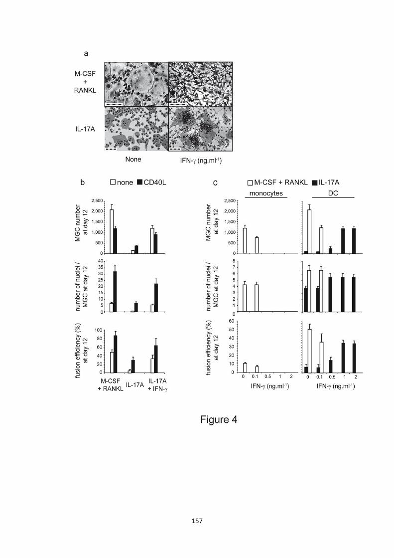
157

158

159
2.4 Manuscrit 4: (in preparation) IL-17A+MMP-12+CSTD+ DC accumulation leads to bronchoepithelium destruction: relevance in pulmonary LCH
by Mohamad Bachar ISMAIL
*Contributed equally as first authors.
1-4*, Alexandre BELOT1-5*, Nathalie BISSAY1-4, Béatrice
BANCEL6, Jan-Inge HENTER7, Maurizio ARICÒ8, Françoise BERGER1-4,6, Abdellatif
TAZI9 and Christine DELPRAT1-4,10
1 CNRS, UMR5239, Laboratoire de Biologie Moléculaire de la Cellule, Lyon, F-69007, France ; 2 Ecole Normale Supérieure de Lyon, Lyon, F-69007, France ; 3 Université de Lyon, Lyon, F-69003, France ; 4 Université de Lyon 1, Villeurbanne, F-69622, France ; 5 Hospices Civils de Lyon, Hôpital Femme Mère Enfant, Bron, F-69500, France ; 6 Hospices Civils de Lyon, Department of Pathology, Department of Microbiology, Centre Hospitalier Lyon Sud, Pierre Bénite, F-69310, France ; 7 Childhood Cancer Research Unit, Department of Women's and Children's Health, Karolinska Institutet, Karolinska University Hospital Solna, S-171 76 Stockholm, Sweden; 8 Department of Pediatric Hematology Oncology, Azienda Ospedaliero-Universitaria Meyer Children Hospital, Florence, I-50139, Italy; 9 Centre National de Référence de l'Histiocytose Langerhansienne; Univ Paris Diderot, Sorbonne Cité; Assistance Publique Hôpitaux de Paris; Service de Pneumologie, Hôpital Saint Louis, Paris, F-75475, France. 10 Institut Universitaire de France, 103 bd St Michel, Paris, F-75005, France
ABSTRACT
Langerhans cell histiocytosis (LCH) is a rare granulomatous disease characterized by an
abnormal accumulation of IL-17A-expressing DCs in involved tissues. LCH predominately
affects children but an isolated rare form of this disease, called pulmonary LCH (pLCH),
occurs in adults destroying their lungs. As in childhood LCH, we identified, for the first time,
that pathological DCs in pLCH express IL-17A. Moreover, we also found IL-17A-expressing
multinucleated giant cells (MGC) in lung granulomas. Surprisingly, most of pathological IL-
17A-expressing DCs express CD1a but not CD207. Serum studies demonstrated that IL-17A
is systemically distributed in patients developing either active childhood or adult pulmonary

160
forms of LCH. Pathological DCs express CCL20 and CCL2 chemokines in pLCH lesions as
revealed by immunofluorescent staining and high levels of CCL2 are detected in serum from
patients affected with this disease. Using immunofluorescence, we also found that matrix
metalloproteinase 12 (MMP-12) and cathepsin D (CTSD) proteases are expressed by
pathological DCs in pLCH lesions. Importantly, using an in vitro co-culture system, we
revealed that IL-17A-treated DCs exhibit potent aggressive functions as they were able to
destroy bronchial epithelial cells. Our data supports that IL-17A-expressing DCs is
responsible for the bronchiole destruction observed in pLCH patients, possibly through their
aggressive proteases. Since DC granulomas constitute the hallmark of pLCH, targeting these
cells and their aggressive functions may provide new avenues in pLCH treatment.
Figure 1 legend: A/ Triple CD207/blue, CD3/red and IL-17A/green staining on section “n”,
with CD1a/purple staining on side-section “n-1” and CD207/blue, CD3/red and IgG1
isotype/green control staining on side-section “n+1” of adult pulmonary LCH biopsy (case
pulm 8). The enlarged inset picture shows the presence of IL-17A+ giant cells (as previously
detected in childhood LCH). Scale bars, 50 μm (5 x 10 μm). C/ Percentages of CD1a, CD3,
CD207, IL-17A expressing cells in adult pulmonary Langerhans cell histiocytosis. B and D/
IL-17A measurement in childhood, adult LCH and control sera and in monocyte or monocyte-
derived DC compared to control. E/ GM-CSF staining in pLCH patient in granuloma (gr) and
bronchio-alveolar cells (br). Scale bars, 50 μm (5 x 10 μm)
Figure 1 results: Most of IL-17A expressing CD1a+DC are CD207-negative
In adult pulmonary LCH lesions, we consistently found more CD1a+ DC than CD207+ LC,
thus numerous CD1a+ CD207– DC were detected. We demonstrate that most, but not all, IL-
17A-secreting DC are CD1a+ CD207– non-LC DC, ranging from 2 to 52% of the CD1a+ DC
(fig 1A, C). Few or no T cells express IL-17A in LCH lesions. IL-17A is increased in sera of
pLCH patients as well as in monocyte-derived DC but not in monocytes (Fig 1 B, D). GM-
CSF is strongly expressed in bronchial alveolar cells and slightly increased in granuloma.
GM-CSF and IFN-γ are not expressed in LCH patient sera (data not shown).

161
Figure 1: Cytokine environment in biopsy & serum in pulmonary LCH patients

162
Figure 2: Chemokine CCL20 and CCL2 in PLCH lesions and serums.
Figure 2 legend : (A) Triple CCR6/blue, CD3/red and CCL20/green staining on section “n”, with
CD1a/purple staining on side-section “n-1” and CD207/blue, CD3/red and IgG1 isotype/green control
staining on side-section “n+1” of adult pulmonary LCH biopsy (case pulm X). The enlarged inset
picture shows the presence of epithelial cells CCL20+ close to the granuloma. Specificity of
immunostainings was demonstrated using isotype controls (not shown). Scale bars, 50 μm (5 x 10
μm). (B) Triple CD3/blue, CCL2/red and CD1a/green staining of adult pulmonary LCH biopsy. (C)
Patient’s sera measurement of CCL2.
Figure 2 results: CCL20 and CCL2 drive DC accumulation in pLCH. CCL20 and CCL2 were
expressed in situ by LCH-DC (Fig2A and B). Here we show that CCL20 was also expressed by
broncho-epithelial cells, in contact with DC granuloma. CCL2 was detected in patient serums contrary
to CCL20 (data not shown), suggesting a distinct endocrine versus paracrine role in cell recruitment
(Fig2C).

163
Figure 3: Detection of CTSD and MMP families in IL-17A-rich microenvironment
Figure 3 legend: CTSD and MMP12 expression within pLCH lesions. Double CTSD/green, MMP-
12/red staining on section “n”, with CD1a/purple staining on side-section “n-1” of adult pLCH lesions
(case pulm 6). Yellow color revealed that pLCH-DC and DC-derived MGC coexpress MMP-12 and
CTSD. Staining is representative of nine studied patients. MMP12 is also expressed by the bronchial
epithelial cells. Specificity of immunostainings was demonstrated using isotype controls (not shown).
Bar scale length is 50μm (5x10μm).
Figure 3 results: Active MMP-12 & CTSD were expressed in pulmonary LCH.. In pLCH lesion,
MMP-12 was expressed by broncho-epithelial cells and LCH-DC. CTSD was expressed by large or
multinucleated DC inside granuloma (Fig 3C).

164
Figure 4: BEAS2B destruction assessment of IL-17A treated DC
Figure 4 legend: (A) Double TRAP (purple) /Hoechst (blue) staining of BEAS-2B cells incubated
with or without IL-17A and IFN-γ cytokines (top panels), with DC alone (middle panels), or with IL-
17A and IFN-γ-treated DC (down panels). DC are double stained whereas BEAS-2B are only Hoechst
positive. (B) High magnification picture of BEAS-2B cells incubated with IL-17A and IFN-γ-treated
DC. Holes in BEAS-2B layer are observed (black arrows) around TRAP+ cells. Data are representative
of two experiments. Bar scale length is 50μm (5x10μm).
Figure 4 results: IL-17A-treated DC induced tissue destruction.To investigate whether IL-17A-
treated DC can damage bronchial epithelial cells, we have set up an in vitro co-culture system
involving immortalized bronchial epithelial cells (BEAS-2B) and primary monocyte-derived DC. We
cultured immature DC either alone or with IL-17A and IFN-γ. In parallel, we cultured a confluent
monolayer of human bronchial epithelial cells from the cell line “BEAS-2B” which has been
immortalized with Sv40/Ad12 virus hybrid contruct. We then added treated DC on bronchial epithelial
cells for 12h, before performing a TRAP/Hoechst staining to discriminate the two cell types. Hoechst
staining localized the nuclei of all viable cells while TRAP identified DC because BEAS-2B are
TRAP-negative. BEAS-2B alone displayed a nice confluent layer of blue nuclei stained by Hoechst
(Fig. 4A top). Addition of IL-17A and/or IFN-γ did not affect BEAS-2B viability (Fig. 4A top).

165
Untreated DC were unable to induce the destruction of BEAS-2B cells (Fig. 4A middle) since Hoechst
staining indicated that BEAS-2B were still alive. As previously published, IL-17A treatment increased
TRAP expression in DC (Coury et al., 2008) thus explaining the higher color intensity of the DC in the
bottom panel. These treated DC destroyed the BEAS-2B layer (Fig. 4A down) leading to the
formation of several holes as shown at higher magnification (Fig. 4B). Treated DC were required to
kill the BEAS-2B cells since neither DC nor cytokine, alone, could destroy the BEAS-2B cell layer.
These data demonstrate that IL-17A and IFN-γ-treated DC exhibit a specific potent ability to destroy
human bronchial epithelial cells, in vitro.

166
Figure 5: Model of bronchiole destruction by pathological infiltrating DC in pLCH lesions
Figure 5 shows a model of bronchiole destruction in pLCH lesions visualized owing to a CD1a red
staining of DC with a counter coloration of nuclei performed with hematoxylin (blue). Aligned narrow
blue nuclei localize healthy bronchial epithelium (square1). In square 2, red pathological DC are
localized near the bronchial epithelial cells. We propose that IL-17A induces CCL20 mainly by
bronchial epithelial cells, thus attracting CCR6+ cells such as DC. CCL2 production by pathological

167
DC may further increase monocyte and DC recruitment. Finally, bronchial epithelium destruction
(square 3) indicates that bronchial epithelial cells have been killed and digested. We propose that
MMP-12 and CTSD may play an essential role in this destructive process. IL-17A, CCL20 and CCL2
as well as proteolytic enzymes like CTSD and MMP-12 may constitute several essential therapeutic
targets in LCH treatment.

168
Material & methods:
Reagents
We used the following antibodies for DC phenotype analysis by cytofluorimetry: FITC-CD1a
(21270013S), FITC-CD3 (21270033S), FITC-CD14 (21270143S), PE-HLA-DR (21278994S) all from
Immunotools and FITC-CD83 (HB15a) from Beckman Coulter. We used recombinant human IL-17A
and IFN-γ from PeproTech.
Patients
Biopsy section samples from 9 patients, all current or ex-smokers (7 women and 2 men, aged
40.3 ± 17 years), referred to (Centre de référence de l’Histiocytose Langerhansienne, France) and
diagnosed as having pLCH, were examined in this study. For each patient, we obtained 5 Serial
section slides with one previously stained for CD1a expression as well as with hematoxylin to
revealed nuclei. Clinical data and manifestations of the 9 patients are shown in table 1. Serum samples
from 22 pLCH patients were also obtained from the same source. Serum from 38 childhood LCH
patients were collected from blood samples obtained from Sweden and Italy.
Table 1: Clinical data of patients included in the pLCH study
Case Sex / Age at
sampling
Smoking status Extrathoracic involvement Treatment
at sampling
Pulm1 F / 47 years Current smoker none none
Pulm2 M / 37 years Ex-smoker skin, bone, anterior pituitary insufficiency with diabetes insipidus
desmopressin and hormonal substitution
Pulm3 F / 23 years Current smoker diabetes insipidus desmopressin
Pulm4 F / 53 years Current smoker none none
Pulm5 F / 48 years Ex-smoker none none
Pulm6 F / 36 years Current smoker none none
Pulm7 F / 45 years Ex-smoker none none
Pulm8 M / 24 years Current smoker diabetes insipidus desmopressin
Pulm9 F / 50 years Current smoker none none
Immunohistofluorescence and confocal microscopy
IL-17A and IFN-γ-treated DC cultured on glass coverslips were first fixed for 20 minutes with
2% paraformaldehyde in PBS. Cells cultured were permeabilized with 0.1% Triton X-100 in PBS for
5 minutes.

169
4-µm paraffin-embedded lung biopsies were deparaffinized and rehydrated. Epitope retrieval
was performed in citrate buffer (10mM, pH6.0) using a water bath for one hour at 99°C.
Cells or tissue sections were incubated 30 minutes in phosphate buffered saline (PBS) -1%
bovine serum albumin (BSA) with 3% human serum to block Fc receptors. They were then incubated
with primary antibodies, overnight at 37°C, in a humidity chamber. Replacement of the primary
antibodies by non-relevant antibodies of the same immunoglobulin isotype was used as negative
control. Table 2 provides a summary of the primary antibodies used. Following 3 washes in PBS-
1%BSA, slides were treated for 15 min in PBS-1% BSA. Detection of the primary antibodies was then
performed with suitable isotype-specific secondary Alexa Fluor 488, 546 and 647-conjugated
antibodies (Invitrogen, 1:200 dilution) for 30min. Following 3 washes in PBS-1%BSA, the sections
were mounted using Mowiol and then analyzed by confocal microscopy using a Carl Zeiss
MicroImaging Inc. LSM 510 confocal microscope. Immunostainings for CD1a and GM-CSF were
performed by immunochemistry. Incubation with appropriate dilutions of anti-CD1a antibody (O10;
Immunotech, Marseille, France) or a sheep anti-human GM-CSF polyclonal antiserum (National
Institute for Biological Standards and Control, Hertfordshire, UK) conjugated to alkalin phosphate,
were realized. Positive cells were revealed using the Vectastain ABC-alkaline phosphatase kit system
obtained from Vector Laboratories (Burlingame, CA). We quantified percentage of positive and
negative cells with ImageJ freeware of National Center for Biotechnology Information.
Table 2: Characteristics of primary antibodies used for immunohistofluorescent stainings
Antibody Clone Species/isotype source concentration μg/ml
IL-17A MAB3171 Mouse/IgG1 R&D Systems 10
CD207/langerin ab49730-100 Mouse/IgG2b Abcam 20
CD3 A0452 Rabbit/IgG DakoCytomation 12
MMP-12 82902 Mouse/IgG2b R&D Systems 25
CTS D E-7: sc-13148 Mouse/IgG1 santa cruz biotechnology 4
CCR6 53103 Mouse/IgG2b R&D Systems 25
CCL20 67310 Mouse/IgG1 R&D Systems 10
CCL2 MAB2791 Mouse/ IgG2b R&D Systems 25
CD1a DM363 Mouse IgG1 Acris antibodies 40
IL-17A, MMP, CCL20 and CCL2 detection
Serum amounts of IL-17A and CCL2 were measured in the serum of patients diluted at 1:2
using human ELISA development kit or Luminex to detect IL-17A (Peprotech, 900-K84), CCL2
(Peprotech, 900-K31), CCL20 (R&D) and MMP . Samples were run in duplicate to ensure

170
reproducibility. Results were analyzed by the Multiskan Spectrum Spectrophotometer with SkanIt
Software 2.2 (Thermo Electron).
Cell purification, differentiation and culture:
Blood was obtained from healthy adult volunteer donors (Etablissement français du sang,
Lyon Gerland, France). Monocytes isolation and purification was performed as previously described
(Fugier-Vivier et al., 1997). Briefly, monocytes were obtained after Ficoll (Lymphocyte separation
medium, Eurobio, CMSMSL01-01) and percoll (GE Healthcare, 17-0891-01) density-gradient
centrifugation, respectively. They are then seeded at 106 cells/mL and maintained in RPMI 1640 (Life
Technologies, Paisley, United Kingdom) supplemented with 10 mM HEPES (N-2-
hydroxyethylpiperazine-N'-2-ethanesulfonic acid), 2 mM L-glutamine, 40 μg/mL gentamicin (Life
Technologies), 10% heat-inactivated fetal calf serum (FCS; Boehringer Mannheim, Meylan, France),
50 ng/mL human recombinant GM-CSF (hrGMCSF), and 500 U/mL hrIL-4. After 6 days of culture,
cells developed into typical immature DC being CD14−, CD1a+, human leukocyte antigen (HLA)-DR+,
and CD83− (not shown).
Affymetrix gene chip study
mRNA were extracted from either unstimulated DC or DC cultured with IL-17A with or
without IFN-γ, after cell lysis, extraction in Trizol (Invitrogen) and purification on a MEGAclear
column (Ambion) to reach an RNA integrity number >9 with Agilent bioanalyser. ProfileXpert
(www.profilexpert.fr) carried out the chip analysis study.
Culture of BEAS-2B cells and coculture with IL-17A and IFN-γ- treated DC
The BEAS-2B cell line, which is an immortalized line of normal human bronchial epithelium derived
by transfection of primary cells with an adenovirus12-SV40 hybrid virus, was obtained from ATCC.
Cells were cultured and maintained at 37°C with 5% CO2 in 75 cm2-cell culture flasks precoated with
a mixture of fibronectin (1:50 dilution; Sigma F0895) and Bovin I collagen (1:100 dilution; Invitrogen
A1064401) both dissolved in LHC-9 serum-free medium (Invitrogen, 12680-013) . Cells were
passaged every three days. For coculture assays of BEAS-2B and IL-17A and IFN-γ-treated DC;
10 000 BEAS-2B cells were plated onto 24-well cell culture plates at 37°C with 5% CO2 until
confluence to cell monolayer. DC were cultured separately in LHC-9 medium with human
recombinant IL-17A (2ng/ml) and IFN-γ (1ng/ml) for 6 days and then added (106cells/ml/well) over
the BEAS-2B layer. 24 hours after, monolayers were examined, photographed, then cells were fixed
and a double TRAP/Hoechst staining was performed. Experiments were repeated twice.

171
TRAP/Hoechst staining
TRAP activity was assessed using the Leukocyte Acid Phosphatase kit (Sigma-Aldrich). Nuclear DNA was stained with a 10 μg/ml solution of Hoechst 33342 (Invitrogen H3570) for 30 min at room temperature. Cells are then washed and fixed with 1% formol. Culture and TRAP/hoechst pictures were analyzed using a Leica DMiRB microscope equipped with x40/0.30 NA or x40/0.55 NA objective lenses (Leica, Wetzlar, Germany) a Leica DC300F camera and the Leica FW400 software

172
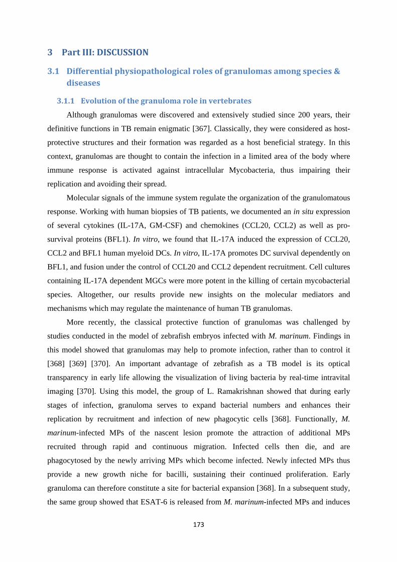
173
3 Part III: DISCUSSION
3.1 Differential physiopathological roles of granulomas among species & diseases
3.1.1 Evolution of the granuloma role in vertebrates
Although granulomas were discovered and extensively studied since 200 years, their
definitive functions in TB remain enigmatic [367]. Classically, they were considered as host-
protective structures and their formation was regarded as a host beneficial strategy. In this
context, granulomas are thought to contain the infection in a limited area of the body where
immune response is activated against intracellular Mycobacteria, thus impairing their
replication and avoiding their spread.
Molecular signals of the immune system regulate the organization of the granulomatous
response. Working with human biopsies of TB patients, we documented an in situ expression
of several cytokines (IL-17A, GM-CSF) and chemokines (CCL20, CCL2) as well as pro-
survival proteins (BFL1). In vitro, we found that IL-17A induced the expression of CCL20,
CCL2 and BFL1 human myeloid DCs. In vitro, IL-17A promotes DC survival dependently on
BFL1, and fusion under the control of CCL20 and CCL2 dependent recruitment. Cell cultures
containing IL-17A dependent MGCs were more potent in the killing of certain mycobacterial
species. Altogether, our results provide new insights on the molecular mediators and
mechanisms which may regulate the maintenance of human TB granulomas.
More recently, the classical protective function of granulomas was challenged by
studies conducted in the model of zebrafish embryos infected with M. marinum. Findings in
this model showed that granulomas may help to promote infection, rather than to control it
[368] [369] [370]. An important advantage of zebrafish as a TB model is its optical
transparency in early life allowing the visualization of living bacteria by real-time intravital
imaging [370]. Using this model, the group of L. Ramakrishnan showed that during early
stages of infection, granuloma serves to expand bacterial numbers and enhances their
replication by recruitment and infection of new phagocytic cells [368]. Functionally, M.
marinum-infected MPs of the nascent lesion promote the attraction of additional MPs
recruited through rapid and continuous migration. Infected cells then die, and are
phagocytosed by the newly arriving MPs which become infected. Newly infected MPs thus
provide a new growth niche for bacilli, sustaining their continued proliferation. Early
granuloma can therefore constitute a site for bacterial expansion [368]. In a subsequent study,
the same group showed that ESAT-6 is released from M. marinum-infected MPs and induces

174
the secretion of MMP9 in neighboring epithelial cells [369] (Fig. 17). MMP9 secretion
facilitates the migration of uninfected MPs into the nascent granuloma, promoting their
infection and leading to bacterial expansion [369]. MMP9 activity may result from its ability
to degrade extracellular matrix components as well as to activate chemokines [188].
Figure 17: ESAT-6-mediated MMP9 induction in epithelial cells promotes phagocyte recruitment and infection in M. marinum-infected zebrafish embryos. This promotes mycobacterial dissemination and expansion. From ref [370].
In contrast to the zebrafish model, granulomas in humans are commonly regarded as
protective structures. This concept is supported by several observations. First, in persons who
did not die from TB, calcified granulomas were found : most of them being sterile while few
others contains live Mycobacteria [371] indicating that these structures are efficiently
involved in mycobacterial killing. Second, the blockade of TNF-α which leads to the
reactivation of latent TB, results in granuloma breakdown and disintegration, along with
mycobacterial dissemination [372]. These observations suggest that granulomas in humans
are host-protective structures which counteract mycobacterial growth and limit the
dissemination of the infection.
Therefore, the role of the granuloma is differentially understood in various hosts.
However, can findings in M. marinum-infected zebrafish be extrapolated to human TB

175
disease? And are they relevant for the understanding of the role of human mycobacterium-
induced granuloma? Actually, there are several differences between M. marinum infection in
zebrafish embryos and TB in humans [373]: first, M. marinum is not Mtb and zebrafish have
no lungs. Second, experimental infection in zebrafish model is by injection and not aerosol
inhalation like in TB disease. Third, the immune system is probably immature in the embryos
of zebrafish and the role of granuloma may change with the immune system development.
Finally, granulomas in zebrafish larvae are formed exclusively by innate immune cells and are
not under the control of adaptive activated T lymphocytes. The role of granulomas may be
different in various groups of vertebrates: it appears negative in lower vertebrate (fish
embryos) and positive in higher vertebrate (mammal adults). Does it result from the evolution
of the vertebrate immune system facing to Mycobacteria between fish and mammals? Or does
it come from the differential development of the immune system between embryo and adult?
The analysis of the evolution of the vertebrate adaptive immune system may allow a
better understanding of these discrepancies in the future.
3.1.2 Contributions and limitations of the murine models to understand
granulomatogenesis
We studied two granulomatous diseases: TB and pLCH. pLCH has no currently any
experimental model and a major difficulty in the field of TB is the lack of a suitable animal
model. Our studies on TB patient biopsies and human myeloid cells showed that CCL20 and
CCL2 chemokines are expressed by both myeloid mono and multinucleated giant cells of the
human TB granuloma. We demonstrated that these chemokines are involved in human
myeloid cell clustering and fusion in vitro. Taken together, these data suggest an important
role of CCL20 and CCL2 in myeloid cell recruitment and fusion within human TB
granulomas.
The mouse was widely used as a TB model and has provided considerable contribution
in our knowledge in TB granulomatogenesis. Indeed, most of cytokine, chemokine and
cellular requirements for granuloma formation and maturation were provided by the murine
model. However, there are several significant differences in the granulomatous response to
Mtb in the lung between human and mice [172].
Human TB granulomas are highly organized suggesting a potential role of chemokines,
which control cell migration and clustering, in the regulation of these structures. They contain
a core of epithelioid MPs and MGCs and are surrounded by T cells. Human granulomas

176
progressively become devoid of blood vessels and hypoxic [172] and are characterized by
necrosis and caseation during TB pathology.
In mouse, the architecture of the granuloma is less organized with diffuse cellular
infiltration described as a loose collection of activated cells. Studies showed that chemokines
have redundant roles in the granulomatous response in this animal (Table VI). In addition to
epithelioid MPs and lymphocytic clusters, elevated numbers of PMNs are present while
MGCs are absent in murine granulomas. Necrosis and caseation are also rarely observed
within these granulomas and these structures are relatively aerobic [172].
Studies conducted in Mtb-infected mice deficient for CCL2 and the CCL20 receptor,
CCR6, showed that these deficiencies don’t affect the adaptive granulomatous response to this
pathogen and revealed that deficient mice clear Mycobacteria [266] [265]. Nevertheless,
based on the fact that murine granulomas are loosely organized and don’t contain MGC while
those of humans are well-structured and contain such giant cells, CCL2 and CCL20 roles may
be different in murine and human granulomas.
In conclusion, the mouse model does not reflect the full spectrum of human granuloma
formation upon infection, thus presenting certain limitations in the understanding of the
human granulomatogenesis in TB [374]. The murine model was certainly fruitful to
understand the role of adaptive cytokines required for granulomatogenesis (e.g. IL-12, IFN-γ,
GM-CSF…). However, the role of this animal model is questionable regarding the chemokine
requirements of granuloma maintenance as well as concerning the mechanisms of MGC
formation and functions.
3.1.3 Emergence of the role of granulomas in the chronic control of cancer cells
Actually, it becomes well admitted that the immune system forms an important barrier
facing to tumor growth and progression. Interplay between immunity and tumors may result
in the elimination of cancer cells, their escape or a continuous equilibrium between them and
the immune system. Importantly, the anti-mycobacterial immune response is clinically used as
a tumor treatment in some instance. Indeed, since the 70’s, BCG-immunotherapy constitutes
the optimal choice and the most potent treatment against invasive bladder cancer. This
immunotherapy is based on several intravesical instillation of BCG (inside the bladder), and
results in 70% response rate. However, dramatic consequences are observed in 30% of
patients related to a hypo or hyper immune responses. Moreover, the exact molecular
mechanisms of this therapy are largely unknown.

177
It is unknown if IL-17A and IFN-γ are involved in the activation of myeloid cells to
become able to control and/or destroy cancer cells. However, it is possible that such
stimulated myeloid cells become able to kill cancer cells or to suppress their proliferation. In
vitro, we set up a co-culture system between DCs and BEAS-2B, a human a transformed
human cell line immortalized by an Adenovirus12-SV40 hybrid virus. Using this system, we
showed that IL-17A and IFN-γ-treated DCs with BEAS-2B induces the destruction of
immortalized BEAS-2B cells, while untreated DCs were unable to mediate this effect. The
anti-tumoral activity was effective in close contact with the effector cytotoxic DCs and
therefore probably not mediated by a soluble death ligand, although the mechanism is not
characterized.
In 2007, an elegant study by Koebel et al. demonstrated, for the first time, that cancer
cells can be chronically controlled by granulomas including giant cells, thus underling an anti-
tumoral granulomatous immune response [375]. After chemical carcinogen injection in mice,
authors found both tumor-developing and tumor-free animals. Importantly, tumor-free mice
were not really free but had latent dormant cancer cells contained by granulomatous structures
of 2-8 mm size, including giant cells [375]. Tumor control in this model was a result of both
increased apoptosis and decreased proliferation of cancer cells. However, when treated with
an antibody cocktail depleting CD4+ and CD8+ cells and neutralizing IFN-γ, these mice
develop sarcomas, thus demonstrating that adaptive immune system is required for this anti-
tumoral granulomatous response. Several similarities exist between the anti-tumoral response
in this mouse model and the anti-Mycobacterium granulomatous responses. Both contained
latent (dormant) cells which can be controlled for decades in giant cell-associated granulomas
but can be re-activated after immune suppression [376]. Moreover, the adaptive immunity is
required for both response types. However, the role of IL-17A was not evaluated in this study.
Neutralization of IL-17A in mouse models with such induced tumors is an important issue
that should be investigated in the future.
In human patients with breast cancer, occult cancer cells can be maintained in a dormant
state [377] and a recent study described that long-term dormancy of breast cancer cells is
maintained by small “growth-restricted micrometastasis” [378]. However, it is not clear if a
granulomatous response is involved in the control of cancer cells in such patients. In the
future, it would be intriguing to characterize the anti-tumoral granulomatous response in
humans. in situ investigation of an IL-17A-dependent anti-tumoral granulomatous response in
human biopsies will clarify if IL-17A is involved in such response.

178
3.2 IL-17A-induced genetic program promotes granulomatogenesis
3.2.1 Is IL-17A a constitutive molecular player in granulomatogenesis?
Granuloma formation is driven by several cytokines which can positively or negatively
regulate this process (Table V). We documented an in situ expression of IL-17A within both
human TB and pLCH granuloma. In TB, we found that IL-17A is mostly expressed by CD3+
T cells which surround the epithelioid core. In contrast, IL-17A expression was restricted to
CD1a+ DC and MGCs in pLCH granulomas. Our results suggest that IL-17A may induce
granuloma maintenance by promoting myeloid cell survival, clustering and fusion.
IL-17A expression in granulomas was documented in several human granulomatous
diseases including crohn’s disease [379], childhood LCH [306] and very recently sarcoidosis
[380] [381] and Blau syndrome [382]. IL-17A was initially considered as a T cell cytokine
produced by Th17 lymphocytes. However, recent studies showed that this cytokine is also
expressed by a wide range of myeloid cells including DCs, MPs and PMNs [306] [321]. In the
context of granulomatous diseases, IL-17A expression was documented in both lymphoid and
myeloid cells. IL-17A was expressed by Th17 cells as well as by myeloid epithelioid cells and
MGCs in the granulomas of crohn’s disease [379] sarcoidosis [380] [381] and Blau syndrome
[382] patients, while it was exclusively expressed by myeloid DCs and MGCs in childhood
LCH granulomas [306]
Our results suggesting a role of IL-17A in promoting granuloma maintenance are in line
with those recently reported in the murine model of mycobacterial infection. In this model,
IL-17A positively regulates mature granuloma formation and maintenance [144] [259]. In the
absence of this cytokine, mice infected with BCG were able to form nascent but not mature
granulomas characterized by an impaired cellular accumulation and organization [259].More
recent findings underline also a positive correlation between IL-17A and the formation of
large granulomatous lesions in a bovine TB model [383].
A growing body of evidences suggests that IL-17A plays also an essential role in
granuloma formation and maintenance in other granulomatous disorders. In murine
schistosomiasis, IL-17A is directly associated with the severity of the granulomatous
inflammation. Indeed, the development of severe and large schistosoma -induced granulomas
correlated with high levels of IL-17A and mice injected with this cytokine showed enhanced
granulomatous inflammation [384] [385]. In contrast, reduced size of granulomas was
associated with the production of lower amounts of IL-17A [386]. Moreover, antibody-
mediated IL-17A neutralization led to a significant reduction of the granuloma size [384]

179
[385]. IL-17A deficiency alters the establishment of granuloma in mice infected with listeria
monocytogenes as demonstrated by the disorganized granulomatous structures with dispersed
cell organization compared with WT animals [387]. In the same way, inhibition of Th17 cell
development reduced the granulomatous response in a murine model of Propionibacterium
acnes-induced granuloma in the liver while enhanced Th17 cell differentiation exacerbated
such response [388]. IL-17A plays also a role in granuloma formation in non infectious
granulomatous diseases. For example, IL-17A was proposed as a central player in granuloma
formation in the murine model of anti-neutrophil cytoplasmic antibodies (ANCA)-induced
systemic vasculitis [389].
Overall, these data showed that IL-17A is now documented in a variety of
granulomatous disorders in both human and animals, thus supporting the evidence that this
cytokine participates in the regulation of granuloma formation and maintenance. An important
remaining issue in our work is to determine the nature of the T cell subpopulation that
produces IL-17A in human TB granulomas: are they Th17 cells or other IL-17A producing
cells such as γδ T cells as described in murine Mycobacterium-induced granulomas [259]?
Further in situ immunohistofluorescence investigations will clarify this question in the future.
Another important matter is to go on investigating IL-17A expression in additional
granulomatous diseases. For example, the literature reports the presence of granulomas during
infections with the filamentous, but not the yeast, form of fungi, and recent studies showed
that the filamentous form triggers the polarization of Th17 cells in vitro while the yeast form
does not [339]. It would be therefore interesting to investigate the expression and the role of
IL-17A in filamentous fungal granulomas. Finally it is important to characterize all the
molecular events which explain why IL-17A is a constitutive factor in granulomatogenesis.
3.2.2 The role of IL-17A-induced CCL20 and CCL2 in granulomatogenesis
The recruitment of a variety of immune cell types during granuloma formation is
essentially orchestrated by an intense local production of chemokines involved in the
regulation of cell trafficking and migration. We focused on the role of two chemokines:
CCL20 and CCL2. CCL20 is the only identified chemokine which binds the chemokine
receptor CCR6 while CCL2 is one of the several ligands of CCR2. CCL20 induces
chemoattraction of immature CCR6+ DCs and subpopulations of effector/memory
lymphocytes including IL-17A-producing lymphocytes. CCL2 regulates migration and
infiltration of several immune cells, especially CCR2+ monocytes/MPs and T cells.

180
Our results demonstrate the rapid induction of CCL20 and CCL2 chemokines by human DCs
after treatment with IL-17A. While CCL2 was continually produced in large quantities until
the 12th day of treatment, CCL20 was transiently produced in the initial phase with lower
amounts. This demonstration may explain the correlations that we observed in two kinds of
granuloma. Indeed, IL-17A expressing granulomas in TB and pLCH showed a simultaneous
expression of both CCL20 and CCL2 chemokines by their myeloid cells. We also detected
large amounts of circulating CCL2 in the serum of patients with LCH whereas CCL20 was
undetectable. CCL20 and CCL2 chemokines may play important roles in the recruitment of
myeloid cells to the granuloma, thereby promoting its formation and maintaining its integrity.
Few reports demonstrate a direct effect of IL-17A on the production of CCL2 and CCL20.
Previously, it was shown that this cytokine up-regulates the expression of CCL20 and CCL2
in human renal and airway epithelial cells, respectively [390] [391]. More recently, a study
reports that IL-17A treatment of human MPs induces the production of CCL2 and CCL20 by
these cells in vitro [392].
Several studies underline a correlation between IL-17A, CCL20 and/or CCL2 and
granuloma formation, maturation and/or size. A recent report in mouse showed that following
intratracheal infection with BCG, CCL20 was induced during the early innate stage of
infection [265] maybe in agreement with the IL-17A production by γδ T cells [259]. In
humans, CCL20 expression was detected by the myeloid mono and multinucleated giant cells
of both childhood LCH [393] and sarcoid [394] granulomas in which IL-17A expression was
also documented in separated studies [306] [380]. In LCH, CCL20 may cause the recruitment
and retention of additional lesional CCR6+ DCs [393] while in sarcoidosis, it may essentially
drive the recruitment of CCR6+ Th17 cells from the blood toward the granuloma [380].
In animals, increase in granuloma size during murine schistosomiasis was associated
with increased CCL2 expression in granuloma MPs and higher levels of IL-17A in granuloma
cells compared to those of control animals [395]. Upon mycobacterial infection, CCL2 levels
were found reduced in IL-17A-deficient mice showing impaired granuloma formation and
maturation [259]. In humans, a recent study suggests a role for CCL2 in both establishment of
latency and maintenance of the integrity of granulomas in asymptomatic individuals with
latent TB [396]. In addition, a recent study suggests that CCL2 forms an important mediator
in recruiting inflammatory CCR2+ cells during pulmonary sarcoidosis [397].
IL-17A-induced CCL20 and CCL2 may be involved in two essential phases of
granuloma formation: initiation and maintenance. In mycobacterium-infected mice, the early
IL-17A secretion [259] may rapidly induce CCL20 secretion by lung resident DCs, thereby

181
recruiting nearby CCR6+ lung DCs to initiate early granuloma formation. The IL-17A-
mediated expression of CCL2 becomes then rapidly massive and ensures the recruitment of
peripheral blood monocytes. These cells differentiate in situ in MPs or DCs according to the
local cytokine environment to promote bacterial containment. BCG-infected mice which over-
express CCL2 by type-II alveolar epithelial cells showed higher bacterial clearance, a
constitutively increased mononuclear phagocyte subset accumulation in their lungs and a
rapid and accelerated induction and resolution of lung granuloma, compared to WT animals
[398].
Overall, through CCL20 and CCL2 chemokines, the host is able to immediately attract
local available tissue myeloid cells around a pathogen, such as Mtb, via CCL20 and then
induces massive recruitment of peripheral blood cells via CCL2. These effects could promote
granuloma initiation and maintenance.
3.2.3 The role of IL-17A-induced CCL20 and CCL2 in cell-cell fusion
Chemotaxis and cytoskeleton rearrangement direct cell clustering and fusion [276]. Our
in vitro experiments showed that neutralizing antibodies against CCL20 or CCL2 efficiently
abrogated the fusion of IL-17A-treated DCs. While anti-CCL20 antibodies block DC
clustering and fusion without affecting their rounded shape, CCL2 neutralization also induces
a morphological change in these cells. Indeed, anti-CCL2 antibodies convert the rounded DCs
into elongated fusiform cells. The morphological change observed with anti-CCL2 revealed
that this chemokine is involved in the regulation of the cell cytoskeleton to achieve fusion and
MGC formation. These results underline the mandatory role of CCL20 and CCL2 in the
chemotaxis for DC-derived MGC formation and revealed an additional role of CCL2 in the
regulation of DC cytoskeleton.
CCL2 is already known to be pivotal in chemotaxis required for myeloid cell fusion. It
is involved in both FBGC [399] and OC [287] formation. Indeed, Kiryakides et al.
demonstrated that after subcutaneous foreign body (biomaterial) implantation, mice lacking
CCL2 show a marked impairment in FBGC formation due to compromised MP fusion
compared to their wild-type counterparts [399]. In vitro, authors showed that a CCL2
inhibitory peptide or an anti-CCL2 antibody reduces FBGC formation from peripheral human
blood monocytes. In osteoclastogenesis, although CCL2 alone is unable to induce OC
formation, this chemokine is required for the fusion of human [287] [288] [289] mouse [290]
and rat [400] myeloid cells into giant OCs. In human myeloid cells, RANKL/RANK signaling
activates the transcription factor NFATc1 (nuclear factor of activated T-cells, cytoplasmic 1)

182
resulting in CCL2 expression [287]. Human monocyte fusion into OCs was inhibited by
cyclosporin A, which blocks NFATc1, and addition of CCL2 after this pharmacological
blockade restored the formation of multinucleated cells although they were unable to resorb
bone [287]. Human monocytes treated with M-CSF and CCL2 fused and form giant cells
which express several OC markers [288]. OC formation was significantly inhibited in cells
derived from CCL2-deficient mice and this inhibition was restored by addition of
recombinant CCL2 [290].
The role of CCL20 in cell-cell fusion is less documented. However, in vitro,
recombinant human CCL20 significantly increased the number of multinucleated OCs, and
blocking anti-CCL20 antibodies inhibits this pro-osteoclastogenic effect [401].
CCL2 is already known to be involved in cytoskeleton rearrangement even in the
absence of cell fusion. Antibody-mediated CCL2 neutralization induces remarkable changes
in the morphology and size of human monocyte-derived MPs infected with HIV, reflecting
important changes in their cytoskeleton organization [402]. However, this morphological
change is different to what we see in DCs as it results in an increase in cell size. This suggests
that morphological change under the control of CCL2 may depend on the cell type. CCL2-
mediated effects on cytoskeleton are also required to cell-cell fusion. Indeed, the defective
fusion of CCL2-null MPs in FBGCs was associated with their inability to undergo
cytoskeletal remodeling, indicating a role of this chemokine in the regulation of this process
[403] [404].
Overall, CCL2 and CCL20 regulate essential features of cell-cell fusion to form giant
cells. However, we found that DCs cultured with both CCL20 and CCL2 in the absence of IL-
17A did not undergo cell fusion (data not shown). This indicates that these chemokines are
necessary but not sufficient for the IL-17A-dependent fusion process. Consequently, IL-17A
induces additional unknown fusogenic molecules required to achieve cell fusion and MGC
formation. According to our transcriptomic data, a potential candidate is DC-STAMP. The
mRNA of this molecule, known to regulate both OC and FBGC formation [291], is highly
expressed in IL-17A-treated DC while absent from untreated cells. DC-STAMP may thus
forms a common molecule necessary for multinucleation induced by different exogenous
stimuli. However, validation of this hypothesis requires additional functional analysis and
blocking assays. DC-STAMP is a cell surface seven-transmembrane protein, thereby
resembling chemokine receptor structures. Although the ligand for DC-STAMP is unknown,
it was proposed that CCL2 may serve as a DC-STAMP ligand [405]. In this hypothesis, IL-

183
17A may simultaneously induce both DC-STAMP and its potential ligand CCL2 in DCs,
thereby potentiating their fusion into MGCs.
3.2.4 The role of IL-17A-induced BFL1 in myeloid cell maintenance
In latent TB, granulomas persist for several decades reflecting the long-term
maintenance of these structures. We were precisely interested in the maintenance of myeloid
granuloma cells: are their maintenance is a result of a local proliferation and/or a long term
survival?
We found that IL-17A-treated DCs did not proliferate in vitro. However, while
unstimulated DCs did not survive more than few days, the number of IL-17A-treated DCs was
stabilized at day 12, thus demonstrating that IL-17A activates long-term survival pathways in
these cells. Our in vitro results showed that IL-17A induced a rapid NF-κB activation
(revealed by the nuclear translocation of the p65/RelA subunit) which in turn stimulates the
expression of the pro-survival protein BFL1. This IL-17A-dependent BFL1 induction in DCs
was blocked by an NF-κB inhibitor. Finally, statistical analysis showed that BFL1 expression
positively correlates with the survival of IL-17A-treated DCs.
IL-17A signaling is known to induce NF-κB activation [309]. Importantly, the promoter
of the gene encoding BFL1 possesses a p65 / RelA responsive element which positively
regulates BFL1 expression [406]. Therefore, in our model of human myeloid DCs, IL-17A-
mediated nuclear translocation of NF-κB provides the basis for BFL1 up-regulation.
Several reports showed that the capacity of cell proliferation within granulomas is very
limited. For example, in LCH granulomas, the proliferation marker Ki-67 was found in less
than 3% of lesional DCs [407]. In situ [3H]-thymidine labeling of sarcoid granuloma revealed
that proliferation and cell division is rare in granuloma myeloid/epithelioid cells and absent in
their derived-MGCs [277]. In the same way, during mycobacterial infections, BrdU
incorporation assay showed no differences in MP proliferation between BCG-infected and
uninfected mice during the first 2 weeks of granuloma development [174].
Regarding this reduced ability of cell proliferation and clonal expansion, inhibition of
apoptosis of granuloma cells may form an important factor which prevents granuloma
disruption and maintains the integrity of these structures. In this context, it was shown that
granuloma cells express several and pro-survival proteins. For example, myeloid cells of the
Mtb-induced granuloma express several pro-survival proteins of the TNF receptor-associated
factor family [182], increased expression of the pro-survival protein BCL-2 and reduced
expression of the anti-survival protein BAX [187]. Similarly, the pro-survival protein AIM
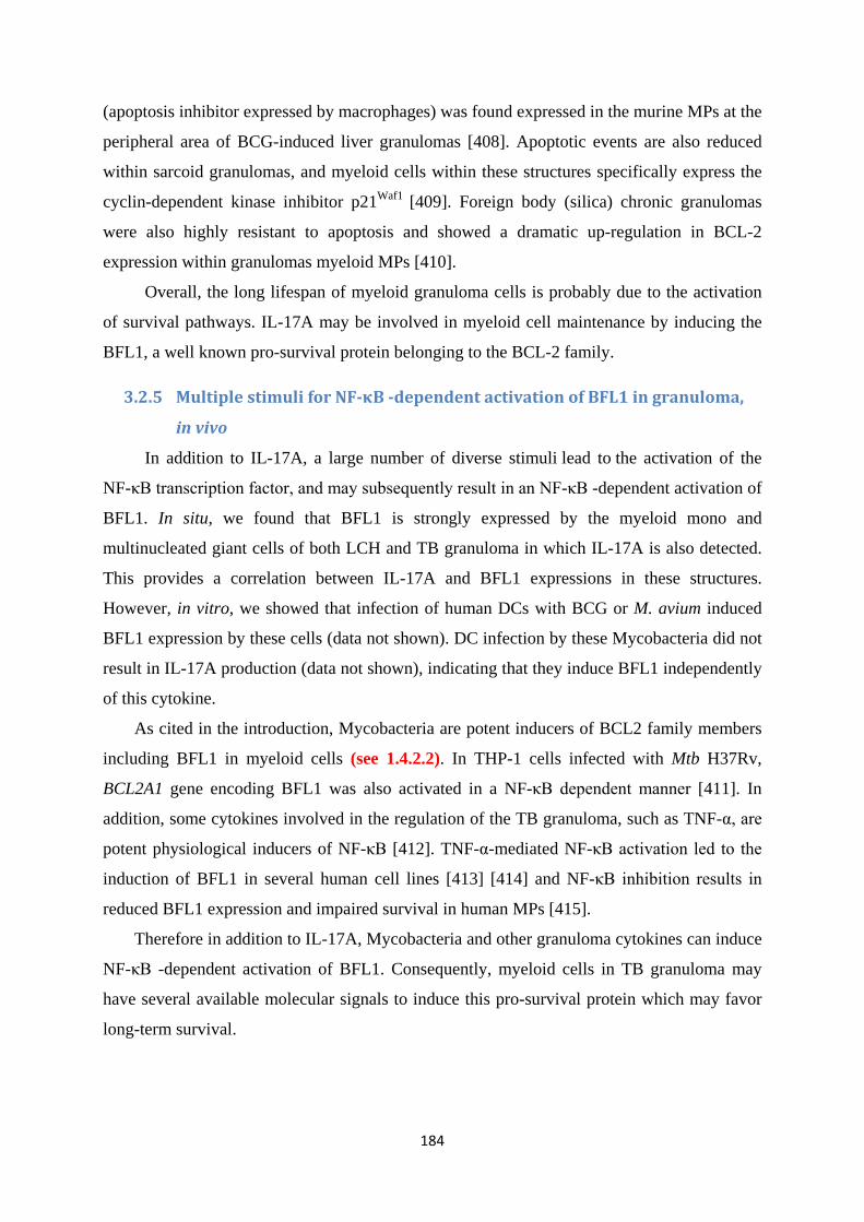
184
(apoptosis inhibitor expressed by macrophages) was found expressed in the murine MPs at the
peripheral area of BCG-induced liver granulomas [408]. Apoptotic events are also reduced
within sarcoid granulomas, and myeloid cells within these structures specifically express the
cyclin-dependent kinase inhibitor p21Waf1 [409]. Foreign body (silica) chronic granulomas
were also highly resistant to apoptosis and showed a dramatic up-regulation in BCL-2
expression within granulomas myeloid MPs [410].
Overall, the long lifespan of myeloid granuloma cells is probably due to the activation
of survival pathways. IL-17A may be involved in myeloid cell maintenance by inducing the
BFL1, a well known pro-survival protein belonging to the BCL-2 family.
3.2.5 Multiple stimuli for NF-κB -dependent activation of BFL1 in granuloma,
in vivo
In addition to IL-17A, a large number of diverse stimuli lead to the activation of the
NF-κB transcription factor, and may subsequently result in an NF-κB -dependent activation of
BFL1. In situ, we found that BFL1 is strongly expressed by the myeloid mono and
multinucleated giant cells of both LCH and TB granuloma in which IL-17A is also detected.
This provides a correlation between IL-17A and BFL1 expressions in these structures.
However, in vitro, we showed that infection of human DCs with BCG or M. avium induced
BFL1 expression by these cells (data not shown). DC infection by these Mycobacteria did not
result in IL-17A production (data not shown), indicating that they induce BFL1 independently
of this cytokine.
As cited in the introduction, Mycobacteria are potent inducers of BCL2 family members
including BFL1 in myeloid cells (see 1.4.2.2). In THP-1 cells infected with Mtb H37Rv,
BCL2A1 gene encoding BFL1 was also activated in a NF-κB dependent manner [411]. In
addition, some cytokines involved in the regulation of the TB granuloma, such as TNF-α, are
potent physiological inducers of NF-κB [412]. TNF-α-mediated NF-κB activation led to the
induction of BFL1 in several human cell lines [413] [414] and NF-κB inhibition results in
reduced BFL1 expression and impaired survival in human MPs [415].
Therefore in addition to IL-17A, Mycobacteria and other granuloma cytokines can induce
NF-κB -dependent activation of BFL1. Consequently, myeloid cells in TB granuloma may
have several available molecular signals to induce this pro-survival protein which may favor
long-term survival.

185
3.3 The IL-17A-induced Giant Myeloid Inflammatory Cell (GMIC)
3.3.1 GMIC: a novel multinucleated cell type of the immune system characterized
by MMP12 & CTSD co-expression
OC is the most well-characterized myeloid giant cell type. Recent studies used
transcriptomic analysis to compare the genetic profile of these cells to monocytes and DCs
[416]. In the same way, we compare IL-17A-dependent MGCs to monocytes, DCs and OCs
by transcriptional profiling using microarrays. In addition to a common core shared by these
myeloid cells, we identified transcriptional signatures that are specific to each cell type (Fig.
18). Monocytes dramatically differ from all other cell types. IL-17A treatment of DCs, which
leads to MGC formation, results in the induction of about 1000 new genes. This indicates that
IL-17A induces a novel genetic program in DCs and underlines that IL-17A-dependent MGCs
are different from DCs. Importantly, the genetic profile of IL-17A-induced MGCs shows
similarities with that of DC-derived OCs, and MGCs express mRNAs of characteristic OC
markers such as TRAP, CTSK and MMP9. However, further investigations at the protein
level showed that IL-17A-induced MGCs co-express two proteolytic enzymes, MMP12 and
CTSD, which were respectively absent and slightly expressed in DC-derived OCs. Moreover,
in vitro, OCs and IL-17A-induced MGCs showed differential mechanisms of initial cell
clustering mandatory for their fusion. Indeed, video microscopy (time lapse) experiments
showed that OC formation occurs through a cell by cell fusion process which progressively
increases the giant cell size. In contrast, the IL-17A-induced MGC formation requires an
initial clustering of cells which subsequently undergo a simultaneous cell fusion process.
Finally, while OCs are defined as bone-resorbing giant cells, IL-17A-induced MGCs were
functionally unable to mediate bone resorption activities.
Comparative transcriptomic studies revealed a closer molecular profile between DCs and
OCs than between monocytes and OCs, showing that OC formation can be more efficiently
induced from DCs than monocytes [416] (Fig. 18) . In the IL-17A-dependent pathway of cell
fusion, both DCs and monocytes can fuse into MGCs, but monocyte fusion requires
additional signal provided by GM-CSF [306] (Fig. 19). As DC differentiation also requires
GM-CSF, it can be assumed that the genetic program induced by this cytokine is mandatory
for the myeloid cell fusion in response to IL-17A. However, the exact role of GM-CSF in this
process is currently unclear and requires future analysis of the genetic profile of GM-CSF +/-
IL-17A-treated monocytes.

186
Figure 18: Dendrogram (Pearson correlation) representing the hierarchical clustering of the transcriptional distance between monocytes, DCs, OCs, BCG-infected DCs and IL-17A+/- IFN-γ-treated DCs. Mo: monocyte; Mo-OC: Mo-derived OC; DC-OC: DC-derived OC; DC-17: IL-17A-treated DC; DC-γ17: IL-17A+IFN-γ-treated DC, BCG-DC: BCG-infected DCs.
Although both OCs and IL-17A-induced MGCs could originate from the same myeloid
cells (DCs and monocytes), these giant cells are different. While they have several
similarities, we showed that they differentially express the MMP12 and CTSD proteolytic
enzymes and develop differential functional activities. These results establish IL-17A-
dependent MGCs as a novel type of multinucleated cells of the immune system that we called
Giant Myeloid Inflammatory Cells (GMICs). The newly characterized GMICs further
increase the already known high degree of plasticity in the world of the mono and
multinucleated giant myeloid cells (Fig. 19).

187
Figure 19: Myeloid cell plasticity. Details in the text – (see 3.3.1)
3.3.2 Comparison of GMICs with the giant cells of LCH and TB granulomas
The exact origin and role of giant cells within granulomas are poorly understood.
Therefore an important issue is the in vivo relevance of GMIC phenotype and functions. Our
results showed that IL-17A treatment of DCs induces a mixed phenotype which associates
markers of both MP (CD68) and DC (CD1a) in these cells until day 8. At day 12, when the
number of IL-17A-dependent GMIC is stabilized, CD1a marker has been down-regulated,
while CD68 remained expressed. Importantly, we also found that BCG or M. avium infection
induces the fusion of human DCs into giant cells in vitro. Strikingly, BCG-induced giant cells
were very similar to GMICs at the transcriptomic level and express the mRNA of MMP12
and CTSD proteases. In situ, we found that both enzymes were co-expressed by the myeloid
mono and multinucleated giant cells in pLCH while only CTSD was expressed by these cells
in TB granuloma.
Previous studies described the giant cells in LCH granulomas as OC-like cells as they
express characteristic OC markers [417]. However, the fact that these cells co-express also
CTSD and MMP12 in pLCH granulomas is in favor of their similarities with GMICs. CTSD
expression by these cells was not reported previously. Nevertheless, MMP12 was already

188
documented in childhood LCH inside the myeloid cells and its expression was associated with
a bad prognosis, suggesting that this protease plays a detrimental role in this disease [418].
In TB, both epithelioid cells and MGCs were historically considered as MP derivatives
[170]. This is contradictory with the fact that GMICs originate from monocytes or DCs.
However, this traditional suggestion was never been formally demonstrated in vivo. In
addition, within murine chronic Mtb granulomas, myeloid cells highly express characteristic
DC markers such as CD11b, CD11c, and DEC205 [182]. In terms of phenotype, epithelioid
cells are different from the canonical monocytes, MPs or DCs, because they showed a mixed
MP-DC phenotype. Interestingly, in vitro control of Mtb replication is achieved by monocytes
or MPs provided they were treated with GM-CSF [419] [228] as well as with DC, generated
from monocytes in the presence of GM-CSF and IL-4 [116]. Recent report confirmed that M-
CSF, the cytokine mostly used for canonical MP differentiation, in vitro, was highly
counterproductive to control virulent Mtb contrary to GM-CSF. As cited above, GM-CSF
conditioned the monocytes to survive and to respond to IL-17A, similarly as DCs. However,
M-CSF could not replace GM-CSF for these effects [306]. We therefore suggest that the
biology of epithelioid myeloid cells in TB granuloma, seems closer related to DCs or GM-
CSF-treated monocytes than to monocytes or canonical MPs. This is supported by the in vitro
ability of these cells to control Mtb growth and to undergo cell fusion in the presence of IL-
17A expressed in TB granuloma. It can be therefore suggested that giant cells of TB
granuloma may result from the fusion of DCs or GM-CSF-treated monocytes as observed for
IL-17A-dependent GMICs, in vitro. The fact that TB granulomas highly express IFN-γ and
GM-CSF [225], which positively impact GMIC formation, strengthens this hypothesis. As
previously cited, both IFN-γ and GM-CSF inhibits osteoclastogenesis (Table VII), thus
weakening the possibility of a link between TB giant cells and RANKL-induced cell fusion.
CTSD expression was previously reported in the granuloma myeloid cells of Mtb-
infected mice [195]. We documented the expression of this protease in the giant cells of
human TB granuloma, adding an argument on their similarity with GMICs. However, we
failed to detect MMP12 in these giant cells. This suggests two hypotheses: first, giant cells of
the TB granulomas share strong similarities with GMICs but are different from them; second,
as MMP12 mRNA was expressed upon BCG infection in human DCs in vitro, pathogenic
Mtb may specifically inhibit the expression of this protease. This second hypothesis is
attractive because MMP12 is a bactericidal molecule [420] (see 3.3.3). It would be interesting
to study MMP12 expression and regulation upon Mtb infection in GMICs.

189
In conclusion, giant cells in LCH granuloma possess several GMIC characteristics while
this is questionable in the case of TB giant cells due to their lack of MMP12 expression. In the
future, it would be interesting to compare the genetic profile of the giant cells of LCH and TB
granulomas with that of IL-17A-dependent GMICs. This can be done through comparative
transcriptomic analysis after giant cell isolation from granulomas by laser micro-dissection, a
technique used to isolate individual cells from tissue sections
3.3.3 Microbicidal role of GMIC against Mycobacteria, in vitro
In humans, TB granulomas with giant cells are thought to control mycobacterial growth,
thereby playing a protective role. In human DC, transcriptomic data showed that IL-17A
modulates the expression of molecules implicated in intracellular vesicle trafficking and
vacuole acidification. It also regulates the expression of oxidative stress molecules and
exacerbates NADPH oxidase activity. In vitro studies showed that IL-17A-treated DCs kept
classical DC functions such as phagocytosis, TRAIL-mediated cytotoxicity and
allostimulatory properties. IFN-γ, which potentiates the IL-17A-dependent fusion of DCs,
does not change the transcriptome induced by IL-17A, and would therefore act as a post-
transcriptional modulator. Regarding these features, we asked if IL-17A and IFN-γ-treated
DCs and their derived GMICs may play a bactericidal role facing to two different
mycobacterium strains, M. avium and BCG. We found that bacilli of both strains were more
efficiently controlled by IL-17A and IFN-γ-treated DCs than by untreated DCs. Stimulation
with CD40L result in a most potent killing of M. avium in both untreated and IL-17A and
IFN-γ-treated DCs. In contrast BCG bacilli grow in CD40L-stimulated cultures of DCs and
this effect was potentiated by IL-17A and IFN-γ cytokines. Electronic microscopy confirms
this finding by showing that BCG bacilli divide and replicate within CD40L-stimulated cells
while this was not observed in other conditions.
Bacterial killing occurs through several microbicidal mechanisms, including protease
activities. An elegant study showed that MMP12 is able to kill bacteria within murine MPs,
thereby directly contributing to their antimicrobial properties [420]. Authors demonstrated
that mice lacking MMP12 exhibit impaired bacterial clearance and increased mortality when
challenged with bacteria. Functionally, after bacterial ingestion, MMP12 is mobilized to MP
phagolysosomes where it adheres to bacterial cell walls and disrupts cellular membranes
resulting in bacterial death. In this way, IL-17A-mediated up-regulation of MMP12 may play
protective roles. However, the bactericidal activity of MMP12 was demonstrated facing to
both Gram+ and Gram- bacteria, while it is unknown if it may exert a similar effect against

190
Mycobacteria, characterized by their unique resistant cell wall. However, if this is the case, it
can be assumed that MMP12 is an important player against BCG and M. avium, and
efficiently contribute in their killing within IL-17A and IFN-γ -treated DCs and their derived
GMICs. This can be verified through the use of selective MMP12 inhibitors in these infected
cells, followed by colony-forming unit (CFU) studies to evaluate their killing abilities in the
absence of this protease. It would be also interesting to evaluate MMP12 expression in
CD40L-stimulated cultures in which we found that BCG, instead of being killed, showed a
striking strong replication. As discussed above, Mtb may potentially inhibit MMP12
expression to avoid its destructive effects, and this must be verified by investigating if this
protease is expressed or not by Mtb-infected myeloid cells treated with IL-17A and IFN-γ
CTSD may be also involved in the microbicidal activities of GMICs. CTSD is a
lysosomal aspartyl protease which is essentially involved in the proteolytic activity within
acidic compartments. Inhibition of CTSD in human monocyte-derived MPs led to enhanced
replication of virulent Mtb in these cells, underlying the important role of this protease in
restricting intracellular Mtb growth [421]. In mycobacterium-infected murine MPs, CTSD
plays a major role in the processing and presentation of a mycobacterial antigen 85B epitope
[422]. Inhibition of CTSD synthesis using RNA interference resulted in a marked decrease in
the presentation of this process, indicating that CTSD take part in microbial degradation
within phagolysosomes.
In addition to MMP12 and CTSD, IL-17A up-regulates the mRNA levels of other
destructive enzymes including NADPH oxidase and TRAP. In vitro, IL-17A and IFN-γ-
treatment potentiates NADPH oxidase activity and TRAP expression. As previously noted,
NADPH oxidase may play a protective role against Mtb infection [212]. The knockdown
of gp91phox/NOX2 subunit of this enzyme inhibited the antimicrobial activity against viable
Mtb in human MPs through the modulation of the expression of the antimicrobial peptide
cathelicidin [423]. TRAP is expressed in human OCs, activated MPs [424], and DCs [425]. It
has two distinct enzymatic functions: a phosphatase activity required for bone resorption and
a catalyzing activity required for the production of oxygen radicals at neutral pH. Indeed, this
enzyme possesses two isoforms: TRAP-5a, expressed in MPs and DCs, which is involved in
bacterial killing, and TRAP-5b, expressed in the OC, which contributes to the fragmentation
of bone matrix components [426] [427].Transgenic mice that overexpress TRAP exhibit
osteoporosis and MPs from these mice produce more oxygen radicals (ROI) and showed
increased bactericidal capacity [428]. The tests we used did not distinguish the two isoforms
of TRAP-5, but it is likely that the significant increase in expression of TRAP activity, we

191
have highlighted in GMICs, is more related to TRAP-5a. It would be interesting to
complement these results by a specific study of these isoforms of TRAP [427] and then,
depending on the results, check if this enzyme is involved in the microbicidal activities of
GMICs through blocking assays.
IL-17A and IFN-γ-treatment as well as BCG or M. avium infection of human DCs
induces the formation of giant cells. However, Mycobacterium-infected DCs acquire the
ability to efficiently kill the bacilli when treated by IL-17A and IFN-γ. This suggests that
cytokine treatment rather than the presence of giant cells is important to kill Mycobacteria.
Nevertheless, a previous report showed that multinucleation is important for the restriction of
mycobacterial growth through a physical sequestration which may limit their cell to cell
spread [429]. To investigate if multinucleation is required for mycobacterial killing,
mycobacterium-infected DCs can be treated with IL-17A and IFN-γ along with fusion
inhibitor molecules such as anti-CCL20 or CCL2 antibodies. The comparison of the
mycobacterial growth in such GMIC-free cultures with that of GMIC-containing cultures will
answer this question.
Finally, as we found that CD40L signaling promotes BCG growth within GMICs, it
would be interesting to repeat this experiment using virulent Mtb to evaluate if, as BCG,
CD40L activation stimulates Mtb replication in GMICs. This may identify a new critical point
in the interaction between pathogenic Mycobacteria and the human immune system effectors.
In TB, the specific structure of the granuloma regionalizes the expression of the CD40L signal
in the periphery on activated T lymphocytes far from the Mtb-enriched core of myeloid cells.
In the hypothesis that CD40L signaling promotes Mtb replication, such specific granuloma
structure may result from an adaptation of the human immune system to prevent Mtb growth
as long as bacteria are captured in the myeloid cells. This may also explain why it is crucial to
maintain granuloma structure to prevent the development of active TB
3.3.4 The destructive role of GMIC against human broncho-epithelial cells
Although granulomas may play a protective role by containing infectious agents (e.g.
Mtb), these structure also destroy the host tissues were they are embedded (e.g. lungs).
Moreover, in granulomatous diseases such as pLCH in which no infectious agents were
identified, granulomas play only destructive roles without any apparent benefice for the host.
In this way, granulomas may form an immunopathological factor which promotes severe
tissue destruction. We found that IL-17A and IFN-γ-treated DCs and their derived GMICs
play aggressive roles as reflected by their ability to destroy a human bronchial epithelial cell

192
line (BEAS-2B). GMIC proteases such as MMP12 and CTSD may be potentially involved in
such destructive activities
MMP12 can play important pathological roles in the destruction of the pulmonary
tissue. The main substrate of MMP12 is elastin [430], a major component which ensures the
elasticity of the pulmonary tissue. MMP12 is also involved in the degradation of other
extracellular matrix proteins including type IV collagen and gelatin as well as non-matrix
proteins such as myelin basic protein [431]. This protease was found markedly increased in
sputum of patients chronic obstructive pulmonary disease (COPD) [432] and mice deficient
for MMP12 were resistant to emphysema, a form of COPD [433]. Although we did not detect
MMP12 expression in TB granuloma, this protease was expressed in pLCH myeloid cells.
Others also reported a high MMP12 expression in the myeloid cells of LCH [418] and sarcoid
[434] granulomas and underlined its correlation with the severity of these lung diseases.
Previously, our group showed that MMP12 is tightly regulated at both transcriptional and
post-transcriptional levels in DCs. IL-17A induces the accumulation of inactive MMP12 in
DCs while cleavage of this inactive form in active MMP12 requires the addition of IFN-γ.
Moreover, MMP12 extracellular secretion requires the presence of a danger signal such as the
TLR4 agonist, LPS [306]. IL-17A and IFN-γ are expressed in both sarcoid and LCH
granulomas [380] [435] [306] [436]. However, the presence of a pathogen which delivers
danger signals to facilitate MMP12 release by myeloid cells is not documented in these
diseases. Further work is needed to clarify LCH and sarcoidosis myeloid cells secrete MMP12
and how this process is regulated.
In addition to its role in antigen presentation CTSD plays also a role in several
biological functions such as apoptosis and autophagy [437]. Moreover, this protease was
found released extracellularly as reported from human MPs and keratinocytes [437] [438]
[439]. CTSD expression and activity was detected in extracellular matrix, and components of
this latter have been found among CTSD substrates [437]. This protease was also described as
a tissue marker associated with bad prognosis and metastasis in several cancers such as breast
cancer and malignant gliomas [437]. In vitro, breast cancer cells secreted an extracellular pro
CTSD which was auto-activated at acidic pH and digested extracellular matrix components,
suggesting a role of this protease in facilitating tumor invasion [440]. Moreover, efficient drug
(Tamoxifen) was shown to decrease secretion of CTSD in breast cancer, in vivo [441]. These
data showed that CTSD, especially through its extracellular form, is playing destructive
functions and promotes extracellular matrix degradation and tumor cell invasion. In a model
of cavitary TB in rabbits, high levels of CTSD were present in the live and dead MPs that

193
surround caseous and liquefied pulmonary lesions, suggesting that this enzyme is a major
protease in the liquefaction process of pulmonary caseous foci [442]. We showed that
granuloma myeloid cells in TB and pLCH express CTSD. It would be interesting to know if
they secrete this protease extracellulary, thus promoting a possible mechanism of host tissue
damage. In vitro models of human mycobacterial granulomas were previously developed
using either BCG or beads coated with Mtb antigens [443]. Both systems are able to trigger a
cellular aggregation reminiscent of natural mycobacterial granulomas, in terms of morphology
and cell differentiation. Extracellular CTSD expression could be investigated in the medium
of these structures and its destructive role may be then evaluated.
Overall, IL-17A and IFN-γ-treated DCs and their derived GMICs can play destructive
roles which potentially depend on their aggressive CTSD and MMP12 proteases. The
blockade of IL-17A or its induced-MMP12 and/or CTSD may thus represent a possible
therapeutic strategy in diseases in which the IL-17A-dependent granulomas efficiently
damage host tissues, as in pLCH or sarcoidosis.
3.3.5 Regulation of IL-17A-mediated responses
To avoid aggressive tissue destruction, granulomas must be tightly regulated and
limited in size and number, and controlled in term of effector destructive functions. As
discussed above, IL-17A promotes myeloid cell recruitment through chemokines, survival via
pro-survival proteins and aggressive activities through destructive enzymes. If not regulated,
such IL-17A-mediated effects exacerbate granuloma formation and promote its aggressive
functions, thereby enhancing host immunopathological responses and tissue destruction. In
this context, a recent paper showed that repeated BCG vaccination of Mtb-infected mice led to
increased IL-17A production which results in pathological consequences [444]. This was
reflected by enhanced lung neutrophila and high mortality of infected mice. These
pathological effects were completely dependent on IL-17A as they were abrogated in animals
treated with IL-17A-blocking antibodies. Enhanced pathology in these mice was
characterized, macroscopically, by a larger number of detectable lesions and, microscopically,
by lesions of increased size compared with control mice [444]. Exaggerated IL-17A
production in murine schistosomiasis induces also enhanced pathology characterized by more
severe and larger granulomatous lesions [384] [385]. These data reflects clear deleterious and
pathological roles of excessive IL-17A production in granulomatous inflammation, indicating
that this process must be finely regulated to avoid pathological consequences.

194
Several reports revealed an immunomodulatory role of IFN-γ which restrains and antagonizes
exacerbated IL-17A-dependent effects to avoid immunopathology. Th1 cell-produced IFN-γ
inhibits the differentiation of Th17 cells during BCG or Mtb infection in mice, thus reducing
pathogenic IL-17A-mediated PMN accumulation and preventing severe granulomatous
responses [445] [446]. In vitro, IFN-γ up-regulates IL-12 (which promotes Th1 responses)
and down-regulates IL-23 (which promotes Th17 responses) in BCG-infected murine DCs
[445]. Chimeric mice in which non-hematopoietic lung epithelial and endothelial cells were
unable to respond to IFN-γ showed overexpressed IL-17A with massive pathological
neutrophilic inflammation [447]. Rutitzky et al. reported that IFN-γ also regulates the IL-
17A-dependent deleterious granulomatous response in mice with induced severe
schistosomiasis. In this model, animals deficient in T-bet (lacking Th1 responses and IFN-γ
production) or in IFN-γ, showed a greater enhancement of hepatic immunopathology which
correlate with a marked increase in IL-17A production by Th17 cells. This correlated with a
concomitant influx of PMNs to granulomas resulting in larger and poorly circumscribed
lesions compared to WT animals [448] [449].
In humans, Mycobacterium-infected individuals with a partial deficiency in the IFN-γ
receptor (partial IFN-γR1 deficiency) developed a response which clinically and
histologically mimicked LCH, thus reflecting a severe non controlled granulomatous response
[450]. The role of IL-17A was not evaluated in such patients, but it can be assumed that the
impairment of the immunomodulatory function of IFN-γ leads to excessive and non-regulated
IL-17A responses which results in such severe response.
A fine balance between IFN-γ and IL-17A responses is therefore essential in
regulating granulomatous responses, and disruption of this balance may lead to important
immunopathological consequences. In this equilibrium state, IFN-γ regulates IL-17A
production and counteracts its detrimental effects. This occurs most likely by inhibiting the
generation of Th17 cells and limiting their dependent response, thus blocking an essential
source of IL-17A. However, Th17 cells are not the exclusive source of IL-17A. Consequently,
the role of IFN-γ in limiting deleterious IL-17A effects may be ineffective in cases where IL-
17A is essentially produced by other cells than Th17 lymphocytes. This might be the case in
LCH in which IL-17A and IFN-γ are both secreted by the same lesional myeloid DCs (Fig.
20). In such conditions, IFN-γ cannot limit IL-17A effects, but in contrast it potentiates their
impact on myeloid cell recruitment, clustering and fusion in destructive MGCs within
granulomas. This may explain the potent aggressive granuloma functions in the highly
destructive LCH disease.

195
Figure 20: IFN-γ differentially regulates IL-17A-mediated effects in TB and LCH. Top: In TB, IFN-γ limits the expansion of IL-17A-producing T cells and may thus regulate IL-17A effects on myeloid cells and attenuate its destructive and pathological-dependent consequences.
Bottom: in pLCH, the initial production of IFN-γ by Th1 is limited by Tregs [407]. IFN-γ produced by the same IL-17A-expressing myeloid cells is unable to inhibit IL-17A production but, in contrast, enhances IL-17A functions leading to a positive amplification loop which promotes the accumulation of pathological DCs and their destructive abilities.

196

197
4 Part IV: CONCLUSIONS & PERSPECTIVES
In 2008, our group demonstrated that IL-17A induces the fusion of human DCs into MGCs
[306]. In this work we characterized this IL-17A-dependent fusion pathway and demonstrated
that in vitro, IL-17A positively regulates survival, clustering and fusion of DCs. We investigated
the molecular mechanisms by which IL-17A regulates these three features. We found that IL-17A
treatment of DCs induces the production of BFL1, a pro survival protein of the BCL2 family. IL-
17A-mediated BFL1 production was correlated with DC survival, indicating the role of BFL1 in
promoting the prolonged lifespan of these cells. We also found that IL-17A induces CCL2 and
CCL20 in DCs, and demonstrated that these chemokines are critical mediators in regulating DC
clustering and fusion in MGCs. However, CCL20 and CCL2alone were not sufficient to mediate
DC fusion, indicating that IL-17A induces additional molecules required for the fusion process. It
would be interesting to determine IL-17A-induced fusogenic molecules in our transcriptomic data,
and then validate their involvement in this fusion pathway using in vitro blocking assays. As
several fusion mediators were identified in other pathways of giant cell formation (OCs, FBGCs);
such experiments would clarify if the IL-17A-dependent fusion pathway relies on the same or on
different fusogenic molecules.
MGCs are the hallmark of granulomas in several granulomatous disorders including TB. In
human TB granuloma, we found that IL-17A was expressed by T lymphocytes while BFL1,
CCL2 and CCL20 were expressed by mono- and multinucleated myeloid giant cells within these
structures. We also found that IL-17A, BFL1, CCL2 and CCL20 were expressed by the myeloid
granuloma cells in pLCH, another pulmonary granulomatous disorder. Along with our in vitro
findings, these results suggest that IL-17A may participate in the maintenance of human
granulomas by promoting myeloid cell survival through BFL1 as well as clustering and fusion
trough CCL2 and CCL20. In diseases in which granulomas play destructive roles such as pLCH,
IL-17A and/or its induced BFL1, CCL2 and CCL20 may form potential therapeutic targets.
Several types of giant cells were previously described including bone-resorbing OCs. Our
transcriptomic data showed that IL-17A-induced MGCs are different from OCs, and in vitro
experiments revealed that they were unable to resorb bone. Moreover, MGCs were characterized
by the co-expression of MMP12 and CTSD and such co-expression may form marker of these
cells. Investigating the nature and the origin of giant cells in granulomatous diseases may open
new avenues for the understanding, classification and treatments of these disorders. Regarding the
origin and characteristics of the IL-17A-induced MGCs we called them GMICs (giant myeloid
inflammatory cells) and we suggest that they form a novel myeloid effector of the immune
system. Methodological skills depicted in a recent paper which characterize follicular helper

198
CD4+ T cell dynamics during Simian immunodeficiency virus infection [451] can be used to
better characterize GMICs. Indeed in this paper, authors elegantly characterized follicular
helper T cells in rhesus macaques at the phenotypic, molecular, functional and tissue
localization level. The techniques depicted in this publication will indeed allow investigating
the relationship between cellular subsets, which means that any divergent functions of cells
such as giant cells could be spatially resolved within an environment tractable to in depth
characterization.
IL-17A-treated DCs and their derived GMICs, have a mixed DC-MP phenotype, kept DC
immune functions, express destructive enzymes and possess potent microbicidal functions.
They were more potent in Mycobacterium (M. avium and BCG) killing than untreated DCs.
Strikingly, when stimulated by CD40L, they showed differential bactericidal abilities: they
better control M. avium while become unable to control BCG growth and replication. Mtb
ability to grow within unstimulated or CD40L stimulated GMIC is an important issue which
must be investigated to clarify the behavior of this pathogen in these giant cells.
Importantly, in addition to their bactericidal roles, we found that IL-17A-treated DCs and their
derived GMICs are potent destructive cells. In vitro, they were able to destroy a layer of
bronchial epithelial cells, while untreated DCs did not. Further investigations are needed to
identify the exact molecular mediators involved in this destructive process. Based on recent
findings which associate IL-17A-induced MMP12 to pulmonary tissue damage in lung
inflammatory diseases such as emphysema [452], this enzyme may represent a potential
candidate. CTSD may also represent another candidate as this protease was found released
extracellulary where it promotes tissue damage. Neutralization of IL-17A-induced destructive
enzymes such as MMP12 may be thus beneficial to avoid tissue damage destruction by
aggressive granulomas.
Although granulomas can be detected in vivo (e.g. using radiology or computed
tomography), the monitoring of the temporal evolution of a granulomatous response is
difficult. We showed that CTSD is highly and specifically expressed by the myeloid cells of
IL-17A-dependent granulomas in pLCH and TB. The use of a CTSD substrate (commercially
available) may be therefore used as a screening tool to localize and follow IL-17A-dependent
granulomas through medical imaging techniques. For example, a CSTD substrate can be
coupled to a radioactive medicine to create a “radiotracer”. The substrate targets the
radiotracer to the CTSD rich myeloid cells of the granuloma and a nuclear imaging technique

199
such as positron emission tomography (PET) scan can be used to localize and follow these
CTSD+ structures. This technique can be used as a diagnostic tool which may provide useful
information on granuloma presence, localization, number and size and allows a precise
monitoring of the stability of these features. This may help the treatment as it may become
easier to decide when treating, with which drugs and for how many time.
Finally, granulomatous structures may be found in the vicinity of the tumors, and granulomatous
reaction may be associated with a better prognosis as reported in some cancers [453]. Recent
data from animal models suggest that tumoral cells, similar to Mtb, may be packaged and
chronically controlled within giant cell-associated granulomas [375]. The anti mycobacterial
response induced by BCG is used to treat invasive bladder cancer through intravesical
instillation of live BCG. However, mechanisms of this treatment remain poorly understood.
Based on the facts that i) BCG induces a granulomatous response ii) granulomas may control
tumoral cells and iii) IL-17A promotes BCG-induced granuloma formation; we propose that the
unknown mechanism which control bladder cancer, following BCG therapy, is an IL-17A-
dependent anti-tumoral granulomatous response induced by BCG infection. Moreover, as we
showed that IL-17A induces myeloid cell fusion into destructive GMICs, it can be suggested that
these cells may control and/or destroy bladder tumoral cells as we reported for the transformed
BEAS-2B cells in vitro. Validation of this hypothesis requires an in vitro model of co-culture
between GMIC and bladder cancer cell lines such as T24, to investigate if they are destroyed by
GMICs. The set-up of this system is already initialized in the laboratory to determine optimal co-
culture conditions. Moreover, it would be interesting to co-culture bladder cancer cells along with
the in vitro granuloma models induced by BCG or mycobacterial antigens to understand if these
granulomas are able to control cancer progression and to determine the role of IL-17A in this
process. If IL-17A-associated to mycobacterial antigens, can lead to the same granulomatous
response with GMIC formation, able to control and/or destroy cancer cells, this could improve
bladder cancer treatment, avoiding side effects seen in 30% of patients due to infection with
live BCG. Moreover, understanding the molecular mechanisms of BCG immunotherapy may
open new avenues to control cancer cells in other tumors.

200

201
5 REFERENCES
1. Rosenblueth, M., et al., Environmental mycobacteria: a threat to human health? DNA Cell Biol, 2011. 30(9): p. 633-40.
2. Driscoll, J.R., Spoligotyping for molecular epidemiology of the Mycobacterium tuberculosis complex. Methods Mol Biol, 2009. 551: p. 117-28.
3. Smith, N.H., et al., Myths and misconceptions: the origin and evolution of Mycobacterium tuberculosis. Nat Rev Microbiol, 2009. 7(7): p. 537-44.
4. Rodrigues, L.C. and D. Lockwood, Leprosy now: epidemiology, progress, challenges, and research gaps. Lancet Infect Dis, 2011. 11(6): p. 464-70.
5. Demangel, C., T.P. Stinear, and S.T. Cole, Buruli ulcer: reductive evolution enhances pathogenicity of Mycobacterium ulcerans. Nat Rev Microbiol, 2009. 7(1): p. 50-60.
6. Silva, M.T., F. Portaels, and J. Pedrosa, Pathogenetic mechanisms of the intracellular parasite Mycobacterium ulcerans leading to Buruli ulcer. Lancet Infect Dis, 2009. 9(11): p. 699-710.
7. Turenne, C.Y., et al., Sequencing of hsp65 distinguishes among subsets of the Mycobacterium avium complex. J Clin Microbiol, 2006. 44(2): p. 433-40.
8. Sreevatsan, S., et al., Restricted structural gene polymorphism in the Mycobacterium tuberculosis complex indicates evolutionarily recent global dissemination. Proc Natl Acad Sci U S A, 1997. 94(18): p. 9869-74.
9. Musser, J.M., A. Amin, and S. Ramaswamy, Negligible genetic diversity of mycobacterium tuberculosis host immune system protein targets: evidence of limited selective pressure. Genetics, 2000. 155(1): p. 7-16.
10. Gagneux, S. and P.M. Small, Global phylogeography of Mycobacterium tuberculosis and implications for tuberculosis product development. Lancet Infect Dis, 2007. 7(5): p. 328-37.
11. Brosch, R., et al., A new evolutionary scenario for the Mycobacterium tuberculosis complex. Proc Natl Acad Sci U S A, 2002. 99(6): p. 3684-9.
12. Eisenstadt, J. and G.S. Hall, Microbiology and classification of mycobacteria. Clin Dermatol, 1995. 13(3): p. 197-206.
13. Chao, M.C. and E.J. Rubin, Letting sleeping dos lie: does dormancy play a role in tuberculosis? Annu Rev Microbiol, 2010. 64: p. 293-311.
14. Gengenbacher, M. and S.H. Kaufmann, Mycobacterium tuberculosis: success through dormancy. FEMS Microbiol Rev, 2012.
15. Wayne, L.G. and C.D. Sohaskey, Nonreplicating persistence of mycobacterium tuberculosis. Annu Rev Microbiol, 2001. 55: p. 139-63.
16. Voskuil, M.I., et al., Inhibition of respiration by nitric oxide induces a Mycobacterium tuberculosis dormancy program. J Exp Med, 2003. 198(5): p. 705-13.
17. Leistikow, R.L., et al., The Mycobacterium tuberculosis DosR regulon assists in metabolic homeostasis and enables rapid recovery from nonrespiring dormancy. J Bacteriol, 2010. 192(6): p. 1662-70.
18. Cole, S.T., et al., Deciphering the biology of Mycobacterium tuberculosis from the complete genome sequence. Nature, 1998. 393(6685): p. 537-44.
19. McKinney, J.D., et al., Persistence of Mycobacterium tuberculosis in macrophages and mice requires the glyoxylate shunt enzyme isocitrate lyase. Nature, 2000. 406(6797): p. 735-8.
20. Neyrolles, O., et al., Is adipose tissue a place for Mycobacterium tuberculosis persistence? PLoS One, 2006. 1: p. e43.
21. Pandey, A.K. and C.M. Sassetti, Mycobacterial persistence requires the utilization of host cholesterol. Proc Natl Acad Sci U S A, 2008. 105(11): p. 4376-80.
22. Daniel, J., et al., Mycobacterium tuberculosis uses host triacylglycerol to accumulate lipid droplets and acquires a dormancy-like phenotype in lipid-loaded macrophages. PLoS Pathog, 2011. 7(6): p. e1002093.

202
23. D'Avila, H., et al., Mycobacterium bovis bacillus Calmette-Guerin induces TLR2-mediated formation of lipid bodies: intracellular domains for eicosanoid synthesis in vivo. J Immunol, 2006. 176(5): p. 3087-97.
24. Peyron, P., et al., Foamy macrophages from tuberculous patients' granulomas constitute a nutrient-rich reservoir for M. tuberculosis persistence. PLoS Pathog, 2008. 4(11): p. e1000204.
25. Kaur, D., et al., Chapter 2: Biogenesis of the cell wall and other glycoconjugates of Mycobacterium tuberculosis. Adv Appl Microbiol, 2009. 69: p. 23-78.
26. Brennan, P.J. and H. Nikaido, The envelope of mycobacteria. Annu Rev Biochem, 1995. 64: p. 29-63.
27. Brennan, P.J., Structure, function, and biogenesis of the cell wall of Mycobacterium tuberculosis. Tuberculosis (Edinb), 2003. 83(1-3): p. 91-7.
28. Rajni, N. Rao, and L.S. Meena, Biosynthesis and Virulent Behavior of Lipids Produced by Mycobacterium tuberculosis: LAM and Cord Factor: An Overview. Biotechnol Res Int, 2011. 2011: p. 274693.
29. WHO, Global tuberculosis control 2011. Geneva, Switzerland, 2011. 30. Lin, P.L. and J.L. Flynn, Understanding latent tuberculosis: a moving target. J Immunol, 2010.
185(1): p. 15-22. 31. Gideon, H.P. and J.L. Flynn, Latent tuberculosis: what the host "sees"? Immunol Res, 2011.
50(2-3): p. 202-12. 32. Lawn, S.D. and A.I. Zumla, Tuberculosis. Lancet, 2011. 378(9785): p. 57-72. 33. North, R.J. and Y.J. Jung, Immunity to tuberculosis. Annu Rev Immunol, 2004. 22: p. 599-623. 34. Golden, M.P. and H.R. Vikram, Extrapulmonary tuberculosis: an overview. Am Fam Physician,
2005. 72(9): p. 1761-8. 35. Sonnenberg, P., et al., How soon after infection with HIV does the risk of tuberculosis start to
increase? A retrospective cohort study in South African gold miners. J Infect Dis, 2005. 191(2): p. 150-8.
36. Shafer, R.W., et al., Extrapulmonary tuberculosis in patients with human immunodeficiency virus infection. Medicine (Baltimore), 1991. 70(6): p. 384-97.
37. Sierra-Madero, J.G., et al., Relationship between load of virus in alveolar macrophages from human immunodeficiency virus type 1-infected persons, production of cytokines, and clinical status. J Infect Dis, 1994. 169(1): p. 18-27.
38. Nakata, K., et al., Low copy number and limited variability of proviral DNA in alveolar macrophages from HIV-1-infected patients: evidence for genetic differences in HIV-1 between lung and blood macrophage populations. Mol Med, 1995. 1(7): p. 744-57.
39. Diedrich, C.R. and J.L. Flynn, HIV-1/mycobacterium tuberculosis coinfection immunology: how does HIV-1 exacerbate tuberculosis? Infect Immun, 2011. 79(4): p. 1407-17.
40. Russell, D.G., et al., Foamy macrophages and the progression of the human tuberculosis granuloma. Nat Immunol, 2009. 10(9): p. 943-8.
41. Russell, D.G., C.E. Barry, 3rd, and J.L. Flynn, Tuberculosis: what we don't know can, and does, hurt us. Science, 2010. 328(5980): p. 852-6.
42. Wallis, R.S., et al., Biomarkers and diagnostics for tuberculosis: progress, needs, and translation into practice. Lancet, 2010. 375(9729): p. 1920-37.
43. Parida, S.K. and S.H. Kaufmann, The quest for biomarkers in tuberculosis. Drug Discov Today, 2010. 15(3-4): p. 148-57.
44. Walzl, G., et al., Immunological biomarkers of tuberculosis. Nat Rev Immunol, 2011. 11(5): p. 343-54.
45. Shi, W., et al., Pyrazinamide inhibits trans-translation in Mycobacterium tuberculosis. Science, 2011. 333(6049): p. 1630-2.
46. Goldberg, D.E., R.F. Siliciano, and W.R. Jacobs, Jr., Outwitting Evolution: Fighting Drug-Resistant TB, Malaria, and HIV. Cell, 2012. 148(6): p. 1271-83.
47. Controlled clinical trial of short-course (6-month) regimens of chemotherapy for treatment of pulmonary tuberculosis. Lancet, 1972. 1(7760): p. 1079-85.

203
48. Koul, A., et al., The challenge of new drug discovery for tuberculosis. Nature, 2011. 469(7331): p. 483-90.
49. Zhang, M., et al., Treatment of tuberculosis with rifamycin-containing regimens in immune-deficient mice. Am J Respir Crit Care Med, 2011. 183(9): p. 1254-61.
50. Udwadia, Z.F., et al., Totally drug-resistant tuberculosis in India. Clin Infect Dis, 2012. 54(4): p. 579-81.
51. Gler, M.T., et al., Delamanid for multidrug-resistant pulmonary tuberculosis. N Engl J Med, 2012. 366(23): p. 2151-60.
52. Behr, M.A., et al., Comparative genomics of BCG vaccines by whole-genome DNA microarray. Science, 1999. 284(5419): p. 1520-3.
53. Mahairas, G.G., et al., Molecular analysis of genetic differences between Mycobacterium bovis BCG and virulent M. bovis. J Bacteriol, 1996. 178(5): p. 1274-82.
54. Lewis, K.N., et al., Deletion of RD1 from Mycobacterium tuberculosis mimics bacille Calmette-Guerin attenuation. J Infect Dis, 2003. 187(1): p. 117-23.
55. Andersen, P. and T.M. Doherty, The success and failure of BCG - implications for a novel tuberculosis vaccine. Nat Rev Microbiol, 2005. 3(8): p. 656-62.
56. Azzopardi, P., et al., Bacille Calmette-Guerin vaccine-related disease in HIV-infected children: a systematic review. Int J Tuberc Lung Dis, 2009. 13(11): p. 1331-44.
57. Mansoor, N., et al., HIV-1 infection in infants severely impairs the immune response induced by Bacille Calmette-Guerin vaccine. J Infect Dis, 2009. 199(7): p. 982-90.
58. Parida, S.K. and S.H. Kaufmann, Novel tuberculosis vaccines on the horizon. Curr Opin Immunol, 2010. 22(3): p. 374-84.
59. Kaufmann, S.H., G. Hussey, and P.H. Lambert, New vaccines for tuberculosis. Lancet, 2010. 375(9731): p. 2110-9.
60. Kaufmann, S.H.E., Fact and fiction in tuberculosis vaccine research: 10 years later. The Lancet Infectious Diseases, 2011. 11(8): p. 633-640.
61. Martin, C., et al., The live Mycobacterium tuberculosis phoP mutant strain is more attenuated than BCG and confers protective immunity against tuberculosis in mice and guinea pigs. Vaccine, 2006. 24(17): p. 3408-19.
62. Sambandamurthy, V.K., et al., Mycobacterium tuberculosis DeltaRD1 DeltapanCD: a safe and limited replicating mutant strain that protects immunocompetent and immunocompromised mice against experimental tuberculosis. Vaccine, 2006. 24(37-39): p. 6309-20.
63. Sweeney, K.A., et al., A recombinant Mycobacterium smegmatis induces potent bactericidal immunity against Mycobacterium tuberculosis. Nat Med, 2011. 17(10): p. 1261-8.
64. Kaufmann, S.H., Future vaccination strategies against tuberculosis: thinking outside the box. Immunity, 2010. 33(4): p. 567-77.
65. McShane, H. and A. Hill, Prime-boost immunisation strategies for tuberculosis. Microbes Infect, 2005. 7(5-6): p. 962-7.
66. Jo, E.K., Mycobacterial interaction with innate receptors: TLRs, C-type lectins, and NLRs. Curr Opin Infect Dis, 2008. 21(3): p. 279-86.
67. Huynh, K.K., S.A. Joshi, and E.J. Brown, A delicate dance: host response to mycobacteria. Curr Opin Immunol, 2011. 23(4): p. 464-72.
68. Schafer, G., et al., Non-opsonic recognition of Mycobacterium tuberculosis by phagocytes. J Innate Immun, 2009. 1(3): p. 231-43.
69. Tanne, A. and O. Neyrolles, C-type lectins in immunity to Mycobacterium tuberculosis. Front Biosci (Schol Ed), 2011. 3: p. 1147-64.
70. Schlesinger, L.S., et al., Phagocytosis of Mycobacterium tuberculosis is mediated by human monocyte complement receptors and complement component C3. J Immunol, 1990. 144(7): p. 2771-80.
71. Ernst, J.D., Macrophage receptors for Mycobacterium tuberculosis. Infect Immun, 1998. 66(4): p. 1277-81.

204
72. Korbel, D.S., B.E. Schneider, and U.E. Schaible, Innate immunity in tuberculosis: myths and truth. Microbes Infect, 2008. 10(9): p. 995-1004.
73. Tailleux, L., et al., DC-SIGN is the major Mycobacterium tuberculosis receptor on human dendritic cells. J Exp Med, 2003. 197(1): p. 121-7.
74. Tailleux, L., et al., DC-SIGN induction in alveolar macrophages defines privileged target host cells for mycobacteria in patients with tuberculosis. PLoS Med, 2005. 2(12): p. e381.
75. Geurtsen, J., et al., Identification of mycobacterial alpha-glucan as a novel ligand for DC-SIGN: involvement of mycobacterial capsular polysaccharides in host immune modulation. J Immunol, 2009. 183(8): p. 5221-31.
76. Bafica, A., et al., TLR9 regulates Th1 responses and cooperates with TLR2 in mediating optimal resistance to Mycobacterium tuberculosis. J Exp Med, 2005. 202(12): p. 1715-24.
77. Coulombe, F., et al., Increased NOD2-mediated recognition of N-glycolyl muramyl dipeptide. J Exp Med, 2009. 206(8): p. 1709-16.
78. Geijtenbeek, T.B., et al., Mycobacteria target DC-SIGN to suppress dendritic cell function. J Exp Med, 2003. 197(1): p. 7-17.
79. Tanne, A., et al., A murine DC-SIGN homologue contributes to early host defense against Mycobacterium tuberculosis. J Exp Med, 2009. 206(10): p. 2205-20.
80. Flannagan, R.S., V. Jaumouille, and S. Grinstein, The cell biology of phagocytosis. Annu Rev Pathol, 2012. 7: p. 61-98.
81. Vieira, O.V., et al., Distinct roles of class I and class III phosphatidylinositol 3-kinases in phagosome formation and maturation. J Cell Biol, 2001. 155(1): p. 19-25.
82. Vergne, I., J. Chua, and V. Deretic, Tuberculosis toxin blocking phagosome maturation inhibits a novel Ca2+/calmodulin-PI3K hVPS34 cascade. J Exp Med, 2003. 198(4): p. 653-9.
83. Christoforidis, S., et al., The Rab5 effector EEA1 is a core component of endosome docking. Nature, 1999. 397(6720): p. 621-5.
84. Simonsen, A., et al., EEA1 links PI(3)K function to Rab5 regulation of endosome fusion. Nature, 1998. 394(6692): p. 494-8.
85. Quinn, M.T., M.C. Ammons, and F.R. Deleo, The expanding role of NADPH oxidases in health and disease: no longer just agents of death and destruction. Clin Sci (Lond), 2006. 111(1): p. 1-20.
86. Nathan, C. and M.U. Shiloh, Reactive oxygen and nitrogen intermediates in the relationship between mammalian hosts and microbial pathogens. Proc Natl Acad Sci U S A, 2000. 97(16): p. 8841-8.
87. Li, W. and J. Xie, Role of mycobacteria effectors in phagosome maturation blockage and new drug targets discovery. J Cell Biochem, 2011. 112(10): p. 2688-93.
88. Via, L.E., et al., Arrest of mycobacterial phagosome maturation is caused by a block in vesicle fusion between stages controlled by rab5 and rab7. J Biol Chem, 1997. 272(20): p. 13326-31.
89. Fratti, R.A., et al., Mycobacterium tuberculosis glycosylated phosphatidylinositol causes phagosome maturation arrest. Proc Natl Acad Sci U S A, 2003. 100(9): p. 5437-42.
90. Sun, J., et al., Mycobacterial nucleoside diphosphate kinase blocks phagosome maturation in murine RAW 264.7 macrophages. PLoS One, 2010. 5(1): p. e8769.
91. Vergne, I., et al., Mechanism of phagolysosome biogenesis block by viable Mycobacterium tuberculosis. Proc Natl Acad Sci U S A, 2005. 102(11): p. 4033-8.
92. Bach, H., et al., Mycobacterium tuberculosis virulence is mediated by PtpA dephosphorylation of human vacuolar protein sorting 33B. Cell Host Microbe, 2008. 3(5): p. 316-22.
93. Wong, D., et al., Mycobacterium tuberculosis protein tyrosine phosphatase (PtpA) excludes host vacuolar-H+-ATPase to inhibit phagosome acidification. Proc Natl Acad Sci U S A, 2011. 108(48): p. 19371-6.
94. Deghmane, A.E., et al., Lipoamide dehydrogenase mediates retention of coronin-1 on BCG vacuoles, leading to arrest in phagosome maturation. J Cell Sci, 2007. 120(Pt 16): p. 2796-806.

205
95. Ferrari, G., et al., A coat protein on phagosomes involved in the intracellular survival of mycobacteria. Cell, 1999. 97(4): p. 435-47.
96. Pieters, J., Mycobacterium tuberculosis and the macrophage: maintaining a balance. Cell Host Microbe, 2008. 3(6): p. 399-407.
97. Schuller, S., et al., Coronin is involved in uptake of Mycobacterium bovis BCG in human macrophages but not in phagosome maintenance. Cell Microbiol, 2001. 3(12): p. 785-93.
98. Walburger, A., et al., Protein kinase G from pathogenic mycobacteria promotes survival within macrophages. Science, 2004. 304(5678): p. 1800-4.
99. Pethe, K., et al., Isolation of Mycobacterium tuberculosis mutants defective in the arrest of phagosome maturation. Proc Natl Acad Sci U S A, 2004. 101(37): p. 13642-7.
100. Stewart, G.R., et al., Mycobacterial mutants with defective control of phagosomal acidification. PLoS Pathog, 2005. 1(3): p. 269-78.
101. Brodin, P., et al., High content phenotypic cell-based visual screen identifies Mycobacterium tuberculosis acyltrehalose-containing glycolipids involved in phagosome remodeling. PLoS Pathog, 2010. 6(9): p. e1001100.
102. Ehrt, S. and D. Schnappinger, Mycobacterial survival strategies in the phagosome: defence against host stresses. Cell Microbiol, 2009. 11(8): p. 1170-8.
103. Chan, J., et al., Lipoarabinomannan, a possible virulence factor involved in persistence of Mycobacterium tuberculosis within macrophages. Infect Immun, 1991. 59(5): p. 1755-61.
104. Ng, V.H., et al., Role of KatG catalase-peroxidase in mycobacterial pathogenesis: countering the phagocyte oxidative burst. Mol Microbiol, 2004. 52(5): p. 1291-302.
105. Pathania, R., et al., Nitric oxide scavenging and detoxification by the Mycobacterium tuberculosis haemoglobin, HbN in Escherichia coli. Mol Microbiol, 2002. 45(5): p. 1303-14.
106. Gandotra, S., M.B. Lebron, and S. Ehrt, The Mycobacterium tuberculosis proteasome active site threonine is essential for persistence yet dispensable for replication and resistance to nitric oxide. PLoS Pathog, 2010. 6(8): p. e1001040.
107. Purwantini, E. and B. Mukhopadhyay, Conversion of NO2 to NO by reduced coenzyme F420 protects mycobacteria from nitrosative damage. Proc Natl Acad Sci U S A, 2009. 106(15): p. 6333-8.
108. Fortune, S.M., et al., Mycobacterium tuberculosis inhibits macrophage responses to IFN-gamma through myeloid differentiation factor 88-dependent and -independent mechanisms. J Immunol, 2004. 172(10): p. 6272-80.
109. Henderson, R.A., S.C. Watkins, and J.L. Flynn, Activation of human dendritic cells following infection with Mycobacterium tuberculosis. J Immunol, 1997. 159(2): p. 635-43.
110. Thurnher, M., et al., Bacillus Calmette-Guerin mycobacteria stimulate human blood dendritic cells. Int J Cancer, 1997. 70(1): p. 128-34.
111. Humphreys, I.R., et al., A role for dendritic cells in the dissemination of mycobacterial infection. Microbes Infect, 2006. 8(5): p. 1339-46.
112. Wolf, A.J., et al., Mycobacterium tuberculosis infects dendritic cells with high frequency and impairs their function in vivo. J Immunol, 2007. 179(4): p. 2509-19.
113. Reljic, R., et al., Time course of mycobacterial infection of dendritic cells in the lungs of intranasally infected mice. Tuberculosis (Edinb), 2005. 85(1-2): p. 81-8.
114. Russell, D.G., Mycobacterium tuberculosis: here today, and here tomorrow. Nat Rev Mol Cell Biol, 2001. 2(8): p. 569-77.
115. Jiao, X., et al., Dendritic cells are host cells for mycobacteria in vivo that trigger innate and acquired immunity. J Immunol, 2002. 168(3): p. 1294-301.
116. Tailleux, L., et al., Constrained intracellular survival of Mycobacterium tuberculosis in human dendritic cells. J Immunol, 2003. 170(4): p. 1939-48.
117. Tailleux, L., et al., Probing host pathogen cross-talk by transcriptional profiling of both Mycobacterium tuberculosis and infected human dendritic cells and macrophages. PLoS One, 2008. 3(1): p. e1403.

206
118. van Crevel, R., T.H. Ottenhoff, and J.W. van der Meer, Innate immunity to Mycobacterium tuberculosis. Clin Microbiol Rev, 2002. 15(2): p. 294-309.
119. Russell, D.G., Who puts the tubercle in tuberculosis? Nat Rev Microbiol, 2007. 5(1): p. 39-47. 120. Torrado, E. and A.M. Cooper, IL-17 and Th17 cells in tuberculosis. Cytokine Growth Factor
Rev, 2010. 21(6): p. 455-62. 121. Roach, D.R., et al., TNF regulates chemokine induction essential for cell recruitment,
granuloma formation, and clearance of mycobacterial infection. J Immunol, 2002. 168(9): p. 4620-7.
122. Lockhart, E., A.M. Green, and J.L. Flynn, IL-17 production is dominated by gammadelta T cells rather than CD4 T cells during Mycobacterium tuberculosis infection. J Immunol, 2006. 177(7): p. 4662-9.
123. Hickman, S.P., J. Chan, and P. Salgame, Mycobacterium tuberculosis induces differential cytokine production from dendritic cells and macrophages with divergent effects on naive T cell polarization. J Immunol, 2002. 168(9): p. 4636-42.
124. Giacomini, E., et al., Infection of human macrophages and dendritic cells with Mycobacterium tuberculosis induces a differential cytokine gene expression that modulates T cell response. J Immunol, 2001. 166(12): p. 7033-41.
125. Redford, P.S., P.J. Murray, and A. O'Garra, The role of IL-10 in immune regulation during M. tuberculosis infection. Mucosal Immunol, 2011. 4(3): p. 261-70.
126. O'Leary, S., M.P. O'Sullivan, and J. Keane, IL-10 Blocks Phagosome Maturation in Mycobacterium tuberculosis-infected Human Macrophages. Am J Respir Cell Mol Biol, 2011.
127. Lowe, D.M., et al., Neutrophils in tuberculosis: friend or foe? Trends Immunol, 2012. 33(1): p. 14-25.
128. Eum, S.Y., et al., Neutrophils are the predominant infected phagocytic cells in the airways of patients with active pulmonary TB. Chest, 2010. 137(1): p. 122-8.
129. Khader, S.A., et al., Interleukin 12p40 is required for dendritic cell migration and T cell priming after Mycobacterium tuberculosis infection. J Exp Med, 2006. 203(7): p. 1805-15.
130. Abadie, V., et al., Neutrophils rapidly migrate via lymphatics after Mycobacterium bovis BCG intradermal vaccination and shuttle live bacilli to the draining lymph nodes. Blood, 2005. 106(5): p. 1843-50.
131. Morel, C., et al., Mycobacterium bovis BCG-infected neutrophils and dendritic cells cooperate to induce specific T cell responses in humans and mice. Eur J Immunol, 2008. 38(2): p. 437-47.
132. Blomgran, R. and J.D. Ernst, Lung neutrophils facilitate activation of naive antigen-specific CD4+ T cells during Mycobacterium tuberculosis infection. J Immunol, 2011. 186(12): p. 7110-9.
133. Schaible, U.E., et al., Apoptosis facilitates antigen presentation to T lymphocytes through MHC-I and CD1 in tuberculosis. Nat Med, 2003. 9(8): p. 1039-46.
134. Perskvist, N., et al., Mycobacterium tuberculosis promotes apoptosis in human neutrophils by activating caspase-3 and altering expression of Bax/Bcl-xL via an oxygen-dependent pathway. J Immunol, 2002. 168(12): p. 6358-65.
135. Persson, Y.A., et al., Mycobacterium tuberculosis-induced apoptotic neutrophils trigger a pro-inflammatory response in macrophages through release of heat shock protein 72, acting in synergy with the bacteria. Microbes Infect, 2008. 10(3): p. 233-40.
136. Aleman, M., et al., Spontaneous or Mycobacterium tuberculosis-induced apoptotic neutrophils exert opposite effects on the dendritic cell-mediated immune response. Eur J Immunol, 2007. 37(6): p. 1524-37.
137. Hedlund, S., et al., Dendritic cell activation by sensing Mycobacterium tuberculosis-induced apoptotic neutrophils via DC-SIGN. Hum Immunol, 2010. 71(6): p. 535-40.
138. Esin, S., et al., Direct binding of human NK cell natural cytotoxicity receptor NKp44 to the surfaces of mycobacteria and other bacteria. Infect Immun, 2008. 76(4): p. 1719-27.
139. Junqueira-Kipnis, A.P., et al., NK cells respond to pulmonary infection with Mycobacterium tuberculosis, but play a minimal role in protection. J Immunol, 2003. 171(11): p. 6039-45.

207
140. Schierloh, P., et al., Increased susceptibility to apoptosis of CD56dimCD16+ NK cells induces the enrichment of IFN-gamma-producing CD56bright cells in tuberculous pleurisy. J Immunol, 2005. 175(10): p. 6852-60.
141. Tanaka, Y., et al., Nonpeptide ligands for human gamma delta T cells. Proc Natl Acad Sci U S A, 1994. 91(17): p. 8175-9.
142. Tsukaguchi, K., B. de Lange, and W.H. Boom, Differential regulation of IFN-gamma, TNF-alpha, and IL-10 production by CD4(+) alphabetaTCR+ T cells and vdelta2(+) gammadelta T cells in response to monocytes infected with Mycobacterium tuberculosis-H37Ra. Cell Immunol, 1999. 194(1): p. 12-20.
143. Dieli, F., et al., Characterization of lung gamma delta T cells following intranasal infection with Mycobacterium bovis bacillus Calmette-Guerin. J Immunol, 2003. 170(1): p. 463-9.
144. Umemura, M., et al., IL-17-mediated regulation of innate and acquired immune response against pulmonary Mycobacterium bovis bacille Calmette-Guerin infection. J Immunol, 2007. 178(6): p. 3786-96.
145. Peng, M.Y., et al., Interleukin 17-producing gamma delta T cells increased in patients with active pulmonary tuberculosis. Cell Mol Immunol, 2008. 5(3): p. 203-8.
146. Fischer, K., et al., Mycobacterial phosphatidylinositol mannoside is a natural antigen for CD1d-restricted T cells. Proc Natl Acad Sci U S A, 2004. 101(29): p. 10685-90.
147. Sada-Ovalle, I., et al., Innate invariant NKT cells recognize Mycobacterium tuberculosis-infected macrophages, produce interferon-gamma, and kill intracellular bacteria. PLoS Pathog, 2008. 4(12): p. e1000239.
148. Vankayalapati, R. and P.F. Barnes, Innate and adaptive immune responses to human Mycobacterium tuberculosis infection. Tuberculosis (Edinb), 2009. 89 Suppl 1: p. S77-80.
149. Brill, K.J., et al., Human natural killer cells mediate killing of intracellular Mycobacterium tuberculosis H37Rv via granule-independent mechanisms. Infect Immun, 2001. 69(3): p. 1755-65.
150. Dieli, F., et al., Vgamma9/Vdelta2 T lymphocytes reduce the viability of intracellular Mycobacterium tuberculosis. Eur J Immunol, 2000. 30(5): p. 1512-9.
151. Dieli, F., et al., Granulysin-dependent killing of intracellular and extracellular Mycobacterium tuberculosis by Vgamma9/Vdelta2 T lymphocytes. J Infect Dis, 2001. 184(8): p. 1082-5.
152. Gansert, J.L., et al., Human NKT cells express granulysin and exhibit antimycobacterial activity. J Immunol, 2003. 170(6): p. 3154-61.
153. Ladel, C.H., et al., Protective role of gamma/delta T cells and alpha/beta T cells in tuberculosis. Eur J Immunol, 1995. 25(10): p. 2877-81.
154. D'Souza, C.D., et al., An anti-inflammatory role for gamma delta T lymphocytes in acquired immunity to Mycobacterium tuberculosis. J Immunol, 1997. 158(3): p. 1217-21.
155. Schierloh, P., et al., NK cell activity in tuberculosis is associated with impaired CD11a and ICAM-1 expression: a regulatory role of monocytes in NK activation. Immunology, 2005. 116(4): p. 541-52.
156. Laochumroonvorapong, P., et al., Perforin, a cytotoxic molecule which mediates cell necrosis, is not required for the early control of mycobacterial infection in mice. Infect Immun, 1997. 65(1): p. 127-32.
157. Cooper, A.M., et al., The course of Mycobacterium tuberculosis infection in the lungs of mice lacking expression of either perforin- or granzyme-mediated cytolytic mechanisms. Infect Immun, 1997. 65(4): p. 1317-20.
158. Kaufmann, S.H., How can immunology contribute to the control of tuberculosis? Nat Rev Immunol, 2001. 1(1): p. 20-30.
159. Baena, A. and S.A. Porcelli, Evasion and subversion of antigen presentation by Mycobacterium tuberculosis. Tissue Antigens, 2009. 74(3): p. 189-204.
160. Schaible, U.E. and S.H. Kaufmann, CD1 and CD1-restricted T cells in infections with intracellular bacteria. Trends Microbiol, 2000. 8(9): p. 419-25.

208
161. Barral, D.C. and M.B. Brenner, CD1 antigen presentation: how it works. Nat Rev Immunol, 2007. 7(12): p. 929-41.
162. Woodworth, J.S. and S.M. Behar, Mycobacterium tuberculosis-specific CD8+ T cells and their role in immunity. Crit Rev Immunol, 2006. 26(4): p. 317-52.
163. Weerdenburg, E.M., P.J. Peters, and N.N. van der Wel, How do mycobacteria activate CD8+ T cells? Trends Microbiol, 2010. 18(1): p. 1-10.
164. Teitelbaum, R., et al., Mycobacterial infection of macrophages results in membrane-permeable phagosomes. Proc Natl Acad Sci U S A, 1999. 96(26): p. 15190-5.
165. Lewinsohn, D.M., et al., Secreted proteins from Mycobacterium tuberculosis gain access to the cytosolic MHC class-I antigen-processing pathway. J Immunol, 2006. 177(1): p. 437-42.
166. Mishra, B.B., et al., Mycobacterium tuberculosis protein ESAT-6 is a potent activator of the NLRP3/ASC inflammasome. Cell Microbiol, 2010. 12(8): p. 1046-63.
167. Winau, F., et al., Apoptotic vesicles crossprime CD8 T cells and protect against tuberculosis. Immunity, 2006. 24(1): p. 105-17.
168. Chen, M., et al., Lipid mediators in innate immunity against tuberculosis: opposing roles of PGE2 and LXA4 in the induction of macrophage death. J Exp Med, 2008. 205(12): p. 2791-801.
169. Divangahi, M., et al., Eicosanoid pathways regulate adaptive immunity to Mycobacterium tuberculosis. Nat Immunol, 2010. 11(8): p. 751-8.
170. Adams, D.O., The granulomatous inflammatory response. A review. Am J Pathol, 1976. 84(1): p. 164-92.
171. Philips, J.A. and J.D. Ernst, Tuberculosis pathogenesis and immunity. Annu Rev Pathol, 2012. 7: p. 353-84.
172. Tsai, M.C., et al., Characterization of the tuberculous granuloma in murine and human lungs: cellular composition and relative tissue oxygen tension. Cell Microbiol, 2006. 8(2): p. 218-32.
173. Via, L.E., et al., Tuberculous granulomas are hypoxic in guinea pigs, rabbits, and nonhuman primates. Infect Immun, 2008. 76(6): p. 2333-40.
174. Egen, J.G., et al., Macrophage and T cell dynamics during the development and disintegration of mycobacterial granulomas. Immunity, 2008. 28(2): p. 271-84.
175. Kataoka, T., The caspase-8 modulator c-FLIP. Crit Rev Immunol, 2005. 25(1): p. 31-58. 176. Dempsey, P.W., et al., The signaling adaptors and pathways activated by TNF superfamily.
Cytokine Growth Factor Rev, 2003. 14(3-4): p. 193-209. 177. Youle, R.J. and A. Strasser, The BCL-2 protein family: opposing activities that mediate cell
death. Nat Rev Mol Cell Biol, 2008. 9(1): p. 47-59. 178. Fratazzi, C., et al., Macrophage apoptosis in mycobacterial infections. J Leukoc Biol, 1999.
66(5): p. 763-4. 179. Oddo, M., et al., Fas ligand-induced apoptosis of infected human macrophages reduces the
viability of intracellular Mycobacterium tuberculosis. J Immunol, 1998. 160(11): p. 5448-54. 180. Zhang, J., et al., Survival of virulent Mycobacterium tuberculosis involves preventing apoptosis
induced by Bcl-2 upregulation and release resulting from necrosis in J774 macrophages. Microbiol Immunol, 2005. 49(9): p. 845-52.
181. Loeuillet, C., et al., Mycobacterium tuberculosis subverts innate immunity to evade specific effectors. J Immunol, 2006. 177(9): p. 6245-55.
182. Ordway, D., et al., Foamy macrophages within lung granulomas of mice infected with Mycobacterium tuberculosis express molecules characteristic of dendritic cells and antiapoptotic markers of the TNF receptor-associated factor family. J Immunol, 2005. 175(6): p. 3873-81.
183. Kremer, L., et al., Mycobacterium bovis Bacillus Calmette Guerin infection prevents apoptosis of resting human monocytes. Eur J Immunol, 1997. 27(9): p. 2450-6.
184. Kausalya, S., et al., Requirement of A1-a for bacillus Calmette-Guerin-mediated protection of macrophages against nitric oxide-induced apoptosis. J Immunol, 2001. 166(7): p. 4721-7.

209
185. Sly, L.M., et al., Survival of Mycobacterium tuberculosis in host macrophages involves resistance to apoptosis dependent upon induction of antiapoptotic Bcl-2 family member Mcl-1. J Immunol, 2003. 170(1): p. 430-7.
186. Hasan, Z., et al., M. leprae inhibits apoptosis in THP-1 cells by downregulation of Bad and Bak and upregulation of Mcl-1 gene expression. BMC Microbiol, 2006. 6: p. 78.
187. Mogga, S.J., et al., Increased Bcl-2 and reduced Bax expression in infected macrophages in slowly progressive primary murine Mycobacterium tuberculosis infection. Scand J Immunol, 2002. 56(4): p. 383-91.
188. Parks, W.C., C.L. Wilson, and Y.S. Lopez-Boado, Matrix metalloproteinases as modulators of inflammation and innate immunity. Nat Rev Immunol, 2004. 4(8): p. 617-29.
189. Chang, J.C., et al., Effect of Mycobacterium tuberculosis and its components on macrophages and the release of matrix metalloproteinases. Thorax, 1996. 51(3): p. 306-11.
190. Price, N.M., et al., Identification of a matrix-degrading phenotype in human tuberculosis in vitro and in vivo. J Immunol, 2001. 166(6): p. 4223-30.
191. Quiding-Jarbrink, M., D.A. Smith, and G.J. Bancroft, Production of matrix metalloproteinases in response to mycobacterial infection. Infect Immun, 2001. 69(9): p. 5661-70.
192. Elkington, P.T., et al., Mycobacterium tuberculosis, but not vaccine BCG, specifically upregulates matrix metalloproteinase-1. Am J Respir Crit Care Med, 2005. 172(12): p. 1596-604.
193. Price, N.M., et al., Unopposed matrix metalloproteinase-9 expression in human tuberculous granuloma and the role of TNF-alpha-dependent monocyte networks. J Immunol, 2003. 171(10): p. 5579-86.
194. Rivera-Marrero, C.A., et al., The down-regulation of cathepsin G in THP-1 monocytes after infection with Mycobacterium tuberculosis is associated with increased intracellular survival of bacilli. Infect Immun, 2004. 72(10): p. 5712-21.
195. Stewart, J.N., et al., Increased pathology in lungs of mice after infection with an alpha-crystallin mutant of Mycobacterium tuberculosis: changes in cathepsin proteases and certain cytokines. Microbiology, 2006. 152(Pt 1): p. 233-44.
196. Reece, S.T., et al., Serine protease activity contributes to control of Mycobacterium tuberculosis in hypoxic lung granulomas in mice. J Clin Invest, 2010. 120(9): p. 3365-76.
197. Buhling, F., et al., Cathepsin K--a marker of macrophage differentiation? J Pathol, 2001. 195(3): p. 375-82.
198. Elkington, P.T., J.M. D'Armiento, and J.S. Friedland, Tuberculosis immunopathology: the neglected role of extracellular matrix destruction. Sci Transl Med, 2011. 3(71): p. 71ps6.
199. Co, D.O., et al., T cell contributions to the different phases of granuloma formation. Immunol Lett, 2004. 92(1-2): p. 135-42.
200. Epstein, W.L., et al., T-cell independent transfer of organized granuloma formation. Immunol Lett, 1986. 14(1): p. 59-63.
201. Suya, H., et al., Skin granuloma formation in mice immunosuppressed by cyclosporine. J Invest Dermatol, 1988. 90(4): p. 430-3.
202. Caruso, A.M., et al., Mice deficient in CD4 T cells have only transiently diminished levels of IFN-gamma, yet succumb to tuberculosis. J Immunol, 1999. 162(9): p. 5407-16.
203. Ladel, C.H., S. Daugelat, and S.H. Kaufmann, Immune response to Mycobacterium bovis bacille Calmette Guerin infection in major histocompatibility complex class I- and II-deficient knock-out mice: contribution of CD4 and CD8 T cells to acquired resistance. Eur J Immunol, 1995. 25(2): p. 377-84.
204. Saunders, B.M., et al., CD4 is required for the development of a protective granulomatous response to pulmonary tuberculosis. Cell Immunol, 2002. 216(1-2): p. 65-72.
205. Fallon, P.G., et al., Elevated type 1, diminished type 2 cytokines and impaired antibody response are associated with hepatotoxicity and mortalities during Schistosoma mansoni infection of CD4-depleted mice. Eur J Immunol, 2000. 30(2): p. 470-80.

210
206. Hernandez, H.J., et al., Expression of class II, but not class I, major histocompatibility complex molecules is required for granuloma formation in infection with Schistosoma mansoni. Eur J Immunol, 1997. 27(5): p. 1170-6.
207. Barnes, P.F., et al., Tuberculosis in patients with human immunodeficiency virus infection. N Engl J Med, 1991. 324(23): p. 1644-50.
208. Sandor, M., J.V. Weinstock, and T.A. Wynn, Granulomas in schistosome and mycobacterial infections: a model of local immune responses. Trends Immunol, 2003. 24(1): p. 44-52.
209. Cooper, A.M., A. Solache, and S.A. Khader, Interleukin-12 and tuberculosis: an old story revisited. Curr Opin Immunol, 2007. 19(4): p. 441-7.
210. MacMicking, J.D., et al., Identification of nitric oxide synthase as a protective locus against tuberculosis. Proc Natl Acad Sci U S A, 1997. 94(10): p. 5243-8.
211. Cooper, A.M., et al., Transient loss of resistance to pulmonary tuberculosis in p47(phox-/-) mice. Infect Immun, 2000. 68(3): p. 1231-4.
212. Lau, Y.L., et al., The role of phagocytic respiratory burst in host defense against Mycobacterium tuberculosis. Clin Infect Dis, 1998. 26(1): p. 226-7.
213. Sousa, A.O., et al., Relative contributions of distinct MHC class I-dependent cell populations in protection to tuberculosis infection in mice. Proc Natl Acad Sci U S A, 2000. 97(8): p. 4204-8.
214. Turner, J., et al., CD8- and CD95/95L-dependent mechanisms of resistance in mice with chronic pulmonary tuberculosis. Am J Respir Cell Mol Biol, 2001. 24(2): p. 203-9.
215. Flynn, J.L., et al., Major histocompatibility complex class I-restricted T cells are required for resistance to Mycobacterium tuberculosis infection. Proc Natl Acad Sci U S A, 1992. 89(24): p. 12013-7.
216. Behar, S.M., et al., Susceptibility of mice deficient in CD1D or TAP1 to infection with Mycobacterium tuberculosis. J Exp Med, 1999. 189(12): p. 1973-80.
217. D'Souza, C.D., et al., A novel nonclassic beta2-microglobulin-restricted mechanism influencing early lymphocyte accumulation and subsequent resistance to tuberculosis in the lung. Am J Respir Cell Mol Biol, 2000. 23(2): p. 188-93.
218. Thoma-Uszynski, S., C.H. Ladel, and S.H. Kaufmann, Abscess formation in Listeria monocytogenes-infected gamma delta T cell deficient mouse mutants involves alpha beta T cells. Microb Pathog, 1997. 22(2): p. 123-8.
219. Hernandez-Pando, R., et al., Pulmonary tuberculosis in BALB/c mice with non-functional IL-4 genes: changes in the inflammatory effects of TNF-alpha and in the regulation of fibrosis. Eur J Immunol, 2004. 34(1): p. 174-83.
220. Lee, C.G., et al., Interleukin-13 induces tissue fibrosis by selectively stimulating and activating transforming growth factor beta(1). J Exp Med, 2001. 194(6): p. 809-21.
221. Sandler, N.G., et al., Global gene expression profiles during acute pathogen-induced pulmonary inflammation reveal divergent roles for Th1 and Th2 responses in tissue repair. J Immunol, 2003. 171(7): p. 3655-67.
222. Gonzalez-Juarrero, M., et al., Disruption of granulocyte macrophage-colony stimulating factor production in the lungs severely affects the ability of mice to control Mycobacterium tuberculosis infection. J Leukoc Biol, 2005. 77(6): p. 914-22.
223. Szeliga, J., et al., Granulocyte-macrophage colony stimulating factor-mediated innate responses in tuberculosis. Tuberculosis (Edinb), 2008. 88(1): p. 7-20.
224. Nambiar, J.K., et al., Modulation of pulmonary DC function by vaccine-encoded GM-CSF enhances protective immunity against Mycobacterium tuberculosis infection. Eur J Immunol, 2010. 40(1): p. 153-61.
225. Bergeron, A., et al., Cytokine patterns in tuberculous and sarcoid granulomas: correlations with histopathologic features of the granulomatous response. J Immunol, 1997. 159(6): p. 3034-43.
226. Verreck, F.A., et al., Human IL-23-producing type 1 macrophages promote but IL-10-producing type 2 macrophages subvert immunity to (myco)bacteria. Proc Natl Acad Sci U S A, 2004. 101(13): p. 4560-5.

211
227. Makino, M., et al., Contribution of GM-CSF on the enhancement of the T cell-stimulating activity of macrophages. Microbes Infect, 2007. 9(1): p. 70-7.
228. Vogt, G. and C. Nathan, In vitro differentiation of human macrophages with enhanced antimycobacterial activity. J Clin Invest, 2011. 121(10): p. 3889-901.
229. Trinchieri, G., Interleukin-12 and the regulation of innate resistance and adaptive immunity. Nat Rev Immunol, 2003. 3(2): p. 133-46.
230. Parham, C., et al., A receptor for the heterodimeric cytokine IL-23 is composed of IL-12Rbeta1 and a novel cytokine receptor subunit, IL-23R. J Immunol, 2002. 168(11): p. 5699-708.
231. Cooper, A.M., et al., Interleukin 12 (IL-12) is crucial to the development of protective immunity in mice intravenously infected with mycobacterium tuberculosis. J Exp Med, 1997. 186(1): p. 39-45.
232. Cooper, A.M., et al., Disseminated tuberculosis in interferon gamma gene-disrupted mice. J Exp Med, 1993. 178(6): p. 2243-7.
233. Flynn, J.L., et al., An essential role for interferon gamma in resistance to Mycobacterium tuberculosis infection. J Exp Med, 1993. 178(6): p. 2249-54.
234. Feng, C.G., et al., Maintenance of pulmonary Th1 effector function in chronic tuberculosis requires persistent IL-12 production. J Immunol, 2005. 174(7): p. 4185-92.
235. Ottenhoff, T.H., D. Kumararatne, and J.L. Casanova, Novel human immunodeficiencies reveal the essential role of type-I cytokines in immunity to intracellular bacteria. Immunol Today, 1998. 19(11): p. 491-4.
236. Ozbek, N., et al., Interleukin-12 receptor beta 1 chain deficiency in a child with disseminated tuberculosis. Clin Infect Dis, 2005. 40(6): p. e55-8.
237. Tabarsi, P., et al., Lethal tuberculosis in a previously healthy adult with IL-12 receptor deficiency. J Clin Immunol, 2011. 31(4): p. 537-9.
238. Bustamante, J., et al., Genetic lessons learned from X-linked Mendelian susceptibility to mycobacterial diseases. Ann N Y Acad Sci, 2011. 1246: p. 92-101.
239. Bogunovic, D., et al., Mycobacterial Disease and Impaired IFN-gamma Immunity in Humans with Inherited ISG15 Deficiency. Science, 2012.
240. Khader, S.A., et al., IL-23 compensates for the absence of IL-12p70 and is essential for the IL-17 response during tuberculosis but is dispensable for protection and antigen-specific IFN-gamma responses if IL-12p70 is available. J Immunol, 2005. 175(2): p. 788-95.
241. Aujla, S.J., P.J. Dubin, and J.K. Kolls, Th17 cells and mucosal host defense. Semin Immunol, 2007. 19(6): p. 377-82.
242. Khader, S.A., et al., IL-23 and IL-17 in the establishment of protective pulmonary CD4+ T cell responses after vaccination and during Mycobacterium tuberculosis challenge. Nat Immunol, 2007. 8(4): p. 369-77.
243. Gerosa, F., et al., Differential regulation of interleukin 12 and interleukin 23 production in human dendritic cells. J Exp Med, 2008. 205(6): p. 1447-61.
244. Scriba, T.J., et al., Distinct, specific IL-17- and IL-22-producing CD4+ T cell subsets contribute to the human anti-mycobacterial immune response. J Immunol, 2008. 180(3): p. 1962-70.
245. Britton, W.J., et al., A tumor necrosis factor mimetic peptide activates a murine macrophage cell line to inhibit mycobacterial growth in a nitric oxide-dependent fashion. Infect Immun, 1998. 66(5): p. 2122-7.
246. Garcia, I., et al., High sensitivity of transgenic mice expressing soluble TNFR1 fusion protein to mycobacterial infections: synergistic action of TNF and IFN-gamma in the differentiation of protective granulomas. Eur J Immunol, 1997. 27(12): p. 3182-90.
247. Algood, H.M., P.L. Lin, and J.L. Flynn, Tumor necrosis factor and chemokine interactions in the formation and maintenance of granulomas in tuberculosis. Clin Infect Dis, 2005. 41 Suppl 3: p. S189-93.
248. Kindler, V., et al., The inducing role of tumor necrosis factor in the development of bactericidal granulomas during BCG infection. Cell, 1989. 56(5): p. 731-40.

212
249. Flynn, J.L., et al., Tumor necrosis factor-alpha is required in the protective immune response against Mycobacterium tuberculosis in mice. Immunity, 1995. 2(6): p. 561-72.
250. Mohan, V.P., et al., Effects of tumor necrosis factor alpha on host immune response in chronic persistent tuberculosis: possible role for limiting pathology. Infect Immun, 2001. 69(3): p. 1847-55.
251. Murray, P.J., et al., T cell-derived IL-10 antagonizes macrophage function in mycobacterial infection. J Immunol, 1997. 158(1): p. 315-21.
252. Turner, J., et al., In vivo IL-10 production reactivates chronic pulmonary tuberculosis in C57BL/6 mice. J Immunol, 2002. 169(11): p. 6343-51.
253. Redford, P.S., et al., Enhanced protection to Mycobacterium tuberculosis infection in IL-10-deficient mice is accompanied by early and enhanced Th1 responses in the lung. Eur J Immunol, 2010. 40(8): p. 2200-10.
254. Jacobs, M., et al., Increased resistance to mycobacterial infection in the absence of interleukin-10. Immunology, 2000. 100(4): p. 494-501.
255. Gong, J.H., et al., Interleukin-10 downregulates Mycobacterium tuberculosis-induced Th1 responses and CTLA-4 expression. Infect Immun, 1996. 64(3): p. 913-8.
256. Higgins, D.M., et al., Lack of IL-10 alters inflammatory and immune responses during pulmonary Mycobacterium tuberculosis infection. Tuberculosis (Edinb), 2009. 89(2): p. 149-57.
257. Mogues, T., et al., The relative importance of T cell subsets in immunity and immunopathology of airborne Mycobacterium tuberculosis infection in mice. J Exp Med, 2001. 193(3): p. 271-80.
258. Bean, A.G., et al., Structural deficiencies in granuloma formation in TNF gene-targeted mice underlie the heightened susceptibility to aerosol Mycobacterium tuberculosis infection, which is not compensated for by lymphotoxin. J Immunol, 1999. 162(6): p. 3504-11.
259. Okamoto Yoshida, Y., et al., Essential role of IL-17A in the formation of a mycobacterial infection-induced granuloma in the lung. J Immunol, 2010. 184(8): p. 4414-22.
260. Allen, S.J., S.E. Crown, and T.M. Handel, Chemokine: receptor structure, interactions, and antagonism. Annu Rev Immunol, 2007. 25: p. 787-820.
261. Saunders, B.M. and W.J. Britton, Life and death in the granuloma: immunopathology of tuberculosis. Immunol Cell Biol, 2007. 85(2): p. 103-11.
262. Mendez-Samperio, P., Expression and regulation of chemokines in mycobacterial infection. J Infect, 2008. 57(5): p. 374-84.
263. Rhoades, E.R., A.M. Cooper, and I.M. Orme, Chemokine response in mice infected with Mycobacterium tuberculosis. Infect Immun, 1995. 63(10): p. 3871-7.
264. Lee, J.S., et al., Expression and regulation of the CC-chemokine ligand 20 during human tuberculosis. Scand J Immunol, 2008. 67(1): p. 77-85.
265. Stolberg, V.R., et al., Cysteine-cysteinyl chemokine receptor 6 mediates invariant natural killer T cell airway recruitment and innate stage resistance during mycobacterial infection. J Innate Immun, 2011. 3(1): p. 99-108.
266. Kipnis, A., et al., Role of chemokine ligand 2 in the protective response to early murine pulmonary tuberculosis. Immunology, 2003. 109(4): p. 547-51.
267. Vesosky, B., et al., CCL5 participates in early protection against Mycobacterium tuberculosis. J Leukoc Biol, 2010. 87(6): p. 1153-65.
268. Peters, W., et al., Chemokine receptor 2 serves an early and essential role in resistance to Mycobacterium tuberculosis. Proc Natl Acad Sci U S A, 2001. 98(14): p. 7958-63.
269. Scott, H.M. and J.L. Flynn, Mycobacterium tuberculosis in chemokine receptor 2-deficient mice: influence of dose on disease progression. Infect Immun, 2002. 70(11): p. 5946-54.
270. Stolberg, V.R., et al., CC chemokine receptor 4 contributes to innate NK and chronic stage T helper cell recall responses during Mycobacterium bovis infection. Am J Pathol, 2011. 178(1): p. 233-44.

213
271. Algood, H.M. and J.L. Flynn, CCR5-deficient mice control Mycobacterium tuberculosis infection despite increased pulmonary lymphocytic infiltration. J Immunol, 2004. 173(5): p. 3287-96.
272. Olmos, S., S. Stukes, and J.D. Ernst, Ectopic activation of Mycobacterium tuberculosis-specific CD4+ T cells in lungs of CCR7-/- mice. J Immunol, 2010. 184(2): p. 895-901.
273. Goncalves, A.S. and R. Appelberg, The involvement of the chemokine receptor CXCR2 in neutrophil recruitment in LPS-induced inflammation and in Mycobacterium avium infection. Scand J Immunol, 2002. 55(6): p. 585-91.
274. Seiler, P., et al., Early granuloma formation after aerosol Mycobacterium tuberculosis infection is regulated by neutrophils via CXCR3-signaling chemokines. Eur J Immunol, 2003. 33(10): p. 2676-86.
275. Hall, J.D., et al., The impact of chemokine receptor CX3CR1 deficiency during respiratory infections with Mycobacterium tuberculosis or Francisella tularensis. Clin Exp Immunol, 2009. 156(2): p. 278-84.
276. Helming, L. and S. Gordon, Molecular mediators of macrophage fusion. Trends Cell Biol, 2009. 19(10): p. 514-22.
277. van Maarsseveen, T.C., W. Vos, and P.J. van Diest, Giant cell formation in sarcoidosis: cell fusion or proliferation with non-division? Clin Exp Immunol, 2009. 155(3): p. 476-86.
278. Kurihara, N., S. Gluck, and G.D. Roodman, Sequential expression of phenotype markers for osteoclasts during differentiation of precursors for multinucleated cells formed in long-term human marrow cultures. Endocrinology, 1990. 127(6): p. 3215-21.
279. Fujikawa, Y., et al., The human osteoclast precursor circulates in the monocyte fraction. Endocrinology, 1996. 137(9): p. 4058-60.
280. Rivollier, A., et al., Immature dendritic cell transdifferentiation into osteoclasts: a novel pathway sustained by the rheumatoid arthritis microenvironment. Blood, 2004. 104(13): p. 4029-37.
281. Arai, F., et al., Commitment and differentiation of osteoclast precursor cells by the sequential expression of c-Fms and receptor activator of nuclear factor kappaB (RANK) receptors. J Exp Med, 1999. 190(12): p. 1741-54.
282. Lacey, D.L., et al., Osteoprotegerin ligand is a cytokine that regulates osteoclast differentiation and activation. Cell, 1998. 93(2): p. 165-76.
283. Yasuda, H., et al., Osteoclast differentiation factor is a ligand for osteoprotegerin/osteoclastogenesis-inhibitory factor and is identical to TRANCE/RANKL. Proc Natl Acad Sci U S A, 1998. 95(7): p. 3597-602.
284. Kong, Y.Y., et al., OPGL is a key regulator of osteoclastogenesis, lymphocyte development and lymph-node organogenesis. Nature, 1999. 397(6717): p. 315-23.
285. Wiktor-Jedrzejczak, W., et al., Total absence of colony-stimulating factor 1 in the macrophage-deficient osteopetrotic (op/op) mouse. Proc Natl Acad Sci U S A, 1990. 87(12): p. 4828-32.
286. Yoshida, H., et al., The murine mutation osteopetrosis is in the coding region of the macrophage colony stimulating factor gene. Nature, 1990. 345(6274): p. 442-4.
287. Kim, M.S., C.J. Day, and N.A. Morrison, MCP-1 is induced by receptor activator of nuclear factor-{kappa}B ligand, promotes human osteoclast fusion, and rescues granulocyte macrophage colony-stimulating factor suppression of osteoclast formation. J Biol Chem, 2005. 280(16): p. 16163-9.
288. Kim, M.S., et al., MCP-1-induced human osteoclast-like cells are tartrate-resistant acid phosphatase, NFATc1, and calcitonin receptor-positive but require receptor activator of NFkappaB ligand for bone resorption. J Biol Chem, 2006. 281(2): p. 1274-85.
289. Hounoki, H., et al., Activation of peroxisome proliferator-activated receptor gamma inhibits TNF-alpha-mediated osteoclast differentiation in human peripheral monocytes in part via suppression of monocyte chemoattractant protein-1 expression. Bone, 2008. 42(4): p. 765-74.

214
290. Miyamoto, K., et al., MCP-1 expressed by osteoclasts stimulates osteoclastogenesis in an autocrine/paracrine manner. Biochem Biophys Res Commun, 2009. 383(3): p. 373-7.
291. Yagi, M., et al., DC-STAMP is essential for cell-cell fusion in osteoclasts and foreign body giant cells. J Exp Med, 2005. 202(3): p. 345-51.
292. Saginario, C., et al., MFR, a putative receptor mediating the fusion of macrophages. Mol Cell Biol, 1998. 18(11): p. 6213-23.
293. Han, X., et al., CD47, a ligand for the macrophage fusion receptor, participates in macrophage multinucleation. J Biol Chem, 2000. 275(48): p. 37984-92.
294. Destaing, O., et al., Podosomes display actin turnover and dynamic self-organization in osteoclasts expressing actin-green fluorescent protein. Mol Biol Cell, 2003. 14(2): p. 407-16.
295. Lakkakorpi, P., et al., Organization of osteoclast microfilaments during the attachment to bone surface in vitro. J Bone Miner Res, 1989. 4(6): p. 817-25.
296. Vaananen, H.K. and M. Horton, The osteoclast clear zone is a specialized cell-extracellular matrix adhesion structure. J Cell Sci, 1995. 108 ( Pt 8): p. 2729-32.
297. Roux, S., New treatment targets in osteoporosis. Joint Bone Spine, 2010. 77(3): p. 222-8. 298. Takayanagi, H., Osteoimmunology: shared mechanisms and crosstalk between the immune
and bone systems. Nat Rev Immunol, 2007. 7(4): p. 292-304. 299. Arron, J.R. and Y. Choi, Bone versus immune system. Nature, 2000. 408(6812): p. 535-6. 300. Li, H., et al., Cross talk between the bone and immune systems: osteoclasts function as
antigen-presenting cells and activate CD4+ and CD8+ T cells. Blood, 2010. 116(2): p. 210-7. 301. Anderson, J.M., Multinucleated giant cells. Curr Opin Hematol, 2000. 7(1): p. 40-7. 302. McNally, A.K. and J.M. Anderson, Interleukin-4 induces foreign body giant cells from human
monocytes/macrophages. Differential lymphokine regulation of macrophage fusion leads to morphological variants of multinucleated giant cells. Am J Pathol, 1995. 147(5): p. 1487-99.
303. DeFife, K.M., et al., Interleukin-13 induces human monocyte/macrophage fusion and macrophage mannose receptor expression. J Immunol, 1997. 158(7): p. 3385-90.
304. Kao, W.J., et al., Role for interleukin-4 in foreign-body giant cell formation on a poly(etherurethane urea) in vivo. J Biomed Mater Res, 1995. 29(10): p. 1267-75.
305. Puissegur, M.P., et al., Mycobacterial lipomannan induces granuloma macrophage fusion via a TLR2-dependent, ADAM9- and beta1 integrin-mediated pathway. J Immunol, 2007. 178(5): p. 3161-9.
306. Coury, F., et al., Langerhans cell histiocytosis reveals a new IL-17A-dependent pathway of dendritic cell fusion. Nat Med, 2008. 14(1): p. 81-7.
307. Rouvier, E., et al., CTLA-8, cloned from an activated T cell, bearing AU-rich messenger RNA instability sequences, and homologous to a herpesvirus saimiri gene. J Immunol, 1993. 150(12): p. 5445-56.
308. Yao, Z., et al., Human IL-17: a novel cytokine derived from T cells. J Immunol, 1995. 155(12): p. 5483-6.
309. Iwakura, Y., et al., Functional specialization of interleukin-17 family members. Immunity, 2011. 34(2): p. 149-62.
310. Wright, J.F., et al., Identification of an interleukin 17F/17A heterodimer in activated human CD4+ T cells. J Biol Chem, 2007. 282(18): p. 13447-55.
311. Gaffen, S.L., Structure and signalling in the IL-17 receptor family. Nat Rev Immunol, 2009. 9(8): p. 556-67.
312. Novatchkova, M., et al., The STIR-domain superfamily in signal transduction, development and immunity. Trends Biochem Sci, 2003. 28(5): p. 226-9.
313. Qian, Y., et al., The adaptor Act1 is required for interleukin 17-dependent signaling associated with autoimmune and inflammatory disease. Nat Immunol, 2007. 8(3): p. 247-56.
314. Bulek, K., et al., The inducible kinase IKKi is required for IL-17-dependent signaling associated with neutrophilia and pulmonary inflammation. Nat Immunol, 2011. 12(9): p. 844-52.

215
315. Sun, D., et al., Treatment with IL-17 prolongs the half-life of chemokine CXCL1 mRNA via the adaptor TRAF5 and the splicing-regulatory factor SF2 (ASF). Nat Immunol, 2011. 12(9): p. 853-60.
316. Mosmann, T.R., et al., Two types of murine helper T cell clone. I. Definition according to profiles of lymphokine activities and secreted proteins. J Immunol, 1986. 136(7): p. 2348-57.
317. Mosmann, T.R. and R.L. Coffman, TH1 and TH2 cells: different patterns of lymphokine secretion lead to different functional properties. Annu Rev Immunol, 1989. 7: p. 145-73.
318. Harrington, L.E., et al., Interleukin 17-producing CD4+ effector T cells develop via a lineage distinct from the T helper type 1 and 2 lineages. Nat Immunol, 2005. 6(11): p. 1123-32.
319. Langrish, C.L., et al., IL-23 drives a pathogenic T cell population that induces autoimmune inflammation. J Exp Med, 2005. 201(2): p. 233-40.
320. Park, H., et al., A distinct lineage of CD4 T cells regulates tissue inflammation by producing interleukin 17. Nat Immunol, 2005. 6(11): p. 1133-41.
321. Cua, D.J. and C.M. Tato, Innate IL-17-producing cells: the sentinels of the immune system. Nat Rev Immunol, 2010. 10(7): p. 479-89.
322. Veldhoen, M., et al., TGFbeta in the context of an inflammatory cytokine milieu supports de novo differentiation of IL-17-producing T cells. Immunity, 2006. 24(2): p. 179-89.
323. Ghoreschi, K., et al., Generation of pathogenic T(H)17 cells in the absence of TGF-beta signalling. Nature, 2010. 467(7318): p. 967-71.
324. Hu, W., et al., Priming Microenvironments Dictate Cytokine Requirements for T Helper 17 Cell Lineage Commitment. Immunity, 2011.
325. Manel, N., D. Unutmaz, and D.R. Littman, The differentiation of human T(H)-17 cells requires transforming growth factor-beta and induction of the nuclear receptor RORgammat. Nat Immunol, 2008. 9(6): p. 641-9.
326. McGeachy, M.J. and D.J. Cua, Th17 cell differentiation: the long and winding road. Immunity, 2008. 28(4): p. 445-53.
327. Annunziato, F., et al., Phenotypic and functional features of human Th17 cells. J Exp Med, 2007. 204(8): p. 1849-61.
328. Hirota, K., et al., Preferential recruitment of CCR6-expressing Th17 cells to inflamed joints via CCL20 in rheumatoid arthritis and its animal model. J Exp Med, 2007. 204(12): p. 2803-12.
329. Curtis, M.M. and S.S. Way, Interleukin-17 in host defence against bacterial, mycobacterial and fungal pathogens. Immunology, 2009. 126(2): p. 177-85.
330. Ouyang, W., J.K. Kolls, and Y. Zheng, The biological functions of T helper 17 cell effector cytokines in inflammation. Immunity, 2008. 28(4): p. 454-67.
331. Hu, Y., et al., The IL-17 pathway as a major therapeutic target in autoimmune diseases. Ann N Y Acad Sci, 2011. 1217: p. 60-76.
332. Gaffen, S.L., An overview of IL-17 function and signaling. Cytokine, 2008. 43(3): p. 402-7. 333. Iwakura, Y., et al., The roles of IL-17A in inflammatory immune responses and host defense
against pathogens. Immunol Rev, 2008. 226: p. 57-79. 334. Fossiez, F., et al., T cell interleukin-17 induces stromal cells to produce proinflammatory and
hematopoietic cytokines. J Exp Med, 1996. 183(6): p. 2593-603. 335. Schwarzenberger, P., et al., IL-17 stimulates granulopoiesis in mice: use of an alternate, novel
gene therapy-derived method for in vivo evaluation of cytokines. J Immunol, 1998. 161(11): p. 6383-9.
336. Forlow, S.B., et al., Increased granulopoiesis through interleukin-17 and granulocyte colony-stimulating factor in leukocyte adhesion molecule-deficient mice. Blood, 2001. 98(12): p. 3309-14.
337. Laan, M., et al., Neutrophil recruitment by human IL-17 via C-X-C chemokine release in the airways. J Immunol, 1999. 162(4): p. 2347-52.
338. Witowski, J., et al., IL-17 stimulates intraperitoneal neutrophil infiltration through the release of GRO alpha chemokine from mesothelial cells. J Immunol, 2000. 165(10): p. 5814-21.

216
339. Acosta-Rodriguez, E.V., et al., Surface phenotype and antigenic specificity of human interleukin 17-producing T helper memory cells. Nat Immunol, 2007. 8(6): p. 639-46.
340. Pene, J., et al., Chronically inflamed human tissues are infiltrated by highly differentiated Th17 lymphocytes. J Immunol, 2008. 180(11): p. 7423-30.
341. Shibata, K., et al., Resident Vdelta1+ gammadelta T cells control early infiltration of neutrophils after Escherichia coli infection via IL-17 production. J Immunol, 2007. 178(7): p. 4466-72.
342. Liu, J., et al., Early production of IL-17 protects against acute pulmonary Pseudomonas aeruginosa infection in mice. FEMS Immunol Med Microbiol, 2010.
343. Ye, P., et al., Interleukin-17 and lung host defense against Klebsiella pneumoniae infection. Am J Respir Cell Mol Biol, 2001. 25(3): p. 335-40.
344. Huang, W., et al., Requirement of interleukin-17A for systemic anti-Candida albicans host defense in mice. J Infect Dis, 2004. 190(3): p. 624-31.
345. Kelly, M.N., et al., Interleukin-17/interleukin-17 receptor-mediated signaling is important for generation of an optimal polymorphonuclear response against Toxoplasma gondii infection. Infect Immun, 2005. 73(1): p. 617-21.
346. Lin, Y., et al., Interleukin-17 is required for T helper 1 cell immunity and host resistance to the intracellular pathogen Francisella tularensis. Immunity, 2009. 31(5): p. 799-810.
347. Khader, S.A. and R. Gopal, IL-17 in protective immunity to intracellular pathogens. Virulence, 2010. 1(5): p. 423-7.
348. Lu, Y.J., et al., Interleukin-17A mediates acquired immunity to pneumococcal colonization. PLoS Pathog, 2008. 4(9): p. e1000159.
349. Higgins, S.C., et al., TLR4 mediates vaccine-induced protective cellular immunity to Bordetella pertussis: role of IL-17-producing T cells. J Immunol, 2006. 177(11): p. 7980-9.
350. Yang, D., et al., Beta-defensins: linking innate and adaptive immunity through dendritic and T cell CCR6. Science, 1999. 286(5439): p. 525-8.
351. Milner, J.D., et al., Impaired T(H)17 cell differentiation in subjects with autosomal dominant hyper-IgE syndrome. Nature, 2008. 452(7188): p. 773-6.
352. Ferwerda, B., et al., Human dectin-1 deficiency and mucocutaneous fungal infections. N Engl J Med, 2009. 361(18): p. 1760-7.
353. Glocker, E.O., et al., A homozygous CARD9 mutation in a family with susceptibility to fungal infections. N Engl J Med, 2009. 361(18): p. 1727-35.
354. Kisand, K., et al., Chronic mucocutaneous candidiasis in APECED or thymoma patients correlates with autoimmunity to Th17-associated cytokines. J Exp Med, 2010. 207(2): p. 299-308.
355. Puel, A., et al., Chronic mucocutaneous candidiasis in humans with inborn errors of interleukin-17 immunity. Science, 2011. 332(6025): p. 65-8.
356. Pappu, R., V. Ramirez-Carrozzi, and A. Sambandam, The interleukin-17 cytokine family: critical players in host defence and inflammatory diseases. Immunology, 2011. 134(1): p. 8-16.
357. Nakae, S., et al., Suppression of immune induction of collagen-induced arthritis in IL-17-deficient mice. J Immunol, 2003. 171(11): p. 6173-7.
358. Lubberts, E., et al., IL-1-independent role of IL-17 in synovial inflammation and joint destruction during collagen-induced arthritis. J Immunol, 2001. 167(2): p. 1004-13.
359. Duerr, R.H., et al., A genome-wide association study identifies IL23R as an inflammatory bowel disease gene. Science, 2006. 314(5804): p. 1461-3.
360. Nair, R.P., et al., Genome-wide scan reveals association of psoriasis with IL-23 and NF-kappaB pathways. Nat Genet, 2009. 41(2): p. 199-204.
361. Lubberts, E., IL-17/Th17 targeting: on the road to prevent chronic destructive arthritis? Cytokine, 2008. 41(2): p. 84-91.
362. Egeler, R.M., et al., Langerhans cell histiocytosis: fascinating dynamics of the dendritic cell-macrophage lineage. Immunol Rev, 2010. 234(1): p. 213-32.

217
363. Merad, M., et al., Langerhans cells renew in the skin throughout life under steady-state conditions. Nat Immunol, 2002. 3(12): p. 1135-41.
364. Wang, Y., et al., IL-34 is a tissue-restricted ligand of CSF1R required for the development of Langerhans cells and microglia. Nat Immunol, 2012. 13(8): p. 753-60.
365. Peters, T.L., K.L. McClain, and C.E. Allen, Neither IL-17A mRNA nor IL-17A protein are detectable in Langerhans cell histiocytosis lesions. Mol Ther, 2011. 19(8): p. 1433-9.
366. Feuillet, S., B. Giroux-Leprieur, and A. Tazi, [Pulmonary Langerhans cell histiocytosis in adults]. Presse Med, 2010. 39(1): p. 107-15.
367. Reece, S.T. and S.H. Kaufmann, Floating between the poles of pathology and protection: can we pin down the granuloma in tuberculosis? Curr Opin Microbiol, 2012. 15(1): p. 63-70.
368. Davis, J.M. and L. Ramakrishnan, The role of the granuloma in expansion and dissemination of early tuberculous infection. Cell, 2009. 136(1): p. 37-49.
369. Volkman, H.E., et al., Tuberculous granuloma induction via interaction of a bacterial secreted protein with host epithelium. Science, 2010. 327(5964): p. 466-9.
370. Ramakrishnan, L., Revisiting the role of the granuloma in tuberculosis. Nat Rev Immunol, 2012. 12(5): p. 352-66.
371. Cosma, C.L., D.R. Sherman, and L. Ramakrishnan, The secret lives of the pathogenic mycobacteria. Annu Rev Microbiol, 2003. 57: p. 641-76.
372. Ehlers, S., Tumor necrosis factor and its blockade in granulomatous infections: differential modes of action of infliximab and etanercept? Clin Infect Dis, 2005. 41 Suppl 3: p. S199-203.
373. Ehlers, S., TB or not TB? Fishing for Molecules Making Permissive granulomas. Cell Host Microbe, 2010. 7(1): p. 6-8.
374. Basaraba, R.J., Experimental tuberculosis: the role of comparative pathology in the discovery of improved tuberculosis treatment strategies. Tuberculosis (Edinb), 2008. 88 Suppl 1: p. S35-47.
375. Koebel, C.M., et al., Adaptive immunity maintains occult cancer in an equilibrium state. Nature, 2007. 450(7171): p. 903-7.
376. Melief, C.J., Cancer: immune pact with the enemy. Nature, 2007. 450(7171): p. 803-4. 377. Brackstone, M., J.L. Townson, and A.F. Chambers, Tumour dormancy in breast cancer: an
update. Breast Cancer Res, 2007. 9(3): p. 208. 378. Willis, L., et al., Breast cancer dormancy can be maintained by small numbers of
micrometastases. Cancer Res, 2010. 70(11): p. 4310-7. 379. Fujino, S., et al., Increased expression of interleukin 17 in inflammatory bowel disease. Gut,
2003. 52(1): p. 65-70. 380. Facco, M., et al., Sarcoidosis is a Th1/Th17 multisystem disorder. Thorax, 2011. 66(2): p. 144-
50. 381. Ten Berge, B., et al., Increased IL-17A expression in granulomas and in circulating memory T
cells in sarcoidosis. Rheumatology (Oxford), 2012. 51(1): p. 37-46. 382. Janssen, C.E., et al., Morphologic and immunohistochemical characterization of granulomas
in the nucleotide oligomerization domain 2-related disorders Blau syndrome and Crohn disease. J Allergy Clin Immunol, 2012. 129(4): p. 1076-84.
383. Blanco, F.C., et al., Increased IL-17 expression is associated with pathology in a bovine model of tuberculosis. Tuberculosis (Edinb), 2011. 91(1): p. 57-63.
384. Rutitzky, L.I., J.R. Lopes da Rosa, and M.J. Stadecker, Severe CD4 T cell-mediated immunopathology in murine schistosomiasis is dependent on IL-12p40 and correlates with high levels of IL-17. J Immunol, 2005. 175(6): p. 3920-6.
385. Wen, X., et al., Dynamics of Th17 Cells and Their Role in Schistosoma japonicum Infection in C57BL/6 Mice. PLoS Negl Trop Dis, 2011. 5(11): p. e1399.
386. Smith, P.M., et al., Genetic control of severe egg-induced immunopathology and IL-17 production in murine schistosomiasis. J Immunol, 2009. 183(5): p. 3317-23.

218
387. Hamada, S., et al., IL-17A produced by gammadelta T cells plays a critical role in innate immunity against listeria monocytogenes infection in the liver. J Immunol, 2008. 181(5): p. 3456-63.
388. Tanaka, T., et al., PDLIM2 Inhibits T Helper 17 Cell Development and Granulomatous Inflammation Through Degradation of STAT3. Sci Signal, 2011. 4(202): p. ra85.
389. Kallenberg, C.G., Pathophysiology of ANCA-associated small vessel vasculitis. Curr Rheumatol Rep, 2010. 12(6): p. 399-405.
390. Van Kooten, C., et al., Interleukin-17 activates human renal epithelial cells in vitro and is expressed during renal allograft rejection. J Am Soc Nephrol, 1998. 9(8): p. 1526-34.
391. Kao, C.Y., et al., Up-regulation of CC chemokine ligand 20 expression in human airway epithelium by IL-17 through a JAK-independent but MEK/NF-kappaB-dependent signaling pathway. J Immunol, 2005. 175(10): p. 6676-85.
392. Shahrara, S., et al., IL-17-mediated monocyte migration occurs partially through CC chemokine ligand 2/monocyte chemoattractant protein-1 induction. J Immunol, 2010. 184(8): p. 4479-87.
393. Annels, N.E., et al., Aberrant chemokine receptor expression and chemokine production by Langerhans cells underlies the pathogenesis of Langerhans cell histiocytosis. J Exp Med, 2003. 197(10): p. 1385-90.
394. Facco, M., et al., Expression and role of CCR6/CCL20 chemokine axis in pulmonary sarcoidosis. J Leukoc Biol, 2007. 82(4): p. 946-55.
395. Joshi, A.D., et al., TLR3 modulates immunopathology during a Schistosoma mansoni egg-driven Th2 response in the lung. Eur J Immunol, 2008. 38(12): p. 3436-49.
396. Hussain, R., et al., CCL2/MCP-I genotype-phenotype relationship in latent tuberculosis infection. PLoS One, 2011. 6(10): p. e25803.
397. Palchevskiy, V., et al., Immune response CC chemokines CCL2 and CCL5 are associated with pulmonary sarcoidosis. Fibrogenesis Tissue Repair, 2011. 4: p. 10.
398. Schreiber, O., et al., Mice that overexpress CC chemokine ligand 2 in their lungs show increased protective immunity to infection with Mycobacterium bovis bacille Calmette-Guerin. J Infect Dis, 2008. 198(7): p. 1044-54.
399. Kyriakides, T.R., et al., The CC chemokine ligand, CCL2/MCP1, participates in macrophage fusion and foreign body giant cell formation. Am J Pathol, 2004. 165(6): p. 2157-66.
400. Li, X., et al., Parathyroid hormone stimulates osteoblastic expression of MCP-1 to recruit and increase the fusion of pre/osteoclasts. J Biol Chem, 2007. 282(45): p. 33098-106.
401. Giuliani, N., et al., CC-chemokine ligand 20/macrophage inflammatory protein-3alpha and CC-chemokine receptor 6 are overexpressed in myeloma microenvironment related to osteolytic bone lesions. Cancer Res, 2008. 68(16): p. 6840-50.
402. Fantuzzi, L., et al., Endogenous CCL2 (monocyte chemotactic protein-1) modulates human immunodeficiency virus type-1 replication and affects cytoskeleton organization in human monocyte-derived macrophages. Blood, 2003. 102(7): p. 2334-7.
403. Jay, S.M., et al., Foreign body giant cell formation is preceded by lamellipodia formation and can be attenuated by inhibition of Rac1 activation. Am J Pathol, 2007. 171(2): p. 632-40.
404. MacLauchlan, S., et al., Macrophage fusion, giant cell formation, and the foreign body response require matrix metalloproteinase 9. J Leukoc Biol, 2009. 85(4): p. 617-26.
405. Vignery, A., Macrophage fusion: the making of osteoclasts and giant cells. J Exp Med, 2005. 202(3): p. 337-40.
406. D'Souza, B.N., et al., Nuclear factor kappa B-dependent activation of the antiapoptotic bfl-1 gene by the Epstein-Barr virus latent membrane protein 1 and activated CD40 receptor. J Virol, 2004. 78(4): p. 1800-16.
407. Senechal, B., et al., Expansion of regulatory T cells in patients with Langerhans cell histiocytosis. PLoS Med, 2007. 4(8): p. e253.

219
408. Miyazaki, T., et al., Increased susceptibility of thymocytes to apoptosis in mice lacking AIM, a novel murine macrophage-derived soluble factor belonging to the scavenger receptor cysteine-rich domain superfamily. J Exp Med, 1999. 189(2): p. 413-22.
409. Xaus, J., et al., High expression of p21 Waf1 in sarcoid granulomas: a putative role for long-lasting inflammation. J Leukoc Biol, 2003. 74(2): p. 295-301.
410. Langley, R.J., et al., Granuloma formation induced by low-dose chronic silica inhalation is associated with an anti-apoptotic response in Lewis rats. J Toxicol Environ Health A, 2010. 73(10): p. 669-83.
411. Dhiman, R., M. Raje, and S. Majumdar, Differential expression of NF-kappaB in mycobacteria infected THP-1 affects apoptosis. Biochim Biophys Acta, 2007. 1770(4): p. 649-58.
412. Schutze, S., et al., TNF-induced activation of NF-kappa B. Immunobiology, 1995. 193(2-4): p. 193-203.
413. Wang, C.Y., et al., NF-kappaB induces expression of the Bcl-2 homologue A1/Bfl-1 to preferentially suppress chemotherapy-induced apoptosis. Mol Cell Biol, 1999. 19(9): p. 5923-9.
414. Takada, Y., M. Andreeff, and B.B. Aggarwal, Indole-3-carbinol suppresses NF-kappaB and IkappaBalpha kinase activation, causing inhibition of expression of NF-kappaB-regulated antiapoptotic and metastatic gene products and enhancement of apoptosis in myeloid and leukemia cells. Blood, 2005. 106(2): p. 641-9.
415. Pagliari, L.J., et al., Macrophages require constitutive NF-kappaB activation to maintain A1 expression and mitochondrial homeostasis. Mol Cell Biol, 2000. 20(23): p. 8855-65.
416. Gallois, A., et al., Genome-wide expression analyses establish dendritic cells as a new osteoclast precursor able to generate bone-resorbing cells more efficiently than monocytes. J Bone Miner Res, 2010. 25(3): p. 661-72.
417. da Costa, C.E., et al., Presence of osteoclast-like multinucleated giant cells in the bone and nonostotic lesions of Langerhans cell histiocytosis. J Exp Med, 2005. 201(5): p. 687-93.
418. Rust, R., et al., Gene expression analysis of dendritic/Langerhans cells and Langerhans cell histiocytosis. J Pathol, 2006. 209(4): p. 474-83.
419. Denis, M. and E. Ghadirian, Granulocyte-macrophage colony-stimulating factor restricts growth of tubercle bacilli in human macrophages. Immunol Lett, 1990. 24(3): p. 203-6.
420. Houghton, A.M., et al., Macrophage elastase kills bacteria within murine macrophages. Nature, 2009. 460(7255): p. 637-41.
421. Welin, A., et al., Importance of phagosomal functionality for growth restriction of Mycobacterium tuberculosis in primary human macrophages. J Innate Immun, 2011. 3(5): p. 508-18.
422. Singh, C.R., et al., Processing and presentation of a mycobacterial antigen 85B epitope by murine macrophages is dependent on the phagosomal acquisition of vacuolar proton ATPase and in situ activation of cathepsin D. J Immunol, 2006. 177(5): p. 3250-9.
423. Yang, C.S., et al., NADPH oxidase 2 interaction with TLR2 is required for efficient innate immune responses to mycobacteria via cathelicidin expression. J Immunol, 2009. 182(6): p. 3696-705.
424. Efstratiadis, T. and D.W. Moss, Tartrate-resistant acid phosphatase in human alveolar macrophages. Enzyme, 1985. 34(3): p. 140-3.
425. Hayman, A.R., et al., Tartrate-resistant acid phosphatase (Acp 5): identification in diverse human tissues and dendritic cells. J Histochem Cytochem, 2001. 49(6): p. 675-84.
426. Halleen, J.M., et al., Intracellular fragmentation of bone resorption products by reactive oxygen species generated by osteoclastic tartrate-resistant acid phosphatase. J Biol Chem, 1999. 274(33): p. 22907-10.
427. Halleen, J.M., et al., Tartrate-resistant acid phosphatase 5b (TRACP 5b) as a marker of bone resorption. Clin Lab, 2006. 52(9-10): p. 499-509.

220
428. Raisanen, S.R., et al., Macrophages overexpressing tartrate-resistant acid phosphatase show altered profile of free radical production and enhanced capacity of bacterial killing. Biochem Biophys Res Commun, 2005. 331(1): p. 120-6.
429. Byrd, T.F., Multinucleated giant cell formation induced by IFN-gamma/IL-3 is associated with restriction of virulent Mycobacterium tuberculosis cell to cell invasion in human monocyte monolayers. Cell Immunol, 1998. 188(2): p. 89-96.
430. Shapiro, S.D., D.K. Kobayashi, and T.J. Ley, Cloning and characterization of a unique elastolytic metalloproteinase produced by human alveolar macrophages. J Biol Chem, 1993. 268(32): p. 23824-9.
431. Chakraborti, S., et al., Regulation of matrix metalloproteinases: an overview. Mol Cell Biochem, 2003. 253(1-2): p. 269-85.
432. Demedts, I.K., et al., Elevated MMP-12 protein levels in induced sputum from patients with COPD. Thorax, 2006. 61(3): p. 196-201.
433. Hautamaki, R.D., et al., Requirement for macrophage elastase for cigarette smoke-induced emphysema in mice. Science, 1997. 277(5334): p. 2002-4.
434. Crouser, E.D., et al., Gene expression profiling identifies MMP-12 and ADAMDEC1 as potential pathogenic mediators of pulmonary sarcoidosis. Am J Respir Crit Care Med, 2009. 179(10): p. 929-38.
435. Moller, D.R., Cells and cytokines involved in the pathogenesis of sarcoidosis. Sarcoidosis Vasc Diffuse Lung Dis, 1999. 16(1): p. 24-31.
436. Colasante, A., et al., Langerhans cells in Langerhans cell histiocytosis and peripheral adenocarcinomas of the lung. Am Rev Respir Dis, 1993. 148(3): p. 752-9.
437. Benes, P., V. Vetvicka, and M. Fusek, Cathepsin D--many functions of one aspartic protease. Crit Rev Oncol Hematol, 2008. 68(1): p. 12-28.
438. Hakala, J.K., et al., Lysosomal enzymes are released from cultured human macrophages, hydrolyze LDL in vitro, and are present extracellularly in human atherosclerotic lesions. Arterioscler Thromb Vasc Biol, 2003. 23(8): p. 1430-6.
439. Vashishta, A., et al., Procathepsin D secreted by HaCaT keratinocyte cells - A novel regulator of keratinocyte growth. Eur J Cell Biol, 2007. 86(6): p. 303-13.
440. Briozzo, P., et al., In vitro degradation of extracellular matrix with Mr 52,000 cathepsin D secreted by breast cancer cells. Cancer Res, 1988. 48(13): p. 3688-92.
441. Dabrosin, C., A.C. Johansson, and K. Ollinger, Decreased secretion of Cathepsin D in breast cancer in vivo by tamoxifen: mediated by the mannose-6-phosphate/IGF-II receptor? Breast Cancer Res Treat, 2004. 85(3): p. 229-38.
442. Converse, P.J., et al., Cavitary tuberculosis produced in rabbits by aerosolized virulent tubercle bacilli. Infect Immun, 1996. 64(11): p. 4776-87.
443. Puissegur, M.P., et al., An in vitro dual model of mycobacterial granulomas to investigate the molecular interactions between mycobacteria and human host cells. Cell Microbiol, 2004. 6(5): p. 423-33.
444. Cruz, A., et al., Pathological role of interleukin 17 in mice subjected to repeated BCG vaccination after infection with Mycobacterium tuberculosis. J Exp Med, 2010. 207(8): p. 1609-16.
445. Cruz, A., et al., Cutting edge: IFN-gamma regulates the induction and expansion of IL-17-producing CD4 T cells during mycobacterial infection. J Immunol, 2006. 177(3): p. 1416-20.
446. Nandi, B. and S.M. Behar, Regulation of neutrophils by interferon-gamma limits lung inflammation during tuberculosis infection. J Exp Med, 2011. 208(11): p. 2251-62.
447. Desvignes, L. and J.D. Ernst, Interferon-gamma-responsive nonhematopoietic cells regulate the immune response to Mycobacterium tuberculosis. Immunity, 2009. 31(6): p. 974-85.
448. Rutitzky, L.I., P.M. Smith, and M.J. Stadecker, T-bet protects against exacerbation of schistosome egg-induced immunopathology by regulating Th17-mediated inflammation. Eur J Immunol, 2009. 39(9): p. 2470-81.

221
449. Rutitzky, L.I. and M.J. Stadecker, Exacerbated egg-induced immunopathology in murine Schistosoma mansoni infection is primarily mediated by IL-17 and restrained by IFN-gamma. Eur J Immunol, 2011. 41(9): p. 2677-87.
450. Edgar, J.D., et al., Interferon-gamma receptor deficiency mimicking Langerhans' cell histiocytosis. J Pediatr, 2001. 139(4): p. 600-3.
451. Petrovas, C., et al., CD4 T follicular helper cell dynamics during SIV infection. J Clin Invest, 2012. 122(9): p. 3281-94.
452. Chen, K., et al., IL-17RA is required for CCL2 expression, macrophage recruitment, and emphysema in response to cigarette smoke. PLoS One, 2011. 6(5): p. e20333.
453. Pavic, M., et al., [Sarcoidosis and sarcoid reactions in cancer]. Rev Med Interne, 2008. 29(1): p. 39-45.

222

223
ABSTRACT
Tuberculosis (TB), caused by Mycobacterium tuberculosis infection, is a deadly disease
which mainly affects lungs. The histological hallmark of TB is the formation of small nodules
called granulomas in affected tissues. These structures, crucial to restrict mycobacterial
growth, are formed by a myeloid cell core, containing multinucleated giant cells (MGCs) and
surrounded by T lymphocytes. In humans, mechanisms of granuloma and MGC formation
and functions are largely unclear.
Previously, our group showed that the pro-inflammatory cytokine IL-17A induces the fusion
of human dendritic cells (DCs) in MGCs. In this work, we identified IL-17A-dependent
molecules that regulate DC survival such as the pro-survival protein BFL1, and clustering and
fusion such as the chemokines CCL20 and CCL2, to form MGCs in vitro. Working on human
TB granulomas, we showed that IL-17A was expressed by T lymphocytes while BFL1,
CCL20 and CCL2 were expressed by the mono and MGCs of the myeloid core. This suggests
a role of IL-17A in granuloma maintenance and giant cell formation by promoting survival,
clustering and fusion of myeloid cells.
Using transcriptomic analysis and in vitro experiments, we then characterized the phenotype,
the immune functions and the microbicidal roles of IL-17A-treated DC and their derived
MGCs. They showed a mixed DC-macrophage phenotype, kept classical DC functions, co-
express the destructive proteases matrix metalloproteinase 12 and cathepsin D, and have
increased microbicidal activities against Mycobacteria. In vitro, IL-17A-induced MGCs were
also able to destroy a human bronchial epithelial cell line. We called these IL-17A-dependent
giant cells GMICs for Giant Myeloid Inflammatory Cells, and propose that they constitute a
new inflammatory myeloid effector with strong bactericidal and destructive activities. We
found that infection of human DCs with Mycobacteria induces the formation of giant cells
independently of IL-17A. We compared giant cells of TB granuloma to myeloid giant cells
either generated in vitro such as osteoclasts, GMICs, BCG-induced giant cells or generated in
vivo in other granulomatous disease such as Langerhans cells histiocytosis. IL-17A-induced
GMICs share striking similarities and genotypic and phenotypic characteristics with
Mycobacterium-induced giant cells.
Altogether, our results show that IL-17A may maintain the integrity of the myeloid core of the
human TB granuloma and promotes the formation of original myeloid giant cells with potent
microbicidal and destructive functions. These results offer a better understanding of the
molecular mechanisms of granuloma and giant cell formation and functions and may help the
development of new TB therapeutic and vaccination strategies.