A crucial role for bone morphogenetic protein-Smad1 ... - Nature
Upload
independentCategory
view
1download
0
Frontline:
Ikaros has a crucial role in regulation of B cell receptorsignaling
Kalle-Pekka Nera1,2, Jukka Alinikula1,2, Perttu Terho2, Elli Narvi1,2,Kid T�rnquist3,4, Tomohiro Kurosaki5, Jean-Marie Buerstedde6 andOlli Lassila1,2
1 Turku Graduate School of Biomedical Sciences, University of Turku, Turku, Finland2 Department of Medical Microbiology, University of Turku, Turku, Finland3 Department of Biology, �bo Akademi University, Turku, Finland4 Minerva Foundation Institute, Biomedicum Helsinki, Helsinki, Finland5 Laboratory for Lymphocyte Differentiation, RIKEN Research Center for Allergy andImmunology, Yokohama, Japan
6 GSF, Institute for Molecular Radiology, Munich, Germany
The transcription factor Ikaros, a key regulator of hematopoiesis, has an essential role inlymphocyte development. In mice, fetal lymphoid differentiation is blocked in theabsence of Ikaros, and whereas T cells develop postnatally, B cells are totally absent.The significance of Ikaros in the B cell development is evident, but how Ikaros regulatesB cell function has neither been established nor previously been studied with B cellsthat lack Ikaros expression. Here we show that disruption of Ikaros in the chicken B cellline DT40 induces a B cell receptor (BCR) signaling defect with reducedphospholipase Cc2 phosphorylation and impaired intracellular calcium mobilization,which is restored by Ikaros reintroduction. Furthermore, we show that lack of Ikarosinduces hyperphosphorylation of Casitas B lymphoma protein subsequent to BCRactivation. These results indicate that the absolute need of Ikaros for development, cellfate decisions and maintenance of B cells is due to the enhancement of BCR signaling.
Introduction
A central issue is to understand the functionalimportance of regulatory genes for the hematolymphoidsystem. The transcription factor Ikaros (Lyf-1) [1, 2] is
expressed in all hematopoietic lineages controlling theearly development of hematolymphoid progenitors.Several gene targeting experiments in mice haveestablished that the nuclear factors encoded by theIkaros gene are essential for the development andfunction of the lymphoid system [3–6], as the develop-ment of the lymphoid lineage is severely compromisedby Ikaros deficiency [3–5], while myeloid and erythroidlineage differentiation is only mildly affected [7]. InIkaros-null mutant mice [3], the long-term reconstitu-tion activity of hematopoietic stem cells is reduced [7],and fetal lymphoid cells are absent [3]. Despite thepostnatal appearance of T cell precursors, B cells are notgenerated in the absence of Ikaros [3].
Correspondence: Dr. Kalle-Pekka Nera, Department of MedicalMicrobiology, University of Turku, Kiinamyllynkatu 13,20520 Turku, FinlandFax: +358-2-233-0008e-mail: [email protected]
Received 24/8/05Revised 11/11/05
Accepted 9/1/06
[DOI 10.1002/eji.200535418]
Key words:B cells � Ikaros � Signal
transduction
Abbreviations: BLNK: B cell linker protein � Cbl: CasitasB lymphoma protein � IP3: inositol 1,4,5-triphosphate �PLCc2: phospholipase Cc2 � sIgM: surface IgM
Kalle-Pekka Nera et al. Eur. J. Immunol. 2006. 36: 516–525516
f 2006 WILEY-VCH Verlag GmbH & Co. KGaA, Weinheim www.eji.de

The Ikaros gene is comprised of seven coding exons,four of which (exons 3–6) can be alternatively spliced togenerate several functionally distinct isoforms [2, 8, 9],all containing twoC-terminal zinc fingers whichmediatefunctionally indispensable protein-protein interactionsbetween Ikaros family members [10]. Four N-terminalzinc finger modules encoded by exons 3–5 mediatesequence-specific DNA binding, which is altered indifferentially spliced isoforms. Only the isoforms with atleast three out of four of the N-terminal zinc fingers arecapable of binding DNA, others are considered domi-nant-negative. The dominant-negative isoforms candimerize with DNA-binding isoforms of Ikaros or withother members of the Ikaros family such as Aiolos [11]and Helios [12, 13], preventing the DNA binding of theformed dimer [4, 10]. Complexes of DNA-bindingisoforms recognize high-affinity Ikaros binding sites[8], which have been described within the regulatoryelements of several lymphoid-associated genes [14].
The molecular function of Ikaros appears to bedualistic depending on whether it acts as a transcrip-tional activator or potentiator that enhances the activityof the promoter, or as a suppressor. For the latter, Ikarosassociates with the nucleosome remodeling and deace-tylation complex and has been considered as a factor toconvert genes from active to inactive state [14]. Insupport of this, Ikaros binds to its consensus sites in theregulatory elements of TdT and k5 genes in vitro, and isimportant for the down-regulation of their activity inlymphocytes [15, 16]. However, the full activation of theCD8 locus during the T cell development is known torequire Ikaros and its family member Aiolos [17],demonstrating an example of positive gene regulation byIkaros.
Despite the extensive studies defining mechanisticdetails of the transcriptional regulation by Ikaros, thefunctional importance of Ikaros, especially in B cells, isnot established. Although several mutant mouse strainscarrying a disruption of the Ikaros gene lack B cells, thefact that the development of B cell lineage was blockedat an early stage in these mice [3–5] has made it difficultto assess the importance of Ikaros for B cell function.The fact that B cells are absent during fetal developmentin mice homozygous for a hypomorphic IkaroslacZ allele,but abnormal hyperproliferative B cells develop post-natally [6], suggests, however, an important role forIkaros in the B cell development and function.
To investigate the functional significance of Ikaros inB cells, we established an Ikaros-deficient DT40 B cellline. Surface IgM (sIgM) expression was increased inIkaros–/–/– cells. However, disruption of Ikaros stronglyreduced the BCR-induced phosphorylation ofphospholipase Cc2 (PLCc2), a crucial cytoplasmicsignaling molecule generating the second messengersinositol 1,4,5-triphosphate (IP3) and diacylglycerol,
which promote the release of calcium from intracellularstores and protein kinase C activation, respectively.Accordingly, the intracellular calcium mobilizationinduced by cross-linking of the sIgM was clearlyattenuated in the absence of Ikaros, providing evidencethat Ikaros is needed to enhance the strength of the BCRsignal. Since signaling through the BCR regulates earlyB cell ontogeny [18], survival of immature B cells [19],the maintenance [20] and cell fate decisions of matureB cells [21], our results provide an explanation for theabsolute requirement for Ikaros in B cells.
Results
Disruption of the Ikaros gene in DT40 B cells
To study the role of Ikaros in B cell function, weestablished an Ikaros-deficient DT40 B cell line showingtotal loss of functional Ikaros transcripts and proteins. Tocreate this novel Ikaros-null mutant B cell line, wedisrupted the coding sequence of the last Ikaros exon(exon 7), which has been shown to be indispensable forIkaros function [10]. All three Ikaros alleles present inwild-type DT40 cells were sequentially targeted by theconstructs Ik-bsr, Ik-neo and Ik-pur (Fig. 1A). Successfulintegration of the constructs into the target locus wasverified by genomic PCR and subsequent Southernblotting, using the Ikaros-specific primer 6f withprimers (Br, Nr and Pr) specific for each of the threeselection markers and the Ikaros-specific probe 6p(Fig. 1A, B). Analysis by RT-PCR confirmed thatIkaros–/–/– cells did not express full-length Ikarostranscripts (Fig. 1C). Four of the Ikaros isoforms (Ik-1, Ik-1A, Ik-2 and Ik-6) [22] are expressed at proteinlevel in DT40 cells. Immunoblot analysis of the C-terminal region verified that none of these isoformswere expressed in Ikaros-deficient cells (Fig. 1D),confirming the cell line as a functionally Ikaros-nullmutant.
Increased surface IgM expression inIkaros-deficient B cells
The Ikaros–/–/– cells were viable, although they exhibitedslower growth than wild-type cells (Fig. 2A) due to aprolonged cell cycle time (data not shown). To gaininsight whether or not the cells have retained features ofB cells, we characterized the cell surface expression ofChB6 (Bu-1), CD45 and sIgM by flow cytometry. Thealloantigen ChB6 is expressed in all avian B cells [23].Ikaros-deficient cells expressed ChB6 and CD45 at levelscomparable to wild-type cells (Fig. 2B). In contrast, sIgMexpression was up-regulated, as indicated by theincreased mean fluorescence intensity ratios of the
Eur. J. Immunol. 2006. 36: 516–525 Highlights 517
f 2006 WILEY-VCH Verlag GmbH & Co. KGaA, Weinheim www.eji.de

sIgM components, i.e. the l heavy chain and the k lightchain (3.1- and 2.7-fold increase in expression, respec-tively; Fig. 2B). Thus, Ikaros–/–/– cells seemed to retainmany characteristics of their B cell phenotype, althoughsIgM expression was enhanced.
Aberrant phosphorylation of cytoplasmicsignaling molecules following sIgM cross-linkingof Ikaros–/–/– cells
As part of an effective humoral immune response adiverse repertoire of antigen-specific effector cells withextremely high proliferative potential is generated. Toachieve this, B cells are instructed continuously by BCRsignals to make cell fate decisions at several checkpoints
during their development. Considering the elevatedsIgM expression in the absence of Ikaros (Fig. 2B) andthe importance of BCR for B cell function, we investi-gated the BCR signaling of Ikaros-deficient cells.
One of the initial events in BCR signaling is thesequential tyrosine phosphorylation of cytoplasmicsignaling molecules. Therefore, we compared theBCR-induced cytoplasmic phosphorylation patterns ofIkaros-deficient and wild-type cells. Phosphotyrosineimmunoblots of wild-type and Ikaros–/–/– whole-celllysates revealed two differentially phosphorylatedproteins (Fig. 3A), which were identified by immuno-precipitations with specific Ab as B cell linker protein(BLNK; also known as SLP-65, BASH and BCA) andCasitas B lymphoma protein (Cbl) (Fig. 3B, C). BLNKand Cbl are both involved in the BCR signaling pathway,being rapidly phosphorylated after BCR activation.However, the lack of Ikaros had no effect on proteinlevels of BLNK or Cbl (Fig. 3B, C). BLNK functions as anadaptor protein, which serves as a scaffold to assembleseveral downstream targets of the antigen-induced
Figure 2. Comparison of cellular growth and surface markerexpression of wild-type and Ikaros–/–/– cells. (A) Time vs. celldensity growth curves of wild-type and Ikaros-deficient cells.(B) Flow cytometric analysis of surface expression of ChB6,CD45 and immunoglobulin (sIgM) l heavy (IgH) and k light (IgL)chains as indicated by logarithmic fluorescence intensity vs.relative cell number.
Figure 1. Gene targeting of the Ikaros locus in DT40 B cells. (A)Schematic presentation of the targeting constructs Ik-bsr, Ik-neo and Ik-pur containing blasticidin, neomycin and puromycinresistance markers, respectively. (B) Integration of targetingconstructs into the three alleles of Ikaros locus monitored bygenomic PCR reactions in which the Ikaros-specific primer 6fwas pairedwith the selectionmarker-specific primers Br, Nr orPr. The obtained genomic PCR products were analyzed bySouthern blot analysis using the Ikaros-specific probe 6p. (C)Southern blot of RT-PCR analyzing the Ikaros expressionwithincDNA of wild-type (WT), Ikaros++/–, Ikaros+/–/– and Ikaros–/–/–
cells. (D) Immunoprecipitation (IP) of Ikaros followed byimmunoblot (IB) using an Ikaros-specific Ab.
Kalle-Pekka Nera et al. Eur. J. Immunol. 2006. 36: 516–525518
f 2006 WILEY-VCH Verlag GmbH & Co. KGaA, Weinheim www.eji.de
activation. In BCR-induced activation of PLCc2, tyrosinephosphorylation of BLNK is crucial, as phosphotyrosine-binding SH2 domains of PLCc2 are required for itsinteraction with BLNK [24], which is an essentialrequirement for PLCc2 activation [25]. In Ikaros-deficient cells, BLNK showed decreased levels ofphosphorylation following the BCR cross-linking com-pared to that of wild-type cells (Fig. 3A, B). In contrastto BLNK, Cbl was hyperphosphorylated in Ikaros-deficient cells following the BCR activation(Fig. 3A, C). In antigen receptor signaling, Cbl is aprominent substrate for tyrosine kinases, which isthought to facilitate the ubiquitinylation of activatedsignaling proteins. In DT40 cells, Cbl has previouslybeen shown to interact with BLNK after BCR ligation andconsequently inhibit the PLCc2 activation [26].
To find an explanation for the aberrant BLNK and Cblphosphorylation, we investigated the activity of Lyn and
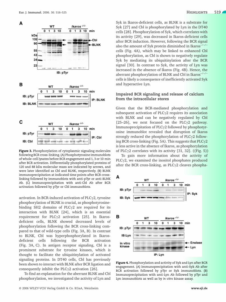
Syk in Ikaros-deficient cells, as BLNK is a substrate forSyk [27] and Cbl is phosphorylated by Lyn in the DT40cells [28]. Phosphorylation of Syk, which correlates withits activity [29], was decreased in Ikaros-deficient cellsafter BCR induction. However, following the BCR signalalso the amount of Syk protein diminished in Ikaros–/–/–
cells (Fig. 4A), which may be linked to enhanced Cblphosphorylation, as Cbl is shown to negatively regulateSyk by mediating its ubiquitinylation after the BCRsignal [30]. In contrast to Syk, the activity of Lyn wasincreased in the absence of Ikaros (Fig. 4B). Hence, theaberrant phosphorylation of BLNK and Cbl in Ikaros–/–/–
cells is likely a consequence of inefficiently activated Sykand hyperactive Lyn.
Impaired BCR signaling and release of calciumfrom the intracellular stores
Given that the BCR-mediated phosphorylation andsubsequent activation of PLCc2 requires its associationwith BLNK and can be negatively regulated by Cbl[25–26], we next focused on the PLCc2 pathway.Immunoprecipitation of PLCc2 followed by phosphotyr-osine immunoblot revealed that disruption of Ikarosstrongly reduced the phosphorylation of PLCc2 follow-ing BCR cross-linking (Fig. 5A). This suggests that PLCc2is less active in the absence of Ikaros, as phosphorylationof PLCc2 correlates with its activity [31, 32]. ((Fig. 5))
To gain more information about the activity ofPLCc2, we examined the inositol phosphates producedafter the BCR cross-linking, as PLCc2 cleaves phospha-
Figure 3. Phosphorylation of cytoplasmic signaling moleculesfollowing BCR cross-linking. (A) Phosphotyrosine immunoblotsof whole-cell lysates before BCR engagement and 1, 3 or 10 minafter BCR activation. Differentially phosphorylated proteins of120 and 88 kDa molecular mass are indicated by arrows, andwere later identified as Cbl and BLNK, respectively. (B) BLNKimmunoprecipitation at indicated time points after BCR cross-linking followed by immunoblots with anti-pTyr or anti-BLNKAb. (C) Immunoprecipitation with anti-Cbl Ab after BCRactivation followed by pTyr or Cbl immunoblots.
Figure 4. Phosphorylation and activity of Syk and Lyn after BCRengagement. (A) Immunoprecipitation with anti-Syk Ab afterBCR activation followed by pTyr or Syk immunoblots. (B)Immunoprecipitation with anti-Lyn Ab followed by pTyr andLyn immunoblots as well as by in vitro kinase assay.
Eur. J. Immunol. 2006. 36: 516–525 Highlights 519
f 2006 WILEY-VCH Verlag GmbH & Co. KGaA, Weinheim www.eji.de

tidylinositol 4,5-biphosphate to produce IP3 followingthe BCR stimulus. In accordance with reduced PLCc2phosphorylation, the increase in the amount of inositolphosphates after BCR activation was significantly(p<0.05) lower in the Ikaros-deficient cells (16�7%)than in the wild-type cells (45�7%; Fig. 5B), indicatinginefficient PLCc2 activation in the absence of Ikaros.
Since PLCc2-mediated IP3 generation is essential forthe BCR-mediated mobilization of calcium throughbinding of IP3 to its receptors, we measured the BCR-induced release of calcium from intracellular stores. Theintracellular calcium mobilization induced by cross-linking of sIgM was clearly diminished in the absence ofIkaros (Fig. 5C), as 40% less free calcium was detectedfrom the cytoplasm of Ikaros-deficient cells compared towild-type cells following the BCR engagement. Thus, inthe absence of Ikaros BCR signal strength was greatlyreduced due to inefficient PLCc2 activation.
Reintroduction of Ikaros expression restores theBCR signaling defect of Ikaros-deficient cells
To verify the observed BCR signaling defect as aconsequence of Ikaros deficiency, we transfected thefull-length Ikaros isoform Ik-1 into Ikaros–/–/– cells(Fig. 6A). The altered sIgM expression of Ikaros-deficient cells was normalized by restoration of Ikarosexpression (Fig. 6B). Moreover, the re-expression of Ik-1fully normalized the attenuated BCR-induced calciummobilization found in Ikaros–/–/– cells (Fig. 6C), in-
Figure 5. Comparison of the PLCc2 pathway activation in wild-type and Ikaros–/–/– cells following BCR cross-linking. (A)Phosphotyrosine and PLCc2 immunoblots of PLCc2 immuno-precipitation products at indicated time points after BCRactivation. (B) Increase in the intracellular inositol phosphatecontent at 45 s after the BCR cross-linking. A significantly(p<0.05) smaller increase of 16�7% (n=3) was observed inIkaros-deficient cells compared to 45�7% (n=5) increaseobserved in wild-type cells. (C) Spectrophotometric measure-ments of intracellular calcium concentration following BCRcross-linking as indicated by the arrow. About 40% less freecalcium was detected from the cytoplasm of Ikaros–/–/– cellscompared to wild-type cells following the BCR cross-linking.
Figure 6. Re-expression of the full-length Ikaros isoform Ik-1normalizes sIgM expression, restores BCR signaling, andlowers the Cbl phosphorylation observed in Ikaros–/–/– cells.(A) Ikaros immunoblots of the Ikaros immunoprecipitates fromwild-type, Ikaros–/–/– and Ikaros–/–/–/Ik-1 cells. (B) Flow cyto-metric analysis of sIgM expression using Ab M1 in wild-type,Ikaros–/–/– and Ikaros–/–/–/Ik-1 cells. Error bars indicate mean SDof independent experiments (n=3 for each of the experiments).(C) Comparison of intracellular calcium mobilization in wild-type and Ikaros–/–/–/Ik-1 cells following BCR cross-linking asindicated by the arrow. (D) Cbl and pTyr immunoblots of Cblimmunoprecipitates from Ikaros–/–/– and Ikaros–/–/–/Ik-1 cellsoverexpressing Ik-1.
Kalle-Pekka Nera et al. Eur. J. Immunol. 2006. 36: 516–525520
f 2006 WILEY-VCH Verlag GmbH & Co. KGaA, Weinheim www.eji.de

dicating that the regulation of BCR signaling isdependent on Ik-1.
To investigate whether the hyperphosphorylation ofCbl after BCR engagement would be due to theattenuated calcium signaling, we studied the phosphor-ylation of immunoprecipitated Cbl from Ik-1-transfectedIkaros–/–/– cells. Cbl phosphotyrosine immunoblotsrevealed that despite restored calcium signaling, Cblphosphorylation was diminished as a consequence of Ik-1 overexpression in Ikaros–/–/– cells after the BCRinduction (Fig. 6D). These findings suggest that Ikarosregulates Cbl phosphorylation independently from theBCR-induced calcium signaling. Furthermore, as Cbl ishyperphosphorylated in the absence of Ikaros(Fig. 3A, B) and unphosphorylated as a result of Ik-1overexpression (Fig. 6D), our results suggest that theexpression level of Ik-1 is an important factor in theregulation of Cbl phosphorylation.
Discussion
Here we show that Ikaros is required to regulate the BCRsignaling machinery, providing first molecular evidencehow Ikaros functions in B cells. Our results demonstratethat in the absence of Ikaros, reduced levels of calciumare released from intracellular stores due to insufficientPLCc2 phosphorylation after BCR cross-linking. Re-stored Ikaros expression in Ikaros-deficient cells nor-malizes the BCR signaling defect, demonstratingunequivocally that Ikaros regulates B cell function byenhancing the BCR signals. In early hematopoieticdifferentiation the expression of Flk2/Flt3 depends onIkaros [7], and it has been proposed that the earlyspecification of multipotential progenitors to the B celllineage is regulated by concerted action of Ikaros andPU.1 [33]. As the defect of Flk2/Flt3 signaling wouldexplain the early requirement for Ikaros in B celldevelopment, the defect in BCR signaling shown byour results provides an explanation for the need ofIkaros at later stages throughout the B cell lineage. Thisis because signal strength seems to be the decisive factorin B cell fate determination [21], and all resting matureB cells need basal BCR signal for survival [20].
Hypomorphic Ikaros mice expressing low levels ofIkaros have less mature B cells in the spleen than wild-type mice [6]. As a clear reduction in the transition ofimmature B cells from bone marrow to spleen has beenobserved in mice having an impaired BCR signalingcaused by deletion of Syk or by truncation of Iga [34,35], immature B cells are thought to migrate from bonemarrow to spleen through a mechanism involving BCRsignaling. Hence, the compromised BCR signaling due toinsufficient Ikaros expression could cause inefficienttransition of immature B cells to the spleen, and
subsequently lead to decreased population of splenicmature B cells, as observed in mice expressing low levelsof Ikaros [6]. Furthermore, since the signaling machi-neries of BCR and pre-BCR share multiple features, oneplausible prediction from our results would be that alsopre-BCR signaling is attenuated during Ikaros defi-ciency. We therefore suggest that (1) the partialdevelopmental block at the pro-B to pre-B transitionand (2) the impaired IL-7-dependent differentiation ofB cell precursors to IgM+ cells in hypomorphic Ikarosmice [6] are both possible consequences of aberrant pre-BCR signaling.
Thus, our results implicate that several possiblecheckpoints during B cell development are regulated byIkaros since (1) B cell progenitors undergo positiveselection at the pro-B to pre-B transition due to pre-BCRexpression and signaling, and (2) pre-BCR signaling isthought to lower the threshold of IL-7 responsepermitting the pre-BCR+ cells to thrive in the limitedIL-7 supply environment [36], leading to subsequentdifferentiation into IgM+ immature B cells.
In T cells, Ikaros is thought to set a threshold forT cell receptor (TCR) signaling [37] and our results withIkaros–/–/– DT40 B cells show that Ikaros has a criticalrole in the regulation of BCR signaling. If Ikaros isinvolved in the regulation of both BCR and TCRsignaling, and since these two signaling machinerieshave common molecules, it is likely that at least someregulatory targets andmechanisms are similar. One suchtarget molecule is Cbl, which is rapidly phophorylatedfollowing both the BCR and TCR activation [28, 38]. Ourresults indicate that Ikaros is needed to suppress theBCR-induced Cbl phosphorylation. The three consensustyrosines of Cbl phosphorylated by Src kinases and Sykupon antigen receptor activation are Y700, Y731 andY774 [38], which are reported to interact with the SH2domains of Vav, p85 of phosphatidylinositol 3-kinaseand Crk, respectively [39, 40]. It is possible that Ikarosdeficiency increases the phosphorylation of thesetyrosines, thus promoting Cbl interactions, which wouldcontribute to the ubiquitinylation of bound signalingmolecules.
Ikaros could also participate in the regulation of selftolerance, as enhanced Cbl phosphorylation has beendescribed in anergic T cells following TCR activation[40]. In fact, another member of the Ikaros family,Aiolos, which binds to the same consensus DNAsequence as Ikaros, is known to be important for theregulation of the BCR signaling threshold [41]. Aiolos-deficient mice lose B cell self tolerance, leading todevelopment of SLE-type autoimmune disease [42].Moreover, Ikaros+/– and Aiolos–/– mice develop T andB cell lymphomas, respectively [42–44]. All thesefindings are in line with our proposal that Ikaros affectsthe immune system by regulating the signaling machin-
Eur. J. Immunol. 2006. 36: 516–525 Highlights 521
f 2006 WILEY-VCH Verlag GmbH & Co. KGaA, Weinheim www.eji.de

ery of BCR (and possibly TCR), whose dysfunction leadsto the development of autoimmune diseases andlymphomas.
However, in contrast to our results showing that BCRsignal strength is diminished in the absence of Ikaros(Fig. 5C), Aiolos-deficient mouse B cells exhibit en-hanced BCR signal strength [41], suggesting that Aiolosdecreases the strength of BCR signals. These oppositephenotypes are not due to the differences in modelsystems, as we have found similar hypersignalingphenotype in Aiolos-deficient DT40 cells (unpublishedobservations). Hence, Ikaros and Aiolos seem to haveopposite function in the regulation of BCR signaling.
At themolecular level, Ikaros appears to be needed inthe regulation of primary events in the proximity of cellmembrane and BCR complex, as indicated by alteredactivity of both Lyn and Syk (Fig. 4), which are the firstcytoplasmic protein tyrosine kinases that becomeactivated upon BCR engagement in DT40 cells. Giventhat Cbl negatively regulates Syk after BCR stimulation[30], the BCR-induced decrease in the amount of Syk(Fig. 4A) is possibly related to the BCR-inducedhyperphosphorylation of Cbl (Fig. 3A, C) in the absenceof Ikaros. However, the fact that tyrosine kinase bindingdomain, which binds to tyrosine-phosphorylated Syk[45, 46], is essential for Cbl-mediated Syk ubiquitinyla-tion [30, 46], suggests that Cbl hyperphosphorylationwould not promote interactions contributing to negativeregulation of Syk. Cbl is phosphorylated by Lyn in theDT40 cells [28], and according to our results Lyn ispotentially hyperactive in the absence of Ikaros(Fig. 4B). However, the Cbl hyperphosphorylation alonedoes not explain the observed BCR signaling defect ofIkaros–/–/– cells. Furthermore, since phosphatase activityand lipid raft environment critically contributes to theactivity of Lyn [47], our results suggest that Ikaros mayalso regulate these primary events of BCR signaling.
To conclude, our results provide first molecularinsight into the functional importance of Ikaros inB cells, showing that it influences BCR-induced PLCc2-dependent calcium mobilization and Cbl phosphoryla-tion. Our Ikaros-null mutant cell line constitutes a newtool to study the function of Ikaros in B cells. This studyon the loss of Ikaros, a transcriptional regulatorassociated with nuclear remodeling complexes, providesnew avenues for research concerning the connections ofnuclear regulators to proximal signaling events on thecell membrane. Further research will be aimed tounderstand how gene expression is perturbed in Ikaros-deficient B cells and how Ikaros regulates BCR signalingat the molecular level.
Materials and methods
Cells and antibodies
Wild-type and Ikaros-mutant chicken DT40 B cells weremaintained in RPMI 1640 supplemented with 10% FCS, 1%chicken serum, 50 lM b-mercaptoethanol, 2 mM L-glutamine,penicillin and streptomycin. The cells were cultured at 40�Cwith 5% CO2. Anti-PLCc2 Ab [25], anti-Syk Ab [31], anti-LynAb [31], anti-BLNK Ab [25] and anti-ChB6 mAb (L22) [23]were described previously. Anti-Ikaros Ab (E-20), anti-Cbl Ab(C-15) and anti-pTyr mAb (PY99) were purchased from SantaCruz Biotechnology. Anti-chicken IgMmAb (M4 andM1), anti-chicken k light chain mAb (L1) and anti-CD45 mAb (LT40)were purchased from Southern Biotechnology Associates Inc.
Targeting vector construction and generation ofIkaros-deficient DT40 B cell line
In gene targeting vectors Ik-bsr, Ik-neo and Ik-pur, selectioncassettes were flanked by 4.0 and 0.7 kb of the chicken Ikarossequence in the 50 and 30 sides, respectively (Fig. 1A). The50 arm of the targeting vector was obtained by genomic PCRfrom DT40 cells using primers 6Lf and 7Lr, which weredesigned based on the coding sequence of chicken Ikaros in theexons 6 and 7, respectively. 6Lf 50-AAAGGTACCGACTAG-CAAGTAACGTCGCCTAACGTAAG-30 added an Acc65I siteand 7Lr 50-AAAGGATCCATCTTATGGAAGATCAGATAGT-CACTTCTCAC-30 added a BamHI site to the genomic PCR-sequence, which were used to clone the fragment into thepUC18 vector. The 30 arm of the targeting vector was obtainedby RT-PCR from DT40 cDNA, using primers 7Rf and 7Rrdesigned for the Ikaros exon 7 sequence. 7Rf 50-AAAGGATC-CAACTAAGAGAAGGAGAACTAGATAATGCAG-30 added a Bam-HI site and 7Rr 50-AAAGTCGACTTAACTCATGTGGAAACGGT-GCTCCCCTCGAGT-30 a SalI site, which were used to clone thefragment into the pUC18 vector. Finally, the selection markerswere cloned between the two flanking Ikaros sequences asBamHI cassettes [48].
The targeting vectors were linearized by Acc65I digestionand introduced into DT40 cells by electroporation at 710 V,25 lF. Ik-bsr was first transfected to wild-type DT40 cellsfollowed by transfections of Ik-neo and Ik-pur, which weresequentially introduced to the heterozygous cells. Transfectantclones were selected in the presence of 50 lg/mL blasticidin S(Ik-bsr transfectants), 2 mg/mL G418 (Ik-neo transfectants) or0.5 lg/mL puromycin (Ik-pur transfectants), depending of thetransfected construct.
After each transfection event the Ikaros-targeted cloneswere selected from the transfectant clones based on threegenomic PCR reactions (Fig. 1B), in which the primer 6f 50-AACTAACCAGAGTGAAATGGCTGAAGACCTG-30 specific forthe chicken Ikaros was used in combination with primersspecific for a selection marker Br (for bsr) 50-CGATTGAA-GAACTCATTCCACTCAAATATACCC-30, Nr (for neo) 50-GCGCATCGCCTTCTATCGCCTTCTTGACGAG-30 and Pr (forpur) 50-CAGCGCCCGACCGAAAGGAGCGCACGACC-30
(Fig. 1A). The obtained genomic PCR-products were probedin Southern hybridization with the Ikaros-specific probe 6p 50-CCTGTGCAAGATAGGGTCAGAAAGATCCCTCG-30.
Kalle-Pekka Nera et al. Eur. J. Immunol. 2006. 36: 516–525522
f 2006 WILEY-VCH Verlag GmbH & Co. KGaA, Weinheim www.eji.de
RT-PCR analysis
Ikaros expression in wild-type DT40, heterozygous Ikaros+/+/–
and Ikaros +/–/–, and homozygous Ikaros–/–/– clones wasanalyzed by RT-PCR. Total RNA was first isolated from5�106 cells with Trizol (Invitrogen) according to the manu-facturer's protocol. Total RNA from 1�106 cells was treatedwith DNase and then used for first-strand cDNA synthesis witholigo p(dT)15 primer (Roche). The cDNA from 1�105 cellequivalent was amplified with primers Ik-f 50-ATGGAAACA-GATGAGGCTCAA-30 and Ik-r 50-TTAACTCATGTG-GAAACGGTG-30 specific for chicken Ikaros. A PCR reactionwith primers b1-f 50-GTGCTGTGTTCCCATCTATCGT-30 andb1-r 50-TGGACAATGGAGGGTCCGGATT-30 specific for chickenb-actin was used as a positive control. Southern hybridizationwas performed for the PCR products with the Ikaros- or b-actin-specific oligonucleotides Ik-p (for Ikaros) 50-TAAATTAAAC-CACTGCGTTCCTCATTATTTGCT-30 and b1-p (for b-actin) 50-AAGCCAACAGAGAGAAGATGACACA-30.
Analysis of cellular growth
The wild-type and Ikaros–/–/– cell cultures were diluted to104 cells /mL and samples were harvested at 24-h intervals.The concentration of cells was analyzed with flow cytometerusing TruCOUNTTM tubes (Becton Dickinson) according to themanufacturer's instructions.
Immunoprecipitation and Western blot analysis
For immunoprecipitations, unstimulated and stimulated cellswere solubilized in lysis buffer (1� PBS, 1% Nonidet-P40,0.5% sodium deoxycholate, 1% SDS, 1 mM EDTA) containingprotease and phosphatase inhibitors [2 mM phenylmethylsul-fonylfluoride, 1 mM Na3VO4 and protease inhibitor 'cocktail'(Roche)], and subsequently lysed for 1 h at 4�C. Before lysisthe stimulated cells were incubated with mAb M4 (4 lg/mL)for indicated times (1, 3 or 10 min). In immunoprecipitationsof Ikaros, 20�106 cells were lysed for each sample, whereas inthe immunoprecipitations of PLCc2 each sample was preparedfrom 10�106 cells, in Lyn and Cbl immunoprecipitations from5�106 cells, and in immunoprecipitations of Syk and BLNKfrom 3�106 cells. After centrifugation, the lysates wereprecleared and sequentially incubated with appropriate Aband protein A-agarose (Santa Cruz Biotechnology) at 4�Covernight. The immunoprecipitates were washed four timeswith lysis buffer, and the samples were denatured at 75�C for10 min in the SDS sample buffer.
For Western blot analysis, the whole-cell lysates orimmunoprecipitates were separated by 4–12% SDS-PAGE,transferred onto nitrocellulose membranes, and detected byappropriate Ab and the enhanced chemiluminescence system(Amersham Pharmacia Biotech). For phosphotyrosine immu-noblots of whole-cell lysates, 1�106 cells were used for thepreparation of each sample.
In vitro kinase assay
For Lyn in vitro kinase assay, 5�106 cells were lysed andimmunoprecipitated in NP40 buffer (1% Nonidet-P40, 50 mM
Tris-HCl pH 8, 150 mM NaCl) with freshly added inhibitors[protease inhibitor cocktail (Roche) and 1 mM Na3VO4]. Theprecipitates were washed four times in NP40 buffer and threetimes in kinase buffer (20 mM Hepes pH 7.4, 5 mM magne-sium acetate, 5 mM MnCl2, 1 mM dithiothreitol). To make thereaction mixture, 2.5 lg acid-denatured rabbit muscle enolase(Sigma) was added as a substrate to 25 lL of kinase buffertogether with 10 lM cold ATP and 10 lCi [c-33P]ATP(>3000 Ci/mmol; NEN). The reactions were allowed toproceed at 30�C for 10 min and terminated by addition ofsample buffer and boiling for 3 min. Samples were separatedon 4–12% SDS-PAGE. The gel was stained, dried anddeveloped by autoradiography.
Calcium measurements
Cells (107) were suspended in Hepes-buffered salt solution(HBSS) containing 20 mM Hepes (pH 7.4), 118 mM NaCl,4.6 mMKCl, 1 mMCaCl2 and 10 mMglucose, andwere loadedwith 1.5 lMFura-2 AM (Molecular Probes) for 45 min at roomtemperature. Following the loading period cells were furtherincubated for 20 min to ensure the complete cleavage ofacetoxymethyl group from Fura-2. Cells were washed twiceand suspended toHBSS lacking CaCl2 and containing 0.05 mMEDTA. Continuous monitoring of fluorescence from cellsuspension (5�106/mL) was performed at 37�C using aHitachi F2000 fluorescence spectrophotometer at the excita-tion wavelengths of 340 and 380 nm, and an emissionwavelength of 510 nm. Calibration and calculation of calciumlevels were done as described previously [49]. Cells werestimulated with 4 lg/mL of mAb M4.
Measurement of inositol phosphates
The cells were pre-incubated with myo-[3H]inositol (10 mCi/100 mm dish) for 36 h, harvested and suspended in HBSS.Following the incubation for 10 min at 37�C, the cells weresuspended to HBSS containing 10 mM LiCl and furtherincubated for 10 min at 37�C. After the incubations, the cellswere stimulated with mAb M4 (2 lg/mL) for 45 s. Inositolphosphates were extracted using 10% v/vHClO4 and separatedusing Amprep (SAX)mini-colums [50]. The radioactivity of thesamples was measured by liquid scintillation counting.
Expression vectors and transfections
The sequence coding for the full-length isoform Ik-1 of Ikaroswas amplified from DT40 cDNA using primers Ik-fN 50-AAAGCTAGCATGGAAACAGATGAGGCTCAAGA-30 and Ik-rN50-ATTGCTAGCTTAACTCATGTGGAAACGGTGCT-30, whichcreated NheI sites to both ends of the PCR product. TheseNheI sites were used to clone the PCR product into theNheI siteof pExpress vector [48]. The expression cassette containing thecloned PCR product between the chicken b-actin promoter andSV40 poly-A sequence was excised from pExpress as a SpeIcassette and subsequently cloned into the pLoxHisD vector[48]. The resulting plasmid was linearized with NotI andtransfected into Ikaros–/–/– cells at 710 V, 25 lF. Transfectantclones were selected in the presence of 1.5 mg/mL histidinol,and the Ikaros expression level within clones was verified byIkaros immunoblots.
Eur. J. Immunol. 2006. 36: 516–525 Highlights 523
f 2006 WILEY-VCH Verlag GmbH & Co. KGaA, Weinheim www.eji.de

Acknowledgements: We thank Drs. J. Liippo and P.Kohonen for critically reading themanuscript andDr. H.Arakawa for kindly providing the pExpress and pLox-HisD vectors. Ms. M. Laine, A. Karvonen, A. Peippo, A.Wierda, M. Erlin and J. Matsson are acknowledged fortheir technical assistance. This work was financiallysupported by The European Union (QLK3-CT-2000-00785), Tekes (the National Technology Agency), theAcademy of Finland, Turku Graduate School of Biome-dical Sciences, the Finnish Cultural Foundation, theFinnish Cultural Foundation of Southwest Finland,Juliana von Wendt Foundation, the Turku UniversityFoundation, The Finnish Cancer Union, Emil and BlidaMaunula Foundation, and the Paulo Foundation.
References
1 Georgopoulos, K., Moore, D. D. and Derfler, B., Ikaros, an early lymphoid-specific transcription factor and a putative mediator for T cell commitment.Science 1992. 258: 808–812.
2 Hahm, K., Ernst, P., Lo, K., Kim, G. S., Truck, C. and Smale, S. T., Thelymphoid transcription factor LyF-1 is encoded by specific, alternativelyspliced mRNAs derived from the Ikaros gene. Mol. Cell Biol. 1994. 14:7111–7123.
3 Wang, J.-H., Nichogiannopoulou, A., Wu, L., Sun, L., Sharpe, A. H.,Bigby, M. and Georgopoulos, K., Selective defects in the development ofthe fetal and adult lymphoid system in mice with an Ikaros null mutation.Immunity 1996. 5: 537–549.
4 Georgopoulos, K., Bigby, M.,Wang, J.-H., Molnar, A., Wu, P.,Winandy, S.and Sharpe, A., The Ikaros gene is required for the development of alllymphoid lineages. Cell 1994. 79: 143–156.
5 Papathanasiou, P., Perkins, A. C., Cobb, B. S., Ferrini, R., Sridharan, R.,Hoyne, G. F., Nelms, K. A. et al., Widespread failure of hematolymphoiddifferentiation caused by a recessive niche-filling allele of the Ikarostranscription factor. Immunity 2003. 19: 131–144.
6 Kirstetter, P., Thomas, M., Dierich, A., Kastner, P. and Chan, S., Ikaros iscritical for B cell differentiation and function. Eur. J. Immunol. 2002. 32:720–730.
7 Nichogiannopoulou, A., Trevisan, M., Neben, S., Friedrich, C. andGeorgopoulos, K., Defects in hematopoietic stem cell activity in Ikarosmutant mice. J. Exp. Med. 1999. 190: 1201–1213.
8 Molnar, A. and Georgopoulos, K., The Ikaros encodes a family offunctionally diverse zinc finger DNA-binding proteins. Mol. Cell Biol. 1994.14: 8292–8303.
9 Liippo, J. and Lassila, O., Avian Ikaros gene is expressed early inembryogenesis. Eur. J. Immunol. 1997. 27: 1853–1857.
10 Sun, L., Liu, A. and Georgopoulos, K., Zinc finger-mediated proteininteractions modulate Ikaros activity, a molecular control of lymphocytedevelopment. EMBO J. 1996. 15: 5358–5369.
11 Morgan, B., Sun, L., Avitahl, N., Andrikopoulos, K., Ikeda, T., Gonzales,E., Wu, P. et al., Aiolos, a lymphoid restricted transcription factor thatinteracts with Ikaros to regulate lymphocyte differentiation. EMBO J. 1997.16: 2004–2013.
12 Kelley, C. M., Ikeda, T., Koipally, J., Avitahl, N., Wu, L., Georgopoulos, K.and Morgan, B. A., Helios, a novel dimerization partner of Ikaros expressedin the earliest hematopoietic progenitors. Curr. Biol. 1998. 8: 508–515.
13 Hahm, K., Cobb, B. S., McCarty, A. S., Brown, K. E., Klug, C. A., Lee, R.,Akashi, K. et al., Helios, a T cell-restricted Ikaros family member thatquantitatively associates with Ikaros at centromeric heterochromatin. GenesDev. 1998. 12: 782–796.
14 Georgopoulos, K.,Haematopoietic cell-fate decisions, chromatin regulationand Ikaros. Nat. Rev. Immunol. 2002. 2: 162–174.
15 Sabbattini, P., Lundgren, M., Georgiou, A., Chow, C., Warnes, G. andDillon, N., Binding of Ikaros to the 5 promoters silences transcription
through a mechanism that does not require heterochromatin formation.EMBO J 2001. 20: 2812–2822.
16 Trinh, L. A., Ferrini, R., Cobb, B. S., Weinmann, A. S., Hahm, K., Ernst, P.,Garraway, I. P. et al., Down-regulation of TdT transcription in CD4+ CD8+
thymocytes by Ikaros proteins in direct competition with Ets activator. GenesDev. 2001. 15: 1817–1832.
17 Harker, N., Naito, T., Cortes, M., Hostert, A., Hirschberg, S., Tolaini, M.,Roderick, K. et al., CD8alpha gene locus is regulated by the Ikaros family ofproteins. Mol. Cell 2001. 10: 1403–1415.
18 Kitamura, D., Roes, J., Kuhn, R. and Rajewsky, K., A B-cell-deficientmouse by targeted disruption of the membrane exon of the immunoglobulinl-chain gene. Nature 1991. 350: 423–426.
19 Meffre, E. and Nussenzweig, M. C., Deletion of immunoglobulin-b indeveloping B cells leads to cell death. Proc. Natl. Acad. Sci. USA 2002. 99:11334–11339.
20 Kraus, M., Alimzhanov, M. B., Rajewsky, N. and Rajewsky, K., Survival ofresting mature B cells depends on BCR signaling via the Igalpha/betaheterodimer. Cell 2004. 117: 787–800.
21 Casola, S., Otipody, K. L., Alimzhanov, M., Humme, S., Uyttersprot, N.,Kutok, J. L., Carroll, M. C. et al., B cell receptor signal strength determinesB cell fate. Nat. Immunol. 2004. 5: 317–327.
22 Liippo, J., Nera, K.-P., Kohonen, P., Lampisuo,M., Koskela, K., Nieminen,P. and Lassila, O., The Ikaros family and the development of earlyintraembryonic hematopoietic stem cells. Curr. Top. Microbiol. Immunol.2000. 251: 51–58.
23 Pink, J. R. and Rijnbeek, A. M., Monoclonal antibodies against chickenlymphocyte surface antigens. Hybridoma 1983. 2: 287–296.
24 Ishiai, M., Sugawara, H., Kurosaki, M. and Kurosaki, T., Association ofphospholipase C-c2 Src homology 2 domains with BLNK is critical for B cellantigen receptor signaling. J. Immunol. 1999. 163: 1746–1749.
25 Ishiai, M., Kurosaki, M., Pappu, R., Okawa, K., Ronko, I., Fu, C., Shibata,M. et al., BLNK required for coupling Syk to PLCc2 and Rac1-JNK in B cells.Immunity 1999. 10: 117–125.
26 Yasuda, T., Maeda, A., Kurosaki, M., Tezuka, T., Hironaka, K.,Yamamoto, T. and Kurosaki, T., Cbl suppresses B cell receptor mediatedphospholipase C (PLC)-c2 activation by regulating B cell linker protein-PLC-c2 binding. J. Exp. Med. 2000. 191: 641–650.
27 Fu, C., Turck, C. W., Kurosaki, T. and Chan, A. C., BLNK: A central linkerprotein in B cell activation. Immunity 1998. 9: 93–103.
28 Tezuka, T., Umemori, H., Fusaki, N., Yagi, T., Takata,M., Kurosaki, T. andYamamoto, T., Physical and functional association of the cbl protooncogeneproduct with an Src-family protein tyrosine kinase, p53/56lyn, in the B cellantigen receptor-mediated signaling. J. Exp. Med. 1996. 183: 675–680.
29 Hutchcroft, J. E., Harrison,M. L. and Geahlen, R. L., Association of the 72-kDa protein-tyrosine kinase PTK72 with the B cell antigen receptor. J. Biol.Chem. 1992. 267: 8613–8619.
30 Rao, N., Ghosh, A. K., Ota, S., Zhou, P., Reddi, A. L., Hakezi, K., Druker,B. K. et al., The non-receptor tyrosine kinase Syk is a target of Cbl-mediatedubiquinylation upon B-cell receptor stimulation. EMBO J. 2001. 20:7085–7095.
31 Takata, M., Sabe, H., Hata, A., Inazu, T., Homma, Y., Nukada, T.,Yamamura, H. et al., Tyrosine kinases Lyn and Syk regulate B cell receptor-coupled Ca2+ mobilization through distinct pathways. EMBO J. 1994. 13:1341–1349.
32 Nishibe, S.,Wahl, M. I., Hernandez-Sotomayor, S.M., Tonks, N. K., Rhee,S. G. and Carpenter, G., Increase of the catalytic activity ofphospholipase C-gamma 1 by tyrosine phosphorylation. Science 1990.250: 1253–1256.
33 Medina, K. L., Pongubala, J. M., Reddy, K. L., Lancki, D. W., Dekoter, R.,Kieslinger, M., Grosschedl, R. et al., Assembling a gene regulatory networkfor specification of the B cell fate. Dev. Cell 2004. 7: 607–617.
34 Turner, M., Gulbranson-Judge, A., Quinn, M. E., Walters, A. E.,MacLennan, I. C. and Tybulewicz, V. L., Syk tyrosine kinase is requiredfor the positive selection of immature B cells into the recirculating B cellpool. J. Exp. Med. 1997. 186: 2013–2021.
Kalle-Pekka Nera et al. Eur. J. Immunol. 2006. 36: 516–525524
f 2006 WILEY-VCH Verlag GmbH & Co. KGaA, Weinheim www.eji.de

35 Torres, R. M., Flaswinkel, H., Reth, M. and Rajewsky, K., Aberrant B celldevelopment and immune response in mice with a compromised BCRcomplex. Science 1996. 272: 1804–1808.
36 Fleming, H. E. and Paige, C. J., Pre-B cell receptor signaling mediatesselective response to IL-7 at the pro-B to pre-B cell transition via an ERK/MAP kinase-dependent pathway. Immunity 2001. 15: 521–531.
37 Avitahl, N., Winandy, S., Friedrich, C., Jones, B., Ge, Y. andGeorgopoulos, K., Ikaros sets thresholds for T cell activation and regulateschromosome propagation. Immunity 1999. 10: 333–343.
38 Feshchenko, E. A., Langdon, W. Y. and Tsygankov, A. Y., Fyn, Yes and Sykphosphorylation sites in c-Cbl map to the same tyrosine residues that becomephosphorylated in activated T cells. J. Biol. Chem. 1998. 273: 8323–8331.
39 Thien, C. B. F. and Langdon, W. Y., Cbl: Many adaptations to regulateprotein tyrosine kinases. Nat. Rev. Mol. Cell. Biol. 2001. 2: 294–307.
40 Boussiotis, V. A., Freeman, G. J., Berezovskaja, A., Barber, D. L. andNadler, L. M., Maintenance of human T cell anergy: Blocking of IL-2 genetranscription by activated Rap1. Science 1997. 278: 124–128.
41 Cariappa, A., Tang, M., Parng, C., Nebelitskiy, E., Carroll, M.,Georgopoulos, K. and Pillai, S., The follicular versus marginal zoneB lymphocyte cell fate decision is regulated by Aiolos, Btk and CD21.Immunity 2001. 14: 603–615.
42 Wang, J.-H., Avitahl, N., Cariappa, A., Friedrich, C., Ikeda, T., Renold, A.,Andrikopoulos, K. et al., Aiolos regulates B cell activation and maturationto effector state. Immunity 1998. 9: 543–553.
43 Winandy, S., Wu, P. and Georgopoulos, K., A dominant mutation in theIkaros gene leads to rapid development of leukemia and lymphoma. Cell1995. 83: 289–299.
44 Winandy, S.,Wu, L.,Wang, J.-H. andGeorgopoulos, K., Pre-T cell receptor(TCR) and TCR-controlled checkpoints in T cell differentiation are set byIkaros. J. Exp. Med. 1999. 190: 1039–1048.
45 Deckert, M., Elly, C., Altman, A. and Liu, Y.-C., Coordinated regulation ofthe tyrosine phosphorylation of Cbl by Fyn and Syk tyrosine kinases. J. Biol.Chem. 1998. 273: 8867–8874.
46 Lupher, M. L. Jr., Rao, N., Lill, N. L., Andonius, C. E., Miyake, S., Clark, E.A., Druker, B. et al., Cbl-mediated negative regulation of the Syk tyrosinekinase. A critical role for Cbl phosphotyrosine-binding domain binding toSyk phosphotyrosine 323. J. Biol. Chem. 1998. 273: 35273–35281.
47 Young, R. M., Holowka, D. and Baird, B. A lipid raft environment enhancesLyn kinase activity by protecting the active site tyrosine from depho-sphorylation. J. Biol. Chem. 2003. 278: 20746–20752.
48 Arakawa, H., Lodygin, D. and Buerstedde, J.-M., Mutant loxP vectors forselectable marker recycle and conditional knock-outs. BMC Biotechnol. 2001.1: 1–7.
49 Grynkiewicz, G., Poenie, M. and Tsien, R. Y., A new generation of Ca2+
indicators with greatly improved fluorescence properties. J. Biol. Chem.1985. 260: 3440–3450.
50 Oldham, K. G., Polyphosphoinositide turnover. In Hume, E. C. (Ed.)Receptor-effector coupling. A practical approach. Oxford University Press,Oxford 1990. pp 99–117.
Eur. J. Immunol. 2006. 36: 516–525 Highlights 525
f 2006 WILEY-VCH Verlag GmbH & Co. KGaA, Weinheim www.eji.de
Copyright © 2022 FDOKUMEN