Identifying mild cognitive impairment at baseline in the Ginkgo Evaluation of Memory (GEM) study
20
IDENTIFYING MILD COGNITIVE IMPAIRMENT AT BASELINE IN THE GINKGO EVALUATION OF MEMORY (GEM) STUDY Beth E. Snitz a , Judith Saxton a , Oscar L. Lopez a , Diane G. Ives b , Leslie O. Dunn a , Stephen R. Rapp c , Michelle C. Carlson d , Annette L. Fitzpatrick e , and Steven T. DeKosky a for the GEM Study Investigators a Department of Neurology, University of Pittsburgh b Department of Epidemiology, University of Pittsburgh c Department of Psychiatry and Behavioral Medicine, Wake Forest University d Department of Mental Health, Johns Hopkins Medical Institutions e Department of Epidemiology, University of Washington Abstract Objectives—To identify, characterize and compare the frequency of Mild Cognitive Impairment (MCI) subtypes at baseline in a large, late-life cohort (N=3,063) recruited into a dementia prevention trial. Method—A retrospective, data-algorithmic approach was used to classify participants as cognitively normal or MCI with corresponding subtype (e.g., amnestic vs. non-amnestic, single domain vs. multiple domain) based on a comprehensive battery of neuropsychological test scores, with and without Clinical Dementia Rating (CDR) global score included in the algorithm. Results—Overall, 15.7% of cases (n=480) were classified as MCI. Amnestic MCI was characterized as unilateral memory impairment (i.e., only verbal or only visual memory impaired) or bilateral memory impairment (i.e., both verbal and visual memory impaired). All forms of amnestic MCI were almost twice as frequent as non-amnestic MCI (10.0% vs. 5.7%). Removing the CDR = 0.5 (“questionable dementia”) criterion resulted in a near doubling of the overall MCI frequency to 28.1%. Conclusion—Combining CDR and cognitive test data to classify participants as MCI resulted in overall MCI and amnestic MCI frequencies consistent with other large community-based studies, most of which relied on the “gold standard” of individual case review and diagnostic consensus. The present data-driven approach may prove to be an effective alternative for use in future large-scale dementia prevention trials. Keywords MCI; Neuropsychology; Dementia Prevention Trials Mild Cognitive Impairment (MCI) is an intermediate state between normal cognitive functioning and dementia (Petersen et al., 1999). The past decade has witnessed an exponential increase in research activity focused on MCI (Petersen & O’Brien, 2006), a critical goal being further refinement and identification of a pre-dementia state of Alzheimer’s disease (AD) or Send correspondence to: Beth E. Snitz, PhD Department of Neurology, University of Pittsburgh 3471 Fifth Avenue, Suite 802 Pittsburgh, PA 15213 USA Phone: 412-692-4820 Fax: 412-692-4031 [email protected]. NIH Public Access Author Manuscript Aging Ment Health. Author manuscript; available in PMC 2010 March 1. Published in final edited form as: Aging Ment Health. 2009 March ; 13(2): 171–182. doi:10.1080/13607860802380656. NIH-PA Author Manuscript NIH-PA Author Manuscript NIH-PA Author Manuscript
Transcript of Identifying mild cognitive impairment at baseline in the Ginkgo Evaluation of Memory (GEM) study

IDENTIFYING MILD COGNITIVE IMPAIRMENT AT BASELINE INTHE GINKGO EVALUATION OF MEMORY (GEM) STUDY
Beth E. Snitza, Judith Saxtona, Oscar L. Lopeza, Diane G. Ivesb, Leslie O. Dunna, StephenR. Rappc, Michelle C. Carlsond, Annette L. Fitzpatricke, and Steven T. DeKoskya for the GEMStudy InvestigatorsaDepartment of Neurology, University of PittsburghbDepartment of Epidemiology, University of PittsburghcDepartment of Psychiatry and Behavioral Medicine, Wake Forest UniversitydDepartment of Mental Health, Johns Hopkins Medical InstitutionseDepartment of Epidemiology, University of Washington
AbstractObjectives—To identify, characterize and compare the frequency of Mild Cognitive Impairment(MCI) subtypes at baseline in a large, late-life cohort (N=3,063) recruited into a dementia preventiontrial.
Method—A retrospective, data-algorithmic approach was used to classify participants ascognitively normal or MCI with corresponding subtype (e.g., amnestic vs. non-amnestic, singledomain vs. multiple domain) based on a comprehensive battery of neuropsychological test scores,with and without Clinical Dementia Rating (CDR) global score included in the algorithm.
Results—Overall, 15.7% of cases (n=480) were classified as MCI. Amnestic MCI wascharacterized as unilateral memory impairment (i.e., only verbal or only visual memory impaired)or bilateral memory impairment (i.e., both verbal and visual memory impaired). All forms of amnesticMCI were almost twice as frequent as non-amnestic MCI (10.0% vs. 5.7%). Removing the CDR =0.5 (“questionable dementia”) criterion resulted in a near doubling of the overall MCI frequency to28.1%.
Conclusion—Combining CDR and cognitive test data to classify participants as MCI resulted inoverall MCI and amnestic MCI frequencies consistent with other large community-based studies,most of which relied on the “gold standard” of individual case review and diagnostic consensus. Thepresent data-driven approach may prove to be an effective alternative for use in future large-scaledementia prevention trials.
KeywordsMCI; Neuropsychology; Dementia Prevention Trials
Mild Cognitive Impairment (MCI) is an intermediate state between normal cognitivefunctioning and dementia (Petersen et al., 1999). The past decade has witnessed an exponentialincrease in research activity focused on MCI (Petersen & O’Brien, 2006), a critical goal beingfurther refinement and identification of a pre-dementia state of Alzheimer’s disease (AD) or
Send correspondence to: Beth E. Snitz, PhD Department of Neurology, University of Pittsburgh 3471 Fifth Avenue, Suite 802 Pittsburgh,PA 15213 USA Phone: 412-692-4820 Fax: 412-692-4031 [email protected].
NIH Public AccessAuthor ManuscriptAging Ment Health. Author manuscript; available in PMC 2010 March 1.
Published in final edited form as:Aging Ment Health. 2009 March ; 13(2): 171–182. doi:10.1080/13607860802380656.
NIH
-PA Author Manuscript
NIH
-PA Author Manuscript
NIH
-PA Author Manuscript

other dementing disorders which could serve as an early clinical target for intervention andtherapy development.
However, the MCI construct is not without criticism and debate regarding its clinical utility.At issue is that much of the research validation of MCI has stemmed from specific clinicalsettings, typically memory disorders specialty clinics. Longitudinal, population-based studiesof MCI have resulted in generally lower and much more variable prevalence estimates andrates of progression to dementia (Fisk & Rockwood, 2005; Ganguli et al., 2004; Ritchie et al.,2001). Of particular concern is the issue of stability of MCI in population-based studies, as therates of MCI subjects who “revert to normal” over follow-up are surprisingly high (e.g.,25-40%; (Ganguli et al., 2004; Larrieu et al., 2002; Palmer et al., 2002; Ritchie et al., 2001).Differences among studies in proportions/prevalence, conversion rates, and “stability “of MCIare clearly influenced by important methodological differences such as study setting, subjectselection, age, MCI definition and assessment approach.
Earlier emphasis on isolated episodic memory impairment (Petersen et al., 1999) has evolvedinto recognition of multiple presentations and etiologies beyond just AD. The InternationalWorking Group on MCI (Winblad et al., 2004) proposed an MCI classification scheme whichis in wide use today, including adaptation by the National Alzheimer’s Coordinating Centerfor its Uniform Data Set among the 30 National Institute on Aging (NIA)-funded Alzheimer’sDisease Centers across the country. The classification scheme includes decision branches foramnestic MCI (a-MCI) vs. non-amnestic MCI (na-MCI; e.g., language, attention, executivefunction or visuospatial impairment), as well as for single cognitive domain MCI (MCI-s) vs.multiple domain MCI (MCI-md). The classification scheme theoretically reflects multipleetiologies underlying mild cognitive impairments of aging, such that a-MCI-s may representa prodromal form of AD, a-MCI-md may reflect incipient AD and/or more vascular etiologies,na-MCI-s may represent prodromal frontotemporal dementia, static vascular damage, etc., andna-MCI-md may evolve into vascular dementia, dementia of Lewy body type (DLB), etc.However, evidence to date for predictive validity of MCI subtypes with longitudinal outcomedata is equivocal (Busse et al., 2006; Fischer et al., 2007; Lopez et al., 2007; Ravaglia et al.,2006; Tabert et al., 2006; Zanetti et al., 2006).
Moreover, there is a relative paucity of descriptive data from large community-based cohortsto characterize the frequency with which specific MCI subtypes occur. Lopez et al. (Lopez etal., 2003a) reported prevalence of 6 % for MCI Amnestic Type and 16 % for MCI MultipleCognitive Deficits Type (including cases with memory impairment) from the CardiovascularHealth Study (CHS), but without further sub-typing. Busse et al. (Busse et al., 2006) reportedbaseline prevalence rates of the four basic MCI subtypes from the Leipzig Longitudinal Studyof the Aged and found the highest for na-MCI-s (5 to 17 %) with no statistically significantdifferences between a-MCI and na-MCI in estimated prevalence rates. Manly et al. (Manly etal., 2005) employed a comprehensive neuropsychological battery and reported a higherfrequency of na-MCI (17%) compared to a-MCI (11%) among ethnically diverse northernManhattan elders. The estimated prevalence for isolated memory impairment was 5 %. Thusthe frequency of MCI subtypes reported in population cohorts is quite variable, althoughevidence to date indicates that single domain amnestic MCI, long the focus of clinic-basedstudies (Petersen et al., 1999), is consistently among the least prevalent cognitive profiles ofMCI in the community.
It is therefore of great interest to further characterize specific cognitive profiles in largecommunity-based cohorts, using comprehensive cognitive testing across multiple cognitivedomains. For example, the frequency of verbal-only vs. visual-only vs. bilateral memoryimpairment in a-MCI is not usually reported and may be useful in further refining predictionof AD progression. Specific patterns of non-memory test impairment within a-MCI-md may
Snitz et al. Page 2
Aging Ment Health. Author manuscript; available in PMC 2010 March 1.
NIH
-PA Author Manuscript
NIH
-PA Author Manuscript
NIH
-PA Author Manuscript

be important to delineate, as there is increasing evidence that memory deficits plus secondarydeficits, particularly in executive functions, are much more predictive of progression thanepisodic memory deficits alone (Bozoki et al., 2001; Tabert et al., 2006). Finally, it is importantto identify and characterize non-amnestic forms of MCI, most prevalent in population cohorts,so that their clinical course can be followed and better understood, and appropriateinterventions initiated as indicated.
A further relevant issue in identifying MCI in community cohorts is the respective roles of theclinical interview vs. formal psychometric testing. The Clinical Dementia Rating (CDR) scaleis the most widely used interview instrument in research settings to rate individuals along acontinuum from normal to severe dementia, and is based on reports of functional change fromthe subject and a knowledgeable informant (Morris, 1993). Proponents of the CDR proposethat, when used correctly, informant-based report of intra-individual change in dailyfunctioning is a more sensitive predictor of dementia than cognitive testing (Morris et al.,2001). How adequately the meaning of CDR scores generalize from clinical settings tocommunity settings, however, remains a critical question (Meguro et al., 2004; Petersen,2004).
The aims of this paper are to identify, characterize and compare the frequency of MCI subtypesin a large, late-life cohort recruited into a dementia prevention trial (Ginkgo Evaluation ofMemory, GEM, Study (DeKosky & al., 2006)). The GEM study assessed detailed cognitiveand functional status at study entry, using a broad neuropsychological test battery andadministration of the CDR with both participant and a collateral source (e.g., family member,close friend, etc.). An important long-term goal is to establish predictive validity of theseassessment and classification methods for future dementia prevention trials.
METHODSThe design of the GEM study has been reported (DeKosky & al., 2006). Briefly, participantswere recruited from four clinical sites, University of Pittsburgh, University of California-Davis,The Johns Hopkins University and Wake Forest University. Other centers involved in theadministration and conduct of the study are a data coordinating center, a clinical coordinatingcenter, a blood laboratory/repository and a diagnostic center for evaluation of dementia. TheData Coordinating Center at the University of Washington, Department of Biostatistics handlescomputer system design, data management and statistical analyses. The Clinical CoordinatingCenter, centered at Wake Forest University, oversees clinical operations, adherence andretention. Blood samples and DNA are sent to the Central Blood Laboratory at the Universityof Vermont for analysis and storage. The Cognitive Diagnostic Center is located at theUniversity of Pittsburgh and has oversight of cognitive screening and entry of cases into thestudy, establishing the neuropsychological test battery, reviewing all evaluation data onsuspected cognitively declined cases, and final adjudication of diagnosis.
ParticipantsDetailed information regarding recruitment of GEM participants has been published(Fitzpatrick et al., 2006). Recruitment began in September of 2000 and was completed in Mayof 2002. The goal of GEM was to include individuals with normal cognition or MCI but toexclude those with dementia. Inclusion criteria included English as the primary language andage 75 years and above. Participants with neurological or neurodegenerative diseases that bythemselves could significantly affect cognition, or participants who carried a higher risk ofdementia (e.g. Parkinson’s disease) were excluded, as were those hospitalized for depressionin the past year. Similarly, anyone on cognitive enhancing medications or treatments forAlzheimer’s disease (AD) (cholinesterase inhibitors or glutamate receptor modulators) wasalso excluded, as were individuals who would not agree to restrict their vitamin E intake to
Snitz et al. Page 3
Aging Ment Health. Author manuscript; available in PMC 2010 March 1.
NIH
-PA Author Manuscript
NIH
-PA Author Manuscript
NIH
-PA Author Manuscript

400 mg/day. Use of over-the-counter Ginkgo biloba was also an exclusion, but the use of otherherbal products was permitted. Because of the questions concerning increased bleeding risksassociated with Ginkgo biloba (Ang-Lee et al., 2001; Rosenblatt & Mindel, 1997) individualswith bleeding disorders, thrombocytopenia, those taking anticoagulants, or with other similarrisks were excluded. A critical inclusion criterion was the requirement that each participantidentify an individual willing to serve as a proxy, someone who had direct or phone contactwith the participant on average at least ten hours per week. This person served as the proxy forthe CDR.
A total of 3,072 participants were recruited into the GEM study. A variety of recruitmentapproaches were used (Fitzpatrick et al., 2006). The vast majority of participants were recruitedvia mass mailings based on voter registration lists, commercially purchased mailing lists, anduniversity lists. Less than eight percent of participants had responded to media advertisements.Three participants were subsequently excluded because of protocol violations which were notdetected at baseline, resulting in a final baseline N of 3,069.
Study procedureThis study was approved by the University of Pittsburgh Institutional Review Board and wasconducted in accordance with the Helsinki Declaration(http://www.wma.net/e/policy/17-c_e/html). Recruitment and screening were done in threephases: phone, clinic screening, and baseline assessment, with cognitive, medical and otherexclusion criteria performed at each stage. The initial telephone interview included a self-reported medical history and the Telephone Interview for Cognitive Assessment (TICS)(Brandt et al., 1988) using a exclusionary cut-off score of 28 or below. Individuals meetingcriteria at this stage were scheduled for a clinic screening visit, during which all participantsand their proxies completed the informed consent process. The screening visit includedadministration of the Modified Mini Mental State examination (3MSE) (Teng & Chui, 1987),the Center for Epidemiological Studies Depression (CES-D) scale (Radloff, 1977), and detailedhealth history and activities of daily living questionnaires. Cut-off scores predicting dementiawere established for the 3MSE based on data from the Cardiovascular Health Study (CHS)cohort (Lopez et al., 2003b). Individuals scoring less than 80 on the 3MSE were excluded. Theremaining participants completed the comprehensive neuropsychological test battery, whichincluded tests within five cognitive domains typically evaluated as part of a work-up fordementia: attention/psychomotor speed; memory for verbal and visual material; languagefunctions; visuospatial/constructional ability; and executive functions including workingmemory ability (Table 1). The battery also included two tests to estimate premorbid intellectualfunctioning, the National Adult Reading Test — American Version (Grober & Sliwinski,1991); and Raven’s Coloured Progressive Matrices (Raven, 1956). Cognitive testing took placein a quiet area without distraction. All technicians administering neuropsychological tests weretrained and certified by experienced study personnel.
We established cut-off scores on neuropsychological tests for the purposes of screening outdementia. These were based on CHS test data from cognitively normal participants, and weredefined as a score equal to or greater than 1.5 standard deviations below the CHS age- andeducation-stratified means. The neuropsychological tests were grouped into cognitivedomains, with a domain defined as two or more tests assessing a cognitive construct.Participants with scores below cut-off on two or more cognitive domains, with one impaireddomain being memory, were excluded on the basis that this level of test performance indicatedthe presence of dementia. Sixty-four participants who had passed the 3MSE screening (i.e.,3MSE ≥ 80) were excluded in this manner (mean age 79.8 years, mean education 13.8 years).
The screening visit included blood drawn for laboratory screening as well as DNA storage forsubsequent apolipoprotein E (ApoE) genotyping. DNA was extracted with a Puregene Kit
Snitz et al. Page 4
Aging Ment Health. Author manuscript; available in PMC 2010 March 1.
NIH
-PA Author Manuscript
NIH
-PA Author Manuscript
NIH
-PA Author Manuscript

(QUIAGEN; Valencia, CA) and ApoE genotyping was performed utilizing the TaqMangenotyping assays (Applied Biosystems; Foster City, CA) by Dr. M. Ilyas Kamboh, Universityof Pittsburgh. Of note, 16 participants refused phlebotomy consent, 425 samples hadinsufficient volume for DNA extraction and 174 did not have sufficient blood drawn for otherreasons, resulting in a subset of 2454 of total baseline participants with ApoE data available.
During the screening visit participants and their proxies were also administered the CDR(Hughes et al., 1982; Morris, 1993), an interviewer-based rating of functional and cognitiveabilities. Ratings are given for six domains - memory, orientation, home and hobbies, judgmentand problem solving, community affairs and personal care - based on information obtainedfrom both the subject and informant. A scoring algorithm takes into account each categoryscore with an additional weighting for the memory section, yielding a global score from 0 (nodementia) to 3 (severe dementia), with 0.5 representing “questionable dementia.” All GEMstudy interviewers completed a rigorous CDR training program, including training andcertification videotaped interviews. Of the participants’ informants who provided informationfor the CDR ratings, 59.6% reported living with the participants. Of those informants who didnot live with a participant, 78% reported visiting with the participant at least once a week and72.2% reported phone or e-mail contact with participants at least once a week.
Participants who passed the neuropsychological test battery returned to the clinic for theirbaseline GEM study visit. At that visit they completed the cognitive portion of the Alzheimer’sDisease Assessment Scale (ADAS-Cog) (Rosen et al., 1984), a symptom and health habitsquestionnaire, and were randomized to one of the two treatment arms (Ginkgo biloba orplacebo). This paper will focus on the results of the cognitive assessments, the CDR interviewand self-reported health history and daily functioning completed during the screening andbaseline visits.
Algorithmic identification of MCI and subtypesFollowing guidelines set out by the International Working Group on MCI (Winblad et al.,2004), we developed data-driven criteria to identify baseline MCI cases in this non-dementedcohort: 1) “self and/or informant report of cognitive decline” was operationalized as CDRglobal score = 0.5; 2) “impairment on objective cognitive tasks” was implemented as aminimum of 2 out of 10 selected neuropsychological test scores (Table 1) impaired at or belowthe 10th percentile of CHS normative data, stratified by age and education (see below); 3)“preserved basic activities of daily living / minimal impairment in complex instrumentalfunctions” was defined by a CDR global rating < 1. Thus the range of functional changeidentified as consistent with MCI in this non-demented cohort was defined by CDR globalscore = 0.5, and within this range of mild cognitive decline in daily life we required evidenceof objective impairment on formal testing. However, we also report results of cognitive testimpairment regardless of CDR score, to examine the impact of CDR rating on MCI subtypefrequencies, and eventually, to examine specific predictors of outcome individually and incombination (i.e., clinical interview data and psychometric test scores).
CHS normative neuropsychological test data were derived from 432 participants clinicallyadjudicated as having normal cognition based on the same comprehensive neuropsychologicalbattery as the one used in the GEM study (Lopez et al., 2003a; Lopez et al., 2007). CHSadjudication had included review of serial neuropsychological data, proxy report of functionalstatus, psychiatric symptoms, and neurological exam findings. Cut-off scores onneuropsychological tests were originally derived from an independent sample of 250unimpaired subjects and were set at > 1.5 SDs below the mean of individuals of comparableage and education for CHS adjudication (Lopez et al., 2003a).
Snitz et al. Page 5
Aging Ment Health. Author manuscript; available in PMC 2010 March 1.
NIH
-PA Author Manuscript
NIH
-PA Author Manuscript
NIH
-PA Author Manuscript

These 432 participants with normal cognition were then stratified into 2 age (< 80 vs. ≥ 80years) × 3 education groups (≤ 12, 13 – 16, and > 16 years) with individual cell n’s rangingfrom n = 34 (≥ 80 and > 16 years education) to n = 107 (< 80 and ≤ 12 years education). Onlyone cell had an n < 50. These age- by education-defined strata were well matched in mean ageand education to the GEM baseline cohort stratified in the same manner (p’s > .05). Tenthpercentile score cut-offs were located within each of the six CHS age-by-education strata foreach of the 10 cognitive test variables. This level of mild impairment was selected because itis close to the commonly used cut-off of 1.5 S.D. below the normative mean (correspondingto approximately the 7th percentile on a theoretically normal curve). However, we required asomewhat higher, more sensitive cut-off since this cohort had already been screened fordementia. As well, many of the cognitive test scores were not normally distributed andpercentile rank cut-offs are therefore more appropriate (Mitrushina et al., 2005). Tenthpercentile cut-offs have been used in other MCI studies (Alladi et al., 2006; Schoonenboom etal., 2005; Solfrizzi et al., 2004).
A minimum of impairment on two out of ten test variables was selected as the basic cognitivecriterion, in an attempt to minimize the number of MCI cases that “revert to normal” on follow-up primarily because of spurious psychometric issues (e.g., measurement error, regression tothe mean, etc.). Examining the distribution of the number of tests impaired (Figure 1) aided inthe decision to choose two tests as a threshold of clinical significance in MCI. For instance,the rate of impairment on one test was relatively common and was not different between CDR0 and CDR 0.5 participants (28% vs. 27%), suggesting this likely represents the base-rate oftest impairment (i.e., diagnostic false positives) observed in healthy older adults (Brooks et al.,2007;Palmer et al., 1998).
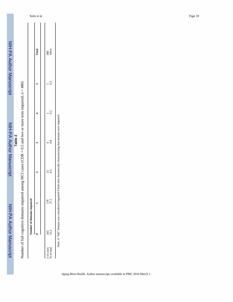
Once the basic MCI criteria were fulfilled, mutually exclusive MCI subtypes were defined asfollows: a-MCI included all MCI cases with either one (unilateral) or both (bilateral) memorytests impaired, with the remaining cases classified as na-MCI. Single domain MCI, includingbilateral amnestic MCI-s, was strictly defined as a full domain impaired (both tests) with noimpaired tests outside of that domain (i.e., the number of tests impaired in MCI-s = 2 for allcases). MCI-md was broadly defined as any MCI case that did not qualify for MCI-s. Again,inspection of the distribution of number of domains impaired supported classificationdecisions: The majority of MCI cases did not have full domains impaired but rather hadscattered, cross-domain tests affected (at least two by definition); these cases would beclassified as MCI-md by the current algorithm (see Table 2).
Data analysis
RESULTSOf the total 3,069 participants at baseline, 1832 (59.7%) had global CDR ratings of 0 (sum ofboxes range 0 – 1.0), 1,231 participants (40.1%) had global CDR ratings of 0.5 (sum of boxesrange 0.5 – 4.5), 2 participants had a CDR rating of 1.0 (but did not meet neuropsychologicalcriteria for dementia at study screening), and 4 were missing CDR data. The 6 participants witheither missing CDR ratings or CDR = 1.0 were excluded from the remaining analyses.
The mean age of the baseline GEMS cohort was 78.5 years (SD 3.3) and the mean number ofyears of education was 14.3 (SD 3.0). The proportion of women was 46.2 % and the proportionof participants who identified themselves as white was 95.5 %. See (DeKosky & al., 2006) forfull description of baseline characteristics of the GEMS cohort.
Figure 2 shows the classification tree for MCI and sub-types following application of thealgorithm. An overall frequency of 15.7 % of baseline cases were classified as MCI. AmnesticMCI, including both unilateral and bilateral memory impairment cases, was almost twice as
Snitz et al. Page 6
Aging Ment Health. Author manuscript; available in PMC 2010 March 1.
NIH
-PA Author Manuscript
NIH
-PA Author Manuscript
NIH
-PA Author Manuscript

frequent as na-MCI (10.0% vs. 5.7%). Full, single domain MCI (both amnestic and non-amnestic) was rare (1.2%), largely because of the restrictive definition used. Of the unilateralamnestic MCI, verbal memory impairment (4.6%) was more frequent than visual memoryimpairment (2.8%).Visual amnestic MCI cases had a significantly higher mean number ofimpaired tests (3.4 out of 10) compared to verbal amnestic MCI cases (2.9 out of 10) (p < .01),indicating that they tended to be more impaired overall.
Among the a-MCI cases (n = 305, including bilateral and unilateral a-MCI), the most frequentsecond full domain impaired was Construction (n = 34, 11.1%), followed by Language (n =27, 8.8%) and Executive Functions (n = 25, 8.2%). The most frequent non-memory testimpaired among a-MCI cases was Rey figure copy (n = 122, 39.9%), followed by animalfluency (n=95, 31.0%), and Stroop interference (n=77, 25.2%).
Because MCI definitions in the literature vary widely, we replicated the classification treewithout the initial “filter” of CDR = 0.5 in order to consider a more strictly psychometricdefinition of MCI. Appendix 1 presents this classification scheme. Inclusion of all cases withCDR 0 and CDR 0.5 results in an overall MCI frequency (28.1 %) almost double the frequencyobserved when CDR 0.5 was required in the MCI criteria (15.7%). The frequency of a-MCI(including bilateral and unilateral cases) is about 50% more frequent (16.6% vs. 10.0%), andna-MCI is approximately twice as frequent (11.5% vs. 5.7%) when the CDR 0.5 requirementis lifted. Within overall MCI, the distributions of subtypes relative to each other (e.g., singlevs. multidomain, amnestic vs. non-amnestic, verbal vs. visual memory impairment) are verysimilar whether or not CDR 0.5 is applied as a criterion.
Demographic, clinical, cognitive and health-related baseline variables for three MCI subtypesand the cognitively normal group are summarized in Table 3. Compared to the normal group,all MCI groups had scores indicating worse performance on the 3MSE and ADAS, as well aslower scores on the two estimated premorbid intellectual ability tests. In general there weredifferences across all groups in gender (highest proportion of females among non-amnesticMCI), self-perceived general health, difficulty with any instrumental activity of daily living(IADL), and history of stroke (highest proportion in the unilateral amnestic MCI group). MCIgroups tended to be slightly older, tended to have more depressive symptoms (although withina restricted range due to study entry screening), and tended to drink fewer alcoholic drinks perweek compared to cognitively normal participants. There were no significant differencesamong groups in presence of the apoe4 allele. We also tested for a difference in apoe4 allelefrequency between all MCI subtypes combined (n = 89, 24.4%) and the normal cognition group(n = 434, 20.9%), which approached, but did not reach, statistical significance (Fisher’s ExactTest, 1-sided, p = .075).
We examined mean memory scores (Table 3) to determine if non-amnestic MCI participantsshowed memory performance lower than expected, even though these participants did not meetthe dichotomous test score cut-off for either memory test. For visual memory but not verbalmemory, the non-amnestic MCI group had a significantly lower mean test score than thecognitively normal group.
Finally, we examined whether there were differences across the four GEM sites in the overallfrequency of MCI. The proportion of MCI cases was higher at Wake Forest University (WFU)(20.5%) than at the other clinical centers (ranging from 13.3% to 14.9%). WFU also had thehighest cognitive ineligibility rate during the screening visit (14.8% compared to 8.7% -12.4%). Possible reasons for these differences include more limited quality of educationalopportunities among this elderly cohort from the South (Manly et al., 2002), and a greaterproportion of preclinical cerebrovascular disease in Southern states (El-Saed et al., 2006).However, these factors remain speculative at this time.
Snitz et al. Page 7
Aging Ment Health. Author manuscript; available in PMC 2010 March 1.
NIH
-PA Author Manuscript
NIH
-PA Author Manuscript
NIH
-PA Author Manuscript

DISCUSSIONThe goal of this paper was to characterize the GEM study cohort at baseline with regard toMCI and its sub-types using a data-driven algorithm. Following the guidelines of theInternational MCI Working Group (Winblad et al., 2004), we required evidence of bothcognitive test impairment and caregiver/participant report of decline in implementing thegeneral definition of MCI. Sub-type classification was based upon scores from acomprehensive neuropsychological evaluation covering five domains of cognition.Application of the algorithm resulted in an overall MCI frequency of 15.7%. Althoughcomparison to prevalence rates reported in other studies is difficult due to variable definitions,impairment thresholds, and assessment and classification methods, our 15.7% frequency ofoverall MCI (including non-amnestic subtypes) in GEM is consistent with other reports fromthe large community-based studies that examined all subtypes of MCI, such as CHS (19%,(Lopez et al., 2003a), the Leipzig Longitudinal Study of the Aged (15.0%, all MCI, 1.0 SDlevel; (Busse et al., 2003) and other community-based studies (16.2%, (Zanetti et al., 2006).Our overall MCI frequency reported here is much lower than the estimated prevalence of 28.3%from an ethnically diverse cohort in northern Manhattan (Manly et al., 2005), although it isimportant to note that those northern Manhattan participants had a mean educational level of8.2 years, compared to 14.3 years in the GEM study.
The 10.0% frequency of all amnestic MCI observed in the GEM baseline cohort (unilateralplus bilateral amnestic MCI cases) is also difficult to compare to other studies, most of whichhave focused on the Petersen et al. (Petersen et al., 1999) definition of isolated memoryimpairment. The frequency of isolated memory impairment presently reported is very low(0.6%) because of the strict definition: two memory tests impaired and no other test impaired,within the context of reported cognitive decline (CDR of 0.5). If cases with isolated,unilateral memory impairment were included (n = 107), however, the total frequency of isolatedmemory impairment (n= 124, 4.0%) is in line with other studies which did not require bothvisual and verbal delayed recall deficits (5% CHS, (Lopez et al., 2003a); 2.8 % PAQUID,(Larrieu et al., 2002); 2.9 – 4.0 % MoVIES, (Ganguli et al., 2004); 5% CHSA, (Fisk et al.,2003); 6.4% Northern Manhattan Elders Study, (Manly et al., 2005). An advantage of thepresent approach is the ability to examine and track the evolution of verbal vs. visual memoryimpairment separately. Many population studies of MCI use only verbal memory measures (asdo many clinical evaluations by practitioners). Our data indicate that almost half of all MCIcases have one modality of memory tests impaired but not the other, while within this sub-group, eighty-five individuals (37%) were impaired on the visual memory test only. The visualamnestic MCI cases also tended to be more impaired than the verbal amnestic MCI cases, witha higher mean number of tests impaired. Certainly, the bilateral amnestic MCI cases (16% ofall MCI) are predicted to be at highest risk for progression to AD. To what extent assessingboth modalities of memory increases predictive validity for future cognitive decline can bedetermined on follow-up analyses.
Within the amnestic MCI group, impairments on non-memory tests were relatively commonand distributed fairly evenly among tests of visual-spatial construction, language and executivefunctions. This finding is consistent with Bäckman et al. (Backman et al., 2005) meta-analysisof cognitive deficits in preclinical AD reporting sizeable deficits in multiple cognitive domainsin addition to episodic memory. Again, future studies from this cohort will examine whichcognitive measures in addition to memory will be most predictive of progression to dementia.
The relative contributions of the informant-based clinical interview (CDR) and formalpsychometric testing, and the combination of the two approaches as illustrated here in the basicdefinition of MCI, will also be determined on follow up. Forty percent of GEM studyparticipants at baseline were rated with a global CDR score of 0.5, indicating “questionable
Snitz et al. Page 8
Aging Ment Health. Author manuscript; available in PMC 2010 March 1.
NIH
-PA Author Manuscript
NIH
-PA Author Manuscript
NIH
-PA Author Manuscript

dementia.” While this number appears surprisingly high, Meguro et al. (Meguro et al., 2004)reported an overall prevalence of CDR 0.5 to be 30.2% in a large Japanese community-dwellingcohort. These rates raise the question of the ‘transferability’ of the CDR scale, originallydeveloped for use in specialty memory disorder clinics, to community studies with clearlydifferent selection and population characteristics, even when staff are trained with thestandardized methods utilized in the specialty clinics. The meaning, interpretation andpredictive validity of CDR 0.5 in community/population studies will be compared to clinic-based studies in follow-up. Notably, the CDR and neuropsychological test results areconcordant to a large degree (e.g., Table 3; Figure 1); however, there is also considerableindependence, e.g., a full 49% of CDR 0 participants had one or more cognitive tests impaired,and a sizeable proportion (9%) had at least three tests impaired. Furthermore, 34% of CDR 0.5participants had no tests impaired. Clearly these two assessment approaches are non-overlapping and provide divergent kinds of information about cognitive decline.
An important limitation of the present study which may confine the generalizability of thefindings is the demographic nature of the cohort, that being mostly Caucasian, healthy, andrelatively highly educated. Willingness to participate and be randomized in a long-term clinicaltrial is a selection factor that makes this cohort less representative. However, the differenceswill be instructive, as effective implementation of large-scale dementia prevention trialsdepends upon comprehensive understanding of typical participant characteristics that will havean impact on outcome, e.g., cognitive test performance, informant-based reports of cognitive/daily functional decline, etc. It is also noteworthy that less than eight percent of participantshad initially responded to study recruitment media advertisements, motivated perhaps bymemory or other cognitive concerns. This subset of participants may be significant enough innumber to upwardly bias the reported frequency of MCI, yet it is not nearly as high as in mostclinical trials in which the majority of participants are self-selected. Nevertheless, it is the casethat recruitment and selection characteristics of the GEM study cohort are likely a unique hybridof representative community sampling and clinical trial selection bias.
Another issue to consider is the nature of MCI case definition, which is retrospective andalgorithm-based, rather than prospective and with case-by-case clinical adjudication, twofactors strongly advocated by Petersen (Petersen & Knopman, 2006) and others. One of theaims of our developed algorithm was to take a conservative approach and maximize specificity,perhaps at the expense of sensitivity, in order to minimize the rate of “reversion to normal”observed with high frequency in numerous population studies (Ganguli et al., 2004; Larrieu etal., 2002; Palmer et al., 2002; Ritchie et al., 2001; Visser et al., 2002). To achieve this aim, werequired evidence of at least two tests impaired, as well as evidence of informant report ofcognitive decline, for MCI case definition, a more restrictive definition than many populationstudies which have shown instability of the MCI phenotype. Follow-up analyses will determinethe utility of this approach.
Supplementary MaterialRefer to Web version on PubMed Central for supplementary material.
Appendix 1Alternate classification scheme of GEMS study baseline cohort for MCI and composite MCIcognitive sub-types, without regard to CDR score. Percentages in diamonds representfrequencies of MCI sub-types referenced to entire cohort with valid CDR 0 or CDR 0.5, N =3063.
Snitz et al. Page 9
Aging Ment Health. Author manuscript; available in PMC 2010 March 1.
NIH
-PA Author Manuscript
NIH
-PA Author Manuscript
NIH
-PA Author Manuscript

Appendix 2. GEMS study personnel
Project OfficeRichard L. Nahin, PhD, MPH, Barbara C. Sorkin, PhD, National Center for Complementaryand Alternative Medicine
Clinical CentersLinda Fried, MD, MPH, Michelle Carlson, PhD, Pat Crowley, MS, Claudia Kawas, MD, PauloChaves, MD, Joyce Chabot, John Hopkins University; John Robbins, MD, MHS, KatherineGundling, MD, Sharene Theroux, CCRP, Lisa Pastore, CCRP, University of California-Davis;Lewis Kuller, MD, DrPH, Roberta Moyer, CMA, Cheryl Albig, MA, University of Pittsburgh;Gregory Burke, MD, Steve Rapp, PhD, Dee Posey, Margie Lamb, RN, Wake Forest UniversitySchool of Medicine
Schwabe PharmaceuticalsRobert Hörr, MD, Joachim Herrmann, PhD.
Data Coordinating CenterRichard A. Kronmal, PhD, Annette L. Fitzpatrick, PhD, Fumei Lin, PhD, Cam Solomon, PhD,Alice Arnold, PhD, University of Washington
Cognitive Diagnostic CenterSteven DeKosky, MD, Judith Saxton, PhD, Oscar Lopez, MD, Beth Snitz PhD, M. IlyasKamboh PhD, Diane Ives, MPH, Leslie Dunn, MPH, University of Pittsburgh
Clinical Coordinating CenterCurt Furberg, MD, PhD, Jeff Williamson, MD, MHS; Nancy Woolard, Kathryn Bender,Pharm.D., Susan Margitić, MS, Wake Forest University School of Medicine
Central LaboratoryRussell Tracy, PhD, Elaine Cornell, UVM, University of Vermont
MRI Reading CenterWilliam Rothfus MD, Charles Lee MD, Rose Jarosz, University of Pittsburgh
Data Safety Monitoring BoardRichard Grimm, MD, PhD (Chair), University of Minnesota; Jonathan Berman, MD, PhD(Executive Secretary), National Center for Complementary and Alternative Medicine; HannahBradford, M.Ac., L.Ac., MBA, Carlo Calabrese, ND MPH, Bastyr University ResearchInstitute; Rick Chappell, PhD, University of Wisconsin Medical School; Kathryn Connor, MD,Duke University Medical Center; Gail Geller, ScD, Johns Hopkins Medical Institute; BorisIglewicz, Ph.D, Temple University; Richard S. Panush, MD, Department of Medicine SaintBarnabas Medical Center; Richard Shader, PhD, Tufts University.
Snitz et al. Page 10
Aging Ment Health. Author manuscript; available in PMC 2010 March 1.
NIH
-PA Author Manuscript
NIH
-PA Author Manuscript
NIH
-PA Author Manuscript

REFERENCESAlladi S, Arnold R, Mitchell J, Nestor PJ, Hodges JR. Mild cognitive impairment: applicability of research
criteria in a memory clinic and characterization of cognitive profile. Psychological Medicine 2006;36(4):507–515. [PubMed: 16426486]
Ang-Lee MK, Moss J, Yuan CS. Herbal medicines and perioperative care. Journal of American MedicalAssocation 2001;286:208–216.
Backman L, Jones S, Berger A-K, Laukka EJ, Small BJ. Cognitive impairment in preclinical Alzheimer’sdisease: a meta-analysis. Neuropsychology 2005;19(4):520–531. [PubMed: 16060827]
Becker JT, Boller F, Saxton J, McGonigle-Gibson KL. Normal rates of forgetting of verbal and non-verbal material in Alzheimer’s disease. Cortex 1987;23(1):59–72. [PubMed: 3568706]
Bozoki A, Giordani B, Heidebrink JL, Berent S, Foster NL. Mild cognitive impairments predict dementiain nondemented elderly patients with memory loss. Archives of Neurology 2001;58(3):411–416.[PubMed: 11255444]
Brandt J, Spencer M, Folstein M. The telphone interview for cognitive status. Neuropsychiatry,Neuropsychology and Behavioral Neurology 1988;1:111–117.
Brooks BL, Iverson GL, White T. Substantial risk of “Accidental MCI” in healthy older adults: base ratesof low memory scores in neuropsychological assessment. Journal of the InternationalNeuropsychological Society 2007;13(3):490–500. [PubMed: 17445298]
Busse A, Bischkopf J, Riedel-Heller SG, Angermeyer MC. Subclassifications for mild cognitiveimpairment: prevalence and predictive validity. Psychological Medicine 2003;33(6):1029–1038.[PubMed: 12946087]
Busse A, Hensel A, Guhne U, Angermeyer MC, Riedel-Heller SG. Mild cognitive impairment: long-term course of four clinical subtypes. Neurology 2006;67(12):2176–2185. [PubMed: 17190940]
DeKosky ST, al. E. The Gingko Evaluatio of Memory (GEM) Study: Design and baseline data of arandomized trial of Gingko biloba extract in prevention of dementia. Contemporary Clinical Trials2006;27:238–253. [PubMed: 16627007]
Delis, DC. California Verbal Learning Test. Psychological Corporation; New York: 1987.El-Saed A, Kuller LH, Newman AB, Lopez O, Costantino J, McTigue K, Cushman M, Kronmal R.
Geographic variations in stroke incidence and mortality among older populations in four UScommunities. Stroke 2006;37(8):1975–1979. [PubMed: 16794205]
Fischer P, Jungwirth S, Zehetmayer S, Weissgram S, Hoenigschnabl S, Gelpi E, Krampla W, Tragl KH.Conversion from subtypes of mild cognitive impairment to Alzheimer dementia. Neurology 2007;68(4):288–291. [PubMed: 17242334]
Fisk JD, Merry HR, Rockwood K. Variations in case definition affect prevalence but not outcomes ofmild cognitive impairment. Neurology 2003;61(9):1179–1184. [PubMed: 14610117]
Fisk JD, Rockwood K. Outcomes of incident mild cognitive impairment in relation to case definition.Journal of Neurology, Neurosurgery & Psychiatry 2005;76(8):1175–1177.
Fitzpatrick AL, Fried LP, Williamson J, Crowley P, Posey D, Kwong L, Bonk J, Moyer R, Chabot J,Kidoguchi L, Furberg CD, DeKosky ST, Investigators, G. E. M. S. Recruitment of the elderly intoa pharmacologic prevention trial: the Ginkgo Evaluation of Memory Study experience.Contemporary Clinical Trials 2006;27(6):541–553. [PubMed: 16949348]
Ganguli M, Dodge HH, Shen C, DeKosky ST. Mild cognitive impairment, amnestic type: anepidemiologic study. Neurology 2004;63(1):115–121. [PubMed: 15249620]
Grober E, Sliwinski M. Development and validation of a model for estimating premorbid verbalintelligence in the elderly. Journal of Clinical & Experimental Neuropsychology: Official Journal ofthe International Neuropsychological Society 1991;13(6):933–949.
Hughes CP, Berg L, Danziger WL, Coben LA, Martin RL. A new clinical scale for the staging of dementia.British Journal of Psychiatry 1982;140:566–572. [PubMed: 7104545]
Larrieu S, Letenneur L, Orgogozo JM, Fabrigoule C, Amieva H, Le Carret N, Barberger-Gateau P,Dartigues JF. Incidence and outcome of mild cognitive impairment in a population-based prospectivecohort. Neurology 2002;59(10):1594–1599. [PubMed: 12451203]
Snitz et al. Page 11
Aging Ment Health. Author manuscript; available in PMC 2010 March 1.
NIH
-PA Author Manuscript
NIH
-PA Author Manuscript
NIH
-PA Author Manuscript

Lopez OL, Becker JT, Jagust WJ, Fitzpatrick A, Carlson MC, DeKosky ST, Breitner J, Lyketsos CG,Jones B, Kawas C, Kuller LH. Neuropsychological characteristics of mild cognitive impairmentsubgroups. Journal of Neurology, Neurosurgery & Psychiatry 2006;77(2):159–165.
Lopez OL, Jagust WJ, DeKosky ST, Becker JT, Fitzpatrick A, Dulberg C, Breitner J, Lyketsos C, JonesB, Kawas C, Carlson M, Kuller LH. Prevalence and classification of mild cognitive impairment inthe Cardiovascular Health Study Cognition Study: part 1. Archives of Neurology 2003a;60(10):1385–1389. [PubMed: 14568808]
Lopez OL, Kuller LH, Becker JT, Dulberg C, Sweet RA, Gach HM, Dekosky ST. Incidence of dementiain mild cognitive impairment in the cardiovascular health study cognition study. Archives ofNeurology 2007;64(3):416–420. [PubMed: 17353386]
Lopez OL, Kuller LH, Fitzpatrick A, Ives D, Becker JT, Beauchamp N. Evaluation of dementia in thecardiovascular health cognition study. Neuroepidemiology 2003b;22(1):1–12. [PubMed: 12566948]
Manly JJ, Bell-McGinty S, Tang M-X, Schupf N, Stern Y, Mayeux R. Implementing diagnostic criteriaand estimating frequency of mild cognitive impairment in an urban community. Archives ofNeurology 2005;62(11):1739–1746. [PubMed: 16286549]
Manly JJ, Jacobs DM, Touradji P, Small SA, Stern Y. Reading level attenuates differences inneuropsychological test performance between African American and White elders. Journal of theInternational Neuropsychological Society 2002;8(3):341–348. [PubMed: 11939693][erratumappears in J Int Neuropsychol Soc 2002 May;8(4):605]
Meguro K, Ishii H, Yamaguchi S, Ishizaki J, Sato M, Hashimoto R, Meguro M, Lee E, Tanaka Y, KasuyaM, Sekita Y. Prevalence and cognitive performances of clinical dementia rating 0.5 and mildcognitive impairment in Japan. The Tajiri project. Alzheimer Disease & Associated Disorders2004;18(1):3–10. [PubMed: 15195457][see comment]
Mitrushina, M.; Boone, KB.; Razani, J.; D’Elia, LF. Handbook of Normative Data forNeuropsychological Assessment. Vol. Second Edition ed. Oxford University Press; New York: 2005.
Morris JC. The Clinical Dementia Rating (CDR): Current version and scoring rules. Neurology1993;43:2411–2413. [PubMed: 8232970]
Morris JC, Storandt M, Miller JP, McKeel DW, Price JL, Rubin EH, Berg L. Mild cognitive impairmentrepresents early-stage Alzheimer disease. Archives of Neurology 2001;58(3):397–405. [PubMed:11255443][see comment]
Palmer BW, Boone KB, Lesser IM, Wohl MA. Base rates of “impaired” neuropsychological testperformance among healthy older adults. Archives of Clinical Neuropsychology 1998;13(6):503–511. [PubMed: 14590634]
Palmer K, Wang H-X, Backman L, Winblad B, Fratiglioni L. Differential evolution of cognitiveimpairment in nondemented older persons: results from the Kungsholmen Project. American Journalof Psychiatry 2002;159(3):436–442. [PubMed: 11870008]
Petersen RC. Challenges of epidemiological studies of mild cognitive impairment. Alzheimer Disease &Associated Disorders 2004;18(1):1–2. [PubMed: 15195456][comment]
Petersen RC, Knopman DS. MCI is a clinically useful concept. International Psychogeriatrics 2006;18(3):394–402. [PubMed: 16867203]discussion 409-314
Petersen RC, O’Brien J. Mild cognitive impairment should be considered for DSMV. Journal of GeriatricPsychiatry & Neurology 2006;19(3):147–154. [PubMed: 16880356]
Petersen RC, Smith GE, Waring SC, Ivnik RJ, Tangalos EG, Kokmen E. Mild cognitive impairment:clinical characterization and outcome. Archives of Neurology 1999;56(3):303–308. [PubMed:10190820]
Radloff LS. The CES-D Scale: A self-report depression scale for research in the general population.Applied Psychological Measurement 1977;1:385–401.
Ravaglia G, Forti P, Maioli F, Martelli M, Servadei L, Brunetti N, Pantieri G, Mariani E. Conversion ofmild cognitive impairment to dementia: predictive role of mild cognitive impairment subtypes andvascular risk factors. Dementia & Geriatric Cognitive Disorders 2006;21(1):51–58. [PubMed:16276110]
Raven, JC. Guide to Using the Coloured Progressive Matrices. HK Lewis; London: 1956.Reitan RM. Validity of the Trail-Making Tests as an indication of organic brain damage. Perceptal and
Motor Skills 1958;8:271–276.
Snitz et al. Page 12
Aging Ment Health. Author manuscript; available in PMC 2010 March 1.
NIH
-PA Author Manuscript
NIH
-PA Author Manuscript
NIH
-PA Author Manuscript

Ritchie K, Artero S, Touchon J. Classification criteria for mild cognitive impairment: a population-basedvalidation study. Neurology 2001;56(1):37–42. [PubMed: 11148233]
Rosen WG, Mohs RC, Davis KL. A new rating scale for Alzheimer’s disease. American Journal ofPsychiatry 1984;141(11):1356–1364. [PubMed: 6496779]
Rosenblatt M, Mindel J. Spontaneious hyphema associated with ingestion of Gingko biloba extract. TheEngland Journal of Medicine 1997;336:1108.
Saxton J, Ratcliff G, Munro CA, Coffey EC, Becker JT, Fried L, Kuller L. Normative data on the BostonNaming Test and two equivalent 30-item short forms. Clinical Neuropsychologist 2000;14(4):526–534. [PubMed: 11262721]
Schoonenboom SNM, Visser PJ, Mulder C, Lindeboom J, Van Elk EJ, Van Kamp GJ, Scheltens PH.Biomarker profiles and their relation to clinical variables in mild cognitive impairment. Neurocase2005;11(1):8–13. [PubMed: 15804919]
Solfrizzi V, Panza F, Colacicco AM, D’Introno A, Capurso C, Torres F, Grigoletto F, Maggi S, Del ParigiA, Reiman EM, Caselli RJ, Scafato E, Farchi G, Capurso A, Italian Longitudinal Study on AgingWorking, G. Vascular risk factors, incidence of MCI, and rates of progression to dementia. Neurology2004;63(10):1882–1891. [PubMed: 15557506]
Spreen, O.; Strauss, E. A Compendium of Neuropsychological Tests. Oxford University Press; NewYork: 1998.
Tabert MH, Manly JJ, Liu X, Pelton GH, Rosenblum S, Jacobs M, Zamora D, Goodkind M, Bell K, SternY, Devanand DP. Neuropsychological prediction of conversion to Alzheimer disease in patients withmild cognitive impairment. Archives of General Psychiatry 2006;63(8):916–924. [PubMed:16894068]
Teng EL, Chui HC. The Modified Mini-Mental State (3MS) examination. Journal of Clinical Psychiatry1987;48(8):314–318. [PubMed: 3611032]
Trenerry, MR.; Crosson, B.; DeBoe, J.; Lever, WR. STROOP Neuropsychological Screening Test.Psychological Assessment Resources; Odessa, FL: 1989.
Visser PJ, Verhey FRJ, Jolles J, Jonker C. Course of minimal dementia and predictors of outcome.International Journal of Geriatric Psychiatry 2002;17(9):835–841. [PubMed: 12221657]
Wechsler, D. Wechsler Adult Intelligence Scale-Revised Manual. The Psychological Corporation; NewYork: 1981.
Winblad B, Palmer K, Kivipelto M, Jelic V, Fratiglioni L, Wahlund LO, Nordberg A, Backman L, AlbertM, Almkvist O, Arai H, Basun H, Blennow K, de Leon M, DeCarli C, Erkinjuntti T, Giacobini E,Graff C, Hardy J, Jack C, Jorm A, Ritchie K, van Duijn C, Visser P, Petersen RC. Mild cognitiveimpairment--beyond controversies, towards a consensus: report of the International Working Groupon Mild Cognitive Impairment. Journal of Internal Medicine 2004;256(3):240–246. [PubMed:15324367]
Zanetti M, Ballabio C, Abbate C, Cutaia C, Vergani C, Bergamaschini L. Mild cognitive impairmentsubtypes and vascular dementia in community-dwelling elderly people: a 3-year follow-up study.Journal of the American Geriatrics Society 2006;54(4):580–586. [PubMed: 16686866][seecomment]
Snitz et al. Page 13
Aging Ment Health. Author manuscript; available in PMC 2010 March 1.
NIH
-PA Author Manuscript
NIH
-PA Author Manuscript
NIH
-PA Author Manuscript

Figure 1.Distribution of impaired tests (range = 0-10) for entire GEM study cohort within global CDRscore 0 vs. 0.5.
Snitz et al. Page 14
Aging Ment Health. Author manuscript; available in PMC 2010 March 1.
NIH
-PA Author Manuscript
NIH
-PA Author Manuscript
NIH
-PA Author Manuscript

Snitz et al. Page 15
Aging Ment Health. Author manuscript; available in PMC 2010 March 1.
NIH
-PA Author Manuscript
NIH
-PA Author Manuscript
NIH
-PA Author Manuscript

Figure 2.Classification scheme of GEMS study baseline cohort for MCI and composite MCI cognitivesub-types. Percentages in diamonds represent frequencies of MCI sub-types referenced toentire cohort with valid CDR 0 or CDR 0.5, N = 3063.
Snitz et al. Page 16
Aging Ment Health. Author manuscript; available in PMC 2010 March 1.
NIH
-PA Author Manuscript
NIH
-PA Author Manuscript
NIH
-PA Author Manuscript

NIH
-PA Author Manuscript
NIH
-PA Author Manuscript
NIH
-PA Author Manuscript
Snitz et al. Page 17
Table 1
Cognitive test variables and domains included in MCI case identification
Domain Test variable
Memory -verbal California Verbal Learning Test — long delayed free recall(Delis, 1987)
- visual 24-point modified Rey Osterrieth figure — delayed recall(Becker et al., 1987)
Construction 24-point modified Rey Osterrieth figure — copy condition24-point modified WAIS-R Block Design (Lopez et al., 2006)
Language 30-item Boston Naming Test (Saxton et al., 2000)Animal fluency (Spreen & Strauss, 1998)
Attention/Psychomotor Speed WAIS-R Digit Span forward -total score (Wechsler, 1981)Trail Making Test A — time in sec. (Reitan, 1958)
Executive Functions Trail Making Test B — time in sec. (Reitan, 1958)Stroop color/ word test — interference condition, number ofcolors named (Trenerry et al., 1989)
Note. Any two or more cognitive test variables impaired, regardless of domain membership, fulfilled the basic cognitive criterion for MCI (see Methods).
Aging Ment Health. Author manuscript; available in PMC 2010 March 1.
NIH
-PA Author Manuscript
NIH
-PA Author Manuscript
NIH
-PA Author Manuscript
Snitz et al. Page 18Ta
ble
2
Num
ber o
f ful
l cog
nitiv
e do
mai
ns im
paire
d am
ong
MC
I cas
es (C
DR
= 0
.5 a
nd tw
o or
mor
e te
sts i
mpa
ired;
n =
480
)
Num
ber
of d
omai
ns im
pair
ed
01
23
45
Tot
al
n of
cas
es26
517
831
41
148
0%
of t
otal
55.2
37.1
6.5
0.8
0.2
0.2
100.
0
Not
e. A
“fu
ll” d
omai
n w
as c
onsi
dere
d im
paire
d if
both
test
s the
oret
ical
ly c
hara
cter
izin
g th
at d
omai
n w
ere
impa
ired.
Aging Ment Health. Author manuscript; available in PMC 2010 March 1.

NIH
-PA Author Manuscript
NIH
-PA Author Manuscript
NIH
-PA Author Manuscript
Snitz et al. Page 19Ta
ble
3
Sele
cted
bas
elin
e ch
arac
teris
tics o
f MC
I sub
-type
gro
ups
Nor
mal
(n=2
579)
Uni
late
ral
Am
nest
ic M
CI
(n =
226
)
Bila
tera
lA
mne
stic
MC
I(n
= 7
9)
Non
-Am
nest
icM
CI
(n =
175
)F
or χ
2
N o
rm
ean
% o
rS.
D.
N o
rm
ean
% o
rS.
D.
N o
rm
ean
% o
rS.
D.
N o
rm
ean
% o
rS.
D.
Age
at
rand
omiz
atio
n78
.4a
3.2
79.6
b,c
3.6
79.0
a,b
4.0
80.2
c3.
724
.70**
*
Yea
rs o
fed
ucat
ion
14.4
2.9
14.2
3.2
13.9
3.7
14.4
3.2
.70
Gen
der (
fem
ale)
1177
45.6
%99
43.8
%38
48.1
%99
56.6
%8.
67*
Apo
lipop
rote
in E
4al
lele
143
420
.9%
4526
.2%
1623
.9%
2822
.2%
.39
3MSE
scor
e94
.0a
4.2
89.9
b5.
488
.2c
5.2
90.2
b5.
313
1.42
***
AD
AS
scor
e26.
1a2.
58.
5b2.
99.
9c3.
57.
7b3.
212
2.71
***
Estim
ated
pre
mor
bid
abili
ty
AM
NA
RT
es
timat
ed IQ
117.
2a8.
111
3.7b
9.3
113.
2b9.
911
3.8b
9.0
24.3
9***
R
aven
tota
l
scor
e29
.0a
4.4
25.9
b4.
925
.0b
5.4
25.8
b4.
772
.25**
*
Mem
ory
test
scor
es
CV
LT fr
ee
reca
ll lo
ng
dela
y9.
3a3.
05.
4b2.
63.
6c1.
88.
6a2.
220
7.65
***
R
ey fi
gure
de
laye
d re
call
16.5
a4.
411
.7b
5.3
6.1c
2.9
14.7
d3.
222
0.01
***
CES
-DD
epre
ssio
n sc
ore
(0-3
0)3.
4a3.
44.
3a,b
4.1
4.7b
4.4
4.8b
3.9
15.2
8***
Diff
icul
ty w
ithan
y IA
DL
741
28.7
%87
38.5
%28
35.4
%62
35.4
%13
.13**
Self-
perc
eive
d ge
nera
l hea
lth3
Ex
celle
nt25
09.
7%21
9.3%
67.
7%13
7.5%
15.6
6**
Ver
y go
od10
8142
.1%
7131
.4%
2835
.9%
5531
.6%
G
ood
1080
42.0
%11
249
.6%
3646
.2%
8448
.3%
Fa
ir15
36.
0%20
8.8%
810
.3%
2212
.6%
Po
or5
.2%
2.9
%0
.0%
0.0
%H
isto
ry o
f “he
art
atta
ck”4
241
9.5%
3214
.2%
911
.7%
1810
.3%
5.53
His
tory
of a
ngin
a424
59.
7%27
11.9
%9
11.5
%23
13.2
%3.
62H
isto
ry o
f stro
ke4
652.
6%13
5.7%
45.
3%6
3.5%
9.78
*H
isto
ry o
f hea
rtfa
ilure
449
1.9%
94.
0%1
1.3%
21.
2%5.
69
Hyp
erte
nsio
n410
9643
.5%
9441
.6%
3039
.0%
8348
.8%
2.76
Dia
bete
s422
58.
8%22
9.7%
56.
5%24
13.8
%5.
68C
ance
r in
last
5ye
ars4
482
18.7
%39
17.3
%13
16.5
%30
17.1
%.7
22
Smok
ed in
pas
t 30
days
773.
0%8
3.5%
33.
9%9
5.2%
2.73
Num
ber o
f drin
kspe
r wee
k3.
7a6.
63.
2a,b
7.3
2.2a,
b4.
92.
1b4.
54.
49**
Aging Ment Health. Author manuscript; available in PMC 2010 March 1.

NIH
-PA Author Manuscript
NIH
-PA Author Manuscript
NIH
-PA Author Manuscript
Snitz et al. Page 20
Nor
mal
(n=2
579)
Uni
late
ral
Am
nest
ic M
CI
(n =
226
)
Bila
tera
lA
mne
stic
MC
I(n
= 7
9)
Non
-Am
nest
icM
CI
(n =
175
)F
or χ
2
N o
rm
ean
% o
rS.
D.
N o
rm
ean
% o
rS.
D.
N o
rm
ean
% o
rS.
D.
N o
rm
ean
% o
rS.
D.
Not
e. M
eans
with
in a
giv
en ro
w w
ith d
iffer
ent s
uper
scrip
ts a
re st
atis
tical
ly d
iffer
ent a
t p <
.05
by S
chef
fé p
roce
dure
.
* p <
.05
**p
< .0
1
*** p
< .0
01
1 Red
uced
sam
ple
with
Apo
E ge
noty
ping
, tot
al n
= 2
445
(see
Met
hods
)
2 Hig
her s
core
s ind
icat
e w
orse
per
form
ance
3 “Sel
f-pe
rcei
ved
gene
ral h
ealth
” w
as d
icho
tom
ized
into
“G
ood
” or
“V
ery
Goo
d” o
r “Ex
celle
nt”
vs. “
Fair”
or “
Poor
” fo
r pur
pose
s of χ
2 te
st.
4 Self-
repo
rted.
Aging Ment Health. Author manuscript; available in PMC 2010 March 1.