Emmetropization and optical aberrations in a myopic corneal refractive surgery chick model
Upload
independentCategory
view
0download
0
Identification of Recurrent Chromosomal Aberrationsin Germ Cell Tumors of Neonates and Infants UsingGenomewide Array-Based Comparative GenomicHybridization
Imke Veltman,1 Joris Veltman,1 Irene Janssen,1 Christina Hulsbergen-van de Kaa,2 Wolter Oosterhuis,3
Dominik Schneider,4 Hans Stoop,3 Ad Gillis,3 Susanne Zahn,4 Leendert Looijenga,3 Ulrich Gobel,4
and Ad Geurts van Kessel1*
1Departmentof HumanGenetics,Radboud University Nijmegen Medical Centre,Nijmegen,The Netherlands2Departmentof Pathology,Radboud University Nijmegen Medical Centre,Nijmegen,The Netherlands3Departmentof Pathology,Erasmus University Medical Centre Rotterdam/Daniel den Hoed Cancer Centre,Rotterdam,The Netherlands4Clinic of Pediatric Oncology,Hematology and Immunology,Heinrich Heine University,Dˇsseldorf,Germany
Human germ cell tumors (GCTs) of neonates and infants comprise a heterogeneous group of neoplasms, including teratomas
and yolk sac tumors with distinct clinical and epidemiologic features. As yet, little is known about the cytogenetic constitution
of these tumors. We applied the recently developed genomewide array-based comparative genomic hybridization (array
CGH) technology to 24 GCTs derived from patients under the age of 5 years. In addition, we included seven tumors derived
from children and adolescents older than 5 years. In the series from those under the age of 5 years, most teratomas displayed
normal profiles, except for some minor recurrent aberrations. In contrast, the yolk sac tumors displayed recurrent losses of
1p35–pter and gains of 3p21–pter and of 20q13. In the GCTs of patients older than 5 years, the main recurrent anomalies
included gains of 12p and of whole chromosomes 7 and 8. In addition, gains of the 1q32–qter region and losses of the 6q24–
qter and 18q21–qter regions were frequent in GCTs of varied histology, independent of age. We concluded that array CGH is
a highly suitable method for identifying recurrent chromosomal anomalies in GCTs of neonates and infants. The recurrent
anomalies observed point to chromosomal regions that may harbor novel diagnostic/prognostic identifiers and genes relevant
to the development of these neoplasms. VVC 2005 Wiley-Liss, Inc.
INTRODUCTION
Human germ cell tumors (GCTs) comprise a
heterogeneous group of neoplasms exhibiting a
high degree of epidemiologic, clinical, histologic,
and genetic diversity. They present at all ages but
are most frequently encountered as testicular
GCTs in adolescent boys and young adult men.
Previous genetic and epidemiologic studies have
indicated that GCTs can be divided into four
groups: (1) teratomas and yolk sac tumors (YSTs)
in neonates and infants; (2) GCTs of the adult tes-
ticular type, that is, seminomas and nonsemino-
mas; (3) dermoid cysts (mature cystic teratomas) of
the ovary; and (4) spermatocytic seminomas in eld-
erly men (Oosterhuis et al., 1997; Gobel et al.,
1998; Looijenga and Oosterhuis, 1999; Schneider
et al., 2004; Woodward et al., 2004). Several studies
were aimed at identifying the precursor cells of
these neoplasms, and although it is believed that
most GCTs originate from germ cells, Sano (1999)
suggested that extragonadal GCTs may originate
from undifferentiated pluripotent stem cells. Other
studies have pointed to these neoplasms having a
cellular origin, arising from transformed germ cells
at different stages of development (Van Gurp
et al., 1994; Ross et al., 1999; Bussey et al., 2001;
Schneider et al., 2001b; Stoop et al., 2001).
GCTs in adolescents and young adults develop
primarily in the gonads, whereas the sacrococcygeal
region is the main location of GCTs in children
under the age of 5 years (Schneider et al., 2004).
Testicular GCTs in adult men with a teratoma
component almost always show a malignant clinical
course with metastatic spread. In contrast, (pure)
mature teratomas in neonates and infants show
*Correspondence to: Ad Geurts van Kessel, Department ofHuman Genetics 417, Radboud University Nijmegen MedicalCentre, P.O. Box 9101, 6500 HB Nijmegen, The Netherlands.E-mail: [email protected]
Contract grant sponsor: Max-Eder grant from the DeutscheKrebshilfe e.V. (to D.S. and S.Z.).
Received 21 December 2004; Accepted 22 March 2005
DOI 10.1002/gcc.20208
Published online 3 May 2005 inWiley InterScience (www.interscience.wiley.com).
VVC 2005 Wiley-Liss, Inc.
GENES, CHROMOSOMES & CANCER 43:367–376 (2005)
clinical and histopathologic features of benign
tumors, although they may recur after incomplete
resection.
Two extensive epidemiologic studies among
children with GCTs revealed that age is a critical
factor for histology and prognosis. The median age
of children who present with a teratoma is
5 months. Above this age, YSTs become more
prevalent, and risk-adapted chemotherapy in addi-
tion to surgical resection may be necessary (Gobel
et al., 1998; Schneider et al., 2004). A suitable clini-
cal diagnostic marker for the presence of poten-
tially malignant YST components is an elevated
level of serum �-fetoprotein (AFP). However, clin-
ical detection of YST may be hampered by physio-
logically elevated AFP levels during infancy
(Blohm et al., 1998). Histologically, some neonatal
teratomas contain YST foci that may be detected
by immunohistochemistry (Harms and Janig, 1986;
Hawkins et al., 1993; Isaacs, 2004).
Testicular GCTs (group 2) have been studied
most extensively at both the cytogenetic and molec-
ular levels. These studies have revealed that, next
to gross numerical and structural abnormalities,
chromosome arm 12p sequences are overrepre-
sented in almost all of these tumors, mostly because
of one or more copies of an i(12p) chromosome. To
date, i(12p) and gain of the complete 12p arm have
not been detected in GCTs of neonates and infants.
Other recurrent genomic aberrations in testicular
GCTs of adults include overrepresentation of (parts
of) chromosomes 7, 8, 12, and X, and underrepresen-
tation of (parts of) chromosomes 4, 5, 11, 13, 18, and
Y (reviewed by Looijenga et al., 2003). Little is
known about the cytogenetic constitution of GCTs
of neonates and infants. The sparsity of available
information (reviewed by Veltman et al., 2003a)
indicates that in this patient group, teratomas may
be diploid and have few, if any, cytogenetic aberra-
tions (Bussey et al., 1999; Mostert et al., 2000;
Schneider et al., 2002). In the malignant counter-
parts of these teratomas, that is, in YSTs, recurrent
chromosomal aberrations have been observed, in
which loss of the distal parts of 1p and 6q and gain
of 20q appear to occur most frequently (Perlman
et al., 1994, 1996, 2000; Bussey et al., 1999; Mostert
et al., 2000; Hu et al., 2001; Schneider et al., 2001a,
2002; van Echten et al., 2002). On the basis of site of
occurrence, histology, presumed origin, and cytoge-
netics, we and others have suggested that the GCTs
of neonates and infants constitute a distinct sub-
group (Perlman et al., 1996, 2000; Oosterhuis et al.,
1997; Schneider et al., 2001b, 2004; van Echten
et al., 2002; Veltman et al., 2003a).
In this study, we aimed to identify genomic
regions recurrently affected in GCT subtypes of
neonates and infants in order to (i) refine the diag-
nosis and prognosis of these subtypes and (ii) gener-
ate tools for the identification of disease-associated
genes. We used the recently developed microarray-
based comparative genomic hybridization (array
CGH) technology that has rapidly become, because
of its high resolution, the method of choice for the
molecular cytogenetic analysis of inherited and
acquired disorders, including GCTs (Wilhelm et al.,
2002; Veltman et al., 2003b; Zafarana et al., 2003;
Heidenblad et al., 2004; Vissers et al., 2004).
MATERIALS ANDMETHODS
Tumor Samples
Thirty-one GCTs of children under the age of
16 years were selected. These GCTs originated
from different anatomic sites and presented with
different histologies, mainly teratomas and yolk sac
tumors (Table 1). The tumor samples were collected
at the Department of Pathology, Erasmus MC
(Rotterdam, The Netherlands) and at the Depart-
ment of Pathology, Radboud University Nijmegen
Medical Centre (Nijmegen, The Netherlands).
Additional tumor samples were derived from
patients enrolled in a prospective trial for nontestic-
ular GCTs in children (MAKEI 96, Dusseldorf,
Germany). Fresh-frozen tumor samples were char-
acterized (histologically and morphologically)
according to MAKEI study guidelines (Gonzalez-
Crussi, 1982; Gobel et al., 2000). The samples were
considered suitable for study when the percentage
of tumor cells was more than 80%, except for six
cases that were either micro- or macrodissected. For
microdissection, laser-capture systems (Leica AS
LMD or Arcturus Pixcell II) were used to isolate
the tumor components from cases 5, 8, 10, 11, and
25 (Bernsen et al., 1998; Veltman et al., 2002). In
one case (case 3), macrodissection was used to sepa-
rate stroma from tumor components. Genomic
DNA was isolated using the QIAamp purification
kit (QIAGEN, Hilden, Germany) and was used in
array CGH.
Array-Based Comparative Genomic Hybridization
Array CGH was performed using a BAC microar-
ray containing 3,569 colony-purified FISH-verified
clones, including approximately 3,200 clones ob-
tained through a collaboration with the Children’s
Hospital Oakland Research Institute, BACPAC
Resources (Oakland, CA), and other groups, to
cover the genome with a 800-kb resolution. This
368 VELTMAN ETAL.

clone set is a subset of a larger set currently being
distributed by BACPAC Resources (http://bacpac.
chori.orn/). Our array has been extensively vali-
dated for the detection of copy number changes in
normal-versus-normal control experiments as well
as in experiments using cases with known microde-
letion syndromes (Vissers et al., 2003). Details on
BAC array preparation, probe labeling, hybridiza-
tion, scanning, and data analysis also were
described by Vissers et al. (2003). In brief, equal
amounts of patient and reference genomic DNA
were labeled by random priming with Cy3-dUTP
or Cy5-dUTP. Labeled test and reference DNA
samples were mixed with Cot-1 DNA, coprecipi-
tated, and resuspended in a hybridization solution.
After denaturation of probe and target DNA,
hybridization and posthybridization washing proce-
dures were performed using a GeneTac Hybridisa-
tion Station (Genomic Solutions, Cambrigeshire,
UK), according to the instructions of the manufac-
turer. Fluorescence intensity images were acquired
using an Affymetrix 428 scanner and analyzed by a
Genepix Pro 5.1 (Axon Instruments Inc., Foster
City, CA). To obtain a genomic copy number ratio
TABLE 1. Characterization of Germ Cell Tumors
CaseSub-group Agea Sex Site Histology Copy number lossesb Copy number gainsb
1 1 < 1 m sacrococcygeal mat. teratoma2 1 < 1 f sacrococcygeal mat. teratoma3 1 < 1 f sacrococcygeal mat. teratoma (14q11)d
4 1 < 1 f sacrococcygeal mat. teratoma5 1 < 1 m testis mat. þ im.
teratoma6 1 < 1 f sacrococcygeal mat. teratoma7 1 < 1 m retroperitoneal mat. teratoma8 1 < 1 m sacrococcygeal mat. þ im.
teratoma9 1 < 1 f sacrococcygeal mat. teratoma 12p
10 1 < 1 m testis mat. teratoma11 1 < 1 f neck mat. þ im.
teratoma12 1 < 1 f sacrococcygeal mat. þ im.
teratoma20p 20q
13 1 2 m sacrococcygeal mat. teratoma14 1 1 m sacrococcygeal mat. teratoma15 1 < 1 m kidney im. teratoma16 1 < 1 m sacrococcygeal im. teratoma 18, 20p 1917 2 < 1 m sacrococcygeal yolk sac tumor 1p22–p31, 12p, 14q32–qter,
15q25, 18q21–qter16p13–pter, 19, 20q
18 2 1 f sacrococcygeal yolk sac tumor19 2 1 f uterus/vagina yolk sac tumor 1p35–pter, 6q22–qter, 18q 20q13–qter20 2 1 m testis yolk sac tumor 5, 6, 8 1q, 12p, 19q13–qter, 20q21 2 1 m testis yolk sac tumor 1p35–pter, 1p22–p31, (4p13)d,
7q11, 16p, 19, 20p3p21–pter, (5q35)d, 20q
22 2 2 f sacrococcygeal yolk sac tumor 6q22–qter, 13pter–q21, 15q25 1q, 3p21–pter, 20q23 2 3 m mediastinum yolk sac tumor 1p35–pter, 4q21–q28, 5q12–q14, 8p,
18p11–pter, 20p3p21–pter, (7q11)d, 20q
24 2 3 f sacrococcygeal yolk sac tumor 1p35–pter, 6q24–qter,18q21–qter, 19, 21q22–qter
1q, 20q
25 3 6 f sacrococcygeal im. teratoma 6q16–qter 1q24–qter, 1226 3 7 f ovary mixed tumorc 18q 7, 8, 12p, 1927 3 8 f ovary mat. teratoma28 3 11 f ovary mat. teratoma 12p29 3 11 f ovary dysgerminoma 18q 1q, 7, 8, 10, 12p13–pter,
12p11, 19, 2130 3 12 f ovary im. teratoma 7, 8, 12p, 2131 3 16 f pelvis yolk sac tumor 1p, 4, 6q, 7q32–qter, 8p, 13 1q, 6p, 17q, 20q
aIn years.bCopy number changes of genome regions in several BAC clones, depicted by chromosome bands, chromosome arms, or whole chromosomes.cEmbryonal carcinoma, yolk sac tumor, teratoma.dEncompassing three BAC clones.
369ARRAY CGH PROFILES OF NEONATAL/INFANTILE GCTS

for each spot, the median local background was
subtracted from the median pixel intensity of both
dyes. Data normalization was performed by using
the software package SAS version 8.0 (SAS Insti-
tute, Cary, NC) per array subgrid by applying lowess
curve fitting to predict the log2-transformed test
over reference (T/R) values. All mapping informa-
tion regarding clone locations, cytogenetic bands,
and genomic content were retrieved from the UCSC
genome browser (April 2003 and May 2004 freezes).
RESULTS
Patient Classification and Array CGH Analysis
Genomic DNA extracted from 24 GCTs of neo-
nates and infants (0–5 years old) were analyzed
with the array CGH technique for the presence of
copy number alterations. On the basis of their his-
tology, the tumors were divided into two subgroups
(Table 1): subgroup 1, consisting of 16 teratomas
(mature as well as immature types), and subgroup
2, consisting of eight YSTs. In addition, we
included a third cohort (subgroup 3) of seven
GCTs derived from patients above the age of 5
(range: 6–16 years) and with different histologies:
four teratomas, one YST, one dysgerminoma, and
one mixed GCT with embryonal carcinoma, YST,
and teratoma components. Most tumors in sub-
groups 1 and 2 originated in the sacrococcygeal
region, whereas most tumors in the third subgroup
originated in the ovary (Table 1).
We used a genomewide microarray that con-
tained 3,569 BAC clones (spotted in triplicate) cov-
ering the entire human genome. Because sex
matches are required for the retrieval of reliable X/
Y data, we analyzed only clones mapping to the
autosomes. Differentially labeled patient and nor-
mal control DNA samples were hybridized to the
arrays, whereas two sex-matched and four sex-mis-
matched normal-versus-normal hybridizations
were included as controls. Figure 1 shows repre-
sentative array CGH profiles of tumor samples
selected from each subgroup. Note the differences
between the three profiles: an almost normal pro-
file in a teratoma of the first subgroup (Fig. 1A),
multiple copy number changes including loss of 1p
and 20p and gain of 20q in a YST of the second
subgroup (Fig. 1B), and multiple copy number
changes including gain of 12p in a mixed GCT of
the third subgroup (Fig. 1C).
Array CGH Profiles in Infantile Teratomas
Most teratomas of subgroup 1 showed normal
array CGH profiles, independent of tumor location
(Table 1). In three cases (9, 12, and 16; Table 1),
variation in DNA copy number was detected.
Interestingly, one of these profiles (case 12) again
showed loss of 20p and gain of 20q (Fig. 2A), sug-
gesting the presence of an i(20q) chromosome. In
addition, 20p was deleted in the profile of case 16,
next to loss of chromosome 18 and gain of chromo-
some 19. The profile of case 9 showed loss of a
complete 12p arm. Figure 3A displays a frequency
Figure 1. Genomic profiles of three germ cell tumors. Dark squaresrepresent mean normalized T/R values for each clone on a log2 scale.The log2 T/R values of the genomic clones are ordered from chromo-some 1 to 22, and within each chromosome by Mb position as pre-sented in the UCSC genome browser (April 2003 freeze). The verticaldotted lines separate the clones per chromosome. (A) An example of asubgroup 1 profile; a sacrococcygeal teratoma in a baby boy (case 1).Note the single deleted clone on 5p15 (RP11-88l18). (B) An example ofa subgroup 2 profile; a mediastinal yolk sac tumor in a 3-year-old boy(case 23) with loss of 1pter–p35 and gain of 3pter–p21 and 20q. (C) Anexample of a subgroup 3 profile, an ovarian mixed germ cell tumor in a7-year-old girl (case 26) with the typical gain on 12p co-occurring with anumber of other imbalances; gain of chromosomes 7, 8, and 19 and lossof 18q.
370 VELTMAN ETAL.
plot of copy number changes in subgroup 1 based
on percentages of gain (log2 T/R > 0.3) and loss
(log2 T/R � 0.3) measured for each individual
BAC clone on the array. Except for a few isolated
clones, no recurrent gross (>5 Mb) copy number
changes could be detected in this subgroup
(Table 2). In contrast, small genomic regions
(<5 Mb) frequently were found to be imbalanced.
Listed in Table 3 are the BAC clones that showed
gain or loss in at least four of the array CGH pro-
files (including subgroups 1–3) not coinciding with
the recurrent gross genomic changes presented in
Table 2.
Recurrent Genomic Aberrations in
Infantile Yolk Sac Tumors
The genomic profiles of the infantile YSTs (sub-
group 2) revealed several frequently occurring
gross (>5 Mb) chromosomal alterations (Fig. 3B).
Seven cases showed gain and/or loss of multiple
large chromosomal regions, and one case appeared
to exhibit a normal profile (Table 1). The following
recurrent gross aberrations (listed in Table 2) were
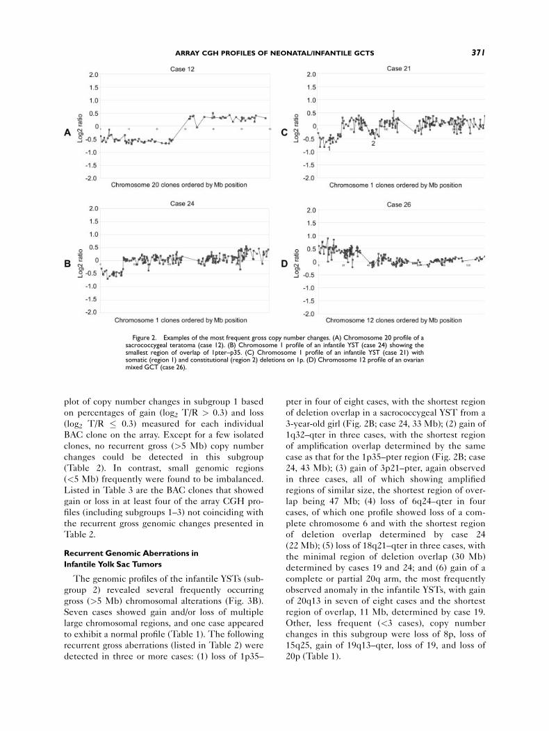
detected in three or more cases: (1) loss of 1p35–
pter in four of eight cases, with the shortest region
of deletion overlap in a sacrococcygeal YST from a
3-year-old girl (Fig. 2B; case 24, 33 Mb); (2) gain of
1q32–qter in three cases, with the shortest region
of amplification overlap determined by the same
case as that for the 1p35–pter region (Fig. 2B; case
24, 43 Mb); (3) gain of 3p21–pter, again observed
in three cases, all of which showing amplified
regions of similar size, the shortest region of over-
lap being 47 Mb; (4) loss of 6q24–qter in four
cases, of which one profile showed loss of a com-
plete chromosome 6 and with the shortest region
of deletion overlap determined by case 24
(22 Mb); (5) loss of 18q21–qter in three cases, with
the minimal region of deletion overlap (30 Mb)
determined by cases 19 and 24; and (6) gain of a
complete or partial 20q arm, the most frequently
observed anomaly in the infantile YSTs, with gain
of 20q13 in seven of eight cases and the shortest
region of overlap, 11 Mb, determined by case 19.
Other, less frequent (<3 cases), copy number
changes in this subgroup were loss of 8p, loss of
15q25, gain of 19q13–qter, loss of 19, and loss of
20p (Table 1).
Figure 2. Examples of the most frequent gross copy number changes. (A) Chromosome 20 profile of asacrococcygeal teratoma (case 12). (B) Chromosome 1 profile of an infantile YST (case 24) showing thesmallest region of overlap of 1pter–p35. (C) Chromosome 1 profile of an infantile YST (case 21) withsomatic (region 1) and constitutional (region 2) deletions on 1p. (D) Chromosome 12 profile of an ovarianmixed GCT (case 26).
371ARRAY CGH PROFILES OF NEONATAL/INFANTILE GCTS

Loss of 1p35–pter in a Yolk Sac Tumor and a
Constitutional del(1)(p22p31)
Subgroup 2 included a testicular YSTof a 1-year-
old boy (case 21) whose array CGH profile revealed
two 1p regions with genomic loss (Fig. 2C, regions
1 and 2). This was confirmed by a replicate array
CGH experiment and an independently performed
whole-chromosome CGH analysis (not shown).
One of these regions overlapped with the 1p35–
pter region that was frequently found to be deleted
in the samples of subgroup 2 (see above). The
other region mapped to 1p22–p31. In addition, this
boy suffered from mental retardation. Through
conventional cytogenetic analysis of blood lympho-
cytes, a constitutional mosaicism for the del(1)
(p22p31) was detected (not shown). The bounda-
ries of the constitutional deletion were determined
by BAC clones RP11-451c22 and RP11-114k16,
encompassing a �10-Mb region. Interestingly, loss
of this region also was detected in one of the other
infantile YSTs (subgroup 2; case 17). It contained
49 genes, including those encoding MSH4 (involved
in meiotic recombination), AK5 (adenylate kinase
5), FUBP1 (binds MYC promoter), PRKACB (kin-
ase, catalytic subunit of AMPK), DLAD (DNase II
beta), SPATA1 (spermatogenesis associated 1),
SSX2IP (SSX2 interacting protein), NYD-SP29 (a
presumed testis development protein), BCL10
(B-cell CLL/lymphoma 10), and CYR61 (promotes
angiogenesis, cell adhesion, cell proliferation, and
chemotaxis).
Recurrent Genomic Aberrations in GCTs from
Children More than 5 Years Old
The array CGH profiles of the older patients
(subgroup 3; >5 years) again revealed gross recur-
rent genomic abnormalities (Fig. 3C). Four of the
seven cases showed anomalies encompassing mul-
tiple large genomic regions. Two cases showed pro-
files with only one or three gross genomic aberra-
tions, and in one case the profile appeared to be
normal (Table 1). The most prevalent abnormality
was gain of 12p (present in five of seven cases;
Figs. 3C and 2D). Four of the profiles showed gain
of the complete 12p arm (cases 25, 26, 28, and 30),
whereas in one case (case 29) copy number gains
were detected in two distinct regions on 12p
[12p13–pter (15 Mb) and 12p11 (2 Mb)].
Another gross recurrent copy number change
was gain of 1q32–qter, observed in three of the
seven cases. Copy number loss of a part of 6q was
detected in two cases, gains of entire chromosomes
7 and 8 in three cases, loss of 18q in two cases, gain
of a complete chromosome 19 in two cases, and
gain of a complete chromosome 21 in two cases.
DISCUSSION
We used the recently developed array CGH
technique to analyze 24 archival GCTs from chil-
dren under the age of 5 years. The GCTs were div-
ided into two subgroups on the basis of histology
(teratomas in subgroup 1 and YSTs in subgroup 2).
In addition, seven GCTs of patients older than
5 years were included (subgroup 3). The 5-year
age cutoff was chosen on the basis of the results of
Figure 3. Overview of the genomewide frequency of copy numberchanges. The percentage of copy number gain is shown in black (log2 T/R > 0.3) and of copy number loss in gray (log2 T/R � 0.3) from chromo-somes 1–22 ordered by megabase position as presented in the UCSCgenome browser (April 2003 freeze). (A) Overview of subgroup 1,infantile GCT profiles with a teratoma histology (age < 5 years).(B) Overview of subgroup 2, infantile GCT profiles with a yolk sac his-tology (age < 5 years). (C) Overview of subgroup 3, GCT profilesderived from children above the age of 5 years and having a mixed his-tology. It can be seen clearly that these groups differ markedly in thenumber of abnormalities as well as in the genomic regions involved.
372 VELTMAN ETAL.

previous studies that showed a clear age distribu-
tion in the histology and tumor location of germ
cell tumors (Gobel et al., 1998; Schneider et al.,
2004). The aim of the present study was to obtain
detailed information on the cytogenetic constitution
of the different GCTs of neonates and infants in
order to identify (i) recurrent chromosomal aberra-
tions with diagnostic and/or prognostic implications
TABLE 2. Recurrent Copy Number Changes of Genomic Regions >5 Mb
Gain/loss Chromosome banda Flanking donesb Size (Mb) Subgroup 1 Subgroup 2 Subgroup 3
Gain 1q32–qter RP11-155f3-CTB-167k11 43 0 3 3Gain 3p21–pter RP11-91p19-RP5-1186b18 47 0 3 0Gain chr. 7 159 0 0 3Gain chr. 8 146 0 0 3Gain 12p13–pter RP11-174g6-CTB-124k20 15 0 0 5Gain 12p11 RP11-38f11-RP11-78f16 2 0 0 5Gain 19q13–qter RP11-2j15-qter 12 1 2 2Gain 20q13 RP11-1e23-RP11-124d1 11 1 7 1Loss 1p35–pter RP11-4p6-CTC-232b23 33 0 4 1Loss 6q24–qter RP11-40o24-RP1-57h24 22 0 4 2Loss 8p11–pter RP11-210f15-RP11-91j19 37 0 2 1Loss 18q21–qter RP11-43k24-RP5-964m9 30 1 3 2Loss 20p12 RP11-51o4-RP11-149o7 10 2 2 0
aDetermined by the minimal region of overlap.bClones that flank the minimal region of overlap.
TABLE 3. Recurrent Copy Number Changes of Genomic Regions <5 Mb
Gain/loss Clone nameChromosome
band Subgroup 1 Subgroup 2 Subgroup 3 GenesaLCVe
detected
Gain CTB-172i13 andRP5-1011o17
2qter 4 0 1 B
Gain RP11-89a12 3p14.1 2 2 2 TAFA1 —Gain RP11-554b18 3p12.1 3 1 2 — —Gain RP11-80h24 3p12.1 3 0 1 — —Gain RP11-91a15 3p11 2 1 1 EPHA3 —Gain RP11-40d5 4q28 1 3 0 — —Gain RP11-467a2 5q13.2 3 0 1 TNPO1 —Gain RP11-17o4 9q34 4 1 1 AK130729 —Gain RP11-88n14 14q21 2 1 2 — BGain RP1-149d14 17p11 2 4 2 — —Loss RP11-11m9 4p15 1 1 3 — —Loss RP11-82m24 5p15 1 1 2 — —Loss RP11-29n3 5p15 1 1 2 CTNND2 —Loss RP11-135M13 5p15 0 1 3 FBXL7 —Loss RP11-88I18 5p15 9 3 4 — BLoss CTD-2041d13 5q13 1 3 2 several genesb ALoss RP11-121g20 6p21 1 3 0 several genesc —Loss RP11-4o1 9q31 1 1 2 SUSD1 —Loss RP11-95j4 9q32 3 2 3 — —Loss RP11-180c18 10p15 2 1 1 ADARB2 —Loss RP11-115g22 15q13 3 0 1 CHRNA7 ALoss RP11-33b6 15q24 2 0 3 ARIH1, GOLGA6, BBS4 —Loss RP11-368n21 16p11 1 2 2 several genesd —Loss RP11-348E16 18q11 1 1 3 RBBP8 —Loss RP11-19I3 18q21 2 0 2 AK127467 —Loss RP11-135L16 22q13 0 3 2 PARVB, PARVG, AK0740 —Loss RP11-93F4 22q13 2 1 1 TRMT1, GTSE1, CELSR1 —
aBased on the UCSC human genome browser (May 2004 freeze).bX83301, AK130833, SERF1A, SMN1, SMN2, BIRC1, GTF2H2.cCAPN11, BC062989, c6orf110, AL353957, SLC29A1, HSPCB, SLC35B2, NFKBIE, AK130739, AARSL, MGC33600, AK126839, AL833884, SPATS1.dAK023827, LAT1-3TM, AF271775, BC062756, SPN, QPRT.eLarge-scale copy number variations (LCVs), presented as such in (A) Sebat et al. (2004) and/or (B) Iafrate et al. (2004).
373ARRAY CGH PROFILES OF NEONATAL/INFANTILE GCTS

and (ii) genomic regions that may harbor genes rel-
evant to tumor formation.
In agreement with the results of previous cyto-
genetic and conventional CGH studies, we found
no gross anomalies in most of the teratoma-derived
profiles (subgroup 1). In contrast, most YST-
derived profiles (subgroup 2) showed gross recur-
rent copy number changes. These recurrent
changes included loss of 1p (1p35–pter), gain of 1q
(1q32–qter), gain of 3p (3p21–pter), loss of 6q
(6q24–qter), loss of 18q (18q21–qter), and gain of
20q (20q13), all of which had been detected previ-
ously (Perlman et al., 1996; Mostert et al., 2000;
Hu et al., 2001; Schneider et al., 2001a). Losses of
(distal parts of) 1p and 6q were considered the
most prevalent genomic aberrations in YSTs of
neonates and infants. We detected loss of the distal
parts of 1p (1p35–pter) in half these cases. A simi-
lar percentage was found in an ongoing independ-
ent LOH study that included one of the present
cases (case 24). One patient (case 21) with a YST
exhibited a constitutional 1p22–p31 deletion next
to a somatic 1p35–pter deletion in the tumor. The
patient was mentally retarded, and the constitu-
tional deletion may be causally related to the men-
tal status of this patient. This deletion may also
have acted as a predisposing factor for YST devel-
opment in this patient. This idea was supported by
a similar loss of 1p21–p31 found in three infantile
YST cases described previously (Mostert et al.,
2000) and in one additional case of subgroup 2 with
a 1p22–p31 deletion in this study. The most preva-
lent anomaly in subgroup 2 was gain of (parts of)
20q, with the shortest region of overlap, of 11 Mb,
at 20q13 and containing multiple genes (�90).
Gain of 20q or 20q11–q13 is common in human
cancers (Hodgson et al., 2003; Kimura et al., 2004)
and also has been reported in GCTs, both adult
and infantile types (Looijenga et al., 2000; Mostert
et al., 2000; Perlman et al., 2000; Schneider et al.,
2002). In contrast, such chromosome 20 anomalies
were not noted in a large cytogenetic study of
childhood GCTs (Bussey et al., 1999). This may be
a result of the experimental limitations, that is, an
i(20q) chromosome can easily be overlooked in
conventional cytogenetic analysis. Interestingly,
gain of 20q and loss of 20p also were observed in a
teratoma profile of a tumor that contained both ter-
atoma and YSTcomponents (case 12).
Subgroup 3 (>5 years) showed heterogeneous
profiles, with gain of (parts of) 12p as the main
aberration (5 of 7 cases). Gain of (parts of) 12p
together with gain of complete chromosomes 7 and
8 was observed in three cases. These chromosomal
anomalies are common in adult testicular GCTs
(group 2; Looijenga et al., 2003) and also have been
detected in several malignant ovarian GCTs (Rio-
pel et al., 1998; van Echten et al., 1998; Kraggerud
et al., 2000). As most GCTs from children older
than 5 years in this study were ovarian GCTs, our
data support the hypothesis that ovarian and testic-
ular GCTs may share a common pathogenic path-
way. In accordance with the literature, the two
minimal regions of overrepresentation on 12p
mapped to 12p13–pter and 12p11, respectively
(Rodriguez et al., 2003; Zafarana et al., 2003;
Henegariu et al., 2004). So far, gain of a part of 12p
has been observed only rarely in neonatal or infan-
tile GCTs (Mostert et al., 2000). In line with these
observations, in the present study, gain of 12p was
detected only once (case 20) in the GCTs from
children under age 5 (subgroups 1 and 2).
Two profiles in the third subgroup resembled
profiles in subgroups 1 and 2. Similar to the profiles
of subgroup 1, the profile of an ovarian teratoma of
an 8-year-old showed no gross aberrations (case
27). The array CGH profile of a pelvic YST from a
16-year-old female patient (case 31) resembled the
infantile or neonatal YST profiles in several
aspects, including loss of 1p35–pter, loss of 6q, and
gain of 20q. No gain of 12p was observed.
Together, these observations suggest that this
tumor may actually fit into subgroup 2. Notably,
this patient had suffered from a sacrococcygeal
YST during infancy. Therefore, the tumor exam-
ined in the present study can be considered a
metachronous secondary tumor. Unfortunately, no
material of the primary tumor was available for
analysis. Nevertheless, this observation indicates
that some tumor components that reflect the child-
hood-type GCT may have persisted until the
metachronous tumor manifested itself. That the
array CGH profile diverged from the profile that
should be considered appropriate for the patient’s
age strongly supports the idea that GCTs that
develop during infancy are biologically distinct
from those that develop during adulthood. Gain of
(parts of) 1q, loss of (parts of) 6q, and loss of (parts
of) 18q were detected in profiles of tumors from
both subgroups 2 and 3, implying that these
regions may be related to the pathogenesis of
GCTs in both these subgroups.
In addition to the gross recurrent copy number
changes discussed above, some novel small regions
(<5 Mb) encompassing one or a few BACs showed
recurrent copy number alterations (Table 3). Some
of these regions were affected only in specific sub-
groups, including 2qter and 9q34 in teratomas (sub-
374 VELTMAN ETAL.

group 1), 4q28 and 6p21 in YSTs (subgroup 2), and
4p15, 15q24, and 18q11 in GCTs of children older
than age 5 (subgroup 3). Other regions, including
5p15, 5q13, 9q32, 17p11, and 22q13, showed gain
or loss in all GCT subgroups. Recently, it was
found that large-scale copy number variations
(LCVs, encompassing 100 Kb or more) may signifi-
cantly contribute to the array of naturally occurring
genomic polymorphisms (Iafrate et al., 2004; Sebat
et al., 2004). On the basis of this information, we
evaluated whether the recurrent anomalies detec-
ted in this study coincided with any of the LCVs
reported in the previous articles. Indeed, we found
that several small regions presented in Table 3
were described as such, including those on 2qter,
5p15, and 5q13. In reverse, we concluded that the
other recurrent anomalies that we detected most
probably resemble tumor-related copy number
changes (marked in Table 3).
Taken together, we have concluded that
genomewide array CGH technology is highly suit-
able for the detection of recurrent genomic anoma-
lies in GCTs of neonates and infants. This study
confirmed data from previous studies indicating
that most neonatal and infantile teratomas do not
exhibit gross genomic aberrations. Furthermore,
we concluded that the genomes of the GCTs differ
markedly between the two age groups described
here. For neonatal and infantile YSTs, regions
20q13, 1p35–pter, and 3p21–pter and individual
BAC clone regions may harbor novel diagnostic/
prognostic identifiers and genes relevant to the
development of these neoplasms.
ACKNOWLEDGMENTS
The authors thank Rolph Pfundt and Huub
Straatman for advice and support, Eric Schoen-
makers and Pieter de Jong for BAC clone collec-
tions, and Walter van der Vliet and Henry Dijkman
for expert technical assistance.
REFERENCES
Bernsen MR, Dijkman HB, de Vries E, Figdor CG, Ruiter DJ,Adema GJ, van Muijen GN. 1998. Identification of multiplemRNA and DNA sequences from small tissue samples iso-lated by laser-assisted microdissection. Lab Invest 78:1267–1273.
Blohm ME, Vesterling-Horner D, Calaminus G, Gobel U. 1998.Alpha 1-fetoprotein (AFP) reference values in infants up to 2 yearsof age. Pediatr Hematol Oncol 15:135–142.
Bussey KJ, Lawce HJ, Olson SB, Arthur DC, Kalousek DK, KrailoM, Giller R, Heifetz S, Womer R, Magenis RE. 1999. Chromo-some abnormalities of eighty-one pediatric germ cell tumors: sex- ,age-, site-, and histopathology-related differences—a Children’sCancer Group study. Genes Chromosomes Cancer 25:134–146.
Bussey KJ, Lawce HJ, Himoe E, Shu XO, Heerema NA, PerlmanEJ, Olson SB, Magenis RE. 2001. SNRPN methylation patternsin germ cell tumors as a reflection of primordial germ cell devel-opment. Genes Chromosomes Cancer 32:342–352.
Gobel U, Calaminus G, Engert J, Kaatsch P, Gadner H, Bokkerink JP,Hass RJ, Waag K, Blohm ME, Dippert S, Teske C, Harms D. 1998.Teratomas in infancy and childhood. Med Pediatr Oncol 31:8–15.
Gobel U, Schneider DT, Calaminus G, Haas RJ, Schmidt P, HarmsD. 2000. Germ-cell tumors in childhood and adolescence. GPOHMAKEI and the MAHO study groups. Ann Oncol 11:263–271.
Gonzalez-Crussi F. 1982. Extragonadal teratomas. 2nd series, Fas-cicle 18. Washington, DC: Armed Forces Institute of Pathology.
Harms D, Janig U. 1986. Germ cell tumours of childhood. Report of170 cases including 59 pure and partial yolk-sac tumours. Virch-ows Arch A Pathol Anat Histopathol 409:223–239.
Hawkins E, Issacs H, Cushing B, Rogers P. 1993. Occult malignancyin neonatal sacrococcygeal teratomas. A report from a CombinedPediatric Oncology Group and Children’s Cancer Group study.Am J Pediatr Hematol Oncol 15:406–409.
Heidenblad M, Schoenmakers EF, Jonson T, Gorunova L, VeltmanJA, Geurts van Kessel A, Hoglund M. 2004. Genome-wide array-based comparative genomic hybridization reveals multiple ampli-fication targets and novel homozygous deletions in pancreatic car-cinoma cell lines. Cancer Res 64:3052–3059.
Henegariu O, Heerema NA, Thurston V, Jung SH, Pera M, VanceGH. 2004. Characterization of gains, losses, and regional amplifi-cation in testicular germ cell tumor cell lines by comparativegenomic hybridization. Cancer Genet Cytogenet 148:14–20.
Hodgson JG, Chin K, Collins C, Gray JW. 2003. Genome amplification ofchromosome 20 in breast cancer. Breast Cancer Res Treat 78:337–345.
Hu J, Schuster AE, Fritsch MK, Schneider DT, Lauer S, PerlmanEJ. 2001. Deletion mapping of 6q21–26 and frequency of 1p36deletion in childhood endodermal sinus tumors by microsatelliteanalysis. Oncogene 20:8042–8044.
Iafrate AJ, Feuk L, Rivera MN, Listewnik ML, Donahoe PK, Qi Y,Scherer SW, Lee C. 2004. Detection of large-scale variation in thehuman genome. Nat Genet 36:949–951.
Isaacs H Jr. 2004. Perinatal (fetal and neonatal) germ cell tumors.J Pediatr Surg 39:1003–1013.
Kimura Y, Noguchi T, Kawahara K, Kashima K, Daa T, Yokoyama S.2004. Genetic alterations in 102 primary gastric cancers by compa-rative genomic hybridization: gain of 20q and loss of 18q are asso-ciated with tumor progression. Mod Pathol 17:1328–1337.
Kraggerud SM, Szymanska J, Abeler VM, Kaern J, Eknaes M, HeimS, Teixeira MR, Trope CG, Peltomaki P, Lothe RA. 2000. DNAcopy number changes in malignant ovarian germ cell tumors.Cancer Res 60:3025–3030.
Looijenga LH, Oosterhuis JW. 1999. Pathogenesis of testicular germcell tumours. Rev Reprod 4:90–100.
Looijenga LH, Rosenberg C, Van Gurp RJ, Geelen E, Echten-Arends J, de Jong B, Mostert M, Oosterhuis JW. 2000. Compara-tive genomic hybridization of microdissected samples from differ-ent stages in the development of a seminoma and a non-semi-noma. J Pathol 191:187–192.
Looijenga LH, Zafarana G, Grygalewicz B, Summersgill B,Debiec-Rychter M, Veltman J, Schoenmakers EF, Rodriguez S,Jafer O, Clark J, Geurts van Kessel A, Shipley J, Van Gurp RJ,Gillis AJ, Oosterhuis JW. 2003. Role of gain of 12p in germ celltumour development. APMIS 111:161–171.
Mostert M, Rosenberg C, Stoop H, Schuyer M, Timmer A, OosterhuisJ, Looijenga L. 2000. Comparative genomic and in situ hybridizationof germ cell tumors of the infantile testis. Lab Invest 80:1055–1064.
Oosterhuis JW, Looijenga LH, van Echten J, de Jong B. 1997. Chro-mosomal constitution and developmental potential of human germcell tumors and teratomas. Cancer Genet Cytogenet 95:96–102.
Perlman EJ, Cushing B, Hawkins E, Griffin CA. 1994. Cytogeneticanalysis of childhood endodermal sinus tumors: a Pediatric Oncol-ogy Group study. Pediatr Pathol 14:695–708.
Perlman EJ, Valentine MB, Griffin CA, Look AT. 1996. Deletion of1p36 in childhood endodermal sinus tumors by two-color fluores-cence in situ hybridization: a Pediatric Oncology Group study.Genes Chromosomes Cancer 16:15–20.
Perlman EJ, Hu J, Ho D, Cushing B, Lauer S, Castleberry RP. 2000.Genetic analysis of childhood endodermal sinus tumors by compa-rative genomic hybridization. J Pediatr Hematol Oncol 22:100–105.
Riopel MA, Spellerberg A, Griffin CA, Perlman EJ. 1998. Geneticanalysis of ovarian germ cell tumors by comparative genomichybridization. Cancer Res 58:3105–3110.
Rodriguez S, Jafer O, Goker H, Summersgill BM, Zafarana G, GillisAJ, Van Gurp RJ, Oosterhuis JW, Lu YJ, Huddart R, Cooper CS,Clark J, Looijenga LH, Shipley JM. 2003. Expression profile ofgenes from 12p in testicular germ cell tumors of adolescents andadults associated with i(12p) and amplification at 12p11.2–p12.1.Oncogene 22:1880–1891.
375ARRAY CGH PROFILES OF NEONATAL/INFANTILE GCTS

Ross JA, Schmidt PT, Perentesis JP, Davies SM. 1999. Genomicimprinting of H19 and insulin-like growth factor-2 in pediatricgerm cell tumors. Cancer 85:1389–1394.
Sano K. 1999. Pathogenesis of intracranial germ cell tumors recon-sidered. J Neurosurg 90:258–264.
Schneider DT, Schuster AE, Fritsch MK, Calaminus G, Harms D,Gobel U, Perlman EJ. 2001a. Genetic analysis of childhood germcell tumors with comparative genomic hybridization. Klin Padiatr213:204–211.
Schneider DT, Schuster AE, Fritsch MK, Hu J, Olson T, Lauer S,Gobel U, Perlman EJ. 2001b. Multipoint imprinting analysis indi-cates a common precursor cell for gonadal and nongonadal pedia-tric germ cell tumors. Cancer Res 61:7268–7276.
Schneider DT, Schuster AE, Fritsch MK, Calaminus G, Gobel U,Harms D, Lauer S, Olson T, Perlman EJ. 2002. Genetic analysisof mediastinal nonseminomatous germ cell tumors in childrenand adolescents. Genes Chromosomes Cancer 34:115–125.
Schneider DT, Calaminus G, Koch S, Teske C, Schmidt P, Haas RJ,Harms D, Gobel U. 2004. Epidemiologic analysis of 1,442 chil-dren and adolescents registered in the German germ cell tumorprotocols. Pediatr Blood Cancer 42:169–175.
Sebat J, Lakshmi B, Troge J, Alexander J, Young J, Lundin P, ManerS, Massa H, Walker M, Chi M, Navin N, Lucito R, Healy J, HicksJ, Ye K, Reiner A, Gilliam TC, Trask B, Patterson N, ZetterbergA, Wigler M. 2004. Large-scale copy number polymorphism inthe human genome. Science 305:525–528.
Stoop H, van Gurp R, de Krijger R, Geurts van Kessel A, Koberle B,Oosterhuis W, Looijenga L. 2001. Reactivity of germ cell matura-tion stage-specific markers in spermatocytic seminoma: diagnosticand etiological implications. Lab Invest 81:919–928.
van Echten J, van Doorn LC, van der Linden HC, van der Veen AY,Burger CW, de Jong B. 1998. Cytogenetics of a malignant ovariangerm-cell tumor. Int J Cancer 77:217–218.
van Echten J, Timmer A, van der Veen AY, Molenaar WM, de JongB. 2002. Infantile and adult testicular germ cell tumors. a differentpathogenesis? Cancer Genet Cytogenet 135:57–62.
Van Gurp RJ, Oosterhuis JW, Kalscheuer V, Mariman EC, LooijengaLH. 1994. Biallelic expression of the H19 and IGF2 genes inhuman testicular germ cell tumors. J Natl Cancer Inst 86:1070–1075.
Veltman I, van Asseldonk M, Schepens M, Stoop H, Looijenga L,Wouters C, Govaerts L, Suijkerbuijk R, Geurts van Kessel A.2002. A novel case of infantile sacral teratoma and a constitutionalt(12;15)(q13;q25) pat. Cancer Genet Cytogenet 136:17–22.
Veltman IM, Schepens MT, Looijenga LHJ, Strong LC, Geurts vanKessel A. 2003a. Germ cell tumours in neonates and infants: a dis-tinct subgroup? APMIS 111:152–160.
Veltman JA, Fridlyand J, Pejavar S, Olshen AB, Korkola JE, de VriesS, Carroll P, Kuo WL, Pinkel D, Albertson D, Cordon-Cardo C,Jain AN, Waldman FM. 2003b. Array-based comparative genomichybridization for genomewide screening of DNA copy number inbladder tumors. Cancer Res 63:2872–2880.
Vissers LE, de Vries BB, Osoegawa K, Janssen IM, Feuth T, ChoyCO, Straatman H, Van der Vliet W, Huys EH, van Rijk A, SmeetsD, Ravenswaaij-Arts CM, Knoers NV, van der Burgt I, de Jong PJ,Brunner HG, Geurts van Kessel A, Schoenmakers EF, VeltmanJA. 2003. Array-based comparative genomic hybridization for thegenomewide detection of submicroscopic chromosomal abnormal-ities. Am J Hum Genet 73:1261–1270.
Vissers LE, van Ravenswaaij CM, Admiraal R, Hurst JA, de VriesBB, Janssen IM, van der Vliet WA, Huys EH, de Jong PJ,Hamel BC, Schoenmakers EF, Brunner HG, Veltman JA, Geurtsvan Kessel A. 2004. Mutations in a new member of the chromo-domain gene family cause CHARGE syndrome. Nat Genet 36:955–957.
Wilhelm M, Veltman JA, Olshen AB, Jain AN, Moore DH, Presti JCJr, Kovacs G, Waldman FM. 2002. Array-based comparativegenomic hybridization for the differential diagnosis of renal cellcancer. Cancer Res 62:957–960.
Woodward PJ, Heidenreich A, Looijenga LHJ, et al. 2004. Testiculargerm cell tumors. In: Eble JN, Sauter G, Epstein JI, SesterhennIA, editors. World Health Organization classification of tumours.Pathology and genetics of the urinary system and male genitalorgans. Lyon, France: IARC Press. p 217–278.
Zafarana G, Grygalewicz B, Gillis AJ, Vissers LE, van der Vliet WA,Van Gurp RJ, Stoop H, Debiec-Rychter M, Oosterhuis JW, Geurtsvan Kessel A, Schoenmakers EF, Looijenga LH, Veltman JA.2003. 12p-Amplicon structure analysis in testicular germ celltumors of adolescents and adults by array CGH. Oncogene22:7695–7701.
376 VELTMAN ETAL.
Copyright © 2022 FDOKUMEN