Total Lip Reconstruction with Tendinofasciocutaneous Radial Forearm Flap

Identification of flap-structure specific endonuclease 1 as a factorinvolved in long-term memory formation of aversive learning
Lorena Saavedra-Rodríguez1,2, Adrinel Vázquez1,2, Humberto G. Ortiz-Zuazaga3, NataliyaE. Chorna4, Fernando A. González4, Lissette Andrés1, Karen Rodríguez1, FernandoRamírez1, Alan Rodríguez1, and Sandra Peña de Ortiz1,2
1 Molecular & Cellular Cognition Laboratory, University of Puerto Rico-Río Piedras Campus, San Juan,P.R. 00931-3360
2 Functional Genomics Research Center, Department of Biology, University of Puerto Rico-Río PiedrasCampus, San Juan, P.R. 00931-3360
3 High Performance Computing Facility, University of Puerto Rico-Central Administration, San Juan, P.R.00931
4 Department of Chemistry, University of Puerto Rico-Rio Piedras Campus
AbstractWe previously proposed that DNA recombination/repair processes play a role in memory formation.Here, we examined the possible role of the fen-1 gene, encoding a flap structure-specificendonuclease, in memory consolidation of conditioned taste aversion (CTA). Quantitative real timepolymerase chain reaction (PCR) showed that amygdalar fen-1 mRNA induction was associated tothe central processing of the illness experience related to CTA and to CTA itself, but not to the centralprocessing resulting from the presentation of a novel flavor. CTA also increased expression of theFen-1 protein in the amygdala, but not the insular cortex. In addition, double immunofluorescenceanalyses showed that amygdalar Fen-1 expression is mostly localized within neurons. Importantly,functional studies demonstrated that amygdalar antisense knockdown of fen-1 expression impairedconsolidation, but not short-term memory, of CTA. Overall, these studies define the fen-1endonuclease as a new DNA recombination/repair factor involved in formation long-term memories.
KeywordsMemory Formation; Amygdala; Conditioned Taste Aversion; DNA Recombination/Repair;Endonuclease
INTRODUCTIONConditioned taste aversion (CTA) represents a form of conditioning established when animalsassociate a new taste (conditioned stimulus, CS) with visceral illness (unconditioned stimulus,US), resulting in the subsequent avoidance of such taste (Garcia et al., 1985; Bernstein,1999). Acquisition of taste aversion results from and requires the succession of different phaseswithin the brain: encoding and storing the novel taste, processing the somatic illness, and finally
Corresponding Author: Dr. Sandra Peña de Ortiz, Department of Biology, University of Puerto Rico, P. O. Box 23360, San Juan, PR00931-3360. Tel: 787-764-0000 ext. 7957, Fax: 787-764-3875, EMAIL: E-mail: [email protected] Editor: Dr. Marie T. FilbinReviewing Editor: Dr. Ray Dolan
NIH Public AccessAuthor ManuscriptJ Neurosci. Author manuscript; available in PMC 2009 November 6.
Published in final edited form as:J Neurosci. 2009 May 6; 29(18): 5726–5737. doi:10.1523/JNEUROSCI.4033-08.2009.
NIH
-PA Author Manuscript
NIH
-PA Author Manuscript
NIH
-PA Author Manuscript

establishing an association between the taste memory and the illness experience (Bures,1998). The aversive memories generated during CTA are robust and persistent in time, andinvolve brain regions such as the amygdala and the insular cortex (Gallo et al., 1992; Bermudez-Rattoni and Yamamoto, 1998; Lamprecht and Dudai, 2000). Moreover, the taste (Bermudez-Rattoni, 2004; Yamamoto, 2006) and illness (Lamprecht and Dudai, 1996; Yamamoto et al.,1997; Spencer and Houpt, 2001; St Andre et al., 2007; Bernstein and Koh, 2007) componentsof CTA induce specific neuromolecular processes in the brain. For example, the amygdalashows robust neural activation to either the new taste or the toxin-induced illness experiencewhen they are presented separately (Bernstein and Koh, 2007). The individual presentation ofthe CS or US seems to elicit the activation of amygdalar cellular and molecular signals, whichcould be required for the final establishment of the association of both experiences. Here, weembarked in a study aimed at deciphering molecular mechanisms involved in CTAconsolidation, including those related to the representation of taste and illness individually,with a specific interest in identifying factors related to DNA recombination/repair processes.
Previously, we reported that DNA ligase-dependent recombination/repair processes arenecessary for consolidation of CTA (Wang et al., 2003) and context fear conditioning (Coló-Cesario et al., 2006a). We also showed that the mature rodent brain displays DNA-ligasedependent Non-Homologous End Joining (NHEJ, Ren and Peña de Ortiz, 2002), a known DNArecombination/repair mechanism required for the final stages of site-specific recombination inthe immune system (Soulas-Sprauel et al., 2007). Importantly, we found that NHEJ is rapidlyinduced in the hippocampus following contextual fear conditioning (Colón-Cesario et al.,2006a). Additional studies by us determined that the gene encoding terminal deoxynucleotidyltransferase (TdT), known to be involved in somatic recombination in the immune system(Benedict et al., 2000), is also associated to learning and memory processes in the mouse brain(Peña de Ortiz et al., 2003). The experiments presented here resulted in the discovery that thegene encoding flap structure-specific DNA endonuclease-1 (Fen-1), identified by us usingDNA microarrays (to be published elsewhere), is a factor necessary for consolidation of CTA.The fact that an endonuclease, such as Fen-1, is required for long-term memory (LTM)formation of aversive experiences supports the notion that DNA recombination/repairpathways are involved in processes that lead to the storage of information in the brain.
MATERIALS AND METHODSAnimals
Male Long Evans rats (Harlan Sprague Dawley) weighing 275–300 g were used. They wereindividually caged at 22±°C in a 12-h light/12-h dark cycle, with food and water ad libitum,except during behavioral tests during which water restriction was utilized at certain points (seebelow). All procedures were approved by the Institutional Animal Care and Use Committeeof the Rio Piedras Campus at the University of Puerto Rico in compliance with the guidelinesof the National Institutes of Health (NIH) for the care and use of laboratory animals(Department of Health and Human Services–NIH publication number 86-23).
Behavioral trainingThe CTA training was done as previously described by us (Wang et al., 2003; Ge et al.,2003). Briefly, rats were habituated for 4 days to drink plain water during 10 min each day,using the bottle presentation method. On day 5, CTA-trained animals were exposed to the novelflavor or CS (0.1% dextrose solution) in the drinking bottle for 10 min. This was followed 40min later, by an i.p. LiCl injection (100 mg/Kg body weight), used as the US in our studies.To examine fen-1 molecular changes related to novel taste or illness experiences we utilizedthe following additional behavioral groups: Flavor-Only animals, exposed to the CS and 40
Saavedra-Rodríguez et al. Page 2
J Neurosci. Author manuscript; available in PMC 2009 November 6.
NIH
-PA Author Manuscript
NIH
-PA Author Manuscript
NIH
-PA Author Manuscript

min later injected (i.p) with saline solution, and Toxin-Only animals, exposed to plain waterinstead of CS, and 40 min later injected (i.p) with LiCl (See Fig. 1a).
Total RNA extractionCTA, Flavor-only, and Toxin-only animals were used as behavioral groups for the real-timePCR study. For this experiment, 3 independent groups of RNA pools each from 3 rats wereused as biological replicates (3 pools of 3 animals each). Rats were decapitated 3 h afterbehavioral training and their brains were rapidly dissected, chilled in iced cold phosphatebuffered saline (PBS), washed with stabilization reagent (RNAlater QIAGEN, Valencia, CA)and then transferred to a rat brain matrix. Amygdalar-enriched tissue punches (3 mm wide)were collected from coronal brain slices using as a reference the Paxino’s Rat Brain Map(Paxinos and Watson, 1998), which locates the amygdala between −2.12 mm and −4.52 mmbregma points (see Fig. 1b). Amygdalar tissue from each behavioral group was pooled (n = 3animals in each pool), and RNA extraction was done using an RNA isolation Kit (QIAGEN)according to the manufacturer’s instructions and as described by us (Peña de Ortiz et al.,2003; Robles et al., 2003). The concentration and integrity of RNA were determined using theNanoDrop-1000 Spectrophotometer (NanoDrop Technologies, Rockland, DE) and the 2100Bioanalyzer (Agilent Technologies, Santa Clara, CA) with an RNA 6000 Nano LabChip® Kit(Agilent).
Primer design for real-time PCRSequences of genes analyzed (fen-1, Accession # AF281018 and glyceraldehyde 3-phosphatedehydrogenase (gapdh), Accession # M17701) were obtained from Genbank. We used theIntegrated DNA Technologies PrimerQuest and Oligo Analyzer programs to design specificprimers suitable for real-time PCR and also avoid possible hairpins, dimers and cross dimers.A BLAST search was done on all primers to ensure that they would not potentially anneal toother targets. The following forward and reverse primers were used: fen-1-forward 5′-ATTGCTGTTCGTCAGGGTGG-3′, fen-1-reverse 5′-GGTCTCCCCCTCCTCGTTC-3′;gapdh-forward 5′-ATGATTCTACCCACGGCAAG-3′, gapdh-reverse 5′-CTGGAAGATGGTGATGGGTT-3′. All primers were synthesized by Sigma (SIGMA-ALDRICH, St. Louis, MO).
Real-time PCRAmygdalar RNA from 3 independent pools, each composed of samples from 3 animals perbehavioral group (CTA, Flavor-only, and Toxin-only), was used for the study of selected genesusing real-time PCR. Animals were trained and sacrificed at the 3 h time point as detailedabove. For the antisense knockdown validation experiments (see antisense experiments below),amygdalar RNA was extracted from antisense and random oligonucleotide-treated animals(n = 4 each), sacrificed 3 h or 12 d after the last oligonucleotide infusion. cDNA was obtainedfrom total RNA samples, extracted as detailed above, using the TaqMan RT Reagents (AppliedBiosystems, Foster City, CA). Briefly, 20 μl of reaction mixture containing 20 ng of RNA, 500μM of each dNTP, 1x TaqMan RT buffer, 25 mM MgCl2, 2.5 μM random hexamers, 40URNase Inhibitor, and 125U MultiScribe reverse transcriptase were incubated at 25 °C for 10min followed by incubation at 48° C for 40 min. The reaction was stopped by incubating at95°C for 5 min. To generate the standard curves, a pooled cDNA from the behavioral groupswas synthesized and specific primers for fen-1 and gapdh were designed to work under thesame cycling conditions. Real-time PCR was performed using an iCycler iQ Real-time PCRDetection System (Bio-Rad, Hercules, CA) and the QuantiTect SYBR Green PCR kit(QIAGEN). Briefly, 250 ng of cDNA were combined with 0.5 μM of each primer, 1xQuantiTect SYBR Green PCR Master Mix (HotStarTaq DNA polymerase, quantiTect SYBRgreen PCR buffer, dNTP mix and SYBR green I) and PCR-grade water to a volume of 25 μl.
Saavedra-Rodríguez et al. Page 3
J Neurosci. Author manuscript; available in PMC 2009 November 6.
NIH
-PA Author Manuscript
NIH
-PA Author Manuscript
NIH
-PA Author Manuscript

The cycling conditions for all primers were: 95° C for 15 min to activate the HotStarTaqpolymerase, followed by 40 cycles consisting of three steps, 45 s at 95° C (denaturation), 30s at 58.5 °C (annealing), and 30 s at 72 °C (extension). The PCR program was completed bya melting temperature analysis consisting of 1 min at 95 °C (denaturation), 2 min at 55 °C(annealing) and then 101 steps lasting 8 s each, through which temperature ranged from 55 to95° C. Amplification plots were produced in order to calculate the threshold cycle (Ct), andstandard curves of Ct versus log cDNA dilution were generated for both target and referencegenes. Since all reactions were done in triplicate, the average Ct and nanomoles were used forplotting and quantification. For quantification, we applied the Standard Curve method in whichthe quantity of the target gene was expressed relative to the amount of the reference gene. Inthis case, nanomoles of fen-1 cDNA were divided by those of gapdh to obtain a normalizedtarget expression value.
Protein extractionCTA rats were decapitated 15 min, 30 min, 3 h or 3.5 h (n = 3 per time point) after trainingand their brains were obtained, chilled on ice cold PBS, and used to dissect amygdala aspreviously explained, and also insular cortex between 1.70 mm and 0.70 mm bregma points.Amygdala or insular cortex tissue punches from 3 animals per time point were combinedyielding one pool sample per group. Pooled tissue was stored at −80°C until used for proteinextraction. Protein extracts were prepared as described by us previously (Ren and Peña deOrtiz, 2002; Wang et al., 2003; Colón-Cesario et al., 2006a). Tissues were homogenized inextraction buffer [30 mM HEPES/KOH, pH 7.9, 0.5 M KCl, 5 mM MgCl2, 1 mMEDTA, 2mM dithiothreitol (DTT), 20% glycerol, 1 mM phenylmethylsulfonyl fluoride (PMSF), and 1μg/ml each of leupeptin and aprotinin] and incubated for 1 h in ice. The extract was centrifugedat 14,000 rpm for 1 h at 4°C. The supernatant was then dialyzed for 5 h in dialysis buffer (30mM HEPES/KOH, pH 7.9, 50 mM KCl, 2 mM EDTA, 5 mM MgCl2, 1 mM DTT, 10%glycerol, 1 mM PMSF, and1 μg/ml each of leupeptin and aprotinin). Dialyzed fractions werecentrifuged at 14,000 rpm for 30 min at 4°C. Protein extracts were stored at −80°C until used.The protein concentration was determined by the Bradford method as detailed by us previously(Ren and Peña de Ortiz, 2002; Wang et al., 2003; Colón-Cesario et al., 2006a).
Western blottingFor Western Blotting, protein samples (40 μg) were first separated on a 6% sodium dodecylsulfate-polyacrilamide gel electrophoresis (SDS-PAGE). The separated proteins in the gel weretransferred to a nitrocellulose membrane using a semidry electroblotter system at 5V and 4 °C overnight. Then, the membrane was blocked with SuperBlock solution (Pierce, Rockford,IL) for 1.5 h at rt on an orbital shaker. After 5 washes of 5 min each with PBS Tween-20 (PBS-T), the membrane was incubated with a 1:3000 dilution of a polyclonal antibody raised againsta human Fen-1 peptide (Abcam, Cambridge, MA) at 4 °C overnight, washed with PBS-T andthen again incubated with 1:500 anti-rabbit horseradish peroxidase (HRP)-conjugatedsecondary antibody for 1 h. The membrane was soaked briefly with Supersignal WestPicoChemiluminescent Substrate (Pierce) for immune detection. For normalization purposes,membrane was stripped of antibodies and re-probed for β-actin as described previously (Ge etal., 2003; Colón-Cesario et al., 2006b). Membranes were analyzed using the Gel Doc System(Bio-Rad, Hercules, CA), and expression was normalized by dividing the mean opticaldensities of Fen-1 immunopositive signals per time point by the corresponding β-actin signaloptical densities. These Western blot analyses were repeated 3 times in different gel runs andblots, respectively. Specificity of the Fen-1 antibody was demonstrated by pre-absorption testswith Fen-1 peptide. Briefly, the Fen-1 polyclonal antibody was incubated during 4–5 h at rt ina dilution of 1:8 with the peptide derived from a portion of the latter half of the N-terminaldomain of human Fen-1, the same peptide from which the antibody used in our studies wasraised (Abcam). Then, this antibody-peptide mix was used for Western Blotting as above.
Saavedra-Rodríguez et al. Page 4
J Neurosci. Author manuscript; available in PMC 2009 November 6.
NIH
-PA Author Manuscript
NIH
-PA Author Manuscript
NIH
-PA Author Manuscript

ImmunofluorescenceTo examine the cellular localization of Fen-1 expression, rats (n =8) were decapitated 3.5 hafter CTA training and their brains were immediately isolated, washed with ice cold PBS, andstored at −80 °C. Frozen coronal amygdalar sections (20 μm thick) were obtained in a cryostatat −20 °C, placed on positively charged glass slides (Probe-On Slides, Fisher Sci., PR) andused for immunofluorescence analysis. Briefly, sections were allowed to air dry for 20 min,fixed with 2% paraformaldehyde during 20 min, and then washed twice with PBS, 5 min eachtime. Permeabilization was done with 0.1% Triton X-100 in 0.1% of sodium citrate for 5 minand washed as described above. The sections were then incubated with 5% goat serum in PBSfor 30 min. Double-immunofluorescence was performed incubating the sections with primaryanti-Fen-1 polyclonal antibody (Abcam) diluted at 1:250 in 1% goat serum/PBS together withprimary anti-Neuronal Nuclei (NeuN) monoclonal antibody (Millipore, Billerica, MA) orprimary anti-Glial Fibrillary Acidic Protein (GFAP) monoclonal antibody (Sigma), diluted at1:100 and 1:400 in 1% horse serum/PBS, respectively. Sections were then incubated overnightat 4°C in a moist chamber. On the next, sections were washed twice in PBS and were thenincubated for 2 h at rt in a dark room with Alexa Fluor 488-conjugated goat anti- rabbitimmunoglobulin G (IgG) (for detection of Fen-1) and Alexa Fluor 568-conjugated goat anti-mouse IgG (Invitrogen, Carlsbad, CA) (for detection of NeuN), diluted both at 1:100 in 1%goat serum/PBS, followed by PBS washing. Different sections were incubated with AlexaFluor 568-conjugated goat anti-rabbit IgG for detection of Fen-1, and Alexa Fluor 488-conjugated donkey anti-mouse IgG for detection of GFAP, diluted both at 1:100 in 1% goatserum/PBS. The slides were mounted using permanent mounting medium (Vector LaboratoriesInc., Burlingame, CA). All slides were first scanned at low magnification (10X) to locate theamygdala, which was subsequently analyzed at higher magnification (40X) using a ZeissLSM-5 Pascal scanning confocal microscope (Thornwood, NY, USA). Final image compositeswere created using Zeiss LSM5 PASCAL Image software, version 3.2.
Cell culture, RNA isolation, real-time PCR and confocal immunofluorescence microscopyTo examine the constitutive fen-1 expression of rat neuronal cells and astrocytes, fen-1 mRNAlevels were measured in rat naïve PC-12 cells, PC-12 cells differentiated to the neuronalphenotype, and DI TNC astrocytes, using real-time PCR. Rat pheochromocytoma PC-12 cellsand rat DI TNC were obtained from American Type Culture Collection (Manassas, VA, USA).PC-12 cells were cultured in F-12K medium (ATCC) containing 15% (v/v) Horse Serum(ATCC), and 2.5% (v/v) Fetal Clone III Serum (FCIII) (Invitrogen). Rat DI TNC immortalizedastrocytes (American Type Culture Collection (ATCC), Manassas, VA) were maintained inDulbecco’s modified Eagle’s medium (DMEM) (Invitrogen), containing 5 % (v/v) fetal cloneIII serum (FCIII) (Invitrogen). All culture media were supplemented with 1% streptomycinand 1% penicillin obtained from Sigma, and cells were cultured at 37°C in a humidifiedatmosphere of 5% CO2.
Total RNA was isolated from naive PC-12 cells, PC-12 cells differentiated to neuronalphenotype with 100 ng Staurosporine (Sigma), as well as DI TNC cells (n =3 independentexperiments), using the TRIZOL reagent following the manufacturer’s instructions(Invitrogen). For real-time PCR, 1 mg of total RNA was obtained from DI TNC cells, naïvePC-12 cells and differentiated PC-12 cells. cDNAs were derived using the ReverseTranscription System kit from Promega (Madison, WI) following the manufacturer’sinstructions. Real-time PCR for detection of fen-1 and gapdh was performed as previouslydescribed in this section.
Immunofluorescence analysis was used to visualize cellular morphology. Briefly, PC-12 andDI TNC cells were plated on two-wells Laboratory-Tek chamber slides (Nalge Nunc Int.,Rochester, NY, USA) at a density of 1 × 10⁵ cells/well for 48 h prior imaging. In addition,
Saavedra-Rodríguez et al. Page 5
J Neurosci. Author manuscript; available in PMC 2009 November 6.
NIH
-PA Author Manuscript
NIH
-PA Author Manuscript
NIH
-PA Author Manuscript

PC-12 cells were treated with 100 ng Staurosporin for an extra 24 h to achieve homogenouspopulation of neurons. Next, cells were fixed for 10 min in PBS containing 3.7% formaldehyde,washed with PBS, permeabilized with 0.1% (v/v) Triton X-100 in PBS for 3 min, and washedthree times with PBS. Then, they were incubated with 5% horse serum (Invitrogen) for 30 minfollowed by incubation with 5U Alexa-fluor 488-conjugated phalloidin (Invitrogen) in 5%horse serum to visualize F-actin and washed. Images were acquired as described before usinga Zeiss LSM-5 Pascal scanning confocal microscope equipped with an Alpha-Fluor 100 × 1.45DIC oil immersion objective. Final image composites were created using Zeiss LSM5PASCAL Image software, version 3.2.
fen-1 antisense oligonucleotidesGapmer antisense oligonucleotides were designed to target the start codon of rat fen-1 mRNA.This 21 bp gapmer contains a central block of phosphorothioate-deoxynucleotides, sufficientto induce RNase H cleavage, flanked by blocks of 2′-O′methyl-modified ribonucleotides thatprotect the internal block from nuclease degradation. As a control, a random sequence was alsodesigned with the same backbone modifications and base composition as fen-1 antisense, butin a scrambled order and without homology to any known rat gene. fen-1 antisense and randomoligonucleotide sequences were as follows: 5′-mCmAmUmGmGT*A*A*C*A*C*A*G*G*A*G*mCmAmAmUmG-3′ and 5′-mAmCmGmCmAA*G*A*T*C*G*A*G*A*C*T*mAmGmGmAmU-3′, respectively(where m = 2′ OMethyl RNA and * = Phosphorothioate DNA). Antisense and randomsequences were synthesized by Integrated DNA Technologies (Coralville, IA). All threebatches of antisense and random oligonucleotides used to complete these studies were receivedlyophilized and fully purified by high performance liquid chromatography. Oligonucleotideswere dissolved in sterile 0.9% saline solution to a final concentration of 2 nmol/μl.
SurgeryRats were handled for 2–3 days before undergoing surgery, following similar methods as thosedescribed by us before (Wang et al., 2003; Vázquez and Peña de Ortiz, 2004; Colón-Cesarioet al., 2006b). For surgery, animals were anesthetized with sodium pentobarbital (50 mg/kg,i.p.), and placed into a stereotaxic apparatus (David Kopf Instruments, Tujunga, CA), with thenose angled at 0°. After a scalp incision was made, lambda and bregma were located, and holeswere drilled in the skull above the target region. Bilateral guide cannulae (8 mm long) wereimplanted above the basolateral amygdala complex using the following coordinates: anterior-posterior (AP), −3.4 mm from bregma; medio-lateral (ML), 5.0 mm from midline; dorso-ventral (DV), −7.6 mm from skull. The cannulae were secured to stainless steal screws withdental cement and a light-curable resin. Wire stylets were inserted into the guides and checkedevery day to ensure clean and functional cannulae. After surgery animals were allowed torecover for 4 days before behavioral experiments.
Diffusion studiesAfter cannulae implantation, injectors were inserted and animals (n = 4) were infused withFITC-fen-1 antisense to estimate the area of the antisense diffusion within the amygdala. Aninfusion of 1 μl of FITC-fen-1 antisense oligonucleotide (2 nmol) was delivered bilaterally intothe amygdala during a 2 min period at a rate of 0.5μl/min. Animals were decapitated 4 h afterinfusion, and their brains were isolated and stored at −80 °C. Coronal amygdalar sections, 20μm thick, were visualized using a fluorescence microscope (Pixcell II, Arcturus), anddigitalized photomicrograhs were obtained.
Saavedra-Rodríguez et al. Page 6
J Neurosci. Author manuscript; available in PMC 2009 November 6.
NIH
-PA Author Manuscript
NIH
-PA Author Manuscript
NIH
-PA Author Manuscript

Intraamygdalar infusions, behavioral training, and memory testingTo assess the effectiveness of the infusion pump system and to adapt the animals to receiveintraamygdalar infusions, rats were subjected to bilateral infusions (2 min at 0.5μl/min) of 0.9%saline on the day before conditioning. The infusion was accomplished by inserting a 30 gaugestainless steel injector into the guide cannulae so that it extended 1 mm beyond the tip of theguide, right above the targeted amygdalar regions. For the behavioral training, rats werehabituated during 4 days as previously described in the CTA protocol above, and on day 5,animals were infused bilaterally into the amygdala with 1 μl of fen-1 antisense or randomoligonucleotides, 1 h before dextrose (CS) presentation. A second oligonucleotide infusion,either fen-1 antisense or random oligonucleotides (1 μl each), was delivered to animals of bothgroups 1 h after LiCl injection (US). A set of animals receiving either fen-1 antisense or randomoligonucleotide infusions (n = 6 in each group) into the amygdala, as detailed above, weresubjected to a short-term memory test 2 h after conditioning. During the test, the animals werepresented with a choice of plain water and dextrose solutions for 10 min. Liquid consumptionwas measured by weighing the drinking bottles before and after consumption. Associativelearning and memory were assessed by the aversion to the CS, measured as an Aversion Indexcalculated as follows: water intake/(water intake + dextrose intake).
CTA memory consolidation was tested in a separate set of animals (n =11 in each group,antisense or random oligonucleotide-treated animals) 48 h after conditioning. Aversion indexeswere calculated as described for the short-term memory test. Twelve days after the LTM test,these previously infused animals were subjected to a new CTA training experience using 0.1%glycine as the CS. No oligonucleotide brain infusions were delivered during this conditioning,and the new CTA memory was also tested 48 h after training. In a separate control experiment,a new group of untrained animals received fen-1 antisense or random (n = 4) oligonucleotideamygdalar infusions and using real-time PCR, the amygdalar fen-1 mRNA levels weremeasured 12 d later (corresponding to the start of the second CTA in the experiment above).
Statistical analysisAll statistical analyses were performed with Prism 4 software (GraphPad Software). For allthe behavioral and molecular experiments, we assumed statistical significance at P < 0.05. Inthe real-time PCR studies, one-way analysis of variance (ANOVA) and Newman-Keuls post-tests were used to compare fen-1 mRNA expression between behavioral groups. Two-wayANOVA and Bonferroni post-testing were applied to analyze the results from the Westernblotting experiments as well as the differences in terms of the aversion index in the short-termmemory (STM) and LTM tests between fen-1 antisense and random oligonucleotide-treatedrats. The constitutive levels of fen-1 mRNA measured by real-time PCR in different cell typeswere examined using one-way ANOVA and Newman-Keuls post-test. Two-way ANOVA andBonferroni post-testing were also used in the LTM data, but only to determine differences inCS consumption. Finally, fen-1 mRNA expression measured by real-time PCR betweenfen-1 antisense and random-treated animals was examined using Student’s t-tests.
RESULTSfen-1 mRNA is induced in the amygdala as a result of CTA
For these studies we decided to focus on fen-1, a flap structure-specific endonuclease involvedin different aspects of DNA metabolism, including recombination/repair. Initially, we usedquantitative real time PCR to determine if the expression of fen-1 is modulated in the amygdalain association to CTA. The experimental design used in this study is depicted in Fig. 1a–c. Apreliminary time course study identified changes in amygdalar fen-1 mRNA levels 3 h, but not1 h, after CTA training (data not shown). Moreover, previous studies by us have identifiedimportant changes in gene expression and function related to learning and memory processes,
Saavedra-Rodríguez et al. Page 7
J Neurosci. Author manuscript; available in PMC 2009 November 6.
NIH
-PA Author Manuscript
NIH
-PA Author Manuscript
NIH
-PA Author Manuscript

including CTA (Ge et al., 2003), both in the amygdala or the hippocampus 3 h after behavioraltraining (Peña de Ortiz et al., 2000 and Robles et al., 2003). For that reason, we decided tofocus on the 3 h time point for these studies. Amplification and melting temperature curves offen-1 and gapdh (the control housekeeping gene) are depicted to show the cycle thresholds(Ct) for both genes (Fig. 2a), and to determine that only one product per gene was generated(Fig. 2b). As seen in Fig. 2c, the results showed that amygdalar gapdh mRNA levels (measuredby the Ct values) were similar between the three groups examined, CTA, Flavor-Only andToxin-Only (one-way ANOVA: F(2,35) = 0.14; P > 0.05). In contrast, for fen-1 mRNA (Fig.2d), one-way ANOVA detected significant differences between the groups after normalizationwith the standard curve (F(2,17)= 3.97; *P = 0.0412). Newman-Keuls determined thatamygdalar fen-1 mRNA expression was significantly higher in CTA-trained animals incomparison to Flavor-only rats (*P < 0.05). No specific significant differences were observedin the amygdalar expression of fen-1 mRNA between CTA and Toxin-only groups (P > 0.05)or between Flavor-only and Toxin-only groups (P > 0.05). Overall, these results indicate thathigher amygdalar fen-1 mRNA levels are associated to CTA 3 h after training, probably alsorelated to the illness component of this conditioning.
Fen-1 protein shows regionally specific induction as a result of CTACTA-trained animals were decapitated 15 min, 30 min, 3 h or 3.5 h (n = 3 per time point) afterconditioning and protein extracts from amygdala and insular cortex, a brain region also relatedto CTA (Gallo et al., 1992; Bermudez-Rattoni and Yamamoto, 1998), were used for Fen-1detection by Western blotting. We chose to examine Fen-1 protein expression at these particulartime points because we wanted to determine if there was a contrast between early Fen-1expression seen at 15 or 30 min after training versus the expression of the protein based on themRNA upregulation seen in our real-time PCR studies 3 h after CTA. In fact, we decided touse the 3.5 h time point because the previous data was obtained based on mRNA levels andwe anticipated that the protein might follow a more delayed course of induction. Resultsrevealed that Fen-1 was indeed induced in the amygdala between 3 to 3.5 h after training,compared to the earlier time points (Fig. 3a), in agreement with our previous findings withfen-1 mRNA. No Fen-1 induction was detected in the insular cortex (Fig. 3b) as a result ofCTA. The specificity of the Fen-1 antibody used in these studies was confirmed using Fen-1peptide preabsorption assays (Fig. 3c). Quantitative and statistical analysis of Fen-1 signals(Fig. 3d) showed significant differences in Fen-1 levels between the groups (two-wayANOVA: Time Factor, F(3, 16) = 5.11, *P = 0.0114; Region Factor, F(1, 16) = 15.99, **P =0.001), with no significant interaction between the Time and Region factors (F(3, 16) = 1.397,P = 0.2801). Bonferroni post-testing indicated specific significant differences in Fen-1 withinthe amygdala, but not the insular cortex, when comparing the 3.5 h versus the 15 min (**P <0.01), 30 min (++P < 0.01), and 3 h (@P < 0.05) time points. The results indicate that thetraining-induced upregulation of the fen-1 gene occurs both at the mRNA and protein levelsand that such induction is specific to the amygdala, compared to the insular cortex.
Fen-1 protein is localized within amygdalar neuronal cellsFen-1 is an endonuclease involved in several DNA metabolism processes common for all typesof cells, such as Okazaki fragment removal during replication, long-patch base excision repair,NHEJ, and homologous recombination (Liu et al., 2004; Shen et al., 2005; Casta et al.,2008). Hence, we considered it important to establish the cell-type specificity of Fen-1 afterCTA and determine whether it is expressed by neurons, glial cells or both. Based on its knownrole in DNA replication, we did expect to see Fen-1 expression in glial cells. On the other hand,based on its upregulation after CTA learning, we also expected to see Fen-1 expressed inneurons.
Saavedra-Rodríguez et al. Page 8
J Neurosci. Author manuscript; available in PMC 2009 November 6.
NIH
-PA Author Manuscript
NIH
-PA Author Manuscript
NIH
-PA Author Manuscript

Double-immunofluorescence of Fen-1 antibody with the neuronal marker, neuronal nuclei(NeuN), or the astrocytic marker, glial fibrillary acidic protein (GFAP) was performed onamygdalar sections from CTA animals (n =8) decapitated 3.5 h after training. Using confocalmicroscopy, we examined amygdalar sections double-labeled with Fen-1 (Alexa Fluor 488,green signal) and NeuN (Alexa Fluor 568, red signal) (Fig. 4a). These immunofluorescentimages show co-localization of Fen-1-expressing cells with those expressing NeuN, indicatingthe presence of Fen-1 in neurons. In contrast, images from amygdalar sections double-labeledwith Fen-1 (Alexa Fluor 568, red signal) and GFAP (Alexa Fluor 488, green signal) suggestthat Fen-1-expressing cells differ in localization and morphology with the GFAP-expressingcells (Fig. 4b,c). While the immunofluorescence signal for Fen-1 corresponds to large andround cells, the GFAP-positive cells display the distal processes distinctive of astrocytes, withrelatively small cell bodies and long intermediate filaments. For example, Fig. 4c shows howFen-1-positive cells (labeled with Alexa Fluor 568) are wrapped and surrounded by GFAP-positive cells (Alexa Fluor 488). These findings indicate that Fen-1 appeared to be expressedpredominantly in neurons, as suggested by the co-localization between Fen-1 and NeuN.
Although Fen-1 protein expression was not detected in astrocytes using thisimmunofluorescence analysis, we examined the basal fen-1 mRNA levels in cultured ratneuronal cells and astrocytes to compare the constitutive Fen-1 expression in both types ofcells. For this, fen-1 mRNA expression was measured in rat naive PC-12 cells, PC-12 cellsdifferentiated to the neuronal phenotype, and DI TNC astrocytes, using real-time PCR. As seenin Fig. 5a, one-way ANOVA detected significant differences in fen-1 mRNA levels betweenthe groups (F(2,17)= 6.180; *P = 0.011). Newman-Keuls determined that constitutive fen-1mRNA levels are significantly higher in the PC-12 neuronal cells in comparison to the naivePC-12 cells (*P < 0.05) and the DI TNC astrocytes (**P < 0.01). No statistical significancewas found in fen-1 mRNA levels between naive PC-12 cells and the DI TNC astrocytes (P >0.05). The morphology of the three cell types used in this study can be appreciated in Fig. 5b.These results confirm that although Fen-1 is constitutively expressed in astrocytes, this basalglial expression is significantly lower than the basal Fen-1 neuronal expression.
Fen-1 plays a role in consolidation of CTAThe previous gene expression experiments demonstrated that fen-1 is induced in the amygdala3 h after CTA training, although the animals subjected only to the illness component of CTAalso showed a tendency to have high fen-1 mRNA levels. Previous studies support the ideathat the processing of the US in CTA is critical for overall associative learning in this paradigm(Lamprecht and Dudai, 1996; Swank et al., 1996; Yasoshima et al., 2006). Thus, we consideredit essential to determine the functional role of fen-1 in the consolidation of CTA memory. Weused an antisense approach to knockdown fen-1 expression in the amygdala and examined itseffects on CTA memory formation.
Animals were implanted with bilateral cannulae directed to the basolateral amygdala (BLA)complex. We focused on the BLA, not only because this region has been shown to be involvedin CTA consolidation (Schafe et al., 1998; Morris et al., 1999; Rollins et al., 2001), but alsobecause several studies suggest that CTA acquisition and consolidation requireneurotransmission involving inputs from the BLA onto the insular cortex (Bermudez-Rattoniand McGaugh, 1991). We did not target Fen-1 expression in the insular cortex because of ourfindings with the Western blot analyses, which showed that no changes in Fen-1 expressionoccurred as a result of CTA in this cortical region. Representative photomicrographs andschematics illustrating the distribution of cannula placements throughout the amygdala foranimals used in our behavioral experiments are included in Fig. 6a and b. Since no behavioraldifferences were observed, regardless of the diverse cannulae positions, behavioral data fromall animals was used (see below). We also examined the diffusion of fen-1 antisense
Saavedra-Rodríguez et al. Page 9
J Neurosci. Author manuscript; available in PMC 2009 November 6.
NIH
-PA Author Manuscript
NIH
-PA Author Manuscript
NIH
-PA Author Manuscript

oligonucleotides in the amygdala (Fig. 6c,d) through consecutive rostrocaudal sections.Diffusion of the infused FITC-fen-1 antisense oligonucleotide was concentrated within theanterior, posterior, and ventral BLA. The antisense also extended into other amygdalar areas,including the medial (CeM) and capsular (CeC) divisions of the central amygdala (CeA), theanterior basomedial (BMA), the posterior basomedial (BMP), and the ventromedial lateralamygdala (LaVM). This FITC-fen-1 antisense oligonucleotide was clearly incorporated intothe cells within these regions (Fig. 6e).
We next evaluated the effects of amygdalar fen-1 knockdown on CTA STM as well as LTM(Fig. 7a) Different sets of rats were used for each memory test in order to avoid potentialconfounding effects related to aversive extinction processes. For the STM test, rats wereinfused bilaterally with fen-1 antisense (n =6) or random oligonucleotides (n =6) 1 h beforethe CS and 1 h after the US, and the memory test was done 2 h after conditioning. For the LTMtest, rats were infused bilaterally with fen-1 antisense (n =11) or random (n =11)oligonucleotides 1 h prior to the CS and 1 h after the US, and the memory test was done 48 hafter conditioning. We then subjected the behavioral data (Fig. 7b) to a Two-way ANOVAanalysis that found a significant effect caused by the oligonucleotide treatment (F(1, 29) =9.834, **P = 0.0039), and a significant interaction between the oligonucleotide treatment andthe type of memory test (STM vs LTM) (F(1, 29) = 6.677, *P = 0.0151). Moreover, Bonferronipost-testing identified a significant difference in terms of the aversion index betweenoligonucleotide treatments only in the LTM test (**P < 0.01). Specifically, the fen-1 antisense-treated rats displayed a significantly lower aversion to dextrose than the random-treatedanimals, 48 h after conditioning. Overall, we determined that the amygdalar fen-1 antisenseoligonucleotide treatment had no effect on STM, but significantly impaired LTM.
This effect was also observed when comparing dextrose consumption between antisense andrandom oligonucleotide-treated animals during the conditioning day and the LTM test (Fig.7c) (two-way ANOVA: dextrose consumption factor, F(1,40) = 18.34; ***P = 0.0001; testingfactor, F(1,40) = 1.596; P = 0.2138; interaction effect, F(1,40) =7.690; **P = 0.0084). Thedata presented here correspond to the animals used for the LTM test only. During conditioning,both groups of animals consumed similar amounts of dextrose, showing that the antisense-treated rats were not sick due to the intra-amygdalar infusions. However, 48 h afterconditioning, the antisense group consumed more dextrose than the random-treated group(Bonferroni post-testing, *P < 0.05), indicating that the antisense animals displayed poormemory of their aversive experience.
To examine potential non-specific impairments that could be caused by the antisense treatment,the same antisense and random-treated animals used for the LTM test, were subjected to asecond CTA using glycine as the CS. This type of behavioral control has been previously usedby us for the assessment of the potential long-term effects of blocking a CTA experience(Wang et al., 2003). Habituation for this new training was initiated one week after the firstbehavioral test. The second conditioning, now in the absence of new intra-amygdalaroligonucleotide infusions, was given 12 d after the LTM test of the first CTA. This time delaybetween the two CTA trainings was used considering the fact that gapmer antisenseoligonucleotides, such as those used here, have been detected in the brain several days afteradministration (Zhang et al., 1995). In addition, we considered it unlikely that by the timeanimals were subjected to the second CTA they would still present residual molecular effectsdue either to the antisense treatment or to the training related to the first CTA experience. Morespecifically, we did not expect that any changes in Fen-1 expression in antisense or random-treated rats occurring as a result of the first CTA experience would still be present during thesecond CTA training. This is in fact what we observed (see below).
Saavedra-Rodríguez et al. Page 10
J Neurosci. Author manuscript; available in PMC 2009 November 6.
NIH
-PA Author Manuscript
NIH
-PA Author Manuscript
NIH
-PA Author Manuscript

The behavioral results of the second CTA experiment were the following. No significantdifferences in the aversion to the new CS were detected between the groups (Student’s t test:random, 0.8072 ± 0.02170 versus fen-1 antisense, 0.7796 ± 0.02770; t(20) = 0.7835; P = 0.4425;n =11) [Deleted Fig. 7d]. These findings demonstrate that the animals previously infused withthe antisense oligonucleotide were capable of forming and recalling a new aversive experiencewhen this second CTA was carried out without targeting fen-1 expression. Thus, any antisense-related irreversible damage on the neural circuits involved in CTA is unlikely.
In addition, we performed real-time PCR experiments to determine the effectiveness of theantisense oligonucleotide treatment on knockdown fen-1 mRNA expression in the amygdala.Rats were infused 1 h prior to the CS and 1 h after the US with bilateral fen-1 antisense orrandom oligonucleotides. Amygdalar fen-1 mRNA expression was first analyzed in trainedrats decapitated 3 h after the last infusion. fen-1 mRNA was normalized against gapdh mRNA,which remained constant in both groups [Deleted Fig 8] (Student’s t test: random, 21.63 ±0.3746 vs fen-1 antisense, 22.24 ± 0.5122; t(20) = 0.9684; P = 0.3444; n =4). In contrast, afterquantification and normalization of amygdalar fen-1 mRNA for both antisense and random-treated animals (Fig. 8b), we found that the antisense treatment caused a significant decreasein fen-1 mRNA (Student’s t test: random, 0.8792 ± 0.03149 versus fen-1 antisense, 0.5913 ±0.04313; t(26) = 5.24; ***P = 0.0001; n =4). These results show the effectiveness and selectivityof the antisense treatment on knockdown fen-1 expression.
Finally, we wanted to verify that fen-1 amygdalar mRNA levels at the time of the second CTAwere similar between animals treated with antisense or random oligonucleotides in their firstCTA test (see Fig. 7b and c). Here, rats were infused bilaterally with fen-1 antisense (n=4) orrandom (n=4) oligonucleotides, but in the absence of behavioral training. These animals wereplaced in their home cages and 12 d later were sacrificed to obtain amygdalar RNA. fen-1mRNA levels were normalized against those of gapdh, which remained constant in both groups(Student’s t test: random, 29.96 ± 0.2332 vs fen-1 antisense, 29.80 ± 0.07326; t(6) = 0.6342;P = 0.5494; n =4). After quantification and normalization of amygdalar fen-1 mRNA for bothantisense and random-treated animals, we found no differences between the groups (Student’st test: random, 0.5428 ± 0.04966 versus fen-1 antisense, 0.5300 ± 0.04708; t(6) = 0.1863; P =0.8583; n =4). Thus, we can conclude that the lack of behavioral differences between groupsin the second CTA experiment (Fig. 7b) was not due to any long-term effects of the antisensetreatment. Overall, our findings demonstrate that the Fen-1 endonuclease is required for normalconsolidation of CTA.
DISCUSSIONTogether with epigenetic (Korzus et al., 2004; Levenson and Sweatt, 2005; Vecsey et al.,2007), transcriptional (Squire and Barondes, 1970; Alberini et al., 1994; Silva et al., 1998),and translational (Schafe and Ledoux, 2000; Scharf et al., 2002; Quevedo et al., 2004) generegulatory mechanisms, DNA recombination/repair processes may contribute to LTMformation (Crick, 1984; Peña de Ortiz and Arshavsky, 2001). DNA recombination/repairprocesses require cutting, processing, and rejoining of DNA by endonucleases, polymerases,and ligases, among other factors (Bassing et al., 2002; Gellert, 2002; Pavlov et al., 2006). Herewe focused on Fen-1, a flap structure-specific endonuclease involved in several DNAmetabolic pathways such as replication, recombination and repair (Liu et al., 2004; Shen et al.,2005; Casta et al., 2008).
Fen-1 is upregulated in the amygdala in association to CTAUsing real-time PCR, we compared amygdalar fen-1 expression of CTA-trained animals withthat of animals subjected to the individual components of this aversive learning paradigm:flavor or illness. Interestingly, we found that fen-1 mRNA levels are significantly higher in
Saavedra-Rodríguez et al. Page 11
J Neurosci. Author manuscript; available in PMC 2009 November 6.
NIH
-PA Author Manuscript
NIH
-PA Author Manuscript
NIH
-PA Author Manuscript

CTA-trained animals in comparison to the Flavor-only, but not to the Toxin-only rats. Thissuggests that fen-1 is induced in the amygdala following CTA and that such induction couldbe associated to the illness component of the paradigm. The finding that the illness componentof CTA causes changes in gene expression in the amygdala is consistent with reports by others.Specifically, studies have demonstrated that the amygdala is involved in processing and storinginformation about the illness component of CTA (Yamamoto et al., 1992; Gu et al., 1993;Lamprecht and Dudai, 1996; Swank et al., 1996; Yasoshima et al., 2006). Lamprecht and Dudai(1995) demonstrated that the expression of c-fos mRNA is markedly increased in the CeAshortly after the systemic administration of the gastrointestinal toxin LiCl and, moreover, thatthis induction is needed for the acquisition of CTA memory (Lamprecht and Dudai, 1996).Several studies have also shown that c-Fos is significantly induced in the CeA (Spencer andHoupt, 2001; Bernstein and Koh, 2007) and in the BLA (Ferreira et al., 2006; St Andre et al.,2007) due to LiCl injection. This evidence suggests that changes in gene expression might benecessary for the proper amygdalar representation of the US, an important phase in CTAmemory formation (Bernstein and Koh, 2007).
Importantly, Western blotting analyses also confirmed the amygdalar gene regulation offen-1 at the protein level. Our results indicate that Fen-1 protein is significantly increased inthe amygdala of CTA animals 3.5 h after training. In addition, we showed that this amygdalarincrease of Fen-1 expression was not found in the insular cortex, another region involved inCTA consolidation (Gallo et al., 1992; Bermudez-Rattoni and Yamamoto, 1998). Thus, ourfindings demonstrate that the amygdalar upregulation of Fen-1 following CTA is regionallyspecific, pointing perhaps to a particular involvement of the amygdala in consolidationprocesses that recruit DNA recombination/repair machineries. Interestingly, our previousstudies (Colón-Cesario et al., 2006a) suggested similar differences between the hippocampusand cortical areas with respect to rapid induction of recombination/repair factors in responseto conditioning. In addition, results from these previous studies suggested that hippocampalDNA recombination/repair mechanisms were activated in a dual wave fashion, with animmediate and a more delayed response. These findings were reminiscent of previous findingswith protein synthesis inhibitors in context fear conditioning (Bourtchuladze et al., 1998).Similar results were obtained with CTA (Houpt and Berlin, 1999). Our finding of Fen-1induction at 3 h after CTA training may represent one of the waves of recombination/repairnecessary for consolidation.
Because of the widely established role of Fen-1 during DNA replication (Liu et al., 2004), aprocess common for all type of cells, we wanted to determine the cellular localization of thisendonuclease within the amygdala after CTA. Results from our immunofluorescence analysisdemonstrated that NeuN, but not GFAP, was colocalized with Fen-1 expression, suggestingthat Fen-1 positive cells are predominantly neurons. Indeed, the constitutive Fen-1 expressionin rat neuronal cells is significantly higher than the basal Fen-1 expression in astrocytes.Overall, the findings discussed so far suggest that Fen-1 amygdalar induction was behavioral-,temporal-, and regional- specific.
Fen-1 is required for CTA memory consolidationAs stated above, the amygdala is important for the generation of the representation of the UScomponent of CTA and blocking the formation of such representation blocks CTAconsolidation (Lamprecht and Dudai, 1996; Swank et al., 1996; Yasoshima et al., 2006). Hence,we decided to extend our studies by using experiments addressing the functional role offen-1 in the formation of CTA memory. Antisense, but not random, oligonucleotides wereeffective in selectively suppressing the levels of amygdalar fen-1 mRNA, and also impairedCTA consolidation without affecting short-term memory or the future capacity to learn a novelCTA experience (see Fig. 7 and 8). It is unlikely that these findings are due to interference of
Saavedra-Rodríguez et al. Page 12
J Neurosci. Author manuscript; available in PMC 2009 November 6.
NIH
-PA Author Manuscript
NIH
-PA Author Manuscript
NIH
-PA Author Manuscript

the CS representation, an argument that could be made based on our double injection approach,because amygdalar fen-1 expression was unrelated to the flavor component of CTA, and noeffect was found on the short-term memory test. More importantly, the findings of our fen-1knockdown studies rule out the possibility that Fen-1 function is solely related to the illnessexperience and support the notion that this DNA endonuclease is required for associativememory formation.
Mechanisms of action of Fen-1 in DNA recombination/repairThis report constitutes the first to demonstrate that Fen-1, and for that matter any DNAendonuclease, is a factor involved in memory processes in the brain. Fen-1 is a structure-specific nuclease with three different activities, including 5′ flap endonuclease, 5′ exonucleaseand gap endonuclease activities (Liu et al., 2004; Shen et al., 2005). These functions are criticalfor Okazaki fragment processing during DNA replication, for DNA repair, and recombination(Liu et al., 2004). In the case of DNA repair, Fen-1 participates in the long patch base excisionpathway, recognizing the abasic sites left by the apurinic/apyrimidinic endonuclease cleavage(Ranalli et al., 2002). Fen-1 also participates during NHEJ, an intermediary repair phase withinvariable-diversity-joining region (V(D)J) recombination of the immune system, by processingthe DNA flaps that occur after DNA end alignments (Wu et al., 1999). In addition to the rolesin DNA replication and repair, Fen-1 has been involved in the maintaining of genomic stabilityand preventing the expansion of repeat sequences (Liu et al., 2004).
The impairment caused by amygdalar Fen-1 suppression on CTA consolidation seems to bemore related to a possible blockade of DNA recombination/repair functions, involving Fen-1,than to an interruption of DNA replication. There are no conclusive findings supporting thatcell proliferation and DNA synthesis are activated in the amygdala after a single-trialconditioning task, such as CTA. In addition, controversial data exist regarding neurogenesisin the amygdala (Bernier et al., 2002; Park et al., 2006; Gould, 2007; Fowler et al., 2008). Inall the cases identifying new cells in the amygdala, it is unclear if such new cells were generatedin situ or if they were generated in the subventricular zone (SVZ) and then migrated into theamygdala, a more likely scenario than the former. Moreover, newborn cells of highlyneurogenic regions, such as the SVZ and the hippocampus, are still immature 48 h after DNAsynthesis and require between 4 to 5 weeks to become incorporated into functional circuitries(van Praag et al., 2002). These temporal boundaries depart from those seen here both forupregulation of fen-1 expression after CTA and for the expression of amnesic effects causedby fen-1 knockdown evident 48 h post-acquisition. As indicated above, LTM formation isknown to require transcriptional regulation of gene expression (Kida et al., 2002). In fact,facilitation of transcription by chromatin relaxation as a result of the regulation of histoneacetylation is also important for LTM (Levenson and Sweatt, 2005; Vecsey et al., 2007). Wepropose that DNA recombination/repair mechanisms involving Fen-1, DNA ligase, and TdT,possibly operate upstream of transcriptional regulation as occurs with V(D)J recombination inthe immune system (Schatz, 2004). It is also possible that DNA recombination/repairmechanisms may affect transcription by altering the promoter region of specific target genesso that may be transcribed constitutively after a learning experience that will lead to theformation of a durable memory.
ConclusionThese studies have helped identify a novel factor required for the consolidation of aversivememories. Because of the known and well characterized biochemical functions of Fen-1 as aDNA endonuclease, our findings provide support for the proposed role of DNA recombination/repair mechanisms in memory consolidation (Peña de Ortiz and Arshavsky, 2001; Peña deOrtiz et al., 2003; Wang et al., 2003; Colón-Cesario et al., 2006a). Studies by other groups alsosupport the notion that the immune and nervous systems might share similar mechanisms to
Saavedra-Rodríguez et al. Page 13
J Neurosci. Author manuscript; available in PMC 2009 November 6.
NIH
-PA Author Manuscript
NIH
-PA Author Manuscript
NIH
-PA Author Manuscript

establish or strengthen synaptic connections (Hu et al., 2000; Boulanger et al., 2001; Sykenand Shatz, 2003). Future work will help elucidate in more detail the specific molecularmechanisms mediated by Fen-1 during the memory consolidation processes for aversiveexperiences.
AcknowledgmentsThis work was supported by NIH (NCRR 5P20 RR 15565-02, NCRR P20 RR-016470, NIGMS SO6GMO8102, andNIMH 1SC1MH086072 - 01). We also acknowledge the Materials Characterization Center at the University of PuertoRico for administrative support. The following pre-doctoral fellowships supported LSR (Merit Fellowship andDissertation Fellowship from the Deanship Graduate Studies, DEGI-UPR). Finally, LA and AR are undergraduateresearch trainees supported by NIH (5T34GM07821).
References1. Alberini CM, Ghirardi M, Metz R, Kandel E. C/EBP is an immediate-early gene required for the
consolidation of long-term facilitation in Aplysia. Cell 1994;76:1099–114. [PubMed: 8137425]2. Bassing CH, Swat W, Alt FW. The mechanism and regulation of chromosomal V(D)J recombination.
Cell 2002;109:45–55.3. Benedict CL, Gilfillan S, Thai TH, Kearney JF. Terminal deoxynucleotidyl transferase and repertoire
development. Immunol Rev 2000;175:150–57. [PubMed: 10933600]4. Bermudez-Rattoni F. Molecular mechanisms of taste-recognition memory. Nat Rev Neurosci
2004;5:209–17. [PubMed: 14976520]5. Bermudez-Rattoni F, McGaugh JL. Insular cortex and amygdala lesions differentially affect acquisition
on inhibitory avoidance and conditioned taste aversion. Brain Res 1991;549:165–70. [PubMed:1654172]
6. Bermudez-Rattoni, F.; Yamamoto, T. Neuroanatomy of conditioned taste aversion: lesion studies. In:Bures, J.; Bermudez-Rattoni, F.; Yamamoto, T., editors. Conditioned Taste Aversion. Oxford: OxfordUniversity Press; 1998. p. 26-45.
7. Bernier PJ, Bedard A, Vinet J, Levesque M, Parent A. Newly generated neurons in the amygdala andadjoining cortex of adult primates. Proc Natl Acad Sci USA 2002;99:11464–69. [PubMed: 12177450]
8. Bernstein IL. Taste aversion learning: a contemporary perspective. Nutrition 1999;15:229–234.[PubMed: 10198919]
9. Bernstein IL, Koh MT. Molecular signaling during taste aversion learning. Chem Senses 2007;32:99–103. [PubMed: 17110527]
10. Boulanger LM, Huh GS, Shatz CJ. Neuronal plasticity and cellular immunity: shared molecularmechanisms. Curr Opin Neurobiol 2001;11(5):568–78. [PubMed: 11595490]
11. Bourtchuladze R, Abel T, Berman N, Gordon R, Lapidus K, Kandel ER. Different training proceduresrecruit either one or two critical periods for contextual memory consolidation, each of which requiresprotein synthesis and PKA. Learn Mem 1998;5:365–74. [PubMed: 10454361]
12. Bures, J. Ethology, physiological psychology, and neurobiology of CTA. In: Bures, J.; Bermudez-Rattoni, F.; Yamamoto, T., editors. Conditioned Taste Aversion. Oxforf: Oxford University Press;1998. p. 1-10.
13. Casta LJ, Buguliskis JS, Matsumoto Y, Taraschi TF. Expression and biochemical characterization ofthe Plasmodium falciparum DNA repair enzyme, flap endonuclease-1 (PfFEN-1). Mol BiochemParasitol 2008;157:1–12. [PubMed: 17928073]
14. Colón-Cesario M, Wang J, Ramos X, García HG, Dávila JJ, Laguna J, Rosado C, Peña de Ortiz S.An inhibitor of DNA recombination blocks memory consolidation, but not reconsolidation, in contextfear conditioning. J Neurosci 2006a;26:5524–33.
15. Colón-Cesario WI, Martínez-Montemayor MM, Morales S, Félix J, Cruz J, Adorno M, Pereira L,Colón N, Maldonado-Vlaar CS, Peña de Ortiz S. Knockdown of Nurr1 in the rat hippocampus:implications to spatial discrimination learning and memory. Learn Mem 2006b;13:734–744.
16. Crick F. Memory and molecular turnover. Nature 1984;312:101. [PubMed: 6504122]
Saavedra-Rodríguez et al. Page 14
J Neurosci. Author manuscript; available in PMC 2009 November 6.
NIH
-PA Author Manuscript
NIH
-PA Author Manuscript
NIH
-PA Author Manuscript

17. Ferreira G, Ferry B, Meurisse M, Lévy F. Forebrain structures specifically activated by conditionedtaste aversion. Behav Neurosci 2006;120:952–62. [PubMed: 16893300]
18. Fowler CD, Liu Y, Wang Z. Estrogen and adult neurogenesis in the amygdala and hypothalamus.Brain Res Rev 2008;57:342–51. [PubMed: 17764748]
19. Gallo M, Roldan G, Bures J. Differential involvement of gustatory insular cortex and amygdala inthe acquisition and retrieval of conditioned taste aversion in rats. Behav Brain Res 1992;52:91–97.[PubMed: 1335264]
20. Garcia J, Lasiter PS, Bermudez-Rattoni F, Deems DA. A general theory of aversion learning. Ann NY Acad Sci 1985;443:8–21. [PubMed: 3860080]
21. Ge H, Chiesa R, Peña de Ortiz S. HZF-3 expression in the amygdala after establishment of conditionedtaste aversion. Neuroscience 2003;1:1–4. [PubMed: 12849735]
22. Gellert M. V(D)J recombination: RAG proteins, repair factors, and regulation. Annu Rev Biochem2002;71:101–32. [PubMed: 12045092]
23. Gould E. How widespread is adult neurogenesis in mammals? Nat Rev Neurosci 2007;8:481–88.[PubMed: 17514200]
24. Gu Y, Gonzalez MF, Chin DY, Deutsch JA. Expression of c-fos in brain subcortical structures inresponse to nauseant lithium chloride and osmotic pressure in rats. Neurosci Lett 1993;157:49–52.[PubMed: 8233031]
25. Houpt TA, Berlin R. Rapid, labile, and protein synthesis-independent short-term memory inconditioned taste aversion. Learn Mem 1999;6:37–46. [PubMed: 10355522]
26. Huh GS, Boulanger LM, Du H, Riquelme PA, Brotz TM, Shatz CJ. Functional requirement for classI MHC in CNS development and plasticity. Science 2000;290(5499):2155–9. [PubMed: 11118151]
27. Kida S, Josselyn SA, Peña de Ortiz S, Kogan JH, Chévere I, Masushige S, Silva AJ. CREB requiredfor the stability of new and reactivated fear memories. Nature Neurosci 2002;5:348–355. [PubMed:11889468]
28. Korzus E, Rosenfeld MG, Mayford M. CBP histone acetyltransferase activity is a critical componentof memory consolidation. Neuron 2004;42:961–72. [PubMed: 15207240]
29. Lamprecht R, Dudai Y. Differential modulation of brain immediate early genes by intraperitonealLiCl. Neuroreport 1995;7:289–93. [PubMed: 8742472]
30. Lamprecht R, Dudai Y. Transient expression of c-fos in rat amygdala during training is required forencoding conditioned taste aversion memory. Learn Mem 1996;3:31–41. [PubMed: 10456074]
31. Lamprecht, R.; Dudai, Y. The amygdala in conditioned taste aversion: it’s there but where?. In:Aggleton, J., editor. The amygdala. New York: Oxford University Press; 2000. p. 310-331.
32. Levenson JM, Sweatt JD. Epigenetic mechanisms in memory formation. Nat Rev Neurosci2005;6:108–118. [PubMed: 15654323]
33. Liu Y, Kao HI, Bambara RA. Flap endonuclease 1: a central component of DNA metabolism. AnnuRev Biochem 2004;73:589–615. [PubMed: 15189154]
34. Morris R, Frey S, Kasambira T, Petrides M. Ibotenic acid lesions of the basolateral, but not the central,amygdala interfere with conditioned taste aversion: evidence from a combined behavioral andanatomical tract-tracing investigation. Behav Neurosci 1999;113:291–302. [PubMed: 10357454]
35. Park JH, Cho H, Kim H, Kim K. Repeated brief epileptic seizures by pentylenetetrazole causeneurodegeneration and promote neurogenesis in discrete brain regions of freely moving adult rats.Neuroscience 2006;140:673–84. [PubMed: 16616429]
36. Pavlov YI, Shcherbakova PV, Rogozin IB. Roles of DNA polymerases in replication, repair, andrecombination in eukaryotes. Int Rev Cytol 2006;255:41–132. [PubMed: 17178465]
37. Paxinos, G.; Watson, C. The rat stereotaxic coordinates. Vol. 4. New York: Academic Press, SpiralBound; 1998.
38. Peña de Ortiz S, Arshavsky Y. DNA recombination as a possible mechanism in declarative memory:a hypothesis. J Neurosci Res 2001;63:72–81. [PubMed: 11169616]
39. Peña de Ortiz S, Colón M, Carrasquillo Y, Padilla B, Arshavsky YI. Experience-dependent expressionof terminal deoxynucleotidyl transferase in mouse brain. Neuroreport 2003;14:1141–44. [PubMed:12821797]
Saavedra-Rodríguez et al. Page 15
J Neurosci. Author manuscript; available in PMC 2009 November 6.
NIH
-PA Author Manuscript
NIH
-PA Author Manuscript
NIH
-PA Author Manuscript

40. Quevedo J, Vianna MR, Martins MR, Barichello T, Medina JH, Roesler R, Izquierdo I. Proteinsynthesis, PKA, and MAP kinase are differentially involved in short- and long-term memory in rats.Behav Brain Res 2004;154:339–343. [PubMed: 15313021]
41. Ranalli TA, Tom S, Bambara RA. AP endonuclease 1 coordinates flap endonuclease 1 and DNAligase I activity in long patch base excision repair. J Biol Chem 2002;277:41715–24. [PubMed:12200445]
42. Ren K, Peña de Ortiz S. Non-homologous DNA end joining in the mature rat brain. J Neurochem2002;80:949–59. [PubMed: 11953445]
43. Robles Y, Vivas-Mejía PE, Ortiz-Zuazaga HG, Felix J, Ramos X, Peña de Ortiz S. Hippocampal geneexpression profiling in spatial discrimination learning. Neurobiol Learn Mem 2003;1:80–95.[PubMed: 12737936]
44. Rollins BL, Stines SG, McGuire HB, King BM. Effects of amygdala lesions on body weight,conditioned taste aversion, and neophobia. Physiol Behav 2001;72:735–42. [PubMed: 11337006]
45. Schafe GE, LeDoux JE. Memory consolidation of auditory pavlovian fear conditioning requiresprotein synthesis and protein kinase A in the amygdala. J Neurosci 2000;20:RC96. [PubMed:10974093]
46. Schafe GE, Thiele TE, Bernstein IL. Conditioning method dramatically alters the role of amygdalain taste aversion learning. Learn Mem 1998;5:481–92. [PubMed: 10489263]
47. Scharf MT, Woo NH, Lattal KM, Young JZ, Nguyen PV, Abel T. Protein synthesis is required forthe enhancement of long-term potentiation and long-term memory by spaced training. J Neurophysiol2002;87:2770–77. [PubMed: 12037179]
48. Schatz DG. Antigen receptor genes and the evolution of a recombinase. Semin Immunol 2004;16:245–56. [PubMed: 15522623]
49. Shen B, Singh P, Liu R, Qiu J, Zheng L, Finger LD, Alas S. Multiple but dissectible functions ofFEN-1 nucleases in nucleic acid processing, genome stability and diseases. Bioessays 2005;27:717–29. [PubMed: 15954100]
50. Silva AJ, Kogan JH, Frankland PW, Kida S. CREB and memory. Annu Rev Neurosci 1998;21:127–48. [PubMed: 9530494]
51. Soulas-Sprauel P, Rivera-Munoz P, Malivert L, Le Guyader G, Abramowski V, Revy P, de VillarttyJP. V(D)J and immunoglobulin class switch recombinations: a paradigm to study the regulation ofDNA-end joining. Oncogene 2007;26:7780–91. [PubMed: 18066091]
52. Spencer CM, Houpt TA. Dynamics of c-fos and ICER mRNA expression in rat forebrain followinglithium chloride injection. Mol Brain Res 2001;93:113–126. [PubMed: 11589989]
53. Squire LR, Barondes SH. Actinomycin-D: effects on memory at different times after training. Nature1970;225:49–50.
54. St Andre J, Albanos K, Reilly S. C-fos expression in the rat brain following lithium chloride-inducedillness. Brain Res 2007;1135:122–128. [PubMed: 17204251]
55. Swank MW, Ellis AE, Cochran BN. c-fos antisense blocks acquisition and extinction of conditionedtaste aversion in mice. Neuroreport 1996;7:1866–70. [PubMed: 8905682]
56. Syken J, Shatz CJ. Expression of T cell receptor beta locus in central nervous system neurons. ProcNatl Acad Sci U S A 2003;100(22):13048–53. [PubMed: 14569018]
57. van Praag H, Schinder AF, Christie BR, Toni N, Palmer TD, Gage FH. Functional neurogenesis inthe adult hippocampus. Nature 2002;415:1030–34. [PubMed: 11875571]
58. Vázquez A, Peña de Ortiz S. Lead (Pb (+2)) impairs long-term memory and blocks learning-inducedincreases in hippocampal protein kinase C activity. Toxicol Appl Pharmacol 2004;200:27–39.[PubMed: 15451305]
59. Vecsey CG, Hawk JD, Lattal KM, Stein JM, Fabian SA, Attner MA, Cabrera SM, McDonough CB,Brindle PK, Abel T, Wood MA. Histone deacetylase inhibitors enhance memory and synapticplasticity via CREB:CBP-dependent transcriptional activation. J Neurosci 2007;27:6128–40.[PubMed: 17553985]
60. Wang J, Ren K, Pérez J, Silva AJ, Peña de Ortiz S. The antimetabolite ara-CTP blocks long-termmemory of conditioned taste aversion. Learn Mem 2003;10:503–509. [PubMed: 14657261]
Saavedra-Rodríguez et al. Page 16
J Neurosci. Author manuscript; available in PMC 2009 November 6.
NIH
-PA Author Manuscript
NIH
-PA Author Manuscript
NIH
-PA Author Manuscript

61. Wu X, Wilson TE, Lieber MR. A role for FEN-1 in nonhomologous DNA end joining: the order ofstrand annealing and nucleolytic processing events. Proc Natl Acad Sci USA 1999;96:1303–8.[PubMed: 9990019]
62. Yamamoto T. Neural substrates for the processing of cognitive and affective aspects of taste in thebrain. Arch Histol Cytol 2006;69:243–255. [PubMed: 17287579]
63. Yamamoto T, Sako N, Sakai N, Iwafune A. Gustatory and visceral inputs to the amygdala of the rat:conditioned taste aversion and induction of c-fos-like immunoreactivity. Neurosci Lett1997;226:127–130. [PubMed: 9159506]
64. Yamamoto T, Shimura T, Sako N, Azuma S, Bai WZ, Wakisaka S. C-fos expression in the rat brainafter intraperitoneal injection of lithium chloride. Neuroreport 1992;3:1049–52. [PubMed: 1337282]
65. Yasoshima Y, Sako N, Senba E, Yamamoto T. Acute suppression, but not chronic genetic deficiency,of c-fos gene expression impairs long-term memory in aversive taste learning. Proc Natl Acad SciUS A 2006;103:7106–11.
66. Zhang R, Lu Z, Zhao H, Zhang X, Diasio R, Habus I, Jiang Z, Iyer R, Yu D, Agrawal S. In vivostability, disposition, and metabolism of a “hybrid” oligonucleotide phosphorothioate in rats. BiochPharm 1995;50:545–556.
Saavedra-Rodríguez et al. Page 17
J Neurosci. Author manuscript; available in PMC 2009 November 6.
NIH
-PA Author Manuscript
NIH
-PA Author Manuscript
NIH
-PA Author Manuscript
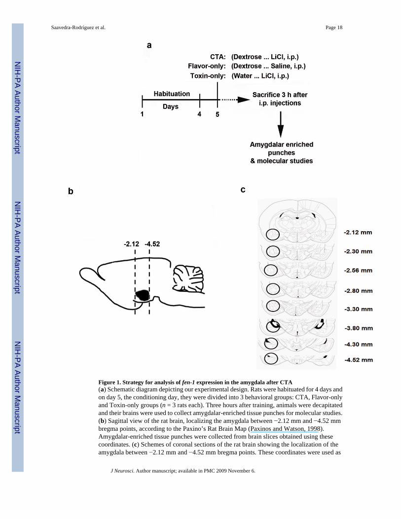
Figure 1. Strategy for analysis of fen-1 expression in the amygdala after CTA(a) Schematic diagram depicting our experimental design. Rats were habituated for 4 days andon day 5, the conditioning day, they were divided into 3 behavioral groups: CTA, Flavor-onlyand Toxin-only groups (n = 3 rats each). Three hours after training, animals were decapitatedand their brains were used to collect amygdalar-enriched tissue punches for molecular studies.(b) Sagittal view of the rat brain, localizing the amygdala between −2.12 mm and −4.52 mmbregma points, according to the Paxino’s Rat Brain Map (Paxinos and Watson, 1998).Amygdalar-enriched tissue punches were collected from brain slices obtained using thesecoordinates. (c) Schemes of coronal sections of the rat brain showing the localization of theamygdala between −2.12 mm and −4.52 mm bregma points. These coordinates were used as
Saavedra-Rodríguez et al. Page 18
J Neurosci. Author manuscript; available in PMC 2009 November 6.
NIH
-PA Author Manuscript
NIH
-PA Author Manuscript
NIH
-PA Author Manuscript

reference to extract the amygdalar tissue. The amygdala in each section is pointed out with acircle.
Saavedra-Rodríguez et al. Page 19
J Neurosci. Author manuscript; available in PMC 2009 November 6.
NIH
-PA Author Manuscript
NIH
-PA Author Manuscript
NIH
-PA Author Manuscript

Figure 2. Training-induced upregulation of fen-1 gene occurs at mRNA level in the amygdalaReal-time PCR for fen-1 mRNA detection was performed in amygdalar cDNA samples fromCTA, Flavor-only and Toxin-only animals (n = 3 rats per group) decapitated 3 h after training.(a) Amplification curves for fen-1 (right) and gapdh (left) were obtained to identify the cyclethresholds (Ct) where reporter fluorescence intensity significantly exceeded backgroundfluorescence signals. Lower Ct values reflect higher amounts of cDNA, and thus expression atthe mRNA level. (b) A melting temperature curve analysis was performed to determine thatthe reactions yielded only one product per gene. In this graph, the rate of change of the RFUswith time is plotted versus the temperature. Each peak corresponds to the melting temperatureof fen-1 or gapdh, the highest peak corresponding to gapdh. CF: curve fit; RFU: relativefluorescence unit; T: time (c) gapdh mRNA levels (measured by amplification threshold cycles,Ct), used here for normalization, remained constant in the amygdala of CTA (grey bar), Flavor-only (white bar) and Toxin-only (dark bar) groups (one-way ANOVA: F (2,35)= 0.14; P >0.05). (d) Bar graphs depicting amygdalar fen-1 normalized expression in CTA (grey bar),Flavor-only (white bar) and Toxin-only (dark bar) groups using the Standard Curve as aquantification method. One-way ANOVA identified significant differences between the groups(*P < 0.05). Newman-Keuls multiple testing specifically revealed significant differencesbetween the CTA and Flavor-only groups (*P < 0.05), but not between CTA and Toxin-onlygroups, or between Flavor-only and Toxin-only groups.
Saavedra-Rodríguez et al. Page 20
J Neurosci. Author manuscript; available in PMC 2009 November 6.
NIH
-PA Author Manuscript
NIH
-PA Author Manuscript
NIH
-PA Author Manuscript

Figure 3. Fen-1 protein analysis after CTA trainingCTA-trained rats were decapitated 15 min, 30 min, 3 h or 3.5 h (n =3 rats per time point) aftertraining and their brains were isolated to collect amygdalar and insular cortex enriched tissuepunches. Protein extracts from these regions were prepared for Fen-1 detection using Westernblotting. Representative Western blots show Fen-1 and β-actin levels in (a) the amygdala and(b) the insular cortex. Fen-1 was specifically induced in the amygdala between 3 to 3.5 h afterconditioning. (c) Western blot showing Fen-1 antibody specificity. A pre-absorption assayusing Fen-1 peptide and the antibody generated through this immunogen, shows the specificityof the antibody against Fen-1 protein. M: marker; 1: Fen-1 signal (42 KD) in absence of peptide;2: Loss of Fen-1 signal after pre-incubation of Fen-1 peptide with its antibody. (d) Bar graphspresenting the quantitative analysis of Fen-1 normalized signals in the amygdala (grey bars)and in the insular cortex (white bars) after training. Two-way ANOVA detected significantdifferences in Fen-1 expression in the amygdala, but not in the insular cortex, when comparingthe 3.5 h versus the 15 min (**P < 0.01), 30 min (++P < 0.01), and 3 h (@P < 0.05) timepoints. Error bars indicate s.e.m.
Saavedra-Rodríguez et al. Page 21
J Neurosci. Author manuscript; available in PMC 2009 November 6.
NIH
-PA Author Manuscript
NIH
-PA Author Manuscript
NIH
-PA Author Manuscript

Figure 4. Cell-type specificity of Fen-1Amygdalar sections of CTA-trained rats (n =8) decapitated 3.5 h after training were used forimmunofluorescence and analyzed using confocal microscopy. (a) Representative images ofa double immunostaining using Fen-1 antibody labeled with Alexa Fluor 488 (detected in thegreen channel) and NeuN antibody labeled with Alexa Fluor 568 (detected in the red channel).The merge of both images reveal co-localization of Fen-1-expressing cells and neurons. (b)Representative images of double immunostaining using Fen-1 antibody, now labeled withAlexa Fluor 568 (detected in the red channel), and GFAP antibody, here labeled with AlexaFluor 488 (detected in the green channel). The merge of both images shows differences inmorphology and localization between the Fen-1-expressing cells and astrocytes. (c) Mergedimages of double immunostaining with Fen-1 antibody and GFAP antibody. Fen-1-expressingcells (labeled with Alexa Fluor 568) are surrounded by astrocytes (labeled with Alexa Fluor488). Scale bar: 20 μm.
Saavedra-Rodríguez et al. Page 22
J Neurosci. Author manuscript; available in PMC 2009 November 6.
NIH
-PA Author Manuscript
NIH
-PA Author Manuscript
NIH
-PA Author Manuscript

Figure 5. Comparative analysis of fen-1 gene expression in DI TNC astrocytes, naive PC-12 cellsand PC-12 cells differentiated to the neuronal phenotype(a) The Standard Curve method of relative quantification was used to calculate gene expressionlevels. The means of amplification data for fen-1 expression relative to gapdh are shown aswhisker box plots (n = 6, two replicates for three independent experiments). The upper andlower whiskers indicate 95th and 5th percentiles respectively; the lines inside the boxes indicatethe median. One-way ANOVA detected significant differences in fen-1 mRNA levels betweenthe groups (F(2,17)= 6.180; *P = 0.011). Newman-Keuls determined that constitutive fen-1mRNA levels are significantly higher in the PC-12 neuronal cells in comparison to the naivePC-12 cells (*P < 0.05) and the DI TNC astrocytes (**P < 0.01). No significance (ns) wasfound between fen-1 mRNA levels of naive PC-12 cells and the DI TNC astrocytes (P > 0.05).(b) Confocal immunofluorescence images (showing F-actin) of DI TNC astrocytes, naivePC-12 cells and PC-12 cells differentiated to neuronal phenotype taken at magnification 100Xprior comparative analysis of fen-1 expression and presented in inverted grey scale mode forbetter visualization. Scale bar: 10 μm.
Saavedra-Rodríguez et al. Page 23
J Neurosci. Author manuscript; available in PMC 2009 November 6.
NIH
-PA Author Manuscript
NIH
-PA Author Manuscript
NIH
-PA Author Manuscript

Figure 6. Cannula placements and fen-1 antisense oligonucleotide diffusion within the amygdala(a) Schematic representation of the amygdala at different rostrocaudal planes illustratingcannulations. After behavioral treatments, fen-1 antisense and random oligonucleotide-treatedrats (n = 11 animals per treatment) were decapitated, their brains were dissected and coronalsections were obtained in order to verify cannula placements. Injector tips for each cannula arerepresented by dark circles. Most tips targeted the BLA, the CeA, and the LaVM. A few tipswere also found targeting more posterior amygdalar areas. Numbers indicate the distance frombregma in millimeters. (b) Representative bright light photomicrograph of a cannula track (toparrow) and injector tip (bottom arrow) targeting the BLA. Scale bar: 0.01 mm (1Xmagnification). (c) Schemes of coronal sections showing the diffusion of microinjected FITC-fen-1 antisense oligonucleotides into the amygdala of animals (n =4) decapitated 3 h after thelast oligonucleotide infusion. The fluorescently-labeled fen-1 antisense (represented by greenshading) was detected through different amygdalar nuclei from anterior to posterior areas,especially in the basolateral amygdalar complex. (d) Representative photomicrograph(corresponding to the same section as in b) showing FITC-fen-1 antisense oligonucleotidediffusion in the BLA. The cannula track and the injector tip area are also visible and pointedout with arrows. Scale bar: 0.1 mm (10X magnification). (e) Photomicrograph at highermagnification indicating the incorporation of the FITC-fen-1 antisense oligonucleotides intothe cells. Scale bar: 0.2 mm (20X magnification).
Saavedra-Rodríguez et al. Page 24
J Neurosci. Author manuscript; available in PMC 2009 November 6.
NIH
-PA Author Manuscript
NIH
-PA Author Manuscript
NIH
-PA Author Manuscript

Figure 7. Amygdalar fen-1 antisense treatment caused impairment of CTA memory consolidation(a, left) Animals were infused into the amygdala with fen-1 antisense (n = 6) or random (n =6)oligonucleotides 1 h prior to the dextrose and 1 h after the LiCl injection, and STM was tested2 h after conditioning. (a, right) A separate group of rats were infused into the amygdala withfen-1 antisense (n =11) or random oligonucleotides (n =11) 1 h prior to the dextrose and 1 hafter the LiCl injection, and subjected to a LTM test 48 h after conditioning. (b) Bar graphdepicting the differences in aversion index between fen-1 antisense and randomoligonucleotide-treated rats, during the STM or LTM tests. The fen-1 antisense-treated rats(grey bars) had a significantly lower aversion to the dextrose than the random-treated animals(white bars) only during the LTM test 48 h after conditioning (Bonferroni post-test, **p <
Saavedra-Rodríguez et al. Page 25
J Neurosci. Author manuscript; available in PMC 2009 November 6.
NIH
-PA Author Manuscript
NIH
-PA Author Manuscript
NIH
-PA Author Manuscript

0.01). (c) Bar graphs showing the differences in dextrose consumption (ml) between fen-1antisense (grey graphs) and random (white bars) oligonucleotide-treated animals at the timeof CS presentation (time 0) and the LTM test (48 h after training). Two-way ANOVA detectedsignificant differences in dextrose consumption between the groups only during the memorytest (Bonferroni post-testing, *P < 0.05). These results suggest that the antisense treatment didnot make the animals sick, since both groups ingested similar volumes of dextrose at the initialCS presentation. In addition, the fen-1 antisense treated rats consumed more dextrose thancontrols at 48 h, indicating that they had poorer memory of the taste-illness association. Errorbars indicate s.e.m.
Saavedra-Rodríguez et al. Page 26
J Neurosci. Author manuscript; available in PMC 2009 November 6.
NIH
-PA Author Manuscript
NIH
-PA Author Manuscript
NIH
-PA Author Manuscript
Copyright © 2022 FDOKUMEN