Identification, Docking and ADMET Studies - MDPI
10
molbank Communication Bioactive Antidiabetic Flavonoids from the Stem Bark of Cordia dichotoma Forst.: Identification, Docking and ADMET Studies Nazim Hussain 1 , Bibhuti Bhushan Kakoti 2 , Mithun Rudrapal 3, * , Khomendra Kumar Sarwa 4 , Ismail Celik 5 , Emmanuel Ifeanyi Attah 6 , Shubham Jagadish Khairnar 7 , Soumya Bhattacharya 8 , Ranjan Kumar Sahoo 9 and Sanjay G. Walode 3 Citation: Hussain, N.; Kakoti, B.B.; Rudrapal, M.; Sarwa, K.K.; Celik, I.; Attah, E.I.; Khairnar, S.J.; Bhattacharya, S.; Sahoo, R.K.; Walode, S.G. Bioactive Antidiabetic Flavonoids from the Stem Bark of Cordia dichotoma Forst.: Identification, Docking and ADMET Studies. Molbank 2021, 2021, M1234. https://doi.org/10.3390/M1234 Academic Editor: Giovanni Ribaudo Received: 7 May 2021 Accepted: 9 June 2021 Published: 11 June 2021 Publisher’s Note: MDPI stays neutral with regard to jurisdictional claims in published maps and institutional affil- iations. Copyright: © 2021 by the authors. Licensee MDPI, Basel, Switzerland. This article is an open access article distributed under the terms and conditions of the Creative Commons Attribution (CC BY) license (https:// creativecommons.org/licenses/by/ 4.0/). 1 Kingston Imperial Institute of Technology and Science, Dunga, Dehradun 248007, India; [email protected] 2 Department of Pharmaceutical Sciences, Dibrugarh University, Dibrugarh 786004, India; [email protected] 3 Rasiklal M. Dhariwal Institute of Pharmaceutical Education & Research, Chinchwad, Pune 411019, India; [email protected] 4 Department of Pharmacy, Government Girls Polytechnic, Raipur 492001, India; [email protected] 5 Department of Pharmaceutical Chemistry, Faculty of Pharmacy, Erciyes University, Talas, Kayseri 38280, Turkey; [email protected] 6 Department of Pharmaceutical and Medicinal Chemistry, University of Nigeria, Nsukka 410001, Nigeria; [email protected] 7 MET Institute of Pharmacy, Bhujbal Knowledge City, Nashik 422003, India; [email protected] 8 Guru Nanak Institute of Pharmaceutical Science and Technology, 157/F, Nilgunj Road, Panihati, Kolkata 700114, India; [email protected] 9 School of Pharmacy and Life Sciences, Centurion University of Technology and Management, Bhubaneswar 752050, India; [email protected] * Correspondence: [email protected]; Tel.: +91-86-3872-4949 Abstract: Cordia dichotoma Forst. (F. Boraginaceae) has been traditionally used for the management of a variety of human ailments. In our earlier work, the antidiabetic activity of methanolic bark extract of C. dichotoma (MECD) has been reported. In this paper, two flavonoid molecules were isolated (by column chromatography) and identified (by IR, NMR and mass spectroscopy/spectrometry) from the MECD with an aim to investigate their antidiabetic effectiveness. Molecular docking and ADMET studies were carried out using AutoDock Vina software and Swiss ADME online tool, re- spectively. The isolated flavonoids were identified as 3,5,7,3 0 ,4 0 -tetrahydroxy-4-methoxyflavone-3-O- L-rhamnopyranoside and 5,7,3 0 -trihydroxy-4-methoxyflavone-7-O-L-rhamnopyranoside (quercitrin). Docking and ADMET studies revealed the promising binding affinity of flavonoid molecules for human lysosomal α-glucosidase and human pancreatic α-amylase with acceptable ADMET prop- erties. Based on computational studies, our study reports the antidiabetic potential of the isolated flavonoids with predictive pharmacokinetics profile. Keywords: C. dichotoma; flavonoids; antidiabetic; α-glucosidase; α-amylase; docking; ADMET 1. Introduction Cordia dichotoma Forst. (also known as Indian cherry, F. Boraginaceae) is a traditionally important deciduous medicinal plant widely grown in India, Sri Lanka and other tropical countries of the world [1]. This plant has been traditionally (Ayurveda, Unani and Siddha medicines) used for the management of a variety of human ailments/disorders [1]. Leaves and stem bark have been used traditionally in the treatment of fever, dyspepsia, diarrhea, leprosy, gonorrhea and wounds [2]. Leaves, seeds, bark and fruits have been reported to Molbank 2021, 2021, M1234. https://doi.org/10.3390/M1234 https://www.mdpi.com/journal/molbank
-
Upload
khangminh22 -
Category
Documents
-
view
4 -
download
0
Transcript of Identification, Docking and ADMET Studies - MDPI
molbank
Communication
Bioactive Antidiabetic Flavonoids from the Stem Bark ofCordia dichotoma Forst.: Identification, Docking andADMET Studies
Nazim Hussain 1, Bibhuti Bhushan Kakoti 2, Mithun Rudrapal 3,* , Khomendra Kumar Sarwa 4,Ismail Celik 5, Emmanuel Ifeanyi Attah 6, Shubham Jagadish Khairnar 7, Soumya Bhattacharya 8,Ranjan Kumar Sahoo 9 and Sanjay G. Walode 3
�����������������
Citation: Hussain, N.; Kakoti, B.B.;
Rudrapal, M.; Sarwa, K.K.; Celik, I.;
Attah, E.I.; Khairnar, S.J.; Bhattacharya,
S.; Sahoo, R.K.; Walode, S.G. Bioactive
Antidiabetic Flavonoids from the
Stem Bark of Cordia dichotoma Forst.:
Identification, Docking and ADMET
Studies. Molbank 2021, 2021, M1234.
https://doi.org/10.3390/M1234
Academic Editor: Giovanni Ribaudo
Received: 7 May 2021
Accepted: 9 June 2021
Published: 11 June 2021
Publisher’s Note: MDPI stays neutral
with regard to jurisdictional claims in
published maps and institutional affil-
iations.
Copyright: © 2021 by the authors.
Licensee MDPI, Basel, Switzerland.
This article is an open access article
distributed under the terms and
conditions of the Creative Commons
Attribution (CC BY) license (https://
creativecommons.org/licenses/by/
4.0/).
1 Kingston Imperial Institute of Technology and Science, Dunga, Dehradun 248007, India;[email protected]
2 Department of Pharmaceutical Sciences, Dibrugarh University, Dibrugarh 786004, India;[email protected]
3 Rasiklal M. Dhariwal Institute of Pharmaceutical Education & Research, Chinchwad, Pune 411019, India;[email protected]
4 Department of Pharmacy, Government Girls Polytechnic, Raipur 492001, India;[email protected]
5 Department of Pharmaceutical Chemistry, Faculty of Pharmacy, Erciyes University, Talas,Kayseri 38280, Turkey; [email protected]
6 Department of Pharmaceutical and Medicinal Chemistry, University of Nigeria, Nsukka 410001, Nigeria;[email protected]
7 MET Institute of Pharmacy, Bhujbal Knowledge City, Nashik 422003, India; [email protected] Guru Nanak Institute of Pharmaceutical Science and Technology, 157/F, Nilgunj Road, Panihati,
Kolkata 700114, India; [email protected] School of Pharmacy and Life Sciences, Centurion University of Technology and Management,
Bhubaneswar 752050, India; [email protected]* Correspondence: [email protected]; Tel.: +91-86-3872-4949
Abstract: Cordia dichotoma Forst. (F. Boraginaceae) has been traditionally used for the management ofa variety of human ailments. In our earlier work, the antidiabetic activity of methanolic bark extractof C. dichotoma (MECD) has been reported. In this paper, two flavonoid molecules were isolated(by column chromatography) and identified (by IR, NMR and mass spectroscopy/spectrometry)from the MECD with an aim to investigate their antidiabetic effectiveness. Molecular docking andADMET studies were carried out using AutoDock Vina software and Swiss ADME online tool, re-spectively. The isolated flavonoids were identified as 3,5,7,3′,4′-tetrahydroxy-4-methoxyflavone-3-O-L-rhamnopyranoside and 5,7,3′-trihydroxy-4-methoxyflavone-7-O-L-rhamnopyranoside (quercitrin).Docking and ADMET studies revealed the promising binding affinity of flavonoid molecules forhuman lysosomal α-glucosidase and human pancreatic α-amylase with acceptable ADMET prop-erties. Based on computational studies, our study reports the antidiabetic potential of the isolatedflavonoids with predictive pharmacokinetics profile.
Keywords: C. dichotoma; flavonoids; antidiabetic; α-glucosidase; α-amylase; docking; ADMET
1. Introduction
Cordia dichotoma Forst. (also known as Indian cherry, F. Boraginaceae) is a traditionallyimportant deciduous medicinal plant widely grown in India, Sri Lanka and other tropicalcountries of the world [1]. This plant has been traditionally (Ayurveda, Unani and Siddhamedicines) used for the management of a variety of human ailments/disorders [1]. Leavesand stem bark have been used traditionally in the treatment of fever, dyspepsia, diarrhea,leprosy, gonorrhea and wounds [2]. Leaves, seeds, bark and fruits have been reported to
Molbank 2021, 2021, M1234. https://doi.org/10.3390/M1234 https://www.mdpi.com/journal/molbank
Molbank 2021, 2021, M1234 2 of 10
exhibit anti-inflammatory, anthelmintic, antibacterial, antileprotic, antiviral, diuretic, astrin-gent, demulcent, laxative/purgative, expectorant/antitussive, tonic, immunomodulatory,hepatoprotective and gastroprotective/antiulcer activities [3,4].
Recent literature have also reported the anti-inflammatory [3], antidiabetic [4], an-ticancer [2] and antioxidant [3] activities for the bark extract of C. dichotoma. Phytocon-stituents like3′,5-dihydroxy-4′-methoxyflavanone-7-O-α-L-rhamnopyranoside, β-sitosteryl-3β-glucopyranoside-6′-O-palmitate, quercitrin and β-sitosterol have already been isolatedfrom the leaves of this plant [5]. Betulin, lupeol-3-rhamnoside, β-sitosterol, taxifolin-3-5-dirhamnoside, hesperitin-7-rhamnoside, rutin, chlorogenic acid and caffeic acid have beenreported from seeds of C. dichotoma [2–5]. Flavonoids, the most important class of plantpolyphenolics possessing diverse range of biological/pharmacological potential [6–8], areattributed to be the most dominant phytochemical components in various plant parts ofC. dichotoma. In this work, the isolation and identification of bioactive flavonoids fromthe methanolic bark extract of C. dichotoma Forst was carried out. The isolated flavonoidmolecules were further investigated for their antidiabetic potential and pharmacokineticproperties by molecular docking and ADMET studies.
2. Results and Discussion
The phytochemical analysis revealed the presence of flavonoids, alkaloids, glycosides,saponins, steroids, carbohydrates and proteins in the methanolic bark extract of C. dichotomaForst (MECD) [2–4].
2.1. Identification of Isolated Phytocompounds2.1.1. Compound 1 (MECD-1)
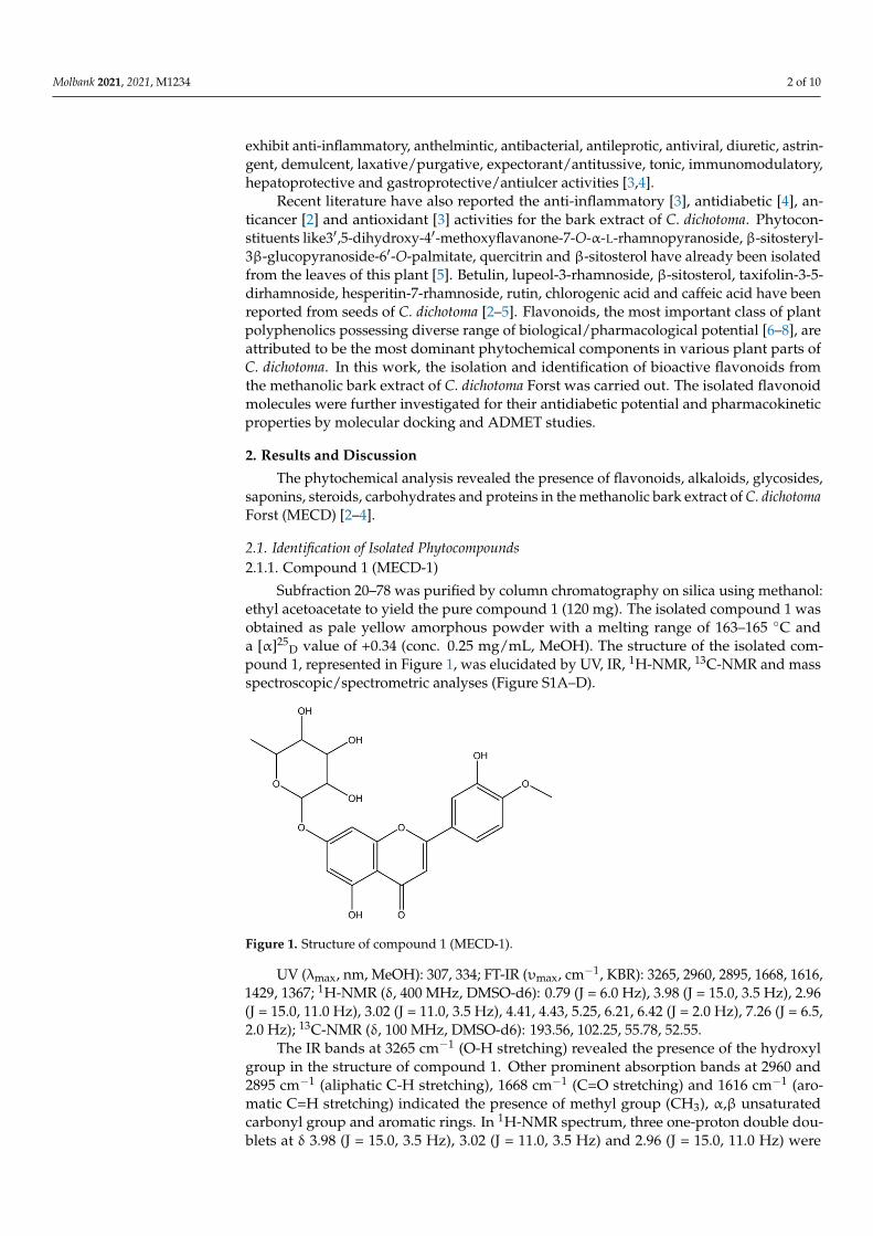
Subfraction 20–78 was purified by column chromatography on silica using methanol:ethyl acetoacetate to yield the pure compound 1 (120 mg). The isolated compound 1 wasobtained as pale yellow amorphous powder with a melting range of 163–165 ◦C anda [α]25
D value of +0.34 (conc. 0.25 mg/mL, MeOH). The structure of the isolated com-pound 1, represented in Figure 1, was elucidated by UV, IR, 1H-NMR, 13C-NMR and massspectroscopic/spectrometric analyses (Figure S1A–D).
Molbank 2021, 2021, x FOR PEER REVIEW 3 of 10
methoxy group was confirmed by position of the methyl signal at δ 52.55. The molecular ion [M]+ peak was obtained at m/z 446.0, which concord the molecular formula of the com-pound 1 as C22H22O11. The NMR spectral data also supported the structure of the com-pound. A thorough spectral interpretation suggests that the compound 1 (MECD-1) is 5,7,3,-trihydroxy-4-methoxyflavone-7-O-L-rhamnopyranoside.
Figure 1. Structure of compound 1 (MECD-1).
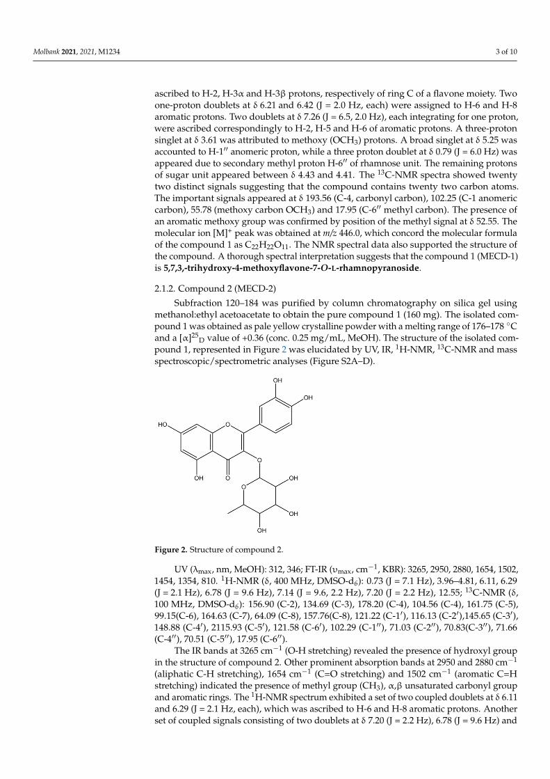
2.1.2. Compound 2 (MECD-2) Subfraction 120–184 was purified by column chromatography on silica gel using
methanol:ethyl acetoacetate to obtain the pure compound 1 (160 mg). The isolated com-pound 1 was obtained as pale yellow crystalline powder with a melting range of 176–178 °C and a [α]25D value of +0.36 (conc. 0.25 mg/mL, MeOH). The structure of the isolated compound 1, represented in Figure 2 was elucidated by UV, IR, 1HNMR, 13CNMR and mass spectroscopic/spectrometric analyses (Figures S2A-D).
UV (λmax, nm, MeOH): 312, 346; FT-IR (υmax, cm−1, KBR): 3265, 2950, 2880, 1654, 1502, 1454, 1354, 810. 1HNMR (δ, 400 MHz, DMSO-d6): 0.73 (J = 7.1 Hz), 3.96–4.81, 6.11, 6.29 (J = 2.1 Hz), 6.78 (J = 9.6 Hz), 7.14 (J = 9.6, 2.2 Hz), 7.20 (J = 2.2 Hz), 12.55; 13CNMR (δ, 100 MHz, DMSO-d6): 156.90 (C-2), 134.69 (C-3), 178.20 (C-4), 104.56 (C-4), 161.75 (C-5), 99.15(C-6), 164.63 (C-7), 64.09 (C-8), 157.76(C-8), 121.22 (C-1’), 116.13 (C-2’),145.65 (C-3’), 148.88 (C-4’), 2115.93 (C-5’), 121.58 (C-6’), 102.29 (C-1”), 71.03 (C-2”), 70.83(C-3”), 71.66 (C-4”), 70.51 (C-5”), 17.95 (C-6”).
The IR bands at 3265 cm−1 (O-H stretching) revealed the presence of hydroxyl group in the structure of compound 2. Other prominent absorption bands at 2950 and 2880 cm−1 (aliphatic C-H stretching), 1654 cm−1 (C=O stretching) and 1502 cm−1 (aromatic C=H stretching) indicated the presence of methyl group (CH3), α,β unsaturated carbonyl group and aromatic rings. The 1HNMR spectrum exhibited a set of two coupled doublets at δ 6.11 and 6.29 (J = 2.1 Hz, each), which was ascribed to H-6 and H-8 aromatic protons. Another set of coupled signals consisting of two doublets at δ 7.20 (J = 2.2 Hz), 6.78 (J = 9.6 Hz) and a double-doublet at δ 7.14 (J = 9.6, 2.2 Hz) were ascribed to H-2′, H-5′ and H-6′ aromatic protons of ring B. A doublet at δ 5.15 (J = 8.1 Hz) was assigned to H-1” anomeric proton, while as another doublet at δ 0.73 (J = 7.1 Hz) was attributed to methyl protons (H-6”) of rhamnose unit. The remaining protons of rhamnose resonated from δ 4.81 to 3.96. A single proton singlet at δ 12.55 was attributed to hydroxyl proton. The 13CNMR spectrum displayed signals for twenty-one carbons. Important signals appeared for car-bonyl carbon (δ 178.20, C-4), anomeric carbon (δ 102.29, C-1”) and methyl carbon (δ 17.94, C-6”). The molecular ion [M]+ peak was obtained at m/z 448.0, which concord the molec-ular formula of the compound as C21H20O11. The 1H and 13C NMR data was compared with other reported flavonoids and was found to be 3,5,7,3,4,-tetrahydroxy-4-methoxyflavone-3-O-L-rhamnopyranoside (Quercitrin (Quercetin-3-O-L-rhamnoside).
Figure 1. Structure of compound 1 (MECD-1).
UV (λmax, nm, MeOH): 307, 334; FT-IR (υmax, cm−1, KBR): 3265, 2960, 2895, 1668, 1616,1429, 1367; 1H-NMR (δ, 400 MHz, DMSO-d6): 0.79 (J = 6.0 Hz), 3.98 (J = 15.0, 3.5 Hz), 2.96(J = 15.0, 11.0 Hz), 3.02 (J = 11.0, 3.5 Hz), 4.41, 4.43, 5.25, 6.21, 6.42 (J = 2.0 Hz), 7.26 (J = 6.5,2.0 Hz); 13C-NMR (δ, 100 MHz, DMSO-d6): 193.56, 102.25, 55.78, 52.55.
The IR bands at 3265 cm−1 (O-H stretching) revealed the presence of the hydroxylgroup in the structure of compound 1. Other prominent absorption bands at 2960 and2895 cm−1 (aliphatic C-H stretching), 1668 cm−1 (C=O stretching) and 1616 cm−1 (aro-matic C=H stretching) indicated the presence of methyl group (CH3), α,β unsaturatedcarbonyl group and aromatic rings. In 1H-NMR spectrum, three one-proton double dou-blets at δ 3.98 (J = 15.0, 3.5 Hz), 3.02 (J = 11.0, 3.5 Hz) and 2.96 (J = 15.0, 11.0 Hz) were
Molbank 2021, 2021, M1234 3 of 10
ascribed to H-2, H-3α and H-3β protons, respectively of ring C of a flavone moiety. Twoone-proton doublets at δ 6.21 and 6.42 (J = 2.0 Hz, each) were assigned to H-6 and H-8aromatic protons. Two doublets at δ 7.26 (J = 6.5, 2.0 Hz), each integrating for one proton,were ascribed correspondingly to H-2, H-5 and H-6 of aromatic protons. A three-protonsinglet at δ 3.61 was attributed to methoxy (OCH3) protons. A broad singlet at δ 5.25 wasaccounted to H-1′′ anomeric proton, while a three proton doublet at δ 0.79 (J = 6.0 Hz) wasappeared due to secondary methyl proton H-6′′ of rhamnose unit. The remaining protonsof sugar unit appeared between δ 4.43 and 4.41. The 13C-NMR spectra showed twentytwo distinct signals suggesting that the compound contains twenty two carbon atoms.The important signals appeared at δ 193.56 (C-4, carbonyl carbon), 102.25 (C-1 anomericcarbon), 55.78 (methoxy carbon OCH3) and 17.95 (C-6′′ methyl carbon). The presence ofan aromatic methoxy group was confirmed by position of the methyl signal at δ 52.55. Themolecular ion [M]+ peak was obtained at m/z 446.0, which concord the molecular formulaof the compound 1 as C22H22O11. The NMR spectral data also supported the structure ofthe compound. A thorough spectral interpretation suggests that the compound 1 (MECD-1)is 5,7,3,-trihydroxy-4-methoxyflavone-7-O-L-rhamnopyranoside.
2.1.2. Compound 2 (MECD-2)
Subfraction 120–184 was purified by column chromatography on silica gel usingmethanol:ethyl acetoacetate to obtain the pure compound 1 (160 mg). The isolated com-pound 1 was obtained as pale yellow crystalline powder with a melting range of 176–178 ◦Cand a [α]25
D value of +0.36 (conc. 0.25 mg/mL, MeOH). The structure of the isolated com-pound 1, represented in Figure 2 was elucidated by UV, IR, 1H-NMR, 13C-NMR and massspectroscopic/spectrometric analyses (Figure S2A–D).
Molbank 2021, 2021, x FOR PEER REVIEW 4 of 10
Figure 2. Structure of compound 2.
2.2. Molecular Docking Molecular docking is used to understand the drug–receptor interaction, binding af-
finity and binding orientation of bioactive molecules into the target protein molecule. The objective behind docking study is to predict a particular biological activity based on the binding orientation/affinity of small molecule ligands to the appropriate target active site [9]. In the docking study, the binding affinity was predicted in terms of the interaction energy (kcal/mole). Results of docking (binding) energies are given in Table 1. Both the compounds exhibited very good binding affinity against both α-glucosidase and α-amyl-ase enzyme. Compound 1 (MECD-1) exhibited more binding affinity against alpha-amyl-ase compared to the alpha-glucosidase. On the hand, the compound 2 (MECD-2) showed better affinity against alpha-glucosidase than alpha-amylase. Not much variation in bind-ing energies between these two enzymes were observed. Docking scores of isolated com-pounds were compared with that of the standard drug, acarbose. Against α-glucosidase, the binding affinity of compounds 1 and 2 was comparatively more affinity than the standard drug. On the other hand, the binding affinity of compounds 1 and 2 were less to some extent than that the standard drug against α-amylase. No significant difference in activities between isolated test compounds and the standard compound was observed. The test compounds were found to have α-glucosidase and α-amylase inhibitory potential to a similar degree as that of the standard drug, acarbose. Overall, both the isolated flavo-noids exhibited significant inhibitory potential of human glucosidase and amylase en-zymes.
Table 1. Docking data of compounds.
Compound Binding Energy (kcal/mole) 5NN8 1B2Y Compound 1 (MECD-1) −7.8 −8.6 Compound 2 (MECD-2) −8.0 −7.8 Acarbose (Standard drug) −7.6 −9.4 5NN8: Human lysosomal acid α-glucosidase; 1B2Y: Human pancreatic α-amylase; MECD: Metanolic bark extract of C. dichotoma.
Post-docking visualization of protein–ligand complexes revealed that the com-pounds interacted with active site residues of the protein molecules through the formation of predominantly hydrogen bonding interactions (Figures 3 and 4). From the observation of 2D interaction diagrams of compound 1-α-glucosidase complexes, it is clear that com-pound 1 formed H-bonds with Trp59, Gln63, Asp197, Asp300 and Asp356 residues, whereas the compound 2 interacted with Tyr62, His101, His201 and Gly306 residues
Figure 2. Structure of compound 2.
UV (λmax, nm, MeOH): 312, 346; FT-IR (υmax, cm−1, KBR): 3265, 2950, 2880, 1654, 1502,1454, 1354, 810. 1H-NMR (δ, 400 MHz, DMSO-d6): 0.73 (J = 7.1 Hz), 3.96–4.81, 6.11, 6.29(J = 2.1 Hz), 6.78 (J = 9.6 Hz), 7.14 (J = 9.6, 2.2 Hz), 7.20 (J = 2.2 Hz), 12.55; 13C-NMR (δ,100 MHz, DMSO-d6): 156.90 (C-2), 134.69 (C-3), 178.20 (C-4), 104.56 (C-4), 161.75 (C-5),99.15(C-6), 164.63 (C-7), 64.09 (C-8), 157.76(C-8), 121.22 (C-1′), 116.13 (C-2′),145.65 (C-3′),148.88 (C-4′), 2115.93 (C-5′), 121.58 (C-6′), 102.29 (C-1′′), 71.03 (C-2′′), 70.83(C-3′′), 71.66(C-4′′), 70.51 (C-5′′), 17.95 (C-6′′).
The IR bands at 3265 cm−1 (O-H stretching) revealed the presence of hydroxyl groupin the structure of compound 2. Other prominent absorption bands at 2950 and 2880 cm−1
(aliphatic C-H stretching), 1654 cm−1 (C=O stretching) and 1502 cm−1 (aromatic C=Hstretching) indicated the presence of methyl group (CH3), α,β unsaturated carbonyl groupand aromatic rings. The 1H-NMR spectrum exhibited a set of two coupled doublets at δ 6.11and 6.29 (J = 2.1 Hz, each), which was ascribed to H-6 and H-8 aromatic protons. Anotherset of coupled signals consisting of two doublets at δ 7.20 (J = 2.2 Hz), 6.78 (J = 9.6 Hz) and

Molbank 2021, 2021, M1234 4 of 10
a double-doublet at δ 7.14 (J = 9.6, 2.2 Hz) were ascribed to H-2′, H-5′ and H-6′ aromaticprotons of ring B. A doublet at δ 5.15 (J = 8.1 Hz) was assigned to H-1′′ anomeric proton,while as another doublet at δ 0.73 (J = 7.1 Hz) was attributed to methyl protons (H-6′′)of rhamnose unit. The remaining protons of rhamnose resonated from δ 4.81 to 3.96. Asingle proton singlet at δ 12.55 was attributed to hydroxyl proton. The 13C-NMR spectrumdisplayed signals for twenty-one carbons. Important signals appeared for carbonyl carbon(δ 178.20, C-4), anomeric carbon (δ 102.29, C-1′′) and methyl carbon (δ 17.94, C-6′′). Themolecular ion [M]+ peak was obtained at m/z 448.0, which concord the molecular formulaof the compound as C21H20O11. The 1H and 13C NMR data was compared with otherreported flavonoids and was found to be 3,5,7,3,4,-tetrahydroxy-4-methoxyflavone-3-O-L-rhamnopyranoside (Quercitrin (Quercetin-3-O-L-rhamnoside).
2.2. Molecular Docking
Molecular docking is used to understand the drug–receptor interaction, bindingaffinity and binding orientation of bioactive molecules into the target protein molecule.The objective behind docking study is to predict a particular biological activity based onthe binding orientation/affinity of small molecule ligands to the appropriate target activesite [9]. In the docking study, the binding affinity was predicted in terms of the interactionenergy (kcal/mole). Results of docking (binding) energies are given in Table 1. Both thecompounds exhibited very good binding affinity against both α-glucosidase and α-amylaseenzyme. Compound 1 (MECD-1) exhibited more binding affinity against alpha-amylasecompared to the alpha-glucosidase. On the hand, the compound 2 (MECD-2) showed betteraffinity against alpha-glucosidase than alpha-amylase. Not much variation in bindingenergies between these two enzymes were observed. Docking scores of isolated compoundswere compared with that of the standard drug, acarbose. Against α-glucosidase, thebinding affinity of compounds 1 and 2 was comparatively more affinity than the standarddrug. On the other hand, the binding affinity of compounds 1 and 2 were less to someextent than that the standard drug against α-amylase. No significant difference in activitiesbetween isolated test compounds and the standard compound was observed. The testcompounds were found to have α-glucosidase and α-amylase inhibitory potential to asimilar degree as that of the standard drug, acarbose. Overall, both the isolated flavonoidsexhibited significant inhibitory potential of human glucosidase and amylase enzymes.
Table 1. Docking data of compounds.
Compound Binding Energy (kcal/mole)
5NN8 1B2Y
Compound 1 (MECD-1) −7.8 −8.6Compound 2 (MECD-2) −8.0 −7.8Acarbose (Standard drug) −7.6 −9.4
5NN8: Human lysosomal acid α-glucosidase; 1B2Y: Human pancreatic α-amylase; MECD: Metanolic bark extractof C. dichotoma.
Post-docking visualization of protein–ligand complexes revealed that the compoundsinteracted with active site residues of the protein molecules through the formation of predom-inantly hydrogen bonding interactions (Figures 3 and 4). From the observation of 2D interac-tion diagrams of compound 1-α-glucosidase complexes, it is clear that compound 1 formedH-bonds with Trp59, Gln63, Asp197, Asp300 and Asp356 residues, whereas the compound 2interacted with Tyr62, His101, His201 and Gly306 residues through H-bonds (Figure 3a,b).The 3D diagrams revealed the binding conformation and binding poses of the compounds atthe catalytic site of α-glucosidase (Figure 3c,d) were observed.

Molbank 2021, 2021, M1234 5 of 10
Molbank 2021, 2021, x FOR PEER REVIEW 5 of 10
through H-bonds (Figure 3a,b). The 3D diagrams revealed the binding conformation and binding poses of the compounds at the catalytic site of α-glucosidase (Figure 3c,d) were observed.
Figure 3. (a) Two-dimensional interactions between compound 1 and α-glucosidase, (b) 2D interactions between com-pound 2 and α-glucosidase showing hydrogen bonding and other non-covalent interactions with amino acid residues at the active site, (c) 3D representation of protein-ligand interactions showing binding conformation and (d) binding poses/binding modes of both the compounds at the catalytic site of α-glucosidase.
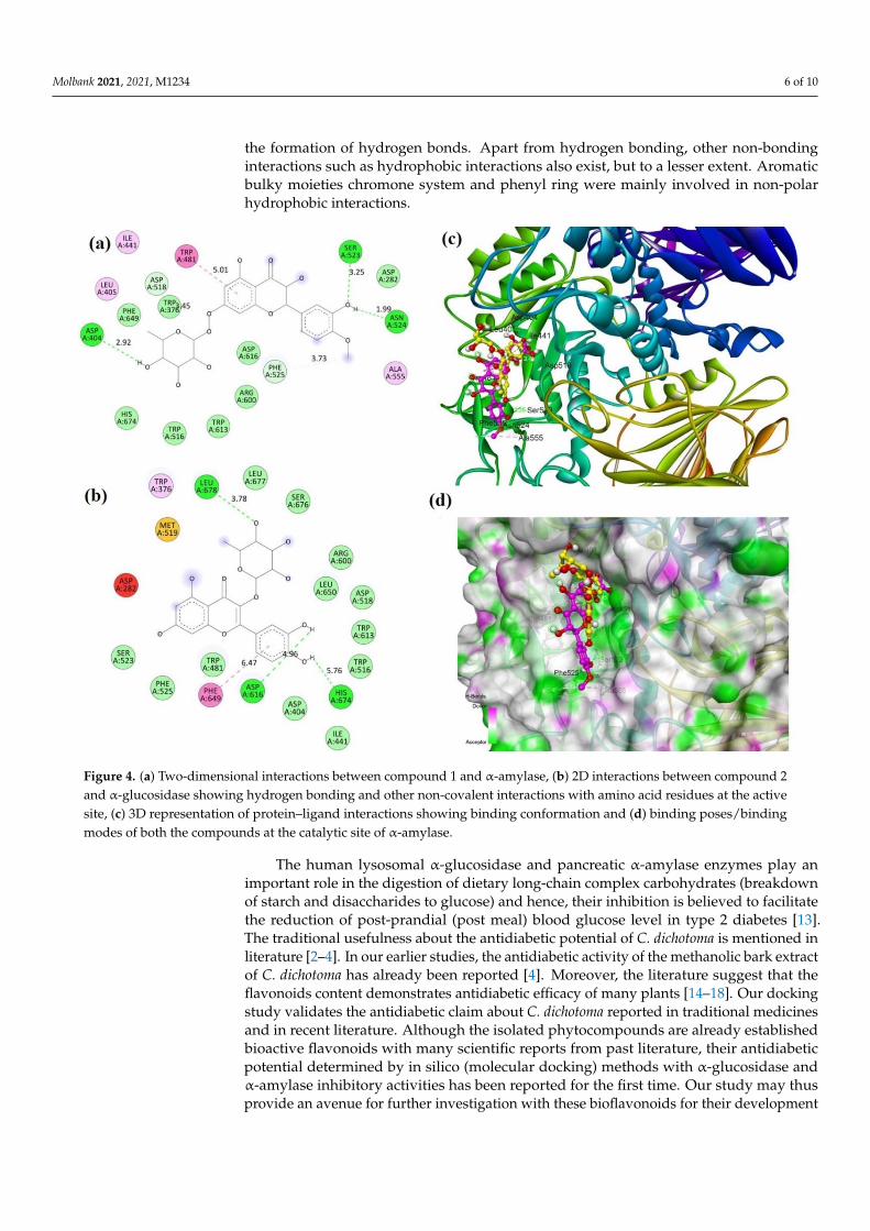
From the observation of 2D interaction diagrams of compound 2-α-amylase com-plexes, it is clear that compound 1 formed prominent H-bonds with Asp404, Ser523 and Ser524 residues, whereas the compound 2 interacted with Asp616, His674 and Leu678 residues through H-bonds (Figures 3b and 4a). The 3D diagrams revealed the binding conformation and binding poses of the compounds at the catalytic site of alpha-amylase (Figures 3d and 4c) were observed.
Upon critical analysis of protein–ligand interactions, favorable binding orientations and/or binding modes of flavonoid molecules for both the α-glucosidase and α-amylase enzyme were evident. Both the compounds structurally represent glycosides of flavones, which are abundantly found in plant kingdom. The glycone part (sugar), i.e., the rham-nose moiety is similar in both the compounds, while the aglycone part (non-sugar bioac-tive principle, flavanone moiety) is dissimilar. In compound 1, it is 5,7,3′-trihydroxy-4-methoxyflavone, whereas, it is 3,5,7,3′,4′-tetrahydroxy-4-methoxyflavone, i.e., quercetin in compound 2. The aglycone, i.e., the flavonoid moiety is a polyhydroxylated C6-C3-C6 tri-cyclic heteroaromatic system (phenylchromone) [10–12] with distinct structural features, particularly in terms of nature and pattern of ring substitutions. There is a close structural resemblance between these two isolated flavonoid glycosides. The basic flavone skeleton along with hydroxylated/methoxylated aromatic ring interacted predominantly with the active site residues of target protein molecules. Polar groups such as phenolic hydroxy groups and carbonyl moiety contributed significantly in protein-ligand interactions with the formation of hydrogen bonds. Apart from hydrogen bonding, other non-bonding in-teractions such as hydrophobic interactions also exist, but to a lesser extent. Aromatic
Figure 3. (a) Two-dimensional interactions between compound 1 and α-glucosidase, (b) 2D interactions between compound2 and α-glucosidase showing hydrogen bonding and other non-covalent interactions with amino acid residues at the activesite, (c) 3D representation of protein-ligand interactions showing binding conformation and (d) binding poses/bindingmodes of both the compounds at the catalytic site of α-glucosidase.
From the observation of 2D interaction diagrams of compound 2-α-amylase com-plexes, it is clear that compound 1 formed prominent H-bonds with Asp404, Ser523 andSer524 residues, whereas the compound 2 interacted with Asp616, His674 and Leu678residues through H-bonds (Figures 3b and 4a). The 3D diagrams revealed the bindingconformation and binding poses of the compounds at the catalytic site of alpha-amylase(Figures 3d and 4c) were observed.
Upon critical analysis of protein–ligand interactions, favorable binding orientationsand/or binding modes of flavonoid molecules for both the α-glucosidase and α-amylaseenzyme were evident. Both the compounds structurally represent glycosides of flavones,which are abundantly found in plant kingdom. The glycone part (sugar), i.e., the rham-nose moiety is similar in both the compounds, while the aglycone part (non-sugar bioac-tive principle, flavanone moiety) is dissimilar. In compound 1, it is 5,7,3′-trihydroxy-4-methoxyflavone, whereas, it is 3,5,7,3′,4′-tetrahydroxy-4-methoxyflavone, i.e., quercetinin compound 2. The aglycone, i.e., the flavonoid moiety is a polyhydroxylated C6-C3-C6tricyclic heteroaromatic system (phenylchromone) [10–12] with distinct structural features,particularly in terms of nature and pattern of ring substitutions. There is a close structuralresemblance between these two isolated flavonoid glycosides. The basic flavone skeletonalong with hydroxylated/methoxylated aromatic ring interacted predominantly with theactive site residues of target protein molecules. Polar groups such as phenolic hydroxygroups and carbonyl moiety contributed significantly in protein-ligand interactions with
Molbank 2021, 2021, M1234 6 of 10
the formation of hydrogen bonds. Apart from hydrogen bonding, other non-bondinginteractions such as hydrophobic interactions also exist, but to a lesser extent. Aromaticbulky moieties chromone system and phenyl ring were mainly involved in non-polarhydrophobic interactions.
Molbank 2021, 2021, x FOR PEER REVIEW 6 of 10
bulky moieties chromone system and phenyl ring were mainly involved in non-polar hy-drophobic interactions.
Figure 4. (a) Two-dimensional interactions between compound 1 and α-amylase, (b) 2D interactions between compound 2 and α-glucosidase showing hydrogen bonding and other non-covalent interactions with amino acid residues at the active site, (c) 3D representation of protein–ligand interactions showing binding conformation and (d) binding poses/binding modes of both the compounds at the catalytic site of α-amylase.
The human lysosomal α-glucosidase and pancreatic α-amylase enzymes play an im-portant role in the digestion of dietary long-chain complex carbohydrates (breakdown of starch and disaccharides to glucose) and hence, their inhibition is believed to facilitate the reduction of post-prandial (post meal) blood glucose level in type 2 diabetes [13]. The tra-ditional usefulness about the antidiabetic potential of C. dichotoma is mentioned in litera-ture [2–4]. In our earlier studies, the antidiabetic activity of the methanolic bark extract of C. dichotoma has already been reported [4]. Moreover, the literature suggest that the flavo-noids content demonstrates antidiabetic efficacy of many plants [14–18]. Our docking study validates the antidiabetic claim about C. dichotoma reported in traditional medicines and in recent literature. Although the isolated phytocompounds are already established bioactive flavonoids with many scientific reports from past literature, their antidiabetic potential determined by in silico (molecular docking) methods with α-glucosidase and α-amylase inhibitory activities has been reported for the first time. Our study may thus pro-vide an avenue for further investigation with these bioflavonoids for their development as potent antidiabetic molecules with alpha-glucosidase and alpha-amylase inhibitory agents for the treatment of type 2 diabetes mellitus.
2.3. ADMET Results of predicted ADMET (absorption, distribution, metabolism, excretion and
toxicity) data showed that both the isolated compounds possess good solubility profile, which is in favor of their oral bioavailability. There is a prediction of poor intestinal ab-sorption, while the compounds were predicted to be non-inhibitors of the cytochromes (CYP450) [19]. Poor intestinal absorption might be due to their limited oil/water partition
Figure 4. (a) Two-dimensional interactions between compound 1 and α-amylase, (b) 2D interactions between compound 2and α-glucosidase showing hydrogen bonding and other non-covalent interactions with amino acid residues at the activesite, (c) 3D representation of protein–ligand interactions showing binding conformation and (d) binding poses/bindingmodes of both the compounds at the catalytic site of α-amylase.
The human lysosomal α-glucosidase and pancreatic α-amylase enzymes play animportant role in the digestion of dietary long-chain complex carbohydrates (breakdownof starch and disaccharides to glucose) and hence, their inhibition is believed to facilitatethe reduction of post-prandial (post meal) blood glucose level in type 2 diabetes [13].The traditional usefulness about the antidiabetic potential of C. dichotoma is mentioned inliterature [2–4]. In our earlier studies, the antidiabetic activity of the methanolic bark extractof C. dichotoma has already been reported [4]. Moreover, the literature suggest that theflavonoids content demonstrates antidiabetic efficacy of many plants [14–18]. Our dockingstudy validates the antidiabetic claim about C. dichotoma reported in traditional medicinesand in recent literature. Although the isolated phytocompounds are already establishedbioactive flavonoids with many scientific reports from past literature, their antidiabeticpotential determined by in silico (molecular docking) methods with α-glucosidase andα-amylase inhibitory activities has been reported for the first time. Our study may thusprovide an avenue for further investigation with these bioflavonoids for their development
Molbank 2021, 2021, M1234 7 of 10
as potent antidiabetic molecules with alpha-glucosidase and alpha-amylase inhibitoryagents for the treatment of type 2 diabetes mellitus.
2.3. ADMET
Results of predicted ADMET (absorption, distribution, metabolism, excretion andtoxicity) data showed that both the isolated compounds possess good solubility profile,which is in favor of their oral bioavailability. There is a prediction of poor intestinal ab-sorption, while the compounds were predicted to be non-inhibitors of the cytochromes(CYP450) [19]. Poor intestinal absorption might be due to their limited oil/water partitioncoefficient (logP) values (−1.64 and −1.84). CYP450 enzymes are largely involved in drugmetabolism. Non-inhibition of CYP450 enzymes suggests that compounds do not suppressthe metabolic function of the enzymes. Inhibition can lead to increased bioavailability ofcompounds that normally undergo extensive first-pass elimination or to decreased elimi-nation of compounds dependent on metabolism for systemic clearance. Compounds didnot exhibit the property of blood brain barrier (BBB) penetration. It substantiates that thecompounds are devoid of producing CNS toxicities. Furthermore, quercitrin (compound 2)was predicted to be a substrate to permeability of glycoprotein (p-gp), whereas the otherflavonoid molecule (compound 1) did not show such property. Glycoprotein is responsiblefor the efflux of drug molecules out of the target cells [20]. A good drug candidate shouldnot only have sufficient efficacy against the therapeutic target, but also show appropriateADMET properties at a therapeutic dose. It is therefore inevitable to evaluate the ADMETprofile of drug-like molecules to avoid the failure of candidate drugs at the clinical stage ofdrug development [21].
3. Materials and Methods3.1. Collection of Plant
The barks of Cordia dichotoma Forst. were collected from the Duhai forest of Ghaziabad,Uttar Pradesh, India. The plant species was identified from National Institute of ScienceCommunication and Information Resources, New Delhi, India. The voucher specimen(NISCAIR/RHMD/Consult/2012-13/2025/33) of the bark of Cordia dichotoma Forst. wassubmitted at the herbarium of the department for future reference.
3.2. Preparation of Methanolic Bark Extract
The shade-dried barks of C. dichotoma were pulverized to a coarse powder and defattedusing petroleum ether by the cold maceration method [2] to remove fat, latex and non-polarcompounds of high molecular weights. The defatted plant residues were then maceratedsuccessively with methanol to obtain the desired extract [3,4]. The collected extract wasfiltered through Whatman No. 1 filter paper. Rotary evaporator was used to concentratethe filtrate. The concentrated extract was preserved in refrigerator at 4 ◦C for further use.The percentage yield of the methanolic bark extract of C. dichotoma (MECD) was 7.11%.
3.3. Phytochemical Analysis
Chemical tests for the screening and detection of phytochemical constituents of theMECD were carried out using the standard procedures [22,23].
3.4. Isolation of Phytocompounds
The MECD was subjected to column chromatographic separation using silica gel(packed column, 100–200 mesh) and a glass column (6.0 × 3 inch dimension) [6] for theisolation of bioactive phytoconstituents in pure form. The elution was carried out bygradient separation technique using the solvent system of n-hexane/ethyl acetate. Thecolumn was eluted successively with n-hexane:ethyl acetate in increasing order of polarity(98:2, 95:5, 90:10, 80:20, 60:40, 50:50, 35:65, 30:70, 25:75, 20:80 and 100%). The fractionscollected were subjected to thin-layer chromatography (TLC) to check their homogeneity.Chromatographically identical fractions (having the same Rf values) were combined to-
Molbank 2021, 2021, M1234 8 of 10
gether and concentrated. The concentrated fractions were purified by crystallization withmethanol/benzene and confirmed by their sharp melting points.
3.5. Identification of Isolated Compounds
Ultraviolet (UV)–visible spectra were recorded on Shimadzu UV-1700 UV–visiblespectrophotometer (Shimadzu, Kyoto, Japan) and the wave lengths of maximum absorption(λmax, nm) were reported. Infrared (IR) spectra were obtained on a Bruker alpha Fouriertransform (FT-IR) spectrometer (Bruker, MA, USA) using the KBR disc and reported interms of frequency of absorption (υmax, cm−1). 1H and 13C nuclear magnetic resonance(NMR) spectra were recorded on Bruker Avance II 400 FT-NMR spectrometer (Bruker,MA, USA) at 400 and 100 MHz, respectively using tetramethylsilane (TMS) as an internalstandard (δ 0.00 ppm) and CDCl3 as a solvent. Mass spectra were obtained on a LC–MSWater 4000 ZQ instrument (Waters, Massachusetts, USA) using electrospray ionization(ES+). The m/z values were recorded in the range of m/z between 100 and 500 and the m/zvalues of the most intense molecular ion [M]+ peak were considered. Melting points weredetermined on an electric melting point apparatus (JSGW, Model 3046). (Jain ScientificGlass Works, Ambala, India)
3.6. Molecular Docking
The X-ray crystal structure of proteins, viz., human lysosomal acid α-glucosidase(PDB ID: 5NN8) and human pancreatic α-amylase (PDB ID: 1B2Y) were reposited byRoig-Zamboni et al. [24] and Nahoun et al. [25] having resolution of 2.45 Å and 3.20 Å,respectively were retrieved from the RCSB protein data bank (http://www.rcsb.org/(accessed on 13 March 2021)).
Prior to docking, The docking was performed in the AutoDock Vina software(TheScripps Research Institute, La Jolla, CA, USA) [26] in accordance with the standard proce-dure. The protein crystal structure was prepared prior to the docking process. Hydrogenatoms were added to the protein structure, and all ionizable residues were set at theirdefault protonation at pH 7.4. The active site coordinates were determined with the dimen-sions of x = −15.941, y = −37.643, z = 92.912 for 5NN8 and x = 22.116, y = 4.749, z = 45.878for 1B2Y, and, a grid box with radius of 25× 25× 25 A3 was generated for both the proteins.Similarly, the ligands were prepared and energy minimized using Chem3D 17.0 software.During the docking process, the receptor was rigidly held, while the ligands were allowedto flex during the refinement. Binding energies of docking were recorded and analyzed.The best docked poses and binding modes of protein–ligand interactions were obtainedusing the Discovery Studio visualizer.
3.7. ADMET Prediction
Predictive ADMET (pharmacokinetics) parameters were studied using web-basedSwiss ADME tool developed by Daina et al. [27]. Solubility, intestinal absorption, oil/waterpartition coefficient (logP), CYP450 inhibition, blood brain barrier penetration and p-gpsubstrate were predicted [28].
4. Conclusions
This study reports two bioactive flavonoids, viz., 3,5,7,3′,4′-tetrahydroxy-4-methoxy-flavone-3-O-L-rhamnopyranoside and 5,7,3′-trihydroxy-4-methoxyflavone-7-O-L-rhamno-pyranoside (Quercitrin) isolated and identified from the methanolic bark extract Cordiadichotoma Forst. The molecular docking study investigated the antidiabetic potential of theisolated flavonoids against human lysosomal acid α-glucosidase and human pancreaticα-amylase enzymes. The predictive ADMET study demonstrated acceptable pharma-cokinetics of the isolated compounds. The in silico study needs to be further validatedby in vitro and in vivo experimental assays in order to confirm the antidiabetic effec-tiveness for the flavonoids reported herein. Our study may thus provide an avenue forfurther investigation with these bioflavonoids for their development as potent antidia-
Molbank 2021, 2021, M1234 9 of 10
betic molecules with α-glucosidase and α-amylase inhibitory agents for the treatment oftype 2 diabetes mellitus.
Supplementary Materials: The following are available online, Figure S1A: FT-IR spectrum of com-pound 1, Figure S1B: 1H-NMR spectrum of compound 1, Figure S1C: 13C-NMR spectrum of com-pound 1, Figure S1D: Mass spectrum of compound 1, Figure S2A: FT-IR spectrum of compound 2,Figure S2B: 1H-NMR spectrum of compound 2, Figure S2C(1) and Figure S2C(2): 13H-NMR spectrumof compound 2, Figure S2D: Mass spectrum of compound 2.
Author Contributions: Conceptualization, N.H. and B.B.K.; methodology, N.H.; software, I.C.;validation, I.C. and M.R.; formal analysis, M.R. and E.I.A.; investigation, N.H. and I.C.; resources,B.B.K. and I.C.; data curation, M.R., I.C. and S.J.K.; writing—original draft preparation, E.I.A. andS.J.K.; writing—review and editing, M.R. and E.I.A.; visualization, E.I.A.; supervision, B.B.K.; projectadministration, B.B.K.; funding acquisition, S.J.K., K.K.S., S.B., R.K.S. and S.G.W. All authors haveread and agreed to the published version of the manuscript.
Funding: This research received no external funding.
Institutional Review Board Statement: Not applicable.
Informed Consent Statement: Not applicable.
Data Availability Statement: Additional data will be made available on request.
Acknowledgments: Authors gratefully acknowledge the TÜBITAK (The Scientific and TechnologicalResearch Council of Turkey), ULAKBIM (Turkish Academic Network and Information Centre),High Performance and Grid, Computing Centre (TRUBA resources), Turkey for providing necessaryfacilities for performing the computational studies. Authors extend their sincere thanks to SagarikaChandra for her needful help towards editing the images incorporated in the manuscript.
Conflicts of Interest: The authors declare no conflict of interest.
References1. Jamkhande, P.G.; Barde, S.R.; Patwekar, S.L.; Tidke, P.S. Plant profile, phytochemistry and pharmacology of Cordia dichotoma
(Indian cherry): A review. Asian Pac. J. Trop. Biomed. 2013, 3, 1009–1012. [CrossRef]2. Hussain, N.; Kakoti, B.B.; Rudrapal, M.; Junejo, J.A.; Laskar, M.A.; Lal, M.; Sarwa, K.K. Anticancer and Antioxidant Activities of
Cordia dichotoma Forst. Int. J. Green. Pharm. 2020, 14, 265–273.3. Hussain, N.; Kakoti, B.B.; Rudrapal, M.; Sarwa, K.K. Anti-inflammatory and Antioxidant Activities of Cordia dichotoma Forst.
Biomed. Pharmacol. J. 2020, 13, 2093–2099. [CrossRef]4. Hussain, N.; Kakoti, B.B.; Rudrapal, M.; Rahman, Z.; Rahman, M.; Chutia, D.; Sarwa, K.K. Antidiabetic Activity of the bark of
Indian Cherry, Cordia dichotoma. Biosci. Biotech. Res. Comm. 2020, 13, 2211–2216. [CrossRef]5. Ragasa, C.Y.; EbajoJr, V.D.; Mariquit, M.; Mandia, E.H.; Tan, M.C.S.; Brkljaca, R.; Urban, S. Chemical constituents of Cordia
dichotoma G. Forst. J. Appl. Pharm. Sci. 2015, 5, 16–21.6. Junejo, J.A.; Rudrapal, M.; Mohammed, A.; Zaman, K. New flavonoid with antidiabetic potential from Tetrastigma angustifolia
(Roxb.) Deb leaves. Braz. J. Pharm. Sci. 2020, 56, e18806. [CrossRef]7. Junejo, J.A.; Mondal, P.; Rudrapal, M.; Zaman, K. Antidiabetic assessment of the hydro-alcoholicleaf extracts of the
plant Tetrastigma angustifolia (Roxb.), a traditionally used North-Eastern Indian vegetable. Biomed. Pharmacol. J. 2014, 7,635–644. [CrossRef]
8. Junejo, J.A.; Gogoi, G.; Islam, J.; Rudrapal, M.; Mondal, P.; Hazarika, H.; Zaman, K. Exploration ofantioxidant, antidiabetic andhepatoprotective activity of Diplazium esculentum, a wild edible plant from North Eastern region of India. Future J. Pharm. Sci.2018, 4, 93–101. [CrossRef]
9. Kumar, S.; Kaushik, A.; Narasimhan, B.; Shah, S.A.; Lim, S.M.; Ramasamy, K.; Mani, V. Molecular docking, synthesis andbiological significance of pyrimidine analogues as prospective antimicrobial and antiproliferative agents. BMC Chem. 2019, 13, 85.[CrossRef] [PubMed]
10. Wen, W.; Alseekh, S.; Fernie, A.R. Conservation and diversification of flavonoid metabolism in the plant kingdom. Curr. Opin.Plant. Biol. 2020, 55, 100–108. [CrossRef]
11. Arora, S.; Itankar, P. Extraction, isolation and identification of flavonoid from Chenopodium album aerial parts. J. Trad. Comp. Med.2018, 8, 476–482. [CrossRef] [PubMed]
12. Kishore, N.; Twilley, D.; Blom van Staden, A.; Verma, P.; Singh, B.; Cardinali, G.; Lall, N. Isolation of flavonoids and flavonoidglycosides from Myrsine africana and their inhibitory activities against mushroom tyrosinase. J. Nat. Prod. 2018, 81, 49–56.[CrossRef] [PubMed]
Molbank 2021, 2021, M1234 10 of 10
13. Tundis, R.; Loizzo, M.R.; Menichini, F. Natural products as α-amylase and α-glucosidase inhibitors and their hypoglycaemicpotential in the treatment of diabetes: An update. Mini Rev. Med. Chem. 2010, 10, 315–331. [CrossRef]
14. Junejo, J.A.; Zaman, K.; Rudrapal, M.; Hussain, N. Antidiabetic and Antioxidant Activity of Hydro-alcoholic Extract of Oxalisdebilis Kunth. Leaves in Experimental Rats. Biosci. Biotech. Res. Comm. 2020, 13, 860–867. [CrossRef]
15. Junejo, J.A.; Rudrapal, M.; Zaman, K. Antidiabetic activity of Carallia brachiata Lour. Leaves hydro-alcoholic extract (HAE) withantioxidant potential in diabetic rats. Indian J. Nat. Prod. Resour. 2020, 11, 18–29.
16. Junejo, J.A.; Rudrapal, M.; Nainwal, L.M.; Zaman, K. Antidiabetic activity of hydro-alcoholic stem bark extract of Callicarpaarborea Roxb. with antioxidant potential in diabetic rats. Biomed. Pharmacother. 2017, 95, 84–94. [CrossRef]
17. Rashed, K.N.; Butnariu, M. Isolation and antimicrobial and antioxidant evaluation of bioactive compounds from Eriobotryajaponica stem. Adv. Pharm. Bull. 2014, 4, 75–81.
18. Conforti, F.; Statti, G.A.; Tundis, R.; Menichini, F.; Houghton, P. Antioxidant activity of methanolic extract of Hypericumtriquetrifolium Turra aerial part. Fitoterapia 2002, 73, 479–483. [CrossRef]
19. Lynch, T.; Price, A.L. The effect of cytochrome P450 metabolism on drug response, interactions, and adverse effects. Am. Fam.Physician. 2007, 76, 391–396.
20. Feng, B.; Mills, J.B.; Davidson, R.E.; Mireles, R.J.; Janiszewski, J.S.; Troutman, M.D.; de Morais, S.M. In vitro P-glycoprotein assaysto predict the in vivo interactions of P-glycoprotein with drugs in the central nervous system. Drug Metab. Dispos. 2008, 36,268–275. [CrossRef]
21. Isyaku, Y.; Uzairu, A.; Uba, S. Computational studies of a series of 2-substituted phenyl-2-oxo-,2-hydroxyl-and 2-acylloxyethylsulfonamides as potent anti-fungal agents. Heliyon 2020, 6, e03724. [CrossRef]
22. Onah, O.E.; Babangida, K.J. Phytochemical Investigation and Antimicrobial Activity of Hexane, Ethyl Acetate and MethanolFractions from Stem Bark of Icacina trichantha Oliv (Icacinaceae). J. Chem. Environ. Sci. Appl. 2020, 7, 7–12. [CrossRef]
23. Jigna, P.; Sumitra, C. Phytochemical screening of some plants from western region of India. Plan. Arch. 2008, 8, 657–662.24. Roig-Zamboni, V.; Cobucci-Ponzano, B.; Iacono, R.; Ferrara, M.C.; Germany, S.; Bourne, Y.; Sulzenbacher, G. Structure of human
lysosomal acid α-glucosidase–a guide for the treatment of Pompe disease. Nat. Commun. 2017, 8, 1–10. [CrossRef] [PubMed]25. Nahoum, V.; Roux, G.; Anton, V.; Rougé, P.; Puigserver, A.; Bischoff, H.; Payan, F. Crystal structures of human pancreatic
α-amylase in complex with carbohydrate and proteinaceous inhibitors. Biochem. J. 2000, 346, 201–208. [CrossRef] [PubMed]26. Trott, O.; Olson, A.J. AutoDockVina: Improving the speed and accuracy of docking with a new scoring function, efficient
optimization, and multi threading. J. Comput. Chem. 2010, 31, 455–461.27. Daina, A.; Michielin, O.; Zoete, V. Swiss ADME: A free web tool to evaluate pharmacokinetics, drug-likeness and medicinal
chemistry friendliness of small molecules. Sci. Rep. 2017, 7, 1–13. [CrossRef]28. Kato-Schwartz, C.G.; deSá-Nakanishi, A.B.; Guidi, A.C.; Gonçalves, D.A.; Bueno, F.G.; Zani, B.P.M.; Peralta, R.M. Carbohydrate
digestive enzymes are inhibited by Poincianellapluviosa stem bark extract: Relevance on type 2 diabetes treatment. Clin. Phytosci.2020, 6, 1–11. [CrossRef]