
i j s s - INTERNATIONAL JOURNAL OF SCIENTIFIC STUDY
263
July 2015 • Volume 3 • Issue 4 International Journal of Scientific Study www.ijss-sn.com i j s s e-ISSN: 2321 - 595X p-ISSN: 2321 - 6379 www.ijss-sn.com International Journal of Scientific Study September 2015 • Volume 3 • Issue 6 September 2015 • Volume 3 • Issue 6
-
Upload
khangminh22 -
Category
Documents
-
view
2 -
download
0
Transcript of i j s s - INTERNATIONAL JOURNAL OF SCIENTIFIC STUDY

July 2015 • Volume 3 • Issue 4
International Journal of Scientifi c Study
www.ijss-sn.com
ijss
e-ISSN: 2321 - 595Xp-ISSN: 2321 - 6379
ww
w.ijss-sn.com
International Journal of Scientifi c Study
September 2015 • Volum
e 3 • Issue 6 September 2015 • Volume 3 • Issue 6
About The JournalInternational Journal of Scientifi c Study (IJSS) is a monthly journal publishing research articles after full peer review and aims to publish scientifi cally sound research articles in across all science like Medicine, Dentistry, Genetics, Pharmacy, etc.
Each article submitted to us would be undergoing review in three stages: Initial Review, Peer Review & Final Review.
All rights are reserved with journal owner. Without the prior permission from Editor, no part of the publication can be reproduced, stored or transmitted in any form or by any means.
Abstracting & Indexing InformationIndex Medicus (IMSEAR), Global Index Medicus, Index Copernicus, Directory of Open Access Journals(DOAJ), Google Scholar, WorldCat, SafetyLit, WHO Hinari, Genamics Journal Seek Ulrichsweb Serials Solutions , International Committee of Medical Journal Editors(ICJME) Geneva Foundation for Medical Education & Research(GFMER), Socolar, Bielefeld Academic Search Engine(BASE) , Research Bible , Academic Journals Database, J-Gate , Jour Informatics, Directory of Research Journal Indexing(DRJI), Scientifi c Indexing Services(SIS)Rubriq-Beta, SHERPA RoMEO, New Jour, EIJASR), IndianScience.in, CiteFactor , Scientifi c Journal Impact Factor (SJIF), Journal Index.net, ROAD, Global Impact Factor(GIF) , International Society for Research Activity (ISRA), Advanced Science Index, OpenAccessArticles.com, etc
Information for AuthorsThe authors should follow “Instructions to Authors” which is available on website http://www.ijss-sn.com/instructions-to-authors.html. Authors should fi ll the Copyright Transfer form & Confl ict of Interest
form. Manuscripts should be submitted directly to: [email protected].
Publication ChargesInternational Journal of Scientifi c Study aims to encourage research among all the students, professionals, etc. But due to costs towards article processing, maintenance of paper in secured data storage system, databases and other fi nancial constraints, authors are required to pay. However discount will be provided for the non-funding quality research work upon request. Details about publication charges are mentioned on journal website at: http://www.ijss-sn.com/publication-charges.html.
Advertising PolicyThe journal accepts display and classifi ed advertising Frequency discounts and special positions are available. Inquiries about advertising should be sent to [email protected].
Publishing DetailsPublisher Name: Smile Nation - Lets Smile TogetherRegistered Offi ce: International Journal of Scientifi c Study, 9/2, Satyalok Building, Gadital, Hadapsar, Pune, Maharashtra, India – 411028.Designed by: Tulyasys Technologies (www.tulyasys.com)
DisclaimerThe views and opinions published in International Journal of Scientifi c Study (IJSS) are those of authors and do not necessarily refl ect the policy or position of publisher, editors or members of editorial board. Though the every care has been taken to ensure the accuracy and authenticity of Information, IJSS is however not responsible for damages caused by misinterpretation of information expressed and implied within the pages of this issue. No part of this publication may be reproduced without the express written permission of the publisher.
General Information
International Journal of Scientifi c Study

International Journal of Scientifi c Study
Dr. Swapnil S. Bumb – India (BDS, MDS, MPH, MSc, PGDHA, PDCR)
Assistant Professor, ACPM Dental College, Dhule, Maharashtra, India
Dr. Dhairya Lakhani, India
Founder & Editor In Chief
D Dh i L kh i I di
Founder Editor
Dr. Stephen Cohen – United States of America (MA, DDS, FACD, FICD)
Diplomate of the American Board of Endodontics Senior editor for nine Editions of the defi nitive Endodontics Textbook - Pathways of the Pulp, and a Co-editor of the renamed 10th edition
Cohen’s Pathways of the Pulp.
Dr. Abdel Latif Mohamed – Australia (MBBS, FRACP, MRCPCH, MPaeds, MPH, AFRACMA, MScEpi, MD)
Professor in Neonatology, The Clinical School, Australian National University Medical School, AustraliaOpen Researcher and Contributor ID (ORCID): 0000-0003-4306-2933, Scopus ID: 13610882200
Dr. Bipin N. Savani – United States of America (M.D)Professor of Medicine Director, Vanderbilt University Medical Center and Veterans Affairs Medical Center, Vanderbilt- Ingram
Cancer Center, Nashville, TN, USA.Associate Editor (previously co-editor) of the journal “Bone Marrow Transplantation” (offi cial journal of the European Group
for Blood and Marrow Transplantation- EBMT).Editorial advisory board: Biology of Blood and Marrow Transplantation (offi cial journal of the American Society of
Blood and Marrow Transplantation.
Dr. Yousef Saleh Khader Al-Gaud, Jordan – (BDS, MSc, MSPH, MHPE, FFPH, ScD) Professor (Full) - Department of Community Medicine
Jordan University of Science and Technology, Jordan, Irbid
Dr. P. Satyanarayana Murthy – India (MBBS, MS, DLO)Professor and Head, Department of ENT and Head & Neck Surgery, Dr.Pinnamaneni Siddhartha Institute of Medical Sciences and
Research Center, Chinnaautapalli, GannavaramEditor - Indian journal of Otolaryngology (1991),
Editorial Chairman, Indian Journal of Otolaryngology and Head & Neck Surgery 2006-2009 & 2009-2012Editor, International Journal of Phonosurgery and Laryngology
Editor in Chief designate, International Journal of Sleep Science and SurgeryEditor in Chief Designate, Journal of Inadian Academy of Otorhinolaryngology and Head & Neck Surgery
Dr. Sidakpal S. Panaich – United States of America (M.D)Interventional Cardiology Fellow, Department of Cardiology, Michigan State University/Borgess Medical CenterCardiology Fellow, Department of Internal Medicine/Cardiology, Wayne State University/Detroit Medical Center
Associate Editors
Dr. Silvana Beraj, Albania Dr. Mohannad Saleh Kiswani, JordanDr. João Malta Barbosa, United States of America Dr. Safalya Kadtane, India
Dr. Anastasia M. Ledyaeva, Russia Dr. Dorcas Naa Dedei Aryeetey, Kumasi, GhanaDr. Asfandyar Sheikh, Pakistan Dr. Animasahun Victor Jide, Sagamu, Nigeria
Dr. John Park, Scotland Dr. Hingi Marko C , Mwanza City, Tanzania
Senior Editorial Board Member
Editorial Board

International Journal of Scientifi c Study Sep 2015 • Vol 3 • Issue 6
Contents
ORIGINAL ARTICLES
Frequency of Variations in Axillary Artery Branches and its Surgical ImportanceSreenivasulu Kanaka, Ravi Theja Eluru, Moula Akbar Basha, R Somasekhar, G Kanchanalatha, K S Haniman 1
Phosphide Poisoning in Children in Tertiary Care Hospital of South India:A Retrospective StudyMallesh Kariyappa, Anil Kumar Kejjaiah, Rakesh Saraswathipura Ramachandrappa, Asha Benakappa 5
Effect of Nebulized Lignocaine for the Treatment of Post-Operative Sore ThroatTumulu Rajmohan Rao, C Subrahmanyam, Ankur Parmar, Shailesh Patil 10
Sexual Dimorphism of Human Hip Bone with Respect to Chilotic Indexin North Karnataka RegionMohammad Muzammil Ahmed, Mohammed Jeelani, Syeda Arshiya Tarnum 14
Isolation and Speciation of Malassezia in Patients Clinically Suspected ofPityriasis VersicolorB C Sharath Kumar, Anjana Gopi, Divya Harindranath, Divya Gupta, T K Hitha, Syeda Misbah Ul Khair 18
Comparison of the Effi cacy of Hip Screw and Nailing in IntertrochantericFractures of Femur at Tertiary Care Level Center: A Prospective StudyPiyush Jain, Sanjeev Kumar Jain 24
Benign Pelvic Masses Associated with Raised CA 125 Level:Radiological Pathological CorrelationRahul Ranjan, Supriya Katiyar, Alok Kumar, Jaya Mishra 28
Comparative Study of Surgical Outcome of Single-Flap Anastomosisversus Double-Flap Anastomosis Technique of External DacryocystorhinostomyShubhra Mehta, Manoj Mehta, Moneesh Saxena, Parul Pathak 33
Evaluation of Relation between Bizygomatic Width and Mesiodistal Dimensionof Maxillary Central Incisor in Indian Population: An In Vivo StudyAnkita Rawat, S R Godbole, Seema Sathe, Nikta Patidar, Shraddha Ramteke 38
International Journal of Scientifi c Study Sep 2015 • Vol 3 • Issue 6
Effects of Cigarette Smoking on Adult Male Seminal Fluid:A Retrospective StudyRenu Jain, Vibhor Jain, Seema Awasthi, Shyomali Dutta, Sanjeev Kumar Jain 43
Comparative Study on Antibiotic Resistance Profi le of Extended-SpectrumBeta-Lactamase and Non-Extended-Spectrum Beta-LactamaseEscherichia coli Infections among Pediatric Population withCommunity-Acquired Urinary Tract Infections in a Tertiary Care Centre.K V Nisha, Rathika D Shenoy, Veena Shetty, Vijaya Shenoy, Avinash Shetty 47
A Simplifi ed Approach to Dacryocystorhinostomy: A Prospective StudySheikh Sajjad, Wasim Rashid, Imtiyaz Lone, Nusrat Shaheen, Mehreen Latif 52
Pattern of Ocular Diseases in Children Attending Outpatient Departmentof A Rural Medical College in Central IndiaShubhra Mehta, Manbir Singh, Amandeep Chawla, Anishi Agarwal 57
Occurrence of Adenomyosis in Hysterectomy Specimen and its ClinicalCorrelation in a Tertiary Care Hospital in Mandya, Karnataka, IndiaM S Siddegowda, M R Manjunath, S Shivakumar 61
Clinical Profi le of Acute Myocardial Infarction in Elderly Patients:A Cross-Sectional StudyAkshatha Savith 65
Evaluation of the Effect of Progesterone and Placebo in Parturient ofSymptomatic Placenta Previa: A Prospective Randomized Control StudyPoonam Singh, Sanjeev Kumar Jain 69
Evaluation of Oxidative Stress Marker Malondialdehyde Level in theCord Blood of Newborn InfantsSuman Jain, Ashwati Nair, Chanchal Shrivastava 73
Hepatitis B and Hepatitis C Virus Co-infection among HumanImmunodefi ciency Virus Infected Patients of TripuraPradip Bhaumik, Prasun Bhattacharjee, Samir Kumar Sil 77
Dysnatraemia in Heart Failure: A Descriptive StudyHuvappa Ganiger, A G Ravishankar 81

International Journal of Scientifi c Study Sep 2015 • Vol 3 • Issue 6
Comparative Evaluation between the Effects of Low-Intensity LaserTherapy and Transcutaneous Electric Nerve Stimulation onTemporomandibular Joint DisordersBharat Khosla, Ajay Singh, Nitin Agarwal, Anuj Mishra 86
Maternal Near Miss Death among Women with Eclampsia in Tertiary Care CenterKshama Kedar, Anuja Choudhary 93
Percutaneous Treatment of Hepatic Hydatid Cysts using Betadineand Hypertonic SalineVivek Patre, Vibha Patre, Anand Masih Lakra, Shipra Sharma, Rabia Parveen Siddiqui, Harsh Shah 99
Analysis of 62 Cases of Ectopic Pregnancies in a Rural Medical CollegeSet Up at Nalgonda Telangana, IndiaSunita Mishra, Vrunda Chaudhary, Rajesh Kaul, Bushra Tabassum 103
Pattern of Malignant Tumors in a Teaching Hospital of Western IndiaJignasa N Bhalodia, Haren V Oza, Palak J Modi 107
Clinico-radiological Spectrum of Posterior Reversible EncephalopathySyndrome: A Study from Teaching Hospital in North KarnatakaUmesh G Rajoor, B N Seema 111
Self-medication for Medical Abortion in Rural Scenario: Why tochoose Unsafe Way?Meena Armo, Kavita Babbar, Sushma Viswas 115
Tuberculous Empyema Thoracis: Clinical, Bacteriological Features,and Its Medical ManagementBrahma Prakash, Parul Khare, Anuj Kumar Bhatnagar 120
Morbidity Pattern and Hospital Outcome of Neonates Admitted in aTertiary Care Teaching Hospital, MandyaP V Sridhar, P S Thammanna, M Sandeep 126
Histopathological Spectrum of Central Nervous System Tumors:A Single Centre Study of 100 CasesSarita Nibhoria, Kanwardeep Kaur Tiwana, Richa Phutela, Akanksha Bajaj, Sahil Chhabra, Saloni Bansal 130

International Journal of Scientifi c Study Sep 2015 • Vol 3 • Issue 6
Comparative Evaluation of Results of Cross Pin Fixation byConventional Method with Dorgan’s Method in Displaced SupracondylarFracture in ChildrenPrasanta Kumar Saha 135
Cyto-histological Correlative Study of Thyroid Neoplasms by Imprint MethodH B Shashidhar, D Vani, N S Ashwini,, M Sandhya, M Bharathi 140
Otitis Media with Effusion Plain Myringotomy versus Myringotomywith Grommet InsertionRajeev Reddy 145
Correlation of Non-alcoholic Fatty Liver Disease and Diabetes MellitusS Ranjani, F Abubacker Sulaiman, Srinivasa Mudali, Ashraf Ahmed, Karunya Lakshmi 148
Screening of Synthetic Benzofuran “3,4-Dihydro 4-Oxo-Benzofuro (3,2-d)Pyrimidine-2-Propionic Acid” for Anti-infl ammatory Activity in AcuteModels of Infl ammationLakshminarayana .K, Umesh G Wari, Suresh S Kendri 152
Evaluation of Mediastinal Mass Lesions Using Multi-detector RowComputed Tomography and Correlation with Histopathological DiagnosisKireet Pulasani, Indira Narayanaswamy, H V Ramprakash 156
Correlation of Magnetic Resonance Imaging and Bone Scintigraphyin Stress Injuries of Lower Extremities BonesSuresh A, Arti Chaturvedi 164
Comparison of Latencies and Time to Stabilization of Pulse Oximetersat a Tertiary Health Care FacilityRyan Ward, Punita Raheja, Anupam Singh 170
Burst Abdomen: A Post-operative MorbidityPradeep Soni, Vibha Baghel Haripriya, Anil Haripriya, Vishnu Dutt 175
Pattern of Prostatic Lesions in Chhattisgarh Institute ofMedical Sciences, Bilaspur: A Retrospective Tertiary Hospital Based StudyRC Arya, MK Minj, Amit K Tiwari, Amit Bhardwaj, Digvijay Singh, Atul Manoharrao Deshkar 179

International Journal of Scientifi c Study Sep 2015 • Vol 3 • Issue 6
REVIEW ARTICLES
Ergonomics in Dentistry: An Ounce of Prevention is Better thanPounds of Cure: A ReviewHarsh Rajvanshi, Kumar Anshul, Maria Mali, Shriya Sarin, Ilham Zaidi, Vishnu Ravi Kumar 183
Occupational Stress in Anesthesiologists and Coping Strategies: A ReviewRanjana Khetarpal, Veena Chatrath, Jagjit Kaur, Ashish Verma 188
Oral Candidiasis - Widely Prevalent, Frequently MissedPankaj Rathod, Rohit Punga, Vipinder Dalal, Dhananjay Rathod 193
CASE REPORTS
Retroperitoneal Dermoid Cyst: Case Report and Its ManagementTapan Kumar Nayak, Banabihari Mishra, Arnab Maity, Jyoti Ranjan Dash, Debabrata Tadu 199
Crocker & Hartzell’s Disease of the Tongue: Two Case Reports withReview of LiteratureP Rajesh Raj, Nadah Najeeb Rawther, Shaji Varghese, Jittin James, Giju Baby George 203
Intralesional Sclerotherapy in Hemangiomas of the Glans PenisKshitij Manerikar, Gurjit Singh, Iqbal Ali 207
Leiomyoma of Nipple: A Rare Case Report and Review of LiteratureSmita S Masamatti, H R Manjunath, B D Bhaskar Babu 210
An Unusual Cause of Cough: Hamman’s SyndromeMehmet Unlu, Pinar Cimen, Emre Oner 214
Peripheral Ossifying Fibroma of the Posterior Maxilla: A Rare Case ReportP Rajesh Raj, Esha Nausheen, Nadah Najeeb Rawther, Jittin James 217
Heterotopic Pancreas in Gastric Antrum: A Report of Two CasesNisha Kaul, Harsh Kapoor, Vinod Kaul 221
Stone in the Scrotum: Scrotal Calcinosis Cutis: A Rare Case ReportManoananth Arivazhagan, Srinidhi Manjunath, Kanakapura Srinivasamurthy Bala Subrahmaniya,Basavaraju Nanjaiah 226

International Journal of Scientifi c Study Sep 2015 • Vol 3 • Issue 6
Herlyn–Werner–Wunderlich Syndrome-Early Diagnosis withUltrasonography in a 3-month-old Female ChildT Anil Balraj, Nadeem Ahmed, B Avinash 230
Horseshoe Kidney with Bilateral Ureteropelvic Junction Obstructionwith Multiple Renal Calculi: A Case ReportPiyush P Singhania, Nandkishor R Raut, Sanish S Shringarpure, Niraj Tiwari, Saket Sathe 233
Failed Deep Anterior Lamellar Keratoplasty in Avellino Stromal Dystrophy:A Case Report and Review of LiteratureNita Shanbhag, Nilay Patel, Nadim Khatib, Nupur Bhatt 236
Traumatic Diaphragmatic Hernia: A Case ReportS Venkata Reddy, B Anuradha, P Sushma, A B Jagadeesh, K Varun Prakash 241
Infl ammatory Myofi broblastic Tumor of Central Nervous System: A Case ReportK Harsha Vardhan, K V V S N Murthy, Sudhir Suggala 244
Giant Colloid Cyst of Third Ventricle: A Rare Case ReportB Hayagreeva Rao, K Satyavaraprasad, P Krishna Rajiv, T Phaneeswar 247
CASE PICTORIALS
Fundus Macular Hypoplasia in a Case of Oculocutaneous AlbinismSumit Grover, Shalini Gupta, Manisha Kataria, Bhawna P Khurana 251
Conservative Management for Recurrent Temporomandibular Joint DislocationM Khaja Khalid Nawaz 253

1 International Journal of Scientifi c Study | September 2015 | Vol 3 | Issue 6
Frequency of Variations in Axillary Artery Branches and its Surgical ImportanceSreenivasulu Kanaka1, Ravi Theja Eluru2, Moula Akbar Basha3, R Somasekhar4, G Kanchanalatha5, K S Haniman6
1Associate Professor, Department of Anatomy, Viswabharathi Medical College, Kurnool, Andhra Pradesh, India, 2,3,4Assistant Professor, Medical College, Kurnool, Andhra Pradesh, India, 5Professor, Department of Anatomy, Viswabharathi, Medical College, Kurnool, Andhra Pradesh, India, 6Professor and Head, Department of Anatomy, Viswabharathi Medical College, Kurnool, Andhra Pradesh, India
circumfl ex humeral, and posterior circumfl ex humeral arteries. The subscapular artery divides into circumfl ex scapular and thoracodorsal arteries. It continues as the brachial artery, which divides into the radial and the ulnar artery in the forearm at the level of neck of the radius.1-3 The variation in the branching pattern of axillary artery is not uncommon. Any deviation in the development of the vascular plexus of the limb bud may be responsible for variations in the branching pattern of the axillary artery. Normal anatomy and variations of the axillary artery have received great attention by radiologists and vascular surgeons. Knowledge about variations of axillary artery is used during surgeries for lymph nodes in the axilla and pectoral region.
The variation in the branching pattern observed considerably; the common known is the subscapular artery origination from a common trunk with the posterior circumfl ex humeral artery.4,5 The variations in the origin of the anterior circumfl ex humeral, posterior circumfl ex
INTRODUCTION
Variation in the origin, branching, course, branches of the axillary artery have received much importance of anatomists, surgeons, and particularly vascular surgeons. The axillary artery is a continuation of the subclavian artery, extending from the outer border of fi rst rib to the lower border of teres major muscle. The axillary artery normally gives off one branch from it’s the fi rst part, i.e., superior thoracic, two branches from second part, i.e., lateral thoracic and thoracoacromial arteries, and three branches from third part, i.e., subscapular, anterior
Original Article
Abstract
Introduction: Variation in the branching pattern of axillary artery is not uncommon. Awareness of variation of the axillary artery and its branching pattern is very much necessary for both radiologists and vascular surgeons. Knowledge about any variations in branching pattern is kept in mind during surgeries for lymph nodes in the axilla and pectoral region.
Materials and Methods: Axillary artery and its branches in 30 cadavers, on both right and the left sides of males and females, aged 26-70 years, through routine dissection on the axillary regions on both sides were taken for study.
Results: Most common variation found was the origin of posterior circumfl ex humeral artery from subscapular artery (30%) and duplex origin of subscapular artery from third part as usual and anomalous origin from second part along with thoracoacromial trunk (20%).
Conclusion: Accurate knowledge of the normal and variant arterial anatomy of the axillary artery is important for clinical procedures in this region. Branches of the axillary artery are used for fl aps in reconstructive surgeries in the pectoral region. Orthopedic surgeons, also need to know about any variation in branching pattern while attempting to reduce old dislocations, especially when the artery is adherent to the articular capsule.
Key words: Axillary artery, Origin, Subscapular artery, Variation
Access this article online
www.ijss-sn.com
Month of Submission : 07-2015Month of Peer Review : 08-2015Month of Acceptance : 08-2015Month of Publishing : 09-2015
Corresponding Author: Dr. Sreenivasulu Kanaka, S/o K. Venkata Swamy, H. No. 87-1377-13, Seshadri Nagar, Near Nandyal Check Post, Kurnool - 518 002, Andhra Pradesh, India. Phone: +91-9000911158. E-mail: [email protected]
DOI: 10.17354/ijss/2015/380

Sreenivasulu, et al.: Variations of Axillary Artery Branches
2International Journal of Scientifi c Study | September 2015 | Vol 3 | Issue 6
humeral are occasional, whereas anomalous origin is common for profunda brachial artery. The posterior circumfl ex humeral artery may arise from the profunda brachial artery, and pass back below the teres major to enter the quadrangular space.
There are considerable variations found in a number of branches that arose from the axillary artery: Two or more of usual branches may arise by a common trunk or named artery viz. deltoid, acromial, clavicular, or pectoral branch may arise directly from axillary artery.5
MATERIALS AND METHODS
In present study, the axillary artery and its branches in 30 cadavers, on right and the left sides of both males and females, aged 26-70 years, was studied through routine dissection for undergraduate medical students at Viswabharathi Medical College, Kurnool, Andhra Pradesh (state). The dissections were performed on the axillary regions on both sides, 60 axillary arteries in all. We have studied the total number of branches arising from three parts of axillary artery and variations in branching pattern which includes variation in the origin from anomalous location, along with other branches as a common trunk, duplex origin, and absence of origin found in main branches and sub-branches.
OBSERVATIONS AND RESULTS
In the present study, it was found that most common variation observed was the origin of posterior circumfl ex humeral artery from subscapular artery (30%) (Figure 1 and Table 1) and origin of subscapular artery from the third part as usual and anomalous origin of subscapular from the second part along with thoracoacromial trunk, i.e., duplex origin (20%) (Figure 2 and Table 2), least common were absence of superior thoracic artery. No signifi cant correlation was observed in relation with nerves and preference of variation to right or left side.
DISCUSSION
Many reports of variations in the branching pattern of the axillary artery are available. According to Huelke’s study, the subscapular artery arises from the fi rst part of axillary artery in 0.6% cases, from the second part in 15.7% cases, and from the third part in 79.2% cases. Lateral thoracic artery arises from the fi rst part of axillary artery in 10.7% cases, from the second part in 52.2% cases, and from the third part in 1.7% cases. The posterior
circumfl ex humeral artery arises from the third part of axillary artery in 67.5% cases and from the subscapular artery in 15.2% cases.6
Figure 1: Anomalous origin of posterior circumfl ex humeral artery from subscapular artery
Figure 2: Thoracoacromial trunk and subscapular artery arising as common trunk from second part and accessory subscapular
artery from third part (duplex origin)
Table 1: Percentage of variations of axillary artery branchesBranches Percentage of variation
More common Less commonName of the main branch
Superior thoracic 5Lateral thoracic 5Thoracoacromial 10Subscapular 15Ant circumfl ex humeral 4Post circumfl ex humeral 25
Name of sub branchCircumfl ex scapular 15Thoracodorsal 15

Sreenivasulu, et al.: Variations of Axillary Artery Branches
3 International Journal of Scientifi c Study | September 2015 | Vol 3 | Issue 6
De Garis and Swartley, in their study, found 5-11 branches arising directly from the axillary artery the most common number the 8. Heulke, in his study, found two to seven branches that arose from the axillary artery.7 In the present study, it was found 5-8 branches from an axillary artery (Table 3).
Pandey and Shukla studied about, thoracoacromial trunk variations particularly at the level of origin of its branches, more on the right side, and divided these variations into three groups. In the fi rst group, the common trunk was absent but deltoacromial and clavipectoral sub-trunks arose directly from the second part of the axillary artery. In the second group, only one branch, i.e., a clavicular branch of from the second part of an axillary artery and the remaining three were arising from thoracoacromial trunk. In the third group, all classical branches of thoracoacromial trunk arose directly from the second part of the axillary artery, and the common trunk was absent. In 5% of the limbs, thoracoacromial trunk divided 1.2 cm after its origin into deltoacromial and clavipectoral sub-trunks, which were divided into deltoid and acromial, clavicular and pectoral branches, respectively.8 In the present study, it was found the pattern of division of thoracoacromial trunk according to the fi rst group in the majority of cases.
Saeed et al. has reported a common subscapular-circumfl ex humeral trunk from the third part of axillary artery, which divided into subscapular, anterior circumfl ex humeral, and posterior circumfl ex humeral arteries in 3.8% of cases.9 Ramesh et al. also reported a common trunk from the third part of the left axillary artery, which gave origin to subscapular, anterior circumfl ex humeral, posterior circumfl ex humeral, profunda brachial, and ulnar collateral arteries.4 Vijaya et al. reported a common trunk from the third part of the axillary artery, which gave origin to
anterior circumfl ex humeral, posterior circumfl ex humeral, subscapular, radial collateral, middle collateral, and superior ulnar collateral arteries with absent profunda brachial artery.10 Cavdar et al. mentioned the third part of axillary artery variation as its division into superfi cial and deep brachial arteries: The superfi cial brachial artery was divided into radial and ulnar arteries in cubital fossa; and deep brachial artery divided into anterior circumfl ex humeral, posterior circumfl ex humeral, subscapular, and profunda brachial arteries, so it may be similar to common trunk as it was found in present study.11 Furthermore in current study, it was found a common trunk from the third part of axillary artery in 20% of the limbs; Bhargava considered this common trunk as an original axillary brachial trunk, which failed to develop in early fetal life and became obstructed. Subsequently, an apparent axillary brachial trunk developed for supplying the distal part of the limb. This was probably a vasa aberration, which sometimes arose from the brachial artery. This type of arrangement would give a good blood supply to the limb through profunda brachial if axillary artery or brachial artery was connected distally to the origin of this common trunk.
Daimi et al. found duplex origin in the posterior circumfl ex humeral arteries arising from the third part of the axillary artery as two trunks: One artery continued laterally together with axillary nerve and appeared in the quadrangular space; the other one passed medially piercing teres minor muscle and appeared on the dorsal surface of scapula.12 We found posterior circumfl ex humeral arteries arising from subscapular artery in 25% of the limbs: In the present study, it was found the origin of posterior circumfl ex humeral artery from subscapular artery in 25% of cases and common trunk of subscapular and thoracoacromial trunk from the second part of axillary artery in 15% of cases (Figures 1 and 2, Tables 1 and 2).
Any deviation in the embryonic development of the vascular plexus of the upper limb bud may cause variations in the branching pattern of the axillary artery. The manner of deviation may be an arrest at any stage of development of vessels followed by regression, retention, or reappearance, thus leading to variations in the arterial origin and course of major upper limb vessels. Anomalous branching pattern may represent persisting branches of the capillary plexus of the developing limb buds and their unusual course, it may be a cause for concern to the radiologists and vascular surgeons, and may lead to complications in surgeries that involve the axilla and pectoral regions.
Knowledge of branching pattern of axillary artery is necessary during antegrade cerebral perfusion in aortic surgery, while treating the axillary artery thrombosis, using the medial arm skin fl ap, reconstructing the axillary artery
Table 2: Types of variations and its distribution-axillary artery branchesType of variation Name of artery Percentage
of variationCommon stem Lateral thoracic, thoracoacromial
and subscapular10
Duplex origin Subscapular 15Absence Superior thoracic 5Abnormal site of origin Post circumfl ex humeral 25
Table 3: Average number of branches arising from axillary arteryNumber of branches Author’s name6-11 DeGaris and Swartley4-7 Heulke5-8 Present study

Sreenivasulu, et al.: Variations of Axillary Artery Branches
4International Journal of Scientifi c Study | September 2015 | Vol 3 | Issue 6
after trauma, treating axillary artery hematoma and brachial plexus palsy, considering the branches of the axillary artery for the use of microvascular graft to replace the damaged arteries, creating the axillary-coronary bypass shunt in high-risk patients, catheterizing or cannulating the axillary artery for several procedures, during surgical intervention of fractured upper end of humerus, and shoulder dislocations. Therefore, both the normal and abnormal anatomies of the axillary artery should be well-known for accurate diagnostic interpretation and surgical intervention.
CONCLUSION
Common variations need to be observed in axillary artery dissection are i) origin of posterior circumfl ex humeral from subscapular artery in 25% of limbs, ii) subscapulary artery arising from the second part along with thoracoacromial trunk (15%) iii) accessory subscapular artery from the third part, i.e., duplex origin (15%).
Any clinical procedures in pectoral and axillary regions require accurate knowledge of the normal and variant arterial anatomy of the axillary artery.11 The importance of axillary artery and its branches lies in the usage for coronary bypass and fl aps in reconstructive surgeries. All vascular surgeons need thorough knowledge of variation in branching pattern while attempting to reduce old
dislocations, especially when the artery is adherent to the articular capsule.4,5,13,14
REFERENCES
1. Dutta AK. Essentials of Human Anatomy. Superior and Inferior Extremities. 4th ed., Vol. 3. Kolkata: Current Books International; 2009. p. 47, 48.
2. Chourasia BD. Human Anatomy. Upperlimb and Thorax. 5th ed., Vol. 1. New York, NY: McGraw-Hill; 2005. p. 55-7.
3. Standring S. Gray’s Anatomy: The Anatomical Basis of Clinical Practice. 9th ed. St. Louis: Elsevier; 2015. p. 842-5.
4. Ramesh TR, Prakashchandra S, Suresh R. Abnormal branching pattern of the axillary artery and its clinical signifi cance. Int J Morphol 2008;26:389-92.
5. Huelke DF. Variation in the origins of the branches of the axillary artery. Anat Rec 1959;135:33-41.
6. Huelke DF. A study of the transverse cervical and dorsal scapular arteries. Anat Rec 1958;132:233-45.
7. De Garis CF, Swartley WB. The axillary artery in white and negro stocks. Am J Anat 1928;41:353-97.
8. Pandey SK, Shukla VK. Anatomical variations of the cords of brachial plexus and the median nerve. Clin Anat 2007;20:150-6.
9. Saeed M, Rufai AA, Elsayed SE, Sadiq MS. Variations in the subclavian-axillary arterial system. Saudi Med J 2002;22:206-212.
10. Vijaya PS, Venkata RV, Satheesha N, Mohandas R, Sreenivasa RB, Narendra P. A rare variation in the branching pattern of the axillary artery. Indian J Plast Surg 2006;39:222-3.
11. Cavdar S, Zeybek A, Bayramiçli M. Rare variation of the axillary artery. Clin Anat 2000;13:66-8.
12. Daimi SR, Siddiqui AU, Wabale RN. Variations in the branching pattern of axillary artery with high origin of radial artery. Int J Anat Var 2010;3:76-7.
13. Gaur S, Katariya SK, Vaishnani H, Wani IN, Bondre KV, Shah GV. A cadaveric study of branching pattern of the axillary artery. Int J Biol Med Res 2012;3:1388-91.
14. Goldman EM. Axillary artery and branch variations in an 83 year-old male Caucasian. FASEB J 2008;22:770-6.
How to cite this article: Kanaka S, Eluru RT, Basha MA, Somasekhar R, Kanchanalatha G, Haniman KS. Frequency of Variations in Axillary Artery Branches and its Surgical Importance. Int J Sci Stud 2015;3(6):1-4.
Source of Support: Nil, Confl ict of Interest: None declared.

5 International Journal of Scientifi c Study | September 2015 | Vol 3 | Issue 6
Phosphide Poisoning in Children in Tertiary Care Hospital of South India: A Retrospective StudyMallesh Kariyappa1, Anil Kumar Kejjaiah2, Rakesh Saraswathipura Ramachandrappa2, Asha Benakappa3
1Consultant Cardiologist and Associate Professor, Department of Pediatrics, Bangalore Medical College and Research Institute, Bengaluru, Karnataka, India, 2Resident, Department of Pediatrics, Bangalore Medical College and Research Institute, Bengaluru, Karnataka, India, 3Professor & Head, Department of Pediatrics, Bangalore Medical College and Research Institute, Bengaluru, Karnataka, India
residues in the form of phosphite and hypophosphite of aluminum without affecting the food value of grains.3 Self-poisoning with paraquat and AlP ingestion have reported fatality in excess of 70%, although earlier studies report higher mortality (70-100%).4-6 Mortality is higher when more than two tablets are consumed, and none survive with three or more tablets ingestion.6 AlP has also emerged as one of the most common poisonings in children, with a mortality ranging from 30 to 100%.7-9 Studies suggest that phosphides and organophosfates (OP) are commonly implicated in fatal poisonings in the northern and southern part of India, respectively.10,11
In one study of 2039 autopsies, 208 cases (10.02%) of death due to poisoning and AlP leads the lists of most killer poisons and accounts for 35.1% of deaths.3 However, only one study has been done in north India on phosphide poisoning outcome among children aged 12 years and younger.12
INTRODUCTION
The phosphides, aluminum phosphide (AlP) in particular, which are used as rodenticides and insecticides are becoming an agent of choice for suicides. During the last 10 years, AlP has gained notoriety as an effective suicidal agent and has resulted in thousands of deaths in last two decades.1,2 AlP, solid fumigant, was declared as an ideal fumigant pesticide in 1973 for its effectiveness, easy to use and low cost properties. Phosphine gas (PH3) is liberated from phosphide diffuses uniformly throughout the stored grains, leaving non-toxic
Original Article
Abstract
Background: Phosphide poisoning is among the most lethal poisons with high reported mortality. Incidence varies in different parts of the world and parts of the country. There few reports in pediatric age group.
Materials and Methods: A 33 patients below 18 years of age admitted to tertiary child care hospital in south India from January 1, 2013 to June 30, 2015 were retrospectively analyzed.
Results: The 33 were found to have acute phosphide poisoning and accounted for 9.93% of all poisonings and 0.5% of all admissions to a pediatric emergency. It was the fi fth most common cause of acute poisoning in children. Males were only 10 out of 33 phosphide poisoning. 54.5% of all phosphide poisoning was observed in more than 14 years of age, followed by 10-14 years and 5-9 years age group with 21% in each group. The peak in poisoning was observed in winter and spring. Children were treated with gastric lavage with 1:1000 dilution potassium permanganate followed by sodium bicarbonate, activated charcoal, magnesium sulfate, inotropic support, and atropine whenever bradycardia and decreased plasma pseudocholinesterase. One case left against medical advice, but he had improved clinically and laboratory wise at the time of leaving. Only one death occurred out of 33 patients (3.3% mortality).
Conclusion: Phosphide is the fi fth most common cause of poisoning in <18 years of age. Multipronged approach will certainly reduce mortality. The plasma pseudocholinesterase is to be measured, and low levels of the enzyme are the indication for using atropine and pralidoxime. Randomized control trials are necessary to substantiate our observation.
Key words: Atropine, Pseudocholinesterase, Organophosphorus, Zinc phosphide
Access this article online
www.ijss-sn.com
Month of Submission : 07-2015Month of Peer Review : 08-2015Month of Acceptance : 08-2015Month of Publishing : 09-2015
Corresponding Author: Dr. Mallesh Kariyappa, 210/A-3, Sharavathi Block, National Games Village, Koramangala, Bengaluru, Karnataka, India. Phone: 91-9448176097. E-mail: [email protected]
DOI: 10.17354/ijss/2015/381

Kariyappa, et al.: Phosphide Poisoning in Children
6International Journal of Scientifi c Study | September 2015 | Vol 3 | Issue 6
In the view of it’s public health concern, non-availability of specifi c antidote and low survival rate and no similar study in geographically different south India, this study was undertaken to study acute phosphide poisoning and mortality in tertiary child care hospital from south India.
MATERIALS AND METHODS
This is a retrospective study of all the children aged below 18 years admitted with a diagnosis of acute poisonings, Vanivilas Women and Children’s Hospital, tertiary health care center under Bangalore Medical College and Research Institute, Bengaluru. Data was collected for the period from January 1, 2013 to June 30, 2015 from the medico-legal registry, inpatient records, and daily duty reports. Forensic reports of gastric aspirates and post-mortem findings were not analyzed. Specially designed data collection performa was used for getting information on demographic profi le, name, quantity nature of poison, route of exposure, information regarding fi rst aid received else, signs and symptoms, investigations done, treatment given, complications, treatment outcomes, and events of mortality and the reasons for the mortality.
RESULTS
Three hundred and thirty two children were admitted and treated for acute poisoning in a pediatric emergency. The poisoning contributed for 5.3% of total admissions (332 of 6199 admissions) during the period of January 2013-June 2015. 33 children of 332 children were due to phosphide ingestion. The phosphides accounted for 9.93% of all poisonings and 0.5% of all admissions to a pediatric emergency. It was the fi fth most common cause of acute poisoning in children. First four were in the order; kerosene, snake bites and scorpion stings, OP compounds, and drugs (Figure 1). AlP accounted for 15 and zinc phosphide for 18 of phosphide poisoning. Males were only 10 (30%) out of 33 phosphide poisoning (Figure 2). This is in contrast to all major groups of poisoning except kerosene where males dominated the female. Incidence of phosphide poisoning increased with age. 54.5% of all phosphide poisoning was observed in more than 14 years of age, followed by 10-14 years and 5-9 years age group with 21% in each group. Only one case was observed in <1 year of age and 1-4 years age group each (Figure 3).
Seasonal VariationMaximum number of phosphide poisoning cases was observed during winter season followed by spring season (Figure 4). This is in contrast to the incidence of all-cause admissions due to poisonings. A number of admissions due to all causes were maximum during summer (31%)
followed by the rainy season. There was no correlation in different months of the year, but peaking observed was correlated with seasonal variation when 2 years valves were merged to calculate occurrence (Figure 5).
SymptomsGastrointestinal symptoms were present in 22 (66%), central nervous system symptoms in 6 (18.1%), respiratory symptoms in 6 (18%), hypotension in 5 (15.1%), and asymptomatic in one. Overall, more than the system was involved in seven patients.
Figure 1: Distribution of various types of acute poisoning in children
Figure 2: Gender distribution in acute phosphide poisoning
Figure 3: Agewise distribution of phosphide poisoning

Kariyappa, et al.: Phosphide Poisoning in Children
7 International Journal of Scientifi c Study | September 2015 | Vol 3 | Issue 6
Toxic DoseExact dose phosphide ingested was available only in four of our study patients. Rest were either unknown or nor given the information. Lowest consumed dose was 1 g, and highest was 5 g. One child who had consumed 3 g of AlP presented with drowsiness and hypotension. He did not respond to treatment and succumbed.
TreatmentThe gastric lavage was carried out using normal saline, 1:1000 dilution potassium permanganate followed by sodium bicarbonate lavage, activated charcoal for gastrointestinal contamination. Intravenous fluids, inotropic support, vitamin K, vitamin C, and magnesium sulfate 100 mg/kg, 6th hourly was administered up to 4 days depending on the severity of symptoms. The children were monitored for vital parameters, renal function tests, and liver functions tests wherever required. One patient aged 15 years who stable with supportive treatment, normal total counts, and liver functions tests started gasping at about 24 h of ingestion with low volume pulses, prolonged capillary refi ll time, bradycardia (heart rate - 32/min), and saturation of 64%. Pupils were 3 mm bilaterally and reactive to light. There were a lot of bronchial secretions. Cardiopulmonary resuscitation was
done, and the patient was mechanically ventilated along with inotropic support using dopamine at 10 μg/kg/min. The patient was administered with atropine 0.05 mg/kg bolus followed by infusion at 0.05 μg/kg/h to maintain in optimal atropinization. Injection pralidoxime was given at a dose of 30 mg/kg intravenously. The plasma pseudocholinesterase (PCHE) was found to be low with 3344 U/L (normal value is 5385-12920 U/L). Symptoms dramatically improved within 2 h. Atropine was continued until the elevation of pseudocholinesterase was observed and stopped.
MortalityThe acute phosphide poisoning contributed for 1 out of 7 deaths due to all poisonings. Hence, the mortality rate of phosphide poisonings in our institute is 2.1%, a fi gure not different from the mortality due to all causes poisonings. One out of 33 acute phosphide poisoning admissions died (3% mortality). The 8-year-old male died of accidental consumption of AlP. He had consumed about 3 g of AlP. The presentation was vomiting, altered sensorium, and hypotension after 2 h of consumption. The patient had been received vitamin K, magnesium sulfate, sodium bicarbonate, intravenous fluids, inotropic, and mechanical ventilator support. Comorbid conditions: Depression, mood disorder, adjustment disorder, and deliberate self-harm were the predominant diagnoses in those who were older than 10 years.
DISCUSSION
Phosphide compounds are available as zinc phosphide (Zn3P2), AlP, magnesium phosphide (Mg3P2), and calcium phosphide (Ca3P2). Among these, AlP and Zn3P2 are encountered in our study. Zn3P2 was seen 18 and AlP in 15 out of 33 phosphide poisoning. Zn3P2 has been reported to have lower human mortality.6 The acute phosphide poisoning is either by ingestion or inhalation.13,14
BurdenVery much insurgence of phosphides in the open market during cropping and storage seasons, although poisoning can occur in any season of the year, may explain increased incidence of acute poisoning during winter and spring. The phosphides are second only to OP compounds in our study and observation is inconsistent with in other studies.10,11
Toxic DoseThe exact dose of phosphide ingested was available only in four of our study patients with an average of 3.5 g. Rest were either unknown or nor given the information. Lowest consumed dose was 1 g, and highest was 5 g. One child who had consumed 3 g of AlP presented with drowsiness and hypotension and did not respond to treatment and died. The toxic dose reported in the literature is >1.5, 3 g, and 20 mg/kg for AlP, and 4-5 g for Zn3P2.
12,15-17 The higher dose in our study
Figure 4: Season wise distribution of phosphide poisoning
Figure 5: Scattered diagram showing frequency of occurrence in different months

Kariyappa, et al.: Phosphide Poisoning in Children
8International Journal of Scientifi c Study | September 2015 | Vol 3 | Issue 6
could be fallacious and possibility of inappropriate history regarding the amount of phosphide ingested.
Gender DistributionFemales:males in our study was 7:3 in contrast to reported male:female ratio being 2.1:1 in one north Indian study. The incidence of poisoning was highest in the age group of 21-25 years in that particular study.18 Male predominance (63%) was reported in a study done exclusively in children below 12 years of age.12 Our study population was <18 years and geographically different, i.e. south India. Accessibility for females in grain storage process could be the reasons. Clinical Features
The gastrointestinal symptoms were present in 22 (66%), central nervous system symptoms in 6 (18.1%), respiratory symptoms in 6 (18%), hypotension in 5 (15.1%), and asymptomatic in one. Over all, more than one system were involved in seven children of the study group. The hypotension and metabolic acidosis were less than reported. Nausea was present in 79.4%, vomiting in 76.5%, abdominal pain in 31.4%, and metabolic acidosis in 41.1% in a study of 102 patients with AlP poisoning in the age of 28.5 ± 12.4 years.19 In one more study done exclusively in children, hypotension was observed in 46.7%, respiratory system involvement in 26%, and central nervous system involvement in 12%.12
MortalityOne child who had consumed 3 g of AlP died. Mortality of 3.3% in our study is much lower than reported 46.67% where in 14 of 30 children with phosphide.12 Lethal dose in our died child is inconsistent with observations made in which no survivors had consumed more tablets (2.2 ± 2.4).19 Combination of treatment modalities used rather than individual treatment regimens described fact that less toxic zinc phosphide was observed in 18 of 33 cases, and possibly less amount of phosphide ingested could be reason for low mortality in our study.
MechanismPH3, active form of phosphide, is released when phosphide comes in contact with acid, and it is the culprit for clinical features phosphide poisoning.7 There is conflicting evidence on the occurrence of magnesium disturbances and it’s role.13,14 The most of deaths, mostly due to cardiac complications, occur within 24 h of ingestion. Delayed deaths could occur when adult respiratory distress syndrome (ARDS) supervenes. Circulatory collapse, ARDS, myocardial dysfunction, metabolic acidosis, acute renal failure, disseminated intravascular coagulation, and hepatic necrosis are other reasons for mortality. Varying degree of congestion, edema and leukocyte infi ltration suggesting cellular hypoxia in AlP poisoning, notably in lungs, kidneys and adrenals,
have been documented.20 Decreased PCHE in the absence of liver cell damage is another effect of PH3. These children mimic OP compounds in manifestations.21 Low PCHE levels correlate with outcome in OP poisoning.22,23 This relation of PCHE with mortality in phosphide poisoning needs to be evaluated by randomized control trials.
TreatmentThe principle is same for all phosphides. The gastric lavage with potassium permanganate (KMnO4) activated charcoal + sorbitol solution. KMnO4 (1:1000 solution) oxidizes PH3 in the stomach to phosfate, and reduces the amount of PH3.24 Other modalities are intra-aortic balloon pump, trimetazidine and magnesium sulfate, vitamin C + methylene blue, hyperbaric oxygen therapy, and supportive treatment.25-28 Survival rate of 42% with extensive gastric lavage with coconut oil and sodium bicarbonate solution with simultaneous aspiration and supportive treatment has been reported.29 Mg2+ - carrying nanoparticle and sodium bicarbonate combination, N-acetyl cysteine (NAC) improved survival time animal experiments.30,31 Further studies showed improved out come with NAC and oral sweet almond oil.32,33 The magnesium sulfate improves the outcome in humans by reducing life-threatening cardiac arrhythmias and decreasing apoptosis in neuronal tissue as a result of decreased calcium infl uxs.12 Vitamin C, magnesium, and NAC, and glutathione act against oxidative stress. The hosphide poisoning with reduced PCHE responds very well to atropine and pralidoxime as evidenced from animal study and our observation.21 Decreased PCHE could be the culprit for mimicking OP poisoning. The bradycardia has also been attributed to transient vagotonia that responds to atropine.34 The phosphides shall be considered for differential diagnosis in OP poisoning. Recently, boric acid has been proposed as a non-toxic and effi cient trapping agent and an antidote for PH3 poisoning by investigating the chemical reaction between them.35
Limitations of the StudyThe limitation of our study includes the retrospective nature of data collection, and relatively small sample size, no availability of exact amount of phosphide ingestion. The zinc phosphide which is relatively less toxic than AlP may be another factor for low mortality in our study population. Multiple modalities of treatment were used in management and were not compared between one another.
CONCLUSIONS
The Proper antidote for acute phosphide poisoning is still unavailable although boric acid has been proposed as an antidote for PH3. The combination of treatment modalities would help reduce mortality rather than one modality. Treatment with atropine and pralidoxime when the

Kariyappa, et al.: Phosphide Poisoning in Children
9 International Journal of Scientifi c Study | September 2015 | Vol 3 | Issue 6
presentation is like OP poisoning supported by decreased PCHE, in addition to gastrointestinal decontamination, vitamin C, magnesium, NAC, and supportive therapy, may be an effective treatment. Measurement of PCHE in all case of phosphide poisoning is recommended. Further studies are required on effects atropine and pralidoxime in acute phosphide poisoning in humans.
LEARNING POINTS
1. Phosphide poisoning is the fifth most common cause of acute poisoning in children peaking during adolescence
2. Phosphide peaks during winter and spring3. Phosphide can mimic organophosphorus poisoning4. PCHE levels are to be measured in AlP poisoning5. Combination of various treatment options reduces
mortality6. Treatment with atropine and pralidoxime shall be
considered in every case of bradycardia, hypotension, increased secretions following AlP poisoning even in the absence of PCHE estimation.
REFERENCES
1. Garg S, Chanana A, Tejpaul HR, Gargi J. Fatal period in murder-suicide celphos (Aluminium Phoshide) poisoning. J Punjab Acad Forensic Med Toxicol 2009;9:92-5.
2. Banjaj R, Wasir HS. Epidemic aluminium phosphide poisoning in northern India. Lancet 1988;1:820-1.
3. Chaudhary S, Momin SG, Vora DH, Modi P, Chauhan V, Chotaliya D. An epidemiological study of fatal aluminium phosphide poisoning at Rajkot. IOSR J Pharm 2013;3:17-23. Available from: http://www.iosrphr.org. [Last accessed on 2015 Jun 15].
4. Fitzgerald GR, Barniville G, Flanagan M, Silke B, Carmody M, O’Dwyer WF. The changing pattern of paraquat poisoning: An epidemiologic study. Ir Med J 1978;71:103-8.
5. Chugh SN, Dushyant, Ram S, Arora B, Malhotra KC. Incidence & outcome of aluminium phosphide poisoning in a hospital study. Indian J Med Res 1991;94:232-5.
6. Siwach SB, Yadav DR, Arora B, Dalal S, Jagdish. Acute aluminum phosphide poisoning – an epidemiological, clinical and histo-pathological study. J Assoc Physicians India 1988;36:594-6.
7. Mehrpour O, Jafarzadeh M, Abdollahi M. A systematic review of aluminium phosphide poisoning. Arh Hig Rada Toksikol 2012;63:61-73.
8. Singh S, Dilwari JB, Vashisht R, Malhotra HS, Sharma BK. Aluminium phosphide ingestion. Br Med J 1985;40:110-1.
9. Raman R, Dubey M. The electrocardiographic changes in quick phos poisoning. Indian Heart J 1985;37:193-5.
10. Kanchan T, Menezes RG, Kumar TS, Bakkannavar SM, Bukelo MJ, Sharma PS, et al. Toxicoepidemiology of fatal poisonings in Southern India. J Forensic Leg Med 2010;17:344-7.
11. Singh B, Unnikrishnan B. A profi le of acute poisoning at Mangalore (South India). J Clin Forensic Med 2006;13:112-6.
12. Sharma A, Dishant, Gupta V, Kaushik JS, Mittal K. Aluminum phosphide (celphos) poisoning in children: A 5-year experience in a tertiary care
hospital from northern India. Indian J Crit Care Med 2014;18:33-6.13. Proudfoot AT. Aluminium and zinc phosphide poisoning. Clin Toxicol
(Phila) 2009;47:89-100.14. Shadnia S, Mehrpour O, Abdollahi M. Unintentional poisoning by phosphine
released from aluminum phosphide. Hum Exp Toxicol 2008;27:87-9.15. Vij K. Textbook of Forensic Medicine and Toxicology-Principles and
Practice. 4th ed. New Delhi: Elsevier-A Division of Reed Elsevier India Private Limited; 2008.
16. European Food Safety Authority Scientifi c Reports, 2008. Available from: http://www.efsa.europa.eu/. [Last accessed on 2011 Jun 17].
17. Anger F, Paysant F, Brousse F, Le Normand I, Develay P, Gaillard Y, et al. Fatal aluminum phosphide poisoning. J Anal Toxicol 2000;24:90-2.
18. Kumar A, Vij K. Trend of poisoning in Chandigarh – A six year autopsy study. J Forensic Med Toxicol 2001;18:8-11.
19. Hosseinian A, Pakravan N, Rafi ei A, Feyzbakhsh SM. Aluminum phosphide poisoning known as rice tablet: A common toxicity in North Iran. Indian J Med Sci 2011;65:143-50.
20. Arora B, Punia RS, Kalra R, Chugh SN, Arora DR. Histopathological changes in aluminium phosphide poisoning. J Indian Med Assoc 1995;93:380-1.
21. Mittra S, Peshin SS, Lall SB. Cholinesterase inhibition by aluminium phosphide poisoning in rats and effects of atropine and pralidoxime chloride. Acta Pharmacol Sin 2001;22:37-9.
22. Shiva Kumar S, Raghavan K, Natarajan S, Mohammed Ishaq R, Geetha S. Acute severe organophosphorous poisoning – Role of serum cholinesterase. JAPI 2003;51:1199.
23. Raji V, Somasundaram S, Pandian B, Suresh R. Pseudocholinesterase levels, clinical assessment and outcome in organophosphorus poisoning. J Assoc Physicians India 2003;51:1199.
24. Pajoumand A, Jalali N, Abdollahi M, Shadnia S. Survival following severe aluminum phosphide poisoning. J Pharm Pract Res 2002;32:297-9.
25. Siddaiah L, Adhyapak S, Jaydev S, Shetty G, Varghese K, Patil C, et al. Intra-aortic balloon pump in toxic myocarditis due to aluminum phosphide poisoning. J Med Toxicol 2009;5:80-3.
26. Dueñas A, Pérez-Castrillon JL, Cobos MA, Herreros V. Treatment of the cardiovascular manifestations of phosphine poisoning with trimetazidine, a new antiischemic drug. Am J Emerg Med 1999;17:219-20.
27. Soltaninejad K, Nelson LS, Khodakarim N, Dadvar Z, Shadnia S. Unusual complication of aluminum phosphide poisoning: Development of hemolysis and methemoglobinemia and its successful treatment. Indian J Crit Care Med 2011;15:117-9.
28. Saidi H, Shokraneh F, Ghafouri HB, Shojaie S. Effects of hyperbaric oxygenation on survival time of aluminum phosphide intoxicated rats. J Res Med Sci 2011;16:1306-12.
29. Bajwa SJ, Bajwa SK, Kaur J, Singh K, Panda A. Management of celphos poisoning with a novel intervention: A ray of hope in the darkest of clouds. Anesth Essays Res 2010;4:20-4.
30. Baeeri M, Shariatpanahi M, Baghaei A, Ghasemi-Niri SF, Mohammadi H, Mohammadirad A, et al. On the benefi t of magnetic magnesium nanocarrier in cardiovascular toxicity of aluminum phosphide. Toxicol Ind Health 2013;29:126-35.
31. Azad A, Lall SB, Mittra S. Effect of N-acetylcysteine and L-NAME on aluminium phosphide induced cardiovascular toxicity in rats. Acta Pharmacol Sin 2001;22:298-304.
32. Tehrani H, Halvaie Z, Shadnia S, Soltaninejad K, Abdollahi M. Protective effects of N-acetylcysteine on aluminum phosphide-induced oxidative stress in acute human poisoning. Clin Toxicol (Phila) 2013;51:23-8.
33. Saidi H, Shojaie S. Effect of sweet almond oil on survival rate and plasma cholinesterase activity of aluminum phosphide-intoxicated rats. Hum Exp Toxicol 2012;31:518-22.
34. Chugh SN, Ram S, Singhal HR, Malhotra KC. Signifi cance of heart rate response in shock due to aluminium phosphide poisoning. J Assoc Physicians India 1989;37:708-9.
35. Soltani M, Shetab-Boushehri SF, Mohammadi H, Shetab-Boushehri SV. Proposing boric acid as an antidote for aluminium phosphide poisoning by investigation of the chemical reaction between boric acid and phosphine. J Med Hypotheses Ideas 2013;7:21-2.
How to cite this article: Nawaz MK. Conservative Management for Recurrent Temporomandibular Joint Dislocation. Int J Sci Stud 2015;3(6):5-9.Source of Support: Nil, Confl ict of Interest: None declared.

10International Journal of Scientifi c Study | September 2015 | Vol 3 | Issue 6
Effect of Nebulized Lignocaine for the Treatment of Post-Operative Sore ThroatTumulu Rajmohan Rao1, C Subrahmanyam2, Ankur Parmar3, Shailesh Patil4
1Senior Consultant, Department of Anesthesia, Sunshine Hospitals, Secunderabad, Telengana, India, 2Consultant, Department of Anesthesia, Sunshine Hospitals, Secunderabad, Telengana, India, 3Junior Consultant, Department of Anesthesia, Apollo Indraprastha Hospitals, New Delhi, India, 4Junior Consultant, Department of Anesthesia, Sunshine Hospitals, Secunderabad, Telengana, India
ointments, as opposed to a water-soluble jelly or no lubricant at all.10 However, the incidence was as high as 90% when the uncuffed tubes were lubricated with 4% lignocaine jelly, and the severity of the sore throat in these patients was signifi cantly greater.
A comparison between intubation with dry tubes or a tube lubricated with jelly containing 1% cinchocaine suggests that the use of lubricants containing a local anesthetic may be benefi cial. Of the 248 patients in that study, 39% who were intubated with a dry tube complained of sore throat on the fi rst post-operative day compared with 24% who were intubated with a lubricated tube, which is a signifi cant difference. The incidence decreased rapidly in both groups after the fi rst post-operative day. A further comparison was made in 60 patients between lubrication of the tube with jelly containing cinchocaine and lubrication with the same jelly without cinchocaine.9 The incidence of sore throat was 38% in the non-cinchocaine group versus 25% in the cinchocaine group, which was not statistically signifi cant.
The effect of the application of laryngotracheal lignocaine spray on POST has also been investigated.4 In the study group, after induction of anesthesia and 2 min of mask
INTRODUCTION
Post-operative sore throat (POST) is a common complaint in the post-operative period after tracheal intubation and is a cause of patient dissatisfaction.1 POST causes considerable patient discomfort and in certain surgical procedures may lead to post-operative surgical complication. After tracheal intubation, the incidence of the sore throat varies from 14.4% to 50%.2-9
The post-operative throat symptoms manifest as pain, dysphagia, and hoarseness after the use of tracheal intubation.
The incidence of the sore throat was higher when the cuffed endotracheal tubes were lubricated with lignocaine
Original Article
Abstract
Background: A sore throat is a side effect of general anesthesia with an endotracheal tube. The reported incidence is about 15-50% of patients after tracheal intubation and about 10-30% even with supraglottic airway devices. Several methods are used pre-operatively to reduce the incidence of post-operative sore throat (POST) with variable results.
Materials and Methods: We studied 50 patients prospectively to determine the effectiveness of nebulized lignocaine for the treatment of POST compared to placebo.
Results: Signifi cant number of patients (23/25) reported a reduction in severity of POST in those treated with nebulized lignocaine compared to the control group (9/25) (P = 0.001).
Conclusion: Nebulized lignocaine can be used as a safe and effective treatment for relief of POST resulting from endotracheal intubation. Selection of appropriate patients is important for preventing untoward incidents that may result from the treatment.
Keywords: Complications, Equipment, Intubation, Sore throat
Access this article online
www.ijss-sn.com
Month of Submission : 07-2015Month of Peer Review : 08-2015Month of Acceptance : 08-2015Month of Publishing : 09-2015
Corresponding Author: Dr. Tumulu Rajmohan Rao, 302-A, Usha Enclave, Hyderabad, Telengana, India. Phone: +91-9701185818. E-mail: [email protected]
DOI: 10.17354/ijss/2015/382

Rao, et al.: Lignocaine Nebulization in POST
11 International Journal of Scientifi c Study | September 2015 | Vol 3 | Issue 6
ventilation, the lignocaine spray was applied to the epiglottis, vocal cords, and trachea. Mask ventilation was then continued for a further 2 min prior to intubation. Subjects in the control group were intubated after 4 min of ventilation, with no application of spray. The incidence of the sore throat was 29.2% in the study group and 19.6% in the control group. Although this difference was not statistically signifi cant, it was concluded that the application of lignocaine spray could not be recommended for routine use; it was further suggested that the lignocaine may be irritating or damaging to the tracheal mucosa. However, it should be noted that subjects in the study group underwent two laryngoscopies whereas those in the control group had only one.
Lubrication of endotracheal tube with 1% hydrocortisone was also found to increase the incidence of the sore throat from 50% to 90% when compared with KY jelly.8 There is no study therefore that categorically demonstrates that the use of lubricating jelly containing a local anesthetic is benefi cial in the reduction of POST after tracheal intubation. The application of lignocaine spray before intubation appears to increase the incidence of the sore throat, as a result of either mucosal irritation or repeated laryngoscopy.
The role of suxamethonium in the etiology of POST is unclear. It has been suggested that suxamethonium, which is known to cause post-operative skeletal muscle pain, could also lead to pain in the striated pharyngeal muscles, causing sore throat. In a study of 83 women undergoing dilatation and curettage who did not undergo tracheal intubation, the effect of administration of suxamethonium was examined.11 Patients who received suxamethonium, either as a bolus or by infusion, had a signifi cantly higher incidence of the sore throat, hoarseness, and myalgia 24-30 h post-operatively. Precurarization did not have any effect on these symptoms despite signifi cantly reducing the incidence of muscle fasciculation. Although the patients did not undergo intubation, did not have oral airways inserted and were not suctioned, 20 patients had a nasopharyngeal airway inserted.
The highest incidence of airway use occurred in those given a bolus of suxamethonium and the incidence of the sore throat in these patients was higher than in the other groups. However, it could not be confi rmed statistically that the use of the nasopharyngeal airways contributed to the higher incidence of the sore throat in patients receiving suxamethonium. These fi ndings have not been confi rmed by other investigators. Because airway management was standardized in this study, it would appear that suxamethonium does not increase the incidence of POST.
There is increasing evidence that it may be advantageous to adopt an alternative technique of laryngeal mask airway (LMA) insertion, infl ation of the LMA cuff before insertion, to reduce pharyngeal trauma and POST. A high success rate was obtained when the LMA was inserted already fully infl ated, but it is possible that partial infl ation may have similar benefi ts. Lubrication of the LMA with gels containing a local anesthetic before insertion did not reduce the incidence of the sore throat;11 the use of saline or KY jelly is preferred. However, the issue of whether the limitation of intracuff pressure is benefi cial in reducing sore throat remains unresolved. Reduction of intracuff pressure is certainly possible without adversely affecting spontaneous tidal ventilation, but it may be necessary to maintain the pressure above a certain level to protect the larynx from contamination with oropharyngeal secretions.12
In summary, the use of smaller tracheal tubes with cuffs that have a small area of contact with the tracheal mucosa will reduce the incidence of POST. Careful control of intracuff pressure may be benefi cial even for short-term intubation, and consideration should be given to using either the anesthetic gas mixture or saline to infl ate the cuff. Lubricants containing local anesthetic agents are not useful and may actually increase sore throat incidence.
Numerous methods have been tried to prevent POST with variable results. Prophylactic administration of lignocaine in various forms has been tried for the prevention of POST with confl icting results.
In our study, we aim to study the safety and effi cacy of nebulized lignocaine 2% post-operatively for POST.
MATERIALS AND METHODS
It was a prospective double-blinded study and was conducted over a period of 3 months, from March to June 2008.
Approval was obtained from the Ethical Review Committee of our institution.
The age, sex, weight and “American Society of Anesthesiologists (ASA)” physical statuses of the patients were recorded on a standardized form.
In this study, after taking prior informed consent and proper counseling, 50 patients aged 20-60 years (ASA I and II) with POST were allocated randomly to two groups (C = control/T = test) 25 in each group by simple randomization using computer generated numbers in the post-anesthesia care unit.

Rao, et al.: Lignocaine Nebulization in POST
12International Journal of Scientifi c Study | September 2015 | Vol 3 | Issue 6
T-group: Received 5 ml (100 mg) of 2% lignocaine as nebulization with oxygen;
C-group: Received 5 ml normal saline nebulization with oxygen.
Patients were required:A. To maintain head end elevation for at least 30 min
post-treatmentB. Refrain from eating or drinking during that periodC. Educated to “turn over” if vomiting occurs or to spit
out the secretions.
They were evaluated for severity of cough at 0 h (i.e. before treatment), 1st and 2nd h (after treatment) by using the Edmonton symptom assessment system index.13
POST was graded on a four-point scale (0-3):0: No sore throat.1: Mild sore throat.2: Moderate sore throat.3: Severe sore throat.
Data AnalysisThe statistical analysis was done using Student’s t-test.
RESULTS
Improvement in the severity of POST was observed after the lignocaine nebulization in 23/25 patients as compared with the control group in which only 9/25 patients reported relief (P = 0.001) [Table 1].
The demographic data of both the groups was comparable. The treatment was well tolerated with only transient side effects like oropharyngeal numbness, bitter taste or risk of aspiration which was prevented by selecting fasting patients (elective surgeries), keeping the patients propped up and NPO for 30 min post-treatment.
DISCUSSION
Prophylaxis for POST with intravenous (IV) lignocaine, lignocaine jelly or spray, pre-operative gargles with licorice, or ketamine or IV steroids increase cost and side effects with uncertain effects. Supraglottic airway devices are sometimes not indicated in obese or non-fasting patients.
Therapeutic modalities also have their adverse effect profi les like antihistamines (sedation and dry secretions, paradoxically worsening the cough), phenothiazines (dystonic reactions, sedation, and tardive dyskinesia), opioids
(respiratory depression), and steroids (hyperglycemia, gastric ulcers, impaired healing, immunosuppression).
Nebulized lignocaine is easily available, easily administered, cost effective, acts immediately with short duration of action and minimal systemic effects, less side effects, and no long-term residual effects.
CONCLUSION
POST often leads to patient dissatisfaction.1 It may lead to bleeding. Many prophylactic medications such as lignocaine jelly and hydrocortisone cream were actually found to increase the incidence of POST.8 Therapeutic lignocaine nebulization was well tolerated by the patients and provides immediate relief, except for transient numbness of oropharynx and bitter taste in the mouth.
Our study establishes the effectiveness of lignocaine nebulization for the treatment of POST resulting from endotracheal intubation.
REFERENCES
1. Macario A, Weinger M, Carney S, Kim A. Which clinical anesthesia outcomes are important to avoid? The perspective of patients. Anesth Analg 1999;89:652-8.
2. Christensen AM, Willemoes-Larsen H, Lundby L, Jakobsen KB. Postoperative throat complaints after tracheal intubation. Br J Anaesth 1994;73:786-7.
3. Harding CJ, McVey FK. Interview method affects incidence of postoperative sore throat. Anaesthesia 1987;42:1104-7.
4. Herlevsen P, Bredahl C, Hindsholm K, Kruhøffer PK. Prophylactic laryngo-tracheal aerosolized lidocaine against postoperative sore throat. Acta Anaesthesiol Scand 1992;36:505-7.
5. Jorgensen LN, Weber M, Pedersen A, Munster M. No increased incidence of postoperative sore throat after administration of suxamethonium in endotracheal anaesthesia. Acta Anaesthesiol Scand 1987;31:768-70.
6. Joshi GP, Inagaki Y, White PF, Taylor-Kennedy L, Wat LI, Gevirtz C, et al. Use of the laryngeal mask airway as an alternative to the tracheal tube during ambulatory anesthesia. Anesth Analg 1997;85:573-7.
7. Stout DM, Bishop MJ, Dwersteg JF, Cullen BF. Correlation of endotracheal tube size with sore throat and hoarseness following general anesthesia. Anesthesiology 1987;67:419-21.
8. Stride PC. Postoperative sore throat: Topical hydrocortisone. Anaesthesia 1990;45:968-71.
9. Winkel E, Knudsen J. Effect on the incidence of postoperative sore throat
Table 1: Comparison of test and control group symptomsGroup N Age
(mean±SD)Male:
FemaleSymptom assessment
system indexScore at 0 h
Score at 1 h
Score at 2 h
T-group 25 43±6.28 14:11 50 5 2C-group 25 46±5.63 15:10 49 35 34P value 0.1815 0.001 0.001SD: Standard deviation

Rao, et al.: Lignocaine Nebulization in POST
13 International Journal of Scientifi c Study | September 2015 | Vol 3 | Issue 6
of 1 percent cinchocaine jelly for endotracheal intubation. Anesth Analg 1971;50:92-4.
10. Loeser EA, Stanley TH, Jordan W, Machin R. Postoperative sore throat: Infl uence of tracheal tube lubrication versus cuff design. Can Anaesth Soc J 1980;27:156-8.
11. Capan LM, Bruce DL, Patel KP, Turndorf H. Succinylcholine-induced
postoperative sore throat. Anesthesiology 1983;59:202-6.12. Brimacombe J, Berry A, Brain AI. Optimal intracuff pressures with the
laryngeal mask. Br J Anaesth 1996;77:295-6.13. Bruera E, Kuehn N, Miller MJ, Selmser P, Macmillan K. The Edmonton
symptom assessment system (ESAS): A simple method for the assessment of palliative care patients. J Palliat Care 1991;7:6-9.
How to cite this article: ao TR, Subrahmanyam C, Parmar A, Patil S. Effect of Nebulized Lignocaine for the Treatment of Post-Operative Sore Throat. Int J Sci Stud 2015;3(6):10-13.
Source of Support: Nil, Confl ict of Interest: None declared.

14International Journal of Scientifi c Study | September 2015 | Vol 3 | Issue 6
Sexual Dimorphism of Human Hip Bone with Respect to Chilotic Index in North Karnataka RegionMohammad Muzammil Ahmed1, Mohammed Jeelani2, Syeda Arshiya Tarnum3
1Assistant Professor, Department of Anatomy, Navodaya Medical College, Raichur, Karnataka, India; 2Tutor, Department of Physiology, Employees State Insurance Corporation Medical College, Gulbarga, Karnataka, India, 3Junior Resident, Department of Pediatrics, Employees State Insurance Corporation Medical College, Gulbarga, Karnataka, India
ischium, and pubis which are connected by cartilage and are united as one bone in adults. The ilium includes the upper acetabulum and expanded area above it; the ischium includes the lower acetabulum and bone posteroinferior to it; the pubis forms the anterior acetabulum, separating the ilium from ischium, and the anterior median region where the pubes meet. Determining the sex of the skeletal remains is very important part in any forensic examination or anthropological studies. Therefore, the study of sexual dimorphism of a bone in the group of population is a matter of interest not only for an anatomist but for a forensic expert and an anthropologist. The hip bone is an ideal bone for sex determination because it refl ects the general differences between the two sexes providing high accuracy levels of sex determination and it also shows a special adaptation of female hip bone for childbearing.1 An awareness of the average dimensions of the hip bone in a given population also helps in early detection of
INTRODUCTION
Hip bone, also known as the innominate is large, irregular in shape, centrally constricted bone which is expanded above and below. The lateral surface of hip bone has a deep, cup-shaped acetabulum, articulating with the femoral head, antero-inferior to which there is the large obturator foramen, which is oval or triangular in shape. In front bone articulates with its other side fellow to form the pelvic girdle. Each bone has three parts named as ilium,
Original Article
Abstract
Introduction: The hip bone is an ideal bone for sex determination because it refl ects the general differences between the two sexes and it also shows a special adaptation of female hip bone for childbearing. For sexing of human skeleton opinion of the experts regards the hip bone as providing the highest accuracy levels. Traditional non-metric methods such as visual examination of bone morphology for determination of sex, depends entirely on the ability and experience of an expert. It is almost impossible to assign sex with 100% certainty in all cases unless the whole skeleton is available.
The purpose of Study: The present study is done to determine the sexual dimorphism of human hip bone with respect to chilotic line and chilotic index (CI).
Materials and Methods: In the present study about 50 dry adult human hip bones of unknown sex are studied, the sex of the bones were determined by some non-metrical parameters, about 31 were classifi ed as male and 19 were classifi ed as females. From these two groups, the bones were further studied for metrical parameters which involved measurement of the pelvic segment and the sacral segment of the chilotic line, and the CI was also calculated.
Results: From the present study, it is revealed that the pelvic segment of the chilotic line is greater in females than in male hip bones and vice versa. The mean value of CI in males is found to be 117.86 and that in female hip bones is found to be 79.88. From the present study, it is also seen that the bones having CI <85 are classifi ed as female hip bones and bones having CI >105 are classifi ed as male hip bones.
Conclusion: From this study, it is concluded that the total chilotic line in males is longer than in females.
Key words: Chilotic index, Chilotic line, Pelvic segment, Sacral segment
Access this article online
www.ijss-sn.com
Month of Submission : 07-2015Month of Peer Review : 08-2015Month of Acceptance : 08-2015Month of Publishing : 09-2015
Corresponding Author: Dr. Mohammed Jeelani, H.No. 6-30, C1, Near Masjid E Salahin, Mominpura, Gulbarga - 585 104, Karnataka, India. Phone: +91-9538190365. E-mail: [email protected]
DOI: 10.17354/ijss/2015/383

Ahmed, et al.: Study of Sexual Dimorphism of Human Hip Bone with Respect to Chilotic Index
15 International Journal of Scientifi c Study | September 2015 | Vol 3 | Issue 6
disputed sex by forensic the forensic experts. For sexing of human skeleton opinion of the experts regards the hip bone as providing the highest accuracy levels.2 Traditional non-metric methods such as visual examination of bone morphology for determination of sex, depends entirely on the ability and experience of expert. It is almost impossible to assign sex with 100% certainty in all cases unless the whole skeleton is available.3 The introduction of metric method has provided the simplicity and accuracy to determine the sex of skeletal remains. Techniques requiring the measurements of diameters, circumferences or cross sectional areas of tubular bones may provide the needed means for sexing fragmentary remains.4 Several studies of metrical characteristics in various pelvic regions have been made, leading to the production of various indices. The ilium has received particular attention, e.g. one index compares the pelvic and sacroiliac parts of the bone. A line is extended back from the iliopectineal eminence to the nearest point on the anterior auricular margin and thence to the iliac crest. The auricular point divides this chilotic line into anterior (pelvic) and posterior (sacral) segments, each expressed as a percentage of the other. Chilotic indices display reciprocal values in the sexes: The pelvic part of the chilotic line is predominant in females, and the sacral part in males.
Morphometric measurements done on the right and left sided hip bone indicates that there is bilateral asymmetry of hip bone.5-7 Racial differences in Chinese, Thais, and Nigerians and other populations have been compared.8,9 Various metrical parameters for hip bone have also been evolved. In spite of this not much work has been done in the Indian population. Therefore, the need for the present study to be carried out was felt. The present study will hence provide valuable parameters in the hip bones of the Indian population which would help the forensic experts, orthopedicians and anthropologists. The main objective of the present study is to do the morphometry of 50 adult dry human hip bones (25 right and 25 left) in the Indian population to evaluate various parameters of the hip bone.
MATERIALS AND METHODS
A total of 50 adult human hip bones were collected. The samples for the study were collected randomly independent of the sex. The hip bones were collected from the bone bank of Department of Anatomy, Navodaya Medical College, Raichur.
After collection of the hip bones (25 right and 25 left sided) samples randomly, sex of the hip bones is determined based on the non-metrical parameters,10 as seen in Figure 1
and Figure 2 these bones were further assessed with the metrical parameter. The metric parameter taken under the study is chilotic index (CI), which is calculated with the help of chilotic line as seen in Figure 3, the chilotic line is the line extending from iliopectineal eminence to the nearest point in the anterior auricular margin forming the pelvic segment, and from the previous point to the iliac crest, forming the sacral segment of the chilotic line. The non-metrical parameters taken were:1. Pre-auricular surface: Para-acetabular groove (AG),
Pre-auricular groove (PAG), and Piriform tubercle (PT)2. Composite arch (CA)
Figure 1: Male hip bone showing non-metrical parameters
Figure 2: Female hip bone showing non-metrical parameters
Figure 3: Chilotic line

Ahmed, et al.: Study of Sexual Dimorphism of Human Hip Bone with Respect to Chilotic Index
16International Journal of Scientifi c Study | September 2015 | Vol 3 | Issue 6
3. Inferior pelvis: Margo inferior ossis coxae (MOC), phallic ridge (PR), and Ischio-pubic ramus (IPR) aspect.
According to the above mentioned non-metrical parameters sex of the hip bone can be determined by the presence or absence of these characters, hip bones were classifi ed as female bones due to the presence of PAG, the trait only found in female hip bones. The following characteristic were found with the increasing order of their presence in the hip bone to classify male or female hip bone, (1) MOC, (2) PR, (3) IPR aspect, (4) PT, (5) para AG, (6) CA, and (7) PAG. Accordingly 31 bones were classifi ed as male hip bones and 19 hip bones were classifi ed as female hip bones. Only fully ossifi ed adult hip bones were included in the present study. Hip bones showing wear and tear, any fracture, or pathology were excluded.
The metrical parameter taken under study is CI.
Using vernier callipers, the chilotic line was measured (Figures 3-5). The CI is calculated as follows:
CISacral segmentPelvicsegment
= × 100
RESULTS
All the 50 adult hip bones were measured for chilotic line. After all the measurements, the observations were statistically analyzed by using the unpaired t-test.
As shown in Table 1, the pelvic segment of the male hip bones falls in the range of 41.3-65.5 mm, with the mean value of 54.14 mm, whereas the pelvic segment of the chilotic line for females lie in the range of 53.4-77.5 mm, with the men value of 63.31 mm. While P and t values were 0.0001 and 5.8177, respectively.
As shown in Table 2, the sacral segment of male hip bones falls in the range of 56.1-73.5 mm, with the mean value of 62.98 mm, whereas the sacral segment of the chilotic line for females lie in the range of 42.7-56.4 mm, with the mean value of 49.45 mm. While P and t values were 0.0001 and 10.8844, respectively.
As shown in Table 3, the CI for male lies in the range of 85.648-172.397, with the mean value of 117.865 and for females the CI lies in the range of 55.096-100.177, with the mean value of 79.887. While P and t values were found to be 0.0001 and 7.8437, respectively.
Figure 5: Measuring sacral segment of chilotic line
Figure 4: Measuring pelvic segment of chilotic line
Table 1: Pelvic segment of chilotic lineGroup Males FemalesNumber of bones 31 19Range 41.3-65.5 mm 54.4-77.5 mmMean 54.14 mm 63.31 mmSD 4.942 5.934P<0.0001t=5.8177SD: Standard deviation
Table 2: Sacral segment of chilotic lineGroup Males FemalesNumber of bones 31 19Range 56.1-73.5 mm 42.7-56.4 mmMean 62.98 mm 49.45 mmSD 4.313 4.183P<0.0001t=10.8844SD: Standard deviation
Table 3: Interpretation of CIGroup Males FemalesNumber of bones 31 19Range 85.6-172.39 mm 55.09-100.17 mmMean 117.86 79.88SD 4.313 4.183P<0.0001t=7.8437SD: Standard deviation, CI: Chilotic index

Ahmed, et al.: Study of Sexual Dimorphism of Human Hip Bone with Respect to Chilotic Index
17 International Journal of Scientifi c Study | September 2015 | Vol 3 | Issue 6
In the present study for interpretation of CI, it is seen that higher frequency of male bones, about 26% were in the class interval of 116-125, followed by 23% in the class interval of 106-115, followed by 16% in the class interval of 96-105, followed by 13% in the class interval of 85-95, followed by 10% in the class interval of 126-135, followed by 6% in the class interval of 146-155, followed by 3% in class interval of 136-145, and another 3% in class interval of 146-155.
For interpretation of CI in females, the highest frequency of females bones lie in the class interval of 86-95, about 32%. 26% were in the class interval of 66-75, 21% were in 76-85, 16% were in 55-65, and 5% were in the class interval of 96-105.
In this study, it is seen that the CI differentiates sex except in the range of 85-105 where overlapping values is seen in both sexes, therefore the probability of the bone belonging to male is higher if the CI is more than 105 and to females if the CI is <85.
DISCUSSION
The mean pelvic and the sacral segment of the present study along with the mean of CI in both males and females are compared with other studies as shown in Table 4. In the present study, the mean value of pelvic segment and sacral segment along with the mean value of CI of males is found to be 54.14 mm, 62.98 mm, and 117.86, respectively and in females the mentioned parameters are found to be 63.31 mm, 49.45 mm, and 79.88, respectively. In the study done by Derry,11 the mean values of pelvic segment, sacral segment and chilotic line in males are found to be 54.5 mm, 59.9 mm, and 124.4, respectively, and in females the mean values of
the mentioned parameters were found to be 59.1 mm, 60.8 mm, and 120.5, respectively.
The comparison with other authors is shown in Table 4.
CONCLUSION
From this study, it is concluded that the total chilotic line in males is longer than in females. The value of mean CI in males is found to be 117.86 and in females it is found to be 79.88, there from this study it is seen that average values of CI is more in males then in females. From the present study, it is also seen that few hip bones about 12% in females and 23% in males have overlapping values of CI lying in the range of 85-105, therefore from the present study of CI about 77% of hip bones were accurately classifi ed as male hip bones and about 88% of hip bones were classifi ed as female hip bones, from the present study, it is also concluded that the bones having the values of CI <85 belongs to females and the hip bones having the values of CI above 105 belongs to males.
REFERENCES
1. Shah S, Zalawadia A, Ruparelia S, Patel S, Rathod SP, Patel SV. Morphometric study of greater sciatic notch of dry human hip bone in Gujarat region. NJIRM 2011;2:27-30.
2. Phenice TW. A newly developed visual method of sexing the os pubis. Am J Phys Anthropol 1969;30:297-301.
3. Singh G, Singh S, Singh SP. Identifi cation of sex from tibia. J. Anat Soc India 1975;24:20-4.
4. Black TK 3rd. A new method for assessing the sex of fragmentary skeletal remains: Femoral shaft circumference. Am J Phys Anthropol 1978;48:227-31.
5. Chhibber SR, Singh I. Asymmetry in muscle weight and one-sided dominance in the human lower limbs. J Anat 1970;106:553-6.
6. Singh I. Functional asymmetry in the lower limbs. Acta Anat (Basel) 1970;77:131-8.
7. Dogra SK, Singh I. Asymmetry in bone weight in the human lower limbs. Anat Anz 1971;128:278-80.
8. Varodompun N, Thinley T, Visutipol B, Ketmalasiri B, Pattarabunjerd N. Correlation between the acetabular diameter and thickness in Thais. J Orthop Surg (Hong Kong) 2002;10:41-4.
9. Msamati BC, Igbigbi PS, Lavy CB. Geometric measurements of the acetabulum in adult Malawians: Radiographic study. East Afr Med J 2003;80:546-9.
10. Chaudhary AK, Jain SK. Osteoscopic assessment of sexual dimorphism in hip bone. Acta Med Int 2014;1:22-5.
11. Derry DE. On the sexual and racial characters of the human ilium. J Anat 1923;58:71-83.
12. Davivongs V. The pelvic girdle of the Australian aborigine; sex differences and sex determination. Am J Phys Anthropol 1963;21:443-55.
13. Charnalia VM. Sex difference and determination in human sacra in south India. J Anat Soc India 1967;16:33.
14. Sarangee MP, Maharana PC, Rao KS, Das S. A study on sexual dimorphism of the human hip bones in respect of chilotic line and chilotic index in the population of Orissa. Indian Anthropol 1992;22:13-23.
Table 4: Comparison of mean values of pelvic segment and sacral segment of the chilotic line and also CI with other studiesInvestigator Pelvic
segment (mm)Sacral
segment (mm)CI
Males Females Males Females Males FemalesDerry11 54.5 59.1 59.9 60.8 124.4 120.5Davivongs12 49.88 58.23 60.01 56.74 113.88 114.97Charnalia13 47.14 59.90 65.72 56.74 112.86 115.64Sarangee et al.14 54.3 60.0 62.0 50.35 116.30 110.5Present study 54.14 63.31 62.98 49.45 117.86 79.88CI: Chilotic index
How to cite this article: Ahmed MM, Jeelani M, Tarnum SA. Sexual Dimorphism of Human Hip Bone with Respect to Chilotic Index in North Karnataka Region. Int J Sci Stud 2015;3(6):14-17.
Source of Support: Nil, Confl ict of Interest: None declared.

18International Journal of Scientifi c Study | September 2015 | Vol 3 | Issue 6
Isolation and Speciation of Malassezia in Patients Clinically Suspected of Pityriasis VersicolorB C Sharath Kumar1, Anjana Gopi2, Divya Harindranath3, Divya Gupta4, T K Hitha3, Syeda Misbah Ul Khair3
1Professor and Head, Department of Dermatology, Kempegowda Institute of Medical Sciences, Bengaluru, Karnataka, India, 2Professor, Department of Microbiology, Kempegowda Institute of Medical Sciences, Bengaluru, Karnataka, India, 3Junior Resident, Department of Microbiology, Kempegowda Institute of Medical Sciences, Bengaluru, Karnataka, India, 4Junior Resident, Department of Dermatology, Kempegowda Institute of Medical Sciences, Bengaluru, Karnataka, India
of adults, Malassezia is commonly found in seborrheic areas and becomes pathogenic in warm and humid environment.2 The most common disease caused by Malassezia is pityriasis versicolor (PV).2 Malassezia genus includes 12 species comprising of Malassezia furfur, Malassezia pachydermatis, Malassezia sympodialis, Malassezia globosa, Malassezia obtusa, Malassezia restricta, Malassezia slooffi ae, Malassezia dermatis, Malassezia japonica, Malassezia nana, Malassezia yamotoensis, and Malassezia equi. The fi rst seven species have been studied in relation to PV.2 PV is a mild, chronic, usually asymptomatic superfi cial fungal infection3 that appears as round-to-oval lesions commonly found on the trunk and upper aspects of the arms. These lesions vary in color and can be hypo- or
INTRODUCTION
Malassezia is a lipophilic yeast found in areas rich in sebaceous glands of the human skin and other warm-blooded animals.1 It is a part of normal fl ora of the human skin. In up to 90%
Original Article
Abstract
Introduction: Malassezia species are lipophilic yeasts recognized as commensals of skin that may be pathogenic under certain conditions. The most common described human infection due to a member of the genus is pityriasis versicolor (PV), a chronic, superfi cial disease of the stratum corneum layer of the epidermis containing the typical hyphal elements and yeast cells of Malassezia furfur.
Objectives: Identify and speciate Malassezia in patients clinically suspected of having PV.
Methodology: The study was conducted at the Department of Dermatology and Microbiology at Kempegowda Institute of Medical Sciences, Bengaluru, from July 2012 to June 2014. All clinically suspected cases coming to the Department of Microbiology were subjected to mycological work-up. Skin scrapings were microscopically examined with 10% potassium hydroxide (KOH) for budding yeast cells and fungal fi laments and inoculated for culture using modifi ed Dixon’s media and Sabouraud’s dextrose agar with and without olive oil at various temperatures. The growth in the culture tubes and plates were further speciated using biochemical tests.
Results: Total cases in the period of 2 years were 130. Male to female ratio is 1.6:1. The infection was common in the age group of 20-30 years. The common clinical presentation was hypopigmented lesion over the shoulder, arm, chest, and back. Out of the 130 suspected cases, KOH was positive in 120 and culture was positive in 130. Wood’s lamp examination was positive in 100 cases, negative in 5 and it was not done in 25 cases. Wood’s lamp positive cases were also positive for KOH and culture.
Conclusion: The most common species isolated was M. furfur followed by Malassezia slooffi ae, Malassezia pachydermatis, Malassezia globosa, Malassezia obtusa, and Malassezia restricta. A prospective study was aimed to determine the incidence of Malassezia in patients clinically suspected of having PV and to speciate the identifi ed isolates.
Key words: Malassezia, Pityriasis versicolor, Potassium hydroxide
Access this article online
www.ijss-sn.com
Month of Submission : 07-2015Month of Peer Review : 08-2015Month of Acceptance : 08-2015Month of Publishing : 09-2015
Corresponding Author: Dr. Divya Harindranath, BG1 MS Paramount, 4th Main GM Palya, New Tippasandra Post, Bengaluru - 560 075, Karnataka, India. Phone: +91-9880088876. E-mail: [email protected]
DOI: 10.17354/ijss/2015/384

Kumar, et al.: Isolation and speciation of Malassezia in patients of Pityriasis versicolor
19 International Journal of Scientifi c Study | September 2015 | Vol 3 | Issue 6
hyperpigmented4 or erythematous scaly skin lesions of the stratum corneum.1 These yeasts are also associated with clinical infections such as folliculitis, seborrheic dermatitis, atopic dermatitis, confl uent and reticulate papillomatosis, and systemic infections.5,6 The disease PV was described by Eichstedt in 1846, though he was unable to isolate the organism. Robin in 1853 isolated fungal elements from these lesions and named the organism as Microsporum furfur, since it was identical to the dermatophyte Microsporum audouinii. The disease was later renamed as tinea versicolor. These yeast cells were isolated from human dandruff scales by Malassez in 1874, and Baillon included this group of yeasts under the genus Malassezia. Sabouraud (1904) considered this organism as a cause of dandruff and gave it a new name, Pityrosporum malassez. In 1984, Malassezia gained priority over Pityrosporum and was accepted as the generic name for the fungus.7 Malassezia has also been shown to be associated with invasive infections in immunocompromized patients, especially those receiving intravenous lipids or with central venous catheters.1 It is believed that endogenous factors such as increased sweating, administration of corticosteroids, malnutrition and increased plasma cortisol level, and exogenous factors like high temperature and humidity mediate the development of PV.1 In spite of treatment, the recurrence rate of Malassezia is about 60% in the 1st year and 80% in the 2nd year.1 If the disease is left untreated, it causes cosmetic problems of neck, face, trunk, etc.1 In order to prevent morbidity, recurrence and invasive infections, early laboratory diagnosis of the condition is required.1 Hence, a prospective study was aimed to determine the incidence of Malassezia in patients clinically suspected of having PV and speciate the identifi ed isolates. Numerous studies have shown the relationship between these species and their role as causative agents in triggering of diseases.4 Present study was undertaken to isolate and speciate Malassezia in patients clinically suspected of PV.
OBJECTIVES
• Identify and speciate Malassezia in patients clinically suspected of having PV.
METHODOLOGY
The present study was conducted in the Department of Dermatology and Microbiology at Kempegowda Institute of Medical Sciences, Bengaluru, for 2 years, from July 2012 to June 2014. Consent was taken from all clinically suspected cases and after a detailed clinical history they were subjected to mycological work up.
Lesions of patients clinically suspected of having PV were examined under Wood’s lamp for the presence of golden
yellow fl uorescence in the Department of Dermatology. Those lesions that showed golden yellow fl uorescence were wood’s lamp positive and the others that did not show fl uorescence were Wood’s lamp negative.
Specimen CollectionAfter selection of an appropriate site, the affected area was cleaned aseptically with 70% ethyl alcohol and allowed to dry. Skin scrappings were done from the edges of the lesion using sterile scalpel or with the edge of a clean glass slide. The scrapings were subjected to both microscopy and directly inoculated onto the culture.
Microscopic ExaminationDirect microscopic examination was undertaken by a wet mount preparation with 10% potassium hydroxide (KOH). It was placed in the incubator at 37°C for 15 min and then examined to detect the presence of hyphae and budding yeast cells, which generally exhibit the characteristic appearance of “spaghetti and meatballs (Figure 1).”8,9
Culture StudyAfter the area is cleaned aseptically with 70% ethyl alcohol, the fi ne scales were collected directly onto modifi ed Dixon’s media containing chloramphenicol and Sabouraud’s dextrose agar (SDA). SDA used were SDA with actidione and without olive oil at room temperature and at 37°C, SDA with actidione with olive oil at room temperature and at 37°C. The inoculated slants and plates were observed every day for the suspected growth of Malassezia for 7-21 days before negative results were noted. After the growth is seen, Gram’s-staining was done to see for the Gram-positive budding yeast cells (Figures 2 and 3).
Biochemical TestThe gross morphology of suspected colonies of Malassezia was noted, and speciation was performed using the different biochemical tests such as the catalase test using 30% hydrogen peroxide, bile esculin splitting test, and cremaphore test using 10% castor oil and tween 20, 40, 80 utilization test was done by standard procedure and based on the results, speciation was done for Malassezia.
Catalase TestProduction of gas bubbles on adding a drop of 30% hydrogen peroxide indicated a positive reaction (Figure 4).
Bile Esculin Splitting TestThe colonies are streaked on bile esculin agar and development of black color indicated a positive test (Figure 5).
Tween (20, 40, 80) Utilization TestsA lawn culture of the organism is prepared on SDA plate, and three wells are made into which tween 20, 40, 80 are

Kumar, et al.: Isolation and speciation of Malassezia in patients of Pityriasis versicolor
20International Journal of Scientifi c Study | September 2015 | Vol 3 | Issue 6
of PV. The highest prevalence of PV was observed in 20-30 years old age group with 47%, and the least affected were <10 years and >50 years (Figure 8).
Among the 130 cases, 90 cases presented with the hypopigmented lesion and 40 cases were hyperpigmented (Figure 9).
Wood’s Lamp ExaminationOut of the 130 cases, Wood’s lamp was positive in 100 cases and negative in 5. It was not done in 25 cases (Figure 10).
Figure 1: Potassium hydroxide preparation
Figure 2: Growth on modifi ed Dixon’s media
Figure 3: Gram’s-staining
Figure 4: Catalase test
Figure 5: Bile esculin test
inoculated. Utilization of tweens was assessed by the degree of growth and/or reaction (precipitation) of the lipophilic yeasts around individual wells (Figure 6).
RESULTS
Total suspected cases of PV during the 2 years period was 130. The male to female ratio for the present study was 1.6:1. Out of the 130 suspected cases, 80 were males (62%) and 50 were females (38%) who presented with the clinical features of PV (Figure 7).
Out of the 130 suspected cases, 80 were males (62%) and 50 were females (38%) who presented with the clinical features

Kumar, et al.: Isolation and speciation of Malassezia in patients of Pityriasis versicolor
21 International Journal of Scientifi c Study | September 2015 | Vol 3 | Issue 6
Out of the 130 cases, KOH and culture was positive in 120 cases, 10 cases that were KOH negative was culture positive (Table 1).
Wood’s lamp examination was done for 105 cases.
Out of the 130 cases, the Wood’s lamp examination showed yellow fl uorescence in 100 cases and was negative in 5. All 105 cases were both KOH and culture positive (Table 2).
Wood’s lamp examination was not done in 25 patients. Of the 25 cases were Wood’s lamp examination was not done KOH was positive in 15 and negative in 10 but, all 25 cases were culture positive (Table 3).
Table 4 differentiates the various species based on their biochemical reactions. All Malassezia species are lipid-dependent except M. pachydermatis. The species are
Figure 10: Distribution of Wood’s lamp examination results
Figure 9: Distribution of clinical presentation of the patients
Figure 6: Tween (20, 40, 80) utilization tests
Figure 7: Sex distribution of clinically suspected cases
Figure 8: Distribution of pityriasis versicolor among different age groups
Table 1: Comparison of KOH and culture resultsKOH Culture TotalPositive Positive 120Negative Positive 10KOH: Potassium hydroxide
Table 2: Comparison of Wood’s lamp result, KOH, and cultureTests done Positive NegativeWood’s lamp examination 100 5KOH 105 0Culture 105 0KOH: Potassium hydroxide
Table 3: Comparison of Wood’s lamp result, KOH, and cultureTest Positive NegativeKOH 15 10Culture 25 0KOH: Potassium hydroxide

Kumar, et al.: Isolation and speciation of Malassezia in patients of Pityriasis versicolor
22International Journal of Scientifi c Study | September 2015 | Vol 3 | Issue 6
differentiated based on their growth, catalase reaction, esculin splitting, growth with cremaphore, and tweens at various concentrations.
Figure 11 shows the different species isolated - M. furfur is the most common isolate (52%) followed by M. slooffi ae, M. pachydermatis, M. sympodialis, M. globosa, M. obtusa, M. restricta.
Table 5 shows the distribution of various species of Malassezia based on the body sites. M. furfur has been isolated from shoulder, arm, chest, neck, and back. M. slooffi ae and M. pachydermatis have been isolated from the chest, M. sympodialis and M. globosa has been isolated from back, M. obtusa from shoulder and arm and M. restricta from chest lesion.
DISCUSSION
Association of Malassezia with various skin disorders such as PV, seborrheic dermatitis, and Malassezia folliculitis,10 has been well-known. The frequency of recovery from Malassezia depends on various factors such as age, sex, body sites, and differences in techniques of identifi cation.
The total cases that came to the department of microbiology during the 2 years period were 130. 80 males (58%) and 50 females (42%) were suspected of PV. Male to female ratio was 1.4:1. Many studies by Shah et al.,1 Shoeib et al.,2 Ghosh et al.,3 and Kaur et al.,5 have shown the same pattern of male predominance. The male preponderance is because they are more involved with outdoor activities, which place them at high risk of exposure to factors such as high temperatures and humidity.1
The highest prevalence of PV was observed in 20-30 years old age group, and the least affected were <10 years and >50 years. Studies by Shah et al.,1 Shoeib et al.,2 Tarazooie et al.,4 and Kaur et al.,5 has correlated with our fi ndings. This indicated that the disease generally appears in the late teens, with a peak in the 20 years, when the sebum production is at the highest level and rarely found in
elderly individuals. The most affected areas of the body were the trunk, shoulder, and arm, and least affected site were abdomen and face. These fi nding correlated with other studies of Shah et al.,1 Shoeib et al.,2 and Kaur M et al.5 In the present study, 90 cases showed the hypopigmented lesion and 40 cases were hyperpigmented. Shah et al., Shoeib et al., Kaur M et al., and Bita T et al., have reported hypopigmented lesions as a common clinical presentation. Two cases presented with both hypopigmented and hyperpigmented lesions. Two different species were isolated in the above cases - M. globosa and M. restricta in one of the cases and M. globosa and M. obtusa in another case. Such fi nding has not been reported from any other studies. The hypopigmentation induced by this fungus can be explained on the basis of production of
Table 4: Distribution of species based on their different biochemical reactionsSpecies Lipid-dependence Growth
at 37°CCatalase reaction
Esculin splitting
Cremaphore (10%)
Tween 20 Tween 40 Tween 80
M. furfur + + + − V + + +M. pachydermatis − + V V V + + +M. sympodialis + + + + − − + +M. globosa + − + − − − − −M. obtusa + − + + − − − −M. restricta + − − − − − − −M. slooffi ae + + + − − + + −M. furfur: Malassezia furfur, M. pachydermatis: Malassezia pachydermatis, M. sympodialis: Malassezia sympodialis, M. globosa: Malassezia globosa, M. obtuse: Malassezia obtuse, M. restricta: Malassezia restricta, M. slooffi ae: Malassezia slooffi ae
Table 5: Distribution of Malassezia species based on the body sites in PVSpecies Face Neck Chest Back Shoulder
and armAbdomen
M. furfur (67) 5 10 15 10 25 2M. slooffi ae (23) 3 10 10 - - -M. pachydermatis (17) 3 - 10 2 2 -M. sympodialis (10) - - 2 4 4 -M. globosa (5) 1 - - 3 - 1M. obtusa (4) - - - - 2 2M. restricta (4) - 1 2 - 1 -PV: Pityriasis versicolor, M. furfur: Malassezia furfur, M. slooffi ae: Malassezia slooffi ae, M. pachydermatis: Malassezia pachydermatis, M. sympodialis: Malassezia sympodialis, M. globosa: Malassezia globosa, M. obtuse: Malassezia obtuse, M. restricta: Malassezia restricta
Figure 11: Different species of Malassezia isolated

Kumar, et al.: Isolation and speciation of Malassezia in patients of Pityriasis versicolor
23 International Journal of Scientifi c Study | September 2015 | Vol 3 | Issue 6
dicarboxylic acids, a main component of which is azelaic acid. This acid acts through competitive inhibition of 3,4-dihydroxyphenylalanine tyrosinase and perhaps has a direct cytotoxic effect on hyperactive melanocytes. The pathogenesis of hyperpigmentation is also not fully understood, but it may be due to increased thickness of the keratin layer and more pronounced infl ammatory cell infi ltrate in these individuals that act as a stimulus for the melanocytes.9
Out of the 130 cases the Wood’s lamp examination showed yellow fl uorescence in 100 (77%) cases and all wood’s lamp positive was culture positive. Five cases were wood’s lamp negative but culture positive. In 25 cases, Wood’s lamp was not done but Malassezia species were isolated in culture.
10% KOH was performed on all 130 cases, out of which, 120 samples were KOH positive, and all 130 samples were culture positive. 10 samples which were KOH negative, showed culture positive. In our study, M. furfur was the most common species that was isolated (52%), followed by M. slooffi ae (18%), M. pachydermatis (12%), and M. sympodialis (8%), M. globosa (4%), M. restricta (3%), and M. obtusa (3%). In studies by Shah et al.,1 M. globosa (50%), Shoeib et al., M. sympodialis (54%), Ghosh et al.,3 and M. globosa (53%), and in the study by Kaur et al.,5 M. furfur (52%) was the common isolate.
CONCLUSION
Malassezia species can now be added to a growing list of normal skin fl ora organisms of low virulence that may cause mild recurrent skin infections and serious systemic infection in the susceptible host. Although, Malassezia species are considered as normal microflora of the human skin, these lipophilic yeasts are associated with many skin disorders in particular PV.5 The identifi cation
of Malassezia yeast to species level is of importance to determine which species are implicated in skin disease and whether there is variation in the distribution of the yeast with clinical data, body site, and origin of the population, etc.5 Biomolecular techniques are considered as fast and more accurate methods to identify Malassezia species.6 However, simple methods based on morphology and physiology evaluation, such as the ones employed in this study, are more accessible to every laboratory.6 It would help to prevent recurrences, systemic complications and any cosmetological problems especially in patients of younger age group.
REFERENCES
1. Shah A, Koticha A, Ubale M, Wanjare S, Mehta P, Khopkar U. Identifi cation and speciation of Malassezia in patients clinically suspected of having pityriasis versicolor. Indian J Dermatol 2013;58:239.
2. Shoeib MA, Gaber MA, Labeeb AZ, El-Kholy OA. Malassezia species isolated from lesional and nonlesional skin in patients with pityriasis versicolor. Menoufi a Med J 2013;26:86-90.
3. Ghosh SK, Dey SK, Saha I, Barbhuiya JN, Ghosh A, Roy AK. Pityriasis versicolor: A clinicomycological and epidemiological study from a tertiary care hospital. Indian J Dermatol 2008;53:182-5.
4. Tarazooie B, Kordbacheh P, Zaini F, Zomorodian K, Saadat F, Zeraati H, et al. Study of the distribution of Malassezia species in patients with pityriasis versicolor and healthy individuals in Tehran, Iran. BMC Dermatol 2004;4:5.
5. Kaur M, Narang T, Bala M, Gupte S, Aggarwal P, Manhas A. Study of the distribution of Malassezia species in patients with pityriasis versicolor and healthy individuals in Tertiary Care Hospital, Punjab. Indian J Med Microbiol 2013;31:270-4.
6. Kindo AJ, Sophia SK, Kalyani J, Anandan S. Identifi cation of Malassezia species. Indian J Med Microbiol 2004;22:179-81.
7. Inamadar AC, Palit A. The genus Malassezia and human disease. Indian J Dermatol Venereol Leprol 2003;69:265-70.
8. Gaitanis G, Velegraki A, Mayser P, Bassukas ID. Skin diseases associated with Malassezia yeasts: Facts and controversies. Clin Dermatol 2013;31:455-63.
9. Chander J. Textbook of Medical Mycology. 3rd ed. New Delhi: Mehta Publishers; 2009. p. 122-42.
10. Marcon MJ, Powell DA. Human infections due to Malassezia spp. Clin Microbiol Rev 1992;5:101-19.
How to cite this article: Kumar BCS, Gopi A, Harindranath D, Gupta D, Hitha TK, Ul Khair SM. Isolation and Speciation of Malassezia in Patients Clinically Suspected of Pityriasis Versicolor. Int J Sci Stud 2015;3(6):18-23.
Source of Support: Nil, Confl ict of Interest: None declared.

24International Journal of Scientifi c Study | September 2015 | Vol 3 | Issue 6
Comparison of the Effi cacy of Hip Screw and Nailing in Intertrochanteric Fractures of Femur at Tertiary Care Level Center: A Prospective StudyPiyush Jain1, Sanjeev Kumar Jain2
1Assistant Professor, Department of Orthopedics, Teerthanker Mahaveer Medical College & Research Centre, Moradabad, Uttar Pradesh, India, 2Professor, Department of Anatomy, Teerthanker Mahaveer Medical College & Research Centre, Moradabad, Uttar Pradesh, India
In the past, non-operative method of treatment was practiced on the wider scale due to lack of surgical skills. However, non-operative technique should be performed in patients not having a life expectancy of more than 6 months, patients with chronic or debilitating illness, and patients with infection of the fractured bone itself which can itself lead to implant failure.3 After the introduction of the surgical options for the fi xation of intertrochanteric fractures, the complications related to prolonged bed rest and immobilization are reduced thereby improving the functional outcome of the patients.
Intertrochanteric fractures can be fi xed by intramedullary or extramedullary devices.4 However, the treatment of the respective fractures is much dependent on the type, site and pattern of the fracture. For a long time, dynamic
INTRODUCTION
The intertrochanteric fractures are the extracapsular fractures occurring at the proximal end of the femur along the path of least resistance.1 The fractures of the femur along the intertrochanteric region are common because of the non-homogenous and more complex osseous structure. 26% of all of the hip fractures occurred in Asia in 1990, and the estimates have a rising trend of 37% by 2025.2
Original Article
Abstract
Background: Fractures around the proximal end of the femur is common among the elderly population. Intertrochanteric fractures are common due to osteoporosis and can result in increased morbidity or mortality. Timely operative management is warranted for the better functional outcome of the patients. We conducted our study to compare the effectiveness of dynamic hip screw (DHS) with a proximal femoral nail (PFN) in patients admitted with intertrochanteric fractures.
Materials and Methods: We conducted a prospective, randomized study including 50 patients in our Department of Orthopedics. All the patients were randomized into two groups: Group A (25 patients): Underwent PFN and Group B (25 patients): Were managed with DHS in patients of intertrochanteric fracture. We determined the effectiveness of the implants (PFN/DHS) in terms of intra-operative blood loss, length of surgery, and intra-operative complications. Clinical parameters with respect to the condition of the wound, Harris hip score and shortening and radiological parameters like amount of union or collapse and any associated complications were also assessed in our study.
Results: The duration of surgery in Group A (55.68 ± 5.74 min) was statistically signifi cant to Group B (75.69 ± 6.45 min) (P = 0.0001). The blood loss in patients implanted with PFN (166 ± 20.56 ml) was signifi cant as compared to patients with DHS (245 ± 33.79 ml) (P = 0.0001). The patients with PFN group experienced less shortening (4.21 ± 1.90 mm) as compared to DHS group (8.62 ± 2.09 mm) (P = 0.0001). Only one patient experienced fracture displacement by nail insertion in patients implanted with PFN and two patients had fractured lateral cortex while placement of DHS.
Conclusion: PFN is a better alternative than DHS for the fi xation of intertrochanteric fractures.
Key words: Dynamic hip screw, Intertrochanteric fractures, Proximal femoral nail
Access this article online
www.ijss-sn.com
Month of Submission : 07-2015Month of Peer Review : 08-2015Month of Acceptance : 08-2015Month of Publishing : 09-2015
Corresponding Author: Sanjeev Kumar Jain, Department of Anatomy, Teerthanker Mahaveer Medical College & Research Centre, Moradabad - 244 001, Uttar Pradesh, India. Phone: +91-9997168754. E-mail: [email protected]
DOI: 10.17354/ijss/2015/385

Jain and Jain: A Comparative Study between Hip Screw and Nailing
25 International Journal of Scientifi c Study | September 2015 | Vol 3 | Issue 6
hip screw (DHS) has been a gold standard of treatment for the fractures of the intertrochanteric region but due to better biological advantages the intramedullary nails has also shown promising role.5,6 The intramedullary nails provides better fi xation, allows better weight bearing, less tissue trauma and reduced blood loss as compared to laterally side fi xed plates.7,8
Based on above observations and extensive Cochrane search revealed limited literature regarding the use of intramedullary nail (proximal femoral nailing [PFN]) as compared to DHS. We planned to execute a comparative study on PFN and DHS for intertrochanteric fracture fi xation in elderly patients and tries to fi nd out a promising implant among them.
MATERIALS AND METHODS
After obtaining written consent from the patients and clearance from Institutional Ethical Committee we conducted a prospective, randomized study including 50 patients in our Department of Orthopedics from January 2014 to June 2015. The patients of either sex, above the age of 50 years, with intertrochanteric fracture Type 31-A2/A3 according to Orthopedic Trauma Association classifi cation and with anesthesia fi tness were included in our study. We excluded the patients unwilling for the surgical intervention, <50 years of age, pathological or compound fractures and who are admitted for re-operation.
After obtaining a proper history and physical examination and the radiographs of both the hip joints, the all patient’s investigations according to anesthesia checkup. The patients were planned to get operated within 24 h of anesthesia fi tness. All the patients were randomized into two groups: Group A (25 patients): Underwent PFN and Group B (25 patients): Were managed with DHS in patients of intertrochanteric fracture.
The standard PFN nail length (250 mm) was taken for all the patients. On anterior-posterior (AP) X-ray of the hip joint, the diameter of the femur was determined at the level of the isthmus and simulated as PFN nail diameter and the neck-shaft angle was measured using the goniometer. The DHS side plate length was determined such that it allows the hold of at least 8 cortices to the shaft distal to the fracture. The DHS compression screw length was measured on AP X-ray (deducting magnifi cation) from the head to the base of greater trochanter and neck-shaft angle was obtained using goniometer.
We determined the effectiveness of the implants (PFN/DHS) in terms of intra-operative blood loss, length of
surgery and intra-operative complications. Moreover, we also observed the suitability of the implant based on different intertrochanteric fracture types and also in terms of patient’s functional outcome, recovery, and complications. Clinical parameters with respect to the condition of the wound, Harris hip score and shortening and radiological parameters like amount of union or collapse and any associated complications were also assessed in our study.
As per our institutional protocol, the drain was removed after 1-day. The patients were encouraged for early mobilization and ankle or calf physiotherapy. The wound was inspected on every 3rd day, and the stitches were removed on 11th day post-operatively. The patients were advised to visit the outpatient department clinic every monthly until the fracture unites and then on 6 monthly basis thereafter.
Statistical AnalysisAll the parametric data was analyzed using Student’s t-test and non-parametric data using Chi-square/Fisher test whichever is applicable. Data was analyzed using Statistical Package for Social Sciences version 19.0. A P < 0.05 was considered statistically signifi cant.
RESULTS
We enrolled 50 patients successfully in our study. Out of 50 patients, 22 were males and 28 were females. All patients were comparable in demographic characteristics (Table 1).
The duration of surgery in Group A (55.68 ± 5.74 min) was statistically significant to Group B (75.69 ± 6.45 min) (P = 0.0001) (Table 2 and Figure 1). The blood loss in patients implanted with PFN (166 ± 20.56 ml) was signifi cant as compared to patients with DHS (245 ± 33.79 ml) (P = 0.0001) (Table 2 and Figure 1). The patients with PFN group experienced less shortening
Figure 1: Proximal femoral nail versus dynamic hip screw

Jain and Jain: A Comparative Study between Hip Screw and Nailing
26International Journal of Scientifi c Study | September 2015 | Vol 3 | Issue 6
(4.21 ± 1.90 mm) as compared to DHS group (8.62 ± 2.09 mm) (P = 0.0001) (Table 2). Superfi cial infection was observed to be in one patient in Group A while Group B had a superfi cial infection in three patients (P =0.61). However, none of the patients experienced a deep infection in any of the patients. Due to superfi cial infection, two patients had experienced implant failure in DHS patients while none of the patients had implanted failure in patients with PFN (P = 0.49) (Table 2). Two patients experienced malunion in patients with DHS (P = 0.49). The loss of fl exion at the hip of more than 10° was found in one group in Group A and two patients in Group B (P = 0.99) (Table 2). The Harris hip scores at 1 month were signifi cantly better in PFN group (41.74 ± 4.63) than DHS group (29.91 ± 5.16) (P = 0.0001). However, the Harris hip scores at 6 months and 1 year were also observed to be comparable on inter-group comparison (Table 2 and Figure 1).
On observing the intra-operative complications, only one patient experienced fracture displacement by nail insertion in patients implanted with PFN and two patients had fractured lateral cortex while placement of DHS (Table 3). Only one patient had an improper insertion of compression screw in patient implanted with DHS (Table 3).
DISCUSSION
Intertrochanteric fractures require early operative treatment so as to decrease the morbidity or mortality associated with prolonged bed rest and immobilisation.9 Both intramedullary and extramedullary techniques have been used in the past for the fracture fi xation, but the role of intramedullary nail has observed to be more promising theoretically.
In our study of 50 patients, 28 patients were female with a history of a simple fall or jerk while performing their routine work. This can be explained from the fact that the elderly females are having osteoporosis and thus exposing themselves to fracture risk. Our findings are further supported by Pugh et al.10 and Cummings et al.11 who also reported a greater number of female patients in their study with a history of simple fall.
The duration of surgery in the patients implanted with PFN was statistically signifi cant and less than those whose fractures were fi xed by the lateral plate. However, a study conducted by Khan12 on 70 patients operated for proximal femur fractures, they observed a statistical insignifi cant difference on comparing the mean duration of surgery. As the duration of surgery increases the risk of bleeding and other intra-operative complications rises. We observed a statistically decreased amount of bleeding in patients
treated with PFN. Our fi ndings are supported by Khan12 as they observed similarly increased bleeding with DHS despite of similar length of surgery.
In our study, the patients receiving intramedullary nail (PFN) has less shortening compared to extramedullary fixation. This can be attributed from the fact that intramedullary nail has a short lever arm and thus the distance between the hip joint and fracture site is reduced thereby providing more stability and less deforming forces to the joint. Our fi ndings were supported by Bhakat and Bandyopadhayay13 as they observed similar less shortening on the patients implanted with PFN.
In our study, we encountered with three patients having a superfi cial infection in the DHS implanted group. Out of the three patients, two underwent implant failures, and one was managed by antibiotics and debridement. Only
Table 1: Demographic characteristics (mean±SD)Variables n=25 P value
Group A Group BAge 58.87±4.21 59.37±5.78 0.73Sex (M:F) 10:15 12:13 0.59Weight (kg) 71.13±8.83 70.82±9.07 0.91Height (m) 1.60±0.08 1.61±0.07 0.64SD: Standard deviation
Table 3: Intra-operative complicationsComplications n=25
Group A Group BFailure to achieve closed reduction 0 0Fracture displacement by nail insertion 1 0Improper insertion of compression screw 0 1Varus angulation 0 0Fracture lateral cortex 0 2
Table 2: Comparison between PFN and DHS (mean±SD)Variables n=25 P value
Group A Group BDuration of surgery (min) 55.68±5.74 75.69±6.45 0.0001*Total blood loss (ml) 166±20.56 245±33.79 0.0001*Shortening (mm) 4.21±1.90 8.62±2.09 0.0001*Implant failure (%) 0 (0) 2 (12) 0.49Malunion (%) 0 (0) 2 (8) 0.49Superfi cial infection (%) 1 (4) 3 (12) 0.61Deep infection (%) 0 (0) 0 (0) -Loss of fl exion of hip (>10°) (%) 1 (4) 2 (8) 0.99Harris hip score
At 1 month 41.74±4.63 29.92±5.17 0.0001*At 6 months 88.50±6.31 89.91±7.79 0.49At 1 year 95.31±4.57 94.69±6.47 0.69
PFN: Proximal femoral nail, DHS: Dynamic hip screw, SD: Standard deviation, *: P<.0001

Jain and Jain: A Comparative Study between Hip Screw and Nailing
27 International Journal of Scientifi c Study | September 2015 | Vol 3 | Issue 6
one patient in PFN group had an experience of superfi cial infection but it was managed by proper antibiotics and, therefore, did not landed on the failure of the implant. Moreover, the plate and screw fixation causes lesser anatomical stability of fractures together with osteoporotic bone causes more chances of implant failures. Lunsjo et al.14 reported an incidence of 10% failure rates on patients of unstable Intertrochanteric fractures implanted with DHS. Two patients suffered from malunion in our study who had received a plate fi xed on the lateral cortex. However, Walia et al.15 conducted a study on 30 patients operated for intertrochanteric fractures and reported only one case of malunion with patients implanted with DHS. These fi ndings suggest that PFN is a better alternative for intertrochanteric fractures as it imparts better fracture stability.
For the assessment of joint function and implant follow-up, we performed Harris hip scoring at 1 month, at 6 months, and after 1 year of operative intervention. The scores were signifi cantly better with the patients implanted with intramedullary nail compared to hip screw at 1 month of operation. However, on long-term follow-up the hip scores were observed to be comparable among them. Khan12 showed that the hip scores were better in patients under PFN group, although they evaluated their patients after 3 months.
CONCLUSION
From our study, we conclude that extracapsular fractures are a common entity and can be successfully managed by using PFN intramedullary implant. The patients implanted with PFN had a better functional outcome,
lesser complications and by providing earlier mobilization it decreases the overall morbidity or mortality.
REFERENCES
1. Carter DR, Hayes WC. The compressive behavior of bone as a two-phase porous structure. J Bone Joint Surg Am 1977;59:954-62.
2. Melton LJ 3rd, Kearns AE, Atkinson EJ, Bolander ME, Achenbach SJ, Huddleston JM, et al. Secular trends in hip fracture incidence and recurrence. Osteoporos Int 2009;20:687-94.
3. Bukata SV, Digiovanni BF, Friedman SM, Hoyen H, Kates A, Kates SL, et al. A guide to improving the care of patients with fragility fractures. Geriatr Orthop Surg Rehabil 2011;2:5-37.
4. Yadav S, Sinha S, Luther E, Arora NC, Prasad M, Varma R. Comparison of extramedullary and intramedullary devices for treatment of subtrochanteric femoral fractures at tertiary level center. Chin J Traumatol 2014;17:141-5.
5. Mardani-Kivi M, Mirbolook A, Khajeh Jahromi S, Rouhi Rad M. Fixation of Intertrochanteric Fractures: Dynamic Hip Screw versus Locking Compression Plate. Trauma Mon 2013;18:67-70.
6. Abdulkareem IH. A review of tip apex distance in dynamic hip screw fi xation of osteoporotic hip fractures. Niger Med J 2012;53:184-91.
7. Wood GW nd. Intramedullary nailing of femoral and tibial shaft fractures. J Orthop Sci 2006;11:657-69.
8. Kulkarni GS, Limaye R, Kulkarni M, Kulkarni S. Intertrochanteric fractures. Indian J Orthop 2006;40:16-23.
9. Sancheti Kh, Sancheti P, Shyam A, Patil S, Dhariwal Q, Joshi R. Primary hemiarthroplasty for unstable osteoporotic intertrochanteric fractures in the elderly: A retrospective case series. Indian J Orthop 2010;44:428-34.
10. Pugh WL. A self-adjusting nail-plate for fractures about the hip joint. J Bone Joint Surg Am 1955;37-A:1085-93.
11. Cummings SR, Rubin SM, Black D. The future of hip fractures in the United States. Numbers, costs, and potential effects of postmenopausal estrogen. Clin Orthop Relat Res 1990;163-6.
12. Khan IA. O1013 to nail or to screw? J Bone Joint Surg 2004;86:225-6.13. Bhakat U, Bandyopadhayay R. Comparitive study between proximal
femoral nailing and dynamic hip screw in intertrochanteric fracture of femur. Open J Orthoped 2013;3:291-5.
14. Lunsjo K. Extramedullary fi xation of 569 unstable intertrochanteric fractures: A randomized multicentre trial of the medoff sliding plate versus three other screw-plate systems. Acta Orthopaed 2001;72:133-40.
15. Walia JP, Tailor H, Mann HS, Gupta AC, Rehncy JS, Singh S. A comparative study of 30 cases of trochanteric fracture femur treated with dynamic hip screw and proximal femoral nailing. Pb J Orthopaed 2013;14:6-10.
How to cite this article: Jain P, Jain SK. Comparison of the Effi cacy of Hip Screw and Nailing in Intertrochentric Fractures of Femur at Tertiary Care Level Center: A Prospective Study. Int J Sci Stud 2015;3(6):24-27.
Source of Support: Nil, Confl ict of Interest: None declared.

28International Journal of Scientifi c Study | September 2015 | Vol 3 | Issue 6
Benign Pelvic Masses Associated with Raised CA 125 Level: Radiological Pathological CorrelationRahul Ranjan1, Supriya Katiyar2, Alok Kumar1, Jaya Mishra3
1Assistant Professor, Department of Radiodiagnosis, Rama Medical College & Hospital, Mandhana, Kanpur, Uttar Pradesh, India, 2Assistant Professor, Department of Pathology, Rama Medical College & Hospital, Mandhana, Kanpur, Uttar Pradesh, India, 3Assistant Professor, Department of Obstetrics & Gynecology, Rama Medical College & Hospital, Mandhana, Kanpur, Uttar Pradesh, India
contributing to poor prognosis and outcome.1 For early detection of ovarian cancer, various tumor markers have been studied, and CA 125 is one of them. It is elevated in 80% of malignant ovarian neoplasm of non-mucinous origin.2 Bast3 have proposed CA 125 as a relatively specifi c marker for ovarian cancer. The CA 125 molecule is a 200-kDa glycoprotein and was initially identifi ed on the surface of the ovarian carcinoma cell line OVCA 433.4 The value of CA125 varies between laboratories depending on the type of assay used but levels <35 kIU/L is considered to be normal.5
However, serum CA 125 level can often be elevated in various benign and infl ammatory gynecological conditions as well as a non-gynecological condition such as liver and pulmonary disease. Differentiating benign from early malignant ovarian disease is important and provides a diagnostic challenge. The combination of pelvic mass and elevated level of CA 125 arouses the suspicion of ovarian malignancy, but other conditions should always
INTRODUCTION
The detection of pelvic mass with raised CA 125 level is highly suspicious for ovarian cancer, but there are various other benign pelvic pathologies that mimic the above fi ndings. This is especially important in women of reproductive age group in the Indian subcontinent where pelvic infl ammatory disease and female genital tuberculosis are common.
Ovarian cancer is one of common female gynecological cancer and majority of cases are diagnosed in late stages
Original Article
Abstract
Introduction: The detection of pelvic mass with associated raised CA 125 level is highly suspicious for ovarian cancer, but there are various other benign pelvic pathologies that mimic the above fi ndings, especially in premenopausal women in the Indian subcontinent as seen in our case series.
Purpose: Purpose of the study was to look for different benign pelvic masses associated with raised level of CA 125.
Materials and Methods: It was prospective study in which cases of pelvic mass with raised CA 125 level that turned out to be benign in etiology on complete work up were evaluated and followed up on treatment at 2 months of interval with serial measurement of CA 125.
Result: Out of 15 cases of benign pelvic masses with raised CA 125 level, 10 cases were of genital tuberculosis and associated pelvic infl ammatory disease, 4 cases were of endometriotic cysts, and 1 case was of ruptured dermoid cyst of ovary. All cases showed a signifi cant reduction in CA 125 level on follow-up at 2 months.
Conclusion: There are various benign pelvic masses that are associated with raised CA 125 level. Detailed clinical history and radiological investigations can be helpful in arriving at a correct diagnosis. This is more important in India subcontinent where genital tuberculosis and pelvic infl ammatory disease are common.
Key words: CA 125 antigen (cancer associated 125 antigen), Endometriosis, Ovarian dermoid cyst, Female genital tuberculosis, Ovarian neoplasm, Pelvic infl ammatory disease
Access this article online
www.ijss-sn.com
Month of Submission : 07-2015Month of Peer Review : 08-2015Month of Acceptance : 09-2015Month of Publishing : 09-2015
Corresponding Author: Dr. Rahul Ranjan, Department of Radiodiagnosis, Rama Medical College & Hospital, Mandhana, Kanpur, Uttar Pradesh, India. Phone: +91-9919293810. E-mail: [email protected]
DOI: 10.17354/ijss/2015/386

Ranjan, et al.: Our Experience of Benign Pelvic Masses with Raised Ca 125
29 International Journal of Scientifi c Study | September 2015 | Vol 3 | Issue 6
be considered in the differential diagnosis especially in premenopausal women.
Malkasion6 in a study, found out that out of 59 patients with histologically proven benign ovarian cysts, 17 had elevated concentration of CA 125 in serum. Similar fi ndings were seen in the study by Dixia7 in which many cases of benign pelvic masses had signifi cantly elevated serum level of CA 125. These studies demonstrate that using CA 125 in isolation has limited value in differentiating benign from malignant pelvic masses. Clinical details and radiological information provide crucial additional information on which to base a diagnosis.
Pelvic ultrasound in conjunction with CA 125 represent the most frequent used initial investigation for patient with adnexal masses as ultrasound is widely available and cheaper. Computed tomography (CT) scan has limited value in the initial assessment of adnexal masses due to poor soft tissue discrimination and disadvantage of radiation exposure. Magnetic resonance imaging (MRI) is helpful in detection of the organ of origin of pelvic masses and confi rming the diagnosis of endometriosis. As it is evident from above studies that all the modalities are complimentary to each other with ultrasound remaining the fi rst diagnostic modality of choice as it is cheap and widely available in all units.
As the CA 125 molecule is identifi ed in normal peritoneal and fallopian tubes, it is not surprising that infl ammation of these tissues can result in an increased concentration of CA 125 level in serum. Similar fi ndings were seen in our case series where many patients with confi rmed diagnosis of benign pelvic mass lesions such as pelvic infl ammatory disease, female genital tuberculosis, endometriosis, and ruptured dermoid cyst of ovary were associated with raised serum level of CA 125 that showed signifi cant decrease at 2 months interval on treatment.
MATERIALS AND METHODS
The case series include those cases (15 cases) that presented between January 2013 to January 2015 at our institution as pelvic mass detected on ultrasound with elevated serum level of CA125, however on complete workup by radiological investigations (CT scan/MRI Scan), pre-operative CT/ultrasonography (USG) guided fi ne-needle aspiration cytology (FNAC), diagnostic laparoscopy/therapeutic laparotomy with histopathology examination of operative specimen or biopsy material; turned out to be benign pelvic masses. The CA 125 level more than 35 units/ml was considered as elevated. These cases on treatment were followed at 2 months interval with measurement of CA 125 level.
RESULTS
Out of 15 cases, 6 cases were of tubercular salpingitis. These presented with bilateral complex cystic adnexal masses on transabdominal ultrasound with ovary visualized separately on transvaginal sonography (Figure 1a). CT scan of pelvis revealed bilateral complex adnexal masses with omental thickening and nodularity with minimal free fl uid in pelvis raising suspicion of neoplastic etiology (Figure 1b). Mean CA 125 level of these cases at the time of presentation was 650 units/ml. The patients were subjected to diagnostic laparoscopy which revealed thickened, edematous bilateral fallopian tubes with omental thickening, and extensive adhesions. FNAC and biopsy were taken from omental thickening and thickened fallopian tube which revealed caseating granuloma with acid-fast bacilli (Figure 1c and d). Fluid cytology of ascitic fl uid revealed lymphocytic predominance with absence of malignant cells. The patients were put on anti-tubercular treatment (AKT) and on 2 months follow-up bilateral adnexal masses have signifi cantly reduced in size, and mean CA 125 level decreased to 115 units/ml.
Two patients were of tubercular pyosalpingx. These cases presented with a low-grade fever since 2 months associated with lower abdominal pain. Transabdominal and transvaginal sonography revealed dilated tortuous tubular structures in both adnexa with ovary visualized separately (Figure 2). Echoes were seen within it. Laparoscopy was done for the preservation of Tubal patency, and fertility and diagnosis of tubercular pyosalpingx were made. Mean CA 125 level initially was 250 units/ml which reduced to 65 units/ml at 2 months follow-up on treatment with AKT.
Two cases were of bilateral tubercular tuboovarian abscesses. In these cases, transabdominal and transvaginal sonography revealed bilateral complex tuboovarian masses with ovary not separately visualized. Similar findings were seen on CT scan, and CT guided FNAC revealed lymphocytic caseating granulomas with acid-fast bacilli. Mean CA 125 level at the time of presentation was 370 units/ml which reduced to 75 units/ml at 2 months follow-up on AKT.
A single case of ruptured dermoid cyst of the ovary was encountered with elevated CA 125 level of 300 units/ml. This 27-year-old patient presented with left lower abdominal pain with USG suggestive of bilateral ovarian dermoids with omental fat stranding and edema on the left side (Figure 3a). CT pelvis revealed fat attenuation lesions in both ovaries with calcifi c nodule and fat stranding and infl ammation in surrounding mesentery and omentum on the left side due to ruptured dermoid (Figure 3b). Laparotomy was done in view of adhesions and consequent small bowel

Ranjan, et al.: Our Experience of Benign Pelvic Masses with Raised Ca 125
30International Journal of Scientifi c Study | September 2015 | Vol 3 | Issue 6
obstruction which confi rmed the diagnosis with a biopsy of omentum revealing keratin granulomas and histopathology of specimen s/o dermoid cyst (Figure 3c).
Four cases of elevated CA 125 level were seen in cases of endometriotic cysts. The fi rst case was of 46 years unmarried female presenting with pelvic mass with CA 125 level of 125 units/ml. The transabdominal sonography revealed bilateral adnexal large cystic masses of size more than 10.0 cm with thick septa and without solid component with mild bilateral hydroureteronephrosis. CT pelvis revealed bilateral adnexal complex cystic masses with ovaries not separately visualized. Based on these fi ndings differential of endometriotic cyst versus cystadenocarcinoma was made. The patient underwent laparotomy with histopathology of surgical specimen confi rming the diagnosis of the endometriotic cyst. On 2 months follow-up CA 125 level reduced to 25 units/ml.
The second case is of 55 years post-menopausal women detected to have bilateral complex ovarian lesions with suspicion of the nonenhancing solid component on left side adhering to left lower ureter with consequent mild left sided hydroureteronephrosis on USG and CT scan. CA 125 level was 105 units/ml and differential of malignant ovarian neoplasm versus endometriotic deposits were made. The patient underwent laparotomy and diagnosis of the endometriotic cyst with endometriotic deposits. In cul-de-sac and surrounding left ovary were made. CA 125 level at 2 months follow-up reduced to 20 units/ml.
Last two cases (age 30 and 32 years) presented with bilateral complex ovarian lesions with dense echoes on USG (Figure 4a) with MRI revealing blood product appearing
Figure 2: Transvaginal sonography showing dilated tubular tortuous structure in adnexa with echoes within suggestive of
pyosalpingx
Figure 1: (a) Transvaginal ultrasonograpy (USG) and (b) contrast computed tomography (CT) pelvis showing adnexal mass separate from ovary on USG with CT scan showing heterogeneous enhancing adnexal mass with omental thickening and nodularity,
(c) cytology smears showed granuloma comprising of epithelioid histiocytes, (d) Ziehl-Neelsen staining showed acid-fast bacilli
dc
b
a
Figure 3: (a) Ultrasonography and (b) computed tomography scan showing ruptured dermoid cyst with omental
infl ammation, (c) microphotograph of dermoid shows cyst lined by stratifi ed squamous epithelium with underlying adnexal
structures
c
ba

Ranjan, et al.: Our Experience of Benign Pelvic Masses with Raised Ca 125
31 International Journal of Scientifi c Study | September 2015 | Vol 3 | Issue 6
hyperintense on T1W images (Figure 4b) and hypointense on T2W images confi rming the diagnosis of chocolate cyst (endometriotic cyst). Mean CA 125 level at the time of presentation was 85 units/ml which dropped to 20 units/ml at 2 months follow-up.
Description of these cases has been summarized in Table 1.
DISCUSSION
Ovarian cancer is one of the common malignancy in females in the Indian population. Most of these cases present at late stages and have poor prognosis.1
The incidence of ovarian cancer is low in young women, and epithelial ovarian cancers are not known to occur before menarche, and most of them (though rare) are germ cell tumor, juvenile granulosa cell tumor, and serous borderline tumors. Age-specifi c incidence is 40/100,000 by the age of 50 and rises to 50 per 100,000 women by the age of 65 years .2
Various tumor markers have been studied for the early detection of ovarian cancer and out of them, CA 125 has been proposed by Bast3 as a relatively specifi c marker for ovarian cancer. The CA 125 molecule is a 200-kDa glycoprotein and was initially identifi ed on the surface of the ovarian carcinoma cell line OVCA433 .4 CA 125 is widely
distributed on the surface of both healthy and malignant cells of mesothelial origin, including pleural, pericardial, peritoneal, and endometrial cells, as well as in normal genital tract and amniotic membrane. Interestingly, the molecule is not present on the surface of normal ovarian cells, but is present in 80% of malignant ovarian tissue of non-mucinous origin.2 The value of CA 125 varies between different laboratories depending on type of assay used, but levels <35 kIU/L are considered to be normal .5
In view of wide distribution of CA 125 expression, serum CA 125 levels can be raised in various benign and infl ammatory conditions such as menstruation, pregnancy, endometriosis, pelvic infl ammatory disease, and non-gynecological conditions including various liver and pulmonary diseases. This is more common in the Indian population where cases of female genital tuberculosis and pelvic infl ammatory disease are extensively found as seen in our case series.
It is important to differentiate benign ovarian pathologies from early stages of carcinoma ovary as it has an important impact over patient prognosis. The combination of pelvic mass and elevated level CA 125 arouses the suspicion of ovarian neoplasm, but other benign conditions should always be considered in the differential diagnosis, especially in a pre-menopausal female in the Indian scenario. Malkasion 6 studied 59 patients with histologically proven benign ovarian cysts. Out of these patients 17 had elevated concentrations of CA 125 (12>35 units/ml, 4>65 units/ml, and 1>2000 units/ml). In another study by Dixia 7 using 153 patients with benign pelvic masses, 10 patients had CA 125 concentrations >188 units/ml and one patient had a value of more than 400 units/ml. Nolen et al. screened 65 biomarkers in patients with adnexal masses, and more than half of the biomarkers differed signifi cantly between benign and malignant masses. CA 125 and HE4 in combination provided the highest discrimination between benign and malignant cases.8 These studies demonstrate that using CA 125 in isolation has limited value in differentiating benign from malignant pelvic masses as seen in our case series. The clinical details and radiological investigation provides crucial additional information for making the diagnosis.
Pelvic ultrasound in conjunction with CA 125 represents the most frequent performed initial investigations for patients with adnexal masses. CT scan has limited value in the initial assessment of adnexal masses due to poor soft tissue discrimination and with disadvantages for irradiation,9 but can help to assess the extent of disease in the upper abdomen prior to primary cytoreduction and following chemotherapy to detect resistant disease or recurrence.10 The CT scan in our case study was also not confi rmatory in differentiating infective tuboovarian masses from ovarian
Figure 4: (a) Transvaginal sonography and (b) Magnetic resonance imaging pelvis showing bilateral endometriotic cysts
ba
Table 1: Mean level of ca 125 at initial presentation and two months follow-up in different cases of benign pelvic massesType of cases Number of
patientsInitial
CA125 level (average/
mean)
CA125 at 02 months interval (on treatment)
Tubercular salpingitis with pelvic infl ammatory disease
06 650 115
Tubercular pyosalpingx 02 250 65Complex tuboovarian masses (tuboovarian abscess) of Tubercular etiology
02 370 75
Ruptured ovarian dermoid 01 300 40Endometriotic cysts 04 100 21

Ranjan, et al.: Our Experience of Benign Pelvic Masses with Raised Ca 125
32International Journal of Scientifi c Study | September 2015 | Vol 3 | Issue 6
malignancy. MRI has also been suggested to be helpful in detection of the organ of origin for pelvic masses. MRI has been shown to correctly identify organ of origin in 94% compared to only 66% of ultrasound.11 Review of literature from 1990 to 2006 which included 143 studies showed that ultrasound fi ndings were similar to CT and MRI in the differentiation of benign from malignant ovarian masses.12 However, MRI is extremely useful in confi rming the diagnosis of endometriotic (chocolate) cysts in most of the cases. Currently, newer imaging in the form of positron emission tomography (PET) and CT can be used to judge the extent of the disease and also differentiate between malignant and benign masses.13 As it is evident from above studies, all the modalities are complimentary to each other with ultrasound remaining the fi rst diagnostic modality as it is cheap and widely available in all units.
As the CA 125 molecule is identifi ed in normal peritoneal and fallopian tubes, it is not surprising that infl ammation of these tissues can result in an increased concentration of serum CA 125. Ruibal et al.14 found that nine of twelve women with suspected peritonitis had CA 125 concentrations of >65 units/ml with two patients having a value >500 units/ml. A more defi nitive study examined CA 125 values in 30 patients with a pelvic infl ammatory disease associated with fever who had a good response to antibiotic therapy. CA 125 >100 units/ml was seen in 5 patients (17%) and the highest value was 550 units/ml .15 This increased serum concentration of CA 125 can be explained by the local expression by the infl amed tissue. Another study of 33 patients with the pelvic infl ammatory disease showed that 32 patients had increased concentrations of CA 125 with values between 100 and 1300 units/ml .16 These fi ndings are similar to fi ndings in our case series where the highest level of CA 125 was seen in cases of tubercular salpingitis and pelvic infl ammatory disease that were associated with localized peritonitis and infl ammation of surrounding peritoneum and omentum. However in our cases, the highest level of CA 125 was never >700 units/ml.
Similarly, high level of CA 125 was seen in those cases of endometriosis that were associated with peritoneal implants. The high level of CA 125 seen in the case of ruptured dermoid cyst of ovary could be attributed to infl ammation of omentum induced by keratin granulomas.
Most of our cases were followed at 2 months interval on treatment, where there was a signifi cant reduction in levels of CA 125 in all cases.
CONCLUSION
Detection of pelvic mass with raised CA 125 level raises suspicion for ovarian malignancy, but there are various benign pelvic conditions that are associated with raised CA 125 level. This is more important in the Indian subcontinent where genital tuberculosis and pelvic infl ammatory disease is common as seen in our case series where 66% of cases of benign pelvic masses with raised CA 125 level turned out to be tubercular etiology. Detailed clinical history and radiological investigations can be helpful in arriving at a correct diagnosis and differentiating benign pelvic masses from early ovarian malignancy.
REFERENCES
1. DeVita VT, Rosenberg SA. In: Perez CA, Young RC, editors. Principles and Practice of Oncology. 1982 ed. Philadelphia: JB Lippioncott; 1982.
2. Westhoff C. Ovarian cancer. Annu Rev Public Health 1996;17:85-96.3. Bast RC Jr, A radioimmunoassay using a monoclonal antibody to monitor
the course of epithelial ovarian cancer. N Engl J Med 1983;309:883-7.4. Bast RC Jr, Feeney M, Lazarus H, Nadler LM, Colvin RB, Knapp RC.
Reactivity of a monoclonal antibody with human ovarian carcinoma. J Clin Invest 1981;68:1331-7.
5. Jacobs I, Bast RC Jr. The CA 125 tumour-associated antigen: A review of the literature. Hum Reprod 1989;4:1-12.
6. Malkasian GD Jr, Knapp RC, Lavin PT, Zurawski VR Jr, Podratz KC, Stanhope CR, et al. Preoperative evaluation of serum CA 125 levels in premenopausal and postmenopausal patients with pelvic masses: Discrimination of benign from malignant disease. Am J Obstet Gynecol 1988;159:341-6.
7. Chen DX, Schwartz PE, Li XG, Yang Z. Evaluation of CA 125 levels in differentiating malignant from benign tumors in patients with pelvic masses. Obstet Gynecol 1988;72:23-7.
8. Nolen B, Velikokhatnaya L, Marrangoni A, De Geest K, Lomakin A, Bast RC Jr, et al. Serum biomarker panels for the discrimination of benign from malignant cases in patients with an adnexal mass. Gynecol Oncol 2010;117:440-5.
9. Togashi K. Ovarian cancer: The clinical role of US, CT, and MRI. Eur Radiol 2003;13 Suppl 4:L87-104.
10. Jeong YY, Outwater EK, Kang HK. Imaging evaluation of ovarian masses. Radiographics 2000;20:1445-70.
11. Balan P. Ultrasonography, computed tomography and magnetic resonance imaging in the assessment of pelvic pathology. Eur J Radiol 2006;58:147-55.
12. Liu J, Xu Y, Wang J. Ultrasonography, computed tomography and magnetic resonance imaging for diagnosis of ovarian carcinoma. Eur J Radiol 2007;62:328-34.
13. Risum S, Høgdall C, Loft A, Berthelsen AK, Høgdall E, Nedergaard L, et al. The diagnostic value of PET/CT for primary ovarian cancer – A prospective study. Gynecol Oncol 2007;105:145-9.
14. Ruibal A, Encabo G, Martinéz-Miralles E, Murcia C, Capdevila JA, Salgado A, et al. CA125 seric levels in non-malignant pathologies. Bull Cancer 1984;71:145-6.
15. Halila H, Stenman UH, Seppälä M. Ovarian cancer antigen CA 125 levels in pelvic infl ammatory disease and pregnancy. Cancer 1986;57:1327-9.
16. Paavonen J, Miettinen A, Heinonen PK, Aaran RK, Teisala K, Aine R, et al. Serum CA 125 in acute pelvic infl ammatory disease. Br J Obstet Gynaecol 1989;96:574-9.
How to cite this article: Ranjan R, Katiyar S, Kumar A, Mishra J. Benign Pelvic Masses Associated with Raised CA 125 Level: Radiological Pathological Correlation. Int J Sci Stud 2015;3(6):28-32.
Source of Support: Nil, Confl ict of Interest: None declared.

33 International Journal of Scientifi c Study | September 2015 | Vol 3 | Issue 6
Comparative Study of Surgical Outcome of Single-Flap Anastomosis versus Double-Flap Anastomosis Technique of External DacryocystorhinostomyShubhra Mehta1, Manoj Mehta2,Moneesh Saxena3, Parul Pathak3
1Associate Professor, Department of Ophthalmology, Ruxmaniben Deepchand Gardi Medical College, Ujjain, Madhya Pradesh, India, 2Professor, Department of Ophthalmology, Ruxmaniben Deepchand Gardi Medical College, Ujjain, Madhya Pradesh, India, 3Post Graduate, Department of Ophthalmology, Ruxmaniben Deepchand Gardi Medical College, Ujjain, Madhya Pradesh, India
junction of the lacrimal sac and the NLD. The symptoms of NLD obstruction include persistent watering, mucoid or mucopurulent discharge, chronic conjunctivitis and appearance of sac swelling in the medial canthal area.2 The incidence of NLD obstruction is approximately 10% at 40 years, increasing to 35-40% at 90 years of age.3 Females show a higher prevalence of dacryocystitis as compared to males. Cases of chronic dacryocystitis show a variable microbiological fl ora. Reports from the Indian subcontinent show a predominance of streptococcus pneumoniae, Staphylococcus aureus and staphylococcus epidermidis in cases of dacryocystitis.
Dacryocystorhinostomy (DCR) is the surgical treatment of choice for the management of chronic dacryocystitis. It
INTRODUCTION
Infl ammation of the lacrimal sac is known as dacryocystitis.1 In most cases, it is due to the obstruction of nasolacrimal duct (NLD), thereby leading to stagnation of tears within the lacrimal sac and consequent infl ammation of the sac. Nearly 70% obstructions occur at the level of the
Original Article
Abstract
Background: Dacryocystitis is a common disorder affecting the lacrimal sac. There are a number of surgical procedures available for management of dacryocystitis. External dacryocystorhinostomy (DCR) is the surgical procedure of choice for the management of chronic dacryocystitis.
Materials and Methods: The present study was a prospective, interventional, hospital-based study conducted in the Eye department of R D Gardi Medical College, Ujjain, India. 90 patients >20 years of age were enrolled for the study. The patients were divided into two groups: Group A (45 patients) who underwent single-fl ap DCR and Group B (45 patients) who underwent double-fl ap DCR for studying the surgical outcome of both the techniques of external DCR. Each patient was subjected to a comprehensive ophthalmic evaluation, followed by the surgical procedure as per assigned group and the results were recorded and analyzed in detail.
Results: In our study, 90 patients suffering from chronic dacryocystitis were enrolled. In our study 29 (32.22%) patients were males and 61 (67.77%) patients were females. Our study showed a preponderance of dacryocystitis in the females. 62 (85%) of our patients had epiphora as the main presenting feature. In our study, severe hemorrhage was seen in 17 (18.88%) patients. We did not fi nd any signifi cant difference in the surgical success rate of both Group A and Group B (single-fl ap versus double-fl ap procedure). The success rate was 93.33% in Group A and 97.77% in Group B (P = 0.616, not signifi cant).
Conclusion: Chronic dacryocystitis is a fairly common problem causing troublesome epiphora. In our study, we found that the simpler technique of single-fl ap anastomosis yields comparable results to the technically challenging double-fl ap anastomosis technique of DCR surgery.
Key words: Dacryocystitis, Dacryocystorhinostomy, Hemorrhage, Mucocele, Nasal mucosa
Access this article online
www.ijss-sn.com
Month of Submission : 07-2015Month of Peer Review : 08-2015Month of Acceptance : 08-2015Month of Publishing : 09-2015
Corresponding Author: Dr. Shubhra Mehta, B-4/1, Doctor’s Quarters, Ruxmaniben Deepchand Gardi Medical College, Ujjain, Madhya Pradesh, India. Phone: +91-9977796620. E-mail: [email protected]
DOI: 10.17354/ijss/2015/387

Mehta, et al.: A Comparative Study of Single-Flap versus Double-Flap Dacryocystorhinostomy
34International Journal of Scientifi c Study | September 2015 | Vol 3 | Issue 6
aims to make an epithelium-lined tract between the lacrimal sac and the nasal mucosa, by suturing together the lacrimal sac fl aps and the nasal mucosal fl aps, thereby forming a new conduit and bypassing the site of obstruction. DCR can be performed by external approach, trans-canalicular (laser DCR) or the endonasal approach. The external approach was fi rst introduced in 1904 by a French ophthalmologist Toti.4 Toti’s procedure involves excision of the medial wall of the lacrimal sac, removal of the bony lacrimal fossa and anterior lacrimal crest, and making an opening in the nasal mucosa. The suturing of anterior and posterior fl aps of lacrimal sac with corresponding fl aps of nasal mucosa was independently suggested by Dupuy-Dutemps and Bourguet in France and Ohm in Germany in 1920s.5,6 Suturing of a rubber catheter into the sac was suggested by Iliff.7 The use of silicone tube in dacryocystorhinostomy was introduced by Older.8 The surgical procedure has undergone numerous modifi cations over the years, but the basic premise remains the same. External DCR enjoys a high success rate of 90-95%.9 Thus, the conventional external DCR remains the surgical treatment of choice to relieve obstruction beyond the common canaliculus.10 The common causes of failure include closure of the bony ostium, unrecognized common canalicular obstruction, and post-operative soft tissue infection. The success of external DCR demands good anatomical exposure of the deeply seated lacrimal sac, preparation of an adequately sized bony ostium, and gentle handling of the fragile lacrimal sac and nasal mucosal fl aps, along with proper apposition of the fl aps. All of this has to be accomplished in an extremely restricted surgical fi eld. In order to simplify the surgical maneuverability, without compromising on the anatomical anastomosis, a modifi cation of the classical technique has been attempted by excising both the posterior fl aps and opposing the anterior lacrimal sac fl ap with the anterior nasal mucosal fl ap. The present study was undertaken to assess and compare the effi cacy of single-fl ap (only anterior fl ap) versus double-fl ap (both anterior and posterior fl ap) anastomosis technique of external DCR in the management of chronic dacryocystitis.
MATERIALS AND METHODS
Our study was a prospective, interventional study of 90 patients, who were suffering from chronic dacryocystitis. The study was conducted at the Department of Ophthalmology, R. D. Gardi Medical College, Ujjain, over a period of 18 months, from July 2013 to December 2014. The study protocol was approved by the local and Institutional Ethics Committee. All the patients were explained in detail about the nature of the study, and written informed consent was taken from all those who were enrolled.
The patients were divided into two groups: Group A (45 patients who underwent single-fl ap DCR surgery) and Group B (45 patients who underwent double-fl ap DCR surgery).
Exclusion CriteriaThe patients <20 years of age, patients with common canalicular and individual canalicular obstruction, post-traumatic cases, chronic dacryocystitis cases with fi stula formation, prior failed DCR surgery cases, patients with nasal abnormalities and those with an acute attack of dacryocystitis were also excluded.
Preliminary data of the patient was recorded fi rst, such as name, age, sex, address, and occupation. History of diabetes, hypertension, ischemic heart disease, and any bleeding disorder, history regarding use of antiplatelet or anticoagulant medication was elicited. Chief complaints were recorded. Anterior segment examination was done with torch light and slit lamp. Lacrimal apparatus was examined in detail especially looking for swelling or fi stula in the sac area. Lacrimal puncta were examined for stenosis. Regurgitation on pressure over the sac area was elicited. Syringing and probing was performed in all the patients. Ear, nose, and throat referral was done to rule out nasal pathologies such as deviated nasal septum, hypertrophied inferior turbinate, atrophic rhinitis, and nasal polyps.
Routine pre-operative laboratory investigations done included complete blood count, random blood sugar, testing for HIV and hepatitis B surface antigen, blood pressure recording, and electrocardiography. The pre-anesthetic check-up was done in all patients as surgery was done under local anesthesia along with sedation. Pre-operatively, the patient was started on topical antibiotic eye drops q.i.d., along with nasal decongestant drops t.i.d. 1 week prior to surgery. The surgical procedure of DCR was done as single-fl ap or double-fl ap anastomosis, depending on the assigned group of the patient. All surgeries were performed by a single surgeon.
Surgical TechniqueThe patient was sedated by the anesthetist using intravenous midazolam. Local anesthesia (2% xylocaine with adrenaline 1:200,000 + 0.5% bupivacaine and hynidase) was injected at the surgical site. Anterior nasal packing with roller gauze soaked in 4% xylocaine and adrenaline was done 10 min prior to the commencement of surgery.
A 12-15 mm long skin-deep incision was given, 10 mm away from the medial canthus, taking care to avoid the angular vein. The orbicularis was separated by blunt dissection. The medial palpepral ligament was identifi ed and incised, thereby exposing the lacrimal sac. The periosteum was incised and

Mehta, et al.: A Comparative Study of Single-Flap versus Double-Flap Dacryocystorhinostomy
35 International Journal of Scientifi c Study | September 2015 | Vol 3 | Issue 6
refl ected. Bony ostium measuring approximately 15 mm × 15 mm was fashioned by successive punches from the Citelli’s bone punch. The bony ostium extended anteriorly 5 mm anterior to the anterior lacrimal crest, posteriorly up to the posterior lacrimal crest, superiorly up to the level of medial palpebral ligament and inferiorly to the inferior orbital rim. The Bowman’s probe was passed through the lacrimal punctum to tent up the lacrimal sac.
In patients assigned to Group A (single-fl ap anastomosis), large U-shaped, anterior lacrimal, and anterior nasal mucosal fl aps were fashioned. The small remaining posterior fl aps of the lacrimal sac and nasal mucosa were excised. The large anterior fl ap of the lacrimal sac was sutured to the anterior fl ap of nasal mucosa using 6/0 vicryl sutures.
In patients assigned to Group B (double-fl ap anastomosis), an H-shaped incision was given over the tented lacrimal sac, and a similar H-shaped incision was also given over the exposed nasal mucosa, thereby fashioning almost equal-sized anterior and posterior fl aps. The respective fl aps were then sutured together using 6/0 vicryl sutures.
Remaining procedure was the same for both Group A and Group B. The wound was closed in layers using 6/0 vicryl. The skin incision was closed using either interrupted or continuous subcuticular 6/0 silk sutures. Lacrimal syringing was done at the end of the procedure to confi rm patency of the anastomotic passage. Nasal packing was not routinely done post-operatively.
Post-operative regimen consisted of topical antibiotic eye drops q.i.d and nasal decongestant drops t.i.d for 1 week. Oral antibiotic and analgesic medications were given for 5 days.
Follow-up examination was done on the 1st post-operative day followed by the 7th post-operative day when skin sutures were removed. Subsequently, the patient was called for follow- up at the completion of 1 month, and 3 months post-operatively.
At each follow-up, the patient was asked about the persistence or recurrence of epiphora, presence of ocular mucoid or mucopurulent discharge or nasal bleeding. The surgical wound site was examined for wound gape (early follow-up) and hypertrophic scar (late follow-up). Lacrimal syringing was performed on all the patients at each follow-up visit to confi rm patency of the surgical anastomosis.
Success of the surgical procedure was defined as symptomatic relief of epiphora as reported by the patient and patency of the anastomotic passage on lacrimal syringing at the time of fi nal follow-up.
RESULTS
In our study, 90 patients suffering from chronic dacryocystitis were enrolled. These patients were divided into two groups: Group A (45 patients who underwent single-fl ap DCR) and Group B (45 patients who underwent double-fl ap DCR) for the ease of comparative study of the effi cacy of both these procedures. In our study, 29 (32.22%) patients were males and 61 (67.77%) patients were females. 64 (71.11%) patients were in the age-group 31-60 years of age (Figure 1). Amongst these, 32 (35.55%) patients were in the age group 31-40 years.
In Group A, out of 45 patients who underwent the single-fl ap procedure, there were 12 (13.33%) males, and 33 (36.66%) females. In Group B, out of 45 patients, who underwent double-fl ap procedure, 17 (18.88%) patients were males and 28 (31.11%) patients were females (Table 1). Our study showed a preponderance of dacryocystitis in the females.
Dacryocystitis has varying modes of presentation and the same was found in our study. 62 (68.85%) of our patients had epiphora as the main presenting feature. Epiphora with mucocele was seen in 25 (27.77%) patients (Table 2). Three patients (3.33%) presented with an encysted mucocele.
Figure 1: Age-sex distribution in study group (n = 90 patients)
Table 1: Gender-wise distribution in study group (n=90 patients)Groups Male (%) Female (%) TotalA 12 (13.33) 33 (36.66) 45B 17 (18.88) 28 (31.11) 45Total 29 (32.22) 61 (67.77) 90
Table 2: Presenting features in the study group (n=90 patients)Presentation Number of patients PercentageEpiphora only 62 68.88Epiphora+mucocele 25 27.77Encysted mucocele 03 3.33

Mehta, et al.: A Comparative Study of Single-Flap versus Double-Flap Dacryocystorhinostomy
36International Journal of Scientifi c Study | September 2015 | Vol 3 | Issue 6
DCR surgery is fraught with numerous complications, the main being intra-operative bleeding. In our study, severe hemorrhage was seen in 17 (18.88%) patients (Figure 2). Of these, 7 patients belonged to Group A, and 10 patients belonged to Group B.
In our study, the success of surgical outcome was determined by patency of the anastomotic passage on lacrimal syringing, and symptomatic relief of epiphora as reported by the patient. We did not fi nd any signifi cant difference in the surgical success rate of both Group A and Group B (single-fl ap versus double-fl ap procedure). The success rate was 93.33% in Group A and 97.77% in Group B (P = 0.616, not signifi cant) (Table 3). Failure was seen in 6.66% in Group A, as compared to 2.23% patients in Group B. The overall surgical success rate of external DCR in our study was 95.56%.
DISCUSSION
Even with the advent of modern techniques of laser and endonasal DCR, external DCR still remains the gold - standard technique for the surgical management of chronic dacryocystitis, symptomatic relief to the patient. The pitfalls of external DCR are a slightly longer learning curve, the surgical diffi culty in accessing small and fragile tissues in a restrained anatomical fi eld and more intra-operative bleeding along with prolonged operative time.
Hence, our study aimed to compare the effi cacy and long-term success rate of the simpler technique of single-fl ap external DCR, with the classical technique of double-fl ap external DCR.
Faizal et al. in their study, found a male to female ratio of 1:2.5.11 In our study, 29 (32.22%) patients were males and 61 (67.77%) patients were females. A female preponderance of 89.6% was shown by Ali and Ahmad.12 A possible cause of this is a relatively narrow lower NLD in the females, along with hormonal changes in the middle age which cause a generalized de-epithelialization, thereby leading to obstruction by sloughed off debris. Another cause could be the use of cheap and adulterated cosmetics applied on the inner aspect of the lashes.13
Dubey et al. in their study had 65 (81.25%) patients >30 years of age.14 In our study, 71.11% patients were found to be in the age-group of 31-60 years of age. These results show a higher prevalence of dacryocystitis in patients between 30 and 60 years of age. These results again show us that chronic dacryocystitis should be carefully looked for in the middle-aged and elderly population.
In our study, 62 (68.85%) of our patients had epiphora as the main presenting feature. Epiphora with mucocele was seen in 25 (27.77%) patients. 3 patients (3.33%) presented with the encysted mucocele. These results are comparable with the study of Faizal et al. who found epiphora to be present in 78.6% of the patients.11 These results show the presence of epiphora as the main presenting complaint in the majority of patients of chronic dacryocystitis.
Mat et al. in their study found a success rate of 95.4% in single-fl ap procedure, and a success rate of 95.2% in double-fl ap procedure.15 Baldeschi et al. anastomosed the anterior fl aps only, and had a success rate of 100%.16 Serin et al. found a success rate of 93.75% with posterior-fl ap anastomosis and 96.67% with posterior fl ap excision.17 We also did not fi nd any signifi cant difference in the surgical success rate of both Group A and Group B (single-fl ap versus double-fl ap procedure). In our study, the success rate was 93.33% in Group A (single-fl ap procedure) and 97.77% in Group B (double-fl ap procedure). This result shows that the technically simple single-fl ap procedure of external DCR is as effi cacious as the double-fl ap DCR in the management of chronic dacryocystitis.
CONCLUSION
Chronic dacryocystitis is a fairly common problem causing troublesome epiphora. There are many surgical modalities of managing chronic dacryocystitis. However, external
Figure 2: Intra-operative complications in study group (n = 90 patients)
Table 3: Surgical outcome in study group (n=90 patients)Surgical outcome
n=45 (%) TotalGroup A Group B
Success 42 (93.33) 44 (97.77) 86 (95.56)Failure 03 (6.66) 01 (2.23) 4 (4.44)Total 45 (100) 45 (100) 90 (100)

Mehta, et al.: A Comparative Study of Single-Flap versus Double-Flap Dacryocystorhinostomy
37 International Journal of Scientifi c Study | September 2015 | Vol 3 | Issue 6
DCR remains the mainstay of treatment with a high success rate. In our study, we found that the simpler technique of single-fl ap anastomosis yields comparable results to the technically demanding double-fl ap anastomosis in DCR surgery.
REFERENCES
1. Sihota R, Tandon R. Parsons’ Diseases of Eye. 19th ed. New Delhi: Butterworth- Heinemann Publication; 2003. p. 499.
2. Onerci M. Dacryocystorhinostomy. Diagnosis and treatment of nasolacrimal canal obstructions. Rhinology 2002;40:49-65.
3. Dalgleish R. Idiopathic acquired lacrimal drainage obstruction. Br J Ophthalmol 1967;51:463-8.
4. Susan MH. The history of lacrimal surgery. Adv Ophthal Plastic Reconstruct Surg. 1986;5:139-68.
5. Deshpande S, Agashe A, Loomba A, Dhiware N. Step- by- step dacryocystorhinos tomy for beginners: An expert’s view. J Clin Ophthalmol Res 2014;2:161-5.
6. Madge SN, Selva D. Intubation in routine dacryocystorhinostomy: Why we do what we do. Clin Exp Ophthalmol 2009;37:620-3
7. Iliff CE. A simplifi ed dacryocystorhinostomy 1954-1970. Arch Ophthalmol 1971;85:586-91.
8. Older JJ. Routine use of a silicone stent in a dacryocystorhinostomy. Ophthalmic Surg 1982;13:911-5.
9. Kanski JJ. Clinical Ophthalmology. 5th ed. Oxford, UK: Butterworth- Heinemann Publication; 2003. p. 44-55.
10. Gupta VP, Gupta P, Gupta R. Combined posterior fl ap and anterior suspended fl ap external dacryocystorhinostomy. Oman J Ophthalmol 2011;4:40-1.
11. Faizal AK, Muhammad AY, Muhammad F. The importance of excising or suturing the posterior mucosal fl aps in external dacryocystorhinostomy. Pak J Ophthalmol 2010;26:69-72.
12. Ali A, Ahmad TA. Dacryocystorhinostomy- A review of 51 cases. Pak J Ophthalmol 2001;17:122-8.
13. Zaman M, Babar TF, Saeed N. A review of 120 cases of dacryocystorhinostomies (Dupuy Dutemps and Bourguet technique). J Ayub Med Coll Abbottabad 2003;15:10-2.
14. Dubey A, Jain S, Chandravanshi SL, Tirkey ER, Jain SC. A comparative study of single fl ap versus double fl ap external dacryocystorhinostomy. Natl J Med Dent Res 2014;3:8-13.
15. Mat E, Okumus S, Zorlu F, Kocluk Y, Sozen T. Comparison of results in external dacryocystorhinostomy with single- fl ap and double- fl ap technique. Gaziantep Med J 2013;19:40-2.
16. Baldeschi L, Nardi M, Hintschich CR, Koornneef L. Anterior suspended fl aps: A modifi ed approach for external dacryocystorhinostomy. Br J Ophthalmol 1998;82:790-2.
17. Serin D, Alagöz G, Karsloglu S, Celebi S, Kükner S. External dacryocystorhinostomy: Double-fl ap anastomosis or excision of the posterior fl aps? Ophthal Plast Reconstr Surg 2007;23:28-31.
How to cite this article: Mehta S, Mehta M, Saxena M, Pathak P. Comparative Study of Surgical Outcome of Single-Flap Anastomosis versus Double-Flap Anastomosis Technique of External Dacryocystorhinostomy.. Int J Sci Stud 2015;3(6):33-37.
Source of Support: Nil, Confl ict of Interest: None declared.

38International Journal of Scientifi c Study | September 2015 | Vol 3 | Issue 6
Evaluation of Relation between Bizygomatic Width and Mesiodistal Dimension of Maxillary Central Incisor in Indian Population: An In Vivo StudyAnkita Rawat1, S R Godbole2, Seema Sathe3, Nikta Patidar4, Shraddha Ramteke1
1Post-graduate Student, Department of Prosthondontics, Sharad Pawar Dental College, Wardha, Maharashtra, India, 2Professor and Head, Department of Prosthodontics, Sharad Pawar Dental College, Wardha, Maharashtra India, 3Professor, Department of Prosthodontics, Sharad Pawar Dental College, Wardha, Maharashtra, India, 4Private Practitioner, Department of Prosthodontics, Sharad Pawar Dental College, Wardha, Maharashtra, India
proportion is essential for harmony and facial rhythm. Loss of teeth affects an individual’s facial appearance besides creating psychological trauma to the person. Hence, it is essential that an aesthetically pleasing and functionally comfortable replacement of the missing teeth be provided. According to Young, it is apparent that beauty, harmony, naturalness, and individuality are major qualities of aesthetics.1
In order to attain a pleasing aesthetic appearance, the maxillary anterior teeth must be in proportion to facial morphology.2-4 Several anatomic measurements such as
INTRODUCTION
One of the primary concerns in the aesthetic forefront of dentures is the selection of the anterior teeth; a correct
Original Article
Abstract
Introduction: One of the primary concern in-denture esthetics is the selection of the anterior teeth, correct proportion is essential for harmony and facial rhythm. Loss of teeth not only affects facial appearance but also creates psychological trauma to person, hence it is essential that an esthetically pleasing and functionally comfortable replacement of the missing should be provided. There are various methods available for selection of teeth, i.e. golden proportion bizygomatic width, intercommissural width, interalar width and intrepupillary. However, no universally accepted parameter currently exist for the selection of anterior teeth in Indian population.
Purpose: The purpose of my study is to evaluate the relation between bizygomatic width and dimension of a maxillary central incisor in Indian population.
Materials and
Methods: A sample size of 200 subjects (100 males and 100 females) were selected according to criteria. Facial measurement is done with the help of face bow and dental measurement done with the help of veneer caliper.
Results: According to present study dental measurement in male and female showing highest percentage co-incidence with pound’s formulae with a less standard error of measurement. Comparison of the bizygomatic width of male and female showed male have a larger width than female. Comparison between the mesiodistal dimension of central incisor between male and female and it is found that male have a larger dimension than female with a mean of 8.51 mm. Correlation between facial and dental measurement using Pearson’s correlation coeffi cient showed the relation between the measured value of central incisor by pound’s formulae and by digital veneer caliper.
Conclusion: According to the current study, it is concluded that the correlation between Pound’s formula and the measured value is positive. There was only limited research in the literature on bizygomatic width as a guide for selection of anterior teeth, so future research should focus on this measurement.
Key words: Denture esthetic, Facial rhythm, Golden proportion, Pound’s formulae
Access this article online
www.ijss-sn.com
Month of Submission : 07-2015Month of Peer Review : 08-2015Month of Acceptance : 08-2015Month of Publishing : 09-2015
Corresponding Author: Dr. Ankita Rawat, Sharad Pawar Dental College, Datta Meghe Institute of Medical Science, Wardha, Maharashtra, India. Phone: +91-09503300912. E-mail: [email protected]
DOI: 10.17354/ijss/2015/388

Rawat, et al.: Relation between Bizygomatic Width and Mesiodistal Dimension of Maxillary Central Incisor
39 International Journal of Scientifi c Study | September 2015 | Vol 3 | Issue 6
the intercommissural width, bizygomatic width, inter-alar width, and interpupillary distance have been proposed to aid in determining the correct size of the anterior teeth. The calculation of the dimension of the maxillary anterior teeth for an edentulous patient in the absence of pre-extraction records is a rather onerous task. There are various methods available for selection of teeth, i.e. golden proportion (1.618:1),4-6 bizygomatic width, intercommissural width, interalar width and interpupillary.7-11
Early work on tooth size used to be based on complete denture fabrication and had a negligible scientifi c foundation. Examples of such work include the “temperamental theory”12 where the practitioner determines the tooth form based on the patients’ health and appearance. This hypothesis had been in use in the late 19th century and was replaced by a system using the facial form to determine the tooth form, although this was later discredited. Frush and Fisher13 introduced the dentogenic theory in the late 1950s using the “sex, personality, age (SPA) factor” in selecting denture teeth. Dentogenics uses subtle changes in tooth arrangements to refl ect the SPA factors, such as mimicking ageing by increasing abrasion and spacing. This theory still has some advocates. Confl icting evidence exists in the literature on tooth size. The size of the central incisors as a proportion of skull measurements has been used, including length (1⁄20 skull length) and width (1⁄16 bizygomatic width).
However, no universally accepted parameter currently exists for the selection of anterior teeth in the Indian population.
Hence, the purpose of this study is to evaluate the authenticity of the relation of bizygomatic width with maxillary central incisor in the Indian population.
MATERIALS AND METHODS
A sample population of 200 subjects (100 males and 100 females) was selected from Datta Meghe Institute Medical Science, Sawangi (Meghe), Wardha, Maharashtra, according to the inclusion criteria.
Inclusion Criteria1. They should be Indian2. They should have all permanent maxillary anterior and
premolar teeth3. They should have no history of orthodontic treatment4. They should not have more than one full or one three
fourth crown in maxillary anterior and premolar teeth5. They should not have proximal restorations that
grossly affected the width of maxillary anterior teeth
6. They should be above 18 years of age, so facial growth was complete
7. The corner of the mouth should be situated superiorly to the occlusal horizontal plane
8. There should be no interdental spacing or crowding.
Data CollectionEach subject was seated in a dental chair with the head upright supported by the headrest, so as to enable them to face forward on the horizon, with the occlusal plane of the maxillary teeth parallel to the fl oor.
Facial MeasurementBizygomatic width is measured between two most prominent point on the zygomatic bone with the help of face bow (Figure 1).
Dental MeasurementMesiodistal dimension of maxillary central incisor is measured between interproximal contact points with the help of a vernier caliper (Figure 2).
Figure 1: Measurement of bizygomatic width between two most prominent point on zygoma
Figure 2: Measurement of width of central incisor by digital veneer caliper

Rawat, et al.: Relation between Bizygomatic Width and Mesiodistal Dimension of Maxillary Central Incisor
40International Journal of Scientifi c Study | September 2015 | Vol 3 | Issue 6
RESULTS
According to the present study, dental measurements in males and females show the highest percentage co-incidence with Pound’s formulae (Table 1), with a less standard error of measurement. In Table 2 and a comparison of bizygomatic widths of males and females is depicted, thus showing the male having larger widths (mean = 119.76 mm) as compared to females (mean = 118.43 mm). Graph 1 depicts the bar diagram of the same. Table 3 and a portrays
a comparison between the mesiodistal dimensions of central incisors between males and females, again showing that males have larger dimensions than females, with a mean of 8.51 mm (Graph 2). Table 4 shows a correlation between facial and dental measurements using Pearson’s correlation coeffi cient (Graph 3a and 3b). Table 1 the portrays a comparison between the values of the central incisors, measured using the pound’s formula and the digital veneer caliper (Graph 4).
Graph 1: Comparison of facial measurement in male and femaleGraph 2: Comparison of dental measurement in male and
female
Table 1: Actual between dental measurement and pound’s formulaeGender Mean SD N % difference SE PMale
Dental measurement
8.50 0.23 100 13.17 0.02 0.0004 S, P<0.05
Pound’s formulae 7.38 0.02 100Female
Dental measurement
8.29 0.16 100 10.85 0.01 0.0005 S, P<0.05
Pound’s formulae 7.39 0.03 100SD: Standard deviation, SEM: Standard error
Table 2: Comparison of facial measurement in male and female descriptive statisticsGender N Mean SD SEMMale 100 119.76 0.47 0.04Female 100 118.43 0.46 0.04SD: Standard deviation, SEM: Standard error of mean
Table 2a: Student’s t-testT df P-value Mean
differenceSE
difference95% CI of the
differenceLower Upper
20.11 198 0.000 S, P<0.05
1.33 0.06 1.20 1.46
CI: Confi dence interval, SE: Standard error
Table 3: Comparison of dental measurement in male and female descriptive statisticsGender N Mean SD SEMMale 100 8.51 0.24 0.02Female 100 8.29 0.16 0.01SD: Standard deviation, SEM: Standard error of mean
Table 3a: Student’s t-testT df P-value Mean
differenceSE
difference95% CI of the
differenceLower Upper
7.36 198 0.000 S, P<0.05
0.21 0.02 0.15 0.27
CI: Confi dence interval
Table 4: Correlation coeffi cient between facial and dental measurement Pearson’s correlation coeffi cientGender Mean SD N Correlation
“r”P value
MaleFacial measurement 119.76 0.47 100 0.007 0.94 NS,
P>0.05Dental measurement 8.51 0.24 100Female
Facial measurement 118.43 0.46 100 0.03 0.719 NS, P>0.05Dental measurement 8.29 0.16 100
SD: Standard deviation, NS: Non-signifi cant

Rawat, et al.: Relation between Bizygomatic Width and Mesiodistal Dimension of Maxillary Central Incisor
41 International Journal of Scientifi c Study | September 2015 | Vol 3 | Issue 6
DISCUSSION
In general, every population is genetically diverse due to geographical location and historical background, giving rise to many dental and facial variations. Therefore, information regarding tooth norms groups may prove useful to clinicians when restoring anterior teeth. The size and morphology of the maxillary anterior teeth have been studied in the past to enable the charting of racial norms and gender characteristics. In earlier studies, measurements were made using extracted teeth. However, recent investigations attempted to measure the clinical tooth dimensions either on casts or using computer-based images or intraoral evaluations in most of these studies, the width of the maxillary central incisor was used to assess racial and gender differences.
Gender variations in the dimensions of the anterior teeth have been noted for most racial groups, with men exhibiting wider anterior teeth than women. Gillen et al.14 reported that the maxillary anterior teeth of men were wider and longer than those of women in both white and black populations. Similarly, Sterrett et al.15 reported the mean width and length of the clinical crowns of the maxillary anterior teeth of men to be signifi cantly
greater than the corresponding dimensions in women in a white population. Owens et al.16 measured the width of the maxillary central incisor in several racial groups and noted variations in most of them, with men again having wider central incisors than women. In the present study, the mean width values for the central incisors (P = 0.05) for men were signifi cantly greater than the corresponding dimensions for women; these fi ndings are in agreement with the results of related studies.
The relationship between the width of a central incisor and the bizygomatic width (1:16) is commonly used to determine the size of the maxillary anterior teeth. Cesario and Latta found that a ratio of 6.6, which had previously been proposed, existed between the interpupillary distance and the central incisor width in white men and women, and also in black women. Recently, in a study by Latta et al.,10 the relationships among the width of the mouth, the interalar width, the bizygomatic width, and the interpupillary distance were evaluated. It was concluded that these relationships might be used as references if applied in combination, although racial and gender differences were detected when anatomic measurements were evaluated individually. Forrest et al. reported that there is a correlation between bizygomatic width and maxillary central incisor, but it is not of suffi cient magnitude to justify the use. Hasanreisoglu et al.17 stated that the relationship between bizygomatic width and width of central incisor occurred mostly in females. Boucher stated that tooth selection made by this procedure must be considered as being very tentative.
The present study also shows a correlation between pound’s formulae and bizygomatic width.
CONCLUSION
Within the limitations of the present study, the following conclusions have been drawn:
Comparison of all the mean values between males and females reveal.• Mean bizygomatic width and width of central incisor
were greater in males. According to the current study, it is concluded that the correlation between Pound’s formula and the measured value is positive
• There was only limited research in the literature on bizygomatic width as a guide for selection of anterior teeth, so future research should focus on this measurement
• Furthermore, new methods should be found to standardize the measurement of the bizygomatic width.
Graph 4: Actual between dental measurement and Pound’s formulae
Graph 3: (a) Correlation coeffi cient between facial and dental measurement: male, (b) Correlation coeffi cient between facial
and dental measurement: female
ba

Rawat, et al.: Relation between Bizygomatic Width and Mesiodistal Dimension of Maxillary Central Incisor
42International Journal of Scientifi c Study | September 2015 | Vol 3 | Issue 6
REFERENCES
1. Gomes VL, Gonçalves LC, do Prado CJ, Junior IL, de Lima Lucas B. Correlation between facial measurements and the mesiodistal width of the maxillary anterior teeth. J Esthet Restor Dent 2006;18:196-205.
2. Ricketts RM. The biologic signifi cance of the divine proportion and Fibonacci series. Am J Orthod 1982;81:351-70.
3. Marquardt SR. Dr. Stephen R. Marquardt on the golden decagon and human facial beauty. Interview by Dr. Gottlieb. J Clin Orthod 2002;36:339-47.
4. Gurel G, editor. The Science and Art of Porcelain Laminate Veneers. London: Quintessence; 2003. p. 83-6.
5. Qualtrough AJ, Burke FJ. A look at dental esthetics . Quintessence Int 1994;25:7-14.
6. Ward DH. Proportional smile design using the recurring esthetic dental (red) proportion. Dent Clin North Am 2001;45:143-54.
7. Scandrett FR, Kerber PE, Umrigar ZR. A clinical evaluation of techniques to determine the combined width of the maxillary anterior teeth and the maxillary central incisor. J Prosthet Dent 1982;48:15-22.
8. Cesario VA Jr, Latta GH Jr. Relationship between the mesiodistal width of the maxillary central incisor and interpupillary distance. J Prosthet Dent 1984;52:641-3.
How to cite this article: Rawat A, Godbole SR, Sathe S, Patidar N, Ramteke S. Evaluation of Relation between Bizygomatic Width and Mesiodistal Dimension of Maxillary Central Incisor in Indian Population: An In Vivo Study. Int J Sci Stud 2015;3(6):38-42.
Source of Support: Nil, Confl ict of Interest: None declared.
9. Hoffman W Jr, Bomberg TJ, Hatch RA. Interalar width as a guide in denture tooth selection. J Prosthet Dent 1986;55:219-21.
10. Latta GH Jr, Weaver JR, Conkin JE. The relationship between the width of the mouth, interalar width, bizygomatic width, and interpupillary distance in edentulous patients. J Prosthet Dent 1991;65:250-4.
11. Abdullah MA. Inner canthal distance and geometric progression as a predictor of maxillary central incisor width. J Prosthet Dent 2002;88:16-20.
12. Clapp GW. How the science of esthetic tooth form selection was made easy. J Prosthet Dent 1955;5:596.
13. Frush JP, Fisher RD. The dynesthetic interpretation of dentogenic concept. J Prosthet Dent 1958;8:558-81.
14. Gillen RJ, Schwartz RS, Hilton TJ, Evans DB. An analysis of selected normative tooth proportions. Int J Prosthodont 1994;7:410-7.
15. Sterrett JD, Oliver T, Robinson F, Fortson W, Knaak B, Russell CM. Width/length ratios of normal clinical crowns of the maxillary anterior dentition in man. J Clin Periodontol 1999;26:153-7.
16. Owens EG, Goodacre CJ, Loh PL, Hanke G, Okamura M, Jo KH, et al. A multicenter interracial study of facial appearance. Part 2: A comparison of intraoral parameters. Int J Prosthodont 2002;15:283-8.
17. Hasanreisoglu U, Berksun S, Aras K, Arslan I. An analysis of maxillary anterior teeth: Facial and dental proportions. J Prosthet Dent 2005;94:530-8.

43 International Journal of Scientifi c Study | September 2015 | Vol 3 | Issue 6
Effects of Cigarette Smoking on Adult Male Seminal Fluid: A Retrospective StudyRenu Jain1, Vibhor Jain2, Seema Awasthi3, Shyomali Dutta4, Sanjeev Kumar Jain5
1Assistant Professor, Department of Pathology, Teerthanker Mahaveer Medical College & Research Centre, Moradabad, Uttar Pradesh, India, 2National Program Offi cer, HBS, Moradabad, Uttar Pradesh, India, 3Professor, Department of Pathology, Teerthanker Mahaveer Medical College & Research Centre, Moradabad, Uttar Pradesh, India, 4Professor & Head, Department of Pathology, Teerthanker Mahaveer Medical College & Research Centre, Moradabad, Uttar Pradesh, India, 5Professor, Department of Anatomy, Teerthanker Mahaveer Medical College & Research Centre, Moradabad, Uttar Pradesh, India
underlying diseases and anatomical abnormalities are the leading causes for male infertility but some of the cases are still undiagnosed regarding the cause of male infertility. Based on this fact, it had led to the researchers to observe other parameters like diet, environmental changes, and occupational hazards for male infertility.2 Cigarette smoking has proven to be a culprit for male infertility in the recent past. The major constituents that is harmful for the health during cigarette smoking is nicotine and tar in the particulate form and carbon-mono-oxide in gaseous form.3
The exact mechanism of compromising male fertility by cigarette smoking is still unknown. Recent researches showed that cigarette smoking decreases the function of sertoli and leydig cells and also causes a negative effect on testicular microcirculation.4 Extensive medline search revealed studies that cigarette smoking has a negative effect on human sperm. The sperms of smokers are less in number, abnormal morphology, and with poor
INTRODUCTION
Cigarette smoking is a common addiction spreading worldwide and affecting large scale of global population. According to recent World Health Organization (WHO) estimates 8% of the world’s population is suffering from infertility and the prevalence of smoking estimated worldwide among young adult males is 46%.1
In 40% of the couples visiting infertility clinics, male infertility is also a very common cause.2 Although various
Original Article
Abstract
Background: Cigarette smoking is associated with many systemic health problems. Controversial results were found on the effect of cigarette smoking on seminal analysis of infertile males. The aim of the present study is to evaluate the effects of cigarette smoking on seminal fl uid examination of adult infertile males.
Materials and Methods: We enroled 329 adult infertile male partners of the couples experiencing infertility. The patients were divided into two groups: Group I (non-smokers, n = 211) who ceased smoking >6 months prior to data collection and Group II (smokers, n = 118) who smoked within 6-month time frame. However, we further divided Group II into Group IIa (non-heavy smokers, n = 57) who smokes <20 cigarettes per day and Group IIb (heavy smokers, n = 61) who smokes more than 20 cigarettes per day. Seminal fl uid was obtained according to the standard protocol and examined as per WHO guidelines.
Results: Out of 329 males, 211 were observed to be non-smokers and 118 were addicted to cigarette smoking. We observed abnormal morphology (%) in Group I and Group II as 56 ± 6.45 and 72 ± 7.51, respectively (P = 0.001). Statistically, signifi cant results were observed on examining forward progressive motility (%) in between the groups (61 ± 8.72 and 44.56 ± 7.61; P = 0.001). The concentration of sperms in the seminal fl uid and the increase in leukocyte count was signifi cantly greater in non-smokers than smokers (P = 0.001).
Conclusion: Smoking causes detrimental and harmful effects on human adult male seminogram.
Key words: Cigarette smoking, Male Infertility, Semen Analysis
Access this article online
www.ijss-sn.com
Month of Submission : 07-2015Month of Peer Review : 08-2015Month of Acceptance : 08-2015Month of Publishing : 09-2015
Corresponding Author: Sanjeev Kumar Jain, Department of Anatomy, Teerthanker Mahaveer Medical College & Research Centre, Moradabad, Uttar Pradesh, India. Phone: +91-9997168754. E-mail: [email protected]
DOI: 10.17354/ijss/2015/389

Jain, et al. Cigarette smoking and seminal fl uid
44International Journal of Scientifi c Study | September 2015 | Vol 3 | Issue 6
viability. Some studies revealed that cigarette smoking causes decreased the sperm concentration, motility, and morphology.5-10 Male cigarette smoking causes severe damage to DNA, prevents fertilization of ovum, implantation problems, and poor development of the embryo.11 A large amount of single/double stranded DNA with decreased fertilizing capacity of sperms was observed in a group of smoking individuals.12
Many studies are found comparing the effects of cigarette smoking on human seminal parameters but still in convincing and confl icting results are observed. Based on these facts, we planned to frame a retrospective study comparing the effects of cigarette smoking on human seminogram. Moreover, we also aim to distinguish the seminal parameters of non-heavy smokers and heavy smokers.
MATERIALS AND METHODS
This study was conducted at the Department of Pathology TMMCRC, Moradabad, India. We enroled 329 patients from January to May 2015 and conducted a retrospective study on male partners of the couples experiencing infertility. We excluded the male patients with a history of sexual transmitted diseases, surgery for an inguinal hernia, orchidopexy or any scrotal surgery. Male patients with chronic medical illness (renal, liver, hypertension, diabetes mellitus, etc.) and with abnormal genital examination (hydrocele, varicocele, and ectopic testes) were also excluded from the study.
The patients were divided into two groups: Group I (non-smokers, n = 211) who ceased smoking >6 months prior to data collection and Group II (smokers, n = 118) who smoked within 6-month time frame. However, we further divided Group II into Group IIa (non-heavy smokers, n = 57) who smokes <20 cigarettes per day and Group IIb (heavy smokers, n = 61) who smokes more than 20 cigarettes per day.
Seminal fl uid (2-5 ml) was collected in a wide bore sterile container by masturbation after 3 days of abstinence in a separate room near to the laboratory. The sample with volume <1 ml was rejected from the study protocol. All samples were liquefi ed at 37°C and the samples were analyzed at room temperature by a trained pathologist.
Semen analysis was performed under light microscopy and categorized according to WHO guidelines: sperm concentration ≥20 × 106 per ml, semen volume ≥2 ml, sperm count > 40 × 106 sperm, sperm motility >50% forward progression and morphology >15% normal.13
All the parametric data were analyzed using Student’s t-test and non-parametric data using Chi-square or Fisher test whichever is applicable. Data was analyzed using Statistical Package for Social Sciences version 19.0. A P value of < 0.05 was considered statistically signifi cant.
RESULTS
A total of 329 adult males were successfully included in our study. No signifi cant difference in demographic characteristics was observed in between the two groups (Table 1).
Out of 329 males, 211 were observed to be non-smokers and 118 were addicted to cigarette smoking. The volume and pH of seminal fl uid ejaculated were found to be insignifi cant on comparing both the groups (P = 0.76 and P = 0.99, respectively). Table 2 reveals that abnormal morphology (%) in Group I and Group II was 56 ± 6.45 and 72 ± 7.51, respectively (P = 0.001). Statistically signifi cant results were observed on examining forward progressive motility (%) in between the groups (61 ± 8.72 and 44.56 ± 7.61; P = 0.001). The concentration of sperms in the seminal fl uid was signifi cantly greater in non-smokers than smokers (P = 0.001). We also observed seminal fl uid leukocyte count signifi cantly increased in patients addicted to smoking (P = 0.001) (Table 2 and Figure 1).
We also categorized 118 adult cigarette smoking males into non-heavy smokers and heavy smokers. 57 and 61 males were included in non-heavy smokers and heavy smokers respectively. Statistical insignifi cant results were observed on comparing volume and pH of the seminal fl uid in Group IIa and Group IIb (P = 0.97 and 0.99, respectively) (Table 3 and Figure 2). The patients smoking more than
Table 1: Demographic characteristics (mean±SD)Demographics Group I Group II P valueAge 34.89±6.23 35.51±5.64 0.37Weight 71.57±7.38 72.25±6.93 0.41Height 168.85±8.51 167.84±8.19 0.29SD: Standard deviation
Table 2: Seminal fl uid characteristics (mean±SD)Variables Group I
(n=211)Group II (n=118)
P value
Volume (mean±SD) 2.96±2.04 2.89±1.98 0.76Seminal fl uid pH 7.64±0.01 7.64±0.01 0.99Abnormal morphology (%) 56±6.45 72±7.51 0.001*Forward progressing motility (%) 61±8.72 44.56±7.61 0.001*Concentration×106 (mean±SD) 46.8±22.5 34.4±17.98 0.001*Seminal fl uid leukocyte count increased (%)
41±6.83 55±5.56 0.001*
*P<0.05, SD: Standard deviation

Jain, et al. Cigarette smoking and seminal fl uid
45 International Journal of Scientifi c Study | September 2015 | Vol 3 | Issue 6
20 cigarettes per day has more abnormal morphology (%) in semen sample (69 ± 4.51) than those smoking <20 cigarettes per day (63 ± 7.68) (P = 0.001). The motility (%) of the sperms was observed to be signifi cantly better in Group IIa (48 ± 6.37) than Group IIb (33 ± 5.91) (P = 0.001) (Table 3 and Figure 2). The concentration of sperms in Group IIa (37.71 ± 20.02) was found to be signifi cantly greater than Group IIb (29.95 ± 18.26) (P = 0.001) (Table 3 and Figure 2).
DISCUSSION
In our study, 118 individuals were smokers out of 329 infertile men. However, most of the adults are between the age group of 32-36 years which is contradicting from
the study by Trummer et al.14 who enroled young adults (20-24 years) in their study.
We observed an insignifi cant difference of volume and pH of seminal fl uid between the smokers and non-smokers. These fi ndings were supported by Meri et al.15 who also observed comparable results of volume and pH of seminal fl uid among smokers and non-smokers. A larger amount of abnormal morphology sperms was found in a group involved in smoking. Abnormal sperms could be because of the fact that smoking causes increased DNA fragmentation and leads to more number of single/double stranded DNA.12 Abnormal morphology was also observed by Meri et al.15 in their while comparing smokers with non-smokers. However, Aghamohammadi and Zafari.16 and Trummer et al.14 did not observed any signifi cant difference in sperm morphology on comparing smokers and non-smokers.
Our study also reveals a signifi cant decrease in sperm motility in smoking individuals. However, Meri et al.15 similarly observed a decrease in Types I and II motility but increase in Type IV motility in smoking candidates. These fi ndings are consistent with other studies.17-21 Ozgur et al.22 and Collodel et al.23 observed no change in motility of sperms on comparing between smokers and non-smokers. The concentration of sperms was found to be signifi cantly more in non-smoking individuals. Chia et al.24 and Merino et al.25 also observed decrease in concentration of sperms in smoking individuals. However, Aghamohammadi and Zafari16 did not observed any signifi cant change in concentration of sperms in seminal fl uid.
The uniqueness is that we also observed leukocyte count in seminal fl uid in our study. Signifi cant increase in leukocyte count was found in smoking individuals as compared to non-smoking males. The increase of leukocyte could be attributed from the fact that tobacco metabolites in cigarettes causes infl ammatory reaction and thus leads to the formation of infl ammatory mediators, thereby increasing the leukocytes in seminal fluid.26 Elevated leukocytes are the major source of oxygen free radicals and these reactive oxygen species causes impairment in male fertility.27 These reactive oxygen species causes oxidative injury to DNA and membrane phospholipids.27 Our fi ndings were also supported by Meri et al.15 who also reported signifi cant increase in leukocyte count in smoking infertile males.
We also compared seminal parameters of non-heavy smokers with heavy smokers and observed abnormal morphology, decreased sperm concentration and decreased motility of sperms in infertile males smoking more than 20 cigarettes per day. These fi ndings were further supported by
Table 3: Seminal fl uid characteristics between non heavy smokers and heavy smokers (mean±SD)Variables Group IIa
(n=57)Group IIb
(n=61)P value
Volume (mean±SD) 2.91±1.84 2.90±1.79 0.97Seminal fl uid pH 7.64±±0.01 7.64±0.01 0.99Abnormal morphology (%) 63±7.68 69±4.51 0.001*Forward progressing motility (%) 48±6.37 33±5.91 0.001*Concentration×106 (mean±SD) 37.71±20.02 29.95±18.26 0.03**P<0.05, SD: Standard deviation
Figure 1: Semen analysis characteristics of non-smokers and smokers
Figure 2: Semen analysis characteristics of non-heavy smokers and heavy smokers

Jain, et al. Cigarette smoking and seminal fl uid
46International Journal of Scientifi c Study | September 2015 | Vol 3 | Issue 6
Meri et al.15 also observed similar deranged seminal profi le in individuals smoking more than 20 cigarettes per day. However, Collodel et al.23 similarly observed a decrease in seminal concentration in heavy smokers but they did not observed abnormal morphology and decreased motility in their study group. This could be attributed from the fact that the study group by Collodel et al.23 was small compared to our study.
Our study carries a limitation that we should have also measured hormonal levels like testosterone, follicle stimulating hormone, luteinizing hormone, estradiol to make it more impactful as measured by Trummer et al.14 However, due to fi nancial constraints and institutional limitations we could not able to perform thorough hormonal analysis.
CONCLUSION
From our study, we conclude that cigarette smoking has a detrimental effect on seminal fl uid parameters in infertile males. Moreover, seminogram of heavy smokers are more deranged as compared to males smoking <20 cigarettes per day. Thus, we conclude that not only omitting cigarette smoking but even decreasing the number of cigarettes per day improves the seminal profi le.
REFERENCES
1. Infections, pregnancies, and infertility: Perspectives on prevention. World Health Organization. Fertil Steril 1987;47:964-8.
2. Brugo-Olmedo S, Chillik C, Kopelman S. Defi nition and causes of infertility. Reprod Biomed Online 2001;2:41-53.
3. Hammond D, Fong GT, Cummings KM, O’Connor RJ, Giovino GA, McNeill A. Cigarette yields and human exposure: A comparison of alternative testing regimens. Cancer Epidemiol Biomarkers Prev 2006;15:1495-501.
4. Collin O, Kilter S, Bergh A. Tobacco smoke disrupts testicular microcirculation in the rat. Int J Androl 1995;18:141-5.
5. Reina Bouvet B, Vicenta Paparella C, Nestor Feldman R. Effect of tobacco consumption on the spermatogenesis in males with idiopathic infertility. Arch Esp Urol 2007;60:273-7.
6. Gaur DS, Talekar M, Pathak VP. Effect of cigarette smoking on semen quality of infertile men. Singapore Med J 2007;48:119-23.
7. Ramlau-Hansen CH, Thulstrup AM, Aggerholm AS, Jensen MS, Toft G, Bonde JP. Is smoking a risk factor for decreased semen quality? A cross-
sectional analysis. Hum Reprod 2007;22:188-96.8. Colagar AH, Jorsaraee GA, Marzony ET. Cigarette smoking and the risk of
male infertility. Pak J Biol Sci 2007;10:3870-4.9. Hassan A, Abo-Azma SM, Fayed SM, Mostafa T. Seminal plasma cotinine
and insulin-like growth factor-I in idiopathic oligoasthenoteratozoospermic smokers. BJU Int 2009;103:108-11.
10. Mostafa T. Cigarette smoking and male infertility. J Adv Res 2010;1:179-86.11. Lähdetie J. Micronucleated spermatids in the seminal fl uid of smokers and
nonsmokers. Mutat Res 1986;172:255-63.12. Sokol RZ. Pathophysiology of male infertility. In: Mishell DR, Lobo RA,
Sokol RZ, editors. The Year Book of Infertility and Reproductive Endocrinology. Philadelphia: Mosby Elsevier Health Science; 1996. p. 88-101.
13. World Health Organization (WHO). Laboratory Manual for the Examination of Human Semen and Sperm-Cervical Mucus Interaction. 3rd ed. Cambridge, England: Cambridge University Press; 1992.
14. Trummer H, Habermann H, Haas J, Pummer K. The impact of cigarette smoking on human semen parameters and hormones. Hum Reprod 2002;17:1554-9.
15. Meri ZB, Irshid IB, Migdadi M, Irshid AB, Mhanna SA. Does cigarette smoking affect seminal fl uid parameters? A comparative study. Oman Med J 2013;28:12-5.
16. Aghamohammadi A, Zafari M. The impact of cigarette smoking on sperm parameters: A cross sectional study. IPCBEE 2011;16:81-4.
17. Vine MF, Tse CK, Hu P, Truong KY. Cigarette smoking and semen quality. Fertil Steril 1996;65:835-42.
18. Zavos PM, Correa JR, Antypas S, Zarmakoupis-Zavos PN, Zarmakoupis CN. Effects of seminal plasma from cigarette smokers on sperm viability and longevity. Fertil Steril 1998;69:425-9.
19. Wong WY, Thomas CM, Merkus HM, Zielhuis GA, Doesburg WH, Steegers-Theunissen RP. Cigarette smoking and the risk of male factor subfertility: Minor association between cotinine in seminal plasma and semen morphology. Fertil Steril 2000;74:930-5.
20. Zhang JP, Meng QY, Wang Q, Zhang LJ, Mao YL, Sun ZX. Effect of smoking on semen quality of infertile men in Shandong, China. Asian J Androl 2000;2:143-6.
21. Wang SL, Wang XR, Chia SE, Shen HM, Song L, Xing HX, et al. A study on occupational exposure to petrochemicals and smoking on seminal quality. J Androl 2001;22:73-8.
22. Ozgur K, Isikoglu M, Seleker M, Donmez L. Semen quality of smoking and non-smoking men in infertile couples in a Turkish population. Arch Gynecol Obstet 2005;271:109-12.
23. Collodel G, Capitani S, Pammolli A, Giannerini V, Geminiani M, Moretti E. Semen quality of male idiopathic infertile smokers and nonsmokers: An ultrastructural study. J Androl 2010;31:108-13.
24. Chia SE, Lim ST, Tay SK, Lim ST. Factors associated with male infertility: A case-control study of 218 infertile and 240 fertile men. BJOG 2000;107:55-61.
25. Merino G, Lira SC, Martínez-Chéquer JC. Effects of cigarette smoking on semen characteristics of a population in Mexico. Arch Androl 1998;41:11-5.
26. Comhaire FH, Mahmoud AM, Depuydt CE, Zalata AA, Christophe AB. Mechanisms and effects of male genital tract infection on sperm quality and fertilizing potential: The andrologist’s viewpoint. Hum Reprod Update 1999;5:393-8.
27. Shen HM, Chia SE, Ong CN. Evaluation of oxidative DNA damage in human sperm and its association with male infertility. J Androl 1999;20:718-23.
How to cite this article: Jain R, Jain V, Awasthi S, Dutta S, Jain SK. Effects of Cigarette Smoking on Adult Male Seminal Fluid: A Retrospective Study. Int J Sci Stud 2015;3(6):43-46.
Source of Support: Nil, Confl ict of Interest: None declared.

47 International Journal of Scientifi c Study | September 2015 | Vol 3 | Issue 6
Comparative Study on Antibiotic Resistance Profi le of Extended-Spectrum Beta-Lactamase and Non-Extended-Spectrum Beta-Lactamase Escherichia coli Infections among Pediatric Population with Community-Acquired Urinary Tract Infections in a Tertiary Care Centre.K V Nisha1, Rathika D Shenoy2, Veena Shetty3, Vijaya Shenoy2, Avinash Shetty4
1Assistant Professor, Department of Microbiology, Century International Institute of Dental Science and Research Centre, Kasaragod, Kerala, India, 2Professor, Department of Pediatrics, K S Hegde Medical Academy, Mangalore, Karnataka, India, 3Associate Professor, Department of Microbiology, K S Hegde Medical Academy, Mangalore, Karnataka, India, 4Professor, Department of Pediatrics, School of Medicine, Wake Forest University, USA
INTRODUCTION
Urinary tract infection (UTI) is one of the most common childhood bacterial infections and Escherichia coli is the most common organism causing acute infection across all age groups. Treatment of UTI has become complex because of increasing bacterial resistance to trimethoprim-
Original Article
Abstract
Introduction: Increasing the prevalence of antimicrobial resistance due to the production of extended- spectrum beta-lactamase (ESBL) among bacterial pathogens specifi cally Escherichia coli in community-acquired urinary tract infection (UTI) is a concern. Studies on ESBL E. coli in CA pediatric UTI are limited.
Objectives: To compare antibiogram and thereby antibiotic resistance pattern between ESBL and non-ESBL E. coli isolates in children with CAUTI.
Methods: Prospective study conducted from November 2012 to January 2015 in a tertiary care pediatric center. E. coli in urine cultures from children aged ≤18 years was identifi ed by standard biochemical reactions and antibiotic susceptibility tested by Kirby-Bauer disc diffusion. ESBL production was determined by double-disc diffusion method using ceftazidime (30 mcg) and ceftazidime-clavulanic acid (30 mcg/10 mcg) and confi rmed by E-test using triple ESBL detection kit. Antibiograms were compared for statistical signifi cance.
Results: A total of 385 isolates with culture-proven E. coli CAUTI were included. Of these, 159 (41.3%) were ESBL positive. Overall, resistance to multiple classes of antibiotics were noted among E. coli isolates including ampicillin (69.9%), co-trimoxazole (56.9%), cefi xime (84.4%), ceftazidime (85.7%), ciprofl oxacin (60.5%), and gentamicin (31.7%). The association between ESBL production in E. coli and drug resistance was signifi cant for ceftazidime (0.000), cefi xime (P = 0.000), cefotaxime (P = 0.037), ceftazidime-clavulanic acid (P = 0.000), and levofl oxacin (P = 0.018).
Conclusion: There is a need for continued monitoring of antimicrobial susceptibility of E. coli isolates in community-acquired pediatric UTI. ESBL confi rmatory tests should be part of routine antibiotic susceptibility testing, and ongoing local epidemiological surveillance is critical to monitor variability in resistance pattern.
Key words: Cephalosporins, Clavulanic acid, Extended-spectrum beta-lactamase, Escherichia coli, Quinolones
Access this article online
www.ijss-sn.com
Month of Submission : 07-2015Month of Peer Review : 08-2015Month of Acceptance : 08-2015Month of Publishing : 09-2015
Corresponding Author: K V Nisha, Department of Microbiology, Century International Institute of Dental Science and Research Centre, Poinachi, Kasaragod, Kerala, India. Phone: +91-9946336595. E-mail: [email protected]
DOI: 10.17354/ijss/2015/390

Nisha, et al.: Escherichia coli in Community Acquired Pediatric UTI
48International Journal of Scientifi c Study | September 2015 | Vol 3 | Issue 6
sulfamethoxazole (co-trimoxazole), cephalosporins, fl uoroquinolones, and other standard primary antibiotics. Extended-spectrum beta-lactamases (ESBL) are enzymes which confer microbial resistance to penicillins, third generation cephalosporins and aztreonam, and there is the emergence of ESBL, especially in Enterobacteriaceae. There is a requirement for hospitalization and parenteral treatment with higher antibiotics like carbapenems in the presence of ESBL-producing bacteria in pediatric UTI resulting in fi nancial burden. Although there is growing concern about emergence of antibiotic resistance, data on resistance trends of pediatric uropathogens are few.1
The ESBL-producing strains specifically E. coli are becoming increasingly resistant to non-beta- lactam antibiotics, leading to therapeutic failures.2 Data about clinical isolates which produce ESBL in childhood UTI is limited.3 It was considered that ESBL-producing organisms were predominantly restricted to healthcare facilities, but recent reports suggest that it is prevalent in the community also.4-6 There is no data concerning the antibiogram of ESBL E. coli isolated from community-acquired pediatric UTI from this part of the country. To decide on initiation of empiric therapy, there should be awareness about the local antibiotic resistance pattern of the common causative agents of UTI. Hence, this study aims to determine the emergence of ESBL E. coli in pediatric patients and compare antibiogram, and thereby antibiotic resistance pattern between ESBL and non-ESBL E. coli isolates in children with community-acquired pediatric UTI.
METHODS
SettingThe present study was carried out in the central clinical microbiology laboratory of a tertiary care hospital with a large out- and in-patient pediatric facility in Kerala, India. The study was conducted after due approval from the Institutional Ethics Committee and informed consent obtained from either of the parent.
Study DesignIt is a prospective microbiological study of E. coli isolates from urine specimens of pediatric patients with suspected CAUTI who attended out-patient department between November 2012 and January 2015. Isolates were those of children aged ≤18 years with culture-proven UTI as defi ned by American Academy of Pediatrics and excluding healthcare-associated infection.7,8 Signifi cant bacteriuria was defi ned as growth of a single pathogen >105 CFU/ml in midstream catch, >104 CFU/ml in catheterized, or any growth obtained by a suprapubic aspiration. Isolates excluded were those with healthcare-associated UTI defi ned
as follows: (i) Infection during >48 h hospitalization or within 48 h of discharge, (ii) hospitalization during 90 days preceding culture, (iii) children with chronic systemic illnesses, (iv) on intravenous therapy or specialized wound care, (v) on hemodialysis or antineoplastic chemotherapy within 30 days, and (vi) residing in a nursing home or transferred from another hospital. Bag collected samples and mixed cultures were excluded from the study.
Sample Collection and ProcessingCultures were obtained by midstream catch, bladder catheterization or suprapubic aspiration. Urine microscopy was performed. Semi-quantitative urine culture was done using calibrated loop method. A loopful (0.001 ml) of well mixed un-centrifuged urine was inoculated onto the surface of MacConkey and blood agar. The culture plates were incubated aerobically at 37°C for 18-24 h and bacterial count were expressed as CFU/ml. E. coli was identifi ed by standard microbiological methods and according to the criteria of the National Committee for Clinical Laboratory Standards, with the recommended media and standard control strains.9-11
Sensitivity to various antibiotics were performed for E. coli by standard Kirby-Bauer disc diffusion method.12 After inoculation with organisms and placement of discs, Mueller-Hinton agar plates were incubated at 37°C for 18-24 h and the zone of inhibition (ZOI) was measured. The following standard antibiotic discs (mcg) were used: ampicillin (10), cefi xime (30), gentamicin (10), netilmicin (30), co-trimoxazole (25), ciprofl oxacin (5), norfl oxacin (10), levofl oxacin (5), doxycycline (30), nitrofurantoin (300), and imipenem (10). Screening test for ESBL production was performed using cefotaxime (CTX) and ceftazidime (CAZ) tested in combination with clavulanic acid (CLA). A diameter of inhibition zone of CAZ <22 mm or CTX <27 mm were used as screening test to indicate ESBL production. Phenotypic confi rmatory double-disc test was performed using CTX, CAZ, and CAZ/CLA (CAC) discs. A CAZ 30 μg disc was tested alone and also along with a combination of 10 μg of CLA. An increase in the zone diameter of ≥5 mm for the CAC was considered to be due to an ESBL producer.
For confirmation, minimum inhibitory concentration (MIC) was detected by using Triple ESBL detection Ezy MIC TM Strip (MIX+/MIX [CAZ, CTX, and cefepime Mix: 0.125-16; CAZ, CTX, and cefepime Mix + CLA: 0.032-4]). This phenotypic ESBL detection strip is coated with a mixture of the three different antibiotics with and without CLA on a single strip in a concentration gradient manner. The upper half has CAZ, CTX, and cefepime (mixture) + CLA with the highest concentration tapering downward, whereas lower half is similarly coated in a concentration gradient in the reverse direction. All culture media and antibiotic discs were procured from HiMedia

Nisha, et al.: Escherichia coli in Community Acquired Pediatric UTI
49 International Journal of Scientifi c Study | September 2015 | Vol 3 | Issue 6
Laboratories, Mumbai, India. All results were interpreted according to Clinical and Laboratory Standards Institute (CLSI) guidelines.9 Quality control was performed by CLSI recommended E. coli ATCC 25922 and E. coli 35218 strains.
Statistical AnalysisStatistical software SPSS11 was used for analysis of data. All means (age and ZOI) have been expressed as ± standard deviation (SD). Student t test was used to compare continuous variables and values are given as 95% confi dence interval (CI) derived from the standard error of the mean. Chi-square test was used to compare categorical (gender, percent of resistance) variables. A P < 0.05 was considered as signifi cant.
RESULTS
A total of 385 children with culture-proven E. coli CAUTI were included in the study. Among this 239 (62.1%) were boys and 146 (37.9%) girls with a mean age of 31.44 ± 1.56 months. Of the 385 E. coli isolates, 159 (41.3%) were ESBL positive. Among ESBL positive isolates, 107(67.2%) were from boys and 52 (32.7%) were girls. The mean ages of ESBL and non-ESBL producers were 30.83 months and 31.87 months, respectively with no statistical significance (P = 0.71). Gender was also not statistically signifi cant (chi-square 3.13; P = 0.08). Figure 1 shows the percentage of resistance of CA E. coli to various antimicrobials. Resistance to multiple classes of antibiotics was noted among isolates including ampicillin (69.9%), CTX (92.2%), CAZ (85.7%), cefi xime (84.4%), co-trimoxazole (56.9%), doxycycline (35.8%), gentamicin (31.7%), netilmicin (10.1%) ciprofloxacin (60.5%), levofl oxacin (38.4%), norfl oxacin (55.6%), and nitrofurantoin (15.6%). One strain of ESBL E. coli was resistant to imipenem (0.3%). Table 1 shows the ZOI for
different antibiotics with reference to ESBL production. The ZOI of ESBL positive E. coli was less for ampicillin, cefi xime, CTX, CAZ, ciprofl oxacin, co-trimoxazole, and nitrofurantoin when compared to ESBL negative E. coli. The ZOI for ceftazidime-clavulanic acid was signifi cantly more in case of ESBL positive E. coli. The P value was signifi cant for ampicillin (0.002), cefi xime (0.002), CAZ (0.000), and CAC (0.000).
Table 2 allows the comparison of antibiotic resistance patterns of ESBL positive and negative CA E. coli. The association between ESBL production and drug resistance was signifi cant for cefi xime (P = 0.000), CTX (P = 0.037), CAZ (P = 0.000), and CAC (P = 0.000) when compared to non-ESBL E. coli. ESBL positive E. coli showed more than 90% of resistance to the cephalosporin group of antibiotics and 57% against ciprofl oxacin and norfl oxacin. However, resistance against doxycycline, gentamicin, and levofl oxacin was less than 35%. Least resistance was observed for imipenem, netilmicin, and nitrofurantoin. Non-ESBL
Figure 1: Antimicrobial resistance in percentage of Escherichia coli isolates in community-acquired pediatric urinary tract
infection
Table 1: Zone of Inhibition in millimeters for various antibiotics with reference to ESBL production in E. coli isolates in community-acquired pediatric urinary tract infectionAntibiotic Zone of inhibition (mm) mean±standard deviation P 95% CI
ESBL positive (n=159) ESBL negative (n=226) Upper LowerAmpicillin 11.90±6.77 13.89±5.18 0.002 −3.25 −0.74Cefi xime 6.11±6.086 8.38±8.26 0.002 −3.70 0.82Cefotaxime 12.95±7.03 14.46±7.28 0.601 −2.97 −0.44Ceftazidime 8.10±5.89 13.19±7.34 0.000 −6.42 −3.7Ceftazidime-clavulanic acid 21.96±4.98 16.12±7.17 0.000 4.62 7.05Ciprofl oxacin 15.57±8.85 15.81±8.46 0.533 −2.0 1.50Cotrimoxazole 11.71±9.27 12.63±10.01 0.101 −2.89 1.05Doxycycline 15.00±5.48 15.21±5.011 0.555 −1.27 0.85Gentamicin 15.42±4.22 15.30±3.93 0.118 −0.70 0.95Imipenem 27.85±2.68 27.92±3.122 0.053 −0.07 1.13Levofl oxacin 18.97±5.23 18.42±6.12 0.337 −0.58 1.70Netilmicin 17.31±3.80 16.69±3.45 0.459 −0.12 1.34Nitrofurantoin 17.88±4.29 18.49±2.77 0.119 −1.37 0.16Norfl oxacin 13.03±9.10 13.79±9.71 0.171 −2.69 1.16ESBL: Extended-spectrum beta-lactamase, CI: Confi dence interval, E. coli: Escherichia coli

Nisha, et al.: Escherichia coli in Community Acquired Pediatric UTI
50International Journal of Scientifi c Study | September 2015 | Vol 3 | Issue 6
producers showed a higher percentage of resistance to ciprofl oxacin (P = 0.268) and levofl oxacin (P = 0.018).
DISCUSSION
In this study provides data on the prevalence of ESBL E. coli and their antibiotic resistance pattern in community-acquired pediatric UTI from Kerala, India. Till date, only a few studies have described the epidemiology of community-acquired pediatric UTI caused by ESBL-producing bacteria.2-4,13 The sample size of the present study exceeds the reports of several other authors. The present study (n = 385) has shown that 41.3% of pediatric CA E. coli isolates were ESBL producers. Kizilca et al. (n = 344) from Turkey and Aggarwal et al. (n = 100) from India have reported prevalence of 41.4% and 40% of ESBL positive E. coli in community-acquired UTI in children comparative to our study.14,15 A lower prevalence has been reported by Sood and Gupta (n=213) and Tankhiwale et al. (n = 217) of 23.83% and 18.5%.16,17 There were no statistical differences between the ESBL-producing and non-ESBL-producing E. coli with regard to gender and age. There were no statistical differences between the ESBL-producing and non-ESBL-producing E. coli with regard to gender and age.
In our study, CA ESBL E. coli showed high resistance rate to most of the currently used antimicrobial agents such as ampicillin (74.8%), cephalosporins including third generation (>90%), quinolones specifi cally ciprofl oxacin, and norfl oxacin (about 57%), co-trimoxazole (59.1%)
but comparatively less resistance rate to gentamicin (34.5%), levofl oxacin (31.4%), nitrofurantoin (16.3%), netilmicin (8.1%), and imipenem (0.6%). The data are similar to a study from a tertiary care teaching hospital in Switzerland, in which CA ESBL-producing E. coli urinary isolates showed high resistance rates to most of the currently used oral antimicrobial agents, amoxicillin-clavulanate (69.6%), ciprofl oxacin and norfl oxacin (84.8% and 83.9%, respectively), co-trimoxazole (75.9%), and least for nitrofurantoin (15%).18 Kizilca et al. from Turkey demonstrated a resistance rate of 83.1% for co-trimoxazole, 47.3% for quinolones, 39.9% for aminoglycosides, and 18.2% for nitrofurantoin.14 Our study showed higher resistance to ciprofl oxacin (57.2%) when compared to 39% and 31.5% resistance reported by Calbo et al. and Colodner et al. who respectively in ESBL-producing E. coli isolates.19,20 Azap et al. report extremely high rate of ciprofl oxacin resistance (84%).21 One strain of ESBL E. coli showed imipenem resistance which limits the treatment option using the reserve drug.
It is concerning that non-ESBL-producing E. coli isolates had high rates of resistance to antibiotics. In our study, the resistance rate shown by non-ESBL E. coli is much higher compared to Kizilca et al. data in which non-ESBL-producing isolates showed resistance rate of 4.6% for nitrofurantoin, 9.7% for fl uoroquinolones, and 9.7% for aminoglycosides.14
Our study has limitations. First, there was no correlation with clinical data including the presence of recurrent UTI in these children and evaluation for underlying renal anomalies which could explain the resistance due to polymicrobial therapy. Second, molecular characterization to detect the presence of genes responsible for ESBL production was not done which may be an additional limitation of the study. Finally, our study was done in a specifi c geographic area in the State of Kerala alone.
CONCLUSION
The study provides a regional data on the epidemiology and antibiotic resistance patterns of ESBL and non-ESBL-producing E. coli among pediatric patients with community-acquired pediatric UTI. There is high resistance to commonly used antibiotics in community-acquired pediatric UTI in both groups. Urine culture should be done in children with febrile illness, and there is a need for continued monitoring of antimicrobial susceptibility of E. coli isolates in community-acquired pediatric UTI. ESBL confi rmatory tests should be part of routine antibiotic susceptibility testing, and ongoing local epidemiological surveillance and knowledge are critical to monitor
Table 2: Comparison of antibiotic resistance patterns of ESBL positive and negative E. coli isolates in community-acquired pediatric urinary tract infectionAntibiotic Percentage of
resistanceChi-square P
ESBL positive (n=159)
ESBL negative (n=226)
Ampicillin 74.80 66.30 3.18 0.074Cefi xime 93.00 78.30 15.46 0.000Cefotaxime 95.50 89.80 4.33 0.037Ceftazidime 98.10 76.90 34.02 0.000Ceftazidime-clavulanic acid 69.00 90.70 29.07 0.000Ciprofl oxacin 57.20 62.80 1.22 0.268Co-trimoxazole 59.10 55.30 0.55 0.457Doxycycline 35.20 36.20 0.05 0.830Gentamicin 34.50 29.60 1.05 0.300Imipenem 0.60 - 0.75 0.401Levofl oxacin 31.40 43.30 5.60 0.018Netilmicin 8.10 11.50 1.13 0.287Nitrofurantoin 16.30 15.01 0.12 0.728Norfl oxacin 57.80 53.90 0.57 0.451ESBL: Extended-Spectrum Beta-Lactamase, E. coli: Escherichia coli

Nisha, et al.: Escherichia coli in Community Acquired Pediatric UTI
51 International Journal of Scientifi c Study | September 2015 | Vol 3 | Issue 6
variability in resistance patterns and management. Empiric treatment of childhood UTI with primary antibiotics including ampicillin, cephalosporins, quinolones, and co-trimoxazole may no longer be adequate, and there may be a for revising antibiotic prescription policy and aggressive implementation of antimicrobial stewardship programs.
REFERENCES
1. Gaspari RJ, Dickson E, Karlowsky J, Doern G. Antibiotic resistance trends in paediatric uropathogens. Int J Antimicrob Agents 2005;26:267-71.
2. Rodríguez-Baño J, Navarro MD, Romero L, Martínez-Martínez L, Muniain MA, Perea EJ, et al. Epidemiology and clinical features of infections caused by extended-spectrum beta-lactamase-producing Escherichia coli in non-hospitalized patients. J Clin Microbiol 2004;42:1089-94.
3. Qin X, Zerr DM, Weissman SJ, Englund JA, Denno DM, Klein EJ, et al. Prevalence and mechanisms of broad-spectrum beta-lactam resistance in Enterobacteriaceae: A children’s hospital experience. Antimicrob Agents Chemother 2008;52:3909-14.
4. Fan NC, Chen HH, Chen CL, Ou LS, Lin TY, Tsai MH, et al. Rise of community-onset urinary tract infection caused by extended-spectrum ß-lactamase-producing Escherichia coli in children. J Microbiol Immunol Infect 2014;47:399-405.
5. Chen PC, Chang LY, Lu CY, Shao PL, Tsai IJ, Tsau YK, et al. Drug susceptibility and treatment response of common urinary tract infection pathogens in children. J Microbiol Immunol Infect 2014;47:478-83.
6. Dayan N, Dabbah H, Weissman I, Aga I, Even L, Glikman D. Urinary tract infections caused by community-acquired extended-spectrum ß-lactamase-producing and nonproducing bacteria: A comparative study. J Pediatr 2013;163:1417-21.
7. Subcommittee on Urinary Tract Infection, Steering Committee on Quality Improvement and Management, Roberts KB. Urinary tract infection: Clinical practice guideline for the diagnosis and management of the initial UTI in febrile infants and children 2 to 24 months. Pediatrics 2011;128:595-610.
8. Ha YE, Kang CI, Joo EJ, Park SY, Kang SJ, Wi YM, et al. Clinical implications of healthcare-associated infection in patients with community-onset acute pyelonephritis. Scand J Infect Dis 2011;43:587-95.
9. Clinical and Laboratory Standards Institute. Performance Standards
for Antimicrobial Susceptibility Testing: Twenty-First Informational Supplement M100-S20. Vol. 30. Wayne, PA: CLSI; 2010. p. 46.
10. National Committee for Clinical Laboratory Standards. Performance standards for antimicrobial susceptibility testing (1994) Fourth Informational Supplement. NCCLS Document M7-A3. Villanova: Clinical and Laboratory Standards Institute.
11. Collee JG, Miles RS, Watt B. Tests for identifi cation of bacteria. In: Collee JG, Fraser AG, Marmion BP, Simmons A, editors. Mackie and McCartney Practical Medical Microbiology. 14th ed. Singapore: Churchill Livingstone; 2006. p. 131-49.
12. Bauer AW, Kirby WM, Sherris JC, Turck M. Antibiotic susceptibility testing by a standardized single disk method. Am J Clin Pathol 1966;45:493-6.
13. Topaloglu R, Er I, Dogan BG, Bilginer Y, Ozaltin F, Besbas N, et al. Risk factors in community-acquired urinary tract infections caused by ESBL-producing bacteria in children. Pediatr Nephrol 2010;25:919-25.
14. Kizilca O, Siraneci R, Yilmaz A, Hatipoglu N, Ozturk E, Kiyak A, et al. Risk factors for community-acquired urinary tract infection caused by ESBL-producing bacteria in children. Pediatr Int 2012;54:858-62.
15. Aggarwal R, Chaudhary U, Sikka R. Detection of extended spectrum ß-lactamase production among uropathogens. J Lab Physicians 2009;1:7-10.
16. Sood S, Gupta R. Antibiotic resistance pattern of community acquired uropathogens at a tertiary care hospital in Jaipur, Rajasthan. Indian J Community Med 2012;37:39-44.
17. Tankhiwale SS, Jalgaonkar SV, Ahamad S, Hassani U. Evaluation of extended spectrum beta lactamase in urinary isolates. Indian J Med Res 2004;120:553-6.
18. Meier S, Weber R, Zbinden R, Ruef C, Hasse B. Extended-spectrum ß-lactamase-producing gram-negative pathogens in community-acquired urinary tract infections: An increasing challenge for antimicrobial therapy. Infection 2011;39:333-40.
19. Calbo E, Romaní V, Xercavins M, Gómez L, Vidal CG, Quintana S, et al. Risk factors for community-onset urinary tract infections due to Escherichia coli harbouring extended-spectrum beta-lactamases. J Antimicrob Chemother 2006;57:780-3.
20. Colodner R, Rock W, Chazan B, Keller N, Guy N, Sakran W, et al. Risk factors for the development of extended-spectrum beta-lactamase-producing bacteria in non-hospitalized patients. Eur J Clin Microbiol Infect Dis 2004;23:163-7.
21. Azap OK, Arslan H, Serefhanoglu K, Colakoglu S, Erdogan H, Timurkaynak F, et al. Risk factors for extended-spectrum beta-lactamase positivity in uropathogenic Escherichia coli isolated from community-acquired urinary tract infections. Clin Microbiol Infect 2010;16:147-51.
How to cite this article: Nisha KV, Shenoy RD, Shetty V, Shenoy V, Shetty A. Comparative Study on Antibiotic Resistance Profi le of Extended-Spectrum Beta-Lactamase and Non-Extended-Spectrum Beta-Lactamase Escherichia coli Infections among Pediatric Population with Community-Acquired Urinary Tract Infections in a Tertiary Care Centre. Int J Sci Stud 2015;3(6):47-51.
Source of Support: Nil, Confl ict of Interest: None declared.

52International Journal of Scientifi c Study | September 2015 | Vol 3 | Issue 6
A Simplifi ed Approach to Dacryocystorhinostomy: A Prospective StudySheikh Sajjad1, Wasim Rashid2, Imtiyaz Lone3, Nusrat Shaheen4, Mehreen Latif5
1Associate Professor, Department of Ophthalmology, Sher-i-Kashmir Institute of Medical Sciences and Hospital, Bemina Srinagar, Jammu and Kashmir, India, 2Senior Resident, Department of Ophthalmology, Sher-i-Kashmir Institute of Medical Sciences and Hospital, Bemina Srinagar, Jammu and Kashmir, India, 3Assistant Professor, Department of Ophthalmology, Sher-i-Kashmir Institute of Medical Sciences and Hospital, Bemina Srinagar, Jammu and Kashmir, India, 4Lecturer, Department of Ophthalmology, Sher-i-Kashmir Institute of Medical Sciences and Hospital, Bemina Srinagar, Jammu and Kashmir, India, 5Demonstrator, Department of Forensic Medicine, Sher-i-Kashmir Institute of Medical Sciences and Hospital, Bemina, Srinagar, Jammu and Kashmir, India
the nasal mucosa, and making an anastomosis between the medial wall of the sac and nasal mucosa.
In 1904 Toti1 a French Ophthalmologists fi rst described the technique of external DCR. In his classical method, lacrimal sac was exposed through an external skin incision, a lacrimal fossa rhinostomy was performed, the nasal mucosa and the medial portion of the lacrimal fl ap excised, and the wound closed with skin sutures. The more advanced methods that appeared to be very successful were described by Dupuy-Dutemps and Bourguet2 in 1921. In this procedure, both anterior and posterior fl aps were formed and then anastomosed with nasal mucosa. Older3 introduced the use of silicon tube in external DCR. Numerous modifi cations
INTRODUCTION
External dacryocystorhinostomy (DCR) remains the gold standard procedure for the treatment of nasolacrimal duct obstructions (NLDO). The principle of DCR is the removal of bone lying between the lacrimal sac and
Original Article
Abstract
Introduction: Chronic dacryocystitis usually occurs due to the obstruction of lacrimal passage at the junction of the lacrimal sac and nasolacrimal duct. The reconstruction of lacrimal passages in such cases can be achieved by several surgical techniques. We present an easy and stress-free technique of doing dacryocystorhinostomy (DCR) by suturing of anterior fl aps and excision of the posterior fl aps of the lacrimal sac and nasal mucosa.
Objective: To present the outcome of the modifi ed technique of external DCR with anterior fl ap anastomosis with the excision of posterior fl aps and intubation.
Materials and Methods: The present prospective study was conducted in the Post-graduate Department of Ophthalmology, Sher-i-Kashmir Institute of Medical Sciences and Hospital, Bemina. The study included 62 patients with nasolacrimal duct obstruction who underwent the modifi ed technique of external DCR with anastomosis of anterior fl aps only, whereas the posterior fl aps were excised. The success rate and complications were recorded over a follow-up period of 6 months.
Results: The mean age of our study group was 37.8 + 8.7 years. There was a female preponderance with a male:female ratio of 1:2.6. The success rate of this modifi ed technique was found to be 93.5%. Intraoperative complications were bleeding in 12.9% and laceration of the nasal mucosa in 4.8% cases. Four patients had failed DCR after 12 months. In 3 cases, the obstruction of the bony ostium by granulation tissue and in one case, the sump syndrome was the cause of failure of DCR.
Conclusion: This modifi ed technique of external DCR only simplifi es the surgical procedure without compromising on effi cacy or safety of the procedure. Anterior fl ap DCR is a safe, easy to master and effective surgical procedure.
Key words: Chronic dacryocystitis, Dacryocystorhinostomy, Nasolacrimal duct obstruction
Access this article online
www.ijss-sn.com
Month of Submission : 07-2015Month of Peer Review : 08-2015Month of Acceptance : 08-2015Month of Publishing : 09-2015
Corresponding Author: Dr. Wasim Rashid, Department of Ophthalmology, Sher-i-Kashmir Institute of Medical Sciences and Hospital, Bemina Srinagar, Jammu and Kashmir, India. Phone: +91-9622229898. E-mail: [email protected]
DOI: 10.17354/ijss/2015/391

Sajjad, et al.: A Simplifi ed Approach to Dacryocystorhinostomy
53 International Journal of Scientifi c Study | September 2015 | Vol 3 | Issue 6
have been described for dacryocystorhinostomy, but the basic procedure has withstood the test of time and has a high success rate of 93-95%.4 Watering of the eye due to obstruction of the nasolacrimal duct is called epiphora. It is a common problem about 33% of the complaints in routine ophthalmological practice.5 Tears are secreted by the lacrimal gland with secretory volume of approximately 10 ml/day.6 Tears pass from the lacrimal lake into the canaliculi through the upper and lower lacrimal puncta. When the eyelids are closed the two puncta come in contact with each other and become physiologically occluded when the lids open capillary action draw the tears into the empty canaliculi. Tears then fl ow into the common canaliculi and lacrimal sac. Tears are then directed into the inferior meatus of the nose through the effect of lacrimal pump mechanism in which orbicularis oculi muscle plays an important role and to a fewer extent by the pressure changes within the nose created by normal respiratory movements of the air. Valves within the lacrimal pathway allow only the unidirectional fl ow of tears, i.e., toward the inferior meatus of the nose through the valve of hasner. The most common site of obstruction is at the level of the lacrimal sac and nasolacrimal duct. The obstruction causes stasis of the sac contents. This vicious cycle of stasis and infection causes dacryocystitis. Persistent tearing, mucopurulent discharge expressed through the puncta while pressing on the lacrimal area, chronic conjunctivitis, and swelling of the lacrimal sac in the medial canthal area are the symptoms of nasolacrimal duct obstruction leading to acute or chronic dacryocystitis.6,7
Nasolacrimal duct obstruction is more common in females than males. Zaman et al.1,7 states that the narrow nasolacrimal duct in females predisposes them to the obstruction by sloughed off debris due to the hormonal changes that bring about a generalized de-epithelization.
The failure of external DCR has been attributed to many factors. The important factors responsible for failure are granulation tissues from the nasal mucosa closing the rhinostomy, small size of the ostium, inadequate size, and fashioning of anastomotic fl aps leading to kinking of the canaliculi, sagging of the anterior fl aps, partial thickness lacrimal sac fl aps, and postoperative soft tissue infection.8,9
The success of DCR depends on the adequate anatomical exposure of the deeply seated lacrimal sac to obtain a good anastomosis of the fl aps and a proper sized ostium. Due to a diffi cult anatomical terrain, a constrained surgical fi eld and presence of intraoperative bleeding, handling of fl aps particularly posterior fl aps becomes very diffi cult and strenuous for the surgeon. This diffi culty level is enhanced for those who are starting to learn the procedure.
Keeping in mind, the above mentioned factors in our study, we tried a simple and easy form of external DCR with suturing of anterior fl aps only with the excision of the posterior fl aps along with silicon tube intubation of the fi stula for 6 months.
MATERIALS AND METHODS
The present prospective study was carried out in the Post-graduate Department of Ophthalmology, Sher-i-Kashmir Institute of Medical Sciences and Hospital Bemina, which is a tertiary care center. The present study was carried out between January 2014 and May 2015. The study was approved by Institutional Ethical Committee. Sixty adult patients >20 years of age with the primary NLDO with or without mucocele were included in this study. The exclusion criteria included the patients who had canalicular and punctal occlusion, the lower eyelid deformity, nasal mucosal pathology, bleeding diathesis, and trauma with facial fractures. The complete history was taken in every case and a thorough clinical examination was done which included the anterior segment examination under slit lamp biomicroscope of each eye, the examination of the lacrimal drainage system (punta, swelling, tenderness, and regurgitation on pressure). Preoperatively, the lacrimal sac syringing was done in all cases. A thorough rhinological checkup was done in all cases to exclude the grossly deviated nasal septum, nasal polyps, hypertrophied turbinate and atrophic rhinitis. All routine investigations including hemogram, bleeding time, and clotting time were done. The local antibiotics were started week before surgery. Nasal decongestants were started 24 h prior to the surgery. The written and informed consent were obtained from all the patients.
Surgical TechniqueIn this modifi ed procedure of external DCR, anastomosis of anterior fl aps only was created by suturing anterior fl aps of the lacrimal sac and the nasal mucosa, whereas posterior fl aps were excised.
Premedication containing pentazocin 1 ml and promethazine 1 ml were given intramuscularly one hour prior to the surgery. All surgeries should be performed under local anesthesia using xylocaine 2% and adrenaline 1 in 1,00,000. A curvilinear or the straight skin incision of 8-10 mm length, corresponding to the anterior lacrimal crest was given, care being taken to avoid the angular vein (Figure 1). After that the orbicularis muscle is bluntly dissected until the medial canthal tendon and periosteum are exposed. The anterior limb of the medial canthal tendon is incised exposing the lacrimal sac. The periosteum is incised and refl ected backward. The anterior

Sajjad, et al.: A Simplifi ed Approach to Dacryocystorhinostomy
54International Journal of Scientifi c Study | September 2015 | Vol 3 | Issue 6
lacrimal crest and the lacrimal fossa are exposed using a periosteum elevator, the suture between the lacrimal bone and the frontal process of ethmoid bone is separated. The opening is then enlarged using a bone punch to make a rhinostomy of about 15 mm in diameter (Figure 2a and b). The bony window includes the entire anterior lacrimal crest, lacrimal fossa, and the superomedial wall of the nasolacrimal canal. The utmost care is taken to keep the nasal mucous membrane intact. It is necessary to remove the anterior lacrimal crest down to the level of the nasolacrimal duct. The landmarks of bony ostium were anteriorly up to 5 mm anterior to the anterior lacrimal crest, posteriorly up to posterior lacrimal crest, superiorly up to the level of medial palpebral ligament, and inferiorly up to the beginning of nasolacrimal duct. The margins of osteotomy were made smooth.
The next step was to fashion the mucosal fl aps. A “U” shaped incision was made in the medial wall of lacrimal sac after tenting it with a Bowman’s probe. By this way, we got a larger anterior fl ap and a small posterior fl ap. Similarly, the “U” shaped incision was made in the exposed
nasal mucosa after putting in a nasal pack. Remnants of the posterior mucosal fl aps were excised. The silicon tube intubation was done through the fi stula created. The anterior mucosal fl aps were then sutured using two to three 6-0 vicryl sutures passing through the middle, superior, and inferior edges of the fl aps (Figure 3). Upon the completion of mucosal anastomosis, periosteum, and orbicularis muscles were sutured with 6-0 vicryl. The skin was then closed with 6-0 black silk suture (Figure 4). The light bandage was put on the wound and the nasal pack was removed. The skin sutures were removed on the 7th day post-operatively and the silicon tube was removed at 6 months post-operatively.
Follow-up examinations were done on 1st post-operative day, 7th post-operative day, 1 month, 3 months, 6 months, 9th month, and 12th month. Probing and syringing were attempted if epiphora occurred post-operatively. Absence of epiphora at the end of 1 year follow-up without the need for further surgical intervention was considered a success.
Figure 1: Skin incision Figure 3: Anastomosis of anterior lacrimal and nasal mucosal fl aps
Figure 4: Closure of the incision
Figure 2: (a and b) Ostium formation
ba

Sajjad, et al.: A Simplifi ed Approach to Dacryocystorhinostomy
55 International Journal of Scientifi c Study | September 2015 | Vol 3 | Issue 6
RESULTS
The mean age of our study group was 37.8 + 8.7 years. 45 (72.6%) patients were females and 17 (27.4%) were males. There was a female preponderance noted in our study with a male:female ratio of 1:2.6. 88.7% of the patients were between the ages of 20-50 years (Table 1).
Intraoperative and post-operative complications are shown in Tables 2 and 3, respectively.
The source of bleeding was nasal mucosa in six cases and in two cases, the angular vein was ruptured.
There was no case of orbital hemorrhage, orbital emphysema, and cerebrospinal fl uid (leakage) in our study. The success rate of this technique was 93.5%. Of the 4 patients with failed DCR, three had granulation tissue blocking the rhinostomy site, whereas in one patient sump syndrome developed (Table 4).
DISCUSSION
The management of obstructive lesions occurring in the lacrimal drainage apparatus has a long history. The
external DCR is a highly successful procedure in managing an epiphora due to NLD obstruction. The reported success rate varies between 85% and 100%.10-12 In recent years, endonasal DCR and endolaser DCR has been gaining in popularity over the traditional DCR owing to the advantages of no scar, less tissue damage, and less intraoperative time.13,14 However, these procedures have their own limitations, endonasal laser DCR has a long-term success rate of 79%,14 this procedure is diffi cult to perform in the presence of altered bony anatomy in the region or after trauma.14
The external DCR is a highly successful procedure,10-12 however, the surgical procedure is not technically easy and requires considerable experience as well as operative time. Due to the inaccessibility through a diffi cult anatomical terrain and a constrained surgical fi eld the handling of posterior fl aps in a double fl ap surgery becomes very difficult. To add the level of difficulty intraoperative hemorrhage makes suturing of the posterior fl aps a very strenuous job. For beginners who are starting to learn DCR meticulous handling of posterior fl aps is again a strenuous job. Keeping in mind the above-mentioned factors, we hereby present a simplifi ed way of doing DCR with anastomosis of anterior fl aps along with excision of posterior fl aps. This technique is a common variation of the traditional external DCR.
The age and gender distribution of our patients generally complies with fi gure in literatures.11,15,16 The mean age of our study group was 37.8 ± 8.7 years. Of the 62 patients 45 (72.6%) were females and 17 (27.4%) were males. The male is to female ratio was found to be 1:2.6. This female preponderance can be explained by a narrow lower nasolacrimal duct and the secondary hormonal changes in the middle aged females.17 Zaman et al.17 in his study stated that the narrow lacrimal fossa in females predisposes them to the obstruction by sloughed off debris, due to the hormonal changes that bring about a generalized de-epithelization.
In our study, only 4 patients out of 62 patients had epiphora and discharge 6 months post-operatively, all other patients were symptom-free. This refl ected a success rate of 93.5% which compares favorabe with other studies using different fl ap technique designs.10,11,12,15,16 A study by Elwan et al.11 reported a success rate of 90% with excision of posterior fl ap and 85% with suturing, he concluded that excision of the posterior fl aps of lacrimal sac may improve the success rate. A study by Zaman et al.17 reported a success rate of 98.33% by suturing only the anterior fl ap and engaging them in the muscle layer. Baldeshi et al.10 anastomosed anterior fl ap and did not suture the posterior fl ap and reported a success rate of 100%. Serine et al.18 reported that with posterior fl ap anastomosis success rate was 93.75%
Table 1: Demographic profi le of the study groupAge group in years Male Female Total (%)20-30 3 8 11 (17.7)31-40 8 17 25 (40.3)41-50 4 15 19 (30.6)51-60 2 5 7 (11.3)Total 17 (27.4%) 45 (72.6%) 62 (100)
Table 2: Intraoperative complicationsComplications Number of patients PercentageBleeding 8 12.9Laceration of nasal mucous 3 4.8
Table 3: Post-operative complicationsComplications Number of patients PercentageEpistaxis 5 8.06Wound sepsis 2 3.2Sinusitis 4 6.5Loss of tube 2 3.2Epiphora with mucopurulent discharge
4 6.5
Table 4: Cause of failure of DCRCause of failure Number of patientsOstium closure due to granulation tissue 3Sump syndrome 1DCR: Dacryocystorhinostomy

Sajjad, et al.: A Simplifi ed Approach to Dacryocystorhinostomy
56International Journal of Scientifi c Study | September 2015 | Vol 3 | Issue 6
and with resection it was 96.67%. He suggested that DCR with double fl ap anastomosis has no advantage over DCR with only anterior fl aps.
In our study, the silicon tube intubation was done in every patient. Hussein et al.19 did a comparative study and found a success rate of 94.7% in intubated cases against a success rate of 77.8% in non-intubated cases similarly Advani et al.20 also found a success rate of 95% in intubated cases and 88% in non-intubated cases. From this, we can conclude that the silicon tube intubation is a useful adjunctive procedure in DCR which increases the chances of success. This may have been one of the reasons for a high success rate of 93.5% obtained in our study.
Intraoperative complications encountered were bleeding in 8 patients (12.9%). The source of bleeding was nasal mucosa in 6 cases and in 2 cases angular, the vein was ruptured. The bleeding was controlled by nasal packing and ligating the angular vein, and the surgery was successfully completed. Proper nasal packing is a must as it is critical to reduce the intraoperative bleeding which makes the surgeon comfortable.
Post-opertively, we encountered the epistaxis in 5 patients (8.06%) which required nasal packing. Two patients (3.2%) came back with loss of tube; wound sepsis was seen in two patients who were treated with antiseptic dressing along with betadine and systemic antibiotics. Sinusitis which developed post-operatively was seen in four patients (6.5%) epiphora with mucopurulent discharge (failed DCR) occurred in 4 patients (6.5%). The cause of failure was granulation tissue blocking the rhinostomy site in 3 cases and one patient developed sump syndrome. These complication rates are similar to other studies.21 Our modifi ed anterior fl ap anastomosis technique did not cause any signifi cant complications as compared to other studies while maintaining the well-known reliability of the procedure.
CONCLUSION
The future of lacrimal surgery is changing with the introduction of endoscopes and lasers, but the external DCR still remains the gold standard for lacrimal surgery. This modifi ed technique of external DCR only simplifi es
the surgical procedure without affecting its reliability or complication rates. It is particularly helpful for beginners who are starting to do lacrimal surgery. This study adds to the usefulness of modifi ed anterior fl ap DCR technique. This study concludes that suturing of anterior fl aps with excision of posterior fl aps along with the silicone tube intubation is a successful procedure.
REFERENCES
1. Toti A. New methods of dacryocystorhinostomy. Clin Mod Frienze 1904;10:385-7.
2. Dupuy-Dutemps L, Bourguet M. Dacryocystorhinostomy results. Ann Ocul 1921;158:241-61.
3. Older JJ. Routine use of a silicone stent in a dacryocystorhinostomy. Ophthalmic Surg 1982;13:911-5.
4. Hurwitz JJ, Rutherford S. Computerized survey of lacrimal surgery patients. Ophthalmology 1986;93:14-9.
5. Gillisen JP, Vannoort B. Epiphora, how to solve a common problem? Orbit 1990;9:293-4.
6. Norn MS. Tear secretion in normal eyes. Act Ophthalmol 1963;43:567-77.7. Becker BB. Tricompartment model of the lacrimal pump mechanism.
Ophthalmology 1992;99:1139-45.8. Gupta RC, Gupta P, Kushwaha RN. Failed dacryocystorhinostomy- dealing
with care to succeed. Schol J Appl Med Sci 2013;1:283-5.9. Walland MJ, Rose GE. Factors affecting the success rate of open lacrimal
surgery. Br J Ophthalmol 1994;78:888-91.10. Baldeschi L, Nardi M, Hintschich CR, Koornneef L. Anterior suspended
fl aps: A modifi ed approach for external dacryocystorhinostomy. Br J Ophthalmol 1998;82:790-2.
11. Elwan S. A randomized study comparing DCR with and without excision of the posterior mucosal fl ap. Orbit 2003;22:7-13.
12. Behnawi KH, Ali NA, Al Sirhy EY, Elias NR. Anterior single fl ap external dacryocystorhinostomy outcome in 200 Sudanese patients. Int J Ophthalmol 2009;2:162-4.
13. Dolman PJ. Comparison of external dacryocystorhinostomy with nonlaser endonasal dacryocystorhinostomy. Ophthalmology 2003;110:78-84.
14. Gonnering RS, Lyon DB, Fisher JC. Endoscopic laser-assisted lacrimal surgery. Am J Ophthalmol 1991;111:152-7.
15. Deka A, Saikia SP, Bhuyan SK. Combined posterior fl ap and anterior suspended fl ap dacryocystorhinostomy: A modifi cation of external dacryocystorhinostomy. Oman J Ophthalmol 2010;3:18-20.
16. Dubey A, Jain S, Chandra VL, Tirky ER, Jain SC. A comparative study of single fl ap versus double fl ap external dacryocystorhinostomy. Natl J Med Dent Res 2014;3:8-13.
17. Zaman M, Babar TF, Saeed N. A review of 120 cases of dacryocystorhinostomies (Dupuy Dutemps and Bourguet technique). J Ayub Med Coll Abbottabad 2003;15:10-2.
18. Serin D, Alagöz G, Karsloglu S, Celebi S, Kükner S. External dacryocystorhinostomy: Double-fl ap anastomosis or excision of the posterior fl aps? Ophthal Plast Reconstr Surg 2007;23:28-31.
19. Hussein M, Akhter S, Awan S. DCR (dacryocystorhinostomy) without or without intubation. Pak J Med Res 2005;44:758.
20. Advani RK, Haleopota FM, Shah SI, et al. Indications and results of DCR with silicone tube intubations. Pak J Ophthalmol 2001;17:60-2.
21. Kaçaniku G, Spahiu K, Hoxha G. Anterior fl aps anastomosis in external dacryocystorhinostomy. Med Arh 2011;65:32-4.
How to cite this article: Sajjad S, Rashid W, Lone I, Shaheen N, Latif M. A Simplifi ed Approach to Dacryocystorhinostomy: A Prospective Study. Int J Sci Stud 2015;3(6):52-56.
Source of Support: Nil, Confl ict of Interest: None declared.

57 International Journal of Scientifi c Study | September 2015 | Vol 3 | Issue 6
Pattern of Ocular Diseases in Children Attending Outpatient Department of A Rural Medical College in Central IndiaShubhra Mehta1, Manbir Singh2, Amandeep Chawla3, Anishi Agarwal3
1Associate Professor, Department of Ophthalmology, R D Gardi Medical College, Ujjain, Madhya Pradesh, India, 2Assistant Professor, Department of Ophthalmology, R D Gardi Medical College, Ujjain, Madhya Pradesh, India, 3Post-graduate, Department of Ophthalmology, R D Gardi Medical College, Ujjain, Madhya Pradesh, India
Poor socio-economic condition and lack of proper medical facilities in rural areas are other important causes contributing to childhood blindness. Our study will help in understanding ocular diseases affecting children in a better way.
There are an estimated 19 million children worldwide with visual impairment, of which 1.26 million are bilaterally blind.1 Approximately 500,000 children become blind every year and 60% of these die within 1-2 years of becoming blind.2
It is estimated that 1.5 million children suffer from severe visual impairment and of these, one million children live in Asia.3 Childhood blindness is second only to cataract in terms of blind years.4 Eye diseases are an important cause of medical consultation.5 The pattern of ocular diseases in children varies depending on the anatomical structure involved, such as whole globe or specifi c tissues of the eye such as sclera, cornea, uvea, conjunctiva, lens, and retina. Childhood
INTRODUCTION
Ocular diseases affect every individual in this world, with the only difference being in the pattern of occurrence of disease depending on age, sex, region, and climatic conditions. Children are susceptible to many ocular diseases, especially those who are malnourished and living in unhygienic conditions. The pattern of ocular diseases varies, depending on whether they are living in developed countries or developing countries. Certain ocular diseases in children can lead to blindness or visual impairment, which can directly affect their growth and development.
Original Article
Abstract
Background: Ocular diseases in children vary from region to region. Childhood ocular morbidity can lead to lifelong blindness or visual impairment. Our study will help in understanding ocular diseases affecting children living in rural areas in a better way.
Materials and Methods: The present cross-sectional study was conducted in the Eye Department of R D Gardi Medical College, Ujjain, India. 1100 children aged ≤16 years were enrolled in the study. The children were divided into three groups: 0-5 years, 6-12 years, and 13-16 years, for studying various ocular morbidities in different age groups. Each child was subjected to a comprehensive ophthalmic evaluation, and the results were recorded and analyzed in detail.
Results: Refractive error was most common ocular problem detected in 461 (41.90%) children. 18 (1.63%) children were found to be suffering from amblyopia. Conjunctivitis was seen in 155 (14.09%) children. Congenital cataract, spring catarrh, vitamin A defi ciency, trachoma, and dacryocystitis were other common diseases found in children in our study.
Conclusion: Ocular diseases in children vary from place to place. Climatic conditions, malnutrition, and lack of treatment facilities play an important role in the pattern of occurrence of ocular diseases.
Key words: Congenital cataract, Conjunctivitis, Refractive error, Vitamin A defi ciency
Access this article online
www.ijss-sn.com
Month of Submission : 07-2015Month of Peer Review : 08-2015Month of Acceptance : 09-2015Month of Publishing : 09-2015
Corresponding Author: Dr. Shubhra Mehta, B-4/1, Doctor’s Quarters, R D Gardi Medical College, Ujjain, Madhya Pradesh, India. Phone: +91 9977796620. E-mail: [email protected]
DOI: 10.17354/ijss/2015/392

Mehta, et al.: Pattern of Ocular Diseases in Children in Central India
58International Journal of Scientifi c Study | September 2015 | Vol 3 | Issue 6
ophthalmic disorders can seriously impact development, education, future employment, and quality of life. A major proportion of childhood blindness has been found to be preventable.6 Vitamin A defi ciency is an important cause of visual impairment and night blindness, predominantly seen in children who are malnourished, especially in the rural areas of developing countries. It is estimated that worldwide, every year 5-10 million children develop xerophthalmia, of which a signifi cant proportion go blind.7
In the United States, strabismus, amblyopia, and optical problems affecting visual acuity are the most common ocular problems seen among school - aged children.8 Eye injuries remain a major cause of unilateral visual impairment worldwide.9 School-going children have a high incidence of ocular trauma, which may be trivial injuries or potentially vision threatening injuries. These injuries can be open-globe injuries or closed-globe injuries. The blindness resulting from ocular injuries in children in the rural areas is usually due to the lack of medical facilities and improper treatment taken in the early phase of injury.
Refractive errors, strabismus, and amblyopia are diseases which are commonly found in children and should be managed as soon as they are detected. School health services that regularly screen for refractive error and refer affected children for refractive services can prevent poor performance in school and the development of amblyopia.10
School-going children also show a very high prevalence of allergic conjunctivitis. Vernal keratoconjunctivitis is common in male children, who usually have a complaint of severe itching and foreign body sensation in eyes. Corneal ulcers, trachoma, and dacryocystitis are also seen in children who are malnourished and living in unhygienic conditions.
Congenital cataracts are often seen in children whose mothers were exposed to infections and malnourishment during pregnancy. Children should receive prompt and proper eye care to avoid vision problems and eye morbidities.11
The majority of ocular diseases found in children can be easily managed by improving the screening facilities in the rural areas as it will help in providing early and proper care to the affected children.
MATERIALS AND METHODS
The present study was conducted at R. D. Gardi Medical College, village Surasa; district Ujjain, which predominantly caters to the rural population of Malwa region of Madhya Pradesh. The study period was 6 months, from July
2014 - December 2014. All children ≤16 years of age, reporting to the Eye outpatient department (OPD) for the fi rst time were included in the study. A total of 1100 children were enrolled in the study after taking written informed consent from the guardians. The study protocol was approved by the Local and Institutional Ethics Committee. The children were divided into three groups: Preschool (0-5 years), school-going (6-12 years), and older (13-16 years) children for studying various ocular morbidities in different pediatric age groups.
After taking consent, the preliminary data such as name, age, sex, residence, educational status, and residential area were recorded fi rst. Visual acuity testing and refraction was done for all children. Presence of amblyopia was noted. Detailed anterior segment examination was done by both torch - light and slit - lamp, specially keeping in mind the effects of ocular trauma, vitamin A defi ciency, corneal ulcers, strabismus, and trachoma. Dilated pupil examination was done to see for abnormalities of lens, vitreous, and retina. The presence of congenital cataracts or and any posterior segment disorders was noted. Intraocular pressure was recorded. Ocular USG and laboratory investigations were done wherever required. Only the one main ocular condition or diagnosis, which had led to the current OPD visit, was taken into consideration for each patient. The whole data were then analyzed in detail.
RESULTS
Our study was conducted in Eye OPD, R D Gardi medical College, a rural medical college in Ujjain district of central India. A total of 1100 children were included in the study. 603 (54.82%) were male and 497 (45.18%) were female children in the study group (Table 1). This showed slight preponderance of various ocular diseases in male as compared to female children. There were 124 (11.27%) children ≤5 years of age and 976 children ≥6 years of age in the study group (Table 2). Refractive error was the most common ocular problem detected in 426 (38.72%) children (P < 0.0001). 230 of these children (53.99%) were in the age group of 13-16 years (Table 3 and Figure 1). 18 (1.63%) children were found to be suffering from amblyopia (Table 4 and Figure 2). Strabismus was found in 35 (3.18%) children. Adeoya et al.12 had found the incidence of strabismus as 2.4% in their study group. Dacryocystitis was found in 51 (4.63%) children.
Congenital cataract was found in 24 (2.18%) children, and of these 14 (58.33%) were male and 10 (41.66%) were female children (Table 5), which is suggestive of the preponderance of congenital cataract in the male child. Mothers of the children who had congenital cataract

Mehta, et al.: Pattern of Ocular Diseases in Children in Central India
59 International Journal of Scientifi c Study | September 2015 | Vol 3 | Issue 6
had a history of malnourishment or infection during pregnancy. Ocular injuries were seen in 93 (8.45%) children (P < 0.0001).
Spring catarrh was found in 39 children (3.54%). 22 (56.41%) out of the 39 children were in age group of 6-12 years (Table 6). Trachoma was seen in 48 (4.36%) children. These diseases were common in rural areas due to the exposure of children to the dry, dusty, and unhygienic environment. Corneal ulcers were seen in 12 (1.09%) children in the study group.
Vitamin A defi ciency was seen in 48 (4.36%) children, mostly <5 years of age, who were malnourished. 22 (45.83%) of the 48 children had conjunctival xerosis (Table 7). Lid infl ammations such as the stye, chalazion, and blepharitis were seen in 119 (10.81%) children. Infective conjunctivitis was seen in 155 (14.09 %) children (P < 0.0001). Retinoblastoma was seen in 2 (0.18%) and congenital glaucoma was found in 5 (0.45%) children. Optic atrophy was found in 3 (0.27%) children.
Iridocyclitis was seen in 9 (0.81%) children and chorioretinitis was seen in 6 (0.54%) children in the study group.
Our study found that the most common ocular disease occurring in children ≤16 years of age, in our area, was uncorrected refractive error followed by conjunctival disorders mainly due to adverse climatic conditions and poor hygiene. Vitamin A defi ciency was also common as many of the children were malnourished.
DISCUSSION
Uncorrected refractive error was the most common ocular morbidity in our study, as most of the children (88.72%) in our study group were ≥6 years of age, who could articulate their problems to parents/care givers.
Table 1: Sex distribution in the study group (n=1100)Sex Number PercentageMale 603 54.82Female 497 45.18Total 1100 100
Table 2: Age distribution in the study group (n=1100)Age of children Number Percentage0-5 years 124 11.276-12 years 468 42.5513-16 years 508 46.18Total 1100 100
Table 3: Age-sex distribution of refractive error in study group (n=426)Age of child Male Female Total (%)0-5 years 18 11 29 (6.80)6-12 years 102 65 167 (39.20)13-16 years 112 118 230 (54.01)Total 232 194 426 (100)
Table 4: Pattern of ocular diseases in study group (n=1100)Ocular diseases Number of cases PercentageRefractive error 426 38.72Amblyopia 18 1.63Squint 35 3.18Congenital cataract 24 2.18Dacryocystitis 51 4.63Infective conjunctivitis 155 14.09Eye injuries 93 8.45Stye/Chalazion 119 10.81Corneal ulcer 12 1.09Trachoma 48 4.36Spring catarrh 39 3.54Vitamin A defi ciency 48 4.36Iridocyclitis 09 0.81Chorioretinitis 06 0.54Congenital glaucoma 05 0.45Orbital cellulitis 07 0.63Retinoblastoma 02 0.18Optic atrophy 03 0.27
Figure 1: Age sex distribution of refractive error in study group (n=426)
Figure 2: Pattern of ocular diseases in study group (n=1100)

Mehta, et al.: Pattern of Ocular Diseases in Children in Central India
60International Journal of Scientifi c Study | September 2015 | Vol 3 | Issue 6
Strabismus was found in 3.18% children in our study group which was similar to the study of Adeoya et al.12 who found the incidence of strabismus to be 2.4%.
Conjunctivitis, both infective and allergic, was seen in 17.63% children of our study group. Chakraborty et al.13 had found the incidence of conjunctivitis to be 29.57% in their study. The difference is attributable to the larger sample size of their study group.
Trachoma was found in 4.36% children in our study group, a result which is comparable to the study of Mehari14 where the incidence of trachoma was found to be 7.6%.
We found the incidence of eye injuries to be 8.45% in our study. Demissie et al.1 found an incidence of 15.5%. This difference could be due to the ignorance and apathy of the rural populace, preventing them from seeking prompt medical attention in the event of injury.
Malnutrition is largely responsible for the delayed and improper development of a child, and also leads to
vitamin A defi ciency in the affected children. Consequences of vitamin A defi ciency were seen in 48 children of our study group.
CONCLUSION
Ocular diseases in children vary from place to place. Climatic conditions, malnutrition, and lack of treatment facilities also play an important role in the pattern of occurrence of ocular diseases. Spring catarrh and vitamin A defi ciency were prevalent in our study group. Diseases such as uncorrected refractive errors and congenital cataract were found to be the treatable causes of blindness in our study. Our study highlights the epidemiology of pediatric ocular morbidities in the rural populace, thereby emphasizing the fact that most of the ocular diseases in children are either treatable or avoidable.
REFERENCES
1. Demissie BS, Demissie ES. Patterns of eye diseases in children visiting a tertiary teaching hospital: South-western Ethiopia. Ethiop J Health Sci 2014;24:69-74.
2. World Health Organization. Vision 2020 Action Plan. 2006-2010. p. 14:2, pp. 28-32.
3. Sihota R, Tandon R. Parsons’ Diseases of the Eye. 22nd ed. New Delhi: Elsevier; 2014. p. 570.
4. Gilbert C, Jugnoo SR, Graham EQ. Visual impairment and blindness in children. The Epidemiology of Eye Diseases. 2nd ed. London: Arnold; 2003. p 260-83.
5. Nwosu SN. Childhood eye diseases in Anambra state, Nigeria. Niger J Ophthalmol 1999;7:34-8.
6. Sitorus RS, Abidin MS, Prihartono J. Causes and temporal trends of childhood blindness in Indonesia: Study at schools for the blind in Java. Br J Ophthalmol 2007;91:1109-13.
7. Sommer A, Davidson FR; Annecy accords. Assessment and control of vitamin A defi ciency: The Annecy accords. J Nutr 2002;132:2845S-50.
8. Castanes MS. Major review: The underutilization of vision screening (for amblyopia, optical anomalies and strabismus) among preschool age children. Binocul Vis Strabismus Q 2003;18:217-32.
9. Niiranen M, Raivio I. Eye injuries in children. Br J Ophthalmol 1981;65:436-8.
10. Bodunde OT, Onabolu OO. Childhood eye disease in Sagamu. Niger J Ophthalmol 2004;12:6-9.
11. Adegbehinge BO, Adeoye AO. Refractive error in childhood. Niger J Surg Sci 2005;15:19-25.
12. Onakpoya OH, Adeoye AO. Childhood eye diseases in southwestern Nigeria: A tertiary hospital study. Clinics (Sao Paulo) 2009;64:947-52.
13. Chakraborty C, Mallik S, Chaudhury KP, Das J. Childhood ocular morbidity in Eastern India: A tertiary hospital study. Sudan J Public Health 2012;7:126-30.
14. Mehari ZA. Pattern of childhood ocular morbidity in rural eye hospital, Central Ethiopia. BMC Ophthalmol 2014;14:50.
Table 5: Sex distribution of congenital cataract in study group (n=24)Sex of child Number PercentageMale 14 58.33Female 10 41.66Total 24 100
Table 6: Age-wise distribution of spring catarrh in study group (n=39)Age of child Number Percentage0-5 years 03 7.696-12 years 22 56.4113-16 years 14 35.89Total 39 100
Table 7: Distribution of vitamin A defi ciency in study group (n=48)Condition Number of children PercentageNight blindness 08 16.66Conjunctival xerosis 22 45.83Bitot’s spots 13 27.08Keratomalacia 05 10.41
How to cite this article: Mehta S, Singh M, Chawla A, Agarwal A. Pattern of Ocular Diseases in Children Attending Outpatient Department of a Rural Medical College in Central India. Int J Sci Stud 2015;3(6):57-60.
Source of Support: Nil, Confl ict of Interest: None declared.

61 International Journal of Scientifi c Study | September 2015 | Vol 3 | Issue 6
Occurrence of Adenomyosis in Hysterectomy Specimen and its Clinical Correlation in a Tertiary Care Hospital in Mandya, Karnataka, IndiaM S Siddegowda1, M R Manjunath1, S Shivakumar2
1Associate Professor, Department of Pathology, Mandya Institute of Medical Sciences, Mandya, Karnataka, India, 2Professor and Head, Department of Pathology, Mandya Institute of Medical Sciences, Mandya, Karnataka, India
Frequency of adenomyosis increases with age, peaks at 40-50 years and levels - off after menopause. Increase in frequency is also associated with multiparity.3 Menstrual irregularities, smoking, dilatation, and curettage are said to increase the risk of adenomyosis.4
The pre-operative diagnosis of adenomyosis based on clinical fi ndings is very poor. It is asymptomatic in up to 1/3 to 1/2 of the cases. The symptomatic patients present with progressively increasing the severity of menorrhagia, dysmenorrhea, metrorrhagia, intermenstrual pelvic pain, and rarely dyspareunia.1,3,5 Chronic symptoms such as menorrhagia and dysmenorrhea correlate with the depth of myometrial involvement and age.6
In the majority of the patients, diagnosis is based on histological examination of hysterectomy specimen.4 Radiological procedures and serum CA125 assay are
INTRODUCTION
Our understanding of benign myometrial lesions like adenomyosis lags far behind in spite of increasing prevalence of adenomyosis.1 The adenomyosis is a benign gynecological disorder characterized by invasion of endometrial glands and stroma located haphazardly and deeply within the uterine myometrium.2 It may be associated with hyperplasia and/or hypertrophy of adjacent myometrium.1
Original Article
Abstract
Background: Adenomyosis is a common pathologic fi nding signifi cantly related to reproductive and menstrual characteristics of the patients. It is characterized by the presence of endometrial tissue with or without hypertrophy or hyperplasia of surrounding myometrium. The pre-operative diagnosis based on clinical fi ndings is very poor. This study is aimed at evaluating the occurrence of adenomyosis in hysterectomy specimen and to correlate the symptoms of patients with adenomyosis with histological features.
Materials and Methods: Case records of the patients who have undergone a hysterectomy in our institute from January 2011 to December 2014 were reviewed and clinical data recorded. Morphological fi ndings were recorded and the number of cases diagnosed to have adenomyosis and adenomyosis associated co-morbid conditions were recorded.
Results: The adenomyosis was present in 158 patients out of 952 patients underwent the hysterectomy during the study period. The co-morbid conditions were seen in 78 patients. Majority were in the age group of 31-50 years. Menorrhagia, dysmenorrhea, metrorrhagia, intermenstrual pelvic pain, post-menopausal bleeding, and dyspareunia were the common presenting symptoms.
Conclusions: The adenomyosis was found in 16.59% of hysterectomy specimen commonly in multiparous women of 31-50 years presenting with menorrhagia, dysmenorrhea, metrorrhagia, intermenstrual pelvic pain, post-menopausal bleeding, and dyspareunia. Hence, a pre-operative diagnosis of adenomyosis may be considered in patients presenting with these symptoms without any defi nitive evidence of uterine fi broid, utero-vaginal prolapse, or ovarian mass.
Key words: Adenomyosis, Dysmenorrhea, Hysterectomy, Multipara
Access this article online
www.ijss-sn.com
Month of Submission : 07-2015Month of Peer Review : 08-2015Month of Acceptance : 08-2015Month of Publishing : 09-2015
Corresponding Author: Dr. MS Siddegowda, Department of Pathology, Mandya Institute of Medical Sciences, Mandya - 571 401, Karnataka, India. Phone: +91-9448054730. E-mail: [email protected]
DOI: 10.17354/ijss/2015/393

Siddegowda, et al.: Occurrence of Adenomyosis in Hysterectomy Specimen and its Clinical Correlation
62International Journal of Scientifi c Study | September 2015 | Vol 3 | Issue 6
of limited diagnostic utility.4 A high frequency of pathological lesions are associated with adenomyosis which include leiomyoma, endometriosis, endometrial hyperplasia, carcinoma, and ovarian cyst, suggesting the possibility of a common underlying disorder like hyperestrogenism.1,6
Reported prevalence of adenomyosis in hysterectomy specimen has varied from 8% to 20% in various studies conducted in Greece, Italy, Germany, and the USA. The prevalence is reported to be as high as 57% in Pakistan.7 Some studies have reported a prevalence as high as 61.5%. Wide variation in the prevalence is partially due to the difference in histological criteria adopted for diagnosis and also may be due to the difference in frequency of co-morbidities in different population that necessitate hysterectomy.7
Termination of pregnancy might affect the pathogenesis of adenomyosis, and may infl uence the myometrial depth and the number of foci.8 The adenomyosis is more common among post-menopausal breast cancer patients who were on treatment with tamoxipen.9
The purpose of this study is to characterize the prevalence of adenomyosis in hysterectomy specimen and correlate the symptoms of uterine adenomyosis with histological features and associated co-morbidities.10,11
MATERIALS AND METHODS
The present study is a retrospective study conducted in the Department of Pathology, Mandya Institute of Medical Sciences (MIMS), Mandya for a period of 4 years from January 2011 to December 2014. Prior clearance was taken from the Institutional Scientifi c Committee and Institutional Ethical Committee. The study sample consisted of women who had undergone the hysterectomy and diagnosed to have adenomyosis and adenomyosis associated co-morbid conditions of uterus and ovary on histopathological examination. All pre-menopausal and post-menopausal women who underwent hysterectomy were included in the study. Case records of the patients were reviewed and clinical data such as age, gravida, parity, menstrual history, history of previous surgery, obstetric history, intermenstrual bleeding, dysmenorrhea, dyspareunia, and intermenstrual pelvic pain were noted. Where ever available, information regarding previous dilatation and curettage for gynecological indications was noted. The pre-operative indication of hysterectomy for adenomyosis and associated co-morbid conditions were recorded.
RESULTS
A total of 952 patients underwent the hysterectomy at MIMS, Mandya during the study period. The criteria adopted for diagnosis of adenomyosis include the presence of endometrial glands and stroma at a distance of more than one low power field in the myometrium when measured from the lower border of the endometrium (Figure 1). Additional criteria include the presence of cystically dilated glands and intraluminal bleeding residue in the glands (Figure 2). The adenomyosis was present in 158 patients (16.59%). The youngest patient who underwent hysterectomy was of 21 years of age, and the oldest patient was of 80 years of age. The mean age of the patients who underwent hysterectomy was 50.5 years. The majority (136 patients, 86.07%) of the patients with adenomyosis were in the age group of 30-49 years. Among these patients, 42 patients (26.58%) were in the age group
Figure 1: Photomicrograph is showing many endometrial glands and stroma lying deep in the myometrium, (H and E stain x4)
Figure 2: Photomicrograph showing endometrium with cystically dilated glands surrounded by hypertrophic smooth
muscle bundles in the myometrium, (H and E stain x4)

Siddegowda, et al.: Occurrence of Adenomyosis in Hysterectomy Specimen and its Clinical Correlation
63 International Journal of Scientifi c Study | September 2015 | Vol 3 | Issue 6
of 31-40 years and 94 patients (59.49%) were in the age group of 41-50 years. Only 5 (3.16%) patients were in the age group of 21-30 years and 17 (10.75%) patients were in the age group of 51-80 years (Table 1).
Out of 158 patients, 78 (49.36%) patients had one or more associated co-morbid conditions. About 72 patients were associated with a single co-morbid condition. Among these patients 52 patients had associated leiomyoma of uterus, 5 patients had associated endometrial atrophy, 7 patients had associated endometrial hyperplasia without atypia, 2 patients had associated adenomatous polyp of endometrium, 2 patients had associated serous cystadenoma of ovary, 1 patient had associated mucinous cystadenoma of ovary, 1 patient had associated torsion of benign ovarian cyst, 1 patient had associated mature cystic teratoma of ovary, and one patient had associated cervical intraepithelial neoplasia (CIN) Grade III changes in the cervix. 7 patients of adenomyosis were associated with multiple co-morbid conditions. They include 1 patient with endometrial hyperplasia with atypia, leiomyoma of uterus, and serous cystadenoma of ovary; 1 patient with leiomyoma of uterus and endometriosis of ovary; 1 patient with CIN Grade III of cervix and adenomatous polyp of uterus; 2 patients with endometrial hyperplasia without atypia and leiomyoma of uterus; and 2 patients with leiomyoma and adenomatous polyp of uterus.
The risk of adenomyosis was high in women who complained of heavy menstrual fl ow in comparison with those patients who were reported to have normal menstrual flow and in patients who did not undergo dilatation and curettage. Likewise, patients who complained of dysmenorrhea and intermenstrual pelvic pain had increased the risk of adenomyosis. Most of the patients 149 (94.29%) were multiparous, and the majority of the patients were of parity 2 and 3. So, adenomyosis was more common among multiparous women. The pre-operative clinical diagnosis was dysfunctional uterine bleeding in 380 patients (39.91%), fi broid in 249 patients (26.15%), ovarian tumor in 41 patients (4.30%), utero-vaginal prolapse in 161 patients (16.91%), and post-menopausal bleeding in 121 patients (12.71%). Age of menarche, menopausal status, normal menstrual fl ow, and dyspareunia had no correlation with adenomyosis.
DISCUSSION
The true prevalence of adenomyosis is unknown but various factors such as genetic factor, hyperestrogenic state, increased the intrauterine pressure of pregnancy in parous women, and prior uterine surgery are proposed as possible etiological factors. In the present study of 952 women who had undergone hysterectomy, adenomyosis was seen in 158 patients (16.59%). Similar prevalence was reported in the study conducted by Shaikh (20.6%), Anwar Ali (20.6%), Sharqill et al. (26%), and Vavilis (24.9%). In our study, a total number of 136 patients in the age group of 31-50 years (89.47%) showed adenomyosis. Similar prevalence of adenomyosis for the age group of 31-50 years was reported in the studies by Anwar Ali (90%) and Sabin (95.5%). In our study, only 5 patients (3.16 %) were in the age group 21-30 years. Similar prevalence was reported by Sabin (2%) and Anwar Ali (1.6%) for these age groups. Our study also had adenomyosis in one patient (0.63%) between the age group of 71-80 years, 5 patients (3.16%) between the age group of 61-70 years, and 11 patients (6.96%) between the age group of 51-60 years.
In our study, 110 (69.60%) patients suffered from menorrhagia, 105 (66.45%) patients suffered from dysmenorrhea, 26 (16.45%) patients suffered from utero-vaginal prolapsed, and 7 (4.43%) patients suffered from post-menopausal bleeding (Table 2). In the study by Kheisat, 64% of patients suffered from dysmenorrhea and 3.92% of patients suffered from post-menopausal bleeding. In the study by Arunachalam et al., 70.4% of patients suffered from menorrhagia, 18.4% of patients had utero-vaginal prolapse, and 2.8% of patients had post-menopausal bleeding.
In our study, 94.29% of patients with adenomyosis were multiparous women (Table 3). Similar study by Kheisat showed 96% parity of more than 3. Similar fi ndings were observed in the study by Arunachalam et al. This fact favored the hypothesis that increased intrauterine pressure and invasive nature of trophoblasts in pregnancy probably facilitated implantation of endometrial tissue into the myometrium.
The pre-operative diagnosis of adenomyosis based on clinical signs and symptoms is very low. The observation by Reinhold showed the pre-operative clinical diagnosis of adenomyosis in 2.6-26% patients, and the study by Arunachalam et al. showed 21.2%. Similarly, in our study also pre-operative clinical diagnosis of adenomyosis was 18.4% and was made in the absence of a defi nite pre-operative diagnosis of uterine fi broid, utero-vaginal prolapse, or ovarian pathology in patients who presented with dysfunctional uterine bleeding, intermenstrual pelvic pain, etc.
Table 1: Age-wise distribution of patientsAge in years Numbers Percentage21-30 05 3.1631-40 42 26.5841-50 94 59.4951-60 11 6.9661-70 05 3.1671-80 01 0.63

Siddegowda, et al.: Occurrence of Adenomyosis in Hysterectomy Specimen and its Clinical Correlation
64International Journal of Scientifi c Study | September 2015 | Vol 3 | Issue 6
In our study, patients with adenomyosis had coexisting morbidities like fi broid (26.15%). Similar fi ndings were observed in the studies by Gerson Weiss et al.; Shaikh et al. and Vavilis et al.
CONCLUSION
The prevalence of adenomyosis in hysterectomy patients was found to be 16.59% in our study. It was the most common seen in the age group of 31-50 years. It was also very common in multiparous women with a parity of more
than 2. The presenting symptoms of adenomyosis include menorrhagia, dysmenorrhea, metrorrhagia, intermenstrual pelvic pain, post-menopausal bleeding, and dyspareunia. Hence, clinical diagnosis of adenomyosis should be considered in patients presenting with these symptoms without any defi nitive evidence of uterine fi broid, utero-vaginal prolapse, or ovarian mass.
REFERENCES
1. Arunachalam B, Manivasakan J. A clinico – Pathological study of adenomyosis. J Clin Diagn Res 2012;6:428-30.
2. Brucker SY, Huebner M, Wallwiener M, Stewart EA, Ebersoll S, Schoenfi sch B, et al. Clinical characteristics indicating adenomyosis coexisting with leiomyomas: A retrospective, questionnaire-based study. Fertil Steril 2014;101:237-241.e1.
3. Parazzini F, Vercellini P, Panazza S, Chatenoud L, Oldani S, Crosignani PG. Risk factors for adenomyosis. Hum Reprod 1997;12:1275-9.
4. Reinhold C, Tafazoli F, Wang L. Imaging features of adenomyosis. Hum Reprod Update 1998;4:337-49.
5. Vavilis D, Agorastos T, Tzafetas J, Loufopoulos A, Vakiani M, Constantinidis T, et al. Adenomyosis at hysterectomy: Prevalence and relationship to operative fi ndings and reproductive and menstrual factors. Clin Exp Obstet Gynecol 1997;24:36-8.
6. Bird CC, McElin TW, Manalo-Estrella P. The elusive adenomyosis of the uterus – revisited. Am J Obstet Gynecol 1972;112:583-93.
7. Bergholt T, Eriksen L, Berendt N, Jacobsen M, Hertz JB. Prevalence and risk factors of adenomyosis at hysterectomy. Hum Reprod 2001;16:2418-21.
8. Levgur M, Abadi MA, Tucker A. Adenomyosis: Symptoms, histology, and pregnancy terminations. Obstet Gynecol 2000;95:688-91.
9. Cohen I, Beyth Y, Shapira J, Tepper R, Fishman A, Cordoba M, et al. High frequency of adenomyosis in postmenopausal breast cancer patients treated with tamoxifen. Gynecol Obstet Invest 1997;44:200-5.
10. Thomas JS Jr, Clark JF. Adenomyosis: A retrospective view. J Natl Med Assoc 1989;81:969-72.
11. Azziz R. Adenomyosis: Current perspectives. Obstet Gynecol Clin North Am 1989;16:221-35.
Table 2: The symptoms in adenomyosis (overlap of symptoms)Symptoms No. of patients PercentageMenorrhagia 110 69.60Dysmenorrhea 105 66.45Metrorrhagia 52 32.90Utero-vaginal prolapse 26 16.45Post-menopausal bleeding 7 4.43Intermenstrual pelvic pain 6 3.79Dyspareunia 4 2.53
Table 3: Parity in patients with adenomyosisParity No. of patients Percentage0 2 1.261 7 4.432 59 37.343 71 44.934 12 7.59≥5 7 4.43
How to cite this article: Siddegowda MS, Manjunath MR, Shivakumar S. Occurrence of Adenomyosis in Hysterectomy Specimen and its Clinical Correlation in a Tertiary Care Hospital in Mandya, Karnataka, India. Int J Sci Stud 2015;3(6):61-64.
Source of Support: Nil, Confl ict of Interest: None declared.

65 International Journal of Scientifi c Study | September 2015 | Vol 3 | Issue 6
Clinical Profi le of Acute Myocardial Infarction in Elderly Patients: A Cross-Sectional StudyAkshatha Savith
Assistant Professor, Department of Internal Medicine, Vydehi Institute of Medical Sciences & Research Centre, Bengaluru, Karnataka, India
up to 30% of patients with AMI may not experience any symptom,6 and many experience no pain.7 These patients often complain of shortness of breath, extreme fatigue, nausea, or fainting. While some epidemiological studies identifi ed women and advanced age in men as factors associated with unrecognized AMI,1,4 other reports identifi ed diabetes mellitus.7 A study comparing the clinical presentation of AMI in patients aged more than 65 years with that of young patients showed that although chest pain was the most common presentation in both age groups it was less frequent in elderly (66.3% vs. 89.3%). The atypical presentations were seen more common in the elderly with shortness of breath as the most common atypical presentation (20.8% vs. 5.4% P < 0.001).8
Similarly, another study showed that while the majority of elderly patients with AMI had typical substernal chest pain (62%), 38% of elderly patients as compared to 4% of younger persons had either atypical pain that would be diffi cult to characterize as related to MI or no pain at all. The dyspnea was reported the more frequently by the
INTRODUCTION
Coronary heart disease is the leading cause of death among elderly patients.1 Previous studies have found that in patients with acute myocardial infarction (AMI), old age was associated with a higher prevalence of comorbid conditions, atypical presentation, non-diagnostic electrocardiogram (ECG), complications, and mortality.2,3
Although, chest pain is the most common presentation of AMI in elderly patients, they are also known to present with atypical symptoms such as giddiness, dyspnea, vomiting, sweating, and epigastric pain in the absence of chest pain.4,5 Some investigators have found that
Original Article
Abstract
Introduction: Among elderly patients with acute myocardial infarction (AMI) it is noted that although chest pain is the most common presenting symptom, they can also present with atypical symptoms such as giddiness, dyspnea, vomiting, sweating, shoulder pain, and epigastric pain.
Aims: The aim of this study is to determine the clinical presentation, risk factors, and short-term complications in elderly patients with AMI.
Materials and Methods: This study is a descriptive cross-sectional study done over a period of 1 year including fi fty elderly patients admitted to Father Muller Medical College Hospital with the diagnosis of AMI.
Results: Of the 50 patients, 42 patients (84%) complained of chest pain while 8 patients (16%) did not have chest pain at the time of presentation. Out of the 8 patients, atypical symptoms noted were dyspnea, giddiness, vomiting, sweating, and epigastric pain in the absence of chest pain.
Conclusion: Knowledge of these atypical presentations will help us to consider an acute cardiac event when the elderly present atypically.
Key words: Acute myocardial infarction, Atypical presentations, Chest pain, Elderly patients, Epigastric pain
Access this article online
www.ijss-sn.com
Month of Submission : 08-2015Month of Peer Review : 09-2015Month of Acceptance : 09-2015Month of Publishing : 09-2015
Corresponding author: Dr. Akshatha Savith, A-18, Vydehi Staff Quarters, Vydehi Institute of Medical Sciences & Research Centre, Whitefi eld, Bengaluru - 560 066, Phone: +91-9845244541. E-mail: [email protected]
DOI: 10.17354/ijss/2015/394

Savith: Acute myocardial infarction in elderly patients
66International Journal of Scientifi c Study | September 2015 | Vol 3 | Issue 6
elderly when compared to younger MI patients (58% vs. 48%).9
The absence of chest pain at presentation was among the most signifi cant risk factors predicting lower use of thrombolytic therapy among a subset of AMI patients.10 Lack of chest pain during AMI has been linked to higher mortality rates. The excess mortality rates seen in patients with painless AMI can be at least partly explained by higher rates of congestive heart failure at presentation, possibly in conjunction with a longer delay between time of onset of ischemia to hospital arrival and poor symptom recognition by both patient and physician.11
The proportion of women among elderly MI patients is greater compared to young MI patients.2,5 The possible reason could be a loss of estrogen and its cardiovascular effects in the elderly females.12 Cardiovascular complications including cardiogenic shock, atrial fi brillation, and heart failure were common in elderly patients.13 Ventricular premature contractions and AV blocks were more common in elderly MI patients when compared to young MI patients. In aging persons, the atrioventricular conduction system is subjected to spontaneous fi brosis, and are more vulnerable to ischemia and necrosis.14 Cardiac rupture was signifi cantly higher in elderly individuals. It was observed in a study that the mean age of those with cardiac rupture was 69 years.15
The aim of this study is to determine the clinical presentation, risk factors, and short-term complications of AMI in elderly patients. Knowledge of the differences of the clinical profi le of elderly AMI patients in our population will help identify aspects which may need further evaluation to formulate strategies to improve outcome in elderly AMI patients.
Aims and ObjectivesTo study the clinical presentation, risk factors, and short-term complications of AMI in elderly patients.
MATERIALS AND METHODS
This study is a descriptive cross-sectional study done over a period of 1 year. Fifty elderly patients admitted to Father Muller Medical College Hospital with the diagnosis of AMI satisfying the inclusion criteria were included in the study using purposive sampling technique.
Inclusion Criteria1. Age-60 years or above (ICMR guidelines)2. Typical ECG pattern (ST segment elevation of 0.1 mV
in at least 2 consecutive limb leads or 0.2 mV in at least
2 chest leads for ST elevation MI)3. Elevated cardiac enzyme levels (CKMB or troponin
T/I).Exclusion CriteriaPatients with stable or unstable anginaData of fi fty consecutive elderly patients admitted to this hospital with the diagnosis of AMI were collected. History, examination and investigation fi ndings were recorded in the performa. The complications that these patients developed in the hospital were recorded. The investigations done included fasting blood sugar, fasting lipid profi le, ECG, chest X-ray, and 2D echocardiogram (ECHO).
RESULTS
Among the fi fty patients, the majority of the patients belonged to the age group 60-69 years. Mean age was 69.82 years. 72% of the patients were males. Male to female ratio was 2.57:1. Of the 50 patients, 42 patients (84%) complained of chest pain while 8 patients (16%) did not have chest pain at the time of presentation (Table 1).
Only 58% of the patients presented to the hospital within 12 h of onset of symptoms. 16% presented in the next 12 h and the remaining presented 24 h after symptom onset.
Commonest risk factor found in this study was smoking seen in 58% of the patients included in the study. Next common was hypertension (36%) followed by hypercholesterolemia (30%).
CKMB was elevated in 70% of the patients. Among these, the values were found to be >100 U/dL in 14 patients. Troponin I was positive in 48% of patients.
Inferior wall MI was seen in 48% of the patients, anterolateral MI in 26% patients, and anteroseptal in 22% of the patients. Non-ST elevation MI was seen in 4% of the patients.
Out of the 50 patients, 2D ECHO showed left ventricular ejection fraction (LVEF) <45% in 44% of the patients. LVEF <25% was seen in 8% of patients. Mean LVEF was 38.11.
Table 1: Presenting symptoms of acute myocardial infarction noted in this studySymptoms Frequency PercentChest pain 42 84.0Giddiness 2 4.0Dyspnea 3 6.0Vomiting and sweating 2 4.0Epigastric pain 1 2.0Total 50 100.0

Savith: Acute myocardial infarction in elderly patients
67 International Journal of Scientifi c Study | September 2015 | Vol 3 | Issue 6
Only 50% patients were thrombolysed. The main reason for not thrombolysing the patients was delayed presentation to the hospital.
Among the complications noted during the hospital stay the commonest were acute pulmonary edema and arrhythmias seen in 14 and 16% patients, respectively. Arrhythmias noted were varying degrees of heart block, atrial fi brillation, and ventricular tachycardia. Mortality rate in this study was 18%. The mortality rates among patients presenting with and without chest pain are as depicted in Table 2.
DISCUSSION
In the present study, 72% of the patients were males with a male to female ratio of 2.57:1. In a study which compared the clinical profi le of elderly MI patients with that of young patients it was seen that the male and female ratio was 3:1 in young MI patients while it was 1.37:1 in elderly MI patients.14 Similar observations were made in several other studies. Females constituted a larger percentage of patients in the elderly and very elderly age groups compared with the younger group.4,9
Various authors have previously emphasized the variability in the clinical presentation of AMI in the elderly. Similar fi ndings were noted in our study. In the present study although chest pain was the commonest symptom, 16% of the patients did not have chest pain at the time of presentation. The atypical presentations noted in our study were shortness of breath, giddiness, vomiting, sweating, and epigastric pain. In a study which compared elderly and young MI patients, atypical presenting symptoms were more likely in the elderly than in young patients (33.7% vs. 10.7%).8 Similarly, another study showed that patients more than 65 years were more likely to have atypical pain (38.2%) when compared to younger patients (4%).9
Only 58% of patients presented to the hospital within 12 h of onset of symptoms. This accounted for one of the major reasons for not thrombolysing the patients. Similar fi nding was noted in other studies.5,11,14
In this study, among the risk factors, the commonest risk factor was smoking (58.%). This was unlike the previous studies in which smoking was a less common risk factor in the elderly population. 36% of the patients were hypertensives, 30% had hypercholesterolemia, and 28% were diabetics. In one of the study done before, hypertension was commonly seen in elderly patients (39%). Smoking was seen in only 17.1% of the patients.14 A study which compared elderly and young MI patients observed that the young MI patients were more likely to be smokers (68.8% vs. 31.7%) and have hyperlipidemia (75.9%). However, there was no difference between the two age groups with regard to the presence of hypertension, diabetes mellitus, and history of prior myocardial infarction.8
In this study, 96% of patients had ST elevation MI and only 4% of patients had non-ST elevation MI. Unlike this, a study comparing elderly and young MI patients found that more than half of the elderly patients with MI had a non-diagnostic ECG.15 Similar observation was made in another study in which approximately 40% of elderly and very elderly patients did not demonstrate typical ST elevation with development of Q waves.5
A study conducted earlier showed that cardiac failure was a more common complication among elderly patients when compared to young MI patients. The older patients were also more likely to have some arrhythmias (23.6% vs. 8%).9 The most common complications noted in the study were acute pulmonary edema, cardiogenic shock, and arrhythmias.
CONCLUSION
This study shows that even though chest pain was the most common presentation in elderly AMI patients, they were also found to have atypical presentations such as shortness of breath, giddiness, vomiting, sweating, and epigastric pain. Knowledge of these atypical presentations will help us to consider an acute cardiac event when the elderly patients present with atypical symptoms.
REFERENCES
1. Paul SD, O’Gara PT, Mahjoub ZA, DiSalvo TG, O’Donell CJ, Newell JB, et al. Geriatric patients with acute myocardial infarction: Cardiac risk factor profi les, presentation, thrombolysis, coronary intervention and prognosis. Am Heart J 1996;131:710-5.
2. Marcus FI, Friday K, McCans J, Moon T, Hahn E, Cobb L, et al. Age-related prognosis after acute myocardial infarction (the multicenter diltiazem post-infarction trial). Am J Cardiol 1990;65:559-66.
3. Maggioni AP, Maseri A, Fresco C, Franzosi MG, Mauri F, Santoro E, et al. Age-related increase in mortality among patients with fi rst myocardial infarctions treated with thrombolysis. The Investigators of the Gruppo Italiano per lo Studio della Sopravvivenza nell’Infarto Miocardico (GISSI-2). N Engl J Med 1993;329:1442-8.
Table 2: Mortality with respect to presence or absence of chest painChest pain
Mortality (%) Total(%)Alive Died
Chest painAbsent 5 (12.2) 3 (33.3) 8 (16.0)Present 36 (87.8) 6 (66.7) 42 (84.0)
Total 41 (100.0) 9 (100.0) 50 (100.0)χ2=1.133, P=0.287, NS: Non-signifi cant

Savith: Acute myocardial infarction in elderly patients
68International Journal of Scientifi c Study | September 2015 | Vol 3 | Issue 6
4. Bayer AJ, Chadha JS, Farag RR, Pathy MS. Changing presentation of myocardial infarction with increasing old age. J Am Geriatr Soc 1986;34:263-6.
5. Tresch DD, Brady WJ, Aufderheide TP, Lawrence SW, Williams KJ. Comparison of elderly and younger patients with out-of-hospital chest pain. Clinical characteristics, acute myocardial infarction, therapy, and outcomes. Arch Intern Med 1996;156:1089-93.
6. Kannel WB, Abbott RD. Incidence and prognosis of unrecognized myocardial infarction. An update on the Framingham study. N Engl J Med 1984;311:1144-7.
7. Umachandran V, Ranjadayalan K, Ambepityia G, Marchant B, Kopelman PG, Timmis AD. The perception of angina in diabetes: Relation to somatic pain threshold and autonomic function. Am Heart J 1991;121:1649-54.
8. Woon VC, Lim KH. Acute myocardial infarction in the elderly – the differences compared with the young. Singapore Med J 2003;44:414-8.
9. Applegate WB, Graves S, Collins T, Vander Zwaag R, Akins D. Acute myocardial infarction in elderly patients. South Med J 1984;77:1127-9.
10. Medalie JH, Goldbourt U. Unrecognized myocardial infarction: Five-year
incidence, mortality, and risk factors. Ann Intern Med 1976;84:526-31.11. Ting HH, Bradley EH, Wang Y, Lichtman JH, Nallamothu BK, Sullivan MD,
et al. Factors associated with longer time from symptom onset to hospital presentation for patients with ST-elevation myocardial infarction. Arch Intern Med 2008;168:959-68.
12. Bueno H, Vidán MT, Almazán A, López-Sendón JL, Delcán JL. Infl uence of sex on the short-term outcome of elderly patients with a fi rst acute myocardial infarction. Circulation 1995;92:1133-40.
13. Mehta RH, Rathore SS, Radford MJ, Wang Y, Wang Y, Krumholz HM. Acute myocardial infarction in the elderly: Differences by age. J Am Coll Cardiol 2001;38:736-41.
14. Holay MP, Janbandhu A, Javahirani A, Pandharipande MS, Suryawanshi SD. Clinical profi le of acute myocardial infarction in elderly (prospective study). J Assoc Physicians India 2007;55:188-92.
15. Goldberg RJ, Gore JM, Gurwitz JH, Alpert JS, Brady P, Strohsnitter W, et al. The impact of age on the incidence and prognosis of initial acute myocardial infarction: The Worcester heart attack study. Am Heart J 1989;117:543-9.
How to cite this article: Savith A. Clinical Profi le of Acute Myocardial Infarction in Elderly Patients: A Cross Sectional Study. Int J Sci Stud 2015;3(6):65-68.
Source of Support: Nil, Confl ict of Interest: None declared.

69 International Journal of Scientifi c Study | September 2015 | Vol 3 | Issue 6
Evaluation of the Effect of Progesterone and Placebo in Parturient of Symptomatic Placenta Previa: A Prospective Randomized Control StudyPoonam Singh1, Sanjeev Kumar Jain2
1Assistant Professor, Department of Obstetrics and Gynecology, Teerthanker Mahaveer, Medical College and Research Centre, Moradabad, Uttar Pradesh, India, 2Professor, Department of Anatomy, Teerthanker Mahaveer Medical College & Research Centre, Moradabad, Uttar Pradesh, India
are found in the developed world. This percentage is more in developing countries like India.2 Large percentage of women according to literature experience uterine contractility before the beginning of unconcealed vaginal bleeding.3
The current study shows that symptomatic placenta previa are being treated with tocolytic agents.4 The progesterone is necessary for the preservation of pregnancy and aids in the extension of pregnancy. The progesterone, also known as pregn-4-ene-3, 20-dione is an endogenous steroid and involved in the menstrual cycle, pregnancy, and embryogenesis of humans. It is included in a steroid hormones group called as progestogens. The progesterone is also a key metabolic intermediate in the making of other endogenous steroids such as the sex hormones and the corticosteroids. They show a vital part in brain function as a neurosteroid.5 Reduction of the rate of long-term morbidity requires delayed delivery which facilitates the maturity of vital organs. Though the thorough mechanism of action is not known but proposed mechanisms were:
INTRODUCTION
Antepartum hemorrhage (APH), also called as pre-partum hemorrhage, is genital bleeding in pregnancy from 24th week gestational age to the delivery of the baby. 3-5% of pregnancies worldwide are affected by APH, resulting in perinatal and maternal mortality. APH is found to be usually associated with placenta previa.1
The placenta previa is defi ned as obstetric diffi culty in which the placenta is implanted partially or solely in the lower uterine segment. Maternal mortality and perinatal mortality rate of roughly 0.03% and 8.1%, respectively,
Original Article
Abstract
Background: Antepartum hemorrhage and placenta previa effects majority of pregnant women endangering their life both maternal and fetal. Symptomatic placenta previa are being treated with tocolytic agents. Progesterone is the most common used drug for the preservation of pregnancy and aids in the extension of pregnancy.
Materials and Methods: A 80 patients were randomly assigned to two groups using computer-generated randomization. Group A (40 patients) received a placebo twice a week and Group P (40 patients) received injection 17α-hydroxyprogesterone caproate (500 mg, IM). Various complications related to mother and fetus were compared in both the groups.
Result: Signifi cant difference was seen in the elongation of pregnancy, gestational age at the time of delivery and birth weight of the fetus.
Conclusion: In the present study, 17α OH progesterone use in the hopeful management of symptomatic placenta previa inclines to be valuable than placebo, thus reducing the maternal and fetal complications.
Key words: Placebo, Placenta previa, Pregnancy, Progesterone
Access this article online
www.ijss-sn.com
Month of Submission : 07-2015Month of Peer Review : 08-2015Month of Acceptance : 08-2015Month of Publishing : 09-2015
Corresponding Author: Sanjeev Kumar Jain, Department of Anatomy, Teerthanker Mahaveer Medical College & Research Centre, Moradabad - 244 001, Uttar Pradesh, India. Mobile: +91-9997168754. E-mail: [email protected]
DOI: 10.17354/ijss/2015/395

Singh and Jain: Progesterone and Placebo in Placenta Previa
70International Journal of Scientifi c Study | September 2015 | Vol 3 | Issue 6
(a) It acts principally through creating uterine inertness and upholds cervical length. It has immunosuppressive activity and stops consequence of oxytocin on myometrium. (b) It is a powerful inhibitor of gap junctions between myometrial cells. (c) Local fl uctuations in progesterone or estrogen or progesterone ratio. (d) Suppression of calcium-calmodulin-myosin light chain kinase system, decreasing calcium fl ux and shifting the resting potential of smooth muscle are the root of progesterone action.6
This condition is also linked with decreased fetal birth weight. It should be refl ected as a medical emergency, and medical consideration should be pursued immediately, as if it is left untreated it can lead to death of the mother, fetus, or both.7,8 The aim of the present study is to defi ne the usefulness of intramuscular 17α-hydroxyprogesterone caproate therapy versus placebo in the conventional treatment of patients with symptomatic placenta previa before 34 weeks of gestation. Various parameters like a prolongation of pregnancy and maternal results, i.e. the number of occurrences of bleeding, number of blood transfusion required, birth weight of babies, etc., and were noted to resolve the effect of progesterone.
MATERIALS AND METHODS
After obtaining Institutional Ethical approval 80 pregnant females of maternal age more than 18 years, singleton pregnancy, gestational age of the third trimester were recruited in our Department of Obstetrics and Gynecology with placental previa where placenta on ultrasonographic investigation was located around 5 cm of internal os or symptomatic (minimum one episode of bleeding) placenta previa were enrolled for our study. We excluded the patients with twin pregnancy, premature rupture of membranes, abnormal fetal heart rate (FHR) or severe maternal bleeding advocating early termination of pregnancy, intra-uterine death, abruption placentae, pre-eclampsia, chorioamnionitis, and patients suffering from severe renal or liver or heart disease.
After proper history taking, the examination was performed including the vital parameters were noted. The gestational age of both the groups were correlated both by clinical examination and by using ultrasonography. A skilled gynecologist executed the per-abdominal examination and assessed the parturient’s uterine tone, activity, tenderness, fundal height, liquor amount, FHS pattern, and fetal presentation. The type of presentation of placenta was diagnosed using ultrasonography.
The patients were randomly assigned to two groups using computer-generated randomization. Group A (40 patients)
received a placebo twice a week, and Group P (40 patients) received Inj. 17α-hydroxy progesterone caproate (500 mg, IM). In the both study groups, the assigned drug or placebo as per study protocol were continued until the delivery of the baby or 37 weeks of gestation.
Both the groups received steroid prophylaxis before being enrolled in the study. The amount of bleeding and the neonatal outcome were observed in both the study groups.
Statistical AnalysisAll the parametric data was analyzed using Student’s t-test and non-parametric data using Chi-square or Fisher test whichever is applicable. Data was analyzed using Statistical Package for Social Sciences version 19.0. A P < 0.05 was considered statistically signifi cant.
RESULTS
Regarding baseline features such as maternal age, parity, gestational age at the time of admission, placenta previa type, and Hb% on admission showed no significant difference between IM progesterone group and placebo group (Table 1). However, the signifi cant difference was seen in the elongation of pregnancy in progesterone
Table 1: Comparison of parameters of patients of two groupsVariable Group A Group P P valueAge of mother (years) (mean±SD) 25.62±4.29 25.71±4.87 0.92Parity (mean±SD) 1.19±1.21 1.24±1.22 0.85Gestational age at admission (days) (mean±SD)
235.5±9.01 236.4±9.27 0.67
Placenta previa typeCentral 9 7Partial 8 4Marginal 14 17Low lying 9 11 0.69
Hb % 10.29±1.95 10.47±1.56 0.64SD: Standard deviation, Hb: Hemoglobin
Figure 1: The parameters which showed signifi cant differences in two groups

Singh and Jain: Progesterone and Placebo in Placenta Previa
71 International Journal of Scientifi c Study | September 2015 | Vol 3 | Issue 6
accepting group as compared to the placebo group (P < 0.001). The gestational age at the time of delivery also showed a signifi cant difference (P < 0.001). Maternal outcomes like episodes of bleeding and requirement of blood transfusion also showed no signifi cant difference. Feature like the weight of a baby at the time of delivery showed a significant difference between two groups (P < 0.001). There was no substantial difference in NICU admission and neonatal death in control and study groups (Table 2).
DISCUSSION
Some studies9,10 have recommended that the prophylactic usage of progesterone lessens the rates of preterm birth in pregnancy. Besides these women with a single issue and with a positive history of spontaneous preterm birth, short cervix notifi ed on ultrasound at 19-25 weeks gestation also requires the prophylactic use of progesterone. Both synthetic intramuscular 17α-hydroxyprogesterone (17-OHPC) and natural vaginal micronized preparation have been studied.11 The FDA permitted progesterone practice in 2011 and the American College of Obstetricians and Gynecologists (ACOG) and the Society for Maternal-Fetal Medicine mentioned it in their 2012 treatment guidelines.12 The use of 17-OHPC in patients with placenta previa have been recommended in injectable form and not a vagina. Whereas in women with a short cervix (<20 mm) vaginal progesterone is the desired option. So in this study, IM progesterone was used in the patients.13
In our study, we found a signifi cant difference in parameters like a prolongation of pregnancy and difference in birth weight in two groups. Similar fi ndings were observed in 2004 in which use of ritodrine hydrochloride as tocolytic in symptomatic placenta previa exhibited significant elongation of pregnancy (25.33 vs. 14.47 days, P = 0.05) and variance in birth weight (2270 g vs. 1950 g, P = 0.05). Other parameters such as number of events of hemorrhage following admission, total quantity of blood loss during visit in hospital, blood transfusions numbers, and
maternal diffi culties showed no difference supporting our outcomes.14
Similarly, another metanalysis done by Bose et al. from 1995 to 2009 showed that pregnancy is extended for more than 7 days with continued tocolytics (odd ratio 3.10, 95% of confi dence interval [CI] 1.38-6.96).15
A Cochrane review published in 2013 concise the progesterone effect in women with a past history of preterm birth grounded on 11 RCTs that involved 1899 patients. The progesterone was allied with a decreased risk of preterm birth at <34 weeks (relative risk [RR] 0.31, 95% of CI 0.14-0.69) and perinatal mortality (RR 0.50, 95% of CI 0.33-0.75). Along with this decreased rates of infants, low birth weight <2500 g, neonatal death, necrotizing enterocolitis, and admission to a neonatal intensive care unit was observed.16
The preterm birth mechanism in multiple pregnancies is probably related to uterine distension. Some authors have advised that asymptomatic twin pregnancies may need increased doses of progesterone in order to be effective at falling preterm birth in a similar way to singleton pregnancies.17 So, further research is required to decide the doses of progesterone in different pregnancies.
Recently, protection concerns have been upraised about the issue of use of 17-OHPC. In a study, encompassing women with triplet pregnancies, the group getting weekly injections of 250 mg 17-OHPC experienced 13 mid-trimester fetal fatalities ver. none in the placebo group (P < 0.02).18 In alternative study19 of twin pregnancies in women with a short cervix, treatment with 17-OHPC was connected with a substantial increase in the rate of preterm birth at <32 weeks (29% vs. 12%; P = 0.007).
CONCLUSION
In the present study, 17α OH progesterone use in the hopeful management of symptomatic placenta previa inclines to be valuable than placebo. However, there is inadequate research in this fi eld. So, the prospective randomized clinical trials with a huge number of patients are mandatory to further search the efficiency of progesterone in the symptomatic placenta previa.
REFERENCES
1. Iyasu S, Saftlas AK, Rowley DL, Koonin LM, Lawson HW, Atrash HK. The epidemiology of placenta previa in the United States, 1979 through 1987. Am J Obstet Gynecol 1993;168:1424-9.
2. Silver R, Depp R, Sabbagha RE, Dooley SL, Socol ML, Tamura RK. Placenta previa: Aggressive expectant management. Am J Obstet Gynecol 1984;150:15-22.
Table 2: Comparison of results of two groupsVariable n=40 P value
Group P Group AGestational age at the time of delivery (days) (mean±SD)
245.21±8.27 236.53±9.55 0.001*
Latency (days) (mean±SD) 8.49±3.65 4.01±2.41 0.001*Birth weight (mean±SD) 2.95±0.93 1.52±0.84 0.001*Blood transfusion required 6 8 0.77Recurrent bleeding 31 35 0.38NICU admission 12 19 0.16Neonatal deaths 5 11 0.17NICU: Neonatal intensive-care unit, SD: Standard deviation

Singh and Jain: Progesterone and Placebo in Placenta Previa
72International Journal of Scientifi c Study | September 2015 | Vol 3 | Issue 6
3. Tomich PG. Prolonged use of tocolytic agents in the expectant management of placenta previa. J Reprod Med 1985;30:745-8.
4. Magann EF, Johnson CA, Gookin KS, Roberts WE, Martin RW, Morrison JC. Placenta praevia: Does uterine activity cause bleeding? Aust N Z J Obstet Gynaecol 1993;33:22-4.
5. Besinger RE, Moniak CW, Paskiewicz LS, Fisher SG, Tomich PG. The effect of tocolytic use in the management of symptomatic placenta previa. Am J Obstet Gynecol 1995;172:1770-5.
6. Norwitz ER, Lye SJ. Biology of parturition. In: Creasy RK, Resnick R, Iams JD, editors. Creasy and Resnick’s Maternal-Fetal Medicine. 6th ed. Philadelphia: Elsevier; 2009. p. 69-85.
7. Grazzini E, Guillon G, Mouillac B, Zingg HH. Inhibition of oxytocin receptor function by direct binding of progesterone. Nature 1998;392:509-12.
8. Garfi eld RE, Kannan MS, Daniel EE. Gap junction formation in myometrium: Control by estrogens, progesterone, and prostaglandins. Am J Physiol 1980;238:C81-9.
9. Fonseca EB, Celik E, Parra M, Singh M, Nicolaides KH; Fetal Medicine Foundation Second Trimester Screening Group. Progesterone and the risk of preterm birth among women with a short cervix. N Engl J Med 2007;357:462-9.
10. Hassan SS, Romero R, Vidyadhari D, Fusey S, Baxter JK, Khandelwal M, et al. Vaginal progesterone reduces the rate of preterm birth in women with a sonographic short cervix: A multicenter, randomized, double-blind, placebo-controlled trial. Ultrasound Obstet Gynecol 2011;38:18-31.
11. Committee on Practice Bulletins—Obstetrics, The American College of Obstetricians and Gynecologists. Practice bulletin no 130: Prediction and prevention of preterm birth. Obstet Gynecol 2012;120:964-73.
12. Society for Maternal-Fetal Medicine Publications Committee, with assistance of Vincenzo Berghella. Progesterone and preterm birth prevention: Translating clinical trials data into clinical practice. Am J Obstet Gynecol 2012;206:376-86.
13. Crowther CA, Brown J, McKinlay CJ, Middleton P. Magnesium sulphate for preventing preterm birth in threatened preterm labour. Cochrane Database Syst Rev 2014;8:CD001060.
14. Sharma A, Suri V, Gupta I. Tocolytic therapy in conservative management of symptomatic placenta previa. Int J Gynaecol Obstet 2004;84:109-13.
15. Bose DA, Assel BG, Hill JB, Chauhan SP. Tocolytic therapy in preterm delivery. Am J Perinatol 2011;28:45-50.
16. Dodd JM, Crowther CA, McPhee AJ, Flenady V, Robinson JS. Progesterone after previous preterm birth for prevention of neonatal respiratory distress syndrome (PROGRESS): A randomised controlled trial. BMC Pregnancy Childbirth 2009;9:6.
17. Joy S. Tocolytic agents. Indian Med J 2001;2:11-9.18. Towers CV, Pircon RA, Heppard M. Preterm baby and progesterone
injections. Am J Obstet Gynecol 1999;180:1572-8.19. Fonseca EB, Celik E, Parra M, Singh M, Nicolaides KH. Progesterone and
the risk of preterm birth among women with a short cervix. N Engl J Med 2007;357:462-9.
How to cite this article: Singh P, Jain SK. Evaluation of the Effect of Progesterone and Placebo in Parturient of Symptomatic Placenta Previa: A Prospective Randomized Control Study. Int J Sci Stud 2015;3(6):69-72.
Source of Support: Nil, Confl ict of Interest: None declared.

73 International Journal of Scientifi c Study | September 2015 | Vol 3 | Issue 6
Evaluation of Oxidative Stress Marker Malondialdehyde Level in the Cord Blood of Newborn InfantsSuman Jain1, Ashwati Nair2, Chanchal Shrivastava3
1Assistant Professor, Department of Biochemistry, Pacifi c Institute of Medical Sciences, Udaipur, Rajasthan, India, 2 Research Scholar, Department of Biochemistry, Geetanjali Medical College and Hospital, Udaipur, Rajasthan, India, 3Assistant professor, Department of Physiology, Geetanjali Medical College and Hospital, Udaipur, Rajasthan, India
have oxidative stress. In normal conditions, it is balanced by the antioxidant system but during labor this balance is disturbed due to increased oxidative stress. Malondialdehyde (MDA) is the best indicator of lipid peroxidation and so of oxidative stress. The oxidative stress shows important role in the pathogenesis of many diseases.2 The antioxidants are the substances that reduce oxidation of substrates and constitute the body’s main protection against free radicals injury. Reducing the oxidative stress by supplementation of antioxidant could be an effective option to prevent oxidative stress.3 The oxidative stress caused by more and more free radical formation and due to decreased antioxidant level in the target cells and tissues has been noted to play a valuable role in carcinogenesis.4-6 The levels of free radicals molecules are ruled by various cellular defense mechanisms consisting of enzymes such as catalase, superoxide dismutase, glutathione peroxidase, and glutathione components.7 The perinatal period is a signifi cant period which affects the rest of the newborn’s life. Although the relation between mother-related factors (the presence of systemic diseases
INTRODUCTION
It is said pregnancy gives new birth to mothers as it deals with strong pains and aggressive episodes of systemic changes in metabolism and physiology of the body. It shows unforgettable and severe pain events occur during this period for sustaining mother and fostering the growth and maintenance of fetus.1 Due to high energy demand for growth of the fetus, mother during pregnancy takes large amount of oxygen for metabolic functions. This oxygen very necessary but can be toxic if in excess and cause oxidative stress. Many pathophysiological conditions may
Original Article
Abstract
Introduction: Due to high energy demand for growth of the fetus, a mother takes large amount of oxygen for metabolic functions. This oxygen in excess can lead to many pathological conditions and have oxidative stress. Malondialdehyde level in the umbilical cord blood of newborn is indicative of oxidative stress during the perinatal period. The extent of free radical-induced oxidative stress can be enhanced by the decreased effi ciency of an antioxidant mechanisms. The present study was conducted to investigate the extent of oxidative stress in cesarean section (C/S) and normal vaginal delivery (NVD).
Materials and Methods: Plasma thiobarbituric acid reactive substances (TBARS) were analyzed in the circulations of 30 umbilical cord blood of neonates born via C/S and NVD.
Results: Signifi cantly increased concentrations of plasma TBARS, were observed in cord blood of C/S than NVD and was signifi cantly high (P ≤ 0.0001). Increase levels of lipid peroxidation may be due to excessive oxidative stress.
Conclusion: Our study shows that both the mother and their newborn in C/S are exposed to higher oxidative stress as compared with NVD.
Keywords: Cord blood, Malondialdehyde, Peroxidative stress
Access this article online
www.ijss-sn.com
Month of Submission : 07-2015Month of Peer Review : 08-2015Month of Acceptance : 08-2015Month of Publishing : 09-2015
Corresponding Author: Dr. Suman Jain, Department of Biochemistry, Pacifi c Institute of Medical Sciences, Udaipur, Rajasthan, India. E-mail: [email protected]
DOI: 10.17354/ijss/2015/396

Jain, et al.: MDA in cord blood
74International Journal of Scientifi c Study | September 2015 | Vol 3 | Issue 6
such as preeclampsia and hypertension), birth-related factors (low gestational age, low Apgar score, presence of hypoxic-ischemic encephalopathy, etc.), and exposure of newborns to oxidative stress has been discussed in previous studies, there is an insuffi cient number of studies that have been conducted on this issue.8 The study done by Gülbayzar et al. showed that in the emergency cesarean section (C/S) group, the MDA level was statistically and signifi cantly high in newborns with a low ponderal index; hence, they showed that it serve as an indicator of intrauterine growth retardation. They also showed that in the emergency C/S group, the MDA level was statistically and signifi cantly higher in babies with low hemoglobin and hematocrit values in the cord blood. They showed that anemia accompanies many pathological processes of the neonate and shows negative effects on prognosis; this fact revealed through increasing MDA levels as an indicator of oxidative stress.9 D Weber et al. concluded that oxidative stress (elevated PrCarb) was associated with lower BW/head circumference and small to gestational age (SGA). They explained that incomplete hemodilution showed inverse relation of maternal retinol with baby weight and head circumference, and showed that the association between highest maternal retinol had risk for SGA.10 Studies have shown that the serum concentration of 8-isoprostane, which one of the oxidative stress indices connected with vessel constriction has been increased in women diagnosed as having intrauterine growth restriction (IUGR).11 It has been reported by some workers that during pregnancy complicated by IUGR, MDA concentration in amniotic fl uid was almost three times more than in normal pregnancy.12 Studies have also showed that gravidas with poor pregnancy had increased oxidative damage to their DNA.13 The oxidative DNA damage was increased in mothers with pregnancy complicated by IUGR.14 The result of the study conducted by Kamath et al. (2006) indicates that oxidative stress was induced in IUGR babies and their mothers indicating an increased lipid peroxidation and protein oxidant damage.15 The study done by Dolapo et al. showed signifi cant negative correlations between placenta weight and MDA (r = −0.25; P < 0.05), between birth weight and MDA (r = −0.56; P < 0.05).16 The present study is aimed to determine oxidative stress at the time of delivery.
MATERIALS AND METHODS
The case-control study was conducted and following information was recorded: Mode of delivery, the age of mother, sex of baby, parity, and weight of the baby. A total of 30 healthy neonates (following healthy normotensive pregnancy) were included in the study which were divided on the basis of mode of delivery as normal vaginal delivery (NVD) (n = 20) and C/S (n = 10).
Inclusion Criteria for MothersHealthy mother only on iron folic acid and calcium supplementation were included.
Exclusion Criteria for MothersHistory with alcoholism, smoking hypertension, thyroid disorders, diabetes mellitus, renal diseases, hypercholesterolemia, twins, liver diseases, tuberculosis and asthma, and pregnancy-induced hypertension were excluded.
Inclusion Criteria for NeonatesGestational age between 35 and 42 weeks and absence of congenital anomalies were included.
Exclusion Criteria for NeonatesCongenital malformations, neonates born to the mother with maternal illness, neonates with perinatal problems such as hypoglycemia, pathological jaundice, and instrumental delivery including extraction, and also neonates with hypoxic ischemic encephalopathy and sepsis were excluded.
Sample CollectionAfter delivery and cord clamping umbilical venous blood was taken from the maternal umbilical end. Serum was separated and analyzed for MDA. The plasma MDA level was determined by a method of Yagi.17
Statistical AnalysisThe results are given as mean ± standard deviation (SD) values. The signifi cance of the mean difference between groups was assayed by the unpaired t-test and was correlated by using Pearson correlation coeffi cient.
RESULTS
The age of all the pregnant women was between 22 and 31 years. They all delivered at the gestational age of between 36 and 40 weeks. In our study, out of 30 total newborn, 12 (40%) were male child and 18 (60%) were female child, all were full term newborn. Out of 30 cases, 10 were cesarean cases, and 20 were NVD cases. Mean age of mothers were 24.6 years. No signifi cance statistically was found between maternal age, parity, environmental factors like rural-urban or tribal – non-tribal (Tables 1 and 2).
Table 1: Characteristic of newbornParameter NVD n=20 (%) C/S (n=10) Total (n=30)Gender
Male 6 (20) 6 12Female 14 (46.6) 4 18
Birth timePreterm - - -Term (36-40 weeks) 20 10 30
NVD: Normal vaginal delivery, C/S: Cesarean section

Jain, et al.: MDA in cord blood
75 International Journal of Scientifi c Study | September 2015 | Vol 3 | Issue 6
Mean MDA level in cord blood in the NVD is 4.38 ± 0.28, whereas in C/S it was 6.47 ± 0.51 which was highly signifi cant (P < 0.0001). The male child with NVD had mean ± SD values as 4.35 ± 0.25 and in female as 4.38 ± 0.28, whereas in C/S it was 6.19 ± 0.48 and 6.90 ± 0.10 in male and female child (Table 3).
Table 4 shows the correlation among the measured variables. There were positive correlations between gestational age and NVD (r = 0.047; P = 0.84), birth weight and NVD (r = 0.08; P = 0.73), Furthermore, there were negative correlations between NVD and C/S MDA (r = −0.25; P = −0.46), between gestational age and C/S MDA (r = −0.61; P = −0.05), between birth weight and C/S MDA (r = −0.470; P = −0.16).
DISCUSSION
All neonates were full term normotensive (36-42 weeks) born. This has lead to nullify the effect that gestational age has on oxidative stress. The studies showed increase oxidative stress as pregnancy advances.18,19 The age range of mothers included was not above 40 years. The effect of aging factor on our selected biochemical parameter was also exempted. All neonates were born through spontaneous vertex delivery bearing in mind the effect that different mode of deliveries could have on oxidative stress.20 It has been showed that babies born through elective C/S have less oxidative stress than those born through spontaneous vertex delivery. This has been due to the stress of labor the baby passes with and some pains may even be prolonged.21
The oxidative stress in babies develops the same way it develops in the adult. This is as a result of the generation of free radical in excess. Free radicals like reactive oxygen species when they attack cellular polyunsaturated membrane lipid, a chain reaction occurs. These reactions go on until when scavengers of free radicals act on. These scavengers are
antioxidants available and in fact mechanism is widely studied. Our study considered the measurement of plasma MDA to see the extent of free radical injury (lipid peroxidation).22,23
The role of oxidative stress in various pathological conditions has been recently burning topic of discussion. An increase MDA is indicator of oxidative stress in adult and children and shows that newborns antioxidant defense system is ineffective to fi ght against oxidative stress. An increase in MDA is an indicator of oxidative stress has been studied in various pathological conditions both in adults and children.24 Newborn’s antioxidant defense system is insuffi cient hence oxidative damage results. MDA level is one of the biochemical parameters of this stress. The study done by Denisa et al. showed that lipid peroxidation in blood plasma might be activated. Arikan et al. has shown in their study that lipid peroxidation and antioxidant status have been changed during delivery, and these changes affect the fetus by creating oxidative stress.25,26 Gülbayzar et al. showed mean SD levels in cord in the NVD, emergency C/S group, and elective cesarean group and the values were 2.03 ± 0.42 nmol/mL, 2.21 ± 0.95 nmol/mL, and 0.92 ± 0.29 nmol/mL, respectively. They also found levels as signifi cantly higher when compared to that in the elective cesarean group.9 Mocatta et al. determined that cord blood MDA levels in elective C/S were lower as compared to those of NVD. Yigit et al. also showed that cord blood MDA levels in neonates born via spontaneous vaginal delivery were higher compared to those born via C/S.27,28 In another study, the role of perinatal distress on the production of oxygen radicals and on lipid peroxidation was determined and an increased MDA level, regardless of gestational age, in neonates delivered via C/S compared to borned via spontaneous vaginal delivery.29 Our result also shows oxidative stress in cesarean delivery time. The MDA level was signifi cantly higher (P < 0.0001) in cord blood during C/S than vaginal delivery. Our results agree with results of Siddiqui et al. Their MDA level was higher in C/S than NVD.30 Since MDA is direct adduct of polyunsaturated fatty acids (PUFA) this are essential constituents of cellular membrane lipids and many other cellular components, three deductions can be made, (a) Membrane lipids may be more damaged in C/S than NVD, (b) PUFA located in other cellular or extracellular compartment are more damaged and, (c) else both are partly damaged. We would like to emphasize that damage need not to be necessarily expressed in the form of pathology because tissues are always in the state of
Table 2: Characteristic of motherParameter NVD CesareanAge of mother (mean) 24.9 years 24.00 yearsParity
1 2 22 12 43 6 4
NVD: Normal vaginal delivery
Table 3: Comparison of mean±SD levels of MDA in NVD and C/SParameter NVD Cesarean delivery
Male Female Total Male Female TotalMDA (nmole/mL) 4.35±0.25* 4.38±0.28 4.38±0.28 6.19±0.48 6.90±0.10 6.47±0.51**P≤0.0001, highly signifi cant. SD: Standard deviation, NVD: Normal vaginal delivery, C/S: Cesarean section, MDA: Malondialdehyde

Jain, et al.: MDA in cord blood
76International Journal of Scientifi c Study | September 2015 | Vol 3 | Issue 6
dynamic equilibrium and possess an effective repair system. Our results show that the women and their newborns in C/S are under oxidative stress rather than in NVD.
CONCLUSION
Our study shows that both the mother and their newborn in C/S are exposed to higher oxidative stress as compared with NVD. The antioxidant system of mother is insuffi cient to fi ght up with oxidative stress. NVD is advantageous than C/S in regard to oxidative stress.
ACKNOWLEDGMENT
We sincerely thank R.N.T. Medical College and Associated Hospital and Geetanjali Medical College and Hospital, Udaipur for extending the facilities for conducting the work. We are highly grateful to those patients of our hospital who volunteered to donate their blood when needed for this project. Authors acknowledge the immense help received from the scholars whose articles are cited and included in references of this manuscript. The authors are also grateful to authors/editors/publishers of all those articles, journals and books from where the literature for this article has been reviewed and discussed.
REFERENCES
1. Kinalski M, Sledziewski A, Telejko B, Kowalska I, Kretowski A, Zarzycki W, et al. Lipid peroxidation, antioxidant defence and acid-base status in cord blood at birth: The infl uence of diabetes. Horm Metab Res 2001;33:227-31.
2. Khanna K, Nikunji N, Khanna HD. A study of antioxidant and their preventive potential in cervical dysplasia. Asian J Obs Gynae Pract 2002;l6:3-11.
3. Diplock AT. Antioxidant nutrients and disease prevention: An overview. Am J Clin Nutr 1991;53 1 Suppl:189S-193.
4. Halliwell B, Gutteridge JM. Free Radicals in Biology and Medicines. 3rd ed. UK: Oxford Science Publications; 1990. p. 192.
5. Rajneesh CP, Manimaran A, Sasikala KR, Adaikappan P. Lipid peroxidation and antioxidant status in patients with breast cancer. Singapore Med J 2008;49:640-3.
6. Lopaczynski W, Zeisel SH. Antioxidants: Programmed cell death and cancer. Nutr Res 2001;21:295-307.
7. Parola M, Leonarduzzi G, Robino G, Albano E, Poli G, Dianzani MU. On the role of lipid peroxidation in the pathogenesis of liver damage induced
by long-standing cholestasis. Free Radic Biol Med 1996;20:351-9.8. Lurie S, Matas Z, Boaz M, Fux A, Golan A, Sadan O. Different degrees of
fetal oxidative stress in elective and emergent cesarean section. Neonatology 2007;92:111-5.
9. Gülbayzar S, Arica V, Hatipoglu S, Kaya A, Arica S, Karatekin G. Malondialdehyde level in the cord blood of newborn infants. Iran J Pediatr 2011;21:313-9.
10. Weber D, Stuetz W, Bernhard W, Franz A, Raith M, Grune T, et al. Oxidative stress markers and micronutrients in maternal and cord blood in relation to neonatal outcome. Eur J Clin Nutr 2014;68:215-22.
11. Karowicz-Bilinska A, Kowalska-Koprek U, Suzin J, Sieroszewski P. Analysis of 8-isoprostane concentration as a marker of oxidative stress in pregnant women diagnosed with IUGR. Ginekol Pol 2003;74:1137-42.
12. Bazowska G, Jendryczko A. Concentration of malondialdehyde (MDA) in amniotic fl uid and maternal and cord serum in cases of intrauterine growth retardation. Zentralbl Gynakol 1994;116:329-30.
13. Scholl TO, Stein TP. Oxidant damage to DNA and pregnancy outcome. J Matern Fetal Med 2001;10:182-5.
14. Takagi Y, Nikaido T, Toki T, Kita N, Kanai M, Ashida T, et al. Levels of oxidative stress and redox-related molecules in the placenta in preeclampsia and fetal growth restriction. Virchows Arch 2004;444:49-55.
15. Kamath U, Rao G, Kamath SU, Rai L. Maternal and fetal indicators of oxidative stress during intrauterine growth retardation (IUGR). Indian J Clin Biochem 2006;21:111-5.
16. Dolapo PO, Daniel AA, Adeniran SA, Abolape AI, Patric TA. Cord blood oxidative stress markers correlate with birth and placenta. J Asian Sci Res 2013;3:365-72.
17. Yagi K. Lipid peroxides and human diseases. Chem Phys Lipids 1987;45:337-51.
18. Idogen ES, Odiegwu ME, Momoh SM, Okonofua FE. Effect of pregnancy on total antioxidant capacity in Nigerian women. Park J Med Sci 2008;24:2292-5.
19. Luqman A, Salinu MA, Gafar A, Aremu I, Solasoya A. Effect of vitamin C on malondialdehyde in pregnant Nigerian women. J Basic Appl Sci 2008;4:105-8.
20. Tonini CL, Campagnaro BP, Louro LP, Pereira TM, Vasquez EC, Meyrelles SS. Effects of aging and hypercholesterolemia on oxidative stress and DNA Damage in bone marrow mononuclear cells in apolipoprotein E-defi cient mice. Int J Mol Sci 2013;14:3325-42.
21. Calderon TC, Wu W, Rawson RA, Sakala EP, Sowers LC, Boskovic DS, et al. Effect of mode of birth on purine and malondialdehyde in umbilical arterial plasma in normal term newborns. J Perinatol 2008;28:475-81.
22. Mittler R, Vanderauwera S, Gollery M, Van Breusegem F. Reactive oxygen gene network of plants. Trends Plant Sci 2004;9:490-8.
23. Gerato PL, Polle A, Lea PJ, Azevedo RA. Making the life of heavy metal stressed plants a little easier. Funct Plant Biol 2005;32:481-94.
24. Bilgil G, Dizdarer C, Kalkan S. Relationship between factors in the perinatal period and malondialdehyde concentration in cord blood. Ege J Pediatr 2005;12:7-12.
25. Denisa M, Gustav K, Jaroslav H, Branislav I, Jana P, Zelmira B, et al. Lipid peroxidation and biological parameters in umbilical cord blood of well-adapted term newborns. Biologia 2009;64:409-13.
26. Arikan S, Konukoglu D, Arikan C, Akçay T, Davas I. Lipid peroxidation and antioxidant status in maternal and cord blood. Gynecol Obstet Invest 2001;51:145-9.
27. Mocatta TJ, Winterbourn CC, Inder TE, Darlow BA. The effect of gestational age and labour on markers of lipid and protein oxidation in cord plasma. Free Radic Res 2004;38:185-91.
28. Yigit S, Yurdakök M, Kilinç K, Oran O, Erdem G, Tekinalp G. Serum malondialdehyde concentration as a measure of oxygen free radical damage in preterm infants. Turk J Pediatr 1998;40:177-83.
29. Kirimi E, Peker E, Tuncer O, Yapicioglu H, Narli N, Satar M. Increased serum malondialdehyde level in neonates with hypoxic-ischaemic encephalopathy: Prediction of disease severity. J Int Med Res 2010;38:220-6.
30. Siddiqui H, Noor N, Moin S, Parveen S. Evaluation of oxidative stress markers in maternal and cord blood: Vaginal delivery versus elective caesarean section. Int J Curr Med Res 2014;2:24-7.
How to cite this article: Jain S, Nair A, Shrivastava C. Evaluation of Oxidative Stress Marker Malondialdehyde Level in the Cord Blood of Newborn Infants. Int J Sci Stud 2015;3(6):73-76.Source of Support: Nil, Confl ict of Interest: None declared.
Table 4: Correlation of MDA with other parametersMDA NVD Cesarean delivery
R P R PBirth weight 0.08 0.73 0.479 0.161Gestational age 0.047 0.841 0.617 0.057NVD: Normal vaginal delivery, MDA: Malondialdehyde

77 International Journal of Scientifi c Study | September 2015 | Vol 3 | Issue 6
Hepatitis B and Hepatitis C Virus Co-infection among Human Immunodefi ciency Virus Infected Patients of TripuraPradip Bhaumik1, Prasun Bhattacharjee2, Samir Kumar Sil3
1Associate Professor, Department of Medicine, Agartala Government Medical College, Agartala, Tripura, India, 2Post-graduate Resident, Department of Medicine, Agartala Government Medical College, Agartala, Tripura, India, 3Associate Professor, Department of Physiology, Tripura University, Tripura, India
HIV, HBV, and HCV are the three most common chronic viral infections documented worldwide.4,5 These viruses have similar routes of transmission, namely through blood and blood products, sharing of needles to inject drugs and sexual activity, enabling co-infection with these viruses a common event.6-8
HBV and HCV co-infections in HIV-positive individuals is of utmost importance due to the underlying consequences such as the hepatological problems associated with these viruses, which have been shown to decrease the life expectancy in the HIV infected patients,9 and it may further complicate the treatment of HIV-infected patient.
The co-infection pattern of these viruses showed that 10.0% of the HIV infected population estimated to have chronic HBV infection and around a third estimated to have chronic HCV infection worldwide.9,5 However, studies reported,10-13 that the rates of co-infection of HIV with
INTRODUCTION
India now has the third largest number of individuals with human immunodefi ciency virus/acquired immune defi ciency syndrome (HIV/AIDS) after South Africa and Nigeria.1 Co-infection of HIV with the hepatitis B virus (HBV) and the hepatitis C virus (HCV) is a common event due to the similar routes of transmission.2 The national HIV prevalence is 0.8% and there are certain areas such as Maharashtra, Andhra Pradesh, Tamil Nadu, Karnataka, Manipur, and Nagaland that account for over 80% of all the reported AIDS cases in the country.3
Original Article
Abstract
Background: India has the third largest number of individuals with human immunodefi ciency virus (HIV)/acquired immune defi ciency syndrome after South Africa and Nigeria. Co-infection of HIV with the hepatitis B virus (HBV) and the hepatitis C viruses (HCV) is a common event due to the similar routes of transmission. The study is designed to evaluate the status of co-infection by HBV, HCV virus among HIV infected patients of Tripura.
Materials and Methods: In this cross-sectional prospective study, serological status for HBV and HCV was performed on 453 HIV-positive individuals.
Results: Out of 453 HIV-positive patients 17 patients were positive for hepatitis B surface antigen, i.e., 3.75% and there was male preponderance male being 88.23% and female 11.76%. Two patients (0.44%) were positive for the anti-HCV antibody and both of them were male. There was no dual infected patient found, and the route of transmission was heterosexual.
Conclusion: This may be related to the high heterosexual mode of transmission. The reduced co-infection decreases the possibility of complication among HIV infected patients of Tripura.
Key words: Co-infection, Hepatitis B, Hepatitis C, Human immunodefi ciency virus
Access this article online
www.ijss-sn.com
Month of Submission : 07-2015Month of Peer Review : 08-2015Month of Acceptance : 08-2015Month of Publishing : 09-2015
Corresponding Author: Dr. Pradip Bhaumik, Department of Medicine, Agartala Government Medical College, P.O. Kunjaban, Agartala - 799 006, Tripura, India. E-mail: [email protected]
DOI: 10.17354/ijss/2015/397

Bhaumik, et al.: HBV and HCV Co-infection among HIV Infected Patients
78International Journal of Scientifi c Study | September 2015 | Vol 3 | Issue 6
either HCV or HBV vary from region to region, study population, and risk factors for HIV acquisition.
Despite the effective decline of the mortality and morbidity rate from HIV/AIDS as the result of highly active antiretroviral therapy (HAART), liver diseases due to chronic HBV and HCV infections may become a leading cause of death. The complex interactions between HIV-HBV/HCV co-infection and HAART are increasingly apparent in HIV disease progression.14
In HIV-HBV co-infections, HIV infection causes increased rates of persistent HBV infection, increased cirrhosis and liver-related mortality, and increased the risk of hepatocellular carcinoma at lower CD4T cell counts.15 Similarly in HIV-HCV co-infections, there is a more rapid progress to cirrhosis, end-stage liver disease and hepatocellular carcinoma.16
The impact of HBV and HCV could not be limited in causing liver hepatotoxicity but also results in failure in immunological recovery in HIV-positive patients. A study in Tanzania reported slow rate of immunologic recovery after initiation of HAART treatment and higher risk of hepatotoxicity among HIV/HBV and HIV/HCV co-infected patients.17
Thus, the management of HBV and HCV in HIV infection is complicated and bring high burden in particular where HIV is rampant. As the result, globally HIV, HBV, and HCV become the major public health concerns.18,19 In some countries, screening of HIV infected individuals for HBV and HCV is highly recommended before initiation of antiviral treatment.8
So, in this study, we have screened all the HIV infected patients attended the Anandalok Community Care Center, Agartala, Tripura.
AimTo evaluate the status of co-infection by HBV and HCV virus among HIV infected patients of Tripura.
Objectives1. To know the prevalence of HBV and HCV co-
infection among HIV- positive patient2. To identify the route of transmission among co-
infected patient.
MATERIALS AND METHODS
After Ethical Clearance from Ethical Committee of Agartala Government Medical College and a valid written consent from all patients was taken. It was cross-sectional
prospective study and the study period was October 2008-March 2013. All patients who attended the Anandalok Community Care Center, Agartala, Tripura during the period were evaluated by close ended questionnaire, clinically and along with all relevant investigation.
Inclusion Criteria1. All HIV-positive patients attended the Anandalok
Community Care Center since October 2008-March 2013
2. Who has given consent for this study?
Exclusion Criteria1. Who has not given consent for this study?
All patients had undergone hepatitis B surface antigen (HbsAg) and anti-HCV blood test for evaluation of their HBV and HCV status. Further evaluation was also done for planning of treatment.
All the patients are initially tested for the HbsAg and anti-HCV antibody by the enzyme-linked immune-sorbent assay (ELISA). The positive sera were confi rmed by a repeat ELISA. The cut-off value for reporting the positive results was calculated as per the manufacturer’s direction.
Data AnalysisData was analyzed by frequency distribution.
RESULTS
During the study period since October 2008-March 2013 total 453 HIV-positive patients attended the center. Out of which 311 were male and 142 were female, male tendency ratio being 68.65%.
Out of 453 HIV-positive individual 17 were positive for HbsAg, i.e., 3.75%. The prevalence of HCV among HIV infected patients was only 0.44% (Figure 1).
Among the HBV positive patients, there was a male preponderance male being 88.23% (15) and the female was 11.76% (2) (Figure 2).
Figure 1: Proportion of co-infection

Bhaumik, et al.: HBV and HCV Co-infection among HIV Infected Patients
79 International Journal of Scientifi c Study | September 2015 | Vol 3 | Issue 6
As per as age is concern 16 positive patients, i.e., 84.21% were in between 20 and 40 years of age and three patients, i.e., 15.78% were above 40 years of age. No positive patient found below 20 years (Figure 3).
The source of HBV infection in all the patients were heterosexual mode and majority of them infected from female sex worker (FSW) through unprotected sex.
Central government employee mainly paramilitary forces were HBV co-infected more (47%) than the others and the source was from FSW.
Maximum (47%) HBV-positive patients from the North district followed by (29%) from Dhalai district.
Both HCV-positive patients were infected from FSW. There was no signifi cance cause of HIV, HBV, and HCV co-infection in the study group. There was no dual positive (HBV and HCV) patient found (Figures 4 and 5; Table 1).
The detail about our results is summarized in Table 2.
DISCUSSION
Blood borne viruses HIV, HBV, and HCV have the similar route of transmission and hence the possibility of co-infection is very high. Within India HBV and HCV co-infection among HIV infected patients has been variable from region to region.20-22
Figure 2: Sex distribution of HBV positive HIV Patients.
Figure 3: Age distribution of HBV positive HIV Patients.
Figure 4: Source of hepatitis B virus and hepatitis C virus infection
Hooja et al. in tertiary care hospital in northwest India found that HBV and HCV co-infection among HIV infected patients were 10.5% and 1%, respectively.20
Tripathi et al. in northern India found that HBV and HCV co-infection among HIV infected patients were 2.25% and 1.61%, respectively.21
HIV-HBV co-infection among individuals attending the ICTC of a Tertiary Care Hospital in West Bengal, India done by Sarkar et al. was 8.3%.22
However, we have found HBV prevalence among HIV infected patients in Tripura is 3.75% which is almost similar to HBV prevalence (3.8%) among the general population of Tripura. Heterosexual mode of transmission has been the common mode in our state.
Whereas HCV positivity (0.44%) is further very low probably due to low intravenous (IV) drug use by sharing needle in our state. HCV co-infection is also probably through the heterosexual route.
The co-infection is usually complicated the clinical situation, treatment modality, and the prognosis of patients.
Figure 5: Number of case distribution according to district

Bhaumik, et al.: HBV and HCV Co-infection among HIV Infected Patients
80International Journal of Scientifi c Study | September 2015 | Vol 3 | Issue 6
The source of HBV and HCV co-infection among HIV infected patients is through sexual route. As the IV drug use in Tripura is very low, so HCV co-infection is very low in our state because HBV and HCV mainly transmitted by blood born route further, sexual transmission of HCV is uncommon.23
CONCLUSION
HIV, HBV, and HCV are transmitted through the similar route but in Tripura HIV, HBV, and HCV co-infection is less, which is probably due to the mainly heterosexual mode of transmission. The reduced co-infection decreases the possibility of hepatic complication among HIV infected patients of Tripura.
REFERENCES
1. UNAIDS. Report on the global AIDS epidemic 2010, c2010. Available from: http://www.unaids.org/globalreport/Global_report.html. [Last cited on 2015 Mar 05].
2. Raizada A, Dwivedi S, Bhattacharya S. Hepatitis B, hepatitis C and HIV co-infection at an antiretroviral centre in Delhi. Trop Doct 2011;41:154-6.
3. Roche R, Amrita S, Nayak R. Prevalence of the human immunodefi ciency virus, the hepatitis B virus and the hepatitis C virus among the patients at a tertiary health care center: A fi ve year study. J Clin Diagn Res 2012;6:623-6.
4. McCarron B, Main J, Thomas HC. HIV and hepatotropic viruses: Interactions and treatments. Int J STD AIDS 1997;8:739-45.
5. Soriano V, Barreiro P, Nuñez M. Management of chronic hepatitis B and C in HIV-coinfected patients. J Antimicrob Chemother 2006;57:815-8.
6. McNair AN, Main J, Thomas HC. Interactions of the human immunodefi ciency virus and the hepatotropic viruses. Semin Liver Dis 1992;12:188-96.
7. Horvath J, Raffanti SP. Clinical aspects of the interactions between human immunodefi ciency virus and the hepatotropic viruses. Clin Infect Dis 1994;18:339-47.
8. Chung RT. Hepatitis C and B viruses: The new opportunists in HIV infection. Top HIV Med 2006;14:78-83.
9. Fenton K. The changing global epidemiology of HBV and HCV.9. Front Drug Dev Viral Hepat 2007;3:3-4.
10. Sharifi -Mood B, Metanat M. HIV/AIDs and hepatitis C co-infection. Int J Virol 2006;2:63-6.
11. Koziel MJ, Peters MG. Viral hepatitis in HIV infection. N Engl J Med 2007;356:1445-54.
12. Konopnicki D, Mocroft A, de Wit S, Antunes F, Ledergerber B, Katlama C, et al. Hepatitis B and HIV: Prevalence, AIDS progression, response to highly active antiretroviral therapy and increased mortality in the EuroSIDA cohort. AIDS 2005;19:593-601.
13. Amin J, Kaye M, Skidmore S, Pillay D, Cooper DA, Dore GJ. HIV and hepatitis C coinfection within the CAESAR study. HIV Med 2004;5:174-9.
14. Mallet V, Vallet-Pichard A, Pol S. The impact of human immunodefi ciency virus on viral hepatitis. Liver Int 2011;31:135-9.
15. Thio CL. Hepatitis B and human immunodefi ciency virus coinfection. Hepatology 2009;49:S138-45.
16. Romeo R, Rumi MG, Donato MF, Cargnel MA, Viganò P, Mondelli M, et al. Hepatitis C is more severe in drug users with human immunodefi ciency virus infection. J Viral Hepat 2000;7:297-301.
17. Christian B, Okuma J, Claudia H, Chalamilla G, Spiegelman D, Nagu T. Prevalence of hepatitis B and C co-infection and response to antiretroviral therapy among HIV-infected patients in an urban setting in Tanzania. California: 17th Conference on Retroviruses and Opportunistic Infections in San Francisco; 2010.
18. Leeratanapetch N, Suseangrut W. Hepatitis B virus and hepatitis C virus co-infection with HIV patients at Khon Kaen Hospital. Khon Kaen Hosp Med J 2008;32:229-38.
19. Modi AA, Feld JJ. Viral hepatitis and HIV in Africa. AIDS Rev 2007;9:25-39.20. Hooja S, Singhal A, Vyas N, Nepalia S, Bachhiwal R, Vyas L. Coinfection
of hepatitis B and hepatitis C in human immunodefi ciency virus infected patients in a tertiary care hospital in North West India. Indian J Sex Transm Dis 2012;33:61-2.
21. Tripathi AK, Khanna M, Gupta N, Chandra M. Low prevalence of hepatitis B virus and hepatitis C virus co-infection in patients with human immunodefi ciency virus in Northern India. J Assoc Physicians India 2007;55:429-31.
22. Sarkar S, Bandyopadhyay B, Chakrabarty R, Bhattacharya N, Adhikari S, Mondal S, et al. HIV-HBV coinfection among individuals attending the ICTC of a tertiary care hospital in west Bengal, India. ISRN Virol 2013;3:1-3.
23. Deepak A. Acute viral hepatitis. In: Munjal YP, editor. API Text Book of Medicine. New Delhi: Jaypee Brothers; 2005. p. 864.
How to cite this article: Bhaumik P, Bhattacharjee P, Sil SK. Hepatitis B and Hepatitis C Virus Co-infection among Human Immunodefi ciency Virus Infected Patients of Tripura. Int J Sci Stud 2015;3(6):77-80.
Source of Support: Nil, Confl ict of Interest: None declared.
Table 1: Socio demographic distribution of casesDemographic HBV HCVAge
<2020-40 14 2>40 3
SexMale 15 2Female 2
ReligionHindu 13 2Muslim 1Christian 2Others 1
AreaNorth district 8Dhalai district 5 1South districtWest district 4 1
OccupationCentral government 8State government 4 2Others 5
SourceFSW 16 2Partner 1IV drug userBlood transfusion
IV: Intravenous, HBV: Hepatitis B virus, HCV: Hepatitis C virus
Table 2: Summary of present studyGroups Total Age (years) Male Female Frequency (%)HIV alone 434 36.44 294 140 95.8HIV+HBV 17 35.35 15 2 3.75HIV+HCV 2 37 2 0.44HIV+HBV+HCVHBV: Hepatitis B virus, HCV: Hepatitis C virus, HIV: Human immunodefi ciency virus

81 International Journal of Scientifi c Study | September 2015 | Vol 3 | Issue 6
Dysnatraemia in Heart Failure: A Descriptive StudyHuvappa Ganiger1, A G Ravishankar2
1Junior Resident, Department of Medicine, Mysore Medical College and Research Institute, Mysore, Karnataka, India, 2Professor and Head, Department of Medicine, Mysore Medical College and Research Institute, Mysore, Karnataka, India
and, thus, play a critical role in maintaining electrical gradients across cellular membranes and in muscular excitation.2 It is within the realms of possibility that these small changes in sodium concentrations may have attenuated deleterious effects on cardiac muscle function in patients with chronic heart failure. Several studies have indicated the relationship between admission serum sodium concentration and clinical outcomes in patients hospitalized for heart failure.3-18 Both hyponatremia and hypernatremia indicate a markedly compromised prognosis in heart failure regardless of left ventricular ejection fraction (LVEF). Within the normonatremic patients, patients with the high normal range (i.e., 140-145 mmol/L) carries good prognosis compared with those with the low normal range (i.e., 135-139 mmol/L). It has been found that hyponatremia was independently associated with higher urea levels, higher New York Heart Association
INTRODUCTION
Development of symptomatic heart failure carries a poor prognosis. Community-based studies indicate that 30-40% of patients die within 1 year of diagnosis, and 60-70% die within 5 years mainly from worsening heart failure.1
Normal serum sodium level ranges from 135 to 145 mmol/L. The sodium concentrations affect three-dimensional conformations of proteins and enzymes
Original Article
Abstract
Background: Sodium concentrations affect three-dimensional conformations of proteins and enzymes and thus, play a critical role in maintaining electrical gradients across cellular membranes and in muscular excitation. It is within the realms of possibility that these small changes in sodium concentrations may have attenuated deleterious effects on cardiac muscle function in patients with heart failure.
Objectives: The objectives were to estimate proportion of dysnatraemia in heart failure and to look for correlation between the serum sodium levels with left ventricular ejection fraction (LVEF), systolic blood pressure (SBP), blood urea, and serum creatinine levels.
Materials and Methods: The study included 50 patients of heart failure of any cause and severity admitted to K.R. Hospital.
Results: The prevalence of dysnatraemia was 30%, 24% had hyponatremia and 6% had hypernatremia. Mean diastolic blood pressure (DBP) was lower in patients with hyponatremia. Mean SBP was lower in patients with hypernatremia. Mean LVEF was lower in patients with hypernatremia. The mean LVEF was lower in patients with low normal sodium levels (135-139 mmol/L) (P < 0.019), whereas LVEF was preserved in patients with high normal sodium levels (140-145 mmol/L). Pulmonary artery hypertension (PAH) was present in 88.33% of patients with hyponatremia (P < 0.0495). Mean blood urea and mean serum creatinine were higher in patients with hypernatremia. Mean glomerular fi ltration rate (GFR) was lower in patients with hypernatremia.
Conclusion: Dysnatraemia is a common occurrence in patients with heart failure, with hyponatremia being common. The hypernatremia is associated with lower LVEF, lower SBP, higher blood urea and higher serum creatinine, and lower estimated GFR. The hyponatremia is associated with PAH and lower DBP. LVEF is preserved in patients with high normal sodium levels.
Key words: Blood urea, Glomerular fi ltration rate, Heart failure, Hypernatremia, Hyponatremia, Left ventricular ejection fraction, Serum creatinine, Sodium, Systolic blood pressure
Access this article online
www.ijss-sn.com
Month of Submission : 07-2015Month of Peer Review : 08-2015Month of Acceptance : 08-2015Month of Publishing : 09-2015
Corresponding Author: Dr. Huvappa Ganiger, At and Post-Jakanur, Taluq-Badami, Bagalkot - 587 201, Karnataka, India. Phone: +91-8884356336. E-mail: [email protected]
DOI: 10.17354/ijss/2015/398

Ganiger and Ravishankar: Dysnatraemia in Heart Failure
82International Journal of Scientifi c Study | September 2015 | Vol 3 | Issue 6
(NYHA) class, lower systolic blood pressure (SBP), and lower ejection fraction. Emergency admissions were also correlated with hypernatremia.19 The hyponatremia also predicts cardiorenal syndrome which occurs in heart failure. Due to complex physiology of heart failure, the effects of loop diuretics on serum sodium can be diffi cult to accurately predict thus making frequent monitoring of serum sodium very important.20
Hence this study was done to know the prevalence of dysnatraemia in heart failure as studies on prevalence of hypernatremia in heart failure are lacking, and to fi nd out the correlations between serum sodium measured at the time of admissions with other variables as mentioned in objectives.
Aims and Objectives1. To measure the serum sodium levels at the time of
admission and to estimate proportion of dysnatraemia in heart failure patients
2. To correlate between serum sodium levels measured at the time of admission with the LVEF as determined by two-dimensional (2D) echocardiogarm during hospital stay
3. To correlate between the serum sodium levels with other variables such as SBP, blood urea levels, and serum creatinine levels at the time of admission in heart failure patients.
MATERIALS AND METHODS
The present study was conducted on 50 patients with heart failure of any cause and severity admitted to cardiology ward, K.R Hospital, Mysore attached to Mysore Medical College and Research Institute during the period of November 2012-August 2014.
Inclusion CriteriaThe patients diagnosed with heart failure that are older than 18 years of age.
Exclusion Criteria1. The patients with conditions causing hyponatremia
like - Vomiting, diarrhea, salt-losing nephropathy, diabetic ketoacidosis, cirrhosis, nephrotic syndrome, syndrome of inappropriate antidiuretic hormone secretion, glucocorticoid defi ciency
2. The patients with conditions causing hypernatremia such as - Chronic kidney disease and diabetic insipidus.
Method of StudyThe study was of descriptive or exploratory in nature. Purposive sampling technique was used as a sampling method. Data was collected using a pretested proforma
meeting the objectives of the study. The cases for the study were selected in accordance with the above-mentioned inclusion and exclusion criteria. The purpose of the study was explained to the patient, and informed consent obtained. Detailed history, clinical examination and the following investigations such as electrocardiogram, chest X-ray posterior-anterior view, renal function tests, 2D echocardiogram, and other relevant tests were carried out.
In the present study, blood urea was measured using the urease glutamate dehydrogenase method and serum creatinine was measured using Jaffey kinetic method. Estimated glomerular fi ltration rate (GFR) was calculated using Cockcroft-Gault equation. The serum sodium was measured using easy lyte plus analyzer (method used - direct measurement by ion selective electrode). LVEF was calculated using the modifi ed Simpson’s rule. The pulmonary artery systolic pressure was estimated using tricuspid regurgitation jet method. In the present study, patients with pulmonary artery systolic pressure more than 35 mmHg are considered to have pulmonary artery hypertension (PAH).
Statistical MethodsData was analyzed statistically using descriptive statistics, Chi-square test, product-moment correlation, and cross-tabulations using SPSS (version 20.0).
RESULTS
In the present study, 50 patients of heart failure were included; all of them were in decompensate state at the time of admission (NYHA 4 Class). The serum sodium measured at the time of admission were categorized into normal (135-145 mmol/L), hyponatremia (<135 mmol/L), and hypernatremia (more than 145 mmol/L) (Table 1). The age of patients ranged from 28 to 85 years with mean age of 58.18 ± 13.64 years. Male to female ratio in the present study of heart failure patients was 1:1. In our study, ischemic heart disease was the most common risk factor for heart failure (68%) followed by chronic obstructive pulmonary disease (COPD)/asthma (48%), hypertension (28%), and diabetes mellitus (28%).
Mean SBP was lower in patients with heart failure with hypernatremia (120.0 ± 36.0 mmHg) compared to those with hyponatremia (129.2 ± 27.5 mmHg) and those with normal sodium values (129.0 ± 20.7 mmHg).
Mean diastolic blood pressure (DBP) was lower in patients with heart failure with hyponatremia (80.0 ± 12.8 mmHg) compared to those with hypernatremia (83.3 ± 25.2 mmHg) and those with normal sodium values (81.4 ± 14.2 mmHg).

Ganiger and Ravishankar: Dysnatraemia in Heart Failure
83 International Journal of Scientifi c Study | September 2015 | Vol 3 | Issue 6
The mean LVEF was lower in patients with heart failure with hypernatremia (30.33% ± 7.2%) compared to those with hyponatremia (47.25% ± 14.6%) and those with normal sodium values (39.82% ± 13.7%) (Table 2 and Graph 1).
The mean LVEF was lower in patients with heart failure with low normal sodium values (33% ± 10.3%) compared to those with high normal sodium values (50% ± 11.3%) which was statistically signifi cant (P < 0.019) (Graph 2).
PAH was present in 28 (56%) patients in the present study. PAH was present in 83.33% of patients with hyponatremia and 51.43% of patients with normal sodium values in the present study which was statistically signifi cant (P < 0.0495) (Graph 3).
The mean blood urea was higher (45.67 ± 7.8 mg/dL) in patients with heart failure who had hypernatremia compared to those who had hyponatremia (43.33 ± 7.4 mg/dL) and those who had normal sodium values (43.26 ± 21.6 mg/dL).
The mean serum creatinine was higher (1.23 ± 0.11 mg/dL) in patients with heart failure who had hypernatremia compared to those who had hyponatremia (1.01 ± 0.22 mg/dL) and those who had normal sodium values (1.08 ± 0.27 mg/dL). Mean GFR was lower (42.27 ± 6.8 ml/min) in patients with heart failure who had
hypernatremia compared to those who had hyponatremia (54.86 ± 14.8 ml/min) and those who had normal sodium values (56.37 ± 40.53 ml/min).
DISCUSSION
Heart failure patients with hyponatremia seem to exhibit a pathophysiological profi le that is different from heart failure patients without hyponatremia, as it refl ects a greater
Table 1: General characteristics of study populationVariable Hyponatremia (<135 mmol/L) Normonatremia (135-145 mmol/L) Hypernatremia (>145 mmol/L)Number of patients 12 35 03Women (n) 07 18 00Mean age 54.75±13.34 58.48±13.92 68.33±7.63IHD (n) 06 25 03DM (n) 01 13 00COPD/asthma (n) 09 15 03Mean HR 99.7±10.8 101.7±9.3 102.0±7.2 (P<0.81)Mean SBP 129.2±27.5 129.0±20.7 120.0±36.0 (P<0.808)Mean DBP 80.0±12.8 81.4±14.2 83.3±25.2 (P<0.925)Mean LVEF 47.25±14.6 39.82±13.7 30.33±7.2 (P<0.096)PAH 10 18 00 (P<0.0495)Mean blood urea 43.33±7.4 43.26±21.6 45.67±7.8 (P<0.977)Mean serum creatinine 1.01±0.22 1.08±0.27 1.23±0.11 (P<0.375)Mean GFR 54.86±14.8 56.37±40.53 42.27±6.8 (P<0.802)IHD: Ischemic heart disease, DM: Diabetes mellitus, COPD: Chronic obstructive pulmonary disease, HR: Heart rate, SBP: Systolic blood pressure, DBP: Diastolic blood pressure, LVEF: Left ventricular ejection fraction, PAH: Pulmonary artery hypertension, GFR: Glomerular fi ltration rate
Graph 1: Left ventricular ejection fraction among different sodium groups
Graph 2: Left ventricular ejection fraction among high normal and low normal sodium groups
Table 2: LVEF among high normal and low normal sodium groupsVariable Sodium categories
High normal (140-145 mmol/L)
(n=14)
Low normal (135-139 mmol/L)
(n=21)Mean LVEF (%) 50±11.3 33±10.3 (P<0.019)LVEF: Left ventricular ejection fraction

Ganiger and Ravishankar: Dysnatraemia in Heart Failure
84International Journal of Scientifi c Study | September 2015 | Vol 3 | Issue 6
activation of the rennin-angiotension aldosterone system, the arginine vasopressin system, and the sympathetic nervous system. Both hyponatremia and hypernatremia indicate a markedly compromised prognosis in heart failure regardless of LVEF.
The mean age of study population was 58.1 ± 13.6 years in our study. In Deubner et al. study, median age was 73 years.
The prevalence of dysnatraemia was 30% in our study; 24% had hyponatremia and 6% had hypernatremia.
Deubner et al. study had 17% prevalence of dysnatraemia, 7.2% hyponatremia, and 9.8% hypernatremia. The prevalence of hyponatremia is more in our study as all patients were in decompensated state with NYHA Class 4. The prevalence of hyponatremia increases to 20-30% when the patient is admitted with acute heart failure. The incidence of higher prevalence of hyponatremia would be attributable to the loop diuretics which patients were already on for heart failure prior to admission.
The studies which included patients with decompensated heart failure had a higher prevalence of hyponatremia. Outcomes of a Prospective Trial of Intravenous Milrinone for Exacerbations of Chronic Heart Failure (OPTIME-CHF) trial had 27% prevalence of hyponatremia. ESCAPE trial study had 24% prevalence of hyponatremia similar to our study. OPTIMIZE-HF registry had 26% prevalence of hyponatremia.
Studies on prevalence of hypernatremia are not done, except for Deubner et al. prevalence of hypernatremia in their study was more compared to our study; this is probably because in their study there were no exclusion criteria other than acute de novo heart failure. The increased prevalence of hypernatremia in their study was may be because of associated renal dysfunction.
The mean age of patients in hypernatremia group was more (68.3 ± 7.6 years) compared to that in normal
sodium group (58.4 ± 13.9 years) which was statistically not signifi cant (P < 0.11), may be because of small study group. In Deubner et al. study median age of hypernatremia group was 76 years, and that of normal sodium group was 72 years. This suggests that prevalence of hypernatremia increases in elderly patients with heart failure due to age-related reduction in renal reserve.
Mean SBP was lower in patients with heart failure who had hypernatremia (P < 0.808). Mean DBP was lower in patients with heart failure who had hyponatremia (P < 0.925).
In the previous study of Balling et al., hyponatremia was independently associated with lower SBP compared to patients with normal sodium values.21 However, in our study, we did not fi nd such association, infact in our study hypernatremia was associated with lower SBP. This may be because of small sample size further studies are needed to fi nd a casual relationship.
All patients with hypernatremia had decreased LVEF. In fact mean LVEF was more in hyponatremia group than with group with normal sodium range which was not statistically signifi cant (P < 0.096) may be because of small sample size. This suggests that hyponatremia occurs irrespective of whether the systolic function normal or decreased, others factors might contribute to the pathogenesis of hyponatremia in patients with preserved LVEF.
The mean LVEF was lower in patients with heart failure with low normal sodium values (33% ± 10.3%) compared to those with high normal sodium values (50% ± 11.3%) which was statistically signifi cant (P < 0.019). This shows that in patients with high normal sodium LVEF was preserved and was decreased in patients with low normal sodium group. In Deubner et al. study median, LVEF was 40% in low normal sodium group and 41% in high normal sodium group. In their study, the prognosis was better in patients with high normal sodium compared to those with low normal sodium.
PAH was present in 28 (56%) patients, out of which 10 (35.7%) had hyponatremia rest 18 had sodium in the normal range. PAH was present in 83.33% hyponatremia group compared to 51.43% in the group with sodium in the normal range, which was statistically signifi cant (P < 0.049). Interestingly in a patient with hyponatremia and PAH, COPD/asthma was present in 8 patients (80.0%), and LVEF was preserved in 6 patients (60%). This indicates increased neurohormonal activation and thereby hyponatremia may occur in heart failure patients with associated PAH irrespective of systolic dysfunction severity compared to those without PAH. This also suggests that patient with heart failure with PAH are at more risk for hyponatremia. Increased sympathetic activation has recently been
Graph 3: Pulmonary artery hypertension among different sodium groups

Ganiger and Ravishankar: Dysnatraemia in Heart Failure
85 International Journal of Scientifi c Study | September 2015 | Vol 3 | Issue 6
demonstrated in PAH and is thought to correlate with disease severity and impact hemodynamics in a similar manner to left heart failure. In a study conducted by Campo et al., in patients with PAH, hyponatremia was present in 45.2%. In their study, hyponatremia predicted poor prognosis in patients with PAH admitted for right heart failure.22
In a previous study by Deubner et al., hyponatremia was associated with higher blood urea and serum creatinine levels with lower GFR. In our study, hypernatremia was associated with higher blood urea and serum creatinine levels with lower GFR. This is probably because of small sample size and strict exclusion criterias in our study. The mean age of patients with hypernatremia was the more compared to patients with hyponatremia in our study, which might have lead to an age-related reduction in renal reserve.
CONCLUSION
Dysnatraemia is a common occurrence in patients with heart failure, with hyponatremia being common which needs to be recognized. The hypernatremia is commonly associated with lower LVEF, lower SBP, higher blood urea and higher serum creatinine, and lower GFR. The hyponatremia is commonly associated with PAH and lower DBP. LVEF is preserved in patients with high normal sodium levels.
The sample size in our study was small compared to previous studies hence we couldn’t get signifi cant P values. Further large studies are required to confirm these associations.
The study was restricted to cross-sectional study due to logistical issues; hence the prognostic importance of dysnatraemia in heart failure patients could not be assessed.
REFERENCES
1. Mann DL, Chakinala M. Heart failure, and corpulmonale. In: Harrison’s Principles of Internal Medicine. 18th ed., Ch. 234. New York: McGraw-Hill; 2012. p. 1902.
2. Rose BD, Post TW. Clinical Physiology of Acid-Base and Electrolyte Disorders. 5th ed. New York: McGraw-Hill; 2001.
3. Filippatus TD, Elisaf MS. Hyponatraemia in patients with heart failure. World J Cardiol 2013;5:317-28.
4. Klein L, O’Connor CM, Leimberger JD, Gattis-Stough W, Piña IL, Felker GM, et al. Lower serum sodium is associated with increased short-term mortality in hospitalized patients with worsening heart failure: Results from the Outcomes of a Prospective Trial of Intravenous Milrinone for Exacerbations of Chronic Heart Failure (OPTIME-CHF) study. Circulation 2005;111:2454-60.
5. Gheorghiade M, Rossi JS, Cotts W, Shin DD, Hellkamp AS, Piña IL, et al. Characterization and prognostic value of persistent hyponatremia in
patients with severe heart failure in the ESCAPE Trial. Arch Intern Med 2007;167:1998-2005.
6. Gheorghiade M, Abraham WT, Albert NM, Stough WG, Greenberg BH, O’Connor CM, et al. Relationship between admission serum sodium concentrations and clinical outcomes in patients hospitalized for heart failure: An analysis from the OPTIMISE-HF registry. Eur Heart J 2007;28:980-8.
7. Ghosh S, Smith J, Dexter J, Carroll-Hawkins C, O’Kelly N. Mild hyponatraemia and short outcomes in patients with heart failure in the community. Br J Cardiol 2011;18:133-7.
8. Bettari L, Fiuzat M, Shaw LK, Wojdyla DM, Metra M, Felker GM, et al. Hyponatremia and long-term outcomes in chronic heart failure – An observational study from the duke databank for cardiovascular diseases. J Card Fail 2012;18:74-81.
9. Fonarow GC, Heywood JT, Heidenreich PA, Lopatin M, Yancy CW; ADHERE Scientifi c Advisory Committee and Investigators. Temporal trends in clinical characteristics, treatments, and outcomes for heart failure hospitalizations, 2002 to 2004: Findings from Acute Decompensated Heart Failure National Registry (ADHERE). Am Heart J 2007;153:1021-8.
10. DeWolfe A, Lopez B, Arcement LM, Hebert K. Low serum sodium as a poor prognostic indicator for mortality in congestive heart failure patients. Clin Cardiol 2010;33:E13-7.
11. Vazquez R, Bayes-Genis A, Cygankiewicz I, Pascual-Figal D, Grigorian-Shamagian L, Pavon R, et al. The MUSIC Risk score: A simple method for predicting mortality in ambulatory patients with chronic heart failure. Eur Heart J 2009;30:1088-96.
12. Schrier RW, Sharma S, Shchekochikhin D. Hyponatraemia: More than just a marker of disease severity? Nat Rev Nephrol 2013;9:37-50.
13. Konishi M, Haraguchi G, Ohigashi H, Sasaoka T, Yoshikawa S, Inagaki H, et al. Progression of hyponatremia is associated with increased cardiac mortality in patients hospitalized for acute decompensated heart failure. J Card Fail 2012;18:620-5.
14. Velavan P, Khan NK, Goode K, Rigby AS, Loh PH, Komajda M, et al. Predictors of short term mortality in heart failure - Insights from the Euro Heart Failure survey. Int J Cardiol 2010;138:63-9.
15. Baldasseroni S, Urso R, Orso F, Bianchini BP, Carbonieri E, Cirò A, et al. Relation between serum sodium levels and prognosis in outpatients with chronic heart failure: Neutral effect of treatment with beta-blockers and angiotensin-converting enzyme inhibitors. Data from the Italian Network on Congestive Heart Failure (IN-CHF database). J Cardiovasc Med (Hagerstown) 2011;12:723-31.
16. Sato N, Gheorghiade M, Kajimoto K, Munakata R, Minami Y, Mizuno M, et al. Hyponatremia and in-hospital mortality in patients admitted for heart failure (from the ATTEND registry). Am J Cardiol 2013;111:1019-25.
17. Rusinaru D, Tribouilloy C, Berry C, Richards AM, Whalley GA, Earle N, et al. Relationship of serum sodium concentration to mortality in a wide spectrum of heart failure patients with preserved and with reduced ejection fraction: An individual patient data meta-analysis(†): Meta-Analysis Global Group in Chronic heart failure (MAGGIC). Eur J Heart Fail 2012;14:1139-46.
18. Levy WC, Mozaffarian D, Linker DT, Sutradhar SC, Anker SD, Cropp AB, et al. The Seattle Heart Failure Model: Prediction of survival in heart failure. Circulation 2006;113:1424-33.
19. Deubner N, Berliner D, Frey A, Güder G, Brenner S, Fenske W, et al. Dysnatraemia in heart failure. Eur J Heart Fail 2012;14:1147-54.
20. Romanovsky A, Bagshaw S, Rosner MH. Hyponatremia and congestive heart failure: A marker of increased mortality and a target for therapy. Int J Nephrol 2011;2011:732746.
21. Balling L, Schou M, Videbaek L, Hildebrandt P, Wiggers H, Gustafsson F; Danish Heart Failure Clinics Network. Prevalence and prognostic signifi cance of hyponatraemia in outpatients with chronic heart failure. Eur J Heart Fail 2011;13:968-73.
22. Campo A, Mathai SC, Le Pavec J, Zaiman AL, Hummers LK, Boyce D, et al. Outcomes of hospitalisation for right heart failure in pulmonary arterial hypertension. Eur Respir J 2011;38:359-67.
How to cite this article: Ganiger H, Ravishankar AG. Dysnatraemia in Heart Failure: A Descriptive Study. Int J Sci Stud 2015;3(6):81-85.
Source of Support: Nil, Confl ict of Interest: None declared.

86International Journal of Scientifi c Study | September 2015 | Vol 3 | Issue 6
Comparative Evaluation between the Effects of Low-Intensity Laser Therapy and Transcutaneous Electric Nerve Stimulation on Temporomandibular Joint DisordersBharat Khosla1, Ajay Singh2, Nitin Agarwal3, Anuj Mishra4
1Post-graduate Student, Department of Prosthodontics, Sardar Patel Dental College, Lucknow, Uttar Pradesh, India, 2Professor and Head, Department of Prosthodontics, Sardar Patel Dental College, Lucknow, Uttar Pradesh, India, 3Professor and Head, Department of Oral Medicine and Radiology, Sardar Patel Dental College, Lucknow, Uttar Pradesh, India, 4Senior Lecturer, Department of Oral Medicine and Radiology, Sardar Patel Dental College, Lucknow, Uttar Pradesh, India
musculature and/or temporomandibular joints (TMJ) and associated structures. Failure of any one component of these structures may impair the function of the entire system as a whole. Although the etiology of TMD is not well established, it is known that somatic or psychosocial factors may cause or maintain the disease. It is diagnosed in patients who suffer from muscle and/or joint pain spontaneously, by palpation or function and limited mandibular movements. Joint sounds are another symptom of TMD. Although pain around the myofascial region may be constant or intermittent, it is usually long-lasting and restricting. Non-surgical treatments of TMD generally consist of
INTRODUCTION
Temporomandibular disorders (TMD) consist of a number of clinical conditions that involve the masticatory
Original Article
Abstract
Background: Temporomandibular disorders (TMD) are characterized by the presence of temporomandibular joint (TMJ) and/or masticatory muscle pain and dysfunction. Trans-cutaneous electric nerve stimulation (TENS) and low-level laser therapy (LLLT) have been recognized as therapeutic modalities for the treatment of TMD, especially when the presence of infl ammatory pain is suspected. This study aims to compare the effi cacy of the two treatment modalities in various TMJ disorders.
Objective: The aim of this study was to evaluate the effi cacy of low intensity or LLLT and TENS in the treatment of patients with TMD.
Materials and Methods: A sample of 30 individuals, divided into two groups presenting signs and symptoms of TMD (I - LLLT and II - TENS) was taken. Both therapies were done in a total of 14 sessions. Therapy was performed for 5 days within the 1st week and then on the 30th day and the 60th day. The visual analogue scale was used to evaluate pain, digital vernier calliper used to evaluate mouth opening. Deviation from normal motion and clicking were also noted on opening and closing. The analysis of variance for repeated measurements and Tukey’s tests were used for the statistical analysis.
Results: The results showed an increase in maximum mouth opening and a decrease in tenderness to palpation for both groups. Results also showed a decrease in clicking sound and deviation in both groups, although more evident for the low-level laser group.
Conclusion: Authors concluded that both therapies were effective as part of the TMD treatment though lasers proved better. However, caution is recommended when judging the results due to the self-limiting aspect of musculoskeletal conditions such as TMD.
Key words: Clicking, Dysfunction, Low-level laser therapy, Mouth opening, Temporomandibular joint disorders, Tenderness, Transcutaneous electric nerve stimulation
Access this article online
www.ijss-sn.com
Month of Submission : 07-2015Month of Peer Review : 08-2015Month of Acceptance : 09-2015Month of Publishing : 09-2015
Corresponding Author: Dr. Bharat Khosla, 208, PG Boys Hostel, Sardar Patel Post Graduate Institute of Dental and Medical Sciences, Uthratiya, Rae Bareilly Road, Lucknow - 226 025, Uttar Pradesh, India. Phone: +91-7607701948. E-mail: [email protected]
DOI: 10.17354/ijss/2015/399

Khosla, et al.: Effects of Low-Intensity Laser Therapy and Transcutaneous Electric Nerve Stimulation on TMJ
87 International Journal of Scientifi c Study | September 2015 | Vol 3 | Issue 6
medications (such as non-steroidal anti-infl ammatory drugs and antidepressants), consuming soft foods, treatment of parafunctional activities, occlusal splints, physical therapy such as ultrasound and transcutaneous electrical neural stimulation (TENS) alternative therapies.1
TMD is considered the most common cause of pain of non-dental origin in the orofacial region and contains a varied group of disorders with common symptoms of psycho-physiological orofacial pain, masticatory dysfunction, or both. Signs and symptoms of this dysfunction are present in approximately 86% of the population, most frequent in women in the 30 years old age group.2
Dysfunctions in the masticatory muscles are considered the main originating cause of non-dental pain in the orofacial region. The pain described as facial pain, headache, or earache is commonly intensifi ed by mandibular function. TMDs are also accompanied by recurring headaches and pain in the neck, showing a high incidence of signs and symptoms such as muscle spasms, refl ex pain, diffi culty in joint movement, crepitation, headache, and hearing disorders. The physical therapy approach and appropriate treatment plans for TMD must necessarily be based on the diagnosis. Laser photobiomodulation is a low-cost non-invasive type of treatment that can be used for controlling a variety of such conditions. It is frequently used in clinical practice for pain relief and tissue regeneration and has been certifi ed as benefi cial in treating temporomandibular dysfunctions. Among the therapeutic effects are anti-infl ammatory, analgesic and cell activity modulating actions, which have been proven in various studies. The mode of action of laser photobiomodulation is by activating the components of the mitochondrial respiratory chain, resulting in the start of a series of cellular events. Once absorbed by the tissues, the laser radiation causes the release of histamine, serotonin, bradykinin, and prostaglandins that are related to pain. It is also capable of modifying cell and enzymatic activities.3
The use of low-level laser therapy (LLLT) has gained popularity in recent years as a method of management of many localized, painful, musculoskeletal conditions. Although phototherapy devices have been in use for LLLT treatment since the mid-1960s, their therapeutic value remains controversial as the literature has confl icting results. In the past few years, a number of clinical trials and analyses have shown that LLLT effectively treats different musculoskeletal and neurogenic pain pathologies. However, limited information still exists about the effects of LLLT in such conditions.1
TENS is used to relax hyperactive muscles. It acts like a neuromuscular stimulator. TENS therapy produces a
low-amplitude, a low-frequency alternating stimulus that causes muscles to contract and relax. Applied bilaterally between the TMJ and the coronoid process, the stimulus reaches the deep mandibular division of the trigeminal nerve, as well as the superfi cial facial nerve. TENS therapy is applied to reduce the muscular activity of masticatory muscles.
LLLT is in accordance with TMD’s treatment philosophy because it represents a non-invasive, reversible therapy without any side effects. LLLT makes use of electromagnetic radiation of a single wavelength, usually in the red or infrared regions. LLLT also helps provide treatment for several pathologies like impaired wound healing, pain conditions, and infl ammatory situations.4
AimThe aim of this study was to evaluate the effi cacy of low-intensity laser therapy (LLLT) and compare it with TENS in patients suffering from TMJ pain and dysfunction.
Objectives1. To evaluate and compare the effect of LLLT and
TENS therapy on restricted mouth opening2. To evaluate and compare the relief in TMJ pain after
LLLT and TENS therapy3. To evaluate and compare the relief in reciprocal joint
clicking after LLLT and TENS therapy4. To evaluate and compare the relief in muscle
tenderness after LLLT and TENS therapy5. To evaluate and compare the effect of LLLT and
TENS therapy on the deviation.
MATERIALS AND METHODS
This study involved 30 patients that came to the Department of Prosthodontics and the Department of Oral Medicine and Radiology, Sardar Patel Post Graduate Institute of Dental and Medical Sciences with diagnoses of TMD of multiple causes. Informed consent for participation in this study was obtained in all cases. Following a preliminary evaluation to exclude patients with presence of systemic diseases and those with history of recent trauma, the inclusion criteria comprised of patients with presence of pain, reciprocal joint clicking, restricted mouth opening and jaw deviation and not having medical or pharmacological treatment for TMD. Patients presented multiple causes of TMD. During the study, patients were instructed not to take systemic medication for TMD. Prior to the therapies, the patients were made to sit in a dental chair in an upright position with their heads resting on the head rest. Patients were instructed to open their mouth until it reached maximum capability without excessive discomfort. Clicking sound was noted on opening and closing of the mouth.

Khosla, et al.: Effects of Low-Intensity Laser Therapy and Transcutaneous Electric Nerve Stimulation on TMJ
88International Journal of Scientifi c Study | September 2015 | Vol 3 | Issue 6
Pain was evaluated using the visual analogue scale (VAS). With a digital vernier calliper, the total mouth opening was recorded, with the measure being performed from the incisal of the upper incisors to the incisal of the lower incisors. All patients received both methods randomly alternated, in one treatment session per day for 5 days, followed by recall on the 30th and 60th day. LLLT was performed with a continuous-wave diode laser (Lambda SPA - Italy), emission wavelength of 980 nm, output power of 1 W for 60 s. Four sites were chosen to receive the irradiation: The area over the masseter muscle, the temporal muscle, the condyle, and pre-tragal region. The treatment was performed bilaterally with total time duration of 10 min. TENS therapy was executed with a two-electrode machine (model XFT 320-A; Shenzhen XFT Electronics Co. Ltd. China), output frequency 1-100 Hz. The patients were instructed to adjust the intensity of the equipment according to their sensitivity so that the treatment would not be uncomfortable; the equipment has seven different intensities. The total duration of the treatment was 10 min. Each electrode was placed in one side, so the treatment was performed bilaterally. The position of the electrode was between the TMJ and the coronoid process, to allow the arrival of the stimulus to the trigeminal nerve as well as the facial nerve. The results were compared for each individual treatment (before and after treatment values), as well as between treatments (mean of the after treatment improvement) [Figures 1 and 2].
Statistical AnalysisData were summarized as mean ± standard deviation (SD). Groups were compared by two factors (groups × periods) repeated measures analysis of variance (ANOVA) and the signifi cance of mean difference within and between the groups was done by Tukey’s post-hoc test. Categorical groups were compared by Chi-square (χ2) test. A two-sided (α = 2)
P < 0.05 was considered statistically signifi cant. All analyses were performed on SPSS software.
RESULTS
Basic CharacteristicsThe basic characteristics viz., age and sex distribution of two groups (TENS therapy and LASER therapy) are summarized in Table 1. The age of TENS and LASER groups ranged from 22-66 to 26-52 years, respectively with mean (±SD) 45.20 ± 12.70 years and 40.07 ± 7.30 years, respectively. The mean age of TENS group was slightly higher than LASER group but not differed statistically (t = 1.36, P = 0.186). Further, in both groups, the frequency (%) of males was higher than females with slightly higher being in TENS (66.7%) as compared to LASER (60.0%); however, the sex proportion (female/male) also not differed (P > 0.05) between the two groups (χ2 = 0.14, P = 0.705), i.e. found to be statistically the same. In other words, subjects of two groups were age and sex matched and comparable.
Primary Outcome MeasuresMaximum mouth openingThe pre (day 0) and post (day 1-60) maximum mouth opening of two groups are summarized in Table 2 and also shown in Graph 1. Table 2 and Graph 1 both showed that the mean maximum mouth opening in both groups increase (improve) after the treatments and the increase
Table 1: Basic characteristics of two groupsBasic characteristics
TENS therapy n=15 (%)
LASER therapy n=15 (%)
t/χ2 value P value
Age (years) 45.20±12.70 40.07±7.30 1.36 0.186Sex
Females 5 (33.3) 6 (40.0) 0.14 0.705Males 10 (66.7) 9 (60.0)
TENS: Trans-cutaneous electric nerve stimulation
Figure 1: Providing trans-cutaneous electric nerve stimulation to patient Figure 2: Providing low intensity laser therapy to patient

Khosla, et al.: Effects of Low-Intensity Laser Therapy and Transcutaneous Electric Nerve Stimulation on TMJ
89 International Journal of Scientifi c Study | September 2015 | Vol 3 | Issue 6
Graph 1: Pre- and post-treatments maximum mouth opening of two groups over the periods
Graph 2: For each period, mean maximum mouth opening between the two groups. nsP > 0.05 or *P < 0.05 or **P
< 0.010 - As compared to trans-cutaneous electric nerve stimulation
(improvement) was evident higher in LASER group as compared to TENS group.
Comparing the mean maximum mouth opening of two groups over the periods, ANOVA revealed signifi cant effect of both groups (treatments) (F = 18.81, P < 0.001) and period (time) (F = 65.36, P < 0.001) on maximum mouth opening. Further, the interaction (groups × periods) effect of both on maximum mouth opening was also found to be signifi cant (F = 19.89, P < 0.001).
Further, for each group, comparing the mean maximum mouth opening between the periods (i.e. within groups), Tukey test revealed signifi cant (P < 0.01 or P < 0.001) improvement in maximum mouth opening of both the groups over the periods.
Similarly, for each period, comparing the mean maximum mouth opening between the groups, Tukey test revealed signifi cantly (P < 0.05 or P < 0.01) different and higher improvement in maximum mouth opening of LASER group as compared to TENS group at all post periods (day 1-60). Moreover, at final evaluation, the net improvement (i.e. mean change from day 0 to 60) in maximum mouth opening of LASER group (28.3%) was 3.2-fold (or 19.4%) higher as compared to TENS group (8.9%) [Graph 2].
VASThe pre (day 0) and post (day 1-60) VAS scores of two groups are summarized in Table 3 and also depicted in Graph 3. Table 3 and Graph 3 both showed that the mean VAS scores in both groups decrease (improve) after the treatments, and the decrease (improvement) was evident higher in LASER group as compared to TENS group.
Comparing the mean VAS scores of two groups over the periods, ANOVA revealed signifi cant effect of both groups (treatments) (F = 57.46, P < 0.001) and period (time) (F = 99.24, P < 0.001) on VAS scores. Further, the interaction (groups × periods) effect of both on VAS scores was also found to be signifi cant (F = 16.79, P < 0.001).
Further, for each group, comparing the mean VAS scores between the periods (i.e. within groups), Tukey test revealed signifi cant (P < 0.05 or P < 0.01 or P < 0.001) improvement in VAS scores of both the groups over the periods.
Similarly, for each period, comparing the mean VAS scores between the groups, Tukey test revealed signifi cantly (P < 0.05 or P < 0.01 or P < 0.001) different and higher improvement in VAS scores of LASER group as compared to TENS group at all post periods (day 1-60). Moreover, at fi nal evaluation,
Table 2: Pre- and post-treatments maximum mouth opening (mean±SD) of two groupsGroups Day 0 Day 1 Day 2 Day 3 Day 4 Day 5 Day 30 Day 60 % change (day 0-60)TENS therapy 29.27±3.95 30.47±4.36 30.73±3.81 32.20±4.09 32.73±3.97 32.53±3.50 32.33±3.60 32.13±3.52 8.9LASER therapy 27.87±4.61 35.60±4.45 36.93±3.56 37.73±3.31 38.27±2.81 39.07±2.12 38.93±2.12 38.87±2.00 28.3TENS: Trans-cutaneous electric nerve stimulation, SD: Standard deviation
Table 3: Pre- and post-treatments VAS scores (mean±SD) of two groupsGroups Day 0 Day 1 Day 2 Day 3 Day 4 Day 5 Day 30 Day 60 % change (day 0-60)TENS therapy 7.33±0.90 7.27±0.88 6.13±0.74 5.13±1.55 4.07±2.22 4.20±1.21 4.00±1.13 3.87±1.41 47.3LASER therapy 7.87±0.99 3.47±0.92 2.87±0.52 2.80±0.56 2.53±0.92 2.47±0.83 2.40±0.51 1.80±0.56 77.1TENS: Trans-cutaneous electric nerve stimulation, SD: Standard deviation, VAS: Visual analogue scale
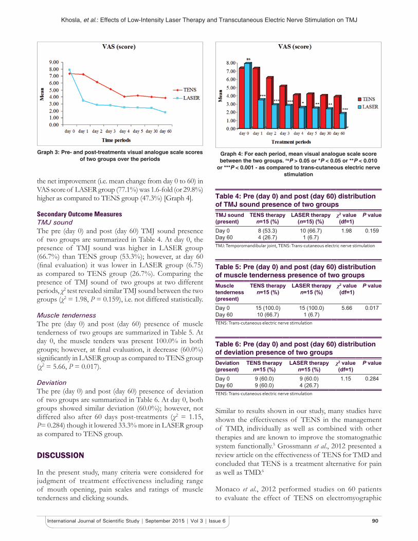
Khosla, et al.: Effects of Low-Intensity Laser Therapy and Transcutaneous Electric Nerve Stimulation on TMJ
90International Journal of Scientifi c Study | September 2015 | Vol 3 | Issue 6
the net improvement (i.e. mean change from day 0 to 60) in VAS score of LASER group (77.1%) was 1.6-fold (or 29.8%) higher as compared to TENS group (47.3%) [Graph 4].
Secondary Outcome MeasuresTMJ soundThe pre (day 0) and post (day 60) TMJ sound presence of two groups are summarized in Table 4. At day 0, the presence of TMJ sound was higher in LASER group (66.7%) than TENS group (53.3%); however, at day 60 (fi nal evaluation) it was lower in LASER group (6.75) as compared to TENS group (26.7%). Comparing the presence of TMJ sound of two groups at two different periods, χ2 test revealed similar TMJ sound between the two groups (χ2 = 1.98, P = 0.159), i.e. not differed statistically.
Muscle tendernessThe pre (day 0) and post (day 60) presence of muscle tenderness of two groups are summarized in Table 5. At day 0, the muscle tenders was present 100.0% in both groups; however, at fi nal evaluation, it decrease (60.0%) signifi cantly in LASER group as compared to TENS group (χ2 = 5.66, P = 0.017).
DeviationThe pre (day 0) and post (day 60) presence of deviation of two groups are summarized in Table 6. At day 0, both groups showed similar deviation (60.0%); however, not differed also after 60 days post-treatments (χ2 = 1.15, P= 0.284) though it lowered 33.3% more in LASER group as compared to TENS group.
DISCUSSION
In the present study, many criteria were considered for judgment of treatment effectiveness including range of mouth opening, pain scales and ratings of muscle tenderness and clicking sounds.
Similar to results shown in our study, many studies have shown the effectiveness of TENS in the management of TMD, individually as well as combined with other therapies and are known to improve the stomatognathic system functionally.5 Grossmann et al., 2012 presented a review article on the effectiveness of TENS for TMD and concluded that TENS is a treatment alternative for pain as well as TMD.6
Monaco et al., 2012 performed studies on 60 patients to evaluate the effect of TENS on electromyographic
Table 4: Pre (day 0) and post (day 60) distribution of TMJ sound presence of two groupsTMJ sound (present)
TENS therapy n=15 (%)
LASER therapy (n=15) (%)
χ2 value (df=1)
P value
Day 0 8 (53.3) 10 (66.7) 1.98 0.159Day 60 4 (26.7) 1 (6.7)TMJ: Temporomandibular joint, TENS: Trans-cutaneous electric nerve stimulation
Table 5: Pre (day 0) and post (day 60) distribution of muscle tenderness presence of two groupsMuscle tenderness (present)
TENS therapy n=15 (%)
LASER therapy n=15 (%)
χ2 value (df=1)
P value
Day 0 15 (100.0) 15 (100.0) 5.66 0.017Day 60 10 (66.7) 1 (6.7)TENS: Trans-cutaneous electric nerve stimulation
Table 6: Pre (day 0) and post (day 60) distribution of deviation presence of two groups
Deviation (present)
TENS therapy n=15 (%)
LASER therapy n=15 (%)
χ2 value (df=1)
P value
Day 0 9 (60.0) 9 (60.0) 1.15 0.284Day 60 9 (60.0) 4 (26.7)TENS: Trans-cutaneous electric nerve stimulation
Graph 3: Pre- and post-treatments visual analogue scale scores of two groups over the periods
Graph 4: For each period, mean visual analogue scale score between the two groups. nsP > 0.05 or *P < 0.05 or **P < 0.010
or ***P < 0.001 - as compared to trans-cutaneous electric nerve stimulation
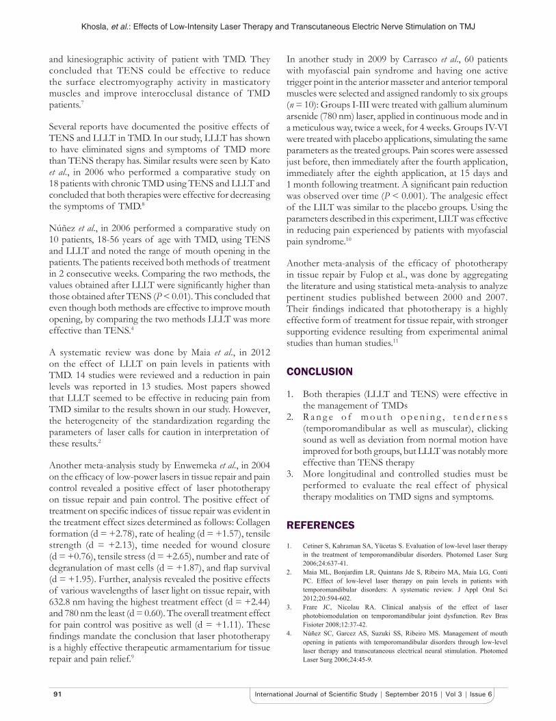
Khosla, et al.: Effects of Low-Intensity Laser Therapy and Transcutaneous Electric Nerve Stimulation on TMJ
91 International Journal of Scientifi c Study | September 2015 | Vol 3 | Issue 6
and kinesiographic activity of patient with TMD. They concluded that TENS could be effective to reduce the surface electromyography activity in masticatory muscles and improve interocclusal distance of TMD patients.7
Several reports have documented the positive effects of TENS and LLLT in TMD. In our study, LLLT has shown to have eliminated signs and symptoms of TMD more than TENS therapy has. Similar results were seen by Kato et al., in 2006 who performed a comparative study on 18 patients with chronic TMD using TENS and LLLT and concluded that both therapies were effective for decreasing the symptoms of TMD.8
Núñez et al., in 2006 performed a comparative study on 10 patients, 18-56 years of age with TMD, using TENS and LLLT and noted the range of mouth opening in the patients. The patients received both methods of treatment in 2 consecutive weeks. Comparing the two methods, the values obtained after LLLT were signifi cantly higher than those obtained after TENS (P < 0.01). This concluded that even though both methods are effective to improve mouth opening, by comparing the two methods LLLT was more effective than TENS.4
A systematic review was done by Maia et al., in 2012 on the effect of LLLT on pain levels in patients with TMD. 14 studies were reviewed and a reduction in pain levels was reported in 13 studies. Most papers showed that LLLT seemed to be effective in reducing pain from TMD similar to the results shown in our study. However, the heterogeneity of the standardization regarding the parameters of laser calls for caution in interpretation of these results.2
Another meta-analysis study by Enwemeka et al., in 2004 on the effi cacy of low-power lasers in tissue repair and pain control revealed a positive effect of laser phototherapy on tissue repair and pain control. The positive effect of treatment on specifi c indices of tissue repair was evident in the treatment effect sizes determined as follows: Collagen formation (d = +2.78), rate of healing (d = +1.57), tensile strength (d = +2.13), time needed for wound closure (d = +0.76), tensile stress (d = +2.65), number and rate of degranulation of mast cells (d = +1.87), and fl ap survival (d = +1.95). Further, analysis revealed the positive effects of various wavelengths of laser light on tissue repair, with 632.8 nm having the highest treatment effect (d = +2.44) and 780 nm the least (d = 0.60). The overall treatment effect for pain control was positive as well (d = +1.11). These fi ndings mandate the conclusion that laser phototherapy is a highly effective therapeutic armamentarium for tissue repair and pain relief.9
In another study in 2009 by Carrasco et al., 60 patients with myofascial pain syndrome and having one active trigger point in the anterior masseter and anterior temporal muscles were selected and assigned randomly to six groups (n = 10): Groups I-III were treated with gallium aluminum arsenide (780 nm) laser, applied in continuous mode and in a meticulous way, twice a week, for 4 weeks. Groups IV-VI were treated with placebo applications, simulating the same parameters as the treated groups. Pain scores were assessed just before, then immediately after the fourth application, immediately after the eighth application, at 15 days and 1 month following treatment. A signifi cant pain reduction was observed over time (P < 0.001). The analgesic effect of the LILT was similar to the placebo groups. Using the parameters described in this experiment, LILT was effective in reducing pain experienced by patients with myofascial pain syndrome.10
Another meta-analysis of the effi cacy of phototherapy in tissue repair by Fulop et al., was done by aggregating the literature and using statistical meta-analysis to analyze pertinent studies published between 2000 and 2007. Their fi ndings indicated that phototherapy is a highly effective form of treatment for tissue repair, with stronger supporting evidence resulting from experimental animal studies than human studies.11
CONCLUSION
1. Both therapies (LLLT and TENS) were effective in the management of TMDs
2. R a n g e o f m o u t h o p e n i n g , t e n d e r n e s s (temporomandibular as well as muscular), clicking sound as well as deviation from normal motion have improved for both groups, but LLLT was notably more effective than TENS therapy
3. More longitudinal and controlled studies must be performed to evaluate the real effect of physical therapy modalities on TMD signs and symptoms.
REFERENCES
1. Cetiner S, Kahraman SA, Yücetas S. Evaluation of low-level laser therapy in the treatment of temporomandibular disorders. Photomed Laser Surg 2006;24:637-41.
2. Maia ML, Bonjardim LR, Quintans Jde S, Ribeiro MA, Maia LG, Conti PC. Effect of low-level laser therapy on pain levels in patients with temporomandibular disorders: A systematic review. J Appl Oral Sci 2012;20:594-602.
3. Frare JC, Nicolau RA. Clinical analysis of the effect of laser photobiomodulation on temporomandibular joint dysfunction. Rev Bras Fisioter 2008;12:37-42.
4. Núñez SC, Garcez AS, Suzuki SS, Ribeiro MS. Management of mouth opening in patients with temporomandibular disorders through low-level laser therapy and transcutaneous electrical neural stimulation. Photomed Laser Surg 2006;24:45-9.

Khosla, et al.: Effects of Low-Intensity Laser Therapy and Transcutaneous Electric Nerve Stimulation on TMJ
92International Journal of Scientifi c Study | September 2015 | Vol 3 | Issue 6
5. Gupta R, Gupta P. Role of TENS and recent advances in management of TMJ: A review. Paripex Indian J Res 2014;9:156-7.
6. Grossmann E, Tambara JS, Grossmann TK, Siqueira JT. Transcutaneous electrical nerve stimulation for temporomandibular joint disorder. Soc Bras para o Estud da Dor 2012;13:271-6.
7. Monaco A, Sgolastra F, Ciarrocchi I, Cattaneo R. Effects of transcutaneous electrical nervous stimulation on electromyographic and kinesiographic activity of patients with temporomandibular disorders: A placebo-controlled study. J Electromyogr Kinesiol 2012;22:463-8.
8. Kato MT, Kogawa EM, Santos CN, Conti PC. TENS and low-level laser
therapy in the management of temporomandibular disorders. J Appl Oral Sci 2006;14:130-5.
9. Enwemeka CS, Parker JC, Dowdy DS, Harkness EE, Sanford LE, Woodruff LD. The effi cacy of low-power lasers in tissue repair and pain control: A meta-analysis study. Photomed Laser Surg 2004;22:323-9.
10. Carrasco TG, Guerisoli LD, Guerisoli DM, Mazzetto MO. Evaluation of low intensity laser therapy in myofascial pain syndrome. Cranio 2009;27:243-7.
11. Fulop AM, Dhimmer S, Deluca JR, Johanson DD, Lenz RV, Patel KB, et al. A meta-analysis of the effi cacy of phototherapy in tissue repair. Photomed Laser Surg 2009;27:695-702.
How to cite this article: Khosla B, Singh A, Agarwal N, Mishra A. Comparative Evaluation between the Effects of Low Intensity Laser Therapy and Transcutaneous Electric Nerve Stimulation on Temporomandibular Joint Disorders. Int J Sci Stud 2015;3(6):86-92.
Source of Support: Nil, Confl ict of Interest: None declared.

93 International Journal of Scientifi c Study | September 2015 | Vol 3 | Issue 6
Maternal Near Miss Death among Women with Eclampsia in Tertiary Care CenterKshama Kedar1, Anuja Choudhary2
1Associate Professor, Department of Obstetrics and Gynecology, Indira Gandhi Government Medical College, Nagpur, Maharashtra, India, 2Senior Resident, Department of Obstetrics and Gynecology, Indira Gandhi Government Medical College, Nagpur, Maharashtra, India
In 2009, the WHO defi ned MNM – “A woman who nearly died but survived a complication that occurred during, pregnancy, child birth, or within 42 days of termination of pregnancy.”3 WHO standardized, identifi cation of organ dysfunction and /or failure as main determinant of severity to identify MNM cases, after the identifi cation of organ dysfunction and/or failure as the main determinants of severity. Clinical signs, laboratory tests, and management interventions were used, all capable of diagnosing organ dysfunction or failure.3 These criteria were previously validated by the WHO Working Group following markers of dysfunction and total maximum sequential organ failure assessment score, applied to an obstetric population.3-5
It has been estimated that hypertensive disorders (HD) in pregnancy cause 50,000 maternal deaths (MD) annually in the world and the vast majority of them occur in low-income or middle-income countries.6
INTRODUCTION
Maternal mortality (MM) is frequently described as “Just the Tip of the Iceberg” alluding that there is a vast base to the iceberg in the form of maternal near miss (MNM), i.e. maternal morbidity which has remained largely undescribed.1 In 2009, Rööst et al. reported MM ratio (MMR) as 187/100,000 live births (LB) and MNM as 50/1000 LB, with a relatively low mortality index (MI) of 3.6%.2
Original Article
Abstract
Introduction: Since long maternal mortality (MM) has been considered as an indicator of maternal health. Review of the cases with near miss (NM) obstetric events has come up as a new tool to investigate MM.
Objectives: To determine the prevalence of NM cases and to analyze the nature of these cases and MM in patients with eclampsia.
Materials and Methods: This study was a retrospective study done for 12 months between January and December 2014. This study was conducted in the Department of Obstetrics and Gynecology, Indira Gandhi Government Medical College, Nagpur, which is a tertiary care center and serves as a referral center for other hospitals. Geller’s fi ve-factor scoring system is used to identify NM cases among patients with eclampsia. The cut-off point for NM case is a score of 8 or greater.
Results: During the study period, 50 women were identifi ed as eclamptic and among these 26 were identifi ed as NM cases. The prevalence of NM obstetric case in this study was 52%. It is observed that rates are higher in areas with poor resources and whenever the organ-system based criteria are used.Among the patients who are categorized as NM, anemia was seen in 10 (38.46%) and high level of uric acid was seen in 16 (61.53%) patients, which were both statistically signifi cant. Neonatal distress was seen in 9 (34.61%) cases, thus signifying that proper antenatal care and timely correction of anemia could have led to improvement in their health, putting these patients life at less risk.
Conclusion: The review of NM cases is helpful in improving maternal health and types of support services commonly required, as it is considered as a good guide to the standard of maternal care.
Key words: Eclampsia, Morbidity, Maternal mortality, Organ dysfunction
Access this article online
www.ijss-sn.com
Month of Submission : 07-2015Month of Peer Review : 08-2015Month of Acceptance : 09-2015Month of Publishing : 09-2015
Corresponding Author: Dr. Anuja Choudhary, Department of Obstetrics and Gynecology, Indira Gandhi Government Medical College, Nagpur, Maharashtra, India. Phone: +91-9130167722. E-mail: [email protected]
DOI: 10.17354/ijss/2015/400

Kedar and Choudhary: Maternal Near Miss Death among Women with Eclampsia in Tertiary Care Center
94International Journal of Scientifi c Study | September 2015 | Vol 3 | Issue 6
HD increase the risk of severe complications by 3-25 times, e.g. placental abruption, thrombocytopenia, disseminated intravascular coagulation, acute pulmonary edema, cerebrovascular disorders, and other conditions, in comparison to women without hypertension.7-9 The contrast between low or very low MMR in high-income countries, compared to low-income or middle-income countries with high MMR has been attributed to the quality of obstetric care, patient access to hospitalization, qualification of health professionals, and structural resources, including the input and availability of intensive care units.10-12
India has made signifi cant progress in reducing its MM rate from 254 (SRS 2004-06) to 212 (SRS 2007-09) to 178 (SRS 2010-12) per 100,000 LB;13 however, there is a long way to go on this journey to meet the millennium development goals.
This study is proposed due to the high association between HDs in pregnancy and severe obstetric or clinical complications. In addition, there are only few studies to date focused on severe morbidity as proposed by the WHO.3 Therefore, the purpose of the current study was to identify the prevalence and factors associated with the risk of severe maternal outcomes (NM and MD) in a female population with eclampsia.
Objectives• To determine the prevalence of NM cases in patients
with eclampsia• To analyze the nature of NM obstetric cases and MM
in patients with eclampsia.
MATERIALS AND METHODS
Place of StudyThe study was conducted in the Department of Obstetrics and Gynecology, Indira Gandhi Government Medical College, Nagpur, which is a tertiary care center of central India and serves as a referral center for other hospitals.
Defi nition of CasesNM events are defi ned as acute obstetric complications that immediately threatens a woman’s survival but do not result in her death either by chance or because of hospital care she receives during pregnancy, labor or within 6 weeks after termination of pregnancy or delivery.14
For identifying NM cases fi ve-factors scoring system was used.15 The fi ve-factor scoring system has the specifi city of 93.9%. It comprises of organ-system failure, intensive care unit admission, transfusion >3 units, extended intubation (>12 h), and surgical intervention (hysterectomy,
relaparotomy). These factors are given the score of 5, 4, 3, 2, and 1, respectively. A fi ve-factor scoring system can theoretically have a score from 0 to 15 (no clinical factor present to all clinical factors present). The cut-off point for NM case is a score of 8 or greater.
Study Design and Identifi cation of CasesThis was a retrospective study done for a period of 12 months between January 1, 2014 and December 31, 2014.
For this specifi c analysis focusing on eclampsia, we included women diagnosed with: Severe preeclampsia (blood pressure ≥160/110 mmHg and/or symptomatology of target organ compromise and/or proteinuria determined by dipstick 2+ or over 24 h ≥2 g; and/or oliguria <30 ml/h and/or thrombocytopenia <100.000 mm3) with presence of seizures.
Five-factor scoring system was used to identify the NM cases.
For each case of NM, variables studied were age, parity, obstetric history, history of the previous hypertensive disease, family history, mode of delivery, perinatal results, and clinical complications.
Information on MDs related to eclampsia and deliveries conducted during the study period were obtained from MM audit and the labor or delivery registers. For each case of MD, data were collected on the demographic characteristics including gestational age at the time of death and the underlying cause of death.
Data AnalysisData were entered into a computer database using Microsoft Excel spreadsheet and statistical analysis was performed. Results are presented as frequencies, percentages, and descriptive statistics.
The frequencies of NM events are reported according to the clinical condition responsible, referral status of the patients and whether the complications were present on arrival or occurred while on admission at the hospital. MM ratio was calculated as the number of MDs per 100,000 LBs.
RESULTS
As shown in Figure 1, total 50 patients were enrolled in the study, where 26 patients were categorized as NM, and 24 were categorized as not NM.
As shown in the Table 1, on further stratifying between the groups, mean age of the patients in Group A was 23.98

Kedar and Choudhary: Maternal Near Miss Death among Women with Eclampsia in Tertiary Care Center
95 International Journal of Scientifi c Study | September 2015 | Vol 3 | Issue 6
with standard deviation (SD) of 3.6 while in Group B mean age was 24.88 with SD of 2.9 hence, it was found that all patients had almost similar characteristics with regard to age in both groups, i.e. patients with NM death and in those with not NM death.
As shown in the Table 2, while analyzing gravidity, NM death was seen to be more common in primigravida. However, the occurrence of NM death was not statistically signifi cant between multigravida and primigravida. Thus, suggesting that increase in gravidity is not an isolated risk factor for the occurrence of NM death.
As shown in Table 3, most of the deliveries were at term period of gestation in both group A and in group B and in control, thus P value at 0.191 was found to be insignifi cant.
As shown in the Table 4, the presence of hypertension in previous pregnancies was not found in any of the multigravida in both Group A and Group B.
As shown in the Table 5, the presence of family history between the NM cases and not NM cases was not found to be statistically signifi cant with P value of 0.480.
As shown in the Table 6, among patients in Group B, 10 patients had anemia (38.46%) which was found to be statistically signifi cant with P value of 0.004, using cut-off for anemia as 11 g% in third trimester, as defi ned by WHO.
Three patients in Group B had thrombocytopenia with platelets <100,000 which was statistically not signifi cant with P value of 0.133.
Uric acid was high in 16 patients (61.53%), which was found to be statistically signifi cant with P value of 0.00003.
Creatinine was raised in 7 patients which was statistically insignifi cant with P value of 0.33.
Additionally, among other laboratory investigations such as prothrombin time or international normalized ratio, bleeding time, and clotting time no statistically signifi cant association was found with NM death.
Figure 1: Prevalence of Anemia
Table 2: GravidityCharacteristics Categories Group A Group B P value Remarks
Not NM NMGravidity Primigravida 15 18 0.308 NS
Multigravida 9 8NM: Near miss
Table 3: POGCharacteristics Categories Group A Group B P value Remarks
Not NM NMPOG at delivery Pre-term 4 7 0.191 NS
Term 20 19Postdate 0 0
POG: Period of gestation, NM: Near miss
Table 4: Past history of hypertensionCharacteristics Categories Group A Group B P value Remarks
Not NM NMPast history Present 2 3 0.462 NS
Absent 22 23NM: Near miss
Table 5: Family historyCharacteristics Categories Group A Group B P value Remarks
Not NM NMFamily history Present 1 0 0.480 NS
Absent 23 26NM: Near miss
Table 1: Age characteristicsCharacteristics Categories Group A Group B P value Remarks
Not NM NMAge group in years Mean (year)±SD 23.98±3.65 24.98±2.91 0.06 NS
≤19 3 0 - -20-34 21 24>35 0 2
NM: Near miss

Kedar and Choudhary: Maternal Near Miss Death among Women with Eclampsia in Tertiary Care Center
96International Journal of Scientifi c Study | September 2015 | Vol 3 | Issue 6
As shown in the Table 7, in the management of the patients, the presence of induction of labor (42.30%) was not statistically signifi cant in patients with NM death.
As shown in the Table 8, meconium staining of amniotic fl uid was seen in 4 (15.38%) cases in Group B which was found to be statistically not signifi cant with P value of 0.189.
In the study, the rate of cesarean section in Group B (n = 26) was 38.4% while 57.7% patients had spontaneous vaginal delivery and 3.9% patients had Instrumental delivery. No statistical signifi cance was seen between the two groups when comparing the mode of delivery.
As shown in the Table 9, in the study, neonatal distress after delivery was seen in 9 (34.61%) cases in Group B which was found to be statistically signifi cant with P value of 0.037.
As shown in the Table 10 post-partum hemorrhage (PPH) was observed in 1 case (2%) in Group A, with no cases in Group B.
DISCUSSION
In this study, the prevalence of MNM among eclamptic patients was found to be 52%. The mean age among NM was seen to be 24 years.
The result of present study is in accordance with the study done by Zanette et al.16 where there were 82,144 LBs in the 27 maternity hospitals participating in the study and 9555women received a diagnosis of severe maternal morbidity. Severe HDs were associated with 70% of these hospital admissions (6706/9555), corresponding to 81.6 cases per 1000 deliveries. Among the total number of women with severe HDs, 94% were classifi ed as potentially life-threatening condition (6315/6706), while 349 cases had organ dysfunction or failure, with prevalence of 4.2 NM cases per 1000 LBs, a MI of 10.7% (42 MD per 391 cases of NM plus MD), and the MNM to MD ratio was 8.3 NM cases to 1 MD. The mortality was almost half of that for non-HD conditions.
In another study done in Kathmandu valley by Rana et al. (2013),17 out of 157 cases identifi ed with NM rate of 3.8 per 1000 LBs, severe complications were PPH 62 (40%) and preeclampsia-eclampsia 25 (17%).
As per study another a cross-sectional observational study done by Kalra and Kachhwaha (2014)18 there were 27,958 deliveries and 26,734 LBs. Totally 112 patients were identifi ed as NM as per Geller’s fi ve-point scoring system while there were 54 MDs, NM rate was 4.18/1000 LBs. As far as NM obstetric morbidity was concerned the most common complication was hemorrhage accounting for 56% cases. The second leading cause was hypertension in the form of eclampsia and preeclampsia accounting for 20 (17.8%) of total NM cases.
The prevalence of NM case was 2.3% in the study done by Shrestha et al.19 HD of the pregnancy accounted for 27.8% of the NM cases in this study.
This wide variation in range was seen due to the difference in the criteria used for the identifi cation of the NM cases
Table 6: Laboratory investigationsCharacteristics Categories Group A Group B P value Remarks
Not NM NMHemoglobin Anemia present 19 10 0.004 Signifi cant
Anemia absent 5 16Platelets Normal 24 23 0.133 NS
Thrombocytopenia 0 3PT/INR Normal 24 26 - NS
Abnormal 0 0Bleeding time Normal 24 26 - NS
Abnormal 0 0Clotting time Normal 24 26 - NS
Abnormal 0 0Uric acid Normal 23 10 0.00003 Signifi cant
Abnormal 1 16Creatinine Normal 24 19 0.33 NS
Abnormal 0 7PT: Prothrombin time, INR: International normalized ratio, NM: Near miss
Table 7: ManagementCharacteristics Categories Group A Group B P value Remarks
Not NM NMInduction of labor Done 12 11 0.293 NS
Not done 12 15NM: Near miss

Kedar and Choudhary: Maternal Near Miss Death among Women with Eclampsia in Tertiary Care Center
97 International Journal of Scientifi c Study | September 2015 | Vol 3 | Issue 6
and the place of study. Rates were higher in resource-poor setting area and whenever the organ-system based criteria was used.18
Among the eclamptic patients who were categorized as NM, anemia was seen to be statistically signifi cant among these patients thus signifying that proper antenatal care and correction of anemia could have lead to improvement in their health putting these patients life at less risk.
In the study patients of eclampsia who were categorized as NM were seen to have high levels of uric acid, which was statistically signifi cant.
Fetal distress was also found to be statistically signifi cant in patients categorized as NM (9/26 patients). Thus, signifying that fetal life was also in jeopardy along with mother’s. So, proper care of the mother is the key to reducing neonatal morbidity and mortality.
CONCLUSION
The review of NM cases, especially in patients with eclampsia, helps delineate continuing threats to maternal health and types of support services the most common required. The NM can be used as a guide to the standard of maternal care, as NM analysis indicated the quality of healthcare. The major causes of NM cases were similar to
the causes of MM. Lessons can be learned from cases of NM which can serve as a useful tool in reducing MM ratio. Need for development of an effective audit system for maternal care which includes both NM obstetric morbidity and mortality is felt.19
REFERENCES
1. Purandare CN. Maternal near miss review: A way forward. J Obstet Gynaecol India 2013;63:213-5.
2. Rööst M, Altamirano VC, Liljestrand J, Essén B. Priorities in emergency obstetric care in Bolivia – maternal mortality and near-miss morbidity in metropolitan La Paz. BJOG 2009;116:1210-7.
3. Say L, Souza JP, Pattinson RC; WHO working group on maternal mortality and morbidity classifi cations. Maternal near miss – towards a standard tool for monitoring quality of maternal health care. Best Pract Res Clin Obstet Gynaecol 2009;23:287-96.
4. Oliveira-Neto A, Parpinelli MA, Cecatti JG, Souza JP, Sousa MH. Sequential organ failure assessment score for evaluating organ failure and outcome of severe maternal morbidity in obstetric intensive care. Scientifi cWorldJournal 2012;2012:172145.
5. Cecatti JG, Souza JP, Oliveira Neto AF, Parpinelli MA, Sousa MH, Say L, et al. Pre-validation of the WHO organ dysfunction based criteria for identifi cation of maternal near miss. Reprod Health 2011;8:22.
6. Duley L. Maternal mortality associated with hypertensive disorders of pregnancy in Africa, Asia, Latin America and the Caribbean. Br J Obstet Gynaecol 1992;99:547-53.
7. Kuklina EV, Ayala C, Callaghan WM. Hypertensive disorders and severe obstetric morbidity in the United States. Obstet Gynecol 2009;113:1299-306.
8. Zhang J, Meikle S, Trumble A. Severe maternal morbidity associated with hypertensive disorders in pregnancy in the United States. Hypertens Pregnancy 2003;22:203-12.
9. Altman D, Carroli G, Duley L, Farrell B, Moodley J, Neilson J, et al. Do women with pre-eclampsia, and their babies, benefi t from magnesium sulphate? The Magpie Trial: A randomised placebo-controlled trial. Lancet 2002;359:1877-90.
10. Duley L. The global impact of pre-eclampsia and eclampsia. Semin Perinatol 2009;33:130-7.
11. Goldenberg RL, McClure EM, Macguire ER, Kamath BD, Jobe AH. Lessons for low-income regions following the reduction in hypertension-related maternal mortality in high-income countries. Int J Gynaecol Obstet 2011;113:91-5.
12. Ghulmiyyah L, Sibai B. Maternal mortality from preeclampsia/eclampsia. Semin Perinatol 2012;36:56-9.
13. Special Bulletin on Maternal Mortality in India 2007-09. Sample Registration System (SRS) Offi ce of Registrar General, India; June, 2011.
14. Filippi V, Alihonou E, Mukantaganda S, Graham WJ, Ronsmans C. Near misses: Maternal morbidity and mortality. Lancet 1998;351:145-6.
15. Geller SE, Rosenberg D, Cox S, Brown M, Simonson L, Kilpatrick S. A scoring system identifi ed near-miss maternal morbidity during pregnancy. J Clin Epidemiol 2004;57:716-20.
16. Zanette E, Parpinelli MA, Surita FG, Costa ML, Haddad SM, Sousa MH, etal. Maternal near miss and death among women with severe hypertensive
Table 8: Maternal outcomeCharacteristics Categories Group A Group B P value Remarks
Not NM NMMeconium staining of amniotic fl uid
Yes 1 4 0.189 NSNo 23 22
Mode of delivery Cesarean section 6 10 0.204 NSSpontaneous vaginal delivery 18 15Instrumental delivery 0 1
NM: Near miss
Table 9: Fetal outcomesCharacteristics Categories Group A Group B P value Remarks
Not NM NMNeonatal distress after delivery
Yes 4 9 0.037 Signifi cantNo 22 15
NM: Near miss
Table 10: Post-delivery complicationsCharacteristics Categories Group A Group B P value Remarks
Not NM NMPPH Yes 1 0 0.411 NS
No 23 26PPH: Post-partum hemorrhage

Kedar and Choudhary: Maternal Near Miss Death among Women with Eclampsia in Tertiary Care Center
98International Journal of Scientifi c Study | September 2015 | Vol 3 | Issue 6
disorders: A Brazilian multicenter surveillance study. Reprod Health 2014;11:4.
17. Rana A, Baral G, Dangal G. Maternal near-miss: A multicenter surveillance in Kathmandu Valley. JNMA J Nepal Med Assoc 2013;52:299-304.
18. Kalra P, Kachhwaha CP. Obstetric near miss morbidity and maternal
mortality in a Tertiary Care Centre in Western Rajasthan. Indian J Public Health 2014;58:199-201.
19. Shrestha NS, Saha R, Karki C. Near miss maternal morbidity and maternal mortality at Kathmandu Medical College Teaching Hospital. Kathmandu Univ Med J (KUMJ) 2010;8:222-6.
How to cite this article: Kedar K, Choudhary A. Maternal Near Miss Death among Women with Eclampsia in Tertiary Care Center. Int J Sci Stud 2015;3(6):93-98.
Source of Support: Nil, Confl ict of Interest: None declared.

99 International Journal of Scientifi c Study | September 2015 | Vol 3 | Issue 6
Percutaneous Treatment of Hepatic Hydatid Cysts using Betadine and Hypertonic SalineVivek Patre1, Vibha Patre2, Anand Masih Lakra3, Shipra Sharma4, Rabia Parveen Siddiqui5, Harsh Shah6
1Associate Professor, Department of Radiodiagnosis, Pt. JNM Medical College, Raipur, Chhattisgarh, India, 2Senior Registrar, Department of Radiodiagnosis, Pt. JNM Medical College, Raipur, Chhattisgarh, India, 3Associate Professor, Department of Anesthesia, Pt. JNM Medical College, Raipur, Chhattisgarh, India, 4Associate Professor, Department of General Surgery, Pt. JNM Medical College, Raipur, Chhattisgarh, India, 5Associate Professor, Department of Pathology, Pt. JNM Medical College, Raipur, Chhattisgarh, India, 6Post-graduate Student, Department of Radiodiagnosis, Pt. JNM Medical College, Raipur, Chhattisgarh, India
the cyst. The patients can be asymptomatic or present with abdominal fullness or vague abdominal pain.2,3 Surgical management in the form marsupialization and tube drainage, omentoplasty, or hepatectomy was the mainstay of treatment.4 Medical management with benzimidazole compounds proved to be effective against the larval forms. Reports of accidental puncture of cysts without any complications led to the development of the percutaneous treatment with the use of scolicidal agent.5 In the year 1985, Mueller et al. fi rst reported percutaneous treatment of hepatic hydatid cysts.6 Subsequently, puncture-aspiration-injection-reaspiration (PAIR) was recommended by the WHO as an alternative method to surgery.7 In recent years percutaneous drainage of hepatic hydatid cysts has emerged as a cost-effective, safe, well
INTRODUCTION
Cystic echinococcosis is an infestation caused by the larval form of Echinococcus granulosus which is an endemic disease found in the cattle rearing areas of South East Asia namely India.1 The clinical features of the disease depend on factors such as the size and site of
Original Article
Abstract
Introduction: Hydatid disease of the liver is an endemic disease in the rural areas of the cattle rearing countries of the world like India. Many treatment options are available for the same including surgery and medical therapy. The introduction of modifi ed puncture aspiration, injection of scolicidal agent, and reaspiration (PAIR) under sonographic guidance in recent years has provided a new treatment option.
Purpose: The purpose of this study was to evaluate the effi cacy of percutaneous treatment of hepatic hydatid cysts under sonographic guidance using betadine (10% povidone iodine + 1% free iodine) and hypertonic saline (20%).
Materials and Methods: A total of 48 patients are having Gharbi Type I and II cysts underwent modifi ed PAIR procedure under ultrasound guidance with the use of local anesthesia. 18G needle was used for the puncture of the cysts, and scolicidal agent was introduced. The scolicidal agents used were hypertonic saline in 24 patients and betadine in 24 patients which was allowed to act for a period of 30 min. The cysts were allowed to drain using a pigtail catheter which was left in situ. The patients were followed up for a period of 12 months. The therapeutic response was assessed by using serial ultrasound scans in all patients every 3 months for 1 year. Reduction in size, pseudomass formation, and wall calcifi cation were used as assessment parameters.
Results: Reduction in size, pseudomass formation, and wall calcifi cation was seen in 46 patients. Two patients treated using hypertonic saline showed recurrence at 6 months who were then treated with betadine.
Conclusion: Modifi ed PAIR therapy is a cost-effective, safe, well tolerated, and minimally invasive treatment for the treatment of Gharbi Type I and Type II hepatic hydatid cysts. The betadine is a preferred scolicidal agent compared to hypertonic saline.
Key words: Echinococcosis, Hepatic, Hypertonic saline solution, Povidone-iodine, Ultrasonography
Access this article online
www.ijss-sn.com
Month of Submission : 07-2015Month of Peer Review : 08-2015Month of Acceptance : 08-2015Month of Publishing : 09-2015
Corresponding Author: Dr. Vivek Patre, Department of Radiodiagnosis, Pt. JNM Medical College, Raipur - 492 001, Chhattisgarh, India. Tel.: 91-771-2889161, Fax: 91-771-2523919, Phone: 91-9329399994. E-mail: [email protected]
DOI: 10.17354/ijss/2015/401

Patre, et al.: PAIR Therapy in Hydatid Cysts
100International Journal of Scientifi c Study | September 2015 | Vol 3 | Issue 6
tolerated, and minimally invasive treatment with lack of serious complications like death.2,5,8-11
The purpose of this study was to determine the effectiveness of modifi ed PAIR therapy using hypertonic saline and betadine (povidone-iodine) as scolicidal agents.
MATERIALS AND METHODS
Patient SelectionThe study was carried out from March 2004 to July 2015 in the Department of Radio-Diagnosis, Pt. JNM Medical College, Raipur, India. Inclusion criteria were patients having single or multiple Gharbi Type I (Figure 1) and Type II hepatic hydatid cysts of size more than 5.0 cm. Exclusion criteria were patient unwilling for treatment, Gharbi Type III-V, cysts with biliary communication, and inaccessible cysts. The total of 48 patients underwent the procedure during this period after undertaking a written and informed consent. 20 were female, and 28 were male in the age group of 12-65 years. 35 cysts were Gharbi Type I and 13 cysts were Gharbi Type II. The cyst diameter varied between 5 and 13 cm with average cyst size being 7.3 cm. 38 cysts were in the right lobe while 10 cysts were in the left lobe. 37 patients presented with abdominal pain, 6 patients presented with abdominal discomfort, 2 patients presented with breathing diffi culty while 3 patients were asymptomatic.
ProcedureThe diagnosis was established using ultrasound machine (Prosound-4000, Aloka, Japan and Aplio-MX, Toshiba, Japan) and serological tests namely ELISA and classifi cation of the hepatic hydatid cysts was done using Gharbi classifi cation. By this classifi cation, Type I cyst refers to a simple cyst without septae, fl oating membranes, and daughter cysts, Type II cyst refers to a cyst with fl oating membranes; Type III cyst is a hydatid cyst with daughter cysts, Type IV cyst is a cyst with internal echoes and solid areas, and Type V cyst refers to areas of calcifi cation in the cysts. Communication with the biliary tree was ruled out by examining the cyst fl uid for bile pigments and salts. Routine hemogram and liver function tests were performed prior to the procedure. Prophylactic oral albendazole 400 mg twice daily was started 7 days prior to the procedure for all patients to avoid anaphylaxis. Oral albendazole was continued for a period of 1 month after the procedure. After an overnight fast for at least 6 h patients underwent sonographic evaluation for ascertaining the depth of the cyst. Before starting the procedure, emergency tray containing drugs such as adrenaline, atropine, hydrocortisone, and chlorpheniramine maleate was kept ready for anaphylaxis in the form of laryngeal
edema, asthma, hypotension, or shock. After administering local anesthetic (2% lignocaine), the hepatic cysts were punctured using 18 gauge needle (Figure 2) and fl uid was aspirated using 8 French or 10 French pigtail catheter (Blue Neem, India) leaving behind only a small amount of fl uid to visualize the catheter tip. The aspirated fl uid was examined for bile pigments and salts to rule out biliary communication. The scolicidal agent (20% hypertonic saline used in 24 patients and 10% betadine used in 24 patients) was injected into the cyst and left in situ for 30 min after clamping the catheter (Figure 3). The scolicidal agent injected was two-thirds of the aspirated volume. The scolicidal agent was reaspirated after 30 min, and the pigtail was left in situ (Figure 4). Vital monitoring was done during the entire procedure and for 24 h after the procedure for any feature of anaphylaxis. The catheter was connected to a drainage bag and removed after the 24 h aspirate was <20 ml which was in the range of 2-7 days. The entire procedure lasted for a range of 35-50 min. The patients were followed up for 1 year by ultrasound examination in every 3 months.
Figure 1: Gharbi Type I hepatic hydatid cyst
Figure 2: Puncture needle within the cyst

Patre, et al.: PAIR Therapy in Hydatid Cysts
101 International Journal of Scientifi c Study | September 2015 | Vol 3 | Issue 6
RESULTS
All the 48 patients showed a response to treatment at 3 months follow-up sonography in the form of a reduction in the size
of the cyst. The average size of the cysts after 3 months was 3.5 cm in diameter. Resolution of the presenting symptoms of abdominal pain, abdominal discomfort, and breathlessness was noted in all the patients at the end of 3 months. 41 patients at 6 months follow-up showed pseudomass formation (Figure 5) while 5 patients showed wall calcifi cation. Two out of the 24 patients treated using hypertonic saline as a scolicidal agent showed recurrence on 6 months follow-up sonography. They were subsequently treated using betadine as a scolicidal agent. No recurrence was noted in them on follow-up sonography, and pseudomass formation was noted at the end of 6 months. 37 patients showed wall calcifi cation at the end of 1 year on sonography. Further reduction in cyst size was noted in all patients at the end of 1 year with the average size of 2.6 cm. A hospital stay of the patients ranged from 2 to 9 days with an average stay of 5 days. Severe complications like anaphylaxis and death were observed in none of the patients. Pain at the injection site was the most common complication noted in 17 patients which was managed using non-steroidal anti-infl ammatory drugs. Febrile illness was noted in 9 patients which was managed using intravenous ceftriaxone for a period of 3 days (Table 1).
DISCUSSION
The hydatid disease is an endemic disease in the cattle rearing areas of South-East Asia namely India and is a major health problem. Until recent times, surgery has been the preferred modality for hepatic hydatid cysts. However, surgery is associated with mortality in up to 6% of the cases.12 Recurrence rates after surgery has been reported to be from 2% to 25%.13-15 Minimally, invasive methods such as PAIR along with oral anti-helminthic therapy is effective with better outcome than surgery in the Gharbi Type I and II cysts with the added advantage of less morbidity, cost effectiveness, and reduced hospital stay. In our study, we classifi ed hepatic
Figure 3: Injection of scolicidal agent producing internal echoes using pigtail drainage catheter
Figure 4: Pigtail catheter in situ post reaspiration
Figure 5: Pseudomass formation at 3 months follow-up
Table 1: Injections of Scolicidal AgentParameters Scolicidal agent Total
Betadine Hypertonic salineAppearance of cysts
At 3 monthsReduction in size 24 24 48
At 6 monthsPseudomass formation 23 18 41Wall calcifi cation 3 3 6Recurrence - 2 2
At 12 monthsReduction in size 24 24 48Wall calcifi cation 20 17 37Recurrence - - -
ComplicationsPain at injection site 9 10 19Febrile illness 1 7 8Hospital stay (average) 4.6 days 5.4 days 5 days

Patre, et al.: PAIR Therapy in Hydatid Cysts
102International Journal of Scientifi c Study | September 2015 | Vol 3 | Issue 6
hydatid cysts sonographically using Gharbi classifi cation16 and treated Gharbi Type I and II cysts. We performed the procedure under sonographic guidance using the transhepatic route for the puncture with the help of a 18G needle. The pigtail catheter was introduced and left in situ for the purpose of aspiration, injection, and reaspiration. Various scolicidal agents have been used by various investigators such as 20% hypertonic saline, 95% alcohol, betadine (10% povidone iodine; 1% free iodine).17-19 WHO recommends the use of hypertonic saline as a scolicidal.7 In this study, both 20% hypertonic saline and betadine were used in a randomized manner in an equal number of patients. We observed a reduction in the cyst size in all the patients at 3 months follow-up. It was noted that 2 patients treated with hypertonic saline showed recurrence at the end of 6 months. They were subsequently treated with betadine which showed no recurrence. All the patients treated using betadine showed no recurrence. At the end of 1 year follow-up, all the patients showed a reduction in cyst size while pseudomass formation was noted in 43 patients and wall calcifi cations in 37 patients. Thus, betadine was 100% effective whereas hypertonic saline was 92% effective which led us to conclude that betadine was a better scolicidal agent. In our study, oral albendazole 400 mg twice daily was administered to all the patients before the procedure and continued for a period of 1 month after the procedure to prevent anaphylaxis and recurrence. Many studies show that modifi ed PAIR is as effective as surgery with lower complication rates.11,20-22
CONCLUSION
Results show that modifi ed PAIR therapy is cost effective, safe, well tolerated, and minimally invasive treatment of Gharbi Type I and II hepatic hydatid cysts with a reduction in hospital stay. In our study, we also concluded that betadine is a better alternative to hypertonic saline.
REFERENCES
1. Ustünsöz B, Akhan O, Kamiloglu MA, Somuncu I, Ugurel MS, Cetiner S. Percutaneous treatment of hydatid cysts of the liver: Long-term results. AJR Am J Roentgenol 1999;172:91-6.
2. Aygün E, Sahin M, Odev K, Vatansev C, Aksoy F, Paksoy Y, et al. The management of liver hydatid cysts by percutaneous drainage. Can J Surg 2001;44:203-9.
3. Schipper HG, Laméris JS, van Delden OM, Rauws EA, Kager PA. Percutaneous evacuation (PEVAC) of multivesicular echinococcal cysts with or without cystobiliary fi stulas which contain non-drainable material: First results of a modifi ed PAIR method. Gut 2002;50:718-23.
4. Ekrami Y. Surgical treatment of hydatid disease of the liver. Arch Surg 1976;111:1350-2.
5. Akhan O, Ozmen MN, Dinçer A, Sayek I, Göçmen A. Liver hydatid disease: Long-term results of percutaneous treatment. Radiology 1996;198:259-64.
6. Mueller PR, Dawson SL, Ferrucci JT Jr, Nardi GL. Hepatic echinococcal cyst: Successful percutaneous drainage. Radiology 1985;155:627-8.
7. Department of Communicable Disease, Surveillance and Response. Bulletin of WHO on PAIR therapy. Vol. 74. 1996. p. 213-42.
8. Yasawy MI, Mohammed AE, Bassam S, Karawi MA, Shariq S. Percutaneous aspiration and drainage with adjuvant medical therapy for treatment of hepatic hydatid cysts. World J Gastroenterol 2011;17:646-50.
9. Khuroo MS, Zargar SA, Mahajan R. Echinococcus granulosus cysts in the liver: Management with percutaneous drainage. Radiology 1991;180:141-5.
10. Polat KY, Balik AA, Oren D. Percutaneous drainage of hydatid cyst of the liver: Long-term results. HPB (Oxford) 2002;4:163-6.
11. Rajesh R, Dalip DS, Anupam J, Jaisiram A. Effectiveness of puncture-aspiration-injection-reaspiration in the treatment of hepatic hydatid cysts. Iran J Radiol 2013;10:68-73.
12. Dawson JL, Stamatakis JD, Stringer MD, Williams R. Surgical treatment of hepatic hydatid disease. Br J Surg 1988;75:946-50.
13. Lewis JW Jr, Koss N, Kerstein MD. A review of echinococcal disease. Ann Surg 1975;181:390-6.
14. Balasegaram M, Kong LF. Surgical treatment of hydatid disease of the liver. Trop Gastroenterol 1982;3:194-200.
15. El Mufti M. The simple hydatid cyst. In: Surgical Management of Hydatid Disease. London: Butterworths; 1998. p. 31-107.
16. Gharbi HA, Hassine W, Brauner MW, Dupuch K. Ultrasound examination of the hydatid liver. Radiology 1981;139:459-63.
17. Saremi F, McNamara TO. Hydatid cysts of the liver: Long-term results of percutaneous treatment using a cutting instrument. AJR Am J Roentgenol 1995;165:1163-7.
18. Acunas B, Rozanes I, Celik L, Minareci O, Acunas G, Alper A, et al. Purely cystic hydatid disease of the liver: Treatment with percutaneous aspiration and injection of hypertonic saline. Radiology 1992;182:541-3.
19. Keshmiri M, Baharvahdat H, Fattahi SH, Davachi B, Dabiri RH, Baradaran H, et al. Albendazole versus placebo in treatment of echinococcosis. Trans R Soc Trop Med Hyg 2001;95:190-4.
20. vanSonnenberg E, Wroblicka JT, D’Agostino HB, Mathieson JR, Casola G, O’Laoide R, et al. Symptomatic hepatic cysts: Percutaneous drainage and sclerosis. Radiology 1994;190:387-92.
21. Giorgio A, Tarantino L, de Stefano G, Francica G, Mariniello N, Farella N, et al. Hydatid liver cyst: An 11-year experience of treatment with percutaneous aspiration and ethanol injection. J Ultrasound Med 2001;20:729-38.
22. Paksoy Y, Odev K, Sahin M, Arslan A, Koc O.Percutaneous treatment of liver hydatid cysts: Comparison of direct injection of albendazole and hypertonic saline solution. AJR Am J Roentgenol 2005;185:727-34.
How to cite this article: Patre V, Patre V, Lakra AM, Sharma S, Siddiqui RP, Shah H. Percutaneous Treatment of Hepatic Hydatid Cysts using Betadine and Hypertonic Saline. Int J Sci Stud 2015;3(6):99-102.
Source of Support: Nil, Confl ict of Interest: None declared.

103 International Journal of Scientifi c Study | September 2015 | Vol 3 | Issue 6
Analysis of 62 Cases of Ectopic Pregnancies in a Rural Medical College Set Up at Nalgonda Telangana, IndiaSunita Mishra1, Vrunda Chaudhary2, Rajesh Kaul3, Bushra Tabassum4
1Associate Professor, Department of Obstetrics & Gynecology, Kamineni Institute of Medical Sciences, Nalgonda, Telangana, India, 2Professor, Department of Obstetrics & Gynecology, Kamineni Institute of Medical Sciences, Nalgonda, Telangana, India, 3Professor & Head, Department of Obstetrics & Gynecology, Kamineni Institute of Medical Sciences, Nalgonda, Telangana, India, 4Post-graduate Student, Department of Obstetrics & Gynecology, Kamineni Institute of Medical Sciences, Nalgonda, Telangana, India
for further growth, and development of the embryo. In about 95% of ectopic pregnancies, the fertilized ovum implants in the tubes, but rarely in other organs such as abdomen, ovaries, cervix, spleen, omentum, cesarian section scar, and intramural. The risk factors for ectopic pregnancy include pelvic infl ammatory disease (PID), previous tubal surgery, previous ectopic pregnancy, progestin contraceptive, assisted reproduction, ovulation induction, induced abortion, salpingitis isthmica nodosa, smoking, and diethylstilbestrol exposure.1,2 Most of the tubal pregnancies become symptomatic within 12 weeks, but a small number of them progress beyond this gestation and are diagnosed late.3 Consequently, there is a fetal loss with increased maternal morbidity and
INTRODUCTION
The ectopic pregnancy is one of the most dreaded life-threatening obstetric emergencies in early pregnancy. It is defi ned as ectopic implantation of the embryo at sites other than the normal uterine cavity, unfavorable
Original Article
Abstract
Introduction: An ectopic pregnancy is emerging as a life-threatening obstetric emergency as the incidence is increasing in the recent times and has a profound effect on future of women’s fertility. The objective of our study was to study the clinical profi le and management of ectopic pregnancies.
Materials and Methods: The present retrospective study was conducted over a period of 2½ years from December 2012 to May 2015 for risk factors, mode of presentation, treatment modalities, and management of patients with ectopic pregnancy.
Results: On analyzing 62 cases of ectopic pregnancies admitted in medical college, it was found an incidence of 5.22 ectopic pregnancies per 1000 live births. Majority of the patients (53.22%) were in the age group 26-30 years. The second gravida had the highest incidence of ectopic pregnancies (41.93%). Previous tubal ligation was the most common identifi able risk factor, and the majority of cases (24.19%) had a H/O tubectomy. Most of the cases presented with lower abdominal pain (93.54%) followed by amenorrhea (79.03%). The most of the cases (62.90%) in our study had ruptured tubal pregnancy. Unruptured tubal pregnancy was seen in 24.19% cases. Chronic ectopic pregnancy was diagnosed in 8.06% of cases. Interestingly, it was also found three cases of ovarian pregnancy. Partial salpingectomy by the open method was the mainstay of treatment and was done in 79.03% of cases. Eight cases were managed laparoscopically. Partial oopherectomy was carried out for the three cases of ovarian ectopic pregnancy. Two cases were successfully managed by medical management with intramuscular methotrexate.
Conclusion: Although ectopic pregnancy can never be completely prevented, but the incidence can be reduced, and much of the morbidity and mortality is also preventable with effi cacious diagnostic and interventional strategies aimed principally at women who are at high risk for ectopic pregnancy.
Key words: Amenorrhea, Ectopic, Pregnancy, Salpingectomy, Tubal
Access this article online
www.ijss-sn.com
Month of Submission : 07-2015Month of Peer Review : 08-2015Month of Acceptance : 08-2015Month of Publishing : 09-2015
Corresponding author: Dr. Sunita Mishra, 301, Sunshine Habitat, Green Hills Colony, Road No.1, RK Puram, Hyderabad - 500 035, Telangana, India. Phone: +91-9573660173. E-mail: [email protected]
DOI: 10.17354/ijss/2015/402

Mishra, et al.: Ectopic Pregnancies in a Rural Medical College Set Up
104International Journal of Scientifi c Study | September 2015 | Vol 3 | Issue 6
mortality, also compromising future fertility potential. Early diagnosis of ectopic pregnancy is a diffi cult task but can be possible with the help of quantitative beta-human chorionic gonadotropin (β-HCG) level, transvaginal ultrasonography, and laparoscopy.4,5 In spite of the advances in diagnostic methods and management, ectopic pregnancy still remains a very serious threat to maternal safety. Identifying the risk factors and taking steps to reduce them, improves morbidity, mortality, and fertility outcomes. The purpose of present study was to highlight the clinical profi le of patients of ectopic pregnancy, to identify the risk factors and to study the various modalities of management in Kamineni Institute of Medical Sciences, Sreepuram, Narketpally.
MATERIALS AND METHODS
The present study is based on an analysis of 62 cases of ectopic pregnancy admitted to Kamineni Institute of Medical Sciences, Sreepuram, Narketpally over a period of 2½ years from December 2012 to May 2015. The age, parity, marital status, and presenting symptoms were recorded. History of any prior surgery, tubal surgery, infertility, PID, previous ectopic or any medical disorder deemed to be signifi cant, was taken.
Vitals were recorded and a thorough abdominal and bimanual examination was done.
The diagnosis was made with the help of urine pregnancy test and ultrasonography. The abdominal fi ndings on exploration were recorded. Treatment given was recorded as either medical or surgical.
OBSERVATION AND RESULTS
In the present study conducted over a period of 2½ years, the total number of deliveries was 11,861, and the total number of ectopic pregnancies were 62; giving an incidence of 5.22 per 1000 live births.
In the present study, it was found that the majority of ectopic pregnancies, that is 53.22%, occurred in the females of the age group 26-30 years (Table 1).
Second gravide at 41.93% accounted for the maximum number of ectopic pregnancies in the present study (Table 2).
Table 3 shows that more than half of the patient, i.e. 59.65%, had identifi able risk factors, the most important one being a history of tubal ligation at 24.19%. This was followed by a history of prior surgery in 16.12%, a history
of infertility in 9.67%, a history of PID in 6.45%, and a history of previous ectopic pregnancy in 3.22% of cases. In the rest 40.32% patients, there was no identifi able risk factor.
The majority of the patients presented with lower abdominal pain (93.54%), followed by amenorrhea, abnormal uterine bleeding, and syncopal attack. Cervical motion tenderness was elicited in 75.80% of cases (Table 4).
Table 1: Agewise distribution of ectopic pregnancy cases In present studyAge group Number of cases n=62 (%)20-25 14 (22.58)26-30 33 (53.22)31-35 10 (16.12)36-40 5 (8.06)
Table 2: Gravidity in present studyGravidity Number of cases n=62 (%)1 18 (29.03)2 26 (41.93)3 12 (19.35)4 6 (9.67)
Table 3: Risk factors for ectopic pregnancy in present studyRisk factors Number of patients n=62 (%)H/O PID 4 (6.45)H/O tubal ligation 15 (24.19)H/O previous pelvic surgery 10 (16.12)H/O previous ectopic 2 (3.22)H/O infertility 6 (9.67)No identifi able risk factors 25 (40.32)PID: Pelvic infl ammatory disease
Table 4: Symptomatology in present studyClinical features Number of cases n=62 (%)Lower abdominal pain 58 (93.54)Amenorrhea 49 (79.03)Cervical motion tenderness 47 (75.80)Adnexal mass or fullness 39 (62.90)Abnormal uterine bleeding 45 (72.58)Abdominal guarding and rigidity 40 (64.51)Syncope 10 (16.12)
Table 5: Modalities of management in present StudyManagement Number of patients n=62 (%)Partial salpingectomy (open) 49 (79.03)Laparoscopic partial salpingectomy 8 (12.90)Partial oopherectomy 3 (4.83)Medical management 2 (3.22)
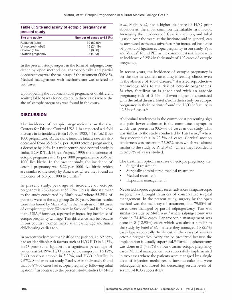
Mishra, et al.: Ectopic Pregnancies in a Rural Medical College Set Up
105 International Journal of Scientifi c Study | September 2015 | Vol 3 | Issue 6
In the present study, surgery in the form of salpingectomy either by open method or laparoscopically and partial oopherectomy was the mainstay of the treatment (Table 5). Medical management with methotrexate was offered to two cases.
Upon opening the abdomen, tubal pregnancies of different acuity (Table 6) was found except in three cases where the site of ectopic pregnancy was found in the ovary.
DISCUSSION
The incidence of ectopic pregnancies is on the rise. Centers for Disease Control USA 1 has reported a 4-fold increase in its incidence from 1970 to 1983, 4.5 to 16.18 per 1000 pregnancies.6 At the same time, the fatality rate has also decreased from 35.5 to 3.8 per 10,000 ectopic pregnancies, a decrease by 90%. In a multicentric case-control study in India, (ICMR Task Force Project, 1990) the incidence of ectopic pregnancy is 3.12 per 1000 pregnancies or 3.86 per 1000 live births. In the present study, the incidence of ectopic pregnancy was 5.22 per 1000 live births which are similar to the study by Ayaz et al. where they found an incidence of 5.8 per 1000 live births.7
In present study, peak age of incidence of ectopic pregnancy is 26-30 years at 53.22%. This is almost similar to the study conducted by Mufti et al.8 where 55.25% of patients were in the age group 26-30 years. Similar results were also found by Majhi et al.9 in their analysis of 180 cases of ectopic pregnancy. Westrom in Sweden10 and Rubin et al. in the USA,11 however, reported an increasing incidence of ectopic pregnancy with age. This difference may be because in our country women marry at an earlier age and fi nish childbearing earlier too.
In present study more than half of the patients, i.e. 59.65%, had an identifi able risk factors such as H/O PID in 6.45%, H/O prior tubal ligation in a signifi cant percentage of patients at 24.19%, H/O prior pelvic surgery in 16.12%, H/O previous ectopic in 3.22%, and H/O infertility in 9.67%. Similar to our study, Patel et al. in their study found that 30.8% of cases had ectopic pregnancy following tubal ligation.12 In contrast to the present study, studies by Mufti
et al., Majhi et al., had a higher incidence of H/O prior abortion as the most common identifi able risk factor. Increasing the incidence of Cesarian section, and tubal ligation over the years at the institute and in general, can be attributed as the causative factor for increased incidence of post tubal ligation ectopic pregnancy in our study. Vyas and Vaidya13 found PID as the commonest risk factor with an incidence of 25% in their study of 192 cases of ectopic pregnancy.
In recent years, the incidence of ectopic pregnancy is on the rise in women attending infertility clinics even in the absence of tubal disease.14 Assisted reproductive technology adds to the risk of ectopic pregnancies. In vitro, fertilization is associated with an ectopic pregnancy risk of 2-5% and even higher if associated with the tubal disease. Patel et al. in their study on ectopic pregnancy in their institute found the H/O infertility in 42.3% of cases.12
Abdominal tenderness is the commonest presenting sign and pain lower abdomen is the commonest symptom which was present in 93.54% of cases in our study. This was similar to the study conducted by Patel et al.,12 where they recorded this in 92.3% of cases. Cervical motion tenderness was present in 75.80% cases which was almost similar to the study by Patel et al.12 where they recorded it in 82.69% of cases studied.
The treatment options in cases of ectopic pregnancy are:• Surgical treatment• Surgically administered medical treatment• Medical treatment• Expectant management.
Newer techniques, especially recent advances in laparoscopic surgery, have brought in an era of conservative surgical management. In the present study, surgery by the open method was the mainstay of treatment, and 79.03% of cases were managed by partial salpingectomy. This was similar to study by Mufti et al.,8 where salpingectomy was done in 74.48% cases. Laparoscopic management was done in 8 (12.90%) cases which was almost similar to the study by Patel et al.,12 where they managed 13 (25%) cases laparoscopically. In almost all the cases of ovarian ectopic pregnancies, ovary can be preserved because the implantation is usually superfi cial.15 Partial oopherectomy was done in 3 (4.83%) of our ovarian ectopic pregnancy cases. Medical management was successfully implemented in two cases where the patients were managed by a single dose of injection methotrexate intramuscular and were subsequently monitored for decreasing serum levels of serum β-HCG successfully.
Table 6: Site and acuity of ectopic pregnancy in present studySite and acuity Number of cases n=62 (%)Ruptured (tubal) 39 (62.90)Unruptured (tubal) 15 (24.19)Chronic (tubal) 5 (8.06)Ovarian pregnancy 3 (4.83)

Mishra, et al.: Ectopic Pregnancies in a Rural Medical College Set Up
106International Journal of Scientifi c Study | September 2015 | Vol 3 | Issue 6
CONCLUSION
The study on ectopic pregnancy which was conducted in Kamineni Institute of Medical Sciences, Sreepuram, Narketpally, aimed at analyzing ectopic pregnancies, the incidence, and the various risk factors, presenting symptoms associated with it, the acuity and the modalities of management utilizing the resources available at our hand. In presented study done in a medical college in the midst of a rural setup, the most common risk factor associated with ectopic pregnancy was a history of a previous tubal ligation. The most of the cases had undergone tubectomy in camps and hence the only possible explanation could be improper aseptic measures leading to scarring of the tubes as a result of post tubectomy infection or infl ammation. PID, infertility, a prior pelvic surgery, and a previous ectopic pregnancy are other identifi able risk factors leading to ectopic pregnancy. A high degree of clinical suspicion of ectopic pregnancy must be held in the presence of any of the risk factors unless proved otherwise.
The rising number of cases of ectopic pregnancy poses a serious concern over maternal mortality. With advances in the fi eld of medicine, more young women are adopting newer fertility control methods such as newer oral contraceptives, intrauterine contraceptive device, and various tubal surgeries to limit their families. Moreover, newer drugs for ovulation induction and tubal reconstructive surgeries have led to delayed conception with increased risk of ectopic pregnancy. Although ectopic pregnancy can never be completely prevented, but it’s incidence can be reduced along with reduction of maternal morbidity and mortality by effi cacious diagnostic and
interventional strategies aimed at all the women at high risk for ectopic pregnancy.
REFERENCES
1. Shah N, Khan NH. Ectopic pregnancy: Presentation and risk factors. J Coll Physicians Surg Pak 2005;15:535-8.
2. Basu A, Candelier C. Ectopic pregnancy with postcoital contraception – a case report. Eur J Contracept Reprod Health Care 2005;10:6-8.
3. Tasnim N, Mahmud G. Advanced abdominal pregnancy – a diagnostic and management dilemma. J Coll Physicians Surg Pak 2005;15:493-5.
4. Farquhar CM. Ectopic pregnancy. Lancet 2005;366:583-91.5. Sohail S. Hemorrhagic corpus luteum mimicking heterotopic pregnancy.
J Coll Physicians Surg Pak 2005;15:180-1.6. Center for Disease Control. Ectopic pregnancy-United States: 1988-1989.
Morb Mortal Wkly Rep 1992;41:591.7. Ayaz A, Emam S, Farooq MU. Clinical course of ectopic pregnancy:
A single-center experience. J Hum Reprod Sci 2013;6:70-3.8. Mufti S, Rather S, Mufti S, Rangrez RA, Wasiqa, Khalida. Ectopic
pregnancy – An analysis of 114 cases. JK Pract 2012;17:20-3.9. Majhi AK, Roy N, Karmakar KS, Banerjee PK. Ectopic pregnancy – an
analysis of 180 cases. J Indian Med Assoc 2007;105:308, 310, 312.10. Weström L, Bengtsson LP, Mårdh PA. Incidence, trends, and risks of
ectopic pregnancy in a population of women. Br Med J (Clin Res Ed) 1981;282:15-8.
11. Rubin GL, Peterson HB, Dorfman SF, Layde PM, Maze JM, Ory HW, et al. Ectopic pregnancy in the United States 1970 through 1978. JAMA 1983;249:1725-9.
12. Patel UM, Gandhi MR, Jani PS, Kakani CR, Thakor N. Clinical profi le and management of ectopic pregnancy in patients with ectopic pregnancy at GMERS medical college and hospital Dharpur-Patan, North Gujarat region, India. Int J Res Med Sci 2015;3:841-5.
13. Vyas PS, Vaidya P. Epidemiology, diagnosis and management of ectopic pregnancy – An analysis of 196 cases. BHJ Orig Res Artic. Available from: html: fi le://e: \epidemiology, diagnosis and management of ectopic pregnancy. [Last accessed on 2015 Aug 22].
14. Clayton HB, Schieve LA, Peterson HB, Jamieson DJ, Reynolds MA, Wright VC. Ectopic pregnancy risk with assisted reproductive technology procedures. Obstet Gynecol 2006;107:595-604.
15. Koo YJ, Choi HJ, Im KS, Jung HJ, Kwon YS. Pregnancy outcomes after surgical treatment of ovarian pregnancy. Int J Gynaecol Obstet 2011;114:97-100.
How to cite this article: Mishra S, Chaudhary V, Kaul R, Tabassum B. Analysis of 62 Cases of Ectopic Pregnancies in a Rural Medical College Set Up at Nalgonda Telangana, India. Int J Sci Stud 2015;3(6):103-106.
Source of Support: Nil, Confl ict of Interest: None declared.

107 International Journal of Scientifi c Study | September 2015 | Vol 3 | Issue 6
Pattern of Malignant Tumors in a Teaching Hospital of Western IndiaJignasa N Bhalodia1, Haren V Oza2, Palak J Modi3
1Associate Professor, Department of Pathology, Gujarat Medical Education and Research Society Medical College, Sola, Ahmedabad, Gujarat, India, 2Professor, Department of Pathology, BJ Medical College, Ahmedabad, Gujarat, India, 3Tutor, Department of Pathology, Gujarat Medical Education and Research Society Medical College, Sola, Ahmedabad, Gujarat, India
about 6 million people dies.2 Apart from cancers due to genetic defect, around 80-90% of cancers develop due to lifestyle and environment factors.1
In our country prevalence of cancer is less compared to developed countries, it has increased during past few decades. Every year on an average 0.7 million new cases are being diagnosed. It adds to the prevalence of 2.5 million, out of which nearly half of the patient dies within few years. Over 70% of the cases report for diagnosis and treatment in advanced stages of the disease. It is one important cause of poor survival and high mortality rates in cancer patients.2,3 There is variation in ethnicity and socioeconomic status in different people in our country. It has a major infl uence on lifestyle and beliefs and leads to differences in cancer incidence and mortality.4
Globally in the male lung, stomach and colorectal cancers are the leading cancers, whereas breast, lungs, and stomach
INTRODUCTION
Cancer has become one of the leading causes of deaths worldwide. An epidemic of cancer has transited beyond the geographical boundaries of countries. Our Earth is under the effect of cancer that is spreading rapidly, and the burden of cancer is still increasing daily even though there is a lot improvement in methods of diagnosis and treatments.1,2 It has become a challenge for any healthcare system. Every year nearly 10 million people are being diagnosed as having some type of cancer out of which
Original Article
Abstract
Background: Diseases such as cancer and other non-communicable diseases are fast replacing communicable diseases in India and other developing countries. The burden of cancer is still increasing worldwide despite advances for diagnosis and treatment.
Purpose: To study pattern and histopathological subtypes of the different malignant tumors operated in a medical college associated hospital of western India.
Materials and Methods: All biopsies submitted for histopathology to pathology department Gujarat Medical Education and Research Society Medical College, Sola, Ahmedabad from April 2010 to March 2015, included in study. All biopsies were reviewed. Analysis was performed for age, site and diagnosis in patients with malignant lesions.
Results: Total 7209 biopsies were received. Out of it 229 cases were diagnosed as malignant lesions. 125 (54.59%) cases were female patients in which most common malignancy was breast carcinoma, followed by squamous cell carcinoma of oral cavity and carcinoma of cervix. 104 (45.41%) cases were male patients and most common malignancy was squamous cell carcinoma of oral cavity followed by carcinoma of penis and gastrointestinal adenocarcinoma.
Conclusion: Site-specifi c data in this study can augment the National Cancer Registry Program and is an essential indicator for the magnitude and pattern of the cancer problem in India.
Key words: Histopathology, Malignancy, Pattern of cancer, Prevalence, Site
Access this article online
www.ijss-sn.com
Month of Submission : 07-2015Month of Peer Review : 08-2015Month of Acceptance : 08-2015Month of Publishing : 09-2015
Corresponding Author: Dr. Jignasa Bhalodia, B-37, Marutinandan Vihar Bunglows, Nr. Aarohi Villa, 200 Feet S.P. Ring Road, Bopal, Ahmedabad - 380 058, Gujarat, India. Phone: +91-9925594684. E-mail: [email protected]
DOI: 10.17354/ijss/2015/403

Bhalodia, et al.: Pattern of Malignant Tumors
108International Journal of Scientifi c Study | September 2015 | Vol 3 | Issue 6
cancer constitute top three leading cancers in female.3 In contrary the common sites for cancer in Indian male are an oral cavity, lungs, esophagus and stomach and cervix, breast, and oral cavity among Indian female. Even within India, there are variations in the prevalence and pattern of different cancers.2 In Southern India, the common cancers among male were of stomach, oral cavity, esophagus, and leukemia whereas in females leading causes of cancers were of cervix, breast, oral, and esophageal. Similarly, a study from West Bengal, an Eastern state of India has showed head and neck, lungs, and oral cancers are predominant in males, whereas among female breast cancer was leading cause of cancer followed by cervical and stomach cancers. In contrast, among North India male major cases of cancer are of gastro-intestinal tract (GIT), larynx and lung. In North Indian female breast, cervix and lung were leading causes of malignancies.2
There is need of obtaining more information on cancer epidemiology such as prevalence, incidence, and risk factors to generate evidence, which will help in taking effective measures to prevent and control the cancer epidemic in the country. There is limited published cancer data available in the state of Gujarat. Due to existence of diverse pattern of cancer occurrence, we conducted the present study to explore the pattern and trend of cancer among patients of Gujarat the West Indian state.2
MATERIALS AND METHODS
We conducted a retrospective study in Gujarat Medical Education and Research Society Medical College, Sola, Ahmedabad in Gujarat. This hospital-based study was conducted for the period April 1st, 2010 to March 31st, 2015. The study subjects consisted of all cases diagnosed as cancer. Tissue biopsies received in histopathology section of the pathology department. They fi xed in buffered formalin. Thorough gross examination of specimen was done. Appropriate sections taken as per the standard guideline mentioned in the textbook. After processing in tissue processor, sections were cut and stained with H and E stain. Special stains and immunohistochemistry were done wherever needed. The diagnosis offered on histological evaluation. Tumor, node, metastasis staging and classifi cation of the tumor done in specimens with radical surgery.
RESULTS
Total 7209 biopsies received from April 2010 to March 2015. Out of that, 229 patients were diagnosed as having cancer. In present study, 104 (45.41%) were males and
125 (54.59%) were females diagnosed as having cancer with the male:female ratio of 1:1.2.
Table 1 shows age distribution of these patients. The incidence of cancer was highest 34.93% in age group 41-50 years. It was very low in the age group below 30 years.
Table 2 shows the most common type of malignancy was squamous cell carcinoma affecting 118 (51.52%) patients. In females most common type of malignancy were breast carcinoma 43 (23.14%) followed by squamous cell carcinoma of oral cavity 18 (8.69%), cervical cancers 14 (6.11%) and GIT malignancies 12 (5.24%). In males most common type of malignancy was squamous cell carcinoma of oral cavity 67 (29.25%) followed by squamous
Table 1: Age distribution of patients in cancer patientsAge (years) Number of cases Percentage≤20 3 1.3121-30 13 5.6731-40 41 17.9041-50 80 34.9351-60 45 19.6561-70 32 13.9771-80 15 6.55Total 229 100
Table 2: Different types of malignancies in males and femalesOrgan/site Type of malignancy Male (%) Female (%)Oral cavity SCC 67 (29.25) 18 (7.86)Penis skin SCC 10 (4.36) -Cervix and vulva SCC - 14 (6.11)Lymphnode (metastasis)
Metastatic SCC 5 (2.18) 1 (0.43)
Foot SCC 2 (0.87) -Rectum anal canal SCC 1 (0.43)Breast Invasive ductal
carcinoma0 53 (23.14)
GIT Adenocarcinoma 6 (2.6) 12 (5.24)Ovary carcinoma - 5 (2.18)Thyroid gland carcinoma 1 (0.43) 6 (2.62)Skin BCC 2 (0.87) 3 (1.31)Skin Malignant melanoma 2 (0.87) 3 (1.31)Uterus Endometrial carcinoma - 4 (1.74)Testis Testicular carcinoma 2 (0.87) -Lymph node Lymphoma 2 (0.87) 1 (0.43)Kidney Renal cell carcinoma
of kidney2 (0.87) 1 (0.43)
Salivary gland Mucoepidermoid carcinoma of salivary gland
1 (0.43) 1 (0.48)
Others 1 (0.43) 3 (1.31)Total 104 (45.41) 125 (54.59)
SCC: Squamous cell carcinoma, BCC: Basal cell carcinoma

Bhalodia, et al.: Pattern of Malignant Tumors
109 International Journal of Scientifi c Study | September 2015 | Vol 3 | Issue 6
cell carcinoma of penis 10 (4.83%) and carcinomas of GIT 6 (2.6%).
DISCUSSION
Contributing factors such as modern civilization, industrialization, urbanization, changes in daily lifestyle, population growth, and aging, are responsible for epidemiological changes in many diseases, including cancers, in India as well as other countries.5 Data of occurrence of cancer in different areas are very important. Cancer is an endemic disease as it affects every nation. However, it has considerable variation in frequency according to the site incidence. The geographical differences in incidences help us to get an idea of causative factors. It also helps us in separating environmental and ethnic factors from intrinsic factors. The total world incidence shows that cancer is an enormous health problem, and it is the second killer disease in humans. The burden of cancer is increased globally, and it has shifted from high to low and medium-resource countries. It is both due to demographic changes and by temporal and geographic shifts in the distribution of the major risk factors. The most important factors that contribute to these change in trends are growth and aging of populations, the entrenchment of modifi able risk factors particularly cigarette smoking, western diet and physical inactivity in developing countries and the slower decline in cancers related to infectious etiologies.3
Cancer can cause both social and economic consequences for Indian people, often leading to family impoverishment and societal inequity. Age-adjusted incidence rates for cancer are still quite low in the demographically young country. More than 1 million new cancer cases are diagnosed every year in a population of 1.2 billion. In age-adjusted terms, this represents a combined male and female incidence of about a quarter of that recorded in Western Europe. However, an estimated 600,000-700,000 deaths in India were caused by cancer in 2012. In age-standardized terms, this fi gure is very close to the burden of mortality seen in high-income countries. These fi gures are partly indicative of low rates of early-stage detection and poor treatment outcomes. Etiology of many cancer cases in India are tobacco use, infections, and other avoidable causes. Social factor, like inequalities determines India’s cancer burden, with poorer people more likely to die from cancer before the age of 70 due to lack of money for proper treatment.5
The present study represents that more number of females reported in comparison to males with the ratio of 1:1.2. It is comparable with that of cancer incidence in Murthy and Mathew study and Deshpande et al. study.1,6 Trend analysis of cancer incidence in India for the period from
1964 to 1996 showed that the overall rates of cancer are increasing with greater increase among females. The trend is reverse to global trend (IARC, 2008). It may be due to the fact that Indian females are at a higher of risk of getting cancer as compared to males (IARC, 2008; ICMR, 2010) and main cause of it is reproductive risk factors among Indian females and recent lifestyle changes in Indian communities.2,7,8 The largest increase among females was seen in cases of breast cancer and among males cases of the prostate cancers. Increasing trends noticed in cases of lymphoma, urinary bladder, gall bladder and brain tumors in both sexes. There is also increase in cancer of the colon in females and kidney cancer in males. Esophageal and stomach cancers were decreasing in both. Cervical cancer showed a decreasing trend,1,5 and this could be due to reproductive risk factors among Indian females have changed and recent lifestyle changes in Indian communities.
In our study, mouth is the leading site of cancer among males (31.4%) followed by squamous cell carcinoma of penis and GIT carcinoma. Head and neck neoplasia constitute one of the commonest cancers in India. Consumption of tobacco is one of the most important avoidable cancer risk factor for development if this neoplasia. Between 25% and 30% of all cancers in developed countries is tobacco-related.1 Our country is the third largest producer and consumer of tobacco. History of our country shows that there is a variety of ways of using tobacco like chewing and smoking. The habits of use vary from area to area. Data show a habit of chewing tobacco is 15-70% and smoking is 23-77% in different part of India.1 It has been estimated that in 1996, 184 million persons used tobacco in the country in one or other form.1 The risk of having cancer due to tobacco use is investigated extensively. There is higher incidence of cancers of the lung, larynx, esophagus, pancreas and bladder due to tobacco consumption. Bidi smoking is cause of cancer of oropharynx as well as larynx.9,10 Tobacco-related cancers account for nearly 50% of all cancer cases among men and 25% of all cancers among women.1,11 Oral and pharyngeal cancers have a high incidence in South Asia, even among women. In this area, people use of smokeless tobacco orally which is considered the predominant risk factor for oral cancers.6 The trend show rise in mouth cancer in Mumbai and Delhi Population-Based Cancer Registry among male, whereas among female there is reverse trend. This cancer could be attributed to increase in tobacco consumption among males in any form.1
Breast cancer is the leading site of cancer followed by squamous cell carcinoma of oral cavity, cancers of cervix uteri and GIT adenocarcinomas caners, in females. Breast cancer is most common and leading cancer among female around the world. The incidence of breast cancer is increasing in India. There are estimated 80,000 new

Bhalodia, et al.: Pattern of Malignant Tumors
110International Journal of Scientifi c Study | September 2015 | Vol 3 | Issue 6
cases diagnosed annually. The rise of incidence of breast cancer increased by approximately 50% between the years 1965 and 1985. Much of this increase is due to greater urbanization and improved life expectancy. The incidence rate is higher in urban areas compared with rural areas. Furthermore, age at puberty and pregnancy-related factors such as parity, age at giving birth to fi rst child, and a number of children are factors possibly related to breast cancer.12
Cancer prevention includes primary and secondary prevention methods. Primary prevention refers to avoiding carcinogens in the environment or dietary elements and dietary supplementation with putative protective agents. Secondary prevention aims at early detection and treatment of premalignant conditions of oral, cervical, and breast cancers.1
Non-communicable diseases including cancer are emerging as public health problems in India. The major risk factors for these diseases are tobacco, dietary habits, inadequate physical activity, alcohol consumption, and infections due to viruses. Primary prevention is the most important cause to reduce the burden of cancer. Extensive health education is required to be directed to control/reduce the use of tobacco. Education about proper nutrition, safe sexual practices and attention to personal and genital hygiene is needed for increasing public awareness. Prophylactic vaccinations against human papilloma virus and hepatitis B virus infection are useful strategies for the prevention of carcinoma of the cervix and in the control of liver cancer. Further, screening for uterine cervix, oral and breast cancers could have a signifi cant effect on reducing mortality from cancer.
CONCLUSION
Cancer is emerging as an important health problem in developing country. It is the second leading cause of death in the world. Half of all men and one-third of all women will develop cancer during their lifetime. Millions of
people are either living with cancer or had cancer. This work is just a brief insight in the broad and complicated fi eld of cancer. This presented epidemiological data on cancer provides valuable information on patterns of cancer prevalence and incidence. By knowing the type and occurrence of different tumors, we can plan preventive measures such as eradication of tobacco use, vaccination, and genetic screening. We can also prepare strategies for early diagnosis and treatment of patients to increase their 5-year survival rate.
REFERENCES
1. Murthy NS, Mathew A. Cancer epidemiology, prevention and control. Curr Sci 2004;86:519-27.
2. Hussain MA, Pati S, Swain S, Prusty M, Kadam S, Nayak S. Pattern and trends of cancer in odisha, India: A retrospective study. Asian Pac J Cancer Prev 2012;13:6333-6.
3. Cancer Mortality, the Global Burden of Disease 2004 Update. World Health Organization, 2004. Available from: http://www.who.int/healthinfo/global_burden_disease/GlobalHealthRisks_report_full.pdf.
4. Moore MA, Manan AA, Chow KY, Cornain SF, Devi CR, TriningsihFX, etal. Cancer epidemiology and control in peninsular and island South-East Asia– Past, present and future. Asian Pac J Cancer Prev 2010;11 Suppl 2:81-98.
5. Mallath MK, Taylor DG, Badwe RA, Rath GK, Shanta V, Pramesh CS, et al. The growing burden of cancer in India: Epidemiology and social context. Lancet Oncol 2014;15:e205-12.
6. Deshpande JD, Singh KK, Phalke DB. Profi le of cancer cases at a tertiary care level teaching hospital in rural western Maharashtra, India. Natl J Community Med 2012;3:607-11.
7. ICMR, Development of an Atlas of Cancer in India, A Project of National Cancer Registry Programme. Available from: http://www.ncrpindia.org/Cancer_Atlas_India/map.htm.
8. International Agency for Research on Cancer. (2008). WHO, World Cancer Report 2008. Available from: http://www. iarc.fr/en/publications/pdfs-online/wcr/.
9. Nandakumar A, Thimmasetty KT, Sreeramareddy NM, Venugopal TC, Rajanna, Vinutha AT, et al. A population-based case-control investigation on cancers of the oral cavity in Bangalore, India. Br J Cancer 1990;62:847-51.
10. Sankaranarayanan R, Duffy SW, Padmakumary G, Day NE, Krishan Nair M. Risk factors for cancer of the buccal and labial mucosa in Kerala, southern India. J Epidemiol Community Health 1990;44:286-92.
11. Mehrotra R, Singh M, Gupta RK, Singh M, Kapoor AK. Trends of prevalence and pathological spectrum of head and neck cancers in North India. Indian J Cancer 2005;42:89-93.
12. Sinha R, Anderson DE, McDonald SS, Greenwald P. Cancer risk and diet in India. J Postgrad Med 2003;49:222-8.
How to cite this article: Bhalodia JN, Oza HV, Modi PJ. Pattern of Malignant Tumors in a Teaching Hospital of Western India. Int J Sci Stud 2015;3(6):107-110.
Source of Support: Nil, Confl ict of Interest: None declared.

111 International Journal of Scientifi c Study | September 2015 | Vol 3 | Issue 6
Clinico-radiological Spectrum of Posterior Reversible Encephalopathy Syndrome: A Study from Teaching Hospital in North KarnatakaUmesh G Rajoor1, B N Seema2
1Associate Professor, Department of Medicine, Koppal Institute of Medical Sciences, Koppal, Karnataka, India, 2Assistant Professor, Department of OBG, Shamnur Shivashankarappa Institute of Medical Sciences & Research Centre, Davangere, Karnataka, India
vasogenic edema, best seen on magnetic resonance imaging (MRI) of the brain.2 This syndrome can be triggered by pre-eclampsia or eclampsia, hypertensive emergencies, renal disease, sepsis, exposure to immunosuppressive agents, and rarely autoimmune disorders.2-6 Despite the syndrome’s name, radiographic lesions in PRES are rarely isolated to the “posterior” parieto-occipital white matter and instead of involve the cortex, frontal lobes, basal ganglia, and the brain stem.1,7 The pathophysiology of PRES remains elusive. Several theories have been proposed.2 The hypertension related PRES is due failure of cerebrovascular autoregulation, which in turn results in vasogenic edema.8 Non-hypertensive PRES may be due to the immune response to various stimuli. Early recognition of PRES is important for the timely institution of therapy. Over the past 5-10 years, it has been diagnosed the more frequent due to greater awareness and the availability of
INTRODUCTION
Reversible posterior leukoencephalopathy syndrome also referred as posterior reversible encephalopathy syndrome (PRES), is characterized by neuroimaging fi ndings of reversible vasogenic subcortical edema without the infraction.1 The clinical diagnosis of PRES includes the presence of encephalopathy, seizures, headache, and visual symptoms, as well as radiologic fi ndings of focal reversible
Original Article
Abstract
Background: Posterior reversible encephalopathy syndrome (PRES), is characterized by neuroimaging fi ndings of reversible vasogenic subcortical edema without an infraction. A clinical diagnosis of PRES includes the presence of encephalopathy, seizures, headache, and visual symptoms, as well as radiologic fi ndings of focal reversible vasogenic edema, best seen on magnetic resonance imaging of the brain.
Objective: To retrospectively identify patients with PRES with a characteristic clinical presentation and neuroimaging abnormalities.
Materials and Methods: A 54 patients were included in the study. Medical records of these patients were reviewed for demographic data, clinical history, blood pressure measurements, laboratory investigations, predisposing condition and neuroimaging. The primary etiology of PRES was determined for each case on the basis of the diagnosis of the attending clinician’s.
Results: Out of the 54 retrospectively identifi ed cases, 48 were females and 6 were males. Mean age of the patients at presentation was 30.94 years. The most common clinical presentation was seizures, seen in 44 patients (81.48%). Primary etiologies of PRES included hypertension (n = 21 [38.88%]), normotension (n = 07 [12.97%]), pre-eclampsia, or eclampsia (n = 14 [25.92%]).
Conclusion: PRES is an under-diagnosed condition, needs a high degree of suspicion for diagnosis. In this study, females are commonly affected, and most of them were in a postpartum period and had a good prognosis.
Key words: Encephalopathy, Pre-eclampsia, Vasogenic
Access this article online
www.ijss-sn.com
Month of Submission : 08-2015Month of Peer Review : 09-2015Month of Acceptance : 09-2015Month of Publishing : 09-2015
Corresponding Author: Dr. Umesh G Rajoor, Department of Medicine, Koppal Institute of Medical Sciences, Koppal - 583 231, Karnataka, India. E-mail: [email protected]
DOI: 10.17354/ijss/2015/404
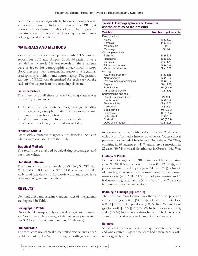
Rajoor and Seema: Posterior Reversible Encephalopathy Syndrome
112International Journal of Scientifi c Study | September 2015 | Vol 3 | Issue 6
better non-invasive diagnostic techniques. Though several studies were done in India and elsewhere on PRES, it has not been extensively studied of late. The purpose of this study was to describe the demographics and clinic-radiologic profi le of PRES.
MATERIALS AND METHODS
We retrospectively identifi ed patients with PRES between September 2011 and August 2014. 54 patients were included in the study. Medical records of these patients were reviewed for demographic data, clinical history, blood pressure measurements, laboratory investigations, predisposing condition, and neuroimaging. The primary etiology of PRES was determined for each case on the basis of the diagnosis of the attending clinician.
Inclusion CriteriaThe presence of all three of the following criteria was mandatory for inclusion.
1. Clinical history of acute neurologic change including a headache, encephalopathy, convulsions, visual symptoms, or focal defi cit
2. MRI brain fi ndings of focal vasogenic edema3. Clinical or radiologic proof of reversibility.
Exclusion CriteriaCases with alternative diagnosis, not favoring inclusion criteria were excluded from the study.
Statistical MethodsThe results were analyzed by calculating percentages, and the mean values.
Statistical SoftwareThe statistical software namely SPSS 15.0, STATA 8.0, MEDCALC 9.0.1, and SYSTAT 11.0 were used for the analysis of the data and Microsoft word and excel have been used to generate the tables.
RESULTS
Demographics and baseline characteristics of the patients are depicted in Table 1.
Demographic Profi leOut of the 54 retrospectively identifi ed cases, 48 were females, and 6 were males. The mean age of the patients at presentation was 30.94 years (maximum-minimum, 17-80 years).
Clinical Profi leThe most common clinical presentation was seizures, seen in 44 patients (81.48%), including 39 with generalized
tonic-clonic seizures, 3 with focal seizures, and 2 with status epilepticus. One had a history of epilepsy. Other clinical presentations included headache in 36 patients (66.67%), vomiting in 24 patients (44.44%) and altered sensorium in 22 cases (40.74%), visual disturbances in 09 cases (16.67%).
Etiological Profi lePrimary etiologies of PRES included hypertension (n = 21 [38.88%]), normotension (n = 07 [12.97%]), and pre-eclampsia or eclampsia (n = 14 [25.92%]). Out of 41 females, 26 were in postpartum period. Other causes were sepsis (n = 6 [11.11%], 3 had pneumonia and 1 had urosepsis), renal failure (n = 4 [7.40]), and 2 were on immunosuppressive medications.
Radiologic Findings (Figures 1-3)The most common location was the parieto-occipital and cerebellar region (n = 35 [64.81%]), followed by frontal lobe (n = 14 [25.92%]), temporal lobe (n = 09 [16.67%]), and basal ganglia (n = 05 [9.25%]). 20 (37.03%) had cortical involvement, and 3 (5.55%) had subcortical involvement. The lesions were asymmetrical in 40 cases and symmetrical in 14 cases.
Outcome53 patients recovered with the appropriate treatment, and one expired. Expired patients had severe sepsis with multiorgan dysfunction.
Table 1: Demographics and baseline characteristics of the patientsVariable Number of patients (%)Demographics
Males 13 (24.07)Females 41 (75.93)Male:female 1:8Mean age 30.94
Clinical presentationSeizures 44 (81.48)Headache 36 (66.67)Vomiting 24 (44.44)Altered sensorium 22 (40.74)Visual disturbances 09 (16.67)
EtiologyAcute hypertension 21 (38.88)Normotensive 07 (12.97)Pre-eclampsia or eclampsia 14 (25.92)Sepsis 06 (11.11)Renal failure 04 (7.40)Immunosuppression 02 (3.7)
Neuroimaging fi ndingsParieto-occipital lobes 27 (50)Frontal lobe 14 (25.92)Temporal lobe 09 (16.67)Cerebellum 08 (14.81)Basal ganglia 05 (9.25)Brainstem 03 (5.55)Subcortical 20 (37.03)Cortical 03 (5.55)Deep white matter 07 (12.96)

Rajoor and Seema: Posterior Reversible Encephalopathy Syndrome
113 International Journal of Scientifi c Study | September 2015 | Vol 3 | Issue 6
DISCUSSION
As the name implies, PRES is classically associated with the features of subcortical vasogenic edema, patchy symmetrical bilateral involvement with the preferential involvement of the posterior head regions, and complete clinical and radiological resolution.9 The clinical signs and fi ndings on the neuroimaging in patients with the PRES are consistent enough that this entity should be readily recognizable. Its causes are diverse, but common precipitants are acute elevations of blood pressure, renal decompensation, fluid retention, and treatment with immunosuppressive agents.9
The pathophysiology of PRES remains elusive. Several theories have been proposed, the most wide accepted of which states that rapidly developing hypertension leads to a breakdown in cerebral autoregulation, particularly in the posterior head region where there is a relative lack of sympathetic innveration. Hyperperfusion ensues
with protein and fluid extravasation, producing focal vasogenic edema.2,10 An alternative theory, which has been best characterized in pre-eclampsia, eclampsia, and sepsis implicates endothelial dysfunction. A third theory proposes that vasospasm with subsequent ischemia may be responsible.
Early recognition of PRES is important for the timely institution of therapy, which typically consists of gradual blood pressure control and withdrawal of potentially offending agents. Although reversible by definition, secondary complications, such as status epilepticus, intracranial hemorrhage, and massive ischemic infraction, can cause substantial morbidity and mortality.11
The observations are compared with the studies done by others on the PRES. In our study of 54 patients, maximum numbers of cases (76.93%) were seen in females in the age group of 21-40 years. This correlates well with a similar study by Fugate et al.2 (65%). The mean age of onset in the present study was 30.94 years which is comparable with Patil et al.12 study (29.90 years).
The most common clinical presentation was seizures, seen in 44 patients (81.48%), including 39 patients with generalized tonic-clonic seizures. These findings are comparable with Fugate et al.2 study (74%).
Cho and Lee13 in their study reported that PRES associated with the pregnancy, in the peripartum period presented with the seizures - Generalized tonic-clonic type, headache and visual disturbances. Similarly in our study, PRES is predominantly affected in postpartum female population.
Pedraza et al.14 reported that PRES is most common associated with hypertension, pre-eclampsia-eclampsia, and HELLP syndrome. Similarly, fi ndings are noted in our study also.
The most common location in the neuroimaging in this study was the parieto-occipital and cerebellar region (64.81%), followed by frontal lobe (25.92%), temporal lobe (16.67%), and basal ganglia (9.25%). These fi ndings are comparable with Fugate et al.2 study and McKinney et al.15 study.
CONCLUSION
PRES is a challenging condition because of its variability of clinical symptoms and signs. A high index of clinical suspicion is needed to diagnose PRES. Data from observational studies on outcome and mortality in PRES scarce. We found many of the classic etiologies and triggering factors known to be
Figure 1: Patient 1-areas of altered signal intensity appearing hyperintense on fl uid attenuation inversion recovery images
seen in bilateral parieto-occipital region
Figure 2: Patient 17-areas of altered signal intensity appearing hyperintense on fl uid attenuation inversion recovery images
seen in bilateral parieto-occipital region
Figure 3: Patient 33-bilateral asymmetrical areas of hyperintense signal are seen in bilateral parieto-occipital areas

Rajoor and Seema: Posterior Reversible Encephalopathy Syndrome
114International Journal of Scientifi c Study | September 2015 | Vol 3 | Issue 6
associated with PRES, including acute hypertension, pre-eclampsia-eclampsia, and sepsis. The overall prognosis for survival and functional independence is better in PRES when compare with other neurological conditions.
REFERENCES
1. Lee VH, Wijdicks EF, Manno EM, Rabinstein AA. Clinical spectrum of reversible posterior leukoencephalopathy syndrome. Arch Neurol 2008;65:205-10.
2. Fugate JE, Claassen DO, Cloft HJ, Kallmes DF, Kozak OS, Rabinstein AA. Posterior reversible encephalopathy syndrome: Associated clinical and radiologic fi ndings. Mayo Clin Proc 2010;85:427-32.
3. Hauser RA, Lacey DM, Knight MR. Hypertensive encephalopathy. Magnetic resonance imaging demonstration of reversible cortical and white matter lesions. Arch Neurol 1988;45:1078-83.
4. Raroque HG Jr, Orrison WW, Rosenberg GA. Neurologic involvement in toxemia of pregnancy: Reversible MRI lesions. Neurology 1990;40:167-9.
5. Bartynski WS, Boardman JF, Zeigler ZR, Shadduck RK, Lister J. Posterior reversible encephalopathy syndrome in infection, sepsis, and shock. AJNR Am J Neuroradiol 2006;27:2179-90.
6. Kur JK, Esdaile JM. Posterior reversible encephalopathy syndrome – An underrecognized manifestation of systemic lupus erythematosus. J Rheumatol 2006;33:2178-83.
7. Bartynski WS, Boardman JF. Distinct imaging patterns and lesion distribution in posterior reversible encephalopathy syndrome. AJNR Am J Neuroradiol 2007;28:1320-7.
8. Schwartz RB, Jones KM, Kalina P, Bajakian RL, Mantello MT, Garada B, et al. Hypertensive encephalopathy: Findings on CT, MR imaging, and SPECT imaging in 14 cases. AJR Am J Roentgenol 1992;159:379-83.
9. Hinchey J, Chaves C, Appignani B, Breen J, Pao L, Wang A, et al. A reversible posterior leukoencephalopathy syndrome. N Engl J Med 1996;334:494-500.
10. Primavera A, Audenino D, Mavilio N, Cocito L. Reversible posterior leucoencephalopathy syndrome in systemic lupus and vasculitis. Ann Rheum Dis 2001;60:534-7.
11. Schwartz RB. A reversible posterior leukoencephalopathy syndrome. N Engl J Med 1996;334:1743.
12. Patil VC, Agrwal V, Rajput A, Garg R, Krhirsagar K, Chaudhari V. Clinical profi le and outcome of posterior reversible encephalopathy syndrome (PRES). Ann Trop Med Public Health 2015;8:105-12.
13. Cho HJ, Lee HJ. Posterior reversible encephalopathy syndrome in early postpartum woman. Hong Kong J Emerg Med 2012;19:58-61.
14. Pedraza R, Marik PE, Varon J. Posterior reversible encephalopathy syndrome: A review. Crit Care Shock 2009;12:135-43.
15. McKinney AM, Short J, Truwit CL, McKinney ZJ, Kozak OS, SantaCruz KS, et al. Posterior reversible encephalopathy syndrome: Incidence of atypical regions of involvement and imaging fi ndings. AJR Am J Roentgenol 2007;189:904-12.
How to cite this article: Rajoor UG, Seema BN. Clinico-radiological Spectrum of Posterior Reversible Encephalopathy Syndrome: A Study from Teaching Hospital in North Karnataka. Int J Sci Stud 2015;3(6):111-114.
Source of Support: Nil, Confl ict of Interest: None declared.

115 International Journal of Scientifi c Study | September 2015 | Vol 3 | Issue 6
Self-medication for Medical Abortion in Rural Scenario: Why to choose Unsafe Way?Meena Armo1, Kavita Babbar2, Sushma Viswas3
1Associate Professor & Head, Department of Obstetrics & Gynecology, Government Chhattisgarh Institute of Medical Sciences, Bilaspur, Chhattisgarh, India, 2Associate Professor, Department of Obstetrics & Gynecology, Government Chhattisgarh Institute of Medical Sciences, Bilaspur, Chhattisgarh, India, 3Senior Resident, Department of Obstetrics & Gynecology, Government Chhattisgarh Institute of Medical Sciences, Bilaspur, Chhattisgarh India
incidence of abortion for several reasons. They exclude abortions completed using medical abortion pills (MAP) sold without a prescription by pharmacists (who represent an important source of abortion services since medical abortion became available for sale by prescription starting in 2003)4, as well as abortions done by untrained providers. It is also likely that many abortions performed by registered providers in certifi ed facilities are not reported because of defi ciencies in data reporting systems and data collection practices.1
However, since1988, a new and easier alternative has existed in the form of medical abortion.5 The term “medical abortion” refers to pregnancy termination with abortion-inducing medications (MAP) in the place of surgical intervention. Various drugs and combinations have been used for fi rst trimester abortion. The most widely researched
INTRODUCTION
In India, around 6.4 million abortions are performed annually, of which 56% are unsafe which accounts for 8-20% of all maternal deaths.1,2 As per Family welfare statistics latest number of induced abortions in India is estimated to be 6,36,306 with an annual rate of about two abortions per 1,000 women aged 15-49 years in 2013.3 However, these numbers greatly underestimate the actual
Original Article
Abstract
Background: Abortion has been legal in India for over 40 years, yet more than half of all abortions are unsafe. Medical methods of abortion have been proven to be effective and safe option to save women’s lives when practiced under medical supervision. However due to lack of knowledge regarding abortion rights, medical abortion, and safe abortion services, many women dependent on self-administration for abortion that could be highly dangerous if neglected or concealed. The study evaluates magnitude and outcome of self-medication and gives a faint idea about factors prevailing women to choose unsafe ways to terminate unwanted pregnancies in our scenario.
Materials and Methods: A retrospective observational study included 142 patients with history of unsafe medical abortion by self-administration of misoprostol and mifepristone or medical abortion pills, conducted in Chhattisgarh Institute of Medical Sciences a Government Medical College, Bilaspur, during the period of June 2009 to May 2014.
Results: In our study period, total cases of unsafe abortion admitted with complication were 400 of which 142 (35.5%) women had a history of self-medication. Majority of patients were surprisingly educated, rural, from the low socioeconomic background and Hindu by religion. The most frequent complain for admission was severe bleeding. The majority sought an abortion in the fi rst trimester. The most common reason for the termination was unintended pregnancy. The major complications observed in 51 (35.9%) cases only and surgical interventions required in 64 (45%) women.
Conclusion: Every woman is entitled to access quality health services including safe abortion. Yet for decades a matter of protecting health and lives of women has been neglected which really needs quick attention and necessary actions.
Key words: Abortion, Mifepristone, Misoprostol, Self-medication, Unwanted pregnancies
Access this article online
www.ijss-sn.com
Month of Submission : 07-2015Month of Peer Review : 08-2015Month of Acceptance : 08-2015Month of Publishing : 09-2015
Corresponding Author: Dr. Meena Armo, Department of Obstetrics & Gynecology, Government Chhattisgarh Institute of Medical Sciences, Bilaspur - 495 001, Chhattisgarh, India. Phone: +91-9425558454. E-mail: [email protected]
DOI: 10.17354/ijss/2015/405

Armo, et al.: Self-Medication for Medical Abortion
116International Journal of Scientifi c Study | September 2015 | Vol 3 | Issue 6
drugs and popular combination is of mifepristone with misoprostol, when taken within the fi rst nine weeks of pregnancy, these pills are associated with a 98% success rate.6 There are clear guidelines have been formulated by organizations such as World Health Organization (WHO), Federation of Obstetrics and Gynecological Society of India, and International Pregnancy Advisory Services (Ipas) India for patients and providers regarding the use of abortion pills. However, MAP are widely available over the counter, and its unsupervised consumption is rising day-by-day particularly in rural areas where access to abortion services and quality of treatment is poor.7,8
The study evaluates magnitude and outcome of self-medication, it also gives a faint idea about factors prevailing leading these women to choose unsafe ways to terminate unwanted pregnancy in our scenario, so that it would help policy makers to formulate new strategies and strengthen existing health services.
MATERIALS AND METHODS
This study was a retrospective observational study carried out in the department of Obstetrics and Gynecology of Chhattisgarh Institute of Medical Sciences a Government Medical College and 2nd biggest tertiary care hospital, after due permission from the Ethical Committee of our Institution. All case records of 400 women with the diagnosis of unsafe medical abortion were analyzed between the periods of June 2009-May 2014 and data were collected from case sheets. Total 142 women were included for a study that had given a history of self-administration of MAP and admitted for complications such as severe vaginal bleeding, lower abdominal cramps, incomplete abortion, and for blood transfusion. By self-medication or self-administration, we mean that these pregnant women had no prior medical consultation and had taken MAP, which was purchased from the pharmacy without any prescription either by self or by some close relative. Each case was thoroughly evaluated and discussed on the basis of demography profi le, gestational age at the time of termination, reason for termination, complaint, complication, examination fi ndings, investigation reports, and management. The statistical analysis performed by SPSS version 21.0 and qualitative data were presented as frequencies and percentages.
RESULTS
The total number of unsafe abortions observed over a period of 5 years was 400 of which 142 (35.5%) women presented with a history of self-medication of MAP were included and thoroughly analyzed for the study.
Among total patients, 100 (70.4%) were married, 32 (22.5%) were separated or widow, and 10 (7.0%) women were unmarried. In our study, 96 (67.6%), 23 (16.2%), 22 (15.5%), and 1 (0.7%) were between 20 and 29 years, 30-39 years, <20 years, 40 years of age group, respectively. Majority 104 (73.2%) were educated, 81 (57%) from rural back ground, 133 (93.7%) were Hindu by religion, and 106 (74.6%) were from low socioeconomic status (Table 1).
Of the total 142 women, 40 (28.2%), 100 (70.4%), 2(1.4%) women were nulliparous, multiparous, and grand multipara, respectively. The majority of patients had taken MAP within 14 days before coming to our hospital. Duration of pregnancy at the time of abortion was <12 weeks in 104 (73.2%), 13-20 weeks in 31 (21.8%), and >20 weeks in 7 (4.9%) women (Table 2).
Majority 100 (70.4%) were admitted after self-medication for heavy vaginal bleeding, 21 (14.8%) for severe abdominal pain, 13 (9.2%) for ultrasonography (USG) diagnosed retained products for evacuation, and 8 (5.6%) for blood transfusion (Graph 1).
Table 1: Demographic details (n=142)Variable No. %Age in years
<20 22 15.520-29 96 67.630-39 23 16.2>40 1 0.7
Marital statusMarried 100 70.4Unmarried 10 0.7Separated or widow 32 22.5
Educational statusUneducated 38 26.8Educated 104 73.2
ResidenceRural 81 57Urban 61 43
ReligionHindu 133 93.7Muslim 9 6.3
Socioeconomic statusLow 106 74.6Middle 36 25.3
Table 2: Parity and gestational weeks (n=142)Variable No. %Parity
0 40 28.21-3 100 70.44-6 2 1.4
Gestational weeks<12 104 73.213-20 31 21.8≥21 7 4.9

Armo, et al.: Self-Medication for Medical Abortion
117 International Journal of Scientifi c Study | September 2015 | Vol 3 | Issue 6
Unintended pregnancy was the reason for abortion in majority of 124 (87.3%) cases, of which 36 (25.4%) for birth spacing, 35 (24.6%) were lactating or ignorant of being pregnant, 32 (22.5%) were divorce or widow, 18 (12.6%) had undergone abortion due to sex preference, 11 (7.7%) were unaware of any contraceptive methods, and rest of 10 (7.04%) were unmarried teenagers (Graph 2).
Major complications observed in 51 (35.9%) cases only, of which moderate to severe anemia seen in 42 (29.6%), shock in 5 (3.5%) drug failure in 3 (2.11%), and septicemia in 1 (0.7%) patient (Table 3).
The majority of 63 (44.3%) women required surgical evacuation, 31 (21.8%) required blood transfusion, and 1 (0.7%) patient required intensive care. Hysterotomy with tubectomy performed in 1 (0.7%) patient for the previous cesarean section with drug failure, while 46 (32.4%) patients were kept under observation only (Graph 3). Fortunately,
no mortality observed in our study and all patients discharged well after making them aware of post-abortion contraception.
DISCUSSION
All termination of pregnancy although a safe procedure in trained hands can produce disastrous outcome when performed by untrained and unauthorized people in improper settings.
As per guidelines by Ipas India, commonly used drugs for Medical Methods of Abortion are a combination of mifepristone and misoprostol. Drug Controller General of India approved the use of mifepristone (in April 2002) and misoprostol (in December 2006) for termination of pregnancy up to 49 days gestation period, later on in December 2008, mifepristone + misoprostol (1 tablet of mifepristone 200 mg and 4 tablet of misoprostol 200 mcg each) combipack was approved by the Central Drugs Standard Control Organization, Directorate General of Health Services for the medical termination of intrauterine pregnancy (MTP) for up to 63 days gestation.
Obstetrician and Gynecologist, registered medical practitioner as defi ned by the MTP Act, can only prescribe the MAP to those patients, who are able to understand the instructions, ready for minimum three follow-up visits, ready for surgical procedure if required, having good family support and easy access to appropriate healthcare facility if needed. After counseling and history-taking, a pelvic examination is a must to confi rm pregnancy and to rule out ectopic pregnancy or fi broid. Hemoglobin, urine examination, blood grouping, and Rh typing examination are recommended. USG examination is optional. The patient is asked to report immediately if excessive bleeding, pain, fainting, or any problem occurs after MAP intake.9
Table 3: Complications*Complications No. %Moderate to severe anemia 42 29.6Shock 5 3.5Failure 3 2.8Septicemia 1 0.7Total 51 35.9*More than one complication was present in some of the women
Graph 1: Complaints at admission
Graph 2: Reasons for seeking abortion Graph 3: Management
Armo, et al.: Self-Medication for Medical Abortion
118International Journal of Scientifi c Study | September 2015 | Vol 3 | Issue 6
In our study, 142 patients had self-administered MAP for abortion which accounts for 35.5% of total 400 unsafe abortions, and 18.4% of total 758 abortions observed in 5 years. The majority of women were between 20 and 29 years of age group, married, and sought the abortion in the fi rst trimester, although women from all segment and of all age seek the abortion in India.7,10,11 Studies indicate that practice of self-induction is declining in general, though 1-10% adolescents, unmarried women, rural, and economically disadvantaged women still rely in these practices similar as our fi ndings,10,12 where unmarried or separated or widow contributed 29.57%, out of which 15.5% were contributed by adolescents as girls in our state are generally married off at an early age.13 Unintended pregnancy was the major reason for termination 87.3% among the women in our study, suggests a high unmet need which is slightly higher 24.8% in our state than unmet need of India.14 Researchers have found that underlying reasons for abortion could be risk to women’s health, contraceptive failure, pregnancies occurring soon after marriage or occurring outside of marriage, nonconsensual sex, and sexual violence which is more often observed in rural areas like in our state where about 30% of ever-married women have experienced spousal violence.12,14,15
Abortion due to sex preference observed in 12.7% cases although no confi rmatory evidence available at the time of admission indicates female feticide might be widely-practiced although unreported leading to continuous declining sex ratio at birth in our state.13,16
The availability of MAP through chemists is widespread and well-documented, studies suggests that when faced with an unwanted pregnancy, many women in India choose to fi rst attempt to terminate the pregnancy on their own, often using drugs purchased from a chemist without a prescription17-19 similar as our study where majority were educated, might be aware of medical pills, found it safer and convenient to conceal their termination.
The most frequent complains for admission was heavy vaginal bleeding and incomplete abortion in our study which could be due to higher gestation age, erratic drug schedule, or incomplete regimen. It has been found that serious complications such as bleeding, sepsis, and failure were reported more in women taking drug as self-medication than under a doctor’s supervision.19-21 Hence, it appears that medical abortion highlights the need for vigilance and early access to medical help to control morbidity and mortality.22
Shortcomings of Safe Abortion in Our ScenarioIn Chhattisgarh, of 25.5 million population 78% live in rural areas, 37% account for tribal population where more than half
of 27 districts have been classifi ed as remote areas despite that, percent of abortion is only 2.1% here (Census 2011-2012).
As per APIP 2012-2013, states 13 district hospitals and 46 community health centers have facilities to provide abortion services although these services are not at all properly utilized by most of the village people that could be because of lack of physical access to health services due to dense forest and infi ltration of extremist groups in certain areas, high out of pocket expenditure incurred on travelling and purchasing medicines. One study highlights huge shortage of 11% medical doctors at PHCs and 50% at CHCs, severe defi ciency of MTP trained providers, essential equipment’s and medicines required for abortion, leaving an almost complete void of abortion facilities in rural areas of our state. In PHCs where medical doctor or clinician is not available, pharmacists and nurses usually provide care.23,24 Thus, majority of the population living in remote areas devoid of basic facilities sought abortion through unsafe ways such as self-medication or through pharmacists, nurses, or quacks.
People also avoid approaching government hospitals due to poor confi dentiality offered and risk of being forced for sterilization or to use any form of contraception after abortion.25
In some areas, private doctors have stopped offering abortion services altogether especially second-trimester services, out of fear that they will be prosecuted for providing sex-selective abortions, even if they are not knowingly doing so.26
It has been seen that major decision regarding abortion and contraception are taken by husbands or mother-in-law in rural areas, indicate poor knowledge of couple about their reproductive rights and MTP law which says that the consent of the pregnant woman is required enough for termination of her pregnancy and in case of a minor, a written consent of the guardian is mandatory.
Apart from lack of awareness about legal abortion, knowledge and acceptance of all modern family planning methods is very low in women of state as compared to other EAG states, which can be attributed to improper counseling prior to abortion.27
CONCLUSION
Overall, the quality of health care at PHCs and CHCs in our state is very poor in remote areas in addition lack of awareness about health compounds the problem. Thus effective measures required are: Improving the literacy rates, strengthening of existing infrastructure, organizing comprehensive abortion

Armo, et al.: Self-Medication for Medical Abortion
119 International Journal of Scientifi c Study | September 2015 | Vol 3 | Issue 6
care trainings or workshops for doctors to improve their working effi ciency and skill, recruiting trained medical doctors especially lady medical offi cers and supporting staffs at each and every level to fi ll the gap, ensuring availability of required equipment’s, and emergency drugs time to time. Attachment of lady counselor in tertiary health care centers just like HIV/STD counselor to help the patients. Free supply of MAP at CHCs, District Hospitals and Medical Colleges can reduce over the counter sale at some extent. Governmental and non-governmental collaborative efforts to promote awareness about contraceptives, abortion right, and safe abortion services by introducing new technology and legislation would be highly desirable. However, sample size of a present study done in a Medical College Hospital is small thus large-scale studies are required to assess the actual magnitude of this problem.
REFERENCES
1. Duggal R, Ramachandran V. The abortion assessment project – India: Key fi ndings and recommendations. Reprod Health Matters 2004;12 24 Suppl:122-9.
2. World Health Organization. Unsafe Abortion: Global and Regional Estimates of the Incidence of Unsafe Abortion and Associated Mortality in 2003. 5th ed. Geneva: World Health Organization; 2007. p. 56.
3. Ministry of Health and Family Welfare. Health and Family Welfare Statistics of India 2013. New Delhi: Ministry of Health and Family Welfare, Statistics Division; 2013.
4. Government of India. The Medical Termination of Pregnancy Rules (Amendment). 2003. Ministry of Health and Family Welfare. Department of Family Welfare: Notifi cation, New Delhi, the 13th June, 2003. Available from: http://mohfw.nic.in/index1.php?sublinkid=3618&level=4&lid=26020&lang=1 [Last accessed on 2015 June 11.
5. Ulmann A. The development of mifepristone: A pharmaceutical drama in three acts. J Am Med Womens Assoc 2000;55 3 Suppl:117-20.
6. WHO. World Health Organization Task Force on Post-ovulatory Methods of Fertility Regulation. Comparison of 2 doses of mifepristone in combination with misoprostol for early medical abortion: A randomised trial. Br J Obstet Gynaecol 2000;107:524-30.
7. Santhya KG, Verma S, Induced abortion – The current scenario in India. In: Jejeebhoy SJ, editor. Looking Back, Looking Forward: A Profi le of Sexual and Reproductive Health in India. Jawahar nagar, India: Rawat Publications; 2004.
8. Ganatra B, Manning V, Pallipamulla SP. Availability of medical abortion pills and the role of chemists: A study from Bihar and Jharkhand, India. Reprod Health Matters 2005;13:65-74.
9. Handbook on Medical methods of Abortion by Government of Madhya Pradesh and Ipas to Expand Access to New Technologies for Safe Abortion 2011. Available from: http://www.health.mp.gov.in/mtp/MMA-Reference%20Manual.pdf. [Last accessed on 2015 Aug 19].
10. Ganatra BR. Abortion research in India: What we know and what we need
to know. In: Ramasubban R, Jejeebhoy S, editors. Women’s Reproductive Health in India. 1st ed. Jaipur, India: Rawat Publications; 2000. p. 186-235.
11. Thaker RV, Deliwala KJ, Sha PT. Self medication of abortion pill: Women’s health in Jeopardy. NHL J Med Sci 2014;1:26-31.
12. Malhotra A, Parasuraman S, Nyblade L, MacQuarrie K, Kasyap N, Walia S. Realizing Reproductive Choices and Rights: Abortion and Contraception in India. Washington DC: International Centre for Research on Women (ICRW); 2003.
13. Annual Health Survey 2011-2012 Fact Sheet, Chhattisgarh, Vital Statistics Division, offi ce of the Registrar General and Census Commissioner, India, New Delhi. Available from: http://www.censusindia.gov.in. [Last accessed on 2014 Aug 23].
14. International Institute for Population Sciences (IIPS). 2010. District Level Household and Facility Survey (DLHS-3), 2007-08: India. Chhattisgarh: Mumbai: IIPS. Available from: http://www.rchiips.org. [Last accessed on 2014 Nov 08].
15. Ravindran TK, Balasubramanian P. “Yes” to abortion but “no” to sexual rights: The paradoxical reality of married women in rural Tamil Nadu, India. Reprod Health Matters 2004;12:88-99.
16. Saha S, Duggal R, Mishra M, editors. Abortion in Maharashtra: Incidence, Care and Cost. Mumbai: Centre for Enquiry into Health and Allied Themes (CEHAT); 2004. p. 1-23.
17. Kumar R, Zavier AJ, Kalyanwala S, Jejeebhoy SJ. Unsuccessful prior attempts to terminate pregnancy among women seeking fi rst trimester abortion at registered facilities in Bihar and Jharkhand, India. J Biosoc Sci 2013;45:205-15.
18. Sowmini CV. Delay in termination of pregnancy among unmarried adolescents and young women attending a tertiary hospital abortion clinic in Trivandrum, Kerala, India, Reprod Health Matters 2013;21:243-50.
19. Ramachandar L, Pelto PJ. Medical abortion in rural Tamil Nadu, South India: A quiet transformation. Reprod Health Matters 2005;13:54-64.
20. Sharma R, Verma U, Khajuria B. Medical termination of pregnancy with mifepristone-misopristol in rural India. J Clin Diagn Res 2008;3:901-4.
21. Banerjee SK, Andersen K. Exploring the pathways of unsafe abortion in Madhya Pradesh, India. Glob Public Health 2012;7:882-96.
22. Jejeebhoy SJ, Francis Zavier AJ, Acharya R, Kalyanwala S, editors. Increasing access to safe abortion in rural Maharashtra: Outcomes of a comprehensive abortion care model. In: Population Council. New Delhi: Population Council; 2011. p. 1-67.
23. Banerjee S. Abortion care services in Chhattisgarh 20 April, 2012. Ipas Devt. Foundation India. Available from: http://www.researchgate.net/publication/260595748. [Last accessed on 2015 Aug 18].
24. Rao KD, Sundararaman T, Bhatnagar A, Gupta G, Kokho P, Jain K. Which doctor for primary health care? Quality of care and non-physician clinicians in India. Soc Sci Med 2013;84:30-4.
25. Bolar T, Marston C, Corby N, Gardiner E. Medical Abortion in India: A Model for the Rest of the World? London: Marie Stopes International; 2009.
26. Stillman M, Frost JJ, Singh S, Moore AM, Kalyanwala S. Abortion in India: A Literature Review. New York: Gluttamecher Institute; 2014.
27. Holla R, Kanchan T, Unnikrishnan B, Kotian MS, Kumar N, Thapar R, et al. Profi le of women seeking medical termination of pregnancy in South India. Int J Gynaecol Obstet 2014;125:253-5.
How to cite this article: Armo M, Babbar K, Viswas S. Self-medication for Medical Abortion in Rural Scenario: Why to choose Unsafe Way?. Int J Sci Stud 2015;3(6):115-119.
Source of Support: Nil, Confl ict of Interest: None declared.

120International Journal of Scientifi c Study | September 2015 | Vol 3 | Issue 6
Tuberculous Empyema Thoracis: Clinical, Bacteriological Features, and Its Medical ManagementBrahma Prakash1, Parul Khare2, Anuj Kumar Bhatnagar3
1Assistant Professor, Department of Pulmonary and Sleep Medicine, Netaji Subhash Chandra Bose Medical College, Jabalpur, Madhya Pradesh, India, 2Ex-JR, Department of Physiology at All India Institute of Medical Sciences, New Delhi, India, 3Head, Department of TB & Chest, Rajan Babu Institute For Pulmonary Medicine and Tuberculosis, Kingsway Camp, Delhi, India
effusion or empyema. TB empyema is a chronic, active infection of the pleural space.
Clinical outcomes of TB empyema are generally believed to be worse compared to those of non-tubercular empyema, because of protracted illness, presence of concomitant fi brocavitary lesions, high bacillary load, development of bronchopleural fi stulae (BPF), and requirement for complicated surgeries in face of compromised lung function.5 Anti-TB drug treatment alone cannot cure the patients with tubercular empyema and almost all cases require some form of external drainage of pleural pus. The secondary infection of this pus by other pyogenic infections may add to the severity of illness that may prolong the time to cure the illness, as well as increases
INTRODUCTION
Empyema thoracis, defi ned as, a collection of pus in the pleural space, which is an anatomical space bordered by both the visceral and parietal pleura. Empyema is an illness with signifi cant morbidity1,2 and morta1ity.3,4 Tuberculosis (TB) of the lung may also lead to formation of pleural
Original Article
Abstract
Background: Tuberculous (TB) empyema is a chronic infection of pleural space which carries a signifi cant morbidity and mortality.
Aims: The aim of this study was to observe clinical, radiological, bacteriological features, and the outcomes obtained by the medical management of TB empyema.
Methodology: A retrospective case analysis of all the adult patients admitted with TB empyema was performed.
Results: Records of 100 patients with TB empyema showed that Cough, expectoration, and chest pain were present in 96%, 94%, and 64% patients, respectively. Direct smear of Pus and sputum showed acid-fast bacilli (AFB) in 57 and 46 patients, respectively, whereas 32 patients had smear positivity for both. Mycobacterial culture of pus was positive in 32/100 patients. The chest radiographs had moderate and far advanced parenchymal lesions in 37 patients each. The median duration of hospital stay was 57 (41-97) days. Eight patients were managed using closed needle aspiration, whereas in others intercostals drainage tube was inserted. About 47 patients were discharged after removal of the intercostal tube (ICD) tube, whereas 36 patients had residual empyema space, so was discharged with an open drainage tube. Eight patients died and four left the treatment against advice and 5 were referred for thoracic surgery.
Conclusion: Confi rmation of diagnosis by presence of mycobacteria in pus and sputum sample and draining of empyema cavity using ICD tube or closed needle aspiration along with effective anti-tubercular drugs results in healing, and many patients may not requires surgery.
Key words: Antitubercular drugs, Drainage, Empyema tuberculous, Pleural cavity
Access this article online
www.ijss-sn.com
Month of Submission : 07-2015Month of Peer Review : 08-2015Month of Acceptance : 08-2015Month of Publishing : 09-2015
Corresponding Author: Dr. Brahma Prakash, Department of Pulmonary & Sleep Medicine, Netaji Subhash Chandra Bose Medical College, Jabalpur, Madhya Pradesh, India. Phone: +91 8435665901. E-mail: [email protected]
DOI: 10.17354/ijss/2015/406

Prakash, et al. Tuberculous Empyema
121 International Journal of Scientifi c Study | September 2015 | Vol 3 | Issue 6
the cost of treatment, in the form of antibiotics that may be required to treat this infection.
In this study, our objectives were to present the clinical features, bacteriological features and to review our experience with mainly medical management (requiring Intercostal drainage or aspiration of pus as only minor surgical procedures) of tubercular empyema by presenting the outcomes.
METHODOLOGY
Study DesignA retrospective case analysis of all adult TB empyema cases admitted at a tertiary care institute for TB and pulmonary diseases from March 2011 to March 2012 was done for the study.
Patient’s PopulationThe patients of empyema suffering from TB confi rmed either by, presence of acid-fast bacilli (AFB) in sputum or pus samples, or radiological chest X-ray fi ndings suggestive of TB or diagnosed as TB on clinical grounds in absence of any evidence of TB bacteriologically and radiologically were taken, as cases, for the purpose of study.
Exclusion CriteriaPatients of age younger than 13 years and those with empyema due to other causes were excluded from the study.
Study ProcedureThe case fi les of tubercular empyema patients admitted were reviewed retrospectively. Data with respect to clinical profi le, bacteriological status, radiological aspect, and outcomes were recorded. By reviewing the case history recorded in the case fi le, the presence of cardinal symptoms of chest disease (i.e. cough, expectoration, breathlessness, chest pain, and hemoptysis) and their duration was recorded. Patients reporting constitutional symptoms such as fever, weight loss, and loss of appetite were also recorded. Any associated condition which may cause immunosuppression such as HIV infection or history of intake of corticosteroids for long periods was also recorded. Past history of TB and the treatment received by the patient for it was obtained from case fi le. As part of the management of these patients, their samples of sputum, and pus is send for microbiological investigations for the presence of AFB in direct smear in sputum and direct smear and mycobacterial culture in pus sample. The reports of these investigations were recorded for the purpose of the study. The detection of presence of any pyogenic bacteria in aerobic cultures of pus specimen from pleural cavity was also noted for every case. Patients having small volumes of pus (<200 ml) estimated by
ultrasound and without any evidence of bronchopleural fi stula (absence of air fl uid level) on chest radiograph were managed by therapeutic pus aspiration by closed needle as per the empyema management protocol of the institute. Those with massive pleural collections and presence of bronchopleural fi stula were managed by intercostal drainage of pus by tube thoracostomy with underwater seal, by inserting a intercostal tube (ICD) of size 20-32 F, depending on the patient’s clinical situation. No negative suction device was applied to ICD during treatment, and the ICD tube was changed every week. By reviewing the case notes, the type of treatment, and length of stay in hospital and outcome of patients were recorded. The anti-TB treatment was given as per Revised National TB Program of India,6 under direct supervision of ward nurse to the patients. Patients categorized as newly diagnosed TB were given alternate day regimen with isoniazid, rifampicin, pyrazinamide and ethambutol during intensive phase and rifampicin and isoniazid during continuation phase. The re-treatment category patients received injection streptomycin in addition to the above drugs in Intensive phase. Those with documented secondary infection in pus were given 3 weeks of antibiotics also in addition to anti-tubercular drugs as per the sensitivity testing report. Outcomes of patients were recorded as any one of the following such as patient discharged after successful aspiration of pus by closed needle technique or removing ICD tube and sealing the ICD site, Discharge after removing under water seal drainage and putting small open drainage tube, patients referred for surgery for empyema, death and left against medical advice (LAMA).
The post ICD tube drainage or post closed needle aspiration chest X-ray was considered for evaluation of the parenchymal lesions in tubercular empyema patients. The disease extent radiologically was graded as per the classifi cation of the National TB Association of the USA7 which grades TB lesions as:• Minimal• Moderately advanced• Far advanced.
The study was approved by the Institutional Ethics Committee. Informed consent of patients was not recorded as the study was retrospective in nature, done by reviewing only the medical records of patients.
Statistical AnalysisThe above information from the performa was tabulated in a Microsoft excel sheet and data analyzed for frequencies and distribution by using statistical software SPSS version 13 (SPSS inc., Chicago, IL) on a personal computer. The results obtained were tested for normality distribution. Intergroup analysis was done by using Chi-square test,

Prakash, et al. Tuberculous Empyema
122International Journal of Scientifi c Study | September 2015 | Vol 3 | Issue 6
independent t-test, Mann–Whitney test or Kruskal–Wallis test as appropriate for the data. The values which were not normally distributed were written as median with inter quartile range. The relationship between two parameters was evaluated by using Pearson correlation coeffi cient or Spearman rank correlation coeffi cient, if they were appropriate. The level of signifi cance was set at P < 0.05.
RESULTS
One hundred patients of tubercular Empyema were admitted during the study period. Male preponderance was observed in the study group, in which 83 male patients and 17 female patients were enrolled for the study.
The mean age of patients was 31.1 ± 13 years (ranging from 13 to 70 years) with the majority of them belonging to younger age group 15-30 years. General characteristics of the study group and the presence of co-morbid conditions are shown in Table 1.
The majority of patients were chest symptomatics with complaints pertaining to respiratory system such as cough, expectoration, chest pain, and breathlessness. A cough was complained by 96 patients, amongst them 44 had chronic cough of more than 2 months duration. Most of the tubercular Empyema patients also had expectoration with the cough (94%). Chest pain and shortness of breath was present in 64% and 84% patients. Only ten patients had complaint of hemoptysis. 95 patients reported fever, 90 had loss of appetite and 84 had history of signifi cant weight loss.
In this study, it was seen that 53 patients had no past history of intake of anti-tubercular drugs, and 47 patients had reported intake of anti-tubercular treatment for at least 1 month in past. Accordingly, when patients were categorized as per the Revised National TB Control Program (RNTCP) guidelines,6 it was found that 53 patients belonged to new, 18 to treatment after default, eight relapse, six failure and 15 to others category. The Mantoux test was done for 62 patients, in which 43 were tuberculin test positive and 19 were tuberculin negative. When the Mantoux test was studied in relation to sputum and Pus for AFB positivity, it was found that 23 patients with Mantoux test positive were sputum positive and 20 were sputum negative. Eight patient each was Mantoux negative, but were sputum positive and Pus culture positive. About 15 patients with pus culture positive were Mantoux positive and 29 were Mantoux positive but Pus culture negative.
Bacteriological AspectAmong the patients under study, sputum smear for AFB was positive in 46/100 patients. As per WHO classifi cation
of sputum smear8 which is based on bacteriological load, it was seen that majority of patients were 1+ (21/46), next common was 3+ (17/46), followed by 2+ (6/46), and scanty +ve (2/46), in that order. The study of AFB in pus smears in patients of tubercular Empyema showed that 57/100 were AFB direct smear positive, with majority having 1+ bacteriological load (25/57), 16/57 were 2+, 10/57 as scanty +ve, and 6/57 were 3+ positive and 43 were reported as smear negative in pus.
Pus sample mycobacterial cultures grew AFB in only 32/100, while 64 samples were reported negative and four samples got contaminated in the process of culture.
When the patients were studied in correlation with sputum smear and pus smear for AFB positivity, it was found that 40/100 patients were positive for AFB in both sputum and pus samples. The results of these correlations are shown in Table 2
Among patients whose pus smear was positive (57/100), 33 reported to be culture negative, whereas eight patients had direct smear negative, but culture had evidence of growth of AFB in pus. In studying the coinfection of pus samples with pyogenic aerobic bacteria’s, it was found that 36/100 had documented secondary infection, where as in 64/100 the culture for pyogenic bacteria’s was sterile.
Radiological FindingsAs per the grading system of Chest X-ray, it was found that 28 patients had mild lesions in lung parenchyma, 37 were graded as moderate and 37 as far advanced, as described
Table 1: General characteristics and co-morbid conditionsParameter ValueAge (in years) 31.1±13Sex ratio (male:female) 4.88 (≈5:1)BMI in Kg/mt2 16.2±3.2ATT history (n=100) 47/100Sputum smear AFB positive (n=100) 46/100Mantoux test positive (n=62) 43/62Secondary infection (n=100) 36/100HIV (n=100) 4/100DM (n=100) 5/100On long term steroids (n=100) 1/100BMI: Body mass index, ATT: Antithrombotic trialists, DM: Diabetes mellitus, AFB: Acid fast bacilli
Table 2: AFB smears statusAFB smear status Number (n=100)Sputum +ve, Pus +ve 40Sputum +ve, Pus -ve 6Sputum -ve, Pus +ve 17Sputum -ve, Pus -ve 37AFB: Acid fast bacilli, +ve: Positive, -ve: Negative

Prakash, et al. Tuberculous Empyema
123 International Journal of Scientifi c Study | September 2015 | Vol 3 | Issue 6
in Table 3. The severity of lung parenchyma involvement on chest radiograph did not had any signifi cant difference in duration of stay or favorable outcomes (P > 0.05).
Treatment OutcomesThe median duration of stay was 57 days (41-97 days) for the study group. The median duration of stay in sputum smear negative patients was 42 days (range 33-56 days) and it was signifi cantly lower than those patients who were sputum smear positive for AFB (Median-98.5 days; P < 0.001). Duration of stay positively correlated moderately with sputum smear positivity (r = 0.640; P = 0.003) and weakly with Pus smear (r = 0.213; P = 0.03) and pus culture positivity (r = 0.216; P = 0.03). Duration of stay was also seen to be more in patients who had documented secondary infection in pus (r = 0.261; P = 0.009). The duration of stay did not correlated with any presenting symptoms signifi cantly.
Among the 100 patients of tubercular empyema, 47 were discharged after satisfactory lung expansion, whereas 36 patients had persistent empyema space (incomplete lung expansion), but very minimal or no pus discharge, so open drainage procedure was done in them and discharged from hospital with an advice to attend a special clinics organized once in a weeks.
Eight patients succumbed to their illness. In the study, four patients had decided to quit the treatment against medical advice, so their outcome was recorded as LAMA. Those patients who had pus discharge of more than 100 ml each day through ICD even after completion of an intensive phase of anti-tubercular treatment were referred to other center with thoracic surgery facilities for thoracic surgeons opinion. Five patients were found suitable for thoracic surgery and hence were referred to that center for surgeries and their further progress was not known, so their outcome has been recorded as “referred for thoracic surgery” (Table 4). For the purpose of statistical analysis the outcomes discharged after treatment and open drainage were clubbed as “favorable outcome” and death referred for thoracic surgery and LAMA were clubbed into “not favorable” outcomes. It was found that outcomes correlated weakly with duration of stay (r = −0.316; P = 0.001) and with age (r = 0.334; P = 0.001). The outcomes did not correlate with sputum smear or pus smear positivity and Pus culture positivity for AFB (Table 5).
DISCUSSION
TB is a common cause of empyema in countries like India where prevalence of TB is high,9 whereas in developed countries post pneumonic and post surgical etiology is
more common in patients with empyema.10 TB has been found to be the cause of empyema in approximately 65% of cases in studies reported from high prevalence regions of the world.9,11,12
TB affects the patients commonly in their productive age (21-40 years). In our study also the mean age was 31.1 years, and majority belonged to age group 21-40 years, which was similar to fi ndings in earlier studies.11-14 The studies done in western countries reported the usual age of patients with empyema as 40-50 years, where the etiology of empyema was usually post-pneumonic and post-surgical.15,16
The symptoms in patients of tubercular empyema are similar to that reported by patients of pulmonary TB. Many patients have cough with expectoration and fever (95% cases in our study). Tubercular as well as non-tubercular empyema patients report cough and fever, but these symptoms are present for a longer duration in tubercular patients, prior to their visit to health facility.12,14,17 Our study also shows that, as many as 40-45% patients had symptoms of cough, expectoration and fever of more than 2 months duration. Expectoration of blood with sputum was reported by only 10% of patients, although 46% of the patients under study were suffering from
Table 3: X-ray statusX-ray grade Number of patients (n=100)No lesion 4Mild 28Moderate 37Far advanced 31
Table 4: Outcomes of patients after minimally invasive treatment approachOutcomes NumbersDischarged after treatment 47Discharged on open drainage 36Death 8LAMA 4Referred for thoracic surgery 5LAMA: Left against medical advice
Table 5: Outcomes and its relation with pus smear resultsOutcome Pus smear
positive (n=57) (%)
Pus smear negative
(n=43) (%)Discharged after treatment (n=47) 27 (47.3) 20 (46.5)Discharged on open drainage (n=36) 21 (36.8) 15 (34.8)Death (n=8) 2 (3.5) 6 (13.9)LAMA (n=4) 2 (3.5) 2 (4.6)Referred for thoracic surgery 5 (8.7) 0 (0)LAMA: Left against medical advice

Prakash, et al. Tuberculous Empyema
124International Journal of Scientifi c Study | September 2015 | Vol 3 | Issue 6
sputum positive pulmonary TB also and studies in past have shown that hemoptysis occurs in 21-27% of patients with pulmonary TB.
Pleural fl uid studies of empyema patients have shown high AFB positivity. The pleural fl uid (pus) AFB positivity in our study was 57%, which is almost similar to studies done by Kundu et al.18 and Goyal et al.,12 who had found AFB positivity of 71.6% and 50%, respectively. The yield of this test can be increased if a standardized methodology is followed for AFB staining and reporting. In our study, pleural pus direct smear and culture for AFB was done in an intermediate reference lab accredited under RNTCP. Hence, testing of pleural fl uid for AFB in a reliable lab may help in actually confi rming tubercular etiology rather than the other etiologies of pleural pus in most of the cases. The pus in the pleural cavity may get co infected with secondary bacterial infection through a persistent BPF, but the yield of bacterial culture of pleural fl uid in empyema is low with conventional laboratory methods.19,20 In our study only 36% patients had documented secondary infection in pus culture.
Therapy for tubercular empyema consists of prompt drainage of infected pleural space, effective anti-tubercular treatment regimen and treatment of associated secondary infection of pleural space.15 Treatment of these tubercular empyema patients with intercostal drainage for a prolonged period (median 57 days) has resulted in successfully discharging 47 out of 100 patients. Some patients of empyema without bronchopleural fistula can also be managed by closed needle aspirations.15 Kamat et al. had reported shorter duration of hospital stay of 7.9 weeks (55 days) of patients suffering from pyogenic empyema as compared to 12.2 weeks (85 days) in patients with tubercular empyema,18 similarly, a study done on tubercular empyema patients had shown that majority of cases had duration of stay of 2-4 months.21
In our study, it is seen that duration of stay was longer in patients with smear positive for AFB as compared to smear negative. The time taken for the treatment response also depends upon the presence of secondary bacterial infection in pus, as is seen from results in our study in which the duration of stay correlated with the presence of secondary infection.
Discharging patients with persistent empyema space on open drainage, whose lung does not expand further due to formation of adhesions with thickened pleura is an advantageous method of managing an empyema patient conservatively. In the majority of cases simple procedure of chronic open drainage not only allows these debilitated patients to gain independence from tubing’s and bottles,
but may also allow a freedom in rehabilitative activities such as doing some lighter kind of jobs as their occupations. In our study, it has been seen that 36 out of 100 patients were discharged on open drainage with advice of regular follow-up visits.
Subjecting all patients to thoracic surgery procedures, the type of surgical procedure and timing of empyema patients to these surgeries remains a matter of debate. Sonmezoglu et al.22 in their study have found open drainage better than Eloesser fl ap and pneumonectomy, in terms of morbidity and complication rates and recommended that these procedures should be avoided wherever possible, where as many studies now recommend performing thoracic surgeries such as video assisted thoracoscopy earlier in cases of empyema. So, it is still not clear whether these surgical procedures should bypass simple medical thoracostomies in all cases of empyema.23,24 Ashbaugh reports, decortication is more effective than open drainage in reducing morbidity and mortality when surgical intervention is necessary.16
In countries such as ours, with limited thoracic surgical facilities and expertise available, it becomes diffi cult to subject every patient of tubercular empyema with persistent empyema cavity to major thoracic surgical procedure and many times TB patients are chronically ill and therefore, too debilitated (as in our study with mean body mass index of patient only 16.2 kg/mt2), so that the general condition of these patients may not permit performing these procedures upon them. A Large number of tubercular empyema patients may not be suitable candidates for surgical therapy also because of involvement of both or the contra lateral lung with TB. Furthermore, the arguments made in favor of subjecting patients early to surgical therapy by shortening the medical management of empyema were based on studies mostly done on patients suffering from parapneumonic effusions, where the empyema is usually acute and also does not carry the extent of morbidity as in tubercular empyema.25-28 So, performing studies on tubercular empyema patients for demonstrating benefi ts of early surgical procedures is needed, such that recommendations on subjecting the patient for surgical therapy may be made.
The mortality from empyema ranges from 11% to 50%.12,17 In our study, we had 8% mortality. Many patients are unable to tolerate prolonged treatment required along with the discomfort of chest tubes in situ, hence may decide to leave the treatment, as shown in our study (4%). It was found in a study, that majority of patients decided to quit treatment from the same center after 2.5 months of treatment.17 In our study, we found that the median duration of stay for patients who had LAMA was 25.5 days.

Prakash, et al. Tuberculous Empyema
125 International Journal of Scientifi c Study | September 2015 | Vol 3 | Issue 6
CONCLUSION
It can be concluded from the above study that, all patients of tubercular empyema have chronic morbidity and may require hospitalization. Closed intercostals drainage is an effective method of draining empyema cavity, therefore resulting in early healing when an effective anti-tubercular treatment regimen is also given in these patients. Many patient of tubercular empyema may not require surgery, but the most important aspect remains to be proper diagnosis of TB as cause of empyema, which can be best confi rmed by presence of TB bacteria in pus and sputum samples.
REFERENCES
1. Jess P, Brynitz S, Friis Møller A. Mortality in thoracic empyema. Scand J Thorac Cardiovasc Surg 1984;18:85-7.
2. Hutter JA, Harari D, Braimbridge MV. The management of empyema thoracis by thoracoscopy and irrigation. Ann Thorac Surg 1985;39:517-20.
3. Varkey B, Rose HD, Kutty CP, Politis J. Empyema thoracis during a ten-year period. Analysis of 72 cases and comparison to a previous study (1952 to 1967). Arch Intern Med 1981;141:1771-6.
4. Lemmer JH, Botham MJ, Orringer MB. Modern management of adult thoracic empyema. J Thorac Cardiovasc Surg 1985;90:849-55.
5. Al-Kattan KM. Management of tuberculous empyema. Eur J Cardiothorac Surg 2000;17:251-4.
6. Central TB Division (India). Revised National Tuberculosis Control Programme. Training Course for Program Manager (Modules1-4). April, 2011.
7. National Tuberculosis Association of the USA. Diagnostic Standards and Classifi cation of Tuberculosis. New York: National Tuberculosis Association; 1961.
8. World Health Organization. Treatment of Tuberculosis: Guidelines for National Programmes. 2nd ed. Geneva: WHO/TB/97.220; 1997.
9. Malhotra P, Aggarwal AN, Aggarwal R, Ray P, Gupta D, Jindal SK. Clinical characteristics and outcome of empyema thoracis in 117 patients. A comparative analysis of tubercular vs. Non tuberculous aetiologies. Respir Med 2007;101:423-30.
10. Lights RW. Parapneumonic effusions and empyema In: Rhyner S, Winter N,
Koleth J, editors. Pleural Diseases. 5th ed. Philadelphia: Lippincott Williams and Wilkins; 2007. p. 170-210.
11. Acharya PR, Shah KV. Empyema thoracis: A clinical study. Ann Thorac Med 2007;2:14-7.
12. Goyal SP, Tandon RK, Patney NL, Misra OP. Management of tubercular empyema thoracis – A review of 53 cases. Indian J Tuberc 1976;23:103-9.
13. Singh V, Behera RK, Jain NK. Anaerobic bacteriology of empyema. Lung India 1983;1:12-5.
14. Tandon RK, Misra OP. Clinicopathological study of thoracis empyema and evaluation of its surgical treatment. Indian J Chest Dis 1974;16:21-30.
15. LeMense GP, Strange C, Sahn SA. Empyema thoracis. Therapeutic management and outcome. Chest 1995;107:1532-7.
16. Ashbaugh DG. Empyema thoracis. Factors infl uencing morbidity and mortality. Chest 1991;99:1162-5.
17. Kamat SR, Kudalkar SS, Maydeo DV, Walimbe S, Kulkarni KG, Hanmantgad RR, et al. A prospective study of 100 cases of chronic empyema in Bombay. Lung India 1985;3:15-9.
18. Kundu S, Mitra S, Mukherjee S, Das S. Adult thoracic empyema: A comparative analysis of tuberculous and nontuberculous etiology in 75 patients. Lung India 2010;27:196-201.
19. Ahmed RA, Marrie TJ, Huang JQ. Thoracic empyema in patients with community-acquired pneumonia. Am J Med 2006;119:877-83.
20. Maskell NA, Batt S, Hedley EL, Davies CW, Gillespie SH, Davies RJ. The bacteriology of pleural infection by genetic and standard methods and its mortality signifi cance. Am J Respir Crit Care Med 2006;174:817-23.
21. Khanna BK. Management of tuberculous empyema. Indian J Tuberc 1987;34;141-9.
22. Sonmezoglu Y, Turna A, Cevik A, Demir A, Sayar A, Dincer Y, et al. Factors affecting morbidity in chronic tuberculous empyema. Thorac Cardiovasc Surg 2008;56:99-102.
23. Lee SF, Lawrence D, Booth H, Morris-Jones S, Macrae B, Zumla A. Thoracic empyema: Current opinions in medical and surgical management. Curr Opin Pulm Med 2010;16:194-200.
24. Tassi GF, Marchetti GP, Pinelli V, Chiari S. Practical management of pleural empyema. Monaldi Arch Chest Dis 2010;73:124-9.
25. Bilgin M, Akcali Y, Oguzkaya F. Benefi ts of early aggressive management of empyema thoracis. ANZ J Surg 2006;76:120-2.
26. Schweigert M, Solymosi N, Dubecz A, Beron M, Thumfart L, Oefner-Velano D, et al. Surgical management of pleural empyema in the very elderly. Ann R Coll Surg Engl 2012;94:331-5.
27. Muhammad MI. Management of complicated parapneumonic effusion and empyema using different treatment modalities. Asian Cardiovasc Thorac Ann 2012;20:177-81.
28. Vaziri M, Abed O. Management of thoracic empyema: Review of 112 cases. Acta Med Iran 2012;50:203-7.
How to cite this article: Prakash B, Khare P, Bhatnagar AK. Tuberculous Empyema Thoracis: Clinical, Bacteriological Features and Its Medical Management. Int J Sci Stud 2015;3(6):120-125.
Source of Support: Nil, Confl ict of Interest: None declared.

126International Journal of Scientifi c Study | September 2015 | Vol 3 | Issue 6
Morbidity Pattern and Hospital Outcome of Neonates Admitted in a Tertiary Care Teaching Hospital, MandyaP V Sridhar1, P S Thammanna2, M Sandeep3
1Assistant Professor, Department of Pediatrics, Mandya Institute of Medical Sciences, Mandya, Karnataka, India, 2Associate Professor, Department of Pediatrics, Mandya Institute of Medical Sciences, Mandya, Karnataka, India, 3Senior Resident, Department of Pediatrics, Mandya Institute of Medical Sciences, Mandya, Karnataka, India
Survey-3 report, current neonatal mortality rate (NMR) in India is 39 per 1000 live births, neonatal deaths accounts for nearly 77% of all infant deaths (57/1000) and nearly half of under-fi ve child deaths (74/1000).3 Preterm and low birth weight (LBW) babies are at increased risk of perinatal mortality and morbidity.4 As per the report sheet published in the Lancet, the major direct causes of neonatal mortality are pre-term birth (27%), infection (26%), asphyxia (23%), congenital anomalies (7%), others (7%), tetanus (7%), and diarrhoea (3%).5 There are very scanty data which are available regarding the neonatal mortality and morbidity pattern in India. Advancement in perinatal and neonatal care have signifi cantly helped in reducing NMR in developed countries, but the mortality and morbidity are still high in developing countries.6 To
INTRODUCTION
The neonatal period is a very vulnerable period of life due to many problems, which in most of the cases is preventable.1,2 Of the 25 million babies born in India every year 1 million die, India alone contributes to 25% of neonatal mortality around the world. As per the National Family Health
Original Article
Abstract
Introduction: Neonatal period is a very vulnerable period of life due to many problems. India alone contributes to about 25% of neonatal mortality around the world. In spite of advances in perinatal and neonatal care, neonatal mortality is still high in developing countries. This study was undertaken to study the disease pattern and outcome of neonates admitted to the neonatal intensive care unit (NICU) of a tertiary care teaching hospital, Mandya.
Study Design: Retrospective study of medical records for 1 year (January 2014-December 2014).
Materials and Methods: Neonates admitted to NICU, Mandya Institute of Medical Scienes, Mandya during the study period were included in the study; the data were recorded in predesigned proforma. The data were analyzed using appropriate statistical tool.
Results: A total of 1487 neonates were admitted to NICU during the study period, 54 neonates left against medical advice, 79 were referred to other centers hence excluded from analysis. The ratio of the males to female admitted was 1.45:1. The major causes of morbidity were neonatal sepsis (28.8%), respiratory distress syndrome (RDS) (23.85%), birth asphyxia (17.72%), neonatal jaundice (7.02%), and meconium aspiration syndrome (5.47%). In this study, overall mortality rate was 7.16%. Most of the deaths were due to RDS (43.3%), birth asphyxia (37.11%), neonatal sepsis (8.25%), and congenital anomalies (8.25%). Neonates with birth weight <1500 g had poor outcome compared to neonates with birth weight more than 2500 g.
Conclusion: This study identifi ed prematurity, low birth weight, neonatal sepsis, and birth asphyxia as major causes of morbidity and RDS, birth asphyxia as the major contributors to the neonatal mortality. Improving antenatal care, maternal health and timely referral of high risk cases to tertiary care hospital will help to improve neonatal outcome.
Key words: Birth asphyxia, Neonatal morbidity, Neonatal mortality, Neonatal sepsis
Access this article online
www.ijss-sn.com
Month of Submission : 07-2015Month of Peer Review : 08-2015Month of Acceptance : 08-2015Month of Publishing : 09-2015
Corresponding Author: Dr. Thammanna PS, Department of Pediatrics, Mandya Institute of Medical Sciences, Mandya - 571 401, Karnataka, India. Phone: +91-9449627788. E-mail: [email protected]
DOI: 10.17354/ijss/2015/407

Sridhar, et al.: Morbidity Pattern and Hospital Outcome of Neonates Admitted in a Tertiary Teaching Hospital
127 International Journal of Scientifi c Study | September 2015 | Vol 3 | Issue 6
apply preventive strategies, we should have the data on morbidities which are claiming the neonatal life. This study was undertaken to study the disease pattern and outcome of neonates admitted to the neonatal intensive care unit (NICU) of a tertiary care teaching hospital located in Mandya, Karnataka, India.
MATERIALS AND METHODS
This hospital based retrospective study was carried out in the NICU, Department of Pediatrics, at Mandya Institute of Medical Sciences, Mandya, Karnataka, India, for a period of 1 year from January 2014 to December 2014. The Institutional Ethical Committee approved the study protocol. Our NICU caters to the population of Mandya district and neighboring district of Ramnagara. Approximately 6000 deliveries are conducted per year in the hospital; the majority of patients belong to below poverty line income group. Our NICU has bed strength of 17, facility for phototherapy, surfactant administration, exchange transfusion, and ventilation are available.
A retrospective case record review and analysis of all the newborn babies admitted to the NICU during the study period was done and neonates satisfying inclusion and exclusion criteria were included in the study.
Inclusion CriteriaAll neonates admitted to NICU.
Exclusion CriteriaNeonates who left hospital against medical advice (LAMA) and neonates who were referred due to non-availability of beds and surgical intervention were excluded from the study.
These neonates were categorized as inborn if delivered in the Medical college Hospital and as outborn if born outside. The data were recorded in predesigned Proforma.
StatisticsData collected were compiled and entered in MS Excel spreadsheet and analyzed using appropriate statistical tools in Open Epi statistical software, version 2.3.1.
RESULTS
A total number of babies admitted to NICU during the study period was 1487 of which 54 neonates LAMA and 79 neonates were referred to other centers hence these neonates were excluded from the study. A total of 1354 neonates were included for the data analysis. Out of this
59.23% (802) were male and 40.77% (552) were females, ratio is 1.45:1. Of the total admissions 71.71% (971) were inborn neonates and 28.29% (383) were outborn neonates (Table 1). 52.21% (707/1354) neonates had birth weight >2500 g, 40.55% (549/1354) of neonates belonged to LBW category (1500-2499 g), 6.06% (82/1354) of neonates belonged to very LBW (VLBW) group (1499-1000 g), 1.18% (16/1354) of neonates were of extremely LBW (ELBW) category (<1000 g). On applying one sample Chi-square test to see the observed frequency distribution in males and females neonates overall admitted to NICU, it was found to be statistically signifi cant (P < 0.001).
The major causes of the morbidity for admission to NICU were neonatal sepsis (28.8%), respiratory distress syndrome (RDS) (23.85%), hypoxic ischemic encephalopathy (17.72%), neonatal jaundice (7.02%) and meconium aspiration syndrome (5.47%) (Table 2).
In this study, overall NICU mortality rate was 7.16% (97/1354). The mortality rate in inborn neonates was 6.69% (65/971) whereas mortality rate in outborn neonates was 8.36% (32/383), the difference in mortality rate between inborn and outborn neonates was statistically insignifi cant (P = 0.284). The mortality in males was 6.86% (55/802), in females was 7.61% (42/552), the difference in the mortality rate among male and female neonates was statistically insignifi cant (P = 0.596). The major causes for mortality were RDS (43.4%), hypoxic ischemic encephalopathy (37.11%), and neonatal sepsis (8.25%) (Table 3).
Table 1: Sex distribution of admitted neonatesSex Inborn (%) Outborn (%) Total (%)Male 573 (59.01) 229 (59.79) 802 (59.23)Female 398 (40.99) 154 (40.21) 552 (40.77)Total admission 971 (71.71) 383 (28.29) 1354 (100)
Table 2: Morbidity profi le of neonates admitted to NICUMorbidity profi le Inborn (%) Outborn (%) Total (%)Respiratory distress syndrome 227 (23.38) 96 (25.07) 323 (23.85)Meconium aspiration syndrome 47 (4.84) 27 (7.05) 74 (5.47)Respiratory distress (other causes)
31 (3.19) 11 (2.87) 42 (3.1)
Hypoxic ischemic encephalopathy
176 (18.13) 64 (16.71) 240 (17.72)
Sepsis/pneumonia/meningitis 285 (29.35) 105 (27.42) 390 (28.8)Neonatal jaundice 65 (6.69) 30 (7.83) 95 (7.02)Congenital anomaly 48 (4.94) 10 (2.61) 58 (4.29)Intrauterine growth restriction 51 (5.25) 21 (5.48) 72 (5.32)Hypothermia 12 (1.24) 9 (2.35) 21 (1.55)Hypoglycemia 6 (0.62) 7 (1.83) 13 (0.96)Others 23 (2.37) 3 (0.78) 26 (1.92)Total 971 (100) 383 (100) 1354 (100)NICU: Neonatal intensive care unit

Sridhar, et al.: Morbidity Pattern and Hospital Outcome of Neonates Admitted in a Tertiary Teaching Hospital
128International Journal of Scientifi c Study | September 2015 | Vol 3 | Issue 6
On comparing survival among different birth weight groups (Table 4), it was seen that there was statistically signifi cant difference between VLBW and normal birth weight group (P ≤ 0.05), and between ELBW and normal birth weight group (P ≤ 0.05). However, there was no statistically signifi cant difference in survival among LBW and normal birth weight group (P = 0.368).
It was observed that the duration of time between admission and death was <1 day in 42.27% of deaths (41/92) followed by 1-3 days in 40.21% of deaths (39/97).
DISCUSSION
This study was conducted to delineate the morbidity pattern, outcome and factors leading to mortality of neonates admitted to NICU of tertiary care teaching hospital. Precise data regarding mortality and morbidity pattern for NICU admission are useful for many reasons.
In our study, total of 1354 neonates were admitted of which 71.71% neonates were inborn and rest were outborn babies (28.29%), male preponderance of admission to NICU was noted similar admission pattern has been seen in study conducted by Roy et al.7 47.79% of neonates admitted had LBW and 28.58% of neonates admitted were preterm baby. This may probably be due to poor maternal health condition, low socio-economic status and less visits to health care facility. Similar rate of LBW and preterm baby admission has been reported by study conducted
by Garg et al. in New Delhi.8 According to the UNICEF “The state of the World’s Children 2010” report, 28% neonates are born with LBW in India.9 The most common specific morbidity for admission was neonatal sepsis (28.8%) followed by RDS (23.85%) and hypoxic ischemic encephalopathy (17.72%). A study conducted by Gaucham et al. in Nepal reported that neonatal jaundice, sepsis and perinatal asphyxia as being commonest indication for admission to NICU.10 According to national neonatal-perinatal database (NNPD) sepsis (36%) is the most common morbidity responsible for admission followed by prematurity (26.5%) and perinatal asphyxia (10%).11 Birth asphyxia is an important cause of neonatal morbidity and mortality, its incidence in our study is 17.72% which is similar to fi ndings of Chandra et al.12 Neonatal sepsis acts as an important cause for morbidity and mortality especially among LBW and preterm babies.
Mortality rate observed in our study is 7.16% is lower than that of the mortality rates observed in study conducted by Rakholia et al.13 The most common causes of mortality were RDS (43.3%), birth asphyxia (37.11%), and sepsis (8.25%). Similar pattern of outcome has been reported by study conducted by Rashid et al.14 In contrast the study report published by ICMR reports sepsis (32.8%) as the major cause for neonatal mortality followed by birth asphyxia (22.3%) and prematurity (16.8%).15 In the study done at JIPMER, sepsis was the cause for death in 52.3% of neonates followed by birth asphyxia and injuries (29.23%).16 Majority of deaths in our study was attributable to RDS and birth asphyxia; this may probably be due to poor antenatal care, malnourished pregnant women, less availability of health facility, delivery by untrained professional and delay in referral from peripheral hospitals. Birth weight <1500 g were associated with high number of mortality in preterm neonates.
CONCLUSION
According to this study neonatal sepsis, RDS and birth asphyxia are leading causes of morbidities in newborn babies. In spite of many advances in neonatal care above factors still continue to be the leading causes of morbidity in neonates. Common causes of neonatal mortality were RDS, birth asphyxia, neonatal sepsis and congenital anomalies. The majority of morbidities and subsequently the mortalities can be prevented by improving antenatal care, maternal health, timely intervention, referring at appropriate time to tertiary care centers for high risk cases, preventing preterm deliveries and care of neonates at centers with facility. This study has some limitations, as this was a hospital based retrospective study, the cause of death was determined using the data available in case
Table 3: Comparison of deaths among inborn and outborn neonatesCause of neonatal death Inborn (%) Outborn (%) Total (%)Respiratory distress syndrome 26 (40) 16 (50) 42 (43.3)Sepsis/meningitis/pneumonia 6 (9.23) 2 (6.25) 8 (8.25)Meconium aspiration syndrome 2 (3.08) 1 (3.13) 3 (3.09)Hypoxic ischemic encephalopathy (birth asphyxia)
25 (38.46) 11 (34.37) 36 (37.11)
Congenital anomaly 6 (9.23) 2 (6.25) 8 (8.25)Total 65 (100) 32 (100) 97 (100)
Table 4: NICU outcome in different birth weight groupBirth weight NICU
admission (%)
Deaths (%)
Percentage of death is each
group (%)More than 2500 g 707 (52.21) 32 (32.99) 4.53LBW (1500-2499 g) 549 (40.55) 31 (31.96) 5.65VLBW (1000-1499 g) 82 (6.06) 22 (22.68) 26.83ELBW (<1000 g) 16 (1.18) 12 (12.37) 75Total 1354 (100) 97 (100) -NICU: Neonatal intensive care unit, LBW: Low birth weight, VLBW: Very low birth weight, ELBW: Extremely low birth weight

Sridhar, et al.: Morbidity Pattern and Hospital Outcome of Neonates Admitted in a Tertiary Teaching Hospital
129 International Journal of Scientifi c Study | September 2015 | Vol 3 | Issue 6
record sheets, Neonates who LAMA and those who were referred to other centers due to non-availability of NICU beds and needed surgical intervention were excluded from study and could hence modify the results. In our study, we did not divide the deaths into early and late neonatal period. As the majority of the patients presenting to us belong to low socio-economic status, the results from this study cannot be a complete refl ection of the problem in the community as a whole.
REFERENCES
1. Prasad V, Singh N. causes of morbidity and mortality in neonates admitted in Government Medical College, Haldwani in Kumaun region (Uttarakhand) India. JPBS 2011;8:1-4.
2. Bhutta ZA. Priorities in newborn management and development of clinical neonatology in Pakistan. J Coll Phys Surg Pak 1997;7:231-4.
3. NFHS-3: Ministry of Health and Family Welfare, Government of India. Available from: http://www.mohfw.nic.in/NFHS-PRESENTATION.htm. [Last accessed on 2014 Nov 08].
4. Roy KK, Baruah J, Kumar S, Malhotra N, Deorari AK, Sharma JB. Maternal antenatal profi le and immediate neonatal outcome in VLBW and ELBW babies. Indian J Pediatr 2006;73:669-73.
5. Lawn JE, Cousens S, Zupan J; Lancet Neonatal Survival Steering Team. 4 million neonatal deaths: When? Where? Why? Lancet 2005;365:891-900.
6. Ng PC. Diagnostic members of infection in neonates. Arch Dis Child 2004;89:229-35.
7. Roy RN, Nandy S, Shrivastava P, Chakraborty A, Dasgupta M, Kundu TK. Mortality pattern of hospitalized children in a tertiary care hospital of Kolkata. Indian J Community Med 2008;33:187-9.
8. Garg P, Krishak R, Shukla DK. NICU in a community level hospital. Indian J Pediatr 2005;72:27-30.
9. UNICEF. The State of The World’s Children, 2010. New York: UNICEF; 2010. p. 92-5.
10. Gauchan E, Basnet S, Koirala DP, Rao KS. Clinical profi le and outcome of babies admitted to Neonatal Intensive Care Unit (NICU). J Inst Med 2011;33:1-5.
11. Morbidity and mortality among outborn neonates at 10 tertiary care institutions in India during the year 2000. J Trop Pediatr 2004;50:170-4.
12. Chandra S, Ramji S, Thirupuram S. Perinatal asphyxia: Multivariate analysis of risk factors in hospital births. Indian Pediatr 1997;34:206-12.
13. Rakholia R, Rawat V, Bano M, Singh G. Neonatal morbidity and mortality of sick newborns admitted in a teaching hospital of Uttarakhand. CHRISMED J Health Res 2014;1:228-34.
14. Rashid A, Ferdous S, Chowdhury T, Rahman F. The morbidity pattern and the hospital outcome of the neonates who were admitted in a tertiary level hospital in Bangladesh. Bangladesh J Child Health 2003;27:10-3.
15. ICMR Young Infant Study Group. Age profi le of neonatal deaths. Indian Pediatr 2008;45:991-4.
16. Augustine T, Bhatia BD. Early neonatal morbidity and mortality pattern in hospitalised children. Indian J Matern Child Health 1994;5:17-9.
How to cite this article: Sridhar PV, Thammanna PS, Sandeep M. Morbidity Pattern and Hospital Outcome of Neonates Admitted in a Tertiary Care Teaching Hospital, Mandya. Int J Sci Stud 2015;3(6):126-129.
Source of Support: Nil, Confl ict of Interest: None declared.

130International Journal of Scientifi c Study | September 2015 | Vol 3 | Issue 6
Histopathological Spectrum of Central Nervous System Tumors: A Single Centre Study of 100 CasesSarita Nibhoria1, Kanwardeep Kaur Tiwana1, Richa Phutela2, Akanksha Bajaj2, Sahil Chhabra2, Saloni Bansal2
1Associate Professor, Department of Pathology, Guru Gobind Singh Medical College and Hospital, Faridkot, Punjab, India, 2Post-graduate Student, Department of Pathology, Guru Gobind Singh Medical College and Hospital, Faridkot, Punjab, India
(males:3.0/100,000; females:2.1/100,000).2 In 2008, the rates had risen to 3.8/100,000 in males and 3.1/100,000 in females, although the incidence rates in developed countries (males:5.8/100,000; females:4.4/100,000) still remained higher than those in developing countries (males: 3.2/100,000; females: 2.8/100,000).
In developing countries like India, due to lack of complete registration of newly diagnosed cases with local cancer registries, the exact tumor burden of such disease goes unnoticed and is underestimated. Hospital-based prevalence data, therefore, forms the basis for estimating the disease load. With increased availability of diagnostic facilities and better healthcare, the incidence of CNS tumors seems to be on the rise in developing countries.3
All the CNS tumors were divided into seven categories: Tumors of neuroepithelial tissue; tumors of the cranial and paraspinal nerves; tumors of the meninges; lymphomas
INTRODUCTION
Though central nervous system (CNS) tumors are not as frequent as tumors of many other sites,1 they showed a varied histopathologic spectrum. It has been revealed by International Agency for Research on Cancer that the worldwide incidence rate of CNS tumors in 2002 was 3.7/100,000 population among males and 2.6/100,000 population among females. The incidence rates were higher in developed countries (males:5.8/100,000; females:4.1/100,000) than in developing countries
Original Article
Abstract
Introduction: Central nervous system (CNS) tumors are not frequent tumors with primary malignant brain tumors accounting for 2% of all cancers in the U.S adults. The incidence rates are lower in developing countries like compared to the developed countries while increased rates have been observed in both.
Purpose: The aim of the present study is to study the histopathological spectrum of CNS tumors irrespective of age in single tertiary care center.
Material and Methods: The present 5 years study from a single tertiary care center, patient clinically diagnosed with CNS tumors and registered between 2009 and 2014 in pathology department were included and classifi ed according to WHO 2007 classifi cation along with grading of the tumor.
Results: A wide range of histopathological spectrum of CNS tumors was observed and was classifi ed according to the recent WHO classifi cation system. The primary CNS tumors were graded from Grade 1 to Grade 1V. Overall tumors of neuroepithelial tissue (51.7%) was the most common entity followed by the tumors of meninges (34.8%), metastatic tumors (5.6%), tumors of peripheral nerves (4.5%), tumors of the sellar region (2.3%), and lymphomas and hematopoietic neoplasm (1.1%).
Conclusion: Rising global trends in the incidence of CNS tumors, irrespective of age have been observed. The present study highlights the histological diversity in CNS tumors in both, adult as well as pediatric age groups.
Key words: Astrocytoma, Central nervous system, Meningioma, Tumors
Access this article online
www.ijss-sn.com
Month of Submission : 07-2015Month of Peer Review : 08-2015Month of Acceptance : 08-2015Month of Publishing : 09-2015
Corresponding Author: Richa Phutela, House Number BV, 1283, Street Number 1, Chowk Number 4, Jain Nagar, Abohar, Punjab, India. Phone: +91-9988442680. E-mail: [email protected]
DOI: 10.17354/ijss/2015/408

Nibhoria, et al.: Histopathological Spectrum of Central Nervous System Tumors
131 International Journal of Scientifi c Study | September 2015 | Vol 3 | Issue 6
and hematopoietic neoplasms; germ cell tumors; tumors of the sellar region; and metastatic tumors. The WHO classifi cation offers a crude histological grading system, in which each CNS tumor is classifi ed as Grades I-1V according to its degree of malignancy. This system can provide an estimate for the prognosis of a patient. In this study, age, sex and the histological tumor type and grade were systematically recorded.
The spectra of the malignant tumors were different in the pediatric and adult groups. For the adults, astrocytic tumors, tumors of meninges, and metastatic tumors occupied the top three places while astrocytic tumors took the lead in pediatric cases.
MATERIALS AND METHODS
The present 5 years study from a single tertiary care center, the patient diagnosed with CNS tumors and registered between 2009 and 2014 in the pathology department were consecutively screened. The H and E stained histopathological slides of biopsy received were evaluated. The cases were diagnosed and characterized where necessary using immunohistochemistry and categorized according to the WHO 2007 classifi cation. The inclusion criteria were cases of CNS tumors of all age groups. The tumors of peripheral nervous system and non-neoplastic conditions of the CNS were excluded. With these criteria, a total of 100 cases of CNS tumors were studied, and their histological typing and grading was done.
RESULTS
The present study was conducted during 2009-2014 in which 100 cases of clinically diagnosed CNS tumors received in the department of pathology were studied. Out of the 100 clinically diagnosed CNS tumors, neoplasm was seen in 89% of the cases and 07% showed reactive gliosis while biopsy was inadequate or inconclusive in 04%. Among the CNS tumors, the majority (95.5%) presented as space occupying lesion in the brain and only 4.5% were intra-spinal tumors. The CNS tumors showed a slight male predominance (Male: Female = 1.2:1) and a broad range was found, i.e. 0-70 years with the mean age of 40. The pediatric tumors contributed 12.3% of all CNS tumors (Table 1).
A wide range of histopathological spectrum of CNS tumors was observed and was classifi ed according to the recent WHO classifi cation system. The primary CNS tumors were graded from I to IV. Overall tumors of neuroepithelial tissue (51.7%) was the most common entity followed by the tumors of meninges (34.8%), metastatic
tumors (5.6%), tumors of peripheral nerves (4.5%), tumors of the seller region (2.3%), and hemolymphoid neoplasm (1.1%)(Figure 1) respectively (Table 2).
The tumors of neuroepithelial tissue comprised mainly of astrocytic tumors (39.32%) followed by oligodendroglial tumors (4.50%), (Figure 2) mixed gliomas (2.25%), ependymal tumors (2.25%), choroid plexus tumors (2.25%), and embryonal tumors (1.12%). Male predominance was seen in the ratio of 1.8:1.0 and the mean age was found to be 36.4 years. Among the astrocytic tumors, anaplastic
Table 1: Depicting the age, sex, histological subtypes and WHO grading of meningiomasAge Sex Histologic subtypes WHO grade45 Female Transitional I55 Male Transitional I31 Female Fibroblastic I50 Female Meningotheliomatous I50 Female Transitional I48 Male Meningotheliomatous I50 Female Transitional I35 Female Transitional I49 Female Transitional I35 Female Meningotheliomatous I70 Male Fibroblastic I21 Female Transitional I45 Female Meningotheliomatous I42 Female Meningotheliomatous I55 Female Fibroblastic I60 Female Transitional I19 Female Fibroblastic I71 Male Meningotheliomatous I65 Female Meningotheliomatous I24 Female Atypical meningioma II28 Male Atypical meningioma II32 Female Atypical meningioma II58 Male Meningotheliomatous I30 Male Meningotheliomatous I62 Female Meningotheliomatous I49 Female Transitional I50 Male Meningotheliomatous I
Figure 1: Plasmacytoma (H&E X400)

Nibhoria, et al.: Histopathological Spectrum of Central Nervous System Tumors
132International Journal of Scientifi c Study | September 2015 | Vol 3 | Issue 6
Meningiomas (Figure 3) dominated the tumors of meninges contributing 30.3% of the total 34.8% followed by mesenchymal, not meningeal tumors (3.4%) and occasional tumors of uncertain histogenesis (1.1%).
The mean age in meningiomas was found to be 45.5 years, and family preponderance was seen in the ratio of 1:1.2.
Atypical meningiomas constituted 3.34% of the total meningiomas and occurred in the younger age group with a mean age of 28 years. A wide range of histologic subtypes was observed in meningiomas, and WHO grading system was applied to them (Table 3).
This was followed by metastatic tumors (Figure 4) that constituted 5.6% of the total tumors with a mean age of 61 years and slight male preponderance (3:2).
The pediatric tumors were peculiar by their histologic diversity with astrocytic tumors (25% of pediatric tumors) taking a lead. The median age in pediatric tumors was found to be 9.4 years with equal male to female ratio (1:1). (Table 4)
DISCUSSION
The incidence of CNS tumors is quite low in adults while they form the second most common childhood tumors after leukemia.4 Adult CNS tumors differ
Table 2: Percentage breakup of CNS tumors with histologic subtypes and WHO gradingTumor types WHO
gradingPercentage
Tumor of neuroepithelial tissue 39.3Astrocytic tumors
Diffuse astrocytoma Grade IIAnaplastic astrocytoma Grade IIIGlioblastoma multiforme Grade IV
Oligodendroglial tumors 4.5Oligodendroglioma Grade IIAnaplastic oligodendroglioma Grade III
Mixed gliomas 2.3Oligoastrocytoma Grade IIAnaplastic oligoastrocytoma Grade III
Ependymal tumors 2.3Ependymoma Grade II
Choroid plexus tumors 2.3Choroid plexus papilloma Grade IChoroid plexus carcinoma Grade IV
Embryonal tumors 1.1Medulloblastoma Grade IV
Tumors of peripheral nerves 4.5Neurofi broma Grade I
Tumors of meningesMeningiomas 34.8Meningiomas Grade IAtypical meningiomas Grade II
Mesenchymal, non-meningothelial tumors 3.4Lipoma Grade IHaemangioma Grade I
Tumors of uncertain histogenesis 1.1Hemangioblastoma Grade I
Lymphomas and haemopoietic neoplasm 1.1Plasmacytoma Grade IV
Tumors of the sellar region 2.3Craniopharyngioma Grade I
Metastatic tumors 5.6CNS: Central nervous system, WHO: World Health Organization
Table 3: Percentage split of astrocytic tumorsType of astrocytic tumor
WHO grade
Percentage
Diffuse astrocytoma Grade II 34.3Anaplastic astrocytoma Grade III 45.7Glioblastoma multiforme Grade IV 20WHO: World Health Organization
Figure 2: Oligodendroglioma (H&E X100)
astrocytomas Grade III (45.7%) was the commonest type followed be diffuse astrocytoma Grade II (34.3%) and glioblastoma multiforme Grade IV (20.0%).
Figure 3: Meningioma (H&E X100)

Nibhoria, et al.: Histopathological Spectrum of Central Nervous System Tumors
133 International Journal of Scientifi c Study | September 2015 | Vol 3 | Issue 6
signifi cantly from childhood brain tumors in relation to their sites of origin, clinical presentation, tendency to disseminate, histological features and their biological behavior. Whereas in adults the predominant CNS tumor types are metastases, glial neoplasms, and meningiomas, in children, besides gliomas, other major tumor types including primitive embryonal neoplasms are also common. In recent times, an enhanced understanding of these biological differences between adult and childhood CNS tumors has led to investigations in distinct molecular and genetic pathways and therapeutic approaches for each tumor type.
It has been observed worldwide that the incidence of CNS tumors is on a rise. Moreover due to the high mortality seen in CNS tumors, they form the most challenging group of tumors for neurooncologists.
In the present study comprising of 100 CNS tumors irrespective of age, from a single center, have been categorized according to the recent WHO classifi cation.5 In our study, the most common CNS tumors in the
descending order are tumors of neuroepithelial tissue (51.7%) followed by tumors of meninges (34.8%) and metastatic tumors (5.6%). This was in concordance with the nationwide database in France which revealed the proportion of tumors of neuroepithelial tissue and the
Figure 5: Anaplastic Astrocytoma exhibiting vascular proliferation. (H&E X400)
Figure 6: Glioblastoma Multiforme (H&E X400)
Figure 4: Metastatic carcinomatous deposits (H&E X100)
Table 4: Distribution of CNS tumors with histologic subtypes among children and teenagers (age 0-19 years)Age Sex Histologic subtype WHO grade Percentage18 years Female Cavernous hemagioma Grade 1 8.310 months Male Choroid plexus carcinoma Grade IV 8.38 years Male Medulloblastoma Grade IV 8.317 years Female Craniopharyngioma Grade I 8.35 years Male Choroid plexus papilloma Grade I 8.310 years Male PNST-neurofi broma Grade I 8.315 years Male Diffuse fi brillary astrocytoma Grade II 8.36 months Male Inadequate biopsy 8.314 years Female Anaplastic oligodendroglioma Grade III 8.319 years Female Meningioma-fi broblastic Grade I 8.311 years Female Glioblastoma multiforme Grade IV 8.35 years Female Anaplastic astrocytoma Grade III 8.3CNS: Central nervous system, PNST: Peripheral nerve sheath tumors, WHO: World Health Organization

Nibhoria, et al.: Histopathological Spectrum of Central Nervous System Tumors
134International Journal of Scientifi c Study | September 2015 | Vol 3 | Issue 6
meninges were 53.9% and 28.8%, respectively, from 2004 to 2008.6 Another population-based report from Central Brain Tumor Registry of the United States (CBTRUS) recorded rates of 33.7% and 35.5%, respectively, from 2004 to 2007.7 The reason for this variation remains unknown and requires further investigation.8 For the tumors of the meninges, increasing trends were observed around the world,9-15 and improvements in the diagnostic technologies are still considered to be the major causal factor. Worldwide studies have also demonstrated an apparently increased incidence of metastatic tumors,16 supporting their third position in our study.
The tumor spectra varied from adults to children and teenagers as well as from males to females. In our study, astrocytic tumors (45.7%) were the major tumor entity with anaplastic astrocytoma (Figure 5) topping the list followed by diffuse astrocytoma (34.3%). This was contrary to CBTRUS data, in which glioblastoma (Figrue 6) and anaplastic astrocytoma were the most common malignant tumors in adults.
The CNS tumors in children and teenagers showed a great histologic diversity. The most common CNS tumor in our study was astrocytic tumors comprising 25% of all tumors in this age group. This was in concordance with the study did by Chen et al. who found astrocytomas to be leading tumors (29.2%) in this age group in their 60 years review of cases.8 While in a multi-institutional study did by Jain et al. fi nd a slight higher proportion of astrocytoma (34.7%) as compared to our study.3
The incidence of various CNS tumors in the current study falls well within the range seen in the international studies for every tumor category.
CONCLUSION
Rising global trends in the incidence of CNS tumors, irrespective of age have been observed. The present study
highlights the histologic diversity in CNS tumors in both, adult as well as pediatric age groups.
REFERENCES
1. He J, Gu D, Wu X, Reynolds K, Duan X, Yao C, et al. Major causes of death among men and women in China. N Engl J Med 2005;353:1124-34.
2. Ferlay J, Bray F, Pisani P, Parkin DM. GLOBOCAN 2002: Cancer Incidence, Mortality and Prevalence Worldwide, Version 2.0. IARC Cancerbase No. 5. Lyon: IARC; 2004.
3. Jain A, Sharma MC, Suri V, Kale SS, Mahapatra AK, Tatke M, et al. Spectrum of pediatric brain tumors in India: A multi-institutional study. Neurol India 2011;59:208-11.
4. Rosemberg S, Fujiwara D. Epidemiology of pediatric tumors of the nervous system according to the WHO 2000 classifi cation: A report of 1,195 cases from a single institution. Childs Nerv Syst 2005;21:940-4.
5. Louis DN, Ohgaki H, Wiestler OD, Cavenee WK, editors. World Health Organization Classifi cation of Tumors of the Central Nervous System. Lyon: IARC; 2007.
6. Rigau V, Zouaoui S, Mathieu-Daudé H, Darlix A, Maran A, Trétarre B, et al. French brain tumor database: 5-year histological results on 25 756 cases. Brain Pathol 2011;21:633-44.
7. CBTRUS Stastical Report: Primary Brain and Central Nervous System Tumors Diagnosed in the United States in 2004-2007. Available from: http://www.cbtrus.org/2011-NPCR-SEER/WEB-0407-Report-3-3-2011.pdf. [Last accessed on 2015 Jul 9].
8. Chen L, Zou X, Wang Y, Mao Y, Zhou L. Central nervous system tumors: A single center pathology review of 34,140 cases over 60 years. BMC Clin Pathol 2013;13:14.
9. Baldi I, Gruber A, Alioum A, Berteaud E, Lebailly P, Huchet A, et al. Descriptive epidemiology of CNS tumors in France: Results from the Gironde Registry for the period 2000-2007. Neuro Oncol 2011;13:1370-8.
10. Staneczek W, Jänisch W. Epidemiologic data on meningiomas in East Germany 1961-1986: Incidence, localization, age and sex distribution. Clin Neuropathol 1992;11:135-41.
11. Hoffman S, Propp JM, McCarthy BJ. Temporal trends in incidence of primary brain tumors in the United States, 1985-1999. Neuro Oncol 2006;8:27-37.
12. Christensen HC, Kosteljanetz M, Johansen C. Incidences of gliomas and meningiomas in Denmark, 1943 to 1997. Neurosurgery 2003;52:1327-33.
13. Greig NH, Ries LG, Yancik R, Rapoport SI. Increasing annual incidence of primary malignant brain tumors in the elderly. J Natl Cancer Inst 1990;82:1621-4.
14. Klaeboe L, Lonn S, Scheie D, Auvinen A, Christensen HC, Feychting M, et al. Incidence of intracranial meningiomas in Denmark, Finland, Norway and Sweden, 1968-1997. Int J Cancer 2005;117:996-1001.
15. Kaneko S, Nomura K, Yoshimura T, Yamaguchi N. Trend of brain tumor incidence by histological subtypes in Japan: Estimation from the Brain Tumor Registry of Japan, 1973-1993. J Neurooncol 2002;60:61-9.
16. Alexandru D, Bota DA, Linskey ME. Epidemiology of central nervous system metastases. Prog Neurol Surg 2012;25:13-29.
How to cite this article: Nibhoria S, Tiwana KK, Phutela R, Bajaj A, Chhabra S, Bansal S. Histopathological Spectrum of Central Nervous System Tumors: A Single Centre Study of 100 Cases. Int J Sci Stud 2015;3(6):130-134.
Source of Support: Nil, Confl ict of Interest: None declared.

135 International Journal of Scientifi c Study | September 2015 | Vol 3 | Issue 6
Comparative Evaluation of Results of Cross Pin Fixation by Conventional Method with Dorgan’s Method in Displaced Supracondylar Fracture in ChildrenPrasanta Kumar Saha
Assistant Professor, Department of Orthopedics, Institute of Post-graduation and Medical Research and Seth SukhlalKarmani Memorial Hospital, Kolkata, West Bengal, India
fossa and olecaron fossa.2 The standard mode of treatment for the displaced fracture in children is closed reduction and percutaneous pin fi xation.3-5 Completely displaced fractures usually necessitate surgical treatment. Different pin fi xation techniques have been described including medial lateral 2 cross pins and lateral 2 pins.6 It has been proved that cross pin fi xation is superior than lateral pin fi xation.7-9 Although ulnar nerve injury up to 6% from use of a medial pin is common, and this possibility is most likely to occur when the medial epicondyle cannot be palpated in swollen elbows.10 There are many techniques for Kirschner wire fi xation. The incidence of cubitus varus deformity after treatment was about 5% according to Flynn while Arino reported that it was almost 21%. In a prospective study, Chai reported that 8 (15%) of 54 patients developed iatrogenic ulnar nerve defi cit after treatment with medial-
INTRODUCTION
Supracondylar fracture of the humerus is among the most common fractures in children, and completely displaced fractures usually necessitate surgical treatment.1 Supracondylar area is most vulnerable to fracture because or tubular shaft of humerus becomes triangular in this area leads to stress raiser, and thin cortical bone due to coronoid
Original Article
Abstract
Introduction: Closed manipulation and percutaneous K-wire fi xation are the standard treatment of displaced supracondylar fractures of the humerus in children. The most common complication is an injury of the ulnar nerve during insertion of medial K-wire. In this study, two fi xation methods, the traditional cross wire (medial and lateral) and the Dorgan’s percutaneous lateral cross-wiring technique will be evaluated.
Purpose: The aim of the study is to compare the results by two different modalities of management.
Methods: The study was done in our institution over a period of 1½ year. The inclusion criteria are displaced fracture (Gartland Type II and III) without any neurovascular defi cits. We exclude open fractures, pathological fractures, more than seven days old fractures. Patients are followed up for a period of 12 months.
Results: Among the 97 patients, full follow-up is available for 85 patients. Among them, 45 patients are treated with conventional cross pin method and 40 patients are treated by Dorgan method. We have found there are no statistically difference results between two groups.
Conclusion: The conclusion is that close reduction and percutaneous fi xation techniques are the standard methods by providing rigid stability and good union rate. Dorgan’s lateral cross-wiring technique has the advantage of both-stability of cross wire fi xation and avoiding the ulnar nerve injury. Although the results are not statistically signifi cant.
Key words: Children, Conventional medial-lateral pinning, Dorgan’s method, Supracondylar humerus fracture
Access this article online
www.ijss-sn.com
Month of Submission : 08-2015Month of Peer Review : 08-2015Month of Acceptance : 09-2015Month of Publishing : 09-2015
Corresponding Author: Prasanta Kumar Saha, 6A/1B Raja Bagan Lane, Dum Dum, Kolkata - 700 028, West Bengal, India. Phone: +91-9903151908. E-mail: [email protected]
DOI: 10.17354/ijss/2015/409

Saha: Conventional Mediolaterl Pinning and Dorgan’s Method
136International Journal of Scientifi c Study | September 2015 | Vol 3 | Issue 6
lateral pin fi xation. In supracondylar humerus fracture among them conventional K-wire fi xation is a most popular surgical method. Controversy exists about the optimal K-wire confi guration in displaced Type II and III fracture. In present study an alternate method of fi xation with very minimum complications. In our study, we compare the outcome of displaced supracondylar fractures fi xed by a conventional cross K-wire technique in one group, and another group of patients operated by two cross K-wire from the lateral side was used.
MATERIALS AND METHODS
This is a prospective randomized controlled study conducted at Institute of Post-Graduate Medical Education & Research) from February 2011 to March 2014 after getting ethical permission. The catchment area of the Institute is Kolkata and its suburban area. All the patients with displaced supracondylar fracture (Garland Type - II and III) were included in this study. The guardians of the children were properly counseled about advantages, disadvantages, and possible complication of the procedure. After getting proper written consent from the guardian, children were included in the study. The inclusion criteria are displaced supracondylar fracture (Garland Type - II and III), close fracture without any distal neurovascular deficit and fracture occurring within 1 week. The patients with open fracture, pathological fracture, fracture associated with other co-morbid condition, previous fracture in either elbow and concomitant fracture or other injury in the same limb that will alter the treatment protocol of the supracondylar fractures were excluded.
Every patient was thoroughly examined at the time of admission. The fi rst step was to assess the vascular status of the limb. Once, the case was deemed to be without vascular defi cit (defi ned as absent or feeble <50% of the volume on the other side), and then evaluation was done using other exclusion and inclusion criteria. The median (with anterior interosseus) radial and ulnar nerves were tested and the fi nding documented. The skin condition and the amount of swelling were assessed. Fractures classifi cations were done by radiographs.
Check radiographs of the elbow were taken again if required in the anteroposterior and the true lateral planes if possible. The elbow was immobilized in a posterior long arm plaster of paris slab in the comfortable position of 50-60° of fl exion. Analgesics were given, and the arm elevated to decrease swelling. The main investigator would perform the randomization by drawing lots – odd numbers signify medial-lateral pin fi xation while even numbers would be treated by two lateral pin fi xations by Dorgan method.
All patients were operated in supine position under general anesthesia within 48 h of admission. Tourniquet was not applied. The fi rst reduction manoeuvre was performed with traction applied to the forearm with an assistant applying counter traction. First, the medial or lateral displacement of the fracture was corrected. After that rotational displacement was reduced with pronation and supination of the forearm. Final, fi xation by smooth K-wire into the medial condyle from the lateral side. The medial condyle should not be penetrated to avoid ulnar nerve injury, but cortical involvement could be achieved (Figure 1). The principal is that the wires should cross above the fracture line. The similar way, conventional method of cross fi xation was done (Figure 2).
Post-operative immediate neurological assessment for median, ulnar, and radial nerves, and anteroposterior and lateral X-rays were performed.
The patients with iatrogenic ulnar nerve lesions in both groups were followed up without any treatment. The mean hospitalization period was 1.7 days (range 1-5 days). We
Figure 1: X-ray photograph of Dorgan method of fi xation
Figure 2: X-ray photograph of cross pin method of fi xation

Saha: Conventional Mediolaterl Pinning and Dorgan’s Method
137 International Journal of Scientifi c Study | September 2015 | Vol 3 | Issue 6
removed the cast K-wires after three weeks and started gentle active elbow exercises. At the last follow-up, we evaluate a range of motion and carrying angle by goniometer at both elbows. We evaluate the functional and cosmetic results according to the criteria proposed by Flynn et al. (Table 1).
RESULTS
In the present study, 97 treated patients for displaced supracondylar fracture were considered. Later 12 patients were drop out in follow-up. Among the 85 patients, we have treated 40 patients by Dorgan method and rest 45 patients have treated by conventional cross pin fi xation method. The mean age of patients 7.51 years (range from 3 to 12 years) with standard deviation 0.08 years. The duration of injury to admission to the hospital was 0.33-72 h with mean 7.02 h. The interval from injury to surgery range from 10 to 80 h (mean, 32.98 h). Both groups under study had a similar pattern of duration from injury to surgery (Table 2). Among the 45 patients of crossed pin fi xation group, 9 patients having Type-II fracture, 22 patients having Type III (post medial), and 14 patients Type III (post lateral) fracture. Among the 40 patients of Dorgan method group, 8 patients have Type II, 20 patients having Type III (post medial), and 12 patients have Type III (post lateral) fracture (Figure 3). 40 patients of Dorgan group, 23 patients have right elbow injury and 27 patients of cross pin fi xation group having the right side supracondylar fracture humerus.
Sixty six out of 85 patients (33 on each group) had a carrying-angle loss of 0-4.9°, which was considered to be an excellent result (Figure 4). Of the eleven patients rated as having achieved a good result, nine had crossed pin fi xation while two had Dorgan pin fi xation. There were
only six patients in whom the result was rated as fair; two had crossed pin fi xation, and four had Dorgan lateral pin fi xation. Of the two patients rated as poor, 1 patient on each group. The mean loss of elbow extension was 7.21° (range, 2.15-16.4°) in cross pin fi xation patients, while in Dorgan lateral pin fi xation patients was 7.11° (range, 3.70-17.91°). Similarly, the mean loss in elbow fl exion was 8.68° (range, 0.04-17.32°) and 11.28° (range, 0.86-21.66°) in conventional cross pin fi xation and Dorgan lateral pin fi xation patients respectively (Figure 5 and Table 3).
In 10 patients, the capitellum and medial epicondylar epiphyses had already fused at follow-up. So, 74 patients were available for measurement The Baumann angle loss and MEE angle. The mean Baumann angle loss in the cross pin fi xation group and the Dorgan method of pin fi xation group was 5.96° and 5.30,° respectively. The mean MEE angle loss in the cross pin fi xation group and the Dorgan pin fi xation group was 6.07° and 6.93°, respectively. Analyses of both the Baumann angle loss and the MEE angle loss using Student t-test showed no signifi cant differences (Table 4).
Five (of all 97 patients) neurologic defi cits were diagnosed on admission. One patient had ulnar nerve palsy while the other had median nerve injury. Of the 85 patients available
Table 2: Duration injury to surgery for two methods of pin fi xationInjury to surgery interval (h)
Cross pin fi xation
Dorgan method
Total
0-23.9 10 9 1924-47.9 27 22 4948-71.9 6 8 14>72 2 1 3Total 45 40 85
Figure 3: Distribution of type of fracture
Figure 4: Analysis of results of carrying angle loss
Table 1: Modifi ed Flynn’s criteria to evaluate outcome of treatmentOutcome Loss of elbow ROM (°) Loss of carrying angle (°)Excellent 0-5 0-5Good 6-10 6-10Fair 11-15 11-15Poor >15 >15Rom: Range of motion

Saha: Conventional Mediolaterl Pinning and Dorgan’s Method
138International Journal of Scientifi c Study | September 2015 | Vol 3 | Issue 6
for outcome evaluation, seven ulnar nerve injuries (fi ve patients in the cross pin fi xation group and two patients in the Dorgan lateral pin fi xation group) were detected after the treatment procedure. Three radial nerve injury were found in Dorgan pin fi xation group. These patients were followed up 6 weekly in the clinic, and all of them recovered completely within six.
We use crossed table method and Fisher’s exact test to analyses the data of iatrogenic ulnar nerve injury between the two groups. The P = 0.428. Hence, there is no signifi cant difference in the incidence of ulnar nerve injury.
Pin tract infection was found in fi ve patients. Two of the patients were treated with cross pin fi xation. All the patients were antibiotics orally. They all recovered at the subsequent follow-up. We have not encountered any vascular injury or defi cit during and after surgery. We have found that patients with vascular defi cit have good capillary refi lling of the fi ngers and the radial pulse eventually reappeared after surgery. We have not found compartment syndrome or Volkmann ischemic contracture on the last clinical review.
In our study, according to Flynn criteria, among the 40 patients treated with Dorgan method 15 patients have excellent and 12 patients have a good result. In comparison to 21 patients have excellent and 14 patients have a good result by a conventional method (Figure 6). By applying student t-test, there was no statistical difference between the two groups (Table 5).
DISCUSSION
The mean follow-up duration of the 85 patients was 8.93 months (range, 3.13-14.73 months). Some patients
had developed elbow stiffness which was recovered by proper physiotherapy during follow-up. All the fractures were reduced under image intensifi er. The Baumann angle and carriying angle was measured and compared with opposite side. There was no loss of reduction in both coronal and sagital during healing time. The enrolment of both groups was randomized, and no statistical difference was found. So, there was no difference in stability providing by two methods of fi xation between two groups.
Thirty seven patients had carrying-angle loss of 10° or more compared to the opposite elbow (16 of the patients in cross pin fi xation, and 21 in Dorgan lateral pin fi xation group). These patients would develop cubitus varus deformity which must be treated. Malunion in the sagital plane rarely requires surgical correction. Sagital plane deformity would not cause any functional disability and some improvement also happened due to remodeling of bone. So, it did not require any treatment.
Five patients in medial-lateral pin fi xation group while two patients in Dorgan pin fi xation group had ulnar nerve injury. We analyze the data by using the crossed table method and Fisher’s exact test. The P = 0.24. Therefore, there was no statistical difference. We need a larger sample for bringing
Table 4: Comparison of Baumann angle loss and MEE angle lossParameters Mean±SD P value
(student t-test)Cross pin
fi xationDorgan method of pin fi xation
Baumann angle loss (°) 5.96±5.5 5.3±5.0 0.646MEE angle loss (°) 6.07±5.2 6.93±6.6 0.597SD: Standard deviation
Figure 5: Analysis of results of fl exion and extension loss
Figure 6: Analysis of result by Flynn criteria
Table 3: Analysis of results of carrying angle, elbow fl exion and extension loss by student t-testParameters Mean±SD P value
(student t-test)Cross pin
fi xationDorgan method of pin fi xation
Carrying angle loss (°) 3.58±4.5 3.7±4.24 0.913Elbow lexion loss (°) 7.14±9.2 7.1±9.2 0.991Elbow extension loss (°) 8.6±8.64 11.26±10.4 0.322SD: Standard deviation

Saha: Conventional Mediolaterl Pinning and Dorgan’s Method
139 International Journal of Scientifi c Study | September 2015 | Vol 3 | Issue 6
certainty in this issue. There was only one iatrogenic radial nerve injury in the Dorgan-lateral pin fi xation. The value is too small to be analyzed. All nerve injuries in this study were most likely neuropraxia or axonotmesis. All patients were recovered without exploration or repair within 6 months after the operation. None of the patients in our series developed evidence of ischemic contracture to suggest muscle necrosis at follow-up.
CONCLUSION
From this prospective study, we come to conclusion that both methods provide rigid stability in both coronal and sagital plane with negligible chance of nerve injury which has no statistical signifi cance. However, further studies with a larger and more varied ethnic and occupations group may be warranted to bring certainty in this dilemma.
REFERENCES
1. Herring JA, editor. Upper extremity injuries. In: Tachdjian’s Pediatric Orthopaedics. 3rd ed. Philadelphia, PA: W. B. Saunders; 2002. p. 2115-250.
2. Tolo VT, Skaggs DL, editors. Masters Techniques in Orthopaedic Surgery: Pediatric Orthopaedics. Philadelphia, PA: Lippincott; 2007. p. 1-15.
3. Skaggs DL, Hale JM, Bassett J, Kaminsky C, Kay RM, Tolo VT. Operative treatment of supracondylar fractures of the humerus in children. The consequences of pin placement. J Bone Joint Surg Am 2001;83A:735-40.
4. Sibinski M, Sharma H, Sherlock DA. Lateral versus crossed wire fi xation for displaced extension supracondylar humeral fractures in children. Injury 2006;37:961-5.
5. Khan AQ, Goel S, Abbas M, Sherwani MK. Percutaneous K-wiring for Gartland type III supracondylar humerus fractures in children. Saudi Med J 2007;28:603-6.
6. Tripuraneni KR, Bosch PP, Schwend RM, Yaste JJ. Prospective, surgeon-randomized evaluation of crossed pins versus lateral pins for unstable supracondylar humerus fractures in children. J Pediatr Orthop B 2009;18:93-8.
7. Solak S, Aydin E. Comparison of two percutaneous pinning methods for the treatment of the pediatric type III supracondylar humerus fractures. J Pediatr Orthop B 2003;12:346-9.
8. Beaty JH, Kasser JR, editors. Supracondylar fractures of the distal humerus. In: Rockwood and Wilkins’ Fractures in Children. 5th ed. Philadelphia, PA: Lippincott Williams & Wilkins; 2001. p. 577-624.
9. Weinberg AM, Castellani C, Arzdorf M, Schneider E, Gasser B, Linke B. Osteosynthesis of supracondylar humerus fractures in children: A biomechanical comparison of four techniques. Clin Biomech (Bristol, Avon) 2007;22:502-9.
10. Wind WM, Schwend RM, Armstrong DG. Predicting ulnar nerve location in pinning of supracondylar humerus fractures. J Pediatr Orthop 2002;22:444-7.
Table 5: Comparison of result of two groups according to Flynn criteriaParameter by Flynn criteria
Dorgan method (n=40)
Cross pin fi xation (n=45)
P value
(Excellent+good) 27 35 <0.05(Fair+poor) 13 10 <0.05
How to cite this article: Saha PK. Comparative Evaluation of Results of Cross Pin Fixation by Conventional Method with Dorgan’s Method in Displaced Supracondylar Fracture in Children. Int J Sci Stud 2015;3(6):135-139.
Source of Support: Nil, Confl ict of Interest: None declared.

140International Journal of Scientifi c Study | September 2015 | Vol 3 | Issue 6
Cyto-histological Correlative Study of Thyroid Neoplasms by Imprint MethodH B Shashidhar1, D Vani2, N S Ashwini2,3, M Sandhya4, M Bharathi5
1Associate Professor, Department of Pathology, Mysore Medical College & Research Institute, Mysore, Karnataka, India, 2Associate Professor, Department of Pathology, Mysore Medical College & Research Institute, Mysore, Karnataka, India, 3Post-graduate, Student, Department of Pathology, Mysore Medical College & Research Institute, Mysore, Karnataka, India, 4Post-graduate, Student, Department of Pathology, Mysore Medical College & Research Institute, Mysore, Karnataka, India, 5Head of Department, Department of Pathology, Mysore Medical College & Research Institute, Mysore, Karnataka, India
and the operating surgeon. Thyroid nodules are common with an estimated prevalence ranging from 4% by palpation to 67% by ultrasonography.1 Most of the thyroid neoplasms are clinically palpable and after a provisional clinical diagnosis, fi ne-needle aspiration plays a major role in determining nature of the tumor. It is during the operative procedure, that the role of pathologist comes into force, through the intra-operative diagnosis.
Imprint cytology (IC) is an intra-operative diagnostic technique introduced by Dudgeon and Patrick in the year 1927. After pioneering work, this technique remained in oblivion for years. Later, the immense potential of intra-operative imprint cytodiagnosis was recognized and it came
INTRODUCTION
Disorders involving thyroid have been unique in both presentation and symptoms. Due to this, diagnosis of thyroid lesions and most importantly, thyroid neoplasms, forms an important aspect involving both the pathologist
Original Article
Abstract
Background: Imprint cytology (IC) has become an indispensable component in the intra-operative rapid cytodiagnosis of tumor types. Its rise as a worthy alternative to frozen sections in places where facilities for the same are not available is commendable as it is inexpensive procedure which yields smears, easy to interpret and is worthy of consideration to be included as a routine intra-operative diagnostic procedure in the evaluation of thyroid neoplasms.
Objectives: The objective of this study was to adopt intra-operative thyroid neoplasms in order to facilitate decision regarding extent of surgery to be performed, expedite the planning for therapy and to assess the effi cacy of intra-operative IC by correlation with histopathological diagnosis.
Methods: A total of 96 patients with clinically diagnosed thyroid swellings from K. R. Hospital referred to the Department of Pathology and subsequently confi rmed as thyroid neoplasms by fi ne-needle aspiration cytology and posted for surgery formed the basis for this study. Immediately following the surgical removal of the neoplasm, imprint smears were taken from the tumor area. The air dried or wet-fi xed smears were stained routinely by rapid hematoxylin and eosin and by modifi ed rapid papanicolaou method.
Results: Out of 96 cases of thyroid neoplasms, 58 cases were diagnosed as benign and 38 cases as malignant. Follicular neoplasms were 54 forming the majority among the benign tumors with 4 cases of Hurthle cell adenomas. Amongst the malignant tumors, 30 papillary carcinomas, 4 follicular carcinomas, and 2 cases each of non-Hodgkin lymphoma maltoma and anaplastic carcinoma were diagnosed.
Conclusion: The overall sensitivity was 95.45%, the specifi city, and positive predictive value was 100% each and negative predictive value of 90.6% was seen in the present study. The overall accuracy encountered was 95.52%.
Key words: Adenomas, Benign, Neoplasm, Thyroid
Access this article online
www.ijss-sn.com
Month of Submission : 07-2015Month of Peer Review : 08-2015Month of Acceptance : 09-2015Month of Publishing : 09-2015
Corresponding Author: Dr. NS Ashwini, Department of Pathology, Mysore Medical College & Research Institute, Mysore, Karnataka, India.Phone: +91-09886021425. E-mail: [email protected]
DOI: 10.17354/ijss/2015/410

Shashidhar, et al.: Imprint cytology study of thyroid neoplasms
141 International Journal of Scientifi c Study | September 2015 | Vol 3 | Issue 6
to be utilized in various diagnostic works.2 Since freezing artifacts are avoided, the cytological details provided by the intra-operative cytological imprint specimens are superior to that of frozen sections (FS). Imprints cover a wide area of tissue, and therefore, differences of histological structures are more likely to be represented adequately and clearly than in conventional paraffi n embedded sections taken from a small area. IC is simple, inexpensive, rapidly diagnostic, and easy to perform procedure especially in thyroid neoplasms. It has gained immense popularity globally and has been accepted as a routine diagnostic procedure in most of the hospitals worldwide. The present study was carried out to evaluate the diagnostic value of IC intraoperatively along with histopathological correlation.
METHODS
The study comprises of 96 cases of IC of thyroid neoplasms. The lesions were confirmed as thyroid neoplasms by fi ne-needle aspiration cytology (FNAC) and posted for surgery. The imprint procedure was done intraoperatively at K. R. Hospital attached to Mysore Medical College, Mysore. The results were subsequently correlated with the histopathological diagnosis.
IC was done intraoperatively under the guidance of the operating surgeon after obtaining informed consent from the patient. The procedure was done under absolute aseptic precautions by the pathologist.
The imprint smears were prepared from the cut surface of freshly dissected thyroid specimen by touching the glass slides on surface, focusing on suspicious areas. The smears were immediately wet-fi xed in 95% alcohol fi xative and stained by ultrafast Papanicolaou technique. Carnoy’s fi xative was used when smears were hemorrhagic and in few cases, smears were air dried and stained with May-Grünwald-Giemsa stain. The resected specimen was sent for histopathological examination for correlative study.
RESULTS
Of the 96 cases, maximum incidence of cases was in the 21-30 and 31-40 years with 28 cases in each category. Greater than 60 years category showed lowest incidence with 6 cases (Figure 1). The present study showed a female predominance with majority of cases being females (94 cases) (Figure 2). The most common clinical diagnosis was solitary nodule of thyroid accounting to 54.16% (52 cases). The follicular neoplasm constituted the most common diagnosis by FNAC owing to 72.9% (70 cases). 62.5% of cases were diagnosed as follicular neoplasm by IC followed by 22.9% of cases with a diagnosis of papillary
carcinoma (Table 1). On IC, 70 cases were diagnosed as benign (72.9%) and 26 were malignant (27.09%) (Figure 3). Histopathologically, 54 cases (56.25%) were found to be of follicular adenoma followed by 30 cases (31.25%) of papillary carcinoma. Two cases each of anaplastic
Table 1: Distribution of cases diagnosed by ICImprint diagnosis Number of cases PercentageFollicular neoplasm 60 62.5Papillary carcinoma 22 22.91Colloid goiter 6 6.25Hurthle cell neoplasm 4 4.16NHL 2 2.08Anaplastic carcinoma 2 2.08Total 96IC: Imprint cytology, NHL: Non-Hodgkin lymphoma
Table 2: Distribution of cases based on histopathologyHistopathological diagnosis Number of cases PercentageFollicular adenoma 54 56.25Papillary carcinoma 30 31.25Anaplastic carcinoma 2 2.08NHL maltoma 2 2.08Follicular carcinoma 4 4.16Hurthle cell adenoma 4 4.12Total 96NHL: Non-Hodgkin lymphoma
Table 3: Correlative results of IC and histopathologyImprint diagnosis Diagnosis by histopathologyBenign (70) (72.9%) Benign (58) (60.41%)
Follicular neoplasm (60) Follicular adenoma (54)Colloid goiter (10)Hurthle cell neoplasm (4) Hurthle cell adenoma (4)
Mailignant (26) (27.08%) Malignant (38) (39.58%)Papillary carcinoma (24) Papillary carcinoma (30)Anaplastic carcinoma (2) Anaplastic carcinoma (2)NHL maltoma (2) NHL maltoma (2)
Follicular carcinoma (4)IC: Imprint cytology, NHL: Non-Hodgkin lymphoma
Figure 1: Age distribution in the present study

Shashidhar, et al.: Imprint cytology study of thyroid neoplasms
142International Journal of Scientifi c Study | September 2015 | Vol 3 | Issue 6
carcinoma and non-Hodgkin lymphoma (NHL) maltoma were reported (Tables 2 and 3).
DISCUSSION
IC has been an accepted method for the intra-operative cytodiagnosis of thyroid neoplasms, because of its rapidity of diagnosis and high sensitivity in delineating benign and malignant tumors. The quick decisions that can be taken during management of cases postoperatively following imprint diagnosis, only add up to the many advantages that the method has to offer.
Jose et al. studied 98 cases in which the age incidence varied from 17 years to 65 years with a mean age of 35.5 years. The male to female ratio was 1:5.5.3 According to Taneri et al., the mean age was 47 and the ranged being between 24 and 67 years.4 In the present study of 96 cases, the age varied between 16 and 85 years with a mean age of 38.17 years. The male to female ratio was found to be 1:47.
Francis and Das in their study involving FNAC, IC and FS found a sensitivity of 68.4%, 85%, and 65%, respectively. In all the three procedures, the specifi city was 100%, and the false positive rate was 0%. A 93.3% diagnostic accuracy was achieved with combined IC and FS.5 In the present study, sensitivity was 95.74%, and a specifi city of 100% was noted. The positive predictive value (PPV) stood at 100%
and negative predictive value (NPV) at a value of 90.6%. The overall accuracy of the study was 95.52%. Ferit et al. reported a sensitivity of 83.3%, specifi city of 97.7%, PPV of 75%, NPV of 97.7%, and accuracy of 96%.4
In the present study, out of 96 cases diagnosed by imprint study, 70 cases were benign with a 72% incidence and 26 were malignant with a 28% incidence.
The follicular adenomas (Figures 4-6) accounted to 93% of benign cases (54/58) in the present study. The follicular adenomas are the most common among thyroid neoplasms. The cytology shows follicular cells in sheets and dispersed cells. The presence of micro follicular aggregates implies a follicular neoplasm. Since the presence of capsule cannot be assessed in aspirates, these are better termed as follicular neoplasms.3,6-8
Four cases of Hurthle cell neoplasm (Figures 7 and 8) were reported which on cytology showed cellular aspirates
Figure 2: Sex incidences in the present study
Figure 3: Distribution of benign and malignant cases diagnosed by imprint cytology
Figure 4: Follicular neoplasm showing gray white encapsulated mass with occasional areas of hemorrhage and degeneration
Figure 5: Imprint smear of Follicular neoplasm showing numerous microfollicles

Shashidhar, et al.: Imprint cytology study of thyroid neoplasms
143 International Journal of Scientifi c Study | September 2015 | Vol 3 | Issue 6
of tumor cells in loosely cohesive groups and papillaroid architecture. These tumor cells were large, polygonal to
oval with characteristic abundant fi nely granular blue gray cytoplasm.3,9
Among the malignant tumors, papillary carcinoma (Figures 8 and 9) was the most common encountered constituting 78.94% of malignant cases (30/38). It is the most common malignancy of thyroid with a female sex predominance of 3:1. The spread of the tumor is by lymphatics. Cytologically, aspirates are cellular and are comprised of papillary fronds with a fi brovascular core and lined by tumor cells. These cells are large with nuclei showing intranuclear cytoplasmic inclusions, nuclear grooves, and clefts.10-14
Two cases of anaplastic carcinoma (Figure 10) were reported which on cytology shows bizarre giant or spindle cells in singles and small clusters. The cells have pleomorphic nuclei and maybe multinucleate with coarse clumped chromatin.6,11,15,16
Two rare cases of NHL maltoma (Figure 11) were encountered. It is an indolent tumor in the thyroid which remains localized for a long time before dissemination. The
Figure 6: Histopathology of follicular adenoma depicting a well circumscribed lesion composed of micro and macrofollicles
with surrounding compressed normal thyroid tissue
Figure 7: Imprint smear of Hurthle cell neoplasm showing oxyphil cells in follicular patterns and clusters
Figure 8: Imprint smear showing papillary structures with nuclear features
Figure 10: Imprint smear (a) and histopathology (b) of anaplastic carcinoma thyroid
ba
Figure 9: Gross (a) and microscopy (b) of well encapsulated variant of papillary carcinoma of thyroid
ba
Figure 11: Gross (a) and imprint smear of non-Hodgkins lymphoma
ba

Shashidhar, et al.: Imprint cytology study of thyroid neoplasms
144International Journal of Scientifi c Study | September 2015 | Vol 3 | Issue 6
lymphoma cells are centrocyte-like with irregular, folded, darkly stained nuclei, and scanty pale cytoplpasm.6,16
CONCLUSION
Intra-operative cytology of imprint smears is very valuable in determining the precise area of surgical extirpation of thyroid tumors and also serves as an object of defi ning therapeutic decision quickly. It is a safe, simple, feasible, and dependable method for rapid cytodiagnosis of thyroid neoplasms.
ACKNOWLEDGMENTS
Authors would like to acknowledge Dr. A.L. Hemalatha, Professor of Pathology for her guidance and support for this study.
REFERENCES
1. Chehrei A, Ahmadinejad M, Tabatabaee SA, Hashemi SM, Kianinia M, Fateh S, et al. Touch imprint and crash preparation intra operative cytology versus frozen section in thyroid nodule. J Res Med Sci 2012;17:475-80.
2. Anila KR, Krishna G. Role of imprint cytology in intra operative diagnosis of thyroid lesions. Gulf J Oncolog 2014;1:73-8.
3. Jose RM, Smile SR, Iyengar KR. The role of imprint cytology in intraoperative diagnosis of thyroid swellings. Indian J Pathol Microbiol 2002;45:393-6.
4. Taneri F, Poyraz A, Salman B, Tekin E, Akyuerek N, Bayram O, et al. Using imprint and frozen sections in determining the surgical strategies for thyroid pathologies. Endocr Regul 2001;35:71-4.
5. Francis IM, Das DK. Role of FNA, intraoperative cytology and frozen section in the diagnosis of breast lumps and thyroid lesions. Int J Kuwait Univ Health Sci Cent 1999;8:173-82.
6. Rosai J, Ackerman LV. Ackerman’s Surgical Pathology. 10th ed., Vol. 2. St. Louis: Elsevier; 2011. p. 515-68.
7. Pluot M, Faroux MJ, Rain J, Patey M, Mallaisy T, Simatos A. Imprint cytology in the diagnosis of tumors of the thyroid. Arch Anat Cytol Pathol 1989;37:36-9.
8. Bose SM. Imprint cyto-diagnosis of thyroid goitres. Indian J Surg 1993;55:319-23.
9. Kini SR, Miller JM, Hamburger JI. Cytopathology of Hürthle cell lesions of the thyroid gland by fi ne needle aspiration. Acta Cytol 1981;25:647-52.
10. Kanjanavirojkul N, Puapairoj A, Patarapadungkit N. Imprint cytology from a childhood case of metastatic papillary thyroid carcinoma. J Med Assoc Thai 2000;83:348-51.
11. Pacini F, Leslie J, De Groot, Dudgeon LS, Patrick CV. Neoplasms of thyroid. A new method for rapid microscopical diagnosis of tumors, with an account of 200 cases examined. Br J Surg 1927;15:257-61.
12. Carcangiu ML, Zampi G, Pupi A, Castagnoli A, Rosai J. Papillary carcinoma of the thyroid. A clinicopathologic study of 241 cases treated at the University of Florence, Italy. Cancer 1985;55:805-28.
13. Kaneko C, Shamoto M, Niimi H, Osada A, Shimizu M, Shinzato M. Studies on intranuclear inclusions and nuclear grooves in papillary thyroid cancer by light, scanning electron and transmission electron microscopy. Acta Cytol 1996;40:417-22.
14. Elston CW, Ellis IO. Thyroid gland. In: Symmers WC, editor. Systemic Pathology. 3rd ed. Edinburgh: Churchill Livingstone; 1998.
15. Livolsi VA, Montone K, Sack M. Pathology of Thyroid Disease: Sternberg Diagnostic Surgical Pathology. 3rd ed. Philadelphia, PA: Lipincott Williams and Wilkins; 1999.
16. Rasbach DA, Mondschein MS, Harris NL, Kaufman DS, Wang CA. Malignant lymphoma of the thyroid gland: A clinical and pathologic study of twenty cases. Surgery 1985;98:1166-70.
How to cite this article: Shashidhar HB, Vani D, Ashwini NS, Sandhya M. Cyto-histological Correlative Study of Thyroid Neoplasms by Imprint Method. Int J Sci Stud 2015;3(6):140-144.
Source of Support: Nil, Confl ict of Interest: None declared.

145 International Journal of Scientifi c Study | September 2015 | Vol 3 | Issue 6
Otitis Media with Effusion Plain Myringotomy versus Myringotomy with Grommet InsertionRajeev Reddy
Associate Professor, Department of Otorhinolaryngology, Khaja Banda Nawaz Institute of Medical Sciences, Gulbarga, Karnataka, India
is sterile, but becomes a good source for the growth of bacteria, eventually leading to the stage of suppuratition that is characterized by the presence of pus which eventually gets extruded in the external auditory canal presenting as discharge as a result of the rupture of the tympanic membrane. Eventually, the disease goes into remission following antibiotic treatment or by nature of the hosts immunity. In certain instances, this does not happen leading to the persistence of collection in the middle ear which is sterile as a result of long-standing usage of antibiotics. The primary source of all these events is the Eustachian tube infection. Children who frequently complain of repeated ear pain and discharge not responding to regular line of treatment often are investigated for adenoidal hypertrophy, by radiological examinations or diagnostic nasal endoscopy, which if enlarged needs surgery to relieve the chronic Eustachian tube obstruction.6-9
The sequel of long standing Eustachian tube pathology varies from suppurative otitis media to otitis media with effusion and atelectasis. In individuals who are prone for such repeated Eustachian tube insults need treatment of
INTRODUCTION
Acute suppurative otitis media is defi ned as the acute infl ammation of the mucoperiosteum of the middle ear cleft. The pathology arising in the Eustachian tube and spreading on to the middle ear cavity.1,2 The causative organisms are believed to be Staphylococcus aureus, Streptococci, Haemophilus Infl uenza, Moraxella. The course of the disease begins with the stage of Eustachian Catarrh where pain and deafness are the predominating symptoms and examination of the tympanic membrane reveals retraction and congestion.3-5
The second stage is the presuppurative stage characterized by collection of fl uid in the middle ear cavity which initially
Original Article
Abstract
Introduction: Otitis media with effusion is one of the most common causes of conductive deafness. Thorough assessment leading to the diagnosis is imperative for good treatment options. Various modalities have been tried for the same with mixed outcome.
Aim: The aim of present study is to compare the two of the commonest surgical procedures performed for the treatment of the condition.
Materials and Methods: Retrospective study based on the analysis of the records of 72 patients presenting to the outpatient department of KBN Institute of Medical Sciences, Gulbarga, Karnataka, India, between January 2015 and August 2015. All the patients had been subjected to the tuning fork tests, pure tone audiometry and tympanometry and conclusively diagnosed with otitis media with effusion.
Result: The results were assessed on the basis of subjective improvement in symptoms and closure of A-B gap on pure tone audiograms.
Conclusion: Otitis media with effusion has long been known to be one of the leading causes of conductive deafness in the population especially the pediatric group.
Key words: Audiometry, Deafness, Effusion, Myringotomy, Tympanotomy
Access this article online
www.ijss-sn.com
Month of Submission : 07-2015Month of Peer Review : 08-2015Month of Acceptance : 08-2015Month of Publishing : 09-2015
Corresponding Author: Dr.eRajeev Reddy, Department of Otorhinolaryngology, Ruby Opposite Catholic Church, Anand Nagar, Gulbarga -r585 103, Karnataka, India. Phone: +91-09880887877. E-mail: [email protected]
DOI: 10.17354/ijss/2015/411

Reddy: Otitis media with Effusion - Myringotomy vs Grommet
146International Journal of Scientifi c Study | September 2015 | Vol 3 | Issue 6
the cause as in adenoidectomy. Most children respond after this with eventual resolution of symptoms but in a few if the middle ear collection fails to resolve which symptomatically presents as persistence of deafness and aural fullness, myringotomy with or without grommet (Figure 1) or exploration of middle ear in the form of tympanotomy may be needed. Long standing use of antibiotics for treatment of repeated ear infections often predisposes the individual to the development of otitis media with effusion, which if untreated leads to more irreversible sequelae such as atelectasis and retraction pockets which would then necessitate a surgical exploration of the middle ear and reconstruction.10-13
MATERIALS AND METHODS
The protocol was approved by Local Ethics Committee, and Written Informed Consent was taken from each patient. A retrospective study based on the analysis of the records of 72 patients presenting to the outpatient department of KBN Institute of Medical sciences, Gulbarga, Karnataka, India, between January 2015 and August 2015. All cases of Retractions suggestive of an unsafe pathology were excluded from the study. All the patients had been subjected to the tuning fork tests, pure tone audiometry and tympanometry. Moreover conclusively, diagnosed with otitis media with effusion.
RESULTS
About 38 patients (52.77%) underwent myringotomy with grommet insertion and 34 patients (47.22%) underwent only myringotomy. Of the 38 patients who underwent myringotomy with grommet insertion (Figure 1), 32 (84.21%) patients had improvement in the symptoms and 27 (71%) had closure of the air-bone gap on pure tone audiogram.
The remaining 34 patients underwent only myringotomy of which 24 (70.58%) had improvement in hearing and 20 (58.82%) had changes in audiogram (Table 1).
DISCUSSIONS
Otitis media with effusion is a significant cause for conductive deafness in the general population. Adenoids are known to be the primary cause for the same in the pediatric age group. Even adult patients who are subjected to prolonged antibiotic treatment for middle ear infections are also predisposed to develop otitis media with effusion. The characteristic feature of the condition being an accumulation of sterile thick fl uid in the middle ear that
fails to respond to the regular line of treatment leading to hearing impairment and occasionally pain and tinnitus. The examination fi nding of an intact tympanic membrane that appears dull and bulging with reduced mobility seems to aid in the diagnosis. At times, the fi nding of air-fl uid level on the membrane, as well as bubbles visualized also help the diagnosis. Tympanometry reveals a classical type B curve with negative middle ear pressures also helps in the diagnosis. Surgical intervention in the form of ventilation tubes or myringotomy or exploratory tympanotomy has been tried with varying results.14,15 Although pediatric age is the most common involved, at times even adults seem to be affected by the condition. Most children often are advised adenoidectomy with Grommet insertion for the condition. At times when the diagnosis is in doubt even plain myringotomy has been suggested for the treatment of the condition. Over a period of time the grommet will aerate the middle ear suffi ciently till the Eustachian tube recovers its function.
CONCLUSIONS
Otitis media with effusion has long been known to be one of the leading causes of conductive deafness in the population especially the pediatric group. Hence, the study has been designed to study the effi cacy of Grommets against plain myringotomy in middle ear effusion. The study proves that myringotomy with Grommet insertion yields signifi cant improvement in results both subjectively and on Audiometry, compared to myringotomy alone. Only
Table 1: Results of the studyNumber of patients (72) Myringotomy
only (34)Myringotomy with
Grommet (38)Subjective improvement (%) 24 (70.58) 32 (84.21)Closure of A-B gap (%) 20 (58.82) 27 (70.58)
Figure 1: Grommet on the tympanic membrane

Reddy: Otitis media with Effusion - Myringotomy vs Grommet
147 International Journal of Scientifi c Study | September 2015 | Vol 3 | Issue 6
in few select patients where the secretions are very thick does the need for exploration deserves a mention, or in cases which fail to respond to myringotomy.
REFERENCES
1. Bluestone CD. Pathogenesis of otitis media: Role of eustachian tube. The Pediatric infectious disease journal 2005;15:281-291.
2. Bluestone CD. Eustachian Tube: Structure, Function, and Role in Otitis Media. Hamilton: BC Decker Inc.; 2005.
3. Lindeman P, Holmquist J. Mastoid volume and Eustachian tube function in ears with cholesteatoma. Am J Otol 1987;8:5-7.
4. Poe DS, Pyykkö I, Valtonen H, Silvola J. Analysis of Eustachian tube function by video endoscopy. Am J Otol 2000;21:602-7.
5. Chi M, Tong F, Andrew C, Hasselt V. Otitis Media with Effusion in Adults. 7th ed., Vol. 3. Ch. 237b. London: Scott Brown; 2008. p. 388.
6. Browning G, Merchant S, Kelly G, Swan L, Canter R, Mckerrow W. Cronic Otitis Media. 7th ed., Vol. 3. Ch. 237c. London: Scott Brown; 2008. p. 396.
7. Linstrom CJ, Silverman CA, Rosen A, Meiteles LZ. Eustachian tube endoscopy in patients with chronic ear disease. Laryngoscope 2000;110:1884-9.
8. Boedts D, Ars B. Histopathological research on eardrum perforations. Arch Otorhinolaryngol 1977;215:55-9.
9. Ah-Tye C, Paradise JL, Colborn DK. Otorrhea in young children after tympanostomy-tube placement for persistent middle-ear effusion: Prevalence, incidence, and duration. Pediatrics 2001;107:1251-8.
10. Jassir D, Buchman CA, Gomez-Marin O. Safety and effi cacy of topical mitomycin C in myringotomy patency. Otolaryngol Head Neck Surg 2001;124:368-73.
11. Sadé J. Infl ammatory and non-infl ammatory factors related to secretory otitis media. Int J Pediatr Otorhinolaryngol 1979;1:41-59.
12. Gravel JS, Wallace IF. Effects of otitis media with effusion on hearing in the fi rst 3 years of life. J Speech Lang Hear Res 2000;43:631-44.
13. Robert H, Lisal H. Audiologic evaluation of otitis media patients. Otolaryngol Clin North Am 1991;24:877-99.
14. Wilson JP. Mechanics of middle and inner ear. Br Med Bull 1987;43:821-37.15. Flanary VA. Long-term effect of adenotonsillectomy on quality of life in
pediatric patients. Laryngoscope 2003;113:1639-44.
How to cite this article: Reddy R. Otitis Media with Effusion-Plain Myringotomy versus Myringotomy with Grommet Insertion. Int J Sci Stud 2015;3(6):145-147.Source of Support: Nil, Confl ict of Interest: None declared.

148International Journal of Scientifi c Study | September 2015 | Vol 3 | Issue 6
Correlation of Non-alcoholic Fatty Liver Disease and Diabetes MellitusS Ranjani1, F Abubacker Sulaiman2, Srinivasa Mudali3, Ashraf Ahmed4, Karunya Lakshmi5
1Post-graduate, Department of Radiology and Imaging Sciences, Chettinad Hospital and Research Institute, Kelambakkam, Kanchipuram, Tamil Nadu, India, 2Associate Professor, Department of Radiology and Imaging Sciences, Chettinad Hospital and Research Institute, Kelambakkam, Kanchipuram, Tamil Nadu, India, 3Professor, Department of Radiology and Imaging Sciences, Chettinad Hospital and Research Institute, Kelambakkam, Kanchipuram, Tamil Nadu, India, 4Assistant Professor, Department of Radiology and Imaging Sciences, Chettinad Hospital and Research Institute, Kelambakkam, Kanchipuram, Tamil Nadu, India, 5Post-graduate Resident, Department of Radiology and Imaging Sciences, Chettinad Hospital and Research Institute, Kelambakkam, Kanchipuram, Tamil Nadu, India
It is the most common disease found in large population worldwide. Many studies have proven that non-alcoholic fatty liver is found to be in present in diabetes mellitus patient.2
The non-alcoholic fatty liver disease can be diagnosed using various imaging modality such as computed tomography, ultrasound, and histology. Where in ultrasound proves to be accurate and safe out of other noninvasive modality.3,4 Though computed tomography and magnetic resonance imaging serves to be more accurate but has its own disadvantage of using ionizing and time-consuming procedure. The grading of fatty liver is done based on the appearance of the liver and its vascular structures. Diabetes mellitus is quite prevalent among all age groups wherein the predisposing factors such as obesity and impairment of lipid metabolism in the liver. In a study conducted on obese
INTRODUCTION
Non-alcoholic fatty liver disease always has natural co-existence of diabetes mellitus. It is a clinicopathologic condition of steatohepatitis in patients who do not consume alcohol. It is a wide range of metabolic syndrome.1 It is a progressive disease from non-alcoholic steatosis to hepatic cancer rarely. Ballooning of hepatocytes due to the accumulation of fat and lobular infl ammation, which is a typical characterization of non-alcoholic fatty liver disease.
Original Article
Abstract
Introduction: The non-alcoholic fatty liver disease is a metabolic disorder that has typical characteristics of insulin resistance. The portal vein transports the glucose absorbed from the intestinal tract to the liver. Though the metabolism of glucose is controversial, some authors suggest that absorbed glucose is retained in the liver, so the rise in peripheral glucose concentrations shows the effect on postprandial glucose levels. Thus, this proves liver‘s major role in the regulation of systemic blood glucose levels. Hence, non-alcoholic fatty liver disease tends to cause diabetes due to impairments in liver function. The grading of fatty liver is done with radiological imaging and their respective postprandial sugar levels are analyzed.
Aim: The aim of the study is to assess the correlation of non-alcoholic fatty liver and diabetes mellitus.
Materials and Methods: Ultrasound reports of 100 patient non-alcoholics were collected prospectively from January to May 2015. The co-relation of sugar levels and grading in ultrasound is done, and respective percentages were obtained.
Results: Out of 100 cases, 30 and above aged patients were diagnosed to have fatty liver, and they were more prone to diabetes mellitus with glucose levels higher as the grading increases.
Conclusion: In general, patients presenting with fatty liver in ultrasound examination should check for diabetes as they have more chances and as the grade of fatty liver in ultrasound increases the probability of occurrence of diabetes mellitus also increases.
Key words: Diabetes mellitus, Fatty liver, Ultrasound
Access this article online
www.ijss-sn.com
Month of Submission : 07-2015Month of Peer Review : 08-2015Month of Acceptance : 09-2015Month of Publishing : 09-2015
Corresponding Author: Dr. Abubacker Sulaiman F, Department of Radiology and Imaging Sciences, Chettinad Hospital and Research Institute, Kelambakkam, Kanchipuram - 603 103, Tamil Nadu, India. Phone: +91-9710754999. E-mail: [email protected]
DOI: 10.17354/ijss/2015/412

Ranjani, et al.: Ultrasound Grading of NAFLD
149 International Journal of Scientifi c Study | September 2015 | Vol 3 | Issue 6
women with diabetes, it was proven that all the women had non-alcoholic fatty liver disease with elevated liver enzymes.4-7
Grading of fatty liver is done from Grade 0 to Grade III, where Grade 0 is normal appearance of liver with normal echotexture and size of liver (Figure 1), and as grade increases the echogenicity as the grade increases.
The increase in echogenicity solemnly depends on the accumulation of fat in hepatocytes, which makes the liver appear darker and reduces the contrast of the image. This makes the anechoic vascular structures invisible or unable to differentiate in terms of contrast.
Diabetes mellitus proven to be more earlier predictor of non-alcoholic fatty liver disease, which is been proven in many studies. Though ultrasound tends to have its own limitations in accuracy, it is proven to be better than other invasive techniques.
MATERIALS AND METHODS
In 100 non-alcoholic patients who have fulfilled the inclusion criteria are selected, and their liver are imaged using ultrasound and their postprandial sugar levels are diagnosed and a statistical co-relation of the collected data is done. The grading of fatty liver is done based on the appearance of liver’s echogenicity and size is examined.
In Grade I, there is mild increase in echoes when compared to normal echogenicity with diaphragm appearing normal (Figure 2).
In Grade II, there is diffused increase in echoes and impaired appearance of anechoic vascular structures (Figure 3).
In Grade III, there is marked increase in echoes and no visualization of diaphragm and vasculature, due to loss of penetration of ultrasound waves. There is an increase in liver size also (Figure 4).
Figure 1: Grade 0 Fatty liver
Figure 2: Grade 1 Fatty liver
Figure 3: Grade 2 Fatty liver
Figure 4: Grade 3 Fatty liver

Ranjani, et al.: Ultrasound Grading of NAFLD
150International Journal of Scientifi c Study | September 2015 | Vol 3 | Issue 6
These patients blood samples are collected and their postprandial sugar levels are investigated the values are collected, and statistical evaluation is done.
RESULTS
The study revealed that there is a signifi cant correlation in fatty liver and occurrence of diabetes mellitus in patients without infl uence of alcohol consumption. In my study, it was 20 patients were graded to be Grade 0, i.e., normal appearance of liver, and out of it 17 patients had normal postprandial sugar levels (90-140 mg/dl) (Graph 1) and 3 patients had mild increase in sugar level (145-158 mg/dl) (Graph 2). About 38 patients were diagnosed to be Grade I in ultrasound examination (Graph 3), 24 patients were diagnosed to Grade II and 18 patients were diagnosed to be Grade III (Graph 4). Patients with Grade II and III fatty liver disease had obvious increase in postprandial blood sugar level. Grade I disease show borderline increase in sugar levels.
DISCUSSIONS
In our study, we were able to conclude that there was an association of non-alcoholic fatty liver and occurrence of diabetes mellitus. It was statistically proven that there was strong co-relation as grading of fatty liver increases there is a marked increase in postprandial sugar values. Thus which reveals that prevalence of diabetes mellitus is more when the severity of fat accumulation increases in the liver.8 Hence, we were able to use the grading of fatty liver using ultrasound as a predictor for occurrence of diabetes mellitus. Though the sensitivity of ultrasound grading should be taken into account where the specifi city is quite less comparatively other advanced modalities such as computed tomography, magnetic resonance imaging, and biopsy. However, few years before biopsy was considered to be ideal modality of diagnosing fatty liver due to the evolution of ultrasound and other non-invasive modalities nowadays it became obsolete.9-11
Administration of lipid-lowering drugs has been evaluated in patients with NAFLD/NASH, but not in large prospective controlled trials, and it has been associated with biochemical and histological improvement, but not all studies. The use of statins appears to be safe in patients monitored closely, to treat hyperlipidemia.12
The study shows that non-alcoholic fatty liver disease grading tends to be a precursor for the early detection of diabetes mellitus and which can be used an early diagnostic tool in detection of diabetes mellitus. This tends to prove that the prevalence of diabetes mellitus in non-alcoholic
fatty liver disease patients is higher on higher grades such as Grade II and Grade III. In Grade 0, i.e., control group the blood glucose level is almost normal. And those patients who are likely to be diabetic are advised some lifestyle changes and weight control management to control their
Graph 1: Fatty liver grade-0
Graph 2: Fatty liver grade-1
Graph 3: Fatty liver grade-2
Graph 4: Fatty liver grade-3

Ranjani, et al.: Ultrasound Grading of NAFLD
151 International Journal of Scientifi c Study | September 2015 | Vol 3 | Issue 6
blood glucose levels,13 with follow-up checkup. There have been many studies proven earlier that non-alcoholic fatty liver disease patients are more prevalent to diabetes mellitus. In this study, the results seem to be similar. The age group of non-alcoholic fatty liver disease is commonly middle-aged group, especially 30 years old and above is more prone. The grade of fatty liver and as well as blood glucose levels seems to be high.14
CONCLUSIONS
In our study, we were able to conclude that there is strong association of non-alcoholic fatty liver and occurrence of diabetes mellitus. It is statistically proven that there is strong co-relation as grading of fatty liver increases there is a marked increase in postprandial sugar values.
ACKNOWLEDGMENT
With great pleasure and profound gratitude, authors deeply express their sincere thanks to their Vice Chancellor Prof. Dr. K Ravindran, Chettinad University for all his support. Authors feel privileged to express our sincere thanks to their Chief Radiographer Mr. P. M. Sethu Madhavan and all the radiology department staffs for their valuable support.
REFERENCES
1. Charlton M. Nonalcoholic fatty liver disease: A review of current understanding and future impact. Clin Gastroenterol Hepatol 2004;2:1048-58.
2. Bedogni G, Miglioli L, Masutti F, Tiribelli C, Marchesini G, Bellentani S. Prevalence of and risk factors for nonalcoholic fatty liver disease: The dionysos nutrition and liver study. Hepatology 2005;42:44-52.
3. Machado M, Marques-Vidal P, Cortez-Pinto H. Hepatic histology in obese patients undergoing bariatric surgery. J Hepatol 2006;45:600-6.
4. Namikawa C, Shu-Ping Z, Vyselaar JR, Nozaki Y, Nemoto Y, Ono M, et al. Polymorphisms of microsomal triglyceride transfer protein gene and manganese superoxide dismutase gene in non-alcoholic steatohepatitis. J Hepatol 2004;40:781-6.
5. de Alwis NM, Day CP. Non-alcoholic fatty liver disease: The mist gradually clears. J Hepatol 2008;48:S104-12.
6. Jou J, Choi SS, Diehl AM. Mechanisms of disease progression in nonalcoholic fatty liver disease. Semin Liver Dis 2008;28:370-9.
7. Vuppalanchi R, Chalasani N. Nonalcoholic fatty liver disease and nonalcoholic steatohepatitis: Selected practical issues in their evaluation and management. Hepatology 2009;49:306-17.
8. Haukeland JW, Damås JK, Konopski Z, Løberg EM, Haaland T, Goverud I, et al. Systemic infl ammation in nonalcoholic fatty liver disease is characterized by elevated levels of CCL2. J Hepatol 2006;44:1167-74.
9. Hui JM, Hodge A, Farrell GC, Kench JG, Kriketos A, George J. Beyond insulin resistance in NASH: TNF-alpha or adiponectin? Hepatology 2004;40:46-54.
10. Crespo J, Cayón A, Fernández-Gil P, Hernández-Guerra M, Mayorga M, Domínguez-Díez A, et al. Gene expression of tumor necrosis factor alpha and TNF-receptors, p55 and p75, in nonalcoholic steatohepatitis patients. Hepatology 2001;34:1158-63.
11. Yuan M, Konstantopoulos N, Lee J, Hansen L, Li ZW, Karin M, et al. Reversal of obesity- and diet-induced insulin resistance with salicylates or targeted disruption of Ikkbeta. Science 2001;293:1673-7.
12. Hoad CL, Marciani L, Kaye P, Guha IN, Costigan C, Austin A, et al. Qualitative magnetic resonance imaging (MRI) in the evaluation of the degree of steatosis, iron accumulation and fi brosis in chronic liver diseases (MRKER study). Gut 2011;60:A55-6.
13. Choi CS, Savage DB, Kulkarni A, Yu XX, Liu ZX, Morino K, et al. Suppression of diacylglycerol acyltransferase-2 (DGAT2), but not DGAT1, with antisense oligonucleotides reverses diet-induced hepatic steatosis and insulin resistance. J Biol Chem 2007;282:22678-88.
14. Targher G, Bertolini L, Padovani R, Rodella S, Tessari R, Zenari L, et al. Prevalence of nonalcoholic fatty liver disease and its association with cardiovascular disease among type 2 diabetic patients. Diabetes Care 2007;30:1212-8.
How to cite this article: Ranjani S, Sulaiman AF, Mudali S, Ahmed A, Lakshmi K. Correlation of Non-alcoholic Fatty Liver Disease and Diabetes Mellitus. Int J Sci Stud 2015;3(6):148-151.
Source of Support: Nil, Confl ict of Interest: None declared.

152International Journal of Scientifi c Study | September 2015 | Vol 3 | Issue 6
Screening of Synthetic Benzofuran “3,4-Dihydro 4-Oxo-Benzofuro (3,2-d) Pyrimidine-2-Propionic Acid” for Anti-infl ammatory Activity in Acute Models of Infl ammationLakshminarayana .K1, Umesh G Wari2, Suresh S Kendri2
1Professor and Head, Department of Pharmacology, Vijayanagara Institute of Medical Sciences, Ballari, Karnataka, India, 2Associate Professor, Department of Pharmacology, Vijayanagara Institute of Medical Sciences, Ballari, Karnataka, India
Infl ammatory responses occur in three distinct phases, namely (1) an acute transient phase, characterized by local vasodilation and increased capillary permeability; (2) a delayed, sub-acute phase, most prominently represented by infi ltration of leukocytes and phagocytic cells, and (3) a chronic proliferative phase, in which tissue degeneration and fi brosis occur. All the above mentioned processes of inflammation are mediated by the inflammatory mediators such as prostaglandins leukotrienes, histamine, and serotonin.
When a tissue is injured, from any cause prostaglandin synthesis in that tissue increases. Prostaglandins have two major actions: They are mediators of infl ammation and they also sensitize pain receptors at the nerve endings, lowering their threshold of response to stimuli and allowing other mediators of infl ammation, e.g., histamine, serotonin, and bradykinin to intensify the activation of the sensory nerve endings. Naturally, a drug that inhibits the
INTRODUCTION
Infl ammation is a defensive response mechanism of the body against the injurious stimuli, which helps to remove the harmful stimuli and initiate the process of healing of tissue. Infl ammatory process involves a series of events that can be elicited by numerous stimuli such as infectious agents, ischemia, antigen-antibody interaction, and thermal, or other physical injury. At a macroscopic level, the response to injurious stimuli usually is accompanied by clinical signs of erythema, edema, tenderness, and pain.
Original Article
Abstract
Background: Benzofurans are heterocyclic compounds consisting of fused benzene and furan rings. They possess signifi cant antibacterial, antifungal and antidepressant property, some studies have shown that they have anti-infl ammatory property also. So, this study was conducted to ascertain if the compound “3,4-dihydro 4-oxo-benzofuro (3, 2-d) Pyrimidine-2-propionic acid” has anti-infl ammatory activity in acute infl ammation.
Materials and Methods: Albino rats were treated with benzofuran compound under study and phenyl butazone both 100 mg/kg, orally with 2% gum acacia as suspending agent and the effects were observed in acute models of infl ammation namely, carrageenin-induced rat paw edema, and turpentine-induced peritonitis.
Results: Our study showed that benzofuron compound under study exhibited signifi cant anti-infl ammatory activity in both the models.
Conclusion: With our study, we conclude that benzofurans have signifi cant anti-infl ammatory activity and further detailed work with this compound, in different dosage profi les might be worth undertaking.
Key words: Benzofurans, Carrageenin, Infl ammation, Phenyl butazone, Turpentine
Access this article online
www.ijss-sn.com
Month of Submission : 08-2015Month of Peer Review : 09-2015Month of Acceptance : 09-2015Month of Publishing : 09-2015
Corresponding Author: Dr. K Lakshminarayana, Vijayanagara Institute of Medical Sciences, Cantonment, Ballari - 583 104, Karnataka, India. Phone:+910-9480494105/+91-9538323901. E-mail: [email protected]
DOI: 10.17354/ijss/2015/413

Lakshminarayana, et al.: Screening of Benzofuran Compound for Anti-infl ammatory Activity
153 International Journal of Scientifi c Study | September 2015 | Vol 3 | Issue 6
synthesis of prostaglandins and leukotrienes can be of use in treating infl ammation. Moreover, one way of inhibiting their synthesis is by inhibiting the enzymes cyclooxygenase and lipoxygenase.
Chemically, benzofurans are heterocyclic compounds containing fused benzene and furan rings. Benzofuran moiety const i tute core of severa l interest ing pharmacologically active products. They possess antibacterial,1 antifungal,2 anti-inflammatory,3,4 and antidepressant,5 activity. Benzofurans have been known to inhibit the enzymes cyclooxygenase and lipoxygenase,3,4 which are for the reasons mentioned above could be of use in treating infl ammation. So, we undertook this study to screen the anti-infl ammatory activity of the synthetic benzofuran compound, 3,4-dihydro 4-oxo-benzofuro (3,2-d) pyrimidine-2-propionic acid.
MATERIALS AND METHODS
Benzofuran compound 3,4-Dihydro-4-oxo-benzofuro (3,2-d) pyrimidine-2-propionic acid was the test drug under study, it is a white solid, insoluble water with following formula,
The benzofuran compound which was originally synthesized in the chemistry laboratory of Gulbarga University by Dr. Y. Agasimundin, reader in Chemistry, Gulbarga University, Gulbarga was used for the study.
Phenyl butazone6 that is a 3,5-dioxo-1,2-diphenyl-4-n-butyl pyrazolidine, a pyrazolone derivative, a well-known anti-infl ammatory drug was used as standard drug was obtained from Pacifi c Pharmaceuticals Pvt. Ltd., Bangalore. All the aforementioned drugs were administered by mouth as a suspension with 2% gum acacia, in the dose of 100 mg/kg body weight with help of polythene tube. The control animals received an equal volume of plain 2% gum acacia suspension.
Animals used in the present study were Wistar albino rats of either sex of average weight 120 to 200 g, which were in bred in the central animal house, were used for experiment. The study was done after getting the clearance of Institutional Animal Ethical Committee.
All the animals were allowed food and water ad libitum both being withdrawn just before the experiment. The animals were housed in a polypropylene cage under standard conditions in dim light and noise free room.
The above animals were divided into two main groups, one for the carrageenin-induced infl ammation and the other for turpentine-induced peritonitis.
Carrageenin-induced rat paw edema model; In this model the fi rst group of rats were sub divided into three groups of six rats each, one group of rats acted as control which was treated with 2% gum acacia orally, another group received standard drug phenyl butazone in the dose of 100 mg/kg body weight orally, the remaining group was treated with the test compound the benzofuran in the dose of 100 mg/kg body weight orally.
All the drugs were given 1 h prior to the subplantar injection of infl ammation inducing agent carrageenin in 0.05 ml in the right hind paw.
The right hind paw volume was measured by using mercury plethysmograph, immediately after the subplantar injection of carrageenin, (0 h volume), and also at the end of 4 h. The difference between the 0 h paw edema volume and volume after 4 h indicated the actual edema.
So, the mean paw edema volume in animals treated with drugs groupwise was compared with that in the control group, and the anti-infl ammatory activity of drugs was measured by the formula.
Percent of inhibition of edema, i.e., anti-infl ammatory activity =100 [1-Vt\VC] where Vt is the mean volume of paw edema in drug-treated group, and Vc is the mean volume of paw edema in the control group.
Turpentine-induced peritonitis model; in this model also rats were divided into three groups of six each. Here, again 2% gum acacia acted as control and 100 mg/kg of phenylbutazone as standard drug and benzofuran in the dose of 100 mg/kg as test drug. Peritonitis was induced by intraperitoneal injection of 0.5 ml of turpentine. All the drugs were given orally with gum acacia as a suspending agent, 1 h prior to intraperitoneal injection of 0.5 ml of turpentine. After the end of 4 h, the animals were sacrifi ced and the exudates were collected and measured immediately by cutting open the abdomen. The same formula was used as in the carrageenin method in order to calculate the percent of anti-infl ammatory activity.
Statistical AnalysisAll the data obtained were tabled as mean and standard error of mean, the data were analysed using Student’s t-test.
RESULTS
There was signifi cant reduction in the amount of rat paw volume in carrageenin model, i.e., the percent of inhibition

Lakshminarayana, et al.: Screening of Benzofuran Compound for Anti-infl ammatory Activity
154International Journal of Scientifi c Study | September 2015 | Vol 3 | Issue 6
of edema formation was signifi cantly higher as compared to control group. Moreover, the amount of reduction in exudates formation in turpentine-induced peritonitis model was also signifi cant in both the models the percent of anti-infl ammatory activity of test drug was statistically signifi cant with P < 0.05 in both the models.
The Table 1 illustrates that the test compound benzofuran has got signifi cant anti-infl ammatory activity as compared to control in the carrageenin-induced rat paw model.
All the drugs are administered orally in the dose of 100 mg/kg body weight.
In Table 2 also it is seen that the test drug benzofuran has got signifi cant anti-infl ammatory activity as compared to control.
All the drugs are administered orally in the dose of 100 mg/kg body weight.
The Figure 1 clearly shows that the percent of inhibition of edema formation is signifi cantly higher with test drug under study.
The Figure 2 illustrates that the amount of exudates formation is signifi cantly lower in the benzofuran group, i.e., the percent of inhibition of exudates formation is signifi cantly higher than the control group.
DISCUSSION
Infl ammation can be defi ned as “the local reaction of vascularized tissue to injury.7 Acute infl ammation is a stereotypical response to all forms of injury, whatever the causative agent, it is of relatively short duration, lasting
for few minutes to several hours or 1-2 days, and its main characteristics are exudation of fl uid and plasma protein and emigration of leukocytes. All these effects of acute inflammation are mainly mediated by prostaglandins, leukotrienes and histamine.8
Arthritis, arthalgia of major and minor joints is the burning problem of the age old populations affecting both the population equally well. The remedies for the same of the curative efforts are still a mirage however the present study throws a light on the usage of Benzoforan compounds, because of their anti-fungal, anti microbials properties and exploratory, anti-infl ammatory activity of the said compound requires further evaluation as to consider for human trials for their aforesaid properties, however
Table 1: Carrageenin-induced rat paw model
Groups Mean paw edema volume (cm)±SEM
Percent inhibition
Control 0.58±0.036 -Phenylbutazone (standard) 0.27±0.024 53.44*Benzofuran (test drug) 0.36±0.03 37.93**Indicates highly signifi cant: P<0.001, SEM: Standard error for mean
Table 2: Turpentine-induced peritonitis modelGroups Mean peritoneal exudates
volume (ml)±SEMPercent
inhibitionControl 3.33±0.15 -Phenylbutazone (standard) 1.87±0.12 43.84**Benzofuran (test drug) 2.77±0.11 16.82***Indicates highly signifi cant: P<0.001, *indicates signifi cant: P<0.05, SEM: Standard error for mean
Figure 1: The percent of inhibition in carrageenin-induced paw edema model. Y axis indicates the percent of inhibition, C: Control, P: Phenyl butazone (standard), T: Test drug, i.e.,
benzofuran, *indicates P<0.01
Figure 2: The percent of inhibition in turpentine-induced peritonitis model. Y axis indicates the percent of inhibition, C: Control, P: Phenylbutazone (standard), T: Test drug, i.e.,
benzofuran. **indicates highly signifi cant P value, i.e., P<0.01. *indicates the signifi cant, i.e., P<0.05

Lakshminarayana, et al.: Screening of Benzofuran Compound for Anti-infl ammatory Activity
155 International Journal of Scientifi c Study | September 2015 | Vol 3 | Issue 6
the toxic effects and therapeutic benefi ts with minimal adverse effects are those criteria’s, which still needs to be fathomed out.
In our study, we observed that the test drug benzofuran showed signifi cant anti-infl ammatory activity in both the models. These observations can be explained on the basis that benzofurans are potent inhibitors of cyclooxygenase and lipoxygenase7 enzymes which are responsible for the synthesis of mediators of inflammation like prostaglandins and leukotrienes. From structure activity relationship, presence of certain functions like – COOH in benzofuran derivatives also contributes to the increase in potency of the test drug.7 Recently, metalloproteinases have been implicated as mediators of infl ammation,9 and fusion of pyrimidine group to benzofurans also potentiates their anti-infl ammatory activity by inhibition of metalloproteinases.10
CONCLUSION
With all the above-mentioned considerations we conclude that the test drug benzofuran has got signifi cant anti-
infl ammatory activity and further detailed work with this benzofuran compound, in different dosage profi les might be worth undertaking.
REFERENCES
1. Kirimiz C, Ahmedzade M, Koca M. Synthesis and antimicrobial activity of some novel derivatives of benzofurans. Euro J Chem 2008;43:300-8.
2. Shazia NA, Philip CS, Daniel RH. Synthesis of cicerofuran an atifungal benzofuran and some related analogues. Tetrahedron 2006;62:4214-26.
3. Balasaheb JM, Agasimundin YS. Microwave assisted synthesis of benzofuran analogues of fenamates as NSAIDS. J Chil Chem Soc 2009;54:77-9.
4. Sangeetha M, Soni B. In vitro anti-infl ammatory studies of 3-(1l- benzofuran 2 –acyl)-5 –ary isoxozole. Int J Res Pharm Biomed Sci 2011;2:1200-5.
5. Malik WU, Mahesh VK, Raishgani M. Synthesis and biological evaluation of some benzofuran derivatives. Indian J Chem 1971;9:655.
6. Wagner W, Khanna P. In: Katzung’s Basic and Clinical Pharmacology. 9th ed. New York, NY: Lange; 2001. p. 586.
7. Chakraborty JK, Eggleton RJ, Gallogehen PT. 5 Acyl 3- substituted benzofuran –2(3H)- ones as potential anti-infl ammatory agents. J Med Chem 1987;30:1663-8.
8. Mitchiell RN, Cotran RS. In: Robbins Basic Pathology. 7th ed. Philadelphia, PA: Elsevier-Health Publications; 2002. p. 33.
9. Whelan CJ. Metalloprotease inhibitors as anti-infl ammatory agents: An evolving target? Curr Opin Investig Drugs 2004;5:511-6.
10. Lambert W. Pyrimidine fused bicyclic metalloproteinase inhibitors. WO, 2004014916A1; 2014.
How to cite this article: Lakshminarayana K, Wari UG, Kendri SS. Screening of Synthetic Benzofuran “3,4-Dihydro 4-Oxo-Benzofuro (3,2-d) Pyrimidine-2-Propionic Acid” for Anti-infl ammatory Activity in Acute Models of Infl ammation. Int J Sci Stud 2015;3(6):152-155.
Source of Support: Nil, Confl ict of Interest: None declared.

156International Journal of Scientifi c Study | September 2015 | Vol 3 | Issue 6
Evaluation of Mediastinal Mass Lesions Using Multi-detector Row Computed Tomography and Correlation with Histopathological DiagnosisKireet Pulasani1, Indira Narayanaswamy2, H V Ramprakash3
1Senior Resident, Vydehi Medical College and Research Centre, Bangalore, Karnataka, India, 2Professor, Vydehi Medical College and Research Centre, Bengaluru, Karnataka, India, 3Professor and Head, Vydehi Medical College and Research Centre, Bengaluru, Karnataka, India
row CT (MDCT) into clinical practice in 1998 constituted a fundamental evolutionary step in the development and ongoing refinement of CT imaging techniques. This promising three-dimensional (3D) imaging tool allows substantial anatomical volumes to be routinely covered with isotropic sub-millimeter spatial resolution.1 The ability of high-resolution MDCT scans to precisely localize lesions and biopsy needles, along with the delineation of adjacent structures, diagnostic fi ne-needle aspiration for both benign and malignant disease processes has become a quite safe and highly accurate procedure.2
The mediastinum is an extremely complex and interesting area of the body. The multitude of diseases affecting the
INTRODUCTION
Computed tomography (CT) is a new method of forming images from X-rays. It was developed and introduced into clinical use by the British physicist Godfrey Hounsfi eld in 1972. It had a tremendous impact in the field of diagnostic radiology. The introduction of multi-detector
Original Article
Abstract
Background: Multi-detector row computed tomography (MDCT) is a promising three-dimensional imaging tool allowing substantial anatomical volumes to be routinely covered with isotropic sub-millimeter spatial resolution. MDCT scans precisely localize lesions and biopsy needles diagnostic fi ne-needle aspiration for both benign and malignant disease processes has become a quite safe and highly accurate procedure.
Aims and Objectives: To correlate MDCT fi ndings of the mediastinal mass lesions with histopathology. To differentiate between benign and malignant mediastinal mass lesions based on MDCT fi ndings. To evaluate MDCT characteristics of mediastinal mass lesions.
Materials and Methods: This study was performed on 50 cases in the age group [6-76] years with clinical or radiological suspicion of mediastinal lesions referred from Departments of Medicine, Surgery and Pediatrics between September 2011 and October 2013 to the Department of radio-diagnosis, Vydehi Institute of Medical Sciences & Research Centre, Bangalore for MDCT evaluation.
Results: The study included 36 cases of benign lesions and 14 malignant lesions. Cases of mediastinal lesions were found between age groups 6 and 76 years. All 50 cases were verifi ed histopathologically. Diagnostic accuracy of 92% and 84% was seen for benign and malignant lesions, respectively.
Conclusion: With the high rate of diagnostic accuracy, MDCT plays a signifi cant role in the assessment of various mediastinal pathology. The maximum number of cases occurred in 4th-6th decade. Anterior mediastinum was the most common compartment to be involved (52%) followed by posterior mediastinum (30%). MDCT defi nitely has a major role to play in the evaluation of a mediastinal mass regarding diagnosis, distribution pattern and mass effect on adjacent structures.
Key words: Benign, Malignant, Mediastinum, Multi-detector row computed tomography
Access this article online
www.ijss-sn.com
Month of Submission : 08-2015Month of Peer Review : 09-2015Month of Acceptance : 09-2015Month of Publishing : 09-2015
Corresponding Author: Indira Narayanaswamy, 2066, 16th D main, HAL II Stage, Bengaluru - 560 008, Karnataka, India. Phone: +910-9480494105/+91-9538323901. E-mail: [email protected]
DOI: 10.17354/ijss/2015/414

Pulasani, et al.: Evaluation of Mediastinal Mass Lesions Using Multi-detector Row CT
157 International Journal of Scientifi c Study | September 2015 | Vol 3 | Issue 6
mediastinum vary considerably, ranging from tumors (benign to extremely malignant) cysts, vascular anomalies, lymph node (LN) masses, mediastinitis, mediastinal fi brosis, and pneumo-mediastinum.3 Hence, every possible effort has to be made to arrive at a specifi c diagnosis at the earliest. Although conventional radiographs can now show recognizable abnormalities in many patients with mediastinal abnormalities, radiographs are limited in their sensitivity and ability to delineate the extent of mediastinal abnormalities and the relationship of masses to specific mediastinal structure.4 With the CT these problems are overcome because of its excellent density resolution and tomographic format helping the clinicians and radiologists in identifying the precise location, extent, and characterization of these masses.5 Cross-sectional imaging of the mediastinum by CT demonstrates precise anatomic details and is the imaging modality of choice for most the mediastinal lesions.6-8 This study was done to evaluate the primary mediastinal mass lesions on MDCT by correlating the MDCT fi ndings with histopathology, differentiating between benign or malignant mass lesions and to characterize the mediastinal lesions by MDCT.
The differential diagnosis of a mediastinal mass on CT is usually based on several fi ndings, including its location, identifi cation of the structure from which it is arising, whether it is single, multifocal or diffuse, its size and shape, its attenuation, the presence of calcifi cation and its character and amounts, and its opacifi cation following contrast administration.9,10 In this study, all the cases were subjected to MDCT evaluation for better characterization, extent, probable tissue of origin, and effect on adjoining structures. Plain and contrast studies were performed. The present study comprised of 50 patients.
MATERIALS AND METHODS
All patients referred to Department of Radio-Diagnosis, VIMS & RC between September 2011 and October 2013 with clinical suspicion of mediastinal space occupying lesions or who had a chest radiogram with a suspicious mediastinal abnormality were included in the study. Thorough clinical history and clinical examination was done before CT examination. All the cases taken up for the CT were evaluated for the distribution, MDCT features of the mediastinal mass and also the involvement of adjoining structures. Vascular lesions arising from aorta and cardia, traumatic causes and various types of hernias were excluded from the study.
CT images were obtained with general electrical medical systems 16 slice MDCT machine with 5 mm collimation; 0.6 mm reconstruction interval, gantry rotation speed
of 0.6 s, pitch of 1.375:1, 120 kV, and 350 mA were a constant feature for all cases. Routine anteroposterior to program of the thorax was initially taken in all patients in the supine position with the breath held. An axial section of 10 mm thickness was taken from the level of thoracic inlet to the level of suprarenals. In all cases, plain scan was followed by contrast scan. For contrast enhancement initially 80-100 ml of injection of iohexol or in a dose of 300 mg of iodine/kg body weight (in children) was given and axial sections were taken from thoracic inlet to the level of suprarenals.
Sagittal and coronal reconstructions were made wherever necessary. The magnification mode was commonly employed, and the scans were reviewed on a direct display console at multiple window settings (i.e., mediastinal window at 320/40; lung window 1400/-600; bone window of 2400/200) to examine the wide variation of tissue density and also to look for osseous involvement. The pre and post contrast attenuation values, the size, location of the mass, presence of calcifi cation, mass effect on adjoining structures and others.
RESULTS
In the study, out of 50 cases, 30 cases (60%) were males and 20 cases (40) were females. Of 2 cases, 14 cases (28%) were children. Among them 8 were males (i.e., 57.2%) and 6 were females (i.e., 48.2%). The most common age group to present with the mediastinal mass was between 46 and 60 years (Table 1).
In present study of 50 cases, Cough was the most common clinical symptom constituting 44% followed by dyspnea 38%, fever 20% and chest pain 20%. In the study, out of 50 cases, 3 cases had no symptoms pertaining to the chest and CT showed the incidental involvement of the mediastinum (Graph 1).
In the study the anterior mediastinal masses formed the majority with 52% (n = 26) of the total masses (Graph 2). Among the anterior mediastinal masses 52% (n = 26), thymic masses formed the majority constituting 26.9% (n = 7), followed by metastatic LN 19.2% (n = 5) (Table 2). Middle mediastinal masses comprised of 18% (n = 9) of the total mediastinal masses. Among them the metastatic LN involvement formed the majority, i.e., 44.5% (n = 4) followed by tuberculosis (TB) LN enlargement constituting 22.2% (n = 2) (Table 3). Posterior mediastinal masses comprised 30% (n = 15) of the total mediastinal masses, the majority were contributed to neural tumors constituting 40% (n = 6) followed by paravertebral abscess constituting 20% (n = 3) (Table 4).

Pulasani, et al.: Evaluation of Mediastinal Mass Lesions Using Multi-detector Row CT
158International Journal of Scientifi c Study | September 2015 | Vol 3 | Issue 6
Among the thymic masses, thymoma constituted 42.8% (n = 3) and is seen predominantly in age group of 46-60 years and males outnumbered females in the ratio of 2:1. Thymic hyperplasia comprised 28.6% (n = 2) and was seen in the age group of 0-15 years. In the study of 6 cases of neurogenic tumors, neurofi broma constituted 50% (n = 3), ganglioneuroblastoma16.6% (n = 1), schwannoma 16.6% (n = 1) and paraganglioma 16.6% (n = 1).
In the study, lymph nodal masses constituted 40% (n = 20) of the total mediastinal masses. Among these the metastatic LN involvement is the predominant constitutes 39.1% (n = 9) followed by TB LN enlargement 34.8% (n = 8).
In the study, majority of the masses, showed heterogeneous enhancement, i.e., 44% (n = 22) followed by homogenous enhancement; 28% (n = 14) non enhancing masses constituted 12 (n = 6) (Graph 3).
In the study, majority were solid masses constituting 54% (n = 27) of the cases followed by solid and cystic masses in 22% (n = 11) of the cases (Graph 4). In the study, 24% (n = 12) of the cases showed calcifi cation in the mediastinum mass (Graph 5). Mass effect was noted in 62% of the cases and was predominantly noted on the airways.
DISCUSSION
In present study of 50 cases, 35 of benign and 11 malignant cases correlated with histopathological examination (HPE), With the sensitivity of MDCT for evaluating the benign lesions is 95%, specifi city is 91%, with a positive predictive value of 97% and for malignant lesions sensitivity 78%,
Table 1: Age and sex distributionAge in years
Male Female TotalNumber of cases Percentage Number of cases Percentage Number of cases Percentage
0-15 8 57.2 6 42.8 14 2816-30 3 37.5 5 62.5 8 1631-45 4 57.2 3 42.8 7 1446-60 11 73.3 4 26.7 15 30>61 4 66.7 2 33.3 6 12Total 30 60 20 40 50 100
Graph 1: Clinical symptoms distribution
Graph 2: Compartmental distribution of mediastinal masses
Table 2: Anterior mediastinal lesions distributionMediastinal lesions Number of cases PercentageThymic masses 7 26.9Metastatic LN 5 19.2TB LN 4 15.4Sarcoidosis 1 3.8Lymphoma 6 23.2Thyroid mass 2 7.7Genu cell tumor 1 3.8Total 26 100LN: Lymph nodes
Table 3: Middle mediastinal lesions distributionMediastinal lesions Number of cases PercentageMetastatic LN 4 44.5TB LN 2 22.2Neuroenteric cyst 1 11.1Esophageal duplication cyst 1 11.1Bronchogenic cyst 1 11.1Total 9 100TB: Tuberculosis, LN: Lymph nodes
Table 4: Posterior mediastinal lesions distributionMediastinal lesions Number of masses PercentageNeural tumors 6 40Paravertebral abscess 3 20TB LN 2 13.3Esophageal mass 2 13.3Extramedullary hemopoiesis 1 6.66Lymphangioma 1 6.66Total 15 100TB: Tuberculosis, LN: Lymph nodes

Pulasani, et al.: Evaluation of Mediastinal Mass Lesions Using Multi-detector Row CT
159 International Journal of Scientifi c Study | September 2015 | Vol 3 | Issue 6
specifi city is 33% positive predictive value of 84% (Table 5).
The present study did not correlate with HPE in 4 cases (Table 6). In one case, the diagnosis of mediastinal lipomatosis was given on MDCT, but histopatholigical diagnosis was thymolipoma. Both these entities have similar fi ndings on CT, except that thymoilipoma has soft tissue strandings within the fat density lesion suggestive of thymic tissue.8 One case was diagnosed as tubercular LN on MDCT, histopathological diagnosis was metastastic LN. The node showed peripheral rim enhancement with
central area of hypodensity (necrosis) (Figure 3). This is the feature seen both in tubercular and metastatic LN and can be diffi cult to differentiate unless calcifi cation is seen.11,12 Another case, showed LN with homogenous enhancement and no egg shell calcifi cations on MDCT leading to the erroneous diagnosis of lymphoma rather than sarcoidosis. The absence of calcifi cation can cause confusion between the two entities. One case of retrosternal mass with calcifi cation was diagnosed as thyroid carcinoma on MDCT and histopathological diagnosis was goiter with benign nodular calcifi cation. In the absence of infi ltration into the adjacent structures and regional LN, benign entity should have been considered.
In present study, granulomatous lesions constituted 16%, which is greater in comparison to Wychulis et al.6 study (i.e., 6.3%) probably due to the higher prevalence of TB in comparison to the western population. According to Im et al.9 series, right paratracheal LN enlargement was seen in 87% of cases whereas Present study showed 60% involvement. Similarly in Im et al.9 study, 52% of the TB LN enlargement showed central areas of low attenuation with rim enhancement on contrast study. The present study showed 40% involvement. The present study had 3 cases of paravertebral abscess (5.6%) which was associated with vertebral body destruction.
In present study, the thymic tumors formed the majority with 14%, which is similar to studies conducted by Cohen et al.11 In a study by Chen et al.13 on 34 patients with CT diagnosis of thymic mass, thymoma constituted 42% and thymic cyst 2.9%. Whereas present study of 7 patients with thymic mass, thymoma constituted 42% and hemihyperplasia 28 %. According to Naidich et al.,14
Thymoma is most common seen between 50 and 60 years that is comparable to this study in which the 3 patients with thymoma where of age 40, 48 and 48 years, respectively.
Intrathoracic goiters are also common cause of mediastinal enlargement. Thyroid masses account for 11-15% of mediastinal masses.15 In the present study, they represented only 4% of the cases.
MDCT Characteristics of Mediastinal MassesThe normal thymus conforms to the shape of the adjacent vessels on CT, whereas a thymic mass does not intend to. Furthermore, a mass gives rise to focal swelling, usually centered away from the midline, whereas the normal gland is approximately symmetrical.
Thymic cystCongenital are typically unilocular, contains clear fl uid of water density with a thin wall usually <6 cm in diameter. Acquired are usually multilocular, wall of variable thickness
Graph 3: Computed tomography enhancement pattern of mediastinal masses
Graph 4: Distribution of the masses based on their attenuation
Graph 5: Number of masses with calcifi cations

Pulasani, et al.: Evaluation of Mediastinal Mass Lesions Using Multi-detector Row CT
160International Journal of Scientifi c Study | September 2015 | Vol 3 | Issue 6
and range in size from 3 to 17 cm in diameter, sometimes septations and calcifi cation of the cyst wall may be seen.16
ThymomaOn CT, thymomas appear as homogenous soft tissue density masses, which are usually sharply demarcated and oval, round or lobulated in shape, project to one side of the mediastinum, and do not conform to the normal shape of the thymus (Figure 1). Rarely, cystic with discrete nodular components, except in patients with cystic masses, thymomas usually enhance homogenously and not uncommonly may contain calcium. Pleural implants may be present which are often unilateral and usually unassociated with pleural effusion. Large tumors have areas of hemorrhage, necrosis or cyst formation.
Thymic carcinomaThymic carcinoma cannot be distinguished from thymoma on CT unless enlarged LN are visible in the mediastinum or distant metastases are evident.17 On CT, seen as homogenous soft tissue mass or heterogeneous with areas
of cystic necrosis. Calcifi cation is seen in 10-40% cases. Obliteration of fat planes and extension into pericardium and pleura is usually seen.
ThymolipomaCT shows a fatty mass with varying amounts of intermixed soft tissue representing thymic tissue. Sometimes it is predominantly fatty so that it is impossible to distinguish a thymolipoma from a mediastinal lipomatosis.18
Germ cell tumorsGerm cell tumors include benign and malignant teratoma, seminomas, embryonal carcinoma, endodermal sinus (yolk sac) tumor, choriocarcinoma, and mixed types.
TeratomaMDCT often shows combination of fl uid fi lled cysts, fat, soft tissue and areas of calcifi cation (Figure 5). Calcifi cation seen in 20-80% of cases, being focal, rim like, or rarely representing teeth or bone. A fat fl uid level is particularly diagnostic. Cystic teratoma characteristically has a thick wall, soft tissue septations and in homogenous areas approaching fatty attenuation values, within a predominantly near water density mass; differentiates from other benign cysts.5
SeminomaTypically, primary mediastinal seminomas are large, smooth or lobulated, homogenous soft tissue masses, although small areas of low-attenuation may be seen. Obliteration of fat planes is common and pleural or pericardial effusion may be present.
Table 6: Correlation with histopathologyCT diagnosis Histopathologic
diagnosisHPE
benignHPE
malignantThyroid CA Benign nodule +Mediastinal lipomatosis Thymolipoma +TB LN Metastatic LN - +Lymphoma Sarcoidosis + -CT: computed tomography, HPE: Histopathological examination, TB: Tuberculosis, LN: Lymph nodes
Table 5: CT diagnosis and fi nal diagnosisMediastinal lesions Final diagnoses Percentage CT diagnosis PercentageBronchogenic cyst 1 2 1 2Mediastinal LN with carcinoma lung 9 18 8 19Esophageal duplication cyst 1 2 1 2Esophageal mass 2 4 2 4Germ cell tumor 1 2 1 2Paraganglioma 1 2 1 2Lymphangioma 1 2 1 2Lymphoma 6 12 7 14Neuroblasloma 1 2 1 2Neuroenteric cyst 1 2 1 2Neurogenic tumor 4 8 4 8Paravertebral abscesses 3 6 3 6Extramedullary hemopoiesis 1 2 1 2TB LN 8 16 18Retrosternal goitre 1 2Thymic cyst 1 2 1 2Thymic hyperplasia 2 2 2 4Thymolipoma 1 2Thymoma 3 6 3 3Sarcoidosis 2 4 1 2Mediastinal lipomatosis 1Carcinoma thyroid 1Total 50 100 50 100CT: Computed tomography, TB: Tuberculosis, LN: Lymph nodes
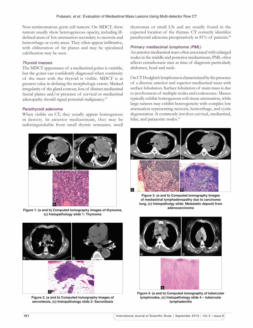
Pulasani, et al.: Evaluation of Mediastinal Mass Lesions Using Multi-detector Row CT
161 International Journal of Scientifi c Study | September 2015 | Vol 3 | Issue 6
Non-seminomatous germ cell tumors: On MDCT, these tumors usually show heterogeneous opacity, including ill-defi ned areas of low attenuation secondary to necrosis and hemorrhage or cystic areas. They often appear infi ltrative, with obliteration of fat planes and may be spiculated calcifi cation may be seen.
Thyroid massesThe MDCT appearance of a mediastinal goiter is variable, but the goiter can confi dently diagnosed when continuity of the mass with the thyroid is visible. MDCT is at greatest value in defi ning the morphologic extent. Marked irregularity of the gland contour, loss of distinct mediastinal fascial planes and/or presence of cervical or mediastinal adenopathy should signal potential malignancy.19
Parathyroid adenomaWhen visible on CT, they usually appear homogenous in density. In anterior mediastinum, they may be indistinguishable from small thymic remnants, small
thymomas or small LN and are usually found in the expected location of the thymus. CT correctly identifi es parathyroid adenoma preoperatively in 81% of patients.20
Primary mediastinal lymphoma (PML)An anterior mediastinal mass often associated with enlarged nodes in the middle and posterior mediastinum, PML often affects extrathoracic sites at time of diagnosis particularly abdomen, head and neck.
On CT Hodgkin’s lymphoma is characterized by the presence of a discrete anterior and superior mediastinal mass with surface lobulation. Surface lobulation of main mass is due to involvement of multiple nodes and coalescence. Masses typically exhibit homogenous soft tissue attenuation, while large tumors may exhibit heterogeneity with complex low attenuation representing necrosis, hemorrhage, and cystic degeneration. It commonly involves cervical, mediastinal, hilar, and paraaortic nodes.21
Figure 1: (a and b) Computed tomography images of thymoma, (c) histopathology slide 1: Thymoma
c
ba
Figure 3: (a and b) Computed tomography Images of mediastinal lymphadenopathy due to carcinoma
lung, (c) histopatholgy slide: Metastatic deposit from adenocarcinoma
a
dc
b
Figure 2: (a and b) Computed tomography Images of sarcoidosis, (c) histopathology slide 2: Sarcoidosis
c
ba
Figure 4: (a and b) Computed tomography of tubercular lymphnodes, (c) histopathology slide 4 – tubercular
lymphadenitis
b
c
a

Pulasani, et al.: Evaluation of Mediastinal Mass Lesions Using Multi-detector Row CT
162International Journal of Scientifi c Study | September 2015 | Vol 3 | Issue 6
Non-Hodgkin’s lymphoma comprises of mediastinal large B-cell lymphoma and lymphoblastic lymphoma and is more common in children than Hodgkin’s lymphoma. Large cell lymphomas are typically confi ned to the mediastinum and contiguous nodal areas initially without showing extrathoracic disease at presentation. It may present with hematogenous spread to kidney, liver, ovary, adrenal gland, GI tract, and central nervous system during disease progression or at recurrence. CT demonstrates mediastinal mass without surface lobulation, often associated with vascular involvement and pleural or pericardial effusion. Lymphoblastic lymphoma is characterized by mass without surface lobulation involving vascular structures often associated with pleural and pericardial effusion, systemic nodal involvement including cervical, axillary, para aortic, mesenteric and inguinal groups and by hepatomegaly and splenomegaly.22
SarcoidosisIn the order of decreasing frequency, paratracheal, aortopulmonary, subcarinal, and prevascular LN are commonly involved.23 LN shows dense or stippled or egg shell calcifi cation and rarely enhance or appear necrotic (Figure 2).
TBThe enlarged LN usually shows central area of low attenuation on MDCT with peripheral enhancement (Figure 4).
Pleuropericardial cystMDCT shows thin walled unilocular water density (0-20 HU) cystic structure. Wall may calcify. Majority of them arises in the anterior cardiophrenic angle, more so on the right side.23
Bronchogenic cystMDCT typically shows sharply marginated thin walled mediastinal mass of homogenous soft tissue or water attenuation. Rarely, calcification of the cyst wall is present. When dense, bronchogenic cysts may be diffi cult to distinguish from solid lesions. An important clue can be their lack of enhancement following contrast administration.24
Esophageal duplication cystsOn MDCT indistinguishable from bronchogenic cyst except for the location.
Neuroenteric cystsMDCT appearance is same as that of other duplication cyst, but the presence of vertebral abnormality points to the diagnosis, vertebral anomalies are present in half of the cases.
LymphangiomasMDCT usually shows a smooth, lobulated mass, which may mold to or envelop, rather than displace, the adjacent mediastinal structures. They are either unilocular or multilocular with near water density. Calcifi cation is rare. Thin enhancing septations within the mass may be seen.25
Neurogenic tumorsAt CT, many neural tumors have mixed density, including low attenuation region, on non-contrast enhanced CT Schwannoma often demonstrate lower attenuation than skeletal muscle because of their high lipid content, interstitial fl uid and areas of cystic degeneration. Neurofi bromas are often more homogenous and show higher attenuation than schwannomas because they have fewer of these histologic features. These lesions are heterogeneously enhancing following contrast administration.
Plexiform Neurofi bromas are seen in association with neurofi bromatosis-1, on CT demonstrates low attenuation infiltrative masses along the mediastinal nerves and sympathetic chain.
Mediastinal paragangliomasRounded soft tissue masses which are usually extremely vascular and therefore enhance brightly Paravertebral lesions.
Tuberculous paravertebral lesionsCT scan is excellent for visualization of endplate destruction, fragmentation of the vertebrae, and paravertebral calcifi cations. Infl ammatory collections and masses are best seen after the contrast administration. Small necrotic foci are recognized by CT scans but are diffi cult to fi nd in the conventional radiographs. Extension into the
Figure 5: (a and b) Computed tomography images of germ cell tumor with calcifi cations, (c) histopathology slide 5 - germ cell
tumor
c
ba

Pulasani, et al.: Evaluation of Mediastinal Mass Lesions Using Multi-detector Row CT
163 International Journal of Scientifi c Study | September 2015 | Vol 3 | Issue 6
canal of epidural abscesses and bony fragments are well demonstrated on axial CT images. MDCT is also used for guiding percutaneous biopsy and postdrainage follow-up.
Extramedullary hemopoietic tissueExtramedullary hematopoietic tissue typically produces one or smoother, lobular, spherical masses in the paravertebral gutter, usually in the lower thorax, often they are bilateral and symmetrical. Extramedullary hemopoietic tissues are low-density masses due to fat content.
Esophageal carcinomaNarrowing of the esophageal lumen or dilatation caused by obstruction. Thickening of the esophageal wall, either symmetric or asymmetric. Loss of periesophageal fat planes, with or without evidence of invasion of surrounding organs.
CONCLUSION
MDCT plays a significant role in the assessment of various mediastinal pathology with an accuracy of 92% on the whole, 97% and 84% for benign and malignant lesions, respectively, MDCT is a promising 3D imaging tool which allows substantial anatomical volumes to be routinely covered with isotopic sub-millimeter spatial resolution highly useful for the investigation of mediastinal masses. MDCT defi nitely has a major role to play in the evaluation of a mediastinal mass lesions regarding their characterization and differentiating between benign and malignant lesions.
REFERENCES
1. Webb WR. Advances in computed tomography of the thorax. Radiol Clin North Am 1983;21:723-39.
2. Protopapas Z, Westcott JL. Transthoracic hilar and mediastinal biopsy. Radiol Clin North Am 2000;38:281-91.
3. Benjamin SP, McCormack LJ, Effl er DB, Groves LK. Primary tumors of the mediastinum. Chest 1972;62:297-303.
4. Armstrong P. Mediastinal and hilar disorders. In: Armstrong P, Wilson AG, Dee P, Hansell OM, editors. Imaging of Diseases of the Chest. London: Mosby; 2000. p. 789-892.
5. Sones PJ Jr, Torres WE, Colvin RS, Meier WL, Sprawls P, Rogers JV Jr. Effectiveness of CT in evaluating intrathoracic masses. AJR Am J Roentgenol 1982;139:469-75.
6. Wychulis AR, Payne WS, Clagett OT, Woolner LB. Surgical treatment of mediastinal tumors: A 40 year experience. J Thorac Cardiovasc Surg 1971;62:379-92.
7. Baron RL, Levitt RG, Sagel SS, Stanley RJ. Computed tomography in the evaluation of mediastinal widening. Radiology 1981;138:107-13.
8. Rosado-de-Christenson ML, Pugatch RD, Moran CA, Galobardes J. Thymolipoma: Analysis of 27 cases. name Am J Respir Crit Care Med 2001;357:1191-4.
9. Im JG, Itoh H, Shim YS, Lee JH, Ahn J, Han MC, et al. Pulmonary tuberculosis: CT fi ndings – early active disease and sequential change with anti-tuberculous therapy. Radiology 1993;186:653-60.
10. Müller NL, Mawson JB, Mathieson JR, Abboud R, Ostrow DN, Champion P. Sarcoidosis: Correlation of extent of disease at CT with clinical, functional, and radiographic fi ndings. Radiology 1989;171:613-8.
11. Cohen AJ, Thompson L, Edwards FH, Bellamy RF. Primary cysts and tumors of the mediastinum. Ann Thorac Surg 1991;51:378-84.
12. Felson B, editor. The mediastinum. Chest Roentgenology. Ch. 11. Philadelphia: W.B. Saunders; 2002. p. 389-420.
13. Chen JL, Weisbrod GL, Herman SJ. Computed tomography and pathologic correlations of thymic lesions. J Thorac Imaging 1988;3:61-5.
14. Naidich DP, Webb WR, Muller NL, Zerhouni EA, Seigelmann SS. Mediastinum. In: Naidich DP, Muller NL, Zerhouni EA, Webb WR, Krinsky GA, editors. Computed Tomography and Magnetic Resonance of the Thorax. 3rd ed., Ch. 2. Philadelphia: Lippincott Williams and Wilkins; 1999. p. 38-160.
15. Prasad A, Chauhan BK. Computerized tomographic evaluation of mediastinal lesions – Pictorial assay. Indian J Radiol Imag 2001;11:65-70.
16. Armstrong P, Padley S. The normal chest. Grainger and Allison’s Diagnostic Radiology: A Textbook of Medical Imaging. 4th ed., Vol. 1, Ch. 14. London: Churchill Livingstone; 2001. p. 294-6.
17. Hartmann CA, Roth C, Minck C, Niedobitek G. Thymic carcinoma. Report of fi ve cases and review of the literature. J Cancer Res Clin Oncol 1990;116:69-82.
18. Baron RL, Lee JK, Sagel SS, Levitt RG. Computed tomography of the abnormal thymus. Radiology 1982;142:127-34.
19. Armstrong R, Padley SP. The mediastinum. In: Grainger RG, Allison D, Adam A, Dixon AK, editors. Diagnostic Radiology. 4th ed., Vol. 1, Ch. 17. London: Churchill Livingstone; 2001. p. 353-76.
20. Cates JD, Thorsen MK, Lawson TL, Middleton WD, Foley WD, Wilson SD, et al. CT evaluation of parathyroid adenomas: Diagnostic criteria and pitfalls. J Comput Assist Tomogr 1988;12:626-9.
21. Lazzarino M, Orlandi E, Paulli M, Boveri E, Morra E, Brusamolino E, et al. Primary mediastinal B-cell lymphoma with sclerosis: An aggressive tumor with distinctive clinical and pathologic features. J Clin Oncol 1993;11:2306-13.
22. Tateishi U, Müller NL, Johkoh T, Onishi Y, Arai Y, Satake M, et al. Primary mediastinal lymphoma: Characteristic features of the various histological subtypes on CT. J Comput Assist Tomogr 2004;28:782-9.
23. Kuhlman JE, Fishman EK, Hamper UM, Knowles M, Siegelman SS. The computed tomographic spectrum of thoracic sarcoidosis. Radiographics 1989;9:449-66.
24. Aktogu S, Yuncu G, Halilçolar H, Ermete S, Buduneli T. Bronchogenic cysts: Clinicopathological presentation and treatment. Eur Respir J 1996;9:2017-21.
25. Jeung MY, Gasser B, Gangi A, Bogorin A, Charneau D, Wihlm JM, et al. Imaging of cystic masses of the mediastinum. Radiographics 2002;22:S79-93.
How to cite this article: Pulasani K, Narayanaswamy I, Ramprakash HV. Evaluation of Mediastinal Mass Lesions Using Multi-detector Row Computed Tomography and Correlation with Histopathological Diagnosis. Int J Sci Stud 2015;3(6):156-163.
Source of Support: Nil, Confl ict of Interest: None declared.

164International Journal of Scientifi c Study | September 2015 | Vol 3 | Issue 6
Correlation of Magnetic Resonance Imaging and Bone Scintigraphy in Stress Injuries of Lower Extremities BonesSuresh A1, Arti Chaturvedi2
1Associate Professor, Department of Radiodiagosis, Vydehi Institute of Medical Sciences Bengaluru, Karnataka, India. 2Professor, Department of Radiodiagosis, Command Hospital Air Force, Bengaluru, Karnataka, India
trained manpower and fi nancial loss to the organization. It also affects the morale of the recruit and trained manpower which is unquantifi able.2
Stress fractures or fatigue fractures were originally reported by Breithaupt, Military Surgeon in Prussian army recruits in 1855. He described a syndrome of painful swollen feet amongst soldiers after long marches as “March fracture” the stress fractures in the metatarsals.2 Using conventional radiography, these signs and symptoms were fi rst reported as stress fractures in 1897. Stress fractures are most common noted in the lower extremities, these injuries also occur in the upper extremities, ribs, spine, and pelvis.3
Clinical evaluation of stress fractures includes a thorough history, a physical examination, and imaging studies. The recent advancement of musculoskeletal imaging techniques has aided the evaluation and diagnosis of stress-related
INTRODUCTION
Stress fracture is a common disability amongst Armed forces personnel especially in military recruits due to high standard of training imparted in the various military regimental training centers and academies.1
It is a frequent cause of discontinuity in training. It results in loss of training time to the recruit, loss of man-hours,
Original Article
Abstract
Introduction: Stress fracture of foot and long bones of leg is a common disability. The problem of “stress fracture” is clinically signifi cant amongst armed forces and athletes due to the high standard of training imparted in the various military regimental training centers and academies. This study was undertaken to compare fi ndings of scintigraphy and magnetic resonance imaging (MRI) in early detection of stress injuries of the bone when radiography fi ndings were normal or inconclusive.
Purpose: We undertook a prospective study of symptomatic patients who were referred to Department of Radiology, Command Hospital Air Force, Bengaluru, Karnataka, India, from various military units with history of prolonged exertion and clinical suspicion of stress injury.
Methodology: In this prospective study, 50 consecutive patients with suspicion of stress injuries of bone and with negative radiographs were selected for the study from May 2006 to June 2009. Scintigraphy and MRI were performed on the same day or within 4 days and the fi ndings were compared for correlation accuracy of MRI and scintigraphy.
Results: In all patients, both scintigraphy and MRI indicated stress injury of bone. On Evaluation of MRI for grades of bone stress injury in relation to bone scintigraphy fi ndings. There is good agreement between MRI and bone scintigraphy and with a strong P value (P < 0.001). Compared with scintigraphy, MRI showed more diagnostic information, such as fracture line, periosteal and soft tissue edema.
Conclusion: From these observations, we conclude that MRI provides more diagnostic information than scintigraphy and is recommended as the diagnostic and assessment tool in early stages of stress injury of bone.
Key words: Bone scintigraphy, Magnetic resonance imaging, Radiography, Stress fracture
Access this article online
www.ijss-sn.com
Month of Submission : 08-2015Month of Peer Review : 09-2015Month of Acceptance : 09-2015Month of Publishing : 09-2015
Corresponding Author: Dr. Suresh A, Department of Radiodiagnosis, Vydehi Institute of Medical Sciences and Research Centre, #82 EPIP Area, White Field, Bengaluru - 560 066. Phone: +91-9980956851. E-mail: [email protected]
DOI: 10.17354/ijss/2015/415

Suresh and Chaturvedi: Magnetic Resonance Imaging & Bone Scintigraphy
165 International Journal of Scientifi c Study | September 2015 | Vol 3 | Issue 6
injuries. Standard management of stress fractures consists of pain-free crutch ambulation for lower extremity stress fractures followed by a gradual return to weight-bearing activity culminating in running.4
PathophysiologyThe stress fractures are caused by continuous and repetitive muscular pulling on the normal bone, resulting in a disruption of the periostium and the underlying bone, at the origin or insertion of a given muscle.5 Stress fractures are in a very real sense a microfracture secondary to intense muscle contraction.6 Unaccustomed prolonged and excessive physical activity that causes resorption of bone in excess of repair and bone formation in certain regions can also result in stress fractures.
Overuse or fatigue stress fracturesIt is clinically described in military recruits and runners in whom normal bone is exposed to repeated abnormal stress.6
Types of overuse stress fracture are compression type and distraction type. This is subdivided as transverse, longitudinal and oblique. The transverse fracture is more dangerous and this type is known to cause a complete fracture if not diagnosed at an early stage and properly treated. The common locations of stress fractures are the tibia, fi bula, metatarsals, calcaneum, and navicular bones. It can also occur at rare sites such as pubic-ramus, pubic symphysis, spine, femoral neck, femoral diaphysis, distal shaft of humerus and proximal ulna.7,8
The main modality for investigating of stress fractures at the initial stage is the plain X-ray which is easily available at all primary health care centers. Sometimes, the plain radiographic fi ndings of stress fracture are diffi cult to interpret and may even be normal in early stages. Hence, there is a high possibility of missing early stress fractures which can further lead to complete bone fracture if the part is not rested, and the patient continues his rigorous training activity. In recent years, newer techniques such as scintigraphy and magnetic resonance imaging (MRI) enable for timely and accurate diagnosis and treatment of stress fractures, thus preventing signifi cant morbidity.9
This study was undertaken to compare the effi cacy of bone scintigraphy and MRI in the diagnosis of stress injuries of lower limb bones in those symptomatic patients whose initial plain radiographs were negative for stress injuries and recommend the best imaging modality for early detection of stress injuries of bone.
METHODOLOGY
The study was conducted on 50 patients of suspected stress injuries of lower limb bones, in the age group of
18-32 years who were referred to Radiology Department, Command Hospital (Air force), Bengaluru and whose initial plain radiographs were negative for stress injuries. The study was conducted from November 2006 to June 2009.
Inclusion criteria included apparently healthy individuals belonging to the armed forces presenting with history of prolonged repetitive exertion and reporting with pain. Exclusion criteria included acute direct trauma to the bones and metabolic bone diseases. Patients with negative X-rays were subjected to MRI and bone scan according to the following protocols:
MRI equipment used for study was Siemens Magnetom Avanto 1.5 tesla. The following imaging protocol was followed fast spin-echo (SE) short-tau inversion-recovery (STIR) (TR, 2400-3600; TE, 20-40; inversion time, 80-100 m) T1- weighted (T1W) SE imaging (repetition time [TR], 500 m; echo time [TE], 25 m) and T2-weighted (T2W) SE imaging (TR, 3000; TE, 100) using the above sequences coronal images of the effected region were obtain after application of surface coil.
MRIStudy was evaluated for: (a) Marrow changes of edema/contusion, (b) cortical/periosteal changes (Table 1).10
ScintigraphyThe patient was injected with 20 mCi of 99mTc-methyl diphosphonate intravenously. Anterior and posterior whole body images were acquired using dual head Siemens E-cam Gamma Camera 3 h after injection. If abnormalities seen in whole body image require detailed evaluation, further spot views of particular areas were taken.
Scintigraphy study was evaluated for: Increase uptake of tracer at the involved bone (Table 2).11
Comparison of scintigraphy and MRI fi ndings and gradings were done with Chi-square test and Fisher Exact Test.
Statistical MethodsDescriptive statistical analysis has been carried out in the present study. Chi-square/Fisher exact test has been used to fi nd the signifi cance of correlation of examination and results. Kappa Statistic has been used to fi nd the signifi cance of agreement between the bone MRI and scintigraphy fi ndings. Diagnostic statistics viz. sensitivity, specifi city, positive predictive value, negative predictive values, accuracy was used to correlate the fi ndings of MRI with bone scintigraphy.
Kappa statistic for agreementInter-rater agreement statistic (Kappa) to evaluate the agreement between two classifications on ordinal or nominal scales (Cohen, 1960). Agreement is quantifi ed by

Suresh and Chaturvedi: Magnetic Resonance Imaging & Bone Scintigraphy
166International Journal of Scientifi c Study | September 2015 | Vol 3 | Issue 6
the Kappa (K) or weighted Kappa (Kw) statistic: K is 1 when there is perfect agreement between the classifi cation system; K is 0 when there is no agreement better than chance; K is negative when agreement is worse than chance.
Value of K/strength of agreement: P < 0.20 - Poor, P = 0.21-0.40 - Fair, P = 0.41-0.60 - moderate, P = 0.61-0.80 - Good, and P = 0.81-1.00 - Very good.
RESULTS
In this prospective study of 50 patients, the age distribution of patients studied (Table 3). The age range varied from 18 to 32 years, the mean age was 21.68 years. The maximum number of patients was in the age group of 21-25 years constituting 52%. The least number of patients who developed stress injuries were more than 25 years old.
Duration of exertion in days of patients studied (Table 4). The average duration for stress injuries to develop during the training period was 28.1 days. The maximum number of patients to develop stress injuries was seen from 11 to 20 days who were subjected to un acclimatized training. The least number of patients who developed stress injuries had undergone more number of gradual acclimatized training days.
Studying the bones involved in patients (Table 5). The common bone involved was tibia. The right tibia was involved in 33 patients, followed by left tibia in 11 patients, femur in one patient (2%) and metatarsal in one patient (2%). The most common part of the tibia to be involved was proximal thirds of shaft.
On studying the region of fracture involved in bones (Table 6). The most common part of the tibia to be involved was proximal thirds of shaft. This pattern of injuries was observed more in recruits who underwent running and cross county pattern of training.
Scinigraphic results total patients - 50, Positive - 46, Negative - 04.
Additional fi ndings - tracer up take was seen at asymptomatic sites in nine cases.
MRI results total patients - 50, Positive - 48, Negative - 02.
Additional fi ndings - fracture line was seen in two cases
On correlating the fi ndings of bone scintigraphy and MRI (Table 7). There is good agreement between bone
Table 3: Age distribution of patients studiedAge in years n (%)18-20 20 (40.0)21-25 26 (52.0)>25 4 (8.0)Total 50 (100.0)Mean±SD 21.68±3.96SD: Standard deviation
Table 4: Duration of exertion in days of patients studiedDuration of exertion in days n (%)Up to 10 10 (20.0)11-20 22 (44.0)21-30 7 (14.0)31-50 4 (8.0)51-80 5 (10.0)>80 2 (4.0)Total 50 (100.0)Mean±SD 28.16±28.87SD: Standard deviation
Table 5: Bone involvement of patients studiedBone involvement n (%)Left femur 1 (2.0)Left tibia 15 (30.0)Right tibia 33 (66.0)Metatarsal foot 1 (2.0)Total 50 (100.0)
Table 6: Region of fracture of patients studiedRegion n (%)Upper third tibia 22 (44.0)Middle third tibia 18 (36.0)Lower third tibia 8 (16.0)Neck of femur 1 (2.0)Meta tarsal 1 (2.0)Total 50 (100.0)
Table 1: Grading of stress fracture of bone on MRIGrade 0 NormalGrade 1 Positive on STIRGrade 2 Positive on STIR plus positive on T2WIGrade 3 Positive on T1WI and T2WI, but without cortical breakGrade 4 Positive on T1WI and T2WI fracture lineSTIR: Short-tau inversion recovery, T1W: T1-weighted, T2W:T2-weighted
Table 2: Grading of stress fracture of bone on scintigraphyGrade 0 NormalGrade 1 Irregular uptake and/or a poorly defi ned area of increased
activityGrade 2 Similar to Grade 1, but more intense yet still poorly defi nedGrade 3 Sharply marginated area of increased activity, usually focal
or uniform in shapeGrade 4 Similar grade 3, but more intense uptake

Suresh and Chaturvedi: Magnetic Resonance Imaging & Bone Scintigraphy
167 International Journal of Scientifi c Study | September 2015 | Vol 3 | Issue 6
scintigraphy and MRI with kappa coeffi cient of 0.699 (P < 0.001).
On evaluation of MRI for grades of bone stress injury in relation to Bone scintigraphy fi ndings. (Table 8) There is good agreement between MRI and bone scintigraphy and with a strong P value (P < 0.001).
DISCUSSION
The stress fractures can be diagnosed by following imaging modalities. Plain radiography, computed tomography (CT), ultrasound, skeletal scintigraphy, and MRI.
RadiographyStress fracture of the bone can be effectively diagnosed on plain radiographs by taking two orthogonal views of the involved part to demonstrate the bony injury. In cortical bone, stress fractures are diagnosed based on the presence of endosteal reaction, periosteal callus without a fracture line or circumferential periosteal reaction with a fracture line through one cortex, sometimes frank fracture may be present. Generally for the periosteal reaction to develop it takes more than 3 weeks preceding the injury. In cancellous bone, stress fractures are poorly visible but sometimes may be appreciated as fl ake like patches of new bone formation 2-3 weeks after the onset of pain. The mineralized fracture site appears as a focal linear area of sclerosis oriented perpendicular to the trabeculae While the location and nature of a fracture are usually readily demonstrated on radiographs, the status of the adjacent soft tissue is usually very diffi cult to assess. The conventional radiographs have a sensitivity of 15-35% in early fractures, which increases to 30-70% during later exams because of overt bone reaction.12 Even though sensitivity of the conventional radiographs is very low, plain X-ray examination is mandatory as a fi rst imaging study to detect an overt fracture or rule out differentials such as infection or malignancy. In the present study, plain X-rays were used as screening modality to exclude patients with in stress injury of bone.
CTWith the advent of multislice CT scanners, helical data acquisition is fi ve times faster than sequential CT imaging.
Using thin collimation with multiplanar reformats, bony and soft tissue injury can be easily demonstrated. In bone stress injuries, CT fi ndings show cortical breach with surrounding endosteal and periosteal sclerosis however early marrow changes can be missed Hence, the role of CT is mainly limited to differential diagnosis. CT plays an important role in diagnosing longitudinal stress fracture of the tibia, which is diffi cult to diagnose on plain radiographs. Fractures involving regions of complex anatomy such as spine and pelvis can be conveniently demonstrated on CT. The disadvantage of CT is that and it involves excessive radiation exposure, In view of its limitations this modality was not included in our study.
Ultrasound (USG)Evaluation of musculoskeletal injuries can be done using, higher resolution linear transducers of (7-12 MHz). Detail evaluation of the superfi cial structures such as Tendon, ligaments muscles and fractures can be made. Although USG is unable to penetrate cortical bone, the high refl ective and very large acoustic impedance make it possible to assess the bone contour and cortical fracture.13 USG by its ability to visualize developing callus before radiographic changes are evident and can also be useful in assessing fractures after initial radiographs. USG avoids excessive radiation exposure is relatively inexpensive and give accurate measurements about the process of calcifi cation.14
Skeletal ScintigraphyBone scintigraphy is an imaging technique used in the detection of stress injury of bones since 1970’s as described by Geslien et al., and Wilcox et al., as it can demonstrate subtle changes in bone metabolism even
Table 7: Correlation of fi ndings of bone scintigraphy and MRIBone scintigraphy↓
MRI (%) Total (%)Grade 0 Grade 1 Grade 2 Grade 3 Grade 4
Grade 0 0 4 (8.0) - - - 4 (8.0)Grade 1 1 (2.0) 15 (30.0) - - - 16 (32.0)Grade 2 1 (2.0) 1 (2.0) 13 (26.0) - - 15 (30.0)Grade 3 - 2 (4.0) 1 (2.0) 9 (18.0) 1 (2.0) 13 (26.0)Grade 4 - - - - 2 (4.0) 2 (4.0)Total 2 (4.0) 22 (44.0) 14 (28.0) 9 (18.0) 3 (6.0) 50 (100)Strongly signifi cant kappa co-effi cient of 0.699 (P<0.001)
Table 8: Evaluation of MRI in relation to bone scintigraphy fi ndingsMRI grade Sensitivity Specifi city PPV NPV Accuracy P valueGrade I 93.8 79.4 68.2 96.4 84.0 <0.001**Grade II 86.7 97.1 92.9 94.4 94.0 <0.001**Grade III 69.2 100.0 100.0 90.2 92.0 <0.001**Grade IV 100.0 97.9 66.7 100.0 98.0 <0.001****Strongly signifi cant P≤0.01, MRI: Magnetic resonance imaging, PPV: Positive predictive value, NPV: Negative predictive values

Suresh and Chaturvedi: Magnetic Resonance Imaging & Bone Scintigraphy
168International Journal of Scientifi c Study | September 2015 | Vol 3 | Issue 6
before seen on plain radiography. In this technique, the imaging is based on the detection of radiation emitted from a 99mTc - diphosphonate a radioactive radiopharmaceutical inside the patient as the phosphate compound gets incorporated into the new bone at the site of injury. Hod et al.: In a study conducted on female recruits of Israeli defense force has reported a sensitivity of 100% for stress fractures and bone scans of shin splint lesions occasionally show linear longitudinal uptake.15 However, bone scintigraphic fi ndings of stress reaction are nonspecifi c. Some authors reported that 20-40% of the lesions seen on scintigraphs were asymptomatic.16
Although bone scintigraphy was considered to be the gold standard, false-negative bone scintigraphs have been reported by Milgrom et al.: Keene and Lash and Sterling et al.16-18 In our study, we a observed four patients to be positive on MRI, which were not picked by scintigraphic imaging. The limitation of bone scans is that a positive scan may not be seen for 24 h following injury despite its high sensitivity, the specifi city of bone scintigraphy is inferior to that of MRI because it cannot quantify the fi ner details of the lesion sometimes pathological conditions such as a tumors, infections, bone infarctions, or periostitis can also produce a positive fi nding. The scintigraphy, has a radiation risk were the radiation dose is equal to a dose of 2 years of background radiation.
MRIThe MRI on the human body was fi rst performed in 1977 by Raymond Damadian. This landmark discovery has revolutionized the fi eld of imaging. This breakthrough discovery in imaging has made an extraordinary difference in demonstrating the contrast between MR signals from normal and abnormal tissues. It can also demonstrate the nature and extent of injuries. Without moving the patient image acquisition with a variety of pulse sequences can be used for characterizing tissues. MRI is sensitive in detecting changes in bone marrow at all fractures sites even <24 h. Fracture on MRI is described on the basis of SE images emphasizing on T1 and T2 contrast. As compared to SE images STIR images provide superior contrast between normal and abnormal marrow. The MRI features on T1W SE images consisted of irregular intramedullary zones of hypointensity described as a band-like area of low signal intensity. On STIR images a corresponding zone of hyperintensity extending to the outer cortical margin can be seen. MRI using T2W SE and STIR sequences can consistently demonstrate prominent signal abnormalities at fracture sites including those in which radiographic signs are subtle. MRI is as sensitive, as but more specifi c than scintigraphy.19 In addition to changes in bone, it gives information about the surrounding soft tissues in all three dimensions. T2W and STIR- sequences are useful for detecting edema of cancellous bone, periosteum, and bone
marrow edema; however is this is a nonspecifi c fi nding and should be interpreted in correlation with clinical history. A fracture can be seen as lower signal intensity in the middle of the edema, but it is better seen as a low-density signal line involving the cortex on T1-images.
Ishibashi et al.: In a study conducted on 31 patients comparing scintigraphy and MRI for stress injuries of bone have reported a good correlation of Grades of scintigraphy and MRI as compared to scintigraphy MRI showed more diagnostic information.20 In the present study, we also observed similar results in the patients studied. An illustration of few such representative cases, images for Case 1 are given as (Figure 1a-d) wherein bone scan revealed intense well defi ned areas of increase tracer uptake in middle thirds shaft of right femur, hypointense on T1W and T2W suggesting good correlation between bone scan and bone scintigraphy for Grade I stress fracture and the corresponding area appeared as hyper intense signal on STIR. In Case 2 images are given as (Figures 2a-d) wherein bone scan revealed sharply marginated area of increased signal intensity in upper third shaft of right femur and in middle third shaft of left femur. On MRI, STIR and T2W images, the corresponding areas appeared as hyperintense signal intensities, on T1W coronal revealed a hypointense band with cortical break on right side suggesting grade IV stress on right side and Grade II fracture on left side. Though there is a good correlation between bone scan and bone scintigraphy where the tracer uptake was clearly seen, MRI provides detailed information regarding marrow edema, cortical break and surrounding soft tissues.
Figure 1: (a) Bone scan revealed intense well defi ned area of increase tracer up take in middle thirds shaftof right femur, (b) T1-weighted coronal magnetic resonance imaging (MRI)
fi gure reveled normal signal intensities, (c) T2-weighted coronal MRI fi gure revealed normal signal intensities, and (d) short-tau
inversion-recovery coronal MRI fi gures revealed focal area of hyper intense intensity in middle third shaft of right femur.
Diagnosed as case of Grade I stress fracture
dc
ba

Suresh and Chaturvedi: Magnetic Resonance Imaging & Bone Scintigraphy
169 International Journal of Scientifi c Study | September 2015 | Vol 3 | Issue 6
CONCLUSION
In our study, we observed a good correlation of fi ndings between MRI and skeletal scintigraphy for stress injuries of bone with Kappa value of 0.699 (P < 0.001). On evaluation of MRI in relation to bone scintigraphy fi ndings for grading of stress injuries, MRI has comparable sensitivity to scintigraphy for detection of stress fractures and MRI has additional advantage of depicting the surrounding tissue infl ammatory process, as well as providing greater anatomical details. Hence, we recommend MRI as the initial imaging modality in early detection of stress injuries of bone.
ACKNOWLEDGMENTS
Dr. J. P. Singh Ex Head and Professor Department of Radiodiagnosis Command hospital Air force Bangalore for his valuable guidance in conducting the study and Dr. Abishek Balachandran for preparation of this manuscript.
All the Staff, nuclear medicine department Command hospital Air force Bengaluru.
REFERENCES
1. Behera RC, Sawal P, Bajpai RK, Jayaprakash CA, Roy S. Stress fractures among military recruits and Navy cadets. MJAFI 1990;46:24-7.
2. Basappa KK, Chandra S, Ganguly SK. Stress fracture among recruits and trained soldiers. MJAFI 1992;48:288-90.
3. Daffner RH, Pavlov H. Stress fractures: Current concepts. AJR Am J Roentgenol 1992;159:245-52.
4. Bennell KL, Malcolm SA, Thomas SA, Reid SJ, Brukner PD, Ebeling PR, et al. Risk factors for stress fractures in track and fi eld athletes. A twelve-month prospective study. Am J Sports Med 1996;24:810-8.
5. Almeida SA, Williams KM, Shaffer RA, Brodine SK. Epidemiological patterns of musculoskeletal injuries and physical training. Med Sci Sports Exerc 1999;31:1176-82.
6. Milgrom C, Simkin A, Eldad A, Nyska M, Finestone A. Using bone’s adaptation ability to lower the incidence of stress fractures. Am J Sports Med 2000;28:245-51.
7. Raju KS, Sharma S, Yadav RC, Singh MV. An epidemiological study of stress fractures among fl ight cadets at air force academy. Indian J Aerosp Med 2005;49:48-53.
8. Kiuru MJ, Pihlajamaki HK, Ahovuo JA. Fatigue stress injuries of the pelvic bones and proximal femur: Evaluation with MR imaging. Eur Radiol 2003;13:605-11.
9. Berger FH, de Jonge MC, Maas M. Stress fractures in the lower extremity. The importance of increasing awareness amongst radiologists. Eur J Radiol 2007;62:16-26.
10. Arendt EA, Griffi ths HJ. The use of MR imaging in the assessment and clinical management of stress reactions of bone in high-performance athletes. Clin Sports Med 1997;16:291-306.
11. Fredericson M, Bergman AG, Hoffman KL, Dillingham MS. Tibial stress reaction in runners. Correlation of clinical symptoms and scintigraphy with a new magnetic resonance imaging grading system. Am J Sports Med 1995;23:472-81.
12. Armfi eld DR, Towers JD, Robertson DD. Radiographic and MR imaging of the athletic hip. Clin Sports Med 2006;25:211-39.
13. Tuite MJ, Desmet AA. MRI of selected sports injuries: Muscle tears, groin pain, and osteochondritis dissecans. Semin Ultrasound CT MR 1994;15:318-40.
14. Craig JG, Jacobson JA, Moed BR. Ultrasound of fracture and bone healing. Radiol Clin North Am 1999;37:737-51.
15. Hod N, Ashkenazi I, Levi Y, Fire G, Drori M, Cohen I, et al. Characteristics of skeletal stress fractures in female military recruits of the Israel defense forces on bone scintigraphy. Clin Nucl Med 2006;31:742-9.
16. Milgrom C, Chisin R, Giladi M, Stein M, Kashtan H, Margulies J, et al. Negative bone scans in impending tibial stress fractures. A report of three cases. Am J Sports Med 1984;12:488-91.
17. Keene JS, Lash EG. Negative bone scan in a femoral neck stress fracture. A case report. Am J Sports Med 1992;20:234-6.
18. Sterling JC, Webb RF Jr, Meyers MC, Calvo RD. False negative bone scan in a female runner. Med Sci Sports Exerc 1993;25:179-85.
19. Tyrrell PN, Davies AM. Magnetic resonance imaging appearances of fatigue fractures of the long bones of the lower limb. Br J Radiol 1994;67:332-8.
20. Ishibashi Y, Okamura Y, Otsuka H, Nishizawa K, Sasaki T, Toh S. Comparison of scintigraphy and magnetic resonance imaging for stress injuries of bone. Clin J Sport Med 2002;12:79-84.
How to cite this article: Suresh A, Chaturvedi A. Correlation of Magnetic Resonance Imaging and Bone Scintigraphy in Stress Injuries of Lower Extremities Bones. Int J Sci Stud 2015;3(6):164-169.
Source of Support: Nil, Confl ict of Interest: None declared.
Figure 2: (a) Bone scan revealed sharply marginated area of increased signal intensity in upper thirds shaft of right femur and also in middle thirds shaft of left femur, (b) T2-
weighted coronal magnetic resonance imaging (MRI) fi gures revealed a hypo intense band with cortical break in right
femur, (c) T2-weighted coronal MRI revealed hyper intense signal intensities in upper thirds shaft of femur on right side
and middle thirds shaft of femur on left side, and (d) short-tau inversion-recovery coronal MRI fi gures revealed hyper intense
signal intensities in the corresponding anatomical locations shaft of tibias. Diagnosed as Grade IV stress on right side and
Grade II on left side
dc
ba

170International Journal of Scientifi c Study | September 2015 | Vol 3 | Issue 6
Comparison of Latencies and Time to Stabilization of Pulse Oximeters at a Tertiary Health Care FacilityRyan Ward1, Punita Raheja2, Anupam Singh3
1Lead Respiratory Therapist, Department of Medicine, Benedictine Health Centre, Minneapolis, Minnesota, USA, 2Resident, Department of Surgery, Vasudeva Nursing Home, New Delhi, India, 3Senior Resident, Department of Cardiology, NDMC Medical College, Hindu Rao Hospital, New Delhi, India
assessment has increased with the greater availability of all-in-one portable devices.
In many clinical situations, the prompt availability of the result is as important as the value itself - for instance, in acute emergencies such as MI, anaphylactic shock, status asthmaticus or in ambulatory care when visits tend to be scheduled very frequently. In these cases, a shorter duration of time required to obtain the measurement is crucial.4-6
While pulse oximeters perform well in routine conditions, factors such as severe hypoxia, artifacts, latency to fi rst reading, and time to stabilization of readings adversely affect their utility in challenging patient conditions.6
Time to stabilization of the pulse oximeter and latency of readings is of critical importance in scenarios such as
INTRODUCTION
Fingertip pulse oximeters are often used as a surrogate for measuring tissue oxygenation. They have proved to be useful tools in different hospital set-ups and even in challenging and adverse set-ups such as prehospital intubations and critical care in intensive care unit.1-3 Use of fingertip pulse oximeters for efficient spot-check
Original Article
Abstract
Introduction: Fingertip pulse oximeters are commonly used as non-invasive modes of measuring oxygen saturation (SpO2) and heart rate (HR) in critical care units. In these cases, a shorter duration of time required obtaining the measurement and time to stabilization is crucial.
Objective: Our aim was to compare two new generation pulse oximeters with respect to latency in the fi rst reading and time to stabilization of SpO2 and HR.
Materials and Methods: This study was a prospective, comparative non-blinded, observational study of two fi ngertip pulse oximeters (Nonin Medical 9560 and CHOICEMMED MD300C1) in 20 in patients at Benedectine Health Center suffering from either one or more chronic diseases. SpO2 and HR readings were collected at the time of fi rst display, 30 s, 60 s and at the time of stabilization of the reading (no more than a 1% change in SpO2, <3 bpm change in HR). Statistical analysis was performed with SAS 9.2 software.
Results: A total of 20 subjects were monitored. The average time to fi rst reading across all fi ngers was signifi cantly longer with the CHOICEMMED device (10.3 ± 0.8 s) as compared to Nonin Medical (9.0 ± 0.8 s) pulse oximeter. The mean difference was 1.3 s (95% confi dence interval (CI), (0.05, 2.61), P = 0.04). The average time to stabilization of SpO2 across all fi ngers was also statistically signifi cantly longer in the CHOICEMMED (27.4 ± 2.2 s) than Nonin Medical pulse oximeter (11.4 ± 2.0 s). The mean difference was 15.9 s (95% CI (12.57. 19.29), P < 0.0001).
Conclusion: The pulse oximeter (Model 9560) by Nonin Medical showed shorter latency for fi rst reading and time to stabilization when compared to CHOICEMMED (MD300C1), which is a distinct advantage in emergency situations in acute patients. Further larger studies are needed to validate these fi ndings in different clinical settings.
Key words: Comparison, Hypoxia, Latency, Oximeter, Saturation
Access this article online
www.ijss-sn.com
Month of Submission : 08-2015Month of Peer Review : 09-2015Month of Acceptance : 09-2015Month of Publishing : 09-2015
Corresponding Author: Dr. Punita Raheja, D-43 Panchsheel Enclave, New Delhi - 110017, India. Phone: +91-9810975946/+91-8800593568. E-mail: [email protected]
DOI: 10.17354/ijss/2015/416

Ward, et al.: Comparison of Latencies and Time to Stabilization of Pulse Oximeters
171 International Journal of Scientifi c Study | September 2015 | Vol 3 | Issue 6
rapid-sequence intubation, pre-hospital intubation, and checking oxygenation status in sudden cardiac status, when any delay can be counterproductive to the patient’s prognosis.7-12 These parameters depend on sensitivity of signal acquisition of the pulse oximeter, signal averaging time of the oximeter, site of measurement, perfusion status, motion artefacts, arrhythmias, and hemoglobinopathies.8-10
Some studies have focused on this critical aspect. Macleod et al. showed that latency of response to hypoxia might be markedly different depending on hypothermia, site of measurement and hypoxic challenge.3 Pulse oximeters use signal averaging time to obtain nonfl uctuating measurement of oxygenation. Though signal averaging time in default mode is <10 s, it has been recommended that for fi nger pulse oximeters the time to stabilization and response to change in tissue oxygenation should be <30 s.3,4,7 However, many such studies have been based in hypoxia laboratories and may not be refl ective of their performance in clinical scenarios.
Aim of the present study was to compare the time to stabilization and time to fi rst reading of two pulse oximeters used at a tertiary Hospital center in challenging patient conditions, and infer about their performance under the same.
MATERIALS AND METHODS
DesignThis study was a prospective, non-randomized, non-controlled, observational study. The study was designed to include up to 20 subjects. Pulse oximetry was done on these patients in resting condition.
Subjects who met the inclusion/exclusion criteria were asked to participate as a volunteer in the study. Informed consent forms were made available to interested participants. A study representative from Benedictine Health Center of Minneapolis was available to answer any questions. When the study representative felt that the subject understood the purpose, procedures, benefi ts, risks, discomforts, and precautions of the study, the subject was asked if they wanted to participate in the study and if so, the subject was asked to sign the appropriate informed consent. Each subject received a copy of the signed informed consent.
Following receipt and documentation of the IRB-approved informed consent, subjects were considered enrolled in the study. The demographic and anthropometric questions were completed.
Subjects were seated in a private location and had a reference pulse oximeter placed on their right little fi nger
for the entire duration of the procedure. If other limitations or conditions existed, preventing placement on the little fi nger, the reference oximeter was placed on the left little fi nger or not placed on this subject and indicated on the chronic renal failures.
A video recorder was used to monitor the study. The recording window was limited to the subject’s hands. The following test procedure was used for each oximetry system on each digit:1. Apply the pulse oximetry system to the application site
(e.g., fi nger)2. Record the time to fi rst displayed measurement3. Record the time to stable measurement (no more than
a 1% change in oxygen saturation (SpO2), <3 bpm change in pulse rate, or change in pulse strength)
4. Record the SpO2 pulse rate, and pulse strength at fi rst measurement, 30 s, 60 s, and stable measurement
5. Remove the sensor6. Note special conditions during testing (e.g., imposed
motion, patient motion, and device malfunctions) by marking the event in the collection system.
The order of placement and order of systems was randomized using a blocked randomization scheme with randomly sized blocks. The same application site was not used twice in a row. Each oximetry system was placed on each applicable location at least once during the testing.
For the extended stability and extend motion readings, the following procedure was performed:1. Apply the test pulse oximetry system(s) to the
application site (s) (e.g., fi nger)2. Record the readings for a minimum of 10 min. Length
of recording did not exceed 30 min.
DevicesThe devices used in this study included pulse oximeters manufactured by Nonin Medical Model 9560 and CHOICEMMED MD300Cl.
The pulse oximeters used in this study were indicated for use in measuring, displaying, and storing functional SpO2 of arterial hemoglobin and pulse rate. Pulse oximetry sensors used in this study included fi ngertip pulse oximeters.
Instructions for installation and use of the devices, including any necessary storage and handling requirements, preparation for use, re-use requirements, pre-checks of safety and performance, and precautions to be taken after use were provided where appropriate.
Statistical CalculationStatistical analysis was performed with SAS 9.2 software (SAS Institute Inc., 2011. SAS System for Windows, Release

Ward, et al.: Comparison of Latencies and Time to Stabilization of Pulse Oximeters
172International Journal of Scientifi c Study | September 2015 | Vol 3 | Issue 6
9.2. SAS Institute Inc., Cary, NC, USA) using generalized estimating equations to account for the correlation between multiple readings on the three fi ngers of the same subject.
RESULTS
A total of 21 subjects were enrolled in this study between February 28, 2011 and September 22, 2011 at Benedictine Health Care Center. One patient withdrew consent prior to study participation, while remaining subjects participated. Subjects were enrolled from both white and non-white races. Subjects weighed an average 86.36 kg and were 172.5 cm tall. The demographic features are described in Table 1.
Characteristics of Pulse OximetersEach of the subjects at Benedictine Health Center underwent simultaneous placement of a Nonin Medical Model 9560 and CHOICEMMED MD300Cl on the index, middle and ring fi ngers. One device was placed on the particular fi nger (index, middle, or ring) on the left hand, and the other device was placed on the same fi nger of the contralateral hand. The order of the placement was assigned using a randomizations scheme such that no fi nger had a sensor placed twice in a row.
With each placement, data were collected by visual inspection of the oximeters. These data included SpO2 and heart rate (HR) at fi rst reading, 30 s, 60 s and stabilization. Time to stabilization and time to fi rst reading were also recorded.
The average time to fi rst reading across all fi ngers was signifi cantly longer with the CHOICEMMED device
(10.3 ± 0.8 s, mean ± standard error) as compared to Nonin Medical (9.0 ± 0.8 s, mean ± standard error) pulse oximeter. The mean difference was 1.3 s (95% confi dence interval (CI), (0.05, 2.61), P = 0.04). (Figure 1) The average time to stabilization of SpO2 across all fi ngers was also statistically signifi cantly longer in the CHOICEMMED (27.4 ± 2.2 s, mean ± standard error) pulse oximeter than Nonin Medical pulse oximeter (11.4 ± 2.0 s, mean ± standard error). The mean difference was 15.9 s (95% CI (12.57. 19.29), P < 0.0001). (Figure 2) There was no signifi cant difference between time of fi rst reading for HR (P = 0.09) and HR at stabilization (P = 0.37) between the two oximeters.
Study FlowNo follow-up was predicated or performed for this study. No adverse events were reported in this study. There were three protocol deviations. All three were deviations with respect to randomization order.
Figure 1: Comparative representation of time to fi rst reading in both pulse oximeters in seconds
Figure 2: Comparative representation of time to stabilization in both pulse oximeters in seconds
Table 1: Demographic variables of participants of the studyCharacteristics Benedictine Health
( n=20 subjects)Age 62±11.6Gender (%)
Male 14/20 (70)Female 6/20 (30)
RaceAmerican Indian 1/20Asian 0/20Black 4/20Pacifi c 0/20White 15/20
Skin colorVery light 4/20Light 7/20Intermediate light 5/20Dark 4/20
Height (cm) 172.5±12.5Weight (kg) 86.36±21.1
Ward, et al.: Comparison of Latencies and Time to Stabilization of Pulse Oximeters
173 International Journal of Scientifi c Study | September 2015 | Vol 3 | Issue 6
DISCUSSION
During the study performance of two pulse oximeters under challenging patient conditions was evaluated. While both of the pulse oximeters achieved time to fi rst reading and time to stabilization within 30 s, Nonin medical device outperformed CHOICEMMED device both in time to fi rst reading and time to stabilization of SpO2 by 1.3 s and 11.4 s respectively. These fi ndings demonstrate that there is variability in performance of the pulse oximetry devices and future clinical validation must be carried out in various settings to verify claims made by manufacturers.
The average time to fi rst reading across all fi ngers was signifi cantly longer with the CHOICEMMED device (10.3 ± 0.8 s) as compared to Nonin Medical (9.0 ± 0.8 s) pulse oximeter. The mean difference was 1.3 s (95% CI, (0.05, 2.61), P = 0.04). The average time to stabilization of SpO2 across all fi ngers was also statistically signifi cantly longer in the CHOICEMMED (27.4 ± 2.2 s) pulse oximeter than Nonin Medical pulse oximeter (11.4 ± 2.0 s). The fi ndings are in accordance with study of errors of pulse oximetry by Severinghaus et al. In a study by Macleod et al., it was seen that forehead and ear sensors (central location) had up to six times faster responses to change in ventilation than fi ngertip oximeter. However, our study focused on fi ngertip pulse oximeters alone, because of the prevalence in practice which can lead to wider generalization of results. This study is unique in comparing the time to stabilization and fi rst reading of Nonin Medical and CHOICEMMED pulse oximeter. While the Nonin device performed better than CHOICEMMED, we advocate testing these parameters in different challenging clinical settings.
Two major factors might affect the time to stabilization and time for fi rst reading of pulse oximeters, fi rst is proximity to large vessels. The probes which are central in location (forehead, ear, Nasal) and hence closer to great vessels consistently have shown faster response times.13-16 Signal averaging time also affects the time to stabilization of most pulse oximeters. Signal averaging time is a technique used by pulse oximeters to obtain nonfl uctuating measurements. Most new generation oximeters have signal average time <10 s in default states, though they can be calibrated to lesser times as less as 3 s.14,15 In fact American sleep medicine guidelines recommend signal averaging time <3 s for pulse rate >80 beats/min or higher.13 Faster response time of the Nonin device might be attributable its better handling of artefacts, low perfusion fi lter and lower signal averaging times of <3 s at even lower pulse rates of 60 beats/min. Hypothermia, vasoconstriction and low gestation age can also affect time to fi rst reading.15-17 However, our study was not aimed to detect performance of pulse oximetry in these situations. Similarly fi nger characteristics such
as deformity, nail pigmentation, and clubbing might also infl uence the response time. In our study, there was no signifi cant difference in stabilization times across various fi ngers in both groups of pulse oximeters.
Our study has many strengths, it focuses on a clinically important parameter, which can be of value in situation where emergency assessment of tissue oxygenation is vital to management of rapid sequence and crash intubation.11,12,18 We also performed these measurements in ‘real-world’ clinical settings as opposed to hypoxia laboratories. We compared two new generation pulse oximeters to verify their claims. While both of these pulse oximeters had time to stabilization <30 s, Nonin’s pulse oximeter performed signifi cantly better. Some weaknesses of the study were open blinded assessment of comparisons. We could not account for more challenging situations like hypothermia and poor perfusion.19,20 We hope these lacunae would be taken care of in future studies.
CONCLUSION
We were able to assess the ‘real world ‘ performance of two pulse oximeters and determine vital clinical parameters of time to stabilization and time to fi rst reading in both the groups. While time to stabilization was <30 s in both the groups. Nonin’s device was almost 2.5 times faster in time to stabilization. The Pulse oximeter (Model 9560) by Nonin Medical showed shorter latency for fi rst reading and time to stabilization when compared to CHOICEMMED (MD300C1), which is a distinct advantage in emergency situations in acute patients. Further, larger studies are needed to validate these findings in different clinical settings. We also recommend testing various pulse oximeters across different clinical settings and in other challenging conditions such as hypothermia, poor perfusion, sepsis, and with vasoconstrictors to enable wider generalization of clinical fi ndings.
REFERENCES
1. Sinex JE. Pulse oximetry: Principles and limitations. Am J Emerg Med 1999;17:59-66.
2. Mendelson Y. Pulse oximetry: Theory and applications for noninvasive monitoring. Clin Chem 1992;38:1601-7.
3. MacLeod DB, Cortinez LI, Keifer JC, Cameron D, Wright DR, White WD, et al. The desaturation response time of fi nger pulse oximeters during mild hypothermia. Anaesthesia 2005;60:65-71.
4. Yönt GH, Korhan EA, Khorshid L. Comparison of oxygen saturation values and measurement times by pulse oximetry in various parts of the body. Appl Nurs Res 2011;24:e39-43.
5. Valdez-Lowe C, Ghareeb SA, Artinian NT. Pulse oximetry in adults. AJN Am J Nurs 2009;109:52-9.
6. Severinghaus JW, Naifeh KH, Koh SO. Errors in 14 pulse oximeters during profound hypoxia. J Clin Monit 1989;5:72-81.
7. Rheineck-Leyssius AT, Kalkman CJ. Advanced pulse oximeter signal processing technology compared to simple averaging. I. Effect on frequency

Ward, et al.: Comparison of Latencies and Time to Stabilization of Pulse Oximeters
174International Journal of Scientifi c Study | September 2015 | Vol 3 | Issue 6
of alarms in the operating room. J Clin Anesth 1999;11:192-5.8. Tremper KK. A second generation technique for evaluating accuracy and
reliability of second generation pulse oximeters. J Clin Monit Comput 2000;16:473-4.
9. Vagedes J, Bialkowski A, Wiechers C, Poets CF, Dietz K. A conversion formula for comparing pulse oximeter desaturation rates obtained with different averaging times. PLoS One 2014;9:e87280.
10. Jose B, Lodha R, Kabra SK. Comparison of two new generation pulse oximeters with arterial oxygen saturation in critically ill children. Indian J Pediatr 2014;81:1297-301.
11. Aguilar SA, Davis DP. Latency of pulse oximetry signal with use of digital probes associated with inappropriate extubation during prehospital rapid sequence intubation in head injury patients: Case examples. J Emerg Med 2012;42:424-8.
12. Davis DP, Aguilar S, Sonnleitner C, Cohen M, Jennings M. Latency and loss of pulse oximetry signal with the use of digital probes during prehospital rapid-sequence intubation. Prehos Emerg Care 2011;15:18-22.
13. Epstein LJ1, Kristo D, Strollo PJ Jr, Friedman N, Malhotra A, Patil SP, et al.
Clinical guideline for the evaluation, management and long-term care of obstructive sleep apnea in adults. J Clin Sleep Med 2009;5:263-76.
14. Pedersen T, Nicholson A, Hovhannisyan K, Moller AM, Smith AF, Lewis SR. Pulse oximetry for perioperative monitoring. Cochrane Database Syst Rev 2014;3:CD002013.
15. Gandhi B, Rich W, Finer N. Time to achieve stable pulse oximetry values in VLBW infants in the delivery room. Resuscitation 2013;84:970-3.
16. Shah A, Shelley KH. Is pulse oximetry an essential tool or just another distraction? The role of the pulse oximeter in modern anesthesia care. J Clin Monit Comput 2013;27:235-42.
17. Chan ED, Chan MM, Chan MM. Pulse oximetry: Understanding its basic principles facilitates appreciation of its limitations. Respir Med 2013;107:789-99.
18. Barak-Shinar D, Amos Y, Bogan RK. Sleep disordered breathing analysis in a general population using standard pulse oximeter signals. Sleep Breath 2013;17:1109-15.
19. Kamat V. Pulse oximetry. Indian J Anaesth 2002;46:261-8.20. Jubran A. Pulse oximetry. Inten Care Med 2004;30:2017-20.
How to cite this article: Ward R, Raheja P, Singh A. Comparison of Latencies and Time to Stabilization of Pulse Oximeters at a Tertiary Health Care Facility. Int J Sci Stud 2015;3(6):170-174.
Source of Support: Nil, Confl ict of Interest: None declared.

175 International Journal of Scientifi c Study | September 2015 | Vol 3 | Issue 6
Burst Abdomen: A Post-operative MorbidityPradeep Soni1, Vibha Baghel Haripriya2, Anil Haripriya1, Vishnu Dutt3
1Associate Professor, Department of Surgery, Chhattisgarh Institute of Medical Sciences, Bilaspur, Chattisgarh, India, 2Assistant Professor, Department of Obstetrics and Gynaecology, Chhattisgarh Institute of Medical Sciences, Bilaspur, Chattisgarh, India, 3Professor and Dean, Department of Radio Diagnosis, Chhattisgarh Institute of Medical Sciences, Bilaspur, Chattisgarh, India
of different types of suture materials.8 A surgeon can perform a technically perfect operation in a patient, who is severely compromised by the disease process and still have a complication.
Various pre-operative factors which are predisposing to this unpleasant and tragic post-operative complication are studied.9 Because of high mortality rate due to burst abdomen in surgical operations. The management of these ranges from simple dressing to further closure of burst abdomen followed by a period of intensive care.10
The purpose of the present study was to assess the effi cacy of closure of laparotomy incisions and its different predisposing factors.
METHODS
The present study was conducted among 162 patients underwent laparotomy by midline incisions in the Department of General Surgery and Obstetrics and
INTRODUCTION
Burst abdomen is a severe post-operative complication experienced by Surgeons and Gynecologist, who do a signifi cant amount of surgery. The frequency as described in the international data ranged from 0.4% to 3.5%.1-3 and is also associated with a mortality rate in patients as high as 45%.4 While our country data stated still higher frequency of burst abdomen with overall rate of 4.8% and 6.6%.5,6
Burst abdomen is associated with several factors which increases its incidence.7 Efforts have been made to conquer this diffi culty with various innovations using
Original Article
Abstract
Introduction: Burst abdomen is a severe post-operative complication experienced by Surgeons and Gynecologist, who do a signifi cant amount of surgery. Burst abdomen usually occurs due to various predisposing factors which can be prevented to some extent by having knowledge regarding them.
Purpose: The purpose of this study was to assess the patients, who had undergone laparotomy and developed burst abdomen.
Methods: The study was among 162 patients underwent laparotomy to know the occurrence of burst abdomen. The age of the study subjects was from 31 to 64 years. Pre-operative predisposing factors related to burst abdomen were also recorded. The clinical diagnosis of the patients was done such as physical examination, hematological investigations.
Results: Out of 162 patients treated, 16 developed burst abdomen with a percentage of 9.87%. Patients treated with emergency laparotomies was higher in number (15/102, i.e., 14.7%) while only one patient with elective laparotomy (1/60, i.e., 1.7%). The frequency of burst abdomen was signifi cantly higher in older age group as compared to younger. The most common risk factors as malnutrition (20.3%), followed by obesity (12.5%), anemia (12.4%), wound infection (11.9%), coughing (9.6%), and distension (8.5%).
Conclusion: Burst abdomen is a serious complication of impaired wound healing. More studies are needed to spread knowledge about it to reduce its incidence.
Key words: Burst abdomen, Death, Laparotomy, Morbidity, Post-operative
Access this article online
www.ijss-sn.com
Month of Submission : 08-2015Month of Peer Review : 09-2015Month of Acceptance : 09-2015Month of Publishing : 09-2015
Corresponding Author: Dr. Vibha Baghel Haripriya, Department of Obstetrics and Gynaecology, Chhattisgarh Institute of Medical Sciences, Bilaspur, Chattisgarh, India. E-mail: [email protected]
DOI: 10.17354/ijss/2015/417

Soni, et al.: Burst Abdomen A Post-Operative Morbidity
176International Journal of Scientifi c Study | September 2015 | Vol 3 | Issue 6
Gynecology, Chhattisgarh Institute of Medical Sciences, Bilaspur, Chattisgarh, India. The age of the study subjects was from 31 to 64 years.
Inclusion and Exclusion CriteriaPatients undergoing emergency laparotomies were included in the study and patients with previous laparotomies were excluded.
The demographic profi le of the patients was noted as age and gender. Further details regarding date of operation and discharge were observed also a detail history of the disease was taken. Other problems of the patients were also recorded as anemia, vomiting, coughing, distension, diabetes, hypoproteinemia, obesity, and malignancy, immuonocompromised patients and wound infection. The clinical diagnosis of the patients was done like physical examination, hematological investigations, blood sugar, radiological investigations, and ultrasonography.
A prophylactic dose of antibiotics was given to all the study participants. All the cases were closed with non-absorbable monofi lament, synthetic suture. Follow-up examination of wound was post-operatively onward.
RESULTS
A total of 162 patients treated with laparotomy through a midline incision during study. The mean age of patients was 47.32 ± 12.644 years. The number of male patients was 94 and female patients were 68 (out of which 48 females had pregnancy). A total of 102 patients with various pathologies were treated with emergency laparotomies while 60 were treated with elective laparotomies.
The study showed that out of 162 patients treated in this study, 16 developed burst abdomen with a percentage o f 9.87%. Emergency laparotomies cases showed a higher number (15/102, i.e., 14.7%) while only one patient developed burst abdomen treated with elective laparotomy (1/60, i.e., 1.7%).
The frequency of burst abdomen was signifi cantly higher in older age group as compared to younger. Regarding the severity of burst abdomen 47% of the patients had partial burst and 53% had complete burst. The occurrence of burst abdomen was almost higher in the older patients as compared to young ones as shown in Table 1.
All the patients with burst abdomen have most common risk factors as malnutrition (20.3%), followed by obesity (12.5%), anemia (12.4%), wound infection (11.9%), coughing (9.6%), distension (8.5%), immune compromised patients (7.4%), hypoproteinemia (7.1%), vomiting (5.7%),
and diabetes mellitus (4.6%) as mentioned in Graph 1. The study also showed different pathologies as mentioned in Graph 2 showing tuberculosis abdomen (23.6%) as common cause, followed by obstruction due to post-operative band (18.5%) and obstetrical complications such as rupture uterus and obstructed labor (14.3%).
DISCUSSION
Burst abdomen is considered to be there when intestine or other viscera are seen through the abdominal wound after surgery (general and obstetric surgeries). It commonly occurs among patients on 6th-8th day after operation. There might be different factors relating to the occurrence of burst abdomen like suture material and technique, postoperative coughing and vomiting, distension,
Table 1: Age and percentage of study participants with burst abdomenAge Number of patients Burst abdomen (%)30-40 26 9.7041-50 38 17.5051-60 66 36.9061-70 32 35.90
Graph 2: Frequency of pathologies among patients
Graph 1: Frequency of predisposing factors in patients

Soni, et al.: Burst Abdomen A Post-Operative Morbidity
177 International Journal of Scientifi c Study | September 2015 | Vol 3 | Issue 6
wound infection, obesity, hypoproteinemia, anemia, and immunocompromised conditions.11
The frequency of burst abdomen in the present survey was 9.87% with a higher range than other studies which showed a frequency ranging from 5.3% to 8.3%.5,12 The incidence of burst abdomen is comparatively less in many Western studies which showed its occurrence as 0.4-3.5%. This may be attributable to poor nutritional status of patients, severe anemia hypoproteinemia, delay in presentation to tertiary health care hospitals, diseases, such as pregnancy with obstructed labor and rupture uterus, pancreatitis, tuberculous abdomen, and perforation peritonitis, among patients in the current study.
However, study done by Amini et al. in Pakistan showed high prevalence than the present data. It also showed that frequency of burst abdomen was higher following emergency laparotomies (14.89%) than elective laparotomy (2.7%).13
Similarly, our study showed higher frequency of burst abdomen in the cases done with emergency laparotomies than elective laparotomy. It is due to the fact that patients who undergo emergency surgery are generally in suboptimal condition and the chances of contagion of the surgical fi eld are higher than in elective surgery. Furthermore, the concert of the surgeon might be affected that could lead to suboptimal closure of the abdomen at the ending of procedure.2
Some studies showed that elective and emergent laparotomy have also achieved a statistically similar risk.14,15 Indian authors have also reported burst abdomen occurrence from 10% to 30% of emergency cases.16,17
Many patients undergoing emergency laparotomy suffer from one of these co-morbid conditions, detrimental to healing. It was observed profound necrosis of the aponeurotic layers of abdomen in these cases. Such necrotic linea alba does not hold sutures well which cut out with about of coughing or sneezing.
There are major risk factors for burst abdomen as malnutrition (20.3%), followed by obesity (12.5%), anemia (12.4%), wound infection (11.9%), coughing (9.6%), distension (8.5%), immunocompromised patients (7.4%), hypoproteinemia (7.1%), vomiting (5.7%), and diabetes mellitus (4.6%) in this study. Sinha et al. carried out a study in Oula University Hospital,18 among 48 patients who developed burst abdomen and found that 65% patients with pre-operative hypoalbuminemia, other risk factors included anemia, malnutrition, chronic lung disease and emergency procedure. In another study, 43.8% of patients showed hemoglobin <10 g% as the chief risk factor. Other
factors were poor nutritional status, obesity, diabetes mellitus, and hypoproteinemia.19
The maximum incidence of burst abdomen was seen within 7 days and these fi ndings were in correlation to Parmar et al. study.9 In the present study, no mortality was seen in the case of burst abdomen, whereas mortality rate related to this surgery mentioned in the literature is 15-24%.20 Fischer stated it to be 36%,21 while in a local study by Hanif et al. it was 50%.22
CONCLUSION
Burst abdomen is a severe follow-up of impaired wound healing. Many factors can pre-dispose to this serious problem as anemia, hypoproteinemia, obesity, coughing, vomiting, distension, malnutrition and diabetes mellitus. Post-operatively it can be prevented by improving the nutritional status of the patient along with strict aseptic precautions and proper surgical technique.
REFERENCES
1. Swaroop M, Williams M, Greene WR, Sava J, Park K, Wang D. Multiple laparotomies are a predictor of fascial dehiscence in the setting of severe trauma. Am Surg 2005;71:402-5.
2. Waqar SH, Malik ZI, Razzaq A, Abdullah MT, Shaima A, Zahid MA. Frequency and risk factors for wound dehiscence/burst abdomen in midline laparotomies. J Ayub Med Coll Abbottabad 2005;17:70-3.
3. Kirshtein B, Roy-Shapira A, Lantsberg L, Mizrahi S. Use of the “Bogota bag” for temporary abdominal closure in patients with secondary peritonitis. Am Surg 2007;73:249-52.
4. Fleischer GM, Rennert A, Rühmer M. Infected abdominal wall and burst abdomen. Chirurg 2000;71:754-62.
5. Adnan A, Shams NA, Irfan S, Manzar S. Abdominal wound dehiscence: An ongoing dilemma. Pak J Surg 2009;25:204-8.
6. Murtaza B, Saeed S, Sharif MA. Postoperative complications in emergency versus elective laparotomies at a peripheral hospital. J Ayub Med Coll Abbottabad 2010;22:42-7.
7. Gürleyik G. Factors affecting disruption of surgical abdominal incisions in early postoperative period. Ulus Travma Derg 2001;7:96-9.
8. Goligher JC, Irvin TT, Johnston D, De Dombal FT, Hill GL, Horrocks JC. A controlled clinical trial of three methods of closure of laparotomy wounds. Br J Surg 1975;62:823-9.
9. Parmar G, Gohil A, Hathila V. Burst abdomen: A grave postoperative complication. Internet J Surg 2008;20:1-8.
10. David CB, Andrew NK. Abdominal wound ehiscence and incisional hernia. Surgery 2006;24:234-5.
11. Begum B, Zaman R, Ahmed M, Ali S. Burst abdomen-A preventable morbidity. Mymensingh Med J 2008;17:63-6.
12. Shaikh MS, Shaikh SA, Shaikh BA. Abdominal wound dehiscence: Frequency and risk factors. J Surg Pak 2005;10:30-3.
13. Amini AQ, Khan NA, Ahmad J, Memon AS. Management of abdominal wound dehiscence: Still a challenge. Pak J Surg 2013;29:84-7.
14. Dudley HA. Layered and mass closure of the abdominal wall. A theoretical and experimental analysis. Br J Surg 1970;57:664-7.
15. Efron G. Abdominal wound disruption. Lancet 1965;1:1287-93.16. Chowdhury SK, Choudhury SD. Mass closure versus layer closure of
abdominal wound: A prospective clinical study. J Indian Med Assoc 1994;92:229-32.
17. Shukla HS, Kumar S, Misra MC, Naithani YP. Burst abdomen and suture material: A comparison of abdominal wound closure with monofi lament

Soni, et al.: Burst Abdomen A Post-Operative Morbidity
178International Journal of Scientifi c Study | September 2015 | Vol 3 | Issue 6
nylon and chromic catgut. Indian J Surg 1981;43:487-91.18. Sinha A, Jayanth DH, Saurabh P, Srihari SR, Uthraa RT. Wound dehiscence
still a post-operative morbidity: A retrospective study. J Evid Based Med Healthc 2015;2:5712-7.
19. Grantcharov TP, Rosenberg J. Vertical compared with transverse incisions in abdominal surgery. Eur J Surg 2001;167:260-7.
20. Adrian S, Peter L. Wound dehiscence incisional hernia and para-stomal
hernia. In: Moris PJ, William W, editors. Oxford Textbook of Surgery. 22nd ed. UK: Oxford University Press; 2000. p. 1983-90.
21. Madsen G, Fischer L, Wara P. Burst abdomen: Clinical features and factors infl uencing mortality. Dan Med Bull 1992;39:183-5.
22. Hanif N, Ijaz A, Niazi UF, Akhtar I, Zaidi AA, Khan MM. Acute wound failure in emergency and elective laparotomies. J Coll Physicians Surg Pak 2000;11:23-6.
How to cite this article: Soni P, Haripriya VB, Haripriya A, Dutt V. Burst Abdomen: A Post-operative Morbidity in Medical College, Bilaspur, India. Int J Sci Stud 2015;3(6):175-178.
Source of Support: Nil, Confl ict of Interest: None declared.
179 International Journal of Scientifi c Study | September 2015 | Vol 3 | Issue 6
Pattern of Prostatic Lesions in Chhattisgarh Institute of Medical Sciences, Bilaspur: A Retrospective Tertiary Hospital Based StudyRC Arya1, MK Minj2, Amit K Tiwari3, Amit Bhardwaj4, Digvijay Singh4, Atul Manoharrao Deshkar5
1Associate Professor and Head, Department of Pathology, Government Chhattisgarh Institute of Medical Sciences, Bilaspur, Chhattisgarh, India, 2Associate Professor, Department of Pathology, Government Chhattisgarh Institute of Medical Sciences, Bilaspur, Chhattisgarh, India, 3Assistant Professor, Department of Pathology, Government Chhattisgarh Institute of Medical Sciences, Bilaspur, Chhattisgarh, India, 4Demonstrator, Department of Pathology, Government Chhattisgarh Institute of Medical Sciences, Bilaspur, Chhattisgarh, India, 5Associate Professor and Head, Department of Physiology, Coordinator Medical Education Unit, Government Chhattisgarh Institute of Medical Sciences Bilaspur CG and Secretary IMA College of GP Chhattisgarh State, India
normal adult. It is retroperitoneal organ and encircles the neck of the bladder and urethra. The prostate is an exocrine gland and forms a signifi cant component of seminal fl uid. Histologically it consists of glands lined by basal cuboidal cells and inner secretory columnar cells (double layered) and it depends on testicular androgen for its normal functioning. Important cause for prostatic morbidity and mortality includes inflammation, benign nodular enlargement and tumors. Benign prostatic hyperplasia (BPH) alone affects 210 million males worldwide, and it is so common in advanced ages that it can be considered as a part of normal ageing process.1 BPH is the most common
INTRODUCTION
The prostate gland is the largest secondary male reproductive organ and weighs approximately 20 gms in a
Original Article
Abstract
Introduction: Prostate gland is a very important source of male morbidity and mortality. Its prevalence varies from place to place with highest in African Americans and least in Asians. Its spectrum of diseases consists of infl ammation, nodular hyperplasia to malignancy. All the diseases risks increases with age. Furthermore, new histological variants have come up with ongoing research on prostate since last few decades. New modality of diagnosis also has been developed comprising of radiological modalities and prostatic biomarkers. However, still histological diagnosis is mainstay for the fi nal diagnosis.
Purpose: The present study was conducted to know the pattern of prostatic lesions around Bilaspur, Chhattisgarhi. It is situated in the central-east region of India and is predominantly a tribal belt.
Materials and Methods: The study period was from 2003 to 2014 in a tertiary level hospital and based on retrospective study. Total 215 prostatic samples were received in the department of pathology, out of which 195 were diagnosed as benign lesions, and 20 cases were found malignant. Benign prostatic hyperplasia (BPH) alone was the highest entity in benign category and adenocarcinoma of prostate in the malignant category. Routine stain used was hematoxillin and eosin.
Result: The highest age group presenting to us for benign prostatic lesions was 61-70 (83 cases) and malignant prostatic lesions (6 cases each) among 71-80 years age-group. The youngest patient presenting with prostatic lesion was 35 years, and oldest was of 95 years of age. Common lesion found was BPH. The prostatic cancer among overall malignancies was found to be 2.74% (20/729) during the study period.
Conclusion: The present study showed patterns of prostatic lesions in Central India which is a tribal belt. Neoplastic lesions are more common than non-neoplastic lesions. Among the histological types of prostatictic lesions, BPH is predominant type, followed by BPH with prostatitis.
Key words: Adenocarcinoma, Neoplastic, Prostate, Prostatitis
Access this article online
www.ijss-sn.com
Month of Submission : 08-2015Month of Peer Review : 09-2015Month of Acceptance : 09-2015Month of Publishing : 09-2015
Corresponding Author: Dr. Atul Manoharrao Deshkar, Department of Pathology, Government Chhattisgarh Institute of Medical Sciences, Bilaspur, Chhattisgarh, India. Phone: +91-9893008889. E-mail: [email protected]
DOI: 10.17354/ijss/2015/418

Arya, et al.: Pattern of Prostatic Lesions in CIMS
180International Journal of Scientifi c Study | September 2015 | Vol 3 | Issue 6
urologic disorder in men beyond 40 years of age group and is almost present in men aged 80-90 years of age group.2 Furthermore, prostatic tumors are a very important cause of male morbidity and mortality and prostate cancer is second only to lung cancer among cancer-related deaths in men.3 Prostate cancer is responsible for 3% of all deaths in men above 55 years of age.4 The incidence of prostatic cancer increases rapidly with age than any other cancer. Its incidence varies from 8% in the 4th decade to 50% in the 5th decade and 75% in the 8th decade. Therefore, incidence of prostatic diseases including prostate cancer are expected to increase, as average life expectancy of men is increasing.5,6 There is a defi nite ethnic variation in its prevalence with most common in African-Americans followed by Scandinavians, Caucasians and least common in Asians.7
In India, prostate cancers constitute about 5% of all male cancers.8 Screening of prostatic lesions constitute prostate-specifi c antigen, digital rectal examination, and transrectal ultrasound, but biopsy remains the gold standard diagnostic tool for fi nal diagnosis. Histological criteria for prostate cancer diagnosis are irregular growth pattern, nuclear atypia, high N/C ratio, increased mitotic index and absence of double layered epithelium.9 However, it becomes very challenging in the presence of benign mimickers, mixed with benign glands and limited tissue availability. Under such circumstances immunohistochemistry plays a valuable diagnostic role.10 Recent up gradation of our knowledge due to work of various authors on prostate in last two decades shows new variants of BPH, premalignant lesions as well as new histological variants and prognostic factors. High-grade prostatic intraepithelial neoplasia is considered as a premalignant condition of prostatic adenocarcinoma.11 As most of the patients are of old age, therefore possible outcome of treatment versus treatment morbidity is the essential and hence accurate diagnosis and judicious management are essential. For this, many systems were developed but tumor, node, metastasis staging and Gleason’s grading are universally accepted.12 As there is no previous data from central-eastern part of India, where Chhattisgarh is located and mostly comprising of tribal population, therefore we have conducted this study to survey pattern of prostate diseases based on analysis of histopathological specimens received by Department of Pathology, Chhattisgarh Institute of Medical Sciences (CIMS), Bilaspur, a tertiary level teaching hospital. The data will be useful for planning and management of prostatic diseases especially cancer prostate in this tribal zone where mostly healthcare facilities are not optimum. The prostatic biopsies, total prostatectomy specimens and prostatic chips obtained by transurethral resection of prostate (TURP) forms a signifi cant volume of surgical pathology material received in histopathology department
of our institute, accounting for 6.09% (215/3526) of all surgical specimens.
Aims and ObjectivesThe present study was conducted to determine histological patterns and age distribution of various prostatic lesions in Bilaspur zone of Chhattisgarh, a central-eastern state of India by studying formalin-fi xed prostate specimens and to carry out detailed morphologic study of various prostatic lesions.
MATERIALS AND METHODS
The study was conducted in the Department of Pathology, CIMS a teaching institute and tertiary level hospital of government situated at Bilaspur, Chhattisgarh. The study period was from 2003 to 2014 and prostatic specimen data were collected retrospectively from archives of histopathology register. During this period, a total of 215 prostate specimens were received, fi xed in 10% neutral buffered formalin immediately. Three types of prostrate tissue biopsy were received-open prostatectomy, TURP chips and trucut needle biopsy. Tissue processing was done manually, followed by hematoxylin and eosin staining. The slides were examined under light microscope, and histopathological diagnosis was made.
RESULTS
Total 215 prostatic specimens were received during the study period. Out of rest 215 cases, 195 cases were benign and 20 malignant (Table 1 and Chart 1).
Histological subtypes showed predominantly BPH alone with 99 cases (46.04%) followed by BPH with chronic prostatitis in 82 cases (38.13%) among benign lesions. Adenocarcinoma prostate was predominant variant among malignant cases with 18 cases (8.37%) followed by transitional cell carcinoma with 2 cases (0.93%) (Table 2 and Chart 2).
The most common age group presenting with benign prostatic lesions was 61-70 years with 83 cases followed by 51-60 and 71-80 years age-groups. The common age groups affected mainly by malignant lesion were equally shared by
Table 1: Distribution of benign and malignant lesions
Histological types TotalBenign Malignant195 20 215

Arya, et al.: Pattern of Prostatic Lesions in CIMS
181 International Journal of Scientifi c Study | September 2015 | Vol 3 | Issue 6
two groups between 61-70 and 71-80 with a contribution of 6 cases in each group. The youngest patient presenting with prostatic lesion was 35 years and oldest was of 95 years of age (Table 3 and Chart 3).
DISCUSSION
Total cases studied in pathology department from 2003 to 2014 were 3526, in which malignant cases were 729 (20.68%), and benign cases were 2797 (79.32%). Out of total biopsies received in Department of Pathology 20 (0.57%) cases found prostatic malignancies and 195 (5.53%) cases found benign prostatic diseases. The contribution of prostate malignancy in overall malignancies was found 2.74% (20/729). In the present study, age group affected with prostatic pathology was 35-95 years with mean age 66.08 similar to other Indian studies.13 The most common pathology encountered BPH and BPH with prostatitis 184 cases (85.58%) (Chart 3), which is quite higher as compared to other studies however it has been found that BPH and infl ammatory changes has a predominant pathology as reported in various studies.14-16
Malignant lesions encountered in predominant age group includes two age groups affected equally 61-70 and 71-80, having 6 cases each (5.58%) with most common malignancy of adenocarcinoma of prostate (Chart 4),
which is concordance with Indian and Western studies however the prevalence in present study was quite low.2,11 The low prevalence in the present study may be supported by the low average age of this tribal region, lack of health awareness and low volume of cases included in the study. Adenocarcinoma of the prostate contributes 90% of all malignant lesions in countered in this study followed by transitional cell carcinoma which was 10% which co-insides with other studies (Chart 5).14 Other lesions like granulomatous (tubercular) and pure glandular hyperplasia have contributed as rare fi ndings.
Table 2: Distribution of histological sub-typesHistological sub-types Benign/borderline/malignant Number of cases % age (out of 215)BPH alone Benign 99 46.04BPH with chronic prostatitis Benign 82 38.13BPH with acute on chronic prostatitis Benign 3 1.40Granulomatous prostatitis (including tubercular) Benign 2 0.93BPH with squamous metaplasia Benign 4 1.86Glandular hyperpasia only Benign 2 0.93BPH with urolith Benign 1 0.47Clear cell hyperplasia Benign 1 0.47Malakoplakia Benign 1 0.47Adenocarcinoma prostate Malignant 18 8.37Transitional cell carcinoma Malignant 2 0.93Total 215 100BPH: Benign prostatic hyperplasia
Table 3: Age-wise distribution of casesAge interval Benign Malignant Total≤30 0 0 031-40 2 0 241-50 17 1 1851-60 46 5 5161-70 83 6 8971-80 40 6 4681-90 6 2 891-100 1 0 1Total 195 20 215
Chart 1: Distribution of benign and malignant lesions
Chart 2: Distribution of histological sub-types

Arya, et al.: Pattern of Prostatic Lesions in CIMS
182International Journal of Scientifi c Study | September 2015 | Vol 3 | Issue 6
CONCLUSION
According to this study prostatic lesions are common in age group of 51-80 years. Neoplastic lesions are more common than non-neoplastic lesions. Among the histological types of prostatictic lesions, BPH is predominant type, followed by BPH with prostatitis. The aim of this retrospective
analysis was to study the prevalence of prostatic lesions at a tertiary care hospital in a tribal zone of central India. We have concluded this study in order to gain insight about the overall pattern of prostatic lesions in our settings.
REFERENCES
1. Epstein JI. The lower urinary tract and male genital system. In: Kumar V, Abbas AK, Fausto N, Aster JC, editors. Abbas, Robbins and Cotran Pathological Basis of Disease. 8th ed. Philadelphia, Pennsylvania: Saunders; 2010.
2 Angurana N. Int J Basic Appl Med Sci 2014;4:163-7. Available from: http://www.cibtech.org/jms.htm/Angurana. [Last accessd on 25 Jul 2015].
3. Landis SH, Murray T, Bolden S, Wingo PA. Cancer statistics, 1999. CA Cancer J Clin 1999;49:8-31, 1.
4. Scardino PT, Weaver R, Hudson MA. Early detection of prostate cancer. Hum Pathol 1992;23:211-22.
5. Carter HB, Coffey DS. The prostate: An increasing medical problem. Prostate 1990;16:39-48.
6. Rosai J. Male reproductive system. In: Rosai and Ackerman’s Surgical Pathology. 9th ed., Vol. 1. Missouri: Mosby; 2005. p. 1361-8.
7. Haas GP, Sakr WA. Epidemiology of prostate cancer. CA Cancer J Clin 1997;47:273-87.
8. Consolidated Report of Population Based Cancer Registries 2001-2004: Incidence and Distribution of Cancer. Bangalore (IND): Coordinating Unit, National Cancer Registry Programme, Indian Council of Medical Research; 2006.
9. Garg M, Kaur G, Malhotra V, Garg R. Histopathological spectrum of 364 prostatic specimens including immunohistochemistry with special reference to grey zone lesions. Prostate Int 2013;1:146-51.
10. Kumaresan K, Kakkar N, Verma A, Mandal AK, Singh SK, Joshi K. Kumaresan K. Diagnostic utility of α-methylacyl CoA racemase (P504S) & HMWCK in morphologically diffi cult prostate cancer. Diagn Pathol 2010;5:83.
11. Chandanwale S, Jadhav PS, Anwekar SC, Kumar H, Buch AC, Chaudhari US. Clinico-pathological study of benign & malignant lesions of prostate. IJPBS 2013;3:162-78.
12. Eble JN, Sauter G, Epstein JI, Sesterhenn IA, editors. World Health Organization. Tumors of Prostate. WHO Classifi cation of the Tumors: Tumors of Urinary Systems and Male Genital Organs. Lyon: IARC Press; 2004. p. 159-214.
13. Sinha S, Siriguri SR, Kanakmedala SK, Bikkasani K. Prostate biopsy fi ndings in Indian men: A hospital-based study. Indian J Cancer 2011;48:175-80.
14. Albasri A, El-Siddig A, Hussainy A, Mahrous M, Alhosaini AA, Alhujaily A. Histopathologic characterization of prostate diseases in Madinah, Saudi Arabia. Asian Pac J Cancer Prev 2014;15:4175-9.
15. Bostanci Y, Kazzazi A, Momtahen S, Laze J, Djavan B. Correlation between benign prostatic hyperplasia and infl ammation. Curr Opin Urol 2013;23:5-10.
16. Vral A, Magri V, Montanari E, Gazzano G, Gouravas V, Marras E, et al. Topographic and quantitative relationship between prostate infl ammation, proliferative infl ammatory atrophy and low-grade prostate intraepithelial neoplasia: A biopsy study in chronic prostatitis patients. Int J Oncol 2012;41:1950-8.
Chart 3: Age-wise distribution of cases
Chart 4: Distribution of benign cases
Chart 5: Distribution of malignant cases
How to cite this article: Arya RC, Minj MK, Bhardwaj A, Tiwari AK, Singh D, Deshkar AM. Pattern of Prostatic Lesions in Chhattisgarh Institute of Medical Sciences, Bilaspur: A Retrospective Tertiary Hospital Based Study. Int J Sci Stud 2015;3(6):179-182.
Source of Support: Nil, Confl ict of Interest: None declared.

183 International Journal of Scientifi c Study | September 2015 | Vol 3 | Issue 6
Ergonomics in Dentistry: An Ounce of Prevention is Better than Pounds of Cure: A ReviewHarsh Rajvanshi1, Kumar Anshul2, Maria Mali3, Shriya Sarin4, Ilham Zaidi5, Vishnu Ravi Kumar2
1Intern, I.T.S Centre for Dental Studies and Research, Muradnagar, Uttar Pradesh, India, 2Intern, Manipal College of Dental Sciences, Manipal, Karnataka, India, 3House Offi cer, Department of Endodontics, Fatima Jinnah Dental College, Karachi, Pakistan, 4Postgraduate Student, School of Public Health, University of North Texas Science Centre, Fort Worth, USA, 5Student, Department of Oral Medicine and Radiology, Faculty of Dentistry, Jamia Millia Islamia, New Delhi, India
takes account of the worker’s capabilities and limitations to ensure that task, equipment’s, information, and the environment suit each worker.3
Musculoskeletal disorder (MSD) can affect the body’s muscle, joints, tendons, ligaments, and nerves. They are caused by work itself or by working environment. It has been noted that back pain is most common followed by neck pain and shoulder pain.4 If early signs and symptoms are noted, with the understanding of mechanisms of progression of disease, MSDs can be prevented at a much larger scale.
Aims and ObjectivesThis review aims at:• To identify potential risk of MSDs in dental offi ce
amongst all concerned personnel• To discuss various preventive methods and remedies
for problems arising due to poor ergonomics.
MSDs
Symptoms of MSDs5:1. Excess fatigue in the shoulder and neck2. Tingling, burning sensation in arms3. Weak grip
INTRODUCTION
Just over one in four of today’s 20-year-old will become disabled before they retire.1 While it is easy to imagine a carpenter falling off a roof or a farmer getting caught in a combine. The reality is many work-related injuries occur when the worker is simply sitting in an offi ce chair or a dental stool.
In Greek, “Ergo” means work, and “Nomos” means natural law or systems. Ergonomics, therefore, is an applied science concerned with designing products and procedures for maximum effi ciency and safety.2
Proper ergonomic design is necessary to prevent repetitive strain injuries, which can develop over time and lead to long-term disability. Ergonomics is concerned with the effi ciency of persons in their working environment. It
Review Article
Abstract
Ergonomics in an applied science concerned with designing products and procedures for maximum effi ciency and safety. It is a scientifi c discipline that studies workers and their relationship to their occupational environment. Dentists and dental hygienists are at more risk for developing work-related musculoskeletal disorders (MSDs) as compared to general public because of their prolonged working hours and diffi cult postures. The most frequent injuries occur in spine, back, shoulders, elbows, and hands leading to many complex conditions such as Carpel tunnel syndrome, and sciatica. This paper is a review study of various studies and articles from around the world and identifi es the potential risks and hazards of MSDs and discuss various methods to minimize such risks.
Key words: Dentist, Ergonomics, Musculoskeletal disorders, Occupational hazard
Access this article online
www.ijss-sn.com
Month of Submission : 07-2015Month of Peer Review : 08-2015Month of Acceptance : 08-2015Month of Publishing : 09-2015
Corresponding Author: Dr. Harsh Rajvanshi, 12/44, Vasundhara, Ghaziabad - 201 012, Uttar Pradesh, India. Phone: +91-9582117261. E-mail: [email protected]
DOI: 10.17354/ijss/2015/419

Rajvanshi, et al.: A Review on Ergonomics in Dentistry
184International Journal of Scientifi c Study | September 2015 | Vol 3 | Issue 6
4. Numbness in fi ngers and hands5. Clumsiness and dropping of objects6. Hypersensitivity in hands and fi ngers.
Signs of MSDs5:1. Decreased range of motion2. Loss of normal sensations3. Decreased grip strength4. Loss of normal movement5. Loss of coordination.
Risk factors leading to MSDs5:• Repetitions• Forceful exertions• Awkward postures• Contact stress• Vibration• Genetics• Poor lighting.
Mechanisms involved in MSDs:• Prolonged state postures• Muscle ischemia/Necrosis6
• Hypomobile joints• Spinal disc herniation and degeneration6
• Neck and shoulder injury• Carpal tunnel syndrome7
• Lower back pain.
According to a survey done by Sharma et al amongst the Indian Dentists revealed that 23% Dentists do not seek treatment after diagnosis of MSDs and up to 5% dentists do not seek medical advice on appearance of symptoms of MSDs.8
PREVENTION
MSD can be prevented to a very large extent if care and precautions are taken during work.
Seating: Continued seating results in inactivation of upper and lower erectus spinae muscle and contributes to greater lower back compressive loading in lumber spine.9 Dental stool must be at correct height, offer optimum arm, and elbow support, be stable with fi ve casters and stable base and offer neutral back, neck shoulder support.
Saddle style stools (Figure 1)10 helps avoid transfer of pressure to the posterior thighs and maintains the lumbar curve of the lower back by placing pelvis in a more neutral position.
Arm support during procedures (Figure 2)10 helps in giving rest to wrists and arms during procedure and prevents carpel tunnel syndrome.7
Magnifi cation and procedure scopes: These devices can help the clinician prevent from gradually tilting his or her hand and leaning forward toward the patient (Figure 3).11,14
Microscopes: Using a microscope lets the clinician focus the eyes specifi cally on operating fi eld. There is no need
Figure 1: Saddle style stool
Figure 2: Stool with arm rests
Figure 3: Magnifi cation scope

Rajvanshi, et al.: A Review on Ergonomics in Dentistry
185 International Journal of Scientifi c Study | September 2015 | Vol 3 | Issue 6
to fl ex the neck, upper spine, and lower back to improve visibility (Figure 4).12
Dental loupes: Most frequent used. They offer ×2-5 magnifi cation. They do not allow more than 25° forward tilting of the head.10 Loupes are of two types – fl ip Up loupes (Figure 5),13 they can be fl ipped during procedure but are bulky and need to be realigned frequently.
The other variety is fi xed loupes (Figure 6), they are lighter and give a wider fi eld vision.
Selection of instruments: The instrument must reduce exertion of force and maintain hand and wrist in neutral position. Ergonomic guidelines for instrumentation are:11
For hand instruments:
• Hollow or resin handles• Round, knuckled, or compressible handles• Carbon steel construction.
For automatic handpieces:
• Lightweight• Suffi cient power• Built in light source• Swivel mechanisms• Easy activation• Easy maintenance.
For syringes and dispensers
• Adequate lumen size• Ease in cleaning• Knurled handles• Easy activation and placement.
Rheostat positioning: The rheostat must be placed close to the operator or that the knee is at about 90-100° angle. If placed outside this zone, the operator must shift weight to one side leading to asymmetric stresses on the back.11
Exercises: The importance of exercises cannot be overstated in the prevention of MSDs. The workout should not require any special equipment, should not be technically diffi cult to master and should be of short duration. Three most common used exercises are:11
• The Un-Twister (Figure 7)11
Legs in tripod position
Rest left elbow on left knee
Stretch arm overhead toward ceiling
Hold for 2-3 breath cycles, repeat
• Trunk Rotation (Figure 8)11
Sit tall, cross right leg over left leg
Place left forearm on right thigh and turn trunk to right
Hold and repeat
• The Reversal (Figure 9)11
Support wrists on hips and slowly lean backward
Do not overextend the head
Hold and repeat
Weight control: For additional 10 pounds of weight we carry, 100 pound of force is generated to lower back.
Figure 4: Microscope
Figure 5: Flip up loupes

Rajvanshi, et al.: A Review on Ergonomics in Dentistry
186International Journal of Scientifi c Study | September 2015 | Vol 3 | Issue 6
Dentist micro breaks: The operator can take a break to do stretching while the assistant light cures or mixes cements, etc.
DISCUSSION
Dental professionals are amongst the most targeted group for MSD because of their long procedural working hours and awkward postures. Development of disorders ranging from a simple sprain to carpel tunnel syndrome can be seen amongst clinicians.15 More awareness about good ergonomics in necessary for better health of dentists. A study was done by Kanteshwari et al. showed only 50% of the respondents were aware about ergonomics16 and 59.6% in another study done by Gopinadh et al.17 These statistics clearly demonstrate the need for awareness regarding ergonomics.
Many other factors like a constant vibration in the handpiece, lighting of workplace, shape of stool, micro
breaks, etc. affect the effi ciency of the Dentist. Åkesson et al. in their study18 noted that practice of four-handed dentistry proved to be signifi cant in reducing stress which was supported by Finkbeiner.19,20 In a study done by Lund, he appreciated the need for optimum temperature and lighting of the workplace for better ergonomics.21
The successful application of ergonomics assures high productivity and avoidance of illnesses and injuries. Unsuccessful application on the other hand can lead to work-related MSD. It is critical to seek prompt medical care for symptoms of ergonomic stress/detect risk factors.
REFERENCES
1. U.S. Social Security Administration, Fact Sheet February, 7, 2013.2. ADA Specifi cations – Ergonomics for Dental Students 2011.3. Kahri P. Ergonomics and teamwork in dental treatment. Planmeca 2005.4. Valachi B, Valachi K. Mechanisms leading to musculoskeletal disorders in
dentistry. J Am Dent Assoc 2003;134:1344-50.5. Anand P. Ergonomics in general dental practice. People J Sci Res 2012;5:25-6.
Figure 6: Fixed loupes
Figure 7: The untwister
Figure 8: Trunk rotation
Figure 9: The reversal

Rajvanshi, et al.: A Review on Ergonomics in Dentistry
187 International Journal of Scientifi c Study | September 2015 | Vol 3 | Issue 6
6. Al Wazzan KA, Almas K, Al Shethri SE, Al-Qahtani MQ. Back & neck problems among dentists and dental auxiliaries. J Contemp Dent Pract 2001;2:17-30.
7. Shugars D, Miller D, Williams D, Fishburne C, Strickland D. Musculoskeletal pain among general dentists. Gen Dent 1987;35:272-6.
8. Sharma P, Golchha V. Awareness among Indian dentist regarding the role of physical activity in prevention of work related musculoskeletal disorders. Indian J Dent Res 2011;22:381-4.
9. Al-Eisa E, Egan D, Deluzio K, Wassersug R. Effects of pelvic asymmetry and low back pain on trunk kinematics during sitting: A comparison with standing. Spine (Phila Pa 1976) 2006;31:E135-43.
10. Available from: http://www.bluelight.org/vb/archive/index.php/t-735234.html. [Last accessed on 2015 Jul 05].
11. Available from: http://www.magnavu.com/why_magnavu.php. [Last accessed on 2015 Jul 05].
12. Available from: http://www.indexmedica.dk/dk/nyheder.html. [Last accessed on 2015 Jul 05].
13. Available from: http://www.loupedirect.com/specials.html. [Last accessed on 2015 Jul 05].
14. Taschieri S, Del Fabbro M, Weinstein T, Rosen E, Tsesis I. Magnifi cation in modern endodontic practice. Refuat Hapeh Vehashinayim 2010;27:18-22, 61.
15. Singh H. Ergonomic application in dental practice. Indian J Dent Res 2011;22:816.
16. Kanteshwari K, Sridhar R, Mishra AK, Shirahatti R, Maru R, Bhusari P. Correlation of awareness and practice of working postures with prevalence of musculoskeletal disorders among dental professionals. Gen Dent 2011;59:476-83.
17. Gopinadh A, Devi KN, Chiramana S, Manne P, Sampath A, Babu MS. Ergonomics and musculoskeletal disorder: As an occupational hazard in dentistry. J Contemp Dent Pract 2013;14:299-303.
18. Åkesson I, Balogh I, Skerfving S. Self-Reported and Measured Time of Vibration Exposure at Ultrasonic Scaling in Dental Hygienists. In: Occupational Health Risks in Dentistry-musculoskeletal Disorders and Neuropathy in Relation to Exposure to Physical Workload, Vibrations and Mercury. Thesis, Department of Occupational and Environmental Medicine, Lund University; 2000.
19. Finkbeiner BL. Four-handed dentistry revisited. J Contemp Dent Pract 2000;1:74-86.
20. Finkbeiner BL. Selecting equipment for the ergonomic four-handed dental practice. J Contemp Dent Pract 2001;2:44-52.
21. Lund AE. Have you or a member of your staff ever sustained an injury that is unequivocally related to the provision of dental care? J Am Dent Assoc 2001;132:284.
How to cite this article: Rajvanshi H, Anshul K, Mali M, Sarin S, Zaidi I, Kumar VR. Ergonomics in Dentistry: An Ounce of Prevention is Better than Pounds of Cure: A Review. Int J Sci Stud 2015;3(6):183-187.
Source of Support: Nil, Confl ict of Interest: None declared.

188International Journal of Scientifi c Study | September 2015 | Vol 3 | Issue 6
Occupational Stress in Anesthesiologists and Coping Strategies: A ReviewRanjana Khetarpal1, Veena Chatrath2, Jagjit Kaur3, Ashish Verma3
1Associate Professor, Department of Anesthesia and Critical Care, Government Medical College, Amritsar, Punjab, India, 2Professor and Head, Department of Anesthesia and Critical Care, Government Medical College, Amritsar, Punjab, India, 3Junior Resident, Department of Anesthesia and Critical Care, Government Medical College, Amritsar, Punjab, India
BURN OUT SYNDROME: CAUSES AND EFFECTS
Burnout , descr ibed as emot ional exhaust ion, depersonalization, and lack of personal accomplishment in response to chronic occupational stress occurs in anesthesiologists and is associated with adverse patient outcomes and increase in medical errors. The acuity and intensity of challenges in work environments such as anesthesiology which is stressful at baseline leads to increase in burnout.
Predominant age group for burnout syndrome has been reported to be 30-50 years. This may suggest that professionals with lower risk for burnout are those who due to their professional maturity have greater control of their emotions in stressful situations.2-4
A study5 was conducted where a questionnaire was used to study the Burnout syndrome. It included Maslach Burnout inventory. It highlighted low job satisfaction in 47.7%, and depersonalization in 48.5%. The burnout syndrome prevalence was 10.4% and occurred mainly amongst men (64.2%) aged 30-50 years (64.2%) with children (57.1%) and the following other features: Title of specialist (42.8%) over 10 years in profession (64.2%), work in night shifts
INTRODUCTION
Occupational stress is defi ned as harmful and emotional responses that occur when the requirements of a job do not match the capabilities, resources or needs of the worker and can lead to poor health and injury.1 Of course, a certain amount of stress is necessary in order to function well in any demanding job; it is when the stress becomes excessive that problems may arise. Stress and practice of anesthesia are no strangers and much discussion about its recognition and prevention has taken place in recent years. The scope of work of anesthesiologist has now expanded to include areas such as an emergency and intensive care, and management of acute and chronic pain. In addition, in teaching hospitals they have added responsibility of research, teaching, and administration. So, demand-supply gap of anesthesiologist has greatly increased, and they are overworked.
Review Article
Abstract
Anesthesia is perceived to be a stressful specialty with growing production pressure on clinicians. Occupational stress due to confl icts between the demands of home and work, demand supply gap, fear of conception, job security, and litigation in anesthesiologists leads to burnout characterized by emotional exhaustion, depersonalization, and a sense of low professional accomplishments. This leads to problems like alcoholism, substance abuse, marital and interpersonal problems, emotional disorders, decreased empathy, and psychological withdrawal from work. Positive coping strategies include recognition, support of family and colleagues, organized work environment in the form of professional assistance and proper equipment, use of communication skills and effi cient time management. Providing medicolegal protection, continuing medical education, workshops, monitoring physical and mental health of anesthesiologists is advocated to reduce the stress sources.
Key words: Anesthesia, Depersonalization, Mental health, Time management
Access this article online
www.ijss-sn.com
Month of Submission : 08-2015Month of Peer Review : 08-2015Month of Acceptance : 08-2015Month of Publishing : 09-2015
Corresponding Author: Dr. Veena Chatrath, 41/3 The Mall, Amritsar, Punjab, India. Phone: +91-9814112355. E-mail: [email protected]
DOI: 10.17354/ijss/2015/420

Khetarpal, et al.: Occupational Stress in Anesthesiologists and Coping Strategies
189 International Journal of Scientifi c Study | September 2015 | Vol 3 | Issue 6
(71.4%), sedentary (57.1%) and not attending courses or activities unrelated to medicine (78.5%). There was no difference between married and unmarried.
The syndrome has a higher prevalence in professionals who do not exercise management positions which suggests that authority, support from colleagues and job satisfaction may be protective factors. Higher prevalence of syndrome in professionals with exclusively public employment indicates public service lack of working conditions as a potential risk factor. Kain et al.6 reported that many anesthesiologists exhibit symptoms of chronic stress, sources of which included competence related factors, production pressures, long, and odd working hours, including night calls, fear of litigation, financial uncertainty, and interpersonal relationships.
Burnout syndrome may lead to problems like alcoholism, substance abuse, marital problems, emotional disorders, decreased empathy and psychological withdrawal from work.7-9 This leads to depression and other psychological illnesses and make them prone to drug addiction. The incidence of abuse is higher than general physicians and estimated to be around 1% in faculty members and 1.6% of residents.10 The commonly abused drugs include opioids (morphine, fentanyl, and sufentanil), propofol, ketamine, nitrous oxide, and volatile anesthetics.11-14 Addiction to drugs impairs the health care professional and make them vulnerable. This can put the life of patients in danger and increases the professional liability for the anesthesiologist himself and his group in case of a lawsuit. It may be diffi cult to identify an impaired anesthesiologist as they tend to self-diagnose and treat themselves. The warning signs include absenteeism from work, changes in eating and sleeping pattern, mood swings in the form of confl ict with colleagues and reporting late for duty.15
Lindfors et al .16 found that one in four Finnish anesthesiologists seriously thought of committing suicide some time or the other, with higher incidence in persons with poor health, low social support, and family problems. Professional burnout in the anesthesiologist and critical care professionals have been attributed to multiple causes. Sleep defi cit leads to daytime sleepiness and impaired performance. Shift duties and irregular working hours due to complex and emergency cases leads to irregular sleep patterns.
Inadequate sleep reduces ability to perform sustained work and increases accidents. Sleep deprivation can also increase gastrointestinal and cardiovascular diseases, increases adverse pregnancy outcomes, increases risk of breast cancer, decreases immunity, impairs glucose metabolism, and leads to a decrease in cerebral metabolic
rate and psychological diseases.7,8,17 Fatigue causes decreased attention, prolonged reaction time, impaired memory, and decision-making8,17-19 thereby leading to errors and accidents. A stressed person gets fatigued easily than a relaxed person. Fatigued anesthesiologist is unable to maintain performance standards for long periods. A study reported an increase in incidence of unintentional dural puncture during epidural anesthesia at night (midnight to 6 am).20
Aviation industry is continuously making efforts to decrease fatigue and stress among their pilots and thereby reducing the error. However, ironically the standards for anesthesiologists are far behind that of the aviation industry. An number of fl ying hours of airline pilots is fi xed and regulated by federation aviation administration. Anesthesiologists in the interest of patients have to provide their services for extended hours. Pre-operative/prefl ight stress of a pilot is similar, but pilot will not fl y the plane unless ground or maintenance staff gives 100% fi tness to the plane but anesthesiologists has to depend upon physicians for pre-operative preparations (with no responsibility) and patient has to be invariably taken up with calculated risk. Take off, fl ight time, landing, and taxing of plane have been compared with induction, maintenance, reversal, and post-operative period, respectively each phase requiring multitasking, full alertness, concentration putting lot of stress on anesthesiologists mind.15 The accomplishments of anesthesiologists have not necessarily resulted in the improved recognition of their role in health care system. Previous studies have shown that recognition of anesthesiologists as a medical doctor by the patients varies from 65% to 82%.21-23 Lack of recognition by patients plays a major role in bringing about dissatisfaction. Seniority makes minimal difference unlike other specialties as they always play their role behind the curtain. Only 28% anesthesiologists give talks to lay public about anesthesia, 9% of them do not even explain their role to patients on pre-operative visits. Taking the time to establish rapport with patient and patient’s family before and after anesthesia makes anesthesiologist more visible and more recognized and appreciated.24
Hawton et al.25 noted that there was a higher rate of suicide in female doctors than males and that anesthesiologists along with psychiatrists, general practitioners and community health doctors had higher suicide rate than other hospital specialties. Anesthesiologists working in teaching hospitals have better working conditions, more operating room assistance, and the better academic environment resulting in better job satisfaction as compared to those working in community hospitals.26,27
The Australian anesthetic incident monitoring study has shown that quality of anesthetic assistance is associated

Khetarpal, et al.: Occupational Stress in Anesthesiologists and Coping Strategies
190International Journal of Scientifi c Study | September 2015 | Vol 3 | Issue 6
with both the development and resolution of critical incidents.28 From 5837 reports, adequate assistance contributed in 187 cases while skilled assistance in 808 cases minimized the incidents.
A good relationship with the surgeon is of fundamental importance in anesthesiology.29 Poor interpersonal relationship may lead of considerable stress. Jenkins and Wong26 found that senior Canadian respondents got higher regards from surgeons in comparison to the younger respondents. In a study on Californian anesthesiologists30 96% often had great working relationships with surgeons, but slightly over half did not believe that surgeons understand the risk of anesthesia. Surgeons pressurize the anesthesiologists to proceed with cases instead of postponing in spite of high risk and to hasten anesthetic procedures. This confl ict of interest is a cause of stress. Conflicting demands is regarded as a risk factor for overwork.31 One of the factors causing dissatisfaction is lack of resources for purchasing required equipment and drugs. In 1967, the results of survey of morbidity among Russian anesthesiologists were published.32 There was no control group but rates for several conditions including liver disorder, headaches, insomnia, and spontaneous abortions among females were thought to be high; it was observed that, besides exposure to anesthetic gases, anesthesiologists also had long and irregular hours of stressful work and were exposed to extremes of temperature.
There are many pre-operative complications that may occur due to some unavoidable circumstances which may be beyond the control of anesthesiologists’ skill and knowledge; and when they occur in an American Society of Anesthesiologists (ASA) Grade I patient, medicolegal problems are almost inevitable. These worries are always at the back of mind of anesthesiologist while performing his/her routine tasks, making him/her anxiety prone, and adding signifi cantly to stress. On occasions, surgeries last longer than expected, and an anesthesiologist’s entire schedule gets upset. He/she has to curtail the family time, is compelled to miss social gatherings and functions on many occasions. Many patients are in ASA Grade III, IV, and V and are critical. A few surgeries such as cardiothoracic, pediatric, and neurosurgery continue for long duration and need greater attention all throughout. Patient having medical problems such as diabetes, hypertension, ischemic heart disease, and obstructive pulmonary diseases and their associated complications need eternal vigilance. Minor errors in judgment can be disastrous.11 Limitation of time is one of the most common reasons due to constant pressure to meet schedule, perform procedures quickly and move between the hospitals. The factors that contribute to specifi c stress are proximity to suffering and death, physical and emotional needs of patients, and the
pressure to always get good results even under varying conditions and expectations, and relationship within working environment. Most important is the relationship and interaction with surgeons. This type of relationship may involve confusion about responsibilities of each individual because of poorly defi ned boundaries leading to disarrangements over way to achieve the goals and select elements priority wise.33
Physical stress results from exhausting factors of surgical environment, including noise pollution, exposure to anesthetic gases, radiations, latex, infections, excessive cold/heat, use of uncomfortable chairs, and even the detrimental in the limited space. Distractions in the form of external staff entering, exiting or initiating case-irrelevant conversation or case irrelevant discussions within the operating room team, acoustic distractions like telephones, radio, etc., faculty/equipment or teaching, work environment is known to cause stress in Surgeons, anesthesiologists and operating room team.34 Although some distractions may be inevitable, others particularly during tasks that require undivided attention should be pro-actively limited as they can induce human error and have negative consequences on patient safety.35 Noise pollution leads to sympathoadrenal activation which is detrimental in people with chronic anxiety/hypertension.4 Night shifts leads to sleepiness and fatigue generate lack of agility and attention, slowness of cognitive functions and refl exes, in addition to making the individual more impatient with everyday activities.
Continuing medical education (CMEs) and demands for performance indication pressurize all doctors. Keeping updated in current developments and insisting on minimum monitoring standards are of vital importance. Inability to have control and to organize work to ensure reaching desired goals produces frustration. In addition, anesthesia trainees face the heavy responsibility of service work with its large proportion of emergency work, coupled with increasing necessity to be successful in post-graduate examination at the fi rst attempt.
Anesthesiologists often perceive themselves as powerless to change/control the situation.36
Coping StrategiesThe effective management of stress hinges on owns recognition and it affects all of us and it is not a sign of weakness. The management of stress hinges on the recognition of the nature and cause of stress and response of individuals. Stress management involves developing strategies which help to control stress. It is important to recognize the things which cannot be changed. Personal and family time is very important to combat stress. It is

Khetarpal, et al.: Occupational Stress in Anesthesiologists and Coping Strategies
191 International Journal of Scientifi c Study | September 2015 | Vol 3 | Issue 6
important to develop hobbies which act as diversion and counteract the undesirable outcomes of stress.36
Stressed doctors are reluctant to reveal their problems in case they prejudice their career prospects or job security. Communication skills and assertiveness are of paramount importance in dealing with stress as a discussion with friends, family and colleagues is really helpful. Interpersonal relationships, soft skills, communication skills and a high emotional quotient are required for the anesthesiologist to function smoothly as a team. Having a good network of professional associates helps in time of crisis for moral support and professional health.24 Regular physical and relaxation exercises and meditation are helpful. Doctors are neither infallible nor superhuman. All are subject to stress and fatigue with varying abilities to respond reasonably to these without affecting themselves negatively. Time management and realizing that time cannot be expanded infi nitely to meet demands is important. Anxiety in both social and work setting occurs if one is not able to speak up for what they feel is right. Learning to say “No” for a way that does not violate the rights of others and should be appropriate without being aggressive. People have irrational belief that doctors are capable of anything and everything which is demanded of them. To establish priorities and to be selective in tasks to be done while allocating appropriate time is to do things well but not obsessively is important. Doctors are often reluctant to accept that they need help and keep postponing things till it gets too late. A proper support system and “mentor system” at workplace is very important. More attention should be paid to improving working conditions, improving fee structure and coordinated efforts to improve public awareness about the challenges being faced by the anesthesiologists.37
Realization of inner self potential, discipline at work place, a good sleep pattern, regular exercise and good nutrition are essential to combat stress.38
Medicolegal protection by the hospital/directors and proper assistance in such matter to anesthesiologist will help combat stress. Better work organization help to combat stress, better time management, and job control, which reduces stress. Better working conditions in terms of ensuring availability of proper and latest anesthetic equipment’s at all small/large hospitals, better remuneration for work done, good assistance, and limiting number of working hours, both elective and emergency could go a long way in reducing stress in anesthesiologists. Support of ASA and state component societies is crucial to meet the high need for education and professional resources. Strategies to lower stress also include “splitting” weekend call or having a second call available to allow rest periods or to help with complex patients, e.g., transplant, utilizing
pre-operative assessment clinics for day case and same-day admission patients and maintenance and learning new skills to ensure a high standard of practice, including legislature, business, and fi nancial management courses, for which time and resources will be needed. Restructuring workload to allow time for research, vacation and group practice meetings improves the communicative and interpersonal skills. Regular morbidity and mortality reviews with other department’s especially surgical colleagues improve communication and knowledge both.39
In future, we should include appropriate stress management skills including counseling in the training of all junior anesthesiologists to prevent damaging and wasteful results of stress.40
Sleep deprived anesthesiologists should never be pressurized to provide anesthesia. Various studies have shown that planned naps can improve subsequent alertness and performance. A National Aeronautics and Space Administration fi eld study suggested a 40 min nap increased performance (34%) and physiologic alertness (54%) compared with the no nap condition. The maximum duty at a stretch should not be more than 12 h. One should have compulsory 1 week paid leave for a holiday every 6 months irrespective of the stage of career. Lounges with facilities for recreation (newspapers, treadmill) should be located in close vicinity to workplace so that doctors can de-stress themselves. Anesthesiologists should be compulsorily retired for 30 min every 4-5 h for de-stressing themselves. Stress management workshops should be conducted at regular intervals to help manage stressful times. There should be designated comfortable separate rooms for both genders adjacent to the operating room for night duty anesthesiologist for sleep and rest during postcall.15
CONCLUSION
Stress and professional burnout are a known entity in anesthesiologist due to imbalance between the demands being made and the ability to meet these demands. There is a need to set the protocol which would help reduce occupational stress and further improve effi ciency and job satisfaction among anesthesiologists. Authorities like ASA may urge large scale multi center studies to lay down standards related to number of working hours per day and per week, number of night call duties per week, making proper assistance mandatory, preparing standard protocols, and guidelines for the anesthetic management of different clinical cases. Providing a periodic monitoring of these professionals’, mental and physical health, medicolegal protection, workshops, and CMEs can help to achieve the target.

Khetarpal, et al.: Occupational Stress in Anesthesiologists and Coping Strategies
192International Journal of Scientifi c Study | September 2015 | Vol 3 | Issue 6
REFERENCES
1. US Department of Health and Human Services. Stress at Work. Cincinnati, Ohio: NIOSH Publications; 1999.
2. Ramirez AJ, Graham J, Richards MA, Cull A, Gregory WM. Mental health of hospital consultants: The effects of stress and satisfaction at work. Lancet 1996;347:724-8.
3. Fernández Torres B, Roldán Pérez LM, Guerra Vélez A, Roldán Rodríguez T, Gutiérrez Guillén A, De las Mulas Béjar M. Prevalence of burnout among anesthesiologists at Hospital Universitario Virgen Macarena de Sevilla. Rev Esp Anestesiol Reanim 2006;53:359-62.
4. Lederer W, Kinzl JF, Trefalt E, Traweger C, Benzer A. Signifi cance of working conditions on burnout in anesthetists. Acta Anaesthesiol Scand 2006;50:58-63.
5. Svensen E, Arnetz BB, Ursin H, Eriksen HR. Health complaints and satisfi ed with the job? A cross-sectional study on work environment, job satisfaction, and subjective health complaints. J Occup Environ Med 2007;49:568-73.
6. Kain ZN, Chan KM, Katz JD, Nigam A, Fleisher L, Dolev J, et al. Anesthesiologists and acute perioperative stress: A cohort study. Anesth Analg 2002;95:177-83.
7. Shams T, El-Masry R. Job stress and burnout among academic career anaesthesiologists at an Egyptian University Hospital. Sultan Qaboos Univ Med J 2013;13:287-95.
8. Mion G, Libert N, Journois D. Burnout-associated factors in anesthesia and intensive care medicine 2009 survey of the French society of anesthesiology and intensive care. Ann Fr Anesth Reanim 2013;32:175-88.
9. Walsh AM, McCarthy D, Ghori K. Anesthesiology resident burnout-an Irish perspective. Anesth Analg 2014;118:482-3.
10. Booth JV, Grossman D, Moore J, Lineberger C, Reynolds JD, Reves JG, et al. Substance abuse among physicians: A survey of academic anesthesiology programs. Anesth Analg 2002;95:1024-30.
11. Wischmeyer PE, Johnson BR, Wilson JE, Dingmann C, Bachman HM, Roller E, et al. A survey of propofol abuse in academic anesthesia programs. Anesth Analg 2007;105:1066-71.
12. Fry RA. Substance abuse by anaesthetists in Australia and New Zealand. Anaesth Intensive Care 2005;33:248-55.
13. Garland EL, Howard MO, Perron BE. Nitrous oxide inhalation among adolescents: Prevalence, correlates, and co-occurrence with volatile solvent inhalation. J Psychoactive Drugs 2009;41:337-47.
14. Wilson JE, Kiselanova N, Stevens Q, Lutz R, Mandler T, Tran ZV, et al. A survey of inhalational anaesthetic abuse in anaesthesia training programmes. Anaesthesia 2008;63:616-20.
15. Gupta N, Gupta A, Garg R. Professional burnout in anaesthesia and critical care – How to decrease it. J Anesth Crit Care Open Access 2015;2:00056.
16. Lindfors PM, Meretoja OA, Luukkonen RA, Elovainio MJ, Leino TJ. Suicidality among Finnish anaesthesiologists. Acta Anaesthesiol Scand 2009;53:1027-35.
17. Cassidy CJ, Griffi ths E, Smith AF. ‘Safety in sleep’: Anaesthetists, patients and the European working time directive. Anaesthesia 2004;59:841-3.
18. Wisetborisut A, Angkurawaranon C, Jiraporncharoen W, Uaphanthasath R, Wiwatanadate P. Shift work and burnout among health care workers. Occup Med (Lond) 2014;64:279-86.
19. Grau A, Suñer R, García MM. Burnout syndrome in health workers and relationship with personal and environmental factors. Gac Sanit 2005;19:463-70.
20. Okell RW, Sprigge JS. Unintentional dural puncture. A survey of recognition and management. Anaesthesia 1987;42:1110-3.
21. Le May S, Dupuis G, Harel F, Taillefer MC, Dubé S, Hardy JF. Clinimetric scale to measure surgeons’ satisfaction with anesthesia services. Can J Anaesth 2000;47:398-405.
22. García-Sánchez MJ, Prieto-Cuéllar M, Galdo-Abadín JR, Palacio-Rodríguez MA. Can we change the patient’s image of the anesthesiologist? Rev Esp Anestesiol Reanim 1996;43:204-7.
23. Hennessy N, Harrison DA, Aitkenhead AR. The effect of the anaesthetist’s attire on patient attitudes. The infl uence of dress on patient perception of the anaesthetist’s prestige. Anaesthesia 1993;48:219-22.
24. Asal KS, Mehta KH, Patel KD. Cross over confi dential study of vulnerability to stress and reactions of stress in anaesthesiologists of Gujarat. NHL J Med Sci 2014;3:77-81.
25. Hawton K, Clements A, Sakarovitch C, Simkin S, Deeks JJ. Suicide in doctors: A study of risk according to gender, seniority and specialty in medical practitioners in England and Wales, 1979-1995. J Epidemiol Community Health 2001;55:296-300.
26. Jenkins K, Wong D. A survey of professional satisfaction among Canadian anesthesiologists. Can J Anaesth 2001;48:637-45.
27. Dercq JP, Smets D, Somer A, Desantoine D. A survey of Belgian anesthesiologists. Acta Anaesthesiol Belg 1998;49:193-204.
28. Kluger MT, Bukofzer M, Bullock M. Anaesthetic assistants: Their role in the development and resolution of anaesthetic incidents. Anaesth Intensive Care 1999;27:269-74.
29. Irita K, Takahashi S. Lack of communication between anesthesiologists and surgeons: Comparison of questionnaire survey among anesthesiologists with that among surgeons concerning pre-anesthetic evaluation of surgical patients. Masui 2000;49:7-17.
30. Gaba DM, Howard SK, Jump B. Production pressure in the work environment. California anesthesiologists’ attitudes and experiences. Anesthesiology 1994;81:488-500.
31. Seeley HF. The practice of anaesthesia – a stressor for the middle-aged? Anaesthesia 1996;51:571-4.
32. Vaisman AI. Work in operating theatres and its effects on the health of anaesthiologist. Eksp Khir Anesteziol 1967;12:44-9.
33. Shidhaye R, Divekar D, Dhulkhed V, Goel G, Gupta A, Shidhaye R. Evaluation of stressors and coping strategies for stress in Indian anaesthesiologists. Indian J Anaesth 2011;55:193-8.
34. Wheelock A, Suliman A, Wharton R, Babu ED, Hull L, Vincent C, et al. The impact of operating room distractions on stress, workload, and teamwork. Ann Surg 2015;261:1079-84.
35. Engelmann CR, Neis JP, Kirschbaum C, Grote G, Ure BM. A noise-reduction program in a pediatric operation theatre is associated with surgeon’s benefi ts and a reduced rate of complications: A prospective controlled clinical trial. Ann Surg 2014;259:1025-33.
36. The Association of Anaesthetists of Great Britain and Ireland. Stress in Anaesthetists. London: AAGBI; 1997.
37. Khan TH. Job satisfaction in Pakistani anesthesiologists. Anaesth Pain Intensive Care 2011;15:1-12.
38. Bajwa SJ, Kaur J. Risk and safety concerns in anesthesiology practice: The present perspective. Anesth Essays Res 2012;6:14-20.
39. Wells LT. Occupational stress in anaesthesia. ASA 2008;72:1-2.40. Dickson DE. Stress. Anaesthesia 1996;51:523-4.
How to cite this article: Khetarpal R, Chatrath V, Kaur J, Verma A. Occupational Stress in Anesthesiologists and Coping Strategies: A Review. Int J Sci Stud 2015;3(6):188-192.Source of Support: Nil, Confl ict of Interest: None declared.

193 International Journal of Scientifi c Study | September 2015 | Vol 3 | Issue 6
Oral Candidiasis - Widely Prevalent, Frequently MissedPankaj Rathod1, Rohit Punga2, Vipinder Dalal3, Dhananjay Rathod4
1Senior Lecturer, Department of Oral Surgery, PDM Dental College and Research Centre, Bahadurgarh, Haryana, India, 2Reader, Department of Oral Surgery, PDM Dental College and Research Centre, Bahadurgarh, Haryana, India, 3Senior Consultant, Department of Dental Surgery, General Hospital, Bahadurgarh, Haryana, India, 4Post-graduate Student, Department of Orthodontics, Seema Dental College and Hospital, Rishikesh, Uttarakhand, India
early and later life.5,6 In the general population, carriage rates have been found to range from 20% to 75% without any symptoms.5 The incidence of candidiasis in the oral cavity with predominant C. albicans isolation has been reported to be 45% in neonates.7 45-65% in childrens,8
30-45% of healthy adults,9 50-65% in cases of long-term denture wearers,10 65-88% in those residing in acute and long-term facilities,11-13 90% in patients with acute leukemia undergoing chemotherapy,14 and 95% of patients with HIV infection.15 C. albicans is a normal commensal of the oral cavity and is usually asymptomatic in healthy individuals. However, extensive overgrowth of the fungi can sometimes lead to symptoms such as altered taste sensation, local discomfort, and occasionally dysphagia.
Systemic candidiasis carries a mortality rate of 71-79%.16 It is important for all the clinicians treating the older patients to be aware of the risk factors, diagnosis, and treatment of oral candidiasis. In a recent study, it was found that 30% of clinicians agreed that, even without examining the oral cavity, they would prescribe nystatin for oral candidiasis on the request of assistant staff.17 Such negligence can lead to an inaccurate diagnosis, missed pathologies, and failure to address the risk factors which may lead to recurrence of candidiasis.
INTRODUCTION
The candidiasis is an opportunistic infection commonly affecting the oral cavity. It is often undiagnosed among elderly, particularly in denture wearers and in many cases is avoidable with proper oral hygiene care. It can also be a mark of systemic diseases, such as diabetes mellitus and is a common problem among the immunocompromised.1 The candidiasis of the oral cavity is caused due to overgrowth or infection by a yeast-like fungi, Candida.2,3 The predominant ones are Candida albicans (the commonest), Candida tropicalis, Candida glabrata, Candida guilliermondii, Candida pseudotropicalis, Candida krusei, Candida lusitaniae, Candida parapsilosis, and Candida stellatoidea.
C. albicans, C. glabrata, and C. tropicalis represent more than 80% of isolates from clinical infection.4 The oral candidiasis is the most common human fungal infection especially in
Review Article
Abstract
A Candidiasis describes a group of yeast-like fungal infections involving the skin and mucous membranes. Infection is caused by Candida species, typically, Candida albicans. The candidiasis is seen orally in people with altered oral ecology (from dental appliances, hyposalivation, or the use of immunosuppressants or antimicrobials) and/or impaired immunity (e.g., transplant recipients, persons on immunosuppressive treatments, persons with HIV/AIDS, or other cellular immune defects). With the high prevalence and opportunistic nature, it is one of the most common infections found in the oral cavity, especially in the geriatric population. Due to asymptomatic nature of the infection and clinicians negligence during the examination, it is one of the frequently missed pathology. The purpose of this study is to enumerate in detail the various types, epidemiology, and management of oral candidiasis.
Key words: Candidiasis, Cheilitis, Erythema, Immunology, Leukemia, Pemphigoid, Stomatitis, Squamous cell carcinoma
Access this article online
www.ijss-sn.com
Month of Submission : 07-2015Month of Peer Review : 08-2015Month of Acceptance : 08-2015Month of Publishing : 09-2015
Corresponding Author: Dr. Pankaj Rathod, Department of Oral Surgery, PDM Dental College and Research Centre, Sarai Aurangabad, Jajjhar, Bahadurgarh - 124 507, Haryana, India. Phone: +91-9992029736/9354845574. E-mail: [email protected]
DOI: 10.17354/ijss/2015/421

Rathod, et al.: Oral Candidiasis - Widely Prevalent, Frequently Missed
194International Journal of Scientifi c Study | September 2015 | Vol 3 | Issue 6
CLASSIFICATION AND TYPES
There are different types of oropharyngeal candidiasis including acute pseudomembranous, acute atrophic, chronic hyperplastic, denture stomatitis, median rhomboid glossitis, and angular cheilitis.18
Acute Pseudomembranous Candidiasis (Thrush)They commonly occur as adherent white plaques resembling curdled milk or cottage cheese on the surface of the labial and buccal mucosae, hard and soft palates, tongue, periodontal tissues, and oropharynx. The membrane can be scrapped off with a swab to expose the underlying erythematous mucosa (Figure 1). It is often easily diagnosed and is one of the commonest forms of oropharyngeal candidiasis accounting for almost a third.19 The diagnosis can be confi rmed microbiologically either by culturing a swab from an oral rinse or by staining a smear from the affected area. Histologically, it is characterized by extensive white pseudomembranes consisting of desquamated epithelial cells, fi brin, and fungal hyphae. Predisposing factors include debilitating diseases such as diabetes mellitus, extremes of age, HIV infections, leukemias, those under steroid therapy, antibiotics or psychotropic drugs, and terminally ill patients.
Acute Atrophic CandidiasisAcute atrophic candidiasis also known as erythematous candidiasis is commonly associated with burning sensations in the oral cavity or the tongue. Clinical appearance of white fl ecks may not be the prominent feature. The tongue may appear to be bright red or even give a bald appearance. The diagnosis may be sometimes diffi cult and should be considered in the differential diagnosis of a sore tongue, especially in a long-term denture wearing old patient who has received antibiotic therapy or who is on inhaled steroids. A swab from the affected area usually helps in confi rming the diagnosis.
Chronic Hyperplastic CandidiasisIt characteristically occurs on the buccal mucosa or lateral border of the tongue as a speckled or homogenous white lesion (Figure 2). It is usually associated with smoking, and complete resolution of the infection seems to be dependent on cessation of the habit.20 This condition can progress to severe dysplasia or malignancy and is also referred to as Candidal leukoplakia (Figure 3). Candida species may not always be isolated from the lesions of oral leukoplakia, and their presence in these premalignant lesions may be suggestive of a complicating factor rather than a causative one.21 This condition may be confused with lichen planus, pemphigus or pemphigoid, or squamous cell carcinoma.
Denture StomatitisThis condition is characterized by a localized chronic erythema of tissues in a denture wearing area. Lesions usually occur on the palate and upper jaw but may also
Figure 1: Pseudomembranous candidiasis involving the dorsal surface of tongue
Figure 2: Hyperplastic candidiasis involving the right lateral border of tongue
Figure 3: Candidal leukoplakia involving the left commissural region

Rathod, et al.: Oral Candidiasis - Widely Prevalent, Frequently Missed
195 International Journal of Scientifi c Study | September 2015 | Vol 3 | Issue 6
affect the mandibular tissues (Figure 4). It is quite a common lesion with a high incidence rate of up to 65%.
Median Rhomboid GlossitisIt occurs as a chronic symmetrical lesion on the tongue anterior to circumvallate papillae. It is made up of atrophic fi liform papillae. The presence of Candida is detected in more than 85% of the cases in a biopsy of this area.22 It is often associated with smoking and the use of inhaled steroids.
Angular CheilitisIt is an erythematous fi ssuring at one or both corners of the mouth, usually associated with an intraoral candidal infection. Other organisms implicated are staphylococci and streptococci. In case of staphylococci, the reservoir is commonly in the anterior region of the nostrils and spreads to the angle of mouth which has been confi rmed by phage typing.23,24 Facial wrinkling at the corners of the mouth and along the nasolabial fold, especially in older persons, leads to a chronically moist environment that predisposes to this lesion.25 This wrinkling is even worse in long-term denture wearers, as resorption of the alveolar ridges leads to a reduction in height of the lower face when the mouth is closed.26 Other factors implicated in the etiology of this condition are iron defi ciency anemia and vitamin B12 defi ciency.
Chronic Mucocutaneous CandidiasisIt describes a group of rare syndromes, which sometimes include a defi nable immune defect, in which persistent mucocutaneous candidiasis response is extremely poor to topical anti-fungal treatment. Recent studies have shown defect in cytokine (interleukine 2 and interferon-g) production with reduced lymphocyte function (TH1 and TH2) activity in response to candidal and few bacterial antigens.
PATHOPHYSIOLOGY
C. albicans is the predominant causal organism of most types of candidiasis. It is a relatively harmless organism
inhabitating the oral cavity of almost 50% of the population. Other species including C. krusei, have been found in immunocompromised persons. C. glabrata is an emerging cause of oropharyngeal candidiasis in patients receiving radiation for head and neck cancer.27 In patients with HIV infection, new species, such as Candida dubliniensis and Candida inconspicua, have been recognized.
EPIDEMIOLOGY
FrequencyThe candidiasis is common in groups at risk, such as patients who are immunocompromised. Incidence of infection is rising, primarily because of HIV infection and both increase in candidal species and resistance to antifungals.28
SexThe candidiasis is reported to occur with equal frequency in both the sexes worldwide, except in areas where males with HIV infection outnumber females.
AgeThe candidiasis predominantly occurs in the older-aged persons; however, it is primarily seen in the third and fourth decades of life in those with HIV infections.
MortalityThe candidiasis may occasionally predispose to esophageal spread that may prove to be life-threatening.29
HISTOPATHOLOGY
Histologically, an increase thickness of the parakeratin layer with elongated rete ridges are seen. The candidal hyphae infi ltrate the parakeratin layer and rarely penetrate into the cell layers of infected epithelium. Chronic infl ammatory cell infi ltrate in the connective tissue with neutrophilic microabscesses in the parakeratin layer is a prominent feature (Figure 5).30
MANAGEMENT
Taking a history followed by a thorough examination of the oral cavity, including the hard and soft palates, the buccal mucosae are usually good starting points. In case of denture wearers, the examination should be done after they have been removed. Predisposing factors should be identifi ed and resolved followed by an assessment of the type, severity, and chronicity of the infection.
The correct diagnosis can be reached based on the fi nding of characterized lesion, ruling out the other possibilities, Figure 4: Denture stomatitis

Rathod, et al.: Oral Candidiasis - Widely Prevalent, Frequently Missed
196International Journal of Scientifi c Study | September 2015 | Vol 3 | Issue 6
and assessing the response to antifungal treatment. Acute pseudomembranous and chronic atrophic candidiasis can be treated based on the clinical features, however when the initial therapy is not successful, culture and sensitivity testing can be undertaken. Imprint cultures,6 have also been used for identifi cation of Candida species, where sterile foams dipped in Sabouraud’s broth are placed for 30 s on the lesion, and then for an hour in Sabouraud’s agar containing chloramphenicol, after which they are incubated. Acute atrophic and chronic hyperplastic types may mimic other lesions and to rule out any kind of malignancy, a biopsy is recommended in addition to the empirical therapy. The oral hygiene maintenance and topical antifungals are usually adequate for uncomplicated forms of oral candidiasis.31
The oral hygiene involves scaling of teeth and regular cleaning dentures. Dentures should be cleaned and disinfected daily and left out overnight or at least 6 h daily. The dentures soaked in a denture cleaning solution such as chlorhexidine has been found to be very effective in eliminating Candida than brushing.32 This is because the porous and irregular surfaces of the dentures on which the Candida can easily adhere, cannot be removed by brushing alone. When rinsing the mouth with topical antifungal, the patient should ensure that the dentures are removed and that the entire oral mucosa is coated with antifungal and held in the mouth for a few minutes. The incorporation of an antifungal with a denture liner is recommended for patients with denture wearers. Furthermore, the mucosal surfaces should regularly be brushed using a soft brush. After disinfection, dentures should be allowed to air dry as this also kills adherent Candida on dentures.33 Chlorhexidine can discolor both dentures and natural dentition if not removed adequately after disinfection. Other denture cleaning methods, like ultrasonic cleaning tanks using a suitable solution, are not routinely used but found to be effective.34
Topical Antifungal TherapyUse of topical antifungal therapy is the fi rst line treatment for uncomplicated oral candidiasis. In cases where systemic treatment is essential, topical therapy should continue, as this reduces the dose and duration of systemic treatment required.35-37 The adverse effects and drug interactions are more likely to occur with systemic agents than with topical agents. In the early part of 20th century, gentian violet, an aniline dye was used for the treatment of candidiasis. However, because of its limitations such as staining of the oral mucosa and the developing resistance, polyene antibiotics such as nystatin (1951) and amphotericin B (1956) were introduced. They act by binding to the cell membrane sterols of the fungi, thereby altering the cell membrane permeability.38,39 Nystatin and amphotericin are not absorbed from the gastrointestinal tract. Whereas other drugs such as miconazole, clotrimazole, or ketoconazole, used for the topical application have side effects such as vomiting and diarrhea.
Nystatin is the most widely used topical agent for the treatment of oral candidiasis.2,3 It is available as an oral rinse, pastille, and suspension. The oral rinse contains sucrose and is found to be very useful in patients with HIV infections and also in completely edentulous patients. The clotrimazole troche can be an alternative to nystatin suspensions for those patients who fi nd it unpalatable.
Systemic Antifungal TherapyThis therapy is appropriate in patients intolerant of or refractory to topical treatment and those at the high risk of developing systemic infections.40
Both nystatin oral rinses and clotrimazole troches have high levels of sucrose content and in diabetic or immunocompromised patients or in the presence of decayed teeth, triazoles such as fl uconazole or itraconazole have been found to be effective.41 The ketoconazole has also been found to be equally effective, but its use is not recommended in elderly patients, due to drug interactions and side effects which include hepatotoxicity.
The fl uconazole is a potent and selective inhibitor of fungal enzymes which are involved in the synthesis of ergosterol. It disrupts cell wall formation followed by leakage of cellular contents and ultimately resulting in cell death. It is well absorbed by the gastrointestinal tract and does not produce side-effects such as hepatotoxicity. It is now listed in the dental practitioners’ formulary as well as the British National Formulary and is widely used both the practices.
Itraconazole has a wider spectrum of activity than fl uconazole, and is, therefore, valuable, in the treatment of candidiasis in immunocompromised patients who are resistant to fl uconazole. Resistance to antifungals has
Figure 5: Histopathology showing tubular hyphae of Candida albicans embedded in the parakeratin layer
(Periodic acid-Schiff stain)

Rathod, et al.: Oral Candidiasis - Widely Prevalent, Frequently Missed
197 International Journal of Scientifi c Study | September 2015 | Vol 3 | Issue 6
become increasingly common since the introduction of fl uconazole especially in patients with advanced HIV disease and recurrent and long-term treatment.42,43
Topical antifungal steroid creams and ointments are recommended for the treatment of angular cheilitis. Any concurrent intraoral lesions should also be treated at the same time, dietary defi ciencies should be excluded and treated if found. Failure to respond to therapy especially in chronic atrophic candidiasis is usually due to non-compliance with the treatment.
In patients undergoing treatment for cancers, oral prophylaxis with antifungal agents reduces the incidence of oral candidiasis, and in such cases, fl uconazole have been found to be more effective than topical polyenes. Similar therapy has also been found to be effective in patients with HIV infections.
PROGNOSIS
The prognosis for oral candidiasis is good with appropriate and effective treatment. Relapse, when it occurs is more often due to, inability to resolve the underlying or predisposing cause of infection, poor compliance with the therapy and failure to remove and clean dentures appropriately, in case of denture wearers.
DISCUSSION
Infection with the yeast-like fungal organism C. albicans is termed as candidiasis or candidosis. C. albicans is the primary organism causing the infection, although another member of the Candida genus, such as C. tropicalis, C. krusei, C. parapsilosis, and C. guilliermondii, may be also found intraorally, but very rarely cause the disease. Candida species are a routine component of the normal oral microfl ora. Factors such as systemic diseases, habits such as smoking, tobacco chewing, and long-term denture use, predispose to candidal infections. Clinically, candidiasis may present as white irregular fl ecks to severe erythematous patches. They commonly occur in older patients, but of late, their incidence is increasing in the third and fourth decades of life. The diagnosis is usually confi rmed by taking a swab culture or biopsy of the affected area if required. Management of candidiasis includes identifying and removing the predisposing factors, use of antifungal therapy, good oral hygiene maintenance, and long-term follow-up.
CONCLUSION
The candidiasis is of various types affecting different regions in and around the oral cavity. They are usually
asymptomatic and rarely produce any problem to the patients, and hence, often missed during routine clinical examination. Obtaining a thorough history and identifying the underlying cause are the fi rst step toward successful management. The treating practitioner should have a complete knowledge about the dosage, actions and side-effects of antifungal agents used for the treatment. Hence, it is important for all the clinicians, not to miss the candidal infection during routine examinations, and treat them appropriately.
REFERENCES
1. Boriollo MF, Bassi RC, dos Santos Nascimento CM, Feliciano LM, Francisco SB, Barros LM, et al. Distribution and hydrolytic enzyme characteristics of Candida albicans strains isolated from diabetic patients and their non-diabetic consorts. Oral Microbiol Immunol 2009;24:437-50.
2. Epstein JB. Antifungal therapy in oropharyngeal mycotic infections. Oral Surg Oral Med Oral Pathol 1990;69:32-41.
3. Guida RA. Candidiasis of the oropharynx and esophagus. Ear Nose Throat J 1988;67:832, 834-6, 838-40.
4. Ghannoum MA, Radwan SS. Candida Adherence to Epithelial Cells. Boca Raton, FL: CRC Press; 1990.
5. Abu-Elteen KH, Abu-Alteen RM. The prevalence of Candida albicans populations in the mouths of complete denture wearers. New Microbiol 1998;21:41-8.
6. Manning DJ, Coughlin RP, Poskitt EM. Candida in mouth or on dummy? Arch Dis Child 1985;60:381-2.
7. Berdicevsky I, Ben-Aryeh H, Szargel R, Gutman D. Oral Candida in children. Oral Surg Oral Med Oral Pathol 1984;57:37-40.
8. Lucas VS. Association of psychotropic drugs, prevalence of denture-related stomatitis and oral candidosis. Community Dent Oral Epidemiol 1993;21:313-6.
9. Arendorf TM, Walker DM. The prevalence and intra-oral distribution of Candida albicans in man. Arch Oral Biol 1980;25:1-10.
10. Aldred MJ, Addy M, Bagg J, Finlay I. Oral health in the terminally ill: A cross-sectional pilot survey. Spec Care Dentist 1991;11:59-62.
11. Cumming CG, Wight C, Blackwell CL, Wray D. Denture stomatitis in the elderly. Oral Microbiol Immunol 1990;5:82-5.
12. Holbrook WP, Hjorleifsdottir DV. Occurrence of oral Candida albicans and other yeast-like fungi in edentulous patients in geriatric units in Iceland. Gerodontics 1986;2:153-6.
13. Rodu B, Carpenter JT, Jones MR. The pathogenesis and clinical signifi cance of cytologically detectable oral Candida in acute leukemia. Cancer 1988;62:2042-6.
14. Dupont B, Graybill JR, Armstrong D, Laroche R, Touzé JE, Wheat LJ. Fungal infections in AIDS patients. J Med Vet Mycol 1992;30 Suppl 1:19-28.
15. Fraser VJ, Jones M, Dunkel J, Storfer S, Medoff G, Dunagan WC. Candidemia in a tertiary care hospital: Epidemiology, risk factors, and predictors of mortality. Clin Infect Dis 1992;15:414-21.
16. Morgan R, Tsang J, Harrington N, Fook L. Survey of hospital doctors’ attitudes and knowledge of oral conditions in older patients. Postgrad Med J 2001;77:392-4.
17. Lewis MA, Lamey PJ. Clinical Oral Med. Oxford: Butterworth-Heinemann; 1995.
18. Samaranayake LP. Nutritional factors and oral candidosis. J Oral Pathol 1986;15:61-5.
19. Silverman S Jr, Luangjarmekorn L, Greenspan D. Occurrence of oral Candida in irradiated head and neck cancer patients. J Oral Med 1984;39:194-6.
20. Dreizen S. Oral candidiasis. Am J Med 1984;77:28-33.21. Budtz-Jörgensen E. Etiology, pathogenesis, therapy, and prophylaxis of oral
yeast infections. Acta Odontol Scand 1990;48:61-9.22. Kanbe T, Li RK, Wadsworth E, Calderone RA, Cutler JE. Evidence for
expression of the C3d receptor of Candida albicans in vitro and in vivo

Rathod, et al.: Oral Candidiasis - Widely Prevalent, Frequently Missed
198International Journal of Scientifi c Study | September 2015 | Vol 3 | Issue 6
obtained by immunofl uorescence and immunoelectron microscopy. Infect Immun 1991;59:1832-8.
23. MacFarlane TW, Helnarska SJ. The microbiology of angular cheilitis. Br Dent J 1976;140:403-6.
24. Shay K, Truhlar MR, Renner RP. Oropharyngeal candidosis in the older patient. J Am Geriatr Soc 1997;45:863-70.
25. Penhall B. Preventive measures to control further bone loss and soft tissue damage in denture wearing. Aust Dent J 1980;25:319-24.
26. Mandell GL, Bennet JE, Dolin R. Anti-fungal agents. Principles and Practice of Infectious Diseases. 4th ed. New York: Churchill Livingstone; 1994. p. 401-10.
27. Redding SW, Dahiya MC, Kirkpatrick WR, Coco BJ, Patterson TF, Fothergill AW, et al. Candida glabrata is an emerging cause of oropharyngeal candidiasis in patients receiving radiation for head and neck cancer. Oral Surg Oral Med Oral Pathol Oral Radiol Endod 2004;97:47-52.
28. Lafl eur MD, Qi Q, Lewis K. Patients with long-term oral carriage harbor high-persister mutants of Candida albicans. Antimicrob Agents Chemother 2010;54:39-44.
29. Sitheeque MA, Samaranayake LP. Chronic hyperplastic candidosis/candidiasis (candidal leukoplakia). Crit Rev Oral Biol Med 2003;14:253-67.
30. Golecka M, Oldakowska-Jedynak U, Mierzwinska-Nastalska E, Adamczyk-Sosinska E. Candida-associated denture stomatitis in patients after immunosuppression therapy. Transplant Proc 2006;38:155-6.
31. Tanida T, Okamoto T, Okamoto A, Wang H, Hamada T, Ueta E, et al. Decreased excretion of antimicrobial proteins and peptides in saliva of patients with oral candidiasis. J Oral Pathol Med 2003;32:586-94.
32. Odman PA. The effectiveness of an enzyme-containing denture cleanser. Quintessence Int 1992;23:187-90.
33. Stafford GD, Arendorf T, Huggett R. The effect of overnight drying and water immersion on candidal colonization and properties of complete dentures. J Dent 1986;14:52-6.
34. Gwinnett AJ, Caputo L. The effectiveness of ultrasonic denture cleaning: A scanning electron microscope study. J Prosthet Dent 1983;50:20-5.
35. Barkvoll P, Attramadal A. Effect of nystatin and chlorhexidine digluconate on Candida albicans. Oral Surg Oral Med Oral Pathol 1989;67:279-81.
36. Barkvoll P, Hurlen B. Conventional treatment of oral candidiasis--new aspects. Nor Tannlaegeforen Tid 1989;99:116-9.
37. Epstein JB, Polsky B. Oropharyngeal candidiasis: A review of its clinical spectrum and current therapies. Clin Ther 1998;20:40-57.
38. Gupta AK, Sauder DN, Shear NH. Antifungal agents: An overview. Part I. J Am Acad Dermatol 1994;30:677-98.
39. Bennet JE. Antimicrobial agents, Antifungal agents. In: Gilman AG, Rall TW, Nies AS, editors. Goodman and Gilman’s the Pharmacological Basis of Therapeutics. 8th ed. New York: Pergamon Press; 1990. p. 1165-81.
40. Epstein JB, Freilich MM, Le ND. Risk factors for oropharyngeal candidiasis in patients who receive radiation therapy for malignant conditions of the head and neck. Oral Surg Oral Med Oral Pathol 1993;76:169-74.
41. Blatchford NR. Treatment of oral candidosis with itraconazole: A review. J Am Acad Dermatol 1990;23:565-7.
42. Heinic GS, Stevens DA, Greenspan D, MacPhail LA, Dodd CL, Stringari S, et al. Fluconazole-resistant Candida in AIDS patients. Report of two cases. Oral Surg Oral Med Oral Pathol 1993;76:711-5.
43. Rex JH, Rinaldi MG, Pfaller MA. Resistance of Candida species to fl uconazole. Antimicrob Agents Chemother 1995;39:1-8.
How to cite this article: Rathod P, Punga R, Dalal V, Rathod D. Oral Candidiasis - Widely Prevalent, Frequently Missed. Int J Sci Stud 2015;3(6):193-198.
Source of Support: Nil, Confl ict of Interest: None declared.

199 International Journal of Scientifi c Study | September 2015 | Vol 3 | Issue 6
Retroperitoneal Dermoid Cyst: Case Report and Its ManagementTapan Kumar Nayak1, Banabihari Mishra2, Arnab Maity3, Jyoti Ranjan Dash3, Debabrata Tadu3
1Senior Resident, Department of General Surgery, Sriram Chandra Bhanja Medical College, Cuttack, Odisha, India, 2Assosciate Professor, Department of General Surgery, Sriram Chandra Bhanja Medical College, Cuttack, Odisha, India, 3Postgraduate, Department of General Surgery, Sriram Chandra Bhanja Medical College, Cuttack, Odisha, India
transformation tissue in the cyst needs more aggressive treatment.2,3
CASE REPORT
A 20 years male presented with swelling in the lower abdomen since 3 months. He had no history of pain, fever, loss of appetite and weight, hematemesis, malena and normal bladder, and bowel habit. His general physical examination was normal. Abdominal examination revealed a palpable mass of size 15 cm × 10 cm occupying hypogastric, right and left iliac, right and left lumbar, lower part of the umbilical region. It was non-tender, fi rm, smooth surface, well-defi ned margin (lower margin could not reached), fi xed, does not moves with respiration. On head and leg raised the swelling decreases in size. There was no other organomegaly, no shifting dullness, no other mass palpable, scrotum well developed, and bilateral testis normal in size and sensation intact. On digital rectal examination, no abnormality detected. Rest of the systemic physical examinations was unremarkable. Ultrasonogram shows a large solid mass of 140 cm × 125 cm × 80 cm size having irregular and ill-defi ned margin seen in the mid-lower abdomen possibly retroperitoneal with displacement of bowel loops and causing obstruction in both ureter, right > left. Cystic areas and multiple septa were seen in the posterior part of the mass. The solid area shows
INTRODUCTION
Dermoid cyst in the retroperitoneum is a rare phenomenon occurs due to aberrant migration of germ cells from the yolk sac during fetal development.1 It usually occurs in midline structures. Gonadal structures, e.g., ovary, testis are the most common site for teratoma localisation.2 It comprises the tissues derived from all the three germ cell layers. It is lined by squamous epithelium showing fully formed or rudimentary dermal papillae, sebaceous glands, hair, and sweat follicles and it contains sebaceous materials. Most are benign, but any tissue type can undergo malignant transformation. Retroperitoneal cysts are asymptomatic in one-third of patients.3,4 Symptomatic patients have large size cyst at the time of presentation. Contrast-enhanced computed tomography (CECT) of the abdomen is used to confi rm the diagnosis.2,5 Surgical excision of the whole cystic mass is the choice of treatment. After complete surgical resection the 5 years survival rate is nearly 100%. The presence of malignant
Case Report
Abstract
Retroperitoneal dermoid is a rare clinical entity. It usually presents as an abdominal mass as it was found in our case. At the time of presentation, it usually has large size (D > 10 cm). Here, we describe a 20 years male presenting to our hospital with swelling of the abdomen. After radiological and cytological examination, a provisional diagnosis of the dermoid cyst was done. On laparotomy, it was found to be retroperitoneal in location. Histopathology study was suggestive of benign cystic teratoma. Post-operative recovery was uneventful. After 6 months of follow-up patient is doing well, and there are no sign and symptoms of recurrence.
Key words: Abdominal Mass, Dermoid, Retroperitoneum, Teratoma
Access this article online
www.ijss-sn.com
Month of Submission : 07-2015Month of Peer Review : 08-2015Month of Acceptance : 09-2015Month of Publishing : 09-2015
Corresponding Author: Tapan Kumar Nayak, Department of Surgery, Sriram Chandra Bhanja Medical College, Cuttack, Odisha, India. Phone: +91-9861363474. E-mail: [email protected]
DOI: 10.17354/ijss/2015/422

Nayak, et al.: Retroperitoneal Dermoid Cyst
200International Journal of Scientifi c Study | September 2015 | Vol 3 | Issue 6
posterior echo diminution. CECT of the whole abdomen showed a large mixed density mass of size 143 mm × 131 mm × 105 mm seen in the right side of the pelvic cavity and right iliac fossa, extends up to the bifurcation of aorta level (Figures 1 and 2). The lesions caused displacement and splicing of the rectum, displacement of small bowel loops and compression of both ureters causing bilateral hydroureteronephrosis (right > left). It also causing abutment with splicing of right common iliac vessels suggestive of retroperitoneal location. It contains nodular and speculated, linear, and serpentine calcifi cation with wall calcifi cation and isointense solid parts and fat containing areas suggestive of dermoid tumor. On fi ne needle aspiration cytology of the mass about 1 ml of thick straw colored fl uid aspirated which showed clusters of cells with round to oval nucleus and multivaculated cytoplasm, few giant cells. This is suggestive of benign cystic tumor with sebaceous differentiation. Laboratory investigations were within normal limits.
Laparotomy was done with the mid-midline incision. The cyst was strongly adherent to small bowel at the upper level, sigmoid colon at left side, ascending colon at the right side and fi xed to the posterior structures (Figures 3 and 4). So meticulously, we excised about 40% of the cyst wall from its adherents. Posterior cyst wall (around 60%) left behind (considering dense adhesion to retroperitoneal major vessels) and inner wall is cauterized (Figure 5). Hemostasis achieved and excised specimen send for histopathology study. Post-
operatively patient recovered unevently and discharged. A histopathological study was consistent with benign cystic teratoma as it lined by squamous epithelium, contains intraluminal hair, bone pieces, sebaceous gland, fat cells.
DISCUSSION
Dermoid cyst is a cystic teratoma that contains an array of developmentally mature and solid tissues. Whereas a teratoma is a true tumor composed of multiple tissues of kinds foreign to the part in which it arises. Retroperitoneum is a rare site for dermoid cyst. Other rare sites are mediastinum, sacrococcygeal, central nervous system. Primary retroperitoneal teratoma constitute about 1-11% of all primary retroperitoneal tumors.2,3 Where retroperitoneal tumors do not include tumors of organs and retroperitoneal metastasis. About 43-45% of retroperitoneal teratomas diagnosed within the 1st year of life and 10-20% after 30 years of age. Adult retroperitoneal dermoid cyst commonly affects females (15-40 years of age). There is 25% chance of malignancy,6 malignant degeneration is higher in adults than children (25.8%:6.8%). Endodermal sinus tumor most common occurs in children (median age 1.5 years, alpha-fetoprotein [AFP] +ve), whereas in adults squamous cell carcinoma. The sign
Figure 1: Axial images of contrast-enhanced computed tomography pelvis showing well-defi ned heterogenous
retroperitoneal mass lesion present in the right side anterior to the psoas muscle, pushing the bladder to the left anteriorly
containing fatty tissues and calcifi cations
Figure 2: Coronal and saggital images of contrast-enhanced computed tomography abdomen and pelvis showing
heterogenous well-defi ned lesion in the pelvis superior to urinary bladder

Nayak, et al.: Retroperitoneal Dermoid Cyst
201 International Journal of Scientifi c Study | September 2015 | Vol 3 | Issue 6
pain, back pain, genitourinary symptoms, lower extremity, or genital edema.3,7 It can present as an unexplained abscess in the sacrococcygeal area, gonads, mediastinum or retroperitoneum.8 Differential diagnosis retroperitoneal cysts includes retroperitoneal sarcoma, hydatid cyst, ovarian tumor, mesenteric cyst, and renal tumors.4
CECT is the investigation of choice for diagnosis and to assess the extent of the cyst preoperatively.9 Magnetic resonance image is superior to ultrasonography and CECT, it predict respectability.5,10 About 50% of men with retroperitoneal tumors also have testicular carcinoma in situ, so testicular ultrasonogram is mandatory for them. Defi nitive diagnosis is made by histopathological examination.11 Serum tumor markers (AFP, CEA, CA 19-9) level should be measured in suspicious patients of malignancy.7 They can be helpful in diagnosis, monitoring disease, and detecting relapse.
Whenever possible symptomatic cyst should be excised completely while adjacent vitals structure should be preserved. Complete resection of the tumor is curative. Marsupialization or draining of the cyst usually results in a recurrence, as we are expecting in our case. Spillage of cyst contents may lead to infection or recurrence. Between 1932 and 1987, 32 adults cases, male 17, female 15 were reported. In them left kidney upper pole was mostly affected.12 Adjuvant radio and chemotherapy are given if malignant transformed retroperitoneal cyst detected histopathologically.
CONCLUSION
Though successful removal of dermoid cyst has been reported in diffi cult situations, open surgery is the most reliable and safe procedure for retroperitoneal dermoid. One should kept in mind that excision of the cyst as well as the preservation of the adjacent vital structures is also utmost important.
REFERENCES
1. Kanizsai B, Turi Z, Orley J, Szigetvári I, Doszpod J. Sonographic diagnosis of a retroperitoneal dermoid cyst in a young girl. Ultrasound Obstet Gynecol 1998;12:367-8.
2. Lane RH, Stephens DH, Reiman HM. Primary retroperitoneal neoplasms: CT fi ndings in 90 cases with clinical and pathologic correlation. AJR Am J Roentgenol 1989;152:83-9.
3. Gschwend J, Burke TW, Woodward JE, Heller PB. Retroperitoneal teratoma presenting as an abdominal-pelvic mass. Obstet Gynecol 1987;70:500-2.
4. Pandya JS, Pai MV, Muchhala S. Retroperitoneal teratoma presenting as acute abdomen in an elderly person. Indian J Gastroenterol 2000;19:89-90.
5. Bellin MF, Duron JJ, Curet P, Dion-Voirin E, Grellet J. Primary retroperitoneal teratoma in the adult: Correlation of MRI features with CT and pathology. Magn Reson Imaging 1991;9:263-6.
6. Gatcombe HG, Assikis V, Kooby D, Johnstone PA. Primary retroperitoneal
Figure 3: Cyst is adherent to the adjacent intra-abdominal hollow viscera
Figure 4: Removal of the contents of cyst e.g., hairs, fatty tissues, bone pieces
Figure 5: Posterior wall of the cyst, strongly adherent to the major vessels posteriorly
and symptoms of the retroperitoneal cyst are due to its compression to the adjacent structures, e.g., abdominal

Nayak, et al.: Retroperitoneal Dermoid Cyst
202International Journal of Scientifi c Study | September 2015 | Vol 3 | Issue 6
teratomas: A review of the literature. J Surg Oncol 2004;86:107-13.7. Lambrianides AL, Walker MM, Rosin RD. Primary retroperitoneal teratoma
in adults. Urology 1987;29:310-2.8. Nguyen CT, Kratovil T, Edwards MJ. Retroperitoneal teratoma presenting
as an abscess in childhood. J Pediatr Surg 2007;42:E21-3.9. Davidson AJ, Hartman DS, Goldman SM. Mature teratoma of the
retroperitoneum: Radiologic, pathologic, and clinical correlation. Radiology 1989;172:421-5.
10. Choi BI, Chi JG, Kim SH, Chang KH, Han MC. MR imaging of retroperitoneal teratoma: Correlation with CT and pathology. J Comput Assist Tomogr 1989;13:1083-6.
11. Gilligan T, Kantoff P. UpToDate patient preview. In: Oh WK, editor. Extragonadal Germ Cell Tumors Involving the Mediastinum and Retroperitoneum. USA: UpToDate; 2009.
12. Engel RM, Elkins RC, Fletcher BD. Retroperitoneal teratoma. Review of the literature and presentation of an unusual case. Cancer 1968;22:1068-73.
How to cite this article: Nayak TK, Mishra B, Maity A, Dash JR, Tadu D. Retroperitoneal Dermoid Cyst: Case Report and Its Management. Int J Sci Stud 2015;3(6):199-202.
Source of Support: Nil, Confl ict of Interest: None declared.

203 International Journal of Scientifi c Study | September 2015 | Vol 3 | Issue 6
Crocker & Hartzell’s Disease of the Tongue: Two Case Reports with Review of LiteratureP Rajesh Raj1, Nadah Najeeb Rawther1, Shaji Varghese2, Jittin James3, Giju Baby George4
1Post-graduate Student, Department of Oral Medicine & Radiology, Mar Baselios Dental College, Kerala, India, 2Reader, Department of Orthodontics, PSM Dental College, Trissur, Kerala, India, 3Senior Lecturer, Department of Prosthodontics, Mar Baselios Dental College, Kerala, India, 4Professor, Department of Oral Medicine & Radiology, Mar Baselios Dental College, Kerala, India
of the 52-year-old female patient who came with an asymptomatic lobulated and pedunculated swelling on the dorsum of the tongue; and another case of the 53-year-old male patient who came with an asymptomatic non-lobulated sessile swelling on the lateral aspect of the tongue. The etiological factor was different in both the cases as the female patient had a poor oral hygiene and the male patient had sharp lingual cusps of the teeth that were lying in close approximation with the tongue.
We have also reviewed the literature and discussed the present cases with reference to the same.
CASE REPORT
The 52-year-old female patient came to the Department of Oral Medicine and Radiology with a chief complaint of a swelling on the dorsum of the tongue which she has been noticing for the past 6 months. She also gave a history of bleeding from the swelling while brushing and while having food. She also reported that the swelling was initially small in size which later increased to the present size.
On intraoral examination (Figure 1) there was the presence of an exophytic, pink, lobulated and pedunculated lesion measuring about 1 cm in diameter with a pseudomembranous surface with areas of erythema. The oral hygiene status was poor with a few missing teeth. The lesion was soft
INTRODUCTION
Crocker and Hartzell’s disease is a synonym for pyogenic granuloma which is otherwise also known as “Capillary hemangioma”1 or “granuloma pyogenicum”2 due to the presence of numerous blood capillaries. It is an inflammatory hyperplasia of the skin and mucous membrane. First case of pyogenic granuloma was reported in 1844 by Hullihen and the term was coined by Hartzell in 1904.3 It occurs due to chronic low-grade trauma, physical trauma, hormonal factors, bacteria, viruses, certain drugs, calculus, and poor oral hygiene. The gingiva is the most common site of occurrence, accounting for 75% of all cases.4 It is a non-neoplastic tumor of the oral cavity and has got many clinical and histological forms. The term itself is considered as a misnomer as it is not associated with pus clinically and histologically it is an angiomatous lesion rather than a granulomatous lesion.5
In this article, we have presented two case reports of pyogenic granuloma occurring on the tongue. One case
Case Report
Abstract
Crocker and Hartzell’s disease is an infl ammatory hyperplasia of the skin and oral mucosa. This lesion is not associated with pus and histologically it resembles an angiomatous lesion rather than a granulomatous lesion. It is considered as a non-neoplastic tumor, and it presents itself in the oral cavity in various clinical and histological forms. The most common site of occurrence in the oral cavity is the gingiva. In this article, we are presenting two cases of Crocker and Hartzell’s disease occurring on the tongue at two different locations. A 52-year-old female patient with a lesion on the dorsum of the tongue, and a 53-year-old male patient with the lesion on the lateral border of the tongue.
Key words: Disease, Gingiva, Non-neoplastic, Pyogenic granuloma, Tongue
Access this article online
www.ijss-sn.com
Month of Submission : 07-2015Month of Peer Review : 08-2015Month of Acceptance : 08-2015Month of Publishing : 09-2015
Corresponding Author: Dr. P Rajesh Raj, Department of Oral Medicine & Radiology, Mar Baselios Dental College, Kerala, India. Phone: +91-8907147108. E-mail: [email protected]
DOI: 10.17354/ijss/2015/423

Raj, et al.: Crocker & Hartzell’s Disease of the Tongue
204International Journal of Scientifi c Study | September 2015 | Vol 3 | Issue 6
in consistency and non-tender with minimal bleeding. The provisional diagnosis was pyogenic granuloma with a differential diagnosis of irritational fi broma.
In another case, the 53-year-old male patient came to the Department with a chief complaint of a swelling in the right side of the tongue since the last 3 months. The patient experienced mild pain and bleeding while having food.
On intraoral examination (Figure 2) there was the presence of a sessile swelling of approximate size 1 cm × 0.5 cm on the right lateral border of the tongue almost 4-5 cm behind the tip of the tongue. The surface seemed to be mildly erythematous. On palpation, the lesion was fi rm in consistency and mild tenderness was present. Sharp lingual cusps in relation to 45, 46 were observed. In this case also we considered pyogenic granuloma as a provisional diagnosis.
Both the patients were diabetic and were being prepared for biopsy with written consent after routine blood
investigations and blood sugar examinations. Excisional biopsy was done under local anesthesia, and the specimens were sent to the department of oral pathology for histological examination.
The histopathological diagnosis was given as pyogenic granuloma in both the cases.
DISCUSSION
The exact etiology of this lesion is unknown though it was originally believed to be a botryomycosis infection.3 Regezi et al.,4 suggested that pyogenic granuloma can be caused by any stimulant or an injury such as calculus or foreign material and Ainamo2 suggested that routine tooth brushing causes repeated trauma to the gingiva resulting in irritation and exuberant proliferation of these connective tissue lesions. Trauma to deciduous dentition, abnormal tooth development, occlusal interferences, and immunosuppressive drugs such as cyclosporine and improper placement of healing cap for implants are some of the other precipitating factors for pyogenic granulomas.2 Kerr et al. has reported that staphylococci and botryomycosis cause localization of infection in blood vessel walls which contribute to the formation of the lesion.3
In our patients, the lesion may have occurred in the lady due to the poor oral hygiene and in the male due to the chronic trauma of a sharp cusp on the lateral border of the tongue.
Oral pyogenic granulomas occur in all age groups, from young to the old, but are the most frequent encountered in females in their the second decade due to the increased levels of circulating hormones estrogen and progesterone.2 In our case both patients were in their the fi fth decade of life. Hosseini et al.,2 observed an increase in gingival enlargements during pregnancy and atrophied cases in menopause. Yuan et al.,2 reported that the morphogenetic factors were higher in pyogenic granuloma rather than normal gingiva supporting the mechanism of angiogenesis in oral pyogenic granulomas in pregnant females. However, the effects of female hormones on oral pyogenic granulomas were questioned by Bhaskar and Jacoway5 since they found lesions both in males and females and concluded that there is no specifi c sex predilection. Even in our case both the sexes have been affected.
Bhaskar and Jacoway5 also demonstrated the presence of Gram-positive and Gram-negative bacilli in the ulcerated form of pyogenic granuloma suggesting that these organisms are contaminants from the oral cavity. This probably justifi ed the inclusion of the term “pyogenic” in the term pyogenic granuloma which otherwise shows prominent capillary growth within a granulomatous mass
Figure 1: A lobulated and pedunculated swelling on the dorsum of the tongue of the 52-year-old female patient
Figure 2: A smooth surfaced sessile lesion on the lateral border of the tongue in the 53-year-old male patient

Raj, et al.: Crocker & Hartzell’s Disease of the Tongue
205 International Journal of Scientifi c Study | September 2015 | Vol 3 | Issue 6
rather than the presence of pyogenic organisms and pus, so the term itself is a misnomer suggesting that it is not a granuloma in the real sense.4
The pyogenic granuloma of the oral cavity appears as an elevated, smooth or exophytic, sessile, or pedunculated growth covered with red hemorrhagic, and compressible erythematous papules, which appears lobulated and warty showing ulcerations and at times covered by yellow fi brinous membrane.4 There are two kinds of pyogenic granuloma namely lobular capillary hemangioma and non-lobular capillary hemangioma type, which can be differentiated by their histological features.1 In case 1, the lesion was pedunculated and exophytic in nature. In case 2, the lesion was sessile and erythematous in nature.
The color varies from pink, red, or reddish purple depending on the vascularity of the growth.2 Young pyogenic granulomas are highly vascular as they are composed predominantly of hyperplastic granulation tissue with capillaries.1 Thus, even minor trauma may cause considerable bleeding in such lesions. Whereas older lesions become more collagenized and pink.7 In our case, both the lesions were erythematous due to trauma from the adjacent teeth.
The most common site of occurrence is the gingiva. Besides the gingiva it can also be noticed on the lips, tongue, or buccal mucosa, affecting the maxilla more than the mandible, the anterior region more than the posterior with the buccal surfaces being affected more than the lingual surfaces.8 In our case, both the lesions have occurred on the tongue stressing on the fact that it has occurred at two different sites.
The size of the lesion varies from a few millimeters to several centimeters in diameter. It is usually a slow asymptomatic growth,6 but at times may grow rapidly.2 In our case, both lesions were approximately 1 cm in size.
Over the years various authors have suggested synonyms for the lesion such as granuloma gravidarum, pregnancy tumor, Crocker and Hartzell’s disease, vascular epulis, benign vascular tumor, hemangiomatosis granuloma, epulis teleangiectaticum granulomatosa, and lobular capillary hemangioma.7 Kelley and Bernard regarded pyogenic granuloma as a “benign, acquired, vascular, and neoplasm.”9
Although pyogenic granuloma is diagnosed clinically, radiographic and histopathological investigations aid in confi rming the diagnosis and treatment. Radiographs are advised to rule out bony destructions suggestive of malignancy or to identify a foreign body.10
Histopathologically pyogenic granuloma is characterized by stratifi ed squamous surface epithelium with underlying
connective tissue showing the proliferation of endothelial cells in a lobular pattern with numerous capillaries.8 In both our cases also similar histopathological fi ndings were found (Figures 3 and 4).
The differential diagnosis for pyogenic granuloma included peripheral giant cell granuloma, peripheral ossifying fibroma, metastatic cancer,6 hemangioma, pregnancy tumor, conventional granulation tissue hyperplasia, Kaposi’s sarcoma, bacillary angiomatosis, and non-Hodgkin’s lymphoma.2 The peripheral giant cell granuloma can be histologically identifi ed due to the presence of multinucleated giant cells.6 Ossifying fi broma or peripheral odontogenic fi broma occurs exclusively on the gingiva; however, it has a minimal vascular component compared to pyogenic granuloma.1,4 Due to the proliferating blood vessels differential diagnosis of pyogenic granuloma from a hemangioma is made histologically in which hemangioma shows endothelial cell proliferation without acute infl ammatory cell infi ltrate,2 which is a common fi nding in pyogenic granuloma. The
Figure 3: Histopathology picture of the 52-year-old female patient
Figure 4: Histopathology picture of the 53-year-old male patient

Raj, et al.: Crocker & Hartzell’s Disease of the Tongue
206International Journal of Scientifi c Study | September 2015 | Vol 3 | Issue 6
diagnosis of pregnancy tumor is based on history and the infl uence of the female sex hormones.2 Conventional hyperplastic gingival infl ammation resembles pyogenic granuloma in histopathologic sections, and it is impossible for the pathologist to reach a diagnosis and in such cases.2 The pyogenic granuloma is distinguished from Kaposi’s sarcoma in acquired immunodefi ciency syndrome due to the proliferation of dysplastic spindle cells, vascular clefts, extravasated erythrocytes, and intracellular hyaline bodies2 none of which are seen in pyogenic granuloma.
The treatment for pyogenic granuloma includes excisional biopsy and for larger lesions incisional biopsy is recommended.4 Conservative surgical excision of the lesion with removal of irritant such as plaque, calculus, and foreign materials is recommended for small painless non-bleeding lesions.
Various other treatment modalities such as use of neodymium-doped yttrium aluminium garnet laser, carbon dioxide laser, fl ash lamp pulse dye laser, cryosurgery, electrodessication, and sodium tetradecyl sulfate sclerotherapy5 and use of intralesional steroids2 have been used by various clinicians.
Even our two cases, an excisional biopsy was being done. When the patient was being reviewed after 1 week, the biopsy site had completely healed, and we continued the follow-up for 1 month and 6 months (Figure 5).
Taira et al.,4 have shown a recurrence rate of 16% of cases. Incomplete excision, failure to remove etiologic factors or repeated trauma contributes to recurrence of these lesions.1 Vilmann et al.,2 emphasized the need of follow-up for the cases.
CONCLUSION
Crocker and Hartzell’s disease or pyogenic granuloma is a common lesion of the skin and oral cavity, especially the gingiva. The cases of pyogenic granuloma occurring in an extra gingival region such as on the tongue in a male and female patient in the fi fth decade of life gives an insight into its myriad etiological factors, clinical features and appearances, histological presentations, treatment
modalities, and recurrence rates and describes how the diagnosis and treatment of such a case was completed and followed up for a period of 6 months. The article also highlights the fact that though the term pyogenic granuloma is frequently used it is not associated with pus, and histologically it resembles angiomatous lesion rather than granulomatous lesion indicating that the term “pyogenic granuloma” is a misnomer.
REFERENCES
1. Kurian B, Sasirekha R, Ebenezer. Pyogenic granuloma – A case report and review. Int J Dent Sci Res 2014;2:66-8.
2. Gomes SR, Shakir QJ, Thaker PV, Tavadia JK. Pyogenic granuloma of gingival: A misnomer? J Indian Soc Periodontol 2013;17:514-9.
3. Kamal R, Dahiya P, Puri A. Oral pyogenic granuloma: Various concepts of etiopathogenesis. J Oral Maxillofac Pathol 2012;1679-82.
4. Regezi JA, Sciubba JJ, Jordan RC. Oral Pathology: Clinical Pathological Considerations. 4th ed. Philadelphia: W.B. Saunders; 2003. p. 115-6.
5. Taira JW, Hill TL, Everett MA. Lobular capillary hemangioma (pyogenic granuloma) with satellitosis. J Am Acad Dermatol 1992;27:297-300.
6. Bhaskar SN, Jacoway JR. Pyogenic granuloma – Clinical features, incidence, histology, and result of treatment: Report of 242 cases. J Oral Surg 1966;24:391-8.
7. Nevile BW, Damm DD, Allen CM, Bouquot JE. Oral and Maxillofacial Pathology. 2nd ed. Philadelphia: W.B. Saunders; 2004.
8. Shagafi S, Mahmoodabadi RZ, Danesh-Sani SA, Mahmoodi P, Esmaili M. Oral pyogenic granuloma: A retrospective analysis of 151 cases in Iranian population. Int J Oral Maxillofac Pathol 2011;2:3-6.
9. Jafarzadeh H, Sanatkhani M, Mohtasham N. Oral pyogenic granuloma: A review. J Oral Sci 2006;48:167-75.
10. Graham RM. Pyogenic granuloma: an unusual presentation. Dent Update 1996;23:240-1.
Figure 5: Complete recovery of the lesion after a follow-up of 1 month
How to cite this article: Raj RP, Rawther NN, Varghese S, James J, George GB. Crocker & Hartzell’s Disease of the Tongue: Two Case Reports with Review of Literature. Int J Sci Stud 2015;3(6):203-206.
Source of Support: Nil, Confl ict of Interest: None declared.

207 International Journal of Scientifi c Study | September 2015 | Vol 3 | Issue 6
Intralesional Sclerotherapy in Hemangiomas of the Glans PenisKshitij Manerikar1, Gurjit Singh2, Iqbal Ali3
1Post-graduate Student, Department of Surgery, Dr. D. Y. Patil Medical College, Hospital and Research Centre, Pimpri, Pune, Maharashtra, India, 2Professor and Head, Department of Surgery, Dr. D. Y. Patil Medical College, Hospital and Research Centre, Pimpri, Pune, Maharashtra, India, 3Professor, Department of Surgery, Dr. D. Y. Patil Medical College, Hospital and Research Centre, Pimpri, Pune, Maharashtra, India
over the glans penis since last 10 years. The patient had no urinary complaints or any diffi culty in erection. Patient gave no signifi cant family history.
On examination, there was an elevated irregular bluish red lesion of size 2 cm × 1.5 cm on the left dorsolateral portion of the glans penis (hemangioma of the glans penis) (Figures 1 and 2). The lesion was compressible, painless and non-pulsatile. There was no local rise in temperature. Lesion did not increase on erection of penis. Both the testicles were normal in size and shape.
Clinical examination was consistent with the diagnosis of hemangioma on the glans penis. Color Doppler studies of lesion on the glans penis showed well-defi ned, hypoechoic lesion of approximately 1.4 cm × 0.8 cm on dorsal aspect of the glans penis, in the subcutaneous plane. It showed few feeding vessels traversing it and showing column fl ow on Doppler imaging. Lesion was abutting the tunica albuginea. Corpora cavernosa was normal. There was no infl ux or effl ux from the corpus spongiosum.
Treatment was performed under local anesthesia using 2% lidocaine for penile block. 2 ml of sclerosant sodium tetradecyl sulfate 2% was used. The fi rst hemangioma
INTRODUCTION
Benign vascular malformation is called as “hemangiomas.” They are classifi ed as capillary, cavernous, arteriovenous, venous, and mixed subtypes. Urinary tract hemangiomas constitutes 2% of all hemangiomas.1 Only 1% of all hemangiomas comprises scrotal and penile hemangiomas which can extend into perineum, thigh or even anterior abdominal wall.2 We present a case of young man with hemangioma on the glans penis which was treated with intralesional sclerotherapy.
CASE REPORT
The 22-year-old unmarried man came with complaints of gradually increasing painless swelling of insidious onset
Case Report
Abstract
Hemangiomas are vascular benign lesions which can occur on any part of the body. Urinary tract hemangiomas and specifi cally that of glans penis are extremely rare. Treatment modalities available for such lesions are surgical excision, cryotherapy, sclerotherapy, and electrofulguration. In view of the paucity of reported patients a gold standard for treatment is yet to be established. A 22-year-old male with hemangioma of glans penis of 10 years duration presented because of his concern about lesion rupturing during the act of coitus. The patient was successfully treated with sclerotherapy using 2 ml of 2% sodium tetradecyl sulfate. Lesion resolved completely 5 weeks post-sclerotherapy and there was no evidence of any recurrence in follow-up period of next 2 years.
Key words: Hemangiomas, Laser therapy, Penis, Sclerotherapy, Sodium tetradecyl sulfate
Access this article online
www.ijss-sn.com
Month of Submission : 07-2015Month of Peer Review : 08-2015Month of Acceptance : 08-2015Month of Publishing : 09-2015
Corresponding Author: Dr. Kshitij Manerikar, Department of Surgery, Dr. D. Y. Patil Vidyapeeth’s Padmashree Dr. D. Y. Patil Medical College, Hospital & Research Centre, Pimpri, Pune, Maharashtra, India. Phone: +91-9967979386. E-mail: [email protected]
DOI: 10.17354/ijss/2015/424

Manerikar, et al.: Hemangiomas of the Glans Penis
208International Journal of Scientifi c Study | September 2015 | Vol 3 | Issue 6
was emptied by compression. A tourniquet was applied at the base of the penis to prevent further refi lling of hemangioma. We used 26-gauge needle for injecting 2 ml of 2% sodium tetradecyl sulfate into hemangioma directly. Compression of the hemangioma was maintained exactly for 5 min after which it was released. Post-sclerotherapy, compression dressing was applied over the lesion on retracted glans penis for 48 h.
Close observation was kept on the patient for development of preputial edema, urinary complaints or any bleeding through injected sites within fi rst 48 h. After that daily cleaning of glans penis with normal saline and repositioning of prepuce was done twice a day, until fi rst 2 weeks. The patient was followed up every 3rd day after the treatment. There was mild telangiectasia in the surrounding area and formation of scab over the lesion. In 7th week post sclerotherapy scab separated on its own leaving a fl at area beneath it with minimum pigmentation.
Lesion resolved completely (post-sclerotherapy after 5 weeks) (Figures 3 and 4). The patient will be followed up quarterly over next 2 years for any recurrence.
DISCUSSION
Benign vascular malformations are called as hemangiomas. Urinary tract hemangiomas constitutes 2% of all hemangiomas.1 Scrotal and penile hemangiomas comprises 1% of all hemangiomas which occasionally may extend into perineum, thigh or even anterior abdominal wall.2
Approximately 80% of penile hemangiomas are located in the glans.3 Hemangiomas of urethra, genital skin and prostate have been also reported rarely.4
Reports of hemangioma of glans penis are scarce in the available literature.5 Hemangiomas of glans penis usually present within fi rst two decades of life, and they usually enlarge with the increasing age.6 Such lesions may continue to grow although small, asymptomatic lesions may be left under observation.4 However in our case, this young patient was concerned about rupture of the lesion during act of coitus hence intervention in the form of sclerotherapy was performed.
Figure 1: Hemangioma of glans penis
Figure 2: Hemangioma of glans penis
Figure 3: Post-sclerotherapy after 5 weeks
Figure 4: Post-sclerotherapy after 5 weeks
Manerikar, et al.: Hemangiomas of the Glans Penis
209 International Journal of Scientifi c Study | September 2015 | Vol 3 | Issue 6
The majority of lesions of hemangioma of glans penis do not require any treatment as they are asymptomatic. Treatment is only required in cases of the presence of symptoms such as pain, bleeding from lesion or for cosmetic purposes.
Due to less number of reported cases of glans penis hemangioma there is a lack of standard therapeutic protocol. Different types of available therapies for treatment of hemangioma of glans penis include cryotherapy, surgical excision, sclerotherapy, and electrofulguration.7
Jimenez-Cruz and Osca were the first to introduce neodymium:Yttrium-aluminum-garnet (Nd:YAG) laser in the treatment of hemangioma of glans penis. Nd:YAG laser has excellent tissue coagulation properties without causing any fi brosis, but its tissue penetration rate is deeper as it is poorly absorbed by body pigment.8 Potassium thiophosphate laser is preferable in children, mainly in large lesions as it is absorbed by hemoglobin and hence produces less scar. According to available literature laser treatment for such hemangiomas has shown good functional and cosmetic result.9 However, availability of laser therapy and cost-effectiveness is major issue.
Intralesional sclerotherapy is a cost-effective method which is easily available and is inexpensive. Prolonged compression over the injected site after injection of sclerosant is known to achieve better results with least morbidity.10 Compression causes direct contact of sclerosant agent with the endothelium which disrupts endothelium and causes edema in few minutes. This causes thrombus formation in lumen of vessel followed by subsequent fi brosis leading to endosclerosis.9
Amongst variety of sclerosants which are available such as 30% hypertonic saline, 2% or 3% sodium tetradecyl sulfate, ethanolamine, 2% hydroxypolyaethoxydodecan polidocanol, and polyiodide iodine, sodium tetradecyl sulfate 2%, 2 ml in quantity was used by us. We used
26-gauge needle for injecting sclerosant, but other gauge needles have also been used according to literature.3
CONCLUSION
Surgical excision has high risk of damaging underlying structures and causing deformity. Since hemangioma over glans grows beneath the epidermis so partial resection of corpus spongiosum is almost always necessary.3 Glans penis is devoid of subcutaneous tissue and tunica albuginea of corpus spongiosum is very poorly developed. This characteristic anatomy of glans penis makes it diffi cult to dissect skin from corpus spongiosum.
The rarity of hemangioma of glans penis and its poor documentation with respect to treatment makes it diffi cult to choose best method among available modalities. This case is presented for its rarity and satisfactory outcome of sclerotherapy.
REFERENCES
1. Amaro JL, Agostinho AD, Polido Júnior A, Costa RP, Trindade Filho JC, Trindade JC. Treatment of hemangioma of the glans penis using Nd:Yag laser. Apropos of a case. J Urol (Paris) 1997;103:62-3.
2. Lin CY, Sun GH, Yu DS, Wu CJ, Chen HI, Chang SY. Intrascrotal hemangioma. Arch Androl 2002;48:259-65.
3. Tsujii T, Iwai T, Inoue Y, Kubota T, Kihara K, Oshima H. Cutaneous hemangioma of the penis successfully treated with sclerotherapy and ligation. Int J Urol 1998;5:396-7.
4. Ulker V, Esen T. Hemangioma of the glans penis treated with Nd:YAG laser. Int Urol Nephrol 2005;37:95-6.
5. Sharma GR. Hemangioma of glans penis. Internet J Urol 2005;3:2.6. Rastogi R. Diffuse cavernous hemangioma of the penis, scrotum, perineum,
and rectum – A rare tumor. Saudi J Kidney Dis Transpl 2008;19:614-8.7. Savoca G, De Stefani S, Buttazzi L, Gattuccio I, Trombetta C, Belgrano E.
Sclerotherapy of hemangioma of the glans penis. Urology 2000;56:153.8. Jimenez-Cruz JF, Osca JM. Laser treatment of glans penis hemangioma.
Eur Urol 1993;24:81-3.9. Kumar A, Goyal NK, Trivedi S, Dwivedi US, Singh PB. Primary cavernous
hemangioma of the glans penis: Rare case report with a review of the literature. Aesthetic Plast Surg 2008;32:386-8.
10. Weiss RA, Goldman MP. Advances in sclerotherapy. Dermatol Clin 1995;13:431-45.
How to cite this article: Manerikar K, Singh G, Ali I. Intralesional Sclerotherapy in Hemangiomas of the Glans Penis. Int J Sci Stud 2015;3(6):207-209.
Source of Support: Nil, Confl ict of Interest: None declared.

210International Journal of Scientifi c Study | September 2015 | Vol 3 | Issue 6
Leiomyoma of Nipple: A Rare Case Report and Review of LiteratureSmita S Masamatti1, H R Manjunath2, B D Bhaskar Babu3
1Assistant Professor, Department of Pathology, Sapthagiri Institute of Medical Sciences and Research Centre, Bengaluru, Karnataka, India, 2Assistant Professor, Department of Surgery, Sapthagiri Institute of Medical Sciences and Research Centre, Bengaluru, Karnataka, India,3Associate Professor, Department of Anesthesiology, Sapthagiri Institute of Medical Sciences and Research Centre, Bengaluru, Karnataka, India
pseudolymphoma, and haemangioma.3 Leiomyoma of the nipple and areola is one of the most uncommon non-epithelial tumours.1 Because of its low morbidity most cases go unreported. According to previous literature, only about 50 cases have been reported till date.4 It can even occur in breast parenchyma and may mimic malignancy clinically.5 In one of the largest review in English literature in the year 1989, only 19 case of leiomyoma involving the nipple or areola were identifi ed, of which four were in men.2
Cutaneous leiomyomas can be divided into fi ve types: (1) Multiple piloleiomyomas, (2) solitary piloleiomyomas, (3) solitary genital leiomyoma, (4) solitary angioleiomyoma, and (5) leiomyomas with additional mesenchymal elements.6 They can also be classifi ed into 3 categories according to the muscle fi bers from which they originate, namely, piloleiomyomas: Originating from the smooth muscle fi bers of the arrector pili muscle; angioleiomyoma, originating from the tunica media of the blood vessels; and dartoic or genital leiomyoma, originating from the smooth muscles of the scrotum, nipple, areola, or vulva. Genital leiomyomas are the least common type of leiomyoma and those located over nipple and areolas are still rarer.7 It arises from vascular, arrector pili, genital,
INTRODUCTION
Leiomyoma is a benign smooth muscle neoplasm. It commonly occurs in organs such as uterus, however, can also arise in esophagus and small bowel.1 Virchow, the eminent 19th century German pathologist stated: “Die mamma ist die amme der geschulste lehre” which meant, “The breast is the wet nurse of the student of tumor, because so many different types of neoplasm develop within it.”2
Benign lesions of the breast in total are much more frequent than malignant ones. However, there are no exact epidemiologic data on the prevalence of benign or malignant lesions of the nipple and the bibliography on benign nipple tumors which have been described in the literature includes leiomyoma, milium, fl orid papillomatosis, syringomatous adenoma, nevoid hyperkeratosis, fi broma,
Case Report
Abstract
Cutaneous leiomyomas are benign smooth muscle tumors and are rare. They usually occur in genitourinary and gastrointestinal tracts. According to review of the literature, only about 50 cases have been reported until date. They can be solitary and rarely multiple. They are reported predominantly in middle aged females, with an approximate sex ratio (female:male) of 3:1. Nipple leiomyomas tend to be smaller than 2 cm in diameter and must be clinically differentiated from angiolipomas, glomus tumors, eccrine spiradenomas, neurofi bromas, nevi, lipomas, Paget’s disease, and breast carcinoma. Histopathological examination and immunohistochemistry are necessary to establish the diagnosis and to differentiate it from benign and malignant lesions. Complete excision of the tumor with histologically confi rmed tumor free margin is the recommended treatment as it has high risk of recurrence. Herein we report a case of leiomyoma of the nipple of right breast in a 40-year-old woman.
Key words: Benign neoplasm, Leiomyoma, Nipple, Rare
Access this article online
www.ijss-sn.com
Month of Submission
Corresponding Author: Smita S Masamatti, DQ 14, Staff Quarters, Sapthagiri Medical College and Hospital, Chikkasandra, Hesargatta Main Road, Bengaluru, Karnataka, India. Phone: +91-9741147555. E-mail: [email protected]
DOI: 10.17354/ijss/2015/425

Masamatti, et al.: Leiomyoma of Nipple
211 International Journal of Scientifi c Study | September 2015 | Vol 3 | Issue 6
and areola smooth muscles.5 There is a well-developed layer of smooth muscle in the corium of the areola from which the leiomyoma might arise. Contraction of these muscularis mamillae may be seen on stimulation of the nipple.2 Nipple leiomyomas can be unilateral or bilateral.6 It usually occurs in middle aged females, but few reports of male nipple leiomyomas associated with gynecomastia have also been reported in the literature.8 The groin and nipple leiomyomas are usually solitary and are asymptomatic while the cutaneous leiomyomas are sometimes painful, either spontaneously or in response to cold, emotional or tactile stimuli. The pain is thought to occur due to calcium dependent contraction of smooth muscle cells within the tumor.6
Other symptoms of nipple tumors include, pruritis, serosanguinous discharge, lichenifi cation, erosion, and nodular enlargement produced by either malignant or benign nipple tumors although neoplasm occurring in the skin and subcutis tissue over breast are benign, they may be misdiagnosed clinically as primary breast carcinoma.3 We report a case of leiomyoma of the nipple in a 40-year-old female. The gross and microscopic features along with various differential diagnoses are discussed.
CASE REPORT
A 40-year-old female patient came to our hospital with 6 months history of enlarging, painful, hard lesion of right nipple. There was no history of nipple discharge or nipple ulceration. No history of breast lumps or axillary lymphadenopathy. There was no history of trauma or intake of drugs. All routine investigations were within normal limits. The lesion was completely excised and was sent for histopathological examination. Following excision, there has not been any history of recurrences during a follow-up period of 4 months.
Gross ExaminationThe skin covered mass was well-circumscribed, measuring about 3 cm × 2 cm × 1 cm. The cut surface showed homogenous, whitish and whorled appearance (Figure 1a and b).
Microscopic ExaminationSections studied showed raised hyperkeratotic epidermis with minimal papillomatosis with increased basal layer pigmentation. The dermis showed the presence of a well-circumscribed, unencapsulated tumor composed of spindle cells with oval shaped nuclei and eosinophilic cytoplasm arranged in interlacing bundles and short fascicles (Figure 2). The glandular elements were absent. There was no cytological atypia, mitosis or necrosis. Immunostaining
for smooth muscle actin was positive (Figure 3). In view of clinical, histopathological and immunohistochemistry fi ndings, the lesion was designated as the nipple leiomyoma.
Figure 2: Microphotograph is showing a polypoid, nipple like lesion covered by epidermis with basal hyperpigmentation. The dermis shows a well-circumscribed mass composed of
smooth muscle cells (H and E, ×40)
Figure 3: Microphotograph showing typical interlacing fascicles of spindle cells with abundant cytoplasm and oval nuclei with
blunt ends (H and E, ×100)
Figure 1: (a) Gross picture of well-circumscribed skin covered mass, (b) Cut surface revealing homogenous whitish surface
with whorled out appearance
ba

Masamatti, et al.: Leiomyoma of Nipple
212International Journal of Scientifi c Study | September 2015 | Vol 3 | Issue 6
DISCUSSION
Leiomyoma of the nipple is a very rare benign neoplasm, which was fi rst described by Virchow in 1854.4 These tumors are most common seen in middle aged women with an approximate sex ratio (female: male) of 3:1.6 In females, it has been speculated to be related to estrogen and progesterone.8 Other causes include trauma and certain drugs like oral contraceptives. Similar nipple leiomyomas are also seen in males, associated with gynecomastia, or idiopathic.3 Duration of these lesions ranges from 1-month to several years, and it usually involves the right breast, as in our case. No history of nipple discharge or retraction has been noted in all cases. It is usually single but occasionally can involve both breasts. Sometimes it can be a part of Reed’s syndrome which is an autosomal dominant genetic condition, characterized by multiple cutaneous and uterine leiomyomatosis.6
The origin of leiomyoma in nipple and parenchyma has been proposed by Kaufman et al. in his theory that these neoplasm arise from smooth muscle cells that surround capillaries in the subcutaneous tissue of the breast. Diaz-Arias et al. suggested that the origin of these tumors may be from, (a) Teratoid origin with extensive overgrowth of the myomatous elements, (b) embryologically displaced smooth muscle from nipple, (c) angiomatos smooth muscle, (d) multipotent mesenchymal cells, and (e) myoepithelial cells.5
Hereditary multiple cutaneous leiomyomatosi s is a tumor predisposition syndrome characterized by multiple cutaneous and uterine leiomyomas and increased risk of developing renal cancer up to 1-7%. Hence, abdominal imaging should be done in patients with family history and multiple leiomyomas. Our patient did not have uterine leiomyomas, family history of cutaneous leiomyomas or renal mass.
Nipple leiomyomas are usually <2 cm, and it should be clinically differentiated from angiolipomas, glomus tumor, eccrine spiradenomas, neurofibroma, nevi, lipomas, Paget’s disease, and breast carcinoma. Histopathological examination is necessary to establish the diagnosis. Piloleiomyomas are smooth muscle tumors and are usually well differentiated. They occur mainly in the reticular dermis and are not encapsulated. The smooth muscle bundles are interlaced with varying amount of collagen. Special stains like von gieson can be used to distinguish smooth muscle from collagen. Immunohistochemical stains for desmin, S100, smooth muscle actin marker for smooth muscle differentiation may be performed to detect leiomyoma.6 The electron microscopy details are round to fusiform nuclei with frequent folds and notches. Moderately dispersed chromatin and surrounded by a compact nucleolus. The cytoplasm shows prominent parallel arrays of thin fi laments with focal densities, numerous pinocytic vesicles, and focal aggregates of glycogen. Focal intercellular desmosome like structures can be seen.5
The various differential diagnosis include adenoleiomyoma, cystosarcoma phyllodes, fi broadenoma with prominent smooth muscle, fi bromatosis, benign spindle cell tumor of the breast, fi brous histiocytoma, myoid hamartoma, myoepithelioma, and leiomyosarcoma. The fi rst three lesions contain ductal elements, hence can be ruled out. Fibromatosis, benign spindle cell tumor, fibrous histiocytoma, and myoepithelioma are composed of fi broblasts, myofi broblasts, and myoepithelial cells than purely smooth muscle bundles. Myoid hamartoma is composed of scattered glandular elements between fi brous tissue and smooth muscle. Leiomyosarcoma is a malignant lesion that presents in the middle aged and elderly female patients. It is more cellular with pleomorphism, mitotic activity and sometimes coagulative type of necrosis.1 Rarely leiomyomas show cytological atypia and mitosis.9 However, our case did not reveal increased cellularity, mitosis or cytological atypia.
Medical treatment includes calcium channel blocker and alpha adrenergic blockers to reduce the pain. Gabapentin an anticonvulsant drug has also been used to control piloleiomyomas related pain. The usual treatment is complete excision with tumor free margins as there is a very high chance of recurrences. Even carbon dioxide laser is also used as a treatment modality.6,7
CONCLUSION
Leiomyoma of the nipple is rare benign smooth muscle tumors. Careful history taking and complete excision of the tumor with histologically tumor free margins is the recommended treatment as it has a high rate of local
Figure 4: Microphotograph of immunohistochemistry marker (smooth muscle actin [SMA]) showing diffuse
positivity (SMA, ×100)

Masamatti, et al.: Leiomyoma of Nipple
213 International Journal of Scientifi c Study | September 2015 | Vol 3 | Issue 6
recurrence up to 50%. Re-excision should be performed in the case of positive margin. Sometimes, cutaneous leiomyomas may be a sign of underlying systemic disorders. If multiple cutaneous leiomyomas are present, then detailed family history and abdominal imaging should be performed to rule out malignancy. Histopathological examination and immunohistochemistry stains help to distinguish leiomyoma from other benign and malignant breast lesions.
REFE RENCES
1. Koirala K, Shrestha ML, Chalise PR, Shrestha BB, Shrestha R. Leiomyoma of breast: A report of rare case. Nepal Med Coll J 2008;10:207-8.
2. Aranovich D, Kaminsky O, Schindel A. Retroareolar leiomyoma of the male breast. Isr Med Assoc J 2005;7:121-2.
3. Spyropoulou GA, Pavlidis L, Trakatelli M, Athanasiou E, Pazarli E, Sotiriadis D, et al. Rare benign tumours of the nipple. J Eur Acad Dermatol Venereol 2015;29:7-13.
4. Pavlidis L, Vakirlis E, Spyropoulou GA, Pramateftakis MG, Dionyssiou D, Demiri E. A 35-year-old woman presenting with an unusual post-traumatic leiomyoma of the nipple: A case report. J Med Case Rep 2013;7:49.
5. Diaz-Arias AA, Hurt MA, Loy TS, Seeger RM, Bickel JT. Leiomyoma of the breast. Hum Pathol 1989;20:396-9.
6. Deveci U, Kapakli MS, Altintoprak F, Cayirci M, Manukyan MN, Kebudi A. Bilateral nipple leiomyoma. Case Rep Surg 2013;2013:475215.
7. López V, López I, Alcacer J, Ricart JM. Successful treatment of leiomyoma of the nipple with carbon dioxide laser. Actas Dermosifi liogr 2013;104:928-30.
8. Nakamura S, Hashimoto Y, Takeda K, Nishi K, Ishida-Yamamoto A, Mizumoto T, et al. Two cases of male nipple leiomyoma: Idiopathic leiomyoma and gynecomastia-associated leiomyoma. Am J Dermatopathol 2012;34:287-91.
9. Slone S, O’Connor D. Scrotal leiomyomas with bizarre nuclei: a report of three cases. Mod Pathol 1998;11:282-7.
How to cite this article: Masamatti SS, Manjunath HR, Babu BD. Leiomyoma of Nipple: A Rare Case Report and Review of Literature. Int J Sci Stud 2015;3(6):210-213.
Source of Support: Nil, Confl ict of Interest: None declared.

214International Journal of Scientifi c Study | September 2015 | Vol 3 | Issue 6
An Unusual Cause of Cough: Hamman’s SyndromeMehmet Unlu1, Pinar Cimen2, Emre Oner3
1Specialist, Department of Pulmonology, Luleburgaz State Hospital, Kirklareli, Turkey, 2Associate Professor, Department of Pulmonology, Izmir Training and Research Hospital for Thoracic Medicine and Surgery, Izmir, Turkey, 3Senior Resident, Department of Emergency Medicine, Ankara University Medical Faculty, Ankara, Turkey
CASE REPORT
A 16-year-old female presented to the emergency department of our hospital with a progressive non-productive cough over the last 2 weeks. She was a non-smoker, and there was no past history of a chronic disease. She had never consumed illicit drugs and had no allergies. She had visited an otorhinolaryngologist 10 days ago and was prescribed non-specifi c antibiotics with the diagnosis of acute pharyngitis.
The patient presented with a respiratory rate of 14 breaths/min, heart rate of 70 beats/min, and blood pressure of 120/70 mmHg. The 12-lead electrocardiogram was without any rhythm abnormalities. Chest auscultation revealed the presence normal lung sounds. No crepitus detected on both sides of the neck. There was no sign of an obvious upper respiratory tract infection. Hemogram and results of blood chemistry were within normal limits. Erythrocyte sedimentation rate was 8 mm/h and serum level of C-reactive protein was 0.1 mg/dl. Plasma level of D-dimer calculated by enzyme-linked immunosorbent assay was <500 μg/L and Well’s score for pulmonary thromboembolism was 0.
Her chest radiograph demonstrated a linear air shadow along the left upper border of trachea (Figure 1). She gave no history of trauma. Thus, she underwent computerized
INTRODUCTION
Pneumomediastinum is the presence of air within the confi nes of mediastinal structures which originates from the alveolar space or conducting airways.1 This entity was fi rst described by Laennec in 1819.2 However, fi rst reported case of spontaneous pneumomediastinum belongs to Hamman and the disease was later named in his honor (Hamman’s syndrome).3 Hamman’s syndrome is a rare medical condition without any apparent predisposing factor or disease. It is triggered by coughing, vomiting, intense exertion, and Valsalva maneuver, all of which cause sudden increase in the intra alveolar pressure without trauma.4 It may be misdiagnosed because of its uncommon presentation and non-specifi c symptoms such as only cough, as seen in this case.
Hereby, we report a case of Hamman’s syndrome misdiagnosed as upper respiratory tract infection.
Case Report
Abstract
Spontaneous pneumomediastinum or Hamman’s syndrome is a rare entity without any predisposing factors. Besides its rarity, it is generally presented with non-specifi c respiratory symptoms (such as an only cough) and may be misdiagnosed or underdiagnosed. A 16-year-old female presented to the emergency department of our hospital with progressive non-productive cough over the last 2 weeks. She had been prescribed non-specifi c antibiotics with the diagnosis of upper respiratory tract infection and no improvement occurred in 10 days. Further clinical evaluation of the patient revealed a case of Hamman’s syndrome. This syndrome should always be kept in mind especially in young patients presenting with non-specifi c respiratory symptoms, and if there is a doubt further evaluation should be performed.
Key words: Diagnosis, Pneumomediastinum, Spontaneous, Syndrome, Treatment
Access this article online
www.ijss-sn.com
Month of Submission : 07-2015Month of Peer Review : 08-2015Month of Acceptance : 08-2015Month of Publishing : 09-2015
Corresponding Author: Mehmet Unlu, Lüleburgaz Devlet Hastanesi 2. Bina Göğüs Hastalıkları Polikliniği, Lüleburgaz, Kırklareli, Türkiye. Phone: +0090-507-6507616. E-mail: [email protected]
DOI: 10.17354/ijss/2015/426

Unlu, et al.: Hamman’s Syndrome and Cough
215 International Journal of Scientifi c Study | September 2015 | Vol 3 | Issue 6
tomography (CT) of the thorax with the prediagnosis of Hamman’s syndrome and CT revealed the presence of air trapping in the mediastinum (Figure 2). There was no sign of a concomitant pneumothorax or subcutaneous emphysema.
The patient was hospitalized, and nasal oxygen therapy was administered during the bed rest. She was discharged on the 5th day of follow-up as her complaints disappeared. Last contact with her was a polyclinic visit for control 3 months after discharge, and she was free of all previous symptoms.
DISCUSSION
The pathophysiology of this syndrome was described by Macklin and Macklin based on the results of an animal study.5 According to their explanation, following the terminal alveolar rupture (primary pathology), alveolar air passes through the perivascular interstitial
tissue towards the hilum. Then, it reaches mediastinum and is being trapped among the mediastinal structures. Besides, pneumomediastinum may be complicated with subcutaneous emphysema or pneumothorax in 40-100% of the cases, if intrathoracic air leaks into the adjacent soft tissues.6
Hamman’s syndrome occurs more frequent in young men. Although the exact incidence of this disease is still subject of debate, it is estimated to be present in approximately 1/30,000 emergency department referrals.7
Chest and neck pain, dyspnea, hypotension, dysphagia, subcutaneous emphysema, and cough are the common features of Hamman’s syndrome. Chest pain is usually retrosternal and may radiate to the neck or into the back. In almost all cases, physical examination reveals no abnormality. Palpable crepitus is only can be detected in patients complicated with subcutaneous emphysema, so it may be absent in half of the patients.6 Precordial crunching sound synchronous with heart beat is the characteristic of this syndrome and called as Hamman’s sign. However, again it is present approximately only in half of the cases. In this case, non-productive cough was the only symptom of the patient. There was no palpable crepitus and Hamman’s sign was absent.
For the diagnosis of Hamman’s syndrome, all other secondary causes of pneumomediastinum should be ruled out. These causes include penetrating or blunt trauma to the chest, forceful vomiting (Boerhaave’s syndrome), medical procedures such as bronchoscopy and esophagoscopy, esophageal and tracheobronchial rupture, and dental procedures. Besides, some studies reported usage of cocaine and marijuana, and the presence of asthma (usage of bronchodilators) as the secondary causes of pneumomediastinum.6,8 All these secondary reasons were excluded in this case.
The high degree of suspicion is very important for the establishment of the diagnosis.9 There is no consensus on the investigation of this disease. Some authors point to the chest radiography (combination of posteroanterior and lateral graphs) as being suffi cient in nearly all cases and CT is recommended only in doubtful cases.3,10 However, it should be remembered that chest radiography may be normal on admission and CT is the gold standard in detecting mediastinal air. CT is also accurate in diagnosing tracheobronchial and esophageal ruptures. Electrocardiography may demonstrate non-specifi c ST segment changes, reduced voltage, and axis deviations in some cases.11 In this case, posteroanterior chest radiography revealed the presence of air shadow suggesting
Figure 1: Chest radiograph of the patient on admission
Figure 2: Thorax computerized tomography images of the patient on admission

Unlu, et al.: Hamman’s Syndrome and Cough
216International Journal of Scientifi c Study | September 2015 | Vol 3 | Issue 6
pneumomediastinum and CT was ordered for the correction of the pre-diagnosis.
The treatment of this syndrome includes bed rest, analgesics if needed and oxygen administration. It is usually benign and non-recurrent. The patient should be hospitalized for a minimum of 24 h to prevent potential complications.8 In most cases, spontaneous pneumomediastinum resolves within several days, as seen in this case. Administration of antibiotics is only recommended in cases presented with signs of an infection or mediastinitis. However, there is also a life-threatening condition called as malignant pneumomediastinum which is characterized by the presence of excess air in the mediastinum. In such cases, subcutaneous aspiration and incisions may be required to evacuate mediastinal air, and if subcutaneous aspiration is not sufficient cervical mediastinotomy should be considered.12
CONCLUSION
Hamman’s syndrome should always be kept in mind especially in young patients presenting with non-specifi c respiratory symptoms and if there is a suspicion further radiological evaluation should be performed.
REFERENCES
1. T ezel C, Varer P, Baysungur V, Okur E, Halezeroglu S. Spontaneous pneumomediastinum: Report of two cases. Ulus Travma Acil Cerrahi Derg 2011;17:368-70.
2. Laennec RT. A Treatise on Diseases of the Chest on Mediate Auscultation. Translated by Forbes J. 2nd ed. London: T & G Underwood; 1819.
3. Hamman L. Spontaneous mediastinal emphysema. Bull John Hopkins Hosp 1939;64:1-21.
4. Panacek EA, Singer AJ, Sherman BW, Prescott A, Rutherford WF. Spontaneous pneumomediastinum: Clinical and natural history. Ann Emerg Med 1992;21:1222-7.
5. Macklin MT, Macklin CC. Malignant interstitial emphysema of the lungs and mediastinum as an important occult complication in many respiratory diseases and other conditions: An interpretation of the clinical literature in the light of laboratory experiment. Medicine 1944;23:281-358.
6. Koullias GJ, Korkolis DP, Wang XJ, Hammond GL. Current assessment and management of spontaneous pneumomediastinum: Experience in 24 adult patients. Eur J Cardiothorac Surg 2004;25:852-5.
7. Newcomb AE, Clarke CP. Spontaneous pneumomediastinum: A benign curiosity or a signifi cant problem? Chest 2005;128:3298-302.
8. Weissberg D, Weissberg D. Spontaneous mediastinal emphysema. Eur J Cardiothorac Surg 2004;26:885-8.
9. Meron G, Tobler K, Kurkciyan I. Self-induced subcutaneous emphysema and pneumomediastinum. Chest 2002;122:386.
10. Yellin A, Gapany-Gapanavicius M, Lieberman Y. Spontaneous pneumomediastinum: Is it a rare cause of chest pain? Thorax 1983;38:383-5.
11. Freixinet J, García F, Rodríguez PM, Santana NB, Quintero CO, Hussein M. Spontaneous pneumomediastinum long-term follow-up. Respir Med 2005;99:1160-3.
12. Abolnik I, Lossos IS, Breuer R. Spontaneous pneumomediastinum. A report of 25 cases. Chest 1991;100:93-5.
How to cite this article: Unlu M, Cımen P, Oner E. An Unusual Cause of Cough: Hamman’s Syndrome. Int J Sci Stud 2015;3(6):214-216.
Source of Support: Nil, Confl ict of Interest: None declared.

217 International Journal of Scientifi c Study | September 2015 | Vol 3 | Issue 6
Peripheral Ossifying Fibroma of the Posterior Maxilla: A Rare Case ReportP Rajesh Raj1, Esha Nausheen1, Nadah Najeeb Rawther1, Jittin James2
1Post-graduate Student, Department of Oral Medicine and Radiology, Mar Baselios Dental College, Ko thamangalam, Kerala, 2Senior Lecturer, Department of Prosthodontics, Mar Baselios Dental College, Kothamangalam, Kerala
endosteum and periodontal ligament around the root apex and subsequently causes expansion of the medullary cavity.1 The peripheral type on the other hand arises from the periodontal soft tissue around the interdental papilla.6
OFs are generally seen in the second and third decade of life.7 There is a defi nite predilection for the maxilla in the oral cavity and females are generally affected 2-4 times more than their male counterparts.8 The etiology is unclear.9 Irritation of the tissue due to ill-fi tting dentures, dental plaque, calculus, masticatory forces, faulty restoration, and trauma are proposed to be the initiating factors.7 The mineralized portion could have origination from the periosteal cells or periodontal ligament.1
POF appears clinically as a soft pedunculated/sessile nodular mass, pink or red in color which may or may not be ulcerated. POF is believed to be clinically and histopathologically similar to a pyogenic granuloma, but it undergoes fibrous maturation and subsequent calcifi cation.1 Racial predominance has been reported where 71% of whites against 31% blacks were affected.
CASE REPORT
A 13-year-old female patient reported to the Department of Oral Medicine and Radiology with the chief complaint
INTRODUCTION
Fibromas are benign tumors that are composed of fi brous or connective tissue. Peripheral ossifying fi broma (POF) accounts for about 3.1% of all oral cavity tumors and about 9.6% of gingival overgrowths.1-3
Ossifying fi broma (OF) is classifi ed as a benign bone neoplasm. It is considered to be a type of fi bro-osseous lesion. It consists of highly cellular and fi brous components along with varying amounts of calcified tissue that resembles bone, cementum or both. In 1968, Hamner et al. analyzed 249 cases of fi bro-osseous jaw lesions of periodontal membrane origin and classifi ed them. In 1973, Waldron and Giansanti reported 65 cases and concluded that this group of lesions was best described as lesions arising from cells in the periodontal ligament.4,5
There are two types of OFs: The central type and peripheral type. The central type is believed to originate from the
Case Report
Abstract
Arriving at a correct diagnosis of lesions with the similar clinical presentation is a tricky business. Localized gingival overgrowths, one of the most common encountered lesions in the oral cavity, has to be studied carefully to differentiate between pyogenic granuloma, irritation fi broma, peripheral central giant cell granuloma, and peripheral ossifying fi broma (POF). POF is a reactive lesion of the oral cavity which has been described under various names like cement-ossifying fi broma (OF), peripheral cemental OF, occurs in about 9% of all gingival growths, usually seen in anterior maxilla followed by anterior mandible and is more prevalent in females. Etiological factors include chronic irritation or trauma due to faulty restorations, ill-fi tting dentures and calculus. This is a case presentation of POF seen in the posterior maxilla of a 13-year-old female patient. Surgical excision followed by histopathological confi rmation was done to arrive at the diagnosis.
Key words: Bone neoplasm, Fibroma, Ossifying fi broma, Pyogenic granuloma, Swelling
Access this article online
www.ijss-sn.com
Month of Submission : 07-2015Month of Peer Review : 08-2015Month of Acceptance : 08-2015Month of Publishing : 09-2015
Corresponding Author: Dr. Esha Nausheen, Department of Oral Medicine and Radiology, Mar Baselios Dental College, Kothamangalam, Ernakulam, Kerala. Phone: +91-9895541308. E-mail: [email protected]
DOI: 10.17354/ijss/2015/427

Raj, et al.: Peripheral Ossifying Fibroma of the Posterior Maxilla
218International Journal of Scientifi c Study | September 2015 | Vol 3 | Issue 6
of a swelling of the gum in the upper left back tooth region since 2 months. The patient was apparently well when she noticed the swelling 2 months ago.
The swelling was gradual in onset, small in size initially and gradually increased to the present size. The growth was continuous in nature without any periods of remission or exacerbations. The swelling was associated with a gradual and continuous pain which was localized, throbbing type and mild in intensity, which aggravated on brushing. Bleeding from the swelling was also noticed while brushing. Her past medical and dental histories were non-contributory.
On intraoral examination (Figure 1), an oval shaped pedunculated swelling of size 1.8 cm × 1.2 cm with an irregular surface was seen arising from the marginal gingival of 27 extending anteroposteriorly from middle third of palatal aspect of 27-1 cm distally. The overlying mucosa was erythematous with no ulceration. On palpation, all inspectory fi ndings were confi rmed. The swelling was fi rm in consistency at the center and soft toward the periphery. The swelling was tender, and bleeding was noticed on palpation.
On the basis of history and clinical examination, we arrived at a provisional diagnosis of pyogenic granuloma. Clinically, the differential diagnosis of POF, traumatic fi broma, and peripheral giant cell granuloma were considered.
Radiographic and histological investigations were then carried out. The intra-oral periapical radiograph (Figure 2) and orthopantomography (Figure 3) of 27 regions revealed fl ecks of radio-opacity in the area distal to 27.
Following radiographic investigations, the radiographic diagnosis was decided as POF.
After routine blood examination, excision biopsy (Figure 4) was done under local anesthesia along with curettage of the periodontal ligament and periosteum to reduce the chances of recurrence.
The histopathological picture Figures 5 and 6 shows an ulcerated keratinized stratifi ed squamous surface epithelium in association with a cellular connective tissue exhibiting spindle-shaped and stellate-shaped cells and collagen fibers, exhibiting numerous globular, hematoxiphillic calcifi ed masses within the connective tissue. In many foci lamellated bone formation can be seen with osteocytes within osteocytic lacunae within the connective tissue.
Correlating the clinical and investigative fi ndings a fi nal diagnosis of the lesion was established as POF. The patient was kept under follow-up (Figure 7).
DISCUSSION
WHO defi nes POF as a demarcated or rarely encapsulated neoplasm consisting of fi brous connective tissue containing varying amounts of mineralized material resembling bone or cementum.8
Figure 1: Intraoral picture of the lesion
Figure 2: Intraoral periapical radiograph showing calcifi ed fl ecks distal to 27
Figure 3: Orthopantomography showing calcifi ed mass distal to 27

Raj, et al.: Peripheral Ossifying Fibroma of the Posterior Maxilla
219 International Journal of Scientifi c Study | September 2015 | Vol 3 | Issue 6
POF has been described under various names such as epulis, calcifying cementoblastic granuloma, peripheral cementifying fibroma, and peripheral fibroma with cementogenesis, peripheral cement-OF, ossifying fibroepithelial polyp, and peripheral fibroma with osteogenesis.5,7,9 Menzel fi rst described OF in 1872, but Montgomery assigned a name to it only in 1927. The term POF was coined by Eversol and Robin.2,7
Etiopathogenesis of POF has been uncertain though origin from the cells of the periodontal ligament has been suggested. This could be because of occurrence of POF exclusively in the interdental papilla; presence of oxytalan fi bers in the mineralized matrix;1,5 age distribution of the lesion is inversely proportional to the number of teeth lost and the histological picture related to the fi bro cellular component is similar to the lesions arising from the gingival.5
POF is a fairly common lesion, comprising nearly 1-3% of oral gingival growths. Clinically, the POF presents
as an exophytic, smooth surfaced, pink or red nodular mass that is sessile. These features are consistent with the fi ndings in our case. Approximately, 60% of POFs occur in females with predilection for maxilla, and more than 50% of all cases occur in the incisor-cuspid region.2,8 However, in our case the lesion was seen in the posterior maxilla which is unusual. Hormones seem to play a role in the progress of the lesions accounting for the female predilection.6
Clinically differential diagnosis for gingival growths includes fibroma, peripheral giant cell granuloma, pyogenic granuloma, peripheral odontogenic fi broma, and POF.2,8 A confi rmatory diagnosis of POF is made by histopathologic evaluation of biopsy specimens. The following features are usually observed during the microscopic examination; intact or ulcerated stratifi ed squamous surface epithelium; benign fi brous connective tissue with varying numbers of fi broblasts; sparse to profuse endothelial proliferation; mineralized material consisting of mature, lamellar or woven osteoid, cementum-like
Figure 6: Histopathological picture under ×40
Figure 7: Follow-up image after 6 months
Figure 4: The excised mass of size 1.8 cm × 1.2 cm
Figure 5: Histopathological picture under ×10

Raj, et al.: Peripheral Ossifying Fibroma of the Posterior Maxilla
220International Journal of Scientifi c Study | September 2015 | Vol 3 | Issue 6
material or dystrophic calcifi cations; and acute or chronic infl ammatory cells in lesions.4
Surgical excision is the preferred choice of treatment for POF. In addition, any identifi able irritant such as an ill-fi tting dental appliance and faulty restoration should be corrected.9
The recurrence rate for POF has been reported to vary between 8.9% and 20%.6 It could be because of incomplete removal during surgery, repeated injury or persistence of the local irritants. It is important to remove lesion completely including the periosteum and periodontal ligament along with the possible cause, to bring down the chances of recurrence.10,11
CONCLUSION
POF is benign, slowly progressive reactive lesion which is diffi cult to differentiate with other reactive lesions of the oral cavity. The lesion is usually seen in the anterior maxilla, but the presence of this lesion in the posterior maxilla makes this case a rare entity. Histopathologic confi rmation is mandatory. Complete surgical excision
down to the periosteum is the preferred treatment. As the recurrence rate is high (8-20%) close post-operative follow-up is required.6
REFERENCES
1. Yadav A, Mishra MB. Peripheral cemento ossifying fi broma of mandible: A case report. Indian J Stomatol 2011;26:193-6.
2. Mishra MB, Bhishen KA, Mishra S. Peripheral ossifying fi broma. J Oral Maxillofac Pathol 2011;15:65-8.
3. Verma E, Chakki AB, Nagaral SC, Ganji KK. Peripheral cemento-ossifying fi broma: Case series literature review. Case Rep Dent 2013;2013:930870.
4. Waldron CA, Giansanti JS. Benign fi bro-osseous lesions of the jaw: A clinical-radiologic review of sixty-fi ve cases. Oral Surg Oral Med Oral Pathol 1973; 35: 340-350.
5. García de Marcos JA, García de Marcos MJ, Arroyo Rodríguez S, Chiarri Rodrigo J, Poblet E. Peripheral ossifying fi broma: A clinical and immunohistochemical study of four cases. J Oral Sci 2010;52:95-9.
6. Barot VJ, Chandran S, Vishnoi SL. Peripheral ossifying fi broma: A case report. J Indian Soc Periodontol 2013;17:819-22.
7. Chaudhary A, Panat SR. Sangamath: Peripheral ossifying fi broma: A case report. J Dent Sci Oral Rehabil 2011:20-1.
8. Rao KV, Tanuja P, Reddy MN. Peripheral ossifying fi broma: A case report. Int J Case Rep Images 2012;32:11-4.
9. Nazareth B, Arya H, Ansari S, Arora R. Peripheral ossifying fi broma – A clinical report. Int J Odontostomat 2011;54:153-6.
10. Chhina S, Gakhar A, Mavi S, Kalra S. Peripheral ossifying fi broma: A case report & review of literature. IJSS Case Rep Rev 2015;1:61-4.
11. Jain A, Deepa D. Recurrence of peripheral ossifying fi broma: A case report. People J Sci Res 2010;3:23-5.
How to cite this article: Raj PR, Nausheen E, Rawther NN, James J. Peripheral Ossifying Fibroma of the Posterior Maxilla: A Rare Case Report. Int J Sci Stud 2015;3(6):217-220.
Source of Support: Nil, Confl ict of Interest: None declared.

221 International Journal of Scientifi c Study | September 2015 | Vol 3 | Issue 6
Heterotopic Pancreas in Gastric Antrum: A Report of Two CasesNisha Kaul1, Harsh Kapoor2, Vinod Kaul3
1Professor, Department of Anatomy, Santosh Medical College, Pratap Vihar, Ghaziabad, Uttar Pradesh, India, 2Director and Senior Consultant, Department of Gastroenterology, Primus Super Specialty Hospital, New Delhi, India, 3Senior Consultant & Laparoscopic Surgeon, Department of General & Laparoscopy Surgery, Fortis Aashlok Hospital, New Delhi, India
male predominance, with male to female ratio as 3:1.2 The most recognized locations of ectopic pancreatic tissue includes: (1) Proximal duodenum (17-36%), (2) gastric antrum including gastric duplication cysts - 25-38%, (3) jejunum - 15-21%, (4) Meckel’s diverticulum 5.3%, and (5) ileum 5.8%. Less common regions involved are esophagus, gallbladder, common bile duct, spleen, mesentery, mediastinum, periampullary site of duodenum, and fallopian tube 7%;1,2 least common sites being tongue, submandibular salivary gland, and lymph node.3
Most of the time, the heterotopic pancreas is located in the stomach where it is maximally seen in the antrum; either on the posterior wall or anterior wall, being more common along the greater curvature. The involvement of submucosal layer, muscularis, and subserosal layer is 73%, 17%, and 10%, respectively. The macroscopic appearance is that of a benign fi rm submucosal mass on a broad base, sharply circumscribed from the surrounding tissue. The diagnostic tools include contrast radiography, computerized tomography (CT) scan, and endoscopic ultrasonography (EUS).1 Findings of endoscopy of upper GIT: The typical endoscopic fi nding is a fi rm round or oval subepithelial lesion with a central umbilication or depression, which
INTRODUCTION
Heterotopic or ectopic pancreas is defi ned as the presence of abnormally located pancreatic glandular tissue with no structural and vascular connection with main pancreas. Most of the heterotopic pancreatic lesions are asymptomatic and are found incidentally along gastrointestinal tract (GIT) during endoscopic examinations, laparotomies, and autopsies. However, this anomalous pancreatic tissue may present as various types of acute or chronic gastrointestinal manifestations. Eventually, many serious complications may develop including upper GI bleeding, gastric ulcers, pyloric obstruction, pancreatitis, pseudocysts, abscesses, or even malignant degenerations.1 The incidence of ectopic pancreas seen at autopsies ranges between 5% and 13.7%. It is more common in the age group of 30-50 years, having
Case Report
Abstract
Pancreatic glandular tissue situated outside the normal anatomical site of the human body is called heterotopic pancreas. The incidence is very low as mostly it remains asymptomatic. It is usually found in association with stomach and duodenum, but it may be found in relation with any organ and even outside the abdominal cavity. When symptomatic, it may present as pain in the abdomen, pancreatitis, gastrointestinal tract bleed, abscesses, cysts, or malignancy of the concerned organ. Gastric ectopic pancreas is diagnosed by ultrasonic endoscopy and tissue biopsy. Conservative treatment gives a temporary relief. Local wedge resection of this non-malignant lesion is the ultimate treatment of choice. In the present study, we report cases of ectopic gastric antral pancreas in two young males aging 20 and 16 years who happened to be cousins. They presented with vague symptoms of epigastric pain, nausea, vomiting, etc. One of the patients had h/o melena. The diagnosis was made by endoscopic ultrasonography and confi rmed by endoscopic biopsy. This study further supports the genetic theory of ectopic pancreas as there could be a common type of abnormal genetic signaling pathway leading to trans-commitment of non-pancreatic tissue progenitors to pancreatic lineage in both patients leading to a common type of ectopic pancreatic pathology.
Key words: Abnormal organogenesis, Endoscopic ultrasonography, Gastric antrum, Heterotopic pancreas
Access this article online
www.ijss-sn.com
Month of Submission : 08-2015Month of Peer Review : 09-2015Month of Acceptance : 09-2015Month of Publishing : 09-2015
Corresponding Author: Dr. Nisha Kaul, 23, Double Storey, New Rajinder Nagar, New Delhi – 110 060, India. Phone: +91-9871606960/9990366713. E-mail: [email protected]
DOI: 10.17354/ijss/2015/428

Kaul, et al.: Heterotopic Pancreas in Gastric Antrum
222International Journal of Scientifi c Study | September 2015 | Vol 3 | Issue 6
corresponds to the opening of a duct. This central dimpling or umbilication implies a presumptive diagnosis of ectopic pancreas during preoperative endoscopy.4 The fi ndings may sometimes be also that of polypoid mass (submucosal or muscularis growth) with central umbilication. The tissue area appears as discrete, yellowish gray nodules with well-defi ned lobules of acinar tissues that may be replete with islets of Langerhans as well as exocrine glands. The nodules are small (1-3 cm) in diameter though lesions in the stomach are usually larger than other sites averaging 2.4 cm.5 However, these fi ndings are not present in all the patients. In these patients, EUS would be helpful for predicting ectopic pancreas.4 The characteristic EUS features of ectopic pancreas reported are as: (1) Indistinct borders, (2) heterogeneous echogenicity, (3) the presence of an anechoic area, and its location within the second, third, fourth, or fi fth layers of the stomach.4
Classifi cation of Heterotopic PancreasBased on the sonographic appearance of the layer of origin, ectopic pancreas is classifi ed into two types:1. Superfi cial type (s-type)2. Deep type (d-type).4
Histological classifi cation5
Heinrich classifi ed heterotopic pancreas histologically into three types: Type 1 - Showing all the components of pancreas such as ducts, acini, and endocrine islets, Type 2 - Showing ducts type with acini, Type 3 - Showing ducts with only a few acini or dilated ducts only, the so called adenomyoma.
EtiologyTwo important factors are recognized; they are:
Embryological factorsa. Buds of embryonic tissue penetrate into the wall
of rapidly growing intestine with a consequent of separation from the main pancreas and subsequent autonomous growth1
b. An inappropriate expression of pluripotent embryonic mesenchymal tissue of the gastrointestinal tract with subsequent development of pancreatic tissue1,3
c. According to Derbyshire RC, prior to the fusion of the ventral and dorsal pancreatic buds, small branches from them may become attached to the gut wall at various locations. These branches remain anchored to the gut wall and as the pancreatic gland pulls away from the gut, these remain grafted in its new location on the gut wall and develop as heterotrophic pancreatic tissue.3
Genetic factors1. Hes-1 - Main effectors of Notch signaling pathway
regulates fate and differentiation of many cell types during the development including region - appropriate specifi cation of pancreas in the foregut endoderm
through the regulation of expression of Ptf1a (a transcription factor). Any deviation and abnormality in this pathway is considered to be responsible for the pathogenesis of ectopic pancreas6
2. Pancreatic and duodenal homeobox gene-1 (Pdx1) is region - specifi c transcriptional regulators playing a pivotal role in pancreas organogenesis. Pdx1 - Null mutants have an early block in pancreas formation6
3. Transforming growth factor-B, (TGF-B), fi broblast growth factor (fgf), notch and hedgehog signaling pathways regulate and interact with each other to govern pancreas development; anything going wrong with their interactive activity leads to anomalies7
4. Inhibition of sonic hedgehog (shh) leads to ectopic development of pancreatic tissue6
5. Trans-commitment of non-pancreatic tissue progenitors to pancreatic lineage.6
CASE REPORTS
We present the case reports of two patients who are Afghani nationals and happen to be distant cousins.
Case I (Figures 1-3)A 21-year-old male from Afghanistan visited the Department of Gastroenterology of a private super specialty hospital at New Delhi, with H/O vague pain in the upper abdomen and chronic dyspepsia for a period of 1-year. There was no H/o weight loss, malena, haemetamesis, drug intake for any chronic disease, etc.
General physical examinationAll parameters were within normal limits. Abdominal examination revealed tenderness in epigastric and umbilical region on deep palpation. Routine blood, urine, stool, and all biochemical tests were within the normal range. The
Figure 1: Plain endoscopic images of the stomach (Case I). Gastric antrum showing, (i) A simple umbilicated lesion,
(ii) closed pyloric end of the stomach
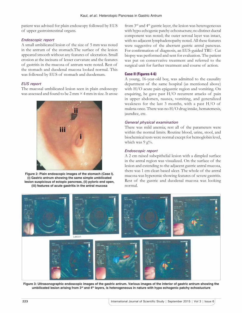
Kaul, et al.: Heterotopic Pancreas in Gastric Antrum
223 International Journal of Scientifi c Study | September 2015 | Vol 3 | Issue 6
patient was advised for plain endoscopy followed by EUS of upper gastrointestinal organs.
Endoscopic reportA small umbilicated lesion of the size of 5 mm was noted in the antrum of the stomach.The surface of the lesion appeared smooth without any features of ulceration. Small erosion at the incisura of lesser curvature and the features of gastritis in the mucosa of antrum were noted. Rest of the stomach and duodenal mucosa looked normal. This was followed by EUS of stomach and duodenum.
EUS reportThe mucosal umbilicated lesion seen in plain endoscopy was assessed and found to be 2 mm × 4 mm in size. It arose
from 3rd and 4th gastric layer, the lesion was heterogeneous with hypo echogenic patchy echostucture; no distinct ductal component was noted; the outer serosal layer was intact, with no adjacent lymphadenopathy noted. All these features were suggestive of the aberrant gastric antral pancreas. For confi rmation of diagnosis, an EUS-guided TRU- Cut biopsy was performed and sent for evaluation. The patient was put on conservative treatment and referred to the surgical unit for further treatment and course of action.
Case II (Figures 4-6)A young, 16-year-old boy, was admitted to the causality department of the same hospital (as mentioned above) with H/O acute pain epigastric region and vomiting. On enquiring, he gave past H/O recurrent attacks of pain in upper abdomen, nausea, vomiting, and generalized weakness for the last 3 months, with a past H/O of malena once. There was no H/O drug intake, hematemesis, jaundice, etc.
General physical examinationThere was mild anemia; rest all of the parameters were within the normal limits. Routine blood, urine, stool, and biochemical tests were normal except for hemoglobin level, which was 9 g%.
Endoscopic reportA 2 cm raised subepithelial lesion with a dimpled surface in the antral region was visualized. On the surface of the lesion and extending to the adjacent gastric antral mucosa, there was 1 cm clean based ulcer. The whole of the antral mucosa was hyperemic showing features of severe gastritis. Rest of the gastric and duodenal mucosa was looking normal.
Figure 2: Plain endoscopic images of the stomach (Case I). (i) Gastric antrum showing the same simple umbilicated
lesion suspicious of ectopic pancreas, (ii) pyloric end open, (iii) features of acute gastritis in the antral mucosa
Figure 3: Ultrasonographic endoscopic images of the gastric antrum. Various images of the interior of gastric antrum showing the umbilicated lesion arising from 3rd and 4th layers, is heterogeneous in nature with hypo echogenic patchy echostucture

Kaul, et al.: Heterotopic Pancreas in Gastric Antrum
224International Journal of Scientifi c Study | September 2015 | Vol 3 | Issue 6
Klob published his fi rst microscopic list of observations of ectopic pancreas in 1859.1,3 Later on after a gap of more than one century, a large series of about 212 cases of heterotrophic pancreas were published by Dolan et al.1 A 60-year-old patient with hiatus hernia had ectopic pancreatic tissue at the esophago-gastric junction, which was presented as ductal adenocarcinoma. Malignant changes at such gastric sites are rare.8 Hlavaty et al., in 2002, identifi ed an 8 mm gastric hemispherical polyp as a case of heterotopic pancreas in a 60-year-old woman.1
Out of 105 gastrostomies performed in the past 5 years, ectopic pancreas was found only in one case, i.e. 0.9% by Christodoulidis et al., in their study. They reported a case of ectopic pancreas in gastric antrum, measuring 5 cm × 3 cm × 4 cm extending into the submucosa and partially into muscularis externa.2
Basilios Papaziogas et al. confi rmed ectopic pancreas with endoepithelial carcinoma of gastric antrum in a female of 56 years of age.5 A 25-year-old lady was diagnosed as a case of ectopic pancreas in the gastric fundus at gastroesophageal junction. The histological fi ndings of the biopsy specimen
Figure 4: Plain endoscopic images of gastric antrum (Case II). (i) A raised subepithelial lesion with a dimpled surface seen in the gastric antrum, (ii) a clear-based ulcer on the surface of the
lesion, ( iii) hyperemic antral mucosa
Figure 5: Plain endoscopic images of gastric antrum (Case II). (i) Hyperemic antral mucosa, (ii) pyloric end open
Figure 6: Endoscopic ultrasonography images of gastric antrum. Hyperemic antral mucosa suggestive of severe
gastritis and pyloric end open. Various images of the interior of gastric antrum showing a big mural lesion lying in the deep
mucosal and submucosal layers with extension into muscularis externa. The interior echo texture is heterogeneous, with hypo echogenic lobular echo texture. An echogenic, irregular tubular
structures presence in the lesio
EUS reportThere was a 16 mm × 8 mm mural lesion in the antrum of the stomach. The location of the lesion was in the deep mucosal and submucosal layers, with extension into the muscularis propria layer. There was no extension through and beyond the muscularis propria. The margins of the lesion were ill-defined. The internal echotexture was heterogeneous, with hypo echogenic pathy/lobular echo structure. Besides, anechoic irregular tubular structures were present in the lesion. No enlarged perigastric lymph nodes were seen. A EUS guided Tru-Cut biopsy was also performed for the confi rmation of diagnosis.
Plain endoscopy and EUS fi ndings were suggestive of pancreatic rest/ectopic pancreatic tissue with an antral benign clean-based ulcer. This patient was also treated conservatively and was then referred to the surgical unit for simple resection or endoscopic mucosal resection.
DISCUSSION
The pathological condition - Ectopic pancreas has been found to be associated with various organs of the body, mostly upper GIT. Hence, its presentation is also varied. The condition was diagnosed very early in the 18th century. Schultz, in 1727, was the fi rst to report the incidence of heterotopic pancreas1 which was found in upper GIT in an ileal diverticulum during the autopsy of a new born.3

Kaul, et al.: Heterotopic Pancreas in Gastric Antrum
225 International Journal of Scientifi c Study | September 2015 | Vol 3 | Issue 6
of the same patient were reported as gastric heterotopic pancreas with pancreatic intraepithelial neoplasm-2 which is believed to represent a precursor lesion for the development of ductal adenocarcinoma.9 Various sites other than the stomach, where ectopic pancreas was diagnosed by various researchers are: A 5-year-old child with a diagnosis of Meckel’s diverticulum was found to have heterotopic pancreatic tissues in the various parts of the gastrointestinal tract by Baysoy et al.10 An isolated heterotopic pancreas, at the terminal ileum of a 47-year-old male, was found to be the cause of ileo-ileal intussusception by Ahmed Monier et al.11 Heterotopic pancreas in the spleen with malignant degeneration to mucinous cyst adenocarcinoma was reported by Nisar et al.12
A case of heterotopic pancreas adjacent to ampulla of vater mimicking cholangiocarcinoma was reported by Atindriya Biswas et al. in UK.3 Goodarzi et al. reported a case of pancreatic heterotopia in the rectum of a 42-year-old female in whom it had turned into ductal adenocarcinoma.13 An 18-year-old post-pubertal girl having an H/O cholecystitis was diagnosed as a case of heterotopic pancreas in the gallbladder, the ectopic pancreas being responsible for cholecystitis.14 A 45-year-old healthy lady who went for a routine medical check-up was found to have soft tissue mass in the left lobe of liver on CT scan. Left hepatic lobectomy was performed and subsequent histopathologicalstudy of the mass removed revealed the presence of ectopic pancreatic tissue present in the liver. This intrahepatic pancreatic tissue had changed into adenocarcinoma.15 Lizhi-Zhang et al reported two very rare cases of ectopic pancreatic mass in anterior. Mediastinum of thoracic cavity in two young females who were15 and16 years old respectively.16
CONCLUSION
Although pancreatic heterotopia is a rare entity, yet it should always be considered in the differential diagnosis of extra mucosal gastric lesion/GI stromal tumor and any undiagnosed abdominal ailment. The quick and easy diagnostic tool is to do EUS and a simultaneous biopsy.
Such diagnostic procedures are all the most important in the cases where pancreatic heteropenia presents primarily as pancreatitis, hyperinsulinism, Zollinger Ellison syndrome, common bile duct obstruction, etc. It is often impossible to distinguish gastric pancreatic heterogenic tissue from primary or metastatic cancer on endoscopy because endoscopic biopsies are sometimes unremarkable. Hence, a frozen section should be taken at the time of surgery to confi rm
the diagnosis. Surgical excision provides symptomatic relief and treatment also. Surgery is recommended, especially if the diagnosis is uncertain. Our case study of two patients revealed that ectopic pancreas can also be present at a young age as described by a few authors rather than conventionally declared in the third or fourth decade of life. Our patients had the same diagnosis and were also related to each other. This fact further enlightens the role of genetic theory in the pathogenesis of ectopic pancreas. It is presumed that both of them had some common genetic pattern and abnormal genetic signaling pathway which predisposed to the formation of ectopic pancreas in both individuals, that too involving antrum of the stomach in both cases.
REFERENCES
1. Hlavaty T, Lukac L, Vyskocil M, Galbavy S. Heterotopic pancreas in gastric antrum with macroscopic appearance of gastric polyp. Bratisl Lek Listy 2002;103:117-20.
2. Christodoulidis G, Zacharoulis D, Barbanis S, Katsogridakis E, Hatzitheofi lou K. Heterotopic pancreas in the stomach: A case report and literature review. World J Gastroenterol 2007;13:6098-100.
3. Biswas A, Hussain EA, Feakins RM, Abraham AT. Heterotopic pancreas mimicking Cholangiocarcinoma. A case report and literature review. J Pancreas (Online) 2007;8:28-34.
4. Kim GH. EUS fi ndings of gastric ectopic pancreas. Video J Encyclopaedia GI Endosc 2012;1:160-1.
5. Papaziogas B, Koutelidakis I, Tsiaousis P, Panagiotopoulou K, Paraskevas G, Argiriadou H, et al. Carcinoma developing in ectopic pancreatic tissue in the stomach: A case report. Cases J 2008;1:249.
6. Fukuda A, Kawaguchi Y, Furuyama K et al. Ectopic pancreas formation in Hes1-knockout mice reveals plasticity of endodermal progenitors of the gut, bile duct, and pancreas. J Clin Invest 2006;116:1484-93.
7. Hebrok M. Hedgehog signaling in pancreas development. Mech Dev 2003;120:45-57.
8. Guillou L, Nordback P, Gerber C, et al. Ductal adenocarcinoma arising in a heterotopic pancreas situated in a hiatal hernia. Arch Pathol Lab Med 1994;118:568-71.
9. Sadeghi NR, Godambe A, Shienbaum AJ, Alloy A. Premalignant gastric heterotopic pancreas. Gastroenterol Hepatol (N Y) 2008;4:218-21.
10. Baysoy G, Balamtekin N, Uslu N, Karavelioglu A, Talim B, Ozen H. Double heterotopic pancreas and Meckel’s diverticulum in a child: Do they have a common origin? Turk J Pediatr 2010;52:336-8.
11. Monier A, Awad A, Szmigielski W, Muneer M, Alrashid A, Darweesh A, et al. Heterotopic pancreas: A rare cause of ileo-ileal intussusception. Pol J Radiol 2014;79:349-51.
12. Nisar PJ, Zaitoun AM, Lobo DN, Rowlands BJ. Heterotopic pancreas in the spleen: Malignant degeneration to mucinous cystadenocarcinoma. Eur J Gastroenterol Hepatol 2002;14:793-6.
13. Goodarzi M, Rashid A, Maru D. Invasive ductal adenocarcinoma arising from pancreatic heterotopia in rectum: Case report and review of literature. Hum Pathol 2010;41:1809-13.
14. Elhence P, Bansal R, Agrawal N. Heterotopic pancreas in gall bladder associated with chronic cholecystolithiasis. Int J Appl Basic Med Res 2012;2:142-3.
15. Yan ML, Wang YD, Tian YF, Lin Y. Adenocarcinoma arising from intrahepatic heterotopic pancreas: A case report and literature review. World J Gastroenterol 2012;18:2881-4.
16. Zhang L, Peng LQ, Yu JQ, Yuan HM, Chu ZG, Zeng HJ, et al. Ectopic pancreas in the anterior mediastinum: A report of two cases and review of the literature. Oncol Lett 2014;7:1053-1056.
How to cite this article: Kaul N, Kapoor H, Kaul V. Heterotopic Pancreas in Gastric Antrum: A Report of Two Cases. Int J Sci Stud 2015;3(6):221-225.
Source of Support: Nil, Confl ict of Interest: None declared.

226International Journal of Scientifi c Study | September 2015 | Vol 3 | Issue 6
Stone in the Scrotum: Scrotal Calcinosis Cutis: A Rare Case ReportManoananth Arivazhagan1, Srinidhi Manjunath2, Kanakapura Srinivasamurthy Bala Subrahmaniya3, Basavaraju Nanjaiah1
1Post-graduate, Department of General Surgery, Mysore Medical College and Research Institute, Mysore, Karnataka, India, 2Senior Resident, Department of General Surgery, Mysore Medical College and Research institute, Mysore, Karnataka, India, 3Associate Professor, Department of General Surgery, Mysore Medical College and Research Institute, Mysore, Karnataka, India
of pre-existing pathology it is termed as idiopathic scrotal calcinosis. Metastatic calcifi cations are usually generalized and due to metabolic changes such as hypercalcemia and hyperphosphatemia as in end-stage renal diseases and hyperparathyroidism and dermatomysositis.7,8 Pabuccuogh et al. proposed degeneration and necrosis of dartos muscle as the reason for calcifi cation which is supported by King et al., Fischer et al., Armjo et al., and Kelten et al.7 Ito et al. described scrotal calcinosis is consequence of excessive discharge and accumulation of material debris in lumina of eccrine epithelial cyst using immunohistochemistry which showed slight positivity for antibodies to sulfated mucopolysaccharides.4 Shapiro et al. 14 case series proposed scrotal calcinosis is idiopathic as there is no epithelial lining around calcium deposition, keratin remnants, granulomatous reaction and, infl ammation infi ltrates which is supported by Shal et al., Parlakgumus et al., Anureet et al., Wright et al., Karaca et al., and Dombale et al.1-3,8 Fukaya et al. and Ueds et al. mentioned role of mast cell in formation of calcifi cation.7,9 Dini et al. proposed the term “idiopathic” can be used if the cause is not known7 as in our case. In our case, we are reporting a case of idiopathic scrotal calcinosis evidenced by the lack of infl ammatory and
INTRODUCTION
Idiopathic scrotal calcinosis is a rare benign condition with painless slow growing nodular masses within the dermis of the scrotal skin. It was fi rst described by Lewinskey in 1883.1-3 Deposition of calcium in the skin, subcutaneous tissue, muscles, and visceral organs is known as calcinosis, and it more common involves skin and it is called calcinosis cutis. There are four types of calcinosis cutis based on their etiology such as dystrophic, metastatic, iatrogenic, and idiopathic.1 Age group is 20-40 years.4-6 Various theories on pathogenesis have been proposed by authors favoring idiopathic and dystrophic calcifi cation. In dystrophic calcifi cation, calcifi cation occurs as a consequence of pre-existing condition such as an epidermal cyst, etc. and when there is no evidence
Case Report
Abstract
Scrotal calcinosis is a rare benign disorder involving scrotal skin resulting from deposition of calcium within the dermis. It was fi rst described by Lewinskey in 1883. Deposition of calcium in the skin, subcutaneous tissue, muscles, and visceral organs is known as calcinosis and it more commonly involves skin and it is called calcinosis cutis. It usually presents as slow growing asymptomatic multiple hard to fi rm nodules. Pathogenesis is still under debate as to whether the calcifi cation is dystrophic or idiopathic. Excision is the treatment of choice followed by primary closure or scrotal reconstruction using split-thickness skin graft. Recurrence is rare. In this article, we report a case of idiopathic scrotal calcinosis cutis which was treated by primary excision at our institute. We have also reviewed the relevant literature.
Key words: Calcifi cations, Calcinosis, Dermis, Scrotum, Skin diseases
Access this article online
www.ijss-sn.com
Month of Submission : 07-2015Month of Peer Review : 08-2015Month of Acceptance : 08-2015Month of Publishing : 09-2015
Corresponding Author: Dr. Manoananth Arivazhagan, #219, PGs and Interns Hostel For Men, Mysore Medical College and Research Institute, Irwin Road, Mysore - 570 001, Karnataka, India. E-mail: [email protected]
DOI: 10.17354/ijss/2015/429

Arivazhagan, et al.: Stone in the Scrotum: Scrotal Calcinosis Cutis
227 International Journal of Scientifi c Study | September 2015 | Vol 3 | Issue 6
epithelial cells. In this article, we have also elaborated the available literature on scrotal calcinosis.
CASE REPORT
A 45-year-old diabetic male patient presented with painless multiple swelling in the scrotum for 8 years which gradually progressed over the years. He neither gave any history suggestive of metabolic disorder, hormonal derangement, sexually transmitted diseases, nor trauma. On examination, multiple yellowish, fi rm nodules present in the scrotal skin with no ulceration or discharge.(Figure 1) The patient’s blood picture, blood sugar, serum calcium, phosphate, parathyroid hormone, calcitonin, and vitamin D levels are within normal limits. (Figure 2) Excision of the nodules from the scrotal skin was done. Grossly excised specimen is about 4 cm × 3 cm ×2 cm and chalky white areas were seen below the skin on cut section. (Figure 3) Microscopic picture shows epidermis and dermis with multiple foci of calcium deposits in the
subcutaneous tissue with no malignancy or infl ammatory cells seen. (Figure 4)
DISCUSSION
Idiopathic scrotal calcinosis is a rare benign condition with painless slow growing nodular masses within the dermis of the scrotal skin.1,2 Incidence of the disease is not known.1 It was fi rst described by Lewinskey in 1883.1-3 Deposition of calcium in the skin, subcutaneous tissue, muscles, and visceral organs is known as calcinosis, and it more commonly involves skin and it is called calcinosis cutis. There are four types of calcinosis cutis based on their etiology such as dystrophic, metastatic, iatrogenic, and idiopathic.1
Scrotal calcinosis is usually asymptomatic but occasionally causes heaviness, itching, ulceration, and chalky white
Figure 1: Pre-operative picture
Figure 2: Post-excision skin gap
Figure 3: Excised specimen
Figure 4: Epidermis and dermis with multiple foci of calcium deposits in the subcutaneous tissue with no malignancy or
infl ammatory cells seen

Arivazhagan, et al.: Stone in the Scrotum: Scrotal Calcinosis Cutis
228International Journal of Scientifi c Study | September 2015 | Vol 3 | Issue 6
exudative discharge.2,4-6,8 The patient mainly comes for cosmetic reasons.8 Age group is 20-40 years, youngest and oldest reported are 9 and 85 years, respectively.2,4-6 Initially, it resembles the color of scrotal skin later it changes into yellow, and duration is about 10 years ranging from 3 months to 46 years.8
Microscopic picture shows amorphous basophilic calcium deposits within dermis surrounded by lymphocytic infi ltration, histiocytes, and hyalinization.2,6-8 Histological picture shows muscle, epithelial cells, and foreign body granuloma during early stage and it shows only calcifi cation in the advanced stage.2
The pathogenesis is still in the debate, various theories have been proposed by authors favoring idiopathic and dystrophic calcifi cation. In dystrophic calcifi cation, there must be a local favoring condition such as pre-existing epidermal cyst, eccrine duct milia, eccrine epithelial cyst, degenerated dartos muscle, and connective tissue disorders such as scleroderma, systemic lupus erythromatous, dermatomysositis, and minor trauma. Squamous cell epithelial lining may present, and patient has normal serum calcium and phosphorus levels.1
Song et al. described spectrum of changes takes place in scrotal calcinosis as mild to moderate infl ammation of epidermal cyst is followed by mononuclear cell infi ltration and foreign body granuloma formation and lastly resorption of cyst wall and keratin remnants leaving calcium deposits only8,9 which is supported by Swinhart et al., Akosa et al., Saad et al., Dubey et al., Parlakgumus et al., and Dini and Colatraneschi et al.2,6,8
Pabuccuogh et al. proposed degeneration and necrosis of dartos muscle as the reason for calcifi cation which is supported by King et al., Fischer et al., Armjo et al., and Kelten et al.7 Ito et al. described scrotal calcinosis is consequence of excessive discharge and accumulation of material debris in lumina of eccrine epithelial cyst using immunohistochemistry which showed slight positivity for antibodies to sulfated mucopolysaccharides.4
Dare and Axelson et al. supported scrotal calcinosis arising from pre-existing eccrine milia using immnunohistochemistry which showed antibodies to carcinoembryonic antigen.6 Carson et al. described sequences following minor trauma and invasion of nanobacteria and formation of calcium apatite crystals.6 Veress and Feinstein et al. favored minor trauma following which calcifi cation occur.10
Metastatic calcifications are usually generalized and due to metabolic changes such as hypercalcemia and hyperphosphatemia as in end-stage renal diseases
hyperparathyroidism and dermatomysositis involving visceral organs and joints.8,11,12 Pallavi et al. reported as case of scrotal calcinosis due to normocalcemic hyperparathyroidism which doesn’t need parathyroidectomy unless symptomatic.11
Shapiro et al. 14 case series proposed scrotal calcinosis is idiopathic as there is no epithelial lining around calcium deposition, keratin remnants, granulomatous reaction, and infl ammation infi ltrates which is supported by Shal et al., Parlakgumus et al., Anureet et al., Wright et al., Karaca et al., and Dombale et al.1-3,8 Fukaya et al. and Ueds et al. mentioned role of mast cell in formation of calcifi cation.7,9 Dini et al. proposed the term “idiopathic” can be used if the cause is not known7 as in our case. Idiopathic and dystrophic calcifi cations are usually involves one general area (calcinosis circumscripta). The iatrogenic calcifi cations mainly occur at the site of invasive procedure due to tissue damage.12
Differential diagnosis are teratoma, gonadoblastomas, leydig cell tumors, calcifi ed onchocercoma, neurofi broma, ancient schwannomas, steatomas, lipomas, fi bromas, and scrotal calcinosis may also be due to chronic epididymitis, calcifi ed appendix testis, appendix epididymis, and sperm granuloma due to sperm extravasation and hematoma.4,8
Diagnosis is confi rmed by biopsy. If swelling is <4 mm, pinch and punch excision is advised.8 Surgery is the treatment of choice.4,8 If it is massive, subtotal excision of the scrotal wall is preferred. If it is extensively involved, excision followed by complex scrotal reconstruction using meshed split thickness skin graft as the scrotal skin is rugged.8 Recurrence is very low mainly due to microscopic foci of calcifi cation left over.8
CONCLUSION
Idiopathic scrotal calcinosis cutis is a rare benign lesion. Metabolic and hormonal work-up is required to rule out other causes. Irrespective of the etiology, surgical excision is required both for confi rming the diagnosis as well as for treatment. Scrotal calcinosis must be included in the differential diagnosis of cutaneous swellings in the scrotal region.
REFERENCES
1. Dombale VD, Basarkod SI, Kotabagi HB, Farheen U. Extensive idiopathic scrotal calcinosis: A case report. J Clin Diagn Res 2012 Suppl 1 6:478-9.
2. Anureet K, Rimpi B, Manas M, Jasbir S. Idiopathic calcinosis of scrotum; A rare scrotal skin disorder. J Adv Res Biol Sci 2011;3:113-4.
3. Shah V, Shet T. Scrotal calcinosis results from calcifi cation of cysts derived from hair follicles: A series of 20 cases evaluating the spectrum of changes resulting in scrotal calcinosis. Am J Dermatopathol 2007;29:172-5.
4. Parlakgumus A, Canpolat ET, Caliskan K, Colakoglu T, Yildirim S, Ezer A,

Arivazhagan, et al.: Stone in the Scrotum: Scrotal Calcinosis Cutis
229 International Journal of Scientifi c Study | September 2015 | Vol 3 | Issue 6
et al. Scrotal calcinosis due to resorption of cyst walls: A case report. J Med Case Rep 2008;2:375.
5. Celik O, Ipekci T, Kazimoglu H. Idiopathic scrotal calcinosis. Saudi Med J 2013;34:1294-5.
6. Tela UM, Ibrahim MB. Scrotal calcinosis: A case report and review of pathogenesis and surgical management. Case Rep Urol 2012;2012:Article ID: 475246, 3.
7. Kelten EC, Akbulut M, Çolakoglu N, Bayramoglu H, Duzcan SE. Scrotal calcinosis: Is it idiopathic or dystrophic? Aegean Pathol J 2005;2:4-7.
8. Kiremitci S, Yüksel S, Anafarta K, Tulunay Ö. Scrotal calcinosis: A case report and review of literature. Ankara Üniv Tıp Fak Mecmuasi 2011;64:46-51.
9. Ibrahim M, Ibrahim GK, Mohammad MA, Aji1 SA, Umar AB, Nurlan AN, et al. Calcinosis of the scrotum in children: Report of two cases and review of the literatur. Arch Int Surg 2013;3:142-6.
10. Suha B, Uluocak N, Köseoğlu RD, Erdemir F, Sezer E. Idiopathic scrotal calcinosis: A rare scrotal skin disorder. Ankara Üniv Tıp Fak Mecmuasi 2005;58:20-2.
11. Pallavi R, Bautista JE, Lam K. Scrotal calcinosis in normocalcemic primary hyperparathyroidism. Available from: http://www.consultantlive.com. [2015 aug 14].
12. Scheinfeld NS. Skin disorders in older adults: Manifestations of endocrine and metabolic diseases. Consultant 360 2012;52:144-53.
How to cite this article: Manoananth AB, Srinidhi M, Balasubrahmaniya KS, Basavaraju N. Stone in the Scrotum: Scrotal Calcinosis Cutis: A Rare Case Report. Int J Sci Stud 2015;3(6):226-229.
Source of Support: Nil, Confl ict of Interest: None declared.

230International Journal of Scientifi c Study | September 2015 | Vol 3 | Issue 6
Herlyn–Werner–Wunderlich Syndrome-Early Diagnosis with Ultrasonography in a 3-month-old Female ChildT Anil Balraj1, Nadeem Ahmed2, B Avinash3
1Assistant Professor, Department of Pediatric Surgery, Kakatiya Medical College, MGM Hospital, Warangal, Telangana, India, 2Associate Professor, Department of Radiology, Kakatiya Medical College, MGM Hospital, Warangal, Telangana, India, 3Post-graduate Student, Department of Radiology, Kakatiya Medical College, MGM Hospital, Warangal, Telangana, India
CASE REPORT
The 3-month-old child was presented with a swelling at the introitus noticed by the mother; there were no other complaints such as pain, fever, or symptoms suggestive of urinary tract infection. She was referred for an ultrasound examination which revealed a bicornuate uterus with separate cervices (bicornis bicollis uterus), and an anechoic collection or cyst in the vagina. There was no evidence of hydrometra or hydrosalpinx. The right kidney was not visualized. Left kidney showed normal morphology (Figures 1-4).
On examination under anesthesia, the cyst revealed to be obstructed hemivagina fi lled with clear fl uid. It was drained, and the septum excised. Further examination showed two cervices suggestive of uterus didelphys (Figures 5 and 6).
DISCUSSION
Herlyn–Werner–Wunderlich syndrome comprises of obstructed hemivagina and ipsilateral renal agenesis and
INTRODUCTION
Herlyn–Werner–Wunderlich syndrome is a rare Mullerian duct and Wolffi an duct anomaly with uterus didelphys, unilaterally obstructed hemivagina, and ipsilateral renal agenesis (OHVIRA).1 Due to functional patency of hemivagina and normal menstrual cycles, this type of obstructive anomaly initially remains unrecognized, usually presenting at puberty with recurrent pelvic pain and/or mass due to hematocolpos or hematometra. Moreover, the menstrual fl ow that comes from the patent hemivagina resembles normal menses leading to delay in diagnosis and surgery.2,3 Developmental abnormalities of Mullerian duct have an estimated incidence of 1.1-3.5% among women.4,5
Case Report
Abstract
Herlyn–Werner–Wunderlich syndrome is an uncommon, complex uterine anomaly - obstructed hemivagina with ipsilateral renal agenesis, usually presenting in adolescence. Here we report a rare presentation of this entity in a 3-month-old child, diagnosed on transabdominal ultrasonography alone. The patient underwent surgery wherein imaging fi ndings were confi rmed, and vaginoplasty was done. We present this case to highlight the role of ultrasonography in diagnosing this condition even though magnetic resonance imaging is the most accurate imaging modality in diagnosing this condition; and also the early age of presentation which is unusual. We describe the fi ndings on ultrasound along with a review of the literature on Herlyn–Werner–Wunderlich syndrome.
Key words: Hemivagina, Ipsilateral renal agenesis, Mullerian anomalies, Obstructed hemivagina with ipsilateral renal agenesis syndrome, Uterus didelphys
Access this article online
www.ijss-sn.com
Month of Submission : 07-2015Month of Peer Review : 08-2015Month of Acceptance : 08-2015Month of Publishing : 09-2015
Corresponding Author: Dr. T Anil Balraj, #2-4-164/204, Hari Towers, Ramnagar, Hanamkonda, Warangal, Telangana, India. Phone: +91-9703045632. E-mail: [email protected]
DOI: 10.17354/ijss/2015/430

Balraj, et al.: Herlyn–Werner–Wunderlich Syndrome in a 3-month-old Child
231 International Journal of Scientifi c Study | September 2015 | Vol 3 | Issue 6
was fi rst reported in 1922. It is more recently known by the acronym OHVIRA and commonly associated with uterine didelphys or rarely septate uterus.6 The incidence
of OHVIRA syndrome is very rare, and only isolated case reports have been published.4
Female reproductive tract develops both from paramesonephric and mesonephric ducts.
Figure 1: Sagittal image showing empty right renal fossa. Compare with normal left kidney
Figure 2: Sagittal image showing fl uid or cyst in vagina
Figure 3: Axial section at level of fundus showing bicornuate uterus
Figure 4: Axial section at level of cervix showing two cervices
Figure 5: Bluish cystic swelling at introitus
Figure 6: Obstructed hemivagina and patent hemivagina under anesthesia

Balraj, et al.: Herlyn–Werner–Wunderlich Syndrome in a 3-month-old Child
232International Journal of Scientifi c Study | September 2015 | Vol 3 | Issue 6
The OHVIRA syndrome occurs due to the developmental arrest of ipsilateral Wolffi an duct resulting in developmental failure of distal hemivagina leading to obstructed hemivagina. The uterine didelphys commonly associated with OHVIRA syndrome occurs due to failure in the fusion of paired Mullerian ducts.4 The female genital and urinary tracts have a similar embryological origin from the intermediate mesoderm, which is responsible for the increased association of their anomalies. Ipsilateral renal agenesis is a common urologic anomaly seen in OHVIRA syndrome.7,8
Typically, a patient with this rare condition presents after menarche with non-specifi c symptoms of recurrent pelvic pain with vaginal or pelvic mass. However in our case the patient presented at a very early age, which is very rare.9 The pelvic examination may show a bulging vaginal mass, but sometimes the mass may be small and diffi cult to determine.10 In general, ultrasonography is the initial screening tool in cases of suspected female genital tract anomalies. However, magnetic resonance imaging (MRI) is the most accurate diagnostic method as it better depicts uterine anatomy and has excellent tissue characterization, which play a crucial role in surgical planning.1,11 In our case, however, ultrasonography alone was reliable in diagnosing this rare anomaly with proper delineation of uterine contour. The sonographic fi ndings were clinically and peroperatively corroborated.
Mullerian agenesis and renal agenesis often coexist in Herlyn syndrome, so it is important to look for urinary tract anomalies in all cases of Mullerian duct anomalies.12 Genitourinary anomalies can be associated with other abnormalities such as an atrial septal defect, coarctation of aorta, and spinal anomalies. Hence, a complete physical examination and relevant investigations are necessary in all patients.13 Ipsilateral renal agenesis is found in 81% cases of OHVIRA syndrome and 25-50% show genital anomalies.14
The classic treatment is resection of the vaginal septum to relieve the obstructed hemivagina. Hemihysterectomy is no longer preferred, as the reported incidence of pregnancy is similar with hemihysterectomy and transvaginal repair. A successful pregnancy occurs in 87% of the patients with OHVIRA syndrome.10,12,13,15
CONCLUSION
OHVIRA syndrome is an uncommon congenital anomaly with clinical signifi cance and simple surgical management. Imaging, particularly MRI plays an important role in diagnosis. However, an initial screening ultrasound can sometimes diagnose it as in our case. An early correct diagnosis is a goal to relieve the symptoms and prevent complications caused by retrograde menstruation resulting in endometriosis and in preserving sexual and conceptional abilities.
REFERENCES
1. Kimble RM, Khoo SK, Baartz D, Kimble RM. The obstructed hemivagina, ipsilateral renal anomaly, uterus didelphys triad. Aust N Z J Obstet Gynaecol 2009;49:554-7.
2. Shih CL, Hung YC, Chen CP, Chien SC, Lin WC. Resectoscopic excision of the vaginal septum in a virgin with uterus didelphys and obstructed unilateral vagina. Taiwan J Obstet Gynecol 2010;49:109-11.
3. Nigam A, Raghunandan C, Yadav R, Tomer S, Anand R. OHVIRA syndrome: Rare cause of chronic vaginal discharge in an unmarried female. Congenit Anom (Kyoto) 2011;51:153-5.
4. Bajaj SK, Misra R, Thukral BB, Gupta R. OHVIRA: Uterus didelphys, blind hemivagina and ipsilateral renal agenesis: Advantage MRI. J Hum Reprod Sci 2012;5:67-70.
5. Strassmann EO. Fertility and unifi cation of double uterus. Fertil Sterils 1966;17:165-76.
6. Troiano RN. Magnetic resonance imaging of mullerian duct anomalies of the uterus. Top Magn Reson Imaging 2003;14:269-79.
7. Han B, Herndon CN, Rosen MP, Wang ZJ, Daldrup Link H. Uterine didelphys associated with obstructed hemivagina and ipsilateral renal anomaly (OHVIRA) syndrome. Radiol Case Rep 2010;5:327.
8. Orazi C, Lucchetti MC, Schingo PM, Marchetti P, Ferro F. Herlyn-Werner-Wunderlich syndrome: Uterus didelphys, blind hemivagina and ipsilateral renal agenesis. Sonographic and MR fi ndings in 11 cases. Pediatr Radiol 2007;37:657-6.
9. Sanghvi Y, Shastri P, Mane SB, Dhende NP. Prepubertal presentation of Herlyn-Werner-Wunderlich syndrome: A case report. J Pediatr Surg 2011;46:1277-80.
10. Smith NA, Laufer MR. Obstructed hemivagina and ipsilateral renal anomaly (OHVIRA) syndrome: Management and follow-up. Fertil Steril 2007;87:918-22.
11. Tanaka YO, Kurosaki Y, Kobayashi T, Eguchi N, Mori K, Satoh Y, et al. Uterus didelphys associated with obstructed hemivagina and ipsilateral renal agenesis: MR fi ndings in seven cases. Abdom Imaging 1998;23:437-41.
12. Gholoum S, Puligandla PS, Hui T, Su W, Quiros E, Laberge JM. Management and outcome of patients with combined vaginal septum, bifi d uterus, and ipsilateral renal agenesis (Herlyn-Werner-Wunderlich syndrome). J Pediatr Surg 2006;41:987-92.
13. Zurawin RK, Dietrich JE, Heard MJ, Edwards CL. Didelphic uterus and obstructed hemivagina with renal agenesis: Case report and review of the literature. J Pediatr Adolesc Gynecol 2004;17:137-41.
14. Li S, Qayyum A, Coakley FV, Hricak H. Association of renal agenesis and mullerian duct anomalies. J Comput Assist Tomogr 2000;24:829-34.
15. Mandava A, Prabhakar RR, Smitha S. OHVIRA syndrome (obstructed hemivagina and ipsilateral renal anomaly) with uterus didelphys, an unusual presentation. J Pediatr Adolesc Gynecol 2012;25:e23-5.
How to cite this article: Balraj TA, Ahmed N, Avinash B. Herlyn–Werner–Wunderlich Syndrome-Early Diagnosis with Ultrasonography in a 3-month-old Female Child. Int J Sci Stud 2015;3(6):230-232.
Source of Support: Nil, Confl ict of Interest: None declared.

233 International Journal of Scientifi c Study | September 2015 | Vol 3 | Issue 6
Horseshoe Kidney with Bilateral Ureteropelvic Junction Obstruction with Multiple Renal Calculi: A Case ReportPiyush P Singhania1, Nandkishor R Raut2, Sanish S Shringarpure3, Niraj Tiwari2, Saket Sathe2
1Associate Professor, Department of Urology, MGM Medical College & Hospital, Navi Mumbai, Maharashtra, India, 2Resident, Department of Urology, MGM Medical College & Hospital, Navi Mumbai, Maharashtra, India, 3Assistant Professor, Department of Urology, MGM Medical College & Hospital, Navi Mumbai, Maharashtra, India
The management of a young patient who presented with horseshoe kidney with recurrent multiple renal calculi due to bilateral pediatric ureteropelvic junction (PUJ) obstruction is described.
CASE REPORT
A 24-year-old male patient presented with complaints of intermittency, burning micturation, pain lower abdomen, fullness in both fl anks, increased frequency of micturation, and dysuria off and on for 3 years. The patient gave a history of left-sided open pyelolithotomy 3 years back. The operation was uneventful, but there was no total stone clearance. The patient was evaluated and investigated. Complete blood count, liver function tests, and renal function test were normal. Urine routine microscopy was showing signs of infection. Ultrasound (USG) abdomen showed the fi ndings suggestive of horseshoe kidney with bilateral multiple renal calculi with gross hydronephrosis. Intravenous pyelogram (Figure 1) was suggestive of multiple bilateral renal calculi with bilateral hydronephrosis with horseshoe kidney. Nuclear renogram (Figure 2) was showing horseshoe kidney with bilateral impaired parenchymal function and obstructed drainage pattern on the left side and sluggish partially obstructed drainage pattern on the right side.
INTRODUCTION
Horseshoe kidney is the commonest fusion anomaly of the genitourinary tract with a prevalence of 1/400-1/800.1 It is characterized by renal malrotation, variable blood supply, and a propensity to form ureteropelvic junction (UPJ) obstruction in up to one-third of cases.2 UPJ obstruction is postulated to develop secondary to congenital stricture, high ureteral insertion, an abnormal ureteral course over the isthmus, crossing vessels supplying the isthmus, or abnormal motility of the UPJ segment.3
The most common complication of horseshoe kidney is kidney calculus. It was previously believed that such a high frequency of calculus formation in these patients was due to the higher rate of infection, stasis, and obstruction. However, the last reviews are suggestive for metabolic causes in most of the patients.4
Case Report
Abstract
Horseshoe kidney is the commonest fusion anomaly of the genitourinary tract with a prevalence of 1/400-1/800. It is characterized by renal malrotation, variable blood supply, high insertion of the ureter, and a propensity to form an ureteropelvic junction (UPJ) obstruction in up to one-third of cases. The most common complication of horseshoe kidney is kidney calculus. The management of a young patient who presented with horseshoe kidney with recurrent multiple renal calculi due to bilateral UPJ obstruction with recurrent urinary tract infection is described. During follow-up after 3 months, the patient was symptomatically relieved, there were no complaints of pain or fullness in the abdomen and the patient has started doing daily regular activities and work.
Key words: Horseshoe kidney, Percutaneous nephrolithotomy, Pyeloplasty, Ureteropelvic junction obstruction, Urolithiasis
Access this article online
www.ijss-sn.com
Month of Submission : 07-2015Month of Peer Review : 08-2015Month of Acceptance : 08-2015Month of Publishing : 09-2015
Corresponding Author: Dr. Nandkishor R Raut, Department of Urology, MGM Medical College & Hospital, Kamothe, Navi Mumbai - 410 209, Maharashtra, India. Phone: +91-9404904176. E-mail: [email protected]
DOI: 10.17354/ijss/2015/431

Singhania, et al.: Management of B/L UPJ Obstruction with multiple renal calculi in Horse shoe kidney
234International Journal of Scientifi c Study | September 2015 | Vol 3 | Issue 6
The patient was subjected to bilateral retrograde pyelogram which showed bilateral UPJ obstruction with high insertion of the ureter. Bilateral open pyelolithotomy with pyeloplasty with double-J (DJ) stenting was done through a lower midline incision. Post-operative hospital stay was uneventful. Follow-up USG and X-ray kidneys-ureters-bladder (KUB) after 2 weeks showed residual calculi, 4 on right and 3 on the left side. Urine culture was positive for Escherichia coli >105 which was managed conservatively with injectable cefotaxime for 7 days. X-ray KUB 3 weeks later showed right renal calculi with left lower ureteric calculi. The right side percutaneous nephrolithotomy (PCNL) with left sided ureterorenoscopy lithotripsy (URSL) was done, complete stone clearance (Figure 3) was achieved and bilateral DJ stenting was done. During follow-up, the patient was symptomatically relieved; there were no complaints of pain or fullness in the abdomen.
Figure 1: Intravenous pyelogram suggestive of multiple bilateral renal calculi with bilateral hydronephrosis with horseshoe kidney
Figure 2: Nuclear renogram showing horseshoe kidney with bilateral impaired parenchymal function and
obstructed drainage pattern on the left side and sluggish partially obstructed drainage pattern on the right side.
Differential renal function of left moiety - 55.79% and right moiety - 44.21%

Singhania, et al.: Management of B/L UPJ Obstruction with multiple renal calculi in Horse shoe kidney
235 International Journal of Scientifi c Study | September 2015 | Vol 3 | Issue 6
DISCUSSION
Horseshoe kidney is the result of a developmental defect occurring between 4th and 8th weeks of embryogenesis. As the kidney develops, the inferior poles fuse and its ascent is arrested by the inferior mesenteric artery. The kidneys are fused by an isthmus, which can be a band of fi brous tissue or a thick rim of functional renal tissue. Clinical fi ndings are those of infection, calculi, obstruction or tumor due to anomalous position of pelvis, and ureters. As the most common complication of the horseshoe kidney necessitating surgical intervention, urolithiasis has an incidence of 20-60%, and UPJ obstruction occurs at an incidence of 15-33%.5
The primary technical challenges of pyeloplasty in this population relate to aberrant lower pole vessels, the unfamiliar caudal position of the kidney, and renal isthmus. Conventionally, the management of UPJ obstruction in horseshoe kidney has been open dismembered pyeloplasty with isthmusectomy and nephropexy of the ipsilateral kidney.3 Simple Anderson–Hynes pyeloplasty via a fl ank incision without additional division of the isthmus and lateropexy of the kidney is also a highly effective and safe procedure for treating UPJ obstruction in horseshoe kidney.6
The calculi in a horseshoe and ectopic kidneys present unique challenges in decision-making and technical
aspects of treatment. PCNL has been shown to be highly successful with an overall stone-free rate of 75-100% in a few series.7
In the case of this patient who presented with horseshoe kidney with recurrent multiple renal calculi due to bilateral PUJ obstruction with recurrent UTI. The patient was managed successfully with bilateral open pyelolithotomy with pyeloplasty in a single setting. The patient developed UTI during follow-up period, which related to bilateral DJ stent in situ, and managed conservatively with antibiotics. In view of residual calculi, the right PCNL and left URSL was done, and complete stone clearance was achieved. After 3 weeks, bilateral DJ stent was removed. During follow-up after 3 months, the patient was symptomatically relieved, there were no complaints of pain or fullness in the abdomen and the patient has started doing daily regular activities and work.
CONCLUSION
There should be a high index of suspicion for UPJ obstruction in a patient presenting with horseshoe kidney with recurrent multiple renal calculi. Its management requires multimodal approach with the judicious use of endoscopic and open surgical intervention. Correction of UPJ obstruction is recommended to treat the symptoms and prevent the recurrence of calculi in these patients.
REFERENCES
1. Kaufman E. Textbook of special pathological anatomy vol.2 Berlin; de Gryter 1957; pp. 427-436.
2. Lallas CD, Pak RW, Pagnani C, Hubosky SG, Yanke BV, Keeley FX, et al. The minimally invasive management of ureteropelvic junction obstruction in horseshoe kidneys. World J Urol 2011;29:91-5.
3. Yohannes P, Smith AD. The endourological management of complications associated with horseshoe kidney. J Urol 2002;168:5-8.
4. Evans WP, Resnick MI. Horseshoe kidney and urolithiasis. J Urol 1981;125:620-1.
5. Viola D, Anagnostou T, Thompson TJ, Smith G, Moussa SA, Tolley DA. Sixteen years of experience with stone management in horseshoe kidneys. Urol Int 2007;78:214-8.
6. Schuster T, Dietz HG, Schütz S. Anderson-Hynes pyeloplasty in horseshoe kidney in children: Is it effective without symphysiotomy? Pediatr Surg Int 1999;15:230-3.
7. Mosavi-Bahar SH, Amirzargar MA, Rahnavardi M, Moghaddam SM, Babbolhavaeji H, Amirhasani S. Percutaneous nephrolithotomy in patients with kidney malformations. J Endourol 2007;21:520-4.
Figure 3: Post-operative X-ray kidneys-ureters-bladder showing complete stone clearance
How to cite this article: Singhania PP, Raut NR, Shringarpure SS, Tiwari N, Sathe S. Horseshoe Kidney with Bilateral Ureteropelvic Junction Obstruction with Multiple Renal Calculi: A Case Report. Int J Sci Stud 2015;3(6):233-235.
Source of Support: Nil, Confl ict of Interest: None declared.

236International Journal of Scientifi c Study | September 2015 | Vol 3 | Issue 6
Failed Deep Anterior Lamellar Keratoplasty in Avellino Stromal Dystrophy: A Case Report and Review of LiteratureNita Shanbhag1, Nilay Patel2, Nadim Khatib2, Nupur Bhatt2
1Professor and Head, Department of Ophthalmology, Dr. D.Y. Patil Medical College, Nerul, Navi Mumbai, India, 2Post-graduate Student, Department of Ophthalmology, Dr. D.Y. Patil Medical College, Nerul, Navi Mumbai, India
granular deposits, but with progression these lines coalesce with the round opacities.2 First signs of the disease can be seen as early as 3 years of age in homozygotic cases, but most common during puberty or early adulthood.3 Visual acuity deteriorates as the central visual axis becomes affected, and corneal erosions can result in episodes of pain. Homozygotes can have a more rapid course comparing with heterozygotes patients. Visual rehabilitation is achieved by corneal transplantation usually lamellar.
CASE REPORT
Female 45 years old presented with complaints of gradual progressive diminution of vision since 10 years of age. She was unable to do her routine activities in the past 3-4 months necessitating a visit to the eye doctor. There was no contributory history in the form of trauma, infl ammation, or any surgical procedure in the past. The patient did not have any systemic illness negating any metabolic disorders. Family members did not have any similar visual problem.
On examination, the ocular fi ndings in both eyes were similar. Best corrected visual acuity 6/60 due to opacity
INTRODUCTION
Corneal dystrophy (CD) is defi ned as bilateral and symmetric primary corneal disease, without previous associated ocular inflammation. Most cases of CD have an autosomal dominant inheritance pattern, starting at the fi rst decades of life, with a stable or slowly progressive course. CDs are classifi ed according to the involved corneal layer (Table 1).
Avellino CD was fi rst described by Folberg et al. (1988). It exhibits features of both granular and lattice dystrophy. It is inherited in an autosomal dominant fashion and related to the transforming growth factor beta-induced gene (TGFβ1), locus 5q31.1 Biomicroscopically, granular deposits are more superfi cial and as the disease progresses, a snowfl ake appearance deeper in the stroma can be noted. The linear refractile deposits tend to be deeper than the
Case Report
Abstract
Corneal dystrophy (CD) has an autosomal dominant inheritance pattern. A female 45 years of age presented with both eyes dimension of vision in the recent past without any associated ocular pathology. Examination revealed an Avellino’s stromal CD. Multiple white dots about 0.5 mm in diameter, studding the stroma not involving the endothelium, spread over central 8 mm with an interdigitating network of fi laments. Deep anterior lamellar keratoplasty (DALK) on the table showed the fi laments were anchored to the endothelium not allowing a separation though anterior segment optical coherence tomography and histopathology showed they are neatly separated from it. We are publishing this case to highlight the need for a good corneal endothelial count in the donor cornea and the need to keep the option of penetrating keratoplasty in a Avellino stromal dystrophy though DALK is the fi rst choice of management.
Key words: Avellino dystrophy, Corneal dystrophy, Deep anterior lamellar keratoplasty, Keratoplasty
Access this article online
www.ijss-sn.com
Month of Submission : 07-2015Month of Peer Review : 08-2015Month of Acceptance : 08-2015Month of Publishing : 09-2015
Corresponding Author: Dr. Nilay Patel, Department of Ophthalmology, Dr. D.Y. Patil Medical College, Nerul, Navi Mumbai, India. Phone: +91-9833079560. E-mail: [email protected]
DOI: 10.17354/ijss/2015/432

Shanbhag, et al.: Failed DALK in Avellino Stromal Dystrophy
237 International Journal of Scientifi c Study | September 2015 | Vol 3 | Issue 6
involving the optical axis. Ocular adnexa, conjunctiva, and sclera were normal. The cornea was normal in size and shape. Multiple white dots about 0.5 mm in diameter eroding out of the epithelium, studding the stroma not involving the endothelium, spread over 8 mm area, with the interdigitating network of fi laments. No vascularization, pigmentation, or degeneration (Figures 1 and 2). The corneal sensations were present. The rest of anterior segment and posterior segment were within normal limit (Table 2).
To support our clinical finding, we ordered anterior segment optical coherence tomography of both eyes. There was deep stromal involvement sparing the descemet’s membrane (DM) and the endothelium (Figure 3). For visual rehabilitation, a decision to do deep anterior lamellar keratoplasty (DALK) was taken as the stromal involvement spared the descemets and the endothelium. Routine investigation for anesthesia fi tness was taken and tissue arranged with a good endothelial count of 2450 cells/cumm. This was done in contingency to an inadvertent conversion to penetrating keratoplasty (PKP). On the table, Big bubble technique was done after lamellar dissection of 300 μ of the central 8 mm of the corneal tissue. A central cruciate incision over the central cornea over an iris repository was done to prevent an inadvertent corneal perforation. During the lamellar dissection to
the periphery, it was noticed that the fi laments in the stroma were deeply embedded into the descemets and the endothelium making it diffi cult to fi nd the plane between the stroma and descemets. The descemets
Table 1: Type of CDCD categorized by corneal layer
Epithelial and subepithelial dystrophiesEBMDEREDSMCDMeesmann’s CDLisch epithelial CDGelatinous drop-like CD
Bowman’s layer dystrophiesReis-Bucklers’ CDThiel-Benke CDGrayson-Wilbrandt CD
Stromal dystrophiesLattice (Type I, II, III, and lV)Granular (Groenouw type, Avellino dystrophy)Macular (Type I and II)SCCDCongenital stromal CDFleck’s CDPosterior amorphous CDCentral cloudy dystrophy of FrancoisPre-Descemet’s CD
Descemet and endothelial dystrophiesFuch’s endothelial CD (early onset and late onset)Posterior polymorphous dystrophyCongenital hereditary endothelial dystrophyX-linked endothelial dystrophy
CD: Corneal dystrophy, EBMD: Epithelial basement membrane dystrophy, ERED: Epithelial recurrent erosion dystrophy, SMCD: Subepithelial mucinous corneal dystrophy, SCCD: Schnyder’s corneal crystalline dystrophy
Figure 1: Right eye stromal corneal dystrophy external and slitlamp view
Figure 2: Left eye stromal corneal dystrophy external and slitlamp view
Figure 3: BE anterior segment optical coherence tomography showing stromal corneal dystrophy
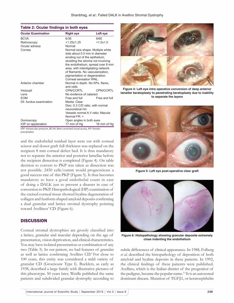
Shanbhag, et al.: Failed DALK in Avellino Stromal Dystrophy
238International Journal of Scientifi c Study | September 2015 | Vol 3 | Issue 6
and the endothelial residual layer were cut with corneal scissor and donor graft full thickness was replaced on the recipient 8 mm corneal defect bed. It is thus mandatory not to separate the anterior and posterior lamellae before the recipient dissection is completed (Figure 4). On table decision to convert to PKP was taken as dissection was not possible. 2450 cells/cumm would prognosticate a good success rate of this PKP (Figure 5). It thus becomes mandatory to have a good endothelial count in case of doing a DALK just to prevent a disaster in case of conversion to PKP. Histopathological (HP) examination of the excised corneal tissue showed hyaline degeneration of collagen and fusiform shaped amyloid deposits confi rming a dual granular and lattice stromal dystrophy pointing toward Avellinos’ CD (Figure 6).
DISCUSSION
Corneal stromal dystrophies are grossly classifi ed into a lattice, granular and macular depending on the age of presentation, vision deprivation, and clinical characteristics. You may have isolated presentation or combination of any two (Table 3). In our patient, we had features of granular as well as lattice confi rming Avellino CD.4 For close to 100 years, this entity was considered a mild variety of granular CD (Groenouw Type I). Bucklers, as early as 1938, described a large family with illustrative pictures of this phenotype. 50 years later, Weidle published the same patients and subdivided granular dystrophy according to
subtle differences of clinical appearance. In 1988, Folberg et al. described the histopathology of deposition of both amyloid and hyaline deposits in these patients. In 1992, the clinical fi ndings of these patients were published. Avellino, which is the Italian district of the progenitor of the pedigree, became the popular name.1,3 It is an autosomal dominant disease. Mutation of TGFβ1, or keratoepithelin
Table 2: Ocular fi ndings in both eyesOcular Examination Right eye Left eyeBCVA 6/36 6/60Retinoscopy +1.25±1.25 +1.0±1.0Ocular adnexa NormalCornea Normal size shape. Multiple white
dots about 0.5 mm in diameter eroding out of the epithelium, studding the stroma not involving the endothelium, spread over 8 mm area, with interdigitating network of fi laments. No vascularization, pigmentation or degeneration. Corneal sensation WNL.
Anterior chamber Normal in depth. No KPs, fl ares, and cells
Iris/pupil CPN/CCRTL CPN/CCRTLLens No evidence of cataractEOM Free and full Free and fullDil. fundus examination Media: Clear
Disc: 0.3 C/D ratio, with normal neuroretinal rimVessels normal A V ratio; Macula: Normal FR: +
Gonioscopy Open angles in both eyesIOP on applanation 17 mm of Hg 16 mm of HgIOP: Intraocular pressure, BCVA: Best corrected visual acuity, KP: Keratic precipitates
Figure 4: Left eye intra operative conversion of deep anterior lamellar keratoplasty to penetrating keratoplasty due to inability
to separate the layers
Figure 6: Histopathology showing granular deposite extremely close indenting the endothelium
Figure 5: Left eye post-operative clear graft

Shanbhag, et al.: Failed DALK in Avellino Stromal Dystrophy
239 International Journal of Scientifi c Study | September 2015 | Vol 3 | Issue 6
gene in human chromosome 5 (5q31) is the key pathogenic process. Corneal trauma activates TGFβ1 and then it overproduces TGFβ1p, which is the main component of the corneal opacity (Table 4).
Homozygous patients have an earlier onset of presentation, as early as 3 years of age, compared with heterozygote patients, which may be diagnosed by 10 years.5 Initial slit lamp signs are subtle superfi cial stromal tiny whitish dots. In the next stage, rings or stellate-shaped snowfl ake stromal opacities appear between the superfi cial stroma and the mid stroma, also demonstrate lattice lines in the deeper cornea.6 Typically, these lines are located deeper than the snowfl ake stromal opacity. In the fi nal stage, there is a more superfi cial, translucent fl attened breadcrumb opacity, which may coalesce in the anterior stroma. Some patients only manifest multiple white dots with persistent epithelial erosions. By adulthood, there are larger, very dense subepithelial irregularly shaped opacities, which may become deeper with time. Vision decreases with age as the central visual axis becomes affected. Pain may accompany mild corneal erosions. It is slowly progressive though homozygotes demonstrate more rapid progression. On HP exam, corneal opacities extend from the basal epithelium to the deep stroma and individual opacities stain positive with H&E, Masson trichrome, or Congo red. 2 Mixed deposits of hyaline and amyloid; hyaline stains with Masson Trichrome and amyloid stains with Congo red3
(Table 5). Initial management options include lubricating drops, or autologous serum therapy, punctal plugs, bandage contact lenses ± antibiotics for recurrent erosions. Surgical management options are phototherapeutic keratectomy, lamellar keratoplasty, DALK, and PKP. However, the recurrence is still an unsolved problem. DALK has been proposed as an excellent alternative to PKP for corneal diseases that do not affect the endothelium. In patients with epithelial and stromal CD and keratoconus, DALK
preserves native endothelium and reduces host immune system reaction and graft rejection. Current surgical techniques of DALK involve manual dissection up to near DM or injection of air, fl uid, or viscoelastic into deep stroma to create a plane of separation between DM and stromal tissue.7 Malbrans stromal “peeling technique” technique allows dissection of deep corneal stroma in a safe and effective manner even in cases of advanced keratoconus. Intrastromal air injection to aid stromal resection was fi rst described by Archila in 1985. Price and Chau described “air lamellar keratoplasty” using similar technique with deeper dissection and lyophilized full thickness donor tissue for replacement.8 Sugita and Kondo described technique of hydro-delamination of posterior stromal fi bers with an injection of saline using 27-guage cannula, which was performed after the removal of anterior
Table 3: Types of corneal stromal dystrophy and its characteristicsCharacteristics Lattice Granular MacularGenetics Autosomal dominant Autosomal dominant Autosomal recessiveOnset 1st decade of life Early adolescence 1st decadeVision Early reduction with obvious clouding Good until middle age Reduced by 30-40 years, FC by
50 yearsSymptoms Severe recurrent erosions Minimal infl ammation and irritation Mild recurrent erosionsOpacites Grayish “pipe cleaner” linear, branching,
threads; dots and fl akes; distinct bordersGrayish opaque granules; bread crumbs; sharp borders
Grayish opaque spots; indistinct borders
Intervening stroma Relatively clear Clear Diffusely clearDistribution of Opacities
Entire cornea with dots; linear opacities central;periphery usually clear;Progress to central disciform by middle age
Axial only; periphery clear Entire cornea; but most dense centrally
Histopathology Large hyaline lesions with scattered fi brillar material; also subepithelial
Discrete, hyaline, granulated Diffuse, granular, nonhyaline,Asso with keratocytes
Defect Structural protein: Primary amyloidosis of cornea
Structural proteins: Hyaline degeneration of collagen
Metabolic: Defective acid mucopolysaccharide metabolism
Table 4: TGFβ1 and CD
TGFβ1 (gene product of TGFβ1) is very abundant in cornea>30 mutations in TGFβ1 gene that result in corneal dystrophies68 kDa protein known as keratoepithelin
It is secreted by corneal epithelial cells and is found in normal stroma bound to type VI collagen Mutations in the TGFβ1 gene protein aggregation in the cornea
2/2 protein misfoldingTGFβ1 induced protein accumulates as insoluble products in various forms.
The severity, clinicopathologic variations, age of onset, and location of deposits all depend in the type of amino acid alterations in the protein
CD: Corneal dystrophy, TGFβ1: Transforming growth factor beta-induced gene
Table 5: Staining patternStaines Lattice Granular Macular AvellinoPAS + + +Trichrome masson + + +Congo red (under polarization) + +Alcian blue +PAS: Periodic acid schiff

Shanbhag, et al.: Failed DALK in Avellino Stromal Dystrophy
240International Journal of Scientifi c Study | September 2015 | Vol 3 | Issue 6
three quarters depth of recipient tissue.8,9 The DM was exposed in the central 5 mm zone prior to the placement of full thickness donor tissue. A similar technique using viscoelastic instead of saline, i.e., “visco-delamination” was described by Morris et al., Melles described a technique of planned depth lamellar dissection in DALK. This involves injection of air into the anterior chamber following which an incision is made in the peripheral corneal stroma, observing the refl ex of the sharp tip against the convex mirror from air endothelium interface. Anwar and Teichmann described the “big bubble” technique, wherein air injection was performed into the deep stromal tissue to achieve separation of DM from the corneal stroma. Manual DALK techniques have been described which allow deep dissection down to near DM levels.10 These are useful in cases where in big bubble is not achieved following air injection, or there is scarring process involving the DM for which air injection is contraindicated.10
Major indications for conversion of DALK to PKP include macroperforations and extensive DM deposition. The rate of conversion from DALK to PKP was about 2.96%. Other studies have reported 0-14% conversion rate.8 A study by Unal et al. in 2013 had 94.6% cases of stromal dystrophy successfully treated with DALK out of which 35.3% had a recurrence.
CONCLUSION/SUMMARY
An attempt was made to perform a DALk in this case primarily for its benefits of early rehabilitation and reduced chances of graft rejection. No disruption of the
anterior chamber refuting peripheral anterior synechiae and secondary glaucoma. Reduced number of sutures, even 8 or 12 is adequate preventing vascularization and infi ltration. Sutures can be done away if tissue glue with placement sutures are used. However, the fi brillar strands binding the posterior stroma to descemet and endothelium in optical axis make it mandatory to do a PKP for better visual recovery inspite of enhanced risk of rejection.
REFERENCES
1. Holland EJ, Daya SM, Stone EM, Folberg R, Dobler AA, Cameron JD, et al. Avellino corneal dystrophy. Clinical manifestations and natural history. Ophthalmology 1992;99:1564-8.
2. Weiss JS, Moller HU, Lisch W, Kinoshita S, Aldave AJ, Belin MW, et al. The IC3D classifi cation of the corneal dystrophies. Cornea 2008;27 Suppl 2:S1-83.
3. Klintworth GK. Corneal dystrophies. Orphanet J Rare Dis 2009;4:7.4. Han KE, Kim TI, Chung WS, Choi SI, Kim BY, Kim EK. Clinical
fi ndings and treatments of granular corneal dystrophy type 2 (Avellino corneal dystrophy): A review of the literature. Eye Contact Lens 2010;36:296-9.
5. Lucarelli MJ, Adamis AP. Avellino corneal dystrophy. Arch Ophthalmol 1994;112:418-9.
6. Kennedy SM, McNamara M, Hillery M, Hurley C, Collum LM, Giles S. Combined granular lattice dystrophy (Avellino corneal dystrophy). Br J Ophthalmol 1996;80:489-90.
7. Houshang A, Nejad B, Alimardani A, Amoozadeh J. Deep anterior lamellar keratoplasty using the big-bubble technique. Iran J Ophthalmol 2009;21:15-22.
8. Archila EA. Deep lamellar keratoplasty dissection of host tissue with intrastromal air injection. Cornea 1984-1985;3:217-8.
9. Sugita J, Kondo J. Deep lamellar keratoplasty with complete removal of pathological stroma for vision improvement. Br J Ophthalmol 1997;81:184-8.
10. Rama P, Knutsson KA, Razzoli G, Matuska S, Viganò M, Paganoni G. Deep anterior lamellar keratoplasty using an original manual technique. Br J Ophthalmol 2013;97:23-7.
How to cite this article: Shanbhag N, Patel N, Khatib N, Bhatt N. Failed Deep Anterior Lamellar Keratoplasty in Avellino Stromal Dystrophy: A Case Report and Review of Literature. Int J Sci Stud 2015;3(6):236-240.
Source of Support: Nil, Confl ict of Interest: None declared.

241 International Journal of Scientifi c Study | September 2015 | Vol 3 | Issue 6
Traumatic Diaphragmatic Hernia: A Case ReportS Venkata Reddy1, B Anuradha2, P Sushma3, A B Jagadeesh3, K Varun Prakash3
1Associate Professor, Department of General Surgery, Rangaraya Medical College, Kakinada, Andhra Pradesh, India, 2Associate Professor, Department of Radiology, Rangaraya Medical College, Kakinada, Andhra Pradesh, India, 3Postgraduate, Department of General Surgery, Rangaraya Medical College, Kakinada, Andhra Pradesh, India
It can be managed through a laparotomy or a thoracotomy3 and in the current scenario with minimal access surgery.
CASE REPORT
The 18-year-old male presented to the emergency room (ER) with complaints of dyspnoea at rest, orthopnoea and pain in the left upper abdomen due to fall from tree and blunt trauma to the abdomen. Examination revealed the presence of bowel sounds over the left thorax and normal bowel sounds in the abdomen and decreased breath sounds on left hemi thorax.
A plain X-ray taken in the ER showed bowel loop shadows in the chest (Figure 1a and b).
The patient was then stabilized in ER and later shifted for a computed tomography (CT) of thorax and abdomen which showed herniation of the bowel loops through the left hemi diaphragm. Mediastinal shift to the right and there was little lung fi eld visualized on the left chest was observed. Diagnosis of diaphragmatic hernia was made.
After stabilization, patient was shifted to operation theatre with high-risk consent. Exploratory laparotomy revealed a diaphragmatic tear of size 15 cm × 12 cm with herniation of stomach, transverse colon, spleen, and omentum into the thoracic cavity. Contents (Figure 2a-c) are reduced,
INTRODUCTION
Diaphragmatic rupture occurs due to blunt or penetrating injury, which would be either as an acute presentation or delayed as respiratory distress or obstruction.1 The course of events following disruption varies highly.2 This overshadowing injury to the bony skeleton, lungs, central nervous system, or abdominal viscera may obscure a diaphragmatic problem in the immediate post-traumatic period.3 The manifestation of diaphragmatic injury that represents fi rst is delayed occurrence of visceral strangulation. The most serious complication of a diaphragmatic hernia is strangulation, and the occurrence of symptoms of intestinal obstruction or pulmonary compression in a patient with a recent or old wound of the chest or upper abdomen should arouse suspicion. Inadequacy of surgical diagnosis and management may be due in part to the relative infrequency of diaphragmatic rupture. However, early recognition and prompt surgical treatment appears to be within the capability of surgeons.4
Case Report
Abstract
A traumatic diaphragmatic hernia is caused as a result from blunt or penetrating trauma and occurs in about 5% of cases of severe blunt trauma to the trunk. Early diagnosis of traumatic diaphragmatic hernia is necessary to decrease morbidity and mortality associated with it. This case report describes the case of 18-year-old male who had an accidental fall from the tree and had a diagnosis of traumatic diaphragmatic hernia. On physical examination, upper abdomen is tender left hypochondrium is tender breath sounds over left half of chest are not heard, and bowel sounds are heard. Thorocoabdominal X-rays and computerized tomographic imaging have demonstrated multiple gas shadows in the left side of thorax with collapse of the left lung. Laparotomy and exploration revealed a diaphragmatic tear of size 15 cm with herniation of stomach, transverse colon, spleen, and omentum.
Key words: Diagnosis, Diaphragm, Hernia, Trauma
Access this article online
www.ijss-sn.com
Month of Submission : 07-2015Month of Peer Review : 08-2015Month of Acceptance : 08-2015Month of Publishing : 09-2015
Corresponding Author: Dr. S Venkata Reddy, Rangaraya Medical College, Department of General Surgery, Kakinada, Andhra Pradesh, India. Phone: +91-9849107656. E-mail: [email protected]
DOI: 10.17354/ijss/2015/433

Reddy, et al.: Traumatic Diaphragmatic Hernia
242International Journal of Scientifi c Study | September 2015 | Vol 3 | Issue 6
and defect is closed with proline mesh. Lung tissue was compressed that did not expand even after reduction of a hernia. Then, a left thoracic drain was placed, and abdomen is closed after ensuring the rest of abdomen was normal.
DISCUSSION
IncidenceDiaphragmatic injury accounts for about 0.8-1.6% of blunt trauma abdomen. Nearly about 4-6% of patients who undergo surgery for trauma have a diaphragmatic injury.2
EtiologyThe major cause of diaphragmatic injury is either by penetrating or blunt injuries to the abdomen. They are mostly diagnosed as part of multi-organ injury, or later either with respiratory distress or as intestinal obstruction.5 Shearing of a stretched membrane, the sudden force transmission through viscera acting as viscous fl uid and avulsion at the point of diaphragmatic attachment is considered as possible mechanism in blunt injury. The most common injuries occur on left side, of about 68.5% of the patients and right side injuries accounts for 24.2%, and 1.5%
had bilateral rupture, 0.9% had pericardial rupture, and 4.9% were unclassifi ed in the present collective review.6 Many autopsy studies have revealed that incidence of rupture is almost equal on bilateral sides, but the greater force needed for the right rupture is associated with more grave injuries. A positive pressure gradient of 7-20 cm of H2O between the intraperitoneal and the intrapleural cavities forces the contents into the thorax. With severe blunt trauma, the pressures may rise to as high as 100 cm of water.7-8
Clinical FeaturesThe patient with a diaphragmatic rupture often presents to surgeon with symptoms of breathlessness, which is been mistaken as bronchopneumonia. Abdominal signs due to obstruction may be another mode of presentation. The grading of severity has been proposed by Grimes who discussed diaphragmatic rupture in phases- acute, latent and the obstructive phase.7
The acute presentation is in the patient with poly trauma associated with multiple intra-abdominal and chest injuries. The latent phase is when herniation occurs through undetected diaphragmatic ruptures and rents. The obstructive phase is when the loop of herniated bowel obstructs, and the patient develops distension and strangulation.9
InvestigationsAn X-ray is diagnostic when the nasogastric tube is seen in the chest. The collar sign is seen when abdominal contents are seen in the thorax with or without focal constriction. Elevation and distortion of the hemi diaphragm are corroborative signs. A CT thorax has a sensitivity of 14-82% and a specificity of 87% and permits direct visualization of the contents and the rupture. Focussed abdominal sonography for trauma is now a good aid in diagnosing diaphragmatic hernia.
ManagementWhen a diagnosis of diaphragmatic rupture is suspected in a patient with poly trauma, military anti-shock trousers are contra-indicated as it could cause severe cardiopulmonary deterioration. The patient is stabilized and taken up for emergent surgery. While controversies exist between laparotomy and thoracotomy-laparotomy is preferred as this is often associated with other abdominal injuries.10 In the series of 15 patients who underwent initial thoracotomy, 7 required laparotomy for associated injuries as against one in 65 that required thoracotomy after laparotomy. Minimally invasive procedures (abdominal and thoracic) are now-a-days preferred in small defects detected early. Laparotomy remains the gold standard in large defects. While simple suture is suffi cient in the former, larger defects need a synthetic mesh.11-12
Figure 1: (a and b) Computerised tomographic chest showing bowel gas shadows in left hemithorax
ba
Figure 2: (a-c) Intraoperative pictures showing the defect and contents
c
ba

Reddy, et al.: Traumatic Diaphragmatic Hernia
243 International Journal of Scientifi c Study | September 2015 | Vol 3 | Issue 6
CONCLUSION
Knowledge of diaphragmatic hernia is essential for both the physician and the surgeon in atypical abdominal and respiratory discomfort, especially when there is history of trauma. This hernia is amenable to correction by minimal access surgery and requires a prompt diagnosis aided by a high index of suspicion.
REFERENCES
1. Cameron JL. Diaphragmatic injury. In: Current Surgical Therapy. 7th ed. St Louis, MO: Mosby-Year Book; 2001. p. 1095-100.
2. Graivier L, Freeark RJ. Traumatic diaphragmatic hernia. Arch Surg 1963;86:363-73.
3. Hood RM. Traumatic diaphragmatic hernia. Ann Thorac Surg 1971;12:311-24.
4. Simpson J, Lobo DN, Shah AB, Rowlands BJ. Traumaticdiaphragmatic rupture: Associated injuries and outcome. Ann R Coll Surg Engl 2000;82:97-100.
5. Shah R, Sabanathan S, Mearns AJ, Choudhury AK. Traumatic rupture of diaphragm Ann Thorac Surg 1995;60:1444.
6. Carter BN, Giuseffi J, Felson B. Traumatic diaphragmatic hernia. Am J Roentgenol Radium Ther Nucl Med 1951;65:56-72.
7. Ala-Kulju K, Verkkala K, Ketonen P, Harjola PT. Traumatic rupture of right diaphragm. Scand J Thorac Cardiovasc Surg 1986;20:109-14.
8. Grimes OF. Traumatic injuries of the diaphragm. Diaphragmatic hernia. Am J Surg 1974;128:175-81.
9. Shackleton KL, Stewart ET, Taylor AJ. Traumatic diaphragmatic injuries: Spectrum of radiographic fi ndings. Radiographics 1998;18:49-59.
10. Sliker CW. Imaging of diaphragm injuries. Radiol Clin North Am 2006;44:199-211.
11. Blaivas M, Brannam L, Hawkins M, Lyon M, Sriram K. Bedside emergency ultrasonographic diagnosis of diaphragmatic rupture in blunt abdominal trauma. Am J Emerg Med 2004;22:601-4.
12. Hanna WC, Ferri LE, Fata P, Razek T, Mulder DS. The current status of traumatic diaphragmatic injury: Lessons learned from 105 patients over 13 years. Ann Thorac Surg 2008;85:1044-8.
How to cite this article: Reddy SV, Anuradha B, Sushma P, Jagadeesh AB, Prakash KV. Traumatic Diaphragmatic Hernia: A Case Report. Int J Sci Stud 2015;3(6):241-243.
Source of Support: Nil, Confl ict of Interest: None declared.

244International Journal of Scientifi c Study | September 2015 | Vol 3 | Issue 6
Infl ammatory Myofi broblastic Tumor of Central Nervous System: A Case ReportK Harsha Vardhan1, K V V S N Murthy2, Sudhir Suggala3
1Assistant Professor, Department of Neurosurgery, Government General Hospital, Vijayawada, Andhra Pradesh, India, 2Incharge Professor, Department of Neurosurgery, Government General Hospital, Vijayawada, Andhra Pradesh, India, 3Consultant Neurosurgeon, Nagarjuna Hospital, Kanuru, Vijayawada, Andhra Pradesh, India
entity has a high frequency of recurrence and malignant transformation compared with IMT not affecting the central nervous system (CNS).3
Here, we describe a case of cerebral intraparenchyma-tous IMT in an adult, which seemed to exhibit the benign and infl ammatory characteristics of IMT-CNS or infl ammatory pseudotumor.
CASE REPORT
The 4-year-old female was admitted to our emergency department with one episode of generalized tonic-clonic seizures. She was also suffering from recurrent headaches and occasional vomiting for the past 3 months. Neurological examination revealed GCS of 14/15 and right hemiparesis MRC grade 4/5. Fundus examination revealed bilateral papilledema. Results of blood examination were normal. Computed tomography scan brain (Figure 1) revealed an isointense extra-axial lesion involving the left parieto-occipital region with intense homogenous contrast enhancement, causing squashing of ipsilateral ventricle and hydrocephalus. Grossly, it was grayish-white, fi rm to hard moderately vascular, and arising from left anterior petrous
INTRODUCTION
Inflammatory myofibroblastic tumor (IMT) is a rare lesion consisting of myofi broblastic spindle cells and infl ammatory cells that occurs primarily in soft tissue and the viscera of children and young adults.1,2 IMT has been described under many different names, such as infl ammatory pseudotumor and plasma cell granuloma. It usually follows a benign clinical course and is most common seen in the lung, omentum, and mesentery. Diagnosis depends on histological examination because the radiological and clinical fi ndings are non-specifi c. IMT is a rare disease with unknown etiology characterized by non-neoplastic polyclonal proliferation of mature plasma cells and other mononuclear cells. IMT in the central nervous tends to arise from meningeal structures. The disease
Case Report
Abstract
Since many year there has been a debate regarding the disease entity of infl ammatory myofi broblastic tumor (IMT) in the central nervous system (IMT-CNS) because of its rarity and high frequency of recurrence. IMT-CNS is an important differential diagnosis among tumor-like intracranial lesions, and total resection is required. A 4-year-old female presented with a rare infl ammatory myofi broblastic tumor (IMT) manifesting as a recurrent headache, vomiting, and one episode of GTCS. Computed tomography Scan Brain and contrast revealed an isointense extra-axial lesion involving the left parieto-occipital region with intense homogenous contrast enhancement. The tumor was grayish-white, fi rm to hard moderately vascular and was resected en bloc with a clear margin. Histological examination revealed multiple spindle cells with plasma cells and lymphocytes scattered among these spindle cells. The spindle cells were diffusely immunopositive for vimentin and negative for smooth muscle actin, epithelial membrane antigen, S100 negative, and CD34 negative so a diagnosis of benign spindle cell lesion infl ammatory myofi broblastic tumor was rendered.
Key words: Central nervous system, Infl ammation, Tumor
Access this article online
www.ijss-sn.com
Month of Submission : 08-2015Month of Peer Review : 09-2015Month of Acceptance : 09-2015Month of Publishing : 09-2015
Corresponding Author: Dr. Sudhir Suggala, Consultant Neurosurgeon, Nagarjuna Hospital, Kanuru, Vijayawada, Andhra Pradesh - 520 010, Phone: +91-9949416094. E-mail: [email protected]
DOI: 10.17354/ijss/2015/434

Vardhan, et al.: Infl ammatory Myofi broblastic Tumor of CNS
245 International Journal of Scientifi c Study | September 2015 | Vol 3 | Issue 6
dura mater (Figure 2). Morphological appearance was like meningioma. The lesion was resected en bloc.
Histopathology revealed multiple spindle cells in patternless arrangement with plasma cells and lymphocytes scattered among these spindle cells. There was no pleomorphism, mitoses or necrosis. The spindle cells were diffusely immunopositive for vimentin and negative for smooth muscle actin, epithelial membrane antigen, S100 negative, and CD34 negative so a diagnosis of benign spindle cell lesion IMT was rendered.
DISCUSSION
IMT is a rare tumor that can exceptionally be found in the CNS. IMT is a distinctive myofi broblastic spindle cell lesion accompanied by an infl ammatory infi ltrate of plasma cells, lymphocytes, and eosinophils.4 Three subtypes have been
described: (1) Compact spindle cells with infl ammatory cells, (2) infl ammatory areas resembling nodular fasciitis, and (3) dense plate-like collagen resembling scar.
IMT-CNS is rare, with only approximately 100 reported cases.3 IMTs of the CNS can be classifi ed into two histopathological types: A form rich in spindle myofi broblasts mixed with few infl ammatory cells, also called the FHC variant, and the PCG-like type composed mainly of plasma cells and lymphocytic infi ltration. Recent case series proposed that the two types are different in terms of tumor aggressiveness. Localization of IMT can be divided into 5 types: Intraparenchymatous, meningeal, mixed intraparenchymatous and meningeal, intraventricular, and extending to the cranial cavity/sphenoid sinus.3 The etiology of IMT-CNS is unknown, but 60% of IMT-CNS tumors have arisen from the dural/meningeal structures and only 12% from intraparenchymatous lesions.3 The present case had attachment to dural/meningeal structures, and was classifi ed as the meningeal type.
The neuroimaging fi ndings and clinical course of IMT-CNS are non-specifi c, such that differential diagnosis is important in the case of meningeal lesions, especially with meningioma or sarcoma.5 Earlier cases likely included an indiscriminate mixture of neoplastic and non-neoplastic process. Of patients with extrapulmonary IMT, 15% had one or more recurrences over a period of 1-24 months (mean 6 months),1 but patients with IMT-CNS had recurrence rate after incomplete resection of 40% within 2 years.3 Two of the three young patients with IMT-CNS extending from the meningeal structures into the brain tissue showed local recurrence after resection of the tumor and histological investigations revealed transformation into a semi-malignant fi brohistiocytic tumor in one patient.3 Therefore, whether IMT-CNS is a different entity from IMT non-CNS remains controversial. Therefore, patients need close follow-up. The FHC variant often contains clonal rearrangements in chromosome band 2p23 that constitutively activate the ALK gene.6 ALK is a tyrosine kinase receptor that is normally expressed in the developing CNS. In IMTs located outside of the CNS, investigators have reported several fusion genes that render ALK oncogenic. ALK rearrangements with specific fusion genes have been reported in other types of cancer such as anaplastic large cell lymphoma, non-small cell lung cancer, and renal medullary carcinoma.
IMT of the CNS that express ALK can have an aggressive course despite gross total resection. The ALK expression in IMT of the CNS is specifi c to the FHC variant. Compared with IMT of the CNS that do not express ALK, the reported recurrence rate of ALK-positive tumors tend to be higher. Further research with longer follow-up is needed to clarify the natural history of this rare tumor.
Figure 1: (a and b) Computed tomography scan brain showing an isointense extra-axial lesion involving the left parieto-occipital region with intense homogenous contrast
enhancement, causing effacement of ipsilateral ventricle and hydrocephalus
ba
Figure 2: (a-d) Grayish-white, fi rm to hard moderately vascular tumor. Furthermore, seen in the histopathological picture (d) multiple spindle cells in patternless arrangement with
plasma cells and lymphocytes scattered among these spindle cells without any pleomorphism or necrosis
d
cba

Vardhan, et al.: Infl ammatory Myofi broblastic Tumor of CNS
246International Journal of Scientifi c Study | September 2015 | Vol 3 | Issue 6
CONCLUSION
Total resection IMTs should be achieved as theses tumors recur often rapidly. Confi rmation of the FHC variant by histopathology warrants searching for ALK expression. Such patients can be offered adjuvant therapy such as radiotherapy or novel ALK inhibitors.
REFERENCES
1. Coffi n CM, Watterson J, Priest JR, Dehner LP. Extrapulmonary infl ammatory myofi broblastic tumor (infl ammatory pseudotumor). A clinicopathologic and immunohistochemical study of 84 cases. Am J
Surg Pathol 1995;19:859-72.2. Fletcher CD, Unni KK, Mertens F. World health organization classifi cation
of tumours. Pathology and Genetics of Tumours of Soft Tissue and Bone. Lyon, IA: CR Press; 2002. p. 91-3.
3. Häusler M, Schaade L, Ramaekers VT, Doenges M, Heimann G, Sellhaus B. Infl ammatory pseudotumors of the central nervous system: Report of 3 cases and a literature review. Hum Pathol 2003;34:253-62.
4. Cook JR, Dehner LP, Collins MH, Ma Z, Morris SW, Coffi n CM, et al. Anaplastic lymphoma kinase (ALK) expression in the infl ammatory myofi broblastic tumor: A comparative immunohistochemical study. Am J Surg Pathol 2001;25:1364-71.
5. Brandsma D, Jansen GH, Spliet W, Van Nielen K, Taphoorn MJ. The diagnostic diffi culties of meningeal and intra-cerebral plasma cell granulomas: Presentation of three cases. J Neurol 2003;250:1302-6.
6. Griffi n CA, Hawkins AL, Dvorak C, Henkle C, Ellingham T, Perlman EJ. Recurrent involvement of 2p23 in infl ammatory myofi broblastic tumors. Cancer Res 1999;59:2776-80.
How to cite this article: Vardhan KH, Murthy KV, Suggala S. Infl ammatory Myofi broblastic Tumor of Central Nervous System: A Case Report. Int J Sci Stud 2015;3(6):244-246.
Source of Support: Nil, Confl ict of Interest: None declared.

247 International Journal of Scientifi c Study | September 2015 | Vol 3 | Issue 6
Giant Colloid Cyst of Third Ventricle: A Rare Case ReportB Hayagreeva Rao1, K Satyavaraprasad2, P Krishna Rajiv3, T Phaneeswar3
1Associate Professor, Department of Neurosurgery, King George Hospital, Andhra Medical College, Visakhapatnam, Andhra Pradesh, India,2Professor and Head, Department of Neurosurgery, King George Hospital, Andhra Medical College, Visakhapatnam, Andhra Pradesh, India,3Postgraduate, Department of Neurosurgery, King George Hospital, Andhra Medical College, Visakhapatnam, Andhra Pradesh, India
CASE REPORT
The 62-year-old male patient presented with intermittent headache for 3 months and occasional drop attacks. On clinical examination, patient had no neurological defi cits. Fundoscopy revealed papilledema. Hormonal levels were within normal limits.
Computerized tomography (CT) showed 3.2 by 2.8 cm non-contrast enhancing well-circumscribed homogeneously hyperdense mass extending from the third ventricle to lateral ventricles causing obstructive hydrocephalous (Figure 1).
Magnetic resonance imaging (MRI) shows isointense on T1 and T2 WS and hyper intense on fl uid attenuated inversion recovery sequences suggestive of the third ventricular colloid cyst causing obstructive hydrocephalus (Figures 2-5).
Right frontal craniotomy was done, and frontal horns are approached through midfrontal gyrus transcortical - Transventricular approach. The huge bluish cystic lesion was identifi ed in the lateral ventricle protruding through the foramen Monro.
INTRODUCTION
The colloid cysts are believed to be derived from either primitive neuroepithelium of the tela choroidea or from endoderm. Colloid cysts are benign lesions which constitute 0.55-2% of all intracranial tumors and comprise 55% of the third ventricle’s lesions.1
Approximately, three persons per million per year are affected from this condition.2 Colloid cysts are commonly located near the anterior part of the third ventricle, close to the foramen of Monro. Most of the patients remain asymptomatic for the long time while some can present with paroxysmal headache or nonspecifi c symptoms to sudden death. Most of them are small in size <1 cm. The symptoms usually correlate with the location and the rapidity of increase in size and development of obstructive hydrocephalus. We report a rare case of huge colloid cyst.
Case Report
Abstract
Colloid cysts are rare non-neoplastic true epithelium-lined cysts of the central neuraxis, commonly arising in the anterior part of the third ventricle. They have a wide spectrum of clinical presentation being asymptomatic for a long time while some can present with paroxysmal headache, gait disturbance, nausea, vomiting, behavioral changes, weaknesses of lower limbs, impaired memory, new learning disability, and sudden death. Patients may remain asymptomatic cyst size more than 3 cm are defi ned as huge or giant colloid cyst. Only a few cases of huge colloid cysts have been reported in the literature. We present a case of a 62-year-old man with huge colloid cyst of the third ventricle causing obstructive hydrocephalus.
Key words: Anterior third ventricle, Giant colloid cyst, Hydrocephalous, Intraventricular cyst
Access this article online
www.ijss-sn.com
Month of Submission : 08-2015Month of Peer Review : 09-2015Month of Acceptance : 09-2015Month of Publishing : 09-2015
Corresponding Author: Dr. B Hayagreeva Rao, Department of Neurosurgery, King George Hospital, Visakhapatnam, Andhra Pradesh, India. Phone: +91-09848192892. E-mail: [email protected]
DOI: 10.17354/ijss/2015/435

Rao, et al.: Giant Colloid Cyst of Third Ventricle
248International Journal of Scientifi c Study | September 2015 | Vol 3 | Issue 6
Soft gelatinous viscous cystic component was aspirated from the cyst to decompress it. With meticulous dissection and gentle separation, cyst wall was completely excised. His intra-operative and post-operative period were uneventful. The patient was discharged on 10th post-operative period with no neurological deficit histopathological report has confi rmed the diagnosis of colloid cyst (Figure 6). At 6 months of follow-up, he has no fresh neurological defi cits and follow-up CT scan has mild hydrocephalous (Figure 7).
DISCUSSION
Colloid cysts are congenital lesions, either primitive neuroepithelium of the tela choroidea or endodermal
Figure 1: Contrast-enhanced computed tomography brain-S/O 3.2 cm × 2.8 cm giant non enhancing well-
circumscribed hyperdense colloid cyst in third ventricle extending into lateral ventricles with obstructive
hydrocephalous
Figure 2: Magnetic resonance imaging brain axial T1 (WT)
Figure 3: Sagital T1
Figure 4: Coronal T1
Figure 5: Axial T2
Figure 6: Histopathological slide of the colloid cyst: Pseudo stratifi ed columnar ciliated epithelium with focal fl attened
and fi brocollagenous tissue with pink protienaceous material toward the lumen side

Rao, et al.: Giant Colloid Cyst of Third Ventricle
249 International Journal of Scientifi c Study | September 2015 | Vol 3 | Issue 6
in origin, classically located in the third ventricle. Derived from the Greek word “kollodes” meaning glue, cyst contains mucoid and gelatinous matrix commonly lined by cuboidal and columnar epithelium or pseudo columnar or columnar mucous secreting epithelium.1,2
Approximately, one persons per million per year are affected by this entity with the prevalence of 1/8500 persons.1 Though colloid cysts are congenital lesions, these cysts commonly present in fourth through the seventh decade of life, but cases have been reported in pediatric age group also.3
Colloid cysts are benign lesions and carry good prognosis and are mostly asymptomatic. If symptomatic, colloid cysts usually produce non-localizing signs of raised intracranial pressure usually associated with changing head position. Cysts larger than 3 cm called “giant colloid cysts.”1,4,5 Size of the colloid cyst usually ranges between 0.5 and 2.5 cm3. Most of the cysts are usually <1 cm and are asymptomatic, but size is not a good predictor of the outcome as sudden neurological decline and death can occur at any size, though such occurrences have been documented with cysts larger than 1-1.5 cm.2,3,6 Acute blockage of cerebrospinal fl uid with instant brain herniation is one of the proposed mechanisms of sudden death. Another is the disturbance of hypothalamus-mediated cardiovascular refl ex control.3 Hence, it is advised for a regular follow-up if asymptomatic and recommended to intervene if the patient become symptomatic or when the size is more than 1 cm. MacDonald et al. stressed that younger patients are more likely to become symptomatic during their lifetime and require surgery. Goyal et al. have emphasized that pediatric colloid cysts have a higher incidence of sudden worsening and a worse clinical profi le as compared to adult colloid cysts.3
Though the rate of growth of the colloid cyst is uncertain, and it is not possible to predict which colloid cysts will become symptomatic, one predictor is MRI (T2-weighted images) where in colloid cysts with hyperintense signals on T2-weighted images were suggested to have higher water content and would be more capable of further cyst expansion. The low signal intensity on T2-weighted images have a higher intracystic viscosity contents with an increased diffi culty to suck the contents while most high-signal lesions are easy to aspirate.7 These lesions are usually hyperdense on CT scan.8 One of the sutdy has postulated from their observations in a study of 122 that there is a correlation between the CT scan cyst density with viscosity of the cystic contents. So, the colloid cysts with hyper density on CT scan can be managed by cyst aspiration more successfully compared with hypo dense cysts.3 The MRI signal intensity of colloid cysts is notoriously variable, with any combination of T1- and T2-signal intensities. The most common appearance is hyper intensity in T1-weighted sequences and iso to hypo intensity in T2-weighted sequences. This variation is believed to be a result of the proteinaceous fl uid, as well as the paramagnetic effects of the metal ions in the fl uid and haemorrhage.7,8
CONCLUSION
Colloid cysts are rare benign slowly growing cystic lesions commonly diagnosed incidentally on brain imaging for their small size and asymptomatic nature. These cysts are to be operated if they become symptomatic as sudden deterioration even to the extent of death can occur. Cyst can acquire large size and cysts larger than 3 cm called as huge cysts are very rare. We report one such rare case in Indian scenario second to one reported by VHS hospital, Chennai.4
REFERENCES
1. Coce N, Pavliša G, Nankovic S, Jakovcevic A, Seronja-Kuhar M, Pavliša G. Large hemorrhagic colloid cyst in a 35-year-old male. Turk Neurosurg 2012;22:783-4.
2. Shaktawat SS, Salman WD, Twaij Z, Al-Dawoud A. Unexpected death after headache due to a colloid cyst of the third ventricle. World J Surg Oncol 2006;4:47.
3. Goyal N, Sharma BS, Mahapatra AK. Third ventricular colloid cysts in children – A series of eight cases and review of the literature. Turk Neurosurg 2014;24:1-7.
4. Kapu R, Pande A, Vasudevan MC, Ramamurthi R. Giant colloid cyst of third ventricle with microhemorrhages causing neurological deterioration: A very rare presentation. Neurol India 2012;60:557-8.
5. Hamlat A, Casallo-Quiliano C, Saikali S, Adn M, Brassier G. Huge colloid cyst: Case report and review of unusual forms. Acta Neurochir (Wien) 2004;146:397-401.
Figure 7: Post-operative computerized tomography scan brain axial section

Rao, et al.: Giant Colloid Cyst of Third Ventricle
250International Journal of Scientifi c Study | September 2015 | Vol 3 | Issue 6
6. Symss NP, Ramamurthi R, Kapu R, Rao SM, Vasudevan MC, Pande A, et al. Complication avoidance in transcallosal transforaminal approach to colloid cysts of the anterior third ventriclen: An analysis of 80 cases. Asian J Neurosurg 2014;9:51-7.
7. Yadav YR, Yadav N, Parihar V, Kher Y, Ratre S. Management of colloid
cyst of third ventricle. Turk Neurosurg 2015;25:362-71.8. Woodley-Cook J, Martinez JL, Kapadia A, Munoz DG, Bharatha A,
Spears J. Neurosurgical management of a giant colloid cyst with atypical clinical and radiological presentation. J Neurosurg 2014;121:1185-8.
How to cite this article: Rao BH, Satyavaraprasad K, Rajiv PK, Phaneeswar T. Giant Colloid Cyst of Third Ventricle: A Rare Case Report. Int J Sci Stud 2015;3(6):247-250.
Source of Support: Nil, Confl ict of Interest: None declared.

251 International Journal of Scientifi c Study | September 2015 | Vol 3 | Issue 6
Fundus Macular Hypoplasia in a Case of Oculocutaneous AlbinismSumit Grover1, Shalini Gupta2, Manisha Kataria3, Bhawna P Khurana4
1Resident, Department of Ophthalmology, Dr. Baba Saheb Ambedkar Medical College and Hospital, Rohini, Delhi, India, 2Assistant Professor, Department of Ophthalmology, Dr. Baba Saheb Ambedkar Medical College and Hospital, Rohini, Delhi, India, 3Senior Resident, Department of Ophthalmology, Dr. Baba Saheb Ambedkar Medical College and Hospital, Rohini, Delhi, India, 4Fellow, Narayana Nethralaya, Bangaluru, Karnataka, India
of refraction. Color vision is generally normal. The amplitudes of the scotopic electroretinographic response may be more than normal, and a high electrooculographic Arden ratio may be recorded. Abnormal decussation of optic nerve fi bers at the chiasma is seen with misrouting of the optic nerve fi bers.
Anterior segment fi ndings: A pink refl ex is sometimes seen through the undilated iris (Figure 1). Retroillumination
Albinism comprises a heterogenous group of inherited disorders characterized by the reduction or total absence of melanin pigment biosynthesis from the eye, hair, and skin.
All the current known types of albinism are inherited as autosomal recessive fashion with the exception of ocular albinism, which is X-linked recessive.
Albinism has been classifi ed as to type by the degree and the distribution of hypopigmentation as total versus partial, ocular versus oculocutaneous.
The prevalence of all types of albinism varies considerably worldwide and has been estimated at approximately 1/7000, suggesting that about 1 in 70 people carry gene for oculocutaneous albinism (OCA).
The clinical spectrum of OCA ranges with OCA1 being the most severe type with a complete lack of melanin production throughout life, while the milder forms OCA1B, OCA2, OCA3, and OCA4 shows some pigment accumulation over time.
The ocular consequences of pigmentary dilution are identical in all types of albinism:• Nystagmus may be detected in fi rst few months of life• VA ranges from 20/80 to 20/400 but may be as good
as 20/40 in some patients of OCA-2• Patients are generally photophobic, and there is a
high incidence of strabismus and astigmatic errors
Access this article online
www.ijss-sn.com
Month of Submission : 07-2015Month of Peer Review : 08-2015Month of Acceptance : 08-2015Month of Publishing : 09-2015
Corresponding Author: Dr. Sumit Grover, House No-1134, Sector-21, Gurgaon, Haryana, India. Phone: +91-9871264596.E-mail: [email protected]
DOI: 10.17354/ijss/2015/436Case Pictorial
Figure 1: Clinical picture showing undilated hypopigmented iris
Figure 2: Fundus pictures shows prominence of the choroidal vasculature because of the lack or paucity of pigment in the
overlying RPE and surrounding choroidal stroma

Grover, et al.: Macular Hypoplasia in Oculocutaneous Albinism
252International Journal of Scientifi c Study | September 2015 | Vol 3 | Issue 6
shows transillumination through the iris and the globe. The outline of the lens can be seen.
Fundus fi ndings: There is prominence of the choroidal vasculature because of the lack or paucity of pigment in the overlying RPE and surrounding choroidal stroma (Figure 2).
The macula is always hypoplastic and foveal pit is absent. Many post-mortem histopathological studies1-3 shows no foveal pit or umbo but thick central ganglion and nuclear cell layers.
The retinal vessels fail to wreath the fovea (Figure 3). Some pigment may be present in the presumed macular area, obscuring a view of the submacular choroidal vasculature. FFA fi ndings in few studies showed multiple window defects in the RPE with normal foveal avascular zone.4 Patients with tyrosine positive OCA were more likely to demonstrate foveation than those with tyrosinase negative disease.
Points to Ponder• The etiology of macular hypoplasia which is the most
important factor causing visual impairment is not fully understood but may be related to reduced amount of melanin in the RPE. Hypoplastic macula may also be seen in aniridia and retinopathy of Prematurity.
• Differential Diagnosis includes entities that can present with cutaneous and ocular hypopigmentation namely waardenburg syndrome, vitiligo, congenital nystagmus, achromatopsia.
REFERENCES
1. Fulton AB, Albert DM, Craft JL. Human albinism. Light and electron microscopy study. Arch Ophthalmol 1978;96:305-10.
2. Mietz H, Green WR, Wolff SM, Abundo GP. Foveal hypoplasia in complete oculocutaneous albinism. A histopathologic study. Retina 1992;12:254-60.
3. Naumann GO, Lerche W, Schroeder W. Foveolar aplasia in tyrosinase-positive oculocutaneous albinisim (author’s transl). Albrecht Von Graefes Arch Klin Exp Ophthalmol 1976;200:39-50.
4. Gregor Z. The perifoveal vasculature in albinism. Br J Ophthalmol 1978;62:554-7.
How to cite this article: Grover S, Gupta S, Kataria M, Khurana BP. Fundus Macular Hypoplasia in a Case of Oculocutaneous Albinism. Int J Sci Stud 2015;3(6):251-252.
Source of Support: Nil, Confl ict of Interest: None declared.
Figure 3: Fundus picture shows hypoplastic macula with the absent foveal pit. The retinal vessels fail to wreath the fovea

253 International Journal of Scientifi c Study | September 2015 | Vol 3 | Issue 6
Conservative Management for Recurrent Temporomandibular Joint DislocationM Khaja Khalid Nawaz
Consultant Oral and Maxillofacial surgeon, Dr. H Gordon Roberts Hospital, East Khasi Hills, Shillong, Meghalaya, India
into acute, chronic/long standing dislocation, and recurrent dislocation. The predisposing factors are laxity of ligaments, capsule and ligament injury, degenerative joint disease, non-synchronized muscle function, the morphologic condition of the condyle and eminence. Conservative or surgical manipulation may be required to reduce the dislocated condyle.3
An 18-year-old female reported to the dental department with a chief complaint of inability to close the mouth; she was diagnosed to have TMJ dislocation. Manual reduction of the joint was done by Nilatons technique. After many attempts of manual reduction, she continued to have the same problem for many weeks. Following several unsuccessful attempts of manual reduction, the patient
Temporomandibular joint (TMJ) dislocation is a condition when the mandibular condyle is displaced anteriorly beyond the articular eminence from its articulations and requires manipulation by another individual to return to its normal position.1,2 TMJ dislocation is subdivided
Case Pictorial
Access this article online
www.ijss-sn.com
Month of Submission : 07-2015Month of Peer Review : 08-2015Month of Acceptance : 08-2015Month of Publishing : 09-2015
Corresponding Author: Dr. M Khaja Khalid Nawaz, Hospital Quartress, Dr. H Gordon Roberts Hospital, East Khasi Hills, Shillong, Meghalaya, India. Phone: +91-9952654408/9444433030. E-mail: [email protected]
DOI: 10.17354/ijss/2015/437
Figure 1: Clinical picture after placing upper and lower arch bars
Figure 2: Clinical picture after placing posterior bite block on both sides
Figure 3: Clinical picture after placing elastics along with posterior bite blocks
Figure 4: Posterior acrylic bite blocks

Nawaz: Management for Recurrent TMJ Dislocation
254International Journal of Scientifi c Study | September 2015 | Vol 3 | Issue 6
was managed conservatively by placing upper and lower arch bars along with posterior acrylic bite blocks and application of elastic traction. After 3 weeks, the elastics and the posterior acrylic bite blocks were removed but the upper and lower arch bars were retained for 3 more weeks. The patient never got her joints dislocated in these 3 weeks, and fi nally the arch bars were removed. Every case of prolonged dislocation has its own unique features (Figures 1-5). Conservative approaches should be attempted initially; surgical treatment can be used only after these have failed.
Points to Ponder• IMF with guiding elastics and posterior bite blocks after
reduction is recommended to allow infl ammation and oedema to subside and to prevent redislocation
• Initially try all the conservative treatments fi rst, surgical treatment can be used only after these have failed.
REFERENCES
1. Sato K, Umeno H, Nakashima T. Conservative treatment for recurrent dislocation of temporomandibular joint. J Laryngol Otol Suppl 2009;72-4.
2. Tipps SP, Landis CF. Prolonged bilateral mandibular dislocation. J Oral Maxillofac Surg 1982;40:524-7.
3. Akinbami BO. Evaluation of the mechanism and principles of management of temporomandibular joint dislocation. Systematic review of literature and a proposed new classifi cation of temporomandibular joint dislocation. Head Face Med 2011;7:10.
Figure 5: Orthopantomogram
How to cite this article: Nawaz MK. Conservative Management for Recurrent Temporomandibular Joint Dislocation. Int J Sci Stud 2015;3(6):253-254.
Source of Support: Nil, Confl ict of Interest: None declared.