Hypoblast gives rise to prochordal plate - Notes Next Door
11
■ Hypoblast gives rise to prochordal plate: Indicates the future site of mouth and establishes craniocaudal axis ■ The extraembryonic mesoderm is derived from yolk sac cells (hypoblast). Large cavities develop in extraembryonic mesoderm, and they join to form the extraembryonic cavity (or chorionic cavity). Formation of the extraembryonic cavity splits the extraembryonic mesoderm into two layers: Parietal or Somatopleuric extraembryonic mesoderm lining the trophoblast and outside of the amniotic cavity Visceral or Splanchnopleuric extraembryonic mesoderm lining the outside of yolk sac. ■ Rule of 2s for 2nd week: 2 germ layers (bilaminar germ disc): epiblast and hypoblast. 2 cavities: amniotic cavity and yolk sac. 2 components to placenta: cytotrophoblast and syncytiotrophoblast Extra-embryonic mesoderm splits into 2: splanchnopleuric and somatopleuric. ■ On day 14, a connecting stalk is formed. At first, the connecting stalk is at the caudal end of the embryonic disc. With the formation of the tail fold, the connecting stalk moves to the ventral part of the embryo, near the area of the umbilical opening. The connecting stalk is now attached only in the region of the umbilical opening. The blood vessels connecting the placenta to the embryo pass through the connecting stalk. Initially, there are 2 arteries and 2 veins. Later the right vein disappears (the left vein is left behind). The connecting stalk then consists of 2 arteries and 1 vein. The extraembryonic mesoderm of the connecting stalk becomes Wharton's jelly In the umbilical cord. Wharton's jelly protects the blood vessels in the umbilical cord.
-
Upload
khangminh22 -
Category
Documents
-
view
1 -
download
0
Transcript of Hypoblast gives rise to prochordal plate - Notes Next Door
■ Hypoblast gives rise to prochordal plate: Indicates the future site of mouth and establishes craniocaudal axis ■ The extraembryonic mesoderm is derived from yolk sac cells (hypoblast). Large cavities develop in extraembryonic mesoderm, and they join to form the extraembryonic cavity (or chorionic cavity). Formation of the extraembryonic cavity splits the extraembryonic mesoderm into two layers: Parietal or Somatopleuric extraembryonic mesoderm lining the trophoblast and outside of the amniotic cavity
Visceral or Splanchnopleuric extraembryonic mesoderm lining the outside of yolk sac.
■ Rule of 2s for 2nd week: 2 germ layers (bilaminar germ disc): epiblast and hypoblast. 2 cavities: amniotic cavity and yolk sac. 2 components to placenta: cytotrophoblast and syncytiotrophoblast Extra-embryonic mesoderm splits into 2: splanchnopleuric and somatopleuric. ■ On day 14, a connecting stalk is formed. At first, the connecting stalk is at the caudal end of the embryonic disc. With the formation of the tail fold, the connecting stalk moves to the ventral part of the embryo, near the area of the umbilical opening. The connecting stalk is now attached only in the region of the umbilical opening. The blood vessels connecting the placenta to the embryo pass through the connecting stalk. Initially, there are 2 arteries and 2 veins. Later the right vein disappears (the left vein is left behind). The connecting stalk then consists of 2 arteries and 1 vein. The extraembryonic mesoderm of the connecting stalk becomes Wharton's jelly In the umbilical cord. Wharton's jelly protects the blood vessels in the umbilical cord.
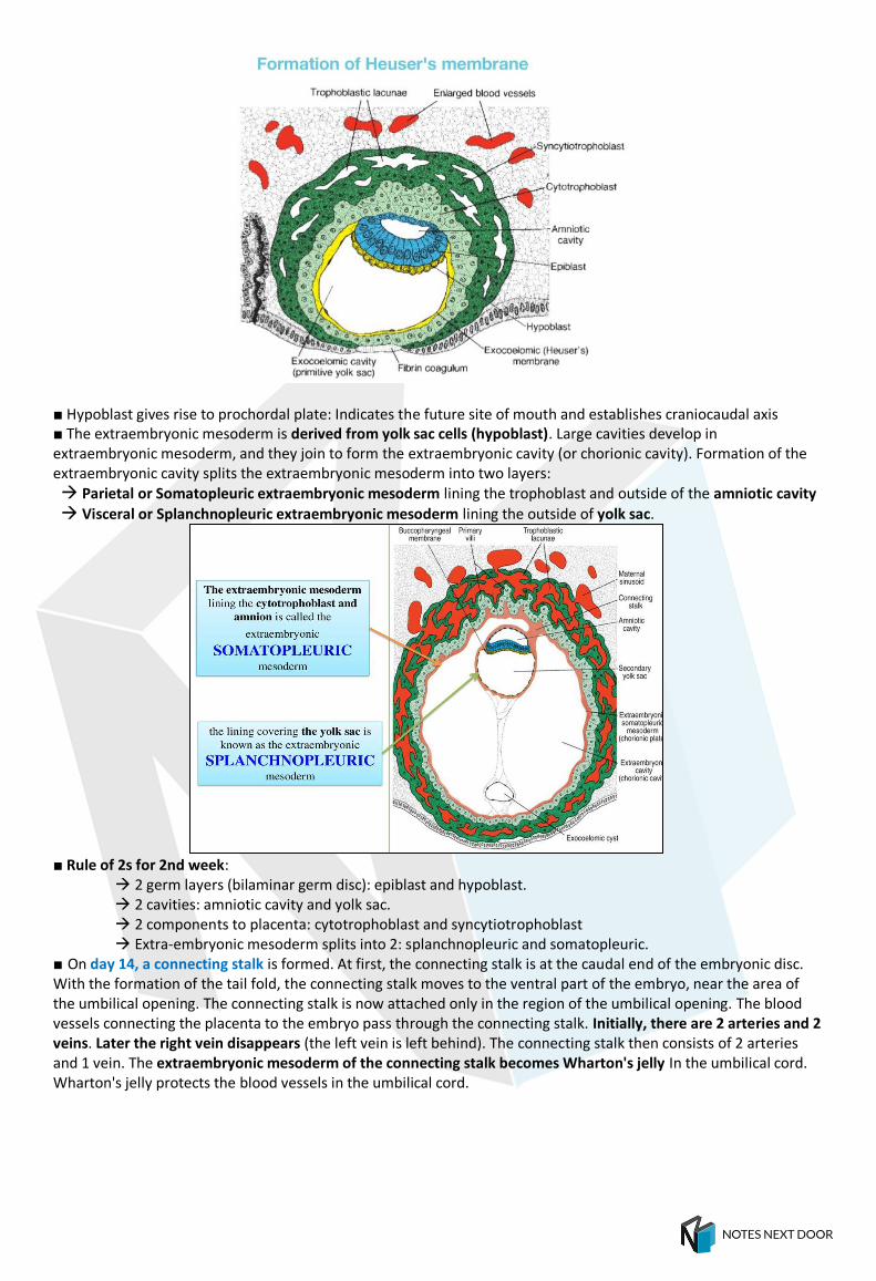
EMBRYONIC PERIOD (3–8 WEEKS) Embryonic Period is the stage during which, each of the three germ layers (ectoderm, endoderm and mesoderm) gives rise to its own tissues and organ systems. Stage of Trilaminar Germ Disc—3rd Week ■ Gastrulation: During the 3rd week of intra-uterine life, three definitive germ layers are formed (ectoderm, mesoderm, and endoderm). These three germ layers develop from the epiblast by a process of cell proliferation and migration known as gastrulation. This transforms the bilaminar germ disc into a trilaminar structure. ■ The beginning of gastrulation is marked by formation of primitive streak. ■ By the 15th day of development, some epiblast cells (ectoderm) lying along the central axis, near the caudal end of the embryonic disc, begin to proliferate. These proliferated cells form an elevation called primitive streak that bulges into the amniotic cavity.
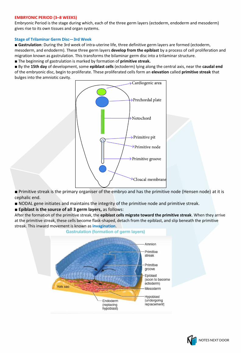
■ Primitive streak is the primary organiser of the embryo and has the primitive node (Hensen node) at it is cephalic end. ■ NODAL gene initiates and maintains the integrity of the primitive node and primitive streak. ■ Epiblast is the source of all 3 germ layers, as follows: After the formation of the primitive streak, the epiblast cells migrate toward the primitive streak. When they arrive at the primitive streak, these cells become flask-shaped, detach from the epiblast, and slip beneath the primitive streak. This inward movement is known as invagination.

Some of the invaginated epiblast cells displace the hypoblast to create the embryonic endoderm. Endoderm is the first germ layer to form. Some of the invaginated cells then start occupying the space between the epiblast and newly created endoderm to form mesoderm. Cells remaining in the epiblast then form the ectoderm.
The intraembryonic mesoderm (also known as secondary mesoderm) is formed by the invagination and movement of epiblast cells between the newly formed endoderm and the epiblast.This intraembryonic mesoderm spreads throughout the disc, except in the region of the prechordal plate and cloacal membrane. In these two regions, the ectoderm is in direct contact with the endoderm. In the later stages of development, the ectoderm and endoderm mostly persist as lining epithelium. Mesoderm forms most of the tissues of the body, including bone, cartilage, connective tissue, muscles (skeletal, smooth, cardiac), blood, lymph, and cardiovascular organs.
■ Primitive streak regresses by 4th week (26th day). ■ The buccopharyngeal membrane at the cranial end of the trilaminar germ disc consists of a small region of tightly adherent ectoderm and endoderm cells that represent the future opening of the oral cavity. Formation of notochord - Steps: 1. Primitive knot: The cranial end of the primitive streak thickens to form the primitive knot (primitive node or Hensen's node). 2. Primitive pit (Blastopore): A depression appears in the center of the knot known as the primitive pit (or blastopore). 3. Notochordal process: The cells in the primitive knot multiply and pass cranially in the midline between the ectoderm and endoderm, reaching up to the caudal margin of the prechordal plate. These cells form a solid chord known as the notochordal process or head process. 4. Notochordal canal: The cavity of the blastopore then extends into the notochordal process to form a tube-like structure called the notochordal canal. 5. Notochordal plate: The floor of the notochordal canal gets intercalated (mixed up) with the endoderm and gradually breaks down. This eventually leads to the formation of a flat plate-like structure notochordal plate. 6. Definitive notochord: The cells from the notochordal plate proliferate to form a solid rod-like structure called a definitive notochord.
■ Notochord defines the axis and provides central axis to the embryonic disc. ■ The notochord induces the overlying ectoderm to thicken and become neuroectodermal cells and thereby form the neural plate, which later forms the neural tube.
■ Although the notochord regresses, it has 2 important remnants in adult: nucleus pulposus of intervertebral disc and apical ligament of dens of axis vertebra.
Extra Edge
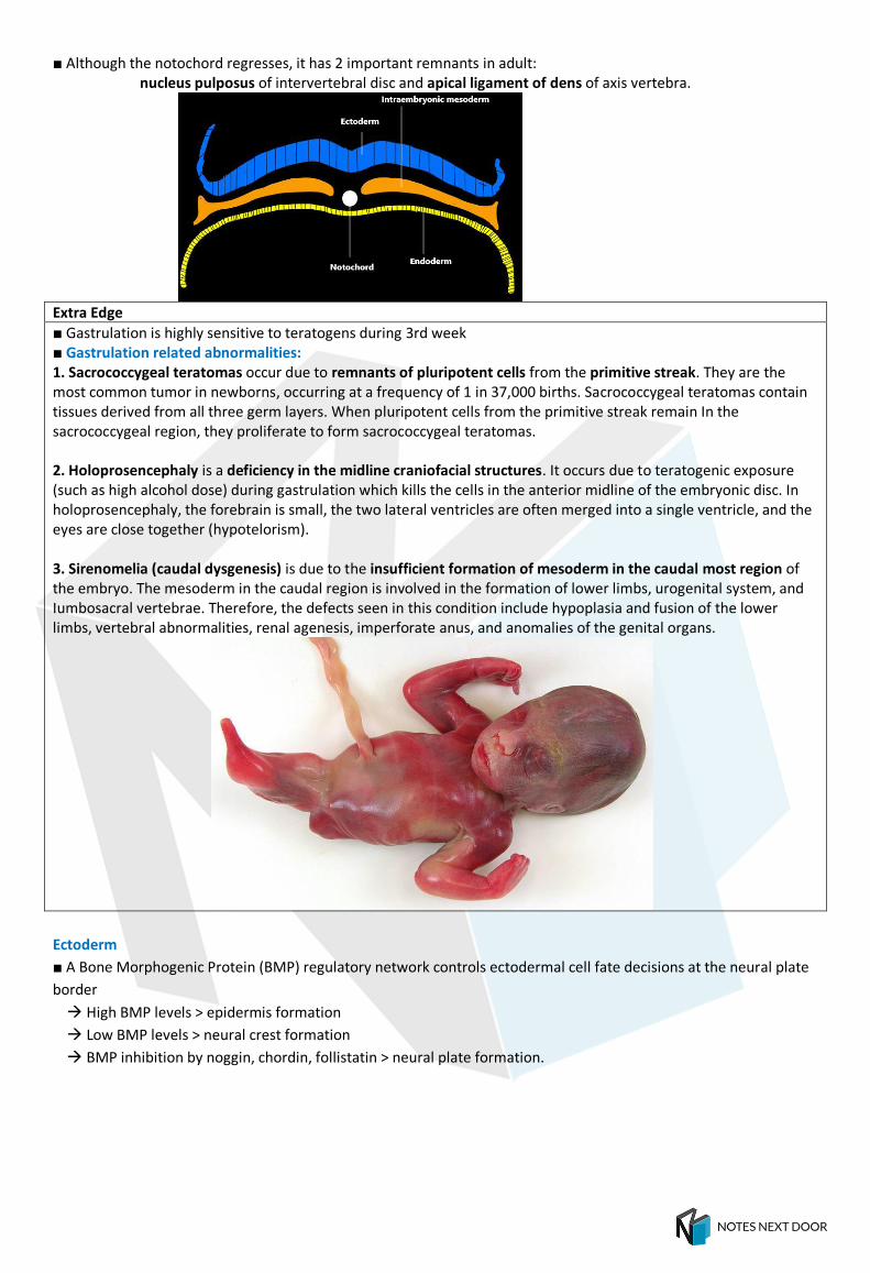
■ Gastrulation is highly sensitive to teratogens during 3rd week ■ Gastrulation related abnormalities: 1. Sacrococcygeal teratomas occur due to remnants of pluripotent cells from the primitive streak. They are the most common tumor in newborns, occurring at a frequency of 1 in 37,000 births. Sacrococcygeal teratomas contain tissues derived from all three germ layers. When pluripotent cells from the primitive streak remain In the sacrococcygeal region, they proliferate to form sacrococcygeal teratomas. 2. Holoprosencephaly is a deficiency in the midline craniofacial structures. It occurs due to teratogenic exposure (such as high alcohol dose) during gastrulation which kills the cells in the anterior midline of the embryonic disc. In holoprosencephaly, the forebrain is small, the two lateral ventricles are often merged into a single ventricle, and the eyes are close together (hypotelorism). 3. Sirenomelia (caudal dysgenesis) is due to the insufficient formation of mesoderm in the caudal most region of the embryo. The mesoderm in the caudal region is involved in the formation of lower limbs, urogenital system, and Iumbosacral vertebrae. Therefore, the defects seen in this condition include hypoplasia and fusion of the lower limbs, vertebral abnormalities, renal agenesis, imperforate anus, and anomalies of the genital organs.
Ectoderm
■ A Bone Morphogenic Protein (BMP) regulatory network controls ectodermal cell fate decisions at the neural plate
border
High BMP levels > epidermis formation
Low BMP levels > neural crest formation
BMP inhibition by noggin, chordin, follistatin > neural plate formation.
Neurulation
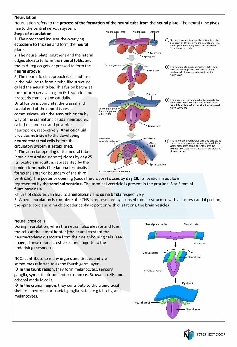
Neurulation refers to the process of the formation of the neural tube from the neural plate. The neural tube gives rise to the central nervous system. Steps of neurulation 1. The notochord induces the overlying ectoderm to thicken and form the neural plate. 2. The neural plate lengthens and the lateral edges elevate to form the neural folds, and the mid- region gets depressed to form the neural groove. 3. The neural folds approach each and fuse in the midline to form a tube-like structure called the neural tube. This fusion begins at the (future) cervical region (5th somite) and proceeds cranially and caudally. Until fusion is complete, the cranial and caudal end of the neural tubes communicate with the amniotic cavity by way of the cranial and caudal neuropores called the anterior and posterior neuropores, respectively. Amniotic fluid provides nutrition to the developing neuroectodermal cells before the circulatory system is established. 4. The anterior opening of the neural tube (cranial/rostral neuropore) closes by day 25. Its location in adults is represented by the lamina terminalis (The lamina terminalis forms the anterior boundary of the third ventricle). The posterior opening (caudal neuropore) closes by day 28. Its location in adults is represented by the terminal ventricle. The terminal ventricle is present in the proximal 5 to 6 mm of filum terminale. Failure of closures can lead to anencephaly and spina bifida respectively 5. When neurulation is complete, the CNS is represented by a closed tubular structure with a narrow caudal portion, the spinal cord and a much broader cephalic portion with dilatations, the brain vesicles.
Neural crest cells: During neurulation, when the neural folds elevate and fuse, the cells at the lateral border (the neural crest) of the neuroectoderm dissociate from their neighbouring cells (see image). These neural crest cells then migrate to the underlying mesoderm. NCCs contribute to many organs and tissues and are sometimes referred to as the fourth germ layer: In the trunk region, they form melanocytes, sensory ganglia, sympathetic and enteric neurons, Schwann cells, and adrenal medulla cells. In the cranial region, they contribute to the craniofacial skeleton, neurons for cranial ganglia, satellite glial cells, and melanocytes.

Mesoderm (Intraembryonic mesoderm) ■ After the notochord is formed, the intraembryonic mesoderm on either side can be seen organized into 3 layers: 1. Paraxial mesoderm: Thickened plate of the mesoderm close to the midline, on either side of the notochord. It forms: Somitomeres: They are rounded structures, formed in the region of the head—7 in number and gives rise to mesoderm and muscles of the head and jaw. Somites: From the occipital region below, somitomeres further organize Into somites. The first pair of somites arise in the occipital region of the embryo at approximately the 20th day of development. From here, new somites appear In craniocaudal sequence at a rate of approximately three pairs per day. By the end of the 5th week, 42 to 44 pairs of somites are present. There are 4 occipital, 8 cervical, 12 thoracic, 5 lumbar, 5 sacral, and 8 to 10 coccygeal pairs. The first occipital and the last five to seven coccygeal somites later disappear. The remaining somites form the axial skeleton.
The days 20–30 is called ‘somite period of human development’. Age of embryo upto 5 weeks is calculated by estimating the number of somites (forensic importance!). Each somite is divided into three parts - medial sclerotome; middle myotome and lateral dermatome. Origin of Intervertebral dics: Peripheral Annulus fibrosis is from sclerotome Central nucleus pulposus is a remnant of notochord 2. Intermediate mesoderm: Longitudinal strip of mesoderm that connects paraxial with the lateral plate mesoderm.It gives rise to nephrotomes and nephrogenic cord and ultimately forms the genital ridge and urogenital system (kidneys and gonads).

3. Lateral Plate mesoderm = A thinner layer of mesoderm that lies laterally. This layer develops a large cavity called the intraembryonic coelom. The lateral plate mesoderm is divided by this intraembryonic celom to form: Somatopleuric or parietal layer of intraembryonic mesoderm that is in contact with the ectoderm. This forms the parietal layer of peritoneal, pleural, and pericardial membranes. Splanchnopleuric or visceral layer of intraembryonic mesoderm that is in contact with endoderm. This forms the visceral layer of the peritoneal, pleural, and pericardial membranes.
The lateral plate mesoderm also gives rise to wall of the gut, smooth muscle component of dorsal aorta/descending aorta. Flash card:
TIMELINE OF EVENTS AFTER FERTILIZATION
Age in days Developmental events
< 24 hours Fertilization zygote is formed; cleavage begins immediately
2-3 days Embryo , two cell stage 5-8 cell stage
3-4 days Stage of compaction and formation of Morula (12-16 cell stage)
4 The advanced Morula enters the uterine cavity; Early blastocyst is formed
5 Late blastocyst is formed
5-6 Zona pellucida disappears
6-7 Implantation of blastocyst occurs (Implantation corresponds to 20-22 days of the menstrual cycle)
8 Bilaminar disc is formed; Trophoblast differentiates into cytotrophoblast and syncytiotrophoblast
9 Primary yolk sac forms
10-11 Embryo completely implanted into endometrium
12 Formation of extraembryonic mesoderm
13 Formation of primary villi
14 Formation of Prochordal plate and connecting stalk
15 Formation of primitive streak

16 Intraembryonic mesoderm is formed; disc is now three layered – gastrulation starts; Formation of secondary and tertiary villi
18 Formation of trilaminar embryonic disc
19 Neurulation begins; formation of neural plate
20 Formation of neural folds and neural; groove
21 Formation of first pair of somites; fetoplacental circulation established
3-8 weeks Internal organs develop
4th week Forelimb buds appears (26th day) and hindlimb buds (28th day) Formation of face starts (continues upto 8th week) Heart begins to beat
8-12 weeks Sex identifiable
10-12 weeks Swallowing
11 weeks Fetal breathing movements
12 weeks Fetal urine formation occurs

DERIVATIVES OF GERM LAYERS
Ectoderm:
Surface ectoderm Neurectoderm Neural crest
1. Epithelial linings of: • Lower anal canal • Distal part of male urethra • External auditory meatus ƒ 2.Skin: Epidermis, hair, nails, sweat and sebaceous glands ƒ 3.Ear: • External ear/Pinna • Inner ear (utricle, semicircular canals, vestibular ganglion of CN VIII, saccule, cochlear duct (organ of Corti), spiral ganglion of CN VIII ƒ 4.Glands: • Anterior pituitary, adenohypophysis (from Rathke’s pouch – a thickened oral ectoderm) • Parotid gland • Mammary gland ƒ 5.Olfactory placode ƒ 6.Tooth enamel (ameloblasts) ƒ 7.Eye: • Lens of the eye • Conjunctival and Corneal epithelium • Lacrimal glands • Tarsal glands (Meibomian, Zeis, Moll)
1.Neural tube ƒ 2.All CNS neurons (Astrocytes, Oligodendrocytes, Ependymocytes, Tanycytes, choroid plexus cells) ƒ 3.Glands: • Posterior pituitary (neurohypophysis) • Pineal gland ƒ 4.Eye: • Smooth muscles of iris (Sphincter and Dilator pupillae) • Epithelium of Iris and Ciliary body • Retina, Retinal pigment epithelium • Optic Nerve • Optic vesicle and cup
1.Cells: • Melanoblast/MelaNoCytes (except Retinal Pigment Epithelium) • SchwanN Cells and Satellite glial cells • ChromaffiN Cells • Parafollicular C cells of thyroid • Trunk and Cranial neural crest cells ƒ 2.Adrenal medulla ƒ 3.Connective tissue and bones of face and Skull 4.Pia and arachnoid mater ƒ 5.Pharyngeal arch cartilage ƒ 6.Odontoblasts (dentin of teeth) ƒ 7.Heart: Aorticopulmonary septum (conotruncal septum) and endocardial cushions ƒ 8.All Ganglia and All enteric plexus • Dorsal root ganglia of spinal nerves • Sensory ganglia of Cranial Nerves 5,7,9 and 10 • Sympathetic chain ganglia: prevertebral sympathetic ganglia • Parasympathetic ganglia of the gut (Meissner’s and Auerbach’s plexus, CN X) and abdominal pelvic cavity • Parasympathetic ganglia of cranial nerves: Ciliary (CNIII), Pterygopalatine (CN VII), submandibular (CN VII) and Otic (CN IX) ganglion ƒ 9.Eye: • Ciliary muscles and Iris stroma • Corneal stroma, keratocytes and corneal endothelium • Trabecular meshwork endothelium • Sclera

Mesodermal derivatives:
Endoderm derivatives 1. Epithelial lining of: • GI tract • Respiratory tract: pharynx, larynx, trachea, bronchi, lungs ƒ 2. Urogenital tract: • Urinary bladder (except trigone) • Female urethra and most of male urethra, inferior 2/3 of vagina ƒ 3. Ear: • Tympanic cavity • Middle ear • Auditory tube • Pharyngotympanic (Eustachian) tube - from proximal part of tubotympanic recess) ƒ 4. Organs: • Liver (hepatocytes), gallbladder • Lungs • Tonsils ƒ 5. Glands: • Pancreas • Thyroid and parathyroid • Glands of GI tract, submandibular and sublingual glands

Branchial/Pharyngeal apparatus ■ Pharyngeal arches appear in the 4th or 5th week of development and contribute to the formation of the head and neck region. They are formed as a series of 6 thickenings in the cranial most part of the foregut (the future pharynx). They are craniocaudally numbered from I to VI. The 3rd, 4th and 6th arches do not have special names. The 5th arch disappears soon after formation so that only five arches remain. ■ Branchial (pharyngeal) apparatus/arches initially have identical structures: 1. Surface ectoderm on the outside 2. A mesenchymal core derived from mesoderm 3. Endoderm derived epithelium on the inside. [The mesoderm in the arches is derived from paraxial mesoderm and lateral plate mesoderm. This is invaded by the neural crest cells that contribute to the formation of skeletal elements and connective tissue of the head and neck region.] ■ Fully developed branchial (pharyngeal) apparatus/arches is composed of (‘CAP’ from outside to inside) as follows: 1. Branchial/Pharyngeal Clefts – derived from ectoderm 2. Branchial/Pharyngeal Arches (core of arches) – derived from mesoderm and neural crests. 3. Branchial/Pharyngeal Pouches – derived from endoderm.
■ Pharyngeal membrane: In the Interval between any two adjoining pharyngeal arches, the endoderm extends outward in the form of pharyngeal pouch (or endodermal pouch) to meet the ectoderm, which dips into this interval as a pharyngeal cleft (or ectodermal cleft). The membrane formed where the pharyngeal cleft comes in contact with the pouch is called the pharyngeal membrane. Initially, there are four pharyngeal membranes between the five arches. Only the first pharyngeal membrane (which separates the 1st pharyngeal pouch from the 1st pharyngeal groove) develops and contributes to the formation of the tympanic membrane and this represents all three layers of the embryonic disc (ecto, meso and endoderm). The 2nd, 3th, and 4th pharyngeal membranes do not develop further into any structure. They get obliterated when the mesoderm from the 2nd arch overgrows and fills the succeeding clefts. ■ Each arch has 2 nerves - Pre-trematic (along the caudal border of the arch) and Post-trematic (along the cephalic border of each arch); In all arches ONLY the post-trematic nerve persists EXCEPT in the first arch where both persist as mentioned in table. ■ Each pharyngeal arch is characterized by Its own muscular components. The muscular components of each arch have their own cranial nerve, and wherever the muscle cells migrate, they carry their nerve component with them. The neurons of 5th, 7th, 9th, and 10th cranial sensory ganglia that supply the pharyngeal arch structures are derived from ectodermal placodes (epipharyngeal placodes) and from the neural crest cells. ■ The above mentioned cranial nerves – V, VII, IX, X are the only ones with both motor and sensory components.