Hydrophobins In Wood biology and Biotechnology
278
Hydrophobins In Wood biology and Biotechnology Dissertation In Partial Fulfillment of the Requirements for the Degree Doctor of Philosophy (PhD) Of the Faculty of Forest Sciences and Forest Ecology Georg-August-University of Göttingen Submitted by Sudhakar Peddireddi Born in Machilipatnam India Göttingen, 2008
-
Upload
khangminh22 -
Category
Documents
-
view
4 -
download
0
Transcript of Hydrophobins In Wood biology and Biotechnology

Hydrophobins
In Wood biology and Biotechnology
Dissertation
In Partial Fulfillment of the Requirements for the Degree Doctor of Philosophy (PhD)
Of the Faculty of Forest Sciences and Forest Ecology Georg-August-University of Göttingen
Submitted by
Sudhakar Peddireddi
Born in Machilipatnam India
Göttingen, 2008

D7 Prof. Dr. Ursula Kües, examiner Prof. Dr. Stefan Schütz, co-examiner Prof. Dr. Reiner Finkeldey, co-examiner Date of Oral Examination: 28th March 2008

Acknowledgements
i
Acknowledgements
It is pleasure to thank all the people who made this thesis possible. This is perhaps the
hardest chapter that I have to write because it is simple to name all the people that
helped to get this PhD work done, but it will be tough to thank them enough. I will
try……
I would like to express my sincere thanks to Prof. Dr. Ursula Kuees and Prof. Dr.
Andrea Polle for giving me the opportunity to work for this PhD and for their
guidance in finishing this research work. I would like to extend my gratitude to Prof.
Dr. Stefan Schütz for his willingness to evaluate my thesis and many thanks to Prof.
Dr. Reiner Finkeldey for accepting to be one of my examiners.
My gratitude to Dr. Andrzej Majcherczyk for introducing me to the protein work,
supervision and for stimulating discussions. Special thanks to Dr. Patrik Hoegger
from whom I learnt not only scientific things but also many potential tips beyond
research that are useful to lead a happy life. Mojtaba Zomorrodi is greatly
acknowledged for the technical support, being available all the time and keeping
friendly environment in the lab.
I am indebted to my many student colleagues Dr. Sreedhar Kilaru, Dr. Rajesh
Velagapudi, Dr. Ravi Chandra Dwivedi, Dr. Prayook Srivilai, Martin Rühl, Dorothea
Fragner, Monica Navarro Gonzalez, Wassana Chaisaena, Christa Lang, Katerina
Svobodova, Ihtzaz Malik, Banyat Cherdchim, Dong Sheng Wei for providing their
constant support and a stimulating and fun environment in which to learn and grow.
Many thanks to Dr. Jhansi Kalyani Pemmasani for her constant help and moral
support.
Special thanks to Dr. Annette Naumann and Dr. Andrea Olbrich not only for helping
me with FTIR analysis and microscopy but also for their encouraging support all the
time. I can never forget Karin Lange and Alexandra Dolynska for their immense
support during my hard days. I thank Dr. Kürsten, Dr. Büttner and all fellow members
in our PhD programme. Special thanks to Dr. Bernd Kopka for help in all computer

Acknowledgements
ii
related things and to Marianne Smiatacz for providing the cleaned glass ware for the
experiments all the time.
I am always grateful to Prof. Dr. Sarath Babu Mogallapu for being my mentor through
out my education career. I thank our collaborator Prof. Dr. H.A.B Wösten for
providing me with the fungal strains used in this work. Thanks to Dr. Oliver
Weigenand for introducing me to the decay experiments and Dr. Payam Fayyaz for
helping in the statistical analysis.
Many thanks to Prof. Dr. Alireza Kharazipour, Dr. Christian Schöpper, Christian
Bonn, Dr. Lars Kloeser and Dr. Cora Müller for their readiness in helping on strength
test of wood, discussions and support. Thanks to Gisbert Langer-Kettner for being
always ready to cut wood samples for my experiments and to Volker Meng for
sharing information regarding S. commune infected Juglans tree.
I cannot forget the support given by all the members of the institute especially Karin,
Monica, Martin, Manika, Annette, Rodica, Andrzej, Ursel, and many others in my
hard days during 2006 & 2007. Above all, I would like to thank Göttingen University,
all the Germans, my Indian friends in Germany who had provided me a friendly
environment during my stay in Germany.
I am very grateful to Competence Network for the sustainable use of wood (NHN-
Kompetenznetz für Nachhaltige Holznutzung) and University of Göttingen for the
financial support for this research work.
I cannot end without thanking my family, on whose constant encouragement and love
I have relied throughout my time at the institute. This thesis is dedicated to my
parents, my brother and to the invisible power in this world (God), without whom
none of this would have been possible.

Zusammenfassung
iii
Zusammenfassung
Ziel dieser Arbeit war es, die Rolle von Hydrophobinen beim Holzabbau und ihre
Fähigkeit, Oberflächen – insbesondere Holzoberflächen - zu beschichten, zu
untersuchen. Hydrophobine sind kleine amphipatische pilzliche Proteine, die dafür
bekannt sind, die Oberflächenspannung von feuchten Oberflächen zu vermindern
(Wasser/Luft-Grenzflächen), die Bildung von Lufthyphen durch Beschichtung mit
einem hydrophoben Proteinfilm zu erleichtern und das Anhaften von pilzlichen
Hyphen an hydrophobes Material zu unterstützen. Das SC3-Hydrophobin von
Schizophyllum commune ist bisher das bestuntersuchte Klasse I-Hydrophobin. Ein
weiteres Protein, SC15, unterstützt SC3 bei der Bildung von Lufthyphen und ihrer
Anheftung an Oberflächen. In Abwesenheit des SC3-Hydrophobins verleiht es
Hyphen eine geringe Hydrophobizität. Da von dem Weißfäulepilz S. commune SC3-
und SC15-Hydrophobin-Mutanten existieren, wurde diese Art ausgewählt, um die
Funktion dieser Proteine bei Holzbesiedelung und -abbau zu untersuchen. Versuche
mit Holzblöcken und Sägemehl mit verschiedenen S. commune-Stämmen haben
gezeigt, dass sowohl pilzliche Mono- als auch Dikaryen - einschließlich co-isogenen
Wildtyp-Stämmen, ∆Sc3-Mutanten, ∆Sc15-Mutanten und ∆Sc3-∆Sc15-
Doppelmutanten - auf Buchen-, Birken- und Kiefernholz wachsen konnten. Die
Ergebnisse zeigen, dass weder SC3 noch SC15 notwendig sind, damit S. commune in
das Holz eindringen, das Holz abbauen und die Holzfestigkeit beeinflussen kann.
Allerdings unterschied sich das Ausmaß der Myzelbildung an der Holzoberfläche in
Abhängigkeit vom Vorhandensein von SC3-Hydrophobin. Meistens war der
Holzabbau durch die Pilzstämme gering (Laubholz) oder fand nicht statt
(Kiefernholz). Bei den Versuchen mit Holzblöcken und Sägemehl betrug der
maximale Abbau durch S. commune-Stämme 4-5% (Buche und Birke) bzw. 9-16%
(Buche und Kiefer). S. commune-Stämme entfärbten das Kiefernsägemehl, nicht aber
das Buchen- und Birkensägemehl. Obwohl die Holzblöcke nur geringfügig oder gar
nicht abgebaut wurden, wurde die Holzfestigkeit negativ durch die S. commune-
Stämme beeinflusst. Der maximale Festigkeitsverlust durch S. commune-Stämme
betrug bei Buche, Birke bzw. Kiefer 21, 52 bzw. 35%.

Zusammenfassung
iv
S. commune ist ein opportunistischer Krankheitserreger von geschwächten Bäumen.
Die Besiedelung eines Juglans ailantifolia-Baumes durch den Pilz wurde während
einer Zeitspanne von drei Jahren beobachtet. Der Pilz verursachte während dieser Zeit
das Absterben mehrerer Äste. An mindestens einem Ast war S. commune nicht der
einzige Pilz. Neben Fruchtkörpern von S. commune wurden solche von Trametes
hirsuta gefunden. Beide Pilze wurden von diesem Baum isoliert und ihre Interaktion
untersucht.
Interaktionsuntersuchungen von S. commune und T. hirsuta resultierten im
„Deadlock“-Phänomen, bei dem sich eine Grenzzone zwischen den Pilzstämmen
bildet, in der das Weiterwachstum der Opponenten verhindert wird. Mit
fortschreitender Zeit verdrängte T. hirsuta S. commune. Als Stressreaktion während
der Konkurrenzsituation war eine blaue Pigmentierung an der pilzlichen
Interaktionszone zu beobachten. Alle S. commune-Stämme (monokaryotische und
dikaryotische Wildtyp-Stämme und monokaryotische Hydrophobin-Mutanten) außer
der dikaryotischen SC3-Hydrophobin-Mutante bildeten die Grenzzone und
Pigmentierung in der Interaktionszone aus. Dies deutet darauf hin, dass das SC3-
Hydrophobin nicht essentiell an der Stressreaktion von S. commune beteiligt ist.
Protein- und Polysaccharid-Produktion eines S. commune-Wildtyp-Stamms und einer
co-isogenen Hydrophobin-Mutante in Flüssigkultur wurden verglichen. In
Flüssigkultur wurde mittels FTIR-(Fourier-Transform-Infrarot)-Spektroskopie
festgestellt, dass sich die Protein- und Polysaccharidgehalte im Myzel des Wildtyps
und der Hydrophobin-Mutante signifikant unterscheiden. Der Proteingehalt des
Wildtyps war höher als der in der Mutante, während der Polysaccharidgehalt der
Mutante mit der Zeit höher war. Myzel von verschiedenen Wildtyp-Stämmen und co-
isogenen Mutanten, das auf der Oberfläche von Buchenholz gewachsen war, wurde
durch Clusteranalyse von FTIR-Spektren voneinander unterschieden. FTIR-Spektren
von Myzel verschiedener Wildtyp-Stämme und co-isogener Mutanten, das im Holz
gewachsen war, bildeten einen gemeinsamen Cluster. Die Flächen der Protein-
„Peaks“ der untersuchten Stämme unterschieden sich auf der Holzoberfläche nicht
signifikant voneinander. Die Flächen der Polysaccharid-„Peaks“ der dikaryotischen
SC3-Hydrophobin-Mutante ∆Sc3 4-39 x ∆Sc3 4-40 waren auf der Holzoberfläche

Zusammenfassung
v
hingegen signifikant höher als die der monokaryotischen Mutanten ∆Sc3 4-39 und
∆Sc3 4-40.
Das SC3-Hydrophobin von S. commune wurde aus dem Pilzmyzel mit einem Ertrag
von 0.1 % (w/w, 0.5 mg an SC3-Hydrophobin aus 500 mg Myzel) aufgereinigt und
zum Beschichten von hydrophobem und hydrophilem Material verwendet.
Hydrophobes Teflon und Formwar-Filme wurden hydrophil und hydrophiles Glas
wurde hydrophob nach Beschichtung mit SC3-Hydrophobin. Weiter wurden die
amphipatischen Eigenschaften von Proteinfilmen auf Buchenholz getestet.
Buchenholz wurde in Abhängigkeit von der verwendeten SC3-Konzentration
hydrophil oder hydrophob. Niedrige SC3-Konzentrationen (50, 25, 15 µg/ml)
bewirkten Hydrophobie und hohe Konzentrationen (100 bis 400 µg/ml) Hydrophilie
bei Buchenholz. Oberflächen-Aktivitäten weiterer Klasse I-Hydrophobine von
anderen Pilzarten wurden an Buchenholz untersucht. Circa 0,3 bis 0,1% (w/w)
Hydrophobin ließ sich aus Myzel von Coprinopsis cinerea und Pleurotus ostreatus-
Kulturen aufreinigen. Die Hydrophobine dieser beiden Pilze wiesen die gleichen
Eigenschaften bezüglich der Oberflächenaktivität auf Holz auf wie das SC3-
Hydrophobin. ATR-FTIR-(Abgeschwächte Totalreflexions-Fourier Transform
Infrarot)-Spektren von mit Hydrophobin behandeltem Holz zeigten eine höhere
„Peak-Intensität“ in dem Spektralbereich, der der Amid I-Bande von Proteinen
zugeschriebenen wird, als unbehandeltes Holz, was für die Bindung der
Hydrophobine an Holzkomponenten spricht.


Summary
vi
Summary The main aims of this research were to study the role of hydrophobins in wood decay
processes and to test the ability of these proteins to coat surfaces, particularly wood
surfaces. Hydrophobins are small amphiphatic fungal proteins that are known to
decrease the surface tension of moist surfaces (water/air interfaces), to help in the
formation of aerial hyphae by coating them with a protein film and making them
hydrophobic and to support attachment of the fungal hyphae to hydrophobic
materials. The SC3 hydrophobin from Schizophyllum commune is the best studied
hydrophobin until now. Another protein, SC15, is known to act supportive to the SC3
hydrophobin in the formation of aerial hyphae and their attachment to surfaces and, in
the absence of the SC3 hydrophobin, it mediates low levels of hyphal hydrophobicity
and attachments. Due to the existence of several SC3 hydrophobin and SC15 mutant
strains, the white rot fungus S. commune was selected for this study to study the
functions of these proteins in wood colonization and decay. Wood block and saw dust
tests with various S. commune strains showed that both fungal monokaryons and
dikaryons, including co-isogenic wild type strains, ∆Sc3 mutants, ∆Sc15 mutants and
∆Sc3 ∆Sc15 double mutants, were able to grow on beech, birch and pine wood. The
obtained results indicate that neither the SC3 hydrophobin nor the SC15 protein are
principally necessary for S. commune to enter into the wood, to decay the wood or to
affect the strength to the wood. However, there were differences in the overall
formation of surface mycelium in correlation of the presence of the SC3 hydrophobin.
In most instances, the wood decay by the fungal strains were low (deciduous wood) or
there was no decay (pine wood). In wood block and saw dust decay tests, the
maximum decay caused by S. commune strains was 4-5% (beech and birch), and
9-16% (beech and birch), respectively. S. commune strains decolorized the pine saw
dust but not beech or birch saw dust. Although there was only little or no decay in
wood blocks, the strength of the wood was negatively affected by S. commune strains.
The maximum strength loss caused by S. commune strains in beech, birch and pine
wood was 21, 52 and 35 %, respectively.
S. commune is an opportunistic pathogen on weakened trees. An infestation of a
Juglans ailantifolia tree by the fungus was observed over a period of 3 years. The
fungus caused over the time die off of a number of branches. In at least one branch,

Summary
vii
S. commune was not the only fungus. Next to S. commune fruiting bodies,
carpophores of Trametes hirsuta were observed. Both fungi were isolated from the
tree and studied in interactions. Furthermore, SC3 hydrophobin mutants and their co-
isogenic wild type strains were included in the study.
Interaction studies of S. commune strains with T. hirsuta on agar plates resulted in the
deadlock phenomenon in which a barrier is formed between the fungal strains
hindering the opponents in further growth. Subsequently with time, the deadlock
situation changed by the partial replacement of S. commune by T. hirsuta. As a stress
reaction during the combat interaction, a blue pigmentation was observed at the
fungal interaction zone. All the S. commune strains (monokaryotic and dikaryotic wild
type strains and hydrophobin monokaryon mutants) except the SC3 hydrophobin
mutant dikaryon showed the barrier formation and the pigmentation in the interaction
zone, indicating that in principle the SC3 hydrophobin is not involved in the stress
reactions of S. commune.
Further, differences between a S. commune wild type strain and a coisogenic
hydrophobin mutant were studied in liquid cultures in relation to the produced protein
and polysaccharide. In the liquid cultures, as determined by the FTIR (Fourier
Transform Infrared) spectroscopy analysis, there were significant differences between
the mycelium of the wild type strain and the mycelium of the hydrophobin mutant
with respect to protein and polysaccharide contents. The protein content were higher
in the wild type strain than in the mutant, whereas the polysaccharide content was
higher over the time in the mutant strain. When growing on beech wood, the
mycelium of different S. commune wild type strains and the co-isogenic hydrophobin
mutants on the surface of the wood were discriminated by FTIR spectra submitted to a
cluster analysis. Inside the wood, however, the FTIR spectra of the mycelia of the
different S. commune wild type strains and hydrophobin mutants clustered together.
There were no significant differences noted amongst the values from protein peak
area values calculated from the FTIR spectra in between the tested strains on the
surface of beech wood. On the surface of wood, the polysaccharide peak area value of
the dikaryotic Sc3 hydrophobin mutant ∆Sc3 4-39 x ∆Sc3 4-40 was significantly
higher than that of the monokaryotic mutants ∆Sc3 4-39 and ∆Sc3 4-40.

Summary
viii
SC3 hydrophobin of S. commune was purified from fungal mycelium with yields of
0.1% (w/w, 0.5 mg of SC3 hydrophobin from 500 mg of mycelium) and used to coat
hydrophobic and hydrophilic materials. Hydrophobic Teflon and form-war film
turned hydrophilic and hydrophilic glass turned hydrophobic upon coating with SC3
hydrophobin. Further, the amphipathic nature of films of this protein was tested on
solid beech wood. Beech wood turned hydrophilic or hydrophobic, depending on the
concentration of SC3 hydrophobin applied. Lower concentrations (50, 25, 15 µg/ml)
of SC3 resulted in hydrophobic behavior and higher concentrations (100 to 400
µg/ml) in hydrophilic behavior of beech wood. The surface activity of other class I
hydrophobins from different fungal species were also tested on the beech wood.
About 0.3 and 0.1% (w/w) purified hydrophobins were obtained from the mycelium
of Coprinopsis cinerea and Pleurotus ostreatus cultures, respectively. The
hydrophobins of both fungi followed the same pattern of surface activity on wood
than the Sc3 hydrophobin. Results from ATR-FTIR (Attenuated Total Reflection -
Fourier Transform Infrared) spectra of hydrophobin-treated beech wood showed a
higher intensity in the peak associated with amide I band of proteins indicating the
binding of hydrophobin protein to the wood components.


Table of contents
ix
Table of contents
Acknowledgements i
Zusammenfassung iii
Summary vi
Table of contents ix
1. General Introduction 1
1.1 General background and aim of this thesis 1
1.2 The fungus Schizophyllum commune 2
1.3 Schizophyllum commune growth on wood 3
1.4 Application of hydrophobins with wood 4
1.5 References 4
2. Growth of Schizophyllum commune in natural environments and
interaction with other fungi 9
2.1 Abstract 9
2.2 Introduction 10
2.3 Materials and Methods 13
2.3.1 Juglans tree 13 2.3.2 Isolation of fungal strains from wood 13
2.3.3 Fungal strains 13
2.3.4 Growth conditions 14 2.3.5 Juglans decay test 14
2.3.6 Interaction studies 15
2.3.7 Statistical analysis 15
2.4 Results 16
2.4.1 S. commune as a pathogen on a Juglans ailantifolia tree 16
2.4.1.1 Fungal isolations 18
2.4.2 Wood decay test 33
2.4.3 Fungal interactions 41
2.5 Discussion 50
2.6 References 53

Table of contents
x
3. Wood decay by Schizophyllum commune 57 3.1 Abstract 57
3.2 Introduction 58
3.3 Materials and methods 60
3.3.1 Fungal strains 60
3.3.2 Growth conditions 60
3.3.3 Wood decay tests 61
3.3.3.1 Wood block test 61
3.3.3.2 Saw dust decay test 61
3.3.4 Bending strength test of wood 62
3.4 Results 62
3.4.1 Wood block decay tests 62 3.4.2 Decay of saw dusts from different tree species by
S. commune strains 72
3.5 Discussion 85
3.6 References 87
4. FTIR spectroscopy of a S. commune monokaryotic wild type and a
coisogenic Sc3 hydrophobin mutant 91
4.1 Abstract 91
4.2 Introduction 92
4.2.1 FTIR Spectroscopy 92
4.2.2 IR Spectral regions for microorganisms 92
4.2.3 FTIR studies on bacteria, yeast and ascomycetes 93
4.2.4 FTIR studies on basidiomycetes 94
4.3 Materials and methods 95
4.3.1 Fungal strains 95
4.3.2 Growth conditions 95
4.3.3 Culture harvest and mycelia grinding 95
4.3.4 ATR - FTIR Spectroscopy 96
4.3.5 Data analysis 96
4.3.6 Statistical analysis 96
4.4 Results 99

Table of contents
xi
4.5 Discussion 113
4.6 References 113
4.7 Appendix to chapter 4 118
5. FTIR microscopy of S. commune in wood 128 5.1 Abstract 128
5.2 Introduction 129
5.3 Material and methods 129
5.3.1 Fungal strains 129
5.3.2 Growth conditions 130
5.3.3 Wood block test 130
5.3.4 Light Microscopy 131
5.3.5 Sample preparation for FTIR microscopy 131
5.3.6 FTIR microscopy 131
5.3.7 Spectral data analysis 132
5.3.8 Statistical analysis 132
5.4 Results 132
5.4.1 Wood decay test and light microscopy 132
5.4.2 Discrimination of fungi and wood by FTIR analysis 135 5.4.3 Discrimination of fungal strains 140
5.4.4 Distribution of S. commune mycelium within beech wood 143
5.4.5 Quantification of protein and polysaccharide contents
in the mycelium 144
5.5 Discussion 148
5.6 References 150
6. Hydrophobins in wood technology 153
6.1 Abstract 153
6.2 Introduction 154
6.3 Material and methods 155
6.3.1 Fungal strain 155
6.3.2 Growth conditions 156
6.3.3 Culture harvest 156

Table of contents
xii
6.3.4 Purification of SC3 hydrophobin 156
6.3.5 Analytical procedures 157
6.3.6 Coating SC3 hydrophobin on Teflon, form-war film and glass 157
6.3.7 Coating beech wood with SC3 hydrophobin 158
6.3.8 Water absorption measurements 158
6.3.9 FTIR-spectroscopy 158
6.3.9.1 Purified hydrophobin 158
6.3.9.2 Control Teflon and SC3 treated Teflon 159
6.3.9.3 Control beech wood and SC3 treated beech wood 159
6.3.9.4 FTIR-spectral analysis 160
6.3.9.5 Wood vessel measurement 160
6.3.9.6 Statistical analysis 161
6.4 Results 161
6.4.1 Purified hydrophobin 161
6.4.2 SC3 coatings on hydrophobic and hydrophilic materials 163 6.4.3. Hydrophobin activity on beech wood surface 165
6.4.4. FTIR-spectroscopy 168
6.5 Discussion 169
6.6 References 172
6.7 Appendix to chapter 6 177
7. Class I hydrophobins from C. cinerea and P. ostreatus in wood technology 180
7.1 Abstract 180
7.2 Introduction 181
7.3 Materials and methods 181
7.3.1 Fungal strains 181
7.3.2 Growth conditions 182
7.3.3 Culture harvest 182
7.3.4 Purification of hydrophobin from C. cinerea and
P. ostreatus aerial hyphae 182
7.3.5 Analytical procedures 183
7.3.6 Coating beech wood with hydrophobins of Coprinopsi
and Pleurotus 183

Table of contents
xiii
7.3.7 Water absorption measurements 184
7.3.8 FTIR-spectroscopy 184
7.3.8.1 Purified hydrophobin from C. cinerea and P. ostreatus 184
7.3.8.2 Control beech wood, C. cinerea and
P. ostreatus hydrophobins treated beech wood 185
7.3.8.3 FTIR-spectral analysis 185
7.3.8.4 Statistical analysis 185
7.4 Results 185
7.4.1 Purified hydrophobins from vegetative mycelium of
Coprinopsis and Pleurotus 185
7.4.2 Hydrophobin activity on beech wood surface 189
7.4.3 FTIR-spectroscopy 194
7.5 Discussion 195
7.6 References 197
7.7 Appendix 200
8. General discussion: Hydrophobins in wood biology and technology 204
8.1 Introduction 204
8.2 Why to use S. commune for this project 204
8.3 S. commune in natural environments, interaction with other
fungi and possible role of hydrophobins in combat interactions 205
8.4 Wood colonization and decay ability of S. commune and
effect of hydrophobins in protein secretion 206
8.5 Application of hydrophobins in wood technology 209
8.6 Some general conclusions from this thesis work regarding the
function of hydrophobins 210
8.7 Future Outlook 211
8.8 References 213
8.9 Appendix to the General discussion:
8.9.1 Effects of deleting the SC3 hydrophobin gene and/or the protein SC15
gene from the genome of S. commune on growth of the fungus 222
8.9.2 Introduction 222
8.10.1 Methods and materials 222

Table of contents
xiv
8.10.1.1 Fungal strains 222
8.10.1.2 Growth conditions 223
8.10.1.3 Culture harvest 223
8.10.1.4 Ergosterol measurements 223
8.10.1.4.1 Sample preparation for ergosterol 223
8.10.1.4.2 Quantitative analysis of ergosterol by GC-MS 224
8.10.1.5 2 D Electrophoresis 225
8.10.1.5.1 Precipitation of protein samples from culture
supernatants with TCA 225
8.10.1.5.2. First dimension of 2D-PAGE 226
8.10.2 Results 226
8.10.2.1 Biomass production 226
8.10.2.2 Protein gel electrophoresis 230
8.10.3 Further conclusions 230
Curriculum Vitae 232


Chapter 1
General
Introduction


General Introduction & aim of thesis
1
1. General Introduction
1.1 General background and aim of this thesis Schizophyllum commune is a widely distributed wood inhabiting basidiomycete found
throughout the tropical and temperate regions of the world (Raper 1958, James et al.
1999). It grows mainly on dead wood but can also be found as a pathogen on branches
of living trees (Adaskaveg 1993, Latham 1970, Peddireddi et al. 2005). In the
laboratory, the fungus is used as a model fungus for studying mating types and
fruiting body development in the basidiomycetes (Raper & Fowler 2004). S. commune
is further well known by the invention of small proteins called hydrophobins that
were discovered as products of genes abundantly expressed during the emergence of
fruiting bodies and aerial hyphae (Wessels 1994, Wessels 1997). Hydrophobins self-
assemble into amphipathic films, coat the fungal surfaces, make them hydrophobic
thereby enabling the fungal structures to grow into the air and protects these from
adverse conditions (Wösten 2001, Walser et al. 2003). These proteins are well known
for their remarkable biophysical properties (Wessels 1997). Coats of hydrophobins
transform hydrophilic surfaces into hydrophobic and hydrophobic surfaces into
hydrophilic (Wösten et al. 2000). This surprising property has caused much attention
for applications in biotechnology such as in tissue engineering (increase in
biocompatibility of medical implants and medical devices), dispersions and emulsions
(e.g. drug delivery using oil vesicles stabilized with a hydrophobin), nanotechnology
(e.g patterning molecules at a surface with a nanometric accuracy), as an intermediate
for attaching molecules to a surface (e.g. immobilization of antibodies in a biosensor)
(Scholtmeijer et al. 2001). Biologically, hydrophobins help the fungus in growth into
the airspace by allowing hyphal tips to leave a liquid environment into the gaseous
phase (Wösten et al.1999, Wösten 2001). In the multicellular fruiting bodies,
hydrophobins have a water repellent function which avoids the airspaces in the
structures to be filled with water (Lugones et al. 1999). It is however not known
whether hydrophobins participate in fungal interactions such as in wood colonization
and in the pathogenicity of S. commune. The work in this thesis is focused on basic
research to study the role of hydrophobins in wood biology. In addition, the proteins
were tested for their technical application in wood coatings.

General Introduction & aim of thesis
2
1.2 The fungus Schizophyllum commune S. commune is described as a severe wood destroyer in tropical regions and as a mild
rot in temperate regions (Schmidt & Liese 1980). It is most commonly found growing
on fallen trunks of deciduous trees and less often on conifers (Cooke 1961,
Breitenbach & Kränzlin 1991). It also acts as a pathogen on standing trees
(Adaskaveg et al. 1993, Latham 1970, Peddireddi et al. 2005). S. commune is often
found growing on dead and living wood along with other fungi such as Trametes
hirsuta (this study). Down- and up-regulation of hydrophobin genes were reported in
the interaction of Phlebiopsis gigantea and Heterobasidion parviporum (Adomas et
al. 2006). Interaction studies showed that S. commune is able to interact in form of the
dead lock phenomenon with T. hirsuta or in form of dead lock followed by partial
replacement by T. hirsuta. The outcomes of fungal interactions usually show either
the dead lock phenomenon (where neither fungus makes ingress into the territory of
the other), or replacement (where one fungus grows over through the other such that
the latter was no longer recoverable by isolation), or partial replacement (where one
fungus was recoverable from some but not all of the territory that it originally held)
(Wald et al. 2004). The laboratory of Wessels has produced mutants in hydrophobin
expression by deleting the hydrophobin encoding gene Sc3 from the genome (Wösten
et al. 1994). These mutants were used in comparative studies with wild type strains to
define biological functions of hydrophobins. In confrontation studies, unlike a wild
type dikaryon, the S. commune hydrophobin mutant dikaryon ∆Sc3 4-39 x ∆Sc3 4-40
did not show a strong dead lock reaction or the formation of barrage (barrier zone of
hyphal growth) and pigmentation in interaction with T. hirsuta (this work). Dikaryons
are secondary mycelia in the life cycle of higher basidiomycetes that form through
mating of the primary mycelia, also known as homokaryons, that arise from
germination of the sexual basidiospores. Dikaryons are the commonly found mycelia
in nature that form the mushrooms in which the basidiospores are formed. Germinated
mycelia in contrast are only short lived since as soon they meet a mating partner, i. e.
another monokaryon (of different and thus compatible mating type) they will
transform into a dikaryon (Kües 2000). Therefore, it is quite interesting to learn that
the monokaryotic hydrophobin S. commune mutant strains ∆Sc3 4-39 and ∆Sc3 4-40
were able to form barrages and produce pigmentation in confrontation with T. hirsuta.
The barrage formation of the S. commune dikaryon is often associated with the ability

General Introduction & aim of thesis
3
of the fungus to resist an opponent. Barrier formation and pigmentation are seen as a
stress reaction of a fungus caused by the presence of the opponent. Data presented in
this thesis suggest a possible role hydrophobins during stress reactions of the fungus
in the form of a dikaryon (see chapter 2 of this thesis for details).
1.3 Schizophyllum commune growth on wood Many S. commune strains were shown to secrete enzymes such as xylanases,
cellulases, laccases and peroxidases (Bray & Clark 1995, de Vries et al. 1986,
Haddadin et al. 2002, Hegarty et al. 1987, Oku et al. 1993, Schmidt & Liese 1980,
Senior et al. 1991, Thygesen et al. 2003) which are usually considered to play a key
roles in the wood decay processes (Hoegger et al. 2007). However, there were also
contradictory reports where there were no detectable enzyme activities in fungal
cultures (Nsolomo et al. 2000, own unpublished results) and on wood (Boyle et al.
1992, own unpublished results). Although S. commune is regarded as a white rot
fungus, wood decay tests in the laboratory in many instances did not show
considerable weight losses of wood (Hegarty et al. 1987, Nsolomo et al. 2000, Schirp
et al. 2003, Humar et al. 2001, 2002, Nilsson & Daniel 1983). To know whether the
fungus needs hydrophobin coatings to penetrate into and grow inside the wood, we
have used several hydrophobin mutants for wood decay experiments. Wood block
tests and saw dust tests performed with S. commune resulted in low level decay of up
to 5% and 15% mass loss, respectively (see chapter 3 of this thesis for details).
Disruption of the Sc3 hydrophobin gene in S. commune affected the cell wall
composition of the fungus. In contrast, compared to the wild type strain, the amount
of mucilage increased considerably in the Sc3 mutants (van Wetter et al. 2000, Walser
et al. 2003). To further understand this process and to investigate the behavior of
mycelium in the wood, we studied fungus infested wood with FTIR (Fourier
transform infrared spectroscopy) and compared the relative protein and
polysaccharide amounts by respective peaks in the FTIR spectra of a S. commune wild
type and a hydrophobin mutant in liquid cultures. As shown previsously in the
literature (van Wetter et al. 2001), the relative mycelial polysaccharide content was
significantly higher in the hydrophobin mutant strain. In contrast, the mycelial protein
content was higher in wild type strain (see chapter 4 of this thesis for details).

General Introduction & aim of thesis
4
FTIR microscopy is a promising new technique to detect fungi in wood (Naumann et
al. 2005, Naumann et al. 2007). To investigate the alterations in the mycelium due to
the hydrophobin mutation while the fungus grows on wood, we measured FTIR
spectra of the fungal mycelium both from the wild type and hydrophobin mutants of
S. commune in and on wood. On the wood surface, the fungal strains differed as was
shown by qualitative cluster analysis. In contrast, no significant differences were
noted among the relative mycelial protein content between the wild type strains and
the hydrophobin mutants on the surface of the wood, but at least in one instance, a
wild type strain had lower polysaccharide content. The mycelial polysaccharide
content of the hydrophobin mutants within wood was significantly lower compared to
wild type (see chapter 5 of this thesis for details).
1.4 Application of hydrophobins with wood Hydrophobins are known to be the most surface active proteins with remarkable
biophysical properties. They were shown to change the nature of both natural and
artificial surfaces (Scholtmeijer et al. 2001, Wösten 2001). Hydrophobic surfaces such
as Teflon can be changed to hydrophilic and hydrophilic surfaces such as glass can be
changed to hydrophobic by coating with hydrophobins. The coatings of hydrophobins
differ from that of other proteins in that they can resist treatments such as with hot-
SDS solutions. In consequence, they can only be removed by treatments with harsh
acids like tri-fluoro-acetic acid or periodic acid (Wessels 1997). In this study, we
purified and successfully tested the wood coating ability of Sc3 hydrophobin of
S. commune (see chapter 7 of this thesis for details). The hydrophobins from
C. cinerea and P. ostreatus were also tested, in concentration dependent manner; the
hydrophobins either caused a water repellent or a water absorbent effect in the wood
(see chapter 8 of this thesis for details).
1.5 References Adaskaveg, J.E., Miller, R.W. & Gilbertson, R.L. (1993). Wood decay, lignocolous
fungi, and decline of peach trees in South Carolina. Plant Disease, 77, 707-
711.
Adomas, A., Eklund, M., Johannson, M. & Asiegbu, F.O. (2006). Identification and
analysis of differentially expressed cDNAs during nonself-competitive

General Introduction & aim of thesis
5
interaction between Phlebiopsis gigantea and Heterobasidion parviporum.
FEMS Microbiology Ecology, 57, 26-39.
Boyle, C.D., Kropp, B.R. & Reid, I.D. (1992). Solubilization and mineralization of
lignin by white rot fungi. Applied and Environmental Microbiology, 58, 3217-
3224.
Bray, M.R. & Clarke, A.J. (1995). Identification of an essential tyrosyl residue in the
binding site of Schizophyllum commune xylanase A. Biochemistry, 34, 2006-
2014.
Breitenbach, J. & Kränzlin, F. (1991). Fungi of Switzerland. Vol. 3. Mykologia,
Luzern, Switzerland, p 318.
Cooke, W.B. (1961). Genus Schizophyllum. Mycologia, 53, 575-599.
de Vries, O.M.H., Kooistra, W.H.C.F. & Wessels, J.G.H. (1986). Formation of an
extracellular laccase by a Schizophyllum commune dikaryon. Journal of
General Microbiology, 132, 2817-2826.
Haddadin, M.S., Al-Natour, R., Al-Qsous, S. & Robinson, R.K. (2002). Bio-
degradation of lignin in olive pomace by freshly-isolated species of
Basidiomycete. Bioresource Technology, 82, 131-137.
Hegarty, B., Steinfurth, A., Liese, W. & Schmidt, O. (1987). Comparative
investigations on wood decay and cellulolytic and xylanolytic activity of some
basidiomycete fungi. Holzforschung, 41, 265-269.
Humar, M., Petrič M. & Pohleven, F. (2001). Changes of the pH value of impregnated
wood during exposure to wood-rotting fungi. Holz als Roh- und Werkstoff,
59, 288-293.
Humar, M., Petrič, M., Pohleven, F., Šentjurc, M. & Kalan, P. (2002). Changes in
EPR spectra of wood impregnated with copper-based preservatives during
exoposure to several wood-rotting fungi. Holzforschung, 56, 229-238.
Hoegger, P.J., Majcherczyk, A., Dwivedi, R.C., Kilaru, S., Svobodová, K. & Kües, U.
(2007). Enzymes in wood degradation. In Wood Production; Wood
Technology, and Biotechnological Impacts (U. Kües, ed), Universitatsverlag
Göttingen. ISBN: 13: 978-3-3-938616-84-0. pp 383-432.
James, T.Y., Porter, D., Harmick, J.L. & Vilgalys, R. (1999). Evidence for limited
intercontinental gene flow in the cosmopolitan mushroom, Schizophyllum
commune. Evolution, 53, 1665-1677.

General Introduction & aim of thesis
6
Kües, U. (2000). Life history and developmental processes in the basidiomycete
Coprinus cinereus. Microbiology and Molecular Biology Reviews, 64, 316-
353.
Latham, A.J. (1970). Development of apple fruit rot and basidiocarp formation by
Schizophyllum commune. Phytopathology, 60, 596-598.
Lugones, L.G., Wösten, H.A.B., Birkenkamp, K.U., Sjollema, K.A., Zagers, J. &
Wessels, J.G.H. (1999). Hydrophobins line air channels in fruiting bodies of
Schizophyllum commune and Agaricus bisporus. Mycological Research, 103,
635-640.
Naumann, A., Navarro-González, M., Peddireddi, S., Kües, U. & Polle, A. (2005).
Fourier transform infrared microscopy and imaging: Detection of fungi in
wood. Fungal Genetics & Biology, 42, 829-835.
Naumann, A., Peddireddi, S., Kües, U. & Polle, A. (2007). Fourier transform infrared
microscopy in wood analysis. In Wood Production; Wood Technology, and
Biotechnological Impacts (U. Kües, ed), Universitätsverlag Göttingen. ISBN:
13: 978-3-3-938616-84-0. pp 179-196.
Nilson, T. & Daniel, G. (1983). Formation of soft rot cavities in relation to concentric
layers in wood fibre walls. The international research group on wood
preservation, IRG/WP/1185, 1-20.
Nsolomo, V.R. & Venn, K. (2000). Capacity of fungi to colonise wood of the East
African camphor tree, Ocotea usambarensis. Mycological Research, 104,
1468-1472.
Oku, T., Roy, C., Watson, D.C., Wakarchuk, W., Campbell, R., Yaguchi, M., Jurasek,
L. & Paice, M.G. (1993). Amino acid sequence and thermostability of
xylanase A from Schizophyllum commune. FEBS Letters, 334, 296-300.
Peddireddi, S., Velagapudi, R., Hoegger, P.J., Majcherczyk, A., Naumann, A.,
Olbrich, A., Polle, A. & Kües, U. (2005). Multiple hydrophobin genes in
mushrooms. In: VI Genetics and Cellular Biology of Basidiomycetes
(Pisabarro, G. & Ramírez, L. eds.), University of Publica de Navarra,
Pamplona, Spain, pp. 151-163.
Raper, C.A. & Fowler, T.J. (2004). Why study Schizophyllum? Fungal Genetics
Newsletter, 51, 30-36.

General Introduction & aim of thesis
7
Raper, J.R., Krongelb, G.S. & Baxter, M.G. (1958). The number and distribution of
incompatibility factors in Schizophyllum. American Naturalist, 92, 212-232.
Schmidt, O. & Liese, W. (1980). Variability of wood degrading enzymes of
Schizophyllum commune. Holzforschung, 34, 67-72.
Senior, D.J., Mayers, P.R. & Saddler, J.N. (1991). The interaction of xylanase with
commercial pulps. Biotechnology and Bioengineering, 37, 274-279.
Scholtmeijer, K., Wessels, J.G.H. & Wösten, H.A.B. (2001). Fungal hydrophobins in
medical and technical applications. Applied Microbiology and Biotechnology,
56, 1-8.
Schirp, A., Farrell, R.L. & Kreber, B. (2003). Effects of New Zealand sapstaining
fungi on structural integrity of unseasoned radiate pine. Holz als Roh- und
Werkstoff, 61, 369-376.
Thygesen, A., Thomsen, A.B., Schmidt, A.S., Jorgensen, H., Ahring, B.K. & Olsson,
L. (2003). Production of cellulose and hemicellulose degrading enzymes by
filamentous fungi cultivated on wet-oxidised wheat straw. Enzyme and
Microbial Technology, 32, 606-615.
van Wetter, M.A., Wösten, H.A.B., Sietsma, J.H. & Wessels, J.G.H. (2000).
Hydrophobin gene expression affects hyphal wall composition in
Schizophyllum commune. Fungal Genetics and Biology, 31, 99-104.
Walser, P.J., Velagapudi, R., Aebi, M. & Kües, U. (2003). Extracellular matrix
proteins in mushroom development. Recent Researches in Developmental
Microbiology, 7, 381-415.
Wessels, J.G.H. (1994). Developmental regulation of fungal cell wall formation.
Annual Reviews of Phytopathology, 32, 413-437.
Wessels, J.G.H. (1997). Hydrophobins: Proteins that change the nature of the fungal
surface. Advances in Microbial Physiology, 38, 1-45.
Wösten, H.A.B. (2001). Hydrophobins: Multipurpose proteins. Annual Review of
Microbiology, 55, 625-646.
Wösten, H.A.B. & de Vocht, M.L. (2000). Hydrophobins, the fungal coat unraveled.
Biochimica et Biophysica Acta 1469, 79-86.
Wösten, H.A.B., Schuren, F.H.J. & Wessels, J.G.H. (1994). Interfacial self-assembly
of a hydrophobin into an amphipathic protein membrane mediates fungal
attachment to hydrophobic surfaces. EMBO Journal, 13, 5848-5854.

General Introduction & aim of thesis
8
Wösten, H.A.B., van Wetter, M.A., Lugones, L.G., van der Mei, H.C., Busscher, H.J.
& Wessels, J.G.H. (1999). How a fungus escapes the water to grow into the
air. Current Biology, 9, 85-88.

Chapter 2
Growth of Schizophyllum commune in natural
environments and interaction with other fungi


S. commune in nature & interaction with other fungi
9
2. Growth of Schizophyllum commune in natural
environments and interaction with other fungi
2.1 Abstract Schizophyllum commune was observed as an opportunistic pathogen on a Juglans
ailantifolia tree. The fungus is found associated with other organisms on the same tree
like the lichen Xanthoria and the white rot T. hirsuta. The infection of S. commune
increased in a course of time towards the base of the branch as seen by the spreading
of fruiting body formation towards the base of branches. S. commune strain ScJa 1
was isolated from the infected Juglans tree and another S. commune strain ScFs1 from
a dead beech trunk. Wood decay test of Juglans wood with these and other
S. commune strains resulted in mass loss up to 10%. Sc3 hydrophobin mutants not
producing hydrophobins as their parental wild type strains were able to decay Juglans
wood. However the growth pattern of Schizophyllum strains varied on Juglans wood
blocks. Wild type strains showed dense mycelium on the wood blocks and the Sc3
hydrophobin mutants little formation of aerial hyphae. Interaction studies between
two fungal species on agar plates showed that S. commune reacts with a dead lock
phenomena with T. hirsuta ThJa 1. Further, only the hydrophobin mutant dikaryon
∆Sc3 4-39 x ∆Sc3 4-40 was unable to show the dead lock reaction or formation of a
barrage and production of pigmentation in the interaction with T. hirsuta ThJa 1. In
contrast, the hydrophobin monokaryotic mutant strains S. commune ∆Sc3 4-39 and
∆Sc3 4-40 strains were able to form barrages and pigmentation in the fungal
interaction zone as their wild type parents. In conclusion, secretion of hydrophobins is
not principally necessary for defense reactions against fungal opponents on the same
substrate. On wood, S. commune was shown to invade the substrate first to be later
replaced by T. hirsuta. In the natural situation, S. commune unlikely will therefore be
able to defend itself on a wooden substrate. As a way out, abundant number of
fruiting bodies might be timely produced that allow distribution of the fungus by
basidiospores.

S. commune in nature & interaction with other fungi
10
2.2 Introduction Schizophyllum commune, the split gill mushroom, is one of the most widely
distributed wood inhabiting basidiomycetes found throughout the tropical and
temperate regions (Adaskaveg 1998, Raper et al. 1958, James et al. 1999, James and
Vilgalys 2001, Schmidt and Liese 1980). The fungus is known by humans since the
early times when primitive people used it as food or a type of chewing gum. It is still
consumed today under specific common names such as virinche, parefi, soningan
some, tukunw or buangi in countries like Congo, Peru, India (Assam) and Thailand
(Cooke 1961).
The habitat of Schizophyllum is usually on wood such as beech, pine, birch, peach,
balsa wood, Ocotea sp., Abies sp., Bambusa sp., Calamus sp., Citrus sp., Eucalyptus
sp., Hevea sp., Juglans sp., Mangifera sp., Protium sp., Prunus sp., Pyrus sp.,
Quercus sp., Salix sp., etc and also on herbaceous materials (Cooke 1961, Dai 2005,
Nsolomo et al. 2000, Nicolotti et al. 1998, Brady et al. 2005). Apart from the above
mentioned habitats, specimens of S. commune have been reported from more than 300
named plants in almost every geographical area in the world. For example, the plant
pathologist Otto A. Reinking, during the early 1900s made 211 collections of
S. commune from at least 47 host species of woody plants (Cooke 1961). The species
is more commonly found on fallen trunks and on branches of deciduous trees and less
often on conifers (Cooke 1961, Breitenbach and Kränzlin 1991).
S. commune is reported to be a typical white rot (Hegarty et al. 1987). It is regarded as
a saprobe but also as an opportunistic wound pathogen on living trees. The fungus
colonizes aggressively the trees under stress causing white rot of the sapwood
(Adaskaveg 1998). Various studies describe the fungus as a pathogen on standing
trees, amongst them are Mangifera indica (mango tree), Malus sp., and Prunus sp.
(Adaskaveg et al. 1993, Brady 2005, Dai 2005, Latham 1970). In case of Pyrus malus
(apple), all parts of the tree were found to be infected including the fruits (Cooke
1961). Next to infesting branches of living trees, S. commune is also found growing
on the mature leaves e.g. of Tectona grandis (teak) which is one of the most valuable
timber resources in the tropical regions (Chareprasert et al. 2006). As described for
peach, symptoms caused by this fungus on trees include fresh leaves turning necrotic

S. commune in nature & interaction with other fungi
11
or chlorotic and dry, wood discoloration and white sapwood rot (Dai 2005). Recent
reports state that the species can also grow on the bale of grass silage on farms in the
Irish midlands (Brien et al. 2005, Brady et al. 2005, Brien et al. 2007). In the tropical
world, S. commune is found throughout wherever sugar cane is grown. Fruiting bodies
are regularly seen on internodes of Saccharum officinarum (Cooke 1961). Singer
(1949), reported S. commune growing on stolons of strawberries in Europe.
Generally, S. commune is a severe wood destroyer in tropical regions and a mild rot in
temperate regions (Schmidt and Liese 1980). The species can tolerate a wide range of
temperatures up to 50oC. Laboratory tests showed that the species can also tolerate
high saline conditions of up to 70 g/l and hence it is referred as a euryhaline species. It
can grow at salinities higher than that of sea water and is thus also found growing on
open sites exposed to seawater as well as on branches of living Casuarina littoralis
trees in the tropical coastal forest of Papua New Guinea (Castillo and Demoulin
1997).
Surprisingly by the various observations in nature, wood decay tests with S. commune
in the laboratory in most instances did not result in considerable mass loss of wood
(Hegarty et al. 1987, Nsolomo et al. 2000, Humar et al. 2001, 2002, Schmidt and
Liese 1980). In nature, S. commune is found growing along with other organisms (Fig.
1a and 7). Chareprasert et al. (2006) for example reported that S. commune is growing
as an endophyte on teak leaves along with Fusarium, Penicillium and Xylariaceae
members. Toole (1951) reported Stereum complicatum and S. commune as the first
fungi on fire wounds of trees. Heptig (1941) noted S. commune, Panus stipticus,
Daldinia concentrica, Nummularia sp., Stereum sp., and Polyporus sp. growing on
fire wounds of oak one year after the fire (Shigo 1967).
The development of fungal communities in woody species is complex with
multidimensional pathways (Heilmann-Clausen and Boddy 2005). Usually, the
substrate is occupied by several different decomposing organisms, eventually
dominated by the wood decaying fungi (Woods et al. 2005). Interspecific antagonistic
fungal interactions have been studied since the early 1980s. In natural environments,
the interspecific antagonistic fungal interactions can be for the space rather than

S. commune in nature & interaction with other fungi
12
directly for nutrients (Heilmann-Clausen and Boddy 2005). Antagonistic interactions
are common when mycelia of different individuals meet. These interactions appear to
be important in determining the community patterns among wood decay fungi and
may affect the overall decay rate of wood. The term combative is used to describe the
active antagonistic interspecific mycelial interactions among filamentous fungi. The
outcome of combative interactions can be either replacement or deadlock. In the
replacement pattern, one fungus gains the territory of the other whereas in the
deadlock situation as a mutual exclusion reaction neither species is capable of taking
dominance over the other. Recent studies also showed that the interspecific
interactions can alter the functions of the mycelium such as mycelial search patterns,
distribution and reallocation of nutrients within mycelia and respiration (Boddy 2000,
Owens et al. 1994, Wald et al. 2004). Morphology changes of mycelia are common
during interactions which take place by the formation of stationary barrages resisting
the invasion of the opponent, invasion by mycelial fronts, mycelial fans and linear
organs like cords and rhizomorphs. Such changes are often correlated with differences
in physiology, metabolism and enzyme production (Wald et al. 2004). Interactions
can result in inter- and intra-cellular pigment formation, changes in enzyme activities
and in marked differences in hydrophobic metabolites (Boddy 2000, Wald et al.
2004). Mycelial interactions in wood were reported where complete replacement of
one species by another occurred without any interaction zones in wood after 152 days
(Wells and Boddy 2002). Internal spread of different fungi such as Bjerkandera
adusta, Chondrostereum purpureum, Coriolus versicolor, Daedaleopsis confragosa,
Hypholoma fasciculare, Phlebia merismoides, Pseudotrametes gibbosa and Stereum
hirsutum individually and in combination inoculated into hardwood stumps of beech
(Fagus sylvatica), birch (Betula pendula) and oak (Quercus robur) were studied
previously in the laboratory and colonization patterns were found to vary for different
fungus-tree combinations (Rayner 1979). The outcome of fungal interactions varied
depending on the supplied resources, e.g. water potential and the gaseous regime, and
to a lesser extent on temperature, size and quality of resource. Fungi occupying larger
wood resources have a relatively higher success in combat than those occupying
smaller resources when challenged with the same species (Boddy 2000). Interactions
of S. commune with other fungi like Pleurotus ostreatus and Trametes versicolor
were studied previously by Tsujiyama and Minami (2005) in agar tests (malt extract

S. commune in nature & interaction with other fungi
13
2%, agar 1.5% at 28oC) where the former was overgrown by the latter. Strong
activities of phenol-oxidising enzymes were detected in dual cultures of S. commune
and P. ostreatus (Tsujiyama and Minami 2005).
In this chapter, studies on growth of S. commune in natural environments, antagonistic
behaviour towards Trametes hirsuta and the possible role of hydrophobins in these
interactions are presented.
2.3 Materials and Methods
2.3.1 Juglans tree A tree of Juglans ailantifolia growing near to the Göttingen Zentrum für Molekulare
Biowissenschaften (GZMB) was moved by a few meters in 2003 due to construction
works. There was no visible fungal infection e.g. by fungal fruiting bodies before the
tree was moved. Fruiting bodies of S. commune were first noticed in spring (May)
2004.
2.3.2 Isolation of fungal strains from wood Fruiting bodies were collected from the Juglans tree as well from fallen branches of a
beech tree in the forest Billingshäuser Schlucht of Göttingen. A piece of the tissue
aseptic taken from inside of the fruiting bodies were placed on the Maloy agar [2%
malt extract (Oxoid, Hampshire, England), 4mg/l benlate as a fungicide working
against ascomycetes 50% WP (Dupont, Paris, France), 1% agar (Serva, Heidelberg,
Germany); after autoclaving 100 mg/l streptomycin was added against bacteria].
Plates were incubated at 25oC under continuous light. Grown mycelia were further
transferred onto fresh 2% malt extract plates. Clamps at hyphal septa were observed
with a Zeiss Axiophot photomicroscope (Zeiss, Göttingen, Germany) equipped with a
soft imaging colour view II Mega pixel digital camera (Soft Imaging System,
Münster, Germany) that was linked to a computer equipped with analySIS® software
programme (Soft Imaging System, Münster, Germany).
2.3.3 Fungal strains S. commune dikaryon ScJa 1 and T. hirsuta dikaryon ThJa 1 were isolated from the
living Juglans tree and S. commune dikaryon ScFs 1 from a fallen beech branch. The

S. commune in nature & interaction with other fungi
14
co-isogenic S. commune monokaryons 4-39 (MATA41 MATB41, CBS 341.81), 4-40
(MATA43 MATB43, CBS 340.81), the corresponding Sc3 hydrophobin mutants 72-3
(∆Sc3 MATA41 MATB41), ∆Sc3 4-40 (∆Sc3 MATA43 MATB43) and the homokaryon
(multiple nuclei share one common cytoplasm as it is found in hyphal cells or
mycelium and all nuclei are genetically identical), S. commune Acon Bcon fbf
(selected for our studies because of high mRNA abundance for Sc3 protein) recessive
natural mutant for fruiting were kindly provided by Prof. Wösten, Utrecht, The
Netherlands. Dikaryotic S. commune strains 4-39 x 4-40 (MATA41 MATB41 x
MATA43 MATB43), ∆Sc3 4-39 x ∆Sc3 4-40 (MATA41 MATB41 x MATA43 MATB43)
were produced by mating the corresponding monokaryons. Wild type S. commune
monokaryon ScLs 48 and dikaryon ScLs 79 were obtained from the own institute's
collection. S. commune dikaryon ScFs 2 was kindly provided by Prof. Holdenrieder,
ETH Zurich, Switzerland, and P. ostreatus N001 (dikaryon) kindly by Prof. Ramirez,
Universida Publica de Navarra, Pamplona, Spain.
2.3.4 Growth conditions All strains were grown at 25 oC in continuous light. For wood decay tests,
S. commune strains were cultivated on S. commune minimal medium (20 g glucose,
1.5 g L-asparagine, 1 g K2HPO4, 0.5 g MgSO4 x 7H2O, 1 g yeast extract, 0.12 mg
thiamine-HCl, 0.1 mg pyridoxine HCl, 0.005 mg biotin, 0.2 mg CuSO4 x 5H2O, 0.08
mg MnCl2 x 4H2O, 0.4 mg cobaltous chloride hexahydrate, 1.2 mg calcium nitrate
tetrahydrate per 1 liter H2O; Dons et al. 1979), T. hirsuta on BSM [Basidiomycete
standard medium; 5 g glucose monohydrate, 0.65 g L- asparagine 1 g KH2PO4, 0.5 g
MgSO4 x 7H2O, 0.5 g KCl, 0.5 g yeast extract, 10 g agar, 50 ml stock solution I (0.2 g
FeSO4 x 7H20 per liter), 50 ml stock solution II (0.16 g Mn(CH3COO)2 x 4H2O, 0.04
g Zn(NO3)2 x 4H2O, 1 g Ca(NO3)2 x 4H2O, 0.06 g CuSO4 x 5H2O) per 1 liter H2O, pH
4.5; Hüttermann and Volger 1973] and P. ostreatus on SMY medium (10 g of
sucrose, 10 g of malt extract, 4 g of yeast extract, per 1 liter H2O; pH 5.6; Penas et al.
2002). For interaction studies, all strains were grown on BSM.
2.3.5 Juglans decay test Non-infected branches of Juglans were cut into 3 x 1 x 0.5 cm3 (longitudinal to the
axis x tangential to the axis x radial to the axis) blocks and used for the decay test.

S. commune in nature & interaction with other fungi
15
Wood was not sorted either from the outer or inner part of the branch and was dried in
an oven at 100oC for two days. Initial weights of the dried wood blocks were
determined before soaking the samples overnight in water and autoclaving. Each six
wood blocks were used in 9 cm Petri plates. To avoid the direct contact of the wood
blocks with the agar, sterile steel grids were used beneath the wood blocks. Wood
blocks were transferred onto the steel grids when the mycelium of the growing fungi
covered half of the Petri plates. Mycelium was inoculated in the middle of the plates
by placing a block of agar with mycelium that was cut with a cork borer (1cm Ø).
Petri plates were sealed with Parafilm (PECHINEY, Chicago, USA) and incubated at
25oC both for initial mycelial growth as well as for further incubation, once the wood
blocks were placed onto the grids. To check decay by the strains, harvests were
performed after 8, 12, 16 and 20 weeks of incubation. Plates without fungi but with
wood blocks were used as negative controls. After incubation, the mycelium attaching
to the wood blocks was separated by using a scalpel. Wood blocks were then dried at
100oC for 3 days and weighed to note the final (dried) weight. For decay test with
mixed infections of S. commune and T. hirsuta each 2 wood blocks of three Petri
plates were used and harvest was performed after 10 weeks of incubation. Due to
limited availability of Juglans wood, controls (wood without fungal infections) were
not included in the mixed infection experiment. Mass loss of wood was calculated as
(initial wt – final wt)/initial wt x 100.
2.3.6 Interaction studies Pre-cultures were prepared on 1% agar BSM medium for all the strains. 9 cm Ø Petri
dish cultures were incubated 7 to 10 days until the mycelium was fully grown in the
plate. Blocks of agar were cut with a cork borer (1cm Ø) from the edge of the plate of
each strain. Using aseptic conditions, two agar blocks with mycelium (one from each
species) were placed 4 cm apart in 9 cm diameter Petri dishes. All the S. commune
strains were paired against T. hirsuta ThJa 1 and 3 replicate dishes were prepared for
each combination. Petri dishes were sealed with parafilm and incubated for 6 weeks.
2.3.7 Statistical analysis

S. commune in nature & interaction with other fungi
16
Statistics was performed by using SPSS software release 9.0.0, standard version
(SPSS Inc.). Data sets were compared using Duncan multiple range test up to 95%
confidence level (p≤0.05).
2.4 Results 2.4.1 S. commune as a pathogen on a Juglans ailantifolia tree In the spring (May) 2004, a S. commune infection on a J. ailantifolia tree growing
next to the Göttingen Zentrum für Molekulare Biowissenschaften (GZMB) was noted
by the formation of numerous white fruiting bodies on the sunny side of two large
branches of the tree (Fig. 1). The tree was therefore further observed over the time
(Fig. 2) and the infection monitored from time to time. Due to an increase in fungal
infection, the tree appeared to weaken with time (Fig. 3a). Infected branches in May
2004 had a low outgrowth of leaves compared to branches appearing to be still
healthy. In November 2004, masses of fruiting bodies were seen on the two infected
branches (Fig. 2a, 3a).
In May 2005, the tree produced numerous leaves on all healthy branches but not on
branches infected with S. commune (Fig. 2b, 3b and 3c). In June 2005, healthy
branches were covered with foliage except the two S. commune infested branches
where formation of a leave was an exception (Fig. 2c, 4a, 4b). In June 2005, the two
weakened and nearly dead branches were then cut from the tree, resulting in an even
foliage appearance on the remaining tree (Fig. 2d).
Nevertheless, on a new branch in September and October 2005 new fruiting bodies
appeared (Fig. 2d branch 3 and Fig. 4c). The infection did however not hamper leave
formation in spring 2006 (Fig. 2f) and spring 2007 (Fig. 2i), even though, judging
from fruiting body formation, the infection spread further over the branch over the
time from top to bottom (Fig. 2d to 2i, 4c). Over the time, compared between the
years 2005 and 2006, there was an increase in the number of fruiting bodies on the
third and later infected branch (Fig. 5a, 5b). There was an increase in fruiting body
formation over a distance of about 30 cm per year at the lower part of the branch (Fig.
5c).

S. commune in nature & interaction with other fungi
17
Probably, the Juglans tree became weak due to its movement in 2003 from one place
to another, and S. commune appeared to be an opportunistic pathogen on this tree. As
described above, fruiting bodies of S. commune initially appeared on two branches
and the infection passed on to a third branch in course of time, although the two
earlier infected branches were removed in June 2005 (Fig. 2e). Importantly, on all the
three branches, the fruiting bodies of S. commune faced their gills towards a south-
west direction, consistent with other reports from the literature that S. commune
requires sun to produce fruiting bodies on branches (Breitenbach and Kränzlin 1991).
It is possible that the branches of the tree weakened by the transfer were sun burned in
the very hot summer 2004 resulting in cracks in the bark and allowing the fungus to
enter the tree through these cracks (Fig. 6). However, on the third branch showing
S. commune fruiting bodies in the year 2005, we observed the mushrooms to brake
through the bark that was overgrown with epiphytic lichens (Xanthoria sp., Fig. 7).
The infection obviously caused severe cracking of the bark of the branch by fruiting
body formation over the time (Fig. 8). Emerging fruiting bodies appeared newly in
cracks at the lower part of the branch but also in cracks where senescent fruiting
bodies from the year before still resided (Fig. 8g).
Further in March 2007, new fruiting bodies of S. commune were noticed at the cut
surface of the branch number 2 trimmed in the year 2005 (Fig. 9). Also here, fruiting
bodies initiated in cracks but not of the bark but of the dead wood. From the
observations we have, we however cannot decide whether the fruiting bodies were
from the old infection noted first in the branch in the year 2004 or from a new
infestation that made use of cracks in the aging wood.
Since S. commune is reported to be usually not aggressive in wood degradation (see
introduction) it was an obvious thought to audit the branches for other infections. The
fungus was at least on one branch (Fig. 2a branch 1) not the only basidiomycete, as
also fruiting bodies of the white rot species T. hirsuta were discovered on the branch
(Fig. 1a). On the third branch showing S. commune fruiting bodies first in the year

S. commune in nature & interaction with other fungi
18
2005 (Fig. 5a, 5b) we did not notice another fungal species, but only the harmless
epiphytic lichens (Fig.7).
With cutting down the infected branches in year 2005, there was a chance to inspect
the wood underneath places with S. commune fruiting body formation. At such places,
there were clear signs of white rot (Fig. 10a, 10b). The place of decay supports that
S. commune should have some decaying ability although in the branch infested in
addition by T. hirsuta, the second fungus might support growth of S. commune due to
more aggressive wood decay.
2.4.1.1 Fungal isolations Fruiting bodies of S. commune as well as fruiting bodies of T. hirsuta were collected
in November 2004 from the J. ailantifolia (walnut) tree (Fig. 7b) for strain isolation
(S. commune ScJa 1, T. hirsuta ThJa 1). Furthermore, S. commune ScFs 1 was isolated
from a fruiting body from a dead Fagus sylvatica (beech) branch collected in the
Billingshäuser Schlucht, Göttingen in May 2004 (Fig. 7a). Isolated mycelia were
observed under a microscope. Clamp cells were discovered on hyphae of all isolates
indicating that they are dikaryotic. ITS sequences confirmed for S. commune ScJa 1,
ScFs 1 and T. hirsuta ThJa 1 the species designation (P. J. Hoegger, personal
communication).
Fig. 1 Formation of numerous white fruiting bodies on the upper sunny side of two large branches of a Juglans ailantifolia tree. Enlarged view of branch 1 showing fruiting bodies of
1a 1b

S. commune in nature & interaction with other fungi
19
S. commune and T. hirsuta (1a) and enlarged view of branch 2 showing only fruiting bodies of S. commune (1b).
2a
1
2
3
November 2004

S. commune in nature & interaction with other fungi
20
2b
12
3
May 2005

S. commune in nature & interaction with other fungi
21
2c
June 2005
2 1
3
3October 2005
2d

S. commune in nature & interaction with other fungi
22
April 20063
2e
May 20063
2f
S. commune in nature & interaction with other fungi
23
October 20063
2g
March 20073
2h

S. commune in nature & interaction with other fungi
24
End of April 2007
3
2i
January 20083
2j
Fig. 2 Living Juglans ailantifolia tree infected with S. commune in a course of time. Infected branches are marked with numbers 1, 2 and 3. An infection on branch 3 was first noted in autumn 2005.

S. commune in nature & interaction with other fungi
25
3a 3b 3c
Fig. 3 Symptoms of S. commune infection on a J. ailantifolia tree. Numerous S. commune fruiting bodies were formed on the infected branch of J. ailantifolia in November 2004 (3a). Healthy branches showing leaves in May 2005 but not the branches infected with S. commune (indicated with arrows in 3b, 3c).

S. commune in nature & interaction with other fungi
26
4a 4b
4c
Fig. 4 Branches of J. ailantifolia infected with S. commune. Branches producing in June 2005 only a few exceptional leaves (compare Fig. 2d) unlike the other yet healthy branches of the tree (4a, 4b) and formation of new fruiting bodies on branch 3 (compare Fig. 2e) in September and October 2005 (4c).

S. commune in nature & interaction with other fungi
27
06. 10. 05 10. 10. 06
3a 3b
New fruitingbodies
31. 10. 06
3c
Fig. 5 Spread of infection of S. commune in a branch of a living Juglans ailantifolia tree (branch 3 in Fig. 2e to 2k) as visualized by the formation of fruiting bodies. The length of the yearly spread between 2005 and 2006 is indicated by the two sticks. The lower points to the maximum spread of fruiting body formation in 2006, upper to the maximum spread of fruiting body formation in 2005.

S. commune in nature & interaction with other fungi
28
Fig. 6 Cracks in the bark of branch 1 are used for fruiting bodies of S. commune to break to the surface of the branch. It is possible that the cracks originate from sun burning in the hot summer of the year 2004, allowing S. commune to enter the branch of J. ailantifolia. Photo was taken in June 2005.
S. communefruiting bodies
S. communefruiting bodies
Xanthoria sp.
Fig. 7 S. commune growing along with lichens (Xanthoria parietina) on branch 3 (see Fig. 2h to 2k) of a living Juglans tree. Photo was taken on 31st of October 2006.

S. commune in nature & interaction with other fungi
29
September 2005 October 2005 April 2006
8a 8c8b

S. commune in nature & interaction with other fungi
30
October 2006
8e
March 2007 January 2008
g
May 2006
8d 8f 8g
Fig. 8 Weakened branch of a Juglans ailantifolia tree (branch 3 in Fig. 2e to 2k) in a course of time due to a S. commune infection. Note the severity of the cracks on the branch which is possibly due to the formation of fruiting bodies. Alternatively, the cracks may help the fungus to emerge onto the surface of the branch. Arrows indicate same position on the branch.

S. commune in nature & interaction with other fungi
31
Fig. 9 Formation of new S. commune fruiting bodies at the cut surface of the stump of branch number 2 from the living Juglans tree shown in Fig. 2a to 2d (photo taken in March 2007).
10a 10b10b
Fig. 10 Discs from a S. commune infected branch of J. ailantifolia (branch 2 in Fig. 2a to 2d) cut at places of fruiting body formation. Note the loosened structure and white colour of the wood underneath the fruiting bodies suggesting white rot to happen and note the dark stained portion of the fungal infection demarcating the still healthy wood.
S. commune fruiting bodies
Cut surface of branch 2

S. commune in nature & interaction with other fungi
32
S. commune
11a
S. commune T. hirsuta
11b
Fig. 11 S. commune fruiting bodies growing on fallen branch of Fagus sylvatica (11a), and T. hirsuta and S. commune growing close to each other on a branch of a living Juglans ailantifolia tree (11b).

S. commune in nature & interaction with other fungi
33
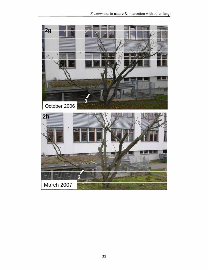
2.4.2 Wood decay test The newly isolated S. commune strains as well as strains from the institutes collection
(ScLs 79 and ScLs 48) and fungal collections of Prof. Holdenrieder from Zurich
(S. commune ScFs 2) and of Prof. Wösten from Utrecht [S. commune 4-39 (MATA41
MATB41, CBS 341.81), 4-40 (MATA43 MATB43, CBS 340.81), 72-3 (∆Sc3 4-39,
MATA41 MATB41), ∆Sc3 4-40 (∆Sc3 MATA43 MATB43), the self compatible
homokaryon S. commune Acon Bcon fbf (MATA41mut MATB41mut fbf CBS 341.81)
characterized by mutations in the mating type loci A41 and B41 and by a defect in
gene fbf that completely blocks fruiting and leads to a faster growth rate, and
S. commune dikaryons 4-39 x 4-40 (MATA41 MATB41 x MATA43 MATB43), and
∆Sc3 4-39 x ∆Sc3 4-40 (MATA41 MATB41 x MATA43 MATB43) produced by mating
from corresponding monokaryons] were tested for growth on J. ailantifolia wood
blocks. The white rot P. ostreatus N001 and T. hirsuta ThJa 1 were used as a positive
control for growth on wood (Fig. 12 & 13).
The new S. commune isolate ScJa 1 from J. ailantifolia wood showed a stronger
formation of aerial mycelium (Fig. 12d) than the isolate ScFs 1 from beech wood
(Fig.12e) and all other S. commune strains (Fig. 12f, 12g, 12h) suggesting that this
strain can easily attack the wood. However, the overall effect on mass loss over the
time did not distinguishd from any of the other tested S. commune strains (Fig. 14,
Table 1, 2). Most interestingly, the growth pattern of the coisogenic S. commune wild
type strains 4-39 (MATA41 MATB41, CBS 341.81), 4-40 (MATA43 MATB43, CBS
340.81) and the respective hydrophobin mutants ∆Sc3 4-39 and, ∆Sc3 4-40 and the
self-compatible homokaryotic fruiting mutant S. commune Acon Bcon fbf (MATA41
mut MATB41 mut fbf) strain varied dramatically on the Juglans wood blocks. The
wildtype strains showed a denser mycelium on the wood blocks whereas the
mycelium was very thin in case of the hydrophobin mutants, especially in case of
∆Sc3 4-39 when comparing it to the wildtype parental strain 4-39 (Fig. 13a, 13b, 13c,
13d, 13e, 13f). Despite the thin mycelial growth of the hydrophobin mutants and the
homokaryon Acon Bcon fbf, there was no significant difference (p ≤ 0.05) in mass
loss with little increase over the time for the different samples (Fig. 14, Tables 1 and
2). Generally, mass loss of Juglans wood caused by S. commune strains varied from 3
to 10%, whereas T. hirsuta and P. ostreatus strongly decayed Juglans resulting in up

S. commune in nature & interaction with other fungi
34
to 45% loss of mass (Fig. 14, Table 1). The comparable low mass loss caused by the
S. commune strains suggests that easy accessible wood compounds might have been
degraded rather than that there was an aggressive attack on the lignocellulose.
Furthermore, since over the time there was no clearly visible, respectively statistically
significant increase in mass loss upon incubation with any of the S. commune strains,
the variations between single samples (either for a given strain over the time, or for
different strains at the same or different time points) have to be considered to reflect
statistical variations in the test system. Statistical variations are influenced by the fact
that only 2 or 3 wood blocks could be analysed per strain due to the limited total
amount of J. ailantifolia wood that was available for this study. Furthermore, these
wood blocks might have shown variations in quality (e.g. being from the outer or the
inner part of the branch they came from, this possibly representing nutrient richer
sapwood or nutrient low heart wood).
On the whole, statistically significant values of mass loss were encountered with
S. commune probes against the non-treated wood (case a in Table 2) likely due to
easily degradable organic matter in the wood blocks (Fig. 14 and Tables 1 and 2).
Furthermore, statistically significant differences were seen when comparing
S. commune treated wood samples with those of P. ostreatus N001 and T. hirsuta
ThJa 1 treated samples (Fig. 14 and Tables 1 and 2). This is easily be explained by the
aggressive wood decay performed by the latter two species. At the end of the
incubation after 20 weeks, apart from the light coloured wood blocks decayed by
T. hirsuta and P. ostreatus N001, all the wood samples incubated with S. commune
closely resembled in look the control samples (Fig. 15).

S. commune in nature & interaction with other fungi
35
12d
12a 12b
12c
Control P. ostreatus N001
T. hirsutaThJa 1 ScJa 1
12g
12f12e
12h
ScFs 1 ScFs 2
ScLs 79 ScLs 48
Fig. 12 Growth morphology of basidiomycetes on J. ailantifolia wood blocks after 8 weeks of incubation at 25 oC in light. Negative control without fungus (a), P. ostreatus N001 (b), T. hirsuta ThJa 1 (c), S. commune strains ScJa 1 (d), ScFs 1(e), ScFs 2 (f), ScLs 79 (g), and ScLs 48 (h). Note that strains ScJa 1, ScFs 1, ScFs 2 and P. ostreatus N001 (Fig 12d, 12e, 12f & 12b) formed primordia and fruiting bodies on the plates.

S. commune in nature & interaction with other fungi
36
13a
13c 13d
13b
4-39 ∆Sc3 4-39
4-40 ∆Sc3 4-40
13e
13g
13f
∆Sc3 4-39 x ∆Sc3 4-40
Acon Bcon fbf
4-39 x 4-40
Fig. 13 Growth morphology of wildtype and hydrophobin mutants after 8 weeks of incubation at 25oC in light on J. ailantifolia wood blocks. S. commune strains 4-39 (a), ∆Sc3

S. commune in nature & interaction with other fungi
37
4-39 (b),4-40 (c), ∆Sc3 4-40 (d), 4-39 x 4-40 (e), ∆Sc3 4-39 x ∆Sc3 4-40 (f), and Acon Bcon fbf MATA41mut MATB41mut fbf (g).

S. commune in nature & interaction with other fungi
38
0
10
20
30
40
50
(a) C
ontrol w
ood
(b) P.ostr
eatus N00
1
(c) T. h
irsuta
ThJa1
(d) ScJ
a 1(e)
ScFs 1
(f) ScF
s 2(g) S
cLs 79
(h) ScL
s 48
(k) 4-
40(m
) 4-39
x 4-4
0(o) A
con Bco
n fbf
Strains
% M
ass
loss
of J
ugla
nsw
ood
8 weeks12 weeks16 weeks20 weeks
(i) 4-3
9(h) ∆
Sc3 4-
39(l)
∆Sc3
4-40
(n) ∆Sc3
4-39
x∆S
c34-4
0
Fig. 14 Decay of Juglans ailantifolia wood caused by S. commune strains (wild type and hydrophobin mutants) after 8, 12, 16 and 20 weeks of incubation. Values are averages of 3 wood blocks, respectively 2 wood blocks (compare Table 1). (a) to (o) indicate the labelling used for the statistical analysis presented in Table 2.
S. commune in nature & interaction with other fungi
39
Table. 1 Percentage of mass loss values of Juglans ailantifolia wood blocks caused by S. commune strains after 8, 12, 16 and 20 weeks.
Values are averages of 3 wood blocks. * indicates averages from two values. (+) P. ostreatus N001 and T. hirsuta ThJa 1 served as positive controls.
Fungal Juglans ailantifolia decay after
Strain 8 weeks 12 weeks 16 weeks 20 weeks
Control
(wood without fungi) 1.6 ± 1.05 1.20 ± 1.05 0.86 ± 0.58 0.25 ± 0.35
P. ostreatus N001(+) 11.64 ± 1.0 24.84 ± 4.97* 32.05 ± 3.39 36.22 ± 3.04
T. hirsuta ThJa 1(+) 23.69 ± 4.79 28.66 ± 4.02 29.51± 1.92* 45.45 ± 0.09*
ScJa 1 5.75 ± 1.14 2.53 ± 0.49 5.40 ± 0.21 10.89 ± 4.33
ScFs1 8.86 ± 3.39 3.01 ± 0.31 7.07 ± 2.01 6.97 ± 2.93
ScFs 2 4.36 ± 1.69 9.48 ± 2.88* 4.66 ± 0.36* 10.81 ± 3.78
ScLs 79 8.10 ± 0.69 3.09 ± 0.89 6.62 ± 3.76 9.57 ± 2.78
ScLs 48 6.50 ± 1.48 3.48 ± 0.09 5.60 ± 1.08* 7.19 ± 3.13
4-39 6.01 ± 1.28 4.79 ± 1.20 9.64 ± 3.04 7.26 ± 2.66
∆Sc3 4-39 5.30 ± 1.94 4.64 ± 3.03 6.63 ± 2.12 6.86 ± 1.14*
4-40 3.63 ± 0.65* 5.30 ± 0.90 10.83 ± 4.68 8.96 ± 1.51
∆Sc3 4-40 3.95 ± 0.18 4.55 ± 0.86 5.57 ± 0.97 6.35 ± 1.06
4-39 x 4-40 3.71 ± 1.11 5.58 ± 1.88 8.86 ± 0.66 7.53 ± 2.43
∆Sc3 4-39 x ∆Sc3 4-40 5.90 ± 1.87 4.86 ± 0.99 7.72 ± 3.43 7.5 ± 2.42
Acon Bcon fbf 4.99 ± 1.70 3.95 ± 2.21 6.89 ± 2.92 5.50 ± 1.68

S. commune in nature & interaction with other fungi
40
Table 2 Statistical data (calculated by using Duncan multiple range test) indicating the significant differences (p≤0.05) in the mass loss of Juglans wood caused by different fungal strains during different time periods (compare Table 1 and Fig. 14).
(*) For an easy understanding, the fungal strains were indicated by the alphabetical letters.
Sample number Fungal strains *
8 weeks decay different with
12 weeks decay different with
16 weeks decay different with
20 weeks decay different with
1 ScJa 1 d a, b, c, f, i, k, m b, c, f b, c, k b, c
2 ScFs 1 e a, c, f, i, k, m b, c, f a, b, c b, c
3 ScFs 2 f a, b, c, e a, b, c, d, e, g, h, o b, c, k b, c
4 ScLs 79 g a, e, b, c b, c, f a, b, c b, c
5 ScLs 48 h a, b, c b, c, f b, c b, c
6 4-39 i b, c, e a, b, c a, b, c b, c
7 ∆Sc3 4-39 j b, c a, b, c a, b, c b, c
8 4-40 k b, c, e a, b, c a, b, c, f b, c
9 ∆Sc3 4-40 l b, c, e a, b, c b, c b, c
10 4-39 x 4-40 m b, c, e a, b, c a, b, c b, c
11 ∆Sc3 4-39 x ∆Sc3
4-40 n b, c a, b, c, f a, b, c b, c
12 Acon Bcon fbf o b, c b, c, f a, b, c b, c
13 Control wood a b, c, e, g, h, i b, c, f, i, j, k, l, m, n b, c, e, i, j, k, m, n, o b, c
14 P. ostreatus N001 b
a, d, f, g, h, i, j, k, l, m, n,
o
a, d, e, f, g, h, i, j, k, l, m,
n, o
a, d, e, f, g, h, i, j, k, l, m,
n, o
a, c, d, e, f, g, h, i, j, k, l,
m, n, o,
15 T. hirsuta ThJa 1 c
a, b, d, e, f, g, h, i, j, k, l,
m, n, o
a, b, d, e, f, g, h, i, j, k, l,
m, n, o
a, d, e, f, g, h, i, j, k, l, m,
n, o
a, b, d, e, f, g, h, i, j k, l,
m, n, o

S. commune in nature & interaction with other fungi
41
Fig. 15 Juglans wood blocks after the 20 weeks decay test by different S. commune strains at 25 oC in continuous light.
2.4.3 Fungal interactions Since there is the possibility, that growth of S. commune in wood in nature is
promoted by the presence of other, possibly stronger wood decaying fungi, reactions
of S. commune strains in dual inoculations on agar BSM medium plates with
T. hirsuta were observed. Dual inoculations resulted in interactions with deadlock

S. commune in nature & interaction with other fungi
42
formation followed by replacement of S. commune by T. hirsuta over the time (Fig.
16, 17 & 18). S. commune strains except the hydrophobin mutant dikaryon ∆Sc3 4-39
x ∆Sc3 4-40 formed barrages (formation of dense mycelia between both the fungi)
with T. hirsuta. There was also pigment formation (coloured zone) at the interaction
region after 1 week of incubation compared to the other S. commune strains (Fig. 16,
17, 18).
The pigmentation was more prominently dark or broad in case of ScFs2 (Fig. 16a).
S. commune strain ScFs2 formed numerous primordia/fruiting bodies in the single
culture (Fig. 19), whereas there was no primordia formation in presence of T. hirsuta
(Fig. 16a). Only the S. commune dikaryon ∆Sc3 4-39 x ∆Sc3 4-40 did not form either
a barrage or produced pigmentation (Fig. 18c, 18f). The growth rate of ∆Sc3 4-39 x
∆Sc3 4-40 strain was much lower in the dual cultures in the presence of the T. hirsuta
strain ThJa 1 than in the single culture (Table 3). From this confrontation tests, it
appears that S. commune is generally weaker than T. hirsuta, at least under conditions
on BSM agar Petri dishes that offers easily accessible nutrients.
In contrast, in the interaction study on Juglans wood blocks, S. commune strain ScJa 1
appeared to be the first invader of the J. ailantifolia wood whereas at a later stage
after 10 days T. hirsuta ThJa 1 overgrow the wood and completely replaced the
S. commune ScJa 1 (Fig. 20c, 20f). The mixed infection of S. commune ScJa 1 and
T. hirsuta ThJa 1 caused a mass loss of up to 46% which was similar to the decay
caused by T. hirsuta alone whereas S. commune ScJa 1 alone reduced in this
experiment a mass loss of wood by about 16.51 ± 1.93 % (Fig. 21). The higher decay
in the dual infection was probably due to complete replacement of S. commune ScJa 1
by T. hirsuta ThJa 1 over the time. By visual inspection, the wood blocks after 10
weeks of decay infected with T. hirsuta ThJa 1 closely resembled the wood blocks
treated with mixed infection of S. commune ScJa 1 and T. hirsuta ThJa 1 (Fig. 22).
Comparison of the wood blocks decayed by S. commune ScJa 1 from experiment 1
and 2 revealed that few replicate samples in experiment two appeared whiter but on a
whole is not very different from the first experiment (Fig. 23).

S. commune in nature & interaction with other fungi
43
S. c. ScFs 2
S. c. ScFs 1
S. c. ScJa 1
S. c.ScFs 2
S. c.ScFs 1
S. c.ScJa 1
T. h. ThJa 1
T. h. ThJa 1
T. h. ThJa 1
T. h. ThJa 1
T. h.ThJa 1
T. h.ThJa 1
B B B
PP P
16a 16b
16d
16c
16e 16f
Fig. 16 Interactions between T. hirsuta ThJa 1(T. h), and S. commune ScFs 2, ScFs 1, ScJa 1 (S. c) on BSM medium under incubation in constant light at a temperature of 25oC. Barrier formation (B) is followed by the subsequent replacement of S. commune ScFs 2, ScFs 1, and ScJa 1 by T. hirsuta ThJa 1 (a, b, c). The lower row (d, e, f) shows the plates photographed from the bottom to present the pigmentation (P) occurring at the interaction zone of the two species.

S. commune in nature & interaction with other fungi
44
17a 17b 17c
17d 17e 17f
S. c.4-39
T. h.ThJa 1
S. c.4-40
T. h.ThJa 1
S. c.4-39 x 4-40
T. h. ThJa 1
T. h. ThJa 1
S. c.4-39
T. h. ThJa 1
S. c.4-40
T. h.ThJa 1
S. c.4-39 x 4-40
B
P PP
B B
Fig. 17 Interactions between T. hirsuta ThJa 1(T. h), and S. commune (S. c) 4-39 (MATA41 MATB41), 4-40 (MATA43 MATB43) and 4-39 x 4-40. Barrier formation on BSM medium under incubation in constant light at a temperature of 25 oC. Barrier formation (B) is followed by the subsequent replacement of S. commune strains by T. hirsuta ThJa 1 (a, b, c). The lower row (d, e, f) shows the plates photographed from the bottom to present the pigmentation (P) occurring at the interaction zone of the two species.

S. commune in nature & interaction with other fungi
45
18a 18b 18c
18d 18e 18f
S. c∆Sc3 4-39
S. c.∆Sc3 4-40
S. c.∆Sc3 4-39
x ∆Sc3 4-40
S. c.∆Sc3 4-39
S. c.∆Sc3 4-40
T. h.ThJa 1
T. h.ThJa 1 T. h.
ThJa 1
T. h.ThJa 1
T. h.ThJa 1
T. h.ThJa 1
B BB
P P N P
S. c.∆Sc3 4-39
x ∆Sc3 4-40
Fig. 18 Interactions between T. hirsuta ThJa 1 (T. h), and S. commune hydrophobin mutants (S. c) ∆Sc3 4-39 (MATA41 MATB41), ∆Sc3 4-40 (MATA43 MATB43), ∆Sc3 4-39 x ∆Sc3 4-40 on BSM medium under incubation in constant light at a temperature of 25oC. Barrier formation (B) is followed by the subsequent replacement of S. commune by T. hirsuta (a, b, c). The lower row (d, e) shows the plates photographed from the bottom to present the pigmentation (P) at the interaction zone of the two species. Note the insignificant barrier and no pigmentation (NP) formed between S. commune strain ∆Sc3 4-39 x ∆Sc3 4-40 and T. hirsuta ThJa 1(c, f).

S. commune in nature & interaction with other fungi
46
Fig. 19 Numerous mycelial tufts formed (fruiting body primordia) in the single culture of S. commune ScFs 2 under continuous light at a temperature of 25 oC on BSM medium.

S. commune in nature & interaction with other fungi
47
Table 3 Effect of opposing colonies of S. commune to T. hirsuta ThJa 1 with mean extension rate (mm/day) on BSM medium compared with controls, and growth towards and away from opponents incubated under constant light illumination at a temperature of 25oC.
Extension of opponent (S. commune) Extension of T. hirsuta ThJa 1 Strains
Towards
T. h.
Away
T. h.
Diam. in
interaction
< control
Towards and
away significant
difference
Towards
S. c.
Away
S. c.
Diam. in
interaction
< control
Towards
and away
significant
difference
4-39 3.78 ± 1.30 3.83 ± 1.17 ns ns 5.60 ± 1.34 4.13 ± 1.73 ns ns
∆Sc3 4-39 3.89 ± 1.53 4.28 ± 1.52 ns ns 5.00 ± 2.00 4.00 ± 2.00 ns ns
4-40 4.00 ± 1.40 4.63 ± 1.84 S ns 4.00 ± 1.67 4.25 ± 2.05 S ns
∆Sc3 4-40 3.75 ± 1.03 4.33 ± 1.00 ns ns 4.00 ± 2.30 5.60 ± 2.34 S ns
4-39 x 4-40 4.67 ± 1.36 4.22 ± 1.09 ns ns 4.38 ± 2.06 3.71 ± 1.80 S ns
∆Sc3 4-39
x
∆Sc3 4-40
2.00 ± 1.06 1.89 ± 1.05 S ns 6.22 ± 2.53 3.63 ± 1.77 ns S
ScFs 2 4. 00 ± 1.65 4.22 ± 0.66 ns ns 5.13 ± 2.79 3.33 ± 1.94 ns ns
ScFs 1 5.33 ± 1.63 3.67 ± 1.80 ns ns 5.00 ± 2.23 3.29 ± 2.36 ns ns
ScJa 1 6.00 ± 2.54 4.25 ± 2.25 ns ns 4.25 ± 4.20 4.14 ± 2.54 S ns
ns indicate non-significance and S significant (p≤0.05) stimulation

S. commune in nature & interaction with other fungi
48
20d
20b 20c20a
20e 20f
Fig. 20 Wood decay test of Juglans with mixed infections. S. commune ScJa 1 (a, d), T. hirsuta ThJa 1 (b, e) and mixed culture of S. commune ScJa 1 and T. hirsuta ThJa 1 (c, f). Pictures shown are 3 weeks and 7 weeks old on upper and lower row, respectively. Incubated at 25 oC in continuous light on BSM medium.

S. commune in nature & interaction with other fungi
49
0
10
20
30
40
50
60
70
80
T. hirsutaThJa 1
S. commune ScJa 1
Fungus
% M
ass
loss
T. hirsuta ThJa 1+
S. commune ScJa 1
Fig. 21 Decay of Juglans ailantifolia wood caused by T. hirsuta ThJa 1, S. commune strain ScJa 1 and mixed infection of ThJa 1 and ScJa 1 after 10 weeks of incubation. Values are averages of 6 wood blocks.

S. commune in nature & interaction with other fungi
50
Fig. 22 Juglans wood blocks after 10 weeks decay test by T. hirsuta ThJa 1, S. commune ScJa 1 and mixed infection of S. commune ScJa 1 + T. hirsuta ThJa 1 strains at 25oC in continuous light.
Fig. 23 Comparison of Juglans wood blocks after decay by S. commune ScJa 1 from experiment 1 (upper row) and experiment 2 (lower row).
2.5 Discussion The cosmopolitan fungus S. commune grows in very diverse environmental conditions
on various types of dead wood (Cooke 1961, Schmidt & Liese 1980, Raper &
Krongelb 1958, James & Vilgalys 2001, James et al. 1999). It has been described
previously as a wound parasite and in a few cases it was recorded as an opportunistic
pathogen (Adaskaveg 1993, Shigo 1967, Cooke 1961, Dai 2005, Latham 1970). The
previous studies are supported by our observations when S. commune fruiting bodies
were found growing on a fallen beech branch (Fig. 11a) and in the other case on the
living Juglans tree that was weakened by the movement of the tree from one place to
another (Fig. 11b). S. commune as a pathogen is reported to cause chlorotic, necrotic
symptoms of Prunus leaves, wood discoloration and sap wood rot of Prunus (Dai
2005). On the other hand it was also reported to occur as an endophyte on teak leaves
(Chareprasert et al. 2006). However, wood decay tests in the laboratory with
Schizophyllum previously reported on pine, beech and picea wood samples caused a
mass loss only of up to 5%. Hong (1982) reported a mass loss of Hevea brasiliensis
(rubber tree) by Schizophyllum as high as 47.7%. No wood decay tests were

S. commune in nature & interaction with other fungi
51
performed previously in the laboratory in the combination of Schizophyllum and
Juglans wood. In our study, the mass loss of Juglans wood caused by Schizophyllum
strains varied from 3 to 10% in 20 weeks incubation time (Fig. 14). Schizophyllum
strain ScJa 1 originally isolated from the same Juglans tree caused in the first
experiment mass loss of 10.89 ± 4.33 % being highest of all the Schizophyllum strains
used in this study. Analysis of the values over the time in this series of experiments
however did not reveal much statistical differences in the ability of S. commune
strains to degrade wood. Thereby, the Sc3 hydrophobin mutant’s ∆Sc3 4-39, ∆Sc3 4-
40 and ∆Sc3 4-39 x ∆Sc3 4-40 were able to cause mass loss of Juglans to a similar
level than the wild type strains in a range from 4 to 7%, although there was a
morphological difference in the growth pattern of the S. commune wild type and
Sc3 hydrophobin mutants while growing on Juglans wood blocks. The T. hirsuta
strain ThJa 1 which was isolated from the same branch closely growing to
S. commune ScJa 1 in contrast decayed Juglans strongly with mass losses of up to
45%. In a second set of experiments set up to test dual infections on wood (Fig. 21),
the mass loss by S. commune ScJa 1 was slightly higher with 16 ± 5.09 % supporting
that the strains had indeed a low level activity of wood decay. This suggestion is in
accordance with the observation on J. ailantifolia branches where white rot occurred
directly under places in the bark where S. commune fruiting bodies broke through
(Fig. 10).
Interactions between different fungi can be seen not only in cultures as demonstrated
in this study (Fig. 15 to 17) but also when the fungi are growing in decaying wood in
nature (Iakovlev and Stenlid 2000). The competitive interactions in the agar based
systems in many instances were found to correlate with that of the natural systems
(Boddy & Rayner 1983, Holmer et al. 1997, White and Boddy 1992, Woods et al.
2005). Growing the strains S. commune ScJa 1 and T. hirsuta ThJa 1 obtained from
the same branch of a living Juglans tree resulted in barrier formation and
pigmentation on the agar system and with overgrowth of S. commune by T. hirsuta
(Fig. 15c, 15f). Like wise on wood where S. commune was first active, in the end
T. hirsuta overgrow the other species (Fig. 20). These observations suggest that also
in nature, S. commune might be overtaken by T. hirsuta.

S. commune in nature & interaction with other fungi
52
The mechanisms involving the confrontive interactions between two different species
might affect the synthesis and release of hydrophobic metabolites from the hyphae
and their conversion to free radicals and polymers in the presence of enzymes
(Iakovlev and Stenlid 2000). The formation of barrages is often associated with the
ability of the fungus to resist the invasion by the opponent (Boddy 2000, Iakovlev &
Stenlid 2000, Wald et al 2004). In our study, all the S. commune strains except the Sc3
mutant dikaryon (∆Sc3 4-39 x ∆Sc3 4-40) have shown an ability to produce a barrage
with pigmentation when confronted to T. hirsuta. Since also the Sc3 hydrophobin
mutant monokaryon strains 72-3 (∆Sc3 4-39) and ∆Sc3 4-40 were able to form a clear
barrier against T. hirsuta ThJa 1, a principle requirement for the hydrophobins
forming hydrophobic layers on the surface of hyphae (Wösten 2001, Walser 2003) is
not obvious, although the mutant dikaryon behaved differently. In a recent study
analysing interactions between Phlebiopsis gigantea and Heterobasidion parviporum
it was found by molecular approaches that a specific hydrophobin gene of
H. parviporum was up-regulated whereas other two hydrophobin genes were down-
regulated at the interaction zone during the nonself-competitive interaction between
P. gigantea and H. parviporum (Adomas et al. 2006). It was also shown that
hydrophobins are involved in cell wall assembly and that hydrophobin monomers can
act as toxins or elicitors (Talbot 2001).
Although from our results it is evident that hydrophobins are not principally necessary
for the defense reactions in S. commune, it is not clear why the SC3 hydrophobin
mutant dikaryon did not produce pigmentation and barrier formation towards
T. hirsuta ThJa 1. It has been shown that some hydrophobins have lectin
(a carbohydrate-binding protein) specificities that might be involved in interaction
with the exposed environments (Wösten 2001, Walser et al. 2003). In particular, lectin
activity has been shown for hydrophobin SC4 (van Wetter et al. 2000a). Since in
contrast to the wildtype dikaryon, this hydrophobin is the only one being expressed in
the Sc3 hydrophobin mutant dikaryon (van Wetter et al. 2000a, Wösten 2001), it is
possible that in the case of lack of gene Sc3 the dikaryon-specific SC4 hydrophobin
might have a different interaction process with T. hirsuta Thja 1. SC3 and SC4 are
known to be able to replace each other in function, but not completely. For example
transformed ∆Sc3 monokaryons expressing SC4 are not as water-repellent as the co-

S. commune in nature & interaction with other fungi
53
isogenic monokaryon expressing the SC3 hydrophobin (van Wetter et al. 2000a).
Hydrophobins help to secrete proteins from the cells (Talbot et al. 1996) and to retain
proteins in the cell walls (Talbot et al. 1996, van Wetter et al. 2000b). If one or both
functions are impaired in the case of Sc3 knockout dikaryon, enzymes or metabolites
for production of pigments might be missing providing the outcome in the interaction
between S. commune and T. hirsuta from the first organisms. However, this would not
explain the different reaction between ∆Sc3 monokaryons and the ∆Sc3 dikaryon.
Likely, the interaction process between S. commune and T. hirsuta is much more
complex as can be foreseen at the current time.
2.6 References Adaskaveg, J.E., Miller, R.W. & Gilbertson, R.L. (1993). Wood decay, lignicolous
fungi and decline of peach trees in South Carolina. Plant Disease, 77, 707-711.
Adaskaveg, J.E . (1998). Wood decay of Landscape trees. Western Arborist, 24, 1-5.
Adomas, A., Eklund, M., Johannson, M. & Asiegbu, F.O. (2006). Identification and
analysis of differentially expressed cDNAs during nonself-competitive
interaction between Phlebiopsis gigantea and Heterobasidion parviporum.
FEMS Microbiology Ecology, 57, 26-39.
Boddy, L. & Rayner, A.D.M. (1983). Ecological roles of Basidiomycetes forming
decay communities in attached oak branches. New Phytologist, 93, 77-88.
Breitenbach, J. & Kränzlin, F. (1991). Pilze in der Schweiz. Mykologia, Luzern,
Switzerland, Vol. 3, p. 318, ISBN: 3-85604-030-7.
Boddy, L. (2000). Interspecific combative interactions between wood-decaying
basidiomycetes. FEMS Microbiology Ecology, 31, 185-194.
Brien, M.O., Kiely, P.O., Forristal, P.D. & Fuller, H.T. (2007). Visible fungal growth
on baled grass silage during the winter feeding season in Ireland and silage
characteristics associated with the occurance of fungi. Animal Feed Science
and Technology, 139, 234-256.
Cooke, W.B. (1961). The genus Schizophyllum. Mycologia, 53, 575-599.
Chareprasert, S., Piapukiew, J., Thienhirun, S., Whilley, A.J.S. & Sihanonth, P.
(2006). Endophytic fungi of teak leaves Tectona grandis L. and rain tree

S. commune in nature & interaction with other fungi
54
leaves Samanea saman Merr. World Journal of Microbiology &
Biotechnology, 22, 281-486.
Dai, Y.C. (2005). First report of sapwood rot of peach caused by Schizophyllum
commune in China. Plant Disease, 89, 778.
Hüttermann, A. & Volger, C. (1973). Induction of arylglucosidase in Fomes annosus
by cellobiose. Archiv für Mikrobiologie, 93, 195-204.
Hegarty, B., Steinfurth, A., Liese, W. & Schmidt, O. (1987). Comparative
investigations on wood decay and cellulolytic and xylanolytic activity of some
basidiomycete fungi. Holzforschung, 41, 265-269.
Humar M., Petrič M & Pohleven F. (2001). Changes of the pH value of impregnated
wood during exposure to wood-rotting fungi. Holz als Roh- und Werkstoff,
59, 288-293.
Humar, M., Petrič, M. & Pohleven, F., Šentjurc, M. & Kalan, P. (2002). Changes in
EPR spectra of wood impregnated with copper-based preservatives during
exposure to several wood-rotting fungi. Holzforschung, 56, 229-238.
Heilmann-Clausen, J. & Boddy, L. (2005). Inhibition and stimulation effects in
communities of wood decay fungi: Exudates from colonized wood influence
growth by other species. Microbial Ecology, 49, 399-406.
Iakovlev, A. & Stenlid, J. (2000). Spatiotemporal patterns of laccase activity in
interacting mycelia of wood-decaying basidiomycete fungi. Microbial
Ecology, 39, 236-245.
James, T.Y. & Vilgalys, R. (2001). Abundamce and diversity of Schizophyllum
commune spore clouds in the Caribbean detected by selective sampling.
Molecular Ecology, 10, 471-479.
James, T.Y., Porter, D., Hamrick, J.L. & Vilgalys, R. (1999). Evidence for limited
intercontinental gene flow in the cosmopolitan mushroom, Schizophyllum
commune. Evolution, 53, 1665-1677.
Latham, A.J. (1970). Development of apple fruit rot and basidiocarp formation by
Schizophyllum commune. Phytopathology, 60, 596-598.
Nsolomo, V.R., Venn, K. & Solheim, H. (2000). The ability of some fungi to cause
decay in the East African camphor tree, Ocotea usambarensis. Mycological
Research, 104, 1473-1479.

S. commune in nature & interaction with other fungi
55
Owens, E.M., Reddy, C.A. & Grethlein, H.E. (1994). Outcome of interspecific
interactions among brown-rot and white rot wood decay fungi. FEMS
Microbiology Ecology, 14, 19-24.
Pen as, M.M., Rust, B., Larraya, L.M., Ramírez, L. & Pisabarro, A.G. (2002).
Differentially regulated, vegetative mycelium specific hydrophobins of the
edible basidiomycete Pleurotus ostreatus. Applied and Environmental
Microbiology, 68, 3891-3898.
Raper, J.R. & Krongelb, G.S. (1958). Genetic and environmental aspects of fruiting in
Schizophyllum commune Fr. Mycologia, 50, 707-740.
Rayner, A.D.M. (1979). Internal spread of fungi inoculated into hardwood stumps.
New Phytologist, 82, 505-517.
Shigo, A.L. (1967). Successions of organisms in discoloration and decay of wood.
Forestry Research, 2, 237-299.
Schmidt, O. & Liese, W. (1980). Variability of wood degrading enzymes of
Schizophyllum commune. Holzforschung, 34, 67-72.
Talbot, N.J., Kershaw, M.J., Wakley, G.E., de Vries, O.M.H., Wessels, J.G.H. &
Hamer, J.E. (1996). MPG1 encodes a fungal hydrophobin involved in surface
interactions during infection-related development of Magnaporthe grisea. The
Plant Cell, 8, 985-999.
van Wetter, M.A., Wösten, H.A.B. & Wessels, J.G.H. (2000a). SC3 and SC4
hydrophobins have distinct roles in formation of aerial structures in dikaryons
of Schizophyllum commune. Molecular Microbiology, 36, 201-210.
van Wetter, M.A., Wösten, H.A.B., Sietsma, J.H. & Wessels, J.G.H. (2000b).
Hydrophobin gene expression affects hyphal wall composition in
Schizophyllum commune. Fungal Genetics and Bioogy, 31, 99-104.
Wösten, H.A.B. (2001). Hydrophobins: multipurpose proteins. Annual Review of
Microbiology, 55, 625-646.
Wells, J.M. & Boddy, L. (2002). Interspecific carbon exchange and cost of
interactions between basidiomycete mycelia in soil and wood. Functional
Ecology, 16, 153-161.
Wessels, J.G.H., de Vries, O.M.H., Ásgeirsdóttir, S.A. & Schuren, F.H.K. (1991).
Hydrophobin genes involved in formation of aerial hyphae and fruit bodies in
Schizophyllum. The Plant Cell, 3, 793-799.

S. commune in nature & interaction with other fungi
56
Wald, P., Pitkänen, S. & Boddy, L. (2004). Interspecific interactions between the rare
tooth fungi Crelophus cirrhatus, Hericium erinaceus and H. coralloides and
other wood decay species in agar and wood. Mycological Research, 108,
1447-1457.
Woods, C.M., Woodward, S. & Redfern, D.B. (2005). In vitro interactions in artificial
and wood-based media between fungi colonizing stumps of Sitka spruce.
Forest Pathology, 35, 213-229.

Chapter 3
Wood decay by Schizophyllum commune


Wood decay by Schizophyllum commune
57
3. Wood decay by Schizophyllum commune
3.1 Abstract Wood decay tests with beech, birch and pine wood were performed to test the decay
ability of Schizophyllum commune. In wood block tests, the maximum decay caused
by S. commune strains was 4-5% whilst the control strains P. ostreatus and
T. versicolor showed mass loss up to 30 and 50%, respectively. Overall, the mass loss
caused by different S. commune strains including the SC3 hydrophobin and SC15
mutants, regardless of being a monokaryon or a dikaryon, were not significantly
(p<0.05) different from each other. In saw dust decay tests of beech, birch and pine,
the decay varied from 9 to 16% amongst the S. commune strains; P. ostreatus and
T. versicolor decayed 20 to 30% and 30 to 40%, respectively of the wood samples.
S. commune strains decolorized the pine saw dust which was not observed in case of
P. ostreatus and T. versicolor. Regardless lack of visible decay, the strength of wood
was negatively affected by S. commune. Maximum strength losses caused by
S. commune strains in beech, birch and pine wood blocks were 21, 52 and 35%,
respectively. Amongst the S. commune strains, there were no significant differences in
the strength losses of wood. P. ostreatus N001 caused 36, 123 and 188% strength loss
of beech, birch and pine wood, respectively, whereas T. versicolor reduced the
strength of beech, birch and pine wood by 43, 34 and 94%, respectively.

Wood decay by Schizophyllum commune
58
3.2 Introduction Schizophyllum commune as one of the most widely distributed wood inhabiting
basidiomycetes is usually considered to be a white rot fungus. However, wood decay
tests in the laboratory in most instances did not result in considerable weight losses
(Hegarty et al. 1987, Nsolomo et al. 2000, Humar et al. 2001, 2002, Schirp et al.
2003). Schmidt and Liese (1980), Nilsson and Daniel (1983) and Leithoff and Peek
(2001) measured between 1 to 6% weight loss for different strains and concluded the
fungus is a rather weak wood-destroyer. Similar results were obtained when growing
S. commune strains on J. ailantifolia wood (Chapter 2 of this thesis). In rare cases,
mass losses of nearly 50% were observed, mainly due to lignin degradation and to a
low degree due to cellulose degradation (Hong 1982). Although there might be no or
little weight loss in wood decay tests, toughness of wood can be negatively affected.
32% strength loss has been measured (Schirp et al. 2003). In flake board tests with
S. commune, weight loss of 9.6% occurred together with strength loss (67.5%
modulus of rupture loss; Hadi et al. 1995). A monokaryon of S. commune was shown
to some extent to demethylate lignin. This was not due to production of laccases,
peroxidases or ligninases (Trojanowski et al. 1986). Poor lignin degradation in
another study was thought to result from the inability of the fungus to solubilize lignin
(Boyle et al. 1992). On wheat straw, some tropical isolates were shown to degrade
lignin (up to 15%) and to cause simultaneous mass loss of up to 23.5%. Another strain
showed even higher mass loss (26.7%) but no lignin decay (Capelari and Zadrazil
1997). Lignin in olive pomace was efficiently degraded by a S. commune isolate (up
to 52.7% of lignin breakdown). During pomace degradation, high laccase activities
were recorded (Haddadin et al. 2002). Many S. commune strains have xylanase and
cellulase activities (Schmidt and Liese 1980, Clarke and Yaguchi 1986, Hegarty et al.
1987, Bray and Clarke 1995, Haltrich et al. 1995, Thygesen et al. 2003). Most of them
have also laccase and peroxidase activities (Schmidt and Liese 1980). de Vries et al.
(1986) described laccase activity specific to the dikaryotic state of S. commune whilst
in the parental monokaryons, they could not detect this activity. Phenoloxidase
activities were not found in other studies, also not upon treatment with phenolic
inducers (Boyle et al. 1992, Nsolomo et al. 2000). By the well established xylanase
and cellulase activities of the fungus, it has been suggested that the main role in nature
is to recycle carbon by breaking down cellulose and xylans in fallen wood (Raper and
Wood decay by Schizophyllum commune
59
Fowler 2004). In conclusion, the ability of S. commune to degrade wood remains to be
a puzzle.
S. commune is used as a model fungus to study mating types and fungal development
in basidiomycetes (Raper and Fowler 2004). Well known is the discovery of
hydrophobins in this fungus, small-secreted fungal proteins of about 100 to 140 amino
acids with 8 conserved cysteine residues. Upon secretion, hydrophobins self-assemble
into amphipathic stable films that cover fungal cells and make their surfaces
hydrophobic. Therefore, these films enable fungal structures to grow into the air and
protect them from adverse environmental conditions. Due to the hydrophobin coating,
aerial hyphae are repellent towards water. Within mushrooms, hydrophobin films coat
the air channels and prevent them from filling with water. In total, four different
hydrophobins have been described in S. commune (SC1, SC3, SC4 and SC6). SC3,
the best characterized hydrophobin, is expressed in the vegetative mycelium of
monokaryons and dikaryons. The other three, SC1, SC4 and SC6, are dikaryon
specific. SC4 is low expressed in the mycelium and well in mushroom tissues. SC1
and SC6 are specific to fruiting stages (Wösten 2001, Walser et al. 2003). Another
important secreted protein, SC15 (not a hydrophobin), mediates formation of aerial
hyphae and attachment in the absence of the SC3 hydrophobin by making the aerial
hyphae hydrophobic (Lugones et al. 2004).
So far, it was not known whether hydrophobins participate also in wood colonization
and pathogenicity of S. commune and in degradation of lignin as a hydrophobic
component (Wösten et al. 1994). Is it necessary that the hyphae have a hydrophobic
surface when growing in wood? First tests of SC3 hydrophobin mutants on
J. ailantifolia wood gave no indications for a requirement of this small hydrophobic
proteins (see chapter 2 of this thesis). In this chapter, the participation of SC3 and
SC15 proteins in the decay process and their affect on the strength of beech, birch and
pine wood are discussed by making use of knockout mutants of S. commune (van
Wetter et al. 1996, Lugones et al. 2004, de Jong 2006) that lack either the Sc3 gene,
the Sc15 gene or both genes. Monokaryons lacking the Sc15 gene or both genes are
characterized by a strong decrease in formation of aerial hyphae and surface
hydrophobicity (Wösten et al. 1994, van Wetter et al. 1996) whereas monokaryons

Wood decay by Schizophyllum commune
60
lacking the Sc15 gene behave like the wildtype (Lugones et al. 2004). Most
interesting are the double mutants. They form no aerial mycelium and are fully
hydrophilic (Lugones et al. 2004).
3.3 Materials and methods
3.3.1 Fungal strains The co-isogenic S. commune monokaryons 4-39 (MATA41 MATB41, CBS 341.81),
4-40 (MATA43 MATB43, CBS 340.81), their corresponding Sc3 hydrophobin mutants
72-3 (∆Sc3 MATA41 MATB41), ∆Sc3 4-40 (∆Sc3 MATA43 MATB43), and the
corresponding Sc15 protein mutants ∆Sc15 4-39 (∆Sc15 MATA41 MATB41), ∆Sc15
4-40 (∆Sc15 MATA43 MATB43) and the Sc3Sc15 double mutant ∆Sc3∆Sc15
(MATA41 MATB43) and ∆Sc3∆Sc15 (MATA43 MATB41) were kindly provided by
Prof. Wösten, Utrecht, The Netherlands. Dikaryotic S. commune strains 4-39 x 4-40
(MATA41 MATB41 x MATA43 MATB43), ∆Sc3 4-39 x ∆Sc3 4-40 (MATA41 MATB41
x MATA43 MATB43), ∆Sc15 4-39 x ∆Sc15 4-40 (MATA41 MATB41 x MATA43
MATB43), ∆Sc3∆Sc15 MATA41B43 x ∆Sc3∆Sc15 MATA43B41 were produced by
mating the corresponding monokaryons on S. commune minimal medium (Dons et al.
1979). Trametes versicolor strain 6 originated from the institute's collection.
Pleurotus ostreatus N001 was kindly provided by Prof. Ramirez, Universida Publica
de Navarra, Pamplona, Spain.
3.3.2 Growth conditions All strains were grown at 25 oC in continuous light. S. commune strains were
cultivated on S. commune minimal medium (20 g glucose, 1.5 g L-asparagine, 1 g
K2HPO4, 0.5 g MgSO4 x 7H2O, 1 g yeast extract, 0.12 mg thiamine-HCl, 0.1 mg
pyridoxine HCl, 0.005 mg biotin, 0.2 mg CuSO4 x 5H2O, 0.08 mg MnCl2 x 4H2O, 0.4
mg cobaltous chloride hexahydrate, 1.2 mg calcium nitrate tetrahydrate per 1 liter
H2O; Dons et al. 1979), T. versicolor 6 on BSM [Basidiomycete standard medium; 5
g glucose monohydrate, 0.65 g L-asparagine 1 g KH2PO4, 0.5 g MgSO4 x 7H2O, 0.5 g
KCl, 0.5 g yeast extract, 10 g agar, 50 ml stock solution I (0.2 g FeSO4 x 7H20 per
liter), 50ml stock solution II (0.16 g Mn(CH3COO)2 x 4H2O, 0.04 g Zn(NO3)2 x
4H2O, 1 g Ca(NO3)2 x 4H2O, 0.06 g CuSO4 x 5H2O) per 1 liter H2O, pH 4.5;

Wood decay by Schizophyllum commune
61
Hüttermann and Volger 1973] and P. ostreatus on SMY medium (10 g of sucrose, 10
g of malt extract, 4 g of yeast extract, per 1 liter H2O; pH 5.6; Penas et al. 2002).
3.3.3 Wood decay tests
3.3.3.1 Wood block test
Three types of wood – quality selected beech, birch and pine blocks of 10 cm x 1 cm
x 0.5 cm (longitudinal to the axis x tangential to the axis x radial to the axis) were
used. From each type of wood, 8 replicate wood blocks were used for each harvest per
each fungal strain. The wood blocks were dried in an oven at 60oC for two days.
Initial weights of the dried wood blocks were determined before soaking the samples
overnight in water and autoclaving. Each four wood blocks were used per 18 cm Petri
plate filled with 70 to 75 ml S. commune minimal agar medium on which mycelium
was grown. To avoid the direct contact of the wood blocks with the agar, sterile steel
grids were used beneath the wood blocks. Wood blocks were transferred onto the steel
grids when the mycelium of the growing fungi covered half of the Petri plates.
Mycelium was inoculated in the middle of the plates by placing a block of agar with
mycelium that was cut with a cork borer (1cm Ø). Petri plates were sealed with
Parafilm (PECHINEY, Chicago, USA) and incubated at 25oC both for initial mycelial
growth for 7 to 10 days as well as for further incubation, once the wood blocks were
placed onto the grids. To check decay by the strains, harvests were performed after 16
and 20 weeks of incubation with the wood blocks. Plates without fungi but with wood
blocks were used as negative controls. After incubation, the mycelium attaching to the
wood blocks was separated by using a scalpel. Wood blocks were then dried at 104°C
for 3 days and weighed to note the final (dried) weight. Mass loss of wood was
calculated as (initial wt – final wt/initial wt) x 100.
3.3.3.2 Saw dust decay test
Three types of saw dust, namely beech, birch and pine, were used. The amount of saw
dust used per 500 ml flask was 6 g. 5 replicates were used for each strain for each
type of saw dust. In each flask, 50 ml of S. commune minimal medium was added.
The saw dust was exposed to air and not completely immersed in the medium. 10 ml
homogenised inoculum was added for each replicate. To check the decay by the
strains, harvest was done after 20 weeks incubation at 25oC. The decayed saw dust

Wood decay by Schizophyllum commune
62
was dried at 104oC for 3 days and weighed to note the final (dried) weight. Mass loss
of the saw dust was calculated as mentioned above for the wood block decay test.
3.3.4 Bending strength test of wood
After calculating the mass loss, wood blocks have been used for determining the
bending strength (ability to absorb shock with some permanent deformation and more
or less injury to the wood specimen). Before measurement, the wood blocks were
oven dried at 104oC for 1 day. The wood blocks were kept perpendicular to the force
applied, calculated according to the European standard EN 310 (Wood-based panels;
Determination of modulus of elasticity in bending and of bending strength; DK
674.03: 620.172.225: 620.174; 1993) with a slight modification. Bending strength
was calculated as:
fm = 3 Fmax L1/ 2bt2
F max = absolute strength (N)
L = length of the sample holder
b = width
t = thickness
fm = relative bending strength (N/mm2)
3.4 Results
3.4.1 Wood block decay tests All S. commune strains, both monokaryons and dikaryons including the ∆Sc3 and
∆Sc15 mutants and the ∆Sc3∆Sc15 double mutant, were able to grow on beech, birch
and pine wood (Fig. 1, 2, 3). There was however a difference in the overall formation
of surface mycelium. The wild type monokaryons and the wild type dikaryon formed
abundant aerial mycelium on the agar surface of the cultures and on all wood samples
(Fig. 1, 2, and 3, samples 4, 5, 6). Similarly, the ∆Sc15 strains were able to produce
abundant aerial mycelium (Fig. 1, 2, and 3, samples 10, 11, 12). In contrast, the ∆Sc3
monokaryons lacking the hydrophobin SC3 had hardly any aerial mycelium both on
the agar medium as on the surface of the wood blocks (Fig. 1, 2, and 3, samples 7 and
8), confirming previous results by van Wetter et al. (1996) that protein SC3 is
required for abundant formation of aerial mycelium on the monokaryon. The dikaryon

Wood decay by Schizophyllum commune
63
lacking the Sc3 gene in both its nuclei possessed some aerial mycelium both on the
agar and on the wood surface (Fig. 1, 2 and 3, sample 9) in accordance with that
hydrophobin SC4 takes partially over the function of SC3 in formation of aerial
mycelium on the dikaryon (van Wetter et al. 2000). Finally, monokaryons with the
double mutations had no aerial mycelium on agar and wood surface whereas the
dikaryon resulting from crosses of the two double mutants produced low levels of
aerial mycelium on agar and wood (Fig. 1, 2 and 3, samples 13, 14, 15) which is again
consistent with the observation that hydrophobin SC4 can partially take over the
function of SC3 (van Wetter et al. 2000). Mass loss of all wood samples was
determined after 16 and 20 weeks of cultivation. After five months incubation, mass
loss of the control samples without fungi of beech wood, birch wood and pine wood
was 6.4 ± 1.8%, 5.7 ± 0.4% and 6.7 ± 1.4%, respectively (Fig. 4, 5, 6). The highest
mass loss of samples treated with S. commune strains in comparison was 11.6 ± 3.8%,
10.5 ± 1.2% and 9.5 ± 0.4%, respectively. When substracting the value for mass loss
of the control samples, the highest mass loss of samples treated with S. commune
strains were 5.2 ± 1.7 %, 4.8 ± 0.5 % and 3.2 ± 2.2 % in beech, birch and pine wood
respectively. As determined by statistical Duncan multiple range test of SPSS
software, no significant differences were observed in the amount of decay caused by
the S. commune wild types and the hydrophobin and the ∆Sc15 mutants, regardless of
being a monokaryon or a dikaryon (data not shown). Mass loss of wood samples
treated with P. ostreatus N001 and T. versicolor 6 were in between 30 and 50% for
the differed types of wood (Fig. 4, 5, 6) indicating that the decay experiment in
principle worked.
Bending strength was tested from all wooden samples at the end of the experiment
(after 4 and 5 months of incubation, respectively). All S. commune strains were found
to be able to affect the strength of wood (Fig. 7, 8, 9). The maximum strength loss
caused by S. commune strains in beech, birch and pine wood was 21, 52 and 35%,
respectively (Fig. 7, 8, 9). Highest strength loss of about 188% was caused by
P. ostreatus N001 in pine (Fig. 9). T. versicolor 6 was able to cause strength loss of
pine wood up to 95% (Fig. 9) compared to control (wood without fungi). As
determined by statistical Duncan multiple range test of SPSS software, no significant
differences were observed in the strength loss caused by the S. commune wild types,

Wood decay by Schizophyllum commune
64
hydrophobin and the ∆Sc15 mutants, regardless of being a monokaryon or a dikaryon
(data not shown).
1 2 3
4 5
12
6
7
1311 12
8 9
15
10 11
13 14
C
Wt
∆SC3
∆SC15
∆SC3 +
∆SC15
Fig. 1 Growth morphology of S. commune strains on beech wood blocks. Controls (C) beech wood without fungi (1), P. ostreatus N001 (2), T. versicolor 6 (3); S. commune wild type strains (Wt) 4 -39 (4), 4-40 (5), 4-39 x 4-40 (6); S. commune Sc3 mutant strains (∆Sc3) ∆Sc3 4-39 (7), ∆Sc3 4-40 (8), ∆Sc3 4-39 x ∆Sc3 4-40 (9); S. commune Sc15 mutant strains ∆Sc15 4-39 (10), ∆Sc15 4-40 (11), ∆Sc15 4-39 x ∆Sc15 4-40 (12); S. commune SC3 and SC15 mutant strains (∆SC3 + ∆SC15 ) ∆Sc3 ∆Sc15 MATA41 MATB43 (13), ∆Sc3 ∆Sc15 MATA43 MATB41 (14) and ∆Sc3∆Sc15 MATA41 MATB43 x ∆Sc3 ∆Sc15 MATA43 MATB41 (15). Plates were photographed after 20 weeks of incubation with wood blocks at a time point when effects of senescence on the mycelia were already obvious.

Wood decay by Schizophyllum commune
65
1 2 3
64 5
97 8
1210 11
14 1513
C
Wt
∆SC3
∆SC15
∆SC3 +
∆SC15
Fig. 2 Growth morphology of S. commune strains on birch wood blocks. Controls (C) birch wood without fungi (1), P. ostreatus N001 (2), T. versicolor 6 (3); S. commune wild type strains (Wt) 4 -39 (4), 4-40 (5), 4-39 x 4-40 (6); S. commune Sc3 mutant strains (∆Sc3) ∆Sc3 4-39 (7), ∆Sc3 4-40 (8), ∆Sc3 4-39 x ∆Sc3 4-40 (9); S. commune Sc15 mutant strains ∆Sc15 4-39 (10), ∆Sc15 4-40 (11), ∆Sc15 4-39 x ∆Sc15 4-40 (12); S. commune SC3 and SC15 mutant strains (∆SC3 + ∆SC15 ) ∆Sc3 ∆Sc15 MATA41 MATB43 (13), ∆Sc3 ∆Sc15 MATA43 MATB41 (14) and ∆Sc3∆Sc15 MATA41 MATB43 x ∆Sc3 ∆Sc15 MATA43 MATB41 (15). Plates were photographed after 20 weeks of incubation with wood blocks at a time point when effects of senescence on the mycelia were already obvious.

Wood decay by Schizophyllum commune
66
1 2 3
124 5
136 7
148 9
11 1510
C
Wt
∆SC3
∆SC15
∆SC3 +
∆SC15
Fig. 3 Growth morphology of S. commune strains on pine wood blocks. Controls (C) pine wood without fungi (1), P. ostreatus N001 (2), T. versicolor 6 (3); S. commune wild type strains (Wt) 4 -39 (4), 4-40 (5), 4-39 x 4-40 (6); S. commune Sc3 mutant strains (∆Sc3) ∆Sc3 4-39 (7), ∆Sc3 4-40 (8), ∆Sc3 4-39 x ∆Sc3 4-40 (9); S. commune Sc3 mutant strains ∆Sc15 4-39 (10), ∆Sc15 4-40 (11), ∆Sc15 4-39 x ∆Sc15 4-40 (12); S. commune SC3 and SC15 mutant strains (∆SC3 + ∆SC15 ) ∆Sc3 ∆Sc15 MATA41 MATB43 (13), ∆Sc3 ∆Sc15 MATA43 MATB41 (14) and ∆Sc3∆Sc15 MATA41 MATB43 X ∆Sc3 ∆Sc15 MATA43 MATB41 (15). Plates were photographed after 20 weeks of incubation with wood blocks at a time point when effects of senescence on the mycelia were already obvious.

Wood decay by Schizophyllum commune
67
0
10
20
30
40
50
60
100
% M
ass
loss
of b
eech
woo
d 4 months incubation5 months incubation
Control (w
ood with
out fungus)
P. ostr
eatus N00
1T. v
ersico
lor 64-3
9
4-40
4-39 x
4-40
Strains∆S
c3 4-
39∆S
c3 4-
40
∆Sc3
4-39
x ∆Sc3
4-40
∆Sc1
5 4-39
∆Sc1
5 4-40
∆Sc1
5 4-39
x ∆Sc1
5 4-40
∆Sc3
∆Sc1
5 MATA41
MATB43
∆Sc3
∆Sc1
5 MATA43
MATB41
∆Sc3
∆Sc1
5 MATA41
MATB43
x
∆Sc3
∆Sc1
5 MATA43
MATB41
Fig. 4 Beech wood decay by S. commune wild type and hydrophobin and SC15 mutants. Strains were grown on S. commune minimal medium at 25°C in continuous light. Values shown were averages of mass losses of 7 wood blocks.

Wood decay by Schizophyllum commune
68
0
10
20
30
40
50
100
% M
ass
loss
of b
irch
woo
d 4 months incubation5 months incubation
Control (w
ood with
out fungus)
P. ostr
eatus N00
1T. v
ersico
lor 6
4-39
4-40
4-39 x
4-40
Strains∆S
c3 4-
39∆S
c3 4-
40
∆Sc3
4-39
x ∆S
c3 4-
40∆S
c15 4
-39∆S
c15 4
-40
∆Sc1
5 4-39
x ∆S
c15 4
-40
∆Sc3
∆Sc1
5 MATA41
MATB43
∆Sc3
∆Sc1
5 MATA43
MATB41
∆Sc3
∆Sc1
5 MATA41
MATB43
x
∆Sc3
∆Sc1
5 MATA43
MATB41
Fig. 5 Birch wood decay by S. commune wild type and hydrophobin and SC15 mutants. Strains were grown on S. commune minimal medium at 25oC in continuous light. Values shown were averages of mass losses of 7 wood blocks.

Wood decay by Schizophyllum commune
69
0
10
20
30
40
50
60100
% M
ass
loss
of p
ine
woo
d 4 months incubation5 months incubation
Control (w
ood with
out fungus)
P. ostr
eatus N00
1T. v
ersico
lor 6
4-39
4-40
4-39 x
4-40
Strains∆S
c3 4-
39∆S
c3 4-
40
∆Sc3
4-39
x ∆Sc3
4-40
∆Sc1
5 4-39
∆Sc1
5 4-40
∆Sc1
5 4-39
x ∆S
c15 4
-40
∆Sc3
∆Sc1
5 MATA41
MATB43
∆Sc3
∆Sc1
5 MATA43
MATB41
∆Sc3
∆Sc1
5 MATA41
MATB43
x
∆Sc3
∆Sc1
5 MATA43
MATB41
Fig. 6 Pine wood decay by S. commune wild type and hydrophobin and SC15 mutants. Strains were grown on S. commune minimal medium at 25oC in continuous light. Values shown were averages of mass losses of 7 wood blocks.

Wood decay by Schizophyllum commune
70
0
20
40
60
80
Strains
P. ostr
eatus N00
1
T. vers
icolor 6
4-39
4-40
4-39 x
4-40
∆Sc3
4-39
∆Sc3
4-40
∆Sc3
4-39
x ∆Sc3
4-40
∆Sc1
5 4-39
∆Sc3
4-40
∆Sc1
5 4-39
x ∆S
c15 4
-40
∆Sc3
∆Sc1
5 MATA41
MATB43
∆Sc3
∆Sc1
5 MATA43
MATB41
∆Sc3
∆Sc1
5 MATA41
MATB43
x
∆Sc3
∆Sc1
5 MATA43
MATB41
% S
tren
gth
loss
of b
eech
woo
d4 months5 months
Fig. 7 Bending strength loss of beech wood caused by S. commune strains (wild type strains, hydrophobin and SC15 mutants). Percentages were calculated from the average values of bending strength obtained from 7 different wood blocks with fungi (control wood without fungus absolute value – sample wood with fungus absolute value) divided by the average of 7 values obtained from control samples without fungi and multiplied with 100. In case of T. versicolor strain 6 treated wood blocks after 5 months of incubation, no value was determined since the decay proceeded so far that the samples were brittle and could therefore not anymore be measured. Note that the standard deviations in case of the P. ostreatus and the T. versicolor samples are very high (P. ostreatus 36 ± 9% after 4 months and 31 ± 9% after 5 months of incubation; T. versicolor 43 ± 29% after 4 months of incubation) since the structure of the wood was severely affected resulting in difficulties of measuring exact values.

Wood decay by Schizophyllum commune
71
020
4060
80100
120140
160180
200
% S
tren
gth
loss
of b
irch
woo
d4 months5 months
Strains
P. ostr
eatus N00
1
T. vers
icolor 6
4-39
4-40
4-39 x
4-40
∆Sc3
4-39
∆Sc3
4-40
∆Sc3
4-39
x ∆Sc3
4-40
∆Sc1
5 4-39
∆Sc3
4-40
∆Sc1
5 4-39 x ∆
Sc15 4
-40
∆Sc3
∆Sc1
5 MATA41
MATB43
∆Sc3
∆Sc1
5 MATA43
MATB41
∆Sc3
∆Sc1
5 MATA41
MATB43
x
∆Sc3
∆Sc1
5 MATA43
MATB41
Fig. 8 Bending strength loss of birch wood caused by S. commune strains (wild type strains, hydrophobin and SC15 mutants). Percentages were calculated from the average values of bending strength obtained from 7 different wood blocks with fungi (control wood without fungus absolute value – sample wood with fungus absolute value) divided by the average of 7 values obtained from control samples without fungi and multiplied with 100. Note that the standard deviations in case of the P. ostreatus and the T. versicolor samples are very high (P. ostreatus 88 ± 26% after 4 months and 123 ± 69% after 5 months of incubation; T. versicolor 34 ± 6% after 4 months and 19 ± 3% after 5 months of incubation) since the structure of the wood was severely affected resulting in difficulties of measuring exact values.

Wood decay by Schizophyllum commune
72
020406080
100120140160180200220240260280300
Strains
P. ostr
eatus N00
1
T. vers
icolor 6
4-39
4-40
4-39 x
4-40
∆Sc3
4-39
∆Sc3
4-40
∆Sc3
4-39
x ∆Sc3
4-40
∆Sc1
5 4-39
∆Sc3
4-40
∆Sc1
5 4-39
x ∆S
c15 4
-40
∆Sc3
∆Sc1
5 MATA41
MATB43
∆Sc3
∆Sc1
5 MATA43
MATB41
∆Sc3
∆Sc1
5 MATA41
MATB43
x
∆Sc3
∆Sc1
5 MATA43
MATB41
% S
tren
gth
loss
of p
ine
woo
d4 months5 months
Fig. 9 Bending strength loss of pine wood caused by S. commune strains (wild type strains, hydrophobin and SC15 mutants). Percentages were calculated from the average values of bending strength obtained from 7 different wood blocks with fungi (control wood without fungus absolute value – sample wood with fungus absolute value) divided by the average of 7 values obtained from control samples without fungi and multiplied with 100. Note that the standard deviations in case of the P. ostreatus and the T. versicolor samples are very high (P. ostreatus 188 ± 56% after 4 months and 121 ± 44% after 5 months of incubation; T. versicolor 28 ± 10% after 4 months and 94 ± 47% after 5 months of incubation) since the structure of the wood was severely affected resulting in difficulties of measuring exact values.
3.4.2 Decay of saw dusts from different tree species by S. commune strains
To further elucidate the decay abilities of S. commune strains, the fungi were grown
on different types of saw dusts, offering the fungi a large surface area and a better
aeration for their growth. All S. commune mono- and dikaryons including the Sc3 and
Sc15 mutants grew on beech, birch and pine saw dusts (Fig. 10, 11, 12). However, the
morphological appearances of the different strains grown on the saw dust samples
varied. Mainly two types of surface growth were observed. On beech saw dust, as
expected, the SC3 monokaryotic mutants ∆Sc3 4-39 and ∆Sc3 4-40 as well as the SC3

Wood decay by Schizophyllum commune
73
and SC15 double mutants ∆Sc3 ∆Sc15 MATA41 MATB 43 and ∆Sc3 ∆Sc15 MATA43
MATB 41 and the SC3, SC15 dikaryotic double mutant ∆Sc3 ∆Sc15 MATA41 MATB
43 x ∆Sc3 ∆Sc15 MATA43 MATB 41 had a relatively thin mycelium (Fig. 10, samples
7, 8, 13, 14, 15 respectively). Furthermore, the S. commune strain 4-39 and its SC15
mutant ∆Sc15 4-39 grow in a thinner manner (Fig. 10, samples 4, 10), unlike all the
other strains (Fig. 10, samples 5, 6, 9, 11, 12). Similarly on birch saw dust, the
S. commune strain 4-39 and its mutants (the SC3 mutant ∆Sc3 4-39, the SC15 mutant
∆Sc15 4-39, the SC3, SC15 monokaryotic double mutants ∆Sc3 ∆Sc15 MATA41
MATB 43 as well as the 4-40 mutant ∆Sc3 ∆Sc15 MATA43 MATB 41, Fig. 11,
samples 4, 7, 10, 13, 14) had a relatively thin mycelium on the surface of unlike other
strains (Fig. 11, samples 5, 6, 8, 9, 11, 12, 15), whereas on pine saw dust, most of the
strains grew with a stronger mycelium (Fig. 12, samples 5, 6, 8, 9, 11, 12).
It thus appears (i) that with the much larger wooden surfaces, effects caused by the
deletion of the SC3 hydrophobin gene are less severe in causing a different phenotype
compared to wild type than on surfaces of very limited area and (ii) that differences in
the genetic backgrounds other than the hydrophobin genes can have stronger effects
on the fungal growth on the wood. Since monokaryons 4-39 and 4-40 are co-isogenic,
such differences, if present, must be closely linked to the mating type genes, being the
only known differences between the two strains. Such growth effects encountered for
the strains might not be effective on all types of wood saw dust: on pine saw dust,
aerial mycelium was present in all cultures to a more or less extent but not in the
culture of the ∆Sc3 mutant ∆Sc3 4-39 (Fig. 12). It can however not be excluded that
the relative often occurring thin mutation happened during cultivation. The thin
mutation, as the name says, gives rise to thin mycelium (Fowler and Mitton 2000). All
S. commune strains were found to decolorize the pine saw dust indicating likely a
decay action by the fungi (Fig. 15). This decolorization affect was however not
observed in case of beech and birch saw dust (Fig. 13, 14). Nevertheless, mass loss
was encountered for all S. commune strains on all three types of saw dust (Fig. 16, 17,
18). Mass losses varied by the S. commune strains from 9 to 16% in 4 months
incubation time. As determined by Duncan multiple range test, there were some
significant differences in the decay caused by the S. commune strains including the
Sc3 and Sc15 mutants but there was no particular pattern observed among the three

Wood decay by Schizophyllum commune
74
types of substrates (Table 1). In these series of experiments, P. ostreatus N001
decayed 26, 32 and 20% of beech, birch and pine saw dust, respectively, and
T. versicolor strain 6 39, 40 and 28% of beech, birch and pine saw dust, respectively.
This indicates the principle functionality of the experiment. Unlike the S. commune
strains, P. ostreatus N001 did however not decolorize either beech, birch or pine saw
dust (Fig. 13, 14, 15 sample 2). T. versicolor strain 6 decolorized beech but not birch
or pine saw dust (Fig. 13, 14, 15 sample 3).
12
1513
2
9
10
32
4 6
7
5
11
8
14
C
Wt
∆SC3
∆SC15
∆SC3 +
∆SC15
1
4
Fig. 10 Growth morphology of S. commune strains on beech saw dust. Control (saw dust without fungi) (1), P. ostreatus N001 (2), T. versicolor 6 (3), S. commune wild type strains 4-39 (4), 4-40 (5), 4-39 x 4-40 (6); S. commune SC3 mutant strains ∆Sc3 4-39 (7), ∆Sc3 4-40 (8), ∆Sc3 4-39 x ∆Sc3 4-40 (9); S. commune SC15 mutant strains ∆Sc15 4-39 (10), ∆Sc15 4-40 (11), ∆Sc15 4-39 x ∆Sc15 4-40 (12); S. commune SC3 and SC15 mutant strains (∆SC3 + ∆SC15) ∆Sc3 ∆Sc15 MATA41 MATB43 (13), ∆Sc3 ∆Sc15 MATA43 MATB41 (14) and ∆Sc3∆Sc15 MATA41 MATB43 x ∆Sc3 ∆Sc15 MATA43 MATB41 (15). Cultures were photographed after 16 weeks of incubation at 28oC under constant illumination. Note that in

Wood decay by Schizophyllum commune
75
sample 8, the amount of aerial mycelium might appear to be more intensive due to the fact that the fungi grew on the surface of the glass flask, limiting full view on to the surface growth on the saw dust.
1211
3
1413
4
15
8
6
2
9
5
10
7
C
Wt
∆SC3
∆SC15
∆SC3 +
∆SC15
1
Fig. 11 Growth morphology of S. commune strains on birch saw dust. Control (saw dust without fungi) (1), P. ostreatus N001 (2), T. versicolor 6 (3), S. commune wild type strains 4-39 (4), 4-40 (5), 4-39 x 4-40 (6); S. commune SC3 mutant strains ∆Sc3 4-39 (7), ∆Sc3 4-40 (8), ∆Sc3 4-39 x ∆Sc3 4-40 (9); S. commune SC15 mutant strains ∆Sc15 4-39 (10), ∆Sc15 4-40 (11), ∆Sc15 4-39 x ∆Sc15 4-40 (12); S. commune SC3 and SC15 mutant strains (∆SC3 + ∆SC15) ∆Sc3 ∆Sc15 MATA41 MATB43 (13), ∆Sc3 ∆Sc15 MATA43 MATB41 (14) and ∆Sc3∆Sc15 MATA41 MATB43 x ∆Sc3 ∆Sc15 MATA43 MATB41 (15). Cultures were photographed after 16 weeks of incubation at 28oC under constant illumination.

Wood decay by Schizophyllum commune
76
1
12
4
11
6
7
5
13 14
8 9
10
15
32C
Wt
∆SC3
∆SC15
∆SC3 +
∆SC15
Fig. 12 Growth morphology of S. commune strains on pine saw dust. Control (saw dust without fungi) (1), P. ostreatus N001 (2), T. versicolor 6 (3), S. commune wild type strains 4-39 (4), 4-40 (5), 4-39 x 4-40 (6); S. commune SC3 mutant strains ∆Sc3 4-39 (7), ∆Sc3 4-40 (8), ∆Sc3 4-39 x ∆Sc3 4-40 (9); S. commune SC15 mutant strains ∆Sc15 4-39 (10), ∆Sc15 4-40 (11), ∆Sc15 4-39 x ∆Sc15 4-40 (12); S. commune SC3 and SC15 mutant strains (∆SC3 + ∆SC15) ∆Sc3 ∆Sc15 MATA41 MATB43 (13), ∆Sc3 ∆Sc15 MATA43 MATB41 (14) and ∆Sc3∆Sc15 MATA41 MATB43 x ∆Sc3 ∆Sc15 MATA43 MATB41 (15). Cultures were photographed after 16 weeks of incubation at 28oC under constant illumination.

Wood decay by Schizophyllum commune
77
1
4
2
1514
11 12
13
8 9
5
10
3
6
7
C
Wt
∆SC3
∆SC15
∆SC3 +
∆SC15
Fig. 13 S. commune strains do not decolorize beech saw dust. Control (saw dust without fungi) (1), P. ostreatus N001 (2), T. versicolor 6 (3), S. commune wild type strains 4-39 (4), 4-40 (5), 4-39 x 4-40 (6); S. commune SC3 mutants ∆Sc3 4-39 (7), ∆Sc3 4-40 (8), ∆Sc3 4-39 x ∆Sc3 4-40 (9); S. commune SC15 mutants ∆Sc15 4-39 (10), ∆Sc15 4-40 (11), ∆Sc15 4-39 x ∆Sc15 4-40 (12); S. commune SC3 SC15 mutants ∆Sc3 ∆Sc15 MATA41 MATB43 (13), ∆Sc3∆Sc15 MATA43 MATB41 (14) and ∆Sc3∆Sc15 MATA41 MATB43 x ∆Sc3 ∆Sc15 MATA43 MATB41 (15). Flasks were photographed from beneath after 16 weeks of cultivation at 25oC under constant illumination.

Wood decay by Schizophyllum commune
78
1
1514
11 12
13
2 3
4
98
10
5 6
7
C
Wt
∆SC3
∆SC15
∆SC3 +
∆SC15
Fig. 14 S. commune strains do not decolorize birch saw dust. Control (saw dust without fungi) (1), P. ostreatus N001 (2), T. versicolor 6 (3), S. commune wild type strains 4-39 (4), 4-40 (5), 4-39 x 4-40 (6); S. commune SC3 mutants ∆Sc3 4-39 (7), ∆Sc3 4-40 (8), ∆Sc3 4-39 x ∆Sc3 4-40 (9); S. commune SC15 mutants ∆Sc15 4-39 (10), ∆Sc15 4-40 (11), ∆Sc15 4-39 x ∆Sc15 4-40 (12); S. commune SC3 SC15 mutants ∆Sc3 ∆Sc15 MATA41 MATB43 (13), ∆Sc3∆Sc15 MATA43 MATB41 (14) and ∆Sc3∆Sc15 MATA41 MATB43 x ∆Sc3 ∆Sc15 MATA43 MATB41 (15). Flasks were photographed from beneath after 16 weeks of cultivation at 25oC under constant illumination.

Wood decay by Schizophyllum commune
79
9
10 11
14 15
2 3
4
1
65
7 8
12
13
C
Wt
∆SC3
∆SC15
∆SC3 +
∆SC15
Fig. 15 S. commune strains decolorize pine saw dust. Control (saw dust without fungi) (1), P. ostreatus N001 (2), T. versicolor 6 (3), S. commune wild type strains 4-39 (4), 4-40 (5), 4-39 x 4-40 (6); S. commune SC3 mutants ∆Sc3 4-39 (7), ∆Sc3 4-40 (8), ∆Sc3 4-39 x ∆Sc3 4-40 (9); S. commune SC15 mutants ∆Sc15 4-39 (10), ∆Sc15 4-40 (11), ∆Sc15 4-39 x ∆Sc15 4-40 (12); S. commune SC3 SC15 mutants ∆Sc3 ∆Sc15 MATA41 MATB43 (13), ∆Sc3∆Sc15 MATA43 MATB41 (14) and ∆Sc3∆Sc15 MATA41 MATB43 x ∆Sc3 ∆Sc15 MATA43 MATB41 (15). Flasks were photographed from beneath after 16 weeks of cultivation at 25oC under constant illumination.

Wood decay by Schizophyllum commune
80
Strains
% M
ass
loss
of b
eech
saw
dus
t
0
10
20
30
40
50
60
Control (w
ood with
out fungus)
P. ostr
eatus N00
1T. v
ersico
lor 6
4-39
4-40
4-39 x
4-40
∆Sc3
4-39
∆Sc3
4-40
∆Sc3
4-39
x ∆S
c3 4-
40∆S
c15 4
-39∆S
c15 4
-40
∆Sc1
5 4-39
x ∆S
c15 4-4
0
∆Sc3
∆Sc1
5 MATA41
MATB43
∆Sc3
∆Sc1
5 MATA43
MATB41
∆Sc3
∆Sc1
5 MATA41
MATB43
x
∆Sc3
∆Sc1
5 MATA43
MATB41
Fig. 16 Decay of beech saw dust by S. commune wild type strains, hydrophobin and SC15 mutants after 4 months of incubation as determined from mass losses. Strains were grown in a flask on 50 ml S. commune minimal medium at 25°C in continuous light containing 6 g saw dust. Values shown were averages of 5 replicates.

Wood decay by Schizophyllum commune
81
0
10
20
30
40
50
60
Strains
% M
ass
loss
of b
irch
saw
dus
t
Control (w
ood with
out fungus)
P. ostr
eatus N00
1T. v
ersico
lor 6
4-39
4-40
4-39 x
4-40
∆Sc3
4-39
∆Sc3
4-40
∆Sc3
4-39
x ∆S
c3 4-
40∆S
c15 4
-39∆S
c15 4
-40
∆Sc1
5 4-39
x ∆S
c15 4
-40
∆Sc3
∆Sc1
5 MATA41
MATB43
∆Sc3
∆Sc1
5 MATA43
MATB41
∆Sc3
∆Sc1
5 MATA41
MATB43
x
∆Sc3
∆Sc1
5 MATA43
MATB41
Fig. 17 Decay of birch saw dust by S. commune wild type strains, hydrophobin and SC15 mutants after 4 months of incubation as determined from mass losses. Strains were grown in a flask on 50 ml S. commune minimal medium at 25oC in continuous light containing 6 g saw dust. Values shown were averages of 5 replicates.

Wood decay by Schizophyllum commune
82
0
10
20
30
40
50
60
Strains
% M
ass
loss
of p
ine
saw
dus
t
Control (w
ood with
out fungus)
P. ostr
eatus N00
1T. v
ersico
lor 6
4-39
4-40
4-39 x
4-40
∆Sc3
4-39
∆Sc3
4-40
∆Sc3
4-39
x ∆S
c3 4-
40∆S
c15 4
-39∆S
c15 4
-40
∆Sc1
5 4-39
x ∆S
c15 4
-40
∆Sc3
∆Sc1
5 MATA41
MATB43
∆Sc3
∆Sc1
5 MATA43
MATB41
∆Sc3
∆Sc1
5 MATA41
MATB43
x
∆Sc3
∆Sc1
5 MATA43
MATB41
Fig. 18 Decay of pine saw dust by S. commune wild type strains, hydrophobin and SC15 mutants after 4 months of incubation. Strains were grown in a flask on 50 ml S. commune minimal medium at 25oC in continuous light containing 6 g saw dust. Values shown were averages of 5 replicate.

Wood decay by Schizophyllum commune
83
Table 1 Statistical data (calculated by using the Duncan multiple range test) indicating significant differences (p≤0.05) in the mass loss of different types of saw dust (beech, birch and pine) caused by different fungal strains during 4 months of incubation.
(*) For an easy understanding, fungal strains were marked by alphabetical letters.
Beech saw dust Birch saw dust Pine saw dust
4 months incubation 4 months incubation 4 months incubation
Strains
* different with different with different with
4-39 d a, b, c, k a, b, c a, b, c, h, i, j, m, n, o
4-40 e a, b, c, j, k a, b, c, j a, b, c, h, i, j, o
4-39 x 4-40 f a, b, c, k a, b, c, j a, b, c, h, i, j, l, m, n, o
∆Sc3 4-39 g a, b, c, k a, b, c a, b, c, h, i, j, l, m, n, o
∆Sc3 4-40 h a, b, c, k a, b, c a, b, c, d, e, f, g, k, l
∆Sc3 4-39 x ∆Sc3 4-40 i a, b, c a, b, c a, b, c, d, f, g
∆Sc15 4-39 j a, b, c a, b, c, e, f, o, n a, b, c, d, e, f, g, k
∆Sc15 4-40 k a, b, c, d, h, e, f, i, j, m, n, o a, b, c a, b, c, h, j
∆Sc15 4-39 x ∆Sc15 4-40 l a, b, c, k a, b, c a, b, c, g, h
∆Sc3 ∆Sc15MATA41 MATB43 m a, b, c, j, k a, b, c a, b, c, d, g
∆Sc3 ∆Sc15MATA43 MATB41 n a, b, c, k a, b, c, j a, b, c, d, g
∆Sc3 ∆Sc15MATA41 MATB43 x
∆Sc3 ∆Sc15MATA43 MATB41 o a, b, c, k a, b, c, j a, b, c, d, e, f, g
Control wood a b,c,d,e,f,g,h,i,j,k,l,m,n,o b,c,d,e,f,g,h,i,j,k,l,m,n,o b,c,d,e,f,g,h,i,j,k,l,m,n,o
P. ostreatus N001 b a,c,d,e,f,g,h,i,j,k,l,m,n,o a,c,d,e,f,g,h,i,j,k,l,m,n,o a,c,d,e,f,g,h,i,j,k,l,m,n,o
T. versicolor 6 c a,b,c,d.e,f,g,h,i,j,k,l,m,n,o a,b,d,e,f,g,h,i,j,k,l,m,n,o a,b,d,e,f,g,h,i,j,k,l,m,n,o

Wood decay by Schizophyllum commune
84
Table 2 Comparison of percentage decay levels of beech, birch, pine wood blocks and saw dust by S. commune wild type, hydrophobin and SC15
mutants.
27.6 ± 3.939.8 ± 0.938.7 ± 5.734.5 ± 1518.3 ± 6.840.6 ± 2.134.5 ± 2.552.2 ± 3132.1 ± 22T. versicolor 6
20.2 ± 0.632.5 ± 0.826.3 ± 1.232.8 ± 10.530 ± 5.627.2 ± 6.226.8 ± 6.120.4 ± 7.818.1 ± 5.7P. ostreatus N001
15.3 ± 1.311.0 ± 1.011.3 ± 0.91.8 ± 0.10.6 ± 0.032.6 ± 0.11.6 ± 0.041.9 ± 0.22.1 ± 0.1
∆Sc3∆Sc15 MATA41 MATB43 x ∆Sc3∆Sc15 MATA43 MATB41
13.7 ± 1.211.3 ± 1.310.0 ± 1.22.3 ± 0.10.9 ± 0.024.8 ± 0.53.9 ± 0.52.7 ± 0.32.2 ± 0.2∆Sc3∆Sc15 MATA43 MATB41
14.5 ± 1.111.6 ± 1.211.8 ± 0.72.1 ± 0.10.2 ± 0.013.4 ± 0.22.7 ± 0.22.1 ± 0.20.9 ± 0.04∆Sc3∆Sc15 MATA41 MATB43
14.2 ± 1.111.0 ± 1.711.5 ± 1.12.1 ± 0.10.1 ± 0.012.8 ± 0.042.2 ± 0.13.2 ± 0.62.4 ± 0.4∆Sc15 4-39 x ∆Sc154-39
13.2 ± 0.912.8 ± 1.214.8 ± 1.11.9 ± 0.10.3 ± 0.023.4 ± 0.64.2 ± 1.03.5 ± 0.42.9 ± 0.3∆Sc15 4-40
15.8 ± 1.114.9 ± 2.313.4 ± 1.42.2 ± 0.10.4 ± 0.024.1 ± 0.33.1 ± 0.44.6 ± 0.83.6 ± 0.8∆Sc15 4-39
15.0 ± 1.812.9 ± 3.012.3 ± 1.02.4 ± 0.10.6 ± 0.032.9 ± 0.12.4 ± 0.14.0 ± 0.72.2 ± 0.3∆Sc3 4-39 x ∆Sc3 4-40
16.2 ± 1.112.4 ± 0.89.4 ± 1.02.6 ± 0.11.4 ± 0.043.8 ± 0.63.6 ± 0.33.2 ± 0.42.7 ± 0.4∆Sc3 4-40
11.3 ± 1.311.3 ± 1.110.4 ± 0.92.4 ± 0.11.2 ± 0.053.4 ± 0.12.7 ± 0.13.0 ± 0.23.2 ± 0.6∆Sc3 4-39
12.3 ± 0.710.7 ± 1.010.6 ± 1.03.3 ± 2.27.0 ± 5.32.9 ± 0.12.4 ± 0.12.4 ± 0.32.0 ± 0.24-39 x 4-40
13.0 ± 1.110.2 ± 0.69.7 ± 0.92.3 ± 0.11.2 ± 0.033.2 ± 0.14.3 ± 1.62.5 ± 0.72.4 ± 0.54-40
11.7 ± 1.211.3 ± 1.510.8 ± 1.12.8 ± 0.13.0 ± 0.93.3 ± 0.12.7 ± 0.25.2 ± 1.74.2 ± 1.14-39
4 months incubation
4 months incubation
4 months incubation
5 months incubation
4 months incubation
5 months incubation
4 months incubation
5 months incubation
4 months incubation
Pine saw dust
Birch saw dust
Beech saw dustPine wood blocksBirch wood blocksBeech wood blocks
Strains
Values were percentages obtained from (mass loss of fungal treated sample – mass loss of the control).

Wood decay by Schizophyllum commune
85
3.5 Discussion In this study, Schizophyllum commune wild type strains and coisogenic hydrophobin
and SC15 mutants were tested for their ability to decay wood. Hydrophobins are
known to help the fungus in the formation of aerial hyphae and in the process of
attachment of the fungus to hydrophobic materials (Lugones et al. 2004, Wessels et al.
1991, Wösten et al. 1993, 1994, 1999, Wessels 1997). Previous studies showed that,
in the absence of the Sc3 hydrophobin, protein SC15 helps in the attachment to
surfaces (Lugones et al. 2004). Only one PhD study by de Jong (2006) was performed
previously to check the role of hydrophobins in the wood decay process and de Jong
(2006) concluded that SC3 and SC15 proteins function in aerial hyphae formation but
not in the substrate colonization during growth on wood. This conclusion is supported
by the experiments presented in this chapter. No wood decay tests were performed in
the study by de Jong (2006) to determine possible mass losses caused by various
S. commune strains. Such tests are presented for the first time for hydrophobin
mutants in this thesis.
In this study, S. commune single and double mutants of the SC3 hydrophobin as well
as of the SC3 supporting protein SC15 were used to test their wood decay ability on
three different types of wood saw dusts: beech, birch and pine. S. commune strains
showed little decay effects when grown on beech wood blocks. Decay levels after 4
and 5 months of incubation were between 2 to 5% (percentage of mass loss of fungal
treated sample after subtracting the mass loss of the control, see Table 2). These
results were similar to the observations made for S. commune strains by Schmidt and
Liese (1980). In the experiments of these authors, S. commune strains caused no or
only little decay of beech wood blocks of about 0.4 to 4.1% after incubation for 5
months. In another beech block test performed by Leithoff and Peek (2001), the
S. commune strain MUCL 31016 caused a minor mass loss of up to 1.3%.
In the birch wood block test performed in this PhD study, decay levels caused by
different S. commune strains after 4 and 5 months of incubation varied from 1.5 to
4.2% (percentage of mass loss of fungal treated sample after subtracting the mass loss
of the control, see Table 2). There was only one previous study of S. commune on

Wood decay by Schizophyllum commune
86
birch wood blocks mentioned in the literature by Erikson (1990) in which there was
no mass loss (0%) observed after 12 weeks of incubation.
In this PhD study, the decay levels of pine wood blocks caused by S. commune strains
were in between 0.1 to 7% (percentage of mass loss of fungal treated sample after
subtracting the mass loss of the control, see Table 2) similar to the observations from
previous studies by Schmidt and Liese (1980) who reported mass losses by
S. commune on pine wood of 0 to 2.9%. Other studies performed by Schirp et al.
(2003), Hegarty et al. (1987), Nilsson and Daniel (1983) revealed also only minor
mass losses of 0, 0.6 and 4%, respectively, by S. commune on pine wood blocks.
Our data however suggest that the decay rate by S. commune increased when saw dust
is used. Decay rates under such conditions were higher between 5 to 7% (Table 2).
This could be due to a better aeration in the saw dust than in the wood blocks as well
as due to a large wood surface area. However, one has to also consider in these series
of experiments that BSM medium was added to the saw dust. It is possible that with
the extra supplied nutrients, S. commune becomes stronger and its metabolism better
regulated so that wood might be more aggressively attacked. Nutritional regulation of
enzymes acting in wood decay in basidiomycetes is not uncommon. For example,
supply of sufficient nitrogen can cause induction of decay enzymes such the laccases
[e.g. see the review of Rühl et al. (2007) for further information].
There were no previous reported studies on decay tests of S. commune with saw dust
of beech, birch or pine. In this study, S. commune strains caused decolorization of
pine saw dust which might be an indication of a white rot activity (Fig. 15). In
contrast to the S. commune strains, the white rots P. ostreatus and T. versicolor
however did not decolorize the pine saw dust. The decolorization of pine wood
therefore could be due to substrate specificity and thereby by specific enzyme or
metabolite activities performed by the S. commune strains which may not be offered
by P. ostreatus N001 and T. versicolor 6. All the S. commune strains and P. ostreatus
N001 did not decolorize either beech or birch saw dust. T. versicolor in contrast
decolorized beech but not birch or pine. The above data suggest that the secretion and

Wood decay by Schizophyllum commune
87
type of enzymes or metabolites by these fungi could be specific to the substrate used
or that the different substrates react differently with them.
Hydrophobin mutants as well as SC15 mutants of S. commune were able to decolorize
and degrade the pine saw dust at a similar rate than the wild type strains. Also, all
S. commune strains were able to affect the strength of wood. Maximum strength loss
caused by S. commune strains in pine wood blocks was 35% (Fig. 9) which is in
agreement with the previous studies by Schirp et al. (2003) in which these authors
observed 32% loss in strength of pine wood.
In conclusion from this study, S. commune wild type strains and hydrophobin and
SC15 single as well double mutants, monokaryons as well as dikaryons, were able to
grow on beech, birch and pine wood and could cause a mild decay at least when wood
was supplied in form of saw dust. All S. commune strains used in this study were able
to cause loss in strength of compact beech, birch and pine wood blocks.
3.6 References Boyle, C.D., Kropp, B.R. & Reid, I. (1992). Solubilization and mineralization of
lignin by white rot fungi. Applied and Environmental Microbiology, 58, 3217-
3224.
Bray, M.R. & Clarke, A.J. (1995). Identification of an essential tyrosyl residue in the
binding site of Schizophyllum commune xylanase A. Biochemistry, 34, 2006-
2014.
Clarke, A.J. & Yaguchi, M. (1986). Difference spectrophotometric study on the
interaction of cellulose from Schizophyllum commune with substrate and
inhibitors. Biochemica et Biophysica Acta, 870, 401-407.
Capelari, M. & Zadrazil, F. (1997). Lignin degradation and in vitro digestibility of
wheat straw treated with Brazilian tropical species of white rot fungi. Folia
Microbiologica, 42, 481-487.
Dons, J.J.M., de Vries, O.M.H. & Wessels, J.G.H. (1979). Characterization of the
genome of the basidiomycete Schizophyllum commune. Biochimica et
Biophysica Acta, 563, 100-112.

Wood decay by Schizophyllum commune
88
de Vries, O.M.H., Kooistra, W.H.C.F. & Wessels, J.G.H. (1986). Formation of an
extra cellular laccase by a Schizophyllum commune dikaryon. Journal of
General Microbiology, 132, 2817-2826.
de Jong, J.F. (2006). Aerial hyphae of Schizophyllum commune: their function and
formation. Ph. D thesis. Utrecht University, The Netherlands. ISBN: 90-393-
41435.
Erikson, K.E.L., Blanchette, R.A. & Ander, P. (1990). Microbial & Enzymatic
degradation of wood and wood components. Springer series in wood, p 37.
Fowler, T.J. & Mitton, M.F. (2000). Scooter, a new active transposon in
Schizophyllum commune, has disrupted two genes regulating signal
transduction. Genetics, 156, 1585-1594.
Hüttermann, A. & Volger, C. (1973). Induction of arylglucosidase in Fomes annosus
by cellobiose. Archiv fur Microbiologie, 93, 195-204.
Hong, L.T. (1982). The decay of tropical hardwoods. II. Mass loss and degradation of
cell-wall components of Hevea brasiliensis caused by Ganoderma
applanatum, Poria sp., Schizophyllum commune and Trametes corrugate. The
Malaysian Forester, 45, 124-126.
Hegarty, B., Steinfurth, A., Liese, W. & Schmidt, O. (1987). Comparative
investigations on wood decay and cellulolytic and xylanolytic activity of some
basidiomycete fungi. Holzforschung, 41, 265-269.
Hadi, Y.S., Darma, I.G.K.T., Febrianto, F. & Herliyana, E.N. (1995). Acetylated
rubber wood flake board resistance to bio-deterioration. Forest Products
Journal, 45, 64-66.
Haltrich, D., Sebesta, B. & Steiner, W. (1995). Induction of xylanase and cellulase in
Schizophyllum commune. Enzymatic degradation of insoluble carbohydrates
ACS Symposium Series, 618, 305-318.
Humar, M., Petric, M. & Pohleven, F. (2001). Changes of the pH value of
impregnated wood during exposure to wood-rotting fungi. Holz als Roh- und
Werkstoff, 59, 288-293.
Haddadin, M.S., Al-Natour, R., Al-Qsous, S. & Robinson, R.K. (2002). Bio-
degradation of lignin in olive pomace by freshly-isolated species of
Basidiomycete. Bioresource Technology, 82, 131-137.

Wood decay by Schizophyllum commune
89
Humar, M., Petric, M., Pohleven, F., Sentjurc, M. & Kalan, P. (2002). Changes in
EPR spectra of wood impregnated with copper-based preservatives during
exoposure to several wood-rotting fungi. Holzforschung, 56, 229-238.
Leithoff, H. & Peek, R.D. (2001). Heat treatment of bamboo. The International
Research Group on Wood Preservation, IRG/WP01-40216, 4, 2-11.
Lugones, L.G., de Jong, J.F., de Vries, O.M.H., Jalving, R., Dijksterhuls, J. &
Wösten, H.A.B. (2004). The SC15 protein of Schizophyllum commune
mediates formation of aerial hyphae and attachment in the absence of the SC3
hydrophobin. Molecular Microbiology, 53, 707-716.
Nilson, T. & Daniel, G. (1983). Formation of soft rot cavities in relation to concentric
layers in wood fibre walls. The International Research Group on Wood
Preservation, IRG/WP/1185, 1-20.
Nsolomo, V.R. & Venn, K. (2000). Capacity of fungi to colonise wood of the East
African camphor tree, Ocotea usambarensis. Mycological Research, 104,
1468-1472.
Pen as, M.M., Rust, B., Larraya, L.M., Ramírez, L. & Pisabarro, A.G. (2002).
Differentially regulated, vegetative mycelium specific hydrophobins of the
edible basidiomycete Pleurotus ostreatus. Applied and Environmental
Microbiology, 68, 3891-3898.
Raper, C.A. & Fowler, T.J. (2004). Why study Schizophyllum? Fungal Genetics
Newsletter, 51, 30-36.
Rühl, M., Kilaru, S., Navarro-González, M., Hoegger, P.J., Kharazipour, A. & Kües,
U. (2007). Production of laccase and other enzymes for the wood industry.
Wood Production, Wood Technology and Biotechnological Impacts (U. Kües,
Ed.), Universitätsverlag Göttingen, ISBN: 13: 978-3-3-938616-84-0. pp. 469-
507.
Schmidt, O. & Liese, W. (1980). Variability of wood degrading enzymes of
Schizophyllum commune. Holzforschung, 34, 67-72.
Schirp, A., Farrell, R.L. & Kreber, B. (2003). Effects of New Zealand sapstaining
fungi on structural integrity of unseasoned radiate pine. Holz als Roh- und
Werkstoff, 61, 369-376.
Wood decay by Schizophyllum commune
90
Trojanowski, J., de Vries, O.M.H., Wessels, J.G.H. & Hüttermann, A. (1986).
Capacity to demethylate lignin and oxidase production by Schizophyllum
commune (monokaryon) and Polyporus species. Microbios Letters, 33, 67-70.
Thygesen, A., Thomsen, A.B., Schmidt, A.S., Jorgensen, H., Ahring, B.K. & Olsson,
L. (2003). Production of cellulose and hemicellulose degrading enzymes by
filamentous fungi cultivated on wet-oxidised wheat straw. Enzyme and
Microbial Technology, 32, 606-615.
van Wetter, M.A., Schuren, F.H.J., Schuur, T.A. & Wessels, J.G.H. (1996). Targeted
mutation of the SC3 hydrophobin gene of Schizophyllum commune affects
formation of aerial hyphae. FEMS Microbiology Letters, 140, 265-269.
van Wetter, M.A., Wösten, H.A.B. & Wessels, J.G.H. (2000). SC3 and Sc4
hydrophobins have distinct roles in formation of aerial structures in dikaryons
of Schizophyllum commune. Molecular Microbiology, 36, 201-210.
Wessels, J.G.H., de Vries, O.M.H., Ásgeirsdóttir, S.A. & Schuren, F.H.K. (1991).
Hydrophobin genes involved in formation of aerial hyphae and fruit bodies in
Schizophyllum. The Plant Cell, 3, 793-799.
Wösten, H.A.B., de Vries, O.M.H. & Wessels, J.G.H. (1993). Interfacial self-
assembly of a fungal hydrophobin into a hydrophobic rodlet layer. The Plant
Cell, 5, 1567-1574.
Wösten, H.A.B., Schuren, F.H.J. & Wessels, J.G.H. (1994). Interfacial self-assembly
of a fungal hydrophobin into an amphipathic membrane mediates fungal
attachment to hydrophobic surfaces. EMBO Journal, 13, 5848-5854.
Wessels, J.G.H. (1997). Hydrophobins: Proteins that change the nature of the fungal
surface. Advances in Microbial Physiology, 38, 1-45.
Wösten, H.A.B., van Wetter, M.A., Lugones, L.G., van der Mei, H.C., Busscher, H.J.
& Wessels, J.G.H. (1999). How a fungus escapes the water to grow into the
air. Current Biology, 9, 85-88.
Wösten, H.A.B. (2001). Hydrophobins: Multipurpose proteins. Annual Review of
Microbiology, 55, 625-646.
Walser, P.J., Velagapudi, R., Abei, M. & Kües, U. (2003). Extracellular matrix
proteins in mushroom development. Recent Res. Devel. Microbiology, 7, 381-
415.

Chapter 4
FTIR spectroscopy of a S. commune
monokaryotic wild type
and a coisogenic Sc3 hydrophobin mutant


FTIR spectroscopy: S. commune wild type and Sc3 mutant
91
4. FTIR spectroscopy of a S. commune monokaryotic wild
type and a coisogenic Sc3 hydrophobin mutant
4.1 Abstract ATR-FTIR spectroscopy was used to study a S. commune wild type monokaryon and
a SC3 hydrophobin mutant grown in standing liquid cultures. The spectra recorded at
different growth ages revealed both qualitative and quantitative differences. A clear
difference in the amine peak of the FTIR spectra associated with the proteins was
observed from day 5 to day 10. This difference could result from the secretion of the
SC3 hydrophobin which is usually the dominating secreted protein during the
formation of aerial hyphae in the wild type strain. Quantification of the peak areas by
an integration method detected higher values of protein associated peaks in the wild
type strain than in the hydrophobin mutant at different ages of the cultures. In
contrast, higher peak area values of the polysaccharide associated peak at wave
number region (1190 to 900 cm -1) were noted in the SC3 hydrophobin mutant.

FTIR spectroscopy: S. commune wild type and Sc3 mutant
92
4.2 Introduction
4.2.1 FTIR Spectroscopy Fourier transform infrared (FTIR) spectroscopy has been successfully used as a tool
for rapid identification of various bacteria, few fungi and algae at species as well as
strain level (Choo-Smith et al. 2001, Mariey et al. 2001, Naumann et al. 1991, Ngo-
Thi et al. 2003). The technique is generally used to determine the chemical
composition of organic compounds in samples. In combination with artificial neural
networks, application of FTIR spectroscopy offers a potential method for reliable
identification of micro-organisms and biodiagnagnostics (Udelhoven et al. 2000). The
principle of the technique is based on the absorption of the infrared radiation by
molecular bonds such as C-H, O-H, C=O, N-H, C-C, etc. Characteristic transmittance
or reflection patterns can be observed due to the bending, stretching or twisting of the
molecular bonds caused by the energy absorption of the sample, thus an infrared
spectrum represents a fingerprint which is a characteristic of any chemical substance.
Wave number (ν), the reciprocal of wave length, is generally used as a physical unit
of FT-IR spectroscopy (Tseng et al. 1996, Chiriboga et al. 1998, Kümmerle et al.
1998, Günzler and Gremlich 2002, Martín et al. 2005, Rosén and Persson 2006). The
overall molecular composition is different for different organisms and hence the FTIR
spectra will also be different (Maquelin et al. 2003). The polysaccharides, proteins,
lipids and other compounds containing distinct molecular bonds or functional groups
in the biological samples absorb the energy and give absorbance at the specific wave
number region of the spectra due to characteristic molecular vibrations (Wolkers et al.
2004, Naumann et al. 2005, Holme and Jelle 2006). So, this technique offers a
chemically based discrimination of intact microbial cells and produces complex
classic biochemical fingerprint spectra that are highly reproducible and distinct for
different microorganisms (Udelhoven et al. 2000, Lasch et al. 2003, Irudayaraj et al.
2002).
4.2.2 IR Spectral regions for microorganisms Five different regions of the mid-infrared spectra are described for microbiological
samples. I. the fatty acid region (3,050 to 2,800 cm-1), II. the amide region (1,750 to
1,500 cm-1) where protein and peptide bands dominate, III. the mixed region (1,500 to
1,200 cm-1) containing vibrations of fatty acids, proteins and polysaccharides, IV. the

FTIR spectroscopy: S. commune wild type and Sc3 mutant
93
polysaccharide region (1,200 to 900 cm-1) and V. the species specific region (900 to
700 cm-1) that is characteristic at species level (Kümmerle et al. 1998, Udelhoven et
al. 2000). Multivariate data analysis techniques such as factor analysis, cluster
analysis, or artificial neural network analysis are the common strategies to study the
hidden information in the microbial spectral data to differentiate, identify and classify
microorganisms and tissue specific substructures without prior information (Yu and
Irudayaraj 2005, Lasch et al. 2002).
FTIR spectroscopy is also a powerful tool for studying the secondary structure of
proteins and subtle changes in the protein structure (Troullier et al. 2000). The ATR-
FTIR (attenuated total reflection Fourier transform infrared) spectroscopy mode has
an advantage that the infra-red beam penetrates a short distance called penetration
depth which is about 3 µm into the sample and makes the measurement of solids,
liquids and powders easy and quick and thus became a very powerful method to
determine the structure of biological materials, in particular the components of
biological membranes like proteins that cannot be studied by x-ray crystallography
and NMR (Acha et al. 2000, Vigano et al. 2000, Galichet et al. 2001, Holme and Jelle
2006). ATR occurs when a beam of radiation enters from a denser medium into a less
dense medium. It has also several other advantages that it requires very low amount
(10 µg) of material and does not require the knowledge of protein concentration. The
differences in the concentration can be eliminated by baseline correction and
normalization (Faix 1991), not disturbed by turbid samples, such as large membrane
fragments or precipitates, allows the determination of the secondary structures for
membrane proteins (Goormaghtigh et al. 1990, Paris et al. 2005).
4.2.3 FTIR studies on bacteria, yeast and ascomycetes Differentiation of bacteria such as Salmonella (enteritidis and typhimurium),
Escherichia coli (serotype O26, O27 and O157: H7), Yersinia enterocolitis and
Shigella boydii was achieved down to the strain level by FTIR micro spectrometry
(Yu and Irudayaraj 2005). Identification and discrimination at the species and strain
level up to 90% was described for Lactobacillus isolates (Curk et al. 1994).
Discrimination of four bacterial isogenic strains of Pseudomonas aeruginosa was
possible by ATR-FTIR spectroscopy in conjunction with statistical methods

FTIR spectroscopy: S. commune wild type and Sc3 mutant
94
(Sockalingum et al. 1997, Bouhedja et al. 1997). Identification of 9 strains of
Saccharomyces cervisiae was achieved by FTIR microspectroscopy (Wenning et al.
2002). More recently, the intra-specific characterization of the airborne filamentous
fungi Aspergillus and Penicillium was demonstrated on the genus, species and strain
level (Fischer et al. 2006). The strains with mutations of biochemical characters such
as slime production can have significantly different FTIR spectra due to major
changes in cellular compositions (Kümmerl et al. 1998). Structural modifications in
the cell wall of the mutated yeast strain AGLSC have been studied by FTIR
spectroscopy (Galichet et al. 2001). Characterization and identification of moulds
grown on different media is possible by using ATR-FTIR spectroscopy (Holme and
Jelle 2006). The ATR technique has also been used to study the structure, orientation
and tertiary structures in peptides and membrane proteins during the last years
(Vigano et al. 2000). Screening large numbers of plants for a broad range of cell wall
phenotypes has also been in practice by using FTIR spectroscopy (McCann et al.
2001).
4.2.4 FTIR studies on basidiomycetes FTIR studies concerning the basidiomycete fungi have also been reported (Naumann
et al., 2005, Holme and Jelle 2006), but they are very few. Genetically defined
variation through mutation approaches offers best opportunity to identify a broad
range of structural alterations in cell walls leading to the improved understanding of
fungal growth. Our study is the first report describing FTIR studies of a fungal
hydrophobin mutant. During mycelial growth, hydrophobins are the most abundant
secreted proteins in cultures of Schizophyllum commune (Wessels 1999, Wösten et al.
1999, Wösten 2000). Disruption of Sc3 hydrophobin gene has affected the cell wall
composition in S. commune (van Wetter et al. 2000). The amount of mucilage [water
soluble (1-3) β -glucan with single residues attached by (1-6) β-linkages)] secreted
into the medium has increased dramatically in the Sc3 knock out mutant (van Wetter
et al. 2000). Molecules of the cell wall may provide mechanical strength, porosity and
can control cell adhesion. The functions may not be only mechanical, but could also
be biological (McCann et al. 2001).

FTIR spectroscopy: S. commune wild type and Sc3 mutant
95
In this study, we applied a quantitative integration method to a set of ATR-FTIR
spectra recorded on fungal mycelium grown in liquid culture in order to later apply
the technique to study the fungal growth on the surface and inside the wood (see
chapter 5 of this thesis, Naumann et al. 2005). We wish to address the following
questions, Is it possible to discriminate the wild type and its coisogenic SC3
hydrophobin mutant of S. commune with regard to protein and polysaccharide also
with respect to the different growth age of cultures? Can FTIR spectra be used to
identify the differences in the age of the fungal strains?
4.3 Materials and methods
4.3.1 Fungal strains Schizophyllum commune wildtype monokaryotic strain 4-39 (MATA41 MATB41, CBS
341.81) and the coisogenic hydrophobin mutant 72-3 (∆ Sc3 MATA41 MATB41)
characterized by a disrupted Sc3 gene (Wessels et al. 1991, van Wetter et al. 1996)
were kindly provided by Prof. Wösten, Utrecht, The Netherlands. Trametes versicolor
6 originated from the institute's collection is used as a general control.
4.3.2 Growth conditions Strains were grown at 28 oC in dark and cultivated on BSM [Basidiomycete standard
medium; 5g glucose monohydrate, 0.65g L-asparagine 1g KH2PO4, 0.5g MgSO4 x
7H2O, 0.5g KCl, 0.5g yeast extract, 10g agar, 50 ml stock solution I (0.2g FeSO4 x
7H20 per liter), 50ml stock solution II (0.16g Mn(CH3COO)2 x 4H2O, 0.04g Zn(NO3)2
x 4H2O, 1g Ca(NO3)2 x 4H2O, 0.06g CuSO4 x 5H2O) per 1 liter H2O, pH 4.5;
Hüttermann and Volger 1973] liquid medium as standing cultures in 500 ml
Erlenmeyer flasks containing 100 ml of the culture medium, inoculated with 10 ml
homogenized mycelial macerate. Three replicates were grown for each harvest from
each strain.
4.3.3 Culture harvest and mycelia grinding Harvest of complete cultures was done on day 1, 2, 3, 4, 5, 6, 7, 8, 9, 10, 15, 20, 25,
30, 35, 40, 45 and 60. Mycelium was separated from the culture medium with a nylon
mesh (0.3 mm pore size) in a funnel connected to vacuum. The scrapped mycelium

FTIR spectroscopy: S. commune wild type and Sc3 mutant
96
was collected into an Eppendorf tube and freeze dried in a lyophilisator. The dry
weights of the mycelium were noted to determine growth curves (Fig. 1). Mycelium
was grinded (about 1 hr per each sample) with a ball mill using tungsten carbide
beads of 3 mm size.
4.3.4 ATR - FTIR Spectroscopy All the powdered samples were left overnight at the temperature of 20 oC with the
FTIR spectrometer to avoid temperature and moisture shifts during the measurements.
About 1 mg of the mycelial powder was used for measuring the spectra. FTIR spectra
of the mycelium were recorded with the FTIR spectrometer Equinox 55 (Bucker,
Germany) combined with an attenuated total reflection (the infrared beam is
attenuated and totally reflected) unit (Dura SamplIR). Conditions: 32 scans at 4 cm -1
resolution. The mycelial powder was pressed onto the crystal where the infrared beam
is attenuated at the interface between the sample and the crystal. Three measurements
were made for three replicates of each harvest day from each strain.
4.3.5 Data analysis FTIR spectra coming from the same day for a strain (i.e. in total 3 values) were
baseline corrected, vector normalized and averaged. Spectral data were evaluated
using the OPUS version 5.0 software (Bruker, Germany). Baseline correction was
done by the rubber band method using 64 baseline points. Quantification of peak
areas associated with proteins and polysaccharides was performed by integration
method A described in the OPUS version 5.0 software, Bruker, Germany (Fig. 2). For
integration of the peaks, the 3 replicate spectra from each growth day of each strain
were vector normalized, baseline corrected and integrated.
4.3.6 Statistical analysis Statistics was performed by using SPSS software release 9.0.0, standard version
(SPSS Inc.). The individual peak area values from each replicate were used for t-test
(P<0.05). In case of more than two data sets, data were compared using Duncan
multiple range test up to 95% confidence level (p≤0.05).

FTIR spectroscopy: S. commune wild type and Sc3 mutant
97
Fig. 1 Biomass production of S. commune monokaryon 4-39 and its coisogenic Sc3 hydrophobin knockout mutant 72-3 over the time grown in standing BSM liquid medium at 28oC in dark condition. Each three 100ml cultures per day were analysed and average values and standard deviations calculated. The arrow indicates the day of emerge of mycelium into the air in the cultures of the monokaryon 4-39. Cultures of mutant 72-3 did not form aerial mycelium.
0
0.05
0.1
0.15
0.2
0.25
2 3 4 5 6 7 8 9 10 15 20 25 30 35 45 60
Day
Dry
wei
ght o
f myc
eliu
m in
gra
ms/
flask
4-39 (wild type)
72-3 (Sc3 mutant)
emergence to the air

FTIR spectroscopy: S. commune wild type and Sc3 mutant
98
Fig. 2 Interactive set up method A for the integration of the peaks from the FTIR spectral regions associated with proteins (1760-1477 cm-1) and polysaccharides (1190-900 cm-1, values not marked in the this figure due to limitation of the software to display more than two values).

FTIR spectroscopy: S. commune wild type and Sc3 mutant
99
4.4 Results The overall growth curve over the time of S. commune monokaryon 4-39 and the co-
isogenic Sc3 hydrophobin mutant followed the same pattern with exponential phase
from day 3 to 5 of incubation, a growth peak at day 6 and a rapid decline of biomass
afterwards (Fig. 1). The FTIR spectra of the S. commune wild type and hydrophobin
mutant from the different age groups are shown in Fig. 3 and 4. Important differences
were observed in FTIR absorption spectra between the wild type and the hydrophobin
mutant from day 5 to day 10 particularly in the amide region of the spectra i.e., 1760
to 1477 cm-1 where protein and peptide bands dominate (Kümmerle et al. 1998, Jung
2000, Salzer et al. 2000, Yu & Irudayaraj 2005, Fig. 5e to 5j). These differences could
result from the secretion of Sc3 hydrophobin which is usually dominating during the
formation of aerial hyphae in the wild type strain.
The differences in the appearance or disappearance of the peaks with bands numbered
1 to 6 in Fig. 3 and 4 indicates the possibility of this method to identify the changes
occurring in the fungal cell walls in different age points. Comparison of the spectra of
wild type and hydrophobin mutant showed both qualitative and quantitative
differences at different growing ages (Fig. 5, Table 1 and 2). Integration of the peak
areas associated with proteins (spectral region 1760 to 1477 cm -1) showed higher
values of wild type strain compared to the Sc3 hydrophobin mutant strain (Table 1).
Higher differences among peak areas were observed on day 9 and 10. In contrast to
the protein peak area values, the mutant showed higher peak areas of the
polysaccharide region (spectral region 1190 to 900 cm-1) than the wild type strain
over the whole growth period (Table 2).

FTIR spectroscopy: S. commune wild type and Sc3 mutant
100
Wave number cm-1
600800100012001400160018001234
567891015
202530354560
Day
1 2 3 5 64
Fig. 3 ATR-FTIR spectra (baseline corrected and vector normalized) of the S. commune wild type 4-39 at different age growth. Bands marking
regions1 to 6 indicate the differences in the peak intensities, appearance or disappearance of peaks in the FTIR spectral regions (1. 1680-1600 cm -1
for amide I, 2. 1580-1500 cm -1 for amide II, 3. 1432-1365 cm -1 & 4. 1391-1361 cm -1 for mixed region of fatty acids, proteins and polysaccharides,
5. 1272-1210 cm -1 for amide III and 6. 1090-1060 cm-1 for polysaccharides) [(Kümmerle et al. 1998, Jung 2000, Salzer et al. 2000,Yu & Irudayaraj
2005)].

FTIR spectroscopy: S. commune wild type and Sc3 mutant
101
Fig. 4 ATR-FTIR spectra (baseline corrected and vector normalized) of the S. commune Sc3 mutant 72-3 (∆Sc3 4-39) at different age growth. Bands 1 to 6 indicate the differences in the peak intensities, appearance or disappearance of peaks in the FTIR spectral regions (1. 1680-1600 cm -1 for amide I, 2. 1580-1500 cm -1 for amide II, 3. 1432-1365 cm -1 & 4. 1391-1361 cm -1 for mixed region of fatty acids, proteins and polysaccharides, 5. 1272-1210 cm -1 for amide III and 6. 1090-1060 cm -1 for polysaccharides) [(Kümmerle et al. 1998, Jung 2000, Salzer et al. 2000, Yu & Irudayaraj 2005)].
60080010001200140016001800Wave number cm-1
123456789
1015202530354560
Day
1 2 3 4 5 6

FTIR spectroscopy: S. commune wild type and Sc3 mutant
102
60080010001200140016001800
Wave number (cm-1)
0.00
0.02
0.04
0.06
0.08
0.10
0.12
0.14
Abs
orba
nce
units
S. commune 4-39 (wild type)S. commune 72-3 (Sc3 knockout)
Day 15a1 2 3 4 5 6
1 2 3 4 5 6
60080010001200140016001800
0.00
0.02
0.04
0.06
0.08
0.10
0.12
0.14
Abs
orba
nce
units
Wave number (cm-1)
S. commune 4-39 (wild type)S. commune 72-3 (Sc3 knockout)
Day 25b

FTIR spectroscopy: S. commune wild type and Sc3 mutant
103
1 2 3 4 5 6
60080010001200140016001800
0.00
0.02
0.04
0.06
0.08
0.10
0.12
0.14
Abs
orba
nce
units
Wave number (cm-1)
S. commune 4-39 (wild type)S. commune 72-3 (Sc3 knockout)
Day 35c
1 2 3 4 5 6
60080010001200140016001800
0.00
0.02
0.04
0.06
0.08
0.10
0.12
0.14
Abs
orba
nce
units
Wave number (cm-1)
S. commune 4-39 (wild type)S. commune 72-3 (Sc3 knockout)
Day 45d

FTIR spectroscopy: S. commune wild type and Sc3 mutant
104
1 2 3 4 5 6
60080010001200140016001800
0.00
0.02
0.04
0.06
0.08
0.10
0.12
0.14
Abs
orba
nce
units
Wave number (cm-1)
S. commune 4-39 (wild type)S. commune 72-3 (Sc3 knockout)
Day 55e
1 2 3 4 5 6
60080010001200140016001800
0.00
0.02
0.04
0.06
0.08
0.10
0.12
0.14
Abs
orba
nce
units
Wave number (cm-1)
S. commune 4-39 (wild type)S. commune 72-3 (Sc3 knockout)
Day 65f

FTIR spectroscopy: S. commune wild type and Sc3 mutant
105
1 2 3 4 5 6
60080010001200140016001800
0.00
0.02
0.04
0.06
0.08
0.10
0.12
0.14
Abs
orba
nce
units
Wave number (cm-1)
S. commune 4-39 (wild type)S. commune 72-3 (Sc3 knockout)
Day 75g
1 2 3 4 5 6
60080010001200140016001800
0.00
0.02
0.04
0.06
0.08
0.10
0.12
0.14
Abs
orba
nce
units
Wave number (cm-1)
S. commune 4-39 (wild type)S. commune 72-3 (Sc3 knockout)
Day 85h

FTIR spectroscopy: S. commune wild type and Sc3 mutant
106
1 2 3 4 5 6
60080010001200140016001800
0.00
0.02
0.04
0.06
0.08
0.10
0.12
0.14
Abs
orba
nce
units
Wave number (cm-1)
S. commune 4-39 (wild type)S. commune 72-3 (Sc3 knockout)
Day 95i
1 2 3 4 5 6
60080010001200140016001800
0.00
0.02
0.04
0.06
0.08
0.10
0.12
0.14
Abs
orba
nce
units
Wave number (cm-1)
S. commune 4-39 (wild type)S. commune 72-3 (Sc3 knockout)
Day 105j

FTIR spectroscopy: S. commune wild type and Sc3 mutant
107
1 2 3 4 5 6
60080010001200140016001800
0.00
0.02
0.04
0.06
0.08
0.10
0.12
0.14
Abs
orba
nce
units
Wave number (cm-1)
S. commune 4-39 (wild type)S. commune 72-3 (Sc3 knockout)
Day 155k
1 2 3 4 5 6
60080010001200140016001800
0.00
0.02
0.04
0.06
0.08
0.10
0.12
0.14
Abs
orba
nce
units
Wave number (cm-1)
S. commune 4-39 (wild type)S. commune 72-3 (Sc3 knockout)
Day 205l

FTIR spectroscopy: S. commune wild type and Sc3 mutant
108
1 2 3 4 5 6
60080010001200140016001800
0.00
0.02
0.04
0.06
0.08
0.10
0.12
0.14
Abs
orba
nce
units
Wave number (cm-1)
S. commune 4-39 (wild type)S. commune 72-3 (Sc3 knockout)
Day 255m
1 2 3 4 5 6
60080010001200140016001800
0.00
0.02
0.04
0.06
0.08
0.10
0.12
0.14
Abs
orba
nce
units
Wave number (cm-1)
S. commune 4-39 (wild type)S. commune 72-3 (Sc3 knockout)
Day 305n

FTIR spectroscopy: S. commune wild type and Sc3 mutant
109
1 2 3 4 5 6
60080010001200140016001800
0.00
0.02
0.04
0.06
0.08
0.10
0.12
0.14
Abs
orba
nce
units
Wave number (cm-1)
S. commune 4-39 (wild type)S. commune 72-3 (Sc3 knockout)
Day 355o
1 2 3 4 5 6
60080010001200140016001800
0.00
0.02
0.04
0.06
0.08
0.10
0.12
0.14
Abs
orba
nce
units
Wave number (cm-1)
S. commune 4-39 (wild type)S. commune 72-3 (Sc3 knockout)
Day 455p

FTIR spectroscopy: S. commune wild type and Sc3 mutant
110
1 2 3 4 5 6
60080010001200140016001800
0.00
0.02
0.04
0.06
0.08
0.10
0.12
0.14
Abs
orba
nce
units
Wave number (cm-1)
S. commune 4-39 (wild type)S. commune 72-3 (Sc3 knockout)
Day 605q
Fig. 5 Comparison of the ATR-FTIR spectra (vector normalized and base line corrected) of the S. commune 4-39 (wild type) and Sc3 knockout mutant 72-3 (∆Sc3 4-39) grown in standing liquid structures at 28 C in dark from day 1 to day 60 of cultivation. Bands 1 to 6 indicate 1. 1680-1600 cm -1 for amide I, 2. 1580-1500 cm -1 for amide II, 3. 1432-1365 cm -1 & 4. 1391-1361 cm -1 for mixed region of fatty acids, protein and polysaccharides, 5. 1272-1210 cm -1 for amide III and 6. 1090-1060 cm -1 for polysaccharides) [(Kümmerle et al. 1998, Jung 2000, Salzer et al. 2000, Yu & Irudayaraj 2005)]. A summary of all spectra is shown as an overview in Fig. 8 in the appendix to this chapter.

FTIR spectroscopy: S. commune wild type and Sc3 mutant
111
Table 1 Comparison of peak area values of the protein part of the FTIR spectra (spectral region 1760 to 1477 cm -1) from the mycelium of wild type and the Sc3 hydrophobin mutant of Schizophyllum commune as calculated by the integration method A. The spectra used were vector normalized and base line corrected. Spectral region representing the peaks associated with amide I and amide II bands of the protein were used for integration.
Day
of growth
S. commune
wild type
S. commune
Sc3 mutant
Difference
of protein
associated
peak area
More area of
protein
associated
peak in
Statistically
significant
difference
1 6.7682 6.2219 0.5463 wild type No
2 7.7218 6.8736 0.8482 wild type Yes
3 8.1511 7.1684 0.9827 wild type Yes
4 7.4917 7.1060 0.3857 mutant No
5 6.5179 6.5840 0.0661 mutant No
6 7.0272 6.1450 0.8822 wild type No
7 7.1420 6.3430 0.799 Wild type No
8 6.2741 5.2765 0.9976 wild type Yes
9 6.1794 4.7005 1.4789 wild type Yes
10 5.3012 3.7134 1.5878 wild type Yes
15 3.0455 2.4262 0.6193 wild type Yes
20 2.5535 2.1030 0.4505 wild type Yes
25 2.5745 2.0640 0.5105 wild type Yes
30 2.4478 1.9886 0.4592 wild type Yes
35 2.4428 2.1039 0.3389 wild type Yes
45 2.4229 2.0096 0.4133 wild type Yes
60 2.3309 1.9674 0.3635 wild type Yes

FTIR spectroscopy: S. commune wild type and Sc3 mutant
112
Table 2 Comparison of peak area values of the polysaccharide part of the FTIR spectra (spectral region1190 to 900 cm -1) from the wild type and the Sc3 hydrophobin mutant of S. commune as calculated by the integration method A. The spectra used were vector normalized and base line corrected. Spectral region 1190 to 900 cm -1 was used for integration.
Day
of growt
h
S. commun
e
wild type
S. commune
Sc3 mutant
Difference of
polysaccharid
e peak area
More area of
polysaccharide
associated peak in
Statisticall
y
significant
difference
1 10.7192 10.8821 0.1629 Sc3 mutant No
2 10.3598 10.6327 0.2729 Sc3 mutant Yes
3 9.9530 10.4370 0.484 Sc3 mutant Yes
4 10.4092 10.4916 0.0824 Sc3 mutant No
5 10.9773 11.0367 0.0594 Sc3 mutant No
6 10.5648 11.0499 0.4851 Sc3 mutant No
7 10.54 10.9732 0.4332 Sc3 mutant No
8 11.2829 11.9982 0.7153 Sc3 mutant Yes
9 11.4979 12.4231 0.9252 Sc3 mutant Yes
10 12.1726 13.0464 0.8738 Sc3 mutant Yes
15 13.3619 13.5976 0.2357 Sc3 mutant No
20 13.6399 13.7679 0.128 Sc3 mutant Yes
25 13.4985 13.6781 0.1796 Sc3 mutant No
30 13.5807 13.8025 0.2218 Sc3 mutant Yes
35 13.5699 13.5786 0.0087 Sc3 mutant No
45 13.6864 13.7978 0.1114 Sc3 mutant No
60 13.7716 13.8613 0.0897 Sc3 mutant No

FTIR spectroscopy: S. commune wild type and Sc3 mutant
113
4.5 Discussion This study was performed in order to check whether FTIR spectroscopy can be used
to differentiate the mycelium of a Schizophyllum commune wild type and a coisogenic
hydrophobin mutant strains and in order to determine how cultural age will influence
this. Previously, we published from fungal infected wood that FTIR spectra recorded
from the fungal mycelium and the wood can be distinguished (Naumann et al. 2005).
Knowing the differences among the wild type and the hydrophobin mutant will be
very useful in studying the effect of hydrophobin during the growth of fungus inside
wood. Hydrophobins are secreted by the fungus at their apices which self-assemble
and coat the fungal hyphae (Wessels 1997, Wösten 2001, Walser et al. 2003). The Sc3
hydrophobin is the most abundant protein produced by the S. commune wild type
strain during aerial hyphae formation (Wessels 1997, Wösten 2001). Previous studies
by van Wetter et al. (1996, 2000) showed that the disruption of the Sc3 hydrophobin
gene resulted in the hindrance of the aerial growth and the production of large
amounts of mucilage into the liquid cultures. Our results support these previous
studies by van Wetter et al. (2000). We observed in the FTIR spectra higher values of
the peak areas of the polysaccharide associated peaks from the mycelium of the Sc3
hydrophobin mutant than in the FTIR spectra of the corresponding wild type strain,
particularly between day 5 to day 10 of incubation (Fig. 5e to 5j and Table 2)
indicating that the hydrophobins affect the cell wall composition. Highest differences
between the wild type and hydrophobin mutant of the protein and polysaccharide peak
areas were observed in the cultures of day 9 and 10 (Table 1 and 2). Further to this,
the spectra of both the wild type and the hydrophobin mutant showed changes in the
intensity/appearance or disappearance of the peaks with the increasing culture age,
which can indicate the possibility of the production of different proteins or
polysaccharides or other metabolites specific to the fungal strains (Fig. 3 and 4).
4.6 References Acha, V., Meurens, M., Naveau, H. & Agathos, S.N. (2000). ATR-FTIR sensor
development for continuous on-line monitoring of chlorinated aliphatic
hydrocarbons in a fixed-bed bioreactor. Biotechnology & Bioengineering, 68,
473-487.

FTIR spectroscopy: S. commune wild type and Sc3 mutant
114
Bouhedja, W., Sockalingum, G.D., Pina, P., Allouch, P., Bloy, C., Labia, R., Millot,
J.M. & Manfait, M. (1997). ATR-FTIR spectroscopic investigation of E. coli
transconjugants β-lactams-resistance phenotype. FEBS Letters, 412, 39-42.
Chiriboga, L., Xie, P., Yee, H., Vigorita, V., Zarou, D., Zakim, D. & Diem, M.
(1998). Infrared spectroscopy of human tissue. I. Differentiation and
maturation of epithelial cells in the human cervix. Biospectroscopy, 4, 47-53.
Choo-Smith, L.P., Maquelin, K., van Vreeswijk, T., Bruining, H.A., Puppels, G.J.,
Ngo Thi, N.A., Kirschner, C., Naumann, D., Ami, D., Villa, A.M., Orsini, F.,
Doglia, S.M., Lamfarraj, H., Sockalingum, D., Manfait, M., Allouch, P. &
Endtz, P. (2001). Investigating microbial (micro)colony heterogeneity by
vibrational spectroscopy. Applied and Environmental Microbiology, 67, 1461-
1469.
Curk, M.C., Peladan, F. & Hubert, J.C. (1994). Fourier Transform Infrared (FTIR)
Spectroscopy for identifying Lactobacillus species. FEMS Microbiology
Letters, 123, 241-248.
Faix, O. (1991). Classification of lignins from different botanical origins by FT-IR
spectroscopy. Holzforschung, 45, 21-27.
Fischer, G., Braun, S., Thissen, R. & Dott, W. (2006). FT-IR spectroscopy as a tool
for rapid identification and intra-species characterization of airborne
filamentous fungi. Journal of Microbiological Methods, 64, 63-77.
Galichet, A., Sockalingum, G.D., Belarbi, A. & Manfait, M. (2001). FTIR
spectroscopic analysis of Saccharomyces cervisiae cell walls: study of an
anomalous strain exhibiting a pink-colored cell phenotype. FEMS
Microbiology Letters, 197, 179-186.
Günzler, H. & Gremlich, H.U. (2002). IR spectroscopy: An introduction. Wiley-VCH,
Weinheim, Germany
Goormaghtigh, E., Cabiaux, V. & Ruysschaert, J.M. (1990). Secondary structure and
dosage of soluble and membrane proteins by attenuated total reflection
Fourier-transform infrared spectroscopy on hydrated films. European Journal
of Biochemistry, 193, 409-420.
Hüttermann, A. & Volger, C. (1973). Induction of arylglucosidase in Fomes annosus
by cellobiose. Archiv für Mikrobiologie, 93, 195-204.

FTIR spectroscopy: S. commune wild type and Sc3 mutant
115
Holme, J. & Jelle, B.P. (2006). Infrared spectroscopy as a tool for measurements of
moulds. ISBN 82-536-0913-2 NBI note, 84, 1-13.
Irudayaraj, J., Yang, H. & Sakhamuri, S. (2002). Differentiation and detection of
microorganisms using Fourier transform infrared photoacoustic spectroscopy.
Journal of Molecular Structure, 606, 181-188.
Kummerle, M., Scherer, S. & Seiler, H. (1998). Rapid and reliable identification of
food- borne yeasts by Fourier-Transform Infrared Spectroscopy. Applied and
Environmental Microbiology, 64, 2207-2214.
Lasch, P., Haensch, W., Neil Lewis, E., Kidder, L.H. & Naumann, D. (2002).
Charatcterization of Colorectal Adenocarcinoma sections by spatially resolved
FT-IR microspectroscopy. Applied Spectroscopy, 56, 1-9.
Lasch, P., Schmitt, J., Beekes, M., Udelhoven, T., Eiden, M., Fabian, H., Petrich, W.
& Naumann, D. (2003). Antemortem identification of Bovine Spongiform
Encephalopathy from serum using Infrared spectroscopy. Analytical
Chemistry, 75, 6673-6678.
Mariey, L., Signolle, J.P., Amiel, C. & Travert, J. (2001). Discrimination,
classification, identification of microorganisms using FTIR spectroscopy and
chemo metrics. Vibrational Spectroscopy, 26, 151-159.
Martín, J,A., Solla, A,, Woodward, S. & Gil, L. (2005). FT-IR spectroscopy as a new
method for evaluating host resistance in the Dutch elm disease complex. Tree
Physiology, 25, 1331-1338.
Maquelin, K., Kirschner, C., Choo-smith, L.P., Ngo-Thi, N.A., van Vreeswijk, T.,
Stämmler, M., Endtz, H.P., Bruining, H.A., Naumann, D. & Puppels, G.J.
(2003). Prospective study of the performance of Vibrational spectroscopies for
rapid identification of bacterial and fungal pathogens recovered from blood
cultures. Journal of Clinical Microbiology, 41, 324-329.
McCann, M.C., Bush, M., Milioni, D., Sado, P., Stacey, N.J., Catchpole, G.,
Defernez, M., Carpita, N.C., Hofte, H., Ulvskov, P., Wilson, R.H. & Roberts,
K. (2001). Approaches to understanding the functional architecture of the
plant cell wall. Phytochemistry. 57, 811-821.
Naumann, A., Navarro-González, M., Peddireddi, S., Kües, U. & Polle, A. (2005).
Fourier transform infrared microscopy and imaging: Detection of fungi in
wood. Fungal Genetics and Biology, 42, 829-835.

FTIR spectroscopy: S. commune wild type and Sc3 mutant
116
Naumann, D., Helm, D. & Labischinski, H. (1991). Microbiological characterizations
by FT-IR spectroscopy. Nature, 351, 81-82.
Ngo-Thi, N.A., Kirschner, C. & Naumann, D. (2003). Characterization and
identification of microorganisms by FT-IR microspectrometry. Journal of
Molecular Structure, 661, 371-380.
Paris, C., Lecomte, S. & Coupry, C. (2005). ATR-FTIR spectroscopy as a way to
identify natural protein-based materials, tortoiseshell and horn, from their
protein-based imitation, galalith. Spectrochimica Acta Part A- Molecular and
Biomolecular Spectroscopy, 62, 532-538.
Rosén, P. & Persson, P. (2006). Fourier-transform infrared spectroscopy (FTIRS), a
new method to infer past changes in tree-line position and TOC using lake
sediment. Journal of Paleolimnology, 35, 913-923.
Sockalingum, S,D., Bouhedja, W., Pina, P., Allouch, P., Mandray, C., Labia, R.,
Millot, J.M. & Manfait, M. (1997). ATR-FTIR spectroscopic investigation of
imipenem-susceptible and –resistant Pseudomonas aeruginosa isogenic
strains. Biochemical and Biophysical Research Communications, 232, 240-
246.
Trouller, A., Reinstädler, D., Dupont, Y., Naumann, D. & Forge, V. (2000). Transient
non-native secondary structures during the refolding of α-lactalbumin detected
by infrared spectroscopy. Nature structural biology 7, 78-86.
Tseng, D.Y., Vir R., Traina, S.J. & Chalmers, J.J. (1996). A Fourier-transform
infrared spectroscopic analysis of organic matter degradation in a bench-scale
solid substrate fermentation (composting) system. Biotechnology &
Bioengineering, 52, 661-671.
van Wetter, M.A., Schuren, F.H.J., Schuurs, T.A. & Wessels, J.G.H. (1996). Targeted
mutation of the Sc3 hydrophobin gene of Schizophyllum commune affects
formation of aerial hyphae. FEMS Microbiology Letters, 140, 265-269.
van Wetter, M.A., Wösten, H.A.N., Sietsma, J.H. & Wessels, J.G.H. (2000).
Hydrophobin gene expression affects hyphal wall composition in
Schizophyllum commune. Fungal Genetics and Biology, 31, 99-104.
Vigano, C., Manciu, L., Buyse, F., Goormaghtigh, E. & Ruysschaert, J.M. (2000).
Attenuated total reflection IR spectroscopy as a tool to investigate the

FTIR spectroscopy: S. commune wild type and Sc3 mutant
117
structure, orientation and tertiary structure changes in peptides and membrane
proteins. Biopolymers, 55, 373-380.
Walser, P.J., Velagapudi, R., Abei, M. & Kües, U. (2003). Extracellular matrix
proteins in mushroom development. Recent Researches in Developmental
Microbiology, 7, 381-415.
Wenning, M., Seiler, H. & Scherer, S. (2002). Fourier-Transform Infrared
Microspectroscopy, a novel and rapid tool for identification of yeasts. Applied
and Environmental Microbiology, 68, 4717-4721.
Wessels, J.G.H. (1997). Hydrophobins: Proteins that change the nature of the fungal
surface. Advances in Microbial Physiology, 38, 1-45.
Wessels, J.G.H. (1999). Fungi in their own right. Fungal Genetics and Biology, 27,
134-145.
Wessels, J.G.H., De Vries, O.M.H., Ásgeirsdóttir, S.A. & Springer, J. (1991). The thn
mutation of Schizophyllum commune, which suppresses formation of aerial
hyphae, affects expression of the Sc3 hydrophobin gene. Journal of General
Microbiology, 137, 2439-2445.
Wösten, H.A.B. (2001). Hydrophobins: Multipurpose proteins. Annual Review of
Microbiology, 55, 625-646.
Wösten, H.A.B., Richter, M. & Willey, J.M. (1999). Structural proteins involved in
emergence of microbial aerial hyphae. Fungal Genetics and Biology, 27, 153-
160.
Wolkers, W.F., Oliver, A.E., Tablin, F. & Crowe, J.H. (2004). A Fourier-transform
infrared spectroscopy studies of sugar glasses. Carbohydrate Research, 339,
1077-1085.
Udelhoven, T., Nauman, D. & Schmitt, J. (2000). Development of a hierarchical
classification system with artificial neural networks and FT-IR spectra for the
identification of bacteria. Applied spectroscopy, 54, 1471-1479.
Yu, C. & Irudayaraj, J. (2005). Spectroscopic characterization of microorganisms by
Fourier transform infrared microspectroscopy. Biopolymers, 77, 368-377.

FTIR spectroscopy: S. commune wild type and Sc3 mutant
118
4.7 Appendix T. versicolor 6 was used as a general control in the experiment and the details are
presented as attachments to chapter 4. Although the growth peak of T. versicolor 6
occurred on day 7 of cultivation one day later than those in the S. commune cultures,
the overall growth curve of T. versicolor 6 resembled that of the S. commune strains
with a fast increase of biomass production, a one day peak of highest biomass and a
rapid decline in biomass. The dry biomass of T. versicolor 6 was generally higher
during the exponential growth stage than that of S. commune strains (Fig. 6). In
general, as calculated by the integration method A, the mycelial protein content was
significantly lower over the time in the cultures of T. versicolor 6 when compared
with S. commune strains (Fig. 9, Table 3 and 4) and the polysaccharide contents were
higher than in S. commune strains (Table 5 and 6). On the whole, the FTIR method
worked for differentiating the fungi at species as well as strain level with respect to
the protein and polysaccharides levels.

FTIR spectroscopy: S. commune wild type and Sc3 mutant
0
0.05
0.1
0.15
0.2
0.25
0.3
2 3 4 5 6 7 8 9 10 15 20 25 30 35 45 60
Day
Dry
wei
ght o
f myc
eliu
m in
gra
ms/
flask
S. commune 4-39S. commune 72-3(Sc3 mutant)T. versicolor 6
Fig. 6 Biomass production of T. versicolor 6, S. commune monokaryon 4-39 and its coisogenic Sc3 hydrophobin knockout mutant 72-3 over the time grown at 28oC in dark condition. Each three 100 ml cultures per day were analysed and average values and standard deviations calculated. The arrows indicate the day of emergence of mycelium into the air in the cultures of the S. commune 4-39 and T. versicolor 6.

FTIR spectroscopy: S. commune wild type and Sc3 mutant
60080010001200140016001800
1
23456789
10
30
6045
35
152025
1 2 3 4 5 6
Wave number cm-1
Day
7
Fig. 7 ATR-FTIR spectra (baseline corrected and vector normalized) of the T. versicolor 6 at different growth ages. Bands 1 to 6 indicate the differences in the peak intensities, appearance or disappearance of peaks in the FTIR spectral regions (1. 1680-1600 cm -1 for amide I, 2. 1580-1500 cm -1 for amide II, 3. 1432-1365 cm -1 & 4. 1391-1361 cm -1 for mixed region of fatty acids, proteins and polysaccharides, 5. 1272-1210 cm -1 for amide III and 6. 1090-1060 and 7. 1003-980 cm -1 for polysaccharides) [(Kümmerle et al. 1998, Jung 2000, Salzer et al. 2000, Yu & Irudayaraj 2005)].

FTIR spectroscopy: S. commune wild type and Sc3 mutant
121
Table 3 Comparison of peak area values of the protein part of the FTIR spectra (spectral region 1760 to 1477 cm -1) from the mycelium of T. versicolor 6, S. commune wild type 4-39 and the Sc3 hydrophobin mutant ∆Sc3 4-39 as calculated by the integration method A. The spectra used were vector normalized and base line corrected. Spectral region representing the peaks associated with amide I and amide II bands of the protein were used for integration.
Protein peak
S. commune S. commune Day growth
T. versicolor
6 wild type Sc3 mutant
1 3.8783 6.7682 6.2219
2 4.4864 7.7218 6.8736
3 4.4639 8.1511 7.1684
4 4.5696 7.4917 7.1060
5 5.2304 6.5179 6.5840
6 5.5889 7.0272 6.1450
7 6.2691 7.1420 6.3430
8 6.0020 6.2741 5.2765
9 5.2722 6.1794 4.7005
10 4.2327 5.3012 3.7134
15 4.1290 3.0455 2.4262
20 3.5556 2.5535 2.1030
25 3.5120 2.5745 2.0640
30 3.2462 2.4478 1.9886
35 2.8771 2.4428 2.1039
45 2.4815 2.4229 2.0096
60 5.3345 2.3309 1.9674

FTIR spectroscopy: S. commune wild type and Sc3 mutant
122
Table 4 Statistical data (calculated by using the Duncan multiple range test) indicating the significant differences (p≤0.05) in the area of protein part of the FTIR spectra (spectral region 1760 to 1477 cm -1) from the mycelium of T. versicolor 6, S. commune wild type 4-39 and the Sc3 hydrophobin mutant ∆Sc3 4-39 as calculated by the integration method A. The spectra used were vector normalized and base line corrected. Spectral regions representing the peaks associated with amide I and amide II bands of the protein were used for integration. For an easy understanding, the fungal strains were indicated by the alphabetical letters.
ND indicates no significant difference with other two strains.
Protein peak a b c
S. commune S. commune Day growth
T. versicolor
6 wild type Sc3 mutant
1 ND ND ND
2 b, c a, c a, b
3 b, c a, c a, b
4 b, c a a
5 b, c a, c a, b
6 b a ND
7 ND ND ND
8 c c a, b
9 ND c b
10 b, c a, c a, b
15 b, c a, c a, b
20 b, c a a
25 b, c a, c a, b
30 b, c a, c a, b
35 b, c a, c a, b
45 c c a, c
60 b, c a, c a, b

FTIR spectroscopy: S. commune wild type and Sc3 mutant
123
Table 5 Comparison of peak area values of the polysaccharide part of the FTIR spectra (spectral region1190 to 900 cm -1) from T. versicolor 6, S. commune wild type 4-39 and the Sc3 hydrophobin mutant ∆Sc3 4-39 as calculated by the integration method A. The spectra used were vector normalized and base line corrected. Spectral region 1190 to 900 cm -1 was used for integration.
Polysaccharide peak
S. commune S. commune
Day growth T. versicolor 6
wild type Sc3 mutant
1 12.4236 10.7192 10.8821
2 11.8828 10.3598 10.6327
3 11.8980 9.9530 10.4370
4 11.7442 10.4092 10.4916
5 11.6066 10.9773 11.0367
6 11.6355 10.5648 11.0499
7 11.4772 10.54 10.9732
8 11.5715 11.2829 11.9982
9 12.0341 11.4979 12.4231
10 12.3120 12.1726 13.0464
15 12.9174 13.3619 13.5976
20 13.2148 13.6399 13.7679
25 13.1967 13.4985 13.6781
30 13.4114 13.5807 13.8025
35 13.7999 13.5699 13.5786
45 14.0490 13.6864 13.7978
60 11.5515 13.7716 13.8613

FTIR spectroscopy: S. commune wild type and Sc3 mutant
124
Table 6 Statistical data (calculated by using Duncan multiple range test) indicating the significant differences (p≤0.05) in the polysaccharide part of the FTIR spectra (spectral region1190 to 900 cm -1) from the mycelium of T. versicolor 6, S. commune wild type 4-39 and the Sc3 hydrophobin mutant ∆Sc3 4-39 as calculated by the integration method A. The spectra used were vector normalized and base line corrected. Spectral region 1190 to 900 cm -1 was used for integration.
Polysaccharide peak a b c
S. commune S. commune Day growth
T. versicolor 6
wild type Sc3 mutant
1 b, c a a
2 b, c a a
3 b, c a, c a, b
4 b, c a a
5 b, c a a
6 b a ND
7 b, c a a
8 c c a, b
9 ND ND ND
10 ND c b
15 b, c a a
20 b, c a a
25 ND ND ND
30 b, c a, c a, b
35 ND ND ND
45 b, c a a
60 b, c a a
For an easy understanding, the fungal strains were indicated by the alphabetical letters. ND indicates no significant difference with other two strains.

FTIR spectroscopy: S. commune wild type and Sc3 mutant
125
600800100012001400160018001
Wave number (cm-1)
23
4
56
7
8
9
10
15
20
25
30
35
45
60
1 2 3 4 5 6
Day
S. commune 4-39 (wild type)S. commune 72-3 (Sc3 knockout)
Fig. 8 Comparison of the ATR-FTIR spectra (vector normalized and base line corrected) of the S. commune 4-39 (wild type) and Sc3 knockout mutant 72-3 (∆Sc3 4-39) grown in standing liquid structures from day 1 to day 60. Bands 1 to 6 indicate the differences in the peak intensities, appearance or disappearance of peaks in the FTIR spectral regions over the growth time (1. 1680-1600 cm -1 for amide I, 2. 1580-1500 cm -1 for amide II, 3. 1432-1365 cm -1 and 4. 1391-1361 cm -1 for mixed region of fatty acids, proteins and polysaccharides, 5. 1272-1210 cm -1 for amide III and 6. 1090-1060 cm -1 for polysaccharides) [(Kümmerle et al. 1998, Jung 2000, Salzer et al. 2000, Yu & Irudayaraj 2005)].

FTIR spectroscopy: S. commune wild type and Sc3 mutant
126
600 800 1000 1200 1400 1600 1800 1
2
3
4
5
6
7
8
9
10
15
20
25
30
35
45
60
1 2 3 4 5 6 7
T. versicolor 6 S. commune 4-39
Wave number cm-1
Day

FTIR spectroscopy: S. commune wild type and Sc3 mutant
127
Fig. 9 Comparison of the ATR-FTIR spectra (vector normalized and base line corrected) of T. versicolor 6 and S. commune 4-39 (wild type) grown in standing liquid BSM cultures from day 1 to day 60. Bands 1 to 7 mark regions of major differences in the peak intensities, appearance or disappearance of peaks in the FTIR spectral regions over the growth time (1. 1680-1600 cm -1 for amide I, 2. 1580-1500 cm -1 for amide II, 3. 1432-1365 cm -1 and 4. 1391-1361 cm -1 for mixed region of fatty acids, proteins and polysaccharides, 5. 1272-1210 cm -1 for amide III and 6. 1090-1060 cm -1 and 7. 1003-980 cm -1 for polysaccharides) [(Kümmerle et al. 1998, Jung 2000, Salzer et al. 2000, Yu & Irudayaraj 2005)].


Chapter 5
FTIR microscopy of S. commune in wood


FTIR microscopy of S. commune in wood
128
5. FTIR microscopy of S. commune in wood
5.1 Abstract FTIR microscopy has been used for the qualitative and quantitative analysis of protein
and polysaccharide content in the mycelium of S. commune wild type strains and SC3
hydrophobin mutants, while growing on the surface and inside of beech wood.
Classification by cluster analysis differentiated wood fibers, the empty lumen of wood
vessels, and the fungal strains from the surface of the beech wood. Within the wood,
all the S. commune strains clustered together but they were discriminated from
P. ostreatus. Peak area values of the vector-normalized, baseline-corrected and
integrated FTIR spectra of the mycelium of different strains were used for quantitative
analysis of proteins and polysaccharides.

FTIR microscopy of S. commune in wood
129
5.2 Introduction FTIR microscopy has recently been used for detecting fungi within wood (Naumann
et al. 2005, 2007). Analysis of fungi concentrated so far on distinguishing Trametes
versicolor and Schizophyllum commune. These fungi were clearly differentiated from
each other and mycelium within a species was differentiated according to whether it
grew on the surface or within the wood (Naumann et al. 2005). In a study on food
yeasts, it had been shown that strains with mutations of biochemical characters such
as slime production can have significantly different FTIR spectra due to major
changes in cellular compositions (Kümmerle et al. 1998). In chapter 4 it has been
reported that a S. commune wild type strain and a coisogenic ∆Sc3 hydrophobin
mutant differ in slime production as well as in protein content and that this differences
can be detected by FTIR analysis.
FTIR is also a useful technique for studying changes in the wood caused by fungal
attack. Pandey & Pitman (2003) and Pandey & Nagveni (2007) studied the relative
changes in the intensities of peak areas and height values of lignin and carbohydrate
associated bands from the spectra of brown-rot and white-rot decayed wood.
Recently, the technique has been used for cell wall analysis of beech and poplar wood
(Naumann & Polle 2006). There were however no previous FTIR studies on specific
differences in polysaccharide and protein production occurring between mycelia of
different fungal strains when the fungi are growing on and within the wood.
The objective of the study described in this chapter was therefore to analyze the
protein and polysaccharide contents of mycelia from S. commune strains growing on
and within wood. Monokaryons and dikaryons were considered being either wild type
or knock-out mutants of the Sc3 hydrophobin gene(s).
5.3 Materials and methods
5.3.1 Fungal strains The co-isogenic S. commune monokaryons 4-39 (MATA41 MATB41, CBS 341.81),
and 4-40 (MATA43 MATB43, CBS 340.81), and the corresponding ∆Sc3 hydrophobin
mutants 72-3 (∆Sc3 MATA41 MATB41), and ∆Sc3 4-40 (∆Sc3 MATA43 MATB43)
were kindly provided by Prof. Wösten, Utrecht, The Netherlands. Dikaryotic

FTIR microscopy of S. commune in wood
130
S. commune strains 4-39 x 4-40 (MATA41 MATB41 x MATA43 MATB43) and ∆Sc3 4-
39 x ∆Sc3 4-40 (MATA41 MATB41 x MATA43 MATB43) were produced by mating
the corresponding monokaryons. P. ostreatus N001 (dikaryon) kindly provided by
Prof. Ramirez, Universida Publica de Navarra, Pamplona, Spain, was used as a
control strain for growth on and within wood.
5.3.2 Growth conditions All strains were grown at 28oC in dark condition. S. commune strains were cultivated
on S. commune minimal medium (20 g glucose, 1.5 g L-asparagine, 1 g K2HPO4, 0.5
g MgSO4 x 7H2O, 1 g yeast extract, 0.12 mg thiamine-HCl, 0.1 mg pyridoxine HCl,
0.005 mg biotin, 0.2 mg CuSO4 x 5H2O, 0.08 mg MnCl2 x 4H2O, 0.4 mg cobaltous
chloride hexahydrate, 1.2 mg calcium nitrate tetrahydrate per 1 liter H2O; Dons et al.
1979), and P. ostreatus on SMY medium (10 g of sucrose, 10 g of malt extract, 4 g of
yeast extract, per 1 liter H2O; pH 5.6; Pen as et al. 2002).
5.3.3 Wood block test Beech wood was cut into 3 x 1 x 0.5 cm3 (longitudinal to the axis x radial to the axis x
tangential to the axis) blocks, quality selected and used for the study. The wood was
dried in an oven at 100oC for two days. Initial weights of the dried wood blocks were
determined. Each three wood blocks were used in 18 cm Petri dishes. Per each fungal
strain, 3 different Petri dishes were used. To avoid the direct contact of the wood
blocks with the agar, sterile steel grids were used beneath the wood blocks. Wood
blocks were transferred onto the steel grids when the mycelium of the growing fungi
covered half of the Petri dishes. Mycelium was inoculated in the middle of the plates
by placing a block of agar with mycelium that was cut with a cork borer (1cm Ø)
from the edge of the Petri dish pre-cultures grown on 1% agar S. commune medium.
Petri dishes were sealed with Parafilm (PECHINEY, Chicago, USA) and incubated at
28oC both for initial mycelial growth as well as for further incubation, once the wood
blocks were placed onto the grids. To check decay by the strains, harvests were
performed after 8, 12, 16, 20 and 24 weeks of incubation. Plates without fungi but
with wood blocks were used as negative controls. After 24 weeks of incubation, wood
blocks were harvested. One wood block from each Petri dish was stored at -20oC,
until further use for FTIR microscopy. One wood block from each Petri dish was used

FTIR microscopy of S. commune in wood
131
for light microscopy. The mycelium attaching to the other wood block/Petri dish was
separated by using a scalpel. The wood blocks were then dried at 100 oC for 3 days
and weighted to note the final (dried) weight. Mass loss of wood was calculated as
(initial wt – final wt)/initial wt x 100.
5.3.4 Light Microscopy Sections of 30 µm thickness were prepared by a microtome and placed onto a glass
slide, stained for 5 min with 20% lactophenol blue, washed with water, treated with
0.1 M sodium acetate and 100 µM MBTH (3-methyl-2-benzothiozolinone hydrazone
hydrochloride monohydrate) for 5 min and then a drop of glycerine was placed onto
the section which was then covered with a cover-slip. Samples were analysed with a
Zeiss Axiophot photomicroscope (Zeiss, Göttingen, Germany) equipped with a soft
imaging colour view II Mega pixel digital camera (Soft Imaging System, Münster,
Germany) that was linked to a computer equipped with analySIS® software
programme (Soft Imaging System, Münster, Germany).
5.3.5 Sample preparation for FTIR microscopy For the measurement of spectra from the surface of the wood, mycelium from wood
blocks stored at -20oC was collected carefully with forceps onto a glass slide. To
measure spectra of hyphae inside the wood blocks, sections were prepared
transversely through the wood cells. Wood blocks were cut with a freeze microtome
at a thickness of 30 µm and sections were air dried on glass slides under cover slips
using weights (approx. 70g) of lead blocks.
5.3.6 FTIR microscopy FTIR absorption spectra of wood sections were recorded with the FTIR spectrometer
Equinox 55 combined with the IR microscope Hyperion 3000 (Bruker Optics,
Ettlingen, Germany) including a single channel MCT detector and a 64 x 64 focal
plane array 1detector (FPA). Wood sections were placed on a KBr window (2 mm).
For single channel detector measurements, regions were defined by visual control
with a knife edge aperture of 20 x 35 µm. Spectra of empty vessels, wood fibres,
mycelia from the surface and inside wood were recorded with a 15 x Cassegrain
objective and the single channel detector, resolution 4 cm-1, 100 scans. FTIR images

FTIR microscopy of S. commune in wood
132
of wood sections containing the mycelium were produced with a focal plane array
detector with resolution 12 cm-1 and 100 scans.
5.3.7 Spectral data analysis For each fungal strain, three wood blocks were used (each one from three different
plates). One section from each wood block was analysed. Three different spectra per
section from three different places were recorded each for wood fibres, vessel lumina
and mycelium in vessels. In addition, three different spectra were recorded from
mycelium detached with forceps from the surface of the wood. Spectral data were
evaluated using OPUS version 5.0 software (Bruker, Germany). Cluster analysis was
performed with original spectra subjected to first derivative, vector normalization of
the region 1800 to 600 cm-1. A dendrogram was constructed executing Ward’s
algorithm. For quantitative analysis, individual spectra were vector normalized, base
line corrected (rubber band method with 64 base line points) and integrated (method
A, described in the OPUS version 5.0 software, Bruker, Germany). Peak areas
corresponding to the protein (region 1760 to 1477 cm-1) and polysaccharide bands
(1190 to 900 cm-1) were used for integration. To check the distribution of mycelia
inside the wood blocks, mycelial spectra were extracted from the Focal plane array
(FPA) data set and cut to a range of 1250-922 cm-1 and correlated with the 4096
spectra of the imaging data set with the trace computation function of the OPUS
software.
5.3.8 Statistical analysis Statistics was performed by using SPSS software release 9.0.0, standard version
(SPSS Inc., Chicago, USA). Data sets were compared using Tukey HSD multiple
range test up to 95% confidence level (p≤0.05).
5.4 Results 5.4.1 Wood decay test and light microscopy Wood decay tests were performed with beech wood and the S. commune
monokaryotic strains 4-39 and 4-40, the dikaryon 4-39 x 4-40 and their co-isogenic
Sc3 hydrophobin mutants ∆Sc3 4-39, ∆Sc3 4-40 and ∆Sc3 4-39 x ∆Sc3 4-40,

FTIR microscopy of S. commune in wood
133
respectively. Beech wood sections were analyzed under the microscope after 8 weeks
of incubation with the fungal strains. Light microscopic analysis revealed the presence
of hyphae inside the vessel lumina of the beech wood sections (Fig. 1). Clamps at the
septa of hyphae that accumulated inside the vessel lumina were observed in case of
the wild type S. commune dikaryon 4-39 x 4-40 and its hydrophobin mutant ∆Sc3 4-
39 x ∆Sc3 4-40 (Fig. 1c and 1f). Note also the chlamydospore production by
monokaryons and dikaryons in the vessels lumina (Fig. 1).
The mass loss of beech wood caused by S. commune strains calculated after 8, 12, 16,
20 and 24 weeks of incubation was between 0 and 2%. P. ostreatus used as general
control decayed beech wood almost completely (93%) over the time (Table 1).

FTIR microscopy of S. commune in wood
134
Table 1 Mass loss of beech wood by S. commune strains and P. ostreatus after 8, 12, 16, 20 and 24 weeks of incubation at 28oC in dark condition.
Mass loss in % after incubation of
Strain
8 weeks 12 weeks 16 weeks 20 weeks 24 weeks
4-39 1.20 ± 0.56 1.18 ± 1.0 1.22 ± 2.11 1.46 ± 0.46
0.89 ± 0.24
∆Sc3 4-39 0.02 ± 0.78 1.49 ± 0.78 0.66 ± 0.43 2.12 ± 2.72 0.53 ± 1.38
4-40 1.58 ± 2.37 1.46 ± 0.89 2.19 ± 1.64 2.20 ± 0.75 0.8 ± 0.51
∆Sc3 4-40 0.96 ± 1.09 3.31 ± 4.89 0.69 ± 0.01 1.61 ± 3.97 0.49 ± 0.7
4-39 x 4-40 0.97 ± 1.51 1.94 ± 2.43 2.24 ± 1.49 1.62 ± 0.61 0.48 ± 2.55
∆Sc3 4-40 x ∆Sc3 4-40 0.39 ± 0.95 1.18 ± 2.09 1.86 ± 0.01 0.30 ± 0.06 0.72 ± 1.69
P. ostreatus N001 54.38± 3.95 74.2 ± 7.2 81 ± 0.86 86.12 ± 1.63 93.95 ± 2.29
Control 0.41± 0.49 0.04 ± 0.78 2.17 ± 0.83 0.32 ± 0.11 0.39 ± 0.09

FTIR microscopy of S. commune in wood
135
4-39
∆Sc3 4-39
4-40 4-39 x 4-40
∆Sc3 4-40 ∆Sc3 4-39 x ∆Sc3 4-40
a
d e f
b c
Fig. 1 Light microscopy pictures of S. commune wild type monokaryons 4-39 (a), 4-40 (b), the dikaryon 4-39 x 4-40 (c) and the corresponding Sc3 hydrophobin mutants ∆Sc3 4-39 (d), ∆Sc3 4-40 (e) and ∆Sc3 4-40 x ∆Sc3 4-40 (f) inside beech wood vessel lumina after 24 weeks of incubation at 28oC in constant dark. Clamp cells at hyphal septa of the dikaryons are indicated by arrows. Note that there is apparently chlamydospore production within the vessels by monokaryons and dikaryons
5.4.2 Discrimination of fungi and wood by FTIR analysis Beech wood blocks were infected with S. commune wild type strains and hydrophobin
mutants or with P. ostreatus strain N001 and incubated for six months before
determining FTIR spectra of wood fibers, empty vessels lumina and mycelia on and
within wood. The spectra from the wood fibers, empty vessels, mycelia on the surface
of beech and mycelia inside beech could in all cases be visually distinguished (Fig. 2).
Several peaks were observed in the spectra of the mycelia and the wood fibers typical
for fungal mycelia and beech wood, respectively. These were assigned according to
Mochaček-Grošev et al. (2001), Naumann et al. (2005) and Pandey and Pitman (2003)
(see Fig. 2a and Table 2).

FTIR microscopy of S. commune in wood
136
Table 2 Peak assignments of the absorption bands in the FTIR spectra of fungal mycelium and beech wood (Fig. 2a).
Note: The spectra of the wood fibers in the wavelength region of 1200 to 1000 cm-1 were not well resolved in this study probably due to the relative high thickness (30 µm) of the samples. This thickness of wood sections was required since with a lower thickness of samples, there is the possibility that the mycelium could drop out from the wood vessels (Naumann et al. 2005). Peak assignments were done according to Mochaček-Grošev et al. 2001, Naumann et al. 2005, and Pandey and Pitman 2003)
Type of
material
Peak
number
Peak
position
Peak assignment
1 1735-1720 cm-1 C=O stretching for esters (lipids)
2 1650-1630 cm-1 amide I (protein)
3 1557-1542 cm-1 amide II (protein)
4 1424-1408 cm-1 C-H bending
5 1390-1360 cm-1 amide III (protein)
6 1326-1315 cm-1 amide III (protein)
7 1268-1240 cm-1
8 1214-1200 cm-1 C-C stretching, C-O stretching, C-H
deformation (pyranose ring)
9 1150-1136 cm-1
10 1060-1030 cm-1
Fungal
mycelium
11 934-925 cm-1 α-anomer C-H deformation (glucan band)
12 1738 cm-1 unconjugated C=O (in xylans)
13 1650 cm-1 absorbed O-H, conjugated C-O
14 1596 cm-1 aromatic skeletal vibrations in lignin
15 1505 cm-1 aromatic skeletal vibrations in lignin
16 1464 cm-1 C-H deformation in lignin and carbohydrates
17 1429 cm-1
18 1370 cm-1 C-H deformation in cellulose and hemicellulose
19 1331/1321 cm-1 C-H vibration in cellulose, C1-O vibration in
syringyl derivatives
20 1245 cm-1 syringyl ring and C-O stretch in lignin and xylan
Beech
wood
fibers
21 899 cm-1 C-H deformation in cellulose

FTIR microscopy of S. commune in wood
137
60080010001200140016001800
Wave number cm-1
0.0
0.2
0.4
0.6
0.8
1.0
1.2
Abs
orba
nce
units
1
221
201917 18
15
16
14
13
12
11
10
9
86 7
543
Wood fibersS. commune 4-39 on beechS. commune 4-39 inside beechEmpty vessel
2a
60080010001200140016001800
Wave number cm-1
0.0
0.2
0.4
0.6
0.8
1.0
1.2
Abs
orba
nce
units
Wood fibersS. commune ∆Sc3 4-39 on beechS. commune ∆Sc3 4-39 inside beechEmpty vessel
2b

FTIR microscopy of S. commune in wood
138
60080010001200140016001800
Wave number cm-1
0.0
0.2
0.4
0.6
0.8
1.0
1.2
Abs
orba
nce
units
Wood fibersS. commune 4-40 on beech S. commune 4-40 inside beechEmpty vessel
2c
Wave number cm-1
Abs
orba
nce
units
60080010001200140016001800
0.0
0.2
0.4
0.6
0.8
1.0
1.2
Wood fibersS. commune ∆Sc3 4-40 on beechS. commune ∆Sc3 4-40 inside beechEmpty vessel
2d

FTIR microscopy of S. commune in wood
139
Wave number cm-1
Abs
orba
nce
units
60080010001200140016001800
0.0
0.2
0.4
0.6
0.8
1.0
1.2
Wood fibersS. commune 4-39 x 4-40 on beech S. commune 4-39 x 4-40 inside beechEmpty vessel
2e
Wave number cm-1
Abs
orba
nce
units
60080010001200140016001800
0.0
0.2
0.4
0.6
0.8
1.0
1.2
2f Wood fibersS. commune ∆Sc3 4-39 x ∆Sc3 4-40 on beech S. commune ∆Sc3 4-39 x ∆Sc3 4-40 inside beechEmpty vessel

FTIR microscopy of S. commune in wood
140
60080010001200140016001800
Wave number cm-1
0.0
0.2
0.4
0.6
0.8
1.0
1.2
Abs
orba
nce
units
Wood fibersP. ostreatus N001 on beech P. ostreatus N001 inside beechEmpty vessel
2g
Fig. 2 FTIR spectra of beech wood fibers (brown), mycelium grown either on the beech wood surface (blue) or, inside the vessel lumina (red) and the empty wood vessel without mycelium (green). Measurements were performed after 24 weeks of fungal incubation with wood at 28oC under constant dark condition. The following strains were analyzed: 4-39 (2a), ∆Sc3 4-39 (2b), 4-40 (2c), ∆Sc3 4-40 (2d), 4-39 x 4-40 (2e), ∆Sc3 4-39 x ∆Sc3 4-40 (2f) and P. ostreatus N001 (2g). Each spectrum represents the average of 9 spectra, three from each section of three different wood blocks, each from different Petri dishes. The spectra were processed by baseline-correction with the rubber-band method using 64 baseline points as described in the OPUS version 5.0 software (Bruker, Germany).
Note: The P. ostreatus N001 inhabited wood (Fig. 2g) was very much destroyed and, hence, the spectra of the wood fibers showed a high noise and were not well resolved.
5.4.3 Discrimination of fungal strains Cluster analysis resulted in a high heterogeneity between the spectra of wood and the
fungal mycelium (Fig. 3). Two main clusters were observed, one from the wood and
the other from the fungal mycelia. The spectra of wood fibers and the empty vessel
lumina discriminated well and clustered separately from the spectra of the fungal
mycelia. From the fungal cluster, two sub-clusters were observed. One sub-cluster is
from the mycelium inside the wood which showed a very irregular pattern,
irrespective of mono- and dikaryons and irrespective of wild type strains or

FTIR microscopy of S. commune in wood
141
hydrophobin mutants. The other sub-cluster is from the mycelium growing on the
wood surface, which again splitted into two small sub-clusters of monokaryons and
dikaryons. These small sub-clusters distinguished further into individual strains with
low heterogeneity. Overall from the cluster analysis, wood was clearly discriminated
from fungal mycelia and fungi could be discriminated to the species as well as strain
level on the surface of the wood. Inside the wood, S. commune and P. ostreatus were
distinguished but not the individual S. commune strains.

FTIR microscopy of S. commune in wood
142
012345678910
Heterogeneity
Data Preprocessing: First Derivative + Vector Normalization
Ward's Algorithm Frequency Ranges =
Standard 1801 -
600 /cm
Mycelium inside wood
Mycelium on the wood surface
WoodWood fibers
Empty vessel lumina
∆Sc3 4-39X ∆Sc3 4-40
4-39 X 4-40
∆Sc3 4-39
4-40 (wild type)
∆Sc3 4-404-39 (wild type)
Dikaryons
Monokaryons
P. ostreatus
P. ostreatus
S. commune irregular pattern inside wood(wild type and Sc3 mutants)
Mono- and dikaryons
Fig. 3 Cluster analysis of FTIR spectra of mycelium of S. commune wild type and Sc3 mutants inside and on the beech wood. Wood and fungal mycelium were discriminated. Mycelium on the wood surface and inside the wood clustered separately. Sub-clusters of strains were observed on the wood surface. For cluster analysis, original spectra were subjected to first derivative vector normalization in the frequency range of 1800 to 600 cm-1 using OPUS version 5.0 software. The dendrogram was constructed using Ward's algorithm described in the OPUS version 5.0 software, Bruker, Germany.

FTIR microscopy of S. commune in wood
143
5.4 4 Distribution of S. commune mycelium within beech wood To investigate the distribution of mycelium of the wild type and hydrophobin mutants
inside the beech wood, the spectra from the FPA data set were subjected to trace
computation by the OPUS version 5.0 software (Bruker Optics, Germany) and false
color images were created (4C, 4D, 5C, 5D). The areas filled with mycelium inside
the vessel lumina with high and low concentrations were clearly visible as indicated
by the pink-red and yellow color in Fig. 4 and Fig. 5. These FPA spectral data of the
mycelium inside the beech wood were further used for the quantification of mycelial
protein and polysaccharide contents as described in the following.
S. commune4-39 (Wild type)
S. commune∆Sc3 4-39
(Sc3 knockout)
S.c.
W
S.c.
W
A
B
C
D
Fig. 4 Light microscopic view of beech wood cross section (A, B) presenting wood fibers (w) and vessel lumina with S. commune mycelium (S. c.). FTIR microscopic image of the same sections computed by correlation of the wave number region 1250-922 cm-1 with the FPA data set by the trace computation function (C, D). Dense S. commune mycelium is displayed pink-red and sparse mycelium in green. Blue indicates areas with no correlation with the fungal spectrum.

FTIR microscopy of S. commune in wood
144
S. commune4-39 x 4-40
Wild type dikaryon
S. commune∆Sc3 4-39 x ∆Sc3 4-40
dikaryon
W
W
S.c.
S.c.
A
D
C
B
Fig. 5 (A, B) Light microscopic view of beech wood cross section presenting of wood fibers (w) and vessel lumina with S. commune (S. c.). (C, D) FTIR microscopic image of the same section computed by correlation of the by wave number region 1250-922 cm-1 with the FPA data set by the trace computation function. Dense S. commune mycelium is displayed pink-red and sparse mycelium in green. Blue indicates areas with no correlation with the fungal spectrum.
5.4.5 Quantification of protein and polysaccharide contents in the mycelium
The protein and polysaccharide content in the mycelium of S. commune strains
growing on the surface of beech wood and inside the beech wood, respectively, have
been calculated from the FTIR spectra by using the integration method as described in
the OPUS version 5.0 software, Bruker, Germany. The area values of the protein and
polysaccharide associated peaks of different strains are shown in Fig. 6 and 7,
respectively.
On the surface of beech wood, the mycelial protein content of different strains of
S. commune is not significantly different from each other (Fig. 6, Table 3), but the
dikaryotic hydrophobin mutant strain ∆Sc3 4-39 x ∆Sc3 4-40 had a higher

FTIR microscopy of S. commune in wood
145
polysaccharide content than wild type strain 4-39 and the hydrophobin mutant
monokaryons ∆Sc3 4-39 and ∆Sc3 4-40 (p<0.05) (Fig. 7, Table 4). In contrast,
compared to other strains, strain ∆Sc3 4-40 showed a significantly lower carbohydrate
content inside the wood.
When comparing mycelium from a given strain grown on the surface of wood and
within wood, significant differences were seen in the protein content only for the
monokaryotic hydrophobin mutant strain ∆Sc3 4-40, whilst two strains, the
monokaryotic hydrophobin mutant ∆Sc3 4-40 and the dikaryotic mutant ∆Sc3 4-40 x
∆Sc3 4-40 showed significant differences in polysaccharide contents. On wood, these
hydrophobin mutants produced by higher amounts of polysaccharides than within
wood (Fig. 7).
Mycelial protein on woodMycelial protein inside wood
0
1
2
3
4
5
6
7
8
9
10
Peak
are
a
4-39
4-40
4-39 x
4-40
P. ostr
eatus N00
1
∆Sc3
4-39
∆Sc3
4-40
∆Sc3
4-39 x
∆Sc3
4-40
Fig. 6 Comparison of peak area values of the protein part of the FTIR spectra from wild type strains of S. commune and Sc3 hydrophobin mutants while growing on the surface of beech wood and inside beech wood, respectively. Peak area values were calculated by the integration method A as described in the OPUS version 5.0 software, Bruker, Germany. The spectra used were vector-normalized, baseline-corrected and integrated. The spectral region 1760 to 1477 cm-1 which represents the peaks associated with amide I and amide II bands of the proteins (Kümmerle et al. 1998, Jung 2000, Salzer et al. 2000, Yu & Irudayaraj 2005) was used for integration.

FTIR microscopy of S. commune in wood
146
0
1
2
3
4
5
6
7
8
9
10
Peak
are
a
Mycelial polysaccharide onwoodMycelial polysaccharide insidewood
4-39 4-4
0
4-39 x
4-40
P. ostr
eatus N00
1
∆Sc3
4-39
∆Sc3
4-40
∆Sc3
4-39 x
∆Sc3
4-40
Fig. 7 Comparison of peak area values of the polysaccharide part of the FTIR spectra from wild type and Sc3 hydrophobin mutants of S. commune while growing on surface of beech wood and inside beech wood, respectively. Peak area values were calculated by the integration method A as described in the OPUS version 5.0 software, Bruker, Germany. The spectra used were vector-normalized, baseline-corrected and integrated. The spectral region 1190 to 900 cm-1 which represents the peak associated with polysaccharides (Kümmerle et al. 1998, Jung 2000, Salzer et al. 2000, Yu & Irudayaraj 2005) was used for integration.

FTIR microscopy of S. commune in wood
147
Table 3 Statistical data (calculated by using Tukey multiple comparisons, SPSS software release 9.0.0, standard version SPSS Inc., Chicago, USA) indicating the significant differences in between the fungal strains (p≤0.05) in the area of the protein (spectral region 1760 to 1477 cm-1) and the polysaccharide part (spectral region 1190 to 900 cm-1) of the FTIR spectra measured from the mycelium of S. commune wild type and the SC3 hydrophobin mutants and P. ostreatus when growing on beech wood surface and inside the wood, respectively.
NDfa, b, c, d, e, ffgP. ostreatus N001
NDa, b, d, gggf∆Sc3 4-39 x ∆Sc3 4-40
dNDgNDe4-39 x 4-40
a, efa, gNDd∆Sc3 4-40
dNDgNDc4-40
NDfgNDb∆Sc3 4-39
dNDdNDa4-39
Inside beech wood
different from
On beech wood
different from
Inside beech wood
different from
On beech wood
different from
PolysaccharideProtein
*Strain
Difference in between fungal strains
* For an easy understanding, the fungal strains were indicated by the alphabetical letters. ND indicates no significant difference with other strains. Peak area values were calculated by the integration method A as described in the OPUS version 5.0 software, Bruker, Germany. The spectra used were vector-normalized and baseline-corrected. Each value represents the average of 9 replicate spectra.

FTIR microscopy of S. commune in wood
148
Table 4 Statistical data (calculated by using Tukey multiple comparisons, SPSS software release 9.0.0, standard version, SPSS Inc., Chicago, USA) indicating the significant differences within each fungal strain (p≤0.05) in the area of the protein (spectral region 1760 to 1477 cm -1) and the polysaccharide part (spectral region 1190 to 900 cm -1) of the FTIR spectra measured from the mycelium of S. commune wild type and the SC3 hydrophobin mutants and P. ostreatus when growing on beech wood surface and inside the wood, respectively. The spectra used were vector normalized and base line corrected. Each value is represented from 9 replicate spectra.
NDSP. ostreatus N001
SND∆Sc3 4-39 x ∆Sc3 4-40
NDND4-39 x 4-40
SS∆Sc3 4-40
NDND4-40
NDND∆Sc3 4-39
NDND4-39
on andinside beech wood
on andinside beech
wood
PolysaccharideProtein
Strain
Difference within fungal strains
S indicates significant difference of the same strain growing on and inside beech wood, ND indicates no significant difference. Peak area values were calculated by the integration method A as described in the OPUS version 5.0 software, Bruker, Germany. The spectra used were vector-normalized and baseline-corrected. Each value is represented from 9 replicate spectra.
5.5 Discussion FTIR spectroscopy is a powerful tool to identify and classify microorganisms (Mariey
et al. 2001). It is also in use to study chemical alterations in wood caused by different
fungi (Anderson et al. 1991a, Anderson et al. 1991b, Owen & Thomas 1989, Pandey
1999). In chapter 4 of this thesis, the FTIR method has been successfully employed to
study the protein and polysaccharide contents in the mycelium of a wild type and a
SC3 hydrophobin mutant strain when grown in the liquid cultures. In this chapter,
FTIR spectroscopy has been employed to study the combination of fungi and wood in
order to further elucidate the possible role of hydrophobins in the wood colonization

FTIR microscopy of S. commune in wood
149
and decay processes. By FTIR-spectra, fungal mycelium was easily distinguished
from spectra of wood vessel’s cell walls and lumina (Fig. 2). This finding allowed a
more detailed analysis of fungal mycelia growing on and in wood. van Wetter et al.
(2000) previously showed that hydrophobin mutants release higher amounts of
glucans into the culture medium. The data in chapter 4 of this thesis suggested also
that the polysaccharide content from the mycelium of the Sc3 hydrophobin mutants is
higher than that of the wild type strain, fully in agreement with the results of van
Wetter et al. (2000). Here, FTIR microscopy has been applied to investigate the
protein and polysaccharide contents from S. commune mycelium while growing on
and within beech wood. By applying cluster analysis of whole spectra of the wild type
strains 4-39, 4-40 and 4-39 x 4-40 and their hydrophobin mutants ∆Sc3 4-39, ∆Sc3
4-40 and ∆Sc3 4-39 x ∆Sc3 4-40 (Fig. 3), the S. commune strains were clearly
discriminated while growing on the surface of beech wood. Further, between most of
the strains there were however no significant differences in the protein or
polysaccharide content in the mycelia but for the dikaryotic hydrophobin mutant ∆Sc3
4-39 x ∆Sc3 4-40 that showed a higher polysaccharide content. Furthermore,
hydrophobin mutant strains ∆Sc3 4-40 and ∆Sc3 4-39 x ∆Sc3 4-40 showed relatively
higher polysaccharide contents on the surface of wood and lower polysaccharide
contents inside the wood.
Also the differences by high polysaccharide content were obvious for two mutant
strain compared to wild type strains, the overall results did not follow the expectations
given by the results from the liquid cultures. Physiologically, growth in liquid
artificial medium and growth in and on wood is likely a very different situation for the
fungi. The liquid medium offers easily accessible nutrients in contrast to wood that
likely needs the production of many degradative enzymes. This might be a reason for
the observations presented here with solid wood, where the fungus never needs to
grow within an unnatural liquid medium. However, another factor to be considered is
the age of the cultures. FTIR-measurements on the wood samples were performed
after 24 weeks of incubation. At this time, very likely aging processes will have
happened that might eliminate previous differences in protein and polysaccharide
contents of the fungi. Therefore, in the future FTIR-measurements of fungal

FTIR microscopy of S. commune in wood
150
mycelium should be performed with young fungal mycelium shortly after infestation
of wood, such as in week 1, 2, 3 and 4 of incubation.
5.6 References Anderson, E.L., Pawlak, Z., Owen, N.L. & Feist, W.C. (1991a). Infrared studies of
wood weathering. Part I: Softwoods. Applied Spectroscopy, 45, 641-647.
Anderson, E.L., Pawlak, Z., Owen, N.L. & Feist, W.C. (1991b). Infrared studies of
wood weathering. Part I: Hardwoods. Applied Spectroscopy, 45, 648-652.
Bouhedja, W., Sockalingum, G.D., Pina, P., Allouch, P., Bloy, C., Labia, R., Millot,
J.M. & Manfait, M. (1997). ATR-FTIR spectroscopic investigation of E. coli
transconjugants β-lactams-resistance phenotype. FEBS Letters, 412, 39-42.
Curk, M.C., Peladan, F. & Hubert, J.C. (1994). Fourier Transform infrared (FTIR)
spectroscopy for identifying Lactobacillus species. FEMS Microbiology
Letters, 123, 241-248.
Faix, O. (1991). Classification of lignins from different botanical origins by FT-IR
spectroscopy. Holzforschung, 45, 21-27.
Fischer, G., Braun, S., Thissen, R. & Dott, W. (2006). FT-IR spectroscopy as a tool
for rapid identification and intra-species characterization of airborne
filamentous fungi. Journal of Microbiological Methods, 64, 63-77.
Heraud, P., Caine, S., Sanson, G., Gleadow, R., Wood, B.R. & McNaughton, D.
(2007). Focal plane array infrared imaging: a new way to analyse leaf tissue.
New Phytologist, 173, 216-225.
Jung, C. (2000). Insight into protein structure and protein-ligand recognition by
Fourier transform infrared spectroscopy. Journal of Molecular Recognition,
13, 325-351.
Kummerle, M., Scherer, S. & Seiler, H. (1998). Rapid and reliable identification of
food- borne yeasts by Fourier-Transform Infrared Spectroscopy. Applied and
Environmental Microbiology, 64, 2207-2214.
Mohacek-Grosev, V., Bozac, R., Puppels, G.J. (2001). Vibrational spectroscopic
characterization of wild growing mushrooms and toadstools. Spectrochimica
Acta Part A-Molecular and Biomolecular Spectroscopy, 57, 2815-2829.
Naumann, A., & Polle, A. (2006). FTIR imaging as a new tool for cell wall analysis
of wood. New Zealand Journal of Forestry Science, 36, 54-59.

FTIR microscopy of S. commune in wood
151
Naumann, A., Navarro-González, M., Peddireddi, S., Kües, U. & Polle, A. (2005).
Fourier transform infrared microscopy and imaging: Detection of fungi in
wood. Fungal Genetics and Biology, 42, 829-835.
Naumann, A., Peddireddi, S., Kües, U. & Polle, A. (2007). Fourier transform infrared
microscopy in wood analysis. Wood Production, Wood Technology and
Biotechnological Impacts (U. Kües, Ed.), Universitatsverlag Göttingen, ISBN:
13: 978-3-3-938616-84-0. pp. 179-196.
Owen, N.L. & Thomas, D.W. (1989). Infrared studies of hard and soft woods.
Applied Spectroscopy, 43, 451-455.
Pandey, K.K. (1999). A study of chemical structure of soft and hardwood and wood
polymers by FTIR spectroscopy. Journal of Applied Polymer Science, 71,
1969 -1975.
Pandey, K.K. & Pitman, A.J. (2003). FTIR studies of the changes in wood chemistry
following decay by brown-rot and white-rot fungi. International
Biodeterioration & Biodegradation, 52, 151-160.
Pandey, K.K. & Nagveni, H.C. (2007). Rapid characterization of brown and white rot
degraded chir pine and rubberwood by FTIR spectroscopy. Holz Roh Werkst,
DOI 10.1007/s00107-007-0181-9.
Sockalingum, S.D., Bouhedja, W., Pina, P., Allouch, P., Mandray, C., Labia, R.,
Millot, J.M. & Manfait, M. (1997). ATR-FTIR spectroscopic investigation of
imipenem-susceptible and –resistant Pseudomonas aeruginosa isogenic
strains. Biochemical and Biophysical Research Communications, 232, 240-
246.
Trouller, A., Reinstädler, D., Dupont, Y., Naumann, D. & Forge, V. (2000). Transient
non-native secondary structures during the refolding of α-lactalbumin detected
by infrared spectroscopy. Nature Structural Biology, 7, 78-86.
van Wetter, M.A., Wösten, H.A.N., Sietsma, J.H. & Wessels, J.G.H. (2000).
Hydrophobin gene expression affects hyphal wall composition in
Schizophyllum commune. Fungal Genetics and Biology, 31, 99-104.
Wenning, M., Seiler, H. & Scherer, S. (2002). Fourier-Transform Infrared
Microspectroscopy, a novel and rapid tool for identification of yeasts. Applied
and Environmental Microbiology, 68, 4717-4721.

FTIR microscopy of S. commune in wood
152
Yu, C. & Irudayaraj, J. (2005). Spectroscopic characterization of microorganisms by
Fourier transform infrared microspectroscopy. Biopolymers, 77, 368-377.

Chapter 6
Hydrophobins in wood technology


Hydrophobins in wood technology
153
6. Hydrophobins in wood technology
6.1 Abstract Hydrophobins are small amphiphatic proteins that self-assemble on surfaces into
films, thereby rendering hydrophilic surfaces hydrophobic and vice versa. The surface
activity of the SC3 hydrophobin from Schizophyllum commune has been tested on
hydrophobic and hydrophilic materials. Hydrophobic Teflon and form-war film
turned hydrophilic and hydrophilic glass turned hydrophobic on coating with SC3
hydrophobin. Further, the amphipathic nature of films of this protein was tested on
solid beech wood. Beech wood turned hydrophilic or hydrophobic, depending on the
concentration of SC3 hydrophobin applied. Lower concentrations (15, 25, 50 µg/ml)
of SC3 resulted in hydrophobic behavior and higher concentrations (100 to 400
µg/ml) resulted in hydrophilic behavior of beech wood. Compared to the untreated
control wood, ATR-FTIR spectra of the hydrophobin-treated beech wood showed an
increase in the intensity of the peak associated with the amide I band of the protein
part of the spectra in a concentration dependent manner, indicating the binding of the
hydrophobin to the wood polymers.

Hydrophobins in wood technology
154
6.2 Introduction Hydrophobins are small proteins (±100 amino acids) produced by filamentous fungi.
So far, they are known in basidiomycetes and ascomycetes (Hakanpaa et al. 2004,
Paris et al. 2003, Scholtmeijer et al. 2001, Wösten 1994, Wösten 2000, Wösten 2001).
Two classes of hydrophobins, namely class I and class II were described based on the
hydropathy patterns and solubility characters of the protein. Both classes of
hydrophobins are characterized by eight conserved cysteines (Wessels 1997, Wösten
& de Vocht 2000, Whiteford & Spanu 2002, Walser et al. 2003). Intramolecular cross
-linked cysteines are considered important for maintaining protein solubility and
preventing premature self-association (de Vocht 2000). The SC3 hydrophobin
produced by the vegetative mycelium of Schizophyllum commune is the best studied
hydrophobin until now (Wösten 2001, de Vocht et al. 2002, Fan et al. 2006). It
belongs to class I type hydrophobins because of its insoluble nature in aqueous
solvents such as hot SDS solutions. It can only be dissolved by harsh treatments using
organic solvents such as tri-fluoro acetic acid or formic acid. In contrast, class II
hydrophobins are readily dissolved in solvents such as 60% ethanol or water (Torkkeli
et al. 2002, Wessels 1997, Walser et al. 2003).
The treatment with the tri-fluoro acetic acid or formic acid process of hydrophobins
oxidizes cysteine to cystic acid and thus breaks the disulphide bridges, thereby
permanently breaking the hydrophobin structure. The process however was found to
be effective in disrupting short range hydrophobic interactions leaving the
hydrophobin monomers intact and biologically active, thus allowing them to
reassemble into amphipathic polymers upon exposure to aeration (Kershaw & Talbot
1998).
Hydrophobins are surface active proteins (Wösten 2001). Their natural function is to
adhere to surfaces and function as surfactants (Kurppa et al. 2007). They have a
characteristic property to assemble into an amphipathic protein film when confronted
to a hydrophilic-hydrophobic interface (Wessels 1997). In this way, they can change
the polarity of a surface (Rillig 2005). They are thus able to change the biophysical
properties of surfaces and can also attach molecules to surfaces that do not have an
affinity in general (Scholtmeijer et al. 2001). Hydrophobins can work as coatings and

Hydrophobins in wood technology
155
adhesive agents (Kisko et al. 2005). Their ability to self-assemble into amphipathic
membranes, to modify both hydrophobic and hydrophilic surfaces as applied from
aqueous solutions, to adsorb onto the surfaces differing from the other proteins (for
example in contrast to Class I hydrophobins, BSA can be removed with hot SDS)
make them interesting candidates for medical and technical applications (Hektor &
Scholtmeijer 2005, Janssen et al. 2004, Scholtmeijer et al. 2001 & Stroud et al. 2003,
Wessels 1997, Wessels 2000). Inspite of their remarkable biophysical properties, very
little is know about their structural features (Torkkeli et al. 2002).
SC3 from S. commune as the best described hydrophobin is known to be the most
surface active protein (Wösten 1999 & Wösten 2001). The activity of hydrophobins is
at least similar to the traditional biosurfactants containg glycolipids, phospholipids,
lipoproteins, neutral lipids, substituted fatty acids and lipopolysaccharides (Jenny et
al. 1991, Wösten & de Vocht 2000). In contrast to the above mentioned surfactants,
the surface activity of the hydrophobins is depending on amino acid sequences and
not on a lipid molecule. Three states of SC3 have been spectroscopically identified
namely the monomeric state, the α-helical and the β-sheet state. The monomeric state
converts to the α-helical state upon binding to the surface which ends up with the β-
sheet state upon time (de Vocht et al 2002). Simulations of the molecular dynamics of
the SC3 hydrophobin showed that the loop between the third and fourth cysteines is
responsible for both the binding and the transition to the α-helical state. The lack of
determination of the three-dimensional structure of SC3 protein has limited the further
understanding of its binding to surfaces (Fan et al. 2006).
In this chapter, the coating ability of the SC3 hydrophobin on different hydrophobic
and hydrophilic materials and on solid wood was tested and its surface activity was
discussed.
6.3 Materials and methods
6.3.1 Fungal strain The S. commune monokaryotic strain 4-40 (MATA43 MATB43, CBS 340.81) was
kindly provided by Prof. Wösten, Utrecht, The Netherlands.
Hydrophobins in wood technology
156
6.3.2 Growth conditions Strain 4-40 was grown at 25°C in continuous light on S. commune minimal medium
(20 g glucose, 1.5 g L-asparagine, 1 g K2HPO4, 0.5 g MgSO4 x 7H2O, 1 g yeast
extract, 0.12 mg thiamine-HCl, 0.1 mg pyridoxine HCl, 0.005 mg biotin, 0.2 mg
CuSO4 x 5H2O, 0.08 mg MnCl2 x 4H2O, 0.4 mg cobaltous chloride hexahydrate, 1.2
mg calcium nitrate tetrahydrate per 1 liter H2O; Dons et al. 1979). Pre-cultures were
prepared on 1% agar S. commune minimal medium. Petri dish cultures were incubated
for 7 days. Blocks of agar (1 cm3) were cut from the edge of the growing colony and
used as inoculums for the pre-cultures in 500 ml Erlenmeyer flasks containing 100 ml
liquid medium. Pre-cultures were incubated for 10 days. Main cultures were
inoculated with 10 ml homogenized mycelial macerate from the pre-cultures in 100
ml liquid medium.
6.3.3 Culture harvest Cultures were grown for 10 days. The mycelium was separated from the culture
medium with a nylon mesh (0.3 mm pore size) in a funnel connected to vacuum. The
scrapped mycelium was collected into a 50 ml Falcon tube (SARSTED AG & Co,
Nümbrecht, Germany) and freeze dried in a lyophilisator. Lyophilized mycelium was
finely grinded by using mortar and pestle in the presence of liquid nitrogen.
6.3.4 Purification of SC3 hydrophobin SC3 hydrophobin was purified from the aerial hyphae according to Wösten et al.
(1993). Non-covalently bound lipids were removed from the mycelium by extracting
with chloroform/methanol (2:1 [v/v]) for 5 times. The resulting pellet was extracted
twice with hot SDS (100°C), washed eight times with water and freeze-dried. The
freeze-dried material was dissociated by sonicating 3 times for 30 seconds each in
presence of TFA (Tri-fluoro acetic acid, 200 µl/mg of freeze-dried material) at 4°C.
The suspension was centrifuged at 4000 rpm for 10 minutes and the supernatant was
collected into a fresh Falcon tube and dried. The dried pellet was re-suspended in 60%
ethanol (1 ml/mg), 0.1 M Tris, pH 8.0, tightly closed and left overnight at room
temperature. The suspension was centrifuged and the supernatant was dialyzed
(dialysis tubing, size 2, Dia 18/32”-14.3 mm, Mol. wt. cut off 12-14 kD, Medicell
International Ltd, London) against three changes of water for 60 min each time. The

Hydrophobins in wood technology
157
solution was bubbled with compressed air (with a needle connected to the tube from
air supplying device) and the foam was collected into a fresh Falcon tube and freeze-
dried in a lyophilisator to get the powdered form of the SC3 protein. Protein was
quantified by the Lowry method according to Peterson (1977). About 0.1 % (w/w) of
SC3 hydrophobin was obtained through this process (0.5 mg of SC3 hydrophobin
from 500 mg of mycelium). For subsequent usage, 4 mg of SC3 powder was
solubilized in 10 ml of water.
6.3.5 Analytical procedures 1D-SDS-PAGE was performed according to Laemmli (1970). A 12 x 12 cm
discontinuous 15 % acrylamide gel with 0.025 M Tris base, pH 8.3 was used as
electrode buffer. The purified SC3 hydrophobin sample was mixed with sample buffer
containing 0.06 M Tris-Cl, pH 6.8, 10 % glycerol and 0.025 % bromophenol blue.
The samples were reduced with 2β-mercapto-ethanol to a 5 % total volume, cooked at
98°C for 4 min and then cooled to room temperature. A 20 µl sample with 1 µg/µl
protein was loaded and run at 10-15°C with a constant current of 20 mA. Gels were
fixed overnight in 30 % methanol with 12 % acetic acid, 0.05 % formaldehyde and
stained with silver as described by Blum et al. (1987). The stained gel was scanned at
300 dpi resolution (Microtek, TMA, Germany).
6.3.6 Coating SC3 hydrophobin on Teflon, form-war film and glass Teflon sheets of size 1 x 1 cm, 100 µm thickness (Plast Brand, PTFE Manschetten,
Brand GMBH + Co, Werthein, Germany) were treated with 1 mg/0.5 ml of SC3
protein for 24 hrs and dried at room temperature for 5 hrs. To check the surface
activity, the Teflon sheets were tested with 5 µl of colored water. Form-war films
were prepared by immersing Parafilm (PECHINEY, Chicago, USA) of size 0.5 x 0.5
cm into 5 % polyvinyl formaldehyde in water free chloroform supplied by PLANO,
Wetzlar, Germany and are treated with 1.5 mg/0.5 ml of SC3 protein for 24 hrs and
dried at room temperature for 5 hrs. To check the surface activity of the formed films,
form-war films were tested with 5 µl of colored water. Glass cover slips (size 22 x 22
mm, Menzel GmbH Co KG, Braunschweig, Germany) were treated with 30 µg/ml of
SC3 protein for 24 hrs and dried at room temperature for 5 hrs and tested with 5 µl of
colored water.

Hydrophobins in wood technology
158
6.3.7 Coating beech wood with SC3 hydrophobin Beech wood blocks (0.7 x 0.5 x 1 cm3, longitudinal to the axis, radial to the axis x
tangential to the axis) with the cross-sectioned fibers facing downwards were
immersed in 1 ml solution containing SC3 hydrophobin in glass tubes (1.5 cm in Ø,
10 cm in length) which were tightly closed. 5, 10, 15, 25, 50, 100, 150, 200, 250 and
400 µg/ml concentrations of SC3 protein were used. Note that the solutions with 5
and 10 µg/ml were produced using a 20 µl pipette by pipetting 12.5, respectively 25
µl of a 4 mg/10 ml hydrophobins solution into water, in order to give a final volume
of 1 ml hydrophobins solution. Higher concentrations were prepared by pipetting with
either a 200 µl or an 1000 µl pipette appropriate volumes of the original hydrophobin
solution of 4 mg/10 ml into water, in order to give a final volume of 1 ml with the
required final concentration of hydrophobins. Wood blocks immersed in water
without SC3 hydrophobin were used as controls. Three replicate wood blocks were
used for each concentration and incubated at room temperature for 24 hrs without
disturbing. After incubation, wood blocks were taken out onto a glass plate with
forceps and oven dried at 70°C for 2 days. Three independent sets of experiments
were performed, thereby using wood blocks from the same batch whereas the
hydrophobin solutions were each time newly prepared from aliquots of the SC3
hydrophobin powder.
6.3.8 Water absorption measurements After drying, the 0.7 x 0.5 x 1 cm3-sized wood blocks were placed into a glass Petri
dish, 5 µl of water was placed onto the tangential section of the wood blocks and the
Petri dish was closed with the lid. The time for the absorption of the water by the
wood was noted with a stop watch.
6.3.9 FTIR-spectroscopy
6.3.9.1 Purified hydrophobin About 0.5 mg purified SC3 hydrophobin was used for the measurement. The
hydrophobin powder was pressed onto the crystal with a concave press tip (specified
for materials such as powders, pellets and irregular shaped solids as described by the
manufacturer, Dura SamplIR) where the infrared beam in the FTIR-measurements is
attenuated at the interface between the powder and the crystal. FTIR absorption

Hydrophobins in wood technology
159
spectra of hydrophobin were recorded (conditions: 32 scans at 4 cm-1 resolution) with
an FTIR spectrometer Equinox 55 (Bruker Optics, Ettlingen, Germany) combined
with an Attenuated Total Reflection (the infrared beam is attenuated and totally
reflected) unit (Dura Sampl IR). Three replicate measurements were performed for
one SC3 hydrophobin preparation. The spectra were vector-normalized, baseline-
corrected (rubber band method, 64 points as described in the OPUS version 5.0
software, Bruker, Germany) and averaged.
6.3.9.2 Control Teflon and SC3 treated Teflon Teflon sheets of size 1 x 1 cm, 100 µm thickness (Plast Brand, PTFE Manschetten,
Brand GMBH + Co, Werthein, Germany) were used for FTIR measurement as
described above. The Teflon sheets were pressed onto the crystal with a flat press tip
(specified for materials such as films, wood blocks, paper, etc.) as described by the
manufacturer, Dura SamplIR) where in the FTIR-measurements the infrared beam is
attenuated at the interface between sample and the crystal. Each 3 replicate
measurements were performed for 3 samples of control and of SC3-treated Teflon.
Total 9 spectra each of control Teflon and SC3-treated Teflon were vector-
normalized, baseline corrected (rubber band method, 64 points as described in the
OPUS version 5.0 software, Bruker, Germany) and averaged.
6.3.9.3 Control beech wood and SC3 treated beech wood Beech wood blocks that were used for the water absorption measurements were oven
dried at 70°C for 2 days and kept in the FTIR-measurement room at 19oC for one day.
FTIR absorption spectra of wood blocks were recorded with an FTIR spectrometer
Equinox 55 (Bruker Optics, Ettlingen, Germany) combined with an Attenuated Total
Reflection as described above. The wood blocks were pressed onto the crystal with a
flat press tip (specified for materials such as wood blocks, paper etc. as described by
the manufacturer, Dura SamplIR) where in the FTIR-measurements the infrared beam
is attenuated at the interface between the wood block and the crystal. At least 3
replicate measurements were made for each replicate wood block of each
concentration of hydrophobin-treated wood blocks.

Hydrophobins in wood technology
160
6.3.9.4 FTIR-spectral analysis Spectral data were evaluated using OPUS version 5.0 software (Bruker, Germany).
All FTIR spectra from purified SC3 hydrophobin, Teflon and wood blocks treated
with the different hydrophobin concentrations were vector-normalized, baseline-
corrected and averaged. Baseline-correction was made by the rubber band method
using 64 base line points as described in the OPUS version 5.0 software, Bruker,
Germany.
6.3.9.5 Wood vessel measurement Beech wood sections of 25 µm thickness were prepared by a microtome and placed
onto a glass slide. Samples were analysed with a Zeiss Axiophot photomicroscope
(Zeiss, Göttingen, Germany) equipped with a soft imaging colour view II Mega pixel
digital camera (Soft Imaging System, Münster, Germany) that was linked to a
computer equipped with analySIS® software programme (Soft Imaging System,
Münster, Germany). In total, each one section from three different wood blocks were
analysed. Per section, three different areas of 1.13 x 0.84 µm2 were considered. All
vessels in this area were counted. In average, 110 ± 10 were present. Per observation
area, the diameter of 30 vessels were determined as shown in Fig. 1, i.e. in total 270
vessels.
75 µm
50 µm

Hydrophobins in wood technology
161
Fig. 1 An example of a wood cross-section showing the strategy of measurement of vessel diameter inside the beech wood. The measured data were used for the calculation of the total surface area of the vessels in wood blocks as given in the results section.
6.3.9.6 Statistical analysis Statistics was performed by using SPSS software release 9.0.0, standard version
(SPSS Inc., Chicago, USA). Data sets were compared using LSD test up to 95%
confidence level (p≤0.05).
6.4 Results
6.4.1 Purified hydrophobin About 0.1% (w/w) of SC3 hydrophobin (Fig. 2) from the mycelium of S. commune
(0.5 mg of SC3 hydrophobin from 500 mg of mycelium) was obtained through the
isolation process described in the methods section. The molecular weight and the
purity of the protein was confirmed by SDS-PAGE (Fig. 3) showing an intense band
at 24 kD being the size described previously for SC3 (Wösten et al. 1993, Lugones et
al. 2004). FTIR analysis of SC3 hydrophobin revealed a number of absorption peaks
that are formed due to amide vibrations of the peptide group of the protein (Fig. 4,
Table 1, compare Jung 2000).
Fig. 2 Powdered form of purified SC3 hydrophobin from the mycelium of S. commune monokaryon 4-40 (MATA43MATB43, CBS 340.81).

Hydrophobins in wood technology
162
14.4
18.4
25.0
35.0
45.0
66.2116
Sc3
MW kD
Fig. 3 Silver stained 1D-SDS-PAGE of purified SC3 hydrophobin from the mycelium of S. commune 4-40. Molecular weight markers are given at the left. 20 µg of protein was loaded on to the gel.
Wave number cm-1
10001500200025003000350040000.00
0.05
0.10
0.15
0.20
Abs
orba
nce
units
Amide A (N-H)
turns
β-sheet
Amide I
Amide II
Amide III
Amide IV
Amide VI
Fig. 4 ATR-FTIR spectrum of purified SC3 hydrophobin. The spectrum shown was vector-normalized, baseline-corrected and averaged from 3 replicate spectra.

Hydrophobins in wood technology
163
Table 1 Amide vibrations of the peptide group (Fig. 4) in the absorption bands in the FTIR spectra of the SC3 hydrophobin
Peak position (cm-1) Band Assignment
3320 and 3389 Amide A NH stretch, in resonance with amide II
overtone
3171 and 2942 unknown
1678 and 1633 Amide I, ß-sheet
Mainly CO stretching, slightly coupled with
CN stretching, CCN deformation, NH
bending
1587 unknown
1549 Amide II NH bending coupled with CN stretching
1461, 1392, 1339, 1293 unknown
1293 Amide III NH bending and CN stretching
1213, 1140, 1022 unknown
789, 724 Amide VI Out-of-plane NH bending
659, 629 Amide IV OCN bending, coupled with other modes
Peaks were assigned according to Jung 2000.
6.4.2 SC3 coatings on hydrophobic and hydrophilic materials Teflon sheets (size 1 x 1 cm, 100 µm thickness), form-war film (size 0.5 x 0.5 cm),
and glass slides (size 22 x 22 mm) were treated with purified SC3 solutions as
described in the methods section. The surface activity of hydrophobic and hydrophilic
materials changed upon treatment with SC3 hydrophobin. Hydrophobic Teflon and
form-war film turned hydrophilic (Fig. 5A, 5B, 5C, 5D) and hydrophilic glass turned
hydrophobic (5E, 5F) on coating with SC3 hydrophobin. For FTIR-analysis of coated
material, Teflon was chosen by its hardness which can resist pressure applied during
the FTIR-measurement from the flat press tip (see methods). Whilst the glass samples
would brake by such measurement and the very thin film form-war samples would not
easily release from the parafilm for such measurements.
FTIR analysis of control Teflon and SC3 hydrophobin-treated Teflon showed
differences in the spectra (Fig. 6). An intense peak associated with the amide I band
of the peptides in the protein at 1653 cm-1 was observed in the hydrophobin-treated

Hydrophobins in wood technology
164
Teflon. There were also four unknown peaks observed at 1468, 1376, 1022 and 1002
cm-1 in the hydrophobin-treated Teflon.
A B
C D
E F
Fig. 5 Coating of SC3 hydrophobin on hydrophobic (Teflon, Form-war film) and hydrophilic (glass) materials. Control Teflon (A), SC3-coated Teflon (B), control form-war film (C), SC3-coated form-war film (D), control glass (E) & SC3-coated glass (F). 1 mg, 1.5 mg and 30 µg SC3 hydrophobin concentrations for coatings were randomly chosen in each 1 ml water was used for coating Teflon, form-war film and glass, respectively.

Hydrophobins in wood technology
165
Wave number cm-1
1300140015001600170018000.00
000.
0005
0.00
100.
0015
0.00
200.
0025
0.00
30
Abs
orba
nce
units
Control teflon
SC3 hydrophobin treated teflon
Amide I (Protein)
Fig. 6 ATR-FTIR spectra of control Teflon and SC3 hydrophobin treated Teflon. Spectra shown were vector normalized, baseline corrected and average of 9 replicate spectra of three different samples each of control Teflon and SC3 treated Teflon.
6.4.3 Hydrophobin activity on beech wood surface Beech wood samples were treated with purified SC3 solutions as described in the
methods section. The surface activity of beech wood blocks changed upon treatment
with SC3 hydrophobin when compared with the control wood (wood treated only with
water, Fig. 7 and 8). The change in surface activity was dependent on the
hydrophobin concentration. Lower SC3 hydrophobin concentrations (between 15 and
100 µg total hydrophobin) resulted in lower hydrophilic behavior of the wood blocks
and higher SC3 hydrophobin concentrations (150 to 400 µg total hydrophobin)
resulted in higher hydrophilicity of the wood compared to the untreated control wood
samples (Fig. 9). The differences in water repellency were significant in the range of
15 to 50 µg and in the range of 200 to 400 µg (see Table 2).
Since the wood vessels show a capillary action to transport water in the wood (Sperry
2003), it has to be assumed that the inner surface of the wood vessels acts hydrophilic.
During the treatment with hydrophobins in watery solutions, it is likely that
hydrophobin applied to the wood may be onto the surface of the vessels within the

Hydrophobins in wood technology
166
wood. To estimate the minimum amount of hydrophobin required to bring a change in
the nature of the wood, the overall wood vessel surface was calculated. For this, the
total inner surface area of vessels was calculated by analyzing each 3 different
measurement areas each 0.84 x 1.13 mm2 per section of three different wood blocks
contained on an average 110 ± 10 (Fig. 1). The surface area A of a wood vessel was
calculated as:
A = Π x (d1+ d2)/2 x h
Π = 22/7 = 3.141592
d1 = longest diameter of the vessel (e.g., 75 µm in Fig. 1)
d2 = shortest diameter of the vessel (e.g., 50 µm in Fig. 1)
h = height of the wood block (assuming that the vessel goes through the whole block
in a more or less even way).
On an average, 5794 vessels per whole wood block cross section of 10 x 5 mm2 were
present. A single vessel going through the whole wood block of 5 mm in length would
have a surface of about 0.98 ± 0.31 mm2. In total, 56.8858 cm2 = 0.00568 m2 vessel
surface arise for the whole wood block. Wessels (1997) reported that 1.5 mg of SC3
hydrophobin is needed to cover a Teflon surface area of 1 m2. In accordance, it should
require 0.00855 mg (8.55 µg) to cover the 0.00568 m2 vessel surface. However, the
minimum amount of SC3 hydrophobin needed in laboratory experiments to bring a
drastic change in the nature of wood from hydrophilic to hydrophobic was about 15
µg for 0.00568 m2 that equals to 2.6 mg for 1 m2 surface area of the wood vessel. This
value is about 1.73 fold higher than the value given by Wessel (1997) for covering the
same surface area of Teflon.
A B
Fig. 7 Effect of treatment of beech wood with SC3 hydrophobin on water uptake of the wood. SC3-treated wood (A) and control wood (B). Photographs were taken 1 minute after placing 5

Hydrophobins in wood technology
167
µl water on the wood blocks. 400 µg of SC3 hydrophobin in 1 ml water was used for the treatment of the sample.
A B
Fig. 8 Effect of treatment of beech wood with SC3 hydrophobin on water uptake of the wood. SC3-treated wood (A) and control wood (B). Photographs were taken 10 minute after placing 5 µl water on the wood blocks. 15 µg of SC3 hydrophobin in 1 ml water was used for the treatment of the sample.
0
2
4
6
8
10
12
14
16
18
20
5 10 15 25 50 100 150 200 250 400 Control
SC3 hydrophobin concentrations (µg)
Tim
e in
min
utes
Fig. 9 Measurement of time required for the absorption of 5 µl water by the SC3 hydrophobin treated beech wood. Note, the jump in the results between the 10 µg and the 15 µg SC3 treatment might be due a fine error caused by a pipette change during the experiment (see material and methods). Values were averages of 9 values. The data presented in this figure were calculated from that of three individual sets of experiments. The results of the individual experiments are presented in Fig. 11, 12, 13 in the appendix.

Hydrophobins in wood technology
168
Table 2 Statistical data (calculated by LSD comparison of SPSS) indicating significant differences (p≤ 0.05) in the time of water absorption by different concentration of SC3-hydrophobin treated beech wood in comparison with the untreated control beech wood.
SC3 hydrophobin concentration
(µg)
Significance with control wood
5 NS
10 NS
15 S
25 S
50 S
100 NS
150 NS
200 S
250 S
400 S
For each concentration, in total 9 values obtained from three independent experiments were used for the calculation. S indicate significant and NS not significant differences. The results of the individual experiments are presented in Tables 3, 4 and 5 in the appendix.
6.4.4 FTIR-spectroscopy FTIR spectroscopy was used to check the binding of SC3 protein to the beech wood
surface. ATR-FTIR spectra of the SC3-hydrophobin treated beech wood showed an
increase in the intensity of the peak associated with the amide I band (at 1640 cm -1
wave number region) of the spectra in a concentration dependent manner (Fig. 10).

Hydrophobins in wood technology
169
130014001500160017001800Wave number cm-1
Control
250
200
150
100
50
2515105
400
µg
Fig. 10 ATR-FTIR spectra of S. commune hydrophobin SC3-treated beech wood. The intensity of the peak associated with the amide I band (as indicated by the horizontal line inserted into the spectra to elucidate the increase in the respective peak) of proteins increased with an increase in the concentration of the SC3 hydrophobin. Each spectrum represents vector-normalized, baseline-corrected averages of at least nine replicate spectra.
6.5 Discussion Hydrophobins as small highly stable with an amphiphatic character are of interest as
coating material due to their non-toxic (they are consumed by humans as parts of
edible mushrooms since ancient days) and environment friendly nature. Hydrophobins
such as SC3 from S. commune are secreted by the hyphal tips, cover the hyphae in
form of a one-layered protein film and mediate the attachment of the hyphae to the
solid supports (Wessels 1997, Wösten et al. 1994). During self-assembly of the firstly
free proteins into films, the hydrophobins adopt a rodlet structure. Hydrophobin
protein rodlets are about 10 nm thick. Since films of hydrophobins are amphiphatic,
the hydrophilic surface of the hyphae renders hydrophobic (Wösten et al. 1993,
Wösten et al. 1994, Wessels 1997). Moreover, the amounts of hydrophobins required
to coat the surfaces are extremely in low quantities (Wessels 1997). These surprising
features of the hydrophobins together with their function to mediate adhesion to
surfaces has found a special interest in biotechnology, e.g. in tissue adhering on
Teflon surfaces for medical purposes or in stabilization of foams in the food industry

Hydrophobins in wood technology
170
(Scholtmeijer et al. 2001). Further, the adsorption of hydrophobins to the surfaces is
possible at room temperature which helps in easy handling of the experimental setup
and in applications (Wösten et al. 1994 and present study). To the authors opinion it
might also be possible that hydrophobins are used in combination with other coating
materials, for example in order to reduce at least the percentage of other chemical
required for a specific coating, particularly if toxic, expensive and/or from non-
renewable resources. Precedent uses of applying hydrophobins in combination with
other surfactants are however not yet reported.
Hydrophobin coatings on various hydrophobic (Teflon, parafilm, polyethylene,
polymethylmeta-acrylate, Nylon, insect cuticles, oil droplets) and hydrophilic (glass,
cellulose) materials have been tested previously for their ability to change the
respective surfaces from hydrophobic to hydrophilic or, respectively, from
hydrophilic to hydrophobic (Hektor & Scholtmeijer 2005, Linder et al. 2005,
Scholtmeijer et al. 2001, Wessels 1997, Wösten et al. 1994). The results for Teflon
and for glass presented in this study confine the literature reports. Similarly, the
results obtained with the Form-war film were as expected from behavior of similar
materials. There were however no previous studies of hydrophobin coatings on wood
materials. In this study, for the first time, adsorption of the SC3 hydrophobin on beech
wood has successfully been tested. In the present study, 15 µg SC3 could bring a
significant change in the wettability of wood surface for a beech wood block being 1
x 0.7 x 0.5 cm3 in size.
Transport of water takes place from one part of a plant to another under capillary
suction at cell walls of xylem cells, such as tracheids, vessels, etc. (Sperry 2003).
Xylem vessels in wood of deciduous trees are formed from elongated cells called
vessel elements having enlarged lumen diameters through intense inflated growth of
the cell walls. Cell walls in between vessel elements will degrade during the process
of vessel formation, giving rise to large tubes. On maturity, vessels have relatively
large lumens, lack cytoplasmic content and have lignified secondary cell walls
(Zwieniecki and Holbrook 2000). The size of various pores inside the wood such as
tracheid lumina, pits in between vessels and cell wall pores range about 10, 2, and
0.01 µm, respectively (Baines et al. 1983, Sperry 2003). Pores play an important role

Hydrophobins in wood technology
171
for the transport of water whilst the lignification of the secondary cell walls help to
resist the pressure put on them during the water transport (Banks and Levy 1980,
Baines et al. 1983, Fahlén 2005, Zwieniecki and Holbrook 2000). For the water
transport, the inner surface of the vessels has to allow contacts to the water through
adhesion. In consequence, the inner surface of the vessels has to have a certain
hydrophilic character (Canny 2000). For wood in applications, this property can be a
serious problem since wood taking up water is prone to attack e.g. by various types of
fungi (white rot, brown rot, soft rot; see Schwarze 2000 and Schmidt 2006 for
excellent overviews on fungal wood decay). To overcome problems in applications
due to wettability of wood, various types of measures are applied to reduce wettability
heat treatments, oil treatments, chemical treatments and combinations thereof (for
examples from the literature see Sailer et al. 2000, Podgorski et al. 2000, Petrissans et
al. 2003, Petric et al. 2007, Xie et al, 2006, Gao and Li 2007). There are however
disadvantages in such treatments. Heat-treatment for example can cause changes in
visual properties (colour) and in static properties (Sailer et al. 2000). Chemicals are
often toxic (Mai and Militz 2007), despite that they are often also expensive and come
usually from non-renewable resources.
Application of the non-toxic hydrophobins that are easily produced in fungal culture
could overcome some of the problems related to currently perform wood modification
techniques. Because hydrophobin protein rodlets are about 10 nm thick, it was
assumed that hydrophobins are able to intrude not only the wood vessels and the pits
between the vessels, but migrates also into the smallest pores inside the wood
structure. This feature turned out to be very helpful in changing the nature of interior
parts of the wood. For example as shown in this work, the vessel elements could be
made hydrophobic by coating with hydrophobins. This phenomenon can bring a
potential application of hydrophobins in wood technology, in particular in the field of
wood modification.
Wessels (1997), Wösten et al. (1994) and Scholtmeijer et al. (2001) described that 1
to 1.5 mg of SC3 can coat 1 m2 of Teflon surface. The present study with the solid
beech wood indicated that 2.6 mg of SC3 is required to change the nature of 1 m2
surface area of beech wood vessels. Wood is very non-homogeneous (Gardner et al.

Hydrophobins in wood technology
172
1991), it is likely that more protein might be attached to the rough surfaces of wood
vessels than to smooth surfaces such as Teflon. Although for industrial applications
higher amounts of SC3 are needed, our results at the small scale indicate that low
amounts of SC3 hydrophobin could bring drastic changes in the wettability of wood
material. However, there is not only the possibility to reduce the wettability of wood
by application of low amounts of hydrophobin but also the possibility of increasing
wettability by application of higher amounts of hydrophobin (see Fig. 9). This latter
possibility might be for example of interest when through should be transferred into
the wood with the help of watery solutions. Further studies have to reveal whether
after such treatment, another round of hydrophobin application can render the wood
again more hydrophobic.
6.6 References Banks, W.B. & Levy. J.F. (1980). The effect of cell wall swelling on the permeability
of Grand Fir Wood. Wood Science and Technology, 14, 49-62.
Baines, E.F., Woodward, C.J., Levy, J.F. & Dickinson, D.J. (1983). Indirect
measurement of pore size and permeability in Scots Pine and Norway Spruce.
Journal of Experimental Botany, 34, 694-704.
Blum, H., Beier, H. & Gross, H.J. (1987). Improved silver staining of plant proteins,
RNA and DNA in polyacrylamide gels. Electrophoresis, 8, 93-99.
Canny, M.J. (2000). Water transport at the extreme – restoring the hydraulic system in
a resurrection plant. New Phytologist, 148, 187-189. Dons, J.J.M., de Vries, O.M.H. & Wessels, J.G.H. (1979). Characterization of the
genome of the basidiomycete Schizophyllum commune. Biochimica et
Biophysica Acta, 563, 1, 100-112.
de Vocht, M.L., Reviakine, I., Wosten, H.A.B., Brisson, A., Wessels, J.G.H. &
Robillard,G.T. (2000). Structural and functional role of the disulfide bridges in
the hydrophobin SC3. Journal of Biological Chemistry, 275, 37, 28428-28432.
de Vocht, M.L., Reviakine, I., Peter-Ulrich, W., Bergsma-Schutter, W., Wösten,
H.A.B., Vogel, H., Brisson, A., Wessels, J.G.H. & Robeillard, G.T. (2002).
Self –assembly of the hydrophobin SC3 proceeds via two structural
intermediates. Protein Science, 11, 1199-1205.

Hydrophobins in wood technology
173
Fahlén, J. (2005). The cell wall ultrastructure of wood fibres-effects of the chemical
pulp fibre line. PhD Thesis, KTH Fibre and Polymer Technology, Stockholm.
Fan, H., Wang, X., Zhu, J., Robillard, G. & Mark, A.E. (2006). Molecular dynamics
simulations of the hydrophobin SC3 at a hydrophobic/hydrophilic interface.
Proteins: Structure, Function & Bioinformatics, 64, 863-873.
Hakanpaa, J., Parkkinen, T., Hakulinen, N., Linder, M. & Rouvinen, J. (2004).
Crystallization and preliminary X-ray characterization of Trichoderma reesi
hydrophobin HFBII. Acta Crystallographica, D, 60, 163-165.
Hektor, H.J. & Scholtmeijer, K. (2005). Hydrophobins: proteins with potential.
Current Opinion in Biotechnology. 16, 1-6.
Jenny, K., Käppeli, O. & Fiechter, A. (1991). Biosurfactants from Bacillus
licheniformis: structural analysis and characterization. Applied Microbiology
and Biotechnology, 36, 5-13.
Jung, C. (2000). Insight into protein structure and protein-ligand recognition by
Fourier Transform Infrared Spectroscopy. Journal of Molecular Recognition,
13, 325-351.
Janssen, M.I., van Leeuwen, M.B.M., van Kooten, T.G., de Vries, J., Dijkhuizen, L.
& Wösten, H.A.B. (2004). Promotion of fibroblast activity by coating with
hydrophobins in the β-sheet end state. Biomaterials, 25, 2731-2739.
Kershaw, M.J. & Talbot, N.J. (1998). Hydrophobins and repellents: Proteins with
fundamental roles in fungal morphogenesis. Fungal Genetics and Biology, 23,
18-33.
Kisko, K., Torkkeli, M., Vuorimass, E., Lemmetyinen, H., Seeck, O.H., Linder, M. &
Serimaa, R. (2005). Langmuir-Blodgett films of hydrophobins HFBI and
HFBII. Surface Science, 584, 35-40.
Kurppa, K., Jiang, H., Szilvay, G.R., Nasibulin, A.G., Kauppinen, E.I. & Linder, M.B.
(2007). Controlled hybrid nanostructures through protein-mediated
noncovalent functionalization of carbon nanotubes. Angewandte Chemie-
International Edition, 46, 6446-6449.
Laemmli, U.K. (1970). Cleavage of structural proteins during assembly of head of
bacteriophage-T4. Nature, 227, 680-685.

Hydrophobins in wood technology
174
Linder, M.B., Szilvay, G.R., Nakari-Setälä, T. & Penttilä, M.E. (2005).
Hydrophobins: the protein-amphiphiles of filamentous fungi. FEMS
Microbiology Reviews, 29, 877-896.
Lugones, L.G., de Jong, J.F., de Vries, O.M.H., Jalving, R., Dijksterhuis, J. &
Wösten, H.A.B. (2004). The SC15 protein of Schizophyllum commune
mediates formation of aerial hyphae and attachment in the absence of the SC3
hydrophobin. Molecular Microbiology, 53, 707-716.
Mai, C. & Militz, H. (2007). Chemical wood protection. Wood Production, Wood
Technology and Biotechnological Impacts (U. Kües, Ed.), Universitätsverlag
Göttingen, ISBN: 13: 978-3-3-938616-84-0. pp. 259-271.
Peterson, G.L. (1977). A simplification of the protein assay method of Lowry et al.
which is more generally applicable. Analytical Biochemistry, 83, 346-356.
Podgorski, L., Chevet, B., Onic, L. & Merlin, A. (2000). Modification of wood
wettability by plasma and corona treatments. International Journal of
Adhesion and Adhesives, 20, 103-111.
Petrissans, M., Gerardin, P., Bakali, I. & Serraj. M. (2003). Wettability of heat-treated
wood. Holzforschung, 57, 301-307.
Petric, M., Knehtl, B., Krause, A., Militz, H., Pavlic. M., Petrissans. M., Rapp, A.,
Tomazic, M., Welzbacher, C., Gerardin, P. (2007). Wettability of waterborne
coatings on chemically and thermally modified pine wood. Journal of
Coatings Technology and Research, 4, 203-206.
Rillig, M.M.C. (2005). A connection between fungal hydrophobins and soil water
repellency? Pedobiologia, 49, 5, 395-399.
Schwarze, F.W.M.R., Engels, J. & Mattheck, C. (2000). Fungal strategies of wood
decay in trees. Springer-Verlag, Germany. ISBN 3-540-6705-2.
Sailer, M., Rapp, A.O., Leithoff, H. & Peek, R.D. (2000). Upgrading of wood by
application of an oil-heat treatment.Holz als Roh- und Werkstoff, 58, 15-22.
Scholtmeijer, K., Wessels, J.G.H. & Wösten, H.A.B. (2001). Fungal hydrophobins in
medical and technical applications. Applied Microbiology and Biotechnology,
56, 1-8.
Sperry, J.S. (2003). Evolution of water transport and xylem structure. International
Journal of Plant Sciences, 164, 115-127.

Hydrophobins in wood technology
175
Stroud, P.A., Goodwin, S., Butko, P., Cannon, G.C. & McCormick, C.L. (2003).
Experimental evidence for multiple assembled states of Sc3 from
Schizophyllum commune. Biomacromolecules, 4, 956-967.
Schmidt, O. (2006). Wood and tree fungi. Springer-Verlag, Germany. ISBN 3-540-
32138-1.
Torkkeli, M., Serimaa, R., Lkkala, O. & Linder, M. (2002). Aggregation and self-
assembly of hydrophobins from Trichoderma reesi: low resolution structural
modely. Biophysical Journal, 83, 2240-2247.
Walser, P.J., Velagapudi, R., Abei, M. & Kües, U. (2003). Extracellular matrix
proteins in mushroom development. Recent Researches in Developmental
Microbiology, 7, 381-415.
Wessels, J.G.H. (1997). Hydrophobins: Proteins that change the nature of the fungal
surface. Advances in Microbial Physiology, 38, 1-45.
Wessels, J.G.H. (2000). Hydrophobins, unique fungal proteins. Mycological
Research, 14, 4, 153-159.
Whiteford, J.R. & Spanu, P.D. (2002). Hydrophobins and the interactions between
fungi and plants. Molecular Plant Pathology, 3, 5, 391-400.
Wösten, H.A.B. (2001). Hydrophobins: Multipurpose proteins. Annual Review of
Microbiology, 55, 625-646.
Wösten, H.A.B. & de Vocht, M.L. (2000). Hydrophobins, the fungal coat unravelled.
Biochemica et Biophysica Acta, 1469, 79-86.
Wösten, H.A.B., de Vries, O.M.H. & Wessels, J.G.H. (1993). Interfacial self-
assembly of a fungal hydrophobin into a hydrophobic rodlet layer. The Plant
Cell, 5, 1567-1574.
Wösten, H.A.B., Schuren, F.H.J. & Wessels, J.G.H. (1994). Interfacial self-assembly
of a hydrophobin into an amphipathic protein membrane mediates fungal
attachment to hydrophobic surfaces. EMBO Journal, 13, 5848-5854.
Wösten, H.A.B., van Wetter, M.A., Lugones, L.G., van der Mei, H.C., Busscher, H.J.
& Wessels, J.G.H. (1999). How a fungus escapes the water to grow into the
air. Current Biology, 9, 2, 85-88.
Xie, Y.J., Krause, A., Militz, H. & Mai. C. (2006). Coating of performance of finishes
on wood modified with an N-methylol compound. Progress in Organic
Coatings, 57, 291-300.132

Hydrophobins in wood technology
176
Zwieniecki, M.A. & Holbrook, N.M. (2000). Bordered pit structure and vessel wall
surface properties. Implications for embolism repair. Plant Physiology, 123,
1015-1020.

Hydrophobins in wood technology
177
6.7 Appendix to chapter 6
0
2
4
6
8
10
12
14
16
18
20
5 10 15 25 50 100 150 200 250 400 Control
SC3 hydrophobin concentrations (µg)
Tim
e in
min
utes
Fig. 11 Measurement of time required for the absorption of 5 µl water by the SC3 hydrophobin treated beech wood (Experiment 1).
Table 3 Statistical data (calculated by LSD comparison of SPSS) indicating significant differences (p≤ 0.05) in the time of water absorption by different concentration of SC3-hydrophobin treated beech wood in comparison with the untreated control beech wood (Experiment 1).
SC3 hydrophobin concentration (µg) Significance with control wood
5 S
10 NS
15 S
25 S
50 S
100 NS
150 NS
200 NS
250 NS
400 S
For each concentration, in total 3 values were used for the calculation. S indicate significant and NS not significant differences.

Hydrophobins in wood technology
178
0
2
4
6
8
10
12
14
16
18
20
5 10 15 25 50 100 150 200 250 400 Control
SC3 hydrophobin concentrations (µg)
Tim
e in
min
utes
Fig. 12 Measurement of time required for the absorption of 5 µl water by the SC3 hydrophobin treated beech wood (Experiment 2).
Table 4 Statistical data (calculated by LSD comparison of SPSS) indicating significant differences (p≤ 0.05) in the time of water absorption by different concentration of SC3-hydrophobin treated beech wood in comparison with the untreated control beech wood (Experiment 2).
SC3 hydrophobin concentration
(µg)
Significance with control wood
5 NS
10 NS
15 S
25 S
50 S
100 NS
150 NS
200 NS
250 NS
400 S
For each concentration, in total 3 values were used for the calculation. S indicate significant and NS not significant differences.

Hydrophobins in wood technology
179
0
2
4
6
8
10
12
14
16
18
20
5 10 15 25 50 100 150 200 250 400 Control
SC3 hydrophobin concentrations (µg)
Tim
e in
min
utes
Fig. 13 Measurement of time required for the absorption of 5 µl water by the SC3 hydrophobin treated beech wood (Experiment 3).
Table 5 Statistical data (calculated by LSD comparison of SPSS) indicating significant differences (p≤ 0.05) in the time of water absorption by different concentration of SC3-hydrophobin treated beech wood in comparison with the untreated control beech wood (Experiment 1).
SC3 hydrophobin concentration
(µg)
Significance with control wood
5 NS
10 NS
15 S
25 S
50 S
100 NS
150 NS
200 NS
250 NS
400 S
For each concentration, in total 3 values were used for the calculation. S indicate significant and NS not significant differences.


Chapter 7
Class I hydrophobins from
C. cinerea and P. ostreatus in wood technology


C. cinerea and P. ostreatus hydrophobins in wood technology
180
7. Class I hydrophobins from C. cinerea and P. ostreatus in
wood technology
7.1 Abstract The surface activities of class I hydrophobins from Coprinopsis cinerea and Pleurotus
ostreatus have been tested on wood. Beech wood turned either hydrophilic or
hydrophobic, depending on the concentration of hydrophobins applied. Lower
concentrations (15, 25, 50 µg/ml) of hydrophobins from C. cinerea and P. ostreatus
resulted in hydrophobic behavior and higher concentrations (100 to 400 µg/ml)
resulted in hydrophilic behavior of beech wood. Compared to the untreated control
wood, ATR-FTIR spectra of the C. cinerea and P. ostreatus hydrophobin-treated
beech wood showed an increase in the intensity of the peak associated with the amide
I band of the protein part of the spectra in a concentration dependent manner,
indicating the binding of the hydrophobin to the wood polymers.

C. cinerea and P. ostreatus hydrophobins in wood technology
181
7.2 Introduction SC3, a class I hydrophobin from S. commune shown in chapter 6 to coat wood under
changing its wettability is until now the best studied hydrophobin (Wösten 2001).
Hydrophobins from other basidiomycetes also belong to class I although there is little
sequence conservation between different hydrophobins (Walser et al. 2003). Amongst
the class I hydrophobins from basidiomycetes are hydrophobins from Coprinopsis
cinerea (Ăsgeirsdóttir et al 1997, Velagapudi 2006) and Pleurotus ostreatus
(Ăsgeirsdóttir et al. 1998). Hydrophobins from C. cinerea and P. ostreatus were
classified as class I hydrophobins, since they are highly insoluble in the aqueous
solutions like hot SDS. In the vegetative mycelium of wild type monokaryon
C. cinerea JV6, two genes for hydrophobins have been described in the literature
namely for proteins CoH1 and CoH2 that are sized about 10 kD CoH1 has been
purified from fungal cultures (Ăsgeirsdóttir et al 1997). However, recently it was
reported that CoH2 is not expressed in the vegetative mycelium of C. cinerea
(Velagapudi 2006). The vegetative mycelium of P. ostreatus N001 has also been
shown to express two hydrophobins, namely POH2 and POH3. Based on the amino
acid sequence homology, these hydrophobins are relatively similar to SC3 of
S. commune. Omitting the variable N-termini of the hydrophobins, the homology
between SC3 and POH2 as well as POH3 is 65.4% and that between CoH1 and
POH2/POH3 is 69.5% and 64.6% respectively. The high percentages of homology
between the hydrophobins of S. commune, C. cinerea and P. ostreatus suggest that
these class I hydrophobins can partially substitute each other in function
(Ăsgeirsdóttir et al. 1998, Wösten 2001, Walser et al. 2003).
In this chapter, the surface activity of the class I hydrophobins from C. cinerea and
P. ostreatus on beech wood have been tested to check whether there is functional
similarity among class I hydrophobins from different fungi in wood coating.
7.3 Materials and methods
7.3.1 Fungal strains The monokaryotic C. cinerea strain JV6 was from the institute`s own collection and
P. ostreatus N001 (dikaryon) was kindly provide by Prof. Ramirez, Universida
Publica de Navarra, Pamplona, Spain.

C. cinerea and P. ostreatus hydrophobins in wood technology
182
7.3.2 Growth conditions The strain C. cinerea JV6 was grown on YMG/T (4 g yeast, 10 g malt extract, 4 g
glucose and 100 mg tryptophan, 1 liter water; Granado et al. 1997) medium at 37oC in
the dark and P. ostreatus N001 on SMY (10 g of sucrose, 10 g of malt extract, 4 g of
yeast extract, 1 liter of H2O, pH 5.6; Penas et al. 2002) medium at 28oC in darkness.
Pre-cultures were prepared on 1% agar medium. Petri dish cultures were incubated for
7 days. Blocks of agar (1 cm3) were cut from the edge of the cultures and used as
inoculums for the pre-cultures in 500 ml Erlenmeyer flasks containing 100 ml liquid
medium. Standing cultures were incubated for 10 days. Main cultures were inoculated
with 10 ml homogenized mycelial macerate from the pre-cultures in 100 ml liquid
medium.
7.3.3 Culture harvest Harvest of cultures was done after cultivating standing main cultures for 7 days at
28oC and 37oC for P. ostreatus and C. cinerea respectively. The mycelium was
separated from the culture medium with a nylon mesh (0.3 mm pore size) in a funnel
connected to vacuum. The respective scrapped mycelium was collected into a Falcon
tube (SARSTED AG & Co, Nümbrecht, Germany) and freeze-dried in a lyophilisator.
Lyophilized mycelium was finely grinded by using mortar and pestle in the presence
of liquid nitrogen.
7.3.4 Purification of hydrophobin from C. cinerea and P. ostreatus
aerial hyphae Hydrophobins were purified from the aerial hyphae as described by Wösten et al.
(1993) and Ăsgeirsdóttir et al. (1998). Non-covalently bound lipids were removed
from the mycelium by extracting with chloroform/methanol (2:1 [v/v]) for 5 times.
The solutions were centrifuged for 10 minutes at 4000 rpm and the resulting pellet
was extracted twice with hot 2% (w/v) SDS, 0.05 M Tris/Hcl, pH 6.8, at 100oC for 10
min. After further centrifugation for 10 min at 4000 rpm, the resulting pellet was
washed eight times with water and freeze-dried. The freeze-dried material was
dissociated by sonicating (3 times, 30 seconds each) in presence of TFA (Tri-fluoro
acetic acid, 200 ul/mg of freeze dried material) at 4oC. The suspension was
centrifuged at 4000 rpm for 10 minutes and the supernatant was collected and dried.

C. cinerea and P. ostreatus hydrophobins in wood technology
183
The dried material was re-suspended in 60% ethanol (1 mg/ml), 0.1 M Tris, pH 8.0 in
a 50 ml Falcon tube (SARSTED AG & Co, Nümbrecht, Germany) that was tightly
closed and left overnight at room temperature. The suspension was centrifuged and
the supernatant was dialyzed (Molecular porous membrane, Mol wt cut off 3.5 kD,
SPECTRA/POR, SERVA, Heidelberg, Germany) against three changes of water for
60 min each time. The solution was bubbled with compressed air (with a needle
connected to the tube from air supplying device) and the foam was collected and
freeze-dried in a lyophilisator to get the powdered pellet form of hydrophobin proteins
from the mycelium of Coprinopsis and Pleurotus, respectively. Protein was quantified
by Bradford reagent according to the manufacturer’s protocol (Pierce, Rockford, IL,
USA). For subsequent usage, 4 mg of SC3 powder was solubilized in 10 ml of water.
7.3.5 Analytical procedures 1D-SDS-PAGE was performed according to Laemmli (1970). A 12 x 12 cm
discontinuous 15 % acryl amide gel was used with 0.025 M Tris base, pH 8.3 as
electrode buffer. The purified hydrophobin sample was mixed with sample buffer
containing 0.06 M Tris-cl, pH 6.8, 10% glycerol, 0.025% bromophenol blue. Samples
were reduced with 2β-mercapto-ethanol to 5% of total volume and then cooked at
98oC and eventually cooled to room temperature. 10 µg samples with 1 µg/µl protein
was loaded on the gel and run at 10-15oC with a constant current of 20 mA. Gels were
fixed overnight in 30% methanol with 12% acetic acid, 0.05% formaldehyde and
stained with silver as described by Blum et al. (1987). The stained gel was scanned at
300 dpi resolution (Microtek, TMA, Hannover, Germany).
7.3.6 Coating beech wood with hydrophobins of Coprinopsis and
Pleurotus Beech wood blocks (0.7 x 0.5 x 1 cm3, longitudinal to the axis x radial to the axis x
tangential to the axis) with the cross-sectioned fibers facing downwards were
immersed in 1 ml solution containing hydrophobin in glass tubes (1.5 cm in Ø, 10 cm
in length) which were tightly closed. 5, 10, 15, 25, 50, 100, 150, 200, 250 and 400
µg/ml concentrations of hydrophobins were used. Note that the solutions with 5 and
10 µg/ml were produced by pipetting with a 20 µl pipette 12.5 and 25 µl respectively,
of 4 mg/10 ml hydrophobin solution into water, in order to give a final volume of 1 ml

C. cinerea and P. ostreatus hydrophobins in wood technology
184
hydrophobins solution. Higher concentrations were prepared by pipetting with either a
200 µl or an 1000 µl pipette appropriate volumes of the original hydrophobin solution
of 4 mg/10 ml into water to give a final volume of 1 ml with the required final
concentration of hydrophobins. Wood blocks immersed in water without hydrophobin
were used as controls. Three replicate wood blocks were used for each concentration
for the hydrophobins of Coprinopsis and Pleurotus, respectively and incubated at
room temperature for 24 hrs without disturbing. After incubation, wood blocks were
taken out onto a glass plate with forceps and oven dried at 70oC for 2 days. Two
independent set of experiments were performed, thereby using wood blocks from the
same batch whereas the hydrophobin solutions were each time newly prepared from
aliquots of the C. cinerea and P. ostreatus hydrophobin powder, respectively.
7.3.7 Water absorption measurements After drying, the 0.7 x 0.5 x 1 cm3-sized wood blocks were placed into a glass Petri-
dish, 5 µl of water was placed onto the tangential section of the wood blocks (as
shown in Fig. 7 and Fig. 8 in chapter 6 of this thesis) and the Petri-dish was closed
with the lid. The time for the absorption of the water by the wood was noted with a
stop watch.
7.3.8 FTIR-spectroscopy
7.3.8.1 Purified hydrophobin from C. cinerea and P. ostreatus About 0.5 mg purified hydrophobins from C. cinerea and P. ostreatus were used for
the measurement. The hydrophobin powder was pressed onto the crystal with a
concave press tip (specified for materials such as powders, pellets and irregular
shaped solids as described by the manufacturer, Dura SamplIR) where the infrared
beam in the FTIR-measurements is attenuated at the interface between the powder and
the crystal. FTIR absorption spectra of hydrophobin were recorded (conditions: 32
scans at 4 cm-1 resolution) with an FTIR spectrometer Equinox 55 (Bruker Optics,
Ettlingen, Germany) combined with an Attenuated Total Reflection (the infrared
beam is attenuated and totally reflected) unit (Dura Sampl IR). Six replicate
measurements were performed for each of C. cinerea and P. ostreatus hydrophobins.
The spectra were vector-normalized, baseline-corrected (rubber band method, 64

C. cinerea and P. ostreatus hydrophobins in wood technology
185
points as described in the OPUS version 5.0 software, Bruker, Germany) and
averaged.
7.3.8.2 Control beech wood, C. cinerea and P. ostreatus hydrophobins
treated beech wood Beech wood blocks that were used for the water absorption measurements were oven
dried at 70 oC for 2 days and kept in the FTIR measurement room at 19oC for one day.
FTIR absorption spectra of wood blocks were recorded with an FTIR spectrometer
Equinox 55 (Bruker Optics, Ettlingen, Germany) combined with an Attenuated Total
Reflection (the infrared beam is attenuated and totally reflected) unit (Dura Sampl
IR). Conditions: 32 scans at 4 cm -1 resolution. The wood blocks were pressed onto
the crystal with a flat press tip (specified for materials such as wood blocks, paper etc.
as described by the manufacturer, Dura SamplIR) where the infrared beam is
attenuated at the interface between the wood block and the crystal. At least 3 replicate
measurements were made for each replicate wood block of each concentration of
hydrophobin treated wood blocks.
7.3.8.3 FTIR-spectral analysis Spectral data were evaluated using OPUS version 5.0 software (Bruker, Germany).
All FTIR spectra from purified hydrophobins and wood blocks treated with the
different hydrophobin concentrations were vector-normalized, base-line corrected and
averaged. Base line correction was made by the rubber band method using 64 base
line points as described in the OPUS version 5.0 software, Bruker, Germany.
7.3.8.4 Statistical analysis Statistics was performed by using SPSS software release 9.0.0, standard version
(SPSS Inc., Chicago, USA). Data sets were compared using LSD test up to 95%
confidence level (p≤0.05).
7.4 Results
7.4.1 Purified hydrophobins from vegetative mycelium of Coprinopsis
and Pleurotus

C. cinerea and P. ostreatus hydrophobins in wood technology
186
About 0.3 % (w/w, 1600 µg hydrophobin from 0.5 g of mycelium) and 0.1 % (w/w,
600 µg hydrophobin from 0.5 g of mycelium) of hydrophobin was obtained from
Coprinopsis and Pleurotus vegetative mycelium, respectively (Fig. 1). The finally
obtained dried hydrophobins differed in structure. The hydrophobins from
P. ostreatus were obtained as flat, yellowish coloured pellets whereas the
hydrophobins from C. cinerea were obtained as powder as previously the SC3
hydrophobin from S. commune. The different appearance of the purified hydrophobins
suggested that the P. ostreatus sample was probably of lower purity. When dissolving
in water the solution of P. ostreatus hydrophobin was turbid whereas that of
C. cinerea was clear, supporting this assumption. The molecular weight and purity of
the protein was confirmed by SDS-PAGE. Two bands below 10 kD were observed for
C. cinerea JV6 and two bands were also observed in case of P. ostreatus N001, one at
9 kD and another a faint band at 17 kD (Fig. 2). In the sample of the P. ostreatus
hydrophobin, a background smear was visible, again indicating a lower purity of these
hydrophobins. FTIR analysis of C. cinerea and P. ostreatus hydrophobin revealed a
number of absorption peaks that are formed due to amide vibrations of the peptide
group in the proteins (Fig. 3, 4 and Tables 1, 2). The spectra of the C. cinerea
hydrophobins resembled that of S. commune SC3 spectra (compare Fig. 4 in
chapter 6) whereas that of P. ostreatus was less fine in resolution (Fig. 4), supporting
the view that there were differences in purity between the different protein samples.
A B
Fig. 1 Purified hydrophobins from the mycelium P. ostreatus N001 (A) and C. cinerea JV6 (B). In case of P. ostreatus, the hydrophobins were obtained as flat, yellowish pellets whereas hydrophobins of C. cinerea was laso obtained as yellow colour but in the form of powder as previously the SC3 hydrophobins from S. commune (compare Fig. 2 in chapter 6).

C. cinerea and P. ostreatus hydrophobins in wood technology
187
14.4
18.4
25.0
35.0
45.0
66.2
116
MW kDC. c. P. o.M
Fig. 2 Silver stained 1D-SDS-PAGE of purified hydrophobins from the vegetative mycelium of C. cinerea JV6 (C. c.) and P. ostreatus N001 (P. o.). A faint band in the P. ostreatus sample at 17 kD is indicated by arrow. Molecular weight markers are given at the left.
Wave number cm-1
10001500200025003000350040000.00
0.05
0.10
0.15
0.20
Abs
orba
nce
units
Amide A (N-H)
β-sheet
Amide I
turns
Amide II
Amide III
Amide IV and VI
Fig. 3 ATR-FTIR spectra of purified C. cinerea hydrophobins. The Spectrum shown was obtained from vector-normalized, baseline-corrected and averaged 6 replicate spectra.

C. cinerea and P. ostreatus hydrophobins in wood technology
188
Table 1 Amide vibrations of the peptide group (Fig. 3) in the absorption bands in the FTIR spectra of C. cinerea hydrophobin.
P-eak position (cm-1) Band Assignment
3289 Amide A NH stretch, in resonance with amide
II overtone 3186, 2931 and 1587 unknown
1675 and 1630 Amide I, ß-sheet
Mainly CO stretching, slightly
coupled with CN stretching, CCN
deformation, NH bending
1549 Amide II NH bending coupled with CN
stretching
1465 and 1400 unknown
1340 and 1293 Amide III NH bending and CN stretching
1205, 1144, 1029 and 900 unknown
789 and 720 Amide VI Out-of-plane NH bending
659 and 629 Amide IV OCN bending, coupled with other
modes
Peaks were assigned according to Jung 2000.
Wave number cm-1
10001500200025003000350040000.00
0.05
0.10
0.15
0.20
Abs
orba
nce
units
Amide A (N-H)Amide I
Amide II
Amide III
Amide IV and VI
Fig. 4 ATR-FTIR spectra of purified P. ostreatus hydrophobins. The spectrum shown was obtained from vector-normalized, baseline-corrected and averaged 6 replicate spectra.

C. cinerea and P. ostreatus hydrophobins in wood technology
189
Table 2 Amide vibrations of the peptide group (Fig. 4) in the absorption bands in the FTIR spectra of P. ostreatus hydrophobin.
Peak position (cm-1) Band Assignment
3282 Amide A NH stretch, in resonance with
amide II overtone
2919 and 2854 unknown
1636 Amide I, ß-sheet
Mainly CO stretching, slightly
coupled with CN stretching,
CCN deformation, NH
bending
1541 Amide II NH bending coupled with CN
stretching
1460 and 1392 unknown
1239 Amide III NH bending and CN stretching
1033 unknown
800, 720 and 705 Amide IV and VI
Out-of-plane NH bending,
OCN bending, coupled with
other modes
Peaks were assigned according to Jung 2000.
7.4.2 Hydrophobin activity on beech wood surface Beech wood samples were treated with purified hydrophobin solutions of C. cinerea
and P. ostreatus as described in the methods section. The surface activity of beech
wood blocks changed upon treatment with C. cinerea and P. ostreatus hydrophobins
(Fig. 5, 6, 7, 8 and Tables 3, 4), when compared with the control wood (wood treated
only with water). The change in surface activity was dependent on hydrophobin
concentration. Lower hydrophobin concentrations (below 100 µg total hydrophobin)
resulted in lower hydrophilic behavior of the wood blocks and higher concentrations
(100 to 400 µg total hydrophobin) resulted in higher hydrophilicity of the wood
compared to the control wood (Fig. 9 and 10). Compared between the different
sources of hydrophobins, the results with P. ostreatus were more variable, possibly
again since the hydrophobins were not as pure (compare Fig. 10 and Fig. 15 in the

C. cinerea and P. ostreatus hydrophobins in wood technology
190
attachment). Because of this variability, no average values were calculated from the
two experiments with the P. ostreatus hydrophobins. The average values of the two
experiments performed with C. cinerea hydrophobins are found in the attachment.
Also there was a relatively high general fine variation between the two experiments
giving rise to larger standard deviation bars (Fig. 14 in the attachment), low
hydrophobin concentrations of around 15 µg caused an immense increase in
hydrophobicity, whereas higher concentrations (200 to 400) increased the wettability
of the wood.
A B
Fig. 5 Effect of treatment of beech wood with C. cinerea hydrophobin on water uptake of the wood. C. cinerea hydrophobin treated wood (A) and control wood (B). Photographs were taken 1 minute after placing 5 µl water on the wood blocks. 400 µg of C. cinerea hydrophobin in 1 ml water was used for the treatment.
A B
Fig. 6 Effect of treatment of beech wood with C. cinerea hydrophobin on water uptake of the wood. C. cinerea hydrophobin treated wood (A) and control wood (B).

C. cinerea and P. ostreatus hydrophobins in wood technology
191
Photographs were taken after 10 minutes after placing 5 µl water on the wood blocks. 15 µg of C. cinerea hydrophobin in 1 ml water was used for the treatment.
A B
Fig. 7 Effect of treatment of beech wood with P. ostreatus hydrophobin on water uptake of the wood. P. ostreatus hydrophobin treated wood (A) and control wood (B). Photographs were taken 3 minutes after placing 5 µl water on the wood blocks. 400 µg of P. ostreatus hydrophobin in 1 ml water was used for the treatment.
A B
Fig. 8 Effect of treatment of beech wood with P. ostreatus hydrophobin on water uptake of the wood. P. ostreatus hydrophobin treated wood (A) and control wood (B). Photographs were taken 10 minutes after placing 5 µl water on the wood blocks.15 µg of P. ostreatus hydrophobin in 1 ml water was used for the treatment.

C. cinerea and P. ostreatus hydrophobins in wood technology
192
0
2
4
6
8
10
12
14
5 10 15 25 50 100 150 200 250 400 Control
C. cinerea hydrophobin concentrations (µg)
Tim
e in
min
utes
Fig. 9 Measurement of time required for the absorption of 5 µl water by the C. cinerea hydrophobin treated beech wood. Note, the jump in the results between the 10 µg and the 15 µg C. cinerea hydrophobin treatment might be due a fine error caused by a pipette change during the experiment (see material and methods). Values were averages of 3 values from experiment 1. The results of the experiment 2 and the combined experiments are presented in Fig. 13 and 14 in the appendix. Table 3 Statistical data (calculated by LSD comparison of SPSS) indicating significant differences (p≤ 0.05) in the time of water absorption by different concentration of C. cinerea hydrophobin-treated beech wood in comparison with the untreated control beech wood (Experiment 1).
C. cinerea hydrophobin
concentration (µg)
Significance with control wood
5 S 10 NS 15 S 25 S 50 NS 100 NS 150 NS 200 NS 250 NS 400 NS
For each concentration total 3 values from experiment 1 are used for the calculation. S indicate significant and NS not significant. Statistical data of experiment 2 and the combined result of experiment 1 and 2 are provided in the appendix (see Tables 5 and 6).

C. cinerea and P. ostreatus hydrophobins in wood technology
193
0
5
10
15
20
25
30
5 10 15 25 50 100 150 200 250 400 Control
P. ostreatus hydrophobin concentrations (µg)
Tim
e in
min
utes
Fig. 10 Measurement of time required for the absorption of 5 µl water by the P. ostreatus hydrophobin treated beech wood. Note, the jump in the results between the 10 µg and the 5 µg P. ostreatus hydrophobin treatment might be due a fine error caused by a pipette change during the experiment (see material and methods). Values were averages of 3 values experiment 1. The results of the experiment 2 are presented in Fig. 15 in the appendix. Table 4 Statistical data (calculated by LSD comparison of SPSS) indicating significant differences (p≤ 0.05) in the time of water absorption by different concentration of P. ostreatus hydrophobin treated beech wood in comparison with the untreated control beech wood (Experiment 1).
P. ostreatus hydrophobin
concentration (µg)
Significance with control wood
5 NS
10 S 15 NS 25 NS 50 NS 100 NS 150 NS 200 NS 250 NS 400 NS
For each concentration total 3 values from experiment 1 are used for the calculation. S indicate significant and NS not significant. Statistical data of experiment 2 and the combined result of experiment 1 and 2 are provided in the appendix (see Tables 7 and 8).

C. cinerea and P. ostreatus hydrophobins in wood technology
194
7.4.3 FTIR-spectroscopy ATR-FTIR spectra of the C. cinerea and P. ostreatus hydrophobin-treated beech
wood showed an increase in the intensity of the peak associated with the amide I band
(at 1640 cm-1 wave number region) of the spectra in a concentration dependent
manner (Fig. 11 & 12). In case of the spectra from the C. cinerea hydrophobin-treated
wood of concentrations 15 to 150 µg total hydrophobin, within one broad peak, in the
region of 1615 to 1640 cm-1 there were small peaks observed which might be the α-
helix and β-sheet structural forms of the hydrophobins on the beech wood surface. It
could also be possible that in the lower concentrations the distribution of the
hydrophobin is not uniform whereas in the higher concentrations the hydrophobin
binding to the wood could be more uniform.
130014001500160017001800
Wave number (Cm-1)
Control
250
200
150100
50251510
5
400µg
Fig. 11 ATR-FTIR spectra of C. cinerea hydrophobin treated beech wood. The intensity of the peak associated with amide I band (as indicated by the horizontal line inserted into the spectra to elucidate the increase in the respective peak) increased with increase in the concentration of the C. cinerea hydrophobin. Each spectrum represents the average of at least 9 vector-normalized, baseline-corrected.

C. cinerea and P. ostreatus hydrophobins in wood technology
195
130014001500160017001800
Wave number Cm-1
Control
250
200
150100
5025
15105
400
( g p )
µg
Fig. 12 ATR-FTIR spectra of P. ostreatus hydrophobin-treated beech wood. The intensity of the peak associated with amide I band (as indicated by the horizontal line inserted into the spectra to elucidate the increase in the respective peak) increased with increase in the concentration of the P. ostreatus hydrophobin. Each spectrum represents the average of atleast 9 vector-normalized, baseline-corrected.
7.5 Discussion In this chapter, the surface activity of class I hydrophobins from C. cinerea and
P. ostreatus were tested on their abilities of coating solid beech wood as previously
done with the class I hydrophobin SC3 from S. commune (see chapter 6 of this thesis).
Overall, the data suggest that the behavioral pattern of all class I hydrophobins were
similar. Lower concentrations of hydrophobins (~15 µg/ml) resulted in an increase of
hydrophobicity and higher concentrations (~400 µg/ml) in an increase of wettability.
However, it should thereby also be noted that in case of P. ostreatus hydrophobin-
treated wood samples the effect was not very demarking from the control wood
samples (see Fig. 10 and Fig. 15 in the appendix to this chapter) as was observed in
cases of SC3 of S. commune and of C. cinerea hydrophobin-treated wood. This effect
might be due to not further defined impurities present in the P. ostreatus sample.

C. cinerea and P. ostreatus hydrophobins in wood technology
196
Previously, hydrophobins were tested to coat surfaces such as glass and Teflon in
experiments in which the hydrophobins were able to change the surfaces hydrophilic
to hydrophobic and vice versa (Wösten et al. 1994, present study). Since wood is a
non-homogeneous material (Gardner et al. 1991) and the cell walls consist of both
hydrophobic and hydrophilic components (Dean 1978, Uraki et al. 2006), the surface
activity the hydrophobins conferred to the wood (hydrophobicity or hydrophilicity)
seemed to be very much influenced by the actual hydrophobin concentration used
(Fig. 9 and 10). The data from FTIR spectroscopy in which there was increase in the
intensity of the peak associated with the amide I band supports that the hydrophobin
activity on wood was concentration dependent (Fig. 11 and 12). The results indicate
that the hydrophobins could bind to the wood as they do with other materials (de
Vocht et al. 1998, Janssen et al. 2004, Linder et al. 2005, see chapter 6 of this thesis).
However, the splitting of the amide I band in wood samples treated with lower
concentrations of C. cinerea (Fig. 11) proteins suggest that different protein
configurations might be present in these samples. Different protein conformations as
folding intermediates have been described in the literature (Wösten and de Vocht
2000) that could explain the FTIR results.
Since there are both hydrophobic (e.g. lignin) and hydrophilic (e.g. cellulose and
hemicellulose) components in wood (Dean 1978, Uraki et al. 2006), it is currently not
known to which components hydrophobins are binding, possibly to both. It is also not
known whether the hydrophobins can enter the cell walls. It would therefore be an
interesting task to study where the hydrophobins are specifically located within the
wood – just on the inner surface of the vessels or also within the cell-walls and to
which wood polymeric component the hydrophobins are binding. Binding to the
hygroscopic cellulose and hemicelluloses should render these hydrophobic which
would explain the results of the treatments with the low hydrophobin concentrations.
Binding to the hydrophobic lignin should render them hydrophilic which would
explain the results with the higher hydrophobin concentrations. In chapter 6 of this
thesis it was reported that, in order to obtain a high hydrophobic reaction on the
hydrophilic vessel surface, about twice as much hydrophobin was required to coat the
total inner vessel surface than that what is reported to be needed to cover a
comparable surface area of Teflon (Wessels 1997). Since the estimation on the vessels

C. cinerea and P. ostreatus hydrophobins in wood technology
197
surfaces can by nature of the three-dimensional structure of the wood and the irregular
overall vessel lumen structure be only very rough, the required amount determined for
wood and Teflon coating is probably indeed very similar. Taking this for granted,
higher concentrations might either form a secondary hydrophobin layer on the first
layer but with opposing surfaces resulting in higher wettability of the wood.
Alternatively, higher concentrations of hydrophobins might penetrate the wood cell
walls making these more hygroscopic by making the hydrophilic character of the
lignin. For both possibilities, experimental support is currently lacking. However, a
simple mathematical addition of hydrophobin concentrations to two-fold, three-fold,
four-fold, etc. does not go along with repetitive changing between hydrophobic and
hydrophilic situations in the results of the laboratory experiments. Instead, the
hydrophobicity decreased constantly with increasing hydrophobin concentrations
(Fig. 9 and Fig. 10), arguing rather for hydropobins migrating at higher concentrations
more into the cell walls, thereby interacting with the lignin and reducing the
hydrophobicity of the cell walls.
7.6 References Ăsgeirsdóttir, S.A., Halsall, J.R., Casselton, L.A. (1997). Expression of two closely
linked hydrophobin genes of Coprinus cinereus is monokaryotic-specific and
down-regulated by the oid-1 mutation. Fungal Genetics and Biology, 22, 54-
63.
Ăsgeirsdóttir, S.A., de Vries, O.M.H. & Wessels, J.G.H. (1998). Identification of
three differentially expressed hydrophobins in Pleurotus ostreatus (oyster
mushroom). Microbiology, 144, 2961-2969.
Blum, H., Beier, H. & Gross, H.J. (1987). Improved silver staining of plant proteins,
RNA and DNA in polyacrylamide gels. Electrophoresis, 8, 93-99.
Dean, R.C. (1978). Mechanisms of wood digestion in the shipworm Bankia gouldi
bartsch: enzyme degradation of celluloses, hemicelluloses and wood cell
walls. Biological Bulletin, 155, 297-316.
de Vocht, M.L., Scholtmeijer, K., van der Vegte. E.W., de Vries. O.M.H., Sonveaux.
N., Wösten, H.A.B., Ruysschaert, J.M., Hadziioannou, G., Wessels, J.G.H. &
Robillard, G.T. (1998). Structural characterization of the hydrophobin SC3, as

C. cinerea and P. ostreatus hydrophobins in wood technology
198
a monomer and after self-assemble at hydrophobic/hydrophilic interfaces.
Biophysical Journal, 74, 2059-2068.
Gardner, D.J., Generalla, N.C., Gunnells, D.W. & Wolcott, M.P. (1991). Dynamic
wettability of wood. Langmuir, 7, 2498-2502.
Granado, J.D., Kertesz-Chaloupková, K., Aebi, M. & Kües, U. (1997) Restriction
enzyme-mediated DNA integration in Coprinus cinereus. Molecular &
General Genetics, 256, 28-36.
Janssen, M.I., van Leeuwen, M.B.M., van Kooten, T.G. de Vries, J., Dijkhuizen. L. &
Wösten, H.A.B. Promotion of frbroblast activity by coating with hydrophobins
in the ß-sheet end state. Biomaterials, 25, 2731-2739.
Laemmli, U.K. (1970). Cleavage of structural proteins during assembly of head of
bacteriophage-T4. Nature, 227, 680-685.
Linder, M.B., Szilvay, G.R., Nakari-Setälä. T. & Penttilä. M.E. (2005). FEMS
Microbiology Reviews, 29, 877-896.
Pen as, M.M., Rust, B., Larraya, L.M., Ramírez, L. & Pisabarro, A.G. (2002).
Differentially regulated, vegetative mycelium specific hydrophobins of the
edible basidiomycete Pleurotus ostreatus. Applied Environmental
Microbiology, 68, 3891-3898.
Uraki, Y., Usukura, Y., Kishimoto, T. & Ubukata, M. (2006). Amphiphilicity of a
lignin-carbohydrate complex. Holzforschung, 60, 659-664.
Velagapudi, R. (2006). Extracellular matrix proteins in growth and fruiting body
development of straw and wood degrading basidiomycetes. PhD thesis,
Georg-August University, Göttingen, Germany.
Wösten, H.A.B. (2001). Hydrophobins: Multipurpose proteins. Annual Review of
Microbiology, 55, 625-646.
Wösten, H.A.B., de Vries, O.M.H. & Wessels, J.G.H. (1993). Interfacial self-
assembly of a fungal hydrophobin into a hydrophobic rodlet layer. The Plant
Cell, 5, 1567-1574.
Wösten, H.A.B., Schuren, F.H.J. & Wessels, J.G.H. (1994). Interfacial self-assembly
of a hydrophobin into an amphipathic protein membrane mediates fungal
attachment to hydrophobic surfaces. EMBO Journal, 13, 5848-5854.

C. cinerea and P. ostreatus hydrophobins in wood technology
199
Walser, P.J., Velagapudi, R., Abei, M. & Kües, U. (2003). Extracellular matrix
proteins in mushroom development. Recent Researches in Developmental
Microbiology, 7, 381-415.

C. cinerea and P. ostreatus hydrophobins in wood technology
200
Appendix to chapter 7
0
5
10
15
20
25
30
35
40
5 10 15 25 50 100 150 200 250 400 Control
C. cinerea hydrophobin concentrations (µg)
Tim
e in
min
utes
Fig. 13 Measurement of time required for the absorption of 5 µl water by the C. cinerea hydrophobin treated beech wood (Experiment 2).
Table 5 Statistical data (calculated by LSD comparison of SPSS) indicating significant differences (p≤ 0.05) in the time of water absorption by different concentration of C. cinerea hydrophobin-treated beech wood in comparison with the untreated control beech wood (Experiment 2).
C. cinerea hydrophobin
concentration (µg)
Significance with control wood
5 NS 10 NS 15 NS 25 NS 50 NS 100 NS 150 NS 200 NS 250 NS 400 S

C. cinerea and P. ostreatus hydrophobins in wood technology
201
For each concentration total 3 values from experiment 2 are used for the calculation. S indicate significant and NS not significant.
0
5
10
15
20
25
30
5 10 15 25 50 100 150 200 250 400 Control
C. cinerea hydrophobin concentrations (µg)
Tim
e in
min
utes
Fig. 14 Measurement of time required for the absorption of 5 µl water by the C. cinerea hydrophobin treated beech wood (Experiment 1 and 2).
Table 6 Statistical data (calculated by LSD comparison of SPSS) indicating significant differences (p≤ 0.05) in the time of water absorption by different concentration of C. cinerea hydrophobin-treated beech wood in comparison with the untreated control beech wood (Experiment 1 and 2).
C. cinerea hydrophobin
concentration (µg)
Significance with control wood
5 NS 10 NS 15 S 25 NS 50 NS 100 NS 150 NS 200 NS 250 NS 400 S

C. cinerea and P. ostreatus hydrophobins in wood technology
202
For each concentration total 6 values obtained from two independent experiments are used for the calculation. S indicate significant and NS not significant.
0
5
10
15
20
25
30
5 10 15 25 50 100 150 200 250 400 Control
P. ostreatus hydrophobin concentrations (µg)
Tim
e in
min
utes
Fig. 15 Measurement of time required for the absorption of 5 µl water by the P. ostreatus hydrophobin treated beech wood (Experiment 2).
Table 7 Statistical data (calculated by LSD comparison of SPSS) indicating significant differences (p≤ 0.05) in the time of water absorption by different concentration of P. ostreatus hydrophobin treated beech wood in comparison with the untreated control beech wood (Experiment 2).
P. ostreatus hydrophobin
concentration (µg)
Significance with control wood
5 NS 10 NS 15 NS 25 NS 50 S
100 NS 150 NS 200 S 250 NS 400 NS

C. cinerea and P. ostreatus hydrophobins in wood technology
203
For each concentration total 3 values from experiment 2 are used for the calculation. S indicate significant and NS not significant.
Table 8 Statistical data (calculated by LSD comparison of SPSS) indicating significant differences (p≤ 0.05) in the time of water absorption by different concentration of P. ostreatus hydrophobin treated beech wood in comparison with the untreated control beech wood (Experiment 1 and 2).
P. ostreatus hydrophobin
concentration (µg)
Significance with control wood
5 NS 10 S 15 S 25 NS 50 NS 100 NS 150 NS 200 S 250 NS 400 NS
For each concentration total 3 values from experiment 2 are used for the calculation. S indicate significant and NS not significant.


Chapter 8
General discussion:
Hydrophobins in wood biology and
biotechnology


General discussion
204
8. General discussion: Hydrophobins in wood biology and
biotechnology
8.1 Introduction Bioactive compounds such as sterols, phenols, glycoproteins and polysaccharides
produced by a fungus during various stages of growth and development play an
important role in the fungal life cycle (Shimizu et al 2003). The basidiomycete fungus
Schizophyllum commune, the split-gill mushroom, is a species shown to produce
various active substances/metabolites during its growth (Wessels 1965, 1979). This
fungus is well known by the discovery of small secreted proteins called hydrophobins
having an amphiphatic character. These small proteins are reported to perform various
biological functions in the fungal life cycle (Wessels 1997). For example, the SC3
hydrophobin of S. commune is known to be involved in the formation of aerial hyphae
by coating the hyphae with self-assembled layers of hydrophobin monomers and
thereby making their surfaces hydrophobic (Wösten 2001). The SC4 hydrophobin in
contrast coats the air channels within fruiting bodies thereby avoiding water filling in
the fruiting bodies and preventing them from collapse (van Wetter et al. 2000). There
can be different numbers of hydrophobins in a single fungus performing divergent
functions (Wösten 2001, Walser et al. 2003, Velagapudi 2006). Other examples of
fungal hydrophobins from our laboratory involves the basidiomycete fungus
Coprinopsis cinerea which has 34 different hydrophobin genes whose functional
analysis is in progress (Peddireddi et al. 2005, Velagapudi 2006). In this thesis work,
the primary focus was to characterize the possible roles of hydrophobins in wood
colonization and decay processes and to check the biotechnological applications of
these surface active-proteins in wood technology.
8.2 Why to use S. commune for this project S. commune as a widely distributed cosmopolitan basidiomycete fungus is used as a
model fungus for studying mating types and fungal development in the
basidiomycetes (Raper et al. 1958, Raper and Fowler 2004). S. commune was selected
as a model organism for this study due to the existence of various hydrophobin mutant
strains (van Wetter et al. 1996, Lugones et al. 2004, de Jong 2006). Several

General discussion
205
hydrophobins have been described in S. commune, of which SC3 from the vegetative
mycelium of the fungus is the best characterized until now. Deletion of the Sc3 gene
from the fungus results in lack of formation of a hydrophobin coat on aerial hyphae
which, in consequence, remain hydrophilic. Another secreted protein called SC15 (not
a hydrophobin since they do not contain the conserved 8 cysteines that are typical for
the hydrophobins) has been described which helps the fungus in attachment to
surfaces in the absence of SC3 hydrophobin. Deletion of the gene for the SC15
protein in a strain with a functional Sc3 gene has however no obvious phenotype. In
this study, deletion mutants of the genes for the SC3 hydrophobin and for the SC15
protein were both used to study their potential influence in wood colonization and
wood decay processes.
8.3 S. commune in natural environments, interaction with other fungi
and possible role of hydrophobins in combat interactions In nature, the widely distributed fungus S. commune is usually found growing on dead
branches mostly of deciduous trees and less often also on branches of coniferous tress.
In some instances, it is also acting as pathogen on weakened living trees (Latham
1970, present study). In this study, we reported S. commune growing on a living
Juglans ailantifolia tree (Peddireddi et al. 2005). On the same tree, fruiting bodies of
another basidiomycete fungus Trametes hirsuta, were observed on a branch infected
also by S. commune. To check the interaction of S. commune with other fungal
species, we have isolated both these fungi and used them in laboratory combat tests.
Dual inoculations of the isolated strains of S. commune ScJa 1 and T. hirsuta ThJa 1
on agar medium resulted in a deadlock situation as a mutual exclusion reaction in
which neither species is capable of taking dominance over the other. The deadlock
reaction eventually was followed by the replacement of S. commune by T. hirsuta (see
chapter 2 of this thesis).
Further studies in this direction were performed in order to test the involvement of
hydrophobins in the fungal interactions. Thereby, we used the co-isogenic, well
characterized monokaryotic and dikaryotic laboratory S. commune strains 4-39, 4-40
and 4-39 x 4-40 and their corresponding Sc3 hydrophobin mutants and paired them
against the isolated strain T. hirsuta ThJa 1 upon which their antagonistic behaviour

General discussion
206
was studied. All S. commune wild-type strains showed first a barrier formation and a
blue pigmentation at the interaction zone against T. hirsuta ThJa 1 and they were
subsequently replaced by T. hirsuta ThJa 1. The monokaryotic hydrophobin mutant
strains also showed a similar pattern. However, there was no pigmentation and only
insignificant barrier formation by the dikaryotic Sc3 hydrophobin mutant. Since
pigmentation and barrier formation by the hydrophobin mutant monokaryons
occurred, this suggest that, in principle, the SC3 hydrophobin is not necessary for
defense reactions against other fungi (see chapter 2 of this thesis).
8.4 Wood colonization and decay ability of S. commune and effect of
hydrophobins in protein secretion Schizophyllum commune found growing in nature on fallen trunks and branches is
regarded as a white rot fungus but strains of the fungus showed in most of the
instances in the laboratory a low ability to degrade wood (Humar et al 2001, 2002,
Nilsson & Daniel 1983, this study). In nature, it can also act as a pathogen on living
tress (present study). In our study we have used wood (un-decayed) from the same
tree (Juglans ailantifolia) where it was found acting as an opportunistic pathogen and
also wood from other natural substrates of this fungus such as beech (Fagus sylvatica)
and pine (Pinus sylvestris) to test the decay ability of this fungus (see chapter 3 and 5
of this thesis). Further to this, several hydrophobin mutant strains and SC15 mutant
strains were also studied to define a possible involvement of the hydrophobin proteins
in the wood colonization and decay processes (see chapter 3 and 5 of this thesis). The
mutants were also able to enter the wood as the wild type strains and there was no
significant difference in the decay caused by the wild type and mutant strains
(Chapter 3 of this thesis). Interestingly as determined by FTIR analysis, there were
some differences in the mycelial protein and polysaccharide contents of SC3
hydrophobin mutants compared with other strains when growing in and on wood (see
chapter 5 of this thesis). A more obvious pattern of differences in the mycelial protein
and polysaccharide contents was observed when the fungi were grown in the liquid
cultures (see chapter 4 of this thesis). The qualitative differences in the FTIR spectra
amongst the wild type and mutant strains harvested from liquid cultures and also 2D
electrophoresis data on proteins produced by a S. commune wild type strain and a SC3
hydrophobin mutant (Fig. 1) support the hypothesis that there are differences in the

General discussion
207
type and quantity of the protein/metabolites secreted by the wild type and
hydrophobin mutant strains (see chapter 4 and appendix to this chapter). Generally,
when using solid wood blocks, wood decay by S. commune strains was low in wood
from deciduous trees (3-9% in J. ailantifolia wood, 1-3% in beech, 1-5% in birch) or
there was no wood decay (0-0.5%) in pine wood (see chapter 3 of this thesis). The
limited wood decay ability of S. commune found on J. ailantifolia, beech and birch
wood confirms a preference of the fungus for wood of deciduous trees. The results
also confirm earlier reports by other researchers (Schmidt and Liese 1980, Schirp
et al. 2003) that usually on the decay ability of S. commune strains is low. Further
results from this thesis indicated however that S. commune can decay wood to a
greater extent when provided in saw dust form (see chapter 3 of this thesis). Since
nutrients were added to the sawdust, it is currently not clear, whether a better aeration
due to a larger surface for fungal colonization helped the fungus to attack the wood or
whether supply of nutrients (such as nitrogen source) in higher concentration induced
production of enzymes required for wood decay or whether both together were the
reason for the increase in decay values. In any case, no obvious differences in wood
decay were encountered in the behaviour of S. commune wild type strains,
hydrophobin mutants and SC15 mutants. Accordingly, the SC3 hydrophobin and the
SC15 protein are not necessary for S. commune to enter and degrade the wood
although deletion of the Sc3 hydrophobin gene obviously affects the secretome of
S. commune (Fig. 1 and see previous report on effects on protein secretion by deletion
of the Sc3 gene from the fungal genome as reported by van Wetter et al. 2000 and
Talbot et al. 1996). There are obvious differences in visual proteins secreted by the
wild type strain 4-39 and the SC3 hydrophobin mutant strain ∆SC3 4-39 as indicated
by presence or absence of individual spots in 2D-gels or by strong differences in
intensities of protein spots (Fig. 1). Currently it is however not known whether
secreted enzymes acting in wood decay are affected. Since the sequence of the
genome of S. commune will soon be released by the JGI (Joint Genome Institute,
http://www.jgi.doe.gov/index.html), identification of secreted proteins by the wild
type and the mutant strain can soon be addressed.

General discussion
208
Fig. 1 2D-gel electrophoresis comparison of the protein contents in culture supernatants of the S. commune wild type 4-39 (A) and the Sc3 hydrophobin knock out mutant ∆Sc3 4-39 (B) grown in standing culture in liquid S. commune minimal medium (Dons et al. 1979) at 25 oC in continuous light. The amount of protein loaded per lane was about 100 µg (further experimental details are given in the attachment). Encircled in the photos of the 2D gels are areas where obvious differences between the two strains are seen in the presence or absence or in the intensities of protein spots. For the correct interpretation of the results it is important to know that in the experiment the wild-type monokaryon 4-39 showed an unusual slow growth
B
A
14.18.
25.
35.
45.0
66.
116.M.W
3 10pI
3 10pI

General discussion
209
phenotype compared to other strains grown in parallel (see details in the attachment to the discussion). It is possible that the frequent thn mutation arised in the strain that leads to a change in growth pattern (thin hyphal mycelium, Fowler and Mitton 2000). Since this was not further enquired at the end of the experiment, it has to be confirmed in the future, whether the differences in protein spots seen in the two 2D gels are indeed differences between a healthy wild type strain and the co-isogenic ∆SC3 hydrophobin mutant strain.
8.5 Application of hydrophobins in wood technology Hyrophobins are the most surface active proteins known and can be used for changing
the nature of surfaces (Wösten and de Vocht 2000). Hydrophobic surfaces can be
changed to hydrophilic and hydrophilic surfaces can be changed to hydrophobic
(Wessels 1997). In this study, the surface activity of the SC3 hydrophobin from
S. commune has been tested on hydrophobic and hydrophilic materials. Hydrophobic
Teflon and form-war film turned hydrophilic and hydrophilic glass turned
hydrophobic on coating with SC3 hydrophobin (see chapter 6 of this thesis). The SC3
protein was also used as a coating on solid beech wood. Beech wood turned both
hydrophilic and hydrophobic, depending on the concentration of SC3 hydrophobin
used. Lower concentrations (50, 25, 15 µg/ml) of SC3 resulted in hydrophobic
behavior and higher concentrations (100 to 400 µg/ml) resulted in hydrophilic
behavior of beech wood. To examine whether other hydrophobins show a similar
behavior on the wood surface, hydrophobins from Coprinopsis cinerea and Pleurotus
ostreatus were isolated and used to coat on beech wood (see chapter 7 of this thesis).
In principle, hydrophobins from S. commune, C. cinerea and P. ostreatus behaved in a
similar manner. The binding of the different hydrophobins to the solid wood surfaces
was analysed with FTIR spectroscopy. Compared to the control wood ATR-FTIR
spectra of the hydrophobin-treated beech wood showed an increase in the intensity of
the peak associated with the amide I band of the protein part of the spectra. This
increase occurred in a concentration dependent manner of the hydrophobins indicating
the binding of the hydrophobins to the wood. An interesting question to solve is to
what the hydrophobins will bind. The cell wall of wood vessels consists of cellulose,
hemicellulose and lignin (Zwieniecki and Holbrook 2000). Attaching the hydrophobin
films to the hydrophilic cellulose microfibrils of the inner S3 layer of the cell walls
(Fahlén 2005) should give the vessels inner surface then a hydrophobic nature, an
assumption that is in agreement with the results obtained when using hydrophobin

General discussion
210
solutions of low concentration for coating the wood (chapter 6 and see also Fig. 2).
Why is it than that the effect of making the wood hydrophobic is reversed when
applying higher concentrations of hydrophobins to the wood? As discussed in detail in
chapter 6 and 7 of this thesis, by the small size of the hydrophobins, which are about
110 amino acids in length (Wösten 2001, Walser et al. 2003, Velagapudi 2006) and
which, when assembled, adopt a rodlet structure of 10 nm in size (Wösten et al. 1994,
Wessels 1997), there is the possibility that the proteins enter through small pores into
the cell walls of the wood vessels. Upon entering, they will come in contact with
hydrophobic lignin. When interacting with this, the hydrophobic character of the
lignin should be turned into hydrophilic – which is the effect we see when we use
high concentrations of hydrophobins (see chapter 6 and 7 of this thesis).
A B
Fig. 2 Beech wood blocks were treated with water (A) or a solution of SC3 hydrophobin (3 ml solution 1mg/ml in concentration) (B) as described in chapter 6 of this thesis. Subsequently, 10 µl of a solution of red food colouring was pipetted onto the wood and the wood blocks were photographed after 3 minutes.
8.6 Some general conclusions from this thesis work regarding the
function of hydrophobins 1. The SC3 hydrophobin and the interacting SC15 protein are not necessary for
S. commune to enter into wood and S. commune strains are able to decay the wood

General discussion
211
to a low level irrespectively of whether being a wild type strain or a hydrophobin
mutant strain.
2. There were significant differences in the secretome of a S. commune wild type and
a hydrophobin mutant strain in standing liquid cultures, i.e. deletion of the Sc3
hydrophobin gene affects the secretome.
3. Accordingly, S. commune wild type strains and hydrophobin mutants could be
discriminated while growing on the surface of beech wood.
4. When growing within wood, differences between the strains were not as obvious
as when growing on the wood surface.
5. S. commune was able to interact in competitive manner with another, fungal white
rot species, T. hirsuta, in similar pattern irrespectively whether being a
monokaryotic wild type strain or a monokaryotic hydrophobin mutant strain,
whereas a dikaryotic hydrophobin mutant strain showed a less strong defense
reaction compared to a wild type dikaryotic strain.
6. Class I hydrophobins from S. commune, C. cinerea and P. ostreatus were
successfully applied in coating of wood.
7. Effects of hydrophobin application to wood depended on protein concentration –
low hydrophobin concentrations (15 µg) resulted in making the wood
hydrophobic, high hydrophobin concentrations (400 µg) in making the wood more
hydrophilic.
8.7 Future outlook 1. In this PhD work, the general ability of hydrophobins in application in wood
coatings was tested. From this work, it became clear that the hydrophobins are
able to bind to the wood components. Currently it is not known to which
component of wood the hydrophobins are binding. An interesting task would
therefore be to elucidate to which component exactly the hydrophobins are
binding. Once knowing this, one can further try to optimize the long term stability
of the protein on the wood which would open up interesting possibilities of the
usage of hydrophobins for products of the wood market. A special interest for
applications for example could be in the production of wood windows that need to
resist high humidity why appropriate coatings of best protecting properties
General discussion
212
particularly also for joineries are searched for (Amburgey and Johnson 1979,
Ahola et al. 1999, Alblas and Kattenis 2002, de Meijer 2002, Grull et al. 2004).
2. For economical applications, large amounts of cheaply produced hydrophobins are
required. Hydrophobins are naturally the most produced proteins by the fungi with
yields up to 60 mg/l of culture medium (Scholtmeijer et al. 2001). However, the
handling of these proteins-isolation from culture medium and/or mycelium,
purification into powdery form and subsequent solubilization-is quite difficult,
and requires a lot of expertise in order to regularly obtain high amounts of proteins
(own experience, R. Velagapudi and U. Kües, personal communication, Linder et
al. 2005, Walser et al. 2003). Often, after harvesting and purification, the proteins
do not solve well in water or they quickly aggregate again, possibly by impurities
that still will be present in the samples. If the latter happens, the proteins will not
anymore be suitable for coating experiments. Thus, better methods for protein
purification, solubilization and storage promoting better stability in the
monomeric form need to be established.
3. With better purification, solubilization and storage techniques, there is the
possibility to overexpress hydrophobins e.g. in suitable fungal hosts to obtain even
higher protein yields. Previous studies in this direction using the bacterium
Escherichia coli and the yeast Hansenula polymorpha were not successful in
obtaining higher yields of the proteins (Scholtmeijer et al. 2001). However, if
overproduction finally works, this can bring a drastic change in the application of
hydrophobins not only in the wood technology as demonstrated in this study, but
also in several other fields of economical importance (e.g. in food technology,
medicine, pharmacy, nanotechnology, etc).
4. In experiments of basic research in this and other studies, S. commune was shown
to be a mild wood rot fungus in the temperate regions but it is regarded as a severe
wood destroyer in the tropical regions (Schmidt and Liese 1980). It would be
interesting to study the different enzymes for wood decay that are produced by
strains of this fungus from different parts of the world. One should test the ability
of the individual enzymes as well as combinations of different enzymes for their
ability to degrade different wood polymers in order to understand why
S. commune behaves differently in the different parts of the world.

General discussion
213
5. In this study, in case of C. cinerea and P. ostreatus, there were more than one
hydrophobin used for the coating of the wood. Neglecting the negative effects
caused likely by the impurities present in the P. ostreatus hydrophobin sample
used the combination of different hydrophobins of similar or different biological
functions in this study did not reveal principal differences in the coating of wood.
The effects observed on wood could however be different with application of
other hydrophobins and other combinations. To understand this aspect better,
different hydrophobins coming from the same species should be separated and
their function individually and in combination tested [for example by production
in an ascomycetous yeast that do not possess hydrophobin such as Saccharomyces
cerevisiae (Dujon et al. 1994, Goffeau et al. 1997, Mewes et al. 1997, Winzeler
and Davis 1997)].
8.8 References Amburgey, T.L. & Johnson, B.R. (1979). Comparing ammoniation and water-
repellent preservative treatments for protecting wood window units from
decay. Forest Products Journal, 29, 23-28.
Ahola, P. Derbyshire, H. Hora, G. & de Meijer, M. (1999). Water protection of
wooden window joinery painted with low organic solvent content paints with
known composition. Part 1. Results of inter-laboratory tests. Holz als Roh-und
Werkstoff, 57, 45-50.
Albas. B.P. & Kettenis. J.J. (2002). Assessing the performance of joinery coatings.
Surface Coatings International Part B-Coatings Transactions, 85, 177-183.
Beauvais & Latge. (2001). Membrane and cell wall targets in Aspergillus fumigatus.
Drug Resistance Updates, 4, 38-49.
Charcosset, J.Y. & Chauvet, E. (2001). Effect of culture conditions on ergosterol as
an indicator of biomass in the aquatic hyphomycetes. Applied and
Environmental Microbiology, 67, 5, 2051-2055
Dons, J.J.M., de Vries, O.M.H. & Wessels, J.G.H. (1979). Characterization of the
genome of the basidiomycete Schizophyllum commune. Biochimica et
Biophysica Acta, 563, 100-112.

General discussion
214
Dujon, B., Alexandraki, D., Andre, B., Ansorge, W., Baladron, V., Ballesta, J.P.G.,
Banrevi, A., Bolle, P.A., Bolotinfukuhara, M., Bossier, P., Bou, G., Boyer, J.,
Buitrago, M.J., Cheret, G., Colleaux, L., Dalgnanfornier, B., Delrey, F., Dion,
C., Domdey, H., Dusterhoft, A., Dusterhus, S., Entlan, K.D., Erfle, H.,
Esteban, P.F., Feldmann, H., Fernandes, L., Fobo, G.M., Fritz, C., Fukuhara,
H., Gabel, C., Gaillon, L., Carciacantalejo, J.M., Garciaramirez, J.J., Gent,
M.E., Ghazvini, M., Goffeau, A., Gonzalez, A., Grothues, D., Guerreiro, P.,
Hegemann, J., Hewitt, N., Hilger, F., Hollenberg, C.P., Horaitis, O., Indge,
K.J., Jacquier, A., James, C.M., Jauniaux, J.C., Jimeneza, A., Keuchel, H.,
Kirchrath, L., Kleine, K., Kotter, P., Legrain, P., Liebl, S., Louis, E.J., Silva,
A.M.E., Marck, C., Monnier, A.L., Mostl. D., Muller, S., Obermaier, B.,
Oliver, S.G., Paillier, C., Pascolo, S., Pfeiffer, F., Philippsen, P., Planta, R.J.,
Pohl, F.M., Pohlmann, R., Portetelled, D., Purnelle, B., Puzos, V., Rad, M.R.,
Rasmussen, S.W., Remacha, M., Revuelta, J.L., Richard, G.F., Rieger, M.,
Rodriguespousada, C., Rose, M., Rupp, T., Santos, M.A., Schwager, C.,
Sensen, C., Skala, J., Soares, H., Sor, F., Stegemann, J., Tettelin, H., Thierry,
A., Tzermia, M., Urrestarazu, L.A., Vandyck, L., Vanvlietreedijk, J.C.,
Valens, M., Vandenbol, M., Viela, C., Vissers, S., Vonwettstein, D., Voss, H.,
Wiemann, S., Xu, G., Zimmermann, J., Haasemann, M., Becker, I. & Mewes,
H.W. (1994). Complete DNA-sequence of yeast chromosome-XI. Nature, 369,
371-378.
Dawson-Andoh, B.E. (2002). Ergosterol content as a measure of biomass of potential
biological control fungi in liquid cultures. Holz als Roh- und Werkstoff, 60,
115-117.
de Meijer, M. (2002). Comparison between laboratory water permeability tests and
wood moisture content of full-scale window frames. Surface Coatings
International Part B-Coatings Transactions, 85, 131-137.
de Jong, J. (2006). Aerial hyphae of Schizophyllum commune: their function and
formation. Ph. D thesis, Utrecht University, The Netherlans, ISBN: 90-393-
41435.
Fowler, T.J. & Mitton, M.F. (2000). Scooter, a new active transposon in
Schizophyllum commune, has disrupted two genes regulating signal
transduction. Genetics, 156, 1585-1594.

General discussion
215
Fahlén, J. (2005). The cell wall ultrastructure of wood fibres – effects of the chemical
pulp fibre line. PhD thesis, Royal Institute of Technology (KTH), Stockholm,
Sweden.
Goffeau, A., Aert, R., Agostini-Carbone, M.L., Ahmed, A.., Aigle, M.,
Alberghina, L., Albermann, K., Albers, M., Aldea, M., Alexandraki,
D., Aljinovic, G., Allen, E., Alt-Mörbe, J., André, B., Andrews, S., Ansorge,
W., Antoine, G., Anwar, R., Aparicio, A., Araujo, R., Arino, J., Arnold,
F., Arroyo, J., Aviles, E., Backes, U., Baclet, M. C., Badcock, K., Bahr,
A., Baladron, V., Ballesta, J.P.G., Bankier, A.T., Banrevi, A., Bargues,
M., Baron, L., Barreiros, T., Barrell, B.G., Barthe, C., Barton, A.B., Baur,
A., Bécam, A.M., Becker, A., Becker, I., Beinhauer, J., Benes, V., Benit,
P., Berben, G., Bergantino, E., Bergez, P., Berno, A., Bertani, I., Biteau,
N., Bjourson, A.J., Blöcker, H., Blugeon, C., Bohn, C., Boles, E., Bolle,
P.A., Bolotin-Fukuhara, M., Bordonné, R., Boskovic, J., Bossier, P., Botstein,
D., Bou, G., Bowman, S., Boyer, J., Brandt, P., Brandt, T., Brendel,
M., Brennan, T., Brinkman, R., Brown, A., Brown, A.J.P., Brown,
D., Brückner, M., Bruschi, C.V., Buhler, J.M., Buitrago, M.J., Bussereau,
F., Bussey, H.., Camasses, A., Carcano, C., Carignani, G., Carpenter,
J., Casamayor, A., Casas, C., Castagnoli, L., Cederberg, H., Cerdan,
E., Chalwatzis, N., Chanet, R., Chen, E., Chéret, G., Cherry,
J.M., Chillingworth, T., Christiansen, C., Chuat, J.C., Chung, E., Churcher,
C., Churcher, C.M., Clark, M.W., Clemente, M.L., Coblenz,
A., Coglievina, M., Coissac, E., Colleaux, L., Connor, R., Contreras,
R., Cooper, J., Copsey, T., Coster, F., Coster, R., Couch, J., Crouzet,
M., Cziepluch, C., Daignan-Fornier, B., Dal Paro, F., Dang,
D.V., D'Angelo, M., Davies, C.J., Davis, K., Davis, R.W., De Antoni, A.,
Dear, S., Dedman, K., Defoor, E., De Haan, M., Delaveau, Th., Del Bino,
S., Delgado, M., Delius, H., Delneri, D., Del Rey, F., Demolder, J., Démolis,
N., Devlin, K., de Wergifosse, P., Dietrich, F.S., Ding, H., Dion, C., Dipaolo,
T., Doignon, F., Doira, C., Domdey, H., Dover, J., Du, Z., Dubois, E., Dujon,
B., Duncan, M., Durand, P., Düsterhöft, A., Düsterhus, S., Eki,
T., El Bakkoury, M., Eide, L.G., Entian, K.D., Eraso, P., Erdmann, D., Erfle,
H., Escribano, V., Esteban, M., Fabiani, L., Fabre, F., Fairhead, C., Fartmann,

General discussion
216
B., Favello, A., Faye, G., Feldmann, H., Fernandes, L., Feroli,
F., Feuermann, M., Fiedler, T., Fiers, W., Fleig, U.N., Flöth, M., Fobo,
G.M., Fortin, N., Foury, F., Francingues-Gaillard, M.C., Franco, L., Fraser,
A., Friesen, J.D., Fritz, C., Frontali, L., Fukuhara, H., Fulton, L., Fuller,
L.J., Gabel, C., Gaillardin, C., Gaillon, L., Galibert, F., Galisson, F., Galland,
P., Gamo, F.J., Gancedo, C., Garcia-Cantalejo, J.M., García-Gonzalez,
M.I., Garcia-Ramirez, J.J., García-Saéz, M., Gassenhuber, H., Gatius,
M., Gattung, S., Geisel, C., Gent, M.E., Gentles, S., Ghazvini, M., Gigot,
D., Gilliquet, V., Glansdorff, N., Gómez-Peris, A., Gonzaléz, A., Goulding,
S.E., Granotier, C., Greco, T., Grenson, M., Grisanti, P., Grivell,
L.A., Grothues, D., Gueldener, U., Guerreiro, P., Guzman, E., Haasemann,
M., Habbig, B., Hagiwara, H., Hall, J., Hallsworth, K., Hamlin, N., Hand,
N.J., Hanemann, V., Hani, J., Hankeln, T., Hansen, M., Harris, D., Harris,
D.E., Hartzell, G., Hatat, D., Hattenhorst, U., Hawkins, J., Hebling,
U., Hegemann, J., Hein, C., Hennemann, A., Hennessy, K., Herbert,
C.J., Hernandez, K., Hernando, Y., Herrero, E., Heumann, K., Heuss- Neitzel,
D., Hewitt, N., Hiesel, R., Hilbert, H., Hilger, F., Hillier, L., Ho, C., Hoenicka,
J., Hofmann, B., Hoheisel, J., Hohmann, S., Hollenberg, C.P., Holmstrøm,
K., Horaitis, O., Horsnell, T.S., Huang, M.E., Hughes, B., Hunicke-Smith,
S., Hunt, S., Hunt, S.E., Huse, K., Hyman, R.W., Iborra, F., Indge,
K.J., Iraqui Houssaini, I., Isono, K., Jacq, C., Jacquet, M., Jacquier, A., Jagels,
K., Jäger, W., James, C.M., Jauniaux, J.C., Jia, Y., Jier, M., Jimenez,
A., Johnson, D., Johnston, L., Johnston, M., Jones, M., Jonniaux,
J.L., Kaback, D.B., Kallesøe, T., Kalman, S., Kalogeropoulos, A., Karpfinger-
Hartl, L., Kashkari, D., Katsoulou, C., Kayser, A., Kelly, A., Keng,
T., Keuchel, H., Kiesau, P., Kirchrath, L., Kirsten, J., Kleine, K., Kleinhans,
U., Klima, R., Komp, C., Kordes, E., Korol, S., Kötter, P., Krämer, C.,
Kramer, B., Kreisl, P., Kucaba, T., Kuester, H., Kurdi, O., Laamanen, P.,
Lafuente, M.J., Landt, O., Lanfranchi, G., Langston, Y., Lashkari, D.,
Latreille, P., Lauquin, G., Le, T., Legrain, P., Legros, Y., Lepingle, A.,
Lesveque, H., Leuther, H., Lew, H., Lewis, C., Li, Z. Y., Liebl, S., Lin, A.,
Lin, D., Logghe, M., Lohan, A.J.E., Louis, E.J., Lucchini, G., Lutzenkirchen,
K., Lyck, R., Lye, G., Maarse, A.C., Maat, M.J., Macri, C., Madania, A.,

General discussion
217
Maftahi, M., Maia e Silva, A., Maillier, E., Mallet, L., Mannhaupt, G., Manus,
V., Marathe, R., Marck, C., Marconi, A., Mardis, E., Martegani, E., Martin,
R., Mathieu, A., Maurer, C.T.C., Mazón, M.J., Mazzoni, C., McConnell, D.,
McDonald, S., McKee, R.A., McReynolds, A.D.K., Melchioretto, P.,
Menezes, S., Messenguy, F., Mewes, H.W., Michaux, G., Miller, N.,
Minenkova, O., Miosga, T., Mirtipati, S., Möller-Rieker, S., Möstl, D.,
Molemans, F., Monnet, A., Monnier, A.L., Montague, M.A., Moro, M.,
Mosedale, D., Möstl, D., Moule, S., Mouser, L., Murakami, Y., Müller-Auer,
S., Mulligan,J., Murphy, L., Muzi Falconi, M., Naitou, M., Nakahara, K.,
Namath,A., Nasr, F., Navas, L., Nawrocki, A., Nelson, J., Nentwich, U.,
Netter, P., Neu, R., Newlon, C.S., Nhan, M., Nicaud, J.M., Niedenthal, R.K.,
Nombela, C., Noone, D., Norgren, R., Nußbaumer, B., Obermaier, B., Odell,
C., Öfner, P., Oh, C., Oliver, K., Oliver, S.G., Ouellette, B.F., Ozawa, M.,
Paces, V., Pallier, C., Pandolfo, D., Panzeri, L., Paoluzi, S., Parle-Mcdermott,
A.G., Pascolo, S., Patricio, N., Pauley, A., Paulin, L., Pearson, B.M., Pearson,
D., Peluso, D., Perea, J., Pérez-Alonso, M., Pérez-Ortin, J.E., Perrin, A., Petel,
F.X., Pettersson, B., Pfeiffer, F., Philippsen, P., Piérard, A., Piravandi, E.,
Planta, R.J., Plevani, P., Poch, O., Poetsch, B., Pohl, F.M., Pohl, T.M.,
Pöhlmann, R., Poirey, R., Portetelle, D., Portillo, F., Potier, S., Proft, M.,
Prydz, H., Pujol, A., Purnelle, B., Puzos, V., Rajandream, M.A., Ramezani
Rad, M., Rasmussen, S.W., Raynal, A., Rechmann, S., Remacha, M.,
Revuelta, J.L., Rice, P., Richard, G.F., Richterich, P., Rieger, M., Rifken, L.,
Riles, L., Rinaldi, T., Rinke, M., Roberts, A.B., Roberts, D., Rodriguez, F.,
Rodriguez-Belmonte, E., Rodriguez- Pousada, C., Rodriguez-Torres, A.M.,
Rose, M., Rossau, R., Rowley, N., Rupp, T., Ruzzi, M., Saeger, W., Saiz, J.E.,
Saliola, M., Salom, D., Saluz, H.P., Sánchez-Perez, M., Santos, M.A., Sanz,
E., Sanz, J.E., Saren, A.M., Sartorello, F., Sasanuma, M., Sasanuma, S.I.,
Scarcez, T., Schaaf-Gerstenschläger, I., Schäfer, B., . Schäfer, M., Scharfe,
M., Scherens, B., Schroff, N., Sen-Gupta, M., Shibata, T., Schmidheini, T.,
Schmidt, E.R., Schneider, C., Scholler, P., Schramm, S., Schreer, A.,
Schröder, M., Schwager, C., Schwarz, S., Schwarzlose, C., Schweitzer, B.,
Schweizer, M., Sdicu, A.M., Sehl, P., Sensen, C., Sgouros, J.G., Shogren, T.,
Shore, L., Shu, Y., Skala, J., Skelton, J., Slonimski, P.P., Smit, P.H.M., Smith,

General discussion
218
V., Soares, H., Soeda, E., Soler-Mira, A., Sor, F., Soriano, N., Souciet, J.L.,
Soustelle, C., Spiegelberg, R., Stateva, L.I., Steensma, H.Y., Stegemann, J.,
Steiner, S., Stellyes, L., Sterky, F., Storms, R.K., St. Peter, H., Stucka, R.,
Taich, A., Talla, E., Tarassov, I., Tashiro, H., Taylor, P,. Teodoru, C., Tettelin,
H., Thierry, A., Thireos, G., Tobiasch, E., Tovan, D., Trevaskis, E., Tsuchiya,
Y., Tzermia, M., Uhlen, M., Underwood, A., Unseld, M., Urbanus, J.H.M.,
Urrestarazu, A., Ushinsky, S., Valens, M., Valle, G., Van Broekhoven, A.,
Vandenbol, M., Van Der Aart, Q.J.M., Van Der Linden, C.G., Van Dyck, L.,
Vanoni, M., Van Vliet-Reedijk, J.C., Vassarotti, A., Vaudin, M., Vaughan, K.,
Verhasselt, P., Vetter, I., Vierendeels, F., Vignati, D., Vilela, C., Vissers, S.,
Vleck, C., Vo, D.T., Vo, D.H., Voet, M., Volckaert, G., Von Wettstein, D.,
Voss, H., Vreken, P., Wagner, G., Walsh, S.V., Wambutt, R., Wang, H.,
Wang, Y., Warmington, J.R., Waterston, R., Watson, M.D., Weber, N.,
Wedler, E., Wedler, H., Wei, Y., Whitehead, S., Wicksteed, B.L., Wiemann,
S., Wilcox, L., Wilson, C., Wilson, R., Winant, A., Winnett, E., Winsor, B.,
Wipfli, P., Wölfl, S., Wohldman, P., Wolf, K., Wolfe, K.H., Wright, L.F.,
Wurst, H., Xu, G., Yamasaki, M., Yelton, M.A., Yokohama, K., Yoshikawa,
A., Yuping, S., Zaccaria, P., Zagulski, M., Zimmermann, F.K., Zimmermann,
J., Zimmermann, M., Zhong, W.W., Zollner, A. & Zumstein, E. (1997). The
yeast genome directory. Nature, 387, 5.
Grull, G., Anderl, T. & Teischinger, A. (2004). Wood moisture content of coated
wood/aluminium windows- Part 1: Reactions to exterior humidity stress.
Surface Coatings International Part B-Coatings Transactions, 87, 111-119.
Humar, M., Petric, M. & Pohleven, F. (2001). Changes of the pH value of
impregnated wood during exposure to wood-rotting fungi. Holz Als Roh-und
Werkstoff, 59, 4, 288-293.
Humar, M., Petric, M., Pohleven, F., Sentjurc, M. & Kalan, P. (2002). Changes in
EPR spectra of wood impregnated with copper-based preservatives during
exoposure to several wood-rotting fungi. Holzforschung, 56, 3, 229-238.
Latham, A.J. (1970). Development of apple fruit rot and basidiocarp formation by
Schizophyllum commune. Phytopathology, 60, 596-598.
Lugones, L.G., de Jong, J.F., de Vries, O.M.H., Dijksterhuis, J. & Wösten, H.A.B.
(2004). The Sc15 protein of Schizophyllum commune mediates formation of

General discussion
219
aerial hyphae and attachment in the absence of the Sc3 hydrophobin.
Molecular Microbiology, 53, 2, 707-716.
Linder, M.B., Szilvay, G.R., Nakari-Setälä, T. & Penttilä, M.E. (2005).
Hydrophobins: the protein-amphiphiles of filamentous fungi. FEMS
Microbiology Reviews, 29, 877-896.
Mewes, H.W., Albermann, K., Bähr, M., Frishman, D., Gleissner, A., Hani, J.,
Heumann, K., Kleine, K., Maierl, A., Oliver, S.G., Pfeiffer, F. & Zollner, A.
(1997). Overview of the yeast genome. Nature, 387, 7-65.
Nakayama, H., Nakayama, N., Arisawa, M. & Aoki, Y. (2001). In vitro and in vivo
effects of 14α-demethylase (ERG11) depletion in Candida glabrata.
Antimicrobial Agents and Chemotherapy, 45, 11, 3037-3045.
Nilson, T. & Daniel, G. (1983). Formation of soft rot cavities in relation to concentric
layers in wood fibre walls. The international research group on wood
preservation, IRG/WP/1185, 1-20.
Parks, L.W. (1995). Physiological implications of sterol biosynthesis in yeast. Annual
Review of Microbiology, 49, 95-116.
Pasanen, A.L., Yli-Pieilä, K., Pasanen, P., Kalliokoski, P. & Tarhanen, J. (1998).
Ergosterol content in various fungal species and biocontaminated building
materials. Applied and Environmental Microbiology, 65, 1, 138-142.
Peddireddi, S., Velagapudi, R., Hoegger, P.J., Majecherczyk, A., Naumann, A.,
Olbrich, A., Polle, A. & Kües, U. (2005). Multiple hydrophobin genes in
mushrooms. In: VI Genetics and Cellular Biology of Basidiomycetes
(Pisabarro, G. & Ramírez, L. eds.), University of Publica de Navarra,
Pamplona, Spain, pp. 151-163.
Piovano, M., Clericuzio, M., Tabasso, S., Chamy, M.C., Garbarino, J.A., Vidari, G. &
Vita-Finzi, P. (2005). Studies on Chilean fungi III. free and bound sterols from
Mycena chlorinella (Basidiomycetes). Journal of the Chilean Chemical
Society, 50, 1, 1-5.
Raper, C.A. & Fowler, T.J. (2004). Why study Schizophyllum? Fungal Genetics
Newsletter, 51, 30-36.
Schmidt, O. & Liese, W. (1980). Variability of wood degrading enzymes of
Schizophyllum commune. Holzforschung, 34, 67-72.

General discussion
220
Scholtmeijer, K., Wessels, J.G.H. & Wösten, H.A.B. (2001). Fungal hydrophobins in
medical and technical applications. Applied Microbiology and Biotechnology,
56, 1-8.
Schirp, A., Farrell, R.L. & Kreber, B. (2003). Effects of New Zealand sapstaining
fungi on structural integrity of unseasoned radiate pine. Holz als Roh- und
Werkstoff, 61, 369-376.
Schimizu, K., Fujita, R., Kondo, R., Sakai, K. & Kaneko, S. (2003). Morphological
features and dietary functional components in fruit bodies of two strains of
Pholiota adipose grown on artificial beds. Journal of Wood Science, 49, 193-
196.
Schobayashi, M., Mitsueda, S.I., Ago, M., Fujii, T., Iwashita, K. & Iefuji, H. (2005).
Effects of culture conditions on ergosterol biosynthesis by Saccharomyces
serevisiae. Bioscience, Biotechnology and Biochemistry, 69, 12, 2381-2388.
Talbot, N.J., Kershaw, M.J., Wakley, G.E., de Vries, O.M.H, Wessels, J.G.H. &
Hamer, J.E. (1996). MPG1 encodes a fungal hydrophobin involved in surface
interactions during infection-related development of Magnaporthe grisea. The
Plant Cell, 8, 985-999.
van Wetter, M.A., Schuren, F.H.J., Schuurs, T.A. & Wessels, J.G.H. (1996). Targeted
mutation of the Sc3 hydrophobin gene of Schizophyllum commune affects
formation of aerial hyphae. FEMS Microbiology Letters, 140, 2-3, 265-269.
van Wetter, M.A., Wösten, H.A.B. & Wessels, J.G.H. (2000). SC3 and SC4
hydrophobins have distinct roles in formation of aerial structures in dikaryons
of Schizophyllum commune. Molecular Microbiology, 36, 201-210.
Velagapudi, R. (2006). Extracellular matrix proteins in growth and fruiting body
development of straw and wood degrading basidiomycetes. PhD thesis,
Georg-August University, Göttingen, Germany.
Walser, P.J., Velagapudi, R., Abei, M. & Kües, U. (2003). Extracellular matrix
proteins in mushroom development. Recent Researches in Developmental
Microbiology, 7, 381-415.
Wessels, J.G.H. (1965). Morphogenesis and biochemical processes in Schizophyllum
commune FR. Koninklijke Nederlandese Botanische Vereniging, 1-113.
Wessels, J.G.H. (1997). Hydrophobins: Proteins that change the nature of the fungal
surface. Advances in Microbial Physiology, 38, 1-45.

General discussion
221
Wessels, J.G.H. & Sietsma, J.H. (1979). Wall structure and growth in Schizophyllum
commune. British Mycological Society Symposium 2, 27-48.
Winzeler. E.A. & Davis. R.W. (1997). Functional analysis of the yeast genome.
Current Opinion in Genetics and Development, 7, 771-776.
Wösten, H.A.B. (2001). Hydrophobins: Multipurpose proteins. Annual Review of
Microbiology, 55, 625-646.
Wösten, H.A.B. & de Vocht, M.L. (2000). Hydrophobins, the fungal coat unravelled.
Biochemica et Biophysica Acta, 1469, 79-86.
Wösten, H.A.B., Schuren, F.H.J. & Wessels, J.G.H. (1994). Interfacial self-assembly
of a hydrophobin into an amphipathic protein membrane mediates fungal
attachment to hydrophobic surfaces. EMBO Journal, 13, 5848-5854.
Zwieniecki, M.A. & Holbrook, N.M. (2000). Bordered pit structure and vessel wall
surface properties. Implications for embolism repair. Plant Physiology, 123,
1015-1020.

General discussion
222
8.9 Appendix to the General discussion:
8.9.1 Effects of deleting the SC3 hydrophobin gene and/or the protein
SC15 gene from the genome of S. commune on growth of the fungus
8.9.2 Introduction SC3 is the only hydrophobin expressed and secreted in monokaryons of S. commune
and it is known to influence protein secretion by the fungus (van Wetter et al. 2000,
Talbot et al. 1996, present study). It also influences the surface hydrophobicity of
fungal hyphae by forming an amphiphatic film over the fungal cell walls with the
hydrophobic surface targeted to the outside and the hydrophilic surface targeted to the
cell walls (Wösten 2001). In its function to supply a hydrophobic surface to fungal
cells, hydrophobin SC3 is supported in an unknown manner by another small secreted
protein, SC15. It is so far not known, whether SC15 also helps in protein secretion. A
∆Sc15 mutant and a double mutant lacking both a functional Sc3 gene and a
functional Sc15 gene were therefore also included in this study.
Ergosterol is the principal sterol of the fungal cell membranes (Beauvais & Latge
2001, Piovano et al 2005). The ergosterol (C28H44O; Mol wt: 396.66 g) content in the
fungal mycelium is usually considered to be the measure of biomass as it is present in
all the cells as a component of cell membranes (in equal amounts) (Charcosset and
Chauvet 2001, Dawson-Andoh 2002, Pasanen et al 1998, Nakayama et al 2001). Free
sterols in the plasma membrane play an important role in fluidity and permeability of
the membranes thereby affecting the activities of membrane-bound proteins (Parks
1995, Shobayashi et al 2005). This study was performed to check further whether the
SC3 hydrophobin mutation causes differences in the quality and quantity of protein
secreted by S. commune by analyzing the SC3 hydrophobin mutant ∆Sc3 4-39 and its
co-isogenic wild type strain 4-39. The ergosterol content of the S. commune wild type
strain and the co-isogenic mutant strain have been measured to whether lack of
hydrophobins affects the permeability regulation of the membranes.
8.10.1 Methods and materials
8.10.1.1 Fungal strains

General discussion
223
S. commune wild type strain 4-39 (MATA41 MATB41, CBS 341.81), the co-isogenic
∆Sc3 hydrophobin mutant ∆Sc3 4-39, the co-isogenic ∆Sc15 mutant ∆Sc15 4-39 and
the Sc3Sc15 double mutant ∆Sc3∆Sc15 MATA41 MATB43 were kindly provided by
Prof. Wösten, The Netherlands.
8.10.1.2 Growth conditions All strains were grown in liquid standing cultures at 25 oC in continuous light. Strains
were cultivated on S. commune minimal medium (20 g glucose, 1.5 g L-asparagine,
1 g K2HPO4, 0.5 g MgSO4 x 7H2O, 1 g yeast extract, 0.12 mg thiamine-HCl, 0.1 mg
pyridoxine HCl, 0.005 mg biotin, 0.2 mg CuSO4 x 5H2O, 0.08 mg MnCl2 x 4H2O, 0.4
mg cobaltous chloride hexahydrate, 1.2 mg calcium nitrate tetrahydrate per 1 liter
H2O; Dons et al. 1979). Pre-cultures were prepared on 1% agar medium. Petri dish
cultures were incubated for 7 days. Blocks of agar (1 cm3) were cut from the edge of
the fungal colonies and used as inoculums for the pre-cultures in 500 ml Erlenmeyer
flasks containing 100 ml liquid medium and incubated for 10 days. Main cultures
were inoculated with 10 ml homogenized mycelial macerate from the pre-cultures in
100 ml liquid S. commune minimal medium. Three replicates were used for each day
of harvest from each strain.
8.10.1.3 Culture harvest Cultures were harvested on day 3, 5, 6, 7, 8, 9, 10, 11, 13, 15, 17, 19, 21, 23 and 25 of
cultivation. Mycelium was separated from the supernatant with a nylon mesh (0.3mm
pore size) through a Büchner funnel connected to a vacuum pump. The scrapped
mycelia were collected into 50 ml Falcon tubes (SARSTED AG & Co, Nümbrecht,
Germany) and freeze-dried in a lyophilisator and the dry weight of the biomass
determined. Culture supernatants were collected separately into other 50 ml Falcon
tubes and used for protein measurements. Protein was quantified by using Bradford
reagent according to the manufacturer’s protocol (Pierce, Rockford, IL, USA).
8.10.1.4 Ergosterol measurements
8.10.1.4.1 Sample preparation for ergosterol

General discussion
224
The mycelium from each replicate was taken into a 100 ml pyrex bottle (PYREX,
Paris, France). About 50 ml of 10% KOH in methanol and 200 ppm or 0.001g of 2, 6-
di-tetra-butyl 4-methyl phenol were added. 250 µg of cholesterol was then added and
the samples were incubated (hydrolyzed) in a water bath for 8 hours at 50 oC.
Subsequently, samples were cooled down to room temperature. An aliquot of 6 or
7 ml per sample was taken into a fresh 10 ml glass reagent tube (PYREX) and
centrifuged at 2000 rpm, 4 oC for 15 min. 4 ml supernatant was taken into a fresh
10 ml glass tube (PYREX) and 2 ml n-hexane was added and mixed well with the
samples. 4 ml of distilled water was added and samples were shaken vigorously for
30 min after which they were centrifuged at 2000 rpm at 4 oC for 15 min. 1 ml
n-hexane phase (upper phase) was taken into an Eppendorf tube (SARSTED AG &
Co, Nümbrecht, Germany), dried in a speed-vac (Eppendorf, Concentrator 5301,
Hamburg, Germany) and stored at -20 oC until further use for GC-MS analysis.
8.10.1.4.2 Quantitative analysis of ergosterol by GC-MS Ergosterol has been determined according to Nielsen & Madsen (2000) with slight
modifications. Samples were dissolved in 50 µl pyridine (PIERCE, Rockford, USA)
and 50 µl BSTFA [(N,O-bis (Trimethylsilyl) trifluoro acetamide, PIERCE)] in an
Eppendorf tube and allowed to react at room temperature for 30 min and subsequently
the mixture was evaporated to dryness in the speed-vac (Eppendorf Concentrator
5301). The pellet was redissolved in 150 µl toluene and the resulting solution was
transferred into a 100-200 µl limited conical glass container that is placed in a 300 µl
glass vial (12x32 mm, clear, Alltech GROM GmbH, Rottenburg-Hailfingen,
Germany). The glass vial was tightly closed with a rubber cap and a metal ring and
fitted into the GC auto sampler for sample injection.
A gas-chromatography system (Agilent technologies 6890N Network, USA) fitted
with a high temperature ion source was used for the determination of ergosterol. The
system was controlled with a PC containing chemstation software (G1701DA MSD
Chemstation, Agilent 5973 Network MS coupled to 6890N GC system, Agilent 7683
series, Aligent, USA) automatic sample injector was used to inject samples of 1.0 µl
with split ratio 40:1 at 320 oC. Separation was performed on a 30 m (length) x 0.25
mm (ID), 0.25 µm film thickness, DB-5 ms capillary column (Agilent 122-5532,

General discussion
225
Agilent, Santa Clara, California, USA) with the following temperature program:
initial trap 80 oC for 1 min, then 30 oC/min to 260 oC, then 7 oC/min to 320 oC holding
3.5 min to a final run of 22.26 min. An Agilent 5973 network mass selective detector
has been used. Tune settings were as follows: Ion source temperature of 300oC,
electron multiplier at 200 V and 3 ion tune of 69, 218.9 and 502.
Cholesterol (10 µg/ml) was used as an internal standard (I.S.). An ergosterol spectrum
standard curve for quantifying was obtained by using 200, 100, 50, 10, 2, 0.5, 0.1 and
0.01 µg/ml ergosterol. From the spectra data files, the peak area of cholesterol and
ergosterol were derived from the coordinates m/z 329 and m/z 363, respectively, by
using the automated peak detection and integration part of the chemstation software.
8.10.1.5 2D Electrophoresis
8.10.1.5.1 Precipitation of protein samples from culture supernatants
with TCA Supernatants were separated from fungal mycelium with a nylon mesh (0.3 mm pore
size) from 7 day old cultures of S. commune wild type 4-39 and of the hydrophobin
mutant ∆Sc3 4-39. Samples were centrifuged in 50 ml Falcon tubes at 20,000 rpm for
at least 30 min to remove polysaccharides and other impurities. The solutions were
decanted into fresh 50 ml Falcon tubes. TCA stock solution (45.4 ml water to 100 g
TCA, kept at 4oC) was added in amounts of 1/10 of the sample volumes, and the
samples were vortexed and kept on ice overnight. After protein precipitation, samples
were centrifuged at 4000 rpm for 30 min. The protein pellets were dispersed as much
as possible with 3 ml of wash solution [80% acetone/20% Tris-Hcl (50 mM, pH 7.5)
at 4oC], by stirring or by sonication for a few seconds. The pellets were again
centrifuged at 4000 rpm for 10 min and the pH of the supernatants were determined.
The washing process with acetone/Tris-Hcl was repeated until the pHs of the
supernatants were similar to the pH of the acetone-Tris stock solution (pH 7.5).
Pellets were finally dispersed with 3 ml of 100% ice-cold acetone and incubated at -
20oC for 30 min. The pellets were then centrifuged at 4000 rpm for 10 min and the
pHs of the supernatants were determined (usually, the pH should be between 5 and 6).
The remaining pellets were air dried at room temperature.

General discussion
226
8.10.1.5.2 First dimension of 2D-PAGE
The air dried pellets were dissolved in 100 µl rehydration buffer without DTT and
without bromo-phenol blue [8 M urea, 4 % (w/v) CHAPS, 0.5 % IPG buffer
(Amersham Biosciences, New Jersey, USA)]. A 4 µl sample was separated for
measuring the amount of protein. The remaining sample was used for 2D-PAGE,
dissolved in rehydration buffer [(with 1.25 ml bromophenol blue), 3.5 mg DTT and
125 µl ethylene glycol)] and thoroughly mixed. Sample solutions containing
approximately 100 µg protein were mixed with already prepared rehydration buffer
and made up to a final volume of 250 µl. The protein solutions were loaded onto 13
cm IPG-strips that were of a pH range 3-10 (Amersham Biosciences, New Jersey,
USA). The IPG-strips of 13 cm length were placed onto the ceramic holders. The
samples were then rehydrated and subsequently focused in an Ettan IPGphor
isoelectric focusing machine (Amersham, New Jersey, USA) for a total of 40,000 Vhr
at 15°C with the following settings: 50 µA per strip with rehydration (12 hr, 20 V,
step and hold), 150 V (1hr), 200 V (1hr), 500 V (1hr), 1,000 Vhr, 8,000 V (gradient, 1
hr), 8000 V (focusing to 40,000 Vh), all according to the protocol of the manufacture.
In the 2nd dimension, proteins were separated on 12% PAGE (Poly Acryl amide gel
Electrophoresis) using Electrophoresis unit (Biometra, Göttingen, Germany) together
with a protein marker (Precision Protein Marker, 14–116 kDa, Fermentas, St. Leon-
Rot, Germany). Proteins were separated at 15°C for 3 hrs in two steps: Step-1: 20 mA
constant for 30 min (low voltage is applied for short time to allow the migration of
proteins from the IPG strip onto the gels), step-2: 35 mA constant for approximately
two and a half hours. The gels were then overnight fixed in fixation solution
containing 30% methanol with 12% acetic acid and 0.5% formaldehyde and
subsequently stained with silver (Blum et al. 1987). Stained gels were scanned at 300
dpi resolution (Microtek, TMA 1600, Hannover, Germany).
8.10.2 Results
8.10.2.1 Biomass production The biomass and the ergosterol content of the mycelia and the protein concentrations
in the cultures supernatants were determined over the time for standing S. commune
minimal medium liquid cultures of the S. commune wild type strain 4-39 and the

General discussion
227
co-isogenic S. commune mutant strains ∆Sc3 4-39, and ∆Sc15 4-39 and the double
mutant ∆Sc3∆Sc15 MATA41MATB43. Of the four tested strains, the ∆Sc15 mutant
had the highest biomass production and, unexpectedly, the wild type monokaryon
4-39 showed the lowest biomass (Fig. 3A). The two mutants lacking a functional SC3
hydrophobin gene had an alike biomass production to each other that was lower than
that of the ∆Sc15 mutant. A similar order of strains were observed upon determination
of the ergosterol contents in the biomass per 100 ml culture (Fig. 3B) and the protein
amounts in the culture supernatant (Fig. 3C ). When calculating the relative amounts
of ergosterol to biomass (Fig. 3D), respectively the relative amount of protein in the
supernatant to biomass (Fig. 3E), it became clear that the ergosterol values were too
variable to allow any deeper conclusions on the effect of the hydrophobin and the
SC15 deletions on permeability regulation of the membranes. In contrast, the relations
between the amounts of proteins present in the supernatants to the biomass produced
revealed in most instances similar values for all strains with no drastic variations.
0
0.05
0.1
0.15
0.2
0.25
0 5 10 15 20 25
Day
Dry
wei
ght i
n g/
100
ml c
ultu
re
4-39 (wild type)∆Sc3 4-39∆Sc15 4-39∆Sc3 ∆Sc15 A41 B43
3A

General discussion
228
0
500
1000
1500
2000
2500
3000
0 5 10 15 20 25
Day
Ergo
ster
ol(µ
g)/1
00 m
l
4-39 (wild type)∆Sc3 4-39∆Sc15 4-39∆Sc3 ∆Sc15 A41 B43
3B
0
2
4
6
8
10
12
14
16
18
0 5 10 15 20 25Day
Prot
ein
in µ
g/m
l sup
erna
tant
4-39 (wild type)∆Sc3 4-39∆Sc15 4-39∆Sc3 ∆Sc15 A41 B43
3C

General discussion
229
0
5000
10000
15000
20000
25000
30000
35000
40000
0 5 10 15 20 25
Day growth
Rat
io (E
rgos
tero
l/dry
wt)
4-39 (wild type)∆Sc3 4-39∆Sc15 4-39∆Sc3 ∆Sc15 A41 B43
3D
0
500
1000
1500
2000
2500
0 5 10 15 20 25
Day
Rat
io (P
rote
in/d
ry w
t)
4-39 (wild type)∆Sc3 4-39∆Sc15 4-39∆Sc3 ∆Sc15 A41 B43
3E
Fig. 3 Comparison of S. commune wild type strain 4-39, its coisogenic SC3 mutant ∆Sc3 4-39, the SC15 mutant ∆Sc15 4-39, the SC3 SC15 mutant ∆Sc3 ∆Sc15 MATA41MATB43 of dry

General discussion
230
weight in grams per 100 ml liquid culture (3A), Ergosterol (µg) per 100 ml culture medium (3B), Protein (µg) per ml culture medium (3C), Ratio of Ergosterol to biomass (3D), Ratio of Protein to biomass (3E).
8.10.2.2 Protein gel electrophoresis Proteins from day 7 of cultivation of the wild type strain 4-39 and the SC3
hydrophobin mutant strain were collected and analysed by 2D-gel electrophoresis (see
Fig. 1 in the general discussion of chapter 8). The data from the 2D gel
electrophoresis showed that the proteins secreted into the culture supernatant by the
wild type and by the Sc3 knockout mutant are mostly in the acidic range. There were
several spots that appeared in the typical low molecular weight range of
hydrophobins. By visual inspection of the gels, it appeared that a number of protein
spots were specific to the wild type strain and the others specific to mutant strain.
Also, there were more stronger spots in the mutant than in the wild type strain
(Fig. 1).
8.10.3 Further conclusions Care has however to be taken not to over interpret the data presented in this
attachment. The wild type strain 4-39 showed a surprisingly poor growth during this
experiment, a growth behaviour alike to the SC15 mutant strain ∆Sc15 4-39 was
instead expected for the wild type strain. In the literature, it has been repeatedly been
reported that S. commune strains were prone in high frequencies to a transposon-
induced mutant thn that regularly occurs during growth of the fungus and causes by
unknown reasons a growth advantage compared to the normal wild type growth. The
thn mutation has a serious effect on the appearance of the mycelium of the fungus - it
causes a thin growth by producing thin hyphal growth (Fowler and Mitton 2000).
Since in the experiment presented here the strains were not further analysed for their
mycelial morphology, the comparison of protein production between the wild type
and the SC3 hydrophobin mutant strain by 2D-analysis has to be repeated in the future
to exclude the possibility that the differences seen were not due to new genetic defects
that accumulated in one of the strains. With such a new analysis, it would be
interesting to define by proteomic techniques the specific proteins that differ between
the two strains.

General discussion
231
The data that are presented in this appendix appear to show that hydrophobins likely
affect the secretome of S. commune. The lack of current availability of the genome
sequence of S. commune has limited our protein work to go-on further. Thus, the
interesting and challenging task to identify the proteins that are expressed/secreted or
not secreted in the absence of specific hydrophobin genes should soon be possible to
address. This can be effectively performed when once the genome sequence is
available. The genome of the fungus is currently in progress of sequencing at the JGI
(Joint Genome Institute, http://www.jgi.doe.gov/index.html).


Curriculum vitae
232
Curriculum vitae
Personal profile
Name : Sudhakar Peddireddi
Date of Birth : 15th December 1976
Place of Birth : Machilipatnam, India
Gender : Male
Marital status : Single
Nationality : Indian
Languages Known : Telugu (Mother Tongue), Hindi and English
Education
Ph. D : Georg-August-University of Göttingen, Germany (Since April 2003) Thesis title : “Hydrophobins in wood biology and technology”
M. Sc (1998-2000) : Chemistry, Devi Ahilya University, Indore, Madhya Pradesh, India
B. Sc. (1995-1998) : Botany, Zoology and Chemistry, Acharya Nagarjuna University, Andhra Pradesh, India
Work Experience
Senior Research Fellow : National Agricultural Technology Project (World Bank (08.2001 - 02.2003) Sponsored) of Indian Council of Agricultural Research,
A.N.G.R. Agricultural University, India. Project title: “Evaluation of locally available feeds and fodders, improvement in their quality and formulation of economic and complete rations for ruminants in irrigated cotton based production system” Principal Investigator : Prof. Dr. G.V. Krishna Reddy

Curriculum vitae
233
Research Assistant: Dept. of Chemistry, Siddhartha Engineering College (06.2000 -07.2001) Vijayawada, India
Project title: “Analysis of various water samples for different analytical parameters” Supervisor : Prof. Dr. Sarath Babu Mogallapu
Tutor in Chemistry: Sri Krishnaveni Junior Science College, Vijayawada, Part-time India (09.2001- 06.2002)
Publications
2007 Naumann, A., Peddireddi, S., Kües, U., Polle, A. (2007). Fourier transform infrared microscopy in wood analysis. In: Kües, U. (ed.) Wood production, wood technology and biotechnological impacts, Iniversitätsverlag Göttingen, Göttingen, Germany (ISBN: 13-978-3-3-938616-84-0), pp. 179-198
Rühl, M., Kilaru, S., Majcherczyk, A., Hoegger, P. J., Navarro-Gonzalez, M., Chaisaena, W., Naumann, A., Peddireddi, S., Malik, I., Kües, U. (2007). Biotechnological research on basidiomycete fungi. In: Kharazipour, A. R., Müller, C. & Schöpper, C. (Eds.). Review of forests, wood products and wood biotechnology of Iran and Germany. Eigenverlag, Institut für Forstbotanik, Universität Göttingen, pp. 58-69
2005 Peddireddi, S., Velagapudi, R., Hoegger, P. J., Majcherczyk, A., Naumann, A., Olbrich, A., Polle, A., Kües, U. (2005). Multiple hydrophobin genes in mushrooms. In: Pisabarro, A.G., Ramírez, L (eds.) Genetics and Cellular Biology of Basidiomycetes VI. Universidad Pública de Navarra, Pamplona,. Spain (ISBN 84-9769-107-5). Pamplona, 3-6 June 2005, Spain, pp.151-164
Naumann A., Navarro-González M., Peddireddi S., Kües U., Polle A. (2005). Fourier transform infrared microscopy and imaging: Detection of fungi in wood. Fungal Genetics and Biology. 42, 829-835.

Curriculum vitae
234
Conference contributions: Oral presentations at conferences and foreign institutes
2006 Peddireddi, S. Hydrophobins in wood degradation. 25th May 2006, PhD Seminar Molecular Plant Sciences, Göttingen Center for Molecular Biosciences, Göttingen, Germany.
2005 Peddireddi, S., Velagapudi R., Hoegger P. J., Majcherczyk A., Naumann A., Olbrich A., Polle A., Kües U. Multiple hydrophobin genes in mushrooms. In "Genetics and Cellular Biology of Basidiomycetes VI." Pamplona, 3-6 June 2005, Spain.
Posters
2006 Malik, I., Hoegger, P. J., Peddireddi, S., Naumann, A., Kües, U.. Identification of fungi in wood by sequencing and FTIR spectroscopy and microscopy. Abstract + Poster, Biology of Yeasts and Filamentous Fungi, Frankfurt a. M., 12. - 14. 10. 2006.
Hoegger, P., Malik, I., Peddireddi, S., Naumann, A., Kües, U. Towards a database for the identification of fungi in wood. Abstract + Poster, Impact of genomics on fungal biology, Nancy, Frankreich, 18. – 20. 09. 2006. Peddireddi, S., Naumann., A. Polle., A. & Kües, U. Schizophyllum commune in wood colonization and degradation, Eight European Conference of Fungal Genetics, 8-11 April 2006, Vienna, Austria.
2005 Velagapudi, R., Peddireddi, S., Hoegger, P.J., Majcherczyk, A., Nau-
mann, A., Olbrich, A., Polle, A., Kües, U. The mushroom Coprinopsis cinerea has multiple class I hydrophobins but no class II hydrophobins. 7th VAAMSymposium Molecular Biology of Fungi, 4-6 September 2005, Bochum, Germany. Abstract: 7th VAAM Symposium 2005, Bochum, Program and Abstract Book: Molecular Biology of Fungi (Kück, U., Pöggeler, S., Nouwrousian, M., Hoff, B., eds), Bochumer Universitätsverlag, Europäischer Universitätsverlag GmbH (ISBN 3-89966-018-8). Peddireddi, S., Velagapudi, R., Hoegger, P.J., Majcherczyk, A., Naumann, A., Olbrich, A., Polle, A., Kües, U., Multiple hydrophobin genes in mushrooms. Genetics and Cellular Biology of Basidiomycetes VI. Pamplona, 3-6 June 2005, Spain. Abstract: congress VI Genetics and Cellular Biology of Basidiomycetes, Pamplona 3 -6 June, Part II Posters (AG Pisabarro, ed.).

Curriculum vitae
235
Naumann, A., Navarro-González, M., Peddireddi, S., Schützendübel, A., Kües, U., Polle, A. (2004) Species identification and detection of fungi in biological materials by FTIR microscopy. Cellular Biology of Basidiomycetes VI. Pamplona, 3-6 June 2005, Spain. Abstract: congress VI Genetics and Cellular Biology of Basidiomycetes, Pamplona 3 -6 June, Part II Posters (AG Pisabarro, ed.).
Velagapudi, R., Peddireddi, S., Hoegger, P., Majcherczyk, A., Polle, A. and Kües, U., How many hydrophobins does a mushroom need. 23rd Fungal Genetics Conference, 15- 20 March 2005, Asilomar, USA. Abstract: Fungal Genetics Newsletter 52.
2004 Velagapudi, R., Peddireddi, S., Pemmasani, J.K., Majcherczyk, A.,
Hoegger, P., Naumann, A., Polle. A., Kües. U. Hydrophobins in white rot fungi. Zukunftsweisende Umweltforschung Stiftungslehrstühle der DBU stellensich vor, 6-7 December 2004, Zentrum für Umweltkommunikation der DBU- Osnabrück, Germany Abstract: Abstractsammlung: Zukunftsweisende Umweltforschung – Stiftungslehrstühle der DBU stellen sich vor. Velagapudi, R., Peddireddi, S., Kilaru, S., Hoegger, P.J., Majcherczyk, A., Polle, A. and Kües, U., Hydrophobin genes in higher basidiomycetes. VAAM Workshop, Stoffwechsel der Pilze, Fachhochschule Lausitz 16-18 September 2004, Senftenberg, Germany. Abstract: Tagungsband VAAM-Workshop Stoffwechsel der Pilze, Fachhochschule Lausitz 16 - 18 September 2004, Organizer: Fachbereich Bio-, Chemie und Verfahrenstechnik, Lausitzer Technologie Transferstelle, Lausitzer Biotech-Initiative zur Förderung der regionalen Biotechnologie Velagapudi, R., Peddireddi, S., Kilaru, S., Hoegger, P.J., Majcherczyk, A., Polle, A. and Kües, U., Hydrophobin genes in higher basidiomycetes. 2nd International Ustilago maydis Meeting, 25-29 August 2004, Rauischholzhausen, Germany Abstract: in conference book - Poster Session II Abstracts. Velagapudi, R., Kilaru, S., Hoegger, P.J., Dwivedi, R.C., Srivilai, P., Peddireddi, S., Navarro-González, M., Pemmasani, L.K., Zomorrodi, M., Lange, K., Majcherczyk, A., Kües, U., Multi-gene families in the basidiomycete Coprinopsis cinerea, (Poster). Gordon Research Conferences, 20-25 June 2004, Plymouth, USA Abstract: Fungal Genetics Newsletter 50.
Peddireddi, S., Pemmasani, J.K., Velagapudi, R., Majcherczyk, A., Polle, A. & Kües, U., Hydrophobins in White Rot Fungi, VAAM 2004 Sonderausgabe, Braunschweig. March 28-31, 2004. Braunschweig, Germany. Abstract: Biospektrum Sonderausgabe, 2004, ISSN-0947-0867. Page 154 Abstract: in Biospektrum Sonderausgabe 2004.

Curriculum vitae
236
Other conference contributions
2005 Kües, U. (speaker), Pukkila, P., Stajich, J., Dietrich, F., Gathman, A., Kilaru, S., Hoegger, P.J., James, T.Y., Lilly, W., Velagapudi, R., Chaisaena, W., Dwivedi, R.C., Holzwarth, F., Majcherczyk, A., Navarro-Gonzalez, M., Peddireddi, S., Pemmasani, J.K., Rühl, M., Saathoff, A., Srivilai, P. (2005). A mushroom in the genomic era. VAAM-Tagung: Molekularbiologie der Pilze, 4-7 September, Bochum, Germany Abstract: 7th VAAM Symposium 2005, Bochum, Program and Abstract Book: Molecular Biology of Fungi (Kück, U., Pöggeler, S., Nouwrousian, M., Hof,f B., eds.), Bochumer Universitätsverlag, Europäischer Universitätsverlag GmbH (ISBN 3-89966-018-8).