Properties of humic substances from the baltic sea and lake ermistu mud
Upload
independentCategory
view
1download
0
Ramos et al. Chemical and Biological Technologies in Agriculture (2015) 2:3 DOI 10.1186/s40538-014-0030-0
RESEARCH Open Access
Humic matter elicits proton and calcium fluxesand signaling dependent on Ca2+-dependentprotein kinase (CDPK) at early stages of lateralplant root developmentAlessandro C Ramos1, Leonardo B Dobbss2, Leandro A Santos3, Mânlio S Fernandes3, Fábio L Olivares4,Natália O Aguiar4 and Luciano P Canellas4*
Abstract
Background: The humic acid (HA) fraction of soil organic matter (SOM) exerts an effective plant growth promotionthrough a complex mechanism involving a coordinated activation of several key ion transport and signalingsystems. We investigated the effects of HA on H+ and Ca2+ cellular dynamics at the early stages of lateral plant rootdevelopment.
Results: Emergence of lateral root in rice seedlings were related to specific H+ and Ca2+ fluxes in the rootelongation zone underlying an activation of the plasma membrane H+-ATPase and of the Ca2+-dependent proteinkinase (CDPK). The latter was coupled with an increased expression of the voltage-dependent OsTPC1 Ca2+channelsand two stress responsive CDPK isoforms, such as OsCPK7 and OsCPK17.
Conclusions: HA act as molecular elicitors of H+ and Ca2+ fluxes, which seem to be upstream of a complex CDPKcell-signaling cascade. These findings shed light on the first ion signaling events involved in the mechanism ofaction of HA on plant growth and development.
Keywords: Calcium ion; Anion channel; H+-ATPase; Rice; Ion-selective vibrating probe; pH signaling; Physiologicaleffects of humic substances
BackgroundHumic substances (HS) represent the major componentsof soil organic matter (SOM) and are complex supra-molecular associations of biotically and abiotically trans-formed biomolecules released in soil after cell lyses [1].They exert diverse morphological, physiological, and bio-chemical influence on plant growth [2]. HS effects maybe either stimulating or inhibiting plant activities, de-pending on their origin, molecular composition, andconcentration, and also varying with plant species andinteractions with symbiotic microorganisms [3]. Despiteprolonged efforts aimed to correlate molecular compos-ition and structural arrangements of humic substances
* Correspondence: [email protected] Estadual do Norte Fluminense Darcy Ribeiro (UENF) Núcleo deDesenvolvimento de Insumos Biológicos para Agricultura (NUDIBA), Av.Alberto Lamego 2000, Campos dos Goytacazes 28013-602, BrazilFull list of author information is available at the end of the article
© 2015 Ramos et al.; licensee Springer. This is aAttribution License (http://creativecommons.orin any medium, provided the original work is p
with their biological activity on higher plants, the involvedmechanisms of action are yet to be elucidated [4-7].Tip-growing plant cells, as root hair and cells in
elongation zone, exhibit markedly polarized internalgradients and/or external fluxes of important regulatoryions (e.g., calcium, protons, and anions). Calcium (Ca2+)ions are since long recognized to represent important sec-ond messengers in many signaling pathways, while in-creasing evidences have accounted pH changes to severalCa2+-dependent signal transduction in plant and fungalcells [8,9].Protons (H+) are major contributors to cell currents
[10] and H+ fluxes through membranes control the cyto-solic pH regulation, the secondary active transport of or-ganic and inorganic nutrients, turgor-regulation, and cellwall plasticity. These processes are described by the ‘acid-growth theory’ [11] that suggests a close relationship
n Open Access article distributed under the terms of the Creative Commonsg/licenses/by/4.0), which permits unrestricted use, distribution, and reproductionroperly credited.
Ramos et al. Chemical and Biological Technologies in Agriculture (2015) 2:3 Page 2 of 12
between H+ pump activity and wall acidification andcell expansion. This mechanism has been also associ-ated with the auxin effects on seed embryo growth [12]and early growth responses to gravistimulation of rootcells [13].Auxin cell signaling elicited by HS was previously
showed by Trevisan et al. [14] using the DR5::GUS con-struct in Arabidopsis, and evidence for their involvementin plasma membrane H+-ATPase activation has beenrecently reported [7,15]. This enzyme creates the elec-trochemical gradient of H+ that energizes secondarytransport processes, including ion and nutrient uptake[16]. The influence of HS on H+-ATPases and H+ trans-port has been extensively reported [17,18].Protein kinases have also paramount importance in
cell signal transduction [19,20]. They are responsible forthe post-translational control of target proteins and actas critical regulators of various signaling cascades [19].Furthermore, many plant receptors are endowed withprotein kinase activity, through the participation in pro-cesses like growth, hormone perception, and stress re-sponses [21]. Cytoplasmic Ca2+ oscillations also play akey role in cell signaling, either through activation of ionchannels or as secondary messenger [22]. Signaling oc-curs when the cell is stimulated to release the Ca2+ ionsstored in intracellular compartments, or when Ca2+ ionsenter the cell via the plasma membrane, resulting incytosolic waves in which the Ca2+ concentration increasesby 1 or 2 orders of magnitude over a short period oftime [22,23].The aim of this work was to verify whether humic mat-
ter is capable to regulate the elongation of lateral roots inrice seedlings by a mechanism of integrated modulation ofmembrane Ca2+ and H+ fluxes and specific calcium-dependent protein kinase (CDPK) activations.
MethodsIsolation of humic acids from vermicompostA vermicompost was obtained from cattle manure. Theorganic residues were mixed and earthworms were ad-ded at a ratio of 5 kg earthworms (Eisenia foetida) perm3 of organic residue. A bed of worms and organic resi-dues was first prepared in a container and additionallayers of organic residues were periodically placed overthe pile, as a function of temperature, until the pilereached 50 cm. At the end of the transformation process(3 months after addition of the last organic residues),worms were transferred into a pile of fresh organic residue(plant + cattle manure) placed in a corner of the containerand the vermicompost removed and sieved through a2-mm sieve. The vermicompost had the following proper-ties: pH 7.8, 46.5 g kg−1 total organic carbon and 17.3 gkg−1 humic acid (HA) carbon. HA were isolated from ver-micompost and purified as reported elsewhere [17]. The
HA were suspended in distilled water and titrated topH 7.0 by automatic titrator (VIT 909 Videotitrator,Copenhagen, Denmark) with a 0.1 NaOH solution underN2. The resulting sodium-humates were then passedthrough a 0.45-μm Millipore filter (Millipore, Billerica, MA,USA), dialyzed against ultrapure water and freeze-dried.
HA characterizationTotal organic carbon (OC), nitrogen (TN), and hydrogen(H) contents were determined by dry combustion usingan automatic CHN analyzer (Perkin-Elmer 2400 Series,Perkin-Elmer, Norwalk, CT, USA). The oxygen (O) con-tent was obtained by difference after deducting the ashcontent (2.1%). The C functionality distributions of thehumic acid samples were determined by solid-state CP-MAS 13C-NMR spectroscopy. The spectra were acquiredwith an Avance 500 MHz (Bruker, Karlsruhe, Germany)spectrometer equipped with a 4-mm-wide bore MAS probeand operating at 13C and 1H frequencies of 125 MHz and500 MHz, respectively. The samples (100 to 200 mg) werepacked in 4-mm zirconia rotors with Kel-F caps, whichwere spun at 13 ± 1 kHz. The spectra were acquired bythe ramped CP-MAS method, with linear amplitude vari-ation of the 1H pulse. The experiments were carried outusing a cross-polarization time of 1.0 ms, an acquisitiontime of 25 ms, a cycle delay of 2 s and a high-power two-pulse phase modulation (TPPM) proton decoupling of70 KHz. The Bruker Topspin 1.3 software (Bruker Biospin,Karlsruhe, Germany) was used to collect and process thespectra. All the free induction decays (FIDs) were trans-formed by applying a 4 k zero filling and a line broadeningof 75 Hz. The spectra were normalized by area and inte-grated in the following 13C chemical shift intervals: 190 to160 ppm (carbonyls of ketones, quinones, aldehydes, andcarboxyls), 160 to 140 ppm (phenols and O-substitutedaromatic C), 140 to 110 ppm (unsubstituted aromatic Cand olefinic C), 110 to 95 ppm (anomeric C), 95 to65 ppm (O-alkyl systems), 65 to 45 ppm (methoxy sub-stituent; N-alkyl groups), and 45 to 0 ppm (alkyl C, mainlyCH2 and CH3). The relative areas of the alkyl (45 to0 ppm) and aromatic (160 to 110 ppm) components weresummed to represent the proportion of hydrophobic C inthe humic samples (degree of hydrophobicity, HB). Simi-larly, the summation of the relative areas in intervals re-lated to polar groups (190 to 160 ppm and 110 to 45 ppm)indicates the degree of C hydrophilicity (HI); the HB/HIratio was then calculated.
Germination of Oryza sativaRice (O. sativa var. Nipponbare) seeds provided by PlantMineral Nutrition Laboratory of UFRRJ, were surfacesterilized by soaking in 0.5% (w/v) NaClO for 30 min,followed by rinsing and then soaking in water for 6 h.Afterward, the seeds were sown on wet filter paper and
Ramos et al. Chemical and Biological Technologies in Agriculture (2015) 2:3 Page 3 of 12
germinated in the dark at 28°C. A preliminary assay wasconducted by using 4-day-old seedlings with roots ap-proximately 5 cm long. These were transferred into asolution containing 2 mM CaCl2 and HA at varyingconcentrations: 0, 0.5, 1.0, 2.0, 4.0, 6.0, and 8.0 mM C L−1.A minimal medium (2 mM CaCl2) has been used to avoidany interference from nutrients that may act synergistic-ally with humic matter on plant growth and metabolism.After regression analysis, a new experiment was carriedout using the best concentration (best dose = 3.5 mM).
Root growth measurementOn the 7th day, 20 seedling roots for each treatmentwere collected to estimate root area using Delta-T scansoftware (Delta-T Devices, Cambridge, UK).
Frequency of sites of lateral root emergenceSeeds of rice were germinated for 4 days in wet filterpaper and rooted in a medium containing 0 or 3.5 mMC HA L−1. The whole root systems (three replicates) ofboth treatments were harvested every day during aperiod of 7 days to evaluate the number of mitotic sitesafter mild digestion by boiling at 75°C for 20 min inKOH (0.5%, w/v) and hematoxylin treatment accordingto Canellas et al. [17].
PM-enriched vesiclesPlasma membrane (PM) vesicles were isolated from riceroots grown with and without 3.5 mM C HA L−1 usingdifferential centrifugation. In brief, about 15 g (freshweight) of rice roots was homogenized using a mortar andpestle in 30 mL of ice-cold buffer containing 250 mm Suc,10% (w/v) glycerol, 0.5% (w/v) polyvinylpyrrolidone-40(40 kD), 2 mM ethylenediaminetetraacetic acid (EDTA),0.5% (w/v) bovine serum albumin, and 0.1 M Tris-HClbuffer, pH 8.0. Just before use, 150 mM KCl, 2 mM di-thiothreitol (DTT), and 1 mM phenylmethylsulfonylfluoride were added to the buffer. The homogenate wasstrained through four layers of cheesecloth and centri-fuged at 8,000×g for 10 min. The supernatant wascentrifuged once more at 8,000×g for 10 min and thenat 100,000×g for 40 min. The pellet was resuspended ina small volume of ice-cold buffer containing 10 mMTris-HCl (pH 7.6), 10% (v/v) glycerol, 1 mM DTT, and1 mM EDTA. The suspension containing membranevesicles was layered over a 20%/30%/42% (w/w) discon-tinuous Suc gradient that contained, in addition to Suc,10 mM Tris-HCl buffer (pH 7.6), 1 mM DTT, and1 mM EDTA. After centrifugation at 100,000×g for 3 hin a swinging bucket, the vesicles that sedimented atthe interface between 30% and 42% (w/w) Suc were col-lected, diluted with three volumes of ice-cold water,and centrifuged at 100,000×g for 40 min. The pelletwas resuspended in a buffer containing 10 mM Tris-HCl
(pH 7.6), 10% (v/v) glycerol, 1 mM DTT, and 1 mMEDTA. The vesicles were either used immediately or fro-zen under liquid N2 and stored at −70°C until use. Proteinconcentrations were determined by the method of Lowryet al. [24].
ATPase activityATPase activity in PM vesicles was determined by meas-uring the release of Pi colorimetrically. Between 80%and 95%, ATPase activity in PM vesicles, as measured atpH 6.5, was inhibited by vanadate (0.1 mM), a very effect-ive inhibitor of the PM P-type H+-ATPase. In all experi-ments, the ATPase activity was measured at 30°C, withand without vanadate, and the difference between thesetwo activities was attributed to the PM H+-ATPase.
ATPase H+ pumpingThe electrochemical H+ gradient generated by the H+-ATPase was estimated from the initial rate of quenchingof the fluorescent pH probe 9-amino-6-chloro-2-methox-yacridine (ACMA) (2.5 μM, 415/485 nm excitation/emis-sion) and expressed in percentage of quenching perminute. The assay medium contained 10 mM HEPES-KOH (pH 6.5), 100 mM KCl, 3 mM MgCl2, 2.5 μMACMA, and 0.05 mg L−1 PM vesicle proteins. The reac-tion was triggered by addition of 1 mM ATP.
Measurements of H+ and Ca2+ anion fluxesMeasurements of ion fluxes in rice plants treated withHA were conducted by using the ion-selective vibratingprobe system. For H+ and Ca2+ measurements, rice rootswere grown in hydroponic conditions and placed inplastic Petri dishes (140 × 140 mm) filled-up with 30 mLof modified Clark solution at ¼ strength supplementedwith 0 mM or 3.5 mM C HA, excepted for Ca2+ mea-surements when 100 μM Ca2+ was used. Three to fourdays after germination, ion fluxes were analyzed by thevibrating probe system. H+-specific vibrating microelec-trodes were produced as described by Feijó et al. [8]. Mi-cropipettes were pulled from 1.5-mm borosilicate glasscapillaries with a Sutter P-98 Flaming Brown electrodepuller (www.sutter.com). These were then baked in cov-ered dishes at 250°C for 8 to 12 h and vaporized with di-methyl dichlorosilane (Sigma, St. Louis, MO, USA) for30 min, and the covers were removed before furtherbaking at 250°C for 1 h. After silanization, the capillarieswere backfilled with a 15- to 20-mm column of electro-lyte (15 mM KCl and 40 mM KH2PO4, pH 6.0 for H+;100 mM KCl for anions; 100 mM CaCl2 for Ca2+) thenfront-loaded with a 20- to 25-μm column of a pH select-ive liquid exchange cocktail from Fluka (Milwaukee, WI,USA). This contained protons (hydrogen ionophore II-cocktail B, no. 95223), chloride (chloride ionophorecocktail A, no 24902), and calcium (calcium ionophore I
Ramos et al. Chemical and Biological Technologies in Agriculture (2015) 2:3 Page 4 of 12
cocktail A, no 21048). This solution was drawn into thetip of the microelectrode by application of suction to thebasal end of the pipette. We used Cl− electrodes to mea-sure the anion fluxes, although this electrode has poorselectivity for Cl− under our experimental conditions. Ac-cording to Messerli et al. (2004), 1 mM NO3
- is enough todiminish the Cl− selectivity. Indeed, under low Cl− con-centration, Cl− detection was influenced by NO3
−, SO42−
and PO42−, suggesting a low selectivity of this ionophore
under our experimental conditions. The Cl− electrodecalibration with different anions also showed that thiselectrode could detect NO3
− but not SO42− and PO4
2−.Therefore, our measurements reflected changes in the ‘an-ionic’ concentration rather than in Cl− fluctuations. AnAg/AgCl wire electrode holder (World Precision Instru-ments, Sarasota, FL, USA) was inserted into the back ofthe microelectrode in order to establish electrical contactwith the bathing solution. The ground electrode was a dryreference (DRIREF-2, World Precision Instruments) thatwas inserted into the sample bath. In order to obtain acalibration line, microelectrodes were calibrated at thebeginning and end of each experiment using standardsolutions covering the experimental range of each ion.Both the slope and intercept of the calibration line wereused to calculate the respective ion concentration fromthe mV values measured during the experiment nearand distant from the root surface. A detailed descrip-tion of the ion-selective vibrating probe system was re-ported by Feijó et al. [8] and Ramos et al [25,26]. Thevibrating-electrode system was attached to a Nikon EclipseTE-300 inverted microscope (Nikon, Melville, NY, USA)and housed inside a copper-sheet Faraday cage over avibration-free platform. For routine experiments, an X20Plan Apo objective under differential interference contrast(DIC) was used.
Kinase activity assaysProtein kinase activity was measured by incorporatingradioactive phosphate from ATP into syntide 2 (Sigma)[27]. Protein kinase activity was assayed in 20 μl of30 mM Hepes-Tris buffer (pH 7.0), 2.5 mM free Mg2+,1.5 mM EGTA and 1.5 mM N-hydroxyethylethylenedia-minetriacetic acid (HEDTA), 10 μM free Ca2+, 50 μMsyntide 2, and 0.2 mM [γ-32P]ATP (3.77 TBq mol–1
from Amersham Bioscience (Piscataway, NJ, USA), un-less stated otherwise). Reactions were started by adding1 μl of a kinase-containing solution and incubated for30 min at room temperature. At the end of incubation,the reaction mixture was spotted on 2 cm × 2 cmWhatman P81 phosphocellulose paper pieces. Thesewere then washed three times with 75 mM H3PO4 (for10 min), rinsed for 5 min in ethanol, air dried, placedin vials with scintillation liquid and levels of radioac-tivity determined.
Rice growth conditions for gene expression experimentRice plants was held in a growth chamber, with a lightcycle of 12/12 h (light/dark), photosynthetic photonsflux of 400 μmol m−2 s−1, relative humidity of 70%, andtemperatures of 28°C/24°C (day/night). Rice seeds (O.sativa L. cv. Nipponbare) were previously disinfected in2% sodium hypochlorite solution for 15 min and thenwashed several times with distilled water. These seedswere germinated only in distilled water. Five days aftergermination, the seedlings were transferred to 0.3 literpots, where five plants per pot received Hoagland solutionmodified to contain 1/4 of the total ionic strength and1.5 mM of N (1.25 mM NO3
−-N and 0.25 mM NH4+-N)
pH 5.8. This nutrient solution was replaced every 2 days.Thirteen days after germination, the plants receivedthe same solution with or without humic acid (3.5 mMof C). The experiment was conducted in a completelyrandom design with three replications. Root sampleswere collected at 24 h for total RNA extraction andreal-time PCR.
Total RNA extraction and cDNA synthesisTotal RNA was extracted according to Gao et al. [28]with modifications using NTES buffer (0.2 M Tris-HClpH 8.0; 25 mM EDTA; 0.3 M NaCl; 2% SDS). Three rootsamples were ground in N2 and homogenized in a mixturecontaining 1 mL NTES buffer and 0.7 mL of phenol:chloroform (1:1). Homogenized samples were centrifugedat 18,000×g for 20 min at 4°C and each supernatanttransferred to a new tube. Total RNA was precipitatedby adding 1/10 volume of 3 M sodium acetate pH 5.2(NaOAcDEPC) and one volume of cold isopropanol.Samples were placed at −80°C for 1 h and centrifugedat 18,000×g for 20 min after that time. Pellets were re-suspended in 0.5 mL of H2ODEPC and precipitated over-night at 4°C by the addition of 0.5 mL of 6 M lithiumchloride (LiClDEPC). After centrifugation at 18,000×g for20 min, pellets were re-suspended with 0.5 mL H2ODEPC
and precipitated again with addition of 0.5 mL LiCl 6 Mfor 1 h on ice. After centrifugation at 18,000×g for 20 min,pellets were re-suspended in 0.5 mL of H2ODEPC, precipi-tated with two volumes of ethanol for 1 h at −80°C andwashed with 70% ethanol. The pellets were dried on icefor 10 min and dissolved in 30 μL of H2ODEPC.The quality of total RNA extracted was verified spec-
trophotometrically through A260/A230 and A280/A260 ra-tios and visualization in agarose gel (1%) with ethidiumbromide. Total RNA samples used for cDNA synthesiswere treated with DNAse I (Invitrogen, Inc., Waltham,MA, USA) by following the manufacturer's instructions.Single strand cDNA was synthesized by using the ‘TaqManReverse Transcription Reagents’ (Applied Biosystems, Inc.,Loughborough, UK) and oligo dT primer according to themanufacturer's instructions.
Ramos et al. Chemical and Biological Technologies in Agriculture (2015) 2:3 Page 5 of 12
Real-time PCRReal-time PCR reactions were performed in duplicate,using ‘SYBR® Green PCR Master Mix’ kit (Applied Bio-systems, Inc.) according to the manufacturer's instruc-tions. PCR reactions were as follows: 10 min at 95°C,45 amplification cycles at 95°C for 15 s and 60°C for1 min (annealing, extension, and fluorescence detection),followed by the ‘melting curve’ in order to verify the speci-ficity of the reaction. Actin gene (NM 001057621.1) wasused as an endogenous control. The primer sequences forOsCPK7, OsCPK17, and OsTPC1 were designed with Pri-mer Express software (Applied Biosystems) (Additionalfile 1: Table S1) and OsA7 [29]. The specificity of theprimer sequences was analyzed using BLAST at TIGR(http://rice.plantbiology.msu.edu/) and in NCBI (http://www.ncbi.nlm.nih.gov) as well as experimentally at theend of the PCR reaction through the melting curve.Relative expression was performed according to Livakand Schmittgen [30], using the nutrient solution with-out humic acid as reference.
ResultsHA characterizationThe C content of the HA was 44.7%, whereas N andH content were 3.51% and 5.36%, respectively. The 13CCPMAS NMR spectrum of HA is shown in supplemen-tary data (Additional file 2). It was characterized bystrong signals in the O-alkyl-C (56 to 110 ppm) andalkyl-C (45 to 0 ppm) regions, revealing a molecularcomposition dominated by carbohydrates and aliphaticcomponents. The signals related to O-Alkyl-C and alkyl-Ccomponents represent in fact the majority of the total or-ganic C, accounting respectively for the 40.9% and 21.9%of the total area of the spectra. The different resonances inthe O-alkyl-C region (56 to 110 ppm) are currently as-signed to monomeric units in oligo- and polysaccharidicchains of plant tissues. The signal centered at 72 ppm cor-responds to the overlapping resonances of carbons 2, 3,and 5 in the pyranoside structure in cellulose and hemicel-lulose. These signals may represent the di-O-alkyl carbonof polysaccharidic chains other than cellulose such as thehemicellulose components contained in cell wall of thevascular tissues, like xylan, glucomannas, etc. Moreover,the shoulders around 20/23 ppm could be assigned tothe methyl group in acetyl substituent in hemicellulosecomponents.The broad peak in the Alkyl-C region (30 ppm) of
NMR spectra indicated the presence of alkyl chains(-CH2- groups) derived mainly from various lipid com-pounds, plant waxes, and plant polyesters like cutin andsuberin. The main signal at 56 ppm may be associatedwith either the methoxyl substituent on aromatic ringsof guaiacyl and syringyl units in lignin, or with C-N bondsin amino acid moieties. The broad band around 131 ppm
may be mainly related to unsubstituted and C-substitutedphenyl carbons of lignin monomers of guaiacyl and sy-ringyl units as well as to condensed aromatic moieties,while the signals shown in the phenolic aromatic region(152 ppm) indicated the presence of O-substituted ringcarbon derived from different aromatic structures. Theresonances included in the 148 to 155 ppm chemical shiftrange are in fact usually assigned to carbons 3, 4, and 5 inthe aromatic ring in lignin components, carbons 3 and 5being coupled to methoxyl substituents (see Additionalfile 2: Figure S1. However, the prominent peak found at145 and 157 ppm in the NMR spectra, suggest also thepresence of tannin derivatives. The aromatic carbon repre-sents 27.9% of spectrum. Finally, the broad signal at174 ppm (8.3%) indicates the content of carbonyl groupsof aliphatic acids and amino acid moieties, as well as thatof acetyl groups in hemicellulose components.
Root growth promotion by HAA quadratic model (R2 = 0.95, P < 0.001) describes the ef-fects of different concentrations of HA on number oflateral roots emerged from rice seedlings (Figure 1). Theoptimal concentration of HA on a carbon content basiswas 3.5 mM. The quantitative results of root area andnumber of mitotic sites were normalized in respect tocontrol. The elongation/differentiation zone of the rootincludes small, densely meristematic cells in continuousmetabolic activity that is more susceptible to lateral rootformation. A marked effect was observed on root surfacearea after exposure to HA (data not shown).
HA effect on H+-ATPase activityTreatment of PM vesicles isolated from rice roots for 7days with 3.5 mM C HA L−1 produced an increase invanadate-sensitive ATP hydrolysis (Figure 2A) as well asa steeper ATP-dependent proton gradient, as measuredby quenching of ACMA fluorescence (Figure 2B). Theinitial rate of gradient formation and ATP hydrolysiswere enhanced by two- to threefold in response to treat-ment with HA.
HA effects on H+, Ca2+ and anion fluxes, and on CDPKAnalysis of the root H+ flux rate using a noninvasivetechnique revealed a differential pattern of H+ fluxes alongthe lateral rice roots when treated with HA (Figure 3A).In HA-treated rice roots, apical (0 to 100 μm), meristem-atic (100 to 300 μm), and elongation (300 to 800 μm)zones showed a considerable increase on H+ efflux ifcompared to the apex zone. In contrast, the controlroots presented strong H+ influxes (Figure 3A). A six-fold stimulation on H+ effluxes was observed at theelongation zone in the presence of 3.5 mM C HA (P <0.001), that resulted in the lowest root surface pH value(Figure 3B). These superior H+ effluxes found at the
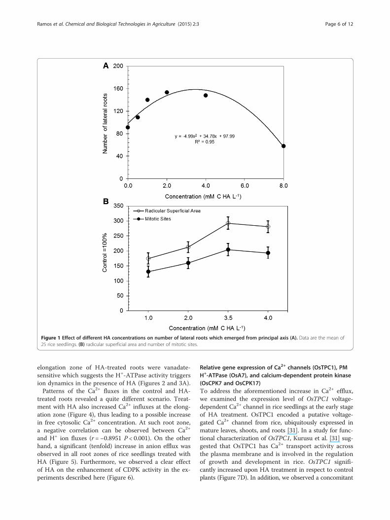
Figure 1 Effect of different HA concentrations on number of lateral roots which emerged from principal axis (A). Data are the mean of25 rice seedlings. (B) radicular superficial area and number of mitotic sites.
Ramos et al. Chemical and Biological Technologies in Agriculture (2015) 2:3 Page 6 of 12
elongation zone of HA-treated roots were vanadate-sensitive which suggests the H+-ATPase activity triggersion dynamics in the presence of HA (Figures 2 and 3A).Patterns of the Ca2+ fluxes in the control and HA-
treated roots revealed a quite different scenario. Treat-ment with HA also increased Ca2+ influxes at the elong-ation zone (Figure 4), thus leading to a possible increasein free cytosolic Ca2+ concentration. At such root zone,a negative correlation can be observed between Ca2+
and H+ ion fluxes (r = −0.8951 P < 0.001). On the otherhand, a significant (tenfold) increase in anion efflux wasobserved in all root zones of rice seedlings treated withHA (Figure 5). Furthermore, we observed a clear effectof HA on the enhancement of CDPK activity in the ex-periments described here (Figure 6).
Relative gene expression of Ca2+ channels (OsTPC1), PMH+-ATPase (OsA7), and calcium-dependent protein kinase(OsCPK7 and OsCPK17)To address the aforementioned increase in Ca2+ efflux,we examined the expression level of OsTPC1 voltage-dependent Ca2+ channel in rice seedlings at the early stageof HA treatment. OsTPC1 encoded a putative voltage-gated Ca2+ channel from rice, ubiquitously expressed inmature leaves, shoots, and roots [31]. In a study for func-tional characterization of OsTPC1, Kurusu et al. [31] sug-gested that OsTPC1 has Ca2+ transport activity acrossthe plasma membrane and is involved in the regulationof growth and development in rice. OsTPC1 signifi-cantly increased upon HA treatment in respect to controlplants (Figure 7D). In addition, we observed a concomitant
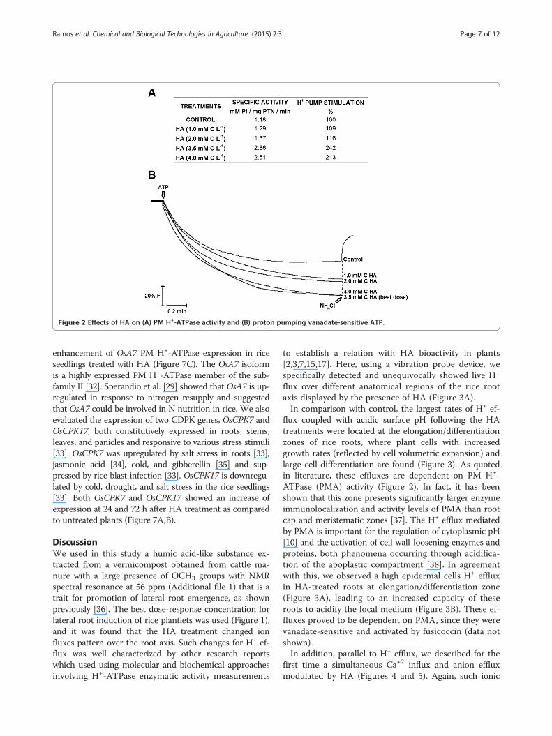
Figure 2 Effects of HA on (A) PM H+-ATPase activity and (B) proton pumping vanadate-sensitive ATP.
Ramos et al. Chemical and Biological Technologies in Agriculture (2015) 2:3 Page 7 of 12
enhancement of OsA7 PM H+-ATPase expression in riceseedlings treated with HA (Figure 7C). The OsA7 isoformis a highly expressed PM H+-ATPase member of the sub-family II [32]. Sperandio et al. [29] showed that OsA7 is up-regulated in response to nitrogen resupply and suggestedthat OsA7 could be involved in N nutrition in rice. We alsoevaluated the expression of two CDPK genes, OsCPK7 andOsCPK17, both constitutively expressed in roots, stems,leaves, and panicles and responsive to various stress stimuli[33]. OsCPK7 was upregulated by salt stress in roots [33],jasmonic acid [34], cold, and gibberellin [35] and sup-pressed by rice blast infection [33]. OsCPK17 is downregu-lated by cold, drought, and salt stress in the rice seedlings[33]. Both OsCPK7 and OsCPK17 showed an increase ofexpression at 24 and 72 h after HA treatment as comparedto untreated plants (Figure 7A,B).
DiscussionWe used in this study a humic acid-like substance ex-tracted from a vermicompost obtained from cattle ma-nure with a large presence of OCH3 groups with NMRspectral resonance at 56 ppm (Additional file 1) that is atrait for promotion of lateral root emergence, as shownpreviously [36]. The best dose-response concentration forlateral root induction of rice plantlets was used (Figure 1),and it was found that the HA treatment changed ionfluxes pattern over the root axis. Such changes for H+ ef-flux was well characterized by other research reportswhich used using molecular and biochemical approachesinvolving H+-ATPase enzymatic activity measurements
to establish a relation with HA bioactivity in plants[2,3,7,15,17]. Here, using a vibration probe device, wespecifically detected and unequivocally showed live H+
flux over different anatomical regions of the rice rootaxis displayed by the presence of HA (Figure 3A).In comparison with control, the largest rates of H+ ef-
flux coupled with acidic surface pH following the HAtreatments were located at the elongation/differentiationzones of rice roots, where plant cells with increasedgrowth rates (reflected by cell volumetric expansion) andlarge cell differentiation are found (Figure 3). As quotedin literature, these effluxes are dependent on PM H+-ATPase (PMA) activity (Figure 2). In fact, it has beenshown that this zone presents significantly larger enzymeimmunolocalization and activity levels of PMA than rootcap and meristematic zones [37]. The H+ efflux mediatedby PMA is important for the regulation of cytoplasmic pH[10] and the activation of cell wall-loosening enzymes andproteins, both phenomena occurring through acidifica-tion of the apoplastic compartment [38]. In agreementwith this, we observed a high epidermal cells H+ effluxin HA-treated roots at elongation/differentiation zone(Figure 3A), leading to an increased capacity of theseroots to acidify the local medium (Figure 3B). These ef-fluxes proved to be dependent on PMA, since they werevanadate-sensitive and activated by fusicoccin (data notshown).In addition, parallel to H+ efflux, we described for the
first time a simultaneous Ca+2 influx and anion effluxmodulated by HA (Figures 4 and 5). Again, such ionic
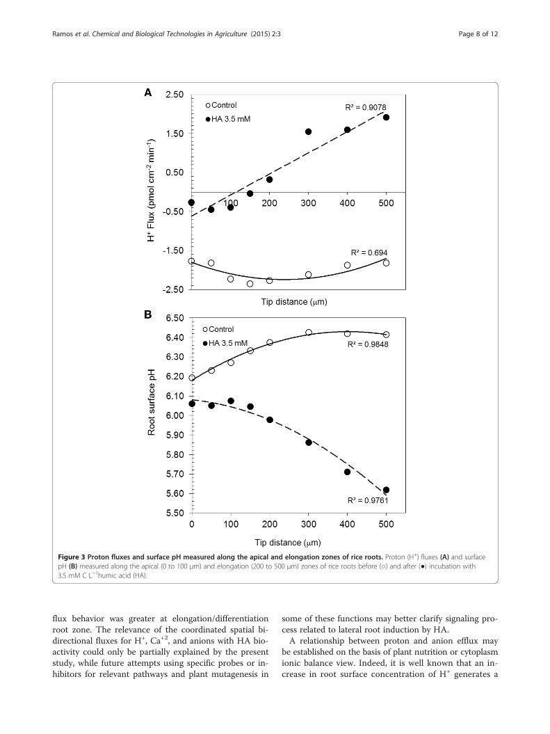
Figure 3 Proton fluxes and surface pH measured along the apical and elongation zones of rice roots. Proton (H+) fluxes (A) and surfacepH (B) measured along the apical (0 to 100 μm) and elongation (200 to 500 μm) zones of rice roots before (○) and after (●) incubation with3.5 mM C L−1humic acid (HA).
Ramos et al. Chemical and Biological Technologies in Agriculture (2015) 2:3 Page 8 of 12
flux behavior was greater at elongation/differentiationroot zone. The relevance of the coordinated spatial bi-directional fluxes for H+, Ca+2, and anions with HA bio-activity could only be partially explained by the presentstudy, while future attempts using specific probes or in-hibitors for relevant pathways and plant mutagenesis in
some of these functions may better clarify signaling pro-cess related to lateral root induction by HA.A relationship between proton and anion efflux may
be established on the basis of plant nutrition or cytoplasmionic balance view. Indeed, it is well known that an in-crease in root surface concentration of H+ generates a
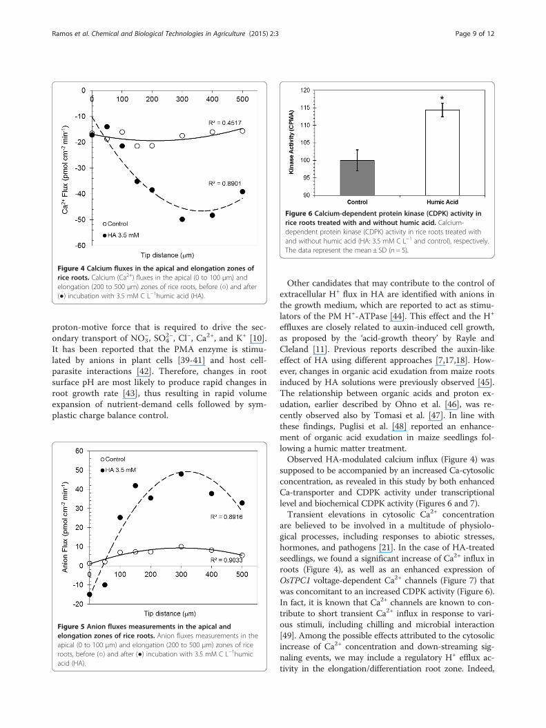
Figure 4 Calcium fluxes in the apical and elongation zones ofrice roots. Calcium (Ca2+) fluxes in the apical (0 to 100 μm) andelongation (200 to 500 μm) zones of rice roots, before (○) and after(●) incubation with 3.5 mM C L−1humic acid (HA).
Figure 6 Calcium-dependent protein kinase (CDPK) activity inrice roots treated with and without humic acid. Calcium-dependent protein kinase (CDPK) activity in rice roots treated withand without humic acid (HA: 3.5 mM C L−1 and control), respectively.The data represent the mean ± SD (n = 5).
Ramos et al. Chemical and Biological Technologies in Agriculture (2015) 2:3 Page 9 of 12
proton-motive force that is required to drive the sec-ondary transport of NO3
−, SO42−, Cl−, Ca2+, and K+ [10].
It has been reported that the PMA enzyme is stimu-lated by anions in plant cells [39-41] and host cell-parasite interactions [42]. Therefore, changes in rootsurface pH are most likely to produce rapid changes inroot growth rate [43], thus resulting in rapid volumeexpansion of nutrient-demand cells followed by sym-plastic charge balance control.
Figure 5 Anion fluxes measurements in the apical andelongation zones of rice roots. Anion fluxes measurements in theapical (0 to 100 μm) and elongation (200 to 500 μm) zones of riceroots, before (○) and after (●) incubation with 3.5 mM C L−1humicacid (HA).
Other candidates that may contribute to the control ofextracellular H+ flux in HA are identified with anions inthe growth medium, which are reported to act as stimu-lators of the PM H+-ATPase [44]. This effect and the H+
effluxes are closely related to auxin-induced cell growth,as proposed by the ‘acid-growth theory’ by Rayle andCleland [11]. Previous reports described the auxin-likeeffect of HA using different approaches [7,17,18]. How-ever, changes in organic acid exudation from maize rootsinduced by HA solutions were previously observed [45].The relationship between organic acids and proton ex-udation, earlier described by Ohno et al. [46], was re-cently observed also by Tomasi et al. [47]. In line withthese findings, Puglisi et al. [48] reported an enhance-ment of organic acid exudation in maize seedlings fol-lowing a humic matter treatment.Observed HA-modulated calcium influx (Figure 4) was
supposed to be accompanied by an increased Ca-cytosolicconcentration, as revealed in this study by both enhancedCa-transporter and CDPK activity under transcriptionallevel and biochemical CDPK activity (Figures 6 and 7).Transient elevations in cytosolic Ca2+ concentration
are believed to be involved in a multitude of physiolo-gical processes, including responses to abiotic stresses,hormones, and pathogens [21]. In the case of HA-treatedseedlings, we found a significant increase of Ca2+ influx inroots (Figure 4), as well as an enhanced expression ofOsTPC1 voltage-dependent Ca2+ channels (Figure 7) thatwas concomitant to an increased CDPK activity (Figure 6).In fact, it is known that Ca2+ channels are known to con-tribute to short transient Ca2+ influx in response to vari-ous stimuli, including chilling and microbial interaction[49]. Among the possible effects attributed to the cytosolicincrease of Ca2+ concentration and down-streaming sig-naling events, we may include a regulatory H+ efflux ac-tivity in the elongation/differentiation root zone. Indeed,
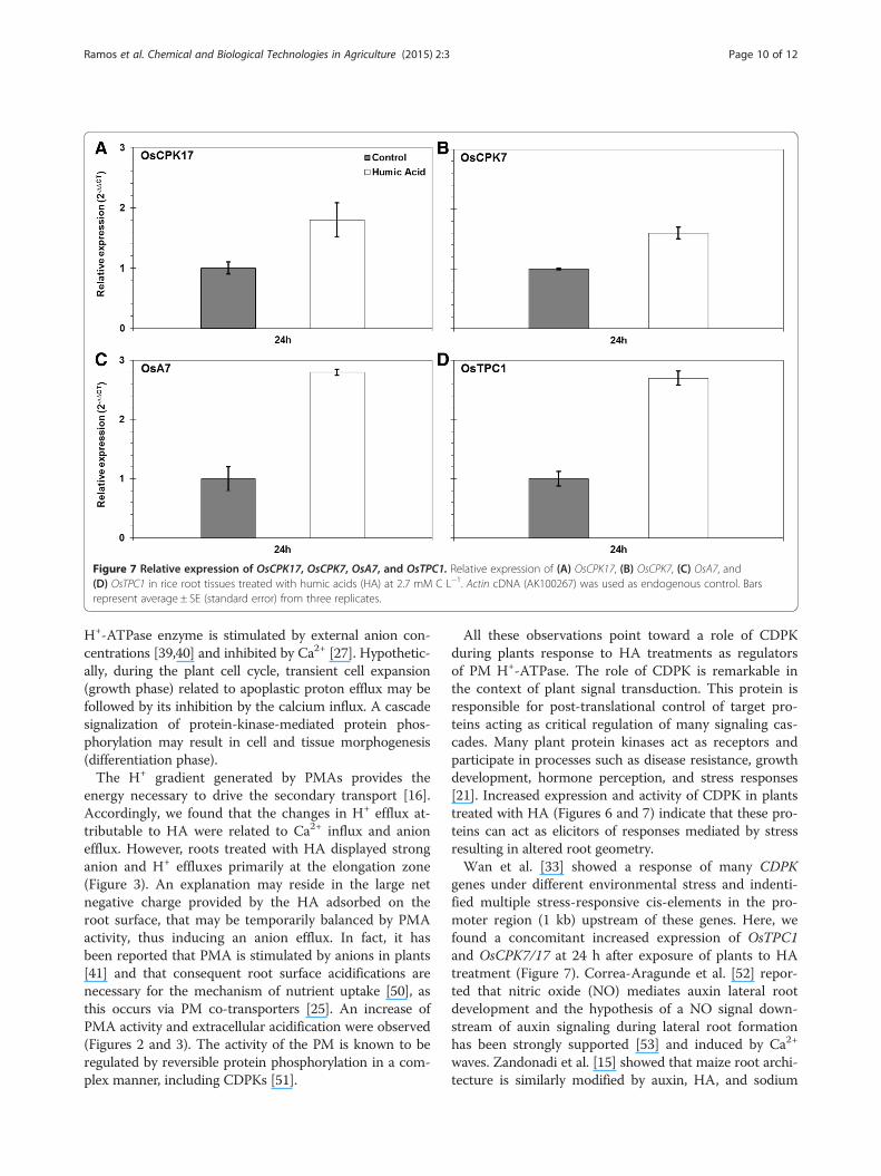
Figure 7 Relative expression of OsCPK17, OsCPK7, OsA7, and OsTPC1. Relative expression of (A) OsCPK17, (B) OsCPK7, (C) OsA7, and(D) OsTPC1 in rice root tissues treated with humic acids (HA) at 2.7 mM C L−1. Actin cDNA (AK100267) was used as endogenous control. Barsrepresent average ± SE (standard error) from three replicates.
Ramos et al. Chemical and Biological Technologies in Agriculture (2015) 2:3 Page 10 of 12
H+-ATPase enzyme is stimulated by external anion con-centrations [39,40] and inhibited by Ca2+ [27]. Hypothetic-ally, during the plant cell cycle, transient cell expansion(growth phase) related to apoplastic proton efflux may befollowed by its inhibition by the calcium influx. A cascadesignalization of protein-kinase-mediated protein phos-phorylation may result in cell and tissue morphogenesis(differentiation phase).The H+ gradient generated by PMAs provides the
energy necessary to drive the secondary transport [16].Accordingly, we found that the changes in H+ efflux at-tributable to HA were related to Ca2+ influx and anionefflux. However, roots treated with HA displayed stronganion and H+ effluxes primarily at the elongation zone(Figure 3). An explanation may reside in the large netnegative charge provided by the HA adsorbed on theroot surface, that may be temporarily balanced by PMAactivity, thus inducing an anion efflux. In fact, it hasbeen reported that PMA is stimulated by anions in plants[41] and that consequent root surface acidifications arenecessary for the mechanism of nutrient uptake [50], asthis occurs via PM co-transporters [25]. An increase ofPMA activity and extracellular acidification were observed(Figures 2 and 3). The activity of the PM is known to beregulated by reversible protein phosphorylation in a com-plex manner, including CDPKs [51].
All these observations point toward a role of CDPKduring plants response to HA treatments as regulatorsof PM H+-ATPase. The role of CDPK is remarkable inthe context of plant signal transduction. This protein isresponsible for post-translational control of target pro-teins acting as critical regulation of many signaling cas-cades. Many plant protein kinases act as receptors andparticipate in processes such as disease resistance, growthdevelopment, hormone perception, and stress responses[21]. Increased expression and activity of CDPK in plantstreated with HA (Figures 6 and 7) indicate that these pro-teins can act as elicitors of responses mediated by stressresulting in altered root geometry.Wan et al. [33] showed a response of many CDPK
genes under different environmental stress and indenti-fied multiple stress-responsive cis-elements in the pro-moter region (1 kb) upstream of these genes. Here, wefound a concomitant increased expression of OsTPC1and OsCPK7/17 at 24 h after exposure of plants to HAtreatment (Figure 7). Correa-Aragunde et al. [52] repor-ted that nitric oxide (NO) mediates auxin lateral rootdevelopment and the hypothesis of a NO signal down-stream of auxin signaling during lateral root formationhas been strongly supported [53] and induced by Ca2+
waves. Zandonadi et al. [15] showed that maize root archi-tecture is similarly modified by auxin, HA, and sodium
Ramos et al. Chemical and Biological Technologies in Agriculture (2015) 2:3 Page 11 of 12
nitroprusside (SNP), thus leading to an increase of lat-eral root abundance, root density, and PM H+-ATPaseactivation.The role of CDPK, NO, Ca2+, and calmodulin were
studied in the auxin-induced adventitious roots in cu-cumber [54]. It was shown that CDPK was stimulated byauxin and depended on NO, Ca2+, and calmodulin dur-ing formation of roots.The present study suggested a new role for HA-
stimulated plant physiology that reveals a potentiallyimportant aspect of coevolution between humified or-ganic matter and the plant growth and development.Changing the ion fluxes across the root plasma mem-brane implies modifications in transmembrane electricalpotential. The latter is generated by the electrogenic H+
pumps involved in the ion transport systems as well as bythe signal transduction related to root morphogenesis.
ConclusionsHumic matter acts a plant elicitor by triggering a seriesof cell signaling events. Our results suggest a model forcell signaling in roots that is directly linked to nutrientuptake and lateral root emergence. We have shown thatHA induce a positive modulation of PM H+-ATPase ac-tivity and expression that controls the H+ efflux, rootsurface pH, and consequently triggers modifications inthe anion fluxes. This hyperpolarization, in turn, inducesa pH signal that modulates Ca2+ transport by increasingcytosolic free Ca2+ concentration, which then acts as asecond messenger though the mediation of a variety ofcellular responses like CDPK activities and anion chan-nel activation. A large anion efflux is activated by theHA negative charges which are built up on the root sur-face and re-induce H+-ATPase activity. Within the effortto elucidate the complex HA-plant response mechanism,our results show that HA have an effect on various entrypoints of the cell signaling machinery, such as fluxes ofH+ and Ca2+, H+-ATPase and CDPK activity, as well asthe expression of Ca2+ transporters. Overall, our findingsof this work indicate that plant nutrient uptake andgrowth promotion mediated by humic matter may con-sist in a pH-dependent phenomenon.
Additional files
Additional file 1: Table S1. Gene-specific primers for real-time PCR.
Additional file 2: Figure S1. CP/MAS 13C NMR spectra of humic acids.
Competing interestsThe authors declare that they have no competing interests.
Authors' contributionsACR carried out the ion flux measurement and drafted the manuscript.LBD carried out the CDPK activity and drafted the manuscript. LAS and MSFcarried out the molecular genetic studies and drafted the manuscript.
NOA did the humic acid characterization. FLO and LPC conceived of thestudy and participated in its design and coordination and helped to draftthe manuscript. All authors read and approved the final manuscript.
AcknowledgementsThe authors wish to thank Fundação de Amparo à Pesquisa do Estado doRio de Janeiro (FAPERJ) and Conselho Nacional de Pesquisa (CNPq), INCT forBiological Nitrogen Fixation, and International Foudation of Science (IFS) forfinancial support.
Author details1Laboratório de Bioquímica e Fisiologia de Microrganismos, UniversidadeEstadual do Norte Fluminense Darcy Ribeiro (UENF), Av. Alberto Lamego2000, Campos dos Goytacazes 28013-602, Brazil. 2Laboratório deMicrobiologia Ambiental e Biotecnologia, Universidade de Vila Velha (UVV),Rua Comissário José Dantas de Melo 21, Boa Vista, Vila Velha, Espírito Santo,Brazil. 3Departamento de Solos da Universidade Federal Rural do Rio deJaneiro (UFRRJ), Seropédica, km 7 BR 465, Seropédica, Rio de Janeiro CEP23851-970, Brazil. 4Universidade Estadual do Norte Fluminense Darcy Ribeiro(UENF) Núcleo de Desenvolvimento de Insumos Biológicos para Agricultura(NUDIBA), Av. Alberto Lamego 2000, Campos dos Goytacazes 28013-602,Brazil.
Received: 11 October 2014 Accepted: 19 December 2014
References1. Orsi M (2014) Molecular dynamics simulation of humic substances.
Chem Biol Technol Agr 1:102. Nardi S, Carletti P, Pizzeghello D, Muscolo A (2009) Biological activities of
humic substances. In Senesi N,Xing B Huang PM (ed) Biophysico-ChemicalProcesses Involving Natural Non Living Organic Matter in EnvironmentalSystems. Vol 2, part 1: fundamentals and impact of mineral-organic biotainteractions on the formation, transformation. Turnover and storage ofnatural nonliving organic matter (NOM). Wiley, Hoboken, pp305-340
3. Canellas LP, Olivares FL (2014) Physiological responses to humic substancesas plant growth promoter. Chem Biol Technol Agr 1:3
4. Mora V, Bacaicoa E, Zamarreño AM, Agguirre E, Garnica M, Fuentes M,García-Mina JM (2010) Action of humic acid on promotion of cucumbershoot growth involves nitrate-related changes associated with the root-to-shoot distribution of cytokinins, polyamines and mineral nutrients. J PlantPhysiol 167:633–642
5. Canellas LP, Dobbss LB, Oliveira AL, Chagas JG, Aguiar NO, Rumjanek VM,Novotny EH, Olivares FL, Spaccini R, Piccolo A (2012) Chemical properties ofhumic matter as related to induction of plant lateral roots. Eur J Soil Sci63:315–324
6. Mora V, Bacaicoa E, Baigorri R, Zamarreño AM, García-Mina JM (2014) NOand IAA key regulators in the shoot growth promoting action of humic acidin Cucumis sativus L. J Plant Growth Regul 33:430–439
7. Dobbss LB, Canellas LP, Olivares FL, Aguiar NO, Peres LEP, Azevedo M,Spaccini R, Piccolo A, Façanha AR (2010) Bioactivity of chemicallytransformed humic matter from vermicompost on plant root growth. J AgrFood Chem 58:3681–3688
8. Feijó JA, Sainhas J, Hackett GR, Kunkel JG, Hepler PK (1999) Growing pollentubes posses a constitutive alkaline band in the clear zone and a growth-dependent acidic tip. J Cell Biol 144:483–496
9. Konrad KR, Wudick MM, Feijó JA (2012) Calcium regulation of tip growth:new genes for old mechanisms. Curr Opin Plant Biol 14:721–730
10. Felle HH (2001) pH: signal and messenger in plant cells. Plant Biol 3:577–59111. Rayle DL, Cleland RE (1992) The acid growth theory of auxin-induced cell
elongation is alive and well. Plant Physiol 99:1271–127412. Rober-Kleber N, Albrechtová JTP, Fleig S, Huck N, Michalke W, Wagner E,
Speth V, Neuhaus G, Fischer-Iglesias C (2003) Plasma membrane H+-ATPaseis involved in auxin-mediated cell elongation during wheat embryodevelopment. Plant Physiol 131:1302–1312
13. Fasano JM, Swanson SJ, Blancaflor EB, Dowd PE, Kao T, Gilroy S (2001)Changes in root cap pH are required for the gravity response of theArabidopsis root. Plant Cell 13:907–92
14. Trevisan S, Pizzeghello D, Ruperti B, Francioso O, Sassi A, Palme K,Quaggiotti S, Nardi S (2010) Humic substances induce lateral root formation
Ramos et al. Chemical and Biological Technologies in Agriculture (2015) 2:3 Page 12 of 12
and expression of the early auxin-responsive IAA19 gene and DR5 syntheticelement in Arabidopsis. Plant Biol 12:604–614
15. Zandonadi DB, Santos MP, Dobbss LB, Olivares FL, Canellas LP, Binzel ML,Okorokova-Façanha AL, Façanha AR (2010) Nitric oxide mediates humicacids-induced root development and plasma membrane H+-ATPaseactivation. Planta 231:1025–1036
16. Palmgren MG (2001) Plant plasma membrane H+-ATPases: powerhouses fornutrient uptake. Annl Rev Plant PhysiolPlant Mol Biol 52:817–845
17. Canellas LP, Façanha AO, Olivares FL, Façanha AR (2002) Humic acidsisolated from earthworm compost enhance root elongation, lateral rootemergence, and plasma membrane H+-ATPase activity in maize roots. PlantPhysiol 130:1951–1957
18. Quaggiotti S, Rupert B, Pizzeghello D, Francioso O, Tugnoli V, Nardi S (2004)Effect of low molecular size humic substances on nitrate uptake andexpression of genes involved in nitrate transport in maize (Zea mays L.).J Exp Bot 55:803–813
19. Salomon D, Bonshtien A, Sessa G (2009) A chemical-genetic approach forfunctional analysis of plant protein kinases. Plant Signal Behav 4:645–647
20. Zhang Y, Gao P, Yuan JS (2010) Plant protein-protein interaction networkand 58. Interactome Curr Genomics 11:40–46
21. Kudla J, Batistic O, Hashimoto K (2010) Calcium signals: the lead currency ofplant information processing. Plant Cell 22:541–563
22. Hamada H, Kurusu T, Okuma E, Nokajima H, Kiyoduka M, Koyano T,Sugiyama Y, Okada K, Koga J, Saji H, Miyao A, Hirochika H, Yamane H,Murata Y, Kuchitsu K (2012) Regulation of a proteinaceous elicitor-inducedCa2+ influx and production of phytoalexins by a putative voltage-gatedcation channel, OsTPC1, in cultured rice cells. J Biol Chem 23:9931–0039
23. Pei ZM, Murata Y, Benning G, Thomine S, Klusener B, Allen GJ, Grill E,Schroeder JI (2000) Calcium channels activated by hydrogen peroxidemediate abscisic acid signalling in guard cells. Nature 406:731–734
24. Lowry OH, Rosebrough NJ, Farr AL, Randall RJ (1951) Protein measurementwith the Folin phenol reagent. J Biol Chem 193:265–275
25. Ramos AC, Façanha AR, Lima PT, Feijó JA (2008) pH signature for theresponses of arbuscular mycorrhizal fungi to external stimuli. PlantSignalBehav 3:850–852
26. Ramos AC, Lima PT, Dias PN, Kasuya MCM, Feijó JA (2009) A pH signalingmechanism involved in the spatial distribution of calcium and anion fluxesin ectomycorrhizal roots. New Phytol 181:448–462
27. Lino B, Baizabal-Aguirre VM, González de la Vara LE (1998) The plasma-membrane H+-ATPase from beet root is inhibited by a calcium-dependentphosphorylation. Planta 204:352–359
28. Gao J, Liu J, Li B, Li Z (2001) Isolation and purification of functional totalRNA from blue-grained wheat endosperm tissues containing high levels ofstarches and flavonoids. Plant Mol Biol Rep 19:185–1185
29. Sperandio MVL, Santos LA, Bucher CA, Fernandes MS, Souza SR (2011)Isoforms of plasma membrane H + -ATPase in rice root and shoot aredifferentially induced by starvation and resupply of NO3
- or NH4+. Plant Sci
180:251–25830. Livak KJ, Schmittgen TD (2001) Analysis of relative gene expression data
using real-time quantitative PCR and the 2 − ΔΔCt method. Methods25:402–408
31. Kurusu T, Sakurai Y, Miyao A, Hirochika H, Kuchitsu K (2004) Identification ofa putative voltage-gated Ca2+-permeable channel (OsTPC1) involved in Ca2+
influx and regulation of growth and development in rice. Plant Cell Physiol45:693–702
32. Arango M, Gevaudant F, Oufattole M, Boutry M (2003) The plasmamembrane proton pump ATPase: the significance of gene subfamilies.Planta 216:355–365
33. Wan B, Lin Y, Mou T (2007) Expression of rice Ca2+-dependent proteinkinases (CDPKs) genes under different environmental stresses. FEBS Lett581:1179–1189
34. Akimoto-Tomiyama C, Sakata K, Yazaki J, Nakamura K, Fujii F (2003) Ricegene expression in response to Nacetylchitooligosaccharide elicitor:comprehensive analysis by DNA microarray with randomly selected ESTs.Plant Mol Biol 52:537–551
35. Abbasi F, Onodera H, Toki S, Tanaka H, Komatsu S (2004) OsCDPK13, acalcium-dependent protein kinase gene from rice, is induced by cold andgibberellin in rice leaf sheath. Plant Mol Biol 55:541–552
36. Aguiar NO, Novotny EH, Oliveira AL, Rumjanek VM, Olivares FL, Canellas LP(2013) Prediction of humic acids bioactivity using spectroscopy andmultivariate analysis. J Geochem Explor 129:95–102
37. Enriquez-Arredondo C, Sanchez-Nieto S, Rendon-Huerta E, Gonzalez-Halphen D, Gavilanes-Ruiz M, Diaz-Pontones D (2005) The plasma mem-brane H+-ATPase of maize embryos localizes in regions that are critical dur-ing the onset of germination. Plant Sci 169:11–19
38. Hager A (2003) Role of the plasma membrane H+-ATPase in auxin inducedelongation growth. Historical and new aspects. J Plant Res 116:483–505
39. Churchill KA, Sze H (1984) Anion-sensitive, H+ pumping ATPase of oat roots:direct effects of Cl−, NO3
- and a disulfonic stilbene. Plant Physiol 76:490–49740. Ullrich CI, Novacky AJ (1990) Extra- and intracellular pH and membrane
potential changes induced by K+, CI−, H2PO4-, NO3-, uptake and fusicoccinin root hairs of Limnobium storoniferum. Plant Physiol 94:1561–1567
41. Zimmermann S, Thomine S, Guern J, Barbier-Brygoo H (1994) An anioncurrent at the plasma membrane of tobacco protoplasts shows ATP-dependent voltage regulation and is modulated by auxin. Plant J 6:707–716
42. Vieira L, Slotki I, Cabantchik ZI (1995) Chloride conductive pathways whichsupport electrogenic H+ pumping by Leishmania major Promastigotes.J Biol Chem 270:5299–5304
43. Peters WS, Felle HH (1999) The correlation of profiles of surface pH andelongation growth in maize roots. Plant Physiol 121:905–912
44. Garnett TP, Shabala SN, Smethurst PJ, Newman IA (2001) Kinetics ofammonium and nitrate uptake by eucalypt roots and associated protonfluxes measured using ion selective microelectrodes. Funct Plant Biol30:1165–1176
45. Canellas LP, Teixeira Junior LRL, Dobbss LB, Silva CA, Médici LO, ZandonadiDB, Façanha AR (2008) Humic acids cross interactions with root and organicacids. Ann Appl Biol 153:157–166
46. Ohno T, Nakahira S, Suzuki Y, Kani T, Hara T, Koyama H (2004) Molecularcharacterization of plasma membrane H+-ATPase in a carrot mutant cell linewith enhanced citrate excretion. Plant Physiol 122:265–274
47. Tomasi N, Kretzschmar T, Espen L, Weisskopf L, Thoe Fuglsang A, PalmgrenG, Neumann G, Varanini Z, Pinton R, Martinoia E, Cesco E (2009) Plasmamembrane H+-ATPase-dependent citrate exudation from cluster roots ofphosphate-deficient white lupin. Plant Cell Environ 32:465–475
48. Puglisi E, Fragoulis G, Del Re AM, Spaccini R, Gigliotti G, Said-Pullicino D,Trevisan M (2008) Carbon deposition in soil rhizosphere followingamendments with soluble fractions, as evaluated by combined soil-plantrhizobox and reporter gene systems. Chemosphere 73:1292–1299
49. Thion L, Mazars C, Nacry P, Bouchez D, Moreau M, Ranjeva R, Thuleau P(1998) Plasma membrane depolarizationactivated depolarizationactivatedcalcium channels, stimulated by microtubule-depolymerizing drugs inwild-type Arabidopsis thaliana protoplasts, display constitutively largeactivities and a longer half-life in ton 2 mutant cells affected in theorganization of cortical microtubules. Plant J 13:603–610
50. Forde BG (2000) Nitrate transporters in plants: structure, function andregulation. v. Biochim Biophys Acta 1465:219–235
51. Vera-Estrella R, Barkla BJ, Higgins VJ, Blumwald E (1994) Plant defenseresponse to funga1 pathogens. Activation of host-plasma membraneH+-ATPase by elicitor-induced enzyme dephosphorylation. Plant Physiol104:209–215
52. Correa-Aragunde N, Graziano M, Lamattina L (2004) Nitric oxide plays acentral role in determining lateral root development in tomato. Planta218:900–905
53. Kolbert Z, Bartha B, Erdei L (2008) Exogenous auxin-induced NO synthesis isnitrate reductase-associated in Arabidopsis thaliana root primordia. J PlantPhysiol 165:967–975
54. Lanteri ML, Pagnussat GC, Lamattina L (2006) Calcium and calcium-dependent protein kinases are involved in nitric oxide- and auxin-inducedadventitious root formation in cucumber. J Exp Bot 57:1341–1351
Copyright © 2022 FDOKUMEN