Human CD46-transgenic mice in studies involving replication-incompetent adenoviral type 35 vectors
11
Human CD46-transgenic mice in studies involving replication-incompetent adenoviral type 35 vectors Sandra Verhaagh, 1 Esmeralda de Jong, 1 3 Jaap Goudsmit, 1 Sylvie Lecollinet, 2 Gert Gillissen, 1 Margreet de Vries, 3 Kees van Leuven, 3 Ivo Que, 4 Krista Ouwehand, 1 Ratna Mintardjo, 1 Gerrit Jan Weverling, 1 Katarina Rados ˇevic ´, 1 Jennifer Richardson, 2 Marc Eloit, 2 Clemens Lowik, 4 Paul Quax 3 and Menzo Havenga 1 Correspondence Menzo Havenga [email protected] 1 Crucell Holland BV, Archimedesweg 4, 2333 CN Leiden, The Netherlands 2 Ecole National Veterinaire d’Alfort, Alfort, France 3 Gaubius Laboratory, TNO Leiden, The Netherlands 4 Department of Endocrinology, Leiden University Medical Centre, Leiden, The Netherlands Received 28 June 2005 Accepted 25 October 2005 Wild-type strains of mice do not express CD46, a high-affinity receptor for human group B adenoviruses including type 35. Therefore, studies performed to date in mice using replication-incompetent Ad35 (rAd35) vaccine carriers may underestimate potency or result in altered vector distribution. Here, it is reported that CD46 transgenic mice (MYII-strain) express CD46 in all major organs and that it functions as a receptor for rAd35 vectors. Similar to monkeys and humans, MYII mice highly express CD46 in their lungs and kidneys and demonstrate low expression in muscle. Upon intravenous administration, rAd35 vector genomes as well as expression are detected in lungs of MYII mice, in contrast to wild-type littermates. Expression was predominantly detected in lung epithelial cells. Upon intramuscular administration, the initial level of luciferase expression is higher in MYII mice as compared with wild-type littermates, in spite of the fact that CD46 expression is low in muscle of MYII mice. The higher level of expression in muscle of MYII mice results in prolonged gene expression as assessed by CCD camera imaging for luciferase activity. Finally, a significant dose-sparing effect in MYII mice as compared with wild-type littermates on anti-SIVgag CD8 + T-cell induction following intramuscular vaccination with an rA35.SIVgag vaccine was observed. This dose-sparing effect was also observed when reinfusing dendritic cells derived from MYII mice after exposure to rAd35.SIVgag vaccine as compared with rAd35.SIVgag exposed dendritic cells from wild-type littermates. It was concluded that MYII mice represent an interesting preclinical model to evaluate potency and safety of rAd35 vectors. INTRODUCTION It has been demonstrated that members of the human group B adenoviruses and perhaps several serotypes of subgroup D utilize CD46 as a high-affinity receptor (Gaggar et al., 2003; Segerman et al., 2003; Sirena et al., 2004; Wu et al., 2004). Human subgroup B adenoviruses, Ad11 and Ad35, are rare in the human population resulting in low seroprevalence, whereas group C vectors (Ad2 and Ad5), which are com- monly used for gene therapy and vaccination approaches, have high seroprevalence (Kostense et al., 2004; Nwanegbo et al., 2004; Vogels et al., 2003). High seroprevalence of Ad2 and Ad5 hampers the utility of these vaccine carriers since high anti-vector immunity effectively blunts vaccine potency (Barouch et al., 2004). Therefore, rAd11 and rAd35 vaccine carriers hold the promise of accurate dose control and consistent induction of immune responses against inserted antigens, which was further strengthened by a study showing strong rAd35-mediated induction of anti-SIVgag T-cell responses in naı ¨ve mice or in mice carrying high-level anti- Ad5 neutralizing activity (Barouch et al., 2004; Holterman et al., 2004; Vogels et al., 2003). Interestingly, this study also demonstrates that anti-Ad5 T-cells cross-reacting with rAd35, also known to be present in human PBMC samples (Heemskerk et al., 2003), only marginally affected the potency of the rAd35.SIVgag vaccine, indicating that neutralizing antibodies are predominantly responsible for the diminishing vaccine potency (Barouch et al., 2004). Thus Published online ahead of print on 10 November 2005 as DOI 10.1099/vir.0.81293-0. 3Deceased January 2005. 0008-1293 G 2006 SGM Printed in Great Britain 255 Journal of General Virology (2006), 87, 255–265 DOI 10.1099/vir.0.81293-0
Transcript of Human CD46-transgenic mice in studies involving replication-incompetent adenoviral type 35 vectors
Human CD46-transgenic mice in studies involvingreplication-incompetent adenoviral type 35 vectors
Sandra Verhaagh,1 Esmeralda de Jong,13 Jaap Goudsmit,1
Sylvie Lecollinet,2 Gert Gillissen,1 Margreet de Vries,3 Kees van Leuven,3
Ivo Que,4 Krista Ouwehand,1 Ratna Mintardjo,1 Gerrit Jan Weverling,1
Katarina Radosevic,1 Jennifer Richardson,2 Marc Eloit,2 Clemens Lowik,4
Paul Quax3 and Menzo Havenga1
Correspondence
Menzo Havenga
1Crucell Holland BV, Archimedesweg 4, 2333 CN Leiden, The Netherlands
2Ecole National Veterinaire d’Alfort, Alfort, France
3Gaubius Laboratory, TNO Leiden, The Netherlands
4Department of Endocrinology, Leiden University Medical Centre, Leiden, The Netherlands
Received 28 June 2005
Accepted 25 October 2005
Wild-type strains of mice do not express CD46, a high-affinity receptor for human group B
adenoviruses including type 35. Therefore, studies performed to date in mice using
replication-incompetent Ad35 (rAd35) vaccine carriers may underestimate potency or result in
altered vector distribution. Here, it is reported that CD46 transgenic mice (MYII-strain) express
CD46 in all major organs and that it functions as a receptor for rAd35 vectors. Similar to monkeys
and humans, MYII mice highly express CD46 in their lungs and kidneys and demonstrate low
expression in muscle. Upon intravenous administration, rAd35 vector genomes as well as
expression are detected in lungs of MYII mice, in contrast to wild-type littermates. Expression was
predominantly detected in lung epithelial cells. Upon intramuscular administration, the initial level
of luciferase expression is higher in MYII mice as compared with wild-type littermates, in spite
of the fact that CD46 expression is low in muscle of MYII mice. The higher level of expression in
muscle of MYII mice results in prolonged gene expression as assessed by CCD camera imaging
for luciferase activity. Finally, a significant dose-sparing effect in MYII mice as compared with
wild-type littermates on anti-SIVgag CD8+ T-cell induction following intramuscular vaccination
with an rA35.SIVgag vaccine was observed. This dose-sparing effect was also observed when
reinfusing dendritic cells derived from MYII mice after exposure to rAd35.SIVgag vaccine as
compared with rAd35.SIVgag exposed dendritic cells from wild-type littermates. It was concluded
that MYII mice represent an interesting preclinical model to evaluate potency and safety of
rAd35 vectors.
INTRODUCTION
It has been demonstrated that members of the human groupB adenoviruses and perhaps several serotypes of subgroup Dutilize CD46 as a high-affinity receptor (Gaggar et al., 2003;Segerman et al., 2003; Sirena et al., 2004; Wu et al., 2004).Human subgroup B adenoviruses, Ad11 and Ad35, are rarein the human population resulting in low seroprevalence,whereas group C vectors (Ad2 and Ad5), which are com-monly used for gene therapy and vaccination approaches,have high seroprevalence (Kostense et al., 2004; Nwanegboet al., 2004; Vogels et al., 2003). High seroprevalence of Ad2
and Ad5 hampers the utility of these vaccine carriers sincehigh anti-vector immunity effectively blunts vaccine potency(Barouch et al., 2004). Therefore, rAd11 and rAd35 vaccinecarriers hold the promise of accurate dose control andconsistent induction of immune responses against insertedantigens, which was further strengthened by a study showingstrong rAd35-mediated induction of anti-SIVgag T-cellresponses in naıve mice or in mice carrying high-level anti-Ad5 neutralizing activity (Barouch et al., 2004; Holtermanet al., 2004; Vogels et al., 2003). Interestingly, this studyalso demonstrates that anti-Ad5 T-cells cross-reacting withrAd35, also known to be present in human PBMC samples(Heemskerk et al., 2003), only marginally affected thepotency of the rAd35.SIVgag vaccine, indicating thatneutralizing antibodies are predominantly responsible forthe diminishing vaccine potency (Barouch et al., 2004). Thus
Published online ahead of print on 10 November 2005 as DOI10.1099/vir.0.81293-0.
3Deceased January 2005.
0008-1293 G 2006 SGM Printed in Great Britain 255
Journal of General Virology (2006), 87, 255–265 DOI 10.1099/vir.0.81293-0

far, studies with rAd11 or rAd35 vectors have been per-formed in inbred mice strains that do not express CD46(Gaggar et al., 2003). In fact, whereas CD46 protein con-servation is high between humans, new-world monkeys(80?2%) and old-world monkeys (87?4%), conservation ispoor between humans and pigs, guinea pigs, rats and mice(all <50%). Thus, we hypothesized that perhaps datagenerated so far on potency and distribution of rAd11 andrAd35 vectors in mice may not be representative of whatcan be expected of these vectors in monkeys and humans.Therefore, we set out to identify whether distribution andpotency of rAd35 vectors differ in mice carrying the CD46receptor or wild-type (Wt) littermates. Since the humanCD46 protein (hCD46) serves as a high-affinity receptor forseveral important pathogens including Measles virus (Doriget al., 1993; Manchester et al., 1994; Naniche et al., 1993),Human herpesvirus 6 (Santoro et al., 1999), Streptococcuspyogenes (Okada et al., 1995), Neisseria gonorrhoeae andMeningitides (Kallstrom et al., 1997), different CD46 trans-genic mice models have been generated and studied (Horvatet al., 1996; Miyagawa et al., 1997; Rall et al., 1997; Thorleyet al., 1997). In choosing among different CD46 trans-genic mouse models, we decided on MYII mice, whichwere obtained by transferring a 420 kilobase yeast artificialchromosome clone carrying the hCD46 gene under its ownregulatory sequences (Yannoutsos et al., 1996). These miceexpress all four major isoforms of CD46 known to beexpressed in humans (Kemper et al., 2001; Yannoutsos et al.,1996), i.e. BC1, BC2, C1 and C2, and have been usedsuccessfully to establish animal models, for instance formeasles virus infection (Oldstone et al., 1999).
Based on the studies described here, showing MYII-specificlung sequestration of rAd35 upon intravenous (i.v.) admin-istration, increased level and longevity of genemarking uponintramuscular (i.m.) administration in muscle and a cleartetramer-positive CD8+ T-cell dose-sparing effect uponrAd35.SIVgag vaccination, we conclude that MYII miceprovide an interesting preclinical model for testing genetransfer and vaccination regimens involving rAd35 vectors.
METHODS
Adenoviral vectors and in vitro transduction. The generationof the Ad35 plasmid system and production, purification and titra-tion of recombinant (r) Ad5 and Ad35 vectors have been describedpreviously (Barouch et al., 2004; Holterman et al., 2004; Vogelset al., 2003). The B16F10 mouse melanoma cell line was obtainedfrom the ATCC and was stably transfected using lipofectamine, witha plasmid expressing the BC1 isoform under the control of the SV40promoter. Transfectants were selected by co-transfecting a secondplasmid, expressing neomycin under the control of the cytomegalo-virus promoter, by adding 2?0 mg neomycin ml21 to cell culturemedium. High CD46-expressing cell clones were obtained via FACSsorting (Vantage SE; Becton Dickinson) and cell clone expansionupon limiting dilution. For vector transduction, cells were exposedfor 2 h to 1000 or 2500 virus particles (vp) per cell after which viruscontaining medium was replaced by fresh medium. The percentageof cells expressing green fluorescent protein (GFP) was determined48 h after vector exposure using FACSCalibur (Becton Dickinson).
Human and rhesus monkey monocyte-derived dendritic cells (DC)as well as mouse bone marrow-derived DC of different strains werecultured and characterized essentially as described previously(Ophorst et al., 2004).
CD46 expression and haemagglutination. To analyse CD46surface expression by FACS, cells were stained for 30 min at 4 uCwith 1 : 20 diluted (PBS/0?5% BSA) anti-human CD46 coupled tofluorescein isothiocyanate (FITC; Leinco). For Western blot analysis,frozen tissue of human (n=1), monkey (n=1) or mouse origin(n=3) was homogenized with a blender in appropriate volumes ofRIPA (0?5 g deoxycholic acid, 1% NP40, 10% SDS and PBS) buffercontaining protease inhibitors (mini complete; Roche). Proteinlysates were clarified by centrifugation and total protein content wasdetermined using the Pierce BCA kit. Of each tissue, 10 mg total pro-tein was loaded onto a 4–12% Bis/Tris gel (Invitrogen) and trans-ferred onto an Immobulon-P transfer membrane (Millipore). Afterblocking with TBST (0?1% Tween 20 in Tris-buffered saline) con-taining 5% (w/v) non-fat dry milk (Bio-Rad), membranes wereincubated for 1 h with a 1 : 200 diluted goat polyclonal anti-CD46antibody (Santa Cruz Biotechnology). Subsequently, a 1 : 15 000diluted horseradish peroxidase-coupled rabbit anti-goat IgG poly-clonal antibody (Santa Cruz Biotechnology) was used to visualizesignals by enhanced chemiluminescence detection kit (ECL;Amersham Biosciences). For haemagglutination assays, mouse bloodwas diluted to 3% erythrocyte density in preservation medium(Sanquin). Adenovirus stock solutions of rAd5 and rAd35 werediluted from 26109 to 9?86105 vp per 100 ml PBS containing 5%sucrose, 0?7 mM CaCl2 and 0?7 mM MgCl2. As negative controlonly PBS containing 5% sucrose, 0?7 mM CaCl2 and 0?7 mMMgCl2 was used. The 3% erythrocyte solution was further diluted to0?4% erythrocyte density in PBS. Subsequently, 50 ml 0?4% erythro-cyte solution was added and plates were incubated for 1 h at 37 uCat 10% CO2. Before read-out, plates were centrifuged for 1 min at201 relative centrifugal force.
Breeding of MYII mice and Wt littermates. For breeding pur-poses, Wt C57BL/6 mice were obtained through a registered breed-ing facility (Harlan, The Netherlands). MYII mice were obtainedfrom Dr Grosveld (Erasmus University of Rotterdam, TheNetherlands). Animal studies were performed according to Dutchlaw (Dutch Animal Experimentation Act) and the guidelines fromthe council of the European Committee (EU Dir. 86/609) on theprotection of experimental animals. MYII mice and offspring werehoused under quarantine conditions in individual ventilated type IIcages (Techniplast), since their microbial status did not meet theFELASA standard. MYII mice and Wt littermates were obtained viabreeding of MYII males with Wt C57BL/6 females after more thansix rounds of backcrossing to C57BL/6. Genetic screening of off-spring was performed through DNA analysis of tail tissue. MouseDNA was purified using the Wizard SV 96 genomic DNA purifica-tion system (Promega). To discriminate between Wt littermatesand MYII mice, oligonucleotides: MCPExon6F (59-CAGTACTTGGGATCCCCCAGTTCC-39) and MCPExon7R (59-GGTTTTGTAC-TAGATGGA GGCAGCA-39) were used in a PCR assay on purifiedmouse DNA. Male and female offspring between 6 and 12 weeks ofage were used for all the mice experiments described. Experimentsdescribed here were all performed with mice housed under quaran-tine conditions given their health status, whereas MYII mice adher-ing to the FELASA standard have in the meantime been successfullyobtained for future studies through embryo transfer at Harlan.
Distribution experiments. Wt littermates and MYII mice (n=3per group) were injected i.v. via the tail vein or i.m. via the muscu-lus quadriceps with 1011 rAd5.Luc or rAd35.Luc vp in 50 ml PBScontaining 5% sucrose, 0?7 mM CaCl2 and 0?7 mM MgCl2.Transgene expression in these mice was assessed 24, 48 and 72 hafter vector administration and directly after intraperitoneal (i.p.)
256 Journal of General Virology 87
S. Verhaagh and others
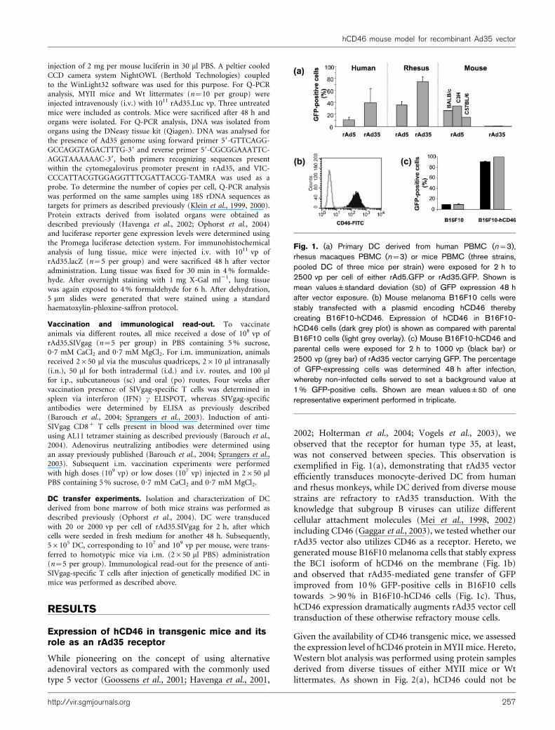
injection of 2 mg per mouse luciferin in 30 ml PBS. A peltier cooledCCD camera system NightOWL (Berthold Technologies) coupledto the WinLight32 software was used for this purpose. For Q-PCRanalysis, MYII mice and Wt littermates (n=10 per group) wereinjected intravenously (i.v.) with 1011 rAd35.Luc vp. Three untreatedmice were included as controls. Mice were sacrificed after 48 h andorgans were isolated. For Q-PCR analysis, DNA was isolated fromorgans using the DNeasy tissue kit (Qiagen). DNA was analysed forthe presence of Ad35 genome using forward primer 59-GTTCAGG-GCCAGGTAGACTTTG-39 and reverse primer 59-CGCGGAAATTC-AGGTAAAAAAC-39, both primers recognizing sequences presentwithin the cytomegalovirus promoter present in rAd35, and VIC-CCCATTACGTGGAGGTTTCGATTACCG-TAMRA was used as aprobe. To determine the number of copies per cell, Q-PCR analysiswas performed on the same samples using 18S rDNA sequences astargets for primers as described previously (Klein et al., 1999, 2000).Protein extracts derived from isolated organs were obtained asdescribed previously (Havenga et al., 2002; Ophorst et al., 2004)and luciferase reporter gene expression levels were determined usingthe Promega luciferase detection system. For immunohistochemicalanalysis of lung tissue, mice were injected i.v. with 1011 vp ofrAd35.lacZ (n=5 per group) and were sacrificed 48 h after vectoradministration. Lung tissue was fixed for 30 min in 4% formalde-hyde. After overnight staining with 1 mg X-Gal ml21, lung tissuewas again exposed to 4% formaldehyde for 6 h. After dehydration,5 mm slides were generated that were stained using a standardhaematoxylin-phloxine-saffron protocol.
Vaccination and immunological read-out. To vaccinateanimals via different routes, all mice received a dose of 108 vp ofrAd35.SIVgag (n=5 per group) in PBS containing 5% sucrose,0?7 mM CaCl2 and 0?7 mM MgCl2. For i.m. immunization, animalsreceived 2650 ml via the musculus quadriceps, 2610 ml intranasally(i.n.), 50 ml for both intradermal (i.d.) and i.v. routes, and 100 mlfor i.p., subcutaneous (sc) and oral (po) routes. Four weeks aftervaccination presence of SIVgag-specific T cells was determined inspleen via interferon (IFN) c ELISPOT, whereas SIVgag-specificantibodies were determined by ELISA as previously described(Barouch et al., 2004; Sprangers et al., 2003). Induction of anti-SIVgag CD8+ T cells present in blood was determined over timeusing AL11 tetramer staining as described previously (Barouch et al.,2004). Adenovirus neutralizing antibodies were determined usingan assay previously published (Barouch et al., 2004; Sprangers et al.,2003). Subsequent i.m. vaccination experiments were performedwith high doses (109 vp) or low doses (107 vp) injected in 2650 mlPBS containing 5% sucrose, 0?7 mM CaCl2 and 0?7 mM MgCl2.
DC transfer experiments. Isolation and characterization of DCderived from bone marrow of both mice strains was performed asdescribed previously (Ophorst et al., 2004). DC were transducedwith 20 or 2000 vp per cell of rAd35.SIVgag for 2 h, after whichcells were seeded in fresh medium for another 48 h. Subsequently,56105 DC, corresponding to 107 and 109 vp per mouse, were trans-ferred to homotypic mice via i.m. (2650 ml PBS) administration(n=5 per group). Immunological read-out for the presence of anti-SIVgag-specific T cells after injection of genetically modified DC inmice was performed as described above.
RESULTS
Expression of hCD46 in transgenic mice and itsrole as an rAd35 receptor
While pioneering on the concept of using alternativeadenoviral vectors as compared with the commonly usedtype 5 vector (Goossens et al., 2001; Havenga et al., 2001,
2002; Holterman et al., 2004; Vogels et al., 2003), weobserved that the receptor for human type 35, at least,was not conserved between species. This observation isexemplified in Fig. 1(a), demonstrating that rAd35 vectorefficiently transduces monocyte-derived DC from humanand rhesus monkeys, while DC derived from diverse mousestrains are refractory to rAd35 transduction. With theknowledge that subgroup B viruses can utilize differentcellular attachment molecules (Mei et al., 1998, 2002)including CD46 (Gaggar et al., 2003), we tested whether ourrAd35 vector also utilizes CD46 as a receptor. Hereto, wegenerated mouse B16F10 melanoma cells that stably expressthe BC1 isoform of hCD46 on the membrane (Fig. 1b)and observed that rAd35-mediated gene transfer of GFPimproved from 10% GFP-positive cells in B16F10 cellstowards >90% in B16F10-hCD46 cells (Fig. 1c). Thus,hCD46 expression dramatically augments rAd35 vector celltransduction of these otherwise refractory mouse cells.
Given the availability of CD46 transgenic mice, we assessedthe expression level of hCD46 protein inMYII mice. Hereto,Western blot analysis was performed using protein samplesderived from diverse tissues of either MYII mice or Wtlittermates. As shown in Fig. 2(a), hCD46 could not be
Fig. 1. (a) Primary DC derived from human PBMC (n=3),rhesus macaques PBMC (n=3) or mice PBMC (three strains,pooled DC of three mice per strain) were exposed for 2 h to2500 vp per cell of either rAd5.GFP or rAd35.GFP. Shown ismean values±standard deviation (SD) of GFP expression 48 hafter vector exposure. (b) Mouse melanoma B16F10 cells werestably transfected with a plasmid encoding hCD46 therebycreating B16F10-hCD46. Expression of hCD46 in B16F10-hCD46 cells (dark grey plot) is shown as compared with parentalB16F10 cells (light grey overlay). (c) Mouse B16F10-hCD46 andparental cells were exposed for 2 h to 1000 vp (black bar) or2500 vp (grey bar) of rAd35 vector carrying GFP. The percentageof GFP-expressing cells was determined 48 h after infection,whereby non-infected cells served to set a background value at1% GFP-positive cells. Shown are mean values±SD of onerepresentative experiment performed in triplicate.
http://vir.sgmjournals.org 257
hCD46 mouse model for recombinant Ad35 vector

detected in Wt littermates, whereas MYII mice displayedexpression in all major organs with most pronouncedexpression in the lungs and kidneys and least expression inmuscle. Similar Western blot analysis on protein samplesderived from organs of non-human primate or humanorigin (Fig. 2b) indicated that in man and monkey,expression of CD46 is also most pronounced in the lungsand kidneys and low in muscle. To identify whether thehCD46 present in MYII mice functions as rAd35 receptor,we exposed primary bone marrow-derived DC of MYIImice and Wt littermates to rAd35.GFP and rAd5.GFP as acontrol. The efficient infection of DC derived from MYIImice with rAd35.GFP (Fig. 2c), coupled to detection ofhCD46 on the membrane of these cells (Fig. 2d), indicatesthat hCD46 functions as rAd35 receptor in these mice.
Subsequent haemagglutination studies using erythrocytesderived from MYII mice and Wt littermates demonstratethat the hCD46 protein was expressed on erythrocytesderived from MYII and functionally binds rAd35 vector(Fig. 2e).
Collectively, these data show that: (i) Wt inbred strains ofmice do not express a high-affinity receptor for rAd35; (ii)hCD46 expressed in MYII mice functions as a high-affinityreceptor for rAd35; (iii) the low hCD46 protein expressioninmuscle and the high level in the lungs and kidneys ofMYIImice is similar to the hCD46 profile in non-human primatesand human, in spite of the fact that MYII mice contain10 hCD46 gene copies (Yannoutsos et al., 1996); and (iv)erythrocytes of MYII mice express hCD46 capable ofbinding to the rAd35 vector.
Vector distribution of rAd35 vector in hCD46-transgenic mice
We have previously reported that i.v. administration ofrAd35.Luc vector did not result in vector accumulation inany organ of different Wt inbred mice strains as demon-strated by the lack of luciferase activity (Vogels et al., 2003).Here, we demonstrate that high dose, i.v. delivery ofrAd35.Luc vector into MYII mice also does not result inhigh-level luciferase expression in this organ or any other
Tg Wt
Tg Wt
Tg Wt Tg Wt Tg Wt Tg Wt Tg Wt Tg Wt Tg Wt Tg Wt
Brain
Heart
Lung
Liver
Spleen
Kidney
Muscle
LN Skin
Brain
Heart
Lung
Liver
Spleen
Kidney
Muscle
LN Skin
(a)
(b)
(c) (d) (e)
Mouse
Monkey
Human
GFP
-pos
itive
cells
(%)
50
40
30
20
10
0
Cou
nts
CD46-FITC
200
160
120
80
40
0100 101 102 103 104
WtTg
TgWt
Virus dilution
0 2 4 8 16 32 64 128
256
rAd35
rAd5
rAd5 rAd35 rAd5 rAd35
75
50
75
50
75
50
Fig. 2. (a) Western blot analysis using 10 mg of total protein extracted from a panel of organs derived from MYII mice (Tg,n=3) or Wt littermates (n=3). The position of the protein marker for 75 and 50 kDa size are shown. LN, Lymph node. (b)Western blot analysis using 10 mg of total protein extracted from a panel of organs derived from either rhesus macaque (n=1)or human (n=1). The positions of the protein markers for 75 and 50 kDa size are shown. LN, Lymph node. (c) DC of Tg miceand Wt littermates were exposed for 2 h to 1000 vp per cell (grey bars) or 2500 vp per cell (black bars) using rAd5.GFP orrAd35.GFP. Shown is mean value±standard error of the mean (SEM) of one representative experiment performed on threemice. (d) Flow cytometric analysis of hCD46 expression on the DC membranes of Tg mice (dark grey plot) or Wt littermates(light grey overlay). (e) Haemagglutination pattern of rAd35 and rAd5 viruses with erythrocytes derived from Wt littermates orTg mice. Virus preparations used were either undiluted (0) or diluted up to 1 : 256 using PBS. Positive haemagglutination isscored when upon centrifugation, virus–erythrocyte interaction prevents formation of an erythrocyte pellet at the bottom of thewells of 96-well plates.
258 Journal of General Virology 87
S. Verhaagh and others
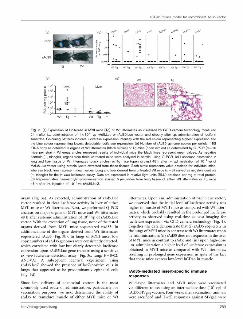
organ (Fig. 3a). As expected, administration of rAd5.Lucvector resulted in clear luciferase activity in liver of eitherMYII mice or Wt littermates. Next, we performed Q-PCRanalysis on major organs of MYII mice and Wt littermates48 h after systemic administration of 1011 vp of rAd35.Lucvector. With the exception of lung tissue, none of the testedorgans derived from MYII mice sequestered rAd35. Inaddition, none of the organs derived from Wt littermatessequestered rAd35 (Fig. 3b). In lungs of MYII mice, lowcopy numbers of rAd35 genomes were consistently detected,which correlated with low but clearly detectable luciferaseexpression upon rAd35.Luc gene transfer using a sensitiveex vivo luciferase detection assay (Fig. 3c, lung: P=0?02,ANOVA). A subsequent identical experiment usingrAd35.lacZ showed the presence of lacZ-positive cells inlungs that appeared to be predominantly epithelial cells(Fig. 3d).
Since i.m. delivery of adenoviral vectors is the mostcommonly used route of administration, particularly forvaccination purposes, we next determined the ability ofrAd35 to transduce muscle of either MYII mice or Wt
littermates. Upon i.m. administration of rAd35.Luc vector,we observed that the initial level of luciferase activity washigher in muscle of MYII mice as compared with Wt litter-mates, which probably resulted in the prolonged luciferaseactivity as observed using real-time in vivo imaging forluciferase expression via CCD camera technology (Fig. 4).Together, the data demonstrate that (i) rAd35 sequesters inthe lungs of MYII mice in contrast withWt littermates uponi.v. administration; (ii) rAd35 does not sequester in the liverof MYII mice in contrast to rAd5; and (iii) upon high-dosei.m. administration a higher level of luciferase expression isobtained in MYII mice as compared with Wt littermates,resulting in prolonged gene expression in spite of the factthat these mice express low-level hCD46 in muscle.
rAd35-mediated insert-specific immuneresponses
Wild-type littermates and MYII mice were vaccinatedvia different routes using an intermediate dose (108 vp) ofrAd35.SIVgag vaccine. Four weeks after vaccination, animalswere sacrificed and T-cell responses against SIVgag were
(a) (b)
(d)(c)
Lung Liver
rAd5
rAd35
Tg Wt
Wt Tg_
Wt Tg_
Wt Tg
Wt Tg Wt Tg Wt Tg Wt Tg Wt Tg Wt Tg Wt Tg Wt Tg
No.
vec
tor c
opie
spe
r 18S
cop
y
10 BrainHeart
LungLiver
SpleenKidney
MuscleLym
ph
1
0.1
0.01
0.001
RLU
per
mg
prot
ein 107
106
105
104
103
102
x100 x100
x200
_ _ _ _ _ _ _ _
Fig. 3. (a) Expression of luciferase in MYII mice (Tg) or Wt littermates as visualized by CCD camera technology measured24 h after i.v. administration of 161011 vp rAd5.Luc or rAd35.Luc vector and directly after i.p. administration of luciferinsubstrate. Colouring patterns indicate luciferase expression intensity with the red colour representing highest expression andthe blue colour representing lowest detectable luciferase expression. (b) Number of rAd35 genome copies per cellular 18SrDNA copy as detected in organs of Wt littermates (black circles) or Tg mice (open circles) as determined by Q-PCR (n=10mice per strain). Whereas circles represent results of individual mice the black lines represent mean values. As negativecontrols (”, triangle), organs from three untreated mice were analysed in parallel using Q-PCR. (c) Luciferase expression inlung and liver tissue of Wt littermates (black circles) or Tg mice (open circles) 48 h after i.v. administration of 1011 vp ofrAd35.Luc vector using protein lysate extracted from these tissues. Each circle represents value obtained for individual mice,whereas black lines represent mean values. Lung and liver derived from untreated Wt mice (n=3) served as negative controls(”, triangle) for the in vitro luciferase assay. Data are expressed in relative light units (RLU) obtained per mg of total protein.(d) Representative haematoxylin-phloxine-saffron stained 5 mm slides from lung tissue of either Wt littermates or Tg mice48 h after i.v. injection of 1011 vp rAd35.lacZ.
http://vir.sgmjournals.org 259
hCD46 mouse model for recombinant Ad35 vector
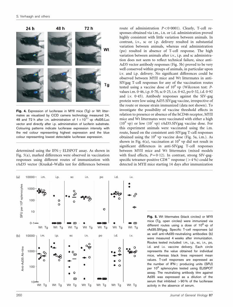
determined using the IFN-c ELISPOT assay. As shown inFig. 5(a), marked differences were observed in vaccinationresponses using different routes of immunization withrAd35 vector (Kruskal–Wallis test for differences between
route of administration P<0?0001). Clearly, T-cell re-sponses obtained via i.m., i.n. or i.d. administration provedhighly consistent with little variation between animals. Incontrast, i.v., sc or i.p. delivery resulted in substantialvariation between animals, whereas oral administration(po) resulted in absence of T-cell response. The highvariation between animals after i.v., i.p. and sc administra-tion does not seem to reflect technical failure, since anti-Ad35 vector antibody responses (Fig. 5b) proved to be verywell conserved within groups of animals, in particular uponi.v. and i.p. delivery. No significant differences could beobserved between MYII mice and Wt littermates in anti-SIVgag T-cell responses for any of the vaccination routestested using a vaccine dose of 108 vp (Wilcoxon test: P-values i.m. 0?46, i.p. 0?70, sc 0?25, i.n. 0?62, po 0?32, i.d. 0?92and i.v. 0?45). Antibody responses against the SIV-gagprotein were low using Ad35.SIVgag vaccine, irrespective ofthe route or mouse strain immunized (data not shown). Toinvestigate the possibility of vaccine threshold effects inrelation to presence or absence of the hCD46 receptor, MYIImice and Wt littermates were vaccinated with either a high(109 vp) or low (107 vp) rAd35.SIVgag vaccine dose. Forthis experiment animals were vaccinated using the i.m.route, based on the consistent anti-SIVgag T-cell responsesobtained using the 108 vp vaccine dose (Fig. 5a, i.m.). Asshown in Fig. 6(a), vaccination at 109 vp did not result insignificant differences in anti-SIVgag T-cell responsesbetween MYII mice and Wt littermates (mixed modelswith fixed effects, P=0?12). In contrast, strong SIV-gag-specific tetramer-positive CD8+ response (>4%) could bedetected in MYII mice starting 14 days after immunization
Fig. 4. Expression of luciferase in MYII mice (Tg) or Wt litter-mates as visualized by CCD camera technology measured 24,48 and 72 h after i.m. administration of 161011 vp rAd35.Lucvector and directly after i.p. administration of luciferin substrate.Colouring patterns indicate luciferase expression intensity withthe red colour representing highest expression and the bluecolour representing lowest detectable luciferase expression.
10000
1000
100
10
0-1
10000
1000
100
10
SFU
per
106
spl
enoc
ytes
Ant
i-Ad3
5 A
b-tit
re
(a)
(b)
i.m. i.p. sc i.n. po i.d. i.v.
i.m. i.p. sc i.n. po i.d. i.v.
Wt Tg Wt Tg Wt Tg Wt Tg Wt Tg Wt Tg Wt Tg
Wt Tg Wt Tg Wt Tg Wt Tg Wt Tg Wt Tg Wt Tg
Fig. 5. Wt littermates (black circles) or MYIImice (Tg, open circles) were immunized viadifferent routes using a dose of 108 vp ofrAd35.SIVgag. Specific T-cell responses (a)as well anti-rAd35-neutralizing antibodies (b)were measured 4 weeks after immunization.Routes tested included: i.m., i.p., sc, i.n., po,i.d. and i.v. vaccine delivery. Each circlerepresents the value obtained for individualmice, whereas black lines represent meanvalues. T-cell responses are expressed asthe number of IFN-c producing units (SFU)per 106 splenocytes tested using ELISPOTassay. The neutralizing antibody titre againstAd35 was expressed as a dilution of theserum that inhibited >90% of the luciferaseactivity in the absence of serum.
260 Journal of General Virology 87
S. Verhaagh and others

using a 107 vp vaccine dose, whereas no tetramer-positiveCD8+ T-cell response could be detected in Wt littermates(Fig. 6b, P<0?001). The tetramer-positive CD8+ responsesdetected correlated with day 28 IFN-c ELISPOT results,which also demonstrated clearly detectable IFN-c producingT-cells in all vaccinated MYII mice as compared with noT-cell response in 4 of 5 Wt littermates (P=0?07). At avaccine dose of 109 vp no difference in ELISPOT results wasobserved (Fig. 6c, P=0?47). In an effort to link the observeddifferences in the induction of anti-SIVgag CD8+ responsesdirectly to the presence of hCD46, DC derived from MYIImice or Wt littermates were isolated and exposed ex vivo to2000 or 20 vp per cell of rAd35.SIVgag vector. Forty-eighthours after a 2 h vector exposure, 56105 DC were admini-stered i.m. to homotypic mice. As shown in Fig. 6(d), cleartetramer-positive CD8+ T-cell responses could be observedin both mice strains after infusion of 56105 DC exposedfor 2 h to 2000 vp per cell. In contrast, clear, albeit low,tetramer-positive responses were observed only in MYIImice after receiving 56105 DC exposed for 2 h to 20 vp percell (Fig. 6e, P=0?03 using mixed models for differencebetween Wt and Tg over time). Again, day 28 IFN-cELISPOT results correlated with CD8+ tetramer staining,demonstrating clear detection of IFN-c producing T cells inMYII mice (Fig. 6f, P=0?01). At 109 vp per 56105 DC, nodifference in the T-cell response was observed betweenMYIImice and Wt littermates (P=0?46).
Collectively, these results demonstrate that direct i.m.vaccination of MYII mice with rAd35 vaccine results in asignificant (>4%) in vivo rAd35 vaccine dose-sparing effectas compared with Wt littermates. Also, transduction ofcultured DC derived from MYII mice with rAd35 results inhigher anti-SIVgag CD8+ responses in vivo upon reinfusionof modified DC as compared with Wt littermates. The latteris most likely related to the increased susceptibility of DC forrAd35 due to expression of hCD46.
DISCUSSION
Many reports have demonstrated that the presence ofcoxsackie and adenovirus receptor, CAR (Bergelson et al.,1997), on cell lines and primary cells, is important to achieveefficient in vitro transduction with Ad5 vectors (Hutchinet al., 2000; Li et al., 1999; Nalbantoglu et al., 1999; Orlickyet al., 2001; Stockwin et al., 2002). Likewise, it has beenreported, and confirmed here, that expression of CD46 oncells significantly augments in vitro gene transfer usinggroup B vectors (Holterman et al., 2004; Vogels et al., 2003).By exploiting the natural diversity in receptor usage amongadenoviruses (Havenga et al., 2002; Von Seggern et al., 1998,2000), we have identified vectors that significantly increaselevels of gene expression in DC and fibroblasts (Ophorstet al., 2004; Rea et al., 2001b), human bone marrow stromacells (Havenga et al., 2002), haematopoeitic stem cells
(a)
(b)
(c)
(d)
(e)
(f)
Tetr
amer
-pos
itive
CD
8 (%
)
Tetr
amer
-pos
itive
CD
8 (%
)
Tetr
amer
-pos
itive
CD
8 (%
)
Tetr
amer
-pos
itive
CD
8 (%
)
SFU
per
106
cel
ls
SFU
per
106
cel
ls
8 1.5
0.5
0.0
1.0
1.5
0.5
0.0
1.0
6
4
2
0
8
6
4
2
0
0 7 14 21 28 0 7 14 21
14 21
28
0 7 280 7
107 109 107 109
14 21 28
600 400
300
100
0
200400
200
0
TgWt
TgWt
TgWt
TgWt
TgWt
TgWt
Time post-vaccination (days)
Time post-vaccination (days) Time post-DC infusion (days)
Time post-DC infusion (days)
Vaccine dose Vaccine dose
Fig. 6. Wt littermates (n=5, black circles)and MYII mice (Tg, n=5, open circles) wereimmunized i.m. with a dose of 109 vp (a) or107 vp (b) of rAd35.SIVgag. Animals weresubsequently monitored in time for develop-ment of anti-SIVgag CD8+ responses viatetramer staining. Shown is mean value±SEM.(c) Twenty-eight days after immunization,animals (n=5) were sacrificed and spleno-cytes were isolated to determine IFN-c-positive T cells in ELISPOT assay. Eachcircle represents the value of individual mice,whereas black lines represent mean values.Wt littermates (n=5, black circles) andTg mice (n=5, open circles) received56105 DC exposed for 2 h to either 109 vp(d) or 107 vp (e) of rAd35.SIVgag. Animalswere monitored in time for development ofanti-SIVgag CD8+ responses via tetramerstaining. Shown is mean value±SEM. (f)Twenty-eight days after immunization, theanimals (n=5) that received rAd35.SIVgagmodified DC were sacrificed and spleno-cytes were isolated to determine IFN-c-positive T cells in ELISPOT assay. Eachcircle represents the value of individual mice,whereas black lines represent mean values.
http://vir.sgmjournals.org 261
hCD46 mouse model for recombinant Ad35 vector

(Knaan-Shanzer et al., 2001), synoviocytes (Goossens et al.,2001) and smooth muscle cells (Havenga et al., 2001, 2002).Such vectors are in demand within the field of regenerativemedicine (de Bruijn et al., 1999; Lamme et al., 1998, 2000;Moutsatsos et al., 2001; Riesle et al., 1998) and ex vivo DC-based vaccination approaches (Rea et al., 2001a). Thus, thein vitro correlation between cellular receptor expression andvector transduction efficiency is relatively clear.
In contrast, the in vivo need for a high-affinity receptor toobtain efficient gene transfer has not been completelyelucidated. For instance, it has been suggested by some thatviral receptors are not major determinants in adenoviraltransduction of muscle (Cao et al., 2001), whereas othershave shown that transduction of skeletal muscle in a humanCAR transgenic mouse model (Tallone et al., 2001) resultedin an approximately 10-fold improved gene transfer ascompared with Wt mice (reviewed by Thirion et al., 2002).Also, a clear discrepancy between in vivo rAd5 distributionand CAR expression in mice upon systemic delivery hasbeen reported, demonstrating that anatomical barriersimpact on vector distribution (Fechner et al., 1999). Suchanatomical barriers challenge the concept of adenoviralvector targeting since any vector, be it of human or non-human origin (Holterman et al., 2004; Kremer et al., 2000;Vogels et al., 2003; Xiang et al., 2002; Zakhartchouk et al.,2003), will need to circumvent such anatomical hurdles.
We have recently shown that in spite of huge differences inthe ability of a panel of different fibre-chimeric adenoviralvectors to infect primary mouse muscle cells, DC and fibro-blasts in vitro, all these vectors elicited similar anti-insert-specific immune responses in mice upon i.m. vaccination(Mercier et al., 2004; Ophorst et al., 2004). Also, clearantigen-specific cellular and humoral immune responsesare consistently obtained after i.m. immunization withrAd11 and rAd35 vectors inWt inbredmouse strains lackinga high-affinity receptor (Barouch et al., 2004; Holtermanet al., 2004; Vogels et al., 2003). Such results suggest thatin the absence of a high-affinity receptor skeletal muscle,transduction occurs in mice.
Although we observed induction of immune responses inWt inbred strains, the absence of a high-affinity receptormay result in an altered potency or distribution of thesevectors. For these reasons, and knowing that both monkeysand humans widely express CD46, we wanted to investigatethe role of the CD46 receptor in in vivo rAd35 vectortransduction efficiency, rAd35 distribution and rAd35vaccine potential. We chose the MYII strain, since thesemice express all four major hCD46 isoforms (Kemper et al.,2001; Yannoutsos et al., 1996). The MYII mice are furtherknown to contain 10 hCD46 gene copies in their genomeand earlier studies using Northern blot analysis havedemonstrated that hCD46 expression was similar betweenhumans andMYII mice in the liver and lungs, whereas in thekidneys, heart and spleen higher levels of hCD46 mRNAwere detected (Yannoutsos et al., 1996). Liver tropism wasassessed by CCD camera imaging and revealed that rAd35
vector does not sequester in liver upon i.v. administration ofeither MYII mice or Wt littermates. These data corroborateour earlier data inWtmice (Vogels et al., 2003) and also dataof Seshidhar Reddy et al. (2003), who demonstrated that anE1B-containing Ad35 vector displays lack of mouse livertropism. It should be taken into account that MYII mice, incontrast to humans, express hCD46 on their erythrocytes,which may lead to capture of rAd35 vector upon i.v.administration obscuring liver infection in these animals(Yannoutsos et al., 1996). However, the data are also in linewith a recent report by Ni et al. (2005), showing that rAd5vectors carrying fibres derived from Ad11 or Ad35 do nottarget the liver of baboons and do not show endothelialdamage and inflammation typically observed with Ad5-based vectors. Thus, the strong liver tropism observed withrAd5 vector, known to cause dose-dependent hepato-toxicity limiting rAd5 dosing, can perhaps be circumventedusing rAd35 vectors. Our Q-PCR analysis revealed thatrAd35, when given at high dose, consistently accumulates atlow rAd35 genome copy numbers in the lungs ofMYII mice,correlating with expression detected in this organ. Thesedata, as well as lack of liver infection in hCD46 transgenicmice, are in further agreement with a recent report showingthat an rAd11 vector does not accumulate in the liver andcan be detected for a short period of time (<72 h) in thelungs of hCD46 mice (Stone et al., 2005).
Upon i.m. administration of rAd35 higher initial markergene expression was detected in MYII mice as comparedwith Wt littermates, which most likely translated in theprolonged expression observed in these MYII mice. Theclear difference in level and longevity in luciferase expres-sion observed in muscle did not correlate with the vaccina-tion data, showing no difference at either 109 or 108 vp ofrAd35.SIVgag dose in T-cell induction levels. However, aclear and significant tetramer-positive CD8+ T-cell re-sponse was observed in MYII mice at the low 107 vp rAd35vaccine dose in contrast to no response in Wt littermates.We hypothesize that only limited antigen expression isalready sufficient to trigger full blown immune responsesand, thus, that a difference between MYII mice or Wtlittermates could be observed only at lower dosing.
To provide further evidence that the presence of the high-affinity receptor for rAd35 vector contributes to the antigen-specific immune response, we genetically modified ex vivocultured DC derived from Wt littermates and MYII miceand reinfused cells into naıve recipients. The observeddifference in the anti-SIVgag-specific CD8+ T-cell responsein MYII mice as compared with Wt littermates directlystems from the ex vivo modified DC, suggesting thatDC derived from MYII mice were more susceptible torAd35, resulting in higher SIVgag antigen expression andprocessing.
Future immunohistochemical analysis on muscle sectionsafter direct i.m. immunization with rAd35 vector are neededto formally prove that the presence of CD46 in the muscle ofMYII mice results in increased uptake of rAd35 vector by
262 Journal of General Virology 87
S. Verhaagh and others

professional antigen presenting cells as compared with Wtlittermates. However, this experiment demonstrates that DCfrom MYII mice are able to become infected with rAd35vector, resulting in antigen expression and processing atsufficient level to induce in vivo anti-SIVgag CD8+
responses upon reinfusion into naıve host.
In summary, the data obtained have demonstrated that inMYII mice the rAd35 vector displays increased targeting andexpression of transgenes in lungs after i.v. administration,increased level and longevity of local expression upon i.m.administration, and a significant dose-sparing effect intetramer-positive CD8+ T cell induction as compared withWt littermates. As such, we conclude that MYII mice pro-vide an interesting preclinical model in studies involvingrAd35 vectors.
ACKNOWLEDGEMENTS
The authors would like to thank Dr Grosveld (Erasmus University ofRotterdam, The Netherlands) for providing the MYII strain, Dr SillivisSmit (Department of Neurology, Erasmus University of Rotterdam,The Netherlands) and Dr Heidt (Biomedical Primate Research Centre(BPRC, Rijswijk, The Netherlands) for tissue donation and Dr Barouchfor providing the SIVgag AL11 tetramers. Also, the authors thank DrDelemarre (Biological Safety Officer and manager DMIII animalfacility, Crucell Holland BV) for expert advice on MYII animal healthtesting and housing.
REFERENCES
Barouch, D. H., Pau, M. G., Custers, J. H. & 15 other authors (2004).Immunogenicity of recombinant adenovirus serotype 35 vaccine inthe presence of pre-existing anti-Ad5 immunity. J Immunol 172,6290–6297.
Bergelson, J. M., Cunningham, J. A., Droguett, G., Kurt-Jones, E. A.,Krithivas, A., Hong, J. S., Horwitz, M. S., Crowell, R. L. & Finberg,R. W. (1997). Isolation of a common receptor for Coxsackie B virusesand adenoviruses 2 and 5. Science 275, 1320–1323.
Cao, B., Pruchnic, R., Ikezawa, M., Xiao, X., Li, J., Wickham, T. J.,Kovesdi, I., Rudert, W. A. & Huard, J. (2001). The role of receptors inthe maturation-dependent adenoviral transduction of myofibers.Gene Ther 8, 627–637.
de Bruijn, J. D., van den Brink, I., Mendes, S., Dekker, R., Bovell,Y. P. & van Blitterswijk, C. A. (1999). Bone induction by implantscoated with cultured osteogenic bone marrow cells. Adv Dent Res 13,74–81.
Dorig, R. E., Marcil, A., Chopra, A. & Richardson, C. D. (1993). Thehuman CD46 molecule is a receptor for measles virus (Edmonstonstrain). Cell 75, 295–305.
Fechner, H., Haack, A., Wang, H. & 9 other authors (1999).Expression of coxsackie adenovirus receptor and alphav-integrin doesnot correlate with adenovector targeting in vivo indicatinganatomical vector barriers. Gene Ther 6, 1520–1535.
Gaggar, A., Shayakhmetov, D. M. & Lieber, A. (2003). CD46 is acellular receptor for group B adenoviruses. Nat Med 9, 1408–1412.
Goossens, P. H., Havenga, M. J., Pieterman, E., Lemckert, A. A.,Breedveld, F. C., Bout, A. & Huizinga, T. W. (2001). Infectionefficiency of type 5 adenoviral vectors in synovial tissue can beenhanced with a type 16 fiber. Arthritis Rheum 44, 570–577.
Havenga, M. J., Lemckert, A. A., Grimbergen, J. M., Vogels, R.,Huisman, L. G., Valerio, D., Bout, A. & Quax, P. H. (2001). Improved
adenovirus vectors for infection of cardiovascular tissues. J Virol 75,
3335–3342.
Havenga, M. J., Lemckert, A. A., Ophorst, O. J. & 13 other authors(2002). Exploiting the natural diversity in adenovirus tropism for
therapy and prevention of disease. J Virol 76, 4612–4620.
Heemskerk, B., Veltrop-Duits, L. A., van Vreeswijk, T., ten Dam,M. M., Heidt, S., Toes, R. E., van Tol, M. J. & Schilham, M. W. (2003).Extensive cross-reactivity of CD4+ adenovirus-specific T cells: implica-
tions for immunotherapy and gene therapy. J Virol 77, 6562–6566.
Holterman, L., Vogels, R., van der Vlugt, R. & 15 other authors(2004). Novel replication-incompetent vector derived from adeno-
virus type 11 (Ad11) for vaccination and gene therapy: low
seroprevalence and non-cross-reactivity with Ad5. J Virol 78,
13207–13215.
Horvat, B., Rivailler, P., Varior-Krishnan, G., Cardoso, A., Gerlier, D.& Rabourdin-Combe, C. (1996). Transgenic mice expressing human
measles virus (MV) receptor CD46 provide cells exhibiting different
permissivities to MV infections. J Virol 70, 6673–6681.
Hutchin, M. E., Pickles, R. J. & Yarbrough, W. G. (2000). Efficiency ofadenovirus-mediated gene transfer to oropharyngeal epithelial cells
correlates with cellular differentiation and human coxsackie and
adenovirus receptor expression. Hum Gene Ther 11, 2365–2375.
Kallstrom, H., Liszewski, M. K., Atkinson, J. P. & Jonsson, A. B.(1997). Membrane cofactor protein (MCP or CD46) is a cellular
pilus receptor for pathogenic Neisseria. Mol Microbiol 25, 639–647.
Kemper, C., Leung, M., Stephensen, C. B., Pinkert, C. A., Liszewski,M. K., Cattaneo, R. & Atkinson, J. P. (2001). Membrane cofactor
protein (MCP; CD46) expression in transgenic mice. Clin Exp
Immunol 124, 180–189.
Klein, D., Janda, P., Steinborn, R., Muller, M., Salmons, B. &Gunzburg, W. H. (1999). Proviral load determination of different
feline immunodeficiency virus isolates using real-time polymerase
chain reaction: influence of mismatches on quantification. Electro-
phoresis 20, 291–299.
Klein, D., Bugl, B., Gunzburg, W. H. & Salmons, B. (2000). Accurateestimation of transduction efficiency necessitates a multiplex real-
time PCR. Gene Ther 7, 458–463.
Knaan-Shanzer, S., Van Der Velde, I., Havenga, M. J., Lemckert,A. A., De Vries, A. A. & Valerio, D. (2001). Highly efficient targeted
transduction of undifferentiated human hematopoietic cells by
adenoviral vectors displaying fiber knobs of subgroup B. Hum Gene
Ther 12, 1989–2005.
Kostense, S., Koudstaal, W., Sprangers, M. & 8 other authors(2004). Adenovirus types 5 and 35 seroprevalence in AIDS risk
groups supports type 35 as a vaccine vector. AIDS 18, 1213–1216.
Kremer, E. J., Boutin, S., Chillon, M. & Danos, O. (2000). Canineadenovirus vectors: an alternative for adenovirus-mediated gene
transfer. J Virol 74, 505–512.
Lamme, E. N., van Leeuwen, R. T., Jonker, A., van Marle, J. &Middelkoop, E. (1998). Living skin substitutes: survival and function
of fibroblasts seeded in a dermal substitute in experimental wounds.
J Invest Dermatol 111, 989–995.
Lamme, E. N., Van Leeuwen, R. T., Brandsma, K., Van Marle, J. &Middelkoop, E. (2000). Higher numbers of autologous fibroblasts in
an artificial dermal substitute improve tissue regeneration and
modulate scar tissue formation. J Pathol 190, 595–603.
Li, D., Duan, L., Freimuth, P. & O’Malley, B. W., Jr (1999). Variabilityof adenovirus receptor density influences gene transfer efficiency and
therapeutic response in head and neck cancer. Clin Cancer Res 5,
4175–4181.
http://vir.sgmjournals.org 263
hCD46 mouse model for recombinant Ad35 vector

Manchester, M., Liszewski, M. K., Atkinson, J. P. & Oldstone, M. B.(1994). Multiple isoforms of CD46 (membrane cofactor protein)
serve as receptors for measles virus. Proc Natl Acad Sci U S A 91,
2161–2165.
Mei, Y. F., Lindman, K. & Wadell, G. (1998). Two closely related
adenovirus genome types with kidney or respiratory tract tropism
differ in their binding to epithelial cells of various origins. Virology
240, 254–266.
Mei, Y. F., Lindman, K. & Wadell, G. (2002). Human adenoviruses of
subgenera B, C, and E with various tropisms differ in both binding
to and replication in the epithelial A549 and 293 cells. Virology 295,
30–43.
Mercier, S., Verhaagh, S., Goudsmit, J., Lemckert, A., Monteil, M.,Havenga, M. & Eloit, M. (2004). Adenovirus fibre exchange alters celltropism in vitro but not transgene-specific T CD8+ immune
responses in vivo. J Gen Virol 85, 1227–1236.
Miyagawa, S., Ikawa, M., Kominami, K., Tanaka, H., Mikata, S.,Matsuda, H., Seya, T., Shirakura, R. & Okabe, M. (1997). The
regulation of membrane cofactor protein (CD46) expression in
transgenic mice: the importance of the first 125 BP of the 39
untranslated region. Transplant Proc 29, 941–942.
Moutsatsos, I. K., Turgeman, G., Zhou, S. & 9 other authors (2001).Exogenously regulated stem cell-mediated gene therapy for bone
regeneration. Mol Ther 3, 449–461.
Nalbantoglu, J., Pari, G., Karpati, G. & Holland, P. C. (1999).Expression of the primary coxsackie and adenovirus receptor is
downregulated during skeletal muscle maturation and limits the
efficacy of adenovirus-mediated gene delivery to muscle cells. Hum
Gene Ther 10, 1009–1019.
Naniche, D., Varior-Krishnan, G., Cervoni, F., Wild, T. F., Rossi, B.,Rabourdin-Combe, C. & Gerlier, D. (1993). Human membrane
cofactor protein (CD46) acts as a cellular receptor for measles virus.
J Virol 67, 6025–6032.
Ni, S., Bernt, K., Gaggar, A., Li, Z.-Y., Kiem, H. P. & Lieber, A. (2005).Evaluation of biodistribution and safety of adenovirus vectors
containing group B fibers after intravenous injection into baboons.
Hum Gene Ther 16, 664–677.
Nwanegbo, E., Vardas, E., Gao, W., Whittle, H., Sun, H., Rowe, D.,Robbins, P. D. & Gambotto, A. (2004). Prevalence of neutralizing
antibodies to adenoviral serotypes 5 and 35 in the adult populations
of The Gambia, South Africa, and the United States. Clin Diagn Lab
Immunol 11, 351–357.
Okada, N., Liszewski, M. K., Atkinson, J. P. & Caparon, M. (1995).Membrane cofactor protein (CD46) is a keratinocyte receptor for the
M protein of the group A streptococcus. Proc Natl Acad Sci U S A
92, 2489–2493.
Oldstone, M. B., Lewicki, H., Thomas, D. & 7 other authors (1999).Measles virus infection in a transgenic model: virus-induced
immunosuppression and central nervous system disease. Cell 98,
629–640.
Ophorst, O. J., Kostense, S., Goudsmit, J. & 9 other authors (2004).An adenoviral type 5 vector carrying a type 35 fiber as a vaccine
vehicle: DC targeting, cross neutralization, and immunogenicity.
Vaccine 22, 3035–3044.
Orlicky, D. J., DeGregori, J. & Schaack, J. (2001). Construction of
stable coxsackievirus and adenovirus receptor-expressing 3T3-L1
cells. J Lipid Res 42, 910–915.
Rall, G. F., Manchester, M., Daniels, L. R., Callahan, E. M., Belman, A. R.& Oldstone, M. B. (1997). A transgenic mouse model for measles virus
infection of the brain. Proc Natl Acad Sci U S A 94, 4659–4663.
Rea, D., Havenga, M. J., van Den Assem, M. & 6 other authors(2001a). Highly efficient transduction of human monocyte-derived
dendritic cells with subgroup B fiber-modified adenovirus vectors
enhances transgene-encoded antigen presentation to cytotoxic T cells.
J Immunol 166, 5236–5244.
Rea, D., Johnson, M. E., Havenga, M. J., Melief, C. J. & Offringa, R.(2001b). Strategies for improved antigen delivery into dendritic cells.
Trends Mol Med 7, 91–94.
Riesle, J., Hollander, A. P., Langer, R., Freed, L. E. & Vunjak-Novakovic, G. (1998). Collagen in tissue-engineered cartilage: types,
structure, and crosslinks. J Cell Biochem 71, 313–327.
Santoro, F., Kennedy, P. E., Locatelli, G., Malnati, M. S., Berger, E. A.& Lusso, P. (1999). CD46 is a cellular receptor for human
herpesvirus 6. Cell 99, 817–827.
Segerman, A., Atkinson, J. P., Marttila, M., Dennerquist, V., Wadell,G. & Arnberg, N. (2003). Adenovirus type 11 uses CD46 as a cellular
receptor. J Virol 77, 9183–9191.
Seshidhar Reddy, P., Ganesh, S., Limbach, M. P., Brann, T.,Pinkstaff, A., Kaloss, M., Kaleko, M. & Connelly, S. (2003).Development of adenovirus serotype 35 as a gene transfer vector.
Virology 311, 384–393.
Sirena, D., Lilienfeld, B., Eisenhut, M. & 8 other authors (2004). Thehuman membrane cofactor CD46 is a receptor for species B
adenovirus serotype 3. J Virol 78, 4454–4462.
Sprangers, M. C., Lakhai, W., Koudstaal, W., Verhoeven, M., Koel,B. F., Vogels, R., Goudsmit, J., Havenga, M. J. & Kostense, S.(2003). Quantifying adenovirus-neutralizing antibodies by luciferase
transgene detection: addressing preexisting immunity to vaccine and
gene therapy vectors. J Clin Microbiol 41, 5046–5052.
Stockwin, L. H., Matzow, T., Georgopoulos, N. T., Stanbridge, L. J.,Jones, S. V., Martin, I. G., Blair-Zajdel, M. E. & Blair, G. E. (2002).Engineered expression of the coxsackie B and adenovirus receptor
(CAR) in human dendritic cells enhances recombinant adenovirus-
mediated gene transfer. J Immunol Methods 259, 205–215.
Stone, D., Ni, S., Li, Z. Y., Gaggar, A., DiPaolo, N., Feng, Q.,Sandig, V. & Lieber, A. (2005). Development and assessment of
human adenovirus type 11 as a gene transfer vector. J Virol 79,
5090–5104.
Tallone, T., Malin, S., Samuelsson, A., Wilbertz, J., Miyahara, M.,Okamoto, K., Poellinger, L., Philipson, L. & Pettersson, S. (2001). Amouse model for adenovirus gene delivery. Proc Natl Acad Sci U S A
98, 7910–7915.
Thirion, C., Larochelle, N., Volpers, C., Dunant, P., Stucka, R.,Holland, P., Nalbantoglu, J., Kochanek, S. & Lochmuller, H. (2002).Strategies for muscle-specific targeting of adenoviral gene transfer
vectors. Neuromuscul Disord 12, 30–39.
Thorley, B. R., Milland, J., Christiansen, D. & 9 other authors (1997).Transgenic expression of a CD46 (membrane cofactor protein)
minigene: studies of xenotransplantation and measles virus infection.
Eur J Immunol 27, 726–734.
Vogels, R., Zuijdgeest, D., van Rijnsoever, R. & 20 other authors(2003). Replication-deficient human adenovirus type 35 vectors for
gene transfer and vaccination: efficient human cell infection and
bypass of pre-existing adenovirus immunity. J Virol 77, 8263–8271.
Von Seggern, D. J., Kehler, J., Endo, R. I. & Nemerow, G. R. (1998).Complementation of a fibre mutant adenovirus by packaging cell
lines stably expressing the adenovirus type 5 fibre protein. J Gen
Virol 79, 1461–1468.
Von Seggern, D. J., Huang, S., Fleck, S. K., Stevenson, S. C. &Nemerow, G. R. (2000). Adenovirus vector pseudotyping in fiber-
expressing cell lines: improved transduction of Epstein–Barr virus-
transformed B cells. J Virol 74, 354–362.
Wu, E., Trauger, S. A., Pache, L., Mullen, T. M., von Seggern, D. J.,Siuzdak, G. & Nemerow, G. R. (2004). Membrane cofactor protein is
264 Journal of General Virology 87
S. Verhaagh and others

a receptor for adenoviruses associated with epidemic kerato-conjunctivitis. J Virol 78, 3897–3905.
Xiang, Z., Gao, G., Reyes-Sandoval, A., Cohen, C. J., Li, Y.,Bergelson, J. M., Wilson, J. M. & Ertl, H. C. (2002). Novel,chimpanzee serotype 68-based adenoviral vaccine carrier forinduction of antibodies to a transgene product. J Virol 76,2667–2675.
Yannoutsos, N., Ijzermans, J. N., Harkes, C., Bonthuis, F., Zhou,C. Y., White, D., Marquet, R. L. & Grosveld, F. (1996). A membranecofactor protein transgenic mouse model for the study of discordantxenograft rejection. Genes Cells 1, 409–419.
Zakhartchouk, A., Zhou, Y. & Tikoo, S. K. (2003). A recombinant E1-deleted porcine adenovirus-3 as an expression vector. Virology 313,377–386.
http://vir.sgmjournals.org 265
hCD46 mouse model for recombinant Ad35 vector