
HTLV1 infection in a population-based cohort of older persons in Guinea-Bissau, West Africa: Risk...
180
OVARIAN CANCER; GENETIC FEATURES AND PROGNOSTIC IMPLICATIONS Ovariumkanker; genetische kenmerken en prognostische implicaties PROEFSCHRIFT Tel' verkrijging van de graad van doctor aan de Erasmus Universiteit Rotterdam op gezag van de Rector Magnificus Prof. dr. il'. J.H. van Bemmel en volgens besluit van het College voor Promoties De open bare verdediging zal plaatsvinden op woensdag 20 september 2000 om 9.45 Uul' door MONIQUE SCHUIJER geboren te Rotterdam
-
Upload
independent -
Category
Documents
-
view
1 -
download
0
Transcript of HTLV1 infection in a population-based cohort of older persons in Guinea-Bissau, West Africa: Risk...

OVARIAN CANCER; GENETIC FEATURES AND PROGNOSTIC IMPLICATIONS
Ovariumkanker; genetische kenmerken en prognostische implicaties
PROEFSCHRIFT
Tel' verkrijging van de graad van doctor aan de Erasmus Universiteit Rotterdam op
gezag van de Rector Magnificus Prof. dr. il'. J.H. van Bemmel en volgens besluit van
het College voor Promoties
De open bare verdediging zal plaatsvinden op
woensdag 20 september 2000 om 9.45 Uul'
door
MONIQUE SCHUIJER
geboren te Rotterdam

PROMOTIECOMMISSIE
PROMOTOREN:
TWEEDE PROMOTOR:
OVERIGE LED EN:
CO-PROMOTOR:
Prof. dr. O. Stoter
Prof. dr. lO.M. Klijn
Dr. E.C. Zwarthoff
Prof. dr. Th. van der Kwast
Prof. dr. C.W. Burger
Dr. P.M.J.l Berns
Oms lag: Kari Ann Pagnano (uit: CA, A Cancer Journal for Clinicians, vol. 45 No.2,
1995)
Dit proefschrift is tot stand gekomen binnen de afdeling Interne Oncologie,
(Iaboratorium Tumorendocrinologie) op de Daniel den Hoed Kliniek en het Josephine
Nefkens Instituut, Academisch Ziekenhuis Rotterdam. Het onderzoek werd financieel
mogelijk gemaakt door de Nederlandse Kankerbestrijding (projectnumll1er OOHK 94-
840). De uitgave van dit proefschrift kwam tot stand met financiele steun van de
Nederlandse Kankerbestrijding en de Erasmus Universiteit Rotterdam.
Oedl1lkt door: W Offsetdrukkerij Ridderprint B.Y., Ridderkerk

The disease of cancer will be banished ji'om life by calm, unJIIII'I),ing, persistent men and women ... and the Illative that will conquer cancer will not be pity nor horror: it will be curiosity to know holV and why.
H.G. Wells

CONTENTS
CHAPTERl Introduction
1.1 Epidemiology
1.1.1 Incidence and 111011ality
1.1.2 Risk factors
1.2 Pathology
1.3 Genetic alterations
1.4 Genetic alterations in ovarian cancer
104.1 Oncogenes
1.4.2 Tumor suppressor genes
1.5 TP53 pathway
1.5.1 TP53 gene structure
1.5.2 Regulation ofTP53
1.5.3 TP53 function
1.5.4 TP53 gene family
1.5.5 Role ofTP53 in cancer
1.6 Prognostic factors
1.7 Aim of the study and outline ofthe thesis
CHAPTER 2
Sporadic CDKN2 (MTSIIpl6'"k4) gene alterations in human ovarian
tumours
CHAPTER 3
High prevalence of codon 213"g'->S(OP mutations of the TP53 gene in
human ovarian cancer in the southwestern part of the Netherlands
7
9
9
10
II
12
13 13
16
21
22
23
27 31
32
33
35
57
71

CHAPTER 4 Reduced expression ofBAX is associated with poor prognosis in
patients with epithelial ovarian cancer. A multifactorial analysis of TP53, p21, BAX and,BCL-2
CHAPTER 5 Are ovarian borderline tumors distinct from ovarian carcinomas?
CHAPTER 6 A: At The Cutting Edge: Is TP53 dysfunction required for
BRCA1-associated carcinogenesis
85
III
127
127
B: BRCA1-associated ovarian carcinogenesis requires TP53 mutation 149
CHAPTER 7 155
Conclusions and perspectives
SUMMARY 171
SAMENVATTING 173
PUBLICATIONS 176
CURRICULUM VITAE 177
DANKWOORD 178

6

CHAPTER 1
INTRODUCTION

8

CHAPTER I
1.1. Epidemiology
1.1.1. lllcidellce alld mortality
Ovarian cancer contributes significantly to the consumption of health care
resources in the Netherlands. As in the industrialized countries of the western
world, ovarian cancer represents the fourth most frequent type of cancer among
females, with approximately 1.500 new cases each year in the Netherlands. The risk
of developing ovarian cancer in a woman's lifetime is estimated to be approximately I
in 70. The incidence of ovarian cancer increases with age and peaks in the eighth
decade. According to data of the Dutch Cancer Registration, collected between 1991
and 1995, the rate increases with age, from 11.8 per 100.000 in the 40 to 44 age group
to a peak rate of 59.6 per 100.000 in the 76 to 79 age group. 1.2
Ovarian cancer is the leading cause of death from gynecological cancer in the
western world. It has a high frequency of metastasis, yet generally remains
localized within the peritoneal cavity. Although multimodality treatment regimens,
including cytoreductive surgery and cisplatin containing combination chemotherapy
have usefully prolonged survival, the overall cure rate of the disease has not changed
dramatically. A significant factor contributing to the poor prognosis of ovarian cancer
patients is that, because of the absence of early symptoms, approximately two-thirds
of the patients will have disease that has already spread beyond the ovaries at the time
of diagnosis. Extensive intra abdominal disease is difficult to eradicate completely by
surgery, and many patients have only a paI1ial response to postoperative
chemotherapy. The development of chemotherapy resistance is also an important
factor contributing to the poor prognosis of ovarian cancer patients. The 5-year
survival for patients with localized disease is approximately 80% whereas only 20%
of the patients diagnosed with disease that has spread outside the pelvis are alive after
5 years.3-5 Interval debulking surgelY has resulted in a slight improvement in survival
rates for patients with advanced ovarian cancer6 but still survival rates are poor. To
design new treatment modalities in order to improve survival rates for ovarian cancer
it is important to understand more about the biology of ovarian cancer.
9

INTRODUCTION
1.1.2. Risk/acio!'s
The cause of ovarian cancer is unknown. Several reproductive factors are thought
to influence the risk of developing ovarian cancer. Endocrine factors are thought to
play an important role in the development of ovarian cancer.' Epidemiological studies
have demonstrated that (multi)parity and oral contraceptive use are associated with a
decreased risk of ovarian cancer.S-
IO A histOlY of breastfeeding, late menarche and
early menopause have also been hypothesized to decrease the risk, but these fmdings
have been inconclusive. These observations have led to the incessant ovulation
I I · 1112 E I I' . I' C lYpot leSIS.' ae 1 OVU at IOn causes a nUllor trauma to t le ovanan SUl'laCe
epithelium by the formation of inclusion cysts. Aberrations in the repair mechanism
might lead to unrestrained proliferation and neoplasia. The risk of ovarian cancer has
also been related to gonadotropin stimulation. '.13 High levels of gonadotropins in
women in the early postmenopause have been suggested to play a role in the
development of ovarian neoplasms. In addition, risk of ovarian cancer may be
increased by factors associated with excess androgenic stimulation of ovarian
epithelial cells. 13 Interestingly, exposure to fertility dl1lgs and hormone replacement
therapy have been suggested to be associated with an increased risk of ovarian cancer
but the findings have been inconsistent. 14-21 Epidemiologic studies suggest
environmental factors to play an important role in ovarian carcinogenesis but
unambiguous associations with industrial exposure to carcinogens or to diagnostic and
therapeutic radiation have not been established. There have been conflicting reports
regarding the association of the use of talcum powder and the development of ovarian
cancer.22-26 Exposure to talc particulates could lead to passage of these materials
through the vaginal reproductive tract to the ovaries. One of the strongest risk factors
found in epidemiologic studies is a positive family histOlY of breast cancer. 2'
Compared to the sporadic form, familial ovarian cancer is unconunOIl, accounting for
approximately 5-10% of ovarian cancers. Three distinct genotypes of hereditary
ovarian cancer have been identified:28 hereditmy breast and ovarian cancer, hereditmy
site-specific ovarian cancer (HOC), and the Lynch type II cancer family syndrome,
which is characterized by the inheritance of non-polyposis colorectal cancer
(HNPCC), endometrial, breast and ovarian cancer.
10

CHAPTER I
1.2. Pathology
Cancer of the ovary is a collection of diverse pathologic entities that can be
broadly characterized as epithelial, germ cell, or stromal in origin. This thesis focuses
on tumors of epithelial differentiation. The common malignant epithelial tumors
account for more than 90% of all ovarian cancers. Epithelial tumors are thought to
arise from the surface epithelium, or serosa, of the ovary and its inclusion cysts?9,30
During embryonic life, the coelomic cavity forms and is lined by mesothelial cells of
mesodermal origin (coelomic epithelium). The pluripotential coelomic epithelium
becomes specialized to form the serosal epithelium covering the gonadal ridge. By a
process of invagination, the coelomic epithelium also gives rise to the mullerian ducts,
from which the fallopian tubes, uterus and vagina arise. As the ovary develops, the
surface epithelium extends into the ovarian stroma to form inclusion glands and
cysts,)! In becoming malignant, the ovarian surface epithelium can exhibit a variety of
mullerian-type diffcrentiations (in order of decreasing frequency): serous (resembling
the fallopian tube), mucinous (resembling the endocervix), endometrioid (resembling
endometrium), and clear cell (glycogen-rich cells resembling endometrial glands in
pregnancy) tumors,)
The nomenclature of ovarian tumors not only reflects cell type (histologic
classification) but also the degree of biological malignancy, Ovarian epithelial
neoplasms can be divided into three biological subtypes: benign tumors, tumors of
low malignant potential (borderline) and malignant tumors:3
Benign epithelial tumors most frequently develop in women between the ages of
20 and 60. They are frequently large in size and are typically cystic, hence the term
cystadenoma. Benign tumors almost always have a serous or mucinous histology.
Furthermore, benign serous tumors are more commonly bilateral than the other
epithelial benign tumors.
The borderline tumor or ovarian tumor of low malignant potential (LMP) is a
clinically distinct, form of epithelial tumor that is intermediate between benign
adenomas and malignant carcinomas. These tumors retain an overall cellular and
nuclear architecture similar to invasive carcinomas and have the ability to metastasize,
but lack the invasive histologic properties of their fully malignant counterparts.
Sometimes malignant tumors are misdiagnosed as borderline. The distinction between
a borderline tumor and malignant tumor is difficult, especially when the decision must
be made on the architectural basis of invasion. The distinction between a pushing
II

INTRODUCTION
border versus destructive infiltrative growth is often the only feature that differentiates
a borderline tumor from one that is fully malignant. Patients with borderline tumors
are usually older than patients with benign tumors and younger than women with
malignant tumors. Patients have an excellent prognosis. Even if the borderline tumor
has spread to the pelvis or abdomen, about 90% of patients are alive after S years.
However, fatalities from the tumor present later and after 20 years 10-20% of the
patients will have died as a result of the disease.
Malignant tumors are characterized by infiltrative destl1lctive growth. They often
present as solid masses with areas of necrosis. These tumors are uncommon in
younger women under age 3S. Symptoms often present when the tumor has already
spread beyond the OVaty and seeded the peritoneum. Since reported survival rates for
these advanced stages are poor (S-year survival: 20%), ovarian cancer is sometimes is
regarded as a "silent killer". Advanced stage serous adenocarcinomas are often
bilateral and it is thought that the multiple tumors are monoclonal in origin.32.J4
1.3. Genetic alterations
It is widely accepted that the pathway leading to formation of a tumor is a
multistep process involving the accumulation of genetic alterations. Several types of
genetic alterations have been identified, including losses or gains of whole
chromosomes, chromosomal translocations (fusions of different chromosomes or of
nonnally non-contiguous segments of a single chromosome), gene mutation (base
substitutions, deletions or insel1ions of a few nucleotides) and gene amplifications
(multiple copies of an amplicon). Epigenetic alterations like methylation may also be
involved in tumorigenesis.
Genes involved in development and other normal physiologic cellular processes
have been implicated in cancer. These include genes involved in signal transduction,
celi cycle control, DNA repair, cell growth and differentiation (growth factors and
their receptors), transcriptional regulation, senescence and apoptosis. Apart from
these, genes involved in angiogenesis, immune regulation, cellular responses to stress,
motility, adhesion and invasion are also involved.J5
The genetic damage in cancer cells is often found in genes termed proto-oncogenes
and tumor suppressor genes. A single mutation in a proto-oncogene may be sufficient
to activate it to an oncogene. The oncogene product wili push cells toward the
cancerous state by contributing to the abnormal growth of cells. In contrast, tumor
12

CHAPTER I
suppressor genes are involved in the suppression of tumor growth. According to
Knudson's two hit hypothesis, inactivation of a tumor suppressor gene involves two
independent mutational events. The first hit usually involves a mutation in one of the
alleles of the gene whereas the second hit may occur by a variety of mechanisms, of
which deletion appears to be the most common. Thus, mutations in proto-oncogenes
result in a gain of function that acts in a dominant fashion to the wild type allele
whereas mutations in tumor suppressor genes result in a loss of function and so act in
a recessive fashion to wild-type.
The minimum number of defined genetic events required for tumor formation is
not known. Recent iI/-vitro experiments have shown that tumor formation can be
mimicked in the laboratory by interfering with at least four distinct pathways.J6
Normal human epithelial and fibroblast cells were converted to tumorigenic cells by
delivering the catalytic hTERT subunit of telomerase (which maintains telomere
length), combined with SV40 large T-antigen (which inactivates both the TP53 and
retinoblastoma "pathways") and an activated RAS oncogene (which induces
transformation to a cancerous state, allowing cells to grow indefinitely in the absence
of growth factors). However, iI/-vivo, cancer relies on the tumor's ability to evade the
immune system, to attract its own blood vessels and to spread around the body. Tumor
formation iI/-vivo likely requires more genetic alterations.
1.4. Genetic alterations in ovarian cancer
The past few years there has been an expansion of the knowledge concerning the
molecular biology of cancer and many oncogenes and tumor suppressor genes have
been discovered. Only few of these have been studied in some detail in ovarian
cancer. Moreover, most studies have been small and inconclusive and often no
mutations have been found in candidate genes. In the next paragraphs some of the
most intensively studied or most promising oncogenes and tumor suppressor genes
that may be involved in ovarian cancer will be discussed.
1.4.1. Ol/cogel/es
Several proto-oncogenes have been extensively studied and found to be altered in
ovarian carcinomas (Table I). The role of HER-2/neu in ovarian cancer has received
much attention. The HER-21I/eu gene, also known as c-ERBB2, codes for an epidermal
13

INTRODUCTION
growth factor (EGF) receptor-like protein. This gene was found to be amplified and
overexpressed in breast cancer and to be associated with a poor prognosis.37,38 The
role of HER-2/neu protein overexpression or gene amplification in ovarian cancer is,
however, less clear. Some have reported overexpression or amplification of HER-
2!lle/{. However, the frequency of these observed changes varies widely (8_40%).39.42
Consequently, overexpression or amplification of HER-2Ine/{ has correlated with a
poor survival in some studies43.46 but not in others.4t ,47.5o Furthermore,
overexpression of HER-2/neu has been associated with a poor response to platitl
containing chemotherapy in ovarian cancer.45,46 Interestingly, an antibody to the
HER-2/neu receptor was shown to mediate an increased sensitivity to cisplatin in
dl1lg-resistant ovarian carcinoma cells containing multiple copies of HER-21Ile/{? In
metastatic breast cancer, combination therapy with the anti-HER-2/neu antibody
trastuzumab (Herceptin) and cisplatin has resulted in better response rates.52
Another proto-oncogene that has been found overexpressed in 57-100% of ovarian
cancers is cFMS, which encodes the receptor for CSF- I (macrophage colony
stimulating factor I), a growth factor required for the growth and differentiation of
monoeytes.53.55 Overexpression of cFMS has been found to be associated with
advanced stage disease and high grade. 56 Furthermore, cFMS overexpression in
metastases of ovarian cancer patients appears to be a strong independent poor
prognostic factor for outcome.57 Ovarian cancer cells express not only c-FMS but also
its ligand CSF_1.53 The presence of both receptor and ligand suggests the presence of
an autocrine mechanism that may modulate cellular proliferation of ovarian cancer
cells. Based on its emblyologic role in throphoblast implantation, CSF- I may be
involved in invasion andlor metastasis.58 Interestingly, during macrophage activation,
CSF-l promotes activity of urokinase-type plasminogen activator (uP A), which in
several malignancies (e.g. lung, breast, colon, prostate) is significantly correlated with
the ability to invade. 59 Expression of uPA and also expression of its inhibitor PAI-I
have also been found increased in advanced stages of ovarian cancer and in ascites but
I . "'fi" . . I 60·63 t lett' prognoshc Slglll lcance 111 ovarian cancer IS controversIa .
The K-RAS gene encodes a signal transduction protein. Although overexpression
and amplification of the K-RAS oncogenc has been described in several studies, it
appears to be a rare evcnt in ovarian cancer.64 ,65 Nevertheless, some have reported a
relationship between p21-RAS expression and shorter surviva1.66,67 Distinct K-RAS
mutations have also been detected in ovarian carcinomas, although these show a wide
variation, fluctuating from 4_30%.68.70 Interestingly, K-RAS mutations have been
14

Table 1: Putative oucogenes and tumor suppressor genes investigated in ovarian cancer
Gene
c-FMS cMYC K-RAS HER-2!neu AKT2
FHIT APC
CDKN2!MTSI PTEN WTI ATM p2 7"'PI
TEL RBI
TP53
OVCAI&2 NFl NM23
BRCAI
Chromosome location
Function % altered Spectrum of mutations
ONCOGENES
5q33.3-q34 receptor-like tyrosine kinase 57-100% overexpression 8q24 transcription factor 30% amplification, overexpression 12p12 signal transduction 4-30% simple (codon 12,13 and codon 61) 17q21-q22 receptor-like tyrosine kinase 8-40% amplification,overexpression 19q 13.1-q 13.2 serine-threonine protein kinase 10-15% amplification., overexpression
TUMOR SUPPRESSOR GENES
3p14.2 Unlmown 4-8% altered transcripts 5q21 binds 0.- and ~-catenin: involved in rare multiple mutations
adhesion 9p21 eyelin-dependent kinase inhibitor rare multiple mutations IOq23.3 phosphatase rare multiple mutations llp13 transcription factor none mutations llq22-q23 protein kinase none mutations 12p13 cydin-dependent kinase inhibitor 30-50% loss of expression
none mutations 12p13 transcription factor none mutations 13q14 cell cycle regulator rare multiple mutations and loss of
expression 17p13.1 cell cycle regulator; DNA repair and 50% multiple mutations and
apoptosis overexpression 17p13.3 lliiknOwn ? loss of expression 17ql1.2 downregulates the active form ofRAS none mutation 17q21.3 nucleoside diphosphate kinase rare mutation
70% enhanced expression 17q21 transcription factor rare multiple mutations

INTRODUCTION
detected more frequently (up to 48%) in ovarian tumors of borderline malignancy.69.71
Based on these findings it has been suggested that borderline tumors may represent a
separate biological entity69
AKT2, a gene encoding a serine-threonine protein kinase related to protein kinase
C, has been found amplified and overexpressed in several ovarian carcinoma cell
Iinesn and amplified in 10-15% of ovarian carcinomas.72,73 AKT2 is activated by a
variety of growth factors via phosphatidylinositol 3-kinase (PI3-kinase) but its normal
cellular role is not well understood. Recently, the PIK3CA gene, which encodes the
p II 0 alpha catalytic subunit of PI3-kinase, has been found frequently increased in
copy number in ovarian cancers, suggesting that PIK3CA may be implicated as an .. 74 oncogene Il1 ovanan cancer.
The nuclear transcription factor cMYC, which is involved in transition from the
GO to the G I phase of the cell cycle, has been reported to be amplified and
overexpressed in approximately 30% of ovarian tumor specimens39,75.77 but
chromosomal rearrangements have not been observed?9,78 Since abnormality of
cMYC is often associated with more aggressive tumors, cMYC may playa role in
disease progression 76 Nevertheless, in ovarian cancer there seems to be no correlation
between cMYC amplification and clinical outcome.77,79
Other proto-oncogenes have been examined in small numbers of ovarian cancer
biopsy specimens and cell lines including LMYC, NMYC, cMYB, cMOS, cSIS, NRAS,
cABL, cFES, VEGF and INT2.8o However, no amplification, deletion, rearrangements,
or point mutations have been observed in these genes.
1.4,2. Till/lOr wppressor gel/es
In general, tumor suppressor gene studies have received far more attention than
oncogene studies in ovarian cancer. However, much of the work has focused on
identifying possible locations where tumor suppressor genes may reside in the genome
rather than the actual study of known tumor suppressor genes. The most popular
approach to identify where tumor suppressor genes may reside in the cancer cell
genome is by examination for loss of heterozygosity (LOH). LOH is determined using
polymorphic markers, which are scattered at high density throughout the genome and
it is often stated that a frequency of LOH of approximately 30% suggests that this
region of the genome may comprise a tumor suppressor gene. Many allelic losses have
16

CHAPTER I
been identified in ovarian cancer, including losses on chromosomes 3, 5, 6, 7, 9, II,
12,17, 18 and 22.80.86
Several regions of LOR on chromosome 3p have been identified in ovarian
cancer, including 3p 12-13, 3p21.1-22, 3p23-24.2 and 3p24-25 but the most interesting
region has been 3pI4.2.87-90 Since loss at chromosome 3p14.2 occurs within the
FRA3B aphidicolin-inducible fragile site, the FHIT (fragile histidine triad) gene
spanning FRA3B has been suggested as a promising candidate tumor suppressor gene.
The FHIT gene belongs to the histidine triad superfamily of nucleotide-binding
proteins, members of which bind and cleave diadenosine polyphosphates but the
function of FRIT remains unknown. Aberrant FHIT gene transcripts have been
detected in esophageal, gastric, lung and head and neck cancer but abnormal
transcripts and lack of normal FHIT in ovarian tumor cell lines or in ovarian tumors
seems to be rare. 91 .94
A high percentage (30-50%) of LOR on chromosome 5q has been
rep0l1ed.86.95.96 The adenomatous polyposis coli (APC) gene, which is located at
5q21-22, has been suggested as a good candidate tumor supprcssor gene. Germ-line
mutations in the APC gene are responsible for familial adenomatosis polyposisl.
Mutation analysis, however, showed that APC was not mutated in ovarian tUlllors.95
Interestingly, another exploratory study showed an association between 5q LOH and
TP53 mutation with 78% (l8/23) of tumors with LOH on 5q also harboring a TP53
mutation.96
Detailed deletion mapping of chromosome 6q sequences have implicated several
broad regions in ovarian cancer involving 6q21-22.3, 6q23.2-q23.3, 6q25.I-q25.2,
6q26 and the telomeric portion in band 6q27.97.101 The estrogen receptor is located at
6q25.1 but no rearrangements in this receptor have been identified. 102 Furthermore,
screening of the AF-6 (ALL-I fusion partner from chromosome 6) gene on 6q27,
which has been identified as a gene involved in acute myeloid leukemia with
t(6;11)(q27;q23) translocations lOJ and has been shown to be a target for RAS,
revealed no mutations.97
With respect to chromosome 7, several studies showed common deleted regions
on chromosome 7q31.1 and 7q31.3 in 50-75% of ovarian tumors, suggesting the
existence of a putative tumor suppressor gene in this region. 104·106 LOH at this region
J Familial adenomatosis polyposis is characterized by the development of hundreds of colonic
polyps in early life, which can lead to colorectal cancer in untreated patients.
17

INTRODUCTION
has been observed more frequently in advanced stages of ovarian cancer. IOS As yet,
the candidate gene remains unknown. The observation that a high frequency of LOH
occurs within the FRA 7G region, an aphidicolin-inducible common fragile site at
7q31.2, may help in the identification of the candidate locus. 107
On chromosome 9 LOH at several loci has been reported, including 9p21, 9q31
and 9q32_34.83.108-111 With respect to 9p21, the p16INK4a or cyclin-dependent kinase
inhibitor 2 (CDKN2) gene has been suggested as a candidate gene. It plays an
important role in regulation of the G I/S phase cell cycle checkpoint. Despite the
identification of frequent homozygous deletions in ovarian cancer cell lines I 12, neither
mutations nor abnormal expression have been found in ovarian tumor tissues (see also
chapter 2), suggesting that p161NK4a does not play an important role in the
pathogenesis of ovarian tumors. A surprising discovery of recent years has been the
realization that the INK 4a locus contains an overlapping gene named p 14ARF• By
interfering with the breakdown of TP53, the product of this gene can also induce cell
cycle arrest following an oncogenic stimulus (see next sections). The role of pl4ARF
inactivation in ovarian cancer has not been studied yet.
LOH on chromosome 10 has mainly been reported in relation to the PTEN/MMAC
(phosphatase and tens in homolog deleted on chromosome ten/mutated in multiple
advanced cancers) gene locus on chromosome IOq23.3. 113•114 The PTEN gene
encodes a protein tyrosine phosphatase with homology to tensin and the ill-vi1'o
function of PTEN appears to be dephosphorylation of phosphotidylinositol 3,4,5-
triphosphate. Germ-line mutations in PTEN have been reported to be responsible for
Cowden' disease. 115 PTEN mutations have been observed frequently in endometrial . 116 M . I I b d . Ii I' " carclI1omas. utatlons lave, lOwever, eell reporte 111 requent y 111 ovarian
tumorsI14.117.118, but interestingly they have been observed more frequently in
d . "d . 113119 . I PTEN I I' I en ometnol -type ovanan tumors . ,suggestlllg t lat may p ay a ro e III t Ie
etiology of this subtype.
Allele imbalance on chromosome 11 loci is a frequent event and three major
regions of LOH have been identified: IlpI5.1_15.5120-122 including the HRAS locus
and the Ilq12-q22 and Ilq23.3-q24.1 regions. 123-126 The Wilms' tumor suppressor
2 Cowden disease is a rare autosomal dominant syndrome characterized by hamartomas in
multiple sites, including the skin, thyroid, breast, oral mucosa and intestine. About one third of
patients have macrocephaly. Patients are at increased risk to develop thyroid carcinoma,
meningiomas and breast cancer.
18

CHAPTER I
gene (WT1), which is involved in the development ofWilms,3 tumor, maps to 111'13
and encodes a transcription factor. 127 In ovarian cancer mutations in WTl have not
been observed. 128.129 Furthermore, the progesterone receptor (PR) gene maps to IIq22
and LOH at this locus has been shown to conciate with low PR expression. 130 Finally,
the ATM gene, which causes ataxia telangiectasia4, maps to IIq23 but so far no
somatic aIterations oflhe ATM gene were found in ovarian tumors. 126
With respect to chromosome 12, two commonly deleted regions at 121'12.3-13.1
and 12q23-ter have been identified. l31 The region of LOH at 12pI2.3-13.1 includes
the genes that code for the ETS-family transcriptional factor, known as TEL, and the
cyclin-dependent kinase inhibitor p27Kipl• Mutational analysis of both TEL and p27K1pl
showed no abnormalities, suggesting that neither of these genes are the target for
inactivation within this region. l31 Interestingly, loss of p27Kipi expression has been
reported in 30-50% of ovarian tumors and a relation between p27KiPI staining and . d' I d 132 1JJ Improve SlUVlva was suggeste. '
The retinoblastomaS susceptibility (RB) tumor suppressor gene is located at
chromosome 13q14 and LOH at this locus has been reported in 30-50% of ovarian
cancer patienls. I34,13S However, LOH at the RB locus does not coincide with loss of
RB expression 1J5,136 and, moreover, mutations of RB have not been observed,
Loss of heterozygosity studies have indicated that chromosome 17 plays the most
significant role in ovarian tumor development. On the short ann, LOH at
171'13.1 86,137.139 as well as LOH at a more distal locus, 171'13.3139.141, has been
observed in high percentages of tumors. The TP53 tumor suppressor gene maps to
171'13.1. Mutation of TP53 is the most conunon genetic aIteration thus lar in ovarian
cancer, mutations being present in approximately 50% of advanced stage ovarian
carcinomas (see next section for function of TP53), With respect to chromosome
171'13.3, two novel candidate tumor suppressor genes, OVCAl and OVCA2, with an
.3 Wilms' tumor is a childhood kidney tumor associated with severe gonadal dysplasia and life
threatening hypertension.
4 Ataxia telangiectasia is a multisystem recessive disease characterized clinically by cerebellar
ataxia, oculocutaneous telangiectasias, immunodeficiency, higher sensitivity to radiomimetic
agents and an increased predisposition to cancer.
S Retinoblastoma is a rare hereditaty disease, occurring in children, affecting retina cell
precursors. Patients are also susceptible for tumors in mesenchymal tissues, often
osteosarcomas or son tissue sarcomas
19

INTRODUCTION
as yet unknown function have been identified within this region. 142 Recently,
expression of OVCAI was shown to be reduced in ovarian tumor cell lines and in
ovarian tumor tissues compared to normal ovarian tissues. 143 Moreover,
overexpression of OVCAI in the ovarian cancer cell line A2780 was shown to
suppress clonal outgrowth in a colony formation assay.143 Interestingly,
hypennethylation at chromosome 17p13.3 has also been reported in approximately
one third of ovarian tumors and it was suggested that hypermethylation precedes
chromosome 17 loss.144
On the long arm of chromosome 17, loss of 17q12-q21 has frequently been
observed. 138,139,145,146 The breast and ovarian cancer susceptibility gene BRCAl,
which localizes to this region (17q21), has been cloned in 1994147 and has since been
the centcr of attention (see chapter 6A for a review), Germ-line mutations in BRCAl
are responsible for approximately 50% of families that have a predisposition to breast
cancer and up to 80% of those in which multiple cases of both breast and ovarian 148 H ' lb' 0 • d' r cancer occur. owevel', mutatIOns lave proven to e Bllrequent In spora Ie lorms
of ovarian cancer.149.152 In addition to the BRCA I locus, two other regions of common
loss have been identified on chromosome 17, one at chromosome band 17q 11.2 (NFl
locus) and the other at 17q23-24 (NM23 and prohibitin).139,153.155 The observed LOH
at the NFl locus suggests that inactivation of the NFl gene, which codes for
neurofibromin, may playa role in the pathogenesis of ovarian cancer. The NFl gene
contains a GTPase-activating protein-related domain that accelerates hydrolysis of
RAS-bound GTP to GDP, thereby conveliing RAS from its active to inactive form.
Germ-line mutations in the NFl gene are responsible for neurofibromatosis I', which is highly associated with the development of neurofibromas. Somatic NFl
mutations have also been observed in tumors other than neurofibroma l56 but in
ovarian tumors no NFl mutations have been detected,157 Thc NM23-1 (non
metastatic) or NMEl (non-metastatic cells expressed) gene has several functions
including nucleoside diphosphate kinase activity, serine autophosphorylation and
protein-histidine kinase activity, Mutation of the NM23-1 gene is a rare event70 but
6 Neurofibromatosis I, also known as von Recklinghausen disease is characterized by increased
incidence of benign peripheral nerve sheath tumors (neurofibromas), which can progress to
malignancy, Patients develop a broad range of nonspecific cognitive impainnents, including
low IQ, learning disabilities and behavorial difficulties.
20

CHAPTER 1
enhanced expression of NM23-1 as well as the isoform NM23-2 has been detected in
ovarian carcinomas, correlating with enhanced expression of HER-2. 158 An inverse
relationship was observed between metastatic potential and expression of NM23-1 in
ovarian cancer, expression being higher in lymph node-negative tumors than in lymph
node-positive cases158.160 and an independent prognostic role was attributed to NM23-
I expression. 160 Interestingly, an increased sensitivity to cisplatin has been observed
in NM23-transfected breast (MDA-MB-435) and ovarian carcinoma (OVCAR-3) cell
lines 161 but expression of NM23 could not predict response to platinum-containing
therapy.162 Finally, many studies have suggested that loss of the entire chromosome
17 may be a relatively frequent event in ovarian tumors, thus deleting TP53, BRCAI
d I . I . . I t 163·166 an ot leI' potentia tumor suppressor genes m a Slllg e even.
Allelic loss at chromosome 18q23 has also been reported in ovarian tumors. 167,168
The DCC (deleted in colorectal carcinoma) gene, which is involved in the
development of colorectal cancer, has been proposed as a candidate gene but the
region of loss does not always include this locus. 167 Moreover, SMAD4 (DPC4) also
maps to 18q21 but mutations in this gene are rare. 168
The neurofibromatosis type 2 (NF2) gene has been prqposed as a plausible
candidate for rep0l1ed losses on chromosome 22q but detailed LOH studies have
shown that the conunon loss region lies distal to NF2.169.170
1.5. TP53 pathway
Abnormalities of the TP53 (tumor protein 53) tumor suppressor gene are among
the most frequent molecular events in human neoplasia. Such abnOlmalities probably
facilitate carcinogenesis primarily through abrogating the tumor suppressor activities
of the wild type TP53 protein, although at least some forms of tumor-associated
mutant proteins may also contribute ove11 oncogenic activities. The current view of
the normal function of TP53 is that it is a transcription factor, which after a certain
stimulus can induce both cell cycle arrest and apoptosis. The biological effect ofTP53
following DNA damage has been most intensively studied. The rapid induction of
TP53 activity in response to genomic damage serves to ensure that cells canying such
damage are effectively taken care off. In addition, TP53 may also contribute directly
. 01' indirectly to particular DNA repair processes. The pivotal role of TP53 in
maintaining genomic integrity has earned it the nickname "guardian of the
genome,,171 and in 1994 it was chosen as "molecule of the year". Besides cell cycle
21

INTRODUCTION
arrest, DNA repair and apoptosis the TP53 gene has also been implicated in senescence, cell differentiation and angiogenesis. It is however beyond the scope of
this thesis to go into detail regarding those functions. The following sections will
discuss the role TP53 plays in the regulation of the cellular stress response as well as
the signals and mechanisms that regulate TP53 activity. Since TP53 is one of the most
studied proteins in the whole of contemporary biology with more than 17.000 papers
so far written, it is inevitable that this Introduction will not be fully comprehensive.
Therefore, some useful www-links relating to TP53 are shown below.
www-links relating to TP53: http://perso.curic . fr/Th ierry .Soussil
Thierry Soussi's TP53 mutation database httQ:llmclnlab.ullc.cdu/dnam/mailmage.hlllll
Neal Cariello's TP53 mutation database and software http://www.jure.fr/p53/homepage ,hIm
Mutation database, introduction, links !.illp:l!n53. genome. ad. j p/
Mutation database and data analysis
http://bioinlorlllutics.weizmanll.ac.il/hotllloiccbnsc/cntrics/p53.htm
Variolls information, gene card for TP53 and other useful links http://www3.llcbi.nlm.nih.gov:SO/hlhin-post/Omim/dispmiIll? 191170
OMIM TP53 site- links to other TP53 infomlation on the WEB
htlp:llwww.pds.mcd.umich.cdu/users/frankilogo.htmI
TP53 stl1lcture
1.5.1. TP53 gelle strllctllre
The 20 kb gene consists of II exons, the first of which is noncoding. The TP53
gene encodes a 393 amino acid phosphoprotein with a molecular weight of 53 kD.
The TP53 protein has several functional domains (Figure I). The highly charged
acidic amino-terminal region is involved in transcriptional activation. m.m This
domain allows the TP53 protein, in the context of its specific binding to a target DNA
sequence, to recruit the basal t!'anscriptional machinelY and thereby activate the
expression of target genes. In addition, this domain is also critically involved in regulating the stability and activity of TP53. The central part of the molecule confers
sequence-specific DNA_binding.174.175 Interestingly, four of the five highly
evolutionaty conserved domains map to this central region of the protein. 176 This
22

CHAPTER I
region is also the most conunon target for mutational inactivation ofTP53. The DNA
binding domain is separated from the transcriptional activation domain by a region
containing a series of repeated proline residues, which is typical for a polypeptide that
can interact with signal transduction molecules that contain an SH3 binding
domain,l77 Furthermore, the carboxy-terminal region contains an oligomerisation regionl78.18o for the formation of stable tetramers, the form in which TP53 is
predominantly found. The adjacent region is enriched in basic amino acids and can
bind to single-stranded DNA and RNA. This region may be involved in the
recognition of damaged DNA and its subsequent repair.181.183 In addition, post
translational modifications of this region may confer key regulatOlY properties.
Finally, the carboxy-terminal region contains three nuclear localization signals, which
are necessalY for directing the protein to the nucleus. 184
Activatioll domain
I II
SHJ binding domain
100
Sequence spedne DNA binding domain
II I I I I II II III IV v
Figllre I: Strllctllre of the TP53 proteill.
Tctrameriz:~lion domain
300
Basic dom~in
393
There are several jimctioJlal domains ill TP53, including Gil N-Ierminal acidic domain which is reqllired for trallscriplional aclivalioll (alllillo acids 20-42), all SH3 dOlllaill, a
seqllellce-specific DNA billdillg domaill (alllillo acids 100-293), a leiralllerizalioll dOlllaill
Ileal' the C-Ierminal elld (alii ilia acids 319-360) alld a highly charged basic regioll allhe
carboxy lerlllillllS which illlel'(lcis direclly with sillgle slrallded DNA. Boxes illdicale Ihe
evolutiOlUUJI conserved regiolls,
1.5.2. Reglllatioll ofTP53
Under normal conditions, TP53 is latent and does not interfere with normal
cellular transactions. Moreover, the TP53 protein is very labile with a half-life of only
23

INTRODUCTION
a few minutes. 185 However, in cells containing wild type TP53 genes, TP53 is
markedly stabilized and its activity is induced by a variety of stimuli, including
I I · 'd . I' 186 I'd d I . 187 d c lemot lerapeullc agents, OXI alIve stress, lypoxla ,nuc eoll e ep etron an
oncogene expression (Figure 2). It is generally accepted that the rapid stabilization
and activation of TP53 protein in response to stress occurs mainly through post
translational mechanisms (reviewed by Prives and Hall)188 although changes in the
rate of transcription or translation may also playa minor role. The post-translational
activation of TP53 involves covalent modifications, particularly protein
phosphorylation. In response to various types of stress TP53 becomes phosphorylated
on multiple sites. A number of kinases have been implicated in this process ill-vi/I'o,
including casein kinase I (serines 6 and 9), DNA-PK (serines 15 and 37), ATM and
ATR (serine 15), CAK (serine 33), cdk2 and cdc2 (serine 315), protein kinase c
(serine 378) and CKII (casein kinase II) (serine 392). Interestingly, phosphatases may
playa role as well in the stabilization and activation ofTP53. lonogenic radiation, for
example, appears to result in both de novo phosphOlylation of serine 15 and
dephospholylation of serine 376. Phosphorylation of TP53 may affect its interaction
with other proteins, including MDM2 (see below), as well as its ability to bind to
DNA. Finally, TP53 may also be subjected to other types of modifications, including
acetYlation and glycosylationl 89, which both may lead to increased DNA binding. The
histone acetylases p300/CBP and PCAF have been shown to directly acetylate TP53
at Iysines 382 and 320 respectively (in the regulatory region of its carboxy-terminal
d .) lb' . I b' d' .. fTP53190191 omall1 I t lere y actlvatmg t Ie 111 mg actiVIty 0 .'
In addition to the covalent modifications described above, protein-protein
interactions also play a role in regulating TP53 (reviewed by layaraman and
Prives).I92 A key player in the regulation of TP53 is the MDM2 (mouse double
minute) protein (in humans also referred to as HDM2). TP53 binds to the MDM2 gene
and activates its transcription. 193,194 On the other hand MDM2 protein binds to TP53
within the TP53 transactivation domain and hereby blocks the transcriptional activity
of TP53. 195,196 Thus an autoregulatOlY loop exists l94, which probably serves to keep
TP53 under tight control and to terminate the signal once the triggering stress has
been effectively dealt with (Figure 3). Furthermore, MDM2 promotes ubiquitination
of TP53, probably by functioning as an E3 ubiquitin protein ligase, which covalently
attaches ubiquitin groups to TP53. 197 The ubiquitinated TP53 is subsequently
degraded by the proteasome. 198 Other mechanisms for TP53 ubiquitination and
degradation also exist, e.g. JNK (c-lun N-terminal kinase). 199 The importance of the
24

CHAPTER 1
TP53-MDM2 interaction is underscored by the finding that IIIdlll2 nullizygous mouse
embryo's are not viable unless tp53 is likewise deleted.200.201 Since degradation of
TP53 requires the binding of MDM2, phosphOlylation of residues positioned within
the binding site for MDM2 may interfere with binding and lead to TP53
stabilization?02 Otherwise MDM2 may become phosphOlylated in a manner that
dismpts its interaction with TP5320J or alternatively MDM2 may retain DNA binding
but become impaired with regard to its ubiquitination activity.204
Cellular stress:
DNA damage /genotoxicity
Oncogene activation
Hypoxia
Heat shock
Nitric oxide
Ribonucleotide depletion
Olher
Figure 2: Sigllals tilat activate TP53.
Adaptive responses:
CD Growth arrest
CD Apoptosis
• Repair
.Olher
Activation results in increased levels of TP53 protein as well as ill increased activity.
Adaptive respol/ses iI/elude but are I/ot restricted to growth arrest, apoptosis alld DNA
repair. Respol/se lIIay be il/jluel/ced by cell al/d tissue-type.
In addition to covalent modifications, the ability of MDM2 to promote TP53
ubiquitination can also be modulated by binding of other regulatOlY proteins. The
pl4ARF (alternative reading frame) protein (also know'l as pl9ARF in mice), for
example, binds to MDM2 and to a lesser extent to TP53 and this binding prevents
MDM2-mediated TP53 proteolysis, apparently by blocking the ubiquitination activity
of MDM2. 205-209 The ARF protein arises through translation of an alternative reading
frame derived from the p16INK4a tumor suppressor gene. The manner by which a single
genetic locus encodes two proteins is unprecedented in mammals. p 16'NK4, is encoded
by three closely linked exons (I a, 2 and 3). An alternative first exon (I P), which maps
25

INTRODUCTION
upstream in the human genome, is spliced to exon 2, yielding a p-transcript that is
almost identical in size to the a-transcript that encodes pI6INK4,. Since the initiator
codon in exon Ip is not in frame with sequences encoding p161NK4' in exon2, the p
transcript encodes the novel polypeptide pI4ARF. Overexpression of both proteins
induces cell cycle arrest through distinct mechanisms: p 161NK4, directly inhibits the D
type cyclin-dependent kinases CDK4 and CDK6 whereas p 14ARF induces the
stabilization of TP53. Signals known to induce signaling via the ARF-TP53 pathway
include MYC210,2II, EIA212,2\3, RAS214,215 and p-catenin?lG The pI4ARF_TP53
pathway thus serves as a cellular defense mechanism against abnormal growth
promoting signals.
Biochemical modifications: ATMIATR, DNA-PK
l §§ §§
Increase in the amount and I activity ofTP53 '"
Transcriptional activation of target genes
Figlll'e 3: Tlte TP53-MDM2 alllol'egnialol'), loop.
Target TP53 for degradation
77,e TP53 pl'otein binds 10 tire MDM2 gene and activates its tl'anscription. TIre I'esultant
MDM2 protein binds 10 TP53 and blocks lire activity ofTP53. In addilion, MDM2 tal'gets
TP53 fol' ubiqllitin-lIIedialed degradation.
26

CHAPTER I
INK4a locus
pl61NK4n / ~ pl4ARF
cyclinfcdk4 1 active cyclin-cdk4 / 1 ----I~~ complex Induction of
1 ~TP53 degradation
Other mitogenic ~
::~~,alS: 7e, RAS, 153~ MDM2
~ Rb-P + E2F Stress I +
Growth arr.est signals &apoptosls
RbJE2F
[ proliferation I
Figl/re 4: The p16INK4"_p14ARF_TP53 cOl/l/ecllol/,
The JNK4a 10cI/s ell codes pI6INK4", which call illd/lce cell cycle arresl, alld pI4ARF, which
call prevell! TP53 ji-Olll breakdowli.
1.5.3, TP53 jill/clioll
Following a stress signal TP53 becomes activated and can induce either a cell
cycle all'est or apoptotic cell death (reviewed by Amundson et al).217 These activities
are for the greater part due to the ability of TP53 to form homotetramers that bind to
specific DNA sequences and activate transcription. 174 The importance of TP53
binding is underscored by the fact that many of the TP53 residues that directly contact
DNA are mutational hotspots in human cancer.218
Growth arrest
With respect to growth arrest, many TP53 target genes have been identified. Two
well-known cell cycle control genes include p21, also known as WAFI (wild type
TP53 activated fragment 1)219, CJPI (cdk-interacting protein 1)220 or SDII (senescent
cell-derived inhibitor I) and 14-3-3<1.221 The induction of p21 is responsible for GI
27

INTRODUCTION
arrest whereas the induction of 14-3-3", mediates G2 arrest. These checkpoints
prevent cells with damaged genomes from undergoing DNA replication or mitosis.
p21 mediates G I arrest by inhibiting the activity of cyclin-dependent kinases (CDKs),
which phosphOlylate the retinoblastoma (RB) gene product.22o In its
hypophosphOlylated form, RB sequesters the E2F transcription factor, thereby
preventing transition from G I to S phase (Figure 5). The RB-E2F complex actively
represses the expression of E2F target genes required for the transition from the G I to
S-phase. In addition, RB recruits histone deacetylase (HDACI), which blocks
transcription by promoting nucleosome compaction.222 p21 also promotes cell cycle
arrest by preventing PCNA (proliferating cell nuclear antigen) from activating DNA
polymerase 0, which is essential for DNA replication. In addition, TP53
transcriptionally activates GADD45 (growth arrest and DNA damage inducible),
which codes for a protein that binds to PCNA.22J GADD45 has also been implicated
in DNA repair.
--8---p21-PCNA ~ /' 1. ~ ~
I t 14-3-3", t t p21 I replication I CAK GADD45 t .J...
arrest .J... / ~ cdc25C-P-+ ct25C cyclinD/cdk4/6
cychnA,E/cdk2 I DNA repair I cyclinB/cdc2-P-+cyclinB/cdc2
Rb/E2F -1 Rb-P + E2F ~ ~ t t I G2 arrest I M phase
I G I arrest I S-phase apoptosis
Figllre 5: [lIdllClioll o/growlh arresl by TP53 (ada pled ill revised/arm/rom Siollov alld HaIlPI).224
Activatioll o/TP53 il/dl/ces p2I. which plays a celllral role ill Ihe il/dl/cliol/ o/GI arresl,
alld 14-3-3u, which prall/ales G2 arresl,
28

CHAPTER 1
TP53 can also trigger growth arrest in a p21-independent way. TP53 can bind to
cyclin Hand p36Matl, thereby inhibiting the protein kinase complex that activates the
CDK2/cyclin A complex required for G liS transition.225 In addition to G I arrest,
TP53 can also induce an efficient G2 arrest. The product of the J 4-3-3<5 gene
sequesters the phosphorylated form of cdc25C, a phosphatase of the cyclinB/cdc2
complex that is essential for the G2/M transitionn6•227 TP53 can also inhibit the
cyclinB/cdc2 complex through the induction of GADD45, which disrupts this
complex, probably via a direct interaction with cdc2.
Apoptosis
TP53 mediates multiple apoptotic pathways and both sequence specific
transactivation dependent and independent pathways have been identified (Figure 6).
With respect to sequence specific transactivation, an increasing number of
TP53-reponsive genes are being associated with apoptotic pathways. By inducing
proteins acting at the receptor signaling level, TP53 may sensitize cells to apoptosis.
The insulin-like growth factor-I binding protein 3 (IGF-BP3) induces apoptosis by
blocking the survival signaling by IGF-I.228 In addition, TP53 also represses the
IGF-I receptor229 and hereby assures an efficient block of this suvival pathway. The
death reccptor Fas/Apo-I/CD95, which is upregulated by TPS3, is another mediator
acting at the level of receptor signaling for apoptosis. Fas/Apo-1 is a membrane
receptor protein from the tumor necrosis factor receptor (TNFR) family. Binding of
the Fas ligand to Fas/ Apo-I activates a cascade of signaling events resulting in
activation of the ICE-like proteases (caspases) culminating in apoptosis. In addition,
TP53 may facilitate the transpol1 of Fas from the Golgi complex to the cell
membrane.23o TP53 also induces the death receptor KILLERlDRS, which is another
member of the TNFR family.23t Interaction with its ligand TRAIL, also called Ap02L,
activates the cytoplasmic death domain of KILLER/DR5, which subsequently . t h d I .. . 232 acttva es t e caspase casca e resu ttng m apoptoslS.
In addition to proteins acting at the level of receptor signaling for apoptosis, TP53
can also transcriptionally activate genes, which encode proteins that act downstream
by activating apoptotic effector proteins. For example, BAX (BCL-2 associated
~rotein X) is a TP53-induced member of the BCL-2 family.233 The BAX protein
promotes apoptosis by facilitating the release of cytochrome C fi'om the mitochondria,
which in tum activates the caspase cascade.234 BAX has been shown to homodimerize
29

INTRODUCTION
as well as heterodimerize with BCL-2, which plays a role in promoting cell survival
and inhibiting apoptosis. The ratio of those two proteins detennines cell survival or
death in a stressed cell. 235 In addition to up-regulation of BAX expression, TP53 can
either directly or indirectly transcriptionally down-regulate the expression of the
BCL-2 gene. 236 Moreover, overexpression of BCL-2 was shown to increase the half
life of BAX, suggesting a feedback mechanism that may help to maintain the ratio of
BCL-2 to BAX protein in physiologically appropriate ranges. 237
An alternative route by which TP53 may signal to the mitochondria is through the
elevation of the levels of reactive oxygen radicals. 238 In this view several TP53-
induced genes (PIGl-14) have been identified with a potential to induce oxidative
stress.239 For example, PIG3 shares homology with an NAPDH-quinone
oxidoreductase, which generates reactive oxygen radicals?39 Other TP53-induced
genes have also been identified, including PAG608, which encodes a zinc finger
protein whose overexpression can promote apoptosis in tumor cell Iines.240 However,
further study will be required to determine the mechanism by which these latter genes
contribute to the control of apoptosis. Furthermore, it is important that although
several TP53-induced target genes can promote apoptosis, the expression of each
alone is usually insufficient to cause significant cell death. The apoptotic target genes
may therefore need to act in concert by activating parallel apoptotic pathways in order
to mount a full apoptotic response.
DNA repair
TP53 has also been implicated in DNA repair processes. 241 ,242 The C-terminus of
the TP53 has been shown to bind directly to sites of DNA damage, including
mismatches l82 , single-stranded DNA243 and irradiated DNA. IS3 TP53 may thus serve
as a damage detector, either alone or as part of a larger recognition complex.
Moreover, a number of DNA repair proteins have been identified that interact with or
regulate TP53, presumably through its C-terminus. These include the XP-B and XP-D
components of TFIlH and RAD51,244 With respect to the latter protein, an interesting
interaction has been observed between BRCA I, RAD51 and TP53 and this is further
discussed in chapter 6A, The redox/repair protein Ref-I was discovered to be a potent
activator of TP53 DNA-binding and transactivation,245 TP53 and its downstream
effector genes have also been shown to playa direct role in DNA repair. As already
discussed, the GADD45 (growth arrest and DNA damage inducible) gene, for
30

CHAPTER I
example, is upregulated by TPS3 in response to DNA damage. GADD45 can stimulate
DNA excision repair and, in addition, binds to PCNA.
Trail
KILLER/DR5
~~~ ........ "" ..
~~~ "" ..... :. ~
Fas f """~ PIGs (e.g. PIG3)
! .... BAX \ Goigi f"" Reactive Oxygen Species
! .... \ ! >BCL-2
J ~ ~
.J Caspase ~ Cytochrome C t:
aCliJlion release
~ I Apoptosis I
Figllre 6: IlIdllClioll of apoplosis by TP53 (adapled ill revised form from Siollov allfi 224 Hallpl).
TP53 medialed apoptosis through direct sequellce specific trallsaclivlltioll depelldellt
(solid lilies) alld illdepelldellt (brokell Iille;) mechallisms.
1.5.4. TP53 gellefamily
Many critical cellular regulators are members of gene families with overlapping
and often complementary functions (e.g. retinoblastoma gene family consisting of
RB-I, pl07 and pI30). For many years TPS3 was not thought to be part ofa family.
Recently however, two mammalian TPS3 homologues, p73 and pSI (also known as
p40, p63, KET or p73L), have been identified (reviewed by Kaelin).252.246-251 Both
p73 and pSI can, at least when overproduced, mimic the ability of TP53 to bind to
31

INTRODUCTION
DNA, activate transcription and induce apoptosis. The p73 gene maps to chromosome
I p36, a region that is frequently deleted in neuroblastoma and a variety of other
human cancers246 whereas p5I maps to chromosome 3q27 -8, which is deleted in some
bladder cancers. As a result of alternative splicing cells produce multiple isoforms of
p73 (p73o: and 13) and pSI (pSIA and B). Moreover, unlike TP53, which is
ubiquitously expressed, expression of p73 and pSI appears to be restricted to certain
tissues. Although pSI and p73 transcripts have been detected in a variety of human
tissues, their expression has not been reported in ovaries.248-251 Moreover, neither p 73
nor p5I appear to be frequently mutated in human cancers.246,253-255 These
observations are, however, based on limited studies reported to date and additional
studies are clearly indicated. Furthermore, it has been suggested that p73 is
monoallelically expressed and that loss of the transcribed allele is associated with
tumorigenesis.246 However, biallelic expression of p73 has been observed in tumor
specimens and it has been demonstrated that p73 mRNA levels are increased rather
th d d · t . I' t d' I' 254256257 an eCl"eaSe m timor tissue re atlve 0 surroun IIlg norma tissue. ' ,
Furthermore, in contrast to tp53-deficient mice, those lackingp?3 show no increased
susceptibility to spontaneous tumorigenesis. 258 Unlike TP53, p73 is not induced by
DNA-damaging agents. The normal filllctions ofp51 and p73 remain to be elucidated.
1.5,5, Role ofTP53 ill callcel'
Mutations in the tumor suppressor gene TP53 occur in about SO% of all human
tumors, making it the most frequent target for genetic alterations (general reviews259-
261 and for updates see websites). Mutation is often accompanied by loss of
heterozygosity. Neve11heless, mutation without LOH may also be disadvantageous
since some TP53 mutants can inactivate wild type TP53 through hetero
oligomerization. Moreover, some TP53 mutants can enhance transformation when
introduced into TP53 nullizygous cells suggesting that prope11ies other than hetero
ologimerization with TPS3 must contribute to their ability to promote transformation.
In addition to l'P53 mutation, altered degradation or neutralization of TPS3 otherwise
may also promote cancer without a need to alter the l'P53 gene itself. For example,
the development of cervical and angogenital cancers has been linked to degradation of
TP53 by the human papilloma virus E6 protein,262-264 Otherwise, excessive MDM2
expression achieved through MDM2 gene amplification or other mechanisms can lead
to neutralization and degradation of TPS3. Sarcomas for example often overproduce
32

CHAPTER 1
MDM2 as a result of amplification?6S,266 Elevated MDM2 levels as the result of
enhanced translation have also been observed in choriocarcinoma cell Iines.267 A
change in the subcellular localization may be another way to inhibit TP53's activities,
For example, TP53 mutations are rare in neuroblastomas but the TP53 protein is
seemingly sequestered in the cytoplasm. Thus, neutralization of TP53 function is a
common and possibly requisite step in human cancer.
1.6. Prognostic factors
The overall 5-year survival rate for women with ovarian carcinoma is on the order
of 30%. Current routinely used prognostic factors are based on clinico-pathological
criteria, which are subject of inter- and intraobserver differences, More quantitative
approaches to identify new biologic factors associated with clinical prognostic
significance may decrease the subjectivity frequently associated with prognostic
factors. Numerous molecular genetic lesions have been identified which may be useful
for prognostic characterization of ovarian cancer patients. However, afier 20 years of
intensive research there are still significant gaps in our knowledge concerning ovarian
cancer etiology, development and treatment. Understanding genetic events that lead to
initiation and progression of ovarian cancer remains an important challenge in
gynecological research. Although several genes involved in ovarian cancer have been
identified, many more genes remain to be discovered and the clinical significance of
the cancer genes already known is still in its infancy. With respect to the classical
prognostic factors, some of these are discussed below.
FIGO stage
The most impoliant determinant of clinical outcome is the surgicopathologic stage
at the initial time of diagnosis. The staging system defined by the International
Federation of Gynecologic Oncologists (FIGO) is shown in Table 2, For patients with
stage I disease survival rates have been reported over 90%.268 Patients with stage III
disease, in which the disease has spread outside the pelvis into the abdominal cavities,
have a 5-year survival rate of approximately 20% whereas patients with stage IV
disease have a survival rate of less than 5%,3,269 Subdividing each stage stage shows
marked differences in patient sUl'vival for the substages. l-S For example, for patients
with stage IlIA disease a 5-year survival of 39.3% has been reported compared to 17%
for stage I1IC. S
33

INTRODUCTION
Tumor volume aud residual tumor rest
The initial volume of tumor mass at the time of diagnosis has been shown to
provide significant prognostic infonnation.27o However, since complete tUnlor cell kill
by chemotherapy is more likely with small tumor volumes than with large tumors, the
extent of residual disease after primalY surgery is of greater imp0l1ance. Patients with
residual tumor nodes smaller than either I or 2 cm after debulking surgery have a
better prognosis than patients in whom such resection is not ca!Tied ouI.6,271.277 The
number of residual masses may be a prognostic factor as well. 278
Table 2: FIGO staging system for epithelial ovarian cancer of the ovary
FICO
Stage I
IA
IB
Ie
Siage II
- IIA
liB
lie
Siage III
IliA
IIIB
lIIe
Siage IV
Definition
hnllor limited to the ovaries
one ovary, no ascites, intact capsule
both ovaries, no ascites, intact capsule
n1ptured capsule, capsular involvement, positive peritoneal washings Of malignant
ascites
ovarian tumor with pelvic extension
pelvic extension to ulcl1IS or tubes
pelvic extension to other pelvic organs (bladder, rectum, or vagina)
pelvic extension plus findings indicated for Ie
tumor outside the pelvis or with positive nodes
microscopic seeding outside the true pelvis
gross deposits ~ 2 em
gross deposits >2 em or positive nodes
distant organ development, including liver parenchyma or pleural space
Reproduced from Cannistra 4
Histology and grade
The descriptive histologic classification of the World Health Organization (WHO)
has found widespread acceptance but there is a high degree of subjectivity (both
interobserver and intraobserver variability) in assigning histologic type and grade?79-
281 There is no consensus on the prognostic relevance of the various histologic
types275, except that the clear cell histology may be associated with an adverse . 3 272 282-284 Fl' I b d I . 'tl prognosIs." Uli lennore, It las een reporte t lat serous carCll10mas Wi 1 a
34

CHAPTER I
high number of psanunoma bodies have a better prognosis than patients whose tumors
demonstrate no or a low psammoma body content. 285 While histologic typing of
epithelial ovarian cancer according to the WHO classification is in wide use, there is
no universally accepted grading system. Most commonly, ovarian carcinomas are
graded in architectural terms as well, moderately or poorly differentiated. However,
other grading systems, as for example the Broders' system that assesses the
percentage of differentiated cells, are also used by different pathologists. Histologic
grade appears to be a patiicularly important prognostic factor in patients with early
stage disease. Stage I patients with well or moderately differentiated tumors have a
significantly better survival compared with poorly differentiated tumors.3•S
,268
However, in advanced stage patients, treated with cisplatin, most studies have failed
to demonstrate a significant correlation between grade and survival. In the last few
decades the introduction of quantitative techniques have allowed for a more objective
and consistent approach to the grading of ovarian carcinomas. Tumor aneuploidy as
demonstrated by DNA flow cytometty, has been shown to be an independent adverse
prognostic factor. 27o,286,287 In addition, quantitative pathologic (morphometrical)
features, including mitotic activity
percentage of epithelium have impOliance.270,286,288
Age and performance status
index, the mean nuclear also been shown to
area and volume have prognostic
Patient characteristics including patient age and performance status (Karnofsky
score) have also been shown to correlate with patient outcome.274,289-291 However,
performance status suffers from problems with subjectivity.
1.7. Aim of the study and outline of the thesis
In ovarian tumorigenesis multiple genetic and epigenetic alterations must occur
before a clinically malignant ovarian tumor manifests. The most likely way to develop
new, effective therapies for epithelial ovarian cancer patients is to improve our
undcrstanding of and ability to identify the genetic changes leading to initiation and
progression of ovarian Cancer and to sensitivity and resistance to chemotherapy. The
aims of this study were to gain more insight into the genetic events that lead to
initiation and progression of ovarian cancer and to assess the added value of currently
available molecular markers in ovarian cancer.
35

INTRODUCTION
Initial studies on cell lines have shown that the multiple tumor suppressor gene I
(MTSI/CDKN2/p/6h1k4a) is homozygously deleted or mutated in many human cancer
cell lines. It was therefore assumed to be an important player in a variety of human
cancers including ovarian cancel'. In chapter 2 the prevalence and relevance of
p16lNK4a alterations in ovarian carcinomas and in ovarian cancer cell lines is described.
There is clear experimental evidence that aberrations in the TP53 tumor suppressor
gene playa critical role in the development and progression of ovarian cancel'. The
TP53 gene is mutated andlor overexpressed in up to 50% of ovarian tumors. However,
the prognostic and predictive significance of TP53 aberrations (i.e. overexpression
and gene mutation) is still under debate. Tumor heterogeneity, small numbers of
tumors, different therapies and different techniques used for studying TP53 may be
responsible for the reported inconsistencies about the prognostic value of TP53. With
respect to techniques, most studies have utilized an immunohistochemical approach to
study TP53 status. Since generally only missense mutations are associated with a
relative overexpression of the protein, studying TP53 alteration by means of
immunohistochemistlY is not adequate to detect all abelTations. Chapter 3 describes a
high prevalence of TP53 non-missense mutations in ovarian carcinoma. Since these
mutations were not accompanied by protein accumulation, the impoliance of
performing both mutational and immunohistochemical analysis is discussed.
Subsequently, chapter 4 describes the prognostic significance of both TP53
mutation and TP53 protein expression, and also of the combination of these data.
Since it is not known how and to what extent TP53 mutations affect the function of
the protein, more insight could come from the study of "downstream genes" ofTP53.
In addition to the clinical value of TP53, chapter 4 describes the expression of celia in
TP53 downstream genes, including the cell cycle inhibitor p21 and the apoptosis
related BAX and BCL-2, in relation to clinico-pathological parameters, clinical
outcome and response to platinum-based chemotherapy.
Although the TP53 gene is frequently altered 01' overexpressed in malignant
ovarian tumors, chapter 5 describes that TP53 alterations are not often observed in
borderline tumors. It is not kno,,:n whether these borderline tumors are precursors of
malignant carcinomas 01' whether they represent a distinct class of tumors. Some data
have indicated that mutations in the proto-oncogene K-RAS are more frequent in
borderline tumors compared to carcinomas, suppoliing the latter hypothesis. Chapter 5
36

CHAPTER I
also describes the prevalence of K-RAS mutations in borderline tumors and discusses
the results in relation to this theory.
Chapter 6A covers a review on the breast cancer susceptibility gene I (SRCAI).
Germ-line mutations in this gene are responsible for up to 80% of families with both
breast and ovarian cancer. It is proposed that TP53 dysfunction may be required for
BRCA I-associated ovarian tumorigenesis. In addition to this hypothesis, chapter 6B
includes our own findings with respect to the presence of TP53 alterations in BRCAl
associated ovarian tumors.
Finally, cliapter 7 critically discusses the results of the studies described in this
thesis and gives new perspectives.
References
I, Netherlands Cancer Registry: Gynaecological tumours in the Netherlands, 1989R 1993.
Utrecht, 1997
2. Netherlands Cancer Registry: Incidence of cancer in the Netherlands 1995, Utrecht, 1998
3. Ozols RF, Rubin SC, Dembo AJ, et al: Epithelial ovarian cancer, in Hoskins WJ, Perez
CA, Young RC (eds): PrinCiples and Practice of Gynecologic Oncolgy. Philadelphia, J.B.
Lippincott, 1992, pp 731-781
4. Cannistra SA: Cancer of the ovary. N Engl J Med 329:1550-9, 1993
5. Friedlander ML: Prognostic factors in ovarian cancer. Scm in 0l1coI25:305~14. 1998
6. van def Burg ME, van Lent M, Buyse M, ct al: The effect of debulking SurgC1Y after
induction chemotherapy on the prognosis in advanced epithelial ovarian cancer.
Gynecological Cancer Cooperative Group of the European Organization for Research and
Treatment of Cancer. N Engl J Med 332:629-34,1995
7. Rao BR, Siotman BJ: Endocrine factors in common epithelial ovarian cancer. Endocr Rev
12:14-26,1991
8. Negri E, Franceschi S, Tzonou A, et al: Pooled analysis of 3 European case-control
studies: I. Reproductive factors and risk of epithelial ovarian cancer. Int J Cancer 49:50-6,
1991
9. Franceschi S, La Vecchia C, Booth M, et al: Pooled analysis of 3 European case-control
studies of ovarian cancer: II. Age at menarche and at menopause. lnt J Cancer 49:57-60,
1991
10. Franceschi S, Parazzini F, Negri E, et al: Pooled analysis of 3 European case-control
studies of epithelial ovarian cancer: Ill. Oral contraceptive use. Int J Cancer 49:6 I -5, 1991
37

INTRODUCTION
II. Falhalla MF: Incessant ovulation--a factor in ovarian neoplasia? Lancet 2: 163, 1971
12. Fathalla MF: Factors in the causation and incidence of ovarian cancer. Obslet GYl1ccol
Surv 27:751-68,1972
13. Risch HA: Honnonal etiology of epithelial ovarian cancer, with a hypothesis concerning
the role of androgens and progesterone. J Natl Cancer Inst 90: 1774-86, 1998
14. Venn A, Watson L, Lumley J, et al: Breast and ovarian cancer incidence aftcr infertility
and in vitro fertilisation. Lancet 346:995-1000, 1995
15. Risch HA: Estrogen replacement therapy and risk of epithelial ovarian cancer. GynecoI
OncoI63:254-7, 1996
16. \Veber AM: Honllone replacement therapy as a risk faclor for epithelial ovarian cancer:
results of a case-control snldy. Obstet Gynecol 90:641-2, 1997
17. Garg PP, Kerlikowske K, Subak L, et al: Homlone replacement therapy and the risk of
epithelial ovarian carcinoma: a meta 8 analysis. Obstet GynecoI92:472M9, 1998
18. Bunneister L, Healy DL: Ovarian cancer in infertility patients. Ann Med 30:525-8,1998
19. Venn A, Watson L, Bruinsma F, et al: Risk of cancer after use of fertility drugs with in
vitro fel1ilisation. Lancet 354: 1586-90, 1999
20. Rossing MA, Daling JR: Complexity of surveillance for cancer risk associated with inM
vitro fertilisation. Lancet 354: 1573·4, 1999
21. Coughlin SS, Giustozzi A, Smith SJ, el al: A melaManalysis of estrogen replacement
therapy and risk of epithelial ovarian cancer. J Clin Epidemioi53:367M375, 2000
22. Heller DS, WesthoffC, Gordon RE, et al: The relationship between perineal cosmetic lalc
usage and ovarian talc particle burden. Am J Obstel Gynecol 174: 1507M 1 0, 1996
23. Chang S, Risch HA: Perineal laic exposure and risk of ovarian carcinoma. Cancer
79:2396·40 I, 1997
24. Cramer DW, Liberman RF, TitusMEmstoff L, et al: Genital talc exposure and risk of
ovarian cancer. Int J Cancer 81 :351 8 6, 1999
25. Wong C, Hempling RE, Piver MS, et al: Perineal talc exposure and subsequent epithelial
ovarian cancer: a case- control study. Obslel GynecoI93:372-6, 1999
26. Whysner J, Mohan M: Perineal application of talc and cornstarch powders: evaluation of
ovarian cancer risk. Am J Obstet Gynecol 182:720-4, 2000
27. Amos CI, Struewing JP: Genetic epidemiology of epithelial ovarian cancer, Cancer
71:566-72,1993
28. Lynch HT, Casey MJ, Lynch J, et al: Genetics and ovarian carcinoma. Semin Oncol
25:265-80, 1998
38

CHAPTER I
29. Woodruff JD: History of ovarian neplnsia: facts and fancy. Obstet Gynecol Annua15:331 M
344, 1976
30. Scully RE: Ovarian tumours: a review. Am J PathoI87:686-720, 1977
31. Clement PB: Histology of the ovary. Am J Surg Pathol 11:277-303, 1987
32. Pejovic T, Heim S, Mandahl N, el al: Bilateral ovarian carcinoma: cytogenetic evidence of
unicentric origin. lnt J Cancer 47:358-61, 1991
33. Kupryjanczyk J, Thor AD, Beauchamp R, et al: Ovarian, peritoneal, and endometrial
serous carcinoma: clonal origin of Illultifocal disease. Mod Pathol 9: 166-73, 1996
34. Abeln Ee, Smit VT, Wessels JW, et al: Molecular genetic evidence for the conversion
hypothesis of the origin of malignant mixed mullerian tumours. J Pathol t 83:424-31, 1997
35. Hanahan D, Weinberg RA: The hallmarks of cancer. Cell 100:57-70,2000
36. Hahn WC, Counter eM, Lundberg AS, et al: Creation of human tumour cells with defined
genetic elements. Nature 400:464-468, 1999
37. Siamon DJ, Clark OM, Wong SG, et al: Human breast cancer: correlation of relapse and
survival with amplification of the HER-2/neu oncogene. Science 235: 177-82, t 987
38. Berns EM, Foekens JA, van Staveren IL, et al: Oncogene amplification and prognosis in
breast cancer: relationship with systemic treatment. Gene t 59: t 1-8, 1995
39. Bems EM, Klijll JG, Henzen-Logmans SC, et al: Receptors for hormones and growth
factors and (onco)-gene amplification in human ovarian cancer. lnt J Cancer 52:218-24,
1992
40. Bast RC, Jr., Boyer CM, Jacobs I, et al: Cell growth regulation in epithelial ovarian
cancer. Cancer 71: 1597-60 I, 1993
4 J. Rubin SC, Finstad CL, Wong OY, et al: Prognostic significance of HER-2/neu expression
in advanced epithelial ovarian cancer: a multivariate analysis. Am J Obstet Gynecol
168:162-9,1993
42. Dobianer K, Hruza C; Ehrlich G, et al: HER-2 amplification but not butYl),lcholinesterase
multability reflects aggressiveness of European-originated ovarian tumors. Gynecol Oncol
56:200-6, 1995
43. Siamon DJ, Godolphin W, Jones LA, et al: Studies of the HER-2/neu proto-oncogene in
human breast and ovarian cancer. Science 244:707-12, 1989
44. Berchuck A, Kamel A, Whitaker R, et al: Overexpression ofHER-2/neu is associated with
poor survival in advanced epithelial ovarian cancer. Cancer Res 50:4087-91, 1990
45. Hengstler JO, Lange J, Kelt A, et al: Contribution of c-erb8-2 and topoisomerase IIalpha
to chemoresistance in ovarian cancer. Cancer Res 59:3206-14, 1999
39

INTRODUCTION
46. Nijman HW, Kenemans P, POOliRKeesom RJ, et al: Influence of chemotherapy on the
expression of p53, HER-2/ncu and proliferation markers in ovarian cancer. Eur J Obstet
Gynecol Reprod Bioi 83:201-6, 1999
47. Rubin SC, Finstad CL, Federici MG, et al: Prevalence and significance of HER-2/neu
expression in early epithelial ovarian cancer. Cancer 73: 1456-9, 1994
48. Medl M, Sevelda P, Czerwenka K, et al: DNA amplification of HER-21neu and INT-2
oncogenes in epithelial ovarian cancer. Gynecol OncoI59:321-6, 1995
49. Fajac A, Benard J, Lhomme C, et al: c-erhB2 gene amplification and protein expression in
ovarian epithelial tumors: evaluation of their respective prognostic significance by
multivariate analysis. Int J Cancer 64: 146-51, 1995
50. Ross JS, Yang F, Kallakury BV, et al: HER-2/neu oncogene amplification by fluorescence
in situ hybridization in epithelial tumors of the ovary. Am J Clin Pathol III :311-6, 1999
51. Pietras RJ, Fendly BM, Chazin VR, et al: Antibody to HER-21neu receptor blocks DNA
repair after cisplatin in human breast and ovarian cancer cells. Oncogene 9: 1829-38, 1994
52. Pegram MD, Slamon DJ: Combination therapy with trastuzumab (Herceptin) and cisplatin
for chemoresistant metastatic breast cancer: evidence for receptor-enhanced
chemosensitivity. Semin OncoI26:89-95, 1999
53. Kacinski BM, Carter 0, Mittal K, et al: Ovarian adenocarcinomas express fms
complementary transcripts and fms antigen, often with coexpression of CSF-I. Am J
PathoI137:135-47,1990
54. Baiocchi 0, Kavanagh JJ, Talpaz M, et al: Expression of the macrophage colony
stimulating factor and its receptor in gynecologic malignancies. Cancer 67:990-6, 1991
55. Tyson FL, Boyer CM, Kaufman R, et al: Expression and amplification of the HER-2/neu
(c-erbB-2) protooncogene in epithelial ovarian tumors and cell lines. Am J Obstet Gynecol
165:640-6,1991
56. Kacinski BM, Carter D, Kohom EI, et al: Oncogene expression in vivo by ovarian
adenocarcinomas and mixed- mullerian tumors. Yale J Bioi Med 62:379-92, 1989
57. Chambers SK, Kacinski BM, Ivins eM, et al: Overexpression of epithelial macrophage
colony-stimulating factor (CSF- I) and CSF-I receptor; a poor prognostic factor in
epithelial ovarian cancer, contrasted with a protective effect of stromal CSF-l. Clin Cancer
Res 3:999-1007,1997
58. Hamilton TC: Ovarian cancer, Part I: Biology. CUlT Probl Cancer 16: I-57, 1992
59. Chambers SK, Wang Y, Gertz RE, et al: Macrophage colony-stimulating factor mediates
invasion of ovarian cancer cells through urokinase. Cancer Res 55: 1578-85, 1995
40

CHAPTER 1
60. Kuhn W, Pache L, Schmalfeldt B, et al: Urokinase (uPA) and PAIwl predict survival in
advanced ovarian cancer patients (FiOD III) after radical surgery and platil1ull1 w based
chemotherapy. Gynecol OncoI55:401-9, 1994
61. Chambers SK, Gertz RE, Jr., Ivins eM, et al: The significance of urokinase- type
plasminogen activator, its inhibitors, and its receptor in ascites of patients with epithelial
ovarian cancer. Cancer 75: 1627-33, 1995
62. van def Burg ME, Henzen-Logmans SC, Berns EM, et al: Expression of urokinase-type
plasminogen activator (uPA) and its inhibitor PAI-I in benign, borderline, malignant
primary and metastatic ovarian tumors. lnt J Cancer 69:475-9, 1996
63. Kuhn W, Schmalfeldt B, Reuning U, et al: Prognostic significance of urokinase (uPA) and
its inhibitor PAIRi for survival in advanced ovarian carcinoma stage FIOO UIe. Br J
Cancer 79: 1746-51, 1999
64. van 't Veer LJ, Henllens R, van den BergRBakker LA, et al: ras oncogene activation in
human ovarian carcinoma. Oncogene 2:157-65,1988
65. Yang-Feng TL, Li SB, Leung WY, et al: Trisomy 12 and K-ras-2 amplification in human
ovarian tumors. Int J Cancer 48:678-81 j 1991
66. Scambia G, Catozzi L, Benedetti-Panici P, et al: Expression of ras p21 oncoprotein in
Ilonllal and neoplastic human endometrium. Gynecol Oncol 50:339-46, 1993
67. Scambia G, Masciullo V, Benedetti Panici P, et al: Prognostic significance of ras/p21
alterations in human ovarian cancer. Br J Cancer 75: 1547-53, 1997
68. Enomoto T, Weghorst CM, Inoue M, et al: K-ras activation occurs frequently in mucinous
adenocarcinomas and rarely in other common epithelial tumors of the human ovary. Am J
Pathol 139:777-85, 1991
69. Teneriello MG, Ebina M, Linnoila RI, et al: p53 and Ki-ras gene mutations in epithelial
ovarian neoplasms. Cancer Res 53:3103-8, 1993
70. Mandai M, Konishi I, Komatsu T, et al: Mutation of the nm23 gene, loss of heterozygosity
at the nm23 locus and K-ras mutation in ovarian carcinoma: correlation with tumour
progression and I1m23 gene expression. Br J Cancer 72:691-5,1995
71. Mok SC, Bell DA, Knapp RC, et al: Mutation ofKRras protooncogene in human ovarian
epithelial tumors of borderline malignancy. Cancer Res 53: 1489-92, 1993
72. Cheng JQ, Godwin AK, Bellacosa A, et al: AKT2, a putative oncogene encoding a
member of a subfamily of protein- serine/threonine kinases, is amplified in human ovarian
carcinomas. Proc Natl Acad Sci USA 89:9267-71,1992
73. Bellacosa A, de Feo 0, Godwin AK, et al: Molecular alterations of the AKT2 oncogene in
ovarian and breast carcinomas. Int J Cancer 64:280-5,1995
41

INTRODUCTION
74, Shayesteh L, Lu Y, Kuo WL, et 31: PIK3CA is implicated as an oncogene in ovarian
cancer. Nat Genet 21 :99-102, 1999
75, Baker VV, Borst MP, Dixon D, et al: c-myc amplification in ovarian cancer. Gynecol
OncoI38:340-2, 1990
76. Tashiro H, Miyazaki K, Okamura H, et al: c-myc over-expression in human primary
ovarian tumours: its relevance to tumour progression. Int J Cancer 50:828-33, 1992
77. van Dam PA, Vergote IB, Lowe DO, et 31: Expression of c-erbB-2, c-myc, and c-ras
oncoproleins, insulin-like growth factor receptor I, and epidennal growth factor receptor in
ovarian carcinoma. J Clin Pat hoi 47:914-9, 1994
78. Smith OM, Groff DE, Pokul RK, ct al: Detennination of cellular oncogene rearrangement
or amplification in ovarian adenocarcinomas. Am J Obstet Gynccol 161:91 1~5, 1989
79. Diebold J, Suchy B, Baretlon GB, ct al: DNA ploidy and MYC DNA amplification in
ovarian carcinomas. Correlation with p53 and bcl-2 expression, proliferative activity and
prognosis. Virchows Arch 429:221-7, 1996
80. Auersperg N, Edelson MI, Mok SC, ct al: The biology of ovarian cancer. Semin Oncol
25:281-304, 1998
81. Dodson MK, Hartmann LC, Cliby W A, et al: Comparison of loss of heterozygosity
patteills in invasive low-grade and high-grade epithelial ovarian carcinomas. Cancer Res
53:4456-60, 1993
82. Yang-Feng TL, Han H, Chen KC, et al: Allelic loss in ovarian cancer. Int J Cancer 54:546-
51, 1993
83. Weitzel IN, Patel J, Smith OM, et al: Molecular genetic changes associated with ovarian
cancer. Gynecol OncoI55:245-52, 1994
84. Oshome RJ, Leech V: Polymerase chain reaction allelotyping of human ovarian cancer. BI'
J Cancer 69:429-38, 1994
85. Brock JA, Liu WH, Smith ST, et al: Detection of numcrical chromosome anomalies in
interphase cells of ovarian carcinomas using fluorescence in situ hybridization. Genes
Chromosomes Cancer 16: 120-9, 1996
86. Saretzki G, Hoffmann U, Rohlke P, et al: Identification of allelic losses in benign,
borderline, and invasive epithelial ovarian tumors and correlation with clinical outcome.
Cancer 80:1241-9, 1997
87. Jones MH, Nakamura Y: Deletion mapping of chromosome 3p in fcmale genital tract
malignancies using microsatellite poiymorphisms. Oncogene 7: 1631-4, 1992
42

CHAPTER I
88. Rimcssi P, Gualandi F, Morelli C, et al: Transfer of human chromosome 3 to an ovarian
carcinoma cell line identifies three regions on 3p involved in ovarian cancer, Oncogene
9:3467-74, 1994
89. Lounis H, Mes-Masson AM, Dian F, et al: Mapping of chromosome 3p deletions in human
epithelial ovarian tumors. Oncogene 17:2359-65, 1998
90. Fullwood P, Marchini S, Rader JS, ct al: Detailed genetic and physical mapping of tumor
suppressor loci on chromosome 3p in ovarian cancer. Cancer Res 59:4662-7, 1999
91. Hendricks DT, Taylor R, Reed M, et al: FHIT gene expression in human ovarian,
endometrial, and cervical cancer cell lines, Cancer Res 57:2112-5, 1997
92, Wang L, Darling J, Zhang JS, ct al: Frequent homozygous deletions in the FRA3B region
in tumor cell lines still leave the FHIT exons intact. Oncogene 16:635-42, 1998
93. Mandai M, Konishi I, Kuroda H, et al: Expression of abnormal transcripts of the FHIT
(fragile histidine triad) gene in ovarian carcinoma. Eur J Cancer 34:745-9,1998
94. Buttitta F, Marchetti A, Radi 0, et al: Evaluation of HilT gene alterations in ovarian
cancer. Br J Cancer 77: 1 048-51, 1998
95. Allan OJ, Cottrell S, Trowsdale J, et al: Loss of heterozygosity on chromosome 5 in
sporadic ovarian carcinoma is a late event and is not associated with mutations in APC at
5q21-22, Hum Mutat 3:283-91,1994
96. Tavassoli M, Steingrimsdottir H, Pierce E, et al: Loss of heterozygosity on chromosome
5q in ovarian cancer is frequently accompanied by TP53 mutation and identifies a tumour
suppressor gene locus at 5q 13.1-21. Br J Cancer 74: 115-9, 1996
97. Saito S, Sirahama S, Matsushima M, et al: Definition of a commonly deleted region in
ovarian cancers to a 300-kb segment of chromosome 6q27. Cancer Res 56:5586-9, 1996
98. Cooke IE, Shelling AN, Le Meuth VO, et al: Allele loss on chromosome aml 6q and fine
mapping of the region at 6q27 in epithelial ovarian cancer. Genes Chromosomes Cancer
15:223-33, 1996
99. Tibiletti MO, Bemasconi B, Furlan D, et al: Early involvement of 6q in surface epithelial
ovarian tumors. Cancer Res 56:4493-8, 1996
100. Colitti CV, Rodabaugh KJ, Welch WR, et al: A novel 4 eM minimal deletion unit on
chromosome 6q25.I-q25.2 associated with high grade invasive epithelial ovarian
carcinomas. Oncogene 16:555-9, 1998
lOt. Shridhar V, Staub J, Huntley B, et al: A novel region of deletion on chromosome 6q23.3
spanning less than 500 Kb in high grade invasive epithelial ovarian cancer. Oncogene
18:3913-8, 1999
43

INTRODUCTION
102. Foulkes WD, Ragoussis J, Stamp OW, et al: Frequent loss of heterozygosity on
chromosome 6 in human ovarian carcinoma, Bf J Cancer 67:551 M 9, 1993
103. Prasad R, Gu Y, Alder H, et al: Cloning of the ALL-I fusion partner, the AF-6 gene,
involved in acute myeloid leukemias with the 1(6;11) chromosome translocation. Cancer
Res 53:5624-8, 1993
104, Zenklusen Je, Weitzel IN, Ball HO, et al: Allelic loss al 7q31.1 in human primary ovarian
carcinomas suggests the existence of a tumor suppressor gene. Oncogene II :359M 63, 1995
105, Koike M, Takeuchi S, Yokota J, et al: Frequent loss of heterozygosity in the region of the
D7S523 locus in advanced ovarian cancer. Genes Chromosomes Cancer 19: 1-5, 1997
106, Edelson MI, Scherer S\V, Tsui Le, et al: Identification of a 1300 kilobase deletion unit on
chromosome 7q31.3 in invasive epithelial ovarian carcinomas. Oncogene 14:2979-84,
1997
107. Huang H, Reed CP, Mordi A, et al: Frequent deletions within FRA 7G at 7q31.2 in
invasive epithelial ovarian cancer. Genes Chromosomes Cancer 24:48-55, 1999
108. Chenevix-Trench G, Kerr J, Friedlander M, et al: Homozygous deletions on the short ann
of chromosome 9 in ovarian adenocarcinoma cell lines and loss of heterozygosity in
sporadic tumors. Am J Hum Genet 55: 143-9, 1994
109. Rodabaugh KJ, Biggs RB, Qureshi JA, et al: Detailed deletion mapping of chromosome 9p
and p 16 gene alterations in human borderline and invasive epithelial ovarian tumors.
Oncogene II: 1249-54, 1995
110. Schultz DC, Vanderveer L, Buetow KH, et al: Characterization of chromosome 9 in
human ovarian neoplasia identifies frequent genetic imbalance on 9q and rare alterations
involving 9p, including CDKN2. Cancer Res 55:2150-7, 1995
Ill. Devlin J, Elder PA, Gabra H, et al: High frequency of chromosome 9 deletion in ovarian
cancer: evidence for three tumour-suppressor loci. Br J Cancer 73:420-3, 1996
112. Kamb A, Gl1Iis NA, Weaver-Feldhaus J, et al: A cell cycle regulator potentially involved
in genesis of many tumor types. Science 264:436·40, 1994
1l3. Obata K, Morland SJ, Watson RH, et al: Frequent PTEN/MMAC mutations in
endometrioid but not serous or mucinous epithelial ovarian tumors. Cancer Res 58:2095-7,
1998
114. Saito M, Okamoto A, Kohno T, et al: Allelic imbalance and mutations of the PTEN gene
in ovarian cancer. Int J Cancer 85:160-5, 2000
115. Lynch ED, Ostenlleyer EA, Lee MK, et al: Inherited mutations in PTEN that are
associated with breast cancer, cowden disease, and juvenile polyposis. Am J HUIll Genet
61: 1254-60, 1997
44

CHAPTER 1
116. Tashiro H, Blazes MS, Wu R, et al: Mutations in PTEN are frequent in endometrial
carcinoma but rare in other conm1On gynecological malignancies. Cancer Res 57:3935·40,
1997
117. Maxwell GL, Risinger JI, Tong B, et al: Mutation of the PTEN tumor suppressor gene is
not a feature of ovarian cancers. Gynecol Oneol 70: 13·6, 1998
118. Yokomizo A. Tindall DJ, Hartmann L, et al: Mutation analysis of the putative tumor
suppressor PTENIMMACI in human ovarian cancer. Int J Oneol 13: 101·5, 1998
119. Lin WM, Forgaes E, Warshal DP, et al: Loss of heterozygosity and mutational analysis of
the PTEN/MMACI gene in synchronous endometrial and ovarian carcinomas. elin Cancer
Res 4:2577-83,1998
120. Viel A, Giannini F, Tumiotto L, et al: Chromosomal localisation of two putative lip
oncosuppressor genes involved in human ovarian tumours. Br J Cancer 66: 1030·6, 1992
121. Vandamme B, Lissens W, Amfo K, et al: Deletion of chromosome IlpI3-1Ip15.5
sequences in invasive human ovarian cancer is a subclonal progression factor. Cancer Res
52:6646-52, 1992
122. Lu KH, Weitzel IN, Kodali S, et al: A novel 4·cM minimally deleted region on
chromosome IlpJ5.1 associated with high grade Ilonmucinous epithelial ovarian
carcinomas. Cancer Res 57:387·90, 1997
123. Foulkes WD, Campbell IG, Stamp GW, et al: Loss of heterozygosity and amplification on
chromosome Ilq in human ovarian cancer. Br J Cancer 67:268-73, 1993
124. Gabra H, Taylor L, Cohen BB, et al: Chromosome II allele imbalance and
clinicopathological correlates in ovarian tumours. Br J Cancer 72:367-75,1995
125. Davis M, Hitchcock A, Foulkes \VD, et al: Refinement of two chromosome llq regions of
loss of heterozygosity in ovarian cancer. Cancer Res 56:741 ·4, 1996
126. Koike M, Takeuchi S, Park S, et al: Ovarian cancer: loss of heterozygosity frequently
occurs in the ATM gene, but stnlctural alterations do not occur in this gene. Oncology
56: 160-3, 1999
127. Davies R, Moore A, Schedl A, et al: Multiple roles for the Wilms' tUlllor suppressor, WT I.
Cancer Res 59:1747s-1750s; discussion 1751s, 1999
128. Blllening W, Gros P, Sato T, et al: Analysis of the Ilp13 Wilms' tumor suppressor gene
(WTl) in ovarian tumors. Cancer Invest 11:393·9, 1993
129. Viel A, Giannini F, Capozzi E, et al: Molecular mechanisms possibly affecting \VTl
function in human ovarian tumors. Int J Cancer 57:515-21,1994
45

INTRODUCTION
130. Gabra H, Langdon SP, Watson JE, et al: Loss of heterozygosity at Ilq22 correlates with
low progesterone receptor content in epithelial ovarian cancer. Clin Cancer Res 1 :945~53,
1995
131. Hatta V, Takeuchi S, Yokota J, et al: Ovarian cancer has frequent loss of heterozygosity at
chromosome 12pI2.3-13.1 (region of TEL and Kipl loci) and chromosome 12q23-ter:
evidence for two new tumour-suppressor genes. Sf J Cancer 75: 1256·62, 1997
132. Masciullo V, Sgambato A, Pacilio C, et al: Frequent loss of expression of the cyclin
dependent kinase inhibitor p27 in epithelial ovarian cancer. Cancer Res 59:3790·4, 1999
133. Baekelandt M. Holm R, Trope CO, et al: Lack of independent prognostic significance of
p21 and p27 expression in advanced ovarian cancer: an immunohistochemical study. Clin
Cancer Res 5:2848-53, 1999
134. Li SB, Schwal1z PE, Lee WH, et al: Allele loss at the retinoblastoma locus in human
ovarian cancer. J Natl Cancer Inst 83:637-40,1991
135. Dodson MK, Cliby WA, Xu HJ, et al: Evidence of functional RB protein in epithelial
ovarian carcinomas despite loss of heterozygosity at the RB 10CllS. Cancer Res 54:610-3,
1994
136. Kim TM, Benedict WF, Xu HJ, et al: Loss of heterozygosity on chromosome 13 is
common only in the biologically more aggressive subtypes of ovarian epithelial tumors
and is associated with nomlal retinoblastoma gene expression. Cancer Res 54:605-9, 1994
137. Eccles OM, Brett L, Lessells A, et al: Overexpression of the p53 protein and allele loss at
17p13 in ovarian carcinoma. Br J Cancer 65:40-4, 1992
138. Foulkes WD, Black OM, Stamp GW, et al: Very frequent loss of heterozygosity
throughout chromosome 17 in sporadic ovarian carcinoma. Int J Cancer 54:220-5, 1993
139. Godwin AK, Vanderveer L, Schultz DC, et al: A common region of deletion on
chromosome 17q in both sporadic and familial epithelial ovarian tumors distal to BRCA 1.
Am J Hum Genet 55:666-77,1994
140. Phillips N, Ziegler M, Saha B, et al: Allelic loss on chromosome 17 in human ovarian
cancer. Int J Cancer 54:85-91, 1993
141. Phillips NJ, Ziegler MR, Radford OM, et al: Allelic deletion on chromosome 17p13.3 ill
early ovarian cancer. Cancer Res 56:606-11, 1996
142. Schultz DC, Vanderveer L, Berman DB, et al: Identification of two candidate tUlllor
suppressor genes 011 chromosome 17p 13.3. Cancer Res 56: 1997-2002. 1996
143. Bmening W, Prowse AH, Schultz DC, et al: Expression of OVCAl, a candidate tumor
suppressor, is reduced in tumors and inhibits growth of ovarian cancer cells. Cancer Res
59:4973-83, 1999
46

CHAPTER I
144. Pieretli M, Powell DE, Gallion HH, et al: Hypermelhylation at a chromosome 17 IIhol
spoilt is a common event in ovarian cancer, Hum Pat hoi 26:398-40 I, 1995
145. Coruelis RS, Neuhauscl1 SL, Johansson 0, et al: High allele loss rates at 17q12-q21 in
breast and ovarian tUlllors from BRCAI-lil1ked families. The Breast Cancer Linkage
Consortium. Genes Chromosomes Cancer 13:203-10, 1995
146. Caduff RF, Svoboda-Newman SM, Ferguson AW, et al: Comparison of alterations of
chromosome 17 in carcinoma of the ovary and of the breast. Virchows Arch 434:517-22,
1999
147, Miki Y, Swensen J, Shattuck-Eidens D, et al: A strong candidate for the breast and ovarian
cancer susceptibility gene BRCA I. Science 266:66· 71, 1994
148, Easton OF, Ford D, Bishop DT: Breast and ovarian cancer incidence in BRCAI~mutation
carriers. Breast Cancer Linkage Consortium. Am j Hum Genet 56:265-71,1995
149. Merajver SD, Pham TM, Caduff RF, et al: Somatic mutations in the BRCAI gene in
sporadic ovarian tumours. Nat Genet 9:439~43, 1995
ISO. Takahashi H, Behbakht K, McGovern PE, et al: Mutation analysis of the BRCAI gene in
ovarian cancers. Cancer Res 55:2998~3002, 1995
151. Berchuck A, Heron KA, Canwy ME, et al: Frequency of gennline and somatic BRCAI
mutations in ovarian cancer. Clin Cancer Res 4:2433~7, 1998
152. Tong D, Stimpfl M, Reinthaller A, et al: BRCAI gene Illutations in sporadic ovarian
carcinomas: detection by PCR and reverse allele-specific oligonucleotide hybridization.
Clin Chem 45:976·81, 1999
153. jacobs 11, Smith SA, Wiseman RW, et al: A deletion lInit on chromosome 17q in epithelial
ovarian tUlllors distal to the familial breast/ovarian cancer loclis. Cancer Res 53: 1218-21,
1993
154. \Vel1heim I, Tangir J, Muto MG, et al: Loss of heterozygosity of chromosome 17 in
human borderline and invasive epithelial ovarian tumors. Oncogene 12:2147-53, 1996
155. Tangir J, Muto MG, Berkowitz RS, et al: A 400 kb novel deletion unit centromeric 10 the
BRCAI gene in sporadic epithelial ovarian cancer. Oncogene 12:735-40, 1996
156. Li Y, Bollag G, Clark R, et al: Somatic mutations in the neurofibromatosis gene in
human tumors. Cell 69:275-81, 1992
157. Foulkcs WD, Englefield P, Campbell IG: Mutation analysis of RASK and the 'FLR exon'
ofNF 1 in sporadic ovarian carcinoma. Em J Cancer 4:528~30, 1994
158. Mandai M, Konishi I, Koshiyama M, el a1: Expression ofll1etastasis~related I1m23~HI and
n11123-H2 genes in ovarian carcinomas: cOITelation with clinicopathology, EGFR, c~erbB-
2, and c~ erbB-3 genes, and sex steroid receptor expression. Cancer Res 54: 1825-30, 1994
47

INTRODUCTION
159. Viel A, DaJltAgnese L, Canzonieri V, et al: Suppressive role of the metastasis-felated
nm23·H I gene in human ovarian carcinomas: association of high messenger RNA
expression with lack of lymph node metastasis. Cancer Res 55:2645-50, 1995
160, SC3mbia G, Ferrandina OJ Marone M, et al: I1m23 in ovarian cancer: correlation with
clinical outcome and other clinicopathologic and biochemical prognostic parameters, J
Clin OncoI14:334-42, 1996
161. Ferguson AW, Flatow U, MacDonald NJ, et al: Increased sensitivity to cisplatin by nm23-
trans fee ted tumor cell lines. Cancer Res 56:2931 ·5, 1996
162. Baekelandt M, Holm R, Trope CO, et al: The significance of metastasis-related factors
cathepsin-D and nm23 in advanced ovarian cancer. Ann OncoIIO:1335AI, 1999
163. Tavassoli M, Ruhrberg C, Beaumont V, et al: Whole chromosome 17 loss in ovarian
cancer. Genes Chromosomes Cancer 8: 195~8, 1993
164. Pieretti M, Powell DE, Gallion HH, et al: Genetic alterations on chromosome 17
distinguish different types of epithelial ovarian tumors. Hum Pathol 26:393~ 7, 1995
165. Papp J, Csokay B, Bosze P, et al: Allele loss from large regions of chromosome 17 is
common only in certain histological SUbtypes of ovarian carcinomas. Br J Cancer 74: 1592~
7, 1996
166. Quezado MM, Moskaluk CA, Bryant B, et al: Incidence of loss of heterozygosity at p53
and BRCAI loci in serous surface carcinoma. HUln PathoI30:203~7, 1999
167. Chenevix~Trench G, Leary J, Kerr J, et al: Frequent loss of heterozygosity on chromosome
18 in ovarian adenocarcinoma which does not always include the DCC locus. Oncogene
7: 1059-65, 1992
168. Takakura S, Okamoto A, Saito M, et al: Allelic imbalance in chromosome band 18q21 and
SMAD4 mutations in ovarian cancers. Genes Chromosomes Cancer 24:264~ 71, 1999
169. Englefield P, Foulkes WD, Campbell fG: Loss of heterozygosity on chromosome 22 in
ovarian carcinoma is distal to and is not accompanied by mutations in NF2 at 22q12. Br J
Cancer 70:905-7, 1994
170. Bryan EJ, Watson RH, Davis M, et al: Localization of an ovarian cancer tumor suppressor
gene to a 0.5-cM region between D22S284 and CYP2D, on chromosome 22q. Cancer Res
56:719-21,1996
171. Lane DP: Cancer. p53, guardian of the genome. Nature 358: 15-6, t 992
172, Fields S, Jang SK: Presence of a potent transcription activating sequence in the p53
protein. Science249:1046-9, 1990
173, Raycraft L, Wu HY, Lozano G: Transcriptional activation by wi1d~type but not
transfonning mutants of the p53 anti~oncogene. Science 249:1049~51, 1990
48

CHAPTER I
174. el~Dejry WS, Kent SE, Pietenpoi JA, et al: Definition of a consensus binding site for p53.
Nat Genet 1:45·9, 1992
175. Pietenpol JA, Tokino T, Thiagalil1gam S, et al: Sequenccwspecific transcriptional
activation is essential for growth suppression by p53. Proe Natl Acad Sci USA 91: 1998~
2002, 1994
176. Soussi T, Caron de Fromentel C, May P: Structural aspects of the p53 protein in relation to
gene evolution. Oncogene 5:945-52,1990
177. Gorilla S, Pavletich NP: Structure of the p53 tumor suppressor bound to the ankyrin and
SH3 domains of53BP2. Science 274:1001·5,1996
178, Sturzbecher HW, Brain R, Addison C, et al: A C-termil1al alpha-helix plus basic region
motif is the major structural determinant of p53 tetramerization. Oncogene 7:1513-23,
1992
179. Clore OM, Omichinski JG, Sakaguchi K, et al: High-resolution structure of the
oligomerization domain of p53 by multidimensional NMR. Science 265:386-91, 1994
180. Jeffrey PD, Gorilla S, Pavletich NP: Crystal structure of the tetramerization domain of the
p53 tumor suppressor at 1.7 angstroms. Science 267:1498-502, 1995
181. Jayaraman J, Prives C: Activation of p53 sequence-specific DNA binding by short single
strands of DNA requires the p53 C·tenninus. Cell 81: 1021·9, 1995
182. Lee S, Elenbaas B, Levine A, et aJ: p53 and its 14 kDa C-temlinal domain recognize
primary DNA damage in the form ofinsertion/deletion mismatches. Cell 81: to 13-20, 1995
183. Reed M, Woelker B, Wang P, et al: The C-tenninal domain of p53 recognizes DNA
damaged by ionizing radiation. Proc Nat! Acad Sci USA 92:9455·9, 1995
184. Shaulsky G, Goldfinger N, Ben-Ze'ev A, et al: Nuclear accumulation of p53 protein is
mediated by several nuclear localization signals and plays a role in tumorigenesis. Mol
Cell Bioi 10:6565·77, 1990
185. Rogel A, Popliker M, Webb CG, et al: p53 cellular tumor antigen: analysis of mRNA
levels in nonnal adult tissues, embryos, and tumors. Mol Cell Bioi 5:2851-5, 1985
186. Graeber TG, Osmanian C, lacks T, et al: Hypoxia-mediated selection of cells with
diminished apoptotic potential in solid tumours. Nature 379:88-91, 1996
187. Linke SP, Clarkin KC, Di Leonardo A, et al: A reversible, p53.dependent GO/GI cell
cycle arrest induced by ribonucleotide depletion in the absence of detectable DNA
damage. Genes Dev 10:934·47, 1996
188. Prives C, Hall PA: The p53 pathway. J Pathol 187: II 2.26, 1999
49

INTRODUCTION
189. Shaw P, Freeman J, Bovey R, et al: Regulation of specific DNA binding by p53: evidence
for a role for 0- glycosylatioll and charged residues at the carboxy-tenninus. Oncogene
12:921-30,1996
190, Gu W, Roeder RO: Activation of p53 sequence-specific DNA binding by acetylation of
the p53 C-telUlinal domain. Cell 90:595-606, 1997
191. Sakaguchi K, Herrera JE, Saito S, et al: DNA damage activates p53 through a
phosphorylation-acetylation cascade. Genes Dev 12:2831-41, 1998
192. Jayaraman L, Prives C: Covalent and noncovalent modifiers of the p53 protein. Cell Mol
Life Sci 55:76-87,1999
193. Barak Y, Juven T, Haffner R, et al: mdm2 expression is induced by wild type p53 activity.
Embo J 12:461-8, 1993
194. \VII X, Bayle JH, Olson D, et al: The p53-mdm-2 autoregulatory feedback loop. Genes
Dev 7: 1126-32, 1993
195. Momand J, Zamhetti GP, Olson DC, et al: The mdm-2 oncogene product [onns a complex
with the p53 protein and inhibits p53-mediated transactivation. Cell 69: 1237-45, 1992
196. Oliner JO, Pietenpoi JA, Thiagalingam S, et al: Oncoprotein MOM2 conceals the
activation domain of tumour suppressor p53. Nature 362:857-60,1993
197. Honda R, Tanaka H, Yasuda H: Oncoprotein MDM2 is a ubiquitin ligase E3 for tumor
suppressor p53. FEBS Lell 420:25-7, 1997
198. Maki CG, Huibregtse JM, Howley PM: In vivo ubiquitination and proteasome-mediated
degradation of p53( I). Cancer Res 56:2649-54, 1996
199. Fuchs SY, Adler V, Buschmann T, et al: JNK targets p53 ubiquitination and degradation
iu nonstressed cells. Genes Dev 12:2658-63, 1998
200. Jones SN, Roe AE, Donehower LA, et al: Rescue of embryonic lethality in Mdm2-
deficient mice by absence ofp53. Nature 378:206-8, 1995
201. Montes de Oca Luna R, Wagner DS, Lozano G: Rescue of early embryonic lethality in
mdm2-deficient mice by deletion ofp53. Nature 378:203-6, 1995
202. Shieh SY, Ikeda M, Taya Y, et al: DNA damage-induced phosphorylation of p53
alleviates inhibition by MDM2. Cell 91 :325-34, 1997
203. Mayo LD, Turchi JJ, Berberich SJ: Mdm-2 phosphorylation by DNA-dependent protein
kinase prevents interaction with p53. Cancer Res 57:5013-6, 1997
204. Kubbutat MH, Ludwig RL, Levine AJ, et al: Analysis of the degradation function of
Mdm2. Cell Growth Differ 10:87-92, 1999
205. Sherr CJ: Tumor surveillance via the ARF-p53 pathway. Genes Dev 12:2984-91, 1998
50

CHAPTER I
206. Kamijo T, \Vebel' JD, Zambetti G, et al: Functional and physical interactions of the ARF
tumor suppressor with p53 and Mdm2. Proc Nat! Acad Sci USA 95:8292-7,1998
207. Zhang Y, Xiong Y, Yarbrough wa: ARF promotes MDM2 degradation and stabilizes
p53: ARF-INK4a locus deletion impairs both the Rb and p53 tumor suppression pathways.
Cell 92:725-34, 1998
208. Pomerantz J, Schreiber-Agus N. Liegeois NJ, et al: The Ink4a tumor suppressor gene
product, p 19Arf, interacts with MDM2 and neutralizes MDM2's inhibition of p53. Cell
92:713-23,1998
209. Honda R, Yasuda H: Association of pI9(ARF) with Mdm2 inhibits ubiquitin ligase
activity of Mdm2 for tumor suppressor p53. Embo J 18:22-7, 1999
210. Henneking H, Eick D: Mediation of c-Myc-indnced apoptosis by p53. Science 265:2091-
3,1994
211. Zindy F, Eischen eM, Randle DH, et al: Myc signaling via the ARF tumor suppressor
regulates p53-dependent apoptosis and immortalization. Genes Dev 12:2424 w 33, 1998
212. Debbas M, White E: Wild·type p53 mediates apoptosis by EIA, which is inhibited by
ElB. Genes Dev 7:546-54,1993
213. de Stanchina E, McCurrach ME, Zindy F, et al: EIA signaling to p53 involves the
p 19(ARF) tumor suppressor. Genes Dev 12:2434-42, 1998
214. Scn·ano M, Lin AW, McCunach ME, et al: Oncogenic cas provokes premature cell
senescence associated with accumulation ofp53 and pI6INK4a. Cell 88:593-602, 1997
215. Palmero I, Pantoja C, Serrano M: p19ARF links the hllHom suppressor p53 to Ras. Nature
395: 125·6, 1998
216. Damalas A, Ben-Ze'ev A, Simcha I, et al: Excess beta-catenin promotes accumulation of
transcriptionally active p53. Embo J 18:3054·63, 1999
217. Amundson SA, Myers TO, FOnlace AJ, Jr.: Roles for p53 in growth alTest and apoptosis:
putting on the brakes after genotoxic stress. Oncogene 17:3287 w 99, 1998
218. Cho Y, Gorilla S, Jeffrey PD, et al: Crystal stl1lcture of a p53 tumor suppressor-DNA
complex: understanding tumorigenic mutations. Science 265:346-55, 1994
219. el-Deiry WS, Tokil1o T, Velclilescli VE, et al: WAFI, a potential mediator orp53 tumor
suppression. Cell 75:817-25, 1993
220. Harper J\V, Adami GR, Wei N, et al: The p21 Cdk-intecacting protein Cipl is a potent
inhibitor of G 1 cyclin· dependent kinases. Cell 75:805-16, 1993
221. Henneking H, Lengauer C, Polyak K, et al: 14-3-3 sigma is a p53 w regulated inhibitor of
G21M progression. Mol Cell 1 :3·11, 1997
51

INTRODUCTION
222. Brehm A. Miska EA, McCance DJ, et al: Retinoblastoma protein recruits histone
deacetylase to repress transcription. Nature 391 :597-60 I, 1998
223. SmiIh ML, Chen IT, Zhan Q, el al: Inleraclion of Ihe p53-regulaled prolein Gadd45 with
proliferating cell nuclear anligen. Science 266: 1376-80, 1994
224. Sionov RV, Haupt Y: The cellular response to p53: the decision between life and death.
Oncogene 18:6145-57, 1999
225. Schneider E, MOlltenarh M, Wagner P: Regulation of CAK kinase activity by p53.
Oncogene 17:2733-41, 1998
226. Peng CY, Graves PR, Thoma RS, et al: Mitotic and 02 checkpoint control: regulation of
14-3-3 prolein binding by phosphorylalion ofCdc25C on serine-216. Science 277:1501-5,
1997
227. Kumagai A, Yakowec PS, Dunphy WO: 14-3-3 proteins act as negative regulators of the
milotic inducer Cdc25 in Xenopus egg exlracls. Mol Bioi Cell 9:345-54, 1998
228. Buckbinder L, Talbott R, Velasco-Miguel S, et al: Induction of the growth inhibitor IGF
binding protein 3 by p53. Nature 377:646-9, 1995
229. Prisco M, Hougo A, Rizzo MO, et al: The insulin~like grO\vth factor I receptor as a
physiologically relevant target of p53 in apoptosis caused by interleukin~3 withdrawal.
Mol Cell Bioi 17: 1084-92, 1997
230. Bennett M, Macdonald K, Chan SW, et al: Cell surface trafficking of Fas: a rapid
mechanism ofp53-mediated apoptosis. Science 282:290-3, 1998
231. Wu GS, Bums TF, McDonald ER, 3rd, el al: KILLERlDR5 is a DNA damage-inducible
p53-regulated death receptor gene. Nat Genet 17: 141-3, 1997
232. MacFarlane M, Ahmad M, Srinivasula SM, et al: Identification and molecular cloning of
two novel receptors for Ihe cytoloxic ligand TRAIL. J Bioi Chem272:25417-20, 1997
233. Miyashita T, Reed JC: Tumor suppressor p53 is a direct transcriptional activator of the
human bax gene. Cell 80:293-9, 1995
234. Narita M, Shimizu S, Ito T, et al: Bax interacts with the pemleability transition pore to
induce penneability transition and cytochrome c release in isolated mitochondria. Proc
Nail Acad Sci USA 95:14681-6,1998
235. Oltvai ZN, Milliman CL, Korsmeyer SJ: Bcl~2 heterodimerizes in vivo with a conserved
homolog, Bax, that accelerat~s programmed cell death. Cell 74:609~ t 9, 1993
236. Miyashita T, Harigai M, Hanada M, et al: Identification of a p53-dependent negative
response element in the bcl- 2 gene. Cancer Res 54:3131-5, 1994
237. Miyashita T, Kitada S, Krajewski S, et al: Overexpression of the Bcl~2 protein increases
the half-life ofp2lBax. J BioI Chem 270:26049-52, 1995
52

CHAPTER I
238. Johnson TM, Yu ZX, Ferrans VJ, el al: Reactive oxygen species are downstream
mediators of p53-dependent apoptosis. Proc Nat! Acad Sci USA 93: 11848-52, 1996
239. Polyak K, Xia Y, Zweier JL, et al: A model for p53-induced apoptosis. Nature 389:300-5,
1997
240. Israeli D, Tessler E, Haupt Y, et al: A novel p53-inducible gene, PAG608, encodes a
uuclear zinc finger protein whose overexpression promotes apoptosis. Embo J 16:4384-92,
1997
241. Smith ML, Chen IT, Zhan Q, et al: Involvement of the p53 tumor suppressor in repair of
u.v.-type DNA damage. Oncogene 10:1053-9, 1995
242. Offer H, Wolkowicz R, Matas D, et al: Direct involvement of p53 in the base excision
repair pathway of the DNA repair machinery. FEBS Lett 450: 197-204, 1999
243. Bakalkin G, SeHvanova G, Yakovleva T, et al: p53 binds single-stranded DNA ends
through the C-tenninal domain and intemai DNA segments via the middle domain.
Nucleic Acids Res 23:362-9, 1995
244. Buchhop S, Gibson MK, Wang XW, et al: Interaction of p53 with the human Rad51
protein. Nucleic Acids Res 25:3868-74, 1997
245. Jayaraman L, Murthy KG, Zhu C, et al: Identification of redox/repair protein Ref-l as a
potent activator ofp53. Genes Dev 11:558-70, 1997
246. Kaghad M, Bonnet H, Yang A, et al: Monoallelically expressed gene related to p53 at
tp36, a region frequently deleted in neuroblastoma and other human cancers. Cell 90:809-
19, 1997
247. Schmale H, Bamberger C: A novel protein with strong homology to the tumor suppressor
p53. Oncogene 15:1363-7, 1997
248. Senoo M, Seki N, Ohira M, et al: A second p53-related protein, p73L, with high homology
to p73. Biochem Biophys Res Commun 248:603-7,1998
249. Trink B, Okami K, Wu L, et al: A new human p53 homologue. Nat Med 4:747-8, 1998
250. Osada M, Ohba M, Kawahara C, et al: Cloning and functional analysis of human pSI,
which stmcturally and functionally resembles p53. Nat Med 4:839-43, 1998
251. Yang A, Kaghad M, Wang Y, et al: p63, a p53 homolog at 3q27-29, encodes multiple
products with transactivating, death-inducing, and dominant-negative activities. Mol Cell
2:305-16, 1998
252. Kaelin WO, Jr.: The emerging p53 gene family. J Natl Cancer Inst 91:594-8, 1999
253. Mai M, Huang H, Reed C, et al: Genomic organization and mutation analysis of p73 in
oligodendrogliomas with chromosome 1 p-arm deletions. Genomics 51 :359-63, 1998
53

INTRODUCTION
254, Kovalev S, Marchenko N, Swendeman S, et al: Expression level, allelic origin, and
mutation analysis of the p73 gene in neuroblastoma tumors and cell tines, Cell Growth
Differ 9:897-903,1998
255, Imyanitov EN, Birrell OW, Filippovich I, et 31: Frequent loss of heterozygosity at Ip36 in
ovarian adenocarcinomas but the gene encoding p73 is unlikely to be the target. Oncogene
18:4640-2, 1999
256, Nomoto S, Haruki N, Kondo M, et al: Search for mutations and examination of allelic
expression imbalance of the p73 gene at Ip36.33 in human lung cancers. Cancer Res
58: 1380-3, 1998
257, Mai M, Yokomizo A, Qian C, el al: Activation ofp73 silent allele in lung cancer. Cancer
Res 58:2347-9, 1998
258. Yang A, Walker N, Bronson R, et al: p73·deficient mice have neurological, pheromonal a
nd inflammatory defects but lack spontaneous tumours. Nature 404:99-103, 2000
259. Greenblatt MS, Bennett WP, Hollstein M, et al: Mutations in the p53 tumor suppressor
gene: clues to cancer etiology and molecular pathogenesis. Cancer Res 54:4855-78, 1994
260. Soussi T, Legros Y, Lubin R, et al: Multifactorial analysis of p53 alteration in human
cancer: a review. Int J Cancer 57:1-9,1994
261. Hollstein M, Rice K, Greenblatt MS, et al: Database of p53 gene somatic mutations in
human tumors and cell lines. Nucleic Acids Res 22:3551-5, 1994
262. Wemess BA, Levine AJ, Howley PM: Association of human papillomavitlls types 16 and
18 E6 proteins with p53_ Science 248:76-9, 1990
263. Mietz JA, Unger T, Huibregtse JM, et al: The transcriptional transactivation function of
wild-type p53 is inhibited by SV40 large T-antigen and by HPV-16 E6 oncoprotein, Embo
J II :50 13-20, 1992
264. Hoppe-Seyler F, Butz K: Repression of endogenous p53 transactivation function in HeLa
cervical carcinoma cells by human papillomavirus type 16 E6, human mdm-2, and mutant
p53_ J Virol 67:311 1-7, 1993
265. Oliner JD, Kinzler KW, Meltzer PS, et al: Amplification of a gene encoding a p53-
associated protein in human sarcomas. Nature 358:80-3, 1992
266. Leach FS, Tokino T, Meltzer P, et al: p53 Mutation and MDM2 amplification in human
soft tissue sarcomas. Cancer ~es 53:2231-4, 1993
267. Landers JE, Haines DS, Strauss JF, 3rd, et al: Enhanced translation: a novel mechanism of
mdm2 oncogene overexpressioll identified in human tumor cells. Oncogene 9:2745-50,
1994
54

CHAPTER 1
268. Young Re, Walton LA, Ellenberg SS, et al: Adjuvant therapy in stage I and stage II
epithelial ovarian cancer. Results of two prospective randomized trials. N Engl J Med
322: 1021-7, 1990
269. Makar AP, Baekelandt M, Trope CO, et al: The prognostic significance of residual
disease, FIOO substage, tumor histology, and grade in patients with FIOD stage III
ovarian cancer. Gynecol Oncol 56: 175-80. 1995
270. Baak JP, Schipper NW, Wisse-Brekelmans Be, et al: The 'prognostic value of
morphomctrical features and cellular DNA content in cis-plat in treated late ovarian cancer
patients. Br J Cancer 57:503-8, 1988
271. Griffiths CT, Parker LM, Fuller AF. Jr.: Role of cytoreductive surgical treatment in the
management of advanced ovarian cancer. Cancer Treat Rep 63:235~40, 1979
272. Einhom N, Nilsson B, Sjovall K: Factors influencing survival in carcinoma of the ovary.
Study from a well-defined Swedish population. Cancer 55:2019-25,1985
273. Gallion HH, van Nagell JR, Donaldson ES, et al: Prognostic implications of large volume
residual disease in patients with advanced stage epithelial ovarian cancer. Gynecol Oneal
27:220-5,1987
274. Voest EE, van Houwelingen JC, Neijt JP: A meta~analysis of prognostic factors in
advanced ovarian cancer with median survival and overall survival (measured with the log
(relative risk] as main objectives. Eor J Cancer Clin Oncol 25:71 I-20, 1989
275. Marsoni S, Torri V, Valsecchi MG, et al: Prognostic factors in advanced epithelial ovarian
cancer. (Gruppo Interregionale Cooperativo di Oncologia Ginecologica (GICOG». Br J
Cancer 62:444-50, 1990
276. Hoskins WJ, McGuire WP, Brady MF, et al: The effect of diameter of largest residual
disease on survival after primaty cytoreductive surgery in patients with suboptimal
residual epithelial ovarian carcinoma. Am J Obstet Gynecol 170:974 M 9; discussion 979M 80,
1994
277. Eisenkop SM, Friedman RL, Wang HJ: Complete cytoreductive surgery is feasible and
maximizes survival in patients with advanced epithelial ovarian cancer: a prospective
study. Gynecol OncoI69:103-8, 1998
278. Hoskins \VJ, Bundy BN, Thigpen JT, et al: The influence of cytoreductive surgery on
recurrence-free interval and survival in small-volume stage III epithelial ovarian cancer: a
Gynecologic Oncology Group study. Gynecol 00co147: 159-66, 1992
279. Hernandez E, Bhagavan BS, Pannley TH, et al: Interobserver variability in the
interpretation of epithelial ovarian cancer. Gynecol Oncol 17: 117~23, 1984
55

INTRODUCTION
280. Baak JP, Langley FA, Talemlan A, et al: The prognostic variability of ovarian tumor
grading by different pathologists. Gynecol Oncol 27: 166-72, 1987
281. Lund B, Thomsen HK, Olsen J: Reproducibility of histopathological evaluation in
epithelial ovarian carcinoma. Clinical implications. Apmis 99:353-8, 1991
282. Malkasian GD, Jr., Decker DO, Webb MJ: Histology of epithelial tumors of the ovary:
clinical usefulness and prognostic significance of the histologic classification and grading.
Semin OncoI2:191-201, 1975
283. O'Brien ME, Schofield JB, Tan S, et al: Clear cell epithelial ovarian cancer
(mesonephroid): bad prognosis only in early stages. Gynecol Oncol 49:250-4, 1993
284. Goff BA, Sainz de la Cuesta R, Muntz HO, et al: Clear cell carcinoma of the ovary: a
distinct histologic type with poor prognosis and resistance to platinum-based
chemotherapy in stage III disease. Gynecol Oncol 60:412-7, 1996
285. Kuhn W, Kaufmann M, Feichter GE, et at: Psammomabody content and DNAwflow
cytometric results as prognostic factors in advanced ovarian carcinoma. Eur J Gynaecol
Oncol 9:234-41, 1988
286. Rodenburg CJ, Cornelisse CJ, Hemlans J, et al: DNA flow cytometry and morphometry as
prognostic indicators in advanced ovarian cancer: a step forward in predicting the clinical
outcome. Gynecol OncoI29:176-87, 1988
287, Vergote 1B, Kaem J, Abeler VM, et al: Analysis of prognostic factors in stage I epithelial
ovarian carcinoma: importance of degree of differentiation and deoxyribonucleic acid
ploidy in predicting relapse. Am J Obstet Gynecol 169:40-52, 1993
288. Katsoulis M, Lekka J, Vlachonikolis I, et al: The prognostic value of morphometry in
advanced epithelial ovarian cancers. Br J Cancer 72:958-63, 1995
289. Omura GA, Brady MF, Homesley RD, et al: Longwterm followwup and prognostic factor
analysis in advanced ovarian carcinoma: the Gynecologic Oncology Group experience, J
elin Oncol 9: 1138-50, 1991
290. Thigpen T, Brady MF, Omura GA, et al: Age as a prognostic factor in ovarian carcinoma.
The Gynecologic Oncology Group experience. Cancer 71 :606- 14, 1993
291. Ries LA: Ovarian cancer, Survival and treatment differences by age. Cancer 71:524·9,
1993
56

CHAPTER 2
SPORADIC CDKN2 (MTS 1 /P16INK4) GENE
ALTERATIONS IN HUMAN OVARIAN TUMOURS
M Schuyer, IL van Staveren, JGM Klljn, MEL van der Burg, G Stoter, SC Henzen-Logmans, JA Foekens and EMJJ Berns
Br J Cancer 74:1069-1073, 1996

58

CHAPTER 2
Summary
The cell cycle regulatOlY proteins p 16 and p21 cause cell cycle an'est at the Gl
checkpoint by inhibiting activity of cyclin D-CDK4 complexes. The TP53 gene,
regulating the p21 protein, is mutated at high fi'equency in ovarian cancer. The CDKN2
gene, encoding the p 16 protein, has been mapped to chromosome 9p21 and encompasses
three exons. To establish the fl-equency of CDKN2 gene abn0l11lalities in ovarian tumour
specimens, we have studied this gene in five ovarian cancer cell lines and in 32 primary
and five metastatic ovarian adenocarcinomas. Using polymerase chain reaction-single
strand conformation polymorphism (PCR-SSCP) and sequencing techniques both exons I
and 2 of the CDKN2 gene, encompassing 97% of the coding sequence, were analysed. In
addition, the TP53 gene was studied for the presence of mutations. The cell line HOC-7
showed a 16 bp deletion in exon 2 of the CDKN2 gene, resulting in a stop codon, whereas
in cell line SK-OV-3 this gene was found to be homozygously deleted. Nine primaty
tumour specimens showed a migration shift on SSCP. Sequencing revealed a common
polYl11Olphism (Ala 148Thr) in seven of these ovarian tumour specimens. The two other
tumour samples were found to contain silent mutations, one at codon 23 (GGT -7GGA)
and the other at codon 67 (GGC-7GGT). Mutations in the TP53 gene were observed in
46% of the ovarian tumour specimens. We conclude that CDKN2 gene alterations are rare
events in human ovarian cancer. The low prevalence of these alterations do not allow for
analysis of an association of this gene with prognosis.
Introduction
Cyclins, cyclin dependent kinases (CDKs) and cyclin-dependent kinase inhibitors
(CKls) playa key role in cell cycle control. To achieve an orderly progression through
the cell cycle, different cyclin-CDK complexes need to be activated and deactivated at
appropriate times. Cyclin D-CDK4 is one of the complexes that promotes cell passage
through the G 1 phase of the cell cycle. It increases the phosphorylation state of the
retinoblastoma protein which then releases transcription factors (e.g. E2F) essential
for progression into the S-phase (reviewed by Sherr, Hartwell and Kastan, and Hunter
and Pines).!') Changes in the amount or composition ofCDKs or their inhibitors may
lead to loss of cell cycle control and thus to uncontrolled cell growth.
One of the inhibitors of cyclin D-CDK4 as well as of other cyclin-CDK complexes
tluoughout the whole cell cycle is the p21 protein, encoded by the WAF 1 (CIP l/SDIl)
59

CDKN2 GENE ALTERA T\ONS IN OV ARlAN CANCER
gene.4•6 Upon genotoxic damage, expression of p21 is induced through the transcriptional
activation by TP53",.7 The TP53 gene is located on chromosome 17p13.1 and mutation of
this gene is the most conunon genetic abnormality yet found in human cancers. The
prevalence of TP53 mutations varies among tumour types with roughly 44% of ovarian
tumours being mutated (reviewed by Greenblatt et af).8
Another negative regulator of cyclin 0-COK4/6 activity is the pl6 protein, encoded
by the CDKN2 (MTSlIp16"Ik4/CDK4I) gene9,IO The CDKN2 gene, which has been
mapped to chromosome 9p21, was found to be deleted or mutated in a wide variety of
tumour cell lines, including nearly 30% of ovarian cancer cell lines. " Interestingly, loss
of heterozygosity (LOH) at 9p has been reported in 31 % (49 out of 157) of human
epithelial ovarian tumours (reviewed by Shelling et af).12
To detennine whether alterations of the CDKN2 gene are involved in ovarian
carcinogenesis, we have studied this gene in 32 primary and five metastatic human
epithelial ovarian tumour specimens and in an additional five ovarian cancer cell lines. To
this end, exons I and 2, constituting 97% of the coding sequence, were examined using
PCR-SSCP and sequencing techniques. Our results suggest that alterations of the CDKN2
gene play no major role in the initiation or progression of ovarian cancer.
Materials and Methods
Cel/lilles
The human ovarian cancer cell lines used in this study were SK-OV-3 (HTB-77),
SK-OV-6, 2780, 2774, HOC-7 (a gift from Dr. GUnther Oaxenbichler, Innsbruck,
Austria). The SK-OV-3 and HOC-7 cell lines originated from ascites whereas the
other cell lines were derived from (adeno)carcinomas (ATCC).
Tllmollr sampies
Thirty-two primary and five metastatic ovarian adenocarcinomas were included in this
study. One patient had bilateral adenocarcinoma of the same histological type. A sample
of both locations was investigated. The mean age as well as the median age of the patients
with ovarian tumours was 56 years (range, 26-85 years). Following the WHO
classification 13 the primmy and metastatic carcinomas were subtyped into serous (/I = 14
primalY, /I = 5 metastatic), mucinous (/I = 4), endometrioid (/I = 7), clear-cell (/I = 2),
mixed (/I = 3), poorly differentiated (/I = I) and unknown (/I = I). To estimate the
60

CHAPTER 2
percentage of tumour cells, frozen sections were made from a representative pm1 of each
tumour and stained with haematoxylin and eosin. The percentages of tumour cells in the
primary tumour specimens were: below 25% (n ~ 8), between 25% and 50% (n ~ 3),
between 50% and 75% (n ~ 8) and above 75% (n ~ 12). With respect to the metsatatic
tumour specimens, the percentages of tumour cells were: between 50% and 75% (/I ~ 3)
and above 75% (/I ~ 2). In general, 68% of these tumours contained over 50% of tumour
cells.
DNA extractioll, polymerase cllaill reactioll (PCR), sillgle stralld cOlljol'll/atioll
polymOlphislll (SSCP) allalysis alld seqllellce allalysis
Tumour specimens were stored in liquid nitrogen. Genomic DNA was extracted from
fi'Ozen tumour tissues or cell lines according to standard procedures. 14 Exons I (150 bp)
and 2 (307 bp) of the CDKN2 genel5 as well as exons 5, 6, 7 and 8 of the TP53 gene were
studied by PCR-SSCP analysis. 16 Locations and sequences of PCR-primers for exon 115
and for exon 217 of the CDKN2 gene are shown in Figure I and Table I respectively.
Briefly, exon I was amplified by PCR using intronic primer pairs l5 as shown in Table
I. Exon 2 was amplified using primer pair M2-UIM2-D, generating a 522 bp fi'ab~uent.
To enhance specificity and to generate smaller fragments, two nested PCRs were carried
out using primer pairs A1IA2 and BIIM2-D. About 200 ng genomic DNA was used for
PCR. Amplification was performed in the presence of 10% dimethyl sulphoxide (DMSO)
and [a32PjdATP using a DNA thermal cycler-480 (Perkin Eimer/Cetus, Norwalk, CT,
USA). To improve specific annealing, a touchdown PCR procedure was used. Cycling
parameters are listed in Table I. Genomic input DNA and PCR product ratios were
compared on ethidium bromide-stained agarose gels (1.3 %) following the first 30 cycles
of PCR. The breast cancer cell lines, MCF7 and MDA-MB-23I, which have a
homozygous deletion of the CDKN2 genel7, were taken as a control.
The exons 5-8 of the TP53 gene were amplified using commercially available primers
(Clontech, Palo Alto, CA, USA). To obtain a false negative rate below 10%, products of
less then 200 bp were generated. IS To this end, the TP53 PCR products were digested
with HinJl (exon 5), HaeIII (exon 6) and BsrI (exon 8). Exon I of the CDKN2 gene was
digested with BSI'I and exon 2 (fragment BIIM2-D) was digested with Kpnl. For SSCP
analysis 32P-labelled PCR products were heat denatured and applied to a non-denaturing
8% polyaclylamide gel containing 10% (v/v) glycerol and electrophoresis was pelfonned
at 30 W for 6 h at room temperature. PCR products showing an altered electrophoretic
61

CDKN2 GENE ALTERATIONS IN OV ARlAN CANCER
mobility were analysed again and then subcloned into a TA cloning vector (PCRII;
Invitrogen, San Diego, CA, USA). At least ten individual clones were pooled and
sequenced by double-stranded sequencing (T7 sequencing kit; Pharmacia, Uppsala,
Sweden) using a 6% denaturing polyacrylamide gel containing 8 M urea.
Table 1: Primer sequences and cycling parameters for amplification
of exon 1 and exon 2 of the CDKN2 gene
Exon Primer sequences
MI-U: 5'-CGGAGAGGGGGAGAGCAG-3'
MI-D: 5'-TCCCCTTTITCCGGAGAATCG-3'
2 M2-U: 5'-GAGAACTCAAGAAGGAAATTGG-3'
M2-D: 5'-TCTGAGCTITGGAAGCTCTCA-3'
Nested primers
AI: 5'-AGCTTCCTITCCGTCATGC-3'
A2: 5'-ACCACCAGCGTGTCCAGGAAG-3'
8 I: 5'-ACTCTCACCCGACCCGTG-3'
M2-D: 5'-TCTGAGCTITGGAAGCTCTCA-3'
Results
Cycling parameters
50 s 92 'C, 30 s 60 'C,
2 min 72 'C, 30 cycles
50 s 92 'C, 50 s 57 'C,
2 min 72 'C, 30 cycles
50 s 92 oe, 50 s 57 oe,
2 min 72 oe, 20 cycles
50 s 92 'C, 50 s 57 'C,
2 min 72 °e. 20 cycles
We have studied alterations in exons I and 2 of the CDKN2 gene in 32 primary and
five metastatic ovarian adenocarcinomas and in five ovarian cancer cell lines using PCR
SSCP and sequencing techniques.
Two celi lines, SK-OV-3 and HOC-7, showed alterations in the CDKN2 gene. No
PCR products for exon I and 2 could be generated using the cell line SK-OV-3,
indicating that the CDKN2 gene is homozygously deleted in this cell line. The integrity of
the DNA was confirmed by a successful amplification of TP53 (exons 5-8). The cell line
HOC-7 was found to have a 16 bp deletion in exon 2 of the CDKN2 gene. This deletion
removes nucleotides at positions 163-178, thereby placing the sequence in a different
reading Ii'ame and introducing a stop codon 256 bp downstream li'Om the deletion. In
addition, this cell line also has a mutation (T---7A) 36 bp downstream ofthis deletion.
Among the 32 primaty tumours examined a total of nine (28%) altered migration
patterns were detected (Figure I and Table 2). Two mobility shifts cOiTelated with silent
62

CHAPTER 2
mutations in exon I (codon 23:GGT---)GGA) and exon 2 (codon 67:GGC---)GGT; Figure
2). The remaining seven (22%) mobility shifts represented a common polymolphism
(GCG---)ACG; Figure 2), substituting a threonine for an alanine at codon 148. The
metastatic tumour specimens, however, revealed no mobility shifts by SSCP.
Primer Locations
Ml-U -A
123 4 5 6
•
Ml-D ....
o
o
o o
NO
B
'2 +., -1 2 3 4 6 6
•
o
o o
M2-D -M2-D ....
c 1 234 6 6
•
Figllre 1: Top: Primer localiolls for ampiijical/oll of exoll 1 alld eXOII 2 of Ihe CDKN2
gelle (as described in Ihe Materials alld /IIelhads seclian). Dol/om: Examples of PCR-SSCP
allalyses ofCDKN2fragmellls. (a) A /IIigralion sh(fl in exall 1. (b and c) Migralioll sh(fls in
exoll 2. The corresponding sequence analyses are shown ill Figure 2. The asterisks indicate Ihe altered /IIigratian paUel'lls. D: denatlll'ed, ND: not dellatllred.
63

CDKN2 GENE ALTERATIONS IN OVARIAN CANCER
Table 2: Genetic alterations of the CDKN2 and TP53 genes in primary
and metastatic ovarian adenocarcinomas.
TP53 CDKN2 alteration Sample Histology Tumour alteration
cells (%) Exon Exon Codon Nucleotide Amino acid change change
Primary 591 serous 525 6 2 67 GGC-->GGT Gly-->Gly 615 serous 525 7 602 serous 525 638 mucinous 525 2 148 GCG-->ACG Ala-->Thr 580 mucinous 525 623 endomctrioid 525 2 148 GCG-->ACG Ala-->Thr 624 mixed 525 604 unknown s 25 2 148 GCG-->ACG Ala--> Thr 582 serous 25 - 50 7 2 148 GCG-->ACG Ala--> Thr 603 serous 25 - 50 657 mucinolls 25 - 50 5 601 serous 50 -75 8 616 serous 50 -75 6 626 serous 50 -75 8 585 serous 50 -75 618 serous 50 -75 459 poorly diff. 50 -75 6 545 mixed 50-75 649 mixed 50 -75 553 serous ? 75 7 562 serous ? 75 621 serous ? 75 565 mucinous ? 75 2 148 GCG-->ACG Ala-->Thr 620 endometrioid ? 75 5 622 endometrioid ? 75 6 564 endometrioid ? 75 6 2 148 GCG-->ACG Ala--> Thr 605 endometrioid ? 75 7 2 148 GCG-->ACG Ala--> Thr 595 endometrioid ? 75 612 endometrioid ? 75 625 clear cell ? 75 23 GGT-->GGA Gly-->Gly 586 clear cell ? 75 596 serous n.d. Metastatic 583 serous 50-75 5 617 serous 50-75 8 540 serous 50-75 547 serous ? 75 7 574 serous > 75 5
64

CHAPTER 2
With respect to TP53 gene alterations, 13 out of32 (41%) primaty tumour specimens
and four out of five (80%) metastatic tumour specimens showed altered migration
pattems on SSCP. Of the seven tumours having a polymorphism in the CDKN2 gene
three tumour specimens showed an alteration in the TP53 gene. DNA sequencing analysis
showed that two mutations occurred in exon 7 (Arg248Trp & Arg248Leu), whereas the
third mutation was found in exon 6 (lie I 95Thr; Table 2). Of the two tumours having a
silent CDKN2 gene mutation, one also showed a mutation in exon 6 (Arg213stop) of the
TP53 gene.
A B c
TGGA TGGA T G G A
~ codon 148 codon 67 codon 23
GGG GGT GGG
V V \; GGA GGT AGG
Flgllre 2: Seqllellce flllalysis ojille CDKN2 gelle III IIl1l11all ovariall callcer. PCR products IVith altered migratioll pallel'lls were allalysed. (a alld b) Silellt IIlll/atiolls ill
codoll 23 (exoll 1) alld 67 (exoll 2). (c) A COlIlllIOIl polYlllorph/slll ill codoll 148 (e.roll 2). The asterisks illdicate the base challges. Seqllences are read ji'01ll bollolll to top ill the 5' __ 3'
direction.
DisclIssion
To determine whether alterations of the CDKN2 gene may be critical in the fonnation
of ovarian cancer, we have analysed primary and metastatic ovarian adenocarcinomas and ovarian cancer cell lines for the presence of CDKN2 gene alterations. One of the five cell
lines tested, SK-OV-3, was found to be homozygously deleted for the CDKN2 gene,
whereas another cell line, HOC-7, showed a partial deletion of 16 bp in exon 2, resulting
65

CDKN2 GENE ALTERATIONS IN OVARIAN CANCER
in a frameshift and a premature stop codon. Okamoto el ails and Schultz et al19 also found
a homozygous deletion in the cell line SK-OV-3. Homozygous deletions have been
repolied in nearly 30% (two out of seven) of ovarian cancer cell lines. II Our solid ovarian
tumour specimens, however, were not indicative of homozygous deletions. Among the 32
primaIy ovarian adenocarcinomas studied, only two silent mutations were found in one
out of 14 serous and one out of two clear cell tumour specimens. The common
polymorphism Alal48Thr, previously described as Alal40Thr by Cairns et apo, was
observed in seven ovarian adenocarcinomas (one out of 14 serous, two out of foul'
mucinous, three out of seven endometrioid and one out one unknown). We observed no
CDKN2 alterations in five metastatic tumour samples. Campbell el aPI and Schultz et al19
observed no mutations in 67 primary and five out of 40 ovarian tumours showing LOH on
9p respectively. In addition, the latter author reported homozygous deletions of the
CDKN2 gene in 14% (16 out of liS) of ovarian neoplasms using comparative multiplex
PCR. However, SO% of the tumours used in their study were cOllUnon epithelial tumours,
whereas the other SO% were of different histopathological subtype, mainly benign
tumours. The low prevalence of CDKN2 gene alterations obselved by us may also be explained
by technical difficulties associated with primaty tumour studies. Data on analyses of
mutations or other genetic abnonnalities in tumours where the material studied contains
less than SO% tumour cells should be interpreted with caution. For example, the presence
of homozygous deletions in tumours may be masked by a considerable non-neoplastic cell
content. Although in the present study the majority of the tumours contained over SO% of
tumour cells, we were not able to obselve major differences in signal intensities when
comparing genomic input DNA and PCR product ratios (after 30 cycles). In addition,
with respect to mutations concern may also exist. However, Table 2 shows that CDKN2
and TP53 gene mutations are equally prevalent in tumour samples with either a smaller or
a higher percentage of tumour cells. A possible underestimation of mutations andJor
deletions in tumour tissues could be excluded by dissecting tumour cells from
sllll'ounding normal tissue. Another explanation for the low prevalence of CDKN2 gene
mutations may be the sensitivity of the SSCP teclmique. To reduce the false-negative rate
below 10%, we digested the PCR products used in this study in order to generate
fragnlents of less than 200 base pairs. 18 Moreover, a nonnal TP53 mutation spectrum was
obselved since, of all tumours studied, 46% showed a TPS3 alteration as determined by
SSCP. A recent review by Shelling et al12 reported that 44% (46 out of lOS) ovarian
tumours showed TP53 mutations, measured by SSCP.
66

CHAPTER 2
A low frequency of CDKN2 gene alteration in tumours and a higher frequency in cell
lines has also been described in tumours of the breastI7,22, head and neck2J,24, lung,
bladder, kidney, brain and colon?O,25 In contrast, homozygous deletions and/or mutations
occur more often in mesotheliomas26, melanomas27, non-small-cell lung carcinomas28,
glioblastomas29 and several other tumours.l°,JI
This study does not mle out a putative role of methylation of the CDKN2 gene in
ovarian cancer. De 1101'0 methylation of the 5'CpG island of CDKN2 is a frequent
abnormality in non-small-cell lung cancer, gliomas, head and neck squamous cell
carcinoma, breast and colon cancer32.3J This methylation could lead to lack of expression
of CDKN2 protein causing loss of cell cycle control. This will be a subject for further
study.
In conclusion, alterations in the CDKN2 gene are infrequent in both primary and
metastatic ovarian adenocarcinomas, suggesting that CDKN2 gene mutations play no
significant role in the initiation or progression of ovarian cancer, A study on an
association with prognosis is not attainable owing to the low prevalence of CDKN2
mutations. Since LOH at 9p21 has been reported in up to 50% of primmy epithelial
ovarian tumours34,J5, one or more other tumour-suppressor genes may be present in the
region of9p21.
Acknowledgements
The authors appreciate the excellent technical assistance of Elly Fiere!. This work
was supp0l1ed by the Dutch Cancer Society (NKB): Grant DDHK 94-840.
References
I. Sherr CJ: Mannnalian GI cyclins. Cell 73:1059-65, 1993
2. Hartwell LH, Kastan MB: Cell cycle control and cancer. Science 266: 1821-8, 1994
3. Hunter T, Pines J: Cyclins and cancer. II: Cyetin D and CDK inhibitors come of age. Cell
79:573-82, 1994
4. el-Deiry WS, Tokino T, Velculescu VE, et al: WAFt, a potential mediator of p53 tumor
suppression. Cell 75:817-25, 1993
5. Harper JW, Adami OR, Wei N, et al: The p21 Cdk-interacting protein Cipl is a potent
inhibitor of 0 I eye lin- dependent kinases. Cell 75:805-16, 1993
6. Xiong Y, Halmon OJ, Zhang H, et al: p21 is a universal inhibitor of cyelin kinases. Nature
67

CDKN2 GENE ALTERATIONS IN OV ARlAN CANCER
366:701-4, 1993
7, el-Deiry WS, Halver JW, O'Connor PM, el al: WAFIICIPI is induced in p53-medialed GI
arresl and apoplosis, Cancer Res 54:1169-74,1994
8. Greenblatt MS, BCIUlctt \VP, Hollstein M, et al: Mutations in the p53 tumor suppressor gene:
clues to cancer etiology and molecular pathogenesis. Cancer Res 54:4855~78) 1994
9. Sen'ano M, HalUlon GJ, Beach D: A new regulatory motif in cell-cycle control causing
specific inhibilion ofcyclin D/CDK4. Nature 366:704-7,1993
10. Nobori T, Miura K, Wu OJ, el al: Deletions of the cyelin-dependent kill3se-4 inhibitor gene in
multiple human cancers. Nature 368:753-6, 1994
II. Kamb A, Gmis NA, Weaver-Feldhaus J, et al: A cell cycle regulator potentially involved in
genesis of many tumor types. Science 264:436-40, 1994
12. Shelling AN, Cooke IE, Gallesan TS: The genetic analysis of ovarian cancer. Bf J Cancer
72:521-7,1995
13, World Health Organization: WHO handbook for rep0l1ing results of cancer treatments,
Geneva, World Health Organization offset publication, 1979
14, Samhrook J, Fritsch E, Maniatis T: Molecular Cloning, A Laboratory Manual (ed 2), New
York, Cold Spring Harbor, 1989
15, Okamoto A, Demetrick DJ, Spillare EA, et al: Mutations and altered expression ofp16INK4
in human cancer. Proc Nail Acad Sci USA 91 :11045-9, 1994
16, Orita M, Suzuki Y, Sekiya T, et al: Rapid and sensitive detection of point mutations and
DNA polymorphisms using the polymerase chain reaction. Genomics 5:874-9, 1989
17. Bems EM, Klijn JG, Smid M, cl al: Infrequenl CDKN2 (MTSl/pI6) gene aiteralions in
human primary breast cancer, Br J Cancer 72:964-7, 1995
18. Hayashi K, Yandell DW: How sensilive is PCR-SSCP? Hum MulaI2:338-46, 1993
19. Schultz DC, Vanderveer L, Buetow KH, el al: Characterization of chromosome 9 in human
ovarian neoplasia identifies frequent genetic imbalance on 9q and rare alterations involving
9p, including CDKN2. Cancer Res 55:2150-7,1995
20. Caims P, Mao L, Merlo A, et al: Rates ofpI6 (MTS1) mutations in primary tumors with 9p
loss. Science 265:415-7,1994
21. Campbell IG, Foulkes WD, Beynon G, et al: LOH and mutation analysis of CDKN2 in
primary human ovarian cancers. Int J Cancer 63:222-5, 1995
22. Xu L, Sgroi D, Siemer CJ, el' al: Mulalional analysis ofCDKN2 (MTSl/pI6ink4) in human
breast carcinomas. Cancer Res 54:5262-4, 1994
23. Zhang SY, Klein-Szanto Al, Sauter ER, et al: Higher frequency of alterations in the
pl6/CDKN2 gene in squamous cell carcinoma cell lines than in primary tumors of the head
68

CHAPTER 2
and neck. Cancer Res 54:5050·3, 1994
24. Lydiatt WM, Murty VV, Davidson BJ, et al: Homozygous deletions and loss of expression of
the CDKN2 gene occur frequently in head and neck squamous cell carcinoma cell lines but
infrequently in primary tumors. Genes Cluomosomes Cancer 13:94-8,1995
25. Spl1lck CH, 3rd, Gonzalez-Zulueta M, Shibata A, et al: pl6 gene in uncultured tumours.
Nature 370: 183·4, 1994
26. Cheng JQ, )hanwar SC, Klein WM, et al: pl6 allerations and deletion mapping of 9p2l·p22
in malignant mesothelioma. Cancer Res 54:5547-51, 1994
27. Hussussian eJ, Stl1lewing JP, Goldstein AM, et al: Gemlline pl6 mutations in familial
melanoma. Nat Genet 8:15·21,1994
28. Hayashi N, Sugimoto Y, Tsuchiya E, et al: Somatic mutations of the MTS (multiple tumor
suppressor) IICDK41 (cyclin-dependent kin3se-4 inhibitor) gene in human primary non-small
cell lung carcinomas. Biochem Biophys Res Commun 202: 1426~30, 1994
29. Sclmlidt EE, lchimura K, Reifenberger G, et al: CDKN2 (p 16/MTSI) gene deletion or CDK4
amplification occurs in the majority of glioblastomas. Cancer Res 54:6321 ~4, 1994
3~. Mod T, Miura K, Aoki T, et al: Frequent somatic mutation of the MTSI/CDK4I (multiple
tumor suppressor/cyclin~dependent kinase 4 inhibitor) gene in esophageal squamous cell
carcinoma. Cancer Res 54:3396~7, 1994
31. Caldas C, Hahn SA, da Costa LT, et al: Frequent somatic mutations and homozygous
deletions of the pl6 (MTSl) gene in pancreatic adenocarcinoma. Nat Genet 8:27~32, 1994
32. Hemmn JG, Merlo A, Mao L, et al: Inactivation orthe CDKN2/p I 6IMTS 1 gene is frequently
associated with aberrant DNA methylation in all common human cancers. Cancer Res
55:4525·30, 1995
33. Merlo A, Hennan JG, Mao L, et al: 5' CpG island methylation is associated with
transcriptional silencing of the tumour suppressor pl61CDKN2/MTSl in human cancers. Nat
Med 1:686·92, 1995
34. Chenevix-Trench G, Kerr J, Friedlander M, et al: Homozygous deletions on the short ann of
chromosome 9 in ovarian adenocarcinoma cell lines and loss of heterozygosity in sporadic
nlmors. Am J Hum Genet 55: 143·9, 1994
35. Weitzel IN, Patel J, Smith OM, et al: Molecular genetic changes associated with ovarian
cancer. Gynecol OncoI55:245·52, 1994
69

70

CHAPTER 3
HIGH PREVALENCE OF CODON 213ARG~STOP
MUTATIONS OF THE TP53 GENE IN HUMAN
OVARIAN CANCER IN THE SOUTHWESTERN PART
OF THE NETHERLANDS
M Schuyer, SC Henzen-Logmans, MEL van der Burg, EJH Fieret, JGM Klljn, JA Foekens and EMJJ Berns
Int J Cancer 76:299-303, 1998

72

CHAPTER 3
Summary
As in many human malignancies, TP53 mutations are the most common genetic
alterations in malignant human ovarian tumours. An approach often used in the
determination of TP53 status is immunohistochemical staining of the protein. Non
missense mutations, especially those of the null type, causing premature termination
codons and resulting in tnmcated proteins, may often not . be detectable by
immunohistochemistty. Therefore, current estimates of TP53 alterations in ovarian
cancer may be inaccurate. By using polymerase chain reaction-single strand
conformation polymorphism analysis and sequencing techniques, we have found a
high prevalence of TP53 non-missense mutations in exons 5-8 in ovarian tumour
specimens from patients from the southwestern pali of The Netherlands. Twenty-nine
of 64 tumours showed mutations, of which 10 were non-missense mutations. The
majority (9 of 10) of these non-missense mutations, including 7 nonsense mutations
and 2 frameshift deletions, were null type mutations and could not be detected by
inununohistochemical staining. Five of the 7 nonsense mutations were mutations at
codon 213 (Arg~Stop). The nature of the high prevalence of this nonsense mutation
in our series of ovarian carcinomas remains unknown. In addition to the 9 null type
mutations, a splice junction mutation was encountered. In conclusion, we have
observed a high prevalence (13%) of ovarian tumours with null type mutations in
exons 5-8 that did not result in inununostaining. Our data suggest that, especially in
ovarian cancer, immunological assessment of TP53 is not an adequate tool to study
TP53 alteration. A frequent nonsense mutation at codon 213 in 5 (8%) out of 64
tumour specimens represents an important finding.
Introduction
Mutation of the tumour suppressor gene TP53, also named as P53, is the most
common single gene alteration identified thus far in many human cancers. The
majority of abnormalities (78%) are missense point mutations within the sequence
specific DNA binding domain encompassing exons 5-8 (for reviews see Greenblatt ef
at and Harris). 1,2 Missense point mutations give rise to the production of proteins with
increased stability. The prolonged half-lives of these mutant proteins cause an
accumulation, mainly in the nucleus, that can be detected immunohistochemically in
contrast to wild type TP53 protein that cannot be detected immunohistochemically
73

TP53 CODON 213 MUTATIONS IN OVAR1AN CANCER
due to its shOl1 half-life.) Immunohistochemishy has therefore become an important
tool in the assessment of TP53 status.
Mutations, however, may not invariably agree with immunohistochemical staining
of the TP53 protein since positive TP53 staining can result from mechanisms other
than TP53 mutation such as binding of TP53 to viral or cellular proteins, thus causing
stable complexes with these proteins. In addition, null type mutations such as
nonsense mutations, insertions and deletions resulting in frameshift errors and some
splice junction mutations may result in truncated protein products that cannot be
detected by immunohistochemical techniques.4
Human ovarian cancer, being the most significant cause of gynaecological deaths
in the western world, is also frequently associated with TP53 mutation. TP53 gene
mutation and/or overexpression occurs in approximately 50% of malignant ovarian
cancers (for review see Shelling ef al).5 Missense point mutations account for more
than 85% of all TP53 gene abnormalities reported in ovarian cancer.6
High frequencies of non-missense mutations have been reported in ovarian cancers
from U.S. women.7,8 In these American studies, analyzing the entire open reading
frame of TP53, deletions OCCUlTed at high frequency (15%).
In our studies on TP53 alterations and protein expression in ovarian carcinomas
from women from the southwestern Netherlands, we have also encountered a high
occurrence rate of nOllumissense mutations within exons 5-8. However, instead of
deletions, we observed that nonsense mutations, particularly a nonsense mutation in
exon 6 (codon 213A,g.-,sIOP), were prevalent. Since these mutations were not detectable
by immunohistochemistry, we recommend careful interpretation of
inm1Ul1ohistochemically obtained data for screening, diagnosis, prognosis and
treatment strategies of human ovarian cancel'.
Materials and Methods
Tumour specimens
Tumour specimens, obtained from 64 women with primary epithelial ovarian
cancer living in the southwestern part of The Netherlands, were snap-frozen at the
time of surgery in liquid nitrogen and stored at -80°C until processing. According to
the World Health Organization criteria9, 30 adenocarcinomas were classified as
serous, 9 as mucinous, 13 as endometrioid, 4 as clear cell, 3 as mixed and 5 as poorly
74

CHAPTER 3
differentiated carcinomas. Tumour stage was determined according to the criteria of
the International Federation of Gynaecology and Obstetrics (FIGO)IO Twenty-five
tumours were stage I or II and 39 tumours were stage III or IV. The median age of the
patients was 51 years (range 28 - 82).
DNA isolalioll alld PCR-SSCP
High m.w. chromosomal DNA was prepared from frozen tissue samples by
proteinase-K digestion and phenol/chloroform extraction according to standard
procedures. II Exons 5-8 of the TP53 gene were amplified using intronic primer pairs
as previously described. 12
For SSCP analysis 32P-Iabelled PCR products were diluted with milliQ-H20 (I :4).
To obtain a false-negative rate below 10%, products smaller than 200 bp were
generated 13. To this end, TP53 PCR products were digested with Hinfl (in case of
exon 5), HaeIlI (exon 6) and BsrI (exon 8). SSCP analysis was performed using a
non-denaturing 8% polyacrylamide gel containing 10% (v/v) glycerol. Gels were 11m
with Ix Tris-Borate-EDTA buffer at 30 W for 6 hI' at room temperature. Breast cancer
cell lines were used as controls: ZR75-1 as a negative control and SK-BR-3 (mutated
in codon 175, exon 5), T-47D (codon 194, exon 6), EVSA-T (codon 241, exon 7) and
MDA-MB-231 (codon 280, exon 8) or the colon cancer cell line HT-29 (codon 273,
exon 8) as positive controls. PCR products showing an altered electrophoretic
mobility were analyzed again. Since a base change in codon 213 destroys a naturally
occurring TaqI restriction site, samples with a nonsense mutation at codon 213 were
reanalyzed to confirm the mutation by digestion of exon 6 PCR product or fragment II
RT-PCR product with the restriction enzyme TaqJ.
RT-PCR
Total RNA was isolated /i'oin 7 3D-ron thick cryostat sections using RNAzolB
(Teltest, Friendswood, TX) as described by the manufacturer. Total RNA was reverse
transcribed to cDNA by using superscriptII RNase H-reverse transcriptase (Gibco
BRL, Breda, The Netherlands) and random hexamers and oligo d(T) as primers.
Subsequently, the resulting cDNA was used for PCR analysis. Two fragments were
amplified covering codons 93-209 (fragment I, exons 4-6) and 188-393 (fragment II,
exons 6-11).
75

TP53 CODON 213 MUTATIONS IN OVARIAN CANCER
Primers used for generating fragment I are:
5'-CTGTCATCTTCTGTCCCTTCCCA-3' (sense) and
5'-TCTGTCATCCAAA TACTCCACACG-3' (antisense).
Primers used for generating fragment II are:
5'-CTGGCCCCTCCTCAGCATCTTAT-3' (sense) and
5'-TCAGTCTGAGTCAGGCCCTTCTGT -3' (antisense).
PCR reactions consisted of 0.8 rnM dNTP, I 11M of each primer, 1.5 mM MgC\z, 50
mM KCI, 10 mM Tris-HCI, 0.1 % Triton X-IOO, 0.2 U Taql DNA polymerase and
approximately one-tenth of cDNA in a total reaction volume of 25 III covered with a
drop of mineral oil. PCR cycling conditions included an initial 2 min denaturation
step at 940 C followed by 35 cycles at 940 C for I min, 600 C for I min, and noc for 2
min, and finally a telminal extension at 72°C for 7 min.
SlIbclollillg alld seqllellcillg
PCR products showing an altered SSCP migration pattern were either sequenced
using an AmpliCycle sequencing kit (Perkin ElmerlRoche Molecular Systems, Nutley,
NJ) or, when SSCP band shifts were indicative of a deletion, were subcloned using a
TA cloning kit (Invitrogen, Leek, The Netherlands). RT-PCR product I of one tumour
sample (919) was also subcloned. Individual clones were sequenced using a T7
sequencing kit (Pharmacia, Uppsala, Sweden). Electrophoresis was performed using a
6% polyacrylamide gel containing 8 M urea in Ix Tris-Borate-EDTA buffer at 60 W.
Fragment II RT-PCR product of a tumour sample (696) with two nonsense
mutations at codon 213 and codon 306 was digested with Taql. A 380 bp Taql
fragment was isolated (QIAquick gel extraction kit, QIAgen, Santa Clarita, CAl and
cloned in a pBluescript SK-vector linearized with Cia 1. Individual clones were
sequenced using a T7 sequencing kit (Phatmacia).
[III III 1111011 iSlocllelllical slaill illg
Immunohistochemical staining for TP53 was performed by a peroxidase-labelled
streptavidin-biotin-complex technique. Five-micron-thick cryostat sections were fixed
in acetone and preincubated with 5% BSA-PBS. Endogenous biotin was blocked with
avidin and biotin (avidin-biotin blocking kit, Vector, Burlingame, CAl for 10 min,
respectively. Sections were subsequently incubated with DO-I (diluted I :200, clone
76

CHAPTER 3
SC-126, Santa Cmz Biotechnology, CAl for one hr. After washing with PBS,
biotinylated rabbit anti-mouse Ig (diluted 1:200, Dako, Glostmp, Denmark) in PBS
containing 2% normal human sennn was applied for 30 min followed by an
incubation with peroxidase-labelled streptavidin-biotin complex (Vecta Stain Elite
Peroxidase kit, Vector) for 30 min. Visualization of the antibodies was performed by
incubating the sections with diaminobenzidine (Fluka, Buchs, Switzerland) in the
presence of hydrogen peroxide for 10 min. All reactions were performed at room
temperature. Sections were finally counterstained with Harris haematoxylin,
dehydrated and mounted with Pel1ex. Breast carcinoma tissues with known TP53
overexpression served as positive controls. In the negative controls primary antibody
was omitted. Sections were evaluated by 2 observers and were considered positive for
TP53 when a distinct nuclear staining was seen in greater than 10% of tumour cells.8
Results
Polymerase chain reaction-single strand conformation polymorphism (PCR-SSCP)
of exons 5-8 of the TP53 gene in 64 primaty epithelial ovarian tumours revealed 30
band shifts in 29 tumours (45%), which were confirmed in independent experiments.
The samples showing altered migration patterns, indicative of a mutation, were
sequenced to study the nature and the location of the mutations. Twenty-nine
mutations (45%) and two (3%) neutral polymorphisms (2 I 3A,.-.Mg) were encountered.
Table 1: Mutations leading to premature termination oftlte protein in Ituman
ovarian cancers
# Histology FIGO % me Exon Codon Mutation Effect Type
no. stage tUl110r
cells
620 endometrioid 2C 25-50 ncg 5 136 CAA~TAA Gln--.-)Stop nonsense
1214 poorly cliff. 4 >75 neg 6 196 CGA~CA Stop 150 bp frameshift
downstream del
1017 scrous 3C >75 neg 6 206 TIGGATG Stop framcshift
A--.-)TGA del
591 serous 3 <25 neg 6 213 CGA-)TGA Arg-)Stop nonsense
616 scrous 3 25·50 neg 6 213 CGA--.-)TGA Arg-)Stop nonsense
697 serous 3B >75 ncg 6 213 CGA--.-)TGA Arg--.-)Slop nonsense
793 endometrioid 2C 25·50 neg 6 213 CGA-)TGA Arg--.-)Stop nonsense
696 poorly diff. 3 >75 neg 6 213 CGA-»TGA Arg--.-)Stop nonsense
8 306 CGA.--)TGA Arg--}Stop nonsense
77

TP53 CODON 213 MUTATIONS IN OVARIAN CANCER
Eighteen tumour specimens were found to have missense mutations and 17 of
these specimens showed positive immunostaining with the monoclonal antibody DO-I
(data not shown). One tumour specimen showed a silent mutation and negative
immunostaining. Moreover, lOnon-missense mutations were encountered in 9 tumour samples. Nine of the lOnon-missense mutations, including 7 nonsense mutations (all
C-t T transitions) and 2 frameshift deletions, were null type mutations causing
premature termination codons and as a result showed negative immunostaining (Table
I). The other non-missense mutation was a splice junction mutation resulting in a
strong nuclear staining (80% of the nuclei). At the genomic level this splice junction
mutation consisted of a deletion of the final 5 bp (tacag) of the intron between exon 4
and 5, thus including the 5'-splice acceptor site (Figure I). To investigate this
specimen in more detail, RT-PCR was performed.
Cloning of the RT-PCR product (fragment I) and sequencing revealed an in-frame
21 bp deletion (codons 126-132, Figure I). Hence, the cell's splicing machinery used a
cryptic splice site (nucleotides 395-396) within exon 5, thereby causing an in-frame
deletion of the first 7 codons of ex on 5.
Seven of the 9 non-missense mutations were encountered in exon 6. Five
mutations were nonsense mutations at codon 213 (213M g-,slop; Table I). To rule out
the possibility that these nonsense mutations could be due to cross-contamination of
DNA, we have performed RT-PCR using fresh cryostat sections. In this way, 3
tumour specimens were indeed confirmed to have nonsense mutations at codon 213.
No frozen tumour specimens were available for the other 2 tumours with a codon 213
nonsense mutation. The non-invasive borderline component, however, was available
from I of these 2 tumours. This sample (591) did not show the nonsense mutation that
was seen in the invasive tumour component, implying that, at least in this case, the
mutation is not germ line based.
One of the tumour specimens with a nonsense mutation at codon 213 was found to
have an additional mutation in exon 8 (696; Table I and Figure 2). To investigate
whether the two mutations were located on the same allele, this specimen was
analyzed in more detail. To this end, RT-PCR product (fragment II) was digested with
TaqI restriction enzyme. Only DNA carrying the wild type sequence at codon 213 will
yield a 380 bp product after this digestion. Subcloning of this 380 bp fragment and
subsequent sequencing demonstrated the presence of the mutation at codon 306. This
shows that the two mutations are not located on the same allele.
78

CHAPTER 3
A: B:
T eGA T eGA
A
G
C A
T c C c a
\: t
c
5 bp deletion 21 bp deletion
F;glll'e 1: Seqllellce ollol),s;s ofllllllOIll' sOlllple 919 w;III a splice jIlIlCI;OIl 11111101;011.
(a) /1I11'OIl;C 5 bp delelioll (Iacag) illc/lldillg Ihe 5 '-splice acceplor sile (1II1derlilled).
G
A
A
C A
A
C T
C C C G T
C C C C T
C A T
(b) 21 bp delelioll ill Ihe eDNA correspolldillg 10 codolls /26-/32. Seqllellees are showlI 5'
(bottom) to 3' (lOP). Exol/ sequences are showII in capitals alld illll'OI1 sequences ill lower
case.
Discussion
The prognosis and treatment strategies of ovarian carcinoma remain to a large degree
based on patient and tumour characteristics and (histo)pathological features (typing,
grading, staging). There is, however, a desire to find a prognostic factor in ovarian
cancer to individualize treatment protocols. The TP53 tumour suppressor gene
is the most commonly altered gene in human ovarian cancer. Nevertheless, the
prognostic value of TP53 abnormalities remains obscure. Different techniques are
used to determine TP53 status of which immunohistochemistlY is most commonly
used. Since not all mutations can be detected by immunohistochemistty,
79

TP53 CODON 213 MUTATIONS IN OVARIAN CANCER
A: B:
EXON6 EXON8
codon 213 T C G A codon 306 T C G
A A G G T<-C-T<-C-
Figure 2: Sequence allalysis o/tulllour sample 696 with two IIOllsense I1Il1tatiolls.
(a) Nonsense mutation ill eXOf/ 6, codoll 2 J JArg---+5to
p,
(b) Nonsense mutation in exoJ/ 8, codoll 306Arg---;Sto
p,
A
discrepancies may exist between inul1unohistochemical data and genetic analysis. An
interesting finding in our studies on TP53 in ovarian cancer is that we observed a high
prevalence of tumours with non-missense mutations. Mutations were detected in 29 of
64 (45%) ovarian tumours. Nine tumour specimens (14%) showed 10 non-missense
mutations. These non-missense mutations showed a poor concordance with
immunohistochemical data. Only I of 9 tumour specimens with non-missense
mutations showed positive inul1unostaining.
The occurrence of tumours with non-missense mutations (31 % of the tumours with
mutations) in exons 5-8 in this study is high. According to a database containing
infol111ation on 333 ovarian cancer mutations (database update 1997:
http://sunsite.unc.edu/dnam/mainpage.html)6, only 15% of the mutations in exons 5-8
are non-missense mutations. A high prevalence of non-missense mutations has also
been repOlted by Skilling et a1 7. Among 64 ovarian carcinomas from midwestern U.S.
women, screened for TP53 dysfunction over the entire open reading frame, 39 (61 %)
mutations were detected. Fourteen of these mutations (36%) were null mutations
(defined as frameshift inseltions and deletions, nonsense and splice junction
mutations) and the majority (9 of 14) were deletions. Ten null mutations (26%),
80

CHAPTER 3
including 8 deletions, I insertion and I splice junction mutation, were located within
exons 5-8. [n a larger study Casey et alB have reported similar findings. Of 108
ovarian tumours from midwestern U.S. women 62 (57%) cases showed mutations in
exons 2-11. Twenty-two tumours showed either deletions, insertions, nonsense or
splice junction mutations. Twelve of these 22 non-missense mutations, including 8
deletions, 2 insertions, I splice junction and I cryptic splice mutation, were located
within exons 5-8. Thus, in these American women the majority of non-missense
mutations in exons 5-8 are deletions whereas in our study nonsense mutations (7 out
of 10) predominate with a nonsense mutation in exon 6 (213A<g->sIoP) being most
prevalent (5 out of 7). This C:G to T:A transition has been described in I ovarian
tumour thus far. 14 Nevertheless, codon 213 is a more frequent target for nonsense
mutations in other human cancers like colorectal (41 % of all nonsense mutations),
gastric (33%) and breast (21 %) carcinoma.6 A codon 213 nonsense mutation has also
been described as a gernlline mutation in a Japanese family with Li-Fraumeni like
syndrome lS and recently, this mutation was detected in a German family with Li
Fraumeni syndrome. 16 Furthermore, codon 213 has also been described as a neutral
polymorphic site with a silent mutation in the third position of the codon, occurring in
3-10% of the n0I11131 population. 17 As expected, we also observed 2 neutral
polymorphisms at codon 213, concordant with these published data.
Both exogenous carcinogens and endogenous biological processes are known to
cause mutations I and may contribute to ovarian carcinogenesis to different extents in
different populations. Since codon 213 consists of a CpG dinucleotide, which is a
target for cytosine methylation, the nonsense mutation at codon 213 could be the
result of endogenous deamination of 5-methylcytosine to thymine. Denissenko et al 18
have suggested that methylated CpG dinucleotides, in addition to being an
endogenous promutagenic- factor, may represent a preferential target for exogenous
chemical carcinogens. Thus, perhaps a combination of endogenous alteration and
exogenous carcinogen could be responsible for the high prevalence of the nonsense
mutation at codon 213 in this study. All patients with codon 213A<g-.Stop nonsense
mutations were Caucasian and were living in the same area. However, no overall
increase in mutations at other CpG dinucleotide sites in the TP53 gene was observed
compared with TP53 mutation databases (update 1997: hllp'Usullsite UIlC cdul
dnam/mainpage.html).1.6
81

TP53 CODON 213 MUTATIONS IN OVARIAN CANCER
In addition to the null type mutations, a splice junction mutation was observed.
This splice junction mutation deletes the final 5 bases at the intron-exon junction of
exon 5. This results in the use of an alternative splice acceptor in exon 5 causing a 21
bp deletion spanning codons 126-132 in the messenger RNA. Casey et a18, using only
mRNA to study TP53 mutation, have also described this 21 bp deletion. Since they
did not investigate the genomic structure, it is tempting to speculate that this 21 bp
deletion starting at codon 126 is also the consequence of a splice junction mutation.
Another intriguing sample was a tumour in which 2 nonsense mutations were
encountered and no itrnllunohistochemical staining was observed. These mutations
were demonstrated not to be assigned t.o the same allele. Since published data have
shown that ovarian carcinoma is mainly of unifocal origin 19.20, we speculate that the
mutations are located on both alleles. Interestingly, this patient had a disease free
survival of only 2 months.
In summary, we have encountered a high prevalence (9 of 64) of tumours with
non-missense mutations in exons 5-8 of the TP53 gene in ovarian cancers from the
southwestern part of The Netherlands. The majority (9 of 10) of non-missense
mutations were null type mutations leading to tnmcated proteins. All tumour
specimens with null type mutations did not show detectable innllunohistochemical
staining. We thus conclude that immunohistochemistry misses a subsk1ntial number of
mutations in ovarian cancer and this may have severe impact on protein-based studies
on the prognostic significance of TP53. Furthermore, a frequent nonsense mutation
(codon 213 Arg~Stop) in TP53 in 5 of 64 (8%) tumour specimens is an important
finding. The nature of the high prevalence of this mutation is unknown. Molecular
epidemiological studies, including patient characteristics, ethnicity, place of
residence, clinical course and mutagen exposure will be necessary to better
understand the high prevalence of the nonsense mutation at codon 213. Studies on
larger series of ovarian tumours are needed to elucidate whether this clustering is
typical for the southwestern part of The Netherlands or also occurs in ovarian tumours
from other parts of The Netherlands.
Acknowledgements
The authors thank Ms. 1.L. Van Staveren and Ms. 1. Verwaard for technical assistance.
We are grateful for the suppDl1 of the Dutch Cancer Society (grant DDHK 94-840).
82

CHAPTER 3
References
I. Greenblatt MS, Bennett WP, Hollstein M, et al: Mutations in the p53 tumor suppressor
gene: clues to cancer etiology and molecular pathogenesis. Cancer Res 54:4855-78, 1994
2, Harris CC: The 1995 Walter Hubert Lecture--molecular epidemiology of human cancer:
insights from the mutational analysis of the p53 tumour-suppressor gene. Bf J Cancer
73:261-9, 1996
3. Levine AJ, Momand J, Finlay CA: The p53 tumour suppressor gene. Nature 351:453-6,
1991
4. Wynford-Thomas 0: P53 in tumour pathology: can we !nlsl immunocytochemistry? J
Pathol 166:329-30, 1992
5. Shelling AN, Cooke IE, Ganesan TS: The genetic analysis of ovarian cancer. Bf J Cancer
72:521-7, 1995
6, Cariello NF, Cui L, Berond C, et al: Database and software for the analysis of mutations in
the human p53 gene. Cancer Res 54:4454-60,1994
7. Skilling JS, Sood A, Niemann T, et al: An abundance of 1'53 null mutations in ovarian
carcinoma. Oncogene 13:117 M 23, 1996
8. Casey G, Lopez ME, Ramos JC, et al: DNA sequence analysis of exons 2 through 11 and
immunohistochemical staining are required to detect all known 1'53 alterations in human
malignancies. Oncogene 13:1971 M 81, 1996
9. World Health Organization: WHO handbook for reporting results of cancer treatments.
Geneva, World Health Organization offset publication, 1979
10. Kottmeier HL: Presentation of therapeutic results in carcinoma of the female pelvis:
experience of the Annual Report on the Results of Treatment in Carcinoma of the Utems,
Vagina, and Ovary. Gynecol OncoI4:13-9, 1976
11. Sambrook J, Fritsch E, Maniatis T: Molecular Cloning. A Laboratory Manual (ed 2). New
York, Cold Spring Harbor, 1989
12. Berns EM, Klijn JG, Smid M, et al: TP53 and MYC gene alterations independently predict
poor prognosis in breast cancer patients. Genes Chromosomes Cancer 16: 170-9, 1996
13. Hayashi K, Yandell DW: How sensitive is PCR-SSCP? Hum Mutat 2:338-46, 1993
14. Milner BJ, Allan LA, Eccles DM, et al: p53 mutation is a common genetic event in
ovarian carcinoma. Cancer Res 53:2128-32,1993
15. Horio Y, Suzuki H, Ueda R, et al: Predominantly tumor-limited expression of a mutant
allele in a Japanese family carrying a gennline p53 mutation. Oncogene 9: 1231-5, 1994
16. Dockhom-Dwomiczak B, Wolff J, Poremba C, et al: A new gennline TP53 gene mutation
83

TP53 CODON 213 MUTATIONS IN OVARIAN CANCER
in a family with Li-Fraumeni syndrome. Eur J Cancer 32A: 1359w 65, 1996
17, Carbone D, Chiba I, Mitsudomi T: Polymorphism at codon 213 within the p53 gene.
Oncogene 6:1691-2,1991
18. Denissenko MF, Chen JX, Tang MS, et al: Cytosine methylation determines hot spots of
DNA damage in the human P53 gene. Proc Nat! Acad Sci USA 94:3893-8, 1997
19. Tsao S\V, Mok CH, Knapp Re, et al: Molecular genetic evidence of a unifocal origin for
human serous ovarian carcinomas. Gynecoi OncoI48:5-10, 1993
20. Abeln Ee, Kuipers-Dijkshoom NJ, Berns EM, et al: Molecular genetic evidence for
unifocal origin of advanced epithelial ovarian cancer and for minor clonal divergence. Bf J
Cancer 72: 1330-6, 1995
84

CHAPTER 4
REDUCED EXPRESSION OF SAX IS ASSOCIATED
WITH POOR PROGNOSIS IN PATIENTS WITH
EPITHELIAL OVARIAN CANCER
A MULTIFACTORIAL ANALYSIS OF TP53, p21, SAX AND SCL-2
M Schuyer, MEL van der Burg, SC Henzen-Logmans, JH Fieret, JGM Klijn, MP Look, JA Foekens, G Stoter and EMJJ Berns
submitted

86

CHAPTER 4
Summary
Purpose: Traditional clinicopathological features do not predict which patients
with ovarian cancer will develop chemotherapy resistance. The TP53 gene is
frequently mutated andlor overexpressed in ovarian cancer but its prognostic
implications are controversial. Furthermore, little is known on the impact of TP53-
downstream genes on prognosis. Therefore we analyzed TP53 mutation and protein
expression as well as the expression of the TP53-downstream genes p21, BAX and
BCL-2 in ovarian tumor tissues and evaluated the results in relation to clinico
pathological parameters, clinical outcome and response to platinum-based
chemotherapy. Methods: Expression of TP53, p21, BAX and BCL-2 was studied
using inul1unohistochemical analysis. TP53 mutation status was studied using SSCP
and sequencing. Associations of tested factors with patient and tumor characteristics
were studied by Spearman rank correlation and Pearsons X' test. The Cox proportional
hazard model was used for univariate and multivariate analysis. The associations of
tested factors with response were tested using logistic regression analysis. Results:
TP53 mutation, p21 and BCL-2 expression were not associated with increased rates of
progression and death. Expression of TP53 was associated with a shmier overall
survival only (relative hazard rate [RHR], 2.01; P = .03). Interestingly, when
combining TP53 mutation and expression data, this resulted in an increased
association with overall survival (P = .008). BAX expression was found to be
associated with both a longer progression-free (RHR, 0.44; P = .05) and overall
survival (RHR, 0.42; P = .03). Those patients whose tumors simultaneously expressed
BAX and BCL-2 had a longer progression-free and overall survival compared to
patients whose tumOl'S did not express BCL-2 (P = .05 and .015 respectively). No
relations were observed between tested factors and response to platinum-based
chemotherapy. Conclusiou: We conclude that BAX expression may represent a novel
prognostic factor for patients with ovarian cancel'. The combined evaluation of BAX
and BCL-2 may provide additional prognostic significance.
Introduction
Epithelial ovarian cancer is the most lethal gynecologic malignancy in Western
countries. About 70% of the patients present with an advanced stage meaning that
widespread intraperitoneal metastasis has already occurred. Despite a high overall
87

MULTIFACTORIAL ANALYSIS OFTP53, p21, BAX AND BCL-2
clinical response rate to modern treatment, including debulking surgelY and platinum
based chemotherapy, reported 5-year survival rates for women with advanced ovarian
cancer are still less than 25%,1 Although the majority of patients initially respond to
chemotherapy, two thirds of the patients will die due to progressive disease that has
become refractoty to chemotherapy. I The prognostic characterization of ovarian
cancer patients is currently routinely based on c1inico-pathological criteria. These
features, however, have been proven insufficient to define prognostic subgroups and
to accurately predict response to chemotherapy. Identification of new prognostic
factors to select patients with good or bad outcome might help to improve treatment.
The resistance of tumors to platinum-containing chemotherapy has been a matter
of great interest during the past decade. The cytotoxic effect of cisplatin and its
analogues is mediated through the interaction with DNA and formation of a variety of
DNA adducts, followed by the induction of programmed cell death (apoptosis) and/or
other mechanisms of cell death,2.3 It has been suggested that defects in the apoptotic
pathway can result in chemotherapy resistance.4 Many genes that either positively or
negatively influence apoptosis have been identified among which are members of the
BCL-2 gene family. The BCL-2 protein has been related to the inhibition of apoptosis
and also to prolonged cell survival following DNA-damage.5•6 On the other hand, the
BAX protein, another member of the BCL-2 family, accelerates apoptosis and
antagonizes the anti-apoptotic function of BCL-2. 7 BAX has been shown to
homodimerize as well as to heterodimerize with BCL-2, and the balance between
BAX and BCL-2 is crucial for survival following an apoptotic stimulus'? In addition,
recent studies have demonstrated that BCL-2 and BAX regulate not only apoptosis
but also the cell cycle. Interestingly, the tumor suppressor gene TP53, which is
mutated frequently (up to 50%) in epithelial ovarian tumors8, has besides cell cycle
arrest, senescence and DNA repair, also been implicated in apoptosis.
Several authors have reported that TP53 mutations, estimated by TP53 protein
accumulation might be of clinical significance in ovarian cancer. However, the
prognostic value of TP53 is still controversial.9 Tumor heterogeneity, small numbers
of tumors and different techniques used for studying TP53 may be responsible for the
repotied inconsistencies about the prognostic value of TP53. Moreover, it is not
known how and to what extent TP53 mutation affects the function of the protein.
More insight could come from the study of "downstream genes" of TP53. To date,
genes considered to be target genes of TP53 include BCL-2, BAX, topoisomerase II,
multidrug resistance gene I (MDR1), insulin like growth factor binding protein-3
88

CHAPTER 4
(IGFBP3), vascular endothelial growth factor (VEGF) and the cell cycle inhibitor
p2llWAFllelPl.
For the present study, we used immunohistochemistry (IHC) to assess the
expression of TP53, p21, BAX and BCL-2 in epithelial ovarian tumors. In addition,
polymerase chain reaction-single strand conformation polymorphism (PCR-SSCP)
and sequence analysis was applied to determine TP53 mutation status. Our aim was to
analyze whether and which protein levels and/or mutational status are significantly
related to patient characteristics, disease outcome and response to platinum-based
chemotherapy. None of the factors investigated showed a clear association with
response to platinum-based chemotherapy. However, we found that high BAX
expression is a favorable prognosticator in univariate analysis. Our results suggest that
BAX expression may be a novel prognostic factor for patients with ovarian cancer.
Patients and Methods
Patiellts alld tisslles
In the present study 102 tumor tissue specimens from patients who underwent
prim31Y surgery for ovarian cancer between 1988 and 1993 in hospitals in the
southwestern p311 of the Netherlands were included. The median age of the patients at
the time of surgely was 56 years (range 27-86). The disease was staged according to
the International Federation of Gynecology and Obstetrics (FIGO).IO Tissue biopsies
were placed on ice inmJediately following surgely and stored in liquid nitrogen.
Ninety-one samples were resected from the tumor within the OV31Y whereas eleven
samples were obtained from the tumor extension to the omentum. Histological typing
and grading were assessed on paraffin-embedded tissue specimens according to the
classification of the World Health Organization (WHO). All tissue samples were
reviewed by the same pathologist (SH-L). A detailed description of patient and tumor
characteristics is listed in Table I. Three patients received radiation and 81 patients
were treated with post-operative chemotherapy. Platinum-containing therapy was
given to 75 patients (71x cyclophosphamide/cisplatin; 3x cyclophosphamide/
carboplatin and Ix taxollcisplatin). The remaining six patients received
cyclophosphamide (2x) or melphalan (3x) and in one patient treatment was not
specified. Clinical response was assessed according to the standard WHO response
criteria. I I In brief, complete response (CR) was defined as the disappearance of all
89

MULTIFACTORIAL ANALYSIS OF TP53, p21, BAX AND BCL-2
clinically measurable tumor lesions. Partial response (PR) was defined as a 50% or
more decrease in size of all lesions. Stable disease (SO) was either a decrease in size
of less than 50% or an increase in size of less than 25% of one or more measured
tumor lesions. Progressive disease (PO) was either a 25% or more increase in size of
one or more clinically measured lesions or the appearance of new disease
manifestations. Twenty-one patients had a complete response, six in whom the CR
was confirmed by second-look laparotomy: in three patients a pathologic CR and in
the other three patients microscopic residual disease was observed. Response was not
assessable in 40 patients, of whom 27 had no macroscopic residual tumor after
surgery and 13 had residual tumor less than I COl. The median follow-up for patients
still alive was 78 months (range 2-120 months).
Table 1: Patient and tumor characteristics
Patient and tumor No. of patients Patient and tUlllor No. of patients
characteristics characteristics All 102 Residual disease FIGD stage None 42
early (I-IIA) 34 ~ lern 28 advanced (liB-IV) 68 > I em 32
Histologic type Ascites serous 51 Present 55 mucinous 13 Absent 46 endometrioid 17 Unknown clear cell 6 Response to chemotherapy'" mixed 7 Complete 21 poorly differentiated 8 Pmiial 4
Tumor grade stable disease I 16 progressive disease II 2 36 not assessable 40 3 42 unknown 4 unknown 8
>I< For 81 patients who received posl-operative chemotherapy
11/11/1111/ 0" i sf 0 C" e 1/1 i sf 1')'
Immunohistochemical staining was performed by a peroxidase labeled
streptavidin-biotin-complex technique on 90 tumor samples. Five micron thick
90

CHAPTER 4
cryostat sections were fixed in 4% buffered formalin in case of TP53 and p21 or in
acetone in case of BAX and BCL-2 and preincubated with 5% BSA-PBS for 5
minutes. Endogenous biotin was blocked with avidin and biotin (Avidin-biotin
blocking kit, Vector Laboratories, Burlingame, CA, USA) for 10 minutes
respectively. Sections were subsequently incubated with the appropriate mouse
monoclonal for one hour, i.e. for TP53 clone DO-I (I :200, Santa Cl1IZ Biotechnology,
Santa Cl1IZ, CA, USA) and DO-7 (1:100, Dako, Glostl1lp, Denmark); for p21 clone
2GI2 (1:100, Phanningen, San Diego, USA); for BAX clone 4FII (I mg/ml, 1:100;
Beckman Coulter BV, Mijdrecht, The Netherlands); for BCL-2 clone 124 (I: 100,
Dako, Glostrup, Denmark). After washing with PBS (2x 5 minutes), biotinylated
rabbit anti mouse Ig (diluted 1:200, Dako) in PBS containing 2% normal human
serum, was applied for 30 minutes followed by an incubation with peroxidase labeled
streptavidin-biotin-complex (Vecta Stain Elite Peroxidase kit, Vector Laboratories,
Burlingame, CA, USA) for 30 minutes. Visualization of the antibodies was performed
by incubating the sections with diaminobenzidine tetra hydrochloride (Fluka Chemie,
Buchs, Switzerland) in the presence of 3% hydrogen peroxide for 10 minutes. All
reactions were performed at room temperature. Sections were finally counterstained
with Harris haematoxylin, dehydrated and mounted with Pertex. Positive and negative
controls were included. Sections were evaluated by two observers (SH-L and JHF).
When possible, dependent on the amount of tumor tissue, 300 epithelial tumor cells
were counted and results were given as the percentage positive tumor cells.
Expression data were divided into two categories: low (,; 10% tumor cells) and high
(> I 0% tumor cells) TP53 expression'lO (79 samples evaluable); no p21 (no staining in
any of the tumor cells) and p21 (>0% tUlllor cells) expression (69 samples evaluable);
low (,; 75% tumor cells) and high (>75% tumor cells) BAX expression, as determined
by isotonic regression analysis l3 (52 samples evaluable); low (,; 40% tumor cells) and
high (>40% tumor cells) BCL-2 expression l4 (88 samples evaluable).
TP53 expression was the only marker studied using two different (DO I and D07)
antibodies, which recognize overlapping epitopes. Staining results for both antibodies
were similar in 65 tumors. However, a low level of staining (range 12-35%) was
observed with D07 antibody in eight tumors that were inununonegative « 10% tumor
cells) with DO I antibody. With respect to clinical correlations. only results with DO I
~ntibody are shown.
91

MULTIFACTORIAL ANALYSIS OF TP53, p21, BAX AND BCL-2
DNA iso/atioll, PCR-SSCP alld seqllellcillg
The tumor tissue was pulverized in the frozen state to a fine powder and
homogenized in phosphate buffer according to the EORTC procedure. IS High
molecular weight chromosomal DNA was isolated from 82 tumor specimens, of
which 70 were also available for our immunohistochemical studies. DNA was isolated
from an aliquot of the total tissue homogenate according to standard procedures. 16
Exons 5-8 of the TP53 gene were subsequently analyzed by single-strand
conformation polymorphism (SSCP).17 Following SSCP, products with an altered
electrophoretic mobility were analyzed again. PCR products were then either
subcloned into a TA-cloning vector (TA-cloning kit, Invitrogen BV, Groningen, The
Netherlands) and sequenced (T7 sequencing kit, Pharmacia Biotech, Uppsala,
Sweden) or were directly sequenced (AmpliCycle sequencing kit, Perkin Elmer Cetus,
Norwalk, CT, USA). Electrophoresis was performed using a 6% denaturing
polyacrylamide gel containing 8 M urea. Sequencing gels were autoradiographed
without intensifying screens.
Statistics
The strength of the associations between TP53, p21, BAX and BCL-2 as
continuous variables was tested by Speal111an rank con-elation. Pearson's x2-test was
used for categorical variables. To test whether staining percentages for TP53 differed
in tumor specimens with and without a mutation the Mann-Whitney test was used. A
cut-off point for BAX expression was determined using isotonic regression analysis. I)
The relationship between patient and tumor characteristics and TP53, p21, BAX and
BCL-2 as categorical variables was tested using Pearson's x'-test. Overall and
progression-free survival probabilities were calculated by the actuarial method of
Kaplan and Meierls and the log-rank test was used to test for differences between
groups. The Cox proportional hazard model was used for univariate and multivariate
survival analysis. To evaluate whether factors contribute to the prognostic value of the
classical prognostic factors in a multivariate analysis for progression-free and overall
survival, patients were stratified by age, FIGO stage, residual tumor rest and the
presence of ascites. The likelihood ratio test was used to test between models with
variables in- and excluded. The association of variables with response to
chemotherapy was tested using logistic regression analysis. All statistical analyses
were performed with STATA statistical software (release 6.0 College Station, TX:
92

CHAPTER 4
Stata Corporation). Two-sided P-values less than .05 were considered statistically
significant.
Results
fllIIlI II110slaill illg
The expression of TP53, p21, BAX and BCL-2 was studied by
immunohistochemishy in primaIY ovarian tumors. Table 2 shows the imlllunostaining
results for each marker. Seventy-nine tumors were evaluable for TP53
inm1Unostaining with DO I antibody. In 31 specimens (39%) no nuclear
inm1Unoreactivity was detected. For the other 48 samples, 13 tumor specimens (16%)
showed TP53 staining in less than 10% of tumor cells and staining in over 10% of
tumor cells was observed in 35 (44%) tumors. These latter specimens were considered
positive using the cut-off point of 10% positive cells. Nuclear p21 expression was
evaluated in 69 ovarian tumors. Nineteen specimens (28%) demonstrated p21
imlllunoreactivity and were defined as positive. BAX inm1Unostaining was evaluable
in only 52 tumors. Cytoplasmic BAX staining was observed in 45 tumors and
according to the cut-off point of 75% tumor cells, 40 tumors (77%) were positive for
BAX. Cytoplasmic BCL-2 inununostaining was evaluable in 88 tumors. Using the
cut-off point of 40% positive tumor cells, 29 (33%) tumors were considered positive.
No significant relationships were observed between TP53, p21, BAX or BCL-2
expression.
Table 2: Immllnostaining results
TP53 p21 BAX BCL-2
Neva!uablc 79 69 52 88
Tumors with staining 48 *t 19 *1 45§ 48§
Range of staining 1-100 1-36 25-100 1-100
Median staining 74 4 100 78
Cut-off point >10 >0 >75 >40
Positive tumors (%) 35 (44) 19 (28) 40 (77) 29 (33)
>I< Only nuclear staining was considered. t Cytoplasmic staining was observed in 16 (20%) hlmor
.specimens of which 15 also had nuclear staining. :j: Cytoplasmic staining was observed in 15 (22%)
hllUor specimens of which 5 also had nuclear staining, § Cytoplasmic staining was considered.
93

MULTIFACTORIAL ANALYSIS OF TP53, p21 , BAX AND BCL-2
TP53 gelle aiteratiolls alld relatiollship with iJlIlII/II/Ostaillillg
Eighty-two epithelial ovarian tumor specimens were studied for TP53 mutations
by PCR-SSCP analysis (exons 5-8) and sequencing. Among 36 (44%) tumor samples
with altered migration patterns, 37 sequence alterations were detected. These included
22 (65%) missense point mutations resulting in an amino acid substitution, 7 nonsense
mutations (in 6 tumors) and 2 frameshift deletions, leading to premature termination
of the protein (24%) and a splice junction mutation leading to an altered transcription
product. Two neutral mutations and three neutral polymorph isms at codon 213, which
generally occur in 3-10% of the normal population l9 , were not scored as mutations
since these sequence alterations do not alter the amino acid.
A significant correlation was observed between TP53 mutation and
inul1unohistochemical status (P < .001). As expected, especially the missense
mutations correlated with positive inul1unostaining. Of the 20 tumor specimens with
missense mutations that were both analyzed by sequencing and
inul1unohistochemistry, 19 specimens were found to be immunopositive (> 1 0%
positive tumor cells). However, of the 7 tumor specimens with non-missense
mutations that were also analyzed by inul1unohistochemistly, only the sample with a
splice site mutation showed nuclear accumulation of the TP53 protein. FUl1hernlore, 9
out of 36 mutation-negative specimens, which were analyzed for TP53 expression,
demonstrated inul1unopositivity although the level of expression was lower (median:
37%; range 15-90%) than that in illul1unopositive tumor specimens with a confirmed
TP53 mutation (median: 90%, range 47-100%; P = .0001).
Relatiollsllips with patiellt alld tlllllor characteristics
A significant correlation was observed between TP53 mutation or overexpression
and advanced FIOO stage (P = .008 and .02 respectively) and between TP53
expression and the size of residual tumor after SurgClY, i.e. in tumors with a residual
tumor rest larger than 1 cm TP53 expression was found more frequently (P = .004;
Table 3). Furthermore, TP53 mutation and TP53 and BCL-2 expression were less
frequently observed in more differentiated tumors, although these differences were
not significant. Staining for p21 was more often observed in tumors of patients with
residual tumor lesions larger than 1 cm (P = .04). No other relations were found
between patient and tumor characteristics and p21, BAX or BCL-2.
94

Table 3: Relationships of TP53 mutation and TP53, p21, BAX and BCL-2 expression with patient and tumor
characteristics
Gene mutation Protein eXEression . __ .. _---
Factor TPS3 TP53 221 EAX ECL-2
0 positive P-vaIue
n positive p- o positive p- o positive p- positive p-n (%) (%)
value (%)
value (%)
value (%) value
All 80 31 (39) 79 35 (44) 69 19 (28) 52 42 (81) 88 29 (33)
Age
:s median 44 16 (36) 42 17(40) 37 8 (22) 25 18 (72) 46 16 (35)
>median 36 15 (42) .63 37 18 (49) .47 32 11 (34) .24 27 22 (81) .42 42 13(31) .70
FIGO-stage
=ly 27 5 (I9) 27 7 (26) 22 4 (I 8) 16 13 (81) 29 11 (38)
advanced 53 26 (49) .008 52 28 (54) .02 47 15 (32) .23 36 27 (75) .62 59 18(31) .49
Tumor rest
::; 1 em 53 17 (32) 54 18 (33) 46 9 (20) 34 28 (82) 59 20 (34)
> I em 27 14 (52) .09 25 17 (68) .004 23 10(43) .04 18 12 (67) .20 29 9 (31) .79
Ascites
absent 37 13 (35) 36 13 (36) 31 7 (23) 24 19 (79) 37 10 (27)
present 42 17 (40) .63 42 22 (52) .15 37 11 (30) .51 28 21 (75) .72 50 19 (38) .28
Histology
serous 38 18 (47) 39 21 (54) 35 10 (29) 27 19 (70) 45 18 (40)
nOD-Serous 42 13(31) .13 40 14 (35) .09 34 9 (26) .85 25 21 (84) .24 43 11 (26) .15
Grode
1 11 2 (I 8) 10 3 (30) 7 2 (29) 9 6 (67) 12 2 (17)
2 30 12 (40) 28 8 (29) 26 7 (27) 14 13 (93) 31 13 (42)
3 33 IS (45) .27 35 19.(54) .O~ 32_ 8.1;!5) _ .~ _ 27_ I2J101_ 2L_ ~ 12 (32) .28

MULTIFACTORIAL ANALYSIS OF TP53, p21, BAX AND BCL-2
Progressioll-free alld overall survival
In Cox univariate regres'sion analysis, older age, advanced FIGO-stage, larger
tumor rest and ascites at presentation were significantly associated with a short
progression-free and overall survival (Table 4). Patients with grade 3 tumors had an
early progression compared to patients with grade I tumors. No association was
observed between p21 or BCL-2 expression and progression-free or overall survival
in univariate analysis. Patients with BCL-2 positive tumors, however, appeared to
have a longer progression-free survival (median: 36 months) and overall survival
(median: 59 months) compared to patients with BCL-2 negative tumors (median PFS:
27 and os: 34 months) but the differences were not statistically significant (Kaplan
Meier curves not shown). TP53 protein expression was found to be associated with a
pOOl' overall survival (Table 4). As shown in Figure IB, patients with TP53
immunopositive tumors experienced an earlier death (P ~ .03) compared to patients
with TP53 negative tumors. Although there was a trend toward a poor progression
free survival in patients with TP53 immunopositive tumors, this difference was not
statistically significant (Figure IA). Patients whose tumors showed TP53 mutations
also tended to have a worse progression-free and overall survival compared to patients
whose tumors exhibited no mutations. However, these differences were not
statistically significant (Figure IC and 0). Categorizing TP53 mutation to missense
mutations, which generally lead to immunocxpression, and non-missense mutations,
did not result in a relation between mutation and prognosis either (Table 4). Next we
combined the results of the genetic and immunohistochemical TP53 analysis. Patients
whose tumors demonstrated a mutation and/or itmnunopositivity had a similar
progression-free and overall survival and were thus combined ("TP53 rest"). Patients
with both mutation negative and immunonegative tumors, however, had a better
progression-free (P ~ .07) and overall survival (P ~ .008) compared to patients with
either one or both parameters positive (Figure I E and F).
Figure 1 (II ext page): Progressioll·free allli overall survival as a filllctioll of TP53
illlllllllloitistocitemical (A alld B), mlltatlollal (C alld D) allli immlllloitistocltemical alld
IIll1tatiollal statlls combilled (E alld F). The resl grollp illeludes paliellis with mllialioll
negalil'elimmulloposWve, mutation positive/immuflonegative and mutation positivel
immulloposilive IU1110rs, The cut-off point Ilsed Jor TP53 e.\pressioll was 10% positive IUllIor ceils. NlImber ill parelliheses illdicale IIl1mber of relapses or deaths/lotal ill each
grollI'.
96

A ,.00- r Progression~free survival
'" i~ t 0.80-; ~
~ 0.60 - ~ ~ TP53low (21/42)
:2 0.40 - "-" TP53 high (20132)
£ ,.><>-P~.18
0·00 -RHR (95% ell: 1.53 (0.82·2.83)
, , 0 "
,. '" .". " "-TP:'.'_ ., " ~ " Tr:;Jh.p l2 " " " . . C 'OO-i~
~ 0.60-1 ~;~~~ '"" 124/46) ~ ~~ 8~n '0 - TP53 m'tatioo (1 ~ 0.60 _
~ OAO-D
£ ,.><> -p= .10
I RHR (95% ell: 1.69 (0.91-3.13) 0.00 - , I I I I
(I 12 ~ ~ _ .~
~'Tn)m""''''.
E (/) '.oo-~
& 0.80-1 ~~ '0 ~ 0.60-
:5 J'l '.<0-
£ ,.><> -p= .07
~
TP53 mutation negative and immunonegative (12/25)
TP53 rest (21134)
I RHR (95% CI): 1.93 (0.94-3.94) 0.00 - I I I ,
'"""'''''nI'mm_''''''''· .... ". TP5h"" ""
12 ~ ~ q 00
" " Months
B Overall survival 1.00 -
'" 0.80 -0
'0 000-~.
rc~ ~ T!'.$>-.I'_w-D_~ , ~ ~-
~~) :5 m 0.40 -
D
£ ,.><> -p= .03
0.00 -RHR (95% ell: 2.01 (1.08-3.74)
, 0 "
,. '" .". " TP5.'""", " " " " Tf>5.' to",_ "" " " " " D "OO-'~~~
~ ,-,,-' ~ -~WildJM~) '0 0.60 - "-----~ -P~53 mutallon (19/30) ~ l...k;-~-'-'L-:g 0.40-
e ~ ,.><> -
P"'.10
0.00 -RHR (95";;' ell: 1.66 (0.91-3.05)
, , 0 "
,. '" .". Jo "- " " " ... ,I~ 'YP< .~
~ ~ TP~J m"""oo" 30 "
(f) --,-- TP53 mutation negative o 0.80 - '--. ~'"L, L-..,_<lD!:Um!D.u.I).9.Qegative (19f30)
F "00-
1 '0 '------L (9127) >- 0.60-
: '3 ~ 0.40- ~_3_~5)
£ 0.20 -p; .008
0.00 _ I RHR (95% CO: 2.85 (1.31-620)
o ""
mUb"""'""",.""",,, '"
T""~ "'" " " ~
"
,. " " Months
'" ." Jo

MULTlFACTORIAL ANALYSIS OF TP53, p21, BAX AND BCL-2
High BAX expression was associated with a favorable progression-fi'ee (P = .05)
and overall survival (P = .03; Table 4 and Figure 2A and B). The median progression
free and overall survival of patients in the high BAX group was 27 and 54 months
respectively compared to II and 18 months in the low BAX group. In an exploratory
subgroup analysis in patients with advanced disease (n = 32 for PFS; n = 35 for OS),
BAX expression was also significantly associated with a longer progression-free
(RHR, 0.40; 95% CI, 0.17 to 0.95; P = .04) and overall survival (RHR, 0.43; 95% CI,
0.19 to 0.97; P = .04). Since BAX and BCL-2 are antagonists and known to form
heterodimers, we studied whether the combined evaluation of BAX and BCL-2
expression provided additional information on overall or progression-free survival.
All patients with a low expression ofBAX in their tumors had a similar poor survival,
irrespective of BCL-2 status. However, of the patients with BAX positive tumors,
those simultaneously expressing BCL-2 showed a significantly longer progression
free and overall survival compared to patients whose tumors did not express BCL-2
(Figure 2C and 0).
MlIltivariale Allal),sis for Progressioll-ji'ee alld Overall SlIrvival
A possible independent prognostic significance of BAX or TP53 expression was
examined by Cox multivariate analysis for progression-free and overall survival.
Univariate analysis (Table 4) showed that age, FIOO stage, residual tumor rest after
cytoreductive surgelY, and the presence of ascites are factors that strongly predict
disease outcome and survival. Van del' Burg el al and Neijt el al have also described
this.20,21 In multivariate analyses patients were stratified by these factors. BAX
expression and grade, both significantly related with progression-free survival in
univariate analysis, were included in the model. Only BAX expression tended to
predict progression (RHR, 0.35; CI, 0.11 to 1.11; P = .075). In the multivariate
analysis for overall survival, BAX or TP53 expression was tested but neither was
found to be independently associated with survival.
Figllre 2 (lIexl page): Progressioll-free alld overall sllrvival as a jilllclioll of BAX
illllllUllOflislocflemical staills (A alld B) alld BAX alld BCL-2 illlllllllloflislocflemical
slaills com billed (C alld D), The Clll-oj)'poillillsed for BAX expressioll was 75% alld for
BCL-2 expressioll 40% positive 11111101' cells. Nlllllber ill parelllheses illdicale IIlImber of
relapses 01' dealhs/lolal ill each grollp.
98

A '00 _ Progression-free survival
& oao-It>_,-'0 I . '->. 0.60 - ,
~ L ~-~-----, Sax high (20137) 2 0.40-:
~ I ~ Baxlow (8/11) ....... 0.20-P=.OS
0.00 _ ; RHR (95% ell: 0.44 (0.19-1.01)
&Xn,gh ~ .. 6
" ~
c '.00 - ~U--,
,. ,; -,
~
'" , '. , - "
~ 080-\~~ ~ MO-' l ~Baxh;9hfBd-2h;9~(5111) :2 0.40 - tBa:< high/ 8cJ-2 Jow (15125) o ct 0.20 _ P = .05 "'Sax low (8/11)
; t vs· RHR (95% GIl: 0.33 (0.11-1.00) 0.00 _ I :l:vs· RHR (95% CO: 0.52 (0.22-125)
, , , o 12 24 -B:c< hJghl!kI-~ high "
Sax h,gl"Bd-~ 1_ 2:>
Bax low " '. . Months
'6 - '0
B '00 _ Overall survival
(/J oao-~ ~ I ~ ~ ~ 060- ~ ~9~9)
~o40-1 ~ a.. 0.20- Sax low (9/12)
, P=.03 I RHR (95% Gil: 0.42 (0.19-0.93) 0.00 - "-"========'--------~
I I I I I I o ,. _ _ _ M
Sa>; high &xl_ -" ,
~ ~
, "
o ,oo-~ _
;3 0.80 -! ~ ~9hf Bd-2 h;gh (4112)
, "
~ o.M-1 ~ L- L ~ 0.40 - ~ ~tBax high/ Bd-2 Jow -g I --
ct 0.20 _ ~p = .015 1"'Bax low (9/12) t vs • RHR (95% Gil: 0.25 (0.08-0.80)
0.00 _ tvs" RHR (95% ell: 0.54 (0.24-124),
au htgl\lBcl-2 ",gh Bax "'I;I\IBd-2 1_ ~,~
6 -" ~ "
, ,. , ~
"
2' -Months
..a , 60
(16126)
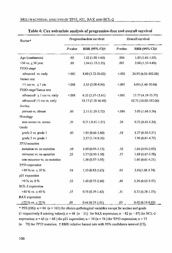
MULTIFACTORIAL ANALYSIS OF TP53, p2I, BAX AND BCL-2
Table 4: Cox univariate analysis of progression-free aud overall survival
Factor* Progression-free survival O\'erall survival
P-value RHR (95% CIlt P-value RHR (95% CI)t
Age (continuous) .03 1.02 (1.00-1.04) .004 1.03 (1.01-1.05)
>56 vs. s 56 year .02 1.94 (1.l2-3.35) .001 2.68 (1.53-4.69)
FIGO-stage
advanced vs. early <.001 8.80 (3.72-20.82) <.001 24.95 (6.05-102.88)
Tumor rest
>lcmvs. s tCIll <.001 5.53 (3.08-9.94) <.001 6.01 (3.40-10.64)
FIGO-stage(ful11or rest
advanced! s I em vs, early <.001 6.33 (2.57-15.61) <.001 17.77 (4.19-75.37)
advanced! > I em YS. early 18.17 (7.10-46.48) 42.71 (10.02-182.06)
Ascites
present vs. absent .01 2.11 (1.20-3.72) <.001 3.05 (1.68-5.56)
Histology
non-serous VS. serous .21 0.71 (0.41-1.21) .24 0.73 (0.43-1.24)
Grade
grade 2 vs. grade I .03 1.95 (0.66-5.80) .18 1.27 (0.50-3.21)
grade 3 VS, grade 1 3.27 (1.l4-9.36) 1.96 (0.81-4.75)
TP53 mutation
mutation vs. no mutation .10 1.69 (0.91-3.13) .10 1.66 (0.91-3.05)
missense vs, no mutation .25 1.77 (0.90-3.50) .27 1.69 (0.87-3.28)
non-missense vs. no mutation 1.50 (0.57-3.95) 1.60 (0.61-4.21)
TP53 expression
>1O%vs.s 10% .18 1.53 (0.82-2.83) .03 2.01( 1.08-3.74)
p2 t expression
>0% VS. 0% .32 1.40 (0.73-2.68) .49 1.26 (0.65-2.47)
BCL-2 expression
>40%V5.540% .37 0.75 (0.39-1.42) .31 0.72 (0.38-1.37)
BAX expression
>75 %V5. <75 % .05 0.44(0.19-1.01) .03 0.42 (0.19-0.93)
• PFS (OS): n ~ 94 (n = 101) for clinico-pathological variables except for ascites and grade
(I respectively 8 missing values); n ~ 48 (n ~ 51) for BAX expression; n ~ 82 (n ~ 87) for BCL-2
expression; n = 65 (n = 68 ) for p21 expression; n = 74 (n = 78 ) for TP53 expression; n = 73
(n = 79) for TP53 mutation. t RHR: relative hazard rate with 95% confidence interval (el).
[00

CHAPTER 4
Respollse to platillum-based chemotherapy ill advallced disease
Of the 68 patients with advanced stage disease, 58 patients received platinum
containing first-line chemotherapy. For three patients response was unknown and for
21 patients response was not assessable due to no or small residual tumor rest after
surgery. The overall clinical response rate was 68% (23/34), i.e. 19 patients achieved
a complete response and four a pat1ial response. One patient had stable disease and 10
patients experienced progressive disease. In an exploratory analysis we studied the
association of the markers investigated with response to platinum-containing
combination therapy in advanced stage disease. There was no significant association
between response to chemotherapy (complete or patiial response versus stable or
progressive disease) and p21, BAX, BCL-2 and TP53 expression or TP53 mutation.
Discussion
Besides the classical prognostic factors, it would be beneficial for patients with
ovarian cancer if there were additional tumor-associated markers that could more
reliably predict the rate of progression and/or the efficacy of response to
chemotherapy. In the present study we determined the relationship between TP53
mutation and expression and the expression of its downstream genes (i.e. the cell
cycle inhibitor p21, the cell death agonist BAX and its antagonist BCL-2) and
evaluated the outcome in relation to patient and tumor characteristics, survival and
response to platinum-containing chemotherapy in patients with ovarian cancer.
There is clear experimental evidence that TP53 aberrations playa critical role in
the development and progression of ovarian cancer. However, the prognostic and
predictive significance of TP53 aberrations (i.e. overexpression and gene mutation) is
still unclear.9,12,22-38 The lack of unanimity may be due to the heterogeneous
population of ovarian cancer patients as well as to methodological differences.
FUl1hermore, miscellaneous chemotherapeutic regimens and different definitions of
response make it difficult to evaluate the predictive value of response to
chemotherapy. Since there still is a controversy with respect to the value of
immunohistochemical or molecular based techniques39-41 , we have chosen to utilize
both approaches to study the clinical relevance of TP53. In the present study we have
found TP53 expression but not TP53 mutation to be of prognostic value. In univariate
analysis, TP53 expression was found to be significantly associated with a poor overall
survival (P ~ .03). However, as shown in multivariate analysis, TP53 expression was
101

MULTIFACTORIAL ANALYSIS OF TP53, p21 , BAX AND BCL-2
not independently associated with survival. Interestingly, when combining TP53
mutation and expression data, this resulted in an increased association with overall
survival (P = ,008) whereas the association with progression-free survival was
borderline significant (P = ,07), Patients whose tumors were both mutation- and
immunonegative had a clear survival advantage compared to patients whose tumors
had either a mutation andlor TP53 overexpression (RHR, 2.85; P = .006). Wen el a(l2
have recently reported that the combination of both TP53 expression and mutation
data results in a stronger prediction of outcome as well. However, more studies are
needed to verify the prognostic value of the combined expression and mutation data.
With respect to treatment, no association between TP53 expression or mutation and
response to platinum-based chemotherapy was found in this relatively small group of
advanced ovarian cancer patients. This is in agreement with data from previous
immunohistochemical-based studies26•3o,34,42 but in contrast to other studies using
either immunological9,24 or molecular-based techniques.27,28
The presence of a TP53 aberration is not informative for the biological function of
TP53. Additional information could be provided by the study of downstream genes of
TP53, i.e. the cell cycle inhibitor p21, the apoptosis-related BAX and its antagonist
BCL-2. Although TP53 regulates the expression of these genes ;11 1';1/,043 -46, we could
not confirm any correlation between TP53 mutation or expression and expression of
p21, BCL-2 or BAX in ovarian tumor specimens. This is consistent with findings
from several other studies29,42,47-49 although an inverse correlation between TP53 and
BCL-2 and between TP53 and p2l has also been reported in ovarian tumor
tissues. 50,5 I The lack of a correlation between TP53 and its downstream genes may
reflect the fact that expression of these genes can also be regulated by TP53-
independent pathways.
Expression of p2l was only associated with tumor rest and not with any of the
other patient and tumor characteristics studied. Furthermore, p2l expression was
neither associated with clinical outcome or with response to platinum-containing
chemotherapy. This is in agreement with a recent report that also failed to find an
association between p21 expression and prognosis or response to platinum-based
chemotherapy in 185 paraffin-embedded tumor specimens from stage III ovarian
cancer patients. 47 However, in contrast to these findings and using a polyclonal
antibody in 295 paraffin-embedded ovarian tumor specimens, Anttila el at reported
that low p21 expression is a marker of poor overall survival.51
102

CHAPTER 4
No statistically significant association between BCL-2 expression and survival
was found. However, patients with increased BCL-2 expression tended to have a
better progression-free and overall survival compared to patients with low BCL-2
expression in their tumors. Several studies have correlated BCL-2 with a survival
advantage in ovarian cancer29,42,48.50 but failed to find an association with overall
response to chemotherapy.42.48 In contrast, BCL-2 expression has also been reported
to be associated with a poor prognosis and resistance to chemotherapy.52.53 Since
BCL-2 is thought to function as an anti-apoptotic protein, a correlation between BCL-
2 expression and a favorable outcome may seem paradoxical. This inhibition of tumor
cell growth by BCL-2 has also been observed in certain solid tumor cell lines and in
breast cancer as well.54,55 Furthermore, it has been suggested that BCL-2 plays a role
in the suppression of angiogenesis.56 Thus BCL-2 may have different functions in
normal differentiated and in cancer cells. Moreover, there is also evidence that BCL-2
functions as a pro-apoptotic protein in some circumstances since overexpression of
the BCL-2 protein has been shown to increase the half-life of the BAX protein.57
We have demonstrated the clinical relevance of BAX protein expression. High
expression levels of BAX were found to be associated with an improved progression
free and overall survival in univariate analysis. When corrected for classical factors,
BAX expression tended to be an independent factor in multivariate analysis for
progression-free survival. The clinical relevance of BAX expression has also been
demonstrated by Tai el al. 58 In a comparable group of 45 ovarian cancer patients these
authors showed that high BAX levels were associated with improved disease-free
survival only. BAX expression was found not to be associated with overall survival,
probably reflecting the short follow-up (median: 21 months) of the patients. In
contrast, another study in 215 ovarian cancer patients described that BAX expression
was correlated with a poor clinical outcome. 59
Since BCL-2 is a critical factor for susceptibility to an apoptotic stimulus, the ratio
of BCL-2 to BAX may be even of greater imp0l1ance.60 Surprisingly, in this study we
observed that the combination of BAX and BCL-2 expression was a stronger
predictor of outcome than BAX expression alone. Patients with both BAX and BCL-2
positive tumors showed a better survival compared to patients with BAX
positive/BCL-2 negative tumors. BCL-2 status did not add on the prognosis of
patients with BAX-negative tumors.
H has been suggested that BAX may be involved in the development of cisplatin
resistance.61 A cisplatin-resistant ovarian cancer cell line was found to have reduced
103

MULTIFACTORIAL ANALYSIS OF TP53, p21, BAX AND BCL-2
BAX mRNA levels, which is consistent with the loss ofTP53'sabilitytotransactivate
BAX as a consequence of TP 5 3 mutation. As in the present study, BAX levels did not
significantly correlate with response to platinum-containing chemotherapy. An
association between high BAX levels and improved response to combination therapy
consisting of paclitaxel and cisplatin in a small group of 26 patients was observed by
Tai et al.58 A relation between BAX and paclitaxel responsiveness has further been
suggested by ill vitro studies showing that BAX could preferentially sensitize ovarian
cancer cells to the effects of paclitaxel and vincristine, as opposed to carboplatin or
ionizing radiation.62-64 The relation between BAX expression and response to
paclitaxel needs further investigation.
In conclusion, TP53 expression but not TP53 mutation was found to predict
overall survival in ovarian cancer patients. The combined evaluation of TP53
mutation and protein expression provides additional infol1nation, especially in those
patients whose tumors are negative for both expression and mutation. Furthermore,
high BAX expression was found to be associated with a favorable outcome in
univariate analysis. The simultaneous evaluation of BAX and BCL-2 expression
provides additional prognostic information when compared to BAX alone. Future
studies should therefore focus on the ratio of BCL-2 to BAX in relation to clinical
outcome.
Acknowledgements
The authors thank Henk POliengen and Marion Meijer-van Gelder for expert
assistance. We gratefully express our thanks to the gynecologists, internists and
pathologists of the following hospitals for providing tumor specimens and clinical
data: University Hospital Rotterdam, St. Clara Hospital, Zuiderziekenhuis, Ikazia
Hospital, St. Franciscus Hospital, Ruwaard van Pulten Hospital, Schiel and Hospital,
Holy Hospital, Beatrix Hospital, Albert Schweitzer Hospital, Drechtsteden Hospital,
Franciscus Hospital, Hospital Zeeuws-Vlaanderen, Hospital Walcheren, St.
Lievensberg Hospital, Ignatius Hospital, St. van Wee I-Bethesda Hospital, St.
Elisabeth Hospital and Pathan.
104

CHAPTER 4
References
1. Ozols R, Rubin S, Dembo A: Principles and practice of gynecologic oncology, in Hoskins
W, Perez C, Young R (eds): Epithelial Ovarian Cancer. Philadelphia, Lippincott, 1992, pp
731-81
2. Eastman A: The fomlation, isolation and characterization of DNA adducts produced by
anticancer platinum complexes, Pharmacol Ther 34: 155~66, 1987
3. Eastman A: Activation of programmed cell death by anticancer agents: cispiatin as a
model syslem. Cancer Cells 2:275-80, 1990
4. Dive C, Hickman JA: Drug~target interactions: only the first step in the commitment to a
programmed cell dealh? Br J Cancer 64: 192-6, 1991
5. Hockenbery D, Nunez G, Milliman C, et al: BCL-2 is an inner mitochondrial membrane
prole in Ihal blocks programmed cell dealh. Nalure 348:334-6, 1990
6. Miyashita T, Reed JC: BCL-2 oncoprolein blocks chemotherapy-induced apoptosis in a
human leukemia cell line, Blood 81:151-7,1993
7, Oltvai ZN, Milliman CL, Korsmeyer SJ: BCL-2 heterodimerizes in vivo with a conserved
homolog, BAX, thai accelerales programmed cell dealh. Cell 74:609-19, 1993
8, Shelling AN, Cooke IE, Ganesan TS: The genetic analysis of ovarian cancer, Br J Cancer
72:521-7,1995
9, Ferrandina G, Fagotti A, Salerno M, et al: pS3 overexpression is associated with
cytoreduction and response to chemotherapy in ovarian cancer, Br J Cancer 81:733·740,
1999
10, Kottmeier HL: Presentation of therapeutic results in carcinoma of the female pelvis:
experience of the Annual Report on the Results of Treatment in Carcinoma of the Utems,
Vagina, and Ovary. Gynecol 0I1coI4:13-9, 1976
II. Organization WH: WHO handbook for reporting results of cancer treatments, Geneva,
World Health Organization off<;et publication, 1979
12, Kohler MF, Kerns BJ, Humphrey PA, et al: Mutation and overexpression ofpS3 in early·
slage epithelial ovarian cancer. Obslet Gynecol 81 :643-50, 1993
13, Barlow R, Bartholomew D, Bremner J: Statistical interference under order restrictions,
London, John Wiley & Sons, 1993
14, Bonetti A, Zaninelli M, Leone R, et 31: BCL·2 but not p53 expression is associated with
resistance to chemotherapy in advanced breast cancer. Clin Cancer Res 4:2331-6. 1998
15, group EBCC: Revision of the standards for the assessment of homlOne receptors in human
breast cancer; report of the second E,O,R,T,C, Workshop, held on 16-17 March, 1979, in
105

MULTIFACTORIAL ANALYSIS OF TP53, p21 , BAX AND BCL-2
the Netherlands Cancer Institute. Eur J Cancer 16: 1513-5, 1980
16, Sambrook J, Fritsch E, Maniatis T: Molecular Cloning. A Laboratory Manual (ed 2), New
York, Cold Spring Harbor, 1989
17. Bems EM, Klijn JG, Smid M, et al: TP53 and MYC gene alterations independently predict
poor prognosis in breast cancer patients. Genes Chromosomes Cancer 16: 170-9, 1996
18. Kaplan E, Meier P: Non-parametric estimation frol11 incomplete observations, J Am Stat
Assoc 53:457-81,1958
19. Carbone D, Chiba I, Mitsudomi T: Polymorphism at codon 213 within the p53 gene.
Oncogene 6:1691-2,1991
20. van def Burg ME, Henzen-Logmans SC, Bems EM, et al: Expression of urokinase-type
plasminogen activator (uPA) and its inhibitor PAI-I in benign, borderline, malignant
primary and metastatic ovarian tumors, Int J Cancer 69:475~9, 1996
21. Neijt JP, ten Bokkel Huinink WW, van del' Burg ME, et al: Long~term survival in ovarian
cancer, Mature data from The Netherlands Joint Study Group for Ovarian Cancer. Eur J
Cancer 27:1367-72, 1991
22. Wen WH, Reles A, Runnebaum IB, et al: p53 Illutations and expression in ovarian
cancers: correlation with overall survival. Int J Gynecol Pathol 18:29·41, 1999
23. Sato S, Kigawa J, Minagawa Y, et al: Chemosensitivity and p53·dependent apoptosis in
epithelial ovarian carcinoma. Cancer 86: 1307·13, 1999
24. Dong Y, Walsh MD, McGuckin MA, ct al: Reduced expression of retinoblastoma gene
product (pRB) and high expression of p53 are associated with poor prognosis in ovarian
cancer. In! J Cancer 74:407-15,1997
25. Geisler JP, Geisler HE, Wiemann MC, et al: Quantification of pS3 in epithelial ovarian
cancer. Gynecol Oncal 66:435-8, 1997
26. Rohlke P, Milde-Langosch K, Weyland C, et al: p53 is a persistent and predictive marker
in advanced ovarian carcinomas: multivariate analysis including comparison with Ki67
immunoreactivity. J Cancer Res Clill Oncol 123:496-501, 1997
27. Buttitta F, Marchetti A, Gadducci A, et al: p53 alterations are predictive of
chemoresistance and aggressiveness in ovarian carcinomas: a molecular and
immunohistochcmical study. Br J Cancer 75:230·5, 1997
28. Righetti SC, Della Ton'c G, Pilotti S, et al: A comparative study of p53 gene mutations,
protein accumulation, and response to cisplatin~based chemotherapy in advanced ovarian
carcinoma. Cancer Res 56:689-93, 1996
29. Diebold J, Baretton G, Felchner M, et al: BCL~2 expression, p53 accumulation, and
apoptosis in ovarian carcinomas. Am J Clin Patholl05:341-9, 1996
106

CHAPTER 4
30. van def Zee AG, Hollema H, Suunneijer AJ, et al: Value of P-glycoprotein, glutathione S
transferase pi, c-erbB-2. and p53 as prognostic factors in ovarian carcinomas. J Clin Oneol
13:70-8,1995
31. Klemi PJ, Pylkkanen L, Kiilholma P, et al: p53 protein detected by inununohistochemistry
as a prognostic factor in patients with epithelial ovarian carcinoma. Cancer 76: 120 1-8,
1995
32. Levesque MA, Katsaros 0, Yu H, et al: Mutant p53 protein overexpression is associated
with poor outcome in patients with well or moderately differentiated ovarian carcinoma.
Cancer 75: 1327-38, 1995
33. Henriksen R, Strang P, Wilallder E, et al: p53 expression in epithelial ovarian neoplasms:
relationship to clinical and pathological parameters, Ki*67 expression and flow cytometry.
Oynecol OncoI53:301-6, 1994
34. Hartmann LC, Podratz KC, Keeney GL, et al: Prognostic significance of p53
imnmnostaining in epithelial ovarian cancer. J Clin Oncol 12:64*9, 1994
35. Niwa K, Itoh M, Murase T, el al: Alteration of p53 gene in ovarian carcinoma:
clinicopathological correlation and prognostic significance. Br 1 Cancer 70: 1191 R 7, 1994
36. Sheridan E, Silcocks P, Smith J, et al: P53 mutation in a series of epithelial ovarian
cancers from the U.K., and its prognostic significance. Eur J Cancer 11: 170 I *4, 1994
37. Bosari S, Via Ie G, Radaelli U, et al: p53 accumulation in ovarian carcinomas and its
prognostic implications. Hum Pathol24: 1175R9, 1993
38. Marks JR, Davidoff AM, Kents BJ, el al: Overexpression and mutation ofp53 in epithelial
ovarian cancer. Cancer Res 51 :2979R 84, 1991
39. Schuyer M, HenzenRLogmans SC, van der Burg ME, et al: High prevalence of codon
213Arg-*>Stop mutations of the TP53 gene in human ovarian cancer in the southwestem
part of The Netherlands.lnt J Cancer 76:299-303, 1998
40. Casey G, Lopez ME, Ramos lC, et al: DNA sequence analysis of exons 2 through 11 and
immunohistochemical staining are required 10 detect all known p53 alterations in human
malignancies. Oncogene 13:1971-81, 1996
41. Skilling lS, Sood A, Niemann T, et al: An abundance of p53 null mutations in ovarian
carcinoma. Oncogene 13:117-23, 1996
42. Herod JJ, Eliopoulos AG, Warwick J, et al: The prognostic significance of BCL-2 and p53
expression in ovarian carcinoma. Cancer Res 56:2178 R 84, 1996
13. el-Deiry WS, Harper JW, O'Connor PM, et aI: WAFI/CIPI is induced in p53-mediated 01
arrest and apoplosis. Cancer Res 54:1169·74, 1994
44. el.Deiry WS, Tokino T, Velculescll VE, et al: WAFI, a potential mediator ofp53 tumor
107

MULTIFACTORIAL ANALYSIS OF TP53, p21, BAX AND BCL-2
suppression. Cell 75:817-25, 1993
45. Miyashita T, Reed Je: Tumor suppressor p53 is a direct transcriptional activator of the
human BAX gene. Cell 80:293-9, 1995
46. Miyashita T, Krajewski S, Krajewska M, et al: Tumor suppressor p53 is a regulator of
BCL~2 and SAX gene expression in vitro and in vivo. Oncogene 9: 1799~805. 1994
47. Baekelandt M, Holm R, Trope CO, et al: Lack of independent prognostic significance of
p21 and p27 expression in advanced ovarian cancer: an inununohistochemical study. Clin
Cancer Res 5:2848-53, 1999
48. Baekelandt M, Kristensen G, Nesland J, et al: Clinical significance of apoptosis-reiated
factors p53, Mdm2, and BCL-2 in advanced ovarian cancer. J Clin Oneol 17:2061-2068,
1999
49. Marone M, Scambia G, Mazzetti S, et al: BCLw2, BAX, bclwXL, and bclwXS expression in
normal and neoplastic ovarian tissues. Clin Cancer Res 4:517w24, 1998
50. Henriksen R, Wilander E, Oberg K: Expression and prognostic significance of BCLw2 in
ovarian tumours. Br J Cancer 72: I 324w9, 1995
51. Anttila MA, Kosma VM, Hongxiu J, et al: p21IW AFI expression as related to p53, cell
proliferation and prognosis in epithelial ovarian cancer. Br J Cancer 79: 1870-8, 1999
52. Kassim SK, Ali HS, Sallam MM, et al: Increased BCL-2 expression is associated with
primary resistance to chemotherapy in human epithelial ovarian cancer. Clin Biochem
32:333-8,1999
53. Mano Y, Kikuchi Y, Yamamoto K, et al: BCLw2 as a predictor of chemosensitivity and
prognosis in primary epithelial ovarian cancer. Eur J Cancer 35: l2l4w 1219, 1999
54. Pietenpol JA, Papadopoulos N, Markowitz S, et al: Paradoxical inhibition of solid tumor
cell growth by bcl2. Cancer Res 54:3714-7,1994
55. Gasparini G, Barbareschi M, Doglioni C, et al: Expression of BCLw2 protein predicts
efficacy of adjuvant treatments in operable node-positive breast cancer. Clin Cancer Res
I: 189-98, 1995
56. Koukourakis MI, Giatromanolaki A, O'Byme KJ, et al: Potential role of BCLw2 as a
suppressor oftulllour angiogenesis in nOIl- small-cell lung cancer. Int J Cancer 74:565-70,
1997
57. Miyashita T, Kitada S, Krajewski S, et al: Overexpression of the BCL-2 protein increases
the half-life ofp21BAX. J BioI Chem 270:26049-52, 1995
58. Tai YT, Lee S, Niloff E, et al: BAX protein expression and clinical outcome in epithelial
ovarian cancer. J Clin Oncol 16:2583-90, 1998
59. Marx D, Binder C, Meden H, et al: Differential expression of apoptosis associated genes
108

CHAPTER 4
BAX and BCLM 2 in ovarian cancer, Anticancer Res 17:2233·40, 1997
60. OItvai ZN, Korsmeyer SJ: Checkpoints ofdueting dimers foil death wishes. Cell 79:189-
92, 1994
61. Perego P, Giarola M. Righetti se, et 31: Association between cisplatin resistance and
mutation of p53 gene and reduced BAX expression in ovarian carcinoma celt systems.
Cancer Res 56:556·62, 1996
62. Strobel T, Kraeft SK. Chen LB, et al: BAX expression is associated with enhanced
intracellular accumulation of paclitaxel: a novel role for BAX during chemotherapy"
induced cell death. Cancer Res 58:4776·8 t, 1998
63. Strobel T, Swanson L, Korsmeyer S, et at: BAX enhances paclitaxel-induced apoptosis
throngh a p53·independent pathway. Proc Nat! Acad Sci USA 93:14094·9, 1996
64. Strobel T, Swanson L, Korsmeyer S, et 31: Radiation-induced apoptosis is not enhanced by
expression of either p53 or BAX in SW626 ovarian cancer cells. Oncogene 14:2753~8,
1997
109

110

CHAPTER 5
ARE OVARIAN BORDERLINE TUMORS DISTINCT
fROM OVARIAN CARCINOMAS?
Part of this chapter has been published in:
Genetic Alterations in Ovarian Borderline Tumours and Ovarian Carcinomas
M Schuyer, SC Henzen-Logmans, MEL van der Burg, JH Fieret, C Derksen, MP Look, ME Meijer-van Gelder, JGM Klljn, JA Foekens
and EMJJ Berns
Eur J Obstet Gynecol 82: 147 -150, 1999

112

CHAPTERS
Summary
Borderline ovarian tumors are intermediate in their clinical behavior between
benign adenomas and malignant neoplasms, and are associated with overall IO-year
survival rates in excess of 90%. It is unclear whether borderline tumors represent a
biological continuum of stepwise progression toward invasive carcinoma or whether
they are distinct entities, each arising de novo. The characterization of oncogenes and
tumor suppressor genes, such as K-RAS and TP53 may give insight into the biology of
borderline tumors and/or may also serve as markers associated with prognosis. We
observed that TP53 mutation is infrequent in borderline tumors. In contrast, K-RAS
mutations are more common, having been found in 27% of borderline tumors.
Interestingly, these mutations are strongly associated with the mucinous cell type.
Introduction
As early as 1929 Taylor l reported a series of patients with semi-malignant tumors
of the ovary. However, it was not until 1973 that this group of tumors was formally
recognized by the World Health Organization (WHO) and the International
Federation of Gynecology and Obstetrics (FIGO) as a distinct categOlY with the
interchangeable ten]]s cystadenomas of borderline malignancy and carcinomas of low
malignant potential.2•J Ovarian borderline tumors constitute approximately 15% of
ovarian tumors. Patients with borderline tumors are usually older than patients with
benign neoplasms and younger than women with frank malignancies. Although
borderline tumors have some clinical and pathological features in common with
ovarian carcinomas, they usually behave relatively benignly even in the presence of
widespread abdominal disease. Most borderline tumors are of the serous (60%) or
mucinous (34%) histological subtype, with endometrioid, clear cell, Brenner and
mixed epithelial types making up the remaining 6%. The major histological feature
used to separate ovarian borderline tumors from invasive carcinomas is destructive
stromal invasion. The distinction between a pushing border versus destructive
infiltrative growth is often the only feature that differentiates a borderline tumor from
one that is fully malignant. This sometimes makes this tumor difficult to classify.
The survival of patients with borderline tumors is superior to patients with
epithelial ovarian cancer. The 5-year survival rate is about 95% and 20-year survival
is 80%.4 Borderline ovarian tumors often present as early stage lesions. SurgelY is the
113

K-RAS AND TP53 MUTATIONS IN BORDERLlNETUMORS
recommended treatment for early-stage tumors and patients with these tumors have an
excellent prognosis and a nearly 100% survival. Approximately 15% to 20% of
patients with borderline tumors present with advanced stage disease at the time of
diagnosis.4•s Stage III patients typically have a 56-73% survival long term.6 The
benefit of postsurgical therapy in patients with advanced stage borderline tumors has
not been well established.7
Molecular analyses of oncogenes and tumor suppressor genes have contributed to
the debate as to whether benign and borderline tumors are part of a continuum in the
transformation of epithelial cells to malignancy. A similar pattern of mutations in
benign, borderline and malignant tumors would be consistent with a continuum
hypothesis whereas a different pattern of alterations would support the hypothesis that
they are different entities. Some investigators suspect that borderline tumors, as
intermediate form of malignancy, may be a precursor of invasive carcinomas whereas
others believe that borderline tumors are separate biological entities. Indeed, several
studies showed that the incidence of K-RAS mutation in ovarian borderline tumors
was much higher than in invasive carcinomasS-II
, suggesting that they might occur
through independent pathways8 Other evidence, however, suggests that malignant
epithelial tumors may result from the progressive transformation of benign andlor
borderline tumors. 12 For example, loss of heterozygosity analyses showed no distinct
patterns of loss between borderline tumors and adenocarcinomas but loss of
heterozygosity was observed at lower levels in borderline tumors. IO Furthermore,
benign or borderline epithelium has been identified in some serous and mucinous
carcinomas suggesting malignant progression from altered benign epithelium. 13
The relationship between borderline ovarian tumors and invasive epithelial ovarian
carcinoma will become more clear as more genes are studied. Furthermore, the
discovery of markers that can predict a poor prognosis should aid the clinician in
making decisions about the therapy for a particular patient with a borderline ovarian
tumor. The characterization of oncogenes and tumor suppressor genes, such as K-RAS
and TP53 may give insight into the biology of borderline tumors andlor may also
serve as markers associated with prognosis. Whereas the TP53 gene is the most
commonly mutated tumor suppressor gene in human cancer, the K-RAS gene has been
implicated as the most conmlOnly mutated oncogene associated with human tumors,
including ovarian malignancies. The K-RAS gene is a member of the RAS gene t:1mily
consisting of the three members N-, H- and K-RAS, which code for highly homologous
proteins with a molecular weight of21 kDa. K-RAS localizes to chromosome band 12pl2
114

CHAPTERS
and encodes a small membrane bound GTP-binding protein that serves as a relay signal
from receptor tyrosine kinases (e.g. EGF/PDGF) to the nucleus. 14 In the active GTP
bound confollnation p21-RAS protein transmits a signal to an effector molecule. The
stimulation of signal transduction cascades results in the synthesis or activation of specific
transcription factors, thereby stimulating cell proliferation. The transforming potential of
K-RAS has been related mainly to point mutations in codon 12 although, sometimes,
base substitutions occur in codons 13 or 61. Surveys of tumor series show that K-RAS
mutations are commonly present in human adenocarcinomas originating at several
sites, including colon (up to 80% of colorectal carcinomas), lung (up to 50% of lung
adenocarcinomas), and pancreas (up to 90% of pancreatic carcinomas). 15-17
Interestingly, K-RAS mutations have been detected in benign lesions of the colonic
epithelium that precede the development of malignant tumors. 18 Therefore, K-RAS
mutations have been suggested to represent early genetic events in the process of
carcinogenesis. To get more insight in the biology of ovarian borderline tumors we have studied
K-RAS and TP53 mutations and TP53 expression in borderline tumors and in the
borderline components of carcinomas.
Materials and Methods
TUlIlor specimens
Tumor specimens were obtained /i'om 30 patients diagnosed with a borderline tumor.
Histology was assessed on paraffin material according to the World Health Organization
criteria. 19 Serous tumors were graded according to Burks et al.43 Briefly, nuclear atypia
was graded on a three-tier scale. Grade I was characterized by predominantly round to
oval nuclei with relatively fine chromatin and small nucleoli; grade 2 by moderately
enlarged, oval to more rounded nuclei with less evenly dispersed clu'Omatin and more
prominent nucleoli; grade 3 by markedly enlarged pleomorphic and vesicular nuclei with
many nuclei displaying prominent eosinophilic nucleoli. The same criteria were used for
mucinous tumors. In addition, serous tumors showing a pattern of highly complex
micropapillae stmctures were noted.43 Sections were evaluated by one pathologist (S
HL). Nineteen tumor specimens were classified as serous and II as mucinous. Sixteen
specimens were grade I, 7 grade II, 4 grade III and for 3 specimens grade was not
detennined. In addition, seven so-called borderline components were collected: initially,
lIS

K-RAS AND TP53 MUTATIONS IN BORDERLINE TUMORS
frozen tumor sections were found to contain borderline tumor components but, following
revision, invasive areas pointing to carcinoma were also observed in the paraffin sections
(archive material) of these tumors. In one case, the transition fOlm carcinoma to the
borderline component was observed in the paraffin section. Four of the borderline
components had a serous and three had a mucinous histology. All borderline components
were grade III.
DNA isolalioll alld PCR-SSCP
High molecular weight chromosomal DNA was isolated fi'Om frozen tumor tissue
specimens according to standard procedures.2o Mutations in K-RAS (exon I, encoding
mutational hotspots codons 12 and 13) and TP53 (exons 5-8) were studied using
Polymerase Chain Reaction-Single Strand Conformation Polymorphism (PCR-SSCP) and
sequencing. PCR-SSCP for TP53 was perfOlmed as previously described. 21,22
Exon I of K-RAS was amplified using intronic primers: KRI (5'-TGATAGT
GTATTAACCTTATG-3') and KR2 (5'- TTTATCTGTATCAAA GAATG-3'). Cycling
parameters were 94° for 4 min, followed by 25 cycles of 94°C for I min, 50°C for 1.5 min
and noc for 2 min, and tellllinated by noc for 7 min. PCR products were diluted with
miIliQ-H20 (1:4) and checked on ethidium bromide-stained agarose gels (1.3%). To
decrease the false negative rate, PCR products were subsequently digested with HiIlJl,
resulting in fragments of 95 and 154 bp. SSCP analysis was performed using a non
denattlling 8% polyacrylamide gel containing 10% (v/v) glycerol. Gels were lun with IX Tris-borate-EDTA buffer at 30W for 6 to 7 hI' at room temperature. The colon cancel' cell
lines SW-480 (codon 12:GGL.,GTT) and SW-1398 (codon 12:GGT--+TGT) and the
breast cancer cell line MDA-MB-231 (codonI3:GGC--+GAC) were used as positive
controls. Samples showing an altered migration pattem were analyzed again and
independent PCR products were sequenced using a T7 sequencing kit (Pharmacia,
Uppsala, Sweden) following subcloning using a TA cloning kit (Invitrogen, Leek, The
Netherlands).
11/11/1 /11/0 iii sl oc It e 1/1 is 1 ry
Immunohistochemical staining for TP53 was perfOlmed on frozen formalin-fixed 5
micron thick sections using a peroxidase-labelled streptavidin-biotin-complex technique
as previously described.21 The monoclonal DOl (Santa emz) and D07 (Dako) antibodies
116

CHAPTERS
were used. Sections were considered positive when 10% of tumor cells showed a clear
I .. 23 nuc ear stammg.
Results
K-RAS altem/iolls ill borderlille /1II/lOrs
Seven out of 26 (27%) borderline tumors, which were studied for K-RAS mutations
were found to have mutations in codon 12 (Table I). Interestingly, 5 of8 (63%) mucinous
borderline tumors specimens showed mutations (3x Glyl2Val, 2x Glyl2Asp) compared
to only 2 of 18 (11%) serous tumors (both GlyI2Val).
TCGA
--. • • 1IiIIII.. .. -Wild type Codon 12:Gly
T", T/ G
TCGA
--I11III .... -.... II - .. Mutant Codon 12:Asp
TCGA
-. --'. '!II' -Mutant
Codon 12:Val
Figllre 1: K-RAS codoll 12 IIIlIlaliolls ill ovariall borderlille IlIlIIors
TP53 altem/iolls it/ borderlille /lIl11ors
Thirty tumor specimens were studied for TP53 mutations. Mutation of TP53 was
observed in only one mucinous tumor (Thr284Ala). In addition, two tumor specimens
showed a conunon neutral polymorphism (Gly213Gly). With respect to
immunostaining, the percentage of positive tumor cells ranged from <I % to 55% with
117

K-RAS AND TP53 MUTATIONS IN BORDERLINE TUMORS
Table 1: K-RAS and TP53 alterations in ovarian borderline tumors
TPS3 expression K-RAS TP53 (% positive tumor cells)
Hislology Grade Age mutation mutation DOl D07
serous l 62 n.d. n.d.
scrous 34 <1% <1%
serous 28 <1% <1%
serous 40 9 <1% serous 47 5% 3%
serous 87 3% 12%
serous2 59 28% 8%
serous3 81 Glyl2Vai 30% 2%
serous 40 Glyl2Vai 55% 30%
serous II 74 n.d. <1% <1%
serous II 43 2% 3% serous II 27 4% 6%
serous II 32 8% 3% serous II 32 12% 10% serous II 43 31% 30%
serous II 47 Gly213Gly 17% 16%
serous III' 52 9% <1% serous III 34 17% 5%
serous n.d. 49 9% n.d.
mucinous 53 n.d. 7% 15% mucinous 56 n.d, n.d.
mucinous 72 <1% <1% mucinous 47 2% <1%
mucinous 68 Gly 12Asp 2% <1% mucinous 43 Glyl2Asp 5% 10% mucinous 69 n,d. Gly213Gly <1% <1%
nnlCinolis III 26 Glyl2Vai <1% <1% mucinous III 48 n.d, Thr284Ala <1% <1% mucinous n.d. 83 Glyl2Vai 12% 12%
mucinous n.d. 27 Glyl2Vai <1% <1%
(n.d).: 110t detelmined (~): no mutation
K~RAS mutations: Glyl2Val: GGT-;qrr Glyl2Asp: GGT-;GAT
TP53 mutations: Gly2J3Gly: CGA-;CGG Thr284Ala: ACA-;GCA
I) left ovary; right ovaty contains carcinoma; 2) implant on diaphragm;
3) also contains adenoma; 4) micropapillary paltem
118

CHAPTERS
a median of 6% for the DO I antibody and 3% for the D07 antibody. According to the
10% cut-off point, which is commonly used for invasive carcinomas21,23, TP53
immunostaining with at least one monoclonal antibody was observed in ten of 28 (36%)
tumor specimens analyzed. Only four out of 27 (15%) tumor specimens showed
inullunopositivity with both monoclonal antibodies.
K-RAS allli TP53 afleraliolls ill borderlille compollellis
In addition, seven borderline components were studied from tumors that included a
carcinoma component. Four of these borderline components showed a K-RAS mutation.
Two of these Illutations (both Glyl2Val) were obselved among the serous tumors
whereas the other two (both Glyl2Asp) were found in mucinous tumors. One mucinous
tumor without K-RAS mutation showed a TP53 mutation in exon 8 of the TP53 gene
(Arg273His). The resuits are summarized in Table 2.
Table 2: K-RAS and TP53 alterations in borderline components
TP53 expression K-RAS TP53 (% positive tumor cells)
Hislology Grade Age mutation mutation DOl D07
serous III 62 <1% <1% serous III 76 Glyl2Vai 15% 12% serous JII 29 Glyl2Vai 20% 15% serous m' 33 <1% <1%
mucinous III 68 Glyl2Asp 26% 30% mucinous III 73 Arg273His 58% 65% mucinous m '34 Glyl2Asp 60% 20%
(n.d.): not determined (-): no mutation
I) micropapillary pattem
Discllssion
Despite substantial advances in our understanding of other adenocarcinomas,
particularly in the colon, very little is known about the molecular evolution of ovarian
tumors. It is likely that, as in other solid tUlllors, multiple events including inactivation
119

K-RAS AND TP53 MUTATIONS IN BORDERLINE TUMORS
of tumor suppressor genes and activation of cellular oncogenes are required for the
transformation of normal ovarian epithelium to benign, borderline, malignant and
metastatic tumors. The issue of whether or not borderline tumors progress to fi'ankly
malignant ovarian adenocarcinomas is an important one in screening for ovarian cancer
and in the treatment of borderline tumors. The relationship between borderline ovarian
tumors and fi'ankly invasive adenocarcinomas is now beginning to be explored with the
tools of epidemiology and molecular biology.
In an exploratOlY study we have demonstrated K-RAS mutations in 27% (7 out of 26)
borderline tumors. Interestingly, borderline components were found to have more
K-RAS mutations (4 out of 6 or 67%), although the number is small. Furthermore, it
should be noted that only exon I of the K-RAS gene, encoding codons 12 and 13, has
been analyzed. Although K-RAS mutations can also occur at codon 61, these mutations
are rather infi'equent and therefore the prevalence of K-RAS mutations is not expected to
be highly underestimated. The fact that K-RAS mutations are commonly detected in
ovarian borderline tumors suggests that these changes may represent an early genetic
alteration. High incidences of K-RAS mutations have also been observed by others.
However, data conceming K-RAS mutations in invasive carcinoma are conflicting.
Sevcral authors reported higher K-RAS mutation fi'equencies in borderline tumors (30-
50%) compared to invasive carcinomas (4%).8-11 Based on these observations it has been
suggested that borderline tumors and invasive carcinomas each may follow a different
molecular developmental pathway and thus are separate biological entities.s However, we
observed K-RAS mutations more frequently in mucinous tumors (63%) than in tumors
with a serous histology (II %). Likewise, other authors have also reported an association
of K-RAS mutation with mucinous differentiation in ovarian borderline tumors andior
invasive carcinomas.9.24-26 Interestingly, mucinous tumors represent approximately 35%
of borderline tumors compared to only 10% of ovarian carcinomas. This may explain why
some have observed a higher incidence of K-RAS mutations in borderline tumors
compared to invasive carcinomas. Interestingly, by investigating only tumors with a
mucinous histology, even more K-RAS mutations (85%) have been found in
adenocarcinomas compared to borderline tumors (73%)27, which supports the hypothesis
that borderline tumors represent a pathological continuum between benign and frankly
invasive neoplasms. Since the different histological distribution of borderline tumors and
invasive carcinomas has not been considered, the conclusion that borderline tumors and
carcinomas arise independently is therefore not justified
Finally, the association of K-RAS mutation with mucinous borderline tumors suggests
120

CHAPTERS
that it may playa role in maintaining the mucinous differentiation pathway of ovarian
epithelial cells. Moreover, K-RAS gene mutations have been associated with a mucinous
subtype in other types of human cancers as well, including colon, pancreas and lung
carcinomas.28-33 These studies all SUpp011 the idea that K-RAS activation plays a role in
the cellular pathway of tumor differentiation.
With respect to TP53 alterations, we showed that mutation of the TP53 gene is
infi'equent in borderline tumors. This finding is consistent with that of previous
studies.8,34-36 In addition, several studies have also shown that TP53 overexpression is
rather unconnnon in borderline tumors, although reported percentages varied from 4 to
24% (mean between 10 and 15%).9,31-40 However, it should be noted that different
antibodies, different methods of scoring (e.g. intensity or percentage of stained tnmor
cells) and cut-off points and finally different tumor material (paraffin-embedded or
frozen) make it difficult to compare studies. In the present study we have obselved
immunopositivity for TP53 (using the 10% cut-off point) with at least one monoclonal
antibody in 10 of 28 (36%) borderline tumors and 5 of 7 (71%) borderline components.
When both antibodies are evaluated, only 4 of 27 (15%) borderline tumor specimens are
considered positive. Surprisingly, the percentages of stained tumor cells are much smaller
than usually obselved in ovarian adenocarcinomas,41 Furthermore, carcinomas that do not
stain often completely lack any staining for TP53. It remains unknown why in the absence
of TP53 mutations some borderline tnmor specimens reveal TP53 immunostaining. TP53
mutations may have been missed due to the fact that they are located outside exons 5-8,
Otherwise, the cut-off point of 10% positive tumors cells perhaps needs to be adjusted in
case of ovarian borderline tumors. For example, using a 20% cut-off point, which has also
been used for ovarian carcinomas42, only 2 out of27 (7%) tumor specimens score positive
with both antibodies.
In conclusion, we have shown that K-RAS mutations occur frequently in mucinous
borderline tumors, Conversely, TP53 mutations are uncommon in borderline tumors.
The role of TP53 accumulation in borderline tumors needs to be further investigated,
It remains controversial whether borderline tumors are capable of progression to
invasive carcinoma or whether they represent different entities. Some ovarian
epithelial neoplasms are heterogeneous and benign, borderline and malignant components
may coexist within an individual tnmor. It has been postulated that the benign or
borderline areas might have preceded the development of the malignant component. In
the cunent stndy seven borderline components adjacent to an area of invasive tumor
121

K-RAS AND TP53 MUTATIONS IN BORDERLINE TUMORS
cells were investigated. An increased incidence of K-RAS and TP53 mutation and
accumulation was observed in these borderline components compared to pure
borderline tumors, suggesting that these tumors indeed progress to the malignant
invasive phenotype through an accumulation of genetic alterations. It would be
interesting, especially in these tumors, to study both the borderline and the invasive
component. Microdissection techniques, however, are a prerequisite.
Recently, some investigators have proposed to abandon the borderline categOlY
and retum to the old benign-malignant classification system. Mucinous borderline
tumors have a good prognosis and can be treated as benign tumors with the exception
of those tumors associated with pseudomyxoma peritonei. These latter tumors are
thought to be secondary neoplasms of the appendix. With respect to serous borderline
tumors, these can be unevenly divided into a larger group of atypical proliferative
epithelial cystadenomas and a smaller category of recently described noninvasive
carcinomas, designated micropapillary serous carcinoma (MPSC).43,44 These latter
neoplasms have a complex micropapillary architecture and often lack destructive
infiltrative growth but appear to behave as low-grade invasive carcinoma. They are
often associated with invasive implants that in turn are associated with recurrences
and a poor prognosis. Therefore these tumors should be classified and treated as
carcinoma.43.44 However, this class of tumors is not well characterized and currently
there are no molecular markers available that distinguish between these tumors and
the benign ones with a more favorable prognosis. Since TP53 alterations are common
in serolls ovarian carcinomas and, additionally, predict a poor prognosis in ovarian
carcinoma, TP53 mutation or overexpression could represent a suitable marker.
However, only two tumor specimens in this study met the pathological criteria of a
micropapillary serous tumor44 and no TP53 alteration was found in these tumors. One
other studl5 also reported absence of TP53 mutations in micropapillary serous
ovarian carcinoma. Interestingly, the latter study rep0l1ed a moderately intense TP53
staining in these tumors.
Further analysis of genetic abnormalities in larger tumor sets and in patients with
follow-up may delineate the relationship between borderline ovarian tumors and
epithelial ovarian carcinomas better, and will hopefully lead to a unifying hypothesis
as to the origin of these important ovarian lesions.
122

CHAPTER 5
References
l. Taylor He: Malignant and semi-malignant tumors of the ovary. Surg Gynecol Obstet
48:204-30, 1929
2. Secoy SF, Scully RE, Sobin LH: International histological classification and staging of
tumors, Histologic typing of ovarian tUIllors. Geneva, World Health Organization, 1973
3. Intemational Federation of Gynecology and Obstetrics: Classification and staging of
malignant tumors in the female pelvis. Acta Obslet Gynecol Scand 50: 1-7, 1971
4. Link el, Jr., Reed E, Sarosy G, el al: Borderline ovarian tUIllors. Am J Med 101:217-25,
1996
5. Leake JF: Tumors of low malignant potential. Curr Opin Obstet Gynecol 4:8 t -5, 1992
6. Link el, Jr., Kohn E, Reed E: The relationship between borderline ovarian tumors and
epithelial ovarian carcinoma: epidemiologic, pathologic, and molecular aspects. Gynecol
OncoI60:347-54,1996
7. Chambers JT: Borderline ovarian tumors: a review of treatment. Yale J BioI Med 62:351-
65, 1989
8. Telleriello MO, Ebina M, Linnoila RI, et al: p53 and Ki-ras gene mutations in epithelial
ovarian neoplasms. Cancer Res 53:3103-8,1993
9. Caduff RF, Svoboda-Newman SM, Ferguson AW, et al: Comparison of mutations of Ki
RAS and p53 immunoreactivity in borderline and malignant epithelial ovarian hUllors. Am
J Surg PathoI23:323-8, 1999
10. Chenevix-Trench G, Kerr J, Hurst T, et al: Analysis of loss of heterozygosity and KRAS2
mutations in ovarian neoplasms: clinicopathological correlations. Genes Chromosomes
Cancer 18:75-83, 1997
II. Chenevix-Trench G, Coombs T, Kerr J, et al: Fertility and incidence of KRAS2 mutations
in borderline ovarian adenocarcinomas. J Nat! Cancer Inst 89:890-1, 1997
12. Wolf NO, Abdul-Karim FW, Schork NJ, et al: Origins of heterogeneous ovarian
carcinomas. A molecular cytogenetic analysis of histologically benign, low malignant
potential, and fully malignant components. Am J Pathol 149:511-20, 1996
13. PuIs LE, Powell DE, DePriest PO, et al: Transition frolll benign to malignant epithelium in
mucinous and serous ovarian cystadenocarcinoma. Gynecol OncoI47:53-7, 1992
14. Satoh T, Nakafuku M, Kaziro Y: Function of Ras as a molecular switch in signal
transduction. J Bioi Chem 267:24149-52,1992
15. Bos JL: ras oncogenes in human cancer; a review. Cancer Res 49:4682-9, 1989
16. Capella G, Cronauer-Mitra S, Pienado MA, et al: Frequency and spectnml of mutations at
123

K-RAS AND TP53 MUTATIONS IN BORDERLINE TUMORS
codons 12 and 13 of the c~K-ras gene in human tumors. Environ Health Perspect 93:125-
31,1991
17. Minamoto T, Mai M, Ronai Z: K-ras mutation: early detection in molecular diagnosis and
risk assessment of colorectal. pancreas, and lung cancers--a review. Cancer Detect Prey
24:1-12,2000
18. Vogelstein B, Fearon ER, Hamilton SR, et al: Genetic alterations during colorectal-tumor
development. N Engl J Med 319:525-32,1988
19. Organization WH: WHO handbook for reporting results of cancer treatments. Geneva,
World Health Organization offset publication, 1979
20. Sambrook J, Fritsch E, Maniatis T: Molecular Cloning. A Laboratory Manual (ed 2), New
York, Cold Spring Harbor, 1989
21. Schuyer M, Henzen-Logmans SC, van dec Burg ME, ct al: High prevalence of codon
2I3Arg~->Stop mutations of the TP53 gene in human ovarian cancer in the southwestern
part ofThe Netherlands. Int J Cancel' 76:299-303, 1998
22_ Schuyer M, van Staveren IL, Klijn JG, et al: Sporadic CDKN2 (MTSIIpI6ink4) gene
alterations in human ovarian tumours. Br J Cancer 74: 1069-73, 1996
23. Casey G, Lopez ME, Ramos JC, et al: DNA sequence analysis of exons 2 through II and
immunohistochemical staining are required to detect all known p53 alterations in human
malignancies. Oncogene 13:1971-81, 1996
24. Enomoto T, Weghorst CM, Inoue M, et al: K-ras activation occurs frequently in mucinous
adenocarcinomas and rarely in other common epithelial tumors of the human ovary. Am J
Patllol 139:777-85, 1991
25. Mok SC, Bell DA, Knapp RC, et al: Mutation of K-ras protooncogene in human ovarian
epithelial tumors of borderline malignancy. Cancer Res 53: 1489-92, 1993
26. Ichikawa Y, Nishida M, Suzuki H, et al: Mutation of K-ras protooncogene is associated
with histological subtypes in human mucinous ovarian tumors. Cancer Res 54:33-5, 1994
27. Cuatrecasas M, Villanueva A, Matias-Guiu X, et al: K-ras mutations in mucinous ovarian
tumors: a clinicopathologic and molecular study of95 cases. Cancer 79: 1581-6, 1997
28. Augenlicht LH, Augerol1 C, Yander G, et al: Overexpression of ras in mucus-secreting
human colon carcinoma cells of low tumorigenicity. Cancer Res 47:3763-5, 1987
29. Kobayashi T, Tsuda H, Noguchi M, et al: Association of point mutation in c-Ki-ras
oncogene in lung adenocarcinoma with pal1icular reference to cytologic subtypes. Cancer
66:289-94, 1990
30. Laurent-Puig P, Olschwang S, Delattre 0, et al: Association of Ki-ras mutation with
differentiation and tumor-fonnation pathways in colorectal carcinoma. Int J Cancer
124

CHAPTER 5
49:220·3, 1991
31. Yanagisawa A, Ohtake K, Ohashi K, et al: Frequent c-Ki-ras oncogene activation in
mucous cell hyperplasias of pancreas suffering from chronic inflammation. Cancer Res
53:953·6, 1993
32. Sessa F, Solcia E, Capella C, et al: Intraductal papillary-mucinous tumours represent a
distinct group of pancreatic neoplasms: an investigation of tumour cell differentiation and
K-ras, p53 and c-crhB-2 abnormalities in 26 patients. Virchows Arch 425:357-67,1994
33. Marchetti A, Buttitta F, Pellegrini S, el al: Brollchioloalveolar lung carcinomas: K-ras
Illutations arc constant events in the mucinous subtype. J Pathol 179:254-9, 1996
34. \Vertheim I, Muta MG, Welch \yR, et al: p53 gene mutation in human borderline
epithelial ovarian tumors. J Natl Cancer Inst 86: 1549-51, 1994
35. Kupryjanczyk J, Bell DA, Dimeo D, et al: p53 gene analysis of ovarian borderline tumors
and stage I carcinomas. Hum PathoI26:387-92, 1995
36. Lee JH, Kang YS, Park SY, et al: p53 mutation in epithelial ovarian carcinoma and
borderline ovarian tUlllor, Cancer Genet Cytogenet 85:43-50, 1995
37. Berchuck A, Kohler MF, Hopkins MP, et al: Overexpression of p53 is not a feature of
benign and early-stage borderline epithelial ovarian hUllors. Gynecol Oncol 52:232-6,
1994
38. Kuptyjanczyk J, Bell DA, Yandell DW, et al: p53 expression in ovarian borderline tumors
and stage I carcinomas. Am J Clin Pathol 102:671·6, 1994
39. Eltabbakh GH, Belinson JL, Kennedy AW, et al: p53 and HER-2/neu overexpression in
ovarian borderline tumors. Gynecol Oncol 65:218-24, 1997
40. Skomedal H, Kristensen GB, Abeler VM, et al: TP53 protein accumulation and gene
mutation in relation to overexpression of MDM2 protein in ovarian borderline hllnours
and stage I carcinomas. J Pathol 181: 158·65, 1997
41. Schuyer M, van der Burg ME, Henzen-Logmans SC, et al: Reduced expression ofBAX is
associated with poor prognosis in patients with epithelial ovarian cancer, A multifactorial
analysis ofTP53,p21, BAX and BCL·2. snbmitted
42. Klemi PJ, Pylkkanen L, Kiilholma P, et al: p53 protein detected by immunohistochemistry
as a prognostic factor in patients with epithelial ovarian carcinoma. Cancer 76: 1201-8,
1995
43. Burks RT, Shennan ME, Kunnan RJ: Micropapillary serous carcinoma of the ovary. A
distinctive low-grade carcinoma related to serOllS borderline tumors. Am J Surg Pat hoi
20:1319·30,1996
44. Seidman JD, Kunnan RJ: Subclassification of serous borderline tumors of the ovalY into
125

K-RAS AND TP53 MUTATIONS IN BORDERLINE TUMORS
benign and malignant types. A clinicopathologic study of 65 advanced stage cases. Am J
Surg PathDI 20: 1331-45, 1996
45. Katabuchi H, Tashiro H, eha KR, et al: Micropapillary serous carcinoma of the ovmy: all
immunohistochemical and mutational analysis of p53. Int J Gynecol Pathol 17:54-60,
1998
126

CHAPTER 6
CHAPTER 6A
AT THE CUTTING EDGE
Is TP53 DYSFUNCTION REQUIRED FOR BReA 1 ~ ASSOCIATED CARCINOGENESIS?
M Schuyer, EMJJ Berns
Mol Cell EndocrinoI155:143·52, 1999

128

CHAPTER6A
Summary
The identification of the breast/ovarian susceptibility genes, BRCAI and BRCA2
was an important advancement in the field of breast and ovarian cancer research.
About 40-50% of site specific hereditaIY breast cancers and up to 80% of hereditaIY
breast-ovarian cancers result from mutations in the BRCAI gene. Although BRCA I
mediates multiple functions in the cell, including a role in DNA damage repair and
gene transcription, the role of BRCA I has not completely been elucidated yet. It has
been suggested that mutational inactivation of TP53 may be required for BRCA 1-
associated tumorigenesis. Several studies have shown that TP53 is more frequently
inactivated in BRCAI-associated tumors than in sporadic breast or ovarian cancer. Up
to 90% of BRCAI-associated tumors harbor either a TP53 mutation and/or TP53
protein accumulation. The remaining tumors may well have other alterations affecting
the cell cycle checkpoint. Loss of this checkpoint may be obligatory for BRCA 1-
tumorigenesis. In this review, we discuss recent advances in BRCA I-research and
stress the pivotal role TP53 may play in BRCAI-associated carcinogenesis.
Introduction
Since its identification in 1994, the human breast and ovarian cancer susceptibility
gene (BRCAI) on chromosome 17q21 i has proven to be a gene of great interest.
Inherited mutations in the BRCA 1 gene predispose women to breast and ovarian
cancer and account for nearly half of familial breast cancers and for up to 80% of
families with both breast and ovarian cancer.2 In addition, germ-line mutations of the
BRCAI gene confer a substantially increased risk for prostate cancer in male
probands.3 Moreover, a role for BRCAI as a potential human prostate tumor
suppressor has been proposed.4
Carriers of a BRCAI germ-line mutation have a 90% life-time risk to develop
either breast or ovarian cancer2 although certain BRCAI mutations have been
associated with a considerable lower penetrance.5 Compared to non-familial
(sporadic) breast and ovarian cancel', BRCAI-associated tumors occur at younger age,
are more frequently bilateral, are of higher histological grade, show an increased
proliferative capacity (as demonstrated by higher S-phase fractions and higher mitotic
index) and are more often aneuploid.6.9 Interestingly, the total number of
chromosomal gains and losses, estimated by comparative genomic hybridization, has
129

TP53 DYSFUNCTION IN BRCAI-ASSOCIATED TUMORS
been found to be twice as high in BRCAI-linked breast cancers than in sporadic breast
cancers. IO In contrast with sporadic breast cancer, tumors from BRCA 1 germ-line
carriers are more frequently estrogen receptor (ER), progesterone receptor (PgR) and
HER2/neu negative9,11 and demonstrate more TP53 alterations, The latter alterations
are also more prevalent in BRCA I-associated tumors from ovarian cancer patients but
alterations in the oncogenes K-RAS, ERBB-2 (HER2/lIell), c-MYC alld AKT2, all
known to play a limited role in sporadic ovarian tumorigenesis, have not been
reported. 12
Whether the prognosis of BRCA1-related breast and ovarian cancer differs fi'om their
sporadic counterparts is still a matter of debate. The prognosis for women with BRCA 1-
related breast or ovarian cancer has been rep0l1ed to be similar8,9,13-15 or worse l6,17 than
that for age-matched breast or ovarian cancer patients without BRCAl mutations, In
contrast with these studies, caniers with ovarian cancer have been reported to have a more
favorable outcome thannon-caniers. 18
The majority (86%) of BRCAI mutations that have been described are frameshift,
nonsense or splice-site Illutations that generate a tnmcated BRCAI protein. 19 A genotype
phenotype correlation has been suggested by Gayther et al20 who obsClved that mutations
in the 3' third of the genc are associated with a lower proportion of ovarian cancer.
Furthermore, mutations in either the amino or the carboxyl termini are conelated with
highly proliferating breast cancers,21 Tumors fi'om BRCA1-germ line caIriers show loss of
heterozygosity (LOH) around the BRCAIlocus at 17q21 which invariably involves loss
of the wild-type allele,22-26 This implies that BRCA 1 may fimction as a tumor suppressor
gene, The tumor suppressive function of BRCA 1 is fill1her sUPP0l1ed by experimental
studies which show that antisense oligonucleotides accelerate the growth of normal
and malignant mammaIY epithelial cell lines,27 Moreover, introduction of the wild
type BRCAI gene inhibits growth of breast and ovarian cancer cell lines.28
Interestingly, loss of heterozygosity at the BRCAIlocus also frequently occurs in sporadic
b 29-32 d . . 3133-35 H . BJ'CA1' I reast an ova nan carcinomas.' owever, somatic \ mutations are rare y obselved in these tumors. 31 ,36.38 The reduction in BRCAI mRNA levels obselved in
invasive breast tumors relative to the normal breast epithelium and carcinoma ill silll
suggests a role for BRCA I in sporadic breast cancer?7 The reduced BRCA I levels in
these tumors may result fi'om alterations other than coding-region mutations including
LOH or deletion, preferential alellic expressionJ9 or hypermethylation of the promoter region,40,41 .
130

CHAPTER6A
Both hereditary and sporadic cancer are thought to arise from an accumulation of gene
defects. In addition to the germ line inheritance of a mutant BRCAI allele, not only the
wild-type BRCA I allele has to be inactivated but other acquired somatic alterations must
be involved in the development of a BRCA I-associated tumor as well. Recent studies
suggest that the TP53 gene is a key factor in BRCAI-associated carcinogenesis. Besides
an overview of BRCAI, this paper will focus on the proposed prominent role ofTP53 in
BRCA I-associated carcinogenesis.
BRCAI structure and function
The BRCA 1 gene consists of 24 exons, spanning a 100 kb region on chromosomal
band 17q21. The gene encodes a 1863 amino acid nuclear protein which is expressed
in a variety of adult human tissues including breast, ovary, testis and thymus. I BRCA I
expression is relatively high in tissues undergoing rapid growth and differentiation
and has been shown to be regulated by the steroid hormones estrogen and
progesterone42•43 The induction of BRCA 1 transcription by steroid hormones may
however be indirect and rather be the result of the mitogenic activity of these
hormones.44 Several reports have shown that the BReA I protein is expressed and
phosphorylated in a cell-cycle dependent fashion, with levels increasing in late G I
and maximum expression and phosphOlylation during the S- and M_phases.45•48 In
cell lines several naturally occurring splice variants of BReA I have been
identified.49,5o The subcellular localization remains controversial. BReA I has been
localized to the nucleus51 but others have shown that BReAI localizes to the
cytoplasm 52
Functiollal domaills
Molecular features of the BReA I protein reveal several characteristic domains
(Figure I). Two putative nuclear localization signals (NLS I &2) are located in exon
II but only NLS I is required for nuclear localization. 53 Based on sequence homology,
BReAI exhibits a granin consensus sequence in exon 11.52 Granins are proteins
localized to secretory vesicles and expressed in neuroendocrine tissues but their
function is not clear. The N-terminal region of BReA I contains a RING finger
domain. This zinc-binding cysteine rich sequence is also found in other proteins
which mediate their function through protein-DNA or protein-protein interactions. 54
131

TP 53 DYSFUNCTION IN BRCA I-ASSOCIATED TUMORS
Furthermore, the C-terminal region of BRCA I includes an excess of negatively
charged residues, correlated with a transcriptional activation function of BRCA1.55,56
Apart from this trans activation domain (TAD), the C-terminal region encompasses
two copies of a BRCAI C-tenninal (BRCT) domain, a newly recognized amino acid
motif of approximately 95 amino acid residues, 57 These BRCT domains are thought to
mediate protein-protein interactions and have been found in a large number of
proteins involved in cell cycle checkpoint functions responsive to DNA damage
including a TP53-binding protein (53BP I), DNA repair protein XRCC I, the
Schizosaccharomyces pOll/be protein Rad4 and the Saccharomyces cerevisiae protein
Rad9.58
Illteractillg proteills alld trallscriptioll
Reccntly, several newly discovered proteins have been shown to associate with
BRCAI (Figure I), Using the yeast two-hybrid system Wu et al 59 identified a protein
that binds to the RING finger domain of BRCAI. Interestingly, this BRCAI
Associated RING Domain (BARD I) protein resembles BRCA I in that it contains an
N-terminal RING domain and the C-terminal BRCT domains. BRCA I missense
mutatiolls in the RING finger domain disl11pt the interaction with BARD I, implying
that complex formation with BARDI may be essential in BRCA!-mediated tumor
suppression. 59 Although infrequently, both germ-line and somatically acquired
mutations of BARD! have been observed in breast, ovarian and uterine cancers,
suggesting a role for BARDI in the development of these tumors,60 A second protein
identified by means of a yeast two-hybrid screen for BRCA I RING finger interacting
proteins, is the recently discovered BAP I (BRCA I-Associated Protein I), This
protein is a nuclear-localized ubiquitin carboxy-terminal hydrolase that enhances
BRCA I-mediated suppression of cell growth in colony formation assays,61
Several lines of evidence suggest that BRCA I functions as a regulator of
transcription, The transcriptional activation function of the C-terminal region of
BRCAI was demonstrated by fusion to the DNA-binding domain of the GAL4
protein,55,56 Recently, a C-terminal Interacting Protein (CUP, Figure I) has been
described that specifically interacts with this transactivation domain of BRCA 1.62
Although the function of CUP is unknown, the reported association of CUP with a
transcriptional repressor (CtBP) points to a role in transcription. Furthermore, BRCAI
binds to c-MYC ill vitro in both yeast and mammmalian cells and represses MYC-
132

CHAPTER6A
mediated transcription.63 Since c-MYC acts as a transcription factor promoting cell
proliferation, this underscores the link between BRCA I, tumor suppression and
transcriptional regulation. Additional evidence for the role of BRCA I in
transcriptional activation is provided by the identification of BRCAI as a component
of RNA polymerase II holoenzyme.64 The BRCAI protein is linked to the
holoenzyme complex via RNA helicase A.65 Moreover, BRCA I interacts with CREB
Binding Protein (CBP), also a component of RNA polymerase II holoenzyme with
histone acetyl transferase (HAT) activity, suggesting that one of the mechanisms by
which BRCAI functions is through recruitment ofCBP-associated HATIFAT activity 'fi 66 to spec I IC promoters.
Interacting proteins c-MYC
RING finger domain NLS I NLS2 Granin motif
Functional domains
TP53 interaction domain II
1863
BRCT BRCT
TAD
Figure 1,' The lower part of the figure shows fimetional domains of BRCA I,' the RING finger
domain (amino acids 21-67), two nnc/ear localization sequences (NLS1 & 2, amino acids 500-
508 & 609-615), a graninmotif (amino acids 1214-1223) and a transaetivation domain (TAD,
amino acids 1528-1863) encompassing two BRCA1 C-terminal domains (BRCT). 171e upper
part shows BRCA I-interacting proteins. BARD 1 and BAP I both bind to the RING finger
domain of BRCA I. c-MYC can bind at two BRCA I-binding sites. ClIP binds to the
transaetivation domain of BRCA1. TP53 also associates with the transactivation domain and
ill addition interacts with a more N-Iermillal parI of the protein. hRAD5! associates with bill may not bind directly to BRCA I as indicated by dolted lines.
133

TP 53 DYSFUNCTION IN BReA I-ASSOCIATED TUMORS
BRCAI alld TP53
Further evidence for the role of BRCA 1 in transcriptional regulation was provided
by Somasundaram et al67 who demonstrated that BRCA I transfected into mammalian
cells trans activates expression of the cyclin-dependent kinase inhibitor p21IVAF/CIPI in
a p53-independent manner and in this way contributes to cell cycle arrest. In addition,
Ouchi et a168, using artificial and genomic promoter constmcts containing p53-
responsive elements showed that BRCAI could enhance TP53-dependent gene
expression by acting as a coactivator. Mutant forms of BRCA I lacking the second
BRCT domain showed reduced TP53-mediated transcriptional activation. Other data
have shown that BRCAI and TP53 physically associate both ill vitro and ill vivo.69
The interacting regions map to the N-terminal region (amino acids 224-500) of
BRCA I (TP53 interaction domain I, see Figure I) and the C-tenninal domain of
TP53. Recently, the second BRCT domain (amino acids 1760-1863) of BRCA I has
also been shown to interact with TP53 (TP53 interaction domain II) and to stimulate
TP53-dependent transcription from the p21IVAFI/CIPI promoter. 70
In addition to a role in transcription regulation, studies with knockout mice have
linked BRCAI to DNA damage response pathways. Mntant mouse embryos lacking
Brcal function die early in development, between 6 and 13 days of gestation. 71•n
Smprisingly, these embryos show a decreased expression of the tp53 inhibitor mdm-2
and an increased expression of the cyclin-dependent kinase inhibitor p21, the latter
being a target for tp53 transcriptional activation.72•74 The increased p21 levels cause a
G 1 cell cycle arrest which leads to reduced cellular proliferation in these mouse
embryos. The early embryonic lethality in Brcal-deficient mice can be partially
rescued by tp53 or p21 null mutation. 74•75 Double mutant embryos survive an
additional 1-2 days of gestation which can be partially explained by the abrogation of
the cell cycle arrest.
BRCAI alld DNA repair
COlToborating evidence that BRCA 1 is involved in DNA damage response pathways
comes fi'om the observation that BRCA I associates with human RAD51 (hRAD51), a
homolog of the bacterial RecA protein that is required for mitotic and meiotic
recombination and for repair of double strand DNA breaks in the yeast S. cerevisiae.76
Interestingly, TP53 has also been linked to homologous recombination processes via
134

CHAPTER 6A
interaction with hRAD51. 77,78 Whereas hRAD51 stimulates homologous
recombination, TP53 has been shown to suppress homologous recombination processes. 79,80
In mitotic S-phase cells BRCA I and hRAD51 colocalize to discrete subcellular
nuclear foci (BRCAI nuclear dots)81 The second hereditaty breast cancer gene
product BRCA2, which interacts directly with hRAD51 82,s3, and BARD I also localize
to these nuclear bodies during the S-phase of the cell cycle.84,85 When S-phase cells
sustain DNA damage, BRCA I becomes hyperphosphOlylated, disperses from dot
structures and then accumulates together with BRCA2, hRAD51 and BARD at
proliferating cell nuclear antigen (PCNA)-containing replication stmctures, implying
an interaction of the multiprotein complex with damaged replicating DNA,48,85 These
observations suggest that BRCA I, in cooperation with BRCA2, RAD51 and BARD I,
responds to DNA damage and pat1icipates in a replication checkpoint response. The
fact that mouse embryonic stem cells deficient in Brcal are defective in the ability to
carry out transcription-coupled repair of oxidative DNA damage and their
hypersensitivity to ionizing radiation86 underscores the role of BRCA I in repair.
Moreover, mouse embryos lacking MmRad51 demonstrate reduced cellular
proliferation and show an early embryonic lethality at the same stage as Brca I null
embryos. Once again, emblyonic lethality can be suppressed in a p53 null
background.87
Based on these observations, Bmgarolas and Jacks88 suggested that mutational
TP53 inactivation may be required for BRCA1-associated tumorigenesis, At least
during early mouse embryogenesis, absence of Brca I function results in a failure to
repair damaged DNA and activates a tp53-dependent cell cycle arrest. This cell cycle
arrest can be overcome by elimination of tp53 or p21 function, which allows cells to
proliferate as shown in Figure 2 (which represents an update of the model proposed
Brugarolas and Jacks),88 The view of BRCAI acting as a protein to maintain genomic
stability, coincides with the definition of caretaker genes as proposed by Kinzler and
Vogelstein.89 'Caretaker' genes maintain the integrity of the genome, whereas
'gatekeeper' genes regulate cellular proliferation and differentiation. Inactivation of a
caretaker gene may cause genetic instability resulting in an increased mutation rate
and initiation of a tumor, but only when a gatekeeper gene becomes inactivated, this
process may progress rapidly. Thus, BRCAl may be included in the family of
caretaker genes, whereas TP53 seems to fit the gatekeeper class more explicitly,
135

TP53 DYSFUNCTION IN BRCA I-ASSOCIATED TUMORS
I Caretaker and gatekeeper roles of BRCAI and TP53
WTBRCAI:
DNA REPAIR
DNA
damage
cyetin D
eye lin E
cyclin A
cyclin B
l NON FUNCTIONAL BRCA I:
FAILED DNA REPAIR
CHECKPOINT ACTIVATION
WT TP53 A NON FUNCTIONAL TP53 /' ,"OR P2I
IMDM21-i~ .j. .j. PROLIFERATION
cdk4/6
~ G;] cdk2
cdk2 .j. cdc2 CELL CYCLE
ARREST
Figure 2: The upper parirepresellis Ihe carelaker /tIllClioll of BRCAI. The 1II0dei suggesls
Ihal RAD5I alld BRCAI alld BRCA2 logelher acl as a complex 10 repair damaged DNA.
BRCAI lIIulalioll lIIay lead 10 acculllulatioll of DNA dalllage. Subsequent aclivation of a
checkpoilll mechallism resulls ill activatioll of TP53 alld Ihe upregulatioll of p2I. This latter
proleill illhibits cyclill-depelldelll killases which are respollsible for progressioll of Ihe cell
cycle, Ihus resultillg ill cell cycle arresl (galekeeper /tll/ctioll of TP53 as represellied ill Ihe
lower pari oflhejigure). Cells call escape lhis cell cycle arresl by Ihe illactivatioll ofTP53 or
p2I. Sillce Ihe activity ofTP53 is lIegatively regllialed by MDM2, loss ofMDM2/tll/ctiollmay
also be respollsible for the escape of cells /t'om the collsiraillis oflhe cell cycle. WT, wild-Iype
TP53 alterations ill BRCA1-associated tumol'S
Although the previous section predicts an almost all-important role of TP53 in
BRCAI-associated tumorigenesis and BRCAI-associated tumors might be expected to
exhibit loss of TP53 function, the final proof of which role TP53 really plays in
BRCA I-associated tumorigenesis must come from tumors. In early studies, before the
136

CHAPTER6A
discovery of the BRCA1 gene, immunohistochemically detected TP53 protein
accumulation was seen more often in tumors from patients with familial breast (34%)
or familial breast and ovarian cancer syndrome (52%) than in sporadic breast (22%)
carcinomas.9o In addition, Glebov et al91 found a four times higher incidence of TP53
mutation (58%) in tumors of patients with a family histDlY of breast cancel' (of
unknown BRCA status) compared to sporadic breast tumors (13%). Since the
discovelY of the BRCA 1 gene, Crook et al92 showed that eight tumor specimens from
affected BRCA1 carriers all had TP53 mutations (Table I). In a larger series of both
BRCA 1- and BRCA2-associated breast tumors the same authors demonstrate that 68%
(19/28) of BRCA1-associated breast cancers show TP53 mutation compared to 35%
(7/20) of sporadic grade-matched breast tumors.93 In the same study positive TP53
immunostaining was found in 77% (20126) of BRCA1-associated breast tumors (Table
I) compared to 35% (25/72) of sporadic breast tumors. Others observed positive TP53
staining in 41 % (12/29) of BRCA1-associated breast cancers versus 17.5% of sporadic
breast cancers.94 Interestingly, these authors repDl1ed TP53 accumulation more
consistently in tumors with mutations in the RING finger domain ofBRCAI, pointing
to a possible correlation between the site of the BRCA1 germ-line mutation and the
presence of TP53 alterations. Recently, Phillips et al95 detected TP53 mutations in 10
of 13 (77%) breast tumors from Ashkenazi Jewish BRCA1mutation carriers versus 10
of 33 breast tumors (30%) from non-carriers. A high incidence of TP53 mutation has
also been observed in BRCA1-associated ovarian cancers. Rhei et al 12 observed TP53
mutations in 24 out of 29 (83%) ovarian tumors from patients with BRCA1 mutant
alleles whereas 72% (21129) of the tumors revealed immunohistochemically
detectable TP53 protein expression. It should be noted that in the latter two studies
85% and 76% of the mutations respectively are represented by the Jewish founder
mutation 185deiAG which locates to the RING finger domain. In contrast to these
high frequencies of TP53 mutation in BRCA1-associated tumors, Schlichtholz et al96
repDl1ed a low incidence of TP53 mutations (23%) in II breast and three ovarian
tumor specimens of patients with a BRCA1 germ-line mutation. However, this may be
underestimated since only exons 4-9 of the TP53 gene were analyzed.
An invariable problem with the analysis of TP53 is that incomplete gene analysis
or a small percentage of tumor cells may underestimate the TP53 mutation frequency.
On the other hand, not all mutations result in TP53 accumulation. Null mutations
leading to a tnmcation of the protein do not result in immunostaining and may account
for more than 20% of the TP53 mutations.97,98 Othelwise, mutations that may have
137

Authors]
Sobol et al (1997)94
9' Crook et al (I997) -
Crook et al (1998) 93
Schlichtholz et aI (1998)96
Rhei et al (1998)12
. . 9)95 Phllhps et al (199
overall inci.dence:
Table 1: TP53 mutations and protein accllmruation inBRCAI-associated tumors
Tumor TP53 mutation2 %
breast
breast
ovarian
breast
breast
ovarian
ovarian
breast
N.D.
m III
19128
3111
0/3
24129
10113
64192
100
68
27
83
77
70
TP53
accumulation3
12129
N.D.
N.D.
20126
5111
112
21129
N.D.
59/97
%
41
77
45
72
61
TP53 mutation and
accumulation
combined
N.A.
N.A.
N.A.
22/26
6111
112
28/29
N.A.
57/68
% Remarks
Observed association between positive TPS3 staining
and SRCA1 mutations in the RJN'G finger domain
Type of BRCAI mutations not dcscnbed
85 TP53 exons 2-11 analyzed
55 TP53 exons 4-9 analyzed
97 TP53 exons 2-11 analyzed~ 93% Ashkenazi Jewish
BRCAJ founder mutations: 185dc1AG (76%),
5382insC (l7%t
84
TP53 exons 4-10 analyzed All Ashkenazi Jewish
BRCAI founder mutations: 185deIAG (85%).
5382insC (15%)
'Only those papers are listed that clearly define BRCAI mutations. 2A direct but rather tedious approach to examine TP53 dysfunction is mutation analysis
of the gene. The majority of TP53 mutations localize to the sequence-specific DNA-binding region comprising exons 5-8, which often leads investigators
to study only this part of the gene. A detailed database of TP53 mutations in all human cancers including sporadic breast and ovarian cancers can be
found on the website http://perso.curie.frrrhierrv.soussi. 3 A rapid and simple approach to study the TP53 gene is to examine TP53 protein expression.
In its wild-type fonn, TP53 has a very short half-life. The majority of TP53 mutations (approximately 80%) result in stabilization of the protein, which
allows for immunological detection. 'also referred to as I 87delAG and 5385insC. A database of BRCAI mutations can be found on httpll:www.nhgri.nih.gov/
intI1lmural Jesearch! Lab _ transferlbiciindex.htrnl; N.D.= not determined; N.A . = not applicable

CHAPTER6A
been missed using mutational analysis may be detected using immunohistochemistry.
In addition to mutation, dysregulation of normal TP53 protein may also cause TP53
immunostaining. Binding of TP53 to the product of the MDM2 gene, for example,
may result in TP53 protein accumulation. Therefore, the combined mutation and
immunohistochemical data will give a better estimate of the actual incidence of TP53
aben·ations. As expected, this gives a higher prevalence of TP53 alterations with all
overall incidence of84% TP53 alterations in BRCA1-associated tumors (Table I).
In conclusion, TP53 aberrations in BRCA1-associated tumors are two to three
times as frequent (84%) as in sporadic breast and ovarian cancer. This suggests that
loss of TP53 function may be an elemental step in the transfOlUlation of cells with a
BRCAl mutation.
Is TP53 dysfunction required for BReAl-associated tUlllorigenesis?
Although the inheritance of a BRCAl germ-line mutation subsequently followed
by loss of the wild-type allele are initiating events in the development of a BRCA1-
associated tumor, additional somatic mutations in oncogenes and tumor suppressor
genes are required. Data from mouse models suggest that loss of TP53 function may
be a critical event in BRCA1-related pathogenesis. Indeed, the data summarized in the
previous section demonstrate that there is an indisputable increase in the frequency of
TP53 abeITations in BRCAI-associated tumors compared to sporadic breast or ovarian
tumors. However, the comparison of TP53 alterations in BRCA I -associated and
sporadic tumors may be biased. Heterogeneity in histology could create a bias. For
example, medullalY breast carcinomas are more common among BRCAI-associated
breast cancer as compared to sporadic breast cancer. Furthermore, BRCA I alterations
themselves may create a bias. The Jewish founder mutation 185delAG represents a
large proportion of the BRCAI mutations studied so far. Approximately 90% of the
BRCA1-associated tumors involve loss of heterozygosity. Therefore, in addition to the
mutant germ-line BRCAI-allele these tumors may have an intact BRCAI-allele,
allowing those tumors to develop through a different pathway that may not involve
TP53-inactivation. Finally, the number of tumors investigated is still small and larger
sample sizes are required for a better estimate of the TP53 mutation frequency.
As yet, no other mutations have been reported in BRCAI-associated tumors.
Although the incidence of TP53 abnormalities in BRCAI-associated tumors is high
(84%, Table I), not all BRCA1-associated tumors seem to harbor a TP53 aberration.
139

TP 53 DYSFUNCTION IN BRCAi-ASSOCIATED TUMORS
Although the question "Is TP53 dysfunction required for BRCAi-associated
tumorigenesis?" cannot be answered with affirmative "yes", the prerequisite of TP53
dysfunction for progression of a tumor that is initiated by BRCAI is not yet
disproven. In addition to an underestimation of TP53 aberrations due to technical
difficulties, TP53 function may be eliminated through other mechanisms, such as
hypennethylation or mutation of the TP53 promoter region or large chromosomal
deletions involving the TP53 locus. If not TP53 itself, one or another component of
the TP53-dependent cell cycle control checkpoint may be altered (Figure 2). Since
both TP53 and BRCAI regulate p21, this p2iCipllWa!1 gene would be a likely
candidate. Although intragenic mutations in p2iCipllWa!1 are absent in human
malignancies99, downregulation of p21 expression by other means could result in an
escape from the checkpoint control mechanism. During the preparation of this
manuscript Li et al loo published that binding of CtIP to the BRCT repeats of BRCAI
is abrogated upon DNA damage and that expression of exogenous CtIP diminishes the
transactivation of the p21 promoter. Since both TP53 and CtIP bind BRCA 1 at the
same position, this suggests that their binding may be mutually exclusive and that
only binding of TP53 may transactivate the p21 promoter. Consequently, mutations
resulting in a relative overexpression of CtIP protein may cause the inability to
transcriptionally activate p21. Furthermore, alterations in cell cycle proteins as for
example amplification of the cyclin Dl gene or the cyclin-dependent kinase 4 (CDK4)
gene, known to be involved in a subset of breast cancers lOI,I02 could also be involved
in BRCAi-mediated carcinogenesis.
In conclusion, this review has indicated that TP53 alterations indisputably occur
more often in BRCA i-associated tumors than in sporadic breast or ovarian tumors.
This implies that loss of TP53 function is a critical event in the molecular
pathogenesis of BRCAi-associated tumors. However, since TP53 alterations have not
been found in all BRCA i-associated tumors, we speculate that other genes, most likely
involving the TP53 checkpoint mechanism, might be involved,
Refel'ences
I. Miki Y, Swensen J, Shattuck-Eidens D, et al: A strong candidate for the breast and ovarian
cancer susceptibility gene BRCA I. Science 266:66-71, 1994
2. Easton OF, Ford D, Bishop DT: Breast and ovarian cancer incidence in BRCA I-mutation
carriers. Breast Cancer Linkage Consortium. Am J Hum Genet 56:265-71, t 995
140

CHAPTER6A
3. Ford D, Easton DF, Bishop DT, et al: Risks of cancer in BRCAl~mutatioll calTiers. Breast
Cancer Linkage Consortium. Lancet 343:692-5,1994
4, Fan S, Wang JA, Yuan RQ, et al: BRCAI as a polential human prostate tumor suppressor:
modulation of proliferation, damage responses and expression of cell regulatory proteins.
Oncogene 16:3069·82, 1998
5. Strucwing JP, Harlge P, \Vachaldef S, et al: The risk of cancer associated with specific
mutations of BRCAI and BRCA2 among Ashkenazi Jews. N Engl J Med 336:1401·8,
1997
6. Jacqucmier J, Eisinger F, Birnbaum D, et al: Histoprognostic grade in BRCAl-associated
breast cancer. Lancet 345: 1503, 1995
7, Eisinger F, Stoppa-Lyolll1ct D, Langy M, et al: Geml line mutation at BRCAI affects the
histoprognostic grade in hereditary breast cancer. Cancer Res 56:471-4, 1996
8. Marcus IN, Watson P, Page DL, et al: Hereditary breast cancer: pathobiology, prognosis,
and BRCAI and BRCA2 gene linkage. Cancer 77:697· 709, 1996
9. Verhoog LC, Brekelmans CT, Seynaeve C, et al: Survival and tumour characteristics of
breast-cancer patients with germline mutations ofBRCA I. Lancet 351 :316-21, 1998
10. Tirkkonen M, Johannsson 0, Agnarsson BA, et al: Distinct somatic genetic changes
associated with tumor progression in carriers ofBRCAI and BRCA2 genu-line mutations.
Cancer Res 57:1222·7,1997
II. Johannsson OT, Idvall I, Anderson C, et al: Tumour biological features of BRCAI
induced breast and ovarian cancer. Eur J Cancer 33:362-71, 1997
12. Rhei E, Bogomolniy F, Federici MG, et al: Molecular genetic characterization ofBRCAI
and BRCA2-linked hereditary ovarian cancers. Cancer Res 58:3193-6, 1998
13. Robson M, Gilewski T, Haas B, et al: BRCA-associated breast cancer in young women. J
Clin OncoI16:1642·9, 1998
14. Wagner TM, Moslinger RA, Muhr D, et al: BRCAI-related breast cancer in Austrian
breast and ovarian cancer families: specific BRCAI mutations and pathological
characteristics. Int J Cancer 77:354·60, 1998
IS. Johannsson OT, Ranstam J, Borg A, et al: Survival of BRCAI breast and ovarian cancer
patients: a population- based study from southern Sweden. J Clin Oncol 16:397-404, 1998
16. Foulkes WD, Wong N, Bnll1et JS, et al: Genu-line BRCAI mutation is an adverse
prognostic factor in Ashkenazi Jewish women with breast cancer. Clin Cancer Res 3:2465-
9, 1997
17. Ansquer Y, Gautier C, Fourquet A, et al: Survival in early-onset BRCAI breast-cancer
patients. Institut Curie Breast Cancer Group. Lancet 352:541, 1998
141

TP 53 DYSFUNCTION IN BRCA I-ASSOCIATED TUMORS
18, Rubin SC, Benjamin I, Behbakht K, et al: Clinical and pathological features of ovarian
cancer in women with germ- line mutations ofBRCA I. N Engl J Med 335: 1413-6, 1996
19. Shattuck-Eidens D, McClure M, Simard J, et al: A collaborative survey of 80 mutations in
the BRCA I breast and ovarian cancer susceptibility gene. Implications for presymptomatic
testing and screening, Jama 273:535-41, 1995
20, Gayther SA, Warren W, Mazoyer S, et al: Gennlille mutations of the BRCAI gene in
breast and ovarian cancer families provide evidence for a genotype-phenotype correlation.
Nal Genel II :428-33, 1995
21. Sobol H, Stoppa-Lyollnet D, Bressac-de-Paillerets B, et al: Tnmcation at conserved
terminal regions of BRCAI protein is associated with highly proliferating hereditary
breast cancers. Cancer Res 56:3216·9, 1996
22. Smith SA, Easton OF, Evans DG, et al: Allele losses in the region 17q12·21 in familial
breast and ovarian cancer involve the wild-type chromosome. Nat Genet 2: 128-31, 1992
23. Neuhausen SL, Marshall CJ: Loss of heterozygosity in familial tumors from three
BRCAI-Iinked kindreds. Cancer Res 54:6069-72,1994
24. Merajver SO, Frank TS, Xu J, et al: Gennline BRCAI mutations and loss oftbe wild-type
allele in tumors from families with early onset breast and ovarian cancer. Clin Cancer Res
1:539-44,1995
25. Schildkraut JM, Collins NK, Dent GA, et al: Loss of heterozygosity on chromosome
17qll-21 in cancers of women who have both breast and ovarian cancer. Am J Obstet
Gynecoll72:908-13,1995
26. Comelis RS, Neuhausen SL, Johansson 0, et al: High allele loss rates at 17q 12-q21 in
breast and ovarian tumors from BRCAI-Iinked families. The Breast Cancer Linkage
Consortium. Genes Chromosomes Cancer 13:203-10, 1995
27. Thompson ME, Jensen RA, Obenniller PS, et al: Decreased expression of BRCAI
accelerates growth and is often present during sporadic breast cancer progression. Nat
Genel 9:444-50, 1995
28. Holt JT, Thompson ME, Szabo C, et al: Growth retardation and tumour inhibition by
BRCAI. Nal Genet 12:298-302, 1996
29. Cropp CS, Champeme MH, Lidereau R, et al: Identification of three regions on
chromosome 17q in primalY human breast carcinomas which arc frequently deleted.
Cancer Res 53:5617-9,1993
30. Nagai MA, Yamamoto L, Salaomi S, el al: Detailed deletion mapping of chromosome
segment 17q12-21 in sporadic breast tumours. Genes Chromosomes Cancer 11:58·62,
1994
142

CHAPTER6A
31. Futreal PA, Litl Q, Shattuck~Eidens D, et al: BReAI mutations in primary breast and
ovarian carcinomas. Science 266: 120~2, 1994
32. Saito H, Inazawa J, Saito S, et al: Detailed deletion mapping of chromosome 17q in
ovarian and breast cancers: 2-cM region on 17q21.3 often and commonly deleted in
tumors. Cancer Res 53:3382-5, 1993
33, Takahashi H, Behbakht K, McGovem PE, et al: Mutation analysis of the BRCAI gene in
ovarian cancers, Cancer Res 55:2998-3002, 1995
34. Foulkes WD,' Black DM, Slamp OW, et al: Very frequent loss of heterozygosity
throughout chromosome 17 in sporadic ovarian carcinoma. In! J Cancer 54:220-5, t 993
35. Saretzki 0, Hoffmann V, Rohlke P, et al: Identification of allelic losses in benign,
borderline, and invasive epithelial ovarian tumors and cOITelation with clinical outcome.
Cancer 80:1241-9,1997
36. Merajver SD, Ph am TM, Caduff RF, et al: Somatic mutations in the DRCAI gene in
sporadic ovarian tumours. Nat Genet 9:439-43, 1995
37. Hosking L, Trowsdale J, Nicolai H, et al: A somatic DRCAI mutation in an ovarian
tUlllOur. Nat Genet 9:343-4, 1995
38. Berchuck A, Heron KA, Cantey ME, et al: Frequency of gennline and somatic BRCAI
mutations in ovarian cancer. Clin Cancer Res 4:2433-7, 1998
39. Ozcelik H, To MD, Couture J, et al: Preferential allelic expression can lead to reduced
expression ofBRCAI in sporadic breast cancers. Int J Cancer 77: 1-6, 1998
40. Dobrovic A, Simpfendorfer 0: Methylation of the BRCAI gene in sporadic breast cancer.
Cancer Res 57:3347-50, 1997
41. Rice JC, Massey-Brown KS, Futscher BW: Aberrant methylation of the BRCAI CpG
island promoter is associated with decreased BRCA 1 mRNA in sporadic breast cancer
cells. Oncogene 17:1807-12, 1998
42. Marquis ST, Rajan JV, Wynshaw-Boris A, et aI: The deve/opmental pattem of Brcal
expression implies a role in differentiation of the breast and other tissues. Nat Genet
11:17-26,1995
43. Gudas JM, Nguyen H, Li T, et al: Hormone-dependent regulation of BRCAI in human
breast cancer cells. Cancer Res 55:4561-5, 1995
44. Marks JR, Huper G, Vaughn JP, et al: BRCAI expression is not directly responsive to
estrogen. Oncogene 14:115-21, 1997
45. Gudas JM, Li T, Nguyen H, et al: Cell cycle regulation of BRCAI messenger RNA in
human breast epithelial cells. Cell Growth Differ 7:717-23, 1996
46. Vaughn JP, Davis PL, Jarboe MD, et al: BRCAI expression is induced before DNA
143

TP 53 DYSFUNCTION IN BRCA I-ASSOCIATED TUMORS
synthesis in both nommi and tumor-derived breast cells. Cell Growth Differ 7:711-5, 1996
47. Chen Y, Fanner AA, Chen CF, et al: BReAI is a 220-kDa nuclear phosphoprotein that is
expressed and phosphorylated in a cell cycle-dependent manner, Cancer Res 56:3168-72,
1996
48. Scully R, Chen J, Oehs RL, et al: Dynamic changes of BReAI subnuclear location and
phosphorylation state are initiated by DNA damage. Cell 90:425-35, 1997
49. Wilson CA, Payton MN, Elliott OS, ct al: Differential subcellular localization, expression
and biological toxicity of BReAI and the splice variant BRCAl-deltallb. Oncogene
14:1-16,1997
50. Wang H, Shao N, Ding QM, et al: BReAI proteins are transp0l1ed to the nucleus in the
absence ofscnlm and splice variants BRCAta, BReAtb are tyrosine phosphoproteins that
associate with E2F, cyclins and cyclin dependent kinases. Oncogene 15: 143~57, 1997
51. Scully R, Ganesan S, Brown M, et al: Location of BRCA I in human breast and ovarian
cancer cells. Scicnce 272: 123-6, 1996
52. Jensen RA, Thompson ME, Jetton TL, et al: BRCA I is secreted and exhibits properties of
a granill. Nat Genet 12:303-8, 1996
53. Thakur S, Zhang HB, Peng Y, et al: Localization of BRCA I and a splice variant identifies
the nuclear localization signal. Mol Cell BioI 17:444-52, 1997
54. Saurin AJ, Borden KL, Boddy "MN, et al: Does this have a familiar RING? Trends
Biochem Sci 21:208-14,1996
55. Chapman MS, Venna 1M: Transcriptional activation by BRCAl. Nature 382:678~9, 1996
56. Monteiro AN, August A, Hanafusa H: Evidence for a transcriptional activation function of
BRCAI C-tenninal region. Proc Natl Acad Sci USA 93: 13595-9, 1996
57. Koonin EV, Altschul SF, Bork P: BRCAI protein products ... Functional motifs .... Nat
Genet 13:266-8, 1996
58. Bark P, Hofmann K, Bucher P, et al: A superfamily of conserved domains in DNA
damage~responsive cell cycle checkpoint proteins. Faseb J II :68-76, 1997
59. Wu Le, Wang ZW, Tsan JT, et al: Identification of a RING protein that can interact in
vivo with the BRCA I gene product. Nat Genet 14:430-40, 1996
60. Thai TH, Du F, Tsan JT, et al: Mutations in the BRCAI~associated RING domain
(BARD I) gene in primary breast, ovarian and uterine cancers. Hum Mol Genet 7: 195-202,
1998
61. Jensen DE, Proctor M, Marquis ST, et al: BAPI: a novel ubiquitin hydrolase which binds
to the BRCAI RING finger and enhances BRCAI~mediated cell growth suppression.
Oncogene 16:1097-112, 1998
144

CHAPTER6A
62. Wong AK, Ormonde PA, Pero R, et al: Characterization of a carboxy~tenninal BReAt
interacting protein. Oncogene 17:2279·85, 1998
63. Wang Q, Zhang H, Kajino K, et al: BReAt binds c-Myc and inhibits its transcriptional
and transforming activity in cells. Oncogene 17: 1939-48, 1998
64. Scully R, Anderson SF, Chao DM, et al: BReAl is a component of the RNA polymerase
II holoenzyme. Proc Nail Acad Sci USA 94:5605-10,1997
65. Anderson SF, Schlegel DP, Nakajima T. et al: BReAt protein is linked to the RNA
polymerase II holoenzyme complex via RNA hclicase A. Nat Genet 19:254-6, 1998
66. Cni JQ, Shao N, Chai Y, et al: BRCAI splice variants BRCAla and BRCAlb associate
with CBP co-activator. Oncol Rep 5:591-5,1998
67. Som3sundaram K, Zhang H, Zeng YX, et al: An"cst of the cell cycle by the tumour
suppressor BRCA I requires the CDK-inhibitor p21 W AFIICiP I. Nature 389: 187-90, 1997
68. Ouchi T, Monteiro AN, August A, et al: BRCAI regulates p53-dependent gene expression.
Proc Natl Acad Sci USA 95:2302-6, 1998
69. Zhang H, Somasundaram K, Peng Y, et al: BRCAI physically associates with p53 and
stimulates its transcriptional activity. Oncogene 16: 1713-21, 1998
70. Chai YL, Cui J, Shao N, et al: The second BRCT domain of BRCAI proteins interacts
with p53 and stimulates transcription from the p21WAFtlCIPI promoter. Oncogene
18:263-8, 1999
71. Gowen LC, Johnson BL, Latour AM, et al: Brcal deficiency results ill early embryonic
lethality characterized by neuroepithelial abnormalities. Nat Genet 12: 191-4, 1996
72. Hakem R, de la Pompa JL, Sit'ard C, et al: The tumor suppressor gene Brca 1 is required
for embryonic cellular proliferation in the mouse. Cell 85: 1009-23, 1996
73. Liu CY, Flesken-Nikitin A, Li S, et al: Inactivation of the mouse Brcal gene leads to
fhilure in the morphogenesis of the egg cylinder ill early post implantation development.
Genes Dev 10: 1835-43, 1996
74. Hakem R, de la Pompa JL, Elia A, et al: Partial rescue of Brcal (5-6) early embryonic
lethality by p53 or p21 nllllmlltation. Nat Genet 16:298-302, 1997
75. Ludwig T, Chapman DL, Papaioaunou VE, el al: Targeted mutations of breast cancer
susceptibility gene homologs in mice: lethal phenotypes of Brcal, Brca2, Brcal/Brca2,
Brcallp53, and Brca2/p53 nullizygolls embryos. Genes Dev 11:1226-41, 1997
76. Shinohara A, Ogawa H, Ogawa T: Rad51 protein involved in repair and recombination in
S. cerevisiae is a RecA-like protein. Cell 69:457-70, 1992
T/. Sturzbecher HW, Donzelmann B, Henning W, et al: p53 is linked directly to homologous
recombination processes via RAD51IRecA protein interaction. Embo J 15:1992-2002,
145

TP53 DYSFUNCTION IN BRCA1-ASSOCIATEDTUMORS
1996
78. Buchhop S, Gibson MK, Wang XW, et al: Interaction of p53 with the human Rad51
prolein. Nucleic Acids Res 25:3868-74, 1997
79. Mekeel KL, Tang W, Kachnic LA, el 'al: Inactivation of p53 results in high rates of
homologous recombination, Oncogene 14:1847-57, 1997
80. Dudenhoffer C, Rohaly G, Will K, et al: Specific mismatch recognition in heteroduplex
intermediates by p53 suggests a role in fidelity control of homologous recombination. Mol
Cell Bioi 18:5332-42, 1998
81. Scully R, Chen J, Plug A, et al: Association ofBRCAI with Rad51 in mitotic and meiotic
cells. Cell 88:265-75, 1997
82. Sharan SK, Morimatsu M, Albrecht U, et al: Embtyonic lethality and radiation
hypersensitiviIy medialed by Rad51 in mice lacking Brca2. Nature 386:804-10,1997
83. Wong AKC, Perc R, Onnonde PA, et a1: RADSt interacts with the evolutionarily
conserved BRC motifs in the human breast cancer susceptibility gene brca2. J Bioi Chem
272:31941-4,1997
84. Jin Y, Xu XL, Yang MC, et al: Cell cycle-dependent colocalization of BARDI and
BRCAI proteins in discrete nuclear domains. Proc NaIl Acad Sci USA 94:12075-80, 1997
85. Chen J, Silver DP, Walpita 0, et al: Stahle interaction between the products of the BRCAI
and BRCA2 tumor suppressor genes in mitotic and meiotic cells. Mol Cell 2:317-28, 1998
86. Gowen LC, Avrutskaya AV, Latour AM, et al: BRCAI required for transcription-coupled
repair of oxidative DNA damage. Science 281: 1009-12, 1998
87. Lim DS, Hasty P: A mutation in mouse fadS 1 results in an early embryonic lethal that is
suppressed by a mutation in p53. Mol Cell Bioi 16:7133-43, 1996
88. Brugarolas J, Jacks T: Double indemnity: p53, BRCA and cancer. p53 mutation partially
rescues developmental an-est in Brca I and Brca2 null mice, suggesting a role for familial
breast cancer genes in DNA damage repair. Nat Med 3:721-2,1997
89. Kinzler KW, Vogel stein B: Cancer-susceptibility genes. Gatekeepers and caretakers.
Nature 386:761, 763, 1997
90. Thor AD, Moore DH, II, Edgerton SM, et al: Accumulation of p53 tumor suppressor gene
protein: an independent marker of prognosis in breast cancers. J Nat! Cancer Inst 84:845-
55, 1992
9l. Glebov OK, McKenzie KE,' While CA, et al: Frequent p53 gene mutations and novel
alleles in familial breast cancer. Cancer Res 54:3703-9, 1994
92. Crook T, Crossland S, Crompton MR, et al: p53 Illutations in BRCAI-associated familial
breasl cancer. Lancet 350:638-9, 1997
146

CHAPTER6A
93. Crook T, Brooks LA, Crossland S, et al: p53 mutation with frequent novel condons but not
a mutator phenotype in BRCA I ~ and BRCA2~associated breast tumours. Oncogene
17:1681-9,1998
94, Sobol H, Stoppa-Lyonnet D, Paillerets BB-d, et al: BRCAI-p53 relationship in hereditary
breast cancer. Int J Oneal 10:349-353, 1997
95. Phillips KA, Nichol K, Ozcelik H, et al: Frequency ofp53 mutations in breast carcinomas
from Ashkenazi Jewish carriers of BRCAI mutations. J NaIl Cancer Inst 91 :469-73, 1999
96. Schlichtholz B, Bouchind'llOmme B, Pages S, et al: p53 Illutations in BRCAl-associated
familial breast cancer. Lancet 352:622, 1998
97. Hussain SP, Harris CC: Molecular epidemiology of human cancer: contribution of
mutation spectra studies ofhmlOf suppressor genes. Cancer Res 58:4023-37,1998
98. Schuyer M, Henzen"Logmans SC, van der Burg ME, et al: High prevalence of codon
213Arg"">Stop mutations of the TP53 gene in human ovarian cancer in the southwestern
part of The Netherlands, lilt J Cancer 76:299-303, 1998
99. Shiohara M, ei"Deiry WS, Wada M, et al: Absence of WAFI mutations in a variety of
human malignancies. Blood 84:3781.4, 1994
loO. Li S, Chen PL, Subramanian T, et al: Binding of COP to the BRCT repeats of BReAI
involved in the transcription regulation of p21 is disrupted upon DNA damage. J Bioi
Chem274:11334-8,1999
101. Tanner MM, Karim RA, Nupponen NN, et al: Genetic aberrations in hypodiploid breast
cancer: frequent loss of chromosome 4 and amplification of cyclin D 1 oncogene. Am J
Pathol 153: 191-9, 1998
102. An HX, Beckmann MW, Reifellberger G j et al: Gene amplification and overexpression of
CDK4 in sporadic breast carcinomas is associated with high tumor cell proliferation. Am J
Pathol 154:113-8, 1999
147

148

CHAPTER 6
CHAPTER 68
BReA 1 mASSOCIATED OVARIAN
CARCINOGENESIS REQUIRES TP53 MUTATION
M Schuyer, LC Verhoog, JGM KIiJn, AM van den Ouweland and EMJJ Berns

150

CHAPTER6B
Introduction
Hereditaty ovarian cancer, which comprises approximately 10% of epithelial ovarian
cancers, has been described in association with three autosomal dominant syndromes:
hereditmy breast and ovarian cancer (HBOC), hereditmy site-specific ovarian cancer
(HOC), and the hereditmy nonpolyposis colon cancer syndrome (HNPCC). In 80% of
families with inherited breast and ovarian cancer and in nearly half of familial breast
cancers linkage to the BRCAl gene exists. The majority of repotted BRCAl gene
mutations are frameshift or nonsense mutations that result in premature tlUncation of the
encoded protein.
Recent data have shown that BRCAI and TP53 physically associate and that BRCAI
enhances TP53-dependent gene expression by acting as a co-activator, whereas mutant
fon1ls of BRCA I lacking the second BRCA I C-temunal (BRCT) domain show reduced
TP53-mediated transcriptional activation. 1,2 The cooperative action of BRCA I and TP53
is fllliher strengthened by the observation that early emblyonic lethality of brcal deficient
nuce could be partially rescued by Ip53 01' p21 nullmutations.3 Mutations in the TP53
gene are the most common somatic changes found in sporadic breast and ovarian cancers
but no data are available on TP53 in hereditary ovarian cancers. We investigated the
prevalence of TP53 gene alterations in BRCA1-associated heredita.y ovarian cancers.
Patients and Methods
TP53 gene mutations were studied using polymerase chain reaction-single strand
conformation polymorphism analysis and subsequent sequence analysis 4 on genomic
DNA isolated fi'om seven fi'ozen ovarian tumor specimens from individuals of six
consecutive families with distinct BRCA 1 gel1ll-line mutations, which previously had
been identified by the Depmiment of Clinical Genetics (Erasmus University Rotterdam).
Two tumors were recognized as a primalY peritoneal carcinoma with papillary serous
histology.5 One woman (fanuly I) was diagnosed with this condition 17 months after
prophylactic oophorectomy.
Results and Discussion
In all seven hereditary tumor specimens analyzed acquired TP53 mutations (Table I)
were identified (P<.OI, tested against 27 TP53 gene mutations observed by us in 62
unselected ovarian tumor specimens). These mutations localized to the DNA binding
151

Table 1: TP53 mutations in BRCAl-associated ovarian rumors
BRCAI mutations C1inico-pathologicai characteristics TP53 mutations
Family Designation Predicted Age Histology Research FlGO- Codon N "deotide Amino
effect material stage acid
1411insT F 61 serous PSCP mc 179 CAT---,>CGT His-}Arg
2 lVS12-1643de13835 F 37 serous primary tumor mc 242 TGC---7TCC CyS---7Ser
F 57 serous primary tumor mc 281 GAC---,>GAG Asp---,>Glu
3 lVS12-1643de13835 F 64 serous PSCP 174 CGC---7CAC Arg---,>His
4 E908X N 51 serous peritoneal mc 273 CGT---,>CAT Arg---,>His
metastasis
5 5396+1G>A S 37 poorly diff. prima.ry tumor mc 275 TGT---,>TAT CYS---7Tyr
6 lVS22+5G>A S 49 endometrioid primary tumor mc 237 ATG---,>ATA Met---,>ile
PSCP: papillary serous carcinoma of the peritoneum. F: frameshift; N: nonsense; S: splice-site mutation (as described in BIC, breast cancer information
core). FIGO: International Federation of Gynaecology and Obstetrics. - : unknown.

CHAPTER6B
domain of TP53, which has been shown to be essential for its tnmor suppressive function.
One of the mutations (codon 273) affects direct DNA binding, whereas four other
mutations (codons 174; 179; 237; 242) are localized to the Zn-binding domains. Crook et
al6 recently repolied similar findings for seven BRCAJ-associated fhmilial breast cancers.
Whereas all but one of the mutations in their stndy localized to exon 5 of TP53, the
mutations reported here were equally distributed over exons 5, 7, and 8 of the gene.
What then could be the role of TP53 in BRCA J-associated ovarian cancer? Our data
and those of Crook et al6 suggest that for BRCAJ-associated breast and ovarian tumori
genesis acquired somatic mutation of the TP53 gene is essential. TP53 and BRCA I are
both involved in cellular proliferation and interact directly with RAD51, a DNA repair
protein. Kinzler and Vogelstein theorized that "caretakers" maintain the integrity of the
genome whereas "gatekeepers" regulate cellular proliferation and differentiation.? Thus,
inactivation of a caretaker gene causes genetic instability that results in an increased
mutation rate, but only when a gatekeeper becomes inactivated, neoplasia can occur. We
have provided evidence that llJutation of the TP53 gene could be a necessary step in
BRCAJ-associated ovarian cancer. Based on these observations we would like to propose
a role for BRCAJ as a caretaker whereas TP53 appears to fit the gatekeeper class more
explicitly in BRCAJ-associated ovarian cancer.
Acknowledgements
The authors thank the members of the Rotterdam Committee for Genetic and
Medical Counseling on HereditalY Tumors (W.E.T.) and the gynecologists for their
contributions, E. Fieret, I.L. van Staveren, I.e. Harkes for expert technical assistance,
and M. Schutte, lA. Foekens and M.E.L. van del' Burg for advice. This work was
supported by the Dutch Cancer Society (NKB): Grant DDHK 94-840.
References
1. Ouchi T, Monteiro AN, August A, et al: BReAl regulates p53-depcndcnt gene expression.
Proc Nail Acad Sci USA 95:2302-6, 1998
2. Zhang H, Somasundaram K, Pcng Y, et al: BRCAI physically associates with p53 and
stimulates its transcriptional activity. Oncogene 16: 1713-21, 1998
3. Hakem R, de la Pompa JL, Sirard C, ct al: The tumor suppressor gene Brea I is required
for embryonic cellular proliferation in the mouse. Cell 85: 1009-23, 1996
153

TP53 MUTATIONS IN BRCA1-ASSOCIATED OVARJANTUMORS
4. Schuyer M, Henzen~Logmans SC, van def Burg ME, et al: High prevalence of codon
213Arg-->Stop mutations of the TP53 gene in human ovarian cancer in the southwestern
part of The Nelherlands. Inl J Cancel' 76:299-303,1998
5. Schorge JO, Muto MG, Welch WR, et al: Molecular evidence for multifocal papillary
serous carcinoma of the peritoneum in patients with gennline BReAI mutations. J NaIl
Cancer Insl 90:84 I -845, 1998
6. Crook T, Crossland S, Crompton MR, et al: p53 mutations in BReAI-associated familial
breast cancer. Lancet 350:638-9, 1997
7, Kinzler KW, Vogelstein B: Cancer-susceptibility genes, Gatekeepers and caretakers.
Nature 386:761, 763, 1997
154

CHAPTER 7
CONCLUSIONS AND PERSPECTIVES

156

CHAPTER 7
Ovarian cancer may be regarded as a potentially curable disease for which a
variety of anticancer dlllgs alone or in combination produce durable remissions.
Further progress in the treatment of this disease depends on:
I) refinements of the prognostic groups based on the classical FIOO stage criteria
either by incorporation of morphological attributes including grade, morphomehy,
ploidy, molecular markers of proliferation, germ-line genotyping, genetic
polymorph isms, membrane-linked growth factor levels 01' cell cycle checkpoints
control genes
2) identification of subgroups which may respond to certain dlllgs, either related to
expression of dlllg resistance proteins, presence of repair enzymes or expression of
proteins related to drug-related apoptosis.
Many observations indicate that cancer susceptibility is under complex
multigenetic influences. The pI6'NK4'/RB and the pl4ARF/TP53 pathways have been
shown to playa central role. Multiple cross talks are known to exist between these
two pathways. Both pathways can be deregulated by homozygous deletions of the
INK4alARF locus. 1 With respect to the pl6'NK4aIMTSI gene, cha[lter 2 describes a
low prevalence of pI 6'NK4a mutations in a set of 32 ovarian tumor specimens. Other
authors have also reported a low prevalence of pI6'NK4a1MTSI mutations.2. lo The
method we used to study pldNK4alMTSI mutations, i.e. PCR-SSCP, is not sensitive
enough to detect homozygous deletions unambiguously. Homozygous deletion has
been shown to be an infrequent way of p 16'NK4a/MTS I inactivation in ovarian
cancer.2.4,6.12 In chapter 2 we have suggested that methylation may cause p16'NK4a
inactivation. However, more recent reports have shown that methylation inactivation
of p161NK4a does not play an important role in ovarian carcinogenesis.4,6,8,13.15
Whether the lack of homozygous deletions and hypermethyation of p 16'NK4a is
inherent to the methodology used remains an intriguing question, Future studies
dealing with tumor specimens should therefore utilize microdissection techniques for
the enrichment of tumor cells prior to studying genes. Alternatively, the use of
xenografts, for example, may be useful to demystify the role ofpl6'NK4a inactivation.
Despite the absence of mutations and homozygous deletions of the pI 6JNK4alMTS I
gene in ovarian tumor specimens, these alterations have been observed more often in
"ell lines,16 Since loss of heterozygosity (LOH) at 9p21 occurs in 50-65% of human
epithelial ovarian tumors3,IO and alterations of pl6'NK4a seem to be a rare event in
ovarian carcinogenesis, this raises the possibility that another gene, located in the
157

CONCLUSIONS AND PERSPECTIVES
same region, is implicated. The p15INK4bIMTS2 gene is another member of the INK4
family. This family consists of p 16INK4" p ISINK4b, pISINK4, and p 19INK4d, which are all
inhibitors of CDK4 and CDK6. In contrast to pI6INK4" which is activated by
oncogenic stresses, the expression of piSINK4b is upregulated by the negative growth
factor TGFp.'7 The p 15INK4b gene is also located on 9p21, immediately upstream of
p161NK4a. In this view,pl5lNK4b might be a plausible candidate. Only few studies have
addressed the role of piSINK4b in ovarian cancer. Homozygous deletions of piSINK4b
have been reported to occur in 1-33% of ovarian tumor specimens whereas mutations
are rare.4,5,9 Perhaps, an even more interesting candidate is the p14ARF gene. Recently,
the genetic locus encoding pl6 INK4'/MTS has regained interest because it also encodes
the unrelated pl4ARF protein (or murine homologue pI9ARF), which arises from an
alternative reading frame of the pl6INK4a gene l8 (see Introduction). The p14ARF protein
has turned out to be a negative regulator of the TPS3-destabilizing oncogene MDM2.
Therefore, in view of its capacity to induce cell cycle arrest in cell lines, pl4ARF is
likely to be a good candidate as a target for inactivation. It has been shown that, when
overexpressed, the amino-terminal moiety of ARF (amino acids 1-64), encoded
entirely by exon I p, is sufficient for the induction of cell cycle arrest. 19,20 So far,
however, no tumor-specific point mutations in exon I p have been detected in a variety
of human tumors21 •22 and those found in the exon 2 pmi conunon to both proteins do
not appear to be deleterious for p 14ARF activity while inactivating p 16 INK4'.19
Neveliheless, in small cell lung cancer ARF expression was found to be lost in 6S%
oftumors.23 It is also interesting to mention that haploinsuffieiency by itself may have
strong effects on tumorigenesis?4 Different cell types may have different sensitivities
to changes in the genetic dose of INK4aIARF.
In conclusion, although at the time of its discoveIY the pl61NK4alMTSI gene raised
high expectations as the candidate tumor suppressor gene that could explain the high
rate of LOH at 9p21 observed in multiple cancers including ovarian cancer, it now
becomes clear that the pl6INK4a gene may not be the target. Since the pl4ARF protein
provides a link between the p 16INK4'/RB and the TPS3/RB pathway, inactivation of
the INK4alARF locus could provide a mechanism to interfere with both pathways.
With respect to inactivation of the TPS3 pathway, the high rate of LOH on
chromosome 17p 13 observed in ovarian cancer can be attributed to the inactivation of
the TP53 gene25.27
IS8

CHAPTER 7
In recent years numerous reports have appeared on the relation between TP53
status and (progression-free or overall) survival, and conflicting conclusions have
been reached on the prognostic value of TP53 in ovarian cancer. The lack of
unanimity between authors may be explained by: (a) differences in techniques used
for the analyses of TP53 status; (b) tumor heterogeneity; (c) patient sample size; (d)
subset analyses; (e) retrospective nature of the studies; (f) different treatments of
patient population; (g) different prognostic covariates used in the multivariate
analyses; (iI) the subjectivity inherent to some approaches and (i) publication bias.
Some of these items have been addressed in this thesis and will be discussed below.
With respect to differences in techniques used for the analyses of TP53 status,
many studies have used immunohistochemistry to determine TP53 status. This
approach, however, can give false negative results in the case of stop codons,
frameshift and destabilizing mutations. In contrast to TP53 accumulation that is
indicative for the presence of missense mutations, nonsense and frame shift mutations,
both causing premature termination codons and leading to truncated proteins, do not
cause TP53 accumulation. Heterogeneity also results from the use of different
antibodies, different techniques to prepare sections and different criteria for scoring
positives. Chapter 3 describes a relatively high prevalence (in 8% of the tumors) of a
distinct nonsense mutation at codon 213 (Arg--}STOP) of the TP53 gene. In addition
to common missense mutations (in 28% of the tumors), non-missense mutations,
consisting of frameshift, nonsense and splice site mutations, were detected in 14% of
the tumor specimens. The pattern of TP53 alterations within exons 5-8, i.e. the high
prevalence of non-missense mutations and especially the high prevalence of the codon
213 nonsense mutation, differs from that described in other studies of ovarian cancer
patients from the western world. Future studies have to confirm the high prevalence of
the codon 213 nonsense mutation and, in addition, epidemiological studies including
patient characteristics, ethnicity, place of residence, clinical course and mutagen
exposure will be necessary to understand the high prevalence of this mutation. In
conclusion, chapter 3 as well as other studies28•29 have described a high prevalence of
non-missense mutations in ovarian cancer. Since many studies have used immunohistochemistry to determine TP53 status, the actual TP53 mutation frequency
in ovarian cancer is likely to be underestimated.
Direct assessment of TP53 mutation avoids the uncertainties inherent in inferring
TP53 status from TP53 protein levels. Techniques that detect small changes in DNA
structure caused by mutations are often used as a first step. Although single strand
159

CONCLUSIONS AND PERSPECTIVES
conformation polymorphism analysis (SSCP) is frequently used, other techniques
such as dideoxyfingerprinting (ddF), denaturing gradient gel electrophoresis (DGGE)
and constant denaturing gel electrophoresis (CDGE) have also been applied to
determine TP53 status. Ultimately, sequencing is always necessaty to avoid false
positives caused by known polymorph isms and silent mutations. Since the majority of
TP53 mutations have been reported to be localized to the sequence-specific DNA
binding domain and analysis of the complete coding region is time-consuming, most
researchers have restricted their analysis to exons 5-8 of TP53. Only few studies have
reported mutations outside exons 5-8 in ovarian cancer28,29 and the assumption that
TP53 is frequently targeted by missense mutations in the DNA-binding domain may
well be explained by the trivial fact that most published studies screened exons 5-8,
Future studies should therefore analyze the complete coding region of TP53. An
important advancement in cancer research is the enlly of the DNA chip technology,
which makes it easier to screen the complete TP53 gene. In 1997 Affymetrix launched
its GeneChip® p53 assay for research applications, The GeneChip p53 assay is the
first conmlercially available DNA probe array-based product capable of analyzing the
full-length coding sequence of the human TP53 tumor suppressor gene. This
technology, however, requires diverse specialist equipment and, therefore, the assay is
too expensive for general'laboratOlY use. The technique is limited because only point
mutations and single base pair deletions can be detected but in case of ovarian cancer
this limitation may not be problematic.
With respect to heterogeneity, ovarian cancer represents a broad range of disease.
Approximately 5-10% of ovarian tumors are familial and inherited mutations in the
BRCA I gene account for up to 80% of families with both breast and ovarian cancer.30
Chapter 6A reviews recent literature on BRCAI-fieldwork and stresses the almost all
important role ofTP53 in BRCAI-associated tumorigenesis. The pivotal role ofTP53
is illustrated in chapter 6B by our own findings in a small set of BRCA I-associated
ovarian and peritoneal tumors. The significantly increased frequency of TP53
aberrations in BRCA I-associated tumors needs to be fill,ther investigated in larger sets
of defined tumors. In addition, it is imp0l1ant to unravel whether TP53 is involved in
the development or in the progression of BRCA I-associated tumors, Furthermore, the
interaction between BRCA I, BRCA2, RAD51 and other proteins needs to be further
investigated, especially in relation to their role in repair. To uncover why TP53 is
such an important player in the development of BIICA I-associated tumors may not
160

CHAPTER 7
only give us a better understanding of BRCA I-associated tumorigenesis but may also
provide new tools to improve treatment strategies for hereditaty ovarian cancer.
Finally, the question remains whether there are other genes involved in BRCAI
associated (and naturally also in BRCAI-independent) hereditary ovarian cancer.
The majority of epithelial ovarian tumors are, however, sporadic. They can be
benign (adenomas), intermediate malignant (borderline tumors) or simply malignant
(carcinomas). Ovarian tumors are characterized by differences in the prevalence of
TP53 mutations. Whereas TP53 inactivation may be a prerequisite for the
development of hereditaIY BRCAI-associated tumors (chapter 6), TP53 alterations
have been found in approximately half of the sporadic carcinomas (chapters 3 and 4).
With respect to borderline tumors, chapter 5 discusses the prevalence of TP53
mutations and relative protein overexpression in these tumors. It was shown that TP53
alterations are infrequent in ovarian borderline tumors. However, borderline tumors
have a relative high incidence (27%) of K-RAS mutations. Interestingly, K-RAS
mutations were observed more often among borderline tumors with a mucinous
histology (67%) compared to tumors with a serous histology (11%). Since the
mucinous histology is more conunon among borderline tumors (approximately 35%)
than among carcinomas (approximately 10%), this may explain the reported higher
overall prevalence of K-RAS mutations among borderline tumors compared to
carcinomas. Finally, the total number of genetic alterations (both K-RAS and TP53
alterations) seems to be increased in borderline components that are adjacent to
infiltrative components.
There is clear experimental evidence that TP53 aberrations playa critical role in
the development and progression of ovarian carcinomas but the prognostic and
predictive significance of TP53 aberrations is still unclear. Moreover, it is not known
how and to what extent TP53 mutation affects the function of the protein. More
insight could come from the study of downstream genes of TP53. To date, genes
considered to be target genes ofTP53 include the cell cycle inhibitorp2J1WAFJlCIPI and the apoptosis-associated genes BAX and BCL-2. Our findings on the clinical
relevance of TP53 mutations and the expression of TP53 and its downstream genes
are described in chapte,' 4. In conclusion, we showed that high expression of the
apoptotic protein BAX predicts a favorable progression-free and overall survival in
patients with epithelial ovarian carcinoma. Furthermore, we showed that the combined
evaluation of BAX and BCL-2 expression results in a more pronounced significance
161

CONCLUSIONS AND PERSPECTIVES
of these apoptosis-related proteins: high BCL-2 expression superimposing on the
favorable effect of high BAX expression. The prognostic role of BAX expression has
not been thoroughly studied yet, except for one study by Tai et al. 31 They found high
BAX levels to be associated with improved disease-free survival but not with overall
survival in a similar group of tumors from patients with a shorter follow-up compared
to our study. In the second place, chapter 4 describes that high TP53 expression in
tumors is associated with an unfavorable overall survival. The favorable effect of no
TP53 accumulation is strengthened by the absence of TP53 mutation. TP53 mutation
by itself was not found to be associated with a poor outcome of disease and in
multivariate analyses TP53 expression was not independently associated with
survival. Since TP53 has such an important function in diverse cellular functions (see
Introduction), the lack of an independent correlation between TP53 status and disease
outcome may seem surprising but is consistent with the overall conclusions from
many other studies.32-50 As described above, underestimation of the frequency of
TP53 mutations, small numbers of patients, subset analyses and different prognostic
covariates used in the multivariate analyses make it difficult to compare results.
Although there appear to be numerous databases on TP53 mutations, to our
knowledge, no large multi-centered study or meta-analysis has been published with
respect to the prognostic role of TP53 in ovarian cancer so far and the time is ready
for such an analysis to be carried out.
It is beyond the scope of this thesis to discuss dmg resistance in any detail (for
review see Nooter and Stoter).51 This is an impOliant area of research and the ability
to accurately predict chemosensitivity/chemoresistance would be of major prognostic
interest and an impOliant advance. Recently, using a yeast system, genes (PDE2,
ZDS2) have been identified which confer cellular resistance to cisplatin when
overexpressed. 52 With respect to ovarian cancer and therapy resistance, most of the
studies that have been published have been small and inconclusive. However, there
are data to suggest that GSTpi expression5J, TP53 expression54, BCL-2 expression54, LRP55, MRP56, MDR]57, excision repair58, HER2/neu (cERBB2)59 and laminin
expression58 correlate with response to chemotherapy in ovarian cancer, but they all
deserve more detailed study before they are considered of clinical value. We could not
demonstrate an association between response to platin-based chemotherapy and TP53
alterations or BAX expression (chapter 4). With respect to the latter, an association
was suggested between reduced BAX expression as a consequence of TP53 mutation
162

CHAPTER 7
and cisplatin resistance in cisplatin resistant variants of the ovarian cancer cell line
IOROV-1.6o However, i1l vivo a tumor cell is part of the complete architecture of the
tumor. In cell lines, however, no tumor-stromal interaction exists and it is especially
this interaction, which may be underestimated in many studies. Interestingly, BAX
expression has been associated with a complete response to pac lit axel containing first
line chemotherapy.31 Since nearly all patients received plat in-containing
chemotherapy, we could not investigate the relationship between BAX expression and
response to taxol-containing chemotherapy. Thus, not only the prognostic but also the
predictive role of BAX expression in tumors needs fUliher investigation.
With respect to the techniques used, a fundamentally different technique to study
the biological effect of TP53 mutation is the yeast assay. Since this assay tests the
critical biological function targeted by TP53 mutation in tumors, namely the ability of
TP53 to activate transcription, this technique may even provide a better tool to study
TP53 dysfunction, when possible in combination with sequence analysis. However,
by using such a technique the emphasis is placed on one gene rather than on a series
of genes or gene products. This study is not the first and will not be the last to suggest
that more reliable prognostic infolmation can be obtained from an analysis of multiple
genes associated with one biological pathway. Furthermore, it is likely that TP53 may
be dysfunctional in many of the specimens despite the absence of mutations. If TP53
is directly targeted in >50% of human malignancies, then TP53 negative tumors have
likely sustained epistatic mutations such as MDM2 amplification or ARF loss. It may
thus well be naive to think that a single gene mutation, even one as critical as TP53,
can predict prognosis. After all, carcinogenesis is a complex process, with multiple
genetic lesions and gene product interactions.
GENERAL
There is obvious merit in being able to accurately predict clinical outcome and
tailor treatment according to individual risk and potential benefit for patients with
epithelial ovarian cancer.61 There has been and there continues to be a lot of effOli in
identifying new prognostic and predictive factors. A number of rapidly emerging
technologies including comparative genomic hybridization (COH), multiplexed loss
of heterozygosity analysis, differential display and suppressed subtractive
hybridization are beginning to allow analysis of global genetic changes in an
163

CONCLUSIONS AND PERSPECTIVES
individual tumor. Moreover, the past few years it has been shown that an enormous
amount of information can be obtained through large-scale gene expression analyses.
Gene expression profiling with techniques such as eDNA microarrays, serial analysis
of gene expression (SAGE) and proteomics (MALDI) are novel techniques with
heavy reliance on bioinformatics, which give relative expression levels of known and
unknown genes. These new techniques provide the opportunity for new tumor
classification and possible response to treatment.62-67 It is anticipated that this will be
an area that will rapidly evolve and possibly alter our current approach of classifying
ovarian tumors and predicting response to therapy and patient outcome.
The search for new prognostic factors and testing is thus rapidly evolving with an
increased understanding of the molecular basis for ovarian carcinogenesis and
progression coupled with technological advances such as DNA arrays and
bioinformatics. We are at the threshold of developing a new and more objective as
well as rational approach to predict prognosis and response to therapy. However,
before new prognostic factors find their clinical application, they should be subjected
to rigorous testing and evaluation. Multinational prospective studies will obtain more
generalizable results than small studies. Meta-analysis may also play an important role
to interpret the enormous amount of information. Since we all seek the same goal of
further exploring the biology of ovarian cancer and improving therapies for and
survival of ovarian cancel' patients, working in concert seems an obvious way to
proceed. It therefore is extremely important to stimulate communication between
researcher, pathologist, oncologist and anyone else involved. Finally, it should be
remembered that although from the researcher's point of view the aim is to achieve
the maximum quantity of life, an increasing emphasis also has to be placed on the
quality of life.
References
1. Serrano M: The INK4a/ARF locus in murine tumorigenesis. Carcinogenesis 21:865~869,
2000
2. Schultz DC, Vanderveer L, Buetow KH, el al: Characterization of cluolllosome 9 in
human ovarian neoplasia identifies frequent genetic imbalance on 9q and rare alterations
involving 9p, including CDKN2. Cancer Res 55:2150-7,1995
3. Campbell IG, Foulkes WD, Beynon G, ct al: LOH and mutation analysis of CDKN2 in
primaty human ovarian cancers, Int J Cancer 63:222-5,1995
164

CHAPTER 7
4. Ichikawa Y, Yoshida S, Koyama Y, et al: Inactivation of pl6/CDKN2 and pl5IMTS2
genes in different histological types and clinical stages of primary ovarian tumors. Int J
Cancer 69:466-70, 1996
5. Wong YF, Chung TK, Cheung TH, et al: pl6INK4 and pl5INK4B alterations in primary
gynecologic malignancy. Gynecol OncoI65:319-24, 1997
6. Marchini S, Codegoni AM, Bonazzi C, et al: Absence of deletions but frequent loss of
expression ofp16INK4 in human ovarian tumours. Bf J Cancer 76:146~9, 1997
7. Kanurna T, Nishida J, Gima T, et al: Alterations of the pl6INK4A gene in human ovarian
cancers. Mol Carcinog 18: 134-41, 1997
8. Shih Ye, Kerr J, Lill J, et al: Rare mutations and 110 hypennethylation at the CDKN2A
locus in epithelial ovarian tumours, Int J Cancer 70:508-11,1997
9. Fujita M, Enomoto T, Haha T, et al: Alteration ofpl6 and piS genes in common epithelial
ovarian tumors. Int J Cancer 74:148-55,1997
10. Niederacher D, Yan HY, An HX, et al: CDKN2A gene inactivation in epithelial sporadic
ovarian cancer. Br J Cancer 80: 1920·6, 1999
11. Rodabaugh KJ, Biggs RB, Qureshi JA, et al: Detailed deletion mapping of chromosome 9p
and pl6 gene alterations in human borderline and invasive epithelial ovarian tumors.
Oncogene II: 1249-54, 1995
12. Milde-Langosch K, Ocon E, Becker G, et al: pl6/MTSI inactivation in ovarian
carcinomas: high frequency of reduced protein expression associated with hyper
methylation or mutation in endometrioid and mucinous tumors. Int J Cancer 79:61-5, 1998
13. Ryan A, AI-Jehani RM, Mulligan KT, et al: No evidence exists for methylation
inactivation of the p16 tumor suppressor gene in ovarian carcinogenesis. Gynecol Oncol
68:14-7,1998
14. McCluskey LL, Chen C, Delgadillo E, et al: Differences in pl6 gene methylation and
expression in benign and malignant ovarian tumors. Gynecol OncoI72:87-92, 1999
15. Wong YF, Chung TK, Cheung TH, et al: Methylation of pl6INK4A in primary
gynecologic malignancy. Cancer Lett 136:231-5, 1999
16. Kamb A, Gruis NA, Weaver-Feldhaus J, et al: A cell cycle regulator potentially involved
in genesis of many tumor types. Science 264:436-40, 1994
17. Hannon GJ, Beach D: pl5INK4B is a potential effector of TGF-beta-induced cell cycle
arrest. Nature 371:257-61, 1994
18. QueUe DE, Zindy F, Ashmun RA, et al: Alternative reading frames of the INK4a tumor
suppressor gene encode two unrelated proteins capable of inducing ceB cycle arrest. Cell
83:993-1000,1995
165

CONCLUSIONS AND PERSPECTIVES
19, Quelle DE, Cheng M, Ashmun RA, et al: Cancer-associated mutations at the INK4a locus
cancel cell cycle atTest by p 16INK4a but not by the alternative reading frame protein
p19ARF. Proc Nat! Acad Sci USA 94:669-73, 1997
20. Zhang Y, Xiong Y, Yarbrough WO: ARF promotes MDM2 degradation and stabilizes
p53: ARF-INK4a loclis deletion impairs both the Rb and p53 hlmor suppression pathways.
Cell 92:725-34, 1998
21. Stone S, Jiang P, Dayananth P, et al: Complex stmcture and regulation oflhe Pl6 (MTSl)
locus. Cancer Res 55:2988-94, 1995
22, FitzGerald MG, Harkin DP, Silva-Arrieta S, et al: Prevalence of geml-line mutations in
p 16, P 19ARF, and CDK4 in familial melanoma: analysis of a clinic-based population.
Proc Nat! Acad Sci USA 93:8541-5, 1996
23. Gazzeri S, Della Valle V, Chaussade L, et al: The human p19ARF protein encoded by the
beta transcript of the pl6INK4a gene is frequently lost in small cell lung cancer, Cancer
Res 58:3926-31,1998
24, Holland EC, Hively WP, DePinho RA, et al: A constitutively active epidemml growth
factor receptor cooperates with disruption of G I cell-cycle an-est pathways to induce
glioma-like lesions in mice, Genes Dev 12:3675-85, 1998
25, Shelling AN, Cooke IE, Ganesan TS: The genetic analysis of ovarian cancer. Br J Cancer
72:521-7,1995
26, Matias-Guiu X, Prat J: Molecular pathology of ovarian carcinomas, Virchows Arch
433:103-11,1998
27, Aunoble B, Sanches R, Didier E, et al: Major oncogenes and tumor suppressor genes
involved in epithelial ovarian cancer (review), Int J OncoI16:567-76, 2000
28, Skilling JS, Sood A, Niemann T, et al: An abundance of p53 null mutations in ovarian
carcinoma, Oncogene 13: 117-23, 1996
29, Casey G, Lopez ME, Ramos JC, et al: DNA sequence analysis of exons 2 through 11 and
immunohistochemical staining are required to detect all known p53 alterations in human
malignancies, Oncogene 13: 1971-81, 1996
30, Easton DF, Ford D, Bishop DT: Breast and ovarian cancer incidence in BRCA i-mutation
carriers, Breast Cancer Linkage Consortium, Am J Hum Genet 56:265-71, 1995
31. Tai YT, Lee S, Niloff E, et al: BAX protein expression and clinical outcome in epithelial
ovarian cancer. J Clin OncoI16:2583-90, 1998
32, Ferrandina G, Fagotli A, Salemo M, et al: p53 overexpression is associated with
cytoreduction and response to chemotherapy in ovarian cancer, Br J Cancer 81 :733-740,
1999
166

CHAPTER 7
33. Kohler MF, Kerns BJ, Humphrey PA, et al: Mutatioll And overexpression ofp53 in early
stage epithelial ovarian cancer. Obstet Gynecol 81 :643-50, 1993
34. Wen WH, Reles A, Runnebaunl IB, et al: p53 mutations and expression in ovarian
cancers: correlation with overall survival, Int J Gynecol PathoI18:29-41, 1999
35, ;,~:::: 5: Kigawa J, Minagawa Y, et al: Chemosensitivity and pS3·dependent apoptosis in
epithelial ovarian carc"I~:;.,?~.a. Cancer 86: 1307-13, 1999
Jf'j, Dong Y, Walsh MD, McGuckin MA, ;! ,!lJ: Reduced expression of retinoblastoma gene
prallUCl (pRD) and high expression of p53 are associated wilh poor prognosis in ovarian
cancer. iiit J cRHcH '14,46'1, I §, 1997 37. Geisler JP, Geisler HE, Wiemann MC', e1 MI! Quanllfication of p53 in epithelial ovarian
cancer. Gynecol OncoI66:435-8, 1997
38. Rohlke P, Milde-Langosch K, Weyland C, et at: p53 is a persistent and predictive marker
hi atlvahced ovarian carcin(jhUi§~ mUlllvnriate analysis including comparison with Ki67
immunoreactivl!y; J tWiW Res clil! 0'itdI123~4<J6"101, 1997
39, Buttitta F, Marchetti A, Gadducd A,. et al: p53 alterations are predictive of
chemoresistance and aggressiveness in ovatiilH carcinomas: a mOlecular and
inmlUnohistochemical study. Br J Cancer 75:230-5,1997
40. Righetti SC, Della Torre G, Pilotti S, et al: A comparative study of p53 gene mutations,
protein accUrilUlatit>lll atld response to dsplatlu-based chemotherapy in advanced ovarian
carcinoma. Cancer Res 56:689-93,1996
41, Diebold J, Baretton G, Felchner M, et al: bcl<2 expression, p53 accumulation, and
apoptosis in ovarian carcinomas, Am J Clin Pathol105:341~9, 1996
42. van der Zee AG, Hollema H, Suurmeijer AJ, et al: Value of P~glycoproteln, glutathione S~
transferase pi, c~erbB-2) and p53 as prognostic factors in ovarian carcinomas. J Clio Oncol
13:70-8,1995
43. Klemi PJ, pylkkanen L, Kiilholma P, et al: p53 protein detected by inmlUnohistochemistry
as a prognostic factor in patients with epithelial ovarian carcinoma, Cancer 76: 120 1-8,
1995
44. Levesque MA, Katsaros D, Yu H, et al: Mutant p53 protein overexpression is associated
with poor outcome in patients with well or moderately differentiated ovarian carcinoma,
Cancer 75: 1327-38, 1995
45, Henriksen R, Strang P, Wilander E, et al: p53 expression in epithelial ovarian neoplasms:
relationship to clinical and pathological parameters, Ki-67 expression and flow cytometry,
Gynecol Oneol 53:301-6, 1994
46, Hartmann LC, Podratz KC, Keeney GL, et al: Prognostic significance of p53
167

CONCLUSIONS AND PERSPECTIVES
imntullostainil1g in epithelial ovarian cancer. J Clin Oneol 12:64~9, 1994
47. Niwa K, Itoh M, Murase T, et al: Alteration of p53 gene in ovarian carcinoma:
clinicopathological correlation and prognostic significance. Bf J Cancer 70: 1191-7, 1994
48. Sheridan E, Silcocks P, Smith J, et al: P53 mutation in a series of epithelial ovarian
cancers from the U.K., and its prognostic significance. Eur J Cancer II: 17(\1-4, 1-994
49. Basari S, Viale G, RadaeJIi U, et al: p53 accunn,latioli In ovarian carcinomas and its
prognostic implications. Hum Paihel 24.: 1175-9, 1993
50. Marks JR, Davidoff AM, Kents Bl, et al: Overexpression and mutation orpS3 in epithelial
ovarian cancer. Cancer Res 51:2979-84,1991
51. NaDter K, Stoter G: Molecular mechanisms of multidmg resistance in cancer
chemotherapy. Pathol Res Pract 192:768-80, 1996
52, Burger H, Capello A, Schenk PW, et al: A genome·wide screening in Saccharomyces
cerevisiae for genes that confer resistance to the anticancer agent cisplatin, Biochem
Biophys Res Conllnun 269:767-74, 2000
53, Hamada S, Kamada M, Furumolo H, et al: Expression of glutathione S-transferase-pi in
human ovarian cancer as an indicator of resistance to chemotherapy, Gynecol Oneol
52:313-9, 1994
54, Herod JJ, Eliopoulos AG, Warwick J, et al: The prognostic significance of Bcl-2 and p53
expression in ovarian carcinoma, Cancer Res 56:2178-84,1996
55, Izquierdo MA, van der Zee AG, Vennorken JB, et al: Drug resistance-associated marker
Lrp for prediction of response to chemotherapy and prognoses in advanced ovarian
carcinoma. J Nat! Cancer Inst 87: 1230-7, 1995
56, KavalJaris M, Leary JA, Barrett JA, et al: MDRI and multidmg resistance-associated
protein (MRP) gene expression in epithelial ovarian tumors, Cancer Lett 102:7-16, 1996
57, Bell DR, Gerlach JH, Kartner N, et al: Detection of P-glycoprotein in ovarian cancer: a
molecular marker associated with multidrug resistance, J Clin Oncol 3:311-5, 1985
58. Dabholkar M, Vionnet J, Bostick-Bruton F, et al: Messenger RNA levels of XPAC and
ERCCI in ovarian cancer tissue correlate with response 10 platinum-based chemotherapy,
J Clin Invest 94:703-8, 1994
59, Felip E, Del Campo JM, Rubio D, et al: Overexpression of c-erbB-2 in epithelial ovarian
cancer, Prognostic value and relationship with response to chemotherapy, Cancer 75:2147-
52, 1995
60, Perego P, Giarola M, Righetti SC, et al: Association between cisplatin resistance and
mutation of p53 gene and reduced bax expression in ovarian carcinoma cell systems,
Cancer Res 56:556-62,1996
168

CHAPTER 7
61. Mills GB, Schmandt R, Gershenson D, et al: Should therapy of ovarian cancer patients be
individualized based on underlying genetic defecls? Clin Cancer Res 5:2286-8. 1999
62. Pollack JR, Perou eM, Alizadeh AA, el al: Genome~wide analysis of DNA copy~number
changes using cDNA microarrays. Nal Genel 23:41-6. 1999
63. Duggan DJ, Bittner M, Chen Y, et al: Expression profiling using eDNA micro3lTays. Nat
GeneI21:10-4.1999
64, Golub TR, Slonim DK, Tamayo P, et al: Molecular classification of cancer: class
discovery and class prediction by gene expression monitoring. Science 286:531-7, 1999
65. Perou eM, Jeffrey SS, van de Rijn M, et al: Distinctive gene expression patterns in human
manunary epithelial cells and breasl cancers. Proc Nail Acad Sci USA 96:9212-7. 1999
66. Ross DT, Scherf U, Eisen MB, et al: Systematic variation in gene expression patterns in
human cancer cell lines. Nal GeneI24:227-35. 2000
67. Scherf U, Ross DT. Waltham M, et al: A gene expression database for the molecular
phannacology of cancer. Nal GeneI24:236-44. 2000

- - ..... ...
170

SUMMARY
SUMMARY
Ovarian cancer is the most deadly gynecological disease. Lethality is high due to
the insidious onset of the disease and the development of chemotherapy resistance. As
other cancers, ovarian cancer arises through the accumulation of genetic alterations.
Little is known about the underlying genetic mechanisms responsible for the
development of this biologically aggressive malignancy. The only way to develop
new, effective therapies for epithelial ovarian cancer patients is to improve our
understanding of and ability to identify the genetic changes leading to initiation and
progression of ovarian cancer and to sensitivity and resistance to chemotherapy. This
thesis describes genetic alterations in ovarian cancer and, if possible, their relationship
with clinico-pathological parameters, clinical outcome and response to platinum-based
chemotherapy.
Chapter 2 describes the prevalence of MTSJ/CDKN21p161NK4a alterations in
ovarian carcinomas and in ovarian cancer cell lines. Initial studies with cell lines
showed that this tumor suppressor gene is homozygously deleted or mutated in many
human cancer cell lines. It therefore was suggested to be an important player in a
variety of human cancers including ovarian cancer. However, no somatic mutations
were detected in ovarian tumor tissue specimens. In contrast, the TP53 tumor
suppressor gene is frequently mutated in ovarian cancer and chapter 3 describes the
prevalence of TP53 mutations in ovarian tumors. Interestingly, a nonsense mutation at
codon 213, was seen in 8% of the tumor specimens. In addition, several other
nonsense and frameshift deletions were encountered, all resulting in tnmcated TP53
proteins. These so-called null type non-missense mutations (in 13% of the tumor
specimens) cannot be dete.cted using immunohistochemishY. This in contrast to the
more common missense mutations, which result in stabilization of the TP53 protein
and thus a relative overxepression, that is measurable by immunohistochemishy. We
therefore concluded that it is important not to restrict to one technique when studying
TP53. After all, neither immunohistochemical nor genetic analyses are infallible at
mutation detection. Most studies, however, have utilized only one approach, mainly
immunohistochemishy, to study TP53 status. This may explain why the prognostic
and predictive significance of TP53 aberrations in ovarian cancer is still under debate.
Chapter 4 describes the prognostic significance of both TP53 mutation and TP53
protein expression in ovarian cancer, and also of the combination of these data. Since
171

SUMMARY
it is not known how and to what extent TP53 mutations affect the function of the
protein, chapter 4 also describes the expression of certain downstream genes of TP53,
including the cell cycle inhibitor p211W AFI/CIPI and the apoptosis-related proteins
BAX and BCL-2. Data were evaluated in relation to clinical outcome. Interestingly,
Bax expression was found to be a favorable indicator for both progression-free and
overall survival. Although Bcl-2 was not significantly associated with prognosis, those
patients whose tumors simultaneously expressed high levels ofBAX and BCL-2 had a
longer progression-free and overall survival compared to patients whose tumors did
not express BCL-2. With respect to TP53, only TP53 expression was associated with a
poor overall survival. Combining mutation and expression data resulted in an
increased association with overall survival. No relations were observed between tested
factors and response to platinum-based chemotherapy.
Although chapters 3 and 4 showed that the TP53 gene is frequently altered or
overexpressed in malignant ovarian tumors, chapter 5 demonstrates that TP53
alterations play no major role in the tumorigenesis of ovarian borderline tumors. In
contrast, mutation of the proto-oncogene K-RAS is frequently (27%) observed in
borderline tumors. Further analysis showed that mutations are strongly associated with
the mucinous cell type, implying that K-RAS mutational activation is linked to
mucinous differentiation. Interestingly, there are more mucinous borderline tumors
than mucinous carcinomas and this may explain the reported higher frequencies of
K-RAS mutations in borderline tumors compared to carcinomas.
Chapter 6a covers a review on the breast cancer susceptibility gene I (BRCAJ).
Inherited mutations in the BRCAJ gene are responsible for up to 80% of families with
both breast and ovarian cancer. We hypothesized that TP53 dysfunction may be
required for BRCA J -associated ovarian tumorigenesis. This hypothesis is strengthened
by the observation that a small number of BRCA J -associated ovarian tumors studied
all demonstrated TP53 alterations (chapter 6b).
Finally, chapter 7 critically evaluates the results of the studies described in this
thesis and discusses recently developed technologies. Moreover some ideas are
suggested that may help to improve ovarian cancer research.
172

SAMENV A TIINO
SAMENV ATTING
Ovariumkanker is een ziekte die zieh bij I op de 70 vrouwen manifesteert. Deze
ziekte wordt pas vaak in een laat stadium ontdekt. Naast het operatief vetwijderen van de
tumor worden de meeste patienten behandeld met een zogenaamde platinum-bevattende
ehemotherapie. Vaak eehter wordt er resistentie tegen ehemotherapie ontwikkeld.
Ovariumkanker kent dan ook vaak een dodelijke afloop. Bekend is dat katlker in het
algemeen ontstaat door een opeenstapeling van hoofdzakelijk genetisehe defeeten. Er is
eehter nog weinig bekend over de genetisehe defeeten die ten grondslag liggen laan . , ovadtunkanker. Voor de ontwikkeling van nieuwe effeetieve therapieen is tfteer
genetisehe ketmis nodig omtrent het ontstaan en de progt'essie van ovariunlkanker en de
ontwikkeling van ehemotllerapie resistentie. Het in dit proefsehrift besehreven onderzoek
was erop gerieht om I) meer inzieht te krijgen in genetisehe veranderingen betrokken bij
ovadumkanker en om 2) prognostisehe en predictieve markers te vinden die het beloop
van de ziekte en de respons op ehemotherapie ktumen voorspellen. De aehtergt'ond van
oval'iunlkanker en de daarbij betrokken genen zijn besehreven in de het inleidende
hoofdstuk 1 van dit proefsehrift.
Hoofdstuk 2 besehrijft het voorkomen van M7S1ICDKN2Ip16INK4a mutaties in
oval'iumtllmoren en in ovariumtumor cellijnen. Eerdere studies hadden aangetoond dat dit
tumor suppressor gen vaak gemllteerd of gedeleteerd is in cellijnen afkomstig van veel
verschillende tumoren, inclusief ovarium tumor cellijnen. Aangezien er in
ovariumtumoren vaak verlies van heterozygotie op 9p21, het MTSJlp161NK4a locus, was
waargenomen, werd gespeculeerd dat het MTS1/p16INK4a gen een belangt'ijke rol zou
ktmnen spelen bij de ontwikkeling of progt'essie van ovariumkanker. Het in hoofdstuk 2
besclu'even onderzoek toont echter aan dat mutaties in dit gen niet voorkomen bij
ovariumtumoren. In tegenstelling tot het MTS1/p16INK4a gen is het TP53 tumor suppressor gen weI vaak
gemuteerd in ovariumtumoren, hetgeen besclu'even is in hoofdstuk 3. Alhoewel de
aanwezigheid van mutaties doorgaans wordt onderzocht door rechtstreeks het DNA te
bestuderen, wordt bij TP53 vaak het eiwit onderzocht met bijvoorbeeld
imnmnohistochemie. Immers, de meeste TP53 mutaties leiden tot stabilisatie van het
eiwit. Somnlige TP53 mutaties eehter leiden niet tot een gestabiliseerd eiwit maar juist tot
het verdwijnen van het eiwit. Dergelijke mutaties zullen dus onopgemerkt blijven
wanneer TP53 aileen met immunohistochenlie bestudeerd wordt. Hoofdstuk 3 beschrijft
een relatief hoog percentage (13%) van deze zogenaamde non-nlissense mutaties in een
173

SAMENVATTING
set ovariumtumoren afkomslig van patienten uit het zuidwestelijke deel van Nederland.
Opvallend hierbij was dat een bepaalde mutatie in codon 213 voorkwam in 8% van de
onderzc.chte tumoren. Vanwege het feit dat ongeveer een kwart van de mutaties niet leidt
tot stabilisatie van het eiwit dient het aanbeveling om zich niet aileen te beperken tot
il11mu",ohistochemie bij het bestuderen van TP53 in ovariumtumoren.
Het feit dat de meeste studies zich wei beperkt hebben tot immunohistoehemie zou
kUlmen verklaren waarom de prognostische en predictieve waarde van TP53
veranderingen bij ovariumkanker nog steeds tel' discussie staat. In hoofdstuk 4 is getmcht
het. klinisch belang van zowel TP53 mutaties als ook van TP53 eiwitexpressie te
onderzoeken. Het TP53 eiwit fhnctioneert als een transcriptiefaetor betrokken bij onder
andere de controle van de celcyclus en bij geprogrammeerde celdood (of apoptose).
Omdat niet bekend is of en hoe verschillende TP53 mutaties effect hebben op de functie
van het eiwit, is in hoofdstuk 4 tevens de expressie van zogenoemde "downstream" genen
van TP53 onderzocht, namelijk van de celeyclus remmer p21/wAFI/CIPI en de
apoptose gerelateerde eiwitten BAX en BCL-2. Verhoogde BAX expressie bleek een
indicator te zijn voor een gunstige progressie-vrije en totale overleving, terwijl verhoogde
BCL-2 expressie niet bijdroeg tot een significant veranderde overleving. Onverwacht
echter toonde de combinatie van BAX en BCL-2 expressie aan dat patienten met zowel
hoge BAX als hoge BCL-2 expressiepatronen in hun tumoren een betere overleving
hadden dan patienten met hoge BAX en lage BCL-2 expressie. Met betrekking tot TP53
werd gevonden dat verhoogde expressie geassocieerd is met een slechtere totale
overleving. Het combineren van de TP53 mutatie- en expressiegegevens toonde aan dat
patienten met tumoren zonder mutatie en negatief voor TP53 expressie de meest gunstige
overleving tonen. VerdeI' zijn geen associaties waargenomen tussen de geteste factoren en
respons op platinum bevattende chemotherapie.
Alhoewel TP53 veranderingen vaak voorkomen bij maligne ovariumtumoren, laat
hoofdstuk 5 zien dat deze veranderingen niet fi'equent voorkomen bij de zogenaamde
borderline tumoren. Deze tumoren vormen een intermediair tussen de maligne
carcinomen en benigne adenomen. Het is vooralsnog onduidelijk of borderline tumoren
kUlmen ontaarden in maligne carcinomen of dat deze tumoren een aparte groep yO/men.
Alhoewel TP53 mutaties niet vaak voorkomen in borderline tumoren, beschrijft
hoofdstuk 5 dat mutatie van het proto-oncogen K-RAS relatief vaak voorkomt, namelijk
in ongeveer 30% van de tumoren. Nader onderzoek toonde dat de K-RAS mutaties met
name voorkomen in borderline tumoren met een mucineuze histologie. Tumoren met een
mucineuze histologie komen vaker voor bij borderline tumoren dan bij carcinomen,
174

SAMENV A TIING
hetgeen zou kunnen verklaren waarom K-RAS mutalies vaker in borderline tumorCH gerapporteerd zijn dan in de meer kwaadaardige carcinomen.
In tegenstelling tot sporadische tumoren ":'?\'~\ \J,U nf(llU,kl'l \lunoren een genetisch qere,! qvergeerfd. J-lierdoor is pe ~~n~ p,p, hl'! o.nl\Vikl<el~n van een tumor gedurende het
I~ven *r~ vergrpqt. Itet BR{::;J J !len i& cen !\~n dat vank bctrokken is bij erfelijke VonJJCI1 ¥an IlPf&l, ell qY~ri\lmk~nl\"r. MutatioB In dlt gen zijn verantwoordelijk voor ongeveer 50% van de vrouwen met erfelijke borstkanker en voor 80% van de vroUWei1 met zowel borst- als ovariumkanker. Hoofdstul< 6 geeft een ove17ich\ Y~n de interactie
Vall !3RCA,1 met TP53. Naar aanleidinlo\ Y~n l'\'l1litem\\I\trqw~ioht van TP53 mutaties in
BRDA I-geassociecrcl\l tnn19rcn Wor"\ ge hypotlwse ge~teld dot TP53 dysfunctie een vereiste zou kurmen ziJn voor het ontwikkelen van een BRCA !-geassocieerde ovariumrumor. In het tweede deel van hoofdstuk 6 wordt deze theorie luacht bijgezet met behulp van de eigen resultaten, namelijk dat in een kleine set van exfelUke ovariumtumoren met een bevestigde BRCA! mutatie TP~3, \11l1tft\i(l§ ftan!jetro(fcn zijn in . aile zeven rumoren.
Hoofds!ul< 7 eValtleer! de resuita\en Peschr@y@n in dit proefschrift, bediscllssieert enkele recent QI]!wil¢elge tedmologleen en oppert enkele ideeen, die zouden kllnnen bijdragen om het ovarillmkanker onderzoek te verbeteren.
175

PUBLICATIONS
LIST OF PUBLICATIONS
van der Kop DA, Schuyer M, Scheres B, van del' Zaal BJ, Hooykaas PJ: Isolation and
characterization of an auxin-inducible glutathione S- transferase gene of Arabidopsis
thaliana. Plant Mol Bioi 30:839-44, 1996
Schuyer /11, van Staveren IL, Klijn JGM, van del' Burg MEL, Stoter G, Henzen
Logmans SC, Foekens JA, Bems EMJJ: Sporadic CDKN2 (MTSl/pI6ink4) gene
alterations in human ovarian tumours. BrI Cancer 74: 1069-73, 1996
Schuyer M, Henzen-Logmans SC, van del' Burg MEL, Fieret EJH, Klijn, Foekens JA,
Berns EMJJ: High prevalence of codon 2I3Arg-->Stop mutations of the TP53 gene in
human ovarian cancer in the southwestern part of The Netherlands. Int J Cancer
76:299-303, 1998
Schuyer M, Berns EM: Is TP53 dysfunction required for BRCA I-associated
carcinogenesis? Mol Cell EndocrinoI155:143-52, 1999
Schuyer M, Henzen-Logmans SC, van der Burg MEL, Fiere! JH, Derksen C, Look
MP, Meijer-van Gelder ME, Klijn JGM, Foekens JA, Berns EMJJ: Genetic alterations
in ovarian borderline tumours and ovarian carcinomas. Eur J Obstet Gynecol Reprod
Bioi 82: 147-50, 1999
van del' Kop DA, Schuyer M, Pinas JE, van der Zaal BJ, Hooykaas PJ: Selection of
Arabidopsis mutants overexpressing genes driven by the promoter of an auxin
inducible glutathione S-transferase gene. Plant Mol Bioi 39:979-90, 1999
Schuyer, M, van del' Burg, MEL, Henzen-Logmans SC, Fieret JH, Klijn JGM, Look
MP, Foekens JA, Berns EMJJ:Reduced expression of BAX is associated with poor
prognosis in patients with epithelial ovarian cancer. A multifactorial analysis ofTP53,
p21, BAX and BCL-2. SlIbmitledjorpllblicatioll
176

CURRICULUM VITAE
CURRICULUM VITAE
De schrijver van dit proefschrift werd op 8 oktober 1966 in Rotterdam geboren. In
1986 behaalde zij het diploma gymnasium Il aan de Hugo de Groot
scholengemeenschap te Rotterdam. In hetzelfde jaar begon zij aan een aJ.18listen
opleiding aan het van Leeuwenhoek lnstituut in Delft (later Hogeschool Rottr)rdam & Omstreken, Polytechnische faculteit). Het daaropvolgende jaar koos zij voor de
richting Biotechnologie. Van september 1989 tot mei 1990 liep zij sta,ge bij het
Unilever Research Laboratorium in Vlaardingen op de afdeling Gene Te,c'fillology and
Fermentation en in datzelfde jaar sloot zij haar opleiding met grJed gevolg af,
Aansluitend bleef ze nog een jaar werkzaam als analiste bij Unilewer waarna zij in
1991 begon met een verkorte opleiding Biologie (differentiatie P!iochemie) aan de
Rijksuniversiteit Leiden, Tijdens deze studie werd door haar onderz(Jek ven'icht bij d"
afdeling Moleculaire Plantkunde van de Rijksuniversiteit Leiden en bij de Afdeling
"Reproductive Physiology and Endocrinology" aan Kent State; University (Kent,
USA), In 1994 behaalde zij het doctoraalexamen, Van novembe,r 1994 tot december
1998 was zij werkzaam als assistent in opleiding op de afdeling, Tumorendocrinologie
van de Daniel den Hoed Kliniek in Rotterdam (vanaf 1998 ge',estigd in het Josephine
NeIkens Instituut) op een door de Nederlandse Kankerbestrijd.ing gefinancierd project.
Het onderzoek werd uitgevoerd onder begeleiding van d,r. Els Berns, De in dit
proefschrift beschreven resultaten zijn tijdens deze perio.de gegenereerd, Vanaf I
augustus 2000 is zij werkzaam als clinical research assodate bij Cardialysis B.V, te
Rotterdam,
177

DANKWOORD
DANKWOORD
Na een boek vol wetenschappelijke bevindillgetl \fool' sommigen en abracadabra
voor anderen, is dit stllkje het laatste dfit overblijft van mijn promotieperiode en het
eerste dat iedereen leest bij het ultreiken van het boekje. Hoewel slechts mijn naam op
de oms lag staat, kon dit proefschrif! aileen maar gerealiseerd worden door de inzet,
belangstelling en hlllp van vele anderen. Langs deze weg wil ik dan ook iedereen
bedanken die direct of indirect een bijdrage hebben gel~verd aan dit proef'schriit. Eell Mntal personen wil ik hier op chrollologische volgorde van betrokkenheid bl] ,lii boekje no em en.
Alhoewel het dunkwoord meestal wordt afgegl\l\en met het bedanken van de
partner, wil ik het omdraaien en hiennee beginnen. Ron, jij was (en bent nog steeds)
mijn rots in de branding. Jouw mstige en relativerende instelling heef! mij er lettetHJk
doorheen gesleept en ik hoop dat wij de komende jaren wat meer Ittgt "lIne\! vimle\l: tk wil je bedanken Voor alles, en dat is heel vee!.
Iris vOnllde de overgang tnssen werk ell IlI'l~. iris, ik ken je al vanaf het HLO.
Via jou ben ik bij de Daniel den Hoed kllniek terechtgekomen als AIO. Naast vriendin
werd jij toen ook collega en uiteindelijk paranimf. lk wil je bedanken voor je labhulp
maar ook voor al je sleun en opbeurende woorden. Wij zetten ol\ze vrlettdsdmll vodl1
en hopelijk belevcn we nog vele aerobic uren.
De eerste kennismaking op de Daniel den Hoad KII\II"k was met Els Berns, mijn
copromotor. Els, jouw openheid en vriel\delijkheid maakte direct ind11lk op mij. Je
bleek ook nog eens een kei van een co-promotor te zijn. Zonder jou was dit boekje er
echt niet geweest. Bedankt voor al je steun en begeleiding en veel geluk Ydbl' In de
toekomst.
Samen met Silvia begon ik als AIO. Sil, onze vriendsehllpis III de Ilfgelb\leh Jareh
echt gegroeid. Vooral tijdens het schrijven van dit boekje was jij, .iltheU hlet Marijke,
mijn uitlaatklep en heb ik vee I steun bij jullie kUlmen vimlelt III het lot verbonden, af
en toe in wk en as, maar het is ons tach maar mool gelukt. Ik wens jou en Marijke
veel geluk I:oe.
Ad, Maxime en Marion waren mijn eerste kamergenoten en al was ik een vreemde
eend in de bijt, we konden hel al snel (te) goed met elkanr vinden. Ad, ik zal je humor
miss',,1. Maxime, jou wil ik niet aileen bedanken voor de statistische bewcrking Vall de
resultaten, maar ook voor je vriendschap. Uiteindelijk bleken we veel
gemeenschappelijke interesses te hebben en onze poezengekte op het laatst heef! bij
178

DANKWOORD
ons momenteel voor gezinsuitbreiding gezorgd. Ik wens je een goede tUd toe in
Amerika, Frankrijk, Amsterdam, ofwaar dan ook!
Uiteraard wil ik ook mijn beide promotoren, professor Stoter en profesBor Klijn,
bedanken voor hun interesse en bijdrage aan dit proefschrift. Daarbij zek,,, niet te
vergeten de leden van de kleine promotiecommissie voor het kritisch lezel1 van het
manuscript. Tevens wil ik Maria van del' Burg bed an ken voor haar aanzienlijke
bijdrage aan de totstandkoming van dit boekje. Sonja Henzen-Logmans ben ik dank
verschuldigd voor het begeleiden en het scoren van de immunohistochemische
analyses en Vaal' de werkbesprekingen. Elly Fieret, jou wil ik bedanben voor de
uitvoering van de imm\lIlohistochemie en voor aile andere taken die jij op je nam. Ik
vind het leuk dat je mijn paranimf wil zijn.
De bijdragen van de doctoraalstudent Jessica Verwaard en HLO-stagiai re Caroline
Derksen wil ik niet ongenoemd laten. Caroline, ik vind het leuk dat wij nag steeds
contact hebben en wens je vee I succes met je verdere carriere.
Dan zijn er nag een aantal mensen van het au de "Biochemie" lab die ik ,graag wil
noemen. John Foekens, bedankt dat ik op de afdeling in aile vrijheid het onderzoek
heb mogen uitvoeren en oak bedankt Vaal' het kritisch lezen van manuscripten. Henk
Portengen wil ik bedanken voor het bijhouden van de tumorbank en voor de aanvang
van de statistische analyses. Hany bedankt voor al het "computergedoe" en Berthe, jij
bedankt voor aile welwillende, 80ms overbodige, maar altijd gocd bedoelde hulp.
Buddy was een aangename kamergenoot, waar ik vaak mee of 'am heb gelachen.
Mieke Schutte wil ik bedanken voor de nodige inspirerende woorden. Dank is er oak
voor de labbewoners: Anieta Sieuwerts, Cassandra, Erica, Joan, Clara, Anita Trapman
en Mieke Timmermans, die altijd een goede labsfeer wisten te creeren. Uiteraard
hebben oak aile andere medewerkers, ex-medewerkers, studenten en nieuwkomers
van de afdelingen Moleculaire Biologie, LEPO en het secretari:aat hier enorm aan
bijgedragen. Lambert, Jos (ik zal je oeverloze afwas- en andere Iliitheorieen missenl,
Marcel S.(Fl, Lilian, Ton, Thecla, Ruud, Ad Gillis, Jessica de Bmin en al diegenen die
ik niet genoemd heb, bedankt Vaal' de gezellige tijd. Apart noem ik nag even de
meiden-Iabcollega's Erica, Lisbet, Anita Dekker, Lilian, Marjolein, Mirjam en
Hannie, met wie ik veel 101 heb gehad. De meesten van ons zijn een andere weg
ingeslagen maar ik hoop dat we nog eens een skate- of snowboarduitje zullen maken.
En teuslotte, eindigend waar het allemaal begon, wil ik mijn vader en moeder
bedanken, die mij door aile voor- en tegenspoed heen gesteund hebben en geen klap
begrijpen van dit hele circus. Pa, ik vind het fijn dat je iedere week langskomt en ik
179

DANKWOORD
weet dat je ongelooflijk trots op me zult zijn als je dit boekje in handen krijgt. Lieve
rna, het spijt me dat ik zo weinig tijd VOO! je heb maar ik weet dat je in goede handen
bent. Bedankt voor jullie steun.
:Monique
180