Host Genetic Risk Factors for West Nile Virus Infection and Disease Progression
11
Host Genetic Risk Factors for West Nile Virus Infection and Disease Progression Abigail W. Bigham 1 *, Kati J. Buckingham 1 , Sofia Husain 1 , Mary J. Emond 2 , Kathryn M. Bofferding 1 , Heidi Gildersleeve 1 , Ann Rutherford 3 , Natalia M. Astakhova 4 , Andrey A. Perelygin 4 , Michael P. Busch 5 , Kristy O. Murray 6 , James J. Sejvar 7 , Sharone Green 8 , John Kriesel 3 , Margo A. Brinton 4 , Michael Bamshad 1 * 1 Department of Pediatrics, University of Washington, Seattle, Washington, United States of America, 2 Department of Biostatistics, University of Washington, Seattle, Washington, United States of America, 3 Department of Internal Medicine, Division of Infectious Diseases, University of Utah, Salt Lake City, Utah, United States of America, 4 Department of Biology, Georgia State University, Atlanta, Georgia, United States of America, 5 Blood Systems, San Francisco, California, United States of America, 6 School of Public Health, University of Texas Health Sciences Center at Houston, Houston, Texas, United States of America, 7 National Center for Emerging and Zoonotic Infectious Diseases, Centers for Disease Control and Prevention, Fort Collins, Colorado, United States of America, 8 Department of Medicine, Center for Infectious Disease and Vaccine Research, University of Massachusetts Medical School, Worcester, Massachusetts, United States of America Abstract West Nile virus (WNV), a category B pathogen endemic in parts of Africa, Asia and Europe, emerged in North America in 1999, and spread rapidly across the continental U.S. Outcomes of infection with WNV range from asymptomatic to severe neuroinvasive disease manifested as encephalitis, paralysis, and/or death. Neuroinvasive WNV disease occurs in less than one percent of cases, and although host genetic factors are thought to influence risk for symptomatic disease, the identity of these factors remains largely unknown. We tested 360 common haplotype tagging and/or functional SNPs in 86 genes that encode key regulators of immune function in 753 individuals infected with WNV including: 422 symptomatic WNV cases and 331 cases with asymptomatic infections. After applying a Bonferroni correction for multiple tests and controlling for population stratification, SNPs in IRF3 (OR 0.54, p = 0.035) and MX1, (OR 0.19, p = 0.014) were associated with symptomatic WNV infection and a single SNP in OAS1 (OR 9.79, p = 0.003) was associated with increased risk for West Nile encephalitis and paralysis (WNE/P). Together, these results suggest that genetic variation in the interferon response pathway is associated with both risk for symptomatic WNV infection and WNV disease progression. Citation: Bigham AW, Buckingham KJ, Husain S, Emond MJ, Bofferding KM, et al. (2011) Host Genetic Risk Factors for West Nile Virus Infection and Disease Progression. PLoS ONE 6(9): e24745. doi:10.1371/journal.pone.0024745 Editor: Veerasamy Ravichandran, Food and Drug Administration, United States of America Received May 25, 2011; Accepted August 16, 2011; Published September 15, 2011 This is an open-access article, free of all copyright, and may be freely reproduced, distributed, transmitted, modified, built upon, or otherwise used by anyone for any lawful purpose. The work is made available under the Creative Commons CC0 public domain dedication. Funding: This work was supported by the Rocky Mountain Regional Center of Excellence (National Institutes of Health [NIH] grant # U54 AI-065357), the Centers for Infectious Diseases (CDC grant # CI000216), and the National Institute of Allergy and Infectious Diseases (NIH grant # AI057341). AWB was supported by a training fellowship from the NIH/National Human Genome Research Institute (T32 HG00035). The funders had no role in study design, data collection and analysis, decision to publish, or preparation of the manuscript. Competing Interests: The authors have declared that no competing interests exist. * E-mail: [email protected] (AWB); [email protected] (MB) Introduction Despite vast improvements in medicine and health care, humans still suffer unpredictably from epidemics of infectious disease. One such pathogen for which infection results in wide variation in clinical presentations and outcomes is West Nile virus (WNV). WNV is a neurotropic, mosquito-borne RNA virus that belongs to the Japanese encephalitis virus serocomplex in the family Flaviviridae, and is closely related to viruses that cause dengue fever/hemorrhagic fever, yellow fever, and Japanese encephalitis. The primary transmission cycle of WNV occurs between mosquitoes (primarily Culex species), the insect vector, and birds, the reservoir host. Incidental infections occur when mosquitoes transmit WNV to mammalian ‘‘dead-end’’ hosts including humans and horses. Human WNV was first isolated from a febrile woman in the West Nile province of Uganda in 1937 [1]. Subsequent outbreaks have been reported in Africa, the Middle East, Western Asia, and Europe [2,3,4]. In 1999, WNV emerged in North America and spread from New York City across the continental US and into Canada, Central, and South America [5,6,7]. As of December 31, 2010, the number of documented WNV disease cases and deaths reported to the Centers for Disease Control and Prevention (CDC) totaled 30,584 and 1,214, respectively (http:///www.cdc.gov). Accordingly, WNV poses an increasing threat to public health, making it important to identify factors that predispose humans to WNV-induced disease and death. The clinical course of individuals with WNV infection can be divided into three categories: asymptomatic infections, uncompli- cated West Nile fever (WNF), and West Nile neuroinvasive disease (WNND). Conventionally, WNF is defined as an acute systemic febrile illness in the absence of neurological signs, whereas WNND is characterized by meningitis, encephalitis, paralysis, and/or death. WNND is rare among WNV-infected individuals with ,1% of the WNV infected individuals estimated to develop central nervous system infection [8]. Increased risk of neuroinva- sive disease is reported among immunocompromised individuals and the elderly. However, WNND has been reported among healthy young individuals, indicating that clinical outcome is not solely affected by immune senescence or suppression [9]. PLoS ONE | www.plosone.org 1 September 2011 | Volume 6 | Issue 9 | e24745
-
Upload
independent -
Category
Documents
-
view
3 -
download
0
Transcript of Host Genetic Risk Factors for West Nile Virus Infection and Disease Progression
Host Genetic Risk Factors for West Nile Virus Infectionand Disease ProgressionAbigail W. Bigham1*, Kati J. Buckingham1, Sofia Husain1, Mary J. Emond2, Kathryn M. Bofferding1, Heidi
Gildersleeve1, Ann Rutherford3, Natalia M. Astakhova4, Andrey A. Perelygin4, Michael P. Busch5, Kristy O.
Murray6, James J. Sejvar7, Sharone Green8, John Kriesel3, Margo A. Brinton4, Michael Bamshad1*
1 Department of Pediatrics, University of Washington, Seattle, Washington, United States of America, 2 Department of Biostatistics, University of Washington, Seattle,
Washington, United States of America, 3 Department of Internal Medicine, Division of Infectious Diseases, University of Utah, Salt Lake City, Utah, United States of America,
4 Department of Biology, Georgia State University, Atlanta, Georgia, United States of America, 5 Blood Systems, San Francisco, California, United States of America,
6 School of Public Health, University of Texas Health Sciences Center at Houston, Houston, Texas, United States of America, 7 National Center for Emerging and Zoonotic
Infectious Diseases, Centers for Disease Control and Prevention, Fort Collins, Colorado, United States of America, 8 Department of Medicine, Center for Infectious Disease
and Vaccine Research, University of Massachusetts Medical School, Worcester, Massachusetts, United States of America
Abstract
West Nile virus (WNV), a category B pathogen endemic in parts of Africa, Asia and Europe, emerged in North America in1999, and spread rapidly across the continental U.S. Outcomes of infection with WNV range from asymptomatic to severeneuroinvasive disease manifested as encephalitis, paralysis, and/or death. Neuroinvasive WNV disease occurs in less thanone percent of cases, and although host genetic factors are thought to influence risk for symptomatic disease, the identityof these factors remains largely unknown. We tested 360 common haplotype tagging and/or functional SNPs in 86 genesthat encode key regulators of immune function in 753 individuals infected with WNV including: 422 symptomatic WNVcases and 331 cases with asymptomatic infections. After applying a Bonferroni correction for multiple tests and controllingfor population stratification, SNPs in IRF3 (OR 0.54, p = 0.035) and MX1, (OR 0.19, p = 0.014) were associated withsymptomatic WNV infection and a single SNP in OAS1 (OR 9.79, p = 0.003) was associated with increased risk for West Nileencephalitis and paralysis (WNE/P). Together, these results suggest that genetic variation in the interferon responsepathway is associated with both risk for symptomatic WNV infection and WNV disease progression.
Citation: Bigham AW, Buckingham KJ, Husain S, Emond MJ, Bofferding KM, et al. (2011) Host Genetic Risk Factors for West Nile Virus Infection and DiseaseProgression. PLoS ONE 6(9): e24745. doi:10.1371/journal.pone.0024745
Editor: Veerasamy Ravichandran, Food and Drug Administration, United States of America
Received May 25, 2011; Accepted August 16, 2011; Published September 15, 2011
This is an open-access article, free of all copyright, and may be freely reproduced, distributed, transmitted, modified, built upon, or otherwise used by anyone forany lawful purpose. The work is made available under the Creative Commons CC0 public domain dedication.
Funding: This work was supported by the Rocky Mountain Regional Center of Excellence (National Institutes of Health [NIH] grant # U54 AI-065357), the Centersfor Infectious Diseases (CDC grant # CI000216), and the National Institute of Allergy and Infectious Diseases (NIH grant # AI057341). AWB was supported by atraining fellowship from the NIH/National Human Genome Research Institute (T32 HG00035). The funders had no role in study design, data collection and analysis,decision to publish, or preparation of the manuscript.
Competing Interests: The authors have declared that no competing interests exist.
* E-mail: [email protected] (AWB); [email protected] (MB)
Introduction
Despite vast improvements in medicine and health care,
humans still suffer unpredictably from epidemics of infectious
disease. One such pathogen for which infection results in wide
variation in clinical presentations and outcomes is West Nile virus
(WNV). WNV is a neurotropic, mosquito-borne RNA virus that
belongs to the Japanese encephalitis virus serocomplex in the
family Flaviviridae, and is closely related to viruses that cause
dengue fever/hemorrhagic fever, yellow fever, and Japanese
encephalitis. The primary transmission cycle of WNV occurs
between mosquitoes (primarily Culex species), the insect vector, and
birds, the reservoir host. Incidental infections occur when
mosquitoes transmit WNV to mammalian ‘‘dead-end’’ hosts
including humans and horses.
Human WNV was first isolated from a febrile woman in the
West Nile province of Uganda in 1937 [1]. Subsequent outbreaks
have been reported in Africa, the Middle East, Western Asia, and
Europe [2,3,4]. In 1999, WNV emerged in North America and
spread from New York City across the continental US and into
Canada, Central, and South America [5,6,7]. As of December 31,
2010, the number of documented WNV disease cases and deaths
reported to the Centers for Disease Control and Prevention (CDC)
totaled 30,584 and 1,214, respectively (http:///www.cdc.gov).
Accordingly, WNV poses an increasing threat to public health,
making it important to identify factors that predispose humans to
WNV-induced disease and death.
The clinical course of individuals with WNV infection can be
divided into three categories: asymptomatic infections, uncompli-
cated West Nile fever (WNF), and West Nile neuroinvasive disease
(WNND). Conventionally, WNF is defined as an acute systemic
febrile illness in the absence of neurological signs, whereas WNND
is characterized by meningitis, encephalitis, paralysis, and/or
death. WNND is rare among WNV-infected individuals with
,1% of the WNV infected individuals estimated to develop
central nervous system infection [8]. Increased risk of neuroinva-
sive disease is reported among immunocompromised individuals
and the elderly. However, WNND has been reported among
healthy young individuals, indicating that clinical outcome is not
solely affected by immune senescence or suppression [9].
PLoS ONE | www.plosone.org 1 September 2011 | Volume 6 | Issue 9 | e24745
Identifying the genetic factors (i.e. risk alleles) that influence the
development of WNV disease could help to elucidate pathways
important for increased pathogenicity of WNV disease, facilitate
the identification of individuals at high risk for severe WNV-
induced disease, and provide potential therapeutic targets. How-
ever, to date, few such alleles have been identified [10,11,12,13]. A
32-bp deletion in the coding region of the CC chemokine receptor
5 (CCR5D32) was previously reported to be associated with both
increased susceptibility to WNV infection and death [12,13]. Also,
allelic variants in two of the genes that encode the antiviral enzyme
29–59 oligoadenylate synthetase (OAS), OAS1 and OASL, have
been associated with WNV susceptibility or WNND, although
subsequent attempts to replicate the association with OASL were
unsuccessful [10,11].
We undertook a case-control association study of candidate
genes to search for loci influencing susceptibility and resistance to
WNV infection and disease progression. Significant associations
between WNV infection and SNPs in IRF3, MX1, and OAS1 were
found. No significant associations were identified between WNV
disease and CCR5D32 or polymorphisms in the 59 cis-regulatory
region of CCR5.
Results
Study ParticipantsTo test for SNP associations with WNV infection, symptomatic
WNV-positive patients (n = 422) were compared to asymptomatic
(n = 331) controls. To test for SNP associations with disease
progression, we categorized symptomatic WNV-positive cases as
either West Nile fever/meningitis (WNF/M) (n = 280) or West
Nile encephalitis/paralysis (WNE/P) (n = 140) cases. We chose not
to use the conventional distinction of WNF and WNND for two
reasons. First, the clinical course of infection for WNV patients
presenting with meningitis is more similar to that for WNF
patients than it is to patients who develop encephalitis or paralysis.
Second, a fraction of the patients with meningitis were never
administered a lumbar tap to confirm meningitis. Thus, patients
with meningitis could have been included in the WNF or WNND
categories. For the three significant associations identified as part
of this study, an additional matched control group of 300 random
blood donors (RBD) collected in the United States with unknown
exposure history to WNV was included in the analysis referred to
herein as RBD. The RBDs were included in the analysis in order
to compare our results to previously published results wherein
similar controls were used. Demographic characteristics for each
case category are summarized in Table 1.
Selection of candidate genes and SNPsWe selected 57 candidate genes for association testing on the
risk of WNV disease in our cohort based on several criteria
including: (1) previously reported association with risk of WNV
disease in humans; (2) data supporting involvement in protection
from WNV pathogenesis in an animal model; (3) direct influence
on WNV replication efficiency demonstrated in an in vitro model.
Additional candidates included 29 genes encoding host proteins
involved in the adaptive or innate immune responses that were
either known or predicted to interact with host proteins involved in
WNV infection [14]. In an effort to discover SNPs in these 29
genes, we sequenced either the entire gene (i.e., exons and introns
in genes ,10 kb in length) or only the protein-coding exons (for
genes .10 kb in length) as well as putative regulatory regions
within 1.5 kb upstream of the transcription initiation site in 93
unrelated individuals from three continental groups of the Human
Genome Diversity Project-Centre d’Etude Polymorphisme Hu-
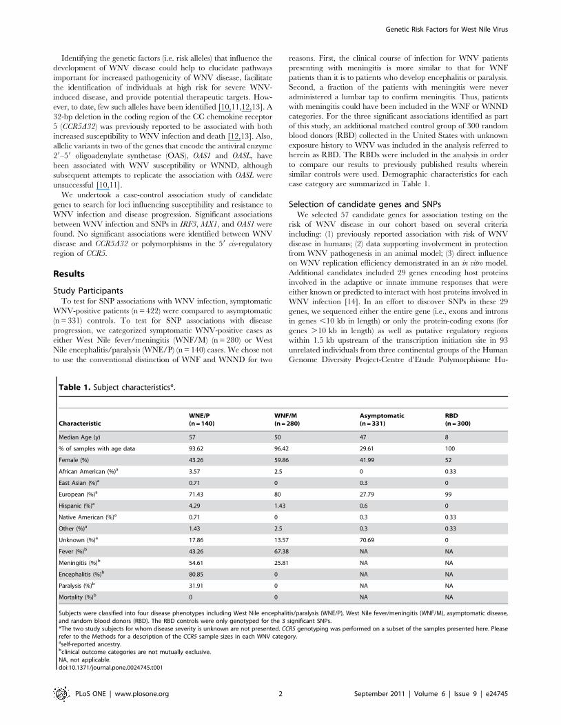
Table 1. Subject characteristics*.
CharacteristicWNE/P(n = 140)
WNF/M(n = 280)
Asymptomatic(n = 331)
RBD(n = 300)
Median Age (y) 57 50 47 8
% of samples with age data 93.62 96.42 29.61 100
Female (%) 43.26 59.86 41.99 52
African American (%)a 3.57 2.5 0 0.33
East Asian (%)a 0.71 0 0.3 0
European (%)a 71.43 80 27.79 99
Hispanic (%)a 4.29 1.43 0.6 0
Native American (%)a 0.71 0 0.3 0.33
Other (%)a 1.43 2.5 0.3 0.33
Unknown (%)a 17.86 13.57 70.69 0
Fever (%)b 43.26 67.38 NA NA
Meningitis (%)b 54.61 25.81 NA NA
Encephalitis (%)b 80.85 0 NA NA
Paralysis (%)b 31.91 0 NA NA
Mortality (%)b 0 0 NA NA
Subjects were classified into four disease phenotypes including West Nile encephalitis/paralysis (WNE/P), West Nile fever/meningitis (WNF/M), asymptomatic disease,and random blood donors (RBD). The RBD controls were only genotyped for the 3 significant SNPs.*The two study subjects for whom disease severity is unknown are not presented. CCR5 genotyping was performed on a subset of the samples presented here. Pleaserefer to the Methods for a description of the CCR5 sample sizes in each WNV category.aself-reported ancestry.bclinical outcome categories are not mutually exclusive.NA, not applicable.doi:10.1371/journal.pone.0024745.t001
Genetic Risk Factors for West Nile Virus
PLoS ONE | www.plosone.org 2 September 2011 | Volume 6 | Issue 9 | e24745
main (HGDP-CEPH). These individuals included 31 sub-Saharan
Africans (Bantu, Mandenka), 31 Europeans (Bergamo, Russian,
French, Orcadian), and 31 East Asians (Cambodian, Dai, Daur,
Han, Hezhen, Japanese, Lahu, Maiozu).
For each candidate gene, haplotypes were inferred for each
continental population and SNPs that tagged each haplotype with
a frequency .5% in Europeans were selected to be genotyped in
the entire cohort. Previously described functional SNPs or newly
identified SNPs that were predicted to have functional conse-
quences were also selected for genotyping. In total, 384 SNPs were
genotyped using an Illumina custom oligo pool assay (OPA) (Table
S1). In addition, we sequenced the 59 cis-regulatory region of CCR5
and genotyped the CCR5D32 deletion in each individual
Analysis of symptomatic WNV infectionTo test for SNPs associated with symptomatic WNV infection,
all WNV-positive cases (i.e., WNF/M and WNE/P cases) were
compared with asymptomatic, WNV-positive blood donors
(Tables S2–S8). A genotypic contingency analysis (362) revealed
a statistically significant association between rs2304207 in intron 2
of IRF3 (p = 0.0399) and symptomatic WNV disease (Table S2).
None of the other associations reached statistical significance. We
also considered dominant, recessive, and additive genetic models
(Tables S3–S5). Under a dominant model, IRF3 rs2304207 was
significantly associated with symptomatic WNV infection
(OR = 0.52, p = 0.007) (Table 2 and Figure 1A). The GC and
GG genotypes were more frequent in asymptomatic WNV-
positive controls than in symptomatic cases. Using a recessive
model, MX1 rs7280422 was significantly associated with symp-
tomatic infection (OR = 0.25, p = 0.042) (Table 2 and Figure 1A).
Specifically, the reference homozygote genotype, CC, was more
common in asymptomatic WNV-positive controls than in
symptomatic cases.
Logistic regression, controlled for population stratification, was
performed for each SNP separately and recapitulated the results of
the contingency table analysis (Tables S6–S8). IRF3 rs2304207
was significantly associated with symptomatic WNV disease
(OR = 0.54 [95% confidence interval (CI) 0.39–0.74]; p = 0.035)
under a dominant genotypic model (Table 3) with the GC and GG
genotypes found significantly more often in asymptomatic WNV-
positive controls than in WNV symptomatic cases. Under a
recessive model, MX1 rs7280422 was associated with symptomatic
infection (OR = 0.19 [95% CI 0.08–0.42], p = 0.014) with the
homozygous reference allele CC found significantly more often in
asymptomatic WNV-positive controls than in WNV symptomat-
ically infected individuals (Table 3 and Figure 1B). No significant
association with WNV infection was identified using an additive
model (Table S6).
Asymptomatic WNV-positive individuals were used as controls
rather than individuals with unknown WNV exposure history
because we thought the former enabled a more powerful test (i.e.,
it is more likely that individuals with unknown exposure history
have never been exposed to WNV than it is that some of the
putatively asymptomatic WNV-positive cases really were symp-
tomatic) [14]. Nevertheless, 300 RBD with unknown exposure
history to WNV were genotyped for rs2304207 and rs7280422
and the data compared to that for all symptomatic WNV cases
(i.e., WNF/M and WNE/P). We included this additional control
group so that our results would be directly comparable to studies
conducted previously. No significant associations were detected for
Table 2. Contingency table analysis odds ratios (OR) of significant SNPs associated with WNV symptomatic infection or West Nileencephalitis/paralysis (WNE/P).
Disease ModelInheritanceModel SNP Gene
Symptomatic vs.Asymptomatic
Symptomaticvs. RBD
Symptomatic Dominant rs2304207 IRF3 0.52** 0.97
Infection rs7280422 MX1 0.74 0.84
rs34137742 OAS1 0.77 0.88
Recessive rs2304207 IRF3 0.72 1.32
rs7280422 MX1 0.25* 0.80
rs34137742 OAS1 1.33 1.23
WNE/P vs. WNF/M+Asymptomatic
WNE/Pvs. RBD
WNE/P vs.WNF/M+RBD
WNE/P vs. WNF/M+Asymptomatic+RBD
WNE/P Dominant rs2304207 IRF3 0.70 1.03 1.00 0.78
rs7280422 MX1 1.22 1.14 1.33 1.20
rs34137742 OAS1 0.96 0.97 1.04 0.96
Recessive rs2304207 IRF3 0.92 1.45 1.29 1.05
rs7280422 MX1 0.38 0.81 0.89 0.47
rs34137742 OAS1 5.61** 3.49** 5.38**** 4.67****
Odds ratios (ORs) are reported for the minor allele.aTwo disease models were used and include WNV symptomatic infection and West Nile encephalitis/paralysis (WNE/P). To identify associations with symptomaticinfection in our cohort, symptomatic WNV was compared to asymptomatic infection. To identify associations with WNE/P in our cohort, we compared WNE/P to WestNile fever/meningitis (WNF/M) combined with asymptomatic infection. To understand if our associations were robust to alterations in the control group definition, wecompared symptomatic infection vs. random blood donors (RBD), WNE/P vs. RBD, WNE/P vs. WNF/M combined with RBD, and WNE/P vs. WNF/M+Asymptomatic+RBD.
*p,0.05,**p,0.01,***p,0.001,****p,0.0001.P-values were corrected for multiple tests using the Bonferroni Correction.doi:10.1371/journal.pone.0024745.t002
Genetic Risk Factors for West Nile Virus
PLoS ONE | www.plosone.org 3 September 2011 | Volume 6 | Issue 9 | e24745
IRF3 (dominant: OR = 0.97 [95% CI 0.69–1.35] p = 0.836) or
MX1 (recessive: OR = 0.80 [95% CI 0.31–2.11] p = 0.655) SNPs
(Table 2).
Analysis of WNV Disease ProgressionTo identify SNP associations with WNV disease progression
(defined here as the presence of WNE/P), WNE/P cases were
compared to a control group consisting of WNF/M individuals
plus asymptomatic, WNV-positive blood donors. A genotypic
contingency analysis (362) revealed a statistically significant
association between rs34137742 in OAS1 (p = 0.012) and risk of
WNE/P (Table S9). In addition, dominant, recessive, and additive
genetic models were considered (Tables S10–S12). Under a
recessive model, the homozygote non-reference genotype, TT, of
OAS1 rs34137742 was significantly associated with WNE/P
(OR = 5.61, p = 0.006) (Table 2 and Figure 1A). Using logistic
regression, this result was recapitulated under a recessive genotypic
model (OR = 9.79, [95% CI 3.60–26.61], p = 0.003) (Table 3,
Figure 1B, and Table S15). No significant associations with WNE/
P were identified using a dominant or an additive model of
inheritance (Tables S13 and S14).
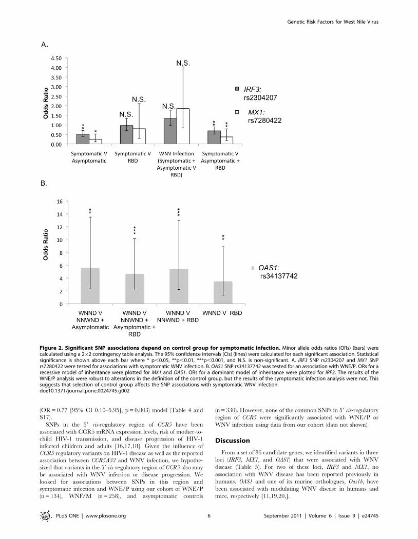
The association between OAS1 rs34137742 and WNE/P was
robust to alterations in the definition of the control group as shown
by comparing the following three donor groups: 1) WNE/P vs.
RBD 2) WNE/P vs. WNF/M and RBD and 3) WNE/P vs. WNF/
M, asymptomatic controls, and RBD. Significant associations were
detected between WNE/P vs. RBD (OR = 3.49 [95% CI 1.36–
8.90] p = 0.0057), WNE/P vs. WNF/M and RBD (OR = 5.38
[95% CI 2.23–12.95] p = 0.000033), and WNE/P vs. WNF/M,
asymptomatic controls, and RBD combined (OR = 4.67 [95% CI
2.15–10.13] p = 0.00002) (Table 2; Figure 2b). In each comparison,
the TT genotype of OAS1 rs34137742 was significantly associated
with WNE/P (Table 2). We also tested if the significant association
we detected for this SNP and WNE/P was identified when using the
conventional WNV phenotype classification. For this analysis, we
compared WNND cases to a control group composed of individuals
with WNF and asymptomatic, WNV-positive blood donors. All
WNV patients reporting meningitis were included in the WNND
group. We did not detect an association between OAS1 rs34137742
and WNND using a dominant (logistic regression OR = 0.83 [95%
CI 0.58–1.21], p = 114.78) or a recessive (logistic regression
OR = 3.66 [95% CI 1.52–8.84], p = 1.29) model of inheritance
after correcting for multiple tests.
Analysis of variants previously associated with WNVdisease
SNPs in OAS1 rs10774671 and OASL rs3213545 previously have
been associated with increased risk of WNV infection (i.e., WNV-
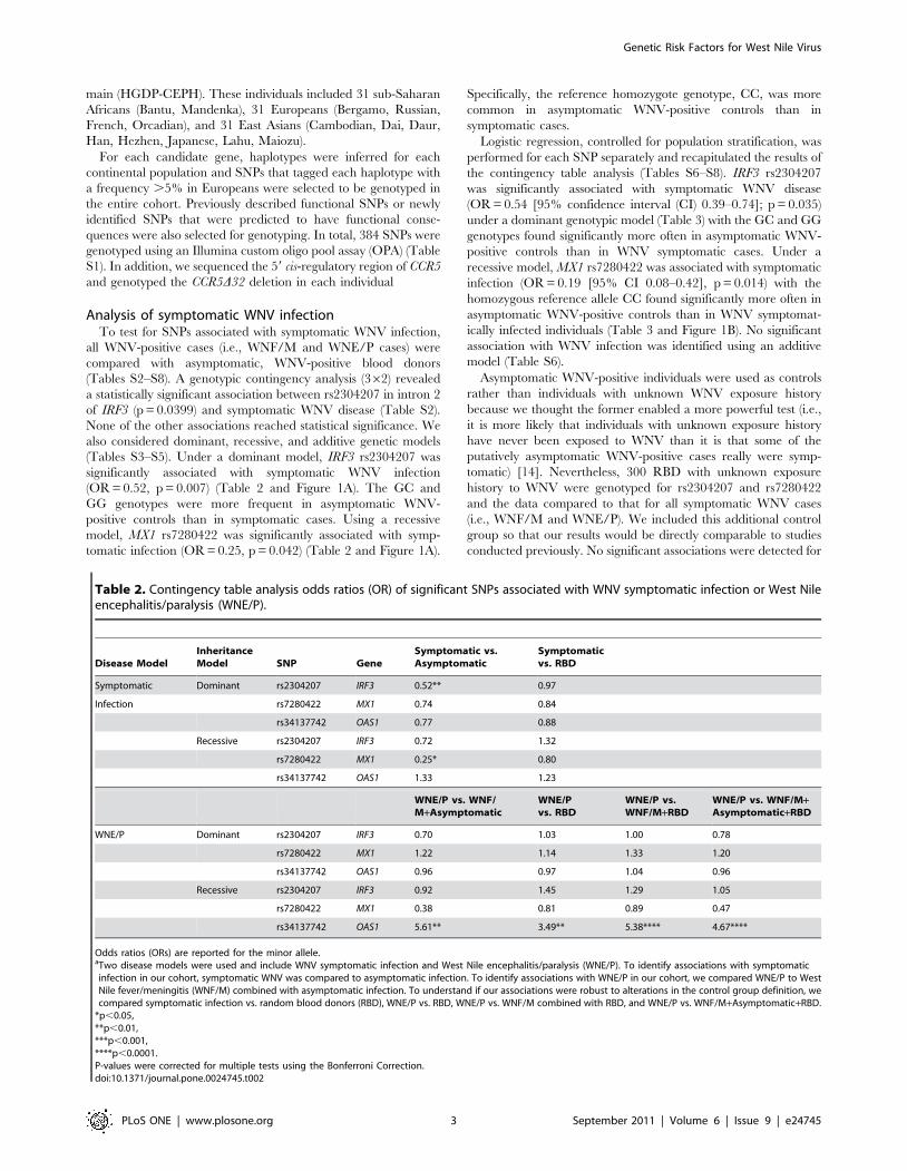
Figure 1. Significant SNPs associated with symptomatic WNV infection or West Nile encephalitis/paralysis (WNE/P). Three SNPs weresignificantly associated with symptomatic WNV infection and WNE/P. These include rs2304207 in IRF3 for symptomatic cases vs. asymptomaticcontrols for a dominant inheritance model, rs7280422 in MX1 for symptomatic cases vs. asymptomatic controls for a recessive inheritance model andrs34137742 in OAS1 for WNE/P vs. West Nile fever/meningitis (WNF/M) using a recessive inheritance model. Contingency table results are presentedin A whereas results for the logistic regression are shown in B. The horizontal axis for each plot shows the genomic position arranged in order ofchromosomal position for each SNP in the dataset. The vertical axis depicts the negative log p-values generated from the chi-square distribution orthe logistic regression model analysis. All p-values were corrected for multiple tests using the Bonferroni correction. The black dashed line indicatesthe five percent significance threshold corrected for multiple tests. No significant SNP associations were identified for WNE/P versus WNF/M for adominant inheritance model.doi:10.1371/journal.pone.0024745.g001
Genetic Risk Factors for West Nile Virus
PLoS ONE | www.plosone.org 4 September 2011 | Volume 6 | Issue 9 | e24745
positive status) and symptomatic WNV infection, respectively
[10,11]. In our cohort, we found no evidence of a significant
association with symptomatic infection for OAS1 SNP rs10774671
using a dominant model (OR 1.21, p = 65.31), or a recessive model
(OR 1.30, p = 81.66). Likewise, no significant associations were
detected for OASL SNP rs3213545 using a dominant model (OR
0.78, p = 34.00) or recessive model (OR 0.52, p = 9.32) (Table 4).
Results of the logistic regression were consistent with the results
of the contingency table analysis (Tables S7, S8). Additionally,
neither SNP was associated with WNE/P (Table 4 and Tables
S9–S15).
Next, we attempted to replicate the previously identified associ-
ations with WNV infection in our cohort by comparing all WNV-
positive cases (WNE/P, WNF/M, and asymptomatic cases)
(n = 751 OAS1 rs10774671 or n = 749 OASL rs3213545) to the
WNV-negative controls from Lim et al. (n = 552) [11]. The WNV-
negative controls used by Lim et al. consisted of random blood
donors collected prior to the 1999 introduction of WNV to the
United States (n = 360) and U.S. blood donors collected by the
American Red Cross who were identified as WNV false positives
(n = 192). OAS1 rs10774671 was not associated with risk of
infection under a dominant (OR = 0.89 [95% CI 0.71–1.11],
p = 0.301) or a recessive (OR = 0.79 [95% CI 0.58–1.09],
p = 0.150) model. In contrast, OASL rs3213545 was significantly
associated with WNV infection under a recessive (OR = 0.63 [95%
CI 0.42–1.58], p = 0.025) model of inheritance (Table 4).
Interestingly, Lim et al. did not find an association between OASL
rs3213545 and WNV infection, even though we did find an
association. Thus, we could replicate neither of the results
obtained by Lim et al. [11].
We repeated the tests of association between OAS1 rs10774671
and OASL rs3213545 and individuals with symptomatic WNV
infection (WNE/P and WNF/M) or WNE/P using the Lim et al.
WNV-negative controls for comparison [11]. Once again,
contingency table analysis revealed no association for symptomatic
infection and OAS1 rs10774671 genotype using a dominant
(OR = 0.97 [95% CI 0.75–1.26], p = 0.81) or a recessive model
(OR = 0.89 [95% CI 0.62–1.27], p = 0.51) (Table 4). Likewise, no
association for WNE/P and OAS1 rs10774671 was detected
(dominant: OR = 1.03 [95% CI 0.6–1.50], p = 0.891, recessive:
OR = 1.25 [95% CI 0.77–2.02], p = 0.372) (Table 4). OASL
rs3213545 was significantly associated with both symptomatic
infection (dominant: OR = 0.72 [95% CI 0.56–0.93], p = 0.012,
recessive: OR = 0.45 [95% CI 0.27–0.77], p = 0.003) and WNE/P
(dominant: OR = 0.56 [95% CI 0.38–0.81], p = 0.002, recessive:
OR = 0.20 [95% CI 0.06–0.65], p = 0.003) (Table 4).
CCR5D32 has been associated previously with an increased risk
of symptomatic WNV infection [12,13,15]. We tested whether
CCR5D32 was associated with risk of symptomatic WNV infection
or disease progression in our cohort of WNE/P, WNF/M, and
asymptomatic WNV-positive cases. No significant associations
were detected for symptomatic WNV infection or WNE/P, under
either a dominant or recessive model of inheritance, by
contingency analysis or logistic regression analysis (Table 4 and
Table S16). Next, to test whether CCR5D32 was associated with
risk of WNV infection, we compared all WNV-positive cases (i.e.
WNE/P, WNF/M, and asymptomatic cases) in our cohort
(n = 713) to published genotype data from the Glass et al. WNV
sero-negative cohort presenting with acute illness, but in whom
WNV had been ruled out by serological testing (n = 1,318) [13]. A
significant association was identified for risk of WNV infection
under a dominant model (OR = 1.36 [95% CI 1.08–1.71],
p = 0.009) (Table 4). We also tested for risk of symptomatic
WNV infection and WNE/P using the Glass et al. WNV sero-
negative controls rather than WNV-positive asymptomatic blood
donors [13]. CCR5D32 was significantly associated with symp-
tomatic infection under a dominant model of inheritance
(OR = 1.38 [95% CI 1.04–1.83], p = 0.026) (Table 4 and Table
S17). No association was identified with WNE/P using a dominant
(OR = 1.45 [95% CI 0.94–2.25], p = 0.094) or recessive
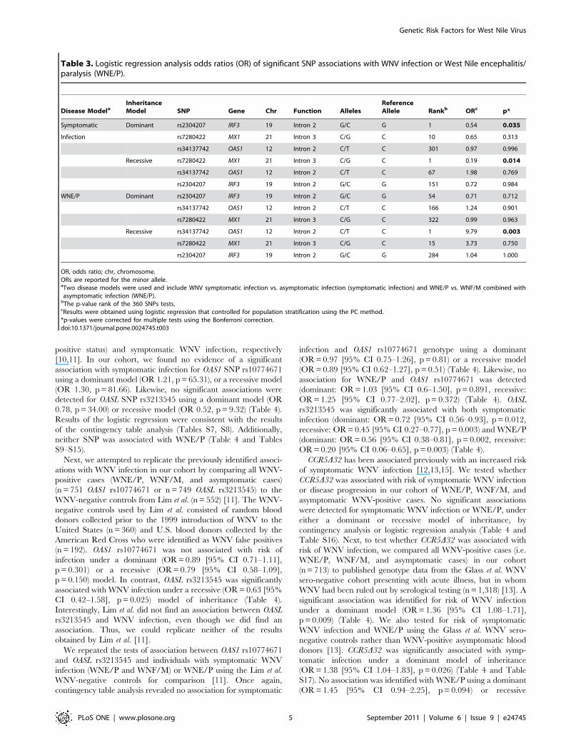
Table 3. Logistic regression analysis odds ratios (OR) of significant SNP associations with WNV infection or West Nile encephalitis/paralysis (WNE/P).
Disease ModelaInheritanceModel SNP Gene Chr Function Alleles
ReferenceAllele Rankb ORc p*
Symptomatic Dominant rs2304207 IRF3 19 Intron 2 G/C G 1 0.54 0.035
Infection rs7280422 MX1 21 Intron 3 C/G C 10 0.65 0.313
rs34137742 OAS1 12 Intron 2 C/T C 301 0.97 0.996
Recessive rs7280422 MX1 21 Intron 3 C/G C 1 0.19 0.014
rs34137742 OAS1 12 Intron 2 C/T C 67 1.98 0.769
rs2304207 IRF3 19 Intron 2 G/C G 151 0.72 0.984
WNE/P Dominant rs2304207 IRF3 19 Intron 2 G/C G 54 0.71 0.712
rs34137742 OAS1 12 Intron 2 C/T C 166 1.24 0.901
rs7280422 MX1 21 Intron 3 C/G C 322 0.99 0.963
Recessive rs34137742 OAS1 12 Intron 2 C/T C 1 9.79 0.003
rs7280422 MX1 21 Intron 3 C/G C 15 3.73 0.750
rs2304207 IRF3 19 Intron 2 G/C G 284 1.04 1.000
OR, odds ratio; chr, chromosome.ORs are reported for the minor allele.aTwo disease models were used and include WNV symptomatic infection vs. asymptomatic infection (symptomatic infection) and WNE/P vs. WNF/M combined withasymptomatic infection (WNE/P).
bThe p-value rank of the 360 SNPs tests.cResults were obtained using logistic regression that controlled for population stratification using the PC method.*p-values were corrected for multiple tests using the Bonferroni correction.doi:10.1371/journal.pone.0024745.t003
Genetic Risk Factors for West Nile Virus
PLoS ONE | www.plosone.org 5 September 2011 | Volume 6 | Issue 9 | e24745
(OR = 0.77 [95% CI 0.10–5.95], p = 0.803) model (Table 4 and
S17).
SNPs in the 59 cis-regulatory region of CCR5 have been
associated with CCR5 mRNA expression levels, risk of mother-to-
child HIV-1 transmission, and disease progression of HIV-1
infected children and adults [16,17,18]. Given the influence of
CCR5 regulatory variants on HIV-1 disease as well as the reported
association between CCR5D32 and WNV infection, we hypothe-
sized that variants in the 59 cis-regulatory region of CCR5 also may
be associated with WNV infection or disease progression. We
looked for associations between SNPs in this region and
symptomatic infection and WNE/P using our cohort of WNE/P
(n = 134), WNF/M (n = 258), and asymptomatic controls
(n = 330). However, none of the common SNPs in 59 cis-regulatory
region of CCR5 were significantly associated with WNE/P or
WNV infection using data from our cohort (data not shown).
Discussion
From a set of 86 candidate genes, we identified variants in three
loci (IRF3, MX1, and OAS1) that were associated with WNV
disease (Table 5). For two of these loci, IRF3 and MX1, no
association with WNV disease has been reported previously in
humans. OAS1 and one of its murine orthologues, Oas1b, have
been associated with modulating WNV disease in humans and
mice, respectively [11,19,20,].
Figure 2. Significant SNP associations depend on control group for symptomatic infection. Minor allele odds ratios (ORs) (bars) werecalculated using a 262 contingency table analysis. The 95% confidence intervals (CIs) (lines) were calculated for each significant association. Statisticalsignificance is shown above each bar where * p,0.05, **p,0.01, ***p,0.001, and N.S. is non-significant. A. IRF3 SNP rs2304207 and MX1 SNPrs7280422 were tested for associations with symptomatic WNV infection. B. OAS1 SNP rs34137742 was tested for an association with WNE/P. ORs for arecessive model of inheritance were plotted for MX1 and OAS1. ORs for a dominant model of inheritance were plotted for IRF3. The results of theWNE/P analysis were robust to alterations in the definition of the control group, but the results of the symptomatic infection analysis were not. Thissuggests that selection of control group affects the SNP associations with symptomatic WNV infection.doi:10.1371/journal.pone.0024745.g002
Genetic Risk Factors for West Nile Virus
PLoS ONE | www.plosone.org 6 September 2011 | Volume 6 | Issue 9 | e24745
IRF3 encodes a member of the interferon regulatory transcrip-
tion factor family involved in the upregulation of type 1 IFN genes
as well as other pathway genes. However, IRF-3 has been reported
to protect mice from WNV-induced disease by both interferon-
dependent and independent mechanisms [21,22]. After a
peripheral WNV infection, Irf32/2 mice exhibited increased
mortality, earlier viral entry into the CNS, and increased virus
levels in the brain and spinal cord compared to wild-type mice
[22]. Irf32/2 mice also exhibit enhanced WNV infection in
macrophages in vivo and ex vivo [22]. Increased replication of WNV
in macrophages in the periphery would be expected to enhance
disease symptoms consistent with the observed association of IRF3
variation with human symptomatic WNV infections. Accordingly,
IRF3 is a compelling candidate for influencing the risk of
symptomatic WNV infection in humans.
MX1 belongs to the MX (myxovirus resistance) family of
interferon-induced proteins that are GTPases with antiviral
functions [23]. Upon viral infection, a host cell secretes type 1
interferons that, in turn, induce the production of MX proteins
that diminish viral replication. In mice, Mx1 confers resistance to
orthomyxoviruses including influenza viruses, but has not been
demonstrated to confer resistance to flaviviruses [24]. However, it
is possible that MX1 may have an effect on flavivirus infections in
humans.
The products of OAS1, OAS2, OAS3, OASL, and their
downstream effector RNASEL each influence host defense by
blocking viral replication [23]. Evidence from human cell culture
indicates that OAS gene products have an antiviral effect on
flavivirus infections [25]. The products of the Oas1b alleles
differentially affect susceptibility to flavivirus-induced disease in
mice, but by an RNase L independent mechanism [19,20]. In the
present study, a single variant in OAS1, SNP rs34137742 located in
intron 2, was identified as a risk factor for human WNV disease
progression, although no association with symptomatic infection as
a whole was identified.
OAS1 rs10774671 was previously reported to be associated with
risk of WNV infection [11]; however, we were unable to replicate
this finding using our asymptomatic controls or the control
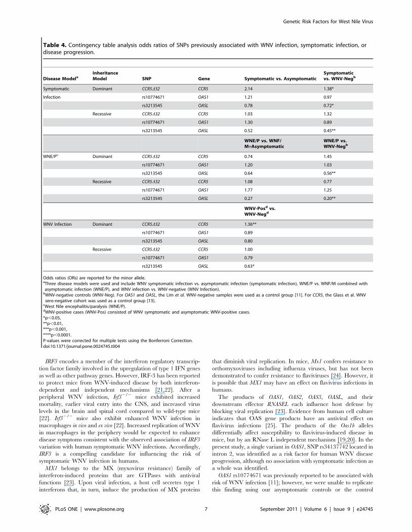
Table 4. Contingency table analysis odds ratios of SNPs previously associated with WNV infection, symptomatic infection, ordisease progression.
Disease ModelaInheritanceModel SNP Gene Symptomatic vs. Asymptomatic
Symptomaticvs. WNV-Negb
Symptomatic Dominant CCR5D32 CCR5 2.14 1.38*
Infection rs10774671 OAS1 1.21 0.97
rs3213545 OASL 0.78 0.72*
Recessive CCR5D32 CCR5 1.03 1.32
rs10774671 OAS1 1.30 0.89
rs3213545 OASL 0.52 0.45**
WNE/P vs. WNF/M+Asymptomatic
WNE/P vs.WNV-Negb
WNE/Pc Dominant CCR5D32 CCR5 0.74 1.45
rs10774671 OAS1 1.20 1.03
rs3213545 OASL 0.64 0.56**
Recessive CCR5D32 CCR5 1.08 0.77
rs10774671 OAS1 1.77 1.25
rs3213545 OASL 0.27 0.20**
WNV-Posd vs.WNV-Negd
WNV Infection Dominant CCR5D32 CCR5 1.36**
rs10774671 OAS1 0.89
rs3213545 OASL 0.80
Recessive CCR5D32 CCR5 1.00
rs10774671 OAS1 0.79
rs3213545 OASL 0.63*
Odds ratios (ORs) are reported for the minor allele.aThree disease models were used and include WNV symptomatic infection vs. asymptomatic infection (symptomatic infection), WNE/P vs. WNF/M combined withasymptomatic infection (WNE/P), and WNV infection vs. WNV-negative (WNV Infection).
bWNV-negative controls (WNV-Neg). For OAS1 and OASL, the Lim et al. WNV-negative samples were used as a control group [11]. For CCR5, the Glass et al. WNVsero-negative cohort was used as a control group [13].
cWest Nile encephalitis/paralysis (WNE/P).dWNV-positive cases (WNV-Pos) consisted of WNV symptomatic and asymptomatic WNV-positive cases.*p,0.05,**p,0.01,***p,0.001,****p,0.0001.P-values were corrected for multiple tests using the Bonferroni Correction.doi:10.1371/journal.pone.0024745.t004
Genetic Risk Factors for West Nile Virus
PLoS ONE | www.plosone.org 7 September 2011 | Volume 6 | Issue 9 | e24745
samples of Lim et al. [11]. In contrast, an association between
another SNP, OAS1 rs34137742, and risk for WNE/P was found
using multiple, different control populations (1. WNF/M plus
asymptomatic WNV-positive cases, 2. RBD, 3. WNF/M plus
RBD, and 4. WNF/M plus asymptomatic WNV-positive cases
and RBD). Furthermore, we found an association between OASL
rs3213545 and risk for WNE/P, symptomatic disease, and
infection, but only with the control samples of Lim et al. [11].
The observation that OASL is associated with WNV disease
confirms a previously reported association discovered in a very
small sample of cases (n = 27) that failed replication in a second
study using the same control samples as included here, but
different WNV-positive samples [10,11].
We did not use the conventional phenotypic classification
method of WNND and WNF. Instead, we grouped West Nile
patients presenting with fever and/or meningitis into a single
category, WNF/M, and categorized patients presenting with
encephalitis and/or paralysis as WNE/P. Our decision was based
on two observations. First, WNV patients presenting with
meningitis are clinically more similar to WNF patients than they
are to WNE/P patients. Second, patients classified as WNF are
likely to include individuals with unconfirmed meningitis [26].
Therefore, patients with meningitis may be included in both the
WNF and WNND categories. When removing West Nile
meningitis patients from the analysis, the association of OAS1
rs34137742 with WNE/P remained significant, but our power to
detect this association decreased. However, if WNND patients
were compared to WNF plus asymptomatic controls, no
association with OAS1 rs34137742 and WNND was detected
(Table S20). These results illustrate the importance of accurate
phenotypic classification, as the associations are contingent upon
the classification system. The difference in our phenotypic classifi-
cation of WNV-positive samples compared to that used by Lim
et al. could explain why we detected an association between OASL
rs3213545 and risk for WNE/P whereas the Lim et al. study did
not detect an association between this SNP and WNND [11].
However, this difference in the classification system cannot explain
our inability to replicate the association for OAS1 rs10774671 and
WNV infection, as this analysis compared all WNV-positive cases
to WNV-negative controls.
It has been shown that the host response to viral infections does
not depend on host factors alone. Rather, the virulence of the
infecting virus strain may impact the clinical course of infection and
could therefore influence the likelihood of disease progression from
asymptomatic infection to more severe forms of a disease. For
example, particular strains of Dengue virus have been shown to be
more virulent than others [27]. Analysis of the specific WNV viral
strains infecting each of the participants in this study was beyond the
scope of this research. However, it is important to point out that
varying degrees of virulence between infecting strains may have
impacted WNV pathogenesis in our cohort. Furthermore, differ-
ences in strain virulence may also account for the discrepant results
obtained in our study compared to previous work.
We searched for loci influencing risk for symptomatic WNV
disease by comparing symptomatic WNV cases to asymptomatic,
WNV-positive individuals. This approach likely is more robust
than using random blood donors with unknown WNV exposure
history or WNV-negative individuals as controls. Groups of RBDs
with an unknown WNV exposure are likely to include previously
uninfected individuals with the potential to develop either WNF/
M or WNE/P, and therefore may reduce the power to detect a
difference between groups. By considering only asymptomatic,
WNV-positive blood donors, the probability of including individ-
uals who might develop WNF/M or WNE/P was eliminated. This
strategy could explain the observation that the significant
associations between IRF3 or MX1 and WNV disease were not
replicated when RBD of unknown WNV infection status were
used for comparison. Similarly, using WNV-negative controls
rather than WNV-positive asymptomatic controls could confound
risk factors for infection and symptomatic disease. This could
explain, in part, our ability to replicate the association between
CCR5D32 and symptomatic infection reported by Glass et al. when
we used sero-negative controls but not when WNV-positive
asymptomatic blood donors were used as a control group [13].
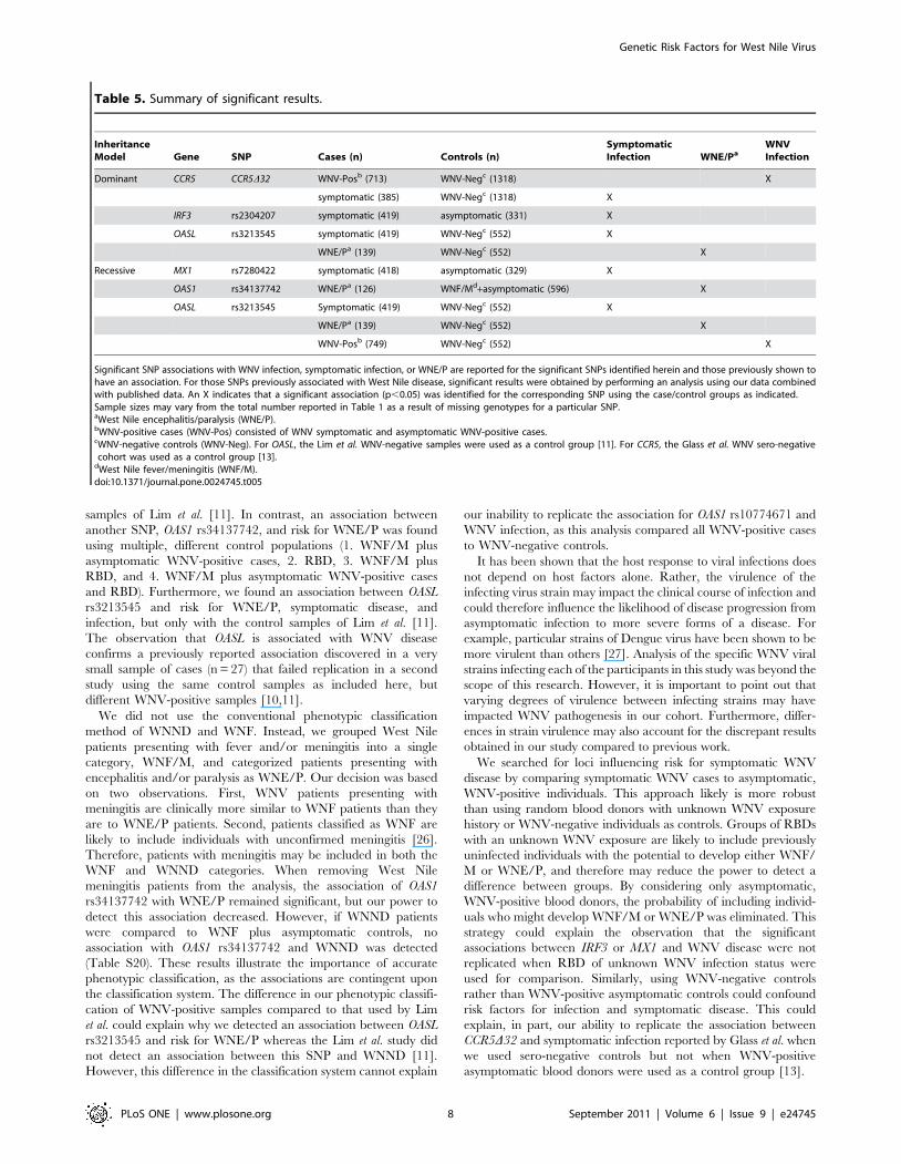
Table 5. Summary of significant results.
InheritanceModel Gene SNP Cases (n) Controls (n)
SymptomaticInfection WNE/Pa
WNVInfection
Dominant CCR5 CCR5D32 WNV-Posb (713) WNV-Negc (1318) X
symptomatic (385) WNV-Negc (1318) X
IRF3 rs2304207 symptomatic (419) asymptomatic (331) X
OASL rs3213545 symptomatic (419) WNV-Negc (552) X
WNE/Pa (139) WNV-Negc (552) X
Recessive MX1 rs7280422 symptomatic (418) asymptomatic (329) X
OAS1 rs34137742 WNE/Pa (126) WNF/Md+asymptomatic (596) X
OASL rs3213545 Symptomatic (419) WNV-Negc (552) X
WNE/Pa (139) WNV-Negc (552) X
WNV-Posb (749) WNV-Negc (552) X
Significant SNP associations with WNV infection, symptomatic infection, or WNE/P are reported for the significant SNPs identified herein and those previously shown tohave an association. For those SNPs previously associated with West Nile disease, significant results were obtained by performing an analysis using our data combinedwith published data. An X indicates that a significant association (p,0.05) was identified for the corresponding SNP using the case/control groups as indicated.Sample sizes may vary from the total number reported in Table 1 as a result of missing genotypes for a particular SNP.aWest Nile encephalitis/paralysis (WNE/P).bWNV-positive cases (WNV-Pos) consisted of WNV symptomatic and asymptomatic WNV-positive cases.cWNV-negative controls (WNV-Neg). For OASL, the Lim et al. WNV-negative samples were used as a control group [11]. For CCR5, the Glass et al. WNV sero-negativecohort was used as a control group [13].
dWest Nile fever/meningitis (WNF/M).doi:10.1371/journal.pone.0024745.t005
Genetic Risk Factors for West Nile Virus
PLoS ONE | www.plosone.org 8 September 2011 | Volume 6 | Issue 9 | e24745
The composition of the asymptomatic control cohort also
appeared to influence the power to detect variants associated with
risk for WNE/P. For example, inclusion of RBD in the control
group with WNF/M or WNF/M and asymptomatic infection
diminished the ORs and the level of significance of the association
between OAS1 rs34137742 and WNE/P. As a corollary, since
some of the asymptomatic individuals may have had mild
symptoms [28], comparing both asymptomatic WNV-positive
cases combined with WNF/M would mitigate errors introduced
by the misclassification of asymptomatic cases.
Age is a known independent risk factor for WNV disease
progression with elderly patients at the highest risk for encephalitis
and death [7,8,29,30,31]. Ages were not available for many of the
asymptomatic WNV-positive blood donors (233 out of 331) so that
we were unable to control for age as a covariate. To test whether
the analyses of WNV disease progression were robust to
confounding by age, the effect of age was tested by comparing
the results of a logistic regression using the subset of cases for
which age information was available to those obtained for the
entire set of samples. Age was available for 94% of WNE/P cases,
96% of WNF/M cases, and 30% of asymptomatic cases. The
results were virtually the same, suggesting that age is not a major
confounder of our analyses. Furthermore, in order for age to
confound the analysis, it would have to be associated with the
SNPs tested here. This could happen via settlement of different
ethnic groups over the relevant time period in the regions from
which cases and controls were collected. However, adjusting for
ethnicity eliminated this potential issue.
In summary, we have identified two novel loci, IRF3 and MX1,
associated with risk of WNV disease, and a SNP in OAS1
associated with WNE/P. Each of the SNPs responsible for the
observed association signal is located in an intron and none is in
strong linkage disequilibrium with known functional variants in
their respective genes. Each of these loci is involved in either an
interferon regulatory pathway or is an effector of the interferon
response. Accordingly, these findings provide further evidence,
albeit indirect, that the interferon pathway MAY play an
important role in modulating human WNV disease, influencing
both the risk of symptomatic infection and disease progression.
Methods
Study ParticipantsSamples from symptomatic WNV-positive patients (n = 422)
were collected from the Southeast, Midwest, Northeast, South-
west, Intermountain West, and West coast regions of the United
States. These cases represented individuals who sought medical
attention, had signs consistent with WNV disease (e.g., fever,
meningitis, encephalitis), and who were subsequently confirmed by
serological testing to have been infected with WNV. Data for case
classification were collected by review of medical records and in
some cases interviews with patients. A WNF/M case (n = 280) was
defined as an individual who had weakness, headache, acute fever,
stiff neck, rash, and/or cerebrospinal fluid (CSF) pleocytosis
consistent with meningitis. A WNE/P case (n = 140) was defined
as an individual who experienced encephalitis or paralysis.
Encephalitis was defined as fever, headache, and altered mental
status ranging from confusion to coma. There were no recorded
deaths among cases in our cohort. For two symptomatic cases, the
phenotypic data were incomplete and further classification was not
possible. WNV nucleic acid positive blood samples (n = 331) were
collected in the Northeast, Midwest, Southwest, Intermountain
West, and West coast regions of the United States and in Canada
by Blood Systems and by a private blood bank in Colorado. Fifty-
seven percent of these individuals self-reported no symptoms and
16 percent reported only one symptom (i.e. headache, body aches,
or skin rash) in the week prior to donation [28]. However, only the
odds of reporting skin rash were significantly higher for confirmed
WNV-positive blood donors compared to uninfected donors.
All subjects and/or their legal guardians provided written
informed consent and the study was approved by the Institutional
Review Boards at Seattle Children’s Hospital, the University of
Washington, the University of Utah, Georgia State University, the
University of Texas Health Sciences Center at Houston, Blood
Systems, the Centers for Disease Control and Prevention, and the
University of Massachusetts Medical School.
DNA Isolation and GenotypingDNA was isolated from 1–20 ml venous blood or white blood
cells using Qiagen’s (Valencia, CA) Puregene DNA purification
system according to the manufacturer’s instructions. Candidate
SNPs, both functional and/or haplotype tagging in Caucasians,
were selected from 86 genes that have been associated with WNV
infections in an animal model or cell culture. In total, 384 SNPs
were genotyped using an Illumina custom oligo pool assay (OPA)
(Table S1). Twenty-four SNPs failed initial quality control filters
and were removed from all downstream analysis. Association
analyses were performed on 360 SNPs distributed across 86 genes.
Of these SNPs, three deviated from Hardy-Weinberg equilibrium
(HWE), but were not significantly associated with WNV infection
or disease progression. A sex check was performed for all patients
by comparing the reported sex in the phenotypic database to the
genotypic sex. Genotypic sex was determined from the heterozy-
gosity estimates of the X chromosome SNPs genotyped using the
Illumina custom OPA. Discrepant sex values existed for seven
study participants, and they were excluded from the analysis.
CCR5D32 was typed manually in our cohort of symptomatic
(n = 394) and asymptomatic WNV-positive cases (n = 342) using
primers flanking the 32-bp deletion. Fragments were resolved on a
2% agarose gel and visualized with ethidium bromide staining. Of
the 736 samples genotyped for CCR5D32, six samples did not
produce reliable genotypes and seven samples failed a sex check
performed using the Illumina OPA data. An additional 10 samples
lacked the genotype data needed to control for population
stratification in the logistic regression. After removal of these 23
samples, 713 and 712 samples were included in the association
analysis for symptomatic WNV infection and WNE/P, respec-
tively. For a single sample, phenotypic information on disease
severity was not available. Therefore, this case was omitted from
the analysis of disease progression. The analysis of symptomatic
WNV disease compared 385 WNV symptomatic cases to 328
asymptomatic WNV-positive controls. The analysis of WNE/P
compared 131 WNE/P cases to controls consisting of 581 WNF/
M cases combined with asymptomatic WNV-positive blood
donors.
To assess variation in the 59 cis-regulatory region of CCR5, a
1.2 kb fragment was PCR-amplified using a pair of primers that
captured exons 1 and 2 and ,1 kb of sequence upstream and
downstream of each. DNA sequencing was performed using
internal primers on an ABI 3130 automated sequencer. Sequence
trace files were aligned and genotype calls were made using Codon
Code (Dedham, MA). Polymorphic sites were confirmed by visual
inspection of the traces. All SNPs were in HWE (p$0.05).
Association AnalysisTo test whether variants were associated with symptomatic
WNV infection, WNE/P and WNF/M cases were compared to
asymptomatic, WNV-positive blood donors. To test whether
Genetic Risk Factors for West Nile Virus
PLoS ONE | www.plosone.org 9 September 2011 | Volume 6 | Issue 9 | e24745
variants were associated with WNE/P, WNE/P cases were
compared to WNF/M cases and asymptomatic WNV-positive
blood donors. Single nucleotide polymorphism (SNP) analysis and
genetic model assessment were performed using PLINK version
1.06 (http://pngu.mgh.harvard.edu/purcell/plink/) [32]. ORs
and 95% CIs were calculated using both contingency tables and
logistic regression. P-values were corrected for multiple tests using
a Bonferroni correction for 337 (autosomal chromosome SNPs) or
360 (autosomal and X chromosome SNPs) tests. Dominant,
recessive, genotypic, additive, and allelic genetic models of
inheritance were considered. For each variant tested, the odds
ratios (OR), p-values, and 95% confidence intervals (CIs)
(unadjusted for multiple tests) for each model of inheritance are
reported in Tables S2–S15 and S18, S19. For all models, we
define D as the minor allele and d as the major allele following the
notation of PLINK [32]. The minor allele was defined based on
the allele frequencies in this dataset. ORs for the minor allele were
calculated using a dominant model (DD and Dd versus dd), a
recessive model (DD versus Dd and dd), and a genotypic model
(DD versus Dd versus dd). The allelic model compared D versus d.
The additive model tested for the additive effects of allelic dosage.
SNPs on the X chromosome were included in the allelic model
only. For the contingency table analysis, allele and genotype
frequency differences between cases and controls were calculated a
using a 262 or a 263 table that compared the number of expected
and the number of observed individuals in each category. Two
sided p-values were estimated by the chi-square test of significance
with 1 (262 table) or 2 (362 table) degrees of freedom. P-values
were corrected for multiple tests using the Bonferroni correction.
In the logistic regression analysis, we controlled for population
stratification by including the first five principle components (PCs)
as covariates in the model. PCs were estimated from the 360 QC
filtered genotypes using EIGENSTRAT [33]. P-values were
estimated using the Wald z-statistic and corrected for multiple
tests using the Bonferroni correction. To determine if age should
be included as a covariate in the model, logistic regression was
performed on the subset of subjects with reported age both with
and without age included as a covariate in the model. Additionally,
we added an age*SNP interaction effect and an age*age‘2 as age
effects are often non-linear. Age, age*SNP, and age*age‘2 were
not found to be a significant covariates in our analysis and were
left out of the logistic regression analysis. Age data were
unavailable for 70% of WNV-positive blood donors, 6% of
WNE/P cases, and 4% of WNF/M cases.
Supporting Information
Table S1 SNPs included on the Illumina Custom OPA.
(XLS)
Table S2 Symptomatic vs. asymptomatic genotypiccontingency table analysis.
(XLS)
Table S3 Symptomatic vs. asymptomatic contingencytable analysis using a dominant model of inheritance.
(XLS)
Table S4 Symptomatic vs. asymptomatic contingencytable analysis using a recessive model of inheritance.
(XLS)
Table S5 Symptomatic vs. asymptomatic contingencytable analysis using an additive model of inheritance.
(XLS)
Table S6 Symptomatic vs. asymptomatic logistic re-gression analysis results for an additive model ofinheritance.
(XLS)
Table S7 Symptomatic vs. asymptomatic logistic re-gression analysis results for dominant model of inher-itance.
(XLS)
Table S8 Symptomatic vs. asymptomatic logistic re-gression analysis results for a recessive model ofinheritance.
(XLS)
Table S9 WNE/P vs. WNF/M genotypic contingencytable analysis.
(XLS)
Table S10 WNE/P vs. WWF/M contingency table anal-ysis using a dominant model of inheritance.
(XLS)
Table S11 WNE/P vs. WNF/M contingency table anal-ysis using a recessive model of inheritance.
(XLS)
Table S12 WNE/P vs. WNF/M contingency table anal-ysis using an additive model of inheritance.
(XLS)
Table S13 WNE/P vs. WNF/M logistic regressionanalysis results for an additive model of inheritance.
(XLS)
Table S14 WNE/P vs. WNF/M logistic regressionanalysis results for a dominant model of inheritance.
(XLS)
Table S15 WNE/P vs. WNF/M logistic regressionanalysis results for a recessive model of inheritance.
(XLS)
Table S16 CCR5 logistic regression results.
(XLS)
Table S17 Contingency table analysis of variants previ-ously association with WNV disease.
(XLS)
Table S18 Symptomatic vs. asymptomatic allelic con-tingency table analysis.
(XLS)
Table S19 WNE/P vs. WNF/M allelic contingency tableanalysis.
(XLS)
Table S20 OAS1 SNP contingency table analysis usingdifferent meningitis classifications.
(XLS)
Acknowledgments
We thank all the study participants and providers who referred cases. We
would also like to thank Monica Sierra and Liliana Rodriguez for their
assistance with data collection, Ned Hayes, Anthony Marfin and Lyle
Petersen for assistance with sample collection, Valerie Winkelman for
processing and providing blood donor samples, Dr. Brian Custer for
acquiring and compiling blood donor symptom data and Avinash
Karangula and Rajshekhar Sunderraman for database management.
Genetic Risk Factors for West Nile Virus
PLoS ONE | www.plosone.org 10 September 2011 | Volume 6 | Issue 9 | e24745
Author Contributions
Conceived and designed the experiments: MB. Performed the experiments:
KJB KMB HG. Analyzed the data: AWB ME. Contributed reagents/
materials/analysis tools: SH KOM AR JK MAB JJS MPB NMA AAP SG
MJB. Wrote the paper: AWB MB.
References
1. Smithburn KC, Hughes TP, Burke AW, Paul JH (1940) A neurotropic virus
isolated from the blood of a native of Uganda. American Journal of Tropical
Medicine 20: 471–492.2. Russell RC, Dwyer DE (2000) Arboviruses associated with human disease in
Australia. Microbes Infect 2: 1693–1704.3. Tsai TF, Popovici F, Cernescu C, Campbell GL, Nedelcu NI (1998) West Nile
encephalitis epidemic in southeastern Romania. Lancet 352: 767–771.4. Hubalek Z, Halouzka J (1999) West Nile fever–a reemerging mosquito-borne
viral disease in Europe. Emerg Infect Dis 5: 643–650.
5. Lanciotti RS, Roehrig JT, Deubel V, Smith J, Parker M, et al. (1999) Origin ofthe West Nile virus responsible for an outbreak of encephalitis in the
northeastern United States. Science 286: 2333–2337.6. Asnis DS, Conetta R, Teixeira AA, Waldman G, Sampson BA (2000) The West
Nile Virus outbreak of 1999 in New York: the Flushing Hospital experience.
Clin Infect Dis 30: 413–418.7. Asnis DS, Conetta R, Waldman G, Teixeira AA (2001) The West Nile virus
encephalitis outbreak in the United States (1999–2000): from Flushing, NewYork, to beyond its borders. Ann N Y Acad Sci 951: 161–171.
8. Mostashari F, Bunning ML, Kitsutani PT, Singer DA, Nash D, et al. (2001)Epidemic West Nile encephalitis, New York, 1999: results of a household-based
seroepidemiological survey. Lancet 358: 261–264.
9. Emig M, Apple DJ (2004) Severe West Nile virus disease in healthy adults. ClinInfect Dis 38: 289–292.
10. Yakub I, Lillibridge KM, Moran A, Gonzalez OY, Belmont J, et al. (2005)Single nucleotide polymorphisms in genes for 29–59-oligoadenylate synthetase
and RNase L inpatients hospitalized with West Nile virus infection. J Infect Dis
192: 1741–1748.11. Lim JK, Lisco A, McDermott DH, Huynh L, Ward JM, et al. (2009) Genetic
variation in OAS1 is a risk factor for initial infection with West Nile virus inman. PLoS Pathog 5: e1000321.
12. Lim JK, Louie CY, Glaser C, Jean C, Johnson B, et al. (2008) Genetic deficiencyof chemokine receptor CCR5 is a strong risk factor for symptomatic West Nile
virus infection: a meta-analysis of 4 cohorts in the US epidemic. J Infect Dis 197:
262–265.13. Glass WG, McDermott DH, Lim JK, Lekhong S, Yu SF, et al. (2006) CCR5
deficiency increases risk of symptomatic West Nile virus infection. J Exp Med203: 35–40.
14. Krishnan MN, Ng A, Sukumaran B, Gilfoy FD, Uchil PD, et al. (2008) RNA
interference screen for human genes associated with West Nile virus infection.Nature 455: 242–245.
15. Lim JK, McDermott DH, Lisco A, Foster GA, Krysztof D, et al. (2010) CCR5deficiency is a risk factor for early clinical manifestations of West Nile virus
infection but not for viral transmission. J Infect Dis 201: 178–185.16. Mummidi S, Bamshad M, Ahuja SS, Gonzalez E, Feuillet PM, et al. (2000)
Evolution of human and non-human primate CC chemokine receptor 5 gene
and mRNA. Potential roles for haplotype and mRNA diversity, differentialhaplotype-specific transcriptional activity, and altered transcription factor
binding to polymorphic nucleotides in the pathogenesis of HIV-1 and simianimmunodeficiency virus. J Biol Chem 275: 18946–18961.
17. Dean M, Carrington M, Winkler C, Huttley GA, Smith MW, et al. (1996)
Genetic restriction of HIV-1 infection and progression to AIDS by a deletion
allele of the CKR5 structural gene. Hemophilia Growth and DevelopmentStudy, Multicenter AIDS Cohort Study, Multicenter Hemophilia Cohort Study,
San Francisco City Cohort, ALIVE Study. Science 273: 1856–1862.18. Kostrikis LG, Neumann AU, Thomson B, Korber BT, McHardy P, et al. (1999)
A polymorphism in the regulatory region of the CC-chemokine receptor 5 geneinfluences perinatal transmission of human immunodeficiency virus type 1 to
African-American infants. J Virol 73: 10264–10271.
19. Elbahesh H, Jha BK, Silverman RH, Scherbik SV, Brinton MA (2011) The Flvr-encoded murine oligoadenylate synthetase 1b (Oas1b) suppresses 2–5A synthesis
in intact cells. Virology 409: 262–270.20. Scherbik SV, Kluetzman K, Perelygin AA, Brinton MA (2007) Knock-in of the
Oas1b(r) allele into a flavivirus-induced disease susceptible mouse generates the
resistant phenotype. Virology 368: 232–237.21. Fredericksen BL, Smith M, Katze MG, Shi PY, Gale M, Jr. (2004) The host
response to West Nile Virus infection limits viral spread through the activation ofthe interferon regulatory factor 3 pathway. J Virol 78: 7737–7747.
22. Daffis S, Samuel MA, Keller BC, Gale M, Jr., Diamond MS (2007) Cell-specificIRF-3 responses protect against West Nile virus infection by interferon-
dependent and -independent mechanisms. PLoS Pathog 3: e106.
23. Samuel CE (2001) Antiviral actions of interferons. Clin Microbiol Rev 14:778–809, table of contents.
24. Staeheli P, Grob R, Meier E, Sutcliffe JG, Haller O (1988) Influenza virus-susceptible mice carry Mx genes with a large deletion or a nonsense mutation.
Mol Cell Biol 8: 4518–4523.
25. Lin RJ, Yu HP, Chang BL, Tang WC, Liao CL, et al. (2009) Distinct antiviralroles for human 29,59-oligoadenylate synthetase family members against dengue
virus infection. J Immunol 183: 8035–8043.26. Sejvar JJ, Marfin AA (2006) Manifestations of West Nile neuroinvasive disease.
Rev Med Virol 16: 209–224.27. Rico-Hesse R, Harrison LM, Salas RA, Tovar D, Nisalak A, Ramos C, et al.
(1997) Origins of dengue type 2 viruses associated with increased pathogenicity
in the Americas. Virology 230: 244–51.28. Custer B, Kamel H, Kiely NE, Murphy EL, Busch MP (2009) Associations
between West Nile virus infection and symptoms reported by blood donorsidentified through nucleic acid test screening. Transfusion 49: 278–288.
29. Craven RB, Roehrig JT (2001) West Nile virus. Jama 286: 651–653.
30. Nash D, Mostashari F, Fine A, Miller J, O’Leary D, et al. (2001) The outbreak ofWest Nile virus infection in the New York City area in 1999. N Engl J Med 344:
1807–1814.31. Petersen LR, Roehrig JT, Hughes JM (2002) West Nile virus encephalitis.
N Engl J Med 347: 1225–1226.32. Purcell S, Neale B, Todd-Brown K, Thomas L, Ferreira MA, et al. (2007)
PLINK: a tool set for whole-genome association and population-based linkage
analyses. Am J Hum Genet 81: 559–575.33. Price AL, Patterson NJ, Plenge RM, Weinblatt ME, Shadick NA, et al. (2006)
Principal components analysis corrects for stratification in genome-wideassociation studies. Nat Genet 38: 904–909.
Genetic Risk Factors for West Nile Virus
PLoS ONE | www.plosone.org 11 September 2011 | Volume 6 | Issue 9 | e24745