Hospital Level : Paediatrics : 2013 edition
640
-
Upload
khangminh22 -
Category
Documents
-
view
0 -
download
0
Transcript of Hospital Level : Paediatrics : 2013 edition
ii
iii

iv
v
vi
vii
viii
TABLE OF CONTENTS
Foreword iii Introduction iv Acknowledgements v The Essential Medicines Concept xx How to use this book xxii A guide to patient adherence in chronic diseases xxvi
CHAPTER 1 - EMERGENCIES AND TRAUMA 1.1 1.1 Paediatric emergencies 1.1
1.1.1 Rapid triage of the child presenting with acute conditions in casualty/out patients 1.1
1.1.2 Approach to the resuscitation of the child 1.3 1.1.3 Anaphylaxis/anaphylactic reactions 1.5 1.1.4 Cardiorespiratory arrest 1.7 1.1.5 Convulsions, not febrile convulsions 1.10 1.1.6 Inhalation, foreign body 1.10 1.1.7 Shock 1.11 1.1.8 Intra-osseous infusion in emergencies 1.16
1.2 Trauma 1.17 1.2.1 Burns 1.17
CHAPTER 2 - ALIMENTARY TRACT 2.1 2.1 Dental and oral disorders 2.1
2.1.1 Gingivitis, uncomplicated 2.1 2.1.2 Periodontitis 2.1 2.1.3 Necrotising periodontitis 2.2 2.1.4 Candidiasis, oral 2.3 2.1.5 Aphthous ulcers 2.3 2.1.6 Herpes gingivostomatitis 2.3
2.2 Gastrointestinal disorders 2.5 2.2.1 Cholera 2.5 2.2.2 Constipation/faecal loading 2.6 2.2.3 Cystic fibrosis 2.7 2.2.4 Diarrhoea, acute 2.9 2.2.5 Diarrhoea, persistent 2.19
ix
TABLE OF CONTENTS
2.2.6 Dysentery 2.23 2.2.7 Gastro-oesophageal reflux disease (GORD) 2.24 2.2.8 Peptic ulcer disease 2.26 2.2.9 Inflammatory bowel disease (IBD) 2.27
2.3 Hepatic disorders 2.28 2.3.1 Bleeding oesophageal varices 2.28 2.3.2 Cirrhosis 2.29
2.3.2.1 Ascites, due to hypoalbuminaemia and/or portal hypertension 2.30
2.3.3 Portal hypertension 2.30 2.3.4 Hepatitis, viral, acute 2.31 2.3.5 Hepatitis, toxin induced, acute 2.32 2.3.6 Hepatitis, chronic, autoimmune 2.33 2.3.7 Hepatitis B, chronic 2.34 2.3.8 Liver failure, acute 2.35
2.4 Malnutrition 2.38 2.4.1 Malnutrition, severe acute 2.38
2.5 Rickets 2.49 2.6 Worm bolus 2.50 2.7 Recurrent abdominal pain 2.51 CHAPTER 3 - BLOOD AND BLOOD-FORMING ORGANS 3.1 3.1 Anaemia, aplastic 3.1 3.2 Anaemia, haemolytic 3.2 3.3 Anaemia, megaloblastic 3.4 3.4 Anaemia, iron deficiency 3.5 3.5 Anaemia of chronic disorders (infection or disease) 3.7 3.6 Anaemia, sickle cell 3.8 3.7 Haemophilia A and B 3.10 3.8 Von Willebrand disease 3.13 3.9 Haemorrhagic disease of the newborn 3.14 3.10 Idiopathic thrombocytopaenic purpura (ITP) 3.14 3.11 Venous thrombo-embolic disease 3.17 3.12 Special considerations in HIV infected children 3.19
3.12.1 Thrombocytopaenia 3.19
x
TABLE OF CONTENTS
CHAPTER 4 - CARDIOVASCULAR SYSTEM 4.1 4.1 Cardiac dysrhythmias 4.1 4.2 Cyanotic congenital heart disease with hypoxaemic
attacks/spells (hypercyanotic spells) 4.6 4.2.1 Tetralogy of fallot 4.7
4.3 Endocarditis, infective 4.8 4.4 Rheumatic fever, acute 4.13 4.5 Myocarditis 4.15 4.6 Dilated cardiomyopathy 4.16 4.7 Pericardial effusion 4.17 4.8 Pericarditis 4.19 4.9 Heart failure 4.20
4.9.1 Heart failure, acute severe with pulmonary oedema and shock 4.22
4.9.2 Heart failure, acute 4.23 4.10 Dyslipidaemia 4.24 4.11 Hypertension in children 4.27
4.11.1 Hypertension, acute severe 4.37 4.11.2 Hypertension, chronic 4.38
4.12 Children with prosthetic heart valves 4.41 CHAPTER 5 - DERMATOLOGY 5.1 5.1 Bullae 5.1
5.1.1 Epidermolysis bullosa 5.1 5.1.2 Staphylococcal scalded skin syndrome 5.1 5.1.3 Chronic bullous disease of childhood 5.2
5.2 Erythema and desquamation 5.2 5.2.1 Erythema Multiforme/Stevens-Johnson syndrome 5.2
5.3 Macules and papules 5.4 5.3.1 Drug reactions 5.4 5.3.2 Acne 5.5 5.3.3 Cellulitis and erysipelas 5.6 5.3.4 Eczema 5.7 5.3.5 Candidiasis 5.8 5.3.6 Psoriasis 5.9 5.3.7 Urticaria 5.10
xi
TABLE OF CONTENTS
5.4 Purpura 5.11 5.4.1 Meningococcaemia 5.11 5.4.2 Henoch-schönlein purpura 5.11 5.4.3 Idiopathic thrombocytopenic purpura (ITP) 5.11
5.5 Vesicles and pustules 5.11 5.5.1 Infections 5.11 5.5.2 Skin and mucosal disorders in HIV 5.11
5.5.2.1 HIV papular pruritic eruption 5.12 5.5.2.2 Kaposi's sarcoma 5.12 5.5.2.3 Warts 5.13
5.5.3 Impetigo 5.13 CHAPTER 6 - RENAL CONDITIONS 6.1 6.1 Nephrological/urological disorders 6.1
6.1.1 Post streptococcal glomerulonephritis 6.1 6.1.2 Urinary tract infection (UTI) 6.4 6.1.3 Nephrotic syndrome (NS) 6.8 6.1.4 Acute kidney injury (renal failure, acute) 6.14 6.1.5 Chronic kidney disease (renal failure, chronic) 6.19 6.1.6 Enuresis 6.25
CHAPTER 7 - ENDOCRINE SYSTEM 7.1 7.1 Disorders of sexual development (DSD) 7.1 7.2 Adrenal hyperplasia, congenital 7.2 7.3 Adrenal insufficiency, acute 7.3 7.4 Diabetes insipidus 7.4 7.5 Diabetes mellitus 7.6
7.5.1 Diabetes mellitus, insulin dependent (type 1) 7.6 7.5.1.1 Guidelines for management of diabetics on sick
days 7.17 7.5.2 Diabetes mellitus, insulin dependent: acute
complications 7.19 7.5.2.1 Cerebral oedema in diabetic coma 7.19 7.5.2.2 Hyperglycaemic ketoacidosis 7.20 7.5.2.3 Hypoglycaemia in diabetics 7.26 7.5.2.4 Nephropathy 7.28
7.5.3 Diabetes mellitus in adolescents 7.28
xii
TABLE OF CONTENTS
7.5.4 Diabetes mellitus, type 2 7.29 7.6 Hypoglycaemia in children 7.30 7.7 Growth disorders 7.32 7.8 Hypocalcaemia in children 7.33 7.9 Hyperkalaemia 7.34 7.10 Hypokalaemia 7.34 7.11 Hypopituitarism 7.35 7.12 Hypothyroidism, neonatal 7.36 7.13 Hypothyroidism in older children and adolescents 7.37 7.14 Hyperthyroidism, Graves disease 7.38 7.15 Obesity 7.38 7.16 Disorders of puberty 7.40 CHAPTER 8 - INFECTIVE/INFECTIOUS DISEASES 8.1 8.1 Helminthiasis, intestinal 8.1 8.2 Amoebiasis (entamoeba histolytica) 8.2 8.3 Cutaneous larva migrans/ancylostoma braziliense (dog
hookworm) 8.3 8.4 Hydatid disease 8.3 8.5 Schistosomiasis (bilharzia) 8.4 8.6 Candidiasis, systemic and other 8.5 8.7 Cytomegalovirus (CMV) infection 8.7 8.8 Diphtheria 8.8 8.9 Malaria 8.10
8.9.1 P. falciparum malaria, non-severe, uncomplicated 8.11 8.9.2 P. falciparum malaria, severe, complicated (or if
repeated vomiting) 8.12 8.9.3 P. ovale, p vivax and p. malariae 8.14 8.9.4 Malaria prophylaxis - self provided care 8.14
8.10 Measles 8.15 8.11 Meningitis, acute bacterial 8.17 8.12 Meningitis, cryptococcal 8.21 8.13 Meningitis, tuberculous (TBM) 8.22 8.14 Meningo-encephalitis/encephalitis, acute viral 8.26 8.15 Mumps 8.28 8.16 Mycobacterium avium complex (MAC) infection 8.29

xiii
TABLE OF CONTENTS
8.17 Pertussis 8.30 8.18 Pneumocystis jiroveci pneumonia (PCP) 8.31 8.19 Poliomyelitis (acute flaccid paralysis) 8.31 8.20 Rabies 8.32 8.21 Tetanus 8.36 8.22 Tick bite fever 8.38 8.23 Toxoplasmosis 8.39 8.24 Typhoid 8.39 8.25 Non-typhoid salmonella (NTS) 8.40 8.26 Varicella (chickenpox) 8.42 8.27 Zoster 8.44 8.28 Sepsis (outside the neonatal period) 8.45 8.29 Staphylococcal septicaemia 8.46 CHAPTER 9 - HUMAN IMMUNODEFICIENCY VIRUS INFECTION 9.1 9.1 Human immunodeficiency virus infections 9.1
9.1.1 The HIV exposed infant 9.4 9.1.2 The HIV infected infant/child 9.8
9.2 Tuberculosis and HIV 9.21 9.3 Specific adverse events and complications 9.22
9.3.1 Lactic acidosis 9.22 9.3.2 Lipodystrophy 9.24 9.3.3 Immune reconstitution inflammatory syndrome (IRIS) 9.25 9.3.4 Wasting syndrome 9.26
9.4 Post exposure prophylaxis following alleged penetrative sexual abuse 9.26
9.5 HIV in adolescence 9.26 CHAPTER 10 - SURGICAL PROPHYLAXIS 10.1 CHAPTER 11 - MUSCULOSKELETAL SYSTEM 11.1 11.1 Arthritis, septic (pyogenic) 11.1 11.2 Arthritis, juvenile idiopathic 11.3 11.3 Osteitis/osteomyelitis, acute 11.3 CHAPTER 12 - CONNECTIVE TISSUE DISORDERS 12.1 12.1 Henoch Schönlein purpura (HSP) 12.1 12.2 Juvenile idiopathic arthritis (JIA) 12.2
xiv
TABLE OF CONTENTS
12.3 Kawasaki syndrome/mucocutaneous lymph node syndrome 12.7
12.4 Systemic lupus erythematosus (SLE) 12.9 12.5 Takayasu arteritis 12.11 CHAPTER 13 - THE NERVOUS SYSTEM 13.1 13.1 Lumbar puncture 13.1 13.2 Seizures 13.2 13.3 Epilepsy 13.5 13.4 Seizures, febrile 13.10 13.5 Status epilepticus (convulsive) 13.11 13.6 Antiretroviral therapy and anticonvulsants 13.15 13.7 Headaches 13.16 13.8 Neurocysticercosis 13.18 13.9 Neuromuscular disorders 13.20
13.9.1 Inflammatory polyneuropathy (Guillain-Barré syndrome) 13.20 13.9.2 Myasthenia gravis 13.23
13.10 Sydenham's chorea 13.24 13.11 Cerebrovascular disease/stroke 13.25 CHAPTER 14 - PAEDIATRIC PSYCHIATRY 14.1
14.1 Sedation of acutely disturbed child or adolescent awaiting psychiatric evaluation 14.3
14.2 Attention deficit hyperactivity disorder (ADHD) 14.3 14.3 Mood disorders 14.7
14.3.1 Depression in childhood and adolescence 14.7 14.3.2 Disruptive mood dysregulation disorder (DMDD) 14.10
14.4 Anxiety disorders 14.11 14.4.1 Post traumatic stress disorder (PTSD) 14.12 14.4.2 Generalised anxiety disorder (GAD) 14.12 14.4.3 Obsessive compulsive disorder (OCD) 14.13
14.5 Childhood psychosis 14.14 14.5.1 Schizophrenia 14.15 14.5.2 Tic disorders 14.17
14.6 Other situations or contexts in which psychotropic drugs may be prescribed 14.18
14.6.1 Psychiatric presentations in HIV infected children and adolescents 14.18
xv
TABLE OF CONTENTS
14.6.2 Pervasive developmental disorders (PDDS) 14.19 14.6.3 Substance abuse 14.19 14.6.4 Behavioural problems associated with intellectual
disability 14.27 CHAPTER 15 - RESPIRATORY SYSTEM 15.1 15.1 Chronic lung infections 15.1
15.1.1 Bronchiectasis 15.1 15.1.2 Lung abscess 15.3 15.1.3 Tuberculosis, perinatal 15.4 15.1.4 Tuberculosis, pulmonary 15.5
15.2 Conditions with predominant wheeze 15.15 15.2.1 Asthma attack, acute 15.15 15.2.2 Asthma, chronic 15.20
15.2.2.1 Infrequent asthma 15.22 15.2.2.2 Persistent asthma 15.23
15.2.3 Bronchiolitis 15.26 15.3 Cough with predominant fever and tachypnoea 15.28
15.3.1 Pneumonia 15.28 15.3.2 Predisposing conditions and modification of
antimicrobial therapy 15.31 15.3.2.1 Pneumonia, viral infection 15.31 15.3.2.2 Fungal infection 15.32 15.3.2.3 Pneumonia due to anaerobic infection 15.33
15.3.2.4 Pneumonia, atypical due to mycoplasma or chlamydial infection 15.33
15.3.2.5 Pneumonia, staphylococcal 15.34 15.3.2.6 Pneumonia in HIV exposed or infected children 15.35 15.3.2.5 Pneumonia, nosocomial 15.37
15.4 Pleural disease 15.38 15.4.1 Effusion and empyema 15.38
15.5 Upper airway diseases 15.39 15.5.1 Epiglottitis 15.39 15.5.2 Laryngotracheobronchitis, acute viral (croup) 15.41
xvi
TABLE OF CONTENTS
CHAPTER 16 - EYE CONDITIONS 16.1 16.1 Eye infection, complicated (severe eye infection) 16.1 16.2 Conjuctivitis, allergic 16.2 16.3 Herpes keratitis and conjuctivitis 16.2 16.4 Cytomegalovirus (CMV) retinitis 16.3 16.5 Chemical burn to the eye 16.4 16.6 Penetrating eye injury with/without a foreign body 16.5 16.7 Non-penetrating eye injury 16.6 16.8 Retinopathy of prematurity (ROP) 16.7 16.9 Congenital glaucoma 16.8 16.10 Leucocoria 16.9 16.11 Strabismus 16.9 16.12 Loss of vision 16.10 CHAPTER 17 - EAR, NOSE AND THROAT 17.1 17.1 Abscess, retropharyngeal 17.1 17.2 Tonsilitis, complicated (peritonsillar cellulitis,
peritonsillar abscess) 17.2 17.2.1 Acute bacterial tracheitis 17.3 17.3 Epistaxis (nose bleed) 17.4 17.4 Mastoiditis 17.6 17.5 Otitis externa 17.7 17.6 Otitis media, acute 17.7 17.7 Otitis media, subacute with effusion 17.8 17.8 Otitis media, chronic, suppurative 17.8 17.9 Rhinitis, allergic 17.9 17.10 Sinusitis, acute 17.10 17.11 Sinusitis, chronic 17.10 17.12 Sinusitis, complicated 17.11 17.13 Sinusitis, uncomplicated 17.12 CHAPTER 18 - POISONING 18.1 18.1 Poisoning 18.1
18.1.1 Anticholinergic poisoning 18.3 18.1.2 Anticoagulant poisoning 18.4 18.1.3 Antidepressant (tricyclic) poisoning 18.5
xvii
TABLE OF CONTENTS
18.1.4 Caustic or corrosive agents, ingestion 18.7 18.1.5 Volatile solvents 18.8 18.1.6 Ethanol poisoning 18.8 18.1.7 Iron poisoning 18.9 18.1.8 Neuroleptic poisoning 18.11 18.1.9 Organophosphate poisoning 18.12
18.1.10 Opioid poisonong 18.13 18.1.11 Paracetamol poisoning 18.14 18.1.12 Petrochemical poisoning 18.17 18.1.13 Salicylate poisoning 18.17 18.1.14 Sedative-hypnotic poisoning 18.19 18.1.15 Sulfonylurea 18.19 18.1.16 Sympathomimetic agent poisoning 18.20
18.2 Envenomation 18.21 18.2.1 Scorpion stings 18.21 18.2.2 Snakebite 18.22 18.2.3 Spider bites (widow spiders) 18.25
18.2.3.1 Spider bites, necrotic arachnidism 18.26 CHAPTER 19 - PREMATURITY AND NEONATAL CONDITIONS 19.1 19.1 Apnoea, neonatal 19.1 19.2 Cyanotic heart disease in the newborn 19.3 19.3 Enterocolitis, necrotising 19.5 19.4 Haemorrhagic disease of the newborn 19.7 19.5 Heart failure in neonates 19.9 19.6 Hypocalcaemia, neonatal 19.12 19.7 Hypoglycaemia, neonatal 19.13 19.8 Hypoxia/ischaemia of the newborn (perinatal
hypoxia/hypoxic-ischaemic encephalopathy) 19.15 19.9 Jaundice, neonatal 19.20
19.9.1 Hyperbilirubinaemia, unconjugated 19.21 19.9.2 Hyperbilirubinaemia, conjugated 19.22
19.10 Jaundice, neonatal, prolonged 19.23 19.11 Meningitis bacterial, neonatal 19.28 19.12 Patent ductus arteriosus (PDA) in the newborn 19.30
xviii
TABLE OF CONTENTS
19.13 Prematurity/preterm neonate 19.31 19.14 Respiratory distress in the newborn 19.34 19.15 Resuscitation of the newborn 19.38 19.16 Seizures, neonatal 19.41 19.17 Septicaemia of the newborn 19.44 19.18 Syphilis, early congenital 19.46 19.19 Tetanus, neonatal 19.48 CHAPTER 20 - PAIN CONTROL & PALLIATIVE CARE IN PAEDIATRICS 20.1 20.1 Management of pain 20.1 20.2 Palliative care 20.6 CHAPTER 21 - INTENSIVE CARE AND ANAESTHETICS 21.1 21.1 Sedation for intensive care procedures 21.1
21.1.1 ICU sedation, neonate 21.1 21.1.2 ICU sedation, infant and child 21.2
21.2 Parenteral nutrition 21.4 21.2.1 Parenteral nutrition, neonatal 21.5 21.2.2 Parenteral nutrition, paediatrics 21.6
21.3 Anaesthetic and post anaesthetic care of children 21.7 21.3.1 Local and regional anaesthesia 21.7 21.3.2 General anaesthesia 21.8
21.3.2.1 Preparation 21.8 21.3.2.2 Induction 21.9 21.3.2.3 Maintenance 21.11
21.3.3 Post operative care 21.12 21.3.4 Management of anaesthetic and post anaesthetic
complications 21.13 CHAPTER 22 - ADOLESCENCE 22.1 22.1 Adolescent chronic disease: transition of care 22.2 22.2 Contraception, teenage pregnancy and teratogenicity risks 22.4 22.3 Hypogonadism in chronic disease 22.4 22.4 Metabolic syndrome associated cardio-metabolic risk 22.6 22.5 Acne 22.7 22.6 Diabetes in adolescence 22.7 22.7 Obesity in adolescence 22.9

xix
TABLE OF CONTENTS
22.8 Anorexia nervosa, bulimia nervosa (eating disorders) 22.9 CHAPTER 23 - DRUG ALLERGIES 23.1 23.1 Drug allergies 23.1 23.2 Immediate hypersensitivity reactions 23.2
23.2.1 Drug related anaphylaxis 23.2 23.2.2 Drug related urticaria 23.2 23.2.3 Drug related angioedema 23.2
23.3 Delayed hypersensitivity reactions 23.3 23.4 Specific allergies 23.3
23.4.1 Allergies to penicillins 23.3 23.4.2 Allergies to sulphonamides 23.5
Guideline for the motivation of a new medicine on the National Essential Medicines List
xxxii
Guidelines for adverse drug reaction reporting xxxvi Disease notification procedures xlv Using the Road to Health booklet xlix Index of conditions lv Index of medicines lxvi Abbreviations lxxix
xx
xxi
xxii
xxiii
xxiv
xxv
xxvi
xxvii
xxviii
xxix
xxx
xxxi

1.1
CHAPTER 1 EMERGENCIES AND TRAUMA
1.1 PAEDIATRIC EMERGENCIES Certain emergencies of the airw ay, breathing, circulation and neurological systems are dealt with in the chapters on respiratory, cardiac and nervous system, respectively. This section deals only with the approach to the severely ill child and selected conditions (cardiorespiratory arrest), anaphylaxis, shock, foreign body inhalation and burns. All doctors should ensure that they have received appropriate training to prov ide at least the basic (and preferably advanced) life support to children.
The most experienced clinician present should take control of the resuscitation.
1.1.1 RAPID TRIAGE OF THE CHILD PRESENTING WITH
ACUTE CONDITIONS IN CASUALTY/OUT PATIENTS Many deaths in hospital/healt h centres oc cur at or early after presentation. Some of th ese deaths can be prevented if v ery sick children are qui ckly identified upon arrival and treatment is started without delay. The word “triage” means sorti ng. The idea of triage is to identify very sick children who will benefit from immediate emergency care f rom those w ho should receive priority care (be placed ahead of the normal queue) or those who can wait to be seen in the normal order of arrival. Triage is the process of rapidly examining all sick children when they first arrive in hospital in order to place them in one of these categories and should be reassessed regularly while awaiting care.
Categories 1 Emergencies: Conditions which cannot wait and require immediate
treatment. 2 Priority signs (place ahead of normal queue). 3 Non-urgent (queue).
1.2
CHAPTER 1 EMERGENCIES AND TRAUMA
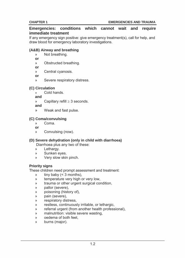
Emergencies: conditions which cannot wait and require immediate treatment If any emergency sign positive: give emergency treatment(s), call for help, and draw blood for emergency laboratory investigations. (A&B) Airway and breathing
» Not breathing. or » Obstructed breathing. or » Central cyanosis. or » Severe respiratory distress.
(C) Circulation
» Cold hands. and » Capillary refill 3 seconds. and » Weak and fast pulse.
(C) Coma/convulsing
» Coma. or » Convulsing (now).
(D) Severe dehydration (only in child with diarrhoea)
Diarrhoea plus any two of these: » Lethargy. » Sunken eyes. » Very slow skin pinch.
Priority signs These children need prompt assessment and treatment:
» tiny baby (< 3 months), » temperature very high or very low, » trauma or other urgent surgical condition, » pallor (severe), » poisoning (history of), » pain (severe), » respiratory distress, » restless, continuously irritable, or lethargic, » referral urgent (from another health professional), » malnutrition: visible severe wasting, » oedema of both feet, » burns (major).
1.3
CHAPTER 1 EMERGENCIES AND TRAUMA
Non-urgent (queue) Proceed with assessment and further treatment according to the child’s priority. A number of different triage processes exist and the above is taken from the South African Emergency Triage Assessment and Treatment (ETAT) course. Other systems may include, in addition, the use of clinical markers such as respiratory rate, blood pressure and pulse rate to add precision to the triage, especially in more resourced settings. Other important conditions are sometimes added to the ETAT guidelines to suit particular local conditions such as identifying infectious diseases that need immediate isolation, dehydration (not severe), facial or inhal ational burns, evidence of meningococcal septicaemia, inconsolable crying, etc. The ETAT triage presented above should be a minimum standard of triage in community health centres, district or regional hospitals in South Africa. Additional items may be added suitable to local conditions and resources. 1.1.2 APPROACH TO THE RESUSCITATION OF THE
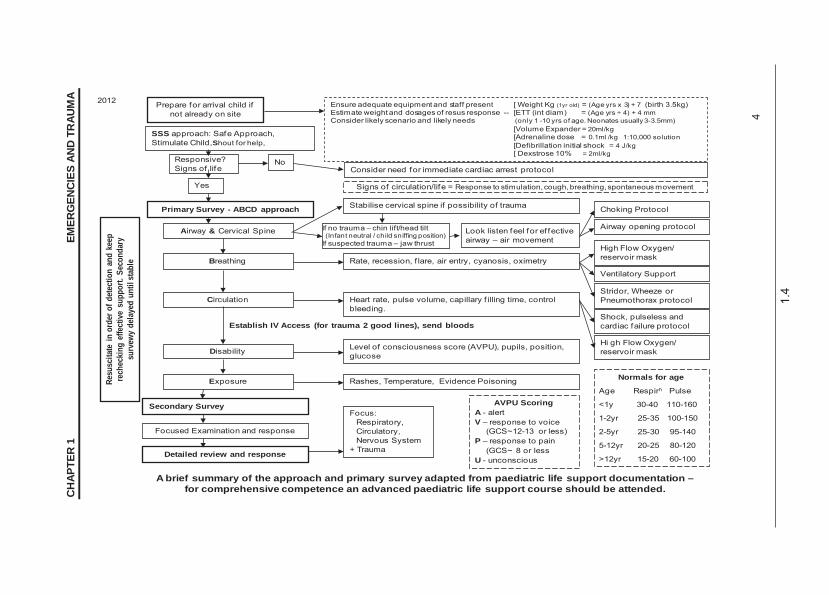
CHILD In approaching a child with potential severe illness or inj ury a struct ured approach will improve the child’s chances of a best possible outcome in the shortest possible time. The following is a diagrammatic overview derived from an advanced Paediatric life support approach.
1.4
CH
APTE
R 1
EMER
GEN
CIE
S AN
D T
RAU
MA
4
Prepare for arrival child if not already on site
Responsive?Signs of life
Primary Survey - ABCD approach
Airway & Cervical Spine
Breathing
Circulation
Disability
Secondary Survey
Exposure
Focused Examination and response
Detailed review and response
Ensure adequate equipment and staff present [ Weight Kg (1yr old) = (Age yrs x 3) + 7 (birth 3.5kg)Estimate weight and dosages of resus response -- [ETT (int diam) = (Age yrs ÷ 4) + 4 mmConsider likely scenario and likely needs (only 1 -10 yrs of age. Neonates usually 3-3.5mm)
[Volume Expander = 20ml/kg[Adrenaline dose = 0.1ml /kg 1:10,000 solution[Defibrillation initial shock = 4 J/kg[ Dexstrose 10% = 2ml/kg
NoConsider need for immediate cardiac arrest protocol
Signs of circulation/life = Response to stimulation, cough, breathing, spontaneous movementYes
Choking ProtocolStabilise cervical spine if possibility of trauma
Look listen feel for ef fective airway – air movement
Airway opening protocol
Rate, recession, f lare, air entry, cyanosis, oximetry
Focus: Respiratory,Circulatory,Nervous System
+ Trauma
Rashes, Temperature, Evidence Poisoning
Level of consciousness score (AVPU), pupils, position, glucose
Heart rate, pulse volume, capillary f illing time, control bleeding.
High Flow Oxygen/ reservoir mask
Ventilatory Support
Stridor, Wheeze or Pneumothorax protocol
Normals for age
Age Respirn Pulse
<1y 30-40 110-160
1-2yr 25-35 100-150
2-5yr 25-30 95-140
5-12yr 20-25 80-120
>12yr 15-20 60-100
Shock, pulseless and cardiac failure protocol
Hi gh Flow Oxygen/ reservoir mask
AVPU ScoringA - alertV – response to voice
(GCS~12-13 or less)P – response to pain
(GCS~ 8 or lessU - unconscious
Establish IV Access (for trauma 2 good lines), send bloods
SSS approach: Safe Approach, Stimulate Child,Shout for help,
Resu
scita
te in
ord
er o
f det
ectio
n an
d ke
ep
rech
ecki
ng e
ffect
ive
supp
ort.
Seco
ndar
y su
rvew
y de
laye
d un
til st
able
If no trauma – chin lift/head tilt(Infant neutral / child sniffing position)
If suspected trauma – jaw thrust
A brief summary of the approach and primary survey adapted from paediatric life support documentation –for comprehensive competence an advanced paediatric life support course should be attended.
2012
1.5
CHAPTER 1 EMERGENCIES AND TRAUMA
To optimise oxygen delivery to the tissue: Oxygen, high flow, 15 L/minute via facemask with reservoir bag or 6–10
L/minute via head box. o If oxygen saturation < 92% or PaO2 < 80 mmHg in spite of oxygen
supply consider need for ventilation and continue respiratory support.
1.1.3 ANAPHYLAXIS/ANAPHYLACTIC REACTIONS T78.2 DESCRIPTION An acute, potentially life-threatening hypersensitivity reaction starting within seconds to m inutes after administration of, or ex posure to, a substance to which the individual is sensitised. Clinical manifestations range from mild urticaria and angioedema to upper airway obstruction, bronchospasm, hypotension, shock and death. The reaction can be short-lived, protracted or biphasic, i.e. acute with recurrence several hours later. Immediate reactions are usually the m ost severe and/or life threatening. DIAGNOSTIC CRITERIA Clinical » Acute onset of signs and symptoms. » Dizziness, paraesthesia, syncope, sweating, flushing, dysrrhythmias. » Swelling of eyes, lips and tongue (angioedema). » Angioedema with upper airway obstruction and stridor. » Hypotension and shock. » Bronchospasm, wheezing, dyspnoea, chest tightness. » Gastrointestinal symptoms such as nausea, vomiting, diarrhoea.
A life-threatening anaphylactic reaction requires immediate treatment. Facilities to initiate treatment must be available at all health centres.
GENERAL AND SUPPORTIVE MEASURES » Place hypotensive or shocked patient in horizontal position. Do not
place patient in a sitting position. » Secure an open airway. If necessary, bag via mask or intubate. » Observe for 24 hours. MEDICINE TREATMENT To maintain arterial oxygen saturation 95% and to abolish cyanosis: Oxygen, 100%, at least 1–2 L/minute by nasal prong.

1.6
CHAPTER 1 EMERGENCIES AND TRAUMA
In severe anaphylaxis nasal oxygen is unlikely to be adequate: Oxygen, 100%, 15 L/minute by face mask.
Epinephrine (adrenaline) 1:1 000, IM, 0.01 mL/kg. (i.e. 10 mcg/kg).
o Can be repeated every 5 minutes, if necessary. o Maximum dose: 0.5 mL. o Do not administer IV unless there is failure to respond to several
doses of IM. o Monitor urine output.
Intravenous fluids Crystalloid solutions e.g.: Sodium chloride 0.9%, IV, 20 mL/kg as a bolus.
o Repeat if necessary until circulation, tissue perfusion and blood pressure improve (up to 40 mL/kg).
Promethazine, IV/IM, 0.25–0.5 mg/kg/dose.
Then continue with: Chlorphenamine, oral, 0.1 mg/kg/dose 6 hourly for 24–48 hours, if
necessary. If associated bronchospasm: Salbutamol, nebulised, 1 mL salbutamol respirator solution in 3 mL
sodium chloride 0.9%. o Nebulise at 20-minute intervals.
Hydrocortisone, IV, 5 mg/kg, 4–6 hourly for 12–24 hours.
o Note: Steroids are adjunctive therapy and are not first line treatment, and should never be the sole treatment of anaphylaxis.
If associated stridor: Epinephrine (adrenaline), 1:1000, nebulise with oxygen, every 15–30
minutes until expiratory obstruction is abolished. o 1 mL epinephrine 1:1 000 diluted in 1 mL sodium chloride 0.9%.
PREVENTATIVE MEASURES AND HOME BASED TREATMENT » Obtain a history of allergies/anaphylaxis on all patients before
administering medication/immunisation. » Identify offending agent and avoid further exposure. » See patient wears allergy identification disc/bracelet. » Train patients to self-administer epinephrine pre-filled auto injecting
device. Specialist initiated for patients who have clear documented anaphylactic reactions.
» Educate patient and parent/caregiver on allergy and anaphylaxis.
1.7
CHAPTER 1 EMERGENCIES AND TRAUMA
REFERRAL
Caution Do not refer the patient during the acute phase.
Transfer can only be done once patient is stable. Patients supplied with self administered epinephrine must be
informed of the shelf life of epinephrine and when they must come in to get a replacement for these.
» Bee sting anaphylaxis for desensitisation. 1.1.4 CARDIORESPIRATORY ARREST I46.9 DESCRIPTION Cardiorespiratory arrest in ch ildren is usually the end result of a period of circulatory or respiratory insufficiency and is seldo m due to a sudden precipitous cardiac event. It is t herefore important to pre- empt cardiorespiratory arrest in children by recognising and urgently treating respiratory or circulatory failure. Cardiorespiratory arrest is dia gnosed clinically in the unresp onsive child who displays no respiratory effort and/or in whom there is no palpable pulse and no signs of life, i.e. cough or spontaneous movement. GENERAL AND SUPPORTIVE MEASURES Always call immediately for help from your colleagues on site.
Ensure an open airway. If there is still no respiration, then commence with artificial breathing using a self-inflating bag, with a reservoir and an appropriate mask. Connect the bag to a high flow oxygen source (15 L/minute). Movement of the chest in response to artificial breaths should be evident. If there is no, or inadequate, air movement with bag-valve-mask ventilation, re-assess the airw ay with appropriate chin-lift/jaw-thrust manoeuvres. If necessary, place an appropriate sized endotracheal tube. In the event of an unexpected arrest or an arres t where there are no witnesses, consider the possibility of a foreign body obstruction. See section 1.1.6: Inhalation, foreign body.
1.8
CHAPTER 1 EMERGENCIES AND TRAUMA
Once effective breathing has been established, provide chest compressions at a rate of 100–120/minute for all children excluding neonates. Provide artificial breaths at a ratio of 15 compressions to 2 breaths in children (15:2). Attach a cardiac monitor to the child and insert an intravenous line. If failure to insert IV line, in sert intra-osseous line. See sect ion 1.1.8: Intra-Osseous Infusion in Emergencies. MEDICINE TREATMENT
Asystole or pulseless electrical activity (ie no palpable pulse even if normal electrical pattern) Epinephrine (adrenaline) 1:10 000, IV/ intra-osseous, 0.1 mL/kg. (Follow
each dose with a small bolus of sodium chloride 0.9%). o 0.1 mL of 1: 10 000 solution = 10 mcg. o Dilute a 1 mL ampoule of epinephrine (adrenaline) 1:1 000 in 9 mL of
sodium chloride 0.9% or sterile water to give a 1: 10 000 solution. OR (less ideally) Epinephrine (adrenaline) 1:1 000, endotracheal, undiluted 0.1 mL/kg down
an endotracheal tube. (This is a higher dose due to the route of administration).
Repeat the dose of epinephrine (adrenaline) every 4 minutes if asystole persists while CPR continues. When an ECG sinus rhythm trace is present continue CPR until an effective pulse and circulation is present. If the arrest was preceded by circulatory shock: Sodium chloride 0.9%, IV, 20 mL/kg as a bolus.
During the resuscitation consider if any of the following correctable conditions are present (and if present correct them): » Hypoxia. » Hypovolaemia. » Hyperkalaemia, hypokalaemia, hypocalcaemia. » Hypothermia. » Tension pneumothorax. » Tamponade (cardiac). » Toxins (e.g. tricyclic antidepressants). » Thrombo-embolic event. Note: There is no evidence to support the routine use of any of the following in cardiac arrest: » sodium bicarbonate, » calcium, » high dose IV epinephrine (adrenaline) (100 mcg/kg/dose).
1.9
CHAPTER 1 EMERGENCIES AND TRAUMA
Ventricular fibrillation or pulseless ventricular tachycardia Proceed to im mediate defibrillation but dur ing this process cardiorespiratory resuscitation (compressions and ventilation) must continue, except during the actual administration of each shock. Continue until adequate circulation can be demonstrated. For pulseless ventricular tachycardia and ventricular fibrillation the defibrillator should be set to asynchronous mode and voltage at 4 J/kg.
Do not increase voltage, give 4 J/kg repeatedly, if needed.
After each sho ck continue CPR for 2 minutes a nd only re-assess the EC G rhythm thereafter. If fibrillation/ventricular tachycardia has changed back to sinus rhythm, stop shock cycle, but cont inue CPR until good stab le circulation and adequate spontaneous breathing is evident. If fibrillation/ventricular tachycardia is still present, give further shocks for 3 x 2-minute cycles of shocks. Thereafter, if necessary , the 2- minute shock cycles should continue but, in addition, give the following after the 3rd shock: Epinephrine (adrenaline) 1:10 000, IV, 0.1 mL/kg and then repeat after
every 2nd shock, i.e. every 4 minutes. (Follow each dose with a small bolus of sodium chloride 0.9%). o 0.1 mL of 1: 10 000 solution = 10 mcg. o Dilute a 1 mL ampoule of epinephrine (adrenaline) 1:1 000 in 9 mL of
sodium chloride 0.9% or sterile water to give a 1: 10 000 solution. After the 3rd and 5th shocks, if normal rhythm has not returned: Amiodarone, IV/IO, 5 mg/kg bolus administered over 3–5 minutes.
Allow one minute of cardiopulmonary resuscitation between the administration of any medicine and a repeat cycle of shocks. If ventricular fibrillation or pulseless ventricular tachycardia persists consider the following (and if present correct): » Hypoxia. » Hypovolaemia » Hyperkalaemia, hypokalaemia, hypocalcaemia. » Hypothermia. » Tension pneumothorax. » Tamponade (cardiac). » Toxins (e.g. tricyclic antidepressants). » Thrombo-embolic event.
1.10
CHAPTER 1 EMERGENCIES AND TRAUMA
REFERRAL » To an intensive care unit after recovery from an arrest. 1.1.5 CONVULSIONS, NOT FEBRILE CONVULSIONS See section 13.2: Seizures. 1.1.6 INHALATION, FOREIGN BODY T17.9 DESCRIPTION Accidental inhalation of solid object that may obstruct the airway at any level. DIAGNOSTIC CRITERIA
Ask specifically about a possible choking episode if there is any suspicion of a foreign body aspiration.
» Initial symptom is frequently a sudden onset of choking followed by persistent unilateral wheeze (may be bilateral), chronic cough, stridor or the child may die suddenly even up to a few days later.
» Segmental or lobar pneumonia failing to respond to standard therapy. » Signs of shift of the mediastinum. » Chest X-ray on full expiration and full inspiration may show hyperinflation
and/or collapse or sometimes, radio-opaque foreign body. GENERAL AND SUPPORTIVE MEASURES ACUTE EPISODE » If coughing effectively and moving air adequately, provide oxygen and refer
urgently for airway visualisation. Carry out transfer with a person who is able to manage the foreign body process accompanying the child.
» If the child is still breathing b ut unable to cough or bre athe adequately, attempt to dislodge the foreign body by cycles of 5 back slaps followed by 5 chest compressions (infants), or 5 Heimlich manoeuvre (child) repeatedly.
» If the child is unconscious carry out standard cardiorespiratory resuscitation, i.e. cardiac compressions and ventilation (15:2).
Caution Blind finger sweeps are dangerous and absolutely contra-indicated.
All cases should have airway visualisation or be referred for airway visualisation.
1.11
CHAPTER 1 EMERGENCIES AND TRAUMA
Foreign body manoeuvres in Children who are not unconscious but unable to cough/breathe adequately. Infants » Check the mouth for any obstruction. If visualised, attempt removal under
direct vision e.g. with Magills forceps. » Lay the infant on an arm/thigh in the head down position and strike the
back of the chest 5 times firmly with the heel of the hand. » If no response, turn the infant over on its back and give 5 chest thrusts
with 2 fingers in the midline to the lower ½ of the sternum compressing the chest by about 1/3 of its diameter.
» Repeat sequence, if necessary, checking in the mouth after each cycle to see if a removable foreign body can be seen.
Children » Check the mouth for any obstruction. If visualised, attempt removal under
direct vision e.g. with Magills forceps. » Strike the back of the chest firmly 5 times while the child sits, lies prone
or kneels. » If no response, attempt Heimlich manoeuvre: standing behind the child,
pass your arms around the body and form a fist just below the sternum. Thrust upwards 5 times.
» Repeat sequence, if necessary, checking in the mouth after each cycle to see if a removable foreign body can be seen.
REFERRAL » All cases for the removal of retained foreign bodies. » Unresolved respiratory complications. 1.1.7 SHOCK R57.9 DESCRIPTION An acute syndrome that reflects the inability of the pulmonary and circulatory system to provide adequate perfusion, oxygen and nutrients to meet the physiological and metabolic demands of organs, tissues and cells. In more common usage it refers to circulatory inadequacy. Compensation is achieved by increased pulse rate, and peripheral vascular constriction. The blood pressure is relatively well maintained but the patient still requires urgent resuscitation.
1.12
CHAPTER 1 EMERGENCIES AND TRAUMA
Depending on the nature and the intrinsic aetiology, shock can be divided into: » Hypovolaemic shock: loss of intravascular fluid, e.g. dehydration,
haemorrhage or fluid shifts. » Distributive shock: e.g. septicaemia and anaphylaxis. » Cardiogenic shock: e.g. cardiac dysfunction. » Dissociative shock: e.g. profound anaemia and carbon monoxide poisoning. » Obstructive shock: e.g. pneumothorax and cardiac tamponade. » Septic shock: many mechanisms are operative in septic shock. » Neurogenic shock: e.g. spinal cord trauma. Complications of shock include multi-organ dysfunction and/or failure. DIAGNOSTIC CRITERIA Evidence of compensated shock include: » cold peripheries, » weak pulse pressure especially peripheral pulse weaker than central pulses, » prolonged capillary filling, i.e. > 3 seconds, » agitation/confusion/decreased level of consciousness, » skin pallor, » increased heart rate, » signs and symptoms of underlying conditions. In uncompensated shock, falling BP and failure to act urgently will result in irreversible shock and death.
Facilities to start treatment of shock must be available at all health centres.
GENERAL AND SUPPORTIVE MEASURES » Follow the ABCD’s algorithm at the beginning of the chapter. » Identify and treat the underlying cause. » Ensure good intravenous or intra-osseous access. In trauma, two large
bore lines for access are important. See section 1.1.8: Intra-Osseous Infusion in Emergencies.
» Take appropriate bloods, e.g. cross match, urea and electrolytes, coagulation studies, full blood count and blood cultures.
» Monitor: > and maintain vital signs, > and correct metabolic parameters, > urinary output – aim for at least 1 mL/kg/hour.
MEDICINE TREATMENT To optimise oxygen delivery to the tissue, administer: Oxygen, high flow, 15 L/minute via facemask with reservoir bag or 6–10
L/minute via head box.
1.13
CHAPTER 1 EMERGENCIES AND TRAUMA
If oxygen saturation < 92% or PaO2 < 80 mmHg consider need to intubate and continue respiratory support. 1. Hypovolaemic shock
Response to each step of management must be reviewed every 15 minutes. If after administration of a total of 40ml/kg of sodium chloride 0.9% fluid,
shock has not resolved consider other causes and the need for inotropes.
For fluid deficit (vs. blood loss): IV fluids to correct the intravascular fluid deficit and improve circulation: Sodium chloride 0.9%, IV, 20 mL/kg rapidly.
o Review after each bolus to see if shock has resolved. In children with severe malnutrition: Sodium chloride 0.9%, IV, 10 mL/kg administered over 10 minutes.
o Review after each bolus to see if shock has resolved. With each re-assessment, if: » Shock has resolved (capillary filling time < 3 seconds, good pulse,
normal blood pressure), do not repeat bolus. Proceed to other care. » Shock is better but still present, repeat bolus (up to 40 mL/kg). After this
further care should be in an ICU setting. Consider CVP monitoring. » Monitor for persistence of shock, i.e.
> Non-responding or decreasing BP. > Non-responding or increasing pulse rate/decreasing volume. > Non-responding or increasing capillary filling time.
» Monitor for fluid or circulatory overload, i.e. > Increasing respiratory rate. > Increasing basal crepitations. > Increasing pulse rate. > Increasing liver size/tenderness. > Increasing JVP.
After stabilisation of the circulation, continue with maintenance fluid volumes according to the age of the patient. For blood loss: Packed red cells, 10 mL/kg or whole blood, 20 mL/kg.
While awaiting blood for replacement begin volume resuscitation with: Sodium chloride 0.9%, IV.
1.14
CHAPTER 1 EMERGENCIES AND TRAUMA
2. Cardiogenic shock Ideally children receiving treatment for cardiogenic shock should be in high care or ICU. Inotropic support: When perfusion is poor and blood pressure response is unsatisfactory, despite adequate fluid replacement. Dobutamine, IV, 5–15 mcg/kg/minute.
Chronotropic/inotropic plus vascular tone support:
If tissue perfusion and blood pressure do not improve satisfactorily on adequate fluid volume replacement and inotropic support consider: Epinephrine (adrenaline), IV infusion, 0.01–1 mcg/kg/minute.
If poor ventricular contractility and increased afterload are considered as the primary problem, do not give epinephrine (adrenaline) but consider adding an afterload reducing agent to the dobutamine infusion but only with specialist advice. 3. Septic shock Children receiving treatment for septic shock should be in an ICU. The resuscitation and treatment of these children must be immediate.
Response to each step of management must be reviewed every 15 minutes.
IV fluids: Sodium chloride 0.9%, IV, 20 mL/kg rapidly.
o Review after each bolus to see if shock has resolved. In children with severe malnutrition: Sodium chloride 0.9%, IV, 10 mL/kg administered over 10 minutes.
o Review after each bolus to see if shock has resolved. With each reassessment, if: » Shock has not resolved after 20 ml/kg of sodium chloride 0.9% fluid,
consider inotropes. » Shock has resolved (capillary filling time < 3 seconds, good pulse,
normal blood pressure), do not repeat bolus. Proceed to other care. » Shock is better but still present, repeat bolus (up to 40 mL/kg). After this
further care should be in an ICU setting. » Monitor for persistence of shock, i.e.:
> Non-responding or decreasing BP. > Non-responding or increasing pulse rate/decreasing volume. > Non-responding or increasing capillary filling time.
1.15
CHAPTER 1 EMERGENCIES AND TRAUMA
» Monitor for fluid or circulatory overload, i.e.:
> Increasing respiratory rate. > Increasing basal crepitations. > Increasing pulse rate. > Increasing liver size/tenderness. > Increasing JVP.
Chronotropic/Inotropic plus vascular tone support If tissue perfusion and blood pressure do not improve satisfactorily on adequate fluid volume replacement. Titrate inotropes against the response, and add additional agent if poor response. Epinephrine (adrenaline), IV infusion, 0.01–1 mcg/kg/minute.
If inadequate response: ADD Dobutamine, IV, 5–15 mcg/kg/minute.
Unresponsive septicaemic shock: Hydrocortisone, IV, 1 mg /kg/dose, 6 hourly until shock has resolved.
Antibiotic therapy Start antibiotics early. Before initiating antibiotic therapy, take blood and urine specimens, if appropriate, for culture and sensitivity testing. Reconsider antibiotic and/or antifungal therapy when culture and sensitivity results become available. 3rd generation cephalosporins, e.g.: Cefotaxime, IV, 75 mg/kg/dose, 8 hourly (neonates).
OR Children > 2 months: Ceftriaxone, IV, 50 mg/kg/dose, 12 hourly.
Caution Patients must be resuscitated and stabilised before referral.

1.16
CHAPTER 1 EMERGENCIES AND TRAUMA
1.1.8 INTRA-OSSEOUS INFUSION IN EMERGENCIES If an intravenous drip cannot be set up within 5 minutes, set up an intra-osseous infusion. 1. Use an intra-osseous needle or if not available FG18 x 1.5 cm (or less
ideally FG20 x 1.5 cm) or lumbar puncture needle. 2. Grasp the thigh and knee above and lateral to the insertion site with the
palm of the left hand (if right-handed). Wrap the fingers around the knee to stabilise the proximal tibia. Do not allow any portion of your hand to rest behind the insertion site.
3. Find the site of insertion i.e. feel the tibial tuberosity. The site of insertion is about 2 cm below this tuberosity on the broad flat medial surface of the tibia.
4. Careful surgical preparation of the injection site as for lumbar punctures.
5. Insert the needle through the skin over the flat surface of the tibia. 6. Holding the needle low down near the skin, advance the needle
through the bony cortex of the tibia, directing the needle perpendicular, i.e. 90° t o the lo ng axis, using a gent le but firm tw isting or drill ing motion.
7. Stop advancing the needle when a sudden decrease in resistance to forward motion of the needle is felt.
8. Remove the stylet from the needle. 9. Slowly inject a small amount of s odium chloride 0.9% through the
needle. Check for any signs of incr eased resistance to injection, increased circumference of t he soft tissues of the calf, or increased firmness of the tissue.
10. If the tes t injection is successful, disconnect the sy ringe and join an infusion set to the needle. Secure the needle and tubing with tape and support it with a bulky dressing.
11. If the test injection is unsuccessful, i.e. infiltration of the sodium chloride 0.9% into the leg tissue is observed, remove the needle and try again on the other leg.
12. The flow rate should ra pidly increase after flushing through. If flow is poor consider the use of a 3 way tap and syringe.
Signs of successful insertion: » Sudden decrease in re sistance to insertion as t he needle passes
through the bony cortex. » The needle remains upright without support. » Fluid flows freely through the needle without evidence of subcutaneous
infiltration.
1.17
CHAPTER 1 EMERGENCIES AND TRAUMA
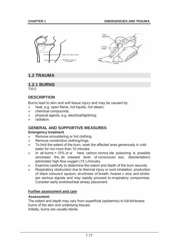
1.2 TRAUMA 1.2.1 BURNS T30.0 DESCRIPTION Burns lead to skin and soft tissue injury and may be caused by: » heat, e.g. open flame, hot liquids, hot steam; » chemical compounds; » physical agents, e.g. electrical/lightning; » radiation. GENERAL AND SUPPORTIVE MEASURES Emergency treatment » Remove smouldering or hot clothing. » Remove constrictive clothing/rings. » To limit the extent of the burn, soak the affected area generously in cold
water for not more than 10 minutes. » In all burns > 10% or w here carbon monox ide poisoning is possible
(enclosed fire, de creased level of consciousn ess, disorientation) administer high flow oxygen (15 L/minute).
» Examine carefully to determine the extent and depth of the burn wounds. » Respiratory obstruction due to thermal injury or soot inhalation, production
of black coloured sputum, shortness of breath, hoarse v oice and stridor are serious signals and may rapidly proceed to respiratory compromise. Consider early endotracheal airway placement.
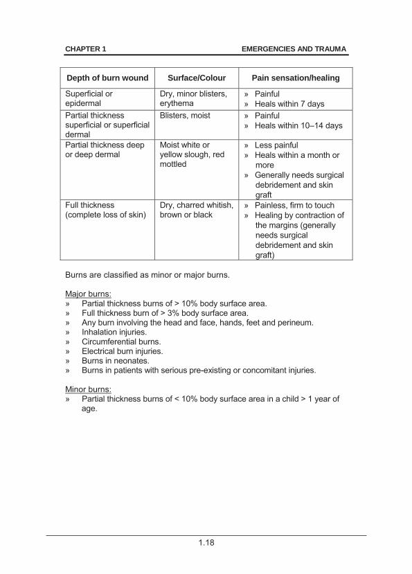
Further assessment and care Assessment: The extent and depth may vary from superficial (epidermis) to full-thickness burns of the skin and underlying tissues. Initially, burns are usually sterile.
1.18
CHAPTER 1 EMERGENCIES AND TRAUMA
Depth of burn wound Surface/Colour Pain sensation/healing
Superficial or epidermal
Dry, minor blisters, erythema
» Painful » Heals within 7 days
Partial thickness superficial or superficial dermal
Blisters, moist » Painful » Heals within 10–14 days
Partial thickness deep or deep dermal
Moist white or yellow slough, red mottled
» Less painful » Heals within a month or
more » Generally needs surgical
debridement and skin graft
Full thickness (complete loss of skin)
Dry, charred whitish, brown or black
» Painless, firm to touch » Healing by contraction of
the margins (generally needs surgical debridement and skin graft)
Burns are classified as minor or major burns. Major burns: » Partial thickness burns of > 10% body surface area. » Full thickness burn of > 3% body surface area. » Any burn involving the head and face, hands, feet and perineum. » Inhalation injuries. » Circumferential burns. » Electrical burn injuries. » Burns in neonates. » Burns in patients with serious pre-existing or concomitant injuries. Minor burns: » Partial thickness burns of < 10% body surface area in a child > 1 year of
age.
1.19
CHAPTER 1 EMERGENCIES AND TRAUMA
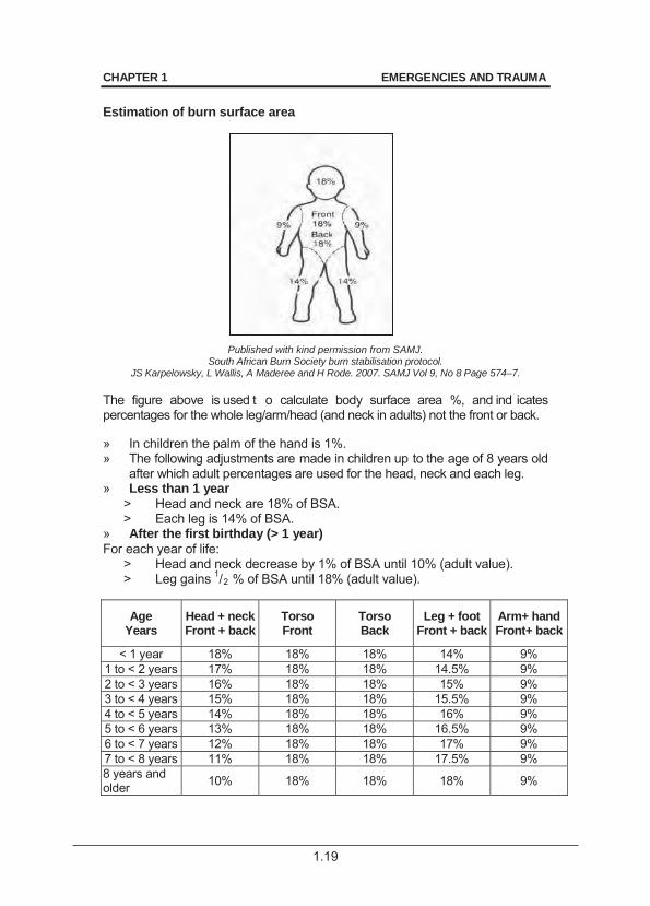
Estimation of burn surface area
Published with kind permission from SAMJ. South African Burn Society burn stabilisation protocol.
JS Karpelowsky, L Wallis, A Maderee and H Rode. 2007. SAMJ Vol 9, No 8 Page 574–7. The figure above is used t o calculate body surface area %, and ind icates percentages for the whole leg/arm/head (and neck in adults) not the front or back. » In children the palm of the hand is 1%. » The following adjustments are made in children up to the age of 8 years old
after which adult percentages are used for the head, neck and each leg. » Less than 1 year
> Head and neck are 18% of BSA. > Each leg is 14% of BSA.
» After the first birthday (> 1 year) For each year of life:
> Head and neck decrease by 1% of BSA until 10% (adult value). > Leg gains 1/2 % of BSA until 18% (adult value).
Age Years
Head + neck Front + back
Torso Front
Torso Back
Leg + foot Front + back
Arm+ hand Front+ back
< 1 year 18% 18% 18% 14% 9% 1 to < 2 years 17% 18% 18% 14.5% 9% 2 to < 3 years 16% 18% 18% 15% 9% 3 to < 4 years 15% 18% 18% 15.5% 9% 4 to < 5 years 14% 18% 18% 16% 9% 5 to < 6 years 13% 18% 18% 16.5% 9% 6 to < 7 years 12% 18% 18% 17% 9% 7 to < 8 years 11% 18% 18% 17.5% 9% 8 years and older 10% 18% 18% 18% 9%
1.20
CHAPTER 1 EMERGENCIES AND TRAUMA
Care Inhalation injury In addition to other treatment, the degree of inhalation injury may warrant: » monitoring of blood gases, » warm humidified oxygen and/or intubation, » positive pressure ventilation. Ensure adequate airway in the presence of inhalational burns. Children with burns may present with delayed onset of airway obstruction. Consider early intubation. Suspect carbon monoxide poisoning in all fire victims. » Obtain carboxyhaemoglobin level. » Treat by administering 100% oxygen. Prevent heat loss Nurse all major burns in a warm room (26°C). Nasogastric drainage Use a nasogastric tube on free drainage in all burns > 10% (especially during transfer). After the 1st 24 hours, commence nasogastric feeds in children who had been started on nasogastric drainage where ileus is not suspected. Nutritional support Consult a dietician as children with burns require a higher than usual intake of nutrients. Start enteral feeds within 6 hours in burns < 10%. Estimate daily energy and protein needs using the formulae:
Energy (kJ): 250 kJ/kg body mass + (150 kJ x % burned BSA) Protein: 3 g/kg body mass + (1 g x % burned BSA)
Maximum % burn area used for calculation should not exceed 50% Give iron and vitamins routinely until burn wounds are healed and/or skin grafting has successfully been completed. Note: Do not supplement iron during sepsis or infection. In addition, provide: » psychological support, » physiotherapy, » occupational therapy, » waterbeds and cradles.
1.21
CHAPTER 1 EMERGENCIES AND TRAUMA
MEDICINE TREATMENT Fluid replacement Burns < 10% of total body surface area: Oral fluids.
Burns > 10% of total body surface area: IV fluid for resuscitation.
If in shock first treat shock. See section: 1.1.7: Shock. As in all fluid administration in sick children volumes are estimates and response must be constantly re-evaluated and rates adjusted appropriately. CALCULATION OF INITIAL FLUID REPLACEMENT (AFTER SHOCK HAS BEEN TREATED) First 24 hours: Replacement fluids for burns Sodium chloride 0.9%, IV.
o Calculate total fluid requirement in 24 hours: [Total % burn x weight (kg) x 4 mL] as sodium chloride 0.9%.
Give half of this volume in the 1st 8 hours. Administer remaining fluid volume in next 16 hours.
Note: If urine output not adequate, increase fluids for the next hour by 50% (continue at higher rate until urine output is adequate then resume normal calculated rate). PLUS Maintenance fluids in children In children, give oral or intravenous maintenance fluid in addition to above calculated volume.
Child maintenance fluid requirement volumes
120 mL/kg/24 hours
All children > 1 year – the sum of the following:
» for each kg of body weight up to 10 kg 100 mL/kg/24 hours
» for each additional kg of body weight more than 10 kg
50 mL/kg/24 hours
» for each additional kg of body weight more than 20 kg
20 ml/kg/24 hours
1.22
CHAPTER 1 EMERGENCIES AND TRAUMA
Example: 24 kg child with 10% burns
1st 24 hours » replacement for expected losses:
4 mL/kg x 24kg x 10% = 960 mL
» maintenance: first 10 kg = 10 kg x 100 mL/kg/24 hours
second 10 kg = 10 kg x 50 mL/kg/24 hours remaining 4kg = 4 kg x 20 mL/kg/24 hours
Total maintenance:
= 1 000 mL+ = 500 mL+ = 80 mL
= 1 580 mL Thus 1st 8 hours
480 mL sodium chloride 0.9% + 527 mL ½ Darrows/ dextrose 5%
Next 16 hours = ½ resuscitat
480 mL sodium chloride 0.9% + 1053 mL ½ Darrows/ dextrose 5%
The above are guidelines, need regular review to maintain urine output 1–2 mL/kg/hour.
Avoid circumferential taping when securing infusion lines, as oedema under the eschar may decrease the venous return.
Second 24 hours: If urine output is adequate, continue resuscitation: Sodium chloride 0.9%, IV, 1.5 mL/kg/% burn/ 24 hours.
PLUS Maintenance: ½ Darrows/dextrose 5%, as per maintenance requirement above.
Part of this volume may be replaced by enteral feeds. Thereafter progressively decrease IV fluids and increase enteral fluids according to response over time. Anaemia If haemoglobin < 7 g/dL: Packed red cells, 10 mL/kg over 3 hours.
Hypoalbuminaemia If indicated by symptomatic hypoalbuminaemia: Albumin 20%, IV, 2 g/kg/day. (2 g = 10 mL).
For pain Paracetamol, oral, 15 mg/kg/dose 6 hourly as required.
Children with large burns need effective pain relief. See section 20.1: Management of pain.
1.23
CHAPTER 1 EMERGENCIES AND TRAUMA
Change of dressing Provide analgesic cover at each dressing change. In major burns, change dressings under procedural sedation or g eneral anaesthesia. Administer medicines half an hour before dressing change: Paracetamol, oral, 15 mg/kg/dose 6 hourly.
PLUS Tilidine, oral, 1 drop per 2.5kg of body weight (i.e. 1 mg/kg/dose).
PLUS For anxiolysis: Midazolam, oral, 0.5 mg/kg/dose.
For severe pain/procedure: ADD Ketamine, oral, 2–5 mg/kg at least 30 minutes before procedure (works
best if given in sweet soft drink). Gastric erosions Preventative medication treatment is not given. Effective early resuscitation and early feeding decrease the incidence of gastric erosion. If gastric erosion is suspected due to haematemesis or brownish gastric aspirates. Omeprazole, oral, 0.4–0.8 mg/kg/dose 12 hourly. Specialist initiated.
o Maximum dose: 20–40 mg/dose. o If 1 month–2 years: 2.5 mg 12 hourly. o If > 2–6 years: 5 mg 12 hourly. o If > 7–12 years: 10 mg 12 hourly.
If unable to take orally: Ranitidine, IV, 1 mg/kg 6 hourly.
Local treatment of burns Gently clean the wounds with running water. Remove loose skin and debride dead tissue and dress with topical antiseptic cream and non-adherent dressing. Thereafter, daily rinse with running water and dress with topical antiseptic cream and non-adherent dressing. In < 20% body surface area burns: Povidone-iodine 0.5% with occlusive dressings.
1.24
CHAPTER 1 EMERGENCIES AND TRAUMA
In > 20% body surface area burns: Silver-sulphadiazine 1%, on non-adhesive dressings.
o Cover with paraffin gauze. o Change dressings daily.
Excise and graft all full thickness or deep dermal burns as soon as the patient is stable. Consider skin grafting in wounds not healed in two weeks. Antibiotics Consider if signs of infection are present as these may be subtle: » pyrexia/hypothermia, » shock (compensated or not
compensated), » rising pulse or respiratory rate, » petechiae.
» leucocytosis/thrombocytopaenia, » looks ill /toxic/altered level of
consciousness, » local inflammatory changes,
The choice of antibiotics is based on the culture and sensitivity results of wound, urine and blood cultures once available. Positive wound cultures alone do not indicate systemic infections requiring antibiotic treatment. Ceftriaxone, IV, 50 mg/kg/dose 24 hourly for 5 days.
If MRSA is suspected or confirmed, replace with: Vancomycin, IV, 15 mg/kg/dose 6 hourly for 5–14 days.
AND Amikacin, IV, for 5–14 days if renal function is satisfactory.
o 1 week to < 10 years: 25 mg stat then 18 mg/kg once daily. o 10 years and older: 20 mg stat then 15 mg/kg.
Tetanus prevention Patients with no previous immunisation in the last 5 years:
Tetanus toxoid, IM, 0.5 mL. Complete course in previously unvaccinated patients. Where deep necrotic lesions are part of the burn and if the immunological status is not known:
Tetanus immunoglobulin, IM, 500 IU.
1.25
CHAPTER 1 EMERGENCIES AND TRAUMA
Prior to transport/referral » Commence resuscitative measures, if necessary. » Administer 100% humidified oxygen by facemask for inhalation injuries, if
necessary. » Cover wounds with clean dressings after hot or smouldering clothing have
been removed.
REFERRAL » Major burn injuries.
2.1
CHAPTER 2 ALIMENTARY TRACT
2.1 DENTAL AND ORAL DISORDERS 2.1.1 GINGIVITIS, UNCOMPLICATED K05.1 DESCRIPTION Inflammation of the gum margin causing the gums to separate from the teeth. Pockets form between the gums and the teeth, where pus and bacteria can collect, eventually causing periodontitis, a disease in the tissue that surround and supports the teeth – See section 2.1.2: Periodontitis. Characteristics of uncomplicated gingivitis:
» change in the normal gum contour, » may be painful, » redness, » swollen gums, » watery exudate/bleeding, » gum recession may occur, » may be recurrent.
GENERAL AND SUPPORTIVE MEASURES Oral hygiene is usually adequate to prevent superficial mouth and gum infection: » Oral hygiene after each meal to remove plaque and food debris. » Frequent thorough brushing of teeth, at least twice daily. » Dental flossing at least once a day. » Homemade warm saline rinse. Dissolve ½ teaspoon of table salt
(sodium chloride) in ± 200 mL warm water. Rinse mouth for one minute twice daily but do not swallow.
MEDICINE TREATMENT Paracetamol, oral, 15 mg/kg/dose 6 hourly when required to a maximum
of 4 doses per 24 hours. Chlorhexidine 0.2%, 15 mL as a mouthwash, 2–4 times daily for 5 days
use after brushing and flossing. 2.1.2 PERIODONTITIS K05.4 DESCRIPTION Progressive gingivitis to the point where the underlying bone is eroded, is characterised by teeth becoming loose in their sockets. It is a cause of tooth loss in adults.
2.2
CHAPTER 2 ALIMENTARY TRACT GENERAL AND SUPPORTIVE MEASURES » Advice on improving and maintaining oral hygiene. See section 2.1.1:
Gingivitis, uncomplicated. General and supportive measures. MEDICINE TREATMENT Chlorhexidine 0.2%, 15 mL as a mouthwash, 2–4 times daily for 5 days.
REFERRAL » All cases. 2.1.3 NECROTISING PERIODONTITIS K05.6 DESCRIPTION An acute very painful infection of the gingival margin characterised by: » foul smelling breath, » loss of gingiva and supporting bone around teeth, and » presence of underlying disease, e.g. HIV May lead to loss of surrounding lips and cheeks if not adequately treated. GENERAL AND SUPPORTIVE MEASURES » Advice on improving and maintaining oral hygiene. See section 2.1.1:
Gingivitis, uncomplicated. General and supportive measures. MEDICINE TREATMENT Amoxicillin/clavulanic acid, oral, 25 mg/kg/dose of the amoxicillin
component 8 hourly for 5 days. Chlorhexidine 0.2%, 15 mL as a mouthwash, 2–4 times daily 30 minutes
after brushing and flossing. o Continue for 5 days.
For pain: Paracetamol, oral, 15 mg/kg/dose 6 hourly when required to a maximum
of 4 doses per 24 hours. REFERRAL For dental treatment: » No improvement within 5 days.
2.3
CHAPTER 2 ALIMENTARY TRACT 2.1.4 CANDIDIASIS, ORAL B37.0 See section 8.6: Candidiasis, systemic and other. 2.1.5 APHTHOUS ULCERS K12.0 DESCRIPTION Painful ulcers in the oropharynx. Minor ulcers (< 1 cm diameter) usually heal within 2 weeks. Major ulcers (> 1 cm diameter) are very painful, often very deep and persist. GENERAL AND SUPPORTIVE MEASURES » Maintain adequate nutrition and hydration by encouraging fluid and food
intake – use bland foods and fluids as they are less painful. » For minor aphthous ulcers, use homemade warm saline rinse. Dissolve
½ teaspoon of table salt (sodium chloride) in ± 200 mL warm water. Rinse mouth but do not swallow.
MEDICINE TREATMENT For pain: Paracetamol, oral, 15 mg/kg/dose 6 hourly when required to a maximum
of 4 doses per 24 hours. REFERRAL » Major aphthous ulcers for further diagnostic evaluation. » Aphthous ulcers not resolving in 3 weeks for further evaluation. 2.1.6 HERPES GINGIVOSTOMATITIS B00.2 DESCRIPTION Inflammation of the mouth structures with ulcers (which may be of v arious numbers and sizes), caused by Herpes simplex virus infection. The normal course of the disease is 7–10 days. DIAGNOSTIC CRITERIA Clinical » General inflammation of the mouth with multiple small ulcers on the
buccal mucosa, palate, anterior tonsillar pillars, tongue, inner lips and gingival margins.
» Fever, malaise and dysphagia. » Tender, enlarged cervical lymph nodes.
2.4
CHAPTER 2 ALIMENTARY TRACT GENERAL AND SUPPORTIVE MEASURES » Maintain adequate nutrition and hydration by encouraging fluid and food
intake – use bland foods and fluids as they are less painful. » If oral nutrition cannot be maintained use oral/nasogastric and/or IV
fluids, if necessary. MEDICINE TREATMENT Chlorhexidine 0.2%, 10 mL as a mouthwash or gargle, 12 hourly.
o Do not swallow. For pain: Paracetamol, oral, 15 mg/kg/dose 6 hourly.
OR Ibuprofen, oral, 5–10 mg/kg/dose 6 hourly after meals.
If more than minor fever blisters: Aciclovir, oral, 250 mg/m2/dose 8 hourly for 7 days (or per kg dose
equivalent below) o If > 1month to 1 year old: 12.5 mg/kg/dose. o If > 1 year to 6 years old: 10 mg/kg/dose. o If > 6 years to 12 years old: 6 mg/kg/dose.
If very severe infection, consider: Aciclovir, IV, 250 mg/m2/dose 8 hourly for 7 days (per kg dose
equivalent below) o If > 1 month to 1 year old: 12.5 mg/kg/dose. o If > 1 year to 6 years old: 10 mg/kg/dose. o If > 6 years to 12 years old 6 mg/kg/dose.
Change to oral as soon as possible. For very painful oral herpes in children > 2 years: Lidocaine (lignocaine) 2% gel applied every 3 to 4 hours.
o Apply a thin layer on the affected areas only. o Do not exceed 3 mg/kg dose, i.e. maximum 0.15 mL/kg of 2% gel.
REFERRAL » Herpes gingivostomatitis not responding to therapy. » Disseminating disease, especially if associated with encephalopathy or
increasing liver span.
2.5
CHAPTER 2 ALIMENTARY TRACT 2.2 GASTROINTESTINAL DISORDERS 2.2.1 CHOLERA A00.9 * Notifiable condition. DEFINITION An acute diarrhoeal disease caused by V. cholerae. DIAGNOSTIC CRITERIA Clinical » Sudden onset of severe, watery diarrhoea, i.e. ‘rice water’ diarrhoea. » Low-grade or no fever. » Persistent vomiting not associated with nausea. » Rapid fluid and electrolyte losses with dehydration, acidosis and
hypovolaemic shock with/without renal failure. » History of contact with a cholera case or the presence of cholera in the
community. Investigations » Positive stool culture. » Agglutinating or toxin-neutralising antibodies in the serum. GENERAL AND SUPPORTIVE MEASURES » Isolate patient and institute barrier nursing. » Ensure adequate hydration and nutrition. » Check blood glucose in patients with decreased level of consciousness. The management of the fluid requirements is the most critical element
of treating a patient with cholera. MEDICINE TREATMENT First treat shock. Once shock has resolved, manage as acute diarrhoea. See section 2.2.4 Diarrhoea, acute. For the management of shock during recognised cholera outbreaks, there may be benefit to replace sodium chloride 0.9% with: Modified Ringers–Lactate, IV.
2.6
CHAPTER 2 ALIMENTARY TRACT Antibiotic treatment Recommended antibiotics may vary according to sensitivities in epidemics. Consult the NICD for the latest recommendations. Current recommendations for severe dehydration are: Ciprofloxacin, oral, 15 mg/kg/dose 12 hourly for 3 days.
In all children who are able to take oral medication Zinc (elemental), oral for 14 days:
o If < 10 kg: 10 mg/day. o If > 10 kg: 20 mg/day.
REFERRAL » Cholera with complications, e.g. persistent shock, renal failure and
severe electrolyte disturbances. 2.2.2 CONSTIPATION / FAECAL LOADING K59.0 DESCRIPTION Constipation: the infrequent passage of hard st ools. This is often due to behavioural retention following previous painful episodes of defaecation, but may also be due to somatic causes or overuse of certain medications. Faecal soiling: the involuntary leakage of small amounts of soft or watery stools secondary to faecal loading.
Causes include: » psychogenic disorders, » incorrect diet, » lack of exercise, » chronic use of enemas, » certain medicines, » metabolic, endocrine, neurogenic and lower bowel abnormalities.
DIAGNOSTIC CRITERIA » Non-tender deformable faecal masses palpable. » Confirm on a straight abdominal X-ray. GENERAL AND SUPPORTIVE MEASURES » Determine and treat the underlying cause. » Treatment involves 3 steps:
> initial clearance of stools, > prevent re-accumulation of hardened retained stool, and > retraining of the gut to achieve regular toilet habits.
» Management is long-term and requires the active involvement of the parents.
2.7
CHAPTER 2 ALIMENTARY TRACT MEDICINE TREATMENT Initial therapy Faecal clearance if faecal loading: Phosphate-containing enema (sodium phosphate 6 mg/mL/ sodium acid
phosphate 16 mg/mL). o Age 2–5 years: 32 mL. o Age 5–11 years: 64 mL. o Repeat once, if necessary.
OR Add-on therapy after failed treatment with phosphate enema: Polyethylene glycol 59 g/L solution with sodium sulphate and
electrolytes , oral/ nasogastric tube, 10–25 mL/kg/hour until clear fluid is passed rectally.
Do not use sweeteners containing sugar. Confirm the bowel is empty. Maintenance therapy Bowel re-training Diet change with additional natural fibre from fruit, vegetables and bran. Additional fibre: Ispaghula husk, oral, 1.75–3.5 g, stirred in water with breakfast.
AND/OR Liquid paraffin, oral, 2 mL/kg/day.
In refractory cases: Lactulose, oral, 0.5 mL/kg/dose 12 hourly.
If faecal loading, maintenance therapy should be continued for months to years. REFERRAL » Suspected organic cause e.g. constipation from birth in a breast-fed
baby. » Inadequate response to therapy. 2.2.3 CYSTIC FIBROSIS E84.9 DESCRIPTION An autosomal recessive disorder of ex ocrine glands, mainly affecting the gut, pancreas and lungs.
2.8
CHAPTER 2 ALIMENTARY TRACT DIAGNOSTIC CRITERIA Clinical » Recurrent infections of the respiratory tract with later bronchiectasis,
respiratory failure and cor pulmonale. » Bulky, greasy and foul-smelling stools. » Occasionally presents with constipation. » Malabsorption with weight loss and failure to thrive. » Meconium ileus. » Family history, rare unless in a sibling. Investigations » Sweat test:
> Quantitative analysis of sodium and chloride concentrations in sweat collected after stimulation by pilocarpine iontophoresis with chloride > 60 mmol/L.
» DNA analysis for delta F508 and a few other mutations. GENERAL AND SUPPORTIVE MEASURES » Nutritional support:
> well balanced diet, > oral intake of at least 120% of recommended daily allowance, > nutritional supplements, > occasionally nocturnal supplemental feeding by nasogastric or
gastrostomy tube. » Physiotherapy and postural drainage. » Psychosocial support. » Genetic counselling. MEDICINE TREATMENT Medicinal treatment is specialised and individualised and should be under the supervision of a subspecialist. Pancreatic enzymes (lipase/amylase/protease), with meals according to
clinical response. o Maximum dose 10 000 lipase units/kg/day.
REFERRAL » All to a recognised cystic fibrosis centre and/or specialist health facility
for confirmation of diagnosis and initiation of treatment. » Management of exacerbations.
2.9
CHAPTER 2 ALIMENTARY TRACT 2.2.4 DIARRHOEA, ACUTE A09.0 DESCRIPTION Diarrhoea is a s erious common childhood illness evidenced by the passing of frequent profuse loose watery stools. Vomiting may or may not be present. Diarrhoeal disease is often caused by viral infection but may be due t o bacterial infection, dietary or other causes. Dehydration and metabolic disturbances are common if treatment is not instituted early and may result in severe disease, irreversible organ damage and death in children. Malnutrition is a serious co-morbidity and/or result of diarrhoeal disease and must be managed correctly employing ongoing feeding. Feeding, minerals, micronutrients and vitamins are continued except during ileus or shock. See section 2.4: Malnutrition. In severe malnutrition or in the young infant (< 2 months of age) bacterial co-infection is common. DIAGNOSTIC CRITERIA Clinical
The assessment of shock and dehydration in children is not always simple. A good initial assessment and frequent re-assessments (4-hourly if
dehydration is present. In the presence of shock continuous reassessment with appropriate adjustment of care are vital in the care of these children.
Shock is shown by one or more of the following: Compensated shock: » delayed capillary refilling time (> 3 seconds); » rapid, weak pulse rate; » cool peripheries. Late (Preterminal): » decreased level of consciousness, » decreased blood pressure, » decreased pulse volume. Dehydration is treated after shock is dealt with: Severe dehydration Some dehydration Sunken eyes Very slow skin pin Drinking poorly
Sunken eyes Slow skin pinch (< 2 sec) Drinks eagerly Irritable/restless
Other indicators of dehydration may be sought but do not add substantially to assessment, e.g.: depressed fontanelle, absent tears, decreased passage of urine.
2.10
CHAPTER 2 ALIMENTARY TRACT Also assess for signs of metabolic, nutritional and other co-morbidities: » severe malnutrition, » decreased bowel sounds, » decreased level of
consciousness, » increased respiratory rate and
chest indrawing, » abnormal tone or floppiness, » persistent or bile stained vomiting, » abdominal distension, » urine for leucocytes or nitrites. Investigations » After resuscitation, in children with severe dehydration, shock or other
signs of metabolic, nutritional or other co-morbidities: > sodium, potassium, urea, creatinine, blood acid-base assessment.
» Stool culture, especially if at a sentinel site for infectious GIT disease, or suspected dysentery, typhoid, cholera.
» Urine test strip on fresh/clean urine specimen for leucocytes, nitrites and blood. Ascertain HIV status with consent in every child.
GENERAL AND SUPPORTIVE MEASURES » Adequate initial assessment and fre quent re-assessment, including
weight, is vital. » Re-assess the patient continuously while shock persists. » If dehydration is present, re-assess the patient 4-hourly and immediately
correct shock or deterioration. » Monitor and maintain:
> hydration and circulation, > normal blood glucose, > blood pressure, > blood electrolytes, > acid-base status.
» Monitor urine output, should be at least 1 mL/kg/hour. This may be difficult in small children with diarrhoea, especially in female infants.
» Monitor body mass regularly. Weigh daily, 6-hourly if unsure of hydration status and child is very ill or sm all. This can be used to indicate response of hydration.
» Continue oral feeds during period of diarrhoea: > if the child is breastfed, continue breastfeeds and encourage the
child to feed longer at each feed; > if the child is exclusively breastfed, give oral rehydration solution
(ORS) in addition to each feed; > if the child is not exclusively breastfed, give ORS and other
appropriate feeds, e.g. breast milk substitutes or food based fluids; > if the child is severely dehydrated or shocked, withhold feeding until
stable, usually a few hours only.
2.11
CHAPTER 2 ALIMENTARY TRACT MEDICINE TREATMENT
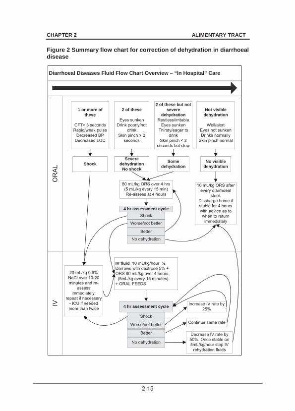
There is no place for antidiarrhoeal medications, i.e. kaolin and pectin, atropine and diphenoxylate, loperamide, or antiemetics in the routine
management of acute diarrhoea.
OUTLINE OF PRACTICAL FLUID THERAPY OF DEHYDRATING WATERY DIARRHOEA With severe malnutrition the assessment of dehydration is more difficult. Avoid intravenous infusions, if possible. Treatment of dehydration requires more care/more frequent assessments. 1. First treat shock, if present (If no shock, proceed to section 2 below)
Sodium chloride 0.9%, IV, 20 mL/kg given as a bolus rapidly.
o After each bolus reassess for persistence of shock, or evidence of circulatory overload.
o Repeat the fluid bolus up to 3 times if shock still persists, provided that evidence of circulatory overload is not present.
o If after the second bolus, i.e. total of 40 mL/kg has been given, the response is inadequate, a th ird bolus can be started. Move the patient to ICU for CVP monitoring and inotropic support.
Treatment of shock in severe malnutrition Shock treatment should be more cautious in patients with severe malnutrition due to poor cardiac reserve and high prevalence of gram negative septicaemia. Sodium chloride 0.9%, IV, 10 mL/kg administered over 10 minutes.
o Up to 4 boluses may be given. However, deterioration may be due to fluid overload and shock may be due to sept icaemia, not always hypovolaemia.
o After 4 boluses (40 mL/kg) further treatment should be in a high care unit.
o Re-assess frequently during treatment of shock. Patients response should guide further fluid therapy.
If pulse and re spiratory rate increases, increasing liver span and gallop rhythm are found suspect fluid overload/ cardiac dysfunction and manage appropriately. See section 1.1.7: Shock.
If an IV infusion cannot be set up within 5 minutes use an intra-osseus infusion. See section 1.1.8: Intra-Osseous Infusion in Emergencies.
During treatment of shock administer oxygen.
2.12
CHAPTER 2 ALIMENTARY TRACT When shock has been treated proceed to the management of dehydration. 2. Severe dehydration or some dehydration 2a) If the child has not failed oral rehydration and was not in shock: Oral rehydration solution (ORS), oral, 80 mL/kg over 4 hours using
frequent small sips (i.e. 5 mL/kg every 15 minutes for 4 hours). o Give more if the child wants more. o Show the caregiver how to give ORS with a cup and spoon. o If child vomits wait 10 minutes and then continue more slowly. o Encourage caregiver to continue feeding the child, especially
breastfeeding. Review after 4 hours: » general condition, » respiratory rate, » capillary filling time, » abdomen (liver span), » level of consciousness, » if passing urine, » skin turgor, » number/quality of stools, and » sunken eyes. Appropriate response at 4 hourly re-assessment:
Shock Treat for shock as under 1 above. No improvement or more dehydrated
Increase drip rate by 25%.
Improving (e.g. increase in weight) but still dehydrated
Continue current drip rate.
No visible dehydration
Decrease drip rate by 50%. If remains well hydrated after a further 4 hours stop IV rehydration fluids and move to ORS.
For prevention of dehydration see under 3 below. 2b) If child fails the above oral treatment, was in shock or has already failed at primary health care level then: IV fluid* ½ Darrows/dextrose 5%, IV, 10 mL/kg/hour administered for 4 hours,
then re-assess. *(This rate is in line with current safety evidence but the need for regular reassessment 4-hourly remains). PLUS Oral rehydration solution Oral rehydration solution (ORS), oral, 80 mL/kg over 4 hours using
frequent small sips (i.e. 5 mL/kg every 15 minutes for 4 hours). PLUS Oral feeds at normal feed volumes and times.
2.13
CHAPTER 2 ALIMENTARY TRACT Review after 4 hours: » general condition, » respiratory rate, » capillary filling time, » abdomen (liver span), » level of consciousness, » if passing urine, » skin turgor, » number/quality of stools, and » sunken eyes. Appropriate response at 4-hourly re-assessment:
Shock Treat for shock as under 1 above. No improvement or more dehydrated
Increase drip rate by 25%.
Improving (e.g. increase in weight) but still dehydrated
Continue current drip rate.
No visible dehydration
Decrease drip rate by 50%. If remains well hydrated after a further 4 hours stop IV rehydration fluids and move to ORS.
For prevention of dehydration see under 3 below. Give oral feeds if: » the level of consciousness is normal, » the child is not in severe distress, » not shocked and, » has no surgical abdomen. 3. No visible signs of dehydration on presentation or a child stable with no dehydration after treatment of dehydration. Show the caregiver how to give ORS with a cup and spoon using frequent small sips. Encourage caregiver to give 10 mL/kg after each diarrhoeal stool until diarrhoea stops. Instruct the caregiver on how to make and use ORS/SSS at home. Home made sugar and salt solution may be used if oral rehydration formula is not available.
HOMEMADE SUGAR AND SALT SOLUTION (SSS) ½ level medicine measure of table salt
plus 8 level medicine measures of sugar
dissolved in 1 litre of boiled (if possible) then cooled water (1 level medicine measure = approximately 1 level 5 mL teaspoon)
Encourage the caregiver to continue feeding the child, especially breastfeeding.
2.14
CHAPTER 2 ALIMENTARY TRACT Instruct the caregiver to give the child extra feeds after the diarrhoea has stopped to make up for the period of inadequate intake. Child should return to hospital immediately if: » no improvement, » blood in stool, » condition deteriorates, » fever develops, » poor drinking or feeding, » sunken eyes, » slow skin pinch. Educate caregivers about hygiene, oral rehydration solution and danger signs of diarrhoea.
2.15
CHAPTER 2 ALIMENTARY TRACT Figure 2 Summary flow chart for correction of dehydration in diarrhoeal disease
Diarrhoeal Diseases Fluid Flow Chart Overview – “In Hospital” Care
IVO
RA
L
1 or more of these
CFT> 3 secondsRapid/weak pulse
Decreased BPDecreased LOC
2 of these
Eyes sunkenDrink poorly/not
drinkSkin pinch > 2
seconds
2 of these but not severe
dehydrationRestless/irritable
Eyes sunkenThirsty/eager to
drinkSkin pinch < 2
seconds but slow
Not visible dehydration
Well/alertEyes not sunkenDrinks normally
Skin pinch normal
ShockSevere
dehydrationNo shock
Some dehydration
No visible dehydration
80 mL/kg ORS over 4 hrs (5 mL/kg every 15 min)Re-assess at 4 hours
Shock
Worse/not better
No dehydration
IV fluid 10 mL/kg/hour ½ Darrows with dextrose 5% + ORS 80 mL/kg over 4 hours (5mL/kg every 15 minutes)+ ORAL FEEDS
20 mL/kg 0.9% NaCl over 10-20 minutes and re-
assess immediately:
repeat if necessary - ICU if needed more than twice
10 mL/kg ORS after every diarrhoeal
stool.Discharge home if stable for 4 hours with advice as to when to return immediately
4 hr assessment cycle
Better
Better
Shock
4 hr assessment cycle
Worse/not better
No dehydration
Decrease IV rate by 50%. Once stable on 5mL/kg/hour stop IV
rehydration fluids
Continue same rate
Increase IV rate by 25%
2.16
CHAPTER 2 ALIMENTARY TRACT Metabolic disturbances Acidosis Metabolic acidosis does not require correction unless extremely severe, i.e. pH < 7.1, or i f the b ody is unable to c orrect the d eficit, e.g. s alicylate poisoning or renal failure. Correction should only be considered with expert supervision. Correcting the renal circulation and shock will lead to self-correction in almost all cases. If correction is necessary: volume of sodium bicarbonate 4.2% required is: Sodium bicarbonate 4.2% in mL = 0.3 x base deficit x weight in kg.
Review response to assess the need for further correction. Hypokalaemia Note: Potassium levels are affected by the degree of acidosis. If potassium is < 3.5 mmol/L but > 2.5 mmol/L: Potassium chloride, oral, 25–50 mg/kg/dose 8 hourly.
If potassium is < 2.5 mmol/L: ½ Darrows/dextrose 5%, 200 mL plus potassium chloride 15%, 2 mL,
into the vacoliter: o 1 mL potassium chloride 15% = 2 mmol in the above dilution, gives
combined K+ of 37 meq/L – do not exceed this amount. o Mix well before administration. o Run at normal rehydration rate (as above).
Oral potassium may also be given during this period: Potassium chloride, oral, 25–50 mg/kg/dose 8 hourly.
Monitor serum potassium 8–12 hourly. Once above 3.0 meq/L, stop IV potassium and continue with oral.
Hypernatraemia (> 150 mmol/L) Oral rehydration is preferable to IV rehydration. If oral rehydration fails, rehydrate using IV over 48 hours but continue giving oral rehydration. IV Fluid rate Rate: o If 2–10 kg: 6 mL/kg/hour o If > 10–20 kg: 5 mL/kg/hour o If > 20–40 kg: 4 mL/kg/hour Serum sodium 160 mmol/L ½ Darrows/dextrose 5%, IV.
2.17
CHAPTER 2 ALIMENTARY TRACT Serum sodium > 160 mmoL Sodium chloride 0.9%/dextrose 5% plus potassium chloride (see below) is used to correct clinical dehydration for the first 48 hours. After changing to maintenance rates use oral rehydration, or if IV fluids are required change to ½ Darrows/dextrose 5%. Sodium chloride 0.9%/dextrose 5% plus potassium chloride (to 20
mmol/L), IV. Repeat serum sodium every 8–12 hours to monitor progress. Failure to decrease sodium levels usually means the rehydration rate is too slow. Fall of sodium levels more than 1 mmol/L/hour on average means the rehydration rate should be reduced. Frequent clinical reassessment is the key to the safe management of this situation. Serum sodium levels may be done more frequently where this is possible. Adjust the drip rate according to response. If convulsions are considered likely, (decreased level of consciousness, hyper-irritable child), in the setting of high serum sodium, consider the use of prophylactic anticonvulsants: Phenobarbitone, IV, 20 mg/kg as a single dose.
OR If IV phenobarbitone not available: Phenobarbitone, oral, 20–30 mg/kg as a single dose.
Hyponatraemia The correction of hyponatraemia is usually only necessary where the serum sodium is significantly decreased (i.e. < 120 mmol/L), or if the patient is symptomatic. Use sodium chloride 0.9% and add potassium chloride and dextrose as indicated below. Give at the rate in dicated for dehydration and ex pect correction to hav e occurred after the following estimated volume:
Volume of sodium chloride 0.9% (mL) = (130–Na+) x body weight in kg x 4.
Administer sodium chloride 0.9%, 200 mL plus potassium chloride 15%, 2 mL plus dextrose 50%, 20 mL into the vacoliter. o Mix well before administration.
After the calculated volume has been given, resume with: ½ Darrows/dextrose 5%, IV, at the required rate.
o Recheck the serum electrolytes.
2.18
CHAPTER 2 ALIMENTARY TRACT Antibiotic therapy Note: » Antibiotics are not routinely used for diarrhoeal disease. » During diarrhoea, absorption of antibiotics may be impaired due to intestinal
hurry. Give antibiotics orally if administered for intra-luminal effect. » Other antibiotics for systemic action are best administered parenterally. » Consider urinary tract infectio n, or septicaemia in children with severe
malnutrition, the immunocompromised and infants < 2 months old. Dysentery Treat initially as shigella dysentery: Ceftriaxone, IV, 50 mg/kg as a single daily dose for 5 days.
OR Ciprofloxacin, oral, 15 mg/kg/dose 12 hourly for 3 days.
CAUTION: USE OF CEFTRIAXONE
After 28 days of age IV ceftriaxone and IV calcium containing fluids may be given but only sequentially with the giving set flushed well between the two
products.
Note: Do not use ceftriaxone in neonates.
For entamoeba histolytica (if demonstrated on stool microscopy, or strongly suspected - this is now a relatively uncommon condition in children in South Africa). Metronidazole, oral, 15 mg/kg/dose 8 hourly for 7 days.
o Severe disease: treat for 10 days. Cholera Treat according to current sensitivities of the organism during epidemic. See section 2.2.1: Cholera. Typhoid
Ceftriaxone, IV, 50 mg/kg once daily for 10–14 days.
CAUTION: USE OF CEFTRIAXONE
After 28 days of age IV ceftriaxone and IV calcium containing fluids may be given but only sequentially with the giving set flushed well between the two
products.
Note: Do not use ceftriaxone in neonates.
2.19
CHAPTER 2 ALIMENTARY TRACT Severe malnutrition See section 2.4.1: Malnutrition, severe acute. Ampicillin, IV, 50 mg/kg/dose 6 hourly for 5 days.
PLUS Gentamicin, IV, 6 mg/kg as a single daily dose for 5 days.
o Confirm normal renal function before second dose. Very young infants , 28 days old Ampicillin, IV, 25–50 mg/kg/dose 6 hourly for 5 days.
PLUS Gentamicin, IV, 6 mg/kg as a single daily dose for 5 days.
o Confirm normal renal function before second dose.
Mineral and micronutrient supplementation All children with diarrhoea. Zinc (elemental), oral,
o If < 10 kg: 10 mg/day. o If > 10 kg: 20 mg/day.
Potassium chloride, oral, 8 hourly.
o If < 6 months: 125 mg. o If > 6 months: 250 mg.
Unless: hyperkalaemic or anuric. REFERRAL » Inability to correct/treat shock/dehydration. » Metabolic complications. » Unresolving diarrhoea. 2.2.5 DIARRHOEA, PERSISTENT K52.9 DESCRIPTION Persistent diarrhoea: diarrhoea for longer than two weeks. Persistent diarrhoea results in significant morbidity and mortality associated with poor nutrition. Persistent diarrhoea is most frequently due to: » temporary loss of disaccharidase activity in the intestinal microvillous
brush border, e.g. lactase loss; or » luminal infection/infestation, which may be non-specific bacterial
overgrowth. Rare causes include food allergies, cystic fibrosis and coeliac disease.
2.20
CHAPTER 2 ALIMENTARY TRACT DIAGNOSTIC CRITERIA Clinical » Persistent diarrhoea without weight loss or dehy dration – consi der
Toddler’s diarrhoea. » Persistent diarrhoea with weight loss and dehydration – consider small
bowel mucosal injury, e.g. lactose intolerance or small bowel bacterial overgrowth.
» Persistent diarrhoea with weight loss but no dehydration – consider a malabsorption syndrome, e.g. coeliac disease, allergic enteropathy, cystic fibrosis, etc.
» Consider the possibility of HIV infection. Investigations Where weight gain falters, dehydration recurs, the child is ill or the diarrhoea continues: » full blood count, » urine and stool microscopy, » serum proteins, » culture and sensitivity tests (MCS), » stool-reducing substances > 0.5% reducing sugar is abnormal if on a
lactose-containing diet. GENERAL AND SUPPORTIVE MEASURES Treatment strategy includes a stepwise approach with modification of the diet, which are not mutually exclusive and are applied according to local resources. » Monitor hydration, stools, nutritional status, weight gain, growth and
other nutritional parameters such as serum proteins. » Nutritional support:
> Aim to p rovide at least 110 kcal/kg/day orally within three day s to protect nutritional state.
> Where the stepwise approach is not possible: Under 4 months: Encourage exclusive breastfeeding if lactose intolerance is not severe. If not exclusive breastfeeding, use breast milk substitutes that are low in lactose, e.g. yoghurt or amasi or specialised formulae or lactose-free milk formula. Children aged 4 months and older: Feeding should be restarted as soon as the child can eat, with small meals 6 times a day.
> Nasogastric feeding may be required in children who eat poorly.
If the response is good, give additional fruit and well-cooked vegetables to children who are responding well. After 7 day s of tr eatment with an effect ive diet, r esume an appropriate diet for age, including milk, which provides at least 110 calories/kg/day. Follow up re gularly to e nsure recovery from diarrhoea, continued weight gain and adherence to feeding advice.
2.21
CHAPTER 2 ALIMENTARY TRACT MEDICINE TREATMENT
CAUTION
Antidiarrhoeal and anti-emetic agents are NOT recommended. Antibiotic therapy Antibiotics are only indicated when specific infections are suspected or where they are used in the Step-Wise Drug Based Empiric Protocol for Management of Diarrhoea. All persistent diarrhoea with blood in stool should be treated as dysentery. See section 2.2.6: Dysentery. For campylobacter: Erythromycin, oral, 10 mg/kg/dose 6 hourly for 7 days.
For G. lamblia: Metronidazole, oral, 7.5 mg/kg/dose 8 hourly for 5–7 days.
For Y. enterocolitica: Ceftriaxone, IV, 50 mg/kg/dose once daily.
OR Cefotaxime, IV, 50 mg/kg/dose 6 hourly.
CAUTION: USE OF CEFTRIAXONE
After 28 days of age IV ceftriaxone and IV calcium containing fluids may be given but only sequentially with the giving set flushed well between the two
products.
Note: Do not use ceftriaxone in neonates.
For Cryptosporidium: No effective treatment available in the presence of HIV related immunosuppression. For Isospora belli: Co-trimoxazole, oral, 5 mg/kg/dose of trimethoprim component 6 hourly
for 10 days then 12 hourly for 3 weeks. For Cyclospora cayetanensis: Co-trimoxazole, oral, 5 mg/kg/dose of trimethoprim component 6 hourly
for 5 days. For Microsporidia: Albendazole, oral. (Specialist supervision)
2.22
CHAPTER 2 ALIMENTARY TRACT
STEP-WISE EMPIRIC PROTOCOL FOR MANAGEMENT OF DIARRHOEA
Commence management at the most appropriate step according to previous management – many infants with persistent diarrhoea will already have failed the “day 1-2” stage and will commence management on “day 3-5”. Day 0
Rehydration: Recommence breast or full-strength formula feeds within 12–24 hours. Additional oral rehydration solution (ORS) to maintain hydration.
Day 1 2
Continue full-strength feeds with additional ORS as required. Day 3 5
Change to lactose-free feeds if not breastfed. Continue additional fluids as required. If diarrhoea resolves, discharge, but continue with lactose-free feeds for 2 weeks.
Day 6 8 Gentamicin, ORAL, 8 mg/kg/dose 4 hourly for 3 days only. Specialist
initiated. Consider change to lactose-free feeds if not breastfed.
Day 9 11
Semi-elemental formula, sucrose- and lactose-free, protein hydrolysate, medium chain triglyceride. Continue additional fluids as required. If diarrhoea resolves, discharge if possible on semi-elemental feeds for at least 2 weeks. If th is is not possible a trial of lactose free feeds before discharge is so metimes successful and, if so, the c hild can be discharged on the lactose free feeds.
If giardia is not excluded: Metronidazole, oral, 7.5 mg/kg/dose 8 hourly for 5 days.
In HIV infected children: Isospora belli and Cyclospora: Co-trimoxazole, oral, 5mg/kg/dose of trimethoprim 12 hourly for 10
days. Day 14+
Consider total parenteral nutrition until diarrhoea has stopped. Thereafter gradually reintroduce semi-elemental feeds.
After success as indicated by weight gain, return of appetite and decrease of diarrhoea, less elemental diets can be judiciously and slowly re-introduced.
2.23
CHAPTER 2 ALIMENTARY TRACT Mineral and micronutrient deficiencies Zinc (elemental), oral,
o If < 10 kg: 10 mg/day. o If > 10 kg: 20 mg/day.
Provide nutritional support. REFERRAL » Inability to maintain hydration. » Seriously compromised nutrition. » Lack of local resources to support the stepwise protocol at any step. » All cases not responding by day 12–13 of the stepwise protocol. » Recurrent diarrhoea. 2.2.6 DYSENTERY A03.9 DESCRIPTION Passage of blood and mucus in the stools. Shigella infection is the most common serious cause in children in South Africa. Complications include:
» dehydration, » convulsions, » shock, » toxic megacolon, » acidosis, » rectal prolapse, » renal failure, and » haemolytic uraemic syndrome.
DIAGNOSTIC CRITERIA Clinical » Sudden onset. » Abdominal cramps, peritonism, urgency, fever and diarrhoea with blood
and mucus in the stools. » Meningismus and convulsions may occur. » Exclude intussusception. Evidence of intussusception includes:
> pain or abdominal tenderness, > bile-stained vomitus, > red currant jelly-like mucus in stool, > appearance of the intussusceptum through the anus.
Investigations » Stool culture to confirm diagnosis of Shigellosis. » Polymorphs and blood on stool microscopy. » Immediate microscopy of warm stool to diagnose amoebic dysentery.
2.24
CHAPTER 2 ALIMENTARY TRACT GENERAL AND SUPPORTIVE MEASURES » Monitor fluid and electrolyte balance. » Ensure adequate nutrition and hydration. MEDICINE TREATMENT Fluid and electrolyte replacement See section 2.2.4: Diarrhoea, acute. Antibiotic therapy Treat as Shigella during an epidemic of Shigellosis, or if the child is febrile, “toxic”-looking, has seizures or if Shigella is cultured from the stool and the child is still ill. Ciprofloxacin, oral, 15 mg/kg/dose 12 hourly for 3 days.
Where oral medication cannot be used: Cefotaxime, IV, 75 mg/kg/dose 8 hourly for 5 days.
OR Ceftriaxone, IV, 50 mg/kg as a single daily dose for 5 days.
CAUTION: USE OF CEFTRIAXONE
After 28 days of age IV ceftriaxone and IV calcium containing fluids may be given but only sequentially with the giving set flushed well between the two
products.
Note: Do not use ceftriaxone in neonates.
For entamoeba histolytica (only if demonstrated on stool microscopy, or strongly suspected): Metronidazole, oral, 15 mg/kg/dose 8 hourly for 7 days.
REFERRAL » Dysentery with complications, e.g. persistent shock, haemolytic uraemic
syndrome and toxic megacolon. 2.2.7 GASTRO-OESOPHAGEAL REFLUX DISEASE (GORD) K21 DESCRIPTION Gastro-oesophageal reflux is rep etitive regurgitation/reflux of gastri c contents into the oesophagus.
2.25
CHAPTER 2 ALIMENTARY TRACT It is t ermed “Uncomplicated GOR” if the on ly symptom is frequent small vomits, in which case no further investigation or treatment is needed. It is termed “Complicated GOR” or “GORD” if associated with the diagnostic criteria below. DIAGNOSTIC CRITERIA Clinical » Recurrent vomiting or regurgitation and any of the following:
> respiratory symptoms, i.e. recurrent wheeze or cough, chronic obstructive airway disease, recurrent aspiration/pneumonia, stridor, apnoea and apparent life-threatening event;
> failure to thrive; and > abnormal posturing or opisthotonus (Sandifer syndrome).
Investigations » 24-hour oesophageal pH monitoring is th e most accurate method of
assessing significant reflux. A negativ e test does not tot ally exclude reflux.
» Endoscopy to confirm oesophagitis may be carried out. » Barium swallow – easy and accessible, but not very sensitive. » Isotope studies ‘milk scan’ – oesophageal and gastric scintiscanning. GENERAL AND SUPPORTIVE MEASURES » Postural treatment: lying on the left side is currently recommended. » Dietary measures such as feed thickeners. If not breastfeeding, frequent
small volume feeds and early introduction of solids. MEDICINE TREATMENT Note: Evidence in support of the following recommendations is weak: Omeprazole, oral, 0.4–0.8 mg/kg/dose 12 hourly. Specialist initiated.
o Maximum dose: 20–40 mg/dose. If 1 month–2 years: 2.5 mg 12 hourly. If > 2–6 years: 5 mg 12 hourly. If > 7–12 years: 10 mg 12 hourly.
REFERRAL » For diagnostic investigations, if not available locally. » GORD not responding to treatment.
2.26
CHAPTER 2 ALIMENTARY TRACT 2.2.8 PEPTIC ULCER DISEASE K27 DESCRIPTION Varying degrees of gastritis or frank ulceration of the stomach or duodenum due to acid and pepsin-laden stomach contents on the gastric and duodenal mucosa in the face of inability of mucosal defence mechanisms to prevent these effects. Peptic ulcers may be primary or secondary, e.g. stress related or associated with NSAID use. DIAGNOSTIC CRITERIA Clinical » Haematemesis or melaena is a relatively common presentation in
children (up to 50%). » Epigastric pain. Pain is often poorly localised in children, described as
dull and aching and frequently does not appear to respond to antacids. Investigations » Endoscopy to confirm diagnosis. GENERAL AND SUPPORTIVE MEASURES » Manage circulation and anaemia, as required. » Stop all non steroidal anti-inflammatory agents. » Remove all stressors identified. MEDICINE TREATMENT Proton pump inhibitor, e.g.: Omeprazole, oral, 0.4–0.8 mg/kg/dose 12 hourly. Specialist initiated.
o Maximum dose: 20–40 mg/dose. If 1 month–2 years: 2.5 mg 12 hourly. If > 2–6 years: 5 mg 12 hourly. If > 7–12 years: 10 mg 12 hourly.
PLUS If Helicobacter pylori positive. (Not routine) Amoxicillin, oral, 25–30 mg/kg 12 hourly for 7 days.
PLUS Metronidazole, oral, 7.5 mg/kg 12 hourly for 7 days.
REFERRAL » Poor response to treatment. » Suspicion of underlying cause.
2.27
CHAPTER 2 ALIMENTARY TRACT 2.2.9 INFLAMMATORY BOWEL DISEASES (IBD) K58 DESCRIPTION Chronic incurable inflammatory diseases of the intestine that are of unknown aetiology. IBD is sub-classified into: » ulcerative colitis, » Crohn’s disease, and » indeterminate colitis. DIAGNOSTIC CRITERIA Clinical » Ulcerative colitis:
> abdominal pain, > chronic diarrhoea with blood in stools, > urgency and tenesmus, > fever, > weight loss, and > arthritis/arthralgia.
» Crohn’s disease: > postprandial pain, > diarrhoea, > weight loss, > fever, > abscess, > perioral disease, > uveitis/conjunctivitis, > arthralgia/arthritis, and > entero-enteric/enterocutaneous fistulae.
Investigations » Blood tests may show moderate anaemia, leucocytosis, raised ESR and
decreased serum proteins. » Stool microscopy and culture, including mycobacteria. » Ulcerative colitis: colonoscopy and biopsy. » Crohn’s disease: barium studies. GENERAL AND SUPPORTIVE MEASURES » Enteral nutrition to achieve optimal growth. » Elemental enteral or par enteral nutrition may be re quired in so me
patients under sub-specialist supervision. REFERRAL » All patients with suspected inflammatory bowel disease for assessment
and initiation of therapy.
2.28
CHAPTER 2 ALIMENTARY TRACT 2.3 HEPATIC DISORDERS 2.3.1 BLEEDING OESOPHAGEAL VARICES I85.0 DESCRIPTION Presentation with haematemesis (fresh blood) or malaena in a patient who has a spontaneous bleed from varices at the oesophageal-gastric junction. The patient may or may not have been known to have chronic liver disease and portal hypertension. This bleeding may be hard to control and be life threatening. GENERAL AND SUPPORTIVE MEASURES » Resuscitation and blood transfusion as required. » For local control of acute bleeds that are not co ntrolled with medicine
treatment: Sengstaken tube. » For secondary prophylaxis after a bleed: consider endoscopic injection
sclerotherapy or variceal banding every 2 weeks until eradicated. » If either or both treatments fail: surgical over-sewing. MEDICINE TREATMENT Octreotide, IV, bolus, 1–2 mcg then 1–5 mcg/kg/hour by infusion.
(Specialist initiated) Post bleed prophylactic management Proton pump inhibitor, e.g.: Omeprazole, oral, 0.4–0.8 mg/kg/dose 12 hourly. Specialist initiated.
o Maximum dose: 20–40 mg/dose. If 1 month–2 years: 2.5 mg 12 hourly. If > 2–6 years: 5 mg 12 hourly. If > 7–12 years: 10 mg 12 hourly.
AND Propranolol, oral, 2 mg/kg/24 hours in 3 divided doses.
o If needed, increase dose to 8 mg/kg/24 hours. o Aim to reduce the resting pulse rate by 25%.
REFERRAL » All to establish diagnosis and initiate treatment. » Bleeding varices: only after commencement of resuscitation and
octreotide, if available.
2.29
CHAPTER 2 ALIMENTARY TRACT 2.3.2 CIRRHOSIS K74.6 DESCRIPTION The end re sult of irreversible damage to the liver tissue, causing a widespread, diffuse process of fibrosis with regenerating nodule formation. The fibrosis and abnormal portosystemic vascular connections that r esult cause ongoing damage. The progression rate is variable, but ultimately results in liver failure. Causes are divided into biliary cirrhosis due to bile duct obstruction and post necrotic cirrhosis where the lesion is hepatocellular. Complications include: » fat malabsorbtion, » liver failure, » portal hypertension, » ascites secondary to hypoalbuminaemia or portal hypertension. DIAGNOSTIC CRITERIA Clinical » Clubbing. » Jaundice. » Hepatomegaly and/or splenomegaly and/or ascites. » Signs and symptoms of complications. Investigations » Liver enzymes may be normal. » FBC shows signs of hy persplenism with reduced circulating red cells,
white cells and platelets. » Prolonged prothrombin time. » Hypo-albuminaemia. » Ultrasound of the liver and spleen may be abnormal. » Liver biopsy confirms cirrhosis. GENERAL AND SUPPORTIVE MEASURES » Ensure adequate nutrition:
> Consult dietician, if available. » If not encephalopathic:
> High protein diet, i.e. 3 g/kg/day and medium chain triglyceride supplementation.
> High carbohydrate diet, supplement with glucose polymers. > If high serum cholesterol or if xanthelasma: low cholesterol diet.
2.30
CHAPTER 2 ALIMENTARY TRACT MEDICINE TREATMENT Multivitamin, oral, 5 mL as a single daily dose.
If INR is abnormal, consider a trial of vitamin K and if no response stop. Vitamin K1 (phytomenodione), oral, 5 mg daily.
2.3.2.1 ASCITES, DUE TO HYPOALBUMINAEMIA AND/OR
PORTAL HYPERTENSION R18 GENERAL AND SUPPORTIVE MEASURES » Restrict sodium intake, 1–2 mmol/kg/24 hours. » Restrict fluids. » If respiratory efforts are compromised by abdominal distension, careful
removal of fluid by abdominal paracentesis may be necessary. MEDICINE TREATMENT Spironolactone, oral, 1–3 mg/kg as a single daily dose.
o Continue for as long as needed to control ascites. o Monitor serum potassium. o Maximum dose: 25 mg.
If insufficient response, add: Furosemide, oral, 1–3 mg/kg as a single daily dose.
OR (do not give furosemide and hydrochlorothiazide together) Hydrochlorothiazide, oral, 1 mg/kg/dose 12–24 hourly.
o Maximum dose: 25 mg daily. REFERRAL » Urgent: Refractory ascites interfering with respiration. » For determination of the underlying cause of t he cirrhosis, portal
hypertension and initiation of treatment. » Cirrhosis, portal hypertension and/or liver failure not responding to
adequate therapy. » Hepatic encephalopathy.
2.3.3 PORTAL HYPERTENSION K76.6 DESCRIPTION Increased portal venous pressure above vena cava pressure. Most commonly secondary to cirr hosis, but causes without cirrhosis may be divided into: » prehepatic portal vein obstruction, » intrahepatic presinusoidal (e.g. bilharzia), » intrahepatic post-sinusoidal.
2.31
CHAPTER 2 ALIMENTARY TRACT DIAGNOSTIC CRITERIA Clinical » Splenomegaly with recurrent ascites, variceal haemorrhage or
hypersplenism. Investigations » FBC shows hypersplenism. » Doppler assisted ultrasound and angiography. » Venacavagram. GENERAL AND SUPPORTIVE MEASURES » Determine and manage underlying cause. REFERRAL » All with symptoms. » All with acute cause and not presentation. 2.3.4 HEPATITIS, VIRAL, ACUTE B17.9 * Notifiable condition DESCRIPTION Acute inflammation of the li ver with varying degrees of hepatocellular necrosis caused by hepatitis A, B and less commonly C, D and E viruses. DIAGNOSTIC CRITERIA Clinical » Prodromal phase:
> nausea, > malaise, > vomiting, > anorexia, > fever, and > right upper quadrant abdominal pain.
» Jaundice, tender hepatomegaly and dark urine. Investigations » Raised transaminases and bilirubin. » Serological evidence of hepatitis virus infection. See section 2.3.7
Hepatitis B, chronic for Hepatitis B interpretation chart GENERAL AND SUPPORTIVE MEASURES » Isolate patient if Hepatitis A for 7–10 after the onset of jaundice. » Inform patient of infectivity risk if hepatitis B, C or D. » Low fat, high carbohydrate diet or any diet that the patient tolerates. » Bed rest does not alter the course of the disease.
2.32
CHAPTER 2 ALIMENTARY TRACT MEDICINE TREATMENT Prophylaxis Hepatitis B vaccine, IM, 0.5 mL.
o If < 1 year: outer side of the right thigh. o If > 1 year: upper arm.
Use opposite side to that for the DPT/Td injection. Give at 6, 10 and 14 weeks.
Neonatal transmission: Babies born to mothers with acute hepatitis B infection at the time of delivery or to mothers who are HBsAg-positive or HBeAg-positive: Hepatitis B immunoglobulin, IM, 0.5 mL within 12 hours of delivery.
PLUS Hepatitis B vaccine, IM, first dose within 12 hours of delivery.
o Continue hepatitis B immunisation according to the recommended immunisation schedule.
REFERRAL » Acute hepatitis with bleeding tendency and altered level of
consciousness – isolation recommended. » Prolonged jaundice or raised transaminases. » Chronic hepatitis with/without cirrhosis. 2.3.5 HEPATITIS, TOXIN INDUCED, ACUTE K71.6 DESCRIPTION Liver damage attributed to a toxin or medici ne. The most common herbal toxin in South Afric a is atr actyloside (Impila), which causes a R eye’s-like syndrome, with liver failure. Senecio ingestion is also seen but this causes endothelial damage in hepatic veins, resulting in veno-occlusive disease with secondary cirrhosis and portal hypertension. There are many medicines that are hepatotoxic. The commonest are:
» anticonvulsants, » immunosuppressants, » cytotoxics, » anti-inflammatories, » analgesics, » antituberculous medication, » antiretrovirals.
DIAGNOSTIC CRITERIA » Depends on the toxin, but the history is usually diagnostic. » Impila poisoning, given orally or rectally, may result in anicteric hepatic
encephalopathy.
2.33
CHAPTER 2 ALIMENTARY TRACT » Presents with onset of severe vomiting, followed by anuria then rapid
depression of level of consciousness, progressing to seizures and/or coma within a day.
GENERAL AND SUPPORTIVE MEASURES » Stop all potential hepatotoxic medication including paracetamol. » Education regarding herbal toxins, if appropriate. MEDICINE TREATMENT For paracetamol poisoning See section 18.1.11: Paracetamol poisoning. Acute liver failure/Hepatic encephalopathy: See section 2.3.8: Liver failure, acute. REFERRAL » All cases of hepatic encephalopathy due to toxin ingestion. » All cases in which rechallenge of medication is considered. 2.3.6 HEPATITIS, CHRONIC, AUTOIMMUNE K75.4 DESCRIPTION Autoimmune induced hepatitis. DIAGNOSTIC CRITERIA Clinical » Jaundice. » Hepatosplenomegaly. » Cutaneous features of chronic liver disease. » Extrahepatic manifestations of the autoimmune process. Investigations » Elevated bilirubin and transaminases. » Hypoalbuminaemia and prolonged prothrombin time. » Auto-immune marker screen. » Protein electrophoresis shows increased gammaglobulin > 25 g/L. » Diagnosis confirmed on liver biopsy. MEDICINE TREATMENT Corticosteroids. Specialist initiated.
AND/OR Azathioprine. Specialist initiated.
2.34
CHAPTER 2 ALIMENTARY TRACT REFERRAL » All for confirmation of diagnosis and initiation of treatment. 2.3.7 HEPATITIS B, CHRONIC B18.1 DESCRIPTION Persistently elevated transaminases after hepatitis B infection. DIAGNOSTIC CRITERIA » Liver biopsy is characteristic. » Hepatitis B serology positive.
Interpretation of Hepatitis B Serololgical Test Results > Susceptible:
HBsAg negative Anti-HBc negative anti-HBs negative IgM anti-HBc negative
> Immune due to vaccination: HBsAg negative Anti-HBc negative anti-HBs positive > 10 milli-unit/mL
> Immune from natural infection: HBsAg negative Anti-HBc positive anti-HBs positive
> Acute infection: HBsAg positive Anti-HBc positive anti-HBs negative IgM anti-HBc positive
> Chronic infection: HBsAg positive Anti-HBc positive anti-HBs negative IgM anti-HBc negative
> Four possible interpretations: 1. Recovering from acute HBV infection 2. Distantly immune anti-HBs level to low to detect 3. Susceptible with false positive anti-HBc 4. Chronic infection with HBsAg levels to low to detect
HBsAg negative Anti-HBc positive anti-HBs negative
» Transaminases are double upper limit of normal. REFERRAL » For confirmation of diagnosis and initiation of treatment.
2.35
CHAPTER 2 ALIMENTARY TRACT 2.3.8 LIVER FAILURE, ACUTE K72.0 DESCRIPTION Acute liver failure is a dev astating clinical syndrome which has a hig h mortality. It resu lts from massive necrosis of l iver cells leading to th e development of he patic encephalopathy. The clinical appearance can be deceptive and it is easy to under-estimate how critically ill these patients are. Refer patients early to secondary or tertiary hospital. The following complications can occur: » coagulopathy, » hypoglycaemia, » cerebral oedema, » renal failure, » encephalopathy, » cardiorespiratory failure, » metabolic acidosis, and » sepsis. DIAGNOSTIC CRITERIA Clinical Appears deceptively well in the early stages. Progressive features include: » malaise, » vomiting, » stupor, » anorexia, » encephalopathy, » foetor hepaticus, » bleeding tendency, » ascites, and » jaundice. The absence of ja undice suggests another process, such as
Reye’s syndrome, which also leads to hepatic encephalopathy. Investigations » Raised or low liver enzymes, low serum albumin, raised bilirubin, raised
blood ammonia, hypoglycaemia. » Prolonged prothrombin time. » Low fibrinogen.
GENERAL AND SUPPORTIVE MEASURES » Admit to high care or intensive care unit. » Monitor:
> blood pressure, > urine output, > heart rate, > neurological state, > respiration, > gastro-intestinal bleeding, > haematocrit, > blood glucose – 3 hourly if comatose, > acid-base status, > liver and renal functions, > co-agulation competence (INR), > electrolytes: sodium, potassium, calcium and phosphate, magnesium.
» Maintain hydration. » With encephalopathy, aim to reduce ammonia production by the gut and
optimise renal excretion.
2.36
CHAPTER 2 ALIMENTARY TRACT » Withdraw protein completely initially followed by restricted intake if level
of consciousness improves, i.e. 0.5–1 g/kg/24 hours. » Stop medium chain triglyceride supplements but maintain an adequate
energy intake. » Stop sedatives, diuretics and hepatotoxic medicines, if possible.
MEDICINE TREATMENT To reduce intestinal protein absorption: Lactulose, oral, 1 g/kg/dose 4–8 hourly via nasogastric tube, then
adjust dose to produce frequent soft stools daily. OR Polyethylene glycol 59 g/L solution with sodium sulphate and
electrolytes, oral/ nasogastric tube, 10–25 mL/kg/hour until clear fluid is passed rectally (about 4–6 hours). o Do not use sweeteners containing sugar. o Confirm the bowel is empty. o Follow with regular lactulose to keep stool loose.
Gentamicin, oral, 12.5 mg/kg/dose 6 hourly for 5 days.
o The intravenous formulation can be given orally. Cerebral oedema: For management of cerebral oedema, see section 13.5: Status epilepticus (convulsive). For pre-operative use or with active bleeding: Fresh frozen plasma, IV, 20 mL/kg administered over 2 hours.
OR Lyophilised plasma, IV, 20 mL/kg administered over 2 hours.
Vitamin K1 (phytomenodione), IV/oral, 2.5–10 mg daily.
o Monitor response to vitamin K1 with INR and PTT. If platelet count < 10 x 109/L or if < 50 and with active bleeding: Platelet transfusion.
For gastrointestinal bleeding: Omeprazole, oral, 0.4–0.8 mg/kg/dose 12 hourly. (Specialist initiated)
o Maximum dose: 20–40 mg/dose. If neonate: 0.5–1 mg/kg 12– 24 hourly. If 1 month–2 years: 2.5 mg 12 hourly. If > 2–6 years: 5 mg 12 hourly. If > 7–12 years: 10 mg 12 hourly.
2.37
CHAPTER 2 ALIMENTARY TRACT For hypoglycaemia: Dextrose 10%, IV bolus 5 mL/kg.
o Administer maintenance as below. Maintenance of fluids until enteral feeding resumed: ½ Darrows/dextrose 5%, IV, 60–80 mL/kg/day.
o Ensure a minimum of 3–6 mmol/kg/day of potassium. o Avoid diuretics.
For anaemia: Packed red cells, 10 mL/kg over 3 hours if haemoglobin < 7 g/dL.
For shock: See Section 1.1.7: Shock. For sedation, if essential: Midazolam, IV, 0.1 mg/kg.
o Benzodiazepines are poorly metabolised in liver failure and may result in prolonged sedation.
o Do not repeat without clinical indication. Seizures are often subclinical or subtle. For seizures: Diazepam, IV, 0.2 mg/kg
o Repeat dose if not controlled in 5 minutes. o Benzodiazepines are poorly metabolised in liver failure and may
result in prolonged sedation. o Do not repeat without clinical indication.
Amelioration of liver injury, in idiopathic/toxin cases: treat as for paracetamol poisoning. See section 18.1.11: Paracetamol poisoning. Antibiotic therapy Where sepsis is suspected, prevent and treat aggressively with intravenous broad spectrum antibiotics. Empiric antibiotic therapy until cultures is known. Ampicillin, IV, 50 mg/kg/dose, 6 hourly.
PLUS Cefotaxime, IV, 75 mg/kg/dose, 8 hourly.
REFERRAL » All for determination of the underlying cause and initiation of treatment. » Hepatic encephalopathy. » Combined hepato-renal failure. » Failure to contain bleeding.
2.38
CHAPTER 2 ALIMENTARY TRACT 2.4 MALNUTRITION E40–E46 2.4.1 MALNUTRITION, SEVERE ACUTE E40–E43 Z-scores » For practical purposes a “z-score” is the number of standard deviations
(SD) below or above the mean. » 2 SD or 2 z -scores above the mean (+2) equates fairly closely to the
97th percentile and 2 SD or 2 z -scores below the mea n (–2) equa tes fairly closely to the 3rd percentile.
» 3 SD or 3 z -scores above or below the mean w ould be r egarded as severe deviation from normal.
» In deviation below normal consider if a reaso nable explanation exists, e.g. severe low birth weight with adequate growth profile subsequently.
Admit all cases with severe complicated acute malnutrition.
Uncomplicated cases may be managed with “ready to use therapeutic food (RUTF)” in ambulatory setting where this service is established. DESCRIPTION Severe Acute Malnutrition (SAM) A multi-deficiency state of severe undernutrition of essential nutrients exacerbated by acute/chronic infection and metabolic disturbances. Severe Acute Malnutrition (SAM) includes but is not restricted to the clinical entities of bilateral pitting oedema (Kwashiorkor), severe wasting (Marasmus), and combination of w asting and bilateral pitting oedema (Marasmic-Kwashiorkor). It is a ssociated with a high bu t significantly modifiable mortality. Criteria for ambulatory treatment of severe acute malnutrition, all of the following must apply: » Children over the age of 6 months with no pitting oedema. PLUS » Alert and feeding well. PLUS » None of the IMCI danger signs/nor those listed below. PLUS » Exclusion of other morbidity, TB and HIV infection.
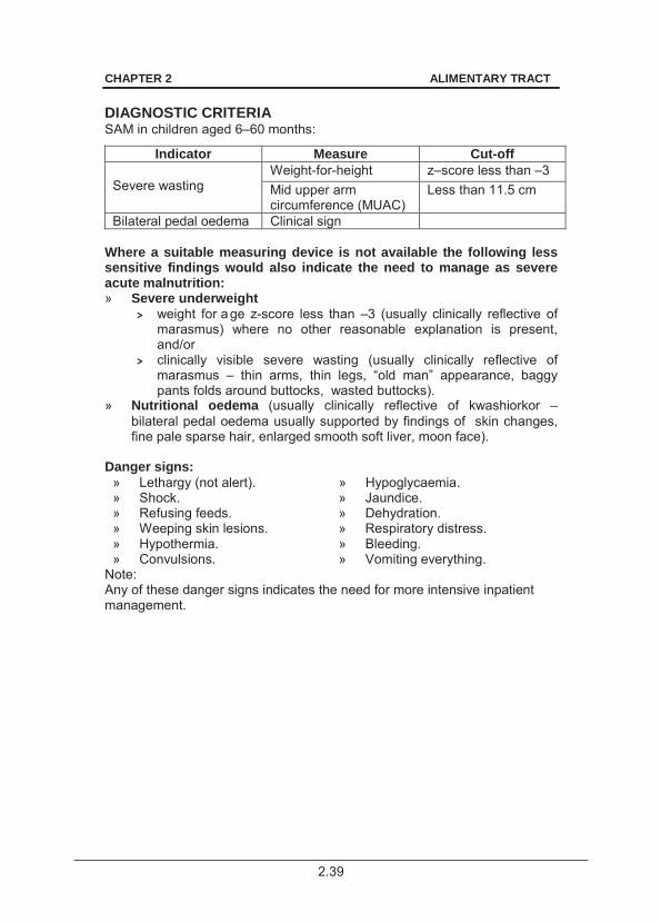
2.39
CHAPTER 2 ALIMENTARY TRACT DIAGNOSTIC CRITERIA SAM in children aged 6–60 months:
Indicator Measure Cut-off Severe wasting
Weight-for-height z–score less than –3 Mid upper arm circumference (MUAC)
Less than 11.5 cm
Bilateral pedal oedema Clinical sign Where a suitable measuring device is not available the following less sensitive findings would also indicate the need to manage as severe acute malnutrition: » Severe underweight
> weight for age z-score less than –3 (usually clinically reflective of marasmus) where no other reasonable explanation is present, and/or
> clinically visible severe wasting (usually clinically reflective of marasmus – thin arms, thin legs, “old man” appearance, baggy pants folds around buttocks, wasted buttocks).
» Nutritional oedema (usually clinically reflective of kwashiorkor – bilateral pedal oedema usually supported by findings of skin changes, fine pale sparse hair, enlarged smooth soft liver, moon face).
Danger signs:
» Lethargy (not alert). » Hypoglycaemia. » Shock. » Jaundice. » Refusing feeds. » Dehydration. » Weeping skin lesions. » Respiratory distress. » Hypothermia. » Convulsions.
» Bleeding. » Vomiting everything.
Note: Any of these danger signs indicates the need for more intensive inpatient management.
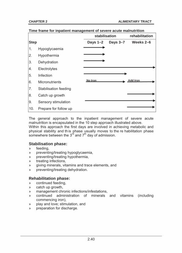
2.40
CHAPTER 2 ALIMENTARY TRACT Time frame for inpatient management of severe acute malnutrition stabilisation rehabilitation Step Days 1–2 Days 3–7 Weeks 2–6
1. Hypoglycaemia
2. Hypothermia
3. Dehydration
4. Electrolytes
5. Infection
6. Micronutrients
7. Stabilisation feeding
8. Catch up growth
9. Sensory stimulation
10. Prepare for follow up The general approach to the inpatient management of severe acute malnutrition is encapsulated in the 10 step approach illustrated above. Within this approach the first days are involved in achieving metabolic and physical stability and th is phase usually moves to the re habilitation phase somewhere between the 3rd and 7th day of admission. Stabilisation phase: » feeding, » preventing/treating hypoglycaemia, » preventing/treating hypothermia, » treating infections, » giving minerals, vitamins and trace elements, and » preventing/treating dehydration. Rehabilitation phase: » continued feeding, » catch up growth, » management chronic infections/infestations, » continued administration of minerals and vitamins (including
commencing iron), » play and love; stimulation, and » preparation for discharge.
No Iron Add Iron
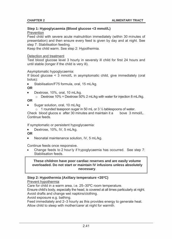
2.41
CHAPTER 2 ALIMENTARY TRACT Step 1: Hypoglycaemia (Blood glucose <3 mmol/L) Prevention Feed child with severe acute malnutrition immediately (within 30 minutes of presentation) and then ensure every feed is given by day and at night. See step 7: Stabilisation feeding. Keep the child warm. See step 2: Hypothermia. Detection and treatment Test blood glucose level 3 hourly in severely ill child for first 24 hours and until stable (longer if the child is very ill). Asymptomatic hypoglycaemia: If blood glucose < 3 mmol/L in asymptomatic child, give immediately (oral bolus): Stabilisation/F75 formula, oral, 15 mL/kg.
OR Dextrose, 10%, oral, 10 mL/kg.
o Dextrose 10% = Dextrose 50% 2 mL/kg with water for injection 8 mL//kg. OR Sugar solution, oral, 10 mL/kg
o 1 rounded teaspoon sugar in 50 mL or 3 ½ tablespoons of water. Check blood glucos e after 30 minutes and maintain it a bove 3 mmol/L. Continue feeds. If symptomatic or persistent hypoglycaemia: Dextrose, 10%, IV, 5 mL/kg.
OR Neonatal maintenance solution, IV, 5 mL/kg.
Continue feeds once responsive. Change feeds to 2 hour ly if hypoglycaemia has occurred. See step 7:
Stabilisation feeds.
These children have poor cardiac reserves and are easily volume overloaded. Do not start or maintain IV infusions unless absolutely
necessary.
Step 2: Hypothermia (Axillary temperature <35ºC) Prevent hypothermia Care for child in a warm area, i.e. 25–30ºC room temperature. Ensure child’s body, especially the head, is covered at all times particularly at night. Avoid drafts and change wet napkins/clothing. Avoid exposure e.g. bathing. Feed immediately and 2–3 hourly as this provides energy to generate heat. Allow child to sleep with mother/carer at night for warmth.
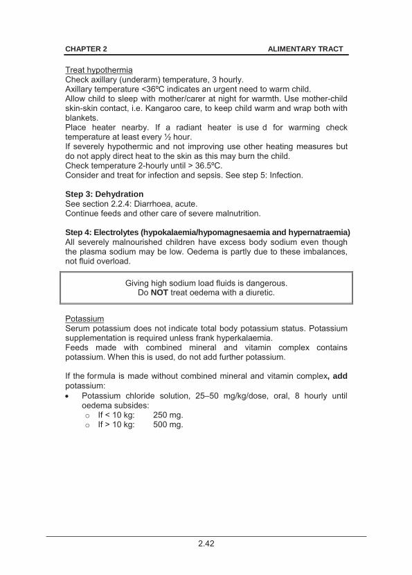
2.42
CHAPTER 2 ALIMENTARY TRACT Treat hypothermia Check axillary (underarm) temperature, 3 hourly. Axillary temperature <36ºC indicates an urgent need to warm child. Allow child to sleep with mother/carer at night for warmth. Use mother-child skin-skin contact, i.e. Kangaroo care, to keep child warm and wrap both with blankets. Place heater nearby. If a radiant heater is use d for warming check temperature at least every ½ hour. If severely hypothermic and not improving use other heating measures but do not apply direct heat to the skin as this may burn the child. Check temperature 2-hourly until > 36.5ºC. Consider and treat for infection and sepsis. See step 5: Infection. Step 3: Dehydration See section 2.2.4: Diarrhoea, acute. Continue feeds and other care of severe malnutrition. Step 4: Electrolytes (hypokalaemia/hypomagnesaemia and hypernatraemia) All severely malnourished children have excess body sodium even though the plasma sodium may be low. Oedema is partly due to these imbalances, not fluid overload.
Giving high sodium load fluids is dangerous. Do NOT treat oedema with a diuretic.
Potassium Serum potassium does not indicate total body potassium status. Potassium supplementation is required unless frank hyperkalaemia. Feeds made with combined mineral and vitamin complex contains potassium. When this is used, do not add further potassium. If the formula is made without combined mineral and vitamin complex, add potassium: Potassium chloride solution, 25–50 mg/kg/dose, oral, 8 hourly until
oedema subsides: o If < 10 kg: 250 mg. o If > 10 kg: 500 mg.
2.43
CHAPTER 2 ALIMENTARY TRACT Magnesium Feeds made with combined mineral and vitamin complex or trace element mix contains magnesium. If formula is made without either of these additives, add magnesium: Trace element mix, oral, daily.
o If < 10 kg: 2.5 mL. o If > 10 kg: 5 mL.
OR Magnesium sulphate 50%, oral, 0.2 mL/kg as a on ce daily dose for at
least 2 weeks. The IV preparation can be given orally. Step 5: Infection Antibiotics Start antibiotics on the first day at admission. If the child has no danger signs, is alert and feeding well: Amoxicillin, oral, 30 mg/kg/dose 8 hourly for 5 days.
All other children: Ampicillin, IV/IM, 50 mg/kg 6 hourly for 7 days.
o Avoid IV infu sions, if p ossible. Use a hep arin lock to av oid fluid overload because of poor cardiac reserves.
PLUS Gentamicin, IV, 6 mg/kg once daily for 7 days.
As soon as ther e is a re sponse and patient can tolerate oral med ication change ampicillin to amoxicillin and continue with gentamicin: Amoxicillin, oral, 30 mg/kg/dose 8 hourly for a further 5 days.
PLUS Gentamicin, IV/IM, 6 mg/kg once daily for 7 days.
If the child is severely ill or fails to improve after 48 hours: Third generation cephalosporin, e.g.: Ceftriaxone, IV/IM, 50 mg/kg/dose once daily.
o If meningitis suspected: use 80 mg/kg/dose.
CAUTION: USE OF CEFTRIAXONE
After 28 days of age IV ceftriaxone and IV calcium containing fluids may be given but only sequentially with the giving set flushed well between the two
products.
Note: Do not use ceftriaxone in neonates. If child does not improve after 5 days, or deteriorates: Refer to higher level of care.
2.44
CHAPTER 2 ALIMENTARY TRACT Intestinal worm infestation Treat after the acute phase: Children 1–2 years of age: Mebendazole, oral, 100 mg 12 hourly for three days.
Children > 2 years: Mebendazole, oral, 500 mg as a single dose immediately.
HIV and TB In children with HIV and TB, good recovery from malnutrition is possible but may take longer. Treatment failure of malnutrition may be more common. Actively investigate for TB and HIV as soon as possible. TB is difficult to diagnose and confirm. Ask about contacts, symptoms, do tuberculin skin test (TST) and chest X-ray. If TST negative, repeat just before discharge. If TB is cli nically likely, presumptive TB treatment is often reasonable, but once begun should be completed. See section 15.1.4: Tuberculosis, pulmonary. HIV is relatively simple to diagnose and confirm. Children < 18 months: PCR and confirm with viral load > 10 000 copies/mL. Children months: rapid test/ELISA and confirm with different rapid test/ELISA. ART is the same as for HIV negative children. Once the child enters the rehabilitative phase, commence antiretroviral therapy without delay if HIV positive. See section 9.1: Human immunodeficiency virus infections. Step 6: Micronutrients Vitamins Vitamin A, oral, as a single dose:
Age Dose No. of capsules Infants < 6 months: 50 000 IU 1 capsule Infants 6–11 months: 100 000 IU 1 capsule Children 12 months to 5 years: 200 000 IU 1 capsule
Record doses in the Road-to-Health booklet. All children with clinical signs of severe vitamin A deficiency (eye changes: xerophthalmia, corneal ulceration, Bitots spots, corneal clouding) and severe measles: Vitamin A, oral, 3 doses.
o First dose, immediately; second dose on day 2 and third dose after 14 days.
o Record the dose given in prescription and the Road to Health Book.
2.45
CHAPTER 2 ALIMENTARY TRACT If on feeds with combined mineral and vitamin complex: Folic acid, oral, 2.5 mg as a single dose.
If not on feeds with combined mineral and vitamin complex: Folic acid, oral, 2.5 mg as a single daily dose.
PLUS Multivitamin, oral, 5 mL as a single daily dose.
Anaemia in malnourished children Non-acute management: Although anaemia is common, do N OT give iron i nitially but wait until the child has a good appetite and starts gaining weight (usually by the second week). Treat severe anaemia with blood transfusion, if: » Symptomatic anaemia (Hb usually below 4 g/dL). OR » If there is respiratory distress with a low Hb. Packed red cells, IV, 5 mL/kg administered over 3 hours.
PLUS Furosemide, IV, 1 mg/kg at the start of the transfusion.
Repeat only if sev ere anaemia or respiratory distress persists and th e haemoglobin is still low. Once gaining weight and oedema has resolved: Iron, oral, 2 mg/kg elemental iron per dose 8 hourly with meals.
o Continue for at least 2 months to replace iron stores. Step 7: Stabilisation feeding Immediate: stabilisation phase: Begin feeding immediately – do not miss feeds. Give “F75/stabilising feed” at 130 mL/kg/day divided into 3 hourly feeds, i.e. 8 times daily. Give all feeds including that at 03h00. If child has gross oedema i.e. if the oedema is up to or b eyond the knee or anasarca, give 100 mL/kg initially and increase progressively. Monitor and record intake carefully.
F75 formula/Stabilisation Fresh cow’s milk 300 mL Sugar 100 g Vegetable oil 20 g Combined mineral and vitamin complex* indicated by insert Water to make up to: 1 000 mL
* If no combined mineral and vitamin complex: Trace element mix, oral, 20 mL daily.
2.46
CHAPTER 2 ALIMENTARY TRACT If danger signs, hypothermia or hypoglycaemia present, feed the same daily volume but divided into 2 h ourly feeds, i.e. 12 t imes daily. Give all feed s including those at 02h00 and 04h00. Give from a cup. Very weak children may be fed by spoon, dropper or syringe. If feeds refused/not finished (i.e. less than 80% of daily amount taken) give all feeds via nasogastric tube. Weigh daily and plot weight gain. Readiness to enter the rehabilitation phase is signalled by a return of appetite, usually about one week after admission. Step 8: Transition feeding and catch up growth Feeding (rehabilitation phase) » Transition
For the first two days replace the initial feeds with equal amounts of “rebuilding/catch-up/F100 formula”. Gradually increase the volume by 10 mL/feed until some formula remains unfinished, usually ± 200 mL/kg/day. When appetite returns introduce a modified diet. Balance the intake by giving 3 modified meals and 5 fe eds of F100. Prepare food without adding salt.
F100 formula/Rebuilding formula (catch-up) Fresh cow’s milk 880 mL Sugar 75 g Vegetable oil 20 mL Combined mineral and vitamin complex * as indicated by insert Water to make up to: 1 000 mL * If no combined mineral and vitamin complex: Trace element mix, oral, 20 mL.
Monitor progress after the transition by assessing the rate of weight gain. Weigh child each morning before feeding and plot the weight. Each week calculate and record weight gain as g/kg/day. If weight gain is: » poor (< 5 g/kg/day) – child requires full reassessment. » moderate (5–10 g/kg/day) – check whether intake targets are being met,
or if infection has been overlooked. » good (>10 g/kg/day) – continue to praise staff and mothers.

2.47
CHAPTER 2 ALIMENTARY TRACT Step 9: Sensory stimulation Stimulation and loving care » Provide tender loving care. » Help and encourage mothers to comfort, feed and play with their
children. » Involve occupational therapist, if available, for structured play otherwise
arrange this as best possible in the ward. » Provide a stimulation program in the ward. Step 10: Prepare for follow up Preparation for discharge » Obtain information on household food security, family background and
socio-economic status and refer appropriately. » Instruct mothers how to modify family foods, how often to feed, what
and how much to give. » Ready to Use Therapeutic Foods (RUTF) may be supplied to facilitate
earlier discharge where this is indicated and available. » Involve mother in discharge planning and follow up plans. » Social assessment. Before discharge, ensure parent/caregiver is able to
access food for the child, ensure all financial supports and grants have been accessed. A social worker may assist in ensuring this. The social worker should also assess for other social risks.
» Make follow-up arrangements. Link patient to PHC systems and Family Health Teams/CCG Workers for close follow-up and monitoring of feeding and compliance with therapeutic feeding program.
» Ensure all immunisations are up to date. » Do not discharge any malnourished child without having adequately
investigated for TB and HIV infection. Repeat TST before discharge as immunity may have returned to normal.
» Write full clinical summary in Road to Health book. Discharge criteria » good appetite, » no infection, » no oedema, » continuous good weight gain for last 5 days, » playful and alert, and » all preparation in place for discharge.
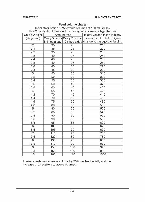
2.48
CHAPTER 2 ALIMENTARY TRACT
Feed volume charts Initial stabilisation /F75 formula volumes at 130 mL/kg/day
Use 2 hourly if child very sick or has hypoglycaemia or hypothermia Childs Weight
(kilograms) Amount feed If total volume taken in a day
is less than the below figure change to nasogastric feeding
Every 3 hours 8 times a day
Every 2 hours 12 times a day
2 35 25 210 2.1 35 25 220 2.2 35 25 230 2.3 40 25 240 2.4 40 25 250 2.5 40 25 260 2.6 40 30 270 2.8 45 30 290 3 50 30 310
3.2 50 35 330 3.4 55 35 350 3.6 60 40 370 3.8 60 40 400 4 65 45 420
4.2 70 45 440 4.4 70 50 460 4.6 75 50 480 4.8 80 50 500 5 80 55 520
5.2 85 55 540 5.4 90 60 560 5.6 90 60 580 5.8 95 65 600 6 100 65 620
6.5 105 70 670 7 115 75 730
7.5 120 80 780 8 130 90 830
8.5 140 90 880 9 150 100 940
9.5 150 100 990 10 160 110 1050
If severe oedema decrease volume by 25% per feed initially and then increase progressively to above volumes.

2.49
CHAPTER 2 ALIMENTARY TRACT 2.5 RICKETS E55.0 DESCRIPTION Failure to calcify osteoid tissue in a growing child, usually due to deficiency of vitamin D, its ac tive metabolites, calcium, phosphorus or other ra re causes. This leads to bone deformity.
Occurs in ex-premature babies during infancy and in children with developmental disability, on anticonvulsants or not exposed to sunlight. In older children it is caused by renal tubulopathy and other rare conditions. DIAGNOSTIC CRITERIA Clinical » Bowing of long bones, widening of metaphyses and cranial bossing. » Occasionally convulsions or tetany due to hypocalcaemia. Investigations » Elevated alkaline phosphatase. » Serum calcium and/or phosphate abnormalities. » X-ray of wrists. GENERAL AND SUPPORTIVE MEASURES » Prevent vitamin D deficiency. » Exposure to sunlight, at least 3 hours a week.
Note: Breast milk does not contain adequate vitamin D to prevent deficiency. Ensure adequate sunlight exposure of infant or provide vitamin D until weaning.
» Normal vitamin D-containing diet for lactating mothers. MEDICINE TREATMENT Prophylaxis For premature babies: Vitamin D, oral, 800 IU, once daily.
Infants who are exclusively breastfed or not on adequate volume of commercial milk formula: Vitamin D, oral, 400 IU, once daily.
Treatment of active rickets Treat only after confirmation of active rickets on X–ray. Vitamin D, oral, 5 000 IU, once daily, in addition to milk in the diet.
o Repeat X–ray after 6–8 weeks. o If no radiological improvement, further investigation is required. o If healing occurs, continue for 3 months. Confirm complete healing
and adequate diet for the future.
2.50
CHAPTER 2 ALIMENTARY TRACT REFERRAL » Rickets presenting in children older than 2 years. » No radiological response to treatment after 6–8 weeks. » Incomplete radiological response. » Rickets secondary to other disease processes. 2.6 WORM BOLUS B77 DESCRIPTION Partial or complete obstruction of t he bowel by a “k not” of Ascaris lumbricoides curled around each other. Usually presents with cramping abdominal pain with/without other evidence of obstruction. May occasionally lead to local necrosis and perforation of the small bowel. DIAGNOSTIC CRITERIA Clinical Cramping abdominal pain associated with/without a palpable worm mass which may also be identified on X-ray abdomen straight or with contrast (when considered safe). Exclusion of other cause of acute abdomen or acute abdominal pain. GENERAL AND SUPPORTIVE MEASURES » Maintain fluid, electrolyte and nutritional needs, IV ro ute may be
needed. » Nil per mouth and free drainage, where clinically indicated. » Observe for fai lure of resolution, complete obstruction or evidence of
necrosis/perforation. » Surgery for complete obstruction, evidence of necrosis or perforation. » Identify possible iron deficiency. MEDICINE TREATMENT Once the bolus resolve treat the ascaris: Children 1–2 years of age: Mebendazole, oral, 100 mg 12 hourly for three days.
Children > 2 years: Mebendazole, oral, 500 mg as a single dose immediately.
REFERRAL » Inability to manage surgical problems, if present. » Prolonged obstruction.
2.51
CHAPTER 2 ALIMENTARY TRACT 2.7 RECURRENT ABDOMINAL PAIN R10.4 DESCRIPTION Recurrent abdominal pain for which no cause can be found occurring at least monthly for 3 consecutive months with severity that interferes with routine function of the child. DIAGNOSTIC CRITERIA Clinical » Peri-umbilical pain associated with belching, bloating with negative
findings on clinical evaluation and no response to acid-blocking medication OR pain below the um bilicus accompanied by abdominal cramps, bloating and distension and with an altered bowel pattern that are consistent with Irritable Bowel Syndrome in adults.
» Either of the above syndromes with the ex clusion of organic disease with appropriate investigation.
» Avoid excessive investigation where the diagnosis is strongly suspected in the presence of a normal clinical evaluation.
» Exclude the following: > Urinary tract infections, urinary tract anomalies, renal disease. > GIT infection, infestation or inflammation. > Chronic abdominal conditions such as tumours or infections, e.g.
TB abdomen. > Gall bladder disease. > Pancreatic disease.
GENERAL AND SUPPORTIVE MEASURES » Manage psychological stressors, anxiety or depression, where present,
appropriately. » Reassure child and family. » Counselling to avoid the re-inforcement of the symptoms with secondary
gain. » Adequate dietary fibre in children with irritable bowel syndrome-type
condition. MEDICINE TREATMENT Manage constipation, where present. See section 2.2.2: Constipation/faecal loading. Manage comorbid an xiety or depression appropriately. See section 14.3.1: Depression in Childhood and Adolescence and section 14.4: Anxiety disorders. REFERRAL » Failure to respond to management » For appropriate psychiatric / psychological management, if not locally available.
3.1
CHAPTER 3 BLOOD AND BLOOD-FORMING ORGANS
3.1 ANAEMIA, APLASTIC D61.0 DESCRIPTION Pancytopaenia caused by bone marrow failure w ith a h ypocellular bone marrow without infiltration or fibrosis. May be acquired or inherited. Inherited bone marrow failure syndromes include Fanconi anaemia which has specific associated phenotypic features and chromosomal abnormalities. DIAGNOSTIC CRITERIA Clinical » Pallor, petechiae, purpura, bleeding, with frequent and/or severe
infections. » Phenotypic features of Fanconi anaemia include:
> café au lait spots, > skin pigmentary changes, > short stature and dysmorphic faces, > hypoplasia/absence of radius, fingerised thumb, > microcephaly, small eyes, hyperreflexia, > renal tract and cardiac abnormalities, > hypogonadism.
Investigations » Full blood count shows pancytopaenia, with anaemia (may be
macrocytic), leucopaenia and thrombocytopaenia. » Hypoplastic bone marrow on trephine biopsy. GENERAL AND SUPPORTIVE MEASURES Limit the u se of blood products as the patient may be sensitised and jeopardise a future bone marrow transplant. Avoid contact sport. MEDICINE TREATMENT For symptomatic anaemia (usually Hb < 7 g/dL): Packed red cells, IV.
o Use leukocyte depleted products.
3.2
CHAPTER 3 BLOOD AND BLOOD-FORMING ORGANS For active bleeding: Platelets, IV, 20 mL/kg, administered immediately and rapidly over 15–
30 minutes through a platelet giving set. o If transplant is a possibility, use single donor apheresis platelets
rather than pooled random donor platelets; preferably group specific.
For fever (T > 38ºC), broad spectrum antibiotics: Take blood cultures first. Ceftriaxone, IV, 50–80 mg/kg once daily.
OR If < 1 month old: Cefotaxime, IV, 25–50 mg/kg/dose, 6 hourly.
AND Amikacin, IV, 25 mg/kg as a loading dose, then 18 mg/kg/dose once
daily. REFERRAL » All cases of suspected aplastic anaemia. » Stabilise patient before transport with blood and/or platelet
transfusions, if necessary, after consultation with a paediatrician or paediatric haematologist.
» All cases for consideration for bone marrow transplant or immunosuppressive therapy in the case of acquired aplastic anaemia.
3.2 ANAEMIA, HAEMOLYTIC D55–59 DESCRIPTION Anaemia caused by excessive destruction of red blood cells. Destruction may be due to: » Corpuscular defects:
> abnormalities of the cell membrane (e.g. hereditary spherocytosis), > enzyme abnormalities (e.g. G6PD deficiency), or > abnormal haemoglobin (e.g. sickle cell anaemia, thalassaemia).
» Extracorpuscular defects: > Auto-immune or isoimmune:
idiopathic warm or cold antibodies, infection triggered e.g. Mycoplasma pneumonia, medicine related e.g. penicillin, secondary to auto-immune disorders e.g. SLE, rheumatoid arthritis, secondary to tumours e.g. lymphoma, thymoma.
> Non-immune: secondary to microangiopathy e.g.: haemolytic uraemic syndrome, infections causing haemolysis e.g. malaria, miscellaneous causes including hypersplenism.
3.3
CHAPTER 3 BLOOD AND BLOOD-FORMING ORGANS DIAGNOSTIC CRITERIA Clinical » Pallor, jaundice, fatigue. » Splenomegaly. Investigations » Anaemia. » Evidence of haemolysis:
> anaemia, > decreased haptoglobin, > reticulocytosis, > unconjugated hyperbilirubinaemia, > increased lactate dehydrogenase (LDH), > urobilinogen in the urine.
» Direct Coombs test (direct antiglobulin) is positive with autoimmune haemolysis.
» Renal function is abnormal in haemolytic uraemic syndrome. » Exclude other autoimmune disorders. » Consider underlying neoplasms. » In patients receiving recurrent transfusions (e.g. thalassaemia) monitor
ferritin levels 3-monthly and discuss with your referral centre if elevated > 1000 mcg/L.
GENERAL AND SUPPORTIVE MEASURES » After appropriate investigations, transfuse the patient and then discuss
with a paediatrician or paediatric haematologist. » Coombs-positive auto-immune haemolytic anaemia may require
transfusion with the least incompatible blood (if cross-matching yields no compatible units).
» In G6PD deficiency, avoid medicines known to cause haemolysis (e.g. aspirin, sulphonamides and primaquine) and be sure to give the patient a list of such medicines at discharge.
MEDICINE TREATMENT Warm antibody autoimmune haemolytic anaemia Under specialist supervision: Prednisone, oral, 2 mg/kg/24 hours until a satisfactory response is
obtained. o Continue treatment for a minimum of 4 weeks. o Taper dose slowly over several weeks while monitoring for relapse.
Chronic haemolytic anaemia All patients indefinitely: Folic acid, oral, 2.5–5 mg daily (depending on the size of the child).
SURGICAL TREATMENT Splenectomy for hereditary spherocytosis with Hb < 10 g/dL only after the child’s fifth birthday.
3.4
CHAPTER 3 BLOOD AND BLOOD-FORMING ORGANS Pre-splenectomy Children who have been v accinated with PCV 7 as part of their ro utine should receive PCV 10 or 13 eight weeks before polysaccharide vaccine. Pneumococcal vaccine (polysaccharide), IM, 0.5 mL as a single dose.
Post splenectomy Phenoxymethylpenicillin, oral, 12 hourly.
o If < 5 years: 125 mg. o If > 5 years: 250 mg. o Give indefinitely until at least until 18 years.
Pneumococcal polysaccharide vaccine. Repeat vaccination as a booster 5 years after the initial dose.
Annual influenza vaccine.
Catch up conjugate pneumococcal vaccine o < 12 months of age: 3 dose series. o 12 months of age and older: 2 doses 8 weeks apart.
REFERRAL » Any child with haemolytic anaemia who has received more than 10
transfusions for assessment for chelation therapy. » All cases associated with evidence of haemolysis as above should be
managed in consultation with a paediatrician or paediatric haematologist.
3.3 ANAEMIA, MEGALOBLASTIC D53.1 DESCRIPTION Anaemia caused by a deficiency of folate and/or vitamin B12. DIAGNOSTIC CRITERIA Clinical » Pallor and fatigue. » Chronic diarrhoea. Investigations » Megaloblastic anaemia: elevated MCV (mean corpuscular volume) and
MCH (mean corpuscular haemoglobin). » Macro-ovalocytes on blood smear, hypersegmentation of neutrophils. » Decreased serum vitamin B12 or red blood cell folate. » Investigations to identify reason for folate or B12 deficiency, e.g.
malabsorption. » Pancytopaenia in severe cases. » Actively exclude leukaemia and aplastic anaemia which may cause
macrocytosis.

3.5
CHAPTER 3 BLOOD AND BLOOD-FORMING ORGANS GENERAL AND SUPPORTIVE MEASURES » Dietary modifications to ensure adequate intake of folate and vitamin B12. » Packed red blo od cell transfusion for sy mptomatic anaemia. Try to
avoid blood transfusion until all investigations have been done. MEDICINE TREATMENT Folic acid deficiency: Folic acid, oral, 5 mg daily until haemoglobin returns to normal value for age.
Prolonged treatment may be needed for malabsorption states and congenital deficiencies.
Vitamin B12 deficiency: Vitamin B12, IM, 500 m cg monthly. Prolonged treatment may be
needed. REFERRAL » All cases of megaloblastic anaemia, except clear nutritional folate
deficiency. 3.4 ANAEMIA, IRON DEFICIENCY D50.9 DESCRIPTION Iron deficiency is the most common cause of an aemia. The commonest causes of iron deficiency anaemia are poor nutritional intake, excessive milk ingestion and blood loss due to parasites (whipworm and hookworm). Lower limits of normal haemoglobin:
Age Hb (g/dL) Birth 13.5
6 weeks 9.5 3 months 10.0
6–12 months 10.5 12–18 months 10.5
18 months–4 years 11.0 4–7 years 11.0
7–12 years 11.5 12 years and older 12 (F) : 13 (M)
Practically: Ferrous gluconate syrup 1 mL/kg/day in 2 doses.
3.6
CHAPTER 3 BLOOD AND BLOOD-FORMING ORGANS DIAGNOSTIC CRITERIA Clinical Symptoms and signs vary with the severity of the deficiency:
» pallor, » delayed motor development, » fatigue, » pica, » irritability, » soft ejection systolic murmur, » behavioural and cognitive effects.
Investigations » Haemoglobin below normal for age. » Hypochromic microcytic anaemia. » Low MCV (mean corpuscular volume) and MCH (mean corpuscular
haemoglobin), increased red cell distribution width. » Decreased serum iron, ferritin and transferrin saturation. » Elevated total iron binding capacity. » Stool examination to identify intestinal parasites or to confirm occult
blood loss. » Iron studies are not necessary if nutritional iron deficiency is strongly
suspected. Document a response to a trial of iron therapy to confirm the diagnosis.
Note: Chronic infections may also cause microcytic hypochromic anaemia. See section 3.5: Anaemia of chronic disorders (infection or disease). GENERAL AND SUPPORTIVE MEASURES » Dietary adjustment. » Counselling. MEDICINE TREATMENT Treatment Iron (elemental), oral, 3 mg/kg/dose 12 hourly with meals.
Elemental iron per preparation
Ferrous gluconate elixir
350 mg/5 mL
40 mg elemental iron/5 mL
8 mg elemental iron per mL
Ferrous gluconate syrup 250 mg/5 mL
30 mg elemental iron per 5 ml
6 mg elemental iron per mL
Ferrous lactate drops 25 mg/mL
25 mg elemental iron/ mL
1 mg elemental iron in 0.04 mL
Ferrous sulphate compound tablets
170 mg
± 65 mg elemental iron per table
± 65 mg elemental iron per tablet
3.7
CHAPTER 3 BLOOD AND BLOOD-FORMING ORGANS Follow up at monthly intervals. The expected response is an increase in Hb of 2 g/dL or more in 3 weeks. Continue for 3–4 weeks after Hb is normal to replenish body iron stores. The reticulocyte count will increase if there is a positive response and may be useful where the diagnosis is in doubt, if done within 1–2 weeks after iron therapy is started. Treat for worms. Children 1–2 years of age: Mebendazole, oral, 100 mg 12 hourly for three days.
Children > 2 years: Mebendazole, oral, 500 mg as a single dose immediately
CAUTION Iron is extremely toxic in overdose, particularly in children All medication should be stored out of reach of children
Prophylaxis All premature babies, day 15 to 1 year: Iron (elemental), oral, 2 mg/kg daily. Multivitamin, drops, oral, 0.3 mL daily.
o Increase dose according to age. Full term babies after 2 months: Iron (elemental), oral, 1 mg/kg daily for one year. Multivitamin, drops, oral, 0.6 mL daily
REFERRAL » Patients not responding to adequate therapy. » Patients in whom easily treatable causes for non-response have been
excluded, e.g.: > non-adherence to therapy, > ongoing blood loss, > ongoing infection.
3.5 ANAEMIA OF CHRONIC DISORDERS (INFECTION OR
DISEASE) D63 DESCRIPTION Anaemia caused by chronic infection or disease. This may be due to interference with nutrient supply or suppression of haemopoiesis. Iron may be trapped in the reticuloendothelial system resulting in relative iron deficiency.
3.8
CHAPTER 3 BLOOD AND BLOOD-FORMING ORGANS Symptomatic anaemia may manifest with tachypnoea, tachycardia not attributable to other causes and heart failure. DIAGNOSTIC CRITERIA Clinical » Pallor, fatigue; » Features of malnutrition or chronic infection e.g. TB, HIV, chronic renal
failure; or » Auto-immune disease may be present. Investigations » Haemoglobin low with usually normocytic, normochromic red cells (may
be microcytic). » TST, chest X-ray and renal function tests. GENERAL AND SUPPORTIVE MEASURES » Emphasise a nutritionally balanced diet that is adequate in protein,
vitamins and minerals for nutritional rehabilitation. » Transfuse for symptomatic anaemia only. MEDICINE TREATMENT » Treat underlying infection, e.g. TB. » Defer iron treatment until acute diseases are controlled, then provide
extra iron (see above) and multivitamins. REFERRAL » All cases with unresolving anaemia and no cause found. 3.6 ANAEMIA, SICKLE CELL D57 DESCRIPTION Haemolytic anaemia due to homozygous inheritance of the sickle cell gene. Patients may experience complications: » Painful vaso-occlusive crises. » Haemolytic crises (usually secondary to infection). » Aplastic crises. » Thrombotic crises, e.g. acute chest syndrome, priapism or stroke. » Splenic sequestration. » Severe infections.
3.9
CHAPTER 3 BLOOD AND BLOOD-FORMING ORGANS
DIAGNOSTIC CRITERIA Clinical » Pallor, jaundice, fatigue all of which may worsen abruptly
(sequestration crisis, aplastic crisis). » Features of complications:
> painful swelling of the hands and feet (dactylitis); > bone pain, abdominal pain; > chest pain, fever, dyspnoea (acute chest syndrome); > convulsions, hemiparesis; > priapism.
Investigations » Laboratory features of haemolytic anaemia. See section 3.2: Anaemia,
haemolytic. » Haemoglobin electrophoresis shows an SS pattern (both parents will
be AS). GENERAL AND SUPPORTIVE MEASURES » Avoid exposure to cold, dehydration and stress. » Increase fluid intake during painful crises. » Heat and/or massage for pain. MEDICINE TREATMENT For sequestration crisis or aplastic crisis: Packed red cells, IV, 15 mL/kg.
o Avoid over-transfusing patients since the increase in viscosity may aggravate the v asculopathy associated with sickle cell disease (a Hb threshold of 13 g/dL has been recommended).
If hypoxic: Oxygen, by face mask.
Exchange transfusions may be us ed to tr eat severe complications (see referral criteria). Prophylaxis against infection Given to all children because functional asplenia is present by 1–2 years of age. Routine vaccinations during infancy.
Catch up conjugate pneumococcal vaccine
o If < 12 months of age: 3 dose series. o If 12 months of age and older: 2 doses 8 weeks apart.
o Pneumococcal polysaccharide vaccine at 2 years (at least 8 weeks after conjugate vaccine). Repeat vaccination as a booster 5 years after the initial dose.
3.10
CHAPTER 3 BLOOD AND BLOOD-FORMING ORGANS Annual influenza vaccination.
Phenoxymethylpenicillin, oral, 12 hourly.
o < 5 years: 125 mg o > 5 years: 250 mg o Give indefinitely.
Treatment Analgesia as required: Paracetamol, oral, 15 mg/kg 6 hourly.
AND Ibuprofen, oral, 10 mg/kg 8 hourly.
AND Codeine phosphate, oral, 0.5–1 mg/kg 4 hourly.
If this is inadequate, see section 20.1: Management of pain. Hydroxyurea, oral, 15 mg/kg.
o Increase by 5 mg/kg every12 weeks. o Maximum dose: 35 mg/kg daily.
Infections: All children < 6 months with axillary temperature and all children > 6 months: 3rd generation cephalosporins, e.g.: Cefotaxime, IV, 25–50 mg/kg/dose, 6 hourly (neonates).
OR Children > 2 months: Ceftriaxone, IV, 50–80 mg/kg/dose once daily.
Acute chest syndrome: Consult a paediatrician. REFERRAL » All children with sickle cell anaemia should be managed in consultation
with a paediatric haematologist or paediatrician. » All children with severe complications that may benefit from exchange
transfusion or intensive care, e.g. stroke, severe vaso-occlusive disease and acute chest syndrome.
3.7 HAEMOPHILIA A AND B D66.7/D66.8 DESCRIPTION Haemophilia A and h aemophilia B are ch ronic bleeding disorders caused, respectively, by a lack of clotting factor VIII or clotting factor IX.
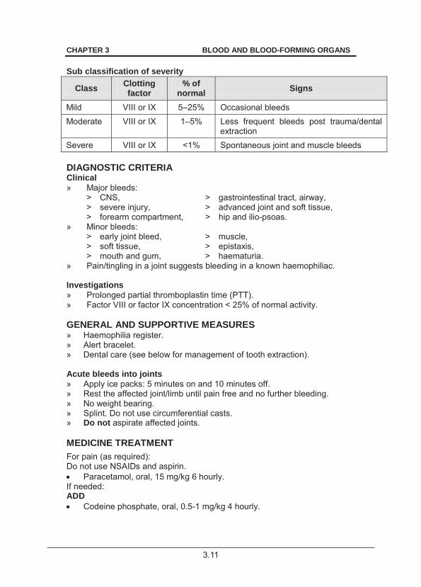
3.11
CHAPTER 3 BLOOD AND BLOOD-FORMING ORGANS Sub classification of severity
Class Clotting factor
% of normal Signs
Mild VIII or IX 5–25% Occasional bleeds Moderate VIII or IX 1–5% Less frequent bleeds post trauma/dental
extraction Severe VIII or IX <1% Spontaneous joint and muscle bleeds DIAGNOSTIC CRITERIA Clinical » Major bleeds:
> CNS, > gastrointestinal tract, airway, > severe injury, > advanced joint and soft tissue, > forearm compartment, > hip and ilio-psoas.
» Minor bleeds: > early joint bleed, > muscle, > soft tissue, > epistaxis, > mouth and gum, > haematuria.
» Pain/tingling in a joint suggests bleeding in a known haemophiliac. Investigations » Prolonged partial thromboplastin time (PTT). » Factor VIII or factor IX concentration < 25% of normal activity. GENERAL AND SUPPORTIVE MEASURES » Haemophilia register. » Alert bracelet. » Dental care (see below for management of tooth extraction). Acute bleeds into joints » Apply ice packs: 5 minutes on and 10 minutes off. » Rest the affected joint/limb until pain free and no further bleeding. » No weight bearing. » Splint. Do not use circumferential casts. » Do not aspirate affected joints. MEDICINE TREATMENT
For pain (as required): Do not use NSAIDs and aspirin. Paracetamol, oral, 15 mg/kg 6 hourly.
If needed: ADD Codeine phosphate, oral, 0.5-1 mg/kg 4 hourly.
3.12
CHAPTER 3 BLOOD AND BLOOD-FORMING ORGANS
For bleeds Emergency treatment while awaiting transfer, if indicated. If serious bleeding in known haemophiliac, and no factor available: Lyophilised plasma, IV, 2 0 mL/kg. Lyophilised plasma contains a
minimum of 0.4 units/mL of each coagulation factor. OR Fresh frozen plasma, IV, 20 mL/kg.
Factor VIII deficiency (with no inhibitor present) Give until patient is pain free and has full movement of j oint/limb. Administration should be 12 hourly for major bleeds, but may be d aily for minor bleeds. Minor bleeds: Factor VIII, intravenous, 20 units/kg.
Major bleeds: Factor VIII, intravenous, 40 units/kg
Use the entire contents of the appropriate volume ampoule. For intracranial bleeds: Factor VIII, intravenous, 40 units/kg 6 hourly.
Decrease frequency if trough level is > 60%, if possible. Factor IX deficiency (with no inhibitor present) Give daily until patient is pain free and has full movement of the joint/limb. Minor bleeds Factor IX, intravenous, 20 units/kg.
Major bleeds: Factor IX, intravenous, 60 units/kg.
Available product is factor IX complex and also contains factors II, VII and X. Home treatment Home treatment of bleeds is promoted by haemophilia treatment centres. Patients or caregivers are educated on the storage, reconstitution and administration of factor and provided with a s upply of fa ctor to be kept at home for use in the event of a bleed. Factor use and bleeding episodes are monitored through the use of an appropriate chart which can be reviewed at consultations and medication collection. Haemophilia with inhibitors Refer for assessment and planning with a haematologist for use of factor VIII or IX at tw ice the usual dose or factor VIII inhibitor-bypassing activity (FEIBA) or recombinant Factor VIIa.
3.13
CHAPTER 3 BLOOD AND BLOOD-FORMING ORGANS For dental extraction: Check that inhibitors are absent. Admit for procedure and post-procedure care and observation. Haemophilia A: Factor VIII, intravenous, 40 units/kg, immediately before extraction.
AND Tranexamic acid, oral, 25 mg/kg/dose 6–8 hourly for 5 days.
Haemophilia B: Factor IX, intravenous, 40 units/kg, immediately before extraction.
For mucous membrane bleeds Tranexamic acid, oral, 25 mg/kg/dose 6–8 hourly.
o Contra-indicated in haematuria. o Use with caution with factor IX complex or factor VIII inhibitor-
bypassing activity and preferably only 12 hours after administration of the factor.
REFERRAL » All cases with suspected or established haemophilia (prolonged PTT
and normal INR), for assessment, genetic counselling and planning of management to a haemophilia treatment centre.
3.8 VON WILLEBRAND DISEASE D68.0 DESCRIPTION Von Willebrand disease is the most common congenital bleeding disorder and is due to reduced amounts or abnormal forms of von Willebrand factor in the circulation. DIAGNOSTIC CRITERIA Clinical » Recurrent epistaxis, prolonged bleeding from lacerations, easy bruising
or gum bleeds. Investigations » Reduction in one or more of the following:
> von Willebrand factor antigen, > ristocetin co-factor and/or collagen binding activity, > factor VIII coagulant activity.
3.14
CHAPTER 3 BLOOD AND BLOOD-FORMING ORGANS GENERAL AND SUPPORTIVE MEASURES » Apply pressure to the bleeding site. » For tooth socket bleeds bite down on a piece of gauze. » For epistaxis, see section 17.3: Epistaxis (Nose bleeds).
Avoid aspirin and NSAIDS
MEDICINE TREATMENT For mild bleeds: Desmopressin, IV, 0.3 mcg/kg in 5 0 mL sodium chloride 0.9%
administered over 30 minutes. o Repeat at 12 and 24 hours, if needed. o Maximum: 3 doses. o Only after a therapeutic trial has demonstrated efficacy in th e
patient. For severe bleeds: Factor VIII, intravenous (Factor VIII containing von Willebrand factor).
o Initial dose: 30 units/kg. For mucous membrane bleeds: Tranexamic acid, oral, 25 mg/kg/dose 6–8 hourly.
For menorrhagia: Combined oral contraceptive, low dose.
REFERRAL » All suspected cases of v on Willebrand disease to a h aemophilia
treatment centre for assessment. 3.9 HAEMORRHAGIC DISEASE OF THE NEWBORN P53 See section 19.4: Haemorrhagic disease of the newborn. 3.10 IDIOPATHIC THROMBOCYTOPAENIC PURPURA (ITP) D69.3 DESCRIPTION Common bleeding disorder of childhood due to the auto-immune destruction of platelets.
3.15
CHAPTER 3 BLOOD AND BLOOD-FORMING ORGANS It occurs most frequently in children aged 2 t o 5 y ears and often follows infection with viruses. Chronic ITP (more than 6 mo nths duration) occurs in 10 to 20% of children with ITP. Complications include severe haemorrhage and bleeding into vital organs. DIAGNOSTIC CRITERIA Clinical » Sudden onset of br uising and bleeding, either spontaneously or aft er
minor trauma, into the skin and mucous membranes and rarely into the organs in an otherwise well child.
» The lesions may range from pinpoint petechial bleedings to l arge ecchymoses, and are often increased on pressure points.
» Epistaxis is common. » Exclude child abuse. » The presence of the following makes the diagnosis of ITP unlikely:
> splenomegaly, > masses, > hepatomegaly, > joint swelling, > lymphadenopathy, > bone pain, > rashes present other than petechiae or ecchymoses.
Investigations » Thrombocytopaenia with normal white cell count and differential, and
normal haemoglobin and red cell morphology, other than the effects of blood loss.
» Normal INR (PT) and partial thromboplastin time (PTT). » Abundant megakaryocytes on bone ma rrow aspiration with normal
erythroid and myeloid cellularity. » A normal LDH and uric acid help to rule out leukaemia. » Indications for bone marrow biopsy/aspiration: Prior to start ing steroids,
or any other abn ormality on FBC or any atypical cells on differential count.
» Test all newly-diagnosed cases for HIV. Follow up pat ients with diagnosis of IT P not c onfirmed with bone marrow aspiration for dev elopment of new clinical signs and abnormalities on laboratory investigations. GENERAL AND SUPPORTIVE MEASURES » Avoid:
> platelet transfusions unless bleeds are life-threatening, > contact sport, injury and trauma, and > dental procedures in acute phase.
» Re-assure patient and family that resolution usually occurs.
3.16
CHAPTER 3 BLOOD AND BLOOD-FORMING ORGANS MEDICINE TREATMENT Avoid medication that affects platelet function, e.g. NSAIDs and aspirin. Acute ITP Active bleeding: Prednisone, oral, 4 mg/kg/dose as a single daily dose for 4 days.
o Stop after 4 days without tapering dose. Chronic ITP Intermittent treatment if platelets 10 x 109/L and significant bleeding episodes: Prednisone, oral, 4 mg/kg/dose as a single daily dose for 4 days.
o Stop after 4 days without tapering dose. Acute life-threatening bleeds (e.g. intracranial bleeding): (acute or chronic ITP) Methylprednisolone, IV, 30 mg/kg/dose administered over 30–60 minutes
as a single daily dose for 3 days. o Maximum dose: 1 g. o Beware of arrhythmias, hypertension, etc. o Check BP daily.
AND Immunoglobulin, human normal, IV, 1 g/kg/day for 2 days.
Note: Immunoglobulins are reserved for the management of acute life threatening bleeds. AND After administration of methylprednisolone: Platelets, IV.
Consider emergency splenectomy. SURGICAL TREATMENT Consider splenectomy in chil dren 5 y ears and ol der with chronic ITP for more than one year plus significant bleeding or substantial limitation in activities as a result of the ITP. Pre-splenectomy Children who have been v accinated with PCV 7 as part of their ro utine should receive PCV 10 or 13 eight weeks before polysaccharide vaccine. Pneumococcal vaccine (polysaccharide), IM, 0.5 mL as a single dose.
3.17
CHAPTER 3 BLOOD AND BLOOD-FORMING ORGANS Post splenectomy Phenoxymethylpenicillin, oral, 12 hourly.
o If < 5 years of age: 125 mg. o If > 5 years of age: 250 mg. o Give indefinitely until at least until 18 years.
Pneumococcal polysaccharide vaccine. Repeat vaccination as a
booster 5 years after the initial dose.
Annual influenza vaccine.
Catch up conjugate pneumococcal vaccine: o If < 12 months of age: 3 dose series. o 12 months of age and older: 2 doses 8 weeks apart.
REFERRAL » Suspected ITP with unusual features such as splenomegaly or
lymphadenopathy. » ITP complicated by severe haemorrhage, bleeding into vital organs or
an intracranial haemorrhage. » ITP that fails to resolve in 6–12 months on adequate treatment (chronic
ITP). » If there is no local capacity to fully investigate the condition.
3.11 VENOUS THROMBO-EMBOLIC DISEASE I82 DESCRIPTION The occurrence of a n occlusive or non-occ lusive thrombus in the v enous circulation and/or consequent pulmonary embolus. It is usually associated with risk factors such as the presence of central venous catheters, venous stasis, endothelial damage and hypercoagulable states due to various causes, e.g. nephrotic syndrome. DIAGNOSTIC CRITERIA Clinical Depends on the site of thrombosis but may be silent. » Deep venous thrombosis of an extremity presents with unilateral limb
swelling. » Upper extremity thrombus may present with associated facial and neck
oedema. » Pulmonary embolus presents with sudden onset of shortness of breath
and chest pain. » Cerebral sinus venous thrombosis presents with seizures or o ther
neurological symptoms and signs. » Renal vein thrombosis presents with haematuria and thrombocytopenia
and oliguria and renal failure if bilateral.
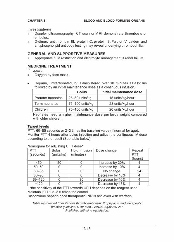
3.18
CHAPTER 3 BLOOD AND BLOOD-FORMING ORGANS Investigations » Doppler ultrasonography, CT scan or MRI demonstrate thrombosis or
embolus. » D-dimer, antithrombin III, protein C, pr otein S, Fa ctor V Leiden and
antiphospholipid antibody testing may reveal underlying thrombophilia. GENERAL AND SUPPORTIVE MEASURES » Appropriate fluid restriction and electrolyte management if renal failure. MEDICINE TREATMENT If hypoxic: Oxygen by face mask.
Heparin, unfractionated, IV, a dministered over 10 minutes as a bo lus
followed by an initial maintenance dose as a continuous infusion. Bolus Initial maintenance dose Preterm neonates 25–50 units/kg 15 units/kg/hour Term neonates 75–100 units/kg 28 units/kg/hour Children 75–100 units/kg 20 units/kg/hour
Neonates need a higher maintenance dose per body weight compared with older children.
Target levels PTT: 60–85 seconds or 2–3 times the baseline value (if normal for age). Monitor PTT 4 hours after bolus injection and adjust the continuous IV dose according to the result (See table below) Nomogram for adjusting UFH dose*
PTT (seconds)
Bolus (units/kg)
Hold infusion (minutes)
Dose change Repeat PTT (hours)
<50 50 0 Increase by 20% 4 50–59 0 0 Increase by 10% 4 60–85 0 0 No change 24 86–95 0 0 Decrease by 10% 4 69–120 0 30 Decrease by 10% 4 >120 0 60 Decrease by 15% 4
*the sensitivity of the PTT towards UFH depends on the reagent used. Maintain PTT 2.5–3.5 times the control. Discontinue heparin once therapeutic INR is achieved with warfarin.
Table reproduced from Venous thromboembolism: Prophylactic and therapeutic practice guideline. S Afr Med J 2013;103(4):260-267
Published with kind permission.
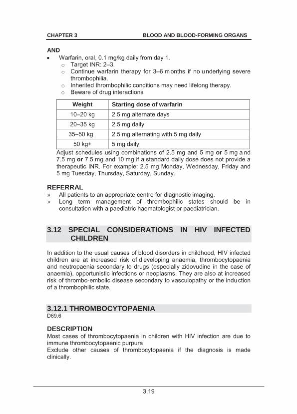
3.19
CHAPTER 3 BLOOD AND BLOOD-FORMING ORGANS AND Warfarin, oral, 0.1 mg/kg daily from day 1.
o Target INR: 2–3. o Continue warfarin therapy for 3–6 m onths if no underlying severe
thrombophilia. o Inherited thrombophilic conditions may need lifelong therapy. o Beware of drug interactions
Weight Starting dose of warfarin
10–20 kg 2.5 mg alternate days 20–35 kg 2.5 mg daily 35–50 kg 2.5 mg alternating with 5 mg daily
50 kg+ 5 mg daily Adjust schedules using combinations of 2.5 mg and 5 mg or 5 mg a nd 7.5 mg or 7.5 mg and 10 mg if a standard daily dose does not provide a therapeutic INR. For example: 2.5 mg Monday, Wednesday, Friday and 5 mg Tuesday, Thursday, Saturday, Sunday.
REFERRAL » All patients to an appropriate centre for diagnostic imaging. » Long term management of thrombophilic states should be in
consultation with a paediatric haematologist or paediatrician. 3.12 SPECIAL CONSIDERATIONS IN HIV INFECTED
CHILDREN In addition to the usual causes of blood disorders in childhood, HIV infected children are at increased risk of d eveloping anaemia, thrombocytopaenia and neutropaenia secondary to drugs (especially zidovudine in the case of anaemia), opportunistic infections or neoplasms. They are also at increased risk of thrombo-embolic disease secondary to vasculopathy or the induction of a thrombophilic state. 3.12.1 THROMBOCYTOPAENIA D69.6 DESCRIPTION Most cases of thrombocytopaenia in children with HIV infection are due to immune thrombocytopaenic purpura Exclude other causes of thrombocytopaenia if the diagnosis is made clinically.
3.20
CHAPTER 3 BLOOD AND BLOOD-FORMING ORGANS DIAGNOSTIC CRITERIA Clinical » Bleeding tendency in a child with HIV infection. » Asymptomatic finding on full blood count. Investigations » Thrombocytopaenia with normal white cell count and red cell indices,
apart from the effects of blood loss. » Normal INR (PT) and partial thromboplastin time (PTT). » Abundant megakaryocytes on bone ma rrow aspiration with normal
erythroid and myeloid cellularity. » Indications for bone marrow investigation: Prior to starting steroids or
any other abnormality on FBC or any atypical cells on differential count. GENERAL AND SUPPORTIVE MEASURES » As for the HIV uninfected child. » Avoid:
> platelet transfusions, unless life-threatening bleeds; > contact sport, injury and trauma; > dental procedures in acute phase; > medication that affects platelet function, e.g. NSAIDs and aspirin.
» Check for interactions with ARTs. MEDICINE TREATMENT As for the HIV uninfected child. Initiate ART if not already initiated. Acute ITP Active bleeding: Prednisone, oral, 4 mg/kg/24 hours as a single daily dose for 4 days.
REFERRAL » All children with refractory symptomatic thrombocytopaenia.
4.1
CHAPTER 4
CARDIOVASCULAR SYSTEM
4.1 CARDIAC DYSRHYTHMIAS I49.9 DESCRIPTION A heart rate that is abnormally slow or fast for age or irregular. Normal heart rate/minute for age:
Newborn 100–160 < 1 year 110–160 1–2 years 100–150 2–5 years 95–140 5–12 years 80–120 > 12 years 60–100
DIAGNOSTIC CRITERIA Clinical » Presenting features may vary with the age of the patient:
> infants: colour changes (pale, mottled), irregular pulse, irritability, tachycardia, feeding difficulties, bradycardia, sweating, signs of cardiac failure, tachypnoeic/apnoeic spells.
> children: dizziness, tachycardia, palpitations, bradycardia, fatigue, syncope, chest pain, signs of cardiac failure.
Investigations » ECG is essential for diagnosis, preferably a 12 lead ECG.
Monitors are inadequate to diagnose most dysrhythmias.
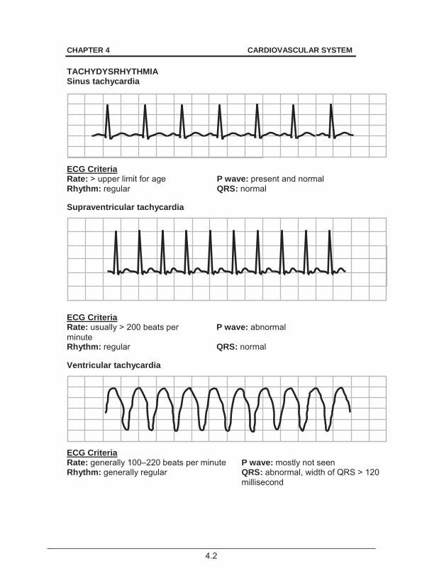
4.2
CHAPTER 4 CARDIOVASCULAR SYSTEM TACHYDYSRHYTHMIA Sinus tachycardia
ECG Criteria Rate: > upper limit for age P wave: present and normal Rhythm: regular QRS: normal Supraventricular tachycardia
ECG Criteria Rate: usually > 200 beats per minute
P wave: abnormal
Rhythm: regular QRS: normal Ventricular tachycardia
ECG Criteria Rate: generally 100–220 beats per minute P wave: mostly not seen Rhythm: generally regular QRS: abnormal, width of QRS > 120
millisecond

4.3
CHAPTER 4 CARDIOVASCULAR SYSTEM BRADYDYSRHYTHMIA Common causes:
hypoxia drug ingestion congenital excessive vagal stimulation
Sinus bradycardia
ECG Criteria Rate: < lower limit for age P wave: present, all look the same Rhythm: regular QRS: normal, 80–120 millisecond Heart block (Complete)
ECG Criteria Rate: low, usually < 60 beats per minute P wave: independent P waves and
QRS's with no relationship between the two (AV dissociation)
Rhythm: regular QRS: can be normal or wide, depending on escape rhythm
GENERAL AND SUPPORTIVE MEASURES » Sinus tachycardia usually requires management of the underlying
condition. » Apply ABC of resuscitation. » Admit to high care or intensive care unit. » Monitor:
> ECG, > oxygen saturation, > blood pressure, > haemoglobin, > heart rate, > acid–base status, > respiratory rate, > blood gases.
» Maintain adequate nutrition and hydration. » Treat pyrexia.
4.4
CHAPTER 4 CARDIOVASCULAR SYSTEM MEDICINE TREATMENT TACHYDYSRHYTHMIAS Emergency treatment. Narrow complex tachycardia Commonly due to supraventricular tachycardia. Stable patient: Attempt vagal stimulation.
Place ice bag on face, or Infants: immerse face in ice-cold water for a few seconds. Older children: try a Valsalva manoeuvre. Eye-ball pressure and carotid massage is contraindicated in children.
Adenosine, IV, 0.1 mg/kg rapid IV push (within seconds). o Follow immediately with a rapid flush of at least 5 mL sodium
chloride 0.9%. o Increase dose in 0.1 mg/kg increments every 2 minutes until return
of sinus rhythm. Follow each dose with a rapid flush of sodium chloride 0.9%.
o Maximum dose: 0.5 mg/kg. Do not exceed 12 mg in total. o Consult with a cardiologist/paediatrician before administration.
Because adenosine is rapidly metabolised, inject adenosine in a good drip, followed with a rapid fl ush of a fl uid bolus. It i s sometimes helpful to have both the syringe with adenosine and the fluid bolus connected to t he giving set a nd having as short as possible line between the syringes and the patient.
Unstable patient – heart failure / shocked:
DC synchronised cardioversion in increments of 1–2 J/kg. If possible, empty the stomach before cardioversion is attempted. Resuscitation facilities must be available. Midazolam for sedation, if necessary.
Broad complex tachycardia Commonly due to ventricular tachycardia. Causes include electrolyte disturbances and drug ingestion. Stable patient (rare):
Send ECG immediately to paediatric cardiologist. If unsure whether narrow or wide angle tachycardia, attempt adenosine as in narrow complex tachycardia.
4.5
CHAPTER 4 CARDIOVASCULAR SYSTEM Unstable patient – heart failure/shock:
Pulseless treat as ventricular fibrillation. DC asynchronised cardioversion in increments of 1–2 J/kg. Resuscitation facilities must be available. Midazolam for sedation, if necessary.
Monitor and correct blood gases and acid-base status. If DC cardioversion fails: Amiodarone, IV, 5 mg/kg slowly over 20 minutes – never as a rapid
infusion. And continue with DC cardioversion. BRADYDYSRHYTHMIAS Try and correct underlying causes.
Stable patient: Observe. Bradydysrhythmia due to vagal stimulation: Atropine, IV/IO, 0.02 mg/kg.
o If no response, repeat in 5 minutes. Unstable patient: Treat as impending arrest: Epinephrine (adrenaline), IV/IO, 0.01 mg/kg.
o Repeat if necessary conferring with referral institution. If no sustained response, consider: Epinephrine (adrenaline), IV infusion, 0.05–2 mcg/kg/minute.
REFERRAL » All children with tachydysrhythmias after acute treatment, excluding
sinus tachycardia due to other causes. » Bradycardia unresponsive to medical treatment, or heart block.
4.6
CHAPTER 4 CARDIOVASCULAR SYSTEM 4.2 CYANOTIC CONGENITAL HEART DISEASE WITH
HYPOXAEMIC ATTACKS/SPELLS (HYPERCYANOTIC SPELLS)
Q24.9 DESCRIPTION Acute worsening of central cyanosis in patients with a co nfirmed or suspected underlying cyanotic congenital heart disease such as Tetralogy of Fallot after the neonatal period. DIAGNOSTIC CRITERIA Clinical » Rapid worsening of central cyanosis, tachypnoea/dyspnoea, anxiety and
alteration in consciousness in the presence of congenital cyanotic heart disease.
» Restless and crying in the presence of congenital cyanotic heart disease.
» Decrease in intensity or disappearance of the systolic murmur in Tetralogy of Fallot during crying.
GENERAL AND SUPPORTIVE MEASURES » Exclude and treat precipitants such as fever, infection and
gastroenteritis. » Calm patient and keep on mother’s lap, if possible. » Place patient in knee-chest position to raise systemic blood pressure
and increase systemic venous return. » Monitor SaO2, heart rate, respiratory rate and acid-base status. » Ensure adequate hydration. MEDICINE TREATMENT Oxygen, 100%, by facemask or by nasal cannula
Volume expander e.g. sodium chloride 0.9%, IV bolus, 20mL/kg administered over 5 minutes.
Morphine, IV, 0.1–0.2 mg/kg as a single dose. o May cause impairment of airway reflexes and respiratory depression.
If clinically acidotic or pH < 7.2: Sodium bicarbonate 4.2%, IV, 2 mL/kg.
If failure to improve the cyanotic spell, consider in consultation with specialist: Ketamine, IV, 0.5–1 mg/kg.
Note: IV ketamine is a general anaesthetic. Take standard precautions for respiratory arrest.
4.7
CHAPTER 4 CARDIOVASCULAR SYSTEM After resolution of spell: If Hb < 10 g/dL, child is anaemic: Packed red cells, 10 mL/kg administered over 3 hours.
Propranolol, oral, 0.5–1 mg/kg/dose 6 hourly.
o Do not exceed 5 mg/kg/day. REFERRAL » If above measures do not work refer urgently for consideration of
phenylephrine, ventilation and emergency surgery. » All cases for assessment. 4.2.1 TETRALOGY OF FALLOT Q21.3 DESCRIPTION Suspect tetralogy of Fallot in a child with cyanosis after the neonatal period. DIAGNOSTIC CRITERIA Clinical » Child with central cyanosis. » May be plethoric due to polycythemia – normal haemoglobin represents
relative anaemia. » May have clubbing. » Possible history of cyanotic spells. » Heart not clinically enlarged. » Right ventricular hypertrophy usually not palpable. » Single second heart sound. » Coarse, ejection systolic murmur over right ventricular outflow tract. » Chest X-ray:
> normal/small heart, > boot shaped/pulmonary bay – concavity where pulmonary artery
should be, > oligaemic lung fields.
» ECG: > right axis deviation and right ventricular hypertrophy.
GENERAL AND SUPPORTIVE MEASURES » Good dental hygiene. MEDICINE TREATMENT Iron (elemental), oral, 1 mg/kg/dose 8 hourly.
Folic acid, oral, 2.5– 5 mg/day.
4.8
CHAPTER 4 CARDIOVASCULAR SYSTEM Propranolol, oral, 0.5–1 mg/kg/dose 6 hourly.
o Do not exceed 5 mg/kg/day. Endocarditis prophylaxis: See section 4.3: Endocarditis, infective. REFERRAL » All children with cyanotic heart defects.
4.3 ENDOCARDITIS, INFECTIVE I33.0 DESCRIPTION Infection of the endothelial surface of the heart. Suspect infective endocarditis in all children with persistent fever and underlying heart disease. DIAGNOSTIC CRITERIA Clinical » An underlying heart defect and a persistent low grade fever without an
obvious underlying cause. » Associated other findings include: fatigue, joint pain, new murmurs,
clubbing, splenomegaly and haematuria. » Must be differentiated from acute carditis due to rheumatic fever. » The Duke criteria have been suggested as a guide to diagnosis, but
have definite limitations as they were developed for use in adult patients.
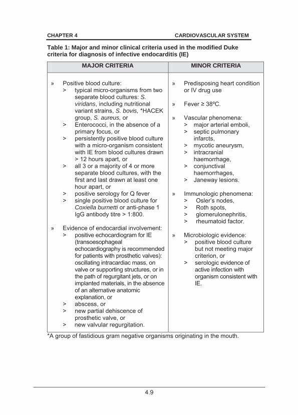
4.9
CHAPTER 4 CARDIOVASCULAR SYSTEM
Table 1: Major and minor clinical criteria used in the modified Duke criteria for diagnosis of infective endocarditis (IE)
MAJOR CRITERIA MINOR CRITERIA
» Positive blood culture:
> typical micro-organisms from two separate blood cultures: S. viridans, including nutritional variant strains, S. bovis, *HACEK group, S. aureus, or
> Enterococci, in the absence of a primary focus, or
> persistently positive blood culture with a micro-organism consistent with IE from blood cultures drawn > 12 hours apart, or
> all 3 or a majority of 4 or more separate blood cultures, with the first and last drawn at least one hour apart, or
> positive serology for Q fever > single positive blood culture for
Coxiella burnetti or anti-phase 1 IgG antibody titre > 1:800.
» Evidence of endocardial involvement:
> positive echocardiogram for IE (transoesophageal echocardiography is recommended for patients with prosthetic valves): oscillating intracardiac mass, on valve or supporting structures, or in the path of regurgitant jets, or on implanted materials, in the absence of an alternative anatomic explanation, or
> abscess, or > new partial dehiscence of
prosthetic valve, or > new valvular regurgitation.
» Predisposing heart condition
or IV drug use » F . » Vascular phenomena:
> major arterial emboli, > septic pulmonary
infarcts, > mycotic aneurysm, > intracranial
haemorrhage, > conjunctival
haemorrhages, > Janeway lesions.
» Immunologic phenomena:
> Osler’s nodes, > Roth spots, > glomerulonephritis, > rheumatoid factor.
» Microbiologic evidence:
> positive blood culture but not meeting major criterion, or
> serologic evidence of active infection with organism consistent with IE.
*A group of fastidious gram negative organisms originating in the mouth.
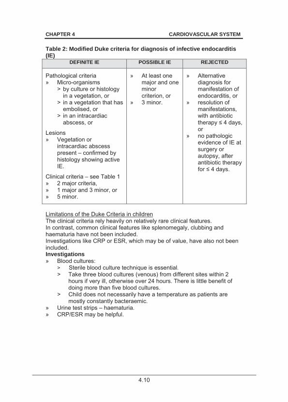
4.10
CHAPTER 4 CARDIOVASCULAR SYSTEM Table 2: Modified Duke criteria for diagnosis of infective endocarditis (IE)
DEFINITE IE POSSIBLE IE REJECTED
Pathological criteria » Micro-organisms
> by culture or histology in a vegetation, or
> in a vegetation that has embolised, or
> in an intracardiac abscess, or
Lesions » Vegetation or
intracardiac abscess present – confirmed by histology showing active IE.
Clinical criteria – see Table 1 » 2 major criteria, » 1 major and 3 minor, or » 5 minor.
» At least one major and one minor criterion, or
» 3 minor.
» Alternative diagnosis for manifestation of endocarditis, or
» resolution of manifestations, with antibiotic therapy or
» no pathologic evidence of IE at surgery or autopsy, after antibiotic therapy
.
Limitations of the Duke Criteria in children The clinical criteria rely heavily on relatively rare clinical features. In contrast, common clinical features like splenomegaly, clubbing and haematuria have not been included. Investigations like CRP or ESR, which may be of value, have also not been included. Investigations » Blood cultures:
> Sterile blood culture technique is essential. > Take three blood cultures (venous) from different sites within 2
hours if very ill, otherwise over 24 hours. There is little benefit of doing more than five blood cultures.
> Child does not necessarily have a temperature as patients are mostly constantly bacteraemic.
» Urine test strips – haematuria. » CRP/ESR may be helpful.
4.11
CHAPTER 4 CARDIOVASCULAR SYSTEM GENERAL AND SUPPORTIVE MEASURES » Bed rest/limit physical activity. » Ensure adequate nutrition. » Maintain haemoglobin > 10 g/dL. » Measures to reduce fever. MEDICINE TREATMENT See section 4.9: Heart failure. For fever: Paracetamol, oral, 15 mg/kg/dose, 6 hourly as required.
Antibiotic therapy Antibiotics are always given IV, according to culture and sensitivity. If culture is available treat according to sensitivities. Empiric treatment If culture is not yet available or is negative: Benzylpenicillin (Penicillin G), IV, 50 000 units/kg/dose, 6 hourly for 4
weeks. PLUS Cloxacillin, IV, 50 mg/kg/dose 6 hourly for 4 weeks.
PLUS Gentamicin, IV, 3 mg/kg/day for 4 weeks.
If culture available: S. viridans Benzylpenicillin (Penicillin G), IV, 50 000 units/kg/dose, 6 hourly for 4
weeks. PLUS Gentamicin, IV, 3 mg/kg/day for 2 weeks.
Enterococci Benzylpenicillin (Penicillin G), IV, 75 000 units/kg/dose, 6 hourly for 4–6
weeks. PLUS Gentamicin, IV, 3 mg/kg/day for 4–6 weeks.
Cloxacillin sensitive staphylococcus Cloxacillin, IV, 50 mg/kg/dose 6 hourly for 4–6 weeks.
If the organism is gentamicin sensitive: ADD Gentamicin, IV, 3 mg/kg/day for 3–5 days.
4.12
CHAPTER 4 CARDIOVASCULAR SYSTEM Multi-Resistant Staphylococcus Aureus (MRSA) Vancomycin, IV, 10 mg/kg/dose infused over 1 hour, 6 hourly for 6
weeks. HACEK organisms Ceftriaxone, IV, 100 mg/kg once daily or ampicillin, IV, 50 mg/kg/dose 6
hourly for 4 weeks. PLUS Gentamicin, IV, 3 mg/kg/day for 4 weeks.
Enteric bacilli, e.g. Klebsiella Piperacillin, IV, 50 mg/kg/dose or ceftazidime, IV, 50 mg/kg/dose 6
hourly for 6 weeks. PLUS Gentamicin, IV, 1 mg/kg/dose 8 hourly for 6 weeks.
Prophylaxis The use of prophylaxis is controversial but still recommended. For children with the following cardiac conditions: » rheumatic heart disease; » prosthetic cardiac valve or prosthetic material used in valve repair; » previous infective endocarditis; » unrepaired cyanotic heart disease, including palliative shunts; » during the first 6 months after complete repair of congenital heart defect
with prosthetic material or device (complete endothelialisation of prosthesis after 6 months);
» repaired cyanotic heart disease with residual defect at or adjacent to prosthetic patch or device; or
» cardiac transplant recipients who develop cardiac valvulopathy. Children with the above cardiac conditions should receive prophylaxis when undergoing the following procedures: » All dental procedures that involve manipulation of gingival tissues or
periapical region of teeth or trauma to oral mucosa. » Procedures on respiratory tract or infected skin, skin structures or
musculoskeletal tissue. Regimens for dental procedures Amoxicillin, oral, 50 mg/kg (maximum 2 g) 1 hour before the procedure.
Patients unable to take oral medication: Ampicillin, IV, 50 mg/kg (maximum 2 g) ½ hour before the procedure.
4.13
CHAPTER 4 CARDIOVASCULAR SYSTEM REFERRAL » All patients with suspected and confirmed infective endocarditis as soon
as possible.
4.4 RHEUMATIC FEVER, ACUTE I01.9 * Notifiable condition. DESCRIPTION Rheumatic fever is a c ommon cause of acquired heart disease with significant morbidity and mortality rates, both in the a cute phase of the disease and as a result of chronic valvular sequelae. DIAGNOSTIC CRITERIA Revised Jones criteria: » Evidence of recent streptococcal infection:
> Elevated ASO-titre or other streptococcal antibody titres. > Positive throat culture for group A beta haemolytic streptococcus. PLUS
» Two major manifestations, or one major and two minor manifestations, justifies the presumptive diagnosis of acute rheumatic fever.
Major manifestations Minor manifestations polyarthritis carditis erythema marginatum subcutaneous nodules Sydenham’s chorea
polyarthralgia fever acute phase reactants:
increased erythrocyte sedimentation rate (ESR) or C-reactive protein (CRP)
ECG: prolonged PR-interval, 0.18 seconds in the absence of carditis
> Chorea for which other causes have been excluded, provides adequate evidence of rheumatic fever without the other criteria for diagnosis being required.
> In children with rheumatic heart disease with fever, it is critical to differentiate recurrence of acute rheumatic fever from infective endocarditis.
For children with rheumatic heart disease, recurrence of some of the above criteria would suggest a recurrence of rheumatic fever but other causes such as IE should be excluded.
4.14
CHAPTER 4 CARDIOVASCULAR SYSTEM GENERAL AND SUPPORTIVE MEASURES » Hospitalise with bed rest until sleeping pulse is normal and signs of
rheumatic activity have resolved. » Restrict physical activity for at least 2 weeks after acute phase reactants
have normalised. » Keep a record of the patients on rheumatic fever prophylaxis so that
attendance can be monitored. MEDICINE TREATMENT Antibiotic therapy To eradicate any streptococci: Benzathine benzylpenicillin (depot formulation), IM, as a single dose.
o If < 30 kg: 600 000 IU. o If 30 kg: 1.2 MU.
OR Phenoxymethylpenicillin, oral, 250–500 mg 12 hourly for 10 days.
Anti-inflammatory therapy Do not start until a definite diagnosis is made. Severe arthritis: Aspirin soluble, oral, 20 mg/kg/dose 6 hourly until the arthritis resolves.
OR If aspirin cannot be tolerated: Ibuprofen, oral, 5 mg/kg/dose, 6 hourly.
Cardiac failure: See section 4.9: Heart failure. Chorea: See section 13.10: Sydenham’s Chorea. Prevention of repeated attacks Any patient with documented rheumatic fever must receive prophylaxis. Intramuscular penicillin is superior to other forms of prophylaxis. Benzathine benzylpenicillin (depot formulation), IM, every 21 days.
o If < 30 kg: 600 000 IU. o If > 30 kg: 1.2 MU.
OR Phenoxymethylpenicillin, oral, 250 mg 12 hourly.
Continue therapy until patients reach 35 years of age. REFERRAL Rheumatic fever: » with residual valvular damage electively for planning of care, » with symptomatic valvular damage, » unresponsive to treatment.
4.15
CHAPTER 4 CARDIOVASCULAR SYSTEM 4.5 MYOCARDITIS I40 DESCRIPTION Myocarditis is an acute inflammation of the cardiac muscle. The majority of paediatric myocarditis cases are se condary to a v iral infection. Viral myocarditis should be suspected whenever a c hild presents with unexplained shortness of breath, dysrhythmia or acute heart failure following a viral illness. DIAGNOSTIC CRITERIA Clinical » Tachycardia. » Clinical signs of biventricular heart failure. » May present with cardiogenic shock. Investigations » ECG changes are non-specific but ST elevation, T wave inversion,
prolonged QTc, small complexes, dysrhythmias or extra-systole may be seen.
» Chest X-ray: > pulmonary congestion, > cardiomegaly, > possible pleural effusion.
» Elevated cardiac troponin T levels are a good indicator of myocarditis. GENERAL AND SUPPORTIVE MEASURES » Restrict fluid (75% of daily requirements) – not a t expense of adequate
caloric intake. » Ensure adequate nutrition, tube-feeding may be necessary. MEDICINE TREATMENT OF VIRAL MYOCARDITIS To prevent hypoxia: Oxygen via face mask, nasal cannula or head box.
Furosemide, oral/ IV, 1 mg/kg/dose 12 hourly.
Consider low dose inotropic support – Refer to ICU. REFERRAL » Viral myocarditis not responding to treatment.
4.16
CHAPTER 4 CARDIOVASCULAR SYSTEM
4.6 DILATED CARDIOMYOPATHY I42.0 DESCRIPTION Dilated cardiomyopathy refers to a group of conditions of diverse aetiology in which both ventricles are dilated with reduced contractility. It is diffi cult and sometimes impossible to distinguish myocarditis from dil ated cardiomyopathy. A w ide variety of disease can mimic dilated cardiomyopathy, but in the majority of cases no identifiable cause is found. One of the common causes of cardiomyopathy is HIV disease. DIAGNOSTIC CRITERIA Clinical » Cardiomegaly with clinical signs of heart failure and poor apical impulse. » May present with cardiogenic shock. Investigations » Chest X-ray:
> pulmonary congestion, > cardiomegaly, > there may be pleural effusion.
» ECG: > Mostly non-specific. > Dysrhythmias or extra-systoles may occur.
GENERAL AND SUPPORTIVE MEASURES » Fluid restriction (75% of daily requirements) – not at expense of
adequate caloric intake. » Ensure adequate nutrition, tube-feeding may be necessary. » Advise bed rest. MEDICINE TREATMENT To prevent hypoxia: Oxygen via face mask, nasal cannula or head box.
Furosemide, IV/oral, 1–3 mg/kg/24 hours in 2–3 divided doses.
AND/OR Spironolactone, oral, 2–4 mg/kg/24 hours in 2 divided doses.
Digoxin, oral, 0.005 mg/kg/dose 12 hourly.
o Syrup: 0.005 mg = 0.1 mL, therefore 0.1 mL/kg/dose 12 hourly. o In older children a once daily dose can be given, i.e. 0.01
mg/kg/day.
4.17
CHAPTER 4 CARDIOVASCULAR SYSTEM Persistent cardiac failure: Captopril, oral.
o Initial dose: 0.5 mg/kg/24 hours in 3 divided doses (8 hourly) for 24–48 hours.
o Increase by 0.5 mg/kg/24 hours every 24–48 hours until maintenance dose of 3–5 mg/kg/24 hours is reached.
Inotropic support. – Refer to ICU. REFERRAL » Urgent: To ICU for inotropic support. » All patients for assessment and consideration of underlying disorders. 4.7 PERICARDIAL EFFUSION I30 DESCRIPTION Accumulation of fluid in the pericardial space, usually secondary to pericarditis. DIAGNOSTIC CRITERIA Clinical » Most patients present with a prolonged history of:
> low cardiac output, > distended neck veins, > muffled or diminished heart sounds.
» Patients with HIV may be a symptomatic and incidentally diagnosed when having a chest X-ray.
» Often associated with TB. » Acute septic pericarditis may occur in patients with septicaemia. Investigations » Exclude TB in all cases. Tuberculin skin test. » ECG:
> small complexes tachycardia, > diffuse T wave changes,
» Chest X-ray: > in pericardial effusion – “water bottle” large globular heart or cardiac
shadow with smoothed out borders. » Ultrasound of heart and pericardium. » Diagnostic pericardiocentesis:
> in all patients with suspected bacterial or neoplastic pericarditis, and in all others in whom the diagnosis is not readily obtained;
> include cell count and differential, culture and gram stain; > an elevated adenosine deaminase (ADA) may be helpful in
diagnosing TB.
4.18
CHAPTER 4 CARDIOVASCULAR SYSTEM CARDIAC TAMPONADE Cardiac tamponade is the accumulation of pericardial fluid that restricts filling and ejection of blood. The child usually presents with a tachycardia, pulsus paradoxus, elevated JVP, hypotension, shock or pulseless electric activity. Features on ECG include electrical alternans, low voltage QRS. Diagnosis is confirmed on sonar. GENERAL AND SUPPORTIVE MEASURES Urgent pericardiocentesis under ultrasound guidance by an experienced person. Pericardiocentesis » Do a coagulation screen if coagulation problems are suspected. » Preferably under ultrasound guidance by an experienced person. » In an emergency, drainage by using a large bore intravenous cannula. » Technique:
> Ensure that full resuscitation equipment is available as well as an IV line and cardiac monitor.
> Administer oxygen via face mask, nasal cannula or head box. > If the patient is restless, it may be necessary to sedate the patient.
In an emergency situation, this is unnecessary. > Position the patient in a 30° sitting-up position. > Prepare the preferred site just to the left of the xiphoid process, 1
cm inferior to the costal margin. > Infiltrate this area with 1% lidocaine (lignocaine). > Maintaining negative pressure on the syringe, insert the needle at a
45° angle to the skin, advancing in the direction of the patient's left shoulder.
> While advancing the needle, observe closely on ECG for ventricular ectopic beats, a sign of myocardial contact, while advancing the needle. If this is noted, gradually withdraw the needle 1–2 cm.
> Once air (pneumopericardium) or fluid begins to fill the syringe, advance the intravenous cannula, withdraw the needle, attach the syringe to the hub of the cannula and slowly aspirate the pericardial fluid.
> Potential complications include: haemopericardium (from laceration of the heart wall or coronary artery), cardiac dysrythmias, pneumothorax, and pneumopericardium.
In an emergency, drainage by using a large bore intravenous cannula may be undertaken without sedation.
4.19
CHAPTER 4 CARDIOVASCULAR SYSTEM MEDICINE TREATMENT
Exclude TB in all cases with pericardial disease, and if suspected TB, give antituberculosis drugs for 6 months plus steroids: Prednisone, oral, 2 mg/kg/day for 4 weeks.
Pain management See section 20.1: Management of pain. Antibiotic therapy Empiric antibiotic treatment until culture and sensitivity results are available. Antibiotic therapy should be continued for 3–4 weeks. In case of purulent pericarditis: Cloxacillin, IV, 50 mg/kg/dose 6 hourly.
PLUS Ceftriaxone, IV, 100 mg/kg as a single daily dose.
Cardiac failure: See section 4.9: Heart failure. REFERRAL » All after stabilisation of patient. 4.8 PERICARDITIS I31.9 DESCRIPTION Causes can be viral or bacterial. The commonest cause is TB. Inflammation of the pericardium: » Classical presentation of v iral pericarditis, loud pericardial rub and chest
pain that is relieved by sitting up. Children often do not complain of chest pain.
» Acute septic pericarditis may occur in patients with septicaemia. TB pericarditis TB pericarditis may present as pericardial effusion (most cases), effusive constrictive pericarditis or constrictive pericarditis. Clinical features include: » chronic cough, » dyspnoea, » chest pain, » fever, » night sweats, » orthopnoea, » and weight loss. Severe pericardial pain is uncommon.
4.20
CHAPTER 4 CARDIOVASCULAR SYSTEM Investigations » Exclude TB. » Echocardiogram. MEDICINE TREATMENT
Treat the cause. Exclude TB in all pericardial disease cases. If TB is suspected, give antituberculosis drugs for 6 months. Corticosteroids: Prednisone, oral, 2 mg/kg/day for 4 weeks.
PLUS NSAID, e.g.: Ibuprofen, oral, 5 mg/kg per dose 6 hourly.
As soon as cultures are proved negative for organisms that can cause purulent pericarditis, TB treatment for 6 months. Pain management: See section 20.1: Management of pain. Antibiotic therapy Empiric therapy should be provided until culture and sensitivity results available. Antibiotic therapy should be continued for 3–4 weeks. Empiric therapy in case of purulent pericarditis: Cloxacillin, IV, 50 mg/kg/dose 6 hourly.
PLUS Ceftriaxone, IV, 100 mg/kg as a single daily dose.
Adjust antibiotic based on culture results. REFERRAL » All patients. 4.9 HEART FAILURE I50.9 DESCRIPTION Clinical syndrome reflecting the inability of the myocardium to meet the oxygen and nutritional/metabolic requirements of the body.
4.21
CHAPTER 4 CARDIOVASCULAR SYSTEM Causes include: » volume overload:
L-R shunt lesions mitral/aortic regurgitation
» pump failure: myocarditis/cardiomyopathy
» high output failure: septicaemia severe anaemia
DIAGNOSTIC CRITERIA Clinical » Acute cardiac failure may present with shock. See section1.1.7: Shock. » History of recent onset of:
> poor feeding, > poor or excessive weight gain, > tachypnoea, > breathlessness, > sweating, > cough.
» Physical findings: > tachycardia, > cardiomegaly, > hypotension, > cold extremities, > weak pulses, > reduced urinary output, > gallop rhythm with/without a cardiac murmur, > pulmonary venous congestion and fluid retention:
tachypnoea, dyspnoea, orthopnoea, recession, wheezing, coarse crepitations, cyanosis;
> systemic venous congestion: hepatomegaly, periorbital oedema - not seen in infants, abnormal weight gain;
> signs and symptoms of underlying condition/disease. Investigations as appropriate for the possible underlying cause » Chest X-ray: cardiomegaly is almost always present. » Electrocardiogram may show evidence of hypertrophy/enlargement of
one or more heart chambers and/or dysrhythmias.
4.22
CHAPTER 4 CARDIOVASCULAR SYSTEM 4.9.1 HEART FAILURE, ACUTE SEVERE WITH
PULMONARY OEDEMA AND SHOCK I50.9 GENERAL AND SUPPORTIVE MEASURES » Treat the underlying disorder/condition. Where the primary cause of
acute pulmonary oedema is renal failure treat as under renal failure. » Restrict fluids, beware of IV fluids. » Place patient in an upright or semi-upright sitting position. » Intubation and ventilation may be required in an ICU setting. MEDICINE TREATMENT Oxygen 100%, administered via face mask or nasal cannula.
Treat the underlying condition: Furosemide, IV, 1–3 mg/kg as a single dose immediately.
For patients not responding to furosemide: Morphine, IV, 0.1 mg/kg.
Manage severe hypotensive or refractory failure in an ICU setting. Inotropic support may help to stabilise patients with severe myocardial dysfunction, hypotension or low cardiac output. May be lifesaving in severe myocarditis or cardiogenic shock.
Dobutamine, IV infusion, 2–15 mcg/kg/minute. o Continue until myocardial function and blood pressure improve.
If no response to dobutamine, consider epinephrine (adrenaline) infusion. Ensure adequate renal function. Once patient stable and maintaining blood pressure: ACE inhibitor, e.g.: Captopril, oral.
o Initial dose: 0.5 mg/kg/24 hours in 3 divided doses (8 hourly) for 24–48 hours.
o Increase by 0.5 mg/kg/24 hours every 24–48 hours until maintenance dose of 3–5 mg/kg/24 hours is reached.
o Continue for as long as needed to control the cardiac failure.
4.23
CHAPTER 4 CARDIOVASCULAR SYSTEM 4.9.2 HEART FAILURE, ACUTE I50.9 GENERAL AND SUPPORTIVE MEASURES » Recognise and treat the underlying condition, e.g. infection,
hypertension, cardiac tamponade, fluid overload. » Fluid restriction (75% of daily requirements) – not at the expense of
adequate caloric intake. » Ensure adequate nutrition, tube-feeding may be necessary. » Monitor blood potassium levels, urea and electrolytes. MEDICINE TREATMENT Oxygen 100%, administered via face mask or nasal cannula.
Combination drug therapy is usually indicated, i.e. start with diuretic, then add digoxin then add ACE inhibitor. Diuretic therapy Furosemide, IV/oral, 1 mg/kg/dose 12 hourly.
Potassium supplements are necessary if furosemide is used without an aldosterone antagonist, i.e. spironolactone.
AND/OR In refractory failure: Spironolactone, oral, 1–2 mg/kg/dose once daily.
ACE inhibitor Note: ACE inhibitors are contraindicated in bilateral renal artery stenosis, coarctation of the aorta and aortic stenosis.
Captopril, oral. o Initial dose: 0.5 mg/kg/24 hours in 3 divided doses (8 hourly) for
24–48 hours. o Increase by 0.5 mg/kg/24 hours every 24–48 hours until
maintenance dose of 3–5 mg/kg/24 hours is reached. If < 1 year do not exceed 4 mg/kg/day.
o Continue as long as needed to control the cardiac failure. OR Enalapril, oral, 0.2–1 mg/kg/day as a single dose or 2 divided doses.
Digoxin Use in consultation with a cardiologist. Digoxin is contraindicated in bradycardia, heart block, cardiac tamponade or hypertrophic cardiomyopathy. Monitor serum potassium and digoxin blood levels and ECG. Intravenous digoxin is dangerous and inappropriate.
4.24
CHAPTER 4 CARDIOVASCULAR SYSTEM Because of the potential confusion with digitalising the dose of digoxin. Start with a maintenance dose. Digoxin, oral, 0.005 mg/kg/dose 12 hourly.
o 0.005 mg = 0.1 mL. o In older children a once daily dose can be given, i.e. 0.01
mg/kg/day. REFERRAL » For determination of the underlying cause, where this is not known and
initiation of treatment after stabilisation. » Deterioration despite adequate treatment.
4.10 DYSLIPIDAEMIA E78.9 DESCRIPTION Dyslipidaemia is a broad term used to describe disorders of lipid metabolism. Hypercholesterolaemia associated with increased levels of apolipoprotein Lp(a) B100-containing lipoproteins (low density lipoprotein (LDL), intermediate density lipoprotein (IDL)) and hy pertriglyceridaemia (chylomicrons, very low density lipoproteins VLDL) have the most serious clinical implications. Hypercholesterolaemia promotes atherosclerosis and hypertriglyceridaemia is a major component of the metabolic syndrome. Clinical features depend on the type of hyperlipidaemia. » Increased chylomicrons are associated with eruptive xanthomas and
hepatosplenomegaly. » Hypertriglyceridaemia is associated with pancreatitis. » Increased levels of VLDL are associated with familial
hypercholesterolaemia (FH). » Heterozygous FH phenotype lacks physical signs. » Homozygous FH phenotype displays physical signs e.g. cutaneous
xanthoma. » Increased LDL is associated with glucose intolerance and
hyperuricaemia. Increased levels of HDL cholesterol protects against coronary heart disease. DIAGNOSTIC CRITERIA Clinical » Hyperlipidaemia may be secondary to chronic kidney/liver disease,
diabetes mellitus, hypothyroidism and medicine treatment e.g. calcineurin inhibitors (transplant patients) and ARV treatment.
4.25
CHAPTER 4 CARDIOVASCULAR SYSTEM » Screening for hyperlipidaemia is i ndicated for c hildren at ris k of
developing premature atherosclerosis, including: > positive family history in parent/grandparent of any of the following
conditions presenting <55 years of age: familial hypercholesterolaemia, cardiovascular disease, metabolic syndrome.
> overweight or obese children, See section 7.16: Obesity. » A high-risk familial hypercholesteraemia is perceived to be the
concomitant occurrence of 3 or more risk factors: > LDL > 4.9 mmol/L (TChol > 5.2 mmol/L), or > HDL < 1.3 mmol/L, and > positive family history of premature coronary heart disease
(myocardial infarct in non-smoking parent < 35 years of age), > male gender, > Lp(a) > 0.3 g/L, > thickened carotid intima (CIMT).
Investigations » Exclude causes of secondary hyperlipidaemia » In most cases non-fasting total cholesterol is determined in children at
risk. If level is higher than upper limit, lipid profile is done after 12 hours of fasting. > Upper limit of S-cholesterol and triglycerides: Total cholesterol 5.2 mmol/L.
> Triglycerides (after 12 hours of fasting) influenced by lifestyle – needs attention if > 1.68 mmol/L, pancreatitis risk if > 10 mmol/L.
GENERAL AND SUPPORTIVE MEASURES Schedule for integrated cardiovascular health promotion in children. » Obesity
> See section 7.15: Obesity. » Blood pressure
> With family history of hypertension < 55 years of age: routine BP measurement from 3 years of age once a year.
> If BP 95th percentile for sex, age, and height percentile, follow up and investigate if persistently elevated.
» Diet > Hypertriglyceridaemias needs a dietary intervention to restrict
triglyceride intake. > Refer to a dietician. > Learning healthy eating habits is an important preventative
measure. > Moderate salt intake.
4.26
CHAPTER 4 CARDIOVASCULAR SYSTEM
» Physical activity > Encourage active child-parent play. > Limit child’s sedentary behaviour such as time watching television
and playing video computer games to a maximum of 2 hours per day or 14 hours per week.
> Children should not be allowed to eat while watching television, i.e. “no grazing”.
> Organised sport 5 times per week for at least 20–30 minute periods.
» Smoking > Encourage members of the household who smoke to stop.
MEDICINE TREATMENT Consider medicine treatment only after failure of general and supportive measures to lower the cholesterol over 6-12 months. Children should be at least 8 years of age for consideration of pharmacological intervention. If LDL-C remains above 4.1 mmol/L in children with multiple risk factors, or above 4.9 mmol/L in those without a family history of premature heart disease, refer for consideration of statins: Statins, e.g.: Simvastatin, oral, 10 mg at night.
Secondary hypercholesterolaemia due to nephrotic syndrome For persistent nephrotic range proteinuria (i.e. protein creatinine ratio > 0.2 g/mmol): ACE inhibitor, e.g.: Captopril, oral.
o Initial dose: 0.5 mg/kg/24 hours in 3 divided doses (8 hourly) for 24–48 hours.
o Increase by 0.5 mg/kg/24 hours every 24–48 hours until maintenance dose of 3–5 mg/kg/24 hours is reached. o Maximum dose: 50 mg 8 hourly.
o Continue as long as needed to control the cardiac failure or as long as proteinuria persists.
OR Enalapril, oral, 0.2–1 mg/kg/day as single dose or 2 divided doses.
REFERRAL
» Children with familial hypercholesterolaemia or pri mary underlying metabolic disorder.
» LDL > 4.1 mmol/L in children with multiple risk factors.
4.27
CHAPTER 4 CARDIOVASCULAR SYSTEM 4.11 HYPERTENSION IN CHILDREN I10 DESCRIPTION Hypertension is defined as systolic and/or diastolic blood pressure the 95th percentile for gender, age and height percentile on at least three consecutive occasions. A s ustained blood pressure of >115/80 mmHg is a bnormal in children between 6 weeks and 6 years of age. Measure blood pressure with the child in a sit ting or supine position with the entire a rm in line w ith the level of the heart. In children, it is eas ier to mo nitor the sy stolic blood pressure because of better correlation and less technical pitfalls than diastolic blood pressure.
In the majority of c hildren hypertension is due to a n identifiable cause. Severe hypertension suggests renal disease. Hypertensive emergency/crisis exists when CNS signs of hypertension appear such as encephalopathy, convulsions, retinal haemorrhages or blindness. Great care is required to reduce the blood pressure in a controlled manner to avoid potentially serious consequences of impaired auto-regulation of cerebral blood flow. Hypertensive urgency is defined as a significant elevation of blood pressure without accompanying end organ damage. Patients are generally symptomatic with complaints of headache, blurred vision and nausea, despite the lack of end organ involvement. A valid assessment of the blood pressure is of extreme importance. The blood pressure is measured by standard auscultation technique in children > 1 year of age. In children < 1 year of age, a f lush technique is usually used, although Doppler measurement would be preferable. Measure the BP in at least one limb, preferably the righ t upper arm. If hypertension is present, measure BP on all four limbs. One should use the widest cuff that can be applied to the upper arm. The cuff bladder must encircle at least 80% of the upper arm and should cover at least 75% of the di stance between the acrom ion and the olecr anon. It is better to use a cuff that is slightly too large than one that is too small. Large cuffs, if covered with linen-like material, can be folded to the appropriate size in smaller infants as long as the bladder encompasses the arm.
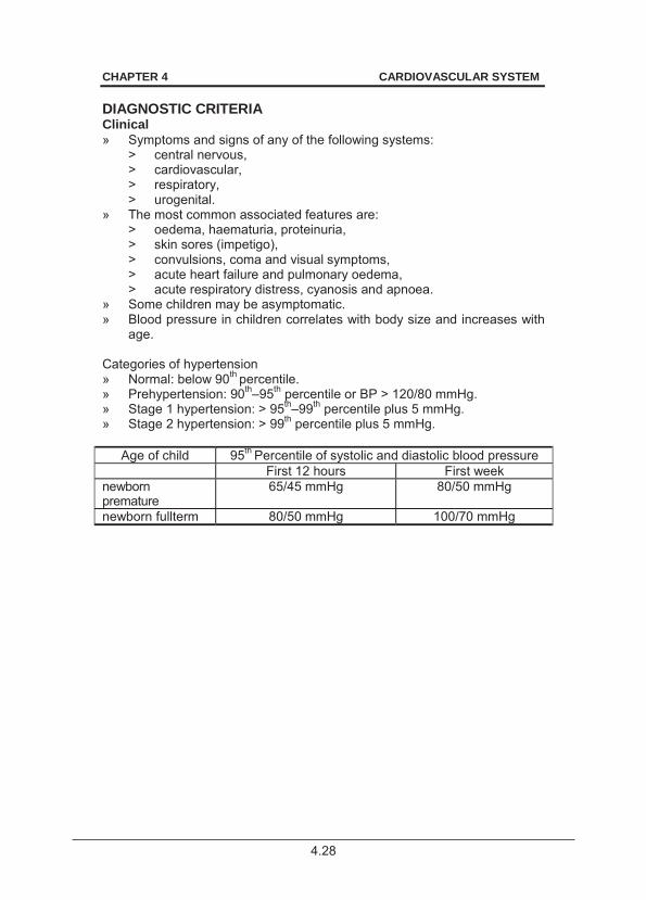
4.28
CHAPTER 4 CARDIOVASCULAR SYSTEM DIAGNOSTIC CRITERIA Clinical » Symptoms and signs of any of the following systems:
> central nervous, > cardiovascular, > respiratory, > urogenital.
» The most common associated features are: > oedema, haematuria, proteinuria, > skin sores (impetigo), > convulsions, coma and visual symptoms, > acute heart failure and pulmonary oedema, > acute respiratory distress, cyanosis and apnoea.
» Some children may be asymptomatic. » Blood pressure in children correlates with body size and increases with
age. Categories of hypertension » Normal: below 90th percentile. » Prehypertension: 90th–95th percentile or BP > 120/80 mmHg. » Stage 1 hypertension: > 95th–99th percentile plus 5 mmHg. » Stage 2 hypertension: > 99th percentile plus 5 mmHg.
Age of child 95th Percentile of systolic and diastolic blood pressure First 12 hours First week newborn premature
65/45 mmHg 80/50 mmHg
newborn fullterm 80/50 mmHg 100/70 mmHg
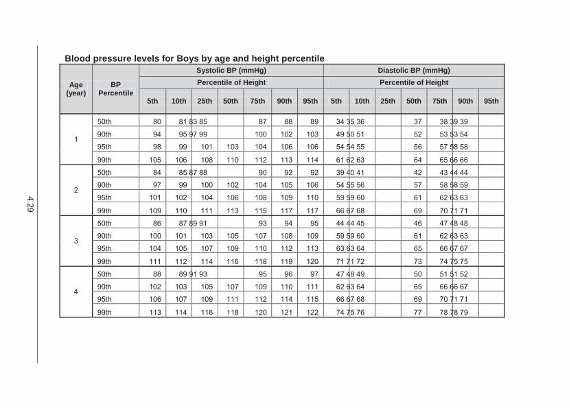
4.29
Blood pressure levels for Boys by age and height percentile
Age (year)
BP Percentile
Systolic BP (mmHg) Diastolic BP (mmHg) Percentile of Height Percentile of Height
5th 10th 25th 50th 75th 90th 95th 5th 10th 25th 50th 75th 90th 95th
1
50th 80 81 83 85 87 88 89 34 35 36 37 38 39 39
90th 94 95 97 99 100 102 103 49 50 51 52 53 53 54
95th 98 99 101 103 104 106 106 54 54 55 56 57 58 58
99th 105 106 108 110 112 113 114 61 62 63 64 65 66 66
2
50th 84 85 87 88 90 92 92 39 40 41 42 43 44 44
90th 97 99 100 102 104 105 106 54 55 56 57 58 58 59
95th 101 102 104 106 108 109 110 59 59 60 61 62 63 63
99th 109 110 111 113 115 117 117 66 67 68 69 70 71 71
3
50th 86 87 89 91 93 94 95 44 44 45 46 47 48 48
90th 100 101 103 105 107 108 109 59 59 60 61 62 63 63
95th 104 105 107 109 110 112 113 63 63 64 65 66 67 67
99th 111 112 114 116 118 119 120 71 71 72 73 74 75 75
4
50th 88 89 91 93 95 96 97 47 48 49 50 51 51 52
90th 102 103 105 107 109 110 111 62 63 64 65 66 66 67
95th 106 107 109 111 112 114 115 66 67 68 69 70 71 71
99th 113 114 116 118 120 121 122 74 75 76 77 78 78 79
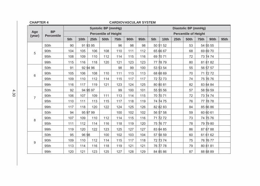
4.30
CHAPTER 4 CARDIOVASCULAR SYSTEM
Age (year)
BP Percentile
Systolic BP (mmHg) Diastolic BP (mmHg) Percentile of Height Percentile of Height
5th 10th 25th 50th 75th 90th 95th 5th 10th 25th 50th 75th 90th 95th
5
50th 90 91 93 95 96 98 98 50 51 52 53 54 55 55 90th 104 105 106 108 110 111 112 65 66 67 68 69 69 70 95th 108 109 110 112 114 115 116 69 70 71 72 73 74 74
99th 115 116 118 120 121 123 123 77 78 79 80 81 81 82
6
50th 91 92 94 96 98 99 100 53 53 54 55 56 57 57 90th 105 106 108 110 111 113 113 68 68 69 70 71 72 72 95th 109 110 112 114 115 117 117 72 72 73 74 75 76 76
99th 116 117 119 121 123 124 125 80 80 81 82 83 84 84
7
50th 92 94 95 97 99 100 101 55 55 56 57 58 59 59 90th 106 107 109 111 113 114 115 70 70 71 72 73 74 74 95th 110 111 113 115 117 118 119 74 74 75 76 77 78 78
99th 117 118 120 122 124 125 126 82 82 83 84 85 86 86
8
50th 94 95 97 99 100 102 102 56 57 58 59 60 60 61 90th 107 109 110 112 114 115 116 71 72 72 73 74 75 76 95th 111 112 114 116 118 119 120 75 76 77 78 79 79 80
99th 119 120 122 123 125 127 127 83 84 85 86 87 87 88
9
50th 95 96 98 100 102 103 104 57 58 59 60 61 61 62 90th 109 110 112 114 115 117 118 72 73 74 75 76 76 77 95th 113 114 116 118 119 121 121 76 77 78 79 80 81 81
99th 120 121 123 125 127 128 129 84 85 86 87 88 88 89
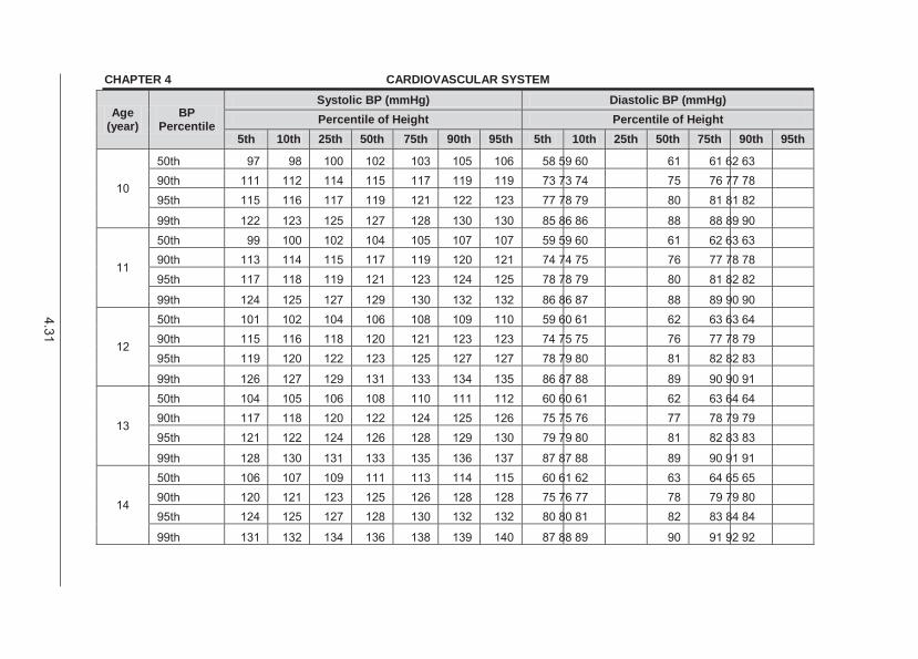
4.31
CHAPTER 4 CARDIOVASCULAR SYSTEM
Age (year)
BP Percentile
Systolic BP (mmHg) Diastolic BP (mmHg) Percentile of Height Percentile of Height
5th 10th 25th 50th 75th 90th 95th 5th 10th 25th 50th 75th 90th 95th
10
50th 97 98 100 102 103 105 106 58 59 60 61 61 62 63 90th 111 112 114 115 117 119 119 73 73 74 75 76 77 78 95th 115 116 117 119 121 122 123 77 78 79 80 81 81 82
99th 122 123 125 127 128 130 130 85 86 86 88 88 89 90
11
50th 99 100 102 104 105 107 107 59 59 60 61 62 63 63 90th 113 114 115 117 119 120 121 74 74 75 76 77 78 78 95th 117 118 119 121 123 124 125 78 78 79 80 81 82 82
99th 124 125 127 129 130 132 132 86 86 87 88 89 90 90
12
50th 101 102 104 106 108 109 110 59 60 61 62 63 63 64 90th 115 116 118 120 121 123 123 74 75 75 76 77 78 79 95th 119 120 122 123 125 127 127 78 79 80 81 82 82 83
99th 126 127 129 131 133 134 135 86 87 88 89 90 90 91
13
50th 104 105 106 108 110 111 112 60 60 61 62 63 64 64 90th 117 118 120 122 124 125 126 75 75 76 77 78 79 79 95th 121 122 124 126 128 129 130 79 79 80 81 82 83 83
99th 128 130 131 133 135 136 137 87 87 88 89 90 91 91
14
50th 106 107 109 111 113 114 115 60 61 62 63 64 65 65 90th 120 121 123 125 126 128 128 75 76 77 78 79 79 80 95th 124 125 127 128 130 132 132 80 80 81 82 83 84 84
99th 131 132 134 136 138 139 140 87 88 89 90 91 92 92
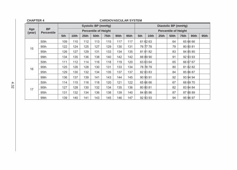
4.32
CHAPTER 4 CARDIOVASCULAR SYSTEM
Age (year)
BP Percentile
Systolic BP (mmHg) Diastolic BP (mmHg) Percentile of Height Percentile of Height
5th 10th 25th 50th 75th 90th 95th 5th 10th 25th 50th 75th 90th 95th
15
50th 109 110 112 113 115 117 117 61 62 63 64 65 66 66 90th 122 124 125 127 129 130 131 76 77 78 79 80 80 81 95th 126 127 129 131 133 134 135 81 81 82 83 84 85 85
99th 134 135 136 138 140 142 142 88 89 90 91 92 93 93
16
50th 111 112 114 116 118 119 120 63 63 64 65 66 67 67 90th 125 126 128 130 131 133 134 78 78 79 80 81 82 82 95th 129 130 132 134 135 137 137 82 83 83 84 85 86 87
99th 136 137 139 141 143 144 145 90 90 91 92 93 94 94
17
50th 114 115 116 118 120 121 122 65 66 66 67 68 69 70 90th 127 128 130 132 134 135 136 80 80 81 82 83 84 84 95th 131 132 134 136 138 139 140 84 85 86 87 87 88 89
99th 139 140 141 143 145 146 147 92 93 93 94 95 96 97
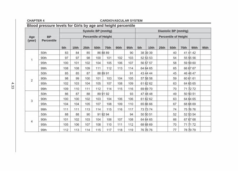
4.33
CHAPTER 4 CARDIOVASCULAR SYSTEM Blood pressure levels for Girls by age and height percentile
Age (year)
BP Percentile
Systolic BP (mmHg) Diastolic BP (mmHg)
Percentile of Height Percentile of Height
5th 10th 25th 50th 75th 90th 95th 5th 10th 25th 50th 75th 90th 95th
1
50th 83 84 85 86 88 89 90 38 39 39 40 41 41 42 90th 97 97 98 100 101 102 103 52 53 53 54 55 55 56 95th 100 101 102 104 105 106 107 56 57 57 58 59 59 60
99th 108 108 109 111 112 113 114 64 64 65 65 66 67 67
2
50th 85 85 87 88 89 91 91 43 44 44 45 46 46 47 90th 98 99 100 101 103 104 105 57 58 58 59 60 61 61 95th 102 103 104 105 107 108 109 61 62 62 63 64 65 65
99th 109 110 111 112 114 115 116 69 69 70 70 71 72 72
3
50th 86 87 88 89 91 92 93 47 48 48 49 50 50 51 90th 100 100 102 103 104 106 106 61 62 62 63 64 64 65 95th 104 104 105 107 108 109 110 65 66 66 67 68 68 69
99th 111 111 113 114 115 116 117 73 73 74 74 75 76 76
4
50th 88 88 90 91 92 94 94 50 50 51 52 52 53 54 90th 101 102 103 104 106 107 108 64 64 65 66 67 67 68 95th 105 106 107 108 110 111 112 68 68 69 70 71 71 72
99th 112 113 114 115 117 118 119 76 76 76 77 78 79 79
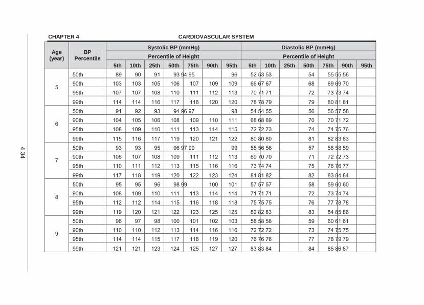
4.34
CHAPTER 4 CARDIOVASCULAR SYSTEM
Age (year)
BP Percentile
Systolic BP (mmHg) Diastolic BP (mmHg) Percentile of Height Percentile of Height
5th 10th 25th 50th 75th 90th 95th 5th 10th 25th 50th 75th 90th 95th
5
50th 89 90 91 93 94 95 96 52 53 53 54 55 55 56 90th 103 103 105 106 107 109 109 66 67 67 68 69 69 70 95th 107 107 108 110 111 112 113 70 71 71 72 73 73 74
99th 114 114 116 117 118 120 120 78 78 79 79 80 81 81
6
50th 91 92 93 94 96 97 98 54 54 55 56 56 57 58 90th 104 105 106 108 109 110 111 68 68 69 70 70 71 72 95th 108 109 110 111 113 114 115 72 72 73 74 74 75 76
99th 115 116 117 119 120 121 122 80 80 80 81 82 83 83
7
50th 93 93 95 96 97 99 99 55 56 56 57 58 58 59 90th 106 107 108 109 111 112 113 69 70 70 71 72 72 73 95th 110 111 112 113 115 116 116 73 74 74 75 76 76 77
99th 117 118 119 120 122 123 124 81 81 82 82 83 84 84
8
50th 95 95 96 98 99 100 101 57 57 57 58 59 60 60 90th 108 109 110 111 113 114 114 71 71 71 72 73 74 74 95th 112 112 114 115 116 118 118 75 75 75 76 77 78 78
99th 119 120 121 122 123 125 125 82 82 83 83 84 85 86
9
50th 96 97 98 100 101 102 103 58 58 58 59 60 61 61 90th 110 110 112 113 114 116 116 72 72 72 73 74 75 75 95th 114 114 115 117 118 119 120 76 76 76 77 78 79 79
99th 121 121 123 124 125 127 127 83 83 84 84 85 86 87
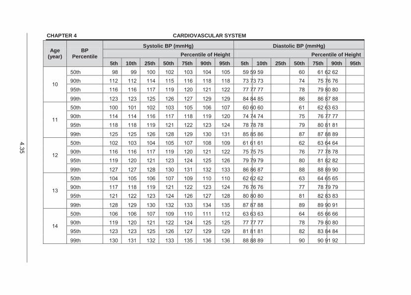
4.35
CHAPTER 4 CARDIOVASCULAR SYSTEM
Age (year)
BP Percentile
Systolic BP (mmHg) Diastolic BP (mmHg) Percentile of Height Percentile of Height
5th 10th 25th 50th 75th 90th 95th 5th 10th 25th 50th 75th 90th 95th
10
50th 98 99 100 102 103 104 105 59 59 59 60 61 62 62 90th 112 112 114 115 116 118 118 73 73 73 74 75 76 76 95th 116 116 117 119 120 121 122 77 77 77 78 79 80 80
99th 123 123 125 126 127 129 129 84 84 85 86 86 87 88
11
50th 100 101 102 103 105 106 107 60 60 60 61 62 63 63 90th 114 114 116 117 118 119 120 74 74 74 75 76 77 77 95th 118 118 119 121 122 123 124 78 78 78 79 80 81 81
99th 125 125 126 128 129 130 131 85 85 86 87 87 88 89
12
50th 102 103 104 105 107 108 109 61 61 61 62 63 64 64 90th 116 116 117 119 120 121 122 75 75 75 76 77 78 78 95th 119 120 121 123 124 125 126 79 79 79 80 81 82 82
99th 127 127 128 130 131 132 133 86 86 87 88 88 89 90
13
50th 104 105 106 107 109 110 110 62 62 62 63 64 65 65 90th 117 118 119 121 122 123 124 76 76 76 77 78 79 79 95th 121 122 123 124 126 127 128 80 80 80 81 82 83 83
99th 128 129 130 132 133 134 135 87 87 88 89 89 90 91
14
50th 106 106 107 109 110 111 112 63 63 63 64 65 66 66 90th 119 120 121 122 124 125 125 77 77 77 78 79 80 80 95th 123 123 125 126 127 129 129 81 81 81 82 83 84 84
99th 130 131 132 133 135 136 136 88 88 89 90 90 91 92
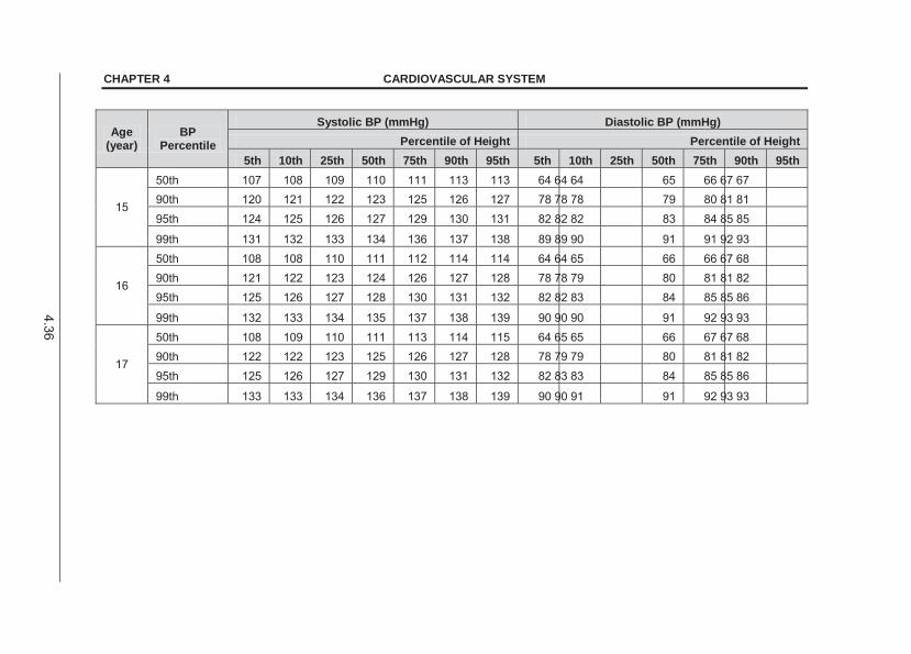
4.36
CHAPTER 4 CARDIOVASCULAR SYSTEM
Age (year)
BP Percentile
Systolic BP (mmHg) Diastolic BP (mmHg) Percentile of Height Percentile of Height
5th 10th 25th 50th 75th 90th 95th 5th 10th 25th 50th 75th 90th 95th
15
50th 107 108 109 110 111 113 113 64 64 64 65 66 67 67 90th 120 121 122 123 125 126 127 78 78 78 79 80 81 81 95th 124 125 126 127 129 130 131 82 82 82 83 84 85 85
99th 131 132 133 134 136 137 138 89 89 90 91 91 92 93
16
50th 108 108 110 111 112 114 114 64 64 65 66 66 67 68 90th 121 122 123 124 126 127 128 78 78 79 80 81 81 82 95th 125 126 127 128 130 131 132 82 82 83 84 85 85 86
99th 132 133 134 135 137 138 139 90 90 90 91 92 93 93
17
50th 108 109 110 111 113 114 115 64 65 65 66 67 67 68 90th 122 122 123 125 126 127 128 78 79 79 80 81 81 82 95th 125 126 127 129 130 131 132 82 83 83 84 85 85 86
99th 133 133 134 136 137 138 139 90 90 91 91 92 93 93
4.37
CHAPTER 4 CARDIOVASCULAR SYSTEM GENERAL AND SUPPORTIVE MEASURES » There is a strong association with overweight patients and high blood pressure. » The majority of these patients have mild hypertension and usually only need
lifestyle modification. » Acute hypertension:
> Bed rest – fowler’s position. > Control fluid intake and output (restriction). > Restrict dietary sodium. > Manage end organ effects.
» Chronic hypertension: > Advise a change in lifestyle. > Institute and monitor a weight reduction programme for obese individuals. > Regular aerobic exercise is recommended in essential hypertension.
» Dietary advice: > Limit salt and saturated fat intake. > Increase dietary fibre intake.
4.11.1 HYPERTENSION, ACUTE SEVERE For acute on chronic hypertension blood pressure needs to be lowered cautiously. Initiate medicines for sustained control as soon as possible to maintain the effect when the emergency measures are discontinued. Rate of BP reduction depends upon starting BP and age of the child. In the absence of central nervous system signs, acute hypertension can be rapidly controlled over 24 hours. If in doubt about the duration of hypertension, reduce BP slower over 48 hours.
patient’s systolic blood pressure and the 95th percentile for that age or height in the first 8 hours, then a further gradual decline over the next 24–48 hours. Do not decrease BP to < 95th percentile in first 24 hours. GENERAL AND SUPPORTIVE MEASURES » Admit patient to paediatric intensive care unit, if possible. » Monitor BP every 10 minutes until stable, thereafter every 30 minutes for 24 hours. » Set up two peripheral intravenous drips. MEDICINE TREATMENT Do not combine medicines of the same class.
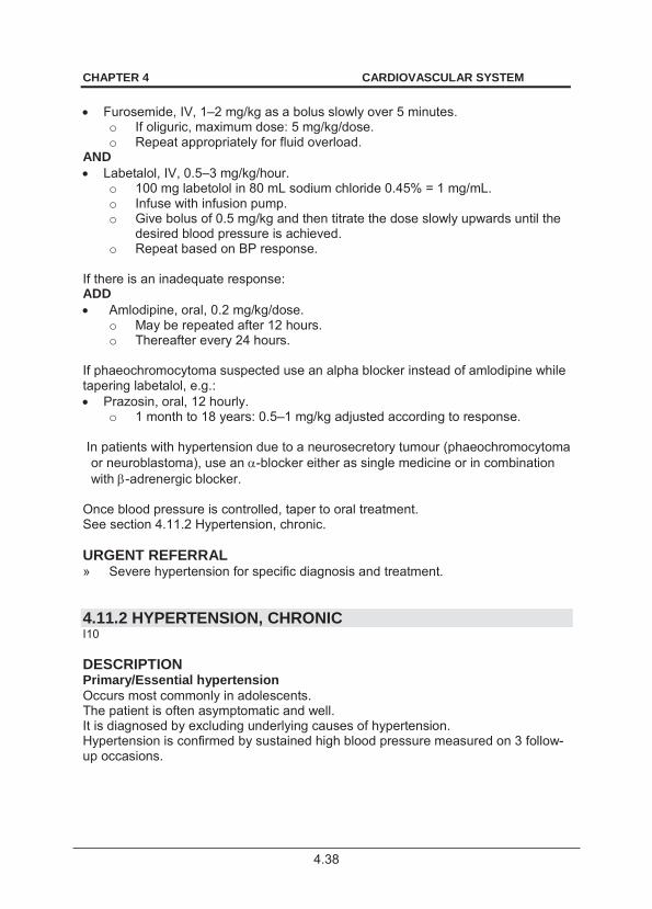
4.38
CHAPTER 4 CARDIOVASCULAR SYSTEM Furosemide, IV, 1–2 mg/kg as a bolus slowly over 5 minutes.
o If oliguric, maximum dose: 5 mg/kg/dose. o Repeat appropriately for fluid overload.
AND Labetalol, IV, 0.5–3 mg/kg/hour.
o 100 mg labetolol in 80 mL sodium chloride 0.45% = 1 mg/mL. o Infuse with infusion pump. o Give bolus of 0.5 mg/kg and then titrate the dose slowly upwards until the
desired blood pressure is achieved. o Repeat based on BP response.
If there is an inadequate response: ADD Amlodipine, oral, 0.2 mg/kg/dose.
o May be repeated after 12 hours. o Thereafter every 24 hours.
If phaeochromocytoma suspected use an alpha blocker instead of amlodipine while tapering labetalol, e.g.: Prazosin, oral, 12 hourly.
o 1 month to 18 years: 0.5–1 mg/kg adjusted according to response. In patients with hypertension due to a neurosecretory tumour (phaeochromocytoma
or neuroblastoma), use an -blocker either as single medicine or in combination with -adrenergic blocker.
Once blood pressure is controlled, taper to oral treatment. See section 4.11.2 Hypertension, chronic. URGENT REFERRAL » Severe hypertension for specific diagnosis and treatment. 4.11.2 HYPERTENSION, CHRONIC I10 DESCRIPTION Primary/Essential hypertension Occurs most commonly in adolescents. The patient is often asymptomatic and well. It is diagnosed by excluding underlying causes of hypertension. Hypertension is confirmed by sustained high blood pressure measured on 3 follow-up occasions.

4.39
CHAPTER 4 CARDIOVASCULAR SYSTEM Chronic secondary hypertension All children with incurable forms of persistent secondary hypertension require medicine treatment over and above general and supportive measures. DIAGNOSTIC CRITERIA Investigations » Urine dipstick test. » Urine MCS. » Blood urea, calcium, creatinine and electrolytes. » Chest X-ray, ECG and abdominal sonar. If all tests are negative, start lifestyle intervention. GENERAL AND SUPPORTIVE MEASURES » Introduce physical activity, diet management and weight reduction, if obese. » Advise teenagers against smoking. » Follow up to monitor blood pressure and educate patient on hypertension:
> if blood pressure decreases, continue with non-drug management and follow up;
> if BP is increasing progressively, reinvestigate to exclude secondary causes or refer;
> if BP is stable but persistently > 95th percentile and secondary causes have been excluded, start medicine treatment after failed non-drug management for 6 months.
» Consider earlier initiation of medicine treatment if positive family history for cardiovascular disease, essential hypertension or diabetes mellitus.
MEDICINE TREATMENT Goal of treatment in uncomplicated primary hypertension with no t arget-organ damage is to achieve BP < 95th percentile. For chronic renal disease, diabetes or hypertension with target-organ damage, target is BP < 90th percentile. Medicine treatment is initiated for Stage 2 hy pertension. Consider therapy in Stage 1 hypertension if there is a family history of cardiovascular disease, hypertension or diabetes. Aim to achieve control of BP over 48–72 hours in symptomatic patients. For ambulatory patients start at the l owest dose of th e preferred medicine and increase the dose until control is achieved. Once the highest recommended dose is reached or if the patient experiences adverse effects from the medicine, add a second medicine from a different class. For patients with persistent hypertension despite the use of first line medicine, add a second/third medicine. There is no specific order in which medicine should be added. Use specific classes of a ntihypertensive medicine according to t he underlying pathogenesis or illness.
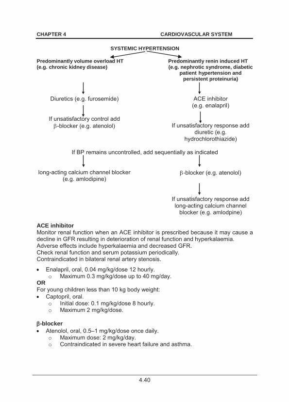
4.40
CHAPTER 4 CARDIOVASCULAR SYSTEM
SYSTEMIC HYPERTENSION Predominantly volume overload HT (e.g. chronic kidney disease)
Predominantly renin induced HT (e.g. nephrotic syndrome, diabetic
patient hypertension and persistent proteinuria)
Diuretics (e.g. furosemide)
If unsatisfactory control add -blocker (e.g. atenolol)
ACE inhibitor (e.g. enalapril)
If unsatisfactory response add diuretic (e.g.
hydrochlorothiazide)
If BP remains uncontrolled, add sequentially as indicated
long-acting calcium channel blocker (e.g. amlodipine)
-blocker (e.g. atenolol)
If unsatisfactory response add long-acting calcium channel
blocker (e.g. amlodpine) ACE inhibitor Monitor renal function when an ACE inhibitor is prescribed because it may cause a decline in GFR resulting in deterioration of renal function and hyperkalaemia. Adverse effects include hyperkalaemia and decreased GFR. Check renal function and serum potassium periodically. Contraindicated in bilateral renal artery stenosis.
Enalapril, oral, 0.04 mg/kg/dose 12 hourly. o Maximum 0.3 mg/kg/dose up to 40 mg/day.
OR For young children less than 10 kg body weight: Captopril, oral.
o Initial dose: 0.1 mg/kg/dose 8 hourly. o Maximum 2 mg/kg/dose.
-blocker Atenolol, oral, 0.5–1 mg/kg/dose once daily.
o Maximum dose: 2 mg/kg/day. o Contraindicated in severe heart failure and asthma.
4.41
CHAPTER 4 CARDIOVASCULAR SYSTEM OR If child less than 10 kg body weight: Propranolol, oral, 0.25–1 mg/kg/dose 8–12 hourly.
o Maximum dose: 1.5 mg/kg/dose. Calcium channel blocker Amlodipine, oral, 0.1–0.2 mg/kg/dose once daily.
Diuretic Hydrochlorothiazide, oral, 0.5–1 mg/kg/dose once daily.
o May cause hypokalaemia. OR Furosemide, oral, 0.5–1.5 mg/kg/dose 12–24 hourly.
o Maximum dose: 6 mg/kg/day. o May cause hypokalaemia.
-blocker, e.g:
Also indicated in patients with phaeochromocytoma-associated hypertension. Prazosin, oral, 12 hourly.
o 1 month to 18 years: 0.5–1 mg/kg adjusted according to response.
REFERRAL » All children with chronic hypertension for specific diagnosis, planning of
treatment and long-term follow-up. » Persistent cough on treatment with ACE inhibitor. 4.12 CHILDREN WITH PROSTHETIC HEART VALVES Z95.2 DESCRIPTION Valve replacement may be required for severe valvular disease when valve repair is not feasible or advisable. The valves may be mechanical valves or bioprosthetic valves or preserved human tissue valves. In children bioprosthetic valves tend to degenerate, calcify and have structural deterioration more frequently and more rapidly compared with adults. Mechanical valves are more commonly used in children. Complications include: » Valve failure. May be abrupt (teari ng of components) or gradua l (with
calcification and stiffening of leaflets). » Prosthetic valve thrombosis. » Prosthetic valve endocarditis. » Haemolytic anaemia.
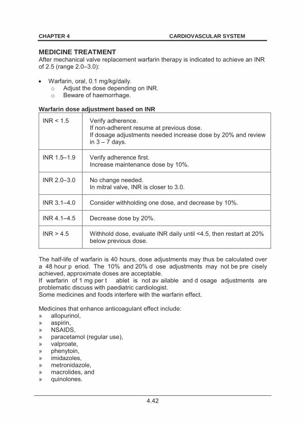
4.42
CHAPTER 4 CARDIOVASCULAR SYSTEM MEDICINE TREATMENT After mechanical valve replacement warfarin therapy is indicated to achieve an INR of 2.5 (range 2.0–3.0): Warfarin, oral, 0.1 mg/kg/daily.
o Adjust the dose depending on INR. o Beware of haemorrhage.
Warfarin dose adjustment based on INR
INR < 1.5 Verify adherence. If non-adherent resume at previous dose. If dosage adjustments needed increase dose by 20% and review in 3 – 7 days.
INR 1.5–1.9 Verify adherence first. Increase maintenance dose by 10%.
INR 2.0–3.0 No change needed. In mitral valve, INR is closer to 3.0.
INR 3.1–4.0 Consider withholding one dose, and decrease by 10%.
INR 4.1–4.5 Decrease dose by 20%.
INR > 4.5 Withhold dose, evaluate INR daily until <4.5, then restart at 20% below previous dose.
The half-life of warfarin is 40 hours, dose adjustments may thus be calculated over a 48 hour p eriod. The 10% and 20% d ose adjustments may not be pre cisely achieved, approximate doses are acceptable. If warfarin of 1 mg per t ablet is not av ailable and d osage adjustments are problematic discuss with paediatric cardiologist. Some medicines and foods interfere with the warfarin effect. Medicines that enhance anticoagulant effect include: » allopurinol, » aspirin, » NSAIDS, » paracetamol (regular use), » valproate, » phenytoin, » imidazoles, » metronidazole, » macrolides, and » quinolones.
4.43
CHAPTER 4 CARDIOVASCULAR SYSTEM Medicines that diminish anticoagulant effect include: » carbamazepine, » phenobarbital, » phenytoin, (both diminished and enhanced effects have been reported) » nevirapine, » rifampicin. Foods that contain high amounts of vitamin K and can decrease the effectiveness of warfarin, e.g.: » spinach, » parsley, and » brussel sprouts. Certain drinks can increase the effect of warfarin e.g. cranberry juice and alcohol.
5.1
CHAPTER 5 DERMATOLOGY
Skin lesions are best characterised by their morphologic appearance which allows consideration of a suitable differential diagnosis. 5.1 BULLAE 5.1.1 EPIDERMOLYSIS BULLOSA Q81.9 DESCRIPTION Congenital, hereditary blistering skin lesions with onset in the newborn. Lesions do not have erythematous base. Loss of nails may occur. GENERAL AND SUPPORTIVE MEASURES » May require care in high or intensive care unit. » Do not rupture bullae. » Prevent infection with appropriate wound care. » Attend to fluid and nutrition balance. REFERRAL » All cases. 5.1.2 STAPHYLOCOCCAL SCALDED SKIN SYNDROME L00 DESCRIPTION Blistering skin condition that presents like scalded skin. GENERAL AND SUPPORTIVE MEASURES » Appropriate wound care. MEDICINE TREATMENT » Cloxacillin, IV, 50 mg/kg/dose 6 hourly for 5 days.
o Neonates Week 1–2, administer 12 hourly. Week 2–4, administer 8 hourly.
OR » Flucloxacillin, oral, 12.5–25 mg/kg/dose 6 hourly for 7 days.
5.2
CHAPTER 5 DERMATOLOGY REFERRAL » Recalcitrant cases. 5.1.3 CHRONIC BULLOUS DISEASE OF CHILDHOOD L12.2 DESCRIPTION Tense blisters that lead to ulceration involving the groin, face and trunk. DIAGNOSTIC CRITERIA » Skin biopsy with immunofluorescence. GENERAL AND SUPPORTIVE MEASURES » Appropriate wound care. REFERRAL » All cases. 5.2 ERYTHEMA AND DESQUAMATION It is a continuum ranging from Erythema Multiforme (EM) to Stevens-Johnson Syndrome and then to the potentially lethal Toxic Epidermal Necrolysis (TEN). 5.2.1 ERYTHEMA MULTIFORME/STEVENS-JOHNSON SYNDROME L51.9/L51.1 DESCRIPTION Acute, vesico-bullous disorder with numerous manifestations on the skin, mucous membranes and, occasionally, internal organs caused mainly by: » medicines, e.g. sulphonamides, phenytoin, phenobarbitone, » exposure to toxic substances, and » infections, e.g. herpes simplex and mycoplasma.
Complications include: » conjunctivitis, » uveitis, » corneal scarring, » fluid loss, » infections, » anaemia, and » oesophageal strictures.
5.3
CHAPTER 5 DERMATOLOGY DIAGNOSTIC CRITERIA Iris or target lesions consisting of a dar k centre, an inner pale ring and an erythematous outer border. In ery thema multiforme these lesions are pathognomonic. Erythematous macules evolve into papules, vesicles, bullae, urticarial plaques or patches of confluent erythema. The centre of the lesion may be vesicular, purpuric or necrotic. Erythema multiforme minor Prodromal symptoms are generally absent. Symmetric crops of skin lesions of diverse morphology, primarily on the ex tensor surfaces of the arms and legs and often including soles and palms with relative sparing of the mucous membranes and the trunk.
Erythema multiforme major (Stevens-Johnson syndrome) A serious, systemic condition involving the skin and at least two mucous membranes. Eruption may be preceded by non-specific prodromal symptoms like: » malaise, » fever, » chills, or » upper respiratory infection. Cutaneous lesions tend to rupture, leaving the skin denuded, with fluid loss, anaemia and high risk of infection. Involvement of oral mucosa is common. GENERAL AND SUPPORTIVE MEASURES » May require care in high or intensive care unit. » Examine daily for systemic involvement, infection and ocular lesions. If
infection is suspected, send blood and skin lesion specimens for culture and sensitivity before initiating antibiotic therapy.
» Do not puncture bullae or vesicles. » Frequent mouth washes for oral lesions. » Cool compresses and wet dressings. » Eye care where eye cannot close. See section 16.1: Eye infection,
complicated (Severe eye infection). » Maintain fluid balance. Beware of shock. » Nasogastric feeds if unable to eat, IV alimentation if enteral feeds are
not possible. » Stop all medicines. MEDICINE TREATMENT For pain: These patients require effective pain control. Change of dressing protocol: See Section 20.1: Management of pain.

5.4
CHAPTER 5 DERMATOLOGY Antibiotic therapy For secondary infections Reconsider choice of antibiotic when the results of cultures become available or the child does not improve. Use IV antibiotics if the oral route cannot be used. Cloxacillin, IV, 50 mg/kg/dose 6 hourly.
OR Cephalexin, oral, 6.25–12.5 mg/kg/dose 6 hourly.
For oral lesions: Chlorhexidine 0.2%, 15 mL as a mouthwash.
o Use as needed. o Do not swallow.
Note: The use of systemic corticosteroids is not recommended. REFERRAL » Erythema multiforme not responding to adequate therapy. » Erythema multiforme with ocular involvement. 5.3 MACULES AND PAPULES 5.3.1 DRUG REACTIONS L27.0 Commonly associated with: » sulphur containing agents, » penicillin, » anti-epileptics (e.g. carbamazepine, lamotrigine), » NSAIDs, » anti-tuberculosis drugs, and » non-nucleoside reverse transcriptase inhibitors. A variety of rashes o ccur, ranging from (worst) erythema multiforme with mucosal involvement, target lesions, blistering and fever, through itchy or painful urticarial eruptions, measles-like maculopapular rashes, to erythema and flat, sy mmetrical macular lesions (fixed drug reactio ns). These are commonly flat or slightly raised lesions of < 0.5 cm in size. Lesions recur upon re-exposure to the causative agent. GENERAL AND SUPPORTIVE MEASURES » Stop causative agents.
5.5
CHAPTER 5 DERMATOLOGY MEDICINE TREATMENT Antihistamines: For children 3 years and older: Cetirizine, oral, as a single dose at night.
o Children 3–12 years: 5 mg. o Children > 12 years: 10 mg.
Where the oral route can be used: Chlorphenamine, oral, 0.1mg/kg/dose as a single dose at night.
Where the oral route cannot be used: Promethazine, IV, 0.1 mg/kg/dose 8–12 hourly.
REFERRAL » Systemic involvement with organ dysfunction. 5.3.2 ACNE L70 DESCRIPTION An inflammatory condition of hair follicles leading to comedone formation that can cause scarring and post inflammation hyper-pigmentation. DIAGNOSTIC CRITERIA » Black or white heads (comedones). GENERAL AND SUPPORTIVE MEASURES » Avoid greasy and oily topical products. » Discourage excessive facial washing. MEDICINE TREATMENT Benzoyl peroxide 5%, topical, applied to affected areas at night.
AND Doxycycline, oral, 100 mg once daily for a maximum of three months.
If ineffective Stop benzoyl peroxide To limit skin irritation, introduce topical retinoids, e.g. tretinoin gel/cream, gradually at night. Tretinoin cream 0.05%, topical, applied sparingly once daily at bedtime
until substantial improvement. o Avoid contact with eyes and mucous membranes. o Limit exposure to sunlight, especially with concomitant use of
doxycycline.

5.6
CHAPTER 5 DERMATOLOGY
Tretinoin is teratogenic. Do not use where pregnancy is a possibility.
If used, ensure adequate contraception.
To avoid sun irritation: Sunscreen, topical, applied daily.
REFERRAL » Recalcitrant and/or fulminant acne. » Psychologically disturbed or depressed patient. » Young females w ith premenstrual flare or w ith clinical signs of
hyperandrogenism for consideration of oral contraceptives. 5.3.3 CELLULITIS AND ERYSIPELAS L03.9/A46 DESCRIPTION Infection of the skin and subcutaneous tissue usually caused by streptococci, staphylococci or H. influenzae. In cellulitis, the border is indistinct. Erysipelas The affected area is: » well demarcated with firm borders, » very tender and warm, » bright red and swollen. Erysipelas must be distinguished from necrotising fasciitis where there is infection and inflammation usually by a gas- forming organism that spreads rapidly along the fascial tissue. Complications may lead to septicaemia. DIAGNOSTIC CRITERIA » Acutely ill child with fever and malaise. » Involved area is swollen, indurated, erythematous and painful/tender
with regional lymphadenopathy. GENERAL AND SUPPORTIVE MEASURES » Ensure adequate nutrition and hydration. » Elevate the affected limb to reduce swelling. » Exclude eczema, immunocompromised state, diabetes and underlying
osteomyelitis. MEDICINE TREATMENT Choice of intravenous or oral antibiotics depends on the severity of the condition.
5.7
CHAPTER 5 DERMATOLOGY Severe disease Cloxacillin, IV, 50 mg/kg/dose 6 hourly for 5 days.
For peri-orbital cellulitis: Ceftriaxone, IV, 50 mg/kg once daily.
OR If one month old or younger: Cefotaxime, IV, 50 mg/kg/dose, 6–8 hourly.
Non-severe disease Cephalexin, oral, 12.5–25 mg/kg/dose 6 hourly for 7 days.
o Child < 2 years: 125 mg. o Child 2–10 years: 250 mg. o > 10 years: 500 mg.
Penicillin allergy Erythromycin, oral, 6.25–12.5 mg/kg/dose, 6 hourly for 5 days.
For pain: Paracetamol, oral, 15 mg/kg/dose, 6 hourly as required.
If needed ADD: Ibuprofen, oral, 5–10 mg/kg/dose, 6 hourly for 72 hours.
o Child < 30 kg, maximum dose: 500 mg/day. REFERRAL » Urgent: necrotising fasciitis. » Poor response to therapy. » Recurrent cellulitis. 5.3.4 ECZEMA L20.9 DESCRIPTION An inflammatory itchy skin condition characterised by: » Vesicles, weeping and crusting in the acute stage. » Scaling and lichenification during the chronic stage. DIAGNOSTIC CRITERIA » Family history of allergies. » Reaction after exposure to allergens. » Typical distribution: face, flexures of knees and elbows, and creases of
neck.
5.8
CHAPTER 5 DERMATOLOGY GENERAL AND SUPPORTIVE MEASURES » Avoidance measures: use neutral soaps and rinse clothes properly after wash. » Keep fingernails short to prevent scratching. » Wrap with dressings soaked in sodium chloride 0.9%. » Avoid sunlight and use sunscreen. MEDICINE TREATMENT Antihistamine Cetirizine, oral, 12 hourly
o Children 6 months to 2 years: 0.25 mg/kg/dose o Children 2–6 years: 2.5 mg. o Children 6 -12 years: 5 mg. o Children > 12 years: 10 mg.
To relieve skin dryness: Aqueous cream, topical.
For baths and prior to applying corticosteroids (after drip drying): Emulsifying ointment, topical.
For the face and skin folds: Hydrocortisone 1%, topical, 12 hourly.
For the body: Betamethasone 0.1%, topical, undiluted applied once daily for 7 days.
o Moisturise with emulsifying ointment during therapy and in subsequent weeks.
Secondary bacterial infection: Cephalexin, oral, 6.25–12.5 mg/kg/dose, 6 hourly for 14 days.
Note: Short term use of topical steroids is recommended. Oral corticosteroids do not have a role in the management of this condition. REFERRAL » Recalcitrant cases. » Concomitant food allergy (allergy clinic). 5.3.5 CANDIDIASIS B37.2 DESCRIPTION Skin infection inv olving axillae, neck and perineum. C ommonly occurs in immunocompromised individuals. Involvement of mouth and perineal regions suggests systemic disease.

5.9
CHAPTER 5 DERMATOLOGY DIAGNOSTIC CRITERIA Clinical » Red raw looking patches with satellite white pustular lesions on an
erythematous base. » Mucosal involvement. Investigations » Wet preparation with potassium hydroxide or biopsy and culture. GENERAL AND SUPPORTIVE MEASURES » Control underlying immunosuppressive state e.g. diabetes, HIV. » Personal hygiene of mothers prior to breast feeding. MEDICINE TREATMENT Imidazole cream 1%, e.g. clotrimazole, topical, applied 8 hourly for 14 days.
If no response: Fluconazole, oral, 3–6 mg/kg/day for 14 days.
REFERRAL » Recalcitrant infection. 5.3.6 PSORIASIS L40.9 DESCRIPTION An inflammatory condition of the skin and joints. DIAGNOSTIC CRITERIA » Scaly, red itchy papules and plaques over scalp, perineum, and skin
folds and extensor surfaces. » Nails may be opaque, deformed and crumbling. » Occasional pustules are seen. GENERAL AND SUPPORTIVE MEASURES » Avoid precipitants e.g. drugs. MEDICINE TREATMENT Local plaques To remove scales in children 12 years and older: Salicylic acid 2% and coal tar in white soft paraffin, applied 8 hourly.
For scalp lesions: Mild coal tar shampoo.
5.10
CHAPTER 5 DERMATOLOGY Severe pustular psoriasis Hydrocortisone 1%, topical, applied 1–2 times daily.
OR Prednisone, oral, 1–2 mg/kg as a single daily dose for 7 days.
REFERRAL » Severe psoriasis and recalcitrant cases. » Intolerance to salicylic acid. » No response to treatment. 5.3.7 URTICARIA L50.9 DESCRIPTION An itchy inflammatory skin and mucosal condition recognised by wheal and flare reaction that may be acute or chronic, often due to irritants, insect bites or allergens. Secondary infective features include excoriation, vesicles and pigmentary changes. Chronic papular eruptive urticaria is often seen in HIV infected individuals. DIAGNOSTIC CRITERIA » History of allergen exposure. » Wheal and flare reaction (“hives”). » Positive skin test if due to allergy. GENERAL AND SUPPORTIVE MEASURES » Limit exposure to precipitants, e.g. drugs, allergens and toxins. » Limit exposure to insects by using topical insect repellent which contains
more than 10% diethyltoluamide (DET). » Wrap with dressings soaked in sodium chloride 0.9%. MEDICINE TREATMENT Chlorphenamine, oral, 0.1 mg/kg/dose as a single dose at night.
Hydrocortisone 1%, topical, applied twice daily as required.
o Useful when applied immediately after insect bite. Severe chronic urticaria Cetirizine, oral, as a single dose at night.
o Child 3–12 years: 5 mg. o Child > 12 years: 10 mg.
REFERRAL » Recalcitrant and chronic cases.
5.11
CHAPTER 5 DERMATOLOGY 5.4. PURPURA D69.9 5.4.1 MENINGOCOCCAEMIA A39.2 DESCRIPTION Palpable bleeding into skin caused by N. meningitides and is associated with rapid spread.
This is a medical emergency and can be fatal. See section 8.25: Sepsis.
5.4.2 HENOCH-SCHÖNLEIN PURPURA D69.0 See section 12.1: Henoch Schönlein Purpura (HSP) 5.4.3 IDIOPATHIC THROMBOCYTOPENIC PURPURA (ITP) D69.3 See section 3.10: Idioptahic thrombocytopaenic purpura (ITP) 5.5. VESICLES AND PUSTULES 5.5.1 INFECTIONS R23.8/L08.9 See section 8.26: Varicella (chickenpox) and section 8.27: Zoster. 5.5.2 SKIN AND MUCOSAL DISORDERS IN HIV Skin and mucosal disorders are more severe in immune suppressed (HIV-infected) patients and may be worsened by IRIS. It may present initially with a skin or a mucosal lesion or skin and mucosal lesions may develop during the course of the HIV illness. Lesions respond to antiretroviral therapy together with treatment for a specific skin and/or mucosal disorder. Skin eruptions or rashes are relatively common in HIV patients and relate to antiretroviral and other medicines.
5.12
CHAPTER 5 DERMATOLOGY Conditions that are frequently more common in patients with HIV or are more specific in their unusual presentation include: » HIV papular pruritic eruption. » Kaposi’s sarcoma. 5.5.2.1 HIV PAPULAR PRURITIC ERUPTION DESCRIPTION Chronic itchy condition with a relap sing course due to an initial allergy to insect bites in a susceptible patient who developed an immune reaction to insect products. In HIV-infected patients the insect bite reactions are severe and recalcitrant with post inflammatory pigmentation and scarring. DIAGNOSTIC CRITERIA » Initial lesion is a pruritic urticarial spot with a central red punctum. » Lesions progress to pruritic papules with or without blisters which, due
to scratching, may present with inflammatory changes, erosions, crusts or scabs with secondary infection.
» Post inflammatory pigmentation and scarring are common. GENERAL AND SUPPORTIVE MEASURES » Prevent insect bites e.g. insect repellents, eradicate fleas and other
insects. MEDICINE TREATMENT Calamine lotion, topical, applied as needed. Chlorphenamine, oral, 0.1 mg/kg/dose 6 hourly. Betamethasone 0.1%, topical, applied 12 hourly for 3 days.
Then, until pruritus subsides: Hydrocortisone 1%, topical applied 12 hourly.
Treat secondary infection with an appropriate antibiotic, if indicated. Treatment of HIV. See section 9.1: Human immunodeficiency virus infections. REFERRAL » No response to treatment. 5.5.2.2 KAPOSI’S SARCOMA C46.9 DESCRIPTION Kaposi’s sarcoma is a v ascular tumour that can present anywhere on the skin and also involve lymph nodes and internal organs, primarily lungs and gastrointestinal tract.
5.13
CHAPTER 5 DERMATOLOGY It is a ssociated with human herpes virus 8 and occurs most commonly in immunocompromised HIV-infected patients. It can be asymptomatic and indolent or aggressive, characterised by explosive growth and death. DIAGNOSTIC CRITERIA » Presents with skin lesions on the limbs particularly the lower leg and
foot but skin lesions may occur anywhere on the body. » Lesions (skin and mucosal) may be bruise-like patches, purple or
purple-red plaques, subcutaneous papules or nodules. » Lymphoedema, ulceration and secondary bacterial infection may occur. GENERAL AND SUPPORTIVE MEASURES » Counselling to assist patient in dealing with the condition. MEDICINE TREATMENT Manage in consultation with an oncologist. Treat secondary infection with an appropriate antibiotic, if indicated. Treatment of HIV. See section 9.1: Human immunodeficiency virus infections. Supportive treatment e.g. pain, once condition is considered untreatable. See section 20.1: Management of pain. REFERRAL » All not responding to ART. » Extensive progressive disease. 5.5.2.3 WARTS MEDICINE TREATMENT Podophyllin resin 20% or sal icylic acid 25% ointment, applied under
plaster nightly. o Protect surrounding skin with petroleum jelly. o Repeat until the wart falls off.
REFERRAL » Extensive warts involving the face. » Genital warts: Refer to STI clinic. 5.5.3 IMPETIGO See Standard Treatment Guidelines and Essential Medicines List for Primary Health Care.
6.1
CHAPTER 6 RENAL CONDITIONS
6.1 NEPHROLOGICAL/UROLOGICAL DISORDERS 6.1.1 POST STREPTOCOCCAL GLOMERULONEPHRITIS N05.9 DESCRIPTION Acute post-streptococcal glomerulonephritis is a disorder of th e kidneys caused by an immunological response of the kidney to nephritogenic strains of streptococci. It develops one to three weeks after a streptococcal throat or skin infection. Immune complexes are deposited in the glomerular basement membrane and/or mesangium of the glomeruli. DIAGNOSTIC CRITERIA Clinical » Occurs predominantly in children 3–12 years old. » Presents 1–3 weeks after streptococcal pharyngitis or skin infection (impetigo). » Characteristic features include:
> facial or generalised oedema, > painless macroscopic haematuria (smoky or tea coloured urine), > oliguria, and > hypertension.
Special investigations to confirm APSGN Urine analysis Macroscopic appearance smoky, brown, bloody Urine test strips 1+ to 3+ haematuria; trace to 2+ proteinuria Microscopic examination dysmorphic red blood cells;
red blood cell and granular casts Blood investigations Streptococcus serology ASO or Anti-DNAseB titre
positive in the absence of prior antibiotic treatment (ASO often negative in preceding skin infections)
Complement study C3 C4
decreased normal
S-biochemistry Serum electrolytes dilutional hyponatraemia, hyperchloraemic
hyperkalaemic metabolic acidosis is common S-Urea & creatinine mildly elevated in the acute phase Full blood count dilutional anaemia; thrombocyte count is normal
6.2
CHAPTER 6 RENAL CONDITIONS GENERAL AND SUPPORTIVE MEASURES » Bed rest is necessary in children with severe hypertension or pulmonary
oedema. » Monitor fluid balance:
> Weigh daily and record fluid intake and output strictly. > Allowed fluid intake is dependent on urine output. > In small children fluid balance is best monitored with regular weighing. > Never use a potassium-containing solution in an anuric patient. > Do not use parenteral fluids if oral intake is possible.
» Fluid management according to fluid status: > Pulmonary oedema plus oliguria/anuria: Do not give fluid. > Hydrated anuric patient without extra-renal fluid losses: Oral
fluid to replace insensible water losses only. Insensible water loss is calculated as: 25 mL/kg/day(400 mL/m2/day).
In exceptional circumstances, if parenteral fluid is required, use an electrolyte free solution i.e. dextrose 5% or 10%, IV.
> Normally hydrated plus oliguria: Oral fluid intake to replace insensible water loss and urine output of previous 24 hours.
> Normally hydrated plus normal urine output: Give normal fluid intake.
» Dietary measures: > Restrict sodium intake in all patients. > Restrict potassium intake until result of serum electrolytes is
available. > Restrict protein intake to 0.5 g/kg/day. > Bread and jam is a relatively safe option if no dietician is available.
MEDICINE TREATMENT Eradication of streptococci Phenoxymethylpenicillin, oral, 50 mg/kg/24 hours in 4 divided doses (6
hourly) for 10 days. OR If unable to take oral medication: Benzathine benzylpenicillin (depot formulation), IM, 30 000 units/kg/dose,
2 doses given 5 days apart. o Maximum dose: 1.2 million units.
Penicillin allergy Erythromycin, oral, 10 mg/kg/dose, 6 hourly for 10 days
Hypertension Hypertension usually develops acutely due to fluid overload.
6.3
CHAPTER 6 RENAL CONDITIONS Hypertensive crisis: Patient with signs of hypertensive encephalopathy, i.e.: » convulsions, » retinal haemorrhages, visual loss and » left heart failure. Hypertensive urgency: Symptomatic patients with significant elevation of blood pressure with complaints of headache, blurred vision and nausea but lacks the above clinical manifestations. Initiate treatment for acute hypertension: See section 4.11.1: Hypertension, acute severe. For acute hypertensive emergency due to post streptococcal glomerulonephritis: Furosemide, IV, 1–2 mg/kg/dose.
If oliguric: Furosemide, IV, 5 mg/kg/dose.
o Administer IV bolus slowly over 5 minutes due to risk of ototoxicity. AND Labetalol, IV, 0.2–1.0 mg/kg/dose as a bolus.
o Maximum bolus dose: 40 mg. o Continue infusion: 0.25–3.0 mg/kg/hour. o Taper infusion rate up or down according to response.
If no hypertensive crisis but persistent significant hypertension: Propranolol, oral, 1-2 mg/kg/dose, 6 hourly.
o Maximum dose: 8 mg/kg/24 hours. If blood pressure is not adequately controlled: ADD Amlodipine, oral, 0.2 mg/kg/dose.
o May be repeated 6 hours later, thereafter once every 24 hours. o Maximum dose: 5 mg. o Crush 5 mg tablet and disperse in 5 mL water: amlodipine 1 mg/mL.
Once blood pressure has normalised, taper and stop antihypertensive treatment. Monitor blood pressure over the next 48 hours to exclude rebound hypertension. Volume overloaded and pulmonary oedema See fluid management in general and supportive measures. Furosemide, slow IV, 2 mg/kg/dose.
o Maximum dose: 5 mg/kg/dose. o Maximum cumulative daily dose: 8 mg/kg/24 hours.
6.4
CHAPTER 6 RENAL CONDITIONS For pulmonary oedema: See fluid management in general and supportive measures. Morphine, IV, 0.1 mg/kg/dose.
o Repeat after 4 hours if required. Oxygen, 100%, 2–3 L/minute by nasal cannula.
REFERRAL Urgent (as soon as possible) » Anuric patient with acute volume overload and unresponsive to
furosemide. » Uncontrolled hypertension. » Oliguric and progressive renal failure. » Cardiac failure or pulmonary oedema not responding to treatment. For specialist advice » Macroscopic haematuria persisting for more than 4 weeks or persistent
proteinuria. » Family history of renal disease. » Streptococcal aetiology unproven (ASOT and anti-DNAseB negative,
normal C3 levels, decreased C4 levels). » Decreased complement levels which persist for more than 6 weeks. » Persistent renal failure after initial recovery. » Persistent hypertension. 6.1.2 URINARY TRACT INFECTION (UTI) N39.0 DESCRIPTION Bacterial infection of the urinary tract. Uncomplicated urinary tract infection (UTI) is an infection which is limited to the lower urinary tract and there are no associated urological anomalies. It is seen most commonly in girls over two years of age. It presents with localising symptoms of dysuria, frequency, urgency, cloudy urine and lower abdominal discomfort. Urine dipstick test show positive leukocyte esterase, nitrites and haematuria. See the St andard Treatment Guidelines and E ssential Medicines List for Primary Health Care Level. Complicated urinary tract infection (UTI) is an infection of the urinary tract involving the renal parenchyma (acute pyelonephritis) or which is associated with underlying congenital anomalies of the kidneys and urinary tract.
6.5
CHAPTER 6 RENAL CONDITIONS It is accompanied by fever and other systemic features as described below. It may result in significant short-term morbidity, including septicaemic shock and acute renal failure, especially in infants. Permanent renal damage may occur in children who have recurring episodes of pyelonephritis. DIAGNOSTIC CRITERIA Clinical » Signs and symptoms are related to the age of the child and are often
non-specific. » Uncomplicated urinary tract infections may cause very few signs and
symptoms. Complicated infections may present with a wide range of signs and symptoms.
» Neonates may present with: > fever, > vomiting, > hypothermia, > prolonged jaundice, > poor feeding, > failure to thrive, > sepsis, > renal failure.
» Infants and children may present with: > failure to thrive, > frequency, > persisting fever, > dysuria, > abdominal pain, > enuresis or urgency.
Examine the urine in any child with fever of unknown origin.
Special investigations » Urine bag specimens are used for screening purposes only.
> When urine dipstick test of bag specimen reveals presence of leukocytes or nitrites, collect urine aseptically for urine MCS.
> Urine specimen is collected aseptically: by suprapubic aspiration or transurethral bladder
catheterisation in acutely ill children < 2 years of age or in smaller children who are unable to co-operate;
or by mid-stream clean catch method in older children.
» Criteria for the diagnosis of UTI: > any culture from a suprapubic urine sample, > a culture of > 104 col/mL urine of a single organism from a catheter
specimen, > a pure culture of > 105 col/mL in a mid-stream clean catch sample
or consistent culture of a pure growth even with counts as low as 104 col/mL.
6.6
CHAPTER 6 RENAL CONDITIONS
» Ultrasound: > Do a ren al ultrasound in all children with first UTI as soon as
possible, unless a normal ultrasound was previously seen. » MCUG:
> in children who have abnormalities of the kidneys, ureter or bladder demonstrated by ultrasound.
GENERAL AND SUPPORTIVE MEASURES » Ensure adequate nutrition and hydration. Maintain hydration with oral
and/or IV fluids if necessary. » For recurring infections:
> avoid irritant soaps and bubble baths, > treat constipation, if present, > treat pinworm, > perineal hygiene, > regular complete emptying of the bladder and/or double voiding, i.e.
making an additional attempt at voiding after the initial flow of urine has ceased.
MEDICINE TREATMENT Antibiotic therapy Total duration of antibiotic therapy: minimum of 7 days. Increase duration to 10–14 days in infants who had septicaemia. The empiric choice of antibiotics depends on the expected sensitivity of the suspected organism. Review antibiotic choice once culture and sensitivity results become available. Oral treatment: Children > 3 months old, who are unwell but not acutely ill and who are not vomiting: Children with uncomplicated UTI: Amoxicillin/clavulanic acid, oral, 30 mg/kg/dose of amoxicillin
component 8 hourly. Parenteral treatment: All neonates and acutely ill infants should preferably be treated parenterally for the first few days until temperature has normalised and they are able to tolerate feeds. Amoxicillin/clavulanic acid, IV, 25 mg/kg/dose 8 hourly.
OR Ceftriaxone, IV, 80 mg/kg daily.
6.7
CHAPTER 6 RENAL CONDITIONS If there is no improvement after 24 hours of IV amoxicillin/clavulanic acid treatment and culture result is pending, a resistant organism may be the cause. ADD Gentamicin IV, 5 mg/kg/dose once daily.
o Do a trough blood level before the third dose. o Consult a specialist if trough levels are high.
If there is evidence of good clinical response to amoxicillin/clavulanic acid alone, change to: Amoxicillin/clavulanic acid, oral, 30 mg/kg/dose of amoxicillin
component 8 hourly. Investigate all children with a structural or functional abnormality of the urinary tract and recurrent symptomatic urinary tract infections for infection. If patient has temperature > or symptoms of urinary tract infection, do urine dipstick test. If positive leucocytes and/or nitrites in fresh urine, collect urine aseptically for MCS. Treat urinary tract infection empirically as above. Long-term prophylactic antibiotic therapy:
Asymptomatic bacteriuria does not require treatment. Use of long-term prophylactic antibiotic therapy for UTI is not
recommended.
Prophylaxis may be indicated in specific risk groups, i.e. for children < 2 years of age and who have a structural or functional abnormality of the urinary tract associated with increased risk of recurrent infections, i.e. grade III or more vesico-ureteric reflux. In this setting consult nephrologist and microbiologist. For pain: Paracetamol, oral, 15 mg/kg/dose, 6 hourly as required.
Avoid NSAIDs. REFERRAL » Poor response to adequate therapy, i.e. persistent positive culture
and/or fever. » If complicated urinary tract infection, i.e. obstruction is suspected or
renal failure. » If recurrent urinary tract infections or repeated positive pure culture of
any micro-organism.
6.8
CHAPTER 6 RENAL CONDITIONS 6.1.3 NEPHROTIC SYNDROME (NS) N04 DESCRIPTION Nephrotic syndrome (NS) is a clinical syndrome associated with massive proteinuria due to increased permeability of t he glomerular basement membrane. Most children have primary (idiopathic) nephrotic syndrome associated with minimal change nephrotic syndrome (MCNS) or foca l segmental glomerulosclerosis (FSGS). In an undefined proportion of patients the disease is caused by genetic mutations in podocyte specific genes. Main causes of secondary nephrotic syndrome include infections (HIV, Hepatitis C), Systemic lupus erythematosis (SLE) and reflux nephropathy. Main complications: » Increased risk of infections with encapsulated organisms, S.
pneumoniae, E coli. Chicken pox and measles are the main major viral infections.
» Hypercoagulable state: increased risk of arterial and venous thrombosis. Aggressive investigation and treatment may be ne cessary to prevent fatal pulmonary embolism.
DIAGNOSTIC CRITERIA Clinical » Massive proteinuria. » Hypo-albuminaemia. » Oedema. » Hyperlipidaemia (hypercholesterolaemia). » Usually normal blood pressure. » Transient microscopic haematuria and/or hypertension in 25% of
children. » Usually normal renal function. Investigations » Urine test strips: 3+ proteinuria; may have trace to 1+ haematuria. » Spot random urine sample protein:creatinine ratio: > 0.2 g/mmol. » Urine microscopy: hyaline and lipid casts. May have occasional red and
white blood cells. » Serum albumin: < 25 g/L. » S-urea and creatinine and electrolytes usually normal. » S-cholesterol: increased. » Investigations to exclude secondary causes of nephrotic syndrome,
including: ASO and Anti-DNAseB titre, hepatitis Bs antigen, hepatitis C antibody, RPR, HIV and CMV antibodies.
» S-complement. » Antinuclear factor antibody and anti-dsDNA.
6.9
CHAPTER 6 RENAL CONDITIONS A presumptive diagnosis of MCNS can be made in children: » who are 2–6 years old and who have:
> normal blood pressure, > normal renal function, > only a trace/1+ haematuria, but no red cell casts, > normal complement levels, and > in whom secondary causes have been excluded.
GENERAL AND SUPPORTIVE MEASURES » Monitor fluid balance. » Monitor urine output strictly and weigh daily (1 kg = 1 L of fluid). » Assess hydration status. » Suspect hypovolaemia in the presence of hypotension, small pulse
volume and cold extremities. » Normovolaemic: normal moist mucosa and normal blood pressure with
well perfused limbs. » Replace ongoing extra-renal losses as for dehydrated child e.g. oral
rehydration for gut losses, etc.
Continued weight gain or anuria is an indication for referral.
» Dietary measures: > Do not restrict oral fluid intake, > Restrict salt intake in all patients. No salt added during preparation
of food, no salt on the table during meals and restrict all salt preserved foods.
> Limit intake of saturated fat. > Normal energy intake. > Normal protein diet for all with normal renal function.
MEDICINE TREATMENT Specific treatment of causative conditions: Where a treatable underlying cause exists this should be treated e.g.: » HIV infection. » Syphilis infection. » SLE. » Streptococcal infection. If hypovolaemic: Sodium chloride 0.9%, IV, 20 mL bolus, immediately over 10 minutes.
Replace ongoing extra-renal losses as for dehydrated child e.g. oral rehydration solution for gut losses, etc.
6.10
CHAPTER 6 RENAL CONDITIONS For short term symptomatic treatment of oedema in patients with anasarca, volume contraction and oliguria and for the short term management of patients with congenital nephrotic syndrome: Albumin, human 20% (salt free), IV, 1 g/kg (i .e. 5 mL/kg) administered
over 5 hours on 2 consecutive days. AND Furosemide, IV, 2 mg/kg, slow IV infusion over 5 hours, i.e. 0.4 mg/kg/hour.
For patients with oedema and hypervolaemia: Furosemide, oral, 2 mg/kg/dose, 12 hourly.
AND Potassium chloride, oral, 5 mL 8 hourly.
o Monitor serum potassium. For patients with intractable oedema who fail to improve with furosemide treatment only: ADD Hydrochlorothiazide oral, 1 mg/kg, once daily.
o Do not exceed 12.5 mg daily. Severe ascites: Spironolactone, oral 1.5 – 2.5 mg/kg/dose, 12 hourly.
All children with non-remitting nephrotic syndrome: Multivitamin, oral, 5 mL daily.
(Formulation to include pyridoxine, other B vitamins, vitamin C 30 mg and vitamin D 400 IU)
Folic acid, oral, 5 mg daily. Calcium (elemental), oral, 10–15 mg/kg/dose, 12 hourly.
o Maximum dose: 1 000 mg (1 g) daily. o Calcium carbonate 420 mg = 168 mg elemental calcium.
Give all children with non-remitting nephrotic syndrome renoprotective treatment as for patients with chronic renal failure
ACE inhibitor An ACE inhibitor is given to decrease proteinuria, irrespective of presence or absence of systemic hypertension. Begin with low dosage and titrate against response and blood pressure.
Enalapril, oral, 0.1 mg/kg once daily. o Increase dose to 0.5 mg/kg/day, as a single dose or two divided doses. o Monitor for adverse effects: hyperkalaemia (increased risk w hen
potassium sparing diuretic is used simultaneously) and acute renal failure (increased risk in children with impaired renal function or volume depletion)
o Do not use if estimated CrCl < 30 mL/minute.
6.11
CHAPTER 6 RENAL CONDITIONS Cholesterol lowering drugs Are not indicated in children with steroid responsive nephrotic syndrome. For these patients only provide dietary advice. For children > 8 years who have non-remitting nephrotic range proteinuria and persistent cholesterol levels > 7 mmol/L: HMGCoA reductase inhibitors (statin), e.g.: Simvastatin, oral, 10 mg at night.
Immunisation
Do not give live vaccines to patients receiving steroid and other immunosuppressive treatment.
Give all other EPI vaccines according to the schedule. Once in remission: Provide all routine vaccinations. In children > 2 years: Pneumococcal vaccine (polysaccharide), IM, 0.5 mL as a single dose.
Varicella-zoster vaccine, SC, 2 doses 6 weeks apart. Check immunity against Hepatitis B. In the absence of any immunity, vaccinate as for any non-immune individual. Hepatitis B vaccine, IM, 1 mL, 3 doses one month apart.
o If the antibody level is considered non-protective or insufficient, give 2 booster doses one month apart.
Antibiotics Patients with anasarca have increased risk of pneumococcal infections, particularly spontaneous pneumococcal peritonitis. For patients with anasarca: Phenoxymethylpenicillin, oral, 125–250 mg, 12 hourly.
Corticosteroids Initiate corticosteroid treatment only in consultation with a specialist. In the absence of a histological diagnosis empiric steroid treatment should only be given to children with presumed minimal change nephrotic syndrome. A rapid response to steroid treatment is usually indicative of MCNS. Patients with clinical features and/or laboratory results which suggest a d iagnosis other than MCNS should be referred for kidney biopsy before steroid treatment is given, i.e. patients with initial macroscopic haematuria, persistent hypertension, persistent low C3 and renal function impairment.

6.12
CHAPTER 6 RENAL CONDITIONS Initial treatment (first course of steroid treatment) Start with high dose prednisone which is continued for 4 weeks. If patient is in remission after 4 w eeks, start tapering the dose over the following 4 months. Prednisone, oral, 2 mg/kg/dose as a single dose in the morning for 4 weeks.
o Maximum dose: 60 mg daily. o Taper dose over next 12 weeks as follows:
2 mg/kg/dose as a single dose on alternate mornings for 4 weeks. 1.5 mg/kg/dose as a single dose on alternate mornings for 4 weeks. 1 mg/kg/dose as a single dose on alternate mornings for 4 weeks. 0.5 mg/kg/dose as a single dose on alternate mornings for 4 weeks.
A short initial course i.e. total course of 8 weeks vs. 20 weeks, is associated with more frequent relapses. Relapses occur in up to 85% of all children with MCNS. If the patient fails to achieve remission after 4 weeks of treatment, continue with the high dose for another 4 weeks (maximum of 8 weeks). Patients who fail to go i nto remission after 8 weeks of steroid treatment are considered steroid resistant and should be referred for kidney biopsy. Note: » For practical reasons a dipstick test is usually performed on a
spontaneously voided urine sample instead of a 24-hour urine sample. » Test urine every morning during cortisone treatment. » Dipstick test should be negative for minimum of 3 consecutive mornings
before decreasing the dose. » If proteinuria recurs, go back one step in the suggested dose for a few
more days before again attempting to decrease the dose. » Some patients do not understand alternate day treatment schedules in
which case daily dose of prednisone is given instead of alternate days. Definitions » Remission: No/trace protein on urine dipstick test for 3 consecutive
days (spot sample urine protein:creatinine ratio < 0.02 g/mmol). » Steroid-sensitive NS: No/trace protein on dipstick test for 3 consecutive
days within 4 weeks after start of standard oral prednisone therapy. » Steroid-resistant NS: Failure to achieve remission in spite of maximum
8 weeks’ treatment with prednisone 2 mg/kg/day. (Spot sample urine protein:creatinine ratio > 0.02 g/mmol).
» Relapse of NS: 3+ prot einuria on ur ine dipstick test or urine protein:creatinine ratio > 0.2 g/mmol for 3 consecutive days.
» Frequently-relapsing NS: two or mo re relapses per 6 months or per 12 month period.
» Steroid-dependent NS: Relapse develops during tapering of stero id treatment or within 2 weeks after stopping treatment.
6.13
CHAPTER 6 RENAL CONDITIONS Long-term corticosteroid treatment suppresses adrenal function. Therefore additional steroids or steroid supplementation is necessary during periods of acute stress, e.g. surgery or septic shock. Treatment of relapse » Always start with high prednisone dose, given daily for one week. » Dipstick test should be negative for minimum of 3 consecutive mornings
before the dose is decreased. » If proteinuria recurs, go back one step in the suggested dose for a few
more days before again attempting to decrease the dose. » Schedule is similar as initial course, but for shorter period of 8 weeks in total. Schedule for relapse Prednisone, oral, 2 mg/kg/dose as a single daily dose for minimum of
one week. Then taper dose as follows: 2 mg/kg/dose as a single dose on alternate mornings for 2 weeks 1.5 mg/kg/dose as a single dose on alternate mornings for 2 weeks 1 mg/kg/dose as a single dose on alternate mornings for 2 weeks 0.5 mg/kg/dose as a single dose on alternate mornings for 2 weeks
Second-line immunosuppressive treatment » Second line immunosuppressive treatment is indicated in children with
steroid sensitive nephrotic syndrome, i.e. frequently-relapsing NS, steroid-dependent NS and in those with steroid toxicity. Kidney biopsy is preferably done before second line immunosuppressive treatment is started due to the risks associated with this treatment.
» It should only be pre scribed after consultation with a paediatrician. It remains the prescriber’s responsibility to monitor the patient at regular intervals for side effects of treatment. Full blood count, urea, creatinine, electrolytes and albumin needs to be done every 10–14 days throughout the course of treatment.
» Second line immunosuppressive treatment for steroid sensitive nephrotic syndrome should only be started when the urine dipstick test is negative.
» It is always given in combination with steroid treatment. Cyclophosphamide, oral, 2 mg/kg/dose once daily for 12 weeks.
o Ensure adequate fluid intake to avoid haemorrhagic cystitis. Children with steroid resistant nephrotic syndrome do not benefit from treatment with cyclophosphamide, but may respond to treatment with cyclosporine or tacrolimus.
6.14
CHAPTER 6 RENAL CONDITIONS REFERRAL » All with congenital nephrotic syndrome. » All with clinical features and/or laboratory results which suggest a
diagnosis other than MCNS, e.g. i nitial macroscopic haematuria, persistent hypertension, persistent low C3 and renal func tion impairment.
» Patients with steroid resistant nephrotic syndrome. » All patients before second line immunosuppressive treatment is
prescribed. 6.1.4 ACUTE KIDNEY INJURY (RENAL FAILURE, ACUTE) N17.9 DESCRIPTION Acute kidney injury (AKI) is a syndrome characterised by a rapid decline in glomerular filtration rate and retention of fluid and nitrogenous waste products. It often presents as a cont inuum of volume responsiveness “prerenal AKI” up to a point of volume unresponsiveness. AKI is classified as prerenal, renal and postrenal failure.
Levels of AKI is d efined by pRIFLE criteria (mnemonic p=paediatric, Risk, Injury, Failure, Loss and End Stage Renal Failure). Paediatric modified RIFLE (pRIFLE) criteria
Level Estimated creatinine clearance (eCrCl)*
Urine output
1 eCrCl by 25 % <0.5 mL/kg/hour for 8 hours 2 eCrCl by 50 % <0.5 mL /kg/hour for >16 hours 3 eCrCl by 75% <0.5 mL /kg/hour for >24 hours
or anuria for 12 hours The previous method of measuring creatinine clearance using 24 hour urine sample is not recommended due to the difficulty of obtaining an accurate 24 hour urine collection in children. A calculated glomerular filtration rate can be ascertained using the height of the child in cm, the serum creatinine (micromol/L) and a factor “K” = 40 for all children older than 1 year and up to puberty. (Counahan-Barratt formula)
eCrCl (mL/min/1.73 m2) = = [40 x height (cm)] S-creatinine (micromol/L)
6.15
CHAPTER 6 RENAL CONDITIONS Normal values for GFR in children:
Age Mean GFR ( ml/min/1.73/ m2) Range Birth 20
7 days 40 25–60 1 month 50 30–70 6 months 75 40–100
12 months 115 65–160 2–12 years 125 90–165
DIAGNOSTIC CRITERIA Clinical » In neonates exclude congenital abnormality of the urinary tract. » Oliguria is the most common manifestation, i.e. :
Neonates: output < 1 mL/kg/hour. Older children: .
» Prerenal: shock and dehydration. » Postrenal: exclude obstruction, e.g. palpable bladder. » Intrinsic kidney disease: oedema, volume overload, hypertension. » Signs of underlying infection/septicaemia, e.g. fever, skin rash, etc. Investigations » Urine macroscopic appearance: brownish with acute tubular necrosis. » Urine test strips: haematuria, proteinuria indicative of glomerular
disease; leucocytes and nitrites in favour of pyelonephritis. » Urine microscopy: red blood cell casts, leukocyte, hyaline and granular casts. » Urine culture to exclude pyelonephritis. » Urine biochemistry:
Pre-renal failure Intrinsic renal failure U-Osmol (mOsmol/L) > 320 equal to serum Osmol FeNa % * < 1 % 3 %
Fractional excretion of sodium% = Urinary sodium x Serum creatinine x 100 Urinary creatinine Serum sodium
*FeNa % becomes an invalid test for pre-renal failure if the child has received furosemide. Note: Serum creatinine is measured in micromol/L and urine creatinine in millimol/L. To convert micromol/L to millimol/L by 1000.
» Ultrasound of kidneys and bladder. » Serum urea, urate, creatinine, electrolytes and osmolarity, glucose,
calcium, phosphate and albumin. » Typical biochemistry: hyperkalaemic metabolic acidosis, hyponatraemia,
hypocalcaemia, hyperphosphataemia. » Full blood count, differential and platelet count.
6.16
CHAPTER 6 RENAL CONDITIONS » Clotting profile. » Cultures and DIC workup as indicated. » ECG on to exclude life threatening hyperkalaemia. » Chest X-ray to evaluate cardiomegaly, pleural effusions and pulmonary
oedema. GENERAL AND SUPPORTIVE MEASURES » Treat the underlying cause. » Monitor fluid intake and output, blood pressure. » Weigh daily. » Nutritional support.
> High-energy diet. Give supplementary nasogastric feeds, if required. Infants should preferably be given breast feeds or an infant milk formula.
> Daily requirements: protein: 1 g/kg maximum carbohydrate: 2–3 g/kg fat: 2 g/kg
» Restrict NaCl, potassium and phosphate intake. » Restrict protein intake when S-urea > 25 mmol/L.
Avoid nephrotoxic or renally excreted medicines, e.g. NSAIDs, aminoglycosides, vancomycin, cough and cold mixtures,
radiocontrast drugs.
» Fluid management: > Depends on volume status, urine output and extra renal losses. > Never use a potassium-containing solution in an anuric patient. > Only use parenteral fluids if oral intake is not possible.
» Fluid management according to fluid status: > Pulmonary oedema plus oliguria/anuria: Do not give fluid. > Hydrated anuric patient without extra-renal fluid losses: Oral
fluid to replace insensible water losses only. Insensible water loss is calculated as: Neonate and young baby: 30–40 mL/kg/day. Older children: 25 mL/kg/day (400 mL/m2/day).
In exceptional circumstances, if parenteral fluid is required, use an electrolyte free solution i.e. dextrose 5% or 10%, IV. Normally hydrated plus oliguria: Oral fluid intake to replace insensible water loss plus urine output of previous 24 hours.
6.17
CHAPTER 6 RENAL CONDITIONS
> Dehydrated, oliguric and ongoing extra-renal fluid losses: Volume for replacement according to losses. Replace fluid losses with an appropriate solution which mirrors losses e.g.: for diarrhoea: ½ Darrows/dextrose 5%, IV or oral rehydration
solution; for vomiting/gastric fluid losses: sodium chloride 0.9%/dextrose
5%. > Normally hydrated plus normal urine output: Give normal fluid
intake. > Shock: See section 1.1.7: Shock. > Polyuria, (urine output > 4 mL/kg/hour): which usually occurs during
the recovery (diuretic) phase of acute tubular necrosis: Replace fluid and electrolyte losses with ½ Darrows/dextrose 5%, IV. Volume to replace is equal to urine output of preceding 12 hours. Fluid balance is critical. Assess at the minimum every 12 hours to make appropriate changes to fluid prescription.
MEDICINE TREATMENT Hyperkalaemia Monitor ECG for signs of hyperkalaemia. Discontinue all sources of intake of potassium. Treat when serum potassium > 6.5 mmol/L. Monitor response to treatment and adjust accordingly. Salbutamol, solution, 2.5–5 mg/dose, nebulise over 20 minutes.
o 0.5–1 mL salbutamol in 1 mL sodium chloride 0.9%. OR Salbutamol, IV, 4 mcg/kg in 5 mL water administered over 30 minutes.
Sodium bicarbonate 4.2%, IV, 4 mL/kg administered over 4 hours. o Do not mix calcium and sodium bicarbonate-containing solutions.
Sodium polystyrene sulphonate, oral/rectal, 1 g/kg in dextrose water.
Calcium gluconate 10 %, IV, 0.5–1 mL/kg/dose slowly over 3–5 minutes.
Dextrose 50%, IV, 2 mL/kg over 20 minutes with/without insulin, soluble, 0.1 units/kg. o Monitor for hypoglycaemia hourly if insulin is used.
If hyperkalaemia persists despite above treatment refer the patient urgently for dialysis.
6.18
CHAPTER 6 RENAL CONDITIONS Other complications Metabolic acidosis: serum pH 7.1 Sodium bicarbonate 4.2 %, IV, 4 mL/kg administered over 2–4 hours.
o Do not mix calcium and sodium bicarbonate containing solutions.
Hypertension See section 4.11: Hypertension in children. Infection Avoid nephrotoxic antibiotics.
Uraemic convulsions See section 13.4: Seizures, febrile. Refer for urgent dialysis Exclude specific causes of convulsions, e.g. hypoglycaemia, hyper- or hyponatraemia, hypocalcaemia or hypertension and treat accordingly.
Anaemia For acute blood loss/active haemolysis and Hb 7 g/dL: Packed red cells, IV, 10 mL/kg administered over 6 hours.
Pulmonary oedema, volume overload and hypertension Do not give fluid to anuric patients with pulmonary oedema. Pulmonary oedema is an indication for dialysis. Intubate and initiate positive pressure ventilation as necessary. Furosemide, IV, 2–5 mg/kg administered over 5 minutes.
o Maximum daily dose: 8 mg/kg/24 hours.
Morphine, IV, 0.1 mg/kg. o Repeat after 4 hours, if required.
Oxygen, 100%, 2–3 L/minute by nasal cannula. REFERRAL Urgent for dialysis when: » Fluid overload is causing pulmonary oedema. » Anuria > 24 hours. » Central nervous system signs, e.g. convulsions or coma. » Uraemic bleeding diathesis. » Uraemic pericarditis. » Hyperkalaemia or hyponatraemia not responding to conservative
treatment. » Persistent metabolic acidosis pH < 7.1 or serum bicarbonate < 10 mmol/L. » Uncontrollable hypertension. » Severe hyperphosphataemia and hypocalcaemia.
6.19
CHAPTER 6 RENAL CONDITIONS 6.1.5 CHRONIC KIDNEY DISEASE (RENAL FAILURE,
CHRONIC) N18.9 DESCRIPTION Chronic kidney disease (CKD) is defined as: “evidence of structural or functional kidney abnormalities ( abnormal urinalysis, imaging studies o r histology) that persist for at least 3 months, with or without a decreased glomerular filtration rate (GFR), as defined by a GFR of less than 60 mL/min/1.73 m2” It is cha racterised by a progr essive decline in renal f unction to end st age renal failure due to pr ogressive loss of functioning glomeruli and is accompanied by the on set or worsening of pr oteinuria. In chil dren the glomerular filtration rate (estimated eGFR, estimated Creatinine Clearance eCrCl) is calculated using the Counahan-Barratt formula:
eCrCl (mL/min/1.73 m2) = = [40 x height (cm)] S-creatinine (micromol/L)
Staging of chronic kidney disease (KDQOI definition) Stage *eGFR
(mL/min/1.73 m2) Features
1 90 Renal parenchymal disease present.
2 60–89 Usually asymptomatic – biochemical abnormalities present.
3 30–59 Biochemical abnormalities and poor growth, poor appetite.
4 15–29 5 <15 (ESRF) End stage renal failure.
*eGFR: estimated glomerular filtration rate DIAGNOSTIC CRITERIA Renal function may deteriorate without clinical symptoms. » Children are likely to present with acute on chronic renal failure during
episodes of acute intercurrent illness. » Poor weight gain and stunting. » Poor appetite, chronic constipation, polydipsia and polyuria. » Children with renal tubular disorders or bilateral renal dysplasia have
obligatory salt wasting and are often unable to concentrate urine. This may result in severe dehydration and metabolic acidosis if they do not have free access to water.
» May present with tachypnoea mimicking acute “respiratory distress” to compensate for metabolic acidosis.
» Chronic anaemia. » Renal osteodystrophy, i.e. bone pain and skeletal deformities.
6.20
CHAPTER 6 RENAL CONDITIONS » Volume overload: oedema, hypertension, heart failure, pulmonary
oedema. » Uraemic symptoms and signs: nausea, vomiting, pruritis, brownish skin
pigmentation, uraemic frost. » Bleeding tendency (mucosa). » Convulsions due to hyponatraemia, hypernatraemia, hypocalcaemia,
uraemia or hypertension. Investigations » Urine:
> Protein:creatinine ratio is usually increased (normal < 0.02 g/mmol). > Iso-osmolar, i.e. urine Osmol 300–350 mOsm/L (normal maximal
urine concentration > 1000 mOsmol/L). » Urine volume may be:
> normal, or > increased (polyuria): > 4 mL/kg/hour, or > decreased (oliguria): < 1.0 mL/kg/hour.
» Urine test strips: > May be normal or reveal proteinuria, haematuria, glycosuria. > Nitrites and leukocytes may indicate UTI. Do urine MCS.
» Urine microscopy > May be normal or reveal casts. > Pus cells, leukocyte casts and bacteria may indicate UTI. Do urine
MCS. » Serum urea:
> Increased, depending on hydration, nutritional state and protein intake.
» Serum creatinine is a better indicator of renal function than serum urea: > Is influenced by age of child and muscle bulk. > It may be only mildly increased in a malnourished child with little
muscle bulk despite advanced renal failure (serum creatinine only starts increasing once renal function has fallen to less than half normal).
» Serum electrolytes: > Hyperkalaemia. > Hyperchloraemia and decreased bicarbonate.
» Calcium, phosphate and ALP: > Decreased calcium. > Increased phosphate. > Increased ALP.
» Plasma parathyroid hormone: > Increased.
» Renal sonar: > To exclude obstruction. > Small shrunken kidneys are indicative of chronic renal failure.
There is no place for renal biopsy in patients with end stage renal failure.
6.21
CHAPTER 6 RENAL CONDITIONS GENERAL AND SUPPORTIVE MEASURES » Determine and treat the underlying cause of chronic renal failure. » Monitor fluid intake and output, and blood pressure. » Weigh daily. » If in respiratory distress due to volume overload:
> Place in sitting position. > Give oxygen, 100%, 2–3 L/minute by nasal prongs.
» Dietary management: > Monitor potassium closely. > Limit potassium intake if serum potassium > 5.5 mmol/L. > Restrict fruit juices, dried fruit, all citrus fruits, bananas, guavas and
tomatoes. > All vegetables should either be soaked for 24 hours before cooking
or water should be decanted twice during cooking. > Restrict phosphate once serum phosphate reaches or exceeds the
upper limit of normal for age, usually >1.8 mmol/L and when GFR <70 mL/min/1.73m2.
> Limit dairy products and other foods with high phosphate content like grains and cereals, carbonated cool drinks, etc.
> Do not limit protein intake. > Restrict salt intake. No salt a dded during preparation of fo od, no
salt on the table during meals and restrict all salt preserved foods. Generally, salt is restricted for hypertensive, oedematous patients, but not for patients with salt losing nephropathies who are polyuric, unless they are hypertensive.
> High-energy diet with supplementary nasogastric feeds or nocturnal fluids for children with poor appetite, polyuria/nocturia and with inadequate intake to maintain growth.
» Fluid management: > Depends on underlying kidney disease. > Use body weight to guide fluid prescription. > Only use parenteral fluids if oral intake is not possible. > Children with tubular abnormalities may be unable to concentrate
their urine and therefore require free access to water. > Anuric: Fluid to replace insensible water losses only.
Use an electrolyte free solution i.e. dextrose 5% or 10%, IV. Insensible water loss is calculated as: Neonate and young baby: 30–40 mL/kg/day. Older children: 25 mL/kg/day (400 mL/m2/day).
> Oliguric with oedema and hypertension: Total volume fluid allowed calculated as: Insensible water loss is calculated as: Neonate and young baby: 30–40 mL/kg/day. Older children: 25 mL/kg/day (400 mL/m2/day).
6.22
CHAPTER 6 RENAL CONDITIONS Use an electrolyte free solution i.e. dextrose 5% or 10%, IV. plus 50% of urine output. plus Extra-renal losses (volume for volume). Use a potassium-free solution, e.g. sodium chloride 0.45%. Once euvolaemic, give same fluids as above to replace 100% of urine output.
> Dehydrated and hypotensive: Give sodium chloride 0.9%, IV bolus immediately and re-assess. Repeat bolus, if necessary. Strictly monitor urine output and fluid losses.
MEDICINE TREATMENT
Avoid nephrotoxic agents and appropriately adjust renally excreted medicines, e.g.
NSAIDs, aminoglycosides, vancomycin, amphotericin B, radiocontrast drugs.
Vitamins and minerals Multivitamin, oral, 5 mL daily.
(Formulation to include pyridoxine, other B vitamins, vitamin C 30 mg and vitamin D 400 IU).
AND Folic acid, oral, 5 mg daily.
For management of hyperphosphataemia/osteodystrophy and hyperparathyroidism: In combination with restricted dietary intake of phosphate: Calcium carbonate, oral, 1–4 tablets chewed 8 hourly with meals.
o 1 tablet is equivalent to 0.168 g elemental calcium.
Alfacalcidol oral, 0.25 mcg daily. Specialist initiated. If serum phosphate is > 2.5 mmol/L, treat the hyperphosphataemia first
to decrease to below < 1.8 mmol/L before beginning the alfacalcidol (to avoid metastatic calcification).
In patients with serum calcium < 2.2 mmol/L start alfacalcidiol early:
Alfacalcidol oral, 0.25 mcg, initially twice weekly. (Specialist initiated) o Increase dose as necessary to maintain serum calcium in upper
normal range.
6.23
CHAPTER 6 RENAL CONDITIONS Chronic metabolic acidosis If serum bicarbonate < 18 mmol/L:
Sodium bicarbonate, oral, 1 mmol/kg/dose 2–3 doses per day after meals. o Adjust according to response. Note: The intravenous formulation can be given orally.
Hyperkalaemia Discontinue all medicines that may cause hyperkalaemia, e.g. potassium sparing diuretics, spironolactone, ACE inhibitors. Exclude volume depletion as an underlying cause for hyperkalaemia. If serum potassium remains > 5.5 mmol/L:
Sodium polystyrene sulphonate, oral/rectal, 1 g/kg/dose in dextrose water, once or twice daily. o Treat accompanying metabolic acidosis.
Anaemia Ensure adequate intake of haematinics. Ensure adequate iron stores. Measure ferritin, transferrin, transferrin saturation and total iron binding capacity. Avoid transfusions if possible due to risk of developing antibodies in a patient who may be a potential candidate for renal transplantation. If a patient has symptomatic anaemia, haemoglobin usually < 7g/dL: Packed cells, IV, 10 mL/kg administered over 6 hours.
If the patient has a persisting haemoglobin level < 8g/dL despite correction of possible deficiencies of iron, folic acid or vitamin B12 treatment, start recombinant human erythropoietin (rHuEPO) in consultation with a paediatric nephrologist. Note: Blood pressure must be controlled before starting rHuEPO treatment. Dose of erythropoietin is gradually increased according to increase in haemoglobin. Target haemoglobin is 10–12 g/dL. Erythropoietin, SC, 75 units/kg/week in divided doses 2–3 times a week.
o Monitor Hb levels every 4 weeks. o Adjust dose until target haemoglobin level of 12 g/dL is reached.
Continue with this dose. o If the Hb level is increasing, do not change dose. o If the Hb level remains unchanged, increase by 25% at 4-week
intervals until maximum dose of 300 units/kg/week is reached. o If Hb level increases > 12 g/dL, stop treatment for one week.
Thereafter continue with 25% less than previous dose per week.
6.24
CHAPTER 6 RENAL CONDITIONS For persistent anaemia: Refer to tertiary centre for nephrologist assessment. Hypertension See section 4.11: Hypertension in children. Dyslipidaemia Dyslipidaemia may contribute to the progression of chronic kidney disease, particularly in ch ildren with nephrotic syndrome. Hypertriglyceridaemia and abnormal apolipoprotein metabolism is a feature of CRF. Dietary intervention is necessary, including limiting saturated fat and cholesterol intake. For children > 8 years with persistent total cholesterol levels > 7 mmol/L: HMGCoA reductase inhibitors (statins), e.g.: Simvastatin, oral, 10 mg at night.
o Maximum dose: 20 mg at night. Refer for advice on management. Renoprotective treatment All children with persistent nephrotic range proteinuria and GFR > 30 mL/minute: ACE inhibitor. (with nephrologist supervision) Enalapril, oral, 0.1 mg/kg/dose, once daily.
o Increase dose to 0.5 mg/kg/day, as a single dose or two divided doses. o Monitor for adverse effects: hyperkalaemia (increased risk w hen
potassium sparing diuretic is used simultaneously) and acute renal failure (increased risk in children with impaired renal function or volume depletion).
o May cause hyperkalaemia, worsening metabolic acidosis and declining renal function while reducing proteinuria.
o Monitor serum ure a and el ectrolytes, i.e. serum potassium and bicarbonate, and renal function within 7 days.
o If serum creatinine has doubled, check hydration status, stop diuretics and halve the dose of ACE inhibitors.
o If renal function does not improve, or hyperkalaemia > 5.5 mmol/L persists, stop ACE inhibitor treatment.
Immunisation Give all EPI vaccines according to the schedule. Provide all routine vaccinations or missing vaccinations in older children. Check immunity against chicken pox and Hepatitis B. In children > 2 years of age: Pneumococcal vaccine (polysaccharide), IM, 0.5 mL as a single dose.
In the absence of any immunity against chickenpox give: Varicella-zoster vaccine, SC, 2 doses 6 weeks apart.
6.25
CHAPTER 6 RENAL CONDITIONS In the absence of immunity against Hepatitis B, vaccinate as for any non-immune individual. Hepatitis B vaccine, IM, 1 mL, 3 doses one month apart.
o If the antibody level is considered non-protective or insufficient, give 2 booster doses one month apart.
REFERRAL » All children with chronic kidney disease, including those with:
> persistent proteinuria or haematuria, > inherited kidney diseases, > renal tubulopathies, > congenital malformation of kidneys, > chronic bilharziasis.
» Patients with dyslipidaemia or hypercholesterolaemia. 6.1.6 ENURESIS R32 DESCRIPTION Enuresis is bedwetting after the age of 5 years. Primary monosymptomatic enuresis refers to incontinence during sleep only. It is of great importance to differentiate between monosymptomatic enuresis and enuresis with associated bladder dysfunction during daytime, because the treatment of these two conditions is totally different. Enuresis is a benign condition with a spontaneous annual resolution rate. Intervention must carry no risk or have minimal side effects. The cure rate of “treatment” should be si gnificantly greater than th e spontaneous cure rat e before it can be considered effective. DIAGNOSTIC CRITERIA Clinical » Clinical evaluation of all children with enuresis should begin with a
structured interview. » Exclude symptoms of underlying systemic disease e.g.:
> diabetes mellitus, > diabetes insipidus, > urinary tract infections, > neurological disturbances, > structural abnormalities.
6.26
CHAPTER 6 RENAL CONDITIONS Investigations » Urine dipstick test in all patients as a screening test. » Urine pH and concentration on first morning urine sample after 10 hour
overnight fast. » If any of the two above is abnormal, further investigations are necessary
to exclude organic causes. » Ultrasound investigation may be necessary to identify structural
abnormalities of the kidneys, pelvis and ureters. GENERAL AND SUPPORTIVE MEASURES » Motivate, counsel and re-assure child and parents. » Advise against punishment and scolding. » Regular fluid intake 6–8 glasses of water spread throughout the day. » Last drink 1 hour before going to bed. » Regular voiding 5–6 times per day. » Diapers should never be used as this will lower the self esteem. » Bell and pad system is effective but only use in children > 7 years and
who are well motivated. » Consider behaviour modification and bladder training exercises in
children with daytime wetting. MEDICINE TREATMENT If general measures have failed after 6 months, consult with a specialist for consideration of desmopressin. For short term treatment only for a patient with enuresis and low self esteem: Desmopressin, oral, 200–400 mcg at night for 3 months. (Specialist
consultation) o Adverse effects include fluid retention, hyponatraemia and cerebral
oedema. In children over 5 years with voiding dysfunction and accompanying daytime wetting: Oxybutinin, oral, 0.1–0.3 mg/kg/dose 8 hourly.
REFERRAL » Suspected underlying systemic illness or chronic kidney disease. » Persistent enuresis in a child > 8 years.
7.1
CHAPTER 7 ENDOCRINE SYSTEM
7.1 DISORDERS OF SEXUAL DEVELOPMENT (DSD) DESCRIPTION The current terminology for neonates presenting with incomplete differentiation of the external genitalia is “disorder of sexual development”. DIAGNOSTIC CRITERIA Clinical » Severe forms of DSD present with one or more of the following:
> varying degrees of hypospadias, > maldescent of one or both gonads, or > atypical size of the phallus, > scrotalisation of labia.
» Simple hypospadias is easily recognised and is typically associated with palpable gonads. In this situation, assure the parents of male gender.
» Suspect true hermaphroditism (XX, ovotesticular DSD) if one or both gonads are not palpable in association with hypospadias.
» Suspect congenital adrenal hyperplasia in an infant with non-palpable gonads and genital ambiguity.
Investigations » Urgent urea/electrolytes and blood sugar to identify possible adrenal
insufficiency. » Further investigations in the referral centre include:
> Genitourinary imaging (e.g. ultrasound). > Chromosome studies for hermaphroditism. > Elevated 17-hydroxyprogesterone level to confirm diagnosis of
adrenal hyperplasia. GENERAL AND SUPPORTIVE MEASURES » If assigning gender is unclear, refer parents to units experienced in
managing these patients. Gender assignment in these infants should only be undertaken after extensive counselling and evaluation by a multidisciplinary team.
» Urgently refer n eonates suspected of having congenital adrenal hyperplasia as a sa lt-losing crisis (characterised by a high potassium and a low sodium) may be life threatening.
7.2
CHAPTER 7 ENDOCRINE SYSTEM
MEDICINE TREATMENT
Congenital adrenal hyperplasia can present with an adrenal crisis. See section 7.3: Adrenal insufficiency, acute. REFERRAL » All cases for confirmation of the diagnosis, counselling and initiation and
monitoring of treatment. » Urgent all cases of congenital adrenal hyperplasia. 7.2 ADRENAL HYPERPLASIA, CONGENITAL E25.0 DESCRIPTION Autosomal recessive enzymatic defects of the cortisol biosynthetic pathways in the adrenal gland. The presentation depends on the severity and type of the enzyme defect. DIAGNOSTIC CRITERIA Clinical » Neonates with disorder of sexual development (ambiguous genitalia). » Adrenal insufficiency (e.g. acute circulatory collapse, hyponatraemia,
hypoglycaemia) See section 7.3: Adrenal insufficiency, acute. » Accelerated growth velocity or precocious pseudopuberty. Investigations See section 7.3: Adrenal insufficiency, acute. » Elevated 17-hydroxyprogesterone in the serum. » Elevated serum renin. GENERAL AND SUPPORTIVE MEASURES » Surgical correction of genital abnormalities after endocrine treatment. » Psychological support for child and family. MEDICINE TREATMENT
Glucocorticoid and mineralocorticoid replacement. To be initiated in consultation with subspecialist. Hydrocortisone, oral, 0.5 mg/kg/day in three divided doses. Specialist
initiated. o The morning dose should be given as early as possible.
Fludrocortisone acetate, oral, 5 mcg/kg/day as single daily dose.
o Range: 50–200 mcg daily.
7.3
CHAPTER 7 ENDOCRINE SYSTEM
For salt losing patients: Sodium chloride, oral, 0.5–1 g for every 10 kg body weight per day.
Glucocorticoids are administered for life. Once growth is complete, prednisone may be giv en once or twice daily. Long-acting glucocorticoids are generally avoided in children because of potential growth suppression. The dose i s individualised by monitoring growth, bone age and h ormonal levels. Adolescent dose: Prednisone, oral, 10 mg/day in two divided doses.
REFERRAL » All cases for confirmation of the diagnosis, counselling and initiation and
monitoring of treatment. 7.3 ADRENAL INSUFFICIENCY, ACUTE E27.4 DESCRIPTION Acute failure of adrenal function, suspected when a p atient presents with hypotension, hypoglycaemia, hyponatraemia, hyperkalaemia, and metabolic acidosis. Patients on chronic steroid therapy are at risk for adrenal insufficiency. Consider augmentation of the steroid dose during times of stress (fever, trauma, and surgery). DIAGNOSTIC CRITERIA Clinical » Acute circulatory collapse. The features include:
> tachycardia, > hypotension, > pallor, > poor peripheral perfusion, > cool clammy skin, > disturbed consciousness, > coma, > metabolic acidosis.
> signs of dehydration, and
» A history of w eakness, anorexia, vomiting, weight loss, salt craving, hyperpigmentation (primary adrenal insufficiency), auto-immune endocrinopathies and steroid-dependence.
» Ambiguous genitalia. » Hyperkalaemia. » Hypoglycaemia. » Hyponatraemia.

7.4
CHAPTER 7 ENDOCRINE SYSTEM
Investigations Take blood for estimation of » Serum electrolytes and blood glucose. » In all suspected cases, take a sample of clotted blood for estimation of
plasma cortisol prior to treating the patient. Send this sample with the patient to the central hospital if laboratory facilities are not locally available.
MEDICINE TREATMENT Stabilisation For shock Sodium chloride 0.9%, IV, 20 mL/kg bolus as needed.
For hypoglycaemia Dextrose 10%, IV, 2–5 mL/kg bolus as needed. Hydrocortisone, IV, 2 mg/kg immediately as a single dose.
o Follow with 0.5 mg /kg/dose every 6 hours. Manage hyperkalaemia. See section 6.1.4: Acute kidney injury (Renal failure, acute). Prevention Patients on chronic steroid therapy are at risk for adrenal insufficiency during stressful situations e.g. sepsis, trauma, elective or emergency surgery. Augment the dose of steroids for the duration of stress. For major stress: Hydrocortisone, IV, 2 mg/kg/day for the duration of the stress.
For minor stress, e.g. URTI: Hydrocortisone, IV, 1 mg/kg/day for 3 days.
Adrenal insufficiency is a life threatening emergency
REFERRAL » All cases immediately after stabilisation.
7.4 DIABETES INSIPIDUS E23.2/ N25.1 DESCRIPTION Suspect diabetes insipidus in any child with polydipsia and polyuria. Infants may present with failure to thrive.
7.5
CHAPTER 7 ENDOCRINE SYSTEM
Central diabetes insipidus is due to deficiency of antidiuretic hormone. Nephrogenic diabetes insipidus occurs if the kidney is unable to respond to antidiuretic hormone. DIAGNOSTIC CRITERIA » Pathological polyuria defined as excretion of > 1.5 L/m2 of urine. In
infants, the corresponding value is > 2.5 L/m2. » Serum osmolality > 300 mOsm/kg, with urine osmolality < 300 mOsm/kg
is suggestive of diabetes insipidus. » A positive water deprivation test. (Only conduct under specialist supervision) MEDICINE TREATMENT Central diabetes insipidus Desmopressin, oral, 50–300 mcg/day 12 hourly.
Older children: Desmopressin, oral, 50 mcg/day, starting dose.
o Titrate according to response. Use the lowest dose at which an antidiuretic effect is obtained.
o Maximum dose: 100 mcg/dose 8 hourly. Infants or where oral administration is not feasible: Desmopressin, nasal solution, 5–10 mcg/day (0.05–0.1 mL), starting
dose. o Titrate according to response. Use the lowest dose at which an
antidiuretic effect is obtained. o Maximum daily dose: 30 mcg/day once or twice daily.
Note: The oral and nasal formulations have different absorption rates and therefore have different dosing
The patient must have a phase of urinary dilution or breakthrough urination before the next dose to ensure that water intoxication does not result. Nephrogenic diabetes insipidus If no response to desmopressin. Treat the underlying cause. Hydrochlorothiazide, oral, 0.5–1 mg/kg/dose 12 hourly. Ibuprofen, oral, 5 mg/kg/dose 12 hourly.
REFERRAL » All cases for evaluation.
7.6
CHAPTER 7 ENDOCRINE SYSTEM
7.5 DIABETES MELLITUS DESCRIPTION A syndrome of abnormal carbohydrate metabolism, associated with a relative or absolute impairment of insulin secretion with varying degrees of peripheral resistance to the action of insulin. 7.5.1 DIABETES MELLITUS, INSULIN DEPENDENT (TYPE 1) E10 DESCRIPTION Most diabetic children have type 1 diabetes, and: » have auto-immune destruction of the pancreatic beta cells as the
underlying cause, » have an absolute requirement for insulin therapy, » will develop diabetic ketoacidosis (DKA) if not given insulin. DIAGNOSTIC CRITERIA » Polydipsia. » Weight loss or failure to gain weight. » Polyphagia. » Weakness or tiredness. » Heavy glycosuria. » Recurrent protracted infections. » Random » Polyuria – this can present as secondary enuresis in young children » Fasting – fasting is not usually needed
for the diagnosis. » Ketonuria. » An oral glucose tolerance test is generally not needed. GENERAL AND SUPPORTIVE MEASURES » Refer to a unit which is able to manage type 1 diabetic patients. » Educate child and caregiver about all aspects of the disease. » Medical alert bracelet should be worn at all times. » Follow-up by medical practitioner or at clinic/hospital at least every 3 months. » Monitor thyroid function annually. » Annual test for proteinuria in children > 12 years of age. » Annual eye examination in children > 12 years of age. Diet: healthy lifelong eating habits » Refer a newly diagnosed patient and family to a dietician. » Principles of the prudent diet:
> Encourage children to reduce the intake of fats and salt and to increase dietary fibre content.
> Provide all diabetics with a meal plan, e.g. “constant carbohydrate meal plan” or “carbohydrates counting meal plan”. There is no one ‘diabetic’ diet. Individualise the diet giving consideration to usual eating habits and other lifestyle changes required.
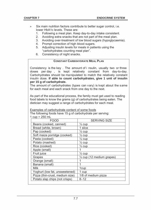
7.7
CHAPTER 7 ENDOCRINE SYSTEM
> Six main nutrition factors contribute to better sugar control, i.e.
lower HbA1c levels. These are: 1. Following a meal plan. Keep day-to-day intake consistent. 2. Avoiding extra snacks that are not part of the meal plan. 3. Avoiding over-treatment of low blood sugars (hypoglycaemia). 4. Prompt correction of high blood sugars. 5. Adjusting insulin levels for meals in patients using the
“carbohydrates counting meal plan”. 6. Consistency of night snacks.
CONSTANT CARBOHYDRATE MEAL PLAN
Consistency is the key . The amount of i nsulin, usually two or three doses per day , is kept relatively constant from day-to-day. Carbohydrates should be manipulated to match the relatively constant insulin dose. If able to count carbohydrates, give 1 unit of insulin per 15 g of carbohydrate. The amount of carbohydrates (types can vary) is kept about the same for each meal and each snack from one day to the next. As part of the educational process, the family must get used to reading food labels to know the grams (g) of carbohydrates being eaten. The dietician may suggest a range of carbohydrates for each meal. Examples of carbohydrate content of some foods The following foods have 15 g of carbohydrate per serving: 1 cup = 250 mL
FOOD SERVING SIZE Beans (cooked, canned) ½ cup Bread (white, brown) 1 slice Pap (cooked) ¼ cup Soft maize porridge (cooked) ½ cup Pasta (cooked) ½ cup Potato (mashed) ½ cup Rice (cooked) Apple (small) 1 Fruit juice ½ cup Grapes ½ cup (12 medium grapes) Orange (small) 1 Banana (small) 1 Milk 1cup Yoghurt (low fat, unsweetened) 1 cup Pizza (thin-crust, medium size) 1/8 of medium pizza Potato slap chips (not crisps) 8–12
7.8
CHAPTER 7 ENDOCRINE SYSTEM
» Tailor the advice to the patients’ lifestyle, economic circumstances and
usual diet and, where possible, avoid drastic changes. » Do not forbid any particular food as this may lead to disturbed attitudes
to food, e.g. carbohydrates are not forbidden but can be taken before exercise, incorporated into a main m eal or used as a s ource of energy during illness when children have a poor appetite.
» Diet should provide adequate nutrition for growth and development. Dietary composition It is recommended that: > approximately 35% of dietary energy should be derived from mono-
and polyunsaturated fat, > 15% from protein, > 50% from carbohydrates. Carbohydrates should always provide at
least 40% of the total calories. Timing of meals and snacks Children receiving twice daily injections of combined short a nd intermediate acting insulin regimens need three main meals and three snacks (mid morning, mid afternoon and prior to bed time). Eat meals and snacks at the same time each day. The timing of insulin injections may need to be adjusted according to the patients’ own circumstances. Preschool aged children may have unpredictable eating habits and may require frequent small meals.
Exercise » Regular exercise helps increase insulin sensitivity; maintains proper
weight, blood pressure, blood glucose and blood fat levels. » Exercise must be regular, i.e. daily. The same a mount of ex ercise
should ideally be done at the same time of the day. » Some form of carbohydrate is necessary before and after intense
exercise to reduce the risk of hypoglycaemia. Blood glucose monitoring may be necessary before and after intense exercise.
Blood glucose testing, record keeping and review of records » Glucometers with compatible strips and bloodletting devices. » Encourage children to perform their own finger-prick blood glucose testing. » Finger prick should be performed at the side of the fingertips. » Encourage the child to monitor his/her blood glucose prior to each main
meal and at bedtime. A daily record of all testing performed should be recorded in a l ogbook. Review logbook frequently to en sure optimal adjustments in management.
» More frequent blood glucose testing is indicated if t he child is unwell, partaking in unusual amounts of phy sical activity or feel s hypoglycaemic.
» For a basal-bolus regimen, testing can be done up to 5 times a day and for other regimens, twice daily.

7.9
CHAPTER 7 ENDOCRINE SYSTEM
Glycaemic targets » Glycaemic targets for y oung children should not be a s strict as for
adults. Balance the ability of the f amily to avoid recurrent hypoglycaemia. A paediatrician should assist in setting practical goals. See table “Monitoring, control and adjustments”.
» Disabling hypoglycaemia is the presence of recurrent and unpredictable episodes of hypoglycaemia requiring third party assistance and leading to anxiety about repeated episodes and leads to a reduced quality of life
» Ideally 80% of the pr e-meal blood glucose values should fall within the target range during home monitoring, but targets may need to be altered based on the age of the child and the ability of the family.
» Infants, toddlers, and preschoolers are unable to recognise or communicate signs and symptoms of low blood glucose. They also have unpredictable eating habits.
» School-age children and some young adolescents have more predictable eating habits but may be lac king in ju dgement. They are able to recognise or communicate signs and symptoms of low blood glucose.
» Most adolescents and young adults are able to recognize and treat low blood glucose reactions. They have predictable eating habits and are able to plan ahead.
» Acceptable target range before meals: Blood glucose levels
> Infants and toddlers 6– 12 mmol/L > School-age children and some young
adolescents 4– 10 mmol/L
> Most adolescents and young adults 4– 8 mmol/L » Monitor HbA1c levels 3 monthly. The aim is to maintain HbA1C as close
as possible to the recommended range, i.e. < 7.5%. Aim for a lower HbA1C in patients who are adherent with regard to h ome glucose monitoring.
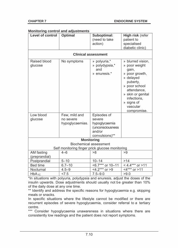
7.10
CHAPTER 7 ENDOCRINE SYSTEM
Monitoring control and adjustments
Level of control Optimal Suboptimal: (need to take action)
High risk (refer patient to specialised diabetic clinic)
Clinical assessment
Raised blood glucose
No symptoms » polyuria,* » polydypsia,*
and » enuresis.*
» blurred vision, » poor weight
gain, » poor growth, » delayed
puberty, » poor school
attendance, » skin or genital
infections, » signs of
vascular compromise.
Low blood glucose
Few, mild and no severe hypoglycaemias.
Episodes of severe hypoglycaemia (unconsciousness and/or convulsions)**
Monitoring Biochemical assessment
Self monitoring finger prick glucose monitoring AM fasting (preprandial)
4–6 >8 >9
Postprandial 5–10 10–14 >14 Bed time 6.7–10 <6.7*** or 10–11 < 4.4*** or >11 Nocturnal 4.5–9 <4.2*** or >9 <4*** or >11 HbA1C <7.5 7.5–9.0 >9.0
*In situations with polyuria, polydypsia and enuresis, adjust the doses of the insulin upwards. Dose adjustments should usually not be greater than 10% of the daily dose at any one time. ** Identify and address the specific reasons for hypoglycaemia e.g. skipping meals or snacks. In specific situations where the lifestyle cannot be modified or there are recurrent episodes of severe hypoglycaemia, consider referral to a tertiary centre. *** Consider hypoglycaemia unawareness in situations where there are consistently low readings and the patient does not report symptoms.

7.11
CHAPTER 7 ENDOCRINE SYSTEM
» Hypoglycaemia unawareness is potentially dangerous. The
(appropriate) insulin dose may need to be a djusted downwards if more than 30% of the readings (during a single week) are below the targe t values indicated.
Urine ketone testing » Hyperglycaemia and substantial ketones (+++) in dicates that DKA is
present. » Test urine for ketones in the following circumstances:
> if vomiting occurs; > any time the blood glucose > 15 mmol/L, especially if the child is
unwell and particularly if the blood glucose has been high for more than 24 hours;
> if unusual drowsiness is present; > in the presence of high temperature, vomiting or diarrhoea, even
when the glucose is < 15 mmol/L; > if abdominal pains occur; > if the breathing is deep and rapid or smells of acetone.
MEDICINE TREATMENT Insulin therapy Principles of insulin therapy: » To provide sufficient insulin throughout the 24 hour period to cover basal
requirements. » To deliver higher boluses of insulin in an attempt to match the glycaemic
effect of meals. » The most suitable areas for insulin injection are:
> the upper, outer area of the arms; > the front and side of the thigh; > the upper, outer surface of the buttocks; and > the abdomen, except the area close to the navel.
» Establish a pat tern for injecting, i.e. horizontally or vertically. Vary the site of injection according to this pattern. When the area has been fully covered move to another area.
» Patients doing strenuous exercise should not inject into their legs.

7.12
CHAPTER 7 ENDOCRINE SYSTEM
Insulin injection technique
» Insulin injection by syringe is usually given into deep subcutaneous
tissue through a two-finger pinch of skin at an angle of 45–90 degrees. » The subcutaneous fat layer should be thicker than the needle length. » There is significant risk of accidental intramuscular injections and hence
more rapid absorption especially in lean individuals. This can be minimised by using a two-finger pinch technique, an in jection angle of 45 degrees and 8 mm needles. Five or 6 mm needles may be appropriate in le an children or those using pens. Only withdraw the needle and release the skin fold on the count of ten.
» Disinfection of the skin is n ot necessary prior to insu lin injections, however injections should be given through clean, healthy skin.
» Needles should not be used for more than 6 injections. » Prefilled insulin syringes are recommended for children. Pen devices
delivering less than 1 unit should be available for selected patients. » Thoroughly mix all ins ulin suspensions before injection by rolling or
inverting the vial ten times so t hat the cloudy suspension mixes thoroughly and uniformly.
Duration of action of standard insulins
Insulin Onset of action
Peak action Effective duration
Regular/short acting 30–60 minutes 2–3 hours 8–10 hours
Intermediate acting 2–4 hours 4–12 hours 12–20 hours
7.13
CHAPTER 7 ENDOCRINE SYSTEM
Choice of insulin regimen » No insulin injection regimen satisfactorily mimics normal physiology.
The choice of insulin regimen should be individualised and will depend on age, duration of diabetes, lifestyle (dietary patterns, exercise schedules, school, work commitments, etc), targets of metabolic control, and particularly, individual patient/family preferences.
» The choice of an in sulin regimen is d etermined by the pati ent’s circumstances. Depending on the p atient’s scope to undertake insulin therapy, a number of alternatives will allow insulin therapy to be tailored to their l ifestyle. Discussion with parents s hould provide the basis for such important decisions.
» It is not possible to prescribe a single best regimen for preschool and primary school children. Individualise the choice of regimen according to family circumstances.
» Multiple daily injections provide for the best glycaemic control in young people with type 1 diabetes. If manageable, this should be the regimen of choice. Initially, a tw ice daily injection regimen may be more manageable.
Whichever insulin regimen is chosen should be supported by comprehensive education appropriate for the age, maturity
and individual needs of the child and family.
Selecting an insulin regimen Total daily insulin dose This is individualised and varies according to age, puberty development, stress and individual variability. Usual range is 0.5–1 units/kg/day, but may be higher or less. The aim is to select a regimen that allows the achievement of glycaemic control without disabling hypoglycaemia. This also requires a comprehensive support programme for the child and family enabling the implementation of an appropriate diet and other care strategies including home blood glucose monitoring and the ability to recognise and manage hypoglycaemic episodes. Where glycaemic control is n ot achieved despite an adeq uate support programme consider referral to a Tertiary centre. Insulin regimens Consult with a paediatric endocrinologist or paediatrician with experience in diabetes care. Repeated consultations are indicated when glycaemic control targets are not achieved.

7.14
CHAPTER 7 ENDOCRINE SYSTEM
The following regimen choices are listed in order of simplicity and flexibility. Regimen 1: Two Injections daily A mixture (premixed combination) of short and intermediate acting
insulins (before breakfast and the main evening meal). The total daily dose is divided so that 2/3 is given in the morning and 1/3
in the evening. The morning or evening dose is then again split between the
intermediate-acting and the short-acting insulin in a 70:30 ratio which is pre-mixed
This regimen is less flexible but easier to instruct.
Regimen 1: Premixed 70/30
Breakfast
intermediate acting (70% of morning dose) + short acting insulin (30 % of morning dose)
2/3 of total daily dose in units
Supper
intermediate acting ( 70% of evening dose) + short acting insulin ( 30% of evening dose)
1/3 of total daily dose in units
OR Regimen 2: Three injections daily A mixture of short and intermediate acting (premixed 70:30) insulin
before breakfast; short acting insulin alone before an afternoon snack or main evening meal; intermediate acting insulin before bed; or variations of this regimen may be used at times.
This requires that the caregiver is aware of three different insulin preparations and can differentiate between them.
Regimen 2
Breakfast
short acting insulin (30% of morning dose) + intermediate acting (70% of morning dose)
2/3 of total daily dose
Supper short acting insulin (1/3 of
evening dose) 1/3 of total daily dose At night ( 21h00)
intermediate acting (2/3 of evening dose)
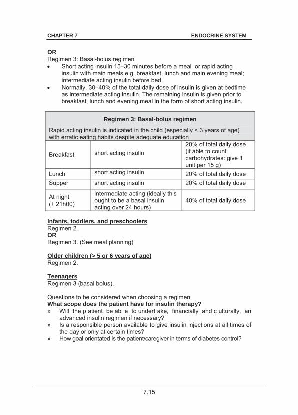
7.15
CHAPTER 7 ENDOCRINE SYSTEM
OR Regimen 3: Basal-bolus regimen Short acting insulin 15–30 minutes before a meal or rapid acting
insulin with main meals e.g. breakfast, lunch and main evening meal; intermediate acting insulin before bed.
Normally, 30–40% of the total daily dose of insulin is given at bedtime as intermediate acting insulin. The remaining insulin is given prior to breakfast, lunch and evening meal in the form of short acting insulin.
Regimen 3: Basal-bolus regimen
Rapid acting insulin is indicated in the child (especially < 3 years of age) with erratic eating habits despite adequate education
Breakfast short acting insulin 20% of total daily dose (if able to count carbohydrates: give 1 unit per 15 g)
Lunch short acting insulin 20% of total daily dose Supper short acting insulin 20% of total daily dose
At night ( 21h00)
intermediate acting (ideally this ought to be a basal insulin acting over 24 hours)
40% of total daily dose
Infants, toddlers, and preschoolers Regimen 2. OR Regimen 3. (See meal planning) Older children (> 5 or 6 years of age) Regimen 2. Teenagers Regimen 3 (basal bolus). Questions to be considered when choosing a regimen What scope does the patient have for insulin therapy? » Will the p atient be abl e to undert ake, financially and c ulturally, an
advanced insulin regimen if necessary? » Is a responsible person available to give insulin injections at all times of
the day or only at certain times? » How goal orientated is the patient/caregiver in terms of diabetes control?
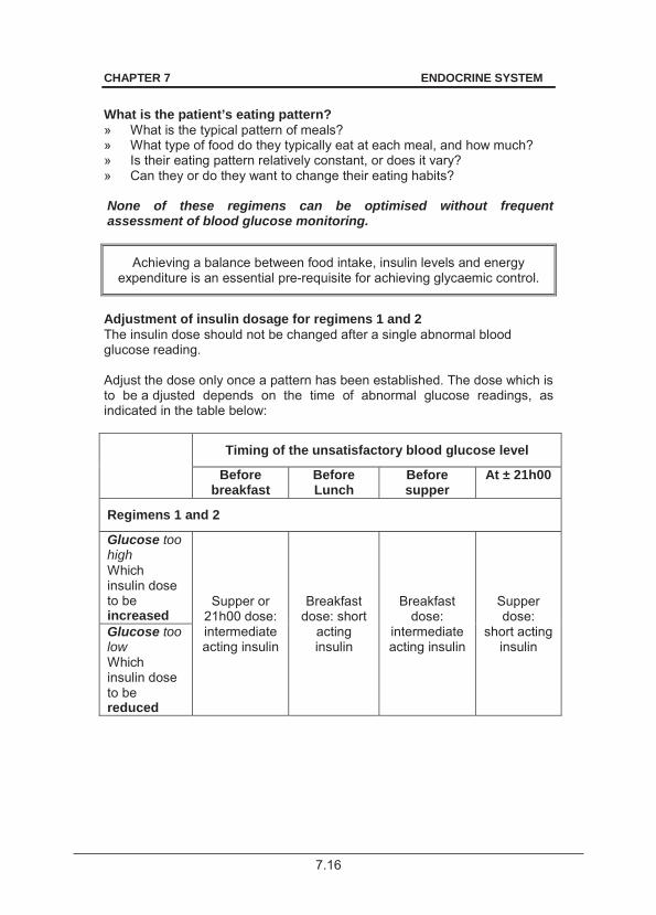
7.16
CHAPTER 7 ENDOCRINE SYSTEM
What is the patient’s eating pattern? » What is the typical pattern of meals? » What type of food do they typically eat at each meal, and how much? » Is their eating pattern relatively constant, or does it vary? » Can they or do they want to change their eating habits? None of these regimens can be optimised without frequent assessment of blood glucose monitoring.
Achieving a balance between food intake, insulin levels and energy expenditure is an essential pre-requisite for achieving glycaemic control.
Adjustment of insulin dosage for regimens 1 and 2 The insulin dose should not be changed after a single abnormal blood glucose reading. Adjust the dose only once a pattern has been established. The dose which is to be a djusted depends on the time of abnormal glucose readings, as indicated in the table below: Timing of the unsatisfactory blood glucose level
Before breakfast
Before Lunch
Before supper
At ± 21h00
Regimens 1 and 2
Glucose too high Which insulin dose to be increased
Supper or 21h00 dose: intermediate acting insulin
Breakfast dose: short
acting insulin
Breakfast dose:
intermediate acting insulin
Supper dose:
short acting insulin
Glucose too low Which insulin dose to be reduced
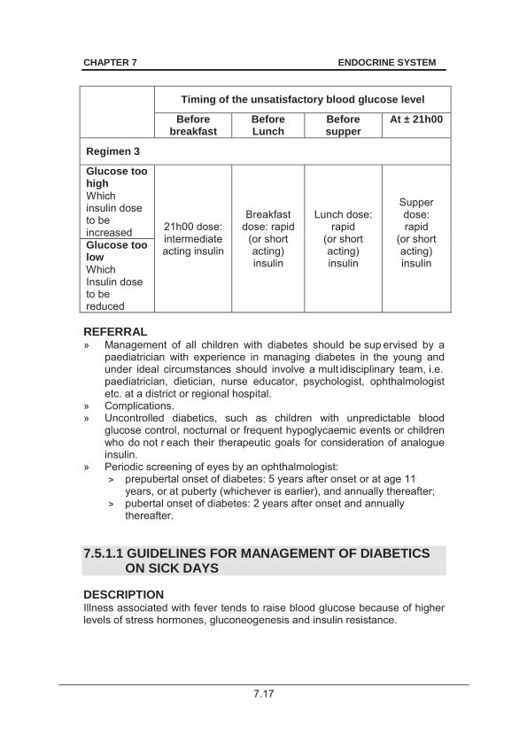
7.17
CHAPTER 7 ENDOCRINE SYSTEM
Timing of the unsatisfactory blood glucose level
Before breakfast
Before Lunch
Before supper
At ± 21h00
Regimen 3
Glucose too high Which insulin dose to be increased 21h00 dose:
intermediate acting insulin
Breakfast dose: rapid
(or short acting) insulin
Lunch dose: rapid
(or short acting) insulin
Supper dose: rapid
(or short acting) insulin
Glucose too low Which Insulin dose to be reduced REFERRAL » Management of all children with diabetes should be sup ervised by a
paediatrician with experience in managing diabetes in the young and under ideal circumstances should involve a mult idisciplinary team, i.e. paediatrician, dietician, nurse educator, psychologist, ophthalmologist etc. at a district or regional hospital.
» Complications. » Uncontrolled diabetics, such as children with unpredictable blood
glucose control, nocturnal or frequent hypoglycaemic events or children who do not r each their therapeutic goals for consideration of analogue insulin.
» Periodic screening of eyes by an ophthalmologist: > prepubertal onset of diabetes: 5 years after onset or at age 11
years, or at puberty (whichever is earlier), and annually thereafter; > pubertal onset of diabetes: 2 years after onset and annually
thereafter. 7.5.1.1 GUIDELINES FOR MANAGEMENT OF DIABETICS
ON SICK DAYS DESCRIPTION Illness associated with fever tends to raise blood glucose because of higher levels of stress hormones, gluconeogenesis and insulin resistance.
7.18
CHAPTER 7 ENDOCRINE SYSTEM
Illness associated with vomiting and/or diarrhoea may lower blood glucose, with the pos sibility of hy poglycaemia and t he development of sta rvation ketones. Illness may result in ketone production. DIAGNOSTIC CRITERIA » Unstable blood glucose measurements as a result of illness or stress or
starvation. » Increased insulin requirements are indu ced by a cata bolic state and
stress. » Ketonuria may also indicate the following:
> ketonuria in the presence of hyperglycaemia is indicative of severe insulin deficiency and calls for urgent therapy to prevent progression into ketoacidosis;
> ketonuria in the presence of low blood glucose levels is indicative of a starvation state or is the result of a counter-regulatory response to hypoglycaemia.
GENERAL AND SUPPORTIVE MEASURES » Monitor glucose more frequently. » Test urine for ketones. » Ensure adequate intake of calories and flu ids on sick days to prevent
ketogenesis. If not enough calories are consumed, ketones will appear in the urine without the development of hyperglycaemia. In this circumstance encourage the patient to eat whatever he/she feels like.
» Treat underlying intercurrent illness. » Special circumstances:
> Gastroenteritis: If hypoglycaemia occurs especially with gastroenteritis, and there is mild ketonuria, ensure that the child takes regular frequent amounts of carbohydrate using oral rehydration solution or intravenous fluids.
> Loss of appetite: Replace meals with easily digestible food and sugar-containing fluids.
> Vomiting: If the patient has difficulty eating or keeping food down and the blood glucose is < 10 mmol/L, encourage the patient to take sugar-containing liquids. Give small volumes. Some glucose will be absorbed. If there is no vomiting, increase the amount of liquid.
MEDICINE TREATMENT Insulin therapy Insulin must always be given each day. Do not skip an insulin injection because of sickness and/or vomiting. If vomiting occurs, IV fluids may be needed to avoid hypoglycaemia.
7.19
CHAPTER 7 ENDOCRINE SYSTEM
Generally the body will require more energy during illness. Insulin allows more glucose to enter into the cells, providing more energy to fight infection. General guidelines when giving extra insulin: » If the blood glucose is rising or if ketones in the urine, the patient must
seek urgent medical attention. Moderate urine ketones » The extra dose of insulin is usually 10–20% of the total daily dose.
This extra insulin is given as short (or rapid) acting insulin every three hours. If the blood glucose drops < 8.3 mmol/L, it may be necessary to sip regular juice or other sugar-containing drinks. This is done to raise the blood glucose before giving the next insulin injection.
Large amount of urine ketones » Give 20% of the total daily insulin dose.
Repeat as above if necessary. Extra fluids In addition to taking extra insulin, extra fluids, e.g. water and fruit j uices are important to prevent acidosis. These fluids replace the fluids lost in the urine and prevent dehydration. REFERRAL » In a child with intercurrent illness urgent specialist medical or nursing
advice must be obtained when: > patient is unable to carry out the advice regarding sick days; > the diagnosis is unclear; > vomiting is persistent, particularly in young children; > blood glucose continues to rise despite increased insulin; > hypoglycaemia is severe; > ketonuria is heavy or persistent; > the child is becoming exhausted, confused, hyperventilating,
dehydrated or has severe abdominal pain. 7.5.2 DIABETES MELLITUS, INSULIN DEPENDENT: ACUTE
COMPLICATIONS E10 7.5.2.1 CEREBRAL OEDEMA IN DIABETIC COMA G93.6 DESCRIPTION A condition of brain swelling during the course of treatment of hyperglycaemic coma.
7.20
CHAPTER 7 ENDOCRINE SYSTEM
Cerebral oedema usually occurs 4–12 hours after the initiation of treatment and often follows an initial period of clinical and biochemical improvement. Cerebral oedema causes significant neurological morbidity and ha s a mortality of approximately 80%.
The cause of cerebral oedema during treatment remains unclear. However, too rapid reduction in intravascular osmolality may aggravate the process. Therefore rehydration should occur more slowly in children with DKA than in other causes of dehydration. DIAGNOSTIC CRITERIA Clinical » Signs and symptoms of cerebral oedema include:
> headache, > confusion, > irritability, > reduced consciousness, > papilloedema, > hypoxaemia, and > specific neurological signs and raised intracranial pressure.
» The risk of cerebral oedema is increased if urea levels are increased or if the PCO2 is persistently low, i.e. < 20 mmHg.
GENERAL AND SUPPORTIVE MEASURES » Admit to ICU, if possible, or to a centre experienced with managing this
condition. » Res it over 72
hours rather than 48 hours pending ICU admission. » Elevate head of bed. » Exclude hypoglycaemia. » Do not use bicarbonate. » Exclude thrombosis, haemorrhage or infection. » Do not delay treatment while waiting for a CT scan to confirm cerebral
oedema. MEDICINE TREATMENT Within 10 minutes of diagnosis: Mannitol 20%, IV, 2.5 mL/kg, immediately over 15 minutes.
o If no initial response: repeat in 30 minutes to 2 hours. 7.5.2.2 HYPERGLYCAEMIC KETOACIDOSIS E10.1 DESCRIPTION Diabetic ketoacidosis (DKA) occurs with relative or a bsolute insulin deficiency, either caused by non-adherence to insulin regimens or by excessive secretion of counterregulatory hormones during stress, e.g. infection, trauma and surgery.
7.21
CHAPTER 7 ENDOCRINE SYSTEM
DIAGNOSTIC CRITERIA » Heavy glycosuria. » Hyperglycaemia, i.e. blood glucose usually > 15 mmol/L, ketonuria, and
pH < 7.3. » Bicarbonate < 15 mmol/L and patients who are clinically dehydrated. » May/may not be vomiting. » May/may not be drowsy. Note: In rare cases blood glucose is not elevated. Children with mild dehydration and not clinically unwell usually tolerate oral rehydration and subcutaneous insulin. See section 7.5.1.1: Guidelines for management of diabetics on sick days. GENERAL AND SUPPORTIVE MEASURES » Admit all children and adolescents to an ICU or ward experienced in the
management of DKA in children and adolescents, if possible. » Ensure patent airway. » If the child is comatose, insert an artificial airway and a urinary catheter. » If comatose or recurrent vomiting insert oro/nasogastric tube and apply
free drainage. MEDICINE TREATMENT Seek specialist advice early in the management. If hypoxaemic: Oxygen via facemask or airway.
The objectives of fluid and sodium replacement therapy in diabetic ketoacidosis are: » Restoration of circulating volume. » Replacement of sodium and extracellular fluid and intracellular fluid
deficits of water. » The restoration of glomerular filtration rate with enhanced clearance of
glucose and ketones from the blood. » To reduce the risk of cerebral oedema. Fluids a: Fluids for resuscitation in shock: Sodium chloride 0.9%, IV, 10–20 mL/kg over 10–30 minutes.
o Repeat if shock persists.
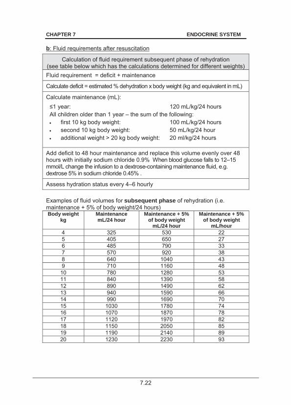
7.22
CHAPTER 7 ENDOCRINE SYSTEM
b: Fluid requirements after resuscitation
Calculation of fluid requirement subsequent phase of rehydration (see table below which has the calculations determined for different weights) Fluid requirement = deficit + maintenance
Calculate deficit = estimated % dehydration x body weight (kg and equivalent in mL)
Calculate maintenance (mL): : 120 mL/kg/24 hours
All children older than 1 year – the sum of the following: first 10 kg body weight: 100 mL/kg/24 hours second 10 kg body weight: 50 mL/kg/24 hour� additional weight > 20 kg body weight: 20 ml/kg/24 hours
Add deficit to 48 hour maintenance and replace this volume evenly over 48 hours with initially sodium chloride 0.9% When blood glucose falls to 12–15 mmol/L change the infusion to a dextrose-containing maintenance fluid, e.g. dextrose 5% in sodium chloride 0.45% .
Assess hydration status every 4–6 hourly
Examples of fluid volumes for subsequent phase of rehydration (i.e. maintenance + 5% of body weight/24 hours) Body weight
kg
Maintenance mL/24 hour
Maintenance + 5% of body weight
mL/24 hour
Maintenance + 5% of body weight
mL/hour 4 325 530 22 5 405 650 27 6 485 790 33 7 570 920 38 8 640 1040 43 9 710 1160 48
10 780 1280 53 11 840 1390 58 12 890 1490 62 13 940 1590 66 14 990 1690 70 15 1030 1780 74 16 1070 1870 78 17 1120 1970 82 18 1150 2050 85 19 1190 2140 89 20 1230 2230 93
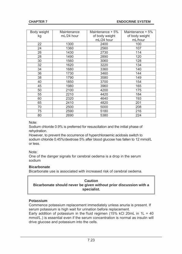
7.23
CHAPTER 7 ENDOCRINE SYSTEM
Body weight
kg
Maintenance mL/24 hour
Maintenance + 5% of body weight
mL/24 hour
Maintenance + 5% of body weight
mL/hour 22 1300 2400 100 24 1360 2560 107 26 1430 2730 114 28 1490 2890 120 30 1560 3060 128 32 1620 3220 134 34 1680 3360 140 36 1730 3460 144 38 1790 3580 149 40 1850 3700 154 45 1980 3960 165 50 2100 4200 175 55 2210 4420 184 60 2320 4640 193 65 2410 4820 201 70 2500 5000 208 75 2590 5180 216 80 2690 5380 224
Note: Sodium chloride 0.9% is preferred for resuscitation and the initial phase of rehydration. However, to prevent the occurrence of hyperchloraemic acidosis switch to sodium chloride 0.45%/dextrose 5% after blood glucose has fallen to 12 mmol/L or less. Note: One of the danger signals for cerebral oedema is a drop in the serum sodium
Bicarbonate Bicarbonate use is associated with increased risk of cerebral oedema.
Caution Bicarbonate should never be given without prior discussion with a
specialist.
Potassium Commence potassium replacement immediately unless anuria is present. If serum potassium is high wait for urination before replacement. Early addition of potassium in the f luid regimen (15% kCl 20mL in 1L = 40 mmol/L.) is essential even if the serum concentration is normal as insulin will drive glucose and potassium into the cells.
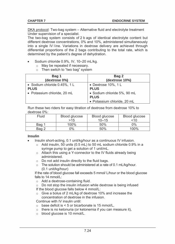
7.24
CHAPTER 7 ENDOCRINE SYSTEM
DKA protocol: Two-bag system – Alternative fluid and electrolyte treatment Under supervision of a specialist. The two-bag system consists of 2 b ags of identical electrolyte content but different dextrose concentrations, 0% and 10%, administered simultaneously into a single IV l ine. Variations in dextrose delivery are achieved through differential proportions of the 2 bags contributing to the total rate, which is determined by the patient’s degree of dehydration. Sodium chloride 0.9%, IV, 10–20 mL/kg.
o May be repeated if necessary. o Then switch to “two bag” system
Bag 1 (dextrose 0%)
Bag 2 (dextrose 10%)
Sodium chloride 0.45%, 1 L PLUS Potassium chloride, 20 mL
Dextrose 10%, 1 L PLUS Sodium chloride 5%, 90 mL
PLUS Potassium chloride, 20 mL
Run these two riders for easy titration of dextrose from dextrose 10% to dextrose 0%:
Fluid Blood glucose >15
Blood glucose 10–15
Blood glucose <10
Bag 1 100% 50% 0% Bag 2 0% 50% 100%
Insulin Insulin short-acting, 0.1 unit/kg/hour as a continuous IV infusion.
o Add insulin, 50 units (0.5 mL) to 50 mL sodium chloride 0.9% in a syringe pump to get a solution of 1 unit/mL.
o Attach this using a Y-connector to the IV fluids already being administered.
o Do not add insulin directly to the fluid bags. o The solution should be administered at a rate of 0.1 mL/kg/hour.
(0.1 unit/kg/hour) If the rate of blood glucose fall exceeds 5 mmol/ L/hour or the blood glucose falls to 14 mmol/L: o Add a dextrose-containing fluid. o Do not stop the insulin infusion while dextrose is being infused If the blood glucose falls below 4 mmol/L: o Give a bolus of 2 mL/kg of dextrose 10% and increase the
concentration of dextrose in the infusion. Continue with IV insulin until: o base deficit is < 5 or bicarbonate is 15 mmol/L, o there is no ketonuria (or ketonemia if you can measure it), o blood glucose is 10 mmol/L.
7.25
CHAPTER 7 ENDOCRINE SYSTEM
Alternative to insulin infusion Where there are no facilities for insulin infusion, e.g. no syringe pumps, staff constraints, etc.: Insulin short-acting, IV, 0.1 unit/kg, hourly.
Changing from intravenous to subcutaneous insulin Continue with intravenous fluids until the child is drinking well and able to tolerate snacks. When oral fluids are tolerated, reduce intravenous fluids. Subcutaneous insulin can be started once the child is well hydrated and able to tolerate a normal diet. The most convenient time to change to subcutaneous insulin is just before a mealtime. Administer the f irst dose of subcutaneous insulin 30 minutes before the meal and continue with the insulin infusion for 90 minutes after the subcutaneous injection to prevent rebound hyperglycaemia. In newly diagnosed diabetics regimen 1 is chosen at a low range d ose divided in the usual way: o Prepubertal children: 0.7 units/kg. o Pubertal children: 1 unit/kg. In established diabetics, give usual insulin. Give supplemental subcutaneous short acting insulin before meals if the blood glucose > 11 mmol/L:
Blood glucose mmol/L
Short-acting Insulin units/kg/dose
11–12 0.06 13–16 0.09
16 0.12 REFERRAL » No improvement » Deterioration of condition, i.e.:
> pH <7.1, > hyperventilation, > shock, > depressed level of consciousness, > persistent vomiting, > age < 5 years.
» Rising blood glucose.
7.26
CHAPTER 7 ENDOCRINE SYSTEM
7.5.2.3 HYPOGLYCAEMIA IN DIABETICS E16.0 DESCRIPTION Autonomic symptoms (hunger, nausea, anxiety, pallor, palp itations, sweating, trembling) usually precede neuroglycopaenic symptoms (impaired thinking, change of mood, irritability, dizziness, headache, tiredness, confusion, and later convulsions and coma). A reversal in appearance of the symptoms is undesirable and is known as hypoglycaemia unawareness. Causes of hypoglycaemia include: » A missed or delayed snack or meal. » Exercise without appropriate dietary preparation. » Alcohol. » Overdose of insulin. » Impaired food absorption e.g. gastro-enteritis. » Addison’s disease. Recurrent hypoglycaemia may necessitate
investigation for this condition. Nocturnal hypoglycaemia Nightmares and headaches may be suggestive of nocturnal hypoglycaemia. Blood glucose concentrations fall to their lowest levels between 02h00 and 04h00. DIAGNOSTIC CRITERIA » Blood glucose < 3. 5–4 mmol/L with symptoms in a k nown diabetic
patient. Good glycaemic control is likely to be associated with occasional hypoglycaemic episodes.
» Grading of severity: Mild (Grade 1) > Child or adolescent is aware of, responds to and self-treats the
hypoglycaemia. > Children < 6 years of age can rarely be classified as grade 1
because they are unable to help themselves. Moderate (Grade 2) > Child or adolescent cannot respond to hypoglycaemia and requires
help from someone else, but oral treatment is successful. Severe (Grade 3) > Child or adolescent is semiconscious or unconscious or in coma
with/without convulsions and may require parenteral therapy with glucagon or intravenous glucose.
7.27
CHAPTER 7 ENDOCRINE SYSTEM
GENERAL AND SUPPORTIVE MEASURES » Determine underlying cause. » Patient education on diabetes and its complications. » If patient is fully alert and conscious, give sugar-containing drink and/or
snack (carbohydrate). » Monitor blood g lucose every 15 min utes until blood glucose is 6–8
mmol/L. MEDICINE TREATMENT Mild or moderate hypoglycaemia: Immediate oral rapidly absorbed simple carbohydrate, e.g.: Glucose, oral, 5–15 g or 1-3 level teaspoons of sugar in a small amount
of water. o Wait 10–15 minutes. o If no response, repeat above. o As symptoms improve, the next meal or oral complex carbohydrate
should be ingested, e.g. fruit, bread, cereal, milk, etc.
Severe hypoglycaemia Outside hospital Glucagon, IM/SC, 0.1–0.2 mg/10 kg.
o If < 12 years of age: 0.5 mg. o If > 12 years of age: 1.0 mg.
In hospital If there is an unsatisfactory response or inability to take oral carbohydrate and signs of disorientation, stupor, convulsions, coma: Dextrose 10%, IV, 2–5 mL/kg.
o Dilute dextrose 50% solution before use to 10% strength. o (1 mL/kg of dextrose 50% plus 4 mL/kg of water for injection, gives
10% dextrose solution). If IV dextrose cannot be given: Glucagon, IM/SC, 10–30 mcg/kg.
Monitor blood glucose every 15 minutes until stable, then repeat 1–2 hourly. Keep blood glucose between 6 and 8 mmol/L. REFERRAL » Recurrent episodes of hypoglycaemia.
7.28
CHAPTER 7 ENDOCRINE SYSTEM
7.5.2.4 NEPHROPATHY N08.3 DIAGNOSTIC CRITERIA » Persistent albuminuria:
> 3 specimens over a 3–6 month period all show increased albumin:creatinine ratio on a spot urine: males: > 2.5 mg/mmol, females: > 3.5 mg/mmol.
» Screening for micro-albuminuria should start from: > prepubertal children: duration of 5 years. > pubertal children: duration of 2 years.
GENERAL AND SUPPORTIVE MEASURES » Optimise diabetic control. » Monitor blood pressure. MEDICINE TREATMENT If albumin:creatinine ratio is > 3.5 mg/mmol: ACE inhibitor, e.g.: Enalapril, oral, 0.1 mg/kg/dose as a single dose or two divided doses.
o Maximum dose: 0.5 mg/kg or 40 mg/day. Note: Exclude non-diabetic nephropathy.
REFERRAL » All patients with significant albuminuria. 7.5.3 DIABETES MELLITUS IN ADOLESCENTS E10 See section 22.6: Diabetes in adolescence. DESCRIPTION Adolescence is that period between puberty and when the patient leaves school to join the workforce. The adolescent and the transition should be managed with special planning, i.e.: » the admission policy of the hospital, » observing the wishes of the adolescent, » emotional and physical maturity considerations, » presence of any co-existing medical, surgical or psychiatric disorder that
may be more appropriately managed in the paediatric service.
7.29
CHAPTER 7 ENDOCRINE SYSTEM
GENERAL AND SUPPORTIVE MEASURES Promote: » normal growth and pubertal development, » psychological development, » maintenance of glycaemic control and adherence, » normal lifestyle, » avoidance of risk taking behaviours (smoking, substance abuse), » sex education. MEDICINE TREATMENT Failure of current insulin regimens are attributed to the endocrine changes of puberty which results in poor glycaemic control. Insulin resistance occurs during puberty, being maximal in late puberty. Normal insulin requirements during puberty: o 1.0–1.4 units/kg/day. This may occasionally be higher, but as a general rule a higher requirement generally necessitates the search for other causes. After puberty, the insulin requirements fall to prepubertal levels. Failure to re duce insulin requirements in th e late adolescent stages may result in excessive weight gain. 7.5.4 DIABETES MELLITUS, TYPE 2 E11 DESCRIPTION Type 2 diabetes develops when insulin secretion cannot meet the increased demand posed by insulin resistance. Type 2 d iabetes may be a ssociated with hyperlipidemia, hypertension, acanthosis nigricans, ovarian hyperandrogenism and non-alcoholic fatty liver disease (features of insulin resistance). DIAGNOSTIC CRITERIA Clinical » Obesity. » Children 10 y ears and older with a strong fa mily history of ty pe 2
diabetes, usually in adolescents with BMI > 95% without auto-antibodies to islet cells and normal serum C-peptide levels.
» Keto-acidosis is unusual in type 2 diabetes. » Symptoms of diabetes plus random plasma glucose > 11 mmol/L or a
fasting glucose > than 7 mmol/L » Type 2 diabet ics may have minimal symptoms or signs for months or
even years before the diagnosis.
7.30
CHAPTER 7 ENDOCRINE SYSTEM
Investigations To confirm diagnosis: » Fasting plasma glucose 7.0 mmol/L confirms the diagnosis. OR » Oral glucose tolerance test.
> Ingestion of 1 .75 g/kg (maximum 75 g) of glucose dissolved in water.
> Plasma glucose > 11 mmol/L 2 hours post ingestion of oral glucose. GENERAL AND SUPPORTIVE MEASURES » Lifestyle modification:
Manage patients who are not ill at diagnosis initially with advice on nutrition and exercise, but most will eventually require medicine therapy.
» Education on r outine blood glucose monitoring. A d aily record of all testing performed should be recorded in a logbook. Record prebreakfast fasting and 2-hour postprandial dinner levels which is sufficient in most cases.
MEDICINE TREATMENT Refer for initiation of therapy. 7.6 HYPOGLYCAEMIA IN CHILDREN E16.2 DESCRIPTION Infants and small children have relatively limited glycogen stores with larger brain/body ratios than adults and are therefore at greater risk of hypoglycaemia during starvation. The causes of hypoglycaemia (outside the neonatal period) include: » hypopituitarism, » adrenal insufficiency, » growth hormone deficiency, » hypothyroidism, » hyperinsulinaemia, » inborn errors of metabolism, » malnutrition, » sepsis, » liver dysfunction, » malaria, » severe illness with poor intake, » accelerated starvation (ketotic hypoglycaemia), » medicine, e.g. insulin, alcohol, aspirin, beta-blockers, oral
hypoglycaemic agents, quinine.
7.31
CHAPTER 7 ENDOCRINE SYSTEM
DIAGNOSTIC CRITERIA Clinical » Acute autonomic symptoms: hunger, nausea, anxiety, pallor,
palpitations, sweating, trembling. » Neuroglycopaenic symptoms: impaired thinking, change of moo d,
irritability, dizziness, headache, tiredness, confusion, and l ater convulsions and coma.
» Patients are of ten asymptomatic especially younger children who may be completely asymptomatic or present only with a behaviour change.
Investigations » Plasma glucose concentration < 2.6 mmol/L. » Although hypoglycaemia is a clin ical emergency requiring prompt
therapy, wherever possible, draw a blood sample for investigation prior to the administration of glucose. Collect 5 mL of blood in a plain tube at the earliest opportunity and send for separation and storage of plasma at –20°C. Such samples may provide clear biochemical evidence of the cause of the hypoglycaemic episode thus avoiding having to subject the child to further investigations.
MEDICINE TREATMENT After collection of initial blood samples: Dextrose 10%, IV, 2–5 mL/kg.
o Dilute dextrose 50% solution before use to 10% strength. (1 mL/kg of dextrose 50% plus 4 mL/kg of water for injection, gives 10% dextrose solution).
If hypoglycaemia persists or the plasma glucose is difficult to maintain in the normal range, consider adrenal insufficiency: ADD Hydrocortisone, IV, 2–3 mg/kg, immediately.
Stabilisation Sodium chloride 0.9%/dextrose 5%, IV, 20 mL/kg bolus as needed.
OR Dextrose 10%, IV, 2–3 mL/kg glucose as needed.
AND Hydrocortisone, IV, 2–3 mg/kg immediately.
If hypoglycaemia persists consider adrenal insufficiency. Ongoing treatment Intravenous fluid therapy as needed. Start oral feeds as soon as possible.
7.32
CHAPTER 7 ENDOCRINE SYSTEM
REFERRAL » All patients with confirmed hypoglycaemia not explained by intercurrent
illness, drugs, persisting or recurrent hypoglycaemia. 7.7 GROWTH DISORDERS R62 DESCRIPTION Constitutional delay in growth is defined as bone age which is significantly delayed compared to chronological age. Familial short stature is bone age equivalent to chronological age. Common features of both are: » short for chronological age, » normal rate of linear growth,
no decline in height percentile. Pathological growth failure are defined by height is disproportionately short relative to the weight or abnormally low linear growth rate.
DIAGNOSTIC CRITERIA » Measure and plot child’s height and weight on growth charts.
Routine monitoring of height and w eight for growth assists in t he diagnosis of problems which would otherwise be missed or would come to light at a sta ge where the outc ome of treat ment may be less favourable.
» A child is regarded as short if his/her height for age z-score is below –2 for gender.
» To further evaluate short stature assess parental height. Target height: > for a boy = (father’s height + (mother‘s height + 13 cm)) ÷ 2 > for a girl = ((father’s height – 13 cm) + mother‘s height) ÷ 2
» If the child’s predicted final height is > 8.5 cm below the target height, monitor growth (height) over 6 months to 1 year.
» If the child’s height for age z-score is below –3, refer immediately. » Suspect endocrine causes when there height is disproportionately short
relative to the weight. » Growth failure o ccurs when the child’s height deviates further from z-
score of –2 over a period of 1 year. GENERAL AND SUPPORTIVE MEASURES » Identify and manage non-endocrine causes of stunted growth, e.g.:
> intra-uterine growth retardation, > chronic disease, > psychosocial deprivation, > skeletal dysplasia and other dysmorphic syndromes.
7.33
CHAPTER 7 ENDOCRINE SYSTEM
REFERRAL » Height for age z-score below –3. » Height 8.5 cm or more below target height. » Growth failure (height deviates further from z-score of –2 over a period
of 1 year). » Suspected endocrine causes. » Dysmorphic child with unidentified syndrome. » Untreated chronic disease. 7.8 HYPOCALCAEMIA IN CHILDREN E83.5 DESCRIPTION The main causes of hypocalcaemia in children are: » vitamin D deficiency, » calcium deficiency, » magnesium deficiency, » reduced parathyroid hormone production, » impaired renal function. DIAGNOSTIC CRITERIA Clinical » Signs and symptoms of tetany include:
> paraesthesia, > positive Chvostek’s sign, > cramps, > positive Trousseau’s sign, > carpopedal spasm, > weakness, > laryngospasm, > lethargy, > prolonged QT interval on the ECG.
Investigations » Blood level to establish cause:
> calcium, > albumin, > phosphate, > magnesium, > ALP.
MEDICINE TREATMENT Acute hypocalcaemia Calcium gluconate 10%, IV, 1–2 mL/kg administered over 5–10 minutes,
6–8 hourly. o Maximum dose: 10 mL. o ECG monitoring is advised.
7.34
CHAPTER 7 ENDOCRINE SYSTEM
If hypomagnesaemic: Magnesium sulphate 50%, IV/IM, 0.2 mL/kg every 12–24 hours.
Chronic therapy Long-term therapy depends on the cause. Manage hypophosphataemia or hyperphosphatemia, depending on the cause of hypocalcaemia, before long-term calcium is initiated. Calcium, elemental, oral, 50 mg/kg/day until normal calcium level is
achieved. o Follow with 30 mg/kg/day.
If vitamin D deficient: Vitamin D, oral, 5 000 IU/day.
For hypoparathyroidism and pseudohypoparathyroidism: Calcitriol, oral, 0.01–0.04 mcg/kg/day.
OR Alfacalcidol, oral, 0.05 mcg/kg/day.
o If < 20 kg: 0.0 5 mcg/kg/day. o If > 20kg: 1 mcg/day.
REFERRAL » Chronic hypocalcaemia. 7.9 HYPERKALAEMIA E87.5 See section 6.1.4: Acute kidney injury (Renal failure, acute). 7.10 HYPOKALAEMIA E87.6 DESCRIPTION Causes include: » prolonged decreased intake and protein energy malnutrition; » increased renal excretion: renal tubular acidosis, amphoteracin B and
diuretics; » increased extrarenal losses; » transmembrane shifts: ß2 stimulants, alkalosis; and » mineralocorticoid excess.
7.35
CHAPTER 7 ENDOCRINE SYSTEM
DIAGNOSTIC CRITERIA Clinical » Cardiac arrhythmias, especially with digitalis. » Neuromuscular dysfunction, e.g. muscle weakness. » Renal: impairment of urine concentrating or diluting ability. Investigations » Serum potassium < 3.0 mmol/L. MEDICINE TREATMENT See section 2.2.4: Diarrhoea, acute. Severe respiratory paralysis and or cardiac arrhythmias: Potassium chloride, IV, < 1 mEq/kg/hour.
o ECG monitoring. o Potassium concentration should not be > 40 mmol/L/infusion. o Never give potassium as an IV bolus.
Less critical situations to correct potassium deficit over 2–3 days: Potassium chloride, oral, 2–6 mEq/kg/day.
Note: 1 g KCl = 13 mEq; 1 mL 15% KCl = 2 mmol; 1 mEq = 1 mmol. 7.11 HYPOPITUITARISM E23.0 DESCRIPTION Multiple or isol ated deficiencies of adre nocorticoid hormone (ACTH), luteinising hormone, thyroid stimulating hormone, and growth hormone manifesting as hypoglycaemia, abnormal body proportions and failure to grow and develop. The deficiency may be due to: » congenital abnormalities with/without midline structural abnormalities of
the brain, » central nervous system tumours, » histiocytosis, » complications of radiation therapy. DIAGNOSTIC CRITERIA Clinical » Neonates with hypopituitarism may present with:
> persistent hypoglycaemia, > cholestatic jaundice (related to low cortisol), > micropenis.
» Growth failure with immature body proportions.
7.36
CHAPTER 7 ENDOCRINE SYSTEM
Investigations » Endocrine evaluation with pituitary function tests under specialist
supervision. » Confirm diagnosis in older children with stimulation tests.
MEDICINE TREATMENT To correct hypoglycaemia: Hydrocortisone, IV, 2-3 mg/kg.
REFERRAL » All patients after stabilisation of hypoglycaemia. 7.12 HYPOTHYROIDISM, NEONATAL P72.2 DESCRIPTION Congenital deficiency of thyroid hormone due to: » aplasia/hypoplasia or ectopia of the thyroid gland, » defects in thyroid hormone biosynthesis, or » intrauterine exposure to antithyroid medicines. Congenital hypothyroidism is one of the co mmon treatable causes of preventable mental retardation in children. Congenital hypothyroidism must be treated as early as possible to avoid intellectual impairment. Symptoms and signs in neonates are unreliable. DIAGNOSTIC CRITERIA Clinical
» Prolonged unconjugated hyperbilirubinaemia.
» Oedema of the extremities and genitals.
» Feeding difficulties. » Bradycardia. » Lethargy. » Anaemia. » Somnolence. » Apnoeic episodes. » Abdominal distension. » Coarse cry. » Umbilical hernia. » Constipation. » Subnormal temperature. » Wide open fontanelles. » Periorbital oedema. » Enlarged tongue. » Delayed dentition. » Short and thick neck. » Broad hands. » Dry skin. » Hair coarse and scanty. » Hypotonia. » Hoarse voice and goitre. » Delayed physical and mental
development.
7.37
CHAPTER 7 ENDOCRINE SYSTEM
Investigations » When suspected, perform TSH test.
> If elevated perform a free T4. Delay in diagnosis and treatment is associated with irreversible neurodevelopmental damage. GENERAL AND SUPPORTIVE MEASURES » Growth and neurodevelopmental assessment. » Regular follow up. MEDICINE TREATMENT Neonates and infants, started as soon as possible, ideally within the first three weeks after birth: Levothyroxine, oral, 10–15 mcg/kg as a single daily dose.
o Adjust dosage to blood levels of T 4 (in the upper half of the reference range) and normalise the T SH (between 0.5–2 mU/L), especially in the first 3 years of life. Check TSH only 6 weeks after adjusting the thyroxine dose.
o Continue treatment indefinitely. REFERRAL » All patients for confirmation of diagnosis and initiation of therapy. 7.13 HYPOTHYROIDISM IN OLDER CHILDREN AND
ADOLESCENTS E03.9 DESCRIPTION Acquired hypothyroidism in childhood and adolescents may be due to: » chronic lymphocytic thyroiditis, » goitrogen induced, » iodine deficiency, » post surgery, » radioactive iodine, » infiltrations, or » medicines, e.g. antiretrovirals. DIAGNOSTIC CRITERIA » Elevated TSH and low thyroxine levels. MEDICINE TREATMENT Levothyroxine, oral, 100 mcg/m2 once daily.
7.38
CHAPTER 7 ENDOCRINE SYSTEM
REFERRAL » All cases for investigation and initiation of therapy. 7.14 HYPERTHYROIDISM, GRAVES DISEASE E05.9/E05.0 DESCRIPTION Hyperthyroidism is a pathological syndrome in which tissue is exposed to excessive amounts of circulating thyroid hormones. The most common cause is Grave’s disease, although thyroiditis may also present with thyrotoxicosis. DIAGNOSTIC CRITERIA Clinical
» Fatigue. » Poor school performance.
» Tachycardia. » Nervousness or anxiety.
» Warm moist hands. » Weight loss. » Thyromegaly. » Palpitations. » Tremor. » Heat insensitivity. » Proptosis.
Investigations » Elevated thyroxine (T4) and suppressed TSH. MEDICINE TREATMENT Carbimazole, oral, 0.5 mg/kg once daily.
AND To block sympathetic hyperactivity: Atenolol, oral, 1–2 mg/kg as a single daily dose.
For children less than 10 kg: Propranolol, oral, 0.2–0.5 mg/kg 6–12 hourly.
o Maximum dose: 1.5 mg/kg/dose 6–12 hourly. REFERRAL » All patients for confirmation of diagnosis, initiation and follow up of
therapy 7.15 OBESITY E66 DESCRIPTION Most children with obesity do not have an underlying pathological cause and have so-called “simple obesity”, i.e. both weight and height are increased.
7.39
CHAPTER 7 ENDOCRINE SYSTEM
In children with pathological obesity, the height is not usually increased when compared to parental height. Causes of pat hological obesity include syndromes, hypothalamic damage, endocrine abnormalities, immobility, impaired skeletal growth or medicines. There has be en a dra matic increase in th e prevalence of c hildhood overweight and its resultant comorbidities. DIAGNOSTIC CRITERIA Clinical » Measurement of w eight alone is ina dequate given the influence of
height on weight. » Assess severity using body mass index (BMI):
body mass index = weight (kg) [height (m)]2
» The BMI varies with age. U se sex-specific BMI charts f or accurate identification of obesity.
» In general obesity is likely if BMI: > 19 kg/m2 at age 5 years, > 20 kg/m2 at age 10 years, and > 25 kg/m2 at age 18 years.
Investigations » Fasting glucose and lipid profile. » ALT. GENERAL AND SUPPORTIVE MEASURES » Weight control by:
> education about the nature of obesity and its longer term consequences;
> healthy eating, e.g. regular meal times, avoidance of excessive “snacking”, fried foods, added fats and sugars and high energy drinks while encouraging foods with high fibre content, with modest calorie restriction;
> increasing physical activity; > reduce sedentary time, e.g. TV watching, computer games,
videogames or time on the telephone; > psychological support.
» Weight loss down to an “ ideal body weight for he ight” is unrealistic. Prevention of fur ther weight gain m ay produce significant longer-term benefits. If the pat ient is over 7 years, or if co mplications are present, aim for a weight loss of 0.5 kg/month.
7.40
CHAPTER 7 ENDOCRINE SYSTEM
MEDICINE TREATMENT Manage hyperlipidaemia See section 4.10: Dyslipidaemia. REFERRAL » All cases of pathological obesity. » Severe/progressive obesity < 2 years. » Serious co-morbidity requiring weight loss. 7.16 DISORDERS OF PUBERTY E30 DESCRIPTION Abnormally early or abn ormally late development of signs of pu berty including the dev elopment of breasts (in girls) or enlar gement of ex ternal genitalia (boys) and sexual hair growth. Often associated abnormality of growth velocity. DIAGNOSTIC CRITERIA » Puberty begins after 9 y ears and u sually not later than 14 years in
males. » Puberty begins after 8 years and usually not later than 13.5 years in
females. » Precocity or delay of pub erty occurring outside these ages n eed
investigation. Investigations » Puberty staging. » Radiological bone age. » Endocrine investigation. GENERAL AND SUPPORTIVE MEASURES » Psychological support. » Treat the cause, e.g. tumours. REFERRAL » All.
8.1
CHAPTER 8 INFECTIVE/INFECTIOUS DISEASES
8.1 HELMINTHIASIS, INTESTINAL B82.0 DESCRIPTION Infestation of the intestine with adult worms. The following species are commonly encountered: » Ascaris lumbricoides (round worm). » Enterobius vermicularis (pin worm). » Trichuris trichiura (whipworm). » Ancylostoma duodenale and Necator americanus (hookworm). » Taenia saginatum and T. solium (beef and pork tapeworms). DIAGNOSTIC CRITERIA » Most infestations are asymptomatic and become apparent with the
passage of a worm rectally or orally. » Signs and symptoms include:
> vague abdominal pains, > perianal itch, > diarrhoea, > vaginitis, > rectal prolapse, > iron deficiency anaemia, and > protein losing enteropathy.
» Surgical complications of m echanical effects occur in the bowel, pancreatic duct or biliary tree m igration of w orm larvae may cause cutaneous, pulmonary or cerebral symptoms. See se ction 13.8: Neurocysticercosis.
» Migration of worm larvae may cause cutaneous, pulmonary or cerebral symptoms. See section 13.8: Neurocysticercosis
» Definitive diagnosis is based on recognition of the worm or identification of worm eggs or proglottids in stool.
GENERAL AND SUPPORTIVE MEASURES Prevent infestation by: » Hand washing. » Careful preparation of foods by adequate washing and cooking. » Wearing shoes (hookworm). » Improved sanitation will protect the environment from contamination.

8.2
CHAPTER 8 INFECTIVE/INFECTIOUS DISEASES MEDICINE TREATMENT All helminths excluding Taenia and Enterobius:
Children 1–2 years of age: Mebendazole, oral, 100 mg 12 hourly for three days.
Children > 2 years: Mebendazole, oral, 500 mg as a single dose immediately.
Enterobius Mebendazole, oral, 100 mg immediately as a single dose.
o Repeat after 2 weeks. Taenia Albendazole, oral for three days.
o If 1–2 years of age: 200 mg. o If > 2 years of age: 400 mg.
REFERRAL » Abdominal complications requiring specialist assessment.
8.2 AMOEBIASIS (ENTAMOEBA HISTOLYTICA) A06.9 DESCRIPTION Amoebic colitis is caused by the parasite Entamoeba histolytica. It can cause localised intestinal disease or disseminated disease. Amoebiasis is n ow relatively uncommon in South Africa, but immunodeficiency is a risk factor. DIAGNOSTIC CRITERIA Clinical » Diarrhoea with mucus, blood and pus (dysentery). » Liver abscesses:
> presents with point tenderness over the liver area, > pleuritic type pain, > fever (often fever of unknown origin).
Laboratory diagnosis of colitis: » Trophozoites or cysts in fresh stool. » Trophozoites in rectal smear (danger of perforation if biopsy is done). » Serological tests (ELISA and agar gel diffusion). GENERAL AND SUPPORTIVE MEASURES » Hand washing. » Careful preparation of foods by adequate washing and cooking. » Aspirate liver abscess if not responding to treatment in 5 days or if
rupture is imminent.
8.3
CHAPTER 8 INFECTIVE/INFECTIOUS DISEASES MEDICINE TREATMENT Metronidazole, oral, 15 mg/kg/dose 8 hourly for 7 days.
o 10 days in severe disease. 8.3 CUTANEOUS LARVA MIGRANS/ANCYLOSTOMA
BRAZILIENSE (DOG HOOKWORM) B76.9/B76.0 DESCRIPTION Infestation of the skin by dog hookworm larvae. Maturation of the larvae cannot occur. The infection is self-limiting. DIAGNOSTIC CRITERIA » Presents as an itchy “serpiginous” skin lesion. GENERAL AND SUPPORTIVE MEASURES » Regular deworming of dogs. » Wearing shoes to protect against infection. MEDICINE TREATMENT Albendazole, oral for three days.
o If 1–2 years of age: 200 mg. o If > 2 years of age: 400 mg.
8.4 HYDATID DISEASE B67 DESCRIPTION The development of hy datid (Echinococcus granulosus) cysts follows ingestion of worm ova that are usually passed in the stools of dogs in sheep farming areas. Cysts may occur in any organ, but are most commonly found in the liver and lungs. DIAGNOSTIC CRITERIA » Typical radiological features. » Diagnostic aspiration of an organ cyst should never be attempted. GENERAL AND SUPPORTIVE MEASURES » Prevent infestation by:
> hand washing, > adequate food preparation, > surgical removal of cysts may be indicated.

8.4
CHAPTER 8 INFECTIVE/INFECTIOUS DISEASES MEDICINE TREATMENT Albendazole, oral, 7.5 mg/kg/dose 12 hourly for three 28 day cycles with
a 14-day interval between each cycle. REFERRAL » All for consideration of PAIR (Percutaneous Puncture, Aspiration,
Injection (of a scolecidal agent), re-aspiration) which should be carried out under expert supervision.
8.5 SCHISTOSOMIASIS (BILHARZIA) B65.0/B65.1 DESCRIPTION Disease manifestations caused by infestation by species of the genus Schistosoma. Infestations with S. haematobium and S. mansoni are endemic in certain areas of South Africa. Nematodes reside in venous plexus draining bladder wall (haematobium) or intestine (mansoni). Complications include: » haematuria, » strictures, » dysuria, » hepatosplenomegaly, » cystitis, » portal hypertension, » calcifications in the bladder wall, » cirrhosis, » obstructive uropathy, » ascites, » bladder stones, » pulmonary hypertension, » intestinal perforation, » bladder cancer, » fistulas, » spinal cord granulomas with pressure effects. DIAGNOSTIC CRITERIA Clinical » Transient pruritic papular rash (swimmers itch) after exposure to
cercariae in the water. » A few weeks after exposure:
> fever, > wheezing, > chills, > hepatosplenomegaly, > headache, > arthralgia, > urticaria, > lymphadenopathy, > cough, and > eosinophilia.
» Haematuria and dysuria. » Abdominal pain and diarrhoea often after ingestion of food.
8.5
CHAPTER 8 INFECTIVE/INFECTIOUS DISEASES Investigations » Positive serological tests for schistosomiasis. » Viable eggs in urine, stools or rectal biopsy specimens. GENERAL AND SUPPORTIVE MEASURES » Educate patient/caregiver on preventative measures. » Symptomatic and supportive treatment. » Avoid exposure to water contaminated by schistosoma. » Surgical intervention to correct or prevent complications. MEDICINE TREATMENT Praziquantel, oral, 40 mg/kg/24 hours as a single dose or in 2 divided
doses on the same day. REFERRAL » Schistosomiasis with suspected complications following adequate
therapy. 8.6 CANDIDIASIS, SYSTEMIC AND OTHER B37 DESCRIPTION Superficial and/or disseminated (systemic) fungal infection caused by C. albicans, C. Tropicalis and other candida species. Risk factors include: » Prolonged, broad-spectrum antibiotic therapy. » Compromised immune system, including patients infected with HIV or on
cancer chemotherapy, and the premature baby. » Steroid therapy. » Diabetes mellitus. » IV hyperalimentation – may directly contaminate solution or as an
associated risk factor. » Instrumentation, and central or peripheral vascular catheters. DIAGNOSTIC CRITERIA Clinical » Oral candidiasis (thrush):
> White plaque adheres to inner cheeks, lips, palate and tongue. > Stomatitis with red mucosa and ulcers may also be present. > In immunocompromised patients, the lesions may extend into the
oesophagus. » Oesophageal candidiasis:
> Presents as difficulty swallowing, drooling or retrosternal pain (irritability in small children).
8.6
CHAPTER 8 INFECTIVE/INFECTIOUS DISEASES » Skin lesions in the newborn:
> A red, maculopapular or pustular rash is seen in infants born to women with candida amnionitis.
» Cutaneous dissemination: > May be represented by scattered, red papules or nodules. > Superficial infections of any moist area, such as axillae or neck
folds, are common and may present as an erythematous, inter-triginous rash with satellite lesions.
» Vulvovaginitis: > A thick cheesy vaginal discharge with intense pruritus, white
plaques on the glans of the penis. > Common in diabetics and patients on broad-spectrum antibiotics. > In recurrent vulvovaginitis, exclude diabetes, foreign body or sexual
abuse. » Systemic or disseminated candidiasis:
> Mimics bacterial sepsis but fails to respond to antibiotics. > Thrombocytopaenia is common. > Ophthalmitis with “cotton wool” retinal exudates may also occur. > Is usually nosocomial.
Investigations
» For oesophageal candidiasis: > Oesophagoscopy or barium swallow. It is reasonable to initiate
treatment on clinical grounds. » Systemic candidiasis:
> Urine and blood cultures are essential. > Budding yeasts and pseudohyphae are seen on microscopy of
biopsy specimens, fluid or scrapings of lesions. GENERAL AND SUPPORTIVE MEASURES » Encourage cup feeding of formula-fed infants, as bottles are difficult to
clean and predispose to candida infection. » Eradicate or minimise risk factors. » Avoid use of pacifiers (dummies), teats and bottles but if used, these
should be sterilised. » Remove all invasive devices, drain abscesses and debride infected
tissue. MEDICINE TREATMENT Oral candidiasis Nystatin suspension 100 000 IU/mL, oral, 1 mL 4 hourly.
o Keep in contact with affected areas for as long as possible. o Suspect immunodeficiency if poor response to treatment.
8.7
CHAPTER 8 INFECTIVE/INFECTIOUS DISEASES If no response: Imidazole oral gel, e.g.: Miconazole gel 2%, oral, apply 8 hourly.
Oesophageal candidiasis Fluconazole, IV/oral, 12 mg/kg immediately as a single dose.
o Follow with 6 mg/kg/day for 3 weeks. Vulvovaginitis Fluconazole, oral, 12 mg/kg as a single dose.
o Maximum dose: 150 mg. OR Imidazole topical/vaginal, e.g. Clotrimazole OR miconazole, applied locally at night for 7–14 days.
o Do not use applicator in girls who are not sexually active. Systemic candidiasis Amphotericin B, IV infusion, 0.5–1 mg/kg/dose once daily over 4 hours
for at least 3 weeks after clinical improvement. o Maximum dose if CNS involvement 1 mg/kg/day. o Maximum cumulative dose: 30–35 mg/kg over 4–8 weeks. o Adjust dosing interval in patients with renal impairment. o Protect from light during infusion. o Check serum potassium and magnesium at least 3 times a week. o Do not use bacterial filter with amphotericin B.
Prehydration before administering amphotericin to prevent renal impairment: Sodium chloride 0.9%, IV, 20 mL/kg plus potassium chloride, 20 mmol/L
infused over 2–4 hours. REFERRAL » Candidiasis not responding to adequate therapy. » Patients with renal and hepatic failure. » Confirmed azole resistance.
8.7 CYTOMEGALOVIRUS (CMV) INFECTION B25.9 DESCRIPTION Usually asymptomatic infections but may cause mononucleosis-like syndrome in children and adolescents. Congenital infections vary from asymptomatic through isolated neural deafness, to severe disease including microcephaly. Severe disease can occur in immunocompromised children especially HIV-infected children, e.g. pneumonia, encephalitis, retinitis and gastrointestinal infections.
8.8
CHAPTER 8 INFECTIVE/INFECTIOUS DISEASES DIAGNOSTIC CRITERIA Diagnosis can be difficult as presence of antibodies to CMV does not imply active infection or causality. » Quantitative CMV PCR (viral load). » Intranuclear inclusion bodies may be seen in biopsy material. MEDICINE TREATMENT In severe disease: Ganciclovir, IV. (Specialist initiated)
o Initial dose: 5 mg/kg administered over 1 hour, 12 hourly for 21 days.
o Follow with: 5 mg/kg daily for 21 days. REFERRAL » All cases of severe organ-related disease or disseminated disease. » For consideration of ganciclovir in patients also infected with PCP. 8.8 DIPHTHERIA A36.9 * Notifiable condition
TELEPHONE HOTLINE National Institute of Communicable Diseases 011 386 6337 or 011 386 6000
After hours 082 883 9920 DESCRIPTION Diphtheria is an acute, communicable infection of the upper respiratory tract, caused by Corynebacterium diphtheriae. Disease is unlikely if the pat ient shows documented evidence of complete immunisation. Cutaneous diphtheria can also occur. Incubation period is between 2 and 7 days. Complications include: » In the first 2 weeks of the disease:
> Cervical lymphadenopathy with peri-adenitis and with swelling of the neck (bull neck).
> Upper airway obstruction by membranes. > Myocarditis.
» Usually after 3 weeks: > Neuritis resulting in paresis/paralysis of the soft palate and bulbar,
eye, respiratory and limb muscles.
8.9
CHAPTER 8 INFECTIVE/INFECTIOUS DISEASES DIAGNOSTIC CRITERIA Clinical » Presents with upper airway obstruction and white to grey adherent
pseudomembranes, myocarditis or peripheral neuritis.
Investigations » Irregular staining Gram positive pleomorphic bacillus on throat swab. » Culture of membrane or throat swab. GENERAL AND SUPPORTIVE MEASURES » Isolate patient in high or intensive care unit until 3 successive nose and
throat cultures at 24-hour intervals are negative. » Usually non-communicable within 4 days of antibiotics. » Nutritional support. » If respiratory failure develops, provide ventilatory support. » Tracheostomy if life-threatening upper airway obstruction. » Bed rest for 14 days. MEDICINE TREATMENT Note: Do not withhold treatment pending culture results. Antibiotic therapy Benzylpenicillin (Penicillin G), IV, 50 000 units/kg/dose, 6 hourly for 10 days.
Penicillin allergy See section 23.4.1: Allergies to penicillins. Close contacts (household and regular visitors): Regardless of immunisation status, isolate contact and swab throat for culture. Keep under surveillance for 7 days. All close contacts: Erythromycin, oral, 12.5 mg/kg/dose 6 hourly for 7 days.
o Maximum dose: 1 000 mg/day. OR If contacts cannot be kept under surveillance: Benzathine benzylpenicillin (depot formulation), IM, single dose
o If < 30 kg 600 000 units o If > 30 kg 1.2 million units
If 1st culture was positive, follow up throat culture after 2 weeks and treat again: Erythromycin, oral, 12.5 mg/kg/dose 6 hourly for 10 days.
o Maximum dose: 1 000 mg/day.
8.10
CHAPTER 8 INFECTIVE/INFECTIOUS DISEASES REFERRAL » All. 8.9 MALARIA B54 * Notifiable disease. DESCRIPTION Malaria is transmitted by the bite of an infected female Anopheles mosquito. The incubation period varies with the spec ies of the parasite, Plasmodium falciparum being shortest, usually 7–21 days, and P. malariae the longest. The incubation period may be prolonged by use of malaria prophylaxis or certain antibiotics. Infection is caused by four species of protozoa of the genus Plasmodium, i.e. P. falciparum, P. vivax, P. malariae and P. ovale. P. falciparum is the most common and causes the most severe disease. The confirmation of the diagnosis and treatment of malaria is an emergency. Complications develop rapidly. Malaria can be missed outside transmission areas. DIAGNOSTIC CRITERIA Clinical » A child living in, or with recent travel history to a malaria transmission
area. » Fever, which may be intermittent. » Flu-like symptoms including sweating or rigors, i.e. cold shaking feeling. » Body pains and headache. » Occasionally diarrhoea, loss of appetite, nausea and vomiting,
tachypnoea and cough. » A young child may present with fever, poor feeding, lethargy, vomiting,
diarrhoea or cough. » Clinical features are non-specific and overlap with many other
infections. Investigations » Testing is urgent. Obtain the result immediately.
> Rapid diagnostic test. In areas where malaria transmission occurs, rapid tests should always be available for malaria screening but cannot be used for monitoring response to treatment as they may remain positive for over 4 weeks.
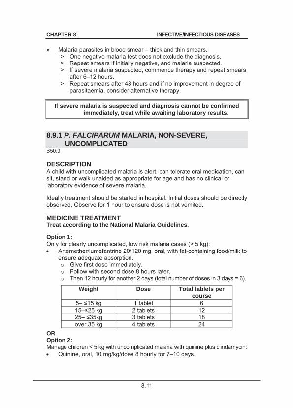
8.11
CHAPTER 8 INFECTIVE/INFECTIOUS DISEASES » Malaria parasites in blood smear – thick and thin smears.
> One negative malaria test does not exclude the diagnosis. > Repeat smears if initially negative, and malaria suspected. > If severe malaria suspected, commence therapy and repeat smears
after 6–12 hours. > Repeat smears after 48 hours and if no improvement in degree of
parasitaemia, consider alternative therapy.
If severe malaria is suspected and diagnosis cannot be confirmed immediately, treat while awaiting laboratory results.
8.9.1 P. FALCIPARUM MALARIA, NON-SEVERE,
UNCOMPLICATED B50.9 DESCRIPTION A child with uncomplicated malaria is alert, can tolerate oral medication, can sit, stand or walk unaided as appropriate for age and has no clinical or laboratory evidence of severe malaria. Ideally treatment should be started in hospital. Initial doses should be directly observed. Observe for 1 hour to ensure dose is not vomited. MEDICINE TREATMENT Treat according to the National Malaria Guidelines. Option 1: Only for clearly uncomplicated, low risk malaria cases (> 5 kg): Artemether/lumefantrine 20/120 mg, oral, with fat-containing food/milk to
ensure adequate absorption. o Give first dose immediately. o Follow with second dose 8 hours later. o Then 12 hourly for another 2 days (total number of doses in 3 days = 6).
Weight Dose Total tablets per course
5– 15 kg 1 tablet 6 15– 25 kg 2 tablets 12 25– 35kg 3 tablets 18 over 35 kg 4 tablets 24
OR Option 2: Manage children < 5 kg with uncomplicated malaria with quinine plus clindamycin: Quinine, oral, 10 mg/kg/dose 8 hourly for 7–10 days.
8.12
CHAPTER 8 INFECTIVE/INFECTIOUS DISEASES 2–3 days after initiating treatment with quinine: Clindamycin, oral, 10 mg/kg/dose 12 hourly for 7 days.
Children who are vomiting but who have no other indications of severe malaria: Quinine, IV, 10 mg/kg/dose 8 hourly administered over 4–6 hours.
o ECG and heart rate monitoring. o Monitor blood glucose levels regularly. o Switch to oral medication, once able to do so.
8.9.2 P. FALCIPARUM MALARIA, SEVERE, COMPLICATED (OR IF REPEATED VOMITING)
B50.0/B50.8 DIAGNOSTIC CRITERIA Clinical » Unable to drink or breastfeed. » Vomits everything. » Renal failure. » Cerebral malaria: manifests with convulsions, which may be subtle,
and/or any change in mental state, ranging from irritability, lethargy to coma, stiff neck or bulging fontanelle.
» Respiratory distress and metabolic acidosis similar to pneumonia. » Anaemia: can be severe and lead to cardiac failure and a depressed
mental state. » Shock: cold moist skin, low blood pressure and collapse. » Hypoglycaemia: can present with convulsions and a depressed mental state. » Jaundice, bleeding, acute renal failure and ARDS are less common in
children than adults.
Investigations » Hyperparasitaemia: > 5% of RBCs infected indicates severe malaria but
a lower parasite density does not exclude severe malaria. » Low Hb (< 6 g/dL). » Test glucose immediately with a fingerprick test. Low blood glucose (< 2.2
mmol/L). » Acidosis: serum lactate (venous) > 5 mmol/L or bicarbonate < 15 mmol/L. » Severe thrombocytopaenia: < 50 x 109/L. » In severe cases, repeat smear after 72 hours and after the completion of
the course of treatment. GENERAL AND SUPPORTIVE MEASURES » Check airway, breathing, circulation (ABC). » Admit to high care or intensive care unit. » Review the child at least twice daily, including holidays. » Avoid overhydration.
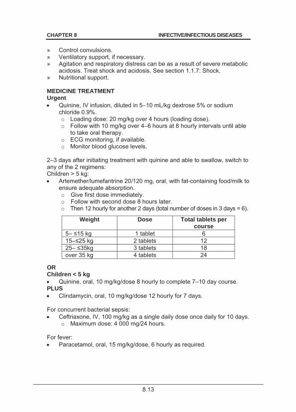
8.13
CHAPTER 8 INFECTIVE/INFECTIOUS DISEASES
» Control convulsions. » Ventilatory support, if necessary. » Agitation and respiratory distress can be as a result of severe metabolic
acidosis. Treat shock and acidosis. See section 1.1.7: Shock. » Nutritional support. MEDICINE TREATMENT Urgent Quinine, IV infusion, diluted in 5–10 mL/kg dextrose 5% or sodium
chloride 0.9%. o Loading dose: 20 mg/kg over 4 hours (loading dose). o Follow with 10 mg/kg over 4–6 hours at 8 hourly intervals until able
to take oral therapy. o ECG monitoring, if available. o Monitor blood glucose levels.
2–3 days after initiating treatment with quinine and able to swallow, switch to any of the 2 regimens: Children > 5 kg: Artemether/lumefantrine 20/120 mg, oral, with fat-containing food/milk to
ensure adequate absorption. o Give first dose immediately. o Follow with second dose 8 hours later. o Then 12 hourly for another 2 days (total number of doses in 3 days = 6).
Weight Dose Total tablets per course
5– 1 tablet 6 15– 2 tablets 12 25– 3 tablets 18 over 35 kg 4 tablets 24
OR Children < 5 kg Quinine, oral, 10 mg/kg/dose 8 hourly to complete 7–10 day course.
PLUS Clindamycin, oral, 10 mg/kg/dose 12 hourly for 7 days.
For concurrent bacterial sepsis: Ceftriaxone, IV, 100 mg/kg as a single daily dose once daily for 10 days.
o Maximum dose: 4 000 mg/24 hours. For fever: Paracetamol, oral, 15 mg/kg/dose, 6 hourly as required.
8.14
CHAPTER 8 INFECTIVE/INFECTIOUS DISEASES For hypoglycaemia: Dextrose 10%, IV
If Hb < 7 g/dL: Packed red cells, IV, 10 mL/kg over 3 hours.
Note: Fluid loss is often underestimated in a febrile, vomiting, sweating child. 8.9.3 P. OVALE, P VIVAX AND P. MALARIAE B53.0/B51.9/B52.9 Chloroquine, oral, 10 mg base/kg as a single dose,
o Follow with 5 mg base/kg given 6, 24 and 48 hours after the first dose.
PLUS To eradicate the organism: Primaquine, oral, 0.25 mg base /kg/day for 14 days. (obtained using
section 21 approval) o Continue chloroquine once weekly until primaquine is obtained.
8.9.4 MALARIA PROPHYLAXIS – SELF PROVIDED CARE In the high-risk malaria areas from September to May in Sout h Africa, malaria prophylaxis should be used, t ogether with preventive measures against mosquito bites. State facilities do not provide prophylactic therapy. It is recommended that persons intending to travel to high-risk areas take the relevant prophylactic therapy. Preventative measures against mosquito bites include: » Use of treated mosquito nets, screens, coils or pads. » Application of insect repellent to exposed skin and clothing. » Wearing long sleeves, long trousers and socks if outside between dusk
and dawn, as mosquitoes are most active at this time. » Visiting endemic areas only during the dry season.
!CAUTION! Pregnant women and children under 5 years should avoid visiting malaria-
endemic areas, as they are more prone to the serious complications of malaria
For chemoprophylaxis refer to National Malaria Guidelines.
8.15
CHAPTER 8 INFECTIVE/INFECTIOUS DISEASES REFERRAL » Urgent: Severe or complicated malaria. » High risk children under 2 years, splenectomised patients. » Malaria not responding clinically to adequate treatment within 48–72
hours (possible resistance). 8.10 MEASLES B05 * Notifiable condition DESCRIPTION The following case definition is an epidemiological and not a diagnostic tool: » Fever and maculopapular rash with any one of the following:
> cough, > coryza/runny nose, > conjunctivitis.
Suspect measles in any child fulfilling the case definition. An acute, highly contagious, viral, childhood exanthem. Incubation period: 8–14 days from exposure to 1st symptoms and 14 days between appearance of rash in source and contact. Complications include: » pneumonia, » feeding difficulties, » laryngotracheobronchitis (croup), » severe diarrhoea, » encephalitis, » otitis media, » stomatitis, and » corneal ulceration. Subacute sclerosing panencephalitis is a rare long-term complication. DIAGNOSTIC CRITERIA Clinical » Prodromal (catarrhal) phase:
> duration 3–5 days, > fever, > runny nose (coryza), > cough, > conjunctivitis.
» Koplik’s spots, followed 3–5 days later with maculopapular rash. » The rash begins to fade after 3 days in the order of its appearance
leaving temporary darker staining. » If fever is still present after the third day of the rash, a complication
should be suspected. Investigations » Serum measles IgM antibodies for confirmation of diagnosis.
8.16
CHAPTER 8 INFECTIVE/INFECTIOUS DISEASES GENERAL AND SUPPORTIVE MEASURES » Notify provincial EPI manager prior to confirmation. » Only admit high risk patients:
> children less than 6 months old, > immune compromised/suppressed children, > children with severe malnutrition, > children with complications.
» Minimal exposure to strong light, if patient is photophobic. » Isolate the patient in a separate room, if possible away from other
children. » All entering the room to wear mask, gloves and gown. » Patient is infectious for 4 days after onset of rash, longer if HIV-infected. » Screen outpatient waiting areas for children with measles. » If pneumonia with hypoxia, give humidified oxygen by means of nasal
cannula. MEDICINE TREATMENT All patients Vitamin A, oral, as a single daily dose for 2 days.
o If < 6 months of age: 50 000 units. o If 6–12 months of age: 100 000 units. o If > 1 year of age: 200 000 units.
For fever Paracetamol, oral, 10–15 mg/kg/dose, 6 hourly as required until fever
subsides. Pneumonia Antibiotics, empirical To cover S. pneumoniae and Gram-negative infection. Total duration of therapy: 5–7 days.
Ampicillin, IV, 25–50 mg/kg/dose 6 hourly. PLUS Gentamicin, IV, 7.5 mg/kg immediately as a single dose.
o Follow with 5 mg/kg once daily. When child improves follow with oral therapy to complete 5–7 days treatment: Amoxicillin, oral, 30 mg/kg/dose 8 hourly.
Penicillin allergy See section 23.4.1: Allergies to penicillins. In very severe progressive or unresponsive pneumonia consider use of aciclovir for possible herpes infection.
8.17
CHAPTER 8 INFECTIVE/INFECTIOUS DISEASES Croup See section 15.5.2: Laryngotracheobronchitis, acute viral (croup). Diarrhoea See section 2.2.4 Diarrhoea, acute. Encephalitis See section 8.14: Meningo-encephalitis/encephalitis, acute viral. Convulsions See section 13.5: Status epilepticus (convulsive). Conjunctivitis Chloramphenicol ophthalmic ointment 1%, inserted 6 hourly for 5 days.
If corneal clouding/ulceration present obtain urgent ophthalmologic consultation. Management of contacts Immunise children older than 6 months if unvaccinated and less than 72 hours since exposure. Between 3 and 6 days after exposure and for contacts less than 6 months old: Immunoglobulin, human normal, IM, 0.25 mL/kg.
If immunodeficient: Immunoglobulin, human normal, IM, 0.5 mL/kg.
Immunise all children > 6 months of age if outbreak occurs. REFERRAL » Children in need of intensive care unit. » Children with depressed level of consciousness. » Children with corneal ulceration/opacity. 8.11 MENINGITIS, ACUTE BACTERIAL G00 * Notifiable condition. (N. meningitidis and H. influenzae) This guideline applies to children > 60 days old. For the management of neonates, see section 19.11: Meningitis bacterial, neonatal. DESCRIPTION Bacterial meningitis most commonly results from haematogenous dissemination of micro-organisms from a distant site, e.g. the nasopharynx. In children, S. pneumoniae and N. meningitides are the usual pathogens.
8.18
CHAPTER 8 INFECTIVE/INFECTIOUS DISEASES Note: Tuberculosis, cryptococcal and partially treated acute bact erial meningitis should be considered when the clinical and laboratory features are not typical of pyogenic meningitis, or when there is a slow onset of disease (> 2 days), especially in any high risk settings such as im mune suppression, TB contact and malnourished children. Differentiation of TB or cryptococcal meningitis from acute bacterial meningitis is not always easy on presentation. Complications include: » Raised intracranial pressure due to cerebral oedema, subdural
effusion/empyema or hydrocephalus. » Other acute complications include:
> cerebral infarctions, > shock, > seizures, > metastatic infection, e.g. arthritis, pneumonia, pericarditis, > disseminated intravascular thrombosis, > inappropriate antidiuretic hormone (ADH) secretion.
Long-term neurological sequelae include deafness, blindness, mental retardation and motor paralysis, e.g. hemiparesis. DIAGNOSTIC CRITERIA Clinical » Fever. » Feeding problems. » Headache. » Irritability. » Vomiting. » Lethargy. » Convulsions » Photophobia. » Signs of meningeal irritation. In young infants signs of meningism are
often absent. » Signs of increased intracranial pressure, e.g. bulging anterior fontanel. » Papilloedema is not a useful sign in young children with meningitis. It is
difficult to elicit and may be absent even with acutely raised ICP. Investigations » Lumbar puncture (LP) (all abnormal findings should lead to serious
considerations of acute bacterial meningitis). Do not do a LP but initiate treatment immediately if clinical signs of severely raised intracranial pressure, i.e. impending cerebral herniation: > decreased or sudden deterioration of level of consciousness, > decerebrate or decorticate posturing, > neurogenic hyperventilation, > unequal dilated or poorly reactive pupils.
» Clinical meningococcaemia (septicaemia) with petechiae/purpura. > Confirm with skin scrape, Gram stain and blood culture.
» Bacterial antigen tests on CSF.
8.19
CHAPTER 8 INFECTIVE/INFECTIOUS DISEASES GENERAL AND SUPPORTIVE MEASURES » Admit to high or intensive care unit, if appropriate. » Monitor, where indicated:
> neurological status, > respiration, > heart rate, > body temperature, > blood pressure, > haematocrit, > acid-base status, > electrolytes, > blood glucose, > blood gases, > fluid balance, i.e. hydration, > serum and urine osmolality.
» Ensure adequate nutrition by enteral feeding where possible. > Use a nasogastric tube if necessary. > If enteral feeding is not possible, give intravenous fluids: paediatric
or neonatal maintenance solution with dextrose. MEDICINE TREATMENT Antibiotic therapy Empiric treatment: Ceftriaxone, IV, 50 mg/kg/dose 12 hourly.
Adjust antimicrobial therapy according to culture and sensitivity. Usual duration of treatment (minimum 5 days fever free):
N. meningitidis 7 days S. pneumoniae 10 days H. influenzae 10 days
In complicated cases, a longer duration of therapy may be required. Re-assess antimicrobial therapy when blood and CSF culture and sensitivity results become available, or when improvement is not evident within 72–96 hours. Seek immediate advice on what treatment to start with when ventriculo-peritoneal shunt infection, spread from sinuses, mastoids, or direct penetrating source of infection is present. For shunts: 3rd generation cephalosporin, e.g.: Ceftriaxone, IV, 50 mg/kg/dose 12 hourly.
PLUS Vancomycin, IV, 15 mg/kg/dose, 6 hourly infused over 1 hour.
Steroid therapy Empiric treatment before culture results are received. Dexamethasone, IV, 0.15 mg/kg 6 hourly for three days starting with or
before initiation of antibiotic. o If an organism other than H. Influenzae is cultured, stop steroid therapy. o Do not delay antibiotic treatment if dexamethasone is not readily
available.
8.20
CHAPTER 8 INFECTIVE/INFECTIOUS DISEASES Fever and headache: Paracetamol, oral, 15 mg/kg/dose, 6 hourly as required.
Convulsions See section 13.5: Status epilepticus (convulsive). Raised intracranial pressure or cerebral oedema Elevate head of bed ± 20 degrees. Maintain PaCO2 at 4–5 kPa (30–35 mmHg); intubate and ventilate if necessary. Avoid fluid overload. Mannitol, IV, 250 mg/kg administered over 30–60 minutes.
Dexamethasone, IV, 0.5 mg/kg 12 hourly.
Chemoprophylaxis for close contacts A close contact is defined as someone living in the same household or dormitory, or institution, or children in the same crèche, or any other “kissing” contact. Health care workers who have intimate contact should receive prophylaxis. N. meningitidis Ciprofloxacin, oral, as a single dose.
o If < 12 years of age: 10 mg/kg. o If > 12 years of age: 500 mg. Note: If < 12 years of age and able to swallow, use a a single 250 mg tablet.
OR Ceftriaxone, IM, single dose
o If < 12 years of age: 125 mg. o If > 12 years of age: 250 mg.
Close contacts who are pregnant: Ceftriaxone, IM, 250 mg.
H. influenzae prophylaxis for all contacts under 5 years who are household contacts (including index case) or day care contacts: Rifampicin, oral, 20 mg/kg/dose, once daily for 4 days.
o Maximum dose: 600 mg o Neonatal dose: 10 mg/kg/dose, once daily for 4 days.
Hib vaccination: Update Hib vaccination in unimmunised or partially immunised children. Give Hib booster to all children <5 years including index case. Hib vaccination on its own is not protective in contact situation.
8.21
CHAPTER 8 INFECTIVE/INFECTIOUS DISEASES REFERRAL » Where lumbar puncture is deferred due to suspected raised intracranial
pressure and/or localising signs start bacterial and tuberculous meningitis treatment immediately.
» Meningitis with complications. » All cases of suspected shunt infection. Start treatment immediately
before referral.
8.12 MENINGITIS, CRYPTOCOCCAL G02.1 DESCRIPTION An uncommon childhood meningitis that may occur in older HIV-infected children with severe CD4 T-cell depletion. Pulmonary and skin involvement can occur. DIAGNOSTIC CRITERIA Clinical » Acute or chronic headache in an older HIV-infected child. Meningism
need not be present. » Often presents with cranial nerve palsy. » Can occur as result of Immune Reconstitution Inflammatory Syndrome
(IRIS) after initiation of antiretroviral therapy. Investigations » Test all cerebrospinal fluid (CSF) specimens from HIV-infected children
with suspected meningitis. » India ink stain, and/or cryptococcal antigen test – more sensitive than
India ink stain. » Fungal culture blood and urine. » Chest X-ray. » Ophthalmological assessment. GENERAL AND SUPPORTIVE MEASURES » Admit to high or intensive care unit, if appropriate. » Monitor, where indicated:
> neurological status, > respiration, > heart rate, > body temperature, > blood pressure, > electrolytes, > haematocrit, > blood glucose, > minerals, > blood gases, > acid-base status, > serum and urine osmolality, and > fluid balance, i.e. hydration.
» Ensure adequate nutrition by enteral fe eding where possible. Use a nasogastric tube if n ecessary. If ent eral feeding is not po ssible, give intravenous fluids: paediatric or neonat al maintenance solution with dextrose.
8.22
CHAPTER 8 INFECTIVE/INFECTIOUS DISEASES MEDICINE TREATMENT Treatment Initial treatment Amphotericin B, IV, 0.7–1.0 mg/kg/day as a daily infusion over 4 hours.
PLUS Fluconazole, IV, 12 mg/kg/day for 2 weeks.
o Maximum dose: 800mg. Prehydration before administering amphotericin B t o prevent renal impairment: Sodium chloride 0.9%, IV, 20 mL/kg plus potassium chloride, 20 mmol/L
infused over 2–4 hours. Consolidation treatment Fluconazole, oral, 12 mg/kg/day for 8 weeks.
o Maximum dose: 800mg. Secondary prophylaxis (maintenance treatment) Fluconazole, oral, 6 mg/kg/day
o Maximum dose: 400 mg. Discontinue secondary prophylaxis: » Children < 6 years on ART: CD 4 count > 25% for at least 6 months. » Children > 6 year on ART: CD 4 count > 200 for at least 6 months.
Adolescents on ART: CD4 count increases to between 100–200 cells/mm3 for at least 6 months.
» Re-start prophylaxis if CD4 count drops below thresholds above. For continued raised intracranial pressure: » Daily therapeutic lumbar puncture is indicated if initial LP manometric
pressure > 25 cm water in the lateral recumbent position. » Continue until pressure stabilises below 25 cm water. » Remove 10-20 mL daily and obtain a closing pressure. REFERRAL » All cases not responding to initial treatment. » All patients with IRIS.
8.13 MENINGITIS, TUBERCULOUS (TBM) G01 * Notifiable condition. DESCRIPTION Tuberculous meningitis is an infection of the meninges caused by M. tuberculosis. Early diagnosis and treatment improves the prognosis.
8.23
CHAPTER 8 INFECTIVE/INFECTIOUS DISEASES Differentiation from acute bacterial meningitis may be difficult. If in any doubt, treat for both conditions. Complications may be acute or long term: » Acute:
> raised intracranial pressure, > hydrocephalus, > cerebral oedema, > brain infarcts, > hemi/quadriplegia, > convulsions, > hyponatraemia due to inappropriate antidiuretic hormone (ADH)
secretion or cerebral salt wasting. Syndrome of inappropriate antidiuretic hormone secretion (SIADH) and cerebral salt wasting both present with hyponatraemia; the former responding to fluid restriction and the latter to fluid replacement, i.e. sodium chloride 0.9%. SIADH has lower serum uric acid and low urine output. Cerebral salt wasting has a normal serum uric acid and high urine output.
» Long term neurological sequelae include: mental handicap, blindness and deafness.
DIAGNOSTIC CRITERIA Clinical » History of contact with an infectious tuberculosis case. » Onset may be gradual with vague complaints of drowsiness (or fatigue),
vomiting, fever, weight loss, irritability and headache. » Later symptoms such as convulsions and neurological fall-out may occur. » Older children may present with behavioural changes. » Examination may reveal signs of meningeal irritation and ra ised
intracranial pressure, convulsions, cranial nerve palsies, localising signs (such as hemiparesis), altered level of consciousness or coma and choroidal tubercles.
» The degree of involvement is classified into 3 stages. Prognosis relates to the stage of the disease. Stage 1: non-specific signs, conscious, rational, no focal neurological
signs, no hydrocephalus. Stage 2: signs of meningeal irritation, confusion and/or focal
neurological signs. Stage 3: stupor, delirium, coma and/or neurological signs, i.e.
hemiplegia. Investigations » CSF findings:
> May vary depending on the stage. > Protein is usually raised. > Chloride and glucose are moderately low. > Lymphocytes usually predominate. > Gram stain is negative and acid-fast bacilli are seldom found.
8.24
CHAPTER 8 INFECTIVE/INFECTIOUS DISEASES
> In selected cases TB PCR based test on CSF should be done, where available. It may be helpful where it is positive, negative PCR does not exclude TB.
> A negative result does not exclude TB and cultures must still be done. Bacilli may be cultured from the CSF but may take up to 4–6 weeks. If culture positive, also do drug susceptibility test. Always send for culture, do not perform stain as low diagnostic yield from low concentration of organisms wastes CSF sample.
» A Mantoux test and chest X-ray must be done, but are often unhelpful. » If depressed level of consciousness or focal neurological signs are
present, a CT scan is useful (do CT first before LP in such cases). » Electrolytes: check for hyponatraemia. GENERAL AND SUPPORTIVE MEASURES » Monitor neurological status on a regular basis. If rapid deterioration in
level of consciousness, consider ventriculoperitoneal shunt. » Attend to nutritional status. Initially nasogastric feeding is usually
needed. » Rehabilitative measures: most patients need physiotherapy and
occupational therapy. » Surgical treatment for non-communicating hydrocephalus, diagnosed
by air encephalogram (VP shunt). » Communicating hydrocephalus with severely raised pressure may be
managed with medicines once hydration status stable and/or with serial lumbar puncture with specialist consultation.
MEDICINE TREATMENT Differentiation from acute bacterial meningitis may be difficult. If in doubt, treat for both conditions. Antituberculosis treatment Requires therapy with a combination of 4 drugs as a special regimen. All treatment should be directly observed therapy. Single drugs may form part of the regimen to provide the total daily required dose for each medicine by supplementing the combination to give the necessary therapeutic dose per kilogram. A 6-month regimen of all 4 the following drugs: Rifampicin, oral, 20 mg/kg as a single daily dose.
PLUS Isoniazid, oral, 20 mg/kg as a single daily dose.
PLUS Pyrazinamide, oral, 40 mg/kg as a single daily dose.
o Maximum daily dose: 2 000 mg. PLUS Ethionamide, oral, 20 mg/kg as a single daily dose.
o Maximum daily dose: 1 000 mg.
8.25
CHAPTER 8 INFECTIVE/INFECTIOUS DISEASES Consider prolonging treatment for another 3 months if there are concerns about ongoing disease. Consult with a specialist. In case of suspected/confirmed multidrug-resistant TBM, refer immediately for admission and treatment. Steroid therapy Prednisone, oral, 2mg/kg as a single daily dose for 4 weeks.
o Maximum daily dose: 60 mg. o Taper to stop over further 2 weeks.
Hydrocephalus Avoid low sodium IV fluids in these patients, i.e. < 60 mmol/L. To differentiate communicating from non-communicating hydrocephalus an air encephalogram is usually required. Communicating hydrocephalus is more common in this condition. In children with a sudden deterioration of level of consciousness and other comatose children with TBM, inform the neurosurgeon before doing the air-encephalogram so th at shunt surgery can immediately be done if the hydrocephalus is non-communicating. Air-encephalogram procedure: do a lumbar puncture, inject 5 ml of air with a syringe and do immediate lateral X-ray of the s kull. Air in t he lateral ventricles on s kull X-ray indicates communicating hydrocephalus; air at base of brain (not in lateral ventricles), indicates non-communicating hydrocephalus. Communicating hydrocephalus If dehydrated, rehydrate with sodium chloride 0.9%, IV. Start diuretics as soon as patient is well hydrated and serum electrolytes are within the normal range. Acetazolamide, oral, 20 mg/kg/dose 8 hourly.
o Maximum daily dose: 1 000 mg. o Monitor for metabolic acidosis and serum potassium derangements.
PLUS Furosemide, oral, 0.3 mg/kg/dose 8 hourly for the first month of
treatment. o Taper slowly over 2 weeks if the intracranial pressure has
normalised, as indicated by clinical response or resolution of hydrocephalus on follow-up scan.
o Do not restrict fluids once on diuretics.
Sudden deterioration of level of consciousness: Mannitol, IV, 250 mg/kg administered over 30–60 minutes.
8.26
CHAPTER 8 INFECTIVE/INFECTIOUS DISEASES REFERRAL » TBM not responding to adequate therapy. » TBM with complications. » Suspicion of non-communicating hydrocephalus. » Suspected drug-resistant TB (contact with drug-resistant TB case). 8.14 MENINGO-ENCEPHALITIS/ENCEPHALITIS, ACUTE
VIRAL A86 DESCRIPTION A number o f viruses cause infection of t he brain and meninges. Herpes simplex is the mo st important because it i s treatable. A high mortality and morbidity is associated with untreated disease. Complications include: » increased intracranial pressure, » permanent neurological deficits, » cerebral oedema, » seizures, » blindness, » deafness, » inappropriate antidiuretic hormone (ADH) secretion. DIAGNOSTIC CRITERIA Clinical » Severe headache, fever, nausea, vomiting, lethargy and abnormal
behaviour. » Alteration in level of consciousness, i.e. drowsiness, confusion, stupor
and coma. » Generalised and/or focal convulsions. » Focal neurological deficits. » Abnormal movements, i.e. basal ganglia involvement. » Cranial nerve palsies (brainstem involvement), loss of sphincter control,
paresis of limbs and segmental sensory loss (spinal cord involvement). » Some patients may have signs of meningeal irritation. » Herpes encephalitis may have an acute and fulminant course. It can
result from primary or recurrent infection. Investigations » Laboratory tests are mostly unhelpful. » CSF may reveal:
> slightly raised protein, > normal glucose level, and > mild pleocytosis, mostly lymphocytes. > A specific virus is sometimes isolated. PCR is helpful, if available. > Red cells are seen with Herpes encephalitis.
8.27
CHAPTER 8 INFECTIVE/INFECTIOUS DISEASES » In some instances, the CSF may be completely normal. » A CT scan of the brain may reveal brain oedema.
> CT findings may only be apparent after 3–5 days. > The herpes virus preferentially involves the temporal lobe and
orbital surfaces of the frontal lobes. » An EEG may demonstrate changes suggestive of herpes encephalitis. GENERAL AND SUPPORTIVE MEASURES » Admit to high or intensive care unit, if appropriate. » Monitor, where indicated:
> neurological status, > respiration, > heart rate, > body temperature, > blood pressure, > electrolytes, > haematocrit, > blood glucose, > acid-base status, > blood gases, > fluid balance, i.e. hydration, > serum and urine osmolality.
» Ensure adequate nutrition by enteral feeding where possible. > Use a nasogastric tube if necessary. > If enteral feeding is not possible, give intravenous fluids: paediatric
or neonatal maintenance solution with dextrose. MEDICINE TREATMENT If herpes simplex virus or varicella zoster virus encephalitis: Aciclovir, IV, 8 hourly administered over 1 hour for 21 days.
o If 0–12 years of age: 20 mg/kg/dose. o If > 12 years of age: 10 mg/kg/dose.
Acute convulsions See section 13.5: Status epilepticus (convulsive). For fever: Paracetamol, oral, 15 mg/kg/dose, 6 hourly until fever subsides.
Raised intracranial pressure or cerebral oedema Elevate head of bed ± 20 degrees. Maintain PaCO2 at 4–5 kPa; intubate and ventilate, if necessary. Avoid fluid overload. Mannitol, IV, 250 mg/kg administered over 30–60 minutes.
o Do not repeat without consultation with a paediatrician. REFERRAL » Deterioration of clinical condition despite adequate treatment. » Meningo-encephalitis with complications or loss of consciousness.
8.28
CHAPTER 8 INFECTIVE/INFECTIOUS DISEASES 8.15 MUMPS B26 DESCRIPTION Mumps is an acute, communicable, viral disease of childhood that commonly affects the salivary glands, chiefly the parotid gland, and frequently the central nervous system. The incubation period is 2–3 weeks. Complications include: » meningo-encephalitis, » oophoritis, » pancreatitis, » thyroiditis, » orchitis, » nerve deafness, » facial nerve paresis, » myocarditis, » nephritis. DIAGNOSTIC CRITERIA Clinical » A prodrome of 1–2 days may precede the salivary gland involvement
and is characterised by fever, malaise, headache and pain in or behind the ear on chewing or swallowing.
» Painful enlargement of the parotid gland/s with the ear usually displaced upward and ou tward with the mandibular angle obliterated. The submaxillary and sublingual glands may also be involved.
» Pain may be referred to the ear. » Papilla of Ste nsen’s duct opposite the upper second molar may be
oedematous and red. » Central nervous system involvement may occur alone or may precede,
accompany or follow inflammation of the salivary glands.
Investigations » Leucopaenia with relative lymphocytosis. GENERAL AND SUPPORTIVE MEASURES » Isolate patient until salivary gland enlargement subsides. » Maintain adequate nutrition and hydration. » Patient may return to school after swelling has subsided. MEDICINE TREATMENT Treat complications as appropriate. For pain and fever: Paracetamol, oral, 15 mg/kg/dose, 6 hourly as required.
8.29
CHAPTER 8 INFECTIVE/INFECTIOUS DISEASES REFERRAL » Mumps with complications not responding to adequate therapy. 8.16 MYCOBACTERIUM AVIUM COMPLEX (MAC) INFECTION A31.0 DESCRIPTION Atypical mycobacterium, causing disease in extremely immunocompromised patients. MAC infection in HIV-infected children usually presents with disseminated disease, often enlarged intra-abdominal lymph nodes and pancytopaenia. Pulmonary, GIT or skin disease is less common. DIAGNOSTIC CRITERIA » MAC may be isolate d from blood, bone marrow, lymph node, other
sterile fluids and tissues. » Confirm diagnosis with a biopsy for histology and culture or 2 culture-
positive sputa or gastric aspirates. » PCR line probe test can be used for diagnosis. GENERAL AND SUPPORTIVE MEASURES » If MAC infection i s localised to a single en larged peripheral lymph
node, an excision of the lymph node is therapeutic. MEDICINE TREATMENT To be managed under specialist care. Therapy consists of a combination of at least two medicines. New generation macrolide e.g.: Clarithromycin, oral, 7.5 mg/kg/dose 12 hourly.
PLUS Ethambutol, oral, 20–25 mg/kg once daily.
For extensive disease in severely immunodeficient child: ADD Fluoroquinolone, e.g.: Levofloxacin, oral, 20 mg/kg once daily.
PLUS Amikacin, IV, 15 mg/kg once daily.
REFERRAL » No response to treatment. » Fibrocavitating pulmonary disease
8.30
CHAPTER 8 INFECTIVE/INFECTIOUS DISEASES 8.17 PERTUSSIS A37.9 * Notifiable condition DESCRIPTION A communicable respiratory infection usually recognised by a parox ysmal cough followed by an inspiratory whoop (absent in young infants) and associated vomiting. Subconjunctival haemorrhages may be pre sent. The cough can persist for 3 months or longer with the infectious period between 2 weeks and 3 months. The disease is more severe in young infants where it may present with apnoea rather than the inspiratory whoop. Incubation period: 7–10 days. Range: 6– 21 days. DIAGNOSTIC CRITERIA » Diagnosis is clinical. » A definitive diagnosis often not possible with respect to viral pertussis-
like syndrome. » FBC usually very high WCC with > 50% lymphocytosis. » Use naso-pharyngeal aspirates if po ssible for sp ecial cultures for
Bordetella pertussis or identify by using PCR. GENERAL AND SUPPORTIVE MEASURES » Isolate patient during first 2 days whilst on antibiotic therapy. » Clear airways by gentle suction taking care not to induce cough. » Appropriate respiratory support for apnoea or respiratory distress/failure. » Encourage oral feeding. If un successful provide nasogastric feeds with
small volumes. » Immunise infant aga inst pertussis even if dia gnosis of pert ussis was
made. MEDICINE TREATMENT If hypoxic: Oxygen, 1–2 L/minute via nasal prongs.
New generation macrolide e.g.: Clarithromycin, oral, 7.5 mg/kg/dose 12 hourly for 7 days.
For fever: Paracetamol, oral, 15 mg/kg/dose, 6 hourly as required.
Management of contacts Prophylaxis only for unimmunised or partly immunised infants < 6 months of age: Macrolide e.g.: Clarithromycin, oral, 7.5 mg/kg/dose 12 hourly for 7 days.
8.31
CHAPTER 8 INFECTIVE/INFECTIOUS DISEASES REFERRAL » Children with seizures or encephalopathy for further evaluation. » Infants requiring intensive care, where none is available on site. 8.18 PNEUMOCYSTIS JIROVECI PNEUMONIA (PCP) B20.6 See section 15.3.2.6: Pneumonia in HIV exposed or infected children. 8.19 POLIOMYELITIS (ACUTE FLACCID PARALYSIS) A80.3 * Notifiable condition Also see section 13.9.1: Inflammatory Polyneuropathy (Guillain-Barré Syndrome). DESCRIPTION Poliomyelitis is caused by polio virus types 1, 2 a nd 3. It mainly affects children under 5 years of age, but a person at any age who does not have immunity may be infected. Risk of paralysis increases with age. Poliomyelitis is uncommon. Most cases of acute flaccid paralysis (AFP) are caused by Guillain-Barré Syndrome, but all cases of AFP should be notified. Humans are the only reservoir. The faecal-oral route is the major route of transmission, although droplet spread can also occur. Incubation period is between 7–14 days. DIAGNOSTIC CRITERIA Clinical » Polio virus infection is asymptomatic in 90–95% of cases. » ± 4–8% will develop abortive polio with some or all of the following
symptoms: > fever, > headache, > stiff neck, > muscle pain, > nausea, > vomiting and diarrhoea.
» ± 1% may present as viral meningitis. » The remaining 1–2% will develop paralysis of sudden rapid onset
reaching full development in hours, maximum 3 days. » Paralysis is often asymmetrical and always flaccid. Reflexes are absent. » Sensation usually not affected. » Lower limbs are affected more than upper limbs and proximal more
than distal muscles.
8.32
CHAPTER 8 INFECTIVE/INFECTIOUS DISEASES Investigations » Send two stool specimens taken 24–48 hours apart to the National
Institute of Communicable Diseases via the local laboratory. GENERAL AND SUPPORTIVE MEASURES » Isolate patient to prevent faecal-oral spread. » Rehabilitative measures:
> Most patients need physiotherapy and occupational therapy. MEDICINE TREATMENT Prevention Immunise all children, including HIV-infected children, according to the EPI programme. Vaccine associated/derived atypical paralytic polio is only associated with live oral polio vaccine (given in new EPI programme only at birth and 6 weeks) and not associated with inactivated polio vaccine (IPV), which is given from 6 weeks on. IPV contraindicated if: previous anaphylactic reaction following IPV or if anaphylaxis with streptomycin, neomycin or polymyxin B. REFERRAL » Children requiring intensive care, if none is available on site. 8.20 RABIES A82.9 * Notifiable condition Inform state veterinarian or local veterinary official. DESCRIPTION A viral infection of the central nervous system following transmission of the rabies virus from the saliva of a ffected animals through bites or contamination of mucosa or skin lesions. Incubation period 2–8 weeks. DIAGNOSTIC CRITERIA Clinical » Signs and symptoms may begin with:
> fever, > headache, > nausea, > diarrhoea, > irritability.
» Early signs include ases. » The acute neurologic phase interspersed with lucid periods manifests with:
> agitation, > mania, > hyperactivity, > hallucinations.
8.33
CHAPTER 8 INFECTIVE/INFECTIOUS DISEASES » Seizures may be precipitated by auditory or tactile stimuli. » Hypersalivation, hydrophobia or aerophobia may occur. » Death is usually due to cardio-respiratory failure. Investigations » Virus specific fluorescent antigen in brain tissue confirms diagnosis in
animals. » Preserve brain tissue of the dead animal. GENERAL AND SUPPORTIVE MEASURES » Symptomatic and supportive treatment. » Prompt cleansing of the bite wound. » Do not suture puncture wounds. » Seek advice.
TELEPHONE HOTLINE National Institute of Communicable Diseases 011 386 6337 or 011 386 6000
After hours 082 883 9920 Post exposure prophylaxis
Caution Start post exposure prophylaxis immediately.
Do not wait for confirmatory laboratory tests in the animal.
Post exposure prophylaxis may be life saving and should always be given if there is the slightest suspicion that the animal may have been rabid. The decision to give post exposure prophylaxis is based on the risk of rabies transmission, the species and behaviour of the animal and the nature of the bite. No laboratory test on t he human victim is p ossible or av ailable to confirm or exclude possible transmission.
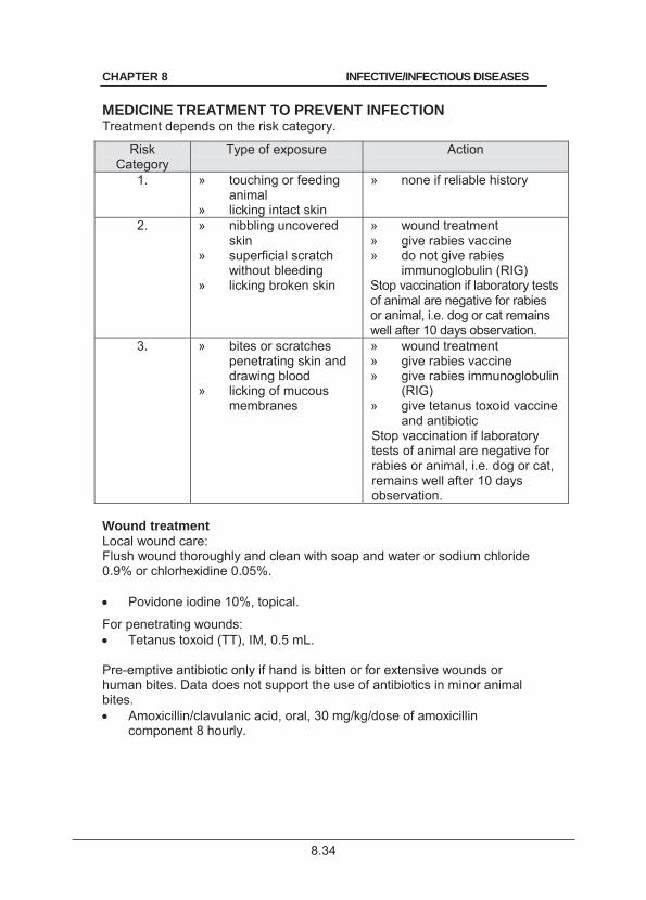
8.34
CHAPTER 8 INFECTIVE/INFECTIOUS DISEASES MEDICINE TREATMENT TO PREVENT INFECTION Treatment depends on the risk category.
Risk Category
Type of exposure Action
1. » touching or feeding animal
» licking intact skin
» none if reliable history
2. » nibbling uncovered skin
» superficial scratch without bleeding
» licking broken skin
» wound treatment » give rabies vaccine » do not give rabies
immunoglobulin (RIG) Stop vaccination if laboratory tests of animal are negative for rabies or animal, i.e. dog or cat remains well after 10 days observation.
3. » bites or scratches penetrating skin and drawing blood
» licking of mucous membranes
» wound treatment » give rabies vaccine » give rabies immunoglobulin
(RIG) » give tetanus toxoid vaccine
and antibiotic Stop vaccination if laboratory tests of animal are negative for rabies or animal, i.e. dog or cat, remains well after 10 days observation.
Wound treatment Local wound care: Flush wound thoroughly and clean with soap and water or sodium chloride 0.9% or chlorhexidine 0.05%. Povidone iodine 10%, topical.
For penetrating wounds: Tetanus toxoid (TT), IM, 0.5 mL.
Pre-emptive antibiotic only if hand is bitten or for extensive wounds or human bites. Data does not support the use of antibiotics in minor animal bites. Amoxicillin/clavulanic acid, oral, 30 mg/kg/dose of amoxicillin
component 8 hourly.
8.35
CHAPTER 8 INFECTIVE/INFECTIOUS DISEASES Rabies Vaccine Must be given for category 2 and 3 bites. Vaccine is administered on days 0, 3, 7, 14, 28. Vaccine is ideally given as soon as possible after exposure, but should still be given if patient presents some time after the exposure. If vaccine administration is delayed > 48 hours, a double dose should be given initially. Rabies vaccine is given IM but never in the buttock. Give to deltoid muscle in adults and antero-lateral aspect of thigh in infants. Adverse events are uncommon and include: » Local reactions:
> pain, > erythema, > swelling or itching at the injection site.
» Systemic reactions: > fever, > arthralgia, > arthritis, > angioedema, > nausea, > vomiting, > malaise.
Rabies Immunoglobulin (RIG) Must be given for category 3 bites only. Always give the vaccine first. Immunoglobulin must be given as soon as possible after exposure, but may be administered up to 7 days after the first vaccine is given. Do not give RIG if the patient has previously received pre- or post-exposure prophylaxis. Rabies immunoglobulin, 20 units/kg.
o Infiltrate around wound with up to 50% of dose. o Administer remaining immunoglobulin into deltoid muscle opposite
to vaccine administration site. o If multiple wounds, dilute in sodium chloride 0.9% to 2–3 times so
that all wounds are infiltrated. o Do not exceed maximum dose as antibody production to the
vaccine is inhibited. o If unavailable, do not delay active immunisation.
REFERRAL » Where prophylactic treatment is not immediately available. » All cases of human clinical rabies for appropriate palliative care.
8.36
CHAPTER 8 INFECTIVE/INFECTIOUS DISEASES 8.21 TETANUS A35 * Notifiable condition. DESCRIPTION Tetanus is an acute spastic paralytic illness caused by tetanospasmin, the neurotoxin produced by Clostridium tetani. The toxin prevents neurotransmitter release from spinal inhibitory neurons.
Complications include: » asphyxia, » bronchopneumonia, » dehydration, » respiratory failure, » hyperpyrexia, » laryngospasm, » inability to suck, chew and swallow. DIAGNOSTIC CRITERIA The diagnosis is made on clinical grounds. Clinical » Unimmunised/incompletely immunised child. » History of wound/trauma or unhygienic care of umbilical cord/stump. » Trismus. » Stiffness of the neck, back and abdominal muscles. » Pharyngospasm, laryngospasm, dysphagia, inability to suck, chew and
swallow which severely compromises feeding and eating activities. » Spontaneous muscle contractions/spasms or muscle contractions/
spasms triggered by minimal stimuli such as touch, sound, light or movement.
» No involvement of sensorium, i.e. consciousness is not disturbed. » Autonomic nervous system instability with hypertension, tachycardia
and dysrhythmias. GENERAL AND SUPPORTIVE MEASURES » Admit to high or intensive care unit, if available. » Ventilatory support, if needed. » Monitor:
> temperature, > blood pressure, > respiration, > blood glucose, > heart rate, > electrolytes, > blood gases, > acid-base status, > SaO2.
» Protect the patient from all unnecessary sensory and other stimuli. » Ensure adequate hydration and nutrition. » Wound care and debridement/umbilical cord care. » Educate parents/caregivers regarding prevention of tetanus by
vaccination.
8.37
CHAPTER 8 INFECTIVE/INFECTIOUS DISEASES MEDICINE TREATMENT For hypoxia: Oxygen 100% by nasal canula.
Tetanus immunoglobulin, IM, 500–2 000 IU as a single dose.
Tetanus toxoid (TT), IM, 0.5 mL
o Not required in immunised patients who have received a booster within the past 5 years.
Metronidazole, IV, 7.5 mg/kg/dose 8 hourly for 10 days duration.
For control of muscle spasms: Diazepam, IV, 0.1–0.2 mg/kg/dose 4–6 hourly, titrated according to
response. o Do not exceed 10 mg/dose. o After improvement, use enteral form in high care setting. o For ventilation and muscle relaxants, see section 21.1.2: ICU
sedation, infant and child. After recovery from tetanus, patients should be actively immunised as the disease does not confer immunity. Prevention of tetanus Minor wounds Children with clean minor wounds do not require tetanus immunoglobulin or antibiotics. Tetanus vaccine should be given, except in fully immunised patients who have received a booster within the past 5 years. For more severe wounds If child with penetrating wound not completely immunised: Tetanus immunoglobulin (TIG), IM.
o If < 5 years of age: 75 IU. o If 5–10 years of age: 125 IU. o If > 10 years of age: 250 IU.
Tetanus toxoid (TT), IM, 0.5 mL.
o Not required in immunised patients who have received a booster within the past 5 years.
REFERRAL » All severe cases.
8.38
CHAPTER 8 INFECTIVE/INFECTIOUS DISEASES 8.22 TICK BITE FEVER A79.9 DESCRIPTION A febrile illness with exanthem caused by Rickettsia conorii with the tick as vector. Recently, other tick-borne Rickettsial diseases have been identified. The rash appears on days 3–5 of the illness. It spreads from the extremities to the trunk, neck, face, palms, and soles within 36 hours. The lesions progress from macular to maculopapular and may persist for 2–3 weeks. Atypical cutaneous findings may occur in a few patients. Complications include: » vasculitis, » encephalitis, » thrombosis, » renal failure, » myocarditis, » pneumonitis, and » thrombocytopaenia. DIAGNOSTIC CRITERIA The diagnosis is made on clinical grounds. Clinical » Fever, headache, malaise, myalgia and arthralgia. » Maculopapular rash which may involve the palms and soles. » Eschar at the site of the tick bite is associated with regional
lymphadenopathy and splenomegaly. Investigations » Diagnosis can be confirmed retrospectively by immunofluorescent
antibody techniques. GENERAL AND SUPPORTIVE MEASURES » Remove tick as soon as possible after detection on the body. MEDICINE TREATMENT Antibiotic therapy Treatment must be started before confirmation of diagnosis by serology. Although not recommended for children < 8 years of age, doxycycline is still regarded as the drug of choice for children with tick-bite fever.
Doxycycline, oral. o If < 50 kg: 4 mg/kg/24 hours in 2 divided doses on the first day,
then 2 mg/kg/24 hours in 2 divided doses for 7 days. o If > 50 kg: 100 mg 12 hourly for 7 days.

8.39
CHAPTER 8 INFECTIVE/INFECTIOUS DISEASES If unable to take oral therapy: Clarithromycin, IV, 7.5 mg/kg/dose 12 hourly for 7 days.
For headache and fever: Paracetamol, oral, 15 mg/kg/dose, 6 hourly as required.
REFERRAL » Patients not responding to adequate therapy. » Patients with complications.
8.23 TOXOPLASMOSIS B58.9 DESCRIPTION Rarely occurs in children. Usually presents as encephalitis, with focal neurological abnormalities occurring in association with headache. Ocular and pulmonary disease is also seen. DIAGNOSTIC CRITERIA Investigations » Diagnosis may be made on blood and CSF serology. » CSF PCR for toxoplasmosis may also be helpful. » CT scan usually reveals multiple bilateral, focal hypodense ring-
enhancing lesions. REFERRAL » All cases.
8.24 TYPHOID A01.1 * Notifiable condition. DESCRIPTION A systemic disease caused by Salmonella typhi. DIAGNOSTIC CRITERIA Clinical: » fever, » anorexia, » headache, » vomiting, » diarrhoea or constipation, » ileus, » abdominal pain or tenderness, » epistaxis, » cough, » delirium,
» hepatomegaly and/or splenomegaly,
» meningismus, » stupor.
8.40
CHAPTER 8 INFECTIVE/INFECTIOUS DISEASES Investigations: » Leucopaenia, anaemia and thrombocytopaenia. » Positive cultures from blood (1st week), stool (after 1st week), urine
and bone marrow. » Serology may be helpful. GENERAL AND SUPPORTIVE MEASURES » Isolate patient until 3 consecutive stools are culture negative. » Correct and maintain fluid and electrolyte status. MEDICINE TREATMENT Note: Relapse and carrier state may occur despite adequate therapy. If haemoglobin < 7 g/dL: Packed red cells, IV, 10 mL/kg.
3rd generation cephalosporin, e.g.: Ceftriaxone, IV, 50 mg/kg once daily for 7 days.
o In severe disease prolong treatment up to 14 days. If cephalosporin resistance consider fluoroquinolones: Ciprofloxacin, oral, 10-20 mg/kg/dose 12 hourly for 7 days.
SURGICAL TREATMENT Surgical intervention for bowel perforation, osteomyelitis, etc. REFERRAL » Inadequate response to treatment. » Patients with complications. 8.25 NON-TYPHOID SALMONELLA (NTS) A02.9 DESCRIPTION Present as: » gastroenteritis, or » extraintestinal (invasive) disease. DIAGNOSTIC CRITERIA Clinical » Self-limiting mucosal intestinal disease presenting with diarrhoea and
vomiting in immunocompetent patients. » Young infants (< 3 mon ths) and immunodeficient children (especially
HIV-infected children) are prone to invasive, even recurrent disease.
8.41
CHAPTER 8 INFECTIVE/INFECTIOUS DISEASES
» Invasive disease includes bacteraemia (fever), osteomyelitis and meningitis.
» There is also an association of invasive NTS with malaria and severe anaemia.
Investigations » Positive cultures from blood, stool, urine and bone marrow. » Serology may be helpful. GENERAL AND SUPPORTIVE MEASURES » Correct and maintain fluid and electrolyte status. MEDICINE TREATMENT Note: Relapse may occur despite adequate therapy. Antibiotic therapy in NTS gastroenteritis may prolong excretion of Salmonella. If haemoglobin < 7 g/dL: Packed red cells, IV, 10 mL/kg.
Gastroenteritis Antibiotic therapy is not generally recommended. However, in infants < 3 months of age and severely immunocompromised children at high risk of developing invasive disease treat as for invasive disease. Invasive disease If < 1 month of age: Cefotaxime, IV/IM, 50–75 mg/kg/dose 8 hourly.
OR If > 1 month of age: Ceftriaxone, IV, 50–80 mg/kg once daily.
Duration: o Bacteraemia: 10–14 days. o Acute osteomyelitis: 4–6 weeks. o Meningitis: 4 weeks.
If cephalosporin resistance treat according to sensitivity. SURGICAL TREATMENT Surgical intervention for osteomyelitis, etc. REFERRAL » Inadequate response to treatment. » Patients with complications.
8.42
CHAPTER 8 INFECTIVE/INFECTIOUS DISEASES 8.26 VARICELLA (CHICKEN POX) B01 DESCRIPTION An acute, highly contagious, viral disease caused by herpes varicella-zoster. It spreads by infective droplets or fluid from vesicles. One attack confers permanent immunity. Varicella is contagious from about 5 days before the onset of the rash until the crusts begin to disappear. Re-activation of the v irus may appear later as herpes zoster or shingles (in children, consider immunosuppression if this occurs). Incubation period is 2–3 weeks.
Complications are more common in immunocompromised patients and include: » secondary skin infection, » pneumonia, » necrotising fasciitis, » encephalitis, » haemorrhagic varicella lesions with evidence of disseminated,
intravascular coagulation. » Two important bacteria causing complications are Staphylococcus
aureus and Streptococcus pyogenes. DIAGNOSTIC CRITERIA Clinical » Mild headache, fever and malaise. » Characteristic rash. » The lesions progress from macules to vesicles in 24–48 hours. » Successive crops appear every few days. » The vesicles, each on an erythematous base, are superficial, tense
‘teardrops’ filled with clear fluid which dry to form fine crusts. » The rash is more profuse on the trunk and sparse at the periphery of
extremities. » At the height of eruption, all stages (macules, vesicles and crusts) are
present at the same time. » The rash lasts 8–10 days and heals without scarring, unless
secondarily infected. » Mucous membranes may be involved. » Pruritus may be severe. » Patients are most contagious from 1–2 days before onset of the rash
until crusting of lesions. GENERAL AND SUPPORTIVE MEASURES » Isolate the patient. » Isolate the neonate until the mother is regarded as non-contagious. » Maintain adequate hydration.
8.43
CHAPTER 8 INFECTIVE/INFECTIOUS DISEASES MEDICINE TREATMENT Antiviral therapy Indicated for immunocompetent patients with varicella complications and for all immunocompromised patients. Initiate as early as possible, preferably within 24 hours of the appearance of the rash. Immunocompromised patients and all cases with severe chickenpox (not encephilits) Aciclovir, oral, 20 mg/kg/dose 6 hourly for 7days.
o Maximum dose: 800 mg/dose. In severe cases or in cases where oral medicine cannot be given: Aciclovir, IV, 8 hourly administered over 1 hour for 21 days.
o If 0–12 years: 20 mg/kg/dose 8 hourly. o If >12 years: 10 mg/kg/dose 8 hourly.
For encephalitis: See section 8.14: Meningo-encephalitis/encephalitis, acute viral. For fever: Paracetamol, oral, 15 mg/kg/dose, 6 hourly as required.
For mild pruritus: Calamine lotion, topical, applied 8 hourly.
For severe pruritus: Chlorphenamine, oral, 0.1 mg/kg 6–8 hourly for 24–48 hours.
Secondary skin infection Cephalexin, oral, 12.5 mg/kg/dose, 6 hourly for 5 days.
Prophylaxis Post exposure prophylaxis must be given to: Neonates whose mothers develop varicella from 5 days before delivery to 2 days after delivery and for an exposure: Varicella-zoster immunoglobulin, IM, 1 mL (100 units) given within 96
hours of exposure. If varicella-zoster immunoglobulin is not available: Aciclovir, oral, 20 mg/kg/dose 6 hourly for 10 days.
Note: In neonates, prophylaxis may not prevent disease.
8.44
CHAPTER 8 INFECTIVE/INFECTIOUS DISEASES Infants and children > 28 days Immunocompromised children exposed to varicella: Aciclovir, oral, 20 mg/kg/dose 8 hourly for 10 days given in the second
week after exposure. Hospitalised immunocompetent children exposed to varicella (to limit spread). Varicella-zoster vaccine, IM, 0.5 mL given within 72 hours of exposure.
OR Aciclovir, oral, 20 mg/kg/dose 8 hourly for 10 days given in the second
week after exposure. REFERRAL » Neonates with varicella. » Patients with complications. 8.27 ZOSTER B02 DESCRIPTION A vesicular eruption in a de rmatomal pattern, which does not cr oss the midline, due to reactivation of herpes varicella-zoster virus. Occurs commonly in immunocompromised children and occasionally in immunocompetent children. DIAGNOSTIC CRITERIA Usually made on clinical grounds. Investigations » Confirm diagnosis by viral culture or Tzanck preparation. GENERAL AND SUPPORTIVE MEASURES » Isolate patient. MEDICINE TREATMENT Within 24 hours of the appearance of the rash for less severe cases: Aciclovir, oral, 20 mg/kg/dose 6 hourly for 7 days.
o Maximum dose: 800 mg/dose. If oral treatment cannot be taken and for severe cases: Aciclovir, IV, 8 hourly administered over 1 hour for 21 days.
o 0–12 years 20 mg/kg/dose o >12 years: 10 mg/kg/dose
In older children where pain may become a problem: Carbamazepine, oral, 5 mg/kg/dose every 8 hours.
8.45
CHAPTER 8 INFECTIVE/INFECTIOUS DISEASES REFERRAL » Disseminated zoster. 8.28 SEPSIS (OUTSIDE THE NEONATAL PERIOD) A41.9 DESCRIPTION Severe sepsis is an uncontrolled inflammatory response as a re sult of suspected or proven infection. (Systemic Inflammatory Response Syndrome) Clinical features include: » raised cardiac output, » decreased systemic resistance, » warm extremities, » a wide pulse pressure. The hyperdynamic state is r ecognised by hyperpyrexia, hyperventilation, tachycardia, and mental confusion. A widespread scarlatiniform rash with secondary desquamation, conjunctivitis, strawberry tongue, vomiting and watery diarrhoea may be present in cases of toxic shock. Haemorrhagic skin lesions are pre sent in many children w ith meningococcaemia, but may also be present in other sev ere sepsis case s. Children 2–3 years of age may present w ith a history of poor feeding, mottled appearance of the skin, acidosis, and inconsolable crying. DIAGNOSTIC CRITERIA Clinical » A systemic inflammatory response with at least two of the following four
criteria, one of w hich must be abnormal temperature or leukocyte count: > core temperature of > 38.5ºC or < 36ºC, > tachycardia, > tachypnoea, > elevated leukocyte count, plus one of the following: > cardiovascular dysfunction, > acute respiratory distress syndrome, or > organ dysfunctions.
Investigations » Blood culture and identify focus of infection e.g. osteomyelitis, abscess. » Investigate for mal aria especially in endemic areas or if there is a
history of travel. » Where meningitis due to meningococcus is suspected, i.e. w ith
petechial rash, lumbar puncture is contra-indicated if the patient is shocked. Do petechial scrapes and blood culture to confirm diagnosis.
8.46
CHAPTER 8 INFECTIVE/INFECTIOUS DISEASES GENERAL AND SUPPORTIVE MEASURES » Admit to high care area. » Early recognition and treatment of septic shock. MEDICINE TREATMENT Empiric antibiotic therapy Choice of antibiotic depends on the severity of the condition and predisposing factors. Reconsider choice of antibiotic when the results of cultures become available or the child does not improve.
Ceftriaxone, IV, 50 mg/kg/dose 12 hourly for 7days.
Suspected meningococcal septicaemia Ceftriaxone, IV, 80 mg/kg/dose 12 hourly.
Confirmed meningococcal septicaemia Benzylpenicillin (Penicillin G), IV, 100 0 00 units/kg/dose immediately,
then 4 hourly for 7 days. Suspected staphylococcal infection (e.g. osteomyelitis) Cloxacillin, IV, 50 mg/kg/dose 6 hourly.
PLUS Ceftriaxone, IV, 50 mg/kg/dose, 12 hourly.
Continue IV antibiotics until there is evidence of good clinical response and laboratory markers of in fection improve (usually about 2 weeks). Oral antibiotics may then be considered. Nosocomial sepsis: manage according to the ba ckground microbiological flora within your institution. Septic shock See section 1.1.7: Shock. REFERRAL » Septicaemia with complications. » Patients requiring intensive care.
8.29 STAPHYLOCOCCAL SEPTICAEMIA A41.2 DESCRIPTION Staphylococci cause disease by direct invasion of tissues w ith liberation of toxins. Septicaemia may occur when haematogenous dissemination occurs from a focus of infection.

8.47
CHAPTER 8 INFECTIVE/INFECTIOUS DISEASES DIAGNOSTIC CRITERIA Clinical Features of septicaemia should raise an index of suspicion of staphylococcal infection. Suggestive features of staphylococcal infection include: » presence of abscesses, » erythema of palms and soles, » osteomyelitis, » septic arthritis, and » endocarditis. Investigations » Send pus for culture and sensitivity. » Blood cultures are fr equently negative in serious staphylococcal
infection, a fin ding that d emonstrates the need for per forming other cultures.
GENERAL AND SUPPORTIVE MEASURES » Surgical drainage or aspiration of pus. » If infection is associated with a foreign body, such as an in travenous
catheter, remove catheter and swab for culture and sensitivity. MEDICINE TREATMENT When S. aureus isolates are likely to be the cause of infection, the most appropriate agents to administer for empiric treatment are based on the relative frequency of CA-MRSA isolates in the particular community. Sensitive staphylococci: Cloxacillin, IV, 50 mg/kg/dose 6 hourly.
Staphylococcus (bone and joint) Cloxacillin, IV, 50 mg/kg/dose 6 hourly.
Methicillin resistant staphylococci (proven/suspected) and signs of seri ous illness: Vancomycin, IV, 15 mg/kg/dose, 6 hourly infused over 1 hour.
REFERRAL » Severe sepsis with organ dysfunction. » Septic shock after resuscitation. » Staphylococci resistant to above antibiotics.
9.1
CHAPTER 9 HUMAN IMMUNODEFICIENCY VIRUS INFECTION 9.1 HUMAN IMMUNODEFICIENCY VIRUS INFECTIONS B20–24
Comprehensive guidelines are available for ART and the care of children with HIV infection in the National Antiretroviral Treatment
Guidelines and the PMTCT Guidelines
DESCRIPTION Human Immunodeficiency Virus (HIV) is a retrovirus infecting immune cells, especially CD4 T lymphocytes. In advanced HIV disease the body loses its ability to fight infections and this stage is characterised by severe damage to organs, opportunistic infections, malignancies and very low CD4 counts. In infants most infection is transmitted from mother to child, while in adolescents and adults, sexual transmission is the usual route. Infants born of H IV infected mothers may be shown to be H IV infected, at risk of being/becoming HIV infected (“HIV exposed”), or known to not hav e HIV infection. To exclude HIV infection in these HIV exposed infants/children, a DNA PCR test (or if 18 months or o lder, a rapid te st or ELISA) should be done in infants at least 6 weeks of age or at least 6 weeks following cessation of breastfeeding and should be negative. DIAGNOSTIC CRITERIA Suspect HIV infection in the following situations: » Exposure of foetus/baby to a HIV infected mother. » Sexual abuse. » Adolescents having unprotected sexual encounters. » Parents with tuberculosis or HIV. » Any child with tuberculosis. » Clinical features of symptomatic HIV infection. » Unexplained severe dermatoses. » Persistent/recurrent ear discharge. » Severe progressive pneumonia especially in the first 6 months of life. » Pneumocystis jiroveci (carinii) and/or Cytomegalovirus pneumonia. » Low weight for age or unsatisfactory weight gain. » Persistent or recurrent diarrhoea in the past three months. » Enlarged lymph nodes in two or more of the following sites: neck, axilla
or groin.
9.2
CHAPTER 9 HUMAN IMMUNODEFICIENCY VIRUS INFECTION
» Oral thrush outside the neonatal period. » Parotid gland swelling. » Liver enlargement. » Spleen enlargement. » Recurrent infections including pneumonia, ear infections, sinusitis,
osteitis and arthritis. » Digital clubbing. » Progressive developmental delay. » The combination of multiple problems. Confirmation of HIV infection Children < 18 months of age: » Do HIV DNA PCR at 6 weeks of age in all HIV exposed infants. » If positive, confirm with the second baseline DNA PCR test. In itiate
treatment while awaiting the second PCR test result. » If the child is breast fed and the 6-week HIV DNA PCR is negative,
repeat testing 6 weeks after complete cessation of breastfeeding. (If the child is 18 months or older, do an ELISA or rapid test).
» If at any time the child has evidence suggesting HIV infection, even if this is before 6 weeks of age or the chi ld has had a prev ious negative PCR, the child should be tested for HIV infection.
:
» Do HIV rapid/ELISA test. » If 1st test is positive, confirm the result with a second test using a kit of a
different manufacturer, and preferably on different blood specimens. Note: Rapid tests may be less reliable in ch ildren with advanced disease. If clinical findings suggest HIV infection but the rapid test is negative, send a further specimen of blood to the laboratory for formal ELISA testing. If test results are still equivocal do an HIV DNA PCR test.
Note: » Negative tests do not exclude infection until 6 weeks after birth and 6
weeks after exposure to other risk of HIV infection (including cessation of breastfeeding).
» Children with discordant HIV test re sults must be discussed with an expert.
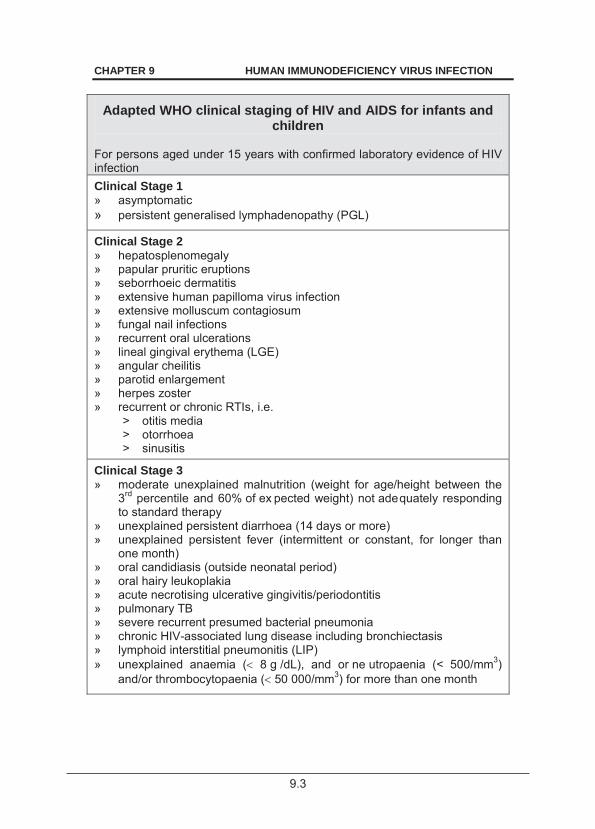
9.3
CHAPTER 9 HUMAN IMMUNODEFICIENCY VIRUS INFECTION
Adapted WHO clinical staging of HIV and AIDS for infants and children
For persons aged under 15 years with confirmed laboratory evidence of HIV infection Clinical Stage 1 » asymptomatic » persistent generalised lymphadenopathy (PGL)
Clinical Stage 2 » hepatosplenomegaly » papular pruritic eruptions » seborrhoeic dermatitis » extensive human papilloma virus infection » extensive molluscum contagiosum » fungal nail infections » recurrent oral ulcerations » lineal gingival erythema (LGE) » angular cheilitis » parotid enlargement » herpes zoster » recurrent or chronic RTIs, i.e.
> otitis media > otorrhoea > sinusitis
Clinical Stage 3 » moderate unexplained malnutrition (weight for age/height between the
3rd percentile and 60% of ex pected weight) not adequately responding to standard therapy
» unexplained persistent diarrhoea (14 days or more) » unexplained persistent fever (intermittent or constant, for longer than
one month) » oral candidiasis (outside neonatal period) » oral hairy leukoplakia » acute necrotising ulcerative gingivitis/periodontitis » pulmonary TB » severe recurrent presumed bacterial pneumonia » chronic HIV-associated lung disease including bronchiectasis » lymphoid interstitial pneumonitis (LIP) » unexplained anaemia ( 8 g /dL), and or ne utropaenia (< 500/mm3)
and/or thrombocytopaenia ( 50 000/mm3) for more than one month
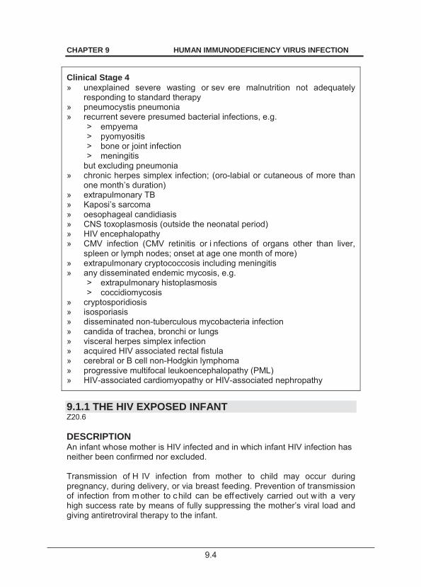
9.4
CHAPTER 9 HUMAN IMMUNODEFICIENCY VIRUS INFECTION
Clinical Stage 4 » unexplained severe wasting or sev ere malnutrition not adequately
responding to standard therapy » pneumocystis pneumonia » recurrent severe presumed bacterial infections, e.g.
> empyema > pyomyositis > bone or joint infection > meningitis
but excluding pneumonia » chronic herpes simplex infection; (oro-labial or cutaneous of more than
one month’s duration) » extrapulmonary TB » Kaposi’s sarcoma » oesophageal candidiasis » CNS toxoplasmosis (outside the neonatal period) » HIV encephalopathy » CMV infection (CMV retinitis or i nfections of organs other than liver,
spleen or lymph nodes; onset at age one month of more) » extrapulmonary cryptococcosis including meningitis » any disseminated endemic mycosis, e.g.
> extrapulmonary histoplasmosis > coccidiomycosis
» cryptosporidiosis » isosporiasis » disseminated non-tuberculous mycobacteria infection » candida of trachea, bronchi or lungs » visceral herpes simplex infection » acquired HIV associated rectal fistula » cerebral or B cell non-Hodgkin lymphoma » progressive multifocal leukoencephalopathy (PML) » HIV-associated cardiomyopathy or HIV-associated nephropathy
9.1.1 THE HIV EXPOSED INFANT Z20.6 DESCRIPTION An infant whose mother is HIV infected and in which infant HIV infection has neither been confirmed nor excluded. Transmission of H IV infection from mother to child may occur during pregnancy, during delivery, or via breast feeding. Prevention of transmission of infection from m other to child can be eff ectively carried out w ith a very high success rate by means of fully suppressing the mother’s viral load and giving antiretroviral therapy to the infant.
9.5
CHAPTER 9 HUMAN IMMUNODEFICIENCY VIRUS INFECTION With the effective use of ant iretrovirals, the risk of HIV transmission through breast feeding is minimised. Where the viral load of the mother cannot be suppressed the risk of breast milk transmission remains significant. The PMTCT plan starts with the mother and may take one of the following routes once she is diagnosed as HIV infected: 1. Pregnant women with HIV infection receive combination antiretroviral
therapy (cART) to suppre ss the HIV viral load to unde tectable levels after which the risk of breast feeding transmission is negligible. cART may have been started before the woman became pregnant or may only be started during pregnancy as soon as the infection is detected. » If the mother is started on cART early in pregnancy (i.e. > 4 weeks
before delivery), and continues while breastfeeding then give antiretroviral prophylaxis (nevirapine (NVP)) to the infant until 6 weeks of age. If DNA PCR is positive in infant 6 weeks of age, start infant cART.
» If the mo ther is started on cART late during pre gnancy (i.e. 4 weeks before d elivery) or at de livery, then g ive antiretroviral prophylaxis (NVP) to the infant until 12 weeks of age. If DNA PCR is positive in infant 6 weeks of age, change infant prophylaxis to cART.
» Mothers who present late and have not received antiretrovirals: o Initiate mother on cART, and giv e antiretroviral prophylaxis
(NVP) to the in fant until 12 weeks of ag e. If DNA PCR is positive in infant 6 weeks of age, change infant prophylaxis to cART.
o If the mother is unable to take cART (known renal disease or active psychiatric disorder) she should be r eferred for ex pert care and for the selection of an appropriate cART. If DNA PCR is positive in infant 6 weeks of age, change infant prophylaxis to cART.
» If the mother has been on cART for life long treatment before the pregnancy, she must have a viral load measurement at 1st booking. o If the viral load is > 1000 copies/mL the cause could be
problems with adherence, or viral resistance. Seek expert advice for the management of the mother and the child.
o If the v iral load is < 1000 copies/mL and t he mother is breastfeeding then give antiretroviral prophylaxis (NVP) to the infant until 6 weeks of age. If DNA PCR is positive in infant 6 weeks of age, change infant prophylaxis to cART.
2. Orphaned or abandoned infants with mother’s HIV status unknown: » See Infant Regimens in table below.
Perform DNA PCR testing on all HIV exposed infants at 6 weeks of age, and if negative, perform an ELISA/rapid test on HIV at 18 months of age.
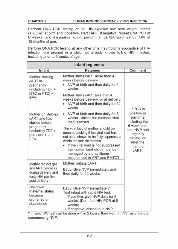
9.6
CHAPTER 9 HUMAN IMMUNODEFICIENCY VIRUS INFECTION
Perform DNA PCR testing on all HIV-exposed low birth weight infants (< 2.5 kg) at birth and if positive, start cART. If negative, repeat DNA PCR at 6 weeks, and if n egative again, perform an EL ISA/rapid test o n HIV at 18 months of age.
Perform DNA PCR testing at any other time if symptoms suggestive of HIV infection are present in a child not already known to b e HIV infected, including prior to 6 weeks of age.
Infant regimens Infant Regimen Comment
Mother starting cART in pregnancy (including TDF + [3TC or FTC] + EFV)
Mother starts cART more than 4 weeks before delivery: NVP at birth and then daily for 6
weeks.
Mother starts cART less than 4 weeks before delivery, or at delivery: NVP at birth and then daily for 12
weeks. If PCR is positive at any time
including the 6 week test,
stop NVP and urgently
initiate, or refer the infant for
cART.
Mother on lifelong cART and has started before pregnancy (including TDF + [3TC or FTC] + EFV)
NVP at birth and then daily for 6 weeks –unless the mothers viral load is raised.
The viral load of mother should be done at booking if the viral load has not been shown to be fully suppressed within the last six months. If the viral load is not suppressed
the mother (and child) must be managed by a practitioner experienced in ART and PMTCT.
Mother did not get any ART before or during delivery and tests HIV positive post delivery
Mother: initiate cART.
Baby: Give NVP immediately and then daily for 12 weeks.
Unknown maternal status because orphaned or abandoned
Baby: Give NVP immediately*. Test infant with rapid HIV test. - If positive, give NVP daily for 6
weeks. (Do infant HIV PCR at 6 weeks).
- If negative, discontinue NVP.
* If rapid HIV test can be done within 2 hours, then wait for HIV result before commencing NVP.

9.7
CHAPTER 9 HUMAN IMMUNODEFICIENCY VIRUS INFECTION Nevirapine (NVP) dose for infant on PMTCT Daily NVP prophylaxis for 6 weeks or 12 weeks – see table above. » Give 1st dose as soon as possible after birth. » Only one dose per 24-hour period; repeat dose once only if baby vomits. » If infant HIV PCR is posi tive at any time, stop NVP, confirm with 2nd
DNA PCR test a nd initiate/refer for cART. Continue normal breastfeeding.
Nevirapine, oral, daily. (Syrup 10 mg/mL)
o Newborns kg and infants:
Age Weight: mg of NVP
mL of NVP syrup 10 mg/mL
Birth to 6 weeks:
2.0–2.5kg: 10 mg 1 mL
> 2.5kg: 15 mg 1.5 mL
6 weeks to 6 months: 20 mg 2 mL
If infant is severely underweight for age (3 SD or 3 z-scores below the mean) give NVP according to weight, (i.e.
4 mg/kg/dose daily) until in the normal weight for age range.
o Premature newborn < 2 kg:
Weight 1st 2 weeks after birth mg of NVP
After 1st 2 weeks after birth
mg of NVP 500 to < 625 g 1 mg 2 mg 625 to < 850 g 1.5 mg 3 mg
850 to < 1 200 g 2 mg 4 mg 1.2 to < 1.5 kg 3 mg 5 mg 1 5 to < 1.9 kg 3.5 mg 6 mg
If infant at time of discharge is severely underweight for age (3 SD or 3 z-scores below the mean) give NVP according to weight, (i.e.
4mg/kg/dose daily) until in the normal weight for age range.
Feeding advice » Exclusive breastfeeding is strongly recommended for the 1st 6 months,
after which the nutritional requirements of the child will require the introduction of complementary foods.
9.8
CHAPTER 9 HUMAN IMMUNODEFICIENCY VIRUS INFECTION » Except where a mother is s hown to be failing cAR T, the advantages of
breastfeeding exceed the risks of HIV transmission in a mother on cART and the mother should be encouraged to breast feed.
» The use of f lash pasteurisation or Pretoria pasteurisation to reduce HIV transmission is sup ported but may pose significant barriers to successful breast milk feeding due to the effort involved.
Co-trimoxazole prophylaxis Indications: » All HIV exposed Infants starting from 4–6 weeks of age. Discontinuation: » If the child is shown to be H IV-uninfected and has not been breastfed
for the last 6 weeks; or » If HIV-infected, the immune system is fully reconstituted and > 1 year of
age (i.e. child 1 to 5 years of age: CD4 > 25%, or child > 5 years of age: CD4 > 350 cells/mm3 on 2 tests at least 3–6 months apart).
Co-trimoxazole (sulfamethoxazole/trimethoprim), oral, once daily (everyday). Recommended daily by weight
band
Dose sulfamethox
azole/ trimethoprim
Suspension 200/40 mg per 5 mL
Single strength
tablet 400/80 mg
Double strength
tablet 800/160 mg
3 to 4.9 kg 100/20 mg 2.5 mL ¼ tablet – 5 to 13.9 kg 200/40 mg 5 mL ½ tablet –
14 to 29.9 kg 400/80 mg 10 mL 1 tablet ½ tablet > 30 kg 800/160 mg – 2 tablets 1 tablet
9.1.2 THE HIV INFECTED INFANT/CHILD B20-24 DESCRIPTION An infant or child in whom HIV infection has been confirmed with 2 appropriate tests. For confirmation of HIV infection see section 9.1: Human immunodeficiency virus infections. GENERAL AND SUPPORTIVE MEASURES Counseling is a vital part of the successful care of children with HIV infection and their families. Specific matters requiring attention are: » The implications of the disease to the family. » Implications of treatment and understanding of the condition and its
care. » The disclosure process within the family and extended family should be
encouraged. Besides the caregiver, help from the family is often useful. » Disclosure to the child of appropriate age and maturity.
9.9
CHAPTER 9 HUMAN IMMUNODEFICIENCY VIRUS INFECTION Treatment of mothers, caregivers and other family members: » Always ask abo ut the caregiver’s health, and the health of ot her
members of the family. » Ensure that mothers and other family members have timeous access to
medical care including cART. » Encourage breastfeeding in all mothers with HIV infected children, with
introduction of weaning foods from 6 months of age. » Always ask at ev ery visit about TB contacts and T B symptoms in all
children and their caregivers. STANDARDISED NATIONAL MONITORING FOR INFANTS AND CHILDREN WITH HIV
At initial diagnosis of HIV Purpose
Verify HIV status To ensure that national testing algorithm has been followed.
Document weight, height, head circumference (< 2 years of age) and development
To monitor growth and development.
Screen for TB symptoms To identify TB and HIV co-infected.
Do CD4 count Children < 5 years of age: Baseline. Do not wait for CD4 count to start ART.
of age: To determine eligibility for ART.
Hb or FBC if available To detect anaemia or neutropenia.
At routine follow-up visits (patients not yet on ART)
Purpose
Document weight, height, head circumference (< 2 years of age) and development.
To monitor growth and development.
Check that a CD4 count has been done in the last 6 months.
To determine if patient has become eligible for ART.
If > 5 years of age WHO clinical staging.
To determine if patient has become eligible for ART.
Screen for TB symptoms. To identify TB/HIV co-infection.
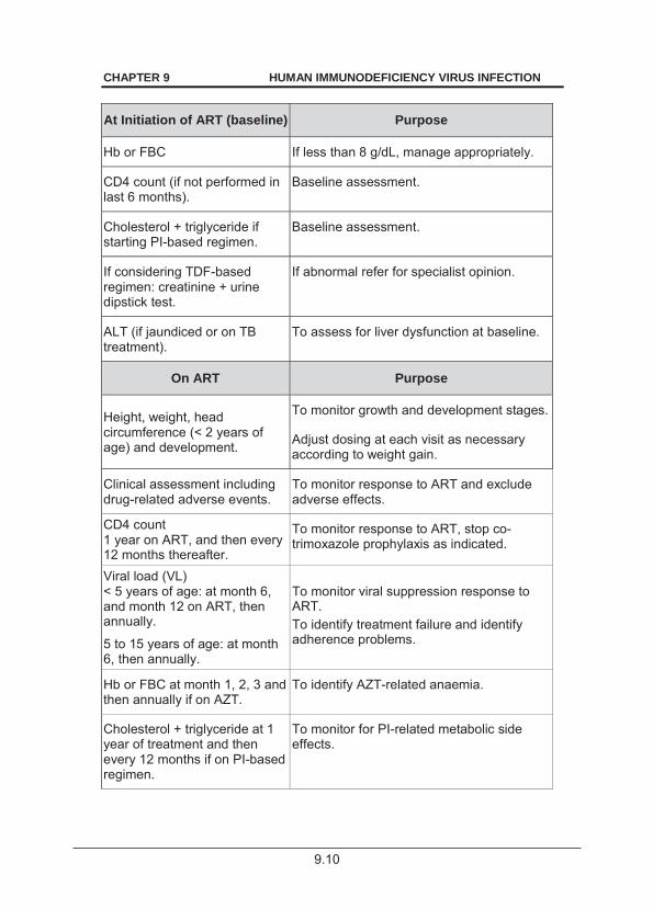
9.10
CHAPTER 9 HUMAN IMMUNODEFICIENCY VIRUS INFECTION
At Initiation of ART (baseline) Purpose
Hb or FBC If less than 8 g/dL, manage appropriately.
CD4 count (if not performed in last 6 months).
Baseline assessment.
Cholesterol + triglyceride if starting PI-based regimen.
Baseline assessment.
If considering TDF-based regimen: creatinine + urine dipstick test.
If abnormal refer for specialist opinion.
ALT (if jaundiced or on TB treatment).
To assess for liver dysfunction at baseline.
On ART Purpose
Height, weight, head circumference (< 2 years of age) and development.
To monitor growth and development stages.
Adjust dosing at each visit as necessary according to weight gain.
Clinical assessment including drug-related adverse events.
To monitor response to ART and exclude adverse effects.
CD4 count 1 year on ART, and then every 12 months thereafter.
To monitor response to ART, stop co-trimoxazole prophylaxis as indicated.
Viral load (VL) < 5 years of age: at month 6, and month 12 on ART, then annually.
5 to 15 years of age: at month 6, then annually.
To monitor viral suppression response to ART. To identify treatment failure and identify adherence problems.
Hb or FBC at month 1, 2, 3 and then annually if on AZT.
To identify AZT-related anaemia.
Cholesterol + triglyceride at 1 year of treatment and then every 12 months if on PI-based regimen.
To monitor for PI-related metabolic side effects.
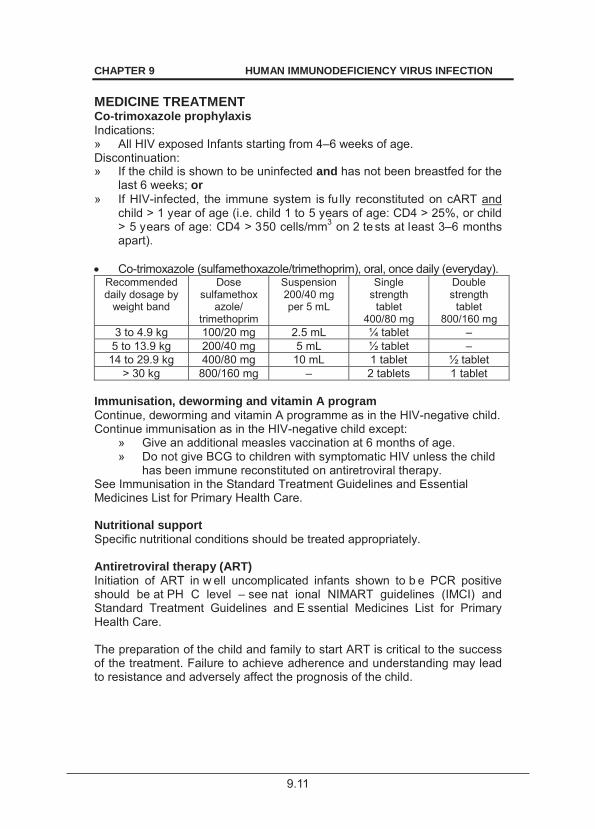
9.11
CHAPTER 9 HUMAN IMMUNODEFICIENCY VIRUS INFECTION MEDICINE TREATMENT Co-trimoxazole prophylaxis Indications: » All HIV exposed Infants starting from 4–6 weeks of age. Discontinuation: » If the child is shown to be uninfected and has not been breastfed for the
last 6 weeks; or » If HIV-infected, the immune system is fu lly reconstituted on cART and
child > 1 year of age (i.e. child 1 to 5 years of age: CD4 > 25%, or child > 5 years of age: CD4 > 350 cells/mm3 on 2 te sts at least 3–6 months apart).
Co-trimoxazole (sulfamethoxazole/trimethoprim), oral, once daily (everyday). Recommended daily dosage by
weight band
Dose sulfamethox
azole/ trimethoprim
Suspension 200/40 mg per 5 mL
Single strength
tablet 400/80 mg
Double strength
tablet 800/160 mg
3 to 4.9 kg 100/20 mg 2.5 mL ¼ tablet – 5 to 13.9 kg 200/40 mg 5 mL ½ tablet –
14 to 29.9 kg 400/80 mg 10 mL 1 tablet ½ tablet > 30 kg 800/160 mg – 2 tablets 1 tablet
Immunisation, deworming and vitamin A program Continue, deworming and vitamin A programme as in the HIV-negative child. Continue immunisation as in the HIV-negative child except:
» Give an additional measles vaccination at 6 months of age. » Do not give BCG to children with symptomatic HIV unless the child
has been immune reconstituted on antiretroviral therapy. See Immunisation in the Standard Treatment Guidelines and Essential Medicines List for Primary Health Care. Nutritional support Specific nutritional conditions should be treated appropriately. Antiretroviral therapy (ART) Initiation of ART in w ell uncomplicated infants shown to b e PCR positive should be at PH C level – see nat ional NIMART guidelines (IMCI) and Standard Treatment Guidelines and E ssential Medicines List for Primary Health Care. The preparation of the child and family to start ART is critical to the success of the treatment. Failure to achieve adherence and understanding may lead to resistance and adversely affect the prognosis of the child.
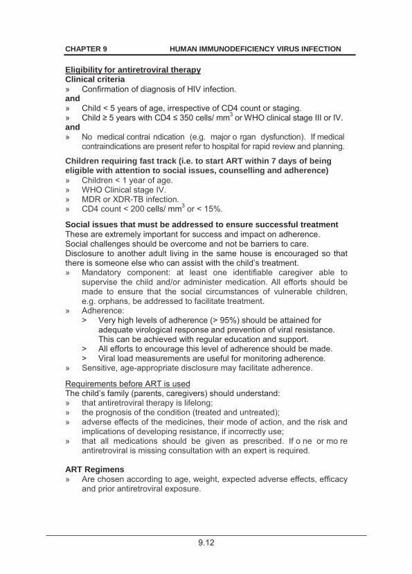
9.12
CHAPTER 9 HUMAN IMMUNODEFICIENCY VIRUS INFECTION Eligibility for antiretroviral therapy Clinical criteria » Confirmation of diagnosis of HIV infection. and » Child < 5 years of age, irrespective of CD4 count or staging. » 3 or WHO clinical stage III or IV. and » No medical contrai ndication (e.g. major o rgan dysfunction). If medical
contraindications are present refer to hospital for rapid review and planning.
Children requiring fast track (i.e. to start ART within 7 days of being eligible with attention to social issues, counselling and adherence) » Children < 1 year of age. » WHO Clinical stage IV. » MDR or XDR-TB infection. » CD4 count < 200 cells/ mm3 or < 15%. Social issues that must be addressed to ensure successful treatment These are extremely important for success and impact on adherence. Social challenges should be overcome and not be barriers to care. Disclosure to another adult living in the same house is encouraged so that there is someone else who can assist with the child’s treatment. » Mandatory component: at least one identifiable caregiver able to
supervise the child and/or administer medication. All efforts should be made to ensure that the social circumstances of vulnerable children, e.g. orphans, be addressed to facilitate treatment.
» Adherence: > Very high levels of adherence (> 95%) should be attained for
adequate virological response and prevention of viral resistance. This can be achieved with regular education and support.
> All efforts to encourage this level of adherence should be made. > Viral load measurements are useful for monitoring adherence.
» Sensitive, age-appropriate disclosure may facilitate adherence. Requirements before ART is used The child’s family (parents, caregivers) should understand: » that antiretroviral therapy is lifelong; » the prognosis of the condition (treated and untreated); » adverse effects of the medicines, their mode of action, and the risk and
implications of developing resistance, if incorrectly use; » that all medications should be given as prescribed. If o ne or mo re
antiretroviral is missing consultation with an expert is required. ART Regimens » Are chosen according to age, weight, expected adverse effects, efficacy
and prior antiretroviral exposure.
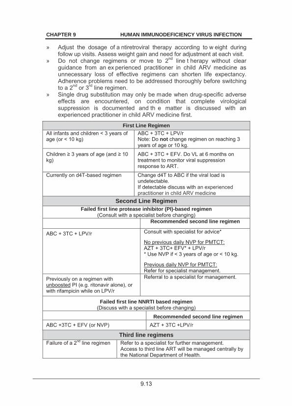
9.13
CHAPTER 9 HUMAN IMMUNODEFICIENCY VIRUS INFECTION
» Adjust the dosage of a ntiretroviral therapy according to w eight during follow up visits. Assess weight gain and need for adjustment at each visit.
» Do not change regimens or move to 2nd line t herapy without clear guidance from an ex perienced practitioner in child ARV medicine as unnecessary loss of effective regimens can shorten life expectancy. Adherence problems need to be addressed thoroughly before switching to a 2nd or 3rd line regimen.
» Single drug substitution may only be made when drug-specific adverse effects are encountered, on condition that complete virological suppression is documented and th e matter is discussed with an experienced practitioner in child ARV medicine first.
First Line Regimen All infants and children < 3 years of age (or < 10 kg)
ABC + 3TC + LPV/r Note: Do not change regimen on reaching 3 years of age or 10 kg.
of age (and kg)
ABC + 3TC + EFV. Do VL at 6 months on treatment to monitor viral suppression response to ART.
Currently on d4T-based regimen Change d4T to ABC if the viral load is undetectable. If detectable discuss with an experienced practitioner in child ARV medicine
Second Line Regimen Failed first line protease inhibitor (PI)-based regimen
(Consult with a specialist before changing) Recommended second line regimen
ABC + 3TC + LPV/r Consult with specialist for advice* No previous daily NVP for PMTCT: AZT + 3TC+ EFV* + LPV/r * Use NVP if < 3 years of age or < 10 kg. Previous daily NVP for PMTCT: Refer for specialist management.
Previously on a regimen with unboosted PI (e.g. ritonavir alone), or with rifampicin while on LPV/r
Referral to a specialist for management.
Failed first line NNRTI based regimen (Discuss with a specialist before changing)
Recommended second line regimen ABC +3TC + EFV (or NVP) AZT + 3TC +LPV/r
Third line regimens Failure of a 2nd line regimen Refer to a specialist for further management.
Access to third line ART will be managed centrally by the National Department of Health.
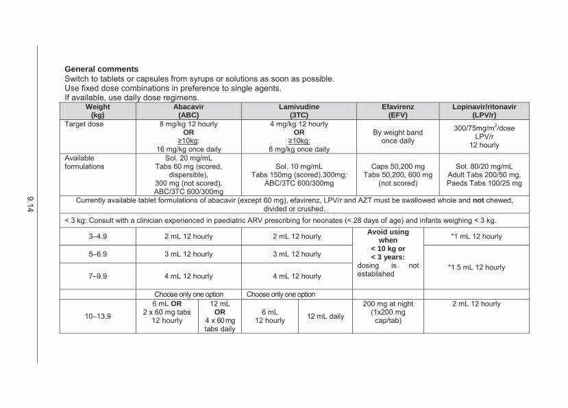
9.14
General comments Switch to tablets or capsules from syrups or solutions as soon as possible. Use fixed dose combinations in preference to single agents. If available, use daily dose regimens.
Weight (kg)
Abacavir (ABC)
Lamivudine (3TC)
Efavirenz (EFV)
Lopinavir/ritonavir (LPV/r)
Target dose 8 mg/kg 12 hourly OR
: 16 mg/kg once daily
4 mg/kg 12 hourly OR
8 mg/kg once daily
By weight band once daily
300/75mg/m2/dose LPV/r
12 hourly
Available formulations
Sol. 20 mg/mL Tabs 60 mg (scored,
dispersible), 300 mg (not scored), ABC/3TC 600/300mg
Sol. 10 mg/mL Tabs 150mg (scored),300mg;
ABC/3TC 600/300mg
Caps 50,200 mg Tabs 50,200, 600 mg
(not scored)
Sol. 80/20 mg/mL Adult Tabs 200/50 mg, Paeds Tabs 100/25 mg
Currently available tablet formulations of abacavir (except 60 mg), efavirenz, LPV/r and AZT must be swallowed whole and not chewed, divided or crushed.
< 3 kg: Consult with a clinician experienced in paediatric ARV prescribing for neonates (< 28 days of age) and infants weighing < 3 kg.
3–4.9 2 mL 12 hourly 2 mL 12 hourly Avoid using when
< 10 kg or < 3 years:
dosing is not established
*1 mL 12 hourly
5–6.9 3 mL 12 hourly 3 mL 12 hourly
*1.5 mL 12 hourly 7–9.9 4 mL 12 hourly 4 mL 12 hourly
Choose only one option Choose only one option
10–13.9
6 mL OR 2 x 60 mg tabs
12 hourly
12 mL OR
4 x 60 mg tabs daily
6 mL 12 hourly 12 mL daily
200 mg at night (1x200 mg
cap/tab)
2 mL 12 hourly
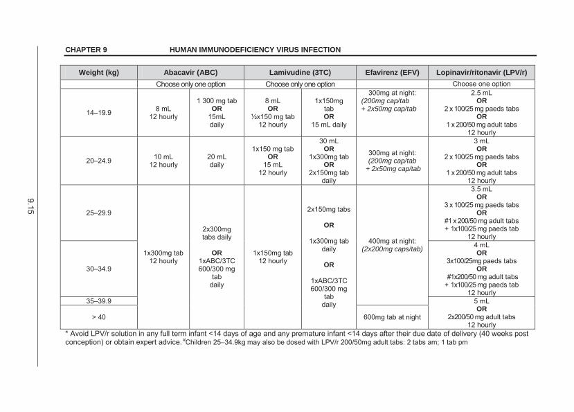
9.15
CHAPTER 9 HUMAN IMMUNODEFICIENCY VIRUS INFECTION
Weight (kg) Abacavir (ABC) Lamivudine (3TC) Efavirenz (EFV) Lopinavir/ritonavir (LPV/r) Choose only one option Choose only one option Choose one option
14–19.9 8 mL 12 hourly
1 300 mg tab OR
15mL daily
8 mL OR
½x150 mg tab 12 hourly
1x150mg tab OR
15 mL daily
300mg at night: (200mg cap/tab + 2x50mg cap/tab
2.5 mL OR
2 x 100/25 mg paeds tabs OR
1 x 200/50 mg adult tabs 12 hourly
20–24.9 10 mL 12 hourly
20 mL daily
1x150 mg tab OR
15 mL 12 hourly
30 mL OR
1x300mg tab OR
2x150mg tab daily
300mg at night: (200mg cap/tab
+ 2x50mg cap/tab
3 mL OR
2 x 100/25 mg paeds tabs OR
1 x 200/50 mg adult tabs 12 hourly
25–29.9
1x300mg tab 12 hourly
2x300mg tabs daily
OR
1xABC/3TC 600/300 mg
tab daily
1x150mg tab 12 hourly
2x150mg tabs
OR
1x300mg tab daily
OR
1xABC/3TC 600/300 mg
tab daily
400mg at night: (2x200mg caps/tab)
3.5 mL OR
3 x 100/25 mg paeds tabs OR
#1 x 200/50 mg adult tabs + 1x100/25 mg paeds tab
12 hourly
30–34.9
4 mL OR
3x100/25mg paeds tabs OR
#1x200/50 mg adult tabs + 1x100/25 mg paeds tab
12 hourly 35–39.9 5 mL
OR 2x200/50 mg adult tabs
12 hourly > 40 600mg tab at night
* Avoid LPV/r solution in any full term infant <14 days of age and any premature infant <14 days after their due date of delivery (40 weeks post conception) or obtain expert advice. #Children 25–34.9kg may also be dosed with LPV/r 200/50mg adult tabs: 2 tabs am; 1 tab pm
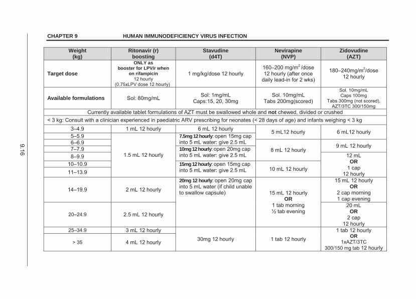
9.16
CHAPTER 9 HUMAN IMMUNODEFICIENCY VIRUS INFECTION
Weight (kg)
Ritonavir (r) boosting
Stavudine (d4T)
Nevirapine (NVP)
Zidovudine (AZT)
Target dose
ONLY as booster for LPV/r when
on rifampicin 12 hourly
(0.75xLPV dose 12 hourly)
1 mg/kg/dose 12 hourly 160–200 mg/m2 /dose 12 hourly (after once
daily lead-in for 2 wks)
180–240mg/m2/dose 12 hourly
Available formulations Sol: 80mg/mL Sol: 1mg/mL Caps:15, 20, 30mg
Sol. 10mg/mL Tabs 200mg(scored)
Sol. 10mg/mL Caps 100mg
Tabs 300mg (not scored), AZT/3TC 300/150mg
Currently available tablet formulations of AZT must be swallowed whole and not chewed, divided or crushed < 3 kg: Consult with a clinician experienced in paediatric ARV prescribing for neonates (< 28 days of age) and infants weighing < 3 kg
3–4.9 1 mL 12 hourly 6 mL 12 hourly 5 mL12 hourly 6 mL12 hourly 5–5.9
1.5 mL 12 hourly
7.5mg 12 hourly: open 15mg cap into 5 mL water: give 2.5 mL 6–6.9
8 mL 12 hourly 9 mL 12 hourly 7–7.9 10mg 12 hourly: open 20mg cap
into 5 mL water: give 2.5 mL 8–9.9 12 mL OR
1 cap 12 hourly
10–10.9 15mg 12 hourly: open 15mg cap into 5 mL water: give 2.5 mL 10 mL 12 hourly 11–13.9
14–19.9 2 mL 12 hourly
20mg 12 hourly: open 20mg cap into 5 mL water (if child unable to swallow capsule) 15 mL 12 hourly
OR 1 tab morning ½ tab evening
15 mL 12 hourly OR
2 cap morning 1 cap evening
20–24.9 2.5 mL 12 hourly
20 mL OR
2 cap 12 hourly
25–34.9 3 mL 12 hourly 30mg 12 hourly 1 tab 12 hourly
1 tab 12 hourly OR
1xAZT/3TC 300/150 mg tab 12 hourly
> 35 4 mL 12 hourly
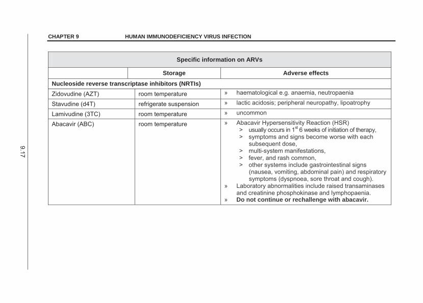
9.17
CHAPTER 9 HUMAN IMMUNODEFICIENCY VIRUS INFECTION
Specific information on ARVs
Storage Adverse effects
Nucleoside reverse transcriptase inhibitors (NRTIs) Zidovudine (AZT) room temperature » haematological e.g. anaemia, neutropaenia
Stavudine (d4T) refrigerate suspension » lactic acidosis; peripheral neuropathy, lipoatrophy
Lamivudine (3TC) room temperature » uncommon
Abacavir (ABC) room temperature » Abacavir Hypersensitivity Reaction (HSR) > usually occurs in 1st 6 weeks of initiation of therapy, > symptoms and signs become worse with each
subsequent dose, > multi-system manifestations, > fever, and rash common, > other systems include gastrointestinal signs
(nausea, vomiting, abdominal pain) and respiratory symptoms (dyspnoea, sore throat and cough).
» Laboratory abnormalities include raised transaminases and creatinine phosphokinase and lymphopaenia.
» Do not continue or rechallenge with abacavir.
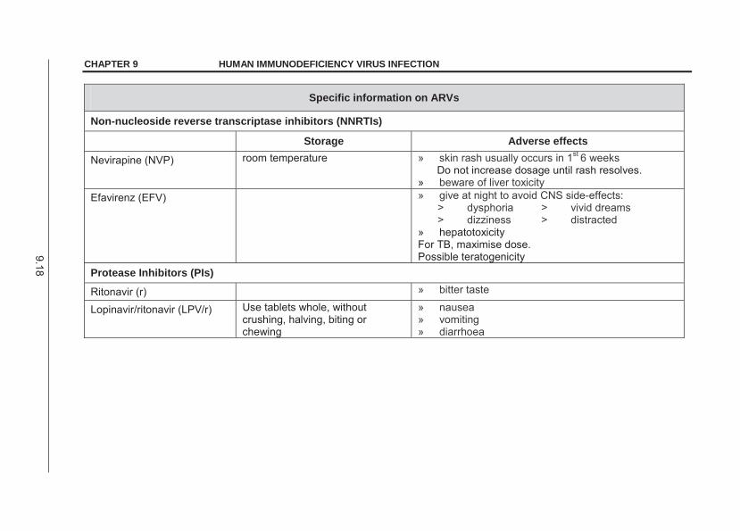
9.18
CHAPTER 9 HUMAN IMMUNODEFICIENCY VIRUS INFECTION
Specific information on ARVs
Non-nucleoside reverse transcriptase inhibitors (NNRTIs)
Storage Adverse effects
Nevirapine (NVP) room temperature » skin rash usually occurs in 1st 6 weeks Do not increase dosage until rash resolves.
» beware of liver toxicity Efavirenz (EFV) » give at night to avoid CNS side-effects:
> dysphoria > vivid dreams > dizziness > distracted
» hepatotoxicity For TB, maximise dose. Possible teratogenicity
Protease Inhibitors (PIs)
Ritonavir (r) » bitter taste
Lopinavir/ritonavir (LPV/r) Use tablets whole, without crushing, halving, biting or chewing
» nausea » vomiting » diarrhoea
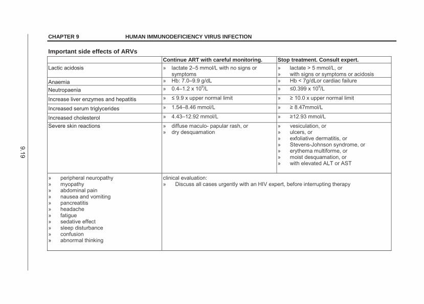
9.19
CHAPTER 9 HUMAN IMMUNODEFICIENCY VIRUS INFECTION
Important side effects of ARVs Continue ART with careful monitoring. Stop treatment. Consult expert. Lactic acidosis » lactate 2–5 mmol/L with no signs or
symptoms » lactate > 5 mmol/L, or » with signs or symptoms or acidosis
Anaemia » Hb: 7.0–9.9 g/dL » Hb < 7g/dLor cardiac failure
Neutropaenia » 0.4–1.2 x 10⁹/L » 9/L
Increase liver enzymes and hepatitis » »
Increased serum triglycerides » 1.54–8.46 mmol/L »
Increased cholesterol » 4.43–12.92 mmol/L »
Severe skin reactions » diffuse maculo- papular rash, or » dry desquamation
» vesiculation, or » ulcers, or » exfoliative dermatitis, or » Stevens-Johnson syndrome, or » erythema multiforme, or » moist desquamation, or » with elevated ALT or AST
» peripheral neuropathy » myopathy » abdominal pain » nausea and vomiting » pancreatitis » headache » fatigue » sedative effect » sleep disturbance » confusion » abnormal thinking
clinical evaluation: » Discuss all cases urgently with an HIV expert, before interrupting therapy
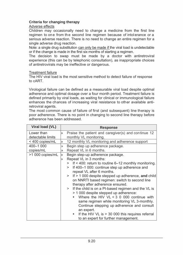
9.20
Criteria for changing therapy Adverse effects Children may occasionally need to change a medicine from the first line regimen to one from the second line regimen because of intol erance or a serious adverse reaction. There is no need to change an entire regimen for a single adverse drug reaction. Note: a single drug substitution can only be made if the viral load is undetectable or if the change is made in the first six months of starting a regimen. The decision to swap must be made by a doctor with antiretroviral experience (this can be by telephonic consultation), as inappropriate choices of antiretrovirals may be ineffective or dangerous. Treatment failure The HIV viral load is the most sensitive method to detect failure of response to cART. Virological failure can be defined as a measurable viral load despite optimal adherence and optimal dosage over a four month period. Treatment failure is defined primarily by viral loads, as waiting for clinical or immunological failure enhances the chances of increasing viral resistance to other available anti-retroviral agents. The most common cause of failure of first (and subsequent) line therapy is poor adherence. There is no point in changing to second line therapy before adherence has been addressed.
Viral load (VL) Response Lower than detectable limits
» Praise the patient and caregiver(s) and continue 12 monthly VL monitoring.
< 400 copies/mL » 12 monthly VL monitoring and adherence support 400–1 000 copies/mL
» Begin step up adherence package. » Repeat VL in 6 months.
>1 000 copies/mL » Begin step-up adherence package. » Repeat VL in 3 months:
> If < 400: return to routine 6–12 monthly monitoring. > If 400–1 000: continue step up adherence and
repeat VL after 6 months. > If > 1 000 despite stepped up adherence, and child
on NNRTI based regimen: switch to second line therapy after adherence ensured.
> If the child is on a PI-based regimen and the VL is > 1 000 despite stepped up adherence: Where the HIV VL < 3 0 000 continue with
same regimen while monitoring VL 3-monthly. Continue stepping up adherence and consult an expert.
If the HIV VL is > 30 000 this requires referral to an expert for further management.
9.21
CHAPTER 9 HUMAN IMMUNODEFICIENCY VIRUS INFECTION REFERRAL » Complicated or v ery ill children should be referred to a practitioner
skilled in the care of such children. » Attempts should be made to refer patients to accredited primary health
care sites once stable on ART.
9.2 TUBERCULOSIS AND HIV B20.0 DESCRIPTION TB and H IV are often c o-morbid conditions. Exclude TB by clini cal examination, chest X-ray, tuberculin skin test (TST), M. tuberculosis PCR test and mycobacterial culture (where TB disease is suspected on clinical or radiological grounds) in all patients before starting ART. Re-evaluate the risk for TB and TB contact at each visit on history (including contact history) and clinical examination. MEDICINE TREATMENT TB prophylaxis Give TB prophylaxis to a ll children exposed to a close contact with an infectious pulmonary TB case (sputum microscopy smear-positive, culture-positive or M. tuberculosis PCR test positive), or who are TST positive, but in whom no evidence of TB disease is present. Isoniazid, oral, 10 mg/kg/dose once daily for 6 months.
o Maximum dose: 300 mg daily. Repeat course if an HIV-infected patient, irrespective of age, is re-exposed to a TB contact at any point after completing TB treatment or prophylaxis. If patient has been exposed to a known MDR or XDR-TB source case or the contact case has failed standard TB treatment, refer for expert opinion. See section 15.1.4: Tuberculosis, pulmonary. TB treatment If the child is not yet on ART: » Commence TB treatment first. Follow with cART, usually after 2–8 weeks.
> After 2 weeks if CD 4 < 50 cells/mm3. > After 8 weeks if CD 4 > 50 cells/mm3.
» Check ALT before commencing cART. If the A LT is raised discuss this with an e xpert as it may not be an ab solute contraindication to treatment.
» Be aware of the possi bility of Immu ne Reconstitution Inflammatory Syndrome (IRIS).
9.22
CHAPTER 9 HUMAN IMMUNODEFICIENCY VIRUS INFECTION If the child is already on ART: » Commence TB treatment taking into consideration possible drug
interactions. If the child needs to take concomitant ART and rifampicin-containing treatment: » Efavirenz: use the maximum dosage according to recommended range
as per package insert of either drug for weight or body surface area. » Abacavir and lamivudine: no adjustment of dosages. » Lopinavir/ritonavir: provide additional ritonavir while on rifampicin-
containing treatment (add sufficient ritonavir to ensure an equal dose in milligrams of lopinavir and ritonavir). For example for e ach mL of lopinavir/ritonavir solution (80/20 mg/mL) add 0.75 mL of r itonavir solution (80 mg/mL).
» Give pyridoxine (vitamin B6) to all ch ildren on TB and ARV treatment, due to shared toxicities of the regimens.
9.3 SPECIFIC ADVERSE EVENTS AND COMPLICATIONS 9.3.1 LACTIC ACIDOSIS E87.2 DESCRIPTION All nucleoside analogues have been associated with lactic acidosis which is rare but l ife threatening. Initial symptoms are v ariable and usually occur between 1–20 months (median 4 months) after starting therapy. The propensity to develop lactic acidosis is h ighest with stavudine, followed by didanosine and then zidovudine. DIAGNOSTIC CRITERIA Clinical Clinical prodromal syndrome: » Generalised fatigue. » Weakness and myalgia. » Gastrointestinal symptoms:
> nausea, > vague abdominal pain, > vomiting, > hepatomegaly, > diarrhoea, > anorexia, > and/or sudden unexplained weight loss.
» Respiratory symptoms: tachypnoea and dyspnoea. » Neurologic symptoms, including motor weakness.

9.23
CHAPTER 9 HUMAN IMMUNODEFICIENCY VIRUS INFECTION Investigations » Laboratory abnormalities:
> Hyperlactataemia: chronic, asymptomatic: 2.1–5 mmol/L. symptomatic severe: > 5 mmol/L.
> Exclude other causes of hyperlactataemia/acidosis. > Lactic acidosis is defined by:
lactate > 5 mmol/L, bicarbonate < 20 mmol/L, severe acidosis, i.e. pH < 7.3, increased anion gap, associated symptoms, precipitating events: (manage each appropriately)
dehydration, sepsis, cardiac failure, severe anaemia.
TREATMENT Lactate 2.1–5.0 mmol/L confirmed and asymptomatic Consider replacing stavudine and didanosine if used with other ARVs. Consider temporary discontinuation all ART while assessing. (When stopping ART, if r egimen contained nevirapine or efavirenz, use lopinavir/ritonavir (300 mg/m2) for 2 weeks to cover the prolonged half life of NNRTIs). Lactate > 5.0mmol/L confirmed, or a single test > 10mm0l/L Stop all ARVs. (When stopping ART, if reg imen contained nevirapine or efavirenz, use lopinavir/ritonavir (300 mg/m2) for 2 w eeks to cover the prolonged half life of NNRTI’s, while getting expert advice.) Provide supportive care, i.e. oxygen (if needed) and IV fluids. In patient with symptomatic hyperlactataemia and/or a blood lactate of > 5 mmol/L: » Stop all ARVs. (When stopping ART, if regimen contained nevirapine or
efavirenz, use lopinavir/ritonavir (300 mg/m2) for 2 weeks to cover the prolonged half life of NNRTIs).
» Consult a specialist for guidance. » Treatment is primarily supportive. » Ensure adequate fluid therapy. » Consider use of bicarbonate to half correct acidosis if severe metabolic
acidosis. » Consider use of: Thiamine, oral, 100 mg daily.
9.24
CHAPTER 9 HUMAN IMMUNODEFICIENCY VIRUS INFECTION REFERRAL » For adjustment of regimen. » For care if beyond capacity of local institution. 9.3.2 LIPODYSTROPHY E88.1 DESCRIPTION Both lipoatrophy and lipohypertrophy can occur as a complication or association of cART. The risk factors include virologic response to therapy and pubertal development during protease inhibitor therapy. Lipodystrophy contributes to non-adherence to AR T as a patient may be embarrassed by his/her physical appearance. Stavudine, didanosine and zidovudine in decreasing order are t he main causes of lipoatrophy. The relationship between lipohypertrophy, hypercholesterolaemia, hypertriglyceridaemia, insulin resistance with puberty, body habitus and ART (especially protease inhibitors), is less clear but an association has been described. DIAGNOSTIC CRITERIA » Lipoatrophy:
> Subcutaneous fat loss (lipoatrophy) of the face, extremities or buttocks.
» Lipohypertrophy: > Fat accumulation (lipohypertrophy) in the abdomen, or over the
dorsocervical spine (buffalo hump) and breast enlargement. > Excessive breast enlargement during puberty (lipomastia).
» Insulin resistance may be suspected if there is: > fasting hyperglycaemia, > frank diabetes or acanthosis nigricans, > biochemical features include an elevated fasting C-peptide or an
abnormal glucose/insulin ratio. » Abnormal lipid profile: See section 4.10: Dyslipidaemia.
> hypercholesterolaemia, i.e. total cholesterol level > 5 mmol/L; and > hypertriglyceridaemia, i.e. fasting triglyceride level > 1.7 mmol/L
with possible consequences of premature atherosclerosis.
GENERAL AND SUPPORTIVE MEASURES » Dietary modification and exercise for li pohypertrophy, insulin resistance and
abnormal lipid profile.
9.25
CHAPTER 9 HUMAN IMMUNODEFICIENCY VIRUS INFECTION MEDICINE TREATMENT Modification of ART, e.g. replace stavudine with abacavir in lipoatrophy. Note: Viral suppression must be present for a single drug substitution. If viral suppression is not present obtain expert advice. If hyperlipidaemia is confirmed, use lipid lowering agents. REFERRAL » All patients for confirmation of diagnosis and initiation of therapy. See section 7.16: Obesity. 9.3.3 IMMUNE RECONSTITUTION INFLAMMATORY
SYNDROME (IRIS) D89.3 DESCRIPTION Clinical deterioration can occur after starting ART due an improvement in the immune system response to organisms already causing infection, e.g.
M bovis BCG, C. neoformans, M. tuberculosis (MTB), Aspergillus, M. avium complex, C. albicans, M. leprae, Human Herpes viruses, P. jiroveci, Human Papilloma virus, CMV, Hepatitis B and C viruses (HBV, HCV), JC virus.
There are 2 manifestations of IRIS: 1. Unmasking occurs when a previously unsuspected condition manifests. 2. Paradoxical, i.e. known condition on appropriate treatment becomes
worse. DIAGNOSTIC CRITERIA » Exclude other active or inadequately treated diseases (including MDR TB). » Presentation:
> Usually during the first 6 weeks after starting ART. > Clinical presentation depends on the cau sative organism and the
organ-system involved, e.g. TB presents with fever, lymphadenopathy, worsening of the orig inal tuberculous lesion, and/or deteriorating chest radiographic manifestations such as miliary pattern or pleural effusion.
9.26
CHAPTER 9 HUMAN IMMUNODEFICIENCY VIRUS INFECTION TREATMENT Treat underlying disease aggressively.
Antimicrobial therapy for specific infections. In severe reactions: Hydrocortisone, IV, 3–6 mg/kg/dose 3–6 hourly on first day.
Follow with: Prednisone, oral, 1–2 mg/kg/dose 24 hourly for at least 7 days.
Continue cART, and manage underlying condition. 9.3.4 WASTING SYNDROME B22.2 This syndrome appears to be a combination of the direct effects of advanced HIV infection and the occurrence of opportunistic infections. TREATMENT Nutritional advice. See section 2.4: Malnutrition. cART may reverse some of the features of HIV wasting syndrome. Exclude chronic infection, e.g. tuberculosis and M. avium complex, malabsorption and malignancy. 9.4 POST EXPOSURE PROPHYLAXIS FOLLOWING
ALLEGED PENETRATIVE SEXUAL ABUSE See Standard Treatment Guidelines and Essential Medicines List for Primary Health Care. 9.5 HIV IN ADOLESCENCE B20-24 DESCRIPTION Adolescence encompasses the period of physical and psychological development from the onset of puberty to maturity. HIV in adolescents may be due to: 1. Vertical infection in infancy that presents as long term non-progressors;
or 2. Sexually acquired HIV from unprotected intercourse. Increasing numbers of perinatally infected infants are surviving to adolescence.
9.27
CHAPTER 9 HUMAN IMMUNODEFICIENCY VIRUS INFECTION Adolescence is a high risk period for non-adherence to therapy. Mood disorders, denial, peer pressure, self-esteem and suicide risk are more common and patients may need to be referred for psychological support. Education about sexual and reproductive health should be commenced early. Every encounter with the adolescent needs to be maximally utilised to discuss condom and contraception use to protect against unplanned pregnancies and STI transmission including HIV is essential. Schools should be taking an active role in this education. Sexually active youth need to be screened for STI symptoms and managed appropriately. Consent For testing and treatment, the current acts and regulations should be followed. Disclosure All adolescents need to be aware of their HIV status. This should be handled sensitively. In addition, disclosure of diagnosis has ramifications for adherence. Disclosure should be p lanned with the caregiver and u sually takes place over 2–3 visits. Intervention by a so cial worker is useful. Determine what the adolescent already knows and discuss with the caregiver about who should disclose and where. Dosage of ARVs In children over the age of 15 years and over 40 kg use adult dosage regimes – consult ART guideline. Medicines are not routinely switched based on age. Contraception in HIV-positive adolescents on ART
Hormonal contraceptives and IUCDs do not prevent sexually transmitted infections.
Additional use of condoms is required. IUCD
HIV is not a contraindication to IUCD use and may be used in adolescents on ART. 380mm2 copper – standard type.
Progestogen-only subdermal implant contraceptive e.g.: Levonorgestrel, 150mg, subdermal two-rod implant.
9.28
CHAPTER 9 HUMAN IMMUNODEFICIENCY VIRUS INFECTION Injectable contraception: e.g. Medroxyprogesterone acetate (long-acting), IM, 150 mg, 12 weekly.
Note: It is unnecessary to shorten the dosage interval for women taking concomitant enzyme-inducing drugs, e .g. rifampicin, antiretrovirals and anticonvulsants.
Combined oral contraceptives (COCs) are indicated for motivated patients where adherence is more likely but are associated with drug-drug interactions.
10.1
CHAPTER 10 SURGICAL PROPHYLAXIS
DESCRIPTION Surgical prophylaxis is th e intra-operative administration of antibiotics to patients to reduce the risk of postoperative wound infection. Specific epidemiological considerations may alter the choice of agents. PRINCIPLES OF SURGICAL PROPHYLAXIS » The need for prophylactic antibiotic therapy is based on the risk of wound
contamination. » The medication chosen should be active against the pathogens most
likely to be associated with wound infections. » Prophylaxis must be given within 30 minutes of the f irst incision, usually
at induction of anaesthesia. Risk factors for developing surgical site infection Classification of degree of contamination likely to b e present during operation: » Class I: C lean procedures, only microorganisms from skin or e xternal
environment are likely to b e introduced (includes operation for blunt trauma).
» Class II: Clean procedures with limited contamination, exposure to micro-organisms colonising the epithelial surfaces and/or lumen of respiratory, gastrointestinal, urinary or genital tract. No evidence of infection.
» Class III: Contaminated, open fresh accidental wounds, operations with major breaks (e.g. open cardiac massage or gr oss spillage from gastrointestinal tract) and incisions in which non-purulent inflammation is encountered.
» Class IV: Dirty and/or infected surgical site indicates that the organism causing postoperative infection was in the operation area before surgery, traumatic wounds with devitalised tissue not immediately attended to, and wounds that involve existing clinical infection or perforated viscera.
These guidelines cover prophylaxis and not therapy for infective conditions. Other risk factors are: » Prolonged duration of operation. » Medical characteristics of the patient (nutritional status, immunosuppression
and co-existent infection at remote body site). Consider antibiotic prophylaxis for class II and if these other risk factors are present.
10.2
CHAPTER 10 SURGICAL PROPHYLAXIS Recommendations for prophylaxis of surgery are limited to class II. For class III and IV, antibiotics are indicated for therapy rather than single dose prophylaxis. Additional procedures for which antibiotic prophylaxis is required include the following: » Head and neck: CSF shunt and grommet insertion. » Cardiothoracic: cardiac pacemaker insertion, interventional cardiac
catheter device placement. » Gastrointestinal: insertion of percutaneous endoscopic gastrostomy.
The prophylactic dose is a single dose equal to the standard therapeutic dose.
A second dose is ONLY given if surgery is prolonged, i.e. > 4 hours for cefazolin OR > 8 hours for metronidazole.
ANTIBIOTIC TREATMENT Cefazolin, IV, 25 mg/kg, 30 minutes before the procedure.
o Maximum dose: 1 000 mg. AND For lower limb am putation, colorectal, appendix, biliary, and pelv ic or oropharyngeal mucosa surgery: Metronidazole, IV, 7.5 mg/kg.
For eye surgery: Chloramphenicol ophthalmic drops, instil in the affected eye.
For ventilation tube insertion: Ofloxacin, ophthalmic drops, instil 1 drop, in t he affected ear. (Single
dose topical antibiotic)
11.1
CHAPTER 11 MUSCULOSKELETAL SYSTEM
11.1 ARTHRITIS, SEPTIC (PYOGENIC) M00.9 DESCRIPTION Septic arthritis may occur as a result of haematogenous seeding of the synovium during transient periods of bacteraemia. Septic or pyogenic arthritis is often part of a generalised septicaemia which may involve more than one joint and is caused by pyogenic micro-organisms. The organisms involved vary: » Neonates – S. aureus, Group B. Streptococci, E. coli, fungi. » Infants/children – S aureus, H. influenzae, Group A Streptococci, S.
pneumoniae, Kingella kingae. » Adolescents (sexually active) – N. gonorrhoea. » Chronic septic arthritis – Brucella, tuberculosis, atypical mycobacteria,
fungi and other uncommon organisms. DIAGNOSTIC CRITERIA The diagnosis is largely clinical and confirmed by finding pus in the joint space.
CAUTION Do not carry out needle aspiration in haemophiliacs.
Clinical » Fever, local pain, loss of function and toxic looking child. » Subtle, non-specific signs of sepsis early in the course of the disease,
especially in neonates. » Local tenderness, warmth, swelling at a joint with restriction of passive
and active movement. » Malaise, irritability, feeding problems and pseudo-paralysis. » If lower extremities are involved, development of a limp or refusal to
bear weight. Investigations » Aspiration of pus from the joint space under ultrasound guidance, if
possible. » Blood and aspirated pus for microscopy, Gram stain, culture and
sensitivity. » Raised white-cell count and ESR and/or CRP.
11.2
CHAPTER 11 MUSCULOSKELETAL SYSTEM GENERAL AND SUPPORTIVE MEASURES » Septic arthritis of the hip (emergency) requires prompt open surgical
drainage at the time of presentation, in consultation with an orthopaedic surgeon.
» Manage most infections of other sites by repeated aspiration or open drainage (not antibiotic instillation), if frank pus is obtained on initial diagnostic aspiration.
» Immobilise affected limb in position of function. » Identify other effects of septicaemia or haematogenous spread and treat
appropriately. » Supportive and symptomatic care. MEDICINE TREATMENT Antibiotic therapy Minimum duration of therapy: 4–6 weeks. IV antibiotics Initiate IV antibiotic treatment immediately. Adjust antibiotic therapy based on culture results or if re sponse to empiric antibiotic treatment is unsatisfactory. Continue with IV antibiotics until there is evidence of good clinical response and laboratory markers of i nfection improve. Continue antibiotic therapy orally. Neonates: Cloxacillin, IV, 50 mg/kg/dose.
o If 1st week of life: 12 hourly. o If 2nd–4th week of life: 8 hourly. o If >4 weeks old: 6 hourly.
PLUS Cefotaxime, IV, 50 mg/kg/dose.
o Preterm: 12 hourly. o If 1st week of life: 8 hourly. o If > 2 weeks old: 6 hourly.
Infants and children: Cloxacillin, IV, 50 mg/kg/dose 6 hourly.
PLUS Ceftriaxone, IV, 80 mg/kg/dose 12 hourly.
Special Circumstances If MRSA is suspected, replace cloxacillin with vancomycin. Vancomycin IV, 15 mg/kg/dose administered over 1 hour given 6 hourly.
Adjust antibiotics once sensitivity results are obtained.
11.3
CHAPTER 11 MUSCULOSKELETAL SYSTEM Oral antibiotics Do weekly CRPs. Continue with oral antibiotics until there are no signs of infection and white cell count/CRP are back to normal. Antibiotics according to sensitivities: Clindamycin, oral, 6 mg/kg/dose 6 hourly.
OR For children able to swallow a capsule: Flucloxacillin, oral, 12.5–25 mg/kg/dose, 6 hourly.
For pain and inflammation: Ibuprofen, oral, 5–10 mg/kg/dose, 6 hourly.
REFERRAL » Multi-organ involvement. » Failure to achieve progressive improvement on treatment. 11.2 ARTHRITIS, JUVENILE IDIOPATHIC M08.0 See section 12.2: Juvenile idiopathic arthritis (JIA). 11.3 OSTEITIS/OSTEOMYELITIS, ACUTE M86.1 DESCRIPTION Most cases result from haematogenous deposition of organisms in the bone marrow after a transient bacteraemic episode. Osteomyelitis most commonly begins in the metaphyses of long bones which are highly vascular. The spread of infection through the epiphysis can result in septic arthritis. The organisms involved vary: » Neonates: S. aureus, Group B. Streptococci, Gram negative (E. coli). » Infants/children: S. aureus, H. influenzae, Group A Streptococci, S.
pneumoniae. » Traumatic direct infection: P. aeruginosa (penetrating foot wounds) » Co-existing medical conditions e.g. diabetes, HIV, leucopaenia: M.
tuberculosis, fungi. » Sickle cell disease: Salmonella, pneumococcus.
11.4
CHAPTER 11 MUSCULOSKELETAL SYSTEM DIAGNOSTIC CRITERIA Clinical » Local pain and tenderness, loss of function, general toxicity and fever. » If lower extremities are involved (development of a limp or refusal to
bear weight). » In neonates, early signs may be subtle or non-specific, e.g. irritability,
feeding problems and pseudoparalysis. » Investigate for multi-organ disease, e.g. endocarditis, pericarditis and
pneumonia. Investigations Diagnostic » Aspiration of pus for microscopy, Gram stain, culture and sensitivity. » Blood culture and full blood count. » Raised white-cell count, CRP. The following may be helpful: » X-ray after 2 weeks. » Bone scan (Tc99*). » MRI. GENERAL AND SUPPORTIVE MEASURES » Surgical drainage if:
> frank pus is aspirated from bone, > clear evidence of progression to soft tissues, > when a marked improvement has not occurred within 24–36 hours
on adequate IV antibiotic treatment, > coexisting septic arthritis.
» Immobilise affected limb in position of function. » Supportive and symptomatic care. MEDICINE TREATMENT Antibiotic therapy Minimum duration of therapy: 4–6 weeks.
IV antibiotics Initiate IV antibiotic treatment immediately as diagnosis is made and blood and pus specimens have been collected. Adjust antibiotic therapy based on culture results or if response to antibiotic treatment is unsatisfactory. Where a single agent has been found to be sensitive, continue treatment on that single agent. Continue with IV antibiotics until there is evidence of good clinical response and laboratory markers of infection improve (usually about 2 w eeks). Oral antibiotics may then be considered. Ongoing fever suggests an undrained focus of pus.

11.5
CHAPTER 11 MUSCULOSKELETAL SYSTEM Neonates: Cloxacillin, IV, 50 mg/kg/dose.
o If 1st week of life: 12 hourly. o If 2nd–4th week of life: 8 hourly. o If > 4 weeks old: 6 hourly.
PLUS Cefotaxime, IV, 50 mg/kg/dose.
o Preterm: 12 hourly. o If 1st week of life: 8 hourly. o If > 2 weeks old: 6 hourly.
Infants and children: Cloxacillin, IV, 50 mg/kg/dose 6 hourly.
PLUS Ceftriaxone, IV, 50 mg/kg/dose 12 hourly.
Special Circumstances If MRSA, replace cloxacillin with vancomycin. Vancomycin IV, 15 mg/kg/dose administered over 1 hour given 6 hourly.
Penetrating foot bone injuries: replace cefotaxime with ceftazidime plus aminoglycoside: Ceftazidime, IV, 50 mg/kg/dose 6 hourly.
PLUS Gentamicin, IV, 6 mg/kg once daily.
Oral antibiotics Do weekly CRPs. Continue with oral ant ibiotics until no s igns of inf ection and w hite cell count/ESR/CRP are back to normal. Antibiotics according to sensitivities: Clindamycin, oral, 6 mg/kg/dose 6 hourly.
OR For children able to swallow a capsule: Flucloxacillin, oral, 25 mg/kg/dose, 6 hourly.
For pain and inflammation: Ibuprofen, oral, 5–10 mg/kg/dose, 6 hourly.
REFERRAL » Multi-organ involvement. » Failure to achieve progressive improvement on treatment.
12.1
CHAPTER 12
CONNECTIVE TISSUE DISORDERS 12.1 HENOCH SCHÖNLEIN PURPURA (HSP) D69.0 DESCRIPTION Henoch Schönlein Purpura (HSP) is an acute leukoclastic vasculitis of small blood vessels usually involving skin, gastrointestinal tract, joints and the kidney. Aetiology is unknown. Complications include: » acute severe abdominal pain, bowel infarction; » nephritis with renal impairment or nephrotic syndrome; » CNS involvement. DIAGNOSTIC CRITERIA Clinical Syndrome consisting of: » Non-thrombocytopenic purpuric skin rash with a very typical distribution on
lower extremities and buttocks. Trunk and upper extremities may be involved. It begins as a wheal or erythematous macule/papule, which develops into red-purple palpable purpura.
» Arthralgia/arthritis in 60–70% of cases: mostly of large joints, i.e. knees and ankles.
» Abdominal pain with colic: may develop haematemesis or intussusception or infarction.
» Renal involvement in 25–50% manifesting with haematuria or proteinuria. » Angio-oedema of scalp, eyelids, lips and ears. » Rarely CNS involvement: seizures, paresis or coma. Investigations » No diagnostic test. » FBC is usually normal but necessary to rule out other conditions with
thrombocytopaenic purpura. » Coagulation studies are normal. » Urine dipstick to evaluate renal involvement. Serum urea, creatinine,
electrolytes and albumin with renal involvement. » Check stools for occult or frank bleeding. GENERAL AND SUPPORTIVE MEASURES » Short period of immobilisation during acute arthritis. » Soft diet for acute gastrointestinal involvement.
12.2
CHAPTER 12 CONNECTIVE TISSUE DISORDERS MEDICINE TREATMENT For arthritis, oedema, fever, malaise: Ibuprofen, oral, 10 mg/kg/dose 6 hourly.
o Reduce dose interval to 8 hourly after pain is managed. For complicated HSP (severe extrarenal symptoms or renal disease): Prednisone, oral, 1–2 mg/kg/dose once daily for 10 days in the morning.
o Reduce dose gradually over 2 weeks. REFERRAL HSP with complications, i.e. in patients with: » persistent proteinuria, or » persistent macroscopic haematuria for kidney biopsy to plan
immunosuppressive treatment, or » persistent abdominal pain, or » progressive glomerulonephritis, or » CNS involvement. 12.2 JUVENILE IDIOPATHIC ARTHRITIS (JIA) M08.0 DESCRIPTION Juvenile Idiopathic Arthritis (JIA) is defined as an arthritis of unknown origin for at least 6 weeks with onset before the age of 16 years. Other causes of arthritis must be excluded e.g. infections, malignancy, trauma, other autoimmune disease. Different clinical subgroups are recognised according to the pattern of on set that manifests within the first 6 months. DIAGNOSTIC CRITERIA Systemic onset » Arthritis in one or more joints. » Plus 2 weeks of daily (quotidian) fever. » With one of the following:
> erythematous macular rash, or > serositis, i.e. pericarditis and pleuritis, or > hepatosplenomegaly, or > generalised lymphadenopathy.
Oligoarthritis » Arthritis affecting one to four joints for first 6 months of disease. » Two categories are recognised:
> Persistent oligoarthritis: affects 4 joints throughout disease course. > Extended oligoarthritis: affects > 4 joints after the first 6 months.
» Occurs more commonly in girls than in boys.
12.3
CHAPTER 12 CONNECTIVE TISSUE DISORDERS » Has early onset before 6 years of age. » Usually asymmetric arthritis that affects mainly large joints. » High risk of developing chronic iridocyclitis. » 65–85% of patients are ANA positive. Polyarthritis (Rheumatoid factor negative) » Arthritis affecting 5 joints in first 6 months of disease. » Negative rheumatoid factor polyarthritis includes 2 subsets:
> one that is similar to adult onset RF negative rheumatoid arthritis characterised by a symmetric synovitis of large and small joints, onset at school age and absence of ANA expression;
> another that resembles oligoarthritis apart from the number of joints affected in the first 6 months of the disease.
Polyarthritis (Rheumatoid factor positive) » Arthritis affecting 5 joints in first 6 months. » Positive rheumatoid factor on 2 separate occasions at least three months apart. » Involves large and small joints. Enthesitis related arthritis » Arthritis and enthesitis or, » arthritis or enthesitis and 2 of the following:
> sacroiliac joint involvement, > HLA-B27 positive, > one 1st or 2nd degree relative with HLA-B27 associated disease, > arthritis in a boy after the age of 8 years, > anterior uveitis associated with pain, redness or photophobia.
Psoriatic Arthritis » Arthritis plus psoriasis in a child, or » Arthritis and 2 of the following:
> dactylitis, > nail pitting, > psoriasis in a first degree relative.
Undifferentiated arthritis » Arthritis not meeting criteria for one of the above categories or fitting more than
one of the above groups. Differential diagnosis JIA is a clinical diagnosis and depends on the persistence of arthritis or typical systemic manifestations and by exclusion of other diseases: » Pyogenic and tuberculous joint infection and osteomyelitis. » Arthritis associated with other acute infectious illnesses. » Acute leukaemia and other malignancies. » Acute rheumatic fever. » Auto immune disorders, SLE or mixed connective tissue disease. » Reiter syndrome, i.e. arthritis, urethritis and conjunctivitis. » Arthritis associated with primary inflammatory bowel disease.

12.4
CHAPTER 12 CONNECTIVE TISSUE DISORDERS Investigations » There is no single diagnostic test. » Full blood count: differential and platelet count. » C-reactive protein and erythrocyte sedimentation rate. » ALT for liver function screen before starting methotrexate. » Serum urea, creatinine and electrolytes. » Muscle enzymes, albumin, calcium, phosphate and alkaline phosphatase. » Auto-antibodies, complement C3 and C4, rheumatoid factor, IgG and IgA levels. » X–ray or ultrasound of affected joints. » Arthroscopy, synovial biopsies, ultrasound and CT scan or MRI, where
necessary. » Eye screen for uveitis. GENERAL AND SUPPORTIVE MEASURES » Occupational and physiotherapy programs may provide the following:
> exercises to increase range of movements of joints and to maintain muscle strength;
> hot water baths, swimming pool exercises; > splints, e.g. nocturnal splints, for pain relief and prevention of contractures; > shoe inserts/raises; > aids for activities of daily living.
» Orthodontic treatment if temporomandibular joints are involved. » Dental care to prevent periodontal disease. » Slit lamp examination 3–4 times yearly for at least the first 5 years of disease
and depending on disease activity.
MEDICINE TREATMENT There is no cure for JIA. Goal of treatment is to eliminate active disease, to normalise joint function, to preserve normal growth, to prev ent long ter m joint damage and d isease complications. Outcome is improved with early aggressive therapy. Oligoarthritis NSAID, e.g.: Ibuprofen, oral, 10 mg/kg/dose 4–6 hourly.
Efficacy is determined within weeks unless there is aggressive progression or severe adverse effects, i.e. gastric irritation, peptic ulcer, hepatic toxicity, renal impairment or platelet dysfunction. As soon as diagnosis confirmed: Intra-articular corticosteroid injection (Rheumatologist or orthopaedic specialist): Methylprednisolone acetate, 1 mg/kg with lignocaine 1%, 0.5 mL 1, if only 1–2 joints.
o If no response: repeat in 3 months. o Young children may require light sedation with midazolam and ketamine. o Large joints, if possible, should be aspirated at same time.
12.5
CHAPTER 12 CONNECTIVE TISSUE DISORDERS If no response: ADD Prednisone, oral, 2 mg/kg as a single daily dose for 2 weeks. Specialist
initiated. o Reduce dose gradually over 4 weeks.
If not controlled after 3 months: ADD Methotrexate, oral, 10-15 mg/m2/week as a single dose on an empty stomach.
Specialist initiated. o Increase dose at monthly intervals up to 1 mg/kg/week until there is
satisfactory response, continue maintenance at the same dose. o Maximum dose: 25 mg/week. o Adverse effects include: nausea, mood changes, raised liver enzymes,
bone marrow toxicity and protein/haematuria. Monitor FBC, LFT, U&E and urine test strips monthly.
PLUS Folic acid, oral, 5 mg daily for the duration of the treatment.
If no r emission in 6 months or more joints involved: treat as for established polyarthritis and refer to a rheumatologist. Note: Refer all patients early to screen for uveitis, especially if ANA positive. Polyarthritis – early Start NSAID as soon as possible. NSAID, e.g.: Ibuprofen, oral, 10 mg/kg/dose 4 – 6 hourly.
Efficacy is determined within weeks unless there is aggressive progression or severe adverse effects, i.e. gastric irritation, peptic ulcer, hepatic toxicity, renal impairment or platelet dysfunction. Refer for initiation of disease-modifying antirheumatic drugs (DMARDs). If no significant improvement in 1 month, or if severe, at onset, start disease modifying drug: Methotrexate, oral, 10–15 mg/m2/week as a single dose on an empty stomach.
(Specialist initiated) o Maximum dose: 25 mg/week.
PLUS Folic acid, oral, 5 mg daily for the duration of the treatment.
Note: Intra-articular steroids (IAS) may be used in conjunction with methotrexate.
12.6
CHAPTER 12 CONNECTIVE TISSUE DISORDERS In severe early disease, for induction and rapid relief of symptoms, consider Prednisone, oral, starting dose: 1 mg/kg/dose once daily.
o Reduce dose gradually to 7.5 –5 mg daily, depending on response. Established arthritis (oligo- or polyarthritis) Implies active disease for 6 months or more on above regimes. Change to, or start subcutaneous methotrexate, maximize dose as above. If no response within 3 months refer.
Systemic onset JIA Systemic JIA is an aggressive systemic disease. Refer to a rheumatologist early. Initiate treatment after consultation with a rheumatologist. NSAID, e.g.: Ibuprofen, oral, 10 mg/kg/dose 4 – 6 hourly.
Efficacy is determined within weeks unless there is aggressive progression or severe adverse effects, i.e. gastric irritation, peptic ulcer, hepatic toxicity, renal impairment or platelet dysfunction. For patients with mild disease begin with: Prednisone oral, 2 mg/kg as a single daily dose.
o Once disease has become inactive, reduce dose gradually. For critically ill patients with internal organ involvement, such as pleuritis, pericarditis, myocarditis or evidence of early macrophage activation syndrome: Methylprednisolone, IV, 30 mg/kg/day for 3 days.
Follow with: Prednisone oral, 2 mg/kg as a single daily dose until disease is controlled.
o These patients may respond in the l ong term to m ethotrexate or cyclosporine but the response is not as good as other JIA patients. Refer early for treatment.
Psoriatic arthritis
onset. Refer early as most children will require a DMARD. Enthesitis related arthritis Start NSAID as soon as possible. NSAID, e.g.: Ibuprofen, oral, 10 mg/kg/dose 4 – 6 hourly.
If severe disease: Prednisone, oral, 1–2 mg/kg as a single daily dose for 2 weeks and reduce
dose rapidly. o If no remission in 2–4 months, refer.
12.7
CHAPTER 12 CONNECTIVE TISSUE DISORDERS Uveitis management Manage in consultation with an ophthalmologist. Use local and intra-ocular steroid first. For poor response: Prednisone, oral, 1–2 mg/kg as a single daily dose for 2–3 months.
REFERRAL » Urgent: uncontrolled systemic disease. » All for confirmation of diagnosis. » Established arthritis (oligo- or polyarthritis). » Suspected JIA not responding to NSAID therapy. » For slit lamp examination, if not locally available. » Patients with iridocyclitis and uveitis. » Adverse reaction to NSAID. » For orthopaedic or orthodontic treatment, e.g. where intra-articular
corticosteroids is indicated. » All patients requiring DMARD to specialist. 12.3 KAWASAKI SYNDROME/MUCOCUTANEOUS LYMPH NODE
SYNDROME M30.3 DESCRIPTION Kawasaki syndrome is an acute self-limiting systemic vasculitis of unknown aetiology occurring predominantly in c hildren. It i nvolves the small and medium arteries. Most serious complication is coronary artery aneurysms. DIAGNOSTIC CRITERIA Clinical » There is no diagnostic test. » Confirm diagnosis by the presence of fever for 5 days, lack of another known
disease process to explain the illness and the presence of 4 of the 5 criteria listed below: 1. bilateral bulbar conjunctival injection without exudates; 2. changes of the lips and oral cavity: reddening of the oral mucosa, pharynx,
lips, strawberry tongue, cracking of lips; 3. polymorphous rash, primarily on the trunk; 4. cervical lymphadenopathy (lymph nodes >1.5 cm diameter); 5. changes of the extremities, including reddening of the palms and soles,
oedema of the hands and/or feet and desquamation of the finger tips and toes. » A high index of suspicion is required especially in younger children who may
present without all of the above or may have incomplete/atypical Kawasaki.
12.8
CHAPTER 12 CONNECTIVE TISSUE DISORDERS » Important differential diagnosis:
> aseptic/bacterial meningitis, > viral or drug eruption, > bacterial adenitis, > diseases mediated by staphylococcal or streptococcal toxins, > rickettsial diseases, > urinary tract infection.
Investigations » C-reactive protein. » FBC: increased white blood cell count and thrombocytosis. » Urine dipstick: transient pyuria. » ESR: elevated. » Echocardiography to detect coronary artery aneurysms: 100% sensitivity, 97%
specificity, done at beginning and 6 weeks after disease improvement. GENERAL AND SUPPORTIVE MEASURES » Routine supportive care. » Tepid sponging for fever. » Maintain hydration with oral fluids. MEDICINE TREATMENT As soon as diagnosed and preferably within first 10 days from onset of fever after specialist consultation. Immunoglobulin, IV, 2 g/kg as a single dose administered over 12 hours.
o Repeat dose, if necessary, if temperature doses not normalise or rash does not resolve within 24 hours.
If fever continues after 2 doses: Methylprednisolone, IV, 30 mg/kg/dose. Specialist consultation.
All children: Aspirin (high dose), oral, 20 mg/kg/dose 6 hou rly for 72 hrs or unti l fever
settles. Follow with: Aspirin, oral, 3–5mg/kg/day until ESR and platelet count are normal if there are
no coronary artery aneurysms. o Continue for at least 2 years if coronary artery abnormalities have resolved
and for life if coronary artery aneurysms persist. REFERRAL » All patients for confirmation of diagnosis. » For echocardiography to confirm presence of coronary artery aneurysms.
12.9
CHAPTER 12 CONNECTIVE TISSUE DISORDERS 12.4 SYSTEMIC LUPUS ERYTHEMATOSUS M32
DESCRIPTION Systemic lupus erythematosus (SLE) i s a m ultisystem inflammatory disease characterised by the presence of auto-antibodies directed against various cellular components, particularly DNA. It is often associated with antiphospholipid-antibody-mediated hypercoaguability. In children it predominantly targets the kidneys (in 50–80%), central nervous system, skin and joints. Treatment of a cute lupus depends on sev erity of illn ess, with more aggre ssive treatment for CNS, renal and haematologic involvement.
DIAGNOSTIC CRITERIA Clinical Diagnosis may be elusive due to its variations in presentation and is confirmed with at least 4 of 11 criteria: 1. malar rash-rash over cheeks, sparing nasal folds; 2. discoid rash-erythematous patches heal with scarring; 3. photosensitivity – skin rash as a result of unusual reaction to sunlight; 4. oral or nasopharyngeal ulcers; 5. non-erosive arthritis-tenderness, swelling or effusion; 6. pleuro-pericarditis; 7. renal disease, i.e. proteinuria and/or cellular casts; 8. neurologic disorder, i.e. seizures or psychosis in the absence of precipitating
circumstances; 9. haematologic disorder: haemolytic anaemia, leukopaenia, lymphopaenia,
thrombocytopaenia; 10. immunologic disorder:
a) anti-dsDNA antibody, b) anti-Sm (Smith) antibody, c) positive antiphospholipid antibodies (anticardiolipin, lupus anticoagulant), d) false positive antitreponemal test;
11. positive anti-nuclear antibody (ANA) test. Investigations
Lack of urinary sediment changes do not exclude active ongoing glomerulonephritis, especially interstitial nephritis.
» Urine dipstick: haematuria and proteinuria. » Urine microscopy: cellular casts. » FBC: differential and platelet count. » Complement, antinuclear antibodies, anti-dsDNA antibodies. » Screen for thyroid involvement. » Serum urea, creatinine, electrolytes, albumin and cholesterol. » Clotting profile, anti-phospholipid antibody and lupus anti-coagulant. » Electrocardiography and chest X-ray.
12.10
CHAPTER 12 CONNECTIVE TISSUE DISORDERS GENERAL AND SUPPORTIVE MEASURES » Counselling education and a team approach. » Adequate rest and appropriate nutrition. » Protect from sunlight, sunscreen, hats and avoidance of sunlight if unprotected. » Physiotherapy to relieve arthralgia. » Psychological support. » Cosmetic management. » Immunisation, especially pneumococcal vaccine. » Prompt management of infections. » Vitamin D and calcium supplementation. MEDICINE TREATMENT All children should be treated by a specialist. All children: Chloroquine (as base), oral, 5 mg/kg/dose once daily.
o Maximum dose: 150 mg. o 6-monthly eye examination necessary.
Induction therapy General systemic disease, cutaneous disease, serositis or musculoskeletal disease: Corticosteroid treatment: Prednisone, oral, 2 mg/kg/day. Maximum daily dose, 60 mg.
o Reduce dose to 0.5 mg/kg/day once daily by 2 months. Musculoskeletal or skin disease: Methotrexate, oral, 10–15 mg/m2/week as a single dose on an empty stomach.
Specialist initiated. o Maximum dose 25mg/week.
PLUS Folic acid, oral, 5 mg daily for the duration of the treatment.
Major organ involvement, lupus nephritis class 3 or 4 or neuropsychiatric involvement, refer. Additional immunosuppressive therapy
Indicated for all patients with life threatening disease particularly lupus nephritis and cerebral lupus. Specialist initiated. Lupus nephritis class 3 or 4 or neuropsychiatric disease Induction therapy Methylprednisolone, IV, 30 mg/kg/dose (maximum 1000 mg) for 3 days.
Follow with: Prednisone, oral, 2 mg/kg/day.
o Reduce dose to 0.5 mg/kg/day once daily by 2 months. AND
12.11
CHAPTER 12 CONNECTIVE TISSUE DISORDERS Cyclophosphamide, IV, 500–750 mg/m2/dose, administered over 2 hours
o Repeat once a month for 6 months. o Cyclophosphamide must be given with pre-hydration and continue
increased fluid intake for 24 hours after cyclophosphamide infusion, o Monitor vital signs during administration of cyclophosphamide.
Maintenance treatment (steroid sparing treatment) Azathioprine, oral, 2–3 mg/kg/dose as single daily dose.
o Maximum dose: 150 mg. o Refer if contraindication to azathioprine or if patient develops adverse
effects with treatment. REFERRAL » All patients for confirmation of diagnosis and initiation/supervision of treatment. » All patients receiving chloroquine treatment must be referred for
ophthalmologic examination, if not locally available. » Lupus nephritis class 5 or macrophage activation syndrome. » For kidney biopsy if any evidence of renal disease. 12.5 TAKAYASU ARTERITIS M31.4 DESCRIPTION Takayasu arteritis is a c hronic inflammatory disease involving large v essels, including the aorta and its main branches and the pulmonary vasculature. Lesions are typically segmental – obliterative and aneurysmal. Symptoms reflect end organ ischaemia. DIAGNOSTIC CRITERIA Clinical Angiographic abnormalities of the aorta or its main branches plus at least one of the following: » Hypertension with no obvious kidney disease. » BP difference in limbs >10 mm Hg. » Decrease in peripheral arterial pulses/absent pulses. » Vascular bruits, particularly over aorta or main branches, carotids, subclavian,
abdominal vessels. May be associated with: » Congestive cardiac failure associated with aortic regurgitation/dilated
cardiomyopathy/hypertension. » Neurologic signs secondary to hypertension/ischaemia. » Any signs of unexplained inflammatory activity. » Strongly positive TST. » Discrepancy in kidney sizes.
12.12
CHAPTER 12 CONNECTIVE TISSUE DISORDERS Investigations » C-reactive protein. » ESR. » Plasma rennin. » Serum urea, creatinine and electrolytes. » TST to exclude tuberculosis. » Electrocardiography and chest X-ray. » Radio-isotope study of kidneys to demonstrate split renal function. GENERAL AND SUPPORTIVE MEASURES Acute hypertension: » Bed rest – Fowler’s position. » Control fluid intake and monitor output with uncontrolled hypertension. » Restrict dietary sodium. » Manage end organ effects. MEDICINE TREATMENT Treat hypertension – refer to chapter 3 for management of hypertension.
CAUTION Never use ACE inhibitor or angiotensin receptor blocker if bilateral
renal artery stenosis is present. Avoid ACE inhibitor if possible due to risk of acute renal failure.
Consider TB treatment if tuberculosis cannot be conclusively excluded. Aspirin soluble, oral, 5 mg/kg/day as single daily dose.
Induction therapy (Specialist initiated) Cyclophosphamide, IV bolus, 500–750 mg/m2 as a single dose.
Continue maintenance treatment with: Methotrexate, oral, 10–15 mg/m2/week. Specialist initiated
o Maximum 25mg/week. PLUS Folic acid, oral, 5 mg daily for the duration of the treatment.
If disease activity is not controlled: ADD Prednisone, oral, 2 mg/kg/day for maximum of 4 weeks.
o Reduce dose slowly over 12 weeks to 0.25 mg/kg on alternate days. For children with extensive disease (arteritis on both sides of d iaphragm and continued activity of disease), continue with: Prednisone, oral, 0.25 mg/kg on alternate days for indefinite period.
12.13
CHAPTER 12 CONNECTIVE TISSUE DISORDERS REFERRAL » All patients for confirmation of diagnosis with conventional angiography or
magnetic resonance imaging angiography.
13.1
CHAPTER 13 THE NERVOUS SYSTEM
13.1 LUMBAR PUNCTURE CONTRAINDICATIONS TO LUMBAR PUNCTURE » Focal neurological signs and depressed level of consciousness. » Clinical signs of raised intracranial pressure, or impending cerebral
herniation: > deep coma, i.e. GCS < 13, or sudden deterioration of level of
consciousness, > decerebrate or decorticate posturing, > neurogenic hyperventilation, > unequal dilated or poorly reactive pupils, > absent doll’s eye reflex.
» Haemodynamic/respiratory unstable patients. » Clinical meningococcaemia (septicaemia) with petechiae/purpura.
(confirm with skin scrape, Gram stain and blood culture.) » Skin sepsis or abnormalities over lumbar puncture site. PROCEDURE » Positioning and re straint are vital in determ ining the su ccess of the
procedure. » The ability of the assistant in restraining is as important as the sk ill of
the ‘operator’. » Preparation entails not only positioning, but attention to
sedation/analgesia, ‘patient comfort’ and safety, as well as factors such as adequate lighting.
» Pay attention to the sterility of the operating field. » Local analgesia with/without sedation may be re quired. See section
20.1: Management of pain. » Ensure that all necessary equipment, e.g. needles, manometers and
specimen tubes are close at hand. » Only the inter spaces below L3 (L3/L 4 or L4/ L5) are used in order t o
avoid damaging the conus medullaris. » With the patient in the lateral recumbent position, the L3/L4 interspace is
found at the level of the line joining the highest points of the two iliac crests.
» Turn the bevel of the needle (with stylet) to face the patient’s side so as to avoid cutting the longitudinal dural fibres.
13.2
CHAPTER 13 CENTRAL NERVOUS SYSTEM » As the ne edle is a dvanced, the firs t ‘give’ or lo ss of r esistance is
encountered with the pierc ing of the lig amentum flavum. A sligh t ‘popping’ sensation is felt as the needle penetrates the dura. Remove the stylet to allow CSF to drain out passively. If no fluid appears, then rotate the needle a quarter turn (90°). If this does not help, replace the stylet and advance the needle a few millimetres and then check for fluid as before.
» Measure the opening pressure using a manometer, with the child relaxed in the lateral decubitus position. In a young relaxed child the opening pressure is in the range of 60–150 mm H2O.
» At end of procedure, re-insert stylet before removing the needle completely.
Note: If intracranial infection is suspected, initiate antimicrobial treatment immediately. Remember to catch a few drops of CSF on a labstick to check for the glucose and white cells which may give an indication of an infection. 13.2 SEIZURES R56.8 DESCRIPTION A seizure is a change in movement, attention or level of awareness that is sustained or repetitive and occurs as a result of abno rmal and excessive neuronal discharges within the brain. When unprovoked seizures are recurrent or typical of a specific syndrome, then the ter m epilepsy is used and specific management applies. See section 13.3: Epilepsy. International League against Epilepsy Classification of seizures is aetiological and clinical. Aetiology » Symptomatic causes with underlying pathology evident. » Idiopathicor genetic. » Unknown. The causes of seizures are multifactorial.
13.3
CHAPTER 13 CENTRAL NERVOUS SYSTEM The commonest seizures in children are febrile convulsions but the history, examination and subsequent investigation must be aimed at eliciting/excluding the following examples of differential diagnoses:
Past perinatal conditions
Infections Poisoning
» congenital infection » hypoxic-ischaemic
damage » trauma » cerebral haemorrhage
or thrombosis » cerebral malformation
or degeneration
» meningitis » encephalitis » brain abscess » febrile convulsion
» accidental ingestion of medicines
» medicine withdrawal in infancy
» environmental toxins
Metabolic conditions Systemic disorders Primary cerebral causes » hypoglycaemia » hypocalcaemia » hypomagnesaemia » hyponatraemia » hypernatraemia » inborn errors of
metabolism
» vasculitis » hypertensive
encephalopathy » uraemia (renal
failure) » hyperammonaemia
(liver failure)
» trauma » haemorrhage » thrombosis » genetic/familial
(syndromic) » tumour » idiopathic
Clinical Within each of the above categories generalised, focal or syndromic seizures occur. Generalised seizures: The epileptic focus arises centrally and spreads to involve both hemispheres of the brain. Generalised seizures may be: » tonic-clonic (grand-mal convulsion), » absence (typical or atypical), » clonic, » tonic or atonic, » myoclonic. Generalised tonic-clonic seizures (GTCS) that continue or recur for more than 30 minutes without regaining consciousness are called Convulsive Status Epilepticus: See section 13.5: Status epilepticus (convulsive).
13.4
CHAPTER 13 CENTRAL NERVOUS SYSTEM Focal seizures: The epileptic activity arises from a particular focus within one hemisphere of the brain. » Simple focal seizure: A focal seizure with unaltered consciousness. » Complex focal seizure: A focal seizure with spread of the seizure to
involve regions that subserve consciousness, memory, emotion and vision, resulting in an altered level of consciousness.
» Partial seizures may progress to generalised tonic-clonic seizures and that is known as secondary generalisation
Epileptic Syndromes – See section 13.3: Epilepsy. DIAGNOSTIC CRITERIA Clinical » Obtain a history:
> eye witness account, aura, > perinatal history, developmental history, school record, family
history and environment. » Examine to exclude obvious aetiology, but in particular look for occult
causes: > general: skin abnormalities, e.g. Sturge-Weber syndrome and
tuberous sclerosis complex; > CNS examination for loss of consciousness, neck stiffness
localising signs, head growth, developmental milestones and fundoscopy;
> CVS examination: check blood pressure. Investigations Investigations should be individualised according to clinical indication.
Always consider hypoglycaemia as a primary or aggravating cause of any seizure.
Basic investigations: » blood glucose in all children; » rapid test for malaria for those who have recently travelled to a malaria
area; » electrolytes (Na, Ca, Mg) in sick and young children; » blood culture in febrile children; » FBC; » lumbar puncture: if m eningitis is suspected and f or first febrile
generalised tonic-clonic seizures in children < 2 years old;
13.5
CHAPTER 13 CENTRAL NERVOUS SYSTEM Note: If the seizure has progressed to status epilepticus (i.e. lasted > 3 0 minutes), then lumbar puncture is contraindicated until raised intracranial pressure is excluded.
» CT-scan (brain): if pers istently reduced c oma score (GCS < 12/15) without known cause, raised intracranial pressure or focal intracranial pathology is suspected;
» EEG: is indicated for recurrent or syndromic seizures where diagnosis cannot be made on clinical grounds alone. Delay EEG for at lea st one week after the convulsive episode.
GENERAL AND SUPPORTIVE MEASURES » Ensure an open airway and administer oxygen. » Position to prevent aspiration of vomitus, i.e. recovery position. » Check glucose during the seizure and blood pressure after the seizure. » Obtain intravenous access if seizure duration > 5 minutes. » Keep child nil per mouth and intravenous fluid volumes at maintenance
rates. » Control fever with tepid sponging. » Aetiology will determine further management. MEDICINE TREATMENT (Of a first time seizure) For fever: Paracetamol, oral, 15 mg/kg/dose 6 hourly, as required.
o In an unconscious child, administer paracetamol via a nasogastric tube.
Urgent medicine treatment is only indicated if the seizure is generalised and lasts more than 5 minutes or is causing systemic compromise. Treat as for Status epilepticus: See section 13.5: Status epilepticus (convulsive). For the management of persistent or recurrent seizures that are not generalised, see section 13.3: Epilepsy.
13.3 EPILEPSY G40.9 DESCRIPTION A condition characterised by recurrent unprovoked seizures associated with abnormal and excessive paroxysmal neuronal discharges. See International League against Epilepsy classification of seizures in section 13.2: Seizures.
13.6
CHAPTER 13 CENTRAL NERVOUS SYSTEM When unprovoked seizures are recurrent, persistent or syndromic, then the child has epilepsy. Besides the classification according to types there are also specific seizure syndromes which impact on management. 1. Absence epilepsy of childhood 2. Benign focal epilepsy of childhood 3. Infantile spasms 4. Lennox-Gastaut syndrome 5. Severe myoclonic epilepsy of infancy 6. Generalised epilepsy with febrile seizures plus (GEFS+) Epileptic syndromes include: Childhood absence epilepsy » Short spells of sud den onset of motor arr est and impairment of
consciousness lasting between 5 and 15 seconds. » Little or no associated movements. » No post-ictal effect. » Onset 3 years until puberty. Benign focal epilepsy of childhood » Sleep related events of hemifacial clonic spasm. » Inability to speak but retained awareness. » Peak onset at ± 6–10 years. » Usually resolves by late adolescence. Infantile spasms (West syndrome) » An infantile onset encephalopathy with epileptic spasms associated
with hypsarrhythmia on the EEG and developmental regression. » It is a neurological emergency. Do not delay diagnosis, treatment and
referral. Early intervention reduces the subsequent neurodisability. » Clinically, the child appears to stare, gives a sudden flexion of the trunk
and head, with the limbs in extension or flexion but he ld in this tonic spasm for a few seconds.
» Events occur in runs and are most common when the infant is going to sleep or rousing.
» The episodes are distressing to the infant and he will often appear red in the face and may cry out.
» Events are often confused with colic. Lennox-Gastaut syndrome (LGS) » Combinations of G TCS, atypical absences, myoclonic seizures, tonic
seizures, atonic drop attacks and occasionally complex partial seizures. » May occur spontaneously but usually symptomatic. » Onset between 2–3 years of age. » Behavioural problems and neuroregression occurs.
13.7
CHAPTER 13 CENTRAL NERVOUS SYSTEM Severe Myoclonic Epilepsy of Infancy (SMEI) » Onset in ch ildren < 1 y ear of age w ith recurrent clusters of febri le
convulsions, severe neuroregression and other non-febrile seizures by 2–3 years.
Generalised epilepsy with febrile seizures plus (GEFS+) » Children with febrile convulsions which persist beyond 6 years. » These children have epilepsy triggered by fever and may warrant
anticonvulsant intervention. » Often family history of febrile convulsions. Note: Infantile spasms, Severe Myoclonic Epilepsy of Infancy and Lennox-Gastaut syndrome are r egarded as epileptic encephalopathies and are a ssociated with neuroregression and behavioural problems. DIAGNOSTIC CRITERIA A child may be diagnosed: » with a specific anatomical or systemic cause for the seizure type (see
table of possible causes); » as having an epileptic s yndrome, i.e. a specific sei zure type associated
with a characteristic EEG , natural history, response to therapy and prognosis;
» with idiopathic epilepsy. GENERAL AND SUPPORTIVE MEASURES Acute » Maintain an open airway. » Place patient on side. » Admit to high or intensive care, as necessary. » If unconscious, consider urinary catheterisation. » Monitor:
> heart rate, > acid–base status. > respiratory rate, > blood gases, > blood pressure, > SaO2, > electrolytes, > neurological status, > blood glucose, > fluid balance, > osmolality.
» Measure anticonvulsant blood levels if there are breakthrough seizures on medication, signs of toxicity, drug interactions or concerns about adherence.
» Control fever with tepid sponging and look for a possible cause of the fever and treat appropriately.
» Cardiovascular and/or respiratory support if the patient is unable to maintain blood gases and blood pressure within the normal physiological range.
» Ventilate to maintain PaCO2 in the low normal range, i.e. 4–4.5 kPa.
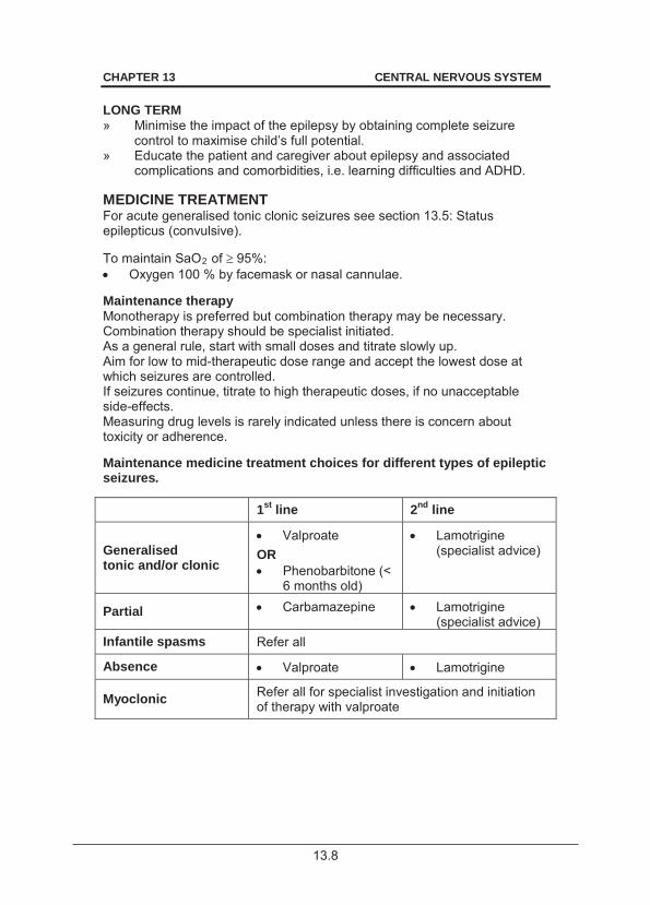
13.8
CHAPTER 13 CENTRAL NERVOUS SYSTEM LONG TERM » Minimise the impact of the epilepsy by obtaining complete seizure
control to maximise child’s full potential. » Educate the patient and caregiver about epilepsy and associated
complications and comorbidities, i.e. learning difficulties and ADHD. MEDICINE TREATMENT For acute generalised tonic clonic seizures see section 13.5: Status epilepticus (convulsive). To maintain SaO2 of 95%: Oxygen 100 % by facemask or nasal cannulae.
Maintenance therapy Monotherapy is preferred but combination therapy may be necessary. Combination therapy should be specialist initiated. As a general rule, start with small doses and titrate slowly up. Aim for low to mid-therapeutic dose range and accept the lowest dose at which seizures are controlled. If seizures continue, titrate to high therapeutic doses, if no unacceptable side-effects. Measuring drug levels is rarely indicated unless there is concern about toxicity or adherence. Maintenance medicine treatment choices for different types of epileptic seizures. 1st line 2nd line
Generalised tonic and/or clonic
Valproate OR Phenobarbitone (<
6 months old)
Lamotrigine (specialist advice)
Partial Carbamazepine Lamotrigine (specialist advice)
Infantile spasms Refer all
Absence Valproate Lamotrigine
Myoclonic Refer all for specialist investigation and initiation of therapy with valproate
13.9
CHAPTER 13 CENTRAL NERVOUS SYSTEM Valproate, oral, 5 mg/kg/dose, 8–12 hourly.
o Increase to 15–20mg/kg/dose 8–12 hourly. o Maximum total daily dose: 40 mg/kg/day. o Exclude liver dysfunction prior to initiating therapy (at least ALT), in
children under 2 years or if clinical suspicion of liver dysfunction. o Monitor at least clinically for hepatotoxicity.
Carbamazepine, oral, 2 mg/kg/dose (starting dose).
o Increase slowly at 2 weekly intervals to 5–10 mg/kg/dose 8–12 hourly.
o Usual maintenance total daily dose: 10– 20 mg/kg. o Maximum total daily dose: 20 mg/kg/day. o Dosage intervals: syrup 8 hourly, tablets 12 hourly. o Exacerbates myoclonic seizures and absence seizures.
Lamotrigine, oral, 0.2 mg/kg/dose starting daily dose. (Specialist initiated)
o Increase slowly at 2 weekly intervals to 1–5 mg/kg/dose 12–24 hourly. o Maximum dose when given with valproate: 5 mg/kg/day. o Lamotrigine is given as add-on therapy for different seizure types and
in drug-resistant paediatric epileptic syndromes, such as Lennox-Gastaut syndrome.
o Double the dose of lamotrigine when using carbamazepine or phenobarbitone and halve the dose when using valproate.
Phenobarbitone, oral, 3–5 mg/kg/dose as single dose at night.
o May be used in children under six months of age. o Is not recommended as maintenance therapy for children older than
2 years due to undesirable side effects such as sedation, behaviour disturbances, hyperkinesia and dependence, except in si tuations where there is poor adherence to other drugs.
o Exacerbates absence seizures. REFERRAL » Suspected but undiagnosed secondary cause. » Partial seizures for neuro-imaging, if facilities or expertise not available. » Generalised seizures other than typical febrile convulsions in children <
2 years. » Seizures that are not controlled within 2 months on one agent with
minimal side effects. » Neuroregression. » Mixed seizure types in one patient. » All myoclonic seizures and infantile spasms at presentation. » If need to add a second medicine.
13.10
CHAPTER 13 CENTRAL NERVOUS SYSTEM 13.4 SEIZURES, FEBRILE R56.0 DESCRIPTION Seizures occurring in children between the ages of 3 months and 6 years associated with a rapid rise in temperature at the beginning of an extracranial illness. Febrile seizures can be simple or complex febrile seizures. Simple febrile seizures: » are generalised tonic-clonic seizures, » are self limiting, usually less than 5 and always less than 15 minutes, » cause no neurological deficit after the convulsion, » have a good prognosis and very rarely develop into epilepsy, » often consist of only one seizure which needs no specific treatment, » there is often a family history of febrile seizures. Complex febrile seizures – febrile seizures with one or more of the following: » last longer than 15 minutes, » are recurrent within the same febrile illness, » have a focal (partial) onset, » have post-ictal, focal neurological abnormalities. Risk factors for recurrent febrile seizures include: » seizure disorder in a first degree relative, » onset before 12 months of age, » complex initial seizures.
DIAGNOSTIC CRITERIA Clinical » Exclude intracranial, extracranial and biochemical causes. » Signs of meningism are unreliable in children < 2 years of age. » If raised intracranial pressure or meningitis cannot be excluded then
the diagnosis of febrile seizures cannot be made. Treat children empirically for meningitis.
Investigations » Lumbar puncture is indicated in:
> children < 2 years of age for exclusion of intracranial infection even when signs of meningism are absent,
> all children who have no focus of infection, particularly those who have received antibiotics prior to the event.
» In children > 2 years of age, where a focus of extracranial infection is present and intracranial infection has been excluded clinically, no further investigation is required.
13.11
CHAPTER 13 CENTRAL NERVOUS SYSTEM » All children with complex febrile seizures and persistent lethargy
require neuro-imaging and then a lumbar puncture if raised intracranial pressure can be reliably excluded.
» Investigate complex febrile seizures to rule out meningitis, focal brain lesions, epilepsy and other possible underlying conditions depending on the clinical picture.
» An EEG is of no value in simple febrile seizures, but consider in complex febrile seizures.
GENERAL AND SUPPORTIVE MEASURES » Control fever with tepid sponging. » Reassure parents and caregivers. » Educate parents and caregivers regarding the management of future
episodes of fever. MEDICINE TREATMENT For fever (temperature ): Paracetamol, oral, 15 mg/kg/dose 6 hourly until fever subsides
(administered by parents). o Paracetamol has no effect on seizure prevention.
If convulsing: See section 13.5: Status epilepticus (convulsive). Continuous prophylactic therapy Routine daily prophylaxis is not recommended for patients with simple febrile seizures. For children with recurrent complex febrile seiz ures, consider prophylactic treatment after weighing the benefits against the ri sks and appr opriate parental counselling. REFERRAL » All patients with complex febrile seizures without an obvious cause of
the seizure and/or not re sponding to initial management should be discussed with a specialist.
» Developmental delay/regression.
13.5 STATUS EPILEPTICUS (CONVULSIVE) G41.9 DESCRIPTION Convulsive status epilepticus (SE) is a medical emergency defined as a generalised tonic-clonic convulsion that persists for 30 minutes or longer, or is repeated frequently enough to prev ent recovery of cons ciousness between attacks.
13.12
CHAPTER 13 CENTRAL NERVOUS SYSTEM After 30 minutes of generalised tonic-clonic seizures, the brain begins to suffer from hypoxia, acidosis, depletion of local energy stores, cerebral oedema and structural damage. Complications include: » hyperpyrexia, » disturbances of blood glucose, » respiratory depression, » renal failure, » cerebral oedema, » acidosis, » blood pressure disturbances, » inappropriate antidiuretic hormone (ADH) secretion, » hypoxic, ischaemic damage to brain, myocardium and muscles. DIAGNOSTIC CRITERIA Clinical » Convulsive seizure lasting 30 minutes or longer.
Manage convulsive seizures that have lasted for 5 minutes or more as for status epilepticus.
» Convulsive status epilepticus may be: > Idiopathic; > secondary to an insult to the brain, e.g. encephalitis, hypoxic
episode, trauma and complex febrile seizures; > as a result of non-adherence and changes in anticonvulsant
therapy. GENERAL AND SUPPORTIVE MEASURES » Maintain an open airway. » Place patient on side. » Admit to high or intensive care, if possible. » If unconscious, consider catheterisation. » Monitor:
> heart rate, > acid–base status, > respiratory rate, > blood gases, > blood pressure, > SaO2, > electrolytes, > neurological status, > blood glucose, > fluid balance, > anticonvulsant blood levels, > osmolality.
» Control fever with tepid sponging. » Cardiovascular and/or respiratory support i f the pat ient is unable to
maintain blood gases and blood pre ssure within the normal physiological range.
» Ventilate to maintain PaCO2 in the low normal range, i.e. 4– 4.5 kPa. MEDICINE TREATMENT To maintain SaO2 95%: Oxygen 100%, by facemask or nasal cannulae.
13.13
CHAPTER 13 CENTRAL NERVOUS SYSTEM Status epilepticus Follow ABCD approach. See flow chart on next page for management of Status epilepticus. For buccal midazolam and rectal diazepam, use the contents of an ampoule. For the purpose of rationalising the management of convulsive status epilepticus, it helps to divide or classify it into different stages as below: » Early SE (5-20 minutes) » Established SE (20–60 minutes) » Refractory SE (beyond 60 minutes) Intravenous fluid: Dextrose 5% in sodium chloride 0.9%, IV.
o Avoid overhydration. Keep fluid volume at maintenance. o Maintain normoglycaemia and electrolytes within the normal range.
Cerebral oedema Treat when clinically suspected/proven. If the patient has a serum osmolality < 320: Mannitol, IV, 250 mg/kg administered over 30–60 minutes.
o Do not exceed two doses without consulting with a specialist. OR Under specialist supervision: Sodium chloride 5%, IV, 2 mL/kg infused over 30 minutes.
Cerebral oedema with associated space occupying lesion Dexamethasone, IV, 0.5 mg/kg 12 hourly.
Other biochemical disorders Correct abnormalities, if present, e.g. glucose, calcium and sodium. For fever: Paracetamol, oral, 15 mg/kg/dose, 6 hourly as required via nasogastric
tube. Once fits are controlled consider maintenance therapy.
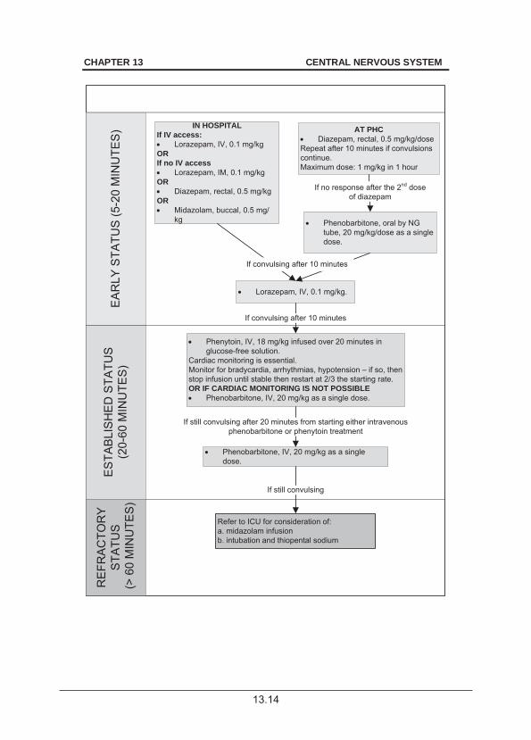
13.14
CHAPTER 13 CENTRAL NERVOUS SYSTEM
ES
TAB
LIS
HE
D S
TATU
S
(20-
60 M
INU
TES
)
RE
FRA
CTO
RY
S
TATU
S (>
60
MIN
UTE
S)
EA
RLY
STA
TUS
(5-2
0 M
INU
TES
) IN HOSPITALIf IV access:
Lorazepam, IV, 0.1 mg/kgORIf no IV access
Lorazepam, IM, 0.1 mg/kgOR
Diazepam, rectal, 0.5 mg/kgOR
Midazolam, buccal, 0.5 mg/kg
AT PHCDiazepam, rectal, 0.5 mg/kg/dose
Repeat after 10 minutes if convulsions continue.Maximum dose: 1 mg/kg in 1 hour
Phenobarbitone, oral by NG tube, 20 mg/kg/dose as a single dose.
If no response after the 2nd dose of diazepam
Lorazepam, IV, 0.1 mg/kg.
Phenytoin, IV, 18 mg/kg infused over 20 minutes in glucose-free solution.
Cardiac monitoring is essential.Monitor for bradycardia, arrhythmias, hypotension – if so, then stop infusion until stable then restart at 2/3 the starting rate.OR IF CARDIAC MONITORING IS NOT POSSIBLE
Phenobarbitone, IV, 20 mg/kg as a single dose.
If convulsing after 10 minutes
If convulsing after 10 minutes
Phenobarbitone, IV, 20 mg/kg as a single dose.
If still convulsing after 20 minutes from starting either intravenous phenobarbitone or phenytoin treatment
Refer to ICU for consideration of:a. midazolam infusionb. intubation and thiopental sodium
If still convulsing
13.15
CHAPTER 13 CENTRAL NERVOUS SYSTEM Note: Once intravenous access is attained take blood for gl ucose, blood gases, electrolytes, FBC and culture and anticonvulsant levels if patient is a known epileptic.
Monitor carefully for drug related respiratory depression.
Intubation, ventilation and administration of thiopental sodium infusion should only be perform ed in a centre w ith trained anaesthetists and a paediatric intensive care unit. REFERRAL
Caution Attempt to control seizures and stabilise the patient before referral.
» Failure to control seizures within 1 hour. » Where the primary cause is unknown, or if the primary cause itself
requires referral. 13.6 ANTITRETOVIRAL THERAPY AND ANTICONVULSANTS Co-administration of anticonvulsant treatment in p atients on anti retroviral therapy has not been well studied yet, and remains a therapeutic challenge. Drug interactions between anticonvulsants and antiretrovirals can arise from a number of mechanisms, including liver metabolism (increase or decrease), competition for protein binding and increase in v iral replication. There is at present no strong evidence to guide clinicians. The following points are important to remember when treating seizures and epilepsy in patients on ART: » The main area of caution is in drugs metabolised in the liver by the
cytochrome P450 enzyme system as this may cause alterations in levels of b oth anticonvulsants and ant iretrovirals leading to toxic or sub-therapeutic drug levels. This particularly pertains to the NNRTI’s and more specifically to PI’s.
» If clinically indicated, monitor anticonvulsant levels in patients taking concurrent ART and anticonvulsant therapy.
» Avoid prescribing carbamazepine, phenobarbital, and phenytoin for patients receiving NNRTIs or PIs, as there are serious P450 interactions involved. In this setting consider lamotrigine and valproate. See section 13.3: Epilepsy.
13.16
CHAPTER 13 CENTRAL NERVOUS SYSTEM » Treat children on ant iretrovirals presenting to c asualty with acute
seizures or in status epilepticus according to the existing standard status epilepticus or acute seizure protocols.
» Although benzodiazepines, phenytoin and phenobarbitone may interact with antiretroviral metabolism, the acute management of acute seizures or SE takes precedence in these instances.
13.7 HEADACHES R51 DESCRIPTION Headache is the mo st common pain syndrome in children of all ag es. Recurrent headaches are a common health problem and can be: » primary, e.g. migraine, or » secondary/symptomatic, e.g. raised intracranial pressure. The actual perception of headache varies according to age and is influenced by factors such as experience, memory and cultural environment. International Classification of Headache Disorders (ICHD) Migraine (without aura) Five or mor e headaches lasting 1–48 hours (duration in children is often shorter, lasting a few hours only) fulfilling at least 2 of the following: » bilateral or unilateral, frontal or parietal in location, » pulsating in character, » moderate or severe, » aggravated by routine activity, » nausea and/or vomiting plus photophobia and/or phonophobia during
headache. Migraine (with aura) At least 2 attacks fulfilling at least 3 of the following: » one or more reversible aura symptoms, » at least one aura developing over > 4 minutes or 2 or more successive
symptoms, » no aura lasting > 1 hour, » headache follows aura in less than 1 hour. Episodic tension-type headache At least 10 prior episodes, occurring less than 15 times per month and lasting 30 minutes to 7 days with at least 2 of the following: » pressing or tightening quality, » mild or moderate intensity, » bilateral location, » no aggravation by routine physical activity, » no nausea or v omiting, and photophobia and phonophobia absent
during headache.
13.17
CHAPTER 13 CENTRAL NERVOUS SYSTEM Cluster headache » Severe unilateral sharp headache associated with conjunctival injection
and lacrimation. » Rare in childhood. Paroxysmal Hemicrania Continua » Cluster headache of shorter duration. Each of the above can occur in combination in any patient, i.e. mixed/comorbid headache. Headaches can also be sub-classified according to temporal patterns, i.e. acute, acute recurrent, chronic progressive/non-progressive, episodic or constant. DIAGNOSTIC CRITERIA » Exclude secondary causes of headache, e.g. raised intracranial pressure. » Red flags in childhood headaches:
> change in pattern (e.g. “worst headache ever”), > progressive course over time, > age younger than 3 years, > nocturnal/wakes child from sleep, > early morning vomiting, > ataxia, > focal neurological signs, > alteration of level of consciousness.
GENERAL AND SUPPORTIVE MEASURES » Environmental and lifestyle changes, e.g. Avoid precipitants such as bright
lights, sleep deprivation and certain foods. » Adequate hydration. » Avoid skipping meals. » Regular exercise. » Stress alleviation and coping skills training where possible. » Headache diary. MEDICINE TREATMENT Treat headaches early with analgesics. Paracetamol, oral, 15 mg/kg/dose 6 hourly as required.
For migraine: Ibuprofen, oral, 10 mg/kg/dose, 6 hourly.
13.18
CHAPTER 13 CENTRAL NERVOUS SYSTEM Persistent vomiting and not tolerating oral feeds: Metoclopramide, oral, 0.15–0.3 mg/kg as a single dose.
OR Metoclopramide, IM/IV, 0.1 mg/kg as a single dose.
Migraine prophylaxis Indicated when headaches occur frequently, impacting on the child’s activity and requiring substantial relief medication. Treat for six months then review. Propranolol, oral, 0.5–3 mg/kg/day in 2–3 divided doses.
o Contraindicated in asthma and heart block. o Avoid in diabetes and depression.
In children who are unable to take propranolol, e.g. asthma: Topiramate, oral, 1–3 mg/kg/day in 1–2 doses. (Specialist initiated)
o Starting dose: 0.5 mg/kg/day. o Titrate dose slowly every 1–2 weeks. o Reinforce behavioural management before considering topiramate.
REFERRAL » Secondary intracranial cause suspected. » Failure to respond to first-line treatment. » No response to treatment. 13.8 NEUROCYSTICERCOSIS B69.0 DESCRIPTION Neurocysticercosis is caused by the cysticercal form, i.e. larval form of the pork tapeworm, T. solium. The larvae may locate in the br ain parenchyma, intraventricular and meningeal areas, spinal canal/cord and ey e, or a combination of thes e regions. Viable cysticerci incite little inflammatory response, but dead cysticerci elicit an increased inflammatory response. Cysticerci in the brain may remain dormant or may cause complications such as: » headache, » focal neurological deficits, » behavioural disorders, » increased intracranial pressure, » visual disturbances, » hydrocephalus, » seizures, » meningitis, » meningo-encephalitis, » spinal cord compression.

13.19
CHAPTER 13 CENTRAL NERVOUS SYSTEM DIAGNOSTIC CRITERIA Clinical » Location and stage of the life cycle of the parasite in the brain determines
the clinical features. » Suspect if child from endemic area, i.e. pig farming area, p resents with
neurological abnormalities such as: > Seizures, > raised intracranial pressure/hydrocephalus, > focal neurological deficits, > cranial nerve palsies, > meningo-encephalitis, > meningitis, > behavioural disorders, > headache.
Investigations » Computed tomography (CT scan) and/or magnetic resonance imaging (MRI
scan) of brain sho wing cysts, granulomas, peri-lesional oedema or calcification of cysts.
» MRI scan may identify more lesions and viable cystic lesions than the CT scan.
» Soft tissue radiology of muscles of lower limbs may demonstrate calcified cysticerci, i.e. rice grain calcifications in muscles.
» Follow-up CT scans and/or MRI scans may help to assess the response to therapy.
GENERAL AND SUPPORTIVE MEASURES Prevention: » Prolonged freezing or thorough cooking of pork to kill the parasite. » Thorough washing of fresh fruit and vegetables in T. solium endemic areas. » Attention to personal hygiene. » Proper sanitation facilities and safe water. » Avoid the use of human excreta as fertiliser. » Look for Taenia ova in the stools of the family members. MEDICINE TREATMENT Calcified cysticerci and a single dying lesion visible on CT scan require no antihelminthic treatment. Patients with multiple cysts usually have a mixture of live and dying cysts and are assumed to have active disease and require treatment. Albendazole, oral, 7.5 mg/kg/dose 12 hourly for 7 days.
o Maximum dose: 400 mg/dose.
13.20
CHAPTER 13 CENTRAL NERVOUS SYSTEM Prevention of neurological manifestations In massive infestations, cysticidal therapy may trigger an inflammatory response. Delaying antihelminthic therapy and adding corticosteroids may lessen the risk. 24 hours prior to albendazole therapy: Dexamethasone, IM, 0.15 mg/kg/dose 6 hourly.
Then follow with oral therapy as soon as possible: Prednisone 1mg/kg/day for the duration of albendazole therapy.
Seizure control See section 13.3: Epilepsy. Treat according to the type of seizure. Anticonvulsant treatment for 6–12 months after neuro-imaging resolution. Recurrent seizures require chronic treatment until seizure-free for 2 years. REFERRAL » All patients for CT scan. Repeat after 6 months to document resolution. » Neurocysticercosis not responding to adequate therapy. » Neurocysticercosis with complications, such as hydrocephalus. » Intractable epilepsy. 13.9 NEUROMUSCULAR DISORDERS 13.9.1 INFLAMMATORY POLYNEUROPATHY (GUILLAIN-BARRÉ SYNDROME) G61.0 * Notifiable condition DESCRIPTION Guillain-Barré syndrome is an acute autoimmune-mediated polyneuropathy which is precipitated by a preceding viral or other infection. It is the most common polyneuropathy in children. Different forms or variants of Guillain-Barré syndrome are described depending on the clinical and/or neurophysiological characteristics. Acute Inflammatory Demyelinating Polyradiculoneuropathy (AIDP) » The most common, accounting for 80–90% of cases. » Characterised mainly by:
> symmetrical, ascending motor weakness, > areflexia, i.e. absence of tendon reflexes, > distal sensory alteration, > pain/dysesthesiae.
13.21
CHAPTER 13 CENTRAL NERVOUS SYSTEM Acute motor axonal neuropathy (AMAN) » Purely a motor form of GBS. » It involves predominantly motor nerves and has an axonal pattern on
electrophysiology (nerve conduction studies). » Although there are similarities with AIDP, the clinical picture tends to be
more severe with more patients suffering from respiratory failure. Acute motor-sensory axonal neuropathy (AMSAN) » Another axonal form of GBS but with sensory involvement. » It is not frequently found in children. Miller-Fischer Syndrome » Patients have external ophthalmoplegia, sensory ataxia, weakness with
areflexia. » Electrophysiological and CSF studies are similar to AIDP. Chronic Inflammatory Demyelinating Polyradiculoneuropathy (CIDP) » May be considered a chronic variant of AIDP. » Most often starts insidiously and progresses slowly, but can have onset
like GBS. » It is managed differently from GBS and should be referred. DIAGNOSTIC CRITERIA Clinical » Preceding respiratory tract or gastrointestinal infection. » Symmetrical, flaccid muscle weakness with early areflexia. » The muscle weakness may ascend rapidly upwards to involve the trunk,
arms, face and cranial nerves. » Bulbar paralysis and respiratory failure may develop. » Autonomic dysfunction. » Relatively mild, or absence of, sensory signs. » Exclude the following:
> poliomyelitis, a rare cau se of hy potonia with abrupt onset of weakness (usually asymmetrical) in association with a febrile illness,
> transverse myelitis: initial flaccid weakness and areflexia typically involving the
lower limbs maximally, occasionally with pain at the onset, but rapidly progressing to
spasticity and hyperreflexia, also a sensory level on trunk, bladder and rectal sphincter involvement.
> Diphtheria, > Botulism.
13.22
CHAPTER 13 CENTRAL NERVOUS SYSTEM Investigations Screen for AFP » Send two stool specimens taken 24–48 hours apart to the National
Institute of Virology via the local laboratory. » Send one stool specimen after 60 days. CSF » CSF findings after 1 week show elevated protein and no cells or only a
few cells, i.e. albumino-cytological dissociation. » CSF glucose is normal. GENERAL AND SUPPORTIVE MEASURES » Notify as Acute Flaccid Paralysis. » Admit to a high or intensive care unit. » Monitor respiratory and autonomic and respiratory functions closely:
> peak expiratory flow rate, > blood pressure, > respiratory rate, > heart rate, > forced vital capacity (FVC), > bulbar functions, > arterial blood gases.
» Ventilation is recommended when: > PCO2 levels start rising, > a progressive fall in the peak expiratory flow rate, > tachycardia and sweating occur, > inspiratory efforts are weak, > inability to talk.
Note: These changes precede hypoxaemia detected on blood gas analysis, and ventilation should begin before frank hypoxaemia occurs. Respiratory care must be meticulous.
» Shoulder weakness, head-lag, weak cough and swallowing difficulties
are an indication for respiratory support. » To determine fluid losses from autonomic instability monitor urine output
and degree of sweating. » Provide adequate nutrition. » Bladder and bowel care as well as pressure-point care. » Routine physiotherapy for chest and limbs, keep ankles in neutral
position (900) (may require foot/hand splints). » Protect eyes and keep moist. » Communicate with child as awareness is maintained. Staff should
remember that children may be very frightened but unable to express their emotions and needs.
13.23
CHAPTER 13 CENTRAL NERVOUS SYSTEM MEDICINE TREATMENT Substantial pain is present (in up to 90%) in the severely affected patients. Pain in this setting is often unrecognised and underestimated. Pain management is essential. See section 20.1: Management of pain. For neuropathic pain: Carbamazepine, oral, 5 mg/kg/dose 12 hourly.
For rapidly progressive ascending paralysis, respiratory dysfunction or loss of ambulation: Immunoglobulin, IV, 1 g/kg/day, slowly over 12–16 hours on two
consecutive days or 0.4 g/kg as a single daily dose on 5 consecutive days early in the disease process. o Use under specialist supervision in an intensive care setting.
REFERRAL » Chronic inflammatory demyelinating polyradiculoneuropathy. » Guillain-Barré syndrome with bulbar paralysis and/or early signs of
respiratory failure. » Patients who have lost or are losing ambulation for management in
consultation with a paediatric neurologist. » Patients with complex Guillain-Barré syndrome. 13.9.2 MYASTHENIA GRAVIS G70.0 DESCRIPTION An auto-immune disorder resulting in muscle fatigue. Mild cases involve the eyes alone, i.e. p tosis and ophthalmoplegia, and s evere cases involve proximal muscle groups, respiratory and bulbar control. DIAGNOSTIC CRITERIA Clinical » Muscle fatiguability with exercise and demonstration of this in the clinic setting:
> Lid-lag test, i.e. failure to maintain upward gaze for 1 minute. > Arm-raising test, i.e. failure to maintain the arms at 90° from the
trunk for 1 minute > Neostigmine test (consult with neurologist):
Best done in a high-care environment. Attach cardiac monitor. Atropine at hand, in case of bradycardia. Give neostigmine bromide, IM, 0.04 mg/ kg, carefully whilst
watching cardiac monitor. Response is not as rapid as with edrophonium. Wait for 15–30 minutes to assess response.
13.24
CHAPTER 13 CENTRAL NERVOUS SYSTEM
> Ice-pack eye test: Put ice in a plastic bag and place it over the patients affected
eye(s) for 1–2 minutes. Positive test is when there is some improvement of the ptosis
and or the opthalmoplegia. Note: Myasthenia gravis patients not uncommonly present in a myasthenic crisis, with bulbar and respiratory compromise. Sometimes this may be th e first mode of clinical presentation. GENERAL AND SUPPORTIVE MEASURES » Occupational and physiotherapy. MEDICINE TREATMENT Pyridostigmine, oral, 1–5 m g/kg/day in 4–6 div ided doses. (Specialist
initiated) REFERRAL » All for confirmation of diagnosis and initiation of treatment (consideration
of steroids, immuno-modulation therapy). » Myasthenic crisis. 13.10 SYDENHAM’S CHOREA I102.9 DESCRIPTION Rapid involuntary jerks affecting any part of the body often incorporated into a voluntary movement in an attempt to mask it. It is an a cute post-streptococcal infection movement disorder and constitutes one of the ma jor criteria for the diagnosis of rheumatic fever. Patient has the appearance of being restless with constant movement. DIAGNOSTIC CRITERIA Clinical » Exclude drug reactions, hyperthyroidism, systemic lupus erythematosus
and neurodegenerative disorders. Investigations » Cardiac screening, i.e. ECG, echocardiogram. » ASOT, antiDNAse. » Erythrocyte sedimentation rate. » dsDNA, if clinically indicated.
13.25
CHAPTER 13 CENTRAL NERVOUS SYSTEM GENERAL AND SUPPORTIVE MEASURES » Emotional support » School support » Occupational therapy MEDICINE TREATMENT Movement disorders: Haloperidol, oral, 0.025 mg/kg/day in 2–3 divided doses.
o Increase dose slowly and incrementally to 0.05 mg/kg/day. PLUS If streptococcal infection: Phenoxymethylpenicillin, oral, 500 mg 12 hourly for 10 days.
THEN Until 21 years of age: Benzathine benzylpenicillin (depot formulation), IM, 1.2 million units
every 28 days. OR Phenoxymethylpenicillin, oral, 250 mg 12 hourly.
REFERRAL » All patients for specialist assessment. 13.11 CEREBROVASCULAR DISEASE/STROKE I67.9 DESCRIPTION Cerebrovascular disease can be ischaemic (thrombotic or embolic) or haemorrhagic, arterial or venous. Arterial ischaemic stroke must always be considered in any child with sudden onset of hemiparesis or other focal neurological disturbance. The clinical features of cerebral venous thrombosis (CVT) include headache, papilloedema, focal neurological signs, seizures (often focal), and alteration of consciousness. Risk factors: » cardiac disorders; » infections, e.g. meningitis, varicella, HIV, etc.; » prothrombotic disorders, e.g.. nephrotic syndrome, protein S/C
deficiencies, etc.; » haematologic disorders, e.g. sickle cell anaemia; » vasculopathies e.g. vasculitis, HIV, moyamoya syndrome.
13.26
CHAPTER 13 CENTRAL NERVOUS SYSTEM The initial evaluation in children includes the following: » Electrocardiography, echocardiography. » Full blood count, INR, PTT. » CSF analysis. » Infectious screening, including varicella, HIV, mycoplasma, TB. » Connective tissue and vasculitic screening. » Thrombophilia screening. See section 3.11: Venous thrombo-embolic
disease. GENERAL AND SUPPORTIVE MEASURES Acute supportive and neuroprotective care directed at preserving damaged but salvageable brain tissue includes the following: » Maintain body temperature in the low to normal range. » Maintain euglycaemia. » Maintain O2 saturation above 95%. » Maintain adequate cerebral perfusion and manage raised intracranial
pressure. » Treat anaemia. » Treat acute seizures promptly. Haemorrhagic stroke requires referral to a centre with neurosurgical expertise and facilities. Early disability assessment and management, includes physiotherapy, speech therapy, occupational therapy, etc. MEDICINE TREATMENT Arterial ischaemic stroke without haemorrhage All patients with confirmed arterial ischaemic stroke: Aspirin soluble, oral 1–5 mg/kg as a daily dose.
o Contraindicated in haemorrhagic stroke or bleeding tendency. REFERRAL » All patients to specialist paediatrician for investigation. » Anticoagulation with enoxaparin and warfarin is be st done in a
specialised setting under cardiologist, haematologist and neurologist supervision.
14.1
CHAPTER 14 PAEDIATRIC PSYCHIATRY
Principles for safe and effective prescribing of psychotropic medication for children and adolescents presenting with psychiatric symptoms » Psychopharmacology is only part of a holistic biopsychosocial treatment
plan. Safe and effe ctive pharmacological management of psychiatric disturbances in c hildren and adolescents should always be part of a comprehensive DSM-IV multi-axial diagnostic assessment and treatment plan, by a skilled clinician.
» Psychiatric problems in this age gro up are complex and requi re a multidisciplinary approach. There should be a n awareness of co-occurring medical conditions as well as of other medications used. Psychiatric comorbidity is very common and often diagnoses are difficult to make as in many instances the illness may still be evolving.
» It is important to refer to a child psychiatrist for a diagnostic assessment, when there is dia gnostic uncertainty and/or a poor r esponse to treatment.
» There should be an aw areness of the s ide effect profile of medications prescribed, as well as potential interactions with other medications. While psychotropic medications are generally well tolerated in children they can be associated with adverse effects. It is therefore important to use the lowest dosage possible and increase slowly, monitoring for side effects.
Common drugs used in psychiatry and their side effects: Selective serotonin reuptake inhibitors (SSRI, e.g. fluoxetine, citalopram) Adverse effects in children and adolescents » Agitation, behavioural disinhibition or ‘activation’, headache, GIT
disturbances (decreased appetite, nausea). » *Less common but important to note: increased risk of suicidal thoughts
or acts, associated with use of certain SSRIs in depressed children and adolescents (e.g. paroxetine).
» *Less common but potentially serious side effect is Serotonin Syndrome, which presents (in increasing severity) as restlessness, tremor, shivering, myoclonus, confusion, convulsions and death.
Special precautions/ investigations/monitoring » Adverse events may be dose related, reduce where indicated. » Check for :
> suicidal ideation, > ‘manic switch’ (may precipitate mania in bipolar illness), > symptoms of Serotonin Syndrome in patients where dosages have
been exceeded or in whom there is simultaneous use of two SSRIs (‘cross-tapering’).
14.2
CHAPTER 14 PAEDIATRIC PSYCHIATRY Tricyclic Antidepressants (e.g. amitriptyline) Adverse effects in children and adolescents » Sedation, anticholinergic, cardiac side effects. » May be more cardiotoxic in children than in adults. Special precautions/ investigations/monitoring » Dangerous and potentially fatal in overdose. Avoid in children with pre-
existing cardiovascular disease. » Do not use in conjunction with other drugs which prolong the QT
interval. » Baseline and on-treatment ECGs should be performed in patients with
pre-existing cardiovascular condition or positive family history. » May precipitate mania in bipolar illness. Stimulant medicines (e.g. methylphenidate) Adverse effects in children and adolescents » Loss of appetite, weight loss, insomnia, headache, abdominal pain. » Dysphoria or depressed mood (higher doses). » May worsen existing tics. » May, at higher doses, lower seizure threshold and precipitate seizures
in children suffering from epilepsy. Special precautions/ investigations/monitoring » Monitor BP, pulse, height and weight. » Monitor for mood change and development of tics. » Use with caution in children who suffer a psychotic illness. ‘Atypical’ antipsychotics (e.g. risperidone, olanzapine) Adverse effects in children and adolescents » Weight gain, metabolic syndrome, extrapyramidal side-effects (EPSE)
such as acute dystonia and parkinsonism, akathisia and sedation at higher dosages (particularly risperidone).
Special precautions/ investigations/monitoring » Monitor weight. » Check prolactin level, glucose and cholesterol, particularly if family history. ‘Typical’ neuroleptics (e.g. haloperidol) Adverse effects in children and adolescents » EPSEs, acute dystonia, akathisia, tardive dyskinesia. » Life threatening side effect is Neuroleptic Malignant Syndrome (NMS)
(fever, altered mental status, muscle rigidity, autonomic dysfunction). Special precautions/ investigations/monitoring » Monitor for EPSEs. » Avoid long term use as risk of tardive dyskinesia, which is irreversible. Benzodiazepines (e.g. lorazepam, diazepam, clonazepam) Adverse effects in children and adolescents » Sedation, anomalous reaction of disinhibition, restlessness.
14.3
CHAPTER 14 PAEDIATRIC PSYCHIATRY Special precautions/ investigations/monitoring » Not for long term use. 14.1 SEDATION OF ACUTELY DISTURBED CHILD OR
ADOLESCENT AWAITING PSYCHIATRIC EVALUATION MEDICINE TREATMENT Exclude organic causes, e.g. encephalopathy or other intracranial pathology, seizures, metabolic disease, medication adverse effects and intoxication. For children under the age of six years Sedation with psychotropic agents should only be considered in extreme cases and only after consultation with a specialist. For children over the age of six years Lorazepam, oral/IM, 0.05–0.1 mg/kg/dose.
o Onset of action: is 20–30 minutes. If sedation is inadequate: Haloperidol, IM, 0.025–0.075 mg/kg/dose.
In case of an acute dystonic reaction secondary to haloperidol: Biperiden, IM/slow IV, 0.05–0.1 mg/kg.
o 6–10 years: 3 mg o >10 years: 5 mg
14.2 ATTENTION DEFICIT HYPERACTIVITY DISORDER (ADHD) F90.0/90.1 DESCRIPTION Children with ADHD display developmentally inappropriate degrees of inattention, impulsiveness and hyperactivity. There are 3 subtypes: » predominantly inattentive, » predominantly hyperactive-impulsive, and » combined. Inattention: » Failing to give close attention to details, » Careless mistakes, » Not listening, » Failure to complete tasks, » Losing things, » Distractibility.
14.4
CHAPTER 14 PAEDIATRIC PSYCHIATRY Hyperactivity: » Fidgetiness. » Out of seat. » Running or climbing excessively. » Being “on the go”, or like “driven by a motor”. Impulsivity: » Blurting out answers. » Difficulty waiting turn. » Interrupting or intruding on others. Outcome » Hyperactivity symptoms decrease and inattention symptoms persist
during adolescence. » Remission is achieved in up to two thirds (30–60%) of patients during
adolescence. » Symptoms persist into adulthood in the remaining one third. » Symptom presentation in adults reflects persistence of problems with
attention and concentration, as well as poor impulse control. » Common adult difficulties include problems in the workplace,
relationship problems, high-risk behaviours including substance misuse as well as as sociated co-morbid conditions such as depression and anxiety.
DIAGNOSTIC CRITERIA » Diagnostic criteria are based on DSM IV. » Symptoms must present before age seven. » Symptom duration of at least six months. » Behaviour is inconsistent with the patient’s developmental level and
intellectual ability. » Presence of functional impairment in more than one setting. » Exclude other mental or physical disorders, e.g. anxiety disorders,
mood disorders, psychotic disorders, hearing impairment. » Common co-morbid conditions include Oppositional Defiant Disorder,
Conduct Disorder, Depression (particularly in girls) and Substance Use Disorders (SUDs), as well as HIV and epilepsy.
» Certain conditions may ‘mimic’ ADHD s uch as, developmental disorders, motor coordination problems, intellectual disability, post-traumatic and post infectious encephalopathy.
Note: » Girls may more commonly present with inattentive-type ADHD. The
diagnosis may therefore be missed. GENERAL AND SUPPORTIVE MEASURES » Identify and treat comorbidities early, such as depressive disorders as
this may prevent the onset of substance misuse (to ‘self-medicate’) and other risk behaviours during adolescence.

14.5
CHAPTER 14 PAEDIATRIC PSYCHIATRY » Parent counseling:
> rules and limit-setting, > positive reinforcement of pro-social behaviour, > consistent routine, and > restrictive diets are of no proven value.
» Behaviour-based therapy: > reward positive behaviour, > improve self-image, and > improve social awareness and adjustment.
» Social skills groups. » Identify learning difficulties and refer to educational support services. MEDICINE TREATMENT There is no absolute contraindication for the concomitant use of methylphenidate with antiepileptic drugs (AEDs) or antiretroviral therapy (ART). However, exercise caution with the dosages prescribed and beware of potential drug-drug interactions. Monitor for potential adverse side effects. For children under the age of six years Refer for diagnostic assessment by a child psychiatrist or paediatrician. For children over the age of six years Initiate treatment using the short-acting methylphenidate formulation until effective dosage achieved. Reduce dose or w ithdraw methylphenidate if a paradoxical increase in symptoms occurs. Methylphenidate, short -acting, oral, 1 mg/kg/day (maximum 2 mg/kg)
o Initial dose: 5 mg 2–3 times daily, before breakfast and lunch, 3rd dose not later than 14h30 (approximately every 3 to 3½ hours).
o Increase the dose at weekly intervals by 5–10 mg until symptoms are controlled. Use the lowest effective dose.
o Maximum daily dose: 60 mg. Do not exceed 40 mg/day without consultation with a child psychiatrist or paediatrician.
If inadequate symptom control due to irregular usage or poor adherence, refer. Contraindications: Absolute: » Hyperthyroidism, » cardiac dysrhythmia, » glaucoma, » concomitant mono-amine oxidase inhibitor therapy. Relative: » hypertension, » anxiety, » agitation, » epilepsy, » tics.
14.6
CHAPTER 14 PAEDIATRIC PSYCHIATRY Discontinuation of treatment » If no ob jective improvement of symptoms is observed, e.g. Conners’
Scales completed by teacher, after appropriate dosage adjustment over a one-month period.
» To establish whether on-going treatment is indicated in a child on long term stimulant therapy, trials of discontinued treatment (trials off treatment) should be part of the management plan.
» Indications for a trial off treatment: > treatment duration in excess of 2-3 years, > adolescent age (particularly late adolescence), and > a substantial reduction in core ADHD s ymptoms, evident in more
than one setting. » Trials off treatment should be planned and at times least disruptive to
the child’s academic and social functioning i.e. time the treatment withdrawal outside of major commitments such as examinations.
» Duration of treatment withdrawal can be for one week to a month, depending on whether stability is maintained.
» Treatment can be withdrawn abruptly, with no need to taper dosages. » Obtain feedback from teachers and parents (verbal feedback,
completion of parent and teacher Conners’ Scales), before and dur ing the trial off treatment.
» Assess the child and document the mental state (symptoms of ADHD), before and during the trial off treatment.
» Monitor 3 monthly for one year. » Re-initiate treatment (at last dosage prescribed), if:
> there is a significant re-emergence of symptoms after one week off treatment and/or during the month off medication, or
> after a longer trial off medication, e.g. at 3 monthly follow up visits, there is evidence of symptom re-emergence.
Note: Adolescents are more l ikely to pr esent with poor co ncentration and inattentiveness, rather than hyperactivity. REFERRAL » No response to medicine treatment after 4–6 weeks. » Presence of comorbid psychiatric conditions with severe functional
impairment: oppositional defiant disorder, mood disorders, anxiety disorders, debilitating tics.
» Presence of uncontrollable seizures. » HIV positive status.
14.7
CHAPTER 14 PAEDIATRIC PSYCHIATRY 14.3 MOOD DISORDERS F30–F39 14.3.1 DEPRESSION IN CHILDHOOD AND ADOLESCENCE F32 DESCRIPTION The clinical presentation of depression includes: » symptoms of depressed mood, » decreased pleasure or interest, » neurovegetative symptoms, » sleep/appetite disturbance, » fatigue, » poor concentration, » psychomotor agitation/retardation, » guilty ruminations, and » thoughts of death and suicide.
Suicide is self-inflicted harm where the intention is to die. Increased suicide risk is associated with the following: » male gender » adolescence » previous attempts and lethality of method used » family history of suicide » presence of a mental illness » social isolation and poor family support » associated substance abuse or physical aggression
The clinical picture of a child and adolescent with major depressive disorder is similar to that of adults except that there are some developmental differences i.e.: » mood is often irritable rather than sad, » somatic complaints, i.e. headache and abdominal pain, » behavioural and academic/school problems occur frequently in children, » withdrawal from social activities, » neurovegetative symptoms are less common than in adults, and » suicide attempts increase in number, tend to be more lethal and
impairment of functioning worsens, with increasing age. The first ep isode of bipolar disorder can present with depression in adolescents. Bipolar depression is often associated with a more sudden onset, psychomotor retardation and in some instances, psychotic symptoms.
14.8
CHAPTER 14 PAEDIATRIC PSYCHIATRY A number of depressed children and adolescents have co-morbid psychiatric disorders. The most frequent co-morbid diagnoses are: » anxiety disorders, » ADHD, » oppositional defiant disorder, » conduct disorder, and » substance misuse, particularly in adolescents. Conduct problems may develop as a c omplication of th e depression and persist after the depression remits. It is important to assess and manage conditions that occur together with depression. DIAGNOSTIC CRITERIA » The presence of at least five of the symptoms of depression for a period
of up to two weeks. » Symptoms should be of a severity to cau se significant functional
impairment and feelings of distress. » Diagnosis is difficult in children, compared to adolescence, as children
may ‘deny’ depressed mood or may not experience symptoms as ‘ego dystonic’. Some children may require diagnostic assessment and treatment in an in-patient setting.
Consider the following in a child presenting with depressed mood: » Exclude underlying medical conditions such as:
> infections, e.g. HIV, cerebral cysticercosis, encephalitis and tuberculous meningitis;
> neurological conditions, e.g. temporal lobe epilepsy, brain tumours; > endocrine disorders, e.g. thyroid conditions.
» Exclude medication-induced mood disturbances e.g. corticosteroids, antiretrovirals (zidovudine, efavirenz), high doses of stimulant medication and barbiturates.
» Exclude substance abuse such as a lcohol and methamphetamine (‘Tik’).
» Assess for suicide risk. GENERAL AND SUPPORTIVE MEASURES » Psychological interventions are con sidered ‘first line’ for mild t o
moderate depression and should be administered by a suitably skilled clinician: > cognitive behavioural therapy (CBT): to address distorted, negative
cognitions, maladaptive patterns of behaviour and communication; > psychodynamic/play therapy: to ide ntify feelings, improve self
esteem and social interactions.
14.9
CHAPTER 14 PAEDIATRIC PSYCHIATRY » Additional interventions:
> family counselling: to addr ess family disharmony, stressors and provide psycho-education;
> input to school: to address academic issues and psycho-education; > social worker: to investigate suspicion of child abuse or neglect.
MEDICINE TREATMENT If there is a failure to respond to psychotherapeutic interventions after 4–6 weeks or if severity of symptoms increases, consider a trial of antidepressant medication, while still continuing with psychotherapy. Initiate treatment in consultation with a specialist. Response to tr eatment should bring about a meaningful reduction in symptoms and improvement in functioning. Once remission is achieved continue medication therapy for at least a further 6–12 months. Fluoxetine, oral, 0.5 mg/kg/day.
o Dose range: 5–40 mg daily. o Recommended average dose: 10–20 mg/day.
If there is a poor response to fluoxetine after an adequate trial of treatment, i.e. 4–6 weeks; or if significant symptoms of anxiety are present; child is HIV positive; consider an alt ernative SSRI, in disc ussion with a child a nd adolescent psychiatrist: Citalopram, oral, 0.4 mg/kg/day.
o Dose range: 10–60 mg daily. o Recommended average dose: 10–20 mg/day.
Note: Be aware of potential side effects, discussed above. A trial of treatment is considered to be ineffective if the patient presents with ongoing, significant depressive symptoms and/or suicidal ideation and where the patient has not achieved an improvement in overall level of functioning. Be aware of the risk of bipol ar ‘switch’ or precipitation of mania in patients with a family history of bipolar disorder. Tricyclic antidepressants are not re commended as first line agents in children, due to insufficient evidence of efficacy, and potentially adverse cardiovascular side effects and lethality in overdose. REFERRAL » Poor response to an adequate trial of treatment i.e. medication trial of
6–8 weeks in combination with psychological treatment. » Presence of co-morbid conditions. » Psychotic symptoms such as delusions or hallucinations. » Suicidal ideation or intent.
14.10
CHAPTER 14 PAEDIATRIC PSYCHIATRY 14.3.2 DISRUPTIVE MOOD DYSREGULATION DISORDER
(DMDD) F31 DESCRIPTION Disruptive mood dysregulation disorder (Bipolar disorder) in children often presents with mixed mood states, with significant mood lability, rage attacks or ‘affective storms’, rather than discrete manic or depressive episodes. The picture therefore may not resemble the clinical picture observed in adults. There is a risk of misdiagnosis or ‘over-diagnosis’ of this disorder, in children presenting with severe aggression and ‘dysregulated’ moods. Manic Episode A distinct period of an ab normally and pers istently elevated, expansive and/or irritable mood. This should represent a si gnificant change in t he patient’s baseline mental status and must last for at least 1 week. During the period of mood disturbance, the pa tient should display the foll owing symptoms: » Elated mood. » Grandiosity. » Decreased sleep. » Hypersexuality. » Racing thoughts and pressured speech. » Increase in goal-directed activity. Depressive episode Similar to symptoms of major depressive episode except that onset may be more rapid, associated with psychomotor retardation, and/or psychotic symptoms. Mixed mood state Presence of both manic and depressive symptoms over a period of 1 week. This presentation is more common in children. Discrete manic and depressive phases are less evident than in adults. The mood disturbance must cause marked impairment of functioning and should not be due to the direct effects of a substance. MEDICINE TREATMENT Refer patient with suspected manic episode immediately for spec ialist assessment and admission for containment and further management.
CAUTION Beware of the ris k of ne uroleptic malignant syndrome, especially with the use of high doses in the first episode.
14.11
CHAPTER 14 PAEDIATRIC PSYCHIATRY Acute treatment Sedate before transfer. See section 14.1: Sedation of acutely disturbed child and adolescent awaiting psychiatric evaluation. Maintenance treatment If previously on maintenance therapy: Re-initiate treatment, in consultation with a psychiatrist.
If not previously on maintenance therapy: Risperidone, oral, 0.5–3 mg da ily, depending on age, weight of child and
clinical response. (Specialist initiated, or in consultation with a psychiatrist) Other medication such as mood stabilisers only to be initiated in
consultation with a psychiatrist. REFERRAL All for diagnostic assessment by a child and adolescent psychiatrist.
14.4 ANXIETY DISORDERS F41.9 Separation Anxiety Disorder and Selective Mutism are diagnostic categories exclusive to the childhood population, while Post-Traumatic Stress Disorder (PTSD), Obsessive Compulsive Disorder (OCD), Social Phobia, Specific Phobia, Panic Disorder and G eneralised Anxiety Disorder (GAD) present across the lifespan. Medicine treatments do not form part of t he primary management of Separation Anxiety Disorder and Selective Mutism, unless the child presents with additional symptoms which merit a diagnosis in one of the other anxiety disorder categories. Anxiety in a child can be misdiagnosed as ADHD, as both conditions may present with increased levels of activity and problems with concentration.
Symptoms ADHD Anxiety Hyperactivity + + Inattention + + Impulsivity + –
(fearful, inhibited) Emotional content/thoughts
» poor school performance, » ‘in trouble’ with adults, » peer rejection/social
isolation, » struggles to control
aggression.
» fear, » threat, » worry, » nightmares with
anxiety themes.
Academic difficulties
More likely Less likely
Poor sleep + +
14.12
CHAPTER 14 PAEDIATRIC PSYCHIATRY
14.4.1 POST TRAUMATIC STRESS DISORDER (PTSD) F43.1 DESCRIPTION The core features of experiences which place patients at risk of PTSD are: » exposure to any traumatic event’ » there is a threat of serious injury or death, » feelings of intense fear, helplessness and horror. DIAGNOSTIC CRITERIA According to DSM IV: » Persistently re-experiencing:
> recurrent thoughts, dreams, > flashbacks, reliving experiences.
» Marked avoidance (conversations, places): > numbing, > diminished interest, > detachment or restricted emotions.
» Hyperarousal (hypervigilance, poor sleep). » Significant distress/impairment. GENERAL AND SUPPORTIVE MEASURES Debriefing in the immediate aftermath of the trauma is not recommended. Psychological interventions, including: » general supportive counseling, » cognitive behavioural strategies, » group and family interventions. MEDICINE TREATMENT Fluoxetine, oral, 0.5 mg/kg/day.
o Recommended average dose: 5–20 mg. o If poor response, consider higher doses in consultation with a
specialist. REFERRAL » Persistent symptoms despite therapy. 14.4.2 GENERALISED ANXIETY DISORDER (GAD) F41.1 DESCRIPTION Excessive anxiety or worry, occurring on most days for at least 6 months.
14.13
CHAPTER 14 PAEDIATRIC PSYCHIATRY DIAGNOSTIC CRITERIA According to DSM IV, presence of 3 of the following: » restlessness or a feeling of being ‘on edge’, » poor concentration or ‘mind going blank’, » irritability, » muscle tension, » sleep disturbance, GAD causes significant distress and impairment in functioning. Exclude substance abuse or a medical condition. GENERAL AND SUPPORTIVE MEASURES These interventions should be performed by a suitably qualified clinician. » Cognitive behavioural therapy (CBT): aimed at changing pessimistic,
anxiety-based cognitions and developing strategies to reduce anxieties and avoidant behaviour patterns.
» Behaviour therapy: relaxation, desensitisation by imagining or exposure to anxiety-provoking situations.
» Psychodynamic/supportive psychotherapy: aimed at promoting self esteem, assertiveness and autonomy.
MEDICINE TREATMENT Fluoxetine, oral, 0.5 mg/kg/day.
o Dose range: 5–40 mg daily. o Recommended average dose: 10–30 mg. o Treatment duration: 6–9 months after resolution of symptoms.
REFERRAL » Failure to respond after 6–8 weeks to an adequate trial of therapy and
medication. » Adverse events with fluoxetine. 14.4.3 OBSESSIVE COMPULSIVE DISORDER (OCD) F42.9 DESCRIPTION Obsessions Persistently recurring thoughts, impulses or images that are experienced as intrusive, inappropriate and not simply excessive worries about realistic problems. Children may not experience these as distressing but the obsessions may interfere with day-to-day functioning. Compulsions Repetitive behaviours or mental acts that a person feels driven to perform according to a rigid ly applied rule in ord er to reduce distress or to prev ent some dreaded outcome.

14.14
CHAPTER 14 PAEDIATRIC PSYCHIATRY DIAGNOSTIC CRITERIA According to DSM IV: » The most common symptoms of OCD in childhood are:
> contamination fears accompanied by compulsive washing and avoidance of “contaminated” objects,
> repetitive checking and counting, > obsessive doubt, > compulsive reading and drawing, > symmetry concerns (e.g. right and left needs to be same).
» Comorbid conditions: > rheumatic fever, > streptococcal throat infection (paediatric autoimmune neuropsychiatric
disorders associated with streptococcal infections (PANDAS)), > tic disorders.
GENERAL AND SUPPORTIVE MEASURES » Provide Cognitive Behavioural Therapy (CBT), if av ailable and
appropriate. » Exposure-based interventions, e.g. contact with “dirt” in a child with
contamination fears, thought stopping techniques, “response prevention”, i.e. blocking of rituals.
These interventions should be carried out by a suitably qualified professional. MEDICINE TREATMENT Fluoxetine, oral, 0.5 mg/kg/day.
o Dose range: 10–40 mg daily. Higher doses may be needed to effectively manage OCD symptoms.
o Recommended average dose: 20–40 mg. Higher dose within the range may be required.
o Duration of treatment: 6 months after resolution of OCD symptoms. REFERRAL » Poor response to a dequate trial of cognitive behavioural therapy and
medication i.e. persistence of obsessions and/or compulsions, with impairment in functioning.
» Co-morbid conditions. 14.5 CHILDHOOD PSYCHOSIS F09 It is i mportant to note that children who present with symptoms such as hallucinations, confusion and intensely aggressive or disturbed behavior may not all be psychotic or suffer from schizophrenia. Delirium should be the first diagnosis to consider, before a psychotic disorder is suspected. Failure to recognise a delir ium may delay the diagnosis of the un derlying medical condition or drug-related delirium and place the child at risk.

14.15
CHAPTER 14 PAEDIATRIC PSYCHIATRY Delirium is a no n-specific neuropsychiatric disorder which indicates global encephalopathic dysfunction in medically ill patients. The core features consist of att entional disturbances, an a ltered level of consciousness and diffuse cognitive deficits. It i s fluctuating in na ture and may present w ith perceptual disturbances, commonly visual hallucinations.
14.5.1 SCHIZOPHRENIA F20.9 DESCRIPTION Schizophrenia is a chronic psychotic disorder characterised by disturbances in thinking, perceptions, emotions and behavior, associated with significant degrees of i mpairment in fun ctioning. Childhood Schizophrenia is rare. Al l children presenting with psychotic features merit further investigation. » Very Early Onset Schizophrenia (VEOS) is defined as onset before age
13 years. » Early Onset Schizophrenia is defined as onset before age 18 years. Onset during childhood and adolescence confers a poorer prognosis for the illness, treatment refractoriness and significant impairment in functioning. DIAGNOSTIC CRITERIA According to DSM IV: » Two or more of the following symptoms need to be present for at least 6
months: > delusions, > hallucinations, > disorganised speech, > grossly disorganised or catatonic behaviour, > ‘negative’ symptoms i.e. affective flattening and lack of volition.
» The same diagnostic criteria for ad ults are u sed, but in ch ildren, delusions are not a s bizarre or systematised as in adults. The clinical presentation in adolescents more closely resembles that in adults.
» Exclude substance abuse (cannabis, methamphetamine) or a m edical condition in a young person presenting with a psychotic episode.
GENERAL AND SUPPORTIVE MEASURES » Supportive individual and family counseling is an important part of the
comprehensive treatment plan. » The aim of individual counseling is to develop healthier coping
strategies and defence mechanisms and to provide structure and l imit regression.
» Family interventions focus on psycho-education, facilitating acceptance of the diagnosis to ensure adequate compliance and support for the patient.
» Educational issues must be attended to su ch as tr ansition back to school after a psychotic episode and academic support.
14.16
CHAPTER 14 PAEDIATRIC PSYCHIATRY MEDICINE TREATMENT Pharmacotherapy is t he first line treat ment for psy chosis in chil dren and adolescents. Acute phase treatment (4–6 weeks) Sedate before transfer. See section 14.1: Sedation of acutely disturbed child and adolescent awaiting psychiatric evaluation. If previously prescribed antipsychotic medication: Re-initiate treatment, in consultation with a psychiatrist.
If not previously on therapy, while awaiting admission: Risperidone, oral.
o Initial dose: 0.5 mg daily. o Use lowest effective dose to limit adverse long term side effects
and to facilitate adherence. o Increase dose by 0.25–0.5 mg daily every 1–2 weeks, depending
on tolerability and age of the child. o Refer if doses in excess of 3 mg are required.
Maintenance phase: (12–24 months) Gradually lower the dose of risperidone from that needed to treat the acute psychotic phase to that needed to prevent relapse and to ensure adequate adherence. Note: » First generation or ‘typical’ antipsychotics (e.g. haloperidol) and second
generation or ‘aty pical’ antipsychotics (e.g. ris peridone) have demonstrated equal efficacy for the treatment of psychotic symptoms.
» First generation antipsychotics are less favourable because of high rates of extrapyramidal side effects and worsening of negative symptoms in children and adolescence with long term use compared to adult populations.
» Exercise caution when prescribing high doses of haloperidol as this may increase the risk of neuroleptic malignant syndrome (NMS), which may be life threatening.
» In a patient with an identified cardiovascular risk, do an ECG before initiating therapy with risperidone.
» Monitor blood pressure, pulse, ECG, blood glucose and lipid profile in patients on long term treatment.
REFERRAL » All children for assessment and for initiation of therapy » Urgent: younger children » Urgent: for admission, of behaviourally disturbed psychotic adolescent.
14.17
CHAPTER 14 PAEDIATRIC PSYCHIATRY 14.5.2 TIC DISORDERS F95.9 DESCRIPTION A tic is a sudden, rapid, recurrent, non-rhythmic stereotyped motor movement or vocalisation and includes the following subtypes: » Chronic motor or vocal tic disorder. » Transient tic disorder. » Tourette’s disorder. Tourette’s disorder is a chronic neuropsychiatric disorder that is characterised by both v ocal and motor tics, and r elated somatosensory urges. It is co mmonly associated with a numb er of co- morbid conditions such as OCD, ADHD as well as disturbances of mood. GENERAL AND SUPPORTIVE MEASURES » Psycho-education of patient, parents, teachers and peers: to reduce the
stigma and social consequences of tics. » Supportive psychotherapy: to assist the individual to cope with the
stigma/ teasing, improve self esteem and improve social skills. » Family therapy: to assist the family in managing associated symptoms
and to reduce stress. MEDICINE TREATMENT Medication to be used for short periods only to reduce severe symptoms. Haloperidol may be initiated as first line since medication treatment is not long term. Note: The natural course of tics is to ‘wax and wane’ or fluctuate. Therefore avoid long term use of neuroleptic medication. For severe and frequent tics that seriously impact on child’s functioning: Haloperidol, oral, 0.02–0.12 mg/kg/day.
o Dose range: 0.25–1.75 mg 12 hourly. o Recommended average dose: 0.5–2 mg/day. o Monitor for extrapyramidal and anticholinergic side effects.
If poor response: Risperidone oral, 0.5 mg/day.
o Dose range, 0.5–3 mg/day. o Recommended average dosage 1mg/day.
REFERRAL » Tourette’s syndrome not responding to therapy. » Tourette’s syndrome with comorbid psychiatric or medical conditions.

14.18
CHAPTER 14 PAEDIATRIC PSYCHIATRY 14.6 OTHER SITUATIONS OR CONTEXTS IN WHICH
PSYCHOTROPIC DRUGS MAY BE PRESCRIBED The following are particular conditions or common behaviours for which psychotropic medication forms part of the management plan. These are: » Psychiatric presentations associated with paediatric HIV. » Behavioural problems associated with pervasive developmental
disorders (PDDs). » Substance use disorders in adolescence. » Behavioural problems associated with Intellectual Disability (ID). 14.6.1 PSYCHIATRIC PRESENTATIONS IN HIV INFECTED
CHILDREN AND ADOLESCENTS DESCRIPTION » HIV positive children and adolescents are a t increased risk of
psychopathology, such as ADHD, depression and anxiety disorders. Psychosis and mania is less common than in the adult population.
» The increased risk of psychopathology is due to the virus itself, side effects of antiretroviral therapy (ART) and those due to psychosocial stressors.
» Symptom presentation of psychiatric disorders in HIV positive children are the same as in the general childhood population.
» ADHD often co-occurs with significant learning difficulties, despite treatment with antiretroviral therapy (ART).
» Psychotic disorders are r are in H IV positive children. Consider a delirium or p artial seizures if an H IV positive child presents with psychotic symptoms.
GENERAL AND SUPPORTIVE MEASURES » Psychological interventions are similar to tho se for H IV negative
children. » Issues specific to the child’s HIV status may need specific intervention, for
problems related to di sclosure of H IV status, stigma, grief counselling, adherence issues, orphanhood and living with a chronic illness.
» Refer to the hospital social worker to address social issues. MEDICINE TREATMENT Start all medications at lower doses and then titrate up slowly. Initiate treatment according to guidance in this chapter. Note: Because of drug-drug interactions between fluoxetine and some antiretroviral medication, initiate treatment with citalopram.
14.19
CHAPTER 14 PAEDIATRIC PSYCHIATRY REFERRAL » All HIV positive children on ART who present w ith severe psychiatric
symptoms such as severe depression, psychosis and/or mania for psychiatric evaluation and initiation of psychotropic medication.
14.6.2 PERVASIVE DEVELOPMENTAL DISORDERS (PDDS) F84 DESCRIPTION The Pervasive Developmental Disorders (PDDs) are neuropsychiatric disorders characterised by patterns of delay and deviance in the development of social, communication and cognitive skills. Onset is usually during the first few years of life and includes conditions such as: » Autistic disorder. » Asperger’s disorder. » Rett disorder. » Childhood disintegrative disorder. GENERAL AND SUPPORTIVE MEASURES » Social skills and family interventions. » Education and social placement. » Behaviour modification, specifically adapted for autism spectrum
disorders. » Early intervention is important for optimal outcome. MEDICINE TREATMENT Not for core autistic symptoms. For severe aggression and self-injurious behavior: Risperidone, oral, 0.125–3 mg daily in 2 divided doses.
o Average daily dose: 0.25–2 mg/day. OR Haloperidol, oral, 0.002–0.12 mg/kg/day.
o Average daily dose: 0.25–2 mg/day. o Dose range: 0.25–1.75 mg 12 hourly. o Monitor for extrapyramidal and anticholinergic side effects.
14.6.3 SUBSTANCE ABUSE F10–19 DESCRIPTION Age of onset of substance abuse can be as early as 8 years. Illicit drugs such as cocaine, amphetamines and cannabis, as well as alcohol abuse are associated with a greater risk for psychosis.
14.20
CHAPTER 14 PAEDIATRIC PSYCHIATRY Behavioural disturbance in the context of a Substance Use Disorders (SUD) may be due to intoxication, withdrawal, or due to a substance induced mood or psychotic disorder. Initial treatment of SU Ds begins w ith medical stabilisation of the pati ent ideally in a medical facility. About one third of youth with SUDs, present with a ‘dual diagnosis’, which is the presence of a co-o ccurring psychiatric disorder and SUD, in the sam e individual. Be aware of the mental state changes associated with illicit drugs.
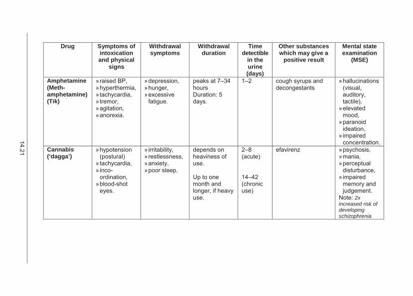
14.21
Drug Symptoms of intoxication and physical
signs
Withdrawal symptoms
Withdrawal duration
Time detectible
in the urine (days)
Other substances which may give a
positive result
Mental state examination
(MSE)
Amphetamine (Meth-amphetamine) (Tik)
» raised BP, » hyperthermia, » tachycardia, » tremor, » agitation, » anorexia.
» depression, » hunger, » excessive
fatigue.
peaks at 7–34 hours Duration: 5 days.
1–2 cough syrups and decongestants
» hallucinations (visual, auditory, tactile),
» elevated mood,
» paranoid ideation,
» impaired concentration.
Cannabis (‘dagga’)
» hypotension (postural)
» tachycardia, » inco-
ordination, » blood-shot
eyes.
» irritability, » restlessness, » anxiety, » poor sleep.
depends on heaviness of use. Up to one month and longer, if heavy use.
2–8 (acute) 14–42 (chronic use)
efavirenz » psychosis, » mania, » perceptual
disturbance, » impaired
memory and judgement.
Note: 2x increased risk of developing schizophrenia
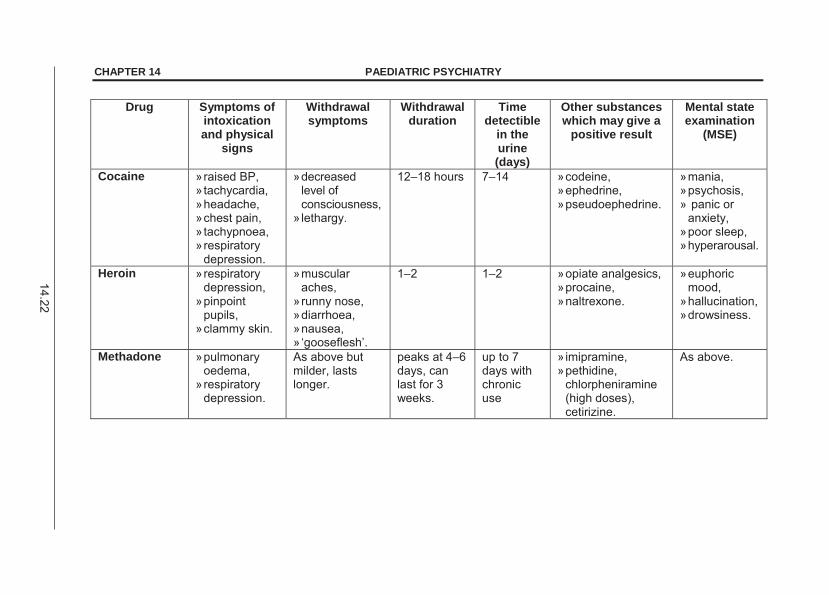
14.22
CHAPTER 14 PAEDIATRIC PSYCHIATRY
Drug Symptoms of intoxication and physical
signs
Withdrawal symptoms
Withdrawal duration
Time detectible
in the urine (days)
Other substances which may give a
positive result
Mental state examination
(MSE)
Cocaine » raised BP, » tachycardia, » headache, » chest pain, » tachypnoea, » respiratory
depression.
» decreased level of consciousness,
» lethargy.
12–18 hours 7–14 » codeine, » ephedrine, » pseudoephedrine.
» mania, » psychosis, » panic or
anxiety, » poor sleep, » hyperarousal.
Heroin » respiratory depression,
» pinpoint pupils,
» clammy skin.
» muscular aches,
» runny nose, » diarrhoea, » nausea, » ‘gooseflesh’.
1–2 1–2 » opiate analgesics, » procaine, » naltrexone.
» euphoric mood,
» hallucination, » drowsiness.
Methadone » pulmonary oedema,
» respiratory depression.
As above but milder, lasts longer.
peaks at 4–6 days, can last for 3 weeks.
up to 7 days with chronic use
» imipramine, » pethidine,
chlorpheniramine (high doses), cetirizine.
As above.
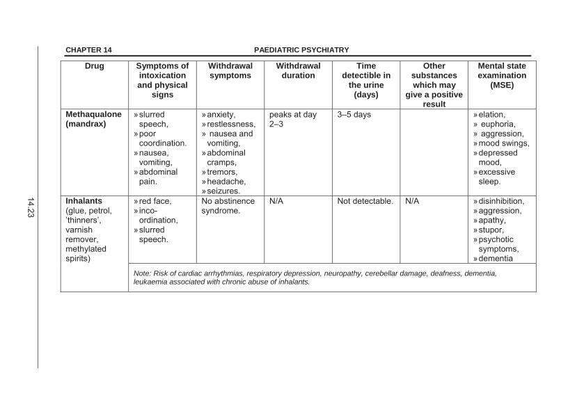
14.23
CHAPTER 14 PAEDIATRIC PSYCHIATRY
Drug Symptoms of intoxication and physical
signs
Withdrawal symptoms
Withdrawal duration
Time detectible in
the urine (days)
Other substances which may
give a positive result
Mental state examination
(MSE)
Methaqualone (mandrax)
» slurred speech,
» poor coordination.
» nausea, vomiting,
» abdominal pain.
» anxiety, » restlessness, » nausea and
vomiting, » abdominal
cramps, » tremors, » headache, » seizures.
peaks at day 2–3
3–5 days » elation, » euphoria, » aggression, » mood swings, » depressed
mood, » excessive
sleep.
Inhalants (glue, petrol, ’thinners’, varnish remover, methylated spirits)
» red face, » inco-
ordination, » slurred
speech.
No abstinence syndrome.
N/A Not detectable. N/A » disinhibition, » aggression, » apathy, » stupor, » psychotic
symptoms, » dementia
Note: Risk of cardiac arrhythmias, respiratory depression, neuropathy, cerebellar damage, deafness, dementia, leukaemia associated with chronic abuse of inhalants.
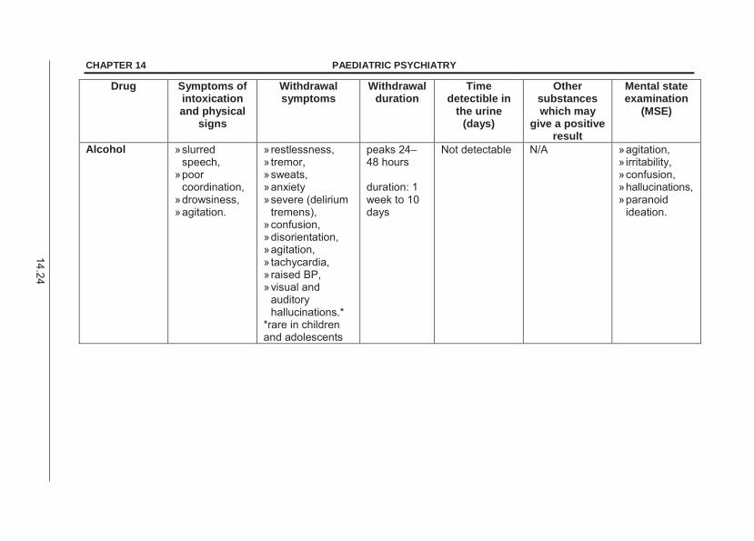
14.24
CHAPTER 14 PAEDIATRIC PSYCHIATRY
Drug Symptoms of intoxication and physical
signs
Withdrawal symptoms
Withdrawal duration
Time detectible in
the urine (days)
Other substances which may
give a positive result
Mental state examination
(MSE)
Alcohol » slurred speech,
» poor coordination,
» drowsiness, » agitation.
» restlessness, » tremor, » sweats, » anxiety » severe (delirium
tremens), » confusion, » disorientation, » agitation, » tachycardia, » raised BP, » visual and
auditory hallucinations.*
*rare in children and adolescents
peaks 24–48 hours duration: 1 week to 10 days
Not detectable N/A » agitation, » irritability, » confusion, » hallucinations, » paranoid
ideation.
14.25
DIAGNOSTIC CRITERIA (according to DSM IV-TR) Substance abuse » recurrent use resulting in a failure to fulfil major obligations, » substance related legal problems, » persistent use of substances in physically hazardous situations, » ongoing use despite negative social or interpersonal consequences. Substance dependence » substantial involvement with illicit substances (daily use, increasing
frequency and amount, school dropout or failure, social withdrawal), » loss of control (unsuccessful efforts to cut down or control use), » withdrawal and tolerance. Substance induced psychotic disorder » prominent hallucination or delusions, » symptoms occur during or within one month of proven substance abuse
or intoxication, » a mental illness such as schizophrenia or a medical condition is not the
cause of the psychosis, » the disturbance does not occur in the course of a delirium, which must
be excluded. Substance induced mood disorder » a significant and sustained disturbance in mood i.e. depressed, irritable,
expansive or elevated, » symptoms occur during or within one month of proven substance abuse
or intoxication, » a mental illness such as bipolar disorder or a medical condition is not
the cause of the mood disturbance. GENERAL AND SUPPORTIVE MEASURES » Conduct a me dical assessment (pulse, temperature, BP, ECG) and
laboratory investigations (FBC, U&E, LFTs, urine toxicology), depending on the specific drug of abuse.
» Manage withdrawal states, depending on substance of abuse. » Refer to the hospital social worker for evaluation of family circumstances
and brief motivational interviewing. MEDICINE TREATMENT OF WITHDRAWAL STATES Consult with a psychiatrist or specialised referral unit. Alcohol Management of withdrawal: Diazepam, oral, 5–15 mg daily in 2–3 divided doses.
o Taper dose over 3–5 days.
14.26
CHAPTER 14 PAEDIATRIC PSYCHIATRY Cannabis/ cannabis and mandrax Management of withdrawal states is rarely required. If symptomatic withdrawal: Diazepam, oral, 5–15 mg daily in 2–3 divided doses.
o Taper dose over 3–5 days.
Stimulants (cocaine, methamphetamine) Management of withdrawal states is rarely required. If symptomatic withdrawal: Diazepam, oral, 5–15 mg daily in 2–3 divided doses.
o Taper dose over 3–5 days. Opioids Mild withdrawal: symptomatic treatment: Diazepam, oral, 2.5–5 mg in four divided doses.
o Taper off over 5–7 days for anxiety, cramps, agitation and cravings. o < 14 years: maximum dose – 10 mg/day.
Symptomatic treatment for stomach cramps, diarrhoea, muscle pain and flu-like symptoms. Moderate to severe withdrawal: Admit to a medical facility.
Hallucinogens No detoxification indicated. Volatile Solvents No detoxification indicated. MEDICINE TREATMENT OF COMORBID PSYCHIATRIC CONDITIONS Treat according to the primary psychiatric condition, as per treatment
guidelines. See section 14.1: sedation of acutely disturbed child or adolescent awaiting psychiatric evaluation; section 14.3: Mood disorders; and section 14.5: Childhood psychosis.
Beware of adv erse interactions between illicit drugs and psychotropic medication i.e. drug lev els of both illicit drugs and psy chotropic medications are altered.
14.27
CHAPTER 14 PAEDIATRIC PSYCHIATRY REFERRAL » All for psychotherapeutic interventions or drug rehabilitation. » Outpatient treatment: refer to SANCA (South Afri can National Council
on Alcoholism and Drug Dependence)
Tel: 011 8923829 or toll free: 0861472622.
» In-patient treatment: refer for in-patient drug rehabilitation. » Patients with severe behavioural disturbance, persistent psychotic and
manic symptoms to an in-patient child and adolescent psychiatric facility, for ongoing containment and management of psychiatric symptoms.
Note: » Detoxification is rarely required in children. Mild withdrawals can be
managed on an out-patient basis. Complicated withdrawals require admission.
» Alcohol withdrawal is very rare. If alcohol withdrawal is suspected, refer and discuss with an addiction specialist.
» DSM-IV-TR diagnostic criteria have been adopted as a guide for adolescent SUD diagnoses. However, the criteria w ere developed for adults, and have not been validated in adolescents.
» Standard protocols for the management of withdrawal states have been developed for adu lts. No standard paediatric guidelines exist. Management should therefore be on a ‘ca se by case’ basis a nd discussed with paediatric specialists.
» Substance induced psychotic or mood disorders should resolve within one month of ce ssation of drug of abu se. Consider major mental illnesses such as schizophrenia or bipolar disorder if symptoms persist beyond one month.
14.6.4 BEHAVIOURAL PROBLEMS ASSOCIATED WITH
INTELLECTUAL DISABILITY F81.9 DESCRIPTION Children with intellectual disabilities present with higher rates of behavioural problems than the g eneral population. Common presentations of a dverse behaviours are: » aggression, » impulsivity, » irritability, » hyperactivity, » short attention span, » self-injurious behavior, and » inappropriate sexual behaviour.
14.28
CHAPTER 14 PAEDIATRIC PSYCHIATRY ADHD commonly co-occurs with intellectual disability and responds to treatment with stimulant medications. Autism more commonly co-occurs with intellectual disability. Depression occurs at a ra te similar to t he general population and 2 5% of children and adolescents suffer significant anxiety. Increased rates of me dical conditions such as seizures, genetic disorders and dysmorphic syndromes are associated with intellectual disability. Intellectual disability associated with comorbid epilepsy is associated with increased rates of ADHD, behavioural dyscontrol and psychosis. DIAGNOSTIC CRITERIA Diagnostic criteria for p sychiatric disorders in children with intellectual disability are the same as those for the general population. However symptom expression may vary with developmental stage or level of intellectual functioning. GENERAL AND SUPPORTIVE MEASURES » Exclude medical conditions in children presenting with behavioural
disturbances, particularly in children who are not a ble to communicate symptoms verbally (e.g. seizures, dental caries, covert infections, poisoning, foreign bodies, space occupying brain lesions and drug side effects).
» Exclude emotional, physical or sexual abuse in a c hild presenting with persistent adverse behaviour and emotional distress (especially in non-verbal children).
» Parental guidance is an important part of the management of children presenting with behavioural problems (psycho-education, behavior management).
» Behaviour modification principles form the basis of p sychotherapeutic intervention.
MEDICINE TREATMENT Psychotropic medication treatment should occur as part of a multidisciplinary diagnostic and therapeutic intervention. Treat according to the pr imary psychiatric condition, as per treatme nt guidelines. Do baseline blood tests and ECGs, particularly in children with underlying medical conditions. Start with the lowest doses possible. Increase dosages cautiously as children with intellectual disability may be more susceptible to adverse effects such as ex trapyramidal side effects (EPSEs), neuroleptic malignant syndrome (NMS) or the disinhibiting effects of benzodiazepines.

14.29
CHAPTER 14 PAEDIATRIC PSYCHIATRY REFERRAL » Children presenting with severe aggression, inappropriate sexual
behavior or self-injurious behavior for a diagnostic assessment, or admission to a terti ary level child psychiatry service, or a dedicated intellectual disability psychiatric service (if such a service exists in the region).
» Children presenting with psychosis or m ania for a n assessment or admission to a psychiatric unit.
» To a social worker or child protection services if abuse is suspected.
15.1
CHAPTER 15 RESPIRATORY SYSTEM
15.1 CHRONIC LUNG INFECTIONS 15.1.1 BRONCHIECTASIS J47 DESCRIPTION Irreversible dilatation of the subsegmental airways, inflammatory destruction of bronchial and peribronchial tissue, and accumulation of exudative material in dependent bronchi that occurs as a result of recurrent bacterial infections and aspiration pneumonia. There is bronchial luminal obstruction; ciliary dyskinesia; thick, tenacious secretions and lung tissue damage. Complications include cor pulmonale and respiratory failure. Predisposing conditions include HIV, TB, cystic fibrosis, primary ciliary dyskinesia primary immunodeficiency syndromes. DIAGNOSTIC CRITERIA » Chronic cough, usually with mucopurulent sputum and occasional
haemoptysis. » A bout of coughing on physical activity or change in posture, particularly
while reclining. » Cyanosis, fever, malaise, anorexia, poor weight gain, halitosis and
clubbing. » Recurrent and persistent lower respiratory tract infections. » Chest X-ray showing cystic dilatation and tram tracking. » If diagnosis is uncertain or where localised disease on chest X-ray is
suspected, perform high resolution computed tomography. Features include cystic dilatation, signet ring sign, tram tracking.
GENERAL AND SUPPORTIVE MEASURES » Identify and treat the underlying disorder or bacterial source. » Clear secretions effectively with postural drainage and physiotherapy. » Eliminate all foci of infection. » Nutritional support. » Nebulise with sodium chloride 0.9% or sodium chloride 3% (hypertonic
saline) to aid sputum expectoration. Mix 3 mL of 5% sodium chloride with 2 mL water to make 3% solution. Method of sputum induction Precaution: If undertaking procedure in acutely sick child with respiratory compromise, be prepared to manage acute bronchospasm as this may be an associated adverse effect.
15.2
CHAPTER 15 RESPIRATORY SYSTEM Nebulise with a bronchodilator: Salbutamol, solution, 0.15–0.3 mg/kg/dose in 2–4mL of sodium
chloride 3% delivered at a flow of 5 L/min ute with oxygen for 20 minutes.
» Perform physiotherapy. » Encourage patient to cough up sputum or if infant or small child obtain
nasopharyngeal aspirate post physiotherapy. » Send sample for culture and cytology as indicated. SURGICAL TREATMENT Consider in localised severe disease or progressive disease despite adequate medical treatment. MEDICINE TREATMENT Empiric antibiotic therapy for acute lung infections: Ampicillin, IV, 25 mg/kg/dose, 6 hourly.
PLUS Gentamicin, IV, 6 mg/kg once daily.
Change antibiotics according to culture and sensitivity results. If poor response and no culture to guide antibiotic choice, consider infection due to S. aureus, TB or fungal infection.
If there is evidence of good clinical response, change to: Amoxicillin/clavulanic acid, oral, 30 mg/kg/dose of amoxicillin
component 8 hourly. Total antibiotic duration of 14 days. Note: These antibiotic regimens do not apply to children with cystic fibrosis. In the acute phase if wheeze is present: Salbutamol solution, 5 mg/mL, nebulise 4 hourly.
o 5 mg salbutamol in 2–4 mL sodium chloride 0.9%. In chronic phase » Theophylline, modified release, oral, 6 mg/kg/dose 12 hourly.
o Titrate dose for optimal response. o Maximum dose 10 mg/kg/dose.
Annual influenza vaccination.
Pneumococcal vaccine (conjugated), 2 additional doses 8 weeks apart. REFERRAL » Poor response to therapy. » For confirmation of the diagnosis. » For early surgical intervention of localised disease.
15.3
CHAPTER 15 RESPIRATORY SYSTEM
15.1.2 LUNG ABSCESS J85 DESCRIPTION A suppurative process that results from de struction of the pulm onary parenchyma and formation of a cavity. The cavity may be single, e.g. after aspiration or multiple, e.g. staphylococcal disease and cystic fibrosis. Lung abscess may follow pneumonia caused by:
» S. aureus, » K. pneumoniae, » anaerobic organisms, » S pneumoniae, » H. influenza, » M. tuberculosis.
Metastatic lung abscesses due to septicaemia or septic emboli may also occur. Complications include:
» bronchiectasis, » rupture into the bronchial tree
or pleural cavity or vessels,
» empyema, » pulmonary osteo-arthropathy, » brain abscess, and
» bronchopleural fistula. DIAGNOSTIC CRITERIA » Intermittent or recurrent fever, malaise, weight loss, anorexia and clubbing. » Productive purulent cough with halitosis and haemoptysis. » Amphoric breathing over the cavity may be present. » Chest X-ray will confirm cavity/cavities with or without an air-fluid level. GENERAL AND SUPPORTIVE MEASURES » Identify underlying cause. » Physiotherapy and postural drainage. » Correct anaemia. » Nutritional support. MEDICINE TREATMENT Empiric antibiotic therapy for at least 14 days. Ampicillin, IV, 25 mg/kg/dose, 6 hourly.
PLUS Gentamicin, IV, 6 mg/kg/day as a single daily dose.
PLUS Metronidazole, IV, 7.5 mg/kg/dose, 8 hourly.
Change antibiotics according to culture and sensitivity results. Poor response and no culture to guide antibiotic choice: ADD Cloxacillin, IV, 50 mg/kg/dose, every 6 hours.
If there is evidence of good clinical response, change to only: Amoxicillin/clavulanic acid, oral, 30 mg/kg/dose of amoxicillin
component 8 hourly.
15.4
CHAPTER 15 RESPIRATORY SYSTEM SURGICAL TREATMENT Consider surgical drainage of abscess and/or resection if medical treatment fails. REFERRAL » Complicated lung abscess not responding to therapy. » Lung abscess where the underlying cause has not been established. 15.1.3 TUBERCULOSIS, PERINATAL P37.0 *Notifiable condition DESCRIPTION Tuberculosis acquired in the first 3 months of life. Perinatal tuberculosis may be acquired in one of the following ways: » transplacental transmission, usually extrapulmonary or disseminated TB, » via the passage of swallowed maternal blood or amniotic fluid during
delivery, usually extrapulmonary TB, or » inhalation of the bacilli during the neonatal period – usually pulmonary TB. DIAGNOSTIC CRITERIA » Hepatosplenomegaly, a suggestive chest X-ray, TB exposure via a
mother or close contact with another source case. » Positive smear or culture on any suitable sample e.g. gastric aspirate in
the neonate or tissue histology suggestive of TB. » Endometrial swabs or sputum samples in the mother positive for M.
tuberculosis. See section 15.1.4: Tuberculosis, pulmonary. GENERAL AND SUPPORTIVE MEASURES » Check drug sensitivity of source. If resistant, refer. » Check HIV status of mother and, if positive, test baby with HIV PCR. » Screen all household contacts for tuberculous infection or disease. » Monitor the nutritional status of the neonate. » Do not give BCG vaccine at birth. MEDICINE TREATMENT Treatment Newborn infant of mother with tuberculosis with newborn having any signs suggestive of illness.
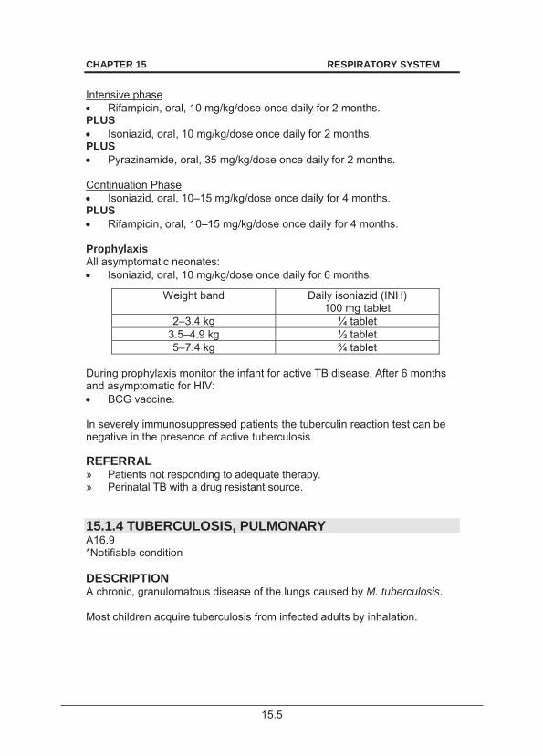
15.5
CHAPTER 15 RESPIRATORY SYSTEM Intensive phase Rifampicin, oral, 10 mg/kg/dose once daily for 2 months.
PLUS Isoniazid, oral, 10 mg/kg/dose once daily for 2 months.
PLUS Pyrazinamide, oral, 35 mg/kg/dose once daily for 2 months.
Continuation Phase Isoniazid, oral, 10–15 mg/kg/dose once daily for 4 months.
PLUS Rifampicin, oral, 10–15 mg/kg/dose once daily for 4 months.
Prophylaxis All asymptomatic neonates: Isoniazid, oral, 10 mg/kg/dose once daily for 6 months.
Weight band Daily isoniazid (INH) 100 mg tablet
2–3.4 kg ¼ tablet 3.5–4.9 kg ½ tablet 5–7.4 kg ¾ tablet
During prophylaxis monitor the infant for active TB disease. After 6 months and asymptomatic for HIV: BCG vaccine.
In severely immunosuppressed patients the tuberculin reaction test can be negative in the presence of active tuberculosis. REFERRAL » Patients not responding to adequate therapy. » Perinatal TB with a drug resistant source. 15.1.4 TUBERCULOSIS, PULMONARY A16.9 *Notifiable condition DESCRIPTION A chronic, granulomatous disease of the lungs caused by M. tuberculosis. Most children acquire tuberculosis from infected adults by inhalation.
15.6
CHAPTER 15 RESPIRATORY SYSTEM Malnourished, immunosuppressed (HIV and AIDS) and children < 3 years of age with pulmonary tuberculosis (PTB) are always regarded as having a very serious disease. Complications include: » enlarged hilar and mediastinal lymphadenopathy with obstruction, e.g.
tracheal or bronchial airway compression or occlusion with secondary atelectasis or hyperinflation;
» local spread of infection, e.g. TB bronchopneumonia, pleural effusion or cavitation;
» disseminated disease, e.g. miliary TB, TB meningitis and metastatic extrapulmonary involvement.
DIAGNOSTIC CRITERIA
Any child presenting with symptoms and signs suggestive of pulmonary TB is regarded as a case of TB if there is: » a chest X-ray suggestive of TB, and/or » history of exposure to an infectious TB case and/or positive Tuberculin
Skin Test (TST) e.g. Mantoux. The diagnosis is supported by a positive smear microscopy and/or positive M. tuberculosis PCR e.g. GeneXpert®. Culture, usually on gastric aspirates or induced sputum, is a confirmatory test.
» Signs and symptoms include: > unexplained weight loss or failure to thrive, > 2 weeks, > chronic unremitting cough for > 14 days, > lymphadenopathy (especially cervical, often matted), > hepatosplenomegaly, > consolidation and pleural effusion.
» The following may be evident on chest X-ray: > Direct or indirect evidence of hilar or mediastinal adenopathy with or
without parenchymal opacification and/or bronchopneumonia, > miliary changes, > pleural effusions.
Note: Miliary pattern on chest X-rays of H IV infected children may also be suggestive of a diagnosis of lymphoid interstitial pneumonitis (LIP). (The miliary pattern of TB extends into the periphery of the lungs whereas LIP usually does not)
» Exposure to an adult with pulmonary tuberculosis.
15.7
CHAPTER 15 RESPIRATORY SYSTEM
» Tuberculin skin test (TST) e.g. Mantoux.
> A positive TST 10 mm. > A TST may be falsely negative in the presence of:
malnutrition, immunodeficiency, e.g. HIV and AIDS, immunosuppression, e.g. steroid therapy, cancer chemotherapy, following overwhelming viral infection, e.g. measles or post
vaccination. In these circumstances a TST induration of 5 mm may be regarded as positive. Frequently, the TST will be n on-reactive in these cases and a decision not to start TB treatment should not be based on a negative TST test.
» M. tuberculosis is suggested by positive microscopy (acid fast bacilli on smear) and confirmed by culture on the fol lowing specimens but most children will not have microbiological confirmation of TB: > early morning gastric aspirate (empty stomach, no oral fo od intake
for 4 hours), > sputum (older children), > induced sputum, > CSF, > pleural and ascitic fluids, > fine needle aspirate biopsies of lymph nodes, > ear swabs for culture in chronic otorrhoea.
» PCR sputum positive on molecular testing. > The M. tuberculosis PCR has an inferior yield to l iquid culture and
therefore should not replace culture. Identification of r ifampicin resistance would imply multidrug resistance and allow modification of therapy accordingly.
> Where available, the molecular M. tuberculosis PCR test should be performed on sputum and gastric aspirates in preference to fluorescent smear for acid fast bacilli as it increases the diagnostic yield and allows early identification of rifampicin resistance.
» Microscopy and culture in all cases. GENERAL AND SUPPORTIVE MEASURES » Identify and treat the source case. » In case of known contact with adult MDR TB case, the child requires
referral for appropriate MDR TB prophylaxis or treatment. » Screen all contacts for TB infection. » Screen all contacts for TB infection. » Monitor the nutritional status of the child to assess response to
treatment. » Only symptomatic pleural effusions should be drained via pleural
aspiration. (In such cases consider adjunctive steroid therapy) » Ensure household infection control practices.
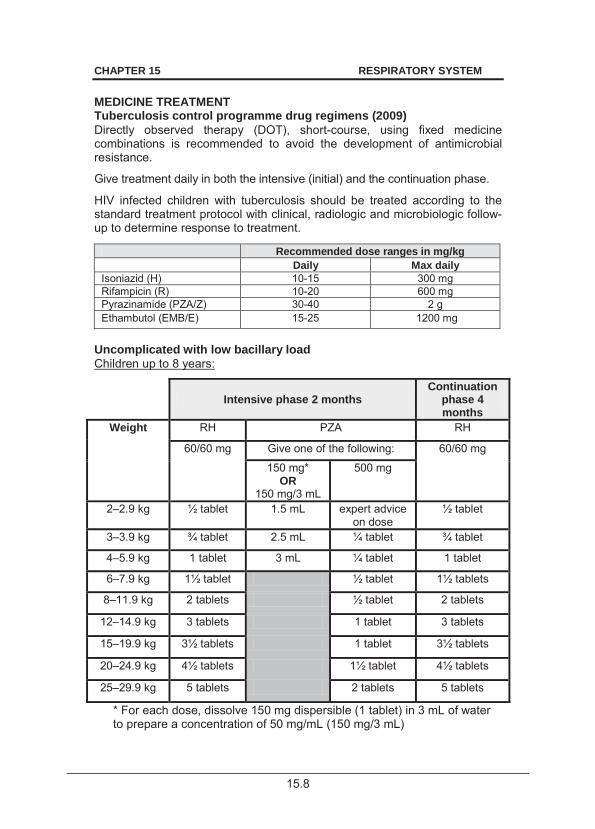
15.8
CHAPTER 15 RESPIRATORY SYSTEM MEDICINE TREATMENT Tuberculosis control programme drug regimens (2009) Directly observed therapy (DOT), short-course, using fixed medicine combinations is recommended to avoid the development of antimicrobial resistance.
Give treatment daily in both the intensive (initial) and the continuation phase.
HIV infected children with tuberculosis should be treated according to the standard treatment protocol with clinical, radiologic and microbiologic follow-up to determine response to treatment.
Recommended dose ranges in mg/kg Daily Max daily Isoniazid (H) 10-15 300 mg Rifampicin (R) 10-20 600 mg Pyrazinamide (PZA/Z) 30-40 2 g Ethambutol (EMB/E) 15-25 1200 mg
Uncomplicated with low bacillary load Children up to 8 years:
Intensive phase 2 months
Continuation phase 4 months
Weight RH PZA RH
60/60 mg Give one of the following: 60/60 mg
150 mg* OR
150 mg/3 mL
500 mg
2–2.9 kg ½ tablet 1.5 mL expert advice on dose
½ tablet
3–3.9 kg ¾ tablet 2.5 mL ¼ tablet ¾ tablet
4–5.9 kg 1 tablet 3 mL ¼ tablet 1 tablet
6–7.9 kg 1½ tablet ½ tablet 1½ tablets
8–11.9 kg 2 tablets ½ tablet 2 tablets
12–14.9 kg 3 tablets 1 tablet 3 tablets
15–19.9 kg 3½ tablets 1 tablet 3½ tablets
20–24.9 kg 4½ tablets 1½ tablet 4½ tablets
25–29.9 kg 5 tablets 2 tablets 5 tablets
* For each dose, dissolve 150 mg dispersible (1 tablet) in 3 mL of water to prepare a concentration of 50 mg/mL (150 mg/3 mL)
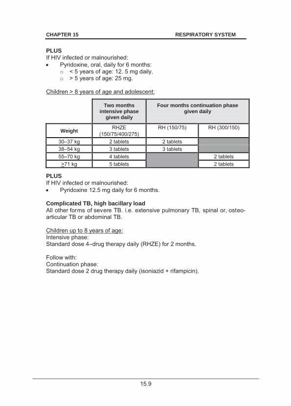
15.9
CHAPTER 15 RESPIRATORY SYSTEM PLUS If HIV infected or malnourished: Pyridoxine, oral, daily for 6 months:
o < 5 years of age: 12. 5 mg daily, o > 5 years of age: 25 mg.
Children > 8 years of age and adolescent:
Two months intensive phase
given daily
Four months continuation phase given daily
Weight RHZE (150/75/400/275)
RH (150/75) RH (300/150)
30–37 kg 2 tablets 2 tablets 38–54 kg 3 tablets 3 tablets 55–70 kg 4 tablets 2 tablets >71 kg 5 tablets 2 tablets
PLUS If HIV infected or malnourished: Pyridoxine 12.5 mg daily for 6 months.
Complicated TB, high bacillary load All other forms of severe TB. i.e. extensive pulmonary TB, spinal or, osteo-articular TB or abdominal TB. Children up to 8 years of age: Intensive phase: Standard dose 4–drug therapy daily (RHZE) for 2 months. Follow with: Continuation phase: Standard dose 2 drug therapy daily (isoniazid + rifampicin).
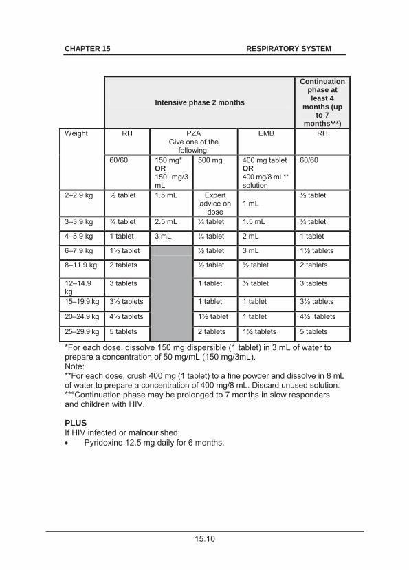
15.10
CHAPTER 15 RESPIRATORY SYSTEM
Intensive phase 2 months
Continuation phase at least 4
months (up to 7
months***) Weight RH PZA
Give one of the following:
EMB RH
60/60 150 mg* OR 150 mg/3 mL
500 mg 400 mg tablet OR 400 mg/8 mL** solution
60/60
2–2.9 kg ½ tablet 1.5 mL Expert advice on
dose
1 mL
½ tablet
3–3.9 kg ¾ tablet 2.5 mL ¼ tablet 1.5 mL ¾ tablet
4–5.9 kg 1 tablet 3 mL ¼ tablet 2 mL 1 tablet
6–7.9 kg 1½ tablet ½ tablet 3 mL 1½ tablets
8–11.9 kg 2 tablets ½ tablet ½ tablet
2 tablets
12–14.9 kg
3 tablets 1 tablet ¾ tablet 3 tablets
15–19.9 kg 3½ tablets 1 tablet 1 tablet 3½ tablets
20–24.9 kg 4½ tablets 1½ tablet 1 tablet 4½ tablets
25–29.9 kg 5 tablets 2 tablets 1½ tablets 5 tablets
*For each dose, dissolve 150 mg dispersible (1 tablet) in 3 mL of water to prepare a concentration of 50 mg/mL (150 mg/3mL). Note: **For each dose, crush 400 mg (1 tablet) to a fine powder and dissolve in 8 mL of water to prepare a concentration of 400 mg/8 mL. Discard unused solution. ***Continuation phase may be prolonged to 7 months in slow responders and children with HIV. PLUS If HIV infected or malnourished: Pyridoxine 12.5 mg daily for 6 months.
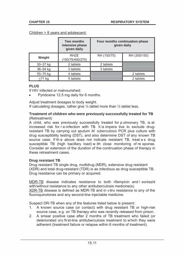
15.11
CHAPTER 15 RESPIRATORY SYSTEM
Children > 8 years and adolescent:
Two months intensive phase
given daily
Four months continuation phase given daily
Weight RHZE (150/75/400/275)
RH (150/75) RH (300/150)
30–37 kg 2 tablets 2 tablets 38–54 kg 3 tablets 3 tablets 55–70 kg 4 tablets 2 tablets >71 kg 5 tablets 2 tablets
PLUS If HIV infected or malnourished: Pyridoxine 12.5 mg daily for 6 months.
Adjust treatment dosages to body weight. If calculating dosages, rather give ½ tablet more than ½ tablet less. Treatment of children who were previously successfully treated for TB (Retreatment) A child, who was previously successfully treated for p ulmonary TB, is at increased risk for r e-infection with TB. It is impera tive to exclude drug-resistant TB by carrying out sputum M. tuberculosis PCR plus culture with drug susceptibility testing (DST), and also determine DST of any known TB source case. If th e above does not indicate resistant TB, treat a s drug susceptible TB (high bacillary load) w ith close monitoring of re sponse. Consider an extension of the duration of the continuation phase of therapy in these retreatment cases. Drug resistant TB Drug resistant TB single drug, multidrug (MDR), extensive drug resistant (XDR) and total drug-resistant (TDR) is as infectious as drug susceptible TB. Drug resistance can be primary or acquired. MDR-TB disease indicates resistance to both rifampicin and i soniazid with/without resistance to any other antituberculosis medicine(s). XDR-TB disease is defined as MDR-TB and in vitro resistance to any of the fluoroquinolones and any second-line injectable medicine. Suspect DR-TB when any of the features listed below is present: 1. A known source case (or contact) with drug resistant TB or high-risk
source case, e.g. on TB therapy who was recently released from prison. 2. A smear positive case after 2 months of TB treatment who failed (or
deteriorated on) first-line antituberculosis treatment to which they were adherent (treatment failure or relapse within 6 months of treatment).
15.12
CHAPTER 15 RESPIRATORY SYSTEM 3. Any severely ill child with TB that failed or got worse on TB treatment.
4. Defaulted TB treatment (> 2 months). 5. Treatment interruptions (< 1 month) or who relapsed while on TB
treatment or at the end of treatment. 6. With recurrent TB disease after completion of TB treatment (retreatment
case). When DR-TB is suspected, submit appropriate microbiological specimens for genotypic drug sensitivity test and culture for phenotypic drug susceptibility testing. M. tuberculosis PCR tests for rifampicin resistance only while the line probe assay (LPA) tests for isoniazid and r ifampicin susceptibility. Second-line LPA tests for other antimicrobial resistance including quinolones. All samples that test positive on molecular PCR testing must have samples submitted for culture and drug susceptibility testing but therapy for MDR-TB must be instituted while awaiting results. False positive results with both the M. tuberculosis PCR. and line probe assay have been recorded. Clinical and radiological correlation with molecular results must always be considered and discuss discordant results with an expert. Manage confirmed DR-TB in a dedic ated MDR-TB unit w ith appropriate infection control measures to prevent nosocomial transmission. Initiate treatment in consultation with a designated expert while awaiting referral to the designated MDR-TB centre. An uni nterrupted medicine supply, direct supervision with proper education and counselling is necessary. The standardised empiric treatment protocol for M DR-TB for children is 5 drugs for 6 months or more for at least 6 days a week during the intensive phase and 4 drugs for at least 6 days a week for 18 months or less during the continuation phase. Exact duration of therapy for the intensive phase is 4 months after the first date of sampling of a negative culture result while the total duration of therapy should be 18 months after the first date of sampling of a negative culture result. Children < 8 years with MDR-TB Intensive phase: Levofloxacin, oral.
o 15–20 mg/kg/dose once daily. o Maximum dose: 1 000 mg.
Amikacin, IV, 15–22.5 mg/kg daily. Terizidone, oral, 15–20 mg/kg daily. Ethionamide, oral, 15–20 mg/kg daily. Pyrazinamide, oral, 30–40 mg/kg daily
15.13
CHAPTER 15 RESPIRATORY SYSTEM Continuation phase: Same as initial phase but stop amikacin. Children > 8 years with MDR-TB Intensive phase: Moxifloxacin, oral, daily.
o < 25 kg: 200mg o > 25 kg: 400 mg
Amikacin, IV, 15–22.5 mg/kg daily. Terizidone, oral, 15–20 mg/kg daily. Ethionamide, oral, 15–20 mg/kg daily. Pyrazinamide, oral, 30–40 mg/kg daily.
Continuation phase: Same as intensive phase but stop amikacin. Other agents may be substituted in special situations and in c onsultation with a designated expert. Cases of DR-TB must be monitored clinically, with radiology and microbiologically for res ponse to t herapy. TB culture conversion occurs when 2 consecutive TB culture results on sputum/gastric aspirates taken 30 days apart are negative and thereafter remains negative. Disseminated (Miliary) TB Children < 8 years A 6-month regimen of all 4 the following medicines: Rifampicin, oral, 20 mg/kg as a single daily dose.
o Maximum dose, oral, 600mg daily. PLUS Isoniazid, oral, 20 mg/kg as a single daily dose.
o Maximum dose, oral, 400mg daily. PLUS Pyrazinamide, oral, 40 mg/kg as a single daily dose.
o Maximum daily dose: 2 000 mg. PLUS Ethionamide, oral, 20 mg/kg as a single daily dose.
o Maximum daily dose: 1 000 mg. Pyridoxine 25 mg daily for 6 months.
Note: All cases of miliary TB should have a lumbar puncture (LP) preformed. Any abnormal CSF results or where a LP is not performed, should be treated as a patient with TBM. See section 8.13: Meningitis, tuberculosis (TBM).
15.14
CHAPTER 15 RESPIRATORY SYSTEM Preventive therapy for TB exposure/infection Screen all children in close contact with an infectious pulmonary TB case for TB disease. Screening includes clinical history/examination and, if available, chest X-ray and tuberculin skin test (TST). Give antituberculosis treatment if the diagnosis of TB disease is confirmed or suspected. Indications for Isoniazid Preventive Therapy (IPT): » All asymptomatic children < 5 years of age, or HIV-infected irrespective
of age, i. e. clinically normal, normal chest X-ray and T ST positive or negative, in close contact with an infectious pulmonary TB case should receive isoniazid preventive therapy (IPT).
» Children < 5 years of age, or HIV-infected irrespective of age, who have had no previous TB treatment or preventive therapy, are asymptomatic without a history of close contact with an infectious pulmonary TB case but found to have a positive TST.
» Previous isoniazid preventive therapy or treatment does not protect the child against subsequent TB exposure/infection. If there is re-exposure to an inf ectious pulmonary TB case a fter completion of 6 months of chemotherapy, children (< 5 y ears or HIV-infected) should receive IPT after each episode of documented TB exposure for 6 months. In cases of re-exposure to infectious source cases while the child is on IPT, the duration of IPT should continue for as long as the source case remains infectious.
Isoniazid, oral, 10 mg/kg daily for 6 months.
Preventive therapy in case of drug-resistant TB contact: Isoniazid monoresistance: Rifampicin, oral, 15 mg/kg daily for 4 months.
Rifampicin monoresistance: Isoniazid, oral, 10 mg/kg daily for 6 months.
MDR or XDR-TB: Close follow-up for two years. Ensure household infection control practices are observed. Refer all cases. REFERRAL » Poor response to standard TB treatment. » Failure to exclude MDR-TB. » Adverse drug reactions (ADR) requiring single drug combinations. » MDR or MDR-TB contact.
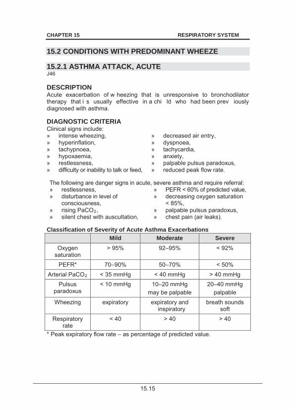
15.15
CHAPTER 15 RESPIRATORY SYSTEM 15.2 CONDITIONS WITH PREDOMINANT WHEEZE 15.2.1 ASTHMA ATTACK, ACUTE J46 DESCRIPTION Acute exacerbation of w heezing that is unresponsive to bronchodilator therapy that i s usually effective in a chi ld who had been prev iously diagnosed with asthma. DIAGNOSTIC CRITERIA Clinical signs include: » intense wheezing, » decreased air entry, » hyperinflation, » dyspnoea, » tachypnoea, » tachycardia, » hypoxaemia, » anxiety, » restlessness, » palpable pulsus paradoxus, » difficulty or inability to talk or feed, » reduced peak flow rate. The following are danger signs in acute, severe asthma and require referral: » restlessness, » disturbance in level of
consciousness,
» PEFR < 60% of predicted value, » decreasing oxygen saturation
< 85%, » rising PaCO2, » silent chest with auscultation,
» palpable pulsus paradoxus, » chest pain (air leaks).
Classification of Severity of Acute Asthma Exacerbations
Mild Moderate Severe Oxygen
saturation > 95% 92–95% < 92%
PEFR* 70–90% 50–70% < 50% Arterial PaCO2 < 35 mmHg < 40 mmHg > 40 mmHg
Pulsus paradoxus
< 10 mmHg 10–20 mmHg may be palpable
20–40 mmHg palpable
Wheezing expiratory expiratory and inspiratory
breath sounds soft
Respiratory rate
< 40 > 40 > 40
* Peak expiratory flow rate – as percentage of predicted value.
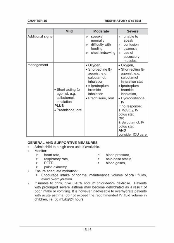
15.16
CHAPTER 15 RESPIRATORY SYSTEM
Mild Moderate Severe Additional signs
» speaks normally
» difficulty with feeding
» chest indrawing
» unable to speak
» confusion » cyanosis » use of
accessory muscles
management
Short-acting ß2 agonist, e.g. salbutamol, inhalation
PLUS Prednisone, oral
Oxygen, Short-acting ß2 agonist, e.g. salbutamol, inhalation ± ipratropium bromide inhalation Prednisone, oral
Oxygen, Short-acting ß2 agonist, e.g. salbutamol inhalation stat Ipratropium bromide inhalation, Hydrocortisone, IV
If no response: ± MgSO4, IV bolus stat OR ± Salbutamol, IV bolus stat AND consider ICU care
GENERAL AND SUPPORTIVE MEASURES » Admit child to a high care unit, if available. » Monitor:
> heart rate, > blood pressure, > respiratory rate, > acid-base status, > PEFR, > blood gases, > pulse oximetry.
» Ensure adequate hydration: > Encourage intake of nor mal maintenance volume of ora l fluids,
avoid overhydration. » If unable to drink, give 0.45% sodium chloride/5% dextrose. Patients
with prolonged severe asthma may become dehydrated as a resu lt of poor intake or vomiting. It is however inadvisable to overhydrate patients with acute asthma: do not exceed the recommended IV fluid volume in children, i.e. 50 mL/kg/24 hours.
15.17
CHAPTER 15 RESPIRATORY SYSTEM Note: Physiotherapy, antihistamines, antibiotics and sedation are not beneficial in the acute setting. Agitation and restlessness are signs of severe hypoxia. MEDICINE TREATMENT Mild and moderate asthma Bronchodilator, i.e. short-acting ß2 agonist Salbutamol, inhalation, using a metered-dose inhaler with a spacer
device. o 200–400 mcg (2–4 puffs) repeated every 20–30 minutes depending
on clinical response. OR Salbutamol, solution, 0.15–0.3 mg/kg/dose nebulise at 20 minute
intervals for 3 doses. o Maximum dose: 5 mg/dose. o 5 mg salbutamol in 4 mL sodium chloride 0.9% delivered at a flow
of 5 L/minute with oxygen. PLUS Prednisone, oral, 1–2 mg /kg, daily immediately up to a maximum of:
o 20 mg: children < 2 years for 5 days. o 30 mg: children 2–5 years for 5 days. o 40 mg: children 6–12 years for 5 days.
Moderate or severe asthma Step 1: To maintain arterial oxygen saturation 95%: Oxygen, 100%, at least 4–6 L/minute by facemask or 1–2 L/minute by
nasal cannula. PLUS Short-acting ß2 agonist: Salbutamol, inhalation, using a metered-dose inhaler with a spacer
device. Up to 10 puffs (1mg) per administration for severe asthma. o 400–600 mcg (4–6 puffs) up to 10 puffs repeated every 20–30
minutes depending on clinical response. OR Salbutamol, solution, 0.15–0.3 mg/kg/dose nebulise at 20 minute
intervals for 3 doses. o Maximum dose: 5 mg/dose. o 5 mg salbutamol in 4 mL sodium chloride 0.9% delivered at a flow
of 5 L/minute with oxygen.
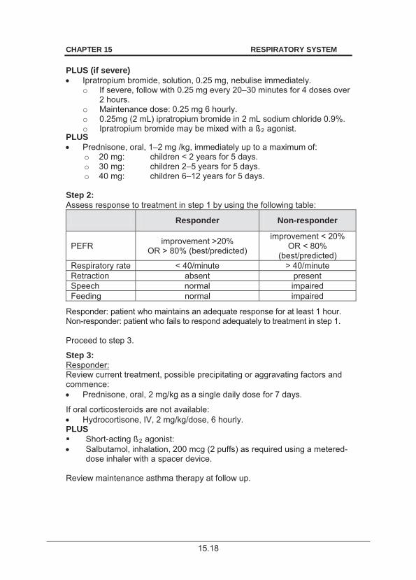
15.18
CHAPTER 15 RESPIRATORY SYSTEM PLUS (if severe) Ipratropium bromide, solution, 0.25 mg, nebulise immediately.
o If severe, follow with 0.25 mg every 20–30 minutes for 4 doses over 2 hours.
o Maintenance dose: 0.25 mg 6 hourly. o 0.25mg (2 mL) ipratropium bromide in 2 mL sodium chloride 0.9%. o Ipratropium bromide may be mixed with a ß2 agonist.
PLUS Prednisone, oral, 1–2 mg /kg, immediately up to a maximum of:
o 20 mg: children < 2 years for 5 days. o 30 mg: children 2–5 years for 5 days. o 40 mg: children 6–12 years for 5 days.
Step 2: Assess response to treatment in step 1 by using the following table:
Responder Non-responder
PEFR improvement >20% OR > 80% (best/predicted)
improvement < 20% OR < 80%
(best/predicted) Respiratory rate < 40/minute > 40/minute Retraction absent present Speech normal impaired Feeding normal impaired
Responder: patient who maintains an adequate response for at least 1 hour. Non-responder: patient who fails to respond adequately to treatment in step 1. Proceed to step 3.
Step 3: Responder: Review current treatment, possible precipitating or aggravating factors and commence: Prednisone, oral, 2 mg/kg as a single daily dose for 7 days.
If oral corticosteroids are not available: Hydrocortisone, IV, 2 mg/kg/dose, 6 hourly.
PLUS Short-acting ß2 agonist: Salbutamol, inhalation, 200 mcg (2 puffs) as required using a metered-
dose inhaler with a spacer device. Review maintenance asthma therapy at follow up.
15.19
CHAPTER 15 RESPIRATORY SYSTEM Non-responder: Intensify treatment as follows: Short-acting ß2 agonist: Salbutamol, solution, 0.15–0.3 mg/kg/dose nebulise at 20 minute
intervals for 3 doses. o Maximum dose: 5 mg/dose. o 5 mg salbutamol in 4 mL sodium chloride 0.9% delivered at a flow
of 5 L/minute with oxygen. AND Ipratropium bromide, solution, 0.25 mg, nebulise immediately.
o If severe, follow with 0.25 mg every 20–30 minutes for 4 doses over 2 hours.
o Maintenance dose: 0.25 mg 6 hourly. o 0.25mg (2 mL) ipratropium bromide in 2 mL sodium chloride 0.9%. o Ipratropium bromide may be mixed with a ß2 agonist.
PLUS Continue corticosteroid: Prednisone, oral, 2 mg/kg as a single daily dose.
OR Hydrocortisone, IV, 2 mg/kg/dose 6 hourly.
Failure to respond - consult paediatrician Magnesium sulphate, IV bolus, 25–75 mcg/kg administered over 20 minutes.
OR Salbutamol, IV, 15 m cg/kg as a sing le dose administered over
10 minutes. Consider need for intensive care. Step 4: Assess response to treatment in Step 3. If non-responsive, admit to intensive care unit for consideration of: Magnesium sulphate, IV bolus, 25–75 mcg/kg administered over 20
minutes (if not already given). AND Salbutamol, IV.
o Loading dose: 0.5 mcg/kg (do not give if stat dose already given). o Follow with: 0.2 mcg/kg/minute. o If necessary, increase dose by 0.1 mcg/kg every 15 minutes. o Maximum dose: 5 mcg/kg/minute. o Monitor electrolytes.
15.20
CHAPTER 15 RESPIRATORY SYSTEM No further response In cases of life threatening asthma in the intensive care unit: Aminophyline, IV, 5 mg/kg, loading dose administered over 20–30
minutes. Omit loading dose in childr en receiving maintenance oral theophylline. o Follow with: 1 mg/kg/hour continuous infusion. o ECG monitoring.
REFERRAL » Acute exacerbation not responding to treatment. 15.2.2 ASTHMA, CHRONIC J 45 DESCRIPTION Asthma is a chronic inflammatory airways disease in which many cells and cellular elements play a role. Susceptible individuals present with recurrent episodes of w heezing, breathlessness, chest tightness and cough particularly in t he early morning. There is widespread variable airflow obstruction that is reversible either spontaneously or w ith treatment. A variety of stimuli, e.g. allergens, viral infections, weather changes, emotional upsets or other irrit ants precipitate inflammation that is associated with increased bronchial hyper-responsiveness. DIAGNOSTIC CRITERIA » Chronic, persistent/recurrent cough and/or wheezing that responds to a
bronchodilator. » Objective evidence of reversible airway obstruction, as measured by > 15%
improvement of the peak flow or > 12% improv ement in th e FEV1 20 minutes after admin istration of an inhaled bronchodilat or confirms the diagnosis. (FEV1 = forced expiratory volume in 1 second)
» A family history of atopy , night or ex ercise-induced coughing and/or wheezing.
Control of asthma The severity of asthm a can v ary with time and re gular reassessments (at least every 3 months) are necessary. On treatment chronic asthma is classified as: » controlled, » partially controlled, or » uncontrolled.
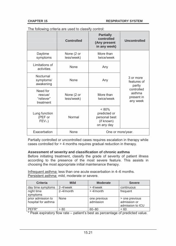
15.21
CHAPTER 15 RESPIRATORY SYSTEM
The following criteria are used to classify control:
Controlled Partially controlled
(Any present in any week)
Uncontrolled
Daytime symptoms
None (2 or less/week)
More than twice/week
3 or more features of
partly controlled asthma
present in any week
Limitations of activities None Any
Nocturnal symptoms/ awakening
None Any
Need for rescue/
“reliever” treatment
None (2 or less/week)
More than twice/week
Lung function (PEF or FEV1)
Normal
< 80% predicted or
personal best (if known)
on any day
Exacerbation None One or more/year.
Partially controlled or uncontrolled cases requires escalation in therapy while cases controlled for > 4 months requires gradual reduction in therapy. Assessment of severity and classification of chronic asthma Before initiating treatment, classify the grade of severity of patient illness according to the presence of the most severe feature. This assists in choosing the most appropriate initial maintenance therapy. Infrequent asthma: less than one acute exacerbation in 4–6 months. Persistent asthma: mild, moderate or severe.
Criteria Mild Moderate Severe day time symptoms 2–4/week > 4/week continuous night time symptoms
2–4/month > 4/month frequent
prior admission to hospital for asthma
None one previous admission
> one previous admission or admission to ICU
PEFR* > 80 60–80 < 60 * Peak expiratory flow rate – patient’s best as percentage of predicted value.
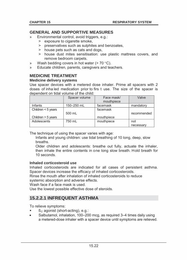
15.22
CHAPTER 15 RESPIRATORY SYSTEM GENERAL AND SUPPORTIVE MEASURES » Environmental control, avoid triggers, e.g.:
> exposure to cigarette smoke, > preservatives such as sulphites and benzoates, > house pets such as cats and dogs, > house dust mites sensitisation: use plastic mattress covers, and
remove bedroom carpets. » Wash bedding covers in hot water (> 70 C). » Educate children, parents, caregivers and teachers. MEDICINE TREATMENT Medicine delivery systems Use spacer devices with a metered dose inhaler. Prime all spacers with 2 doses of inha led medication prior to firs t use. The size of the spacer is dependent on tidal volume of the child:
Spacer volume Face mask/ mouthpiece
Valve
Infants 150–250 mL facemask mandatory Children < 5 years Children > 5 years
500 mL
facemask mouthpiece
recommended
Adolescents 750 mL mouthpiece not necessary
The technique of using the spacer varies with age:
Infants and young children: use tidal breathing of 10 long, deep, slow breaths. Older children and adolescents: breathe out fully, actuate the inhaler, then inhale the entire contents in one long slow breath. Hold breath for 10 seconds.
Inhaled corticosteroid use Inhaled corticosteroids are indicated for all cases of persistent asthma. Spacer devices increase the efficacy of inhaled corticosteroids. Rinse the mouth after inhalation of inhaled corticosteroids to reduce systemic absorption and adverse effects. Wash face if a face mask is used. Use the lowest possible effective dose of steroids. 15.2.2.1 INFREQUENT ASTHMA To relieve symptoms: ß2 agonist (short-acting), e.g.: Salbutamol, inhalation, 100–200 mcg, as required 3–4 times daily using
a metered-dose inhaler with a spacer device until symptoms are relieved.
15.23
CHAPTER 15 RESPIRATORY SYSTEM Note: Failure to respond to 2 doses of an inhaled bronchodilator given 20 minutes apart is an ind ication of an acute exacerbation of a sthma. See s ection 15.2.1: Asthma, acute attack. 15.2.2.2 PERSISTENT ASTHMA Mild persistent asthma When needed for acute exacerbations: ß2 agonist (short-acting), e.g.: Salbutamol, inhalation, 100–200 mcg, as required 3–4 times daily using
a metered-dose inhaler with a spacer device until symptoms are relieved. PLUS Low dose inhaled corticosteroids, e.g.: Beclomethasone or budesonide, inhalation, 50–100 mcg, 12 hourly
using a metered-dose inhaler with a spacer device. Moderate persistent asthma To relieve symptoms: ß2 agonist (short-acting), e.g.: Salbutamol, inhalation, 100–200 mcg, as required 3–4 times daily using
a metered-dose inhaler with a spacer device until symptoms are relieved. PLUS Regular anti-inflammatory treatment with medium-dose inhaled corticosteroids: Budesonide, inhalation, 100–200 mcg, 12 hourly using a metered-dose
inhaler with a spacer device. OR In children > 6 y ears with multiple allergies on other s teroid formulations, low-dose inhaled corticosteroids plus long-acting beta agonist (LABA) e.g.: Fluticasone plus salmeterol by inhalation, 12 hourly. Specialist initiated.
Severe persistent asthma To relieve symptoms: ß2 agonist (short-acting), e.g.: Salbutamol, inhalation, 100–200 mcg, as required 3–4 times daily using
a metered-dose inhaler with a spacer device until symptoms are relieved. PLUS Low-dose inhaled corticosteroids plus LABA, e.g.: Fluticasone plus salmeterol, inhaled, 12 hourly. Specialist Initiated.
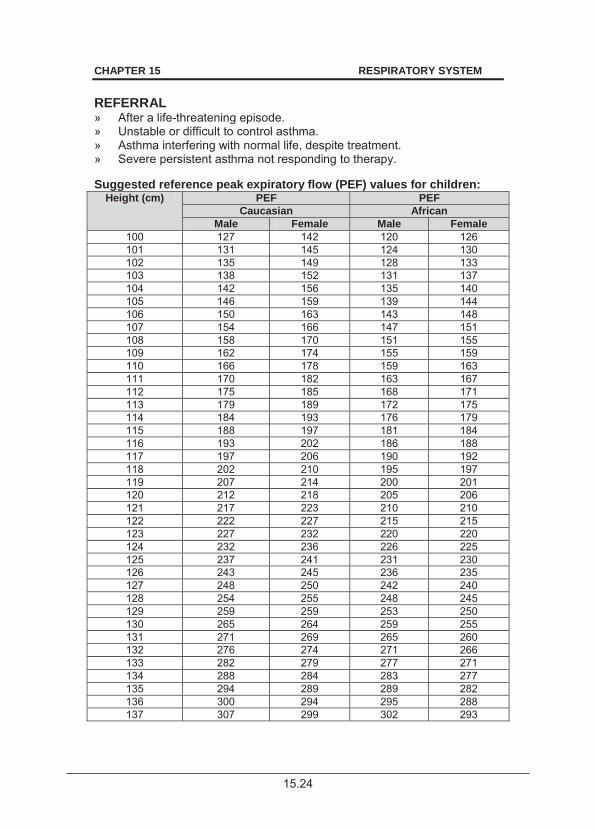
15.24
CHAPTER 15 RESPIRATORY SYSTEM REFERRAL » After a life-threatening episode. » Unstable or difficult to control asthma. » Asthma interfering with normal life, despite treatment. » Severe persistent asthma not responding to therapy. Suggested reference peak expiratory flow (PEF) values for children:
Height (cm) PEF PEF Caucasian African
Male Female Male Female 100 127 142 120 126 101 131 145 124 130 102 135 149 128 133 103 138 152 131 137 104 142 156 135 140 105 146 159 139 144 106 150 163 143 148 107 154 166 147 151 108 158 170 151 155 109 162 174 155 159 110 166 178 159 163 111 170 182 163 167 112 175 185 168 171 113 179 189 172 175 114 184 193 176 179 115 188 197 181 184 116 193 202 186 188 117 197 206 190 192 118 202 210 195 197 119 207 214 200 201 120 212 218 205 206 121 217 223 210 210 122 222 227 215 215 123 227 232 220 220 124 232 236 226 225 125 237 241 231 230 126 243 245 236 235 127 248 250 242 240 128 254 255 248 245 129 259 259 253 250 130 265 264 259 255 131 271 269 265 260 132 276 274 271 266 133 282 279 277 271 134 288 284 283 277 135 294 289 289 282 136 300 294 295 288 137 307 299 302 293
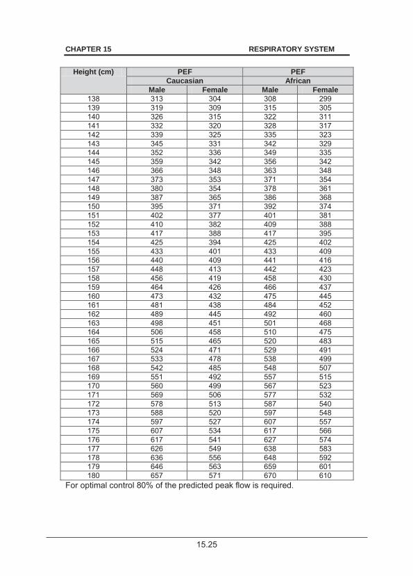
15.25
CHAPTER 15 RESPIRATORY SYSTEM
Height (cm) PEF PEF Caucasian African
Male Female Male Female 138 313 304 308 299 139 319 309 315 305 140 326 315 322 311 141 332 320 328 317 142 339 325 335 323 143 345 331 342 329 144 352 336 349 335 145 359 342 356 342 146 366 348 363 348 147 373 353 371 354 148 380 354 378 361 149 387 365 386 368 150 395 371 392 374 151 402 377 401 381 152 410 382 409 388 153 417 388 417 395 154 425 394 425 402 155 433 401 433 409 156 440 409 441 416 157 448 413 442 423 158 456 419 458 430 159 464 426 466 437 160 473 432 475 445 161 481 438 484 452 162 489 445 492 460 163 498 451 501 468 164 506 458 510 475 165 515 465 520 483 166 524 471 529 491 167 533 478 538 499 168 542 485 548 507 169 551 492 557 515 170 560 499 567 523 171 569 506 577 532 172 578 513 587 540 173 588 520 597 548 174 597 527 607 557 175 607 534 617 566 176 617 541 627 574 177 626 549 638 583 178 636 556 648 592 179 646 563 659 601 180 657 571 670 610
For optimal control 80% of the predicted peak flow is required.
15.26
CHAPTER 15 RESPIRATORY SYSTEM 15.2.3 BRONCHIOLITIS J21.9 DESCRIPTION Bronchiolitis is an a cute viral infection of the small airways of low er respiratory tract affecting children between 4 months to 2 years of age. The most common pathogen is the respiratory syncytial virus. Recurrent episodes of wheeze associated with bronchiolitis may occur, and some of these children may develop asthma. Assessment of severity Signs of severe disease include: » infants under 3 months of age,
especially premature babies; » central cyanosis; » inability to feed;
» respiratory failure; » lower chest wall indrawing; » nasal flaring; » grunting; » distress when speaking or crying; » discomfort in breathing; » apnoea; » hypoxia; » pneumothorax; » convulsions and decreased
level of consciousness. Mild cases are managed as outpatients. DIAGNOSTIC CRITERIA » Prodrome of viral infection: irritability and feeding difficulties. » A wheeze that is slowly responsive or non-responsive to bronchodilators. » Crepitations and signs of hyperinflation of the chest. » A chest X -ray is useful in confirming hyperinflation and associated
segmental atelectasis. GENERAL AND SUPPORTIVE MEASURES » Isolate from other infants, if possible. » Patients with signs of severe disease or associated complications or
underlying cardiorespiratory disorders should be hospitalised for monitoring of: > breathing pattern (apnoea monitoring if < 3 months of age), > signs of respiratory failure, > heart rate and respiratory rate, > temperature, > SaO2, > hydration and nutrition. IV maintenance fluid according to age, if
oral/nasogastric feeds/fluids are not tolerated. Avoid overhydration.
15.27
CHAPTER 15 RESPIRATORY SYSTEM MEDICINE TREATMENT For all hospitalised patients Only if saturation < 92%: Oxygen, humidified, 1–2 L/min via nasal prongs or nasal cannula.
o Ensure clear nasal passages and correctly position the nasal prongs.
For outpatient-based therapy nebulise with: Epinephrine (adrenaline) 1: 1 000, 1 mL diluted in 2–4 mL sodium
chloride 3% immediately and every 2–4 hours. For inpatient therapy nebulise with: Sodium chloride 3%, solution, 2–4 mL, 4–6 hourly.
Mix 3 mL of 5% sodium chloride with 2 mL water to make 3% solution. If there is poor response or deterioration, change to: Epinephrine (adrenaline) 1: 1 000, 1 mL diluted in 2–4 mL sodium
chloride 3% immediately and every 2–4 hours. PLUS Sodium chloride 3%, solution, 2–4 mL, 4–6 hourly.
Antibiotic therapy Routine antibiotic therapy is not indicated. Only use antibiotics if there is: » raised leukocyte count, » persistent fever of 38.5 C, and/or » a chest X-ray showing opacification suggestive of pneumonia. For secondary bacterial infection: Amoxicillin, oral, 30 mg/kg/dose, 8 hourly for 5 days.
Note: There is no evidence that 2-agonists, anticholinergics, inhaled corticosteroids or leukotriene receptor antagonists have any benefit in these children. In severe disease requiring intensive care, oral corticosteroids may be considered after specialist consultation. REFERRAL » Bronchiolitis with signs of respiratory failure.
15.28
CHAPTER 15 RESPIRATORY SYSTEM 15.3 COUGH WITH PREDOMINANT FEVER AND
TACHYPNOEA 15.3.1 PNEUMONIA J18.9 DESCRIPTION Infection of the lung parenchyma that is classified as non-severe, severe or very severe based on clinical features. Empiric antibiotics are indicated in all cases of pneumonia, as delay in treatment is associated with poor outcome. Antibiotic choice is based on an assessment of severity and likely aetiology.
Common bacterial causes of pneumonia include: Neonates: » Group B ß-haemolytic Streptococci. » Klebsiella spp. » E. coli. » Chlamydia. » S. aureus.
Children: » S. pneumoniae. » H. influenzae. » S. aureus. » M. catarrhalis. » M. pneumonia. Common viral causes in infancy and early childhood include: » influenza virus, » para-influenza virus, » measles virus, » respiratory syncytial virus,
» cytomegalovirus, » adenovirus.
Predisposing factors for pneumonia include: » aspiration, » immunosuppression, » septicaemia, » malnutrition, » measles, » whooping cough, » abnormalities in clearance of mucus/secretions (e.g. cystic fibrosis,
foreign body and ciliary dysfunction). Complications of pneumonia include: » respiratory failure, » pleural effusion, » empyema, » pneumothorax, » pleuritis, » bronchiectasis.
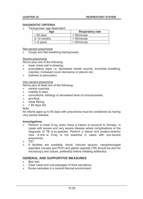
15.29
CHAPTER 15 RESPIRATORY SYSTEM DIAGNOSTIC CRITERIA » Tachypnoea: age dependent:
Age Respiratory rate < 60 days > 60/minute 2–12 months > 50/minute 1–5 years > 40/minute
Non-severe pneumonia » Cough and fast breathing (tachypnoea). Severe pneumonia Above plus one of the following: » lower chest wall in-drawing; » auscultatory signs i.e. decreased breath sounds, bronchial breathing,
crackles, increased vocal resonance or pleural rub; » dullness to percussion. Very severe pneumonia Above plus at least one of the following: » central cyanosis, » inability to feed, » convulsions, lethargy or decreased level of consciousness, » grunting, » nasal flaring, » < 60 days old. Note: All infants aged up to 60 days with pneumonia must be considered as having very severe disease. Investigations » Perform a c hest X-ray when there is f ailure to res pond to therapy, in
cases with severe and very severe disease where complications or the diagnosis of TB is su spected. Perform a lateral and postero-anterior view. A che st X-ray is not essential in cases with non-severe pneumonia.
» TST. » If facilities are available, blood, induced sputum, nasopharyngeal
aspirates (viruses and PCP) and gastric aspirate (TB) should be sent for microscopy and culture, preferably before initiating antibiotics.
GENERAL AND SUPPORTIVE MEASURES » Bed rest. » Clear nasal and oral passages of thick secretions. » Nurse neonates in a neutral thermal environment.
15.30
CHAPTER 15 RESPIRATORY SYSTEM » Monitor:
> respiratory rate, > heart rate, > SaO2, > temperature, > hydration, > blood pressure, > blood gases, > acid-base status, > hypercapnia and/or hypoxia are indications for ventilatory support.
» Maintain nutrition: Continue breast and oral feeds. > Consider small frequent feeds by oro/nasogastric tube or IV fluids
if respiratory rate > 60/minutes or enteral feeds are not tolerated. SURGICAL TREATMENT » To relieve a tension pneumothorax, do ne edle aspiration followed by
chest tube placement. » For symptomatic pleural effusion, do needle aspiration; if empyema,
insert large bore chest tube drainage. » Small or asymptomatic pneumothoraces in infants and children
(excluding neonates) usually do not req uire treatment other than close observation, but identify and treat the u nderlying cause for the pneumothorax.
MEDICINE TREATMENT Oxygen, humidified, by nasal prongs is preferred.
o Continue oxygen until respiratory rate is < 60/minute. To relieve discomfort: Paracetamol, oral/NGT, 15 mg/kg, 6 hourly as required.
If significant degree of wheezing is present: Salbutamol, inhalation, 100–200 mcg, as required using a metered-dose
inhaler with a spacer device until symptoms are relieved. Empirical antibiotic therapy Choice of antibiotic depends on the severity of the condition and predisposing factors. Reconsider choice of antibiotic when the results of cultures become available or the child does not improve. Non severe pneumonia Amoxicillin, oral, 30 mg/kg/dose, 8 hourly for at least 3 days.
Severe pneumonia: If hospitalisation is not required: Amoxicillin, oral, 30 mg/kg/dose, 8 hourly for 5 days.
15.31
CHAPTER 15 RESPIRATORY SYSTEM OR If hospitalisation is required: Ampicillin, IV, 25 mg/kg/dose 6 hourly.
If clinical condition allows use or change to: Amoxicillin, oral, 30 mg/kg/dose, 8 hourly to complete 5 days of therapy.
Very severe pneumonia Assume child is HIV infected until proven otherwise. See section 9.1: Human Immunodeficiency Virus Infections. Birth to 59 days Ampicillin, IV, 25 mg/kg/dose 6 hourly.
PLUS Gentamicin, IV, 6 mg/kg as a single daily dose for 5–10 days.
Observe the response closely and reconsider choice of antibiotics. In children 60 days and older Ceftriaxone, IV, 50 mg/kg/dose 24 hourly.
Switch to oral as soon as there is a response: Amoxicillin/clavulanate, oral, 30 mg/kg/dose of amoxicillin component
8 hourly. 15.3.2 PREDISPOSING CONDITIONS AND MODIFICATION
OF ANTIMICROBIAL THERAPY 15.3.2.1 PNEUMONIA, VIRAL INFECTION J12.9 DESCRIPTION The commonest cause of pneumonia in ch ildren is viral infection. Respiratory syncyctial virus, adenovirus, cytomegalovirus, influenza, parainfluenza, adenovirus, herpes, human metapneumovirus and m easles are the common viruses responsible for infections of the respiratory tract, Children present with fever, cough, rhinorrhea and chest in-drawing. Scattered fine crackles may also occur. DIAGNOSTIC CRITERIA » Chest X-ray: usually patchy interstitial changes. » Nasopharyngeal aspirate for respiratory viruses screen via
immunofluorescence.
15.32
CHAPTER 15 RESPIRATORY SYSTEM GENERAL AND SUPPORTIVE MEASURES » Maintain nutrition. » Maintain hydration. MEDICINE TREATMENT Only if saturation < 92%: Oxygen, humidified, 1–2 L/min via nasal prongs or nasal cannula.
If temperature is > 38.5oC: Paracetamol, oral, 15 mg/kg/dose 6 hourly.
There is no ro le for ro utine antiviral therapy. Refer children with viral pneumonia in respiratory failure for support with mechanical ventilation. Monitor for secondary bacterial infection. REFERRAL » Deterioration of condition. 15.3.2.2 FUNGAL INFECTION J17.2*/B49 DESCRIPTION May occur in immunosuppressed patients and present with deep draining sinuses or associated fungal lesions in the larynx, trachea or mouth. GENERAL AND SUPPORTIVE MEASURES » Perform cultures. » Screening blood test is -glucans. MEDICINE TREATMENT Amphotericin B, IV, 0.6–1.0 mg/kg as a single daily dose infused over
4 hours for at least 14 days. Prehydration before administering amphotericin to prevent renal impairment: Sodium chloride 0.9%, IV, 20 mL/kg plus potassium chloride,
20 mmol/L infused over 2–4 hours.
OR Fluconazole, IV/oral, 10 mg/kg as a single daily dose for at least
14 days.
15.33
CHAPTER 15 RESPIRATORY SYSTEM Neonates < 2 weeks: Amphotericin B, IV, 0.7–1.0 mg/kg every 72 hours infused over 4 hours
for at least 14 days. Prehydrate before administering amphotericin to prevent renal impairment: Sodium chloride 0.9%, IV, 20 mL/kg plus potassium chloride, 20 mmol/L
infused over 2–4 hours. OR Fluconazole, IV/oral, 10 mg/kg every 72 hours for at least 14 days.
Neonates 2–4 weeks Amphotericin B, IV, 0.7–1.0 mg/kg every 48 hours infused over 4 hours
for at least 14 days. Prehydration before administering amphotericin to prevent renal impairment: Sodium chloride 0.9%, IV, 20 mL/kg plus potassium chloride, 20 mmol/L
infused over 2–4 hours OR Fluconazole, IV/oral, 10 mg/kg every 48 hours for at least 14 days.
Note: If screening test or culture-negative, discontinue antifungal treatment. If culture positive, consider sensitivity results and adjust accordingly. 15.3.2.3 PNEUMONIA DUE TO ANAEROBIC INFECTION DESCRIPTION Often seen in comatose patients with aspiration syndromes. MEDICINE TREATMENT Metronidazole, oral, 7.5 mg/kg/dose, 8 hourly for 5 days.
PLUS Ampicillin, IV, 25 mg/kg/dose, 6 hourly for 5 days.
PLUS Gentamicin, IV, 6 mg/kg as a single daily dose for 5 days.
15.3.2.4 PNEUMONIA, ATYPICAL DUE TO MYCOPLASMA
OR CHLAMYDIAL INFECTION J15.7/J16.0 DESCRIPTION This condition is often seen in children of school going age.
15.34
CHAPTER 15 RESPIRATORY SYSTEM Cases present with fever, arthralgia, headache, cough and crepitations. DIAGNOSTIC CRITERIA Clinical » In the neonatal period, chlamydial pneumonia presents with a staccato
cough and sticky eyes. Investigations » Chest X-ray show interstitial infiltrates, lobar consolidation and hilar
adenopathy. MEDICINE TREATMENT Mild disease Macrolide, e.g.: Clarithromycin, oral, 7.5 mg/kg/dose, 12 hourly for 7 days.
For severe atypical pneumonia Macrolide, e.g.: Clarithromycin, IV, 7.5 mg/kg/dose, 12 hourly for 7 days.
15.3.2.5 PNEUMONIA, STAPHYLOCOCCAL J15.2 DESCRIPTION Child presents with a toxic illness, tachypnoea, chest wall in-drawing and cough. DIAGNOSTIC CRITERIA Investigations » Chest X-ray shows a des tructive pneumonia with pneumatocoele,
pleural effusion or empyema. » Markedly elevated white blood cells. MEDICINE TREATMENT Cloxacillin, IV, 50 mg/kg/dose every 6 hours.
If there is evidence of good clinical response, change to: Flucloxacillin, oral, 12.5–25 mg/kg/dose 6 hourly for at least 21 days.
MRSA pneumonia: Vancomycin, IV, 15 mg/kg/dose 6 hourly infused over 1 hour.
15.35
CHAPTER 15 RESPIRATORY SYSTEM 15.3.2.6 PNEUMONIA IN HIV EXPOSED OR INFECTED
CHILDREN DESCRIPTION In additional to common bacterial, fungal and viral pathogens causing pneumonia, opportunistic micro-organisms in a ‘ polymicrobial mix’ are common in these children. Many of these children may fail to respond to the standard antibiotic treatment for pneumonia. Micro-organisms commonly involved are:
» P. jiroveci (PCP), » cytomegalovirus, » Mycobacteria, e.g. M. tuberculosis » Non-typhoidal Salmonella, » S. aureus, » Klebsiella pneumonia, » S. pneumonia, » Candida.
S. pneumonia, S. aureus and gra m negative bacteria e.g. Klebsiella pneumoniae and Non-typhoid Salmonella cause a significant proportion of HIV-related pneumonia in early childhood. P. jiroveci (PCP) » PCP is a common fungal infection of the lung in infants from 2–6 months. » Presents as an acute onset of respiratory distress with minimal/absent
chest signs in a child who is HIV exposed. » Hypoxaemia and cyanosis are common features in severe disease. » Chest X-ray shows a range of abnorm alities including bilateral perihilar
interstitial changes. Perinatal acquired cytomegalovirus associated pneumonia in HIV infected infants » Presents as an interstitial pneumonitis with acute hypoxic respiratory
failure. » It may present as a m ultisystem sepsis-like syndrome, with hepatitis,
neutropenia, pneumonitis, colitis and thrombocytopaenia. » Often occurs in children w ho are severely immunosuppressed (CDC
Immune category 3) and carries a significant mortality. » Transmission occurs by blood transfusion, body fluids and via breast milk
(reduced by freezing, pasteurisation, hand washing). Even though there is a high CMV IgG in breast milk, there is low rate of CMV transmission and therefore continue breast feeding.
» CMV co-infection occurs commonly as polymicrobial infection with PCP and bacteria.
Tuberculosis in HIV infected children » Occurs in children at all ages. » The diagnosis is difficult to confirm. » A Mantoux test of 5 mm induration is indicative of tuberculosis
infection.
15.36
CHAPTER 15 RESPIRATORY SYSTEM HIV positive children with chronic lung disease » Often presents with lymphoid interstitial pneumonitis and bronchiectasis. » Secondary infection with bacteria similar to those seen in acute
pneumonia are commonly isolated from these children. DIAGNOSTIC CRITERIA Investigations » Chest X-ray. » Screen for HIV infection:
> In children < 18 months utilising HIV DNA PCR and quantitative HIV viral load.
> In children > 18 months HIV ELISA (screening and confirmatory). » Investigate for PCP:
> Immunofluorescence and silver methenamine staining on induced sputum sample.
» Screen immediately for CMV using CMV viral load, where available. > A viral load of > 100 000 copies/mL suggests CMV disease: treat. > A viral load below 10 000 copies/mL is regarded as CMV infection:
no therapy recommended. > CMV viral loads between 10 000 copies/mL and 100 000
copies/mL: treat if patient is clinical symptomatic. > Tissue biopsy: intranuclear inclusion bodies, typical ‘owl eye’ cells.
Note: A positive urine culture for CMV or a positive serology does not confirm a diagnosis of CMV disease. Patient would not qualify for treatment solely on those results.
» Tuberculosis: > TST, MCS on induced sputum, M. tuberculosis PCR test on
sputum. GENERAL AND SUPPORTIVE MEASURES » Avoid exposure to infectious agents. » Adequate nutrition. » Monitor oxygen saturations. » Restrict fluid intake. MEDICINE TREATMENT If saturation < 92%: Oxygen, via nasal prongs or nasal cannula.
Treat for bacterial pneumonia. See section 15.3.1: Pneumonia. In all infants between 2 and 6 months with pneumonia consider PCP.
15.37
CHAPTER 15 RESPIRATORY SYSTEM ADD Co-trimoxazole, IV/oral, 5 mg trimethroprim/25 mg
sulphamethoxazole/kg/dose, 6 hourly for 21 days. o Continue co-trimoxazole prophylaxis at the end of this treatment
period until CD4 counts recovers to normal. Children who remain hypoxic on oxygen with proven or highly suspected PCP: Prednisone, oral, 1–2 mg, daily for 7 days.
o Taper dose over the next 7 days. For confirmed CMV disease: Ganciclovir, IV, 5 mg/kg 12 hourly for 21 days (initial dose).
o Follow with: 5 mg/kg daily for 21 days (maintenance dose). Note: Immunoglobulins, interferon and CMV vaccine have no benefit. For children not responding to standard therapy: Exclude TB, S. aureus and gram negative bacteria. If extremely ill consider empiric treatment with: Vancomycin, IV, 15 mg/kg/dose, 6 hourly infused over 1 hour.
PLUS Amikacin, IV, 15–20 mg/kg once daily.
For children with acute pneumonia or chronic lung disease: Treat as above for pneumonia but investigate for TB, NTM, candida and non-typable H. influenzae. Check immunisation status for pneumococcus and H. influenzae. REFERRAL » For commencement of combined ART, where indicated. » In cases of CMV disease for follow up for hearing deficit. 15.3.2.5 PNEUMONIA, NOSOCOMIAL J18.9 DESCRIPTION Children acquiring pneumonia 48–72 hours after hospitalisation. The common pathogens are: » ß-lactamase producing pathogens, » extended spectrum ß-lactamase producing Klebsiella pneumoniae, » P. aeruginosa, » multidrug resistant Acinetobacter species,
15.38
CHAPTER 15 RESPIRATORY SYSTEM
» methicillin resistant S. aureus, » respiratory viruses e.g. respiratory syncytial virus, adenovirus, influenza,
herpes, measles, parainfluenza. GENERAL AND SUPPORTIVE MEASURES » Septic screen including blood and urine cultures. » Nasopharyngeal aspirates for antigens. » Molecular test for respiratory virus. MEDICINE TREATMENT Empirical antibiotic therapy Broad spectrum antibiotics according to local susceptibility patterns. Manage children with underlying predisposing factors according to the susceptibility of the most likely pathogen. Review antibiotic choice once culture and sensitivity results become available. For bacterial infections Start with: Piperacillin/tazobactam, IV, 100 mg/kg/dose 8 hourly.
PLUS Amikacin, IV, 15 mg/kg/dose, daily.
Adjust therapy according to sensitivities. For methicillin resistant S. aureus pneumonia: Vancomycin, IV, 15 mg/kg/dose 6 hourly infused over 1 hour.
15.4 PLEURAL DISEASE 15.4.1 EFFUSION AND EMPYEMA J90 DESCRIPTION A pleural effusion is an accumulation of an exudative or t ransudative fluid between the visceral and parietal pleura. Common causes for exudates are infections, inflammation and malignancy. Common causes of a tr ansudate are cardiac failure, renal failure and hepatic failure. A straw coloured or haemorrhagic effusion is indicative of tuber culosis. A cl oudy or frank ly purulent fluid indicates an empyema.
15.39
CHAPTER 15 RESPIRATORY SYSTEM DIAGNOSTIC CRITERIA » Decreased breath sounds and stony dull on percussion. » Pleural rub early in disease. » Chest X-ray shows uniform opaque opacities in a lamellar distribution at
the costophrenic angles. GENERAL AND SUPPORTIVE MEASURES » Treat small effusions conservatively. » Drain other effusions by either chest drain or needle aspiration. » Send samples for protein, glucose, cytology, microscopy and culture. If
pus is identified or pH < 7.2, insert chest drain. » Transudates do not require drainage. » More aggressive surgical procedures such as open drainage or
decortication are rarely indicated in children. MEDICINE TREATMENT For purulent effusion: Cloxacillin, IV, 50 mg/kg/dose every 6 hours.
PLUS Gentamicin, IV, 6 mg/kg as a single daily dose for 10 days.
If there is evidence of good clinical response, change to: Flucloxacillin, oral, 12.5–25 mg/kg/dose, 6 hourly for a total of 21 days.
If pathogens are cultured in blood from sanctuary sites e.g. bone, heart valves, etc. treat according to sensitivity for prolonged period of 21–42 days.
For straw coloured or haemorrhagic effusion: » Start antituberculosis therapy. If no response, to any of the above therapy, consider fungal infection. 15.5 UPPER AIRWAY DISEASES 15.5.1 EPIGLOTTITIS J05.1 DESCRIPTION Life-threatening upper airway obstruction at the level of the supraglottic structures (epiglottis and arytenoids). The condition is rare since H. influenzae type b v accination has been introduced.
15.40
CHAPTER 15 RESPIRATORY SYSTEM DIAGNOSTIC CRITERIA » Acute onset, high fever, sore throat, dysphagia, refusal to eat or
swallow, drooling and muffled voice. » Position of comfort to protect the upper airway: sitting upright, head
forward, open mouth, neck in extension. GENERAL AND SUPPORTIVE MEASURES » Do not interfere with the protective mechanism of the patient. Allow the
child to remain sitting up. » Avoid all measures that could agitate the patient:
> make no attempt to see the epiglottis, > do not routinely perform X-rays of neck and chest, > delay blood sampling and IV line insertion until after airway is
secured. » Monitor oxygen saturation (pulse oximeter).
Acute airway obstruction
Caution Epiglottitis is an upper airway emergency.
Total upper airway obstruction is imminent by the time stridor appears. Prepare equipment for bag-mask ventilation, endotracheal intubation, needle
cricothyroidotomy and tracheostomy.
» If airway obstructs completely or respiratory arrest occurs, attempt to
establish an airway: ventilate with bag and mask. » If unable to ventilate: intubate. » If unable to intubate: perform needle or surgical cricothyroidotomy.
Total airway obstruction may occur suddenly and quite unpredictably, the patient should ideally be i ntubated before referral. Intubation should preferably be performed under general anaesthesia in an operating theatre. If intubation prior to referral is not possible, transfer patient as an emergency advising transfer staff to avoid lying the child down. Inform the re ceiving hospital before departure. During transport, if th e child decompensates, attempt bag and mask ventilation. After an open airway has been secured: » take blood for cultures, » swab epiglottis for microscopy, culture and sensitivity, » monitor heart rate, respiratory rate, blood pressure and SaO2, » ensure adequate nutrition and hydration.

15.41
CHAPTER 15 RESPIRATORY SYSTEM MEDICINE TREATMENT Oxygen, humidified, if needed.
Ceftriaxone, IV, 50 mg/kg/dose, once daily for 7 days.
REFERRAL » All, once airway is secured. 15.5.2 LARYNGOTRACHEOBRONCHITIS, ACUTE VIRAL
(CROUP) J05.0 DESCRIPTION Potentially life-threatening airway obstruction in children and one of the most common causes of stridor in children aged between 6 months and 2 years. The most common viruses causing laryngotracheobronchitis (LTB) include: » para-influenza virus (most common), » measles, » herpes simplex, » adenovirus. DIAGNOSTIC CRITERIA Clinical » a previously healthy child who, a day or two after the onset of an upper
respiratory tract infection, develops progressive airway obstruction with a barking cough and stridor,
» a mild fever may be present, » stridor becomes softer as airway obstruction becomes more severe.
The following features suggest a different diagnosis: » acute onset of obstruction without prodromal features (foreign body or
angioneurotic oedema), » incomplete immunisation and a membrane in the upper airway
(diphtheria), » high fever, dysphagia, drooling or sitting position (epiglottitis, retro-
pharyngeal abscess, bacterial tracheitis), » recurrent upper airways obstruction (laryngeal papilloma).
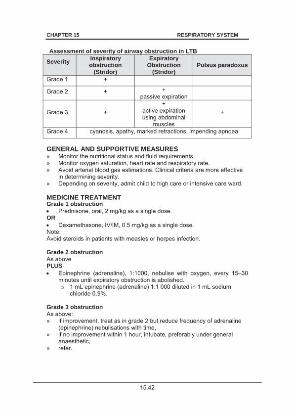
15.42
CHAPTER 15 RESPIRATORY SYSTEM
Assessment of severity of airway obstruction in LTB
Severity
Inspiratory obstruction
(Stridor)
Expiratory Obstruction
(Stridor) Pulsus paradoxus
Grade 1 +
Grade 2 + + passive expiration
Grade 3 + +
active expiration using abdominal
muscles
+
Grade 4 cyanosis, apathy, marked retractions, impending apnoea
GENERAL AND SUPPORTIVE MEASURES » Monitor the nutritional status and fluid requirements. » Monitor oxygen saturation, heart rate and respiratory rate. » Avoid arterial blood gas estimations. Clinical criteria are more effective
in determining severity. » Depending on severity, admit child to high care or intensive care ward. MEDICINE TREATMENT Grade 1 obstruction Prednisone, oral, 2 mg/kg as a single dose.
OR Dexamethasone, IV/IM, 0.5 mg/kg as a single dose.
Note: Avoid steroids in patients with measles or herpes infection. Grade 2 obstruction As above PLUS Epinephrine (adrenaline), 1:1000, nebulise with oxygen, every 15–30
minutes until expiratory obstruction is abolished. o 1 mL epinephrine (adrenaline) 1:1 000 diluted in 1 mL sodium
chloride 0.9%. Grade 3 obstruction As above: » if improvement, treat as in grade 2 but reduce frequency of adrenaline
(epinephrine) nebulisations with time, » if no improvement within 1 hour, intubate, preferably under general
anaesthetic, » refer.
15.43
CHAPTER 15 RESPIRATORY SYSTEM Grade 4 obstruction As above and: » continue steroids, » continue with adrenaline (epinephrine) nebulisation with 100% warm
humidified oxygen, » emergency intubation or intubation under general anaesthesia, if
circumstances permit, » if unable to intubate, bag and mask ventilate and refer urgently. For fever until fever subsides: Paracetamol, oral, 15 mg/kg/dose, 6 hourly.
For suspected herpes: Aciclovir IV, 10–15 mg/kg/dose 8 hourly for 5–7 days.
For suspected bacteria infection in children < 20 kg: Ampicillin, IV, 12.5–25 mg/kg/dose 6 hourly for 5–10 days.
For suspected bacteria infection in children > 20 kg: Ampicillin, IV, 250–500 mg, 6 hourly for 7 days.
AND If bacterial tracheitis is suspected: Cloxacillin, IV, 50 mg/kg/dose 6 hourly for 7 days.
REFERRAL Note: » Intubate all children with grade 3 and 4 airway obstruction not
responding within one hour to adrenaline nebulisations before referral. » Children with an uncertain diagnosis.
16.1
CHAPTER 16 EYE CONDITIONS
16.1 EYE INFECTION, COMPLICATED (SEVERE EYE INFECTION) H44 DESCRIPTION Intensely painful, red eye with or w ithout a discharge (excluding simple or non-painful conjunctivitis). Assess clinically for:
» Herpes conjunctivitis indicated by vesicles on skin next to eye. » Loss of vision. » Irregularity of pupil. » Haziness of the cornea.
Investigations Swab eye for microbiological culture. GENERAL AND SUPPORTIVE MEASURES Patient education on personal hygiene to avoid spread. Educate patient on correct application of ophthalmic drops. Advise patient: » to wash hands thoroughly before applying ophthalmic ointment, » not to share ophthalmic ointments or drops, » not to rub eyes, and » never to use urine or milk to wash the eyes. MEDICINE TREATMENT If herpes infection suspected, treat as outlined in section 16.3: Herpes keratitis and conjunctivitis. During the day: Gentamicin, ophthalmic drops, instill 1 drop 4–6 hourly.
OR Chloramphenicol 0.5%, ophthalmic drops, instill 1 drop 4–6 hourly.
AND Apply at night: Chloramphenicol 1%, ophthalmic ointment.
16.2
CHAPTER 16 EYE CONDITIONS
REFERRAL To ophthalmologist within 24 hours if associated with any of the following: » Reduced vision. » A cloudy cornea. » A corneal opacity or a staining corneal ulcer. » Pus and blood level in the anterior chamber (hypopion and hyphaema). » Cloudiness in the anterior chamber (poor view of iris details). » An irregular or dilated (including partially dilated) pupil. » A cloudy or poor view of the retina. » A poor or greyish red reflex. » Proptosis. » Restricted ocular movements. » Severe ocular pain. Non urgent referral » A unilateral red eye for more than one day. » No improvement after 5 days of treatment. 16.2 CONJUNCTIVITIS, ALLERGIC H10.1 See Standard Treatment guidelines and Essential Medicines List for Primary Health Care Level. 16.3 HERPES KERATITIS AND CONJUNCTIVITIS H90.1* DESCRIPTION Herpes infection of the cornea and/or conjunctiva. DIAGNOSTIC CRITERIA There are three most common forms of this disease. Blepharoconjunctivitis Primary ocular infection involving the eyelids, and/or conjunctivae. The condition is benign and self limiting. May be associated with keratitis: tiny punctuate stains on the cornea when stained with fluorescein and viewed with the coba lt blue light of the d irect ophthalmoscope.
16.3
CHAPTER 16 EYE CONDITIONS
Disciform keratitis Immune response to herpes virus. Decreased visual acuity and corneal sensation. Round dull swollen area in the central cornea. Refer to ophthalmologist. Dendritic ulcer A linear branching ulcer (dendritic ulcer) when stained with fluorescein and viewed with the cobalt blue light of the direct ophthalmoscope. Decreased sensation when compared to the ot her eye. (Use a thread o f cotton from a cotton bud and touch the cornea from the side, away from the visual axis) GENERAL AND SUPPORTIVE MEASURES Pad the eye. MEDICINE TREATMENT Aciclovir, ophthalmic ointment, applied five times per day for 10 days.
If cilliary spasm present: Cyclopentolate, ophthalmic drops, instil 1 drop 8 hourly.
REFERRAL Urgent within 24 hours: » If corneal lesion is not clean/clear or has whitish areas within the bed of
the epithelial ulcer. » If area of corneal staining is not smaller within 24 hours of treatment. » If there is a history of recurrence. » Disciform keratitis for assessment and treatment. 16.4 CYTOMEGALOVIRUS (CMV) RETINITIS B25.8/H30.9 DESCRIPTION Characteristic appearance: opacification of t he retina with areas of haemorrhage, exudate and necrosis. Occurs in immunocompromised patients and could be an important cause of visual impairment in HIV infected patients. DIAGNOSTIC CRITERIA » Confirm retinitis with ophthalmological assessment. » Confirm CMV disease with DNA PCR.
16.4
CHAPTER 16 EYE CONDITIONS
MEDICINE TREATMENT Ganciclovir, intravitreal, 2 mg once a week. (Opthalmologist treatment).
Once immune function has been restored with antiretroviral therapy, i.e. CD4 > 100 ce lls/mm3, maintenance ganciclovir can be stopped but monitor for recurrence.
REFERRAL » All patients to confirm diagnosis and manage treatment. 16.5 CHEMICAL BURN TO THE EYE T26.9 DESCRIPTION Damage to one or both eyes caused by contact with irritating chemical substances, either alkali or acid. Presentation: » pain, » inability to open eye, » blurred vision, » excessive tearing. DIAGNOSTIC CRITERIA » To assess extent of epithelial loss, after irrigating the eye/s, stain the
cornea with fluorescein 2%. Note: If the entire cornea stains, then all the epithelium has been removed by the chemical substance. Compare fluorescein staining in the other eye.
GENERAL AND SUPPORTIVE MEASURES Try to ascertain the exact nature of the chemical agent (without causing a delay in m anagement and r eferral) by the checking of the pH of the conjunctival sac with litmus paper. (Alternatively the pH square of a urin e dipstix strip may be used) Normal tear pH: 6.5–7.6. Irrigate affected eye/s immediately and continuously with copious amounts of sterile water (at least 2L). Use an eye speculum and an IV fluid delivery set. If chemical agent is alkaline, prolong irrigation. Note: Do not attempt to neutralise alkali with acid or vice versa. MEDICINE TREATMENT Anaesthetise eye/s after rinsing the eye and before instilling fluorescein Amethocaine ophthalmic drops, instil 1 drop.
o Repeat every 15 minutes, if necessary.
16.5
CHAPTER 16 EYE CONDITIONS
For pain: Paracetamol, oral, 15 mg/kg/dose, 6 hourly as required.
REFERRAL Urgent » Any severe chemical burn producing any epithelial loss or cloudiness of
the cornea and/or conjunctival blanching. 16.6 PENETRATING EYE INJURY WITH/WITHOUT A FOREIGN BODY S05.5/S05.6 DESCRIPTION Penetration through the cornea or sclera to deeper structures with/without a foreign body still present. DIAGNOSTIC CRITERIA Urgently refer patient with a penetrating eye injury or a severely contused eye to an ophthalmic specialist to avoid endophthalmitis and loss of the eyeball. GENERAL AND SUPPORTIVE MEASURES Note: Use only preservative-free sterile eye drops if there is a possibility of an open eye injury. Apply a clean sterile eye shield that does not cause pressure on the globe and transfer patient to the nearest specialist eye unit. If no eye shield is available, the bottom 1/3 of a paper cup may be used. In cases of high velocity injury with radio-opaque material (metals, certain glass types), an orbital X-ray will reveal a suspected retained intra-ocular foreign body. SURGICAL TREATMENT Should be done by an ophthalmic specialist with an operating microscope. REFERRAL Urgent » Any severe blunt trauma to the eye. » A penetrating eye injury with/without foreign body. » Corneal or scleral laceration. » Distorted pupil. » Flat, shallow or deep anterior chamber (comparative to the other eye). » Blood inside the eye.
16.6
CHAPTER 16 EYE CONDITIONS
16.7 NON-PENETRATING EYE INJURY S05.1 DESCRIPTION An intact cornea and sclera, but severely contused eye. Foreign body on or embedded in the cornea of an intact eye. DIAGNOSTIC CRITERIA Signs depend on site affected and nature of non-penetrating trauma. Corneal injury Contusion: hazy oedematous cornea. Foreign body embedded on/in cornea. Iris injury Sphincter rupture: dilated or irregular pupil margin. Hyphaema: blood in the ant erior chamber due to rupt ure of the blood vessels. Lens injury Cataract: reduced red reflex. Lens suspensory ligaments Subluxed or dislocated lens: abnormal lens position. Retinal injury Blood vessel injury: blood in vitreous, blood on/in the retina. Retinal breaks and tears. Choroidal injury Choroidal break: blood visible under the retina. Optic disc Disc swelling or pallor. MEDICINE TREATMENT Corneal injury A superficial corneal foreign body may be removed with a bud or hypodermic needle. To anaesthetise the cornea for removal of foreign body: Amethocaine ophthalmic drops, instil 1 drop. Repeat every 15 minutes,
if necessary.

16.7
CHAPTER 16 EYE CONDITIONS
To relieve discomfort caused by iris spasm: Cyclopentolate 0.5–1%, ophthalmic drops, 1 drop instilled immediately.
Until epithelialisation is complete: Chloramphenicol, ophthalmic ointment, applied 8 hourly for 5–10 days.
Iris injury Sphincter rupture Manage conservatively. Follow-up in four days to exclude hyphaema. Hyphaema Bed rest for five days. Monitor for co mplications, i.e. increased intraocular pressure, corneal staining, secondary bleed. Atropine 1%, ophthalmic drops, instil one drop 12 hourly for 5 days.
PLUS Topical steroid drops: Dexamethasone, ophthalmic drops, instil one drop 4 hourly for 5 days.
REFERRAL » A deeply embedded or full thickness corneal foreign body. » Hyphaema if u nable to mo nitor for co mplications or if co mplications
develop. » Any eye with severe trauma and decreased visual acuity. » Lens, retina and choroidal injuries – refer within 12 hours. 16.8 RETINOPATHY OF PREMATURITY (ROP) H35.1 DESCRIPTION ROP is a potentially preventable cause of blindness. ROP is classified in five stages, ranging from mild (stage I) to severe (stage V): Stage I – Mildly abnormal blood vessel growth. » Many children who develop stage I improve with no treatment and
eventually develop normal vision. » The disease resolves on its own without further progression.
Stage II – Moderately abnormal blood vessel growth. » Many children w ho develop stage II improv e with no treatme nt and
eventually develop normal vision. » The disease resolves on its own without further progression.
16.8
CHAPTER 16 EYE CONDITIONS
Stage III – Severely abnormal blood vessel growth. » The abnormal blood vessels grow toward the centre of the eye instead
of following their normal growth pattern along the surface of the retina. » Some infants who develop stage III i mprove with no tre atment and
eventually develop normal vision. » However, when infants hav e a certain d egree of St age III and "pl us
disease" develops, treatment is considered. » "Plus disease" means that the blood vessels of the retina have become
enlarged and twisted, indicating a worsening of the disease. » Treatment at this point has a good chance of pr eventing retinal
detachment. Stage IV – Partially detached retina. » Traction from the scar produced by bleeding, abnormal vessels pulls the
retina away from the wall of the eye. Stage V – Completely detached retina and the end stage of the disease. » If the eye is left alone at this stage, the baby can have severe visual
impairment and even blindness. TIMING OF SCREENING » Gestational age less than 27 weeks: screen at 31 weeks gestational age. » Gestational age more than 27 weeks: screen at 4–5 weeks post natal. MEDICINE TREATMENT Dilation of the pupils for ROP screening by ophthalmologist: Cyclopentolate 0.5%/phenylephrine 2.5%, ophthalmic drops, instil one
drop every five minutes for three doses one hour before examination. REFERRAL » All neonates weighing less than 1 500 g or < 32 weeks gestational age to
be screened for ROP by ophthalmological examination. 16.9 CONGENITAL GLAUCOMA Q15.0 DESCRIPTION Congenital glaucoma is caused by abnormal development of the drai ning angle of the eye.
16.9
CHAPTER 16 EYE CONDITIONS
DIAGNOSTIC CRITERIA Symptoms: » Tearing. » Photophobia. » Blepharospasm. Signs: » Enlarged eye (buphthalmos or “cow eye” appearance). » Corneal haziness (due to corneal oedema or scarring). » Optic disc cupping. » Raised intraocular pressure. REFERRAL Urgent (to ophthalmologist): » All patients. 16.10 LEUCOCORIA H44.53 DESCRIPTION Common causes of leucocoria (white pupil) include: » retinoblastoma, » cataract, » persistent fetal vasculature, and » end-stage ROP. DIAGNOSTIC CRITERIA » A white appearance of the pupil instead of the usual black colour. » An absent or diminished red reflex of the fundus of the eye when
examined with a direct ophthalmoscope or on a photograph of the child. REFERRAL Urgent (to ophthalmologist): » All patients 16.11 STRABISMUS H50.9 DESCRIPTION Strabismus (squint) is a misalignment of the two eyes. A non-paralytic squint (concomitant strabismus): will not h ave restrictions of ocular movements in any of the eye positions.
16.10
CHAPTER 16 EYE CONDITIONS
A paralytic squint (incomitant strabismus): will have a restriction in one or more of the six cardinal eye positions. Consider cranial nerve palsy (III, IV or VI). Do a full neurological examination. Complications of strabismus » Amblyopia: a sensory state of an ey e where abnormal v isual
development occurs if that eye is not being used by the brain. Untreated amblyopia leads to permanent visual impairment.
» Diplopia: when a stra bismus occurs after the development of binocularity, the child will perceive a s ensation of d ouble vision (diplopia). Binocularity develops during the 1st decade.
DIAGNOSTIC CRITERIA » The corneal light reflex: Patient a sked to fixate on light held by the
examiner at a distance of 33 cm. The light glistening on the cornea is displaced relative to the pupil.
» The cover test: Cover one ey e and then th e other. This elicits a r e-fixation movement of the non-fixating eye.
REFERRAL » All children with a squint. » Urgent: Any acute onset of strabismus. » Within 24 hours: incomitant strabismus. » Within 1 week: if complications of strabismus present. » Within 1 month: concomitant strabismus. 16.12 LOSS OF VISION H53.1 DESCRIPTION Causes of sudden loss of vision in an outwardly normal eye include: » retinal detachment, » occlusion of the retinal artery or retinal vein/s, » vitreous haemorrhage, » optic and retrobulbar neuritis, » choroiditis. Causes of gradual loss of vision in an outwardly normal eye include: » refractive errors, » cataracts, » retinopathies, » malignancies, » optic nerve and chiasmal disease.
16.11
CHAPTER 16 EYE CONDITIONS
Loss of v ision may also b e associated with trauma, inflammation or othe r abnormalities. REFERRAL » Urgent: all children with sudden visual loss for full ophthalmic
assessment and management. » As soon as possible: all children with gradual visual loss which is not
fully corrected by refraction.
17.1
CHAPTER 17 EAR, NOSE AND THROAT
17.1 ABSCESS, RETROPHARYNGEAL J39.0 DESCRIPTION An infective process of the retropharyngeal space either due to: » lymphatic spread, » extension of infection from surrounding tissues, or » local injury. Always consider cold abscess of TB as a possible cause. DIAGNOSTIC CRITERIA Clinical » Stridor and difficulty in breathing, » dysphagia and drooling, » extension of the neck, and » swelling on one side of posterior pharyngeal wall. Investigations » Lateral X-ray of the ne ck may show the retrop haryngeal space to be
more than one half of the w idth of the adj acent vertebral bodies when the neck is extended, air may be seen in the retropharynx and there is loss of the cervical lordosis.
GENERAL AND SUPPORTIVE MEASURES » Surgical drainage of abscesses. » Protect the airway. » Ensure adequate hydration, IV fluids or by NG tube. MEDICINE TREATMENT Empirical antibiotic therapy Initiate antibiotic treatment immediately even if transfer of the patient is anticipated. Adjust antibiotic therapy based on culture results, if available. Early complications may be treated with antibiotic therapy alone. Third generation cephalosporin, e.g.
Ceftriaxone, IV, 80 mg/kg/dose once daily. PLUS Metronidazole, IV, 7.5 mg/kg/dose 8 hourly.
17.2
CHAPTER 17 EAR, NOSE AND THROAT As soon as there is a response and patient can tolerate oral medication: Amoxicillin/clavulanic acid, oral, 30 m g/kg/dose of t he amoxicillin
component 8 hourly. Note: S. aureus and M. tuberculosis are also et iological agents. Adjust antibiotics
once sensitivity results are obtained. Penicillin allergy See section 23.4.1: Allergies to penicillins. For pain and fever: Paracetamol, oral, 15 mg/kg/dose 6 hourly as required.
REFERRAL » All children with suspected retropharyngeal abscess. 17.2 TONSILLITIS, COMPLICATED (PERITONSILLAR
CELLULITIS, PERITONSILLAR ABSCESS) J03.9 DESCRIPTION An infective process involving the tonsils with spread of infection into the adjacent tissue. It must be differ entiated from hypertrophy of the tonsi ls without infection. Local complications include peritonsillar cellulitis and abscess (quinsy), and suppurative cervical adenitis. Systemic complications include glomerulonephritis, rheumatic fever and bacterial endocarditis. DIAGNOSTIC CRITERIA Clinical » Pyrexia, malaise. » Sore throat, dysphagia, drooling, trismus. » Enlarged, inflamed tonsils, often with superficial pus visible in crypts. » Earache (referred to as otalgia). » Tender and enlarged cervical lymph nodes. Signs of peritonsillar abscess/cellulitis: » Usually unilateral. » Soft palate and uv ula on t he infected side are oed ematous and
displaced medially towards the uninvolved side. Investigations » Microscopy, culture and sensitivity.
17.3
CHAPTER 17 EAR, NOSE AND THROAT GENERAL AND SUPPORTIVE MEASURES » Drain abscesses surgically. » Maintain the airway, if necessary. MEDICINE TREATMENT Empiric antibiotic therapy Initiate antibiotic treatment immediately even if transfer of the patient is anticipated. Adjust antibiotic therapy based on culture results, if available. Early complications may be treated with antibiotic therapy alone. Third generation cephalosporin, e.g.: Ceftriaxone, IV, 80 mg/kg/dose once daily.
PLUS Metronidazole, IV, 7.5 mg/kg/dose 8 hourly
As soon as there is a response and patient can tolerate oral medication: Amoxicillin/clavulanic acid, oral, 30 m g/kg/dose of t he amoxicillin
component 8 hourly for 10 days. Adjust antibiotics once sensitivity results are obtained. Penicillin allergy See section 23.4.1: Allergies to penicillins. For pain and fever: Paracetamol, oral, 15 mg/kg/dose 6 hourly as required.
REFERRAL » Tonsillitis with local complications not responding to adequate treatment. » All cases where surgery may be required and is not available locally. 17.2.1 ACUTE BACTERIAL TRACHEITIS J04.1 DESCRIPTION An acute infective process characterised by marked subglottic oedema, with ulceration, erythema, pseudomembranous formation on the tracheal surface, and thick, mucopurulent secretion which frequently obstructs the lumen. Commonly due to S. aureus.
17.4
CHAPTER 17 EAR, NOSE AND THROAT DIAGNOSTIC CRITERIA Clinical » Severely ill and toxic with airway obstruction and respiratory distress. » Insidious onset, brassy cough, neck pain, dysphagia, no drooling. » Associated co-infection, e.g. pneumonia.
Investigations » Raised white cell count with left shift. » Lateral neck X-ray: hazy tracheal air column. » Upper airway endoscopy. » Bacterial cultures on blood and pharyngeal secretions. GENERAL AND SUPPORTIVE MEASURES » Intubate and suction secretions if features of severe upper airway
obstruction are present. » Mechanical ventilation if associated pneumonia present. MEDICINE TREATMENT Ceftriaxone, IV, 80 mg/kg once daily.
OR If one month old or younger: Cefotaxime, IV, 50 mg/kg/dose, 6–8 hourly.
Adjust antibiotics according to sensitivity results. For pain: Paracetamol, oral, 15 mg/kg/dose 6 hourly.
Give 3 doses of corticosteroids to intubated patients prior to extubation: Dexamethasone, IV, 0.15 mg/kg/dose 8 hourly.
REFERRAL » All cases requiring intubation. 17.3 EPISTAXIS (NOSE BLEED) R04.0 DESCRIPTION Nose bleed may be caused by local or sy stemic diseases, or local trauma, especially nose picking. It occurs from an area anterior and inferior on the nasal septum. Recurrent nose bleeds should alert one to possible systemic diseases e.g. hy pertension and blee ding tendency. Persistent or s evere bleeds may require hospital care. Complications include anaemia and hypovolaemic shock.
17.5
CHAPTER 17 EAR, NOSE AND THROAT DIAGNOSTIC CRITERIA » History of spontaneous and/or recurrent nose bleeds. » Underlying problems include bleeding disorders and local intranasal
pathology. » Examine child for nasal lesions and s igns of hae matological disease
and coagulopathies. GENERAL AND SUPPORTIVE MEASURES Digital pressure » Squeeze the nasal wings (alae) of th e nose between the thumb and
forefinger to apply pressure to the nasal septum and maintain pressure for about 10 minutes.
» The child should sit up and lean forward so as not to swallow the blood, and should breathe through the mouth.
» If digital pressure fails, remove blood clots from the nose. The child may be able to do this by blowing his nose.
MEDICINE TREATMENT Vasoconstrictor If digital pressure fails: Oxymetazoline 0.025%, nose drops, instil 1–2 drops into the affected
nostril(s) and repeat digital pressure as above. Nasal pack If bleeding continues and appears to originate from the anterior nasal cavity, pack the floor of the cavity (rather than the apex) with cotton gauze tape impregnated with: Bismuth iodine paste.
Topical anaesthesia prior to packing: Lidocaine spray 2% solution.
o Do not exceed 3 mg/kg dose. Anaemia If symptomatic anaemia: » haemoglobin is less than 8 g/dL and/or haematocrit is < 25% w ith
ongoing epistaxis, or » there is an underlying disorder in which severe re-bleeding is likely, Packed red cells, IV, 10–15mL/kg.
Treat the underlying disorder appropriately. REFERRAL » Epistaxis caused by a serious underlying disorder. » Epistaxis that is not controlled by the above measures. » Recurrent epistaxis.
17.6
CHAPTER 17 EAR, NOSE AND THROAT 17.4 MASTOIDITIS H70.9 DESCRIPTION A serious condition involving infections of mastoid antrum that could spread to the adjacent brain and could occur secondary to an ear infection. Is usually due to bacterial or fungal infections but also consider tuberculosis in this condition. DIAGNOSTIC CRITERIA Clinical » Fever, severe pain, increasing hearing impairment, tenderness over
mastoid antrum. » Swelling in post-auricular area. Pinna is pushed down and forward. » Tympanic membrane is usually perforated with otorrhoea. » Occasionally, pus breaks through the mastoid tip and forms an abscess
in the neck (Bezold’s abscess). » If neck stiffness and pain, exclude intracranial spread. Investigations » X-rays of mastoid show opacity and air-cell coalescence. » CT scan can confirm the diagnosis. » Collect blood and pus for Gram stain, microscopy, culture and sensitivity
tests before initiation of antibiotic therapy. GENERAL AND SUPPORTIVE MEASURES » Dry mopping of the external auditory canal. MEDICINE TREATMENT Antibiotic therapy Adjust antibiotic therapy based on culture results or if response to antibiotic therapy is unsatisfactory. As soon as there is clinical improvement and patient can tolerate oral medication, change to oral antibiotics based on culture and sensitivity. Total duration of therapy: at least 14 days. Ceftriaxone, IV, 80 mg/kg once daily.
OR If one month old or younger: Cefotaxime, IV, 50 mg/kg/dose, 6–8 hourly.
As soon as there is a response and patient can tolerate oral medication: Amoxicillin/clavulanic acid, oral, 30 m g/kg/dose of amoxicillin
component, 8 hourly.
17.7
CHAPTER 17 EAR, NOSE AND THROAT For pain: Paracetamol, oral, 15 mg/kg/dose 6 hourly as required.
REFERRAL Urgent » To ENT surgeon after initiation of antibiotics. 17.5 OTITIS EXTERNA H60.9 DESCRIPTION Inflammation of the external ear. GENERAL AND SUPPORTIVE MEASURES » Exclude underlying chronic otitis media prior to treatment. » Keep the ear clean and dry using a wick of rolled absorbent cloth. MEDICINE TREATMENT Acetic acid 2% in alcohol, instil 3–4 drops 6 hourly into the cleaned and
dried ear. For pain: Paracetamol, oral, 15 mg/kg/dose 6 hourly as required.
17.6 OTITIS MEDIA, ACUTE H66.9 DESCRIPTION Inflammation of the middle ear that may be complicated by perforation and a purulent ear discharge, which usually resolves spontaneously within 14 days. DIAGNOSTIC CRITERIA » Acute purulent otorrhoea not due to otitis externa. OR at least one of the following: » Distinct fullness or bulging of the tympanic membrane. » Marked redness of the tympanic membrane. » New onset of ear pain, not due to referred pain. GENERAL AND SUPPORTIVE MEASURES » Avoid getting the inside of the ear wet.
17.8
CHAPTER 17 EAR, NOSE AND THROAT MEDICINE TREATMENT Amoxicillin, oral, 30 mg/kg/dose 8 hourly for 5–10 days.
For pain: Paracetamol, oral, 15 mg/kg/dose 6 hourly as required.
17.7 OTITIS MEDIA, SUBACUTE WITH EFFUSION H66.0 DESCRIPTION Inflammation of the m iddle ear with a purulent or ser ous ear discharge that usually resolve spontaneously within 14 days. Where effusion does not resolve within 14 days and last up to 3 months, it i s considered to be subacute otitis media with an effusion. DIAGNOSTIC CRITERIA » Bubbles or air-fluid interfaces. OR at least two of following: » Abnormal color of tympanic membrane: white, yellow, amber, blue. » Opacification not due to scarring and retraction. » Decreased or absent mobility. GENERAL AND SUPPORTIVE MEASURES » Avoid getting the inside of the ear wet. MEDICINE TREATMENT Amoxicillin/clavulanic acid, oral, 30 m g/kg/dose of amoxicillin
component, 8 hourly for 5–10 days. Chlorphenamine, oral, 0.1 mg/kg/dose 6 hourly.
REFERRAL » Chronic otitis media. 17.8 OTITIS MEDIA, CHRONIC, SUPPURATIVE H66.3 DESCRIPTION A purulent discharge from the middle ear with perforation for more than two weeks and/or effusion persists beyond 3 months. Note: TB is an i mportant cause of a chronic discharge from the ear. Persistent or chronic otitis media is also associated with HIV infection in children.
17.9
CHAPTER 17 EAR, NOSE AND THROAT GENERAL AND SUPPORTIVE MEASURES » Dry mopping is the most important part of the treatment. It should be
demonstrated to the child’s caregiver or patient if old enough. » Continue with dry mopping for 4 weeks.
> Roll a piece of clean absorbent cloth into a wick. > Carefully insert the wick into the ear with twisting action. > Remove the wick and replace with a clean dry wick. > Repeat this until the wick is dry when removed. > Soak a clean wick in acetic acid 1% in sodium chloride 0.9%. > Insert carefully into the ear. > Leave in place for 1 minute. > Remove the wick and replace with a clean dry wick. > Observe the patient or caregiver repeat this until the wick is dry
when removed. > Dry the ear by wicking at home three to four times daily until the
wick stays dry. > If bleeding occurs, drying the ear should be stopped temporarily.
» Do not leave anything in the ear. » Do not instil anything else in the ear. » Avoid getting the inside of the ear wet during swimming and bathing by
using ear plugs only during these activities. MEDICINE TREATMENT Fluoroquinolone eardrops, e.g.: Ofloxacin drops, instil 2 dr ops 8 hour ly into the a ffected ear after dry
mopping, for 4 weeks. REFERRAL Emergency » All with suspected intracranial complication. Elective » Large central perforation. » No improvement after 4 weeks. 17.9 RHINITIS, ALLERGIC J30.4 DESCRIPTION Recurrent inflammation of the nasal mucosa due to hy persensitivity to inhaled allergens. May present w ith a running, it chy nose and ex cessive sneezing (runner) and/or with nasal obstruction (blocker).

17.10
CHAPTER 17 EAR, NOSE AND THROAT GENERAL AND SUPPORTIVE MEASURES » Avoid allergens and irritants. MEDICINE TREATMENT Corticosteroid aqueous nasal solution, e.g.: Beclomethasone, 50 mcg, 1 puff into each nostril 12 hourly.
AND/OR Cetirizine, oral, as a single dose at night:
o Children 3–12 years: 5 mg. o Children older than 12 years: 10 mg.
17.10 SINUSITIS, ACUTE J01 DESCRIPTION Inflammation or infection of one or more of the sinuses that occurring most often after a viral infection or with allergic rhinitis. GENERAL AND SUPPORTIVE MEASURES » Steam inhalation to liquefy and remove secretions blocking the nose. MEDICINE TREATMENT For infection: Amoxicillin, oral, 30 mg/kg/dose 8 hourly for 10 days.
For pain: Paracetamol, oral, 15 mg/kg/dose 6 hourly as required.
Oxymetazoline 0.025%, nose drops, instil 2 drops into each nostril 6–8 hourly.
o Do not use continuously for more than 5 days. 17.11 SINUSITIS, CHRONIC J32 DESCRIPTION Chronic purulent postnasal drip for more than two weeks, characterised by nasal congestion, headache, facial pain or percussion tenderness. DIAGNOSTIC CRITERIA Investigations » X-ray or CT scan may show opacities and fluid levels.
17.11
CHAPTER 17 EAR, NOSE AND THROAT GENERAL AND SUPPORTIVE MEASURES » Identify and treat the underlying cause, e.g. nasal allergy. » Hypertonic sodium chloride nose drops may improve outcome. MEDICINE TREATMENT There is no clear evidence that antibiotics improve the outcome. For pain: Paracetamol, oral, 15 mg/kg/dose 6 hourly as required.
REFERRAL » Failure to achieve progressive improvement. 17.12 SINUSITIS, COMPLICATED J32.9 DIAGNOSTIC CRITERIA Clinical » Signs and symptoms of complications:
> Peri-orbital swelling and fever. » Signs of meningeal irritation:
> Neck stiffness, positive Kernig’s and Brudzinski’s signs. » Signs of increased intracranial pressure:
> Hypertension, bradycardia, papilloedema and headache. » Signs of involvement of orbital structures:
> Periorbital oedema, erythema, chemosis, p roptosis, vision loss and ophthalmoplegia.
» Signs of brain involvement: > Neurological signs, ataxia, paresis, paralysis, convulsions and altered
level of consciousness. Investigations » X-ray or CT scan may show opacities and fluid levels. » CT scan will show if there is involvement of intracranial structures, e.g.
brain abscess. » Pus, CSF and bloo d for cul ture and se nsitivity tests. Microscopy and
Gram-staining of pus and CSF specimens may give some indication of the micro-organism/s involved.
MEDICINE TREATMENT Empiric antibiotic therapy Initiate empiric antibiotic therapy and reas sess as so on as culture and sensitivity results become available or if there is no improvement within 48–72 hours. Total duration of therapy: 14 days.

17.12
CHAPTER 17 EAR, NOSE AND THROAT Ceftriaxone, IV, 50–80 mg/kg once daily.
OR If one month old or younger: Cefotaxime, IV, 25–50 mg/kg/dose, 6–8 hourly.
As soon as there is a response and patient can tolerate oral medication: Amoxicillin/clavulanate, oral, 30 mg/kg/dose of amoxicillin component, 8
hourly. Penicillin allergy See section 23.4.1: Allergies to penicillins. For pain: Paracetamol, oral, 15 mg/kg/dose 6 hourly as required.
REFERRAL Urgent » Spread of infection to eye/orbital structures or intracranial structures/brain.
17.13 SINUSITIS, UNCOMPLICATED
See the St andard Treatment Guidelines and E ssential Medicines List for Primary Health Care Level.
18.1
CHAPTER 18 POISONING
18.1 POISONING DESCRIPTION Frequently encountered poisonings in children include: » analgesics, » vitamins and minerals, » hydrocarbons, » anticonvulsants, » pesticides, » phenothiazines, » plant material, » household cleaning products.
» sedatives and antidepressants,
Suspect intentional ingestion in older children (adolescents). DIAGNOSTIC CRITERIA Clinical Can be divided into ‘toxidromes”: Cholinergic: » salivation, » diarrhoea, » lacrimation, » vomiting, » urination, » bronchorrhoea, » defaecation, » miosis (pinpoint pupils).
» bradycardia,
Salicylism: » tachypnoea, » agitation, » metabolic acidosis, » coma, » seizures. Anticholinergic: » fever, » dry/warm skin, » ileus, » blurred vision, » flushing, » mydriasis(dilated pupil), » tachycardia, » coma, » urinary retention, » hallucinations and seizures.
Sedative-hypnotic: » obtundation or coma.
Opiates: » miosis, » decreased bowel sounds, » respiratory depression, » hypothermia, » bradycardia, » hypotension.
» altered (decreased) mental status,
18.2
CHAPTER 18 POISONING
Dystonic reaction: » torticollis, » opisthotonus, » intermittent spasms and tongue thrusting.
Sympathomimetic: » hypertension, » agitation, » tachycardia, » sweating, » hyperthermia, » dilated pupils. Sympathomimetic toxidrome resembles anticholinergic toxidrome, i.e. fight, flight and fright response.
Toxic alcohols: » metabolic acidosis, » hypoglycaemia, » increased osmolar gap, » convulsions, » visual disturbances (methanol), » renal failure (ethylene glycol), » depressed level of consciousness. TREATMENT If the ingestion has definitely occurred, establish whether toxicity is expected and act accordingly. If the possibility of ingestion was remote, only observation is necessary. » Stabilise patient, if necessary. » If there is the risk of toxicity, decontaminate patient. 1. Gastric lavage » Indicated only if patient has ingested a potentially life-threatening poison
and the procedure can be undertaken within 60 minutes of ingestion. » Contraindications:
> if a corrosive substance or volatile hydrocarbon has been ingested, > if patient is unconscious unless the airway is protected.
2. Activated charcoal » Administer only if the patient has ingested a potentially toxic amount of a
poison which is known to be adsorbed by charcoal up to one hour previously. There is insufficient data to support or exclude its use after one hour of ingestion.
Activated charcoal, oral, given as a slurry: o If < 6 years of age: 10 g in 50–100 mL water. o If > 6 years of age: 20–50 g in 100–300 mL water. Placement of a nas ogastric tube may be nece ssary for its pro mpt administration. Avoid activated charcoal in paracetamol poisoning, if only oral n-acetyl cysteine is available for treatment, as it will adsorb the antidote.
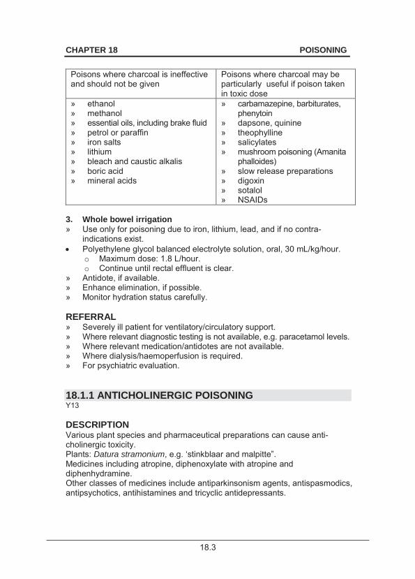
18.3
CHAPTER 18 POISONING
Poisons where charcoal is ineffective and should not be given
Poisons where charcoal may be particularly useful if poison taken in toxic dose
» ethanol » methanol » essential oils, including brake fluid » petrol or paraffin » iron salts » lithium » bleach and caustic alkalis » boric acid » mineral acids
» carbamazepine, barbiturates, phenytoin
» dapsone, quinine » theophylline » salicylates » mushroom poisoning (Amanita
phalloides) » slow release preparations » digoxin » sotalol » NSAIDs
3. Whole bowel irrigation » Use only for poisoning due to iron, lithium, lead, and if no contra-
indications exist. Polyethylene glycol balanced electrolyte solution, oral, 30 mL/kg/hour.
o Maximum dose: 1.8 L/hour. o Continue until rectal effluent is clear.
» Antidote, if available. » Enhance elimination, if possible. » Monitor hydration status carefully. REFERRAL » Severely ill patient for ventilatory/circulatory support. » Where relevant diagnostic testing is not available, e.g. paracetamol levels. » Where relevant medication/antidotes are not available. » Where dialysis/haemoperfusion is required. » For psychiatric evaluation. 18.1.1 ANTICHOLINERGIC POISONING Y13 DESCRIPTION Various plant species and pharmaceutical preparations can cause anti-cholinergic toxicity. Plants: Datura stramonium, e.g. ‘stinkblaar and malpitte”. Medicines including atropine, diphenoxylate with atropine and diphenhydramine. Other classes of medicines include antiparkinsonism agents, antispasmodics, antipsychotics, antihistamines and tricyclic antidepressants.
18.4
CHAPTER 18 POISONING
DIAGNOSTIC CRITERIA Clinical » Alteration of mental status, including delirium, hallucinations, agitation
and seizures. » Peripheral anticholinergic effects include:
> mydriasis, > urinary retention, > tachycardia and arrhythmias, > decreased GIT motility, > flushing, > dry skin and mucous
membranes. Investigations » ECG and continuous cardiac monitoring. » Pulse oximetry. GENERAL AND SUPPORTIVE MEASURES » Stabilise patient, i.e. airway, breathing and circulation. » Cooling for hyperthermia. » Perform decontamination depending on route of exposure. MEDICINE TREATMENT Activated charcoal.
For agitation: Diazepam, IV/oral, 0.1–0.2 mg/kg.
o Maximum dose: 10 mg. For seizures: See section 13.5: Status epilepticus (convulsive). REFERRAL » Cardiac dysrhythmia. » No response to treatment. 18.1.2 ANTICOAGULANT POISONING Y18 DESCRIPTION Poisoning with warfarin and “super-warfarin” (long acting warfarin), marketed as pellets for rats and mice. Over the counter pesticides containing warfarin may be accidentally ingested by toddlers or young children. Bleeding tendencies may be delayed.
Beware, some poisons may contain organophosphates.
18.5
CHAPTER 18 POISONING
DIAGNOSTIC CRITERIA Clinical » Signs and symptoms depend on the potency. » Symptoms may range from the asymptomatic child, e.g. a small child who
has tasted a rodenticide, to significant cases which present with bruising or bleeding, e.g. if the child has ingested “super-warfarin” containing pesticides.
Investigations » Measure prothrombin time.
> Obtain baseline INR if symptomatic. > Obtain a second INR in all cases at 24–32 hours post ingestion and
then 48 hours thereafter as the onset of co-agulopathy may be delayed.
GENERAL AND SUPPORTIVE MEASURES » Observe asymptomatic child. MEDICINE TREATMENT Deranged INR: Vitamin K1, IV/oral, 1– 5 mg/dose administered slowly 6 hourly.
o Repeat if large doses were administered. Oral vitamin K1 is usually preferred to intrav enous vitamin K1 unless more rapid reversal is required (e.g. the patient is bleeding). Intravenous vitamin K1 may cause hypersensitivity reactions. Bleeding: ADD Lyophilised plasma IV, 20 mL/kg.
OR Fresh frozen plasma, IV, 20 mL/kg.
OR Factor IX, intravenous, 25–50 units/kg (0.5–1 mL/kg).
Ingestion of long acting warfarin may be refractory to large doses of vitamin K1 and therapy may be required for several weeks after ingestion. 18.1.3 ANTIDEPRESSANT (TRICYCLIC) POISONING Y11 DESCRIPTION Poisoning with tricyclic antidepressants represent a large portion of poisoning fatalities. There is a high risk of tricyclic antidepressant toxicity in children because of its narrow therapeutic index. Serious toxicity may occur with low doses in children.
18.6
CHAPTER 18 POISONING
DIAGNOSTIC CRITERIA » 10–20 mg/kg of tricyclic antidepressant medication will cause significant
toxicity in most children. » Antidepressant poisoning cause anticholinergic syndromes. » Mainly affects the cardiovascular system, autonomic nervous system,
and central nervous system, leading to: > conduction delays, > dysrhythmias, > hypotension, > altered mental status, > seizures.
GENERAL AND SUPPORTIVE MEASURES » Gastric lavage for large ingestions or patients presenting within a few
hours post ingestion, except if the patient is unconscious. » Circulatory and respiratory support. » Cardiac and ECG monitoring for 48 hours. MEDICINE TREATMENT Activated charcoal, given as a slurry.
o If < 6 years of age: 10 g in 50–100 mL water o If > 6 years of age: 20–50 g in 100–300 mL water
Placement of a n asogastric tube may be necessary for its prompt administration.
For cardiac arrhythmias: Anti-arrhythmic agents. Only under specialist supervision.
For hypotension: Sodium chloride 0.9% or Ringer–Lactate, IV bolus, 20 mL/kg.
Alkalinisation for metabolic acidosis Alkalinisation up to an arterial pH of 7.45: Sodium bicarbonate 4.25%, IV, 2 ml/kg as a bolus.
o May be repeated. o Follow with a continuous infusion in consultation with
specialist/poison centre. For circulatory and respiratory support: See section 1.1.4: Cardiorespiratory arrest. REFERRAL » Any cardiac arrhythmia.
18.7
CHAPTER 18 POISONING
18.1.4 CAUSTIC OR CORROSIVE AGENTS, INGESTION Y19 DESCRIPTION Caustic agents, e.g. sodium hydroxide or potassium permanganate, corrosive agents, e.g. hydrochloric acid. Acids and alkali do not differ in their severity. Note: Battery acid causes significant corrosive damage, whereas household bleach seldom has a corrosive effect. DIAGNOSTIC CRITERIA Clinical » Chief symptom is pain. » Young children may present with:
> crying, > refusal to swallow, > drooling, and > vomiting.
» Stridor or hoarseness indicates laryngeal injury. » The presence of oral or pharyngeal burns does not predict the presence
of oesophageal or gastric injury. » Oesophageal or gastric injury can cause perforation or subsequent
fistula formation. » Patients with no clinical signs or symptoms are unlikely to have
significant oesophageal or other organ injury.
GENERAL AND SUPPORTIVE MEASURES Asymptomatic » Monitor for development of symptoms.
> A 12 hour symptom-free period usually indicates that no intervention is necessary.
Symptomatic » Gastric lavage/emesis is contraindicated in all cases. » Keep patient nil per mouth. » Airway injury may necessitate endotracheal intubation. » Endoscopic evaluation for patient with caustic injury. MEDICINE TREATMENT Prophylactic antibiotics are not indicated. Consider steroid therapy after endoscopy has shown a 2nd or 3rd degree oesophageal injury to red uce oedema and fi brosis. In the setting of G IT bleeding or perforation, steroids are contra-indicated. For pain control: See section 20.1: Management of pain.
18.8
CHAPTER 18 POISONING
REFERRAL » All symptomatic cases for endoscopic evaluation. 18.1.5 VOLATILE SOLVENTS Y16 DESCRIPTION Inhalants include: spray paints, glues and paint thinners which may contain toluene and or n-Hexane. These may contain hydrocarbons which may be aspirated following ingestion, causing a chemical pneumonitis. DIAGNOSTIC CRITERIA
» distinctive odour, » euphoria, » discolouration around mouth/nose, » headaches, » palpitations, » progressive CNS depression, » dizziness, » syncope, » cardiac arrhythmias, » hypokalaemia, » mucous membrane irritation, i.e. sneezing coughing and tearing, » GIT complaints, i.e. nausea, vomiting and abdominal pain, » distal renal tubular acidosis, i.e. hyperchloraemic metabolic acidosis
with a normal anion gap, » peripheral neuropathy and hepatotoxicity may be complications.
GENERAL AND SUPPORTIVE MEASURES » Stabilise airway, breathing and circulation. » Perform a chest X-ray and monitor patient for respiratory symptoms. » Correct fluid and electrolyte abnormalities. MEDICINE TREATMENT For agitation: Diazepam, IV/oral, 0.1–0.2 mg/kg.
o Maximum dose: 10 mg. For cardiac dysrythmias, e.g.: ventricular fibrillation see section 4.1: Cardiac dysrhythmias. REFERRAL » Cardiac dysrhythmia. 18.1.6 ETHANOL POISONING Y15 DESCRIPTION Ethanol is a selective CNS depressant at low concentrations, and a generalised depressant at high concentrations.
18.9
CHAPTER 18 POISONING
DIAGNOSTIC CRITERIA Clinical
» lack of coordination, » stupor, » ataxia, » coma, » slurred speech, » hypoglycaemia, » gait disturbances, » convulsions, » drowsiness.
Investigations » Monitor blood glucose levels. MEDICINE TREATMENT Obtunded patients: Dextrose 10%, IV, 2 mL/kg.
If patients respond to glucose administration, perform serial glucose levels to detect recurrent hypoglycaemia.
18.1.7 IRON POISONING Y14 DESCRIPTION Iron is widely available as an over-the-counter product and is commonly ingested accidentally by toddlers. DIAGNOSTIC CRITERIA Clinical » Toxicity is related to the ingested dose of elemental iron. » Single dose of elemental iron > 20 mg/kg requires hospital assessment
and management.
Elemental iron per preparation
Iron product Strength Elemental content
Elemental content Per mL or tablet
Ferrous gluconate elixir
350 mg/5 mL
40 mg elemental iron/5 mL
8 mg elemental iron per mL
Ferrous gluconate syrup 250 mg/5 mL 30 mg elemental
iron per 5 ml 6 mg elemental iron per mL
Ferrous lactate drops 25mg/mL 25 mg elemental
iron/ mL 1 mg elemental iron in 0.04 mL
Ferrous sulphate compound tablets
170 mg
± 65 mg elemental iron per tablet
± 65 mg elemental iron per tablet
18.10
CHAPTER 18 POISONING
Categories of iron toxicity:
Low risk Medium risk High risk » No history of:
> abdominal pain, > nausea, > vomiting, or
diarrhea. » Asymptomatic for
6 hours. » < 20 mg/kg of
elemental iron ingested.
» Minimal gastrointestinal symptoms.
» Normal physical examination.
» Lethargic. » Acidotic. » Shocked. » May have
evidence of haematemesis or melaena.
» Low risk patients are unlikely to have ingested enough iron to lead to serious poisoning and can be discharged.
» Admit high and medium risk patients. Investigations Medium risk » Abdominal X-ray. » Arterial blood gas. » Electrolytes. » Serum iron levels within 2–6 hours after ingestion. » If no clinical features are present and serum iron < 500 mcg/dL, patient
is low risk. High risk » Arterial blood gas. » Electrolytes. » Serum iron levels within 2–6 hours after ingestion. GENERAL AND SUPPORTIVE MEASURES Medium risk » If more than mild gastrointestinal symptoms or altered mental state,
shock, or acidosis refer for chelation therapy. High risk » Manage airway. » Refer for chelation therapy. MEDICINE TREATMENT Medium and high risk Fluid resuscitation: Sodium chloride 0.9%, IV, 20 mL/kg as an initial bolus followed with
maintenance therapy.
18.11
CHAPTER 18 POISONING
If no signs of gastrointestinal dysfunction e.g. perforation/haemorhage: Whole bowel irrigation.
Chelation therapy For iron ingestion > 60 mg/kg of elemental iron: Desferrioxamine, IV, 15 mg/kg/hour as a continuous infusion until urine
is no longer pink. o Beware of hypotension.
REFERRAL » Urgent: if unable to do the above, urgent transfer is vital. 18.1.8 NEUROLEPTIC POISONING Y11 DESCRIPTION Neuroleptic overdose may cause a depressed level of consciousness, hypotension, tachycardia and cardiac dysrhythmias and seizures. Commonly used neuroleptics include chlorpromazine, Haloperidol and phenothiazine antiemetics. Acute dystonic reactions/extrapyramidal symptoms are distressing adverse reactions (sustained muscle spasms) occurring after an overdose or during chronic therapy with neuroleptics. A ty pical dystonic reaction includes overextension or ov erflexion of the li mbs with abnormal posturing of the trunk. Other extrapyramidal symptoms may occur. The neuroleptic malignant syndrome is un common following an overdose and is an idiosyncratic life threatening reaction presenting with: » temperature dysregulation, » autonomic instability, » altered mental state, » diaphoresis, » musculoskeletal effects (pipe like rigidity). DIAGNOSTIC CRITERIA » Dystonic reactions. » Other extrapyramidal symptoms. GENERAL AND SUPPORTIVE MEASURES » Observe asymptomatic patients for a minimum of 6 hours. » Admit all symptomatic patients for continuous cardiac monitoring. » Test the urine of patients who are hypotensive, have muscular rigidity or
seizures for myoglobin (dipstix test for haemoglobin) and serum creatinine monitored because of the risk of rhabdomyolysis.
18.12
CHAPTER 18 POISONING
MEDICINE TREATMENT Activated charcoal.
For acute dystonic reactions: Biperidin, IV, slow injection.
o If < 1 year of age: 1 mg o If 1–6 years of age: 2 mg o If 6–10 years of age: 3 mg If concomitant significant anticholinergic findings are present, such as fever and dry skin and mucous membranes, a benzodiazepine is preferred.
REFERRAL » Patients with neuroleptic malignant syndrome. » Patient with conduction abnormalities (prolonged QT). 18.1.9 ORGANOPHOSPHATE POISONING Y18 * Notifiable condition DESCRIPTION Organophosphates are potent inhibitors of acetylcholinesterase. Poisoning due to organophosphates is notifiable. DIAGNOSTIC CRITERIA Clinical » Cholinergic toxidrome. » Cholinergic symptoms include:
Muscarinic effects: > diarrhoea, > vomiting. > urination, > lacrimation. > miosis, > bronchorrhoea/bronchoconstriction, > salivation.
Central nicotinic effects: > confusion, > coma, > convulsions.
» Cardiac effects include bradycardia or tachycardia depending on whether muscarinic or nicotinic effects predominate.
» Signs depend on dose and route of exposure (vapour or liquid) as well as the time exposed (vapour).
Investigations » Decreased levels of pseudocholinesterase.
> Use for confirmation only. > Do not wait for levels before treating.
18.13
CHAPTER 18 POISONING
GENERAL AND SUPPORTIVE MEASURES » Ensure use of personal protective equipment. » Remove all patients’ clothing and wash clothes thoroughly. » Ventilate, if necessary. » Wash affected skin with soap and water. » Suction secretions frequently. » Monitor respiratory function closely, as well as heart rate, pupillary size
and level of consciousness. MEDICINE TREATMENT For bradycardia, bronchorrhoea or bronchospasm: Atropine, IV, 0.02–0.05 mg/kg.
o Follow with a repeat dose every 10–15 minutes until bronchial secretions are controlled.
o Titrate dose against the secretions. o The therapeutic endpoint is the clearing of secretions and resolution
of bronchospasm. Note: Atropine may need to be continued for prolonged periods.
o Many repeated doses of atropine may be required and large quantities may be needed. Beware of relapses.
o Tachycardia and mydriasis are not contra-indications for atropine. Treat convulsions. See section 13.5: Status epilepticus (convulsive). REFERRAL » Where ICU facilities are not available. 18.1.10 OPIOID POISONING Y12 DESCRIPTION Codeine is a common drug of abuse. The duration of action of morphine is 3–6 hours. Other oral agents, e.g. codeine and long acting morphine, demonstrate a de layed effect of u p to 4–12 hours. DIAGNOSTIC CRITERIA » Altered level of consciousness. » Classic triad of CNS depression, respiratory depression and miosis
(pupillary constriction). » Hypotension, hypothermia, bradycardia and hyporeflexia.
18.14
CHAPTER 18 POISONING
» Vomiting is common and exposes the patient to the risk of aspiration
especially in patients with depressed consciousness. » Early symptoms: awake and alert presenting within 1–2 hours of
ingestion. GENERAL AND SUPPORTIVE MEASURES » Supportive care, ventilate with bag-mask device. » Monitor oxygen saturation constantly. » Observe for urinary retention. » Airway protection is a priority. MEDICINE TREATMENT Activated charcoal.
Naloxone, IV, 0.1mg/kg.
o If no response after 5 minutes, repeat dose and titrate according to response.
o Duration of action of naloxone is 20–30 minutes. o If repeated doses are naloxone are necessary, a continuous IV
infusion of naloxone can be instituted.
CAUTION All patients treated with naloxone should be observed for at least 12 hours for relapse, especially if a long acting opioid has been
ingested. REFERRAL » Patients requiring multiple doses of naloxone. 18.1.11 PARACETAMOL POISONING Y10 DESCRIPTION Paracetamol poisoning in c hildhood is almost always intentional. The accidental ingestion of paediatric paracetamol elixir preparations by the toddler very rarely achieves toxicity. Adolescents are often not aw are that paracetamol ingestion can be lethal and may unknowingly take a lethal dose as a suicidal gesture. Toxicity can be due to acute ingestions or repeated supratherapeutic ingestion (RSTI).

18.15
CHAPTER 18 POISONING
DIAGNOSTIC CRITERIA » Doses in excess of 150 mg/kg per 24-hour period in healthy children are
potentially toxic. » Serum paracetamol concentration must be measured at least four hours
following ingestion. » Use nomogram to assess risk of toxicity.
Paracetamol treatment nomogram
Nomogram reproduced from the Guidelines of Acute Paracetamol Overdose produced by the Medicines Information Centre, UCT.
Published with kind permission. This nomogram is not relevant for p oisoning with extended release formulations. » Cautions for use of this chart:
> The time co-ordinates refer to time of ingestion. > Serum levels drawn before 4 hours may not represent peak levels. > Use the graph only in relation to a single acute ingestion. > Not useful when there are co-ingestants, in delayed presentation >
24 hours post-ingestion. > The lower solid line 25% below the standard nomogram is included
to allow for possible errors in paracetamol plasma assays and estimated time from ingestion of an overdose.
18.16
CHAPTER 18 POISONING
» If patients present > 8 hours post-ingestion, start on treatment without
waiting for the paracetamol levels. » If the time of ingestion is unknown, start treatment for any detectable
level of paracetamol or any elevation of AST or ALT. » Patients with normal LFTs and undetectable paracetamol levels four
hours after ingestion do not require treatment. » Administer without waiting for plasma paracetamol levels in substantial
overdose, defined as 10 g (20 tablets) or 140 mg/kg, whichever is smaller. Discontinue if plasma levels are in the non-toxic range.
» Normal results at 48 hours excludes hepatic damage. » Do:
> Baseline urine and electrolytes. > Liver enzymes. > Coagulation profile.
The nomogram was not designed for use in overdoses with extended release paracetamol formulations. In this setting, administer N-acetylcysteine. Obtain a second paracetamol 4–6 hours after the original 4 hour concentration. If either value is above the treatment line of the nomogram, administer N-acetylcysteine. GENERAL AND SUPPORTIVE MEASURES » If the patient presents within one hour of ingestion do a gastric lavage. MEDICINE TREATMENT Only if patients presents within 1 hour of ingestion: Activated charcoal.
Acetylcysteine, IV.
First 24 hours o Loading dose: 150 mg/kg in dextrose 5%, 5 mL/kg given over 15
minutes. o 50 mg/kg in dextrose 5%, 5 mL/kg over the next 4 hours; then
100 mg/kg in dextrose 5%, 10 mL/kg over 16 hours. Second 24 hours o 100 mg/kg in dextrose 5%, 10 mL/kg over 24 hours.
If IV not available: Acetylcysteine, oral, 140 mg/kg immediately as a single dose.
o Follow with 70 mg/kg every 4 hours for 17 doses. REFERRAL » Patients with severe hepatocellular damage.
18.17
CHAPTER 18 POISONING
18.1.12 PETROCHEMICAL POISONING Y16 DESCRIPTION Accidental ingestion of paraffin, particularly by toddlers, is common in South Africa. DIAGNOSTIC CRITERIA Clinical » Paraffin is volatile and inhalation of the fumes or aspiration of liquid can
cause serious chemical pneumonitis. » Respiratory distress. » CNS symptoms. Investigations » Chest X-ray. GENERAL AND SUPPORTIVE MEASURES
CAUTION Do not attempt gastric lavage.
» Observe patient for up to 12–24 hours if asymptomatic. » Administer oxygen, if necessary. » Education and counseling regarding prevention. MEDICINE TREATMENT If infection develops 48 hours after ingestion: Amoxicillin, oral, 30 mg/kg/dose 8 hourly for 5 days.
REFERRAL » For ventilatory support. 18.1.13 SALICYLATE POISONING Y10 DESCRIPTION Salicylate poisoning may result from oral and/or topical exposure. Salicylate products vary widely in concentration e.g. oil of wintergreen is 100% methylsalicylate. As little as 4 mL of oil of wintergreen may be fatal in a child.
18.18
CHAPTER 18 POISONING
DIAGNOSTIC CRITERIA Clinical » Doses less than 150 mg/kg will not cause toxicity except in a child with
hepatic or renal disease. » Ingestion of 150–300 mg/kg may result in mild to moderate toxicity. » Ingestion of > 500 mg/kg should be considered a potentially lethal dose. » Features include:
> fever, > hyperventilation, > epigastric pain, > renal failure, > CNS depression, > Hypoglycaemia,
> respiratory alkalosis (initially) followed by metabolic acidosis.
» Monitor blood gases, urine output and urine and electrolytes. » Monitor salicylate level: toxic > 30 mg/dL. » Serial monitoring until declining levels are documented. » Monitor and treat hypoglycaemia. GENERAL AND SUPPORTIVE MEASURES » Gastric lavage. » Correct hydration. MEDICINE TREATMENT After gastric lavage: Activated charcoal.
o May be used for up to 12 hours: due to delayed gastric emptying or if sustained-release/enteric-coated preparations were ingested.
Urine alkalinisation If metabolic disturbances are present: Sodium bicarbonate, IV, 2 mmol/kg/day in maintenance fluid and
administered over 3 hours. o Increase, if necessary, to maintain urine pH above 7.5.
For hydration: ½ Darrows/dextrose 5%, IV.
For bleeding: Vitamin K1 (phytomenodione), IM, 5 mg as a single dose.
REFERRAL » Severe cases for ICU care.

18.19
CHAPTER 18 POISONING
18.1.14 SEDATIVE-HYPNOTIC POISONING Y11 DESCRIPTION Young children or toddlers are typically involved in accidental exposure and ingest small amounts of sedatives. Adolescents may ingest large amounts during suicide, suicidal gesture or for recreational use. Examples of sedative-hypnotics include: benzodiazepines and diphenhydramine. DIAGNOSTIC CRITERIA Clinical » Cardiorespiratory depression. » Decreased level of consciousness. Investigations » Serum drug levels are of no value in the acute treatment phase. » Urine test: may have medico-legal implications. GENERAL AND SUPPORTIVE MEASURES » If there is respiratory depression, intubate, ventilate and transfer. » Gastric lavage. » Supportive treatment only is necessary in most patients. MEDICINE TREATMENT If significant overdose is suspected: Activated charcoal.
REFERRAL » Respiratory depression. 18.1.15 SULFONYLUREA Y14 DESCRIPTION First generation sulfonylureas include chlorpropamide, which is excreted renally. Second generation agents include glimepiride and glipizide and are excreted in the faeces. DIAGNOSTIC CRITERIA Clinical » Coma and seizures. » Profound hypoglycaemia, usually within 4 hours of ingestion.
18.20
CHAPTER 18 POISONING
Investigations » Glucose monitoring is the mainstay of diagnostic testing. GENERAL AND SUPPORTIVE MEASURES » Observe for 24 hours even if a single tablet is ingested. » Glucose containing fluid orally. MEDICINE TREATMENT Activated charcoal.
Dextrose 10% (2 mL/kg), IV boluses.
o Titrate until blood glucose is controlled. Note: Corticosteroids are not indicated. REFERRAL » Patients not responding to intravenous glucose. 18.1.16 SYMPATHOMIMETIC AGENT POISONING Y13 DESCRIPTION Pseudoephedrine in decongestants, methylphenidate and illicit drugs such as cocaine and amphetamines (Tik) are sympathomimetic agents. These agents are frequently abused, especially as recreational drugs. DIAGNOSTIC CRITERIA Clinical
» Hypertension. » Psychosis. » Tachycardia. » Mydriasis. » Tachypnoea. » Diaphoresis. » Agitation. » Hyperthermia: effects of sympathomimetics that predispose to
hyperthermia include: > peripheral vasoconstriction and impaired cutaneous heat loss, > agitation, > seizures, > increased muscle activity, > impaired behavioral responses.
» With cocaine toxicity, cardiovascular manifestations predominate including: > supraventricular and ventricular dysrhythmias, > myocardial ischaemia.
» Neonates of mothers addicted to cocaine may present with withdrawal signs manifested by jitteriness.
18.21
CHAPTER 18 POISONING
Investigations » ECG monitoring to evaluate dysrhythmias. GENERAL AND SUPPORTIVE MEASURES » Admit all seriously ill children to ICU. » Maintain hydration. » Cooling for hyperthermia. » Mildly toxic patients require no specific treatment. MEDICINE TREATMENT Activated charcoal.
For agitation and tachycardia: Diazepam, IV/oral, 0.1–0.2 mg/kg.
o Maximum dose of 10 mg. For severe hypertension: See section 4.11.1: Hypertension, acute severe. For seizures: See section See section 13.5: Status epilepticus (convulsive). REFERRAL » Status epilepticus requiring ICU. » Hypertensive crisis. 18.2 ENVENOMATION 18.2.1 SCORPION STINGS DESCRIPTION Some scorpion species can cause serious systemic toxicity. Thick-tailed scorpions with small pincers are extremely toxic resulting in both local and systemic features. Thin-tailed scorpions with large pincers are much less toxic and likely only to result in local symptoms. DIAGNOSTIC CRITERIA » Pain and paraesthesia occur immediately after envenomation. » Autonomic and m otor findings may differentiate scorpion bites fro m
other causes of pain. » The pain can be exquisitely accentuated by tapping on the affected
region, i.e. “tap test”. » In severe cases cranial nerve dysfunction, blurred vision, pharyngeal
muscle inco-ordination, drooling and respiratory compromise can occur.
18.22
CHAPTER 18 POISONING
» Excessive motor activity may present as restlessness, or uncontrollable
jerking of extremities. » Other serious effects include cardiac dysfunction, pulmonary oedema,
pancreatitis, bleeding disorders and skin necrosis. » Nausea, vomiting tachycardia and severe agitation can also occur. GENERAL AND SUPPORTIVE MEASURES » General supportive care. » Monitor airway, breathing and circulation. MEDICINE TREATMENT For muscle cramps: Calcium gluconate 10%, IV, 0.5 mL/kg by slow intravenous injection.
o Give 0.5–1 mL/minute. o Monitor ECG.
For pain: Paracetamol, oral, 15 mg/kg/dose, 6 hourly as required.
Very painful scorpion stings Lidocaine (lignocaine) 2%, 2 mL injected around the bite as a local
anaesthetic. If not immunised in the past 5 years: Tetanus toxoid, IM, 0.5 mL.
Complete course in previously unvaccinated patients. Antivenom therapy Antivenom therapy is recommended only in cases with systemic signs and is rarely required: Scorpion antivenom, slow IV, 10 mL administered over 3–5 minutes.
REFERRAL » Severe cases requiring intensive care. 18.2.2 SNAKEBITE T63.0 DESCRIPTION The effects of snakebites may be cy totoxic, neurotoxic and/or haemotoxic. The overall effect is determined by the predominant toxin in the snake venom.

18.23
CHAPTER 18 POISONING
In the majority of cases, the species of snake is unknown. The patients can be divided into: » no evidence of bite, no envenomation, » evidence of bi te, minor envenomation, i.e. f ang marks, minimal pain,
minimal swelling and no systemic signs, » evidence of serious envenomation. DIAGNOSTIC CRITERIA Cytotoxic venom » Puff adder, spitting cobra, gaboon adder. » Venom causes severe local damage to t issues and vascular
endothelium. » Severe swelling and local necrosis occurs. Neurotoxic venom » Mamba, non- spitting cobra, rinkhals, berg adder. » Venom causes a paresis and paralysis of skeletal muscles. » Paralysis of respiratory muscles with respiratory failure may occur. » Preceded by severe pain and paraesthesias. » Ophthalmoplegia occurs when ocular muscles become paralysed. » Speech and swallowing may be affected. » Signs and symptoms start within 15–30 minutes. Haemotoxic venom » Boomslang, vine snake. » Venom may cause:
> haemolysis of red blood cells, > ecchymosis, > anaemia, > epistaxis, > consumptive coagulopathy, > haemoptysis > bruises, > haematuria.
GENERAL AND SUPPORTIVE MEASURES » Patients with no evidence of bite and patients with evidence of bite but
only minor envenomation should be admitted for observation. No anti-venom is indicated.
» Do not suck or cut the wound. » Do not apply tourniquet. » Where serious envenomation is suspected: immediate treatment
includes > minimising movement of affected limb, > emergency treatment by bandaging affected limb with crepe
bandage without compromising blood supply, > rapid transportation to a facility with available antivenom is the most
important principle of pre-hospital care, > optimal therapy consisting of placing the patient at rest with the
affected body part raised to the level of the heart, > stabilising circulation and blood pressure.
18.24
CHAPTER 18 POISONING
» For cytotoxic envenomation, surgical intervention, i.e. d ecompression
surgery for established compartment syndrome and deb ridement of necrotic tissue should only be done when absolutely necessary and as conservatively as possible.
» For neurotoxic envenomation, ventilatory and cardiovascular support may be needed in an Intensive Care Unit.
MEDICINE TREATMENT All patients not immunized within the past 5 years: Tetanus toxoid adsorbed vaccine, IM, 0.5 mL.
If children with penetrating wound and who are not completely immunised: Tetanus immunoglobulin, IM.
o If < 5 years of age: 75 IU. o If 5–10 years of age: 125 IU. o If > 10 years of age: 250 IU.
Cleanse wound: Chlorhexidine 0.05% solution in water.
Antivenom therapy Indications: » Consider antivenom in children who are persistently and severely
affected even after the first day. » Painful swelling of the whole hand/foot within 1 hour, spreading to
elbow/knee in 3–6 hours. » Swelling of head, neck or chest. » Significant envenomation e.g. overt neurological signs or bite in close
proximity to airway structures. » Platelet count less than 100 x 109/L. » Fibrinogen less than 100 mg/dL. » Any confirmed black mamba or gaboon adder bite. The dose of antivenom is the same for adult and children.
CAUTION Never administer antivenom without being fully prepared to manage
acute anaphylaxis.
Give a test dose of antivenom. If there is no untoward reaction, give the full dose. If a reaction develops following a test dose, give pre-treatment with epinephrine (adrenaline).
18.25
CHAPTER 18 POISONING
CAUTION Polyvalent antivenom is only effective for the following snake bites:
» Cape cobra » Mamba » Puff adder » Gaboon adder » Rinkhals » Spitting cobra
Boomslang requires specific antivenom.
Antivenoms are available from the South African Vaccine Producers
(SAVP). SAVP emergency number: 083 6520105.
Snakebite antivenoms may be available from specific hospitals in each province.
For cobras, mambas, rinkhals, puff adders and Gaboon viper: Polyvalent snake antivenom, IV.
o Dilute 10 mL in sodium chloride 0.9% 50 mL. o Administer slowly over 15 minutes. o If no reaction occurs, 60–120 mL antivenom diluted in sodium
chloride 0.9%, 200 mL administered slowly over 30 minutes. For boomslang bites: Boomslang antivenom, slow IV, 10 mL administered over 3–5 minutes.
OR Boomslang antivenom, IV infusion, 10–20 mL diluted in sodium chloride
0.9% or dextrose 5%, 50–100 mL administered over 5–10 minutes. o After administration, observe patient.
Correct anaemia and bleeding tendency. REFERRAL » Snakebite with neurotoxic or haemotoxic manifestations may need
intensive care.
18.2.3 SPIDER BITES (WIDOW SPIDERS) T63.3 DESCRIPTION The vast majority of spiders are no t harmful to humans. Widow spiders (Lactrodectus) are found in dark confined areas and the female can produce a potent venom that acts through a calcium mediated mechanism leading to the release of acetylcholine and noradrenaline from nerve terminals.
18.26
CHAPTER 18 POISONING
DIAGNOSTIC CRITERIA » Bites are felt immediately as a pinprick sensation, followed by increasing
local pain that may spread to include the entire extremity. » Typical target lesions, i.e. erythematous ring surrounding a pale center. » Cramp like spasms in large muscle groups, abdominal pain or rig idity,
progressing to generalised pain involving the trunk and abdomen have been described.
GENERAL AND SUPPORTIVE MEASURES » Supportive care of airway, breathing and circulation. MEDICINE TREATMENT For pain and muscle cramps: Calcium gluconate 10%, IV, 0.5 mL/kg by slow intravenous injection.
o Give 0.5–1 mL/minute. o Monitor ECG and respiration.
For severe envenomation to resolve symptoms and shorten duration of illness: Spider antivenom, IV infusion, 5–10 mL diluted in sodium chloride 0.9%
or dextrose 5%, 50–100 mL administered over 5–10 minutes. 18.2.3.1 SPIDER BITES, NECROTIC ARACHNIDISM DESCRIPTION Loxosceles spiders can produce local necrotic skin lesions that are mediated by enzymes. DIAGNOSTIC CRITERIA » Bites are initially painless. » Skin lesions can vary from mildly erythematous lesions to severe local
reaction, i.e. blistering, bluish discolouration progressing to fran k necrosis.
» Systemic effects include nausea, vomiting, fever, chills, arthralgia, haemolysis, thrombocytopaenia, haemoglobinuria and renal failure.
GENERAL AND SUPPORTIVE MEASURES » Supportive care. » Surgical debridement once the clear margins around the necrotic
lesions are established.
18.27
CHAPTER 18 POISONING
MEDICINE TREATMENT For pain: Paracetamol, oral, 15 mg/kg/dose, 6 hourly as required.
Morphine, IV, 0.1 mg/kg/dose 4 hourly.
o Monitor for respiratory depression. Antibiotic therapy for septic lesions.
19.1
CHAPTER 19 PREMATURITY AND NEONATAL CONDITIONS
19.1 APNOEA, NEONATAL P28.3 DESCRIPTION A neonate presenting with episodes of cessation of breathing. Apnoea episodes in a previously asymptomatic well neonate may be the first indication of a serious underlying disease. Apnoea episodes in an already unwell neonate indicate deterioration in the condition of the neonate. DIAGNOSTIC CRITERIA » Cessation of respiration for longer than 20 seconds, with/without cyanosis, pallor
or bradycardia. » Cessation of respiration for less than 20 seconds with cyanosis, pallor and/or
bradycardia. Central apnoea Causes include: » IRDS, » prematurity, » hypoxia/hypercarbia, » sepsis, » acidosis, » meningitis, » temperature disturbances, » hypotension,
» pneumonia, » intraventricular haemorrhage, » patent ductus arteriosus, » hypoglycaemia, » hypermagnesaemia, » atypical convulsions, » anaemia, » rough or excessive handling, and
» medicines (sedatives, anticonvulsants, analgesics). Obstructive apnoea Neonates are obligatory “nose breathers”. Obstruction of the nares make neonates prone to apnoea. Causes of obstructive apnoea include: » choanal atresia, » gastro-oesophageal reflux, » micrognathia, » macroglossia, » secretions (milk, meconium, blood, mucus) lodged in the upper airway, and » neck flexion or extension.

19.2
CHAPTER 19 PREMATURITY AND NEONATAL CONDITIONS
Reflex apnoea or vagally mediated apnoea Is due to: » endotracheal intubation, » passage of a nasogastric tube, » gastro-oesophageal reflux, » overfeeding, and » suction of the pharynx or stomach. Mixed apnoea Apnoea caused by a combination of the above causes. GENERAL AND SUPPORTIVE MEASURES For all forms of neonatal apnoea: » Identify and treat the underlying cause. » Frequent gentle physical stimulation e.g. rubbing of soles of feet. » Nurse premature neonates in the prone position. » Maintain ambient temperature at the lower range of neutral thermal
environment. » Maintain axillary temperature or anterior abdominal wall temperature at 36.2–
36.8°C. » Maintain haematocrit at 40%. » Maintain nasal CPAP of 4 c m water. (Nasal CPAP – not for c entral apnoea
except for apnoea of prematurity.) » Monitor vital signs and parameters relating to the underlying cause. MEDICINE TREATMENT To maintain oxygen/haemoglobin saturation of 88–92% (for very low birth w eight 88–90%) or an oxygen tension in the blood at 60–80 mmHg: Oxygen via nasal cannula, headbox, or mask.
Only for apnoea of prematurity (not term infants): Caffeine base, (anhydrous), oral.
o Loading dose: 10–12.5 mg/kg. o Maintenance dose: 2.5–5 mg/kg/24 hours. Start maintenance dose 24
hours after the loading dose. (Caffeine citrate 20mg = caffeine base 10 mg)
OR Aminophyline, IV/oral.
o Loading dose: 8 mg/kg. (If IV infusion, administer over 30 minutes). o Maintenance dose: 1.5–3 mg/kg/dose 8 hourly. Start maintenance dose 8
hours after loading dose
Maintain aminophylline blood levels at 10–12 mcg/mL. If neonate responds favourably to ca ffeine/aminophyline continue until neonate is apnoea free for 7 days.
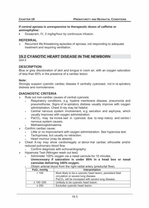
19.3
CHAPTER 19 PREMATURITY AND NEONATAL CONDITIONS If central apnoea is unresponsive to therapeutic doses of caffeine or aminophyline: Doxapram, IV, 2 mg/kg/hour by continuous infusion.
REFERRAL » Recurrent life-threatening episodes of apnoea, not responding to adequate
treatment and requiring ventilation.
19.2 CYANOTIC HEART DISEASE IN THE NEWBORN Q24.9 DESCRIPTION Blue or grey discoloration of skin and tongue in room air, with an oxygen saturation of less than 85% in the presence of a cardiac lesion. Note: Strongly suspect cyanotic cardiac disease if centrally cyanosed, not in re spiratory distress and normotensive. DIAGNOSTIC CRITERIA » Rule out non-cardiac causes of central cyanosis:
> Respiratory conditions, e.g. hyaline membrane disease, pneumonia and pneumothorax. Signs of re spiratory distress usually improve with oxygen administration. Chest X-ray may be helpful.
> Central nervous system involvement, e.g. sed ation and asphyxia, which usually improves with oxygen administration.
> PaCO2 may be increa sed in cyanosis due to resp iratory and centra l nervous system causes.
> Methaemoglobinaemia. » Confirm cardiac cause:
> Little or no improvement with oxygen administration. See hyperoxia test. > Tachypnoea, but usually no retraction. > Heart murmur (may be absent).
» Chest X-ray may show cardiomegaly or abnor mal cardiac silhouette and/or reduced pulmonary blood flow. > Confirm diagnosis with echocardiography.
» Hyperoxia Test (Nitrogen wash out test) > Administer 100% oxygen via a nasal cannula for 10 minutes.
Unnecessary if saturation is under 85% in a head box or nasal cannulae delivering 100% oxygen.
> Obtain arterial blood from the right radial artery (preductal flow). PaO2 mmHg Interpretation
< 100 Most likely to be a cyanotic heart lesion, persistent fetal circulation or severe lung disease. PaCO2 will be increased with severe lung disease.
–200 Unlikely to be cyanotic heart lesion. Excludes cyanotic heart lesion.
19.4
CHAPTER 19 PREMATURITY AND NEONATAL CONDITIONS
GENERAL AND SUPPORTIVE MEASURES » Nurse in neutral thermal environment. » Monitor and maintain within physiological range for age:
> heart rate, > calcium, magnesium, > respiration, > blood glucose, > blood pressure, > blood gases, > body temperature, > acid-base status, and > electrolytes.
» Provide adequate hydration and nutrition. MEDICINE TREATMENT To keep ductus arteriosus open if a duct dependent cyanotic heart lesion is suspected: Prostaglandin therapy, i.e.: Alprostadil, IV, 0.05–0.1 mcg/kg/minute, initial dose.
o Maintenance dose : 0.01–0.1 mcg/kg/minute. OR Dinoprostone, via naso/orogastric tube.
o For babies < 2.5 kg: 0.125 mg 1–2 hour ly (1/4 tablet suspended in 2 mL sterile water), or 50 mcg/kg/dose 1–2 hourly.
o For babies > 2.5 kg: 0.25 mg hourly (½ tablet suspended in 2 mL water). Continue with prostaglandin therapy until corrective or palliative surgery can be done or until patency of the duct is not deemed essential for survival of the infant. Babies on prostaglandin therapy: inspiratory oxygen not more than 40 %. An oxygen saturation of haemoglobin > 75% is acceptable.
Serious side effects of prostaglandins to be aware of may include: Apnoea, fever, diarrhoea, hypotension and seizures
I : » Sodium bicarbonate 4.2 %, IV.
HCO3 needed (mmol) = base excess x 0.3 x body mass (kg). 2 mL sodium bicarbonate 4.2% = 1 mmol HCO3.
SURGICAL TREATMENT » Corrective or palliative surgery. REFERRAL » All cyanotic infants with an underlying cardiac cause for central cyanosis.
19.5
CHAPTER 19 PREMATURITY AND NEONATAL CONDITIONS
19.3 ENTEROCOLITIS, NECROTISING P77 DESCRIPTION Neonate presenting with the consequences of bowel wall injury or necrosis. Risk factors include: » prematurity, » hypotension/shock, » sepsis, » early formula feedings,
» high feeding volumes, » perinatal asphyxia (hypoxia),
» patent ductus arteriosus, and » polycythaemia. DIAGNOSTIC CRITERIA » Early signs are often non-specific, i.e.:
> feeding intolerance, > gastric aspirates, > vomiting, > body temperature instability, > apnoea and lethargy.
» Non-specific signs may progress to more specific signs including: > abdominal distention with ileus, > bloody stools, > peritonitis, > red-purple discolouration of the abdominal wall with abdominal wall
cellulitis, and > bowel perforation.
» X-ray of abdomen may show: > distended loops of intestines, > bowel-wall thickening (oedema), > pneumatosis intestinalis, > hepatic portal venous gas, and > free intraperitoneal air due to perforation.
» Blood samples for culture and sensitivity testing before starting antibiotic therapy.
GENERAL AND SUPPORTIVE MEASURES » Admit to neonatal high-care unit or intensive care unit. » Nurse in neutral thermal environment. » Insert oro/nasogastric tube and apply free drainage.
> Suspected cases should be nil per mouth for 72 hours. > Confirmed cases should be nil per mouth for at least 7 days.
» Provide adequate parenteral nutrition (hyperalimentation) as soon as diagnosis is confirmed.
» Provide cardiovascular and ventilatory support, if necessary.
19.6
CHAPTER 19 PREMATURITY AND NEONATAL CONDITIONS
MEDICINE TREATMENT Depending on age, weight and hydration status: Neonatal maintenance solution, IV.
Add volume of gastric aspirates to daily maintenance fluid volume. If co-agulopathy or septic shock: Plasma, IV, 20 mL/kg over 2 hours.
If haematocrit < 40%: Packed red cells, IV, 10 mL/kg.
Until blood pressure is stabilised: Dopamine, IV, 5–15 mcg/kg/minute.
Empiric antibiotic therapy Ampicillin, IV, 50 mg/kg/dose for 10 days.
o If age < 7 days: 50 mg/kg 12 hourly. o If 7 days – 3 weeks of age: 50 mg/kg 8 hourly. o If > 3 weeks of age: 50 mg/kg 6 hourly.
PLUS Gentamicin, IV, 6 mg/kg once daily for 10 days.
PLUS Metronidazole, IV, for 7 days.
o Loading dose: 15 mg/kg over 60 minutes. o Post natal age < 4 weeks: 7.5 mg/kg/dose 12 hourly. o Postnatal age : 7.5 mg/kg/dose 8 hourly.
Reassess choice of antibiotics when the culture and sensitivity results become available. SURGICAL TREATMENT Surgical intervention is req uired when there is progr essive deterioration of th e clinical condition despite maximal medical support and/or bowel necrosis with/without bowel perforation. Prior to transport to a tertiary hospital for definitive surgery, insert/place a peritoneal drain in babies presenting with severe abdominal distension, due to free air and/or fluid in the peritoneal cavity, compromising respiration and/or blood pressure. Perform the procedure in a theatre, intensive care or high care unit where facilities for monitoring vital signs, resuscitation, ventilation and temperature control of the environment are available. Obtain consent to perform the surgical procedure.
19.7
CHAPTER 19 PREMATURITY AND NEONATAL CONDITIONS
Method of inserting/placing a peritoneal drain » Procedure is sterile; the doctor should be gowned and gloved. » Clean and drape the abdomen. » Administer an appropriate analgesic (e.g. ketamine, IV) immediately before the
start of the procedure. » Identify a si te in either one of the fossae iliaca, make sure it is lateral to the
inferior epigastric artery. » At the intended surgical incision site, inject: Lidocaine (lignocaine) 1%, SC, 0.5 mL.
» Make a small skin incision over the “bubble” of lidocaine (no. 11 blade). » Use a mosquito forceps or clamp to dissect down to the peritoneum, pierce the
latter with a gentl e stab using the cl osed forceps and slightly stretch the peritoneal puncture site with the forceps.
» Note what drains from the peritoneal cavity and send a sample for microscopy and culture.
» Insert a pencil drain of ± 5 mm width with the mosquito clamps or forceps into the peritoneal cavity through the peritoneal stab wound. About 1.5–2 cm of the pencil drain should be inside the peritoneal cavity.
» Fix the drain to the skin with a size 4 – 0 stitch (e.g. PDS). » Cover the drain with a gauze pad or urine collecting bag. REFERRAL » All confirmed cases for specialist care. » Deterioration of clinical condition, despite adequate treatment. » Signs and symptoms of intestinal perforation and peritonitis requiring surgical
intervention. » Recurrent apnoea episodes and/or signs of respiratory failure, requiring
respiratory support. 19.4 HAEMORRHAGIC DISEASE OF THE NEWBORN P53 DESCRIPTION This is due to a d eficiency of vitamin K-dependent clotting factors II, VII, IX and X. All newborns who did not receive vitamin K1 at birth, especially premature babies and breastfed babies, are at risk. Spontaneous bleeding may be fro m any site but is usually gastro-intestinal, producing haematemesis or melaena. Bleeding from the umbilical stump, epistaxis and a cephalohaematoma or subgaleal haemorrhage are also relatively common. Complications may include anaemia, hypovolaemic shock and intracranial haemorrhage with neurological damage. There are three forms of the disorder. Early form: presents within 24 hours of birth in newborns of mothers on treatment with anticonvulsants, e.g. phenytoin and phenobarbitone, or oral anticoagulants.
19.8
CHAPTER 19 PREMATURITY AND NEONATAL CONDITIONS
Classical form: presents during the first week of lif e usually on th e second to seventh day. Late form: presents during the first to fourth month of l ife usually with intracranial haemorrhage in exclusively breastfed babies who did not receive vitamin K prophylaxis at birth. DIAGNOSTIC CRITERIA Special investigations » Prolonged prothrombin time (PT). » Normal partial prothrombin time (PTT). » Increased international normalised ratio (INR) with a normal platelet count. » Normal fibrinogen levels. » Normal thrombin time. Note: Exclude other causes of bleeding in the neonate. Exclude swallowed blood of mother during delivery in babies with melaena. (Apt test or haemoglobin electrophoresis). GENERAL AND SUPPORTIVE MEASURES » Nurse in neutral thermal environment. » Provide adequate nutrition. » Monitor:
> blood pressure, > hydration, > heart rate, > SaO2, > respiratory rate, > haematocrit, > body temperature, > blood glucose, and > coagulation parameters.
MEDICINE TREATMENT Oxygen, if needed.
Fresh frozen plasma or lyophilised plasma, IV, 20 mL/kg over one hour.
If anaemic (haematocrit < 40% or Hb < 13 g/dL): Packed red cells, IV, 10 mL/kg over 1 hour.
o May be repeated if necessary. Vitamin K1, IM, 1 mg as a single dose.
Prophylaxis Vitamin K1, IM, single dose at birth.
Full term newborns: 1 mg Preterm newborns: 0.5 mg
Prophylaxis with oral vitamin K formulation is not recommended.
19.9
CHAPTER 19 PREMATURITY AND NEONATAL CONDITIONS REFERRAL » Deterioration of clinical condition despite adequate treatment. » Suspected intracranial haemorrhage. 19.5 HEART FAILURE IN NEONATES P29.0 DESCRIPTION Clinical syndrome reflecting the inability of the myocardium to meet the oxygen and nutritional or met abolic requirements of the b ody. Heart failure may be acut e or chronic. The main causes of heart failure are: » Congenital heart abnormalities:
> Left-sided outflow obstruction, e.g. interrupted aortic arch, co-arctation of the aorta and aortic valve stenosis.
> Left to right shunts, VSD and PDA. > Hypoplastic left heart. > Complex congenital heart lesions.
» Acquired conditions: > fluid overload, > sepsis, > hypoglycaemia, > hypoxia, > acidosis, > severe anaemia, > dysrhythmias, > cardiomyopathy, > pneumopericardium, > hyperthyroidism, > hypertension.
DIAGNOSTIC CRITERIA Diagnosis relies on history, physical examination and a chest X-ray. Clinical » Acute heart failure may present with shock, i.e. cardiogenic shock. » Heart failure is usually associated with fluid retention and congestion. » History of recent onset of:
> poor feeding, > tachypnoea (> 60 breaths/minute), > sweating, and > poor or excessive weight gain in excess of 30 g/24hours.
» Physical findings: > tachycardia (> 180 beats/minute), > gallop rhythm (with/without a cardiac murmur), > cardiomegaly, > features of ca rdiogenic shock, i.e. cold wet skin, weak pulses,
hypotension, > reduced urinary output, > pulmonary venous congestion and fluid retention,
19.10
CHAPTER 19 PREMATURITY AND NEONATAL CONDITIONS
> systemic venous congestion, > hepatomegaly, and > signs and symptoms of underlying condition/disease.
» Always check the femoral pulses. Special Investigations » Radiology: cardiomegaly is almost always present, cardiothoracic ratio > 60%. » Electrocardiogram may show evidence of hypertrophy of one or more heart
chambers and/or dysrhythmias. » Sonar or u ltrasound may show a reduc ed ejection fraction or shortening
fraction of left ventricle. GENERAL AND SUPPORTIVE MEASURES » Nurse in a neutral thermal environment. » Restrict fluids but ensure adequate nutrition.
> Administer 75% of daily requirements. > Use breast milk or low-salt milk formulae. > Tube feeding.
» Treat the underlying condition, e.g. sepsis and cardiac tamponade. MEDICINE TREATMENT First treat shock, if present. To prevent hypoxia: Oxygen via face mask, nasal cannula or head box.
Combination medicine therapy is usually indicated, Afterload reduction: ACE inhibitor or vasodilator Monitor blood potassium levels and stop potassium supplements while patient is on an ACE inhibitor. ACE inhibitors are contraindicated in renal failure, bilateral renal artery stenosis or a single functioning kidney. Consider ACE inhibitors in persistent heart failure where left s ided outflow obstruction has been excluded, other measures have failed and only after consultation with a paediatrician or paediatric cardiologist. Captopril, oral, 0.01–0.05 mg/kg/dose, 8–12 hourly, initially.
o Adjust dose and interval based on response. o Administer 1 hour before feeding. o Continue as long as needed to control the heart failure
Diuretics Continue diuretic therapy as long as needed to control heart failure. Monitor blood potassium levels.
19.11
CHAPTER 19 PREMATURITY AND NEONATAL CONDITIONS
Potassium supplements may be necessary if furosemide is used without spironolactone. Hypokalaemia and hypochloraemic alkalosis may increase digitalis toxicity. Furosemide, IV/oral, 1–3 mg/kg/24 hours as a single daily dose, or in 4 divided
oral doses. WITH/ WITHOUT Spironolactone, oral, 1–3 mg/kg/dose, once daily.
Digoxin Consider in consultation with paediatrician or cardiologist Monitor digoxin blood levels and ECG. Digoxin is contraindicated in bradycardia, heart block, cardiac tamponade or hypertrophic cardiomyopathy. Digoxin, oral, 0.01 mg/kg/dose 8 hourly for 3 doses, then:
o Maintenance: oral, 0.005 mg/kg/dose for as long as needed to control heart failure: For infants < 37 weeks gestational age: 24 hourly. For in gestational age: 12 hourly.
When oral route is contraindicated: Digoxin, IV, 75% of oral dose.
Inotropic support May help to stabilise patients with severe myocardial dysfunction, hypotension or low cardiac output. May be lifesaving in severe myocarditis or cardiogenic shock. Dobutamine, IV infusion, 2.5–15 mcg/kg/minute.
o Continue until myocardial function and blood pressure improve. o Ensure normovolaemia. o Monitor blood pressure.
Acute left-heart failure: acute pulmonary oedema or pulmonary venous congestion Oxygen 100%, via nasal cannula.
Furosemide, IV, 1–3 mg/kg, immediately.
For patients not responding to furosemide: Morphine, IV, 0.1 mg/kg.
For patients not already on digoxin treatment: Digoxin, as above.
19.12
CHAPTER 19 PREMATURITY AND NEONATAL CONDITIONS Inotropic support, as above.
Afterload reduction, as above.
To raise the alveolar pressure above pulmonary capillary pressure, intubate with intermittent positive ventilation. SURGICAL TREATMENT Palliative or corrective surgery for certain congenital heart lesions. REFERRAL » Deterioration despite adequate treatment. » For determination of the underlying cause, initiation of treatment and
stabilisation. 19.6 HYPOCALCAEMIA, NEONATAL P71.1 DESCRIPTION Acute symptomatic hypocalcaemia may present within the first 72 h ours of bir th (early hypocalcaemia) or after 72 hours of birth (late hypocalcaemia) with apnoea, irritability, seizures, jitteriness or prolonged QTc interval on ECG. Causes of early hypocalcaemia include: » Prematurity. » Respiratory distress syndrome. » Asphyxia/hypoxia. » Neonate of diabetic mother. » Sepsis. Causes of late hypocalcaemia include: » Maternal hyperparathyroidism. » Congenital hypoparathyroidism. » Renal failure. » Hypomagnesaemia. » High phosphate feeds. » Vitamin D deficiency. DIAGNOSTIC CRITERIA » Total serum calcium < 1.8 mmol/L, or » Ionised calcium < 0.7 mmol/L.
19.13
CHAPTER 19 PREMATURITY AND NEONATAL CONDITIONS
MEDICINE TREATMENT Symptomatic hypocalcaemia: Calcium gluconate 10%, IV, 100–200 mg/kg/dose 6–8 hourly.
1 mL of calcium gluconate 10% = 100 mg calcium gluconate = 10 mg elemental calcium = 0.23 mmol calcium
Correct hypomagnesaemia before administering 10% calcium gluconate. Magnesium sulphate 50%, IV, 0.25 mL/kg.
o Monitor levels until deficits are reduced.
Do not administer ceftriaxone, within 48 hours of administering calcium.
Exchange transfusion Calcium gluconate 10%, IV infusion, administered over 10 minutes.
o 100mg for every 100 mL citrated blood exchanged. Acute hypocalcaemia with seizures Calcium gluconate 10%, IV infusion, 100–200 mg/kg, administered over 10
minutes. Repeat in 15 minutes if necessary. o Dilute 1:1 with dextrose 5% or sodium chloride 0.9%. o Do not use calcium chloride. Note: Rapid infusion causes bradycardia/dysrhythmias. Electrocardiographic monitoring is advised. Monitor the heart rate.
CAUTION
Do not mix calcium gluconate with bicarbonate or fluids containing phosphate as precipitation may occur.
Extravasation of calcium can cause tissue necrosis. Do not give intra-arterially or via umbilical venous catheters placed
near the heart or inside the liver. REFERRAL » Persisting or recurrent unexplained hypocalcaemia. 19.7 HYPOGLYCAEMIA, NEONATAL P70.4 DESCRIPTION Neonate presenting with a whole blood glucose below 2.6 mmol/L.
19.14
CHAPTER 19 PREMATURITY AND NEONATAL CONDITIONS
Risk factors include: » prematurity, » respiratory distress, » small for gestational age, » rhesus iso-immunisation, » neonate of diabetic mother, » hyperinsulinism, » sepsis, » post maturity, » hypothermia/hyperthermia, » feeding difficulties, » birth asphyxia, » polycythaemia, and » hereditary defects in carbohydrate or amino acid metabolism. DIAGNOSTIC CRITERIA Clinical Asymptomatic: Hypoglycaemia detected when screening neonates at risk. Symptomatic: » lethargy, » poor feeding, » hypotonia, » respiratory distress, » apnoea, » cardiac failure, » jitteriness, » convulsions, » irritability, » metabolic acidosis, and » coma. Investigations » Whole blood glucose (heel prick) < 2.6 mmol/L. Monitor the blood gl ucose of all neo nates who are at risk of hy poglycaemia regularly, at least 2 hourly, to prevent the development of hypoglycaemia. GENERAL AND SUPPORTIVE MEASURES » Determine and treat the underlying cause. » Enteral feeding, oral or v ia oro/nasogastric tube, after exclusion of v omiting,
ileus or obstruction. MEDICINE TREATMENT Dextrose 10%, bolus IV, 2.5 mL/kg (250 mg/kg).
Dextrose 10% = 10 g dextrose in 100 mL. Do not repeat dextrose bolus.
To raise heel prick blood glucose to a level of 2.6 mmol/L or more, follow with: Dextrose 10%, continuous IV infusion, 6–12 mg/kg/minute or more.
Dextrose dose (mg/kg/min) = (% dextrose solution x rate in mL/hour) (weight x 6)
If heel prick blood glucose remains below 2.6 mmol/L: Dextrose 15%, IV, 15 mg/kg/minute or more
Dextrose 15% = 15 g dextrose in 100 mL. If heel prick blood glucose is above 2.6 mmol/L after IV infusion has been started continue infusion at maintenance rate.
19.15
CHAPTER 19 PREMATURITY AND NEONATAL CONDITIONS Monitor blood glucose at least 2 hourly until blood glucose level stabilises at 2.6 mmol/L or above. To avoid rebound hypoglycaemia, reduce IV dextrose infusion gradually. Before the IV i nfusion is finally discontinued, the ne onate should receive all milk feeds orally or via nasogastric tube. If enteral feeds are not tolerated TPN should be given. Suspect other serious underlying metabolic or biochemical abnormality if the neonate requires > 12 mg/kg/minute of dextrose to maintain a heel prick whole blood glucose > 2.6 mmol/L. Use a central venous line for high concentrations of dextrose. Prior to referral give the following, if available: Glucagon, IM/IV/SC, 0.2 mg/kg single dose.
REFERRAL » Hypoglycaemia not responding to adequate treatment. » Recurrent or persistent hypoglycaemia See also section 7.6 Hypoglycaemia in children. 19.8 HYPOXIA/ISCHAEMIA OF THE NEWBORN
(PERINATAL HYPOXIA/HYPOXIC-ISCHAEMIC ENCEPHALOPATHY)
P21.9 DESCRIPTION Ischaemia and decreased oxygen delivery to the fetus/baby during the prepartum, intrapartum or immediate postpartum period, with hypoxic-ischaemic damage to the central nervous system and to other body systems. Complications include: » Cardiovascular: heart rate and rhythm disturbances, heart failure and
hypotension. » Pulmonary: respiratory distress/respiratory failure, pulmonary hypertension and
pulmonary haemorrhage. » Renal: renal failure, acute tubular/cortical necrosis and urinary retention. » Gastrointestinal tract: ileus and necrotising enterocolitis. » Central nervous system: increased intracranial pressure, cerebral oedema,
encephalopathy, seizures, inappropriate antidiuretic hormone (ADH) secretion, hypotonia and apnoea.
» Metabolic: hypoglycaemia, hyperglycaemia, hypocalcaemia, hypomagnesaemia and metabolic acidosis.
» Body temperature: abnormal. » Other: disseminated intravascular coagulation.
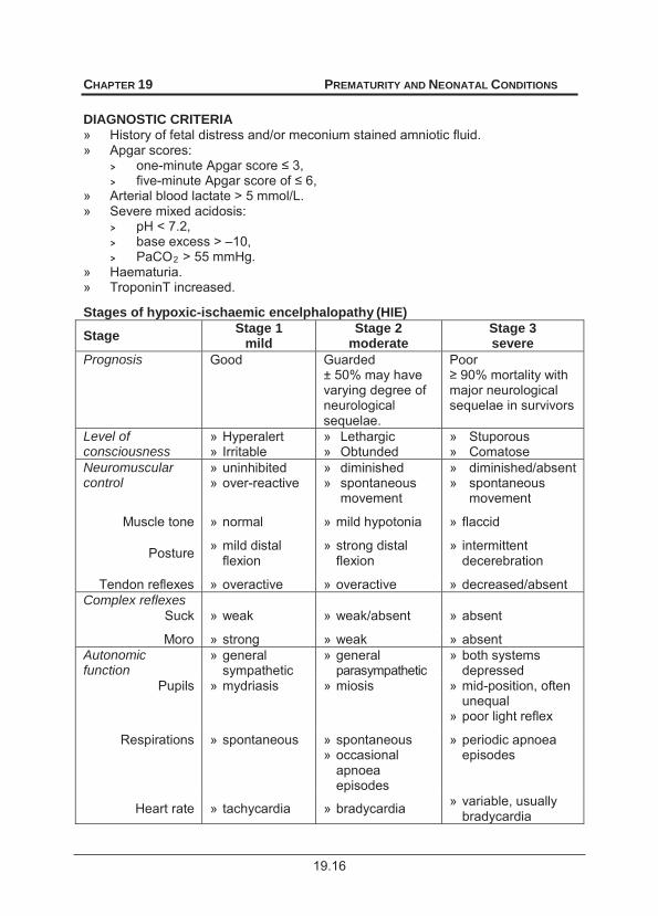
19.16
CHAPTER 19 PREMATURITY AND NEONATAL CONDITIONS DIAGNOSTIC CRITERIA » History of fetal distress and/or meconium stained amniotic fluid. » Apgar scores:
> one-minute Apgar scor , > five- ,
» Arterial blood lactate > 5 mmol/L. » Severe mixed acidosis:
> pH < 7.2, > base excess > –10, > PaCO2 > 55 mmHg.
» Haematuria. » TroponinT increased. Stages of hypoxic-ischaemic encelphalopathy (HIE)
Stage Stage 1 mild
Stage 2 moderate
Stage 3 severe
Prognosis Good Guarded ± 50% may have varying degree of neurological sequelae.
Poor
major neurological sequelae in survivors
Level of consciousness
» Hyperalert » Irritable
» Lethargic » Obtunded
» Stuporous » Comatose
Neuromuscular control
» uninhibited » over-reactive
» diminished » spontaneous
movement
» diminished/absent » spontaneous
movement
Muscle tone » normal » mild hypotonia » flaccid
Posture » mild distal flexion
» strong distal flexion
» intermittent decerebration
Tendon reflexes » overactive » overactive » decreased/absent Complex reflexes
Suck » weak » weak/absent » absent
Moro » strong » weak » absent Autonomic function
» general sympathetic
» general parasympathetic
» both systems depressed
Pupils » mydriasis » miosis » mid-position, often unequal
» poor light reflex
Respirations » spontaneous » spontaneous » occasional
apnoea episodes
» periodic apnoea episodes
Heart rate » tachycardia » bradycardia » variable, usually bradycardia
19.17
CHAPTER 19 PREMATURITY AND NEONATAL CONDITIONS
Stage Stage 1 mild
Stage 2 moderate
Stage 3 severe
Bronchial and salivary secretions
» sparse » profuse » variable
Gastrointestinal motility
» normal or decreased » increased » variable
» ileus Seizures » none » common » uncommon, but
prolonged if present decerebrate
Note: Newborns with stage 3 HIE should not be ventilated. GENERAL AND SUPPORTIVE MEASURES » Resuscitate. » Avoid hyperthermia. » Admit to neonatal high care or intensive care facility, if available. » Whole body or head cooling should be done under supervision of a
paediatrician. > Initiate within 6 hours of birth to maintain rectal (core) temperature at 33–
34ºC for 72 hours. > Slowly rewarm at a rate of 0.5ºC/hour until axillary or skin temperature is at
36.5–36.8ºC. > Mild HIE: ambient temperature at lower range of neutral thermal
environment. > I : whole body or
head cooling. » Ventilatory support if PaO2 < 60 mmHg and/or PaCO2 > 55 mmHg in newborns
with moderate HIE (stage 2). » Maintain:
> Blood glucose at 2.6–6 mmol/L. > H . > Blood pressure at 7 0/35 mmHg in a term infant and 50/35 mmHg in a
preterm infant. Mean blood pressure at least 5–10 mmHg more than the gestational age.
» IV fluids > Frequent assessment of fluid balance, i.e. intake and output. > Restrict fluids to 50–60 mL/kg in the first 24–48 hours. > Use dextrose water 10% or a neonatal maintenance solution potassium-
free until the possibility of renal failure has been excluded. » Maintain serum electrolytes, calcium, magnesium and acid-base status within
normal physiological range. » Nutrition:
> No enteral feeds for at least the first 12–24 hours. > Enteral milk feeds only after ileus has been excluded. > Consider IV alimentation if enteral feeds are not possible after 24 hours.

19.18
CHAPTER 19 PREMATURITY AND NEONATAL CONDITIONS
» Monitor:
> neurological status, > fluid balance, > vital signs, > temperature, > acid-base status, > blood glucose, > blood gases, > electrolytes, > SaO2, > calcium, magnesium, > blood pressure, > renal function, and > brain function (aEEG), where available.
» Follow up for assessment of neurodevelopment, hearing and vision.
MEDICINE TREATMENT To keep PaO2 between 60 and 80 mmHg and saturation 88–92% (normal range): Oxygen.
Haematocrit <40%: Packed red cells, IV, 10mL/kg.
If infection is suspected or confirmed: Cefotaxime, IV, 50 mg/kg/dose.
o 12 hourly for first 7 post natal days and 8 hourly thereafter. o If infection is excluded, antibiotics can be stopped in 72 hours.
Hypotension Sodium chloride 0.9%, IV, 20 mL/kg over 1 hour.
AND Dopamine, IV, 5–15 mcg/kg/minute.
AND/OR Dobutamine, IV, 5–15 mcg/kg/minute if cardiac dysfunction or failure is present.
o Continue with blood pressure support until blood pressure is stabilised. Seizure Control Administer anticonvulsants with monitoring of cardiorespiratory function. Phenobarbitone, IV:
o Loading dose: 20 mg/kg over 10 minutes. o Refractory seizures: Additional 10 mg/kg up to 40 mg/kg. o Maintenance: 4 mg/kg/day beginning 12–24 hours after the loading dose.
Do not use lidocaine if phenytoin was given.
Do not use lidocaine for seizures in newborns with congenital heart disease, dysrhythmias, acute heart failure and shock.
Admit neonates with seizures refractory to phenobarbitone to a high or intensive care unit. Cardiorespiratory support is usually required in this category of infants.
19.19
CHAPTER 19 PREMATURITY AND NEONATAL CONDITIONS For term normothermic neonates: Lidocaine (lignocaine), IV.
o Loading dose: 2 mg/kg over 10 minutes. o Follow with a continuous infusion of:
6 mg/kg/hour for 6 hours, then 4 mg/kg/hour for 12 hours, followed by 2 mg/kg/hour for 12 hours.
o If seizures are well controlled, taper slowly over 12 hours.
For preterm neonates: A safe dose of lidocaine in preterm neonates has not been established but the following dosing schedule has been used. Lidocaine (lignocaine), IV.
o Loading dose: 2 mg/kg over 10 minutes. o Follow with a continuous infusion of 3 mg/kg/hour for 3 days. o Taper dose gradually over next 2 days.
» A safe lidocaine dosing regimen for term infants undergoing hypothermia treatment for HIE has not been established.
» Clearance of lidocaine is slower in hypothermic preterm infants and neonates and there is a risk of accumulation.
» Start tapering earlier than 3 days if seizures are well controlled. » Continuous monitoring of ECG, heart rate and blood pressure is
mandatory if lidocaine is used. » Main adverse effects of lidocaine: dysrhythmias and bradycardia. » Life threatening dysrhythmias may indicate lidocaine toxicity. Treat with: Lipid emulsion 20%, IV, 1.5 mg/kg over 1 minute.
o Follow with a continuous infusion of 0.25 mL/kg/minute for 30 minutes. Refer urgently.
Cardiac failure Restrict fluid. Furosemide, IV/oral/nasogastric tube, 1 mg/kg/24 hours as a single daily dose. Dobutamine IV, 5–15 mcg/kg/minute.
Hypocalcaemia Serum total calcium < 1.8 mmol/L or ionised calcium < 0.7 mmol/L. Calcium gluconate 10%, slow IV, 1–2 mL/kg over 15 minutes under ECG
control. Hypomagnesaemia Serum magnesium < 0.6 mmol/L: Magnesium sulphate 50%, IV, 0.2 mL/kg as a single dose.
19.20
CHAPTER 19 PREMATURITY AND NEONATAL CONDITIONS Hypoglycaemia Blood glucose < 2.6 mmol/L: Dextrose 10%, bolus IV, 2.5–5 mL/kg (250–500 mg/kg).
Dextrose 10% = 10 g dextrose in 100 mL. Do not repeat dextrose bolus.
Inappropriate ADH: Moderate fluid restriction of 50–60 mL/kg/24hours for the first 24–48 hours. Raise head of cot by 10–15 cm. Cerebral oedema/raised intracranial pressure Moderate hyperventilation to lower PaCO2 to 35 mmHg, if ventilation facilities are available. REFERRAL » Neurological assessment of survivors at 3 months. »
hours post birth. » Lidocaine toxicity. 19.9 JAUNDICE, NEONATAL P58 DESCRIPTION Yellow discolouration of the skin and mucous membranes due to hyperbilirubinaemia. Bilirubin is formed mainly from haem catabolism. Jaundice develops when there is an overproduction of bil irubin, defective bilirubin metabolism and/or defective excretion of bilirubin from the body. DIAGNOSTIC CRITERIA Jaundice may have a physiological and a pathological component. Physiological jaundice » Seldom appears before 24–36 hours after birth. » Rarely lasts more than 10 days in the full term infant and 14 days in the pre-
term infant. » Only the unconjugated bilirubin fraction is increased. » Total peak serum bilirubin concentration is usually below 275 micromol/L in the
term infant. » Total bilirubin concentration does not rise by more than 85 micromol/L/24 hours
or 17 micromol/L/hour. » The baby thrives and shows no signs of illness or anaemia. » Treatment is unnecessary.
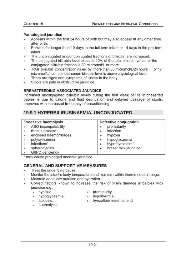
19.21
CHAPTER 19 PREMATURITY AND NEONATAL CONDITIONS
Pathological jaundice » Appears within the first 24 hours of birth but may also appear at any other time
after birth. » Persists for longer than 10 days in the full term infant or 14 days in the pre-term
infant. » The unconjugated and/or conjugated fractions of bilirubin are increased. » The conjugated bilirubin level exceeds 10% of the total bilirubin value, or the
conjugated bilirubin fraction is 30 micromol/L or more. » Total bilirubin concentration ris es by more than 85 micromol/L/24 hours or 17
micromol/L/hour the total serum bilirubin level is above physiological level. » There are signs and symptoms of illness in the baby. » Stools are pale in obstructive jaundice. BREASTFEEDING ASSOCIATED JAUNDICE Increased unconjugated bilirubin levels during the first week of li fe in br eastfed babies is due to calorie and fluid deprivation and delayed passage of stools. Improves with increased frequency of breastfeeding. 19.9.1 HYPERBILIRUBINAEMIA, UNCONJUGATED Excessive haemolysis Defective conjugation » ABO incompatability » rhesus disease » enclosed haemorrhages » polycythaemia » infections* » spherocytosis » G6PD deficiency
» prematurity » infection » hypoxia » hypoglycaemia » hypothyroidism* » breast milk jaundice*
* may cause prolonged neonatal jaundice GENERAL AND SUPPORTIVE MEASURES » Treat the underlying cause. » Monitor the infant’s body temperature and maintain within thermo neutral range. » Maintain adequate nutrition and hydration. » Correct factors known to inc rease the risk of br ain damage in ba bies with
jaundice e.g.: > hypoxia, > prematurity, > hypoglycaemia, > hypothermia, > acidosis, > hypoalbuminaemia, and > haemolysis.
19.22
CHAPTER 19 PREMATURITY AND NEONATAL CONDITIONS PHOTOTHERAPY Guideline for initiating phototherapy: » Terminate phototherapy when the unconjugated bilirubin level is lower than the
recommended phototherapy initiating level, and the cause of the jaundice has been determined and adequately addressed.
» The skin colour of a baby receiving phototherapy does not reflect the degree of jaundice (bilirubin blood level) or the efficacy of the phototherapy.
» Undress the baby and cover the eyes with gauze pad or commercially available eye covers.
» Position the phototherapy unit (fluorescent light bulbs of 400– 500 nm wavelength) not higher than 45 cm above the baby.
» Check spectral irradiance of the fluorescent lights after every 200–300 hours of use to ensure that they are still effective (use radiometer, if available).
» The spectral irradiance should be above 10 microwatt/cm2/nm of wavelength. If spectral irradiance cannot be checked regularly, replace fluorescent light bulbs after 1 000 hours of continuous use.
» A quartz halogen light source (400–500 nm wavelength) can also be used for phototherapy.
» Phototherapy units with diodes emitting light in the blue spectrum or fibro-optic phototherapy units can be used instead of the fluorescent/quartz halogen units.
» A rebound increase in bilirubin may follow termination of phototherapy. » Monitor bilirubin levels approximately 6 hourly after phototherapy has been stopped. Guideline for exchange transfusion (See also the graphs attached): » Exchange transfusion is indicated when the risk of bilirubin encephalopathy and
kernicterus is significant. Referral for exchange transfusion may be needed. MEDICINE TREATMENT Rh incompatibility (i.e. mother Rh negative, baby Rh positive). ABO incompatibility (i.e. mother = O, baby = A, B or AB). As soon as the diagnosis of Rh – or ABO incompatibility is confirmed, administer: Gammaglobulin, IV, 500 mg/kg over 1 hour.
o Can be repeated after 6–8 hours. Mothers of babies with Rh incompatibility should receive: Anti D immunoglobulin, IM, 100 mcg as soon as possible after birth but within
72 hours of birth.
19.9.2 HYPERBILIRUBINAEMIA, CONJUGATED Hepatocellular disease Bile duct obstruction » hepatitis* » total parenteral nutrition* » syphilis » other congenital infections » galactosaemia*
» bile duct hypoplasia/atresia* » choledochal cyst » cystic fibrosis
* May cause prolonged neonatal jaundice.
19.23
CHAPTER 19 PREMATURITY AND NEONATAL CONDITIONS
Conjugated hyperbilirubinaemia is due to intra/extrahepatic obstruction of bile ducts (cholestasis) and usually presents in the second week of life or later. The baby has a green-y ellow skin discolouration, dark bile stained urine and pale acholic stools. Hepatomegaly is com monly present and t he infant often fails to thrive. Neonatal hepatitis, prolonged TPN and biliary atresia or hypoplasia accounts for the majority of cases of conjugated hyperbilirubinaemia. GENERAL AND SUPPORTIVE MEASURES » Treat the underlying cause. » Dietary modifications to counteract the malabsorption of fat and fat soluble
vitamins (A, D, E and K) that may occur in patients with a prolonged conjugated hyperbilirubinaemia.
» When galactosaemia is suspected avoid lactose containing feeds, i.e. breast milk and lactose containing formula.
MEDICINE TREATMENT Fat soluble vitamins (A, D, E and K).
SURGICAL TREATMENT Conditions amenable to surgery e.g. biliary artresia. Hepatoporto-enterostomy for biliary atresia should be done before 60 days of age for optimal outcome. REFERRAL » All cases of jaundice persisting more t han 2 w eeks with conjugated bilirubin
level >20% of total bilirubin, for diagnosis and initiation of treatment. 19.10 JAUNDICE, NEONATAL, PROLONGED DESCRIPTION Jaundice (static or a rising bilirubin) present for more than 10 days in a term infant and 14 days in a preterm infant. The usual causes are: » breast milk jaundice, » hypothyroidism, » hepatitis, » galactosaemia, and » infections, e.g. UTIs. Breast milk jaundice may be confirmed by substituting breastfeeding with formula feeds for 24–48 hours. The bilirubin level will always drop to a lower level and increase again when breastfeeding is resumed. However, the level will not rise t o the original high level. Breast milk jaundice is an unconjugated hyperbilirubinaemia and the infant is always well and thriving.
19.24
CHAPTER 19 PREMATURITY AND NEONATAL CONDITIONS
Abnormal thyroid function, increased TSH and decreased T3 and T 4, indicates hypothyroidism. Unconjugated bilirubin fraction is raised and the i nfant may have clinical signs of hypothyroidism e.g.: » lethargy, » constipation, » feeding difficulties, » hypotonia, » poor cry, » umbilical hernia, » nasal obstruction, » hypothermia, and » bradycardia. Infants with galactosaemia usually present with: » a conjugated hyperbilirubinaemia, » vomiting, » refusal to feed, » hepatomegaly, » failure to thrive, » Hypoglycaemia, and » encephalopathy and later cataracts. DIAGNOSTIC CRITERIA » Hepatitis may be confirmed by abnormal liver function tests, i.e. raised values of:
> AST, > ALP, > ALT, > bilirubin, mainly the conjugated fraction, > gamma GT.
» Hepatomegaly and/or hepatosplenomegaly. » If conjugated hyperbilirubiniaemia – See above. Investigations » Syphilis. See section 19.18: Syphilis, early congenital. » Galactosaemia. » Thyroid function (see hypothyroidism) and ur ine for M CS (see urin ary tract
infection). » Suspect galactosaemia if urine is positive for reducing substances but negative
for glucose in a baby receiving lactose-containing feeds. A ga lactose-1-phosphate uridyl transferase assay will confirm the diagnosis.
GENERAL AND SUPPORTIVE MEASURES » Monitor bilirubin levels. » Treat the underlying cause. » Dietary adjustment for prolonged conjugat ed hyperbilirubinaemia to counterac t
the malabsorption of fat and fat soluble vitamins (A, D, E and K) » Avoid lactose containing feeds, i.e. breast milk and lactose containing
formulae, when galactosaemia is suspected. » Regular follow up until the underlying condition has been resolved. MEDICINE TREATMENT Fat soluble vitamins (A, D, E and K).
19.25
CHAPTER 19 PREMATURITY AND NEONATAL CONDITIONS
REFERRAL » Pathological jaundice, unconjugated and/or conjugated, where the underlying
cause cannot be identified. » Serum unconjugated bilirubin at exchange transfusion level. » Jaundice, unconjugated and/or conjugated, not improving on a dequate
treatment. » Conjugated hyperbilirubinaemia due to conditions requiring surgical
intervention e.g. biliary atresia. » Prolonged neonatal jaundice, excluding breast milk jaundice.
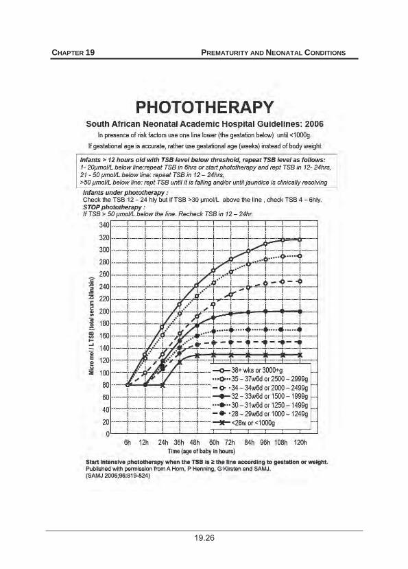
19.26
CHAPTER 19 PREMATURITY AND NEONATAL CONDITIONS
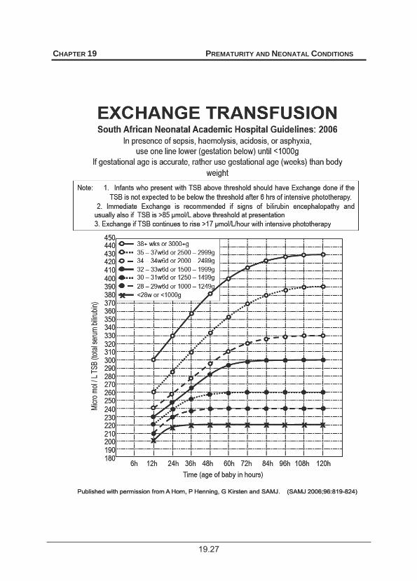
19.27
CHAPTER 19 PREMATURITY AND NEONATAL CONDITIONS
19.28
CHAPTER 19 PREMATURITY AND NEONATAL CONDITIONS
19.11 MENINGITIS BACTERIAL, NEONATAL G01 DESCRIPTION A bacterial infection of the meninges in the first month of life. Consider meningitis in any neonate being evaluated for sepsis or infection, as most organisms implicated in neonatal sepsis also cause neonatal meningitis. The most common causative organisms are Group B ß-haemolytic streptococcus type III and Gram-negative organisms such as E. coli with K1 antigen. Consider S. epidermidis and S. aureus as causative organisms with central nervous system anomalies such as open defects or with indwelling devices such as VP shunts. Consider HIV infection in neonates with meningitis. DIAGNOSTIC CRITERIA Clinical » Clinical presentation is usually with one or more non-specific signs such as:
> temperature disturbances, > altered level of consciousness, > lethargy, > blood glucose disturbances, > irritability, > bulging/full fontanel, > vomiting, > convulsions, > feeding problems, > apnoea, and > vasomotor changes.
» Complications include: > cerebral oedema, > convulsions, > raised intracranial pressure, > hydrocephalus, > vasculitis, with haemorrhage, > subdural effusion, > ventriculitis, > brain abscess, > ischaemia and infarctions of the brain, > inappropriate antidiuretic hormone (ADH) secretion.
» Late complications include: > neurological sequelae, > blindness, > deafness, and > mental retardation.
SPECIAL INVESTIGATIONS » Lumbar puncture:
> CSF appears turbid to purulent. > Protein concentration is increased. > Leucocyte count is increased with a predominance of polymorphonuclear
leucocytes. > Glucose concentration is low, < 2/3 of blood glucose. > Counterimmune electrophoresis of CSF may provide a rapid diagnosis of
some bacterial pathogens. » Gram stain, microscopy, culture and sensitivity of CSF. Rapid antigen tests on
the CSF. » Blood cultures for microscopy, culture and sensitivity.
19.29
CHAPTER 19 PREMATURITY AND NEONATAL CONDITIONS GENERAL AND SUPPORTIVE MEASURES » Admit to high or intensive care unit, if available. » Maintain a neutral thermal environment. » Monitor, where indicated:
> neurological status, > calcium and magnesium, > vital signs, > acid-base status, > electrolytes, > blood glucose, > haematocrit, > serum and urine osmolality, > fluid balance (hydration), > blood gases.
» Ensure adequate nutrition: > Enteral feeding where possible, use nasogastric tube, if necessary. > If enteral feeding is not possible, IV fluids, e.g. neonatal maintenance
solution and parenteral nutrition under supervision by paediatrician. » Limit total daily fluid intake, IV and oral:
> Do not exceed the daily requirements for age. > Prevent fluid overload.
MEDICINE TREATMENT Antibiotics, empirical Cefotaxime, IV, 50 mg/kg over 30 minutes, for 21 days.
o If < 7 days of age: 50 mg/kg 12 hourly. o If 7 days – 3 weeks of age: 50 mg/kg 8 hourly. o If > 3 weeks of age: 50 mg/kg 6 hourly.
PLUS Ampicillin, IV, for 14 days.
o If < 7 days of age: 50mg/kg 12 hourly. o If > 7 days – 3 weeks of age: o If > 3 weeks of age:
50 mg/kg 8 hourly. 50 mg/kg 6 hourly.
During the course of treatment a skull ultrasound should be done. Repeat CSF examination after 48–72 hours to ensure there is a response to therapy. Reconsider choice of antibiotic when the results of blood and CSF cultures become available or the child does not improve within 72–96 hours. No response or intolerant to cephalosporins or ampicillin For patients not responding to adequate antibiotic therapy where no organisms were identified or cultured, consider viruses, fungi and bacteria not usually causing meningitis. Convulsions See section 19.16: Seizure, neonate. Raised intracranial pressure or cerebral oedema Avoid fluid overload. Limit total daily intake, IV and oral. Do not exceed the maintenance requirements for age.
19.30
CHAPTER 19 PREMATURITY AND NEONATAL CONDITIONS
REFERRAL » Meningitis not responding to adequate treatment. » Meningitis with complications. » Follow up is essential for assessing neurodevelopment, hearing and vision. 19.12 PATENT DUCTUS ARTERIOSUS (PDA) IN THE NEWBORN Q25.0 DESCRIPTION Patent ductus arteriosus (PDA) is the extra-uterine persistence of the normal fetal vessel that joins the pulmonary artery to the aorta. DIAGNOSTIC CRITERIA Clinical Depending on size of PDA: » systolic or continuous murmur at left heart base, » hyperactive precordium with easily palpable bounding peripheral pulses. Investigations » Echocardiography will confirm the diagnosis. Risk factors include: » prematurity, » pulmonary hypertension, » hypoxia, » sepsis, » fluid overload, » lung disease, » anaemia, and » congenital cardiac abnormalities. Complications include cardiac failure, systemic hypotension, pulmonary haemorrhage and steal phenomena phenomena such as a decrease in mesenteric blood flow. GENERAL AND SUPPORTIVE MEASURES Preterm Infants » Identify and treat underlying risk factors. » Restrict fluid intake to 80–120 ml/kg/24 hours. Individualise volume to avoid
over restriction of fluid and poor weight gain. » Hb » Monitor cardiac function, renal function and urinary output. » Provide adequate nutrition. » Nurse in neutral thermal environment. MEDICINE TREATMENT Cardiac failure Diuretics Furosemide, IV/oral, 1 mg/kg/24 hours.
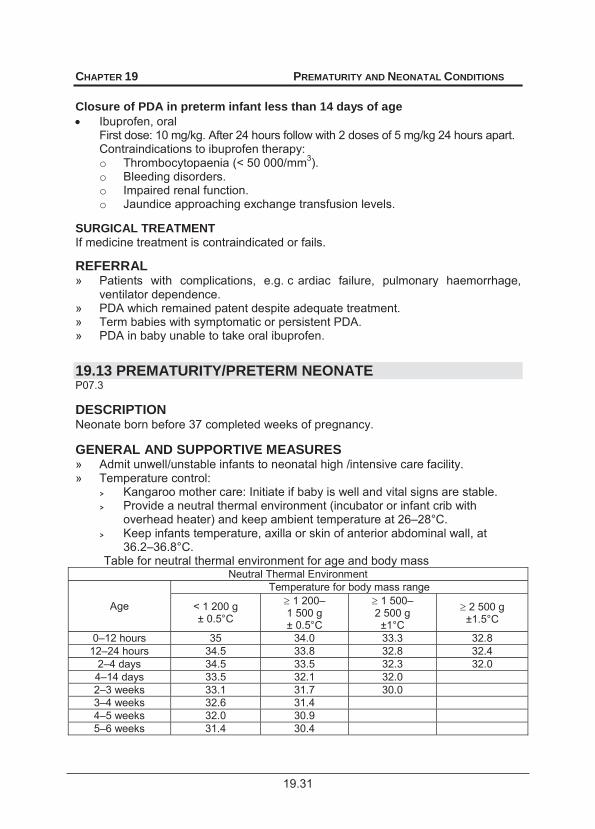
19.31
CHAPTER 19 PREMATURITY AND NEONATAL CONDITIONS Closure of PDA in preterm infant less than 14 days of age Ibuprofen, oral
First dose: 10 mg/kg. After 24 hours follow with 2 doses of 5 mg/kg 24 hours apart. Contraindications to ibuprofen therapy: o Thrombocytopaenia (< 50 000/mm3). o Bleeding disorders. o Impaired renal function. o Jaundice approaching exchange transfusion levels.
SURGICAL TREATMENT If medicine treatment is contraindicated or fails.
REFERRAL » Patients with complications, e.g. c ardiac failure, pulmonary haemorrhage,
ventilator dependence. » PDA which remained patent despite adequate treatment. » Term babies with symptomatic or persistent PDA. » PDA in baby unable to take oral ibuprofen.
19.13 PREMATURITY/PRETERM NEONATE P07.3 DESCRIPTION Neonate born before 37 completed weeks of pregnancy. GENERAL AND SUPPORTIVE MEASURES » Admit unwell/unstable infants to neonatal high /intensive care facility. » Temperature control:
> Kangaroo mother care: Initiate if baby is well and vital signs are stable. > Provide a neutral thermal environment (incubator or infant crib with
overhead heater) and keep ambient temperature at 26–28°C. > Keep infants temperature, axilla or skin of anterior abdominal wall, at
36.2–36.8°C. Table for neutral thermal environment for age and body mass
Neutral Thermal Environment
Age
Temperature for body mass range
< 1 200 g ± 0.5°C
1 200– 1 500 g ± 0.5°C
1 500– 2 500 g ±1°C
2 500 g ±1.5°C
0–12 hours 35 34.0 33.3 32.8 12–24 hours 34.5 33.8 32.8 32.4
2–4 days 34.5 33.5 32.3 32.0 4–14 days 33.5 32.1 32.0 2–3 weeks 33.1 31.7 30.0 3–4 weeks 32.6 31.4 4–5 weeks 32.0 30.9 5–6 weeks 31.4 30.4
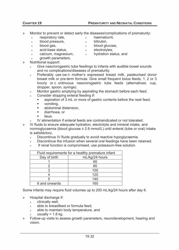
19.32
CHAPTER 19 PREMATURITY AND NEONATAL CONDITIONS » Monitor to prevent or detect early the diseases/complications of prematurity:
> respiratory rate, > haematocrit, > blood pressure, > bilirubin, > blood gas, > blood glucose, > acid-base status, > electrolytes, > calcium, magnesium, > hydration status, and > growth parameters.
» Nutritional support: > Give naso/orogastric tube feedings to infants with audible bowel sounds
and no complications/diseases of prematurity. > Preferably use ow n mother’s expressed breast milk, pasteurised donor
breast milk or pre-term formula. Give small frequent bolus feeds, 1, 2 or 3 hourly or c ontinuous naso/orogastric tube feeds (alternatives: cup, dropper, spoon, syringe).
> Monitor gastric emptying by aspirating the stomach before each feed. > Consider stopping enteral feeding if:
aspiration of 3 mL or more of gastric contents before the next feed, vomiting, abdominal distension, diarrhoea, or ileus.
> IV alimentation if enteral feeds are contraindicated or not tolerated. » IV fluids to ensure adequate hydration, electrolyte and mineral intake, and
noris satisfactory. > Discontinue IV fluids gradually to avoid reactive hypoglycaemia. > Discontinue the infusion when several oral feedings have been retained. > If renal function is compromised, use potassium-free solution.
Fluid requirements for a healthy premature infant
Day of birth mL/kg/24 hours 1 60 2 80 3 100 4 120 5 140
6 and onwards 160
Some infants may require fluid volumes up to 200 mL/kg/24 hours after day 6. » Hospital discharge if:
> clinically well, > able to breastfeed or formula feed, > able to maintain body temperature, and > usually > 1.8 kg.
» Follow-up visits to assess growth parameters, neurodevelopment, hearing and vision.
19.33
CHAPTER 19 PREMATURITY AND NEONATAL CONDITIONS
MEDICINE TREATMENT To maintain haematocrit at 40% or Hb ±13 g/dL for the first 2 weeks of life: Packed red cells, IV, 10 mL/kg.
To maintain oxygen tension in the blood at 60–80 mm Hg: Oxygen, humidified via head box, or nasal cannulae.
Oxygen therapy should be utilised to maintain oxygen saturation of haemoglobin at of 88–92%; and for babies less than 1 000 g, a saturation of 88–90%. Use pulse oximeter.
At birth Vitamin K, IM, 0.5–1 mg. Immunise according to EPI schedule. Iron and multivitamin supplementation from the third week of life.
Prophylaxis Iron (elemental), oral, 2–4 mg/kg/24/hours.
o Ferrous lactate 1 mL = 25 mg elemental iron. o Multivitamin, oral, providing at least vitamin D, 400–800 IU and vitamin A,
1 250–5 000 IU per 24 hours. Continue with iron and vitamin supplementation until the infant is on a balanced diet. REFERRAL Presence of one or more of the following complications that cannot be managed at the facility: » Respiratory distress and/or apnoea attacks requiring ventilatory support. » PDA with cardiac failure not responding to medical management. » Necrotising enterocolitis requiring surgical intervention. » Jaundice with serum unconjugated bilirubin level in the exchange transfusion zone. » Septicaemic infants or infants with infections not responding to therapy. » Pulmonary and/or intraventricular haemorrhage. » Feeding difficulties where the underlying cause is unclear. » Infants requiring hyperalimentation if parenteral nutrition is not available at the
hospital. » Convulsions not responding to treatment. » Congenital abnormalities requiring surgical intervention. » Hypoglycaemia not responding to treatment. » For eye examination/hearing screening:
> infants less than 1.5 kg, > infants < 32 weeks gestation, > infants who received prolonged respiratory support/oxygen, > infants with recurrent apnoea, and > infants with an unstable clinical course.
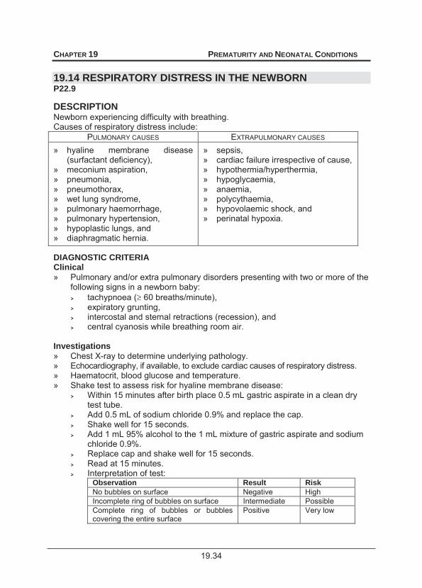
19.34
CHAPTER 19 PREMATURITY AND NEONATAL CONDITIONS
19.14 RESPIRATORY DISTRESS IN THE NEWBORN P22.9 DESCRIPTION Newborn experiencing difficulty with breathing. Causes of respiratory distress include:
PULMONARY CAUSES EXTRAPULMONARY CAUSES » hyaline membrane disease
(surfactant deficiency), » meconium aspiration, » pneumonia, » pneumothorax, » wet lung syndrome, » pulmonary haemorrhage, » pulmonary hypertension, » hypoplastic lungs, and » diaphragmatic hernia.
» sepsis, » cardiac failure irrespective of cause, » hypothermia/hyperthermia, » hypoglycaemia, » anaemia, » polycythaemia, » hypovolaemic shock, and » perinatal hypoxia.
DIAGNOSTIC CRITERIA Clinical » Pulmonary and/or extra pulmonary disorders presenting with two or more of the
following signs in a newborn baby: > tachypnoea ( 60 breaths/minute), > expiratory grunting, > intercostal and sternal retractions (recession), and > central cyanosis while breathing room air.
Investigations » Chest X-ray to determine underlying pathology. » Echocardiography, if available, to exclude cardiac causes of respiratory distress. » Haematocrit, blood glucose and temperature. » Shake test to assess risk for hyaline membrane disease:
> Within 15 minutes after birth place 0.5 mL gastric aspirate in a clean dry test tube.
> Add 0.5 mL of sodium chloride 0.9% and replace the cap. > Shake well for 15 seconds. > Add 1 mL 95% alcohol to the 1 mL mixture of gastric aspirate and sodium
chloride 0.9%. > Replace cap and shake well for 15 seconds. > Read at 15 minutes. > Interpretation of test:
Observation Result Risk No bubbles on surface Negative High Incomplete ring of bubbles on surface Intermediate Possible Complete ring of bubbles or bubbles covering the entire surface
Positive Very low
19.35
CHAPTER 19 PREMATURITY AND NEONATAL CONDITIONS
GENERAL AND SUPPORTIVE MEASURES » Identify and treat underlying cause, e.g.:
> Chest tube and underwater drainage of pneumothorax. > Isovolaemic dilutional exchange transfusion for symptomatic
polycythaemia. » Admit to neonatal high care/intensive care facility, if available. » Handle neonate as little as possible. » Nurse non-intubated infant in the prone position. » Keep in a neutral thermal environment (incubator or infant crib with overhead
heater). Keep room temperature, at 26–28°C, and anterior abdominal wall skin temperature at 36.2–36.8°C.
» Monitor: > blood pressure, > respiratory rate, > peripheral perfusion, > heart/pulse rate, > haematocrit, > acid-base status, > blood glucose, > body temperature, > blood gases, > SaO2, > minerals and electrolytes and > fluid balance.
» Nutrition: > P . > Commence orogastric feeding after 12–24 hours if bowel sounds are
audible and meconium has been passed. > If enteral feeding is not possible 24 hours after birth, start IV
hyperalimentation. » Ventilation (non invasive or invasive) is needed if:
> An oxygen saturation of at least 90% or PaO2 of at least 60mmHg cannot be maiwithout nasal CPAP;
> The PaCO2 rises to > 55 mmHg with uncompensated respiratory acidosis 2.
(1kPa = 7.5 mmHg; 1 mmHg x 0.133 = 1 kPa) MEDICINE TREATMENT To eliminate central cyanosis and to maintain oxygen saturation of haemoglobin at 88–92%: Oxygen, warmed and humidified via head box, or nasal cannula.
o If a pulse oximeter or facility for blood gas analysis is available oxygen, humidified via head box, or nasal cannulae to maintain oxygen tension in the blood at 60–80 mmHg.
o If a pulse oximeter or facility for blood gas analysis is not available, regulate the inspired oxygen concentration in such a way that the least amount of oxygen that will prevent central cyanosis is used.
o Keep PaO2 at 60–80 mmHg and PaCO2 at 35–45 mmHg (arterial blood gas analysis).

19.36
CHAPTER 19 PREMATURITY AND NEONATAL CONDITIONS
Nasal CPAP is needed if the neonate has a good respiratory drive with a PCO2 55 mmHg but unable to maintain a SaO2 of 88–92% on an inspiratory oxygen
1O2) and pneumothorax has been excluded. Administer nasal CPAP at 4– 6 cm H2O and monitor SaO2, blood gas and acid base status. OR Oxygen/air mixture, hi-flow, warmed and humidified via nasal prongs. (Under
specialist supervision) o Do not exceed 6 L/minute. The flow/minute (L/min) approximates the
pressure generated in cm water. Stabilise circulation and blood pressure Neonatal maintenance solution, IV infus ion, 60–80 mL/kg/24 hours (day 1 of
birth) and adapt to daily maintenance requirements. AND/OR Sodium chloride 0.9%, 10–20 mL/kg over 1–2 hours.
o For premature infants restrict to 10 mL/kg. AND/OR Plasma, 10–20 mL/kg over 1–2 hours.
Inotropic support Dopamine, IV, 5 –15 mcg/kg/minute, continued until blood pressure has
stabilised. o Response to inotropic support will be unsatisfactory if the circulating blood
volume is not corrected. Anaemia If anaemia is present, Hct < 40 % and Hb <13 g/dL: Packed red cells, IV, 10 mL/kg over 1–2 hours.
Metabolic acidosis If pH sation of PaO2, PaCO2, blood pressure, volume expansion (hydration) and correction of anaemia: Sodium bicarbonate, 4.2 %, IV, administered slowly.
1 mmol = 2 mL HCO3 needed (mmol) = base excess x 0.3 x body mass (kg) (½ correct base deficit initially) If blood gas and acid base analysis is not available and metabolic acidosis is suspected: Sodium bicarbonate, 4%, slow IV, 2 mL.
CAUTION Do not administer Ca++ containing infusions with sodium bicarbonate solution
19.37
CHAPTER 19 PREMATURITY AND NEONATAL CONDITIONS Polycythaemia Treat with isovolaemic dilutional exchange transfusion using sodium chloride 0.9% if the venous haematocrit is Hct > 65%: Hb >22 g/dL and the baby is symptomatic. Perform under paediatrician’s supervision. Volume to be exchanged (mL) if desired Hct = 50: = [Baby’s Hct – desired Hct (i.e. 50) x body mass (kg)] x 90
Baby’s Hct Hyaline membrane disease (Surfactant deficiency) Supervision by paediatrician. Shake test to assess risk for hyaline membrane disease and/or x-ray chest – see above. If surfactant deficiency is suspected or present, provide respiratory support. » Mild surfactant deficiency: nasal CPAP 4–6 cm H2O. » Moderate surfactant deficiency: “in-out” surfactant followed by nasal CPAP
4–6 cm H2O. Intubate infant and administer surfactant via naso-or orotracheal tube. Ventilate for a few minutes with a Neopuff device or resuscitation bag with a CPAP generating device. Extubate baby and put on nasal CPAP 4–6 cm H2O. Babies may be put on nasal CPAP directly after “in-out” surfactant administration, omitting the ventilation step following “in-out” surfactant.
» Severe surfactant deficiency: intubate baby and v entilate with a v entilator. Administer surfactant via the naso- or orotracheal tube. If a v entilator is not available the in-out surfactant followed by nasal CPAP can be used.
Semi-synthetic surfactant preparations are recommended while 100% synthetic preparations are not recommended. Infection If infection, e.g. bronchopneumonia, is present or suspected, give antibiotics after blood cultures have been taken. Consider the antibiotic sensitivity profile of micro-organisms in a particu lar hospital when prescribing antibiotics. Aminoglycoside, e.g.: Gentamicin, IV, for 10 days in the first week of life.
o If < 32 weeks gestation of age: 5 mg/kg/36 hours. o 32 weeks gestation of age: 5 mg/kg/24 hours. o After first week, 5 mg/kg/24 hours for all gestations.
PLUS Ampicillin, IV, for 10 days.
o If < 7 days of age: 50 mg/kg 12 hourly. o If 7 days – 3 weeks of age: 50 mg/kg 8 hourly. o If > 3 weeks of age: 50 mg/kg 6 hourly.
Review after 72 hours. If infection is confirmed or very strongly suspected continue for 10 days.
19.38
CHAPTER 19 PREMATURITY AND NEONATAL CONDITIONS REFERRAL » No improvement or deterioration despite adequate treatment. » Development of respiratory failure and need for ventilatory support.
19.15 RESUSCITATION OF THE NEWBORN
Be prepared!
Be at the delivery! Check the equipment and emergency medicines!
Ask 3 questions to evaluate the infant: 1. Is the newborn breathing adequately and not just gasping? 2. Is the newborn’s heart rate (HR) above 100 beats per minute? 3. Is the newborn centrally pink, i.e. no central cyanosis? If the answer to all three questions is “yes”, the newborn does not need resuscitation. If the answer to all three questions is “no” the newborn needs resuscitation. Assess the infant using the above 3 questions every 30 seconds during resuscitation. If the newborn is improving, then the intervention e.g. bagging can be stopped. Only if the baby is not responding or getting worse, is further intervention needed e.g. chest compressions (see algorithm). Check that each step has been effectively applied before proceeding to the next step. The algorithm follows the assumption that the previous step was unsuccessful and the newborn is deteriorating. Use the lowest inspiratory oxygen concentration to alleviate central cyanosis and restore a heart rate above 100 beats per minute. There is some evidence that resuscitation with 100% oxygen may be harmful to the baby. An unsatisfactory response to resuscitation includes: » A sustained slow heart rate, usually less than, or equal to, 60/minute or a
progressive decrease in heart rate until cardiac arrest occurs. » Episodes of cardiac arrest, with a progressively weaker response to chest
compressions, positive pressure ventilation and medicines. » A decreasing blood pressure, increasing acidosis, severe hypotonia with
central cyanosis or intense pallor. » Apnoea or weak, irregular and inefficient respiratory efforts. » Consider discontinuation of resuscitation if the unsatisfactory response to
resuscitation persists for > 20 minutes and underlying conditions e.g. pneumothorax, diaphragmatic hernia have been excluded or > 10 min utes of unresponsive cardiac arrest (asystole) and/or > 20 minutes of unsustainable respiration.
» Admit newborns with a favourable response to resuscitation to a neonatal high or intensive care unit, if available, for post resuscitation care – See Section 19.8: Hypoxia/Ischaemia of the Newborn.
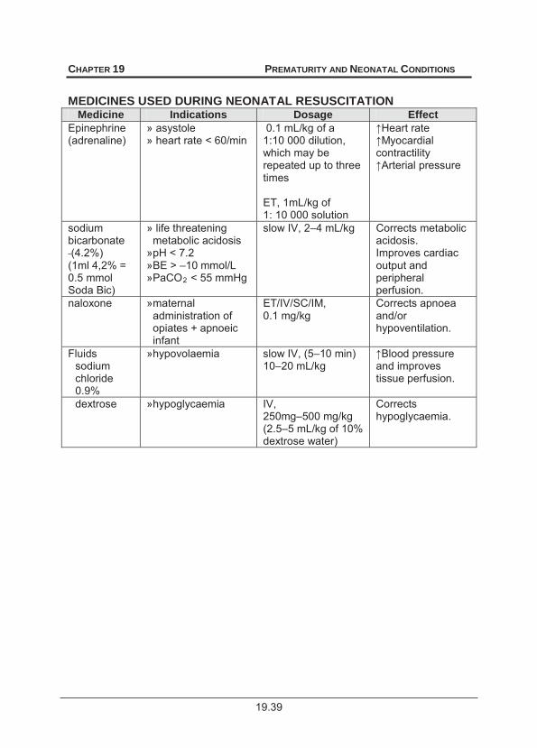
19.39
CHAPTER 19 PREMATURITY AND NEONATAL CONDITIONS
MEDICINES USED DURING NEONATAL RESUSCITATION
Medicine Indications Dosage Effect Epinephrine (adrenaline)
» asystole » heart rate < 60/min
0.1 mL/kg of a 1:10 000 dilution, which may be repeated up to three times ET, 1mL/kg of 1: 10 000 solution
contractility al pressure
sodium bicarbonate (4.2%) (1ml 4,2% = 0.5 mmol Soda Bic)
» life threatening metabolic acidosis
» pH < 7.2 » BE > –10 mmol/L » PaCO2 < 55 mmHg
slow IV, 2–4 mL/kg Corrects metabolic acidosis. Improves cardiac output and peripheral perfusion.
naloxone » maternal administration of opiates + apnoeic infant
ET/IV/SC/IM, 0.1 mg/kg
Corrects apnoea and/or hypoventilation.
Fluids sodium chloride 0.9%
» hypovolaemia slow IV, (5–10 min) 10–20 mL/kg and improves
tissue perfusion.
dextrose » hypoglycaemia IV, 250mg–500 mg/kg (2.5–5 mL/kg of 10% dextrose water)
Corrects hypoglycaemia.
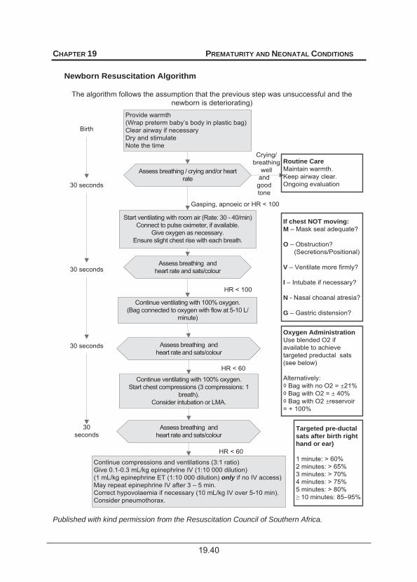
19.40
CHAPTER 19 PREMATURITY AND NEONATAL CONDITIONS
The algorithm follows the assumption that the previous step was unsuccessful and the newborn is deteriorating)
Newborn Resuscitation Algorithm
Crying/breathing
welland good tone
Start ventilating with room air (Rate: 30 - 40/min)Connect to pulse oximeter, if available.
Give oxygen as necessary.Ensure slight chest rise with each breath.
Continue compressions and ventilations (3:1 ratio) Give 0.1-0.3 mL/kg epinephrine IV (1:10 000 dilution) (1 mL/kg epinephrine ET (1:10 000 dilution) only if no IV access)May repeat epinephrine IV after 3 – 5 min.Correct hypovolaemia if necessary (10 mL/kg IV over 5-10 min).Consider pneumothorax.
Birth
Assess breathing / crying and/or heart rate
Routine CareMaintain warmth.Keep airway clear.Ongoing evaluation30 seconds
Provide warmth(Wrap preterm baby’s body in plastic bag)Clear airway if necessaryDry and stimulateNote the time
Assess breathing andheart rate and sats/colour
Continue ventilating with 100% oxygen.(Bag connected to oxygen with flow at 5-10 L/
minute)
Assess breathing andheart rate and sats/colour
Continue ventilating with 100% oxygen.Start chest compressions (3 compressions: 1
breath).Consider intubation or LMA.
Assess breathing andheart rate and sats/colour
30 seconds
30 seconds
30 seconds
If chest NOT moving:M – Mask seal adequate?
O – Obstruction? (Secretions/Positional)
V – Ventilate more firmly?
I – Intubate if necessary?
N - Nasal choanal atresia?
G – Gastric distension?
Oxygen AdministrationUse blended O2 if available to achieve targeted preductal sats (see below)
Alternatively: Bag with no O2 = ±21% Bag with O2 = ± 40% Bag with O2 ±reservoir
= + 100%
Targeted pre-ductal sats after birth right hand or ear)
1 minute: > 60%2 minutes: > 65%3 minutes: > 70%4 minutes: > 75%5 minutes: > 80%
10 minutes: 85–95%
Gasping, apnoeic or HR < 100
HR < 100
HR < 60
HR < 60
Published with kind permission from the Resuscitation Council of Southern Africa.
19.41
CHAPTER 19 PREMATURITY AND NEONATAL CONDITIONS
19.16 SEIZURES, NEONATAL P90 DESCRIPTION Neonatal seizures are usu ally secondary to a serum biochemical disorder or an underlying brain disturbance/injury/malformation. Seizures may be subtle due to the relatively underdeveloped cortex, and do not st op when limbs are f lexed (as opposed to jitteriness). The most likely causes are:
» perinatal asphyxia, » hypocalcaemia, » birth trauma, » hypomagnesaemia, » intracranial haemorrhage, » hyponatraemia, » meningitis, » hypoglycaemia, » narcotic or alcohol withdrawal syndrome, » inborn errors of metabolism, » pyridoxine deficiency, » CNS developmental abnormalities.
DIAGNOSTIC CRITERIA Categories of convulsions » Subtle seizures:
> tonic deviation of the eyes, > ‘swimming’ movements of the arms, > fluttering of the eyelids, > ‘cycling’ movements of the legs, > sucking and chewing movements, > apnoea, > vasomotor changes.
» Tonic clonic movements. » Focal clonic movements. » Myoclonic movements. » Tonic movements/posturing. GENERAL AND SUPPORTIVE MEASURES » Identify and treat the underlying cause, e.g. meningitis and hypoxic-ischaemic
encephalopathy. » Ensure an open airway and administer oxygen, if necessary. » Nurse in neutral thermal environment. » Ensure adequate nutrition and hydration. » Monitor and maintain within accepted physiological range:
> respiration, > acid-base status, > heart rate, > electrolytes, > blood pressure, > minerals, > blood gases, > blood glucose, > SaO2, > haematocrit, > body temperature.
19.42
CHAPTER 19 PREMATURITY AND NEONATAL CONDITIONS
MEDICINE TREATMENT Seizure Control Administer anticonvulsants with monitoring of cardiorespiratory function. Phenobarbitone Phenobarbitone, IV.
o Loading dose: 20 mg/kg administered over 10 minutes. o Refractory seizures: Additional 10 mg/kg/dose up to 40 mg/kg. o Maintenance: 4 mg/kg/day starting 12–24 hours after the loading dose.
Do not use lidocaine if phenytoin was given. Do not use lidocaine for seizures in newborns with congenital heart
disease, dysrhythmias, acute heart failure and shock.
Seizures refractory to phenobarbitone, should be admitted to a high or intensive care unit. Cardiorespiratory support is usually required in this category of infants. Lidocaine (lignocaine) For term normothermic neonates: Lidocaine (lignocaine), IV.
o Loading dose: 2 mg/kg administered over 10 minutes. o Follow with a continuous infusion of:
6 mg/kg/hour for 6 hours, then, 4 mg/ kg/hour for 12 hours, then, 2 mg/kg for 12 hours.
o If seizures are well controlled, slow taper lidocaine over 12 hours. For preterm neonates: A safe dose of lidocaine in preterm neonates has not been established but the following dosing schedule has been used. Lidocaine (lignocaine), IV.
o Loading dose: 2 mg/kg administered over 10 minutes. o Follow with a continuous infusion of 3 mg/kg/hour for 3 days. o Gradually taper lidocaine over next 2 days.
A safe lidocaine dosing regimen for term infants undergoing hypothermia treatment for hypoxic ischaemic encephalopathy has not been established.
Clearance of lidocaine is slower in hypothermic neonates and preterm infants. There is a risk of accumulation.
Start tapering earlier than 3 days if seizures are well controlled. Continuous monitoring of ECG, heart rate and blood pressure is mandatory
if lidocaine is used. Dysrhythmias and bradycardia are the main side effects of lidocaine. Life
threatening dysrhythmias may indicate lidocaine toxicity.
19.43
CHAPTER 19 PREMATURITY AND NEONATAL CONDITIONS
Lidocaine toxicity: Lipid emulsion 20%, IV, 1.5 mg/kg administered over 1 minute.
o Follow with a continuous infusion of 0.25 mL/kg/minute for 30 minutes. (See Referral section.)
Pyridoxine deficiency: Pyridoxine, IV/IM, 20 mg/kg.
Maintenance anticonvulsant therapy Maintenance anticonvulsant therapy is usu ally considered for neonates with underlying brain damage due to hy poxic ischaemic encephalopathy, meningitis, intracranial haemorrhage or birth trauma. Continue until neonate is seizure-free for 2 weeks, then slowly taper to stop. If seizures recur during tapering of an ticonvulsant therapy, continue with maintenance therapy. Follow-up by medical practitioner or at clinic/hospital after discharge Note: Patients with head or whole body cooling should have an adj ustment of th e anticonvulsant doses. Hypocalcaemia
8 mmol/L, or ionized calcium < 0.7 mmol/L. Calcium gluconate 10%, IV, 100–200 mg/kg/dose administered over 10 minutes.
o Dilute 1:4 with dextrose 5% water. o Administer under ECG control over 5 minutes (preferred) or until seizure
ceases. Repeat if necessary. (1 mL of 10% calcium gluconate = 100 mg calcium gluconate)
Hypoglycaemia Serum glucose < 2.6 mmol/L. Dextrose, IV as bolus, 250–500 mg/kg.
o Follow with 6–12 mg/kg/minute or more until blood glucose is within the physiological range. (10% Dextrose = 10g dextrose/100mL)
Hypomagnesaemia Serum magnesium < 0.6 mmol/L. Magnesium sulphate 50%, IV, 0.25 mL/kg administered slowly over 3 minutes
as a single dose. REFERRAL » Seizures not responding to adequate therapy. » Seizures where the underlying cause is unclear. » Refractory cases for further treatment and aEEG monitoring. » Lidocaine toxicity.
19.44
CHAPTER 19 PREMATURITY AND NEONATAL CONDITIONS
19.17 SEPTICAEMIA OF THE NEWBORN P36.9 DESCRIPTION Bacterial or f ungal invasion of bloo d before or af ter birth, which may spread to involve other organs/systems, e.g. meninges (meningitis), lungs, (pneumonia), bone (osteomyelitis), and kidneys (pyelonephritis). DIAGNOSTIC CRITERIA Clinical The baby usually presents with one or more non-specific clinical sign e.g.: » vasomotor changes, » abdominal distension, » feeding problems, » tachycardia, » lethargy, » organomegaly, » jaundice, » petechiae, » diarrhoea, » convulsions, » tachypnoea, » blood glucose disturbances, » temperature disturbances, » hypotonia, » apnoea attacks, » shock, » sclerema, » anaemia, » acidosis, » cyanosis. » Complications include:
> septic shock, > bleeding tendency, > hypoglycaemia, > DIC and/or thrombocytopenia, > apnoea, > metabolic acidosis, > convulsions, > osteomyelitis, > anaemia, > respiratory failure, > meningitis, > necrotising enterocolitis, > bronchopneumonia, > ileus, > cardiac failure, > renal failure, > dehydration, > multi-organ failure.
Investigations » Blood and cerebrospinal fluid cultures. » Blood count and differential count. » C-reactive protein and procalcitonin, if available. GENERAL AND SUPPORTIVE MEASURES » Admit to neonatal high or intensive care facility, if available. » Ensure a neutral thermal environment. » Start infusion with appropriate IV fluid, e.g. neonatal maintenance solution.
19.45
CHAPTER 19 PREMATURITY AND NEONATAL CONDITIONS
» Ensure adequate nutrition:
> enteral feeding where possible, via oro/nasogastric tube after ileus, obstruction, or other contraindications to enteral feeding have been excluded.
> if enteral feeding is not possible, IV fluids, e.g. neonatal maintenance solution and parenteral nutrition under supervision by paediatrician
» Insert naso/orogastric tube. » Oxygen to maintain PaO2 at 60–80 mmHg or oxygen saturation of
haemoglobin at 88–92%. » Ventilatory support if PaCO2 exceeds 55 mmHg. » Monitor:
> Body temperature 36.2–36.8° C (axillary or anterior abdominal wall). > Maintain blood glucose level of 2.6–6.8 mmol/L. > Acid-base status and maintain blood pH of 7.35–7.45. > Maintain a haematocrit of 40%. > Vital signs and respiration, and maintain blood electrolytes and minerals
within their normal physiological ranges. > Clinical progress and for the emergence of complications.
MEDICINE TREATMENT Antibiotic therapy Reconsider choice of antibiotic when the results of blood and CSF cultures become available or the child does not improve within 72–96 hours. Be aware of the antibiotic sensitivity/resistance profile of bacterial pathogens in your hospital/community. Empiric treatment: Aminoglycoside, e.g.: Gentamicin, IV, for 10 days.
o If < 32 weeks gestation: 5 mg/kg/36 hours in the first week of life. o If : 5 mg/kg/24 hours in the first week of life. o Monitor blood levels.
PLUS 3rd generation cephalosporin, e.g.: Cefotaxime, IV, for 10 days.
o If < 7 days of age: 50 mg/kg 12 hourly. o If 7 days of age: 50 mg/kg 8 hourly.
Fungal infections Where fungal septicaemia is demonstrated or suspected: Amphotericin B, IV, 1–1.5 mg/kg/24 hours infused over 2 hours for 14 days.
o Monitor renal function and serum potassium.
19.46
CHAPTER 19 PREMATURITY AND NEONATAL CONDITIONS Anaerobic infections Where anaerobic infection is likely, e.g. after gastro-intestinal surgery for sepsis, or where intra-abdominal sepsis is suspected: Metronidazole, oral/IV, for 10 days.
o Loading dose, IV: 15 mg/kg administered over 60 minutes. o If of age: 7.5 mg/kg 12 hourly. o If of age: 7.5 mg/kg 8 hourly.
Inotropic support Mean blood pressure should not be less than the gestational age (weeks) of the infant plus 5–10 mmHg. If blood pressure is < 60/40 mmHg in term infant or < 50/35 mmHg in pre-term infant: Dopamine, IV, 5–15 mcg/kg/minute as a continuous infusion.
o Continue with dopamine as long as it is necessary to maintain the blood pressure.
REFERRAL » Septicaemia with complications. » Septicaemia not responding to treatment. 19.18 SYPHILIS, EARLY CONGENITAL A50.9 *Notifiable condition. DESCRIPTION Multi-organ infection caused by T. pallidum and acquired by vertical transmission via the transplacental route during pregnancy. DIAGNOSTIC CRITERIA Clinical » Suspect if mother has syphilis or positive serology for syphilis and the baby a
positive non-treponemal serological test at birth w ith a titre sign ificantly higher than that of the mother.
» Large pale greasy placenta. » The following signs may be present at birth or will develop within the first
3 months of life: > hydrops fetalis, > thrombocytopaenia, > anaemia, > lymphadenopathy, > hepatosplenomegaly, > jaundice, > oedema, > hypoalbuminaemia, > condylomata, > pneumonia alba, > hepatitis, > meningitis, > nephrosis/nephritis, > interstitial keratitis, and > transient bullous lesions, commonly on the hands and feet with later
desquamation and an erythematous appearance of palms and soles.
19.47
CHAPTER 19 PREMATURITY AND NEONATAL CONDITIONS
» A generalised reddish maculopapular rash which may also desquamate. » Rhinitis with mucopurulent bloodstained discharge excoriating the upper lip. » Other mucocutaneous lesions of the mouth, anus and genitalia, healing with
scars, especially the corners of the mouth and on the chin. » Involvement of long bones with/without pseudoparalysis of one or more limbs
and radiological findings. Investigations: If mother is positive for syphilis: » X-ray of long bones:
> translucent metaphyseal bands, > osteochondritis, > osteitits, and > metaphysitis and periostitis.
» Confirm syphilis with: > Non-treponemal serological tests, i.e. RPR, VDRL. If there is reason to believe that RPR or VDRL is falsely negative or positive, then do a: > Treponema pallidum haemoglutination test (TPHA), or > Fluorescent Treponema antibody absorption test (FTA – ABS) for both IgG
and IgM. Do not use umbilical cord blood at delivery for laboratory investigations.
GENERAL AND SUPPORTIVE MEASURES » Nurse infant in a neutral thermal environment. » Maintain adequate nutrition and hydration. » Monitor hepatic and renal function. Pneumonia To maintain oxygen saturation at 88–92% or PaO2 at 60-80 mmHg: Oxygen via a head box or nasal cannulae.
1 kPa = 7.5 mmHg 1 mmHg x 0.133 = 1 kPa
Anaemia If Haematocrit < 40% (Hb < 13 g/dL): Packed red cells, 10 mL/kg administered over 3 hours.
MEDICINE TREATMENT Asymptomatic, well baby Mother seropositive or re sult unknown, and moth er has not been trea ted or w as only partially treated: Benzathine benzylpenicillin (depot formulation), IM, 50 000 units/kg as a single
dose into the antero-lateral thigh.
19.48
CHAPTER 19 PREMATURITY AND NEONATAL CONDITIONS Symptomatic baby Procaine penicillin, IM, 50 000 units/kg daily for 10 days (not for IV use).
OR Benzylpenicillin (Penicillin G), IV, 50 000 units/kg 12 hourly for 10 days.
CAUTION Procaine penicillin and benzathine benzylpenicillin must not be given intravenously.
Prevention Screen pregnant women for syphilis at first visit and repeat during the second and/or third trimester. Investigate and treat both parents, if necessary. REFERRAL » Symptomatic infant with complications, e.g. respiratory failure, hepatic failure,
nephrotic syndrome and meningitis.
19.19 TETANUS, NEONATAL A33 DESCRIPTION Tetanus is an a cute spastic paralytic illness caused by tetanospasmin, the neurotoxin produced by C. tetani. Neonatal tetanus is the most common form of the disease, usually caused by umbilical stump infections or contamination. The disease only occurs in infants of non-immunised mothers or mothers with insufficient levels of protecting antibody to tetanus toxin. DIAGNOSTIC CRITERIA Clinical signs » Presents with difficulty in sucking and swallowing due to masseter spasm, i.e.
trismus, usually on day three with associated hunger and crying. » Temperature of 40–41 C. » Tenseness and rigidity of all muscles, including paraspinal and abdominal
muscles. » Fists clenched and the toes fanned. » Opisthotonic spasms and clonic jerks following sudden stimulation by touch
and noise: > spasms are painful, > not true seizures, > there is no loss of consciousness, and > laryngeal spasms may result in respiratory distress.
» Umbilicus may appear normal but there may be discha rge from, or di rt/dung on umbilicus.
19.49
CHAPTER 19 PREMATURITY AND NEONATAL CONDITIONS
» Complications include: Acute:
> aspiration pneumonia, > cardiac dysrhythmias, > pulmonary haemorrhage, > unstable blood pressure, > respiratory failure, > asystole, > CNS haemorrhage, > starvation, > urinary retention, > bleeding into muscles, > rhabdomyolysis, and > iatrogenic paralytic ileus.
Chronic: > contractures, > peripheral paresis, > myositis ossificans, > muscle weakness and atrophy, and > secondary neurologic sequelae due to hypoxic cerebral injury, including
mental retardation, cerebral palsy etc. Special Investigations » Gram-stain of infected umbilical stump may reveal typical Gram positive bacilli. » Anaerobic cultures are not necessary as attempts to culture C. tetani have a
poor yield. » Gram stain of cerebrospinal fluid may be required to rule out meningitis. GENERAL AND SUPPORTIVE MEASURES » Majority of cases w ill require admission to neo natal ICU, full inter mittent
positive pressure ventilation, muscle relaxation and sedation. If no t available, nurse in quiet, cool and dark environment.
» If not intubated, suction the mouth and turn infant 30 minutes after each dose of sedative.
» Insert a nasogastric tube 30 minutes after sedative was given: > Start nasogastric tube feeds preferably after 24 hours of admission. > Give expressed breast milk in small feeds and augment with IV neonatal
maintenance solution as required. » Cut off umbilical stump if present and clean with solution of chlorhexidine and
water 2 hours after human tetanus immunoglobulin (TIG) was given. » Physiotherapy is important to prevent muscle atrophy and contractures. Sedate
infant before p hysiotherapy. Limit unnecessary stimulation, i.e. sou nd, touch and therapeutic manipulation.
» Monitor and maintain body temperature. » Cardiorespiratory monitoring is important due to involvement of respiratory
muscles and sympathetic over activity, i.e. hypertension and tachycardia. » Careful nursing attention to bladder and bowel function:
> Bladder may successfully be emptied using Credé’s method. > Urine retention may occasionally require bladder catheterisation. > Prevent constipation by giving expressed breast milk. If necessary,
glycerine suppositories may be used, once muscle spasms become less frequent and always with prior treatment with sedatives and muscle relaxant (see Medicine Treatment).
» Place small balls of cotton wool in clenched fists and put splints on feet when muscle relaxants are given. Remove them daily to check for pressure sores.
19.50
CHAPTER 19 PREMATURITY AND NEONATAL CONDITIONS
MEDICINE TREATMENT Human anti tetanus immunoglobulin, IM, 500 units.
o Split the volume and administer at 2 different sites. PLUS Metronidazole, IV, for 14 days:
o Loading dose: 15 mg/kg administered over 60 minutes o Maintenance dose: < 4 weeks: 7.5 mg/kg/dose 12 hourly.
: 7.5 mg/kg/dose 8 hourly. PLUS Tetanus toxoid, IM, 0.5 mL into deltoid muscle.
For non-ventilated patient sedate with: Chlorpromazine, oral, 1 mg/kg/dose, 8 hourly via nasogastric tube.
AND Phenobarbitone, oral, 4 mg/kg once daily.
For intubated and ventilated patients: Diazepam, IV, 0.1 mg/kg/dose, 2–4 hourly, as necessary to control spasms in
first few days. o Treatment is sustained for 2–4 weeks and frequency of administration is
decreased as patient improves. o Titrate dose according to response. o Change to oral as intravenous preparation can cause thrombophlebitis.
AND Vecuronium bromide, IV, 0.1–0.2 mg/kg, 2-4 hourly as necessary.
o Decrease frequency of a dministration as spasms become less frequent and less forceful, usually within 7 days.
AND Morphine, IV, 0.05–0.1 mg slowly, every 4–6 hours.
Once infant has improved, replace with Paracetamol, oral, 10 mg/kg/dose 4-6 hourly.
Constipation Glycerine, rectal, ¼ suppository every 2nd day.
Aspiration pneumonia Treat as for nosocomial infections – See section 15.3.2.5: Pneumonia, nosocomial. Preventive management Prevention of neonatal tetanus can be accomplished by prenatal immunisation of the previously unimmunised mother. Pregnant women who have not completed their primary immunisation series should do so before delivery, if possible.
19.51
CHAPTER 19 PREMATURITY AND NEONATAL CONDITIONS
All pregnant women: First pregnancy – three doses of tetanus toxoid:
o first dose on first contact o second dose 4 weeks later o third dose 6 months later even if it is given in the post natal period (after
birth) Subsequent pregnancy:
o one dose during the antenatal period (up to a total of 5 recorded doses) Active immunisation of the infant against tetanus should always be undertaken during convalescence, because the disease does not confer immunity. REFERRAL » All infants with complicated neonatal tetanus. » Onset of neonatal tetanus within the first 3 days of life.

20.1
CHAPTER 20 PAIN CONTROL AND PALLIATIVE CARE IN
PAEDIATRICS 20.1 MANAGEMENT OF PAIN DESCRIPTION Pain is a subjective unpleasant experience comprising sensory and emotional components. Pain needs to be recognised and assessed before it can be managed appropriately. Self-report of pain is the gold standard of pain assessment in children, but this is not possible in non- or pre-verbal children. Parental report gives valuable information. Assessment of physiological and behavioural responses to pain is also important. Physiologic features of pain and anxiety include tachycardia, hypertension and sweating. These may be absent in chronic pain. Behavioural features in acute pain: crying, moaning, irritability, facial grimacing, thrashing, jerking, fisting, arching. Behavioural manifestations in chronic pain are more subtle and ar e often overlooked: features include apathy, disinterest, depression and decreased activity level. If the pathology or procedure would cause pain in an adult, it is likely to be painful for a child. PAIN SCALES TO ASSESS AND REVIEW PAIN MANAGEMENT FLACC scale: » For use in children under 3 years or older non-verbal children. » Evaluate each item and arrive at a total score/10.
Item 0 1 2 Face No particular
expression or smile
Occasional grimace or frown, withdrawn
disinterested
Frequent to constant frown, clenched jaw, quivering chin
Legs Normal position or relaxed
Uneasy, restless, tense
Kicking, or legs drawn up
Activity Lying quietly, normal position,
moves easily
Squirming, shifting back and forth, tense
Arched, rigid or jerking
Cry No cry (awake or asleep)
Moans or whimpers, occasional complaint
Crying steadily, screams or sobs,
frequent complaints Consolability Content, relaxed,
no need to console
Reassured by occasional touching,
hugging or “talking to”, distractible
Difficult to console or comfort
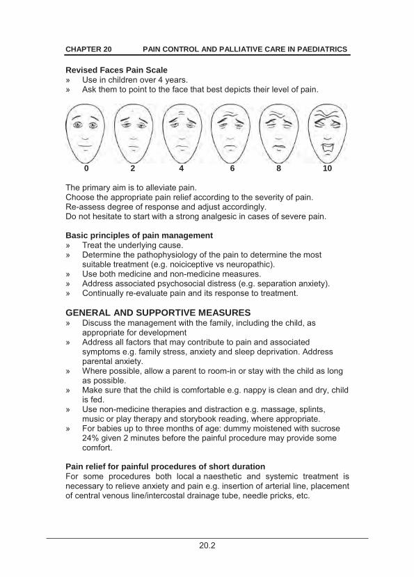
20.2
CHAPTER 20 PAIN CONTROL AND PALLIATIVE CARE IN PAEDIATRICS Revised Faces Pain Scale » Use in children over 4 years. » Ask them to point to the face that best depicts their level of pain.
0 2 4 6 8 10 The primary aim is to alleviate pain. Choose the appropriate pain relief according to the severity of pain. Re-assess degree of response and adjust accordingly. Do not hesitate to start with a strong analgesic in cases of severe pain. Basic principles of pain management » Treat the underlying cause. » Determine the pathophysiology of the pain to determine the most
suitable treatment (e.g. noiciceptive vs neuropathic). » Use both medicine and non-medicine measures. » Address associated psychosocial distress (e.g. separation anxiety). » Continually re-evaluate pain and its response to treatment. GENERAL AND SUPPORTIVE MEASURES » Discuss the management with the family, including the child, as
appropriate for development » Address all factors that may contribute to pain and associated
symptoms e.g. family stress, anxiety and sleep deprivation. Address parental anxiety.
» Where possible, allow a parent to room-in or stay with the child as long as possible.
» Make sure that the child is comfortable e.g. nappy is clean and dry, child is fed.
» Use non-medicine therapies and distraction e.g. massage, splints, music or play therapy and storybook reading, where appropriate.
» For babies up to three months of age: dummy moistened with sucrose 24% given 2 minutes before the painful procedure may provide some comfort.
Pain relief for painful procedures of short duration For some procedures both local a naesthetic and systemic treatment is necessary to relieve anxiety and pain e.g. insertion of arterial line, placement of central venous line/intercostal drainage tube, needle pricks, etc.
20.3
CHAPTER 20 PAIN CONTROL AND PALLIATIVE CARE IN PAEDIATRICS If pain is likely to b e on-going post procedure remember to pr escribe continued analgaesia. For some procedures, e.g. to remove an i ntercostal drain or deep w ound drain or stitches, it is necessary to g ive sedation in c ombination with systemic pain treatment. Sedation should never be used on its own without analgesics for procedural pain. For needle prick site/lumbar puncture, except in emergency situations Lidocaine/prilocaine cream, topical, applied at least 30 minutes before
procedure. o Apply 1–1.5 cm length of cream over the needle puncture site. o Spread cream thinly over 1 cm radius on skin and cover with
polyurethane film dressing.
Burns Children with large burns require regular analgesia. (Refer to persistent pain below) Provide analgesia cover at each dressing change. See section 1.2.1: Burns. In children with major burns, dressings should be changed under general anaesthesia. Short acting analgesics for invasive local procedures
Take standard precautions for respiratory arrest.
Ketamine, oral, 5 mg/kg or IV, 1–2 mg/kg.
OR Midazolam, IV, 0.1–0.2 mg/kg or oral, 0.5 mg/kg or intranasal, 0.2 mg/kg.
o Note: Midazolam has no analgesic effect. Before administering the sedative/anxiolytic/analgesic drug » Withhold food for 4 hours before planned procedure. » Monitor child and have resuscitation equipment available. » Put up an intravenous line with heparin lock in case an unexpected
complication arises. » Where appropriate, written informed consent should be obtained. PAIN RELIEF FOR PERSISTENT PAIN The correct use of the correct analgesic will relieve most pain in children. Treatment should be individualised as pain experiences vary from child to child.
20.4
CHAPTER 20 PAIN CONTROL AND PALLIATIVE CARE IN PAEDIATRICS The broad principles of analgesic use in children: » By the clock (regular rather than as required dosing). » By the correct route for the type of pain (preferably oral, avoid IM injections). » By the child (individualise treatment). Non-opioid medicines Paracetamol, oral.
o Loading dose 20mg/kg/dose, then 15 mg/kg/dose 4–6 hourly. OR Where oral medication cannot be used: Paracetamol, suppositories, 6 hourly.
o If 3–12 months of age: 62.5–125 mg. o If 1–5 years of age: 125–250 mg. o If 6–12 years of age: 250–500mg.
Non-steroidal anti-inflammatory drugs (NSAIDS) Where anti-inflammatory effect is required. Can be used in combination with paracetamol or opioids. Ibuprofen, oral, 10 mg/kg/dose 8 hourly with meals.
Intermediate efficacy opioid Tilidine, oral, 1 drop per 2.5 kg of body weight (i.e. 1 mg/kg/dose).
Strong opioid Morphine, oral (Immediate release morphine (liquid))
o Starting dose: o If 0 – 1 month of age: 0.05 mg/kg 6 hourly. o If > 1–12 months of age: 0.1 mg/kg/dose 4 hourly. o If > 12 months of age: 0.2–0.4 mg/kg/dose 4 hourly.
o Dosing is 4 hourly except in patients with delayed clearance, i.e. newborns, hepatic and renal dysfunction where prescribed 6 hourly.
In non-palliative care, the increase in dose of morphine is limited by side-effects, i.e. hypotension and respiratory depression.
In palliative care the dose of morphine is titrated to the patient’s pain control. Increase dose by 30–50 % with each dose if pain control is sub-optimal.
There is no maximum dose of morphine.
o Breakthrough dose: 50–100% of the regular pain dose. o Give regular and breakthrough doses at least one hour apart. o If the child requires breakthrough doses less than an hour after the
regular dose has been given then it is likely that the regular dose needs to be increased.
o At the end of a 24 hour period all the breakthrough doses should be added up and this value divided by 6 to determine the increase needed in the regular dose for the next day.
o Increase breakthrough dose as regular doses are increased.
20.5
CHAPTER 20 PAIN CONTROL AND PALLIATIVE CARE IN PAEDIATRICS If patient is unable to swallow or is vomiting: Morphine, IM, 4-hourly bolus.
o Starting dose: o If > 1–6 months of age: 0.05 – 1 mg/kg. o If > 6 months of age: 0.1 – 0.2 mg/kg.
In all patients receiving morphine: Lactulose, oral, 2.5-10 mL 12 hourly.
Children receiving properly titrated doses of analgesics, including opioids, do not become addicted. There is a diff erence between tolerance, which is a need for escalating doses to achieve the same therapeutic effect, and addiction. Withdrawal from opioids This must be done for any child who has received morphine for more than 5–7 days. Wean by decreasing the daily dose by one third for three days. Adjunctive analgesic therapy Adjuvant analgesics are drugs that have weak or non-ex istent analgesic action when administered alone but can enhance analgesic actions when co-administered with known analgesic agents. Steroids Steroids may be used as an adjuvant in consultation with a specialist for the following indications: » infiltration of bone/meninges, » compression of nerves and spinal cord, » visceromegaly, » tumour invasion of organs, » stretching of periosteum or peritoneum. Stretching of periosteum or peritoneum: Prednisone, oral, 1–2 mg/kg/day as single dose or in 2 divided doses.
Neuropathic pain: Carbamazepine, oral, 5 mg/kg/dose 12 hourly.
REFERRAL » Adequate analgesia will control pain in almost all cases. Discuss
resistant cases with a specialist.
20.6
CHAPTER 20 PAIN CONTROL AND PALLIATIVE CARE IN PAEDIATRICS 20.2 PALLIATIVE CARE Icd10 DESCRIPTION Palliative care is an approach that aims to improve the quality of life of patients and their families facing the problem associated with life-threatening illness, through the prevention and relief of suffering by means of early identification and impeccable assessment and treatment of pain and other problems, physical, psychosocial and spiritual. A key component to relieving suffering is the management of distressing symptoms that include both pain and non-pain symptoms (e.g. nausea, anxiety, etc). There are certa in key principles that should be applied when managing these symptoms, i.e.: » Determine and treat underlying cause of the symptom including non-
physical causes. » Relieve the symptom without creating new symptoms or unwanted side
effects. » Consider different types of interventions: drug and non-drug
interventions. » Consider whether the treatment is of benefit to the individual patient. Common non-pain symptoms and other problems in paediatric palliative care are described below. Nausea and vomiting Appropriate treatment depends on the cause of nausea and vomiting. Metoclopramide, oral, 0.1 mg/kg/dose, 6–12 hourly.
Use with caution as extrapyramidal side effects may occur (especially at higher doses)
OR Ondansetron, IV, 0.1 mg/kg.
o Maximum dose: 4 mg/day. Anxiety Benzodiazepine, e.g.: Diazepam, oral, 8 hourly.
o If > 2–12 years of age: 2–3 mg. o If >12–18 years of age: 2–10 mg.
20.7
CHAPTER 20 PAIN CONTROL AND PALLIATIVE CARE IN PAEDIATRICS Constipation » Laxatives should be used prophylactically in all patients receiving morphine. Lactulose, oral, 2.5–10 mL 12 hourly.
Dyspnoea » Reduce anxiety by addressing psychosocial factors e.g. parental
separation. » Consider home oxygen concentrator. Morphine, oral (Immediate release morphine (liquid))
o Starting dose: o If 0–1 month of age: 0.05 mg/kg 6 hourly. o If > 1–12 months of age: 0.1 mg/kg/dose 4 hourly. o If > 12 months of age: 0.2–0.4 mg/kg/dose 4 hourly.
Muscle Spasms » Where possible, refer for physiotherapy and/or occupational therapy. Benzodiazepine, e.g.: Diazepam, oral, 8 hourly.
o If > 2–12 years of age: 2–3 mg. o If >12–18 years of age: 2–10 mg.
PLUS Morphine, oral (Immediate release morphine (liquid))
o Starting dose: o If 0–1 month of age: 0.05 mg/kg 6 hourly. o If > 1–12 months of age: 0.1 mg/kg/dose 4 hourly. o If > 12 months of age: 0.2–0.4 mg/kg/dose 4 hourly.
Pruritus Promethazine, IV/oral, 0.1 mg/kg/dose 6 hourly.
Spasmodic abdominal pain Hyoscine butylbromide, IV/oral, 0.5 mg/kg/dose 6–8 hourly.
Oral care Zinc and castor oil cream, topical, applied to lips every 2 hours.
Sodium chloride solution, gargle, to rinse mouth
o Dissolve 5 g sodium chloride in 1 L of water. OR Chlorhexidine 0.2%, 10 mL as a mouthwash or gargle, 12 hourly.
o Do not swallow. Apthous ulcers See section: 2.1.5 Aphthous ulcers.
21.8
CHAPTER 20 PAIN CONTROL AND PALLIATIVE CARE IN PAEDIATRICS Mucositis Chorhexidine 2%/benzydamine, oral rinse, rinse or gargle 6–8 hourly.
Perineal mucositis/nappy rash Zinc and castor oil cream, topical, applied as needed
o If pain is a feature: mix with lidocaine 2% gel. In no improvement within 3 days, suspect candida and: ADD Clotrimazole 2% cream followed by zinc and castor oil ointment applied
after each nappy change. Secretions » Suctioning and re-positioning is often helpful. » Attention to oral hygiene is essential. Hyoscine butylbromide, IV/oral, 0.5 mg/kg/dose 6–8 hourly.
REFERRAL Discuss with a specialist: » Children with symptoms not described here. » Children not responding to management.
21.1
CHAPTER 21 INTENSIVE CARE AND ANAESTHETICS
Healthcare professionals engaged in intensive care and anaesthetics must undergo appropriate training
21.1 SEDATION FOR INTENSIVE CARE PROCEDURES 21.1.1 ICU SEDATION, NEONATE DESCRIPTION Sedation is required for various procedures and situation in newborns in an intensive care setting to decrease discomfort and suffering, and to imp rove the management outcomes of the procedure/care that is being given. GENERAL AND SUPPORTIVE MEASURES In all situations appropriate control of the environment, provision of normal physiological requirements, monitoring of vital signs and provision of comforting care should be incorporated in the care of neonates and minimise the stress on the child. MEDICINE TREATMENT 1. For controlled endotracheal intubation for ventilation
This skill should have been learnt in an appropriate learning situation. If not, endotracheal intubation should take place under supervision of a
specialist. Pre-oxygenate with bag-mask, T-piece or equivalent ventilator. Maintain oxygen saturation between 88–92%. Propofol, IV, 2.5 mg/kg. Adjust as necessary.
OR Midazolam, IV, 0.1–0.2 mg/kg. Caution in premature babies.
OR Ketamine, IV, 1–2 mg/kg.
PLUS Succinylcholine, IV, 2 mg/kg. Causes paralysis and apnoea.
o Note: Avoid succinylcholine in patients with potassium disturbances and neuromuscular disease.
21.2
CHAPTER 21 INTENSIVE CARE AND ANAESTHETICS
PLUS If bradycardia or using ketamine: Atropine, IV, 0.02 mg/kg.
o Maximum dose: 0.6 mg. If emergency with no IV line, consider: Ketamine, IM, 5–10 mg/kg.
2. Short term intubation (In-out endotracheal surfactant administration) Nasal CPAP as required. If inadequate oxygenation on nasal CPAP, pre-oxygenate with bag-mask or T-piece ventilation to maintain saturation between 88-92%. Intubate orally, give surfactant, follow with gentle manual ventilation or CPAP, as required, for 5 minutes. Extubate and recommence nasal CPAP. 3. During continuous mechanical ventilation For pain and sedation: Fentanyl, IV, 5–10 mcg/kg.
o If necessary, follow with IV infusion, 50 mcg/mL at 0.1–0.2 mL/kg/hour, i.e. 5–10 mcg/kg/hour.
OR Morphine, IV, 10–30 mcg/kg/hour.
o i.e. Morphine 1 mg/kg mixed with 50 mL dextrose 5% or sodium chloride 0.9% at 0.5–1.5 mL/hour.
OR Sucrose 24% solution, buccal as needed.
REFERRAL » Inability to provide appropriate care. 21.1.2 ICU SEDATION, INFANT AND CHILD DESCRIPTION Sedation is required for various procedures and situations in infants and children in an intensive care setting to decrease discomfort and suffering and to improve the management outcomes of the procedure/care that is being given.
21.3
CHAPTER 21 INTENSIVE CARE AND ANAESTHETICS
GENERAL AND SUPPORTIVE MEASURES In all situations appropriate control of environment, provision of normal physiological requirements, monitoring of vital signs and provision of comforting care should surround the care of inf ants and children and minimises the stress on the child. MEDICINE TREATMENT
This skill should have been learnt in an appropriate learning situation. If not, endotracheal intubation should take place under supervision of a
specialist.
Pre-oxygenate with bag-mask, T-piece or equivalent ventilator. Maintain oxygen saturation > 90%. 1. For endotracheal intubation Ketamine, IV, 1–2 mg/kg/dose.
OR Propofol, IV, 2.5 mg/kg. Adjust as necessary.
PLUS If muscle relaxant necessary: Succinylcholine, IV, 2 mg/kg. Causes paralysis and apnoea.
2. During continuous mechanical ventilation Midazolam, IV, 1–4 mcg/kg/minute.
o Midazolam 3 mg/kg mixed with 50 mL dextrose 5% at 1–4 mL/hour. PLUS Fentanyl, IV, 5–10 mcg/kg.
o If necessary, follow with IV infusion, 50 mcg/mL at 0.1–0.2 mL/kg/hour, i.e. 5–10 mcg/kg/hour.
OR
Midazolam, IV, 1–4 mcg/kg/minute. o Midazolam 3 mg/kg mixed with 50 mL dextrose 5% at 1–4 mL/hour.
PLUS Morphine, IV, 20–80 mcg/kg/hour.
o Morphine 1 mg/kg mixed with 50 mL dextrose 5% or sodium chloride 0.9% at (1–4 mL/hour).
For procedures (not ventilated)
Take standard precautions for respiratory arrest.
21.4
CHAPTER 21 INTENSIVE CARE AND ANAESTHETICS
Ketamine, oral, 5 mg/kg or IV, 1–2 mg/kg. OR Midazolam, IV, 0.1–0.2 mg/kg or oral, 0.5 mg/kg or intranasal, 0.2 mg/kg.
(No analgesic effect). REFERRAL » Inability to provide appropriate care. 21.2 PARENTERAL NUTRITION » Parenteral nutrition (PN) is t he intravenous administration of amin o
acids (proteins), lipids, carbohydrates, electrolytes, minerals, vitamins and trace elements necessary for metabolic requirements and growth.
» PN may be total, i.e. total parenteral nutrition (TPN) where all nutrients are administered via a central venous line until the infant is again ready to take enteral feeds.
» PN may also be partial i.e. partial parenteral nutrition (PPN) where it is used to supplement enteral feeds in infants who cannot yet tolerate their full complement of enteral feeds.
» Administer PN preferably via a central venous line, especially when it is expected that the infant will require TPN for more than 7 days. For partial parenteral nutrition a peri pheral venous line may be used, especially where it is expected that the infant will require PPN for only a few days (< 7 days). Only PN solutions which contain lipids and with an osmolarity of < 1000 mOsm/L and non-lipid containing PN solutions with an osmolarity < 800 mOsm/L can be safely administered via a peripheral vein.
» Check peripheral vein infusion sites and patency of the line/catheter regularly for tissue infiltration.
» Transport and store PN solutions at 2–8ºC. Start administration of the PN solutions within one hour after removal from the refrigerator.
» Do not make additions to a PN bag or decant contents as the stability and/or the sterility may be compromised.
» Do not use the PN line to collect blood samples. » Administer PN through a dedicated line and do not administer
medications, blood, etc. through the PN line. » Protect PN bags and li nes against exposure to dire ct light (e.g .
phototherapy). Cover the bag with a pouch and use amber lines. » Use a 1.2 micron in-line filter for lipid containing PN solutions and a 0.2
micron filter for lipid-free PN solutions. » Adhere to strict aseptic technique when administering PN solutions.
Check solution and administration sets before starting the infusion. Use bags within 24 hours of starting infusion.
» PN should be prescribed and administered under the supervision of a paediatrician and dietician.
21.5
CHAPTER 21 INTENSIVE CARE AND ANAESTHETICS
Complications of PN » IV line/catheter complications e.g. extravasation, blockage or
bacterial/fungal contamination; » metabolic complications, e.g. hyperglycaemia, high ammonia, metabolic
acidosis, electrolyte and mineral disturbances and hyperlipidaemia; » infection/sepsis; and » cholestatic hepatitis. Monitor: » vital signs and hydration; » blood glucose 12 hourly, maintain blood glucose at 2.6–6 mmol/L; » electrolytes, minerals and acid base on a daily basis or more regularly, if
necessary; » growth parameters and weight, once weekly; » infection markers at least once weekly or more frequently, if necessary;
and » liver enzymes, bilirubin, ammonia, lipids, urea and cr eatinine once
weekly or more frequently, as indicated by the condition of the infant. PARENTERAL NUTRITION FORMULATIONS Use the sta ndard TPN formulations that are co mmercially available for paediatric use a nd that h ave been man ufactured at a G MP (Good Manufacturing Practice) compliant site and have been regulated by the Medicines Control Council. 21.2.1 PARENTERAL NUTRITION, NEONATAL DESCRIPTION Parenteral nutrition (PN) should be considered within 24 hours in neonates where enteral f eeds are not indicated, tolerated or contra-indicated due to medical or surgical conditions e.g. NEC, post intestinal surgery, ileus, bowel obstruction, malabsorption. DOSE AND DURATION OF PN INFUSION The volume of PN to be a dministered to the neonate depends on the age, weight and underlying disease of the neonate. Use the daily fluid requirements as a guid e to deter mine the volume of PN solution to b e administered. The maximum volume of TPN for a neonate should not exceed 150 mL/kg/24 hours. The PN solution should be administered over 24 hours depending on the condition of the infant and volume to be administered.
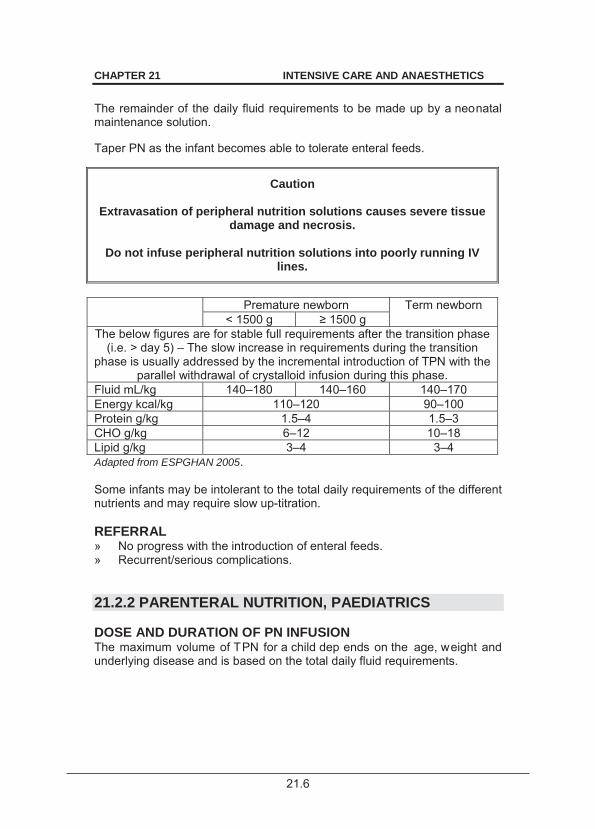
21.6
CHAPTER 21 INTENSIVE CARE AND ANAESTHETICS
The remainder of the daily fluid requirements to be made up by a neonatal maintenance solution. Taper PN as the infant becomes able to tolerate enteral feeds.
Caution
Extravasation of peripheral nutrition solutions causes severe tissue damage and necrosis.
Do not infuse peripheral nutrition solutions into poorly running IV
lines.
Premature newborn Term newborn
< 1500 g 1500 g The below figures are for stable full requirements after the transition phase
(i.e. > day 5) – The slow increase in requirements during the transition phase is usually addressed by the incremental introduction of TPN with the
parallel withdrawal of crystalloid infusion during this phase. Fluid mL/kg 140–180 140–160 140–170 Energy kcal/kg 110–120 90–100 Protein g/kg 1.5–4 1.5–3 CHO g/kg 6–12 10–18 Lipid g/kg 3–4 3–4 Adapted from ESPGHAN 2005. Some infants may be intolerant to the total daily requirements of the different nutrients and may require slow up-titration. REFERRAL » No progress with the introduction of enteral feeds. » Recurrent/serious complications. 21.2.2 PARENTERAL NUTRITION, PAEDIATRICS DOSE AND DURATION OF PN INFUSION The maximum volume of TPN for a child dep ends on the age, weight and underlying disease and is based on the total daily fluid requirements.
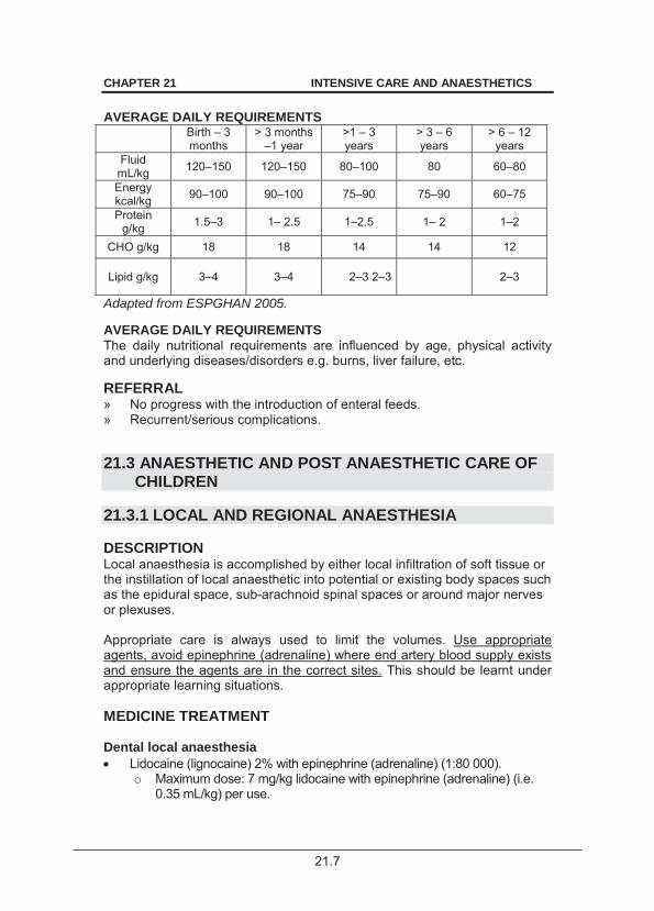
21.7
CHAPTER 21 INTENSIVE CARE AND ANAESTHETICS AVERAGE DAILY REQUIREMENTS Birth – 3
months > 3 months
–1 year >1 – 3 years
> 3 – 6 years
> 6 – 12 years
Fluid mL/kg 120–150 120–150 80–100 80 60–80
Energy kcal/kg 90–100 90–100 75–90 75–90 60–75
Protein g/kg 1.5–3 1– 2.5 1–2.5 1– 2 1–2
CHO g/kg 18 18 14 14 12
Lipid g/kg 3–4 3–4 2–3 2–3 2–3
Adapted from ESPGHAN 2005. AVERAGE DAILY REQUIREMENTS The daily nutritional requirements are influenced by age, physical activity and underlying diseases/disorders e.g. burns, liver failure, etc. REFERRAL » No progress with the introduction of enteral feeds. » Recurrent/serious complications. 21.3 ANAESTHETIC AND POST ANAESTHETIC CARE OF
CHILDREN 21.3.1 LOCAL AND REGIONAL ANAESTHESIA DESCRIPTION Local anaesthesia is accomplished by either local infiltration of soft tissue or the instillation of local anaesthetic into potential or existing body spaces such as the epidural space, sub-arachnoid spinal spaces or around major nerves or plexuses. Appropriate care is always used to limit the volumes. Use appropriate agents, avoid epinephrine (adrenaline) where end artery blood supply exists and ensure the agents are in the correct sites. This should be learnt under appropriate learning situations. MEDICINE TREATMENT Dental local anaesthesia Lidocaine (lignocaine) 2% with epinephrine (adrenaline) (1:80 000).
o Maximum dose: 7 mg/kg lidocaine with epinephrine (adrenaline) (i.e. 0.35 mL/kg) per use.
21.8
CHAPTER 21 INTENSIVE CARE AND ANAESTHETICS
Diffuse local soft tissue infiltration Do not use adrenaline containing lidocaine in sites where vascular (end artery) compromise may result from vasoconstrictor use, i.e. fingers, toes, penis and eyes. Sites where vascular (end artery) compromise is a risk. Lidocaine (lignocaine) 2% (without epinephrine (adrenaline)).
o Maximum dose: 3 mg/kg of lidocaine (i.e. 0.15 mL/kg) per use. Sites where vascular (end artery) compromise is not a risk: Lidocaine (lignocaine) 2% with epinephrine (adrenaline) (1:80 000).
o Maximum dose: 7 mg/kg lidocaine (lignocaine) with epinephrine (adrenaline) (i.e. 0.35 mL/kg) per use.
Spinal anaesthesia
Caution This is a specialist anaesthetic procedure.
Dangers include complete spinal block.
Bupivacaine/dextrose 5/72.7 mg/mL (without epinephrine (adrenaline))
o Maximum dose: 0.1–0.3 mg/kg of bupivacaine (i.e. 0.06 mL/kg). Use by an anaesthetist.
Intercostal nerve block/penile block/digital ring block/brachial (axillary approach) block Lidocaine (lignocaine) 2% (without epinephrine (adrenaline)).
o Maximum dose: 3 mg/kg of lidocaine (i.e. 0.15 mL/kg) per use. OR Bupivacaine 5 mg/mL (without epinephrine (adrenaline) and dextrose).
o Maximum dose: 2 mg/kg of bupivacaine (i.e. 0.4 mL/kg). 21.3.2 GENERAL ANAESTHESIA 21.3.2.1 PREPARATION DESCRIPTION Premedication of ch ildren for anaesthesia is largely a se dative/anxiolytic intervention. Recognition of the child’s condition should guide in the choice of agent. Other special medical interventions such as prevention of hypersecretion are ordered according to specific need or anticipated need.
21.9
CHAPTER 21 INTENSIVE CARE AND ANAESTHETICS
Premedication care should be learnt in an appropriate learning situation.
Pre-operative starvation: » clear fluid: 2 hours, » breast milk: 4 hours, » solids, breast milk substitutes, non-human milk: 6 hours. MEDICINE TREATMENT Premedication: Midazolam, oral, 0.5 mg/kg, 15–30 minutes pre-operative.
o Maximum dose: 7.5 mg. OR For children over 2 years of age: Promethazine, oral, 0.5 mg/kg.
o Maximum dose: 25–100 mg. o Decrease dose of any narcotics given.
21.3.2.2 INDUCTION
Induction should be learnt in an appropriate learning situation.
DESCRIPTION Induction of anaesthesia is the critical part of the transition from consciousness to general anaesthesia. All patient undergoing anaesthesia should be monitored with a minimum of: » clinical observation, » ECG, » blood pressure monitor, » pulse oximeter, and » temperature monitoring in children and infants. It is desirable to do capnography for ventilated patients. This is a period which requires highly attentive and skilled care.
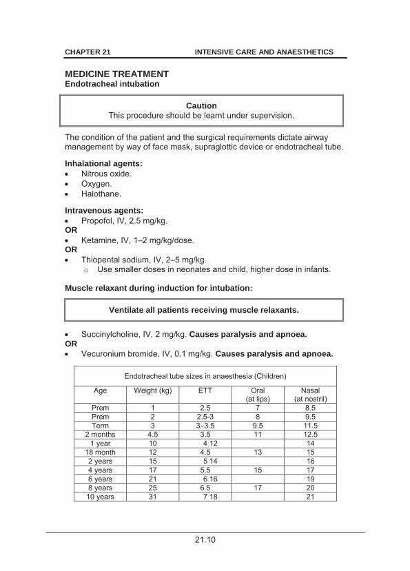
21.10
CHAPTER 21 INTENSIVE CARE AND ANAESTHETICS MEDICINE TREATMENT Endotracheal intubation
Caution This procedure should be learnt under supervision.
The condition of the patient and the surgical requirements dictate airway management by way of face mask, supraglottic device or endotracheal tube. Inhalational agents: Nitrous oxide. Oxygen. Halothane.
Intravenous agents: Propofol, IV, 2.5 mg/kg.
OR Ketamine, IV, 1–2 mg/kg/dose.
OR Thiopental sodium, IV, 2–5 mg/kg.
o Use smaller doses in neonates and child, higher dose in infants. Muscle relaxant during induction for intubation:
Ventilate all patients receiving muscle relaxants.
Succinylcholine, IV, 2 mg/kg. Causes paralysis and apnoea.
OR Vecuronium bromide, IV, 0.1 mg/kg. Causes paralysis and apnoea.
Endotracheal tube sizes in anaesthesia (Children)
Age Weight (kg) ETT Oral (at lips)
Nasal (at nostril)
Prem 1 2.5 7 8.5 Prem 2 2.5-3 8 9.5 Term 3 3–3.5 9.5 11.5
2 months 4.5 3.5 11 12.5 1 year 10 4 12 14
18 month 12 4.5 13 15 2 years 15 5 14 16 4 years 17 5.5 15 17 6 years 21 6 16 19 8 years 25 6.5 17 20
10 years 31 7 18 21
21.11
CHAPTER 21 INTENSIVE CARE AND ANAESTHETICS 21.3.2.3 MAINTENANCE
Caution This procedure should be learnt under supervision.
DESCRIPTION After induction transition occurs to maintenance of adequate level of pain prevention, amnesia and immobility to allow pain free and safe surgical care, i.e. adequate narcosis, analgesia and muscle relaxation. Appropriate care is always required to monitor the patient, detect complications of, and depth of anaesthesia.
MEDICINE TREATMENT Inhalation anaesthesia Oxygen. Nitrous oxide. Halothane. Isoflurane.
Intravenous medicine used during anaesthesia in children (not neonates): Fentanyl, IV, 1 –2 mcg/kg (under anaesthetist supervision up to 5–10
mcg/kg). o Maximum dose: 50–100 mcg if un-ventilated.
OR Morphine, IV, 20–80 mcg/kg/hour.
o Morphine 1 mg/kg mixed with 50 mL dextrose 5% or sodium chloride 0.9% at (1–4 mL/hour).
Muscle relaxant during maintenance: Vecuronium bromide, IV.
o Initial dose: IV bolus, 0.08–0.1 mg/kg. o Maintenance doses: IV, 0.03–0.05 mg/kg, as needed.
Reversal of muscle relaxant Neostigmine plus atropine, IV, 0.1 mL/kg of neostigmine/atropine
solution below: o Neostigmine/atropine solution: 0.5 mL of neostigmine 2.5 mg/mL plus
0.5 mL atropine 0.6 mg/mL plus 0.5 mL sodium chloride 0.9%. To reduce secretions: Atropine, IV, 0.02 mg/kg.
o Maximum dose: 0.6 mg.
21.12
CHAPTER 21 INTENSIVE CARE AND ANAESTHETICS
To protect the eyes from trauma and drying during anaesthesia, gently tape eyelids closed with thin light medical porous adhesive tape, or equivalent. If eyes are required to be open: Hydroxypropylmethylcellulose 0.3%, ophthalmic drops.
21.3.3 POST OPERATIVE CARE DESCRIPTION After surgery adequate control of pain is required for comfort and also for the optimisation of o utcome and minimisation of adverse effects of pain on recovery. Pain relief should be adapted to the specific needs of each patient – according to the severity of pain, site of pain and type of pain. GENERAL AND SUPPORTIVE MEASURES Appropriate control of environment, provision of sedation (as above), normal physiological requirements, monitoring of vital signs and provision of comforting care should be incorporated in the care of infants and children during and after surgery. MEDICINE TREATMENT For pain (post operation): neonates Ventilated neonate: Morphine, IV, 10–30 mcg/kg/hour (0.5–1 mL/hour).
o Morphine 1 mg/kg mixed with 50 mL dextrose 5% or sodium chloride 0.9%.
Unventilated neonate: Tilidine, oral, 1 drop per 2.5 kg (i.e.1 mg/kg/dose) 6 hourly.
Beware of respiratory depression. Monitoring of respiration is essential.
PLUS Paracetamol, oral, 10–20 mg/kg as a single dose immediately.
o Follow with 15 mg/kg/dose 6 hourly. PLUS Sucrose 24%, buccal, as needed.
o Preterm infants: 0.5–1 mL. o Term infants: 2 mL.
21.13
CHAPTER 21 INTENSIVE CARE AND ANAESTHETICS
For pain (post operation): older child (more than 3 months of age) Morphine, IV, 20–80 mcg/kg/hour
o Morphine 1 mg/kg mixed with 50 mL dextrose 5% or sodium chloride 0.9% at (1–4 mL/hour).
OR
Morphine, IM/SC, 0.1 mg/kg 4–6 hourly as necessary.
OR
Tilidine, oral, 1 drop per 2.5 kg (i.e.1 mg/kg/dose) 6 hourly. Use with cardio-respiratory monitoring in infants.
PLUS Paracetamol, oral, 20 mg/kg as a single dose immediately.
o Follow with 15 mg/kg/dose 6 hourly. If oral cannot be used: Paracetamol rectal, 6 hourly.
o If 3–12 months of age: 60–125 mg. o If 1–5 years of age: 125–250 mg. o If 6–12 years of age: 250–500mg.
OR Ibuprofen, oral, 4–10 mg/kg/dose 6–8 hourly.
Monitor renal function and ensure adequate hydration. Regional anaesthesia See section 21.3.1: Local and regional anaesthesia. REFERRAL » Inability to provide appropriate care. 21.3.4 MANAGEMENT OF ANAESTHETIC AND
POSTANAESTHETIC COMPLICATIONS DESCRIPTION Various events may occur during and after anaesthesia which require management. MEDICINE TREATMENT Laryngospasm Bag-mask ventilation and reintroduction of a volatile general anaesthetic agent may overcome laryngospasm without the need for succinylcholine.
21.14
CHAPTER 21 INTENSIVE CARE AND ANAESTHETICS
Succinylcholine, IV, 2 mg/kg. Causes paralysis and apnoea.
Bronchospasm Salbutamol nebulisation – administered in line (i.e. in circuit) or by mask.
See section 15.2.1: Asthma attack, acute. OR Salbutamol, IV 5–10 mcg/kg/minute for 1 hour.
o Follow with 1–2 mcg//kg/minute. Hypersecretion Atropine, IV, 0.02 mg/kg.
o Maximum dose: 0.6 mg. Respiratory depression/apnoea from opiate overdose: Naloxone, IV, 0.01 mg/kg, repeated every 2 minutes, if required, up to 4
times. o Maximum dose: 0.4 mg.
Note: All patients need to be kept under direct observation until the effect of the opiates has completely worn off. Further doses of naloxone may be needed as naloxone has a short duration of action.
Emergence delirium Midazolam, oral, 0.5 mg/kg, 15–30 minutes pre-operative.
o Maximum dose: 7.5 mg. Post operative nausea and vomiting Metoclopramide, IV/IM, 0.15–0.3 mg/kg 6 hourly.
Use with caution as extrapyramdal side effects may occur (especially at
high doses) OR Children > 2 years of age: Ondansetron, slow IV, 0.2 mg/kg.
o Maximum dose: 8 mg. Malignant hyperthermia Dantrolene, IV, 1 mg/kg/minute until improvement.
o Follow with 1–2 mg/kg 6 hourly for 1–3 days. o Maximum dose: 10 mg/kg.
21.15
CHAPTER 21 INTENSIVE CARE AND ANAESTHETICS
Shock See section 1.1.7: Shock. Dysrhythmias See section 4.1: Cardiac dysrhythmias. Prevention of symptomatic hypocalcaemia during rapid large blood transfusion in babies with acid citrate anticoagulated blood: Calcium gluconate 10%, IV infusion.
o 5–10 mL of calcium gluconate 10% added to 200 mL bag of compatible IV infusion fluid.
o Infuse at maintenance IV fluid rate if blood transfusion volumes approach circulating volume of child (~80 mL/kg).
22.1
CHAPTER 22
ADOLESCENCE Adolescence is a per iod of significant physical, emotional, and c ognitive change and is the developmental phase of separation and individuation. The development of independence from fam ily and the pre ssure to conform to peers can impose unique challenges in the management of chronic disease. Despite the stronger identification with the peer group, adolescents with illnesses require more support from family and caregivers. Decreased focus on family and in creased conformity with peer groups r esults in p oor acceptance of a disease. Peer pressure and the need not to be stigmatised by a disease or its treatment dominates the emotional life of adolescents and may alter adherence to medicines. Adolescence spans the period of pubertal development which manifests with physical changes which reflects the maturation of the gonads and hypothalamic pituitary gonadal axis. Generally accepted age for adolescence includes 10 to 19 years although changing views has extended the adolescent/youth period to age 24 years. Significant biologic (reflected in the different stages of puberty) and p sychological changes occur during adolescence. Puberty is the time of somatic growth and sexual maturation, with females developing more fat an d males more muscle mass. Irresponsible behaviour and t endency towards risk ta king are featur es in adolescence that are related to the hormonal changes in puberty. Distinct psychosocial features characterise early, mid- and late adolescence. Recognition of these stages impacts on issues such as adherence. In early adolescence, the individual is unable to th ink abstractly or plan a head; in middle adolescence, concrete thinking in times of stress develops; in l ate adolescence abstract thinking and the ability to anticipate the future and plan develops.
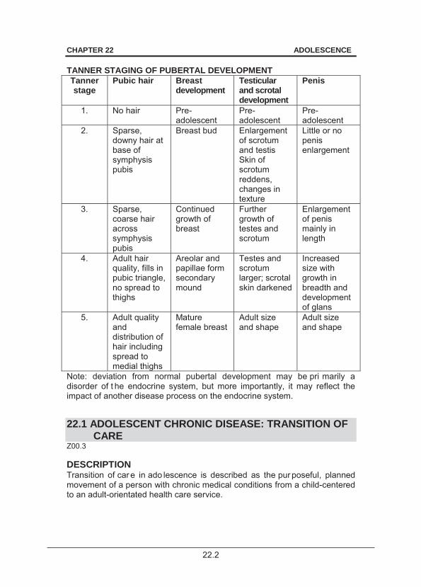
22.2
CHAPTER 22 ADOLESCENCE TANNER STAGING OF PUBERTAL DEVELOPMENT Tanner stage
Pubic hair Breast development
Testicular and scrotal development
Penis
1. No hair Pre-adolescent
Pre-adolescent
Pre-adolescent
2. Sparse, downy hair at base of symphysis pubis
Breast bud Enlargement of scrotum and testis Skin of scrotum reddens, changes in texture
Little or no penis enlargement
3. Sparse, coarse hair across symphysis pubis
Continued growth of breast
Further growth of testes and scrotum
Enlargement of penis mainly in length
4. Adult hair quality, fills in pubic triangle, no spread to thighs
Areolar and papillae form secondary mound
Testes and scrotum larger; scrotal skin darkened
Increased size with growth in breadth and development of glans
5. Adult quality and distribution of hair including spread to medial thighs
Mature female breast
Adult size and shape
Adult size and shape
Note: deviation from normal pubertal development may be pri marily a disorder of t he endocrine system, but more importantly, it may reflect the impact of another disease process on the endocrine system. 22.1 ADOLESCENT CHRONIC DISEASE: TRANSITION OF
CARE Z00.3 DESCRIPTION Transition of car e in ado lescence is described as the pur poseful, planned movement of a person with chronic medical conditions from a child-centered to an adult-orientated health care service.
22.3
CHAPTER 22 ADOLESCENCE Specialised programmes for transition improve adherence and outcomes, e.g. HIV, diabetes, epilepsy and rena l disease. Careful assessment of growth and dev elopment may determine an ind ividualised approach to transition. Chronic disease during this period impacts on growth and development. HIV acquired perinatally as well as that due to unprotected sexual contact (“behaviourally infected”) are encountered during adolescence. GENERAL AND SUPPORTIVE MEASURES » Promote adherence to medicine and follow up. » Counselling and support. » Manage and co-ordinate treatment through a multidisciplinary team
including physicians and paediatricians. MEDICINE TREATMENT Titrate doses according to Tanner staging rather than strictly on b asis of age. » Tanner 1 or 2 (early puberty): use paediatric schedules. » Tanner stage 5 (late puberty): use adult schedules.
Puberty may be de layed in children with chronic disease, adding to discrepancies between Tanner stage-based dosing and age-based dosing.
During puberty doses may vary. Consult relevant package inserts for guidance of dosages.
Optimise therapy of certain medicines by monitoring drug levels, adjusting doses during puberty (particularly in diabetics) and with weight gain.
Be aware of medicine interactions, e.g. induction of oral contraceptive metabolism by rifampicin and recognising changes of drug disposition during puberty.
As psychological changes in puberty affect adherence, convenient medicine formulations and devices that contribute to better disease management may be considered.
Minimise the adverse impact of m edicines on cognition and brain development. REFERRAL » Refer patients with cognitive impairment and mental health problems to
a psychiatrist. » Adolescents with chronic disease for assessment by psychologist and
mental health specialist for recognition of anxiety, depression, attention-deficit disorder and post traumatic stress disorder.
22.4
CHAPTER 22 ADOLESCENCE 22.2 CONTRACEPTION, TEENAGE PREGNANCY AND
TERATOGENICITY RISKS Z30.9 DESCRIPTION Adolescents are at risk for both sexually transmitted disease and unintended pregnancy. Health care workers need to be s upportive of adol escents regardless of whether they are abstinent or sexually active. The foetus in the pregnant adolescent taking medicine may be at risk for teratogenicity. Examples of potential teratogenic medicines include some members of the following classes e.g. anticonvulsants, antiretrovirals, anticoagulants, antithyroids, chemotherapy and radiation. GENERAL AND SUPPORTIVE MEASURES Offer counselling about sex education early in adolescence. Counsel pregnant adolescent females about the risks of teratogenicity. Refer pregnant adolescent for early foetal ultrasonography. MEDICINE TREATMENT For contraception refer to the Standard Treatment Guidelines and Essential Medicines List for Primary health Care. Seek expert advice for pr egnant teenagers on potential teratogenic medicine. REFERRAL » All pregnant teenagers with significant disease requiring chronic
medicine. 22.3 HYPOGONADISM IN CHRONIC DISEASE E23.0 DESCRIPTION Hypogonadism can m anifest as a comp lete lack of se condary sexual development or failure of normal pubertal progression. In addit ion to these features, females may present with menstrual irregularities. Hypogonadism can be divided into: » Hypogonadotrophic hypogonadism (due to hypothalamic or pituitary
disorders), or » Hypergonadotrophic hypogonadism (due primarily to abnormalities of
the gonad).
22.5
CHAPTER 22 ADOLESCENCE DIAGNOSTIC CRITERIA » Hypogonadotrophic hypogonadism is characterised by low levels of
gonadotrophins. > chronic disease may be associated with hypogonadotrophic
hypogonadism”. Pathogenic factors in HIV include malignancies and opportunistic infections which may lead to delayed puberty;
> poorly controlled diabetics may also manifest with delayed puberty. » Hypergonadotrophic hypogonadism is characterised by raised levels of
gonadotrophins. » Free testosterone and total testosterone levels may be normal as the
sex hormone binding globulin levels may be elevated particularly in HIV infected patients.
» Symptoms of hypogonadism: > Reduced energy. > Low mood. > Reduced sense of well being.
» Objective signs of testicular failure include: > Low testicular volume due to testicular atrophy. > Gynaecomastia.
GENERAL AND SUPPORTIVE MEASURES Reassure and support patients with constitutional delay of puberty and growth. MEDICINE TREATMENT The goal of therapy is the stimulation of normal pubertal progression and attainment of genetic potential for height. Initiation of therapy should be under supervision of an endocrinologist, and maintenance therapy should be initiated by a specialist. Males Testosterone cypionate, IM.
o Starting dose: 50 mg monthly for 6 months. Thereafter increase dose to 100 mg monthly for 6 months.
o If inadequate response continue to increase the testosterone dose by 50 mg until an adult replacement dose is achieved, i.e. 250 mg monthly.
o Adjusting dose intervals, if necessary. Females Ethinyl oestradiol, oral.
o Starting dose: 2 mcg daily for 6 months. o Then 5 mcg daily for 6 months; then 10 mcg daily for 6 months;
then 15 mcg daily for 6 months; then 20 mcg daily for 6 months.
22.6
CHAPTER 22 ADOLESCENCE If there is vaginal spotting when the dose of ethinyl oestradiol is 20 mcg daily, or when the endometrial tissue is > 5 mm on pelvic ultrasound: ADD Levonorgestrel, oral, 30 mcg daily from day 14 to 21 of the cycle.
CAUTION: Women on long-term oestrogen replacement require regular breast
examinations.
22.4 METABOLIC SYNDROME ASSOCIATED CARDIO-
METABOLIC RISK E88.81 DESCRIPTION The metabolic syndrome is a cluster of c ardiovascular and diabetes risk factors including abdominal (central) o besity, dyslipidaemia, glucose intolerance, and hypertension. The key factor in the pathogenesis of metabolic syndrome is insulin resistance, a situation arising in obesity. Other factors which increase cardiovascular risk of metabolic syndrome include: » HIV, » chronic kidney disease, » smoking, and » autoimmune inflammatory diseases. DIAGNOSTIC CRITERIA Increased body mass index (BMI) is used to classify obesity although increased waist circumference is considered to be a better sign of metabolic syndrome. Adolescents with an increased BMI, and abnormalities in any two of t he following may be deemed to have the metabolic syndrome: » raised triglycerides > 1.69 mmol/L, » low HDL-C, » raised blood pressure, and » type 2 diabetes/ finger prick fasting glucose > 5.6 mmol/L. GENERAL MEASURES Weight control and lifestyle modification. Medical approach: Screening for at-r isk groups to r educe the b urden of disease e.g. hypertension, cardiovascular risk, and type 2 diabetes. Public health approach: Health promotion at schools.
22.7
CHAPTER 22 ADOLESCENCE Targeted Lifestyle Behaviour Modification: » reduced intake of sugar-sweetened beverages, » consumption of > 5 servings of fruit and vegetables/day, » limit TV viewing to less than 2 hours/day, » advise family not to skip breakfast, » home prepared meals, » minimum of one hour of physical activity per day, and » regular uninterrupted sleep of at least 8 hours per day. MEDICINE TREATMENT If patient has risk factors and LDL > 5.4 mmol/L consider therapy. See section 4.10: Dyslipidaemia. REFERRAL » Morbid obesity not responding to life-style modification and medical
therapy. » Development of complications e.g. obstructive sleep apnoea. 22.5 ACNE L70.9 DESCRIPTION Acne is a di sease of s ebaceous glands and is the commonest complaint affecting adolescents. Self image may be compromised by untreated acne. See section 5.3.2: Acne. 22.6 DIABETES IN ADOLESCENCE E10/11 DESCRIPTION Glycaemic control deteriorates due to increase in growth hormone during puberty and decreased insulin sensitivity. Poor metabolic control may be due to: » Family dynamics e.g. resistance to parental supervision (with increasing
autonomy and drive for personal identity). » Oppositional interaction and emotional liability. » Risk-taking behaviour (e.g. recklessly neglecting to inject themselves
and substance abuse). Aggression and agitation may be features of poorly controlled diabetes.
22.8
CHAPTER 22 ADOLESCENCE Neurocognitive problems may be a consequence of hypoglycaemia. Repeated episodes of hy poglycaemia may affect s patial memory, poor cognitive abilities and delayed recall (which impact on self management). Co-morbid psychiatric disorders and behavioural problems associated with diabetes Adolescents with diabetes may have associated psychiatric disorders which could result in recurrent admissions. Adjustment disorders characterised by anxiety, withdrawal, sadness, and dependency, manifest early and may become permanent if not recognised and treated. Depressive symptoms may present atypically with vegetative symptoms such as fatigue, weight loss and impaired memory. Persistence of symptoms after achieving metabolic control should alert one to the diagnosis. Anxiety disorders are common in diabetics and should be differentiated from hypoglycaemic and hyperglycaemic episodes. Note: Differentiate persistent fears fro m physical symptoms such as palpitations and diaphoresis associated with hypoglycaemic episodes. Eating disorders (anorexia and bulimia) risk taking behaviour and substance abuse are more common in diabetic patients. GENERAL AND SUPPORTIVE MEASURES Active involvement of parents in the management. Co-morbid mental health illnesses should be treated by a psychologist. Specific interventions such as cognitive behavioural therapy, coping skills training, and family based interventions may be necessary. MEDICINE TREATMENT Individualise insulin regimens to avoid hypo- and hyperglycaemia. Insulin regimens that fit with an unrestricted life-style contribute to better control. Insulin resistance occurs during puberty, being maximal in late p uberty. Increase insulin dose in line with requirements and may reach 1.5–2.0 units/kg/day. After puberty, the insulin requirements fall to prepubertal levels. Failure to re duce insulin requirements in t he late adolescent stages may result in excessive weight gain. See section 7.5: Diabetes mellitus. REFERRAL » Uncontrolled diabetes.
22.9
CHAPTER 22 ADOLESCENCE 22.7 OBESITY IN ADOLESCENCE ICD10 See section 7.15: Obesity. 22.8 ANOREXIA NERVOSA, BULIMIA NERVOSA (EATING
DISORDERS) 50.0/50.2/50.9 DESCRIPTION The presence of eating disorders with pathological preoccupation with weight and body shape that are not fully accounted for by an und erlying medical problem. Early symptoms of eating disorders include: » growing interest in composition of food and its caloric content, » avoidance or skipping of main meals, » restriction to healthy foods, frequent weighing, » increased energy or physical restlessness, » discontent with weight and body shape, » increased achievement and » social isolation. Eating disorders can be categorised as: » anorexia nervosa, » bulimia, and » eating disorders not otherwise specified. Fear of weight gain dominates the life of adolescents with eating disorders. Self-esteem depends on maintaining slimness and the ability to control weight, and is based on their distorted body image. In anorexia nervosa an exaggerated wish for thinness leads to weight loss. Anorexia nervosa can have serious medical consequences, including death. Untreated anorexia may be associated with delayed sexual maturation, regression of breast tissue and stunting. Bulimia is a co ndition of self-induced vomiting after e ach eating e pisode. Recurrent vomiting (in the purging subtype) can result in Mallory-Weiss tears. The eating di sorders can impact significantly on phy sical, emotional and social aspects and may be associated with co-morbidity (endocrinopathies including amenorrhoea, and osteopaenia).
22.10
CHAPTER 22 ADOLESCENCE Patients may not seek advice for their eatin g disorder but may complain of hair loss, brittle nails, constipation, headache or fatigue. REFERRAL » Urgently admit patients who develop oedema, cardiovascular signs, or
who are purging. » All suspected patients must be routinely referred to a multidisciplinary
team including physicians, endocrinologists, social workers, psychologists, and psychiatrists.
23.1
CHAPTER 23 DRUG ALLERGIES
23.1 DRUG ALLERGIES T88.7 DESCRIPTION Drug allergy is an im mune-mediated reaction to the dr ug. Common drugs involved include penicillin, sulphonamides, and non-steroidal anti-inflammatory drugs. Classification Drug hypersensitivity reactions are classified as: » immediate ( 1 hour after exposure): anaphylaxis, urticaria,
angioedema; or » delayed ( 6 hour s): often involving rash with or without systemic
symptoms. DIAGNOSIS Drug allergies are diagnosed clinically, based on symptoms and signs, and their timing relative to drug exposure; as well as exclusion of other potential causes. In the acute setting, do laboratory tests based on the patient’s condition to exclude, or to determine the severity of systemic involvement (for example full blood count or liver function tests). Non-specific tests of drug allergy An elevated serum tryptase concentration might help to confirm the diagnosis of anaphylaxis, but a normal concentration does not exclude it. If the diagnosis of anaphylaxis is unclear, take blood 1–4 hours after the onset of symptoms, and a follow-up sample after at least 2 days for comparison. Identification of causative drug Do tests to confirm the causative drug only if the benefit to th e patient outweighs the risk, and only in consultation with a specialist. » Skin tests: subcutaneous skin prick tests, intradermal tests, and patch
tests: > Have variable sensitivity and specificity depending on the drug. > Perform skin and patch tests only in specialised units. Safety
equipment must be available as they can provoke significant reactions.
» Specific IgE against suspected drug: > Available for a few drugs only. > Has low sensitivity but high specificity.
23.2
CHAPTER 23 DRUG ALLERGIES » Oral drug provocation tests:
> Can be done for m ost drugs and are the gol d standard proof of tolerance or allergy.
> Perform only in sp ecialised units. Safety equipment must be available as they can provoke significant reactions.
23.2 IMMEDIATE HYPERSENSITIVITY REACTIONS 23.2.1 DRUG RELATED ANAPHYLAXIS T88.6 See Section 1.1.3: Anaphylaxis/anaphylactic reactions. 23.2.2 DRUG RELATED URTICARIA L50.9 See Section 5.3.7: Urticaria. 23.2.3 DRUG RELATED ANGIOEDEMA T78.3 DESCRIPTION Local swelling of skin and/or mucosal tissue. May occur on its own or with urticaria or anaphylaxis. It must be distinguished from recurrent non pruritic angioedema which has a hereditary component and does not respond to the treatment below. Complement C4 and C1 esterase inhibitor levels are used to help to distinguish the two entities. GENERAL AND SUPPORTIVE MEASURES » Stop potentially causative drug(s). » Monitor airway closely and intubate early if necessary. MEDICINE TREATMENT If symptoms and signs of anaphylaxis: treat as for anaphylaxis (above). If angioedema in isolation: Chlorphenamine, oral, 0.1 mg/kg/dose 6 hourly.
Prednisone, oral, 1–2 mg/kg daily for 1 week.
REFERRAL » All cases after stabilisation for confirmation of diagnosis and long term
management. » Recurrent non pruritic angioedema.
23.3
CHAPTER 23 DRUG ALLERGIES 23.3 DELAYED HYPERSENSITIVITY REACTIONS See sections 5.3.1: Drug reactions and 5.2.1 Erythema multiforme/Stevens-Johnson Syndrome. DESCRIPTION Broad spectrum of clinical manifestations ranging from maculopapular or morbilliform rashes (most common presentation), to life-threatening cutaneous reactions such as Stevens-Johnson Syndrome/toxic epidermal necrolysis (SJS/TEN). Common drugs associated are antiretrovirals (efavirenz or nev irapine), anticonvulsants, anti-tuberculous therapy, penicillins and co-trimoxazole. GENERAL AND SUPPORTIVE MEASURES Stop the suspected causative medicine immediately. Give an a lternate agent. If there are compelling reasons to continue with the suspected medicine, seek expert advice. MEDICINE TREATMENT Mild reactions without systemic or mucosal involvement may be treated symptomatically: Chlorphenamine, oral, 0.1 mg/kg/dose 6 hourly.
REFERRAL » SJS/TEN for management in a centre with experience in treating burns. 23.4 SPECIFIC ALLERGIES 23.4.1 ALLERGIES TO PENICILLINS Z88.0 DESCRIPTION Patients may present with immediate (e.g. anaphylaxis, bronchospasm, angioedema) or delayed reactions (most commonly maculopapular rash without systemic involvement; rarely SJS/TEN or other systemic reactions). GENERAL AND SUPPORTIVE MEASURES Stop penicillin. MEDICINE TREATMENT If an antibiotic is still required, treat with a suitable antibiotic according to the condition.
23.4
CHAPTER 23 DRUG ALLERGIES Milder infections e.g. upper respiratory tract inf ections, impetigo, mild cellulitis: Macrolide, e.g.: Erythromycin, oral, 15 mg/kg/dose 6 hourly for 10 days.
Severe infections, e.g. osteomyelitis, pneumonia: Third generation cephalosporin , provided there is no histor y of immediate
hypersensitivity (see below–Cross- -lactams). Alternative antibiotics for Gram positive infections: Clindamycin, oral, 6 mg/kg/dose 6 hourly.
OR Vancomycin, IV, 15 mg/kg 8 hourly.
Prophylaxis in acute rheumatic disease or post splenectomy for idiopathic thrombocytopaenic purpura, consider: Macrolide e.g.: Erythromycin, oral, 10 mg/kg/dose 12 hourly for life.
Cross-reactivity of other -lactams in patients with penicillin allergy Avoid cephalosporins in patients with a history of an immediate reaction to penicillin (anaphylaxis, urticaria, or angioedema). In patients with a history of rash only, there is about a 5% risk of rash with cephalosporins, but no increased risk of anaphylaxis. The risk of cross reactivity is lower with third generation cephalosporins such as ceftriaxone, which may be used if there are no suitable alternatives. Risk of cross-reactivity is very low with carbapenems. If no alter native antibiotic is av ailable, consider desensitisation after consultation with a specialist.
Perform only in an ICU setting. A history of Stevens-Johnson syndrome, exfoliative dermatitis, or erythroderma is an absolute contra-indication to desensitisation.
Discontinue all ß-adrenergic antagonists. Ensure adequate IV access. Monitor the patient with ECG and spirometry. Oral route is preferred. 1/3 of patients develop a transient reaction during desensitisation or treatment, which is usually mild.
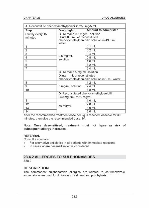
23.5
CHAPTER 23 DRUG ALLERGIES
A: Reconstitute phenoxymethylpenicillin 250 mg/5 mL Step Drug mg/mL Amount to administer Strictly every 15 minutes
B: To make 0.5 mg/mL solution Dilute 0.5 mL of reconstituted phenoxymethylpenicillin solution in 49.5 mL water.
1
0.5 mg/mL solution
0.1 mL 2 0.2 mL 3 0.4 mL 4 0.8 mL 5 1.6 mL 6 3.2 mL 7 6.4 mL C: To make 5 mg/mL solution
Dilute 1 mL of reconstituted phenoxymethylpenicillin solution in 9 mL water
8 5 mg/mL solution
1.2 mL 9 2.4 mL 10 4.8 mL D: Reconstituted phenoxymethylpenicillin
250 mg/5mL = 50 mg/mL 11
50 mg/mL
1.0 mL 12 2.0 mL 13 4.0 mL 14 8.0 mL
After the recommended treatment dose per kg is reached, observe for 30 minutes, then give the recommended dose, IV. Note: Once desensitised, treatment must not lapse as risk of subsequent allergy increases. REFERRAL Consult a specialist: » For alternative antibiotics in all patients with immediate reactions » In cases where desensitisation is considered. 23.4.2 ALLERGIES TO SULPHONAMIDES Z88.2 DESCRIPTION The commonest sulphonamide allergies are related to co-trimoxazole, especially when used for P. jirovecii treatment and prophylaxis.

23.6
CHAPTER 23 DRUG ALLERGIES Patients may present with: » a morbilliform or maculopapular rash only, usually within a few days of
starting treatment (most common presentation), » a rash with fever, » with/without organ dysfunction, usually within 1–2 weeks, » with SJS/TEN, or » rarely with an immediate hypersensitivity reaction. GENERAL AND SUPPORTIVE MEASURES Stop the sulphonamide-containing drug. MEDICINE TREATMENT For P. jirovecii pneumonia prophylaxis in patients with history of rash: Dapsone, oral, 2 mg/kg daily.
o Maximum dose: 100 mg (1 tablet) daily. o Note: Dapsone is a su lphone, not a sul phonamide, but there are
concerns regarding cross-reactivity with sulphonamide allergy. Avoid dapsone if ther e is a hist ory of anaphy laxis, SJS/TEN, or rash with systemic involvement.
If no alter native is available, consider desensitisation, as an inpat ient, in patients with a history of rash, with or without fever. Consult a specialist for desensitisation of patients with a history of anaphylaxis.
Perform only in an ICU or high care setting
Discontinue all ß-adrenergic antagonists. Ensure adequate IV access and immediate availability of all drugs for treatment of anaphylaxis. Monitor the patient with ECG (looking for arrhythmias) and spirometry (looking for a drop in FEV1).
Desensitisation is contra-indicated in patients with a history of SJS/TEN.
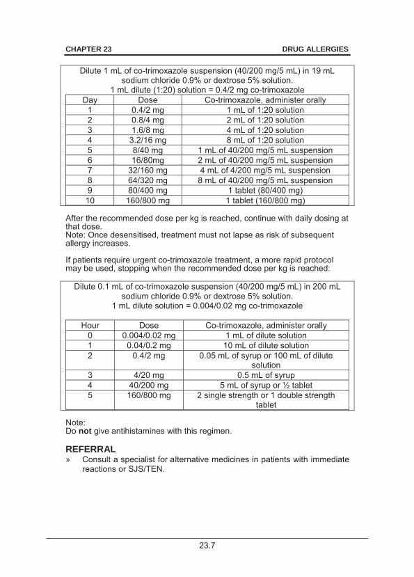
23.7
CHAPTER 23 DRUG ALLERGIES
Dilute 1 mL of co-trimoxazole suspension (40/200 mg/5 mL) in 19 mL sodium chloride 0.9% or dextrose 5% solution.
1 mL dilute (1:20) solution = 0.4/2 mg co-trimoxazole Day Dose Co-trimoxazole, administer orally
1 0.4/2 mg 1 mL of 1:20 solution 2 0.8/4 mg 2 mL of 1:20 solution 3 1.6/8 mg 4 mL of 1:20 solution 4 3.2/16 mg 8 mL of 1:20 solution 5 8/40 mg 1 mL of 40/200 mg/5 mL suspension 6 16/80mg 2 mL of 40/200 mg/5 mL suspension 7 32/160 mg 4 mL of 4/200 mg/5 mL suspension 8 64/320 mg 8 mL of 40/200 mg/5 mL suspension 9 80/400 mg 1 tablet (80/400 mg) 10 160/800 mg 1 tablet (160/800 mg)
After the recommended dose per kg is reached, continue with daily dosing at that dose. Note: Once desensitised, treatment must not lapse as risk of subsequent allergy increases. If patients require urgent co-trimoxazole treatment, a more rapid protocol may be used, stopping when the recommended dose per kg is reached:
Dilute 0.1 mL of co-trimoxazole suspension (40/200 mg/5 mL) in 200 mL sodium chloride 0.9% or dextrose 5% solution.
1 mL dilute solution = 0.004/0.02 mg co-trimoxazole
Hour Dose Co-trimoxazole, administer orally 0 0.004/0.02 mg 1 mL of dilute solution 1 0.04/0.2 mg 10 mL of dilute solution 2 0.4/2 mg 0.05 mL of syrup or 100 mL of dilute
solution 3 4/20 mg 0.5 mL of syrup 4 40/200 mg 5 mL of syrup or ½ tablet 5 160/800 mg 2 single strength or 1 double strength
tablet
Note: Do not give antihistamines with this regimen. REFERRAL » Consult a specialist for alternative medicines in patients with immediate
reactions or SJS/TEN.
xxxii
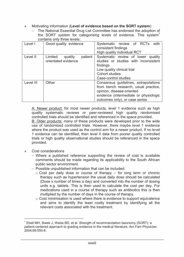
xxxiii
xxxiv
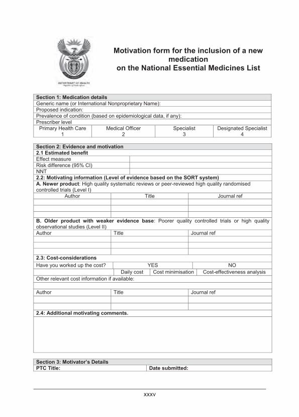
xxxv
xxxvi
xxxvii
xxxviii
xxxix
xl
xli
xlii
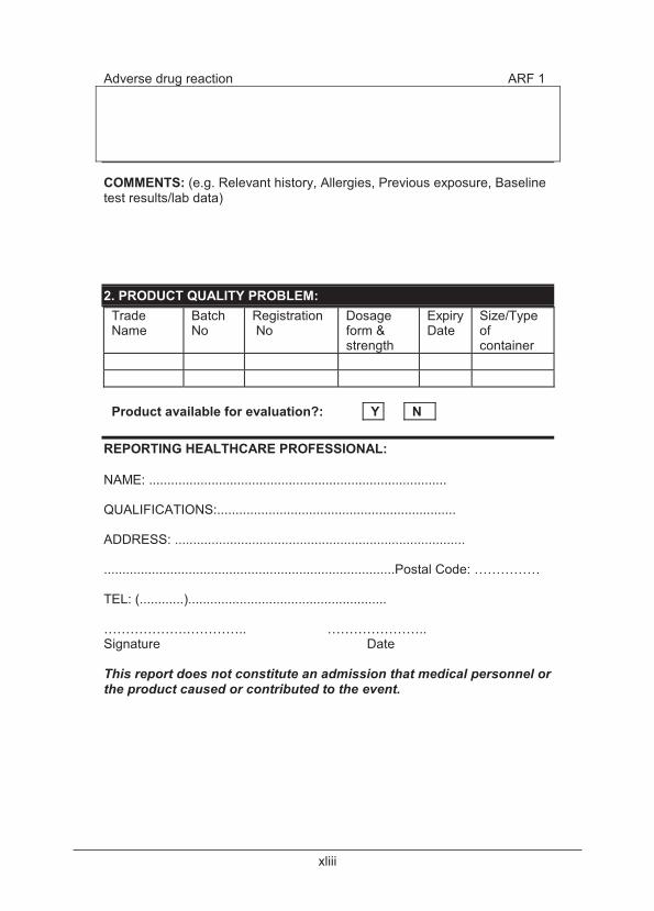
xliii
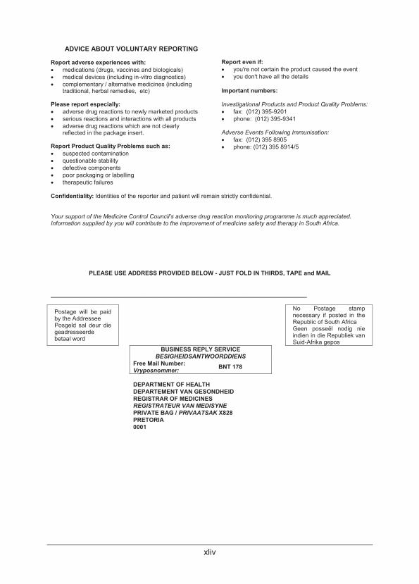
xliv

xlv
xlvi
xlvii
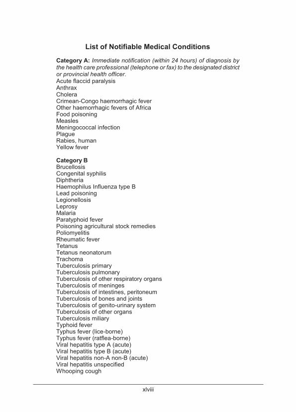
xlviii
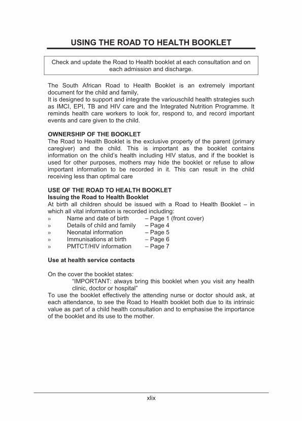
xlix
l
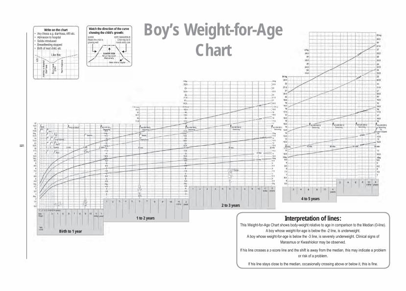
li
Boy’s Weight-for-Age Chart
Birth to 1 year
1 to 2 years
2 to 3 years4 to 5 years
Write on the chart Watch the direction of the curve showing the child’s growth:
GOOD D G O
Interpretation of lines:
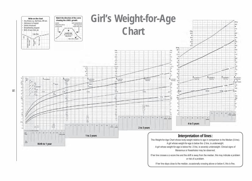
lii
Girl’s Weight-for-Age Chart
Birth to 1 year
1 to 2 years
2 to 3 years4 to 5 years
Interpretation of lines:
Write on the chart Watch the direction of the curve showing the child’s growth:
GOOD D G O

liii
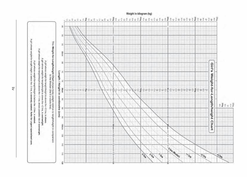
liv
lv
lvi
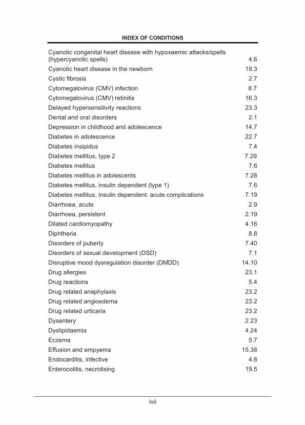
lvii
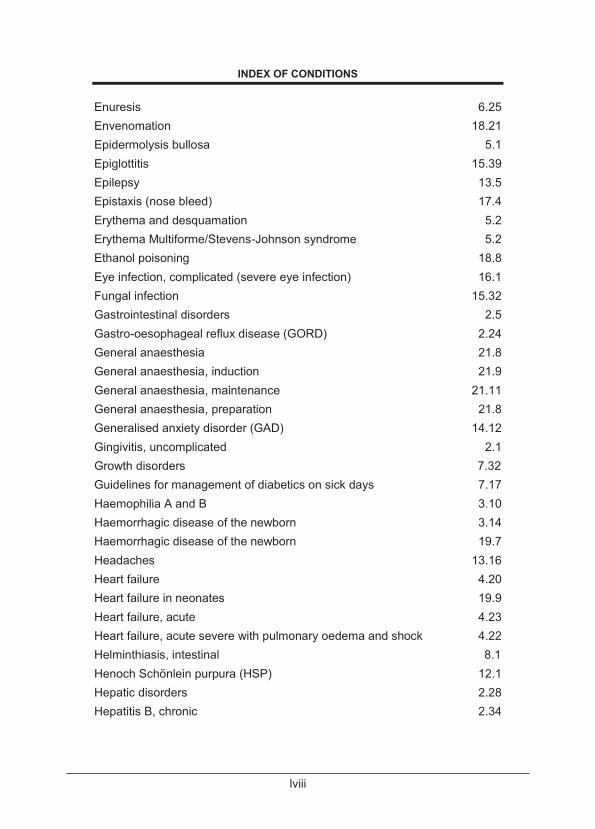
lviii
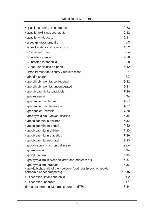
lix
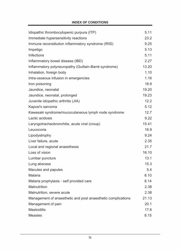
lx
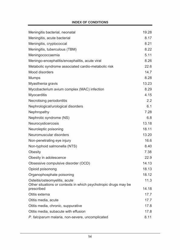
lxi
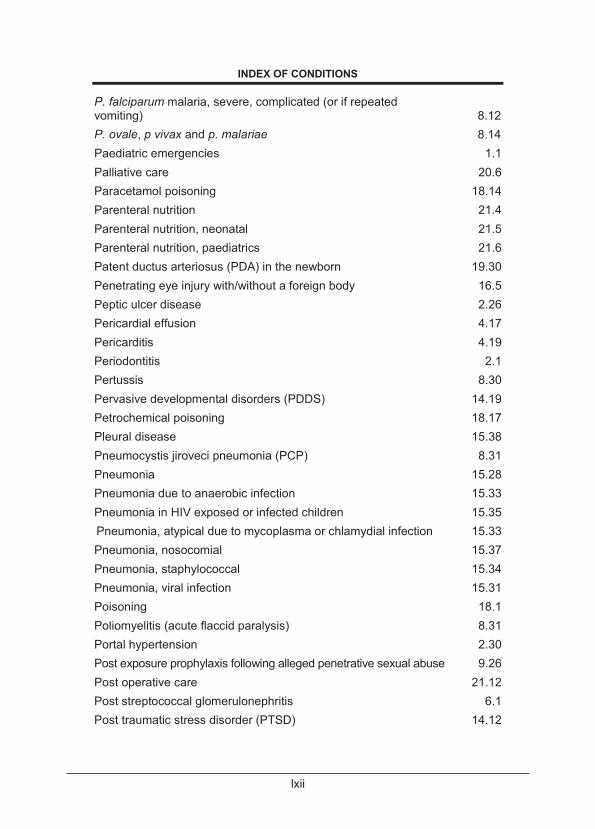
lxii
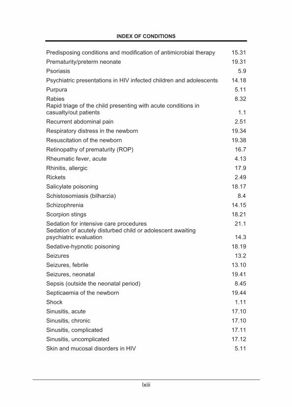
lxiii
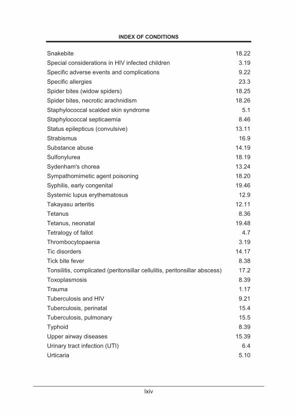
lxiv
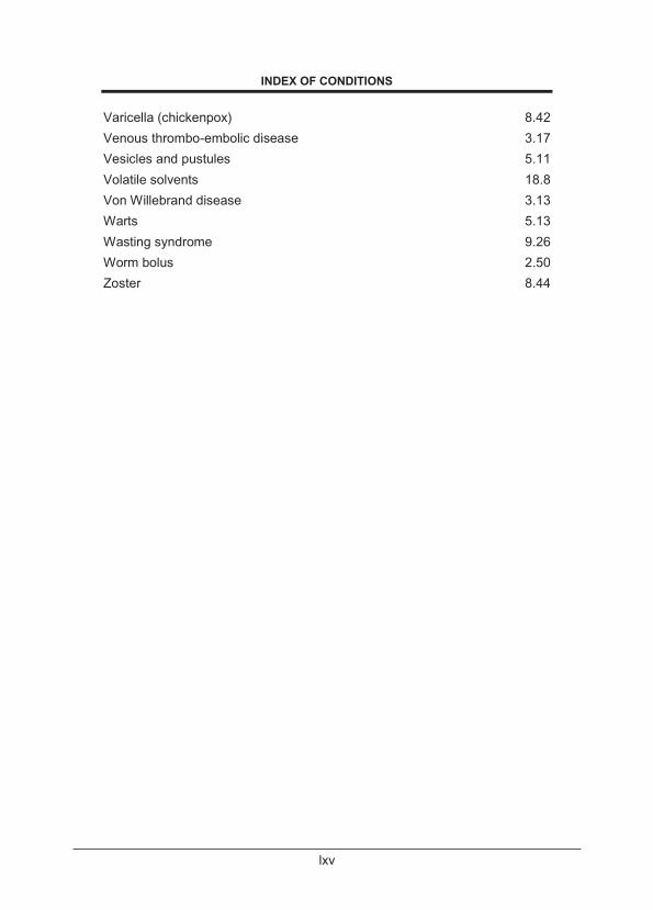
lxv
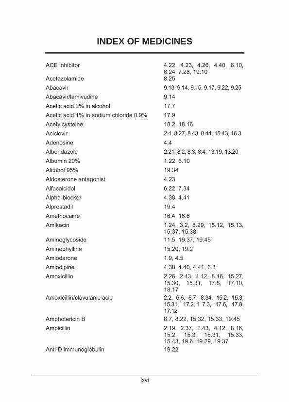
lxvi
INDEX OF MEDICINES
ACE inhibitor 4.22, 4.23, 4.26, 4.40, 6.10,
6.24, 7.28, 19.10 Acetazolamide 8.25 Abacavir 9.13, 9.14, 9.15, 9.17, 9.22, 9.25 Abacavir/lamivudine 9.14 Acetic acid 2% in alcohol 17.7 Acetic acid 1% in sodium chloride 0.9% 17.9 Acetylcysteine 18.2, 18.16 Aciclovir 2.4, 8.27, 8.43, 8.44, 15.43, 16.3 Adenosine 4.4 Albendazole 2.21, 8.2, 8.3, 8.4, 13.19, 13.20 Albumin 20% 1.22, 6.10 Alcohol 95% 19.34 Aldosterone antagonist 4.23 Alfacalcidol 6.22, 7.34 Alpha-blocker 4.38, 4.41 Alprostadil 19.4 Amethocaine 16.4, 16.6 Amikacin 1.24, 3.2, 8.29, 15.12, 15.13,
15.37, 15.38 Aminoglycoside 11.5, 19.37, 19.45 Aminophylline 15.20, 19.2 Amiodarone 1.9, 4.5 Amlodipine 4.38, 4.40, 4.41, 6.3 Amoxicillin 2.26, 2.43, 4.12, 8.16, 15.27,
15.30, 15.31, 17.8, 17.10, 18.17
Amoxicillin/clavulanic acid 2.2, 6.6, 6.7, 8.34, 15.2, 15.3, 15.31, 17.2, 1 7.3, 17.6, 17.8, 17.12
Amphotericin B 8.7, 8.22, 15.32, 15.33, 19.45 Ampicillin 2.19, 2.37, 2.43, 4.12, 8.16,
15.2, 15.3, 15.31, 15.33, 15.43, 19.6, 19.29, 19.37
Anti-D immunoglobulin 19.22
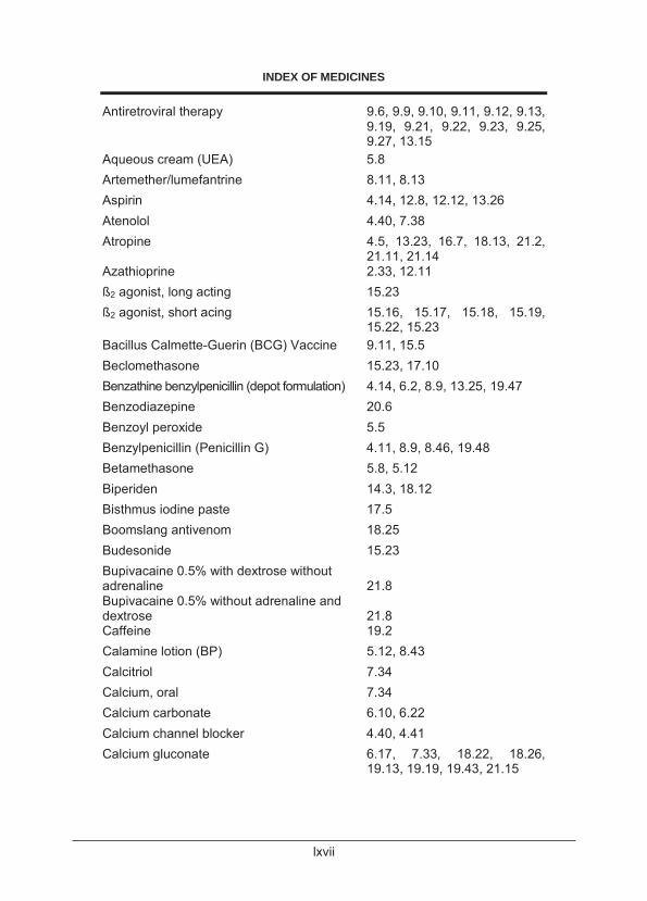
lxvii
INDEX OF MEDICINES
Antiretroviral therapy 9.6, 9.9, 9.10, 9.11, 9.12, 9.13, 9.19, 9.21, 9.22, 9.23, 9.25, 9.27, 13.15
Aqueous cream (UEA) 5.8 Artemether/lumefantrine 8.11, 8.13 Aspirin 4.14, 12.8, 12.12, 13.26 Atenolol 4.40, 7.38 Atropine 4.5, 13.23, 16.7, 18.13, 21.2,
21.11, 21.14 Azathioprine 2.33, 12.11 ß2 agonist, long acting 15.23 ß2 agonist, short acing 15.16, 15.17, 15.18, 15.19,
15.22, 15.23 Bacillus Calmette-Guerin (BCG) Vaccine 9.11, 15.5 Beclomethasone 15.23, 17.10 Benzathine benzylpenicillin (depot formulation) 4.14, 6.2, 8.9, 13.25, 19.47 Benzodiazepine 20.6 Benzoyl peroxide 5.5 Benzylpenicillin (Penicillin G) 4.11, 8.9, 8.46, 19.48 Betamethasone 5.8, 5.12 Biperiden 14.3, 18.12 Bisthmus iodine paste 17.5 Boomslang antivenom 18.25 Budesonide 15.23 Bupivacaine 0.5% with dextrose without adrenaline 21.8 Bupivacaine 0.5% without adrenaline and dextrose 21.8 Caffeine 19.2 Calamine lotion (BP) 5.12, 8.43 Calcitriol 7.34 Calcium, oral 7.34 Calcium carbonate 6.10, 6.22 Calcium channel blocker 4.40, 4.41 Calcium gluconate 6.17, 7.33, 18.22, 18.26,
19.13, 19.19, 19.43, 21.15
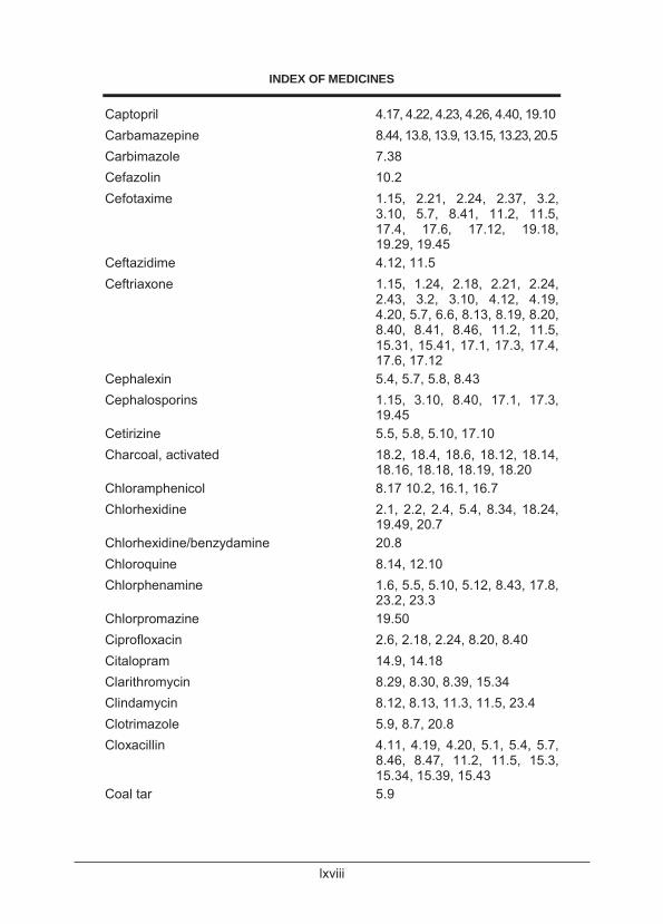
lxviii
INDEX OF MEDICINES
Captopril 4.17, 4.22, 4.23, 4.26, 4.40, 19.10 Carbamazepine 8.44, 13.8, 13.9, 13.15, 13.23, 20.5 Carbimazole 7.38 Cefazolin 10.2 Cefotaxime 1.15, 2.21, 2.24, 2.37, 3.2,
3.10, 5.7, 8.41, 11.2, 11.5, 17.4, 17.6, 17.12, 19.18, 19.29, 19.45
Ceftazidime 4.12, 11.5 Ceftriaxone 1.15, 1.24, 2.18, 2.21, 2.24,
2.43, 3.2, 3.10, 4.12, 4.19, 4.20, 5.7, 6.6, 8.13, 8.19, 8.20, 8.40, 8.41, 8.46, 11.2, 11.5, 15.31, 15.41, 17.1, 17.3, 17.4, 17.6, 17.12
Cephalexin 5.4, 5.7, 5.8, 8.43 Cephalosporins 1.15, 3.10, 8.40, 17.1, 17.3,
19.45 Cetirizine 5.5, 5.8, 5.10, 17.10 Charcoal, activated 18.2, 18.4, 18.6, 18.12, 18.14,
18.16, 18.18, 18.19, 18.20 Chloramphenicol 8.17 10.2, 16.1, 16.7 Chlorhexidine 2.1, 2.2, 2.4, 5.4, 8.34, 18.24,
19.49, 20.7 Chlorhexidine/benzydamine 20.8 Chloroquine 8.14, 12.10 Chlorphenamine 1.6, 5.5, 5.10, 5.12, 8.43, 17.8,
23.2, 23.3 Chlorpromazine 19.50 Ciprofloxacin 2.6, 2.18, 2.24, 8.20, 8.40 Citalopram 14.9, 14.18 Clarithromycin 8.29, 8.30, 8.39, 15.34 Clindamycin 8.12, 8.13, 11.3, 11.5, 23.4 Clotrimazole 5.9, 8.7, 20.8 Cloxacillin 4.11, 4.19, 4.20, 5.1, 5.4, 5.7,
8.46, 8.47, 11.2, 11.5, 15.3, 15.34, 15.39, 15.43
Coal tar 5.9
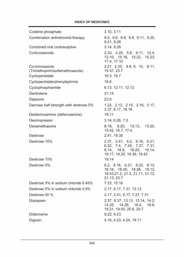
lxix
INDEX OF MEDICINES
Codeine phosphate 3.10, 3.11 Combination antiretroviral therapy 9.5, 9.6, 9.8, 9.9, 9.11, 9.20,
9.21, 9.26 Combined oral contraceptive 3.14, 9.28 Corticosteroids 2.33, 4.20, 5.8, 6.11, 12.4,
12.10, 15.18, 15.22, 15.23, 17.4, 17.10
Co-trimoxazole (Trimethoprim/sulfamethoxazole)
2.21 2.22, 9.8, 9. 10, 9.11, 15.37, 23.7
Cyclopentolate 16.3, 16.7 Cyclopentolate/phenylephrine 16.8 Cyclophosphamide 6.13, 12.11, 12.12 Dantrolene 21.14 Dapsone 23.6 Darrows half strength with dextrose 5% 1.22, 2.12, 2.15, 2.16, 2.17,
2.37, 6.17, 18.18 Desferrioxamine (deferoxamine) 18.11 Desmopressin 3.14, 6.26, 7.5 Dexamethasone 8.19, 8.20, 13.13, 13.20,
15.42, 16.7, 17.4 Dextrose 2.41, 19.35 Dextrose 10% 2.37, 2.41, 6.2, 6.16, 6.21,
6.22, 7.4, 7.24, 7.27, 7.31, 8.14, 18.9, 18.20, 19.14, 19.17, 19.20, 19.39, 19.43
Dextrose 15% 19.14 Dextrose 5% 6.2, 6.16, 6.21, 6.22, 8.13,
18.16, 18.25, 18.26, 19.13, 19.43,21.2, 21.3, 21.11, 21.12, 21.13, 23.7
Dextrose 5% in sodium chloride 0.45% 7.23, 15.16 Dextrose 5% in sodium chloride 0.9% 2.17, 6.17, 7.31, 13.13 Dextrose 50 % 2.17, 2.41, 6.17, 7.27, 7.31 Diazepam 2.37, 8.37, 13.13, 13.14, 14.2,
14.25, 14.26, 18.4, 18.8, 18.21, 19.50, 20.6, 20.7
Didanosine 9.22, 9.23 Digoxin 4.16, 4.23, 4.24, 19.11
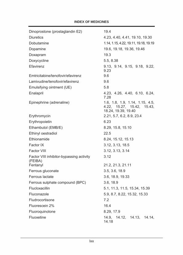
lxx
INDEX OF MEDICINES
Dinoprostone (prostaglandin E2) 19.4 Diuretics 4.23, 4.40, 4.41, 19.10, 19.30 Dobutamine 1.14, 1.15, 4.22, 19.11, 19.18, 19.19 Dopamine 19.6, 19.18, 19.36, 19.46 Doxapram 19.3 Doxycycline 5.5, 8.38 Efavirenz 9.13, 9.14, 9.15, 9.18, 9.22,
9.23 Emtricitabine/tenofovir/efavirenz 9.6 Lamivudine/tenofovir/efavirenz 9.6 Emulsifying ointment (UE) 5.8 Enalapril 4.23, 4.26, 4.40, 6.10, 6.24,
7.28 Epinephrine (adrenaline) 1.6, 1.8, 1.9, 1.14, 1.15, 4.5,
4.22, 15.27, 15.42, 15.43, 18.24, 19.39, 19.40
Erythromycin 2.21, 5.7, 6.2, 8.9, 23.4 Erythropoietin 6.23 Ethambutol (EMB/E) 8.29, 15.8, 15.10 Ethinyl oestradiol 22.5 Ethionamide 8.24, 15.12, 15.13 Factor IX 3.12, 3.13, 18.5 Factor VIII 3.12, 3.13, 3.14 Factor VIII inhibitor-bypassing activity (FEIBA)
3.12
Fentanyl 21.2, 21.3, 21.11 Ferrous gluconate 3.5, 3.6, 18.9 Ferrous lactate 3.6, 18.9, 19.33 Ferrous sulphate compound (BPC) 3.6, 18.9 Flucloxacillin 5.1, 11.3, 11.5, 15.34, 15.39 Fluconazole 5.9, 8.7, 8.22, 15.32, 15.33 Fludrocortisone 7.2 Fluorescein 2% 16.4 Fluoroquinolone 8.29, 17.9 Fluoxetine 14.9, 14.12, 14.13, 14.14,
14.18
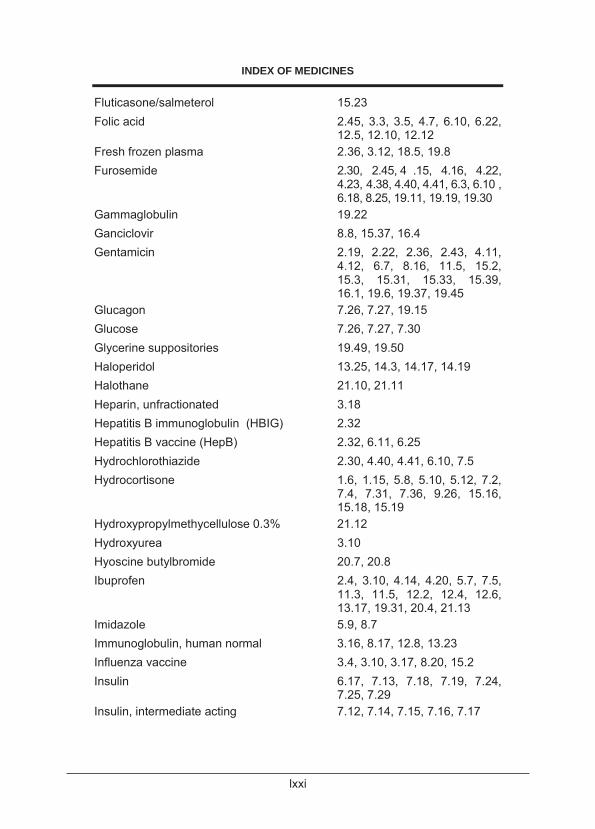
lxxi
INDEX OF MEDICINES
Fluticasone/salmeterol 15.23 Folic acid 2.45, 3.3, 3.5, 4.7, 6.10, 6.22,
12.5, 12.10, 12.12 Fresh frozen plasma 2.36, 3.12, 18.5, 19.8 Furosemide 2.30, 2.45, 4 .15, 4.16, 4.22,
4.23, 4.38, 4.40, 4.41, 6.3, 6.10 , 6.18, 8.25, 19.11, 19.19, 19.30
Gammaglobulin 19.22 Ganciclovir 8.8, 15.37, 16.4 Gentamicin 2.19, 2.22, 2.36, 2.43, 4.11,
4.12, 6.7, 8.16, 11.5, 15.2, 15.3, 15.31, 15.33, 15.39, 16.1, 19.6, 19.37, 19.45
Glucagon 7.26, 7.27, 19.15 Glucose 7.26, 7.27, 7.30 Glycerine suppositories 19.49, 19.50 Haloperidol 13.25, 14.3, 14.17, 14.19 Halothane 21.10, 21.11 Heparin, unfractionated 3.18 Hepatitis B immunoglobulin (HBIG) 2.32 Hepatitis B vaccine (HepB) 2.32, 6.11, 6.25 Hydrochlorothiazide 2.30, 4.40, 4.41, 6.10, 7.5 Hydrocortisone 1.6, 1.15, 5.8, 5.10, 5.12, 7.2,
7.4, 7.31, 7.36, 9.26, 15.16, 15.18, 15.19
Hydroxypropylmethycellulose 0.3% 21.12 Hydroxyurea 3.10 Hyoscine butylbromide 20.7, 20.8 Ibuprofen 2.4, 3.10, 4.14, 4.20, 5.7, 7.5,
11.3, 11.5, 12.2, 12.4, 12.6, 13.17, 19.31, 20.4, 21.13
Imidazole 5.9, 8.7 Immunoglobulin, human normal 3.16, 8.17, 12.8, 13.23 Influenza vaccine 3.4, 3.10, 3.17, 8.20, 15.2 Insulin 6.17, 7.13, 7.18, 7.19, 7.24,
7.25, 7.29 Insulin, intermediate acting 7.12, 7.14, 7.15, 7.16, 7.17
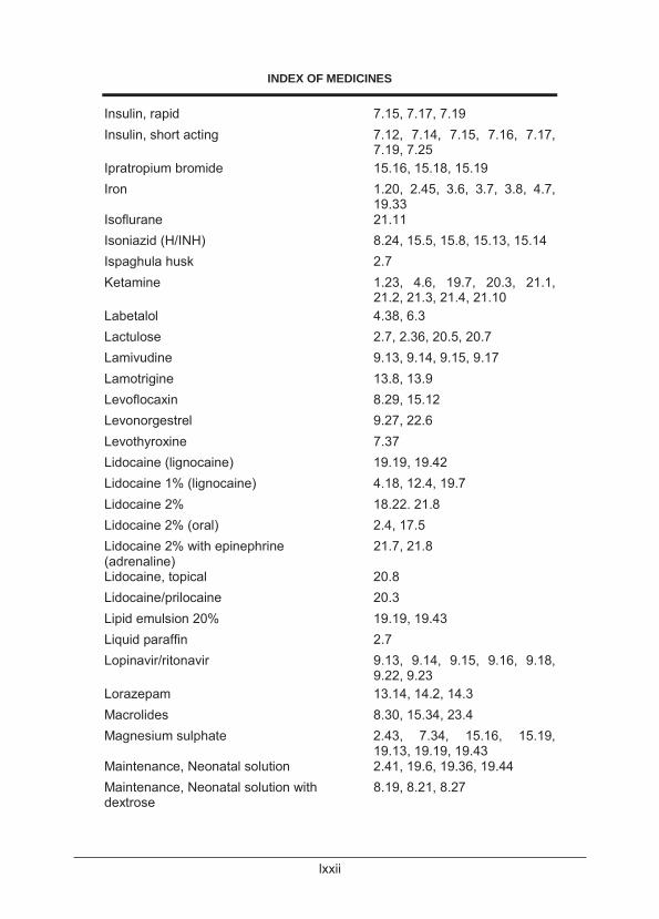
lxxii
INDEX OF MEDICINES
Insulin, rapid 7.15, 7.17, 7.19 Insulin, short acting 7.12, 7.14, 7.15, 7.16, 7.17,
7.19, 7.25 Ipratropium bromide 15.16, 15.18, 15.19 Iron 1.20, 2.45, 3.6, 3.7, 3.8, 4.7,
19.33 Isoflurane 21.11 Isoniazid (H/INH) 8.24, 15.5, 15.8, 15.13, 15.14 Ispaghula husk 2.7 Ketamine 1.23, 4.6, 19.7, 20.3, 21.1,
21.2, 21.3, 21.4, 21.10 Labetalol 4.38, 6.3 Lactulose 2.7, 2.36, 20.5, 20.7 Lamivudine 9.13, 9.14, 9.15, 9.17 Lamotrigine 13.8, 13.9 Levoflocaxin 8.29, 15.12 Levonorgestrel 9.27, 22.6 Levothyroxine 7.37 Lidocaine (lignocaine) 19.19, 19.42 Lidocaine 1% (lignocaine) 4.18, 12.4, 19.7 Lidocaine 2% 18.22. 21.8 Lidocaine 2% (oral) 2.4, 17.5 Lidocaine 2% with epinephrine (adrenaline)
21.7, 21.8
Lidocaine, topical 20.8 Lidocaine/prilocaine 20.3 Lipid emulsion 20% 19.19, 19.43 Liquid paraffin 2.7 Lopinavir/ritonavir 9.13, 9.14, 9.15, 9.16, 9.18,
9.22, 9.23 Lorazepam 13.14, 14.2, 14.3 Macrolides 8.30, 15.34, 23.4 Magnesium sulphate 2.43, 7.34, 15.16, 15.19,
19.13, 19.19, 19.43 Maintenance, Neonatal solution 2.41, 19.6, 19.36, 19.44 Maintenance, Neonatal solution with dextrose
8.19, 8.21, 8.27
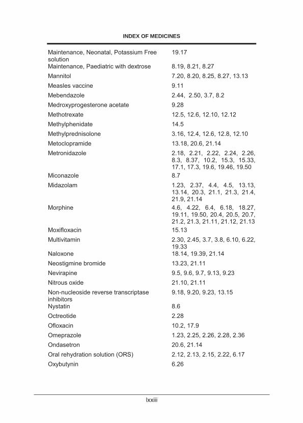
lxxiii
INDEX OF MEDICINES
Maintenance, Neonatal, Potassium Free solution
19.17
Maintenance, Paediatric with dextrose 8.19, 8.21, 8.27 Mannitol 7.20, 8.20, 8.25, 8.27, 13.13 Measles vaccine 9.11 Mebendazole 2.44, 2.50, 3.7, 8.2 Medroxyprogesterone acetate 9.28 Methotrexate 12.5, 12.6, 12.10, 12.12 Methylphenidate 14.5 Methylprednisolone 3.16, 12.4, 12.6, 12.8, 12.10 Metoclopramide 13.18, 20.6, 21.14 Metronidazole 2.18, 2.21, 2.22, 2.24, 2.26,
8.3, 8.37, 10.2, 15.3, 15.33, 17.1, 17.3, 19.6, 19.46, 19.50
Miconazole 8.7 Midazolam 1.23, 2.37, 4.4, 4.5, 13.13,
13.14, 20.3, 21.1, 21.3, 21.4, 21.9, 21.14
Morphine 4.6, 4.22, 6.4, 6.18, 18.27, 19.11, 19.50, 20.4, 20.5, 20.7, 21.2, 21.3, 21.11, 21.12, 21.13
Moxifloxacin 15.13 Multivitamin 2.30, 2.45, 3.7, 3.8, 6.10, 6.22,
19.33 Naloxone 18.14, 19.39, 21.14 Neostigmine bromide 13.23, 21.11 Nevirapine 9.5, 9.6, 9.7, 9.13, 9.23 Nitrous oxide 21.10, 21.11 Non-nucleoside reverse transcriptase inhibitors
9.18, 9.20, 9.23, 13.15
Nystatin 8.6 Octreotide 2.28 Ofloxacin 10.2, 17.9 Omeprazole 1.23, 2.25, 2.26, 2.28, 2.36 Ondasetron 20.6, 21.14 Oral rehydration solution (ORS) 2.12, 2.13, 2.15, 2.22, 6.17 Oxybutynin 6.26
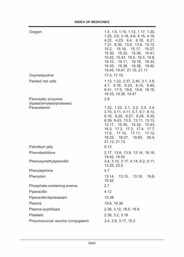
lxxiv
INDEX OF MEDICINES
Oxygen 1.5, 1.6, 1.10, 1.12, 1.17, 1.20, 1.25, 3.9, 3.18, 4.6, 4.15, 4.16, 4.22, 4.23, 6.4, 6.18, 6.21, 7.21, 8.30, 13.5, 13.8, 13.12, 15.2, 15.16, 15.17, 15.27, 15.30, 15.32, 15.36, 15.41, 15.42, 15.43, 19.2, 19.3, 19.8, 19.10, 19.11, 19.18, 19.33, 19.35, 19.36, 19.38, 19.40, 19.45, 19.47, 21.10, 21.11
Oxymetazoline 17.5, 17.10 Packed red cells 1.13, 1.22, 2.37, 2.45, 3.1, 3.9,
4.7, 6.18, 6.23, 8.14, 8.40, 8.41, 17.5, 19.6, 19.8, 19.18, 19.33, 19.36, 19.47
Pancreatic enzymes (lipase/amylase/protease)
2.8
Paracetamol 1.22, 1.23, 2.1, 2.2, 2.3, 2.4, 3.10, 3.11, 4.11, 5.7, 6.7, 8.13, 8.16, 8.20, 8.27, 8.28, 8.30, 8.39, 8.43, 13.5, 13.11, 13.13, 13.17, 15.30, 15.32, 15.43, 16.5, 17.2, 17.3, 17.4, 17.7, 17.8, 17.10, 17.11, 17.12, 18.22, 18.27, 19.50, 20.4, 21.12, 21.13
Petrollium jelly 5.13 Phenobarbitone 2.17, 13.8, 13.9, 13.14, 19.18,
19.42, 19.50 Phenoxymethylpenicillin 3.4, 3.10, 3.17, 4.14, 6.2, 6.11,
13.25, 23.5 Phenylephrine 4.7 Phenytoin 13.14, 13.15, 13.16, 19.8,
19.42 Phosphate-containing enema 2.7 Piperacillin 4.12 Piperacillin/tazobactam 15.38 Plasma 19.6, 19.36 Plasma lyophilised 2.36, 3.12, 18.5, 19.8 Platelets 2.36, 3.2, 3.16 Pneumococcal vaccine (conjugated) 3.4, 3.9, 3.17, 15.2
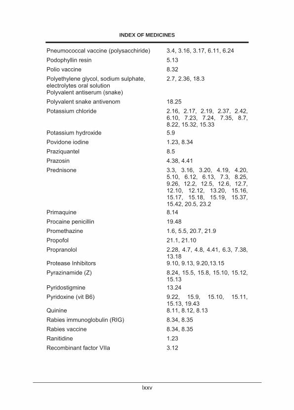
lxxv
INDEX OF MEDICINES
Pneumococcal vaccine (polysacchiride) 3.4, 3.16, 3.17, 6.11, 6.24 Podophyllin resin 5.13 Polio vaccine 8.32 Polyethylene glycol, sodium sulphate, electrolytes oral solution
2.7, 2.36, 18.3
Polyvalent antiserum (snake) Polyvalent snake antivenom 18.25 Potassium chloride 2.16, 2.17, 2.19, 2.37, 2.42,
6.10, 7.23, 7.24, 7.35, 8.7, 8.22, 15.32, 15.33
Potassium hydroxide 5.9 Povidone iodine 1.23, 8.34 Praziquantel 8.5 Prazosin 4.38, 4.41 Prednisone 3.3, 3.16, 3.20, 4.19, 4.20,
5.10, 6.12, 6.13, 7.3, 8.25, 9.26, 12.2, 12.5, 12.6, 12.7, 12.10, 12.12, 13.20, 15.16, 15.17, 15.18, 15.19, 15.37, 15.42, 20.5, 23.2
Primaquine 8.14 Procaine penicillin 19.48 Promethazine 1.6, 5.5, 20.7, 21.9 Propofol 21.1, 21.10 Propranolol 2.28, 4.7, 4.8, 4.41, 6.3, 7.38,
13.18 Protease Inhibitors 9.10, 9.13, 9.20,13.15 Pyrazinamide (Z) 8.24, 15.5, 15.8, 15.10, 15.12,
15.13 Pyridostigmine 13.24 Pyridoxine (vit B6) 9.22, 15.9, 15.10, 15.11,
15.13, 19.43 Quinine 8.11, 8.12, 8.13 Rabies immunoglobulin (RIG) 8.34, 8.35 Rabies vaccine 8.34, 8.35 Ranitidine 1.23 Recombinant factor VIIa 3.12
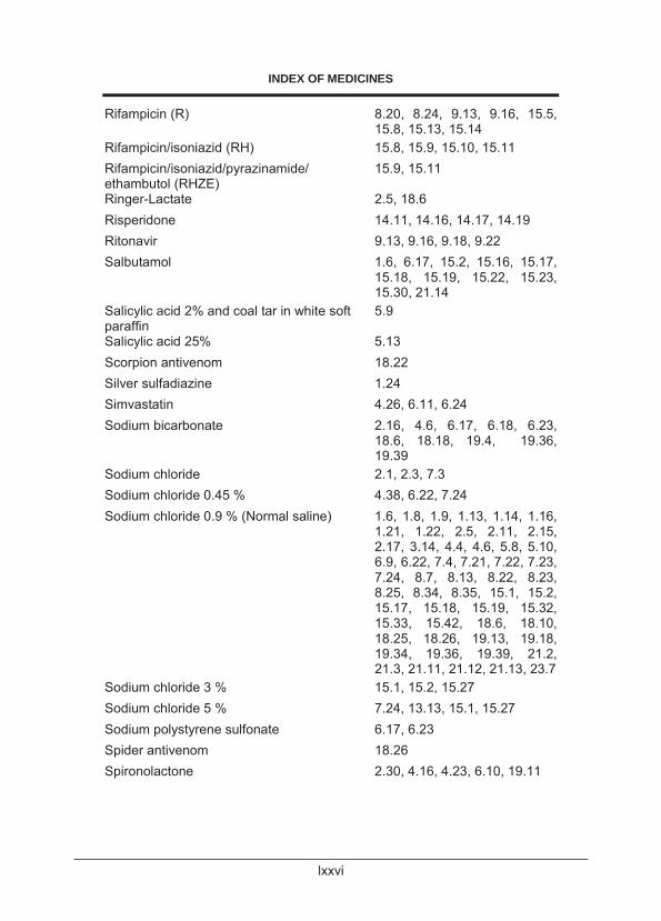
lxxvi
INDEX OF MEDICINES
Rifampicin (R) 8.20, 8.24, 9.13, 9.16, 15.5, 15.8, 15.13, 15.14
Rifampicin/isoniazid (RH) 15.8, 15.9, 15.10, 15.11 Rifampicin/isoniazid/pyrazinamide/ ethambutol (RHZE)
15.9, 15.11
Ringer-Lactate 2.5, 18.6 Risperidone 14.11, 14.16, 14.17, 14.19 Ritonavir 9.13, 9.16, 9.18, 9.22 Salbutamol 1.6, 6.17, 15.2, 15.16, 15.17,
15.18, 15.19, 15.22, 15.23, 15.30, 21.14
Salicylic acid 2% and coal tar in white soft paraffin
5.9
Salicylic acid 25% 5.13 Scorpion antivenom 18.22 Silver sulfadiazine 1.24 Simvastatin 4.26, 6.11, 6.24 Sodium bicarbonate 2.16, 4.6, 6.17, 6.18, 6.23,
18.6, 18.18, 19.4, 19.36, 19.39
Sodium chloride 2.1, 2.3, 7.3 Sodium chloride 0.45 % 4.38, 6.22, 7.24 Sodium chloride 0.9 % (Normal saline) 1.6, 1.8, 1.9, 1.13, 1.14, 1.16,
1.21, 1.22, 2.5, 2.11, 2.15, 2.17, 3.14, 4.4, 4.6, 5.8, 5.10, 6.9, 6.22, 7.4, 7.21, 7.22, 7.23, 7.24, 8.7, 8.13, 8.22, 8.23, 8.25, 8.34, 8.35, 15.1, 15.2, 15.17, 15.18, 15.19, 15.32, 15.33, 15.42, 18.6, 18.10, 18.25, 18.26, 19.13, 19.18, 19.34, 19.36, 19.39, 21.2, 21.3, 21.11, 21.12, 21.13, 23.7
Sodium chloride 3 % 15.1, 15.2, 15.27 Sodium chloride 5 % 7.24, 13.13, 15.1, 15.27 Sodium polystyrene sulfonate 6.17, 6.23 Spider antivenom 18.26 Spironolactone 2.30, 4.16, 4.23, 6.10, 19.11
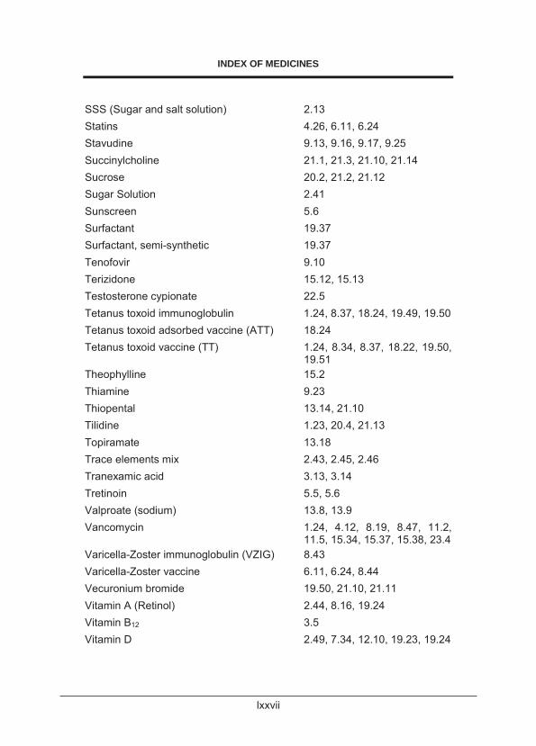
lxxvii
INDEX OF MEDICINES
SSS (Sugar and salt solution) 2.13 Statins 4.26, 6.11, 6.24 Stavudine 9.13, 9.16, 9.17, 9.25 Succinylcholine 21.1, 21.3, 21.10, 21.14 Sucrose 20.2, 21.2, 21.12 Sugar Solution 2.41 Sunscreen 5.6 Surfactant 19.37 Surfactant, semi-synthetic 19.37 Tenofovir 9.10 Terizidone 15.12, 15.13 Testosterone cypionate 22.5 Tetanus toxoid immunoglobulin 1.24, 8.37, 18.24, 19.49, 19.50 Tetanus toxoid adsorbed vaccine (ATT) 18.24 Tetanus toxoid vaccine (TT) 1.24, 8.34, 8.37, 18.22, 19.50,
19.51 Theophylline 15.2 Thiamine 9.23 Thiopental 13.14, 21.10 Tilidine 1.23, 20.4, 21.13 Topiramate 13.18 Trace elements mix 2.43, 2.45, 2.46 Tranexamic acid 3.13, 3.14 Tretinoin 5.5, 5.6 Valproate (sodium) 13.8, 13.9 Vancomycin 1.24, 4.12, 8.19, 8.47, 11.2,
11.5, 15.34, 15.37, 15.38, 23.4 Varicella-Zoster immunoglobulin (VZIG) 8.43 Varicella-Zoster vaccine 6.11, 6.24, 8.44 Vecuronium bromide 19.50, 21.10, 21.11 Vitamin A (Retinol) 2.44, 8.16, 19.24 Vitamin B12 3.5 Vitamin D 2.49, 7.34, 12.10, 19.23, 19.24
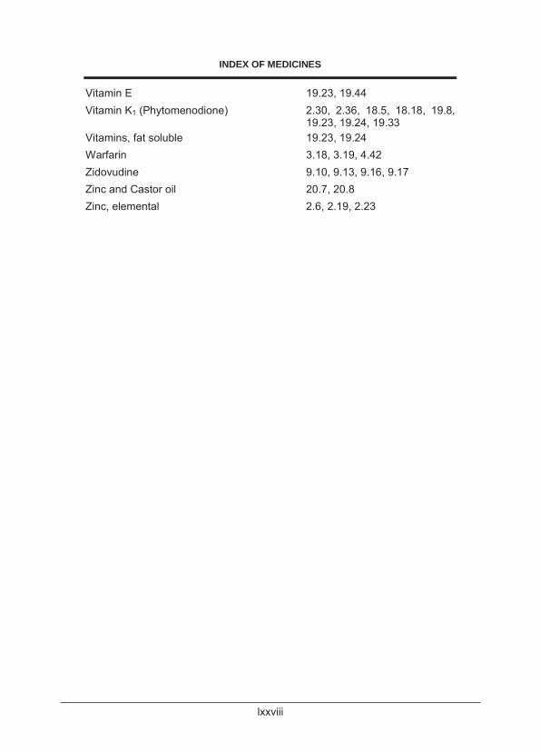
lxxviii
INDEX OF MEDICINES
Vitamin E 19.23, 19.44 Vitamin K1 (Phytomenodione) 2.30, 2.36, 18.5, 18.18, 19.8,
19.23, 19.24, 19.33 Vitamins, fat soluble 19.23, 19.24 Warfarin 3.18, 3.19, 4.42 Zidovudine 9.10, 9.13, 9.16, 9.17 Zinc and Castor oil 20.7, 20.8 Zinc, elemental 2.6, 2.19, 2.23
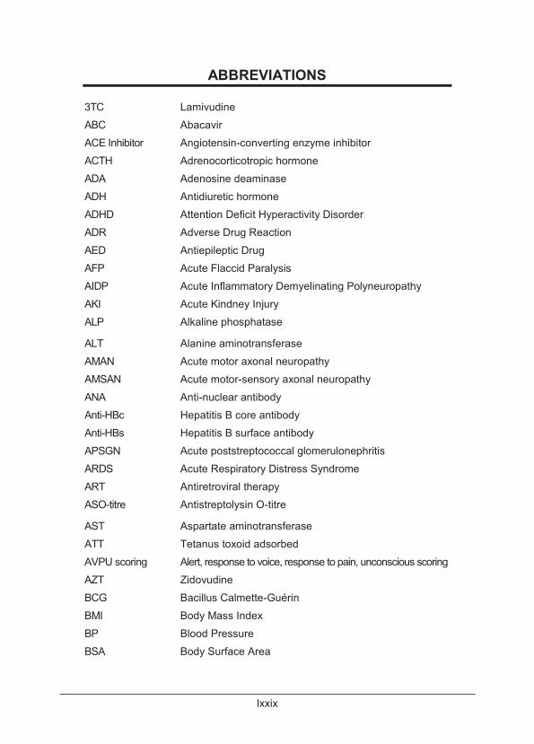
lxxix
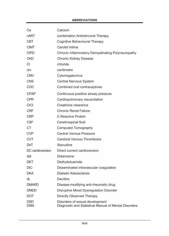
lxxx
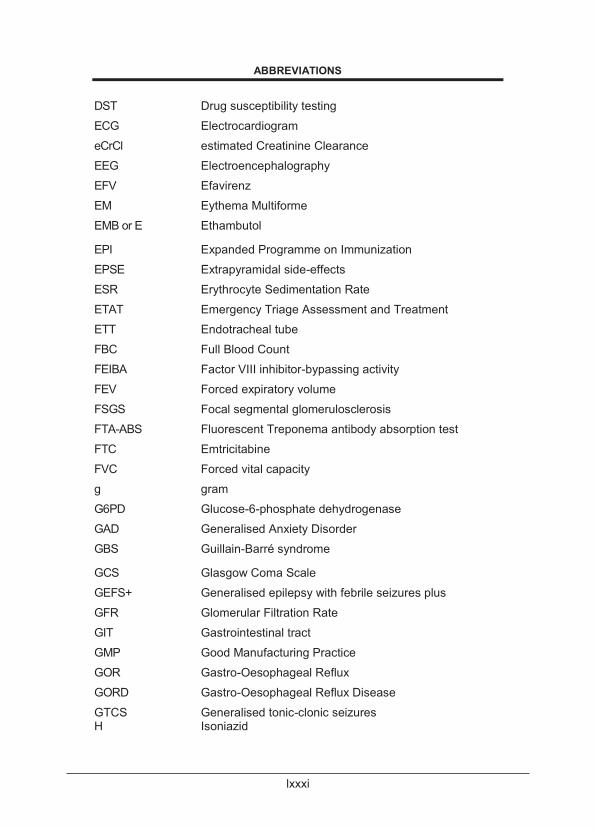
lxxxi
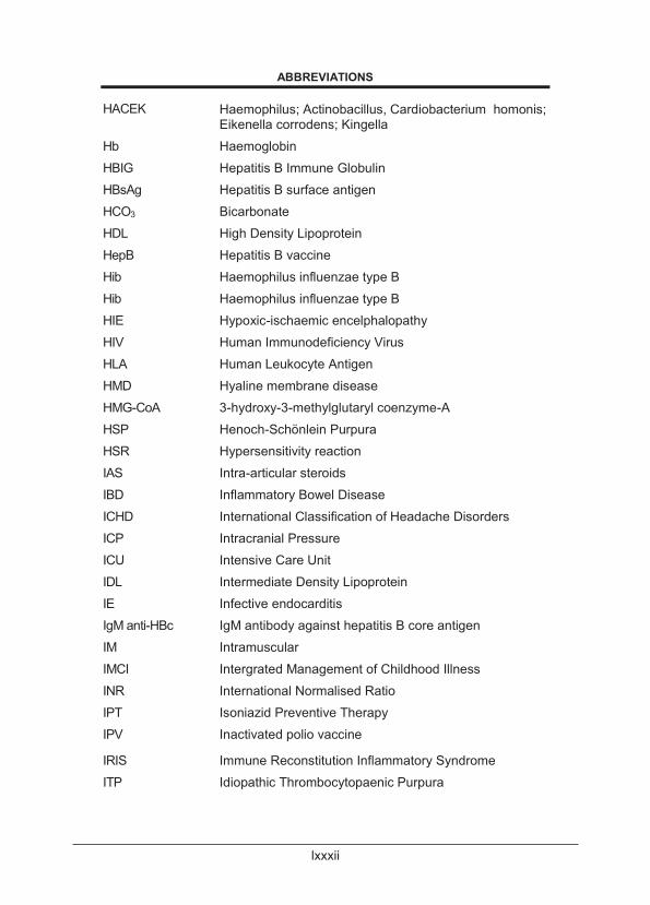
lxxxii
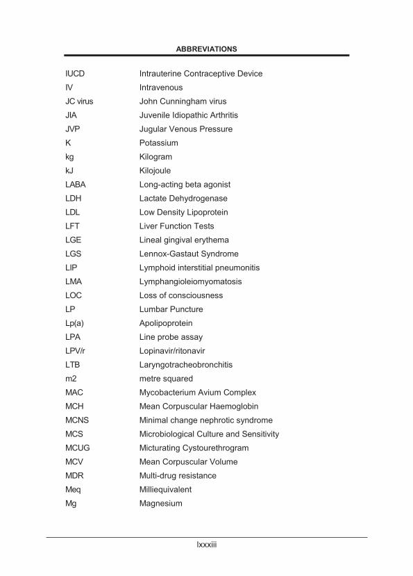
lxxxiii
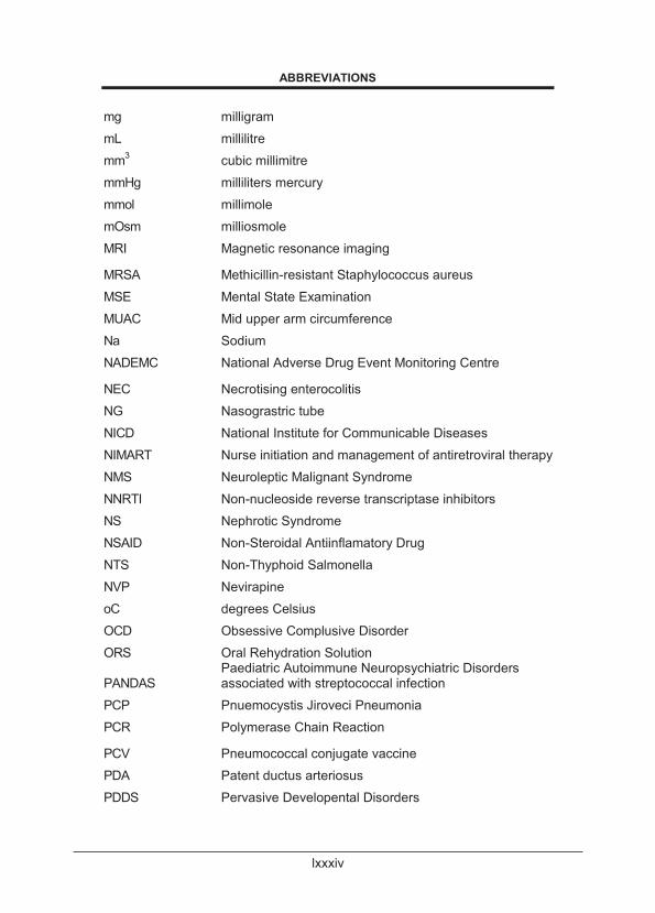
lxxxiv
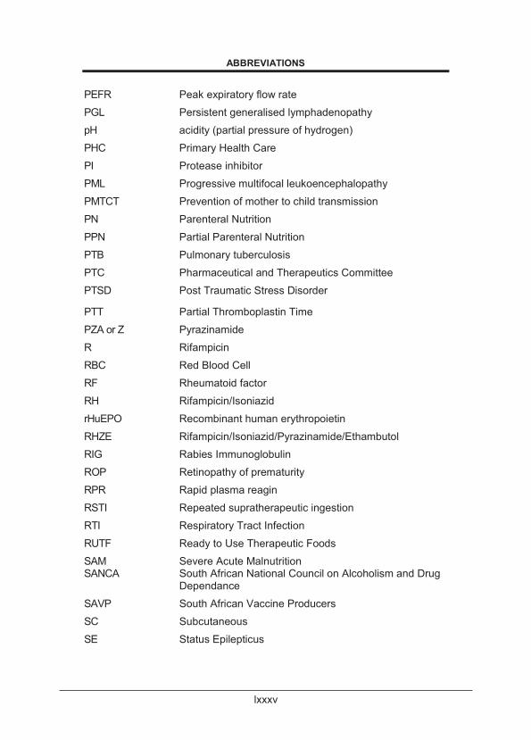
lxxxv
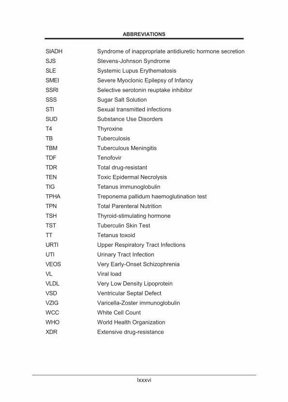
lxxxvi

lxxxvii