Hormonal status affects the progression of STZ-induced diabetes and diabetic renal damage in the VCD...
28
1 F-00022-2007.R1 February 22, 2007 Hormonal status affects the progression of STZ-induced diabetes and diabetic renal damage in the VCD mouse model of menopause Maggie Keck, Melissa J. Romero-Aleshire, Qi Cai, Patricia B. Hoyer, Heddwen L. Brooks 1 Department of Physiology, College of Medicine, University of Arizona, Tucson, AZ 85724 Running Title: Diabetic kidney damage in the VCD mouse model of menopause 1: To whom correspondence should be addressed: Heddwen Brooks, PhD Dept. of Physiology, College of Medicine 1501 N Campbell Ave, University of Arizona Tucson, AZ 85724-5051 Phone: (520) 626-7702 Fax: (520) 626-2382 (email: [email protected] ) Page 1 of 28 Articles in PresS. Am J Physiol Renal Physiol (March 27, 2007). doi:10.1152/ajprenal.00022.2007 Copyright © 2007 by the American Physiological Society.
-
Upload
independent -
Category
Documents
-
view
2 -
download
0
Transcript of Hormonal status affects the progression of STZ-induced diabetes and diabetic renal damage in the VCD...

1
F-00022-2007.R1
February 22, 2007
Hormonal status affects the progression of STZ-induced diabetes and diabetic renal damage in the
VCD mouse model of menopause
Maggie Keck, Melissa J. Romero-Aleshire, Qi Cai, Patricia B. Hoyer, Heddwen L. Brooks1
Department of Physiology, College of Medicine, University of Arizona, Tucson, AZ 85724
Running Title: Diabetic kidney damage in the VCD mouse model of menopause
1: To whom correspondence should be addressed:
Heddwen Brooks, PhD
Dept. of Physiology, College of Medicine
1501 N Campbell Ave, University of Arizona
Tucson, AZ 85724-5051
Phone: (520) 626-7702
Fax: (520) 626-2382
(email: [email protected])
Page 1 of 28Articles in PresS. Am J Physiol Renal Physiol (March 27, 2007). doi:10.1152/ajprenal.00022.2007
Copyright © 2007 by the American Physiological Society.

2
ABSTRACT:
Changes in the estrogen/testosterone balance at menopause may negatively influence the
development of diabetic kidney disease. Furthermore, recent studies suggest that changes in hormone
levels during perimenopause may influence disease development. Injection of 4-vinylcyclohexene
diepoxide (VCD) in B6C3F1 mice induces gradual ovarian failure, preserving both the perimenopausal
(peri-ovarian failure) and menopausal (post-ovarian failure) periods. To address the impact of the
transition into menopause on the development of diabetes and diabetic kidney damage, we used STZ-
induced diabetes in the VCD model of menopause. After six weeks of STZ-induced diabetes, blood
glucose was significantly increased in post-ovarian failure (post-OF) diabetic mice compared to cycling
diabetic mice. In peri-ovarian failure (peri-OF) diabetic mice blood glucose levels trended higher but
were not significantly different from cycling diabetic mice, suggesting a continuum of worsening blood
glucose across the menopausal transition. Cell proliferation, an early marker of damage in the kidney,
was increased in post-OF diabetic mice compared to cycling diabetic mice, as measured by PCNA
immunohistochemistry. In post-OF diabetic mice mRNA abundance of early growth response-1 (Egr-1),
collagen-4α1 and matrix metalloproteinase-9 were increased and 3β-hydroxysteroid dehydrogenase 4 (3β-
HSD4) and transforming growth factor-β2 (TGFβ2) were decreased compared to cycling diabetic mice. In
peri-OF diabetic mice mRNA abundance of Egr-1 and 3β-HSD4 were increased, and TGFβ2 was
decreased compared to cycling diabetic mice. This study highlights the importance and utility of the
VCD model of menopause, as it provides a physiologically relevant system for determining the impact of
the menopausal transition on diabetes and diabetic kidney damage.
Keywords: Diabetes, 3β-HSD4, perimenopause, real-time PCR, estrogen
Page 2 of 28

3
INTRODUCTION: Diabetes is one of the most prevalent and costly diseases afflicting developed countries, with
estimates placing the current global cost of diabetes at $150 billion a year (1). Approximately one third of
all diabetics die of end stage renal disease (33) due to progressive renal damage and hypertension.
17β-estradiol is considered protective against the development and progression of many diseases,
including cardiovascular (9) and renal disease (34). Premenopausal women have slower rates of
progression of non-diabetic renal disease than age-matched men (23), a difference that seems to disappear
after menopause (3). The impact of estrogen on diabetic renal disease is less clear, however. Several
studies demonstrate a decreased incidence of diabetic renal disease in women, but others have found no
difference between men and women (34).
The 5-10 years preceding menopause is termed perimenopause, and during this time estrogen
levels fluctuate, with periods of low estrogen interspersed with periods of very high estrogen (31). The
periods of low estrogen become more frequent as a woman approaches menopause until circulating levels
of 17β-estradiol drop to continuously very low levels at menopause (31). Following menopause the
ovaries secrete androgens (36), though their contribution to the overall hormonal state of the body is
debated (4). Recent studies find that some risk factors for estrogen-influenced diseases begin to develop
during perimenopause (7), (19). This suggests that study of the perimenopausal period may lead to new
understanding of and preventive therapies for estrogen-influenced diseases (37).
The most commonly used animal model of menopause is surgical removal of the ovaries
(ovariectomy). With this model there is no period analogous to perimenopause, and any post-menopausal
androgen secretion by the ovaries is lost. High androgen levels have been implicated in the development
of renal disease (28), (22), thus the lack of postmenopausal ovaries in the ovariectomy model of
menopause, combined with the lack of a perimenopausal period, limits the study of diabetic kidney
disease across the menopausal transition.
Page 3 of 28
4
Here, a new chemical model of menopause, in which repeated daily injections of 4-
vinylcyclohexene diepoxide (VCD) induce gradual ovarian failure in mice (13), was used. In this model,
the period preceding full ovarian failure mimics the perimenopausal period (termed peri-ovarian failure in
this study): cycles gradually become longer and more irregular, estrogen levels decrease, and follicle
stimulating hormone levels increase. Following ovarian failure (termed post-ovarian failure in this study)
the ovaries secrete androgens (20), similar to postmenopausal human ovaries. This model provides a new
and unique opportunity to study the development and progression of diabetic kidney disease across the
menopausal transition.
In this study, the VCD model of menopause was combined with the streptozotocin (STZ) model of
type 1 diabetes. STZ induced diabetes was begun during the period analogous to perimenopause in
humans, (i.e. during peri-ovarian failure) or during the period analogous to post-menopause in humans
(i.e. post-ovarian failure). We hypothesized that changes in hormone levels across the menopausal
transition would negatively affect the development of diabetes and diabetic kidney damage. The data
presented here support this hypothesis and further suggest that the perimenopausal period may be
important in the development and progression of diabetic renal disease. This study highlights the utility
of the VCD mouse model of menopause in the study of diabetic renal disease across the menopausal
transition.
Page 4 of 28
5
METHODS:
Animals: 28 day old female B6C3F1 mice were used in this study. Animals were housed in standard
cages in the animal facility of the Arizona Health Sciences Center under National Institutes of Health
guidelines and had ad libitum access to regular food and water. All protocols were approved by the
Institutional Animal Care and Use Committee at the University of Arizona.
Induction of ovarian failure: 4-vinylcyclohexene diepoxide (Sigma, V3630) was administered via
intraperitoneal (i.p.) injection at a dose of 160 mg/kg body weight using a dosing standard of 2.5 mL/kg
body weight for 15 consecutive days to induce gradual ovarian failure (20), (13). Sesame oil was used as
vehicle control. Progression into ovarian failure was monitored by daily vaginal cytology. Ten
consecutive days of diestrus was considered indicative of ovarian failure (13).
Induction of diabetes: Diabetes was induced by i.p injection of streptozotocin (STZ) (Sigma, S0130)
at a dose of 75 mg/kg body weight using a dosing standard of 0.2 mL/22 g body weight to 4-hour fasted
mice for 3 consecutive days. Mice were separated into two study groups: in the first group STZ was
administered during peri-ovarian failure, 3 days after the end of VCD dosing (Figure 1: Perimenopausal
Study). In the second group STZ was administered post-ovarian failure, two weeks after a mouse entered
ovarian failure (Figure 1: Menopausal Study). In both studies cycling mice (not VCD-injected) were
injected with STZ on the same day. Citrate buffer was used as vehicle control. Urine glucose was
monitored as an indication of the onset of diabetes using standard urine glucose test strips (VWR, 44561-
110). Animals were weighed regularly and visually monitored for signs of ill health. Mice were
sacrificed 6 weeks after STZ injection. Mice were fasted for 4 hours prior to sacrifice, at which point
blood was collected, and blood glucose was measured using the CardioCheck PA Blood Testing Device
(HealthCheck Systems, 2568) with CardioCheck Glucose Test Strips (HealthCheck Systems, 2556).
RNA isolation: Kidneys were separated into cortex and medulla, and RNA was isolated from renal
cortex using the Qiagen RNeasy Mini Kit (74104) according to the manufacturer’s protocol for isolation
Page 5 of 28

6
from tissue. DNA contamination was eliminated during the isolation procedure with a 15 min DNAse
incubation (Qiagen, 79254). RNA was quantified using a Nanodrop® ND1000 spectrophotometer
(Wilmington, DE).
Real-time PCR: Real-time PCR was performed as previously described (21). Briefly, 2.5 µg of RNA
were reverse transcribed with the MLV-Reverse Transcriptase enzyme (Invitrogen, 28025013), and the
resulting cDNA was diluted 1:25 to an approximate final concentration of 8 ng/ul. Each real-time PCR
reaction contained 5 µL SYBR Green master mix (Stratagene, 600581), 1 µL water, 2 µL diluted cDNA,
and 5 pmol each of forward and reverse primer in a total volume of 10 µL. Each reaction was performed
in triplicate at 95°C for 5 min; then 95°C for 15 sec and 60°C for 30 sec for 40 cycles. The RotorGene
RG3000 (Corbett Research, San Francisco, CA) sequence detection system was used. Primers were
designed to the 3´ end of genes of interest using the Primer3 software (30) and are listed in supplemental
data A. Ct values were used to calculate the expression levels of genes of interest relative to the
expression of β-actin mRNA, measured in parallel samples. Analysis was performed as described (11),
and results are presented as mean fold change on a base 2 logarithmic scale.
Immunohistochemistry: Kidneys were fixed in 4% paraformaldehyde overnight, embedded in
paraffin, and sectioned (4µm) by the University of Arizona Pathology Laboratory. Sections were
deparaffinized with xylene and rehydrated in graded ethanol. Endogenous horseradish peroxidase activity
was blocked with 0.3% H2O2 in methanol for 30 min. Antigen retrieval was performed by boiling
sections in 10 mM citrate buffer for 10 min. Nonspecific binding was blocked with 5% goat serum in 1%
BSA with 0.05% Tween-20. Sections were incubated at 4°C overnight in anti-proliferating cell nuclear
antigen (PCNA) (1:100 dilution; Santa Cruz, SC-25280), followed by biotinylated secondary antibody
(1:200 dilution; Zymed, 81-6740) for 30 min at 37°C and horseradish peroxidase-conjugated streptavidin
(1:200 dilution; Zymed, 43-4323) for 30 min at 37°C. Labeling was visualized with chromogen
diaminobenzidine (Zymed, 00-2014), and sections were counterstained with hematoxylin (Zymed, 00-
Page 6 of 28
7
8011). Coverslips were mounted with Permount mounting solution (Fisher, SP15-100). Positively
stained nuclei were counted as a proportion of total nuclei within each of 5 non-overlapping fields of view
for each section. Each field of view contained approximately 200 cells.
Statistics: Data were analyzed using Student’s t-test or one-way ANOVA followed by Student-
Neumann Keuls post-hoc test to identify differences between groups. In all tests, P<0.05 was considered
significant. Results are presented as mean ± standard deviation (SD) or standard error (SEM).
Page 7 of 28

8
RESULTS: Initiation of menopause:
To study the effect of menopause on the development of diabetes and diabetic kidney damage we
combined the STZ model of type 1 diabetes with the VCD model of menopause in B6C3F1 female mice
(20). Repeated injections of VCD induce gradual ovarian failure in mice, which is analogous to
menopause in humans. In this study, diabetes was induced using STZ two weeks after a mouse entered
ovarian failure, as shown in Figure 1. An age-matched cycling mouse (not VCD-injected) was injected
with STZ at the same time (Table 1). Mice were sacrificed 6 weeks after STZ dosing. Age-matched non-
diabetic post-ovarian failure (post-OF) and non-diabetic cycling mice were sacrificed as controls (see
Table 1).
Blood glucose in post-ovarian failure diabetic mice:
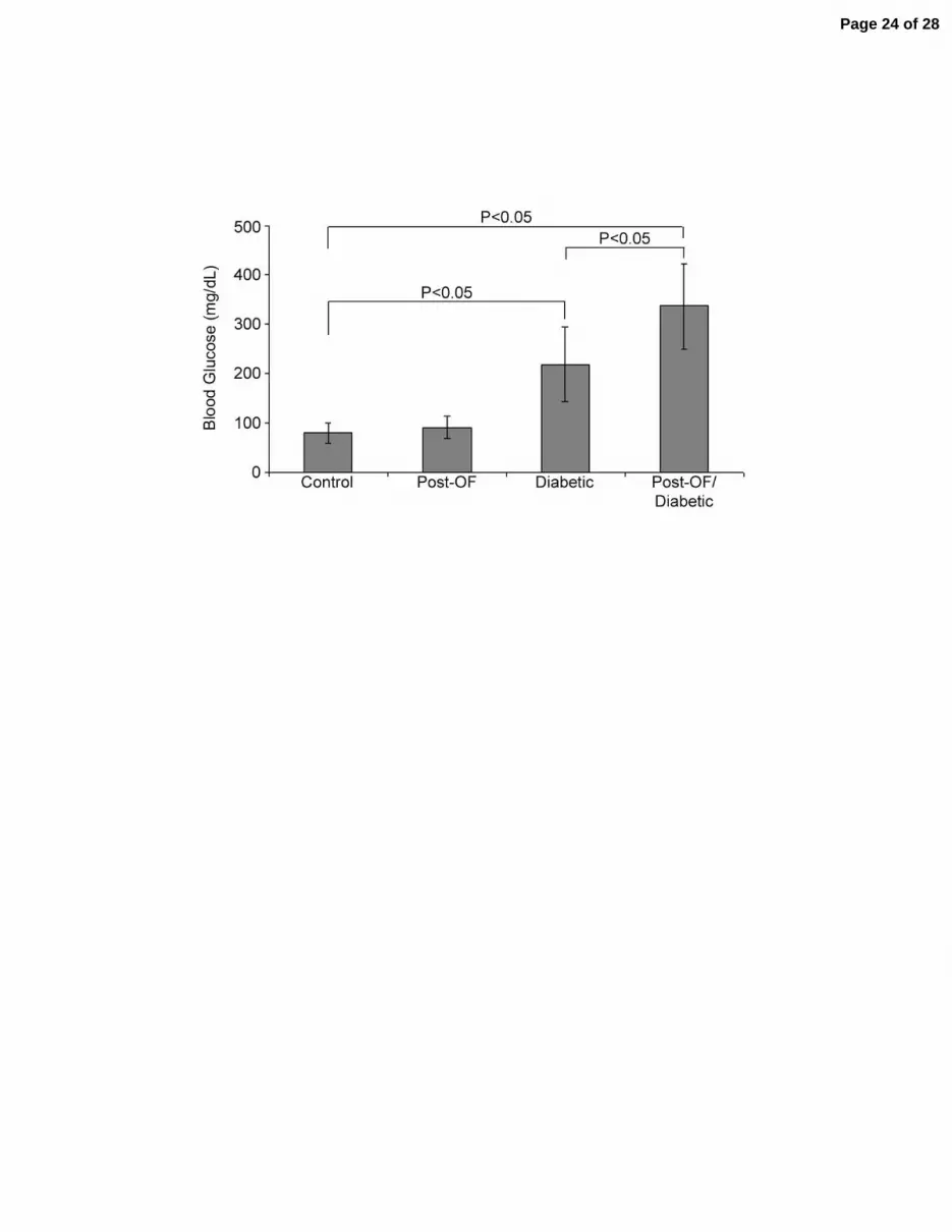
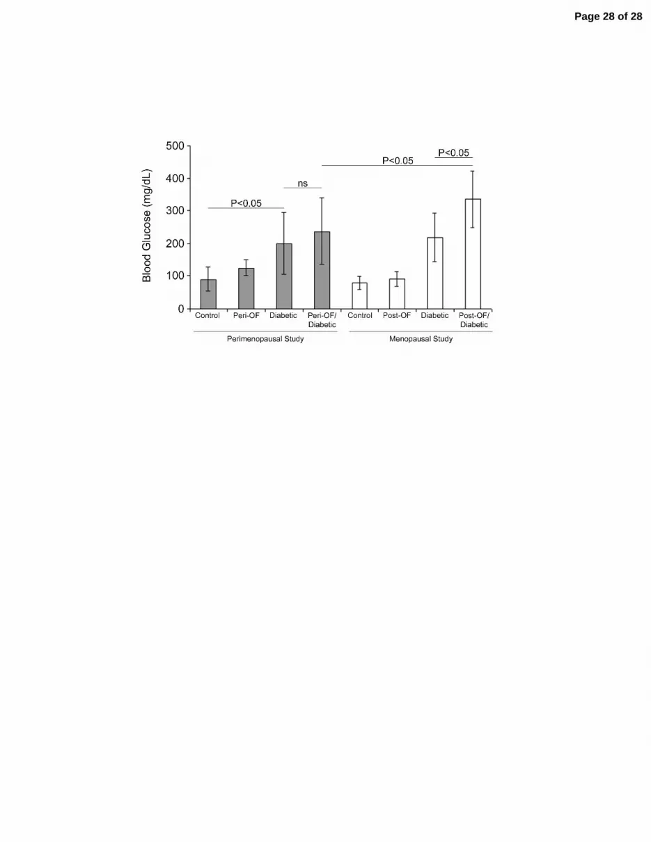
There was a significant increase in blood glucose in all STZ-injected mice compared to control
mice (Figure 2). Furthermore, glucose levels in post-OF diabetic mice were significantly higher than in
cycling diabetic mice (336 ± 86 mg/dL vs. 218 ± 76 mg/dL, P<0.05). There was no significant difference
in glucose levels between non-diabetic post-OF mice and cycling control mice (91 ± 23 vs. 79 ± 20,
P>0.05).
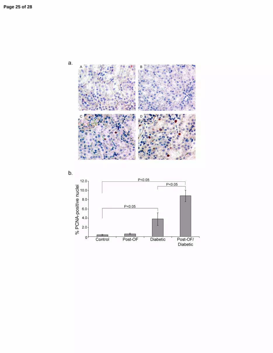
PCNA immunohistochemistry:
Cell proliferation is used as an early indicator of cell stress/damage in the kidney and occurs in the
renal glomeruli and tubules in diabetic mice (24), (38), (14). Proliferating cell nuclear antigen (PCNA) is
a DNA polymerase-associated protein expressed in proliferating cells (39). Immunohistochemistry was
performed on kidney tissue sections using an antibody against PCNA. Representative sections of PCNA
immunohistochemistry are shown in Figure 3Aa. There were significantly more PCNA-positive cells in
the renal cortex of diabetic mice compared to non-diabetic mice (Figure 3Bb). In addition, there was a
further significant increase in PCNA-positive cells in the cortex of post-OF diabetic mice compared to
Page 8 of 28
9
cycling diabetic mice. PCNA staining was primarily in tubular, not glomerular cells and was not seen in
the medulla of any treatment group.
Real-time PCR in post-ovarian failure diabetic mice:
In the kidney the expression of many genes associated with damage, such as collagen 4 and
transforming growth factor β, are altered in response to diabetes (32). Thus, real-time PCR was
performed to determine if the expression of genes associated with diabetic kidney damage was different
depending on hormonal status. Several genes which were differentially expressed in the renal cortex of
post-OF diabetic mice compared to cycling diabetic mice were identified. Three main patterns of changes
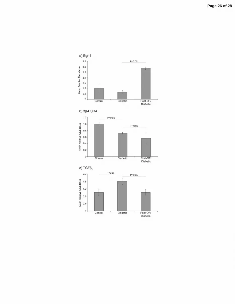
in gene expression emerged, as shown in figure 4: Pattern 1) A significant change (increase or decrease)
in mRNA abundance in post-OF diabetic mice compared to control mice and cycling diabetic mice, with
no change between cycling diabetic and control mice (Figure 4a). The expression of early growth
response 1 (Egr-1), collagen 4α1 (Col4α1), and matrix metalloproteinase 9 (MMP9) fit this pattern. Egr-1
mRNA abundance was increased 2.87 fold (P<0.05) in the cortex of post-OF diabetic mice compared to
cycling diabetic and control mice. Col4α1 mRNA abundance was decreased 1.75 fold (P<0.05) in post-
OF diabetic mice compared to cycling diabetic and control mice. MMP9 mRNA abundance was
decreased 1.89 fold (P<0.05) in post-OF diabetic mice compared to cycling diabetic and control mice.
There were no significant differences in mRNA abundance of any of these genes between cycling diabetic
and control mice. Pattern 2) A significant decrease in mRNA abundance in cycling diabetic mice
compared to control mice and a further significant decrease in post-OF diabetic mice (Figure 4b). mRNA
abundance of 3β-hydroxysteroid dehydrogenase 4 (3β-HSD4) fit this pattern, with a 1.39 fold decrease
(P<0.05) in cycling diabetic mice compared to control mice, and a further 1.30 fold decrease (P<0.05) in
post-OF diabetic mice compared to cycling diabetic mice. The mRNA abundance of 3β-HSD4 in post-
OF diabetic mice was decreased 1.79 fold (P<0.05) compared to control mice. Pattern 3) A significant
change in mRNA abundance in cycling diabetic mice compared to control mice, and a significant change
Page 9 of 28

10
in the opposite direction between post-OF and cycling diabetic mice such that there was no significant
difference between post-OF diabetic and control mice (Figure 4c). Transforming growth factor β2
(TGFβ2) fit this pattern; the mRNA abundance of TGFβ2 increased 1.60 fold (P<0.05) in cycling diabetic
mice compared to control mice and decreased 1.59 fold (P<0.05) in post-OF diabetic mice relative to
cycling diabetic mice. There was no significant difference in mRNA abundance of TGFβ2 between post-
OF diabetic and control mice. In addition, real-time PCR analysis was performed for the following genes:
collagen 1α2, collagen 4α2, estrogen receptor α, fibronectin 1, PCNA, transforming growth factor β1, and
transforming growth factor β3. No significant differences in renal cortex mRNA abundance between post-
OF diabetic mice and cycling diabetic mice were found. For all of the above genes there were no
significant differences in the mRNA abundance between control and non-diabetic post-OF mice.
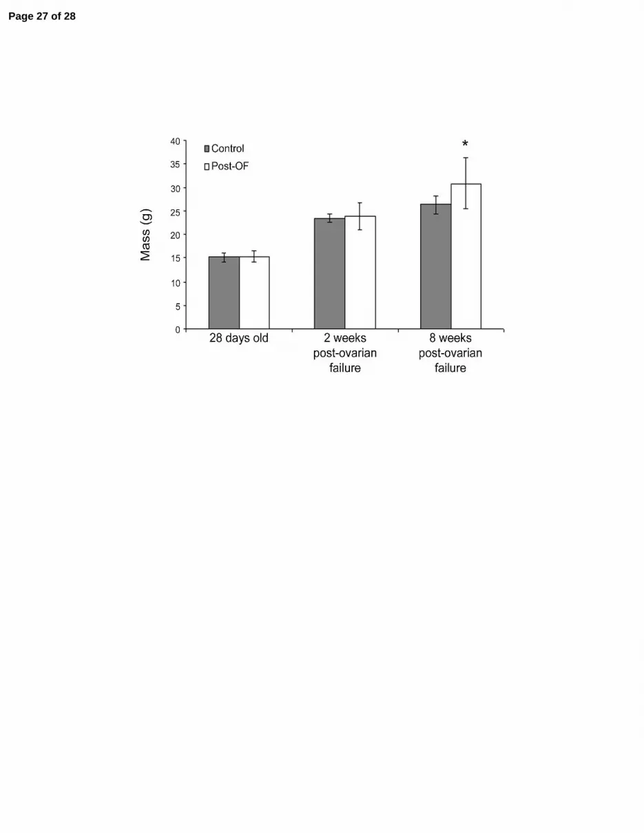
Menopause induced weight gain in non-diabetic mice:
All mice gained weight throughout the study. Compared to control mice, post-OF non-diabetic
mice rapidly gained weight following ovarian failure. 8 weeks post-ovarian failure, post-OF mice
weighed significantly more than control mice (Figure 5). There were no significant differences in weight
between any of the other groups of mice.
Initiation of perimenopause:
One of the strengths of the VCD model of menopause is that full ovarian failure is preceded by a
period of irregular cycling, analogous to perimenopause in human women (20), (13). Thus, in a
complementary study diabetes was induced during peri-ovarian failure (see Figure 1: Perimenopausal
Study); STZ dosing began three days after the end of VCD dosing. In contrast to the menopausal study,
there was no difference in weight between any of the groups of mice at the end of the study.
Blood glucose in peri-ovarian failure diabetic mice:
There was a significant increase in blood glucose in all STZ-injected mice compared to control
mice (Figure 6). Glucose levels in peri-OF diabetic mice trended higher but were not significantly
Page 10 of 28

11
different from cycling diabetic mice (238 ± 102 mg/dL vs. 201 ± 96 mg/dL, P=0.21) (Figure 6). When
compared to the menopausal study, glucose levels in peri-OF diabetic mice were significantly different
from glucose levels in post-OF diabetic mice (238 ± 102 vs. 336 ± 86 mg/dL, P<0.05) (Figure 6).
Real-time PCR in peri-ovarian failure diabetic mice:
Real-time PCR was performed comparing renal cortex mRNA abundance from peri-OF diabetic
mice to cycling diabetic mice. Primers for genes whose expression was significantly different between
post-OF diabetic and cycling diabetic mice (i.e. Egr-1, Col4α1, MMP9, 3βHSD4, and TGFβ2) were used.
As observed in the menopausal study, Egr-1 mRNA abundance was increased in peri-OF diabetic mice
compared to cycling diabetic mice (increase of 2.05 fold, P<0.05). TGFβ2 was decreased in peri-OF
diabetic mice compared to cycling diabetic mice (decrease of 1.6 fold, P<0.05). In contrast to what was
observed in post-OF diabetic mice, 3β-HSD4 was increased in peri-OF diabetic mice compared to cycling
diabetic mice (increase of 1.2 fold, P<0.05), whereas in the post-OF diabetic mice 3β-HSD4 was
decreased compared to cycling diabetic mice.
Page 11 of 28
12
DISCUSSION: VCD is a chemical by-product of rubber manufacturing which has been demonstrated to
selectively destroy primordial and primary follicles in ovaries of rats and mice (reviewed in (8)) without
producing effects in large follicles or other tissues (20). We recently described the novel use of VCD as a
means to induce gradual ovarian failure in mice (12). Ovarian failure in rodents is analogous to
menopause in humans; thus VCD-treated mice serve as a new model of human menopause. Unlike
previous rodent models of menopause, the VCD model preserves the period of irregular cycling and
fluctuating hormone levels which precedes ovarian failure, termed perimenopause in humans (13). Also,
following ovarian failure in the VCD model the follicle-deplete ovaries secrete androgens, similar to the
ovaries of postmenopausal humans (20).
Post-ovarian failure diabetic mice:
In this study we combined the VCD model of menopause with streptozotocin (STZ)-induced type
1 diabetes. Two groups of mice were studied: one in which diabetes was induced during impending
ovarian failure (perimenopause), and one in which diabetes was induced post-ovarian failure
(menopause). Following the induction of diabetes blood glucose levels were significantly higher in post-
ovarian failure diabetic mice than in cycling diabetic mice. Post-ovarian failure mice also had higher
blood glucose than mice in which diabetes was induced during impending ovarian failure (peri-ovarian
failure).
STZ causes diabetes by destroying insulin-producing pancreatic β-cells via oxidative stress-
induced apoptosis. Recent data suggest that the presence of estrogen may protect β-cells from STZ-
induced oxidative stress. In a study in which STZ was used to induce diabetes in ovariectomized rats,
estradiol-treated ovariectomized rats had significantly lower blood glucose than those untreated with
estradiol. The authors speculated that estrogen may attenuate STZ-induced destruction of pancreatic β-
cells by decreasing inflammation (29) and thus may contribute to lower blood glucose levels in estrogen-
Page 12 of 28

13
treated rats. More recently, an in vitro study using STZ on primary cultures of pancreatic β-cells
demonstrated that estrogen protects β-cells from oxidative stress-induced apoptosis (10).
Data from human studies suggest that estrogen also exerts a protective effect on pancreatic β-cells
in humans. In the Women’s Health Initiative Hormone Trial, the incidence of newly diagnosed cases of
diabetes was lower in women on hormone replacement therapy than in placebo-treated women (17). In
perimenopausal women, longer cycle length (i.e. lower estrogen levels) has also been associated with
higher blood glucose levels and hyperinsulinemia (19). Furthermore, in diabetic postmenopausal women,
treatment with hormone replacement therapy improved insulin resistance (17).
Cell proliferation in post-ovarian failure diabetic mice:
Renal cell proliferation and hypertrophy are early complications of diabetes (24), (38), (14);
therefore expression of proliferating cell nuclear antigen (PCNA) can be used as a marker of early kidney
damage. In our study expression of PCNA was increased in kidneys from post-ovarian failure diabetic
mice compared to cycling diabetic mice. These data suggest that renal damage develops more rapidly
and/or severely in post-ovarian failure diabetic mice compared to cycling diabetic mice.
Low estrogen levels increase susceptibility to nephropathy (5), thus the loss of estrogen in post-
ovarian failure diabetic mice may have contributed to the increased proliferation observed in this study.
However, high circulating blood glucose has been positively correlated with an increase in diabetic
nephropathy in mice (6) thus the higher levels of glucose we observed in post-ovarian failure diabetic
mice could have contributed to the increased cell proliferation in our study.
Changes in gene expression in post-ovarian failure diabetic mice:
Diabetic kidney damage is usually associated with the increased accumulation of extracellular
matrix proteins, such as collagen and fibronectin. This increase is thought to be stimulated by increased
expression of TGFβ and the decreased expression and activity of matrix metalloproteinases (40), (32),
(16). This study identified several genes which were differentially expressed in the renal cortex of post-
Page 13 of 28
14
ovarian failure diabetic mice and cycling diabetic mice. For example, in the renal cortex of post-ovarian
failure diabetic mice, MMP9 expression was decreased compared to cycling diabetic mice. This result
correlates well with previous in vivo studies in which MMP9 protein expression and activity level were
decreased in ovariectomized diabetic rats (16) and in ovariectomized Dahl salt-sensitive rats (18).
Previous in vitro studies in mesangial cells have found that estrogen increases the expression of MMP9
(26); thus the loss of estrogen in our model of menopause may explain the observed decrease in MMP9
mRNA expression.
We observed a decrease in mRNA abundance of collagen 4α1 and TGFβ2 in post-ovarian failure
diabetic kidneys compared to cycling diabetic kidneys. These data diverge from studies in which
ovariectomy was associated with increased expression of TGFβ and collagen 4 in diabetic rats (15), (16).
mRNA abundance of 3β-HSD4 was significantly lower in the renal cortex of post-ovarian failure
diabetic mice than in cycling diabetic mice. Murine 3β-HSD4 is a 3-ketosteroid reductase which
metabolizes progesterone and dihydrotestosterone to their inactive forms (2), (27). Recent studies
identified 3β-HSD4 expression as predictive of the degree to which glomerulosclerosis develops in the
diabetic kidney, with lower expression of 3β-HSD4 correlating with more severe glomerulosclerosis (35).
A decrease in 3β-HSD4 expression could lead to an increase in dihydrotestosterone levels.
Dihydrotestosterone has been demonstrated to cause an increase in cell proliferation in a proximal tubule
cell line (25); thus decreased 3β-HSD4 expression could result in increased proliferation of proximal
tubule cells. Our results are consistent with this hypothesis as we found both a decrease in 3β-HSD4
mRNA abundance and an increase in cell proliferation in kidneys from post-ovarian failure diabetic mice
compared to cycling diabetic mice and control mice.
Peri-ovarian failure diabetic mice:
The VCD-model of menopause is unique among rodent menopause models in that it retains
residual ovarian tissue after ovarian failure and mimics the perimenopausal period. Recent studies find
Page 14 of 28
15
that changes in disease risk factors begin during perimenopause (7),(19) suggesting that the
perimenopausal period may be critical in the development and prevention of estrogen-influenced diseases
(37).
Data reported here found a trend toward increased glucose levels in peri-ovarian failure diabetic
mice and a significant difference in blood glucose between post-ovarian failure diabetic mice and cycling
diabetic mice. These data suggest a continuing trend of increasing blood glucose throughout the
menopausal transition as estrogen gradually decreases.
Real-time PCR data from this study suggest that declining estrogen levels during peri-ovarian
failure affect the expression of genes implicated in the development of diabetic kidney disease. For
example, mRNA abundance of TGFβ2 was lower in peri-ovarian failure diabetic mice than in cycling
diabetic mice. This is similar to the decrease in mRNA abundance for this gene observed between post-
ovarian failure diabetic mice and cycling diabetic mice. These data highlight the importance of the VCD
model of perimenopause and menopause, as these changes in gene expression may be important in
understanding disease progression and have implications for disease prevention and treatment.
Conclusion:
This study suggests that estrogen may play an important role in protecting the kidney from
diabetes-induced cell proliferation and disease. It is possible that not all of the differences we observed
between post-ovarian failure diabetic mice and cycling diabetic mice are due to changes in estrogen
levels. The increase in blood glucose levels in post-ovarian failure diabetic mice may account for some of
the differences in gene expression and cell proliferation we observed. Also, preliminary evidence
suggests androgens negatively affect the development of kidney disease (28), (22). The VCD model of
menopause preserves the androgen-producing function of the post-ovarian failure ovaries (20); thus some
of the differences we observed between post-ovarian failure diabetic mice and cycling diabetic mice may
Page 15 of 28

16
be due to differences in androgen levels or differences in the estrogen/androgen ratio as opposed to direct
actions of estrogen alone.
As more women develop diabetes at younger ages, the number of women with diabetes at the time
of the menopausal transition will increase, as will the number of post-menopausal women with diabetes.
Here we introduce a new model for studying the impact of the menopausal transition on the development
of diabetic kidney disease. We show that blood glucose levels are increased in post-ovarian failure
diabetic mice compared to cycling diabetic mice and that there are differences in the mRNA abundance of
genes associated with diabetic kidney disease in post-ovarian failure diabetic mice compared to cycling
diabetic mice. Furthermore, we show there is increased cell proliferation in post-ovarian failure diabetic
mice compared to cycling diabetic mice. Finally, we present data which suggest the importance of
studying the development of diabetic kidney disease in the context of the perimenopausal period.
Together, these data highlight the importance and utility of the VCD model of menopause, as it provides a
physiologically relevant system for determining the impact of diabetes on the kidney during the
menopausal transition.
ACKNOWLEDGEMENTS: We gratefully acknowledge Dr. R. Egleton for the use of the glucometer
and for helpful advice and P. Christian for technical assistance. This work was funded by NIH grant AG-
021948 to PBH, and M. Keck is supported by an NSF-IGERT Fellowship in Genomics, University of
Arizona and The ARCS Foundation, Phoenix, AZ chapter.
Page 16 of 28
17
REFERENCES:
1. Atkins RC. The epidemiology of chronic kidney disease. Kidney Int 67: s14-s18, 2005. 2. Clarke TR, Bain PA, Greco TL, Payne AH. A novel mouse kidney 3 beta-hydroxysteroid
dehydrogenase complementary DNA encodes a 3-ketosteroid reductase instead of a 3 beta- hydroxysteroid dehydrogenase/delta 5-delta 4-isomerase. Mol Endocrinol 7: 1569-1578, 1993.
3. Coggins C, Lewis J, Caggiula A, Castaldo L, Klahr S, Wang S, Beck G. Differences between women and men with chronic renal disease. Nephrol Dial Transplant 13: 1430-1437, 1998.
4. Couzinet B, Meduri G, Lecce MG, Young J, Brailly S, Loosfelt H, Milgrom E, Schaison G. The postmenopausal ovary is not a major androgen-producing gland. J Clin Endocrinol Metab 86: 5060-5066, 2001.
5. Elliot SJ, Karl M, Berho M, Potier M, Zheng F, Leclercq B, Striker GE, Striker LJ. Estrogen Deficiency Accelerates Progression of Glomerulosclerosis in Susceptible Mice. Am J Pathol 162: 1441-1448, 2003.
6. Gurley SB, Clare SE, Snow KP, Hu A, Meyer TW, Coffman TM. Impact of genetic background on nephropathy in diabetic mice. Am J Physiol Renal Physiol 290: F214-F222, 2006.
7. Hinojosa-Laborde C, Craig T, Zheng W, Ji H, Haywood JR, Sandberg K. Ovariectomy augments hypertension in aging female Dahl salt-sensitive rats. Hypertension 44: 405-409, 2004.
8. Hoyer PB, Sipes IG. Assessment of follicle destruction in chemical-induced ovarian toxicity. Annu Rev Pharmacol Toxicol 36: 307-331, 1996.
9. Kannel WB, Hjortland MC, McNamara PM, Gordon T. Menopause and risk of cardiovascular disease: the Framingham study. Ann Intern Med 85: 447-452, 1976.
10. Le May C, Chu K, Hu M, Ortega CS, Simpson ER, Korach KS, Tsai MJ, Mauvais-Jarvis F.Estrogens protect pancreatic beta-cells from apoptosis and prevent insulin-deficient diabetes mellitus in mice. PNAS 103: 9232-9237, 2006.
11. Livak KJ, Schmittgen TD. Analysis of Relative Gene Expression Data Using Real-Time Quantitative PCR and the 2-[Delta][Delta]CT Method. Methods 25: 402-408, 2001.
12. Lohff JC, Christian PJ, Marion SL, Arrandale A, Hoyer PB. Characterization of cyclicity and hormonal profile with impending ovarian failure in a novel chemical-induced mouse model of perimenopause. Comp Med 55: 523-527, 2005.
13. Lohff JC, Christian PJ, Marion SL, Hoyer PB. Effect of duration of dosing on onset of ovarian failure in a chemical-induced mouse model of perimenopause. Menopause 13: 482-488, 2006.
14. Maeda M, Yabuki A, Suzuki S, Matsumoto M, Taniguchi K, Nishinakagawa H. Renal Lesions in Spontaneous Insulin-dependent Diabetes Mellitus in the Nonobese Diabetic Mouse: Acute Phase of Diabetes. Vet Pathol 40: 187-195, 2003.
15. Mankhey RW, Bhatti F, Maric C. 17{beta}-Estradiol replacement improves renal function and pathology associated with diabetic nephropathy. Am J Physiol Renal Physiol 288: F399-F405, 2005.
16. Mankhey RW, Wells CC, Bhatti F, Maric C. 17{beta}-estradiol supplementation reduces tubulointerstitial fibrosis by increasing MMP activity in the diabetic kidney. Am J Physiol Regul Integr Comp Physiol 00375, 2006.
17. Margolis KL, Bonds DE, Rodabough RJ, Tinker L, Phillips LS, Allen C, Bassford T, Burke G, Torrens J, Howard BV. Effect of oestrogen plus progestin on the incidence of diabetes in postmenopausal women: results from the Women's Health Initiative Hormone Trial. Diabetologia 47: 1175-1187, 2004.
18. Maric C, Sandberg K, Hinojosa-Laborde C. Glomerulosclerosis and Tubulointerstitial Fibrosis are Attenuated with 17{beta}-Estradiol in the Aging Dahl Salt Sensitive Rat. J Am Soc Nephrol 15: 1546-1556, 2004.
Page 17 of 28
18
19. Matthews KA, Santoro N, Lasley B, Chang Y, Crawford S, Pasternak RC, Sutton-Tyrrell K, Sowers M. Relation of Cardiovascular Risk Factors in Women Approaching Menopause to Menstrual Cycle Characteristics and Reproductive Hormones in the Follicular and Luteal Phases. JClin Endocrinol Metab 91: 1789-1795, 2006.
20. Mayer LP, Devine PJ, Dyer CA, Hoyer PB. The Follicle-Deplete Mouse Ovary Produces Androgen. Biol Reprod 71: 130-138, 2004.
21. McReynolds MR, Taylor-Garcia KM, Greer KA, Hoying JB, Brooks HL. Renal medullary gene expression in aquaporin-1 null mice. Am J Physiol Renal Physiol 288: F315-F321, 2005.
22. Mulroney SE, Woda C, Johnson M, Pesce C. Gender differences in renal growth and function after uninephrectomy in adult rats. 56: 944-953, 1999.
23. Neugarten J, Acharya A, Silbiger SR. Effect of gender on the progression of nondiabetic renal disease: a meta-analysis. J Am Soc Nephrol 11: 319-329, 2000.
24. Nyengaard JR, Flyvbjerg A, Rasch R. The impact of renal growth, regression and regrowth in experimental diabetes mellitus on number and size of proximal and distal tubular cells in the rat kidney. Diabetologia V36: 1126-1131, 1993.
25. Ouar Z, Sole E, Bens M, Rafestin-Oblin ME, Meseguer A, Vandewalle A. Pleiotropic effects of dihydrotestosterone in immortalized mouse proximal tubule cells. Kidney Int 53: 59-66, 1998.
26. Potier M, Karl M, Zheng F, Elliot SJ, Striker GE, Striker LJ. Estrogen-Related Abnormalities in Glomerulosclerosis-Prone Mice : Reduced Mesangial Cell Estrogen Receptor Expression and Prosclerotic Response to Estrogens. Am J Pathol 160: 1877-1885, 2002.
27. Quinkler M, Johanssen S, Bumke-Vogt C, Oelkers W, Bahr V, Diederich S. Enzyme-mediated protection of the mineralocorticoid receptor against progesterone in the human kidney. Molecular and Cellular Endocrinology 171: 21-24, 2001.
28. Reckelhoff JF, Yanes LL, Iliescu R, Fortepiani LA, Granger JP. Testosterone supplementation in aging men and women: possible impact on cardiovascular-renal disease. Am J Physiol Renal Physiol 289: F941-F948, 2005.
29. Riazi S, Maric C, Ecelbarger CA. 17-[beta] Estradiol attenuates streptozotocin-induced diabetes and regulates the expression of renal sodium transporters. Kidney Int 69: 471-480, 2006.
30. Rozen S, Skaletsky H. Primer3 on the WWW for general users and for biologist programmers. Methods Mol Biol 132: 365-386, 2000.
31. Santoro N, Brown JR, Adel T, Skurnick JH. Characterization of reproductive hormonal dynamics in the perimenopause. J Clin Endocrinol Metab 81: 1495-1501, 1996.
32. Schena FP, Gesualdo L. Pathogenetic Mechanisms of Diabetic Nephropathy. J Am Soc Nephrol 16: S30-S33, 2005.
33. Schrijvers BF, De Vriese AS, Flyvbjerg A. From Hyperglycemia to Diabetic Kidney Disease: The Role of Metabolic, Hemodynamic, Intracellular Factors and Growth Factors/Cytokines. Endocr Rev 25: 971-1010, 2004.
34. Seliger SL, Davis C, Stehman-Breen C. Gender and the progression of renal disease. Curr Opin Nephrol Hypertens 10: 219-225, 2001.
35. Susztak K, Bottinger E, Novetsky A, Liang D, Zhu Y, Ciccone E, Wu D, Dunn S, McCue P, Sharma K. Molecular Profiling of Diabetic Mouse Kidney Reveals Novel Genes Linked to Glomerular Disease. Diabetes 53: 784-794, 2004.
36. Ushiroyama T, Sugimoto O. Endocrine function of the peri- and postmenopausal ovary. Horm Res 44: 64-68, 1995.
37. Williams JK. A mouse model of the perimenopausal transition: importance for cardiovascular research. Arterioscler Thromb Vasc Biol 25: 1765-1766, 2005.
38. Wolf G, Ziyadeh FN. Molecular mechanisms of diabetic renal hypertrophy. 56: 393-405, 1999.
Page 18 of 28

19
39. Zeng XR, Jiang Y, Zhang SJ, Hao H, Lee MY. DNA polymerase delta is involved in the cellular response to UV damage in human cells. J Biol Chem 269: 13748-13751, 1994.
40. Ziyadeh FN, Sharma K, Ericksen M, Wolf G. Stimulation of collagen gene expression and protein synthesis in murine mesangial cells by high glucose is mediated by autocrine activation of transforming growth factor-beta. J Clin Invest 93: 536-542, 1994.
Page 19 of 28
20
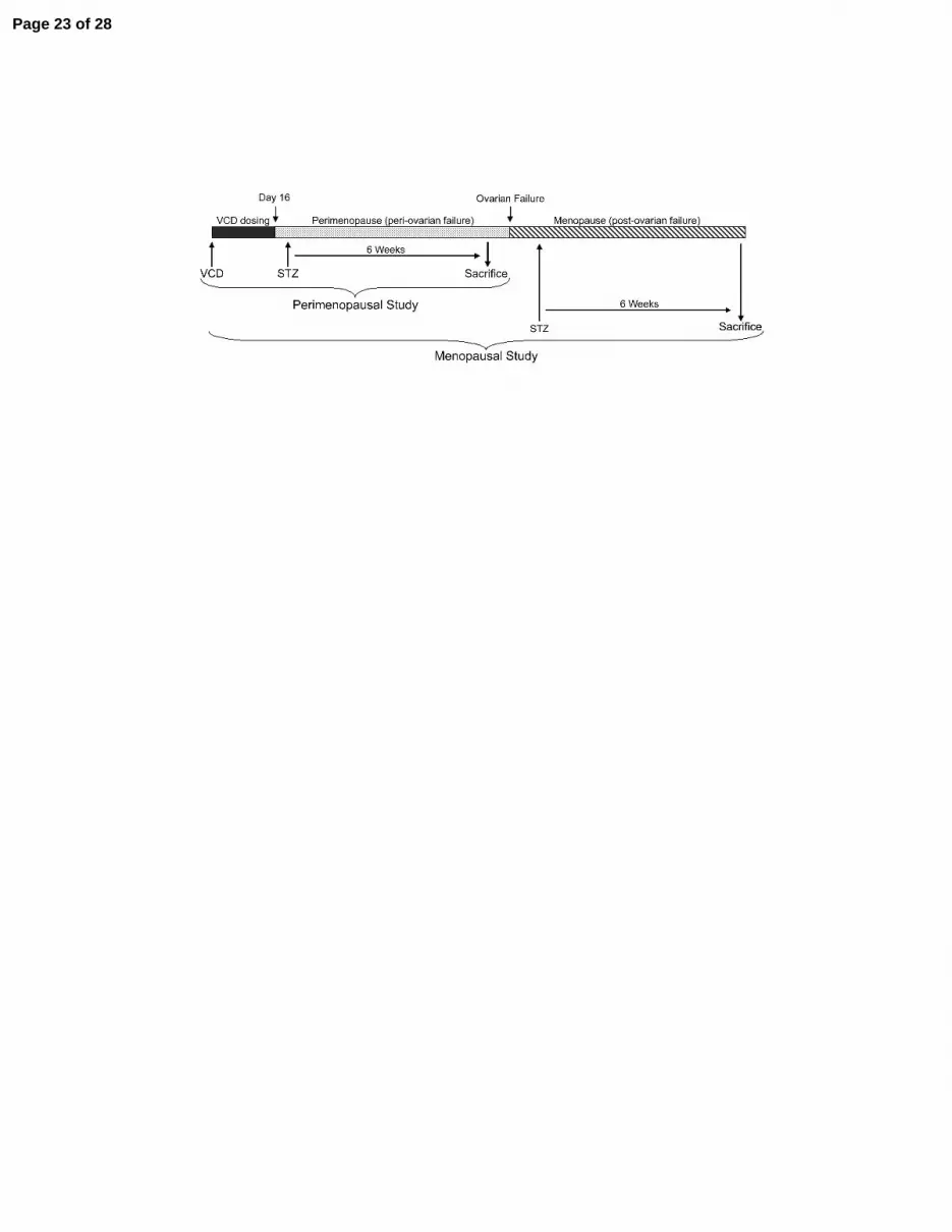
FIGURE LEGENDS: Figure 1: Timeline of experimental study groups – 28 day old female B6C3F1 mice were separated into
two study groups and given i.p. injections of VCD and STZ to induce gradual ovarian failure and
diabetes, respectively. All mice were dosed with VCD for 15 consecutive days and with STZ for 3
consecutive days. Large up arrows indicate the initial day of dosing of VCD or STZ. Sacrifice followed
six weeks after STZ dosing in both studies.
Figure 2: Blood glucose measurements in post-ovarian failure diabetic mice – Cycling or post-
ovarian failure mice were injected with STZ to induce diabetes. Fasted blood glucose levels were
measured 6 weeks after STZ treatment. Results are expressed as mean ± SD. Significant difference
determined by Student Neumann-Keuls post-hoc test following one-way ANOVA. OF - ovarian failure.
See Table 1 for n in each group.
Figure 3: Effect of diabetes and hormonal status on renal cortex cell proliferation – a)
Representative sections from kidney cortex demonstrating PCNA staining (in red); nuclear counterstain
with hemotoxylin (blue). A: Control, B: Post-OF, C: Diabetic, D: Post-OF/Diabetic. Magnification:
400X. b) Graph presenting the mean PCNA-positive nuclei as a percentage of total nuclei. PCNA-
positive nuclei and total nuclei were counted in 5 non-overlapping fields of view for each section, with 3
animals in each treatment group. Results are presented as mean ± SD. Significant difference determined
by Student Neumann-Keuls post-hoc test following one-way ANOVA. OF - ovarian failure.
Figure 4: Patterns of changes in mRNA abundance of genes associated with diabetic kidney
damage – SYBR Green I real-time PCR measurements were used to derive Ct values for calculation of
mRNA abundance. The data shown are the mean relative gene expression normalized to control group ±
Page 20 of 28

21
SEM. Assays for β-actin were run in parallel on each sample for internal controls. Assays were run in
triplicate on cDNA synthesized by reverse transcription from RNA samples. Significant difference
determined by Student's t-test. n=3 for each group. a) Egr-1: early growth response-1 b) 3β-HSD4: 3β-
hydroxysteroid dehydrogenase 4 c) TGFβ2: transforming growth factor β2.
Figure 5: Weight gain in non-diabetic post-ovarian failure mice – Non-diabetic VCD-dosed mice (i.e.
post-ovarian failure mice) weighed the same as control cycling mice until 2 weeks after ovarian failure, at
which point post-OF mice rapidly gained weight. Results are presented as mean ± SD. *P<0.05 as
determined by Student’s t-test (n=9 vs. 9). OF - ovarian failure.
Figure 6: Blood glucose measurements in peri-ovarian failure diabetic mice – Mice were injected
with STZ to induce diabetes, and fasted blood glucose was measured 6 weeks after STZ treatment.
Results are expressed as mean ± SD. Significant difference determined by Student Neumann-Keuls post-
hoc test following one-way ANOVA. Solid bars – perimenopausal study; white bars – menopausal study.
Data from menopausal study (Figure 2) is shown for comparison. OF – ovarian failure, ns – non-
significant.
Page 21 of 28

Table 1: Description of Mouse Groups
Description VCD STZ NumberPerimenopausal Study:
Control cycling non-diabetic n=6
Peri-OF impending ovarian failure non-diabetic + n=9
Diabetic cycling diabetic + n=8
Peri-OF/Diabetic impending ovarian failure diabetic + + n=11
Menopausal Study:Control cycling non-diabetic n=9
Post-OF post-ovarian failure non-diabetic + n=9
Diabetic cycling diabetic + n=9Post-OF/Diabetic post-ovarian failure diabetic + + n=9
Descriptions of treatments given to mice. "+" indicates a chemical which was injected. Otherwisemice were injected with appropriate control buffer. "Number" is the number of mice in each treatmentgroup.
Page 22 of 28
Page 23 of 28
Page 24 of 28
Page 25 of 28
Page 26 of 28
Page 27 of 28
Page 28 of 28

![7Rc^Vcd f_RS]V e` WZ_U ]RTf_R Z_ WRc^ ]Rhd+ E`^Rc](https://static.fdokumen.com/doc/165x107/633cef43f4deedbcff085b82/7rcvcd-frsv-e-wzu-rtfr-z-wrc-rhd-erc.jpg)

![eR]\d WRZ] Rd WRc^Vcd deZT\ e` eYVZc Xf_d - Daily Pioneer](https://static.fdokumen.com/doc/165x107/631dcea81aedb9cd850f765e/erd-wrz-rd-wrcvcd-dezt-e-eyvzc-xfd-daily-pioneer.jpg)
![7Rc^Vcd UZX Z_ YVV]d DYRY cVRTYVd `fe - Daily Pioneer](https://static.fdokumen.com/doc/165x107/63287bbe051fac18490ebf3c/7rcvcd-uzx-z-yvvd-dyry-cvrtyvd-fe-daily-pioneer.jpg)


![7Rc^Vcd cV[VTe DYRY `WWVc dVe eR]\ eVc^d - Daily Pioneer](https://static.fdokumen.com/doc/165x107/63210b07e9691360fe020cc5/7rcvcd-cvvte-dyry-wwvc-dve-er-evcd-daily-pioneer.jpg)
![7Rc^Vcd TR]] `WW ac`eVde dRj SRee]V h`_ hRc ` - Daily ...](https://static.fdokumen.com/doc/165x107/631e6ffe85e2495e150fea8f/7rcvcd-tr-ww-acevde-drj-sreev-h-hrc-daily-.jpg)


![7Rc^Vcd W`c da] DVddZ`_ e` cVaVR] ]Rhd - Daily Pioneer](https://static.fdokumen.com/doc/165x107/63191b214cd7b3442408e4b0/7rcvcd-wc-da-dvddz-e-cvavr-rhd-daily-pioneer.jpg)

