Hominid tarsal, metatarsal, and phalangeal bones recovered from the Hadar formation: 1974-1977...
19
AMERICAN JOURNAL OF PHYSICAL ANTHROPOLOGY 57:701-719 (1982) Hominid Tarsal, From the Hadar Metatarsal, and Phalangeal Bones Recovered Formation: 1974-1 977 Collections BRUCE M. LATIMER, C. OWEN LOVWOY, DONALD C. JOHANSON, AND YVES COPPENS Cleveland Museum of Natural History, Cleveland, Ohio 44106 (B.M.L., C.O.L., D.C.J.); Departments of Anthropology and Biology, Kent State Uniuersity, Kent, Ohio 44242 (B.M.L.,C.O.L.); Department of Human Anatomy, Northeast Ohio Universities College of Medicine, Rootstown, Ohio 44272 (C.0.L.I; Department of Orthopaedic Surgery, Case Western Reserve University, Cleveland, Ohio 44106 (C.0.L.I; Institute of Human Origins, 2700 Bancroft Way, Berkeley, California 94704 (D.C.J.);Laboratoire d'Anthropologie, Musee de L'Homme, Paris 7Sl16, France (Y. C.) KEY WORDS Hadar, Foot, Hominid, Australopithecus ABSTRACT Anatomical descriptions are presented for the foot bones re- covered from the Hadar Formation during the 1974-1977 field seasons inclusive. The following are anatomical descriptions of the foot bones recovered by the International Afar Research Expedition during the field sea- sons of 1974-1977, inclusive (Johanson et al., this volume). The foot bones from the partial skeleton from A.L. 288 are described sepa- rately (Johanson et al., this volume). Anatomical nomenclature follows general usage and Trotter and Peterson (1966). Meas- urements are those in general use or are de- scribed. Morphometric, biomechanical, and kinematic studies are currently in progress and results will be reported subsequently. These descriptions are organized by anatomi- cal region: Tarsal bones are described first, fol- lowed by metatarsal bones and phalanges. The associated partial foot skeleton A.L. 333-115 is described separately. All measurements are in millimeters. DESCRIPTIONS TARSALS A.L. 333-75: fragment of right talus (Fig. 31-K) Preservation This fragment consists of a perfectly preserved right talar head. It is sev- ered from the body of the talus by a transverse section through the neck, preserving only a small portion of the trochlear articular surface and the most distal portion of the talar sulcus. Morphology The distal navicular articular surface is a convex oval with a maximum di- ameter of 23.9. A small triangular facet is pres- ent on the inferomedial corner of the navicular surface for articulation with the plantar cal- caneonavicular ligament. The anterior and middle calcaneal facets are continuous and no contour change is present between the arti- cular surfaces. Inferiorly the talar neck is roughened for ligamentous attachments and is perforated by numerous vascular foramina. A.L. 333-8: right calcaneus (Fig. 1E-G) This specimen consists of the posterior two-thirds of an adult right calca- neus. The existing bone is in good condition except for minor surface abrasion. The anterior portion of the calcaneus is lost owing to a trans- verse break just distal to the calcaneal sulcus, removing all evidence of the anterior talar ar- ticular surface and the cuboid facet. The me- dial portion of the sustentaculum tali is also missing. Morphology The calcaneal tuberosity is an elongated oval with the long axis directed su- peroinferiorly. The inferior half of the tuber- osity is roughened for insertion of the tendo calcaneus. The superior half of the tuberosity, the subbursal surface, is smooth and inclines medially, although this may be slightly ex- aggerated because of erosion of the supero- medial margin of the bone. The medial process of the tuberosity is large and is pitted by a number of prominent vascular foramina. The lateral process is indistinct. In its place a bony ridge runs obliquely across the lateral surface of the calcaneal body, ending in the massive peroneal trochlea. The apex of the peroneal trochlea lies approximately 12.0 inferoposter- iorly from the lateral terminus of the calcaneal Preservation Received March 17, 1981; accepted December 10, 1981. 0002-948318215704-0701805.50 0 1982 ALAN R. LISS, INC.
Transcript of Hominid tarsal, metatarsal, and phalangeal bones recovered from the Hadar formation: 1974-1977...
AMERICAN JOURNAL OF PHYSICAL ANTHROPOLOGY 57:701-719 (1982)
Hominid Tarsal, From the Hadar
Metatarsal, and Phalangeal Bones Recovered Formation: 1974-1 977 Collections BRUCE M. LATIMER, C. OWEN LOVWOY, DONALD C. JOHANSON, AND YVES COPPENS Cleveland Museum of Natural History, Cleveland, Ohio 44106 (B.M.L., C.O.L., D.C.J.); Departments of Anthropology and Biology, Kent State Uniuersity, Kent, Ohio 44242 (B.M.L., C.O.L.); Department of Human Anatomy, Northeast Ohio Universities College of Medicine, Rootstown, Ohio 44272 (C.0.L.I; Department of Orthopaedic Surgery, Case Western Reserve University, Cleveland, Ohio 44106 (C.0.L.I; Institute of Human Origins, 2700 Bancroft Way, Berkeley, California 94704 (D.C.J.); Laboratoire d'Anthropologie, Musee de L'Homme, Paris 7Sl16, France (Y. C.)
KEY WORDS Hadar, Foot, Hominid, Australopithecus
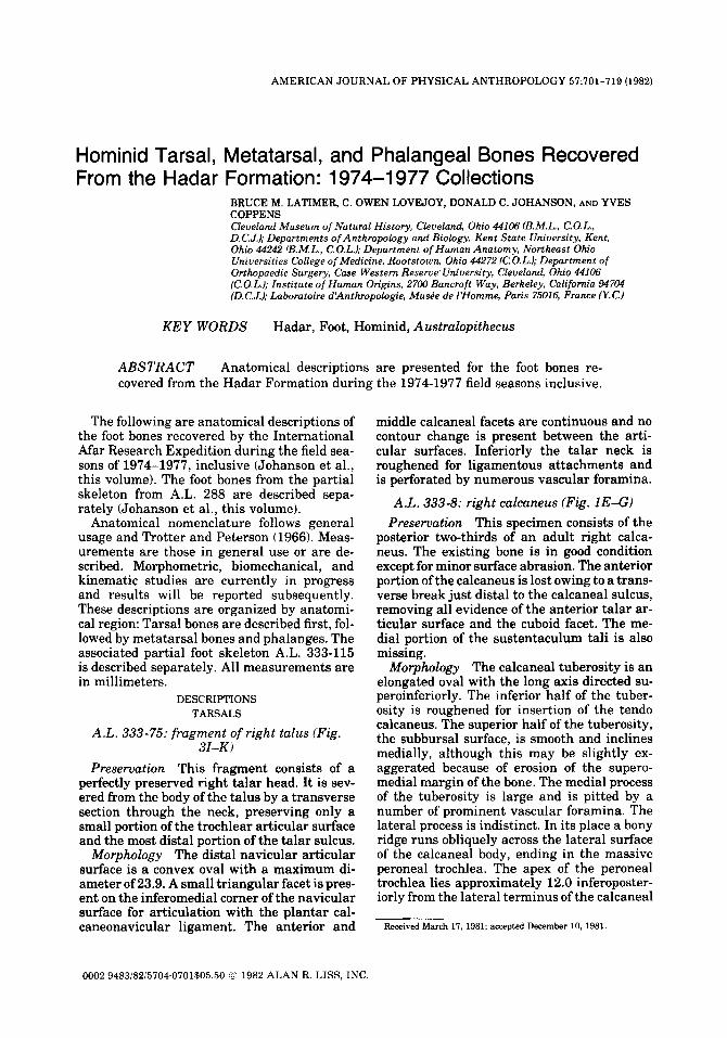
ABSTRACT Anatomical descriptions are presented for the foot bones re- covered from the Hadar Formation during the 1974-1977 field seasons inclusive.
The following are anatomical descriptions of the foot bones recovered by the International Afar Research Expedition during the field sea- sons of 1974-1977, inclusive (Johanson et al., this volume). The foot bones from the partial skeleton from A.L. 288 are described sepa- rately (Johanson et al., this volume).
Anatomical nomenclature follows general usage and Trotter and Peterson (1966). Meas- urements are those in general use or are de- scribed. Morphometric, biomechanical, and kinematic studies are currently in progress and results will be reported subsequently. These descriptions are organized by anatomi- cal region: Tarsal bones are described first, fol- lowed by metatarsal bones and phalanges. The associated partial foot skeleton A.L. 333-115 is described separately. All measurements are in millimeters.
DESCRIPTIONS TARSALS
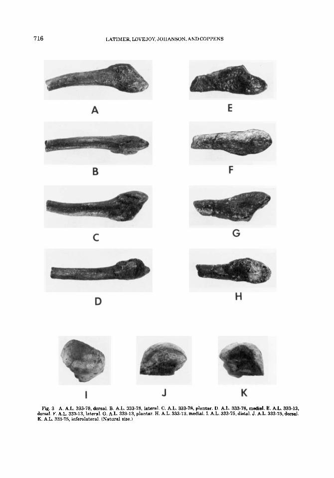
A.L. 333-75: fragment of right talus (Fig. 31-K)
Preservation This fragment consists of a perfectly preserved right talar head. It is sev- ered from the body of the talus by a transverse section through the neck, preserving only a small portion of the trochlear articular surface and the most distal portion of the talar sulcus.
Morphology The distal navicular articular surface is a convex oval with a maximum di- ameter of 23.9. A small triangular facet is pres- ent on the inferomedial corner of the navicular surface for articulation with the plantar cal- caneonavicular ligament. The anterior and
middle calcaneal facets are continuous and no contour change is present between the arti- cular surfaces. Inferiorly the talar neck is roughened for ligamentous attachments and is perforated by numerous vascular foramina.
A.L. 333-8: right calcaneus (Fig. 1E-G) This specimen consists of the
posterior two-thirds of an adult right calca- neus. The existing bone is in good condition except for minor surface abrasion. The anterior portion of the calcaneus is lost owing to a trans- verse break just distal to the calcaneal sulcus, removing all evidence of the anterior talar ar- ticular surface and the cuboid facet. The me- dial portion of the sustentaculum tali is also missing.
Morphology The calcaneal tuberosity is an elongated oval with the long axis directed su- peroinferiorly. The inferior half of the tuber- osity is roughened for insertion of the tendo calcaneus. The superior half of the tuberosity, the subbursal surface, is smooth and inclines medially, although this may be slightly ex- aggerated because of erosion of the supero- medial margin of the bone. The medial process of the tuberosity is large and is pitted by a number of prominent vascular foramina. The lateral process is indistinct. In its place a bony ridge runs obliquely across the lateral surface of the calcaneal body, ending in the massive peroneal trochlea. The apex of the peroneal trochlea lies approximately 12.0 inferoposter- iorly from the lateral terminus of the calcaneal
Preservation
Received March 17, 1981; accepted December 10, 1981.
0002-948318215704-0701805.50 0 1982 ALAN R. LISS, INC.
702 LATIMER. LOVEJOY, JOHANSON, AND COPPENS
sulcus. Surface damage has obliterated most detail with which to reconstruct the course of m. peroneus longus; however, a slight concav- ity immediately posterior to the projection may represent the sulcus for the peroneal tendon. Immediately superior to the trochlear process is a shallow groove for m. peroneus breuis.
The posterior articular surface is strongly convex from front to back and twists along its axis so that the most posterior portion of the facet faces posteromedially and its most an- terior segment inclines anterolaterally. The posterior facet is bounded anteriorly by the calcaneal sulcus and does not extend beyond that point. Postfossilization damage to the sus- tentaculum tali prohibits exact determination of its size, though it is evident from the ori- entation and robust dimensions of the broken surface that the shelf was strong and projected horizontally. The width and depth of the sulcus for m. flexor hallucis longus cannot be esti- mated because of the breakage. On the plantar surface the anterior tubercle is present but no surface morphology is discernible owing to the loss of several large bone flakes. Measure- ments are provided in Table 1.
A.L. 333-37: right calcaneus (Fig. l A , B) Preservation This is the proximal half of an
adult right calcaneus. It is transversely sec- tioned through the posterior talar articular facet, preserving only the posterior portion of that surface. The lateral side of the bone is considerably abraded and cancellous bone is exposed along the posterior margin of the pos- terior facet and around the perimeter of the calcaneal tuberosity.
Morphology The calcaneal tuberosity is large and ovoid in its dorsoplantar dimension. The tuberosity is roughened on its inferior half for insertion of the calcaneal tendon. The su- perior subbursal portion is smooth and inclines superomedially. The preserved portion of the posterior talar facet faces posteromedially.
On the plantar surface the medial tuberosity is large and projecting; the lateral tuberosity is relatively indistinct. The lateral surface ero- sion has removed all evidence of the peroneal trochlea. Measurements are provided in Table 1.
A.L. 333-55: left calcaneus (Fig. 1C,D) Preservation This specimen is the proximal
two-thirds of an adult left calcaneus. The sus- tentaculum tali is missing and a fossilization crack longitudinally bisects the posterior talar facet, but owing to good apposition it is of no
metric consequence. The lateral surface of the calcaneal body is badly abraded in the region of the peroneal trochlea.
Morphology The calcaneal tuberosity is ovoid with its long axis oriented superoinfer- iorly. The smooth proximal half of the tuber- osity, the subbursal surface, is inclined dor- somediad. The posterior calcaneal facet is markedly convex from front to back and is twisted along its long axis so that the anterior portion faces anterolaterad and the posterior portion faces posteromediad.
In lateral aspect the dominant feature is the massive peroneal trochlea. A large bone flake is missing from the apex of the trochlea but its distance from the lateral terminus of the cal- caneal sulcus can be reasonably estimated to be 14.4. An inflated ridge of bone courses back- ward and inferiorly from the peroneal trochlea to the small lateral process of the calcaneal tuberosity. The medial process is much broader
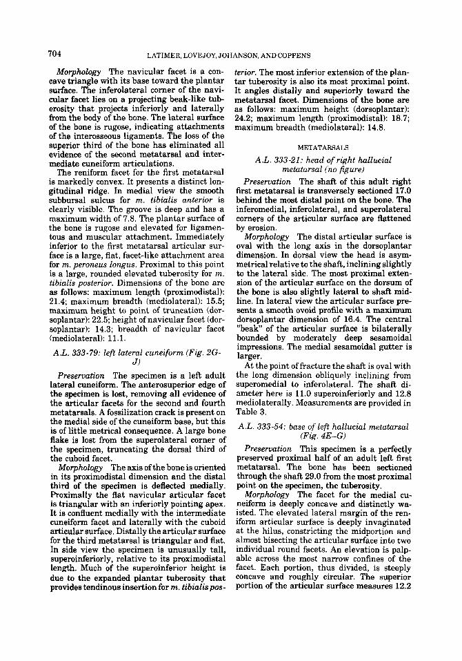
TABLE 1 . Calcaneal metrics (mm)
Measurements A.L. 333-8 A.L. 333-37 A.L. 333-55
Maximum dimension of fragment
Maximum dormplantar height of calcaneal tuberosity
Maximum mediolateral breadth of calcaneal tuberosity
Maximum mediolateral breadth of posterior talar facet
Maximum proximodistal length of posterior talar facet
Proximodistal length of sustentaculum tali base
Dorsoplantar height of sustentaculum tali base
61.9 41.2 52.0
38.1 33.5'
22.7l 24.6
38..8
23.6
24.4' 24.5l
-
29.8'
-
25.5
- - 11.0
'Approximate dimension ( 2 1.0).

HADAR FOOT SKELETON 703
and larger. Measurements are provided in Ta- ble 1.
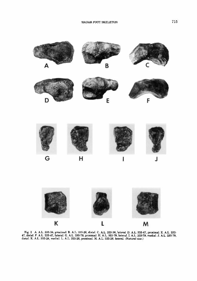
A.L. 333-36: right navicular (Fig. 2A-C) Preservation This specimen is a complete
and undamaged right pedal navicular. Slight surface erosion is evident over much of the bone but does not obscure morphologic detail.
Morphology In dorsal view the dominant feature is the large and robust navicular tub- erosity. It is expanded in its proximodistal di- mension, providing a large, rugose area of at- tachment along its inferior and lateral border for m. tibialis posterior and the plantar cal- caneonavicular (spring) ligament. The tuber- osity is at the same time compressed in its superoinferior dimension, giving the specimen a long and flattened appearance.
The talar facet is a deep ovoid extending well onto the proximal surface of the tuberosity. The facet is steeply concave with a short radius of curvature. In dorsal view the medial third of the facet, which is prolonged onto the tub- erosity, angles with the lateral two-thirds of the facet at approximately 130". In plantar view these two portions of the articular surface coalesce in a more rounded fashion, giving the talar articular facet a sigmoid shape.
On the distal surface the three triangular cuneiform facets are contiguous but clearly distinguishable from each other by distinct differences in contour and angulation. The lat- eral cuneiform facet is slightly concave, the intermediate facet is flat, and the medial cu- neiform facet, the least distinct of the three, is convex in its superior two-thirds and concave in its inferior third. On the lateral surface is a flat rectangular facet for the cuboid that forms a distinct 90" angle with the articular area for the lateral cuneiform. Medial to the cuboid facet the plantar surface of the bone is roughened for ligamentous attachment. The nonarticular areas are pitted by numerous vas- cular foramina. Measurements are provided in Table 2.
A.L. 33347: right nnvicular (Fig. 2D-F) Preservation The specimen is a perfectly
preserved right pedal navicular bone. Morphology This navicular is morphologi-
cally very similar to A.L. 333-36. In dorsal view the dominant feature of this specimen is the expanded navicular tuberosity. It is elon- gated proximodistally but compressed in its superoinferior dimension, contributing to its flattened appearance. The talar facet is a deep concave oval with a raised rim. Several small
TABLE 2. Nauicular metries (mm)
A.L. 333.36 A.L. 333-47 Measurements
Maximum length of 37.0 36.3 specimen
Maximum height of 20.2 21.5 specimen taken perpendicular to length
Proximodistal thickness 22.5 18.5 of tuberosity
Dorsoplantar height of 11.5 10.9 tuberosity
Maximum length of talar 26.1 24.1 facet
Maximum height of talar 16.5 16.5 facet taken perpendicular to length
pits are present on the inferolateral quarter of the facet and may be premortem subchondral defects. This specimen displays the same sig- moid curvature in the talar articular surface as A.L. 333-36. In distal view the cuneiform facets are distinguishable from each other by contour and angular orientation. The facet for the lateral cuneiform is concave, the inter- mediate cuneiform facet is flat with a slight concavity in its dorsal third, and the medial cuneiform facet is convex in its dorsal half and mildly concave in its plantar half. The flat rec- tangular facet for the cuboid is confluent with and angles from the lateral cuneiform articu- lar surface at approximately 110". A sharp, well-developed crest courses obliquely across the dorsal surface of the bone. It represents the ligamentous attachment with the cuneiforms. On the plantar surface of the bone the area receiving the plantar calcaneonavicular liga- ment is deeply grooved and excavated. Meas- urements are provided in Table 2.
A.L. 333-28: right medial cuneiform (Fig.
Preservation The specimen is approxi- mately the inferior two-thirds of an adult right medial cuneiform. The superior portion of the specimen is lost owing to a transverse break through the articular surface for the inter- mediate cuneiform. The navicular facet is well preserved except for some minor surface ero- sion along its margin. The facet for the first metatarsal is slightly damaged on its lateral edge.
2K-M)
LATIMER, LOVEJOY, JOHANSON. AND COPPENS 704
Morphology The navicular facet is a con- cave triangle with its base toward the plantar surface. The inferolateral corner of the navi- cular facet lies on a projecting beak-like tub- erosity that projects inferiorly and laterally from the body of the bone. The lateral surface of the bone is rugose, indicating attachments of the interosseous ligaments. The loss of the superior third of the bone has eliminated all evidence of the second metatarsal and inter- mediate cuneiform articulations.
The reniform facet for the first metatarsal is markedly convex. It presents a distinct lon- gitudinal ridge. In medial view the smooth subbursal sulcus for m. tibialis anterior is clearly visible. The groove is deep and has a maximum width of 7.8. The plantar surface of the bone is rugose and elevated for ligamen- tous and muscular attachment. Immediately inferior to the first metatarsal articular sur- face is a large, flat, facet-like attachment area for m. peroneus longus. Proximal to this point is a large, rounded elevated tuberosity for m. tibialis posterior. Dimensions of the bone are as follows: maximum length (proximodistal): 21.4; maximum breadth (mediolateral): 15.5; maximum height to point of truncation (dor- soplantar): 22.5; height of navicular facet (dor- soplantar): 14.3; breadth of navicular facet (mediolateral): 11.1.
A.L. 333-79: left lateral cuneiform (Fig. 2G- J)
Preservation The specimen is a left adult lateral cuneiform. The anterosuperior edge of the specimen is lost, removing all evidence of the articular facets for the second and fourth metatarsals. A fossilization crack is present on the medial side of the cuneiform base, but this is of little metrical consequence. A large bone flake is lost from the superolateral corner of the specimen, truncating the dorsal third of the cuboid facet.
Morphology The axis of the bone is oriented in its proximodistal dimension and the distal third of the specimen is deflected medially. Proximally the flat navicular articular facet is triangular with an inferiorly pointing apex. It is confluent medially with the intermediate cuneiform facet and laterally with the cuboid articular surface. Distally the articular surface for the third metatarsal is triangular and flat. In side view the specimen is unusually tall, superoinferiorly, relative to its proximodistal lenah. Much of the superoinferior height is due to the expanded plantar tuberosity that provides tendinous insertion for m. tibialispos-
terior. The most inferior extension of the plan- tar tuberosity is also its most proximal point. It angles distally and superiorly toward the metatarsal facet. Dimensions of the bone are as follows: maximum height (dorsoplantar): 24.2; maximum length (proximodistal): 18.7; maximum breadth (mediolateral): 14.8.
METATARSALS
A.L. 333-21: head of right hallucial metatarsal (no figure)
Preservation The shaft of this adult right first metatarsal is transversely sectioned 17.0 behind the most distal point on the bone. The inferomedial, inferolateral, and superolateral corners of the articular surface are flattened by erosion.
Morphology The distal articular surface is oval with the long axis in the dorsoplantar dimension. In dorsal view the head is asym- metrical relative to the shaft, inclining slightly to the lateral side, The most proximal exten- sion of the articular surface on the dorsum of the bone is also slightly lateral to shaft mid- line. In lateral view the articular surface pre- sents a smooth ovoid profile with a maximum dorsoplantar dimension of 16.4. The central “beak of the articular surface is bilaterally bounded by moderately deep sesamoidal impressions. The medial sesamoidal gutter is larger.
At the point of fracture the shaft is oval with the long dimension obliquely inclining from superomedial to inferolateral. The shaft di- ameter here is 11.0 superoinferiorly and 12.8 mediolaterally. Measurements are provided in Table 3.
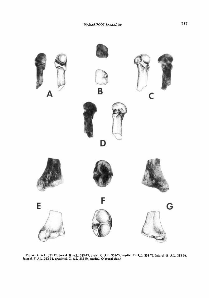
A.L. 333-54: base of left hallucial metatarsal (Fig. 4E-G)
Preservation This specimen is a perfectly preserved proximal half of an adult left first metatarsal. The bone has been sectioned through the shaft 29.0 from the most proximal point on the specimen, the tuberosity.
Morphology The facet for the medial cu- neiform is deeply concave and distinctly wa- isted. The elevated lateral margin of the ren- iform articular surface is deeply invaginated at the hilus, constricting the midportion and almost bisecting the articular surface into two individual round facets. An elevation is palp- able across the most narrow confines of the facet. Each portion, thus divided, is steeply concave and roughly circular. The superior portion of the articular surface measures 12.2

TAB
LE 3
. Met
atar
sal m
etri
cs (mm)
Max
imum
med
iola
tera
l M
axim
um d
orso
plan
tar
Max
imum
med
iola
tera
l M
axim
um d
orso
plan
tar
Max
imum
leng
th
brea
dth
of b
ase
dept
h of
bas
e br
eadt
h of
hea
d de
pth
of h
ead
of sp
ecim
en'
Met
atar
sal I
? U
16.0
16.42
18.7
A.L
. 333-21
-
-
A.L
. 333-54
17.4
23.5
-
A.L
. 333-115(A)
-
A.L
. 333-72
-
-
A.L
. 333-115(B)
-
-
A.L
. 333-115(C)
-
A.L
. 333-115(D)
-
-
A.L
. 333-13
16.0
13.1
-
A.L
. 333-78
15.8
11.6
-
A.L
. 333-115(E)
-
29.6
* -
17.5
17.3
33.7
28
-
Met
atar
sal Il
8 10.7
13.3
33.0
10.7
12.6
25.6
9.2
13.2
23.8
9.5
14.0
B M
etat
arsa
l IV
..1
16.2
0
Met
atar
sal V
2
Met
atar
sal I
II
m
r
e -
43.0
56.1
8.7
12.5
11.5
-
-
-
'Max
imum
len
gth
refe
rs o
nly
to p
rese
rved
leng
th; n
o m
etat
arsa
l sp
ecim
en i
s co
mpl
ete.
'A
ppro
xim
ate
dim
ensi
on ( c
0.5)
.
4
0
cn
706 LATIMER. LOVEJOY, JOHANSON, AND COPPENS
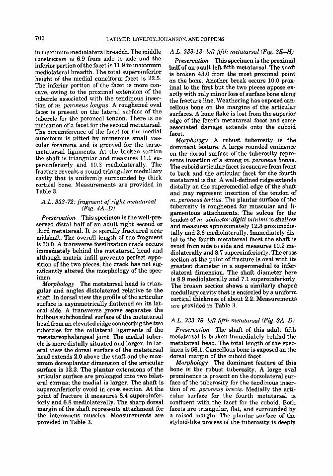
in maximum mediolateral breadth. The middle constriction is 6.9 from side to side and the inferior portion of the facet is 11.9 in maximum mediolateral breadth. The total superoinferior height of the medial cuneiform facet is 22.5. The inferior portion of the facet is more con- cave, owing to the proximal extension of the tubercle associated with the tendinous inser- tion of m. peroneus longus. A roughened oval facet is present on the lateral surface of the tubercle for the peroneal tendon. There is no indication of a facet for the second metatarsal. The circumference of the facet for the medial cuneiform is pitted by numerous small vas- cular foramina and is grooved for the tarso- metatarsal ligaments. At the broken section the shaft is triangular and measures 11.1 su- peroinferiorly and 10.3 mediolaterally. The fracture reveals a round triangular medullary cavity that is uniformly surrounded by thick cortical bone. Measurements are provided in Table 3.
A.L. 333-72: fragment of right metatarsal (Fig. 4A-D)
Preservation This specimen is the well-pre- served distal half of an adult right second or third metatarsal. It is spirally fractured near midshaft. The overall length of the fragment is 33.0. A transverse fossilization crack occurs immediately behind the metatarsal head and although matrix infill prevents perfect appo- sition of the two pieces, the crack has not sig- nificantly altered the morphology of the spec- imen.
Morphology The metatarsal head is trian- gular and angles distolaterad relative to the shaft. In dorsal view the profile of the articular surface is asymmetrically flattened on its lat- eral side. A transverse groove separates the bulbous subchondral surface of the metatarsal head from an elevated ridge connecting the two tubercles for the collateral ligaments of the metatarsophalangeal joint. The medial tuber- cle is more distally situated and larger. In lat- eral view the dorsal surface of the metatarsal head extends 2.0 above the shaft and the max- imum dorsoplantar dimension of the articular surface is 13.3. The plantar extensions of the articular surface are prolonged into two bilat- eral cornua; the medial is larger. The shaft is superoinferiorly ovoid in cross section. At the point of fracture it measures 8.4 superoinfer- iorly and 6.8 mediolaterally. The sharp dorsal margin of the shaft represents attachment for the interosseus muscles. Measurements are
A.L. 333-13: left fifth metatarsal (Fig. 3E-H) Preservation This specimen is the proximal
half of an adult left fifth metatarsal. The shaft is broken 43.0 from tbe most proximal point on the bone. Another break occurs 10.0 prox- imal to the first but the two pieces appose ex- actly with only minor loss of surface bone along the fracture line. Weathering has exposed can- cellous bone on the margins of the articular surfaces. A bone flake is lost from the superior edge of the fourth metatarsal facet and some associated damage extends onto the cuboid facet.
Morphology A robust tuberosity is the dominant feature. A large rounded eminence on the dorsal surface of the tuberosity repre- sents insertion of a strong m. peroneus brevis. The cuboid articular facet is concave from front to back and the articular facet for the fourth metatarsal is flat. A well-defined ridge extends distally on the superomedial edge of the shaft and may represent insertion of the tendon of m. peroneus tertius. The plantar surface of the tuberosity is roughened for muscular and li- gamentous attachments. The sulcus for the tendon of m. abductor dzgiti minimi is shallow and measures approximately 12.3 proximodis- tally and 2.6 mediolaterally. Immediately dis- tal to the fourth metatarsal facet the shaft is ovoid from side to side and measures 10.2 me- diolaterally and 8.7 superoinferiorly. The cross section a t the point of fracture is oval with its greatest diameter in a superomedial to infer- olateral dimension. The shaft diameter here is 8.9 mediolaterally and 7.1 superoinferiorly. The broken section shows a similarly shaped medullary cavity that is encircled by a uniform cortical thickness of about 2.2. Measurements are provided in Table 3.
A.L. 333-78: left fifth metatarsal (Fig. 3A-D) Preservation The shaft of this adult fifth
metatarsal is broken immediately behind the metatarsal head. The total length of the spec- imen is 56.1. Cancellous bone is exposed on the dorsal margin of the cuboid facet.
Morphology The dominant feature of this bone is the robust tuberosity. A large oval prominence is present on the dorsolateral sur- face of the tuberosity for the tendinous inser- tion of m. peroneus brevis. Medially the arti- cular surface for the fourth metatarsal is confluent with the facet for the cuboid. Both facets are triangular, flat, and surrounded by a raised margin. The plantar surface of the
provided in Table 3. styloid-like process of the tuberosity is deeply

HADAR FOOT SKELETON 707
excavated by the tendon of m. abductor digiti minimi. The sulcus is pitted by numerous small vascular foramina and measures 12.6 in length and 3.2 in maximum width. The shaft displays a marked lateral curvature. A cross section through the shaft immediately distal to the fourth metatarsal facet (22.0 distal to the stylus of the tuberosity) would be elliptical from side to side, with sharp medial and lateral edges, a flat dorsal surface, and a more rounded plantar surface. The shaft diameter here is 10.0 mediolaterally and 6.9 superoinferiorly. The sharp medial border may represent inser- tion of the tendon of m. peroneus tertius. The diaphysis becomes progressively more circular as it proceeds distally. The broken section is oval and measures 6.2 mediolaterally and 6.8 superoinferiorly. Measurements are provided in Table 3.
PHALANGES
A.L. 333w-25: proximal pedal phalanx (Fig. 5a)
Preservation This specimen consists of two- thirds of an adult proximal phalanx. The shaft is transversely sectioned 26.5 from the most proximal point on the bone.
Morphology The phalangeal base is ex- panded and surrounded by rugose ligamentous insertions. The proximal articular surface is deeply concave with a sharp dorsal rim. The plantar tubercles for the collateral ligaments of the metatarsophalangeal joint are large and equal in size and shape. A rounded median ridge 5.2 in width begins a t the tubercles and continues the length of the diaphyseal plantar surface. This ridge is bounded laterally by roughened hollows for the fibrous flexor sheath. The complete specimen would have exhibited strong plantar curvature. At the point of fracture the diaphyseal cross section is mediolaterally elongated with a small med- ullary cavity. Measurements are provided in Table 4. A.L. 333w-51: proximal pedal phalanx (Fig.
5b) Preservation The specimen is the proximal
half of an adult proximal phalanx. The speci- men is transversely sectioned a t approxi- mately midshaft and measures 20.8.
Morphology The robust phalangeal base is surrounded by rugose ligamentous insertions. The proximal articular surface is mediolater- ally ovoid and deeply concave. The dorsal mar- gin of the articular surface is a sharp raised rim. The plantar tubercles for the collateral
ligaments of the metatarsophalangeal articu- lation are large and equal in size. A flat median ridge 6.4 in breadth arises at the tubercles and continues the length of the plantar diaphyseal surface. This ridge is bounded laterally by sharp crests for the fibrous flexor sheath. This specimen displays an expanded joint surface. At the point of fracture the diaphyseal cross section is mediolaterally elongated with a small medullary cavity. Measurements are provided in Table 4.
A.L. 333-22: proximal pedal phalanx (Fig. 5c)
Preservation The specimen is the distal half of a proximal phalanx. The shaft is trun- cated 21.8 behind the most distal point on the bone.
Morphology The distal articular surface is trochlear with both condyles of equal size and shape. The groove between the condyles is rel- atively deep and the expanded joint surface encroaches further upon the plantar diaphy- seal surface than the dorsal surface. Shallow circular depressions are present on both sides of the joint for the attachment of the collateral ligaments of the interphalangeal articulation. On the plantar surface are clearly marked at- tachments for the fibrous flexor sheath that flank a median ridge 4.4 wide. The plane of fracture is roughly circular in cross section. Measurements at the broken end are as fol- lows: shaft depth (dorsoplantar): 6.3; shaft breadth (mediolateral): 6.7. The cortex aver- ages 1.6 in thickness at the point of fracture. Additional measurements are provided on Ta- ble 4.
A.L. 333-26: proximal pedal phalanx (Fig. 5 4
Preservation This specimen is perfectly preserved.
Morphology The proximal articular sur- face is deeply concave, circular in outline, and encircled by a raised rim. The distal articular surface is bicondylar with one condyle slightly larger and more anteriorly projecting. The in- tercondylar groove is quite deep and the head displays bilateral pits for attachment of the collateral ligaments of the interphalangeal joint. The phalangeal head is flattened and tilts ventrally relative to the shaft. The shaft is mediolaterally constricted at both ends, giv- ing it a somewhat fusiform shape. The ex- panded joint surfaces incline asymmetrically to one side of the shaft. On the plantar surface is a flat median ridge bounded on both sides
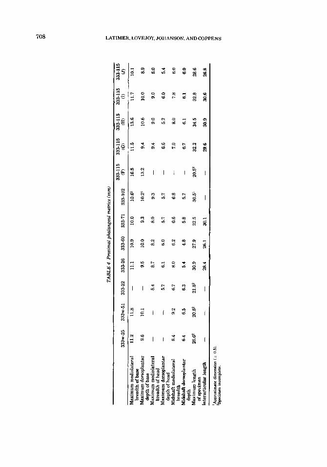
TAB
LE 4
. Pro
xim
al p
hula
ngea
l m
etri
cs (m
m)
333-115
333-115
333-115
333-115
333-115
333~
-25
333~-51 333-22
333-26
333-60
333-71
333-102
(F)
(GI
(H)
(1)
(J)
Max
imum
med
iola
tera
l 11.2
11.8
-
11.1
10.9
10.0
10.6'
16.8
11.5
13.6
11.7
10.1
Max
imum
dor
sopl
anta
r 9.6
10.1
-
9.5
10.0
9.3
10.2'
13.2
9.4
10.6
10.0
8.9
Max
imum
med
iola
tera
l -
-
8.4
8.7
8.2
8.0
9.3
-
9.4
9.0
9.0
8.0
Max
imum
dor
mpl
anta
r -
-
5.7
6.1
6.0
5.7
5.7
-
6.6
5.7
6.0
5.4
brea
dth
of b
ase
dept
h of
bas
e
brea
dth
of h
ead
dept
h of
hea
d
brea
dth
dept
h
of sp
ecim
en
Mid
shaf
t med
iola
tera
l 8.4
9.2
6.7
8.0
6.2
6.6
6.8
-
7.0
8.0
7.8
6.0
Mid
shal
t dor
sopl
anta
r 6.4
6.5
6.3
5.4
4.8
5.8
5.7
-
6.7
6.1
6.1
6.0
Max
imum
leng
th
26.@
20.82
21S
30.9
27.9
32.5
30.5'
20.52
32.2
34.5
32.8
28.6
-
28.4
26.1
30.1
-
-
29.6
30.9
30.6
26.8
Inte
rart
icul
ar le
ngth
-
-
> z U
'App
roxi
mat
e di
men
sion
( c
0.5)
2S
peci
men
inco
mpl
ete.
HADAR FOOT SKELETON 709
by sharp crests for the insertion of the fibrous flexor sheath. The plantar tubercles for at- tachment of the collateral ligaments of the metatarsophalangeal articulation are large and equal in size. The bone displays marked plantar curvature. Measurements are pro- vided in Table 4.
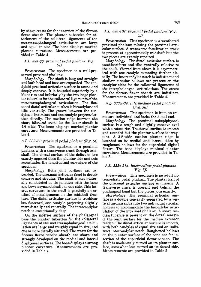
A.L. 333-60: proximal pedal phalanr (Fig. 5 4
Preservation The specimen is a well-pre- sewed proximal phalanx.
Morphology The shaft is long and straight and both head and base are expanded. The con- dyloid proximal articular surface is round and deeply concave. It is bounded superiorly by a blunt rim and inferiorly by the two large plan- tar tubercles for the collateral ligaments of the metatarsophalangeal articulation. The flat- tened distal articular surface is bicondylar and tilts ventrally. The groove between the con- dyles is indistinct and one condyle projects fur- ther distally. The median ridge between the sharp bilateral crests for the flexor sheath is 4.6 wide. The bone displays marked plantar curvature. Measurements are provided in Ta- ble 4. A.L. 333-71: proximal pedal phalanx (Fig. 50
Preservation The specimen is a proximal phalanx with a transverse crack through mid- shaft. The dorsal surface of the defect is less exactly apposed than the plantar side and this accentuates the longitudinal curvature of the specimen.
Morphology Both joint surfaces are ex- panded. The proximal articular facet is deeply concave and circular. The shaft is mediolater- ally constricted at its junction with the base and bows asymmetrically to one side. This lat- eral curvature in the shaft is partially an ar- tifact of misalignment in the midshaft frac- ture. The distal articular surface is trochlear but flattened, one condyle projecting slightly more distally and ventrally. The intercondylar notch is exceptionally deep.
On the inferior surface of the phalangeal base the plantar tubercles for the collateral ligaments of the metatarsophalangeal articu- lation are large and roughly equal in size, and one is more distally situated. The crests for the fibrous flexor tendor sheath are sharp and strongly developed on the medial and lateral disphyseal surfaces. The bone displays a strong plantar curvature. Measurements are pro- vided in Table 4.
A.L. 333-102: proximal pedal phalanx (Fig. 5g)
Preservation This specimen is a weathered proximal phalanx missing the proximal arti- cular surface. A transverse fossilization crack is present at approximately midshaft but the two pieces are exactly repaired.
Morphology The distal articular surface is trochleariform and tilts ventrally relative to the shaft. Viewed from above it is asymmetr- ical with one condyle extending further dis- tally. The intercondylar notch is indistinct and shallow circular hollows are present on the condylar sides for the collateral ligaments of the interphalangeal articulations. The crests for the fibrous flexor sheath are indistinct. Measurements are provided in Table 4.
A.L. 333w-34: intermediate pedal phalanx (Fig. 5h)
Preservation This specimen is from an im- mature individual and lacks the distal end.
Morphology The proximal subepiphyseal surface is a rough and slightly concave oval with a raised rim. The dorsal surface is smooth and rounded but the plantar surface is irreg- ular. A 3.8-wide median plantar ridge is bounded on its medial and lateral sides by roughened hollows for the superficial digital flexors. The bone displays minimal plantar curvature. Measurements are provided in Ta- ble 5.
A.L. 333x-21a: intermediate pedal phalanx (Fig. 5j)
Preservation This specimen is an adult in- termediate pedal phalanx. The plantar half of the proximal articular surface is missing. A transverse crack is present just behind the phalangeal head but the pieces join exactly.
Morphology The proximal articular sur- face is a double concavity separated by a ver- tical median ridge into two individual circular hollows to accommodate the bicondylar artic- ulation of the proximal phalanx. A sharp me- dian tubercle is present on the dorsal margin of the joint surface for the median extensor tendon. The distal articular surface is smooth, with both condyles of equal size and an indis- tinct intercondylar notch. Roughened hollows on the plantar surface of the bone are for in- sertion of the superficial flexor tendon. The shaft is moderately curved on its plantar sur- face, somewhat less curved on its dorsal side. Measurements are provided in Table 5.
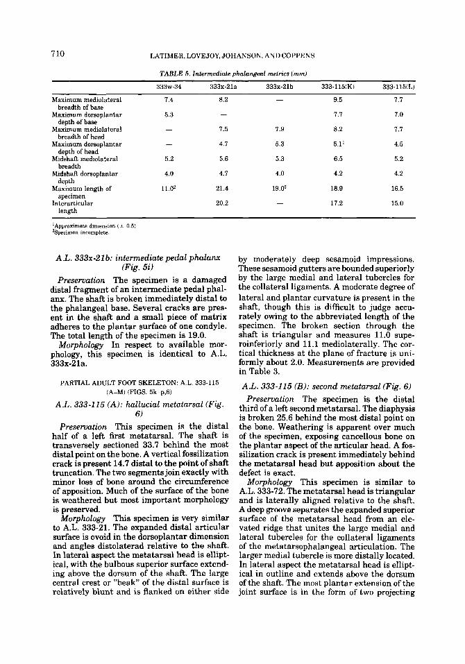
7 10 LATIMER, LOVEJOY, JOHANSON. AND COPPENS
TABLE 5. Intermediate phalangeal metrics (mm)
333w-34 333x-21a 333x-21b 333-115(K) 333-115(L)
Maximum mediolateral 7.4 8.2 - 9.5 7.1
Maximum dorsoplantar 5.3 - - 1.1 7.0
Maximum mediolateral - 1.5 1.9 8.2 1.7
breadth of base
depth of base
breadth of head
depth of head
breadth
depth
specimen
length
Maximum dorsoplantar - 4.1 5.3 5.1’ 4.5
Midshaft mediolateral 5.2 5.6 5.3 6.5 5.2
Midshaft dorsoplantar 4.0 4.1 4.0 4.2 4.2
Maximum length of 11.02 21.4 19.02 18.9 16.5
Interarticular - 20.2 - 17.2 15.0
‘Approximate dimension ( 2 0 5 ) ’Specimen incomplete.
A.L. 333r-21 b: intermediate pedal phalalwc (Fig. 5i)
Preservation The specimen is a damaged distal fragment of an intermediate pedal phal- anx. The shaft is broken immediately distal to the phalangeal base. Several cracks are pres- ent in the shaft and a small piece of matrix adheres to the plantar surface of one condyle. The total length of the specimen is 19.0.
Morphology In respect to available mor- phology, this specimen is identical to A.L. 333x-21a.
PARTIAL ADULT FOOT SKELETON: A.L. 333-115 (A-M) (FIGS. 5k-p,6)
A.L. 333-1 15 (A): hallucial metatarsal (Fig. 6)
Preservation This specimen is the distal half of a left first metatarsal. The shaft is transversely sectioned 33.7 behind the most distal point on the bone. A vertical fossilization crack is present 14.7 distal to the point of shaft truncation. The two segments join exactly with minor loss of bone around the circumference of apposition. Much of the surface of the bone is weathered but most important morphology is preserved.
Morphology This specimen is very similar to A.L. 333-21. The expanded distal articular surface is ovoid in the dorsoplantar dimension and angles distolaterad relative to the shaft. In lateral aspect the metatarsal head is ellipt- ical, with the bulbous superior surface extend- ing above the dorsum of the shaft. The large central crest or “beak” of the distal surface is relatively blunt and is flanked on either side
by moderately deep sesamoid impressions. These sesamoid gutters are bounded superiorly by the large medial and lateral tubercles for the collateral ligaments. A moderate degree of lateral and plantar curvature is present in the shaft, though this is difficult to judge accu- rately owing to the abbreviated length of the specimen. The broken section through the shaft is triangular and measures 11.0 supe- roinferiorly and 11.1 mediolaterally. The cor- tical thickness at the plane of fracture is uni- formly about 2.0. Measurements are provided in Table 3.
A.L. 333-115 (B): second metatarsal (Fig. 6) Preservation The specimen is the distal
third of a left second metatarsal. The diaphysis is broken 25.6 behind the most distal point on the bone. Weathering is apparent over much of the specimen, exposing cancellous bone on the plantar aspect of the articular head. A fos- silization crack is present immediately behind the metatarsal head but apposition about the defect is exact.
Morphology This specimen is similar to A.L. 333-72. The metatarsal head is triangular and is laterally aligned relative to the shaft. A deep groove separates the expanded superior surface of the metatarsal head from an ele- vated ridge that unites the large medial and lateral tubercles for the collateral ligaments of the metatarsophalangeal articulation. The larger medial tubercle is more distally located. In lateral aspect the metatarsal head is ellipt- ical in outline and extends above the dorsum of the shaft. The most plantar extension of the joint surface is in the form of two projecting
HADAR FOOT SKELETON 711
cornua. The larger medial plantar cornu proj- ects inferiorly and posteriorly. The maximum mediolateral dimension of the metatarsal head is measured a t the plantar cornua and is 10.7. At the point of fracture the shaft. is ovoid with an oblique superomedial to inferolateral axis. The shaft dimensions here are 7.6 superoin- ferior and 7.9 mediolateral. The cortical bone is notably thick and at the point of fracture is 2.2. Measurements are provided in Table 3.
A.L. 333-115 (C): third metatarsal (Fig. 6)
Preservation This specimen is badly weath- ered and consists of anterior portion of a left third metatarsal. The shaft is broken 23.8 be- hind the most distal point on the metatarsal head. At the plane of fracture a large bone flake is missing from the superior margin of the diaphysis.
Morphology The metatarsal head is a tall, laterally compressed triangle and inclines slightly toward the lateral side of the bone. This lateral alignment relative to the shaft is less than that seen in the second metatarsal A.L. 333-115(B). The groove that separates the superior surface of the articular head from the shaft is shallower and less distinct than in the second metatarsal. In lateral aspect the arti- cular profile is elliptical and the dorsum of the head is horizontal with the superior surface of the shaft. The medial plantar cornu is the more projecting and is separated from the lateral process by a sharp intercornual notch. Imme- diately proximal to the metatarsal head the shaft is a narrow oval oriented superoinfer- iorly. Damage to the dorsal surface does not allow a superoinferior diaphyseal measure- ment but the maximum mediolateral breadth taken immediately behind the metatarsal head is 5.3. Additional measurements are pro- vided in Table 3.
A L . 333-115 (D): fourth metatarsal (Fig. 6) Preservation This specimen is the articular
head from a left fourth metatarsal. In total length the specimen measures 16.2. The infer- omedial comer of the articular surface, the medial cornu, is damaged, with two bone chips cemented in approximate position.
Morphology The metatarsal head is nar- row and triangular. The junction of the head and shaft is less marked than in the preceding metatarsal specimens (A.L. 333-115 A,B,C,) and the groove separating the head from the tubercles for the collateral ligaments is rela- tively indistinct. In lateral view the articular surface is elliptical in profile. Damage to the
inferomedial surface prohibits evaluation of the medial plantar cornu but the lateral cornu is prominent and projects inferolaterally. The plane of fracture reveals a narrow, laterally compressed ovoid shaft. Dimensions taken im- mediately distal to the section are 5.2 medi- olaterally and 8.3 superoinferiorly. Additional measurements are provided in Table 3.
A.L. 333-115 (E): fifth metatarsal (Fig. 6) Preservation The specimen is a left fifth
metatarsal head. The shaft is sectioned im- mediately beind the head and the fragment is 11.5 in total length. The medial plantar cornu is damaged.
Morphology The articular surface of the metatarsal head encroaches further proxi- mally on the lateral side of the bone. The me- dial tubercle for the collateral ligaments of the metatarsophalangeal articulation is larger and more distally situated than the lateral tu- bercle. On the plantar aspect the smooth sub- chondral surface extends as two inferiorly pro- jecting cornua. The medial cornu is larger and is separated from the articular surface by a shallow groove. The lateral cornu is contiguous with the joint surface. The intercornual notch is deep. The shaft is a rounded triangle a t the plane of fracture and measures 10.0 superoin- feriorly and 7.6 mediolaterally. Additional measurements are provided in Table 3.
A.L. 333-115 (F): proximal hallucial phalanx (Fig. 6)
Preservation The specimen is the posterior half of a proximal phalanx. The bone is broken at approximately midshaft and the total length of the specimen is 20.5. Small pieces of matrix adhere to the superomedial rim of the joint surface but they cannot be removed without damaging the fossil.
Morphology The bone has an expanded and robust base. The proximal articular surface is a deep concave oval with the maximum di- ameter directed mediolaterad. On the infero- lateral margin of the base is a shallow ovoid depression for attachment of the tendons of m. flexor hallucis brevis and m. adductor hallucis. The diaphysis is markedly robust and exhibits a mediolateral elongation of the cross section. The cortex is relatively very thick. Measure- ments are provided in Table 4.
A.L. 333-115 (G): second proximal phalanx (Figs. 5m,6)
Preservation The specimen is a badly weathered second proximal phalanx. A longi-
tudinal crack through the distal third of the specimen results in a slight plantar displace- ment of the affected piece and an exaggeration of the plantar curvature.
Morphology The proximal articular sur- face is circular and deeply concave. The distal joint surface is bicondylar, the medial condyle being slightly larger. The condyles present lat- eral depressions for the collateral ligaments of the interphalangeal joint. The bone is strongly curved in its longitudinal axis. The shaft ta- pers mediolaterally at its proximal and distal ends, assuming a fusiform shape. The plantar diaphyseal surface bears a 4.0-wide median ridge bounded on both sides by sharp crests for the fibrous flexor sheath. These crests are most pronounced at midshaft, contributing to the fusiform appearance of the diaphysis. The plantar tubercles for the collateral ligaments are flat and equal in size. Measurements are provided in Table 4.
712 LATIMER, LOVEJOY, JOHANSON, AND COPPENS
A.L. 333-115 (H): third proximal phalanx (Figs. 5n,6)
Preservation This specimen is a badly weathered third proximal pedal phalanx. The fossil is broken at midshaft but the pieces are well repaired.
Morphology A.L. 333-115 (HI is the largest and most robust of the associated proximal phalanges. The proximal articular surface is deeply concave and elongated mediolaterally, and it is bounded superiorly by a raised rim. The flattened distal articular surface tilts ven- trally and the medial condyle is the more dis- tally projecting. There is a moderate degree of lateral torsion to the phalangeal head. Circu- lar depressions on the sides of the condyles for the collateral ligaments are relatively deep with sharp margins. On the plantar surface the bilateral crests for the fibrous flexor sheath are sharp and strongly developed. The plantar tubercles for the collateral ligaments of the metatarsophalangeal joint are equally devel- oped. The bone displays marked plantar cur- vature. Measurements are provided in Table 4.
A.L. 333-115 (I): Fourth Proximal Phalanx (Figs. 50,6)
Preservation The specimen is an abraded but complete fourth proximal phalanx.
Morphology The proximal joint surface is circular and deeply concave. The distal arti- cular surface is bicondylar, the medial condyle being the larger and more distally projecting. Moderate lateral torsion is present in the phal-
angeal head relative to the shaft. On the plan- tar surface of the bone a raised median ridge 5.2 wide is bilaterally flanked by sharp ridges for the flexor sheath. The large lateral plantar tubercle projects further distally than the in- distinct medial tubercle. Viewed from above, the shaft displays a slight bowing toward the medial side. Measurements are provided in Table 4.
A.L. 333-115 (J): fifth proximal phalanx (Figs. 5p,6)
Preservation This specimen is a fifth prox- imal phalanx. A fossilization crack is present behind the phalangeal head. The two pieces join properly but there is associated bone loss on the dorsum of the shaft.
Morphology This is the smallest of the as- sociated proximal phalanges. The bone has a circular proximal articular surface that is markedly concave. The phalangeal head pre- sents a trochlear surface that is asymmetrical; the larger medial condyle extends a greater distance distally and the head inclines lat- erally in relation to the shaft. A mid-diaphy- seal cross section is roughly circular, not me- diolaterally elongated as in the other 333-115 proximal phalanges. In plantar view the me- dian ridge between the crests for the fibrous flexor sheath is elevated, giving the plantar surface a rounded rather than a flattened as- pect. The lateral plantar tubercle is the larger and occupies a more distal position. Measure- ments are provided in Table 4.
A.L. 333-115 (K): fourth intermediate phalanx (Figs. 5k,6)
Preservation This specimen is a badly weathered fourth intermediate pedal phalanx. A transverse fossilization crack is present in the shaft immediately behind the phalangeal head and an associated crack longitudinally transverses the proximal two-thirds of the bone. The pieces are reasonably well apposed but a large flake is missing from the medial surface of the bone.
Morphology This specimen is very stout, with expanded joint surfaces. The proximal articular area is an oval biconcave facet di- vided by a weak median ridge. It is bounded on the dorsal and plantar margins by sharp median tubercles. The distal articular surface is trochlear, with both condyles badly dam- aged. On the plantar surface the markings for the insertion of the flexor tendons are indis- tinct owing to surface erosion. The inferior sur- face is slightly curved and the dorsal side
HADARFOOTSKELETON 713
straight. Measurements are provided in Table 5 .
A.L. 333-115 (L): fifth intermediate phalanx (Figs. 51,6)
Preservation This specimen is a weathered fifth intermediate phalanx. The bone is broken at midshaft but the pieces join properly.
Morphology The bone is stout, with ex- panded joint surfaces. The proximal articular surface is an oval biconcave facet divided by a weak central crest. Surface erosion has ob- literated the flexor insertions on the bone’s in- ferior surface. In lateral view the bone is mod- erately curved on its ventral side with a flat dorsum. Measurements are provided in Table 5.
A.L. 333-115 (M): proximal epiphysis of Distal phalanx (Fig. 6)
Preservation This specimen is a proximal epiphysis from a fourth or fifth distal phalanx.
Morphology The joint surface is divided by a vertical median crest into two distinct hol- lows. It is rounded on the dorsal border, with a flat plantar base, and measures 7.2 in height and 7.8 in breadth.
LITERATURE CITED Trotter, M, and Peterson, R (1966) Osteology. In BJ Anson
(ed): Morris’ Human Anatomy. New York: Saunders, pp. 133-315.
7 14 LATIMER, LOVEJOY, JOHANSON. AND COPPENS
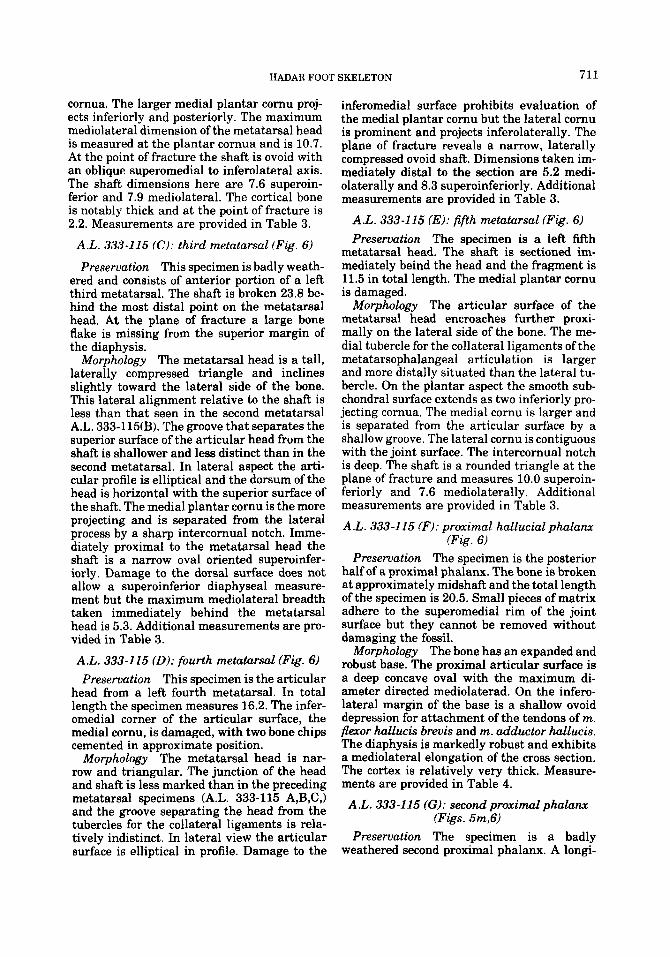
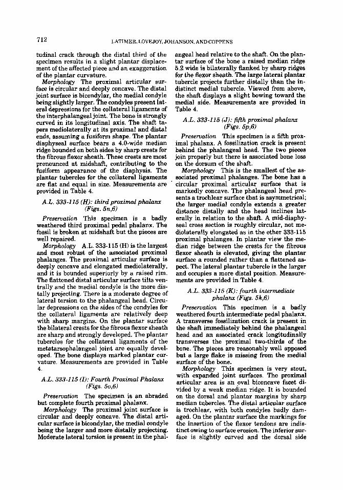
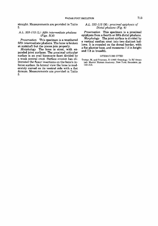
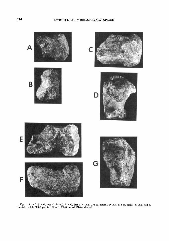
Fig. 1 A. A.L. 333-37, medial. B. A.L. 333-37, dorsal. C. A.L. 333-55, lateral. D. A.L. 333-55, dorsal. E. A.L. 333-8, medial. F. A.L. 333-8, plantar. G. A.L. 333-8, dorsal. (Natural size.)
HADAR FOOT SKELETON 715
Fig. 2 A. A.L. 333-36, proximal. B. A.L. 333-36, distal. C. A.L. 333-36, lateral. D. A.L. 333-47. proximal. E. A.L. 333- 47, distal. F. A.L. 333-47, lateral. G. A.L. 333-79, proximal. H. A.L. 333-79, lateral. 1. A.L. 333'79, medial. J. A.L. 333-79, distal. K. A.L. 333-28, medial. L. A.L. 333-28, proximal. M. A.L. 333-28, lateral. (Natural size.)
7 16 LATIMER, LOVEJOY, JOHANSON, AND COPPENS
Fig. 3 A. A.L. 333-78, dorsal. B. A.L. 333-78, lateral. C. A.L. 333-78, plantar. D. A.L. 333-78, medial. E. A.L. 333-13, dorsal. F. A.L. 333-13, lateral. G. A.L. 333-13, plantar. H. A.L. 333-13, medial. I. A.L. 333-75, distal. J. A.L. 333-75, dorsal. K. A.L. 333-75, inferolateral. (Natural size.)
HADAR FOOT SKELETON 717
Fig. 4 A. A.L. 333-72, dorsal. B. A.L. 333-72, distal. C. A.L. 333-72, medial. D. A.L. 333-72, lateral. E. A.L. 333-54, lateral. F. A.L. 333-54, proximal. G. A.L. 333-54, medial. (Natural size.)
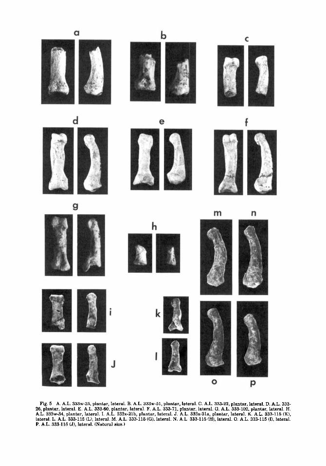
Fig. 5 A. A.L. 333w-25, plantar, lateral. B. A.L. 333w-51, plantar, lateral. C. A.L. 333-22, plantar, lateral. D. A.L. 333- 26, plantar, lateral. E. A.L. 333-60, plantar, lateral. F. A.L. 333-71, plantar, lateral. G. A.L. 333-102, plantar, lateral. H. A.L. 333w-34, plantar, lateral. I. A.L. 333x-21b, plantar, lateral. J. A.L. 333x-21a, plantar, lateral. K. A.L. 333-115 (K), lateral. L. A.L. 333-115 (L), lateral. M. A.L. 333-115 (G), lateral. N. A.L. 333-115 (HI, lateral. 0. A.L. 333-115 (I), lateral. P. A.L. 333-115 (J). lateral. (Natural size.)
HADAR FOOT SKELETON
A
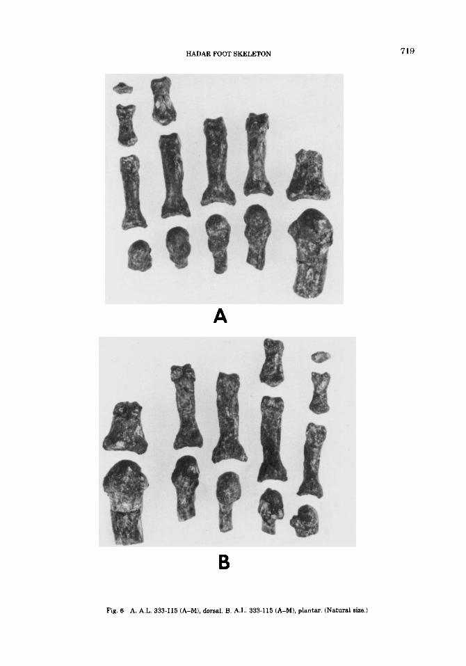
Fig. 6 A. A.L. 333-115 (A-MI, dorsal. B. A.L. 333-115 (A-M), plantar. (Natural size.)
719