Intertarsal and tarso-metatarsal subluxation in the dog
16
J. small Anitti. Ptact. (1976) 17, 427-442. Intertarsal and tarso-metatarsal subluxation in the dog JAMES R. CAMPBELL, DAVID BENNETT AND ROBIN LEE Department of Surgery, University of Glasgow Veterinary School, Bearsden, Glasgow G61 1QH ABSTRACT Forty-four cases of intertarsal subluxation and eight cases of tarso-meta- tarsal subluxation in the dog are reported. The anatoniical features of the hock related to these conditions are briefly considered. There was evidence that the Shetland Sheepdog is predisposed to intertarsal subluxation and in this breed minimal trauma was associated with the Occurrence of the condition. The history, clinical and radiographic features are reported for each condition and the methods and results of treatment described. INTRODUCTION Luxations and subluxations of the canine hock joint may involve the tibio-tarsal joint, the intertarsal joints or the tarso-metatarsal joints. Luxation or subluxation of the tibio-tarsal joint, with or without malleolar fractures, is recorded as the commonest type of hock instability by Pettit (1974). Lawson (1961) first reported intertarsal subluxation in the dog and since then further publications by Meutstege (1971) and Clayton-Jones (1974), who stated that it appears more common in the Shetland Sheepdog and Collie breeds, have appeared. Intertarsal luxation/ subluxation mainly involves the proximal intertarsal joint (i.e. between the first and second rows of tarsal bones) although occasionally the articulation between the fibular and tibia1 tarsal bones is affected. Instability of the distal intertarsal joint is rare, probably because the fourth tarsal bone spans the lateral aspect of the articulation and supports it (Pettit, 1974). Tarso-metatarsal subluxation in the dog was first reported by Arwedsson (1954) and Holt (1974) recently recounted three cases of tibio-tarsal instability and one of tarso-metatarsal with intertarsal instability, following severe trauma in each case. 42 7
-
Upload
independent -
Category
Documents
-
view
1 -
download
0
Transcript of Intertarsal and tarso-metatarsal subluxation in the dog

J . small Anitti. Ptact. (1976) 17, 427-442.
Intertarsal and tarso-metatarsal subluxation in the dog
J A M E S R. C A M P B E L L , D A V I D B E N N E T T A N D
R O B I N L E E
Department of Surgery, University of Glasgow Veterinary School, Bearsden, Glasgow G61 1QH
A B S T R A C T
Forty-four cases of intertarsal subluxation and eight cases of tarso-meta- tarsal subluxation in the dog are reported. The anatoniical features of the hock related to these conditions are briefly considered. There was evidence that the Shetland Sheepdog is predisposed to intertarsal subluxation and in this breed minimal trauma was associated with the Occurrence of the condition. The history, clinical and radiographic features are reported for each condition and the methods and results of treatment described.
I N T R O D U C T I O N
Luxations and subluxations of the canine hock joint may involve the tibio-tarsal joint, the intertarsal joints or the tarso-metatarsal joints. Luxation or subluxation of the tibio-tarsal joint, with or without malleolar fractures, is recorded as the commonest type of hock instability by Pettit (1974). Lawson (1961) first reported intertarsal subluxation in the dog and since then further publications by Meutstege (1971) and Clayton-Jones (1974), who stated that it appears more common in the Shetland Sheepdog and Collie breeds, have appeared. Intertarsal luxation/ subluxation mainly involves the proximal intertarsal joint (i.e. between the first and second rows of tarsal bones) although occasionally the articulation between the fibular and tibia1 tarsal bones is affected. Instability of the distal intertarsal joint is rare, probably because the fourth tarsal bone spans the lateral aspect of the articulation and supports it (Pettit, 1974). Tarso-metatarsal subluxation in the dog was first reported by Arwedsson (1954) and Holt (1974) recently recounted three cases of tibio-tarsal instability and one of tarso-metatarsal with intertarsal instability, following severe trauma in each case.
42 7

428 J . R . C A M P B E L L , D . B E N N E T T A N D R . L E E
During a period of approximately 10 years just over fifty cases of intertarsal and tarso-metatarsal subluxation without complicating bone fractures have been seen in the University of Glasgow Veterinary School. These are recounted in this publication.
Figure 1 shows the number and anatomical relationships of the canine tarsal bones. The intertarsal joints are supported by intertarsal ligaments between
FIG. 1. Anterior view of anatomical specimen of normal canine hock joint, showing the relationship of the different tarsal bones.
individual tarsal bones, together with the tarso-metatarsal ligaments. O n the plantar aspect of the tarsus lie the plantar ligaments. These originate on the tibia1 and fibular tarsals, are attached to the central and fourth tarsals, and insert on the strong tarso-metatarsal joint capsule. Damage to these various ligamentous structures must occur in cases of joint instability. Further details of the anatomy are given by Miller, Christensen & Evans (1965).
I N T E R T A R S A L SUBLUXATION
Forty-four cases of intertarsal subluxation have been referred from practising

S U B L U X A T I O N I N T H E D O G 429
veterinary surgeons in the surrounding area. An evaluation of the history, clinical features, radiographic changes and method of treatment was made for each case. Of the forty-four cases, thirty-nine had involvement of the proximal intertarsal joint. In two cases subluxation occurred at the distal intertarsal joint and three cases showed separation between the tibia1 and fibular tarsal bones (proximal intratarsal joint).
History including breed, age and sex incidence The breed incidence of the forty-four cases are shown in Table 1. A high
proportion of the animals suffering intertarsal subluxation were of the Shetland
TABLE 1. Breed incidence of intertarsal subluxation
~~ ~
Shetland Sheepdog Collie and Collie X Mongrel Greyhound Whippet Labrador Retriever Terrier
Total
22 10 7 2 I 1 1
44
Sheepdog breed (50% of the total). Table 2 shows the total number of dogs seen at the hospital by both the medicine and surgery departments during a 43-year period, and the most commonly represented breeds are also given. The hospital has a fairly high input of the Shetland Sheepdog and Collie breeds which suggests that these breeds are popular and common in the Glasgow area. Nonetheless,
TABLE 2. Showing number of dogs and principal breeds seen at the Glasgow veterinary hospital, over a 4t-year
period
Total number of dogs Principal breeds
Mongrel Terrier Alsatian Labrador Poodle Collie Spaniel Boxer Shetland Sheepdog
7,848
18.1 % 14.6% 10.2% 10.1 % 7.1 % 6.7 5.2 % 3.1 % 2.7%

430 J . R. CAMPBELL, D. BENNETT A N D R. LEE
the high proportion of Shetland Sheepdogs showing intertarsal subluxation indicates a predisposition in this breed. The forty-four cases comprised twenty- nine niales and fifteen females and the average age was 7.8 years (Figs 2 & 3) . Twenty-one cases involved the left hock, eighteen the right, and five cases were bilateral, the second joint developing abnormality 6 weeks to 8 years after the first, except in one case in which both joints became affected at the same time.
0 2 3 4 5 6 7 8 9 1 0 1 1 12
Age (yea r s )
FIG. 2. Showing the age disiribution of Shetland Sheepdogs affected with intertarsal sublwation.
‘r n
Age (years)
FIG. 3. Showing the age distribution of other breeds affected with intertarsal subluxation (age of three cases not recorded).
Eleven of thc patients (25%) had received known severe trauma, such as a road accident or being savaged by other dogs. Only two of these cases were Shetland Sheepdogs but all three cases of intratarsal subluxation are included in this group. Two greyhounds sustained the injury while racing. Nine dogs (20.5 x) sustained only minimal trauma such as landing awkwardly when jumping or falling down steps. I n the remaining twenty-four cases (54.5%) no trauma was recorded and for three of these cases it was known that the injury occurred during nornial vigorous use of the limb (running) but in the others the lameness was not even preceded by this amount of activity. A comparison of these findings in Shetland Sheepdogs with those in other breeds is shown in Table 3.
Most patients were referred several weeks after the onset of lameness and sonie

SUBLUXATION IN THE DOG 43 1
TABLE 3. Comparing some of the principal features of intertarsal sub- luxation as seen in the Shetland Sheepdog and other breeds
No. of cases
Shetland Sheepdogs Other breeds
Ihgs suffering no known trauma 16 8 Dogs suffering only minor trauma 4 5 Dogs suffering major trauma 2 9
Bilaterally affected dogs 4 1
Sex incidence M:F 15:7 14:8
cases had received treatment in the forni of external limb support. The duration of the clinical signs prior to internal fixation is shown in Fig. 4.
Ctinical features Each case was presented with a moderate to severe lameness although all cases,
other than those with intratarsal subluxation of tibia1 and fibular tarsals, were still able to support some body weight on the limb. Abnormal angulation of the hock joint, caused by ‘dorsiflexion’ of the unstable joint was present and on manipula- tion there was marked instability of the joint. The chronically affected animals demonstrated obvious thickening of the joint. Joint pain was not a feature even in chronic cases, although attempts to straighten the joint caused discomfort if the abnormality had been present for more than a few weeks. The three dogs with
Weeks Months
Duration of clinical sigm
FIG. 4. Showing duration of clinical signs of intertarsal subluxation prior to presentation at the hospital.

43 2 J . R . C A M P B E L L , D . B E N N E T T A N D R . L E E
intratarsal subluxation showed rotation of the foot rather than dorsiflexion, and pain and swelling was more prominent since two of them were seen shortly after the accident. Many of the dogs were overweight, the weights of affected Shetland Sheepdogs ranging from 6 to 26 kg.
Radiographic features The most consistent features observed on radiographic examination of the hock
were angulation of the tarsus at the level of the proximal intertarsal articulation, and ‘stepping’ of the articular margins between the tibia1 tarsal and central tarsal bones and the fibular and fourth tarsal bones. These features were merely a reflection of the subluxation occurring at the proximal intertarsal joint. The degree of subluxation varied from relatively minor displacement to almost total luxation of the joint. In addition, soft tissue swelling was generally demonstrable particular- ly posterior to the proximal intertarsal joint (Fig. 5).
In the majority of cases there was roughening and periosteal reaction of the posterior aspect of the fibular tarsal bone adjacent to the articular margin and this change in some instances extended onto the posterior aspects of the central and fourth tarsal bones (Figs 5 & 6 ) .
In about one-third of the radiographs examined there was also periosteal
FIG. 5. Radiograph demonstrating: (1) angulation of the hock and stepping of the articular margins of thc proximal intertarsal joint; (2) soft tissue swelling; (3) periosteal
reaction on the posterior aspect of the fibular, central and fourth tarsal bones.

S U B L U X A T I O N I N T H E D O G 433
FIG. 6. Radiograph demonstrating roughening and periosteal reaction on the anterior and posterior aspects of the hock, in addition to the marked subluxation at the proximal
intertarsal joint.
reaction on the anterior aspect of the hock involving the tibial tarsal, central tarsal and third tarsal bones. Again, the severity of this change varied from very slight to marked and was not obviously associated with the duration of clinical lameness (Fig. 6).
In two cases the subluxation was not a t the proximal intertarsal joint but a t the distal joint and was seen as stepping of the articular margins of central tarsal bone and the combined distal row of tarsal bones (Fig. 7), and in three cases proximal intratarsal luxation/subluxation was visualized as an obvious displacement be- tween the fibular and tibial tarsal bones.
In some cases there were free radio-opaque bodies associated with the proximal intertarsal joint, usually on the caudal aspect, and these may have represented either chip fractures of the proximal tarsal bones and as such may have been associated with ligamentous avulsions, or they may have merely resulted from separation of periarticular exostoses (Fig. 8).
A small series of Shetland Sheepdogs presented with unilateral intertarsal

434 J . R . C A M P B E L L , D . B E N N E T T A N D R . L E E
FIG. 7. Radiograph demonstratixig subluxation of the distal intertarsal joint.
subluxation had the opposite, clinically normal hock joint radiographed and a number of these showed early radiographical evidence of abnormality. The latter showed as a slight stepping of the proximal intertarsal joint, an increased caudal joint space or as a periosteal reaction involving the tarsal bones (Fig. 9). Of these dogs one has subsequently required surgical treatment for subluxation in the originally normal limb.
Treatment All except two cases in this series were treated by arthrodesis of the affected
intertarsal joint. This was achieved in the majority of cases by the use of a single Sherman lag screw (approximately 2" x 9/64" for the Shetland Sheepdog) inserted through the fibular and fourth tarsal bones and into proximal metatarsus (Fig. 10). A lateral or posterior approach is made to the fibular/fourth intertarsal joint, which is exposed by dissection and displacemeiit of the flexor tendons and the joint surfaces cleared of articular cartilage using a small curette and/or osteotome. There is always a marked fibrous reaction surrounding the joint in the chronically lame animals. The screw hole is made using a 7 / 6 4 drill bit through the fibular tarsal and tarsal fourth bones and into the proximal metatarsus. The fibular tarsal drill tract is then over-drilled with a 5/32" bit, before the screw is inserted. In some cases an olecranon lag screw was used rather than the ordinary Sherman

SUBLUXATION IN T H E D O G 435
FIG. 8. Radiograph demonstrating free radio-opaque bodies associated with the caudal aspect of the proximal intertarsal joint in addition to the marked degree of subluxation.
screw and in these instances overdrilling of the fibuldr tarsal bone was not required but this does depend on having the correct length of olecranon screw available. Often the screw does not enter the metatarsal bone itself but passes between two of the bones or posterior to the metatarsus; this appears not to detract from the efficacy of the procedure. A support Zinimer splint dressing may be applied to the lower limb for 2-4 weeks post-surgery. Generally, the animals improved rapidly and were using the limb well by 1 month. Arthrodesis was generally complete in 6-8 weeks although the screw was not removed in most cases. One case, which was treated with a lag screw, also had a tension band wire inserted after the screw had failed to secure a good grip distally.
The hock joint in five cases was arthrodesed using Kirschner wires and a figure- of-eight tension band. This technique also gave satisfactory results although wound healing was slower in some cases and lameness persisted for longer. In one dog the fixation proved inadequate and was replaced by a lag screw. Another case in which a lag screw was not used was treated by the insertion of two stainless steel screws and a figure-of-eight wire suture to stabilize the medial aspect of the joint which was the only area of instability. Two of the three animals showing

436 J . R . C A M P B E L L , D . B E N N E T T A N D R . L E E
FIG. 9. Radiograph of a clinically normal hock showing marked proliferative periosteal reaction on the anterior aspect of the distal tarsal bones and on the posterior aspect of
the fibular tarsal although there is no evidence of subluxation.
subluxation between tibia1 and fibular tarsal bones were treated by lag screwing these two bones together from the antero-medial aspect of the hock joint. In the other case external support applied for 1 month proved satisfactory.
I n one dog, the lag screw broke after 3 months although the animal continued to use the leg. In this dog, however, no attempt had been made to promote arthrodesis of the joint by removal of joint cartilage so it is likely that the con- tinued slight movement between the bones caused metal fatigue in the screw. In another dog the screw bent after a few weeks although arthrodesis was achieved.
Immediate post-operative radiographs were always taken and allowed a check on the alignment of the tarsal bones; in some cases the subluxation had not been completely reduced so that some displacement was still evident. Free pieces of bone were also often seen on the radiograph, presumably produced during curet- tage of the joint surfaces. Radiography at 1 month post-surgery often revealed a greater periarticular reaction than initially present but after the second post- operative month, this reaction had reduced ; this might reflect an increased inflammatory change following surgical trauma which reduced after arthrodesis

S U B L U X A T I O N I N T H E D O G 43 7
FIG. 10. Radiograph showing lag screw fixation of intertarsal subluxation.
had occurred. In certain cases which showed marked bony reactions, the develop- ing exostoses often formed ankylosing bridges of bone across the central/third and fibular/fourth tarsal joints. Two cases at 1 month post-operation showed a very extensive reaction involving the whole of the fibular tarsal bone which may have been associated with infection. Partial and complete arthrodesis of the other intertarsal joints and tarso-metatarsal joints was apparent in some cases although no curettage of these joint surfaces was ever performed.
T A R S 0 - M E T A T A R S A L S U B L U X A T I O N
History including breed, age and Jex incidence Eight dogs with this injury have been presented over the same period of
approximately 10 years. In all cases some major traumatic event had occurred. Three of the dogs had the foot caught while jumping a fence or running through a door, while the majority of the others were involved in road accidents. A variety of breeds were involved (Table 4) although the Collie was well represented. No sex incidence was noted and the ages of the animals ranged from 3 to 10 years, most being 6 or 7 years old

438 J . R . C A M P B E L L , D . B E N N E T T A N D R . L E E
TABLE 4. Breed incidence of tarso-metatarsal sub-
luxation
Collie and Collie X 3 Labrador Retriever 2 Alsatian I
Scottic 1 Total 8
Whippet I
Clinical and radiographic features The animals presented with clinical signs similar to the cases of intertarsal
subluxation but evidence of pain and bruising was usually obvious since all but two of these animals were seen within a few days of the injury.
Radiography showed displacement of the metatarsals from their articulation with the tarsus (Fig. 11).
FIG. 1 1. Radiograph showing tarso-metatarsal subluxation.
S U B L U X A T I O N I N T H E D O G 439
Treatment A variety of methods of treatment were used including simple external support,
pin-cast, compression screwing, compression wiring and plating. External fixation and external/internal fixation did not give good results.
Compression screwing in conjunction with arthrodesis requires penetration of the fourth metatarsal bone by the screw and this is difficult to achieve. Compression wiring suffers from the same disadvantages as in intertarsal subluxation. Curettage of articular cartilage in each case was performed via a ventral, distal incision.
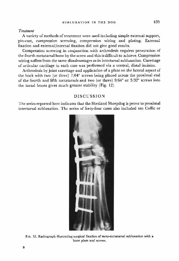
Arthrodesis by joint curettage and application of a plate on the lateral aspect of the hock with two (or three) 7/64" screws being placed across the proximal end of the fourth and fifth metatarsals and two (or three) 9/64" or 5/32" screws into the tarsal bones gives much greater stability (Fig. 12).
D I S C U S S I O N
The series reported here indicates that the Shetland Sheepdog is prone to proximal intertarsal subluxation. The series of forty-four cases also included ten Collie or
FIG. 12. Radiograph illustrating surgical fixation of tarso-metatarsal subluxation with a bone plate and screws.
B

440 J . R . C A M P B E L L , D . B E N N E T T A N D R . L E E
Collie-cross dogs. An assessment of the case histories revealed that in 75% of all cases trauma was absent or minimal and all but two of the Shetland Sheepdogs were in this category. The division between the severe trauma and minimal trauma groups was based on the owners’ appreciation and recapitulation of the incident. Road traffic accidents, dog fights and greyhound racing injuries were classed as severe traumatic episodes whereas falling off a bed or down a few steps or stairs, were placed in the minimal trauma group. Strict categorization was difficult in some cases; certain cases with no known trauma could possibly have experienced injury without the owner’s knowledge although in no case was there any other evidence of injury.
I t is probable that some of the affected Shetland Sheepdogs had suffered a gradual degeneration of the plantar ligament. This assumption is based on the radiographic evidence, in some cases, of change in the proximal intertarsal joint of the apparently normal limb. However, the underlying aetiology is obscure. Although the condition was seen in both large and small Shetland Sheepdogs, the majority of dogs were greater in size than specified by the Kennel Club and it may be that high body weight combined with thin limb bones, a feature of this breed, is an important factor in the development of intertarsal subluxation. Meutstege (1971) reported nine cases of intertarsal subluxation, of which six had a history of trauma; the other three patients were described as old, obese animals.
Shetland Sheepdogs showed a very specific age incidence of 6-9 years, whereas in the other breeds there was a much greater spread of ages with no marked preponderance of any age group. The greater number of cases suffering trauma in the latter grouping probably explains this difference.
The absence of pain in most cases indicate that lameness was due to mechanical instability of the joint. Disruption ofjoint structures with instability and little or no pain is a feature of Charcot’s arthropathy of human patients following joint denervation, such as occurs in Tabes dorsalis and diabetic polyneuropathy (Adams, 1971). One case of intertarsal subluxation in a Shetland Sheepdog which was examined at post-mortem following euthanasia for another, unrelated problem did not show evidence of neural degeneration, either in the peripheral nerves or in the spinal cord.
Treatment of most cases by arthrodesis of the joint using a lag, compression screw as described by Lawson (1961) was found satisfactory. Only the articular cartilage from the fibularlfourth intertarsal joint was removed; Whittick (1 974) describes curettage of several other of the tarsal joints before screwing but this appears unnecessary. Meutstege (1971), using ASIF equipment, also treated intertarsal subluxation by lag-screwing. However, this author, performing the surgery under fluoroscopy through a small skin incision over the point of the hock, did not perform curettage of the articular cartilage and used a screw of length sufficient only to penetrate the fourth tarsal bone. The time taken to reach soundness of these patients was much longer than recorded in the present study, some taking

S U B L U X A T I O N I N T H E D O G 44 1
up to 8 months. Denny (1975) described the use of Kirschner wires and figure-of- eight compression band wiring as an alternative to the lag screw principle -for arthrodesing the hock joint; this technique was used in some patients and although successful in five, arthrodesis was delayed compared to lag screwing. Complica- tions following lag screw arthrodesis included one case where the screw bent, another where the screw completely broke and one case where the dog chewed off the support splint and produced gross self-inflicted damage to the foot removing all but one toe. The two failures of the metal implant were due to incorrect surgical procedure for arthrodesis in one case and probably due to inadequate compression in the other.
Tarso-mctatarsal subluxation only occurs when the strong tarso-metatarsal joint capsule, which is reinforced by the attachment of the plantar ligaments, is torn. This usually only occurs following fairly severe trauma. Fixation is more difficult than in subluxation of the proximal intertarsal joint and of the methods used, arthrodesis by curettage and plating appears most effective. The main disadvantage of this technique is the problem of covering the plate without applying too much tension to the skin. Fairly firm bandaging is necessary to prevent post-operative swelling and wound breakdown. An alternative method of treatment has been described by Le Roux (1971,1972). This technique involves the insertion of a Steinman pin at an angle, through the proximal part of meta- tarsal 5 into the fourth tarsal bone after reduction of the dislocation. Post-operative support of the limb in a plaster cast is described and the pin removed after 6-8 weeks. Complete success in all of the six cases was claimed; the present authors have no experience with this technique. Arwedsson (1954) in his original paper described satisfactory arthrodesis of the tarso-metatarsal joint in three dogs using a transfixation plastering technique, although this procedure has been found unsatisfactory at the Glasgow hospital.
Intertarsal subluxation is the most commonly encountered lesion of the canine hock joint at this hospital; approximately 35% of all hock lesions (excluding superficial abrasions) are of this category. Tarso-metatarsal subluxations comprise approximately 12 %, while tibio-tarsal luxations/subluxations/instability with or without malleolar fractures account for about 24% and tarsal bone fractures, 23 % of all lesions. These incidence figures are different from those reported in the American literature (Pettit, 1974).
R E F E R E N C E S
ADAMS, J.C. (1971) Outline of Osthopaedics, 7th Edn, p. 21. Churchill Livingstone, Edinburgh. ARWEDSSON, G. (1954) Arthrodesis in traumatic plantar subluxation of the metatarsal bones of the
CLAYTON-JONES, D.G. (1974) Hindleglamenessin the dog. In: Veterinary Annual, 14th Edn (Ed. by
DENNY, H.R. (1975) Surgery of the canine hock. In: Veferinary Annual, 15th Edn (Ed. by C. S. G.
dog. 3. Am. vet. med. Ass. 120, 21.
C. S. G. Grunsell & F. W. G. Hill), p. 167. John Wright & Sons Ltd, Bristol.
Grunsell & F. W. G. Hill), p. 207. John Wright & Sons Ltd, Bristol.

442 J . R . C A M P B E L L , D . B E N N E T T A N D R . L E E
HOLT, P.E. (1974) Ligamentous injuries to the canine hock. 3. small Anim. Pract. 15, 457. LAWSON, D.D. (1961) Inter-tarsal subluxation in the dog. J . small Anim. Pract. 1, 179. LE Roux, P.H. (1971) Treatment of luxation of the tarsometatarsal joint of the dog. 31. S. AJL.
LE Roux, P.H. (1972) Treatment of luxation of the tarsometatarsal joint of the dog. 31. S. Afr. vet.
MEUTSTEGE, F.J. (1971) Die Rehandlung der intertarsalen Subluxation beim Hund durch
MILLER, M.E., CHRISTENSEN, G.C. & EVANS, H.E. (1964) Anatomy qf the Dog. W.R. Saunders
PETTIT, G.D. (1974) Cunine Surgey, 2nd Archibald Edn. American Veterinary Publications Inc.,
WHITTICK, W.G. (1974) Canine Orthopaedics. Lea & Febiger, Philadelphia.
vet. med. Ass. 42, 195.
med. Ass. 43, 110.
gedeckte Axthrodesis von 0 s tarsi fibulare und 0 s tarsale IV. Kleintier Praxis, 16, 12.
Company, Philadelphia.
California.