Bone mineral measurements and Tc99m IDP retention in the dog
7
Calcif Tissue Int (1991) 48:120-126 Calcified Tissue International 1991Springer-Verlag New YorkInc. Bone Mineral Measurements and Tc-99m IDP Retention in the Dog Pauline Kolkman, 1 Larry W. Belbeck, 2 and Colin E. Webber 1 1Department of Nuclear Medicine, Chedoke-McMaster Hospitals, 1200 Main Street West, and 2Faculty of Health Sciences, McMaster University Hamilton, Ontario, Canada L8N 3Z5 Summary. The reproducibility of various proce- dures for the noninvasive assessment of the skele- ton in dogs has been measured. In one animal, dual photon measurements of bone mineral mass were made in the whole body, the lumbar spine, and the proximal femur on 10 occasions during a 15-week period. In two other dogs, repeat measurements of Tc-99m imidodiphosphate (IDP) retention were made. For the total body and the proximal femur, the reproducibilities were 2 and 2.5%, respectively. The reproducibility of the lumbar spine was approx- imately 10% and was influenced by the variable fraction of the posterior spinous processes included in the measurement. The reproducibility of IDP re- tention measurements was not established. It was about 8% when all 10 measurements were consid- ered but fell to 1.5% when three consecutive mea- surements were excluded. Dual photon measure- ments of the whole body and the proximal femur can be used to assess bone mass changes in the canine skeleton. Key words: Dog -- Dual photon absorptiometry Imidodiphosphate retention -- Reproducibility. Osteoporosis is a disease of the bone remodeling system in which the rate of resorption exceeds the rate of formation [1]. The progress of the disease can only be modeled appropriately in those animals that have a bone tissue turnover based predomi- nantly on basic multicellular unit remodeling rather than on just growth and modeling [2]. The adult dog is one of the smallest animals that meets this re- Offprint requests to: Colin E. Webber quirement. Smaller animals such as mice and rats or any animal in a period of rapid growth is not useful as a model of osteoporosis. One of the most available procedures for the study of osteoporosis in humans is dual photon ab- sorptiometry which can be used to measure the mass of mineral at either a specific anatomical lo- cation or throughout the whole body. Another tech- nique that may prove in the future to be useful di- agnostically is the measurement of the fraction of a Tc-99m labeled compound phosphate which is re- tained at 24 hours. This latter test is thought to be an indicator of the rate of total body bone mineral turnover [3]. The reproducibility of each of these procedures is generally about 2-3% [4, 5]. Repro- ducibility is an important characteristic of a tech- nique that establishes the minimum detectable change that can be reasonably attributed to a given experimental intervention. The purpose of this ar- ticle is to describe the means we developed to apply these techniques to dogs and to report the repro- ducibility attainable with each technique, Materials and Methods Bone mineral mass was measured using a Norland 2600 dichro- matic densitometer. With this instrument a pencil beam of gamma rays emitted from a radioisotope source (Gd-153) is scanned throughout a region of interest. The gamma ray beam includes photons of two discrete energies. The relative attenua- tion of the two groups of photons allows the measurement of the mass of each of two components that differ in radiation absorp- tion characteristics. For body sites containing mineral, the mass of mineral and soft tissue can be measured. For sites containing no mineral, the mass of lean tissue and fat can be measured [6], The densitometer is calibrated daily using a phantom supplied by the manufacturer. The accuracy of our machine for lumbar spine mineral measurements has been measured and is better than 3% [7]. The reproducibility is checked regularly with a lumbar spine
Transcript of Bone mineral measurements and Tc99m IDP retention in the dog

Calcif Tissue Int (1991) 48:120-126 Calcified Tissue International �9 1991 Springer-Verlag New York Inc.
Bone Mineral Measurements and Tc-99m IDP Retention in the Dog
Pauline Kolkman, 1 Larry W. Belbeck, 2 and Colin E. Webber 1
1Department of Nuclear Medicine, Chedoke-McMaster Hospitals, 1200 Main Street West, and 2Faculty of Health Sciences, McMaster University Hamilton, Ontario, Canada L8N 3Z5
Summary. The reproducibility of various proce- dures for the noninvasive assessment of the skele- ton in dogs has been measured. In one animal, dual photon measurements of bone mineral mass were made in the whole body, the lumbar spine, and the proximal femur on 10 occasions during a 15-week period. In two other dogs, repeat measurements of Tc-99m imidodiphosphate (IDP) retention were made. For the total body and the proximal femur, the reproducibilities were 2 and 2.5%, respectively. The reproducibility of the lumbar spine was approx- imately 10% and was influenced by the variable fraction of the posterior spinous processes included in the measurement. The reproducibility of IDP re- tention measurements was not established. It was about 8% when all 10 measurements were consid- ered but fell to 1.5% when three consecutive mea- surements were excluded. Dual photon measure- ments of the whole body and the proximal femur can be used to assess bone mass changes in the canine skeleton.
Key words: Dog - - Dual photon absorptiometry Imidodiphosphate retention - - Reproducibility.
Osteoporosis is a disease of the bone remodeling system in which the rate of resorption exceeds the rate of formation [1]. The progress of the disease can only be modeled appropriately in those animals that have a bone tissue turnover based predomi- nantly on basic multicellular unit remodeling rather than on just growth and modeling [2]. The adult dog is one of the smallest animals that meets this re-
Offprint requests to: Colin E. Webber
quirement. Smaller animals such as mice and rats or any animal in a period of rapid growth is not useful as a model of osteoporosis.
One of the most available procedures for the study of osteoporosis in humans is dual photon ab- sorptiometry which can be used to measure the mass of mineral at either a specific anatomical lo- cation or throughout the whole body. Another tech- nique that may prove in the future to be useful di- agnostically is the measurement of the fraction of a Tc-99m labeled compound phosphate which is re- tained at 24 hours. This latter test is thought to be an indicator of the rate of total body bone mineral turnover [3]. The reproducibility of each of these procedures is generally about 2-3% [4, 5]. Repro- ducibility is an important characteristic of a tech- nique that establishes the minimum detectable change that can be reasonably attributed to a given experimental intervention. The purpose of this ar- ticle is to describe the means we developed to apply these techniques to dogs and to report the repro- ducibility attainable with each technique,
Materials and Methods
Bone mineral mass was measured using a Norland 2600 dichro- matic densitometer. With this instrument a pencil beam of gamma rays emitted from a radioisotope source (Gd-153) is scanned throughout a region of interest. The gamma ray beam includes photons of two discrete energies. The relative attenua- tion of the two groups of photons allows the measurement of the mass of each of two components that differ in radiation absorp- tion characteristics. For body sites containing mineral, the mass of mineral and soft tissue can be measured. For sites containing no mineral, the mass of lean tissue and fat can be measured [6], The densitometer is calibrated daily using a phantom supplied by the manufacturer. The accuracy of our machine for lumbar spine mineral measurements has been measured and is better than 3% [7]. The reproducibility is checked regularly with a lumbar spine

P. Kolkman et al.: Bone Mineral in the Dog 121
B
Fig. 1. A typical whole body scan (A) and the spine region of interest (B).
phantom [7] which has been measured on 12 occasions during the last 2 years. The mean bone mineral density is 0.89 g cm -2, the standard deviation is 0.016 g cm -2, and the coefficient of vari- ation is 1.8%.
In this work, the sites studied were the whole body, the lumbar spine, and the hip. An example of each scan is shown in Figs. 1, 2, and 3, respectively. Figure 1A shows the whole body bone mineral and soft tissue image. The analysis procedure provides total mineral mass, lean body mass, and fat mass. Although the objective of this work was to establish the reproducibility of bone mineral measurements, the reproducibility of the body composition data was also examined. Figure 1B shows the bone
mineral image analyzed by setting a single rectangular re#on to include just the spine. Figure 2A shows the normal lumbar spine bone mineral image in which three lumbar vertebrae are exam- ined. In human studies, the Norland 2600 provides results for the second, third, and fourth lumbar vertebrae and L1 and L5 are excluded. The interactive system software allows the operator to set the bone edges and the intravertebral limits. As there are seven lumbar vertebrae in the dog, we positioned the regions of interest to include the third, fourth, and fifth lumbar vertebrae and excluded L1, L2, L6, and L7. The lumbar spine scan was also examined with a single rectangular region of interest set to include L3, L4, and L5 (Fig. 2B). The proximal femur was ex-

122 P. Kolkman et al.: Bone Mineral in the Dog
. . . . ~ ~ , ~ ~, o
, ; A : :
L2 �9 . � 9 .
L3 . . . .
! r . , . ' ) ; ~ " o ~ ' ~ # " . "
. . . .
= ,
A
m i~ ~
Fig. 2. A typical lumbar spine scan (A) and the lumbar spine region of interest (B).
amined by setting the region of interest as shown in Fig. 3. In all cases, results are expressed as the total mass of mineral and as an average mass per unit area referred to as the areal mass.
Measurements were made each week for 5 weeks in a female mongrel dog weighing about 17 kg, aged about 1.5 years�9 Sub- sequently, the three sites were measured every other week for l0 weeks giving a total of l0 studies in 15 weeks. On one occasion scans were repeated on the same day without repositioning the animal.
For each study the dog was anesthetized with sodium pento- barbital, and an endotracheal tube was inserted. The duration of anesthesia was such that the whole body scan, the spine scan, and the femur scan could be completed. Typically, the total time required for the three scans was 90 minutes�9 The animal was placed supine on the scanning couch. The front limbs were ex- tended posteriorly and slightly laterally by means of ropes which were attached from above and below the carpi with slip knots to the edge support of the scanning couch. The limbs were kept from contact with the trunk by means of foam pads which could also be used to support the animal on the table. The hind limbs were also extended posteriorly using ropes double looped around the tarsi. The hind limbs could not be separated by more than 5 cm at the point of the hock otherwise the densitometer might scan only one leg. Once a stable position had been achieved, the
dog was aligned symmetrically with its spine parallel to the long axis of the scanning couch. The slip knots around the limbs were left snug but did not interfere with the circulation.
Both the whole body scan and the lumbar spine scan were done with the animal in the above position. To scan the femoral head and neck, the "local region" scan facility was used. The dog was positioned on its left side with the right hind limb lifted and stretched posteriorly and laterally using a slip knot as de- scribed above. The left femur was palpated through the muscle and positioned orthogonal to the spine and flat on the scanning table�9 The upper right limb was supported by pads to help sta- bilize the dog. A weight such as a telephone directory resting on the distal left leg was found to aid position maintenance.
The compound phosphate used for the retention test was Tc- 99m imidodiphosphate (IDP) which has been shown to have a greater fractional retention than other phosphates [3]. Tc-99m retention was measured shortly after and at 24 hours after injec- tion of the radiopharmaceutical. Two mongrel bitches aged 2 and 3 years and weighing 16 kg each were used in this study. Six measurements were made on one dog during a 9-week period and four measurements were made of the second dog in a 6-week period. For each study the dog was anesthetized with sodium pentobarbital and was positioned in lateral recumbency on the couch of a shadow shield whole body counter similar to that

P. Kolkman et al.: Bone Mineral in the Dog 123
16
~8 ,~ 14
# }
~'~ 12
o 10 m C
r 8 _J
6 i i i t
0 20 40 60
9
7
5
1
-1
80 100
"o
(0
Day
Fig. 4. Repeated measurements of lean body mass ( � 9 and fat mass (<)).
Fig. 3. A typical hip scan showing the proximal femur region of interest.
described by Boddy [8]. Between 0.5 and 5 MBq of Tc-99m IDP was injected intravenously. After an interval of 10-20 minutes, the dog was driven through the counter at a speed of 10 cm/ minute. For the 24-hour count, the dog was given an ultrashort- acting barbiturate to ensure constant positioning. For each ac- cumulated spectrum, an energy window was set about the Tc- 99m photopeak. The observed counts were corrected for background, dead time, and decay and the 24-hour count was expressed as a percentage of the first count. All procedures were approved by the Animal Care Committee of McMaster Univer- sity.
Results
Total Body Scan
The results of the repeated measurements of lean body mass and fat mass are shown in Fig. 4. During the 95-day period there was a consistent loss of muscle tissue and a gain of fat. When a straight line was fitted by linear regression analysis to the lean body mass data the correlation coefficient was 0.79 and the rate of loss of muscle tissue was 14 g/day. The standard error of estimate for a measurement of
lean body mass was 338 g which is equal to 2.2% of the mean lean body mass. When total body scans were repeated without repositioning the animal, the difference observed in lean body mass was 15 g.
The correlation coefficient for the increase in fat mass was 0.93 and the rate of gain of fat tissue was 18 g/day. The standard error of estimate for fat mass was 227 g and the difference be tween measure- ments repeated on the same day was 51 g. It should be noted that the fat mass was measured as negative for the first 30 days of the measurement period.
The measurements of total body bone mineral mass and areal mass are shown in Fig. 5. There was no trend with time for either parameter . The mean and standard deviation for mineral mass was 524 --- 9.7 g. The coefficient of variation was 1.9% and the difference between measurements repeated without repositioning was 1.6%. The mean area mass was 0.58 - 0.02 g/cm 2, The coefficient of variation for the areal mass was 3.5% and the difference between repeated measurements was 5.1%. When the rect- angular region of interest was positioned in a whole body scan to include the spine, the results shown in Fig. 6 were obtained. No temporal trend was de- tected for the spine region. The mean mass of min- eral within the region of interest was 57.7 - 5.8 g and the mean areal mass was 0.36 --- 0.04 g/cm 2. The coefficients of variation for each parameter were 10.0 and 12.1%, respectively.
Lumbar Spine Scan
Repeated measurements for lumbar spine scans are shown in Fig. 7. Again there is no evidence of a trend for measurements within the lumbar verte-

124 P. Kolkman et al.: Bone Mineral in the Dog
600
45O
E it1
300
150
" 'Ib ' g - O ' ' ' � 9 . . . . . �9 . . . . . . O " " O - " O . . . . . . . �9
0
O 20 40 60 80
1.00
0.80
0.60
0.40
0.20
0.00
t00
E @
r L <
Day
Fig. 5. Repeated measurements of total body mineral mass (0) and areal mass (0).
E (g
tO
2
100
80
6O
40 O . . 4 . . . . . . ,O" " l id . . . . . . -O �9 " "-O. O . . . - -
2O
0 A i i
0 20 40 6O
1.00
0.80
0.60
0.40
0.20
, 0.00
8O IO0
Day Fig. 6. Repeated measurements of mineral mass (O) and areal mass (0) in the spine region of a total body scan.
E o 03
d) tO tB
<
brae L3, L4, and L5. The sum of the masses within these three ver tebrae was 15.8 --- 2.1 g. The mean areal mass was 0.80 _+ 0.08 g/cm 2. The coefficients of variation were 13.5 and 9.6%, respect ively and the d i f ferences be tween the two m e a s u r e m e n t s made without repositioning were 9.1 and 6.1%, re- spectively.
When a single region of interest was used to an- alyze the same lumbar spine scans, the results shown in Fig. 8 were obtained. The mean mass was 13.6 --- 0.9 g and the mean areal mass was 0.64 --- 0.04 g/cm 2. The coefficients of variation for both parameters were 6.9% and the difference between measurements were 3.2 and 3.3%, respectively.
Hip Scans
The results of the repeated measurements for a re-
"B E
to
a3
20
15'
10
1 . 0 0
0.80
0.60
0.40
0.20
0 . . . . 0.00
0 20 40 60 80 100
E .9. ~ .9 O)
fu
r x.. ,(
Day
Fig. 7. Repeated measurements of mineral mass (O) and areal mass (0) in the lumbar spine.
2~ I ~'~ .- -. . . . - 0 E . . . .
b~ - lO (/) q)
s 5
1.00
0.80
0.60
0.40
0.20
0 . . . . 0.00
0 20 40 60 80 100
E
03 v
d)
/ "
r <
Day
Fig. 8. Repeated measurements mineral mass (O) and areal mass (0) in the lumbar spine region.
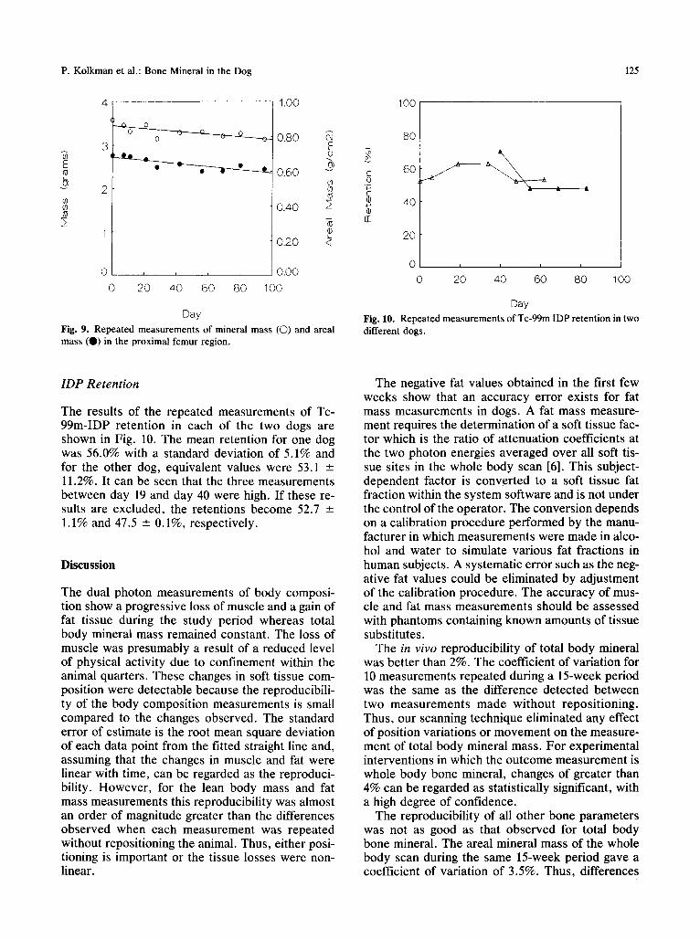
gion of interest posit ioned at the proximal femur are shown in Fig. 9. There was a consis tent loss of min- eral f rom this region throughout the period of study. The correlation coefficients for the straight lines fit- ted to the data were 0.73 for the mineral mass and 0.83 for the areal mass. The corresponding rates of loss were 3.2 mg/d and 0.9 mg/cm2/day. The stan- dard error of est imate for the proximal femur min- eral mass was 0.09 g which is equal to 2.8% of the mean mass. When hip scans were repeated without repositioning, the difference in mass was 1.6%. The standard error of est imate for the areal mass was 0.019 g/cm 2 which is equal to 3.0% of the mean areal mass . The dif ference wi thout reposi t ioning was 2.1%.
P. Kolkman et al.: Bone Mineral in the Dog 125
E tU
b, v
4
3
' 0
1.00
0.S0 E o
0.60 v d) u ]
a3
0.40
0.20
0 . . . . 0,00
0 20 40 60 80 100
x _ <(
Day
Fig. 9. Repeated measurements of mineral mass (O) and areal mass (0 ) in the proximal femur region.
( -
o C @
rr
100
80
60
40
2O
0
0
i i i i
2O 4O 60 8O 100
Day
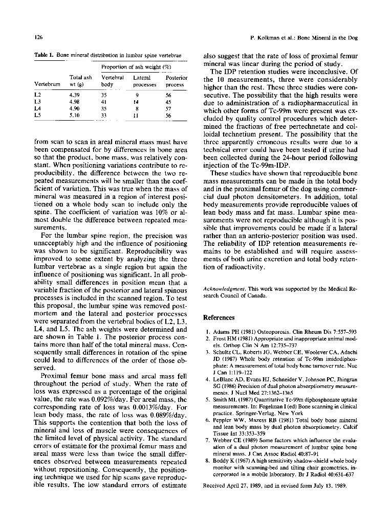
Fig. 10. Repeated measurements of Tc-99m IDP retention in two different dogs.
IDP Retent ion
The results of the repeated measurements of Tc- 99m-IDP retention in each of the two dogs are shown in Fig. 10. The mean retention for one dog was 56.0% with a standard deviation of 5.1% and for the other dog, equivalent values were 53.1 --- 11.2%. It can be seen that the three measurements between day 19 and day 40 were high. If these re- suits are excluded, the retentions become 52.7 -+ 1.1% and 47.5 --- 0.1%, respectively.
Discussion
The dual photon measurements of body composi- tion show a progressive loss of muscle and a gain of fat tissue during the study period whereas total body mineral mass remained constant. The loss of muscle was presumably a result of a reduced level of physical activity due to confinement within the animal quarters. These changes in soft tissue com- position were detectable because the reproducibili- ty of the body composition measurements is small compared to the changes observed. The standard error of estimate is the root mean square deviation of each data point from the fitted straight line and, assuming that the changes in muscle and fat were linear with time, can be regarded as the reproduci- bility. However, for the lean body mass and fat mass measurements this reproducibility was almost an order of magnitude greater than the differences observed when each measurement was repeated without repositioning the animal. Thus, either posi- tioning is important or the tissue losses were non- linear.
The negative fat values obtained in the first few weeks show that an accuracy error exists for fat mass measurements in dogs. A fat mass measure- ment requires the determination of a soft tissue fac- tor which is the ratio of attenuation coefficients at the two photon energies averaged over all soft tis- sue sites in the whole body scan [6]. This subject- dependent factor is converted to a soft tissue fat fraction within the system software and is not under the control of the operator. The conversion depends on a calibration procedure performed by the manu- facturer in which measurements were made in alco- hol and water to simulate various fat fractions in human subjects. A systematic error such as the neg- ative fat values could be eliminated by adjustment of the calibration procedure. The accuracy of mus- cle and fat mass measurements should be assessed with phantoms containing known amounts of tissue substitutes.
The in vivo reproducibility of total body mineral was better than 2%. The coefficient of variation for 10 measurements repeated during a 15-week period was the same as the difference detected between two measurements made without repositioning. Thus, our scanning technique eliminated any effect of position variations or movement on the measure- ment of total body mineral mass. For experimental interventions in which the outcome measurement is whole body bone mineral, changes of greater than 4% can be regarded as statistically significant, with a high degree of confidence.
The reproducibility of all other bone parameters was not as good as that observed for total body bone mineral. The areal mineral mass of the whole body scan during the same 15-week period gave a coefficient of variation of 3.5%. Thus, differences
126 P. Kolkman et al.: Bone Mineral in the Dog
Table 1. Bone mineral distribution in lumbar spine vertebrae
Proportion of ash weight (%)
Total ash Vertebral Lateral Posterior Vertebrum wt (g) body processes process
L2 4.39 35 9 56 L3 4.98 41 14 45 L4 4.90 35 8 57 L5 5.10 33 11 56
from scan to scan in areal mineral mass must have been compensated for by differences in bone area so that the product, bone mass, was relatively con- stant. When positioning variations contribute to re- producibility, the difference between the two re- peated measurements will be smaller than the coef- ficient of variation. This was true when the mass of mineral was measured in a region of interest posi- tioned on a whole body scan to include only the spine. The coefficient of variation was 10% or al- most double the difference between repeated mea- surements.
For the lumbar spine region, the precision was unacceptably high and the influence of positioning was shown to be significant. Reproducibility was improved to some extent by analyzing the three lumbar vertebrae as a single region but again the influence of positioning was significant. In all prob- ability small differences in position mean that a variable fraction of the posterior and lateral spinous processes is included in the scanned region. To test this proposal, the lumbar spine was removed post- mortem and the lateral and posterior processes were separated from the vertebral bodies of L2, L3, L4, and L5. The ash weights were determined and are shown in Table 1. The posterior process con- tains more than half of the total mineral mass. Con- sequently small differences in rotation of the spine could lead to differences of the order of those ob- served.
Proximal femur bone mass and areal mass fell throughout the period of study. When the rate of loss was expressed as a percentage of the original value, the rate was 0.092%/day. For areal mass, the corresponding rate of loss was 0.0013%/day. For lean body mass, the rate of loss was 0.089%/day. This supports the contention that both the loss of mineral and loss of muscle were consequences of the limited level of physical activity. The standard errors of estimate for the proximal femur mass and areal mass were less than twice the small differ- ences observed between measurements repeated without repositioning. Consequently, the position- ing technique we used for hip scans gave reproduc- ible results. The low standard errors of estimate
also suggest that the rate of loss of proximal femur mineral was linear during the period of study.
The IDP retention studies were inconclusive. Of the 10 measurements, three were considerably higher than the rest. These three studies were con- secutive. The possibility that the high results were due to administration of a radiopharmaceutical in which other forms of Tc-99m were present was ex- cluded by quality control procedures which deter- mined the fractions of free pertechnetate and col- loidal technetium present. The possibility that the three apparently erroneous results were due to a technical error could have been tested if urine had been collected during the 24-hour period following injection of the Tc-99m-IDP.
These studies have shown that reproducible bone mass measurements can be made in the total body and in the proximal femur of the dog using commer- cial dual photon densitometers. In addition, total body measurements provide reproducible values of lean body mass and fat mass. Lumbar spine mea- surements were not reproducible although it is pos- sible that improvements could be made if a lateral rather than an anterio-posterior position was used. The reliability of IDP retention measurements re- mains to be established and will require assess- ments of both urine excretion and total body reten- tion of radioactivity.
Acknowledgment. This work was supported by the Medical Re- search Council of Canada.
References
1. Adams PH (1981) Osteoporosis. Clin Rheum Dis 7:557-593 2. Frost HM (1981) Appropriate and inappropriate animal mod-
els. Orthop Clin N Am 12:735-737 3. Schultz CL, Roberts JG, Webber CE, Woolever CA, Adachi
JD (1987) Whole body retention of Tc-99m imidodiphos- phate: A measurement of total body bone turnover rate. Nuc J Can 1:119-122
4. LeBlanc AD, Evans H J, Schneider V, Johnson PC, Jhingran SG (1986) Precision of dual photon absorptiometry measure- ments. J Nucl Med 27:1362-1365
5. Smith ML (1987) Quantitative Tc-99m diphosphonate uptake measurements. In: Fogelman I (ed) Bone scanning in clinical practice. Springer-Verlag, New York
6. Peppier WW, Mazess RB (1981) Total body bone mineral and lean body mass by dual photon absorptiometry. Calcif Tissue Int 33:353-359
7. Webber CE (1989) Some factors which influence the evalu- ation of a dual photon measurement of lumbar spine bone mineral mass. J Can Assoc Radiol 40:87-91
8. Boddy K (1967) A high sensitivity shadow-shield whole body monitor with scanning-bed and tilting chair geometries, in- corporated in a mobile laboratory. Br J Radiol 40:631-637
Received April 27, 1989, and in revised form July 13, 1989.