HLA-DR generic typing by AFLP
11
HLA-DR generic typing by AFLP I. Yunis, M. Salazar, E.J. Yunis. HLA-DR generic typing by AFLP. Tissue Antigens 1991: 38: 78-88. Abstract: We have described a practical and inexpensive method whereby any individual can be typed and assigned to any of the 14 generic HLA- DR types: DR1, DRwlS, DRwl6, DRwl7, DRwl8, DR4, DRwll, DRwl2, DRwl3, DRwl4, DR7, DRw8, DR9 and DRwlO. Previous methods to type these specificities include - among others - serology, conventional RFLP, PCR-oligonucleotidetyping, and a PCR-RFLP method useful for typing homozygous individuals. In the method reported here, DNA is amplified with a set of group-specific primers and then restricted with a number of endonucleases. Six group-specific pairs of primers have been chosen to avoid cross-amplification with other DRB alleles, including DRB3, DRB4 and DRBS alleles, and to anneal at uniform temperature: 61 "C. Endonucleases were chosen to generate unique patterns of easily recognizable bands that led to unequivocal assignments of HLA-DR generic types in heterozygous as well as homozygous individ- uals. This technique involves two steps: 1) Amplification of DNA with six different pairs of primers where DR1, DR4 and DRwlO types can be assigned at once, and 2) Endonuclease digests of amplified DNA to assign DR7, DRw8, DR9, DRwll, DRwl2, DRwl3, DRwl4, DRwl5, DRwl6, DRwl7 and DRwl8 types. Individuals carrying any combination of all alleles published so far can be typed by this method. The ease, low I cost and reliability of this method are discussed. Introduction Serological methods for HLA-DR typing have led to the identification of 14 generic D R types. Al- though there are monospecific sera for most HLA- DR types, DRwl2, DRw13, DRwl4, DRwl7 and DRw 18 are difficult to assign on the basis of serol- ogy alone, particularly in leukemic patients. HLA-DR assignments often involve the use of RFLP, at best a lengthy process that often involves the use of radioactive probes. Since the introduc- tion of PCR technology, a number of methods to detect allele differences at the HLA-DR locus have appeared. Most technologies involve the specific amplification of second exon sequences of the DRB genes followed by the hybridization with sequence- specific oligonucleotide probes to detect variable sequences (1). An alternative would be to amplify each specificity with a different pair of specific primers. Yet another would involve direct sequen- cing of DRB1 second exon of all samples tested. A different set of techniques relies on the ability of endonucteases to detect allele differences within a variable region of class I1 genes in amplified DNA, where every pattern of restriction bands (or AFLPs) generated would be allele-specific (2, 3). lvin Yunis', Marcela Salazafl and Edmond J. Yunis1t2 'American Red Cross, Dedham, Mass, and 'Dana Farber Cancer Institute, Boston, Mass., USA. Key words: HLA-DR - generic typing - AFLP Received 24 April, revised, accepted for publi- cation 5 June 1991 AFLP typing for HLA-DR elicited interest at first but it then became evident that some molecular specificities could not be unambiguously attributed in both homozygous and heterozygous states (4). We have developed an HLA-DR typing strategy that leads to unequivocal assignments of 14 HLA- D R generic types in heterozygous and homozygous individuals. The combination of group-specific am- plifications (GSA) and AFLP reported here in- volves two sequential steps: first, GSA using six HLA-DR group-specific pairs of primers to ampli- fy second exon sequences in six groups of HLA- DR generic types: DRl, DR2, DR4, DRwlO; DR7 and DR9 and finally DR3, DR5, DRw6, DRw8 and DRwl2. At the end of this step, DRl, DR4 and DRwlO types are assigned; second, assign- ments of DR7, DR9, DRw8, DRwl1, -w12, -w13, -w14, -w15, -w16, -w17 and -w18 after digesting amplified DNA with 6 endonucleases: Hue ZIZ, . Hinf I, Nci Z, Mbo 11, BstN Z and Kpn I. Material and methods DNA samples DNA from homozygous typing cells from the Tenth Workshop panel (5) were used. DNAs from 78
-
Upload
independent -
Category
Documents
-
view
1 -
download
0
Transcript of HLA-DR generic typing by AFLP

HLA-DR generic typing by AFLP I. Yunis, M. Salazar, E.J. Yunis. HLA-DR generic typing by AFLP. Tissue Antigens 1991: 38: 78-88.
Abstract: We have described a practical and inexpensive method whereby any individual can be typed and assigned to any of the 14 generic HLA- DR types: DR1, DRwlS, DRwl6, DRwl7, DRwl8, DR4, DRwll, DRwl2, DRwl3, DRwl4, DR7, DRw8, DR9 and DRwlO. Previous methods to type these specificities include - among others - serology, conventional RFLP, PCR-oligonucleotide typing, and a PCR-RFLP method useful for typing homozygous individuals. In the method reported here, DNA is amplified with a set of group-specific primers and then restricted with a number of endonucleases. Six group-specific pairs of primers have been chosen to avoid cross-amplification with other DRB alleles, including DRB3, DRB4 and DRBS alleles, and to anneal at uniform temperature: 61 "C. Endonucleases were chosen to generate unique patterns of easily recognizable bands that led to unequivocal assignments of HLA-DR generic types in heterozygous as well as homozygous individ- uals. This technique involves two steps: 1) Amplification of DNA with six different pairs of primers where DR1, DR4 and DRwlO types can be assigned at once, and 2) Endonuclease digests of amplified DNA to assign DR7, DRw8, DR9, DRwll, DRwl2, DRwl3, DRwl4, DRwl5, DRwl6, DRwl7 and DRwl8 types. Individuals carrying any combination of all alleles published so far can be typed by this method. The ease, low
I cost and reliability of this method are discussed.
Introduction
Serological methods for HLA-DR typing have led to the identification of 14 generic D R types. Al- though there are monospecific sera for most HLA- D R types, DRwl2, DRw13, DRwl4, DRwl7 and DRw 18 are difficult to assign on the basis of serol- ogy alone, particularly in leukemic patients.
HLA-DR assignments often involve the use of RFLP, at best a lengthy process that often involves the use of radioactive probes. Since the introduc- tion of PCR technology, a number of methods to detect allele differences at the HLA-DR locus have appeared. Most technologies involve the specific amplification of second exon sequences of the DRB genes followed by the hybridization with sequence- specific oligonucleotide probes to detect variable sequences (1). An alternative would be to amplify each specificity with a different pair of specific primers. Yet another would involve direct sequen- cing of DRB1 second exon of all samples tested. A different set of techniques relies on the ability of endonucteases to detect allele differences within a variable region of class I1 genes in amplified DNA, where every pattern of restriction bands (or AFLPs) generated would be allele-specific (2, 3).
lvin Yunis', Marcela Salazafl and Edmond J. Yunis1t2 'American Red Cross, Dedham, Mass, and 'Dana Farber Cancer Institute, Boston, Mass., USA.
Key words: HLA-DR - generic typing - AFLP
Received 24 April, revised, accepted for publi- cation 5 June 1991
AFLP typing for HLA-DR elicited interest a t first but it then became evident that some molecular specificities could not be unambiguously attributed in both homozygous and heterozygous states (4).
We have developed an HLA-DR typing strategy that leads to unequivocal assignments of 14 HLA- D R generic types in heterozygous and homozygous individuals. The combination of group-specific am- plifications (GSA) and AFLP reported here in- volves two sequential steps: first, GSA using six HLA-DR group-specific pairs of primers to ampli- fy second exon sequences in six groups of HLA- D R generic types: D R l , DR2, DR4, DRwlO; DR7 and DR9 and finally DR3, DR5, DRw6, DRw8 and DRwl2. At the end of this step, D R l , DR4 and DRwlO types are assigned; second, assign- ments of DR7, DR9, DRw8, DRwl1, -w12, -w13, -w14, -w15, -w16, -w17 and -w18 after digesting amplified DNA with 6 endonucleases: Hue ZIZ, . Hinf I, Nci Z, Mbo 11, BstN Z and Kpn I .
Material and methods DNA samples
DNA from homozygous typing cells from the Tenth Workshop panel (5) were used. DNAs from
78

HLA-DR generic typing by AFLP
was unnecessary. The choice of the enzyme(s) de- pended on the amplified product present in each set of amplifications for every sample. Thus, only products from amplifications with primer pairs DRG2, DRG79 and DRG3568 were digested: the first with Hue 111, the second with Hinf I and the third with four different enzymes, Nci I , Mbo II, BstN Z and Kpn 1.
Aliquots of a DRwl7 homozygous and a DRw17/DRw8 sample were used as controls for enzyme digestion and band migration. Digests were carried out under the same conditions and run side by side with test samples.
Digested DNAs were then run on a 2% NuSieve (FMC Bioproducts, ME)/2% LE agarose (Bethes- da Research Laboratories, ME) submarine gel at 8 V/cm for 2 h in 0.045 M Tris-Borate/0.001 M EDTA, pH 8.0 containing 0.5 pg/ml of ethidium bromide. Resulting patterns were analyzed by measuring band sizes (readings of the gel were done using a digitizer from DNASTAR, Madison, WI) and recording those that were present in each of the digests or by eyeball comparisons of band mi- gration to size markers in the photograph. HLA- DR types were assigned by matching the resulting patterns with those found in HTC cells from the standard cell panel of the Tenth Workshop. Pat- terns for all HLA-DRB1 second exon sequences published were predicted by computer analysis. Re- striction maps of 160 commercially available endo- nucleases (excluding known isoschizomers) were drawn using DNA Inspector IIe (Textco, W Leb- anon, NH) on a Macintosh SE/30 PC.
heterozygous individuals were from the American Red Cross (ARC) DNA repository. All samples have been typed by serology.
DNA extraction
DNA was extracted from peripheral blood lymphocytes, granulocyte pellets, EBV-transform- ed cell lines, or from 10 ml whole blood collected in ACD, EDTA or Na-citrate. In short, nuclei were prepared by lysing cells in 10 ml of 0.32 M sucrose, 10 mM Tris pH 7.5, 5 mM MgC12, 1% Triton X- 100 and 0.02% sodium azide. Nuclei were spun at 3000 g for 10 min and a pellet resuspended and lysed for 1 h at 60°C in 11 ml of 50 mM Tris- HCI pH 8.0,20 mM EDTA pH 8.0, 2% SDS and Proteinase K (Sigma) at 100 pg/ml. Proteins were precipitated by adding 4 ml of a 6M NaCl solution, and spinning at 2000 g for 15 min. The nucleic acid supernatant was transferred to a new tube, and precipitated by the addition of 2 vol of 100% etha- nol. DNA was recovered by spooling, washed out in 70% ethanol and resuspended in 10 mM Tris, 0.1 mM EDTA.
PCR amplification
Two micrograms of genomic DNA (the product of approx. 3 x lo5 nucleated cells) were enzymatically amplified by the PCR method (6). In short, DNA samples were amplified in 100 p1 of reaction con- taining 0.125 mM dNTPs, 50 mM KC1, 10 mM Tris-HC1 pH 8.0, 50 pmoles of each primer, 1.25 units of Taq polymerase (Promega) and 0.01% (w/v) gelatin. Magnesium chloride concentrations for each pair of primers were: 1.5 mM for DRG79 and DRGIO; 2 mM for DRG2; 2.5 mM for DRG4 and DRG3568 primers DNA was amplified for 30 cycles (96°C 30 s, 61°C 30 s, 72°C 60 s) in a programmable thermal cycler (PE Cetus). All ex- periments included positive as well as negative con- trols. Product sizes and amplification extension were controlled by 4% agarose electrophoresis of a 5 pl aliquot.
Endonuclease digestions and gel electrophoresis
Amplified DNA was divided in 15 pl aliquots as needed, dispensed in polypropylene 96-well micro- titer plates and digested with 3-5 units of Hue 111, Hinf I, Nci I , Mbo II, BstN 1 or Kpn I (Biolabs, MA) for 2 h at 37°C (60°C for BstN I ) in the presence of endonuclease buffers (2 pl of those supplied by vendor). Kpn I and BstN I digests were facilitated by the addition of 1 p1 of BSA (100 pg/ ml final concentration). Further correction of the digestion buffer prior to addition of the enzymes
Results Group-specific amplification of HLA-DRB1 alleles: assignments of HLA-DR1, HLA-DR4 and HLA-DRwlO types
Comparisons among the 47 HLA-DRB sequences published so far (7) led to the design of six tempera- ture-compatible group-specific pairs of primers of 20 mer average length, as shown in Table 1. Group specificities of each primer pair were corroborated with DNA from homozygous and heterozygous individuals. Six 5' to 3' primers around the 5' re- gion of the second exon and two antisense primers anneal to six mutually exclusive groups of alleles. All primers were chosen to anneal to HLA-DRB1 alleles only with the exception of one anti sense primer (DRGA/S) which anneals to second exon sequences of DRB3, DRB4 and DRB5. The ampli- fied DNA generates recognizable fragments upon digestion with endonucleases. All primers perform at the same annealing temperature of 61 "C.
Product sizes amplified vary from 203 bp to 265 bp. It is clear that, after the GSA step, no further testing needs to be done in groups DRl, DR4 and

Yunis et al.
Table 1. HLA-DRB1 alleles amplified by the six pairs of amplification primers
Primer Group Sense primers (5' to 3') Product Size Positive Types and Alleles Amplified
DRGl TTCTIGTGGCAGCTTAJW~T(') 264 bp HLA-DRl (DRB1'0101, 0102, 0103) DRGZ CCTGTGGCAGCCTAAGAGGG(') 262 bp HLA-DR2 (DRB1'1501, 1502, 1601, 1602) DRG4 GTllCTTGGAGCAGGTTW) 266 bp HLA-DR4 (DRB1'0401, 0402, 0403, 0404, 0405, 0406, 0407, 0408) DRGl 0 TTCTTGGAGGAGGlTAAGTTr(') 264 bp HLA-DRwlO (DRB1*1001) DRG79 TTTCTTCAACGGGACGGA(*) 203 bp HLA-DR7, HLA-DR9 (DRB1'0701, 0702, 0901) DRG3568 TTCllGGAGTAClCTACG(') 265 bp HLA-DRwl7, DRW18, DRWl 1 , DRWl2, DRwl3, DRwl4. DRW8 (DRB1'0301, 0302.
1101,1102,1103,1104, 1201,1202,1301,1302,1303.1304,1305, 1401, 1402, 0801, 0802, 0803
(') Used together with anti-sense primer 5'6GCTGCACTGTGAAGC-3'. (") Used together with anti-sense primer 5'-GllGTGTCTGCACACGGT-3'. DRGl, DRG2, DRG4, DRG10, DRG79 and DRG3568 are the six different pairs of amplification primers used to type HLA-DR. Size of amplified DNA is shown in base pairs (bp). The HLA-DR generic types are given: In parentheses, the list of different HLA-DR alleles that can be amplified and whose DR type assignment can be made with this method.
DRwlO. Examples of amplifications using those six pairs of primers are shown in Fig. 1. HTC cells used are from the Tenth Workshop panel: HLA- DRl and HLA-DR4 cells are Workshop numbers 9003 and 9026 respectively. Their generic type is shown at the bottom of the photograph. Order of amplified cells is the same in all photographs, One homozygous and one heterozygous cell from the ARC repository were used as examples of HLA- DR9 (ARCY 1273) and HLA-DRwlO/DR7 (ARCY 1567).
Generic typing of HLA-DRwl5, and HLA-DRwl6: Hae Ill digests of DRG2 products
Amplified DNA from cells homozygous for HLA- DRwl5 (9011), HLA-DRwl6 (9016) and hetero- zygous for HLA-DRw15/DRwl6 (ARCY491) were digested with Hae ZZZ. Digests were run on agarose gels and results are shown in Fig. 2a. This figure shows characteristic banding patterns for HLA-DRwl5 and HLA-DRwl6 homozygotes: HLA-DRw 15 celles produced bands of 143,67 and 52 bp whereas HLA-DRwl6 cells produced bands of 143 and 119 bp.
Generic typing of HLA-DR7 and HLA-DR9: Hinf I digests of DRG79 products
Amplified DNA from homozygous cells for HLA- DR7 (9051) and HLA-DR9 (ARCY 1273) and het- erozygous cells for HLA-DR7 (ARCY 1567) and HLA-DR9 (ARCY645) were digested with Hinf I. Resulting digests are shown in Fig. 2a. HLA-DR7 cells produce characteristic bands of 89, 77 and 37 bp, whereas HLA-DR9-carrying cells produce bands of 126 and 78 bp.
Generic typing of HLA-DRw8, HLA-DRwl 1, HLA-DRwl2, HLA-DRwl3, HLA-DRwl4, HLA-DRwl7 and HLA-DRwl8: Nci 4 Mbo II, BstN I and Kpn I digests of DRG3568 products
Amplified DNA from cells homozygous for HLA- DRw8 (9066), HLA-DRwll (9043), HLA- DRwll/l2 (9045), HLA-DRwl3 (9060), HLA- DRwl4 (9061), HLA-DRwl7 (9019) and HLA- DRwl8 (9021) were digested with Nci I, Mbo II, BstN Z and Kpn Z. Digested DNA was run on 4% agarose gel and results are shown in Figs. 2b and 2c. Band patterns are characteristic of each speci- ficity. All possible homozygous and heterozygous combinations and their predicted patterns of bands are shown in Table 2. As can be seen by comparing Figs. 2b and 2c with Table 2, band sizes produced by enzymes Mbo II, Nci Z, Kpn Z and BstN Z co- incide with those predicted in all cases. HLA-DRB alleles *1303 and *1304 of the HLA-DRwl3 gen- eric type can not be distinguished from HLA- DRwll with these enzymes. At this point, an ad- ditional endonuclease, Cfo I, will solve the ambi- guity: HLA-DRwll type (regardless of the allele) will produce bands of 169 and 96 bp, whereas *1303 and *1304 will produce bands of 113,95 and 56 bp. HLA-DRBl allele *1403, while producing the same band patterns of all HLA-DRw13 alleles with Nci Z, Mbo II, and Kpn I, presents a unique pattern with BstN I: 102,64, 59,21 and 18 bp. The newly described specificity D R 5 x 6 (8) can not be easily assigned by AFLP; however, it can be distinguished from alleles *1303 and *1304 using Rsa I where it produces bands of 11 1 , 106 and 39 bp, as opposed to 106, 81, 39 and 30 bp in the two HLA-DRw 13 alleles. Useful endonucleases other than those described above
Alternative endonucleases have been tested in our panel of cells homozygous for each HLA-DR type.
80

HLA-DR generic typing by AFLP
DRG 1 RG
Figure 1. Results of DNA amplifications using six different group-specific pairs of primers (DRGl, DRG2, DRG4, DRGlO, DRG79 and DRG3568). Samples tested are the same in the six panels; their order and generic HLA-DR type are listed in the lower portion of the figure. Samples of homozygous cells for most HLA-DR types are from the Tenth Workshop panel (see results). Samples 13 and 14 (HLA-DR9 and one heterozygous for HLA-DRwlO/DR7) were obtained from locally studied individuals. The bands of amplified DNA are readily identified in each panel. Size of DNA bands in base pairs. Markers M1 and M2 are 123 base pair ladder and PhiX174 RF DNA cut with Taq I .
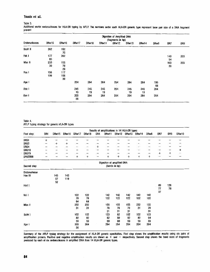
A summary of band patterns produced by enzymes in all cases is shown in Table 3. BssH ZZ, Fok Z, Mbo 11 and Rsa I can be used to distinguish HLA- DRwl5 from HLA-DRwl6 in DNAs amplified by DRG2 specific primers. Fok I and Mbo I can also be used to distinguish HLA-DR7 from HLA-DR9 in DNAs amplified with DRG79 and its comple- mentary primer. Apa Z, Dde I and Ban Z can be used to distinguish HLA-DR types amplified with DRG3568 primers as follows: Apa I cuts once within the second exon of HLA-DRw8, thus pro- ducing two bands of 195 and 68 bp; Bun I cuts once within HLA-DRw 17 second exon, producing bands of 209 and 56 bp. The Table is provided for general information, to point out that it is possible
to distinguish different HLA-DR types with more than one set of enzymes, in homozygous as well as heterozygous individuals. Moreover, such enzymes can be used to confirm the typings done with en- zymes described in Table 4.
Generic typing of DR specificities with six pairs of amplification primers and six endonucleases
We have investigated the use of 13 different endo- nucleases (As shown in Tables 3 and 4) but selected the six most informative: those that detect the largest number of specificities with the minimum effort: Hue III, Nci I , Mbo 11, BstN I, Kpn I and
81

Yunis et al.
Figure 2. Restriction enzyme patterns of amplified DNA from different HLA-DR generic types. Bands are readily recogniz- able in mini gels measuring a few centimeters. Panels show the DNA fragments produced by six endonucleases. N: Nci I; M: Mbo IZ; B: BstN I; K: Kpn I; HE Hinf I and Ha: Hue III. PhiX174 RF DNA cut with Hue I I Z and Tag I were used as molecular weight markers. The numbers represent the fragment size in base pairs (bp) calculated from the marker (numbers are those of gel readings using a DNASTAR digitizer). Fig. 2a corresponds to examples of DNA digests after amplifications with DRG2 and DRG79 pairs of primers. Figs. 2b and 2c correspond to examples of DNA digests after amplifications with DRG3568 primers. The cell used as the example of DRwl2 is a heterozygote for DRwl2/DRwll. In BstN I, the first band from top to bottom is an unidentified product from the amplifi- cation. The second band down corresponds to a 123 bp band from the DRwll type, and the subsequent bands are 82, 80 and 59 bp bands, of which the last one is also a DRwl 1 band.
Figure 2a.
+xi74 I DRwl7- v D D R w l 8 - Haem N M . B K N M 6 K
-122 -78 -64
-232
-31
~
I
- 59
YDDRwII N M B K
-264
Figure 2b.
Figure 2c.
82

HLA-DR generic typing by AFLP
Table 2. Predicted band patterns for homozygous and heterozygous combinations of DRG3568 group
Nci I Mbo II BstN I Kpn I
142 122 78 64 232 155 78 31 123 102 82 80' 64 59 264 109 56 HLA-DR Type
0 1 1 1 1 0 0 1 0 1 1 0 0 1 0 1 1 DRwl7 0 1 1 1 1 0 0 1 0 1 1 0 0 1 1 0 0 DRwl8 1 1 0 0 0 1 1 1 1 0 1 0 0 1 1 0 0 DRwll 1 1 0 0 0 1 1 1 0 0 1 1 0 0 1 0 0 DRwl2 1 1 0 0 0 1 1 1 0 1 1 0 0 1 1 0 0 DRwl3 1 1 0 0 1 0 0 1 0 1 1 0 0 1 1 0 0 DRwl4 1 1 0 0 0 1 1 1 1 0 0 0 1 1 1 0 0 DRw8
0 1 1 1 1 0 0 1 0 1 1 0 0 1 1 1 1 DRwl7/DRwl8 1 1 1 1 1 1 1 1 1 1 1 0 1 1 1 1 1 DRwl7/DRwll 1 1 1 1 1 1 1 1 0 1 1 1 0 1 1 1 1 ORwl7/DRwl2 1 1 1 1 1 1 1 1 0 1 1 0 0 1 1 1 1 DRwl7/DRwl3 1 1 1 1 1 0 0 1 0 1 1 0 0 1 1 1 1 DRwl7/DRwl4 1 1 1 1 1 1 1 1 1 1 1 0 1 1 1 1 1 DRwl7/DRw8
1 1 1 1 1 1 1 1 1 1 1 0 1 1 1 0 0 DRwl8/DRwll 1 1 1 1 1 1 1 1 0 1 1 1 0 1 1 0 0 DRwl8/DRwl2 1 1 1 1 1 1 1 1 0 1 1 0 0 1 1 0 0 DRwl81DRwl3 1 1 1 1 1 0 0 1 0 1 1 0 0 1 1 0 0 DRwl8/0Rwl4 1 1 1 1 1 1 1 1 1 1 1 0 1 1 1 0 0 DRwl8/DRw8 1 1 0 0 0 1 1 1 1 0 1 1 0 1 1 0 0 DRwlllDRwl2 1 1 0 0 0 1 1 1 1 1 1 0 1 1 1 0 0 DRwlllORwl3 1 1 0 0 1 1 1 1 1 1 1 0 1 1 1 0 0 DRwl1IDRwl4 1 1 0 0 0 1 1 1 1 0 1 0 1 1 1 0 0 DRwll/DRw8 1 1 0 0 0 1 1 1 0 1 1 1 0 0 1 0 0 DRwlaDRwl3 1 1 0 0 1 1 1 1 0 1 1 1 0 0 1 0 0 DRwl2/DRwl4 1 1 0 0 0 1 1 1 1 0 1 1 1 1 1 0 0 DRwl2/DRw8 1 1 0 0 1 1 1 7 0 1 1 0 0 1 1 0 0 DRwl3/DRwl4 1 1 0 0 0 1 1 1 1 1 1 0 1 1 1 0 0 DRwI3/DRw8 1 1 0 0 1 1 1 1 1 l l O l l 1 0 0 DRwl4/DRw8
Two bands of same size. Predicted patterns of bands produced by four endonucleases in homozygous and heterozygous combinations of DNA amplified with the OR63568 primer pair. m e numbers under the different endonucleases (Ncl, Mbo 11, BstN I and Kpn I) represent DNA fragments of different sizes (in base pairs): 142, 122, 78 and 64 base pairs for Nci I, 232, 155, 78 and 31 for Mbo II and so forth. The presence of a band in a particular Hlf-DR type is noted as 0. its presence in another type as 1. 80 bp fragment produced by BstN I represents two migra t ing bands.
Hinf I. Fig. 4 is a summary of the general procedure used to type .all samples.
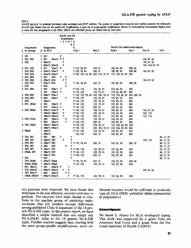
DRwl2 and the remaining 7 as HLA-DRwl 1. Re- sults are summarized in Table 5.
HLA-DR generic types of a panel of unrelated individuals
Seventy-four individuals who had been previously typed as HLA-DR1, 2, 3, 4, 5, w6, 7, w8, 9 and w10 were typed by the AFLP method. Eight were homozygous and 66 heterozygous. There was a total concordance between the serological assign- ments and those made by AFLP. Assignments
HLA-DRw 16, HLA-DRw 17 and HLA-DRw 18 were made by AFLP only. All HLA-DRwl5 and HLA-DRw 16 corresponded to those previously typed as HLA-DR2; similarly, assignments of HLA-DRwl7 and HLA-DRwl8 corresponded to HLA-DR3 as expected. There were 9 HLA-DR5 individuals of whom 2 were reassigned as HLA-
of HLA-DRw11, HLA-DRwl2, HLA-DRw15,
Discussion Sequencing of hypervariable regions of the HLA- DRB genes has permitted the design of new methods to type all alleles within the different ge- neric groups of HLA-DRBl. In general, most methods involve either the use of sequence-specific probes to split alleles after the amplification of a segment of the second exon or the use of a number of primers to amplify different groups of sequences and sets of probes to subtype them. One of the most practical of such oligotyping methods (9) uses one pair of amplification primers and 21 different probes to assign HLA-DR generic types. Other oligotyping methods to type alleles of many, if not
83
Yunis et al.
Table 3. Additional useful endonucleases for HLA-DR typing by AFLP. The numbers under each HLA-DR generic type represent base pair size of a DNA fragment present
Digestion of Amplified DNA (fragments in bp)
Endonucleases DRwl5 DRwl6 DRwl7 DRwl8 DRwl1 DRwl2 DRwl3 DRwl4 DRw8 OR7 DR9
BssH II 262 192 70
Fok I 177 262 149 203
Mbo II 233 155 163 203 a5 54
39 7a 39 29
Rsa I 156 117 106 106
39 Apa I 264 264 264 264 264 264 195
68 Dde I 245 245 245 264 245 245 264
Ban I 209 264 264 264 264 264 264 19 19 19 19 19
56
Table 4. AFLP typing strategy for generic HLA-DR types
Results of amplifications in 14 HLA-DR types First step: DR1 DRwl5 DRwl6 DRwl7 DRwl8 OR4 DRwll DRwl2 DRwl3 DRwl4 DRw8 DR7 DR9 DRwlO
~ ~
Digestion of amplified DNA Second step: (bands in bp)
Endonuclease Hae 111 143 143
67 119 52
89 126
37
Hinf I 77 7a
Nci I 122 122 142 142 142 142 142 7a 7a 122 122 122 122 122 64 64
Mbo II 232 232 155 155 155 232 155
31 31 31 31 BstN 1 102 102 123 82 102 102 123
a2 82 82 80 82 82 64 59 59 59 80 59 59 59
Kpn I 209 264 264 264 264 264 264 56
31 31 78 7a 78 31 78
Summary of the AFLP typing strategy for the assignment of HLA-OR generic specificities. First step shows the amplification results using six Pairs Of amplification primers. Positive and negative amplification results are shown as + and - respectively. Second step shows the band sizes of fragments produced by each of six endonucleases in amplified DNA from 14 HLA-DR generic types.
84

HLA-DR generic typing by AFLP
HLA-DR9; HLA-DRwll, HLA-DRw13 and HLA-DRwl4; HLA-DRw12 and HLA-DRw8; HLA-DRwl7 and HLA-DRwl8; HLA-DRwlS and HLA-DRwl6. Primers chosen for HLA-DR generic types (DRl, DR2, DR3, DR5, DRw6, DRw8 and DR4) are similar but not identical to those used in the DNA component of the Eleventh International Workshop. Antisense primer specific for codon 78 of HLA-DR7 and HLA-DR9 se- quences was found to be specific for those three alleles. A second difficulty involved the cross-am- plification of HLA-DRB3, HLA-DRB4 and HLA- DRB5 genes with any pair of HLA-DRB locus- specific amplification primers tested. Complete al- lele typing by AFLP alone (3) proved impossible upon restriction analysis of HLA-DRB second exon sequences: no group of enzymes produced specific patterns of bands for all alleles (4). Restric- tion enzyme analysis of all published HLA-DRB second exon sequences (7) showed that it was poss- ible to produce unique patterns of bands within groups of sequences. Such differences would be easily recognized in agarose gels, even in all pre- dicted possible heterozygous combinations. Here we report the smallest group of endonucleases cap- able of identifying 14 HLA-DR generic types. Re- sults in heterozygotes showed unequivocal patterns for the 14 HLA-DR types without the need of family studies. Six groups of amplifications were chosen balancing the experimental conditions for amplifications and the usefulness of the smallest number of enzymes to assign 14 HLA-DR types. For instance, there are 11 HLA-DR4 alleles and 4 DRwl1 known sequences. In the case of HLA- DR4, all alleles share a unique sequence within codons 13-14 of the second exon, which makes it possible to amplify them without cross-reactivities. In contrast, HLA-DRwll sequences can not be amplified specifically with any set of primers with- out concomitant amplification of HLA-DRw 17, -w18, -w13 and -w14 related sequences (data not shown). It was impossible for us to find individuals for the 104 possible heterozygous combinations; however, we have typed 74 individuals who had been typed serologically. Table 5 illustrates the HLA-DR types of such cells, 66 of which were heterozygous. Fig. 3 shows the results from ampli- fications and digests of 13 such individuals. DNA band patterns can be easily recognized in all hetero- zygous combinations. The method described not only identifies the generic specificity in all those individuals, but also splits the HLA-DRw6 and HLA-DR3 individuals into DRwl3 and DRwl4, DRwl7 and DRwl8, respectively.
There are several advantages to the use of the AFLP technique which would justify its use: - Enzymes might pick up substitutions along
DffGf
DRG2
DRG4
DffG3568
Figure 3. DNA amplifications of 13 heterozygous individuals using four different group-specific primers: DRGl, DRG2, DRG4 and DRG3568. DRG79 and DRGlO gave negative re- sults (data not shown). HLA-DR assignments were: HLA-DRl: samples 2,3,4, 5, 6,7 and 9. HLA-DR4: samples 6, 7, 8,9, 1 1 and 12. Samples 1, 3,4 and 5 are positive for the DRG2 group, and samples 2, 10, 11, 12 and 13 are positive for DRG3568 group. HLA-DR assignments for these last two groups will be made after endonuclease digests shown in Fig. 5.
all, HLA-DR generic types have been published in recent years.
Amplified fragment length polymorphisms for class I1 genes were first described for typing DQBl alleles (2) and soon an HLA-DR typing strategy (3) was described. Although the method proved the feasibility of such an approach, it did not permit assignments in 81% of the possible combinations. Group-specific amplifications seemed to be the only alternative to the problem of pattern ambi- guity observed by Ollerup when using a single set of HLA-DRB amplification primers. The need to use six different group-specific amplifications was a result of experimental difficulties with primer specificity, especially between HLA-DR7 and
85
Yunis et al.
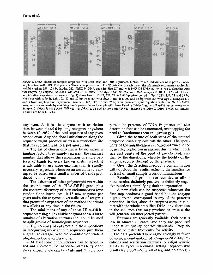
Figure 4. DNA digests of samples amplified with DRG3568 and DRG2 primers. DNAs from 5 individuals were positive upon amplification with DRG3568 primers. Three were positive with DRG2 primers. In each panel, the left sample represents a molecular weight marker: M1: 123 bp ladder, M 2 PhiX174 DNA cut with Hue ZZZ and M3: PhiX174 DNA cut with Tug I . Samples were run enzyme by enzyme. N: Nci I, M: Mbo 11, B: BsiN I, K: Kpn I and H: Hue IZI. DNA samples 2, 10, 1 1 , 12 and 13 from amplification experiment (shown in Fig. 4) show bands of 142, 122, 78 and 64 bp when cut with Nci I . 232, 155, 78 and 31 bp when cut with Mbo ZZ, 123, 102, 82 and 80 bp when cut with BstN I and 264, 209 and 56 bp when cut with Kpn I . Samples 1, 3 and 4 from amplification experiment. Bands of 143, 119, 67 and 52 bp were produced upon digestion with Hue Il l . HLA-DR assignments were made by matching bands present in each sample with those listed in Tables 2 and 4. HLA-DR assignments were: Samples 21 DRwl7; 10: DRwll/DRwl3; 11: DRwIl, 12 and 13 are both DRwl3. Sample 1 is DRwlS/DRwl6 whereas samples 3 and 4 are both DRwl5.
any exon. As it is, six enzymes with restriction sites between 4 and 6 bp long recognize anywhere between 10-20% of the total sequence of any given second exon. Any additional substitution along the sequence might produce or erase a restriction site that may in turn lead to a polymorphism. - The list of chosen enzymes is by no means a
limiting factor: they simply represent the smallest number that allows the recognition of single pat- terns of bands for every known allele. In fact, it is advisable to use more enzymes than is strictly necessary, especially whenever an assignment is go- ing to be based on a small number of bands pro- duced by an enzyme. - The existence of other polymorphisms within
the second exon of the HLA-DRBl gene, plus the constant discovery of new endonucleases (one vendor alone introduces 10-15 new enzymes per year) make the enzymes a versatile set of reagents that permit the expansion of the method to include new alleles at any time in the future. - The mini maps of any of those HLA-DRB1
sequences using all available enzymes show a large number of alternative enzymes that could be used to split groups of homologous sequences. - The accuracy of enzymes and their specificity
in recognizing invariant site sequences give them a great advantage over the condition-dependent oligonucleotide hybridizations. - At least some endonucleases can be lyophili-
zed and, therefore, locus-specific plates to type for every known allele can be easily and reliably pre-
86
pared; the presence of DNA fragments and size determinations can be automated, overstepping the need to fractionate them in agarose gels. - Given the nature of both steps of the method
proposed, each step controls the other: The speci- ficity of the amplification is controlled twice: once by gel electrophoresis in agarose during which both size and purity of the product are checked, and then by the digestions, whereby the fidelity of the amplification is checked by the enzymes. - Given the detection method, a contamination
will not cloud the results, reducing the significance at least of small sample cross-contaminations. - Results of digestions are recorded as all-or-
none results, definitely positive or definitely nega- tive reactions, simplifying their interpretation. - A new allele can be suspected whenever the
first step produces a good band but the enzyme digests do not correspond to any of the patterns described. In fact, since the enzymes come in con- tact with the whole amplified DNA, any alteration in the sequence that may produce or erase a site will generate an unexpected pattern. - Enzymes are generally available; their cost is
low in almost all cases, and they are produced under strict quality control standards. They do have to be tested frequently for activity.
The data presented here argue strongly in favor of using a combination of group-specific amplifi- cations and restriction enzymes to assign generic HLA-DR types in a clinical setting. Reproducible results were obtained in all cases, and no ambigu-
HILA-DR generic typing by AFLP
Table 5. HLA-DR typing in 74 unrelated individuals using serological and AFLP methods. The number of assignments made by each method precedes the individual's HLA-DR type. Shown here are the positive (P) amplifications in each set of groupspecific amplfications. Results of corresponding endonuclease digests used to make the final assignments in the DRG2, DRG79 and DRG3568 groups are shown here as band sizes
Results from GS Amplifications 1 2 4 1 0 7 3
9 5 Assignments Assignments 6 Results from endonuclease digests
# by serology # by AFLP 8 Nci I Mbo I1 BstN I Kpn I Hae 111 Hinf I
2 DR1 4 DR1, DR2 2 DR2
3 DR1, D R 3 1 DR2, DR3 1 DR3,DR5 4 D R l , DR4 2 DR2, OR4 1 DR3, DR4 1 DR4 3 DR1, DR5
3 DR3, DR5 6 DR4, DR5 1 OR5 5 DR1, DRw6
4 DR2, DRw6
3 DR4, DRw6
4 DR5. DRw6
2 DRw6
2 DRl, DR7 1 DR2, OR7 3 DR3, DR7 2 DR4, DR7 2 DR5. DR7 4 DRw6, OR7
1 DR7 1 DR3, DRw8 1 DRw6,DRw8 1 DR1, DRwlO 1 DR2, DRwlO 1 DR4, DRwlO 1 DRw6, DRwlO
2 DR1 P 4 DR1 DRwl5 P P 1 DRwl5 P 1 DRwl5 DRwl6 P 3 DR1 DRwl7 P 1 DRwl5 DRwl7 P 1 DRw18 DRw11 4 DR1 DR4 P P 2 DRwl5 DR4 P P 1 DRwl7 OR4 P 1 DR4 P 1 DR1 DRwl1 P 2 DR1 DRw12 P 3 DRwl7 DRwl1 6 DR4 DRwl l P 1 DRwll 2 DR1 DRwl3 P 3 DR1 DRwl4 P 2 DRwl5 DRwl3 P 1 DRwl6 DRw13 P 1 DRwl6 DRw14 P 1 DR4 DRwl3 P 2 DR4 DRwl4 P 1 DRwl1 D h l 3 3 DRwll DRw14 1 DRwl3 1 DRwl4 2 DR1 DR7 P 2 DRwl6 DR7 P 3 DRwl7 DR7 2 DR4 DR7 P 2 D R w l l DR7 1 DRwl3 DR7 3 DRwl4 DR7 1 DR7 1 DRwl7 DRw8
143,67,52 143, 67, 52 143, 11 9,67, 52
P 122, 78, 64 232, 31 102, 82, 59 209, 56 P 122, 78, 64 232, 31 102, 82, 59 209, 56 143, 67, 52 P 142,122.78.64 232, 155, 78.31 123,102, 82, 59 264
143, 67,52 P 122, 78, 64 232, 31 102, 82, 59 209, 56
P 142,122 155, 78, 31 123, 82, 59 264 P 142, 122 155, 78, 31 123, 82, 80, 59 264 P 142, 122, 78.64 232, 155,78, 31 123,102, 82, 59 264, 209, 56 P T42, 122 P 142, 122 P 142, 122 P 142,122 P 142, 122 P 142, 122 P 142,122 P 142, 122 P 142, 122 P 142, 122 P 142, 122 P 142, 122
142, 122 P P P P 122, 78, 64 P P P142,122 P P 142,122 P P142, 122 P
P 122. 78.64 . . 1 DRwl3 DRw8 P 142,122 1 DR1 DRwlO P P 1 DRwl5 DRwlO P P 1 DR4 DRwlO P P 1 DRwl3 DRwlO P P142, 122
143,67,52 143, 119 143, 119
155, 78,31 123, 82, 59 264 155,78, 31 123, 82, 59 264 155,78,31 102,82,59 264 232, 31 102, 82,59 264 155, 78, 31 102, 82, 59 264 155, 78, 31 102,82, 59 264 232, 31 102.82, 59 264 155, 78, 31 102, 82, 59 264 232, 31 102, 82,59 264 155,78,31 123, 102, 82, 59 264 232,155, 78, 31 123, 102, 82.59 264 155, 78, 31 102, 82, 59 264 232, 31 102, 82. 59 264
89, 77, 37 143. 119 89, 77, 37
232, 31 102, 82, 59 209, 56 89, 77, 37 89, 77, 37
155,78,31 123, 82, 59 264 89, 77, 37 155, 78, 31 102, 82, 59 264 89, 77, 37 232, 31 102, 82, 59 264 89, 77, 37
89, 77. 37 232. 31 102, 82, 59 209.56 155, 78.31 102, 82,59 264
143, 67, 52
155, 78, 31 102, 82, 59 264
ous patterns were observed. We have found this technique to be cost effective, accurate and easy to perform. The enzymes have been chosen to con- form to the smallest group of restriction endo- nucleases that will produce . enough differences among published Class I1 sequences of the 14 gen- eric HLA-DR types. In the present report, we have described a simple method that can assign any HLA-DRB1 allele to the 14 generic HLA-DR types. Further analysis suggests that, starting with the same group-specific amplifications, seven ad-
ditional enzymes would be sufficient to positively type all HLA-DRB 1 published alleles (manuscript in preparation)
Ae knowledgments
We thank S. Alosco for HLA serological typing. This work was supported by a grant from the American Red Cross and a grant from the Na- tional Institutes of Health CA2053 1.
87

Yunis et a).
References 1 . Wordsworth B, Allsopp C. E., Young R, Bell JI. HLA-DR
typing using DNA amplification by the polymerase chain reaction and sequential hybridization to sequence-specific oligonucletide probes. Immunogenetics 1990: 32: 41 3-8. Trucco G , Fntsh R, Giorda R, Trucco M. Rapid detection of IDDM susceptibility with HLA-DQ D alleles as markers. Diabetes 1989: 1617-22. Uryu N, Maeda M, Ota M, Tsuji K, Inoko H. A simple and rapid method for HLA-DRB and -DQB typing by digestion of PCR-amplified DNA with allele specific restric- tion endonucleases. Tissue Antigens 1990: 35 20-3 1. Olerup 0. HLA Class I1 typing by digestion of PCR-ampli- fied DNA with allele-specific restriction endonucleses will fail to unequivocally identify the genotypes of many homo- zygous and heterozygous individuals. Tissue Antigens 1990:
Yang S. Y, Milford E, Hammerling V, Dupont B. Descrip- tion of the reference panel of B-lymphoblastoid cell lines for factors of the HLA system: The B cell panel designated
36: 83-87.
for the 10th International Histocompatibility Workshop. In: Dupont B, ed. Immunobiology of HLA, vol 1. Histo- compatibility Testing, 1987. Berlin: Springer, 1988.
6. Mullis KB, Faloona F. Specific synthesis of DNA in vitro via a polymerase-catalized chain reaction. Methodr Enzym-
7. Marsh S, Bodmer J. HLA-DRB nucleotide sequences. Tissue Antigens 1991: 37: 181-9.
8. Noreen HJ, Santamaria P, Davidson ML, Rich SS, Segall M. Serology, restriction fragment length polymorphism and sequence analysis of a unique HLA Class I1 antigen, DR5 x 6. Human Immunology 1991: 30: 168-73.
9. Vaughan RW, Lanchbury JS, Marsh S, Hall MA, Bodmer JG, Welsh KI. The application of oligonucleotide probes to HLA Class I1 typing of the DRB sub-region. Tissue Antigens 1990: 36: 149-55.
01 1987: 155: 335-50.
Address: Zvrin Yunis American Red Cross Dedham, Mass. U.S.A.
88