Modeling aqueous ferrous iron chemistry at low temperatures with application to Mars

© 2014 Informa UK, Ltd. This provisional PDF corresponds to the article as it appeared upon acceptance. Fully formatted PDF and full text (HTML) versions will be made available soon.
DISCLAIMER: The ideas and opinions expressed in the journal’s Just Accepted articles do not necessarily refl ect those of Informa Healthcare (the Publisher), the Editors or the journal. The Publisher does not assume any responsibility for any injury and/or damage to persons or property arising from or related to any use of the material contained in these articles. The reader is advised to check the appropriate medical literature and the product information currently provided by the manufacturer of each drug to be administered to verify the dosages, the method and duration of administration, and contraindications. It is the responsibility of the treating physician or other health care professional, relying on his or her independent experience and knowledge of the patient, to determine drug dosages and the best treatment for the patient. Just Accepted articles have undergone full scientifi c review but none of the additional editorial preparation, such as copyediting, typesetting, and proofreading, as have articles published in the traditional manner. There may, therefore, be errors in Just Accepted articles that will be corrected in the fi nal print and fi nal online version of the article. Any use of the Just Accepted articles is subject to the express understanding that the papers have not yet gone through the full quality control process prior to publication.
Just Accepted by Free Radical Research
Histological detection of catalytic ferrous iron with the selective turn-on fl uorescent probe RhoNox-1 in a Fenton reaction-based rat renal carcinogenesis model Takahiro Mukaide , Yuka Hattori , Nobuaki Misawa , Satomi Funahashi , Li Jiang , Tasuku Hirayama , Hideko Nagasawa and Shinya Toyokuni
doi: 10.3109/10715762.2014.898844
Abstract
Iron overload of a chronic nature has been associated with a wide variety of human diseases, including infection, carcinogenesis and atherosclerosis. Recently, a highly specifi c turn-on fl uorescent probe (RhoNox-1) specifi c to labile ferrous iron [Fe(II)], but not to labile ferric iron [Fe(III)], was developed. The evaluation of Fe(II) is more important than Fe(III) in vivo in that Fe(II) is an initiating component of the Fenton reaction. In this study, we applied this probe to frozen sections of an established Fenton reaction-based rat renal carcino-genesis model with an iron chelate, ferric nitrilotriacetate (Fe-NTA), in which catalytic iron induces the Fenton reaction specifi cally in the renal proximal tubules, presumably after iron reduction. Notably, this probe reacted with Fe(II) but with neither Fe(II)-NTA, Fe(III) nor Fe(III)-NTA in vitro . Prominent red fl uorescent color was explicitly observed in and around the lumina of renal proximal tubules one hour after an intraperitoneal injection of 10-35 mg iron/kg Fe-NTA, which was dose-dependent, according to semiquantitative analysis. The RhoNox-1 signal colocalized with the generation of hydroxyl radicals, as detected by hydroxyphenyl fl uorescein (HPF). The results demonstrate the transformation of Fe(III)-NTA to Fe(II) in vivo in the Fe-NTA-induced renal carcinogenesis model. Therefore, this probe would be useful for localizing catalytic ferrous iron in studies using tissues.
Free
Rad
ic R
es D
ownl
oade
d fr
om in
form
ahea
lthca
re.c
om b
y Pr
of. S
. Toy
okun
i on
05/0
1/14
For
pers
onal
use
onl
y.
JUST
ACC
EPTE
D
Mukaide T et al. 1
Histological detection of catalytic ferrous iron with the selective
turn-on fluorescent probe RhoNox-1 in a Fenton reaction-based
rat renal carcinogenesis model
Takahiro Mukaide1, Yuka Hattori1,2, Nobuaki Misawa1, Satomi Funahashi1, Li Jiang1,
Tasuku Hirayama3, Hideko Nagasawa3 and Shinya Toyokuni1
1Department of Pathology and Biological Responses, and 2Department of Obstetrics and
Gynecology, Nagoya University Graduate School of Medicine, Nagoya 466-8550, Japan
3Laboratory of Pharmaceutical and Medicinal Chemistry, Gifu Parmaceutical University,
Gifu 501-1196, Japan
Correspondence: Shinya Toyokuni, M.D., Ph.D.; Department of Pathology and
Biological Responses, Nagoya University Graduate School of Medicine, 65
Tsurumai-cho, Showa-ku, Nagoya 466-8550, Japan. Tel: +81-52-744-2086; Fax:
+81-52-744-2091; E-mail: [email protected]
Short title: Localizing catalytic Fe(II) in tissue
Abstract
Free
Rad
ic R
es D
ownl
oade
d fr
om in
form
ahea
lthca
re.c
om b
y Pr
of. S
. Toy
okun
i on
05/0
1/14
For
pers
onal
use
onl
y.

JUST
ACC
EPTE
D
Mukaide T et al. 2
Iron overload of a chronic nature has been associated with a wide variety of
human diseases, including infection, carcinogenesis and atherosclerosis.
Recently, a highly specific turn-on fluorescent probe (RhoNox-1) specific to labile
ferrous iron [Fe(II)], but not to labile ferric iron [Fe(III)], was developed. The
evaluation of Fe(II) is more important than Fe(III) in vivo in that Fe(II) is an
initiating component of the Fenton reaction. In this study, we applied this probe to
frozen sections of an established Fenton reaction-based rat renal carcinogenesis
model with an iron chelate, ferric nitrilotriacetate (Fe-NTA), in which catalytic iron
induces the Fenton reaction specifically in the renal proximal tubules, presumably
after iron reduction. Notably, this probe reacted with Fe(II) but with neither
Fe(II)-NTA, Fe(III) nor Fe(III)-NTA in vitro. Prominent red fluorescent color was
explicitly observed in and around the lumina of renal proximal tubules one hour
after an intraperitoneal injection of 10-35 mg iron/kg Fe-NTA, which was
dose-dependent, according to semiquantitative analysis. The RhoNox-1 signal
colocalized with the generation of hydroxyl radicals, as detected by
hydroxyphenyl fluorescein (HPF). The results demonstrate the transformation of
Fe(III)-NTA to Fe(II) in vivo in the Fe-NTA-induced renal carcinogenesis model.
Therefore, this probe would be useful for localizing catalytic ferrous iron in studies
using tissues.
Keywords: catalytic ferrous iron, fluorescent probe, kidney, oxidative stress,
morphometry
Free
Rad
ic R
es D
ownl
oade
d fr
om in
form
ahea
lthca
re.c
om b
y Pr
of. S
. Toy
okun
i on
05/0
1/14
For
pers
onal
use
onl
y.

JUST
ACC
EPTE
D
Mukaide T et al. 3
Introduction
Iron is an essential element in all living organisms on earth and is the most
abundant heavy metal in humans. Human adults hold approximately 4 grams of
iron in total. Hemoglobin in red blood cells maintains 60% of this iron as the heme
prosthetic group for oxygen binding. The remaining portions of iron are present in
either the cells or extracellular space, including serum. Iron is a cofactor for
various enzymes, is tightly bound to transferrin in serum, forms an iron reserve as
ferritin or may transform into insoluble hemosiderin when overloaded [1].
Iron both has benefits and poses risks. Whereas iron deficiency causes
anemia and muscle weakness, iron overload or even iron misdistribution that
leads to localized chronic iron overload is associated with and is a risk for various
diseases, including infection, cancer, atherosclerosis and autoimmune diseases
[2,3]. An iron importing transporter, DMT1 (Nramp2; natural
resistance-associated macrophage protein), and a hepatic peptide hormone,
hepcidin, were previously discovered in association with a risk for infection [4,5].
There are a plethora of reports of an association between iron overload and
carcinogenesis in both human and animal studies [6-8]. Iron accumulation in an
atheroma that results from hemorrhage appears to be associated with its rupture,
which is a direct cause of infarction in small arteries [9]. It is established that the
synovial fluid in rheumatoid arthritis patients contains catalytic iron [10]. It is
generally accepted that the Fenton reaction, which leads to the generation of
hydroxyl radicals, causes all of the pathologies described above [11]. Therefore,
the localization of ferrous iron has always been a subject of interest as an initiator
Free
Rad
ic R
es D
ownl
oade
d fr
om in
form
ahea
lthca
re.c
om b
y Pr
of. S
. Toy
okun
i on
05/0
1/14
For
pers
onal
use
onl
y.
JUST
ACC
EPTE
D
Mukaide T et al. 4
of the Fenton reaction [12]. However, thus far, no histological methodology has
been established to detect labile or catalytic ferrous iron.
Recently, a highly specific probe, called RhoNox-1, for labile ferrous iron
was developed [13]. In this study, we describe for the first time the application of
this probe to frozen sections of kidney after in vivo oxidative injury using through
ferric nitrilotriacetate [Fe(III)-NTA], which is an established model of Fenton
reaction-based renal carcinogenesis in rats [14-19].
Methods
Materials
RhoNox-1 was a kind gift from Prof. Hideko Nagasawa (Gifu
Pharmaceutical University, Gifu, Japan) [13]. Hydroxyphenyl fluorescin (HPF)
was obtained from Sekisui Medical (Tokyo, Japan). Fe(NO3)3 9H2O was obtained
from Wako (Osaka, Japan), and nitrilotriacetate disodium salt was obtained from
Nakalai Tesque (Kyoto, Japan). Ferric nitrilotriacetate [Fe(III)-NTA] was produced
by mixing 300 mM ferric nitrate solution and 600 mM nitrilotriacetate solution,
followed by pH adjustment to 7.4 with sodium hydrogen bicarbonate, as
previously described [15], and was used within 30 min. All other agents were of
analytical grade.
Animal experiments
The animal experiment committee of the Nagoya University Graduate
School of Medicine approved the following animal experiments. In total, 42 male
Wistar rats (8 weeks old; Shizuoka Laboratory Anima Center, Shizuoka, Japan)
were purchased. These rats were divided into time-course and dose-dependency
Free
Rad
ic R
es D
ownl
oade
d fr
om in
form
ahea
lthca
re.c
om b
y Pr
of. S
. Toy
okun
i on
05/0
1/14
For
pers
onal
use
onl
y.

JUST
ACC
EPTE
D
Mukaide T et al. 5
groups. The time-course group was evaluated 0, 1, 2, 4, 6, 24 and 48 h after a
single injection of 10 mg iron/kg body weight of Fe(III)-NTA; the dose-dependency
group was evaluated following the administration of 0, 10, 15, 20, 25, 30 and 35
mg iron/kg of Fe(III)-NTA (N=3). Fe-NTA was intraperitoneally injected, and the
animals were euthanized at the indicated time. The kidneys were immediately
removed. Some portions of the kidney were fixed with 10 mM phosphate-buffered
10% formalin and were processed for routine paraffin embedding and sectioning
at 3 m, which was followed by hematoxylin and eosin staining to examine the
histology. Some portions were embedded in optimum cutting temperature
compound (Sankyo Miles, Tokyo) for frozen sections.
Histological detection of labile ferrous iron
RhoNox-1 was preserved in a deep freezer at -80°C and dissolved in
dimethyl sulfoxide to produce a 1 mM solution, which was further diluted (1:200)
with 10 mM phosphate-buffered saline (pH 7.4) before use (final concentration 5
M). This diluted solution was used within a single day. Frozen sections of 8 m
thickness were prepared with a cryostat on MAS-GP type A grass slides
(Matsunami, Osaka, Japan), air dried for 3 min, fixed in 10 mM
phosphate-buffered 20% formalin in methanol for 1 min, and washed in deionized
water for 5 min. Then, 200 l of 5 M RhoNox-1 was placed on those specimens
and incubated for 30 min at 37°C in a dark chamber. Unfixed frozen sections were
also used in some experiments. Thereafter, the specimen was counterstained
with 4’,6-diaminido-2-phenylindole, dihydrochloride (DAPI) and observed as
described below. Some of the specimens were further incubated with HPF after
three washes with PBS for 30 min at 37°C in a dark room. We were able to
preserve the frozen sections at -80°C after cutting at least for a week.
Free
Rad
ic R
es D
ownl
oade
d fr
om in
form
ahea
lthca
re.c
om b
y Pr
of. S
. Toy
okun
i on
05/0
1/14
For
pers
onal
use
onl
y.
JUST
ACC
EPTE
D
Mukaide T et al. 6
Imaging analysis
A fluorescence microscope (BZ-9000, Keyence, Osaka, Japan), which
allows simultaneous data acquisition of three different wavelengths, was used for
analyses. To obtain quantitative data, the exposure condition was recorded for
each image. For the quantification of labile ferrous iron, each image was divided
into RGB elements, and only the red component was used for the analysis using
the built-in software (BZ-II analyzer) or ImageJ version 1.47 software
(http://www.rsb.info.nih.gov/ij/). Green component was used for HPF and blue
component was used for nucleus. The number of nuclei was determined, and the
final value was the integration of the red color (RhoNox-1) in tissue divided by the
number of nuclei included in the analyzed area with a 40x objective lens. Eight
random areas in the proximal tubules were used for the analysis of each rat.
Reactivity of the iron solution and iron chelates with RhoNox-1
A black 96-well microplate (#MS-8096K, Sumitomo Bakelite Co., Osaka,
Japan) and RhoNox-1 (1 M final concentration in 10 mM phosphate buffer) were
used for this analysis. Ferrous iron [Fe(II)] solution was prepared from FeSO4
7H2O (Wako, Osaka, Japan). Fe(II)-NTA was produced in a manner similar to the
preparation of Fe(III)-NTA. The pH was adjusted to 7.4 and immediately used.
Each solution containing iron (100 l) was mixed with 1 M RhoNox-1 (100 l),
which was incubated for 1 h at room temperature. Then, RhoNox-1-specific
fluorescence was measured using Powerscan 4 (DS Pharma Biomedical, Osaka,
Japan; excitation, 530 nm; emission, 575 nm; gain 100). The data are shown as
([Sample fluorescence value]-[Background])/[Background].
Free
Rad
ic R
es D
ownl
oade
d fr
om in
form
ahea
lthca
re.c
om b
y Pr
of. S
. Toy
okun
i on
05/0
1/14
For
pers
onal
use
onl
y.

JUST
ACC
EPTE
D
Mukaide T et al. 7
Statistical analysis
The data are shown as the mean±SD. Unpaired t-test, Cochran-Armitage
trend test and Pearson correlation coefficient were used where appropriate with
SPSS 13.0 (SPSS Inc., Chicago, IL). P<0.05 was considered statistically
significant.
Results
Reactivity of Fe(II)-NTA, Fe(III) and Fe(III)-NTA with RhoNox-1
RhoNox-1 dose-dependently reacted with Fe(II) in a range of 0-10 M as
previously described [13]. However, RhoNox-1 reacted with neither Fe(II)-NTA,
Fe(III) nor Fe(III)-NTA (Figure 1).
Ferric nitrilotriacetate (Fe-NTA)-induced renal carcinogenesis model
Oxidative stress, as indicated by lipid peroxidation and DNA modification,
is reported to reach its maximum 30 min to 3 h after an intraperitoneal injection of
Fe-NTA [20]. First, we used unfixed frozen sections to locate Fe(II) 1 h after 10
mg iron/kg Fe(III)-NTA administration, and found strong positivity not only in the
renal proximal tubules but also in their lumina. In addition, the image was blurred
in the absence of fixation (Figure 2). Because these were not optimal for the
morphometric analyses, we tried light fixation as described in the methods
section. Light fixation provided acceptable results both in morphology and
sensitivity, and the results were in good parallel with those of unfixed specimens.
Then, we performed a time-course study. We noted some levels of background
fluorescence in the normal kidney but found that RhoNox-1-specific fluorescence
significantly increased 1 h after the injection, then further increased and was
Free
Rad
ic R
es D
ownl
oade
d fr
om in
form
ahea
lthca
re.c
om b
y Pr
of. S
. Toy
okun
i on
05/0
1/14
For
pers
onal
use
onl
y.

JUST
ACC
EPTE
D
Mukaide T et al. 8
maintained up to at least 6 h. The fluorescence decreased 24 and 48 h after the
injection, when proximal tubular necrosis was dominant. The increase and
decrease in HPF-specific fluorescence were consistent with RhoNox-1-specific
fluorescence. Nucleus-specific fluorescence (DAPI) gradually decreased after
Fe(III)-NTA injection as renal proximal tubular cells degenerated and necrotized
(Figure 3).
Then, we performed a dose-dependence study at 1 h. Most of the animals
were dying at a dose of 35 mg iron/kg at 1 h. The kidneys were swollen with
edema and showed significantly increased weight at 30 and 35 mg iron/kg. We
observed dose-dependent RhoNox-1-specific fluorescence in the renal proximal
tubular cells, which was inversely associated with the number of viable cells as
seen by DAPI-positivity. Furthermore, RhoNox-1 and HPF coexisted (Figure
4A-C). Thus, the intensity of HPF-specific fluorescence was in parallel with
RhoNox-1, and the correlation coefficient was r=0.912 (Figure 5).
Discussion
RhoNox-1, a highly specific turn-on probe for labile ferrous ion, was
recently established. The effect of the turn on for red fluorescence was immense.
In that paper, the application of RhoNox-1 in cultured cells loaded with ferrous
iron was successful [13]. In this study, we applied this technique for the first time
to frozen sections of rat tissue and found that this technique works well for
systemic studies in animals and could be easily extended to samples from
humans as well as other species. We could use unfixed and lightly fixed
specimens.
Free
Rad
ic R
es D
ownl
oade
d fr
om in
form
ahea
lthca
re.c
om b
y Pr
of. S
. Toy
okun
i on
05/0
1/14
For
pers
onal
use
onl
y.
JUST
ACC
EPTE
D
Mukaide T et al. 9
We used an established rat model of the Fenton reaction in vivo.
Fe(III)-NTA is an iron chelate that is soluble at a neutral pH and still retains 3-4
free catalytic ligands. Thus, this molecule is thought to be the most potent iron
catalyst for the Fenton reaction after reduction [21-23]. An intraperitoneal injection
of Fe-NTA causes the Fenton reaction in the lumina of renal proximal tubules
after it is absorbed into the systemic blood flow, which is followed by filtration
through the glomeruli of the kidney [24], where it is believed that Fe(III)-NTA is
reduced to Fe(II)-NTA due to the presence of L-cysteine from the glutathione
cycle [25]. There are many reports on the generation of hydroxyl radical-modified
molecules in situ in this model, such as oxidative DNA base modifications [16]
and various aldehydes (malonaldehyde, 4-hydroxy-2-nonenal, acrolein, etc.)
[18,26]. Eventually, repeated reactions of this nature cause renal cell carcinoma
[17], and it was recently shown that the genomic alterations in these cancers are
quite similar to those alterations in human counterparts [19]. The detection of
labile ferrous iron was principally intraluminal (unfixed) and the surrounding cells
(both unfixed and lightly fixed) in the renal proximal tubules. We found a clear
dose-dependence in the quantification of the signals. The results demonstrate
that this probe can be successfully applied to frozen sections obtained from
tissues.
We fixed a part of the specimen with neutral buffered formalin followed by
paraffin embedding, and performed Perls’ iron staining. However, we did not
obtain positive staining. It is thought that Perls’ iron staining detects hemosiderin
and a part of ferritin; it is unknown whether those compounds are actually
damaging to cellular molecules in vivo. We believe that a portion of these
compounds would be solubilized to a catalytic form. In this sense, the detection of
Free
Rad
ic R
es D
ownl
oade
d fr
om in
form
ahea
lthca
re.c
om b
y Pr
of. S
. Toy
okun
i on
05/0
1/14
For
pers
onal
use
onl
y.

JUST
ACC
EPTE
D
Mukaide T et al. 10
labile ferrous iron is more direct. Furthermore, our data clearly demonstrated that
Fe(II) is produced in vivo from Fe(III)-NTA. We suspect that the Fe(II)-NTA
generated via reduction, presumably through L-cysteine derived from glutathione
through -glutamyl transferase and dipeptidase, in the lumina of renal proximal
tubules [25] is absorbed from the villous luminal membrane as Fe(II) via DMT1
[27] after de-chelation at a mild acidic pH. This hypothesis requires further
investigation.
There are already many markers for oxidative stress. Those markers
include molecules modified by the reaction of hydroxyl radicals, such as
8-hydroxy-2’-deoxyguanosine (8-OHdG) [28] and 4-hydroxy-2-nonenal (HNE)
[29]. Previously, we developed monoclonal antibodies against
8-hydroxy-2’-deoxyguanosine [30] and 4-hydroxy-2-nonenal-modified proteins
[31]. We could successfully apply these antibodies to this model in formalin-fixed
paraffin-embedded sections. We believe that there is a conceptual difference
between these monoclonal antibodies and the present probe, i.e., the presence of
labile ferrous iron constitutes the precise risk for the Fenton reaction, whereas
modified products are the sum of the production and the repair/removal of the
modifications. Furthermore, we demonstrated the coexistence of RhoNox-1 and
HPF. This result indicates that RhoNox-1-positive foci can indeed initiate the
Fenton reaction in situ. Therefore, RhoNox-1 detects catalytic ferrous iron and is
a novel marker for evaluating numerous oxidative stress-associated diseases.
Nevertheless, further studies are necessary to determine the followings: 1)
whether this method is applicable to formalin-fixed paraffin-embedded sections;
2) whether there are any discrepancies between the data regarding this probe
and other modified products in various models, and, if so, what those
Free
Rad
ic R
es D
ownl
oade
d fr
om in
form
ahea
lthca
re.c
om b
y Pr
of. S
. Toy
okun
i on
05/0
1/14
For
pers
onal
use
onl
y.

JUST
ACC
EPTE
D
Mukaide T et al. 11
discrepancies mean (We suspect that labile ferrous iron may be unexpectedly
stable in the absence of hydrogen peroxide in situ); 3) whether this method is
applicable to time-lapse studies using animals; and 4) what kind of efforts would
be necessary to decrease the background fluorescence of RhoNox-1.
In conclusion, in the present study, we developed a novel strategy to
localize catalytic ferrous iron in frozen sections of tissue. We recommend to use
both unfixed and lightly fixed specimens for the initial evaluation. This strategy
would be helpful for analyzing various iron-associated pathologies and
physiologies, including neurodegenerative diseases and iron absorption through
the duodenum. This probe may open up novel research areas for various
oxidative stress-associated diseases.
Free
Rad
ic R
es D
ownl
oade
d fr
om in
form
ahea
lthca
re.c
om b
y Pr
of. S
. Toy
okun
i on
05/0
1/14
For
pers
onal
use
onl
y.

JUST
ACC
EPTE
D
Mukaide T et al. 12
Acknowledgments
This work was supported in part by a grant-in-aid for research from the
Ministry of Education, Culture, Sports, Science and Technology (MEXT) of Japan.
Conflicts of interest
The authors declare that they have no conflicts of interest.
Free
Rad
ic R
es D
ownl
oade
d fr
om in
form
ahea
lthca
re.c
om b
y Pr
of. S
. Toy
okun
i on
05/0
1/14
For
pers
onal
use
onl
y.

JUST
ACC
EPTE
D
Mukaide T et al. 13
References
[1] Wriggleworth JM, Baum H. The biochemical function of iron. In:
Jacobs A, Worwood M, editors. Iron in biochemistry and medicine,
II. London: Academic Press; 1980. p 29-86.
[2] Weinberg ED. The hazards of iron loading. Metallomics
2010;2(11):732-40.
[3] Toyokuni S. Iron as a target of chemoprevention for longevity in
humans. Free Radic Res 2011;45:906-917.
[4] Weinberg ED. Iron availability and infection. Biochim Biophys Acta
2009;1790(7):600-5.
[5] Hentze MW, Muckenthaler MU, Galy B, Camaschella C. Two to
tango: regulation of Mammalian iron metabolism. Cell
2010;142(1):24-38.
[6] Weinberg ED. The role of iron in cancer. European Journal of
Cancer Prevention 1996;5:19-36.
[7] Toyokuni S. Iron-induced carcinogenesis: the role of redox
regulation. Free Radic Biol Med 1996;20:553-566.
[8] Toyokuni S. Role of iron in carcinogenesis: Cancer as a ferrotoxic
disease. Cancer Sci 2009;100(1):9-16.
[9] Yuan XM. Apoptotic macrophage-derived foam cells of human
atheromas are rich in iron and ferritin, suggesting iron-catalysed
reactions to be involved in apoptosis. Free Radic Res
1999;30(3):221-31.
Free
Rad
ic R
es D
ownl
oade
d fr
om in
form
ahea
lthca
re.c
om b
y Pr
of. S
. Toy
okun
i on
05/0
1/14
For
pers
onal
use
onl
y.

JUST
ACC
EPTE
D
Mukaide T et al. 14
[10] Rowley D, Gutteridge JM, Blake D, Farr M, Halliwell B. Lipid
peroxidation in rheumatoid arthritis: thiobarbituric acid-reactive
material and catalytic iron salts in synovial fluid from rheumatoid
patients. Clin Sci (Lond) 1984;66(6):691-5.
[11] Halliwell B, Gutteridge JMC. Free radicals in biology and medicine.
New York: Oxford University Press; 2007.
[12] Fenton HJH. Oxidation of tartaric acid in presence of iron.
J.Chem.Soc. 1894;65:899-910.
[13] Hirayama T, Okuda K, Nagasawa H. A highly selective turn-on
fluorescent probe fro iron(II) to visualize labile iron in living cells.
Chem Sci 2013;4:1250-1256.
[14] Ebina Y, Okada S, Hamazaki S, Ogino F, Li JL, Midorikawa O.
Nephrotoxicity and renal cell carcinoma after use of iron- and
aluminum- nitrilotriacetate complexes in rats. J Natl Cancer Inst
1986;76:107-113.
[15] Toyokuni S, Uchida K, Okamoto K, Hattori-Nakakuki Y, Hiai H,
Stadtman ER. Formation of 4-hydroxy-2-nonenal-modified proteins
in the renal proximal tubules of rats treated with a renal carcinogen,
ferric nitrilotriacetate. Proc Natl Acad Sci USA 1994;91:2616-2620.
[16] Toyokuni S, Mori T, Dizdaroglu M. DNA base modifications in renal
chromatin of Wistar rats treated with a renal carcinogen, ferric
nitrilotriacetate. Int J Cancer 1994;57:123-128.
[17] Nishiyama Y, Suwa H, Okamoto K, Fukumoto M, Hiai H, Toyokuni
S. Low incidence of point mutations in H-, K- and N-ras oncogenes
and p53 tumor suppressor gene in renal cell carcinoma and
Free
Rad
ic R
es D
ownl
oade
d fr
om in
form
ahea
lthca
re.c
om b
y Pr
of. S
. Toy
okun
i on
05/0
1/14
For
pers
onal
use
onl
y.

JUST
ACC
EPTE
D
Mukaide T et al. 15
peritoneal mesothelioma of Wistar rats induced by ferric
nitrilotriacetate. Jpn J Cancer Res 1995;86:1150-1158.
[18] Toyokuni S, Luo XP, Tanaka T, Uchida K, Hiai H, Lehotay DC.
Induction of a wide range of C2-12 aldehydes and C7-12 acyloins in
the kidney of Wistar rats after treatment with a renal carcinogen,
ferric nitrilotriacetate. Free Radic Biol Med 1997;22:1019-1027.
[19] Akatsuka S, Yamashita Y, Ohara H, Liu YT, Izumiya M, Abe K,
Ochiai M, Jiang L, Nagai H, Okazaki Y and others. Fenton reaction
induced cancer in wild type rats recapitulates genomic alterations
observed in human cancer. PLoS ONE 2012;7(8):e43403.
[20] Tanaka T, Nishiyama Y, Okada K, Hirota K, Matsui M, Yodoi J, Hiai
H, Toyokuni S. Induction and nuclear translocation of thioredoxin
by oxidative damage in the mouse kidney: independence of tubular
necrosis and sulfhydryl depletion. Lab Invest 1997;77:145-155.
[21] Toyokuni S, Sagripanti JL. Iron-mediated DNA damage: sensitive
detection of DNA strand breakage catalyzed by iron. J
Inorg.Biochem. 1992;47:241-248.
[22] Toyokuni S, Sagripanti J-L. DNA single- and double-strand breaks
produced by ferric nitrilotriacetate in relation to renal tubular
carcinogenesis. Carcinogenesis 1993;14:223-227.
[23] Toyokuni S, Sagripanti JL. Iron chelators modulate the production
of DNA strand breaks and 8- hydroxy-2'-deoxyguanosine. Free
Radic Res 1999;31(2):123-8.
[24] Toyokuni S, Okada S, Hamazaki S, Minamiyama Y, Yamada Y,
Liang P, Fukunaga Y, Midorikawa O. Combined histochemical and
Free
Rad
ic R
es D
ownl
oade
d fr
om in
form
ahea
lthca
re.c
om b
y Pr
of. S
. Toy
okun
i on
05/0
1/14
For
pers
onal
use
onl
y.

JUST
ACC
EPTE
D
Mukaide T et al. 16
biochemical analysis of sex hormone dependence of ferric
nitrilotriacetate-induced renal lipid peroxidation in ddY mice.
Cancer Res 1990;50:5574-5580.
[25] Okada S, Minamiyama Y, Hamazaki S, Toyokuni S, Sotomatsu A.
Glutathione cycle dependency of ferric nitrilotriacetate-induced
lipid peroxidation in mouse proximal renal tubules.
Arch.Biochem.Biophys. 1993;301:138-142.
[26] Kawai Y, Furuhata A, Toyokuni S, Aratani Y, Uchida K. Formation
of acrolein-derived 2'-deoxyadenosine adduct in an iron-induced
carcinogenesis model. J Biol Chem 2003;278(50):50346-54.
[27] Veuthey T, Hoffman D, Vaidya VS, Wessling-Resnick M. Impaired
renal function and development in Belgrade rats. Am J Physiol
Renal Physiol 2013.
[28] Kasai H. Analysis of a form of oxidative DNA damage,
8-hydroxy-2'-deoxyguanosine, as a marker of cellular oxidative
stress during carcinogenesis. Mutat.Res. 1997;387:147-163.
[29] Uchida K. 4-Hydroxy-2-nonenal: a product and mediator of
oxidative stress. Prog Lipid Res 2003;42(4): p318-43.
[30] Toyokuni S, Tanaka T, Hattori Y, Nishiyama Y, Ochi H, Hiai H,
Uchida K, Osawa T. Quantitative immunohistochemical
determination of 8-hydroxy-2'-deoxyguanosine by a monoclonal
antibody N45.1: its application to ferric nitrilotriacetate-induced
renal carcinogenesis model. Lab Invest 1997;76:365-374.
[31] Toyokuni S, Miyake N, Hiai H, Hagiwara M, Kawakishi S, Osawa T,
Uchida K. The monoclonal antibody specific for the
Free
Rad
ic R
es D
ownl
oade
d fr
om in
form
ahea
lthca
re.c
om b
y Pr
of. S
. Toy
okun
i on
05/0
1/14
For
pers
onal
use
onl
y.

JUST
ACC
EPTE
D
Mukaide T et al. 17
4-hydroxy-2-nonenal histidine adduct. FEBS Lett
1995;359(2-3):189-91.
Free
Rad
ic R
es D
ownl
oade
d fr
om in
form
ahea
lthca
re.c
om b
y Pr
of. S
. Toy
okun
i on
05/0
1/14
For
pers
onal
use
onl
y.

JUST
ACC
EPTE
D
Mukaide T et al. 18
Figure Legends
Figure 1. Reactivity of different forms of iron with RhoNox-1. RhoNox-1 is
specific to Fe(II) among Fe(II), Fe(III), Fe(II)-NTA and Fe(III)-NTA. Each solution
was incubated with RhoNox-1 for 1 h, and emission at 575 nm after excitation at
540 nm was measured. Refer to the text for details. NTA, nitrilotriacetate.
Free
Rad
ic R
es D
ownl
oade
d fr
om in
form
ahea
lthca
re.c
om b
y Pr
of. S
. Toy
okun
i on
05/0
1/14
For
pers
onal
use
onl
y.

JUST
ACC
EPTE
D
Mukaide T et al. 19
Figure 2. RhoNox-1-specific fluorescence in rat renal proximal tubules 1 h after
intraperitoneal injection of 10 mg iron/kg Fe(III)-NTA with unfixed specimens. Note that both
renal proximal tubules and their lumina are strongly stained (bar=40 m).
Free
Rad
ic R
es D
ownl
oade
d fr
om in
form
ahea
lthca
re.c
om b
y Pr
of. S
. Toy
okun
i on
05/0
1/14
For
pers
onal
use
onl
y.

JUST
ACC
EPTE
D
Mukaide T et al. 20
Figure 3. Time-course study of RhoNox-1-specific integrated fluorescence intensity in rat
renal proximal tubules after intraperitoneal injection of 10 mg iron/kg Fe(III)-NTA with
lightly fixed specimens. RhoNox-1 intensity and HPF increased up to 6 h, whereas nuclear
fluorescence (DAPI; 4’,6-diaminido-2-phenylindole, dihydrochloride) decreased with
degeneration and necrosis. Analyses were through BZ-II. AU, arbitrary unit; NTA,
nitrilotriacetate; HPF, hydroxyphenyl fluorescein. **, P<0.01; ***, P<0.001; ####, P<0.0001
vs time=0; unpaired t-test.
Free
Rad
ic R
es D
ownl
oade
d fr
om in
form
ahea
lthca
re.c
om b
y Pr
of. S
. Toy
okun
i on
05/0
1/14
For
pers
onal
use
onl
y.

JUST
ACC
EPTE
D
Mukaide T et al. 21
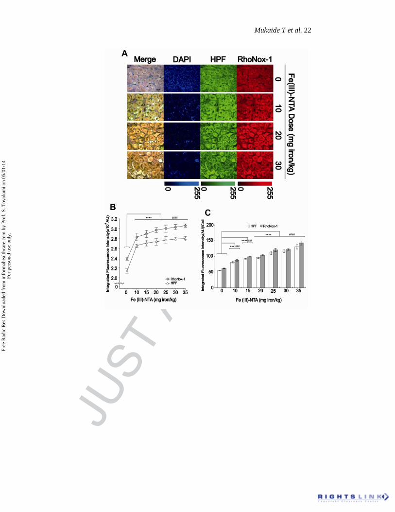
Figure 4. Dose-dependence study of RhoNox-1-specific integrated fluorescence intensity in
rat renal proximal tubules 1 h after intraperitoneal Fe(III)-NTA injection with lightly fixed
specimens. A: Representative pictures obtained from a single frozen section with multi-color
analysis. Colocalization of RhoNox-1 and HPF is evident. DAPI,
4’,6-diaminido-2-phenylindole, dihydrochloride; HPF, hydroxyphenyl fluorescein;
NTA, nitrilotriacetate (bar=40 m). B: Quantification of A for RhoNox-1 and HPF. AU,
arbitrary unit. C: Integrated fluorescence intensity per cell; trends P<0.001 with
Cochran-Armitage test. Analyses were through ImageJ; the results through BZ-II and
ImageJ were proportional (HPF: ***, P<0.001 vs dose=0; ****, P<0.0001 vs dose=0;
RhoNox-1: ###, P<0.001 vs dose=0; ####, P<0.0001 vs dose=0; unpaired t-test).
Free
Rad
ic R
es D
ownl
oade
d fr
om in
form
ahea
lthca
re.c
om b
y Pr
of. S
. Toy
okun
i on
05/0
1/14
For
pers
onal
use
onl
y.
JUST
ACC
EPTE
D
Mukaide T et al. 22
Free
Rad
ic R
es D
ownl
oade
d fr
om in
form
ahea
lthca
re.c
om b
y Pr
of. S
. Toy
okun
i on
05/0
1/14
For
pers
onal
use
onl
y.

JUST
ACC
EPTE
D
Mukaide T et al. 23
Figure 5. The association of RhoNox-1 and HPF. Corresponding data on
integrated fluorescence intensity (IFI) of RhoNox-1 and HPF was compared,
which were proportional. AU, arbitrary unit; HPF, hydroxyphenyl fluorescein.
Free
Rad
ic R
es D
ownl
oade
d fr
om in
form
ahea
lthca
re.c
om b
y Pr
of. S
. Toy
okun
i on
05/0
1/14
For
pers
onal
use
onl
y.
Copyright © 2022 FDOKUMEN