High-performance iterative electron tomography reconstruction with long-object compensation using...
12
High-performance iterative electron tomography reconstruction with long-object compensation using graphics processing units (GPUs) Wei Xu a , Fang Xu a , Mel Jones b,c , Bettina Keszthelyi b,c , John Sedat c , David Agard b,c , Klaus Mueller a, * a Center for Visual Computing, Computer Science Department, Stony Brook University, Stony Brook, NY 11794-4400, United States b Howard Hughes Medical Institute, Department of Biochemistry & Biophysics, University of California at San Francisco, United States c Keck Advanced Microscopy Laboratory, Department of Biochemistry & Biophysics, University of California at San Francisco, United States article info Article history: Received 4 December 2009 Received in revised form 27 March 2010 Accepted 30 March 2010 Available online xxxx Keywords: Tomography Reconstruction Image processing Parallel processing abstract Iterative reconstruction algorithms pose tremendous computational challenges for 3D Electron Tomogra- phy (ET). Similar to X-ray Computed Tomography (CT), graphics processing units (GPUs) offer an afford- able platform to meet these demands. In this paper, we outline a CT reconstruction approach for ET that is optimized for the special demands and application setting of ET. It exploits the fact that ET is typically cast as a parallel-beam configuration, which allows the design of an efficient data management scheme, using a holistic sinogram-based representation. Our method produces speedups of about an order of magnitude over a previously proposed GPU-based ET implementation, on similar hardware, and com- pletes an iterative 3D reconstruction of practical problem size within minutes. We also describe a novel GPU-amenable approach that effectively compensates for reconstruction errors resulting from the TEM data acquisition on (long) samples which extend the width of the parallel TEM beam. We show that the vignetting artifacts typically arising at the periphery of non-compensated ET reconstructions are completely eliminated when our method is employed. Ó 2010 Elsevier Inc. All rights reserved. 1. Introduction Electron Tomography (ET) (see for example Frank, 2006, or Lucic et al., 2005) uniquely enables the 3D study of complex cellu- lar structures, such as the cytoskeleton, organelles, viruses and chromosomes. It recovers the specimen’s 3D structure via comput- erized tomographic (CT) reconstruction from a set of 2D projec- tions obtained with Transmission Electron Microscopy (TEM) at different tilt angles. ET can be accomplished using exact analytical methods (weighted back-projection WBP (Radermacher, 2006) and more recently electron lambda-tomography (Quinto et al., 2009)) or via iterative schemes, such as the Simultaneous Algebraic Reconstruction Technique (SART) (Andersen and Kak, 1984), the Simultaneous Iterative Reconstruction Technique (SIRT) (Gilbert, 1972), and others. The dominant use of the analytical methods is most likely due to their computational simplicity and consequently fast reconstruction speed. Iterative methods, however, have the advantage that additional constraints can be easily and intuitively incorporated into the reconstruction procedure. This, for example, can be exploited to better compensate for noise (Skoglund et al., 1996) and to perform alignment corrections (Castano-Diez et al., 2006; Frank and McEwen, 1992; Lawrence, 1992) during the itera- tive updates. Additional challenges are imposed by the fact that the projection sinogram is vastly undersampled, both in terms of angu- lar resolution (due to dose constraints) and in terms of angular range (due to limited sample access). These types of scenarios can be handled quite well using iterative reconstruction ap- proaches (Andersen, 1989). Thus, iterative approaches have great potential for ET. However, as data collection strategies (Zheng et al., 2004) and electron detec- tors improve, the push has been to reconstruct larger and larger vol- umes (2048 2 512 pixels and beyond). Although the benefits are significant, the major obstacle preventing the widespread use of iter- ative methods in ET so far has been the immense computational overhead associated with these, leading to reconstruction times on the order of hours to days for practical data scenarios. As in many other scientific disciplines, the typical solution to meet these high computational demands has been the use of supercomputers and large computer clusters (Fernández, 2008; Fernández et al., 2004; Zheng et al., 2006), but such hardware is expensive and can also be difficult to use and gain access to. Fortunately, the recently emerging graphics processing units (GPUs) offer an attractive alternative plat- form, both in terms of price and performance. GPUs are available at a price of less than $500 at any computer outlet and, driven by the ever-growing needs and tremendous market capital of computer entertainment, their performance has been increasing at triple the rate of Moore’s law, which governs the growth of CPU processors. 1047-8477/$ - see front matter Ó 2010 Elsevier Inc. All rights reserved. doi:10.1016/j.jsb.2010.03.018 * Corresponding author. E-mail address: [email protected] (K. Mueller). Journal of Structural Biology xxx (2010) xxx–xxx Contents lists available at ScienceDirect Journal of Structural Biology journal homepage: www.elsevier.com/locate/yjsbi ARTICLE IN PRESS Please cite this article in press as: Xu, W., et al. High-performance iterative electron tomography reconstruction with long-object compensation using graphics processing units (GPUs). J. Struct. Biol. (2010), doi:10.1016/j.jsb.2010.03.018
Transcript of High-performance iterative electron tomography reconstruction with long-object compensation using...

Journal of Structural Biology xxx (2010) xxx–xxx
ARTICLE IN PRESS
Contents lists available at ScienceDirect
Journal of Structural Biology
journal homepage: www.elsevier .com/locate /y jsbi
High-performance iterative electron tomography reconstruction withlong-object compensation using graphics processing units (GPUs)
Wei Xu a, Fang Xu a, Mel Jones b,c, Bettina Keszthelyi b,c, John Sedat c, David Agard b,c, Klaus Mueller a,*
a Center for Visual Computing, Computer Science Department, Stony Brook University, Stony Brook, NY 11794-4400, United Statesb Howard Hughes Medical Institute, Department of Biochemistry & Biophysics, University of California at San Francisco, United Statesc Keck Advanced Microscopy Laboratory, Department of Biochemistry & Biophysics, University of California at San Francisco, United States
a r t i c l e i n f o a b s t r a c t
Article history:Received 4 December 2009Received in revised form 27 March 2010Accepted 30 March 2010Available online xxxx
Keywords:TomographyReconstructionImage processingParallel processing
1047-8477/$ - see front matter � 2010 Elsevier Inc. Adoi:10.1016/j.jsb.2010.03.018
* Corresponding author.E-mail address: [email protected] (K. Muelle
Please cite this article in press as: Xu, W., et agraphics processing units (GPUs). J. Struct. Biol.
Iterative reconstruction algorithms pose tremendous computational challenges for 3D Electron Tomogra-phy (ET). Similar to X-ray Computed Tomography (CT), graphics processing units (GPUs) offer an afford-able platform to meet these demands. In this paper, we outline a CT reconstruction approach for ET that isoptimized for the special demands and application setting of ET. It exploits the fact that ET is typicallycast as a parallel-beam configuration, which allows the design of an efficient data management scheme,using a holistic sinogram-based representation. Our method produces speedups of about an order ofmagnitude over a previously proposed GPU-based ET implementation, on similar hardware, and com-pletes an iterative 3D reconstruction of practical problem size within minutes. We also describe a novelGPU-amenable approach that effectively compensates for reconstruction errors resulting from the TEMdata acquisition on (long) samples which extend the width of the parallel TEM beam. We show thatthe vignetting artifacts typically arising at the periphery of non-compensated ET reconstructions arecompletely eliminated when our method is employed.
� 2010 Elsevier Inc. All rights reserved.
1. Introduction
Electron Tomography (ET) (see for example Frank, 2006, orLucic et al., 2005) uniquely enables the 3D study of complex cellu-lar structures, such as the cytoskeleton, organelles, viruses andchromosomes. It recovers the specimen’s 3D structure via comput-erized tomographic (CT) reconstruction from a set of 2D projec-tions obtained with Transmission Electron Microscopy (TEM) atdifferent tilt angles. ET can be accomplished using exact analyticalmethods (weighted back-projection WBP (Radermacher, 2006) andmore recently electron lambda-tomography (Quinto et al., 2009))or via iterative schemes, such as the Simultaneous AlgebraicReconstruction Technique (SART) (Andersen and Kak, 1984), theSimultaneous Iterative Reconstruction Technique (SIRT) (Gilbert,1972), and others. The dominant use of the analytical methods ismost likely due to their computational simplicity and consequentlyfast reconstruction speed. Iterative methods, however, have theadvantage that additional constraints can be easily and intuitivelyincorporated into the reconstruction procedure. This, for example,can be exploited to better compensate for noise (Skoglund et al.,1996) and to perform alignment corrections (Castano-Diez et al.,2006; Frank and McEwen, 1992; Lawrence, 1992) during the itera-
ll rights reserved.
r).
l. High-performance iterative e(2010), doi:10.1016/j.jsb.2010
tive updates. Additional challenges are imposed by the fact that theprojection sinogram is vastly undersampled, both in terms of angu-lar resolution (due to dose constraints) and in terms of angularrange (due to limited sample access). These types of scenarioscan be handled quite well using iterative reconstruction ap-proaches (Andersen, 1989).
Thus, iterative approaches have great potential for ET. However,as data collection strategies (Zheng et al., 2004) and electron detec-tors improve, the push has been to reconstruct larger and larger vol-umes (20482 � 512 pixels and beyond). Although the benefits aresignificant, the major obstacle preventing the widespread use of iter-ative methods in ET so far has been the immense computationaloverhead associated with these, leading to reconstruction times onthe order of hours to days for practical data scenarios. As in manyother scientific disciplines, the typical solution to meet these highcomputational demands has been the use of supercomputers andlarge computer clusters (Fernández, 2008; Fernández et al., 2004;Zheng et al., 2006), but such hardware is expensive and can also bedifficult to use and gain access to. Fortunately, the recently emerginggraphics processing units (GPUs) offer an attractive alternative plat-form, both in terms of price and performance. GPUs are available at aprice of less than $500 at any computer outlet and, driven by theever-growing needs and tremendous market capital of computerentertainment, their performance has been increasing at triple therate of Moore’s law, which governs the growth of CPU processors.
lectron tomography reconstruction with long-object compensation using.03.018

2 W. Xu et al. / Journal of Structural Biology xxx (2010) xxx–xxx
ARTICLE IN PRESS
For example, the recent NVIDIA GPU board GTX 280 has a peak per-formance of nearly one Trillion floating point operations per second(1 TFlop), which is 1–2 orders of magnitude greater than that of astate-of-the-art CPU.
The great performance of GPUs comes from their highly parallelarchitecture, and the vast potential of these boards for generalhigh-performance computing has given rise to the recent trendof General Purpose Computing on GPUs (GPGPU) (Owens et al.,2005). In the past, GPU-programming was only possible via graph-ics APIs, such as CG, GLSL and HDSL, which required programmersto have some background in computer graphics. In order to makethe hardware more accessible to non-graphics programmers, a C-like parallel computing programming interface called CUDA (Com-pute Unified Device Architecture) has recently been introduced byGPU manufacturer NVIDIA. A similar but more general API calledOpenCL has also become available. We have used GLSL for ourimplementation.
The high potential of GPUs for accelerating Computed Tomogra-phy (CT) has been recognized for quite some time in the field of X-rayCT (Cabral et al., 1994; Chidlow and Möller, 2003; Kole and Beekman,2006; Mueller and Xu, 2006; Schiwietz et al., 2006; Wang et al.,2005; Xu and Mueller, 2005, 2007; Xu et al., 2006, 2010), and morerecently also for ET (Castano-Diez et al., 2007, 2008; Lawrenceet al., 2009; Schoenmakers et al., 2005; Schmeisser et al., 2009).The majority of GPU algorithms developed for X-ray CT have focusedon 3D reconstruction from data acquired in perspective (cone- andfan-beam) viewing geometries, using flat-panel X-ray detectors inconjunction with X-ray point sources. This poses certain constraintson how computations can be managed (pipelined) given the highlyparallel SIMD (Single Instruction Multiple Data) architecture ofGPUs. However, data acquisition in ET is typically posed within a par-allel-beam configuration, and this allows for additional degrees offreedom in the implementation, which are not available in the cone-and fan-beam configurations. Our approach exploits these opportu-nities to derive a novel high-performance GPU-accelerated iterativeET reconstruction framework.
The GPU method proposed by Castano-Diez et al. can be viewedas a first step towards achieving high-performance ET. Our frame-work is a substantial advance of their method, speeding up theircalculations by an order of magnitude. Such speedups are espe-cially significant when it comes to 3D reconstructions, which arethe ultimate goal of ET. The method of Castano-Diez et al. enablesonly 2D iterative reconstructions to be accomplished at reasonablespeeds (where reasonable is defined here as being on the order ofminutes). However, reconstructions at the same resolution, butin 3D, still take hours to compute. Our framework, on the otherhand, obtains these 3D reconstructions in an order of minutes,on comparable hardware. Finally, the latest generation of GPUhardware enables further considerable speed increases, whichmay be valued as another demonstration of the immense potentialGPUs have for iterative ET.
SIRT is a commonly used reconstruction algorithm in ET and hasbeen shown to produce good reconstruction results. On the otherhand, SART has been shown to converge at considerably faster rates,but generating somewhat noisier reconstructions. This occurs sinceeach update/correction is only based on a single projection andtherefore does not enjoy the stabilizing effect of a global SIRT updatewhich deals better with noise. We have recently shown (in Xu et al.,2008) that a special caveat is imposed by the SIMD architecture ofGPUs where one must carefully choose the number of parallelthreads for optimal performance. Since the number of parallelthreads grows with the number of projections (or better, the numberof pixels they contain) it turns out that iterations with SIRT are con-siderably faster than those with SART. However, since the slowerconvergence rate requires a larger number of iterations in SIRT thisdoes not necessarily yield gains in real-time performance.
Please cite this article in press as: Xu, W., et al. High-performance iterative egraphics processing units (GPUs). J. Struct. Biol. (2010), doi:10.1016/j.jsb.2010
To find a mechanism to express a compromise between SARTand SIRT, we presented (in Xu et al., 2008, 2010) an Ordered SubsetsSIRT (OS-SIRT) algorithm in which SART has N subsets of 1 projec-tion each, and SIRT has 1 subset of N projections (with N being thenumber of projections acquired). A similar compromise has beenintroduced as OS-SART by (Wang and Jiang, 2004). The rationalwas similar to that of (Hudson and Larkin, 1994) who devisedOS-EM, an ordered subsets algorithm for the Expectation Maximi-zation (EM) algorithm (Shepp and Vardi, 1982). In OS-EM, the bestsubset size is one that most optimally balances the noise compen-sation offered by larger subsets (many projections in one subset)and the smaller number of iterations required for convergence of-fered by smaller subsets (many corrective updates within one iter-ation). However, for our OS-SIRT the focus was to provide amechanism by which one can balance GPU runtime performance(which is convergence as measured in wall-clock time) with noisecancelation (for better reconstruction quality). For the work pre-sented in this paper we have applied this framework to CT recon-struction from TEM data and show that OS-SIRT also provides afavorable algorithm here. We note that the study of OS-SIRT andits optimization for TEM data is not the focus of this paper – thisis subject of future work. Rather, in the current work we haveaimed to provide more insight into GPU-accelerated computingfor ET reconstruction.
Finally, an important issue in CT is the ‘‘long object” reconstruc-tion problem. It arises in spiral CT when the goal is to reconstruct aregion of interest (ROI) bounded by two trans-axial slices, using aset of axially truncated cone-beam projections corresponding to aspiral segment long enough to cover the ROI, but not long enoughto cover the whole axial extent of the object (Defrise et al., 2000).Essentially, in this situation some rays used for ROI reconstructionalso traverse object regions not within the ROI, and these rays aresometimes called ‘‘contaminated” rays. This problem is similar tothe ‘‘local tomography” problem in ET. While for ET the data acqui-sition trajectory is orthogonal to the one in spiral CT, ray contam-ination occurs whenever the object contains material not coveredby every projection (that is, only a sub-region of the object is ex-posed to electrons in a local view). These areas are then incom-pletely reconstructed in the iterative procedure, which isevidenced by vignetting – a brightness fall-off in the peripheral re-gions of the reconstructed object. We derive a method, within ouriterative framework, which effectively compensates for this effect,correcting the contaminated rays for the missing information.
Our paper is structured as follows. Section 2 presents relevantbackground both on reconstruction algorithms and on GPU hard-ware. Section 3 describes our various contributions, that is, the ad-vanced GPU acceleration framework, the extension to the OS-SIRTmechanism, and the long-object compensation method. Section 4presents results and Section 5 ends with conclusions.
2. Background
Before detailing the contributions of this paper, we first give abrief overview over the implemented – and then accelerated andextended – reconstruction algorithms and the relevant intricaciesof GPU hardware. In this paper, we have only considered algebraicreconstruction algorithms, but the hardware acceleration general-izes readily to expectation maximization (EM) type procedures (formore information, see (Xu and Mueller, 2005)).
2.1. Iterative algebraic reconstruction: theory and practice
Most iterative CT techniques use a projection operator to modelthe underlying image generation process at a certain viewing con-figuration (angle) u. The result of this projection simulation is then
lectron tomography reconstruction with long-object compensation using.03.018

W. Xu et al. / Journal of Structural Biology xxx (2010) xxx–xxx 3
ARTICLE IN PRESS
compared to the acquired image obtained at the same viewing con-figuration. If scattering or diffraction effects are ignored, the mod-eling consists of tracing a straight ray ri from each image element(pixel) and summing the contributions of the volume elements(voxels) vj. Here, the basis function wij determines the contributionof a vj to ri. The projection operator is given as:
ri ¼XN
j¼1
v j �wij i ¼ 1;2; :::;M ð1Þ
Here, M and N are the number of rays (one per pixel) and voxels,respectively. One can cast the choice of the weighting factors wij asan interpolation problem, where the rays traverse a field of basisfunctions (kernels) wij, each centered at a voxel vj (Mueller et al.,1999). The most efficient basis functions for GPUs are the near-est-neighbor and linear interpolation functions, in conjunctionwith point sampling. We have shown in earlier work (Xu andMueller, 2006) that for 3D iterative reconstruction (with SART) thistype of sampling is sufficient. There we showed that the error func-tion was similar to the one obtained with Siddon’s line and area/volume integration schemes which assume lower-quality near-est-neighbor kernels (but integrate them). A similar observationwas also made for SIRT (Benson and Gregor, 2005).
Once M and N are sufficiently large, it becomes infeasible tostore the wij as a pre-computed array. In fact, since GPUs are heav-ily optimized for computing and less for memory bandwidth(which is consequence of general semi-conductor technology),computing these wij on the fly is by far more efficient. This is evenmore so, since linear interpolation up to three dimensions and upto 32-bit floating point precision is implemented on GPUs in spe-cial extra-fast ASIC circuitry. As it turns out, on the latest GPU cardsthere is almost no difference in performance for nearest-neighborand bi-linear interpolation. This is fortunate, since iterative algo-rithms are very sensitive to the accuracy of the projector and thusbi-linear interpolation is a requirement for high-quality recon-structions (see Section 4). This sensitivity comes from the needfor accurate correction factors to be used for the iterative updates.Here, linear interpolation strikes a good balance between aliasingand smoothing. The correction update for projection-based alge-braic methods is computed with the following equation:
v ðkþ1Þj ¼ v ðkÞj þ k
Ppi2OSs
pi�riPNl¼1
wil
PN
i¼1wij
ri ¼XN
l¼1
wil � v ðkÞl ð2Þ
We have written this equation as a generalization of the originalSART and SIRT equations to support ordered subsets for the OS-SIRT. Here, the pi are the pixels in the P/S acquired images that forma specific subset OSs where P is the total number of projectionimages, S is the number of subsets, and 1 6 s 6 S. The factor k isa relaxation factor, which will be chosen as a function of subsetsize (for SIRT where S = P, k = 1). The factor k is the iteration count,where k is incremented each time all P projections have been pro-cessed. In essence, all voxels vj on the path of a ray ri are updated(corrected) by the difference of the projection ray ri and the ac-quired pixel pi, where this correction factor is first normalized bythe sum of weight encountered by the (back-projection) ray ri.Since a number of back-projection rays will update a given vj, thesecorrections need also be normalized by the sum of (correction)weights. Note that for SIRT, these normalization weights are trivial.
2.2. Graphics hardware: architecture and programming model
GPUs have their origin as dedicated graphics processors. Ingraphics, high visual detail (when geometric detail is not needed)
Please cite this article in press as: Xu, W., et al. High-performance iterative egraphics processing units (GPUs). J. Struct. Biol. (2010), doi:10.1016/j.jsb.2010
can be generated by simply mapping an image of sufficient resolu-tion onto a large polygon. This requires two units. First, one needs ahigh-performance parallel polygon rasterizer, since each suchpolygon potentially affects many screen pixels onto which the im-age must be mapped. Second, with each rasterized pixel giving riseto a fragment one also requires a high-performance parallel frag-ment processor, able to process the oncoming front of fragmentsefficiently. Traditionally, each such fragment computation was justan interpolation of the mapped image at the coordinates attachedto the fragment by the rasterizer. But now these computations canbe much more involved, such as generating sophisticated lightingeffects, driving complex physical simulations, or, in a GPGPU appli-cation, the execution of an arbitrary program (called shader). Thekey to a successful GPGPU implementation is that these computa-tions can be cast as parallel operations, executed in lock-step(SIMD = Single Instruction Multiple Data) on the parallel fragmentprocessors. This puts certain constraints on both data flow and pro-gramming model, which often requires a creative re-organizationof the existing CPU program to enable optimal utilization of all par-allel GPU resources.
The NVIDIA G70 chip (which forms the GPU in the 7800 GTXand in the Quadro FX 4500) has 24 SIMD fragment processors(and 8 vertex processors), 512 MB of DDR memory, and 165 GFlopsperformance. On the other hand, the recent GTX 280 has 240 gen-eralized processors, 1 GB of DDR memory, and nearly 1TFlops per-formance (for further detail on these chips and boards the reader isreferred to the corresponding Wikipedia pages). As mentioned, upto recently, the only way to interface with GPU hardware was via agraphics API, such as OpenGL or DirectX, and using CG, GLSL, orHDSL for coding the shader programs to be loaded and run onthe fragment processors. With CUDA, the GPU can now directlybe perceived as a multi-processor, and a suitable programminginterface is available for this model where fragments become theCUDA (SIMD) computing threads and the shader programs becomethe computing kernels, which can be launched by a singleinstruction.
Textures are the data structures utilized most frequently in GPUgraphics programming (and they can also be used with CUDA).Textures are essentially 1D–3D arrays supporting data types of var-ious precisions ranging from 8-bit fixed point to 32-bit floatingpoint. Among those, 2D floating point textures are most suitablefor general purpose computing due to their complete support ofall data formats and interpolation schemes. Conceived within agraphics context, textures are designed to contain up to four chan-nels (RGBA) of data, where RGB is (red, green, blue) color and the A(alpha) channel carries the opacity (1-transparency) value. Thisfeature provides additional data parallelism, in addition to theintrinsic pipeline parallelism described above. However, in orderto achieve this parallelism one needs to fulfill even strongerrequirements than just SIMD.
Understanding the GPU memory hierarchy is the key to maxi-mizing computational performance. On the GPU, memory is orga-nized in register, shared, texture, and global memory. Registers andshared memory are fastest and on-chip, while texture and globalmemory is maintained as slower DDR memory and on-board.Shared memory stores recently accessed data blocks for use byparallel threads, and each memory miss causes 100 or more waitclock cycles. Fortunately, GPUs hide these latencies by replacingany waiting thread by another thread that (already) has the datait needs to compute the current SIMD instruction. It is thereforedesirable to (a) maintain data locality among neighboring threadsin order to prevent costly cache misses overall, (b) launch a suffi-cient number of threads (many more than the number of availablemicroprocessors) so the latencies incurred by cache misses can behidden by overlapping the memory waits with computation, and(c) keep the kernel program sufficiently long to amortize setup
lectron tomography reconstruction with long-object compensation using.03.018

4 W. Xu et al. / Journal of Structural Biology xxx (2010) xxx–xxx
ARTICLE IN PRESS
cost, minimize synchronization overhead, and promote efficientinstruction and data flow.
3. Methods
Most relevant in a GPU-accelerated CT reconstruction frame-work is to have an efficient projection and back-projection opera-tor. The remaining operations, such as the correctioncomputations, are simple vector operations of low complexityand can be implemented on the GPU by subtracting two 2D tex-tures, the texture holding the acquired projections and the texturecomputed during projection. In the following we (a) describe anefficient parallel framework that accelerates these operations,and (b) present a new method that efficiently deals with the effectsof the limited specimen coverage of the detector.
3.1. Acceleration of forward and backward projection
We begin our discussion by writing the projection procedure inform of a typical CPU implementation. Assuming S exclusive sub-sets and P projections in total, the pseudo-code for projection isshown in Fig. 1 (the back-projection is interleaved for each subset,but not shown here). A ray steps across a slice, interpolates theslice at the sample positions (which results in the weights), sumsthese contributions (and the weights) and finally divides the raysum by the sum of weights for normalization. We pre-compute thissum of weights in a pre-processing step and store into a texture.
From the code in Fig. 1 we observe that (i) the projection proce-dure has 5 nested loops (indicated in blue), and (ii) the body of thefinal level is the longest in terms of operations. The implementa-tion of Castano-Diez et al. maps this loop structure directly tothe GPU. The body of loop (4) as well as loop (5) and its body areexecuted in the fragment shader, while the head of loop (4) itselfis parallelized by generating a raster of fragments, one for eachloop instantiation. A polygon of size T � 1 is created (where T isthe number of pixels within a projection) and this polygon is ras-terized to the screen. Since each volume slice is processed sepa-rately, the projection data is just a set of 1D lines drawn fromthe set of 2D projections. This process generates one fragment
Fig. 1. Forward projection loop of a str
Fig. 2. Pseudo code for sinogram-based forward projection. The first two grey lines are exfragment in this code is the equivalent of a CUDA thread.
Please cite this article in press as: Xu, W., et al. High-performance iterative egraphics processing units (GPUs). J. Struct. Biol. (2010), doi:10.1016/j.jsb.2010
per pixel and for each pixel the fragment program is executed. Gi-ven this decomposition, executing all instantiations of loop (4)then encompasses a single parallel operation (called pass in GLSL),and therefore one gets VS�P such passes. Furthermore, due to then-existing limits on the number of instructions that could be exe-cuted in one kernel, Castano-Diez et al. were forced to break eachvolume slice into TL tiles and computed the rays sum for each tilein a separate pass, adding the results in the end. Thus the finalnumber of passes became even higher, that is, VS�P�TL. AssumingSIRT and P = 85, VS = 1024 slices, and TL = 4 tiles, this would thenresult in 348 k passes, causing significant overhead.
This early implementation does not promote the rules set for-ward at the end of Section 2.2 which hampers performance andalso scalability. There is only little parallelism, there are only fewthreads per pass, and the threads themselves are short. We haveimproved on this as follows, referring to the pseudo-code givenin Fig. 2:
� Minimize synchronization overhead: We do not subdivide thedomain into tiles, but trace all rays from entry to exit. Option-ally, we pre-compute for each ray its starting locations (rx, ry,)at the slice boundary as well as its direction vector (rdx, rdy)and store these four values into a ray texture TXray.� Encourage latency hiding: We launch all rays in a subset at the
same time. Thus the number of threads can be controlled by thesubset configuration. It is this flexibility that makes our OS-SIRTscheme so attractive and powerful for high-performance GPUcomputing. For this, we group all |OS| 1D projections in a subset(corresponding to a certain volume slice) into a single 2D sino-gram texture TXproj. Then, during projection, we create a poly-gon of size T�|OS|, and use TXsim as a rendering target. Thisgenerates rays/fragments for all angles and pixels in the cur-rently processed subset, and eliminates loop (3).� Exploit RGBA channel parallelism: For this to work, all frag-
ments in these parallel channels must exhibit the exact samemapping function – all that can be different are the data andthe rendering target, with each such simultaneous pair beingstored in the RBGA channels. Such a strong parallelism is readilyexposed in parallel projection, and we can achieve it by storing
aightforward CPU implementation.
ecuted on the CPU, while the remainder is GPU-resident fragment code. Note that a
lectron tomography reconstruction with long-object compensation using.03.018

Fig. 3. Pseudo code for sinogram-based back-projection. The first two grey lines are executed on CPU, while the remainder is GPU-resident fragment code.
Fig. 4. Limited detector/long-object problem: (a) the shadowed area indicated regions not reconstructed, but participating in the image formation, (b) area for acquired sumof weights term.
W. Xu et al. / Journal of Structural Biology xxx (2010) xxx–xxx 5
ARTICLE IN PRESS
and processing a consecutive 4-tuple of volume slices and asso-ciated projection data in the RGBA channels of their correspond-ing textures. This reduces the number of required passestheoretically by a factor of 4, but in practice this factor is about3. GLSL provides a better interface than CUDA for accessingthese functionalities since RGBA color is typically used forgraphics rendering.
All put together, we can reduce the number of passes requiredfor one iteration to VS/4�S. For example, assuming classic SIRT withS = 1 and VS = 1024 slices as before, we would have 256 passes (ifless passes are desired we could also combine equivalent rays inmultiple slices). This is less than 0.1% of the implementation ofCastano-Diez et al. which has a significant impact on reconstruc-tion performance.
Equivalent to the projection code, Fig. 3 lists the pseudo frag-ment code for back-projection. Similar to Castano-Diez.et al. the fi-nal two loops of the above pseudo-codes are explicitly controlledand executed on the GPU and are rendered in a single pass. How-ever, in addition, we also exploit the RGBA 4-way parallelism,reducing the total number of required passes to VS/4�S.
Note that the major difference of back-projection and forwardprojection is that in the former the pixel rays are processed in par-allel (forming a pixel-driven operator), while in the latter the voxelsform the threads (yielding a voxel-driven operator). This makesboth projectors gathering operations which are more efficient thanscattering operations in which every interaction would be a spread-ing instead of an interpolation. More concretely, a voxel-driven for-ward projector would have to splat (scatter, distribute) a kernelfunction onto the detector plane, while a pixel-driven backprojec-tor would have to splat the corrective updates into the reconstruc-tion grid. These already expensive operations would have to bewritten as kernel code, while interpolation is accelerated in specialsuper-fast hardware circuits. Although this forms an unmatchedprojector-backprojector pair it has been shown by (Zeng and Gull-berg, 2000) to work very well in practice.
Finally, since our backprojector uses linear interpolation wherethe weights always sum to 1.0 for each projected voxel the post-weighting normalization in Eq. (2) simplifies to a division by|OSs|, which is the number of projections in subset s. Eq. (2) is thenwritten as:
Please cite this article in press as: Xu, W., et al. High-performance iterative egraphics processing units (GPUs). J. Struct. Biol. (2010), doi:10.1016/j.jsb.2010
v ðkþ1Þj ¼ v ðkÞj þ k
Ppi2OSs
pi�riPNl¼1
wil
jOSsjri ¼
XN
l¼1
wil � v ðkÞl ð3Þ
This reduces the need for keeping track of the sum weights inthe back-projection and saves on memory and calculations.
3.2. Limited detector problem compensation
During the data collection stage only a small portion of the sam-ple is imaged to obtain the tilt projections. This results in the ‘‘lim-ited detector” or ‘‘long object” problem as discussed in Section 1,and an illustration is shown in Fig. 4a. Here an off-center acquiredprojection image contains ray integrals across the whole sample,but the simulated projection at the same angle does not have thecomplete integral since the reconstruction volume must be limited(typically by a box). In other words, voxels residing in the shadowarea of the original complete sample (shown shaded in grey) par-ticipate in the projection formation during imaging, but due tothe restricted reconstruction area (shown in solid color), they donot contribute in the value formation of any pixels during thereconstruction, resulting in severe vignetting effects if we do notcompensate for this.
This vignetting effect is shown for three datasets in Fig. 5a. Thetop row shows the reconstruction of a long slab of uniform density,while the others show two TEM datasets – a tobacco mosaic virusand the HPCcere2 dataset (see below) – all after one iteration withSART. Note that these raw CT slices are vertical cross-sections ofthe stack of slices typically displayed for visualizing the salient bio-logical structures – we present these more familiar cross-sectionalimages in Section 4. We observe that while the vignetting effectsare most prominent for the slices at the top and bottom ends ofthe stack, all slices are principally affected (see the bow-tie likestructures which will cause density fall-off within all cross-sec-tional slices).
We propose a weight correction scheme that effectively re-solves this problem for iterative ET – other compensations existfor analytical algorithms (Sandberg et al., 2003) based on Filteredbackprojection, where an extended area of around double thelength of the region of interest (ROI) is used as the reconstruction
lectron tomography reconstruction with long-object compensation using.03.018

Fig. 5. Limited detector effect: (a) without compensation, and (b) with compensation during iterative reconstruction. Top to bottom row: a uniform slab, the HPFcere2dataset, and the tobacco mosaic virus.
Fig. 6. (Top row) Reconstruction results for a tobacco mosaic virus dataset using the extreme OS configurations (SIRT and SART) and OS-SIRT 5, all taking about 30s toreconstruct (intensity windowing was applied in each to boost contrast). (Bottom row) Zooming into a specific detail (again with intensity windowing). The number ofprojections was 61, the tilt angle 120�, the volume size 680 � 800 � 100, and k was set to 1.0 for SIRT, 0.5 for OS-SIRT 5, and 0.03 for SART.
6 W. Xu et al. / Journal of Structural Biology xxx (2010) xxx–xxx
ARTICLE IN PRESS
target to prevent sampling artifacts. While the over-sampling ap-proach resolves the edge problem, it introduces an extra amount
Please cite this article in press as: Xu, W., et al. High-performance iterative egraphics processing units (GPUs). J. Struct. Biol. (2010), doi:10.1016/j.jsb.2010
of computation. Our approach does not require these extra compu-tations, as we compensate for the missing target regions on the fly.
lectron tomography reconstruction with long-object compensation using.03.018

Fig. 7. Reconstruction results for the HPFcere2 dataset using the extreme OS configurations (SIRT and SART) and OS-SIRT 5, all taking about 22s to reconstruct (intensitywindowing was applied in each to boost contrast). The number of projections was 61, the tilt angle 120�, volume size 356 � 506 � 148, and k was set to 1.0 for SIRT, 1.0 for OS-SIRT 5, and 0.3 for SART.
W. Xu et al. / Journal of Structural Biology xxx (2010) xxx–xxx 7
ARTICLE IN PRESS
In a typical iterative algebraic framework, at a particular tilt an-gle (see Fig. 4), the corrective update is derived as:
Correction ¼ Pacq � Psim
Wsumsim¼ Pacq
Wsumsim� Psim
Wsumsimð4Þ
Here the acquired projection is denoted as Pacq and the simu-lated projection as Psim. The problem with using this equation toderive a correction is that the computed sum of weights Wsumsim
is calculated based on the bounding box which does not exist (inthis closed form) in the acquired data. Therefore, this sum shouldnot be applied towards the acquired projection Pacq. Instead, theacquired sum of weights Wsumacq (shown in Fig. 4b) is the correctvalue that should be used. Using these arguments, we derive anupdated correction equation as follows:
Correction ¼ Pacq
Wsumacq� Psim
Wsumsim
¼ Pacq �Wsumsim � Psim �Wsumacq
Wsumacq �Wsumsim
¼Pacq �
WsumsimWsumacq
� Psim
Wsumsimð5Þ
Consequently, an additional correction factor determined bydividing Wsumsim over Wsumacq should be computed to pre-weightthe acquired projection Pacq before it participates in the regularcorrection stage. In practice, we assume that the true extent ofthe specimen falls within a box extending the box bounding thereconstruction region. The ratio Wsumsim/Wsumacq could then beobtained as the ratio of the length of the parallel rays clipped tothe reconstruction region’s bounding box and the length of theserays fully intersecting the extended box at the given tilt angle.The latter can be computed by L = d/cos(a) where d is the thicknessof the specimen (typically the number of voxels in that dimensionassuming unit cell size) and a is the tilt angle. While using a metricray length L to normalize Pacq has been already described in (Kakand Malcolm Slaney, 1988 and Gilbert, 1972), these authors onlyused this formulation as a measure more accurate than a sum ofdiscrete weights W. They did not differentiate the sum of weightsto be used for weighting Pacq and Psim within a limited detectorscenario.
To reduce computational overhead we pre-compute L for eachtilt angle and store its reciprocal into a constant texture. The ratio
Please cite this article in press as: Xu, W., et al. High-performance iterative egraphics processing units (GPUs). J. Struct. Biol. (2010), doi:10.1016/j.jsb.2010
for a given ray is then computed by multiplying this constant bythe ray’s actual sum of weights Wsumsim obtained from the weightsum texture (see Section 3.1). Alternatively we may also pre-com-pute and store the ratios themselves as a (constant) texture.
The reconstruction results (again after one iteration with SART)presented in Fig. 5b show that this approximation is reasonablyaccurate, and we find that by applying the new correction thestrong vignetting artifacts present in Fig. 6a are effectivelyremoved.
4. Results
We have experimented with two TEM datasets: (1) a cryo data-set of a frozen hydrated tobacco mosaic virus, comprised of 61 pro-jections of size 680 � 800 each and obtained at uniform spacingover a tilt angle of around 120�and (2) a brain synapse (the HPF-cere2 dataset from the Cell Centered Database http://www.ccdb.ucsd.edu), comprised of 61 projections of size2242 � 3340 each and obtained at uniform spacing over a tilt angleof 120� (these are double tilt data, but we only used a single tilt). Inorder to align all projections, we cropped them to size1424 � 2024. Next we show results we have obtained with ourGPU-accelerated SIRT, SART, and OS-SIRT algorithm. We first showreconstructions we obtained, then present the timing results, andend with further results on our limited detector artifact compensa-tion scheme.
4.1. Reconstruction quality, in the context of computationalperformance
For OS-SIRT we experimentally determined that OS-SIRT 5 gavethe best reconstruction quality in the shortest wall-clock time forthe TEM datasets we tested. We used k = 1 for SIRT in all cases.For SART we used a fixed k = 0.3 for the brain synapse datasetand k = 0.05 for the noisier tobacco mosaic virus. Fig. 6 displaysreconstruction results for the tobacco mosaic virus, with all 800slices (resolution 680 � 100) reconstructed via the two extremeOS configurations (SIRT and SART) and OS-SIRT 5. Here we found,similar to Castano-Diez et al. that a single iteration was sufficientfor SART to converge and that SIRT will eventually reach conver-gence as well with similar results, but requires many moreiterations (50 or more). The top row shows the full slice view of
lectron tomography reconstruction with long-object compensation using.03.018

8 W. Xu et al. / Journal of Structural Biology xxx (2010) xxx–xxx
ARTICLE IN PRESS
(a linear intensity window was applied to maximize contrast) ofthe reconstructed volume, and the bottom row shows a detail viewof the same slice (with intensity windowing). All reconstructionswere obtained at the same wall-clock time of 30 s, matching thetime required for one iteration with SART (using the more versatilesingle-channel implementation, see below). It appears that OS-SIRT 5 provides somewhat better detail and feature contrast thanboth SART and SIRT – the tube channels seem better preservedfor OS-SIRT 5. Fig. 7 shows a similar series for the HPFcere2 dataset(with 506 slices at 356 � 148 resolution) obtained at a wall-clocktime of about 22 s, with similar observations. SIRT produces signif-icantly less converged (blurrier) images at this wall-clock time.They improve (sharpen) if further iterations are allowed, whichwe study in Fig. 8. There we see that 20 iterations appear to be suf-ficient to match the reconstruction result obtained with OS-SIRT 5(but at more than triple the time).
Fig. 9 compares the three different algorithms (SART, OS-SIRT 5,and SIRT) quantitatively using the R-factor. Although the R-factorstill improves beyond the images we have shown here, we foundthat these improvements are not well perceived visually and sowe have not shown these images here. To visualize the aspect ofcomputational performance in the context of a quantitative recon-struction quality measure (here, the R-factor) we have insertedinto Fig. 9 a metric which we call the ‘computation time iso-contour’. Here we used 22s for this contour – the approximate time
Fig. 8. Reconstruction results for the HPFcere2 dataset comparing the results obtained wieach to boost contrast). The number of projections was 61, the tilt angle 120� and the voSIRT yield similar results than 6 iterations with OS-SIRT, but at double the wall-clock ti
Please cite this article in press as: Xu, W., et al. High-performance iterative egraphics processing units (GPUs). J. Struct. Biol. (2010), doi:10.1016/j.jsb.2010
required for 1 SART, 6 OS-SIRT, and 8 SIRT iterations (see Fig. 7)which yielded reconstructions of good quality for OS-SIRT 5. Weobserve that OS-SIRT 5 offers a better time-quality performancethan SART, and this is also true for other such time iso-contours(although not shown here) since the time per iteration for OS-SIRTis roughly 1/6 of that for SART. SIRT, on the other hand converges ata much higher R-factor.
Finally, Fig. 10 presents detail results obtained from higher res-olution reconstructions with OS-SIRT 5 (of the HPFcere2 dataset).We confirm that crisper detail can be obtained with higher resolu-tion, and we will present the time overhead required in the follow-ing section.
4.2. Absolute computational performance
We now discuss the general performance of our optimized GPU-accelerated ET-framework. First, to illustrate the benefits of ournew projection/back-projection scheme in general, we provideTable 1 which compares the running times obtained via our frame-work with those reported in (Castano-Diez et al., 2007). The tim-ings presented here refer to a 2D slice reconstruction with SIRT,using projection data from 180 tilt angles, including the time totransfer the data to the GPU. We have run our framework onGPU hardware comparable to the one employed by Castano-Diezet al., that is, the NVIDIA G70 chip (this chip forms the core of both
th extended iterations with SIRT and OS-SIRT 5 (intensity windowing was applied inlume size 356 � 506 � 148. We observe by visual inspection that 20 iterations withme. The relaxation factor k was set to 1.0 for both SIRT and OS-SIRT 5.
lectron tomography reconstruction with long-object compensation using.03.018

Fig. 9. Comparison of SART, OS-SIRT 5, and SIRT in terms of quality (R-factor) andperformance (22s iso-contour) for the HPFcere2 dataset.
Table 2Running time for 1 iteration for the reconstruction of volumes of different sizes(resolutions) using SIRT, OS-SIRT 5 and SART, with the 1-channel and 4-channelsschemes, and parallel projections acquired at 61 tilt angles.
Volume resolution SIRT OS-SIRT 5 SART
1-ch 4-ch 1-ch 4-ch 1-ch 4-ch
356 � 506 � 148 2.642 2.103 3.672 2.278 15.867 5.712712 � 1012 � 296 12.822 10.665 14.625 11.053 47.993 19.3411424 � 2024 � 591 75.708 58.471 82.055 62.135 192.337 87.042
W. Xu et al. / Journal of Structural Biology xxx (2010) xxx–xxx 9
ARTICLE IN PRESS
the Quadro 4500 and the GeForce 7800 GTX boards, with only min-or performance differences). Since then, newer generations of NVI-DIA chips have emerged, with the latest being the G200 chip(available as the GTX 280 board) for which we also report timings.More detail on these two architectures was already presented inSection 2.2. We observe that the significant decrease in passes ofour GPU-algorithm leads to consistent speedups of nearly an orderof magnitude across all resolutions and iteration numbers (7800GTX columns). The newer platforms yield further speedups mainlyfounded in hardware improvements (GTX 280 columns).
Table 2 studies the performance per iteration for SART, SIRT,and OS-SIRT 5 for different volume sizes/resolutions (assumingthe same number of projections). In this table we also comparethe timings obtained for the more versatile single-channel imple-mentation with the RGBA (4) channel solution implemented with
Fig. 10. Reconstruction results for the HPFcere2 dataset using 8 iterations with OS-SIRT 5but the data used was at matching resolution. The relaxation factor k was set to 1.0 for
Table 1Timings for the reconstruction of a single volume slice at different resolutions using SIRT anand platforms.
Number of iterations Slice resolution (Castano-Diez et al.) Quadro
10 256 � 256 N/A50 256 � 256 N/A10 512 � 512 9s50 512 � 512 39s10 1024 � 1024 32s50 1024 � 1024 146s10 2048 � 2048 123s50 2048 � 2048 567s
Please cite this article in press as: Xu, W., et al. High-performance iterative egraphics processing units (GPUs). J. Struct. Biol. (2010), doi:10.1016/j.jsb.2010
GLSL. We make two observations. First, we see that the 4-channelscheme pays off more as the number of subsets increases, withSART being the extreme case where it can achieve speedups be-tween 2 and 3. Second, we observe that the performance gap ofSIRT, OS-SIRT, and SART narrows (but more so for the 4-channelimplementation) with increasing volume size, with eventually SIRTbeing only 1.5 times as fast than SART (for one iteration) in the 4-channel configuration. Since in ET practice the volume slice resolu-tion tends to be at the order of 2–4 k and larger, this means that thechoice of subsets will be mainly determined by the traditionaltradeoff between speed of convergence and noise cancelation.Fig. 11 shows similar trends with a more standardized metric,the number of voxels (multiplied by the number of projections inthe subset) processed per second. This yields a metric for the over-all task complexity measured in gigavoxels/s.
4.3. Limited detector problem compensation
Our final results present the impact of our compensation schemeon reconstruction quality. Section 3.2 has already shown that arti-facts are effectively removed in the reconstructed thick specimen
and for increasing resolution. The number of projections was 61, the tilt angle 120�,all reconstructions.
d parallel projections acquired at 180 tilt angles, comparing different implementations
4500 7800GTX Speedup GTX 280 Speedup
0.41s N/A 0.13s N/A2.05s N/A 0.66s N/A1.23s 7.3 0.34s 26.05.32s 7.3 1.75s 22.24.30s 7.5 1.23s 25.8
21.89s 6.7 6.44s 22.717.39s 7.1 5.06s 24.385.30s 6.7 26.94s 21.0
lectron tomography reconstruction with long-object compensation using.03.018

10 W. Xu et al. / Journal of Structural Biology xxx (2010) xxx–xxx
ARTICLE IN PRESS
slab (which is a portion of a much wider and longer sheet). We havealso mentioned that in most cases this slab is re-sliced orthogonallyand then the grey level densities reversed and windowed, whichyields the images typically visualized and also shown in our Figs. 8–10. In Fig. 12 we present the center slices of the re-sliced slabs ofFig. 5 (center row, the HPFcere2 dataset) before and after grey level
Fig. 11. Reconstruction performance expressed in Gigavoxels/s for differen
Fig. 12. Comparing reconstruction results obtained for the HPFcere2 dataset without andthan for Fig. 7).
Please cite this article in press as: Xu, W., et al. High-performance iterative egraphics processing units (GPUs). J. Struct. Biol. (2010), doi:10.1016/j.jsb.2010
reversal (top and bottom row, respectively) and without and withcompensation (left and right column, respectively) after one itera-tion with SART. We observe, in the uncompensated images, theperipheral density fall-offs at the left and right edge and we also ob-serve a slight vertical stripe artifact due to the bow-tie border. Bothartifacts are effectively removed with our compensation scheme.
t volume sizes, algorithms and the 1-channel and 4-channel scheme.
with limited detector compensation for 1 iteration with SART (at the same settings
lectron tomography reconstruction with long-object compensation using.03.018
W. Xu et al. / Journal of Structural Biology xxx (2010) xxx–xxx 11
ARTICLE IN PRESS
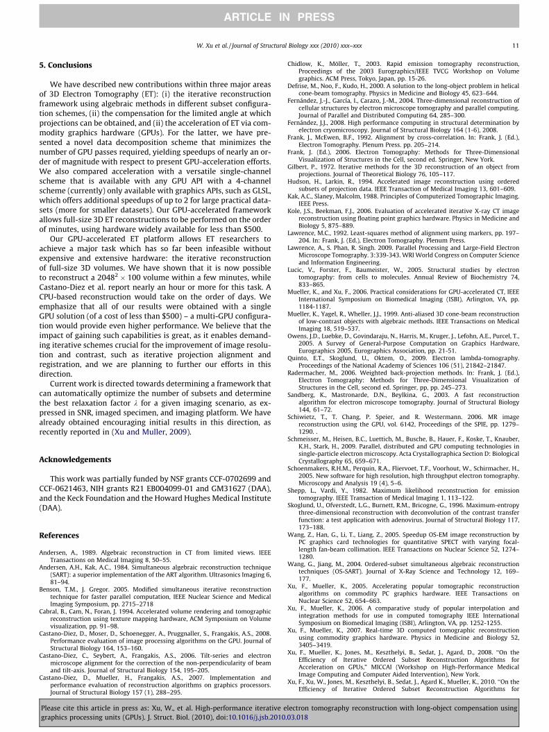
5. Conclusions
We have described new contributions within three major areasof 3D Electron Tomography (ET): (i) the iterative reconstructionframework using algebraic methods in different subset configura-tion schemes, (ii) the compensation for the limited angle at whichprojections can be obtained, and (ii) the acceleration of ET via com-modity graphics hardware (GPUs). For the latter, we have pre-sented a novel data decomposition scheme that minimizes thenumber of GPU passes required, yielding speedups of nearly an or-der of magnitude with respect to present GPU-acceleration efforts.We also compared acceleration with a versatile single-channelscheme that is available with any GPU API with a 4-channelscheme (currently) only available with graphics APIs, such as GLSL,which offers additional speedups of up to 2 for large practical data-sets (more for smaller datasets). Our GPU-accelerated frameworkallows full-size 3D ET reconstructions to be performed on the orderof minutes, using hardware widely available for less than $500.
Our GPU-accelerated ET platform allows ET researchers toachieve a major task which has so far been infeasible withoutexpensive and extensive hardware: the iterative reconstructionof full-size 3D volumes. We have shown that it is now possibleto reconstruct a 20482 � 100 volume within a few minutes, whileCastano-Diez et al. report nearly an hour or more for this task. ACPU-based reconstruction would take on the order of days. Weemphasize that all of our results were obtained with a singleGPU solution (of a cost of less than $500) – a multi-GPU configura-tion would provide even higher performance. We believe that theimpact of gaining such capabilities is great, as it enables demand-ing iterative schemes crucial for the improvement of image resolu-tion and contrast, such as iterative projection alignment andregistration, and we are planning to further our efforts in thisdirection.
Current work is directed towards determining a framework thatcan automatically optimize the number of subsets and determinethe best relaxation factor k for a given imaging scenario, as ex-pressed in SNR, imaged specimen, and imaging platform. We havealready obtained encouraging initial results in this direction, asrecently reported in (Xu and Muller, 2009).
Acknowledgements
This work was partially funded by NSF grants CCF-0702699 andCCF-0621463, NIH grants R21 EB004099-01 and GM31627 (DAA),and the Keck Foundation and the Howard Hughes Medical Institute(DAA).
References
Andersen, A., 1989. Algebraic reconstruction in CT from limited views. IEEETransactions on Medical Imaging 8, 50–55.
Andersen, A.H., Kak, A.C., 1984. Simultaneous algebraic reconstruction technique(SART): a superior implementation of the ART algorithm. Ultrasonics Imaging 6,81–94.
Benson, T.M., J. Gregor. 2005. Modified simultaneous iterative reconstructiontechnique for faster parallel computation, IEEE Nuclear Science and MedicalImaging Symposium, pp. 2715–2718
Cabral, B., Cam, N., Foran, J. 1994. Accelerated volume rendering and tomographicreconstruction using texture mapping hardware, ACM Symposium on Volumevisualization, pp. 91–98.
Castano-Diez, D., Moser, D., Schoenegger, A., Pruggnaller, S., Frangakis, A.S., 2008.Performance evaluation of image processing algorithms on the GPU. Journal ofStructural Biology 164, 153–160.
Castano-Diez, C., Seybert, A., Frangakis, A.S., 2006. Tilt-series and electronmicroscope alignment for the correction of the non-perpendicularity of beamand tilt-axis. Journal of Structural Biology 154, 195–205.
Castano-Diez, D., Mueller, H., Frangakis, A.S., 2007. Implementation andperformance evaluation of reconstruction algorithms on graphics processors.Journal of Structural Biology 157 (1), 288–295.
Please cite this article in press as: Xu, W., et al. High-performance iterative egraphics processing units (GPUs). J. Struct. Biol. (2010), doi:10.1016/j.jsb.2010
Chidlow, K., Möller, T., 2003. Rapid emission tomography reconstruction,Proceedings of the 2003 Eurographics/IEEE TVCG Workshop on Volumegraphics. ACM Press, Tokyo, Japan, pp. 15-26.
Defrise, M., Noo, F., Kudo, H., 2000. A solution to the long-object problem in helicalcone-beam tomography. Physics in Medicine and Biology 45, 623–644.
Fernández, J.-J., García, I., Carazo, J.-M., 2004. Three-dimensional reconstruction ofcellular structures by electron microscope tomography and parallel computing.Journal of Parallel and Distributed Computing 64, 285–300.
Fernández, J.J., 2008. High performance computing in structural determination byelectron cryomicroscopy. Journal of Structural Biology 164 (1-6), 2008.
Frank, J., McEwen, B.F., 1992. Alignment by cross-correlation. In: Frank, J. (Ed.),Electron Tomography. Plenum Press. pp. 205–214.
Frank, J. (Ed.), 2006. Electron Tomography: Methods for Three-DimensionalVisualization of Structures in the Cell, second ed. Springer, New York.
Gilbert, P., 1972. Iterative methods for the 3D reconstruction of an object fromprojections. Journal of Theoretical Biology 76, 105–117.
Hudson, H., Larkin, R., 1994. Accelerated image reconstruction using orderedsubsets of projection data. IEEE Transaction of Medical Imaging 13, 601–609.
Kak, A.C., Slaney, Malcolm, 1988. Principles of Computerized Tomographic Imaging.IEEE Press.
Kole, J.S., Beekman, F.J., 2006. Evaluation of accelerated iterative X-ray CT imagereconstruction using floating point graphics hardware. Physics in Medicine andBiology 5, 875–889.
Lawrence, M.C., 1992. Least-squares method of alignment using markers, pp. 197–204. In: Frank, J. (Ed.), Electron Tomography. Plenum Press.
Lawrence, A., S. Phan, R. Singh. 2009. Parallel Processing and Large-Field ElectronMicroscope Tomography. 3:339-343. WRI World Congress on Computer Scienceand Information Engineering.
Lucic, V., Forster, F., Baumeister, W., 2005. Structural studies by electrontomography: from cells to molecules. Annual Review of Biochemistry 74,833–865.
Mueller, K., and Xu, F., 2006. Practical considerations for GPU-accelerated CT, IEEEInternational Symposium on Biomedical Imaging (ISBI), Arlington, VA, pp.1184-1187.
Mueller, K., Yagel, R., Wheller, J.J., 1999. Anti-aliased 3D cone-beam reconstructionof low-contrast objects with algebraic methods. IEEE Transactions on MedicalImaging 18, 519–537.
Owens, J.D., Luebke, D., Govindaraju, N., Harris, M., Kruger, J., Lefohn, A.E., Purcel, T.,2005. A Survey of General-Purpose Computation on Graphics Hardware,Eurographics 2005, Eurographics Association, pp. 21-51.
Quinto, E.T., Skoglund, U., Oktem, O., 2009. Electron lambda-tomography.Proceedings of the National Academy of Sciences 106 (51), 21842–21847.
Radermacher, M., 2006. Weighted back-projection methods. In: Frank, J. (Ed.),Electron Tomography: Methods for Three-Dimensional Visualization ofStructures in the Cell, second ed. Springer, pp, pp. 245–273.
Sandberg, K., Mastronarde, D.N., Beylkina, G., 2003. A fast reconstructionalgorithm for electron microscope tomography. Journal of Structural Biology144, 61–72.
Schiwietz, T., T. Chang, P. Speier, and R. Westermann. 2006. MR imagereconstruction using the GPU, vol. 6142, Proceedings of the SPIE, pp. 1279–1290. .
Schmeisser, M., Heisen, B.C., Luettich, M., Busche, B., Hauer, F., Koske, T., Knauber,K.H., Stark, H., 2009. Parallel, distributed and GPU computing technologies insingle-particle electron microscopy. Acta Crystallographica Section D: BiologicalCrystallography 65, 659–671.
Schoenmakers, R.H.M., Perquin, R.A., Fliervoet, T.F., Voorhout, W., Schirmacher, H.,2005. New software for high resolution, high throughput electron tomography.Microscopy and Analysis 19 (4), 5–6.
Shepp, L., Vardi, Y., 1982. Maximum likelihood reconstruction for emissiontomography. IEEE Transaction of Medical Imaging 1, 113–122.
Skoglund, U., Ofverstedt, L.G., Burnett, R.M., Bricogne, G., 1996. Maximum-entropythree-dimensional reconstruction with deconvolution of the contrast transferfunction: a test application with adenovirus. Journal of Structural Biology 117,173–188.
Wang, Z., Han, G., Li, T., Liang, Z., 2005. Speedup OS-EM image reconstruction byPC graphics card technologies for quantitative SPECT with varying focal-length fan-beam collimation. IEEE Transactions on Nuclear Science 52, 1274–1280.
Wang, G., Jiang, M., 2004. Ordered-subset simultaneous algebraic reconstructiontechniques (OS-SART). Journal of X-Ray Science and Technology 12, 169–177.
Xu, F., Mueller, K., 2005. Accelerating popular tomographic reconstructionalgorithms on commodity PC graphics hardware. IEEE Transactions onNuclear Science 52, 654–663.
Xu, F., Mueller, K., 2006. A comparative study of popular interpolation andintegration methods for use in computed tomography IEEE InternationalSymposium on Biomedical Imaging (ISBI), Arlington, VA, pp. 1252-1255.
Xu, F., Mueller, K., 2007. Real-time 3D computed tomographic reconstructionusing commodity graphics hardware. Physics in Medicine and Biology 52,3405–3419.
Xu, F., Mueller, K., Jones, M., Keszthelyi, B., Sedat, J., Agard, D., 2008. ‘‘On theEfficiency of Iterative Ordered Subset Reconstruction Algorithms forAcceleration on GPUs,” MICCAI (Workshop on High-Performance MedicalImage Computing and Computer Aided Intervention), New York.
Xu, F., Xu, W., Jones, M., Keszthelyi, B., Sedat, J., Agard K., Mueller, K., 2010. ‘‘On theEfficiency of Iterative Ordered Subset Reconstruction Algorithms for
lectron tomography reconstruction with long-object compensation using.03.018

12 W. Xu et al. / Journal of Structural Biology xxx (2010) xxx–xxx
ARTICLE IN PRESS
Acceleration on GPUs,” Computer Methods and Programs in Biomedicine, (toappear, available online at http://linkinghub.elsevier.com/retrieve/pii/S0169260709002521).
Xu, W., Mueller, K., 2009. ‘‘Learning Effective Parameter Settings for Iterative CTReconstruction Algorithms,” Fully 3D Image Reconstruction in Radiology andNuclear Medicine, Beijing, China, September.
Xue, X., Cheryauka, A., D. Tubbs, D., 2006. Acceleration of fluoro-CT reconstructionfor a mobile C-Arm on GPU and FPGA hardware: a simulation study, Vol. 6142,Proc. SPIE, pp. 1494–1501.
Please cite this article in press as: Xu, W., et al. High-performance iterative egraphics processing units (GPUs). J. Struct. Biol. (2010), doi:10.1016/j.jsb.2010
Zeng, G., Gullberg, G., 2000. Unmatched projector/backprojector pairs in an iterativereconstruction algorithm. IEEE Transactions on Medical Imaging 19 (5), 548–555.
Zheng, Q.S., Braunfeld, M.B., Sedat, J.W., Agard, D.A., 2004. An improved stategy forautomated electron microscopic tomography. Journal of Structural Biology 147,91–101.
Zheng, S.Q., Keszthelyi, B., Branlund, E., Lyle, J.M., Braunfeld, M.B., Sedat, J.W., Agard,D.A., 2006. UCSF tomography: an integrated software suite for real-timeelectron microscopic tomographic data collection, alignment, andreconstruction. Journal of Structural Biology 157, 138–147.
lectron tomography reconstruction with long-object compensation using.03.018