Hierarchically Organized Bimolecular Ladder Network Exhibiting Guided One-Dimensional Diffusion
8
MAKOUDI ET AL. VOL. 6 ’ NO. 1 ’ 549 –556 ’ 2012 www.acsnano.org 549 December 08, 2011 C 2011 American Chemical Society Hierarchically Organized Bimolecular Ladder Network Exhibiting Guided One-Dimensional Diffusion Younes Makoudi, †, ) Emmanuel Arras, †, ) Nenad Kep cija, † Wolfgang Krenner, † Svetlana Klyatskaya, ‡ Florian Klappenberger, †, * Mario Ruben, ‡,§ Ari Paavo Seitsonen, ^ and Johannes V. Barth † † Physik Department E20, Technische Universität München, James-Franck Strasse, 85748 Garching, Germany, ‡ Institute of Nanotechnology, Karlsruhe Institute of Technology, 76021 Karlsruhe, Germany, § IPCMS-CNRS UMR 7504, Université de Strasbourg, 23 Rue du Loess, 67034 Strasbourg, France, and ^ Physikalisch- Chemisches Institut der Universität Zürich, Winterthurerstr. 190, CH-8057 Zürich, Switzerland. ) These authors contributed equally to this work. M olecular self-assembly is ubiqui- tous in biological systems for the production and reproduction of functional units and even living species. 1,2 Among the most important requirements for the formation of the highly complex supramolecular architectures is the pre- sence of hierarchy in the bonding to allow multilevel assembly. 3 Adapting this strategy and creating novel molecular building blocks with the ability to self-assemble into supramolecular networks is a promising bottom-up technique in nanotechnology. 47 Many studies show examples of artificial self-assembled molecular nanostructures; however, they are often limited to only one level of order. Increasing efforts are dedicated to master the formation of or- dered structure comprising a multilevel hierarchy. 812 At each level of organization, novel functionalities can be expected to be realized. 13 For further improvement of the control over the resulting architecture, hier- archy has to be introduced in the bonding motifs itself. 14,15 Here we show a bimolecular two-level hierarchic assembly process which leads to a ladder-shaped two-dimensional (2D) net- work exhibiting intriguing dynamic beha- vior. In contrast to other heteromolecular approaches, 1621 the two organic species couple via two distinct bonding motifs of different strength leading to a 2D supramo- lecular architecture consisting of two sub- lattices. The stronger bound sublattice is organized and linked by a novel hydrogen bond motif. At elevated temperature, the one-dimensional (1D) diffusion of the mole- cules of the weaker bond sublattice is guided by the more stable bimolecular grating. RESULTS The two molecules used in this study, namely, sexiphenyl dicarbonitrile (6DC) and N,N-diphenyl oxalic amide (DOA), are pre- sented in Scheme 1. They were deposited from a double organic molecular beam epi- taxy source onto a prepared Ag(111) sub- strate. Subsequent scanning tunneling microscopy (STM) observations were per- formed at a substrate temperature (T sub ) as indicated, typically 10 K. Both organic spe- cies have separately been the subject of de- tailed studies regarding their behavior on Ag(111) or Au(111) substrates. 22,23 It appears that DOA molecules (a) self-assemble into * Address correspondence to fl[email protected]. Received for review October 12, 2011 and accepted December 8, 2011. Published online 10.1021/nn203963a ABSTRACT The assembly and dynamics of a hierarchical, bimolecular network of sexiphenyl dicarbonitrile and N,N 0 -diphenyl oxalic amide molecules on the Ag(111) surface are studied by scanning tunneling microscopy at controlled temperature. The network formation is governed by a two- step protocol involving hierarchic interactions, including a novel carbonitrileoxalic amide bonding motif. For temperatures exceeding ∼70 K, more weakly bound sexiphenyl dicarbonitrile molecules carry out one-dimensional diffusion guided by the more stable substructure of the network held together by the carbonitrileoxalic amide bonding motif. A theoretical investigation at the ab initio level confirms the different binding energies of the two coupling motifs and rationalizes the network formation and the diffusion pathway. KEYWORDS: supramolecular chemistry . self-assembly . molecular dynamics . hierarchic bonding . scanning tunneling microscopy (STM) . density functional theory (DFT) ARTICLE
Transcript of Hierarchically Organized Bimolecular Ladder Network Exhibiting Guided One-Dimensional Diffusion

MAKOUDI ET AL. VOL. 6 ’ NO. 1 ’ 549–556 ’ 2012
www.acsnano.org
549
December 08, 2011
C 2011 American Chemical Society
Hierarchically Organized BimolecularLadder Network Exhibiting GuidedOne-Dimensional DiffusionYounes Makoudi,†, ) Emmanuel Arras,†, ) Nenad Kep�cija,† Wolfgang Krenner,† Svetlana Klyatskaya,‡
Florian Klappenberger,†,* Mario Ruben,‡,§ Ari Paavo Seitsonen,^ and Johannes V. Barth†
†Physik Department E20, Technische Universität München, James-Franck Strasse, 85748 Garching, Germany, ‡Institute of Nanotechnology, Karlsruhe Institute ofTechnology, 76021 Karlsruhe, Germany, §IPCMS-CNRS UMR 7504, Université de Strasbourg, 23 Rue du Loess, 67034 Strasbourg, France, and ^Physikalisch-Chemisches Institut der Universität Zürich, Winterthurerstr. 190, CH-8057 Zürich, Switzerland. )These authors contributed equally to this work.
Molecular self-assembly is ubiqui-tous in biological systems for theproduction and reproduction of
functional units and even living species.1,2
Among the most important requirementsfor the formation of the highly complexsupramolecular architectures is the pre-sence of hierarchy in the bonding to allowmultilevel assembly.3 Adapting this strategyand creating novel molecular buildingblocks with the ability to self-assemble intosupramolecular networks is a promisingbottom-up technique in nanotechnology.4�7
Many studies show examples of artificialself-assembled molecular nanostructures;however, they are often limited to onlyone level of order. Increasing efforts arededicated to master the formation of or-dered structure comprising a multilevelhierarchy.8�12 At each level of organization,novel functionalities can be expected to berealized.13 For further improvement of thecontrol over the resulting architecture, hier-archy has to be introduced in the bondingmotifs itself.14,15
Here we show a bimolecular two-levelhierarchic assembly process which leads toa ladder-shaped two-dimensional (2D) net-work exhibiting intriguing dynamic beha-vior. In contrast to other heteromolecularapproaches,16�21 the two organic speciescouple via two distinct bonding motifs ofdifferent strength leading to a 2D supramo-lecular architecture consisting of two sub-lattices. The stronger bound sublattice isorganized and linked by a novel hydrogenbond motif. At elevated temperature, theone-dimensional (1D) diffusion of the mole-cules of the weaker bond sublattice isguided by the more stable bimoleculargrating.
RESULTS
The two molecules used in this study,namely, sexiphenyl dicarbonitrile (6DC) andN,N-diphenyl oxalic amide (DOA), are pre-sented in Scheme 1. They were depositedfrom a double organic molecular beam epi-taxy source onto a prepared Ag(111) sub-strate. Subsequent scanning tunnelingmicroscopy (STM) observations were per-formed at a substrate temperature (Tsub) asindicated, typically 10 K. Both organic spe-cies have separately been the subject of de-tailed studies regarding their behavior onAg(111) or Au(111) substrates.22,23 It appearsthat DOA molecules (a) self-assemble into
* Address correspondence [email protected].
Received for review October 12, 2011and accepted December 8, 2011.
Published online10.1021/nn203963a
ABSTRACT
The assembly and dynamics of a hierarchical, bimolecular network of sexiphenyl dicarbonitrile
and N,N0-diphenyl oxalic amide molecules on the Ag(111) surface are studied by scanning
tunneling microscopy at controlled temperature. The network formation is governed by a two-
step protocol involving hierarchic interactions, including a novel carbonitrile�oxalic amide
bonding motif. For temperatures exceeding ∼70 K, more weakly bound sexiphenyl
dicarbonitrile molecules carry out one-dimensional diffusion guided by the more stable
substructure of the network held together by the carbonitrile�oxalic amide bonding motif. A
theoretical investigation at the ab initio level confirms the different binding energies of the
two coupling motifs and rationalizes the network formation and the diffusion pathway.
KEYWORDS: supramolecular chemistry . self-assembly . molecular dynamics .hierarchic bonding . scanning tunneling microscopy (STM) . density functionaltheory (DFT)
ARTIC
LE

MAKOUDI ET AL. VOL. 6 ’ NO. 1 ’ 549–556 ’ 2012
www.acsnano.org
550
hydrogen-bonded 1Dmolecular wires.22,24 6DC on theother hand (b) is found to arrange into a variety ofhighly regular, commensurate, and porous networkswith various symmetries.23,25
As both molecules (6DC and DOA) are co-depositedon the Ag(111) surface, a different open-porous net-work arises, as shown by STM observation reported inFigure 1. The ratio of 6DC to DOA molecules is justunder 2:1 in our experiments (63% 6DC and 37% DOAin Figure 1). The DOA molecules appear as two lobesand are shorter than the 6DCmolecules which look likeelongated sticks. The network consists of a series of6DC�DOA chains (6DCch and DOA aligned vertically inFigure 1), which are connected by brighter 6DC mole-cules that can be seen as rungs (6DCru), making itreminiscent of ladders. The pore size of the rhombiccavities (highlighted by the yellow area) is approxi-mately equal to 7 nm2. When all rungs of the laddersare present, the ratio 6DC to DOA is exactly 2:1.However, we observe here that the number of 6DCruspacers between the different lines is variable anddepends on the coverage of 6DC, thus impacting thedensity of rhombic cavities. Dimensions relevant to theladder network are drawn in yellow in Figure 1, includingangles R, β, γ, δ, and ε of the different units with respectto the highlighted substrate direction and the periodi-cities A and B of the network along two directions.Measured values are R = 15.7�, β = 67.8�, γ = 85.5�, δ =1.8�, ε = 42.0�, A = 33.3 Å, B = 42.1 Å.The ladder-shaped supramolecular structure exhi-
bits chiral properties which result from the heteromo-lecular binding motif as indicated in lower coveragedata of Figure 2a. The STM image displays bimolecularchains that appear in five different directions (out of sixobserved in total) and can be classified in two enan-tiomeric variants (Right and Left). The analysis of all ourdata demonstrates that the two enantiomorphs areevenly present in the samples and that the supramo-lecular chain formation is controlled by the substrateperiodicity. In addition, the energetic hierarchy behindthe different bonds possiblewith the two organic species(Figure 2b) becomes evident. In the network, we couldalready distinguish two different interactions: 6DCch�6DCru and 6DCch�DOA. Both of them involve the carbo-nitrile moiety of the 6DC molecule. The 6DCch�6DCruinteraction is well-known from earlier work23 andinvolves aC�N 3 3 3H�Cphbindingmotif. The 6DC�DOAinteraction on the other hand was not observed prior inthe self-assembly and will be discussed in more detaillater. The last possible interaction, namely, DOA�DOA,
is not present in the ladder network but appears in samplewith excess DOA, as shown in Figure 2a. We can deducefrom this that the 6DC�DOA interaction is stronger thanthemeanvalueof 6DC�6DCandDOA�DOA interactions.
DISCUSSION
For a better understanding of the novel bindingmotif, we start with the construction of a detailed
Scheme 1. Molecular building blocks used: N,N0-diphenyloxalic amide (DOA), sexiphenyl dicarbonitrile (6DC).Hydrogenin white, carbon in black, oxygen in red, and nitrogen in blue.
Figure 1. Overview STM topographic image of an open-porous network obtained after co-deposition of sexiphenyldicarbonitrile (6DCch and 6DCru) and N,N-diphenyl oxalicamide (DOA) molecules on the Ag(111) surface. The sub-strate high-symmetry directions are marked by the red star(bottom right corner), also in all of our images. The differentangles of the molecular units with respect to the indicateddirection are noted R, β, γ, δ, and ε. The periodicity in twodifferent directions is defined by lengths A and B (biasvoltage VB = �1 V, tunneling current I = 70 pA, substratetemperature Tsub = 10 K).
Figure 2. (a) STM topographic image of the chiral bimole-cular chains at low coverage featuring the two enantio-morphs, Right and Left (VB =�1.16 V, I = 80 pA, Tsub = 10 K).(b) Three types of bonds observed: 6DC�DOA, 6DC�6DC,and DOA�DOA. The preferred formation of alternate 6DCand DOA chains proves that the 6DC�DOA interaction isstronger on average than that between identical molecules(namely, 6DC�6DC and DOA�DOA).
ARTIC
LE

MAKOUDI ET AL. VOL. 6 ’ NO. 1 ’ 549–556 ’ 2012
www.acsnano.org
551
model of the network. The apparent height of the twomolecules in our STM images (∼1Å, slightly dependingon the tunneling conditions) is consistent with re-ported values for π-conjugated organic molecules inplanar adsorption geometries.26�30 Therefore, as astarting point, we considered only the flat conforma-tions of bothmolecules to build ourmodel displayed inFigure 3. To derive this model, three different ap-proaches were combined: (i) a thorough analysis ofnumerous STM images, especially regarding the mea-surement of angle R and δ, as well as the anglebetween 6DC�DOA chains of two different chiralities(as seen for example in Figure 2a); (ii) the placementof the N atoms of 6DC molecules at hollow sitesof the Ag(111) surface, as exposed in previouspublications;23,31 (iii) the detailed understanding ofbonding regions as unraveled by theoretical calcula-tions presented below. With this approach, it waspossible to create an accurate model which fits(within usual STM error) all of our images, includingthe most peculiar configurations, such as near stepedges (not shown here). Thismodel, detailed in units ofthe Ag(111) surface in Figure 3, is confronted to a high-resolution STM image of the network and allows a veryaccurate evaluation of dimensions reported in Figure 1.The theoretical values thus are R = 17.5�, β = 67.1�,γ = 84.4�, δ = 1.6�, ε = 41.4�, A = 33.16 Å, B = 40.56 Å.These appear to be within 2� and 4% of the measuredones, well within the usual STM errors. The zoomedregion of Figure 3 shows the substrate registry of ourproposed model.To further study the relative strength of the ob-
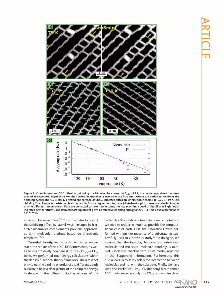
served bonding motifs, we performed a series ofexperiments at different temperatures and evaluatedthe stability of the self-assembled structure. At Tsub =75 K, we observe (Figure 4a) that 6DCru spacers start tomove along the bimolecular chains. This result corro-borates that the 6DCch�6DCru interaction is weakerthan that for the 6DCch�DOA. When the temperatureof the substrate is increased to 102 K (Figure 4b), all6DCru spacers diffuse but remain confined between
6DCch�DOA chains. At 119 K (Figure 4c), the diffusionis even faster, but still the 6DCch�DOA chains remainintact. They finally break at temperatures higher than132 K (not shown).Further analysis of similar images at nine different
temperatures allowed us to draw the Arrhenius plotpresented in Figure 4d. Specifically, as in a previouspublication,31 the hopping rate R is calculated usingthe expression R = he/(hpt), where he is the number ofhopping events occurring during the time t, and hp isthe number of hopping-able 6DC molecules, that is,the molecules that have enough space to perform amovement. At low temperatures, t is the time elapsedbetween two successively taken pictures. For tempera-tures exceeding 103 K, as the hopping process is morefrequent, only single pictures are analyzed, and he isevaluated for each molecule by counting the numberof bright-to-dark and dark-to-bright changes in the6DCru species (see Figure 4b,c), and t is the timenecessary to scan enough lines to visualize the wholemolecule. The best fit of these values gives an effectivehopping barrier of 205 meV ((11meV) and a prefactorof 2.7 � 109.4(0.6 Hz, well within the known broadrange in the literature.29,31,32
Finally, it needs to be emphasized that the ladderrungs have a stabilizing effect on the separation of the6DCch�DOA chains. Interestingly, this holds even inthe dynamic regime. From Figure 4b,c, it is evident thatthe 6DCch�DOA chains form a highly regular gratingwhose periodicity is perfectly retained and unaffec-ted after the onset of rung diffusion. Thus there is adynamic stabilization of the supramolecular arrange-ment, which effect could be used in itself by usingdicarbonitrile or other linkers with appropriate lengthor even other hierarchic bonding patters to fabricatetailored superlattices with definite regularity. By con-trast, previously reported related 1D supramoleculargratings without stabilizing elements on a homo-geneous surface inevitably showed a certain degreeof variation in the distances between the constitut-ing molecular lines,33,34 even in the presence of
Figure 3. High-resolution STM data of the bimolecular open-porous network, superimposed with a ball-and-stick model ofthe molecules over the Ag(111) substrate. The original STM image has been geometrically corrected (shear and expansion∼3%) to better fit the model. Major characteristics of the network are indicated in substrate lattice parameter scale a and a0.The zoomed area highlights the position of molecules over the Ag substrate, where hollow sites are marked with smallgreen dots (VB = �1.12 V, I = 60 pA, Tsub = 10 K).
ARTIC
LE
MAKOUDI ET AL. VOL. 6 ’ NO. 1 ’ 549–556 ’ 2012
www.acsnano.org
552
adatoms between them.35 Thus, the introduction ofthe stabilizing effect by lateral weak linkages in hier-archic assemblies complements previous approach-es with molecular gratings based on anisotropictemplates.19,36
Theoretical Investigation. In order to better under-stand the nature of the 6DC�DOA interaction, as wellas to quantitatively compare it to the 6DCch�6DCrubond, we performed total energy calculations withinthe density functional theory framework. The aim is notonly to get the binding energies of the different bondsbut also to have a clear picture of the complete energylandscape in the different binding regions of the
molecules. Since this requires extensive computations,we tried to reduce as much as possible the computa-tional cost of each. First, the simulations were per-formed without the presence of a substrate, as suc-cessfully used in a previous study.37 By doing so, weassume that the interplay between the substrate�molecule and molecule�molecule bondings is mini-mal, which was checked with a test model, reportedin the Supporting Information. Furthermore, thisalso allows us to study solely the interaction betweenmolecules and not with the substrate. Finally, we haveused the smaller NC�Ph2�CN (biphenyl dicarbonitrile2DC) molecule when only the CN group was involved
Figure 4. One-dimensional 6DC diffusion guided by the bimolecular chains: (a) Tsub = 75 K, the two images show the samearea of the network (Right chirality), the second being taken 6 min after the first one. Arrows are added to highlight thehopping events. (b) Tsub = 102 K. Frizzled appearance of 6DCru indicates diffusion within stable chains. (c) Tsub = 119 K, Leftchirality. The change in the frizzled features results from a higher hopping rate. (d) Arrhenius plot drawn from similar imagesat nine different temperatures. Data are corrected to take into account the low scanning speed of the STM at high hopp-ing rates (temperatures). The derived least-squares fit gives an effective hopping energy of 205( 11 meV and a prefactor of109.4(0.6 Hz.
ARTIC
LE

MAKOUDI ET AL. VOL. 6 ’ NO. 1 ’ 549–556 ’ 2012
www.acsnano.org
553
in the bonding motif. Extensive convergence tests ofNC�PhX�CNmoleculeswith the number of phenyl ringsX up to 4 have indeed shown that the behavior of the CNgroup depends only weakly on X for X > 2.
The energy maps shown in Figure 5 were obtainedvia the following steps: (i) each molecule is first geo-metrically relaxed individually, as in the gas phase; (ii)two or more molecules are placed in various relativepositions and the total energy of the resulting boxis computed as is, without geometrical relaxation;(iii) the binding energy (in each configuration) isextracted by subtracting the total energy of the iso-lated constituting molecules; (iv) results are presentedas energy maps (Figure 5), with colored (respectively
white) areas corresponding to bonding (respectivelyantibonding) configurations and isolines for improvedreadability.
The first result (Figure 5a) is the binding energymapof a 2DCwith a DOA. The angle of 48� between the 2DCand the DOA was derived from a previous full mini-mization revealing this equilibrium angle. We find thatthe binding energy reaches 340 meV at its maximum.In this equilibrium configuration, the closest distancebetween N and H atom is 2.1 Å (green thick line inFigure 5a). It is interesting to note that three interac-tions contribute to this link: CN 3 3 3HN, CN 3 3 3HCph, andCphH 3 3 3OC. The first one is a classical hydrogen bond.The two others will be simply referred to as weakbonds38 (blue thin lines in Figure 5). We evaluated thecontribution of each bond in two steps. First, the bind-ing energy of the configuration in Figure 5a was com-puted with the carbonitrile group of the 2DC replacedby a simple hydrogen. It was found to be∼70meV andcorresponds to the contribution of the CphH 3 3 3Obond. Then, looking at the shape of the initial energymap, especially when the carbonitrile group is next tothe HCph group and relatively far from the HN group,
Figure 5. Energymaps of the three studiedbinding regions.Each black dot on the maps (more than 450 in total) is onesimulation of rigidly displaced molecules and gives theposition of the binding nitrogen atom. Colored areas ofthe interpolated full energy maps correspond to negativeformation energy, hence binding zone, while positive for-mation energy areas are white. Isolines mark 25 meV steps.Dark green lines mark classical hydrogen bonds, whereaslight blue ones highlight weak attractive interaction. (a)Binding of NC�Ph2�CN (2DC) with DOA. (b) Binding of 2DCwith 4DC. (c) Evolution of the formation energy versustranslation along the 4DC ladder component. (d) Migrationregion around the 2DC�DOA�2DC link.
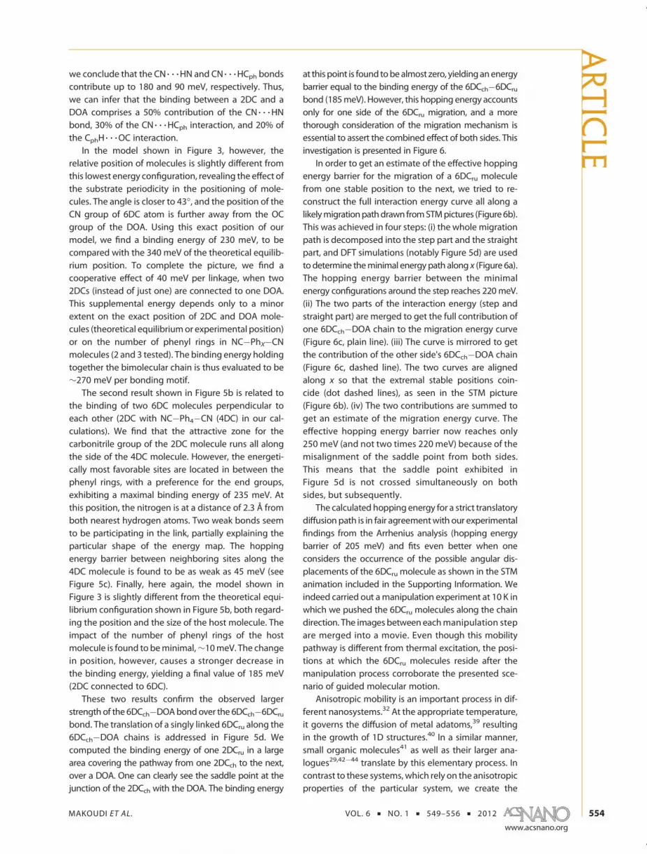
Figure 6. Prospective study of the migration path of 6DCrubetween two 6DCch�DOA chains. (a) Computation of theinteraction energy along the two parts of migration path:the step and the straight part. (b) Migration path drawnfrom a STM picture, namely, the simple translation of the6DCru along the 6DCch�DOA chain guides. (c) Reconstruc-tion of the interaction energy along the full migration pathfor each side. (d) Sum of both contributions, plotted withrespect to the stable state energy. The effective hoppingenergy barrier is 250 meV.
ARTIC
LE
MAKOUDI ET AL. VOL. 6 ’ NO. 1 ’ 549–556 ’ 2012
www.acsnano.org
554
we conclude that the CN 3 3 3HN and CN 3 3 3HCph bondscontribute up to 180 and 90 meV, respectively. Thus,we can infer that the binding between a 2DC and aDOA comprises a 50% contribution of the CN 3 3 3HNbond, 30% of the CN 3 3 3HCph interaction, and 20% ofthe CphH 3 3 3OC interaction.
In the model shown in Figure 3, however, therelative position of molecules is slightly different fromthis lowest energy configuration, revealing the effect ofthe substrate periodicity in the positioning of mole-cules. The angle is closer to 43�, and the position of theCN group of 6DC atom is further away from the OCgroup of the DOA. Using this exact position of ourmodel, we find a binding energy of 230 meV, to becompared with the 340 meV of the theoretical equilib-rium position. To complete the picture, we find acooperative effect of 40 meV per linkage, when two2DCs (instead of just one) are connected to one DOA.This supplemental energy depends only to a minorextent on the exact position of 2DC and DOA mole-cules (theoretical equilibriumor experimental position)or on the number of phenyl rings in NC�PhX�CNmolecules (2 and 3 tested). The binding energy holdingtogether the bimolecular chain is thus evaluated to be∼270 meV per bonding motif.
The second result shown in Figure 5b is related tothe binding of two 6DC molecules perpendicular toeach other (2DC with NC�Ph4�CN (4DC) in our cal-culations). We find that the attractive zone for thecarbonitrile group of the 2DC molecule runs all alongthe side of the 4DC molecule. However, the energeti-cally most favorable sites are located in between thephenyl rings, with a preference for the end groups,exhibiting a maximal binding energy of 235 meV. Atthis position, the nitrogen is at a distance of 2.3 Å fromboth nearest hydrogen atoms. Two weak bonds seemto be participating in the link, partially explaining theparticular shape of the energy map. The hoppingenergy barrier between neighboring sites along the4DC molecule is found to be as weak as 45 meV (seeFigure 5c). Finally, here again, the model shown inFigure 3 is slightly different from the theoretical equi-librium configuration shown in Figure 5b, both regard-ing the position and the size of the host molecule. Theimpact of the number of phenyl rings of the hostmolecule is found to beminimal,∼10meV. The changein position, however, causes a stronger decrease inthe binding energy, yielding a final value of 185 meV(2DC connected to 6DC).
These two results confirm the observed largerstrength of the 6DCch�DOAbondover the 6DCch�6DCrubond. The translation of a singly linked 6DCru along the6DCch�DOA chains is addressed in Figure 5d. Wecomputed the binding energy of one 2DCru in a largearea covering the pathway from one 2DCch to the next,over a DOA. One can clearly see the saddle point at thejunction of the 2DCch with the DOA. The binding energy
at this point is found tobealmost zero, yieldinganenergybarrier equal to the binding energy of the 6DCch�6DCrubond (185meV). However, this hopping energy accountsonly for one side of the 6DCru migration, and a morethorough consideration of the migration mechanism isessential to assert the combined effect of both sides. Thisinvestigation is presented in Figure 6.
In order to get an estimate of the effective hoppingenergy barrier for the migration of a 6DCru moleculefrom one stable position to the next, we tried to re-construct the full interaction energy curve all along alikelymigrationpathdrawn fromSTMpictures (Figure6b).This was achieved in four steps: (i) the whole migrationpath is decomposed into the step part and the straightpart, and DFT simulations (notably Figure 5d) are usedto determine theminimal energy path along x (Figure 6a).The hopping energy barrier between the minimalenergy configurations around the step reaches 220meV.(ii) The two parts of the interaction energy (step andstraight part) are merged to get the full contribution ofone 6DCch�DOA chain to the migration energy curve(Figure 6c, plain line). (iii) The curve is mirrored to getthe contribution of the other side's 6DCch�DOA chain(Figure 6c, dashed line). The two curves are alignedalong x so that the extremal stable positions coin-cide (dot dashed lines), as seen in the STM picture(Figure 6b). (iv) The two contributions are summed toget an estimate of the migration energy curve. Theeffective hopping energy barrier now reaches only250 meV (and not two times 220 meV) because of themisalignment of the saddle point from both sides.This means that the saddle point exhibited inFigure 5d is not crossed simultaneously on bothsides, but subsequently.
The calculated hopping energy for a strict translatorydiffusion path is in fair agreementwith our experimentalfindings from the Arrhenius analysis (hopping energybarrier of 205 meV) and fits even better when oneconsiders the occurrence of the possible angular dis-placements of the 6DCru molecule as shown in the STManimation included in the Supporting Information. Weindeed carried out amanipulation experiment at 10 K inwhich we pushed the 6DCru molecules along the chaindirection. The images between eachmanipulation stepare merged into a movie. Even though this mobilitypathway is different from thermal excitation, the posi-tions at which the 6DCru molecules reside after themanipulation process corroborate the presented sce-nario of guided molecular motion.
Anisotropic mobility is an important process in dif-ferent nanosystems.32 At the appropriate temperature,it governs the diffusion of metal adatoms,39 resultingin the growth of 1D structures.40 In a similar manner,small organic molecules41 as well as their larger ana-logues29,42�44 translate by this elementary process. Incontrast to these systems, which rely on the anisotropicproperties of the particular system, we create the
ARTIC
LE

MAKOUDI ET AL. VOL. 6 ’ NO. 1 ’ 549–556 ’ 2012
www.acsnano.org
555
necessary 1D guides on an isotropic surface by hier-archic self-assembly of appropriately designed tectons.We have demonstrated that the diffusion of themobilespecies is controlled by the interaction with the guides,which represents a novel approach to tailor moleculardynamics on surfaces in a universal fashion. Hierarchicassemblies may be generally useful to direct molecularmotion via the tailored borders.
CONCLUSION
In summary, we have achieved the self-assemblyof a hierarchic bimolecular network of the ladder-type
in which, at temperatures exceeding ∼70 K, 1D diffu-sion of large organic molecules takes place. Theoreticanalysis clarifies that hierarchy is introduced into thechiral supramolecular structure by the energetics ofthe different bonds established between the twoorganic species, including a novel carbonitrile�oxalicamide motif. The dynamics of the mobile species isconsistent with an Arrhenius-type of hopping andsteered by the interaction with the ladder side rails.Our results demonstrate the usefulness of hierarchicdesign and reveal a new approach to guide molecularmotion at nanostructured surfaces.
METHODSThe scanning tunneling microscopy (STM) experiments were
performed using a home-built ultrahigh vacuum (base pressure3 � 10�11 mbar) low-temperature STM. The Ag(111) substratewas prepared by repeated cycles of Arþ sputtering and anneal-ing to 750 K to obtain flat terraces separated by monatomicsteps. The twomolecules, sexiphenyl dicarbonitrile (6DC) andN,N-diphenyl oxalic amide (DOA), were deposited from a doubleorganic molecular beam epitaxy source with the quartz crucibleat 572 and 467 K for 6DC and DOA, respectively, while keepingthe substrate at 300 K. Both sequential and simultaneousdeposition led to similar results. Followingpreparation, STMdatawere acquired at a substrate temperature (Tsub) as indicated,typically 10 K. The system's dynamics was probed in the rangefrom 70 to 120 K.For the theoretical investigation, we have used the projec-
tor-augmented wave approach as implemented in the ABINITcode,45,46 within the local density approximation for the ex-change-correlation energy. The cutoff energy used is 30 Ry.Since the code imposes periodic boundary conditions in allthree directions, we ensured that the simulation box allowed aminimal distance of 8 Å between the molecules and theirperiodic images. We evaluated the subsequent error on totalenergy to be then lower than 10 meV. As molecules are rigidlydisplaced to obtain the energy maps, we checked the impact ofgeometrical relaxation on the binding energy by fully relaxingcalculations for the lowest energy configurations, and we foundenergy differences lower than 10 meV. Also, for simplicityreasons, flat molecules were used in the calculations, whereas6DC are known to present a tilting between the phenylrings.23,47 A test calculation involving two 2DC moleculesrevealed that impact on binding energy of the tilting is lowerthan 15 meV, despite the energy difference of almost 50 meVbetween tilted and untilted isolated 2DC molecule. The fullenergymaps displayed in Figure 5 are obtained using aDelaunayinterpolation between the computed points.
Acknowledgment. Funding by the European Union via ERCAdvanced Grant MolArt (�247299) and the German ResearchFoundation (DFG) through the TUM International GraduateSchool of Science and Engineering (IGSSE) and TUM Instituteof Advanced Study (IAS) and the German Research Foundation(DFG) (BA 3395/2-1) and the Alexander von Humboldt Founda-tion are gratefully acknowledged.
Supporting Information Available: A movie made from aseries of successive images showing the tip-induced diffusionof 6DCru. Molecules were moved from the top-left to thebottom-right by pushing them approximately 3.5 nm (onenetwork lattice constant) along direction A for a single manip-ulation event. A theoretical study of the impact of the Ag(111)substrate on the intermolecular bonding is also provided, whichshows that the explicit inclusion of the substrate in DFTcalculations is not essential to study intermolecular bond-ing occurring between physisorbed molecules, and can be
emulated by the imposing coplanarity between them. Thismaterial is available free of charge via the Internet at http://pubs.acs.org.
REFERENCES AND NOTES1. Lindsey, J. S. Self-Assembly in Synthetic Routes to Molec-
ular Devices;Biological Principles and Chemical Perspec-tives;A Review. New J. Chem. 1991, 15, 153–�180.
2. Philp, D.; Stoddart, J. F. Self-Assembly in Natural and Unnatur-al Systems. Angew. Chem., Int. Ed. Engl. 1996, 35, 1155–1196.
3. Elemans, J. A. A. W.; Rowan, A. E.; Nolte, R. J. M. Master-ing Molecular Matter. Supramolecular Architecturesby Hierarchical Self-Assembly. J. Mater. Chem. 2003,13, 2661–2670.
4. Barth, J. V.; Costantini, G.; Kern, K. Engineering Atomic andMolecular Nanostructures at Surfaces. Nature 2005, 437,671–679.
5. De Feyter, S.; De Schryver, F. C. Two-Dimensional Supra-molecular Self-Assembly Probed by Scanning TunnelingMicroscopy. Chem. Soc. Rev. 2003, 32, 393–393.
6. Barth, J. V. Molecular Architectonic on Metal Surfaces.Annu. Rev. Phys. Chem. 2007, 58, 375–407.
7. Elemans, J.; Lei, S.; De Feyter, S. Molecular and Supra-molecular Networks on Surfaces: From Two-DimensionalCrystal Engineering to Reactivity. Angew. Chem., Int. Ed.2009, 48, 7298–7332.
8. Clair, S.; Pons, S.; Brune, H.; Kern, K.; Barth, J. V. MesoscopicMetallosupramolecular Texturing by Hierarchic Assembly.Angew. Chem., Int. Ed. 2005, 44, 7294–7297.
9. Blüm, M.-C.; C�avar, E.; Pivetta, M.; Patthey, F.; Schneider,W.-D. Conservation of Chirality in a Hierarchical Supra-molecular Self-Assembled Structure with Pentagonal Sym-metry. Angew. Chem., Int. Ed. 2005, 44, 5334–5337.
10. Otero, R.; Schöck, M.; Molina, L. M.; Laegsgaard, E.; Stens-gaard, I.; Hammer, B.; Besenbacher, F. Guanine QuartetNetworks Stabilized by Cooperative Hydrogen Bonds.Angew. Chem., Int. Ed. 2005, 44, 2270–2275.
11. Staniec, P. A.; Perdigao, L. M. A.; Saywell, A.; Champness,N. R.; Beton, P. H. Hierarchical Organisation on a Two-Dimensional Supramolecular Network. ChemPhysChem2007, 8, 2177–2181.
12. Schlickum, U.; Decker, R.; Klappenberger, F.; Zoppellaro,G.; Klyatskaya, S.; Auwärter, W.; Neppl, S.; Kern, K.; Brune,H.; Ruben, M.; et al. Chiral Kagomé Lattice from SimpleDitopic Molecular Bricks. J. Am. Chem. Soc. 2008, 130,11778–11782.
13. Ruben, M.; Ziener, U.; Lehn, J. M.; Ksenofontov, V.; Gutlich,P.; Vaughan, G. B. M. Hierarchical Self-Assembly of Supra-molecular Spintronic Modules into 1D- and 2D-Architec-tures with Emergence of Magnetic Properties. Chem.;Eur. J. 2005, 11, 94–100.
14. Spillmann, H.; Dmitriev, A.; Lin, N.; Messina, P.; Barth,J. V.; Kern, K. Hierarchical Assembly of Two-Dimensional
ARTIC
LE

MAKOUDI ET AL. VOL. 6 ’ NO. 1 ’ 549–556 ’ 2012
www.acsnano.org
556
Homochiral Nanocavity Arrays. J. Am. Chem. Soc. 2003,125, 10725–10728.
15. Meier, C.; Landfester, K.; Kunzel, D.; Markert, T.; Gross, A.;Ziener, U. Hierarchically Self-Assembled Host�Guest Net-work at the Solid�Liquid Interface for Single-MoleculeManipulation. Angew. Chem., Int. Ed. 2008, 47, 3821–3825.
16. de Wild, M.; Berner, S.; Suzuki, H.; Yanagi, H.; Schlettwein,D.; Ivan, S.; Baratoff, A.; Guentherodt, H. J.; Jung, T. A. ANovel Route to Molecular Self-assembly: Self-IntermixedMonolayer Phases. ChemPhysChem 2002, 3, 881–885.
17. Ruiz-Osés, M.; González-Lakunza, N.; Silanes, I.; Gourdon,A.; Arnau, A.; Ortega, J. E. Self-Assembly of HeterogeneousSupramolecular Structures with Uniaxial Anisotropy.J. Phys. Chem. B 2006, 110, 25573–25577.
18. Theobald, J. A.; Oxtoby, N. S.; Phillips, M. A.; Champness,N. R.; Beton, P. H. Controlling Molecular Deposition andLayer Structure with Supramolecular Surface Assemblies.Nature 2003, 424, 1029–1031.
19. Ca~nas-Ventura, M. E.; Xiao, W.; Wasserfallen, D.; Müllen, K.;Brune, H.; Barth, J. V.; Fasel, R. Self-Assembly of PeriodicBicomponent Wires and Ribbons. Angew. Chem., Int. Ed.2007, 46, 1814–1818.
20. Barrena, E.; de Oteyza, D. G.; Dosch, H.; Wakayama, Y. 2DSupramolecular Self-Assembly of Binary Organic Mono-layers. ChemPhysChem 2007, 8, 1915–1918.
21. Hipps, K. W.; Scudiero, L.; Barlow, D. E.; Cooke, M. P. A Self-Organized 2-Dimensional Bifunctional Structure Formedby Supramolecular Design. J. Am. Chem. Soc. 2002, 124,2126–2127.
22. Klappenberger, F.; Ca~nas-Ventura, M. E.; Clair, S.; Pons, S.;Schlickum, U.; Qu, Z. R.; Strunskus, T.; Comisso, A.; Wöll, C.;Brune, H.; et al. Does the Surface Matter? HydrogenBonded Chain Formation of an Oxalic Amide Derivativein Two and Three Dimensional Environment. Chem-PhysChem 2008, 9, 2522–2530.
23. Kühne, D.; Klappenberger, F.; Decker, R.; Schlickum, U.;Brune, H.; Klyatskaya, S.; Ruben, M.; Barth, J. V. Self-Assembly of Nanoporous Chiral Networks with VaryingSymmetry from Sexiphenyl-Dicarbonitrile on Ag(111).J. Phys. Chem. C 2009, 113, 17851–17859.
24. Krenner, W.; Klappenberger, F.; Kühne, D.; Diller, K.; Qu, Z.-R.;Ruben, M.; Barth, J. V. Positioning of Single Co AtomsSteered by a Self-Assembled Organic Molecular Template.J. Phys. Chem. Lett. 2011, 2, 1639–1645.
25. Klappenberger, F.; Kühne, D.; Krenner, W.; Silanes, I.; Arnau,A.; Garcia de Abajo, F. J.; Klyatskaya, S.; Ruben, M.; Barth,J. V. Dichotomous Array of Chiral Quantum Corrals by aSelf-Assembled Nanoporous Kagome Network. Nano Lett.2009, 9, 3509–3514.
26. Henningsen, N.; Rurali, R.; Franke, K. J.; Fernández-Torrente, I.; Pascual, J. I. Trans to Cis Isomerization of anAzobenzene Derivative on a Cu(100) Surface. Appl. Phys. A:Mater. Sci. Process. 2008, 93, 241–246.
27. Böhringer, M.; Morgenstern, K.; Schneider,W. D.; Berndt, R.;Mauri, F.; De Vita, A.; Car, R. Two-Dimensional Self-Assemblyof Supramolecular Clusters and Chains. Phys. Rev. Lett.1999, 83, 324–327.
28. Dmitriev, A.; Lin, N.; Weckesser, J.; Barth, J. V.; Kern, K.Supramolecular Assemblies of Trimesic Acid on a Cu(100)Surface. J. Phys. Chem. B 2002, 106, 6907–6912.
29. Weckesser, J.; Barth, J. V.; Kern, K. DirectObservationof SurfaceDiffusion of Large Organic Molecules at Metal Surfaces: PVBAon Pd(110). J. Chem. Phys. 1999, 110, 5351–5354.
30. Ca~nas-Ventura, M. E.; Klappenberger, F.; Clair, S.; Pons, S.;Kern, K.; Brune, H.; Strunskus, T.;Wöll, C.; Fasel, R.; Barth, J. V.Coexistence of One- and Two-Dimensional Supramolecu-lar Assemblies of Terephthalic Acid on Pd(111) Due to Self-Limiting Deprotonation. J. Chem. Phys. 2006, 125, 184710.
31. Kühne, D.; Klappenberger, F.; Krenner, W.; Klyatskaya, S.;Ruben, M.; Barth, V. J. Rotational and Constitutional Dy-namics of Caged Supramolecules. Proc. Natl. Acad. Sci. U.S.A.2010, 50, 21332–21336.
32. Barth, J. V. Transport of adsorbates at metal surfaces: Fromthermal migration to hot precursors. Surf. Sci. Rep. 2000,40, 75–149.
33. Barth, J. V.; Weckesser, J.; Cai, C.; Günter, P.; Bürgi, L.;Jeandupeux, O.; Kern, K. Building Supramolecular Nano-structures at Surfaces by Hydrogen Bonding. Angew.Chem., Int. Ed. 2000, 39, 1230–1234.
34. Schiffrin, A.; Riemann, A.; Auwärter, W.; Pennec, Y.; Weber-Bargioni, A.; Cvetko, D.; Cossaro, A.; Morgante, A.; Barth,J. V. Zwitterionic Self-Assembly of l-Methionine Nanograt-ings on the Ag(111) Surface. Proc. Natl. Acad. Sci. U.S.A.2007, 104, 5279–5284.
35. Schiffrin, A.; Reichert, J.; Auwärter, W.; Jahnz, G.; Pennec, Y.;Weber-Bargioni, A.; Stepanyuk, V. S.; Niebergall, L.; Bruno,P.; Barth, J. V. Self-Aligning Atomic Strings in Surface-Supported Biomolecular Gratings. Phys. Rev. B 2008, 78,035424.
36. Weckesser, J.; De Vita, A.; Barth, J. V.; Cai, C.; Kern, K.Mesoscopic Correlation of Supramolecular Chirality inOne-Dimensional Hydrogen-Bonded Assemblies. Phys.Rev. Lett. 2001, 87, 096101.
37. Barth, J. V.; Weckesser, J.; Trimarchi, G.; Vladimirova, M.; DeVita, A.; Cai, C.; Brune, H.; Günter, P.; Kern, K. Stereochem-ical Effects in Supramolecular Self-Assembly at Surfaces:1-D versus 2-D Enantiomorphic Ordering for PVBA andPEBA on Ag(111). J. Am. Chem. Soc. 2002, 124, 7991–8000.
38. A complete analysis will be presented in a forthcomingpaper.
39. Senft, D. C.; Ehrlich, G. Long Jumps in the Surface-Diffusion:One-Dimensional Migration of Isolated Adatoms. Phys.Rev. Lett. 1995, 74, 294–297.
40. Röder, H.; Hahn, E.; Brune, H.; Bucher, J. P.; Kern, K. BuildingOne-Dimensional and 2-Dimensional Nanostructures byDiffusion-Controlled Aggregation at Surfaces. Nature1993, 366, 141–143.
41. Briner, B. G.; Doering, M.; Rust, H.-P.; Bradshaw, A. M.Microscopic Molecular Diffusion Enhanced by AdsorbateInteractions. Science 1997, 278, 257–260.
42. Schunack, M.; Linderoth, T. R.; Rosei, F.; Laegsgaard, E.;Stensgaard, I.; Besenbacher, F. Long Jumps in the SurfaceDiffusion of Large Molecules. Phys. Rev. Lett. 2002, 88,156102.
43. Kwon, K.-Y.; Wong, K. L.; Pawin, G.; Bartels, L.; Stolbov, S.;Rahman, T. S. Unidirectional Adsorbate Motion on a High-Symmetry Surface: “Walking” Molecules Can Stay theCourse. Phys. Rev. Lett. 2005, 95, 166101.
44. Weckesser, J.; Barth, J. V.; Kern, K. Mobility and BondingTransition of C60 on Pd(110). Phys. Rev. B 2001, 64, 161403.
45. Gonze, X.; Rignanese, G.-M.; Verstraete, M.; Beuken, J.-M.;Pouillon, Y.; Caracas, R.; Jollet, F.; Torrent, M.; Zerah, G.;Mikami, M.; et al. A Brief Introduction to the ABINIT Soft-ware Package. Z. Kristallogr. 2005, 220, 558–562.
46. Torrent, M.; Jollet, F.; Bottin, F.; Zerah, G.; Gonze, X.Implementation of the Projector Augmented-WaveMethodin the ABINIT Code. Application to the Study of Iron UnderPressure. Comput. Mater. Sci. 2008, 42, 337–351.
47. Klappenberger, F.; Kühne, D.; Marschall, M.; Neppl, S.;Krenner, W.; Nefedov, A.; Strunskus, T.; Fink, K.; Wöll, C.;Klyatskaya, S.; et al. Uniform π-System Alignment in ThinFilms of Template-Grown Dicarbonitrile-Oligophenyls.Adv. Funct. Mater. 2011, 21, 1631–1642.
ARTIC
LE