
Introduction - 1000 islands water trail - Frontenac Arch ...
Upload
independentCategory
view
1download
0
HGS-ETR1, a fully human TRAIL-receptor 1 monoclonal antibody,induces cell death in multiple tumour types in vitro and in vivo
L Pukac*,1, P Kanakaraj1, R Humphreys1, R Alderson1, M Bloom1, C Sung1, T Riccobene1, R Johnson1,M Fiscella1, A Mahoney1, J Carrell1, E Boyd1, XT Yao1, L Zhang1, L Zhong1, A von Kerczek1, L Shepard1,T Vaughan2, B Edwards2, C Dobson2, T Salcedo1 and V Albert1
1Human Genome Sciences Inc., 9800 Medical Center Drive, Rockville, MD 20850, USA; 2Cambridge Antibody Technology, Milstein Building, Granta Park,Cambridge CB1 6GH, UK
Tumour necrosis factor-related apoptosis-inducing ligand (TRAIL) induces apoptosis in a variety of tumour cells through activation ofTRAIL-R1 and TRAIL-R2 death signalling receptors. Here, we describe the characterisation and activity of HGS-ETR1, the first fullyhuman, agonistic TRAIL-R1 mAb that is being developed as an antitumour therapeutic agent. HGS-ETR1 showed specific binding toTRAIL-R1 receptor. HGS-ETR1 reduced the viability of multiple types of tumour cells in vitro, and induced activation of caspase 8, Bid,caspase 9, caspase 3, and cleavage of PARP, indicating activation of TRAIL-R1 alone was sufficient to induce both extrinsic andintrinsic apoptotic pathways. Treatment of cell lines in vitro with HGS-ETR1 enhanced the cytotoxicity of chemotherapeutic agents(camptothecin, cisplatin, carboplatin, or 5-fluorouracil) even in tumour cell lines that were not sensitive to HGS-ETR1 alone. In vivoadministration of HGS-ETR1 resulted in rapid tumour regression or repression of tumour growth in pre-established colon, non-small-cell lung, and renal tumours in xenograft models. Combination of HGS-ETR1 with chemotherapeutic agents (topotecan, 5-fluorouracil, and irinotecan) in three independent colon cancer xenograft models resulted in an enhanced antitumour efficacycompared to either agent alone. Pharmacokinetic studies in the mouse following intravenous injection showed that HGS-ETR1 serumconcentrations were biphasic with a terminal half-life of 6.9–8.7 days and a steady-state volume of distribution of approximately60 ml kg�1. Clearance was 3.6–5.7 ml�1 day�1 kg�1. These data suggest that HGS-ETR1 is a specific and potent antitumour agentwith favourable pharmacokinetic characteristics and the potential to provide therapeutic benefit for a broad range of humanmalignancies.British Journal of Cancer (2005) 92, 1430–1441. doi:10.1038/sj.bjc.6602487 www.bjcancer.com& 2005 Cancer Research UK
Keywords: TRAIL receptor 1; TRAIL; apoptosis; antibody; chemotherapeutic agents
��������������������������������������������������������
Tumour necrosis factor-related apoptosis-inducing ligand(TRAIL), also known as Apo2L, is a member of the TNF ligandsuperfamily (Wiley et al, 1995; Pitti et al, 1996). It inducesapoptosis in many cancer cell lines, with minimal to no effect onmost normal cells (Wiley et al, 1995; Pitti et al, 1996; Kothny-Wilkes et al, 1998; Ashkenazi et al, 1999; Walczak et al, 1999;Lawrence et al, 2001; Evdokiou et al, 2002). TRAIL mediatesapoptosis through two death receptors, TRAIL-receptor 1 (TRAIL-R1, DR4, TNFRSF10A) and TRAIL-receptor 2 (TRAIL-R2,DR5, TNFRSF10B). While additional receptor proteins that bindTRAIL exist (TRAIL-R3/DcR1, TRAIL-R4/DcR2, Osteoprotegerin/OPG), these receptors lack functional death domains and, assuch, do not initiate apoptosis (LeBlanc and Ashkenazi, 2003;MacFarlane, 2003).
The TRAIL death receptor-induced apoptosis is mediatedthrough activation of both extrinsic and intrinsic intracellulardeath signalling pathways. TRAIL binding to death receptorsresults in formation of a death-inducing signalling complex(DISC), consisting of death receptors, adaptor proteins, andprocaspase 8, which leads to processing and activation of
procaspase 8 by an autocatalytic mechanism (Sprick et al, 2000).Activated caspase 8 triggers the extrinsic apoptotic pathway bydirectly activating effectors such as caspase 3 and caspase 7,resulting in cleavage of downstream targets such as poly (ADP-ribose) polymerase (PARP). Caspase 8 can also initiate theintrinsic apoptotic pathway through the activation of Bid.Activated Bid induces oligomerisation of proapoptotic proteinsBax and Bak, resulting in the release of both cytochrome c andSmac/DIABLO from the mitochondria and subsequent activationof caspase 9 (Srivastava, 2001; Ozoren and El-Deiry, 2003). Bothpathways lead to the activation of caspase 3 and eventual apoptoticcell death.
TRAIL induces apoptosis in various tumour cell types in vitro andin vivo. While TRAIL alone induces apoptosis in sensitive cancercell lines, TRAIL activity can be enhanced upon coexposure withvarious chemotherapeutic agents (Gliniak and Le, 1999; Keane et al,1999; Yamanaka et al, 2000; Bouralexis et al, 2001; Evdokiouet al, 2002; Shimoyama et al, 2002). Tumour cells that are resistantto TRAIL can be sensitised by combination treatment withvarious chemotherapeutic drugs (LeBlanc et al, 2002). In vivo, theantitumour efficacy of TRAIL in combination with chemotherapeu-tic agents, such as 5-fluorouracil and irinotecan, was greaterthan the activity of either agent alone (Gliniak and Le, 1999;Revised 27 January 2005; accepted 2 February 2005
*Correspondence: Dr L Pukac; E-mail: [email protected]
British Journal of Cancer (2005) 92, 1430 – 1441
& 2005 Cancer Research UK All rights reserved 0007 – 0920/05 $30.00
www.bjcancer.com
Tra
nsla
tion
al
Th
era
peu
tics
Nagane et al, 2000; Naka et al, 2002; Ray and Almasan, 2003; Singhet al, 2003).
In addition to TRAIL, other methods of targeting TRAILreceptors have been proposed as cancer therapeutics. AgonisticTRAIL-R1 or TRAIL-R2 antibodies may have enhanced therapeu-tic potential due to a prolonged half-life in vivo compared toTRAIL ligand (Yagita et al, 2004). Proof of concept has beendemonstrated with murine or rabbit monoclonal antibodies(mAbs) to human TRAIL-R1 or TRAIL-R2, which have antitumouractivity in vitro and in vivo (Griffith et al, 1999; Muhlenbeck et al,2000; Chuntharapai et al, 2001; Ichikawa et al, 2001). Mech-anistically, these agonistic antibodies work by activation of TRAILreceptor-mediated apoptotic pathways in a manner similar toTRAIL, as a TRAIL-R1 antibody induced PARP cleavage in B-celllymphoma 9D cells (Chuntharapai et al, 2001), and TRAIL-R2antibodies induced activation of caspases and JNK/p38 kinase intumour cells (Ichikawa et al, 2001; Ohtsuka et al, 2003). Enhancedapoptotic signalling and cell killing in vitro has been observed withTRAIL-R1 and TRAIL-R2 antibodies in combination with chemo-therapeutic agents (Ohtsuka et al, 2003). A murine TRAIL-R2monoclonal antibody demonstrated inhibition of tumour growthin a breast cancer xenograft model alone and in combination withchemotherapeutic agents and radiation (Buchsbaum et al, 2003).These results underscore the potential use of agonistic TRAIL-receptor antibodies for cancer treatment; however, use of fullyhuman antibodies would be preferable for clinical applications toavoid the immunogenicity associated with mouse and rabbitantibodies (Berger et al, 2002).
Here, we describe the development and characterisation ofHGS-ETR1 (Mapatumumab), a fully human agonistic monoclonalantibody with high affinity and specificity for TRAIL-R1.HGS-ETR1 induced cell death in tumour cell lines and this killingwas mediated through the activation of both extrinsic andintrinsic death signalling pathways. HGS-ETR1 was shown to havea long half-life in vivo and suppressed the growth of colon, lung,and renal tumours in xenograft models in athymic mice.HGS-ETR1 also enhanced the antitumour efficacy of chemother-apeutic drugs. These results show that HGS-ETR1 is a potentantitumour agent either used alone or in combination with othertherapeutic drugs.
MATERIALS AND METHODS
Cell culture and reagents
Tumour cell lines Colo205, HCT116, H460, H2122, ST486, SW480,RL95-2, SU.86.86, ES2, A498, WM793, and SNU398 (Park et al,1995) were obtained from American Type Culture Collection(ATCC, Rockville, MD, USA). JURL-MK1 tumour cell line wasobtained from Deutsche Sammlung von Mikroorganismen undZellkulturen GmbH (DSMZ, German Collection of Microorganismsand Cell Cultures, Mascheroder, Germany). TTn tumour cell linewas obtained from the Japanese Collection of Research Biore-sources (JCRB Cell Bank, Tokyo, Japan). Tumour cell lines werecultured and cell assays performed in media recommended bysupplier containing at least 10% serum. Chemotherapeuticssources were: topotecan, Calbiochem (San Diego, CA, USA);cisplatin, Bristol Myers-Squibb (Princeton, NJ, USA); irinotecan,Pharmacia & Upjohn (Kalamazoo, MI, USA); 5-fluorouracil (5FU),American Pharmaceutical Partners, Inc. (Schaumburg, IL, USA);and carboplatin and camptothecin, Sigma (St Louis, MO, USA).
Selection and generation of a TRAIL-R1 agonistmonoclonal antibody
Phage display was used to isolate anti-TRAIL-R1 antibodies. Alarge nonimmunised human scFv (single chain variable fragment)
phage display library was used for selection as describedpreviously (Marks et al, 1991). Human soluble extracellulardomain (ECD) TRAIL-R1-flag fusion protein at 10 mg ml�1 inPBS was immobilised onto immunotubes (Nunc, Rochester, NY,USA) overnight at 41C. Three or four rounds of panning selectionswere performed using the scFv human antibody phage library withapproximately 1.4� 1010 individual recombinants (Vaughan et al,1996). A deselection round was performed on an irrelevant fusionprotein to remove any nonspecific binders. From this process, apanel of 1500 clones were selected and screened by ELISA forbinding to TRAIL-R1. Screening of clones by ELISA has beendescribed previously (Jackson et al, 1992). A total of 250 scFvswere identified that bound to TRAIL-R1, but did not bind to anirrelevant fusion protein. DNA sequence analysis of this panelidentified 102 unique scFvs. Candidate scFvs were further selectedfrom this panel based on agonistic activity in viability assays ofSW480 and HeLa cells. The scFvs with the highest activity,including HGS-ETR1, were converted to IgG1, expressed in anNSO mouse myeloma cell line, secreted into culture media, andpurified by a series of standard chromatography steps.
ELISAs
To determine direct binding of fully human IgG1 HGS-ETR1 toTRAIL-R1, TRAIL-receptor extracellular domain fusion proteins(TRAIL-R1, -R2, -R3, -R4) were immobilised on 96-well plates, andvarious concentrations of HGS-ETR1 (0.9–66.7 nM) were added.After incubation for 2 h at room temperature, the wells werewashed and bound HGS-ETR1 antibody was detected usingperoxidase-conjugated goat anti-human IgG Fab and tetramethyl-benzidine (TMB) horseradish peroxidase (HRP) substrate solution(KPL, Gaithersburg, MD, USA) according to the manufacturer’sinstructions.
Flow cytometry
Adherent cell lines were detached from flasks using EnzFree buffer(Quality Biological, Gaithersburg, MD, USA), washed with FACSbuffer (PBS with 0.1% NaN3 and 0.1% BSA) and resuspended at 106
cells per 100 ml. Cell surface expression of TRAIL receptors wasdetermined using 10 mg ml�1 (final concentration) of HGS-ETR1 orcommercial mouse anti-TRAIL-R1 antibodies (eBiosciences, SanDiego, CA, USA). Matched isotype control human IgG (IC mAb) orcommercial mouse IgG (eBiosciences) monoclonal antibodies wereused as negative controls. Cells were incubated with antibodies for20 min at room temperature, washed, resuspended in 200ml FACSbuffer containing 0.5 mg ml�1 propidium iodide and analysed on aFACScan (Becton Dickinson, Franklin Lakes, NJ, USA). Todetermine specificity of HGS-ETR1 binding, FACS analysis wasperformed in the presence of 5 mg ml�1 of soluble TRAIL receptor-Fc fusion proteins (R&D Systems, Minneapolis, MN, USA).
Viability assays
Cell viability was determined using Cell Titer-Glo (Promega,Madison, WI, USA). Cells were plated in 96-well white polystyreneopaque plates (Costar/Corning, Acton, MA, USA) and incubatedovernight at 371C. Indicated concentrations of HGS-ETR1antibody or controls were added. In combination studies, che-motherapeutic agents were also added at the indicated concentra-tions with either control antibody or HGS-ETR1. After 48 hincubation, cell viability was measured according to the manufac-turer’s instructions. In brief, 100ml of assay reagent was added tocells at room temperature, mixed and incubated for approximately30 min. Luminescent signal was read using a Northstar luminescentplate reader. All treatments were performed in triplicate. Theaverage and standard deviation were determined and data plottedusing Prism software (GraphPad Software, San Diego, CA, USA).
Agonistic TRAIL-R1 mAb
L Pukac et al
1431
British Journal of Cancer (2005) 92(8), 1430 – 1441& 2005 Cancer Research UK
Tra
nsl
ati
on
al
Th
era
peu
tics
Fluorescent caspase assays
Cells were plated in black-walled 96-well plates (Costar) at 1� 104
cells well�1 and cultured overnight. Cells were treated withHGS-ETR1 or controls for the indicated times at 371C and caspaseactivity was measured using a tagged caspase substrate (rhoda-mine-labeled DEVD peptide). The assay was performed accordingto the manufacturer’s instructions (Apo-ONE HomogeneousCaspase 3/7 Assay, Promega) and the release of the fluorogenicmoiety by activated caspases was measured by reading plates at405 nm using a fluorometric plate reader. Treatments wereperformed in triplicate and the average and standard deviationdetermined and plotted.
Western blot analysis
Cells (2� 106) were plated overnight in a 150-mm cell culture plateand then treated with the indicated concentrations of HGS-ETR1or control antibody for 4 h at 371C in a cell culture incubator. Cellswere then scraped in ice-cold PBS, centrifuged and cell pelletslysed with 1% NP40 lysis buffer (10 mM HEPES pH 7.5, 0.15 mM
NaCl, 10% glycerol, protease inhibitors cocktail and 1 mM PMSF).The protein concentration of the lysates was determined by BCAmethod (Pierce, Rockford, IL, USA) and normalised with lysisbuffer. The proteins were separated using Tris-glycine polyacry-lamide SDS gel electrophoresis and transferred to nitrocellulosemembranes (Invitrogen, San Diego, CA, USA). The filters wereincubated with antibodies that recognise the pro and cleaved formsof the apoptotic proteins PARP (Pharmingen, San Diego, CA,USA), caspase 3, caspase 8, caspase 9 (Upstate Biotechnology Inc.,Lake Placid, NY, USA), Bid (Cell Signaling, Beverly, MA, USA), andactin (Santa Cruz, Santa Cruz, CA, USA). The bands correspondingto specific proteins were detected by HRP-conjugated secondaryantibodies and enhanced chemiluminescence (Amersham Corp.,Piscataway, NJ, USA).
In vivo tumour models
For pre-established xenograft tumour models (Colo205, HCT116,H2122, and A498), female Swiss athymic mice (Taconic, German-town, NY, USA), 7–8 weeks of age and 20 g average body weight,were used. All experiments were performed in accordance with thecurrent guidelines established by UKCCR and the InstitutionalAnimal Care and Use Committee at Human Genome Sciences Inc.On day 0, 1� 107 tumour cells were injected subcutaneously (s.c.)in the lower right flank of the mouse (H2122, Colo205, A498,HCT116). Prior to any treatments, tumours were grown toB100 mm3 and 8– 10 mice per group were used for each treatmentgroup. In the single-agent models, HGS-ETR1 or isotype controlantibody was administered to the animals intravenously (i.v.) viatail vein in a dose/weight-matched fashion on indicated days.Tumours were established as above for the Colo205 and HCT116combination treatment models. In the Colo205 combinationmodel, 5FU mini-pumps were inserted on day 7. Each 0.5 ml h�1
mini-osmotic pump (Alzet Osmotic Pumps, Durect Corporation,Cupertino, CA, USA) was filled with sterile 5FU solution (50 mg5FU ml�1 water, pH 9.0) for a 1.25 mg kg�1 h�1 flow rate. Micewere anesthetised with inhaled isoflurane, skin was sterilised andthe mini-osmotic pumps containing 5FU were inserted s.c. in apocket formed dorsally under the skin of the mouse. Skin wasclosed with two 9 mm wound clips. Wound clips were removedafter 14 days. Continuous infusion of 5FU was given from days7–21. Isotype control antibody (10 mg kg�1) was administered i.v.on days 7, 10, 12, 14, 17, 19, 21, 24, 26, 28, and 31. HGS-ETR1(10 mg kg�1) was administered i.v. on days 14, 17, 19, 21, 24, 26, 28,and 31 either alone or in animals treated with 5FU. One group ofanimals was untreated. In the HCT116 combination treatmentmodel, HGS-ETR1 or isotype control antibody (10 mg kg�1) was
administered i.v. on days 11, 17, and 24. Irinotecan wasadministered i.p. at a dose of 8 mg kg�1 on days 11, 15, 17, 21,24, and 28. For the SW480 combination treatment de novo model,male Swiss athymic mice (Taconic), 6– 8 weeks of age and 20– 25 gaverage body weight, were used and 1� 107 SW480 cells wereinjected per site, six mice per group on day 0. On day 1, a loadingdose of HGS-ETR1 (20 mg kg�1) was administered i.v.. Topotecanwas administered i.p. at a dose of 0.3 mg kg�1 on days 1 –3. Subse-quently, HGS-ETR1 (10 mg kg�1) and topotecan (0.3 mg kg�1) wereadministered i.p. on days 4, 7, 10, 13, and 16. One group of micewas injected with vehicle control. For all models, tumour size ontwo axes was measured with digital calipers at 3– 4-day intervals.The values were transformed into tumour volumes using thefollowing formula: tumour volume¼ 0.5�width2� length. Un-paired Student t-tests were used to evaluate the significance ofdifferential tumour growth. The difference was consideredsignificant when P was o0.05.
In vivo pharmacokinetics
Male BALB/c mice (8 weeks old, 21–25 g) were obtained from AceAnimals (Boyertown, PA, USA). Mice were injected i.v. via tail veinwith 10 or 20 mg kg�1 HGS-ETR1. Four mice were injected for eachtime point. Mice were killed and blood was collected at 5 min, 3 h,8 h, 1, 2, 3, 4, 7, and 10 days postinjection. Sera prepared from theblood samples were stored at �801C until analysed by ELISA. Forthe ELISA assays, a Maxisorb plate (Nalge Nunc International,Rochester, NY, USA) was coated with a TRAIL-R1 fusion proteinsolution (2 mg ml�1 in PBS), and blocked with a PBS solutioncontaining 10% goat serum. The sera samples to be tested forHGS-ETR1 levels were diluted 2000-fold (0.05%) in a PBS solutioncontaining 0.05% Tween-20. HGS-ETR1 standards and positivecontrols were made in a PBS solution containing control 0.05%BALB/c serum. Diluted samples, standards, and positive controlswere incubated on the plate for 2 h at room temperature. Theplate was washed six times using a PBS solution containing 0.1%Tween-20 and incubated for 2 h with a PBS solution of 10% goatsera containing HRP-labelled anti-human lIgG antibody(1 : 80 000). The plate was washed once more prior to the additionof TMB HRP substrate. After a 15-min development time, the HRPactivity was terminated using dilute sulphuric acid and therelative absorbance at 450 nm was measured using a SpectromaxELISA (Molecular Devices, Sunnyvale, CA, USA) plate reader. Theconcentration of the HGS-ETR1 in the sera samples wasextrapolated from the HGS-ETR1 standard curve. The limit ofquantitation of the assay is 5 ng ml�1 in 100% serum. To obtainpharmacokinetic parameters, serum concentration data werefitted with a two-compartment i.v. bolus model with 1/(predictedvalue)2 weighting using the software package WinNonlin (Phar-sight Corp., Mountain View, CA, USA). Data from all mice in eachdose group were fit together to obtain a pooled estimate of the PKparameters.
RESULTS
HGS-ETR1 binds specifically to human TRAIL-R1
TRAIL-R1-specific scFv antibodies were isolated from humanphage display libraries based on their binding to a TRAIL-R1extracellular domain fusion protein. A panel of 102 unique anti-TRAIL-R1 scFv antibodies was further screened for inhibition ofTRAIL binding to TRAIL-R1 and for cytotoxic effects on HeLa cells(data not shown). Several top candidates were converted to IgG1
and further characterised for agonist activity on multiple tumourcell types and antitumour activity in xenograft models. HGS-ETR1was selected as the lead candidate based on its potent in vitro andin vivo antitumour effects. Binding of the fully human IgG1 form of
Agonistic TRAIL-R1 mAb
L Pukac et al
1432
British Journal of Cancer (2005) 92(8), 1430 – 1441 & 2005 Cancer Research UK
Tra
nsla
tion
al
Th
era
peu
tics
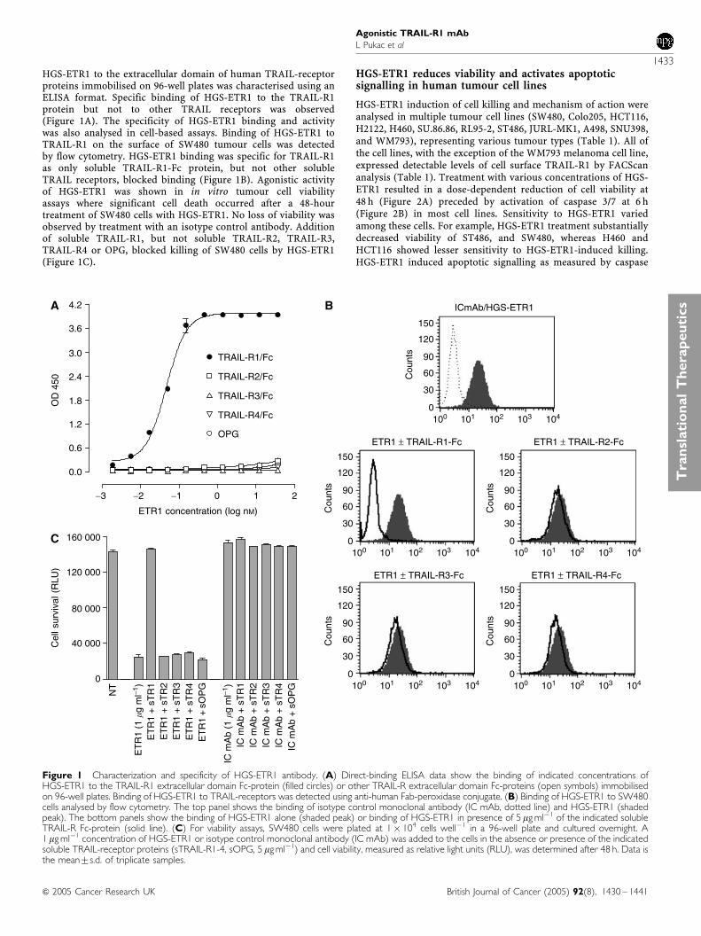
HGS-ETR1 to the extracellular domain of human TRAIL-receptorproteins immobilised on 96-well plates was characterised using anELISA format. Specific binding of HGS-ETR1 to the TRAIL-R1protein but not to other TRAIL receptors was observed(Figure 1A). The specificity of HGS-ETR1 binding and activitywas also analysed in cell-based assays. Binding of HGS-ETR1 toTRAIL-R1 on the surface of SW480 tumour cells was detectedby flow cytometry. HGS-ETR1 binding was specific for TRAIL-R1as only soluble TRAIL-R1-Fc protein, but not other solubleTRAIL receptors, blocked binding (Figure 1B). Agonistic activityof HGS-ETR1 was shown in in vitro tumour cell viabilityassays where significant cell death occurred after a 48-hourtreatment of SW480 cells with HGS-ETR1. No loss of viability wasobserved by treatment with an isotype control antibody. Additionof soluble TRAIL-R1, but not soluble TRAIL-R2, TRAIL-R3,TRAIL-R4 or OPG, blocked killing of SW480 cells by HGS-ETR1(Figure 1C).
HGS-ETR1 reduces viability and activates apoptoticsignalling in human tumour cell lines
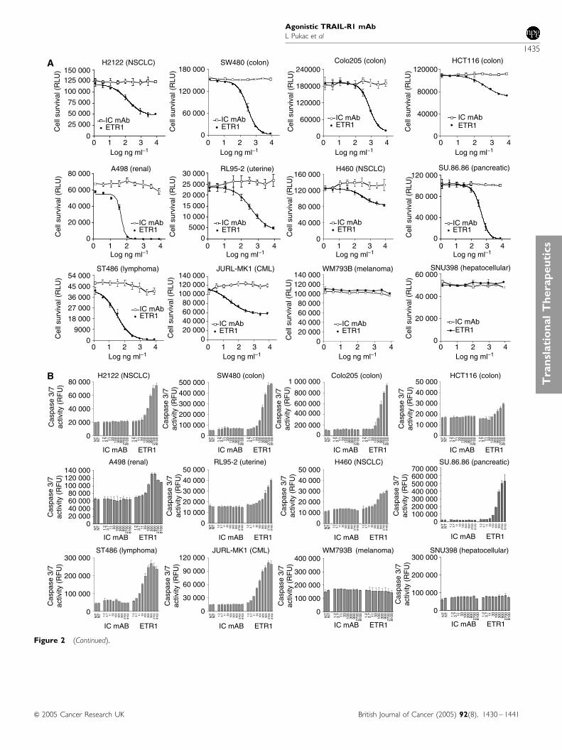
HGS-ETR1 induction of cell killing and mechanism of action wereanalysed in multiple tumour cell lines (SW480, Colo205, HCT116,H2122, H460, SU.86.86, RL95-2, ST486, JURL-MK1, A498, SNU398,and WM793), representing various tumour types (Table 1). All ofthe cell lines, with the exception of the WM793 melanoma cell line,expressed detectable levels of cell surface TRAIL-R1 by FACScananalysis (Table 1). Treatment with various concentrations of HGS-ETR1 resulted in a dose-dependent reduction of cell viability at48 h (Figure 2A) preceded by activation of caspase 3/7 at 6 h(Figure 2B) in most cell lines. Sensitivity to HGS-ETR1 variedamong these cells. For example, HGS-ETR1 treatment substantiallydecreased viability of ST486, and SW480, whereas H460 andHCT116 showed lesser sensitivity to HGS-ETR1-induced killing.HGS-ETR1 induced apoptotic signalling as measured by caspase
−3 −2 −1 0 1 2
4.2
OD
450
3.6
3.0
2.4
1.8
1.2
0.6
0.0
TRAIL-R1/Fc
TRAIL-R2/Fc
TRAIL-R3/Fc
TRAIL-R4/Fc
OPG
ETR1 concentration (log nM)
NT
ET
R1
(1 �
g m
l−1)
ET
R1
+ s
TR
1E
TR
1 +
sT
R2
ET
R1
+ s
TR
3E
TR
1 +
sT
R4
ET
R1
+ s
OP
G
IC m
Ab
(1 �
g m
l−1)
IC m
Ab
+ s
TR
1IC
mA
b +
sT
R2
IC m
Ab
+ s
TR
3IC
mA
b +
sT
R4
IC m
Ab
+ s
OP
G
160 000
120 000
80 000
40 000
0
Cel
l sur
viva
l (R
LU)
150
120
90
60Cou
nts
30
0100 101 102 103 104
150
120
90
60Cou
nts
30
0100 101 102 103 104
150
120
90
60Cou
nts
30
0100 101 102 103 104
150
120
90
60Cou
nts
30
0100 101 102 103 104
150
120
90
60Cou
nts
30
0100 101 102 103 104
ETR1 ± TRAIL-R1-Fc
ICmAb/HGS-ETR1
ETR1 ± TRAIL-R3-Fc ETR1 ± TRAIL-R4-Fc
ETR1 ± TRAIL-R2-Fc
A
C
B
Figure 1 Characterization and specificity of HGS-ETR1 antibody. (A) Direct-binding ELISA data show the binding of indicated concentrations ofHGS-ETR1 to the TRAIL-R1 extracellular domain Fc-protein (filled circles) or other TRAIL-R extracellular domain Fc-proteins (open symbols) immobilisedon 96-well plates. Binding of HGS-ETR1 to TRAIL-receptors was detected using anti-human Fab-peroxidase conjugate. (B) Binding of HGS-ETR1 to SW480cells analysed by flow cytometry. The top panel shows the binding of isotype control monoclonal antibody (IC mAb, dotted line) and HGS-ETR1 (shadedpeak). The bottom panels show the binding of HGS-ETR1 alone (shaded peak) or binding of HGS-ETR1 in presence of 5mg ml�1 of the indicated solubleTRAIL-R Fc-protein (solid line). (C) For viability assays, SW480 cells were plated at 1� 104 cells well�1 in a 96-well plate and cultured overnight. A1mg ml�1 concentration of HGS-ETR1 or isotype control monoclonal antibody (IC mAb) was added to the cells in the absence or presence of the indicatedsoluble TRAIL-receptor proteins (sTRAIL-R1-4, sOPG, 5mg ml�1) and cell viability, measured as relative light units (RLU), was determined after 48 h. Data isthe mean7s.d. of triplicate samples.
Agonistic TRAIL-R1 mAb
L Pukac et al
1433
British Journal of Cancer (2005) 92(8), 1430 – 1441& 2005 Cancer Research UK
Tra
nsl
ati
on
al
Th
era
peu
tics
3/7 activation in all cell lines that were killed by HGS-ETR1treatment. HGS-ETR1 did not induce caspase activity or reducecell viability in WM793 or SNU-398 cells.
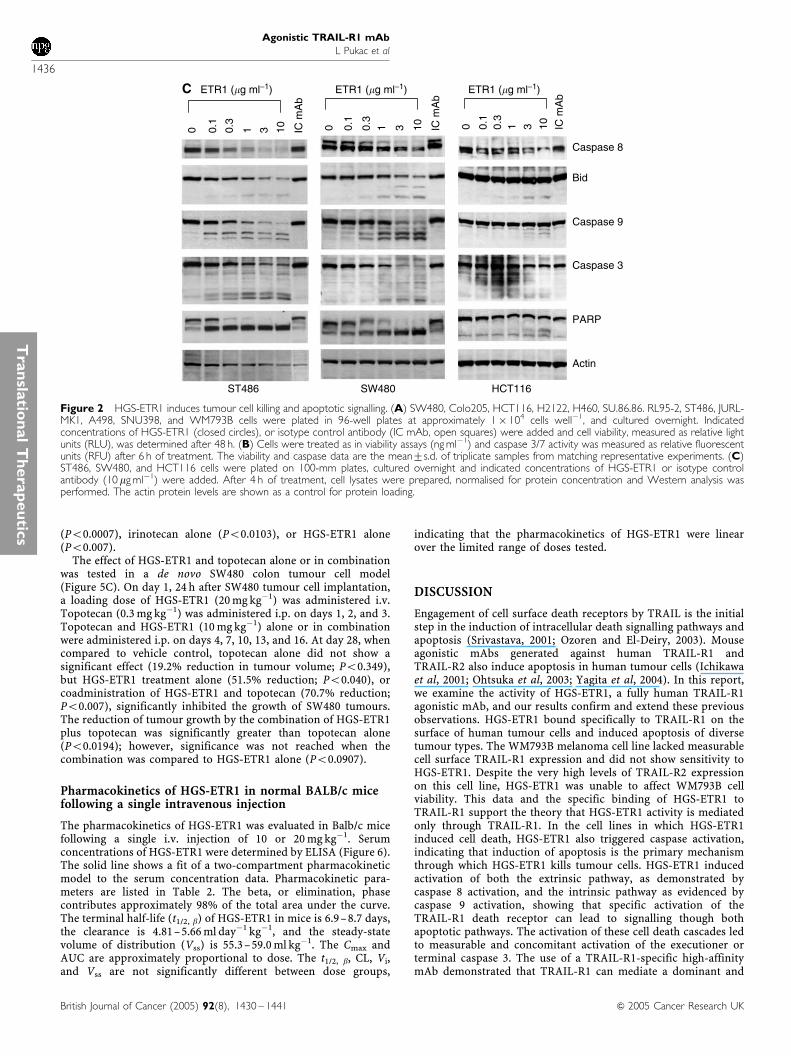
To more fully characterise the signalling pathways used byHGS-ETR1, activation of intracellular apoptotic signalling mole-cules was determined by Western analysis in three cell lines. Dose-dependent activation of caspases, as determined by a decrease inprocaspase 8 levels and cleavage of caspase 9 and caspase 3, wasobserved after a 4-h treatment of ST486 and SW480 cell lines withHGS-ETR1 (Figure 2C). HGS-ETR1 treatment also resulted incleavage of Bid, a substrate of caspase 8, and PARP, a substrate ofcaspase 3. The activation of these pathways by HGS-ETR1 wasclearly evident in ST486 and SW480. In contrast, in HCT116 cells,minimal activation of death signalling proteins was detected athigh concentrations of HGS-ETR1 (Figure 2C).
HGS-ETR1 enhancement of chemotherapeutic agentactivity in vitro
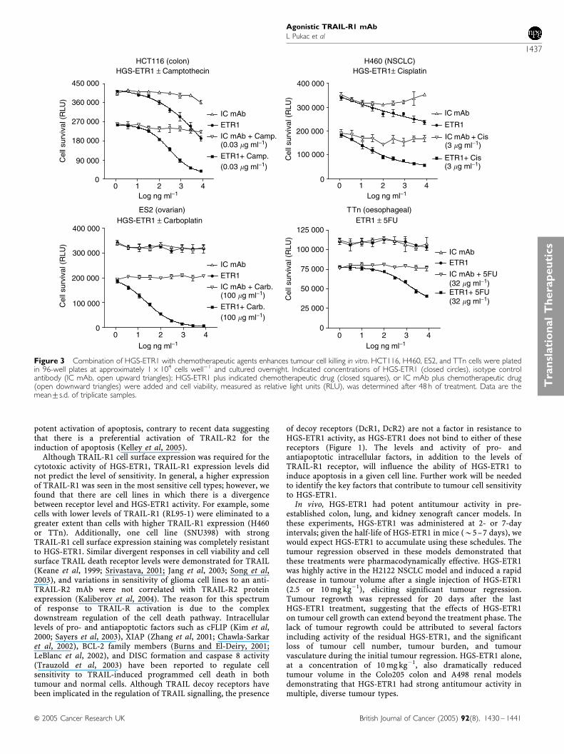
Combination treatment of HGS-ETR1 with chemotherapeuticagents in vitro was analysed in HGS-ETR1-sensitive (HCT116colon, H460 NSCLC) and -insensitive (ES2 ovarian, and TTnoesophageal) tumour cell lines. Cells were treated with variousconcentrations of control antibody or HGS-ETR1 either alone or incombination with camptothecin (HCT116), cisplatin (H460),carboplatin (ES2), or 5-fluorouracil (TTn) and the cell viabilitywas determined after 48 h (Figure 3). Chemotherapeutic drugconcentrations were used that had been previously established togive approximately 50% cell killing by drug alone (data notshown). In each case, the treatment with HGS-ETR1 pluschemotherapeutic drug resulted in enhanced cell killing, whichwas greater than the effect of either agent alone. Combinationtreatment substantially enhanced chemotherapeutic drug cytotoxi-city even in two cell lines (TTn and ES2) where HGS-ETR1treatment alone did not decrease cell viability, suggesting asynergistic interaction between HGS-ETR1 and the chemother-apeutic agents.
HGS-ETR1 reduces the growth of human tumours inxenograft models
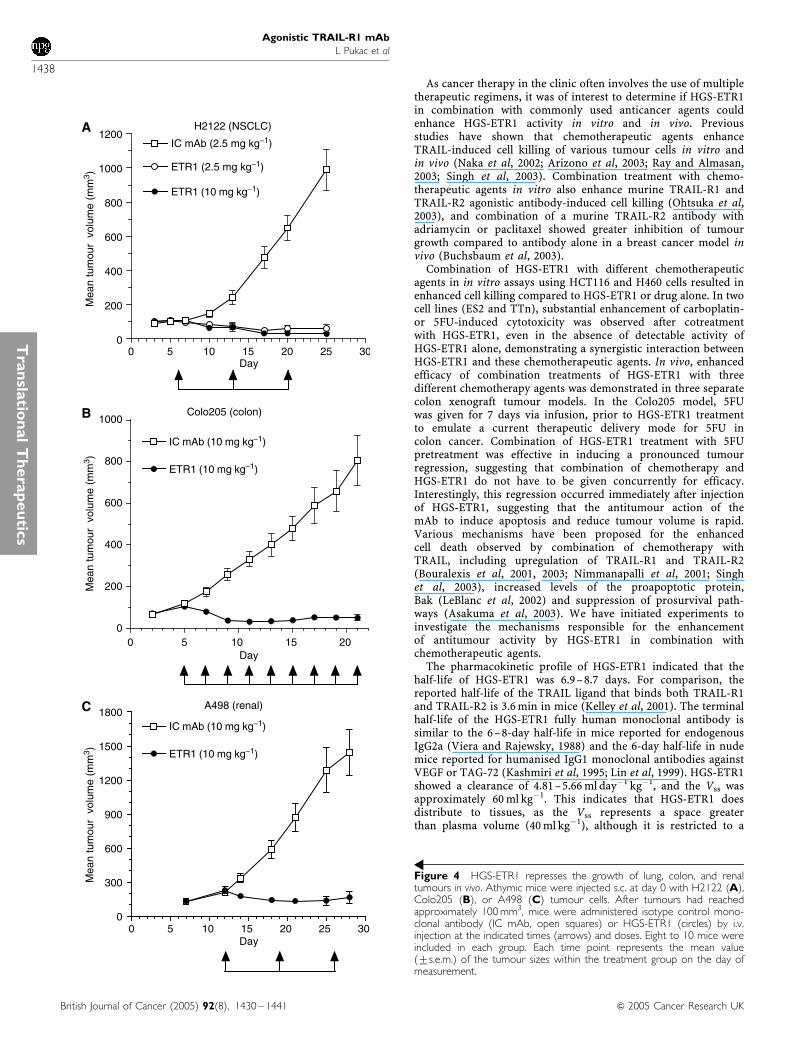
HGS-ETR1 in vivo antitumour activity was evaluated in NSCLC(H2122), colon (Colo205), and renal (A498) tumours in xenograftmodels in athymic nude mice. Tumours were pre-established s.c.to a volume of approximately 100 mm3 before initiation ofantibody treatments. Three weekly treatments of HGS-ETR1 at aconcentration of 2.5 or 10 mg kg�1 showed strong single-agent
antitumour activity in the H2122 NSCLC xenograft model, withsignificant tumour regression observed after a single injection ofHGS-ETR1 (Figure 4, Po0.0001). Tumour volume was reducedapproximately 50% by day 10, 4 days after the first injection ofHGS-ETR1 and by day 25, there was a 97% reduction in tumourvolume for the 10 mg kg�1 dose of HGS-ETR1. In addition, tumourregrowth was suppressed for several weeks after termination ofHGS-ETR1 treatment (data not shown). HGS-ETR1 also inducedtumour regression in colon and renal xenograft tumour models.Treatment with a 10 mg kg�1 dose of HGS-ETR1, every other dayor once a week, respectively, dramatically reduced tumourvolumes in Colo205 colon (Po0.0001 at day 21) and A498 renal(Po0.0001 at day 28) xenograft tumour models compared to theisotype control antibody (Figure 4).
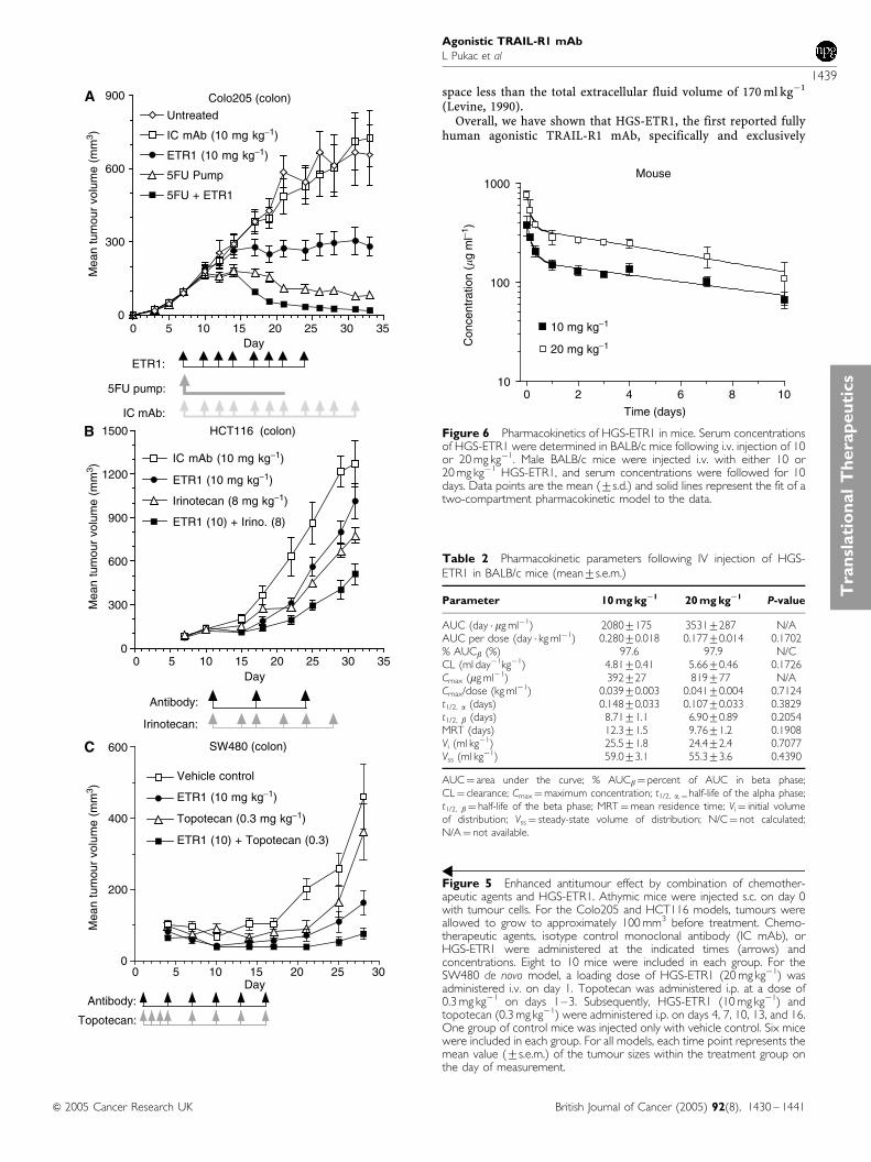
The ability of HGS-ETR1 to enhance the in vivo antitumouractivity of three different chemotherapeutic agents was tested inthree independent colon xenograft models (Figure 5). HGS-ETR1in combination with 5FU was tested in the Colo205 xenograftmodel (Figure 5A). Tumours of approximately 100 mm3 werepre-established, and animals were treated with 5FU alone(continuous infusion mini-pump, 1.25 mg kg�1 h�1, days 7–21),HGS-ETR1 alone (i.v., 10 mg kg�1, days 14, 17, 19, 21, 24, 27, 29,and 31), or combination of 5FU and HGS-ETR1, and comparedto no treatment or treatment with isotype control mAb (i.v.,10 mg kg�1, days 7 –31). Tumour size in nontreated animals wasnot significantly different from isotype control mAb treated.Both 5FU alone (Po0.001) and HGS-ETR1 alone (Po0.001)had a significant cytostatic effect on tumour growth comparedto control. For the HGS-ETR1þ 5FU combination treatmentgroup, mice were pretreated with 5FU for 7 days before treat-ment with HGS-ETR1 and resulted in an immediate decreasein tumour volume evident at day 17. The overall effect ontumour volume after combining HGS-ETR1 and 5FU wassignificantly different when compared to either agent alone(Po0.001).
The effect of combination of HGS-ETR1 with irinotecan wasexamined in pre-existing HCT116 colon tumours (Figure 5B).Tumours of approximately 100 mm3 were established and animalswere treated with irinotecan (i.p., 8 mg kg�1), HGS-ETR1 (i.v.,10 mg kg�1), or isotype control antibody (i.v., 10 mg kg�1). At day31, treatment with irinotecan alone resulted in a 38.8% reductionin tumour growth compared to control antibody (Po0.0113).HGS-ETR1 treatment showed a 20.1% reduction in tumourvolume, which was not significantly different from that of thecontrol antibody group (Po0.2248). The combination ofHGS-ETR1 plus irinotecan resulted in a significant reduction oftumour growth (59.7%) when compared with control antibody
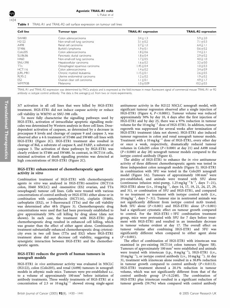
Table 1 TRAIL-R1 and TRAIL-R2 cell surface expression on tumour cell lines
Cell line Tumour type TRAIL-R1 expression TRAIL-R2 expression
SW480 Colon adenocarcinoma 3.471.3 5.972.0H2122 Non-small-cell lung carcinoma 3.170.2 7.170.3A498 Renal cell carcinoma 2.771.2 6.471.1ST486 Burkitt’s lymphoma 1.970.1 0.670.2Colo205 Colon adenocarcinoma 1.870.6 7.472.1SU.86.86 Pancreatic ductal carcinoma 1.870.4 2.970.2H460 Non-small-cell lung carcinoma 1.770.5 4.071.0SNU-398 Hepatocellular carcinoma 1.670.2 3.270.0TTn Oesophageal squamous carcinoma 1.4570.4 1.070.3HCT116 Colon adenocarcinoma 1.470.2 3.470.9JURL-MK1 Chronic myeloid leukaemia 1.1570.1 2.670.5RL95-2 Uterine endometrial carcinoma 1.270.2 1.970.2ES2 Ovarian clear cell carcinoma 1.170.1 4.971.7WM793B Melanoma 1.070.09 10.270.5
TRAIL-R1 and TRAIL-R2 expression was determined by FACs analysis and is expressed as the fold-increase in mean fluorescent signal of commercial mouse TRAIL-R1 or R2antibody vs isotype control antibody. The data is the average7s.d. from two or more experiments.
Agonistic TRAIL-R1 mAb
L Pukac et al
1434
British Journal of Cancer (2005) 92(8), 1430 – 1441 & 2005 Cancer Research UK
Tra
nsla
tion
al
Th
era
peu
tics
SNU398 (hepatocellular)
0
20 000
40 000
60 000
IC mAbETR1
SU.86.86 (pancreatic)
0
40 000
80 000
120 000
ETR1IC mAb
Colo205 (colon)
0 1 2 3 40
60000
120000
180000
240000
IC mAbETR1
SW480 (colon)
0 1 2 3 40
60 000
120 000
180 000
IC mAbETR1
JURL-MK1 (CML)
020 00040 00060 00080 000
100 000120 000140 000
IC mAbETR1
IC mAbETR1
H2122 (NSCLC)
0 1 2 3 40
25 000
Cel
l sur
viva
l (R
LU)
Cel
l sur
viva
l (R
LU)
Cel
l sur
viva
l (R
LU)
Cel
l sur
viva
l (R
LU)
Cel
l sur
viva
l (R
LU)
Cel
l sur
viva
l (R
LU)
Cel
l sur
viva
l (R
LU)
Cel
l sur
viva
l (R
LU)
Cel
l sur
viva
l (R
LU)
Cel
l sur
viva
l (R
LU)
Cel
l sur
viva
l (R
LU)
Cel
l sur
viva
l (R
LU)
50 000
75 000
100 000
125 000150 000
IC mAbETR1
IC mAbETR1
IC mAbETR1
Log ng ml−1
Log ng ml−1
Log ng ml−1
Log ng ml−1 Log ng ml−1
Log ng ml−1 Log ng ml−1 Log ng ml−1
H460 (NSCLC)
0 1 2 3 4Log ng ml−1
0 1 2 3 40
40 000
80 000
120 000
160 000
IC mAbETR1
ST486 (lymphoma)
0 1 2 3 4Log ng ml−1
0 1 2 3 4Log ng ml−1
0 1 2 3 4Log ng ml−1
0 1 2 3 40
9000
18 000
27 000
36 000
45 000
54 000
IC mAbETR1
A498 (renal)
0 1 2 3 40
20 000
40 000
60 000
80 000
WM793B (melanoma)
020 00040 00060 00080 000
100 000120 000140 000
HCT116 (colon)
0 1 2 3 40
40000
80000
120000
IC mAbETR1
RL95-2 (uterine)
0 1 2 3 40
5000
10 000
15 000
20 000
25 000
30 000
Colo205 (colon)
0200 000
400 000600 000
800 000
1 000 000SW480 (colon)
0
100 000
200 000
300 000
400 000
500 000
RL95-2 (uterine)
0
10 000
20 000
30 000
40 000
50 000
JURL-MK1 (CML)
0
30 000
60 000
90 000
120 000ST486 (lymphoma)
0
100 000
200 000
300 000
HCT116 (colon)
0
10 000
20 000
30 000
40 000
50 000
H460 (NSCLC)
0
10 000
20 000
30 000
40 000
50 000
H2122 (NSCLC)
0
20 000
40 000
60 000
Cas
pase
3/7
activ
ity (
RF
U)
Cas
pase
3/7
activ
ity (
RF
U)
Cas
pase
3/7
activ
ity (
RF
U)
Cas
pase
3/7
activ
ity (
RF
U)
Cas
pase
3/7
activ
ity (
RF
U)
Cas
pase
3/7
activ
ity (
RF
U)
Cas
pase
3/7
activ
ity (
RF
U)
Cas
pase
3/7
activ
ity (
RF
U)
Cas
pase
3/7
activ
ity (
RF
U)
Cas
pase
3/7
activ
ity (
RF
U)
Cas
pase
3/7
activ
ity (
RF
U)
Cas
pase
3/7
activ
ity (
RF
U)
80 000
IC mAB ETR1
IC mAB ETR1
IC mAB ETR1 IC mAB ETR1
IC mAB ETR1
IC mAB ETR1 IC mAB ETR1
IC mAB ETR1 IC mAB ETR1
IC mAB ETR1 IC mAB ETR1 IC mAB ETR1
SU.86.86 (pancreatic)
0100 000200 000300 000400 000500 000600 000700 000
A498 (renal)
020 00040 00060 00080 000
100 000120 000140 000
SNU398 (hepatocellular)
0
100 000
200 000
300 000 WM793B (melanoma)
0
100 000
200 000
300 000
400 000
A
B
Figure 2 (Continued).
Agonistic TRAIL-R1 mAb
L Pukac et al
1435
British Journal of Cancer (2005) 92(8), 1430 – 1441& 2005 Cancer Research UK
Tra
nsl
ati
on
al
Th
era
peu
tics
(Po0.0007), irinotecan alone (Po0.0103), or HGS-ETR1 alone(Po0.007).
The effect of HGS-ETR1 and topotecan alone or in combinationwas tested in a de novo SW480 colon tumour cell model(Figure 5C). On day 1, 24 h after SW480 tumour cell implantation,a loading dose of HGS-ETR1 (20 mg kg�1) was administered i.v.Topotecan (0.3 mg kg�1) was administered i.p. on days 1, 2, and 3.Topotecan and HGS-ETR1 (10 mg kg�1) alone or in combinationwere administered i.p. on days 4, 7, 10, 13, and 16. At day 28, whencompared to vehicle control, topotecan alone did not show asignificant effect (19.2% reduction in tumour volume; Po0.349),but HGS-ETR1 treatment alone (51.5% reduction; Po0.040), orcoadministration of HGS-ETR1 and topotecan (70.7% reduction;Po0.007), significantly inhibited the growth of SW480 tumours.The reduction of tumour growth by the combination of HGS-ETR1plus topotecan was significantly greater than topotecan alone(Po0.0194); however, significance was not reached when thecombination was compared to HGS-ETR1 alone (Po0.0907).
Pharmacokinetics of HGS-ETR1 in normal BALB/c micefollowing a single intravenous injection
The pharmacokinetics of HGS-ETR1 was evaluated in Balb/c micefollowing a single i.v. injection of 10 or 20 mg kg�1. Serumconcentrations of HGS-ETR1 were determined by ELISA (Figure 6).The solid line shows a fit of a two-compartment pharmacokineticmodel to the serum concentration data. Pharmacokinetic para-meters are listed in Table 2. The beta, or elimination, phasecontributes approximately 98% of the total area under the curve.The terminal half-life (t1/2, b) of HGS-ETR1 in mice is 6.9–8.7 days,the clearance is 4.81–5.66 ml day�1 kg�1, and the steady-statevolume of distribution (Vss) is 55.3–59.0 ml kg�1. The Cmax andAUC are approximately proportional to dose. The t1/2, b, CL, Vi,and Vss are not significantly different between dose groups,
indicating that the pharmacokinetics of HGS-ETR1 were linearover the limited range of doses tested.
DISCUSSION
Engagement of cell surface death receptors by TRAIL is the initialstep in the induction of intracellular death signalling pathways andapoptosis (Srivastava, 2001; Ozoren and El-Deiry, 2003). Mouseagonistic mAbs generated against human TRAIL-R1 andTRAIL-R2 also induce apoptosis in human tumour cells (Ichikawaet al, 2001; Ohtsuka et al, 2003; Yagita et al, 2004). In this report,we examine the activity of HGS-ETR1, a fully human TRAIL-R1agonistic mAb, and our results confirm and extend these previousobservations. HGS-ETR1 bound specifically to TRAIL-R1 on thesurface of human tumour cells and induced apoptosis of diversetumour types. The WM793B melanoma cell line lacked measurablecell surface TRAIL-R1 expression and did not show sensitivity toHGS-ETR1. Despite the very high levels of TRAIL-R2 expressionon this cell line, HGS-ETR1 was unable to affect WM793B cellviability. This data and the specific binding of HGS-ETR1 toTRAIL-R1 support the theory that HGS-ETR1 activity is mediatedonly through TRAIL-R1. In the cell lines in which HGS-ETR1induced cell death, HGS-ETR1 also triggered caspase activation,indicating that induction of apoptosis is the primary mechanismthrough which HGS-ETR1 kills tumour cells. HGS-ETR1 inducedactivation of both the extrinsic pathway, as demonstrated bycaspase 8 activation, and the intrinsic pathway as evidenced bycaspase 9 activation, showing that specific activation of theTRAIL-R1 death receptor can lead to signalling though bothapoptotic pathways. The activation of these cell death cascades ledto measurable and concomitant activation of the executioner orterminal caspase 3. The use of a TRAIL-R1-specific high-affinitymAb demonstrated that TRAIL-R1 can mediate a dominant and
SW480
Bid
PARP
Actin
Caspase 8
ST486
Caspase 9
Caspase 3
0 0.1
0.3
1 3 10 IC m
Ab
0 0.1
0.3
1 3 10 IC m
Ab
0 0.1
0.3
1 3 10 IC m
Ab
ETR1 (�g ml−1)ETR1 (�g ml−1)ETR1 (�g ml−1)
HCT116
C
Figure 2 HGS-ETR1 induces tumour cell killing and apoptotic signalling. (A) SW480, Colo205, HCT116, H2122, H460, SU.86.86. RL95-2, ST486, JURL-MK1, A498, SNU398, and WM793B cells were plated in 96-well plates at approximately 1� 104 cells well�1, and cultured overnight. Indicatedconcentrations of HGS-ETR1 (closed circles), or isotype control antibody (IC mAb, open squares) were added and cell viability, measured as relative lightunits (RLU), was determined after 48 h. (B) Cells were treated as in viability assays (ng ml�1) and caspase 3/7 activity was measured as relative fluorescentunits (RFU) after 6 h of treatment. The viability and caspase data are the mean7s.d. of triplicate samples from matching representative experiments. (C)ST486, SW480, and HCT116 cells were plated on 100-mm plates, cultured overnight and indicated concentrations of HGS-ETR1 or isotype controlantibody (10 mg ml�1) were added. After 4 h of treatment, cell lysates were prepared, normalised for protein concentration and Western analysis wasperformed. The actin protein levels are shown as a control for protein loading.
Agonistic TRAIL-R1 mAb
L Pukac et al
1436
British Journal of Cancer (2005) 92(8), 1430 – 1441 & 2005 Cancer Research UK
Tra
nsla
tion
al
Th
era
peu
tics
potent activation of apoptosis, contrary to recent data suggestingthat there is a preferential activation of TRAIL-R2 for theinduction of apoptosis (Kelley et al, 2005).
Although TRAIL-R1 cell surface expression was required for thecytotoxic activity of HGS-ETR1, TRAIL-R1 expression levels didnot predict the level of sensitivity. In general, a higher expressionof TRAIL-R1 was seen in the most sensitive cell types; however, wefound that there are cell lines in which there is a divergencebetween receptor level and HGS-ETR1 activity. For example, somecells with lower levels of TRAIL-R1 (RL95-1) were eliminated to agreater extent than cells with higher TRAIL-R1 expression (H460or TTn). Additionally, one cell line (SNU398) with strongTRAIL-R1 cell surface expression staining was completely resistantto HGS-ETR1. Similar divergent responses in cell viability and cellsurface TRAIL death receptor levels were demonstrated for TRAIL(Keane et al, 1999; Srivastava, 2001; Jang et al, 2003; Song et al,2003), and variations in sensitivity of glioma cell lines to an anti-TRAIL-R2 mAb were not correlated with TRAIL-R2 proteinexpression (Kaliberov et al, 2004). The reason for this spectrumof response to TRAIL-R activation is due to the complexdownstream regulation of the cell death pathway. Intracellularlevels of pro- and antiapoptotic factors such as cFLIP (Kim et al,2000; Sayers et al, 2003), XIAP (Zhang et al, 2001; Chawla-Sarkaret al, 2002), BCL-2 family members (Burns and El-Deiry, 2001;LeBlanc et al, 2002), and DISC formation and caspase 8 activity(Trauzold et al, 2003) have been reported to regulate cellsensitivity to TRAIL-induced programmed cell death in bothtumour and normal cells. Although TRAIL decoy receptors havebeen implicated in the regulation of TRAIL signalling, the presence
of decoy receptors (DcR1, DcR2) are not a factor in resistance toHGS-ETR1 activity, as HGS-ETR1 does not bind to either of thesereceptors (Figure 1). The levels and activity of pro- andantiapoptotic intracellular factors, in addition to the levels ofTRAIL-R1 receptor, will influence the ability of HGS-ETR1 toinduce apoptosis in a given cell line. Further work will be neededto identify the key factors that contribute to tumour cell sensitivityto HGS-ETR1.
In vivo, HGS-ETR1 had potent antitumour activity in pre-established colon, lung, and kidney xenograft cancer models. Inthese experiments, HGS-ETR1 was administered at 2- or 7-dayintervals; given the half-life of HGS-ETR1 in mice (B5–7 days), wewould expect HGS-ETR1 to accumulate using these schedules. Thetumour regression observed in these models demonstrated thatthese treatments were pharmacodynamically effective. HGS-ETR1was highly active in the H2122 NSCLC model and induced a rapiddecrease in tumour volume after a single injection of HGS-ETR1(2.5 or 10 mg kg�1), eliciting significant tumour regression.Tumour regrowth was repressed for 20 days after the lastHGS-ETR1 treatment, suggesting that the effects of HGS-ETR1on tumour cell growth can extend beyond the treatment phase. Thelack of tumour regrowth could be attributed to several factorsincluding activity of the residual HGS-ETR1, and the significantloss of tumour cell number, tumour burden, and tumourvasculature during the initial tumour regression. HGS-ETR1 alone,at a concentration of 10 mg kg�1, also dramatically reducedtumour volume in the Colo205 colon and A498 renal modelsdemonstrating that HGS-ETR1 had strong antitumour activity inmultiple, diverse tumour types.
H460 (NSCLC)HGS-ETR1± Cisplatin
0 1 2 3 40
100 000
200 000
300 000
400 000
IC mAb
ETR1
ETR1+ Cis(3 �g ml−1)
IC mAb + Cis(3 �g ml−1)
HCT116 (colon)HGS-ETR1 ± Camptothecin
0 1 2 3 40
90 000
180 000
270 000
360 000
450 000
Cel
l sur
viva
l (R
LU)
Cel
l sur
viva
l (R
LU)
Cel
l sur
viva
l (R
LU)
IC mAb
ETR1
ETR1+ Camp.(0.03 �g ml−1)
IC mAb + Camp.(0.03 �g ml−1)
IC mAb
ETR1
ETR1+ Carb.(100 �g ml−1)
IC mAb + Carb.(100 �g ml−1)
Log ng ml−1
Log ng ml−1 Log ng ml−1
Log ng ml−1
ES2 (ovarian)HGS-ETR1 ± Carboplatin
0 1 2 3 4
TTn (oesophageal)ETR1 ± 5FU
0 1 2 3 40
25 000
50 000
75 000
100 000
125 000
IC mAbETR1
IC mAb + 5FU(32 �g ml−1)ETR1+ 5FU(32 �g ml−1)
0
100 000
200 000
300 000
400 000
Cel
l sur
viva
l (R
LU)
Figure 3 Combination of HGS-ETR1 with chemotherapeutic agents enhances tumour cell killing in vitro. HCT116, H460, ES2, and TTn cells were platedin 96-well plates at approximately 1� 104 cells well�1 and cultured overnight. Indicated concentrations of HGS-ETR1 (closed circles), isotype controlantibody (IC mAb, open upward triangles); HGS-ETR1 plus indicated chemotherapeutic drug (closed squares), or IC mAb plus chemotherapeutic drug(open downward triangles) were added and cell viability, measured as relative light units (RLU), was determined after 48 h of treatment. Data are themean7s.d. of triplicate samples.
Agonistic TRAIL-R1 mAb
L Pukac et al
1437
British Journal of Cancer (2005) 92(8), 1430 – 1441& 2005 Cancer Research UK
Tra
nsl
ati
on
al
Th
era
peu
tics
As cancer therapy in the clinic often involves the use of multipletherapeutic regimens, it was of interest to determine if HGS-ETR1in combination with commonly used anticancer agents couldenhance HGS-ETR1 activity in vitro and in vivo. Previousstudies have shown that chemotherapeutic agents enhanceTRAIL-induced cell killing of various tumour cells in vitro andin vivo (Naka et al, 2002; Arizono et al, 2003; Ray and Almasan,2003; Singh et al, 2003). Combination treatment with chemo-therapeutic agents in vitro also enhance murine TRAIL-R1 andTRAIL-R2 agonistic antibody-induced cell killing (Ohtsuka et al,2003), and combination of a murine TRAIL-R2 antibody withadriamycin or paclitaxel showed greater inhibition of tumourgrowth compared to antibody alone in a breast cancer model invivo (Buchsbaum et al, 2003).
Combination of HGS-ETR1 with different chemotherapeuticagents in in vitro assays using HCT116 and H460 cells resulted inenhanced cell killing compared to HGS-ETR1 or drug alone. In twocell lines (ES2 and TTn), substantial enhancement of carboplatin-or 5FU-induced cytotoxicity was observed after cotreatmentwith HGS-ETR1, even in the absence of detectable activity ofHGS-ETR1 alone, demonstrating a synergistic interaction betweenHGS-ETR1 and these chemotherapeutic agents. In vivo, enhancedefficacy of combination treatments of HGS-ETR1 with threedifferent chemotherapy agents was demonstrated in three separatecolon xenograft tumour models. In the Colo205 model, 5FUwas given for 7 days via infusion, prior to HGS-ETR1 treatmentto emulate a current therapeutic delivery mode for 5FU incolon cancer. Combination of HGS-ETR1 treatment with 5FUpretreatment was effective in inducing a pronounced tumourregression, suggesting that combination of chemotherapy andHGS-ETR1 do not have to be given concurrently for efficacy.Interestingly, this regression occurred immediately after injectionof HGS-ETR1, suggesting that the antitumour action of themAb to induce apoptosis and reduce tumour volume is rapid.Various mechanisms have been proposed for the enhancedcell death observed by combination of chemotherapy withTRAIL, including upregulation of TRAIL-R1 and TRAIL-R2(Bouralexis et al, 2001, 2003; Nimmanapalli et al, 2001; Singhet al, 2003), increased levels of the proapoptotic protein,Bak (LeBlanc et al, 2002) and suppression of prosurvival path-ways (Asakuma et al, 2003). We have initiated experiments toinvestigate the mechanisms responsible for the enhancementof antitumour activity by HGS-ETR1 in combination withchemotherapeutic agents.
The pharmacokinetic profile of HGS-ETR1 indicated that thehalf-life of HGS-ETR1 was 6.9– 8.7 days. For comparison, thereported half-life of the TRAIL ligand that binds both TRAIL-R1and TRAIL-R2 is 3.6 min in mice (Kelley et al, 2001). The terminalhalf-life of the HGS-ETR1 fully human monoclonal antibody issimilar to the 6–8-day half-life in mice reported for endogenousIgG2a (Viera and Rajewsky, 1988) and the 6-day half-life in nudemice reported for humanised IgG1 monoclonal antibodies againstVEGF or TAG-72 (Kashmiri et al, 1995; Lin et al, 1999). HGS-ETR1showed a clearance of 4.81–5.66 ml day�1 kg�1, and the Vss wasapproximately 60 ml kg�1. This indicates that HGS-ETR1 doesdistribute to tissues, as the Vss represents a space greaterthan plasma volume (40 ml kg�1), although it is restricted to a
H2122 (NSCLC)
0 5 10 15 20 25 300
200
400
600
800
1000
1200IC mAb (2.5 mg kg−1)
ETR1 (10 mg kg−1)
ETR1 (2.5 mg kg−1)
Day
Day
Day
Mea
n tu
mou
r v
olum
e (m
m3 )
Mea
n tu
mou
r v
olum
e (m
m3 )
Mea
n tu
mou
r v
olum
e (m
m3 )
Colo205 (colon)
0 5 10 15 200
200
400
600
800
1000
IC mAb (10 mg kg−1)
ETR1 (10 mg kg−1)
IC mAb (10 mg kg−1)
ETR1 (10 mg kg−1)
A498 (renal)
0 5 10 15 20 25 300
300
600
900
1200
1500
1800
A
B
C
Figure 4 HGS-ETR1 represses the growth of lung, colon, and renaltumours in vivo. Athymic mice were injected s.c. at day 0 with H2122 (A),Colo205 (B), or A498 (C) tumour cells. After tumours had reachedapproximately 100 mm3, mice were administered isotype control mono-clonal antibody (IC mAb, open squares) or HGS-ETR1 (circles) by i.v.injection at the indicated times (arrows) and doses. Eight to 10 mice wereincluded in each group. Each time point represents the mean value(7s.e.m.) of the tumour sizes within the treatment group on the day ofmeasurement.
Agonistic TRAIL-R1 mAb
L Pukac et al
1438
British Journal of Cancer (2005) 92(8), 1430 – 1441 & 2005 Cancer Research UK
Tra
nsla
tion
al
Th
era
peu
tics
space less than the total extracellular fluid volume of 170 ml kg�1
(Levine, 1990).Overall, we have shown that HGS-ETR1, the first reported fully
human agonistic TRAIL-R1 mAb, specifically and exclusively
Colo205 (colon)
0 5 10 15 20 25 30 350
300
600
900
Untreated
IC mAb (10 mg kg−1)
ETR1 (10 mg kg−1)
5FU Pump
5FU + ETR1
ETR1:
5FU pump:
IC mAb:
Day
Mea
n tu
mou
r vo
lum
e (m
m3 )
Mea
n tu
mou
r vo
lum
e (m
m3 )
Mea
n tu
mou
r vo
lum
e (m
m3 )
0 5 10 15 20 25 30 350
300
600
900
1200
1500
Irinotecan (8 mg kg−1)
IC mAb (10 mg kg−1)
ETR1 (10 mg kg−1)
ETR1 (10) + Irino. (8)
HCT116 (colon)
Antibody:
Irinotecan:
Day
Day0 5 10 15 20 25 30
0
200
400
600
Vehicle control
ETR1 (10 mg kg−1)
Topotecan (0.3 mg kg−1)
ETR1 (10) + Topotecan (0.3)
SW480 (colon)
Antibody:
Topotecan:
A
B
C
Figure 5 Enhanced antitumour effect by combination of chemother-apeutic agents and HGS-ETR1. Athymic mice were injected s.c. on day 0with tumour cells. For the Colo205 and HCT116 models, tumours wereallowed to grow to approximately 100 mm3 before treatment. Chemo-therapeutic agents, isotype control monoclonal antibody (IC mAb), orHGS-ETR1 were administered at the indicated times (arrows) andconcentrations. Eight to 10 mice were included in each group. For theSW480 de novo model, a loading dose of HGS-ETR1 (20 mg kg�1) wasadministered i.v. on day 1. Topotecan was administered i.p. at a dose of0.3 mg kg�1 on days 1–3. Subsequently, HGS-ETR1 (10 mg kg�1) andtopotecan (0.3 mg kg�1) were administered i.p. on days 4, 7, 10, 13, and 16.One group of control mice was injected only with vehicle control. Six micewere included in each group. For all models, each time point represents themean value (7s.e.m.) of the tumour sizes within the treatment group onthe day of measurement.
0 2 4 6 8 1010
100
1000
10 mg kg−1
20 mg kg−1
Mouse
Time (days)
Con
cent
ratio
n (�
g m
l−1)
Figure 6 Pharmacokinetics of HGS-ETR1 in mice. Serum concentrationsof HGS-ETR1 were determined in BALB/c mice following i.v. injection of 10or 20 mg kg�1. Male BALB/c mice were injected i.v. with either 10 or20 mg kg�1 HGS-ETR1, and serum concentrations were followed for 10days. Data points are the mean (7s.d.) and solid lines represent the fit of atwo-compartment pharmacokinetic model to the data.
Table 2 Pharmacokinetic parameters following IV injection of HGS-ETR1 in BALB/c mice (mean7s.e.m.)
Parameter 10 mg kg�1 20 mg kg�1 P-value
AUC (day � mg ml�1) 20807175 35317287 N/AAUC per dose (day � kg ml�1) 0.28070.018 0.17770.014 0.1702% AUCb (%) 97.6 97.9 N/CCL (ml day�1kg�1) 4.8170.41 5.6670.46 0.1726Cmax (mg ml�1) 392727 819777 N/ACmax/dose (kg ml�1) 0.03970.003 0.04170.004 0.7124t1/2, a (days) 0.14870.033 0.10770.033 0.3829t1/2, b (days) 8.7171.1 6.9070.89 0.2054MRT (days) 12.371.5 9.7671.2 0.1908Vi (ml kg�1) 25.571.8 24.472.4 0.7077Vss (ml kg�1) 59.073.1 55.373.6 0.4390
AUC¼ area under the curve; % AUCb¼ percent of AUC in beta phase;CL¼ clearance; Cmax¼maximum concentration; t1/2, a,¼half-life of the alpha phase;t1/2, b¼ half-life of the beta phase; MRT¼mean residence time; Vi¼ initial volumeof distribution; Vss¼ steady-state volume of distribution; N/C¼ not calculated;N/A¼ not available.
Agonistic TRAIL-R1 mAb
L Pukac et al
1439
British Journal of Cancer (2005) 92(8), 1430 – 1441& 2005 Cancer Research UK
Tra
nsl
ati
on
al
Th
era
peu
tics
bound to TRAIL-R1, and potently induced apoptosis in vitroin a broad range of human tumour cell types via activation ofboth intrinsic and extrinsic signalling cascades. HGS-ETR1 hadan effective pharmacokinetic half-life, and induced rapidtumour regression in multiple xenograft tumour models aloneand in combination with chemotherapeutic agents. These dataindicate that HGS-ETR1 has significant potential as a cancertherapeutic agent. HGS-ETR1 is currently being evaluated in Phase
I/II clinical trials in patients with advanced solid or haematologicaltumours.
ACKNOWLEDGEMENTS
We thank Jessica Manela Alfred Corey and Jessie Wolfe forvaluable advice and assistance.
REFERENCES
Arizono Y, Yoshikawa H, Naganuma H, Hamada Y, Nakajima Y, Tasaka K(2003) A mechanism of resistance to TRAIL/Apo2L-induced apoptosis ofnewly established glioma cell line and sensitisation to TRAIL bygenotoxic agents. Br J Cancer 88: 298 – 306
Ashkenazi A, Pai R, Fong S, Leung S, Lawrence D, Marsters S, Blackie C,Chang L, McMurtrey A, Herbert A, DeForge L, Koumenis I, Lewis D,Harris L, Bussiere J, Koeppen H, Shahrokn Z, Schwall R (1999) Safetyand antitumor activity of recombinant soluble Apo2 ligand. J Clin Invest104: 155 – 162
Asakuma J, Sumitomo M, Asano T, Hayakawa M (2003) Selective Aktinactivation and tumor necrosis actor-related apoptosis-inducing ligandsensitization of renal cancer cells by low concentrations of paclitaxel.Cancer Res 63: 1365 – 1370
Berger M, Shankar V, Vafai A (2002) Therapeutic applications ofmonoclonal antibodies. Am J Med Sci 324: 14 – 30
Bouralexis S, Evdokiou A, Hay S, Atkins G, Findlay D (2001) Enhancedapoptosis of soft tissue sarcoma cells with chemotherapy: a potential newapproach using TRAIL. J Orthop Surg 9: 19 – 22
Bouralexis S, Findlay D, Atkins G, Laabrinidis A, Hay S, Evdokiou A (2003)Progressive resistance of BTK-143 osteosarcoma cells to Apo2L/TRAIL-induced apoptosis is mediated by acquisition of DcR2/TRAIL-R4expression: resensitisation with chemotherapy. Br J Cancer 89: 206 – 214
Buchsbaum D, Zhou T, Grizzle W, Oliver P, Hammond C, Zhang S,Carpenter M, LoBuglio A (2003) Antitumor efficacy of TRA-8 anti-DR5monoclonal antibody alone or in combination with chemotherapy and/orradiation therapy in a human breast cancer model. Clin Cancer Res 9:3731 – 3741
Burns T, El-Deiry W (2001) Identification of inhibitors of TRAIL-induceddeath (ITIDs) in the TRAIL-sensitive colon carcinoma cell line SW480using a genetic approach. J Biol Chem 276: 37879 – 37886
Chawla-Sarkar M, Leaman D, Jacobs B, Borden E (2002) IFN-betapretreatment sensitizes human melenoma cells to TRAIL/Apo2 ligand-induced apoptosis. J Immunol 169: 847 – 855
Chuntharapai A, Dodge K, Grimmer K, Schroeder K, Masters SA, KoeppenH, Ashkenazi A, Kim KJ (2001) Isotype-dependent inhibition of tumorgrowth in vivo by monoclonal antibodies to death receptor 4. J Immunol166: 4891 – 4898
Evdokiou A, Bouralexis S, Atkins G (2002) Chemotherapeutic agentssensitize osteogenic sarcoma cells, but not normal human bone cells, toapo2L/Trail-induced apoptosis. Int J Cancer 99: 491 – 504
Gliniak B, Le T (1999) Tumor necrosis factor-related apoptosis-inducingligand’s antitumor activity in vivo is enhanced by the chemotherapeuticagent CPT-11. Cancer Res 59: 6153 – 6158
Griffith T, Rauch C, Smolak P, Waugh J, Boiani N, Lynch D, Smith C,Goodwin R, Kubin M (1999) Functional analysis of TRAIL receptorsusing monoclonal antibodies. J Immunol 162: 2597 – 2605
Ichikawa K, Liu W, Zhao L, Wang Z, Liu D, Ohtsuka T, Zhang H, Mountz J,Koopman W, Kimberly R, Zhou T (2001) Tumoricidal activity of a novelanti-human DR5 monoclonal antibody without hepatocyte cytotoxicity.Nat Med 7: 954 – 960
Jackson R, McCafferty J, Joshson K, Pope A, Roberts A, Chiswell D,Clackson T, Griffiths A, Hoogenboom H, Winter G (1992) Selection ofvariants of antibodies and other protein molecules using display on thesurface of bacteriophage. In Protein Engineering: A Practical ApproachRees AR, Sternberg M, Wetzel R (eds) pp 277 – 301. Oxford: IRL Press
Jang Y, Park K, Chung H, Kim H (2003) Analysis of the phenotypes ofJurkat clones with different TRAIL-sensitivities. Can Lett 194: 107 – 117
Kaliberov S, Stackhouse M, Kaliberova L, Zhou T, Buchsbaum D (2004)Enhanced apoptosis following treatment with TRA-8 anti-human DR5monoclonal antibody and overexpression of exogenous Bax in humanglioma cells. Gene Ther 11: 658 – 667
Kashmiri S, Shu L, Padlam E, Milenic D, Schlom J, Hand P (1995)Generation, characterization, and in vivo studies of humanized anti-carcinoma antibody CC49. Hybridoma 14: 461 – 473
Keane M, Ettenberg S, Nau M, Russell E, Lipkowitz S (1999) Chemotherapyaugments TRAIL-induced apoptosis in breast cell lines. Cancer Res 59:734 – 741
Kelley R, Totpal K, Lindstrom S, Mathieu M, Billeci K, Deforge L, Pai R,Hymowitz S, Ashkenazi A (2005) Receptor-selective mutants of Apo2L/TRAIL reveal a greater contribution of DR5 than DR4 to apoptosissignaling. J Biol Chem 280: 2205 – 2212
Kelley S, Harris L, Xie D, Deforge L, Totpal K, Bussiere J, Fox J (2001)Preclinical studies to predict the disposition of Apo2L/tumor necrosisfactor-related apoptosis-inducing ligand in humans: characterization ofin vivo efficacy, pharmacokinetics, and safety. J Pharmacol Exp Ther 299:31 – 38
Kim K, Fisher M, Xu S, El-Deiry W (2000) Molecular determinants ofresponse to TRAIL in killing of normal and cancer cells. Clin Cancer Res6: 335 – 346
Kothny-Wilkes G, Kulms D, Poppelmann B, Luger T, Kubin M, Schwartz T(1998) Interluekin-1 protects transformed keratinocytes from tumornecrosis factor-related apoptosis-inducing ligand. J Biol Chem 273:29247 – 29253
Lawrence D, Shahrokh Z, Marsters S, Achilles K, Shih D, Mounho B, HillanK, Totpal K, DeForge L, Schow P, Hooley J, Sherwood S, Pai R, Leung S,Khan L, Gliniak B, Bussiere J, Smith C, Strom S, Kelley S, Fox J, ThomasD, Ashkenazi A (2001) Differential hepatocyte toxicity of recombinantApo2L/TRAIL versions. Nat Med 7: 383385
LeBlanc H, Ashkenazi A (2003) Apo2L/TRAIL and its death and decoyreceptors. Cell Death Differ 10: 66 – 75
LeBlanc H, Lawrence D, Varfolomeev E, Totpal K, Morlan J, Schow P, FongS, Schwall R, Sinicropi D, Ashkenazi A (2002) Tumor-cell resistance todeath receptor-induced apoptosis through mutational inactivation of theproapoptotic Bcl-2 homolog Bax. Nat Med 8: 274 – 281
Levine R (1990) In Pharmacology: Drug Actions and Reactions Levine R(ed) pp 110. Boston: Litttle Brown & Co
Lin Y, Nguyen C, Mendoza J, Escandon E, Fei D, Meng Y, Modi N (1999)Preclinical pharmacokinetics, interspecies scaling, and tissue distribu-tion of a humanized monoclonal antibody against vascular endothelialgrowth factor. J Pharmacol Exp Ther 288: 371 – 378
MacFarlane M (2003) TRAIL-induced signaling and apoptosis. Toxicol Lett139: 89 – 97
Marks J, Hoogenboom H, Bonnert T, McCafferty J, Griffiths A, Winter G(1991) By-passing immunization: human antibodies from V-genelibraries displayed on phage. J Mol Biol 222: 581 – 597
Muhlenbeck F, Schneider P, Bodmer J, Schwenzer R, Hauser A, Schubert G,Scheurich P, Moosmayer D, Tschopp J, Wajant H (2000) The tumornecrosis factor-related apoptosis-inducing ligand receptors TRAIL-R1and TRAIL-R2 have distinct cross-linking requirements for initiation ofapoptosis and are non-redundant in JNK activation. J Biol Chem 275:32208 – 32213
Nagane M, Pan G, Weddle J, Dixit V, Cavenee W, Huang H (2000) Increaseddeath receptor 5 expression by chemotherapeutic agents in humangliomas causes synergistic cytotoxicity with tumor necrosis factor-related apoptosis-inducing ligand in vitro and in vivo. Cancer Res 60:847 – 853
Naka T, Sugamura K, Hylander B, Widmer M, Rustum Y, Repasky E (2002)Effects of tumor necrosis factor-related apoptosis-inducing ligand aloneand in combination with chemotherapeutic agents on patients’ colontumors grown in SCID mice. Cancer Res 62: 5800 – 5806
Nimmanapalli R, Perkins C, Orlando M, O’Bryan E, Nguyen D, Bhalla K(2001) Pretreatment with paclitaxel enhances apo-2 ligand/tumor
Agonistic TRAIL-R1 mAb
L Pukac et al
1440
British Journal of Cancer (2005) 92(8), 1430 – 1441 & 2005 Cancer Research UK
Tra
nsla
tion
al
Th
era
peu
tics
necrosis factor-related apoptosis-inducing ligand-induced apoptosis ofprostate cancer cells by inducing death receptors 4 and 5 protein levels.Cancer Res 61: 759 – 763
Ohtsuka T, Buchsbaum D, Oliver P, Makhija D, Kimberly R, Zhou T (2003)Synergistic induction of tumor cell apoptosis by death receptor antibodyand chemotherapy agent through JNK/p38 and mitochondrial deathpathway. Oncogene 22: 2034 – 2044
Ozoren N, El-Deiry W (2003) Cell surface death receptor signaling innormal and cancer cells. Semin Cancer Biol 13: 135 – 147
Park J, Lee J, Kang M, Park K, Jeon Y, Lee H, Kwon H, Park H, Yeo K, Lee K(1995) Characterization of cell lines established from human hepatocel-lular carcinoma. Int J Cancer 62: 276 – 282
Pitti R, Marsters S, Ruppert S, Donahue C, Moore A, Ashkenazi A (1996)Induction of apoptosis by Apo-2 ligand, a new member of the tumornecrosis factor cytokine family. J Biol Chem 271: 12687 – 12690
Ray S, Almasan A (2003) Apoptosis induction in prostate cancer cells andxenografts by combined treatment with Apo2 ligand/tumor necrosisfactor-related apoptosis-inducing ligand and CPT-11. Cancer Res 63:4713 – 4723
Sayers TJ, Brooks A, Koh CY, Ma W, Seki N, Raziuddin A, Blazar BR, ZhangX, Elliott PJ, Murphy WJ (2003) The proteasome inhibitor PS-341sensitizes neoplastic cells to TRAIL-mediated apoptosis by reducinglevels of c-FLIP. Blood 102: 303 – 310
Shimoyama S, Mochizuki Y, Kusada O, Kaminishi M (2002) Supra-additiveantitumor activity of 5FU with tumor necrosis factor-related apoptosis-inducing ligand on gastric and colon cancers in vitro. Int J Oncol 21:643 – 648
Singh T, Shankar S, Chen X, Asim M, Srivastava R (2003) Synergisticinteraction of chemotherapeutic drugs and tumor necrosis factor-relatedapoptosis-inducing ligand/Apo-2 ligand on apoptosis and on regressionof breast carcinoma in vivo. Cancer Res 63: 5390 – 5400
Song J, Song D, Herlyn M, Petruk K, Hao C (2003) Cisplatin down-regulation of cellular Fas-associated death domain-like interleukin-1beta-converting enzyme-like inhibitory proteins to restore tumornecrosis factor-related apoptosis-inducing ligand-induced apoptosis inhuman melanoma cells. Clin Can Res 9: 4255 – 4266
Sprick M, Weigand M, Rieser E, Rauch C, Juo P, Blenis J, Krammer P,Walczak H (2000) FADD/MORT1 and caspase-8 are recruited to TRAILreceptors 1 and 2 and are essential for apoptosis mediated by TRAILreceptor 2. Immunity 12: 599 – 609
Srivastava R (2001) TRAIL/Apo-2L: mechanisms and clinical applicationsin cancer. Neoplasia 3: 535 – 546
Trauzold A, Schmiedel S, Roder C, Tams C, Christgen M, Arlt SO, WestphalS, Kapischke M, Ungefroren H, Kalthoff H (2003) Multiple andsynergistic deregulations of apoptosis-controlling genes in pancreaticcarcinoma cells. Br J Cancer 89: 1714 – 1721
Vaughan T, Williams A, Pritchard K, Osbourn J, Pope A, Earnshaw J,McCafferty J, Hodits R, Wilton J, Johnson K (1996) Human antibodieswith sub-nanomolar affinities isolated from a large non-immunizedphage display library. Nat Biotechnol 14: 309 – 314
Viera P, Rajewsky K (1988) The half-lives of serum immunoglobulins inmice. Eur J Immunol 18: 313 – 316
Walczak H, Miller R, Ariail K, Gliniak B, Griffith T, Kubin M, Chin W,Jones J, Woodward A, Le T, Smith C, Smolak P, Goodwin R,Rauch C, Schuh J, Lynch D (1999) Tumoricidal activity of tumornecrosis factor-related apoptosis-inducing ligand in vivo. Nat Med 5:157 – 163
Wiley S, Schooley K, Smolak P, Din W, Huang C, Nicholl J, Sutherland G,Smith T, Rauch C, Smith C, Goodwin R (1995) Identification andcharacterization of a new member of the TNF family that inducesapoptosis. Immunity 6: 673 – 682
Yagita H, Takeda K, Hayakawa Y, Smyth M, Okumura K (2004)TRAIL and its receptors as targets for cancer therapy. Cancer Sci 95:777 – 783
Yamanaka T, Shiraki K, Sugimoto K, Ito T, Fujikawa K, Ito M, Takase K,Moriyama M, Nakano T, Suzuki A (2000) Chemotherapeutic agentsaugment TRAIL-induced apoptosis in human hepatocellular carcinomacell lines. Hepatology 32: 482 – 490
Zhang X, Zhang X, Gray C, Nguyen T, Hersey P (2001) Tumor necrosisfactor-related apoptosis-inducing ligand-induced apoptosis of humanmelanoma is regulated by Smac/DIABLO release from mitochondria.Cancer Res 61: 7339 – 7348
Agonistic TRAIL-R1 mAb
L Pukac et al
1441
British Journal of Cancer (2005) 92(8), 1430 – 1441& 2005 Cancer Research UK
Tra
nsl
ati
on
al
Th
era
peu
tics
Copyright © 2022 FDOKUMEN