Pathology of Spontaneously Occurring Salmonellosis in Commercial Broiler Chickens of Kashmir Valley
Upload
independentCategory
view
3download
0
* To whom correspondence should be addressed. E-mail: [email protected] 14 April 2000. Accepted 12 June 2000.
H7N1 avian influenza in Italy (1999 to 2000) inintensively reared chickens and turkeys
Ilaria Capua1*, Franco Mutinelli2, Stefano Marangon3 & Dennis J. Alexander4
1National Reference Laboratory for Newcastle Disease and Avian Influenza, Virology Department,2Histopathology Department, and 3Centro Regionale di Epidemiologia Veterinaria (CREV) IstitutoZooprofilattico Sperimentale delle Venezie, Via Romea 14/A 35020 Legnaro, Padova, Italy, and4Veterinary Laboratories Agency, Weybridge, New Haw, Addlestone KT15 3NB, Surrey, UK
From the end of March to the beginning of December 1999, an epidemic of low pathogenicity avianinfluenza (LPAI) affected the industrial poultry population of northern Italy. The virus responsible forthe epidemic was subtyped as H7N1 with an intravenous pathogenicity index (IVPI) of 0.0, and adeduced amino acid sequence of the region coding for the cleavage site of the haemagglutinin moleculetypical of low pathogenicity viruses. The circulation of the virus in a susceptible population for severalmonths caused the emergence of a highly pathogenic virus with an IVPI of 3.0 and the presence ofmultiple basic amino acids in the deduced amino acid sequence for the cleavage site of thehaemagglutinin molecule. Over 13 million birds were affected by the epidemic and, in the presentpaper, we report the results of the clinical, virological and histopathological investigations performedon affected chickens and turkeys. Clinical, gross and microscopic lesions caused by LPAI were moresevere in turkeys than in chickens, while highly pathogenicity avian influenza (HPAI) caused similarmortality rates in both species. Current European legislation considers LPAI and HPAI as twocompletely distinct diseases, not requiring any compulsory eradication policy for LPAI but enforcingeradication for HPAI. In the Italian 1999 to 2000 epidemic, LPAI mutated to HPAI in a denselypopulated area, causing great economic losses. A reconsideration of the current European Unionlegislation on avian influenza, including LPAI of the H5 and H7 subtypes, could possibly be an aid toavoiding devastating epidemics for the poultry industry.
Introduction
Avian influenza viruses may cause two differentdiseases on the basis of the severity of clinical signsthey induce in susceptible species. Highly patho-genic avian influenza (HPAI) is a devastatingdisease of poultry caused by some viruses of the H5and H7 subtypes. In these viruses, the deducedamino acid sequence of the region coding for thecleavage site of the precursor haemagglutininmolecule, HA0, contains multiple basic amino acids(Wood et al., 1994). This characteristic appears tobe responsible for the virulence of these strains byenabling the virus to replicate throughout the host,damage vital organs and tissues, and thus bringabout the death of the bird (Rott, 1992). In contrast,
viruses causing low pathogenicity avian influenza(LPAI) have only two basic amino acids in thecleavage site motif, and are capable of replicatingonly in limited tissues and organs, mainly therespiratory and digestive tracts, and do not invadethe rest of the body. Evidence collected from recentinfluenza outbreaks indicates that LPAI may mutateand become HPAI, probably after introduction topoultry (Garcia et al., 1996; Perdue et al., 1997),resulting in extremely complex situations that mayhave dramatic effects on the poultry industry.
In Italy, both HPAI and LPAI have been isolatedthroughout the years. The first identification ofHPAI as a disease distinct from bacterial infectionswas reported by Perroncito (1878). Evidence ofHPAI in 1935 to 1937 was reported by Petek
ISSN 0307-9457 (print)/ISSN 1465-3338 (online)/00/060537-07 © 2000 Houghton Trust LtdDOI: 10.1080/03079450020016779
Avian Pathology (2000) 29, 537–543
(1982), with no other outbreaks being reported until1997, when a virulent H5N2 virus was isolated innorth-eastern Italy (Capua et al., 1999a). A differentsituation has been recorded for viruses of lowpathogenicity, since viruses of various subtypeshave been isolated throughout the years frompoultry, especially turkeys, and feral waterfowl(Franciosi et al., 1981; Petek, 1982; Meulemans,1986; Papparella et al., 1994, 1995). Similarisolations of LPAI viruses have been reported inmost poultry-producing countries and have oftenbeen linked with the introduction of viruses frommigratory waterfowl (Alexander, 1995).
From the end of March to 16 December 1999,199 outbreaks of LPAI were diagnosed in theVeneto and Lombardia regions of the northern partof Italy. Approximately 65% of the Italian industrialpoultry population is raised in these regions.Outbreaks were concentrated in the provinces ofVerona, Vicenza, Mantova and Brescia, and affec-ted several avian species including turkey andchicken breeders, broilers, table egg-layers, guineafowl breeders, and meat birds (Capua et al.,1999b).
On 13 December 1999, a suspicion of HPAI wasnotified in a meat turkey flock, which was con-firmed by the laboratory on 17 December. Infectionrapidly spread to a great number of establishments,causing death of over 13 million birds. In this paper,we describe the clinical, pathological and viro-logical findings in intensively reared chickens andturkeys during the Italian 1999 to 2000 H7N1epidemic.
Materials and MethodsClinical and pathological investigations
Birds of different species exhibiting clinical signs were submitted forlaboratory investigations including postmortem examination, bacteriol-ogy, histopathology and attempted virus isolation.
Diagnostic approach to LPAI
Due to the great number of outbreaks, virological investigations wereperformed only on flocks with birds in an acute clinical phase. Bloodsamples were collected from convalescent flocks for the detection ofspecific antibodies. The haemagglutination inhibition (HI) test (CEC,1992) was used for this purpose in all suspect flocks.
Diagnostic approach to HPAI
Following the implementation of Directive 92/40/EEC (CEC, 1992),samples were collected from all infected and suspect flocks forvirological investigation. Serology was not performed routinely onthese flocks due to the high lethality of the virus.
Virus isolation
Samples of lung, trachea and intestine were collected from at least fivebirds found dead or from moribund birds that were killed humanely;lung and trachea were pooled, while the intestine was processedseparately. Organs were processed for virus isolation according to theguidelines in Directive 92/40/EEC (CEC, 1992). Briefly, 10% w/vorgan suspensions in phosphate-buffered saline antibiotic solution werehomogenized, clarified and inoculated into the allantoic cavity of
10-day-old embryonating specific pathogen free (SPF) fowl’s eggs.Allantoic fluids collected from the dead or chilled embryos were testedfor haemagglutinating activity with a 1% v/v suspension of chicken redblood cells. Two serial blind passages were performed for eachsample.
Identification of haemagglutinating agents
Allantoic fluids from embryos showing mortality 24 to 48 h afterinoculation were tested for bacterial contamination by routine methods.The HI and agar gel immunodiffusion tests for virus identification wereperformed as indicated in Directive 92/40/EEC (CEC, 1992). Thehaemagglutinin (H) and neuraminidase (N) subtypes were determinedusing polyclonal chicken antisera as described by Alexander &Spackman (1981).
Pathogenicity of isolates
Representative isolates were assessed for virulence by intravenouspathogenicity index (IVPI) tests in 6-week-old SPF chickens (CEC,1992).
Sequence analysis of the haemagglutinin cleavage site
Nucleic acid was extracted from the viruses isolated and subjected tonucleotide sequencing in the region of the genome coding for thecleavage site of the haemagglutinin molecule as described previously(Wood et al., 1994, 1997).
Histopathology and immunohistochemistry
Selected organs were sampled and immediately fixed in 10%phosphate-buffered formalin. Tissues were embedded in paraffin,sectioned at 3 mm and stained with hematoxylin & eosin. Unstainedparaffin-embedded sections were immunohistochemically examinedfor presence of influenza A nucleoprotein. The primary antibody was amonoclonal antibody against type A influenza virus nucleoprotein(kindly supplied by Dr D.E. Swayne, USDA, ARS, Athens, GA, USA).Briefly, an antigen retrieval step was performed by pressure cookingfor 25 min in citrate buffer (pH 6), and the primary antibody wasapplied at 1:2000 dilution, using the En Vision AP (DAKO K1396)detection system and Nuclear Fast Red (DAKO K1396) aschromogen.
Results
Clinical, pathological and histopathologicalfindings of LPAI
In turkeys reared for meat, the severity of theclinical signs and postmortem lesions varied con-siderably. Mortality ranged from 5 to 97% depend-ing on the age of the affected birds. Clinical signswere dominated by respiratory distress, whichstarted with rales and snicking and then developedinto severe dispnoea, associated with swelling ofthe infraorbital sinuses and conjunctivitis. This wasalways accompanied by complete loss of appetite,febrile condition, ruffled feathers and depression.On postmortem examination of some birds, thepancreas appeared haemorrhagic and hardened. Inolder birds, the clinical signs regressed and most ofthe affected birds recovered. In younger birds, up to40 days of age, the clinical condition evolved into amore severe respiratory disease that, in some cases,resulted in air-sac rupture with subcutaneousemphysema. In these cases, mortality rates rangedfrom 40 to 97%. The most prominent postmortem
538 I. Capua et al.

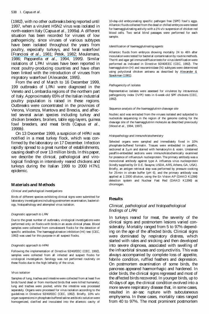
lesion that was present in birds from a largemajority of the affected flocks was the presence ofa fibrinous clot in the sinuses (Figure 1) andtrachea, which appeared in most cases to havecaused death by asphyxiation. The trachea andlungs appeared to be congested and, in some cases,haemorrhagic. Petechial haemorrhages were alsopresent on the epicardium in some cases.
Histologically, the only organ that exhibitedspecific lesions and was seen to be consistentlyaffected was the pancreas, in which focal pancreaticacinar cells necrosis was observed. Avian influenzavirus nucleoprotein was identified in necrotic acinarepithelium of the pancreas.
A milder form of the same clinical condition wasalso observed in infected turkey breeders whoconsistently exhibited rales, coughing, swelling ofthe infraorbital sinuses and a febrile conditionassociated with loss of appetite. Egg productiondecreased from 30 to 80% during the acute phase ofthe disease, but partially recovered to subnormallevels within 3 weeks from the onset of the disease.Egg quality also decreased, with misshapen, fragileand whitish eggs being produced during the drop inegg production. Mortality rates ranged from 5 to20%, while morbidity rates reached 100%. Thepostmortem findings were characterized by conges-tion of lung and trachea, sinusitis and conjunctivitis,and often the so-called ‘egg-yolk peritonitis’ couldbe seen.
Only a limited number of broiler and broilerbreeder flocks was affected. Typically, in broilerbreeders, a drop in egg production of 5 to 20%followed an initial loss of appetite. During thisphase, cyanosis of the combs and wattles could beseen. Morbidity approached 100% and mortalityranged from 3 to 8%. Similar to the turkey breeders,misshapen eggs were also laid in considerablequantities. Pathological findings were restricted tothe ovary and oviduct with colliquation of ovarian
follicles, associated with catarrhal or fibrinousperitonitis. The only other lesion that appeared to bepresent consistently was congestion of lungs andtracheas, which in some cases appeared as pulmo-nary oedema.
In broiler chickens, H7N1 infections were inap-parent in some flocks and characterized in others byanorexia and mild respiratory signs with mortalityrates that were generally low, in the order of 2 to3%. In one case, mortality reached 20%. Post-mortem lesions were limited to the lungs andtracheas, which appeared congested with associatedcatarrhal tracheitis.
Outbreaks in commercial layers were similar tothose observed in broiler breeders. Initial signswere of loss of appetite and depression, followed bya decrease in egg production that generally rangedfrom 3 to 10% but in some cases reached 30%.Recovery to levels seen before the disease wasobtained in only a few cases, while egg productionin most flocks remained 2 to 3% below expectedlevels. Clinical signs were usually present in about20% of the birds, but mortality never exceeded 5%.Gross lesions mainly involved the reproductiveorgans and abdominal cavity. The ovary andoviduct appeared oedematous and, on opening theoviduct, it contained a catarrhal exudate and fibrinclots, often associated with fibrinous and egg-yolkperitonitis. The lungs and tracheas appeared con-gested at times.
Clinical, pathological and histopathologicalfindings of HPAI
In all species and types of bird reared on litter,from the onset of the first clinical signs, anorexiaand depression, 100% mortality was observedwithin 48 to 72 h. The predominant, often theonly, clinical signs that could be seen wereneurological with incoordination and tremors pre-
H7N1 avian influenza in Italy 539
Figure 1. A 28-day-old poult exhibiting fibrin clots in the sinuses.

ceding death. In a limited number of broilerbreeder flocks, cyanosis of the comb and wattlesand petechial haemorrhages on the hock could beseen.
A different situation was noted in caged birds,such as layers, in which the disease eventuallykilled all the birds, but moved within the flock ina much slower way. At first, severe depression ormortality could be seen in only one bird per cagein a restricted area of the house, but slowlyspread to neighbouring cages. This differentbehaviour in spread between caged and litter-reared birds was probably related to the amountof infected faeces in direct contact with thebirds.
Similar to what had been observed with LPAI,the only postmortem lesion that was common tomost affected groups was pancreatitis (Figure 2).In chickens, the spleen occasionally presented
necrotic foci on its surface, and the caecal tonsilsappeared haemorrhagic. Generally, internal organsappeared congested and, in a limited number ofcases, affecting both turkeys and chickens, uratedeposits could be seen in the kidney.
Histologically, pancreatitis with severe, focal todiffuse necrosis of acinar cells was the mainfinding. Pancreatic lobes exhibited strong irregu-lar eosinophilic staining caused by acinar necrosis(Figure 3) and the most severe necrotic foci werelined by a thin rim of inflammatory cell debris.Interstitial oedema was also present, associatedwith fibrinous peritonitis affecting both pancreasand intestine. Spleen lesions were of vascularwall fibrinoid necrosis. No other relevant lesionwas detected elsewhere. Avian influenza virusnucleoprotein was identified in necrotic acinarepithelium of the pancreas, nervous and hearttissues.
540 I. Capua et al.
Figure 2. Viscera of turkey poult exhibiting pancreatitis.
Figure 3. Pancreas of turkey affected by HPAI. Extensive necrosis of acinar cells epithelium associated with lymphatic cellinflitration. Unaffected pancreatic tissue is also present.

Bacteriology
In the LPAI outbreaks, routine bacteriology oftenyielded bacteria such as Escherichia coli, Riemer-ella anatipestifer and Pasteurella multocida, whichwere considered as secondary agents. Negativeresults were obtained from all the cases of HPAI.
Virology and serology
Virus isolation attempts yielded haemagglutinatingagents on first passage, often accompanied by earlyembryo mortality (within 48 h), or were negative.Viruses were characterized serologically and allinfluenza isolates were of the H7N1 subtype.
IVPI tests performed on a number of LPAIisolates all gave a result of 0.0. The deduced aminoacid sequence of the region coding for the cleavagesite of the haemagglutinin molecule for the earlyLPAI isolates was. . .PEIPKGR*GLF. . ., while forsome later isolates it was. . .PEVKGR*GLF. . .,both sequences being typical of virus of lowpathogenicity.
In contrast, isolates obtained from the HPAIoutbreaks had an IVPI of 3.0 and a deduced aminoacid sequence . . .PEIPKGSRVRR*GLF. . ., whichcontains multiple basic amino acids, a featuretypical of highly pathogenic viruses.
Following the low-pathogenicity outbreaks, anti-bodies to H7 subtype were observed in all suspectedflocks. Generally speaking, the mean HI titre washigher for turkeys (1:128) than for chickens(1:32).
Discussion
Clinical and pathological data collected during theepidemic, indicate that turkeys infected with LPAIdevelop a more severe clinical condition whencompared with chickens. Similar evidence has beenreported in other episodes linked to different low-pathogenicity subtypes of avian influenza(Franciosi et al., 1981).
Results obtained in the H7N1 1999 to 200 Italianepidemic confirm evidence collected in other epi-demics, such as the Pennsylvania 1983 to 1984(Kawaoka et al., 1986; Eckroade & Silverman-Bachin, 1987) and the Mexico 1994 to 1995(Campos-Lopez et al., 1996; Senne et al., 1996;Villarreal & Flores, 1997) epidemics, in which itwas demonstrated that HPAI viruses emerged fromLPAI viruses. In all three of these epidemics, theearlier spread of virus of low virulence not onlyresulted in the now predictable mutation to a highlypathogenic virus, but the co-circulation of HPAIand LPAI in a partially immune population madeclinical and laboratory diagnosis difficult to inter-pret. This resulted in delays in the application oferadication measures.
A complex epidemiological situation occurredwith the LPAI, due to the lack of pathognomonic
clinical signs and lesions present in adult birds. Inaddition, the initial difficulty in correctly diagnos-ing the clinical condition led to a delay inidentifying the agent, thus resulting in a consider-able spread of infection in a relatively short time.With such a large number of infected flocks, it wasnot possible to implement a voluntary stamping-outpolicy.
The circulation of LPAI in a susceptible popula-tion resulted in the emergence of a highly patho-genic virus. Moreover, the possibility of humansbeing infected with LPAI (Kurtz et al., 1996; Peiriset al., 1999) places these viruses in a differentposition compared with that they held when Direc-tive 92/40/EEC was drafted. It would thereforeseem advisable to consider whether an eradicationpolicy should be recommended for LPAI of at leastH5 and H7 subtypes. However, stamping outsignificant numbers of infected flocks is onlyrealistically feasible if there is financial supportfrom local or national governments, or from theEuropean Union. At present, with current legisla-tion, this appears to be inapplicable for LPAI.
In order to control the clinical condition asso-ciated with LPAI, an alternative to a stamping-outpolicy is a vaccination strategy. This has severalassociated problems; for example, it may delayeradication, since it does not prevent the virus fromreplicating (Swayne et al., 1999) and is alsotherefore useless from a biological point of view, asHPAI could still emerge by mutation. Furthermore,it would have dramatic implications on intra- andextra-community trade within the currentlegislation.
A reconsideration of European Union legislationcould possibly be a solution to a number ofproblems that have emerged during the Italian 1999to 2000 H7N1 epidemic and in other recentepidemics, as it would appear that control of LPAIoutbreaks caused by H5 and H7 is essential in orderto avoid heavy economic losses to the poultryindustry.
Acknowledgements
The authors wish to thank the staff of the VirologyDepartment of the Istituto Zooprofilattico Sper-imentale delle Venezie and Ruth Manvell, CVLWeybridge for their invaluable technical assistance.Jill Banks, CVL Weybridge and Ernesto TisatoBiotechnology Laboratory Istituto ZooprofilatticoSperimentale delle Venezie are gratefully acknowl-edged for sequencing the isolates. The authors alsowish to thank Dr D.E. Swayne for kindly supplyingthe monoclonal antibody.
References
Alexander, D.J. (1995). The epidemiology and control of avianinfluenza and Newcastle disease – review article. Journal ofComparative Pathology, 112, 105–126.
H7N1 avian influenza in Italy 541

Alexander, D.J. & Spackman, D. (1981). Characterization of influenzaA viruses isolated from turkeys in England during March–May1979. Avian Pathology, 10, 281–293.
Campos-Lopez, H., Rivera-Cruz, E. & Irastorza-Enrich, M. (1996).Situacion y perspectivas del programa de erradicacon de la influenzaaviar en Mexico. Proceedings of the 45th Western Poultry DiseaseConference May 1996 (pp. 13–16). Cancun, Mexico.
Capua, I., Marangon, S., Selli, L., Alexander, D.J., Swayne, D.E., DallaPozza, M., Parenti E. & Cancellotti, F.M. (1999a). Outbreaks ofhighly pathogenic avian influenza (H5N2) in Italy during October(1997) to January (1998). Avian Pathology, 28, 455–460.
Capua, I., Marangon, S., Dalla Pozza, M., Mutinelli, F., Vincenzi, G. &Santucci, U. (1999b). The low pathogenicity avian influenza (H7N1)epidemic in the Veneto region, Italy. Proceedings of the Sixth JointAnnual Meeting of the EU Reference Laboratories for NewcastleDisease and Avian Influenza 29–30 November 1999, pp. 66–72.Brussels, Belgium.
CEC (1992). Council Directive 92/40/EEC of 19 May 1992 introducingCommunity measures for the control of avian influenza. OfficialJournal of the European Commission, L167, 1–15.
Eckroade, R.J. & Silverman-Bachin, L.A. (1987). Avian influenza inPennsylvania – the beginning. In D.E. Swayne & R.D. Slemons(Eds.), Proceedings of the Second International Symposium on AvianInfluenza (pp. 22–32). US Animal Health Association, GeorgiaCenter for Continuing Education, The University of Georgia, Athens,GA, USA.
Franciosi, C., D’Aprile, P.N., Alexander, D.J. & Petek, M. (1981).Influenza A virus infections in commercial turkeys in North EastItaly. Avian Pathology, 10, 303–311.
Garcia, M., Crawford, J.M., Latimer, J.W., Rivera-Cruz, E. & Perdue,M.L. (1996). Heterogeneity in the haemagglutinin gene andemergence of the highly pathogenic phenotype among recent H5N2avian influenza viruses from Mexico. Journal of General Virology,77, 1493–1504.
Kawaoka, Y., Bean, W.J. & Webster, R.G. (1986). Molecular character-isation of the A/Chicken/Pennsylvania/83 (H5N2) influenza viruses.In D.E. Swayne & R.D. Slemons (Eds.), Proceedings of the SecondInternational Symposium on Avian Influenza (pp. 197–206). USAnimal Health Association, Georgia Center for Continuing Educa-tion, The University of Georgia, Athens, GA, USA.
Kurtz, J., Manvell, R.J. & Banks, J. (1996).Avian influenza virusisolated from a woman with conjunctivitis. The Lancet, 348,901–902.
Meulemans, G. (1986). Status of avian influenza in WesternEurope. In D.E. Swayne & R.D. Slemons (Eds.), Proceedingsof the Second International Symposium on Avian Influenza(pp. 77–83). US Animal Health Association, Georgia Center forContinuing Education, The University of Georgia, Athens, GA,USA.
Papparella, V., Fioretti, A. & Menna, L.F. (1994). The epidemiologicalsituation of avian influenza in Italy from 1990 to 1993 in feral birdpopulations and in birds in quarantine. Proceedings of the Joint FirstAnnual Meetings of the National Newcastle Disease and AvianInfluenza Laboratories of Countries of the European Communities1993 (pp. 19–21). Brussels, Belgium.
Papparella, V., Fioretti, A. & Menna, L.F. (1995). The epidemiologicalsituation of avian influenza in Italy 1994. Proceedings of the JointSecond Annual Meetings of the National Newcastle Disease andAvian Influenza Laboratories of Countries of the European Union1994 (pp. 14–15). Brussels, Belgium.
Peiris, M., Yuen, K.Y., Leung, C.W., Chan, K.H., Ip, P.L.S., Lai,R.W.M., Orr, W.K. & Shortridge, K.F. (1999). Human infection withinfluenza H9N2. The Lancet, 354, 916–917.
Perdue, M.L., Garcia, M., Senne, D. & Fraire, M. (1997). Virulence-associated sequence duplication at the hemagglutinin cleavage site ofavian influenza viruses. Virus Research, 49, 173–186.
Petek, M. (1982). Current situation in Italy. Proceedings of the FirstInternational Symposium on Avian Influenza 1981 (pp. 31–34).Carter Composition Corporation, Richmond, VA, USA.
Perroncito, E. (1878). Epizoozia tifoide nei gallinacei. Annali Accade-mia Agricoltura Torino, 21, 87–126.
Rott, R. (1992). The pathogenic determinant of influenza virus.Veterinary Microbiology, 33, 303–310.
Senne, D.A., Rivera, E., Panigahy, B., Fraire, M., Kawaoaka, Y. &Webster, R.G. (1996). Characterization of avian influenza H5N2isolates recovered from chickens in Mexico. Proceedings of the 45thWestern Poultry Disease Conference May 1996 (pp. 5–8). Cancun,Mexico.
Swayne, D.E., Beck, J.R., Garcia, M. & Stone, H.D. (1999). Influenceof virus strain and antigenic mass on efficacy of H5 avian influenzainactivated vaccines. Avian Pathology, 28, 245–255.
Villarreal, C.L. & Flores, A.O. (1997). The Mexican avian influenzaH5N2 outbreak. In D.E. Swayne & R.D. Slemons (Eds.), Proceed-ings of the Fourth International Symposium on Avian Influenza,Athens Georgia (pp 18–22). US Animal Health Association, GeorgiaCenter for Continuing Education, The University of Georgia, Athens,GA, USA.
Wood, G.W., Banks, J., McCauley, J.W. & Alexander, D.J. (1994).Deduced amino acid sequences of the haemagglutinin of H5N1 avianinfluenza virus isolates from an outbreak in turkeys in Norfolk,England. Archives of Virology, 134, 185–194.
Wood, G.W., Banks J., Brown, I.H., Strong I. & Alexander, D.J. (1997).The nucleotide sequence of the HA1 of the haemagglutinin of an H1avian influenza isolate from turkeys in Germany provides additionalevidence suggesting recent transmission from pigs. Avian Pathology,26, 347–355.
542 I. Capua et al.
RESUME
Influenza aviaire H7N1 dans les elevages intensifs de poulets etde dindes en Italie (1999–2000)
Un episode d’influenza aviaire de faible pathogenicite (LPAI) a affectel’industrie avicole au nord de l’Italie, de la fin mars au debut decembre1999. Le virus responsable de l’epidemie etait de sous-type H7N1 avecun indice de pathogenicite intraveineuse (IVPI) de 0,0 et une sequenceen acides amines de la region codant pour le site de clivage del’hemaglutinine, caracteristique des virus de faible pathogenicite. Lacirculation du virus dans une population sensible pendant plusieursmois a permis l’emergence d’un virus tres pathogene avec un IVPI de3,0 et qui presente des acides amines basiques multiples au niveau dusite de clivage de l’hemaglutinine. Plus de treize millions d’oiseaux ontete affectes par l’epidemie et dans cet article nous reportons lesresultats des investigations cliniques, virologiques et histopatholo-giques realisees a partir des poulets et des dindes affectes. Les lesionsmacroscopiques et microscopiques induites par la LPAI ont ete plusseveres chez les dindes que chez les poulets, alors que l’HPAI aentraõ ne des taux similaires de mortalite chez les deux especes. Lalegislation europeenne actuelle considere la LPAI et la HPAI commedeux maladies totalement distinctes. Pour la LPAI aucune politiqued’eradication obligatoire n’est exigee, alors que les mesures afferentessont mises en œuvre pour la HPAI. Lors de l’epidemie italienne de1999–2000, la LPAI a mute en HPAI dans une zone ou la populationest dense, en entra õ nant de tres fortes pertes economiques. Unereconsideration de la legislation europeenne actuelle en matiered’influenza aviaire incluant les sous-types H5 et H7 de la LPAI seraitune aide possible afin d’eviter les epidemies devastatrices pourl’aviculture industrielle.
ZUSAMMENFASSUNG
H7N1-Geflugelinfluenza in Italien (1999–2000) bei intensivaufgezogenen Huhnern und Puten
Von Ende Marz bis Anfang Dezember 1999 wurde die industrielleGeflugelpopulation in Norditalien von einer Epidemie schwachpathogener aviarer Influenza (LPAI) heimgesucht. Das fur die Epide-mie verantwortliche Virus wurde als H7N1 subtypisiert. Es hatte einenintravenosen Pathogenitatsindex (IVPI) von 0,0 und eine fur schwachpathogene Viren typische Aminosauresequenz der fur die Spaltungs-stelle des Hamagglutininmolekuls kodierenden Region. Die mehrereMonate lange Zirkulation des Virus in einer empfanglichen Populationbewirkte die Entstehung eines hoch pathogenen Virus mit einem IVPIvon 3,0 und mit multiplen basischen Aminosauren in der Aminosaur-esequenz fur die Spaltungsstelle des Hamagglutininmolekuls. Uberdreizehn Millionen Tiere waren von der Epidemie betroffen. In dervorliegenden Arbeit beschreiben wir die Ergebnisse der bei den

erkrankten Huhnern und Puten durchgefuhrten klinischen, virolo-gischen und histopathologischen Untersuchungen. Die durch die LPAIverursachten klinischen Symptome und makroskopischen und mikros-kopischen pathologischen Veranderungen waren bei den Puten starkerals bei den Huhnern, wahrend die stark pathogene aviare Influenza(HPAI) bei beiden Spezies zu etwa gleich hohen Mortalitatsratenfuhrte. Die gegenwartige europaische Gesetzgebung sieht LPAI undHPAI als zwei vollkommen verschiedene Krankheiten an und fordertkeine obligatorischen Tilgungsmaßnahmen in Bezug auf LPAI, ver-langt aber im Falle von HPAI die Eradikation. Bei der italienischenEpidemie von 1999–2000 mutierte LPAI in einem Gebiet mit einerdichten Geflugelpopulation zu HPAI, was große wirtschaftlicheVerluste verursachte. Ein Uberdenken der gegenwartigen EU-Gesetz-gebung uber die Geflugel-Influenza unter Einbeziehung von LPAI derSubtypen H5 und H7 konnte fur die Geflugelindustrie moglicherweiseeine Hilfe zum Vermeiden von verheerenden Epidemien sein.
RESUMEN
Influenza aviar H7N1 en Italia (1999–2000) en pollos y pavos deexplotaciones intensivas
Desde el final de marzo hasta el principio de diciembre de 1999, unaepidemia de influenza aviar de baja patogenicidad (LPAI) afecto a la
industria av õ cola del norte de Italia. El virus responsable de estaepidemia se clasifico como subtipo H7N1 con un õ ndice de patogenici-dad intravenosa (IVPI) de 0.0, y una secuencia de aminoacidos de laregion que codifica para el punto de escision de la molecula dehemaglutinina tõ pica de virus de baja patogenicidad. La circulacion delvirus en una poblacion susceptible durante varios meses, causo laaparicion de un virus de alta patogenicidad con un IVPI de 3.0 y lapresencia de multiples aminoacidos basicos en la secuencia deaminoacidos del punto de escision de la molecula de hemaglutinina. Laepidemia afecto a unos trece millones de aves y en el presente art õ culose describen los resultados de los estudios cl´õ nicos, virologicos ehistopatologicos realizados en pollos y pavos infectados.
Los s õ ntomas cl õ nicos, lesiones macroscopicas y microscopicascausadas por la LPAI fueron mas severas en pavos que en pollos,mientras que la HPAI causo una mortalidad similar en ambas especies.La actual legislacion europea considera la LPAI y la HPAI como dosenfermedades completamente diferentes, en tanto que no se requiere laerradicacion para la LPAI, pero imponen una polõ tica de erradicacionen el caso de la HPAI. En la epidemia italiana de 1999–2000, la LPAImuto a HPAI en un area densamente poblada y causo grandes perdidaseconomicas. La reconsideracion de la legislacion europea actualreferida a influenza aviar, incluyendo en ello la LPAI de los subtiposH5 y H7 podrõ a posiblemente ayudar a evitar epidemias devastadoraspara la industria av õ cola.
H7N1 avian influenza in Italy 543
Copyright © 2022 FDOKUMEN