Guidelines for Diagnosis and Management of ... - J-Stage
42
Circulation Journal Official Journal of the Japanese Circulation Society http://www.j-circ.or.jp adolescence or later, and the occurrence of acute coronary syndrome in adults with a history of Kawasaki disease in whom coronary artery lesions were considered regressed after the acute phase of the disease. We also have to address the prob- lem of susceptibility to atherosclerosis in patients with a his- tory of vasculitis due to Kawasaki disease. Because the pathophysiology of cardiovascular sequelae in Kawasaki disease changes over time during childhood, ado- lescence and adulthood, guidance on the diagnosis, treatment and management of cardiovascular sequelae in different ages Forty-five years have passed since 1967, 1 when the first case series of Kawasaki disease was reported. Currently, more than half of the patients diagnosed with Kawasaki disease are 20 years of age or older. As this timeline suggests, it is expected that more than 10,000 patients with cardiovascular sequelae in Kawasaki disease have reached adulthood. However, since Kawasaki disease develops most frequently by around 1 year of age, many internists are still not familiar with it (See Table 3). Recent issues on Kawasaki disease include the high percentage of patients who stop visiting their clinic in early Introduction of the Revised Guidelines Released online September 22, 2014 Mailing address: Scientific Committee of the Japanese Circulation Society, 18F Imperial Hotel Tower, 1-1-1 Uchisaiwai-cho, Chiyoda-ku, Tokyo 100-0011, Japan. E-mail: [email protected] This English language document is a revised digest version of Guidelines for diagnosis and management of cardiovascular sequelae in Kawasaki disease reported at the Japanese Circulation Society Joint Working Groups performed in 2012 (Website: http://www.j-circ. or.jp/guideline/pdf/JCS2013_ogawas_d.pdf). Joint Working Groups: the Japanese Circulation Society, The Japanese Society of Kawasaki Disease, The Japanese Association for Tho- racic Surgery, The Japan Pediatric Society, The Japanese Society of Pediatric Cardiology and Cardiac Surgery, The Japanese College of Cardiology ISSN-1346-9843 doi:10.1253/circj.CJ-66-0096 All rights are reserved to the Japanese Circulation Society. For permissions, please e-mail: [email protected] Guidelines for Diagnosis and Management of Cardiovascular Sequelae in Kawasaki Disease (JCS 2013) – Digest Version – JCS Joint Working Group Table of Contents Introduction of the Revised Guidelines ∙∙∙∙∙∙∙∙∙∙∙∙∙∙∙∙∙∙∙∙∙∙∙∙ 1 I Epidemiology of Kawasaki Disease, Current Acute-Phase Treatment, and Pathophysiology of Acute-Phase Disease ∙∙∙∙∙∙∙∙∙∙∙∙∙∙∙∙∙∙∙∙∙∙∙∙∙∙∙∙∙∙∙∙∙∙∙∙∙∙∙∙∙∙∙∙∙∙∙∙∙ 3 1. Current Epidemiology ∙∙∙∙∙∙∙∙∙∙∙∙∙∙∙∙∙∙∙∙∙∙∙∙∙∙∙∙∙∙∙∙∙∙∙∙∙∙∙∙∙∙∙∙∙∙∙∙∙∙∙∙∙∙∙∙ 3 2. Genetic Background of Kawasaki Disease ∙∙∙∙∙∙∙∙∙∙∙∙∙∙∙∙∙∙∙∙∙∙∙ 5 3. Severity Classification ∙∙∙∙∙∙∙∙∙∙∙∙∙∙∙∙∙∙∙∙∙∙∙∙∙∙∙∙∙∙∙∙∙∙∙∙∙∙∙∙∙∙∙∙∙∙∙∙∙∙∙∙∙∙∙ 7 4. Diagnosis and Treatment of Incomplete Kawasaki Disease∙∙∙∙∙∙∙∙∙∙∙∙∙∙∙∙∙∙∙∙∙∙∙∙∙∙∙∙∙∙∙∙∙∙∙∙∙∙∙∙∙∙∙∙∙∙∙∙∙∙∙∙∙∙∙∙∙∙∙∙∙∙∙∙∙∙∙∙∙∙∙∙∙∙∙∙∙∙ 7 II Genetic Background and Pathology of Cardiovascular Sequelae and Coronary Hemodynamics ∙∙∙∙∙∙∙∙∙∙∙∙∙∙∙∙∙∙∙∙∙∙∙∙∙∙∙∙∙∙∙∙∙∙∙∙∙∙∙∙∙∙∙∙∙∙∙∙∙∙∙∙∙∙∙∙∙∙∙∙∙∙∙∙ 7 1. Genetic Background of Cardiovascular Sequelae ∙∙∙∙∙∙∙∙∙∙∙∙ 7 2. Pathology of Cardiovascular Sequelae ∙∙∙∙∙∙∙∙∙∙∙∙∙∙∙∙∙∙∙∙∙∙∙∙∙∙∙∙∙ 8 3. Coronary Hemodynamics in Patients With Coronary Sequelae ∙∙∙∙∙∙∙∙∙∙∙∙∙∙∙∙∙∙∙∙∙∙∙∙∙∙∙∙∙∙∙∙∙∙∙∙∙∙∙∙∙∙∙∙∙∙∙∙∙∙∙∙∙∙∙∙∙∙∙∙∙∙∙∙∙∙∙∙∙∙∙∙∙∙∙ 8 III Examinations and Diagnosis of Cardiovascular Sequelae ∙∙∙∙∙∙∙∙∙∙∙∙∙∙∙∙∙∙∙∙∙∙∙∙∙∙∙∙∙∙∙∙∙∙∙∙∙∙∙∙∙∙∙∙∙ 9 1. Blood Test, Biomarkers and Arteriosclerosis ∙∙∙∙∙∙∙∙∙∙∙∙∙∙∙∙∙∙∙∙ 9 2. Physiological Examinations ∙∙∙∙∙∙∙∙∙∙∙∙∙∙∙∙∙∙∙∙∙∙∙∙∙∙∙∙∙∙∙∙∙∙∙∙∙∙∙∙∙∙∙∙∙ 11 3. Diagnostic Imaging ∙∙∙∙∙∙∙∙∙∙∙∙∙∙∙∙∙∙∙∙∙∙∙∙∙∙∙∙∙∙∙∙∙∙∙∙∙∙∙∙∙∙∙∙∙∙∙∙∙∙∙∙∙∙∙∙∙∙ 12 4. Cardiac Catheterization ∙∙∙∙∙∙∙∙∙∙∙∙∙∙∙∙∙∙∙∙∙∙∙∙∙∙∙∙∙∙∙∙∙∙∙∙∙∙∙∙∙∙∙∙∙∙∙∙∙∙∙ 14 5. Summary of Examinations and Diagnosis ∙∙∙∙∙∙∙∙∙∙∙∙∙∙∙∙∙∙∙∙∙∙ 15 IV Treatment of Cardiovascular Sequelae∙∙∙∙∙∙∙∙∙∙∙∙∙∙∙∙∙ 16 1. Pharmacotherapy ∙∙∙∙∙∙∙∙∙∙∙∙∙∙∙∙∙∙∙∙∙∙∙∙∙∙∙∙∙∙∙∙∙∙∙∙∙∙∙∙∙∙∙∙∙∙∙∙∙∙∙∙∙∙∙∙∙∙∙∙ 16 2. Non-Pharmacotherapy ∙∙∙∙∙∙∙∙∙∙∙∙∙∙∙∙∙∙∙∙∙∙∙∙∙∙∙∙∙∙∙∙∙∙∙∙∙∙∙∙∙∙∙∙∙∙∙∙∙∙∙∙ 20 3. Summary of Treatment Options ∙∙∙∙∙∙∙∙∙∙∙∙∙∙∙∙∙∙∙∙∙∙∙∙∙∙∙∙∙∙∙∙∙∙∙∙∙∙ 23 V Management and Follow-up During Childhood∙∙∙∙∙∙∙∙∙∙∙∙∙∙∙∙∙∙∙∙∙∙∙∙∙∙∙∙∙∙∙∙∙∙∙∙∙∙∙∙∙∙∙∙∙∙∙∙∙∙∙∙∙∙∙∙∙∙∙∙∙∙∙∙∙∙∙∙∙∙∙∙ 23 1. Guidance on Activities of Daily Life and Exercise ∙∙∙∙∙∙∙∙∙∙∙ 23 2. Follow-up Evaluation ∙∙∙∙∙∙∙∙∙∙∙∙∙∙∙∙∙∙∙∙∙∙∙∙∙∙∙∙∙∙∙∙∙∙∙∙∙∙∙∙∙∙∙∙∙∙∙∙∙∙∙∙∙∙∙ 28 3. Problems in Shifting From Childhood to Adulthood ∙∙∙∙∙∙∙∙ 30 VI Problems During Adulthood∙∙∙∙∙∙∙∙∙∙∙∙∙∙∙∙∙∙∙∙∙∙∙∙∙∙∙∙∙∙∙∙∙∙∙∙∙ 30 1. Progression of Arteriosclerosis: Pathological Features∙∙∙∙∙∙∙∙∙∙∙∙∙∙∙∙∙∙∙∙∙∙∙∙∙∙∙∙∙∙∙∙∙∙∙∙∙∙∙∙∙∙∙∙∙∙∙∙∙∙∙∙∙∙∙∙∙∙∙∙∙∙∙∙∙∙∙∙∙∙∙∙∙∙∙ 30 2. Progression of Arteriosclerosis: Clinical Features∙∙∙∙∙∙∙∙∙∙∙ 30 VII Management of Adults With a History of Kawasaki Disease∙∙∙∙∙∙∙∙∙∙∙∙∙∙∙∙∙∙∙∙∙∙∙∙∙∙∙∙∙∙∙∙∙∙∙∙∙∙∙∙∙∙∙∙∙∙∙∙∙∙∙∙∙∙∙ 31 1. Diagnosis ∙∙∙∙∙∙∙∙∙∙∙∙∙∙∙∙∙∙∙∙∙∙∙∙∙∙∙∙∙∙∙∙∙∙∙∙∙∙∙∙∙∙∙∙∙∙∙∙∙∙∙∙∙∙∙∙∙∙∙∙∙∙∙∙∙∙∙∙∙∙∙∙∙ 31 2. Treatment ∙∙∙∙∙∙∙∙∙∙∙∙∙∙∙∙∙∙∙∙∙∙∙∙∙∙∙∙∙∙∙∙∙∙∙∙∙∙∙∙∙∙∙∙∙∙∙∙∙∙∙∙∙∙∙∙∙∙∙∙∙∙∙∙∙∙∙∙∙∙∙∙ 31 3. Guidance on Lifestyle and Exercise ∙∙∙∙∙∙∙∙∙∙∙∙∙∙∙∙∙∙∙∙∙∙∙∙∙∙∙∙∙∙∙∙ 32 4. Pregnancy, Labor and Childbirth ∙∙∙∙∙∙∙∙∙∙∙∙∙∙∙∙∙∙∙∙∙∙∙∙∙∙∙∙∙∙∙∙∙∙∙∙∙ 32 5. Healthcare System for Adult Patients∙∙∙∙∙∙∙∙∙∙∙∙∙∙∙∙∙∙∙∙∙∙∙∙∙∙∙∙∙∙ 34 VIII Summary ∙∙∙∙∙∙∙∙∙∙∙∙∙∙∙∙∙∙∙∙∙∙∙∙∙∙∙∙∙∙∙∙∙∙∙∙∙∙∙∙∙∙∙∙∙∙∙∙∙∙∙∙∙∙∙∙∙∙∙∙∙∙∙∙∙∙∙∙∙∙ 35 References∙∙∙∙∙∙∙∙∙∙∙∙∙∙∙∙∙∙∙∙∙∙∙∙∙∙∙∙∙∙∙∙∙∙∙∙∙∙∙∙∙∙∙∙∙∙∙∙∙∙∙∙∙∙∙∙∙∙∙∙∙∙∙∙∙∙∙∙∙∙∙∙∙∙∙ 35 Appendix ∙∙∙∙∙∙∙∙∙∙∙∙∙∙∙∙∙∙∙∙∙∙∙∙∙∙∙∙∙∙∙∙∙∙∙∙∙∙∙∙∙∙∙∙∙∙∙∙∙∙∙∙∙∙∙∙∙∙∙∙∙∙∙∙∙∙∙∙∙∙∙∙∙∙∙∙∙∙ 42 JCS GUIDELINES Advance Publication by-J-STAGE
-
Upload
khangminh22 -
Category
Documents
-
view
6 -
download
0
Transcript of Guidelines for Diagnosis and Management of ... - J-Stage

Circulation JournalOfficial Journal of the Japanese Circulation Societyhttp://www.j-circ.or.jp
adolescence or later, and the occurrence of acute coronary syndrome in adults with a history of Kawasaki disease in whom coronary artery lesions were considered regressed after the acute phase of the disease. We also have to address the prob-lem of susceptibility to atherosclerosis in patients with a his-tory of vasculitis due to Kawasaki disease.
Because the pathophysiology of cardiovascular sequelae in Kawasaki disease changes over time during childhood, ado-lescence and adulthood, guidance on the diagnosis, treatment and management of cardiovascular sequelae in different ages
Forty-five years have passed since 1967,1 when the first case series of Kawasaki disease was reported. Currently, more than half of the patients diagnosed with Kawasaki disease are 20 years of age or older. As this timeline suggests, it is expected that more than 10,000 patients with cardiovascular sequelae in Kawasaki disease have reached adulthood. However, since Kawasaki disease develops most frequently by around 1 year of age, many internists are still not familiar with it (See Table 3). Recent issues on Kawasaki disease include the high percentage of patients who stop visiting their clinic in early
Introduction of the Revised Guidelines
Released online September 22, 2014Mailing address: Scientific Committee of the Japanese Circulation Society, 18F Imperial Hotel Tower, 1-1-1 Uchisaiwai-cho, Chiyoda-ku,
Tokyo 100-0011, Japan. E-mail: [email protected] English language document is a revised digest version of Guidelines for diagnosis and management of cardiovascular sequelae in
Kawasaki disease reported at the Japanese Circulation Society Joint Working Groups performed in 2012 (Website: http://www.j-circ.or.jp/guideline/pdf/JCS2013_ogawas_d.pdf).
Joint Working Groups: the Japanese Circulation Society, The Japanese Society of Kawasaki Disease, The Japanese Association for Tho-racic Surgery, The Japan Pediatric Society, The Japanese Society of Pediatric Cardiology and Cardiac Surgery, The Japanese College of Cardiology
ISSN-1346-9843 doi: 10.1253/circj.CJ-66-0096All rights are reserved to the Japanese Circulation Society. For permissions, please e-mail: [email protected]
Guidelines for Diagnosis and Management of Cardiovascular Sequelae in Kawasaki Disease (JCS 2013)
– Digest Version –JCS Joint Working Group
Table of ContentsIntroduction of the Revised Guidelines ∙∙∙∙∙∙∙∙∙∙∙∙∙∙∙∙∙∙∙∙∙∙∙∙ 1I Epidemiology of Kawasaki Disease, Current
Acute-Phase Treatment, and Pathophysiology of Acute-Phase Disease ∙∙∙∙∙∙∙∙∙∙∙∙∙∙∙∙∙∙∙∙∙∙∙∙∙∙∙∙∙∙∙∙∙∙∙∙∙∙∙∙∙∙∙∙∙∙∙∙∙ 3
1. Current Epidemiology ∙∙∙∙∙∙∙∙∙∙∙∙∙∙∙∙∙∙∙∙∙∙∙∙∙∙∙∙∙∙∙∙∙∙∙∙∙∙∙∙∙∙∙∙∙∙∙∙∙∙∙∙∙∙∙∙ 3 2. Genetic Background of Kawasaki Disease ∙∙∙∙∙∙∙∙∙∙∙∙∙∙∙∙∙∙∙∙∙∙∙ 5 3. Severity Classification ∙∙∙∙∙∙∙∙∙∙∙∙∙∙∙∙∙∙∙∙∙∙∙∙∙∙∙∙∙∙∙∙∙∙∙∙∙∙∙∙∙∙∙∙∙∙∙∙∙∙∙∙∙∙∙ 7 4. Diagnosis and Treatment of Incomplete Kawasaki
Disease ∙∙∙∙∙∙∙∙∙∙∙∙∙∙∙∙∙∙∙∙∙∙∙∙∙∙∙∙∙∙∙∙∙∙∙∙∙∙∙∙∙∙∙∙∙∙∙∙∙∙∙∙∙∙∙∙∙∙∙∙∙∙∙∙∙∙∙∙∙∙∙∙∙∙∙∙∙∙ 7II Genetic Background and Pathology of
Cardiovascular Sequelae and Coronary Hemodynamics ∙∙∙∙∙∙∙∙∙∙∙∙∙∙∙∙∙∙∙∙∙∙∙∙∙∙∙∙∙∙∙∙∙∙∙∙∙∙∙∙∙∙∙∙∙∙∙∙∙∙∙∙∙∙∙∙∙∙∙∙∙∙∙∙ 7
1. Genetic Background of Cardiovascular Sequelae ∙∙∙∙∙∙∙∙∙∙∙∙ 7 2. Pathology of Cardiovascular Sequelae ∙∙∙∙∙∙∙∙∙∙∙∙∙∙∙∙∙∙∙∙∙∙∙∙∙∙∙∙∙ 8 3. Coronary Hemodynamics in Patients With Coronary
Sequelae ∙∙∙∙∙∙∙∙∙∙∙∙∙∙∙∙∙∙∙∙∙∙∙∙∙∙∙∙∙∙∙∙∙∙∙∙∙∙∙∙∙∙∙∙∙∙∙∙∙∙∙∙∙∙∙∙∙∙∙∙∙∙∙∙∙∙∙∙∙∙∙∙∙∙∙ 8III Examinations and Diagnosis of
Cardiovascular Sequelae ∙∙∙∙∙∙∙∙∙∙∙∙∙∙∙∙∙∙∙∙∙∙∙∙∙∙∙∙∙∙∙∙∙∙∙∙∙∙∙∙∙∙∙∙∙ 9 1. Blood Test, Biomarkers and Arteriosclerosis ∙∙∙∙∙∙∙∙∙∙∙∙∙∙∙∙∙∙∙∙ 9 2. Physiological Examinations ∙∙∙∙∙∙∙∙∙∙∙∙∙∙∙∙∙∙∙∙∙∙∙∙∙∙∙∙∙∙∙∙∙∙∙∙∙∙∙∙∙∙∙∙∙ 11 3. Diagnostic Imaging ∙∙∙∙∙∙∙∙∙∙∙∙∙∙∙∙∙∙∙∙∙∙∙∙∙∙∙∙∙∙∙∙∙∙∙∙∙∙∙∙∙∙∙∙∙∙∙∙∙∙∙∙∙∙∙∙∙∙ 12 4. Cardiac Catheterization ∙∙∙∙∙∙∙∙∙∙∙∙∙∙∙∙∙∙∙∙∙∙∙∙∙∙∙∙∙∙∙∙∙∙∙∙∙∙∙∙∙∙∙∙∙∙∙∙∙∙∙ 14 5. Summary of Examinations and Diagnosis ∙∙∙∙∙∙∙∙∙∙∙∙∙∙∙∙∙∙∙∙∙∙ 15IV Treatment of Cardiovascular Sequelae ∙∙∙∙∙∙∙∙∙∙∙∙∙∙∙∙∙ 16 1. Pharmacotherapy ∙∙∙∙∙∙∙∙∙∙∙∙∙∙∙∙∙∙∙∙∙∙∙∙∙∙∙∙∙∙∙∙∙∙∙∙∙∙∙∙∙∙∙∙∙∙∙∙∙∙∙∙∙∙∙∙∙∙∙∙ 16
2. Non-Pharmacotherapy ∙∙∙∙∙∙∙∙∙∙∙∙∙∙∙∙∙∙∙∙∙∙∙∙∙∙∙∙∙∙∙∙∙∙∙∙∙∙∙∙∙∙∙∙∙∙∙∙∙∙∙∙ 20 3. Summary of Treatment Options ∙∙∙∙∙∙∙∙∙∙∙∙∙∙∙∙∙∙∙∙∙∙∙∙∙∙∙∙∙∙∙∙∙∙∙∙∙∙ 23V Management and Follow-up During
Childhood ∙∙∙∙∙∙∙∙∙∙∙∙∙∙∙∙∙∙∙∙∙∙∙∙∙∙∙∙∙∙∙∙∙∙∙∙∙∙∙∙∙∙∙∙∙∙∙∙∙∙∙∙∙∙∙∙∙∙∙∙∙∙∙∙∙∙∙∙∙∙∙∙ 23 1. Guidance on Activities of Daily Life and Exercise ∙∙∙∙∙∙∙∙∙∙∙ 23 2. Follow-up Evaluation ∙∙∙∙∙∙∙∙∙∙∙∙∙∙∙∙∙∙∙∙∙∙∙∙∙∙∙∙∙∙∙∙∙∙∙∙∙∙∙∙∙∙∙∙∙∙∙∙∙∙∙∙∙∙∙ 28 3. Problems in Shifting From Childhood to Adulthood ∙∙∙∙∙∙∙∙ 30VI Problems During Adulthood∙∙∙∙∙∙∙∙∙∙∙∙∙∙∙∙∙∙∙∙∙∙∙∙∙∙∙∙∙∙∙∙∙∙∙∙∙ 30 1. Progression of Arteriosclerosis: Pathological
Features∙∙∙∙∙∙∙∙∙∙∙∙∙∙∙∙∙∙∙∙∙∙∙∙∙∙∙∙∙∙∙∙∙∙∙∙∙∙∙∙∙∙∙∙∙∙∙∙∙∙∙∙∙∙∙∙∙∙∙∙∙∙∙∙∙∙∙∙∙∙∙∙∙∙∙ 30 2. Progression of Arteriosclerosis: Clinical Features ∙∙∙∙∙∙∙∙∙∙∙ 30VII Management of Adults With a History of
Kawasaki Disease ∙∙∙∙∙∙∙∙∙∙∙∙∙∙∙∙∙∙∙∙∙∙∙∙∙∙∙∙∙∙∙∙∙∙∙∙∙∙∙∙∙∙∙∙∙∙∙∙∙∙∙∙∙∙∙ 31 1. Diagnosis ∙∙∙∙∙∙∙∙∙∙∙∙∙∙∙∙∙∙∙∙∙∙∙∙∙∙∙∙∙∙∙∙∙∙∙∙∙∙∙∙∙∙∙∙∙∙∙∙∙∙∙∙∙∙∙∙∙∙∙∙∙∙∙∙∙∙∙∙∙∙∙∙∙ 31 2. Treatment ∙∙∙∙∙∙∙∙∙∙∙∙∙∙∙∙∙∙∙∙∙∙∙∙∙∙∙∙∙∙∙∙∙∙∙∙∙∙∙∙∙∙∙∙∙∙∙∙∙∙∙∙∙∙∙∙∙∙∙∙∙∙∙∙∙∙∙∙∙∙∙∙ 31 3. Guidance on Lifestyle and Exercise ∙∙∙∙∙∙∙∙∙∙∙∙∙∙∙∙∙∙∙∙∙∙∙∙∙∙∙∙∙∙∙∙ 32 4. Pregnancy, Labor and Childbirth ∙∙∙∙∙∙∙∙∙∙∙∙∙∙∙∙∙∙∙∙∙∙∙∙∙∙∙∙∙∙∙∙∙∙∙∙∙ 32 5. Healthcare System for Adult Patients ∙∙∙∙∙∙∙∙∙∙∙∙∙∙∙∙∙∙∙∙∙∙∙∙∙∙∙∙∙∙ 34VIII Summary ∙∙∙∙∙∙∙∙∙∙∙∙∙∙∙∙∙∙∙∙∙∙∙∙∙∙∙∙∙∙∙∙∙∙∙∙∙∙∙∙∙∙∙∙∙∙∙∙∙∙∙∙∙∙∙∙∙∙∙∙∙∙∙∙∙∙∙∙∙∙ 35References ∙∙∙∙∙∙∙∙∙∙∙∙∙∙∙∙∙∙∙∙∙∙∙∙∙∙∙∙∙∙∙∙∙∙∙∙∙∙∙∙∙∙∙∙∙∙∙∙∙∙∙∙∙∙∙∙∙∙∙∙∙∙∙∙∙∙∙∙∙∙∙∙∙∙∙ 35Appendix ∙∙∙∙∙∙∙∙∙∙∙∙∙∙∙∙∙∙∙∙∙∙∙∙∙∙∙∙∙∙∙∙∙∙∙∙∙∙∙∙∙∙∙∙∙∙∙∙∙∙∙∙∙∙∙∙∙∙∙∙∙∙∙∙∙∙∙∙∙∙∙∙∙∙∙∙∙∙ 42
JCS GUIDELINES
Advance Publication by-J-STAGE

JCS Joint Working Group
was required, the Japanese Circulation Society Joint Working Groups published the Guidelines for Diagnosis and Manage-ment of Cardiovascular Sequelae in Kawasaki Disease in 2003,2 and the first revision of the guidelines in 2008.3 The present second revision reflects updates over recent years.
The outline of the present revision is essentially similar to the previous versions. However, the present revision includes
detailed descriptions of the pathophysiology of cardiovascular sequelae to provide important information for the diagnosis and treatment of sequelae, and describes genetic background of coronary sequelae and coronary hemodynamics. The chap-ters on the management and education of children with Kawasaki disease were revised and further segmented to provide more practical information suitable in the clinical setting. Because
Table 1. Levels of Recommendations
Class I Conditions for which there is evidence for and/or general agreement that the procedure or treat-ment is useful and effective.
Class II Conditions for which there is conflicting evidence and/or a divergence of opinion regarding the usefulness/efficacy of a procedure or treatment.
Class III Conditions for which there is evidence and/or general agreement that the procedure or treatment is not useful/effective and may in some cases be harmful.
Table 2. Abbreviations
3D three-dimensional IVUS intravascular ultrasound
ACC American College of Cardiology LDL-C low density lipoprotein cholesterol
ACCP American College of Chest Physicians LP late potential
ACE angiotensin converting enzyme LVEF left ventricular ejection fraction
AHA American Heart Association MCLS infantile acute febrile mucocutaneous lymph node syndrome
ALT alanine aminotransferase MCP-1 monocyte chemoattractant protein-1
AMI acute myocardial infarction MDCT multi-detector row computed tomography
APTT activated partial thromboplastin time MLC myosin light chain
APV average peak flow velocity MRA magnetic resonance angiography
ARB angiotensin II receptor blocker MRCA magnetic resonance coronary angiography
AST aspartate aminotransferase MRI magnetic resonance imaging
ATP adenosine triphosphate NO nitric oxide
BID two times a day NSAIDs nonsteroidal antiinflammatory drugs
BNP brain natriuretic peptide nST non-stress test
CABG coronary artery bypass grafting OD once daily
CAG coronary angiography PCI percutaneous coronary intervention
CASP3 caspase 3 PGI2 prostacyclin
CFR coronary flow reserve POBA plain old balloon angioplasty
CK creatine kinase pro-UK pro-urokinase
CK-MB creatine kinase-myocardial band PTCRA percutaneous transluminal coronary rotational atherectomy
CRP C reactive protein PVC premature ventricular contraction
ECG electrocardiography QGS quantitative gated SPECT
EF ejection fraction QOL quality of life
FFRmyo myocardial fractional flow reserve sc subcutaneous
FMD flow-mediated dilatation SCAI Society for Cardiovascular Angiography and Interventions
% FS % fractional shortening SNP single nucleotide polymorphism
HDL-C high density lipoprotein cholesterol SPECT single photon emission computed tomography
H-FABP heart-type fatty acid-binding protein SSFP steady-state free precession
ICAM-1 intercellular adhesion molecule 1 TC total cholesterol
ICT intracoronary thrombolysis Tc technetium
IgG immunoglobulin G TG triglyceride
I map isopotential map TID three times a day
INR international normalized ratio TnI troponin I
ISDN isosorbide dinitrate TnT troponin T
ITPKC inositol 1,4,5-triphosphate 3-kinase C t-PA tissue plasminogen activator
iv intravenous TTP thrombotic thrombocytopenic purpura
IVIG intravenous immunoglobulin UK urokinase
Advance Publication by-J-STAGE

JCS Guidelines for Kawasaki Disease
the susceptibility to atherosclerosis in adults with a history of the disease is expected to become a more important problem in the future, new findings on the pathological and clinical points of view that have been obtained by now are added in this revision.
The Joint Working Groups discussed how to classify coro-nary aneurysms during the acute phase. Although giant aneu-rysms were defined as aneurysms with an internal diameter of >8 mm in children under five years of age, and those with the internal diameter of a segment measuring >4 times that of an adjacent segment in children over five years of age, giant an-eurysms in children under five years of age are defined as those with the internal diameter of ≥8 mm in a currently ongo-
ing national epidemiological survey, recent academic presen-tations and literature about Kawasaki disease. We thus partly modified the classification of giant aneurysms to fit the clinical practice (See Table 4).
Although the present guidelines are based in principle on available evidence, the diagnosis and treatment of cardiovas-cular sequelae in Kawasaki disease are often based on case reports. Emphasis was therefore placed on case reports in the present guidelines as well. Table 1 lists the criteria for levels of recommendations on the procedure and treatment of cardio-vascular sequelae in Kawasaki disease. We hope this revision will help physicians provide better treatment for their patients.
Table 2 lists abbreviations used in the present guidelines.
I Epidemiology of Kawasaki Disease, Current Acute-Phase Treatment, and Pathophysiology of Acute-Phase Disease
1. Current Epidemiology
1.1 Number of Patients and Diagnosis (Figure 1)According to the 21st nationwide survey of Kawasaki disease (2009~2010), the number of patients newly diagnosed with Kawasaki disease was 10,975 (6,249 males and 4,726 females) in 2009, and 12,755 (7,266 males and 5,489 females) in 2010, yielding a total of 23,730 patients, consisting of 13,515 male and 10,215 female patients.4 The sex ratio (male/female) of
patients was 1.32, and that of prevalence was 1.26, suggesting Kawasaki disease is more common among males. The mean prevalence during the 2-year survey period was 222.9 pa-tients/100,000 children 0~4 years of age (247.6 in males and 196.9 in females). The total number of patients with Kawasaki disease reported in the past 20 surveys is 272,749 (157,865 males and 114,884 females).
Figure 1 shows changes over time in the number of patients newly diagnosed with Kawasaki disease each year.4 In addi-tion nationwide increases occurred in 1979, 1982 and 1986,
Figure 1. Changes over time in the number of patients with Kawasaki disease and mortality. In the 21st nationwide survey, 1 patient (female) died in 2 years, with a mortality of 0.004%. IVIG, intravenous immunoglobulins. Adapted from Nakamura Y, et al. J Epidemiol 2012; 22: 216 – 221,4 with modification.
Advance Publication by-J-STAGE

JCS Joint Working Group
the number of patients have tended to increase annually. There is a seasonable pattern in the number of new cases. In the re-cent two years the number of new cases was low in fall (Sep-tember and October) while high in spring and summer. Patients under three years of age accounted for 66.8%. The incidence rate by age shows a monomodal distribution and is highest in boys 6~8 months of age and girls 9~11 months of age. Kawasaki disease was especially prevalent in Kanagawa, Nagano and Wakayama Prefectures, and there have been sporadic increas-es in cases in specific areas.
Patients with a family history of Kawasaki disease account-ed for 1.6% of all patients. Among the reported cases, 163 patients (0.7% of the reported cases; 0.6% in males and 0.8% in females) had a parent who has suffered from Kawasaki disease (74 fathers and 69 mothers had the disease). Recurrent cases accounted for 3.6% (3.9% in males and 3.1% in females). In the latest 2 years, one patient died (mortality: 0.004%). The patient had typical Kawasaki disease that occurred at 3 months of age, and died within 2 months after the onset due to cerebral
infarction.Table 3 summarizes guidance for the diagnosis of Kawasaki
disease.5
1.2 Cardiovascular Complications (Figure 2)Among the patients assessed in the 21st nationwide survey, 9.3% (11.0% in males and 7.1% in females) experienced acute-phase cardiovascular complications, and 3.0% (3.6% in males and 2.1% in females) experienced cardiovascular sequelae. Acute-phase complications included coronary dilatation in 7.26%, valvular lesions in 1.19%, aneurysms in 1.04%, giant aneurysms in 0.24%, coronary stenosis in 0.03%, and myocar-dial infarction in 0.01%, which were less prevalent as com-pared with the previous survey.4
Cardiovascular sequelae included coronary dilatation in 1.90%, aneurysms in 0.78%, valvular lesions in 0.29%, giant aneurysms in 0.22%, coronary stenosis in 0.03%, and myocar-dial infarction in 0.02%. The incidence rate of giant aneu-rysms was about three-fold higher in males than in females.4
Table 3. Diagnostic Guidelines of Kawasaki Disease (MCLS: Infantile Acute Febrile Mucocutaneous Lymph Node Syndrome)
This is a disease of unknown etiology affecting most frequently infants and young children under 5 years of age. The symptoms can be classi-fied into two categories, principal symptoms and other significant symptoms or findings.
A. Principal symptoms
1. Fever persisting for 5 days or more (inclusive of those cases in whom the fever has subsided before the 5th day in response to therapy)
2. Bilateral conjunctival congestion
3. Changes of lips and oral cavity: Redding of lips, strawberry tongue, diffuse injection of oral and pharyngeal mucosa
4. Polymorphous exanthema
5. Changes of peripheral extremities:
(Acute phase): Redding of palms and soles, indurative edema
(Convalescent phase): Membranous desquamation from fingertips
6. Acute nonpurulent cervical lymphadenopathy
At least five items of 1∼6 should be satisfied for diagnosis of Kawasaki disease. However, patients with four items of the principal symptoms can be diagnosed as Kawasaki disease when coronary aneurysm or dilatation is recognized by two-dimensional (2D) echocardiography or coronary angiography.
B. Other significant symptoms or findings
The following symptoms and findings should be considered in the clinical evaluation of suspected patients.
1. Cardiovascular: Auscultation (heart murmur, gallop rhythm, distant heart sounds), ECG changes (prolonged PR/QT intervals, abnormal Q wave, low-voltage QRS complexes, ST-T changes, arrhythmias), chest X-ray findings (cardiomegaly), 2D echo findings (pericardial effusion, coronary aneurysms), aneurysm of peripheral arteries other than coronary (e.g., axillary), angina pectoris or myocardial infarc-tion
2. Gastrointestinal (GI) tract: Diarrhea, vomiting, abdominal pain, hydrops of gallbladder, paralytic ileus, mild jaundice, slight increase of serum transaminase
3. Blood: Leukocytosis with shift to the left, thrombocytosis, increased erythrocyte sedimentation rate (ESR), positive C reactive protein (CRP), hypoalbuminemia, increased α2-globulin, slight decrease in erythrocyte and hemoglobin levels
4. Urine: Proteinuria, increase of leukocytes in urine sediment
5. Skin: Redness and crust at the site of BCG inoculation, small pustules, transverse furrows of the finger nails
6. Respiratory: Cough, rhinorrhea, abnormal shadow on chest X-ray
7. Joint: Pain, swelling
8. Neurological: Cerebrospinal fluid (CSF) pleocytosis, convulsion, unconsciousness, facial palsy, paralysis of the extremities
Remarks
1. For item 5 under principal symptoms, the convalescent phase is considered important
2. Nonpurulent cervical lymphadenopathy is less frequently encountered (approximately 65%) than other principal symptoms during the acute phase
3. Male: Female ratio: 1.3∼1.5:1, patients under 5 years of age: 80∼85%, mortality: 0.1%
4. Recurrence rate: 2∼3%, proportion of siblings cases: 1∼2%
5. Approximately 10% of the total cases do not fulfill five of the six principal symptoms, in which other diseases can be excluded and Kawasaki disease is suspected. In some of these patients coronary aneurysm or dilatation have been confirmed
Adapted from Kawasaki Disease Study Group of the Ministry of Health, Labour and Welfare. Guidelines for the Diagnosis of Kawasaki Disease (MCLS, infantile acute febrile mucocutaneous lymph node syndrome), fifth revision. J Jpn Pediatr Soc 2002; 106: 836 – 837.5
Advance Publication by-J-STAGE

JCS Guidelines for Kawasaki Disease
Day 4 after onset was the most frequent day of the first visit, and 24.4% of the patients first visited the clinic for Kawasaki disease on day 4 after onset, and 65.9% of the patients visited the clinic by day 4 after onset.
1.3 Treatment4The first administration of intravenous immunoglobulin (IVIG) was given most frequently on day 5 after onset on which 37.4% of the patients received the treatment. Among patients under 2 years of age, 72.8% of them started IVIG therapy by day 5 after onset.
Patients receiving IVIG accounted for 89.5% of the patients assessed in the 21st nationwide survey, and 16.6% of the pa-tients did not respond to the treatment. The daily dose of IVIG was 1,900~2,099 mg/kg in 84.5% of the patients receiving the drug, and 900~1,099 mg/kg in 13.7%. The duration of treat-ment was one day in 92.0%, and two days in 7.9%. Addi-tional doses of IVIG were given to 19.1% of the patients. Male patients were dominant in this patient group. Among patients receiving IVIG therapy during the acute phase, 6.5%, 0.9% and 0.8% of the patients received steroids, infliximab and im-munosuppressants such as cyclosporine, respectively.
Among patients not responding to IVIG therapy, 29.0%, 4.3%, and 3.7% of them were treated with steroids, infliximab and immunosuppressants, respectively. Plasmapheresis was conducted in 2.2% of the patients.
1.4 Non-Cardiovascular Complications4
Non-cardiovascular complications observed in patients assessed in the 21st nationwide survey were bronchitis/pneumonia in
2.58%, severe myocarditis in 0.16%, encephalitis/encephalopa-thy in 0.09%, tachyarrhythmia in 0.07%, and macroscopic he-maturia in 0.04%. The incidence rates of encephalitis/enceph-alopathy, severe myocarditis, vomiting, and diarrhea were higher in female patients than in male patients.
1.5 International ComparisonThe prevalence of Kawasaki disease differs substantially among countries. Japan is the highest prevalence of Kawasaki disease in Asia and Oceania. The prevalence of Kawasaki disease in East Asian countries is higher than any other countries, and the number of cases tends to increase in China, Hong Kong, Taiwan and Korea. The prevalences in Korea, Hong Kong, and Taiwan is half, third, and third of that in Japan, respectively. The prevalence in China ranges substantially from 1/100 to 1/7 of that in Japan.
2. Genetic Background of Kawasaki Disease
Although Kawasaki disease is not a genetic disease, the pos-sibility of a genetic predisposition toward it has been sug-gested by the findings that (1) the incidence of Kawasaki dis-ease in Japan is 10~20-fold that in Western countries, (2) the incidence of Kawasaki disease among siblings of patients is about 10-fold that in the general population, and (3) the inci-dence in offspring of parents with a history of Kawasaki dis-ease is about twice that in the general population. Although almost all genetic investigation on Kawasaki disease that were available during the preparation of the previous revision were
Figure 2. Change over time in the incidence of coronary sequelae in Kawasaki disease. Source: Nakamura Y, et al. J Epidemiol 2012; 22: 216 – 221.4
Advance Publication by-J-STAGE

JCS Joint Working Group
case-control studies that investigated specific genes suspected to be involved in Kawasaki disease, six reports of genome-wide association studies to discover genetic polymorphisms without such hypotheses have been published thereafter.6–11
These studies have indicated that the susceptibility to Kawasaki disease may be associated with N-acetylated-α-linked acidic dipeptidase-like 2 (NAALADL2);6 zinc finger homeobox 3 (ZFHX3);6 pellino homolog 1 (PELI1);7 coatomer protein
complex beta-2 subunit (COPB2);8 endoplasmic reticulum ami-nopeptidase 1 (ERAP1);8 immunoglobulin heavy chain vari-able region (IGVH); Fc fragment of immunoglobulin G (IgG), low affinity IIa, receptor (FCGR2A);9,11 inositol 1,4,5-triphos-phate 3-kinase C (ITPKC);9 family with sequence similarity 167 member A (FAM167A);10,11 B lymphoid kinase (BLK);10,11 CD40;10,11 and human leukocyte antigen (HLA).11 Genome-wide linkage disequilibrium analyses have identified ITPKC,12
Table 4. Severity Classification of Cardiovascular Lesions Due to Kawasaki Disease
A. Classification of coronary aneurysms during the acute phase
- Small aneurysms (ANs) or dilatation (Dil): localized dilatation with ≤4 mm internal diameter
In children ≥5 years of age, the internal diameter of a segment measures <1.5 times that of an adjacent segment
- Medium aneurysms (ANm): aneurysms with an internal diameter from >4 mm to <8 mm
In children ≥5 years of age, the internal diameter of a segment measures 1.5∼4 times that of an adjacent segment
- Giant aneurysms (ANl): aneurysms with an internal diameter of ≥8 mm
In children ≥5 years of age, the internal diameter of a segment measures >4 times that of an adjacent segment
B. Severity classification
The severity of Kawasaki disease is classified into the following 5 grades on the basis of findings of echocardiography and selective coronary angiography or other methods:
I. No coronary dilatation: patients with no coronary dilatation including those in the acute phase
II. Transient coronary dilatation during the acute phase: patients with slight and transient coronary dilatation which typically subsides within 30 days after onset
III. Regression: patients who still exhibit coronary aneurysms meeting the criteria for dilatation or more severe change on day 30 after onset, despite complete disappearance of changes in the bilateral coronary artery systems during the first year after onset, and who do not meet the criteria for Group V
IV. Remaining coronary aneurysm: patients in whom unilateral or bilateral coronary aneurysms are detected by coronary angiography in the second year or later and who do not meet the criteria for Group V
V. Coronary stenotic lesions: patients with coronary stenotic lesions detectable by coronary angiography
(a) Patients without ischemic findings: patients without ischemic signs/symptoms detectable by laboratory tests or other examinations
(b) Patients with ischemic findings: patients with ischemic signs/symptoms detectable by laboratory tests or other examinations
Other clinical symptoms or findings:
When patients have moderate or severe valvular disease, heart failure, severe arrhythmia, or other cardiac disease, such conditions should be described in addition to the severity of Kawasaki disease.
Table 5. Scoring Systems to Predict Unresponsiveness to IVIG Therapy
Threshold Point
Kobayashi score18 (≥5 points; sensitivity 76%, specificity 80%)
Na ≤133 mmol/L 2
AST ≥100 IU/L 2
Day of starting treatment (or diagnosis) Day 4 after onset or earlier 2
Neutrophils ≥80% 2
CRP ≥10 mg/dL 1
Platelets ≤300,000/μL 1
Age (months) ≤12 months 1
Egami score19 (≥3 points; sensitivity 76%, specificity 80%)
ALT ≥80 IU/L 2
Day of starting treatment (or diagnosis) Day 4 after onset or earlier 1
CRP ≥8 mg/dL 1
Platelets ≤300,000/μL 1
Age (months) ≤6 months 1
Sano score20 (≥2 points; sensitivity 77%, specificity 86%)
AST ≥200 IU/L 1
Total bilirubin ≥0.9 mg/dL 1
CRP ≥7 mg/dL 1
ALT, alanine aminotransferase; AST, aspartate aminotransferase; CRP, C reactive protein; IVIG, intravenous immu-noglobulin; Na, sodium.
Advance Publication by-J-STAGE

JCS Guidelines for Kawasaki Disease
caspase 3 (CASP3),13 ATP (adenosine triphosphate)-binding cassette, sub-family C, member 4 (ABCC4),14 which were then found in case-control studies that these genetic variations are significantly more common in patients with Kawasaki disease than in healthy individuals.
3. Severity Classification
Kawasaki disease is considered severe when coronary artery lesions develop in association with the disease (Table 4). As treatment options for Kawasaki disease have increased, differ-ent scoring systems have been proposed to predict prognosis of patients with coronary artery lesions according to the pa-tient’s characteristics, blood test results, and clinical course. The scoring system by Asai and Kusakawa15 were used wide-ly in the 1970 s and 1980 s when echocardiography was not common to assess whether coronary angiography (CAG) is indicated for or not. The Iwasa score16 and the Harada score17 were developed to assess the indication of IVIG.
In the current situation where the benefits of initial therapy with IVIG have been established, patients at the highest risk of coronary artery lesions are those not responding to IVIG therapy. Unresponsiveness to IVIG therapy is a surrogate endpoint for the development of coronary artery lesions, and represents the severity of Kawasaki disease. In 2006, scoring systems to predict unresponsiveness to IVIG therapy were published.18–20 These scoring systems are able to predict unre-sponsiveness to IVIG therapy at a sensitivity of around 80%, and also predict occurrence of coronary artery lesions at a simi-lar sensitivity. The reproducibility of these scoring systems has been demonstrated in Japan,21,22 while in North America, the sensitivity of these systems is as low as 30~40%.23 Table 5 lists commonly used scoring systems.18–20
4. Diagnosis and Treatment of Incomplete Kawasaki Disease
A diagnosis of Kawasaki disease is made according to “Diag-nostic Guidelines of Kawasaki Disease (MCLS: Infantile Acute Febrile Mucocutaneous Lymph Node Syndrome)”5 (Table 3) that describes the following six major findings.
1. Fever persisting for 5 days or more (inclusive of those cases in whom the fever has subsided before the 5th day in response to therapy)
2. Bilateral conjunctival congestion3. Changes of lips and oral cavity: Reddening of lips, straw-
berry tongue, diffuse injection of oral and pharyngeal mu-cosa
4. Polymorphous exanthema5. Changes of peripheral extremities: Acute phase: Reddening of palms and soles, indurative
edema Convalescent phase: Membranous desquamation from fin-
gertips6. Acute nonpurulent cervical lymphadenopathy
Patients with at least five of the above six major findings are diagnosed as typical Kawasaki disease (described as “level A certainty” in the questionnaires for the nationwide survey4). A diagnosis of atypical Kawasaki disease (“level B certainty”) is made for patients with four of the six major findings in whom two-dimensional echocardiography or cardioangiography dur-ing illness revealed coronary aneurysms or dilatation and other diseases have been excluded. A diagnosis of incomplete Kawasaki disease is made for other patients such as those who meet four of the six findings but do not have coronary aneu-rysms and those who have three of the six findings and have coronary aneurysms after other diagnoses are excluded.
In the 21st nationwide survey where 23,730 patients were registered during the 2-year survey period, patients with typi-cal, atypical, and incomplete Kawasaki disease accounted for 78.7% (79.0% in males and 78.4% in females), 2.6% (2.7% and 2.5%), and 18.6% (18.3% and 19.0%), respectively.4 The percentage of incomplete Kawasaki disease has increased over time. Incomplete Kawasaki disease is more prevalent in young children <2 years of age, and older children ≥6 years of age. Patients with incomplete Kawasaki disease met four, three, two and one of the six major findings in 65.6%, 26.6%, 6.1%, 0.7%, respectively, and the number of criteria met was un-known in 0.9% of them.
A diagnosis of incomplete Kawasaki disease should not be based only on the number of findings observed, and physicians should interpret the clinical picture of individual patients. Red-ness of the BCG inoculation site in infants and multilocular cervical lymphadenopathy in older children are relatively spe-cific to Kawasaki disease. Physicians should also examine labo-ratory results for findings typical for Kawasaki disease. Spe-cifically, Kawasaki disease is often associated with increased direct bilirubin, increased hepatic enzyme levels, neutrophilia with left shift, thrombopenia, increased C reactive protein (CRP) and increased brain natriuretic peptide (BNP). Physicians should also observe patients for cardiac complications other than cor-onary artery lesions, such as cardiac dysfunction, pericardial effusion, and atrioventricular valve regurgitation.
Coronary artery lesions are prevalent in patients with in-complete Kawasaki disease.24–26 A recent meta-analysis has reported that the risk of occurrence of coronary artery lesions is higher in patients with incomplete Kawasaki disease than in those with typical disease (Odds ratio: 1.45; 95% confidence interval: 1.16~1.81).27 Physicians should consider high-dose IVIG therapy for patients with at least four major findings as those for patients with typical Kawasaki disease. Similar treat-ment equivalent to those for typical cases are also recom-mended for patients who show only three major findings or less.
II Genetic Background and Pathology of Cardiovascular Sequelae and Coronary Hemodynamics
1. Genetic Background of Cardiovascular Sequelae
Following the publication of the previous guidelines, genome-wide genetic analysis such as genome-wide SNP (single nu-
cleotide polymorphism) analysis and linkage disequilibrium analysis have been conducted after the release of the previous revision of the guidelines, and genes associated with the sus-ceptibility of Kawasaki disease and coronary artery lesions have been reported.11,12,28,29 Case-control studies conducted by different study groups have confirmed that ITPKC and CASP3
Advance Publication by-J-STAGE

JCS Joint Working Group
genes are associated with coronary artery lesions due to Kawasaki disease.11,13,30 ITPKC and CASP3 are among the genes specified in genome-wide gene analyses to be related to the susceptibility of Kawasaki disease. However, as many of these genes have no association with the development of coronary artery lesions due to Kawasaki disease, it is suspected that different genes are playing different roles in the development of Kawasaki disease and the development of coronary artery lesions. Detailed genome-wide SNP analysis should be con-ducted in a sufficiently large number of patients with coronary artery lesions to clarify these issues.
2. Pathology of Cardiovascular Sequelae
2.1 Coronary Artery LesionsKawasaki disease is a systemic vasculitis that often affects coronary arteries. Cardiovascular sequelae is becoming less prevalent due to the advancement in treatment, but it is esti-mated that in Japan more than 10,000 adults with a history of Kawasaki disease are living with cardiovascular sequelae.
2.1.1 Natural Course of Acute-Phase Coronary ArteritisCoronary arteritis due to Kawasaki disease develops on day 6~8 after onset when inflammatory cells infiltrate in the intima and adventitia of arteries. This leads to inflammation of all layers of arteries (e.g., panarteritis) around day 10 after onset, and which rapidly progresses to diffuse inflammation affect-ing the entire circumference of the artery. The cells lining ar-teries are severely attacked by monocytes, macrophages, neu-trophils and other inflammatory cells, and arterial dilatation occurs on around day 12 after onset.31,32 Significant infiltration of inflammatory cells continues by around day 25 after onset, and inflammation subsides by around day 40 after onset.
2.1.2 Coronary Sequelaea. Reduction and Regression of Coronary AneurysmsCoronary aneurysms remaining ≥30 days after the onset of Kawasaki disease typically decrease in size during the conva-lescent phase or later. “Regression” of coronary aneurysms, i.e., disappearance of abnormal findings on CAG, often occurs within 1~2 years after onset and typically occurs in the case of small or medium aneurysms.33 Histopathologically, the regres-sion of coronary aneurysms due to Kawasaki disease is an apparent normalization of lumen diameter through circumfer-ential intimal hyperplasia with the migration and proliferation of smooth muscle cells.34 It has been reported that patients may develop coronary stenosis at the site of regressed coro-nary aneurysms, a decrease in diastolic function.35 or abnor-mal vascular endothelial function36–38 after a long period of time. Patients should thus be followed up even after regression of coronary aneurysms.
b. Arteries With Remaining AneurysmsMedium or giant aneurysms that remained during the remote phase typically show the following two different pathological features.
The first type is patent aneurysms without regression. The wall of aneurysms consists of hyalinized fibrous tissues with diffuse calcification. At the inlet and outlet of the aneurysm, intimal hyperplasia and/or luminal narrowing due to organized thrombus are noted.39,40 There have been reported cases of acute coronary syndrome due to thrombotic occlusion of an aneurysm.41
The second type is aneurysms with luminal thrombotic oc-
clusion and partial recanalization. The recanalized lumen is surrounded by a thick layer of smooth muscle cells, and the cross-sectional view of the aneurysm shows a lotus-root ap-pearance. Recanalized lumens may become stenotic due to the proliferation of cellular fibrous tissues, and active remodeling is present at the site of aneurysms even during the remote phase.39,42
c. Coronary Arteries Without Aneurysm FormationAutopsy of patients with a history of Kawasaki disease who died from causes other than the disease has revealed diverse findings in arteries including clear scars of healed arteritis43 and no scars.44 There is no medical consensus about long-term prognosis of coronary artery lesions due to Kawasaki disease. Investigation should be continued to accumulate clinical data.
2.2 Myocardial DisordersSymptoms of myocarditis often develop during the acute phase of Kawasaki disease, but disappear spontaneously. In a histo-pathological evaluation of patients who died during the acute phase of Kawasaki disease, all patients showed inflammatory cell infiltration in the myocardium. Characteristic findings in-cluded (1) a main finding is infiltration of inflammatory cells into the cardiac interstitium, and myocyte injury is rare; (2) neutrophils are predominant cells at the early phase, but mono-cytes and macrophages become predominant over time; (3) inflammatory cell infiltration is observed in all regions of the heart during the acute phase, and filtration is gradually local-ized in the basal area; and (4) inflammatory cells infiltration into the conducting system is also common.45 Some research-ers have reported that interstitial fibrosis as a sequelae of myo-carditis persists during the remote phase,46 while others have pointed out that myocardial lesions in this patient population often represent myocardial fibrosis due to previous ischemia in the area perfused by the coronary artery where the aneu-rysm is present and reported no changes due to myocarditis.47
2.3 Non-Coronary Arterial DisordersKawasaki disease is a systemic vasculitis syndrome that causes vasculitis in a variety of blood vessels including large arteries and small muscular arteries.48–50 Inflammation occurs in blood vessels located outside the solid organs almost spontaneous-ly.51
3. Coronary Hemodynamics in Patients With Coronary Sequelae
3.1 Methods and Criteria for Assessment of Coronary Hemodynamics
It is useful to determine average peak flow velocity (APV), coronary flow reserve (CFR), myocardial fractional flow re-serve (FFRmyo), shear stress, and peripheral vascular resis-tance, among other measures, using a 0.014-inch guide wire equipped with an ultrasonic probe and a high-sensitivity pres-sure sensor (Doppler wires or pressure wires) in order to evaluate the functional severity of coronary artery lesions due to Kawasaki disease. Especially, CFR (CFR = [stress APV] / [APV at rest], where APV is the value at peak dilatation after infusion of papaverine hydrochloride injection) and FFRmyo (FFRmyo = [Mean pressure at a site distal to the coronary le-sion of interest] − {[mean right atrial pressure] / [mean pressure at the coronary ostium]} − [mean right atrial pressure], where these pressures are obtained simultaneously at peak dilatation after infusion of papaverine hydrochloride injection) are suit-
Advance Publication by-J-STAGE

JCS Guidelines for Kawasaki Disease
able for the evaluation of the presence/absence and severity of myocardial ischemia and presence/absence of peripheral coro-nary circulatory disorder. These values are also useful in se-lecting appropriate treatment strategies (catheter intervention vs. coronary artery bypass grafting [CABG]) and postopera-tive evaluation.
The reference values in children are 2.0 for CFR and 0.75 for FFRmyo,52 and identical to those in adults.53–56 Shear stress induces a mechanical stress on vascular endothelial cells, and affects hemodynamics through endothelium-derived vasoac-tive substances. The reference value of shear stress in chil-dren57 that is calculated with an approximation formula using APV and lumen diameter is 40 dyn/cm2.
The APV determined with the above method represents the velocity at the center of the lumen, and the flow velocity near the wall is lower than that at the center. Therefore the shear stress near the wall is lower than the APV. As coronary blood flow fairly correlates with APV, a ratio of the mean coronary blood pressure to APV may be used to calculate total periph-eral resistance. The reference values of total peripheral resis-tance at rest and during vascular dilatation are 4.0 and 2.0, respectively.57
Measurements obtained with pressure wires are useful in the evaluation of stenotic lesions, and those with Doppler wires in the evaluation of dilatation lesions.
3.2 Change in Coronary Hemodynamics Associated With Coronary Artery Lesions
3.2.1 Hemodynamics in Coronary Aneurysms Without Significant Stenosis and in the Distal Vessels
a. Hemodynamics in AneurysmsTurbulent blood flow is present in coronary artery aneurysms, especially giant aneurysms. Although there is no decrease in perfusion pressure, a significant decrease in shear stress, which is known to damage vascular endothelial cells, is noted. It is considered that endothelial cells in giant aneurysms are seri-ously damaged by vasculitis and hemodynamic change. Vas-
cular endothelial dysfunction promotes vasoconstriction, and increases susceptibility to thrombogenesis, inflammation, fi-brosis, oxidation, and atherosclerosis. In giant aneurysms due to Kawasaki disease, thrombogenesis is the biggest problem, because thrombi may be formed readily in giant aneurysms where accelerated platelet aggregation, hypercoagulation and hypofibrinolysis are present. However, some aneurysms with an internal diameter of >8 mm have normal blood flow wave-form, APV and CFR. Because giant aneurysms with normal hemodynamics may be present, functional assessment of an-eurysms should be made to identify aneurysms at risk.
b. Hemodynamics in Vessels Distal to an AneurysmBlood flow waveform, APV, CFR and peripheral vascular re-sistance in vessels distal to an aneurysm are similar to those in the aneurysm. Shear stress is higher in the distal area than in the aneurysms with a significantly large luminal diameter.
On the other hand, FFRmyo in the distal area is within the normal range regardless of the size and shape of aneurysm, unless significant stenoses are present. These findings suggests that vascular endothelial dysfunction, myocardial ischemia and coronary microcirculation disorder due to decreased perfusion may be present in the area distal to a giant coronary aneurysm even when significant stenosis is not present.
3.2.2 Hemodynamics in the Area Distal to a Stenotic Lesion
In the region distal to a coronary stenosis causing myocardial ischemia, CFR, FFRmyo, shear stress, and peripheral vascular resistance are significantly different from those in the control segment, and results outside the reference range are obtained in many of these items.57 The volume of blood perfusing in this region is small, which suggests the presence of endothe-lial dysfunction and myocardial ischemia. Perfusion pressure is also low, but peripheral vascular resistance is rather high as the effect of decreased blood perfusion volume is larger than that of decreased perfusion pressure in this region.
III Examinations and Diagnosis of Cardiovascular Sequelae
1. Blood Test, Biomarkers and Arteriosclerosis
1.1 Blood Test1.1.1 Myocardial Ischemia, Myocardial Infarction (Table 6)a. Markers of Myocardial Cytoplasmi. CK, CK-MBCreatine kinase (CK) and CK-myocardial band (MB) levels increase in 4~6 hours after the onset of myocardial infarction and decrease to normal levels in 2~3 days. The CK and CK-MB levels correlate well with the volume of myocardial necrosis. CK-MB is also a useful indicator of myocardial reperfusion and reinfarction.58 Increases in CK-MB2 and MB2/MB1 ratio may be detected within 4 hours after the onset of myocardial infarction.59
ii. MyoglobinMyoglobin levels increase in 1~2 hours after the onset of myocardial infarction, reach their peak in about 10 hours, and decrease to a normal level in 1~2 days. Myoglobin is useful in early diagnosis of myocardial infarction, and is also a good indicator of reperfusion.58 However, it is not specific to myo-cardium.
iii. Heart-Type Fatty Acid-Binding ProteinHeart-type fatty acid-binding protein (H-FABP) increases in 1~2 hours after the onset of myocardial injury, and is useful in early diagnosis of myocardial infarction, estimating infarct size, and detecting reperfusion.58 The cut-off level for the diagnosis of myocardial infarction is 6.2 ng/mL.60
b. Markers of Myocardial Structural Proteinsi. Myocardial Troponin T and IMyocardial troponin T and I (TnT and TnI) are specific to myocardium, and reach peak levels at 12~18 hours and 90~120 hours after the onset of myocardial infarction. These may be used as markers of reperfusion. TnT is highly sensitive and specific in detecting the onset of myocardial infarction, and is useful in the diagnosis and prognosis assessment of non-ST elevation myocardial infarction.58,61 In whole-blood rapid assay for TnT, a positive test is defined as ≥0.10 ng/mL.62 When a negative result is obtained within 6 hours after the onset of symptoms, the test should be repeated 8~12 hours after onset.
ii. Myosin Light ChainThe plasma myosin light chain (MLC) level reflects the pro-
Advance Publication by-J-STAGE

JCS Joint Working Group
cess of myofibrillar necrosis. MLC is detected in blood in 4~6 hours after the onset of myocardial infarction, reaches a peak level in 2~5 days, and maintains high levels for 7~14 days. MLC1 and MLC2 tests are available, but only the MLC1 test is covered with the national health insurance in Japan. The cut-off level for acute myocardial infarction (AMI) is 2.5 ng/mL. The peak MLC1 level reflects infarct size, and a result of ≥20 ng/mL is defined as major infarction.63
The above-described features of individual markers indicate that myoglobin and H-FABP are useful in detecting early-phase myocardial infarction, and CK-MB and TnT are benefi-cial in diagnosing myocardial infarction ≥6 hours after onset. Primary markers for AMI are CK-MB and TnT (Table 6).
c. Inflammatory Proteinsi. High-Sensitive CRPHigh-sensitive CRP is used as an indicator of the presence of coronary arteriosclerotic lesions,64 and it has been reported that
elevation of high-sensitive CRP is observed in some patients with late-onset coronary sequelae in Kawasaki disease such as coronary artery lesions and myocardial injury.65,66 Elevation of high-sensitive CRP has been reported among patients with-out coronary sequelae after an average of 8 years after the onset of Kawasaki disease, suggesting that low-grade inflam-mation continues after healing of Kawasaki disease.67
ii. Serum Amyloid A ProteinIt has been reported that serum amyloid A protein increases during the acute phase of Kawasaki disease. It has been re-ported that the serum amyloid A protein level remains high even during the remote phase, which suggests the presence of continued inflammation.65
1.1.2 ArteriosclerosisA diagnosis of arteriosclerosis should be made after the pres-ence of dyslipidemia and insulin resistance is confirmed. It has been reported that coronary arteriosclerosis as part of meta-bolic syndrome may develop even during childhood.68 Re-searchers are now investigating whether a history of Kawasaki disease and/or coronary artery lesions is a risk factor for the development of arteriosclerosis in children.
a. Dyslipidemia (Table 7)69
i. Total CholesterolIn adults, a total cholesterol (TC) level of <200 mg/dL is nor-mal, 200~219 mg/dL is borderline, and ≥220 mg/dL is abnor-mal.70
ii. Serum Low Density Lipoprotein CholesterolIn adults a serum low density lipoprotein cholesterol (LDL-C) level of <120 mg/dL is normal, 120~139 mg/dL is borderline, and ≥140 mg/dL is abnormal.70
Table 6. Blood Biochemical Markers of Acute Myocardial Infarction (AMI)
Marker Strengths Weaknesses Clinical use
CK-MB - Rapid and accurate test- Reinfarction can be detected promptly
- Low myocardial specificity (specificity for AMI is low in patients with muscu-loskeletal disorder)
- Low detection rate within 6 hours after onset
- CK-MB is one of the principle biochemical markers, and can be used as a standard test in almost all institutions
Myoglobin - Detectable 1∼2 hours immediately after onset
- Highly sensitive- Reperfusion can be detected
- Poor myocardial specificity- Because the level returns to normal
in 1∼2 days after onset, it cannot be detected in patients who present late after AMI
- Due to poor myocardial specificity, AMI cannot be diagnosed with myoglobin alone
H-FABP - Detectable 1∼2 hours immediately after onset
- Infarct size can be estimated- Reperfusion can be detected
- Rapid test kits are available. It is highly sensitive during the early diag-nosis, but its specificity is relatively low
- Rapid test kits are available through-out Japan and useful in early diagno-sis
TnT - Highly sensitive and highly specific- Diagnosis is possible 8∼12 hours
after onset- Diagnosis is possible when testing is
performed in the first 2 weeks after onset
- Prompt diagnosis is possible with rapid test kits
- Reperfusion can be detected
- Sensitivity is low within 6 hours after onset (Retest 8∼12 hours after onset)
- Sensitivity to late-onset small rein-farction is low
- Rapid test kits are available through-out Japan, and TnT is a principle biochemical marker
MLC - Detectable 4∼6 hours after onset- Diagnosis is possible when testing in
the first 2 weeks after onset
- Sensitivity is relatively low- MLC is excreted renally and may be
abnormal in patients with renal failure
- Rapid diagnostic tests are not avail-able
CK-MB, creatine kinase-myocardial band; H-FABP, heart-type fatty acid-binding protein; MLC, myosin light chain; TnT, troponin T.
Table 7. Criteria for Diagnosis of Dyslipidemia During Childhood (Based on Fasting Blood Samples)
Total cholesterol Normal: <190 mg/dL
Borderline: 190∼219 mg/dL
Abnormal: ≥220 mg/dL
LDL cholesterol Normal: <110 mg/dL
Borderline: 110∼139 mg/dL
Abnormal: ≥140 mg/dL
HDL cholesterol Cut-off value: 40 mg/dL
Triglycerides Cut-off value: 140 mg/dL
HDL, high density lipoprotein; LDL, low density lipoprotein.Source: Okada T, et al. Pediatr Int 2002; 44: 596 – 601.69
Advance Publication by-J-STAGE

JCS Guidelines for Kawasaki Disease
iii. Serum High Density Lipoprotein CholesterolHigh density lipoprotein cholesterol (HDL-C) prevents arte-riosclerosis, and low serum HDL-C levels represent a high risk of arteriosclerosis. In adults, a serum HDL-C level of ≥40 mg/dL is normal, and <40 mg/dL is defined as hypo HDL cholesterolemia.71 Low serum HDL-C levels associated with Kawasaki disease are observed not only during the acute phase, but also among patients with coronary artery lesions in the remote phase.72
iv. Serum TriglyceridesIt is known that hypertriglyceridemia promotes the progres-sion of arteriosclerosis. In adults, a serum triglyceride (TG) level of ≥150 mg/dL is defined as hypertriglyceridemia.70
b. HomocysteineHyperhomocysteinemia is an independent risk factor for arte-riosclerotic disorders such as cerebral infarction and myocar-dial infarction.73 The reference value of plasma homocysteine level is 8.2~16.9 μmol/L in men and 6.4~12.2 μmol/L in women. Plasma homocysteine levels in women increase after meno-pause.74
c. Criteria for Diagnosis of Metabolic Syndrome in Children
Table 8 shows the criteria for diagnosis of metabolic syn-drome in children in Japan.75
d. Children in the Remote Phase of Kawasaki DiseaseIt has been reported that TC and apolipoprotein B levels are higher in individuals who had Kawasaki disease 7~20 years ago than the control group. Children in the remote phase of Kawasaki disease should be observed carefully for the pro-gression of arteriosclerosis.71
e. Adults in the Remote Phase of Kawasaki DiseaseTable 9 shows the reference values for markers of dyslipid-emia in Japanese adults.70 Adults with a history of Kawasaki disease should be instructed to maintain a healthy lifestyle to keep lipid levels within normal ranges.71
2. Physiological Examinations
2.1 Electrocardiography at RestAccording to “Diagnostic Guidelines of Kawasaki Disease (MCLS: Infantile Acute Febrile Mucocutaneous Lymph Node Syndrome)” (See Table 3), during the acute phase of Kawasaki disease, the electrocardiography (ECG) may show prolonged PR interval, deep Q waves, prolonged QT interval, low volt-age, ST-T changes, arrhythmias, and among other findings sug-gestive of myocardial injury and abnormal repolarization.5,76 ECG should be monitored continuously for these changes.77
It has been reported that QT interval during the acute phase does not clearly correlate with the development of coronary artery lesions;78 the suggestion that there are relationships be-tween T waveforms and the presence of myocarditis, coronary arteritis, and left ventricular wall movement;79 and the sugges-tion that there is a relationship between QT dispersion and coronary artery lesions.80–82 Premature ventricular contractions (PVCs) are often observed, and the incidence of PVCs does not differ between patients with and without coronary artery lesions unless coronary stenosis or occlusion is present.83 When myocardial infarction occurs in patients with giant aneurysms, ST-T changes and abnormal Q waves that are consistent with the lesion of infarction are observed.84
2.2 Holter ECGHolter ECG recording is worthwhile in patients complaining
Table 8. Criteria for Diagnosis of Metabolic Syndrome in Japanese Children 6~15 Years of Age (Final Draft in 2006)
Children meeting (1) and at least 2 of items (2)~(4) should be diagnosed with metabolic syndrome.
(1) Abdominal girth ≥80 cm*
(2) Serum lipid
Triglyceride ≥120 mg/dL and/or HDL cholesterol <40 mg/dL
(3) Blood pressure
Systolic pressure ≥125 mmHg and/or diastolic pressure ≥70 mmHg
(4) Fasting blood glucose ≥100 mg/dL
*: Children with an waist-to-height ratio of ≥0.5 fulfill item (1). In elementary school children (6∼12 years of age), those with an abdominal girth of ≥75 cm should be considered to fulfill item (1).HDL, high density lipoprotein.Adapted from Ohzeki T, et al. A cohort study to establish the concept, pathophysiology, and diagnostic criteria of metabolic syndrome in children and design effective interventions: A final report in 2005–2007. 2008: 89 – 91.75
Table 9. Criteria for Management of Hyperlipidemia in Adult Japanese for the Prevention and Treatment of Coronary Artery Disease
Hypercholesterolemia Total cholesterol ≥220 mg/dL
Hyper LDL cholesterolemia LDL cholesterol ≥140 mg/dL
Hypo HDL cholesterolemia HDL cholesterol <40 mg/dL
Hypertriglyceridemia Triglyceride ≥150 mg/dL
HDL, high density lipoprotein; LDL, low density lipoprotein.Adapted from Japan Atherosclerosis Society (JAS) Guidelines for Diagnosis and Treatment of Atherosclerotic Cardiovascular Diseases, 2002. 2002: 5 – 7.70
Advance Publication by-J-STAGE

JCS Joint Working Group
of chest pain, chest discomfort, and/or palpitations. Patients with stenosis or giant aneurysms should undergo Holter ECG recording at least once even though it has been reported that the risk of serious arrhythmia and ischemic changes during remote phase is low among those with normal coronary arter-ies and those who experienced transient coronary artery le-sions during the acute phase.83
2.3 Stress ECG2.3.1 Exercise ECGa. Double or Triple Master’s Two-Step TestAlthough benefits of exercise ECG have been reported, it can-not detect abnormal findings in patients without severe isch-emia.
b. Treadmill Test and Ergometer Stress TestTreadmill test and ergometer stress test can be administered to school-age or older children, though their sensitivity in detect-ing ischemic findings is less than that of myocardial scintigra-phy. It has therefore been recommended that pharmacological stress be added to increase the rate of detection, or that signal-averaged ECG be used.
Treadmill test and ergometer stress test may detect coronary stenosis in some patients. A decrease in coronary reserve due to coronary microcirculation disorder is suspected in patients who have no detectable coronary stenosis but show ST depres-sion during exercise ECG and those with perfusion defect in myocardial scintigraphy.85
2.3.2 Pharmacological Stress Test and Body Surface Potential Mapping
It has been reported that dipyridamole stress tests using body surface potential mapping is highly sensitive and specific to the presence of ischemia, and is a useful method in diagnosing myocardial ischemia in patients including young children.86 Also, dobutamine stress test using body surface potential map-ping is superior to treadmill test in terms of the sensitivity and specificity for myocardial ischemia, and is reported useful in children.87,88 Although magnetocardiography may detect myo-cardial ischemia,89 this is available only in a limited number of institutions.
2.3.3 Electrophysiological TestsLife-threatening ventricular arrhythmias may develop in a small number of patients with a history of Kawasaki disease. Studies of patients with cardiovascular sequelae in Kawasaki disease who underwent electrophysiological evaluation90 have revealed that the prevalence of abnormal sinus or atrioven-
tricular nodal function is significantly higher in patients with cardiac sequelae than in those without them, although the findings of abnormal nodal function were not consistent with the presence of coronary stenosis/occlusion, and are believed to result from myocarditis or abnormal microcirculation in the conducting system.
2.4 Signal-Averaged ECGDuring the acute phase, filtered QRS duration changes by ≥10%.91 It has been reported that myocardial depolarization becomes inhomogeneous but this change is reversible.92 It also has been reported that RMS40 during remote phase is signifi-cantly lower in patients with coronary artery lesions than with-out them, and RMS40 is useful as a predictor of ventricular arrhythmias.93 Signal-averaged ECG is considered highly sen-sitive for myocarditis due to Kawasaki disease in any phase.94 Patients with coronary dilatation with and without stenosis during the acute phase contain a larger proportion of high frequency components, suggesting the presence of myocar-dial involvement.95 The presence of late ventricular potentials evaluated by criteria with an adjustment to body surface area is highly specific for ischemia and previous myocardial infarc-tion.96 Dobutamine stress test may improve the detection of these findings in children who cannot undergo exercise stress test.97
2.5 Summary of Physiological ExaminationsTable 10 summarizes the physiological examinations com-monly used for patients with Kawasaki disease and their rates of detection of cardiac complications.81,84,87,96,97
Because ECG at rest is not sensitive in detecting ischemic lesions in patients in the remote phase of Kawasaki disease, exercise or pharmacological stress tests should be used. Imag-ing should also be performed to assess ischemic lesions more accurately. Holter ECG and signal-averaged ECG should be performed to assess for ventricular arrhythmia even in patients without ischemic lesions.
3. Diagnostic Imaging
3.1 Chest X-Ray3.1.1 X-Ray Finding of Calcified Coronary AneurysmsPathological investigation has revealed that calcification of aneurysms occurs on day 40 after onset or later,98 but becomes detectable with a chest X-ray 1~6 years after onset.99 Observa-tion should be made with frontal and lateral projections.
Table 10. Detection of Cardiac Complications by Common Physiological Examinations
Investigators Examination Target disease Criteria N Sensitivity Specificity
Osada M, et al81 QT dispersion Coronary artery lesions QT ≥60 ms 56 100% (6/6) 92%
Nakanishi T, et al84 12-lead ECG Inferior wall infarction Deep Q in II, III, aVF 7 86% 97%
Anterior wall infarction Deep wide Q in V1∼6 8 75% 99%
Lateral wall infarction Deep Q in I, aVL 7 57% 100%
Ogawa S, et al96 Signal-averaged ECG Myocardial ischemia LP positive 198 69.2% 93.5%
Genma Y, et al97 Dobutamine stress signal-averaged ECG
Myocardial ischemia LP positive 85 87.5% 94.2%
Takechi N, et al87 Dobutamine stress body surface potential mapping
Myocardial ischemia nST >1 115 94.1% 98.9%
I map ≤4 115 41.7% 96.9%
ECG, electrocardiography; I map, isopotential map; LP, late potential; nST, non-stress test.
Advance Publication by-J-STAGE

JCS Guidelines for Kawasaki Disease
3.1.2 Cardiac Dysfunction Due to Previous Myocardial Infarction and Enlarged Heart Shadow Due to Valvular Diseases
An enlarged heart shadow is observed in patients with cardiac dysfunction due to previous myocardial infarction, and in pa-tients with volume overload caused by mitral or aortic insuf-ficiency.
3.2 Echocardiography3.2.1 Echocardiography at RestA technique proposed by Fuse et al. has been used to perform coronary echocardiography and determine the intimal diame-ter of coronary arteries in children.100 This technique is useful to follow up coronary dilatation101,102 and thrombi in coronary aneurysms.103 Three-dimensional (3D) echocardiography is useful in visualizing the right coronary artery and the circum-flex artery, and in visualizing mural thrombi in coronary an-eurysms.104 Echocardiography is the most useful method for evaluation of deterioration of cardiac function due to myocar-dial injury and the severity of valvular disease.105 Detailed reports have been published on evaluation of myocardial in-jury during the acute phase using tissue Doppler imaging.106
3.2.2 Stress EchocardiographyStress echocardiography, especially dobutamine stress echo-cardiography, has been established as a diagnostic method for ischemic heart diseases.107 It is also a useful noninvasive method to diagnose and follow up myocardial ischemia due to Kawasaki disease.
3.2.3 Myocardial Contrast EchocardiographyThe ability of myocardial contrast echocardiography has in-creased to the level comparable to that of myocardial scintig-raphy due to the development and advancement of intravenous contrast agents and the advancement of echocardiography systems.108
3.3 Radionuclide ImagingIn order to ensure the lowest possible radioactive exposure to children, technetium (Tc)-labeled myocardial perfusion agents (e.g., Tc-99 m sestamibi, and Tc-99 m tetrofosmin) are com-monly used.109,110 Stress myocardial single photon emission computed tomography (SPECT) is an important method of diagnosis for coronary stenotic lesions due to Kawasaki dis-ease, and pharmacological stress SPECT is commonly per-formed for children who cannot undergo exercise stress SPECT.111–116 When myocardial ischemia is detected in pa-tients without coronary stenoses and there is a false positive result of myocardial perfusion imaging, the presence of coro-nary microcirculation disorder is suspected.85 The availability of 3D automatic quantitative analysis of ECG-gated myocar-dial perfusion SPECT (quantitative gated SPECT, QGS)117 has allowed physicians to assess for post-ischemic myocardial stun-ning118 and the viability of infarcted myocardium in patients with severe coronary artery lesions due to Kawasaki disease.119,120
3.3.1 Tc-Labeled Myocardial Perfusion ScintigraphyTc-labeled myocardial perfusion scintigraphy is performed under stress at a dose of 10 MBq/kg (maximum 370 MBq, 10 mCi), and the second dose is administered 2~3 hours after the first administration at 2~3 times the first dose (maximum 740 MBq, 20 mCi).121 To obtain clear images, physicians should (1) exercise special caution in avoiding body movement by children during imaging and repeating the imaging when ex-cessive body movement occurs, (2) continue the maximum
stress for at least one minute after administration of perfusion agents under stress, (3) promote elimination of perfusion agents from the liver by eating egg products or cocoa, or obtaining images at least 30 minutes after administration of perfusion agents; (4) have the patient maintain the Monzen position (rais-ing the left arm) throughout the procedure to reduce the influ-ence of scattered rays from the liver to the heart;122 and (5) give the patient soda immediately before the imaging to ex-pand the stomach and reduce the influence of scattered rays from the intestine.
3.3.2 Pharmacological Stress Myocardial Perfusion Scintigraphy
Figure 3 illustrates the outline of pharmacological stress myo-cardial perfusion scintigraphy.112,116,123 In Japan, adenosine has been approved as a nuclear medicine agent, and it is expected that pharmacological stress myocardial perfusion scintigraphy using adenosine will be a common imaging method. Adenos-ine should not be administered with dipyridamole, which po-tentiates the action of adenosine. Adenosine may induce asth-matic attacks, but the half-life of adenosine is short and most of the adverse reactions disappear after discontinuation of the drug.124
3.3.3 Appropriate Doses of Nuclear Medicine AgentsThe Guidelines for Drug Therapy in Pediatric Patients with Cardiovascular Diseases proposed by the Japanese Circulation Society recommend that the dose of nuclear medicine agents for children should be calculated using a formula of “[adult dose] × (years of age + 1) / (years of age + 7)”,125,126 while the
Figure 3. Administration of drugs during myocardial perfu-sion scintigraphy. ATP, adenosine triphosphate; iv, intrave-nous.
Advance Publication by-J-STAGE

JCS Joint Working Group
Committee on Appropriate Use of Nuclear Medicine in Chil-dren of the Japanese Society of Nuclear Medicine recom-mends to determine appropriate doses of agents on the basis of the “dosage card” proposed by the European Association of Nuclear Medicine.127,128
3.4 Coronary CT and MRCA3.4.1 Contrast Coronary CT Angiography (MDCT)Although usefulness of multi-detector row computed tomog-raphy (MDCT) in patients with Kawasaki disease has been reported,129 it has drawbacks such as extensive radiation expo-sure, use of contrast media, and use of β-blockers to control heart rate. However, these drawbacks are being overcome with measures such as decreasing radiation dose to 80 kV in infants and young children, and administering contrast media at low or intermediate doses.130
MDCT has a limitation for visualizing the coronary lumen in segments with calcification, because calcifications cause partial volume effects.131 It has been reported that the detec-tion rate of stenotic lesions is higher in MDCT than in mag-netic resonance coronary angiography (MRCA).132,133 MDCT is superior to MRCA in terms of spatial resolution, image quality, imaging time, and ease of operation. Also, MDCT is useful in visualizing collateral flows that are characteristics to Kawasaki disease.
3.4.2 MRCAMRCA can be repeatedly performed from the acute phase of Kawasaki disease, because this imaging technique requires neither X-ray exposure nor contrast media, and is useful in screening for mild coronary artery lesions and intimal hyper-plasia.134 Because MRCA can be performed during spontane-ous breathing without controlling of the heart rate, infants and young children may undergo it during sleep.135 There are two imaging techniques of MRCA, the bright blood technique [steady-state free precession (SSFP)] which indicates blood flow as white, and the black blood technique,136,137 which in-dicates blood flow as black and occlusions and intimal hyper-plasia as gray. MRCA is superior to MDCT as a method to observe thrombi and intimal hyperplasia. Technical expertise is needed to obtain accurate images, and it takes considerable time to create coronary images from data.
3.4.3 Magnetic Resonance Myocardial ImagingCine magnetic resonance imaging (MRI) is performed using SSFP without contrast media to acquire images from the left ventricular short axis view, long axis view, and four-chamber view to observe ventricular wall motion, and perfusion MRI is performed after infusion of gadolinium-based contrast media to evaluate the severity of myocardial ischemia by observing the first pass of contrast media in the myocardium during ATP stress and at rest from the left ventricular short axis view.138
Delayed-contrast enhanced MRI can visualize the extent and depth of subendocardial infarct lesions by obtaining im-ages 15 minutes after the administration of contrast media with a sequence using T1-weighted gradient echo with myocardial T1 signal suppression. This technique can visualize subendo-cardial infarct lesions and small infarct lesions in the right ventricle. Because the prevalences of occlusions and recanali-zation of the right coronary artery are especially high in pa-tients with Kawasaki disease, precise evaluation of the right ventricular myocardium is important.139
4. Cardiac Catheterization
4.1 CAG4.1.1 Indicationsa. Evaluation of Severity of Coronary Artery Lesions
and Patient Follow-upAlthough in the case of adults CAG is indicated for those who exhibit findings of myocardial ischemia, it is recommended for patients with Kawasaki disease that CAG should be per-formed in those with medium or giant aneurysms during the convalescent phase or later to monitor for the development or progression of localized stenosis, because myocardial isch-emia due to Kawasaki disease cannot be fully detected with other types of examinations and myocardial ischemia may manifest as sudden death.140,141 The severity classification of cardiovascular lesions due to Kawasaki disease (Table 4) is based on the findings of CAG.
b. CAG Before and After PCICAG is required before percutaneous coronary intervention (PCI) to determine whether PCI is indicated, during angioplasty to ensure safe and effective intervention, and after angioplasty to evaluate the results of PCI and follow up patients.113,142,143
c. Intracoronary ThrombolysisThrombi in coronary aneurysms may sometimes be observed during follow-up of medium to giant aneurysms with echocar-diography. In such cases, cardiac catheterization and CAG are performed for intracoronary thrombolysis (ICT).
4.1.2 Coronary Artery Lesions Indicated for CAGa. Dilatation LesionsThe severity classification of cardiovascular lesions due to Kawasaki disease (Table 4), aneurysms with an internal diam-eter of ≤4 mm are defined as small aneurysms, those with from >4 mm to <8 mm as medium aneurysms, and those with ≥8 mm as giant aneurysms. In patients with aneurysms classified as medium or giant, it is desirable to perform CAG during the early part of the convalescent phase for detailed evaluation of the morphology and extent of coronary artery lesions and to specify the methods and duration of follow-up and treatment strategies. Because serious localized stenoses may develop in patients with giant aneurysms in whom examinations have not detected any findings of myocardial ischemia, such patients should undergo CAG every few years.141,144 However, as pre-cise evaluation of coronary stenotic lesions is feasible with MRCA and MDCT, it is expected that in the future it will be possible to omit catheterization for the diagnosis of coronary stenotic lesions in some patients.135,145
Because the development of stenosis after regression of not only large aneurysms but also smaller ones146 and the develop-ment of arteriosclerotic degeneration38,147 have been observed in patients over 10 years after the onset of Kawasaki disease, patients should be followed for a long period of time using coronary imaging techniques such as MRCA and MDCT if follow-up CAG is not feasible.
b. Localized StenosisDuring the remote phase, progressive localized stenosis de-velop mainly in the inlet and outlet of aneurysms. Multi-direc-tional imaging is required to evaluate stenotic lesions. A sig-nificant stenosis is defined as a ≥75% stenosis in lumen diameter in the major coronary arteries and a ≥50% stenosis in lumen diameter in the left main coronary trunk. Patients with signifi-
Advance Publication by-J-STAGE

JCS Guidelines for Kawasaki Disease
cant stenosis should be followed with angiography141,148 or other imaging techniques such as MRCA,135 MDCT129,145 at appropriate intervals based on the speed of progression of the stenosis (from 6 months to several years), even when no signs/symptoms of myocardial ischemia are present, and should be considered for aggressive treatment such as CABG149 and PCI113 based on the results of the above-described follow-up imaging as well as the results of other studies such as myocar-dial scintigraphy,150 exercise ECG, and evaluation of CFR.
c. OcclusionComplete occlusion of a coronary artery is observed in about 16% of patients with coronary artery lesions, and 78% of oc-clusions are visualized with imaging within 2 years after the onset of Kawasaki disease.141 It is not uncommon to find coro-nary occlusive lesions in asymptomatic patients for the first time on routine follow-up imaging. Collateral flows are visual-ized during angiography in all patients with coronary occlu-sion. The presence of a well-developed collateral circulation, for which the patient often shows no ischemic findings, is a characteristic feature of occlusive lesions due to Kawasaki disease. Because the extent of collateral flow and growth/de-velopment of recanalized vessels differ among individuals and depend on the time after occlusion and cause of occlusion (thrombi vs. intimal hyperplasia), follow-up angiography is required.151
4.2 Cardiac Function TestCardiac function is evaluated by determining ventricular pres-sure, cardiac output, ventricular volume, ejection fraction (EF), and/or other parameters.
4.3 Intravascular Ultrasound4.3.1 Morphological Evaluation of Coronary Artery LesionsIntravascular ultrasound (IVUS) is used to evaluate the sever-ity of intimal hyperplasia, presence/absence of thrombi or calcification, and the severity of luminal narrowing. Severe intimal hyperplasia is observed not only in lesions of localized stenosis but also in aneurysms that have regressed. Intimal narrowing and calcification not detected with angiography may be visualized with IVUS. It has been found that obvious intimal hyperplasia may develop during the remote phase in aneurysms with an internal diameter during the acute phase of >4 mm.25 Evaluation of lesions, and especially quantitative evaluation of calcified lesions with IVUS, is required when the means to be used for PCI are selected.143,152,153
4.3.2 Coronary Arterial Vasodilator FunctionIt has been reported that the absence of coronary vasodilata-tion in the coronary artery wall following administration of isosorbide dinitrate (ISDN) or acetylcholine suggests the pres-ence of chronic intimal dysfunction in patients with Kawasaki disease.37,147,148 However, because evaluation of coronary arte-rial vasodilator function may induce coronary spasm or other adverse reactions, its potential benefits and risks should be carefully weighed before it is performed.
5. Summary of Examinations and Diagnosis (Table 11)
Patients with Class III~V severity of coronary artery lesions should undergo examinations listed in Table 11 periodically to follow these lesions over time. As the most clinically sig-nificant cardiovascular sequelae in Kawasaki disease include
coronary stenosis, thrombogenesis in coronary aneurysms, myocardial ischemia, myocardial infarction, vascular endothe-lial dysfunction, and early progression of arteriosclerosis, and these lesions may develop and progress over time in a consid-erable number of asymptomatic patients, patients should be followed and evaluated periodically. Recently, MRCA and MDCT have become commonly used as noninvasive methods to delineate coronary artery lesions accurately, and are expected to reduce the mental and physical burden on patients by mini-mizing the use of invasive catheterization. When these ex-
Table 11. Indications of Imaging Techniques by Severity Classification of Coronary Artery Lesions Due to Kawasaki Disease
Blood Test (biomarkers for myocardial ischemia, myocardial infarction, and arteriosclerosis)
Severity classification IV, V Class I
Severity classification I, II, III Class II
None Class III
Echocardiography at rest, 12-lead ECG
Severity classification I, II, III, IV, V Class I
None Class II
None Class III
Exercise ECG
Severity classification III, IV, V Class I
Severity classification I, II Class II
None Class III
Chest X-ray
Severity classification III, IV, V Class I
Severity classification I, II Class II
None Class III
Holter ECG, signal-averaged ECG
Severity classification IV, V Class I
Severity classification I, II, III Class II
None Class III
Body surface mapping, pharmacological stress ECG, magnetocardiography
Severity classification IV, V Class I
Severity classification I, II, III Class II
None Class III
Stress echocardiography, myocardial contrast echocardiography
Severity classification IV, V Class I
Severity classification I, II, III Class II
None Class III
MRCA, MDCT
Severity classification IV, V Class I
Severity classification I, II, III Class II
None Class III
Myocardial perfusion imaging, stress myocardial perfusion imaging
Severity classification IV, V Class I
Severity classification I, II, III Class II
None Class III
Cardiac catheterization
Severity classification IV, V Class I
Severity classification III Class II
Severity classification I, II Class III
ECG, electrocardiography; MDCT, multi-detector row computed tomography; MRCA, magnetic resonance coronary angiography.
Advance Publication by-J-STAGE

JCS Joint Working Group
aminations reveal that the lesions have progressed to the point that they require interventions such as PCI and CABG, coro-nary hemodynamics should also be assessed using stress myo-
cardial perfusion imaging and cardiac catheterization using Doppler flow wire and pressure wire to select appropriate treatment methods.
IV Treatment of Cardiovascular Sequelae
1. Pharmacotherapy
1.1 Treatment PolicyIn assessment of cases of death during the remote phase in patients with coronary artery lesions, the major cause of death has been found to be ischemic heart disease due to stenotic lesions resulting from coronary intimal hyperplasia or throm-botic occlusion.40,154
In general, treatment of myocardial ischemia is performed to:
- Increase coronary blood flow- Prevent or relieve coronary spasm - Prevent the formation of thrombi- Decrease cardiac work- Protect myocardium- Prevent the remodeling of vessel walls
The main purpose of treatment is to decrease the frequency and severity of chest pain attacks, prevent cardiac accidents, and improve the quality of life (QOL) of patients.155 Drugs used for this purpose include antiplatelet drugs, anticoagulant drugs, calcium channel blockers, nitrates, β-blockers, angio-tensin converting enzyme (ACE) inhibitors, angiotensin II re-ceptor blockers (ARBs) and statins (Table 12).
1.2 Pharmacotherapy of Vascular DisordersIt has been reported that treatment with the ARB candesartan at a dose of 0.2~0.3 mg/kg/day starting within a few days after detecting a coronary aneurysm was effective in preventing stenosis due to intimal hyperproliferation.156 A recent study has reported that ARBs activate NAD(P)H oxidase, inhibit the expression of monocyte chemoattractant protein-1 (MCP-1) and intercellular adhesion molecule 1 (ICAM-1), and inhibit atherosclerotic changes.157 It has also been reported that ARBs exert more potent antiatherosclerotic effects when used with statins. These findings suggest that ARBs may also be effec-tive in preventing arteriosclerosis during the remote phase.
1.3 Antiplatelet Drugs and Anticoagulant Drugs1.3.1 Antiplatelet Drugs (Table 13)Platelet count decreases slightly immediately after the onset of Kawasaki disease (acute phase), and increases during the con-valescent phase. Since platelet aggregation activity remains high during the first 3 months after onset and in some cases the first several months to 1 year after onset, it is preferable that patients with Kawasaki disease, including those without coro-nary sequelae, should be treated with antiplatelet drugs at low doses for about 3 months.158–160
On the other hand, patients with coronary aneurysm due to Kawasaki disease should receive antiplatelet drugs continu-ously to prevent ischemic heart disease and prevent the forma-tion or growth of thrombi by platelet activation. This anti-platelet therapy may decrease the incidence of angina and myocardial infarction. Patients who develop AMI should con-tinue treatment with antiplatelet drugs at low doses in combi-nation with anticoagulant drugs from shortly after the onset of infarction until the infarct has healed.
a. Dosage and AdministrationIt is recommended that children should receive aspirin, a drug inhibiting secondary platelet aggregation, at a low dose (3~5 mg/kg/day, once daily). As aspirin inhibits the produc-tion of prostacyclin (PGI2), a combination of aspirin with other antiplatelet drugs at low doses may be considered for children. Dipyridamole (2~5 mg/kg/day, divided into 3 doses) is expected to potentiate the effect of aspirin, but monotherapy of dipyri-damole is not recommended. Ticlopidine is usually adminis-tered at a dose of 5~7 mg/kg/day, divided into 2 doses, and requires careful observation for major adverse reactions.
b. Adverse Drug ReactionsAspirin may cause adverse drug reactions such as rash, bron-chial asthma and hepatic dysfunction. Special care should be taken for bleeding complications. The use of aspirin should be avoided during epidemic of influenza or varicella as aspirin may cause Reye syndrome. Major adverse reactions to ticlopi-dine include agranulocytosis, serious hepatic dysfunction, and thrombotic thrombocytopenic purpura.
Table 12. Guidelines for Long-Term Pharmacotherapy for Patients With Coronary Aneurysms or Dilatation Due to Kawasaki Disease
Patients without anginal symptoms
- Patients without demonstrated ischemia: antiplatelet drugs
- Patients with demonstrated ischemia: antiplatelet drugs + calcium channel blockers
Patients with anginal symptoms
In addition to antiplatelet drugs;
- Patients with angina of effort: nitrates and/or calcium channel blockers. If treatment is ineffective, add β blockers
- Patients with angina at rest or during sleep: calcium channel blockers
- Patients with angina at night: calcium channel blockers + nitrates, or + K-channel openers (nicorandil)
Patients with cardiac dysfunction and those with valvular disease
- Assess the severity of cardiac dysfunction, and use β-blockers, ACE inhibitors, ARBs and/or statins in addition to antianginal drugs
ACE, angiotensin converting enzyme; ARB, angiotensin II receptor blocker.
Advance Publication by-J-STAGE

JCS Guidelines for Kawasaki Disease
1.3.2 Anticoagulant Drugs (Table 13)Treatment with anticoagulant drugs in patients with Kawasaki disease should be limited for those with medium or giant an-eurysms, those with a history of AMI, and those with abrupt dilatation of a coronary artery associated with a thrombus-like echo, among other special cases. Warfarin is often used for these patients. Intravenous heparin should be used in combina-tion with warfarin in emergency cases, and warfarin is used for long-term anticoagulation in patients with chronic condi-tions. Patients with giant aneurysms should receive a combi-nation of aspirin and warfarin to prevent thrombotic occlusion of aneurysms.162,163
a. Dosage and AdministrationThe maintenance dose of warfarin should be 0.05~0.12 mg/kg/day, once daily to achieve an international normalized ratio (INR) of 2.0~2.5, and should be adjusted carefully to prevent bleeding tendency due to excessive warfarin therapy. The recommended dose of unfractionated heparin ranges from 18 units/kg/day for older children to 28 units/kg/day for infants. The activated partial thromboplastin time (APTT) should be maintained at between 60 and 85 seconds.
b. Adverse Drug Reactions and Drug InteractionsAs warfarin may cause a severe bleeding tendency, patients
should be evaluated carefully for symptoms of bleeding ten-dency during warfarin therapy. The effects of warfarin are reduced by vitamin K, and are potentiated by aspirin, chloral hydrate, and ticlopidine among other drugs. Vitamin K-rich foods such as natto, green and yellow vegetables, and chlo-rella reduce the effect of warfarin significantly. Adverse drug reactions to unfractionated heparin include bleeding, hepatic dysfunction, alopecia, and rash.
1.4 Coronary Vasodilators and Antianginal Drugs (Table 14)1.4.1 Calcium Channel BlockersIn patients with a history of Kawasaki disease, myocardial infarction may occur at rest or during sleep, which suggests the presence of coronary spasm.164,165 The addition of amlo-dipine, a long-acting calcium channel blocker, to the existing regimen may decrease the incidence of cardiovascular events in patients with angina or myocardial ischemia secondary to myocardial infarction. However, amlodipine should not be ad-ministered to patients with congestive heart failure and those with atrioventricular block.
1.4.2 β-BlockersAmong patients with Kawasaki disease, β-blockers may be administered to prevent reinfarction or sudden death in those with a history of myocardial infarction and to decrease long-
Table 13. Antiplatelet Drugs and Anticoagulant Drugs
Drug Dose Adverse drug reactions (ADRs) and precautions
Acetylsalicylic acid (Bufferin® or Bayaspirin®)
- 30∼50 mg/kg/day, divided into 3 doses during the acute phase
- 3∼5 mg/kg/day, once daily after deferves-cence
- Hepatic dysfunction, gastrointestinal ulcer, Reye syndrome (higher incidence at ≥40 mg/kg/day), bronchial asthma.
- Use other drugs during varicella infection and influenza.
Flurbiprofen (Froben®)
- 3∼5 mg/kg/day, divided into 3 doses - Hepatic dysfunction, gastrointestinal ulcer.- Use when severe hepatic disorder due to aspirin develops.
Dipyridamole (Persantin®, Anginal®)
- 2∼5 mg/kg/day, divided into 3 doses - Coronary steal phenomenon, headache, dizziness, thrombocyto-penia, hypersensitivity, dyspepsia.
- May induce angina in patients with severe coronary stenosis.
Ticlopidine (Panaldine®)
- 5∼7 mg/kg/day, divided into 2 doses - Thrombotic thrombocytopenic purpura (TTP), leucopenia (granu-locytopenia), serious hepatic dysfunction.
- Blood tests must be performed every other week during the first 2 months of treatment.
Clopidogrel (Plavix®)
- 1 mg/kg/day, once daily - TTP, gastrointestinal symptoms, malaise, myalgia, headache, rash, purpura, pruritus.
- Bleeding tendency may develop when used with aspirin.
Unfractionated heparin (iv) Low-molecular-weight heparin (sc)
- Loading dose 50 units/kg, maintenance dose 20 units/kg to maintain an APTT of 60∼85 sec (1.5∼2.5 times baseline)
- Infants <12 months of age Treatment: 3 mg/kg/day, divided into 2 doses (every 12 hours)
Prevention: 1.5 mg/kg/day, as above- Children/adolescents Treatment: 2 mg/kg/day, divided into 2 doses (every 12 hours)
Prevention: 1 mg/kg/day, as above
- Major ADRs: Shock/anaphylactoid reaction, bleeding, thrombo-cytopenia, thrombocytopenia/thrombosis associated with hepa-rin-induced thrombocytopenia.
Warfarin (Warfarin®)
- 0.05∼0.12 mg/kg/day, once daily (0.05∼0.34 mg/kg/day in the AHA guidelines)
- 3∼7 days required to obtain efficacy
- Dose should be adjusted to an INR of 2.0∼2.5 and a thrombotest value of 10∼25%.
- Sensitivity to this drug, hepatic dysfunction, and bleeding ADRs are possible.
- The effect of warfarin may be reduced by barbiturates, steroids, rifampicin, bosentan hydrate, and vitamin K-rich foods such as natto, spinach, green vegetables, chlorella, and green juices. The effect of warfarin may be increased by chloral hydrate, NSAIDs, amiodarone, statins, clopidogrel, ticlopidine, antitumor drugs, antibiotics, and antifungal drugs.
The safety and efficacy of the above drugs have not been established in children.AHA, American Heart Association; APTT, activated partial thromboplastin time; INR, international normalized ratio; iv, intravenous; NSAIDs, nonsteroidal antiinflammatory drugs; sc, subcutaneous.
Advance Publication by-J-STAGE

JCS Joint Working Group
term mortality. However, β-blockers may exacerbate already-existing coronary spasm by blocking β adrenergic receptors to thereby potentiate signaling via α adrenergic receptors.
1.4.3 NitratesIn a study of the effect of nitrates on coronary arteries in pa-tients with a history of Kawasaki disease who received an in-tracoronary infusion of ISDN during CAG, the change in di-ameter after nitrate infusion was significantly lower in arteries with persistent aneurysms and arteries with regressed aneurysms than the control segments, which suggested the presence of endothelial dysfunction. However, the coronary vasodilative effects of nitrates are not expected to be beneficial in the treat-ment of acute ischemia due to severe coronary artery lesions. Sublingual or oral spray form of nitrates should be attempted in treating AMI.166,167
ReferencesThe following guidelines were referred to in the preparation of recommendations for the treatment of patients with cardio-vascular sequelae in Kawasaki disease.Guidelines proposed by the Japanese Circulation Society:· Guidelines for diagnostic evaluation of patients with chron-
ic ischemic heart disease (JCS 2010)168
· Guidelines for management of anticoagulant and antiplatelet
therapy in cardiovascular disease (JCS 2009)169
· Guidelines for the primary prevention of ischemic heart disease revised version (JCS 2006)170
· Guidelines for management of acute coronary syndrome with-out persistent ST segment elevation (JCS 2007)171
· Guidelines for treatment of chronic heart failure (JCS 2010)172
· Guidelines for treatment of acute heart failure (JCS 2011)173
Guidelines proposed by the Japanese Society of Pediatric Car-diology and Cardiac Surgery:· Guidelines for pharmacotherapy of heart failure in chil-
dren174
Guidelines proposed by the American Heart Association (AHA)/American College of Cardiology (ACC):· Diagnosis, treatment, and long-term management of Kawasaki
disease: a statement for health professionals from the Com-mittee on Rheumatic Fever, Endocarditis and Kawasaki Disease, Council on Cardiovascular Disease in the Young, American Heart Association175
· AHA/ACC guidelines for secondary prevention for patients with coronary and other atherosclerotic vascular disease: 2006 update: endorsed by the National Heart, Lung, and Blood Institute176
Table 14. Drugs for the Treatment of Angina, Heart Failure, and Ischemic Attacks
Drug Dose Adverse drug reactions and precautions
Drugs for angina
Nifedipine (Adalat®)
- 0.2∼0.5 mg/kg/dose, TID (available as 5 and 10 mg capsules)- Adult dose: 30 mg/day, divided into 3 doses
- Hypotension, dizziness, headache- Care is needed in patients with poor
cardiac function
Slow-release nifedipine (Adalat-CR®, Adalat-L®)
- 0.25∼0.5 mg/kg/day, OD or BID, maximum dose 3 mg/kg/day (Tablets of Adalat-CR® 20 mg, L® 10 mg, and L® 20 mg are available)
- Adult dose: 40 mg/kg, OD (Adalat-L® should be divided into 2 doses)
Same as above
Amlodipine (Norvasc®)
- 0.1∼0.3 mg/kg/dose, OD or BID (maximum dose 0.6 mg/kg/day) (Tablets of 2.5 mg and 5 mg are available)
- Adult dose: 5 mg/day, OD
Same as above
Diltiazem (Herbesser®)
- 1.5∼2 mg/kg/day, TID (maximum dose 6 mg/day) (30 mg tablets)- Adult dose: 90 mg/day divided into 3 doses
Same as above
Drugs for heart failure
Metoprolol (Seloken®)
- Start at 0.1∼0.2 mg/kg/day, divided into 3∼4 doses to titrate to 1.0 mg/kg/day (40 mg tablets)
- Adult dose: 60∼120 mg/day, divided into 2∼3 doses
- Hypotension, cardiac dysfunction, bradycardia, hypoglycemia, bronchial asthma
Carvedilol (Artist®)
- Start at 0.08 mg/kg/day- Maintain at 0.46 mg/kg/day (average)- Adult dose: 10∼20 mg/day, OD
Same as above
Enalapril (Renivace®)
- 0.08 mg/kg/dose, OD (Tablets of 2.5 mg and 5 mg are available)- Adult dose: 5∼10 mg/day, OD
- Hypotension, erythema, proteinuria, cough, hyperkalemia, hypersensitivity, edema
Cilazapril (Inhibace®)
- 0.02∼0.06 mg/kg/day, divided into 1∼2 doses (1 mg tablets)- Adult dose: Start at 0.5 mg/day, OD and titrate
Same as above
Nitrates
Isosorbide dinitrate (Nitorol®)
- Sublingual: one-third∼one-half tablet/dose (5 mg tablets)- Oral: 0.5 mg/kg/day, divided into 3∼4 doses- Adult dose: 1∼2 tablets/dose (sublingual)- Frandol tape S® one-eighth∼1 sheet- Adult dose: 1 sheet (40 mg)/dose- Slow-release tablets (Nitrol-®R, Frandol® tablets) 0.5∼1 mg/kg/dose- Adult dose: 2 tablets/day (20 mg tablets)
- Hypotension, headache, palpitations, dizziness, flushing
Nitroglycerin (Nitoropen®)
- One-third∼one-half tablet/dose sublingual (0.3 mg tablets)- Adult dose: 1∼2 tablets/dose (0.3 mg tablets)
Same as above
The safety and efficacy of the above drugs have not been established in children. Doses should be determined according to the adult doses.BID, two times a day; OD, once daily; TID, three times a day.
Advance Publication by-J-STAGE

JCS Guidelines for Kawasaki Disease
1.5 Thrombolytic Therapy and Reperfusion Therapy (Table 15)126
1.5.1 Thrombolytic TherapyBecause AMI due to Kawasaki disease is mainly caused by thrombotic occlusion of coronary aneurysms, thrombolytic ther-apy is of great importance. The sooner thrombolytic therapy is initiated, the better the effect of therapy will be expected. The ACC/AHA/SCAI (Society for Cardiovascular Angiogra-phy and Interventions) guidelines recommend that thrombo-lytic therapy be performed within 12 hours after the onset of AMI.177a
There are no standard pediatric doses of the drugs used for thrombolytic therapy listed below. Thrombolytic drugs should thus be administered carefully on the basis of the condition of individual patients. It has been reported that the rate of re-canalization is 70~80% after intravenous thrombolytic thera-py, and may be increased by about 10% when intracoronary administration of thrombolytic drugs (urokinase [UK]) is added to intravenous therapy. Since thrombolytic therapy may be complicated by subcutaneous hemorrhage at the site of cath-eter insertion, cerebral hemorrhage, and reperfusion arrhyth-mia, patients should be carefully observed during and follow-ing thrombolytic therapy. As tissue plasminogen activators (t-PAs) and pro-urokinase (pro-UK) are proteins and may in-duce anaphylactic shock, repeated administrations of these
drugs should be avoided whenever possible.
a. Intravenous Thrombolysisi. UKInfuse at a dose of 1.0~1.6×10⁴ units/kg (maximum dose 96×104 units) over 30~60 minutes.
ii. t-PAs- Alteplase (Activacin®, Grtpa®): 29.0~43.5×10⁴ units/kg.
Administer 10% of the total dose over 1~2 minutes intrave-nously and infuse the remainder over 60 minutes.
- Monteplase (Cleactor®): 2.75×10⁴ units/kg. Administer in-travenously over 2~3 minutes.
- Pamiteplase (Solinase®): 6.5×10⁴ units/kg. Administer intra-venously over 1 minute.
b. ICTUK: Administer at a dose of 0.4×10⁴ units/kg over 10 minutes. Administration may be repeated at most four times.
1.5.2 Antithrombotic Therapy During PCIIn general, PCI is indicated for patients within ≤12 hours after onset. Stenting is the most prevalent PCI technique, and the combination of thrombolysis and stenting is also common. Early treatment with oral antiplatelet drugs (aspirin, clopido-
Table 15. Thrombolytic Therapy for the Treatment of Thrombotic Occlusion of Coronary Aneurysms Due to Kawasaki Disease
Mechanism of action
- Using an enzyme activating the fibrinolytic system to convert plasminogen on the clot into plasmin, which degrades fibrin to dissolve the clot.
Indications
- Dissolution of coronary clots in patients with acute myocardial infarction (within 12 hours after onset).
- Dissolution of clots in coronary aneurysms due to Kawasaki disease.
Drugs (drug classes)
- First-generation thrombolytic drugs: Urokinase (U).
- Second-generation thrombolytic drugs: Alteplase (A), a recombinant tissue plasminogen activator (t-PA).
- Third-generation thrombolytic drugs: Monteplase (M), a modified recombinant t-PA. Indicated only in Japan.
- Drugs indicated for children and adolescents in Japan: None. The safety and efficacy of the above drugs have not been established in this population.
- Use in children: The safety and efficacy of the above drugs in the treatment of acute myocardial infarction in children have not been established.
Method of administration
- Administer intravenously. Consider intracoronary thrombolysis for patients not responding to intravenous thrombolysis.
Doses
Intravenous thrombolysis
- Alteplase: 29.0∼43.5×10⁴ units/kg (0.5∼0.75 mg/kg). Administer 10% of the total dose over 1∼2 minutes intravenously and infuse the remainder over 60 minutes (The recommended dose of alteplase is 0.1∼0.6 mg/kg/h for 6 hours in ACCP guidelines177).
- Monteplase: 2.75×10⁴ units/kg. Administer intravenously over 2∼3 minutes.
- Urokinase: 1.0∼1.6×10⁴ units/kg. Administer intravenously over 30∼60 minutes.
Intracoronary thrombolysis
- Urokinase: Administer at a dose of 0.4×10⁴ units/kg over 10 minutes. Administration may be repeated at most four times.
Precautions for use during pregnancy and effects on fetuses
- Fetal death occurred in animal studies (U, A, M). In rabbit studies, embryos and fetuses died at high doses. It has been suggested these drug may induce premature placental separation through their fibrinolytic activity.
- Major adverse drug reactions: Major bleeding including cerebral hemorrhage, hemorrhagic cerebral infarction, arrhythmias, cardiac rupture, and anaphylactic reactions.
- Thrombolytic drugs are relatively contraindicated for patients with cerebral infarction, transient cerebral ischemia, or other neurological disorders, and those with a history of hypertension.
ACCP, American College of Chest Physicians. Adapted from Guidelines for Drug Therapy in Pediatric Patients with Cardiovascular Diseases (JCS 2012). Guidelines for Diagnosis and Treat-ment of Cardiovascular Diseases 2012: 89 – 301, with modification.126
Advance Publication by-J-STAGE

JCS Joint Working Group
grel [Plavix®], and cilostazol [Pletal®]) or intravenous heparin is promptly begun after PCI to prevent the development of in-stent thrombosis.
1.6 Initial (Medical) Treatment for AMI1.6.1 General Treatment PolicyThe main purpose of treatment of AMI in children is, as in adult patients, to decrease mortality during the acute phase and improve long-term prognosis.178–183 It has been reported in adults that reperfusion therapy shortly after the onset of AMI decreases mortality. Since AMI in children with a history of Kawasaki disease is caused by thrombotic occlusion of the coronary arteries, it is essential to initiate thrombolytic therapy or PCI as soon as possible to achieve reperfusion, as in the case of AMI in adult patients.184,185 During the initial treatment immediately after arrival at the emergency department or ad-mission to hospital, prompt diagnosis and initial treatment should be performed to determine the treatment strategy for AMI and prepare for emergency CAG and reperfusion therapy.
1.6.2 Initial Treatmenta. General Treatmenti. Oxygen TherapyOxygen is administered to control myocardial injury.
ii. Establishment of Vascular AccessMore than one means of vascular access should be established to ensure prompt treatment of complications possibly associ-ated with AMI.
iii. Pain ControlMorphine hydrochloride (0.1~0.2 mg/kg) is the most effective agent, and should be slowly administered intravenously.
iv. NitratesNitroglycerin should be administered intravenously or sublin-gually.
v. Intravenous Heparin TherapyUse of heparin therapy prior to reperfusion therapy may in-crease the rate of recanalization. Heparin should be infused continuously at 10~20 units/kg/hr.
vi. Treatment of ComplicationsComplications of AMI such as heart failure, cardiogenic shock, and arrhythmia should be treated accordingly.
b. Reperfusion TherapyReperfusion therapy is performed to reopen the culprit coro-nary artery causing thrombotic occlusion shortly after onset in order to avoid the expansion of infarct lesion and maintain cardiac function.
i. Intravenous Thrombolytic TherapySee Section 1.5.1.a on “Thrombolytic Therapy and Reperfu-sion Therapy”.
c. Anticoagulant Therapy and Antiplatelet Therapy to Prevent Recurrence of AMI
i. HeparinHeparin should be infused intravenously at a dose of 200~400 units/kg/day, and the dose should be adjusted to maintain an APTT 1.5~2.5 times the baseline value.
ii. WarfarinWarfarin should be administered at a dose of 0.1 mg/kg/day once daily, and the dose should be adjusted to maintain an INR of about 2.0~2.5.
iii. AspirinAspirin should be administered at a dose of 3~5 mg/kg/day once daily (maximum dose of 100 mg).
2. Non-Pharmacotherapy
2.1 Catheter-Based Therapy2.1.1 Indications for Catheter-Based TherapyThe following patients are indicated for catheter-based therapy:
1. Patients with ischemic symptoms caused by coronary ste-nosis in whom CAG revealed significant stenosis (≥75% luminal diameter).
2. Patients in whom CAG revealed significant stenosis (≥75% luminal diameter), and have no ischemic symptoms during activities of daily living but show ischemic findings during exercise ECG, exercise stress myocardial perfusion scin-tigraphy, pharmacological stress myocardial perfusion scin-tigraphy, or other appropriate examinations.
3. Patients with ostial lesions are contraindicated for catheter-based therapy.
4. Catheter-based therapy is contraindicated for patients with multivessel disease and those with significant stenosis (≥75% luminal diameter) or occlusion of the contralateral coro-nary arteries. However, the latter cases may be treated with catheter-based therapy when bypass surgery is performed for the lesions in the contralateral coronary arteries.
2.1.2 Types of PCI Techniques, Indications, and Precautions
a. PCIi. Reperfusion Therapy
i-i. ICTUK should be infused at a dose of 0.4×10⁴ units/kg over 10 minutes. Administration may be repeated at most four times.
i-ii. PCIThe guidelines for the management of patients with ST-eleva-tion acute myocardial infarction (JCS 2008) proposed by the Japanese Circulation Society recommend that primary PCI should be considered when it can be performed within 90 minutes (door-to-balloon time) of the first medical contact for patients presenting within 12 hours of symptom onset (Class I recommendation).186 The guidelines also describe that throm-bosuction should be tried first during PCI in order to prevent plaque debris and clots from flowing into peripheral vessels, reduce no-reflow phenomenons, and thereby improve cardiac function after intervention. Stenting is the most prevalent PCI technique. Treatment with oral antiplatelet drugs (aspirin, clopidogrel [Plavix®], and cilostazol [Pletal®]) or intravenous heparin is promptly begun after PCI to prevent the develop-ment of in-stent thrombosis.
ii. Plain Old Balloon AngioplastyIn the treatment of coronary artery lesions due to Kawasaki disease, plain old balloon angioplasty (POBA) is effective in the treatment of stenotic lesions that developed ≤6 years previ-ously, and becomes less effective as time increases.113,142,187–190 Coronary artery legions due to Kawasaki disease are harder
Advance Publication by-J-STAGE

JCS Guidelines for Kawasaki Disease
than those observed in adults, a higher balloon pressure is often required, and the risk of development of new aneurysms related to balloon dilatation is higher than in adults. The rec-ommended balloon pressure is ≤10 atm.
b. StentingStenting is indicated for older children (≥13 years of age) in whom calcification of coronary lesions is relatively mild.152,153,191–198 Stenting can achieve a larger lumen than POBA can. Stenting is also effective in the treatment of coro-nary arteries in which aneurysms and stenosis are present in succession. The risk of the development of new aneurysms after balloon dilatation is lower in patients receiving balloon dilatation and stenting than those receiving balloon dilatation only, but the balloon pressure should not be higher than 14 atm. Lesions that might require a higher balloon pressure re-quire lesion modification using a rotablator. Because surgical treatment of patients with a history of Kawasaki disease is often performed during adolescence, and patients may often receive surgical treatment thereafter, physicians should con-sider the risk of late-onset thrombosis associated with the discontinuation of antiplatelet therapy. The use of drug-eluting stents should be considered carefully.196
c. Rotablator (Percutaneous Transluminal Coronary Rotational Atherectomy, PTCRA)
The rotablator uses a rotating metal burr coated with about 2,000 microscopic diamond particles. The burr rotates at high speeds to grind the arteriosclerotic lesion into small fragments, theoretically ≤5 μm in diameter, that do not cause peripheral thromboembolism and are degraded in the reticulo-endotheli-al system. Some physicians do not use balloon dilatation after coronary rotational atherectomy, but many physicians perform balloon dilatation at low pressures after atherectomy.
d. IVUSIn order to ensure successful implementation of PCI, the de-gree and extent of calcified lesions should be accurately iden-tified to select appropriate procedures. IVUS helps delineate the structure of the coronary artery wall, and determine the percentage of luminal circumference covered with calcifica-tion, and the extent of calcified lesions. It is desirable that physi-cians determine appropriate PCI procedures for children accord-ing to the body size, findings of CAG and IVUS.143,153,196,199–201
2.1.3 Institutions and Backup System RequirementsPCI for patients with coronary artery lesions due to Kawasaki disease is not possible without collaboration between cardi-ologists with expertise in coronary catheterization and pediat-ric cardiologists who understand the natural history and pathol-ogy of cardiac complications of Kawasaki disease. Support from cardiac surgeons is also essential. It is desirable that PCI for this patient population be performed in cardiovascular cen-ters and other institutions specialized in cardiovascular dis-eases.
2.1.4 Postoperative Management, Assessment, and Follow-up
After 4~6 months after PCI, selective CAG should be per-formed to evaluate the outcome of treatment. A survey has revealed that there is no significant difference between the CAGB and PCI groups in the composite primary endpoint of death or AMI, while the incident of repeated revasculariza-tion, a secondary endpoint, is higher in the PCI group.202 Be-cause surgical treatment for cardiovascular sequelae in Kawasaki
disease are typically performed for patients 13~18 years of age, pediatric cardiologists should communicate well with cardiologists to share information and transition the patients to ensure successful long-term management and continued patient education.203
2.2 Surgical Treatment (Table 16)Although the prevalence of coronary artery lesions due to Kawasaki disease tends to decrease, there is still a small num-ber of children who have persistent or progressing coronary artery lesions and eventually develop ischemic heart disease during childhood. For such patients who do not respond well to medical treatment, CABG using autologous pedicle inter-nal mammary artery grafts is the most reliable treatment op-tion.204–208 As the leading causes of death in patients with Kawasaki disease are sudden death and myocardial infarction, the determination of surgical treatment must be made in a timely manner.
2.2.1 Indications for Surgical Treatment of Kawasaki Disease
Table 16 lists the criteria for indications for surgical treatment of cardiovascular sequelae in Kawasaki disease.209 The nature of coronary artery lesions indicated for surgical treatment in this pediatric population is essentially identical to that in the adult population without Kawasaki disease, and includes (1) severe occlusive lesions in the left main coronary trunk; (2) severe occlusive lesions in multiple vessels (2 or 3 vessels); (3) severe occlusive lesions in the proximal portion of the left anterior descending artery; and (4) jeopardized collaterals. Surgical treatment should be considered as a priority option for patients with a history of myocardial infarction in order to ensure secondary prevention of myocardial infarction. In chil-dren, the progression of myocardial ischemia may often occur asymptomatically, and the severity of coronary angiographic findings is often inconsistent with clinical symptoms. Accord-ingly, candidates for CABG should be comprehensively eval-uated on the basis of clinical signs and symptoms as well as findings of CAG, exercise ECG, echocardiography, stress myocardial scintigraphy, left ventriculography, and other tech-niques to specify the location and viability of ischemic myo-cardium.
2.2.2 Age at Surgical TreatmentIn a survey in Japan, patients undergoing CABG for the treat-ment of coronary artery lesions due to Kawasaki disease are 11 years of age on average and range between 1 month and 44 years of age at the time of surgery, with children aged 5~12 years predominant.210 It has been reported that recently CABG can be performed safely even in children younger than those for whom it was previously considered indicated.208,211,212 Sur-gical treatment during the early phase of the disease is benefi-cial in avoiding left ventricular hypokinesia, and has a lower incidence of postoperative events than that during later phases of the disease.211
2.2.3 Surgical TechniquesThe most desirable surgical technique is CABG using pedicle right or left mammary thoracic artery grafts. It has been re-ported that the diameter and length of such grafts increase with the somatic growth of children.208,213 Large saphenous vein grafts were frequently used in the past, but are no longer used in the present day because of its low long-term patency and the absence of growth after grafting.210,213,214 CABG using right gastroepiploic artery grafts is becoming common,215 but
Advance Publication by-J-STAGE

JCS Joint Working Group
these grafts are not well developed in children, and can be used only in older children with a large body size.
2.2.4 Outcome of SurgeryCABG not only reduces anginal symptoms, but also prevents the occurrence of myocardial infarction and improves progno-sis in patients with severe coronary artery legions.
In patients with severe coronary artery lesions due to Kawasaki disease, CABG is also effective in reducing and preventing recurrence of myocardial ischemia and angina. Stud-ies demonstrated the efficacy of CABG, and have revealed that it improved coronary perfusion and left ventricular func-tion during stress testing.216
a. Graft PatencyThe patency of internal thoracic artery grafts, that are the only available grafts for CABG in children, is as high as 87% over-all, and 91%, 100%, and 84% when grafted to the left anterior descending artery, the left circumflex artery, and the right coronary artery, respectively, at 20 years after CABG.217
b. Activities of Daily Life and Problems After SurgeryIn a survey of patients with a significant limitation to activities of daily life due to cardiovascular sequelae in Kawasaki dis-ease, 85% of them became able to participate in gym class in school with no limitation, and many patients have a favorable
social outcome, including marriage and childbirth.207 Accord-ing to a recent report, the 10-, 20- and 25-year survival rates are 98%, 95% and 95%, respectively.217
2.2.5 Other Surgerya. Downsizing Operation of Giant Coronary AneurysmsBlood flow in giant aneurysms is slow and the risk of throm-bogenesis is high.218,219 Recently, attempts have been made to use the combination of CABG and downsizing operation to treat giant aneurysms to improve flow rate and flow pattern in lesions by decreasing the diameter of the aneurysms, and to prevent the formation of thrombi by increasing shear stress on vessel walls. It has been reported that warfarin therapy could be terminated in some patients treated in this fashion.211,220
b. Surgical Treatment of Mitral Valve InsufficiencySurgical treatment should be considered for patients with se-vere mitral valve regurgitation due to Kawasaki disease ac-cording to the severity of regurgitation, age of the patient, the severity of coronary artery lesions, left ventricular function, and other conditions.221 Valvuloplasty is the most commonly used technique. In children undergoing valve replacement, mechanical valves, which are more durable than bioprosthetic valves, are commonly used.222
Table 16. Indications for Surgical Treatment of Kawasaki Disease
Coronary artery bypass grafting (CABG) may be effective in patients who have severe occlusive lesions in main coronary arteries (especially in the central portions of these arteries) or rapidly progressive lesions with evidence of myocardial ischemia. It is preferable to perform CABG using autologous pedicle internal mammary artery grafts regardless of age. Treatment such as mitral valve surgery should be considered when mitral insufficiency not responding to medical treatment is present, although such cases are rare.
CABG
CABG is indicated for patients with angiographically evident severe occlusive lesions of the coronary arteries and viability of myocardium in the affected area. Viability should be evaluated comprehensively, based on the presence/absence of angina and findings of exercise ECG, thallium myocardial scintigraphy, two-dimensional echocardiography, left ventriculography (regional wall movement), and other techniques.
- Findings of coronary angiography: The following findings are most important. When one of the following findings is present, consider surgical treatment.
1. Severe occlusive lesions in the left main coronary trunk
2. Severe occlusive lesions in multiple vessels (2 or 3 vessels)
3. Severe occlusive lesions in the proximal portion of the left anterior descending artery
4. Jeopardized collaterals
In addition, the following conditions should also be considered in determining treatment strategy.
(1) When the event is considered a second or third infarction due to the presence of chronic infarct lesions, surgery may be indicated. For example, surgery may be considered to treat lesions limited to the right coronary artery.
(2) Lesions associated with recanalization of the occluded coronary artery or formation of collateral vessels should be evaluated espe-cially carefully. Surgical treatment may be considered for patients with findings of severe myocardial ischemia.
(3) Whether CABG is indicated should be considered carefully in younger children based on long-term patency of grafts. In general, young children controllable with medical treatment are followed carefully with periodic coronary angiography to allow them to grow, while patients with severe findings have undergone surgery at 1∼2 years of age. It is recommended that pedicle internal mammary artery grafts be used in such cases as well.
- Findings of left ventricular function testing: It is desirable that patients with favorable left ventricular function be treated with surgery, though patients with regional hypokinesis may
also be indicated for surgery. Patients with serious diffuse hypokinesis must be evaluated with particular care and comprehensively based on findings for the coronary arteries and other available data. Heart transplantation may be indicated in rare cases.
Mitral valve surgery
Valvuloplasty and valve replacement may be indicated for patients with severe mitral insufficiency of long duration not responding to medical treatment.
Other surgery
In rare cases, Kawasaki disease has been complicated by cardiac tamponade, left ventricular aneurysm, aneurysms of the peripheral arter-ies, or occlusive lesion, patients with these conditions may be indicated for surgery.
Adapted from “Study on Kawasaki Disease”, the Ministry of Health and Welfare research project on mental and physical disorders. A research report in 1985. 1986: 39 – 42,209 with modification.
Advance Publication by-J-STAGE

JCS Guidelines for Kawasaki Disease
c. Surgical Treatment of Aortic Aneurysms and Peripheral Aneurysms
In addition to coronary aneurysms, patients with Kawasaki dis-ease may develop aneurysms in the ascending aorta, abdomi-nal aorta, iliac artery, or axillary artery.216 The use of syn-thetic grafts has been reported in the treatment of descending aortic aneurysms. Surgical treatment of aneurysms is indicated only for large or progressive lesions.
d. Heart TransplantationMore than ten cases of heart transplantation for the treatment of Kawasaki disease have been reported in the world. It has been reported that they have severe left ventricular dysfunc-tion with a left ventricular fractional shortening (FS) of 5~24%, 16% on average, and that all patients had coronary artery le-sions, and many patients (about 40%) had ventricular arrhyth-mia, ventricular tachycardia, or ventricular fibrillation. Heart transplantation was performed at the age of 8.5 years on aver-age, and two children underwent it at the age ≤4 months of age.223 Heart transplantation is beneficial in (1) patients with significant left ventricular dysfunction, and (2) patients who have life-threatening arrhythmia and significant lesions in peripheral segments of the coronary arteries.
3. Summary of Treatment Options (Table 17)
Table 17 summarizes treatment options for coronary artery lesions by severity. As the major causes of death during the remote phase of Kawasaki disease are coronary stenosis due to intimal hyperplasia and ischemic heart disease due to throm-botic coronary occlusion, pharmacotherapy is important in avoiding these cardiac accidents and improving the QOL of patients. For patients not responding to pharmacotherapy who have the progression of stenotic lesions and cannot control the risk of thrombosis, non-pharmacotherapy such as PCI and CABG should be considered proactively.
Appropriate decision making is essential in the treatment of cardiovascular sequelae in Kawasaki disease according to care-ful consideration of the patient’s disease status, age, school life
(e.g., exercise, school events, and going to university), and social life (e.g., career, job, marriage, and pregnancy/child-birth).
V Management and Follow-up During Childhood
1. Guidance on Activities of Daily Life and Exercise (Including the School Activity
Management Table)
The guidance on activities of daily life and exercise mainly includes management of daily activities in school. The follow-ing management is desirable (See School Activity Manage-ment Table, the 2011 revision.224 Tables 18 and 19).
1.1 Children Without Evidence of Coronary Artery Lesions During the Acute Phase
No restriction of activities of daily life or exercise is needed. In the School Activity Management Table, physicians may indicate “no management needed” for children ≥5 years after onset. During the 5-year period after onset, “E-allowed” (i.e., Category E [intense exercise is allowed] in terms of manage-ment, with school sport club activities “allowed”) should be selected in the table. When a patient is assigned the “no man-agement needed” rating, the attending physician should indi-
cate the termination of follow-up on the existing or newly cre-ated “Acute Phase Kawasaki Disease in Summary” (Figure 5) of the patient, and provide advice on how to prevent lifestyle-
Table 17. Indications of Treatment by Classification of Severity of Coronary Artery Lesions Due to Kawasaki Disease
Antiplatelet drugs (aspirin, dipyridamole, ticlopidine)
Severity classification IV, V Class I
Severity classification III Class II
Severity classification I, II Class III
Anticoagulant drugs (warfarin)
Severity classification IV, V Class I
Severity classification III Class II
Severity classification I, II Class III
Coronary vasodilators (e.g., Calcium channel blockers, β-blockers, nitrates)
Severity classification V Class I
Severity classification IV Class II
Severity classification I, II, III Class III
Drugs for heart failure (ACE inhibitors, ARBs, β-blockers)
Severity classification V Class I
Severity classification IV Class II
Severity classification I, II, III Class III
PCI
Severity classification V (b) Class I
Severity classification V (a) Class II
Severity classification I, II, III, IV Class III
CABG
Severity classification V (b) Class I
Severity classification V (a) Class II
Severity classification I, II, III, IV Class III
ACE, angiotensin converting enzyme; ARBs, angiotensin II receptor blockers; CABG, coronary artery bypass grafting; PCI, percutaneous coronary intervention.
Figure 4. Relationship between the echocardiography-based severity classification (Left) and the severity classification of cardiovascular lesions due to Kawasaki disease (Right).
Advance Publication by-J-STAGE

JCS Joint Working Group
related diseases to the patient and parents.
1.2 Patients Not Evaluated for Coronary Artery Lesions During the Acute Phase
1.2.1 Patients in Whom Examination After the Acute Phase Revealed No Coronary Artery Lesions
No restriction of activities of daily life or exercise is needed. Follow the instructions in Section 1.1 “Children without Evi-dence of Coronary Artery Lesions during the Acute Phase” above.
1.2.2 Patients in Whom Examination After the Acute Phase Revealed Persistent Coronary Artery Lesions According to the Criteria for Severity of Coronary Artery Lesions in This Guideline
a. Patients in Whom Examination After the Acute Phase Revealed No Coronary Artery Lesions (or Revealed Regression of Coronary Artery Lesions)
No restriction of activities of daily life or exercise is needed. Follow the instructions in Section 1.1 “Children without Evi-dence of Coronary Artery Lesions during the Acute Phase” above.
b. Patients Not Evaluated With CAGFollow the instructions on activities of daily life and exercise in Section 1.3 “Patients Who Have Been Evaluated for Coro-nary Artery Lesions during and after the Acute Phase” below. Patients should be categorized into the following groups, and provided with instructions accordingly.
(1) Patients in whom echocardiography detected small aneu-rysms or dilatation
(2) Patients in whom echocardiography detected medium an-eurysms
(3) Patients in whom echocardiography detected giant aneu-rysms
It is desirable that patients in groups (2) and (3) undergo CAG.
c. Patients in Whom CAG Revealed Persistent Coronary Artery Lesions
Follow the instructions on activities of daily life and exercise in Section 1.3 “Patients Who Have Been Evaluated for Coro-nary Artery Lesions during and after the Acute Phase” below. Patients should be categorized into the following groups, and provided with instructions accordingly.
(1) Patients in whom CAG revealed small aneurysms or dila-tation remaining
(2) Patients in whom CAG revealed medium aneurysms re-maining
(3) Patients in whom CAG revealed giant aneurysms remain-ing
1.3 Patients Who Have Been Evaluated for Coronary Artery Lesions During and After the Acute Phase
1.3.1 Patients in Whom Transient Coronary Dilatation Disappeared After the Acute Phase
No restriction of activities of daily life or exercise is needed. Follow the instructions in Section 1.1 “Children without Evi-dence of Coronary Artery Lesions during the Acute Phase” above.
Table 18. School Activity Management Table (for Elementary School Children)
[Revised in 2011]
Name M / F Birth date
1. Diagnosis (findings)
Intensity of exercise
Sport activity
Type
of
sport
Basic exercise*
Warming-up exercise
Exercise-play to improve athletic abilityGrade 1–2
Warming-up exercise
Exercise to improve athletic abilityGrade 3–4
Warming-up exercise
Strength-training exerciseGrade 5–6
Athletics
Running and jumping exercise-play Grade 1–2
Running and jumping exercise Grade 3–4
Athletics Grade 5–6
Ball sports
Games, ball games, tag (for early grades),
games using goals or nets, baseball-type
games (for middle grades)
Grade 1–2
Grade 3–4
Ball sports Grade 5–6
Apparatus
gymnastics
Exercise-play using apparatus Grade 1–2
Apparatus gymnastics using mats, vault-
ing horse or horizontal bars
Grade 3–4
Grade 5–6
Swimming
Play with water Grade 1–2
Floating and swimming Grade 3–4
Swimming Grade 5–6
Dance
Rhythmic play Grade 1–2
Expression movementGrade 3–4
Grade 5–6
Outdoor activities such as play in the snow or on the ice, skiing, skating, and
waterfront activities
Cultural activities
School events and other activities
Remarks
Definitions
Mild exercise: Physical activities that do not increase respiratory rate in average
children at the same age.
Moderate exercise: Physical activities that increase respiratory rate without
causing shortness of breath in average children at the same age. Players may talk
with partners, if any, during exercise.
Intense exercise: Physical activities that increase respiratory rate and cause short-
ness of breath in average children at the same age.
*Basic exercise: including resistance (isometric) exercise.
Adapted from The Japanese Society of School Health. Guide for the use of the School Activity
Management Table for Children with Heart Disease, the 2011 revision. 2013: 3 – 11.224
(Table 18 continued the next page.)
Advance Publication by-J-STAGE

JCS Guidelines for Kawasaki Disease
1.3.2 Patients With Remaining Small Aneurysms or Dilatation
No restriction of activities of daily life or exercise is needed. “E-allowed” should be selected in the School Activity Man-agement Table.
(1) Follow the instructions in Section 1.1 “Children without Evidence of Coronary Artery Lesions during the Acute Phase” above when coronary artery lesions regress.
(2) Patients with remaining coronary artery lesions should be followed up at 2 months, 6 months, and 1 year after onset and annually or later.
School Activity Management Table (for Elementary School Children) Date
( years) School Grade Class
2. Level of management
Management needed: A, B, C, D, E
No management needed
3. School sport club activity
Name of club ( )
Allowed (Note: ) · Prohibited
4. Next visit
years months later
or when symptoms develop
Name of institution:
Name of physician: (seal)
[Level of management: A - Requires treatment at home or in hospital, B - Goes to school but must avoid exercise, C - Can do mild
exercise, D - Can do moderate exercise, E - Can do intense exercise]
Mild exercise (C, D, E - allowed) Moderate exercise (D, E - allowed) Intense exercise (E - allowed)
Balance exercise-play (play consists of different body postures
such as lying down, sitting up/down, and standing up)
Exercise-play using apparatus (grabbing, releasing, rotating,
rolling or going through the apparatus)
Exercise-play to change location (crawling, running, jumping,
and hopping)
Balance exercise (exercise consists of different body postures
such as lying down, sitting up/down, standing up, and hopping)
Exercise using apparatus (grabbing, holding, rotating, and releas-
ing the apparatus, and exercise using a rope)
Strength competition (push or pull the partner, or compete
strength), combination of basic movements
Exercise to improve flexibility (including stretching), light walkingExercise to improve techniques (rhythmic exercise and exercise
using a ball, hoop or clubs)
Full-body activities within a given time/course (short-rope
jumping, long-rope jumping, long-distance running)
Walking in different ways, rubber rope jumping HopscotchFull-strength foot race, straight-course relay race, relay race
with low obstacles
Walking and light standing broad jump Slow jogging, light jumping (standing long/high jump)
Full-strength foot race, round-course relay race, low hurdle
race, long/high jump with short running start
Full-strength sprint, hurdle race, long jump with running start,
high jump with running start
Target shooting with ball throwing, bouncing and catchingTarget shooting with ball kicking and holding, ball kicking, tag,
encampment games
Competition-style exerciseBasic ball handling (passing, catching, kicking, dribbling, shoot-
ing, and batting)
Simple games (games with basic exercises with modified rules to
fit the place and apparatus used)
Exercise-play using climbing frames Exercise-play using monkey bars and wall bars Exercise-play using mat, horizontal bars and vaulting horse
Basic exercises
Mat exercise (basic movements such as forward roll, backward
roll, handstand against wall, and bridging)
Vaulting horse (basic movements such as jumping with legs
apart)
Horizontal bars (basic movements such as forward roll landing)
Basic techniques
Mat exercise (e.g., forward/backward rolls, forward/backward rolls
with legs apart, handstand against wall, and handstand with
support)
Vaulting horse (e.g., jumping with legs apart with short running
start, jumping with legs folded, and forward roll on the horse)
Horizontal bars (e.g., back hip circle with support, forward roll
landing with a leg over the bar, front hip circle, and back hip
circle)
Combination of gymnastic movements
Play with water (foot race, playing train in swimming pool)Floating and diving (e.g., prone float with hands against the wall,
and paper-rock-scissors or staring game in water)Relay race in the pool, bubbling, and bobbing
Floating (e.g., prone float, back float, jelly fish float)
Swimming movements (flutter kicks, frog kicks)
Floating (e.g., kick and float)
Swimming (e.g., repeated bobbing)
Freestyle and breaststroke with supportive apparatus
Freestyle and breaststroke
Pretend play (e.g., birds, bugs, dinosaurs, and animals) Pretend play (e.g., airplane, fun-park rides) Rhythmic play (e.g., bouncing, whirling, twisting, and skipping)
Improvised expression movement Light rhythmic dance, folk dance, simple Japanese folk dance
Combination of variable movements (e.g., rock and samba
dance)
Japanese folk dance with strenuous movements
Playing on snow or ice Waking with ski plates or skates and waterfront activities Skiing and skating
Cultural activities without prolonged activities requiring physical
strengthMost cultural activities not described in the right column
Playing instruments requiring physical exertion (such as
trumpet, trombone, oboe, bassoon, horn), playing or conducting
quick rhythmical music, playing in a marching band
· Follow the above intensity of exercise during athletic festival, during athletic meetings, ball sports competitions, and exercise tests.
· Students other than those in Category “E” should consult with their school physician or their attending physicians in determining whether they will participate in other special school activities such
as class trips, training camp, school trip, camp schools, and seaside schools.
· Consult with their school physician or their attending physicians for the distance of running and swimming (refer to the school curriculum guidelines).
Advance Publication by-J-STAGE

JCS Joint Working Group
1.3.3 Patients With Remaining Medium or Giant Aneurysms
It is desirable that patients of this type be followed by pediatric cardiologists.
a. Patients With No Findings of Stenosis or Myocardial Ischemia
No restriction of activities of daily life or exercise is needed. “E-allowed” should be selected in the School Activity Man-agement Table not including giant aneurysms.
For patients with giant aneurysms, physicians should select “D-prohibited” (Category D [moderate exercise is allowed] in terms of management, with school sport club activities “pro-hibited”) in the School Activity Management Table, in prin-ciple. Patients with regressed giant aneurysms should be fol-lowed periodically because aneurysms with an internal diameter of ≥6 mm may be calcified or progress to stenotic lesions in 10 or 20 years.
b. Patients With Findings of Stenosis or Myocardial Ischemia
Intense exercise should be restricted. The level of allowable exercise should be rated at “D” or more severe category. School sport club activities should be “prohibited”.
c. Patients With a History of Myocardial InfarctionActivities of daily life and exercise should be restricted. Pa-tients should be rated as Category “A” to “E” on the basis of their condition. School sport club activities should be “prohib-ited,” in principle.
1.4 Lesions Other Than Coronary Artery Lesions1.4.1 Valvular DiseasePediatric cardiologists should evaluate patients with valvular disease due to Kawasaki disease to determine whether their activities of daily life and exercise should be restricted. Car-diac functions and indications for surgical treatment should be evaluated. Patients exhibiting improvement of echocardio-graphic findings may be assigned the rating of “no manage-ment needed”.
1.4.2 ArrhythmiaPediatric cardiologists should evaluate patients with arrhyth-mia due to Kawasaki disease to determine whether their ac-tivities of daily life and exercise should be restricted. The criteria for management of patients with arrhythmia (Guide-lines for management of arrhythmia with no underling diseas-es, the 2002 revision, published by the School Cardiac Screen-ing Study Committee of the Japanese Society of Pediatric Cardiology and Cardiac Surgery) should be followed when cardiac function is normal and myocardial ischemia can be ruled out.225 Arrhythmic patients with findings of abnormal cardiac function or myocardial ischemia should be collec-tively evaluated based on all available data.
1.4.3 Aneurysms Other Than Coronary AneurysmsPediatric cardiologists should manage these lesions individu-ally based on their location and severity.
1.5 Management After Heart SurgeryPediatric cardiologists should follow patients undergoing heart surgery such as CABG, valvular surgery, and heart transplan-tation to ensure appropriate follow-up evaluation and patient education.
Table 19. School Activity Management Table (for Junior and Senior High School Students)
[Revised in 2011]
Name M / F Birth date
1. Diagnosis (findings)
Intensity of exercise
Sport activity
Type
of
sport
Basic exercise*Warming-up exercise
Strength-training exercise
Apparatus
gymnastics(Mat, vaulting horse, horizontal bar, and balance beam)
Athletics (racing, jumping, throwing)
Swimming (freestyle, breaststroke, backstroke, butterfly)
Ball sports
Goal games
Basketball
Handball
Soccer
Rugby
Net games
Volleyball
Table tennis
Tennis
Badminton
Baseball-type gamesSoftball
Baseball
Golf
Martial arts Judo, kendo, sumo
Dance Original dance, folk dance, modern dance
Outdoor activityPlay in the snow or on the ice, skiing, skating, camping,
climbing, swimming marathon, water-front activities
Cultural activities
School events and other activities
Remarks
Definitions
Mild exercise: Physical activities that do not increase respiratory rate in
average students at the same age.
Moderate exercise: Physical activities that increase respiratory rate without
causing shortness of breath in average students at the same age. Players
may talk with partners, if any, during exercise.
Intense exercise: Physical activities that increase respiratory rate and cause
shortness of breath in average students at the same age.
* Basic exercise: including resistance (isometric) exercise.
Adapted from The Japanese Society of School Health. Guide for the use of the School Activity
Management Table for Children with Heart Disease, the 2011 revision. 2013: 3 – 11.224
(Table 19 continued the next page.)
Advance Publication by-J-STAGE

JCS Guidelines for Kawasaki Disease
1.6 VaccinationsMaternal antibodies play important roles in preventing mea-sles, rubella, mumps and varicella infections.226 Vaccinations against these diseases should be performed in order at least 6 months after high-dose IVIG therapy.
1.7 Lifestyle Changes to Prevent ArteriosclerosisSince there is concern that a history of Kawasaki disease may
be a risk factor for the development of arteriosclerosis in later life, it is preferable that patients be educated on the prevention of lifestyle-related diseases when they receive their “Acute phase Kawasaki disease in summary” (Figure 5).
1.8 Cooperation With CardiologistsPatients with sequelae in Kawasaki disease should be followed by cardiologists when they grow up. Attending physicians
School Activity Management Table (for Junior and Senior High School Students)
( years) School Grade Class Date
2. Level of management
Management needed: A, B, C, D, E
No management needed
3. School sport club activity
Name of club ( )
Allowed (Note: ) · Prohibited
4. Next visit
years months later
or when symptoms develop
Name of institution:
Name of physician: (seal)
[Level of management: A - Requires treatment at home or in hospital, B - Goes to school but must avoid exercise, C - Can do mild exercise,
D - Can do moderate exercise, E - Can do intense exercise]
Mild exercise (C, D, E - allowed) Moderate exercise (D, E - allowed) Intense exercise (E - allowed)
Light exercise or rhythmic movement to communicate with
other students
Basic movements (throwing, hitting, catching, kicking,
jumping)
Exercise to improve flexibility, techniques, high-force movement, and endur-
ance
Exercise with maximum endurance, speed, and muscle
strength
Calisthenics, light mat exercise, balance exercise, light
jumping
Practice of low-grade technique, running start to perform holding, jumping
and basic techniques (including rotation)Performance, competition, combination of actions
Basic motion, standing broad jump, light throwing, light
jumping (must avoid running)Jogging, short run and jump
Long-distance running, sprint race, competition, time
race
Easy movement in water, float, prone float, kick and float, etc. Slow swimmingCompetition, swimming marathon, time race, start and
turn
Slow exercise
without running
Basic movements (e.g., passing, shoot-
ing, dribbling, feinting, lifting, trapping,
throwing, kicking, and handling)
Training with foot-
work (with no
close body
contact)
Simple games using basic movements (adjust games
according to the time, space and apparatus available to
practice collaborative playing, and offensive/defensive
components)
Time race, applied
practice, simplified
game, game,
competition
CompetitionBasic movements (e.g., passing, servic-
ing, receiving, tossing, feinting, stroking,
and shots)
Basic movements (e.g., pitching, catch-
ing, and batting)
Basic movements (light swinging) Practicing at golf range
Etiquette, basic movement (e.g., ukemi, swinging, sabaki) Practicing simple techniques and forms with modest basic movements Applied practice, competition
Basic movement (e.g., hand gesture, steps, expressions) Dance with modest basic movements, etc. Dance recitals, etc.
Playing on water, snow, or iceWalking with ski plates or skates, slow skiing/skating, hiking on flatlands,
playing in the water, etc.
Climbing, swimming marathon, diving, canoeing,
boating, surfing, wind surfing, etc.
Cultural activities not requiring long-term physical activity Most cultural activities not described in the right column
Playing instruments requiring physical exertion (such as
trumpet, trombone, oboe, bassoon, horn), playing or
conducting quick rhythmical music, playing in a march-
ing band
· Follow the above intensity of exercise during athletic festival, during athletic meetings, ball sports competitions, and exercise tests.
· Students other than those in Category “E” should consult with their school physician or their attending physicians in determining whether they will participate in other special school activities such
as class trips, training camp, school trip, camp schools, and seaside schools.
Advance Publication by-J-STAGE

JCS Joint Working Group
should discuss with patients (or family) the schedule of fol-low-up by different departments in order to ensure lack of interruption of follow-up evaluation. Careful attention should be paid to avoid discontinuation of follow-up evaluation.
2. Follow-up Evaluation
There are no clearly defined policies on the timing and dura-tion of non-invasive follow-up evaluation of patients with a history of Kawasaki disease in Japan. However, there is an almost complete consensus of opinion that patients with giant aneurysms and patients with regressed medium aneurysms should be followed up for life.35,146,148,227
The following guidelines are designed for patients who underwent periodic echocardiography during the acute phase of Kawasaki disease. Patients are classified by severity of coronary artery lesions on the basis of echocardiographic find-ings for the coronary arteries during roughly the first 30 days after onset, and guidance on how to follow up coronary artery lesions by cardiologists is provided based on the severity of echocardiographic coronary findings.
The reader should refer to the “Classification of coronary aneurysms during the acute phase” (Table 4A), and the “Se-verity classification” (Table 4B).
2.1 Severity Classification of Coronary Artery Lesions Based on Echocardiographic Findings
Table 20 lists the severity classification of coronary artery lesions based on echocardiographic findings.
2.2 Relationship Between Echocardiography-Based Severity Classification and the Severity Classification of Cardiovascular Lesions Due to Kawasaki Disease
The severity of cardiovascular lesions evaluated according to the severity classification of cardiovascular lesions due to Kawasaki disease (Table 4B) changes over time depending on the duration after onset. Figure 4 shows typical relationships between the two classification systems.
2.3 Follow-up Evaluation According to the Echocardiography-Based Severity Classification
2.3.1. Patients in Category A-1Patients in Category A-1, i.e., patients with no dilatation of coronary arteries, correspond to Category I of the severity clas-sification of cardiovascular lesions due to Kawasaki disease.
Patients in this category should be followed for 5 years, i.e., at 1, 2, and 6 months and 1 and 5 years after the onset of Kawasaki disease. Additional annual follow-up may be sched-uled from the second to fifth years through consultation be-tween patients/family and attending physicians. Further fol-low-up schedules in the sixth year and thereafter should be scheduled individually.
Follow-up evaluation should include ECG, echocardiogra-phy, and, if required, chest X-ray. It is desirable that patients be evaluated with exercise ECG at the time of final evaluation.
2.3.2 Patients in Category A-2This category (patients with slight and transient dilatation of coronary arteries which subsides within 30 days after the onset of Kawasaki disease) corresponds to Category II of the sever-ity classification of cardiovascular lesions due to Kawasaki
Figure 5. Acute phase Kawasaki disease in summary.
Advance Publication by-J-STAGE

JCS Guidelines for Kawasaki Disease
disease.It is believed that these patients have no significant prob-
lems in terms of coronary artery lesions.37,38,148,166,227 Follow-up examination should be performed as specified in the sec-tion on Category A-1.
2.3.3 Patients in Category A-3This category (patients who have small aneurysms at 30 days after the onset of Kawasaki disease) corresponds to relatively mild cases among those classified in Category III of the sever-ity classification of cardiovascular lesions due to Kawasaki disease.
In principle, patients should be followed every 3 months until findings of dilatation disappear and then annually until entry into elementary school (age of 6, 7), then in 4th grade (age 9, 10), at entry into junior high school (age of 12, 13), and at entry into senior high school (age of 15, 16).
Follow-up examination should be performed as specified in the section on Category A-1, and exercise ECG should be added in children at ages when it is feasible. In patients ≥10 years after onset, coronary imaging by MDCT or MRCA (the latter is more preferable considering radioactive exposure) should be considered as the final coronary imaging at the time of final follow-up evaluation.
2.3.4 Patients in Category A-4This category (Patients who have medium aneurysms at 30 days after the onset of Kawasaki disease) corresponds to some cases among those classified in Categories III, IV, and V.
Since long-term prognosis in this category differs signifi-cantly among patients, the duration of follow-up should be determined individually according to patient condition. Typi-cally, echocardiographically detected coronary dilatations dis-appear within one year after onset, and regression of coronary artery lesions detectable by CAG is observed during the two years after onset.146,230
According to findings reported by now,35–48,148,166,227 patients in category A-4 are further classified into two categories by the internal diameter of coronary aneurysms on day 30 after onset, A-4-1 (>4~<6 mm) and A-4-2 (6~<8 mm). The recom-mended schedules and items of follow-up evaluation are as follows.
a. Patients in Category A-4-1In this category of patients who have medium aneurysms with an internal diameter of >4~<6 mm, thickening of the intima and media is observed, but no progression to stenotic lesions
nor calcification of coronary artery lesions is observed in 20 years after onset. Patients should be evaluated once every 1~3 months with ECG, echocardiography, chest X-ray (when nec-essary), and exercise ECG (when feasible) until dilatation is no longer observed on echocardiography. Following the disap-pearance of dilatation, patients should be evaluated annually. Patients with aneurysms remaining 1 year after onset should be evaluated once every 3~6 months. Selective CAG may be considered on an individual basis. Although the prognosis of this category of patients is considered relatively good, it is desirable that they undergo MDCT or MRCA every five years to evaluate the coronary arteries until further evidence be-comes available.
b. Patients in Category A-4-2In this category of patients who have medium aneurysms with an internal diameter of 6~<8 mm, the incidence rates of ste-notic and calcified lesions increase over time, and the progres-sion to arteriosclerotic lesions is also noted. Patients should be followed in a fashion similar to those in Category A-4-1. Pa-tients must undergo follow-up with selective CAG at least once during the early convalescent phase and at the time of disap-pearance of echocardiographically evident coronary dilatations. Patients with persistent aneurysms should be followed appro-priately.
2.3.5 Patients in Category A-5This category (i.e., patients who have giant aneurysms at 30 days after the onset of Kawasaki disease) corresponds to Cat-egories IV and V of the severity classification of cardiovascu-lar lesions due to Kawasaki disease.
It is believed that aneurysms in patients in this category do not regress completely and may frequently progress to coro-nary occlusive lesions.146,230–232 Patients with persistent giant aneurysms must be followed for life and receive treatment continuously, and should be individually evaluated to design tailor-made treatment.
All patients in this category should undergo initial selective CAG during the early convalescent phase of Kawasaki disease to specify the extent of lesions. As aneurysms outside the coronary arteries may develop in about 2~3% of patients, care-ful observation should be made.146 As thrombotic occlusion in giant aneurysms in right coronary artery may often develop in the first two years after onset,232 and the incidence of stenotic lesions in giant aneurysms in the left anterior descending ar-tery increases over time,146,232 patients should be carefully ob-served for clinical signs/symptoms, and followed with appro-
Table 20. Criteria for Severity Classification of Coronary Artery Lesions Due to Kawasaki Disease
Echocardiographic classification
Severity classification (See Table 4B) Echocardiographic findings
A-1 I No dilatation of coronary arteries. The coronary arteries tend to be larger during the acute phase than in control children,228,229 but no localized dilata-tion is detectable with echocardiography.
A-2 II Slight and transient dilatation of coronary arteries, which subsides within 30 days after onset.
A-3 Milder cases in Category III Small aneurysms with an internal diameter of ≤4 mm are detectable on day 30 after onset.
A-4 Some cases in Categories III,
IV and V
Medium aneurysms are detectable on day 30 after onset.
A-4-1 Medium aneurysms with an internal diameter of >4∼<6 mm are detectable.
A-4-2 Medium aneurysms with an internal diameter of 6∼<8 mm are detectable.
A-5 IV, V Giant aneurysms with an internal diameter of ≥8 mm are detectable on day 30 after onset.
⎱―⎱―⎱
Advance Publication by-J-STAGE

JCS Joint Working Group
priate combinations of ECG, exercise ECG, echocardiography, stress myocardial scintigraphy, selective CAG, MRI, MRCA, MDCT or other appropriate techniques. The duration of fol-low-up differs among individual patients. In general, patients should be evaluated once every 1~3 months during the first year, and once every 3~6 months or later.
2.4 Acute Phase Kawasaki Disease in SummaryIn 2003, the Japan Kawasaki Disease Research Society (cur-rently the Japanese Society of Kawasaki Disease) developed the “Acute phase Kawasaki disease in summary” (Figure 5) to encourage patients who had coronary artery lesions during the acute phase of Kawasaki disease as well as those who were treated successfully to carry summarized medical records dur-ing the acute phase of this disease (e.g., clinical signs/symp-toms, treatment, and cardiac complications).233 Because long-term prognosis of this disease after reaching adulthood is still unclear, it is important for individuals with a history of this disease to keep medical information during the acute phase, and provide the information to healthcare professionals when-ever necessary.
3. Problems in Shifting From Childhood to Adulthood
3.1 Is a History of Kawasaki Disease a Risk Factor for Arteriosclerosis?
No consistent evidence is available on this matter. The pres-ence of medium aneurysms with an internal diameter of ≥6 mm during the acute phase is believed a risk factor for arterioscle-
rosis.227,231,234 In patients with coronary aneurysms with an internal diameter of >4~<6 mm during the acute phase, thick-ening of the intima and media have been noted in some cases during the remote phase,227 and they are considered to be at risk for developing new stenotic or calcified lesions in 30~40 days after onset. Patients with a history of Kawasaki disease, including those who have no coronary artery lesions during the acute phase and those with transient coronary dilatation during the first 30 days after onset of the disease should use the “Acute phase Kawasaki disease in summary” (Figure 5) to provide accurate medical information during the acute phase to cardiologists who treat them.
3.2 Collaboration With Cardiologists Providing Healthcare Services to Adult Patients
Pediatricians with expertise in Kawasaki disease cannot cover healthcare services to adult patients with a history of the dis-ease who experience lifestyle-related diseases, pregnancy, child-birth or other adult-onset conditions. There is a need for spe-cial clinics for adult patients with a history of Kawasaki disease where patients are managed by cardiologists and supported by obstetricians whenever necessary.
3.3 How to Avoid DropoutsIt has been reported that dropouts, i.e., patients who stop visit-ing their clinic, occur in a relatively early phase of follow-up.235,236 As both patients and pediatric cardiologists should be responsible for continuing follow-up evaluations, healthcare professionals should make every effort to prevent dropout by identifying patients who stop visiting their clinic and encour-age them to visit for follow-up.
VI Problems During Adulthood
1. Progression of Arteriosclerosis: Pathological Features
Cardiovascular sequelae caused by arteritis in Kawasaki dis-ease may be named as arteriosclerosis due to Kawasaki dis-ease, which differs substantially from atherosclerosis in etiol-ogy, pathology and pathophysiology. However, the evidence is limited for the effect of atherosclerosis as a lifestyle-related disease during adulthood on arteriosclerosis due to Kawasaki disease.
It has been pointed out that endothelial dysfunction and chronic inflammatory reactions may continue during the re-mote phase in patients with medium or giant aneurysms includ-ing those in whom aneurysms have regressed.35,36,38,237 Because endothelial dysfunction is a precursor of atherosclerosis, it is highly likely that patients with persistent vascular dysfunction and perfusion abnormalities due to sequelae even after the remission of acute-phase inflammation have a high risk for the progression to atherosclerosis. In a study in patients during the remote phase of Kawasaki disease, intravascular echocardiog-raphy revealed the presence of intimal hyperplasia at the sites of regressed coronary aneurysms and severe intimal hyperpla-sia and calcification at the sites of localized stenotic lesions.37
Although a histopathological study has revealed the active remodeling of the coronary artery lesions due to Kawasaki disease during the remote phase,42 only a few reports have described the relationship between arteriosclerotic lesions due to Kawasaki disease and atherosclerosis. A pathological study
of autopsy cases of adult patients with a history of Kawasaki disease has reported that advanced atherosclerotic lesions with microcalcification, necrotic cell debris containing cholesterin crystals, aggregation of foamy cells and hemorrhage are pres-ent in the walls of giant aneurisms, while in regressed aneu-rysms and recanalized aneurysms after thrombotic occlusion, small aggregations of foamy cells and penetration of plasma elements in the thickened intima composed of dense fibrotic tissues.39 When comparing these findings with autopsy find-ings of young Japanese patients with atherosclerosis,238 it can be concluded that coronary aneurysms due to Kawasaki dis-ease have more severe atherosclerotic lesions than in coronary artery lesions in patients without a history of the disease. However, the findings noted in regressed aneurysms and re-canalized lumen represent initial changes in atherosclerosis, and it is thus difficult to conclude that severe atherosclerosis occurs in coronary sequelae in Kawasaki disease. There is no consensus on the relationship between arteriosclerosis due to Kawasaki disease and atherosclerosis, and further investiga-tion is required.
2. Progression of Arteriosclerosis: Clinical Features
Recent studies have reported findings suggestive of the pres-ence of vascular abnormalities in patients with and without cardiovascular sequelae in Kawasaki disease during the re-mote phase of vasculitis due to the disease.39,42,239–241 which
Advance Publication by-J-STAGE

JCS Guidelines for Kawasaki Disease
provoked a discussion about what kind of follow-up measures are required for adults with a history of this disease during childhood.
Vascular disorder in patients with a history of Kawasaki disease ranges widely from endothelial cell dysfunction to struc-tural vascular dysfunction. It is believed that persistent endo-thelial cell dysfunction causes structural vascular dysfunction through complex interactions between inflammation, oxidant stress, different cytokines, among others.242
Morphological assessment of coronary arteries has been per-formed with transthoracic echocardiography and X-ray CAG. However, IVUS, a new technique enabling detailed assess-ment of coronary arteries, has made cardiologists understand the importance of detailed assessment of the coronary arteries as it revealed the presence of intimal hyperplasia in coro-nary arteries that appeared normal with the conventional pro-cedures.243 More recently, MDCT and MRCA using the black blood method have become available as less invasive methods of morphological assessment. These new procedures can de-tect calcification of coronary arteries that develop frequently in the remote phase of Kawasaki disease. In general, calcifica-tion of coronary arteries is considered as a risk factor for car-diovascular events,244,245 but the mechanism of the develop-ment of calcification due to Kawasaki disease has not been clarified. Further investigation is necessary.
It is widely known that endothelial cell dysfunction occurs as a precursor of morphological changes such as intimal hy-
perplasia.246 Because morphological changes decrease the plas-ticity of arteries, assessment of vascular function is drawing attention as a procedure to facilitate early intervention. Flow-mediated dilation (FMD), a new measure of vascular function using reactive hyperemia that stimulates the release of nitric oxide (NO) from endothelium, may accurately detect vascular dysfunction. Recent studies have reported a decrease in FMD during the remote phase of Kawasaki disease,241,247 which sug-gest that the history of Kawasaki disease is a risk factor for endothelial cell dysfunction.
In the presence of endothelial cell dysfunction, it is likely that the process to atherosclerosis, starting with the expression of adhesion molecules and inflammatory processes, occurs over time. However, atherosclerosis is an entity different from arteriosclerosis due to Kawasaki disease occurring during the remote phase of Kawasaki disease. Although the development of atherosclerosis during the remote phase of Kawasaki dis-ease has not been reported, it has been reported that a high-cholesterol diet induced the development of atherosclerosis developed in an animal model of vasculitis similar to Kawasaki disease.248
Further investigation is necessary to understand how ath-erosclerosis develops in coronary arteries affected by arterio-sclerosis due to Kawasaki disease. We hope that this question will be solved when patients with a history of Kawasaki dis-ease who have persistent endothelial cell dysfunction become middle age and have a higher risk for atherosclerosis.
VII Management of Adults With a History of Kawasaki Disease
Currently, there are no data on the actual status and patho-logical features of adult patients with a history of Kawasaki disease, and no standards are available for the diagnosis and treatment of such patients.
The most important points in the management of adult pa-tients with cardiovascular sequelae in Kawasaki disease are (1) to clarify the accurate status of the cardiovascular sequel-ae; (2) to control and manage the risk factors for the progres-sion to atherosclerosis; and (3) to administer appropriate drug and non-pharmacotherapies to ensure better prognosis.
1. Diagnosis
In adult patients, correct evaluation of coronary artery lesions is often difficult with transthoracic echocardiography as the lung overlapping the heart may reduce the quality of images. Noninvasive techniques or catheter-based methods of CAG are required for the evaluation of coronary artery lesions.
1.1 Patients Without Coronary Aneurysms During Childhood
Family and patients should discuss with attending physicians the need for follow-up evaluation on an individual basis, and patients may undergo noninvasive evaluation once every sev-eral years during adulthood if they request it.249
1.2 Asymptomatic Patients With Coronary Aneurysms Persisting From Childhood
It is desirable that patients should be evaluated with noninva-sive techniques 2~3 times each year and that CAG should be performed once every several years.
1.3 Patients With Angina, Myocardial Infarction, Heart Failure, or Severe Arrhythmia in Adulthood
It is desirable that patients should be evaluated with noninva-sive techniques 3~4 times each year and CAG as appropriate.
1.4 Adult Patients With Coronary Aneurysms With Unknown History of Kawasaki Disease
Basically, young adults with coronary aneurysms should be followed similarly to patients who had coronary aneurysms in childhood.
2. Treatment
2.1 PharmacotherapyNo evidence has been obtained on the optimal treatment pe-riod and dose of oral aspirin for adult patients with coronary aneurysms due to Kawasaki disease.
2.1.1 Patients Without Coronary Aneurysms During Childhood
Patients without coronary aneurysms during childhood may discontinue antiplatelet treatments such as aspirin.
2.1.2 Asymptomatic Patients With Coronary Aneurysms Persisting From Childhood
Asymptomatic patients with coronary aneurysms persisting from childhood must, in principle, continue to take aspirin and other appropriate drugs. In addition to improvements of lifestyle such as weight control and smoking cessation, prevention and ap-propriate treatment of coronary risk factors such as diabetes mellitus, hyperlipidemia, and hyperuricemia are necessary.
Advance Publication by-J-STAGE

JCS Joint Working Group
2.1.3 Patients With Angina, Myocardial Infarction, Heart Failure, or Severe Arrhythmia in Adulthood
These patients should be treated in a fashion similar to patients with such conditions associated with etiologies other than Kawasaki disease. In addition to aspirin, antiplatelet drugs, antianginal drugs, diuretics, and other drugs for the treatment of heart failure, or antiarrhythmic drugs may be required. When ischemia is demonstrated on exercise ECG or radionuclide imaging, appropriate coronary intervention should be performed.
2.1.4 Adult Patients With Coronary Aneurysms With Unknown History of Kawasaki Disease
Basically, young adults with coronary aneurysms should be treated as described in Sections 2.1.2 “Asymptomatic Patients with Coronary Aneurysms Persisting from Childhood” and 2.1.3 “Patients with Angina, Myocardial Infarction, Heart Failure, or Severe Arrhythmia in Adulthood” above.
2.2 Non-PharmacotherapyAdult patients with a history of Kawasaki disease include those who were diagnosed with Kawasaki disease during childhood and underwent elective PCI during school age for the treat-ment of angina (coronary aneurysm due to Kawasaki disease, stenosis distal or proximal to a coronary aneurysm), and those who were not diagnosed during childhood but need the treat-ment for coronary artery disease that developed for the first time during adulthood.
Kokura Memorial Hospital in Kyushu, Japan, has conduct-ed a long-term follow-up survey in 39 patients who underwent the first elective PCI for the treatment of a total of 44 cardio-vascular lesions due to Kawasaki disease at the median age of 16 years, and were followed for 13 years. During 5 years after treatment, 35% of the patients underwent repeated PCI for restenosis. Most of these patients underwent repeated PCI in the first year after the initial PCI. No patients received re-peated PCI from 5~15 years after the initial PCI. No patients experienced the progression or rupture of coronary aneurysms. The long-term outcome of giant aneurysms was favorable as Suda et al reported.250 However, 2 patients (5.6%) had cardio-vascular events during adulthood ≥10 years after the initial PCI. These findings indicate that restenosis may occur in some children undergoing PCI at school age during the first year after PCI, but the outcome of repeated PCI is favorable as the lesions are stable with no progression of coronary aneurysms during adulthood. However, these findings also indicate that patients must continue antiplatelet drugs and, for patients with coronary aneurysms, antiplatelet drugs with anticoagulant drugs, and that they tend to develop new coronary artery lesions at an earlier age as compared with people without a history of Kawasaki disease. Patients with a history of Kawasaki disease require more strict medical treatment to prevent arteriosclero-sis as compared with people without it.251
It is not rare that patients in whom Kawasaki disease had been overlooked during childhood present with acute coronary syndrome, which causes more severe symptoms than those observed in children with Kawasaki disease, during adulthood due to coronary artery lesions in Kawasaki disease. Among 3,300 patients who were hospitalized in Kokura Memorial Hospital due to AMI during the period from 2000~2011, 55 patients (1.6%) were hospitalized at the age of less than 40 years, and five of the 55 patients (9.1%) had coronary aneu-rysms specific to coronary sequelae in Kawasaki disease. None of the five patients had been diagnosed with Kawasaki disease. The prevalence of cardiovascular sequelae in Kawasaki dis-ease is consistent with the findings of Daniels et al who re-
ported that coronary sequelae in Kawasaki disease are present in 5% of 261 adults less than 40 years of age who were evalu-ated by angiography for myocardial ischemia.252 The preva-lences of obesity and smokers were significantly lower in these five patients than patients without a history of Kawasaki dis-ease. Many of these five patients did not have known risk fac-tors for arteriosclerosis. PCI was successful in all five patients with a history of Kawasaki disease, but in-hospital mortality was 40%, which was significantly higher than 6% among pa-tients without a history of Kawasaki disease.
3. Guidance on Lifestyle and Exercise
No evidence has been obtained about coronary risk factors that affect the progression and prognosis of coronary artery lesions in adults with a history of Kawasaki disease. However, because they may have additional risk factors for arterioscle-rosis during adulthood, patients with a history of Kawasaki disease should be controlled for known coronary risk factors for arteriosclerosis in adults.
Exercise training may favorably affect body weight, sense of well-being, and the use of drugs for the treatment of coro-nary artery lesions. Exercise therapy should be prescribed ac-cording to the results of risk assessment using exercise testing or other appropriate methods.
4. Pregnancy, Labor and Childbirth
4.1 Pregnancy and LaborPhysicians should assess female patients with a history of Kawasaki disease for whether they may maintain normal car-diac function during pregnancy and labor, the risk of drugs during pregnancy including antithrombotic drugs, optimal meth-ods of delivery, and measures for cardiac accidents that may develop during pregnancy or perinatal period.253 When they reach childbearing age, physicians should assess them for cor-onary artery lesions, myocardial ischemia or myocardial in-jury, treat such disorders if present before pregnancy to reduce the risk during delivery, and explain appropriate measures dur-ing pregnancy and the risk of childbirth to patients. Pregnant women may undergo cardiac and coronary MRI at week 12 of pregnancy or later.254 Although the number of women who have a history of Kawasaki disease and had given birth is small and evidence is limited, there have been no serious cardiac acci-dents reported in this population.255,256
4.2 ChildbirthPatients in NYHA Class I without myocardial ischemia may give birth as normal. Women with cardiac dysfunction with a left ventricular ejection fraction (LVEF) of 40~<50% should be monitored carefully during childbirth for the aggravation of cardiac dysfunction due to change in hemodynamics.257 Pa-tients with normal cardiac function without myocardial isch-emia should be assessed using the standard obstetric criteria to determine the method of delivery. When patients with cardiac dysfunction undergo vaginal delivery, the use of forceps or vacuum extractor and epidural anesthesia are beneficial as mea-sures to avoid the risk of cardiac overload due to pain during the second stage of labor.258 Caesarian section should be con-sidered for women with signs and symptoms of cardiac isch-emia.
Advance Publication by-J-STAGE
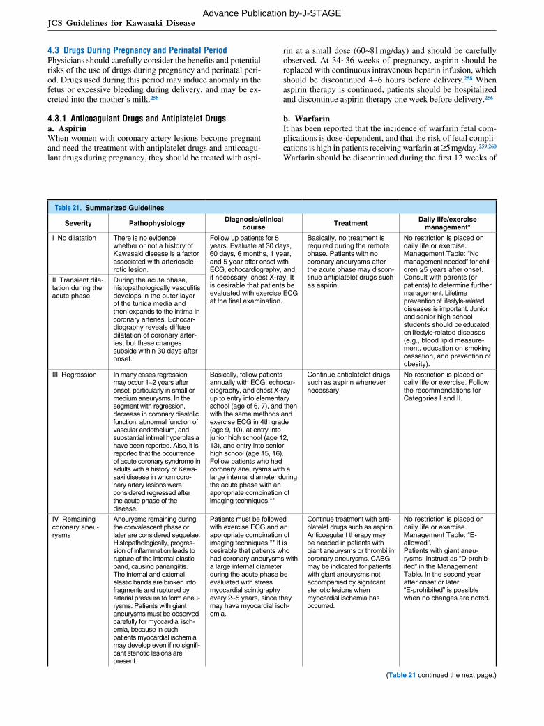
JCS Guidelines for Kawasaki Disease
4.3 Drugs During Pregnancy and Perinatal PeriodPhysicians should carefully consider the benefits and potential risks of the use of drugs during pregnancy and perinatal peri-od. Drugs used during this period may induce anomaly in the fetus or excessive bleeding during delivery, and may be ex-creted into the mother’s milk.258
4.3.1 Anticoagulant Drugs and Antiplatelet Drugsa. AspirinWhen women with coronary artery lesions become pregnant and need the treatment with antiplatelet drugs and anticoagu-lant drugs during pregnancy, they should be treated with aspi-
rin at a small dose (60~81 mg/day) and should be carefully observed. At 34~36 weeks of pregnancy, aspirin should be replaced with continuous intravenous heparin infusion, which should be discontinued 4~6 hours before delivery.258 When aspirin therapy is continued, patients should be hospitalized and discontinue aspirin therapy one week before delivery.256
b. WarfarinIt has been reported that the incidence of warfarin fetal com-plications is dose-dependent, and that the risk of fetal compli-cations is high in patients receiving warfarin at ≥5 mg/day.259,260 Warfarin should be discontinued during the first 12 weeks of
Table 21. Summarized Guidelines
Severity Pathophysiology Diagnosis/clinical course Treatment Daily life/exercise
management*
I No dilatation There is no evidence whether or not a history of Kawasaki disease is a factor associated with arterioscle-rotic lesion.
Follow up patients for 5 years. Evaluate at 30 days, 60 days, 6 months, 1 year, and 5 year after onset with ECG, echocardiography, and, if necessary, chest X-ray. It is desirable that patients be evaluated with exercise ECG at the final examination.
Basically, no treatment is required during the remote phase. Patients with no coronary aneurysms after the acute phase may discon-tinue antiplatelet drugs such as aspirin.
No restriction is placed on daily life or exercise.Management Table: “No management needed” for chil-dren ≥5 years after onset. Consult with parents (or patients) to determine further management. Lifetime prevention of lifestyle-related diseases is important. Junior and senior high school students should be educated on lifestyle-related diseases (e.g., blood lipid measure-ment, education on smoking cessation, and prevention of obesity).
II Transient dila-tation during the acute phase
During the acute phase, histopathologically vasculitis develops in the outer layer of the tunica media and then expands to the intima in coronary arteries. Echocar-diography reveals diffuse dilatation of coronary arter-ies, but these changes subside within 30 days after onset.
III Regression In many cases regression may occur 1∼2 years after onset, particularly in small or medium aneurysms. In the segment with regression, decrease in coronary diastolic function, abnormal function of vascular endothelium, and substantial intimal hyperplasia have been reported. Also, it is reported that the occurrence of acute coronary syndrome in adults with a history of Kawa-saki disease in whom coro-nary artery lesions were considered regressed after the acute phase of the disease.
Basically, follow patients annually with ECG, echocar-diography, and chest X-ray up to entry into elementary school (age of 6, 7), and then with the same methods and exercise ECG in 4th grade (age 9, 10), at entry into junior high school (age 12, 13), and entry into senior high school (age 15, 16). Follow patients who had coronary aneurysms with a large internal diameter during the acute phase with an appropriate combination of imaging techniques.**
Continue antiplatelet drugs such as aspirin whenever necessary.
No restriction is placed on daily life or exercise. Follow the recommendations for Categories I and II.
IV Remaining coronary aneu-rysms
Aneurysms remaining during the convalescent phase or later are considered sequelae. Histopathologically, progres-sion of inflammation leads to rupture of the internal elastic band, causing panangiitis. The internal and external elastic bands are broken into fragments and ruptured by arterial pressure to form aneu-rysms. Patients with giant aneurysms must be observed carefully for myocardial isch-emia, because in such patients myocardial ischemia may develop even if no signifi-cant stenotic lesions are present.
Patients must be followed with exercise ECG and an appropriate combination of imaging techniques.** It is desirable that patients who had coronary aneurysms with a large internal diameter during the acute phase be evaluated with stress myocardial scintigraphy every 2∼5 years, since they may have myocardial isch-emia.
Continue treatment with anti-platelet drugs such as aspirin. Anticoagulant therapy may be needed in patients with giant aneurysms or thrombi in coronary aneurysms. CABG may be indicated for patients with giant aneurysms not accompanied by signifcant stenotic lesions when myocardial ischemia has occurred.
No restriction is placed on daily life or exercise.Management Table: “E- allowed”.Patients with giant aneu-rysms: Instruct as “D-prohib-ited” in the Management Table. In the second year after onset or later, “E-prohibited” is possible when no changes are noted.
(Table 21 continued the next page.)
Advance Publication by-J-STAGE

JCS Joint Working Group
pregnancy, when the major organs systems are developing, and weeks 34~36 of pregnancy and thereafter. For patients in whom discontinuation of warfarin increases the risk of throm-bogenesis, physicians should consider subcutaneous adminis-tration of heparin.258,261
4.3.2 Other DrugsACE inhibitors should be discontinued during pregnancy, as they are teratogenic. Other drugs should be used only when the benefits overweigh the risk.262,263
4.4 Cardiac AccidentsFor patients with a risk of cardiac accidents during pregnancy, cardiologists and obstetricians should collaborate closely to prepare for emergency measures according to the individual patient conditions.
4.4.1 AMIThe presence of a giant aneurysm is among the biggest factors that influence the development of AMI.251 The outcome of myocardial infarction during pregnancy depends on whether the cardiac accident is managed successfully.264,265 Women at 20 weeks of pregnancy or thereafter may undergo catheter intervention via the radial artery approach. As the supine posi-tion may cause inferior vena cava syndrome, physicians should be careful about the body position of the patient.258
4.4.2 ArrhythmiasPatients with myocardial injury, cardiac dysfunction or myo-cardial ischemia may develop ventricular arrhythmias during pregnancy. Physicians should conduct Holter ECG monitoring in such women, and consider treatment when ventricular tachy-
cardia occurs.258
5. Healthcare System for Adult Patients
As patients with a history of Kawasaki disease are often ex-amined by general internists whose knowledge on this disease is limited. The following problems exist: (1) internists have only limited knowledge and experience in the management of adults with cardiovascular sequelae in Kawasaki disease, and there is only limited information on this matter that is avail-able for physicians and non-physician healthcare profession-als; (2) there is only a limited number of specialists who understand the unique pathology and pathophysiology of car-diovascular sequelae in Kawasaki disease during adulthood; and (3) collaborative activities and institutions to ensure effec-tive management through cardiac rehabilitation or other mea-sures have not been fully developed.
5.1 Understanding of Kawasaki Disease by InternistsGeneral internists are not sufficiently aware of the pathophysi-ology of Kawasaki disease during the acute phase. It is impor-tant to foster specialists in this area. Non-physician healthcare professionals should also be educated on cardiovascular se-quelae in Kawasaki disease.
5.2 Collaboration Between Pediatricians and CardiologistsPediatricians and internists must share data on clinical course and laboratory findings obtained from adult patients who had a history of Kawasaki disease. Collaboration between pediatri-cians and internists especially cardiologists is essential to di-agnose, treat and follow patients with cardiovascular sequelae
V-a Coronary stenotic lesions (no findings of ischemia)
Thrombotic occlusion of medium or giant aneurysms may develop during the rela-tively early stage after onset. Sudden death may occur, though two-thirds patients with occlusion are asymp-tomatic. Patients often show improvement of myocardial ischemia due to the develop-ment of recanalized vessels and collateral flow after occlusion. Development/progression of regional stenosis during the remote phase is more prevalent in the left coronary artery than in the right coronary artery. The segments with greatest prevalence are the proximal segment or the main trunk of the left anterior descending artery. The risk of progres-sion to stenosis/occlusion is higher in larger aneurysms. Stenosis may develop during long-term follow up.
Patients must be followed for life, and physicians must design the tailor-made management plan for individ-ual patients. Follow-up examination must include exercise ECG and an appro-priate combination of imaging techniques.** Although schedule may differ among individuals, patients are generally evaluated every 3∼6 months.
Continue treatment with anti-platelet drugs such as aspirin. Use calcium channel blockers, nitrates, β-blockers, ACE inhibitors, and/or angio-tensin receptor II blockers to prevent ischemic attacks and treat heart failure.
No restriction is placed on daily life or exercise. School Activity Management Table: “E-allowed” for patients other than those with giant aneu-rysms. Explain the impor-tance of pharmacotherapy and ensure adherence, as well as symptoms which may occur and actions to be taken when ischemia develops. Patients must be followed at least annually until the stenotic lesions have improved.
V-b Coronary stenotic lesions (with findings of ischemia)
Follow the instructions for pharmacotherapy in Cate-gory V-a. Consider CABG or appropriate PCI tech-nique when exercise ECG or stress myocardial scintig-raphy reveals ischemia.
Exercise shuld be restricted.Categorize in “D” or higher category based on patient condition. School sport club activities should be “prohib-ited”. Select the most appro-priate category from “A” to “D” on the basis of findings of exercise testing and eval-uation of severity of myocar-dial ischemia. Educate patients well about the importance of pharmaco-therapy.
*See Tables 18 and 19. **Imaging techniques include echocardiography (including stress echocardiography), stress myocardial scintigraphy, selective CAG, IVUS, MRI, MRA, and MDCT.ACE, angiotensin converting enzyme; CABG, coronary artery bypass grafting; CAG, coronary angiography; ECG, electrocardiography; IVUS, intravascular ultrasound; MDCT, multi- detector row computed tomography; MRA, magnetic resonance angiography; MRI, magnetic reso-nance imaging; PCI, percutaneous coronary intervention.
Advance Publication by-J-STAGE

JCS Guidelines for Kawasaki Disease
in Kawasaki disease.
5.3 Coronary Aneurysms and Myocardial Infarction in Young Patients and Kawasaki Disease
Asymptomatic patients with coronary aneurysms due to Kawasaki disease may have ischemic heart disease during ado-lescence or adulthood. Myocardial infarction is more preva-lent than angina among patients with cardiovascular sequelae in Kawasaki disease.266 There is a need for a nationwide ar-chive to register and disclose childhood medical records of patients with a history of Kawasaki disease.
5.4 Comparison With Adult-Type Myocardial InfarctionIn the pathologic evaluation of patients with Kawasaki dis-ease, no severe atherosclerotic lesions are observed although substantial arteriosclerosis is present.267 Remodeling of coro-
nary artery lesions in patients with sequelae in Kawasaki dis-ease may persist for years after onset, and is associated with intimal hyperplasia and neovascularization.42 These findings differ from those in juvenile patients with arteriosclerosis not associated with Kawasaki disease.
Because patients with cardiovascular sequelae in Kawasaki disease may have atherosclerosis and may present with more complex lesions in the future, specialists who are fully aware of such condition must be available.
5.5 Cardiac RehabilitationFew medical institutions are providing cardiac rehabilitation programs for adults with cardiovascular sequelae in Kawasaki disease. This patient population needs appropriate cardiac re-habilitation programs and facilities where physicians and re-habilitation specialists work together.
VIII Summary
Table 21 summarizes the pathophysiology, diagnosis, clinical course, treatment, lifestyle management and exercise manage-
ment for patients with a history of Kawasaki disease by sever-ity.
References 1. Kawasaki T. Acute febrile mucocutaneous syndrome with lym-
phoid involvement with specific desquamation of the fingers and toes in children. Japanese Journal of Allergology 1967; 16: 178 – 222 (in Japanese).
2. Guidelines for Diagnosis and Treatment of Cardiovascular Dis-eases (2001–2002 Joint Working Groups Report). Guidelines for diagnosis and management of cardiovascular sequelae in Kawasaki disease (JCS 2003). Circ J 2003; 67 (Suppl IV): 1111 – 1174 (in Japanese).
3. Guidelines for Diagnosis and Treatment of Cardiovascular Dis-eases (2007 Joint Working Groups Report). Guidelines for diagno-sis and management of cardiovascular sequelae in Kawasaki dis-ease (JCS 2008). http://www.j-circ.or.jp/guideline/pdf/JCS2008_ ogawasy_h.pdf (in Japanese) (available January 2013).
4. Nakamura Y, Yashiro M, Uehara R, Sadakane A, Tsuboi S, Aoyama Y, et al. Epidemiologic features of Kawasaki disease in Japan: Results of the 2009-2010 nationwide survey. J Epidemiol 2012; 22: 216 – 221.
5. Kawasaki Disease Study Group of the Ministry of Health, Labour and Welfare. Guidelines for the Diagnosis of Kawasaki Disease (MCLS, infantile acute febrile mucocutaneous lymph node syn-drome), fifth revision. J Jpn Pediatr Soc 2002; 106: 836 – 837 (in Japanese).
6. Burgner D, Davila S, Breunis WB, Ng SB, Li Y, Bonnard C, et al; International Kawasaki Disease Genetics Consortium. A genome-wide association study identifies novel and functionally related susceptibility Loci for Kawasaki disease. PLoS Genet 2009; 5: e1000319, doi:10.1371/journal.pgen.1000319.
7. Kim JJ, Hong YM, Sohn S, Jang GY, Ha KS, Yun SW, et al; Korean Kawasaki Disease Genetics Consortium. A genome-wide association analysis reveals 1p31 and 2p13.3 as susceptibility loci for Kawasaki disease. Hum Genet 2011; 129: 487 – 495.
8. Tsai FJ, Lee YC, Chang JS, Huang LM, Huang FY, Chiu NC, et al. Identification of novel susceptibility Loci for kawasaki disease in a Han chinese population by a genome-wide association study. PLoS One 2011; 6: e16853, doi:10.1371/journal.pone.0016853.
9. Khor CC, Davila S, Breunis WB, Lee YC, Shimizu C, Wright VJ, et al. Genome-wide association study identifies FCGR2A as a susceptibility locus for Kawasaki disease. Nat Genet 2011; 43: 1241 – 1246.
10. Lee YC, Kuo HC, Chang JS, Chang LY, Huang LM, Chen MR, et al. Two new susceptibility loci for Kawasaki disease identified through genome-wide association analysis. Nat Genet 2012; 44: 522 – 525.
11. Onouchi Y, Ozaki K, Burns JC, Shimizu C, Terai M, Hamada H, et al; Japan Kawasaki Disease Genome Consortium; US Kawasaki Disease Genetics Consortium. A genome-wide association study
identifies three new risk loci for Kawasaki disease. Nat Genet 2012; 44: 517 – 521.
12. Onouchi Y, Gunji T, Burns JC, Shimizu C, Newburger JW, Yashiro M, et al. ITPKC functional polymorphism associated with Kawasaki disease susceptibility and formation of coronary artery aneurysms. Nat Genet 2008; 40: 35 – 42.
13. Onouchi Y, Ozaki K, Buns JC, Shimizu C, Hamada H, Honda T, et al. Common variants in CASP3 confer susceptibility to Kawasaki disease. Hum Mol Genet 2010; 19: 2898 – 2906.
14. Khor CC, Davila S, Shimizu C, Sheng S, Matsubara T, Suzuki Y, et al; US and International Kawasaki Disease Genetics Consortia. Genome-wide linkage and association mapping identify suscepti-bility alleles in ABCC4 for Kawasaki disease. J Med Genet 2011; 48: 467 – 472.
15. Asai T. Severity assessment of Kawasaki disease. Acta Paediatr Jpn Overseas Ed 1983; 25: 170 – 175.
16. Iwasa M, Sugiyama K, Ando T, Nomura H, Katoh T, Wada Y. Selection of high-risk children for immunoglobulin therapy in Kawasaki disease. Prog Clin Biol Res 1987; 250: 543 – 544.
17. Harada K. Intravenous gamma-globulin treatment in Kawasaki disease. Acta Paediatr Jpn 1991; 33: 805 – 810.
18. Kobayashi T, Inoue Y, Takeuchi K, Okada Y, Tamura K, Tomomasa T, et al. Prediction of intravenous immunoglobulin unresponsive-ness in patients with Kawasaki disease. Circulation 2006; 113: 2606 – 2612.
19. Egami K, Muta H, Ishii M, Suda K, Sugahara Y, Iemura M, et al. Prediction of resistance to intravenous immunoglobulin treatment in patients with Kawasaki disease. J Pediatr 2006; 149: 237 – 240.
20. Sano T, Kurotobi S, Matsuzaki K, Yamamoto T, Maki I, Miki K, et al. Prediction of nonresponsiveness to standard high-dose gam-ma-globulin therapy in patients with acute Kawasaki disease before starting initial treatment. Eur J Pediatr 2007; 166: 131 – 137.
21. Kobayashi T, Inoue Y, Tamura K, Morikawa A, Kobayashi T. External validation of a scoring system to predict resistance to in-travenous immunoglobulin. J Pediatr 2007; 150: e37; author reply e38, doi:10.1016/j.jpeds.2006.12.036.
22. Seki M, Kobayashi T, Kobayashi T, Morikawa A, Otani T, Takeuchi K, et al. External validation of a risk score to predict intravenous immunoglobulin resistance in patients with Kawasaki disease. Pe-diatr Infect Dis J 2011; 30: 145 – 147.
23. Sleeper LA, Minich LL, McCrindle BM, Li JS, Mason W, Colan SD, et al; Pediatric Heart Network Investigators. Evaluation of Kawasaki disease risk-scoring systems for intravenous immuno-globulin resistance. J Pediatr 2011; 158: 831 – 835.e3, doi:10. 1016/j.jpeds.2010.10.031.
24. Sudo D, Monobe Y, Yashiro M, Mieno MN, Uehara R, Tsuchiya K, et al. Coronary artery lesions of incomplete Kawasaki disease: A nationwide survey in Japan. Eur J Pediatr 2012; 171: 651 – 656.
25. Manlhiot C, Christie E, McCrindle BW, Rosenberg H, Chahal N,
Advance Publication by-J-STAGE

JCS Joint Working Group
Yeung RS. Complete and incomplete Kawasaki disease: Two sides of the same coin. Eur J Pediatr 2012; 171: 657 – 662.
26. Sonobe T, Kiyosawa N, Tsuchiya K, Aso S, Imada Y, Imai Y, et al. Prevalence of coronary artery abnormality in incomplete Kawasaki disease. Pediatr Int 2007; 49: 421 – 426.
27. Ha KS, Jang G, Lee J, Lee K, Hong Y, Son C, et al. Incomplete clinical manifestation as a risk factor for coronary artery abnor-malities in Kawasaki disease: A meta-analysis. Eur J Pediatr 2013; 172: 343 – 349.
28. Kuo HC, Yang KD, Juo SH, Liang CD, Chen WC, Wang YS, et al. ITPKC single nucleotide polymorphism associated with the Kawasaki disease in a Taiwanese population. PLoS One 2011; 6: e17370, doi:10.1371/journal.pone.0017370.
29. Lin MT, Wang JK, Yeh JI, Sun LC, Chen PL, Wu JF, et al. Clinical implication of the c allele of the ITPKC gene SNP rs28493229 in Kawasaki disease: Association with disease susceptibility and BCG scar reactivation. Pediatr Infect Dis J 2011; 30: 148 – 152.
30. Kuo HC, Yu HR, Juo SH, Yang KD, Wang YS, Liang CD, et al. CASP3 gene single-nucleotide polymorphism (rs72689236) and Kawasaki disease in Taiwanese children. J Hum Genet 2011; 56: 161 – 165.
31. Takahashi K, Oharaseki T, Naoe S, Wakayama M, Yokouchi Y. Neutrophilic involvement in the damage to coronary arteries in acute stage of Kawasaki disease. Pediatr Int 2005; 47: 305 – 310.
32. Masuda H, Naoe S, Tanaka N. The pathology of coronary arteries in Kawasaki disease (MCLS): A consideration of the relationship between coronary arteritis and the development of aneurysms. The Journal of Japanese College of Angiology 1981; 21: 899 – 912 (in Japanese).
33. Terminology Committee of the Japanese Society of Kawasaki Dis-ease. Draft rules for the terminology of Kawasaki disease. http://www.jskd.jp/info/pdf/yougo201007.pdf (in Japanese).
34. Sasaguri Y, Kato H. Regression of aneurysms in Kawasaki disease: A pathological study. J Pediatr 1982; 100: 225 – 231.
35. Suzuki A, Yamagishi M, Kimura K, Sugiyama H, Arakaki Y, Kamiya T, et al. Functional behavior and morphology of the coronary artery wall in patients with Kawasaki disease assessed by intravascular ultrasound. J Am Coll Cardiol 1996; 27: 291 – 296.
36. Mitani Y, Okuda Y, Shimpo H, Uchida F, Hamanaka K, Aoki K, et al. Impaired endothelial function in epicardial coronary arteries after Kawasaki disease. Circulation 1997; 96: 454 – 461.
37. Sugimura T, Kato H, Inoue O, Fukuda T, Sato N, Ishii M, et al. Intravascular ultrasound of coronary arteries in children: Assess-ment of the wall morphology and the lumen after Kawasaki disease. Circulation 1994; 89: 258 – 265.
38. Yamakawa R, Ishii M, Sugimura T, Akagi T, Eto G, Iemura M, et al. Coronary endothelial dysfunction after Kawasaki disease: Eval-uation by intracoronary injection of acetylcholine. J Am Coll Car-diol 1998; 31: 1074 – 1080.
39. Takahashi K, Oharaseki T, Naoe S. Pathological study of postcoro-nary arteritis in adolescents and young adults: With reference to the relationship between sequelae of Kawasaki disease and atheroscle-rosis. Pediatr Cardiol 2001; 22: 138 – 142.
40. Tanaka N, Naoe S, Masuda H, Ueno T. Pathological study of se-quelae of Kawasaki disease (MCLS): With special reference to the heart and coronary arterial lesions. Acta Pathol Jpn 1986; 36: 1513 – 1527.
41. Negoro N, Nariyama J, Nakagawa A, Katayama H, Okabe T, Hazui H, et al. Successful catheter interventional therapy for acute coro-nary syndrome secondary to Kawasaki disease in young adults. Circ J 2003; 67: 362 – 365.
42. Suzuki A, Miyagawa-Tomita S, Komatsu K, Nishikawa T, Sakomura Y, Horie T, et al. Active remodeling of the coronary arterial lesions in the late phase of Kawasaki disease: Immunohistochemical study. Circulation 2000; 101: 2935 – 2941.
43. Takahasi K, Shibuya H, Atobe T, Naoe S, Tanaka N. Histopatho-logical study of Kawasaki disease in patients who died of intercur-rent diseases. Prog Med 1987; 7: 21 – 25 (in Japanese).
44. Kikuchi F, Mori N, Naoe S, Makita J. A case of death due to bron-chial asthma attack in a patient with a history of typical Kawasaki disease who has no evidence of current or previous arteritis at au-topsy. Prog Med 1987; 7: 131 – 137 (in Japanese).
45. Harada M, Yokouchi Y, Oharaseki T, Matsui K, Tobayama H, Tanaka N, et al. Histopathological characteristics of myocarditis in acute-phase Kawasaki disease. Histopathology 2012; 61: 1156 – 1167.
46. Yonesaka S, Nakada T, Sunagawa Y, Tomimoto K, Naka S, Takahashi T, et al. Endomyocardial biopsy in children with Kawasaki disease. Acta Paediatr Jpn 1989; 31: 706 – 711.
47. Takahashi K, Shibuya K, Masuda H, Tanaka N. Pathology of car-
diac sequelae of Kawasaki disease. The Journal of Japanese Col-lege of Angiology 1989; 29: 461 – 469 (in Japanese).
48. Amano S, Hazama F, Kubagawa H, Tasaka K, Haebara H, Hamashima Y. General pathology of Kawasaki disease: On the mor-phological alterations corresponding to the clinical manifestations. Acta Pathol Jpn 1980; 30: 681 – 694.
49. Landing BH, Larson EJ. Pathological features of Kawasaki disease (mucocutaneous lymph node syndrome). Am J Cardiovasc Pathol 1987; 1: 218 – 229.
50. Takahashi K, Oharaseki T, Yokouchi Y, Hiruta N, Naoe S. Kawasaki disease as a systemic vasculitis in childhood. Ann Vasc Dis 2010; 3: 173 – 181.
51. Takahashi K, Oharaseki T, Yokouchi Y. Aneurysms due to Kawasaki disease during adulthood: Transition from acute phase coronary arteritis to remote phase arteriosclerotic lesions and a consideration of differences from arteriosclerosis in adults without a history of Kawasaki disease. Vascular Med 2010; 6: 22 – 26 (in Japanese).
52. Ogawa S, Ohkubo T, Fukazawa R, Kamisago M, Kuramochi Y, Uchikoba Y, et al. Estimation of myocardial hemodynamics before and after intervention in children with Kawasaki disease. J Am Coll Cardiol 2004; 43: 653 – 661.
53. Donohue TJ, Kern MJ, Aguirre FV, Bach RG, Wolford T, Bell CA, et al. Assessing the hemodynamic significance of coronary artery stenoses: Analysis of translesional pressure-flow velocity relations in patients. J Am Coll Cardiol 1993; 22: 449 – 458.
54. Ofili EO, Kern MJ, Labovitz AJ, St Vrain JA, Segal J, Aguirre FV, et al. Analysis of coronary blood flow velocity dynamics in angio-graphically normal and stenosed arteries before and after endolu-men enlargement by angioplasty. J Am Coll Cardiol 1993; 21: 308 – 316.
55. Segal J, Kern MJ, Scott NA, King SB 3rd, Doucette JW, Heuser RR, et al. Alterations of phasic coronary artery flow velocity in humans during percutaneous coronary angioplasty. J Am Coll Car-diol 1992; 20: 276 – 286.
56. Pijls NH, van Son JA, Kirkeeide RL, De Bruyne B, Gould KL. Experimental basis of determining maximum coronary, myocardial, and collateral blood flow by pressure measurements for assessing functional stenosis severity before and after percutaneous translu-minal coronary angioplasty. Circulation 1993; 87: 1354 – 1367.
57. Ogawa S. Rheologic and hemodynamic characteristics of coronary arteries. J Jpn Pediatr Soc 2009; 113: 1769 – 1778 (in Japanese).
58. Alpert JS, Thygesen K, Antman E, Bassand JP. Myocardial infarc-tion redefined: A consensus document of The Joint European Soci-ety of Cardiology/American College of Cardiology Committee for the redefinition of myocardial infarction. J Am Coll Cardiol 2000; 36: 959 – 969.
59. Braunwald E, Antman EM, Beasley JW, Califf RM, Cheitlin MD, Hochman JS, et al. ACC/AHA guidelines for the management of patients with unstable angina and non-STsegment elevation myo-cardial infarction: A report of the American College of Cardiology/American Heart Association Task Force on Practice Guidelines (Committee on the Management of Patients With Unstable Angi-na). J Am Coll Cardiol 2000; 36: 970 – 1062.
60. Okamoto F, Sohmiya K, Ohkaru Y, Kawamura K, Asayama K, Kimura H, et al. Human heart-type cytoplasmic fatty acid-binding protein (H-FABP) for the diagnosis of acute myocardial infarction: Clinical evaluation of H-FABP in comparison with myoglobin and creatine kinase isoenzyme MB. Clin Chem Lab Med 2000; 38: 231 – 238.
61. Lindahl B, Venge P, Wallentin L. Relation between troponin T and the risk of subsequent cardiac events in unstable coronary artery disease: The FRISC study group. Circulation 1996; 93: 1651 – 1657.
62. Ohman EM, Armstrong PW, Christenson RH, Granger CB, Katus HA, Hamm CW, et al. Cardiac troponin T levels for risk stratifica-tion in acute myocardial ischemia. GUSTO IIA Investigators. N Engl J Med 1996; 335: 1333 – 1341.
63. Isobe M, Nagai R, Ueda S, Tsuchimochi H, Nakaoka H, Takaku F, et al. Quantitative relationship between left ventricular function and serum cardiac myosin light chain I levels after coronary reperfusion in patients with acute myocardial infarction. Circulation 1987; 76: 1251 – 1261.
64. Khreiss T, József L, Potempa LA, Filep JG. Opposing effects of C-reactive protein isoforms on shear-induced neutrophil-platelet adhesion and neutrophil aggregation in whole blood. Circulation 2004; 110: 2713 – 2720.
65. Mitani Y, Sawada H, Hayakawa H, Aoki K, Ohashi H, Matsumura M, et al. Elevated levels of highsensitivity C-reactive protein and serum amyloid-A late after Kawasaki disease: Association between inflammation and late coronary sequelae in Kawasaki disease. Circulation 2005; 111: 38 – 43.
Advance Publication by-J-STAGE

JCS Guidelines for Kawasaki Disease
66. Yonesaka S, Takahashi T, Sato T, Eto S, Ohtani K, Kitagawa Y, et al. Ongoing myocardial damage in infants and children with Kawasaki disease. Respiration & Circulation 2010; 58: 1273 – 1279 (in Japanese).
67. Borzutzky A, Gutiérrez M, Talesnik E, Godoy I, Kraus J, Hoyos R, et al. High sensitivity C-reactive protein and endothelial function in Chilean patients with history of Kawasaki disease. Clin Rheumatol 2008; 27: 845 – 850.
68. Berenson GS, Srinivasan SR, Bao W, Newman WP 3rd, Tracy RE, Wattigney WA. Association between multiple cardiovascular risk factors and atherosclerosis in children and young adults: The Bogalusa Heart Study. N Engl J Med 1998; 338: 1650 – 1656.
69. Okada T, Murata M, Yamauchi K, Harada K. New criteria of nor-mal serum lipid levels in Japanese children: The nationwide study. Pediatr Int 2002; 44: 596 – 601.
70. Japan Atherosclerosis Society. Japan Atherosclerosis Society (JAS) Guidelines for Diagnosis and Treatment of Atherosclerotic Cardio-vascular Diseases, 2002. 2002: 5 – 7 (in Japanese).
71. Japan Atherosclerosis Society. Japan Atherosclerosis Society (JAS) Guidelines for Prevention of Atherosclerotic Cardiovascular Dis-eases, 2007. 2007: 11 – 13 (in Japanese).
72. Ou CY, Tseng YF, Lee CL, Chiou YH, Hsieh KS. Significant rela-tionship between serum high-sensitivity C-reactive protein, high-density lipoprotein cholesterol levels and children with Kawasaki disease and coronary artery lesions. J Formos Med Assoc 2009; 108: 719 – 724.
73. Boers GH, Smals AG, Trijbels FJ, Fowler B, Bakkeren JA, Schoonderwaldt HC, et al. Heterozygosity for homocystinuria in premature peripheral and cerebral occlusive arterial disease. N Engl J Med 1985; 313: 709 – 715.
74. Welch GN, Loscalzo J. Homocysteine and atherothrombosis. N Engl J Med 1998; 338: 1042 – 1050.
75. Ohzeki T, Nakagawa Y, Nakanishi T, Fujisawa Y, Sai S. Diagnos-tic criteria for metabolic syndrome in Japanese children: A cohort study to establish the concept, pathophysiology, and diagnostic criteria of metabolic syndrome in children and design effective in-terventions: An MHLW grant research project for cardiovascular disease and lifestyle-related diseases led by Ohzeki T. A final report in 2005–2007. 2008: 89 – 91 (in Japanese).
76. Council on Cardiovascular Disease in the Young; Committee on Rheumatic Fever, Endocarditis, and Kawasaki Disease; American Heart Association: Diagnostic guidelines for Kawasaki disease. Circulation 2001; 103: 335 – 336.
77. Ichida F, Fatica NS, O’Loughlin JE, Snyder MS, Ehlers KH, Engle MA. Correlation of electrocardiographic and echocardiographic changes in Kawasaki syndrome. Am Heart J 1988; 116: 812 – 819.
78. Okagawa H, Kondo M, Okuno M, Fujino H, Nishijima S. QT dura-tion in the acute phase of Kawasaki disease. Prog Med 1997; 17: 1761 – 1764 (in Japanese).
79. Onouchi Z, Shimazu S, Tamiya H. The second report on abnormal T waves on ECG in patients with Kawasaki disease: Left ventricu-lar asynergy in short-axis two-dimensional echographic images. J Jpn Pediatr Soc 1986; 90: 827 – 831 (in Japanese).
80. Gravel H, Dahdah N, Fournier A, Mathieu MÈ, Curnier D. Ven-tricular repolarisation during exercise challenge occurring late after Kawasaki disease. Pediatr Cardiol 2012; 33: 728 – 734.
81. Osada M, Tanaka Y, Komai T, Maeda Y, Kitano M, Komori S, et al. Coronary arterial involvement and QT dispersion in Kawasaki disease. Am J Cardiol 1999; 84: 466 – 468.
82. Okuyama J, Osada M, Sugiyama H, Komori S, Ozaki Y. QT disper-sion and its longitudinal change in Kawasaki disease. Japanese Journal of Electrocardiology 2001; 21: 800 – 804 (in Japanese).
83. Nakata T. Premature ventricular contraction in remote-phase Kawasaki disease. Medical Journal Aomori 1995; 40: 98 – 103 (in Japanese).
84. Nakanishi T, Takao A, Kondoh C, Nakazawa M, Hiroe M, Matsumoto Y. ECG findings after myocardial infarction in children after Kawasaki disease. Am Heart J 1988; 116: 1028 – 1033.
85. Hamaoka K, Kamiya Y, Sakata K, Fukumochi Y, Ohmochi Y. Coronary reserve in children of Kawasaki disease with ischemic findings on exercise ECG and myocardial SPECT but angiograph-ically no stenotic lesion. Evaluation of coronary hemodynamics and myocardial metabolism during atrial pacing. J Jpn Pediatr Soc 1991; 95: 145 – 151 (in Japanese).
86. Matsuda M, Shimizu T, Oouchi H, Saito S, Kawade M, Arakaki Y, et al. Diagnosis of myocardial ischemia using body surface electro-cardiographic mapping with intravenous dipyridamole in children who have a history of Kawasaki disease. J Jpn Pediatr Soc 1995; 99: 1618 – 1627 (in Japanese).
87. Takechi N, Seki T, Ohkubo T, Ogawa S. Dobutamine stress surface
mapping of myocardial ischemia in Kawasaki disease. Pediatr Int 2001; 43: 218 – 225.
88. Seki T, Zhang J, Ogawa S, Hirayama T. Dobutamine stress body surface mapping in Kawasaki disease. Journal of Nippon Medical School 1994; 61: 610 – 619 (in Japanese).
89. Shiono J, Horigome H, Matsui A, Terada Y, Watanabe S, Miyashita T, et al. Evaluation of myocardial ischemia in Kawasaki disease using an isointegral map on magnetocardiogram. Pacing Clin Elec-trophysiol 2002; 25: 915 – 921.
90. Sumitomo N, Karasawa K, Taniguchi K, Ichikawa R, Fukuhara J, Abe O, et al. Association of sinus node dysfunction, atrioventricular node conduction abnormality and ventricular arrhythmia in patients with Kawasaki disease and coronary involvement. Circ J 2008; 72: 274 – 280.
91. Hamamoto K, Oku I, Yamato K. Evaluation of intracardiac conduc-tion delay in acute phase of Kawasaki disease by signal averaged electrocardiogram. Heart 1998; 30: 21 – 27 (in Japanese).
92. Tsuchida A, Ito S, Oka R, Saito T, Yoshioka I. Recording by signal-averaged electrocardiogram in patients with Kawasaki disease. J Jpn Pediatr Soc 1990; 94: 1168 – 1173 (in Japanese).
93. Dahdah NS, Jaeggi E, Fournier A. Electrocardiographic depolariza-tion and repolarization: Long-term after Kawasaki disease. Pediatr Cardiol 2002; 23: 513 – 517.
94. Kuramochi Y, Takechi N, Ohkubo T, Ogawa S. Longitudinal esti-mation of signal-averaged electrocardiograms in patients with Kawasaki disease. Pediatr Int 2002; 44: 12 – 17.
95. Takeuchi M, Matsushita A, Tsuda E, Kurotobi S, Kogaki S. Clini-cal assessment of ventricular late potential in children with Kawasaki disease: Frequency analysis and its clinical significance. Prog Med 1994; 14: 1828 – 1832 (in Japanese).
96. Ogawa S, Nagai Y, Zhang J, Yuge K, Hino Y, Jimbo O, et al. Evaluation of myocardial ischemia and infarction by signal-aver-aged electrocardiographic late potentials in children with Kawasaki disease. Am J Cardiol 1996; 78: 175 – 181.
97. Genma Y, Ogawa S, Zhang J, Yamamoto M. Evaluation of myo-cardial ischemia in Kawasaki disease by dobutamine stress signal-averaged ventricular late potentials. Cardiovasc Res 1997; 36: 323 – 329.
98. Tanaka N, Ueno T, Naoe S, Masuda H. Kawasaki disease: Patho-logical features and sequelae of arteritis. Japanese Journal of Clin-ical Medicine 1983; 41: 2008 – 2016 (in Japanese).
99. Nakada T, Yonesaka S, Sunagawa Y, Tomimoto K, Takahashi T, Matsubara T, et al. Coronary arterial calcification in Kawasaki disease. Acta Paediatr Jpn 1991; 33: 443 – 449.
100. Fuse S, Kobayashi T, Arakaki Y, Ogawa S, Katoh H, Sakamoto N, et al. Standard method for ultrasound imaging of coronary artery in children. Pediatr Int 2010; 52: 876 – 882.
101. Newburger JW, Sanders SP, Burns JC, Parness IA, Beiser AS, Colan SD. Left ventricular contractility and function in Kawasaki syndrome. Effect of intravenous gamma-globulin. Circulation 1989; 79: 1237 – 1246.
102. Yanagisawa M, Yano S, Shiraishi H, Nakajima Y, Fujimoto T, Itoh K. Coronary aneurysms in Kawasaki disease: Follow-up observa-tion by two-dimensional echocardiography. Pediatr Cardiol 1985; 6: 11 – 16.
103. Minich LL, Tani LY, Pagotto LT, Young PC, Etheridge SP, Shaddy RE. Usefulness of echocardiography for detection of coronary ar-tery thrombi in patients with Kawasaki disease. Am J Cardiol 1998; 82: 1143 – 1146, A1110.
104. Miyashita M, Karasawa K, Taniguchi K, Kanamaru H, Ayusawa M, Sumitomo N, et al. Usefulness of real-time 3-dimensional echo-cardiography for the evaluation of coronary artery morphology in patients with Kawasaki disease. J Am Soc Echocardiogr 2007; 20: 930 – 933.
105. Nakano H, Saito A, Ueda K, Tsuchitani Y. Valvular lesions com-plicating Kawasaki disease: A Doppler echocardiographic evalua-tion. J Cardiogr 1986; 16: 363 – 371 (in Japanese).
106. Takeuchi D, Saji T, Takatsuki S, Fujiwara M. Abnormal tissue Doppler images are associated with elevated plasma brain natri-uretic peptide and increased oxidative stress in acute Kawasaki disease. Circ J 2007; 71: 357 – 362.
107. Noto N, Ayusawa M, Karasawa K, Yamaguchi H, Sumitomo N, Okada T, et al. Dobutamine stress echocardiography for detection of coronary artery stenosis in children with Kawasaki disease. J Am Coll Cardiol 1996; 27: 1251 – 1256.
108. Ishii M, Himeno W, Sawa M, Iemura M, Furui J, Muta H, et al. Assessment of the ability of myocardial contrast echocardiography with harmonic power Doppler imaging to identify perfusion abnor-malities in patients with Kawasaki disease at rest and during di-pyridamole stress. Pediatr Cardiol 2002; 23: 192 – 199.
Advance Publication by-J-STAGE

JCS Joint Working Group
109. Hijazi ZM, Udelson JE, Snapper H, Rhodes J, Marx GR, Schwartz SL, et al. Physiologic significance of chronic coronary aneurysms in patients with Kawasaki disease. J Am Coll Cardiol 1994; 24: 1633 – 1638.
110. Paridon SM, Galioto FM, Vincent JA, Tomassoni TL, Sullivan NM, Bricker JT. Exercise capacity and incidence of myocardial perfusion defects after Kawasaki disease in children and adoles-cents. J Am Coll Cardiol 1995; 25: 1420 – 1424.
111. Miyagawa M, Mochizuki T, Murase K, Tanada S, Ikezoe J, Sekiya M, et al. Prognostic value of dipyridamole-thallium myocardial scintigraphy in patients with Kawasaki disease. Circulation 1998; 98: 990 – 996.
112. Kondo C, Hiroe M, Nakanishi T, Takao A. Detection of coronary artery stenosis in children with Kawasaki disease: Usefulness of pharmacologic stress 201Tl myocardial tomography. Circulation 1989; 80: 615 – 624.
113. Ogawa S, Fukazawa R, Ohkubo T, Zhang J, Takechi N, Kuramochi Y, et al. Silent myocardial ischemia in Kawasaki disease: Evalua-tion of percutaneous transluminal coronary angioplasty by dobuta-mine stress testing. Circulation 1997; 96: 3384 – 3389.
114. Karasawa K, Ayusawa M, Noto N, Yamaguchi H, Okada T, Harada K. The dobutamine stress T1-201 myocardial single photon emis-sion computed tomography for coronary artery stenosis caused by Kawasaki disease. Ped Cardiol Card Surg 1994; 9: 723 – 733 (in Japanese).
115. Prabhu AS, Singh TP, Morrow WR, Muzik O, Di Carli MF. Safety and efficacy of intravenous adenosine for pharmacologic stress testing in children with aortic valve disease or Kawasaki disease. Am J Cardiol 1999; 83: 284 – 286, A286.
116. Kinoshita S, Suzuki S, Shindo A, Watanage K, Yamashita S. The accuracy and side effects of pharmacologic stress Thallium myo-cardial scintigraphy with adenosine triphosphate disodium (ATP) infusion in the diagnosis of coronary artery disease. The Japanese Journal of Nuclear Medicine 1994; 31: 935 – 941 (in Japanese).
117. Germano G, Erel J, Lewin H, Kavanagh PB, Berman DS. Auto-matic quantitation of regional myocardial wall motion and thicken-ing from gated technetium-99 m sestamibi myocardial perfusion single-photon emission computed tomography. J Am Coll Cardiol 1997; 30: 1360 – 1367.
118. Johnson LL, Verdesca SA, Aude WY, Xavier RC, Nott LT, Campanella MW, et al. Postischemic stunning can affect left ven-tricular ejection fraction and regional wall motion on post-stress gated sestamibi tomograms. J Am Coll Cardiol 1997; 30: 1641 – 1648.
119. Ishikawa Y, Fujiwara M, Ono Y, Tsuda E, Matsubara T, Furukawa S, et al. Exercise- or dipyridamoleloaded QGS is useful to evaluate myocardial ischemia and viability in the patients with a history of Kawasaki disease. Pediatr Int 2005; 47: 505 – 511.
120. Karasawa K, Miyashita M, Taniguchi K, Kanamaru H, Ayusawa M, Noto N, et al. Detection of myocardial contractile reserve by low-dose dobutamine quantitative gated single-photon emission computed tomography in patients with Kawasaki disease and se-vere coronary artery lesions. Am J Cardiol 2003; 92: 865 – 868.
121. Karasawa K, Ayusawa M, Noto N, Sumitomo N, Okada T, Harada K. Optimum protocol of technetium-99 m tetrofosmin myocardial perfusion imaging for the detection of coronary stenosis lesions in Kawasaki disease. J Cardiol 1997; 30: 331 – 339 (in Japanese).
122. Monzen H, Hara M, Hirata M, Nakanishi A, Ogasawara M, Suzuki T, et al. Exploring a technique for reducing the influence of scat-tered rays from surrounding organs to the heart during myocardial perfusion scintigraphy with technetium-99 m sestamibi and techne-tium-99 m tetrofosmin. Ann Nucl Med 2006; 20: 705 – 710.
123. Karasawa K, Ayusawa M, Ymasita T. Pharmacologic stress myo-cardial perfusion imaging for the detection of coronary stenotic lesions due to Kawasaki disease: Comparison of dobutamine and adenosine triphosphate disodium. In: Kato H, editor. Kawasaki disease: Proceedings of the 5th International Kawasaki Disease Symposium, Fukuoka, Japan, 22-25 May 1995. Elsevier 1995: 472 – 478.
124. Yamazaki J, Nishimura T, Nishimura S, Kajiya T, Kojima K, Kato K. The Diagnostic Value for Ischemic Heart Disease of Thalli-um-201 Myocardial Scintigraphy by Intravenous Infusion of SUNY4001 (Adenosine): The Report of Clinical Trial at Multi-Center: Phase III. The Japanese Journal of Nuclear Medicine 2004; 41: 133 – 142 (in Japanese).
125. Subcommittee for Standardization of Radionuclide Imaging, Med-ical and Pharmaceutical Committee for the Japan Radioisotope Association. Recommendations for a pediatric dose in nuclear im-aging. Japan Radioisotope Association. Radioisotopes 1988; 37: 627 – 632 (in Japanese).
126. Guidelines for Diagnosis and Treatment of Cardiovascular Dis-eases (2010–2011 Joint Working Groups Report). Guidelines for Drug Therapy in Pediatric Patients with Cardiovascular Diseases (JCS 2012). Guidelines for Diagnosis and Treatment of Cardiovas-cular Diseases 2012: 89 – 301 (in Japanese).
127. Lassmann M, Biassoni L, Monsieurs M, Franzius C, Jacobs F; EANM Dosimetry and Paediatrics Committees. The new EANM paediatric dosage card. Eur J Nucl Med Mol Imaging 2007; 34: 796 – 798.
128. Pediatric Nuclear Medicine Practice Committee of the Japanese Society of Nuclear Medicine. Consensus guidelines for nuclear medicine procedures for children, 2013 (in Japanese).
129. Kanamaru H, Sato Y, Takayama T, Ayusawa M, Karasawa K, Sumitomo N, et al. Assessment of coronary artery abnormalities by multislice spiral computed tomography in adolescents and young adults with Kawasaki disease. Am J Cardiol 2005; 95: 522 – 525.
130. Kanamaru H, Kimijima T, Mugishima H. Cardiovascular imaging: Cardiac CT. Journal of Japanese Society of Pediatric Radiology 2011; 27: 106 – 117 (in Japanese).
131. Liu X, Zhao X, Huang J, Francois CJ, Tuite D, Bi X, et al. Com-parison of 3D free-breathing coronary MR angiography and 64-MDCT angiography for detection of coronary stenosis in patients with high calcium scores. AJR Am J Roentgenol 2007; 189: 1326 – 1332.
132. Ozgun M, Rink M, Hoffmeier A, Botnar RM, Heindel W, Fischbach R, et al. Intraindividual comparison of 3D coronary MR angiogra-phy and coronary CT angiography. Acad Radiol 2007; 14: 910 – 916.
133. Kanamaru H, Karasawa K, Ichikawa R, Abe O, Miyashita M, Taniguchi K, et al. Advantages of multislice spiral computed to-mography for evaluation of serious coronary complications after Kawasaki disease. J Cardiol 2007; 50: 21 – 27 (in Japanese).
134. Suzuki A, Takemura A, Inaba R, Sonobe T, Tsuchiya K, Korenaga T. Magnetic resonance coronary angiography to evaluate coronary arterial lesions in patients with Kawasaki disease. Cardiol Young 2006; 16: 563 – 571.
135. Takemura A, Suzuki A, Inaba R, Sonobe T, Tsuchiya K, Omuro M, et al. Utility of coronary MR angiography in children with Kawasaki disease. AJR Am J Roentgenol 2007; 188: W534 – W539.
136. Botnar RM, Kim WY, Börnert P, Stuber M, Spuentrup E, Manning WJ. 3D coronary vessel wall imaging utilizing a local inversion technique with spiral image acquisition. Magn Reson Med 2001; 46: 848 – 854.
137. Fayad ZA, Fuster V, Fallon JT, Jayasundera T, Worthley SG, Helft G, et al. Noninvasive in vivo human coronary artery lumen and wall imaging using black-blood magnetic resonance imaging. Circula-tion 2000; 102: 506 – 510.
138. Kim RJ, Wu E, Rafael A, Chen EL, Parker MA, Simonetti O, et al. The use of contrast-enhanced magnetic resonance imaging to iden-tify reversible myocardial dysfunction. N Engl J Med 2000; 343: 1445 – 1453.
139. Katsumata N, Suzuki A, Takemura A, Kitatsume T, Inaba R, Korenaga T, et al. Imaging of recanalized vessels and evaluation of myocardial disorder with MR coronary angiography. Prog Med 2007; 27: 1574 – 1578 (in Japanese).
140. Kato H, Ichinose E, Kawasaki T. Myocardial infarction in Kawasaki disease: Clinical analyses in 195 cases. J Pediatr 1986; 108: 923 – 927.
141. Suzuki A, Kamiya T, Tsuda E, Tsukano S. Natural history of coro-nary arterial lesions in Kawasaki disease. Prog Pediatr Cardiol 1997; 6: 211 – 218.
142. Ino T, Akimoto K, Ohkubo M, Nishimoto K, Yabuta K, Takaya J, et al. Application of percutaneous transluminal coronary angio-plasty to coronary arterial stenosis in Kawasaki disease. Circulation 1996; 93: 1709 – 1715.
143. Sugimura T, Yokoi H, Sato N, Akagi T, Kimura T, Iemura M, et al. Interventional treatment for children with severe coronary artery stenosis with calcification after long-term Kawasaki disease. Cir-culation 1997; 96: 3928 – 3933.
144. Kato H, Ichinose E, Yoshioka F, Takechi T, Matsunaga S, Suzuki K, et al. Fate of coronary aneurysms in Kawasaki disease: Serial coronary angiography and long-term followup study. Am J Cardiol 1982; 49: 1758 – 1766.
145. Tahara M, Waki C, Honda A, Shimozono A, Sato T. Comparative study of various methods of assessing Kawasaki disease-related stenotic lesions in coronary artery aneurysms: Ultrasonic cardiog-raphy, multi-detector row computed tomography, magnetic reso-nance imaging, coronary angiography, and intravascular ultraso-nography. Japanese Journal of Pediatrics 2010; 63: 450 – 456 (in Japanese).
Advance Publication by-J-STAGE

JCS Guidelines for Kawasaki Disease
146. Kato H, Sugimura T, Akagi T, Sato N, Hashino K, Maeno Y, et al. Long-term consequences of Kawasaki disease: A 10- to 21-year follow-up study of 594 patients. Circulation 1996; 94: 1379 – 1385.
147. Suzuki A, Kamiya T, Arakaki Y, Kinoshita Y, Kimura K. Fate of coronary arterial aneurysms in Kawasaki disease. Am J Cardiol 1994; 74: 822 – 824.
148. Iemura M, Ishii M, Sugimura T, Akagi T, Kato H. Long term con-sequences of regressed coronary aneurysms after Kawasaki disease: Vascular wall morphology and function. Heart 2000; 83: 307 – 311.
149. Suzuki A, Kamiya T, Ono Y, Okuno M, Yagihara T. Aortocoronary bypass surgery for coronary arterial lesions resulting from Kawasaki disease. J Pediatr 1990; 116: 567 – 573.
150. Fukuda T, Akagi T, Ishibashi M, Inoue O, Sugimura T, Kato H. Noninvasive evaluation of myocardial ischemia in Kawasaki dis-ease: Comparison between dipyridamole stress thallium imaging and exercise stress testing. Am Heart J 1998; 135: 482 – 487.
151. Suzuki A, Kamiya T, Ono Y, Kinoshita Y, Kawamura S, Kimura K. Clinical significance of morphologic classification of coronary arterial segmental stenosis due to Kawasaki disease. Am J Cardiol 1993; 71: 1169 – 1173.
152. Ishii M, Ueno T, Akagi T, Baba K, Harada K, Hamaoka K, et al; Research Committee of Ministry of Health, Labour and Welfare-- “Study of treatment and long-term management in Kawasaki dis-ease”. Guidelines for catheter intervention in coronary artery lesion in Kawasaki disease. Pediatr Int 2001; 43: 558 – 562.
153. Ishii M, Ueno T, Ikeda H, Iemura M, Sugimura T, Furui J, et al. Sequential follow-up results of catheter intervention for coronary artery lesions after Kawasaki disease: Quantitative coronary artery angiography and intravascular ultrasound imaging study. Circula-tion 2002; 105: 3004 – 3010.
154. Takahashi K, Hirota A, Naoe S, Tsukada T, Masuda H, Tanaka N. A morphological study of intimal thickening in sequelae of coro-nary arterial lesions of Kawasaki disease (1). The Journal of Japa-nese College of Angiology 1991; 31: 17 – 25 (in Japanese).
155. Guidelines for Diagnosis and Treatment of Cardiovascular Dis-eases (1998–1999 Joint Working Groups Report). Guidelines for Secondary Prevention of Myocardial Infarction. Jpn Circ J 2000; 64 (Suppl. IV): 1081 – 1127 (in Japanese).
156. Ogawa S, Fukazawa R, Kamisago M, Uchikoba Y, Ikegam E, Watanabe M, et al. Angiotensin ii type 1 receptor blockers inhibit significant coronary stenosis in patients with coronary aneurysm after Kawasaki disease. Circulation 2004; 110 (Suppl): 707.
157. Li Z, Iwai M, Wu L, Liu HW, Chen R, Jinno T, et al. Fluvastatin enhances the inhibitory effects of a selective AT1 receptor blocker, valsartan, on atherosclerosis. Hypertension 2004; 44: 758 – 763.
158. Yamada K, Fukumoto T, Shinkai A, Shirahata A, Meguro T. The platelet functions in acute febrile mucocutaneous lymph node syn-drome and a trial of prevention for thrombosis by antiplatelet agent. Nihon Ketsueki Gakkai Zasshi 1978; 41: 791 – 802.
159. Shirahata A, Nakamura T, Ariyoshi N. Blood coagulation status in patients beyond 1 year after the onset of Kawasaki disease. J Jpn Pediatr Soc 1990; 94: 2608 – 2613 (in Japanese).
160. Shirahata A, Nakamura T, Asakura A. Optimal aspirin therapy for Kawasaki disease: Discussion of antithrombotic therapy. J Jpn Pediatr Soc 1985; 89: 2207 – 2214 (in Japanese).
161. Deleted in proof.162. Onouchi Z, Hamaoka K, Sakata K, Ozawa S, Shiraishi I, Itoi T, et
al. Long-term changes in coronary artery aneurysms in patients with Kawasaki disease: Comparison of therapeutic regimens. Circ J 2005; 69: 265 – 272.
163. Suzuki A, Kamiya T, Ono Y, Kinoshita Y. Thrombolysis in the treatment of patients with Kawasaki disease. Cardiol Young 1993; 3: 207 – 215.
164. Tsuda E, Yasuda T, Naito H. Vasospastic angina in Kawasaki dis-ease. J Cardiol 2008; 51: 65 – 69.
165. Flynn JT. Pediatric use of antihypertensive medications: Much more to learn. Curr Ther Res Clin Exp 2001; 62: 314 – 328.
166. Sugimura T, Kato H, Inoue O, Takagi J, Fukuda T, Sato N. Vaso-dilatory response of the coronary arteries after Kawasaki disease: Evaluation by intracoronary injection of isosorbide dinitrate. J Pe-diatr 1992; 121: 684 – 688.
167. Ishikita T, Umezawa T, Saji T, Matsuo N, Yabe Y. Functional ab-normality of coronary arteries in children after Kawasaki disease: Distensibility of coronary arterial wall by isosorbide dinitrate. Ped Cardiol Card Surg 1992; 8: 265 – 270 (in Japanese).
168. Guidelines for Diagnosis and Treatment of Cardiovascular Dis-eases (2009 Joint Working Groups Report). Guidelines for Diag-nostic Evaluation of Patients with Chronic Ischemic Heart Disease (JCS 2010). http://www.j-circ.or.jp/guideline/pdf/JCS2010_yamagishi_ h.pdf (in Japanese) (available January 2013).
169. Guidelines for Diagnosis and Treatment of Cardiovascular Dis-eases (2008 Joint Working Groups Report). Guidelines for manage-ment of anticoagulant and antiplatelet therapy in cardiovascular disease (JCS 2009). http://www.j-circ.or.jp/guideline/pdf/JCS2009_hori_h.pdf (in Japanese) (available January 2013).
170. Guidelines for Diagnosis and Treatment of Cardiovascular Dis-eases (2005 Joint Working Groups Report). Guidelines for the primary prevention of ischemic heart disease revised version (JCS 2006). http://www.j-circ.or.jp/guideline/pdf/JCS2006_kitabatake_ h.pdf (in Japanese) (available January 2013).
171. Guidelines for Diagnosis and Treatment of Cardiovascular Dis-eases (2006 Joint Working Groups Report). Guidelines for Man-agement of Acute Coronary Syndrome without Persistent ST Seg-ment Elevation (JCS 2007). http://www.j-circ.or.jp/guideline/pdf/JCS2007_yamaguchi_h.pdf (in Japanese) (available January 2013).
172. Guidelines for Diagnosis and Treatment of Cardiovascular Dis-eases (2009 Joint Working Groups Report). Guidelines for Treat-ment of Chronic Heart Failure (JCS 2010). http://www.j-circ.or.jp/guideline/pdf/JCS2010_matsuzaki_h.pdf (in Japanese) (available January 2013).
173. Guidelines for Diagnosis and Treatment of Cardiovascular Dis-eases (2010 Joint Working Groups Report). Guidelines for Treat-ment of Acute Heart Failure (JCS 2011). http://www.j-circ.or.jp/guideline/pdf/JCS2011_izumi_h.pdf (in Japanese) (available Janu-ary 2013).
174. Scientific Committee for the Japanese Society of Pediatric Cardiol-ogy and Cardiac Surgery. Guidelines for pharmacotherapy of heart failure in children. Ped Cardiol Card Surg 2001; 17: 501 – 512 (in Japanese).
175. Newburger JW, Takahashi M, Gerber MA, Gewitz MH, Tani LY, Burns JC, et al. Diagnosis, treatment, and long-term management of Kawasaki disease: A statement for health professionals from the Committee on Rheumatic Fever, Endocarditis and Kawasaki Dis-ease, Council on Cardiovascular Disease in the Young, American Heart Association. Circulation 2004; 110: 2747 – 2771.
176. Smith SC Jr, Allen J, Blair SN, Bonow RO, Brass LM, Fonarow GC, et al. AHA/ACC guidelines for secondary prevention for pa-tients with coronary and other atherosclerotic vascular disease: 2006 update: Endorsed by the National Heart, Lung, and Blood Institute. Circulation 2006; 113: 2363 – 2372.
177. Monagle P, Chalmers E, Chan A, DeVeber G, Kirkham F, Massicotte P, et al. Antithrombotic therapy in neonates and children: American College of Chest Physicians Evidence-Based Clinical Practice Guidelines (8th Edition). Chest 2008; 133 (6 Suppl): 887S – 968S.
177a. Smith SC Jr, Feldman TE, Hirshfeld JW Jr, Jacobs AK, Kern MJ, King SB 3rd, et al. ACC/AHA/SCAI 2005 guideline update for percutaneous coronary intervention: A report of the American Col-lege of Cardiology/American Heart Association Task Force on Prac-tice Guidelines (ACC/AHA/SCAI Writing Committee to Update 2001 Guidelines for Percutaneous Coronary Intervention). Circula-tion 2006; 113: e166 – e286, doi:10.1161/CIRCULATIONAHA. 106.173220.
178. Kato H, Inoue O, Ichinose E, Akagi T, Sato N. Intracoronary uro-kinase in Kawasaki disease: Treatment and prevention of myocar-dial infarction. Acta Paediatr Jpn 1991; 33: 27 – 35.
179. Nakagawa M, Watanabe N, Okuno M, Okamoto N, Fujino H. Ef-fects of intracoronary tissue-type plasminogen activator treatment in Kawasaki disease and acute myocardial infarction. Cardiology 2000; 94: 52 – 57.
180. Shiraishi J, Sawada T, Tatsumi T, Azuma A, Nakagawa M. Acute myocardial infarction due to a regressed giant coronary aneurysm as possible sequela of Kawasaki disease. J Invasive Cardiol 2001; 13: 569 – 572.
181. Tsubata S, Ichida F, Hamamichi Y, Miyazaki A, Hashimoto I, Okada T. Successful thrombolytic therapy using tissue-type plas-minogen activator in Kawasaki disease. Pediatr Cardiol 1995; 16: 186 – 189.
182. Sato Y, Nishi T. A case of acute myocardial infarction developing following Kawasaki disease in whom PTCR·PTCA proved effec-tive. Ped Cardiol Card Surg 1996; 12: 777 – 782 (in Japanese).
183. Ohkubo M, Ino T, Shimazaki S, Akimoto K, Nishimoto K, Matsubara K, et al. Successful percutaneous transluminal coronary recanaliza-tion with tissue plasminogen activator in Kawasaki disease. J Jpn Pediatr Soc 1994; 98: 1758 – 1765 (in Japanese).
184. Effectiveness of intravenous thrombolytic treatment in acute myo-cardial infarction. Gruppo Italiano per lo Studio della Streptochi-nasi nell’Infarto Miocardico (GISSI). Lancet 1986; 1: 397 – 402.
185. Randomised trial of intravenous streptokinase, oral aspirin, both, or neither among 17,187 cases of suspected acute myocardial infarc-tion: ISIS-2. ISIS-2 (Second International Study of Infarct Survival)
Advance Publication by-J-STAGE

JCS Joint Working Group
Collaborative Group. Lancet 1988; 2: 349 – 360.186. Guidelines for Diagnosis and Treatment of Cardiovascular Dis-
eases (2006–2007 Joint Working Groups Report). Guidelines for the management of patients with ST-elevation myocardial infarc-tion (JCS 2008). Circ J 2008; 72 (Suppl. IV): 1347 – 1464 (in Japa-nese).
187. Ino T, Nishimoto K, Akimoto K, Park I, Shimazaki S, Yabuta K, et al. Percutaneous transluminal coronary angioplasty for Kawasaki disease: A case report and literature review. Pediatr Cardiol 1991; 12: 33 – 35.
188. Satler LF, Leon MB, Kent KM, Pichard AD, Martin GR. Angio-plasty in a child with Kawasaki disease. Am Heart J 1992; 124: 216 – 219.
189. Nishimura H, Sawada T, Azuma A, Kohno Y, Katsume H, Nakagawa M, et al. Percutaneous transluminal coronary angioplasty in a pa-tient with Kawasaki disease: A case report of an unsuccessful an-gioplasty. Jpn Heart J 1992; 33: 869 – 873.
190. Kawata T, Hasegawa J, Yoshida Y, Yoshikawa Y, Kawachi K, Kitamura S. Percutaneous transluminal coronary angioplasty of the left internal thoracic artery graft: A case report in a child. Cathet Cardiovasc Diagn 1994; 32: 340 – 342.
191. Hijazi ZM, Smith JJ, Fulton DR. Stent implantation for coronary artery stenosis after Kawasaki disease. J Invasive Cardiol 1997; 9: 534 – 536.
192. Hijazi ZM. Coronary arterial stenosis after Kawasaki disease: Role of catheter intervention. Catheter Cardiovasc Interv 1999; 46: 337.
193. Ueno T, Kai H, Ikeda H, Ichiki K, Hashino T, Imaizumi T. Coro-nary stent deployment in a young adult with Kawasaki disease and recurrent myocardial infarction. Clin Cardiol 1999; 22: 147 – 149.
194. Hashmi A, Lazzam C, McCrindle BW, Benson LN. Stenting of coronary artery stenosis in Kawasaki disease. Catheter Cardiovasc Interv 1999; 46: 333 – 336.
195. Akagi T, Ogawa S, Ino T, Iwasa M, Echigo S, Kishida K, et al. Catheter interventional treatment in Kawasaki disease: A report from the Japanese Pediatric Interventional Cardiology Investigation group. J Pediatr 2000; 137: 181 – 186.
196. Misumi K, Taniguchi Y. The 34th Kinki-district meeting on Kawasaki disease: Cardiologists’ view of coronary lesions and coronary events associated with Kawasaki disease: On current practices of cardiac intervention with rotablators, excimer laser angioplasty, and drug-eluting stents. Prog Med 2010; 30: 1899 – 1904 (in Japanese).
197. Kato H, Ishii M, Akagi T, Eto G, Iemura M, Tsutsumi T, et al. In-terventional catheterization in Kawasaki disease. J Interv Cardiol 1998; 11: 355 – 361.
198. Oda H, Miida T, Ochiai Y, Toeda T, Higuma N, Ito E. Successful stent implantation in acute myocardial infarction and successful directional coronary atherectomy of a stenotic lesion involving an aneurysm in a woman with Kawasaki disease of adult onset. J In-terv Cardiol 1997; 10: 375 – 380.
199. Miyazaki A, Tsuda E, Miyazaki S, Kitamura S, Tomita H, Echigo S. Percutaneous transluminal coronary angioplasty for anastomotic stenosis after coronary arterial bypass grafting in Kawasaki disease. Cardiol Young 2003; 13: 284 – 289.
200. Tsuda E, Miyazaki S, Takamuro M, Fuse S, Tsuji Y, Echigo S. Strategy for localized stenosis caused by Kawasaki disease: Mid-term results of percutaneous transluminal coronary balloon angio-plasty in two infants. Pediatr Cardiol 2006; 27: 272 – 275.
201. Tsuda E, Miyazaki S, Yamada O, Takamuro M, Takekawa T, Echigo S. Percutaneous transluminal coronary rotational atherec-tomy for localized stenosis caused by Kawasaki disease. Pediatr Cardiol 2006; 27: 447 – 453.
202. Muta H, Ishii M. Percutaneous coronary intervention versus coro-nary artery bypass grafting for stenotic lesions after Kawasaki dis-ease. J Pediatr 2010; 157: 120 – 126.
203. Ariyoshi M, Shiraishi J, Kimura M, Matsui A, Takeda M, Arihara M, et al. Primary percutaneous coronary intervention for acute myocardial infarction due to possible sequelae of Kawasaki disease in young adults: A case series. Heart Vessels 2011; 26: 117 – 124.
204. Kitamura S, Kawachi K, Oyama C, Miyagi Y, Morita R, Koh Y, et al. Severe Kawasaki heart disease treated with an internal mam-mary artery graft in pediatric patients: A first successful report. J Thorac Cardiovasc Surg 1985; 89: 860 – 866.
205. D’ Amico TA, Sabiston DC. Kawasaki’s disease. In: Surgery of the chest. Sabiston DC, Spencer FC, editors. Philadelphia: Saunders, 1990: 1759 – 1766.
206. Mavroudis C, Backer CL, Muster AJ, Pahl E, Sanders JH, Zales VR, et al. Expanding indications for pediatric coronary artery by-pass. J Thorac Cardiovasc Surg 1996; 111: 181 – 189.
207. Kitamura S, Tsuda E, Wakisaka Y. Pediatric coronary artery bypass grafting for Kawasaki disease: 20-years’ outcome. Japanese Jour-
nal of Clinical Medicine 2008; 66: 380 – 386 (in Japanese).208. Tsuda E, Kitamura S, Kimura K, Kobayashi J, Miyazaki S, Echigo
S, et al. Long-term patency of internal thoracic artery grafts for coronary artery stenosis due to Kawasaki disease: Comparison of early with recent results in small children. Am Heart J 2007; 153: 995 – 1000.
209. Kitamura S, Kawasaki T, Kato H, Asai T, Yanagawa H, Hamashima Y, et al. Surgical treatment subcommittee for the Ministry of Health and Welfare research project on mental and physical disorders. Research on disorders of unknown cause in infants and young children, Book 2: Study on Kawasaki disease. A study report in 1985. 1986: 39 – 42 (in Japanese).
210. Tsuda E, Kitamura S; Cooperative Study Group of Japan. National survey of coronary artery bypass grafting for coronary stenosis caused by Kawasaki disease in Japan. Circulation 2004; 110: II61 – II66.
211. Yamauchi H, Ochi M, Akaishi J, Ohmori H, Hinokiyama K, Saji Y, et al. Surgical therapy in patients with giant coronary artery an-eurysms due to Kawasaki disease. Ped Cardiol Card Surg 2004; 20: 94 – 99 (in Japanese).
212. Ozkan S, Saritas B, Aslim E, Akay TH, Aslamaci S. Coronary by-pass surgery in Kawasaki disease in a four-year-old patient: Case report. J Card Surg 2007; 22: 511 – 513.
213. Kitamura S, Seki T, Kawachi K, Morita R, Kawata T, Mizuguchi K, et al. Excellent patency and growth potential of internal mam-mary artery grafts in pediatric coronary artery bypass surgery. New evidence for a “live” conduit. Circulation 1988; 78: I129 – I139.
214. Kitamura S, Kawachi K, Seki T, Morita R, Nishii T, Mizuguchi K, et al. Bilateral internal mammary artery grafts for coronary artery bypass operations in children. J Thorac Cardiovasc Surg 1990; 99: 708 – 715.
215. Takeuchi Y, Gomi A, Okamura Y, Mori H, Nagashima M. Coro-nary revascularization in a child with Kawasaki disease: Use of right gastroepiploic artery. Ann Thorac Surg 1990; 50: 294 – 296.
216. Kitamura S. Surgical management for cardiovascular lesions in Kawasaki disease. Cardiol Young 1991; 1: 240 – 253.
217. Kitamura S, Tsuda E, Kobayashi J, Nakajima H, Yoshikawa Y, Yagihara T, et al. Twenty-five-year outcome of pediatric coronary artery bypass surgery for Kawasaki disease. Circulation 2009; 120: 60 – 68.
218. Fukazawa R, Ikegam E, Watanabe M, Hajikano M, Kamisago M, Katsube Y, et al. Coronary artery aneurysm induced by Kawasaki disease in children show features typical senescence. Circ J 2007; 71: 709 – 715.
219. Kuramochi Y, Ohkubo T, Takechi N, Fukumi D, Uchikoba Y, Ogawa S. Hemodynamic factors of thrombus formation in coronary aneurysms associated with Kawasaki disease. Pediatr Int 2000; 42: 470 – 475.
220. Ogawa S. Issues in healthcare in adults: Current medical and surgi-cal treatment of cardiovascular sequelae of Kawasaki disease. Yamabiko Tsushin 2006; 142: 2 – 7 (in Japanese).
221. Kitamura S, Kawashima Y, Kawachi K, Harima R, Ihara K, Nakano S, et al. Severe mitral regurgitation due to coronary arteritis of mucocutaneous lymph node syndrome: A new surgical entity. J Thorac Cardiovasc Surg 1980; 80: 629 – 636.
222. Endo M. Surgical treatment of Kawasaki disease in the future. Prog Med 1991; 11: 97 – 99 (in Japanese).
223. Travaline JM, Hamilton SM, Ringel RE, Laschinger JC, Ziskind AA. Cardiac transplantation for giant coronary artery aneurysms complicating Kawasaki disease. Am J Cardiol 1991; 68: 560 – 561.
224. Guide for the use of the School Activity Management Table for Children with Heart Disease; Guide for the use of the School Activ-ity Management Table for Children with Renal Disease – For schools and school physicians, the 2011 revision. The Japanese Society of School Health. 2013: 3 – 11 (in Japanese).
225. School Cardiac Screening Study Committee for the Japanese Soci-ety of Pediatric Cardiology and Cardiac Surgery. Guidelines for management of arrhythmia with no underling diseases, the 2002 revision. Ped Cardiol Card Surg 2002; 18: 610 – 611 (in Japanese).
226. Sonobe T. Practice of immunization: High-dose gamma-globulin therapy and immunization. Japanese Journal of Pediatric Medicine 1994; 26: 1929 – 1933 (in Japanese).
227. Tsuda E, Kamiya T, Kimura K, Ono Y, Echigo S. Coronary artery dilatation exceeding 4.0 mm during acute Kawasaki disease pre-dicts a high probability of subsequent late intima-medial thicken-ing. Pediatr Cardiol 2002; 23: 9 – 14.
228. Kurotobi S, Nagai T, Kawakami N, Sano T. Coronary diameter in normal infants, children and patients with Kawasaki disease. Pedi-atr Int 2002; 44: 1 – 4.
229. de Zorzi A, Colan SD, Gauvreau K, Baker AL, Sundel RP, Newburger
Advance Publication by-J-STAGE

JCS Guidelines for Kawasaki Disease
JW. Coronary artery dimensions may be misclassified as normal in Kawasaki disease. J Pediatr 1998; 133: 254 – 258.
230. Akagi T, Rose V, Benson LN, Newman A, Freedom RM. Outcome of coronary artery aneurysms after Kawasaki disease. J Pediatr 1992; 121: 689 – 694.
231. Tsuda E, Kamiya T, Ono Y, Kimura K, Kurosaki K, Echigo S. In-cidence of stenotic lesions predicted by acute phase changes in coronary arterial diameter during Kawasaki disease. Pediatr Car-diol 2005; 26: 73 – 79.
232. Suzuki A. Long-term prognosis of coronary artery disorder. In: Kamiya T, editor. Diagnosis and treatment of Kawasaki disease: With special emphasis on cardiovascular disorders. Osaka: Nippon Rinsho Sha, 1994; 266 – 275 (in Japanese).
233. Ogino H. Introduction of the Kawasaki disease patient card –De-velopment of the “acute phase Kawasaki disease in summary” su-pervised by the Japan Kawasaki Disease Research Society. Prog Med 2003; 23: 1806 – 1811 (in Japanese).
234. Kaichi S, Tsuda E, Fujita H, Kurosaki K, Tanaka R, Naito H, et al. Acute coronary artery dilation due to Kawasaki disease and subse-quent late calcification as detected by electron beam computed to-mography. Pediatr Cardiol 2008; 29: 568 – 573.
235. Shinohara T. Clinical study of dorp out cases in Kawasaki disease with cardiovascular lesions. Proceedings for the 32nd meeting of the Japanese Society of Kawasaki Disease. 2012: 86 (in Japanese).
236. Abe O, Sumitomo N, Kamiyama H, Nakajima S, Kawamura K, Watanabe H, et al. Investigation neglecting medical examination among Kawasaki Disease with reference myocardial perfusion imaging. Proceedings for the 32nd meeting of the Japanese Society of Kawasaki Disease. 2012: 88 (in Japanese).
237. Gordon JB, Kahn AM, Burns JC. When children with Kawasaki disease grow up: Myocardial and vascular complications in adult-hood. J Am Coll Cardiol 2009; 54: 1911 – 1920.
238. Imakita M, Yutani C, Strong JP, Sakurai I, Sumiyoshi A, Watanabe T, et al. Second nation-wide study of atherosclerosis in infants, children and young adults in Japan. Atherosclerosis 2001; 155: 487 – 497.
239. Sakata K, Onouchi Z. Plasma Thrombomodulin levels in patients with Kawasaki disease in long-term periods. J Jpn Pediatr Soc 1993; 97: 93 – 96 (in Japanese).
240. Dhillon R, Clarkson P, Donald AE, Powe AJ, Nash M, Novelli V, et al. Endothelial dysfunction late after Kawasaki disease. Circula-tion 1996; 94: 2103 – 2106.
241. Niboshi A, Hamaoka K, Sakata K, Yamaguchi N. Endothelial dysfunction in adult patients with a history of Kawasaki disease. Eur J Pediatr 2008; 167: 189 – 196.
242. Schober A, Zernecke A. Chemokines in vascular remodeling. Thromb Haemost 2007; 97: 730 – 737.
243. Suzuki A, Tsuda E, Fujiwara M, Arakaki Y, Onn Y, Kamiya T. Intravascular echographic findings of artery lesions of remote-phase Kawasaki disease. Prog Med 1996; 16: 1797 – 1800 (in Japa-nese).
244. Vliegenthart R, Hollander M, Breteler MM, van der Kuip DA, Hofman A, Oudkerk M, et al. Stroke is associated with coronary calcification as detected by electron-beam CT: The Rotterdam Coronary Calcification Study. Stroke 2002; 33: 462 – 465.
245. Kondos GT, Hoff JA, Sevrukov A, Daviglus ML, Garside DB, Devries SS, et al. Electron-beam tomography coronary artery cal-cium and cardiac events: A 37-month follow-up of 5635 initially asymptomatic low- to intermediate-risk adults. Circulation 2003; 107: 2571 – 2576.
246. Ross R. Atherosclerosis: An inflammatory disease. N Engl J Med 1999; 340: 115 – 126.
247. Gaenzer H, Neumayr G, Marschang P, Sturm W, Kirchmair R, Patsch JR. Flow-mediated vasodilation of the femoral and brachial artery induced by exercise in healthy nonsmoking and smoking men. J Am Coll Cardiol 2001; 38: 1313 – 1319.
248. Liu Y, Onouchi Z, Sakata K, Ikuta K. An experimental study on the role of smooth muscle cells in the pathogenesis of atherosclerosis of the coronary arteritis. J Jpn Pediatr Soc 1996; 100: 1453 – 1458 (in Japanese).
249. Ohni S, Goto S, Nakamura H, Yamada T, Hirano M, Sakurai I. Adult multiple coronary aneurysms of Kawasaki’s disease’s se-
quelae; Two autopsy cases. The Official Journal of Japanese Soci-ety of Laboratory Medicine 1998; 46: 177 – 181 (in Japanese).
250. Suda K, Iemura M, Nishiono H, Teramachi Y, Koteda Y, Kishimoto S, et al. Long-term prognosis of patients with Kawasaki disease complicated by giant coronary aneurysms: A single-institution ex-perience. Circulation 2011; 123: 1836 – 1842.
251. Tsuda E, Hirata T, Matsuo O, Abe T, Sugiyama H, Yamada O. The 30-year outcome for patients after myocardial infarction due to coronary artery lesions caused by Kawasaki disease. Pediatr Car-diol 2011; 32: 176 – 182.
252. Daniels LB, Tjajadi MS, Walford HH, Jimenez-Fernandez S, Trofimenko V, Fick DB Jr, et al. Prevalence of Kawasaki disease in young adults with suspected myocardial ischemia. Circulation 2012; 125: 2447 – 2453.
253. Tsuda E. Pregnancy and delivery in patients with a history of Kawasaki disease. Cardioangiology 2011; 69: 341 – 345 (in Japa-nese).
254. De Wilde JP, Rivers AW, Price DL. A review of the current use of magnetic resonance imaging in pregnancy and safety implications for the fetus. Prog Biophys Mol Biol 2005; 87: 335 – 353.
255. Nolan TE, Savage RW. Peripartum myocardial infarction from presumed Kawasaki’s disease. South Med J 1990; 83: 1360 – 1361.
256. Tsuda E, Kawamata K, Neki R, Echigo S, Chiba Y. Nationwide survey of pregnancy and delivery in patients with coronary arterial lesions caused by Kawasaki disease in Japan. Cardiol Young 2006; 16: 173 – 178.
257. Perloff JK, Koos B. Pregnancy and congenital heart disease: The mother and the fetus. In: Perloff JK, Child JS, editors. Congenital heart disease in adults. 2nd edn. Philadelphia: W.B. Saunders, 1998; 144 – 164.
258. Guidelines for Diagnosis and Treatment of Cardiovascular Dis-eases (2009 Joint Working Groups Report). Guidelines for Indica-tion and Management of Pregnancy and Delivery in Women with Heart Disease (JCS 2010). http://www.j-circ.or.jp/guideline/pdf/JCS2010niwa.h.pdf (in Japanese) (available January 2013).
259. Chong MK, Harvey D, de Swiet M. Follow-up study of children whose mothers were treated with warfarin during pregnancy. Br J Obstet Gynaecol 1984; 91: 1070 – 1073.
260. Vitale N, De Feo M, De Santo LS, Pollice A, Tedesco N, Cotrufo M. Dose-dependent fetal complications of warfarin in pregnant women with mechanical heart valves. J Am Coll Cardiol 1999; 33: 1637 – 1641.
261. Arakawa K, Akita T, Nishizawa K, Kurita A, Nakamura H, Yoshida T, et al. Anticoagulant therapy during successful pregnancy and delivery in a Kawasaki disease patient with coronary aneurysm: A case report. Jpn Circ J 1997; 61: 197 – 200.
262. Buttar HS. An overview of the influence of ACE inhibitors on fetal-placental circulation and perinatal development. Mol Cell Biochem 1997; 176: 61 – 71.
263. Cooper WO, Hernandez-Diaz S, Arbogast PG, Dudley JA, Dyer S, Gideon PS, et al. Major congenital malformations after first-trimes-ter exposure to ACE inhibitors. N Engl J Med 2006; 354: 2443 – 2451.
264. Badui E, Valdespino A, Lepe L, Rangel A, Campos A, Leon F. Acute myocardial infarction with normal coronary arteries in a patient with dermatomyositis. Case report. Angiology 1996; 47: 815 – 818.
265. Regitz-Zagrosek V, Blomstrom Lundqvist C, Borghi C, Cifkova R, Ferreira R, Foidart JM, et al. ESC Guidelines on the management of cardiovascular diseases during pregnancy: The Task Force on the Management of Cardiovascular Diseases during Pregnancy of the European Society of Cardiology (ESC). Eur Heart J 2011; 32: 3147 – 3197.
266. Pongratz G, Gansser R, Bachmann K, Singer H, Worth H. Myocar-dial infarction in an adult resulting from coronary aneurysms previ-ously documented in childhood after an acute episode of Kawasaki’s disease. Eur Heart J 1994; 15: 1002 – 1004.
267. Fujiwara H. Sequelae of Kawasaki disease in adults. In: Kawasaki T, Hamashima Y, Kato H, Shigematsu I, Yanagawa H, editors. Kawasaki disease. Tokyo: Nankodo Co., Ltd., 1988; 235 – 240 (in Japanese).
Advance Publication by-J-STAGE

JCS Joint Working Group
Appendix 1 JCS Joint Working GroupChair: •ShunichiOgawa,DepartmentofPediatrics,NipponMedicalSchool
Members: •MamoruAyusawa,DepartmentofPediatrics,NihonUniversitySchool
of Medicine •RyujiFukazawa,DepartmentofPediatrics,NipponMedicalSchool •KenjiHamaoka,DepartmentofPediatricCardiologyandNephrology,
Graduate School of Medical Science, Kyoto Prefectural University of Medicine
•MasahiroIshii,DepartmentofPediatrics,KitasatoUniversity •KazuhikoNishigaki,SecondDepartmentof InternalMedicine,Gifu
University •HirotarouOgino,DepartmentofPediatrics,KansaiMedicalUniversity •TsutomuSaji,DepartmentofPediatrics,TohoUniversityOmoriMed-
ical Center
Collaborators: •HiroshiKamiyama,DepartmentofPediatrics,NihonUniversitySchool
of Medicine
•MasamiOchi,DepartmentofCardiovascularSurgery,NipponMedicalSchool, Graduate School of Medicine
•KeiTakahashi,DepartmentofPathology,TohoUniversitySchoolofMedicine, Ohashi Hospital
•EtsukoTsuda,DepartmentsofPediatricCardiology,NationalCerebraland Cardiovascular Center
•HiroyoshiYokoi,DepartmentofCardiology,KokuraMemorialHos-pital
Independent Assessment Committee: •TakashiAkasaka,DepartmentofCardiovascularMedicine,Wakayama
Medical University •SoichiroKitamura,SakaiCityHospital •Yoshikazu Nakamura, Department of Public Health, Jichi Medical
University •ToshioNakanishi,DepartmentofPediatricCardiology,TokyoWom-
en’s Medical University •Tomoyoshi Sonobe, Department of Pediatrics, Japanese Red Cross
Medical Center(The affiliations of the members are as of August 2013)
Appendix 2 Disclosure of Potential Conflicts of Interest (COI): Guidelines for Diagnosis and Management of Cardiovascular Sequelae in Kawasaki Disease (JCS 2013)
Author
Employer/leadership
position (private
company)
Stake-holder
Patent royalty Honorarium
Payment for
manu-scripts
Research grant
Scholarship (educational)
grant/endowed chair
Other rewards
Potential COI of the marital partner, first-degree family members, or those who share income
and property.
Members: Kazuhiko Nishigaki
Shionogi Boehringer
Ingelheim Japan Mochida
Pharmaceutical
Members: Masami Ochi
St. Jude Medical Japan
Senko Medical Instrument Manufacturing
Edwards Lifesciences Corporation
Companies are listed only by name. The following members have no COI to disclose.Members with no COI to disclose.Chair: Shunichi Ogawa, NoneMembers: Mamoru Ayusawa, NoneMembers: Masahiro Ishii, NoneMembers: Hirotarou Ogino, NoneMembers: Tsutomu Saji, NoneMembers: Kenji Hamaoka, NoneMembers: Ryuji Fukazawa, NoneCollaborators: Hiroshi Kamiyama, NoneCollaborators: Kei Takahashi, NoneCollaborators: Etsuko Tsuda, NoneCollaborators: Hiroyoshi Yokoi, None
Advance Publication by-J-STAGE