Zinc–Tin Oxide Film as an Earth-Abundant Material and Its ...
Upload
independentCategory
view
1download
0
ORIGINAL PAPER
Group 6 Late Embryogenesis Abundant (LEA) Proteinsin Monocotyledonous Plants: Genomic Organizationand Transcript Accumulation Patterns in Response to Stressin Oryza sativa
Rocío Rodríguez-Valentín & Francisco Campos & Marina Battaglia &
Rosa M. Solórzano & Miguel A. Rosales & Alejandra A. Covarrubias
# Springer Science+Business Media New York 2013
Abstract In this study, group 6 Late Embryogenesis Abundant(LEA) proteins from monocot plants were identified along witha set of unique motifs that distinguished them from eudicotLEA6 proteins. Moreover, a LEA6 gene and its correspondingprotein fromOryza sativa (OsLEA6) were also investigated.Wefound that the rice genome contains only one gene of this family,described its genomic organization, and discarded the possibilitysuggested in different databases that this gene was fused with anHSP90 gene forming a chimeric OsHSP90–LEA6 protein. Wealso showed that OsLEA6 transcript accumulates in response towater deficit conditions and that, using specific anti-OsLEA6antibodies, its corresponding protein follows a similar accumu-lation pattern. The analysis of the OsLEA6 gene promotersequence is also included, in which different drought andABA response cis elements were identified. Accordingly, the
ABA responsiveness of OsLEA6 protein is also shown. Thus, aspreviously shown for other LEA6 members present in eudicots,rice and other monocot LEA6 proteins are likely to participate inresponses to stress conditions.
Keywords LEA proteins .Water deficit . Abiotic stress .
ABA . Rice .Oryza sativa
Introduction
The frequent environmental changes in water availability haveled organisms to acquire through evolution a set of responsesthat allow them to adjust and adapt to this stressful situation.These responses affect metabolism and gene expression, lead-ing to changes in enzyme activities and in the levels ofmetabolites, hormones, RNAs, and proteins, among othermolecules (Ingram and Bartels 1996; Shinozaki andYamaguchi-Shinozaki 2007). In plants, a group of proteins,known as Late Embryogenesis Abundant (LEA) proteinsaccumulate to high levels during the last stage of seed devel-opment, at the onset of desiccation, and also in vegetativeorgans in response to water deficit conditions (Battaglia et al.2008; Dure et al. 1981; Hundertmark and HIncha 2008).These proteins have been found in all land plants in whichthey have been looked for, from the earliest taxonomic groupssuch as mosses, through gymnosperm and angiosperm species(Battaglia et al. 2008; Hand et al. 2011). Most of them can beconsidered hydrophilins, a group of highly hydrophilic pro-teins, characterized by a particular amino acid compositionenriched in Gly and other small amino acid residues, thatfavors a flexible conformation in aqueous solutions (Garay-Arroyo et al. 2000; Battaglia et al. 2008). Even though these
Electronic supplementary material The online version of this article(doi:10.1007/s11105-013-0641-9) contains supplementary material,which is available to authorized users.
R. Rodríguez-Valentín : F. Campos :M. Battaglia :R. M. Solórzano :M. A. Rosales :A. A. Covarrubias (*)Departamento de Biología Molecular de Plantas, Instituto deBiotecnología, Universidad Nacional Autónoma de México, Apdo.Postal 510-3, 62250 Cuernavaca, Mor., Mexicoe-mail: [email protected]
Present Address:R. Rodríguez-ValentínInstituto Nacional de Salud Pública (INSP), Av. Universidad 655,62100 Cuernavaca, Mor., Mexico
Present Address:M. A. RosalesInstituto de Recursos Naturales y Agrobiología de Sevilla, ConsejoSuperior de Investigaciones Científicas, Av. Reina Mercedes 10,Sevilla E-41012, Spain
Plant Mol Biol RepDOI 10.1007/s11105-013-0641-9

physicochemical characteristics are prevalent to most LEAproteins, variability in their amino acid sequences reveals atleast seven groups, in which distinctive motifs can be identi-fied for each group. The number of members is different foreach LEA protein group and varies according to the plantspecies (Dure 1993; Battaglia et al. 2008). The analysis oftheir characteristic amino acid composition predicted a flexi-ble structure in aqueous solution, a feature that has beenconfirmed by experimental structural analyses for some LEAproteins from different groups, leading to them being consid-ered as intrinsically unstructured proteins (IUPs) (for review,see Battaglia et al. 2008; Hundertmark and Hincha 2008;Tompa and Kovacs 2010; Sun et al. 2013).
In this study, we focus on group 6 LEA proteins from rice(Oryza sativa ), a highly conserved group, in which fourmotifs can be distinguished (Battaglia et al. 2008). The pro-teins in this group are highly hydrophilic, lack Cys and Trpresidues, and do not coagulate upon exposure to high temper-ature. The information available on this group of LEA pro-teins comes from the characterization of the first identifiedprotein from this group, PvLEA18, from the common bean(Phaseolus vulgaris). In this dicotyledonous plant, this pro-tein and its transcript are highly accumulated in dry seeds andpollen grains, and also respond to water deficit and ABAtreatments (Colmenero-Flores et al. 1997, 1999; Moreno-Fonseca and Covarrubias 2001). Interestingly, this proteinalso accumulates during development under optimal growthconditions, particularly in the expansion zone of bean seedlinghypocotyls, a region that shows lower water potentials thanthose from non-growing regions, and in meristematic regionssuch as the apical meristem and root primordia, as well as inthe vascular cylinder and epidermal tissues (Colmenero-Flores et al. 1999; Moreno-Fonseca and Covarrubias 2001).
To extend the knowledge on group 6 LEA proteins (Pfam-10714) in this work, we analyzed the amino acid sequences ofthe proteins in this group and the organization of their genesfrom monocotyledones. In particular, we characterized group
6 LEA proteins from rice (Oryza sativa ), and report theirgenomic organization, the identification of its promoter re-gion, and its transcript and protein accumulation patternsunder optimal and water deficit conditions.
Materials and Methods
Plant Material and Growth Conditions
Oryza sativa L. ssp. Indica , var. Morelos was used in allexperiments. Seeds were germinated at 26 °C±2 °C in the darkon filter paper soaked with distilled water. About 7–11 daysafter germination (dag), seedlings were transferred to vermicu-lite watered to field capacity (the maximum amount of waterthe soil is able to retain against gravitational forces; Taiz andZeiger 1991) under greenhouse conditions, 60 % relative hu-midity and natural light cycle (250–500 μmol m2 s−1).Alternatively, seedlings were maintained in growth chambersin the dark and under controlled conditions of temperature(26 °C±2 °C) and humidity (60 %±10 %).
Stress Treatments
Two different experiments were carried out. In one case, waterdeficit treatments (1/12 of field capacity, FC) were applied to 7dag rice seedlings during 8, 16, 24, and 48 h; after this timeseedlings were harvested and immediately frozen in liquidnitrogen and kept at −70 °C until used. Control conditionscorresponded to rice seedlings grown under field capacity.The second experiment was performed as follows: 7 dagseedlings were transferred to vermiculite watered to fieldcapacity with nutrient solution [5 mM KNO3, 2.5 mMKH2PO4, 2 mM MgSO4, 2 mM Ca(NO3)2, 0.5 mMFe(EDTA), 0.07 mM H3BO3, 0.014 mM MnCl2, 0.005 mMCuSO4, 0.001 mM ZnSO4, 0.0002 mM Na2MoO4, 0.01 mMNaCl, 0.00001 mM CoCl2] and grown under greenhouse
Table 1 Oligonucleotide and their combinations used in the RT-PCR experiments
Oligo F Oligo R
OsLEA.R-1 OsLEA.R-2 OsLEA.R-3CCC ATA GTC TACCCA CTTAC
TCG GTG GCG GTGTCA ACG C
TGT CCATCG GCAGCT GCA CC
OsHSP F-1 OsHSP F-1/OsLEA.R-1 OsHSP F-1/OsLEA.R-2 –
CGG YGA RAG CAA GAT GGA GG
OsHSP F-2 OsHSP F-2/OsLEA.R-1 OsHSP F-2/OsLEA.R-2 –
CGC ATG CTC AAG CTC GGC CT
OsHSP F-3 OsHSP F-3/OsLEA.R-1 – OsHSP F-3/OsLEA.R-3CTG GTG ATG CTG CTC TTC GAG
All oligonucleotides are represented in 5′–3′ orientation
F forward; R reverse; OsLEA sequence located in the OsLEA6 gene; OsHSP sequence located in the OsHSP90 gene
Plant Mol Biol Rep

conditions (60 % relative humidity and natural light cycle).After 21 dag, plants were transferred to vermiculite watered to1/12 of field capacity and harvested after 8, 16, 24, and 48 h oftreatment. Plants were immediately frozen in liquid nitrogenand kept at −70 °C until used.
Relative Water Content (RWC)
Relative water content (RWC) was determined from 20 com-plete 7 dag seedlings and from 5 complete 21 dag seedlings perexperimental condition. In the case of 21 dag seedlings, RWC
Fig. 1 Alignment of the different available sequences of group 6 LEAproteins in monocot species. The aligned sequences are compared withone representative eudicot LEA6 protein from Phaseolus vulgaris(PvLEA6). The left column describes the species names. The consen-sus sequence for the conserved regions of monocot group 6 LEAproteins is shown at the bottom of each set of sequences. Those amino
acids showing total conservation are shown in uppercase letter ,whereas those showing differences are in lowercase letters . Lettersin shaded blocks highlight those amino acids that are totally con-served among all LEA6 protein sequences, including eudicots andmonocots. Alignment was done using the MUSCLE algorithm (http://www.drive5.com/muscle; Edgar 2004)
Plant Mol Biol Rep
was also determined separately from shoots and roots. Eachsample was weighed (fresh weight), incubated in distillatedwater for 24 h at 4 °C temperature and weighed again (hydratedweight). To determine the sample dry weight, the hydratedsamples were dried at 60 °C for 2 days, before obtainingtheir final weight. RWC was calculated as follows: RWC(%)= [(fresh weight – dry weight) (hydrated weight – dryweight)–1]×100.
ABATreatment
Rice seedlings (11 dag) were transferred to vermiculite wateredto field capacity with a solution containing 100 μM ABA(Sigma-Aldrich), and maintained in a growth chamber in thedark, under controlled conditions of temperature (26 °C±2 °C)and humidity (60 %±10 %). Samples were harvested after 8,16, 24, and 48 h of treatment, immediately frozen in liquidnitrogen, and maintained at −70 °C until use.
Cloning Procedures
Because the OsLEA6 gene does not present introns, its openreading frame (ORF, 339 bp) was isolated from genomic DNAobtained from rice tissues using specific primers: 5′-ATG GAGGCAACCAAGGAGAC-3′ and 5′-CCCATAGTCTACCCACTT AC-3′. The PCR fragment containing OsLEA6 ORF wascloned into the pJET1.2 blunt cloning vector using the CloneJETPCR coning kit (Fermentas-Thermo Fisher Scientific). OsLEA6ORF was also cloned into BamH1 and EcoR1 digested pGEXvector (GE Healthcare Bio-Science, USA) to obtain the GST-OsLEA6 fusion using the following primers: 5′-AAAGGATCCGGA GAA GGA GAA AAA GAC-3′ and 5′-CGC GAATTCCTT GTG ATTAGT GGC ACC-3′.
RNA Extraction and Northern Blot Experiments
Total RNA was extracted from dry seeds according toVicient and Delseny (1999). Total RNA from controland stressed rice seedlings was extracted with TRIzolreagent (Invitrogen-Life Technologies, USA) according tomanufacturer’s instructions. Northern blot hybridizationswere performed according to standard protocols (Brownet al. 2004). RNA (10 μg) from each sample was sepa-rated in formaldehyde-agarose gels, equilibrated with 10×SSC and transferred to nitrocellulose Hybond N+ (GEHealthcare Bio-Science) membranes. Hybridizations werecarried out following standard protocols. Probes were pre-pared by random labeling of PCR products from thecorresponding gene fragments with [α32P] dCTP(3,000 Ci mmol−1) (PerkinElmer, USA). After washing athigh stringency conditions, membranes were used to ex-pose Kodak X-ray films (Kodak, México) using an inten-sifying screen. OsLEA6 probe was obtained by PCRamplification of the complete OsLEA6 ORF cloned inpJET1.2. eIF4 cDNA was used as loading reference.
RT-PCR Experiments
RT-PCRs were carried out using 1 μg of total RNA obtainedfrom 7 dag rice seedlings grown under optimal and limitedirrigation (1/12 FC, 24 or 48 h) using the oligonucleotidesshown in Table 1 in different combinations. The followingamplification conditions were tried with the different oligonu-cleotide combinations: (1) 30 cycles: 30 s/94 °C, 30 s-gradient(55, 57.8, 59.8, 62.2, 65.1, 67.5, 70.7 °C), 30 s/72 °C; (2) 30cycles: 30 s/94 °C, 30 s-gradient (59, 62.3, 63.9, 65.6, 68.7,71 °C), 30 s/72 °C; (3) 35 cycles: 30 s/94 °C, 30 s-gradient
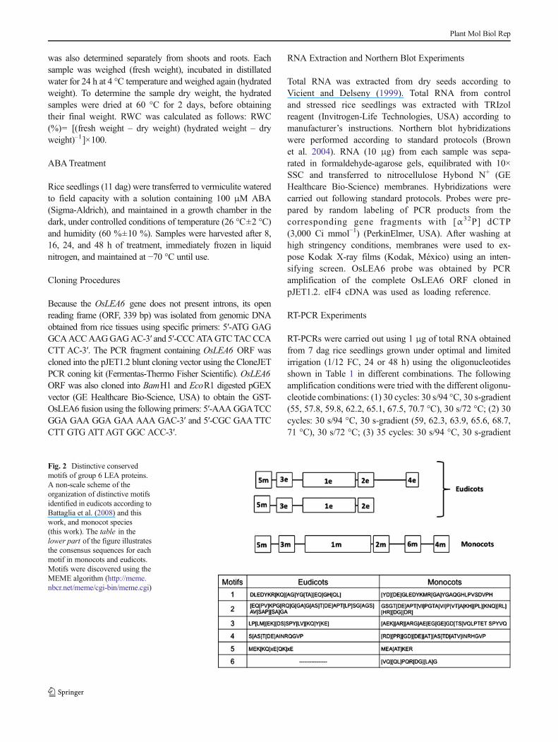
Fig. 2 Distinctive conservedmotifs of group 6 LEA proteins.A non-scale scheme of theorganization of distinctive motifsidentified in eudicots according toBattaglia et al. (2008) and thiswork, and monocot species(this work). The table in thelower part of the figure illustratesthe consensus sequences for eachmotif in monocots and eudicots.Motifs were discovered using theMEME algorithm (http://meme.nbcr.net/meme/cgi-bin/meme.cgi)
Plant Mol Biol Rep

(58.1, 60.8, 63.5, 66, 69.7 °C), 30 s/72 °C; and (4) 35 cycles:30 s/94 °C, 30 s-gradient (59.9, 65.6, 70 °C), 1min/72°C; (5) 35cycles: 30 s/94 °C, 30 s-gradient (63.9, 65.6 °C), 1 min/72 °C.
Protein Extraction and Western Blot
Total proteins from complete rice seedlings were extracted asdescribed by Hurkman and Tanaka (1986) with some modifi-cations. Frozen tissues were homogenized in extractionbuffer:phenol at 1:3 proportion [0.7 M sucrose, 0.5 M Tris-Base, 30 mM HCl, 2 % β−mercaptoethanol, 12 mg/ml PVPP(polyvinyl-polypyrrolidone)]. Organic phase was extractedagain with the same buffer:phenol proportion without PVPPand precipitated with 3 volumes of 0.1 M sodium acetate inmethanol. The pellet was washed once with cold 80% acetoneand solubilized in a buffer containing 20 mM Tris pH 8.0,50 mM NaCl and 0.1 % SDS. Proteins were quantified usingBradford assay with BioRad dye reagent. Total proteins(20–30 μg) were separated in 15 % polyacrylamide gelsin 0.1 M Tris, 0.1 M Tricine, 0.1 % SDS (Schägger andvon Jagow 1987). Proteins were transferred to nitrocellu-lose membranes (Hybond C; Amersham-GE HealthcareLife Sciences, USA), washed with PBS 1×, blocked with5 % skimmed milk (in PBS 1×) and probed with anti-OsLEA6 (1:2,000) antibodies. Membranes were washedand incubated with secondary antibody (1:20,000) (anti-rabbit horse radish peroxidase; Zymed-Life Technologies,USA) for 1 h at room temperature, washed with PBS 1×and developed with peroxidase substrates from SupersignalWest Pico Chemiluminiscent Substrate Kit (PierceBiotechnology, USA, and Thermo Fisher Scientific).Membranes were exposed to X-ray films (Kodak).
Fig. 4 OsLEA6 transcript accumulation patterns in response to waterdeficit treatments. (a) Northern blot experiments using total RNA (10 μg)obtained from complete rice seedlings (7 dag) grown under optimalirrigation (C) or under water deficit (D) and from rice dry seeds. (b)Northern blot experiments using total RNA obtained from shoots or rootsfrom rice seedlings (21 dag) grown under optimal irrigation (C) or underwater deficit (D) and from rice dry seeds. Shoot and root samples weretaken at different times as indicated. To detect the OsLEA6 transcript, aDNA fragment containing the completeOsLEA6 ORFwas used as probe.OseIF4 was used as RNA loading reference. The results in this figurewere reproduced in at least three independent experiments
Fig. 3 Organization of the genomic region containing the LEA6 gene ofOryza sativa as reported in the available genome databases. The upperpart of the figure shows the organization of the HSP90-OsLEA6 regionin chromosome 9 of O . sativa genome as deduced from the analysis inthis manuscript. Black and dark gray, light gray, and white boxes showexons, introns and intergenic regions (IR), respectively. The start and endof open reading frames (ORFs) are indicated by ATG and STOP, respec-tively. LEA6 ORF shows the two possible ATGs depending on the
information in the databases. The most upstream ATG corresponds tothe LEA6 sequence in the putative HSP90-LEA6 fusion, and the secondone to the LEA6 protein produced from the independent OsLEA6 gene.Arrowheads indicate the location and orientation of the oligonucleotidesused to analyze the possible transcripts synthesized from this region. Thepredicted transcripts produced from the two different versions of theHSP90-LEA6 genomic region are shown at the bottom of the scheme
Plant Mol Biol Rep

Production of Antibodies
OsLEA6 antibodies were produced using GST–OsLEA6 fu-sion protein. Recombinant proteins were obtained from 1 l ofE . coli cultures after induction with IPTG. To obtain theGST–OsLEA6 fusion protein, pelleted cells were resuspendedin 20 ml ice-cold PBS and lysed by sonication. After additionof Triton X-100 (1 % final concentration), cell debris werediscarded and supernatant was incubated with glutathione-agarose beads (Sigma-Aldrich) to purify the GST–OsLEA6fusion protein, following standard protocols (Harper andSpeicher 2011).
For polyclonal antibody production, 0.2 mg of purifiedfusion protein was emulsifiedwith complete Freund’s adjuvant(Gibco-Life Technologies, USA) at 1:1 ratio, homogenizedexhaustively, and used to immunize New Zealand femalerabbits, through multiple intradermal injections. Two addition-al doses, every 2 weeks, with incomplete adjuvant, were given.Pre-immune serum was previously taken. The IgG fractionwas precipitated from serum with saturated ammonium sul-phate and stored at 4 °C with 0.02 % sodium azide. To verifythe specific recognition of the antibodies for their own antigen,antibodies were cross-tested with common bean andArabidopsis homologous recombinant proteins by westernblot. The anti-OsLEA6 antibody does not recognize LEA6proteins from Arabidopsis or common bean. Membranes werealso incubated with pre-immune serum as a negative control.Before using the antibody for the different experiments, its
specificity was verified by competition assays using anti-OsLEA6 polyclonal antibody previously incubated with50 μg of the purified recombinant OsLEA6 protein.
Multiple Sequence Alignment and Phylogenetic Analysis
Multiple sequence alignments were built usingMUSCLE (http://www.ebi.ac.uk/Tools/msa/muscle/) with all the plant LEA6amino acid sequences found in public databases. Alignmentswere then used to generate a phylogenetic tree by ClustalW2-phylogeny program (http://www.ebi.ac.uk/Tools/phylogeny/clustalw2_phylogeny/) (Larkin et al 2007) and the phylogeneticanalysis was achieved using the Neighbor Joining algorithm.The tree topology was drawn using program FigTree v.1.1.2(http://tree.bio.ed.ac.uk/software/figtree/) (Goujon et al 2010).
Bioinformatic Analysis
The sequence analysis made was carried out consulting NCBI(http://www.ncbi.nlm.nih.gov/guide/data-software/) andPhytozome (http://www.phytozome.net/) databases and usingbioinformatic tools from ExPASy (http://expasy.org/). Analysisof the intergenic region between OsHSP90 and OsLEA6 wasanalyzed for putative promoter cis-elements using PLACE (ADatabase of Plant Cis-acting Regulatory DNA Elements, http://www.dna.affrc.go.jp/PLACE/signalup.html), EPD (EukaryoticPromoter Database, http://epd.vital-it.ch/cgi-bin/fetch_biomart.cgi), and Plant Promoter Database (PlantPromDB-Softberry,
Fig. 5 Relative water content (RWC) of 7 and 21 dag rice seedlings grownunder well-irrigated (C) and water deficit conditions (WD). (a) RWC fromcomplete 7 dag rice seedlings. (b) RWC from complete 21 dag rice
seedlings. RWC from shoots (c) and roots (d) of 21 dag rice seedlings.Levels of significance are represented by *P<0.05 and ***P<0.001.Statistical analysis was carried by ANOVA and Tukey’s post hoc test
Plant Mol Biol Rep

http://linux1.softberry.com/berry.phtml?topic=plantprom&group=data&subgroup=plantprom). Motifs were discoveredusing the MEME algorithm (http://meme.nbcr.net/meme/cgi-bin/meme.cgi), and alignments were done using the MUSCLEalgorithm (http://www.drive5.com/muscle; Edgar 2004).PONDR scores were calculated using the PONDRVX-LT ver-sion (http://www.pondr.com; Li et al. 1999; Romero et al. 1997,2001).
Results and Discussion
Group 6 LEA Proteins in Monocotyledones
In a previous work, we identified four distinctive motifs ingroup 6 LEA proteins (LEA6) (Pfam-10714) from variousplant species among angiosperms, three of them common toall plant species analyzed (Battaglia et al. 2008). This formeranalysis showed that, in contrast to Eudicotyledones species(eudicots), all nine Monocotyledones species (monocots) in-cluded in this comparative study present the four characteristicmotifs of this group. To search for possible particular motifswithin group 6 LEA proteins from monocots, we updated themultiple alignment analysis using all LEA6 protein sequencesin the available databases, 15 more sequences than in theprevious analysis were included (NCBI, http://www.ncbi.nlm.nih.gov/guide/data-software/; Phytozome, http://www.phytozome.net/) (Fig. 1 and Fig. 1S). This scrutiny allowedus to identify five distinctive motifs of this family for eudicots,which were also present inmonocots, as well as one additionalmotif (6 m) located between motifs 2 m and 4 m, only presentin LEA6 proteins from monocot species (Figs. 1, 2). Analysisof the conserved motifs 1–5 showed some divergence whencompared to the eudicot motifs; these differences are con-served among all monocots, such that these variants of motifs1–5 distinguish LEA6 proteins from eudicots (Figs. 1, 2; Fig.2S). This comparison also revealed some amino acid residueshighly conserved among eudicots and monocots, in particularmost of the tyrosines (Y) and the surrounding residues, whichshow 73–100 % conservation (Figs. 1, 2; Fig. 1S). The aminoacid sequence conservation among monocot LEA6 proteinsand the presence of only one gene in each of the analyzedspecies agree with their segregation in a unique clade in thephylogenetic analysis (Fig. 3S). This analysis also suggeststhat this family is more diverse in eudicots, in which there areLEA6 genes distant to monocots as well as others closelyrelated. That is the case of the three Arabidopsis thalianaLEA6 genes, from which two genes (AtLEA6-1 andAtLEA6-2) seem to share a closer common ancestor thanAtLEA6-3 . A more detailed analysis should be performedwhen more monocot sequences become available.
The fact that none of the identified conserved motifsmatch with some known conserved domain in proteins
has circumvented the prediction of a possible function.It has been shown that a LEA6 protein from the com-mon bean is unable to prevent the inactivation of lactateor malate dehydrogenases during in vitro partial dehy-dration (Reyes et al. 2005), suggesting that, in contrastto LEA proteins from groups 2, 3, 4, or 7 (for review,see Battaglia et al. 2008), group 6 LEA proteins do notpossess this protective property. A functional analysis ofproteins in this family from dicot and monocot speciesis in progress.
LEA6 Gene and Its Genomic Organization
The analysis of the LEA6 genes in those monocot genomescompletely sequenced to date (Oryza sativa , Zea mays ,Sorghum bicolor , Brachypodium distachyon , and Setariaitalica ) showed that none of the LEA6 genes contain intronsin their structure, with ORFs that range from 105 (11.32 kDa)in B . distachyon to 117 (12.16 kDa) amino acid residues in Z .mays and S . bicolor, and a theoretical pI range of 5.29 for
Fig. 6 OsLEA6 protein accumulation patterns in response to waterdeficit (a) and ABA (b) treatments. (a) Total protein extracts (20 μg)obtained after different times of treatment from complete rice seedlingsgrown under optimal irrigation (C ) or under water deficit (D) wereseparated in SDS-PAGE, transferred to nitrocellulose membranes andincubated with anti-OsLEA6 polyclonal antibodies (1:2,000). Mem-branes were washed and incubated with secondary antibodies coupledto peroxidase (1:5,000) (see “Materials and methods” for details). Proteinextracts from dry seeds were also obtained and included in this analysis.In this case, a smaller amount of total protein (5 μg) was loaded to avoidsaturation of the signal. The line at the right side of the dry seed laneindicates the migration of the 17-kDa molecular mass marker. (b) Totalprotein extracts (30 μg) obtained from complete rice seedlings (11 dag)after 8 and 16 h ofABA (100μM) treatment were separated and treated asdescribed in (a). Protein extracts from non-treated plants were alsoincluded as controls (C). Ponceau S staining of nitrocellulose membraneshows protein loading. The results in this figure were reproduced in atleast three independent experiments
Plant Mol Biol Rep

ZmLEA6 to 6.28 for SbLEA6 proteins. Regarding the numberof LEA 6 genes per genome, all the analyzed monocotsshowed only one LEA6 gene.
Concerning the analysis of the genomic organization sur-rounding the LEA6 genes in these monocot species, ouranalysis showed that, in all these genomes, a gene encodinga putative aspartyl-protease was located downstream of its 3′end, whereas upstream of its 5′ end, the identity of the geneswas more heterogeneous. A gene for a FAS-associated proteinwas found in B . distachyon , a sodium/hydrogen exchangerwas identified in S . italica , no gene was reported in the case ofZ .mays , whereas inO . sativa and S . bicolor a gene encodingan HSP90 protein was found.
In rice, two genome organization versions in this region werefound. The IRGSP (International Rice Genome Sequencing
Project) showed two identical copies of the LEA6 gene(Os09g0482300 and Os09g0482550), both located in chromo-some 9; however, even though theMSU-RGAP (Michigan StateUniversity-Rice Genome Annotation Project) also showed twoLEA6 copies in chromosome 9 (LOC_Os09g30438 andLOC_Os09g30418), one of them showed a different organiza-tion.MSU-RGAP showed that LEA6 gene (LOC_Os09g30418)encodes a larger protein (830 amino acid residues) when com-pared with the different LEA6 proteins found in other monocotspecies. Analysis of this ORF sequence revealed that 698 aminoacid residues at its N-terminal region are highly similar to thecytosolic HSP90, whereas 112 amino acid residues at thecarboxy-terminal region correspond to a complete LEA6 protein.In themiddle region of the protein, we found 20 amino acids thatdid not show similarity to any other sequence in databases
Fig. 7 OsLEA6 transcript andprotein sequences and somephysicochemical properties. aNucleotide sequence of OsLEA6transcript showing a length of 776nt. The transcription start and endsites were defined according tothe ESTs reported in databases.Uppercase letters illustrate theOsLEA6 amino acid sequence(112 aa) as deduced from the ORFin the cDNAs cloned and in theavailable ESTs in databases. bHydropathic index (HI) ofOsLEA6 amino acid sequenceaccording to Kyte and Doolitle(1982). Hydrophilic amino acidresidues show negative HI values(vertical lines below horizontalline), whereas hydrophobicamino acid residues show apositive HI (vertical lines abovehorizontal line). Residuenumbers are ordered from amino-to carboxy-terminus of theprotein. c The OsLEA6 structuralorder predicted by PONDRalgorithm, which assigns higherscores to disordered than toordered regions in the protein.The line illustrates the structuralorder along the OsLEA6 proteinsequence. PONDR scores werecalculated using the PONDRVX-LT version (http://www.pondr.com)
Plant Mol Biol Rep

(Fig. 3). A similar gene organization was also detected in NCBItranscript database sequences (Genebank EEE69924). Becauseno internal stop codon was present in these cDNAs, this findingsuggested that one of the LEA6 proteins encoded in the ricegenome was forming a chimeric protein with HSP90 chaperone;however, it was also possible that the region, that in thesedatabases was recognized as an intron, could actually correspondto the LEA6 gene promoter, which during genome sequenceassembly was not detected as such. To address this question, wecharacterized the OsLEA6 transcript and protein as well as theirexpression patterns in rice dry seeds and seedlings subjected towater deficit.
OsLEA6 Transcript and Protein Accumulation Patternsin Response to Water deficit
The existence of a transcript encoding a chimeric proteinproduct of the fusion ofHSP90 and LEA6 genes was exploredby northern blot experiments and by RT-PCR using differentsets of oligonucleotides, RNA from rice seedlings grownunder optimal irrigation or under water deficit conditions(Table 1), that would allow to detect the possible fusiontranscript described in the databases mentioned above. Aftertrying various RT-PCR conditions with different oligonucleo-tides combinations (see “Materials and Methods” for details),no transcript containing the sequences of both genes wasdetected, in agreement with the absence of hybrid ESTs inNCBI databases. Negative results were also obtained from
northern blot experiments. These data indicated that the orga-nization for the OsLEA6 gene presented in these databases isnot correct, possibly due to limitations in the algorithms usedfor their sequence analysis.
Instead, northern blot experiments exhibited the existence ofa transcript with the size expected for theOsLEA6 mRNA (776nucleotides, nt) identified in this genomic region (Fig. 4a). Thedetection of this transcript was possible only from RNAobtained from dry seeds or from rice seedlings grown underwater deficit (Fig. 4), as it has been reported for other plantspecies (Colmenero-Flores et al. 1997; Bies-Ethève et al. 2008).LEA6 transcript was detected 8 h after rice seedlings wereexposed to water limitation, and its accumulation increasedwith time, showing its responsiveness to this stress condition,with the dryness of the seed being the condition leading to thelargest accumulation. This response was more evident in rootsthan in shoots (Fig. 4b), an effect that was also shown in thecommon bean (Phaseolus vulgaris L.) (Colmenero-Flores et al.1997, 1999). To address potential causes for the differentialtranscript accumulation between 7 and 21 dag seedlings uponwater deficit, the RWC was determined. The results showed areduction in the RWC of rice seedlings upon water restrictionand indicate that water status in stressed plants from bothexperiments was similar (Fig. 5a, b). However, the highertranscript accumulation in 7 dag seedlings compared to thatobtained for 21 dag seedlings suggests that OsLEA6 geneexpression may be modulated by additional factors associatedwith the developmental stage. The reduction in RWC for roots
Fig. 8 Cis regulatory elements in the OsLEA6 gene promoter region.Putative promoter elements in the intergenic region betweenOsHSP90 andOsLEA6 genes were predicted using PLACE algorithm as described in“Materials andMethods”. This analysis showed various cis-elements alongthis genomic region, shown by boxes with different fillings , as indicated in
the scheme. The numbers below the black horizontal line indicate thelength of the analyzed region. The black horizontal bar shows the regioncontaining putative ABA Responsive Elements (ABRE) whereas the whitesquares indicate the location of putative grass Drought Responsive Ele-ments (DRE). ATG indicates the translation initiation codon
Plant Mol Biol Rep

and shoots of 21 dag seedlings also showed that water deficittreatments induced a decrease in plant water content in bothplant regions, consistent with the induction of transcript accu-mulation observed for roots and, although in lower intensity,also in shoots (Fig. 5c, d).
In agreement with the transcript accumulation pattern,western blot experiments using OsLEA6 protein specific an-tibodies showed a similar accumulation pattern for this proteinin response to water deficit (Fig. 6a). An evident accumulationwas detected after 16 h treatment with a patent increment after48 h under water limitation.
As for other LEA 6 proteins, OsLEA6 also responded toABA addition, as shown in Fig. 6b, where ABA treatmentsled to an increase in LEA6 protein accumulation. This re-sponse is in agreement with the ABA responsive cis elementsidentified in the promoter region of the corresponding gene(see below).
The specificity of the signal obtained in western blot exper-iments using the anti-OsLEA6 antibody was verified by com-petition experiments using purified OsLEA6 protein such thatthe competed antibody is not longer able to efficiently recog-nize the polypeptide band identified as the OsLEA6 protein(Fig. 4S, indicated by arrows). As shown in Fig. 6, the detectedprotein migrates in SDS-PAGE with an apparent molecularmass of approximately 18 kDa even though the predictedmolecular mass is 11.89 KDa, corresponding to the 112 aminoacids in its ORF. This observation is consistent with the highlevels of structural disorder predicted according to its aminoacid composition (Fig. 7), which shows a low content ofhydrophobic residues, only one cysteine, and a high contentof charged residues and small amino acids (Gly, Thr, Ala)(Table 1S). These physicochemical properties qualifyOsLEA6 proteins as an intrinsically unstructured protein(IUP), an attribute that seems to be characteristic for proteinsin this family (Colmenero-Flores et al. 1997; Battaglia et al.2008).
OsLEA6 Promoter Contains Drought and ABA ResponseElements
The analysis of the 964-bp intergenic region betweenOsHSP90 and OsLEA6 genes showed various sequences thatcould be identified as putative promoter elements as shown inFig. 8. Among these sequences, cis -response elements asso-ciated to the plant response to water deficit were detected.That is the case of the ABRE-motif, associated to ABA-responsive genes, found three times clustered between 650and 750 nt, and the DREB-motif related to drought responses,found twice in a region between 450 and 529 nt. Additionalcis -regulatory elements associated to the ABA response werefound close to the ABRE-motifs such as the MYC-, MYB-,DPBF-, and LTR- boxes. MYC and MYB motifs were alsofound between 360 and 420 nt, 200 and 205 nt, and at 89 nt. A
putative sugar repression box (TATCCA) described as bindingsite of transcription factors mediating sugar and hormoneregulation of the rice α-amylase gene was also identified(Fig. 8). Further analysis is needed to validate the participationof these cis elements in the regulation of the OsLEA6 geneexpression.
In conclusion, in this work, we showed that the HSP90 andLEA6 genes from O . sativa ssp. indica independently tran-scribed from their own promoters, even though someO . sativadatabases suggest the production of a hybrid transcriptencoding a chimeric HSP90–LEA6 protein. We also showedthat the OsLEA6 gene responds to water deficit conditions andto ABA treatment, as in other plant species, by accumulating itstranscript and protein upon these treatments. The finding of cisregulatory elements in the OsLEA6 promoter region is consis-tent with these observations. Finally, we identified one addi-tional motif for group 6 LEA proteins that distinguish monocotLEA6 proteins from their dicot counterparts. This is the firstreport for group 6 LEA genes and proteins in monocots de-scribing their responsiveness to water deficit conditions, andthus further supporting the participation of LEA proteins andmore broadly hydrophilins in stress responses in plants.
Acknowledgements We are grateful to Dr. J.L. Reyes for critical andlanguage review of this manuscript, to J. Mazari for technical support, tothe Sequencing and Oligonucleotide Synthesis Facility of the Instituto deBiotecnología-UNAM for the synthesis of oligonucleotides and DNAsequences needed for this work, and to CONACyT-Mexico (132258-Q)and DGAPA-UNAM (IN208212) for providing funding for this work.
References
Battaglia M, Olvera-Carrillo Y, Garciarrubio A, Campos F, CovarrubiasAA (2008) The enigmatic LEA proteins and other hydrophilins.Plant Physiol 148:6–24
Bies-Ethève N, Gaubier-Comella P, Debures A, Lasserre E, Jobet E,Raynal M, Cooke R, Delseny M (2008) Inventory, evolution andexpression profiling diversity of the LEA (late embryogenesis abun-dant) protein gene family in Arabidopsis thaliana . Plant Mol Biol67:107–124
Brown T, Mackey K, Du T (2004) Analysis of RNA by Northern and slotblot hybridization. Curr Protoc Mol Biol 67:4.9.1–4.9.19
Colmenero-Flores JM, Campos F, Garciarrubio A, Covarrubias AA(1997) Characterization of Phaseolus vulgaris cDNA clones re-sponsive to water deficit: identification of a novel late embryogen-esis abundant-like protein. Plant Mol Biol 35:393–405
Colmenero-Flores JM, Moreno LP, Smith C, Covarrubias AA (1999)PvLEA-18, a member of a new late-embryogenesis-abundant pro-tein family that accumulates during water stress and in the growingregions of well-irrigated bean seedlings. Plant Physiol 120:93–103
Dure L (1993) Structural motifs in LEA proteins. In: Close TJ, Bray EA(eds) Plant responses to cellular dehydration during environmentalstress. The American Society of Plant Physiologists, Rockville, pp91–103
Dure L, Greenway SC, Galau GA (1981) Developmental biochemistry ofcottonseed embryogenesis and germination: changing messengerribonucleic acid populations as shown by in vitro and in vivo proteinsynthesis. Biochemistry 20:4162–4168
Plant Mol Biol Rep

Edgar RC (2004) MUSCLE: multiple sequence alignment with highaccuracy and high throughput. Nucleic Acids Res 32:1792–1797
Garay-Arroyo A, Colmenero-Flores JM, Garciarrubio A, CovarrubiasAA (2000) Highly hydrophilic proteins in prokaryotes and eukary-otes are common during conditions of water deficit. J Biol Chem275:5668–5674
Goujon M, McWilliam H, Li W, Valentin F, Squizzato S, Paern J, LopezR (2010) A new bioinformatics analysis tools framework at EMBL-EBI. Nucleic Acids Res 38(Suppl):W695–W699
Hand SC, Menze MA, Toner M, Boswell, Moore D (2011) LEA proteinsduring water stress not just for plants anymore. Annu Rev Physiol73:115–134
Harper S, Speicher DW (2011) Purification of proteins fused to glutathi-one S-transferase. In: Protein Chromatography. Methods Mol Biol681:259–280
Hundertmark M, Hincha DK (2008) LEA (Late EmbryogenesisAbundant) proteins and their encoding genes in Arabidopsisthaliana . BMC Genomics 9:118–139
HurkmanWJ, Tanaka CK (1986) Solubilization of plant membraneproteinsfor analysis by two-dimensional gel electrophoresis.Plant Physiol 81:802–806
Ingram J, Bartels D (1996) The molecular basis of dehydration tolerancein plants. Annu Rev Plant Biol 47:377–403
Kyte J, Doolittle RF (1982) A simple method for displaying the hydro-pathic character of a protein. J Mol Biol 157:105–132
Larkin MA, Blackshields G, Brown NP, Chenna R, McGettigan PA,McWilliam H, Valentin F, Wallace IM, Wilm A, Lopez R,Thompson JD, Gibson TJ, Higgins DG (2007) ClustalW andClustalX version 2 . Bioinformatics 23:2947–2948
LiX, Romero P, RaniM,DunkerAK,Obradovic Z (1999) Predicting proteindisorder for N-, C-, and internal regions. Genome Inform 10:30–40
Moreno-Fonseca LP, Covarrubias AA (2001) Downstream DNA se-quences are required to modulate Pvlea-18 gene expression inresponse to dehydration. Plant Mol Biol 45:501–515
Reyes JL, Rodrigo MJ, Colmenero-Flores JM, Gil JV, Garay-Arroyo A,Campos F, Salamini F, Bartels D, Covarrubias AA (2005)Hydrophilins from distant organisms can protect enzymatic activitiesfrom water limitation effects in vitro . Plant Cell Environ 28:709–718
Romero P, Obradovic Z, Dunker AK (1997) Sequence data analysis forlong disordered regions prediction in the calcineurin family.Genome Inform 8:110–124
Romero P, Obradovic Z, Li X, Garner E, Brown C, Dunker AK (2001)Sequence complexity of disordered proteins. Proteins: Struct FunctGen 42:38–48
Schägger H, von Jagow G (1987) Tricine-sodium dodecyl sulfate-polyacrylamide gel electrophoresis for the separation of proteins inthe range from 1 to 100 kDa. Anal Biochem 166:368–379
Shinozaki K, Yamaguchi-Shinozaki K (2007) Gene networks involved indrought stress response and tolerance. J Exp Bot 58:221–227
Sun X, Rikkerink EHA, Jones WT, Uversky VN (2013) Multifariousroles of intrinsic disorder in proteins illustrate broad impact on plantbiology. Plant Cell 25:38–55
Taiz L, Zeiger E (1991) Water balance of the plant, In: Plant Physiology.Benjamin/Cummings, San Francisco, pp 81–99
Tompa P, Kovacs D (2010) Intrinsically disordered chaperones in plantsand animals. Biochem Cell Biol 88:167–174
Vicient CM, Delseny M (1999) Isolation of total RNA from Arabidopsisthaliana seeds. Anal Biochem 268:412–413
Plant Mol Biol Rep
Copyright © 2022 FDOKUMEN