GPR84 deficiency reduces microgliosis, but accelerates dendritic degeneration and cognitive decline...
9
GPR84 deficiency reduces microgliosis, but accelerates dendritic degeneration and cognitive decline in a mouse model of Alzheimer’s disease Julie Audoy-Rémus a , Lusine Bozoyan a , Aline Dumas a , Mohammed Filali a , Cynthia Lecours a , Steve Lacroix a,b , Serge Rivest a,b , Marie-Eve Tremblay a,b , Luc Vallières a,b,⇑ a Axis of Neuroscience, University Hospital Center of Quebec, Quebec, QC, Canada b Department of Molecular Medicine, Laval University, Quebec, QC, Canada article info Article history: Received 3 September 2014 Received in revised form 6 January 2015 Accepted 6 January 2015 Available online xxxx Keywords: Neuroinflammation Neurodegeneration Microglial cells Chemotaxis b-Amyloid Multiple sclerosis Endotoxemia Lipopolysaccharide G-protein coupled receptor 84 abstract Microglia surrounds the amyloid plaques that form in the brains of patients with Alzheimer’s disease (AD), but their role is controversial. Under inflammatory conditions, these cells can express GPR84, an orphan receptor whose pathophysiological role is unknown. Here, we report that GPR84 is upregulated in microglia of APP/PS1 transgenic mice, a model of AD. Without GPR84, these mice display both accel- erated cognitive decline and a reduced number of microglia, especially in areas surrounding plaques. The lack of GPR84 affects neither plaque formation nor hippocampal neurogenesis, but promotes dendritic degeneration. Furthermore, GPR84 does not influence the clinical progression of other diseases in which its expression has been reported, i.e., experimental autoimmune encephalomyelitis (EAE) and endotoxic shock. We conclude that GPR84 plays a beneficial role in amyloid pathology by acting as a sensor for a yet unknown ligand that promotes microglia recruitment, a response affecting dendritic degeneration and required to prevent further cognitive decline. Ó 2015 Elsevier Inc. All rights reserved. 1. Introduction The amyloid cascade hypothesis states that altered processing of amyloid precursor protein lead to the release of b-amyloid pep- tides (e.g., Ab42), which initiate the pathological process underly- ing AD (Tanzi and Bertram, 2005). These peptides can aggregate into soluble oligomers and then into insoluble fibrils that accumu- late to form macroscopic amyloid plaques. Whether all of these forms are toxic and how they cause toxicity are questions that are still debated. A prevailing view is that the oligomers, more toxic than the fibrils, bind to and alter the function of certain mem- brane proteins, resulting in synaptic dysfunction, dendritic degen- eration and neuronal death (Benilova et al., 2012). Any brain damage, including that caused by b-amyloid, triggers activation of microglia, the resident immune cells of the CNS. These cells cluster around amyloid plaques, in which they extend cyto- plasmic processes (Combs, 2009). The significance of this response is controversial, but one possibility is that microglia exert an over- all beneficial effect, as suggested by behavioral studies showing that microglia depletion or genetic deletions affecting microglia accelerate cognitive decline in a model of AD (i.e., the APP/PS1 transgenic mouse) (Simard et al., 2006; Naert and Rivest, 2011; Song et al., 2011). However, this effect does not seem to be related to the ability of microglia to eliminate parenchymal amyloid pla- ques, as the latter were not affected (Grathwohl et al., 2009; Mildner et al., 2011) or only modestly (Simard et al., 2006; Tahara et al., 2006; Naert and Rivest, 2011; Song et al., 2011) in the above paradigms. An opposite possibility is that chronically activated microglia exert deleterious effects, for example, by phagocytosing neuronal elements and producing potentially neu- rotoxic molecules such as TNF (Tan et al., 1999, 2002; Fuhrmann et al., 2010; Lee et al., 2010; Weitz and Town, 2012). Further stud- ies on the molecular mediators responsible for such effects are required to clarify the seemingly dichotomous roles of microglia in AD. Activated microglia express GPR84, a seven-transmembrane domain receptor of the rhodopsin superfamily that shows limited http://dx.doi.org/10.1016/j.bbi.2015.01.010 0889-1591/Ó 2015 Elsevier Inc. All rights reserved. ⇑ Corresponding author at: Axis of Neuroscience, University Hospital Center of Quebec, 2705 Laurier Boulevard, Room T2-50, Quebec, QC G1V 4G2, Canada. Tel.: +1 418 525 4444x46233; fax: +1 418 654 2761. E-mail address: [email protected] (L. Vallières). Brain, Behavior, and Immunity xxx (2015) xxx–xxx Contents lists available at ScienceDirect Brain, Behavior, and Immunity journal homepage: www.elsevier.com/locate/ybrbi Please cite this article in press as: Audoy-Rémus, J., et al. GPR84 deficiency reduces microgliosis, but accelerates dendritic degeneration and cognitive decline in a mouse model of Alzheimer’s disease. Brain Behav. Immun. (2015), http://dx.doi.org/10.1016/j.bbi.2015.01.010
Transcript of GPR84 deficiency reduces microgliosis, but accelerates dendritic degeneration and cognitive decline...
Brain, Behavior, and Immunity xxx (2015) xxx–xxx
Contents lists available at ScienceDirect
Brain, Behavior, and Immunity
journal homepage: www.elsevier .com/locate /ybrbi
GPR84 deficiency reduces microgliosis, but accelerates dendriticdegeneration and cognitive decline in a mouse model of Alzheimer’sdisease
http://dx.doi.org/10.1016/j.bbi.2015.01.0100889-1591/� 2015 Elsevier Inc. All rights reserved.
⇑ Corresponding author at: Axis of Neuroscience, University Hospital Center ofQuebec, 2705 Laurier Boulevard, Room T2-50, Quebec, QC G1V 4G2, Canada. Tel.: +1418 525 4444x46233; fax: +1 418 654 2761.
E-mail address: [email protected] (L. Vallières).
Please cite this article in press as: Audoy-Rémus, J., et al. GPR84 deficiency reduces microgliosis, but accelerates dendritic degeneration and codecline in a mouse model of Alzheimer’s disease. Brain Behav. Immun. (2015), http://dx.doi.org/10.1016/j.bbi.2015.01.010
Julie Audoy-Rémus a, Lusine Bozoyan a, Aline Dumas a, Mohammed Filali a, Cynthia Lecours a,Steve Lacroix a,b, Serge Rivest a,b, Marie-Eve Tremblay a,b, Luc Vallières a,b,⇑a Axis of Neuroscience, University Hospital Center of Quebec, Quebec, QC, Canadab Department of Molecular Medicine, Laval University, Quebec, QC, Canada
a r t i c l e i n f o a b s t r a c t
Article history:Received 3 September 2014Received in revised form 6 January 2015Accepted 6 January 2015Available online xxxx
Keywords:NeuroinflammationNeurodegenerationMicroglial cellsChemotaxisb-AmyloidMultiple sclerosisEndotoxemiaLipopolysaccharideG-protein coupled receptor 84
Microglia surrounds the amyloid plaques that form in the brains of patients with Alzheimer’s disease(AD), but their role is controversial. Under inflammatory conditions, these cells can express GPR84, anorphan receptor whose pathophysiological role is unknown. Here, we report that GPR84 is upregulatedin microglia of APP/PS1 transgenic mice, a model of AD. Without GPR84, these mice display both accel-erated cognitive decline and a reduced number of microglia, especially in areas surrounding plaques. Thelack of GPR84 affects neither plaque formation nor hippocampal neurogenesis, but promotes dendriticdegeneration. Furthermore, GPR84 does not influence the clinical progression of other diseases in whichits expression has been reported, i.e., experimental autoimmune encephalomyelitis (EAE) and endotoxicshock. We conclude that GPR84 plays a beneficial role in amyloid pathology by acting as a sensor for a yetunknown ligand that promotes microglia recruitment, a response affecting dendritic degeneration andrequired to prevent further cognitive decline.
� 2015 Elsevier Inc. All rights reserved.
1. Introduction
The amyloid cascade hypothesis states that altered processingof amyloid precursor protein lead to the release of b-amyloid pep-tides (e.g., Ab42), which initiate the pathological process underly-ing AD (Tanzi and Bertram, 2005). These peptides can aggregateinto soluble oligomers and then into insoluble fibrils that accumu-late to form macroscopic amyloid plaques. Whether all of theseforms are toxic and how they cause toxicity are questions thatare still debated. A prevailing view is that the oligomers, moretoxic than the fibrils, bind to and alter the function of certain mem-brane proteins, resulting in synaptic dysfunction, dendritic degen-eration and neuronal death (Benilova et al., 2012).
Any brain damage, including that caused by b-amyloid, triggersactivation of microglia, the resident immune cells of the CNS. Thesecells cluster around amyloid plaques, in which they extend cyto-
plasmic processes (Combs, 2009). The significance of this responseis controversial, but one possibility is that microglia exert an over-all beneficial effect, as suggested by behavioral studies showingthat microglia depletion or genetic deletions affecting microgliaaccelerate cognitive decline in a model of AD (i.e., the APP/PS1transgenic mouse) (Simard et al., 2006; Naert and Rivest, 2011;Song et al., 2011). However, this effect does not seem to be relatedto the ability of microglia to eliminate parenchymal amyloid pla-ques, as the latter were not affected (Grathwohl et al., 2009;Mildner et al., 2011) or only modestly (Simard et al., 2006;Tahara et al., 2006; Naert and Rivest, 2011; Song et al., 2011) inthe above paradigms. An opposite possibility is that chronicallyactivated microglia exert deleterious effects, for example, byphagocytosing neuronal elements and producing potentially neu-rotoxic molecules such as TNF (Tan et al., 1999, 2002; Fuhrmannet al., 2010; Lee et al., 2010; Weitz and Town, 2012). Further stud-ies on the molecular mediators responsible for such effects arerequired to clarify the seemingly dichotomous roles of microgliain AD.
Activated microglia express GPR84, a seven-transmembranedomain receptor of the rhodopsin superfamily that shows limited
gnitive

2 J. Audoy-Rémus et al. / Brain, Behavior, and Immunity xxx (2015) xxx–xxx
similarity to other known receptors (Fredriksson et al., 2003; Foordet al., 2005). In the CNS, GPR84 expression is restricted to microgliaand observed in different pathological conditions, including EAE,endotoxemia, cuprizone-induced demyelination and axotomy(Bedard et al., 2007; Bouchard et al., 2007; Gamo et al., 2008). Inthe periphery, GPR84 is mainly expressed by cells of the myeloidlineage, such as monocytes, macrophages and neutrophils(Yousefi et al., 2001; Wang et al., 2006; Suzuki et al., 2013). In vitrostudies have revealed that GPR84 signals through a Gi/o pathway inresponse to hydroxylated (and to a lesser extent nonhydroxylated)medium-chain free fatty acids (FFAs) of 9–14 carbons in length(Wang et al., 2006; Suzuki et al., 2013). These can induce chemo-taxis and amplify lipopolysaccharide (LPS)-induced cytokine pro-duction in myeloid cells (Wang et al., 2006; Suzuki et al., 2013).Nevertheless, the pathophysiological role of GPR84 and the natureof its endogenous ligand(s) remain unknown.
The goals of the present study were to: (1) examine the expres-sion of GPR84 in APP/PS1 mice; and (2) determine its importancein this model and two others in which GPR84 expression has beenreported, i.e., EAE and endotoxic shock (Bouchard et al., 2007). Ourresults reveal that GPR84 exerts no significant effect on the pro-gression of the latter two diseases, but promotes microgliosis anddendritic homeostasis in APP/PS1 mice. Therefore, this studyascribes for the first time a role to GPR84 in vivo.
2. Materials and methods
2.1. Animals
GPR84-deficient mice were obtained from DeltaOne (Deltagen)and bred with C57BL/6 J mice (Jackson Laboratory) for at least 5generations. These mice are reported to be indistinguishable fromwild-type littermates under normal conditions (Venkataraman andKuo, 2005). APP/PS1 transgenic mice (Borchelt et al., 1997) (Jack-son Laboratory, B6C3-Tg(APP695)3Dbo Tg(PSEN1)5Dbo/J) express-ing a chimeric amyloid precursor protein (APPSwe) and humanpresenilin 1 (A246E variant) under the control of the mouse prionprotein promoter were bred with GPR84-deficient mice for at least3 generations. The genotypes were confirmed by PCR as recom-mended by the providers (primers used: GPR84, 50-aca-gctcagatgccaacttctcctg-30, 50-tcctagagcaatgagacagagggtg-30 and 50-gacgagttcttctgaggggatcgatc-30; APP, 50-gactgaccactcgaccaggttctg-30 and 50-cttgtaagttggattctcatatccg-30; PS1, 50-aatagagaacggcag-gagca-30 and 50-gccatgagggcactaatcat-30). The experiments wereperformed with the approval of our institutional animal ethicscommittee. Sex-matched and age-matched wild-type littermateswere used as controls. EAE and LPS-treated mice were 2 monthsof age, whereas APP/PS1 mice were tested at 2–12 months of age.
2.2. EAE induction and clinical evaluation
Mice were subcutaneously injected into both flanks with a totalof 200 ll of emulsion containing 300 lg of myelin oligodendrocyteglycoprotein (MOG) peptide 35–55 (AnaSpec) dissolved in salineand mixed with an equal volume of complete Freund’s adjuvantcontaining 500 lg of killed Mycobacterium tuberculosis H37 RA(Difco Laboratories). The mice were also injected intraperitoneallywith 20 lg/kg of pertussis toxin (List Biological Laboratories)immediately and 2 days after immunization. The clinical signs ofEAE were scored daily using the following scale: 0, no detectablesign; 1, tail flaccidity; 2, hindlimb weakness and poor righting abil-ity; 3, hindlimb paralysis/paresis; 4, hindlimb paralysis and fore-limb paraparesis; 5, dead or sacrificed for humane reasons.
Please cite this article in press as: Audoy-Rémus, J., et al. GPR84 deficiency rdecline in a mouse model of Alzheimer’s disease. Brain Behav. Immun. (2015)
2.3. LPS treatment and sickness behavior assessment
LPS from Escherichia coli O55:B5 (Sigma–Aldrich) was dissolvedin pyrogen-free saline and injected intraperitoneally at a dose of1 mg/kg. At the indicated time points, the mice were placed inthe stem of a T-maze made of clear Plexiglas (stem: 64 � 12 cm;arms: 30 � 12 cm; height of the walls: 16 cm) and divided intothirteen segments (10 � 10 cm). The locomotor activity of the micewas evaluated by counting the number of rearings and segmentscrossed during a 4-min session. A segment was counted whencrossed by the whole body, excluding the tail. The T-maze wascleaned after each session.
2.4. Cognitive analysis
The cognitive performance of APP/PS1 mice was evaluated in aT-maze (see specifications above) filled with water (23 ± 1 �C) at aheight of 12 cm and containing a platform (11 � 11 cm) sub-merged 1 cm beneath the surface at the end of one or both arms(Filali and Lalonde, 2009). A trial consisted of placing the mousein the stem of the maze and allowing it to swim toward the leftor right arm until it find a platform. If the platform was not foundafter 60 s, the mouse was gently guided toward it. The mouse wasallowed to rest for 20 s on the platform after each trial and for10 min in its cage after each 10-trial block. During the acquisitionphase, 2 training trials were performed with a platform placed inboth arms, and then a maximum of 48 additional trials were per-formed without the platform chosen during the second trainingtrial. The acquisition phase was considered completed when themouse passed the criterion of 5 consecutive errorless trials (correctturn and platform found within 60 s). Two days later, the reversalphase was conducted using the same protocol, except that the plat-form was moved to the opposite arm and that there was no train-ing. The number of trials-to-criterion and the time to reach theplatform (escape time) were recorded.
2.5. Immunostaining
Immunohistochemistry was performed using rabbit anti-Iba1(1:2000, Wako Chemicals), goat anti-doublecortin (DCX) (1:500,Santa Cruz Biotechnology) and rabbit anti-Ki67 (1:500, Abcam)antibodies, as described previously (Audoy-Remus et al., 2008).
2.6. In situ hybridization
Brain sections were analyzed for GPR84 mRNA by radioisotopicin situ hybridization as described previously (Bouchard et al.,2007).
2.7. Congo red staining
Free-floating sections were stained for 30 min in 0.5% Congo reddiluted in 50% ethanol and differentiated (5–10 dips) in 0.01%NaOH diluted in 50% ethanol.
2.8. Stereological analyses
Systematically sampled sections (every 10th section) wereexamined in a blinded fashion using a Stereo Investigator system(Microbrightfield) coupled to a Nikon E800 microscope. To esti-mate the density of GPR84 mRNA+ cells (in situ hybridization)and immunostained cells (Iba1, DCX), we used the fractionator oroptical fractionator method, respectively. The areas of interestwere traced and then sampled using the following countingparameters: distance between counting frames, 200 � 200 lm(GPR84 mRNA+ and DCX+ cells) or 600 � 600 lm (Iba+ cells); frame
educes microgliosis, but accelerates dendritic degeneration and cognitive, http://dx.doi.org/10.1016/j.bbi.2015.01.010
J. Audoy-Rémus et al. / Brain, Behavior, and Immunity xxx (2015) xxx–xxx 3
size, 600 � 600 lm (GPR84 mRNA+ cells), 50 � 50 lm (DCX+ cells)or 100 � 100 lm (Iba+ cells); frame and guard zone thickness(optical fractionator only), 10 lm and P2 lm, respectively. Cellswere counted if their body was at least partially located withinthe counting frame and did not touch the exclusion lines. Totalcount was converted into cell density taking into account thecounting parameters and section thickness. In one analysis, Iba1+
cells were counted only if the cell body contacted an amyloid pla-que. Given its small size, the population of Ki67+ cells was esti-mated using a modified approach in which all the profiles werecounted within the traced areas without using counting frames.The size of Congo red+ plaques was estimated according the Cavali-eri method. Using a 20� Plan Apochromat objective, a point grid of10 � 10 lm was overlaid on each section, and the points that fellwithin the region of interest were counted. The counts were con-verted to area estimates taking into account sampling frequency,magnification, grid size, and section thickness. Plaques countswere expressed as number per mm2.
2.9. Electron microscopy
Mice were anesthetized and perfused with PBS, followed by3.5% acrolein (75 ml) and then 4% paraformaldehyde (150 ml) in100 mM sodium phosphate buffer (pH 7.4). The brains were post-fixed in 4% paraformaldehyde for 1–2 h at 4 �C and cut at 50 lmthickness in PBS using a vibratome. Sections containing theventral part of the hippocampus or the prefrontal cortex werepostfixed in 1% osmium tetroxide, dehydrated in ethanol andpropylene oxide, embedded in Durcupan (Electron MicroscopySciences) overnight at room temperature and laminated betweenACLAR films (Electron Microscopy Sciences) by heating at 55 �Cfor 48–72 h. Areas of the CA1 region, approximately from Bregma�3.07 to �3.51 mm according to the Paxinos and Franklin’s atlas(Paxinos and Franklin, 2012), or of the prefrontal cortex, approx-imately from Bregma +2.93 to +2.45 mm, were excised from thefilms to prepare ultrathin sections (65–80 nm). The latter wereexamined using a Tecnai G2 Spirit BioTwin electron microscope(FEI) equipped with an Orca digital camera (Hamamatsu). Anaverage of 9 sections (�60 nm thick) containing the CA1 pyrami-dal, radiatum and lacunosum-moleculare layers were examinedper mouse. Pyramidal cell bodies showing signs of oxidativestress were observed, accompanied by degeneration of theirdendrites in the radiatum and lacunosum-moleculare layers. Forquantitative analysis, series of images were randomly taken atthe level of the radiatum, excluding dystrophic tissue withamyloid deposition, at magnifications between 1900 and 4800�to obtain a total surface of >10,000 lm2 per mouse. Profiles ofdegenerating dendrites were counted and the results wereexpressed as number per area. These profiles were identifiedusing a combination of established criteria for dendrites (e.g.,the presence of synaptic contacts and protuberances such asspines, filopodia and small branches (Peters et al., 1991)) anddegenerating cellular elements (e.g., condensed cytoplasm,plasma membrane ruffling, mitochondrial alteration and endo-plasmic reticulum dilation (Tremblay et al., 2012)). In addition,approximately 9 sections containing the prefrontal cortex deeplayers were examined in the same manner for the purpose ofqualitative analysis.
2.10. Protein extraction
Hemi-forebrains were collected from saline-perfused mice asdescribed previously (Lesne et al., 2006). Briefly, each was mechan-ically homogenized by 10 passages through a 20-gauge needlewith 500 ll of buffer (50 mM Tris–HCl, pH 7.6, 0.01% NP-40,150 mM NaCl, 2 mM EDTA, 0.1% SDS, 1 mM phenylmethylsulfonyl
Please cite this article in press as: Audoy-Rémus, J., et al. GPR84 deficiency rdecline in a mouse model of Alzheimer’s disease. Brain Behav. Immun. (2015)
fluoride, 1� protease inhibitor cocktail (Sigma)). After centrifuga-tion of the homogenate at 3 000 rpm for 5 min, the supernatant(soluble, extracellular protein-enriched fraction) was collectedand stored at �80 �C until analysis.
2.11. Western blotting
Protein samples (30 lg) were electrophoretically separated inprecast 10–20% Tris-Tricine polyacrylamide gels (Bio-Rad Labora-tories) and transferred to polyvinylidene difluoride membranes(PerkinElmer). The latter were boiled for 10 min in PBS, blockedin Tris-buffered saline containing 0.05% Tween 20 and 5% skimmilk, and incubated in the same buffer supplemented with anti-Ab42 antibody (clone 6E10, 1/1000, Covance). The membraneswere stained using a horseradish peroxidase-conjugated goatanti-mouse IgG antibody (1:1000, Jackson ImmunoResearch) andthe Western Lightning Chemiluminescence Reagent Plus kit (Perk-inElmer). Membranes were stripped in 25 mM glycine–HCl (pH2.0) containing 1% SDS and stained for b-actin using a mouseanti-b-actin antibody (MAB1501; 1:5000; Millipore). The inte-grated density of the bands corresponding to Ab42 monomerswas measured using a gel imaging system (scanner Agfa Arcus II;NIH ImageJ software, version 1.44o), subtracted from backgroundand normalized to b-actin.
2.12. Statistical analyses
Data are expressed as mean ± SE and were compared using one-way or two-way ANOVA (with or without repeated measures) andpost hoc unpaired t tests or Wilcoxon rank-sum tests. Survivalcurves were analyzed using the log-rank test. All these tests usedan alpha of 0.05 and were done with JMP software (SAS Institute).
3. Results
3.1. GPR84 is upregulated in microglia of APP/PS1 mice
We have reported that GPR84 is upregulated in microglia underthe control of TNF and IL-1b and during different pathological con-ditions such as EAE and endotoxemia (Bedard et al., 2007;Bouchard et al., 2007). To test our assumption that GPR84 wouldalso be expressed in a neurodegenerative context, we analyzed,by in situ hybridization, brain sections from APP/PS1 transgenicmice killed at different ages. This technique was used because reli-able immunostaining for GPR84 could not be achieved using com-mercially available antibodies. Virtually no hybridization signal forGPR84 mRNA was detected in wild-type mice (data not shown)and in APP/PS1 mice aged 2 or 3 months (Fig. 1a). In contrast,strong signals were observed in the hippocampus and cerebral cor-tex of older transgenic mice, and the number of positive cellsincreased with age (Fig. 1b–e). To confirm the identity of thesecells, adjacent sections were double labeled for GPR84 mRNA andthe microglia-specific marker Iba1 by combined in situ hybridiza-tion and immunohistochemistry followed or not by Congo redstaining. We observed that the hybridization signals colocalizedwith Iba1 (Fig. 1f) and were generally distributed around or nearCongo red+ amyloid plaques (Fig. 1g). Overall, these results indicatethat microglia overexpress GPR84 chronically in response to b-amyloid.
3.2. GPR84 influences the clinical outcome in APP/PS1 mice, but not inmice suffering from EAE or endotoxic shock
To assess the importance of GPR84 in neuroimmune processes,we evaluated the behavioral effects of a targeted deletion of the
educes microgliosis, but accelerates dendritic degeneration and cognitive, http://dx.doi.org/10.1016/j.bbi.2015.01.010
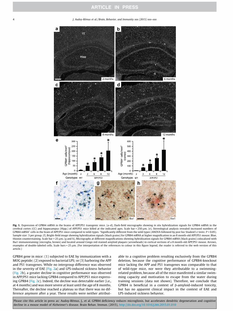
Fig. 1. Expression of GPR84 mRNA in the brains of APP/PS1 transgenic mice. (a–d), Dark-field micrographs showing in situ hybridization signals for GPR84 mRNA in thecerebral cortex (CC) and hippocampus (Hipp.) of APP/PS1 mice killed at the indicated ages. Scale bar = 250 lm. (e), Stereological analysis revealed increased numbers ofGPR84 mRNA+ cells in the brain of APP/PS1 mice compared to wild-types. ⁄Significantly different from the wild types (ANOVA followed by post hoc Student’s t tests: P < 0.05).Sample size: 3 per group. (f), Bright-field image showing hybridization signals (black grains) for GPR84 mRNA at higher magnification in an 8 month-old APP/PS1 mouse. Blue,thionin counterstaining. Scale bar = 25 lm. (g and h), Micrographs at different magnifications showing hybridization signals for GPR84 mRNA (black grains) colocalized withIba1 immunostaining (microglia, brown) and located around Congo red-stained amyloid plaques (arrowheads) in cortical sections of a 8 month-old APP/PS1 mouse. Arrows,examples of double-labeled cells. Scale bars = 25 lm. (For interpretation of the references to colour in this figure legend, the reader is referred to the web version of thisarticle.)
4 J. Audoy-Rémus et al. / Brain, Behavior, and Immunity xxx (2015) xxx–xxx
GPR84 gene in mice: (1) subjected to EAE by immunization with aMOG peptide; (2) exposed to bacterial LPS; or (3) harboring the APPand PS1 transgenes. While no intergroup difference was observedin the severity of EAE (Fig. 2a) and LPS-induced sickness behavior(Fig. 2b), a greater decline in cognitive performance was observedin APP/PS1 mice lacking GPR84 compared to APP/PS1 mice express-ing GPR84 (Fig. 2c). Indeed, the decline was detectable earlier (i.e.,at 4 months) and was more severe at least until the age of 8 months.Thereafter, the decline reached a plateau so that there was no dif-ference anymore after a year. These results were neither attribut-
Please cite this article in press as: Audoy-Rémus, J., et al. GPR84 deficiency rdecline in a mouse model of Alzheimer’s disease. Brain Behav. Immun. (2015)
able to a cognitive problem resulting exclusively from the GPR84deletion, because the cognitive performance of GPR84-knockoutmice lacking the APP and PS1 transgenes was comparable to thatof wild-type mice, nor were they attributable to a swimming-related problem, because all of the mice manifested a similar swim-ming capacity and motivation to escape from the water duringtraining sessions (data not shown). Therefore, we conclude thatGPR84 is beneficial in a context of b-amyloid-induced toxicity,but has no apparent clinical impact in the context of EAE andLPS-induced sickness behavior.
educes microgliosis, but accelerates dendritic degeneration and cognitive, http://dx.doi.org/10.1016/j.bbi.2015.01.010
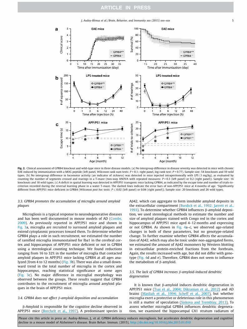
Fig. 2. Clinical assessment of GPR84 knockout and wild-type mice in three disease models. (a) No intergroup difference in disease severity was detected in mice with chronicEAE induced by immunization with a MOG peptide (left panel, Wilcoxon rank sum tests: P > 0.1; right panel, log-rank test: P = 0.77). Sample size: 54 knockouts and 59 wildtypes. (b) No intergroup difference in locomotor activity (an indicator of sickness) was detected in mice injected intraperitoneally with LPS (1 mg/kg), as evaluated bycounting the number of segments crossed and rearings in a T-maze (two-way ANOVA with repeated measures: P = 0.3 (left panel) or 0.2 (right panel)). Sample size: 16knockouts and 18 wild types. (c) A deficit in spatial learning was detected in APP/PS1 transgenic mice lacking GPR84, as indicated by the escape time and number of trials-to-criterion recorded during the reversal learning phase in a water T-maze. The dashed lines indicate the error bars of non-APP/PS1 mice at 4 months of age. ⁄Significantlydifferent from APP/PS1 mice deficient in GPR84 (Wilcoxon post hoc tests: P 6 0.02 (left panel) or 0.04 (right panel)). Sample size: 20 knockouts and 26 wild types.
J. Audoy-Rémus et al. / Brain, Behavior, and Immunity xxx (2015) xxx–xxx 5
3.3. GPR84 promotes the accumulation of microglia around amyloidplaques
Microgliosis is a typical response to neurodegenerative diseasesand has been well documented in mouse models of AD (Combs,2009). As previously reported in APP/PS1 mice and shown inFig. 3a, microglia are recruited to surround amyloid plaques andextend cytoplasmic processes toward them. To determine whetherGPR84 plays a role in such recruitment, we estimated the numberof ramified microglia immunostained for Iba1 in the cerebral cor-tex and hippocampus of APP/PS1 mice deficient or not in GPR84using a stereological counting technique. We found reductionsranging from 16 to 32% in the number of microglia that contactedamyloid plaques in APP/PS1 mice lacking GPR84 at all ages ana-lyzed (from 4 to 12 months) (Fig. 3b). There was also a small down-ward trend in the total number of microglia in the cortex andhippocampus, reaching statistical significance at some ages(Fig. 3c). No major difference in microglial morphology wasobserved between the groups. These results suggest that GPR84contributes to the recruitment of microglia around amyloid pla-ques in the brain of APP/PS1 mice.
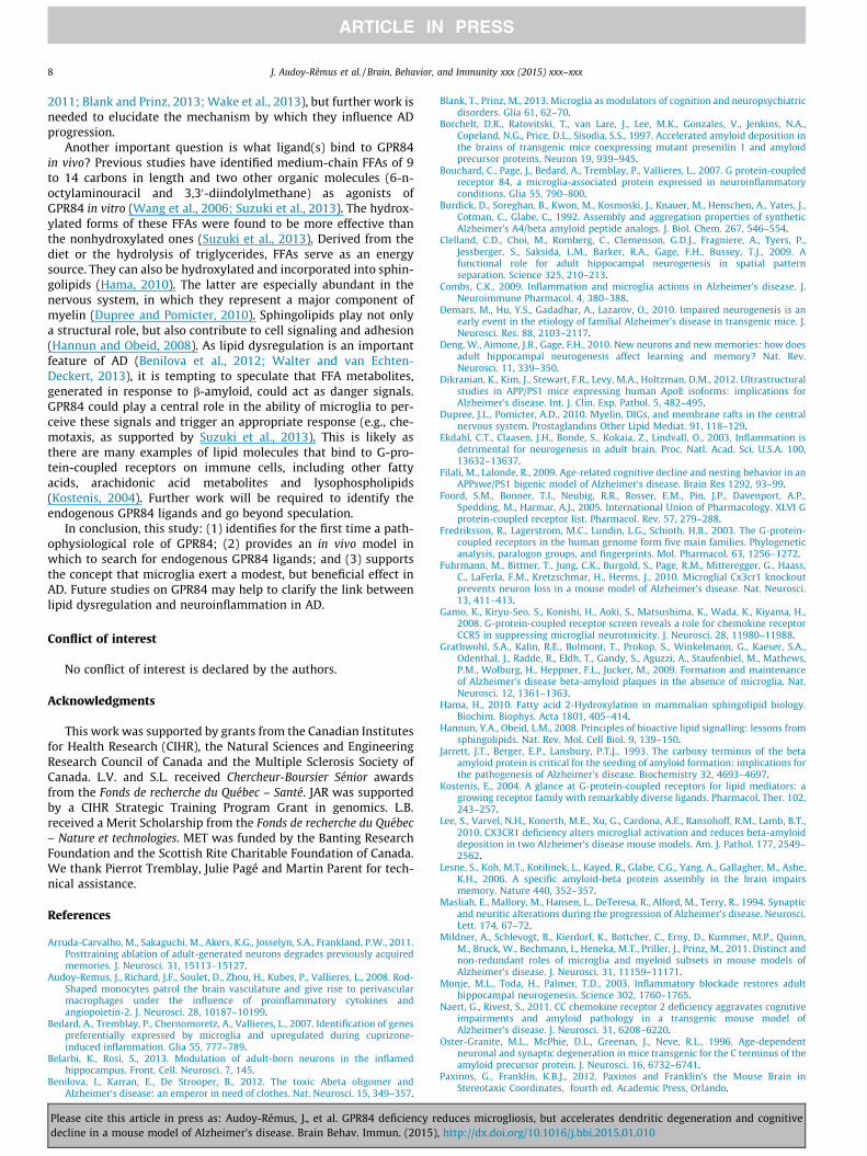
3.4. GPR84 does not affect b-amyloid deposition and accumulation
b-Amyloid is responsible for the cognitive decline observed inAPP/PS1 mice (Borchelt et al., 1997). A predominant species is
Please cite this article in press as: Audoy-Rémus, J., et al. GPR84 deficiency rdecline in a mouse model of Alzheimer’s disease. Brain Behav. Immun. (2015)
Ab42, which can aggregate to form insoluble amyloid deposits inthe extracellular compartment (Burdick et al., 1992; Jarrett et al.,1993). To determine whether GPR84 influences b-amyloid deposi-tion, we used stereological methods to estimate the number andsize of amyloid plaques stained with Congo red in the cortex andhippocampus of APP/PS1 mice aged 4–12 months and expressingor not GPR84. As shown in Fig. 4a–c, we observed age-relatedchanges in both of these parameters, but no genotype-relatedchange. To further examine whether GPR84 affects the accumula-tion of Ab42, which may also be toxic under non-aggregated forms,we estimated the amount of Ab42 monomers by Western blottingin extracellular protein-enriched fractions from the forebrain.Again, the results increased with age, but did not differ with geno-type (Fig. 4d and e). Therefore, GPR84 does not seem to influencethe metabolism of b-amyloid.
3.5. The lack of GPR84 increases b-amyloid-induced dendriticdegeneration
It is known that b-amyloid induces dendritic degeneration inAPP/PS1 mice (Tsai et al., 2004; Dikranian et al., 2012) and ADbrains (Masliah et al., 1994; Scheff et al., 2007), but whethermicroglia exert a protective or deleterious role in this phenomenonis still a matter of speculation (Siskova and Tremblay, 2013). Toassess whether microglial GPR84 influences dendritic degenera-tion, we examined the hippocampal CA1 stratum radiatum of
educes microgliosis, but accelerates dendritic degeneration and cognitive, http://dx.doi.org/10.1016/j.bbi.2015.01.010
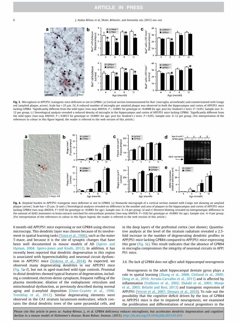
Fig. 3. Microgliosis in APP/PS1 transgenic mice deficient or not in GPR84. (a) Cortical section immunostained for Iba1 (microglia, arrowheads) and counterstained with Congored (amyloid plaque, arrow). Scale bar = 25 lm. (b) A reduced number of microglia per amyloid plaque was observed in both the hippocampus and cortex of APP/PS1 micelacking GPR84. ⁄Significantly different from the wild types (two-way ANOVA: P 6 0.0001 for genotype or <0.0008 for age; post hoc Student’s t tests: P < 0.05). Sample size: 6–12 per group. (c) Stereological analysis revealed a reduced density of microglia in the hippocampus and cortex of APP/PS1 mice lacking GPR84. ⁄Significantly different fromthe wild types (two-way ANOVA: P 6 0.0015 for genotype or <0.0001 for age; post hoc Student’s t tests: P < 0.05). Sample size: 6–12 per group. (For interpretation of thereferences to colour in this figure legend, the reader is referred to the web version of this article.)
Fig. 4. Amyloid burden in APP/PS1 transgenic mice deficient or not in GPR84. (a) Nomarski micrograph of a cortical section stained with Congo red showing an amyloidplaque (arrow). Scale bar = 25 lm. (b and c) Stereological analyses revealed no difference in the number and area of plaques in the hippocampus and cortex of APP/PS1 micelacking GPR84 (two-way ANOVA: P > 0.05 for genotype or <0.0001 for age). Sample size: 6–14 per group. (d and e) Western blotting revealed no intergenotypic difference inthe amount of Ab42 monomers in brain extracts enriched for extracellular proteins (two-way ANOVA: P = 0.82 for genotype or <0.0001 for age). Sample size: 4–9 per group.(For interpretation of the references to colour in this figure legend, the reader is referred to the web version of this article.)
6 J. Audoy-Rémus et al. / Brain, Behavior, and Immunity xxx (2015) xxx–xxx
6 month-old APP/PS1 mice expressing or not GPR84 using electronmicroscopy. This dendritic layer was chosen because of its involve-ment in spatial learning tasks (Tsien et al., 1996), such as the waterT-maze, and because it is the site of synaptic changes that havebeen well documented in mouse models of AD (Spires andHyman, 2004; Spires-Jones and Knafo, 2012). In addition, it hasrecently been reported that dendritic degeneration in this regionis associated with hyperexcitability and neuronal circuit dysfunc-tion in APP/PS1 mice (Siskova et al., 2014). As expected, weobserved many degenerating dendrites in our APP/PS1 mice(Fig. 5a–f), but not in aged-matched wild-type controls. Proximalto distal dendrites showed typical features of degeneration, includ-ing a condensed, electron-dense cytoplasm, frequent ruffling of theplasma membrane, dilation of the endoplasmic reticulum andmitochondrial dysfunction, as previously described during normalaging and b-amyloid deposition (Oster-Granite et al., 1996;Tremblay et al., 2012). Similar degenerating elements wereobserved in the CA1 stratum lacunosum-moleculare, which con-tains the distal dendritic trees of the same pyramidal cells, and
Please cite this article in press as: Audoy-Rémus, J., et al. GPR84 deficiency rdecline in a mouse model of Alzheimer’s disease. Brain Behav. Immun. (2015)
in the deep layers of the prefrontal cortex (not shown). Quantita-tive analysis at the level of the stratum radiatum revealed a 2.5-fold increase in the number of degenerating dendritic profiles inAPP/PS1 mice lacking GPR84 compared to APP/PS1 mice expressingthis gene (Fig. 5c). This result indicates that the absence of GPR84in microglia compromises the integrity of neuronal circuits in APP/PS1 mice.
3.6. The lack of GPR84 does not affect adult hippocampal neurogenesis
Neurogenesis in the adult hippocampal dentate gyrus plays arole in spatial learning (Zhang et al., 2008; Clelland et al., 2009;Deng et al., 2010; Arruda-Carvalho et al., 2011) and is affected byinflammation (Vallieres et al., 2002; Ekdahl et al., 2003; Monjeet al., 2003; Belarbi and Rosi, 2013) and transgenic expression ofAPP/PS1 (Verret et al., 2007; Demars et al., 2010). To rule out thepossibility that the cognitive deficit driven by the loss of GPR84in APP/PS1 mice is due to impaired neurogenesis, we examinedthe proliferation and differentiation of neural progenitors in the
educes microgliosis, but accelerates dendritic degeneration and cognitive, http://dx.doi.org/10.1016/j.bbi.2015.01.010

Fig. 5. Dendritic degeneration in APP/PS1 mice deficient or not in GPR84. (a–f)Electron micrographs of degenerating dendrites (arrows) in the hippocampal CA1stratum radiatum of 6 month-old APP/PS1 mice. Note their condensed cytoplasmand dilated endoplasmic reticulum, which are a typical signs of degeneration,compared to those of normal dendrites (arrowheads). Abbreviations: er, endoplas-mic reticulum; m, mitochondria; s, synapse. Scale bars = 2 lm (a) or 500 nm (b–f).(g) Quantification, using images taken from the stratum radiatum, revealed anincreased number of degenerating dendritic profiles in APP/PS1 mice lacking GPR84compared to APP/PS1 mice expressing this gene (Student’s t test: P = 0.012). Samplesize: 5 per group.
J. Audoy-Rémus et al. / Brain, Behavior, and Immunity xxx (2015) xxx–xxx 7
Please cite this article in press as: Audoy-Rémus, J., et al. GPR84 deficiency rdecline in a mouse model of Alzheimer’s disease. Brain Behav. Immun. (2015)
granular layer of the dentate gyrus in APP/PS1 mice expressing ornot GPR84. While a decrease in the number of cells expressing theproliferation marker Ki67 or the neuroblast marker DCX wasobserved with age, no intergenotype difference was detected(Fig. 6). Therefore, the poorer cognitive performance resulting fromthe absence of GPR84 in the APP/PS1 background is not attribut-able to a defect in adult hippocampal neurogenesis.
4. Discussion
Two main findings emerge from the present work. First, microg-lia increase their expression of GPR84 not only upon EAE, endotox-emia and nerve injury, as previously reported (Bedard et al., 2007;Bouchard et al., 2007; Gamo et al., 2008), but also in a neurodegen-erative context. GPR84 can therefore be considered as a general‘‘activation’’ marker for microglia. Second, GPR84 contributes tob-amyloid-induced microgliosis, a response that is required to pro-mote dendritic homeostasis and prevent further cognitive decline.As discussed below, these findings help to understand the role ofGPR84 in vivo and the importance of microglia in AD.
One question that arises is how microglia protects from b-amy-loid toxicity? Our results indicate that GPR84 influences neitherthe formation or elimination of b-amyloid plaques, nor the concen-tration of soluble Ab42, one of the most abundant and toxic form ofb-amyloid. This is in agreement with previous studies showing thatmicroglia do not affect the plaques (Grathwohl et al., 2009;Mildner et al., 2011) or at best modestly (Simard et al., 2006;Tahara et al., 2006; Naert and Rivest, 2011; Song et al., 2011). Fur-thermore, our results reveal a higher number of degenerating den-drites in the absence of GPR84, leading us to infer that a microglialresponse regulated by GPR84 is required to protect dendrites fromfurther degeneration. Alternatively, it is possible that the absenceof GPR84 reduces the ability of microglia to remove degeneratingdendrites, which may affect neuronal plasticity and thus cognition.The importance of microglia in neuronal plasticity is increasinglyappreciated (Tremblay and Majewska, 2011; Tremblay et al.,
Fig. 6. Hippocampal neurogenesis in APP/PS1 transgenic mice lacking or notGPR84. (a and b) Sections showing the granule cell layer (GCL) of the dentate gyrusimmunostained (black) for Ki67, a marker of proliferating cells, or DCX, a marker ofnewborn neurons. Counterstaining = methyl green. Scale bar = 25 lm. (c and d)Stereological data showing no intergenotype difference in the number of Ki67+ orDCX+ cells. Two-way ANOVA: Ki67, P = 0.59 (genotype), <0.0001 (age) or = 0.52(interaction); DCX, P = 0.56 (genotype), <0.0001 (age) or = 0.45 (interaction).Sample size: 6–9 per group.
educes microgliosis, but accelerates dendritic degeneration and cognitive, http://dx.doi.org/10.1016/j.bbi.2015.01.010
8 J. Audoy-Rémus et al. / Brain, Behavior, and Immunity xxx (2015) xxx–xxx
2011; Blank and Prinz, 2013; Wake et al., 2013), but further work isneeded to elucidate the mechanism by which they influence ADprogression.
Another important question is what ligand(s) bind to GPR84in vivo? Previous studies have identified medium-chain FFAs of 9to 14 carbons in length and two other organic molecules (6-n-octylaminouracil and 3,30-diindolylmethane) as agonists ofGPR84 in vitro (Wang et al., 2006; Suzuki et al., 2013). The hydrox-ylated forms of these FFAs were found to be more effective thanthe nonhydroxylated ones (Suzuki et al., 2013). Derived from thediet or the hydrolysis of triglycerides, FFAs serve as an energysource. They can also be hydroxylated and incorporated into sphin-golipids (Hama, 2010). The latter are especially abundant in thenervous system, in which they represent a major component ofmyelin (Dupree and Pomicter, 2010). Sphingolipids play not onlya structural role, but also contribute to cell signaling and adhesion(Hannun and Obeid, 2008). As lipid dysregulation is an importantfeature of AD (Benilova et al., 2012; Walter and van Echten-Deckert, 2013), it is tempting to speculate that FFA metabolites,generated in response to b-amyloid, could act as danger signals.GPR84 could play a central role in the ability of microglia to per-ceive these signals and trigger an appropriate response (e.g., che-motaxis, as supported by Suzuki et al., 2013). This is likely asthere are many examples of lipid molecules that bind to G-pro-tein-coupled receptors on immune cells, including other fattyacids, arachidonic acid metabolites and lysophospholipids(Kostenis, 2004). Further work will be required to identify theendogenous GPR84 ligands and go beyond speculation.
In conclusion, this study: (1) identifies for the first time a path-ophysiological role of GPR84; (2) provides an in vivo model inwhich to search for endogenous GPR84 ligands; and (3) supportsthe concept that microglia exert a modest, but beneficial effect inAD. Future studies on GPR84 may help to clarify the link betweenlipid dysregulation and neuroinflammation in AD.
Conflict of interest
No conflict of interest is declared by the authors.
Acknowledgments
This work was supported by grants from the Canadian Institutesfor Health Research (CIHR), the Natural Sciences and EngineeringResearch Council of Canada and the Multiple Sclerosis Society ofCanada. L.V. and S.L. received Chercheur-Boursier Sénior awardsfrom the Fonds de recherche du Québec – Santé. JAR was supportedby a CIHR Strategic Training Program Grant in genomics. L.B.received a Merit Scholarship from the Fonds de recherche du Québec– Nature et technologies. MET was funded by the Banting ResearchFoundation and the Scottish Rite Charitable Foundation of Canada.We thank Pierrot Tremblay, Julie Pagé and Martin Parent for tech-nical assistance.
References
Arruda-Carvalho, M., Sakaguchi, M., Akers, K.G., Josselyn, S.A., Frankland, P.W., 2011.Posttraining ablation of adult-generated neurons degrades previously acquiredmemories. J. Neurosci. 31, 15113–15127.
Audoy-Remus, J., Richard, J.F., Soulet, D., Zhou, H., Kubes, P., Vallieres, L., 2008. Rod-Shaped monocytes patrol the brain vasculature and give rise to perivascularmacrophages under the influence of proinflammatory cytokines andangiopoietin-2. J. Neurosci. 28, 10187–10199.
Bedard, A., Tremblay, P., Chernomoretz, A., Vallieres, L., 2007. Identification of genespreferentially expressed by microglia and upregulated during cuprizone-induced inflammation. Glia 55, 777–789.
Belarbi, K., Rosi, S., 2013. Modulation of adult-born neurons in the inflamedhippocampus. Front. Cell. Neurosci. 7, 145.
Benilova, I., Karran, E., De Strooper, B., 2012. The toxic Abeta oligomer andAlzheimer’s disease: an emperor in need of clothes. Nat. Neurosci. 15, 349–357.
Please cite this article in press as: Audoy-Rémus, J., et al. GPR84 deficiency rdecline in a mouse model of Alzheimer’s disease. Brain Behav. Immun. (2015)
Blank, T., Prinz, M., 2013. Microglia as modulators of cognition and neuropsychiatricdisorders. Glia 61, 62–70.
Borchelt, D.R., Ratovitski, T., van Lare, J., Lee, M.K., Gonzales, V., Jenkins, N.A.,Copeland, N.G., Price, D.L., Sisodia, S.S., 1997. Accelerated amyloid deposition inthe brains of transgenic mice coexpressing mutant presenilin 1 and amyloidprecursor proteins. Neuron 19, 939–945.
Bouchard, C., Page, J., Bedard, A., Tremblay, P., Vallieres, L., 2007. G protein-coupledreceptor 84, a microglia-associated protein expressed in neuroinflammatoryconditions. Glia 55, 790–800.
Burdick, D., Soreghan, B., Kwon, M., Kosmoski, J., Knauer, M., Henschen, A., Yates, J.,Cotman, C., Glabe, C., 1992. Assembly and aggregation properties of syntheticAlzheimer’s A4/beta amyloid peptide analogs. J. Biol. Chem. 267, 546–554.
Clelland, C.D., Choi, M., Romberg, C., Clemenson, G.D.J., Fragniere, A., Tyers, P.,Jessberger, S., Saksida, L.M., Barker, R.A., Gage, F.H., Bussey, T.J., 2009. Afunctional role for adult hippocampal neurogenesis in spatial patternseparation. Science 325, 210–213.
Combs, C.K., 2009. Inflammation and microglia actions in Alzheimer’s disease. J.Neuroimmune Pharmacol. 4, 380–388.
Demars, M., Hu, Y.S., Gadadhar, A., Lazarov, O., 2010. Impaired neurogenesis is anearly event in the etiology of familial Alzheimer’s disease in transgenic mice. J.Neurosci. Res. 88, 2103–2117.
Deng, W., Aimone, J.B., Gage, F.H., 2010. New neurons and new memories: how doesadult hippocampal neurogenesis affect learning and memory? Nat. Rev.Neurosci. 11, 339–350.
Dikranian, K., Kim, J., Stewart, F.R., Levy, M.A., Holtzman, D.M., 2012. Ultrastructuralstudies in APP/PS1 mice expressing human ApoE isoforms: implications forAlzheimer’s disease. Int. J. Clin. Exp. Pathol. 5, 482–495.
Dupree, J.L., Pomicter, A.D., 2010. Myelin, DIGs, and membrane rafts in the centralnervous system. Prostaglandins Other Lipid Mediat. 91, 118–129.
Ekdahl, C.T., Claasen, J.H., Bonde, S., Kokaia, Z., Lindvall, O., 2003. Inflammation isdetrimental for neurogenesis in adult brain. Proc. Natl. Acad. Sci. U.S.A. 100,13632–13637.
Filali, M., Lalonde, R., 2009. Age-related cognitive decline and nesting behavior in anAPPswe/PS1 bigenic model of Alzheimer’s disease. Brain Res 1292, 93–99.
Foord, S.M., Bonner, T.I., Neubig, R.R., Rosser, E.M., Pin, J.P., Davenport, A.P.,Spedding, M., Harmar, A.J., 2005. International Union of Pharmacology. XLVI Gprotein-coupled receptor list. Pharmacol. Rev. 57, 279–288.
Fredriksson, R., Lagerstrom, M.C., Lundin, L.G., Schioth, H.B., 2003. The G-protein-coupled receptors in the human genome form five main families. Phylogeneticanalysis, paralogon groups, and fingerprints. Mol. Pharmacol. 63, 1256–1272.
Fuhrmann, M., Bittner, T., Jung, C.K., Burgold, S., Page, R.M., Mitteregger, G., Haass,C., LaFerla, F.M., Kretzschmar, H., Herms, J., 2010. Microglial Cx3cr1 knockoutprevents neuron loss in a mouse model of Alzheimer’s disease. Nat. Neurosci.13, 411–413.
Gamo, K., Kiryu-Seo, S., Konishi, H., Aoki, S., Matsushima, K., Wada, K., Kiyama, H.,2008. G-protein-coupled receptor screen reveals a role for chemokine receptorCCR5 in suppressing microglial neurotoxicity. J. Neurosci. 28, 11980–11988.
Grathwohl, S.A., Kalin, R.E., Bolmont, T., Prokop, S., Winkelmann, G., Kaeser, S.A.,Odenthal, J., Radde, R., Eldh, T., Gandy, S., Aguzzi, A., Staufenbiel, M., Mathews,P.M., Wolburg, H., Heppner, F.L., Jucker, M., 2009. Formation and maintenanceof Alzheimer’s disease beta-amyloid plaques in the absence of microglia. Nat.Neurosci. 12, 1361–1363.
Hama, H., 2010. Fatty acid 2-Hydroxylation in mammalian sphingolipid biology.Biochim. Biophys. Acta 1801, 405–414.
Hannun, Y.A., Obeid, L.M., 2008. Principles of bioactive lipid signalling: lessons fromsphingolipids. Nat. Rev. Mol. Cell Biol. 9, 139–150.
Jarrett, J.T., Berger, E.P., Lansbury, P.T.J., 1993. The carboxy terminus of the betaamyloid protein is critical for the seeding of amyloid formation: implications forthe pathogenesis of Alzheimer’s disease. Biochemistry 32, 4693–4697.
Kostenis, E., 2004. A glance at G-protein-coupled receptors for lipid mediators: agrowing receptor family with remarkably diverse ligands. Pharmacol. Ther. 102,243–257.
Lee, S., Varvel, N.H., Konerth, M.E., Xu, G., Cardona, A.E., Ransohoff, R.M., Lamb, B.T.,2010. CX3CR1 deficiency alters microglial activation and reduces beta-amyloiddeposition in two Alzheimer’s disease mouse models. Am. J. Pathol. 177, 2549–2562.
Lesne, S., Koh, M.T., Kotilinek, L., Kayed, R., Glabe, C.G., Yang, A., Gallagher, M., Ashe,K.H., 2006. A specific amyloid-beta protein assembly in the brain impairsmemory. Nature 440, 352–357.
Masliah, E., Mallory, M., Hansen, L., DeTeresa, R., Alford, M., Terry, R., 1994. Synapticand neuritic alterations during the progression of Alzheimer’s disease. Neurosci.Lett. 174, 67–72.
Mildner, A., Schlevogt, B., Kierdorf, K., Bottcher, C., Erny, D., Kummer, M.P., Quinn,M., Bruck, W., Bechmann, I., Heneka, M.T., Priller, J., Prinz, M., 2011. Distinct andnon-redundant roles of microglia and myeloid subsets in mouse models ofAlzheimer’s disease. J. Neurosci. 31, 11159–11171.
Monje, M.L., Toda, H., Palmer, T.D., 2003. Inflammatory blockade restores adulthippocampal neurogenesis. Science 302, 1760–1765.
Naert, G., Rivest, S., 2011. CC chemokine receptor 2 deficiency aggravates cognitiveimpairments and amyloid pathology in a transgenic mouse model ofAlzheimer’s disease. J. Neurosci. 31, 6208–6220.
Oster-Granite, M.L., McPhie, D.L., Greenan, J., Neve, R.L., 1996. Age-dependentneuronal and synaptic degeneration in mice transgenic for the C terminus of theamyloid precursor protein. J. Neurosci. 16, 6732–6741.
Paxinos, G., Franklin, K.B.J., 2012. Paxinos and Franklin’s the Mouse Brain inStereotaxic Coordinates, fourth ed. Academic Press, Orlando.
educes microgliosis, but accelerates dendritic degeneration and cognitive, http://dx.doi.org/10.1016/j.bbi.2015.01.010

J. Audoy-Rémus et al. / Brain, Behavior, and Immunity xxx (2015) xxx–xxx 9
Peters, A., Palay, S.L., Webster, H.D., 1991. Fine structure of the nervous system:neurons and their supporting cells. Oxford University Press, New York.
Scheff, S.W., Price, D.A., Schmitt, F.A., DeKosky, S.T., Mufson, E.J., 2007. Synapticalterations in CA1 in mild Alzheimer disease and mild cognitive impairment.Neurology 68, 1501–1508.
Simard, A.R., Soulet, D., Gowing, G., Julien, J.P., Rivest, S., 2006. Bone marrow-derived microglia play a critical role in restricting senile plaque formation inAlzheimer’s disease. Neuron 49, 489–502.
Siskova, Z., Justus, D., Kaneko, H., Friedrichs, D., Henneberg, N., Beutel, T., Pitsch, J.,Schoch, S., Becker, A., von der Kammer, H., Remy, S., 2014. Dendritic structuraldegeneration is functionally linked to cellular hyperexcitability in a mousemodel of Alzheimer’s disease. Neuron 84, 1023–1033.
Siskova, Z., Tremblay, M.E., 2013. Microglia and synapse: interactions in health andneurodegeneration. Neural. Plast. 2013, 425845.
Song, M., Jin, J., Lim, J.E., Kou, J., Pattanayak, A., Rehman, J.A., Kim, H.D., Tahara, K.,Lalonde, R., Fukuchi, K., 2011. TLR4 mutation reduces microglial activation,increases Abeta deposits and exacerbates cognitive deficits in a mouse model ofAlzheimer’s disease. J. Neuroinflamm. 8, 92.
Spires-Jones, T., Knafo, S., 2012. Spines, plasticity, and cognition in Alzheimer’smodel mice. Neural Plast. 2012, 319836.
Spires, T.L., Hyman, B.T., 2004. Neuronal structure is altered by amyloid plaques.Rev. Neurosci. 15, 267–278.
Suzuki, M., Takaishi, S., Nagasaki, M., Onozawa, Y., Iino, I., Maeda, H., Komai, T., Oda,T., 2013. Medium-chain fatty acid-sensing receptor, GPR84, is aproinflammatory receptor. J. Biol. Chem. 288, 10684–10691.
Tahara, K., Kim, H.D., Jin, J.J., Maxwell, J.A., Li, L., Fukuchi, K., 2006. Role of toll-likereceptor signalling in Abeta uptake and clearance. Brain 129, 3006–3019.
Tan, J., Town, T., Crawford, F., Mori, T., DelleDonne, A., Crescentini, R., Obregon, D.,Flavell, R.A., Mullan, M.J., 2002. Role of CD40 ligand in amyloidosis in transgenicAlzheimer’s mice. Nat. Neurosci. 5, 1288–1293.
Tan, J., Town, T., Paris, D., Mori, T., Suo, Z., Crawford, F., Mattson, M.P., Flavell, R.A.,Mullan, M., 1999. Microglial activation resulting from CD40-CD40L interactionafter beta-amyloid stimulation. Science 286, 2352–2355.
Tanzi, R.E., Bertram, L., 2005. Twenty years of the Alzheimer’s disease amyloidhypothesis: a genetic perspective. Cell 120, 545–555.
Please cite this article in press as: Audoy-Rémus, J., et al. GPR84 deficiency rdecline in a mouse model of Alzheimer’s disease. Brain Behav. Immun. (2015)
Tremblay, M.E., Majewska, A.K., 2011. A role for microglia in synaptic plasticity?Commun. Integr. Biol. 4, 220–222.
Tremblay, M.E., Stevens, B., Sierra, A., Wake, H., Bessis, A., Nimmerjahn, A., 2011. Therole of microglia in the healthy brain. J. Neurosci. 31, 16064–16069.
Tremblay, M.E., Zettel, M.L., Ison, J.R., Allen, P.D., Majewska, A.K., 2012. Effects ofaging and sensory loss on glial cells in mouse visual and auditory cortices. Glia60, 541–558.
Tsai, J., Grutzendler, J., Duff, K., Gan, W.B., 2004. Fibrillar amyloid deposition leads tolocal synaptic abnormalities and breakage of neuronal branches. Nat. Neurosci.7, 1181–1183.
Tsien, J.Z., Huerta, P.T., Tonegawa, S., 1996. The essential role of hippocampal CA1NMDA receptor-dependent synaptic plasticity in spatial memory. Cell 87,1327–1338.
Vallieres, L., Campbell, I.L., Gage, F.H., Sawchenko, P.E., 2002. Reduced hippocampalneurogenesis in adult transgenic mice with chronic astrocytic production ofinterleukin-6. J. Neurosci. 22, 486–492.
Venkataraman, C., Kuo, F., 2005. The G-protein coupled receptor, GPR84 regulates IL-4 production by T lymphocytes in response to CD3 crosslinking. Immunol. Lett.
Verret, L., Jankowsky, J.L., Xu, G.M., Borchelt, D.R., Rampon, C., 2007. Alzheimer’s-type amyloidosis in transgenic mice impairs survival of newborn neuronsderived from adult hippocampal neurogenesis. J. Neurosci. 27, 6771–6780.
Wake, H., Moorhouse, A.J., Miyamoto, A., Nabekura, J., 2013. Microglia: activelysurveying and shaping neuronal circuit structure and function. Trends Neurosci.36, 209–217.
Walter, J., van Echten-Deckert, G., 2013. Cross-talk of membrane lipids andAlzheimer-related proteins. Mol. Neurodegener. 8, 34.
Wang, J., Wu, X., Simonavicius, N., Tian, H., Ling, L., 2006. Medium chain fatty acidsas ligands for orphan G protein-coupled receptor GPR84. J. Biol. Chem.
Weitz, T.M., Town, T., 2012. Microglia in Alzheimer’s disease: it’s all about context.Int. J. Alzheimers Dis. 2012, 314185.
Yousefi, S., Cooper, P.R., Potter, S.L., Mueck, B., Jarai, G., 2001. Cloning and expressionanalysis of a novel G-protein-coupled receptor selectively expressed ongranulocytes. J. Leukoc. Biol. 69, 1045–1052.
Zhang, C.L., Zou, Y., He, W., Gage, F.H., Evans, R.M., 2008. A role for adult TLX-positive neural stem cells in learning and behaviour. Nature 451, 1004–1007.
educes microgliosis, but accelerates dendritic degeneration and cognitive, http://dx.doi.org/10.1016/j.bbi.2015.01.010