Goat Diseases - The Farmers' Guide - Meat & Livestock Australia
107
Goat Diseases The Farmers' Guide September 2021
-
Upload
khangminh22 -
Category
Documents
-
view
0 -
download
0
Transcript of Goat Diseases - The Farmers' Guide - Meat & Livestock Australia
Goat DiseasesThe Farmers' GuideSeptember 2021

Goat Diseases The Farmers' Guide
Developed by: Emily Litzow, Nick van den Berg and Barton Loechel
Funded by: The FMD Ready project is supported by Meat & Livestock Australia (MLA), through funding from the Australian Government Department of Agriculture, Water and the Environment as part of its Rural R&D for Profit programme, and by producer levies from Australian FMD-susceptible livestock (cattle, sheep, goats and pigs) industries and Charles Sturt University (CSU), leveraging significant in-kind support from the research partners. The research partners for this project are the Commonwealth Science and Industrial Research Organisation (CSIRO), CSU through the Graham Centre for Agricultural Innovation, the Bureau of Meteorology (BOM) and the Australian Department of Agriculture, Water and the Environment, supported by Animal Health Australia (AHA).

Disclaimer: Care is taken to ensure the accuracy of the information at the time of the publication of this Guide. However, MLA and its Partners cannot accept responsibility for the accuracy or completeness of the information or opinions contained in the publication. Noting that this is a general guide on identifying and managing disease, you should make your own enquiries through the appropriate authorities before making decisions concerning your personal circumstances. You accept all risks and responsibility for losses, damages, costs and other consequences resulting directly or indirectly from using this guide and any information or material available from it. Apart from any use permitted under the Copyright Act 1968, all rights are expressly reserved. Requests for further authorisation should be directed to the Content Manager, PO Box 1961, North Sydney, NSW 2059 or [email protected]. © Meat & Livestock Australia
Acknowledgement:This guide was co-developed by members of the FMD Ready Subproject 2 Goat Innovation Platform pilot group, South Australia. The working group, led by Nick van den Berg, contributed to conceptual design, content structure and photo images.
The Goat IP group was an initiative of FMD Ready Subproject 2: A farmer-led partnership for improved animal health surveillance and disease management. Part of a larger project: FMD Ready: Improved surveillance, preparedness and return to trade for emergency animal disease incursions using foot-and-mouth disease as a model.
Photographs were generously supplied by the following individuals/organisations: Jeremy Rogers (PIRSA), John and Bec Falkenhagen (Idealview, SA), Sandra Baxendell (goatvetoz), Berwyn Squire (Agriculture Victoria), Kylie Greentree (LLS NSW), Cathy Zwick (Allambie Station, QLD), Kylie Hopkins (DAF Queensland), Kylie Leahy (Mt Roy Stud, QLD), Colin Trengove (University of Adelaide).
The development of this guide was based on the PIRSA Biosecurity SA document ‘Sheep Diseases – The Farmers’ Guide’.
For any future updates to this Guide please contact [email protected], job reference ‘CSIRO B&M | 21-00248’.

ContentsExotic and Notifiable Diseases 1
Where to get help 2
Good Biosecurity: Have a Biosecurity Plan 3
Good Biosecurity: What to watch out for 4
Managing a biosecurity incursion 6
What Does a Healthy Goat Look Like? 7
Diseases of Goats Keys to Diagnosis by Clinical Sign 8
Acidosis 10
Barbers Pole 12
Bladder Worm 13
Botulism 14
CAE (Caprine Arthritis Encephalitis) 15
Cheesy Gland (CLA) 17
Cobalt Deficiency 18
Coccidiosis 19
Copper Deficiency 20
Copper Poisoning 21
Flystrike 22
Foot Abscess 24
Footrot Notifiable Disease 25
Hardware Disease (Traumatic Reticuloperitonitis) 27
Hydatids 28
Injuries 29
Johne’s Disease Notifiable Disease 30
Listeriosis 32
Lupinosis 33
Mastitis 35
Milk Fever 36
Nitrate Poisoning 37
Oxalate Poisoning 38
Photosensitisation 40
Pinkeye 41
Pneumonia 42
Polioencephalomacia (PEM) 43
Pregnancy Toxaemia 44
Pulpy Kidney Enterotoxaemia 46
Salmonellosis 47
Scabby Mouth (Orf) 48
Selenium Deficiency 50
Tetanus 51
Toe Abscess (White line abscess) 52
Urolithiasis Urinary Calculi or Bladder Stones 53
Vaccination 54
Worms Signs of Worms 58
Worms Types of Worms 59
Worms Treatment and Prevention 61
Drenching 62
Mouthing for Age 63
Foot Paring 64
Disbudding 65
Castration 66
Kidding Normal Birth Process 67
Kidding Difficulties 68
Rearing Orphan Kids 70
Fit to Load 72
National Livestock Identification System (NLIS) 73
Body Condition Scoring 74
Ruminant Disease Investigation 75
Personal Protective Equipment and Standard Post-Mortem Equipment 76
Investigation 77
1. Collect History and Record Findings 77
2. External Examination 78
3. Post-Mortem 79
4. Collecting Samples 97
Euthanasia of Goats 98
Carcass Disposal 100
Acknowledgements and Further Reading 101

1
Exotic and Notifiable DiseasesReporting serious or unusual animal disease signs Any unusual behaviour or clinical signs observed in your livestock or birds should be reported to:
• The 24 hour Emergency Animal Disease Watch Hotline on 1800 675 888 (free call) • Your local veterinarian • Your local government veterinarian or biosecurity department
The sooner an incident is reported, the less chance it has to spread and the more likely it can be controlled and eradicated. This is particularly so if it’s an exotic animal disease or pest.
Unusual signs and Clinical Signs in animals Always keep an eye out for unusual clinical signs in livestock that can include:
• Unexplained deaths, especially of kids. • Sores or ulcers on the feet or in the mouth (this may result in a reluctance to eat or move). • Excessive salivation (drooling should always be treated suspiciously). • A reduction in the milk yield. • Any kind of discharge – diarrhoea especially if it has blood in it. Excessive nasal discharge is also something you should
report unless you know what has caused it. • Look out for staggering or head drooping or severe lameness, especially if it’s more than one animal at the same time.
Less dramatic signs should also be watched for, such as animals not eating properly and animals that are depressed and don’t respond the way they should.

2
Where to get helpEmergency Animal Disease Watch Hotline on 1800 675 888 (free call)
SA: PIRSA Biosecurity Animal Health (08) 8207 7900
QLD: Biosecurity Queensland 132 523
NSW: Local Land Services 1300 795 299
VIC: Agriculture Victoria – Animal Health 136 186
WA: Agriculture and Food (08) 9368 3333
ACT: Environment, Planning and Sustainable Development Directorate 132 281
TAS: Biosecurity Tasmania 1300 368 550
NT: Emergency Animal Disease Watch Hotline 1800 675 888

3
Good Biosecurity: Have a Biosecurity PlanHaving a biosecurity plan can greatly minimise your biosecurity risk and susceptibility to livestock disease outbreaks.
For more information on developing a biosecurity plan, please visit www.animalhealthaustralia.com.au/what-we-do/endemic-disease/farm-biosecurity-plan
For National Goat Health Declarations and additional practical biosecurity information please visit www.farmbiosecurity.com.au
Farm Biosecurity sign Animal Health Australia (AHA) and Plant Health Australia (PHA)
4
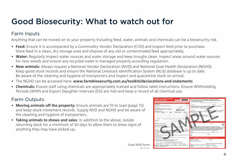
Good Biosecurity: What to watch out forFarm InputsAnything that can be moved on to your property including feed, water, animals and chemicals can be a biosecurity risk.
• Feed: Ensure it is accompanied by a Commodity Vendor Declaration (CVD) and inspect feed prior to purchase. Store feed in a clean, dry storage area and dispose of any old or contaminated feed appropriately.
• Water: Regularly inspect water sources and water storage and keep troughs clean. Inspect areas around water sources for new weeds and ensure any recycled water is managed properly according regulation.
• New animals: Always request a National Vendor Declaration (NVD) and National Goat Health Declaration (NGHD). Keep good stock records and ensure the National Livestock Identification System (NLIS) database is up to date. Be aware of the cleaning and hygiene of transporters and inspect and quarantine stock on arrival.
• The NGHD can be accessed here: www.farmbiosecurity.com.au/toolkit/declarations-and-statements • Chemicals: Ensure staff using chemicals are appropriately trained and follow label instructions. Ensure Withholding
Periods (WHP) and Export Slaughter Intervals (ESI) are met and keep a record of all chemical use.
Farm Outputs• Moving animals off the property: Ensure animals are fit to load (page 72)
and keep stock movement records. Supply NVD and NGHD and be aware of the cleaning and hygiene of transporters.
• Taking animals to shows and sales: In addition to the above, isolate returning stock for a minimum of 30 days to allow them to show signs of anything they may have picked up.
38888888
Goat NVD form MLA/ISC

5
Ferals and Weeds• Wild and feral animal access: Develop a feral animal
control program and ideally co-ordinate it with neighbouring properties. Ensure fences are maintained and feed and water sources are protected.
• Weeds: Be aware of which weeds are found in your region and which are harmful to livestock. Have a weed management plan for your property and request a commodity declaration form for all purchased feed to reduce the risk of bringing weeds onto the property.
People, Vehicles and Equipment• People: Have limited access points to the property,
have a designated parking area for all visitors and provide a visitor register. Boots and equipment should be cleaned on arrival
• Signage: Use signs to inform visitors of biosecurity requirements
• Vehicles: Have limited access points to the property, have a designated parking area for all visitors and have a vehicle wash down facility
• Equipment: Have designated equipment for high biosecurity risk
• Activities / areas and ensure boots and equipment are cleaned before being used for other livestock
Truck carrying livestock MLA

6
Managing a biosecurity incursionSTEP ONE » STEP TWO » STEP THREE » STEP FOUR »
WHAT SHOULD I DO TO PREPARE? WHAT SHOULD I DO IF I NOTICE SOMETHING UNUSUAL?
WHAT CAN I EXPECT FROM CALLING FOR ADVICE?
WHAT SUPPORT CAN I GET TO MANAGE THE PROBLEM?
At a minimum you should have:1. A current farm biosecurity plan2. A working relationship with a
private vet3. An easily accessible list of key
contacts including for your private vet, government biosecurity officer, and the government biosecurity hotline that is easily accessible: The Emergency Animal Disease Hotline 1800 675 888
4. Regular monitoring of your herd to understand what is their normal behaviour and what is abnormal
5. A systematic approach to recording your observations and other key events related to your animals
6. Compliance with National Livestock Identification System (NLIS) requirements
7. Plenty of Personal Protective Equipment (PPE) on hand (check condition as it can perish over time)
Observe, assess, record:First step is to observe and assess the situation and surrounds, to gain information that will assist when you call a professional.Record these details – take photos and/or notes of condition and/or behaviour of the animal such as:• notable signs of illness or infection,
unusual posture or gait, changed feeding or watering habits, how many animals/herds affected, how long they appear to have been sick or dead, whether introduced or home-bred?
Then seek advice: Phone an animal health professional before proceeding further; call either:• your local private vet
Ph: ____ _____________________• a government vet or inspector
Ph: ____ _____________________• if you suspect you are dealing with a
notifiable or exotic disease, immediately: - Call the Emergency Disease Hotline
1800 675 888 - Restrict the movement of animals to
avoid spreading disease and use Personal Protective Equipment to protect yourself.
A phone call to a professional may enable the vet or biosecurity officer to:• determine whether the condition is a
possible notifiable or exotic disease• exclude some diseases/conditions
based on the information you provide• advise on safe handling of sick or
dead animals• develop a list of possible
differential diagnoses• determine whether a farm visit
is necessary • determine whether to proceed with
a post mortem and/or collection of samples
If the disease is confirmed by diagnosis as an exotic or notifiable disease, you will be contacted by your vet or a biosecurity officer who will instruct you on the appropriate actions you must take.
If a non-notifiable disease or other non-notifiable cause (e.g. plant or chemical poisoning) is confirmed, your vet will provide guidance on appropriate treatment and/or necessary actions to protect the rest of your herd.
Your local private or government vet or biosecurity officer can provide advice on what support you may receive if a notifiable or significant disease is suspected and the best management procedures going forward.
The Federal and State/Territory governments support farmers and private vets for diagnosing and managing notifiable diseases in a range of ways.
Adapted from the FMD Ready Subproject 2 Biosecurity and Surveillance Chain of Response Guide

7
What Does a Healthy Goat Look Like?Knowing what a healthy goat looks like, and understanding what normal behaviour is, can greatly help you detect when a goat is unhealthy and/or acting abnormally.
Animals should exhibit a healthy hair coat and skin and have a body condition score appropriate to their production stage. Additional information on body condition scoring can be found on page 74. Both coat and body condition score are good indications of overall health and nutritional status. Signs of an unhealthy animal may include:
• isolation from the rest of the herd• abnormal eating habits• depression• scouring or diarrhoea• abnormal vocalisation• teeth grinding, or• any other abnormal behaviour.
KEY INDICATORS OF GOAT HEALTH NORMAL VALUES / MEASURES
Temperature 38.5–40°C – Rectal Temperature
Respiratory Rate* 10–30 breaths per minute
Heart Rate* 70–90 beats per minute
Rumen Contraction Rate 1–1.5 per minute
*Kids can be significantly higher Healthy Boer Goat MLA

8
Diseases of Goats KEYS TO DIAGNOSIS BY CLINICAL SIGN
AbscessSwellingsCheesy GlandFoot AbscessInjury
Abortions / StillbirthsListeriosisMany other causes
AnaemiaBarbers PoleCobalt DeficiencyCopper DeficiencyJohne’s DiseaseLiver FlukeOxalate Poisoning
BlindnessPinkeyePolioencephalomaciaPregnancy Toxaemia
Bottle JawBarbers PoleJohne’s diseaseLiver fluke
ConvulsionsPolioencephalomaciaPulpy KidneyTetanus
CoughingPneumonia
Downer GoatsBotulismCAECopper PoisoningMilk FeverOxalate PoisoningPolioencephalomaciaPulpy KidneyPregnancy Toxaemia
Ill ThriftBarbers PoleOther WormsCobalt DeficiencyCoccidiosisCopper DeficiencyCopper ToxicityFlystrikeHardware DiseaseJohne’s DiseaseLupinosisMastitisNitrate PoisoningOxalate PoisoningPhotosensitisationPolioencephalomaciaSelenium DeficiencyStomach Worms
JaundiceCopper PoisoningLupinosisPhotosensitisation

9
LamenessLaminitis post AcidosisCAEFoot AbscessFootrotInjuryToe Abscess
Leg PaddlingListeriosisMilk FeverOxalate PoisoningPolioencephalomaciaPulpy KidneyTetanus
LethargyBarbers Pole and other wormsCopper PoisoningHardware DiseaseJohne’s DiseasePolioencephalomaciaPregnancy ToxaemiaSalmonellosisUrolithiasis
Nervous / Neurological SignsBotulismCAECopper DeficiencyListeriosisLupinosisMilk FeverOxalate PoisoningPulpy KidneyPolioencephalomaciaTetanus
Salivation / Frothing at the MouthBotulismListeriosis
ScabsCobalt DeficiencyInjuryLupinosisPhotosensitisationScabby Mouth
ScoursAcidosisCoccidiosisCopper PoisoningJohne’s DiseaseNitrate PoisoningSalmonellosisWorms
Sudden DeathAcidosisBarbers PoleCopper PoisoningFlystrikeHardware DiseaseListeriosisMilk FeverOxalate PoisoningPolioencephalomaciaPulpy KidneySalmonellosisTetanus

10
AcidosisCause• Overconsumption of, or unaccustomed access to, high risk grain such as wheat or barley
Clinical Signs• Signs occur within 24–36 hours of changing to a grain rich diet• Mildly affected goats may have diarrhoea but continue to eat• More severely affected goats may stop eating, have cow-pat like faeces or scouring, rumen
feels sloppy when push on the abdomen, slow or no rumen movements, may spit out their cud and grind their teeth.
• Some affected goats die over 2–3 days and others slowly recover. Those that recover are often chronically lame due to the laminitis that often develops later after a bout of acidosis.
Grain overloaded rumen PIRSA (Jeremy Rogers)

11
Acidosis Continued
Diagnosis• A history of eating high risk grain without being acclimatised, clinical signs and
post-mortem findings• On post-mortem, damage to the rumen wall may be seen • Rumen contents have a foul smell, is acidic (often 5 or below) and can appear milky
Treatment• Remove animals from grain, feed good quality hay, and provide access to water• Treat with a paste of sodium bicarbonate (bicarb soda) at a dose of 100g/goat• Acidosis has a high mortality rate so veterinary treatment is best for expensive goats• Goats that do not die from acidosis may develop laminitis or polioencephalomalacia, so
arrange to have some have some Non-Steroidal Anti-Inflammatory Drugs and Vitamin B1/thiamine on hand
Prevention• Gradual introduction of high risk grains into the diet• Make sure goats cannot get sudden access to grain
Acidosis (Colin Trengove)

12
Barbers PoleCause• Barbers Pole (Haemonchus contortus) is a common worm in goats, especially in Queensland
and Northern NSW. However the range for barbers pole is expanding.
Clinical Signs• Anaemia, lethargy, failure to gain weight and bottle jaw• Weakness and fall behind if mustered• In severe cases, death will occur, often before any body condition is lost
Diagnosis• Performing regular worm egg counts• Clinical Signs• On post-mortem, worms can be seen in the fourth stomach (rumen) or at least the pinpoint
red lesions where they were attached.
Treatment• Drenching with an appropriate and effective drench (see page 62)
Prevention• Using a good and effective drenching program• Grazing management for worm control
Anaemia caused by Barbers Pole LLS (Kylie Greentree)
Bottle Jaw from Barbers Pole LLS (Kylie Greentree)
Barbers Pole infestation of the abomasum PIRSA (Jeremy Rogers)

13
Bladder WormProblem• There are no clinical signs in live goats. At slaughter however, infected organs, usually the
liver, are trimmed or condemned. This is a cost to industry
Cause• Bladder worm is the cystic stage of the dog tapeworm Taenia hydatigena seen in goats• Bladder worm, also known as false hydatids, are not hydatids. They are not a risk to
human health
Clinical Signs• No clinical signs
Diagnosis• Bladder worm cannot be detected in live goat but are readily seen at slaughter when meat
inspectors detect: – Tracts in the liver from migrating larvae – Thin walled fluid filled cysts (‘bladders’) on the liver or in the abdomen – White scars on the liver surface (remnants of old cysts)
Treatment• There is no effective treatment for bladder worm
Prevention• Worm all dogs monthly with a tapeworm tablet (must contain praziquantel)• Don’t feed raw meat / offal to dogs. Instead, cook thoroughly or freeze at -10° C for 10 days • Burn or bury carcasses to prevent scavenging
Bladder Worm MINTRAC

14
BotulismCause• Occurs when potent toxins are produced by the
bacterium Clostridium botulinum• Most cases occur in pastoral areas in association with
phosphorus deficiency, as goats chew old bones to get phosphorus. Can also occur if goats eat spoilt feed.
Clinical Signs• Early signs are incoordination, loss of appetite,
excessive salivation, mild excitability, nervous twitching, tongue protrusion and jaw champing
• As the disease progresses, goat become dull, respiration becomes laboured and flaccid paralysis of limbs sets in
• Affected goats will go down and die quietly, generally within 2–3 days of initial signs
Diagnosis• Diagnosis is based on history and clinical signs
and the stopping of animals becoming affected after vaccination
Treatment• Treatment is often unsuccessful if animals have
become recumbent• Supportive nursing care should be provided to
affected animals
Prevention• Two doses of botulism vaccine 12 months apart
will provide life-long immunity however, it is only recommended in areas where phosphorus deficiency and botulism are problematic.

15
CAE (Caprine Arthritis Encephalitis)
Cause• A viral disease of goats, most commonly found in dairy goats• The disease is generally spread from mother to kid through the colostrum or milk but all
body fluids contain the virus. Prolonged close contact with carriers can spread CAE.
Clinical Signs• There are several different clinical presentations of CAE• Arthritis
– Swelling of one or more knees ‘big knees’– Goats will generally be lame in more than one joint.
• Wasting despite access to feed • Pneumonia with mouth breathing and unable to exercise• “Hard udder” where udder feels like two smooth rocks• Encephalitis
– Generally, occurs in kids 2–4 months of age– Incoordination and hind limb paralysis– Short stepping gate leading to inability to stand and death ‘Big Knees’ caused by CAE
Sandra Baxendell

16
CAE (Caprine Arthritis Encephalitis) Continued
Diagnosis• Clinical signs and serological testing
Treatment• There is no treatment other than palliative care. Animal remains infected for life
Prevention • Use serological testing to detect CAE in infected goats with no sign of disease• Avoid feeding pooled milk to kids, mixing goats from different farms and introducing new
goats into the mob• Avoid sharing unsterilised equipment between goats
Arthritic knee joint caused by CAE MINTRAC

17
Cheesy Gland (CLA)
Problem• Affects carcass quality leading to condemnations and trimmings• Internal abscesses can lead to ill-thrift
Cause• A bacterial infection that causes abscesses in the lymph nodes and lungs• Abscess of the lymph nodes may occur in the head, neck, shoulder, flank or udder
Clinical Signs• Most goats will show no clinical signs and the disease will be detected at slaughter• Fever and loss of appetite may occur
Diagnosis • Detected at slaughter
Treatment • There is no effective treatment for CLA
Prevention• Vaccination is the best prevention. Kids must get two vaccinations 4–6 weeks apart.
Cheesy gland of the udder LLS (Kylie Greentree)
Cheesy gland of the lymph node

18
Cobalt DeficiencyCause• Generally, occurs on sandy coastal soils • Cobalt is utilised by goat in the formation of vitamin B12
Clinical Signs• Goats are ill-thrifty and anaemic• Goats often have weepy eyes, scaly ears, pale skin and mucous membranes, reduced weight
gain and decreased milk production
Diagnosis• Blood testing for B12 and or cobalt• Liver cobalt and Vitamin B12 levels
Treatment• Vitamin B12 injections. Can use vaccines containing B12• Drench with cobalt. Some worm drenches contain added cobalt
Prevention• In recognised cobalt deficient area, B12 injections or • cobalt bullets should be administered Anaemia from cobalt
deficiency LLS (Kylie Greentree)

19
CoccidiosisCause• Caused by a protozoan parasite which invades the cells of the intestinal walls
Clinical Signs• Ill-thrift and severe diarrhoea containing blood. Kids often strain when scouring. • Generally a disease of kids from three weeks to post weaning. • Can occur in stressed adult goats in close confinement• Can result in death• Recovered animals may have permanent intestinal damage resulting in reduced weight gain
and milk production
Diagnosis• Clinical signs and post-mortem examination of the intestines at the laboratory• Faecal worm test results can indicate the presence of coccidial oocysts but it is a poor guide
to the severity of damage to the wall of the intestinal tract
Treatment• Affected goats generally respond to treatment with a ‘sulpha’ drug by injection or drench • Veterinary advice should be sought before administering the treatment as these drugs are
not registered for goats• Electrolyte therapy will help to replace lost fluid due to the scouring
Prevention• Prevent goats from grazing areas with large numbers of oocysts• There are coccidiostats registered for meat goats that can be added to feed pellets• Reduce stresses in the environment
Scour from coccidiosis LLS (Kylie Greentree)

20
Copper DeficiencyCause• Mineral deficiency from grazing pastures deficient in copper or high in minerals that
block copper e.g. molybdenum
Clinical Signs• Ill-thrift scouring, rough dull coat, anaemia and reduced reproduction rate• In kids, a hind limb incoordination or swaying gate can occur ‘swayback’
Diagnosis• History and clinical signs• Testing copper levels in blood or liver
Treatment• Treatment with copper should only be administered after copper deficiency is confirmed.• There are two types of copper treatment available – copper oxide capsules are registered
for goats but injections are not. Worm drenches containing copper can be used under veterinary supervision.
• Treatment of kids with “swayback” is unlikely to be successful as the nerves have not formed properly in utero
Prevention• Where the pasture concentration of copper is low, top dress with copper• Capsules can be used in areas of known copper deficiency as a prevention.
Highly recommended for pregnant does if the property has had cases of “swayback”.
Severely anaemic carcass of a copper deficient goat MINTRAC

21
Copper PoisoningCause• Copper poisoning occurs when copper accumulates to toxic levels in the liver, as a result of
an imbalance between uptake and excretion
Clinical Signs• Sudden death• Jaundice, red urine, depression and collapse
Diagnosis • History and clinical signs • Testing copper levels in blood or tissue.• Copper may have left the liver for the kidneys which are a dark blue/black colour
Treatment• There is no economic treatment for copper toxicity.
Prevention• Avoid feedstuffs and mineral supplements with extra copper and treatments for copper
deficiency, unless copper deficiency is confirmed
Jaundiced carcase as a result of copper poisoning MINTRAC

22
FlystrikeCause• Although flystrike in goats is significantly less common than in goat, it does occur,
particularly in fibre goats.• Lucilia cuprina initiates more than 90% of flystrike in Australia
Clinical Signs• Flystruck animals will generally appear sick and restless and will often separate from
the mob• Flystruck areas will be damp and foul smelling with maggots visible on closer examination
Diagnosis• Clinical signs
Flystruck wound in a goat Cathy Zwick

23
Flystrike Continued
Treatments• Flystruck goats should be treated as soon as they are detected to minimise suffering and reduce the risk of death
1. Shear the hair from the affected area close to the skin, plus 5 cm barrier around the strike to remove maggots2. Collect the maggot-infested hair into a plastic bag and leave the bag in the sun for a couple of days to kill all maggots3. Apply a registered flystrike dressing to the area to prevent re-strike4. Remove struck goats from the herd as leaving them will attract more blowflies. Moving struck animals to a ‘hospital’
paddock allows closer monitoring and recovery and reduces the risk to the rest of the herd
ManagementIf flystrike is a problem in your enterprise, the following can be used to manage the risk:
• Shearing and crutching. • Chemical application. • Dag and scouring management. • Breeding and selection.• For more information, please visit www.flyboss.com.au/sheep-goats
24
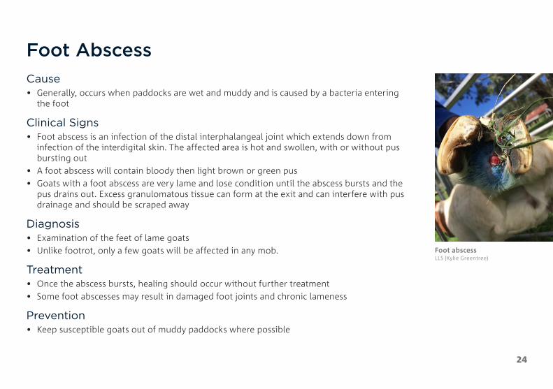
Foot AbscessCause• Generally, occurs when paddocks are wet and muddy and is caused by a bacteria entering
the foot
Clinical Signs• Foot abscess is an infection of the distal interphalangeal joint which extends down from
infection of the interdigital skin. The affected area is hot and swollen, with or without pus bursting out
• A foot abscess will contain bloody then light brown or green pus• Goats with a foot abscess are very lame and lose condition until the abscess bursts and the
pus drains out. Excess granulomatous tissue can form at the exit and can interfere with pus drainage and should be scraped away
Diagnosis• Examination of the feet of lame goats• Unlike footrot, only a few goats will be affected in any mob.
Treatment• Once the abscess bursts, healing should occur without further treatment • Some foot abscesses may result in damaged foot joints and chronic lameness
Prevention• Keep susceptible goats out of muddy paddocks where possible
Foot abscess LLS (Kylie Greentree)

25
Footrot NOTIFIABLE DISEASE
Cause• Footrot is a highly contagious bacterial disease of goats that affects one or more of
their feet • In its virulent form, it can cause significant economic loss through reduced doe fertility,
reduced growth rates and reduced returns from sale goats • In infected mobs, there are significant costs associated with controlling and eradicating
the disease
Clinical Signs• Lameness, generally in more than one foot of a large proportion of the herd• On examination of the feet, signs will range from mild redness, hair loss and moisture
between the toes to lesions underrunning the entire hoof separating the hoof from the toe• Footrot has a characteristic putrid smell but there will be no swelling and no pus
Footrot in goats LLS (Kylie Greentree)

Footrot (Colin Trengove)
26
Footrot Continued
Diagnosis• Diagnosis is made by examination of affected feet and can be confirmed by sending a
bacterial swab to a veterinary laboratory
Treatment• Seek veterinary help to confirm the virulence of the strain/s on your farm and the best
treatment options. These options include foot baths (with paring of under-run hooves), antibiotic injections and vaccinations
• A treatment or eradication program for footrot involves three phases: 1. Control phase – before and during the spread period, to reduce the level of infection in
the flock to the stage where eradication becomes feasible2. Eradication phase – involves the detection and removal of all infected goats during the
non-spread period3. Surveillance phase – involves surveillance of the whole flock to ensure the disease has
been eradicated and preventing reinfection

27
Hardware Disease (Traumatic Reticuloperitonitis)
Cause• Swallowing hard metallic objects such as wire, needles or nails. The object perforates the
reticulum or rumen and causes leakage of gut content into the abdominal cavity• Rare in goats, unlike cattle, due to their feeding behaviour
Clinical Signs• Reduced appetite, ill-thrift, fever and arched stance• Animal will appear pained and depressed• Respirations may be slow and rapid
Diagnosis• A definitive diagnosis can only be made by post-mortem
Treatment• Oral administration of a magnet may assist in immobilising the object• Antibiotics may help prevent infection• If clinical signs persist, a veterinary consultation is required to determine the next course
of treatment• If the animal does not improve and veterinary assistance is unavailable, the animal may
require euthanasia on welfare grounds
Some items that may cause hardware disease MLA

28
HydatidsProblem• There are no clinical signs in live goats, however, at slaughter affected organs
are condemned • Hydatids can cause serious, sometimes fatal, disease in people
Cause• Hydatids are the cystic stage of the dog tapeworm Echinococcus granulosus • The tapeworm is tiny, only 3–6 mm long and lives in the intestines of dogs • The larval cyst forms in intermediate host animals such as goat
Clinical Signs• No clinical signs
Diagnosis• Cannot be detected on live animals but affected carcasses are condemned at slaughter
Treatment • There is no effective treatment for Hydatids
Prevention• Worm all dogs monthly with a tapeworm tablet (must contain praziquantel) • Don’t feet raw meat / offal to dogs. Instead cook through entirely or freeze at -10°C or
10 days. • Burn or bury carcasses to prevent scavenging
Hydatid cysts MINTRAC

29
InjuriesCause• Injuries are generally accidents involving one animal injuring another, or when an animal
falls or strikes surrounding infrastructure (flooring, fence, gate)• Poorly designed or maintained facilities such as yards, laneways, fences, gates and flooring,
may increase the risk of injury to animals while being housed or handled • Poor stockmanship, including rough handling and applying excessive pressure, may also
contribute to the risk of injury
Clinical Signs• Clinical signs may include fractures, dislocations, wounds, subcutaneous bruising,
haematoma, and cellulitis or other forms of infection
Treatment• Animals with severe injuries (such as fractures) that interfere with walking, eating and
drinking, should be euthanised• Animals with wounds should be isolated to a clean area and the wounds cleaned • Lame animals should be moved to a hospital pen with uninhibited trough access• Dislocated joints require early intervention and expertise for successful reduction
Prevention • Ensure all handling facilities are in good working condition and are regularly maintained• Ensure all staff are adequately trained in stock handling
Broken leg DAF (Kylie Hopkins)
Tear in skin from wire
Dog bite wound Cathy Zwick
30
Johne’s Disease NOTIFIABLE DISEASE
Cause• Johne’s disease (JD), also called paratuberculosis, is an incurable wasting disease of livestock • Goats can become infected with JD at any age by eating pasture or drinking water
contaminated with faeces from infected animals • Susceptibility to disease can be influenced by age, breed, stress and the presence of
other diseases • Some goats infected with JD may carry the disease in their intestines and spread the
bacteria with their faeces without ever showing any obvious clinical disease • The disease has a long incubation period and most infected goats do not begin to show any
signs of illness until after two years of age
Clinical Signs• Clinically affected goats show severe wasting which eventually ends in death• The average time from onset of clinical illness to death is about 6–12 weeks • Chronic scouring may sometimes be seen, but this is not a common feature of the disease
in goats • The classic sign of the disease in a mob is a distinct ‘tail’ in body condition, with goats
ranging in condition from good to very poor • The infected goats continue to eat and drink normally until they are too weak to graze, and
eventually die
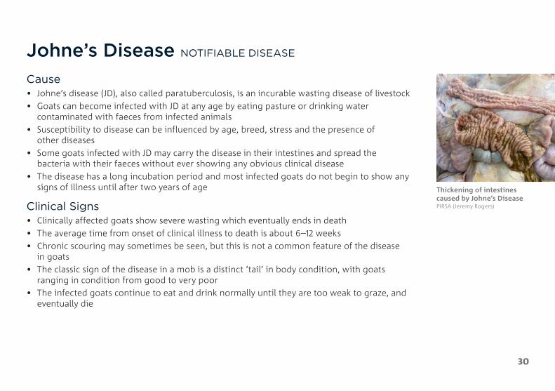
Thickening of intestines caused by Johne’s Disease PIRSA (Jeremy Rogers)

31
Johne’s Disease Continued
Diagnosis• Taking a pooled faecal sample from a selection of animals over two years old on
the property• Individual animals can be tested by faecal samples or blood tests. No test picks up the early
incubation stage • On post-mortem, thickening of the intestines and enlarged lymph nodes may be seen in
affected animals
Treatment• There is no treatment for JD
Prevention• Purchase low risk stock only • Consider vaccination of your mob with Gudair® • Ensure all purchased goats are accompanied by a NGHD and meet the entry requirements
for JD in your state
Wasting cause by Johne’s Disease

32
ListeriosisCause• Listeriosis is a bacterial infection that affects a wide
range of animals, including humans • It is most commonly associated with the feeding of
mouldy silage or spoiled hay
Clinical Signs• Initial signs are depression, anorexia (not eating),
disorientation, head tilt, and circling • As the disease progresses, facial paralysis may develop,
causing the ear and eyelid to droop, the muzzle to be pulled to one side, and a lack of muscle tone of the lip of the affected side
• Profuse salivation may also be observed• Abortion, stillbirths, and neonatal death may occur
in the herd but not generally at the same time as the nervous form
• There is usually a high death rate
Diagnosis• Clinical signs and post-mortem samples sent to a
veterinary laboratory • On post-mortem there will be no significant findings
but if the brain and a portion of the spinal cord are collected, laboratory testing can detect the disease
Treatment• Recovery depends on early intervention with high doses
of antibiotics. In severe cases however, death may occur despite treatment (contact your local veterinarian)
• Any feed (spoiled silage) suspected to be the cause should be removed
• Sick animals should be isolated from healthy animals to prevent the spread of disease between animals
Prevention• Care should be taken to avoid feeding livestock spoiled
silage or ingesting soil• Avoid pastures that are boggy and areas where the soil
has a high pH• Listeriosis can occur in humans and is potentially life-
threatening. If you suspect your stock has listeriosis please contact your local veterinarian
33
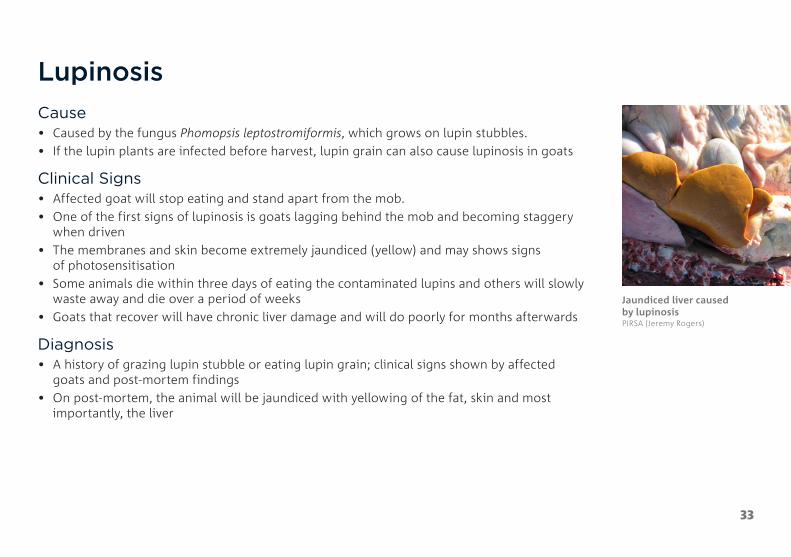
LupinosisCause• Caused by the fungus Phomopsis leptostromiformis, which grows on lupin stubbles. • If the lupin plants are infected before harvest, lupin grain can also cause lupinosis in goats
Clinical Signs• Affected goat will stop eating and stand apart from the mob. • One of the first signs of lupinosis is goats lagging behind the mob and becoming staggery
when driven • The membranes and skin become extremely jaundiced (yellow) and may shows signs
of photosensitisation • Some animals die within three days of eating the contaminated lupins and others will slowly
waste away and die over a period of weeks • Goats that recover will have chronic liver damage and will do poorly for months afterwards
Diagnosis• A history of grazing lupin stubble or eating lupin grain; clinical signs shown by affected
goats and post-mortem findings • On post-mortem, the animal will be jaundiced with yellowing of the fat, skin and most
importantly, the liver
Jaundiced liver caused by lupinosis PIRSA (Jeremy Rogers)

34
Lupinosis Continued
Treatment• There is no treatment for lupinosis, however steps can be taken to minimise the number of
goats affected and the severity of the outbreak • The mob should be removed from the lupin stubble and any access to lupin grain should
be stopped • Affected animals should be fed a low protein diet of hay and grains with a low
protein content • Affected animals should have access to a ready supply of good quality water and plenty
of shelter
Prevention• Check goats daily when grazing lupin stubble, drive the mob around the paddock for at
least 500 metres looking for any animals lagging behind • If rain occurs or there is a heavy dew, remove the goats from the lupin stubble • Graze lupin stubbles as soon as possible after harvest• Remove all stock from the lupin stubble once all the grain has been eaten• Avoid grazing goats that have a history of liver damage that may be more susceptible
to lupinosis • Feed only good quality lupin grain to goats. It is a good idea to have lupins tested before
feeding – this can be done through Agrifood Technology in Werribee, Victoria
Goat standing away from the mob, not eating
35
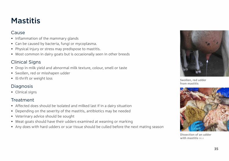
MastitisCause• Inflammation of the mammary glands• Can be caused by bacteria, fungi or mycoplasma. • Physical injury or stress may predispose to mastitis.• Most common in dairy goats but is occasionally seen in other breeds
Clinical Signs• Drop in milk yield and abnormal milk texture, colour, smell or taste• Swollen, red or misshapen udder• Ill-thrift or weight loss
Diagnosis• Clinical signs
Treatment• Affected does should be isolated and milked last if in a dairy situation• Depending on the severity of the mastitis, antibiotics may be needed• Veterinary advice should be sought • Meat goats should have their udders examined at weaning or marking • Any does with hard udders or scar tissue should be culled before the next mating season
Swollen, red udder from mastitis
Dissection of an udder with mastitis MLA

36
Milk FeverCause• Occurs when the calcium levels in the blood fall below
normal• Often seen in conjunction with low blood magnesium
(hypomagnesaemia or grass tetany)• Most common in the last few weeks of pregnancy or
the first few weeks after kidding
Clinical Signs• Initial signs include a staggery gait and muscle tremors
and the goat moves or struggles when approached• Affected goats then go down often in a sitting position
with their head turned around to their flank• Sub-clinical cases can present as dystocia due to weak
uterine muscles• Death will occur within 24–36 hours of initial signs
Diagnosis• Herd history; clinical signs shown by affected goats and
the rapid response to treatment
Treatment• Animals will respond rapidly to calcium injections with
treated goats possibly getting up and walking away within minutes. Ideally, a veterinarian would give 50–80 ml of 20% calcium borogluconate solution IV while monitoring the heart, then another 100 ml under the skin
• Treatment must be given as soon as possible after initial signs to be effective
• Calcium and magnesium solutions are readily available from most rural stores
Prevention• Avoid physical stresses on goats in the last month of
pregnancy, or with young kids at foot• Avoid grazing late pregnancy or lactating does on lush
pasture or cereal crops• Keep calcium and magnesium solution on hand while
handling pregnant or lactating does• Supply a mix of two parts stock salt to one part
stock lime next to the drinking water source for the herd during the last two weeks of pregnancy and early lactation
37
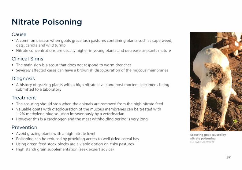
Nitrate PoisoningCause• A common disease when goats graze lush pastures containing plants such as cape weed,
oats, canola and wild turnip• Nitrate concentrations are usually higher in young plants and decrease as plants mature
Clinical Signs• The main sign is a scour that does not respond to worm drenches• Severely affected cases can have a brownish discolouration of the mucous membranes
Diagnosis• A history of grazing plants with a high nitrate level; and post-mortem specimens being
submitted to a laboratory
Treatment• The scouring should stop when the animals are removed from the high nitrate feed• Valuable goats with discolouration of the mucous membranes can be treated with
1–2% methylene blue solution intravenously by a veterinarian • However this is a carcinogen and the meat withholding period is very long
Prevention• Avoid grazing plants with a high nitrate level• Poisoning can be reduced by providing access to well dried cereal hay• Using green feed stock blocks are a viable option on risky pastures• High starch grain supplementation (seek expert advice)
Scouring goat caused by nitrate poisoning LLS (Kylie Greentree)
38
Oxalate PoisoningCause• Occurs when goats graze plants with a high concentration of oxalates such as soursob
and sorrel
Clinical Signs• Acute oxalate poisoning:
– The first signs occur within 1–3 hours of goats eating the high oxalate plants– Affected goats have muscle tremors and a staggery gait. They will go down but will be
alert and struggle when handled. Leg paddling may occur– Within hours the goat will become exhausted, the struggling will stop, they will sink into
a coma with death occurring soon after. Goats will often be found dead without signs of struggling
• Chronic oxalate poisoning:– Goats with chronic oxalate poisoning are generally poor doers and are anaemic with
pale membranes– Sporadic death occurs within an affected herd
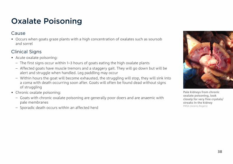
Pale kidneys from chronic oxalate poisoning, look closely for very fine crystals/streaks in the kidney PIRSA (Jeremy Rogers)

39
Oxalate Poisoning Continued
Diagnosis • A history of goats grazing high oxalate plants and clinical signs• Examining the goat’s kidneys under a microscope
Treatment• Goat with acute poisoning should be treated with calcium solution as for milk fever
(page 36)• There is no effective treatment for chronic poisoning
Prevention• Ensure hungry goats do not have access to a paddock with large amounts of high
oxalate plants
Leg paddling of a goat with acute oxalate poisoning

40
PhotosensitisationCause• Occurs when the skin becomes abnormally sensitive to sunlight• It is a symptom rather than a disease and can be the result of several different diseases such
as liver damage and the consumption of specific toxic plants
Clinical Signs• Affected animals are initially restless, they seek shade, shake their heads and may rub their
ears and eyes• Later signs may range from mild to severe sunburn, loss of appetite, jaundice and death• Areas of skin not covered by hair are the most affected such as face, ears and vulva• Fluid may build up under the skin causing swelling of the face and jaw• Eyelids may become swollen causing tears to dribble down the cheeks• Ears become swollen and droopy and are often covered in fine scabs
Diagnosis• Examination of affected goat
Treatment• Identify and address the underlying cause of the disease• Protect affected animals from direct sunlight• Provide nursing care while recovery takes place• Affected animals may benefit from antibiotic, anti-inflammatory and antihistamine
injections (consult your veterinarian)
Photosensitisation from lantana consumption Kylie Leahy, Mt Roy Stud

41
PinkeyeCause• A common bacterial disease of goats, especially when conditions are dusty and there are
large numbers of flies
Clinical Signs• The first sign of pinkeye is the inflammation of the eye membranes and the production of
clear watery tears that run down the cheek• As the pinkeye progresses, the cornea (the clear eye surface) develops a blue haze that
becomes more cloudy and turns white over 3–4 days• Shallow ulcers may develop on the cornea• A goat with pinkeye cannot see out of that eye so goats with pinkeye in both eyes are
totally blind and will lose weight• As the eye heals the blood vessels grow into the cornea making the eye appear pink
Diagnosis• Examination of the eye of affected goats
Treatment• Most cases recover without treatment in 2–3 weeks• Yarding goats can make the situation worse as dust and flies can make the infection spread
through the mob• Antibiotic ointment and antibiotic injections can also be used with positive results, but
must be used under veterinary advice
Pinkeye in a kid with ulceration MLA
Pinkeye in a goat with blue haze and cloudiness

42
PneumoniaCause• Inflammation or infections of the lungs• Pneumonia may be viral, bacterial, fungal or from aspiration.
Clinical Signs• Difficulty breathing, coughing, fever, rapid respiratory rate and nasal discharge• The laboured breathing will often cause lethargy and slow movement
Diagnosis• Clinical signs and post-mortem examination of the chest cavity and lungs
Treatment• Treatment is dependent on the cause of the pneumonia• Veterinary prescribed antibiotics are generally required• Provide easy access to feed and water• Severely affected animals may require euthanasia
Pneumonic changes in the lungs with consolidated portions and pus PIRSA (Jeremy Rogers)

43
Polioencephalomacia (PEM)
Cause• Caused by a deficiency of thiamine (vitamin B1) in goats of all ages and both sexes • May occur in goats on high grain diets, and diets that include plants high in thiaminases
(e.g. bracken, rock fern, mulga fern)
Clinical Signs• The first signs are listlessness and a loss of appetite, affected goats will separate from the
rest of the mob, appear blind and either wander aimlessly or stand still • Goats will adopt one of two stances: – head lowered to the ground; or – ‘Star gazing’ with a
fixed stare into the sky over the horizon • Later, affected goats will go down and if startled may start galloping leg movements and
have convulsions• A goat that is down will generally have its head and neck arched back stiffly• If not treated, goats will get weaker, sink into a coma and die within 2–3 days
Diagnosis• Clinical signs and response to treatment in cases found early• Post-mortem examination of the brain can be used to diagnose PEM
Treatment• Goats respond to thiamine or B1 injections which are available from most rural stores.
Repeat doses will be needed every 6 hours until normal• Mildly affected goats will improve within 6–8 hours of treatment• Goats that are down will have irreversible damage to the brain and should be destroyed on
welfare grounds
Head and neck arched back stiffly from PEM Kylie Leahy Mt Roy Stud

44
Pregnancy ToxaemiaCause• Common disease of ewes in the last six weeks of pregnancy and immediately after kidding• During late pregnancy, the doe has a very high energy requirement to provide for her own
needs and the growth of the kid or kids she is carrying• If the energy requirement is not met by feed intake, the doe will break down her own
body tissues• If the rate at which the doe breaks down her tissues is too rapid, toxic wastes from the
breakdown process accumulate and pregnancy toxaemia occurs• Seen in either very thin or very fat does and does carry triplets or quads• Rangeland does often abort, rather than develop pregnancy toxaemia, if nutrition is poor
Clinical Signs• The initial signs are dullness, swelling of the lower limbs, loss of appetite and affected goats
lagging behind the herd when driven• Later the doe will stand alone, appear dopey and will not move when approached• If driven the doe will appear blind, stumble and go down• The doe will eventually sink into a coma and die within 5–7 days
Heavily pregnant doe with multiples at risk of pregnancy toxaemia MLA
45
Pregnancy Toxaemia Continued
Diagnosis• Based on flock history, presence of ketones in the urine and clinical signs shown by affected does and
post-mortem findings• On post-mortem, the liver is generally tan to yellow in colour and is quite soft and there is often plenty of fat inside the
abdomen (which physically limited feed intake)
Sometimes the liver is so badly affected a piece of it will float in water due to the infiltrated fat
Treatment• Treatment is usually unrewarding as most goats die despite treatment• If treatment is to be given, the strategy is to give the doe a source of energy• Treatment must be given as soon as possible after initial signs to be of any benefit• Oral propylene glycol solutions are readily available from most rural stores. Glycerine can also be used – 60 ml in
warm water twice a day
Prevention• Give pregnant does the best paddock feed available during the month prior to kidding• If necessary, provide supplementary feed during the last few weeks of pregnancy• In herds where multiple births are expected, does should be at least a condition score 3, however they should be no more
than condition score 4. Consider scanning and segregating twins, singles and empties for better feeding outcomes• Keep physical stress on pregnant does to a minimum by avoiding unnecessary mustering or yarding or time off feed
46
Pulpy Kidney Enterotoxaemia
Cause• It is a clostridial disease that mostly affects kids grazing
lush feed but can occur in all ages of goats that are heavily grain fed
• Most cases of pulpy kidney occur in flocks with an inadequate vaccination program
Clinical Signs• Most goats with pulpy kidney are found dead as the
disease develops very quickly and death will occur within hours of initial signs
• Initial signs of pulpy kidney are dullness followed by goats going down with convulsions and frothing at the mouth
• Death will generally occur within 24 hours
Diagnosis• A tentative diagnosis is based on a history of sudden
death while on a high-risk diet
• To confirm pulpy kidney requires laboratory testing of post-mortem samples including portions of the intestines, kidneys, brain and urine
• On post-mortem the pericardial fluid may clot on exposure to air. There may be glucose in the urine and tiny haemorrhages scattered in some muscles
Treatment• There is no economic treatment available. Veterinarian
can treat with intravenous antibiotics and fluids
Prevention• Vaccination is the key to prevention of pulpy kidney.
Note goats require more frequent vaccination than sheep

47
SalmonellosisCause• An infectious disease of livestock caused by Salmonella bacteria
Clinical Signs • Affected animals appear dull, have a fever and have profuse scouring• The scours are often watery with strands of mucous • Death generally occurs within 12–24 hours of onset of clinical signs• Surviving animals may suffer serious setbacks and become poor performers
Diagnosis• Diagnosis is based on clinical signs, mob history, culture of faecal samples and
post-mortem examination• On post-mortem examination, enlarged lymph nodes and inflamed intestines will
be evident
Treatment• Affected goats may be treated with antibiotics and electrolyte therapy if caught early,
however, treatment is generally unsuccessful. This is a zoonotic disease so care must be taken when handling sick goats
• The spread of the disease can be slowed by minimising stress on affected mobs, decreasing stocking density by opening gates and ensuring animals have access to good quality feed and water
Scour from salmonellosis LLS (Kylie Greentree)
48
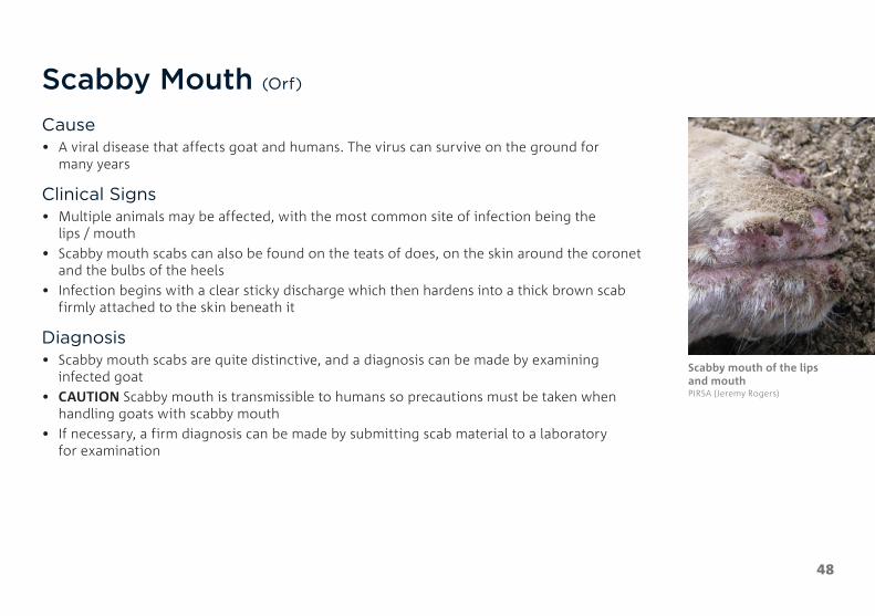
Scabby Mouth (Orf)
Cause• A viral disease that affects goat and humans. The virus can survive on the ground for
many years
Clinical Signs• Multiple animals may be affected, with the most common site of infection being the
lips / mouth• Scabby mouth scabs can also be found on the teats of does, on the skin around the coronet
and the bulbs of the heels• Infection begins with a clear sticky discharge which then hardens into a thick brown scab
firmly attached to the skin beneath it
Diagnosis• Scabby mouth scabs are quite distinctive, and a diagnosis can be made by examining
infected goat• CAUTION Scabby mouth is transmissible to humans so precautions must be taken when
handling goats with scabby mouth• If necessary, a firm diagnosis can be made by submitting scab material to a laboratory
for examination
Scabby mouth of the lips and mouth PIRSA (Jeremy Rogers)
49
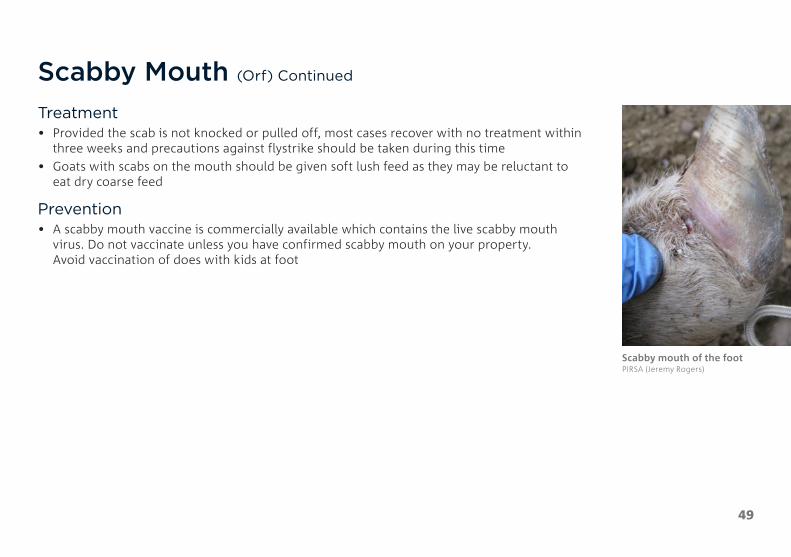
Scabby Mouth (Orf) Continued
Treatment• Provided the scab is not knocked or pulled off, most cases recover with no treatment within
three weeks and precautions against flystrike should be taken during this time• Goats with scabs on the mouth should be given soft lush feed as they may be reluctant to
eat dry coarse feed
Prevention• A scabby mouth vaccine is commercially available which contains the live scabby mouth
virus. Do not vaccinate unless you have confirmed scabby mouth on your property. Avoid vaccination of does with kids at foot
Scabby mouth of the foot PIRSA (Jeremy Rogers)

50
Selenium DeficiencyCause• Selenium deficiency generally occurs in spring and early
summer on clover-dominant pastures• There are selenium deficient areas in many parts
of Australia
Clinical Signs• Generally seen in spring born kids at 3–4 weeks of age• Affected kids walk with a stiff gait and appear weak• Severe cases die of starvation, exposure or predation• Some kids die of heart failure and others recover but
suffer a growth setback
Diagnosis• A tentative diagnosis can be made by examining the
animals for pale muscle tissue on post-mortem• A definitive diagnosis can be made by taking a
blood sample from the animal and submitting it to the laboratory
Treatment• Selenium can be administered to affected goats by
injection or as a drench• There is a very small safety margin in regard to selenium
so treatment should only be administered where muscle disease has been diagnosed and treatment should be strictly given in accordance to manufacturer’s advice
Prevention• Selenium supplements are available as vaccines and
drenches. There are slow release pellets registered for sheep, but not goats
• Care must be taken to not overdose by supplementation with repeat vaccination/drenching

51
TetanusCause• Tetanus is a clostridial disease that mostly affects kids
within three weeks of marking• Most cases of tetanus occur in herds with an inadequate
vaccination program
Clinical Signs• The main signs are muscle spasms including head
tremors, restricted jaw movements, dilated nostrils and pricked ears
• The tail is generally held out and there is stiffness in the legs
• Initially kids can walk with a stiff gait but as the disease progresses, they will go down and have intermittent convulsions
• Sound and sudden movements are likely to set off bouts of convulsions
• Most cases die within 3–4 days
Diagnosis• Based on clinical signs shown by affected goats
Treatment• There is no economic treatment for tetanus. Veterinary
treatment is often disappointing unless started early
Prevention• Vaccination is the key to prevention of tetanus• See the ‘vaccination program’ section for a detailed
description of best practice for vaccinations (page 54)• Good hygiene at kid marking is important in
preventing tetanus

52
Toe Abscess (White line abscess)
Cause• Various agents enter through the hoof via cracks especially in the white line (between hoof wall and sole) area
Clinical Signs• Only one hoof is affected on the severely lame leg• Most cases are only in the toe but if untreated the abscess will discharge eventually at the coronet (where hair meets the
hoof)• Only a small percentage of the herd are infected
Diagnosis• Based on clinical signs shown by affected goats
Treatment• After finding the most painful area, hoof paring to establish drainage and release the pus
Prevention• There is no prevention strategies as the different bacteria that invade the toe are found anywhere in the environment

53
Urolithiasis Urinary Calculi or Bladder Stones
Cause• A common ailment of male goats • The calculi form when minerals in the bladder precipitate to form crystals• The crystals grow in size as more minerals are deposited
Clinical Signs• Affected animals are initially restless and regularly strain to try to pass urine• If the urinary calculi can pass, the animal will release a large volume of urine • If the urinary calculi can’t pass, the urine will become backed up and will start to be
absorbed into the blood stream and the bladder may rupture. If this happens, the animals will initially improve but quickly become dull, fall into a coma and die
Diagnosis• Diagnosis is by examination of the animal and observing clinical signs• On post-mortem, urinary calculi will be visible in the bladder and evidence of leaked urine
or a ruptured bladder or urethra may be evident
Treatment• If the urinary calculi are lodged in the tip of the penis, it can sometimes be milked out or
the worm like structure removed from the end of the penis with scissors• The stones should be kept for mineral analysis as there are a few different types• If the animal can’t pass the calculi on its own, the only treatment available is surgical
removal however this is rarely economical
Urinary calculi being milked out of the tip of the penis PIRSA (Jeremy Rogers)

54
VaccinationVaccines that are registered for use in goats are shown in the table below:
NAME ACTIVES
Coopers® Tasvax 5in1 Vaccine for Sheep, Cattle and Goats Plus Selenium for Lambs
Selenium as Sodium Selenate | Clostridium chauvoei – Toxoid | Clostridium novyi Type B – Toxoid | Clostridium perfringens Type D Toxoid | Clostridium septicum – Toxoid | Clostridium tetani – Toxoid
Gudair® Vaccine Mycobacterium paratuberculosis
Glanvac® 3 Vaccine Clostridium perfringens Type D Toxoid | Clostridium tetani – Toxoid | Corynebacterium pseudotuberculosis (ovis) – Toxoid
Glanvac® 6 Vaccine Clostridium chauvoei – Formol Culture | Clostridium novyi Type B – Toxoid | Clostridium perfringens Type D Toxoid | Clostridium septicum – Toxoid | Clostridium tetani – Toxoid | Corynebacterium pseudotuberculosis (ovis) – Toxoid
Equivac® T Vaccine Clostridium tetani UF Toxoid
Leptoshield® Vaccine Leptospira interrogans serovar pomona – Formol culture | Leptospira borgpetersenii serovar hardjo Type hardjobovi
Glanvac® 6 B12 Vaccine Vitamin B12 (Hydroxocobalamin acetate) | Clostridium chauvoei – Formol Culture | Enterotoxaemia = Pulpy Kidney = Cl. perfringens | Clostridium novyi Type B | Malignant oedema = Clostridium septicum | CLA = Corynebacterium pseudotuberculosis (ovis) | Tetanus = Clostridium tetani
Coopers® Tasvax 5in1 Vaccine for Sheep, Cattle and Goats
Clostridium chauvoei – Killed | Clostridium novyi Type B – Killed | Clostridium chauvoei – Toxoid | Clostridium novyi Type B – Toxoid | Clostridium perfringens Type D Toxoid | Clostridium septicum – Toxoid | Clostridium tetani – Toxoid

55
Vaccination Continued
Clostridial diseases• Available Vaccines – 2-in-1 (tetanus and pulpy kidney),
3-in-1 (tetanus, pulpy kidney and cheesy gland) or 5-in-1 vaccine (tetanus, black disease, black leg, malignant oedema and enterotoxaemia)
• Note research has shown that goats react badly to multiple vaccines so the smallest number of active ingredients should be chosen based on diagnosed problems on your property. The fewer ingredients also means the lumps at the site of vaccination will be smaller
Caseous Lymphadenitis (CLA)• Available Vaccines – Glanvac® 3 (tetanus, pulpy kidney
and cheesy gland), Glanvac® 6 (tetanus, pulpy kidney, black leg, malignant oedema, black disease and cheesy gland) or Glanvac® 6 B12
• Note the manufacturers of Glanvac® recommend that as goats do not commonly suffer from clostridial diseases other than tetanus and enterotoxaemia; that farmers use Glanvac® 3 unless these diseases are known to occur in goats on the property
Leptospirosis • Clinical cases of leptospirosis is rare in goats, unlike
in cattle
Gudair®• Registered as an aid in the control of Johne’s disease
in goats. Only one injection is needed i.e. no booster is required
• Kids should be vaccinated between one and four months of age

56
Vaccination Continued
Clostridial disease and cheesy gland vaccination
For maximum protection of newborn kids: • Vaccinate does four weeks before kidding
For protection of kids from unvaccinated does: • Vaccinate at six weeks with a booster 4–6 weeks later
For protection of kids from vaccinated does: • Vaccinate kids at 6–8 weeks, with a second
4–6 weeks later.
Older stock: • Annual booster timed before known high-risk periods.
More frequently in high-risk situations, such as grain feeding in drought
New stock: • If goats come from a herd with a known history of
vaccination, simply slot the animals into the existing herd program
• If vaccination history unknown, give a sensitising dose as per label directions, then a booster 4–6 weeks later. Follow with annual boosters as per the rest of the herd
• All new stock should be quarantined and carefully monitored for signs of illness/disease for at least seven days, prior to introduction to other animals on the property

57
Vaccination Continued
Key points when injecting: • Give low volume (less than 5 ml) subcutaneous injections high on the goat’s neck, behind
the ear, and high volume ones under the skin of the chest, over the ribs• Give intramuscular injections in the muscles of the neck, in front of the goat’s shoulder, at
a maximum of 10 ml per injection site• Read all product labels and veterinarian’s instructions before use, and take note of the
withholding period, export slaughter interval, dose rate and site of injection. Record the batch number, expiry date and date of treatment
• Use sterilised injecting guns, syringes and sharp needles. Replace needles frequently and change needles that are blunt, bent or dirty
• Train all operators in injection technique, and how to avoid accidental self-injection and exposure to injected medicines
• Take special care when injecting oil-based vaccines and Gudair® so use guarded needle vaccinating guns
• Carefully dispose of needles and other sharps according to local government requirements
Correct vaccination technique for intramuscular injection Agriculture Victoria (Berwyn Squire)

58
Worms SIGNS OF WORMS
Bottle Jaw (sub-mandibular oedema)• Swelling under the jaw results from severe barber’s
pole worm or liverfluke. Johne’s disease can also cause bottle jaw
Coughing and pneumonia• The large lungworm infects the airways of goats and
causes the production of a frothy mucus that will cause the animal to cough; there may also be a nasal discharge. In heavy infections pneumonia (inflammation and infection in the lung tissue) may be evident, accompanied by rapid breathing. Infection with the small lung worm rarely produces clinical evidence of disease in affected animals
Anaemia• Barber’s pole worm is the only roundworm to cause
anaemia (blood loss) as it sucks blood from the lining of the fourth stomach (abomasum). Severe acute or ongoing (chronic) blood loss from barber’s pole worm leads to obvious signs of anaemia. These are pale gums and conjunctiva (inside the eyelids); lack of stamina causing lagging or collapse when mustered; and ultimately death from lack of red blood cells needed to carry oxygen around the body
• Liver fluke can also cause anaemia
Lethargy and Collapse• Lethargy or weakness can result from a severe infection
of many worms
Weight Loss• An infection with scour worms results in a marked
decrease in appetite and reduced digestive efficiency. As a result, affected animals lose weight and with severe infections can become emaciated
Scouring• The presence of scour worms initiates a strong immune
response from the host animal that damages the lining of the gut resulting in diarrhoea (scouring). Coccidiosis can also cause scouring
59
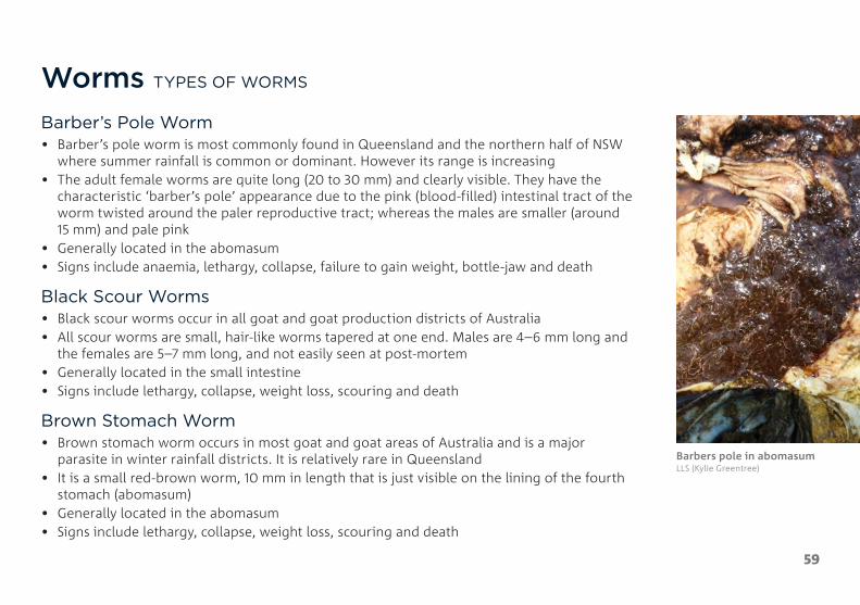
Worms TYPES OF WORMS
Barber’s Pole Worm• Barber’s pole worm is most commonly found in Queensland and the northern half of NSW
where summer rainfall is common or dominant. However its range is increasing• The adult female worms are quite long (20 to 30 mm) and clearly visible. They have the
characteristic ‘barber’s pole’ appearance due to the pink (blood-filled) intestinal tract of the worm twisted around the paler reproductive tract; whereas the males are smaller (around 15 mm) and pale pink
• Generally located in the abomasum• Signs include anaemia, lethargy, collapse, failure to gain weight, bottle-jaw and death
Black Scour Worms• Black scour worms occur in all goat and goat production districts of Australia• All scour worms are small, hair-like worms tapered at one end. Males are 4–6 mm long and
the females are 5–7 mm long, and not easily seen at post-mortem• Generally located in the small intestine• Signs include lethargy, collapse, weight loss, scouring and death
Brown Stomach Worm• Brown stomach worm occurs in most goat and goat areas of Australia and is a major
parasite in winter rainfall districts. It is relatively rare in Queensland• It is a small red-brown worm, 10 mm in length that is just visible on the lining of the fourth
stomach (abomasum)• Generally located in the abomasum• Signs include lethargy, collapse, weight loss, scouring and death
Barbers pole in abomasum LLS (Kylie Greentree)

Host stageInfective larvae become adults that live for many months within the goat’s gut to reprouce and lay eggs– Minimum time from L3 to egg
laying is 18 days– The goat’s immunity can expel
worms or suppress egg laying
Dung containing worm eggs is
passed from the goat onto the
pasture.
Infective larvae wriggle out of the
dung onto the ground and
pasture.
Infective larvae are eaten along with
the pasture. Uneaten larvae die.
Dung stageEggs develop through L1 and L2 stages to L3 ‘infective larvae’– Time from egg to L3 is 4–10 days (slower
when cooler, faster when warmer)– Eggs, L1 and L2 will not develop under
10°C and over 40°C– L1 and L2 feed on bacteria in the dung
Pasture stageThird stage ‘infective larvae’ (L3) move in moisture (rain/dew) and wriggle randomly, some on to the pasture to be eaten by goats– Quite resistant to cold and heat, but
susceptible over 40°C– Most L3 die within 3 (summer) to 6
(winter) months; some live over 1 year– L3 do not feed; they die when energy
reserves are used up (faster at higher temperature and humidity)
60
Worms TYPES OF WORMS Continued
Thin-necked intestinal worm• Thin-necked intestinal worm occurs
in most of the major goat and goat production areas of Australia but is mostly an issue in the winter rainfall districts
• They are relatively long worms with a slender anterior end that is frequently coiled
• Generally located in the small intestines
• Signs include lethargy, collapse, weight loss, scouring and death
61
Worms TREATMENT AND PREVENTION
TreatmentA faecal egg count is best practice in determining when to treat for worms. There are limited drenches registered for treatment of worms in goats. Drench resistance is a growing problem so drenches should only be used when a worm burden is causing ill health or production losses.
When selecting a drench to control worms, consider the following four R’s:
1. The use of REGISTERED drenches with a vet’s prescription for a higher dose rate than on the label
2. Chemical RESIDUES following treatment
3. Worm RESISTANCE to drenches
4. The number of parasites in the REFUGIA (animals, paddocks, mobs not exposed to drench)
Additional information can be found at www.wormboss.com.au and by talking to your local veterinarian.

62
DrenchingKey points when drenching:• Use an effective product for the job and always check that the drench gun is compatible
with the product. If in doubt, ask your veterinarian or animal health adviser.• Carefully read the label before drenching and follow the instructions. Adhere to the
recommended dose rate, withholding period, export slaughter interval and any precautions or contraindications.
• Calibrate the drench gun using a measuring cylinder or large disposable syringe before the start of drenching, and recheck after every 200 goats. Weigh some goats to set the appropriate dose for the heaviest goat in the mob.
• Excess speed can cause goats to be missed, spit out the drench or to be injured.• Keep the goat’s head level (horizontal) with one hand while slipping the gun into the side of
its mouth. Place the tip of the barrel over the goat’s tongue and gently depress the trigger.• When dispensing large volume treatments, such as for metabolic diseases, stop briefly after
each 10 ml to check that the goat is swallowing.• Shake the drench before use to ensure the active ingredient is evenly distributed through
the product.• Hold goats off feed for up to 24 hours before drenching and six hours after drenching if
possible, as it may improve drench efficacy.• Only use a drench off label under the advice of a veterinarian.
Correct drenching technique in goats Agriculture Victoria (Berwyn Squire)
63
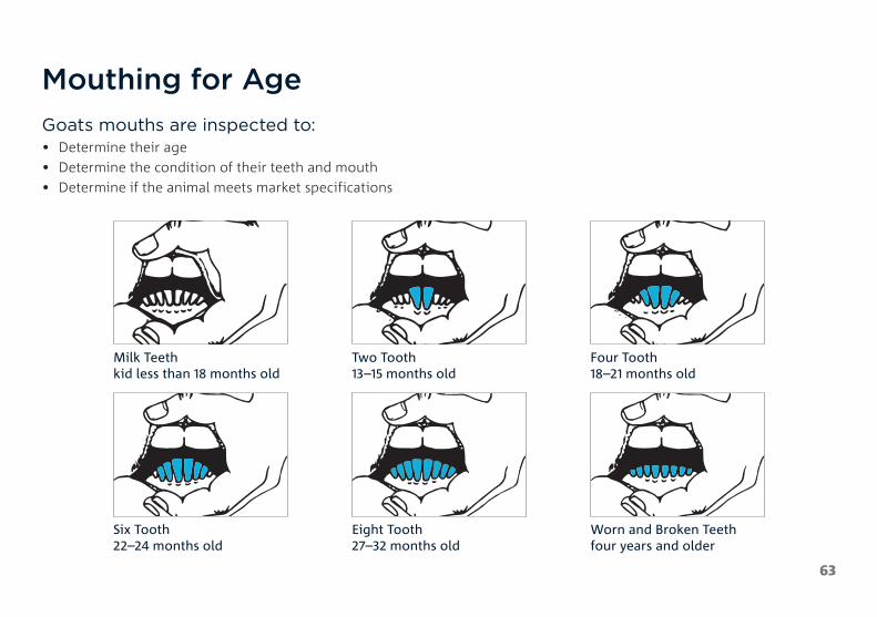
Mouthing for AgeGoats mouths are inspected to:• Determine their age• Determine the condition of their teeth and mouth• Determine if the animal meets market specifications
Milk Teeth kid less than 18 months old
Two Tooth 13–15 months old
Four Tooth 18–21 months old
Six Tooth 22–24 months old
Eight Tooth 27–32 months old
Worn and Broken Teeth four years and older

64
Foot ParingKey points when foot paring:• Foot health is essential to the wellbeing of goats.• Goat productivity can be severely affected by lameness.• Knowing the normal shape of the goat’s foot and how to pare feet will help detect
problems earlier.• Promptly inspect the feet of any goats that are lame and take appropriate action.• Prior to purchasing goats always inspect the feet of as many of the goats as possible and
request a fully completed National Goat Health Declaration.• Inspect bucks’ feet prior to purchase, and at least eight weeks before joining. Pare to the
normal shape if required.• Over trimming feet causes bleeding and pain and can lead to the formation of
toe granulomas.• After paring, observe goats for lameness and bleeding. Keep any lame goats or goats with
bleeding feet in a holding paddock with feed, water and shelter until they have recovered.
Overgrown toe needing paring LLS (Kylie Greentree)
Correct paring technique to bring toe back to a normal shape Agriculture Victoria (Berwyn Squire)

65
DisbuddingKey points when disbudding (removal of horn buds in kids):• Always disbud animals as soon as there is enough horn bud can be felt to properly centre the horn bud to minimise pain
and stress to the animals and limit production losses. This can be as early as 3–4 days in bucks and 5–7 days in does and up to two weeks in miniature kids. Disbudding should not be conducted after three weeks of age.
• Ensure good restraint facilities.• Ensure adequate protection against tetanus.• Ensure instruments are well-maintained, clean and sharp.• Do not use caustic dehorning chemicals.• Do not use tools such as axes, hammers and chain saws.• Pain relief needs to be administered with this procedure. Please consult your local veterinarian for advice on current
pain relief and pain minimisation strategies
• Use a hot disbudding iron to burn the horn bud and area around it. The hot iron also cauterises the blood vessels.• Inspect kids frequently (daily) after disbudding for approximately 10 days to detect complications. If these occur,
treatment should be quickly administered.

66
CastrationKey points when castrating:• Kids should be castrated before they reach 12 weeks of age. Castrating kids older than
12 weeks is not considered best practice. In some states kids over two months can only be castrated by a veterinarian.
• The most common method of castrating kids is with elastrator rings. Elastrator rings:– Have less overall welfare impact on the kids compared to using a marking knife or scalpel– Reduce the risk of bleeding– Reduce the risk of transferring blood between animals and reduces the risk of
spreading disease– Removes the workplace health and safety risk of exposure to blood and working with
knives or scalpels
• The use of a marking knife or scalpel is a less common option.• The operator should be competent in the castration technique.• Pain relief must be given. Cost effective pain relief products are available
through your veterinarian.
Correct castration technique with elastrator rings Agriculture Victoria (Berwyn Squire)

67
Kidding NORMAL BIRTH PROCESS
Signs the doe is ready to kid• Udder will be firm and enlarged • Swollen vulva and thick mucous discharge will appear• Doe may separate from the herd and find a quiet area
The Kidding Process• The doe will paw the ground and get up and down frequently• The water bag will appear and generally burst• For a normal presentation, the nose and front hooves will then be visible• The doe will then lie on the ground and push until the kid is born• The birth membranes will be expelled soon after the kid is born• The doe will lick her kid dry and attempt to feed within the first hour of birth
Thick mucous discharge and appearance of the water bag Kylie Hopkins (DAF Queensland)
Normal kidding presentation of nose and front hooves Kylie Hopkins (DAF Queensland)

68
Kidding DIFFICULTIES
Abnormal Presentations• Forward presentation – head first with front legs back
– Will often require assistance
• Forward presentation – front legs first with head back– Will often require assistance
• Backward presentation – hind legs first– Generally, won’t require assistance– Normal presentation for the second kid
• Breech presentation – rump first with all legs facing forward– Will require assistance
Other Potential Difficulties• Overgrown kids• Twins and triplets• Deformed kids• Dead kids• Twisted Uterus• Retained birth membranes• Prolapsed Uterus
69
Kidding DIFFICULTIES Continued
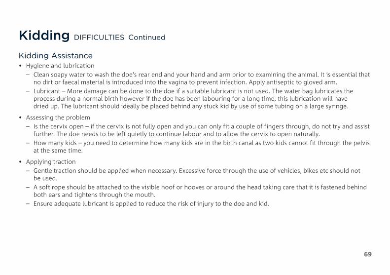
Kidding Assistance• Hygiene and lubrication
– Clean soapy water to wash the doe’s rear end and your hand and arm prior to examining the animal. It is essential that no dirt or faecal material is introduced into the vagina to prevent infection. Apply antiseptic to gloved arm.
– Lubricant – More damage can be done to the doe if a suitable lubricant is not used. The water bag lubricates the process during a normal birth however if the doe has been labouring for a long time, this lubrication will have dried up. The lubricant should ideally be placed behind any stuck kid by use of some tubing on a large syringe.
• Assessing the problem– Is the cervix open – if the cervix is not fully open and you can only fit a couple of fingers through, do not try and assist
further. The doe needs to be left quietly to continue labour and to allow the cervix to open naturally.– How many kids – you need to determine how many kids are in the birth canal as two kids cannot fit through the pelvis
at the same time.
• Applying traction– Gentle traction should be applied when necessary. Excessive force through the use of vehicles, bikes etc should not
be used.– A soft rope should be attached to the visible hoof or hooves or around the head taking care that it is fastened behind
both ears and tightens through the mouth.– Ensure adequate lubricant is applied to reduce the risk of injury to the doe and kid.

70
Rearing Orphan KidsFostering• This is the easiest option if a suitable doe is available and willing to accept the orphan kid.• Confining the doe and orphan kid in a small area will often help with the bonding process
and this should be continued for several days until the bond is well established
Colostrum• The first milk (colostrum) is full of antibodies and is high in energy and vitamin A. Without
it the kid is more susceptible to cold stress, diseases and death.• Newborn kids should receive colostrum as its first feed and this must be in the first
4–6 hours of life, as the kid’s gut loses the ability to absorb antibodies very rapidly after this time. If a suitable freshly kidded doe is available, consider restraining the doe and allowing the orphan kid to suckle the colostrum. Alternatively, milk out one side of the doe and bottle feed the colostrum to the orphan kid.
• Sheep or cow’s colostrum can be used if no goat colostrum is available, alternatively there are commercial powdered colostrum replacer and recipes for artificial colostrum but it is not as effective as the real thing. Raw egg must not be fed to young kids in any colostrum recipe as it is restricted animal matter and can’t be fed to ruminant animals in some states.
For detailed information on colostrum management and feeding, refer to the AHA publication Snatch Rearing and Pre-Weaning Kid Management in Goat Enterprises.
Doe naturally raising twin kids MLA

71
Rearing Orphan Kids Continued
Feeding milk• There are several options for milk for feeding orphan kids
– Goat milk from other lactating does– Full cream cow’s milk– Goat or calf milk replacer
• Milk requirements are based on kids’ weight and should be around 15% of the kid’s weight over a 24 hour period.
• The following table shows an average small breed kid age and milk requirements
AGE MILK REQUIRED
1–3 days 150 ml four times per day
4–9 days 300 ml four times per day
10–14 days 500 ml three times per day
15–21 days Gradually increasing to 1 L twice per day
3–6 weeks 1 L twice per day, gradually decreasing milk over 7–10 days to wean
Kids need access to water grain and hay from the first week. Weaning should only occur if the kid is eating sufficient hay and grain.
Bottle feeding an orphan kid MLA

72
Fit to Load• Remember, it is your responsibility to ensure animals are fit to load and fit for the intended
journey before they are loaded. • Before livestock are loaded, ensure you complete this checklist.
The animal: – can walk on its own by bearing weight on all four legs – is free from visible signs of severe injury or distress or conditions likely to further
compromise its welfare during transport – is strong enough to make the journey (i.e. not dehydrated or emaciated) – can see well enough to walk, load and travel without impairment or distress (e.g. it is not
blind in both eyes) – is not in late pregnancy or too young to travel (refer to the Standards to determine limits
for late pregnancy) – has had adequate access to water prior to loading to meet the maximum time off water
standards
Not fit to load as it is unable to bear weight on all four legs Kylie Hopkins (DAF)
Not fit to load as the doe is too late in pregnancy MLA

73
National Livestock Identification System (NLIS)
The National Livestock Identification System (NLIS) is Australia’s system for the identification and traceability of cattle, sheep and goats. NLIS reflects Australia’s commitment to biosecurity and food safety and provides a competitive advantage in a global market.
As goats are bought, sold and moved along the supply chain, they must be tagged with an NLIS-accredited tag or device, unless they are eligible to be moved tag free in your state or territory.
The NLIS is underpinned by state and territory legislation, which forms the regulatory framework for the system. Producers need to comply with relevant legislation in the jurisdiction in which they operate.
The basic requirements of the NLIS for goat producers are:
• All goat properties must have a Property Identification Code (PIC) or equivalent • All goat movements must be accompanied by a Livestock Production Assurance (LPA) National Vendor Declaration (NVD)
or alternate documentation • All goats must be tagged with an accredited NLIS tag or device before being moved off a property, unless a tagging
exemption applies (rangeland goats harvested from the wild and dairy goats are exempt from tagging in some states and territories)
• When moving goats that are non-vendor bred animals and which have not been previously tagged, post breeder devices must be applied (unless there is a tagging exemption for the movement)
• Check with your state/territory NLIS authority to find out what exemptions apply in your jurisdiction
More information on the NLIS system can be found here: NLIS www.integritysystems.com.au/identification--traceability/national-livestock-identification-system
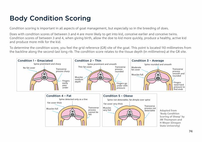
74
Body Condition ScoringCondition scoring is important in all aspects of goat management, but especially so in the breeding of does.
Does with condition scores of between 3 and 4 are more likely to get into kid, conceive earlier and conceive twins. Condition scores of between 3 and 4, when giving birth, allow the doe to kid more quickly, produce a healthy, active kid and produce more milk for the kid.
To determine the condition score, you feel the grid reference (GR) site of the goat. This point is located 110 millimetres from the backline along the second-last long rib. The condition score relates to the tissue depth (in millimetres) at the GR site.
Condition 1 – Emaciated
No fat cover
Spine prominent and sharp
Transverse process sharp
Fingerseasilypass under
Condition 2 – Thin
Thin fat coverSpine prominent and smooth
Muscles medium depth
Transverseprocess rounded
Fingers go under withpressure
Condition 3 – Average
Moderate fat cover
Spine rounded and smooth
Muscles full
Transverse processsmooth and rounded
Fingersneed hardpressure to �nd ends
Condition 4 – Fat
Fat cover thick
Spine detected only as a line
Muscles full Transverse process cannot be felt
Condition 5 – Obese
Fat cover very thick
Spine not detectable; fat dimple over spine
Muscles very full
Transverse process not detectable Adapted from
'Body Condition Scoring of Sheep' by JM Thompson and H Meyer (Oregon State University)
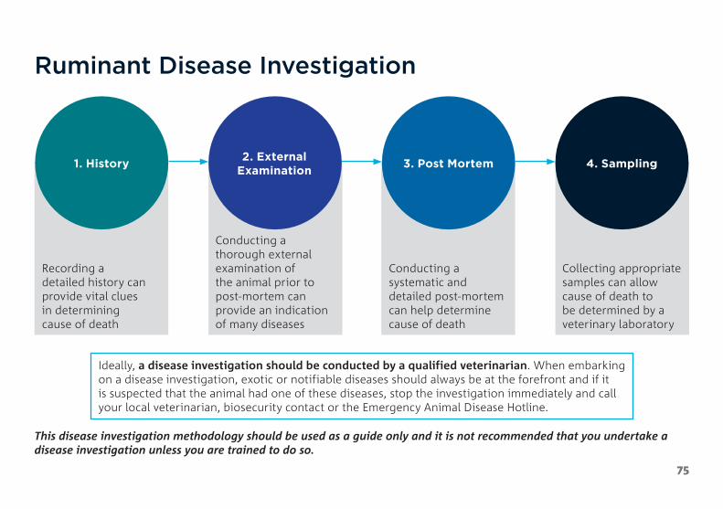
75
Ruminant Disease Investigation
1. History
Recording a detailed history can provide vital clues in determining cause of death
2. External Examination
Conducting a thorough external examination of the animal prior to post-mortem can provide an indication of many diseases
3. Post Mortem
Conducting a systematic and detailed post-mortem can help determine cause of death
4. Sampling
Collecting appropriate samples can allow cause of death to be determined by a veterinary laboratory
Ideally, a disease investigation should be conducted by a qualified veterinarian. When embarking on a disease investigation, exotic or notifiable diseases should always be at the forefront and if it is suspected that the animal had one of these diseases, stop the investigation immediately and call your local veterinarian, biosecurity contact or the Emergency Animal Disease Hotline.
This disease investigation methodology should be used as a guide only and it is not recommended that you undertake a disease investigation unless you are trained to do so.

76
Personal Protective Equipment and Standard Post-Mortem EquipmentIf you attempt to carry out a disease investigation on your property, it is essential that appropriate personal protective equipment be worn.
Some animal diseases can be passed onto humans with debilitating or even fatal results (e.g. anthrax)
At a minimum, latex or nitrile gloves and rubber boots should be worn prior to touching the animal.
• If you have not been vaccinated for Q-Fever or do not know your Q-Fever status, a face mask should be worn.
• Overalls are also recommended. Disposable overalls are available from most rural stores and hardware stores.
Equipment required for a post-mortem:• boning and skinning knives• scissors• scalpel and blades• forceps• large pruning secateurs• small axe• hammer• pH strips
• urine test strips• if you are collecting samples,
you will also need sterile specimen jars, needles and syringes, swabs and formalin
• a camera and ideally a second person to take the photos is also very useful
Personal protective equipment should be worn when conducting a disease investigation MLA
Standard post-mortem equipment MLA

77
Investigation
Collecting history on feed and water can provide insight into why and animal has died MLA
1. Collect History and Record Findings
CONTACT DETAILS • Name, Phone, Address, Email
ANIMAL IDENTIFICATION AND HISTORY OF THE ANIMAL
• Species, Breed, Sex, Age• Number in Herd and Number Affected• Date of Death and Onset of Symptoms• Joining/Reproductive• Supplementary Feeding and Water• Drenching, Vaccinations and Other Treatments• Stock Movements and Introductions
EXTERNAL EXAMINATION • Body Condition Score• Observation of Dead Animal• Ears, Eyes, Nose and Mouth• Anus, Genitalia and Mammary Glands• Skin and Coat• Limbs and Feet
POST-MORTEM • Subcutaneous and Muscle Tissues
• Blood• Joints• Chest Cavity incl. Heart
and Lungs• Trachea and Oesophagus• Liver, Spleen, Kidneys
• Lymph Nodes• Bladder and Urine• Reproductive Tract• Faeces• Large Intestine, Colon, Cecum
and Small Intestines• Abomasum, Omasum,
Reticulum and Rumen

78
Investigation2. External Examination
Ear tag or tattoo or other identification• Body Condition Score: 1–5 (see page 74)• Observation of Dead Animal: Signs of paddling, head position, discharges. Was it under a
tree or near a metal gate? • Check the umbilicus area in kids for any hernias, abscesses or infections• Ears, Eyes, Nose and Mouth: Mucous membrane colour in mouth and around the eyes,
any discharges, ulcerations, scabs, lesions, excess salivation, worn or sharp teeth, mouth abscess or obstructions. NOTE if you are in a known anthrax area and blood discharges are present do not continue with the post-mortem and contact your local veterinarian or government vet or inspector. Anthrax is a notifiable disease in most states.
• Rectum, Genitalia and Mammary Glands: Colour, swelling, discharge, lesions, diarrhoea, prolapse or injury.
• Skin and Coat: Colour, rough or sleek coat, lesions, swellings, patches of missing hair, peeling skin, scorch marks, abnormal growths, presence of lice or other external parasites.
• Wounds• Limbs and Feet: Lesions, swellings, discharge, wounds, overgrown toes, deformities.• When did the animal die. If the animal died more than six hours ago, generally it is
unsuitable for post-mortem.
External examination of the nose and mouth MLA

79
Investigation3. Post-Mortem
3.1 Open the Carcass
Place the goat on its left side. • Reflect the top side limbs• Skin the top side of the goat• Does the underside of the skin feel “tacky”?
If yes, this is a sign of dehydration
Fat• What colour is the subcutaneous fat? • How much subcutaneous fat?
Muscle Tissue • What colour is the muscle tissue? • Are there any cysts or abnormalities?
Joints • What colour is the fluid in the joint? • Are there any erosions or signs of wear and tear
in the joint?
Display stage of a post-mortem MLA

80
3.2 Open the Abdominal Cavity• Carefully cut through the abdominal muscles from
sternum to anus. Take care not to rupture any of the abdominal organs
• How much internal fat is present? (goats have more internal fat than sheep)
Observe the contents in place• Is there any fluid in the cavity? • Is there any discolouration or noticeable findings?• Are any organs displaced or twisted?
Opening the abdominal cavity MLA
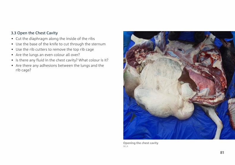
81
3.3 Open the Chest Cavity• Cut the diaphragm along the inside of the ribs• Use the base of the knife to cut through the sternum• Use the rib cutters to remove the top rib cage• Are the lungs an even colour all over? • Is there any fluid in the chest cavity? What colour is it?• Are there any adhesions between the lungs and the
rib cage?
Opening the chest cavity MLA

82
3.4 Open and Examine the Neck and Mouth• Cut the neck from the chest to the chin• Expose the trachea (windpipe) and esophagus and
cut through the middle of the lower jaw to open the oral cavity
• Check for loose teeth, abscesses, injuries, erosions, foreign objects and lesions
Opening the oral cavity MLA

83
3.5 Examine the Lungs• What colour are the lungs? Are there any signs of pus? Are there any congested areas?
All of these can be signs of pneumonia• Normal lungs should be fluffy and pink
Healthy lungs MLA
Pneumonia lungs with consolidation and pus MLA

84
3.6 Examine the Heart• Open the heart sac and examine the surface of the heart. • Is there any fluid in the heart sac? Does the fluid clot on exposure to air? Are there any lesions, white areas or
haemorrhages on the surface of the heart? Dissect the heart. Are the valves intact? Are there any abscesses or cauliflower type growths?
Examination of the surface of the heart MLA
Examination of the heart valves MLA

85
3.7 Examine the Liver• The liver should be red / brown in colour with sharp edges and no discolouration. It should be firm, not friable.• The gall bladder should be proportionate to the liver and content should be watery. Open the gall bladder and bile
ducts to examine for liver fluke.• Consider sampling and saving it fresh or freezing, if concerned about trace mineral levels.
Healthy liver, red/brown in colour with sharp edges MLA
Yellow jaundiced liver PIRSA (Jeremy Rogers)

86
3.8 Examine the Spleen• The spleen should be dark bluish red in colour with white specks.• An enlarged spleen or protruding white areas indicate abnormalities.
Healthy spleen in situ, dark bluish red in colour with white specks MLA
Healthy spleen, dissected MLA

87
3.9 Examine the Kidneys• Examine the fat around the kidney. A lack of fat or gelatinised fat can indicate starvation / wasting• Open the capsule and examine the kidneys. They should be firm and red/brown in colour.
Examination of the fat around the kidney MLA
Healthy kidney, firm and red/brown in colour MLA
Streaks in kidney from oxalate poisoning PIRSA (Jeremy Rogers)

88
3.10 Examine the Mesenteric Lymph Nodes• Are the lymph nodes enlarged? What colour are they?• Enlarged lymph nodes can indicate worms, coccidia, Johne’s disease or chronic intestinal issues
Normal lymph nodes in situ MLA
Normal lymph nodes dissected MLA
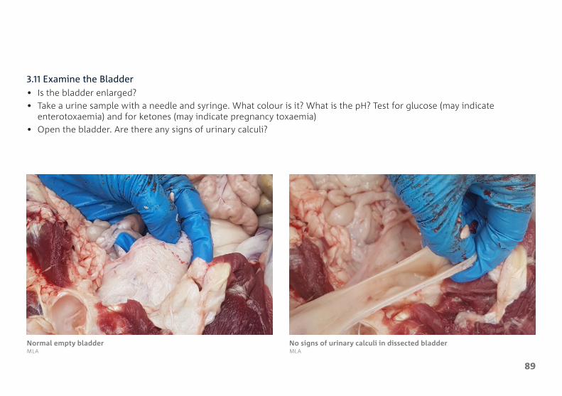
89
3.11 Examine the Bladder• Is the bladder enlarged?• Take a urine sample with a needle and syringe. What colour is it? What is the pH? Test for glucose (may indicate
enterotoxaemia) and for ketones (may indicate pregnancy toxaemia)• Open the bladder. Are there any signs of urinary calculi?
Normal empty bladder MLA
No signs of urinary calculi in dissected bladder MLA

90
3.12 Examine the Reproductive Tract• Is the Doe pregnant? If so, how many kids are there
and how advanced is the pregnancy?• Any signs of infection, retained placenta or
mummified fetuses?• Do not open the uterus if you are not vaccinated for
Q fever. Instead just feel through the uterine wall.
Reproductive tract of an empty doe MLA

91
3.13 Examine the Faeces• What is the colour and consistency of the faeces?• Consider collecting a sample for faecal egg counts• Faecal egg counts can done by a laboratory or vet or can be done yourself
if you have access to a microscope. Instruction can be found here: www.dpi.nsw.gov.au/__data/assets/pdf_file/0006/749292/DIY-worm-egg-counts-livestock-incl-poultry.pdf
Healthy goat faeces Kylie Hopkins (DAF)
Evidence of scouring Scour LLS (Kylie Greentree)

92
3.14 Examine the Digestive System
Abomasum ‘True Stomach’
Produces digestive enzymes
Check for barbers pole worms or the pinpoint haemorrhages from where they were. Large burdens of Ostertagia or brown stomach worms can be seen as a twisting mass although the individual worms are very tiny.
Omasum ‘Bible’
Key function is water and nutrient absorption
Reticulum ‘Honeycomb’
Collects small digesta particles and moves them into the omasum.
Can collect heavy objects such as nails or wire, causing ‘hardware disease’
Rumen ‘Paunch’
Acts as a fermentation vat by hosting microbial fermentation
Check pH and for colour and sloughing of papillae

SmallIntestine
Cecum
LargeIntestine
AbomasumTrue Stomach
Rumen
Mouth
Oesophagus
Recticulum
Omasum
93
Goat digestive systemMLA

94
3.15 Examine the Large Intestine, Colon, Cecum and Ileocecal Junction• The walls should be thin and white in colour. The content should have the consistency of pancake mix• Look for discoloration, corrugations, nodules and signs of ulcerations.• There is a valve at the junction between the caecum and the ilium (small intestine). This should be examined for
discoloration and signs of ulcerations. This site is commonly affected in salmonellosis, parasitism and Johne’s disease.
Ileocecal junction MLA
Examination of the Cecum walls and content MLA

95
3.16 Examine the Small Intestine• The small intestines are made up of three regions, the ilium, jejunum and duodenum• Open the small intestines at several locations and examine the internal walls and content • Look at the colour, thickness, any corrugations, nodules and signs of parasites
Healthy small intestine MLA Thickened intestines from Johne’s Disease PIRSA (Jeremy Rogers)

96
3.17 Examine the Brain• Opening the skull and examining the brain requires training and practice to ensure that the brain tissue is not damaged.• It is recommended that if the animal showed neurological signs, that the brain removal is done by a vet or other
trained professional.• If you are collecting samples to send for testing, the head can be taken to the vet involved for brain removal.
Brain of a goat. Haemorrhage from euthanasia by firearm MLA
Delicately removing the brain to prevent damage MLA

97
Investigation4. Collecting Samples• All samples must be submitted through your private veterinarian or local government vet.• Delivery of samples needs to occur within 24 hours of collection and proper sample handling and storage is vital to
getting accurate results.• All samples should be placed in individual sterile containers or bags and kept in a refrigerator until submitted for
testing. DO NOT FREEZE.
Sampling Process• Lung – cut a small piece including a transition zone of affected and non-affected areas• Heart – cut a thin slice of heart going through both chambers• Liver – cut a small piece including a transition zone of affected and non-affected areas• Spleen – cut a thin slice• Kidney – cut a thin slice that includes all layers• Intestines – cut a small section including contents from the large and small intestines. Ends can be tied with cable ties
or string• Stomach – cut a small section of all four chambers of the stomach• All sections and slices for histology must be fixed in 10 times the volume of 10% buffered formalin. • Faeces – collect a small amount of faeces if possible. Place in a plastic bag and remove the air then chill. • Urine – If there is urine in the bladder, this can be drawn out with a needle and syringe then put in a sterile container• Fluid behind the eye can be drawn out with a needle and syringe then put in a sterile container

98
Euthanasia of GoatsCriteria for Euthanasia1. The animal has no possibility of satisfactory recovery and is experiencing undue pain and/or suffering and prognosis for
recovery is poor.2. The cost or availability of effective treatment for the animal is prohibitive and/or the specific treatment(s) and the
resources needed to apply the treatment are not available.3. The animal is not fit for loading and/or for transport to a human consumption slaughter facility, rendering facility or a
pet food slaughter facility.4. The animal is not fit for slaughter either for human consumption or for processing into pet food.5. There is specific need for a post-mortem for disease investigation.
Methods Available1. Firearm
– firearm laws must be obeyed
2. Captive Bolt– the animal should be immediately bled out following the administration of a captive bolt
3. Lethal Injections– lethal injections should only be administered by a vet
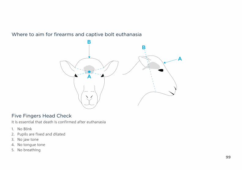
A
A
BB
99
Where to aim for firearms and captive bolt euthanasia
Five Fingers Head CheckIt is essential that death is confirmed after euthanasia
1. No Blink2. Pupils are fixed and dilated3. No jaw tone4. No tongue tone5. No breathing
100
Carcass DisposalMethods of carcass disposal include:
• Cremation (burning)• Composting• Licenced landfills• Burial (preferred option)
There is a lot to take into consideration when determining the best method of disposal and the requirement for that method.
The best source of information for this it the AUSVETPLAN Operation Manual for Disposal available here www.animalhealthaustralia.com.au/wp-content/uploads/2015/09/DISP-08-FINAL24Aug15.pdf

101
Acknowledgements and Further ReadingThe information in this publication has been collected from:
Abbott, K 2018, The Practice of Sheep Veterinary Medicine, University of Adelaide Press: Adelaide.
Allan, S 2010, Foot Abscess in Sheep, NSW DPI Primefact 7, NSW.
Animal Biosecurity Unit 2008, Ovine Johne’s Disease, NSW DPI Primefact 661, NSW.
Animal Health Australia 2020, Farm Biosecurity Plan, viewed 9 July 2021, https://www.animalhealthaustralia.com.au/what-we-do/endemic-disease/farm-biosecurity-plan/.
Animal Health Australia 2016, Australian Industry Welfare Standards and Guidelines for Goats, viewed 9 July 2021, http://www.animalwelfarestandards.net.au/files/2016/07/Goat-Industry-SnG-Final-July-2016-.pdf.
Animal Health Australia and Plant Health Australia, Farm Biosecurity, viewed 4 June 2020, https://www.farmbiosecurity.com.au/.
Animal Health Australia 2012, Australian Animal Welfare Standards and Guidelines – Land Transport of Livestock, Animal Health Australia (AHA), Canberra.
Brightling, A 2006, Livestock Diseases in Australia, C H Jerram and Associates Science Publishers: Mt. Waverley, VIC, Australia.
CSIRO 2020, Biosecurity and surveillance chain of response – Goats, FMD Ready Subproject 2, Canberra, ACT.
Erickson, A 2010, Hydatid Disease, Department of Agriculture and Food Farmnote 448, WA.
Harwood, D and K Mueller 2018, Goat Medicine and Surgery, CRC Press: Boca Raton, Florida, USA.
Hungerford, TG 1990, Hungerford’s Diseases of Livestock, 9th Edition, McGraw-Hill Book Company: Sydney, NSW.
Gardner, J 2006, Pregnancy Toxaemia and Hypocalcaemia of Ewes, Department of Agriculture and Food Farmnote 105, WA.
Green, DS, et al. 1987, Injection site reactions and antibody responses in sheep and goats after the use of multivalent clostridial vaccines. Vet Rec 120(18): 435-439.
Jordan, D 2003, Livestock Health Pregnancy Toxaemia and Hypocalcaemia, DPI&F QLD Primenote SW0107, QLD.
Litzow, E 2015, Sheep Diseases – The Farmers’ Guide, 2nd Edition, PIRSA, SA, viewed 9 July 2021, https://www.pir.sa.gov.au/__data/assets/pdf_file/0005/289265/Sheep_Diseases_-_The_Farmers_Guide_-_2nd_Edition_-_July_2015.pdf
Lloyd, J and Playford, M 2013, A producer’s guide to sheep husbandry practices, MLA, Australia.
Love, S 2008, Hydatids – The Basics, NSW DPI Primefact 812, NSW.

102
Love, S & Hutchinson, G 2007, Wormtest for Livestock and Guide to Egg Counts, NSW DPI Primefact 480, NSW.
Matthews, J 2016, Diseases of the Goat. Wiley-Blackwell: Oxford, UK.
Maxwell, D 2001, Livestock Health – Cheesy Gland in Sheep and Goats, DPI &F QLD Primenote SW0047, QLD.
Meat and Livestock Australia 2020, Farm Biosecurity, viewed 9 July 2021, https://www.mla.com.au/research-and-development/biosecurity/farm-biosecurity/.
Meat and Livestock Australia 2006, The Going into Goats Guide, viewed 9 July 2021, https://www.mla.com.au/extension-training-and-tools/going-into-goats/the-going-into-goats-guide/.
Meat and Livestock Australia & Livecorp 2020, Veterinary Handbook for Cattle, Sheep and Goats, viewed 9 July 2021 http://www.veterinaryhandbook.com.au/.
Mitchell, T 1982, How to tell the age of goats, Reviewed November 2003, NSW Agriculture Agfact A7.2.2.
Newman, R 2007, Livestock Health – Tetanus in Sheep and Goats, DPI&F QLD Primenote SW0088, QLD.
Norris, R 2005, Scabby Mouth, Department of Agriculture and Food Farmnote 17, WA.
Parish, J, Rivera, J & Boland, H 2017, Understanding the Ruminant Animal Digestive System, Mississippi State University Publication 2503, Mississippi.
Paton, M 2005, Cheesy Gland in Sheep and Goats, Department of Agriculture and Food Farmnote 46, WA.
Plant, JW 2004, Sheep Health – Scabby Mouth, NSW Agriculture Agfact A3.9.41, NSW.
Plant, J, Seaman, J & Evers, M 2017, Footrot in Sheep and Goats, NSW DPI Primefact 1533, NSW.
Pezzanite, L, Neary, M, Hutchens, T & Scharko, P 2009, Common Diseases and Health Problems in Sheep and Goats, Purdue University Extension AS-595-W, US.
Pugh, DG, et al. 2021, Sheep, Goat, and Cervid Medicine. Elsevier: Edinburgh, UK.
Robson, S 2007, Photosensitisation in Stock, NSW DPI Primefact 449, NSW.
Thompson, JM and Meyer H 1994, Body Condition Scoring of Sheep, EC1433, Oregon State University.
University of New England 2014, ParaBoss, viewed 4 June 2020, https://www.paraboss.com.au/.
Walker, B 1996, Cheesy Gland Caseous Lymphadenitis in Sheep, NSW DPI Agfact A3.9.21, NSW.
Walker, B 2000, How Vaccination Works, NSW Agriculture AgnoteDAI/190, NSW.
Walker, B 2006, Grain Poisoning of Cattle and Sheep, NSW DPI Primefact 330, NSW.
Wilson, K, Armstrong, B & Knights, G 2005, Livestock Health – Management of Body Lice, DPI &F QLD Primenote SW0102, QLD.

CSIRO B&M | 21-00248