The NeuroCognitive Research Institute - Solve ME/CFS Initiative

Ga
Aa
b
a
ARRAA
KAGGAM
1
s(paim(e
p
VC
0d
Drug and Alcohol Dependence 125 (2012) 27– 36
Contents lists available at SciVerse ScienceDirect
Drug and Alcohol Dependence
jo u rn al hom epage: www.elsev ier .com/ locate /drugalcdep
lutamate, GABA, and other cortical metabolite concentrations during earlybstinence from alcohol and their associations with neurocognitive changes�
nderson Mona,b,∗, Timothy C. Durazzoa,b, Dieter J. Meyerhoffa,b
Department of Radiology and Biomedical Imaging, University of California, San Francisco, CA, USACenter for Imaging of Neurodegenerative Diseases, Veterans Administration Medical Center, San Francisco, CA, USA
r t i c l e i n f o
rticle history:eceived 29 September 2011eceived in revised form 10 March 2012ccepted 10 March 2012vailable online 12 April 2012
eywords:nterior cingulate cortexlutamateABAbstinence from alcoholR spectroscopy
a b s t r a c t
Background: Little is known about the effects of alcohol dependence on cortical concentrations of glu-tamate (Glu) or gamma aminobutyric acid (GABA). We used proton magnetic resonance spectroscopy(MRS) to study cross-sectionally and longitudinally the concentrations of these in alcohol dependentindividuals (ALC) during early abstinence from alcohol.Methods: Twenty ALC were studied at about one week of abstinence from alcohol (baseline) and 36 ALC atfive weeks of abstinence and compared to 16 light/non-drinking controls (LD). Eleven ALC were studiedtwice during abstinence. Participants underwent clinical interviewing, blood work, neuropsychologicaltesting, structural imaging and single-volume proton MRS at 4 Tesla. Absolute concentrations of Glu,GABA and those of other 1H MRS-detectable metabolites were measured in the anterior cingulate (ACC),parieto-occipital cortex (POC) and dorso-lateral prefrontal cortex (DLPFC). Relationships of metabolitelevels to drinking severity and neurocognition were also assessed.Results: ALC at baseline had lower concentrations of Glu, N-acetylaspartate (NAA), choline- (Cho) andcreatine-containing metabolites (Cr) than LD in the ACC, but had normal GABA and myo-inositol (mI)levels. At five weeks of abstinence, metabolite concentrations were not significantly different between
groups. Between one and five weeks of abstinence, Glu, NAA and Cho levels in the ACC increased signifi-cantly. Higher cortical mI concentrations in ALC related to worse neurocognitive outcome.Conclusion: These MRS data suggest compromised and regionally specific bioenergetics/metabolism inone-week-abstinent ALC that largely normalizes over four weeks of sustained abstinence. The correlationbetween mI levels and neurocognition affirms the functional relevance of this putative astrocyte marker.. Introduction
In vivo proton magnetic resonance spectroscopy (1H MRS)tudies have reported lower concentrations of N-acetylaspartateNAA: a marker of neuronal viability) and choline-containing com-ounds (Cho: a marker of glial or cell synthesis/turnover) as wells altered myo-inositol levels (mI: a putative astrocyte marker)n the brain of individuals with alcohol use disorders (AUD), pri-
arily in the frontal lobes, medial temporal lobe, and cerebellumBendszus et al., 2001; Durazzo et al., 2004; Ende et al., 2005; Feint al., 1994; Schweinsburg et al., 2000; Seitz et al., 1999). Lower
� Supplementary material can be found by accessing the online version of thisaper. Please see Appendix A for more information.∗ Corresponding author at: Center for Imaging of Neurodegenerative Diseases,eterans Administration Medical Center, 4150 Clement Street, 114M San Francisco,A 94121, USA. Tel.: +1 415 221 4810x6453; fax: +1 415 668 2864.
E-mail address: [email protected] (A. Mon).
376-8716/$ – see front matter. Published by Elsevier Ireland Ltd.oi:10.1016/j.drugalcdep.2012.03.012
Published by Elsevier Ireland Ltd.
concentrations of frontal white matter NAA, cerebellar NAA andCho as well as higher cerebellar mI have been associated withpoorer neurocognition and motor functions in cross-sectional stud-ies of individuals with AUD (see Bendszus et al., 2001; Durazzoet al., 2004; Parks et al., 2002). With sustained abstinence fromalcohol, regional metabolite concentrations show variable recov-ery (e.g., Bartsch et al., 2007; Bendszus et al., 2001; Durazzo et al.,2006; Ende et al., 2005; Gazdzinski et al., 2008; Martin et al.,1995; Parks et al., 2002; Schweinsburg et al., 2000, 2001), andincreases in some metabolite levels (e.g., NAA and Cho) have beenassociated with improvement on measures of learning and mem-ory, processing speed and working memory (Bartsch et al., 2007;Bendszus et al., 2001; Durazzo et al., 2006; Gazdzinski et al., 2008;Parks et al., 2002). These findings parallel regional morphologi-cal recovery observed during abstinence from alcohol (for review,
see Durazzo and Meyerhoff, 2007) and suggest such adaptive neu-roplastic changes are associated with improved neurocognitionduring periods of sustained sobriety (reviewed in Meyerhoff et al.,2011).
2 hol De
(ehaanaiNrlHGaiatrta2rdd2to
fieacUballpseFnOaococsa
gmoo(lnlDtmcns
inclusion of much gray matter (GM) as possible (Fig. 1 shows the three VOIs
8 A. Mon et al. / Drug and Alco
High field 1H MRS has enabled the quantitation of glutamateGlu) and gamma aminobutyric acid (GABA), which are the primaryxcitatory and inhibitory neurotransmitters, respectively, in theuman brain. Glu and glutamine are involved in the maintenancend promotion of several cell functions (Newsholme et al., 2003),nd imbalances in brain Glu metabolism have been implicated ineurodegeneration in AUD. Specifically during withdrawal fromlcohol, cerebral Glu levels change rapidly. Altered Glu (and primar-ly Ca2+), coupled with a chronic alcohol-induced up-regulation of-methyl-d-aspartate receptors in the cortex, may promote neu-
onal “excitotoxicity” through cation and water disequilibrium,eading to increased oxidative stress and apoptosis (Lovinger, 1993;ughes, 2009; Freund and Anderson, 1996; Tsai and Coyle, 1998).ABAergic neurons are involved in the synchronization of neuronalctivity across brain regions (Clancy et al., 2010), and abnormal-ties in synaptic inhibition mediated by GABAergic neurons aressociated with AUD and other common neuropsychiatric condi-ions (Johnson, 2005; Sanacora and Saricicek, 2007). Collectively,esearch suggests that alterations of regional Glu and GABA concen-rations and of glutamatergic and GABAergic receptors (e.g., Greennd Grant, 1998; Grobin et al., 1998; Kalivas et al., 2009; Spanagel,009), as well as alterations in Glu transmission from the ante-ior cingulate cortex (ACC) to the ventral striatum contribute to theevelopment and maintenance of AUD and other substance useisorders (Kalivas and O’Brien, 2008; Kalivas, 2009; Cramer et al.,011; Kalivas, 2009). Yet, only a few MRS studies have reported onhe effects of AUD on concentrations of Glu, Glu + glutamine (Glx)r GABA.
Glu concentrations in the ACC of non-medicated ALC on therst day of abstinence from alcohol (i.e., during withdrawal) werelevated, but then decreased to normal levels over two weeks ofbstinence (Frischknecht et al., 2010). In a randomized placebo-ontrolled study of acamprosate treatment of detoxified ALC,mhau et al. (2010) observed decreasing levels of Glu in the ACCetween the fourth and 25th day of acamprosate administrationnd trends to increasing Glu levels with placebo. For GABA, a pre-iminary study (Behar et al., 1999) reported significantly lowerevels in the occipital cortex (OCC) of 1-month-abstinent ALC com-ared to controls, but Mason et al. (2006), in a larger follow-uptudy, observed no abnormalities in OCC GABA levels of ALC atither one week or one month of abstinence relative to controls.or technical reasons (a surface coil was used at relatively low mag-etic field strength), these previous studies assessed only the ACC orCC. To better understand the functional relevance of Glu and GABAnd the potential role their regional metabolism plays in the devel-pment and maintenance of AUD, it is imperative to: (a) measureerebral Glu (and/or Glx) and GABA levels in multiple brain regionsf the same cohort, (b) investigate their associations with otherommon brain metabolites, neurocognitive function and drinkingeverity, and (c) explore their concentration changes during earlybstinence.
This report presents fully quantitative cross-sectional and lon-itudinal findings of single voxel MR spectroscopy (SVS) at highagnetic field strength (4 Tesla), focusing on absolute quantitation
f Glu, Glx and GABA concentrations in the ACC, the parieto-ccipital cortex (POC) and the dorso-lateral prefrontal cortexDLPFC) of treatment-seeking ALC in early recovery. Based on theiterature, we hypothesized that: (1) ALC near the inception of absti-ence from alcohol have significantly higher Glx, Glu, and mI and
ower NAA, Cho and GABA concentrations in the ACC, POC andLPFC than light-drinking controls (LD), (2) metabolite concen-
rations normalize in all regions of ALC after approximately oneonth of abstinence from alcohol, and (3) NAA, Cr and Cho con-
entrations correlate positively while Glu and mI levels correlateegatively with measures of learning and memory, visuomotorcanning speed and working memory at both TPs. In secondary
pendence 125 (2012) 27– 36
analyses, we explored the data for evidence of the excitotoxic-ity theory of Glu-induced neuronal injury in AUD (Freund andAnderson, 1996; Hughes, 2009; Lovinger, 1993; Tsai and Coyle,1998), which would suggest an inverse relationship between Gluand NAA concentrations in ALC.
2. Materials and methods
2.1. Participants
Forty four (39 males, 5 females) ALC were recruited from the San Francisco VAMedical Center Substance Abuse Day Hospital and the San Francisco Kaiser Perma-nente Chemical Dependence Recovery Programs. All ALC participants met DSM-IVcriteria for alcohol dependence at the time of study. Male ALC had consumed morethan 150 alcoholic drinks (one drink contains 13.6 g of pure ethanol) per month forat least 8 years while female ALC had consumed more than 80 drinks per month forat least 6 years prior to enrollment. All ALC participants were tested daily for breathalcohol while in outpatient treatment to ensure sobriety. Inclusion and exclusioncriteria are fully detailed elsewhere (Durazzo et al., 2004). In brief, participants wereexcluded for a history of abuse or dependence on other substances within the pastfive years (other than nicotine), and for neurological or psychiatric disorders that areknown to affect brain neurobiology or neurocognition. Hepatitis C, type-2 diabetes,hypertension, unipolar mood disorder (major depression and/or substance-inducedmood disorder) were permitted in the ALC cohort given their high prevalence in AUD(Hasin et al., 2007; Mertens et al., 2003, 2005; Parekh and Klag, 2001; Stinson et al.,2005). Sixteen age-matched LD were recruited from the local community and hadno history of medical or psychiatric conditions known to influence the outcomemeasures of this study.
2.2. Clinical assessment
ALC participants completed the Structured Clinical Interview for DSM-IV Axis IDisorder Patient Edition, Version 2.0 (First et al., 1998) and standardized question-naires for alcohol withdrawal (CIWA-Ar; Addiction Research Foundation ClinicalInstitute of Withdrawal Assessment for Alcohol; Sullivan et al., 1989), depression(Beck Depression Inventory (BDI); Beck, 1978) and anxiety symptomatology (State-Trait Anxiety Inventory, Y-2, STAI; Spielberger et al., 1977) within one day of MRstudy. We assessed alcohol consumption over lifetime with the lifetime drinking his-tory (LDH; Skinner and Sheu, 1982; Sobell et al., 1988; Sobell and Sobell, 1990). Fromthe LDH we derived the age at onset of heavy drinking (defined as >100 alcoholicdrinks per month) and estimated the average number of alcoholic drinks consumedper month over 1 year, 3 years, 8 years before enrollment and over lifetime.
A brief neurocognitive battery assessed aural working memory (Wechsler AdultIntelligence Scale-III (WAIS-III) Digit Span), visuomotor scanning speed and inciden-tal learning (WAIS-III Digit Symbol; Wechsler, 1997), auditory–verbal (Delis et al.,2000) and visuospatial learning and memory (Brief Visual memory Test-Revised;Benedict, 1997) in LD as well as in ALC at TP1 and TP2 follow-up (for details, seeDurazzo et al., 2007).
To evaluate the nutritional status and alcohol-related or other hepatocellu-lar injury in ALC, we obtained laboratory tests for serum albumin, pre-albumin,alanine aminotransferase (ALT), aspartate aminotransferase (AST) and gamma-glutamyltransferase (GGT) within 3 days of the MR scans at each time point.
Of the 44 ALC participants, 20 were scanned at time point 1 (TP1 at 9 ± 4 days ofabstinence from alcohol) and 36 scanned at TP2 (34 ± 7 days of abstinence). Not allparticipants contributed spectra from every VOI. Eleven participants were scannedat both TPs (at approximately 9 and 34 days of abstinence) and used for longitudinalanalyses. The sixteen LD were each scanned once.
Table 1 shows demographics, laboratory and alcohol consumption variables forLD and ALC studied at TP1. ALC showed significantly greater depressive and anxietysymptomatology. The demographics of ALC studied at TP2 were similar to those atTP1. Also, the demographics, laboratory and alcohol consumption variables of thesmaller longitudinal sample were representative of the ALC group shown in Table 1.
2.3. Imaging and processing methods
MRI data were acquired on a 4 Tesla Bruker MedSpec system usingan 8-channel transmit-receive head coil and a Siemens Trio console(Siemens, Erlangen, Germany). A Magnetization Prepared Rapid Gradient(TR/TE/TI = 2300/3/950 ms, 7◦ flip angle, 1.0 × 1.0 × 1.0 mm3 resolution) and aturbo spin-echo (TR/TE = 8400/70 ms, 150◦ flip angle, 0.9 × 0.9 × 3 mm3 resolution)sequences were used to acquire 3-D sagittal T1-weighted and 2D axial T2-weightedanatomical images, respectively. The T1 and T2 images were then displayed andvolumes-of-interest (VOIs) for MRS placed over the ACC (35 × 25 × 20 mm3), inthe POC (20 × 40 × 20 mm3) and right DLPFC (size: 40 × 20 × 20 mm3), maximizing
on axial T2-weighted images). The T1 images were also used for segmentationinto GM, WM and cerebrospinal fluid (CSF), using the expectation maximizationsegmentation method (Van Leemput et al., 1999), and the segmented data usedto estimate tissue fraction (TF) and CSF contributions to each VOI. After 3-D

A. Mon et al. / Drug and Alcohol Dependence 125 (2012) 27– 36 29
Table 1Demographics, laboratory and alcohol consumption variables for LD and ALC at TP1.
Variable LD ALC p-Value
Number of participants (male, female) 16(14, 2) 20(17, 3) –Age (years) 49.0 ± 10.1 53.9 ± 8.8 NSEducation (years) 15.4 ± 2.7 13.8 ± 2.3 0.048Monthly alcohol consumption: 1 year average 19.8 ± 21.7 338.7 ± 168.9 –Monthly alcohol consumption: 3 years average 19.8 ± 21.8 329.5 ± 164.6 –Monthly alcohol consumption: lifetime average 18.2 ± 16.9 232.6 ± 130.7 –Age of onset of heavy drinking (years) – 22.8 ± 5.7 –Months of heavy drinking – 290± 125.6 –Smoker (%) 35 65 –MCV 93.2 ± 3.8 96.2 ± 5.0 NSPrealbumin 28.2 ± 6.2 27.2 ± 9.6 NSAMNART 115.3 ± 8.3 116.4 ± 6.0 NSCIWA-Ar – 0.63 ± 1.4 –GGT 52.5 ± 91.6 55.6 ± 40.5 NSAlbumin 4.1 ± 0.3 4.2 ± 0.3 NSAST 22.6 ± 7.1 32.6 ± 10.1 0.031ALT 19.3 ± 8.1 30.9 ± 12.0 0.057WBC 6.1 ± 1.6 6.1 ± 1.1 NSRBC 4.6 ± 0.4 4.3 ± 0.4 NSHemoglobin 14.8 ± 1.1 14.1 ± 1.4 NSHematocrit 42.7 ± 3.2 41.0 ± 4.1 NSBDI 5.4 ± 4.8 13.1 ± 7.9 0.004STAI-Y2 33.5 ± 7.4 45.6 ± 10.3 0.002
Note: mean ± standard deviation; AMNART, American National Adult Reading test; MCV, mean corpuscular volume; CIWA, Addiction Research Foundation Clinical Instituteo partatR
sw(acsGVfl2Me(ciatwtcttwa
f Withdrawal Assessment for Alcohol; GGT, gamma-glutamyltransferase; AST, ased Blood Cells; BDI, Beck Depression Inventory; STAI-Y2, Trait-Anxiety-Index.
himming and water suppression of each VOI, NAA, Cr, Cho, mI and Glu signalsere acquired with a Stimulated Echo Acquisition Mode (STEAM) sequence
TR/TE/TM = 2000/12/10 ms, 90◦ flip angle, 2000 Hz spectral bandwidth, 2.5 mincquisition time). Immediately afterwards, a reference water signal was alsoollected from the same VOIs with the same STEAM sequence but without wateruppression and used for scaling metabolite peak areas. Signals from GABA andlx were acquired immediately after the water spectrum from the exact sameOIs with a modified J-editing sequence (MEGA PRESS: TR/TE = 2000/71, 90◦
ip angle, 2000 Hz spectral bandwidth, 12.5 min acquisition time; Kaiser et al.,008). The MRS data were processed using a combination of an in-house writtenatlab program and IDL (Research Systems, Inc., Boulder, CO) with SITOOLS (Soher
t al., 1996). First, the Matlab program was used to convert the time domain dataraw data) to frequency domain data (i.e., 1-D Fourier transformation) after DCorrection and apodization with a 2 Hz Lorentz–Gaussian filter. Using a priorinformation of metabolite frequencies, phases and amplitudes, the metabolite peakreas (METarea) as well as the reference water peak area (H2Oarea) were then fitted,ogether with baselines (see Figs. 2 and 3) using SITOOLS. A correction factor (CF),hich accounted for differences between the transmitter voltage amplitudes used
o acquire the metabolite and unsuppressed water signal (for each participant) was
alculated as CF sin(radians(90 * METtrans-amp/H2Otrans-amp)), (where METtrans-amp ishe transmitter voltage amplitude for the metabolite and H2Otrans-amp is that forhe corresponding reference water signal). The water peak area was then adjustedith CF as H2Oarea-adjusted = H2Oarea * CF. The metabolite peak areas were then scaledccording to the formula METscaled = METarea/H2Oarea-adjusted. The METscaled values
Fig. 1. T2-weighted images showing ACC (left), right DL
e aminotransferase; ALT, alanine aminotransferase; WBC, White Blood Cells; RBC,
were then used to estimate metabolite concentrations ([MET]) (in mmol) whileaccounting for TF for each VOI according to the formula
[MET] = METscaled
TF, where TF = GM + WM
GM + WM + CSF.
2.4. Data analyses
We conducted three main statistical analyses using SPSS, v18. In the first anal-ysis, multivariate analysis of covariance (MANCOVA) was used for cross-sectionalgroup comparisons at TP1 and TP2 to test for differences in NAA, Cr, Cho, mI andGlu concentrations between ALC and LD in each VOI. GABA and Glx were also ana-lyzed similarly, but separately from NAA, Cr, Cho, mI and Glu, because the numberof spectra from the J-editing sequence (i.e., 15 for POC of ALC at TP1) with acceptablequality was less than those from the STEAM sequence (i.e., 20 POC of for ALC at TP1).Although age was not significantly different between the groups, the age range ofthe participants was large (28–68), and age is associated with metabolite concen-trations (Schuff et al., 2001); therefore, we used it as a covariate in the analyses.
Also, because cigarette smoking is associated with brain metabolite concentrationsin individuals with AUD (Durazzo et al., 2004, 2006, 2011), we covaried for smokingstatus in all cross-sectional analyses. We evaluated for group differences in totalbrain tissue, GM, WM, CSF contributions to the VOIs, but none was significantlydifferent between the groups. Table 2 shows the percentage of GM, WM and CSFPFC (middle) and POC (right) volumes-of-interest.

30 A. Mon et al. / Drug and Alcohol De
Table 2GM, WM and CSF contributions to ACC, POC and DLPFC in 16 LD and 20 ALC at TP1.
Tissue type (%) ACC POC DLPFC
GM in LD 46.8 ± 4.1 60.4 ± 4.0 40.3 ± 5.3GM ALC 45.4 ± 2.9 60.8 ± 4.5 39.0 ± 4.8WM in LD 33.5 ± 4.4 29.2 ± 4.8 52.8 ± 6.8WM in ALC 34.3 ± 5.8 29.8 ± 4.8 53.7 ± 7.3CSF in LD 18.8 ± 4.7 9.5 ± 5.8 6.1 ± 2.6CSF in ALC 19.4 ± 4.4 8.6 ± 3.0 6.4 ± 3.4
Note: mean ± standard deviation.
Fig. 2. STEAM sequence spectrum (TR/TE/TM = 2000/12/20 ms) with main reso-n
ce
ua(lcoadcoeofc
ances for NAA, Glu, Cr, Cho and mI.
ontributions to each VOI for ALC at TP1 and LD, depicting similar contributions ofach tissue type in both groups.
The second analysis assessed metabolite level changes between TP1 and TP2sing paired t-tests on metabolite-to-water ratios (i.e., ratio of metabolite peakreas to corresponding reference water peak areas) of the longitudinal sample11 ALC). In the third analyses, relationships (Spearman’s Rho) between metabo-ite concentrations within each VOI, as well as correlations between metaboliteoncentrations and measures of auditory-verbal, visuomotor scanning speed, visu-spatial learning and memory, delayed visual memory and working memory weressessed in ALC and LD separately. Correlations of metabolite concentrations withrinking severity, as well as associations between changes in metabolite levels andhanges in the aforementioned neurocognitive measures were also assessed in ALCnly. A p-value of ≤0.05 was considered statistically significant in all the analyses,xcept analyses that assessed inter-correlations between Glu (or Glx) and Cr, Chor mI, which required correction for multiple comparisons. The conservative Bon-erroni method was used to correct for the effect of multiple comparisons on theseorrelations.
Fig. 3. J-edited spectrum of GABA and Glx (TR/TE = 2000/71 ms).
pendence 125 (2012) 27– 36
3. Results
3.1. Participant characterization
ALC and LD participants were not significantly different onage, but LD had significantly more years of education than ALC(p = 0.048). ALC participants had significantly higher aspartateaminotransferase levels than LD participants at TP1 (p = 0.031). Allother laboratory measures as well as total brain tissue, GM, WM andCSF contributions to the VOIs did not differ significantly betweenALC and LD groups.
Two ALC participants (10%) received benzodiazepine treatmentat TP1; all of their metabolite concentrations were within the rangeof ALC who did not receive benzodiazepine treatment, but theseparticipants did not provide data at TP2. Also, three ALC partic-ipants were positive for hepatitis C at TP1 and four at TP2, butagain these participants were not part of the longitudinal sam-ple as they did not have data at both TPs. Nine ALC participantshad medically controlled hypertension at either TP; two of theseparticipants had data at both TP1 and TP2 and were part of the lon-gitudinal sample. However, there were no significant differencesbetween participants with and without hepatitis C or controlledhypertension on any of our outcome measures, suggesting theseconditions did not likely influence group findings. Comparison ofthe participants of the longitudinal sample with the rest of the ALCgroup in terms of age, drinking severity, neurocognitive measures,laboratory variables and medical/psychiatry conditions showed nosignificant group differences on any of these measures. Thereforeour longitudinal sample can be considered representative of theentire sample in this report.
3.2. Cross-sectional group comparisons
3.2.1. TP1 (9 ± 4 days of abstinence). In the ACC, MANOVA,with age and cigarette smoking as covariates, indicated signif-icant differences between LD and ALC across metabolite levels(F = 3.11, p = 0.028) for the STEAM sequence data. Follow-up t-testsshowed significantly lower concentrations of Glu (p = 0.012), NAA(p = 0.001), Cr (p = 0.014), and a trend for lower Cho (p = 0.085)compared to LD. For the J-editing sequence data (GABA and Glx),MANCOVA gave statistically similar metabolite levels in LD andALC; however, the follow-up t-tests showed a trend for lower Glx(p = 0.066) in ALC compared to LD. GABA and mI concentrationsdid not differ significantly between groups. See Table 3 for groupmeans, standard deviations and effect sizes.
In the POC and DLPFC, similar MANCOVA analysis showedno significant group differences of metabolite concentrations.Predicted follow-up comparisons showed no significant differ-ences between the groups in any of the metabolite levels. SeeTables 4 and 5.
3.2.2. TP2 (34 ± 7 days of abstinence). The MANCOVA indicated nosignificant metabolite level differences between LD and ALC at TP2in any VOI. See Tables 3–5.
3.3. Longitudinal change of metabolite ratios in 11 ALC
In the ACC between TP1 and TP2, ALC demonstrated signifi-cant increases in ratios of Glu (p = 0.008), NAA (p = 0.034), and Cho(p = 0.037) to water. Fig. 4 depicts the increase of ACC Glu-to-waterratios in most ALC participants over time. A plot of ACC Glu con-centrations (combining cross-sectional TP1 and TP2 data) againstdays of abstinence showed a moderate positive correlation (r = 0.31,

A. Mon et al. / Drug and Alcohol Dependence 125 (2012) 27– 36 31
Table 3ACC metabolite concentrations at 9 ± 4 days (TP1) and 34 ± 7 days of abstinence (TP2) (cross-sectional samples).
Metabolite LD (12) ALC (18): TP1 ALC (31): TP2 ES, p1 ES, p2
Glu 4.08 ± 0.54 3.46 ± 0.79 3.87 ± 0.98 0.92, 0.012 0.26, NSGlx 2.23 ± 0.71 1.79 ± 0.59 2.18 ± 1.12 0.67, 0.066 0.05, NSGABA 1.31 ± 0.25 1.26 ± 0.39 1.44 ± 0.63 0.15, NS 0.27, NSNAA 5.52 ± 0.90 4.46 ± 0.76 5.15 ± 1.30 1.27, 0.001 0.33, NSCr 4.63 ± 0.77 3.89 ± 0.90 4.48 ± 1.08 0.88, 0.014 0.15, NSCho 1.37 ± 0.21 1.25 ± 0.25 1.30 ± 0.36 0.52, 0.085 0.24, NSmI 4.28 ± 1.19 4.19 ± 0.94 4.41 ± 1.08 0.08, NS 0.011, NS
Note: mean ± standard deviation; ES stands for effect size, p1 is the p-value for LD measures vs. TP1-ALC measures; while p2 is the p-value for LD measures vs. TP2–ALCmeasures.
Table 4POC metabolite concentrations at TP1 and TP2 (cross-sectional samples).
Metabolite LD (16) ALC (20): TP1 ALC (31): TP2 ES, p1 ES, p2
Glu 4.18 ± 0.62 4.03 ± 0.48 4.05 ± 0.81 0.27, NS 0.18, NSGlx 2.06 ± 0.46 1.89 ± 0.50 1.93 ± 0.41 0.35, NS 0.30, NSGABA 1.50 ± 0.36 1.50 ± 0.30 1.53 ± 0.46 0.00, NS 0.07, NSNAA 5.56 ± 1.09 5.27 ± 0.74 5.49 ± 1.0 0.31, NS 0.07, NSCr 4.56 ± 0.95 4.48 ± 0.61 4.54 ± 0.85 0.10, NS 0.02, NSCho 0.89 ± 0.42 0.83 ± 0.31 0.87 ± 0.25 0.16, NS 0.11, NSmI 3.49 ± 0.84 3.41 ± 0.70 3.28 ± 0.82 0.10, NS 0.25, NS
Abbreviations as in Table 3.
Table 5DLPFC metabolite concentrations at TP1 and TP2 (cross-sectional samples).
Metabolite LD (12) ALC (12): TP1 ALC (25): TP2 ES, p1 ES, p2
Glu 3.13 ± 0.67 3.21 ± 0.57 3.08 ± 0.87 0.13, NS 0.06, NSGlx 2.21 ± 0.94 2.18 ± 0.76 2.15 ± 0.86 0.06, NS 0.07, NSGABA 1.54 ± 0.49 1.56 ± 0.47 1.71 ± 0.78 0.04, NS 0.26, NSNAA 5.35 ± 0.97 5.48 ± 0.96 5.35 ± 1.01 0.13, NS 0.00, NSCr 4.43 ± 0.74 4.72 ± 0.66 4.48 ± 0.81 0.35, NS 0.06, NSCho 1.08 ± 0.13 1.09 ± 0.19 1.08 ± 0.21 0.06, NS 0.00, NSmI 3.60 ± 1.00 3.80 ± 0.79
Abbreviations as in Table 3.
Fmn
pt
p
ig. 4. Plots of ACC Glu/H2O in 11 ALC at TP1 and TP2. The gray region indicatesean ± standard deviation of Glu/H2O in LD. Glu increases in the 11 ALC were sig-
ificant (p = 0.008), even when removing the individual with the greatest changeCho.
= 0.034) between Glu and time (see Supplementary Fig. 11), fur-her supporting Glu increases in early abstinence. There were no
1 Supplementary material can be found by accessing the online version of thisaper. Please see Appendix A for more information.
3.76 ± 0.84 0.02, NS 0.17, NS
significant longitudinal changes in ratios of Cr, mI, and GABA towater in the ACC.
In the POC, the Glu-to-water ratio increased significantlybetween TP1 and TP2 (p = 0.049). Ratios of NAA (p = 0.092), Cr(p = 0.053) and Cho (p = 0.065) to water tended to increase over thesame interval.
In the DLPFC, there were no significant longitudinal changes inany of the metabolite ratios of this small sample.
3.4. Cross-sectional correlations among main outcome measuresin ALC
3.4.1. TP1. Glu and NAA concentrations correlated positively inthe ACC (r = 0.73, p = 0.001) (see Fig. 5a), POC (r = 0. 58, p = 0.007;Supplementary Fig. 22), and DLPFC (r = 0.64, p = 0.018). mI con-centrations of the ACC and POC related negatively to visuomotorscanning speed, delayed visual memory and verbal learning;POC mI also correlated negatively with visuospatial memory andauditory–verbal learning (all r ≤ 0.48, p < 0.047, see Table 6). In theDLPFC, Glx correlated negatively with verbal learning and visuospa-tial learning (both r ≤ 0.51, p < 0.014). Average number of drinks per
month over one-year prior to study was positively related to mIin the ACC (r −0.64, p = 0.04); no other associations between alco-hol consumption and metabolite concentrations were observed in2 Supplementary material can be found by accessing the online version of thispaper. Please see Appendix A for more information.

32 A. Mon et al. / Drug and Alcohol Dependence 125 (2012) 27– 36
ACCGluConcentration(mmol)1 2 3 4 5
2.5
3.0
3.5
4.0
4.5
5.0
5.5
6.0
ACCGluConcentration(mmol)1 2 3 4 5 6 7
2
3
4
5
6
7
8
9
10
a: r = 0.73, p = 0.001 b: r = 0.83, p < 0.0001
Cho
Fig. 5. Plots of ACC NAA concentration against ACC Glu concentration. (a) Plot for TP1 and (b) plot for TP2.
Table 6Spearman correlations among TP1 and TP2 main outcome measures for ALC.
VOI Correlations TP1 correlation coefficient TP1 p-value TP2 correlation coefficient TP2 p-value
ACC Glu vs. NAA 0.73 0.001 0.83 0.0001Glu vs. Cr 0.83 0.004 0.63 0.004Glu vs. Cho 0.74 0.004 0.78 0.002Glx vs. NAA – NS NSmI vs. monthly alcohol consumption: 1 year average 0.64 0.004 – NSmI vs. verbal learning −0.52 0.040 – NSmI vs. delayed visual memory −0.49 0.047 – NSmI vs. visuomotor scanning speed −0.53 0.027 – NS
POC Glu vs. NAA 0.58 0.007 0.92 0.0001Glu vs. Cr 0.68 0.004 0.92 0.001*
Glu vs. Cho 0.78 0.001 0.78 0.001*
mI vs. verbal learning −0.51 0.039 – NSmI vs. visuomotor scanning speed −0.48 0.040 – NSmI vs. visuospatial memory −0.67 0.002 0.43 0.024mI vs. auditory–verbal learning −0.51 0.039 −0.35 0.020
DLPFC Glu vs. NAA 0.64 0.018 0.62 0.001Glu vs. Cr 0.57 0.040 0.52 0.008Glu vs. mI 0.69 0.009 – NSGlx vs. verbal learning – NS −0.58 0.011Glx vs. visuospatial learning – NS −0.51 0.014Cho vs. working memory – NS 0.41 0.0–43mI vs. BDI – NS −0.55 0.005
tw
3t(DGcnmppa
p
Glu vs. BDI –
* p-Value corrected for 3 comparisons.
he other VOIs. See Table 6 for statistically significant associationsithin the ALC group.
.4.2. TP2. Similar to the observation at TP1, Glu and NAA concen-rations at TP2 correlated positively in the ACC (r = 0.83, p < 0.0001)see Fig. 5b), POC (r = 0.92, p < 0.0001) (Supplementary Fig. 33), andLPFC (r = 0.62, p = 0.001). However, in contrast to TP1, the ACClx concentration also related positively to the corresponding NAAoncentration (r = 0.60, p = 0.004). Auditory–verbal learning relatedegatively to POC mI concentrations (r = −0.36, p = 0.020). Workingemory related positively to DLPFC Cho concentration (r = 0.44,
= 0.008) and negatively to DLPFC mI concentration (r = −0.40,
= 0.025). BDI was negatively correlated to mI (r = −0.55, p = 0.005)nd Glu (r = −0.43, p = 0.035) in the DLPFC.3 Supplementary material can be found by accessing the online version of thisaper. Please see Appendix A for more information.
NS −0.43 0.035
3.5. Correlations among longitudinal changes of main outcomemeasures in ALC
In the ACC between TP1 and TP2, the change of the Glu-to-water ratio related positively to changes in the correspondingNAA (r = 0.92, p < 0.001), Cr (r = 0.72, p = 0.013) and Cho (r = 0.92,p < 0.001) ratios.
Similarly, in the POC, the Glu-to-water ratio change correlatedpositively to the corresponding changes in ratios of NAA (r = 0.62,p = 0.044), Cr (r = 0.68, p = 0.020) and Cho (r = 0.76, p = 0.007) towater.
In the DLPFC, similar to a lack of cross-sectional associations,there were no significant correlations between changes in any ofthe metabolite-to-water ratios.
Between TP1 and TP2, measures of delayed visual memory,processing speed and visuomotor scanning speed (all p < 0.001)as well as working memory (p = 0.029) improved. However, theseneurocognitive improvements did not correlate significantly with

hol De
aV
3i
(ACpspnf
4
GsnWome1cs0acVmiossottfinm(dwatTrciohitst
mnsiH
A. Mon et al. / Drug and Alco
ny of the metabolite-to-water ratio changes in any of theOIs.
.6. Cross-sectional correlations among main outcome measuresn LD
Glu and NAA concentrations related positively in the POCr = 0.91, p < 0.001) and DLPFC (r = 0.74, p = 0.006), but not in theCC (r = 0.29, p = 0.34). Working memory was positively related toho concentrations in the ACC (r = 0.74, p = 0.015) and POC (r = 0.81,
= 0.029), but not in the DLPFC, where a corresponding relation-hip was observed in ALC at TP2. Delayed visual memory relatedositively to POC NAA concentration (r = 0.78, p = 0.039). Neurocog-itive measures did not correlate with mI in any of the VOIs, as was
requently observed in the ALC group.
. Discussion
In this 4 Tesla MRS study, we observed lower concentrations oflu and Glx in the ACC, but not in the POC or DLPFC, of treatment-eeking ALC at approximately nine days of abstinence (TP1), whichormalized over four weeks of sustained abstinence from alcohol.e also observed lower concentrations of NAA and Cho in the ACC
f ALC at TP1, which is consistent with previous findings in treat-ent seeking ALC (Bendszus et al., 2001; Durazzo et al., 2004; Ende
t al., 2005; Fein et al., 1994; Schweinsburg et al., 2000; Seitz et al.,999). In contrast to previous reports, ACC Cr levels were signifi-antly lower in ALC at TP1 compared to LD. All effect sizes for theignificant metabolite group differences in the ACC were at least.88, indicating large magnitude of differences and/or tight vari-nce between ALC and LD on these measures. Importantly, theseohorts did not differ significantly on GABA concentrations in anyOI at TP1 or TP2. Longitudinal analyses indicated that significantetabolite changes over four weeks of abstinence were primar-
ly apparent in the ACC for Glu, NAA, Cr, and Cho. Although nonef the POC metabolite concentrations in ALC at TP1 (n = 20) wereignificantly different from LD, POC Glu-to-water ratios increasedignificantly between TP1 and TP2 (n = 11), while the POC ratiosf NAA, Cr and Cho to water tended to increase over the sameime. Thus, at TP2, there were no significant metabolite concen-ration differences between ALC and LD in any VOI. The overallndings suggest that ALC, at approximately nine days of absti-ence demonstrated abnormally low concentration of the generaletabolic pool of Glu as well as compromised neuronal integrity
i.e., decreased NAA concentration) and cellular bioenergetics (i.e.,ecreased Cr concentration) in the ACC, which largely normalizedithin one month of sustained abstinence from alcohol. Glu, NAA
nd Cr levels are closely tied to cellular oxidative phosphoryla-ion (Baslow and Guilfoyle, 2007; Newsholme et al., 2003; Pan andakahashi, 2005; Ross and Bluml, 2001); therefore the significantlyeduced concentrations of these metabolites in the ACC may indi-ate a general compromise of bioenergetics/metabolism of tissuen that region after acute detoxification, which recovers to normalver approximately one month of sustained abstinence from alco-ol. We posit that the strong recovery of biomarkers of neuronal
ntegrity and cellular bioenergetics in the ACC of ALC during short-erm abstinence is functionally relevant, given the role of the ACC inelf-monitoring as well as in regulation of emotional and affectiveone and behavior (Bush et al., 2000, 2002).
Recently, Umhau et al. (2010) reported decreasing MRS-easured ACC Glu concentrations in acamprosate-treated absti-
ent ALC between the fourth and 25th day of medication and,imilar to our findings, a trend to increasing Glu concentrationsn placebo-treated abstinent ALC within the same time interval.owever, as they did not study a light-drinking control group, they
pendence 125 (2012) 27– 36 33
were unable to make a statement about the relative Glu level atthe beginning of treatment. Our cross-sectional data showed thatACC Glu concentration in ALC at TP1 (9 ± 4 days) were significantlylower than in LD; four weeks later at TP2, the Glu concentrations inALC had increased to concentrations statistically similar to LD. Sinceacamprosate suppresses Glu concentrations, it suggests that levelsof this metabolite in the acamprosate treated patients of Umhauet al. may have been further suppressed below normal concentra-tions during the three weeks of pharmacotherapy. In this context, itis worth noting that Glu imbalance has been implicated in addictivedisorders by limiting a person’s ability to adapt to new information,such as changing substance use behavior despite adverse conse-quences (Kalivas, 2009). When this imbalance is ameliorated inpreliminary clinical trials with a regimen of agents that indirectlyincrease Glu, craving for and desire to use cocaine and to smokecigarettes both decrease (for reviews see Justin et al., 2008; Kalivasand Volkow, 2011; Olive et al., 2012). Thus, ACC Glu increaseswithin the first few weeks of pharmacotherapy-free abstinencefrom alcohol may parallel these observations, underlying successfulabstinence from alcohol.
Elevated levels of MRS-measured (i.e., mostly intracellular) Glxor Glu concentrations have been reported in humans and in ani-mals during withdrawal from alcohol. Frischknecht and colleagues(2010) reported high Glx concentration in ALC at the first day ofwithdrawal from alcohol. Similarly, high extracellular Glu con-centrations in the striatum of alcohol-dependent rats between 12and 24 h of withdrawal from alcohol were also observed usingmicrodialysis (Rossetti and Carboni, 1995). However, after 36 h ofwithdrawal, the Glu concentrations returned to normal levels. Fur-thermore, after 24 weeks of vaporized alcohol exposure of rats, Zahrand colleagues found elevated MRS-measured concentrations ofGlx and Glu (in addition to Cho) in the basal ganglia (Zahr et al.,2009). However, they did not measure metabolite concentrationsduring withdrawal. Since our TP1 data were acquired at about ninedays from the initiation of abstinence, it is possible we missed thebrief critical period occurring immediately after cessation of alco-hol consumption where Glu may be elevated. Together, our newfindings and those of the literature suggest dynamic Glu levels dur-ing early abstinence: elevated levels of Glu (or Glx) at withdrawalappear to drop rapidly to below normal levels (as we observed atapproximately nine days of abstinence) and then increase againtowards normal concentrations within three to four weeks of absti-nence from alcohol.
As opposed to ACC Glu concentrations, all cross-sectionalregional GABA concentrations in this ALC sample during early absti-nence were not significantly different from those in controls in anyof the VOIs. Furthermore, regional cortical GABA concentrations didnot change appreciably over time. A previous small study found thatGABA levels were significantly lower in the occipital cortex (OCC)of five ALC at one month of abstinence compared to nine controls(Behar et al., 1999). However, in a follow-up study with five non-smoking and seven smoking ALC, the same group observed elevatedOCC GABA in the non-smoking ALC at one week of abstinence fromalcohol (relative to controls), which normalized after four weeks ofabstinence. However, neither the entire ALC nor the smoking ALCgroup demonstrated lower OCC GABA compared to controls. GABAlevels that were lower in the smoking ALC than in the non-smokingALC did not change during abstinence (Mason et al., 2006). Our anal-yses in a larger cohort showed no significant GABA concentrationdifferences in any VOI (including the POC) between smoking andnon-smoking ALC at either TP. We also observed no significant maineffect of smoking when analyzing the ALC and LD groups combined;
nevertheless, smoking was used as a covariate in our analyses sinceit has been shown to affect brain metabolite concentrations in indi-viduals with AUD (Durazzo et al., 2004, 2006, 2011). However, itshould be noted that the voxel size in the two previous studies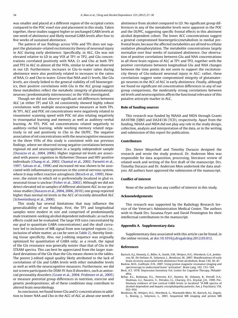
3 hol De
wctofi
pietaiaolitn
AcAvtlataifir(ai1cwendvh(
gsmeotiiooSdTcancoga
t
4 A. Mon et al. / Drug and Alco
as smaller and placed at a different region of the occipital cortexompared to the POC voxel size and placement in this study. Takenogether, these studies suggest higher or unchanged GABA levels atne week of abstinence and likely normal GABA levels after four tove weeks of sustained abstinence.
The pattern of our findings across VOIs and TPs does not sup-ort the glutamate-related excitotoxicity theory of neuronal injury
n ALC during early abstinence. Specifically, in ALC, Glu was notlevated relative to LD in any VOI at TP1 or TP2, and Glu concen-rations correlated positively with NAA, Cr and Cho at both TP1nd TP2 in ALC in almost all the VOIs, similar to what we observedn our LD. Furthermore, increases in Glu-to-water ratios duringbstinence were also positively related to increases in the ratiosf NAA, Cr and Cho to water. Given that NAA and Cr levels, like Gluevels, are closely linked to the general viability of cell bioenerget-cs, their positive correlations with Glu in the ALC group suggesthese metabolites reflect the metabolic integrity of glutamatergiceurons (predominately interneurons) in the VOIs investigated.
Though we did not observe significant mI differences betweenLC (at either TP) and LD, mI consistently showed highly robustorrelations with multiple neurocognitive measures at both TPs.t TP1, ACC and POC mI concentrations were negatively related toisuomotor scanning speed with POC mI also relating negativelyo visuospatial learning and memory as well as auditory–verbalearning. At TP2, POC mI concentrations related negatively touditory–verbal learning, while working memory related nega-ively to mI and positively to Cho in the DLPFC. The negativessociation of mI concentrations with the neurocognitive measuresn the three VOIs of this study is consistent with our previousndings, where we observed strong negative correlations betweenegional mI and neurocognition in a largely independent sampleDurazzo et al., 2004, 2006). Higher regional mI levels are associ-ted with poorer cognition in Alzheimer Disease and HIV-positivendividuals (Chang et al., 2002; Chantal et al., 2002; Parnetti et al.,997; Salvan et al., 1998) and increased mI was shown to be asso-iated with inflammatory processes in the central nervous system,here it may reflect reactive astrogliosis (Bitsch et al., 1999). How-
ver, the extent to which mI is preferentially localized in glial vs.euronal tissue is unclear (Fisher et al., 2002). Although we did notetect elevated mI in samples of different abstinent ALC in our pre-ious studies (Durazzo et al., 2004, 2006, 2010), one group reportedigher than normal mI levels in the ACC of recently detoxified ALCSchweinsburg et al., 2000).
This study has several limitations that may influence theeneralizability of our findings. First, the TP1 and longitudinalamples were modest in size and comprised of predominantlyale treatment-seeking alcohol dependent individuals; as such sex
ffects could not be evaluated. The large VOI sizes (necessitated byur goal to quantitate GABA concentrations) and the skull curva-ure led to inclusion of MR signal from non-targeted regions (i.e.,nclusion of white matter, as can be seen in Table 2), thereby limit-ng tissue specificity. Also, our J-editing sequence was originallyptimized for quantitation of GABA only; as a result, the signalf the Glx resonance was generally noisier than that of Glu in theTEAM spectra. This can best be appreciated from the larger stan-ard deviations of the Glx than the Glu means shown in the tables.he poorer J-edited signal quality likely attributed to the lack oforrelations of Glx and GABA levels with other metabolite levelss well as with the neurocognitive measures. Furthermore, we didot screen participants for DSM-IV Axis II disorders, such as antiso-ial personality disorders (Grant et al., 2004; Pridmore et al., 2005)r measure potential group differences in nutrition, exercise and
enetic predispositions; all of these conditions may contribute toltered brain neurobiology.In conclusion, we found lower Glu and Cr concentrations in addi-ion to lower NAA and Cho in the ACC of ALC at about one week of
pendence 125 (2012) 27– 36
abstinence from alcohol compared to LD. No significant group dif-ferences in any of the metabolite levels were apparent in the POCand the DLPFC, suggesting specific frontal effects in this abstinentalcohol dependent cohort. The lower ACC concentrations suggesta general compromised bioenergetics/metabolism in medial pre-frontal brain, because the affected metabolites are all tied to cellularoxidative phosphorylation. The metabolite concentrations largelynormalize over four weeks of sustained abstinence. Our observa-tion of positive correlations between Glu and NAA concentrationsin all three brain regions of ALC at TP1 and TP2, together with thepositive correlations between longitudinal Glu and NAA changesbetween the time points do not seem to support the excitotoxi-city theory of Glu-induced neuronal injury in ALC; rather, thesecorrelations suggest some compromised integrity of glutamater-gic neurons in the ACC of ALC in early abstinence. Finally, althoughwe found no significant mI concentration differences in any of ourgroup comparisons, the moderately strong correlations betweenmI levels and neurocognition affirm the functional relevance of thisputative astrocyte marker in ALC.
Role of funding sources
This research was funded by NIAAA and NIDA through GrantsAA10788 (DJM) and DA24136 (TCD), respectively. Apart from thefunding, NIAAA and NIDA did not play any role in the study design,collection, analysis and interpretation of the data, or in the writingand submission of this report for publication.
Contributors
Drs. Dieter Meyerhoff and Timothy Durazzo designed theresearch and wrote the study protocol. Dr. Anderson Mon wasresponsible for data acquisition, processing, literature review ofrelated work and writing of the first draft of the manuscript. Drs.Timothy Durazzo and Dr. Anderson Mon undertook the data anal-ysis. All authors have approved the submission of the manuscript.
Conflict of interest
None of the authors has any conflict of interest in this study.
Acknowledgements
This research was supported by the Radiology Research Ser-vice of the Veteran’s Administration Medical Center. The authorswish to thank Drs. Susanna Fryer and David Pennington for theirintellectual contributions to the manuscript.
Appendix A. Supplementary data
Supplementary data associated with this article can be found, inthe online version, at doi:10.1016/j.drugalcdep.2012.03.012.
References
Bartsch, A.J., Homola, G., Biller, A., Smith, S.M., Weijers, H.G., Wiesbeck, G.A., Jenkin-son, M., De Stefano, N., Solymosi, L., Bendszus, M., 2007. Manifestations of earlybrain recovery associated with abstinence from alcoholism. Brain 130, 36–47.
Baslow, M.H., Guilfoyle, D.N., 2007. Using proton magnetic resonance imaging andspectroscopy to understand brain “activation”. Brain Lang. 102, 153–164.
Beck, A.T, 1978. Depression Inventory Vol. Center for Cognitive Therapy, Philadel-phia.
Behar, K.L., Rothman, D.L., Petersen, K.F., Hooten, M., Delaney, R., Petroff, O.A.,Shulman, G.I., Navarro, V., Petrakis, I.L., Charney, D.S., Krystal, J.H., 1999. Pre-
liminary evidence of low cortical GABA levels in localized 1H-MR spectra ofalcohol-dependent and hepatic encephalopathy patients. Am. J. Psychiatry 156,952–954.Bendszus, M., Weijers, H.G., Wiesbeck, G., Warmuth-Metz, M., Bartsch, A.J., Engels,S., Boning, J., Solymosi, L., 2001. Sequential MR imaging and proton MR

hol De
B
B
B
B
C
C
C
C
D
D
D
D
D
D
D
E
F
F
F
F
F
G
G
G
G
H
A. Mon et al. / Drug and Alco
spectroscopy in patients who underwent recent detoxification for chronicalcoholism: correlation with clinical and neuropsychological data. Am. J. Neu-roradiol. 22, 1926–1932.
enedict, R., 1997. Brief Visuospatial Memory Test – Revised: Professional ManualVol. Psychological Assessment Resources, Inc., Odessa, FL.
itsch, A., Bruhn, H., Vougioukas, V., Stringaris, A., Lassmann, H., Frahm, J., Brück, W.,1999. Inflammatory CNS demyelination: histopathologic correlation with in vivoquantitative proton MR spectroscopy. AJNR Am. J. Neuroradiol. 20, 1619–1627.
ush, G., Luu, P., Posner, M.I., 2000. Cognitive and emotional influences in anteriorcingulate cortex. Trends Cogn. Sci. 4, 215–222.
ush, G., Vogt, B.A., Holmes, J., Dale, A.M., Greve, D., Jenike, M.A., Rosen, B.R., 2002.Dorsal anterior cingulate cortex: a role in reward-based decision making. Proc.Natl. Acad. Sci. U. S. A. 99, 523–528.
hang, L., Ernst, T., Witt, M.D., Ames, N., Gaiefsky, M., Miller, E., 2002. Relationshipsamong brain metabolites, cognitive function, and viral loads in antiretroviral-naive HIV patients. NeuroImage 17, 1638–1648.
hantal, S., Labelle, M., Bouchard, R.W., Braun, C.M., Boulanger, Y., 2002. Correlationof regional proton magnetic resonance spectroscopic metabolic changes withcognitive deficits in mild Alzheimer disease. Arch. Neurol. 59, 955–962.
lancy, B., Defelipe, J., Espinosa, A., Fairen, A., Jinno, S., Kanold, P., Luhmann,H.J., Rockland, K.S., Tamamaki, N., Yan, X.X., 2010. Cortical GABAergic neu-rons: stretching it remarks, main conclusions and discussion. Front. Neuroanat.4, 7.
ramer, S.C., Sur, M., Dobkin, B.H., O’Brien, C., Sanger, T.D., Trojanowski, J.Q., Rumsey,J.M., Hicks, R., Cameron, J., Chen, D., Chen, W.G., Cohen, L.G., deCharms, C., Duffy,C.J., Eden, G.F., Fetz, E.E., Filart, R., Freund, M., Grant, S.J., Haber, S., Kalivas, P.W.,Kolb, B., Kramer, A.F., Lynch, M., Mayberg, H.S., McQuillen, P.S., Nitkin, R., Pascual-Leone, A., Reuter-Lorenz, P., Schiff, N., Sharma, A., Shekim, L., Stryker, M., Sullivan,E.V., Vinogradov, S., 2011. Harnessing neuroplasticity for clinical applications.Brain 134, 1591–1609.
elis, D.C., Kramer, J.H., Kaplan, E., Ober, B.A., 2000. California Verbal Learning TestVol., 2nd ed. The Psychological Corporation, San Antonio, TX.
urazzo, T.C., Gazdzinski, S., Banys, P., Meyerhoff, D.J., 2004. Cigarette smokingexacerbates chronic alcohol-induced brain damage: a preliminary metaboliteimaging study. Alcohol. Clin. Exp. Res. 28, 1849–1860.
urazzo, T.C., Gazdzinski, S., Banys, P., Meyerhoff, D.J., 2006. Brain metaboliteconcentrations and neurocognition during short-term recovery from alcoholdependence: preliminary evidence of the effects of concurrent chronic cigarettesmoking. Alcohol. Clin. Exp. Res. 30, 539–551.
urazzo, T.C., Meyerhoff, D.J., 2007. Neurobiological and neurocognitive effects ofchronic cigarette smoking and alcoholism. Front. Biosci. 12, 4079–4100.
urazzo, T.C., Rothlind, J.C., Gazdzinski, S., Banys, P., Meyerhoff, D.J., 2007. Chronicsmoking is associated with differential neurocognitive recovery in abstinentalcoholic patients: a preliminary investigation. Alcohol. Clin. Exp. Res. 31,1114–1127.
urazzo, T.C., Pathak, V., Gazdzinski, S., Mon, A., Meyerhoff, D.J., 2010. Metabolite lev-els in the brain reward pathway discriminate those who remain abstinent fromthose who resume hazardous alcohol consumption after treatment for alcoholdependence. J. Stud. Alcohol Drugs 71, 278–289.
urazzo, T.C., Mon, A., Gasdzinski, S., Meyerhoff, D.J., 2011. Chronic cigarettesmoking in alcohol dependence: associations with cortical thickness and N-acetylaspartate levels in the extended brain reward system. Addict. Biol.,doi:10.1111/j.1369-1600.2011.00407.x.
nde, G., Welzel, H., Walter, S., Weber-Fahr, W., Diehl, A., Hermann, D., Heinz, A.,Mann, K., 2005. Monitoring the effects of chronic alcohol consumption andabstinence on brain metabolism: a longitudinal proton magnetic resonancespectroscopy study. Biol. Psychiatry 58, 974–980.
ein, G., Meyerhoff, D.J., Di Sclafani, V., Ezekiel, F., Poole, N., MacKay, S., Dillon,W.P., Constans, J.-M., Weiner, M.W., 1994. 1H magnetic resonance spectroscopicimaging separates neuronal from Glial changes in alcohol-related brain atrophy.NIAAA Res. Mon. No. 27/Alcohol Glial Cells., 227–241.
irst, M.B., Spitzer, R.L., Gibbon, M., Williams, J.B.W., 1998. Structured Clinical Inter-view for DSM-IV Axis I Disorders – Patient Edition (SCID-I/P, Version 2. 0, 8/98revision), Vol. Biometrics Research Department, New York, NY.
isher, S.K., Novak, J.E., Agranoff, B.W., 2002. Inositol and higher inositol phosphatesin neural tissues: homeostasis, metabolism and functional significance. J. Neu-rochem. 82, 736–754.
reund, G., Anderson, K.J., 1996. Glutamate receptors in the frontal cortex of alco-holics. Alcohol. Clin. Exp. Res. 20, 1165–1172.
rischknecht, U., Hermann, D., Hoerst, N., Tunc-Skarka, N., Kiefer, F., Ende, G., Mann,K.F., 2010. Increased ACC glutamate levels in patients during alcohol withdrawalreturn to normal after two weeks of abstinence. Alcohol. Clin. Exp. Res. 34, 132A.
azdzinski, S., Durazzo, T.C., Yeh, P.H., Hardin, D., Banys, P., Meyerhoff, D.J., 2008.Chronic cigarette smoking modulates injury and short-term recovery of themedial temporal lobe in alcoholics. Psychiatry Res. 162, 133–145.
rant, B., Stinson, F., Dawson, D.P.C., Dufour, M., Compton, W., Pickering, R.P., Kaplan,K., 2004. Prevalence and co-occurance of substance use disorders and indepen-dent mood and anxiety disorders. Arch. Gen. Psychiatry 61, 807–816.
reen, K.L., Grant, K.A., 1998. Evidence for overshadowing by components of theheterogeneous discriminative stimulus effects of ethanol. Drug Alcohol Depend.52, 149–159.
robin, A.C., Matthews, D.B., Devaud, L.L., Morrow, A.L., 1998. The role of GABA(A)receptors in the acute and chronic effects of ethanol. Psychopharmacology (Berl.)139, 2–19.
asin, D.S., Stinson, F.S., Ogburn, E., Grant, B.F., 2007. Prevalence, correlates, disabil-ity, and comorbidity of DSM-IV alcohol abuse and dependence in the United
pendence 125 (2012) 27– 36 35
States: results from the National Epidemiologic Survey on Alcohol and RelatedConditions. Arch. Gen. Psychiatry 64, 830–842.
Hughes, J.R., 2009. Alcohol withdrawal seizures. Epilepsy Behav. 15, 92–97.Johnson, B.A., 2005. Recent advances in the development of treatments for alcohol
and cocaine dependence: focus on topiramate and other modulators of GABA orglutamate function. CNS Drugs 19, 873–896.
Justin, T., Gass, M., Olive, M.F., 2008. Glutamatergic substrates of drug addiction andalcoholism. Biochem. Pharmacol. 75, 218–265.
Kaiser, L.G., Young, K., Meyerhoff, D.J., Mueller, S.G., Matson, G.B., 2008. A detailedanalysis of localized J-difference GABA editing: theoretical and experimentalstudy at 4 T. NMR Biomed. 21, 22–32.
Kalivas, P.W., O’Brien, C., 2008. Drug addiction as a pathology of staged neuroplas-ticity. Neuropsychopharmacology 33, 166–180.
Kalivas, P.W., 2009. The glutamate homeostasis hypothesis of addiction. Nat. Rev.Neurosci. 10, 561–572.
Kalivas, P.W., Lalumiere, R.T., Knackstedt, L., Shen, H., 2009. Glutamate transmissionin addiction. Neuropharmacology 56 (Suppl. 1), 169–173.
Kalivas, P.W., Volkow, N.D., 2011. New medications for drug addiction hidding inglutamatergic neuroplasticity. Mol. Pshychiatry 16, 974–986.
Lovinger, D.M., 1993. Excitotoxicity and alcohol-related brain damage. Alcohol. Clin.Exp. Res. 17, 19–27.
Martin, P.R., Gibbs, S.J., Nimmerrichter, A.A., Riddle, W.R., Welch, L.W., Willcott,M.R., 1995. Brain proton magnetic resonance spectroscopy studies in recentlyabstinent alcoholics. Alcohol. Clin. Exp. Res. 19, 1078–1082.
Mason, G.F., Petrakis, I.L., de Graaf, R.A., Gueorguieva, R., Guidone, E., Coric,V., Epperson, C.N., Rothman, D.L., Krystal, J.H., 2006. Cortical gamma-aminobutyric acid levels and the recovery from ethanol dependence:preliminary evidence of modification by cigarette smoking. Biol. Psychiatry 59,85–93.
Mertens, J.R., Lu, Y.W., Parthasarathy, S., Moore, C., Weisner, C.M., 2003. Medicaland psychiatric conditions of alcohol and drug treatment patients in an HMO:comparison with matched controls. Arch. Intern. Med. 163, 2511–2517.
Mertens, J.R., Weisner, C., Ray, G.T., Fireman, B., Walsh, K., 2005. Hazardous drinkersand drug users in HMO primary care: prevalence, medical conditions, and costs.Alcohol. Clin. Exp. Res. 29, 989–998.
Meyerhoff, D.J., Durazzo, T.C., Ende, G., 2011. Chronic alcohol consumption, absti-nence and relapse: brain proton magnetic resonance spectroscopy studies inanimals and humans. Curr. Top. Behav. Neurosci., epub ahead of print.
Newsholme, P., Procopio, J., Lima, M.M., Pithon-Curi, T.C., Curi, R., 2003. Glutamineand glutamate – their central role in cell metabolism and function. Cell Biochem.Funct. 21, 1–9.
Olive, M.F., Cleva, R.M., Kalivas, P.W., Malcoln, R.J., 2012. Glutamatergic medicationsfor the treatment of drug and behavioral addictions. Pharmacol. Biochem. Behav.100, 801–810.
Pan, J.W., Takahashi, K., 2005. Interdependence of N-acetyl aspartate and high-energy phosphates in healthy human brain. Ann. Neurol. 57, 92–97.
Parnetti, L., Tarducci, R., Presciutti, O., Lowenthal, D.T., Pippi, M., Palumbo, B., Gobbi,G., Pelliccioli, G.P., Senin, U., 1997. Proton magnetic resonance spectroscopycan differentiate Alzheimer’s disease from normal aging. Mech. Ageing Dev. 97,9–14.
Parekh, R.S., Klag, M.J., 2001. Alcohol: role in the development of hypertension andend-stage renal disease. Curr. Opin. Nephrol. Hypertens. 10, 385–390.
Parks, M.H., Dawant, B.M., Riddle, W.R., Hartmann, S.L., Dietrich, M.S., Nickel, M.K.,Price, R.R., Martin, P.R., 2002. Longitudinal brain metabolic characterization ofchronic alcoholics with proton magnetic resonance spectroscopy. Alcohol. Clin.Exp. Res. 26, 1368–1380.
Pridmore, S., Chambers, A., McArthur, M., 2005. Neuroimaging in psychopathy. Aust.N. Z. J. Psychiatry 39, 856–865.
Ross, B., Bluml, S., 2001. Magnetic resonance spectroscopy of the human brain. Anat.Rec. 265, 54–84.
Rossetti, Z.L., Carboni, S., 1995. Ethanol withdrawal is associated with increasedextracellular glutamate in the rat striatum. Eur. J. Pharmacol. 283, 177–183.
Salvan, A.M., Ceccaldi, M., Confort-Gouny, S., Milandre, C., Cozzone, P.J., Vion-Dury, J.,1998. Correlation between cognitive status and cerebral inositol in Alzheimer-type dementia. J Neurol. 245, 686–688.
Sanacora, G., Saricicek, A., 2007. GABAergic contributions to the pathophysiologyof depression and the mechanism of antidepressant action. CNS Neurol. Disord.Drug Targets. 6, 127–140.
Schuff, N., Ezekiel, F., Gamst, A.C., Amend, D.L., Capizzano, A.A., Maudsley, A.A.,Weiner, M.W., 2001. Region and tissue differences of metabolites in normallyaged brain using multislice 1H magnetic resonance spectroscopic imaging.Magn. Reson. Med. 45, 899–907.
Schweinsburg, B.C., Taylor, M.J., Videen, J.S., Alhassoon, O.M., Patterson, T.L., Grant, I.,2000. Elevated myo-inositol in gray matter of recently detoxified but not long-term alcoholics: a preliminary MR spectroscopy study. Alcohol. Clin. Exp. Res.24, 699–770.
Schweinsburg, B.C., Taylor, M.J., Alhassoon, O.M., Videen, J.S., Brown, G.G., Patter-son, T.L., Berger, F., Grant, I., 2001. Chemical pathology in brain white matterof recently detoxified alcoholics: a 1H magnetic resonance spectroscopy inves-tigation of alcohol-associated frontal lobe injury. Alcohol. Clin. Exp. Res. 25,924–934.
Seitz, D., Widmann, U., Seeger, U., Nagele, T., Klose, U., Mann, K., Grodd, W., 1999.Localized proton magnetic resonance spectroscopy of the cerebellum in detox-ifing alcoholics. Alcohol. Clin. Exp. Res. 23, 158–163.
Skinner, H.A., Sheu, W.J., 1982. Reliability of alcohol use indices. The Lifetime Drink-ing History and the MAST. J. Stud. Alcohol 43, 1157–1170.

3 hol De
S
S
S
S
S
S
S
Corporation, San Antonio, TX.
6 A. Mon et al. / Drug and Alco
obell, L.C., Sobell, M.B., Riley, D.M., Schuller, R., Pavan, D.S., Cancilla, A., Klajner, F.,Leo, G.I., 1988. The reliability of alcohol abusers’ self-reports of drinking and lifeevents that occurred in the distant past. J. Stud. Alcohol 49, 225–232.
obell, L.C., Sobell, M.B., 1990. Self-reports issues in alcohol abuse: state of the artand future directions. Behav. Assess. 12, 77–90.
oher, B.J., van Zijl, P.C., Duyn, J.H., Barker, P.B., 1996. Quantitative proton MR spec-troscopic imaging of the human brain. Magn. Reson. Med. 35, 356–363.
panagel, R., 2009. Alcoholism: a systems approach from molecular physiology toaddictive behavior. Physiol. Rev. 89, 649–705.
pielberger, C.D., Gorsuch, R.L., Lushene, R., Vagg, P.R., Jacobs, G.A., 1977. Self-Evaluation Questionaire. Consulting Psychologists Press, Palo Alto, CA.
tinson, F.S., Grant, B.F., Dawson, D.A., Ruan, W.J., Huang, B., Saha, T., 2005. Comor-bidity between DSM-IV alcohol and specific drug use disorders in the United
States: results from the National Epidemiologic Survey on Alcohol and RelatedConditions. Drug Alcohol Depend. 80, 105–116.ullivan, J., Sykora, K., Schneiderman, J., Naranjo, C., Sellers, E., 1989. Assesmentof alcohol withdrawal: the revised clinical institute withdrawl assesment foralcohol scale. Br. J. Addict. 84, 1353–1357.
pendence 125 (2012) 27– 36
Tsai, G., Coyle, J.T., 1998. The role of glutamatergic neurotransmission in the patho-physiology of alcoholism. Annu. Rev. Med. 49, 173–184.
Umhau, J.C., Momenan, R., Schwandt, M.L., Singley, E., Lifshitz, M., Doty, L., Adams,L.J., Vengeliene, V., Spanagel, R., Zhang, Y., Shen, J., George, D.T., Hommer, D.,Heilig, M., 2010. Effect of acamprosate on magnetic resonance spectroscopymeasures of central glutamate in detoxified alcohol-dependent individuals: arandomized controlled experimental medicine study. Arch. Gen. Psychiatry 67,1069–1077.
Van Leemput, K., Maes, F., Vandermeulen, D., Suetens, P., 1999. Automated model-based tissue classification of MR images of the brain. IEEE Trans. Med. Imaging18, 897–908.
Wechsler, D., 1997. Wechsler Memory Scale – III (WMS – III). Vol. The Psychological
Zahr, N.M., Mayer, D., Vinco, S., Orduna, J., Luong, R., Sullivan, E.V., Pfefferbaum, A.,2009. In vivo evidence for alcohol-induced neurochemical changes in rat brainwithout protracted withdrawal, pronounced thiamine deficiency, or severe liverdamage. Neuropsychopharmacology 34, 1427–1442.
Copyright © 2022 FDOKUMEN