Global genetic diversity and geographical and host-species distribution of beak and feather disease...
17
See discussions, stats, and author profiles for this publication at: https://www.researchgate.net/publication/49702033 Global genetic diversity and geographical and host-species distribution of beak and feather disease virus isolates Article in Journal of General Virology · April 2011 DOI: 10.1099/vir.0.028126-0 · Source: PubMed CITATIONS 36 READS 83 5 authors, including: Some of the authors of this publication are also working on these related projects: Plant-cell pack (aka "cookie") technology View project Full genome sequencing of Avibacterium paragallinarum View project Arvind Varsani Arizona State University 709 PUBLICATIONS 3,412 CITATIONS SEE PROFILE Rob Richard Bragg University of the Free State 96 PUBLICATIONS 593 CITATIONS SEE PROFILE Inga I Hitzeroth University of Cape Town 41 PUBLICATIONS 537 CITATIONS SEE PROFILE Ed Rybicki University of Cape Town 434 PUBLICATIONS 6,986 CITATIONS SEE PROFILE All content following this page was uploaded by Arvind Varsani on 03 December 2016. The user has requested enhancement of the downloaded file. All in-text references underlined in blue are added to the original document and are linked to publications on ResearchGate, letting you access and read them immediately.
Transcript of Global genetic diversity and geographical and host-species distribution of beak and feather disease...

Seediscussions,stats,andauthorprofilesforthispublicationat:https://www.researchgate.net/publication/49702033
Globalgeneticdiversityandgeographicalandhost-speciesdistributionofbeakandfeatherdiseasevirusisolates
ArticleinJournalofGeneralVirology·April2011
DOI:10.1099/vir.0.028126-0·Source:PubMed
CITATIONS
36
READS
83
5authors,including:
Someoftheauthorsofthispublicationarealsoworkingontheserelatedprojects:
Plant-cellpack(aka"cookie")technologyViewproject
FullgenomesequencingofAvibacteriumparagallinarumViewproject
ArvindVarsani
ArizonaStateUniversity
709PUBLICATIONS3,412CITATIONS
SEEPROFILE
RobRichardBragg
UniversityoftheFreeState
96PUBLICATIONS593CITATIONS
SEEPROFILE
IngaIHitzeroth
UniversityofCapeTown
41PUBLICATIONS537CITATIONS
SEEPROFILE
EdRybicki
UniversityofCapeTown
434PUBLICATIONS6,986CITATIONS
SEEPROFILE
AllcontentfollowingthispagewasuploadedbyArvindVarsanion03December2016.
Theuserhasrequestedenhancementofthedownloadedfile.Allin-textreferencesunderlinedinblueareaddedtotheoriginaldocumentandarelinkedtopublicationsonResearchGate,lettingyouaccessandreadthemimmediately.

Global genetic diversity and geographical andhost-species distribution of beak and featherdisease virus isolates
Arvind Varsani,1,2 Guy L. Regnard,3 Robert Bragg,4 Inga I. Hitzeroth3
and Edward P. Rybicki3,5
Correspondence
Edward P. Rybicki
Received 19 October 2010
Accepted 17 December 2010
1School of Biological Sciences, University of Canterbury, Christchurch 8140, New Zealand
2Electron Microscope Unit, University of Cape Town, Rondebosch, Cape Town 7701, South Africa
3Department of Molecular and Cell Biology, University of Cape Town, Rondebosch, Cape Town7701, South Africa
4Department of Microbial Biochemical and Food Biotechnology, University of the Free State,Bloemfontein, South Africa
5Institute of Infectious Disease and Molecular Medicine, University of Cape Town, Observatory,Cape Town 7925, South Africa
Psittacine beak and feather disease (PBFD) has a broad host range and is widespread in wild and
captive psittacine populations in Asia, Africa, the Americas, Europe and Australasia. Beak and
feather disease circovirus (BFDV) is the causative agent. BFDV has an ~2 kb single stranded
circular DNA genome encoding just two proteins (Rep and CP). In this study we provide support
for demarcation of BFDV strains by phylogenetic analysis of 65 complete genomes from
databases and 22 new BFDV sequences isolated from infected psittacines in South Africa. We
propose 94 % genome-wide sequence identity as a strain demarcation threshold, with isolates
sharing .94 % identity belonging to the same strain, and strain subtypes sharing .98 % identity.
Currently, BFDV diversity falls within 14 strains, with five highly divergent isolates from
budgerigars probably representing a new species of circovirus with three strains (budgerigar
circovirus; BCV-A, -B and -C). The geographical distribution of BFDV and BCV strains is strongly
linked to the international trade in exotic birds; strains with more than one host are generally
located in the same geographical area. Lastly, we examined BFDV and BCV sequences for
evidence of recombination, and determined that recombination had occurred in most BFDV and
BCV strains. We established that there were two globally significant recombination hotspots in
the viral genome: the first is along the entire intergenic region and the second is in the C-terminal
portion of the CP ORF. The implications of our results for the taxonomy and classification of
circoviruses are discussed.
INTRODUCTION
Psittacine beak and feather disease (PBFD) was firstdescribed in Australian psittacine species in the 1970s.However, it may have been present prior to this as recordsbetween 1887 and 1888 associate the decline of Psephotus sp.in Southern Australia with impaired flight as a result offeather deformities (Pass & Perry, 1984; Raidal et al., 1993).The disease has a broad host range and is widespread in both
wild and captive psittacine populations found in Asia, Africa,the Americas, Europe and Australasia (Bassami et al., 2001;Bendheim et al., 2006; Bert et al., 2005; Ha et al., 2007;Kiatipattanasakul-Banlunara et al., 2002; Raidal et al., 1993;Raidal & Cross, 1994a; Ritchie et al., 1990; Sanada et al., 1999;Wirminghaus et al., 1999). The worldwide spread of PBFDhas been largely because of the international trade in ‘exotic’psittacines (Doneley, 2003; Rahaus & Wolff, 2003;Wirminghaus et al., 1999). Initially PBFD was thought tobe restricted to Old World and South Pacific psittacines;however, it is now evident that the disease also affects NewWorld psittacines (Bert et al., 2005; Ritchie et al., 1990).PBFD mainly affects young birds and is characterized by lossof feathers, abnormally shaped feathers and the overgrowthand irregularity of the surface of the beak. The disease has
The GenBank/EMBL/DDBJ accession numbers for the BFDV isolatessequenced as part of this study are HM748918–HM748939. Furtherinformation regarding these can be found in Table 1.
Four supplementary figures are available with the online version of thispaper.
Journal of General Virology (2011), 92, 752–767 DOI 10.1099/vir.0.028126-0
752 028126 G 2011 SGM Printed in Great Britain

been associated with immunosuppression and most birdswith PBFD die from a secondary bacterial and/or viralinfection (Doneley, 2003; Pass & Perry, 1984; Ritchie et al.,1989; Schoemaker et al., 2000). Apart from the main clinicalmanifestations, peracute and subclinical infections have beendescribed; the peracute infection affects young birds and beakand feather abnormalities are notably absent (Doneley, 2003;Rahaus & Wolff, 2003; Schoemaker et al., 2000).
The causative agent of PBFD is beak and feather diseasevirus (BFDV), which belongs to the genus Circovirus of thefamily Circoviridae (Ritchie et al., 1989; Ritchie et al., 1990;Studdert, 1993). Differences in the clinical and pathologicalmanifestation of PBFD are thought to be because of hostfactors rather than antigenic or genetic variation in BFDV(Bassami et al., 2001).
BFDV virions have icosahedral T51 symmetry and are non-enveloped, with a diameter of 14–16 nm (Ritchie et al.,1989). BFDV is one of the smallest known animal viruses,with a simple and compact 2 kb ambisense single-strandedcircular DNA genome that encodes two major genes(Crowther et al., 2003; Niagro et al., 1998). The comple-mentary strand encodes the single capsid protein (cp) geneand the virion strand encodes the replication associated gene(rep). Replication occurs through rolling-circle replicationinvolving a dsDNA intermediate (Bassami et al., 1998). Boththe gene products are homologous to the CP and Repproteins of porcine circovirus (PCV) (Niagro et al., 1998).The Rep protein of circoviruses is more similar to the Repproteins of nanoviruses and geminiviruses than to those ofany other viruses with a similar rolling-circle replicationmechanism. The BFDV genome contains a replicationhairpin–loop structure that is characteristic of all circo-viruses, nanoviruses and geminiviruses.
Previous phylogenetic analyses of BFDV have focusedpredominantly on partial sequence data of rep or cp. It hasbeen shown that there are differences in the evolution ofthe major genes and intergenic regions of BFDV. The repgene has been found to be highly conserved, probablyowing to functional constraints upon it, and showed signsof purifying selection, whereas analysis of the cp hasshowed positive selection with a high degree of variation atthe amino acid level, suggesting that intraspecies antigenicvariation might occur (Bassami et al., 2001; Heath et al.,2004; Hughes & Piontkivska, 2008; Raue et al., 2004;Ypelaar et al., 1999). Interestingly, there is evidence tosuggest that the two intergenic regions are also conserved(Hughes & Piontkivska, 2008).
Research has suggested that the virus isolates may begrouped according to host species or the ability of the virusto cause disease, and not by geographical location (Albertynet al., 2004; de Kloet & de Kloet, 2004; Khalesi et al., 2005;Raue et al., 2004; Ritchie et al., 2003). This has beensupported by a recent study that showed that cockatiel virusisolates were serologically and genetically different fromother BFDV isolates (Shearer et al., 2008). However,Southern African strains have diverged from viruses found
in other parts of the world to produce regionally distinctlineages (Heath et al., 2004). The available evidence thereforesuggests that the relationship between BFDV isolates,psittacine species and pathogenicity is very complex, andrequires further study (de Kloet & de Kloet, 2004).
Lefeuvre et al. (2009) demonstrated that recombination isrampant amongst all ssDNA viruses, and that recombina-tion breakpoints are more frequent on the periphery offunctional genes or within the intergenic regions. As thereare demonstrated differences in the rates of evolutionbetween the BFDV cp, rep and intergenic-region sequences, acomprehensive examination of whole genomes is essential toinfer accurate phylogenies and to assess the evolution ofBFDV through recombination. With the advent of modernmolecular techniques, and particularly of W29 polymerase-mediated isothermal rolling-circle amplification (RCA) ofwhole genomes, the number of genome sequences of ssDNAviruses available has increased exponentially recently. Of the87 complete genome sequences presently available for BFDVisolates, including five highly divergent BFDV-like budger-igar isolates from Japan and China, and 22 from this study,30 genomes were amplified using RCA methods in less thana year (Varsani et al. 2010; Ortiz-Catedral et al., 2010) and.40 full-length genomes have been deposited in GenBankwithin the last year.
In this study, BFDV genomic DNA was isolated using RCAfrom 22 infected psittacines from South Africa. Thesegenomes were fully sequenced and compared with 65complete genomes already deposited in GenBank. Weanalysed all these complete genomes of BFDV, determinedthe current diversity of BFDV, and established a thresholdvalue for the reliable demarcation of BFDV strains. Inaddition, we provide insights into the geographical andhost-species distribution of BFDV strains and analyse therecombination patterns between strains. We further suggestthe establishment of at least one new species of circovirus, toaccommodate the viruses found in budgerigars.
RESULTS AND DISCUSSION
Strain demarcation of BFDV and BCV
We cloned and completely sequenced 22 BFDV genomesfrom infected parrots in South Africa. For an objectiveclassification of these genomes we aligned 65 completeBFDV genomes available in GenBank with the 22 from thisstudy. The 87 genomes, including the five BFDV-likegenomes from budgerigars, all shared greater than 82.6 %pairwise identity. Since there is no clear guideline for theclassification of BFDV isolates, we analysed the frequency ofpairwise sequence identities of the 87 genomes at intervals of0.1 % and 1 % (Fig. 1a), and compared them with those ofother sampled circoviruses (831 genomes, 344 737 pairwiseidentities; Fig. 1b). The highest frequency peak of pairwiseidentities of the 87 BFDV-type genomes (3742 pairwiseidentities) is between 90 and 94.5 % (Fig. 1a), with an
BFDV diversity and taxonomy
http://vir.sgmjournals.org 753

Fig. 1. (a) Pairwise identity plots (sampled at 0.1 and 1 % pairwise identity frequencies) showing the proportion of pairwisedistances of BFDV/BCV. (b) Pairwise identity plot generated from the analysis of 833 circoviruses genomes. Proposeddemarcations of species, strain, variant and subtype are identical to those already in place for begomoviruses. (c) Maximum-likelihood tree constructed using PHYML (model GTR+I+G4) and a two-dimensional graphical representation of pairwise(pairwise deletion of gaps) nucleotide sequence identity between representative species of avian circoviruses and two recentlyidentified circoviruses from chimpanzee and human faecal samples (Li et al., 2010).
A. Varsani and others
754 Journal of General Virology 92

intermediate peak between 83 and 89 %. In the case of thegoose circoviruses, the main similarity peak is at 90–94 %(277 pairwise identities; Supplementary Fig. S1a, available inJGV Online) with minor peaks at 94–98 % and 982100 %.PCV-1 isolates were all between 98 and 100 % identical (704pairwise identities; Supplementary Fig. S1a), whereas PCV-2has two peaks of identity, at 94–97.5 % and 982100 %(141 205 pairwise identities; Supplementary Fig. S1a).
A clear trough is observed at 94 % for circovirus pairwiseplots, which is very similar to that for geminiviruses.Therefore, based on our pairwise analysis, we propose that agenome-wide pairwise identity threshold of 94 % (withpairwise deletion of gaps) should be considered for straindemarcation for BFDV and for other circoviruses. Thegroupings around 94–98 % and 98–100 % could then beconsidered as strain variants and subtypes, respectively.When circovirus identity profiles are compared with thoseof the very well-characterized geminiviruses (ssDNA plantviruses; see Fauquet et al., 2008), a very similar distributionis observed.
While PCV1, PCV-2 and goose circoviruses isolates share.90 % sequence identity, in the case of BFDV a significantproportion of pairwise distances fall between 83 and 89 %.The isolates contributing to this are those isolated frombudgerigars from Japan (AB277748, AB277749, AB277750and AB277751) and China (GQ386944). We thereforepropose a circovirus species demarcation at ,89 % whole-genome identity; this is the same as that specified by theInternational Committee on Taxonomy of viruses (ICTV)for geminivirus species demarcation (Fauquet et al., 2008;Stanley et al., 2005). A global comparison of the pairwisesequence identities (344 737 pairwise identities) of allcircovirus genomes including these five isolates (Fig. 1band Supplementary Fig. S1a) clearly supports our proposal.
Accordingly, and because they all fall below this threshold inwhole-genome comparisons with other BFDV-type isolates,we propose that the five budgerigar isolates (AB277748,AB277749, AB277750, AB277751 and GQ386944) bereclassified as a new species of circovirus – budgerigarcircovirus (BCV).
Suggestions for a new taxonomic classification ofcircoviruses
Currently the family has only two recognized genera,Circovirus and Gyrovirus. However, the recent discovery oftwo new circoviruses and 12 novel circo-like viruses (whichhave tentatively been classified as cycloviruses) from faecalsamples using metagenomic approaches (Li et al., 2010)points to the necessity of establishing new taxa andaddressing various taxonomic discrepancies within thetaxonomic family Circoviridae. Our analysis of pairwiseidentities of all circoviruses (Fig. 1b) reveals a significantpeak at 40–50 % identity and a minor peak at 53–56 %identity. A two-dimensional pairwise identity matrix (Fig.1c) clearly indicates that three new genera (indicated by
different coloured branches in the maximum-likelihoodphylogenetic tree shown in Fig. 1c) need to be created toaccommodate the circovirus species that share pairwiseidentities between 40–50 %. We therefore propose thatcirco- and circo-like viruses that share between 40–50 %whole-genome sequence identity be considered as membersof different genera, and that whole-genome identities of 50–89, 89–88 and 98–100 % be used as species, strain andsubtype delimiters, respectively. A further suggestion, shouldthese ever be recognized, is that subgenera be established toaccommodate species that share 53–56 % identity.
We suggest that the ICTV circovirus working committeeurgently review the current circovirus classification, asrapidly improving molecular and sequencing technologieswill probably result in a great diversity of circoviruses beingdiscovered in the near future.
Phylogenetic analysis of BFDV and BCV
Complete genomes of BFDV have been characterized fromAustralia (AU; n511), Germany (n51), Japan (JP; n52),New Zealand (NZ; n55), Portugal (PT; n56), Thailand(TH; n517), UK (n52), USA (n52), South Africa (ZA;n535) and Zambia (ZM; n51), and whole genomes ofproposed BCV have been characterized from China (CN;n51) and Japan (n54). The maximum-likelihood phylo-genetic tree (Fig. 2) supports the 94 % strain thresholddetermined from direct pairwise comparisons. Thus, withina monophyletic grouping of BFDV species, 14 strains (A–N;Fig. 2) were identified from 30 parrot species from 10countries (Figs 3 and 4), whereas, within the monophyleticBCV grouping, three strains (A–C) were identified frombudgerigars from China and Japan. The pairwise distancebetween BCV-A and -B is 94.6 %. However, there is strongphylogenetic support (Fig. 2) to assign these two isolates asseparate strains. Very limited diversity (~99.9 % sequenceidentity) is observed within variants of BFDV-F infectingvarious psittacines from Thailand, and variants of BFDV-Linfecting Psittaculla krameri from South Africa. When thestrains themselves are compared, however, they are clearlydistinct from one another (Supplementary Fig. S1).
We propose the following nomenclature for BFDV andBCV genomes: BFDV-strainsubtype [country : lab ID : year]e.g. BFDV-A1[NZ : B79 : 2008], BFDV-A2[NZ : B51 : 2009].We suggest the use of the international two letter countryID code, as is the norm with geminivirus taxonomy. Thedetails of the strains and nomenclature for 87 completegenomes of BFDV and BCV are provided in Table 1.
Geographical distribution of BFDV and BCV strains
Based on the current global sampling of BFDV and BCVisolates together with their whole-genome analysis (Figs 3and 4), it is clear that BFDV-A (n55) is localized to NewZealand; BFDV-B (n56), -D (n52) and -G (n51) toAustralia; BFDV-E (n54), -F (n510) and -H (n52) toThailand; and BFDV-L (n53) and -M (n520) to Southern
BFDV diversity and taxonomy
http://vir.sgmjournals.org 755
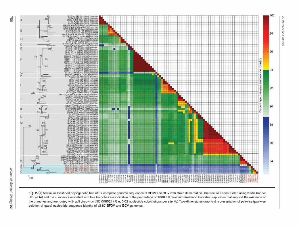
Fig. 2. (a) Maximum-likelihood phylogenetic tree of 87 complete genome sequences of BFDV and BCV with strain demarcation. The tree was constructed using PHYML (modelF81+G4) and the numbers associated with tree branches are indicative of the percentage of 1000 full maximum-likelihood bootstrap replicates that support the existence ofthe branches and are rooted with gull circovirus (NC 008521). Bar, 0.02 nucleotide substitutions per site. (b) Two-dimensional graphical representation of pairwise (pairwisedeletion of gaps) nucleotide sequence identity of all 87 BFDV and BCV genomes.
A.V
arsaniand
others
75
6Jo
urnalo
fG
eneralV
irolo
gy9
2

Fig. 3. (a) Graphical representation of the distribution of BFDV and BCV isolated from various species. (b) Country-widedistribution of the various BFDV and BCV strains.
BFDV diversity and taxonomy
http://vir.sgmjournals.org 757

Fig. 4. (a) Graphical representation of the distribution of BFDV and BCV strains isolated from various species. (b) Distribution ofBFDV and BCV strains in each country.
A. Varsani and others
758 Journal of General Virology 92

Africa. BFDV-C is predominantly found in South Africa,with the exception of one sample from the US. Seven of thenine isolates of BFDV-I (n59) are from South Africa andtwo are from Portugal. BFDV-J (n56) has a Europeanpresence; one isolate from each of Germany, Portugal andthe UK. BFDV-K has been isolated from infectedpsittacines from Thailand (n51), UK (n51), Australia(n52) and USA (n51). Three of the five BFDV-N isolatesare from South Africa and the remaining two from Japan.However, all of these five BFDV-Ns are isolated frombudgerigars (Melopsittacus undulates). BCV-A (n51) and-B (n51) are from Japan, whereas BCV-C (n53) wasfound in budgerigars from China (n51) and Japan (n52).
Our analysis indicates that strains appear to be scattered insome cases, or localized to a specific region in others. Theapparently haphazard geographical distribution of certainstrains can almost certainly be linked to the internationaltrade in exotic birds. The strains traced to single locationscould represent either endemicity or a founder effect, withthe arrival of an infected bird in a region followed by therapid spread of the disease amongst a naıve hostpopulation leading to genetic drift. High mutation ratesobserved in single-stranded DNA genomes may accentuatethe effects of genetic drift (Duffy & Holmes, 2009; Harkinset al., 2009; van der Walt et al., 2008). As most of thesamples here were described within the last 5 years, thissupports the idea that these are emerging strains adaptingto a new set of conditions. What can be seen here is: thediscovery of endemicity, with geographically limitedstrains; the recent dissemination of established strains, asshown by the haphazard geographical distribution of somestrains; and the emergence in naıve hosts of new strainsfrom established ones, as shown by the localized closelyrelated isolates. Efforts to regulate the animal trade andstrict biosecurity controls should be implemented tocontrol the spread of the disease, which is obviously quitemobile (Bert et al., 2005; Wirminghaus et al., 1999).
Host range of BFDV strains
BFDV-A has so far only been found in wild populations ofred-fronted parakeets (Cyanoramphus novaezelandiae, n55).Red-fronted parakeets are native to New Zealand and theseBFDV-A isolates represent the first reported BFDV infectionof native New Zealand psittacines. The limited geneticvariability amongst all of the BFDV-A isolates suggests thatthis infection of the population is probably recent.
BFDV-B has a wide host range in Australia, infecting variousCacatua (n53), Eolophus (n51) and Nymphicus (n52)species. BFDV-C has only been found in Cacatua alba (n51),Pionities leucogaster (n51) and Psittacus erithacus (n51)from South Africa (n53) and an unknown species from theUSA (n51). BFDV-D is limited to Cacatua sp. (n52) fromAustralia, whereas BFDV-E is found in Cacatua sulphurea(n51), Ara ararauna (n51) and Eclectus roratus (n52), allfrom Thailand. BFDV-F has the widest host range, infectingProbosciger aterrimus (n51), Ara sp. (n53), Ara nobilis
(n51), Psittacula sp. (n53) and Pscittacus erithacus (n51),all these isolates being from Thailand. An isolate from arainbow lorikeet (Trichoglossus haematodus) is the solemember of BFDV-G from Australia and is the most divergentof all BFDV isolates (Fig. 2; Supplementary Fig. S1). BothBFDV-H isolates are from Thailand and infect Cacatua sp.BFDV-I infects Poicephalus sp. (n55) from South Africa andP. erithacus (n54) from South Africa and Portugal.
Despite the predominantly European presence, all BFDV-Jisolates are from P. erithacus. The South African-limitedBFDV-J infected Pscittacula krameri (n53). BFDV-Kinfects Agapornis sp. (n53), Psephotus haematogaster(n51) and P. krameri (n51). BFDV-M predominantlyinfects Poicephalus sp. (n515); however, other hostsinclude Agaponis personata (n51), Amazona sp. (n52),Eclectus roratus (n51) and P. erithacus (n51). BFDV-N hasonly been isolated from budgerigars (Melopsittacus undu-lates) in South Africa (n53) and Japan (n52). BCV-A and-B have been isolated from budgerigars in Japan, whereasBCV-C is from China (n51) and Japan (n52).
The findings for the host ranges of BFDV strains are similarto those for geographical distribution, with some strainsinfecting a wide host range and others being specific to aparticular host. Strains with more than one host are generallylocated in the same geographical area, or, as in the case ofBFDV-F1 and BFDV-J1, from the same breeding facility,while strains with multiple geographical sites predominantlyhave the same host. Similar findings have been reported by deKloet & de Kloet (2004). The dispersion seen for some strainscould represent a common source, which highlights theimplications of animal trafficking in the spread of disease.Additionally, perceived host specificities may be an artefact ofbreeding facilities with one or only a few species of birds.BFDV-N and BCV-A, -B, -C, infecting budgerigars, andBFDV-G, infecting a rainbow lorikeet (Trichoglossus haema-todus), exhibit host specificity as described by Ritchie et al.(2003). In contrast, strain BFDV-F was isolated from variouspsittacine species from a specific breeding facility and allisolates were .99 % identical. In this strain there was no hostspecificity, and the isolation may be evidence of an initialoutbreak in a naıve population of psittacines.
Genome analysis of BFDV and BCV
Within all of these genomes we identified the characteristiccircovirus nonanucleotide origin of replication sequence(TAGTATTAC; Meehan et al., 1997) within the potentialstem–loop structure. We also identified the TATA boxes,indirect and inverted repeats, polyadenylation signals andthe three conserved rolling circle replicon motifs (FTLNN,GxxHLQGY and YxxK) within the rep gene and thenuclear localization domain (RRRxARPYxRRRHxRRx-RxxRRRRxFRRRRFSTxRIYTLRLxRQ; Heath et al., 2006)on the N terminus of the capsid protein.
Estimates of site-specific variation were determined usingthe fixed effects likelihood (FEL) method implemented in
BFDV diversity and taxonomy
http://vir.sgmjournals.org 759

Table 1. Details of complete genomes determined for BFDV and BCV isolates with strain demarcation and proposed nomenclature
BFDV isolates sequenced as part of this study are shown in boldface type. Strains that may require reclassification are shown in red. Numbers in the publication column indicate the origin of the data in that
row: 1, Ortiz-Catedral et al. (2010); 2, Bassami et al. (2001); 3, Shearer et al. (2008); 4, Bassami et al. (1998); 5, Heath et al. (2004); 6, Niagro et al. (1998); 7, Sariya et al., unpublished; 8, Henriques and
Fevereiro, unpublished; 9, this study; 10, de Kloet & de Kloet (2004); 11, Varsani et al. (2010); 12, Ogawa et al., unpublished; 13, Ogawa et al. (2010); 14, Zhuang et al., unpublished.
A.V
arsaniand
others
76
0Jo
urnalo
fG
eneralV
irolo
gy9
2

Ta
ble
1.
cont
.
BFDV diversity and taxonomy
http://vir.sgmjournals.org 761

HYPHY. The ratio of non-synonymous to synonymoussubstitutions within rep over 291 codons was 0.506583substitutions site21 (negative selection overall), whereas forcp over 248 codons it was 0.938893 substitutions site21,suggesting that cp is under neutral selection. These resultscan be supported by previous data and our data (Fig. 5) inthat the overall variation of rep was less than that of cp, andthat rep was found to be under purifying selection (de Kloet& de Kloet, 2004; Heath et al., 2004; Hughes & Piontkivska,2008; Ritchie et al., 2003). These observed differences makeit necessary to base comparisons on complete genomes andnot individual ORFs. Similarly, comparisons cannot bemade between phylogenies based on different genes. Heathet al. (2004) have brought into question the reliability ofBFDV phylogenies and phylogenetic inferences based on rep,owing to the presence of recombination.
Within the BFDV and BCV rep genes (Supplementary Fig.S2, available in JGV Online) we found evidence of positiveselection at six codon sites (four codon sites when BFDVrep genes were analysed on their own), predominantlylocated in the 59 portion (Supplementary Fig. S2), whereas121 sites were under negative selection (P,0.1 significancelevel, binomial probability). The N-terminal tail of Repforms part of a positively charged surface on the proteinand is responsible for dsDNA binding. This surface bindsto specific DNA sequences; this positions the catalyticdomain with respect to the conserved nonanucleotide(Vega-Rocha et al., 2007). The N-terminal region does notcontain nuclear localization signals as described in PCV,and is dependent on the CP for transport into the nucleus(Heath et al., 2006). Similarly, we found evidence forpositive selection in eight codon sites in the BFDV andBCV cp genes (six codon sites when BFDV cp genes wereanalysed on their own) (Supplementary Fig. S2) with 105sites under negative selection (P,0.1 significance level,binomial probability).
Maximum-likelihood phylogenetic analysis of the Rep andCP proteins yielded trees with very poor bootstrap supportfor the branches (Supplementary Fig. S3, available in JGVOnline); thus, it is difficult to infer any evolutionaryrelationship between strains. This difficulty is probablycompounded by the high degree of recombination amongstvirus strains (see below). Pairwise identity comparisons ofthe rep and cp genes (Supplementary Fig. S4, available inJGV Online) and Rep and CP proteins (Fig. 5) indicate thatthe cp gene is more divergent and possibly evolving at afaster rate than the rep gene. However, the most strikingfact about the pairwise distance between the Rep and CPproteins and the rep and cp genes is that cp shows slightlyless diversity than rep. However, more diversity is observedat the amino acid sequence level for CP compared withRep. Despite the fact that we found cp to be under neutralselection, we note that it is evolving faster that rep.
We analysed the BFDV and BCV sequences for evidence ofrecombination in all 87 full-length sequences. We foundevidence of recombination in most BFDV and BCV strains
(Fig. 6). BFDV-A (New Zealand specific, red-frontedparakeet), -C (predominantly South Africa, variousspecies), -F (Thailand specific, various species), -G(Australia specific, rainbow lorikeet) and -L (SouthAfrica specific, ring necked parakeets) were not detectablyrecombinant. Twelve of the 14 unique recombinationevents detected were within rep or in sequences encodingthe N-terminal region of rep. A similar distribution ofrecombination break points has been found withinbegomoviruses and mastreviruses (Lefeuvre et al., 2007;Varsani et al., 2008, 2009). Break-point density analysisrevealed two globally significant recombination hotspots(local P values ,0.05, binomial probability), the first beingalong the entire intergenic region between the start of repand cp, and the second being in the 39 portion of cpadjacent to the intergenic regions (Fig. 6). Lefeuvre et al.(2009) describe similar hotspots amongst all circoviruses.
The majority of recombination events detected were inter-strain recombinants, with a few exceptional intra-strainrecombinants (Table 2). BFDV-J1 isolates are recombinantsgenerated from inter- and intra-species recombination. Alarge number of recombination break points were detectedfor BCV-A, -B and -C. Furthermore, it is important to notethat all recombinant regions (regions/events 1, 2, 4, 11 and14) in the BCV-A, -B and -C isolates are from BFDVsisolated from budgerigars, which is further evidence fortheir demarcation as a species as recombination frequencydecreases with increasing genetic distance in ssDNA viruses(Lefeuvre et al., 2009). BFDV-N is a recombinant of BFDV-M1 (minor parent) and BCV-A (major parent). All theBFDV-M isolates are from South Africa, and, based on thefact that budgerigars are endemic to Australia, recombina-tion event 14 (Table 2) clearly indicates that the psittacinesharbouring the BFDV-N strain and BFDV-M1 strains areeither from the same breeding facility or have come intocontact at some point in the pet psittacine trade.
CONCLUSION
These data suggest that the distribution of BFDV and BCVisolates is influenced by the trade in psittacines, with naturalgeographical as well as host specificity of strains becomingblurred following the movement of birds. The trade inpsittacines and the close proximity in which the captivebirds are housed opens the way for multiple infections withBFDV, and the consequent emergence of recombinantstrains. The differing rates of evolution between rep and cpsuggest that phylogenetic comparisons should be madeusing complete genomes. We note that the results herein didnot take into account the possibility of strains groupingaccording to severity of infection/virulence. This wouldrequire a much more detailed veterinary study and wasbeyond the scope of our investigation.
In this study we propose a set of strain demarcations forBFDV, as well as a new nomenclature, and further suggestthat certain budgerigar isolates represent a new circovirus
A. Varsani and others
762 Journal of General Virology 92

Fig. 5. Two-dimensional graphical representation of pairwise (pairwise deletion of gaps) CP and Rep amino acid sequence identity for all BFDV and BCV genomes.
BF
DV
diversityand
taxonomy
http://vir.sgm
journals.org7
63

Fig. 6. Maximum-likelihood phylogenetic tree, showing the genome sequences of BFDV and BCV, with recombination eventslabelled according to Table 2, and recombination break-point distribution plots, indicating recombination breakpoint hot spotsdetectable in BFDV and BCV species. The Linearized genome map above the plots indicates the starting and ending alignmentpositions. CP, Coat protein gene; Rep, replication associated protein gene. Bar, 0.02 nucleotide substitutions per site.
A. Varsani and others
764 Journal of General Virology 92

species. In addition, we address the current discrepanciesin the circovirus taxonomic classification and provideimportant suggestions for the ICTV circovirus committee.
METHODS
Viral sampling, cloning and sequencing of whole genomes.Blood samples were taken from the 22 BFDV-infected birds[Poicephalus robustus (n510), Poicephalus gulielmi massaicus (n54),P. krameri (n53), Amazona sp. (n52), E. roratus (n51) and P.erithacus (n52)] from various avian breeding facilities in South Africain 2006. DNA was extracted using a Qiagen QIAamp DNA mini kitaccording to the manufacturer’s protocols. RCA was used as a methodto amplify the BFDV genomes in a non-specific manner as outlined byShepherd et al. (2008) and by using a tenfold dilution of the DNA and aTempliPhi kit (GE Healthcare). The resulting concatamers of BFDVgenomes were linearized into monomeric genomes (~2 kb) withBamHI restriction enzyme (Fermentas). The monomeric genomes werecloned into pGEM-3Zf(+) cloning vector (Promega Biotech) andsequenced at Macrogen Inc. (Korea) by primer walking.
Sequence analysis. Genomes of the 22 BFDV isolates wereassembled using DNAMAN (version 5.2.9; Lynnon Biosoft). Thesegenomes, together with all of the available full-length genomes of
BFDV in GenBank (n587 as of 20 April 2010), were aligned using the
CLUSTAL W subalignment tool (Thompson et al., 1994) available in
MEGA (gap open penalty510, gap extension penalty55; Tamura et al.,
2007) with manual editing. MEGA was also used to calculate pairwise
Hamming- or p-distance comparisons to determine the pairwise
sequence identities shared by aligned genomes using pairwise deletion
of gaps, as opposed to scoring gaps as a fifth nucleotide state.
Maximum-likelihood phylogenies of the BFDV genomes were inferred
using PHYML (Guindon & Gascuel, 2003) with 1000 non-parametric
bootstrap replicates using model F81+G4 (determined using
MODELTEST; Posada, 2006). Maximum-likelihood phylogenetic trees
based upon alignments of the predicted amino acid sequences of Rep
and CP were constructed using PHYML (Guindon & Gascuel, 2003) with
best fit model5JTT+G as determined by PROTEST (Abascal et al., 2005).
Synonymous and non-synonymous substitutions and selection in the
replication and coat protein genes were analysed using FEL methods
(Kosakovsky Pond & Frost, 2005) implemented in HYPHY (Pond &
Frost, 2005).
Recombination analysis. Recombination amongst all genomes from
this study, and those publicly available in GenBank, was analysed using
the RDP (Martin & Rybicki, 2000), GENECONV (Padidam et al., 1999),
BOOTSCAN (Martin et al., 2005a), MAXCHI (Smith, 1992), CHIMAERA
(Posada & Crandall, 2002), SISCAN (Gibbs et al., 2000), and 3SEQ (Boni
Table 2. Details of recombinant events detected in BFDV and BCV genomes
Breakpoint coordinates are the nucleotide positions of detected recombination breakpoints in the multiple sequence alignment used to detect
recombination. Wherever possible, parental sequences are identified. For each identified event the minor parent is the contributor of the sequence
within the indicated region, the major parent being the apparent contributor of the rest of the sequence. However, it should be noted that the identified
parental sequences are not the actual parents but are simply those sequences most similar to the actual parents in the dataset analysed. Recombinant
regions and parental viruses were identified using the RDP (R), GENECONV (G), BOOTSCAN (B), MAXIMUM CHI SQUARE (M), CHIMAERA (C) and SISTER SCAN
(S) methods. The reported P value is for the method in bold type and is the most-significant P value calculated for the region in question.
Event Recombinant Major parent Minor parent Breakpoint Method P value
Begin (nt) End (nt)
1 BCV-C Unknown BCV-A 1836 439 RGBMCS 4.83610221
BCV-B
2 BCV-A Unknown BFDV-N 542 1230 RGBMCS 6.69610216
BCV-B
3 BFDV-D1 BFDV-D2 BFDV-B 289 1144 RGMCS 1.65610224
4 BCV-C Unknown BFDV-N 916 1223 RGMCS 4.78610212
5 BFDV-K1 BFDV-K3 unknown 43 205 RGBMCS 7.80610212
BFDV-K2
BFDV-K4
BFDV-K5
6 BFDV-M3 BFDV-M BFDV-I2 1937 405 RGBMCS 2.06610213
BFDV-I3
BFDV-I4
BFDV-I5
7 BFDV-J1 BFDVJ3 BFDV-K 644 1223 RGBMC 2.9661028
BFDV-J2 BFDVJ4
8 BFDV-B2 BFDV-B6 BFDV-B1 79 900 GBMCS 1.7461028
9 BFDV-K3 BFDV-M Unknown 37 220 RGBMCS 9.9661027
10 BFDV-J1 BFDV-M1 BFDV-J4 1984 213 MCS 4.7761025
BFDV-J2 BFDV-M2
11 BFDV-N BCV-A BFDV-M1 131 374 GMC 6.3961027
12 BFDV-H1 BFDV-H2 BFDV-I 1587 1693 MCS 1.3361024
13 BFDV-I BFDV-B BFDV-K 1288 1837 MCS 6.5261024
14 BCV-B BCV-A BCV-C 440 541 RGBM 6.7461024
BFDV diversity and taxonomy
http://vir.sgmjournals.org 765

et al., 2007) methods, which were implemented in the program RDP3
(Martin et al., 2005b). We used default settings throughout the analysis
and only potential recombination events detected by two or more of
the above methods in combination with phylogenetic evidence of
recombination were considered significant. The approximate break-
point positions and recombinant sequence(s) inferred for every
potential recombination event were manually checked and adjusted
where necessary by using the extensive phylogenetic and recombination
signal analysis features available in RDP3.
ACKNOWLEDGEMENTS
This study was supported by funding provided by the South African
National Research Foundation and by generous donation of blood
and samples from infected birds. The authors gratefully acknowledge
Gillian De Villiers for technical assistance.
REFERENCES
Abascal, F., Zardoya, R. & Posada, D. (2005). ProtTest: selection of
best-fit models of protein evolution. Bioinformatics 21, 2104–2105.
Albertyn, J., Tajbhai, K. M. & Bragg, R. R. (2004). Psittacine beak and
feather disease virus in budgerigars and ring-neck parakeets in South
Africa. Onderstepoort J Vet Res 71, 29–34.
Bassami, M. R., Berryman, D., Wilcox, G. E. & Raidal, S. R. (1998).Psittacine beak and feather disease virus nucleotide sequence analysis
and its relationship to porcine circovirus, plant circoviruses, and chicken
anaemia virus. Virology 249, 453–459.
Bassami, M. R., Ypelaar, I., Berryman, D., Wilcox, G. E. & Raidal, S. R.(2001). Genetic diversity of beak and feather disease virus detected in
psittacine species in Australia. Virology 279, 392–400.
Bendheim, U., Karnieli, A., Perl, S., Lublin, A. & Davidson, I. (2006).Prevalence of psittacine circovirus in Israel. Isr J Vet Med 61, 12–15.
Bert, E., Tomassone, L., Peccati, C., Navarrete, M. G. & Sola, S. C.(2005). Detection of beak and feather disease virus (BFDV) and avian
polyomavirus (APV) DNA in psittacine birds in Italy. J Vet Med B
Infect Dis Vet Public Health 52, 64–68.
Boni, M. F., Posada, D. & Feldman, M. W. (2007). An exact non-
parametric method for inferring mosaic structure in sequence triplets.
Genetics 176, 1035–1047.
Crowther, R. A., Berriman, J. A., Curran, W. L., Allan, G. M. & Todd, D.(2003). Comparison of the structures of three circoviruses: chicken
anemia virus, porcine circovirus type 2, and beak and feather disease
virus. J Virol 77, 13036–13041.
de Kloet, E. & de Kloet, S. R. (2004). Analysis of the beak and feather
disease viral genome indicates the existence of several genotypes
which have a complex psittacine host specificity. Arch Virol 149,
2393–2412.
Doneley, R. J. (2003). Acute beak and feather disease in juvenile African
grey parrots – an uncommon presentation of a common disease. Aust
Vet J 81, 206–207.
Duffy, S. & Holmes, E. C. (2009). Validation of high rates of
nucleotide substitution in geminiviruses: phylogenetic evidence from
East African cassava mosaic viruses. J Gen Virol 90, 1539–1547.
Fauquet, C. M., Briddon, R. W., Brown, J. K., Moriones, E., Stanley, J.,Zerbini, M. & Zhou, X. (2008). Geminivirus strain demarcation and
nomenclature. Arch Virol 153, 783–821.
Gibbs, M. J., Armstrong, J. S. & Gibbs, A. J. (2000). Sister-scanning: a
Monte Carlo procedure for assessing signals in recombinant sequences.
Bioinformatics 16, 573–582.
Guindon, S. & Gascuel, O. (2003). A simple, fast, and accuratealgorithm to estimate large phylogenies by maximum likelihood. SystBiol 52, 696–704.
Ha, H. J., Anderson, I. L., Alley, M. R., Springett, B. P. & Gartrell, B. D.(2007). The prevalence of beak and feather disease virus infection inwild populations of parrots and cockatoos in New Zealand. N Z Vet J55, 235–238.
Harkins, G. W., Delport, W., Duffy, S., Wood, N., Monjane, A. L.,Owor, B. E., Donaldson, L., Saumtally, S., Triton, G. & other authors(2009). Experimental evidence indicating that mastreviruses probablydid not co-diverge with their hosts. Virol J 6, 104.
Heath, L., Martin, D. P., Warburton, L., Perrin, M., Horsfield, W.,Kingsley, C., Rybicki, E. P. & Williamson, A. L. (2004). Evidence ofunique genotypes of Beak and feather disease virus in Southern Africa.J Virol 78, 9277–9284.
Heath, L., Williamson, A. L. & Rybicki, E. P. (2006). The capsid proteinof beak and feather disease virus binds to the viral DNA and isresponsible for transporting the replication-associated protein intothe nucleus. J Virol 80, 7219–7225.
Hughes, A. L. & Piontkivska, H. (2008). Nucleotide sequence poly-morphism in circoviruses. Infect Genet Evol 8, 130–138.
Khalesi, B., Bonne, N., Stewart, M., Sharp, M. & Raidal, S. (2005). Acomparison of haemagglutination, haemagglutination inhibition andPCR for the detection of psittacine beak and feather disease virusinfection and a comparison of isolates obtained from loriids. J GenVirol 86, 3039–3046.
Kiatipattanasakul-Banlunara, W., Tantileartcharoen, R., Katayama, K.,Suzuki, K., Lekdumrogsak, T., Nakayama, H. & Doi, K. (2002).Psittacine beak and feather disease in three captive sulphur-crestedcockatoos (Cacatua galerita) in Thailand. J Vet Med Sci 64, 527–529.
Kosakovsky Pond, S. L. & Frost, S. D. (2005). Not so different afterall: a comparison of methods for detecting amino acid sites underselection. Mol Biol Evol 22, 1208–1222.
Lefeuvre, P., Martin, D. P., Hoareau, M., Naze, F., Delatte, H., Thierry,M., Varsani, A., Becker, N., Reynaud, B. & Lett, J. M. (2007). Begomo-virus ‘melting pot’ in the south-west Indian Ocean islands: moleculardiversity and evolution through recombination. J Gen Virol 88, 3458–3468.
Lefeuvre, P., Lett, J. M., Varsani, A. & Martin, D. P. (2009). Widelyconserved recombination patterns among single-stranded DNAviruses. J Virol 83, 2697–2707.
Li, L., Kapoor, A., Slikas, B., Bamidele, O. S., Wang, C., Shaukat, S.,Masroor, M. A., Wilson, M. L., Ndjango, J.-B. N. & other authors(2010). Multiple diverse circoviruses infect farm animals and arecommonly found in human and chimpanzee feces. J Virol 84, 1674–1682.
Martin, D. & Rybicki, E. (2000). RDP: detection of recombinationamongst aligned sequences. Bioinformatics 16, 562–563.
Martin, D. P., Posada, D., Crandall, K. A. & Williamson, C. (2005a). Amodified bootscan algorithm for automated identification ofrecombinant sequences and recombination breakpoints. AIDS ResHum Retroviruses 21, 98–102.
Martin, D. P., Williamson, C. & Posada, D. (2005b). RDP2: recombi-nation detection and analysis from sequence alignments. Bioinfor-matics 21, 260–262.
Meehan, B. M., Creelan, J. L., McNulty, M. S. & Todd, D. (1997).Sequence of porcine circovirus DNA: affinities with plant circo-viruses. J Gen Virol 78, 221–227.
Niagro, F. D., Forsthoefel, A. N., Lawther, R. P., Kamalanathan, L.,Ritchie, B. W., Latimer, K. S. & Lukert, P. D. (1998). Beak and featherdisease virus and porcine circovirus genomes: intermediates betweenthe geminiviruses and plant circoviruses. Arch Virol 143, 1723–1744.
A. Varsani and others
766 Journal of General Virology 92

Ogawa, H., Katoh, H., Sanada, N., Sanada, Y., Ohya, K., Yamaguchi, T.& Fukushi, H. (2010). A novel genotype of beak and feather diseasevirus in budgerigars (Melopsittacus undulatus). Virus Genes 2, 231–235.
Ortiz-Catedral, L., Kurenbach, B., Massaro, M., McInnes, K.,Brunton, D. H., Hauber, M. E., Martin, D. P. & Varsani, A. (2010). Anew isolate of beak and feather disease virus from endemic wild red-fronted parakeets (Cyanoramphus novaezelandiae) in New Zealand.Arch Virol 155, 613–620.
Padidam, M., Sawyer, S. & Fauquet, C. M. (1999). Possible emergenceof new geminiviruses by frequent recombination. Virology 265, 218–225.
Pass, D. A. & Perry, R. A. (1984). The pathology of psittacine beakand feather disease. Aust Vet J 61, 69–74.
Pond, S. L. & Frost, S. D. (2005). Datamonkey: rapid detection ofselective pressure on individual sites of codon alignments.Bioinformatics 21, 2531–2533.
Posada, D. (2006). ModelTest Server: a web-based tool for thestatistical selection of models of nucleotide substitution online. NucleicAcids Res 34 (Web Server), W700–W703.
Posada, D. & Crandall, K. A. (2002). The effect of recombination onthe accuracy of phylogeny estimation. J Mol Evol 54, 396–402.
Rahaus, M. & Wolff, M. H. (2003). Psittacine beak and feather disease:a first survey of the distribution of beak and feather disease virusinside the population of captive psittacine birds in Germany. J VetMed B Infect Dis Vet Public Health 50, 368–371.
Raidal, S. R. & Cross, G. M. (1994a). Control by vaccination ofpsittacine beak and feather disease in a mixed flock of Agapornis spp.Aust Vet Pract 24, 178–180.
Raidal, S. R., McElnea, C. L. & Cross, G. M. (1993). Seroprevalence ofpsittacine beak and feather disease in wild psittacine birds in New SouthWales. Aust Vet J 70, 137–139.
Raue, R., Johne, R., Crosta, L., Burkle, M., Gerlach, H. & Muller, H.(2004). Nucleotide sequence analysis of a C1 gene fragment ofpsittacine beak and feather disease virus amplified by real-timepolymerase chain reaction indicates a possible existence of genotypes.Avian Pathol 33, 41–50.
Ritchie, B. W., Niagro, F. D., Lukert, P. D., Steffens, W. L., III &Latimer, K. S. (1989). Characterization of a new virus from cockatooswith psittacine beak and feather disease. Virology 171, 83–88.
Ritchie, B. W., Niagro, F. D., Latimer, K. S., Lukert, P. D., Steffens,W. L., III, Rakich, P. M. & Pritchard, N. (1990). Ultrastructural, proteincomposition, and antigenic comparison of psittacine beak and featherdisease virus purified from four genera of psittacine birds. J Wildl Dis26, 196–203.
Ritchie, P. A., Anderson, I. L. & Lambert, D. M. (2003). Evidence forspecificity of psittacine beak and feather disease viruses among avianhosts. Virology 306, 109–115.
Sanada, Y., Sanada, N. & Kubo, M. (1999). Electron microscopicalobservations of psittacine beak and feather disease in an umbrellacockatoo (Cacatua alba). J Vet Med Sci 61, 1063–1065.
Schoemaker, N. J., Dorrestein, G. M., Latimer, K. S., Lumeij, J. T., Kik,M. J., van der Hage, M. H. & Campagnoli, R. P. (2000). Severeleukopenia and liver necrosis in young African grey parrots (Psittacus
erithacus erithacus) infected with psittacine circovirus. Avian Dis 44,470–478.
Shearer, P. L., Bonne, N., Clark, P., Sharp, M. & Raidal, S. R. (2008).Beak and feather disease virus infection in cockatiels (Nymphicushollandicus). Avian Pathol 37, 75–81.
Shepherd, D. N., Martin, D. P., Lefeuvre, P., Monjane, A. L., Owor,B. E., Rybicki, E. P. & Varsani, A. (2008). A protocol for the rapidisolation of full geminivirus genomes from dried plant tissue. J VirolMethods 149, 97–102.
Smith, J. M. (1992). Analyzing the mosaic structure of genes. J MolEvol 34, 126–129.
Stanley, J., Bisaro, D. M., Briddon, R. W., Brown, J. K., Fauquet, C. M.,Harrison, B. D., Rybicki, E. P. & Stenger, D. C. (2005). Geminiviridae.In Virus Taxonomy (VIIIth Report of the ICTV), pp. 301–306. Editedby C. M. Fauquet, M. A. Mayo, J. Maniloff, U. Desselberger & L.A. Ball. London: Elsevier/Academic Press.
Studdert, M. J. (1993). Circoviridae: new viruses of pigs, parrots andchickens. Aust Vet J 70, 121–122.
Tamura, K., Dudley, J., Nei, M. & Kumar, S. (2007). MEGA4: molecularevolutionary genetics analysis (MEGA) software version 4.0. Mol BiolEvol 24, 1596–1599.
Thompson, J. D., Higgins, D. G. & Gibson, T. J. (1994). CLUSTAL W:improving the sensitivity of progressive multiple sequence alignmentthrough sequence weighting, position-specific gap penalties andweight matrix choice. Nucleic Acids Res 22, 4673–4680.
van der Walt, E., Martin, D. P., Varsani, A., Polston, J. E. & Rybicki, E. P.(2008). Experimental observations of rapid Maize streak virus evolutionreveal a strand-specific nucleotide substitution bias. Virol J 5, 104.
Varsani, A., Shepherd, D. N., Monjane, A. L., Owor, B. E., Erdmann,J. B., Rybicki, E. P., Peterschmitt, M., Briddon, R. W., Markham, P. G.& other authors (2008). Recombination, decreased host specificityand increased mobility may have driven the emergence of maizestreak virus as an agricultural pathogen. J Gen Virol 89, 2063–2074.
Varsani, A., Monjane, A. L., Donaldson, L., Oluwafemi, S., Zinga, I.,Komba, E. K., Plakoutene, D., Mandakombo, N., Mboukoulida, J. &other authors (2009). Comparative analysis of Panicum streak virusand Maize streak virus diversity, recombination patterns andphylogeography. Virol J 6, 194.
Varsani, A., de Villiers, G. K., Regnard, G. L., Bragg, R. R., Kondiah, K.,Hitzeroth, I. I. & Rybicki, E. P. (2010). A unique isolate of beak and featherdisease virus isolated from budgerigars (Melopsittacus undulatus) inSouth Africa. Arch Virol 155, 435–439.
Vega-Rocha, S., Gronenborn, B., Gronenborn, A. M. & Campos-Olivas, R. (2007). Solution structure of the endonuclease domainfrom the master replication initiator protein of the nanovirus fababean necrotic yellows virus and comparison with the correspondinggeminivirus and circovirus structures. Biochemistry 46, 6201–6212.
Wirminghaus, J. D., Downs, C. T., Symes, C. T. & Perrin, M. R. (1999).Conservation of the Cape parrot in southern Africa. S Afr J Wildl Res29, 118–129.
Ypelaar, I., Bassami, M. R., Wilcox, G. E. & Raidal, S. R. (1999). Auniversal polymerase chain reaction for the detection of psittacinebeak and feather disease virus. Vet Microbiol 68, 141–148.
BFDV diversity and taxonomy
http://vir.sgmjournals.org 767