Gliotoxin-induced cytotoxicity in three salmonid cell lines: Cell death by apoptosis and necrosis
11
Gliotoxin-induced cytotoxicity in three salmonid cell lines: Cell death by apoptosis and necrosis S.J. DeWitte-Orr, N.C. Bols * University of Waterloo, Department of Biology, 200 University Avenue West, Waterloo, Ontario, Canada N2L 3G1 Received 26 February 2005; received in revised form 25 May 2005; accepted 26 May 2005 Abstract Epithelial (CHSE-214), fibroblast (RTG-2) and macrophage (RTS11) cell lines from Chinook salmon and rainbow trout were tested for their sensitivity to gliotoxin, a fungal metabolite. Gliotoxin treatment for 6 or 24 h caused cell viability to decrease in a dose-dependent manner, with effective concentrations (EC 50 s) being similar for the three cell lines but varying with exposure time. Under some exposure conditions, hallmarks of apoptosis were detected. Apoptosis was evaluated by the appearance of fragmented nuclei upon H33258 staining and of genomic DNA laddering into 180 bp oligomers. Gliotoxin induced cell detachment in RTG-2 and CHSE-214 cultures, under some conditions. These were the only cultures of these two cell lines in which apoptosis was detected, and apoptotic cells appeared more frequent in the detached population. At the highest concentration, 15 AM, the cells died by an alternative mode, likely necrosis. By contrast, in RTS11 cultures cell detachment was not observed, and apoptosis occurred over a wider concentration range, even 15 AM, reaching levels of over 90%. The preferential death by necrosis for epithelial cells (CHSE-214) and by apoptosis for macrophages (RTS11) could be a beneficial host response to gliotoxin-producing fungi, leading respectively to the development and then resolution of inflammation. D 2005 Elsevier Inc. All rights reserved. Keywords: Gliotoxin; Macrophages; Apoptosis; RTS11; Cell line; Necrosis; RTG-2; CHSE-214 1. Introduction Although human and rodent cell lines have been invaluable tools for studying apoptosis or programmed cell death in mammals, the capacity of fish cell lines for apoptosis has been largely unexplored, despite their growing use in toxicology and ecotoxicology (Bols et al., 2005; Segner, 1998). Apoptosis is a fundamental process of normal animal development and physiology and is impli- cated in many disease processes and toxicological responses (Jones and Gores, 2005). Several physical treatments and a rapidly expanding list of chemical and biological agents induce apoptosis in mammalian cell lines. Yet some mammalian cell lines do not undergo apoptosis but die by necrosis (Kolenko et al., 1999). For fish cell lines, apoptosis has been best characterized in response to a few viruses (Chiou et al., 2000; Hong and Wu, 2002), although recently a bullhead fibroblast cell line has been shown to express several apoptosis-related genes (Busch et al., 2004). Know- ing whether a particular fish cell line is able to undergo apoptosis would help in choosing the cell line(s) appropriate for studying the toxic mechanisms of ecotoxicants and for monitoring them in environment samples. Therefore, several salmonid cell lines have been studied for their response to gliotoxin, which is known to kill mammalian cell lines by apoptosis and is sometimes used as a positive control (Kashimoto et al., 2003). Gliotoxin is an epipolythiodioxopiperazine fungal metab- olite that has various bioactivities, the most studied being immunosuppression and induction of apoptosis (Waring and Beaver, 1996). Immunosuppression appears to be mediated by the ability of gliotoxin to inhibit nuclear factor (NF)-nB. Apoptosis induced by gliotoxin is caspase dependent but the 1532-0456/$ - see front matter D 2005 Elsevier Inc. All rights reserved. doi:10.1016/j.cca.2005.05.015 * Corresponding author. Tel.: +1 519 888 4567x3993; fax: +1 519 746 0614. E-mail address: [email protected] (N.C. Bols). Comparative Biochemistry and Physiology, Part C 141 (2005) 157 – 167 www.elsevier.com/locate/cbpc
Transcript of Gliotoxin-induced cytotoxicity in three salmonid cell lines: Cell death by apoptosis and necrosis

wwwelseviercomlocatecbpc
Comparative Biochemistry and Physiol
Gliotoxin-induced cytotoxicity in three salmonid cell lines
Cell death by apoptosis and necrosis
SJ DeWitte-Orr NC Bols
University of Waterloo Department of Biology 200 University Avenue West Waterloo Ontario Canada N2L 3G1
Received 26 February 2005 received in revised form 25 May 2005 accepted 26 May 2005
Abstract
Epithelial (CHSE-214) fibroblast (RTG-2) and macrophage (RTS11) cell lines from Chinook salmon and rainbow trout were tested for
their sensitivity to gliotoxin a fungal metabolite Gliotoxin treatment for 6 or 24 h caused cell viability to decrease in a dose-dependent
manner with effective concentrations (EC50s) being similar for the three cell lines but varying with exposure time Under some exposure
conditions hallmarks of apoptosis were detected Apoptosis was evaluated by the appearance of fragmented nuclei upon H33258 staining
and of genomic DNA laddering into 180 bp oligomers Gliotoxin induced cell detachment in RTG-2 and CHSE-214 cultures under some
conditions These were the only cultures of these two cell lines in which apoptosis was detected and apoptotic cells appeared more frequent
in the detached population At the highest concentration 15 AM the cells died by an alternative mode likely necrosis By contrast in RTS11
cultures cell detachment was not observed and apoptosis occurred over a wider concentration range even 15 AM reaching levels of over
90 The preferential death by necrosis for epithelial cells (CHSE-214) and by apoptosis for macrophages (RTS11) could be a beneficial host
response to gliotoxin-producing fungi leading respectively to the development and then resolution of inflammation
D 2005 Elsevier Inc All rights reserved
Keywords Gliotoxin Macrophages Apoptosis RTS11 Cell line Necrosis RTG-2 CHSE-214
1 Introduction
Although human and rodent cell lines have been
invaluable tools for studying apoptosis or programmed cell
death in mammals the capacity of fish cell lines for
apoptosis has been largely unexplored despite their growing
use in toxicology and ecotoxicology (Bols et al 2005
Segner 1998) Apoptosis is a fundamental process of
normal animal development and physiology and is impli-
cated in many disease processes and toxicological responses
(Jones and Gores 2005) Several physical treatments and a
rapidly expanding list of chemical and biological agents
induce apoptosis in mammalian cell lines Yet some
mammalian cell lines do not undergo apoptosis but die by
1532-0456$ - see front matter D 2005 Elsevier Inc All rights reserved
doi101016jcca200505015
Corresponding author Tel +1 519 888 4567x3993 fax +1 519 746
0614
E-mail address ncbolssciborguwaterlooca (NC Bols)
necrosis (Kolenko et al 1999) For fish cell lines apoptosis
has been best characterized in response to a few viruses
(Chiou et al 2000 Hong and Wu 2002) although recently
a bullhead fibroblast cell line has been shown to express
several apoptosis-related genes (Busch et al 2004) Know-
ing whether a particular fish cell line is able to undergo
apoptosis would help in choosing the cell line(s) appropriate
for studying the toxic mechanisms of ecotoxicants and for
monitoring them in environment samples Therefore several
salmonid cell lines have been studied for their response to
gliotoxin which is known to kill mammalian cell lines by
apoptosis and is sometimes used as a positive control
(Kashimoto et al 2003)
Gliotoxin is an epipolythiodioxopiperazine fungal metab-
olite that has various bioactivities the most studied being
immunosuppression and induction of apoptosis (Waring and
Beaver 1996) Immunosuppression appears to be mediated
by the ability of gliotoxin to inhibit nuclear factor (NF)-nBApoptosis induced by gliotoxin is caspase dependent but the
ogy Part C 141 (2005) 157 ndash 167
SJ DeWitte-Orr NC Bols Comparative Biochemistry and Physiology Part C 141 (2005) 157ndash167158
mechanism of cell death is still incompletely understood
(Zhou et al 2000) Cells in primary cultures and in vivo
have been shown to die by apoptosis in response to
gliotoxin (Waring and Beaver 1996) but a growing body
of work has been amassed using cell lines Gliotoxin has
been shown to induce apoptosis in fibroblast epithelial and
macrophage-like cell lines from mice rats pigs and humans
(Kashimoto et al 2003 Kweon et al 2003 Upperman et
al 2003 Waring and Beaver 1996 Zhou et al 2000)
Recently the cytotoxicity of gliotoxin to an insect cell line
was demonstrated (Fornelli et al 2004)
As well as being a reagent in cell biology research
gliotoxin is of interest as a toxic or virulence factor of fungi
that cause diseases in humans and other animals (Waring
and Beaver 1996) For example gliotoxin from Aspergillus
fumigatus has been implicated in gut barrier dysfunction in
humans and in respiratory disease in turkeys respectively
(Richard 1997 Upperman et al 2003) A role for gliotoxin
in fish diseases has yet to be reported but A fumigatus has
been isolated from the intestinal tract of a marine fish
(Numata et al 1992) and fungal infections cause important
economic losses in salmon aquaculture (Meyer 1991)
In this study the cytotoxicity of gliotoxin to epithelial
(CHSE-214) fibroblast (RTG-2) and macrophage (RTS11)
cell lines from Chinook salmon and rainbow trout (RT) was
undertaken for three purposes The first was to determine
the capacity of these cell lines to undergo apoptosis in order
that this information could be used to choose the appropriate
cell lines for studies in toxicology and ecotoxicology For
this aim the study has been done in cultures with and
without fetal bovine serum (FBS) Although commonly
used to grow fish cell lines FBS can modify responses to
toxicants (Bols et al 2005) but these three cell lines can
survive under some conditions for a week or more without
FBS (Barlian et al 1993 Lee et al 1988 Pagniello et al
2002) making comparisons of responses with and without
FBS possible The second purpose was to study for the first
time the cytotoxicity of gliotoxin to fish cells which to date
has been studied only with mammalian cells but which
potentially could be a product of fish fungal pathogens The
final purpose was to compare the mode of cell death induced
by gliotoxin in the three different cell line types epithelial
fibroblast and macrophage Although killing the three cell
lines gliotoxin preferentially killed the epithelial and
fibroblast cell lines by necrosis and the macrophage cell
line by apoptosis
2 Materials and methods
21 Cell culture
The cell lines were chinook salmon (Oncorhynchus
tshawytscha) embryo (CHSE-214) rainbow trout (Onco-
rhynchus mykiss) gonadal (RTG-2) and rainbow trout
monocytemacrophage (RTS11) RTG-2 and CHSE-214
were obtained from the American Type Culture Collection
(ATCC) whereas RTS11 was developed in this laboratory
(Ganassin and Bols 1998) The routine growth of these cell
lines has been described in detail previously (Bols and Lee
1994 DeWitte-Orr et al 2005) Briefly cultures were
maintained at 18ndash21 -C in Leibovitzrsquos L-15 supplemented
with fetal bovine serum (FBS) and penicillin Gstreptomy-
cin sulfate These items were purchased from Sigma-Aldrich
(St Louis MO USA) Cultures were grown in 25 and 75
cm2 plastic tissue culture flasks from Nunc (Roskilde
Denmark) For experiments slide flasks (Nunc) as well as 6
and 96 well plates (FalconBecton-Dickenson Franklin
Lakes NJ USA) were used
22 Cytotoxicity assay
221 Plating and dosing
Cells were seeded into 96 well plates in L-15 growing
medium with or without the FBS supplement CHSE-214
and RTG-2 cells were seeded at 40000 cells per well while
RTS11 were seeded at 150000 cells per well Cells were
allowed to settle and reattach for 24 h at their normal
growing temperature before being exposed to any com-
pounds The cells were then dosed with varying concen-
trations of gliotoxin (Sigma) For the potentially toxic
effects of the carrier ethanol cells were treated with an equal
volume of the carrier itself as a control After a specific
incubation time cultures were evaluated for cytotoxicity In
no cases was the solvent used at a concentration that was
cytotoxic Results were calculated as a percent of the carrier-
treated control cells
222 Measuring cytotoxicity
Two fluorescent indicator dyes Alamar Bluei (Medi-
corp Montreal PQ) and 5-carboxyfluorescein diacetate
acetoxymethyl ester (CFDA-AM) (Molecular Probes
Eugene OR) were used to evaluate cell viability Viable
cells cause the reduction of Alamar Bluei dye resulting in
a chemical change from a non-fluorescent blue form
(resazurin) to a fluorescent red form (resorufin) (OrsquoBrien
et al 2000) CFDA-AM which is also taken up by live
cells is hydrolyzed by non-specific intracellular esterases to
yield the fluorescent form carboxyfluorescein These
esterases are only active within the confines of a cell
therefore CFDA-AM can be used to monitor cell membrane
integrity (Schirmer et al 1997) The levels of fluorescence
are measured using a microplate spectrofluorometer Alamar
Blue and CFDA-AM assays were performed as previously
described (Ganassin et al 2000) Briefly cells were seeded
in a 96 well plate as described above After 24 h incubation
at normal growing temperature the cells were dosed with
gliotoxin After further incubation for the described amount
of time at normal growing temperatures the media was
removed and the two fluorescent dyes were added to the
wells diluted in a minimal salt solution called L-15ex The
cells were incubated with the dyes for 60 min before the
SJ DeWitte-Orr NC Bols Comparative Biochemistry and Physiology Part C 141 (2005) 157ndash167 159
microwells were read with a fluorometric plate reader
(Molecular Devices SpectraMax) Excitation and emission
wavelengths were 530 and 595 nm for Alamar Bluei and
485 and 530 nm for CFDA-AM respectively
23 Determining apoptosis
For evidence of apoptosis cultures were evaluated for
nuclear fragmentation as assessed by Hoechst staining and
genomic DNA fragmentation Unless otherwise indicated
RTS11 cells were seeded at 40106 cells in a 9cm2 slide
flask for Hoechst staining and 70106 cells in a 125cm2
tissue culture treated flask genomic DNA (gDNA) extrac-
tion RTG-2 and CHSE-214 were seeded at 10106 cells
in a 9cm2 slide flask and 16106 cells in a 125cm2 tissue
culture treated flask Cells were seeded in L-15 growing
medium and incubated for 24 h at normal growing
temperatures The cells were then dosed with gliotoxin
and incubated at normal growing temperatures for the
required amount of time
231 Hoechst 33258 stain
After the indicated exposure time cultures were fixed by
adding an equal volume of Carnoyrsquos fixative (methanol
glacial acetic acid 31) which was prepared fresh with each
use to existing media exposing the cells for 2 min
Following the initial fixation the media and fixative was
removed and the cells were exposed to fresh fixative twice
each incubation being 5 min In cultures where gliotoxin
induced cell detachment the detached cells were collected
in the first fixativemedia incubation and centrifuged in a
swinging bucket rotor centrifuge at 444g for 5 min to
collect the cells These collected cells were fixed twice with
Carnoyrsquos fixative with a 5 min centrifugation between
fixing Cells were then adhered to a glass slide using a
cytocentrifuge (Shandon Elliott) at 500 rpm for 5 min
RTS11 RTG-2control15 microM GTcontrol
Fig 1 Effect of gliotoxin on the morphology of three salmonid cell lines RTS11 R
15 AM gliotoxin or carrier (EtOH) alone before being fixed stained with the fluoroc
and fluorescence microscopy Phase contrast photographs of the cultures are shown
the bottom panels All pictures were taken at 400 magnification
The adherent and cytospun detached cell slides were air-
dried and stained with 05 AgmL Hoechst (H) 33258
(Riedel-de Haen Ag Seetz-Hannover) for 10 min followed
by a rinse in deionized water After drying a coverslip was
mounted onto the slide with PBSglycerol (11) The
fluorescent nuclei were visualized using a fluorescence
microscope with an ultra-violet (UV) filter A total of 300
nuclei in three randomly chosen fields on each slide were
scored as being intact or having the characteristic frag-
mented appearance of apoptotic cells In many cases the
scoring of the detached cells was difficult because the RTG-
2 and CHSE-214 cells usually detached in clumps or sheets
which sometimes folded back on themselves during collec-
tion onto slides making the visualization and scoring of
individual nuclei difficult Thus the percentage of apoptotic
nuclei among the detached cells was not calculated
232 DNA fragmentation ladder
After a specified treatment all cells whether adherent or
detached were collected and genomic DNA was extracted
using a GenElutei mammalian genomic DNA miniprep kit
(Sigma) which is based on column affinity technology The
genomic DNA was eluted from the column using 100 ALMilli-Q water and 35 AL of this DNA was resolved by
electrophoresis on a 2 (wv) agarose gel for 4 h at 60 V
The DNA ladders were visualized by staining gels with 05
AgmL ethidium bromide and photographed under a UV
transillumination
233 Transmission electron microscopy
RTS11 were seeded in 25 cm2 tissue culture treated
flasks at 105106 cells per flask in L-15 growing medium
After 24 h incubation at 18 -C to allow reattachment the
cells were dosed with 15 AM gliotoxin and incubated again
at 18 -C for 2 h When the gliotoxin treated cells began to
show signs of blebbing they were collected and pelleted
CHSE-21415 microM GT control 15 microM GT
TG-2 and CHSE-214 cultures were treated for 24 h in serum free L-15 with
hrome H33258 which stains nuclear DNA and examined by phase contrast
in the top panels and the corresponding fluorescence pictures are shown in
Table 1
Effect of gliotoxin (GT) on the viability of three salmonid cell lines
Cell line Exposure
conditions
Mean EC50s from
two viability assays
Time
(h)
FBS
Alamar Blue
AMTSD (n =3)
CFDA-AM
AMTSD (n =3)
RTS11
(macrophage)
6 15 135T006 1679T01956 0 024T004 0471T0133
24 15 045T007 0475T0072
24 0 011T001 0111T0002
RTG-2
(fibroblast)
6 10 200T074 1055T9276 0 148T110 511T331
24 10 031T013 034T014
24 0 012T001 012T002CHSE-214
(epithelial)
6 10 165T065 434T073
6 0 094T032 801T516
24 10 073T062 169T137
24 0 021T018 099T011
See text for statistical comparison
10
20
40
60
80
120
-
0
20
40
60
80
100
120
140
GT concentration (microM) GT concentration (microM) GT concentration (microM)
00001 0001 001 01 1 10 100 100001 0001 001 01 1 10 100 100001 0001 001 01 1 10 100
o
f con
trol
o
f con
trol
o
f con
trol
100
0
20
40
60
80
120
100
+FBS Alamar Blue+FBS CFDA-AM-FBS Alamar Blue-FBS CFDA-AM
+FBS Alamar Blue+FBS CFDA-AM-FBS Alamar Blue-FBS CFDA-AM
+FBS Alamar Blue+FBS CFDA-AM-FBS Alamar Blue-FBS CFDA-AM
A B C
Fig 2 Effect of 6 h exposures to gliotoxin on the viability of three salmonid cell lines RTS11 (A) RTG-2 (B) CHSE-214 (C) were exposed to gliotoxin in L-
15 with or without FBS Cell viability was measured using two fluorescent indicator dyes Alamar Bluei (AB) and CFDA-AM and was expressed on the y-
axis as a of control cultures treated with the carrier (EtOH) alone The x-axis represents the concentration of gliotoxin in AM on a logarithmic scale For each
concentration six wells were exposed and the mean and standard deviations are graphed This graph is representative of three similar experiments (see Table 1)
SJ DeWitte-Orr NC Bols Comparative Biochemistry and Physiology Part C 141 (2005) 157ndash167160
using a swinging bucket rotor centrifuge The cell pellet was
resuspended in 02 M phosphate buffer (pH 72) for 30 min
The phosphate buffer was then replaced and cells were
incubated overnight in a 02 M phosphate buffer with 25
glutaraldehyde (pH 72) The cells were then washed three
times with 02 M phosphate buffer (pH 72) and incubated
in 1 OsO4 solution for 2 h The OsO4 solution was then
removed by washing the cells twice in distilled water for 30
min The cells were then dehydrated in a series of graded
acetone (to 100) The dehydrated cells were embedded in
Epon-Araldite in Beem capsules Sections of the embedded
cells were taken at 60-90 nm using an ultramicrotome
(Reichert Ultracut E) These sections were placed on 3 mm
copper grids and stained with freshly prepared lead citrate
for 10 min after which the cells were rinsed with distilled
water The cells were then stained with freshly prepared
saturated uranyl acetate stain in 70 ethanol To remove the
stain the sections were washed in 70 ethanol and dried
Prepared sections were viewed using a transmission electron
microscope (Philips CM10)
24 Data analysis
For Alamar Blue and CFDA-AM assays cytotoxicity was
indicated by a decline in fluorescence units (FUs) for
experimental cultures relative to control cultures FUs for
culture wells with no cells were constant and subtracted from
FUs for experimental and control cultures For graphical
presentation the results were plotted using SigmaPlot
(Jandel Scientific) and EC50 values were determined using
the logistic function option in SigmaPlot for dosendashresponse
curves (Dayeh et al 2003) EC50 values for the two different
assays and for different exposure conditions were compared
by an unpaired t-test EC50 values between the three cell
lines were compared by a one-way analysis of variance
(ANOVA) For the number of apoptotic nuclei in cultures
after different gliotoxin exposures an ANOVA with Dun-
nettrsquos post-test was performed ( p005) All statistical
analyses were done using GraphPad InStat (version 300 for
Windows 95 GraphPad Software San Diego California
USA wwwgraphpadcom)
3 Results
Cultures of macrophage (RTS11) fibroblast (RTG-2) and
epithelial (CHSE-214) cell lines were exposed to gliotoxin
at different concentrations in the basal medium Leibovitz
L-15 with or without fetal bovine serum (FBS) for usually
6 or 24 h and examined for changes in morphology
viability and mode of cell death
31 Morphology
As judged by light microscopy gliotoxin caused morpho-
logical changes in cultures which varied with the exposure
time the presence of FBS and cell line Membrane blebbing
was seen as early as 1 h in RTS11 cultures but not for several
RTS11 RTG-2 CHSE-214
GT (microM) 0 015 15 0 015 15 0 015 15
Fig 3 Effect of 6 h gliotoxin exposures on the integrity of genomic DNA in
three salmonid cell lines Genomic DNAwas extracted after RTS11 RTG-2
and CHSE-214 cultures had been exposed to varying gliotoxin concen-
trations in L-15 with FBS and run on a 2 agarose gel The DNA marker is
in 100bp increments with an arrow at 500bp An asterisk identifies the
position of the 180 bp fragment Only RTS11 demonstrated the typical 180
bp oligomers distinctive of apoptosis
SJ DeWitte-Orr NC Bols Comparative Biochemistry and Physiology Part C 141 (2005) 157ndash167 161
hours later with the other two cell lines (data not shown) In
RTS11 cultures with or without FBS cells remained attached
to the growth surface for exposures of up to 24 h at all
gliotoxin concentrations tested By contrast in RTG-2 and
CHSE-214 cultures cells largely remained attached under all
conditions after 6 h but some were detached by 24 h under
restricted conditions Detachment was observed at 015 and
15 AM in L-15 without FBS and at 15 and 15 AMwith FBS
Without FBS the majority of cells remained attached
whereas with FBS nearly all cells detached leaving just a
few adherent cells The detached cells were often seen in
clumps or sheets After 24 h at 15 AM in L-15 without FBS
RTG-2 and CHSE-214 cells remained attached and had a
RTS11 R
EtOHControl
15 microM GT
Fig 4 Effect of 6 h gliotoxin exposures on the nuclear morphology of three salmo
RTS11 RTG-2 and CHSE-214 cultures in serum free L-15 were exposed to 15 AMthe fluorochrome H33258 which stains nuclear DNA and examined by fluoresce
bottom panel shows fragmented nuclei only in the gliotoxin-treated RTS11 cultur
distinctively different morphology than the control cells (Fig
1) that might be best described as shrunken with lengthy
cellular extensions The results from the Hoechst staining
highlight differences in nuclear morphology between cell
lines RTS11 nuclei were fragmented indicating apoptosis
while RTG-2 and CHSE-214 were intact suggesting an
alternative form of cell death perhaps necrosis
32 Viability after 6 h gliotoxin exposures
Whether in L-15 with or without FBS the three cell lines
showed a dose-dependent decline in viability after a 6 h
exposure to gliotoxin (Fig 2) Viability was measured as a
decline in rezasurin reduction (Alamar Bluei AB) which
indicates an impairment of metabolism and in the con-
version of 5-carboxyfluorescein diacetate acetoxymethyl
ester (CFDA-AM) to CF which measures esterase activity
and indicates an impairment of plasma membrane integrity
The EC50s from the dosendashresponse curves were used to
compare viability measurements Metabolism as measured
by AB was more sensitive to impairment by gliotoxin at 6 h
than plasma membrane integrity as indicated by CFDA AM
in the three cell lines without FBS and in RTS11 and CHSE-
214 with FBS (t-test p lt005 Table 1) Regardless of the
measure of cell viability the EC50s were similar between the
cell lines (ANOVA p gt005) However for RTS11 EC50s
were higher in L-15 with FBS than in L-15 without FBS (t-
test p lt005) but for the other two lines EC50s were similar
in L-15 with or without FBS (t-test p gt005)
33 Mode of cell death after 6 h gliotoxin exposures
During 6 h treatments with gliotoxin RTS11 cultures
demonstrated several significant hallmarks of apoptosis
whereas these were absent in RTG-2 and CHSE-214 cultures
TG-2 CHSE-214
nid cell lines as revealed by H33258 staining and fluorescence microscopy
gliotoxin or EtOH alone (control) After 6 h they were fixed stained with
nce microscopy The top panel shows intact nuclei in control cultures The
e All pictures were taken at 400 magnification
SJ DeWitte-Orr NC Bols Comparative Biochemistry and Physiology Part C 141 (2005) 157ndash167162
When genomic DNA (gDNA) from control and gliotoxin-
treated cultures were run on a 2 agarose gel in order to
ascertain whether the DNA had been cleaved into 180 bp
oligomers gDNA laddering was evident in gliotoxin-treated
GT concentration (microM)
EtOH 15e-3 0015 015 15 15
a
popt
tic n
ucle
i
0
20
40
60
80
100
120
-
GT concentration (microM)
EtOH 15e-3 0015 015 15 15
a
popt
oc
nucl
ei
0
20
40
60
80
100
120
- FBS + FBS
GT concentration ( M)
EtOH 15e-3 0015 015 15 15
a
popt
otic
nuc
lei
0
20
40
60
80
100
120
- FBS + FBS
A
E
C
GT concentration ( M)
EtOH 15e-3 0015 015 15 15
a
popt
otic
nuc
lei
0
20
40
60
80
100
120
- FBS + FBS
GT concentration ( M)
EtOH 15e-3 0015 015 15 15
a
popt
otic
nuc
lei
0
20
40
60
80
100
120
- FBS + FBS
GT concentration (microM)
EtOH 15e-3 0015 015 15 15
a
popt
otic
nuc
lei
0
20
40
60
80
100
120
- FBS + FBS
RTS11 6h
RTG-2 6h
CHSE-214 6h
Fig 5 Percentage of fragmented nuclei in the cells remaining adherent in cultu
gliotoxin (GT) The cell lines were RTS11 (A and B) RTG-2 (C and D) and CHS
and F) in the presence [stippled bar] or absence [solid bar] of fetal bovine ser
microscopy and 300 randomly chosen nuclei were scored for fragmentation The
were compared to the values in control cultures by Dunnettrsquos test An asterisk ident
of fragmented nuclei is plotted A considerable number of cells were detached in
24 h in the absence of FBS and these detached cells were not considered in the calc
214 cultures after 24 h exposure to 15 and 15 AM GT in presence of FBS
cultures of RTS11 but not in RTG-2 and CHSE-214 (Fig 3)
Hoechst (H) 33258 staining revealed that many cells in
RTS11 cultures had fragmented nuclei which is another
common characteristic of apoptosis Even at high gliotoxin
GT concentration (microM)
EtOH 15e- 0015 015 15 15
a
popt
otic
nuc
lei
0
20
40
60
80
100
120
GT concentration (microM)
EtOH 15e-3 0015 015 15 15
a
popt
otic
nuc
lei
0
20
40
60
80
100
120
- FBS + FBS
GT concentration (microM)
EtOH 15e-3 0015 015 15 15
a
popt
otic
nuc
lei
0
20
40
60
80
100
120
- FBS + FBS
B
F
D
GT concentration ( M)
EtOH 15e-3 0015 015 15 15
a
popt
otic
nuc
lei
0
20
40
60
80
100
120
- FBS + FBS
GT concentration ( M)
EtOH 15e-3 0015 015 15 15
a
popt
otic
nuc
lei
0
20
40
60
80
100
120
GT concentration ( M)
EtOH 15e-3 0015 015 15 15
a
popt
otic
nuc
lei
0
20
40
60
80
100
120
- FBS + FBS
RTS11 24h
RTG-2 24h
CHSE-214 24h
res of three salmonid cell lines treated with increasing concentrations of
E-214 (E and F) and were exposed for either 6 h (A C and E) or 24 h (B D
um (FBS) After H33258 staining cultures were viewed by fluorescence
results were analyzed by a one-way ANOVA and if significant the values
ifies the cultures statistically significant from control cultures ( p lt005) The
RTG-2 (C) and CHSE-214 (E) cultures exposed to 015 and 15 AM GT for
ulation of the fragmented nuclei All cells detached in RTG-2 and CHSE-
A B
Fig 6 Transmission electron micrograph of RTS11 exposed to gliotoxin
RTS11 cultures in serum free L-15 were exposed to 15 AM gliotoxin or
carrier alone for 2 h and then examined by transmission electron
microscopy Control cultures (A) demonstrated single intact nuclei with
uniform chromatin Gliotoxin treated cultures (B) demonstrated cellular
blebbing and nuclear fragmentation (indicated by arrows) both indicative
of apoptosis Both pictures were taken at 8900 magnification
SJ DeWitte-Orr NC Bols Comparative Biochemistry and Physiology Part C 141 (2005) 157ndash167 163
concentrations the vast majority of RTG-2 and CHSE-214
nuclei remained intact (Fig 4) In correspondence with the
cell viability data the total number of apoptotic nuclei was
lower in gliotoxin treated RTS11 cultures with FBS For
example approximately 60 of the nuclei in RTS11 cultures
treated with 15 AM gliotoxin for 6 h in L-15 without serum
were apoptotic while those treated in L-15 with serum had
only about 20 (Fig 5) The fragmentation of RTS11 nuclei
was confirmed by transmission electron microscopy (TEM)
RTS11 cultures treated with gliotoxin for 2 h clearly showed
cellular blebbing nuclear fragmentation and compartmen-
talization of nuclear material (Fig 6B) Cells in carrier
treated control cultures had single intact nuclei with evenly
distributed chromatin as well as an intact continuous
cellular membrane (Fig 6A)
34 Viability after 24 h gliotoxin exposures
Measurement of cell viability after 24 h gliotoxin
treatments revealed several subtle changes from 6 h (Table
1 Fig 7) For each cell line impairment of membrane
Gliotoxin concentration (microM) Gliotoxin concen
00001 0001 001 01 1 10 100 00001 0001 001 01
o
f con
trol
o
f con
trol
0
20
40
60
80
100
120
0
20
40
60
80
100
120140
+FBS Alamar Blue+FBS CFDA-AM-FBS Alamar Blue-FBS CFDA-AM
+FBS Alamar Blue+FBS CFDA-AM-FBS Alamar Blue-FBS CFDA-AM
A
Fig 7 Effect of 24 h exposures to gliotoxin on the viability of three salmonid cell l
15 with or without FBS Cell viability was measured using two fluorescent indic
control cultures treated with the carrier (EtOH) alone The x-axis represents the con
six wells were exposed and the mean and standard deviations are graphed This
integrity as measured with CFDA-AM and of metabolism as
measured by AB was now the same (t-test p gt005 Table
1) The exception was CHSE-214 in no serum where the
AB EC50 was significantly lower than the CFDA-AM EC50
(Table 1) The dosendashresponse curves for each cell line
shifted to the left with 24 h EC50s being lower than 6 h
EC50s (t-test plt005) in all cases except for AB on CHSE-
214 in FBS (t-test p gt005) The 24 h EC50s for the three
cell lines continued to be similar to each other except for
viability assessments with CFDA-AM on cultures exposed
without FBS In this case the EC50 with CHSE-214 was
higher than those with RTS11 and RTG-2 (t-test p lt005
Table 1) As with 6 h exposures 24 h EC50s for RTS11 were
higher in FBS than in L-15 alone (t-test p lt005 Table 1)
35 Mode of cell death after 24 h gliotoxin exposures
After 24 h with 15 AM gliotoxin all three cell lines
showed hallmarks of apoptosis nuclear fragmentation and
DNA laddering This was seen most definitively in cultures
without FBS (Fig 8) H33258 staining was used to study
nuclear fragmentation and nuclear integrity was scored for
cells that remained on the growth surface For RTS11 all
cells remained attached but for the other two cell lines
some cells did detach Among the attached cells the of
fragmented nuclei was considerably higher in the 15 AMgliotoxin-treated cultures than in control cultures for all
three cell lines (Fig 8 bottom panel) For RTG-2 and
CHSE-214 the values underestimate the of fragmented
nuclei in the cultures as cells with fragmented nuclei were
seen in the detached or floating population and were
estimated at approximately 50 but could not be scored
accurately Floating cells were included along with attached
cells in the extraction of gDNA from cultures Electro-
phoresis of gDNA from all three types of cultures revealed
clear ladders (Fig 8 top panel) This laddering also was
seen in cultures with FBS (data not shown) However in
RTG-2 and CHSE-214 cultures with FBS nearly all cells
tration (microM) Gliotoxin concentration (microM)
1 10 100 00001 0001 001 01 1 10 100
o
f con
trol
0
20
40
60
80
100
120
140
+FBS Alamar Blue+FBS CFDA-AM-FBS Alamar Blue-FBS CFDA-AM
B C
ines RTS11 (A) RTG-2 (B) CHSE-214 (C) were exposed to gliotoxin in L-
ator dyes AB and CFDA-AM and was expressed on the y-axis as a of
centration of gliotoxin in AM on a logarithmic scale For each concentration
graph is representative of three similar experiments (see Table 1)
0
20
40
60
RTS11 RTG-2 CHSE-214
GT (microM) 0 0 015 015 15 15 15 15 0 015 15 15
80
100
0 015 15 15GT concentration (microM)
0 015 15 15GT concentration (microM)
0 015 15 15GT concentration (microM)
a
po
pto
tic
nu
clei
0
20
40
60
80
100
a
po
pto
tic
nu
clei
0
20
40
60
80
100
a
po
pto
tic
nu
clei
Fig 8 Effect of 24 h exposures to gliotoxin on the appearance of apoptotic hallmarks of three salmonid cell lines After being exposed to varying
concentrations of gliotoxin in serum free L-15 RTS11 RTG-2 and CHSE-214 cultures were examined for two hallmarks of apoptosis gDNA laddering and
nuclear fragmentation Genomic DNA laddering is shown in the top panels with the position of the 180 bp fragment being identified by an asterisk The
percent of cells with fragmented nuclei was scored in H33258 stained cultures and are plotted in the bottom panels In RTG-2 and CHSE-214 cultures at 015
and 15 AM gliotoxin some cells detached and these were included in the DNA analysis but only the attached cells were scored for nuclear fragmentation
SJ DeWitte-Orr NC Bols Comparative Biochemistry and Physiology Part C 141 (2005) 157ndash167164
detached and many fragmented nuclei could be seen in the
floating cells but the of fragmented nuclei could not
scored easily
After 24 h with 15 AM gliotoxin RTS11 appeared to
die differently than the other two cell lines The majority
of nuclei in RTS11 cultures with or without FBS were
fragmented (Fig 5) DNA laddering also was evident
although the bands were not quite as prominent as at 15
AM gliotoxin (Fig 8) Very few fragmented nuclei were
seen in RTG-2 and CHSE-214 cultures without FBS (Fig
5) and although not counted very few were seen in
cultures with FBS which had nearly all detached cells No
DNA laddering was seen in either RTG-2 or CHSE-214
cultures at this concentration (Fig 8) These results
suggested that although apoptosis continued in RTS11
cultures an alternative death mode dominated in RTG-2
and CHSE-214 cultures treated with 15 AM gliotoxin
FBS clearly modified the apoptotic response in RTS11
but this was less clear with the other two cell lines (Fig 5)
Relative to control RTS11 cultures gliotoxin caused a
significant increase in the of fragmented nuclei in
cultures at 015 AM in L-15 alone but not in L-15 with
FBS at 24 h (ANOVA p lt005) This also appeared to be
the case for CHSE-214 but not with RTG-2 although for
both cell lines the detachment of cells in L-15 and not in L-
15 with FBS complicated the issue (Fig 5) Finally in
serum-free L-15 without any gliotoxin treatment RTS11
cultures unlike RTG-2 and CHSE-214 had a significant
number of apoptotic nuclei (Fig 5) although not always
enough to yield 180 bp oligomers upon agarose electro-
phoresis of gDNA The trigger for this apoptosis is under
investigation
4 Discussion
As judged by several endpoints gliotoxin was cytotoxic
to the salmonid cell lines RTS11 (monocytemacrophage)
RTG-2 (fibroblast) and CHSE-214 (epithelial) with cyto-
toxicity varying somewhat between lines and being modu-
lated by fetal bovine serum (FBS) One of the most sensitive
measures was an impairment of metabolism as measured by
the reduction of Alamar Blue (AB) (resazurin) EC50 values
after 6 h but not 24 h exposures were lower than the
measure of plasma membrane integrity the CFDA-AM
assay This could be due to gliotoxinrsquos early disruption of
mitochondrial activity as the toxin has been shown to target
mitochondria in mammalian cells (Kweon et al 2003) For
RTS11 the EC50s from both viability assays were signifi-
cantly higher when FBS was present This suggests that
FBS protected cells either directly or indirectly by binding
gliotoxin making less available to exert toxicity Yet FBS
had little or no impact on these viability endpoints in RTG-2
and CHSE-214 cultures Conversely for RTG-2 and CHSE-
214 but not RTS11 gliotoxin caused cell detachment after 24
h and this was most obvious at intermediate concentrations
SJ DeWitte-Orr NC Bols Comparative Biochemistry and Physiology Part C 141 (2005) 157ndash167 165
(0 15 and 15 AM) in cultures without FBS and higher
concentrations (15 and 15 AM) in cultures with FBS
Previously gliotoxin was observed to detach adherent
mammalian cells in primary cultures and cell lines (Jordan
and Pedersen 1986 Piva 1994) Gliotoxin has been
reported to inhibit macrophages from attaching to plastic
surfaces (Waring et al 1988) but macrophage detachment
appears not to have been described As judged by the EC50s
from the AB and CFDA-AM assays cell viability continued
to decline between 6 and 24 h By the same criteria little
difference was found between the cell lines suggesting a
similar sensitivity to the overall killing by gliotoxin Finally
as discussed below in cultures that by the above endpoints
had lost viability the features of apoptosis were observed
after 24 h in some cases and absent in other situations
suggesting that alternative modes of killing also were
possible This was assumed to be necrosis as this is the
most common alternative (Edinger and Thompson 2004)
As well as being a response to extreme conditions necrosis
can be considered a normal physiological and programmed
event under some circumstances and after some stimuli
(Proskuryakov et al 2003)
The capacity to undergo apoptosis was demonstrated in
the three salmonid cell lines Nuclear fragmentation and
DNA laddering are characteristic features of apoptosis (Loo
and Rillema 1998) and these hallmarks were observed
consistently in cultures at some time during gliotoxin
treatment In contrast to RTS11 demonstrating this process
in RTG-2 and CHSE-214 was surprisingly difficult requir-
ing a longer exposure time occurring under specific culture
conditions and being generally less pronounced Previ-
ously RTS11 but not RTG-2 and CHSE-214 was shown to
die by apoptosis in response to transcriptional- and trans-
lational-inhibitors and double-stranded RNA (dsRNA)
(DeWitte-Orr et al 2005) In CHSE-214 infectious
pancreatic necrosis virus (IPNV) induced apoptosis (Hong
et al 1998) and at least some of the apoptotic machinery
caspase 6 has been demonstrated (Miwa et al 2002)
Recently butylated hydroxyanisole was shown to cause
necrosis and apoptosis in RTG-2 cultures with the latter
demonstrated by the TUNEL assay (Jos et al 2005) Thus
although capable of apoptosis the cell lines would appear to
have some intrinsic differences either in their competence
for this death mode or in how they respond to the inducing
agents that have been studied to date Overall demonstrat-
ing the capacity of these salmonid cell lines to undergo
apoptosis should improve their utility in toxicology and
ecotoxicology helping in the choice of appropriate cell lines
and culture conditions for demonstrating this mode of cell
death
In this the first systematic investigation of gliotoxinrsquos
cytotoxicity to fish cells the responses of salmonid cell lines
were found to be similar with those of mammalian cell lines
Gliotoxin has been shown to be toxic to mammalian cells
from different tissues (Beaver and Waring 1994) and this
seemed true for salmonid cells As well as the cell lines of
the current study gliotoxin was cytotoxic to the rainbow
trout gill epithelial cell line RTgill-W1 (Dayeh et al 2005)
For mammalian cells cytotoxicity usually has been
observed in the 01ndash3 AM range (Beaver and Waring
1994) which encompassed the EC50 values for the salmonid
cell lines as determined by the viability assays used For
gliotoxin-induced apoptosis of mammalian cells immune
cells have appeared more sensitive than other cell types
(Braithwaite et al 1987) which was echoed in the salmonid
cell lines by the monocytemacrophage cell line RTS11
being more susceptible to this form of death
At least under some conditions gliotoxin clearly killed
the fibroblast and epithelial cell lines by an alternative mode
to apoptosis likely necrosis RTG-2 and CHSE-214
exposed to 15 AM gliotoxin in the absence of FBS for 24
h were clearly dead as judged by viability assays and
confirmed by their profoundly altered morphological
appearance Hallmarks of apoptosis were absent in these
cultures As no apoptotic cells were even seen at 6 h the
absence of apoptotic cells at 24 h suggests that all RTG-2
and CHSE-214 cells died by necrosis early in the exposure
which is often the case with extreme stimuli (Proskuryakov
et al 2003) Specifically the mode of death for many
mammalian cells has been found to switch from apoptosis to
necrosis at concentrations above 10 AM (Hurne et al 2002)
which also appears to be the case under these conditions for
RTG-2 and CHSE-214 By contrast for RTS11 with or
without FBS apoptotic cells were seen at 6 h and even more
at 24 h with over 80 being apoptotic This suggests that at
the highest gliotoxin concentration most RTS11 died by
apoptosis and if any did die by necrosis the numbers were
small
The appearance of apoptotic cells in gliotoxin-treated
CHSE-214 and RTG-2 cultures required 24 h exposures and
correlated with detachment of cells from the plastic growth
surface On the basis of individual cells the correlation
between cells being detached and undergoing apoptosis was
not precise Some detached cells did not have fragmented
nuclei and some attached cells were apoptotic However
with one exception which is discussed in the following
paragraph the correlation held at the culture level Hall-
marks of apoptosis were always seen in cultures in which at
least some of the cells had detached In those cultures in
which both floating and adherent cells were present the
proportion of apoptotic cells appeared higher in the
detached population The reasons behind the concurrence
of detachment and apoptosis in RTG-2 and CHSE-214
cultures can only be speculated on at this time Apoptotic
cells could be preferentially detached Alternatively as these
two cell lines appear to be able to die by either necrosis or
apoptosis in response to gliotoxin and do so in the same
culture detachment could be a trigger to favor the apoptotic
over the necrotic pathway
Treatment of RTG-2 and CHSE-214 in FBS to 15 AMgliotoxin was the one exception to the concurrence of
detachment and apoptosis Under these conditions the cells
SJ DeWitte-Orr NC Bols Comparative Biochemistry and Physiology Part C 141 (2005) 157ndash167166
had all detached but apoptotic hallmarks were absent or
much less evident With 15 AM gliotoxin most cells
appeared to have died by necrosis with or without FBS
but when FBS was present the dead cells detached The
detachment might have been by a different mechanism than
that which occurred at lower gliotoxin concentrations
Without FBS nearly all dead cells remained attached The
cells likely were attached slightly differently to the plastic
surface with FBS than without FBS and this could account
for detachment occurring in the first case and not in the
second
The difference between RTS11 versus CHSE-214 and
RTG-2 could be in how adherence is regulated and how this
in turn regulates apoptosis Although RTS11 cells grow
loosely suspended in cultures with FBS and become more
strongly adherent without FBS (Ganassin et al 2000
Ganassin and Bols 1998 Pagniello et al 2002) gliotoxin
did not cause a notable detachment under either condition
and the level of apoptotic cells was highest without FBS
when the cells were most adherent By contrast the other
two cell lines adhere strongly to the growth surface and
apoptosis was most evident only upon detachment of the
cells Integrins which mediate cell adhesion are usually
restricted to certain cell types (Hynes 1992) and might be
expected to differ between RTS11 and the adherent cell
lines Fibronectin (Fn) which is a ligand for several
integrins has been shown to prevent the apoptosis of
human cells in culture by integrin-dependent mechanisms
(Scott et al 1997) Although knowledge of salmonid
integrins is limited RTG-2 express integrins and attach to
Fn (Lee and Bols 1991 Reinhart and Lee 2002)
The apparent preferential death by necrosis for adherent
epithelial cells and by apoptosis for macrophages and for
detached epithelial and fibroblast cells could be a
beneficial host response to gliotoxin-producing fungi
infection which presumably begins at an intact adherent
layer of epithelial cells The rapid killing of a few
epithelial cells by necrosis would release cytosolic compo-
nents that could initiate ultimately an adaptive immune
response but more immediately an inflammatory response
which would include the attraction of the macrophages to
assist in killing the fungus and regulating the resolution of
the inflammation Later during the fungal infection and
associated inflammatory response epithelial and fibroblasts
might detach from the extracellular matrix and having
these detached cells and the macrophages die by apoptosis
would allow them to be safely removed by other macro-
phages and prevent further provocation of the inflamma-
tory response
Acknowledgements
This research was supported by scholarships to SJD-O as
well as a grant to NCB from the Natural Science and
Engineering Research Council of Canada The authors
would like to thank Mr Dale Weber for his help with the
TEM preparation
References
Barlian A Ganassin RC Tom D Bols NC 1993 A comparison of
bovine serum albumin and chicken ovalbumin as supplements for the
serum-free growth of Chinook salmon embryo cells CHSE-214 Cell
Biol Int 17 677ndash684
Beaver J Waring P 1994 Lack of correlation between early intracellular
calcium ion rises and the onset of apoptosis in thymocytes Immunol
Cell Biol 72 489ndash499
Bols NC Lee LEJ 1994 Cell lines availability propagation and
isolation In Hochachka PW Mommsen TP (Eds) Biochemistry
and Molecular Biology of Fishes vol 3 Elsevier Amsterdam pp 145ndash
159
Bols NC Dayeh VR Lee LEJ Schirmer K 2005 Use of fish cell
lines in toxicology and ecotoxicology of fish In Mommsen TP
Moon TW (Eds) Biochemistry and Molecular Biology of Fishes
environmental Toxicology vol 6 Elsevier Amsterdam pp 43ndash82
Braithwaite AW Eichner RD Waring P Mullbacher A 1987 The
immunomodulating agent gliotoxin causes genomic DNA fragmenta-
tion Mol Immunol 24 47ndash55
Busch CR Heath DD Hubberstey A 2004 Sensitive genetic
biomarkers for determining apoptosis in the brown bullhead (Ameiurus
nebulosus) Gene 329 1ndash10
Chiou PP Kim CH Ormonde P Leong JC 2000 Infectious
hematopoietic necrosis virus matrix protein inhibits host-directed gene
expression and induces morphological changes of apoptosis in cell
cultures J Virol 74 7619ndash7627
Dayeh VR Schirmer K Lee LEJ Bols NC 2003 The use of fish-
derived cell lines for investigation of environmental contaminants Curr
Prot Toxicol 151ndash1517
Dayeh VR Grominsky S DeWitte-Orr SJ Sotornik D Yeung CR
Lee LEJ Lynn DH Bols NC 2005 Comparing a ciliate and a
fish cell line for their sensitivity to several classes of toxicants by a
novel application of multiwell filter plates to Tetrahymena Res
Microbiol 156 93ndash103
DeWitte-Orr SJ Zorzitto JR Sutton LP Bols NC 2005 Preferential
induction of apoptosis in the rainbow trout macrophage cell line
RTS11 by actinomycin D cycloheximide and double stranded RNA
Fish Shellfish Immunol 18 279ndash295
Edinger AL Thompson CB 2004 Death by design apoptosis necrosis
and autophagy Curr Opin Cell Biol 16 663ndash669
Fornelli F Minervini F Logrieco A 2004 Cytoxicity of fungal
metabolites to lepidopteran (Spodoptera frugiperda) cell line (SF-9)
J Invertebr Pathol 85 74ndash79
Ganassin RC Bols NC 1998 Development of a monocytemacro-
phage-like cell line RTS11 from rainbow trout spleen Fish Shellfish
Immunol 8 457ndash476
Ganassin RC Schirmer K Bols NC 2000 Cell and tissue culture In
Ostrander GK (Ed) The Handbook of Experimental Animals the
Laboratory Fish Academic Press London pp 631ndash651
Hong JR Wu JL 2002 Induction of apoptotic death in cells via Bad
gene expression by infectious pancreatic necrosis virus infection Cell
Death Differ 9 113ndash124
Hong JR Lin TL Wu JL 1998 Apoptosis precedes necrosis of fish
cell line with infectious pancreatic necrosis virus infection Virology
250 76ndash84
Hurne AM Chai CLL Moerman K Waring P 2002 Influx of
calcium through a redox-sensitive plasma membrane channel in
thymocytes causes early necrotic cell death induced by the epipolythio-
dioxopiperazine toxins J Biol Chem 277 31631ndash31638
Hynes RO 1992 Integrins versatility modulation and signaling in cell
adhesion Cell 69 11ndash25
SJ DeWitte-Orr NC Bols Comparative Biochemistry and Physiology Part C 141 (2005) 157ndash167 167
Jones BA Gores GJ 2005 Physiology and pathophysiology of
apoptosis in epithelial cells of the liver pancreas and intestine Am J
Physiol Gastrointest Liver Physiol 273 G1174ndashG1188
Jordan TW Pedersen JS 1986 Sporidesmin and gliotoxin induce cell
detachment and perturb microfilament structure in cultured liver cells
J Cell Sci 85 33ndash46
Jos A Repetto G Rios JC del Peso A Salguero M Hazen MJ
Molero ML Fernandez-Freire P Perez-Martin JM Labrador V
Camean A 2005 Ecotoxicological evaluation of the additive butylated
hydroxyanisole using a battery with six model systems and eighteen
endpoints Aquat Toxicol 71 183ndash192
Kashimoto T Ueno S Hanajima M Hayashi H Akeda Y
Miyoshi S Hongo T Honda T Susa N 2003 Vibrio vulnificus
induces macrophage apoptosis in vitro and in vivo Infect Immun
71 533ndash535
Kolenko V Uzzo RG Bukowski R Bander NH Novick AC
Hsi ED Finke JH 1999 Dead or dying necrosis versus apoptosis
in caspase-deficient human renal cell carcinoma Cancer Res 59
2838ndash2842
Kweon Y-O Paik Y-H Schnabl B Qian T Lemasters JJ Brenner
DA 2003 Gliotoxin-mediated apoptosis of activated human hepatic
stellate cells J Hepatol 39 38ndash46
Lee LEJ Bols NC 1991 Identification of fibronectin in rainbow trout
and influence of cortisol on fibronectin accumulation by rainbow trout
cell cultures J Aquat Anim Health 3 31ndash40
Lee LEJ Martinez A Bols NC 1988 Culture conditions for arresting
and stimulating the proliferation of a rainbow trout fibroblast cell line
RTG-2 In Vitro Cell Dev Biol 24 795ndash802
Loo DT Rillema JR 1998 Measurement of cell death Methods Cell
Biol 57 251ndash264
Meyer FP 1991 Aquaculture disease and health management J Anim
Sci 69 4201ndash4208
Miwa S Mano N Aranishi F 2002 Detection of caspase activities in
cultured fish cells infected with viral deformity virus Fish Pathol 37
35ndash37
Numata A Takahashi C Matsushita T Miyamoto T Kawai K
Usami Y Matsumura E Inoue M Ohishi H Shingu T 1992
Fumiquinazolines novel metabolites of a fungus isolated from a
saltfish Tetrahedron Lett 33 1621ndash1624
OrsquoBrien J Wilson I Orton T Pognan F 2000 Investigation of the
Alamar Blue (resazurin) fluorescent dye for the assessment of
mammalian cell cytotoxicity Eur J Biochem 267 5421ndash5426
Pagniello KB Bols NC Lee LEJ 2002 Effect of corticosteroids on
viability and proliferation of the rainbow trout monocytemacrophage
cell line RTS11 Fish Shellfish Immunol 13 199ndash214
Piva TJ 1994 Gliotoxin induces apoptosis in mouse L929 fibroblast
cells Biochem Mol Biol Int 33 411ndash419
Proskuryakov S Konoplyannikov AG Gabai VL 2003 Necrosis a
specific form of programmed cell death Exp Cell Res 283 1ndash16
Reinhart B Lee LEJ 2002 Integrin-like substrate adhesion in RTG-2
cells a fibroblastic cell line from rainbow trout Cell Tissue Res 307
165ndash172
Richard JL 1997 Gliotoxin a mycotoxin associated with cases of avian
aspergillosis J Nat Toxins 6 11ndash18
Schirmer K Chan AGJ Greenberg GM Dixon DG Bols NC
1997 Methodology for demonstrating and measuring the photo-
cytotoxicity of fluoranthene to fish cells in culture Toxicol In Vitro
11 107ndash119
Scott G Cassidy L Busacco A 1997 Fibronectin suppresses apoptosis
in normal human melanocytes through an integrin-dependent mecha-
nism J Invest Dermatol 108 147ndash153
Segner H 1998 Fish cell lines as a tool in aquatic toxicology In
Braunbeck T Hinton DE Streit B (Eds) Ecotoxicology Bir-
khauser Basel pp 1ndash38
Upperman JS Potoka DA Zhang X-R Wong K Zamora R Ford
HR 2003 Mechanisms of intestinal-derived fungal sepsis by
gliotoxin a fungal metabolite J Pediatr Surg 38 966ndash970
Waring P Beaver J 1996 Gliotoxin and related epipolythiodioxopiper-
azines Gen Pharmacol 27 1311ndash1316
Waring P Eichner RD Mullbacher A Sjaarda A 1988 Gliotoxin
induces apoptosis in macrophages unrelated to its antiphagocytic
properties J Biol Chem 263 18493ndash18499
Zhou X Zhao A Goping G Hirszel P 2000 Gliotoxin-induced
cytotoxicity proceeds via apoptosis and is mediated by caspases and
reactive oxygen species in LLC-PK1 cells Toxicol Sci 54 194ndash202

RTS11 RTG-2 CHSE-214
GT (microM) 0 015 15 0 015 15 0 015 15
Fig 3 Effect of 6 h gliotoxin exposures on the integrity of genomic DNA in
three salmonid cell lines Genomic DNAwas extracted after RTS11 RTG-2
and CHSE-214 cultures had been exposed to varying gliotoxin concen-
trations in L-15 with FBS and run on a 2 agarose gel The DNA marker is
in 100bp increments with an arrow at 500bp An asterisk identifies the
position of the 180 bp fragment Only RTS11 demonstrated the typical 180
bp oligomers distinctive of apoptosis
SJ DeWitte-Orr NC Bols Comparative Biochemistry and Physiology Part C 141 (2005) 157ndash167 161
hours later with the other two cell lines (data not shown) In
RTS11 cultures with or without FBS cells remained attached
to the growth surface for exposures of up to 24 h at all
gliotoxin concentrations tested By contrast in RTG-2 and
CHSE-214 cultures cells largely remained attached under all
conditions after 6 h but some were detached by 24 h under
restricted conditions Detachment was observed at 015 and
15 AM in L-15 without FBS and at 15 and 15 AMwith FBS
Without FBS the majority of cells remained attached
whereas with FBS nearly all cells detached leaving just a
few adherent cells The detached cells were often seen in
clumps or sheets After 24 h at 15 AM in L-15 without FBS
RTG-2 and CHSE-214 cells remained attached and had a
RTS11 R
EtOHControl
15 microM GT
Fig 4 Effect of 6 h gliotoxin exposures on the nuclear morphology of three salmo
RTS11 RTG-2 and CHSE-214 cultures in serum free L-15 were exposed to 15 AMthe fluorochrome H33258 which stains nuclear DNA and examined by fluoresce
bottom panel shows fragmented nuclei only in the gliotoxin-treated RTS11 cultur
distinctively different morphology than the control cells (Fig
1) that might be best described as shrunken with lengthy
cellular extensions The results from the Hoechst staining
highlight differences in nuclear morphology between cell
lines RTS11 nuclei were fragmented indicating apoptosis
while RTG-2 and CHSE-214 were intact suggesting an
alternative form of cell death perhaps necrosis
32 Viability after 6 h gliotoxin exposures
Whether in L-15 with or without FBS the three cell lines
showed a dose-dependent decline in viability after a 6 h
exposure to gliotoxin (Fig 2) Viability was measured as a
decline in rezasurin reduction (Alamar Bluei AB) which
indicates an impairment of metabolism and in the con-
version of 5-carboxyfluorescein diacetate acetoxymethyl
ester (CFDA-AM) to CF which measures esterase activity
and indicates an impairment of plasma membrane integrity
The EC50s from the dosendashresponse curves were used to
compare viability measurements Metabolism as measured
by AB was more sensitive to impairment by gliotoxin at 6 h
than plasma membrane integrity as indicated by CFDA AM
in the three cell lines without FBS and in RTS11 and CHSE-
214 with FBS (t-test p lt005 Table 1) Regardless of the
measure of cell viability the EC50s were similar between the
cell lines (ANOVA p gt005) However for RTS11 EC50s
were higher in L-15 with FBS than in L-15 without FBS (t-
test p lt005) but for the other two lines EC50s were similar
in L-15 with or without FBS (t-test p gt005)
33 Mode of cell death after 6 h gliotoxin exposures
During 6 h treatments with gliotoxin RTS11 cultures
demonstrated several significant hallmarks of apoptosis
whereas these were absent in RTG-2 and CHSE-214 cultures
TG-2 CHSE-214
nid cell lines as revealed by H33258 staining and fluorescence microscopy
gliotoxin or EtOH alone (control) After 6 h they were fixed stained with
nce microscopy The top panel shows intact nuclei in control cultures The
e All pictures were taken at 400 magnification
SJ DeWitte-Orr NC Bols Comparative Biochemistry and Physiology Part C 141 (2005) 157ndash167162
When genomic DNA (gDNA) from control and gliotoxin-
treated cultures were run on a 2 agarose gel in order to
ascertain whether the DNA had been cleaved into 180 bp
oligomers gDNA laddering was evident in gliotoxin-treated
GT concentration (microM)
EtOH 15e-3 0015 015 15 15
a
popt
tic n
ucle
i
0
20
40
60
80
100
120
-
GT concentration (microM)
EtOH 15e-3 0015 015 15 15
a
popt
oc
nucl
ei
0
20
40
60
80
100
120
- FBS + FBS
GT concentration ( M)
EtOH 15e-3 0015 015 15 15
a
popt
otic
nuc
lei
0
20
40
60
80
100
120
- FBS + FBS
A
E
C
GT concentration ( M)
EtOH 15e-3 0015 015 15 15
a
popt
otic
nuc
lei
0
20
40
60
80
100
120
- FBS + FBS
GT concentration ( M)
EtOH 15e-3 0015 015 15 15
a
popt
otic
nuc
lei
0
20
40
60
80
100
120
- FBS + FBS
GT concentration (microM)
EtOH 15e-3 0015 015 15 15
a
popt
otic
nuc
lei
0
20
40
60
80
100
120
- FBS + FBS
RTS11 6h
RTG-2 6h
CHSE-214 6h
Fig 5 Percentage of fragmented nuclei in the cells remaining adherent in cultu
gliotoxin (GT) The cell lines were RTS11 (A and B) RTG-2 (C and D) and CHS
and F) in the presence [stippled bar] or absence [solid bar] of fetal bovine ser
microscopy and 300 randomly chosen nuclei were scored for fragmentation The
were compared to the values in control cultures by Dunnettrsquos test An asterisk ident
of fragmented nuclei is plotted A considerable number of cells were detached in
24 h in the absence of FBS and these detached cells were not considered in the calc
214 cultures after 24 h exposure to 15 and 15 AM GT in presence of FBS
cultures of RTS11 but not in RTG-2 and CHSE-214 (Fig 3)
Hoechst (H) 33258 staining revealed that many cells in
RTS11 cultures had fragmented nuclei which is another
common characteristic of apoptosis Even at high gliotoxin
GT concentration (microM)
EtOH 15e- 0015 015 15 15
a
popt
otic
nuc
lei
0
20
40
60
80
100
120
GT concentration (microM)
EtOH 15e-3 0015 015 15 15
a
popt
otic
nuc
lei
0
20
40
60
80
100
120
- FBS + FBS
GT concentration (microM)
EtOH 15e-3 0015 015 15 15
a
popt
otic
nuc
lei
0
20
40
60
80
100
120
- FBS + FBS
B
F
D
GT concentration ( M)
EtOH 15e-3 0015 015 15 15
a
popt
otic
nuc
lei
0
20
40
60
80
100
120
- FBS + FBS
GT concentration ( M)
EtOH 15e-3 0015 015 15 15
a
popt
otic
nuc
lei
0
20
40
60
80
100
120
GT concentration ( M)
EtOH 15e-3 0015 015 15 15
a
popt
otic
nuc
lei
0
20
40
60
80
100
120
- FBS + FBS
RTS11 24h
RTG-2 24h
CHSE-214 24h
res of three salmonid cell lines treated with increasing concentrations of
E-214 (E and F) and were exposed for either 6 h (A C and E) or 24 h (B D
um (FBS) After H33258 staining cultures were viewed by fluorescence
results were analyzed by a one-way ANOVA and if significant the values
ifies the cultures statistically significant from control cultures ( p lt005) The
RTG-2 (C) and CHSE-214 (E) cultures exposed to 015 and 15 AM GT for
ulation of the fragmented nuclei All cells detached in RTG-2 and CHSE-
A B
Fig 6 Transmission electron micrograph of RTS11 exposed to gliotoxin
RTS11 cultures in serum free L-15 were exposed to 15 AM gliotoxin or
carrier alone for 2 h and then examined by transmission electron
microscopy Control cultures (A) demonstrated single intact nuclei with
uniform chromatin Gliotoxin treated cultures (B) demonstrated cellular
blebbing and nuclear fragmentation (indicated by arrows) both indicative
of apoptosis Both pictures were taken at 8900 magnification
SJ DeWitte-Orr NC Bols Comparative Biochemistry and Physiology Part C 141 (2005) 157ndash167 163
concentrations the vast majority of RTG-2 and CHSE-214
nuclei remained intact (Fig 4) In correspondence with the
cell viability data the total number of apoptotic nuclei was
lower in gliotoxin treated RTS11 cultures with FBS For
example approximately 60 of the nuclei in RTS11 cultures
treated with 15 AM gliotoxin for 6 h in L-15 without serum
were apoptotic while those treated in L-15 with serum had
only about 20 (Fig 5) The fragmentation of RTS11 nuclei
was confirmed by transmission electron microscopy (TEM)
RTS11 cultures treated with gliotoxin for 2 h clearly showed
cellular blebbing nuclear fragmentation and compartmen-
talization of nuclear material (Fig 6B) Cells in carrier
treated control cultures had single intact nuclei with evenly
distributed chromatin as well as an intact continuous
cellular membrane (Fig 6A)
34 Viability after 24 h gliotoxin exposures
Measurement of cell viability after 24 h gliotoxin
treatments revealed several subtle changes from 6 h (Table
1 Fig 7) For each cell line impairment of membrane
Gliotoxin concentration (microM) Gliotoxin concen
00001 0001 001 01 1 10 100 00001 0001 001 01
o
f con
trol
o
f con
trol
0
20
40
60
80
100
120
0
20
40
60
80
100
120140
+FBS Alamar Blue+FBS CFDA-AM-FBS Alamar Blue-FBS CFDA-AM
+FBS Alamar Blue+FBS CFDA-AM-FBS Alamar Blue-FBS CFDA-AM
A
Fig 7 Effect of 24 h exposures to gliotoxin on the viability of three salmonid cell l
15 with or without FBS Cell viability was measured using two fluorescent indic
control cultures treated with the carrier (EtOH) alone The x-axis represents the con
six wells were exposed and the mean and standard deviations are graphed This
integrity as measured with CFDA-AM and of metabolism as
measured by AB was now the same (t-test p gt005 Table
1) The exception was CHSE-214 in no serum where the
AB EC50 was significantly lower than the CFDA-AM EC50
(Table 1) The dosendashresponse curves for each cell line
shifted to the left with 24 h EC50s being lower than 6 h
EC50s (t-test plt005) in all cases except for AB on CHSE-
214 in FBS (t-test p gt005) The 24 h EC50s for the three
cell lines continued to be similar to each other except for
viability assessments with CFDA-AM on cultures exposed
without FBS In this case the EC50 with CHSE-214 was
higher than those with RTS11 and RTG-2 (t-test p lt005
Table 1) As with 6 h exposures 24 h EC50s for RTS11 were
higher in FBS than in L-15 alone (t-test p lt005 Table 1)
35 Mode of cell death after 24 h gliotoxin exposures
After 24 h with 15 AM gliotoxin all three cell lines
showed hallmarks of apoptosis nuclear fragmentation and
DNA laddering This was seen most definitively in cultures
without FBS (Fig 8) H33258 staining was used to study
nuclear fragmentation and nuclear integrity was scored for
cells that remained on the growth surface For RTS11 all
cells remained attached but for the other two cell lines
some cells did detach Among the attached cells the of
fragmented nuclei was considerably higher in the 15 AMgliotoxin-treated cultures than in control cultures for all
three cell lines (Fig 8 bottom panel) For RTG-2 and
CHSE-214 the values underestimate the of fragmented
nuclei in the cultures as cells with fragmented nuclei were
seen in the detached or floating population and were
estimated at approximately 50 but could not be scored
accurately Floating cells were included along with attached
cells in the extraction of gDNA from cultures Electro-
phoresis of gDNA from all three types of cultures revealed
clear ladders (Fig 8 top panel) This laddering also was
seen in cultures with FBS (data not shown) However in
RTG-2 and CHSE-214 cultures with FBS nearly all cells
tration (microM) Gliotoxin concentration (microM)
1 10 100 00001 0001 001 01 1 10 100
o
f con
trol
0
20
40
60
80
100
120
140
+FBS Alamar Blue+FBS CFDA-AM-FBS Alamar Blue-FBS CFDA-AM
B C
ines RTS11 (A) RTG-2 (B) CHSE-214 (C) were exposed to gliotoxin in L-
ator dyes AB and CFDA-AM and was expressed on the y-axis as a of
centration of gliotoxin in AM on a logarithmic scale For each concentration
graph is representative of three similar experiments (see Table 1)
0
20
40
60
RTS11 RTG-2 CHSE-214
GT (microM) 0 0 015 015 15 15 15 15 0 015 15 15
80
100
0 015 15 15GT concentration (microM)
0 015 15 15GT concentration (microM)
0 015 15 15GT concentration (microM)
a
po
pto
tic
nu
clei
0
20
40
60
80
100
a
po
pto
tic
nu
clei
0
20
40
60
80
100
a
po
pto
tic
nu
clei
Fig 8 Effect of 24 h exposures to gliotoxin on the appearance of apoptotic hallmarks of three salmonid cell lines After being exposed to varying
concentrations of gliotoxin in serum free L-15 RTS11 RTG-2 and CHSE-214 cultures were examined for two hallmarks of apoptosis gDNA laddering and
nuclear fragmentation Genomic DNA laddering is shown in the top panels with the position of the 180 bp fragment being identified by an asterisk The
percent of cells with fragmented nuclei was scored in H33258 stained cultures and are plotted in the bottom panels In RTG-2 and CHSE-214 cultures at 015
and 15 AM gliotoxin some cells detached and these were included in the DNA analysis but only the attached cells were scored for nuclear fragmentation
SJ DeWitte-Orr NC Bols Comparative Biochemistry and Physiology Part C 141 (2005) 157ndash167164
detached and many fragmented nuclei could be seen in the
floating cells but the of fragmented nuclei could not
scored easily
After 24 h with 15 AM gliotoxin RTS11 appeared to
die differently than the other two cell lines The majority
of nuclei in RTS11 cultures with or without FBS were
fragmented (Fig 5) DNA laddering also was evident
although the bands were not quite as prominent as at 15
AM gliotoxin (Fig 8) Very few fragmented nuclei were
seen in RTG-2 and CHSE-214 cultures without FBS (Fig
5) and although not counted very few were seen in
cultures with FBS which had nearly all detached cells No
DNA laddering was seen in either RTG-2 or CHSE-214
cultures at this concentration (Fig 8) These results
suggested that although apoptosis continued in RTS11
cultures an alternative death mode dominated in RTG-2
and CHSE-214 cultures treated with 15 AM gliotoxin
FBS clearly modified the apoptotic response in RTS11
but this was less clear with the other two cell lines (Fig 5)
Relative to control RTS11 cultures gliotoxin caused a
significant increase in the of fragmented nuclei in
cultures at 015 AM in L-15 alone but not in L-15 with
FBS at 24 h (ANOVA p lt005) This also appeared to be
the case for CHSE-214 but not with RTG-2 although for
both cell lines the detachment of cells in L-15 and not in L-
15 with FBS complicated the issue (Fig 5) Finally in
serum-free L-15 without any gliotoxin treatment RTS11
cultures unlike RTG-2 and CHSE-214 had a significant
number of apoptotic nuclei (Fig 5) although not always
enough to yield 180 bp oligomers upon agarose electro-
phoresis of gDNA The trigger for this apoptosis is under
investigation
4 Discussion
As judged by several endpoints gliotoxin was cytotoxic
to the salmonid cell lines RTS11 (monocytemacrophage)
RTG-2 (fibroblast) and CHSE-214 (epithelial) with cyto-
toxicity varying somewhat between lines and being modu-
lated by fetal bovine serum (FBS) One of the most sensitive
measures was an impairment of metabolism as measured by
the reduction of Alamar Blue (AB) (resazurin) EC50 values
after 6 h but not 24 h exposures were lower than the
measure of plasma membrane integrity the CFDA-AM
assay This could be due to gliotoxinrsquos early disruption of
mitochondrial activity as the toxin has been shown to target
mitochondria in mammalian cells (Kweon et al 2003) For
RTS11 the EC50s from both viability assays were signifi-
cantly higher when FBS was present This suggests that
FBS protected cells either directly or indirectly by binding
gliotoxin making less available to exert toxicity Yet FBS
had little or no impact on these viability endpoints in RTG-2
and CHSE-214 cultures Conversely for RTG-2 and CHSE-
214 but not RTS11 gliotoxin caused cell detachment after 24
h and this was most obvious at intermediate concentrations
SJ DeWitte-Orr NC Bols Comparative Biochemistry and Physiology Part C 141 (2005) 157ndash167 165
(0 15 and 15 AM) in cultures without FBS and higher
concentrations (15 and 15 AM) in cultures with FBS
Previously gliotoxin was observed to detach adherent
mammalian cells in primary cultures and cell lines (Jordan
and Pedersen 1986 Piva 1994) Gliotoxin has been
reported to inhibit macrophages from attaching to plastic
surfaces (Waring et al 1988) but macrophage detachment
appears not to have been described As judged by the EC50s
from the AB and CFDA-AM assays cell viability continued
to decline between 6 and 24 h By the same criteria little
difference was found between the cell lines suggesting a
similar sensitivity to the overall killing by gliotoxin Finally
as discussed below in cultures that by the above endpoints
had lost viability the features of apoptosis were observed
after 24 h in some cases and absent in other situations
suggesting that alternative modes of killing also were
possible This was assumed to be necrosis as this is the
most common alternative (Edinger and Thompson 2004)
As well as being a response to extreme conditions necrosis
can be considered a normal physiological and programmed
event under some circumstances and after some stimuli
(Proskuryakov et al 2003)
The capacity to undergo apoptosis was demonstrated in
the three salmonid cell lines Nuclear fragmentation and
DNA laddering are characteristic features of apoptosis (Loo
and Rillema 1998) and these hallmarks were observed
consistently in cultures at some time during gliotoxin
treatment In contrast to RTS11 demonstrating this process
in RTG-2 and CHSE-214 was surprisingly difficult requir-
ing a longer exposure time occurring under specific culture
conditions and being generally less pronounced Previ-
ously RTS11 but not RTG-2 and CHSE-214 was shown to
die by apoptosis in response to transcriptional- and trans-
lational-inhibitors and double-stranded RNA (dsRNA)
(DeWitte-Orr et al 2005) In CHSE-214 infectious
pancreatic necrosis virus (IPNV) induced apoptosis (Hong
et al 1998) and at least some of the apoptotic machinery
caspase 6 has been demonstrated (Miwa et al 2002)
Recently butylated hydroxyanisole was shown to cause
necrosis and apoptosis in RTG-2 cultures with the latter
demonstrated by the TUNEL assay (Jos et al 2005) Thus
although capable of apoptosis the cell lines would appear to
have some intrinsic differences either in their competence
for this death mode or in how they respond to the inducing
agents that have been studied to date Overall demonstrat-
ing the capacity of these salmonid cell lines to undergo
apoptosis should improve their utility in toxicology and
ecotoxicology helping in the choice of appropriate cell lines
and culture conditions for demonstrating this mode of cell
death
In this the first systematic investigation of gliotoxinrsquos
cytotoxicity to fish cells the responses of salmonid cell lines
were found to be similar with those of mammalian cell lines
Gliotoxin has been shown to be toxic to mammalian cells
from different tissues (Beaver and Waring 1994) and this
seemed true for salmonid cells As well as the cell lines of
the current study gliotoxin was cytotoxic to the rainbow
trout gill epithelial cell line RTgill-W1 (Dayeh et al 2005)
For mammalian cells cytotoxicity usually has been
observed in the 01ndash3 AM range (Beaver and Waring
1994) which encompassed the EC50 values for the salmonid
cell lines as determined by the viability assays used For
gliotoxin-induced apoptosis of mammalian cells immune
cells have appeared more sensitive than other cell types
(Braithwaite et al 1987) which was echoed in the salmonid
cell lines by the monocytemacrophage cell line RTS11
being more susceptible to this form of death
At least under some conditions gliotoxin clearly killed
the fibroblast and epithelial cell lines by an alternative mode
to apoptosis likely necrosis RTG-2 and CHSE-214
exposed to 15 AM gliotoxin in the absence of FBS for 24
h were clearly dead as judged by viability assays and
confirmed by their profoundly altered morphological
appearance Hallmarks of apoptosis were absent in these
cultures As no apoptotic cells were even seen at 6 h the
absence of apoptotic cells at 24 h suggests that all RTG-2
and CHSE-214 cells died by necrosis early in the exposure
which is often the case with extreme stimuli (Proskuryakov
et al 2003) Specifically the mode of death for many
mammalian cells has been found to switch from apoptosis to
necrosis at concentrations above 10 AM (Hurne et al 2002)
which also appears to be the case under these conditions for
RTG-2 and CHSE-214 By contrast for RTS11 with or
without FBS apoptotic cells were seen at 6 h and even more
at 24 h with over 80 being apoptotic This suggests that at
the highest gliotoxin concentration most RTS11 died by
apoptosis and if any did die by necrosis the numbers were
small
The appearance of apoptotic cells in gliotoxin-treated
CHSE-214 and RTG-2 cultures required 24 h exposures and
correlated with detachment of cells from the plastic growth
surface On the basis of individual cells the correlation
between cells being detached and undergoing apoptosis was
not precise Some detached cells did not have fragmented
nuclei and some attached cells were apoptotic However
with one exception which is discussed in the following
paragraph the correlation held at the culture level Hall-
marks of apoptosis were always seen in cultures in which at
least some of the cells had detached In those cultures in
which both floating and adherent cells were present the
proportion of apoptotic cells appeared higher in the
detached population The reasons behind the concurrence
of detachment and apoptosis in RTG-2 and CHSE-214
cultures can only be speculated on at this time Apoptotic
cells could be preferentially detached Alternatively as these
two cell lines appear to be able to die by either necrosis or
apoptosis in response to gliotoxin and do so in the same
culture detachment could be a trigger to favor the apoptotic
over the necrotic pathway
Treatment of RTG-2 and CHSE-214 in FBS to 15 AMgliotoxin was the one exception to the concurrence of
detachment and apoptosis Under these conditions the cells
SJ DeWitte-Orr NC Bols Comparative Biochemistry and Physiology Part C 141 (2005) 157ndash167166
had all detached but apoptotic hallmarks were absent or
much less evident With 15 AM gliotoxin most cells
appeared to have died by necrosis with or without FBS
but when FBS was present the dead cells detached The
detachment might have been by a different mechanism than
that which occurred at lower gliotoxin concentrations
Without FBS nearly all dead cells remained attached The
cells likely were attached slightly differently to the plastic
surface with FBS than without FBS and this could account
for detachment occurring in the first case and not in the
second
The difference between RTS11 versus CHSE-214 and
RTG-2 could be in how adherence is regulated and how this
in turn regulates apoptosis Although RTS11 cells grow
loosely suspended in cultures with FBS and become more
strongly adherent without FBS (Ganassin et al 2000
Ganassin and Bols 1998 Pagniello et al 2002) gliotoxin
did not cause a notable detachment under either condition
and the level of apoptotic cells was highest without FBS
when the cells were most adherent By contrast the other
two cell lines adhere strongly to the growth surface and
apoptosis was most evident only upon detachment of the
cells Integrins which mediate cell adhesion are usually
restricted to certain cell types (Hynes 1992) and might be
expected to differ between RTS11 and the adherent cell
lines Fibronectin (Fn) which is a ligand for several
integrins has been shown to prevent the apoptosis of
human cells in culture by integrin-dependent mechanisms
(Scott et al 1997) Although knowledge of salmonid
integrins is limited RTG-2 express integrins and attach to
Fn (Lee and Bols 1991 Reinhart and Lee 2002)
The apparent preferential death by necrosis for adherent
epithelial cells and by apoptosis for macrophages and for
detached epithelial and fibroblast cells could be a
beneficial host response to gliotoxin-producing fungi
infection which presumably begins at an intact adherent
layer of epithelial cells The rapid killing of a few
epithelial cells by necrosis would release cytosolic compo-
nents that could initiate ultimately an adaptive immune
response but more immediately an inflammatory response
which would include the attraction of the macrophages to
assist in killing the fungus and regulating the resolution of
the inflammation Later during the fungal infection and
associated inflammatory response epithelial and fibroblasts
might detach from the extracellular matrix and having
these detached cells and the macrophages die by apoptosis
would allow them to be safely removed by other macro-
phages and prevent further provocation of the inflamma-
tory response
Acknowledgements
This research was supported by scholarships to SJD-O as
well as a grant to NCB from the Natural Science and
Engineering Research Council of Canada The authors
would like to thank Mr Dale Weber for his help with the
TEM preparation
References
Barlian A Ganassin RC Tom D Bols NC 1993 A comparison of
bovine serum albumin and chicken ovalbumin as supplements for the
serum-free growth of Chinook salmon embryo cells CHSE-214 Cell
Biol Int 17 677ndash684
Beaver J Waring P 1994 Lack of correlation between early intracellular
calcium ion rises and the onset of apoptosis in thymocytes Immunol
Cell Biol 72 489ndash499
Bols NC Lee LEJ 1994 Cell lines availability propagation and
isolation In Hochachka PW Mommsen TP (Eds) Biochemistry
and Molecular Biology of Fishes vol 3 Elsevier Amsterdam pp 145ndash
159
Bols NC Dayeh VR Lee LEJ Schirmer K 2005 Use of fish cell
lines in toxicology and ecotoxicology of fish In Mommsen TP
Moon TW (Eds) Biochemistry and Molecular Biology of Fishes
environmental Toxicology vol 6 Elsevier Amsterdam pp 43ndash82
Braithwaite AW Eichner RD Waring P Mullbacher A 1987 The
immunomodulating agent gliotoxin causes genomic DNA fragmenta-
tion Mol Immunol 24 47ndash55
Busch CR Heath DD Hubberstey A 2004 Sensitive genetic
biomarkers for determining apoptosis in the brown bullhead (Ameiurus
nebulosus) Gene 329 1ndash10
Chiou PP Kim CH Ormonde P Leong JC 2000 Infectious
hematopoietic necrosis virus matrix protein inhibits host-directed gene
expression and induces morphological changes of apoptosis in cell
cultures J Virol 74 7619ndash7627
Dayeh VR Schirmer K Lee LEJ Bols NC 2003 The use of fish-
derived cell lines for investigation of environmental contaminants Curr
Prot Toxicol 151ndash1517
Dayeh VR Grominsky S DeWitte-Orr SJ Sotornik D Yeung CR
Lee LEJ Lynn DH Bols NC 2005 Comparing a ciliate and a
fish cell line for their sensitivity to several classes of toxicants by a
novel application of multiwell filter plates to Tetrahymena Res
Microbiol 156 93ndash103
DeWitte-Orr SJ Zorzitto JR Sutton LP Bols NC 2005 Preferential
induction of apoptosis in the rainbow trout macrophage cell line
RTS11 by actinomycin D cycloheximide and double stranded RNA
Fish Shellfish Immunol 18 279ndash295
Edinger AL Thompson CB 2004 Death by design apoptosis necrosis
and autophagy Curr Opin Cell Biol 16 663ndash669
Fornelli F Minervini F Logrieco A 2004 Cytoxicity of fungal
metabolites to lepidopteran (Spodoptera frugiperda) cell line (SF-9)
J Invertebr Pathol 85 74ndash79
Ganassin RC Bols NC 1998 Development of a monocytemacro-
phage-like cell line RTS11 from rainbow trout spleen Fish Shellfish
Immunol 8 457ndash476
Ganassin RC Schirmer K Bols NC 2000 Cell and tissue culture In
Ostrander GK (Ed) The Handbook of Experimental Animals the
Laboratory Fish Academic Press London pp 631ndash651
Hong JR Wu JL 2002 Induction of apoptotic death in cells via Bad
gene expression by infectious pancreatic necrosis virus infection Cell
Death Differ 9 113ndash124
Hong JR Lin TL Wu JL 1998 Apoptosis precedes necrosis of fish
cell line with infectious pancreatic necrosis virus infection Virology
250 76ndash84
Hurne AM Chai CLL Moerman K Waring P 2002 Influx of
calcium through a redox-sensitive plasma membrane channel in
thymocytes causes early necrotic cell death induced by the epipolythio-
dioxopiperazine toxins J Biol Chem 277 31631ndash31638
Hynes RO 1992 Integrins versatility modulation and signaling in cell
adhesion Cell 69 11ndash25
SJ DeWitte-Orr NC Bols Comparative Biochemistry and Physiology Part C 141 (2005) 157ndash167 167
Jones BA Gores GJ 2005 Physiology and pathophysiology of
apoptosis in epithelial cells of the liver pancreas and intestine Am J
Physiol Gastrointest Liver Physiol 273 G1174ndashG1188
Jordan TW Pedersen JS 1986 Sporidesmin and gliotoxin induce cell
detachment and perturb microfilament structure in cultured liver cells
J Cell Sci 85 33ndash46
Jos A Repetto G Rios JC del Peso A Salguero M Hazen MJ
Molero ML Fernandez-Freire P Perez-Martin JM Labrador V
Camean A 2005 Ecotoxicological evaluation of the additive butylated
hydroxyanisole using a battery with six model systems and eighteen
endpoints Aquat Toxicol 71 183ndash192
Kashimoto T Ueno S Hanajima M Hayashi H Akeda Y
Miyoshi S Hongo T Honda T Susa N 2003 Vibrio vulnificus
induces macrophage apoptosis in vitro and in vivo Infect Immun
71 533ndash535
Kolenko V Uzzo RG Bukowski R Bander NH Novick AC
Hsi ED Finke JH 1999 Dead or dying necrosis versus apoptosis
in caspase-deficient human renal cell carcinoma Cancer Res 59
2838ndash2842
Kweon Y-O Paik Y-H Schnabl B Qian T Lemasters JJ Brenner
DA 2003 Gliotoxin-mediated apoptosis of activated human hepatic
stellate cells J Hepatol 39 38ndash46
Lee LEJ Bols NC 1991 Identification of fibronectin in rainbow trout
and influence of cortisol on fibronectin accumulation by rainbow trout
cell cultures J Aquat Anim Health 3 31ndash40
Lee LEJ Martinez A Bols NC 1988 Culture conditions for arresting
and stimulating the proliferation of a rainbow trout fibroblast cell line
RTG-2 In Vitro Cell Dev Biol 24 795ndash802
Loo DT Rillema JR 1998 Measurement of cell death Methods Cell
Biol 57 251ndash264
Meyer FP 1991 Aquaculture disease and health management J Anim
Sci 69 4201ndash4208
Miwa S Mano N Aranishi F 2002 Detection of caspase activities in
cultured fish cells infected with viral deformity virus Fish Pathol 37
35ndash37
Numata A Takahashi C Matsushita T Miyamoto T Kawai K
Usami Y Matsumura E Inoue M Ohishi H Shingu T 1992
Fumiquinazolines novel metabolites of a fungus isolated from a
saltfish Tetrahedron Lett 33 1621ndash1624
OrsquoBrien J Wilson I Orton T Pognan F 2000 Investigation of the
Alamar Blue (resazurin) fluorescent dye for the assessment of
mammalian cell cytotoxicity Eur J Biochem 267 5421ndash5426
Pagniello KB Bols NC Lee LEJ 2002 Effect of corticosteroids on
viability and proliferation of the rainbow trout monocytemacrophage
cell line RTS11 Fish Shellfish Immunol 13 199ndash214
Piva TJ 1994 Gliotoxin induces apoptosis in mouse L929 fibroblast
cells Biochem Mol Biol Int 33 411ndash419
Proskuryakov S Konoplyannikov AG Gabai VL 2003 Necrosis a
specific form of programmed cell death Exp Cell Res 283 1ndash16
Reinhart B Lee LEJ 2002 Integrin-like substrate adhesion in RTG-2
cells a fibroblastic cell line from rainbow trout Cell Tissue Res 307
165ndash172
Richard JL 1997 Gliotoxin a mycotoxin associated with cases of avian
aspergillosis J Nat Toxins 6 11ndash18
Schirmer K Chan AGJ Greenberg GM Dixon DG Bols NC
1997 Methodology for demonstrating and measuring the photo-
cytotoxicity of fluoranthene to fish cells in culture Toxicol In Vitro
11 107ndash119
Scott G Cassidy L Busacco A 1997 Fibronectin suppresses apoptosis
in normal human melanocytes through an integrin-dependent mecha-
nism J Invest Dermatol 108 147ndash153
Segner H 1998 Fish cell lines as a tool in aquatic toxicology In
Braunbeck T Hinton DE Streit B (Eds) Ecotoxicology Bir-
khauser Basel pp 1ndash38
Upperman JS Potoka DA Zhang X-R Wong K Zamora R Ford
HR 2003 Mechanisms of intestinal-derived fungal sepsis by
gliotoxin a fungal metabolite J Pediatr Surg 38 966ndash970
Waring P Beaver J 1996 Gliotoxin and related epipolythiodioxopiper-
azines Gen Pharmacol 27 1311ndash1316
Waring P Eichner RD Mullbacher A Sjaarda A 1988 Gliotoxin
induces apoptosis in macrophages unrelated to its antiphagocytic
properties J Biol Chem 263 18493ndash18499
Zhou X Zhao A Goping G Hirszel P 2000 Gliotoxin-induced
cytotoxicity proceeds via apoptosis and is mediated by caspases and
reactive oxygen species in LLC-PK1 cells Toxicol Sci 54 194ndash202

0
20
40
60
RTS11 RTG-2 CHSE-214
GT (microM) 0 0 015 015 15 15 15 15 0 015 15 15
80
100
0 015 15 15GT concentration (microM)
0 015 15 15GT concentration (microM)
0 015 15 15GT concentration (microM)
a
po
pto
tic
nu
clei
0
20
40
60
80
100
a
po
pto
tic
nu
clei
0
20
40
60
80
100
a
po
pto
tic
nu
clei
Fig 8 Effect of 24 h exposures to gliotoxin on the appearance of apoptotic hallmarks of three salmonid cell lines After being exposed to varying
concentrations of gliotoxin in serum free L-15 RTS11 RTG-2 and CHSE-214 cultures were examined for two hallmarks of apoptosis gDNA laddering and
nuclear fragmentation Genomic DNA laddering is shown in the top panels with the position of the 180 bp fragment being identified by an asterisk The
percent of cells with fragmented nuclei was scored in H33258 stained cultures and are plotted in the bottom panels In RTG-2 and CHSE-214 cultures at 015
and 15 AM gliotoxin some cells detached and these were included in the DNA analysis but only the attached cells were scored for nuclear fragmentation
SJ DeWitte-Orr NC Bols Comparative Biochemistry and Physiology Part C 141 (2005) 157ndash167164
detached and many fragmented nuclei could be seen in the
floating cells but the of fragmented nuclei could not
scored easily
After 24 h with 15 AM gliotoxin RTS11 appeared to
die differently than the other two cell lines The majority
of nuclei in RTS11 cultures with or without FBS were
fragmented (Fig 5) DNA laddering also was evident
although the bands were not quite as prominent as at 15
AM gliotoxin (Fig 8) Very few fragmented nuclei were
seen in RTG-2 and CHSE-214 cultures without FBS (Fig
5) and although not counted very few were seen in
cultures with FBS which had nearly all detached cells No
DNA laddering was seen in either RTG-2 or CHSE-214
cultures at this concentration (Fig 8) These results
suggested that although apoptosis continued in RTS11
cultures an alternative death mode dominated in RTG-2
and CHSE-214 cultures treated with 15 AM gliotoxin
FBS clearly modified the apoptotic response in RTS11
but this was less clear with the other two cell lines (Fig 5)
Relative to control RTS11 cultures gliotoxin caused a
significant increase in the of fragmented nuclei in
cultures at 015 AM in L-15 alone but not in L-15 with
FBS at 24 h (ANOVA p lt005) This also appeared to be
the case for CHSE-214 but not with RTG-2 although for
both cell lines the detachment of cells in L-15 and not in L-
15 with FBS complicated the issue (Fig 5) Finally in
serum-free L-15 without any gliotoxin treatment RTS11
cultures unlike RTG-2 and CHSE-214 had a significant
number of apoptotic nuclei (Fig 5) although not always
enough to yield 180 bp oligomers upon agarose electro-
phoresis of gDNA The trigger for this apoptosis is under
investigation
4 Discussion
As judged by several endpoints gliotoxin was cytotoxic
to the salmonid cell lines RTS11 (monocytemacrophage)
RTG-2 (fibroblast) and CHSE-214 (epithelial) with cyto-
toxicity varying somewhat between lines and being modu-
lated by fetal bovine serum (FBS) One of the most sensitive
measures was an impairment of metabolism as measured by
the reduction of Alamar Blue (AB) (resazurin) EC50 values
after 6 h but not 24 h exposures were lower than the
measure of plasma membrane integrity the CFDA-AM
assay This could be due to gliotoxinrsquos early disruption of
mitochondrial activity as the toxin has been shown to target
mitochondria in mammalian cells (Kweon et al 2003) For
RTS11 the EC50s from both viability assays were signifi-
cantly higher when FBS was present This suggests that
FBS protected cells either directly or indirectly by binding
gliotoxin making less available to exert toxicity Yet FBS
had little or no impact on these viability endpoints in RTG-2
and CHSE-214 cultures Conversely for RTG-2 and CHSE-
214 but not RTS11 gliotoxin caused cell detachment after 24
h and this was most obvious at intermediate concentrations
SJ DeWitte-Orr NC Bols Comparative Biochemistry and Physiology Part C 141 (2005) 157ndash167 165
(0 15 and 15 AM) in cultures without FBS and higher
concentrations (15 and 15 AM) in cultures with FBS
Previously gliotoxin was observed to detach adherent
mammalian cells in primary cultures and cell lines (Jordan
and Pedersen 1986 Piva 1994) Gliotoxin has been
reported to inhibit macrophages from attaching to plastic
surfaces (Waring et al 1988) but macrophage detachment
appears not to have been described As judged by the EC50s
from the AB and CFDA-AM assays cell viability continued
to decline between 6 and 24 h By the same criteria little
difference was found between the cell lines suggesting a
similar sensitivity to the overall killing by gliotoxin Finally
as discussed below in cultures that by the above endpoints
had lost viability the features of apoptosis were observed
after 24 h in some cases and absent in other situations
suggesting that alternative modes of killing also were
possible This was assumed to be necrosis as this is the
most common alternative (Edinger and Thompson 2004)
As well as being a response to extreme conditions necrosis
can be considered a normal physiological and programmed
event under some circumstances and after some stimuli
(Proskuryakov et al 2003)
The capacity to undergo apoptosis was demonstrated in
the three salmonid cell lines Nuclear fragmentation and
DNA laddering are characteristic features of apoptosis (Loo
and Rillema 1998) and these hallmarks were observed
consistently in cultures at some time during gliotoxin
treatment In contrast to RTS11 demonstrating this process
in RTG-2 and CHSE-214 was surprisingly difficult requir-
ing a longer exposure time occurring under specific culture
conditions and being generally less pronounced Previ-
ously RTS11 but not RTG-2 and CHSE-214 was shown to
die by apoptosis in response to transcriptional- and trans-
lational-inhibitors and double-stranded RNA (dsRNA)
(DeWitte-Orr et al 2005) In CHSE-214 infectious
pancreatic necrosis virus (IPNV) induced apoptosis (Hong
et al 1998) and at least some of the apoptotic machinery
caspase 6 has been demonstrated (Miwa et al 2002)
Recently butylated hydroxyanisole was shown to cause
necrosis and apoptosis in RTG-2 cultures with the latter
demonstrated by the TUNEL assay (Jos et al 2005) Thus
although capable of apoptosis the cell lines would appear to
have some intrinsic differences either in their competence
for this death mode or in how they respond to the inducing
agents that have been studied to date Overall demonstrat-
ing the capacity of these salmonid cell lines to undergo
apoptosis should improve their utility in toxicology and
ecotoxicology helping in the choice of appropriate cell lines
and culture conditions for demonstrating this mode of cell
death
In this the first systematic investigation of gliotoxinrsquos
cytotoxicity to fish cells the responses of salmonid cell lines
were found to be similar with those of mammalian cell lines
Gliotoxin has been shown to be toxic to mammalian cells
from different tissues (Beaver and Waring 1994) and this
seemed true for salmonid cells As well as the cell lines of
the current study gliotoxin was cytotoxic to the rainbow
trout gill epithelial cell line RTgill-W1 (Dayeh et al 2005)
For mammalian cells cytotoxicity usually has been
observed in the 01ndash3 AM range (Beaver and Waring
1994) which encompassed the EC50 values for the salmonid
cell lines as determined by the viability assays used For
gliotoxin-induced apoptosis of mammalian cells immune
cells have appeared more sensitive than other cell types
(Braithwaite et al 1987) which was echoed in the salmonid
cell lines by the monocytemacrophage cell line RTS11
being more susceptible to this form of death
At least under some conditions gliotoxin clearly killed
the fibroblast and epithelial cell lines by an alternative mode
to apoptosis likely necrosis RTG-2 and CHSE-214
exposed to 15 AM gliotoxin in the absence of FBS for 24
h were clearly dead as judged by viability assays and
confirmed by their profoundly altered morphological
appearance Hallmarks of apoptosis were absent in these
cultures As no apoptotic cells were even seen at 6 h the
absence of apoptotic cells at 24 h suggests that all RTG-2
and CHSE-214 cells died by necrosis early in the exposure
which is often the case with extreme stimuli (Proskuryakov
et al 2003) Specifically the mode of death for many
mammalian cells has been found to switch from apoptosis to
necrosis at concentrations above 10 AM (Hurne et al 2002)
which also appears to be the case under these conditions for
RTG-2 and CHSE-214 By contrast for RTS11 with or
without FBS apoptotic cells were seen at 6 h and even more
at 24 h with over 80 being apoptotic This suggests that at
the highest gliotoxin concentration most RTS11 died by
apoptosis and if any did die by necrosis the numbers were
small
The appearance of apoptotic cells in gliotoxin-treated
CHSE-214 and RTG-2 cultures required 24 h exposures and
correlated with detachment of cells from the plastic growth
surface On the basis of individual cells the correlation
between cells being detached and undergoing apoptosis was
not precise Some detached cells did not have fragmented
nuclei and some attached cells were apoptotic However
with one exception which is discussed in the following
paragraph the correlation held at the culture level Hall-
marks of apoptosis were always seen in cultures in which at
least some of the cells had detached In those cultures in
which both floating and adherent cells were present the
proportion of apoptotic cells appeared higher in the
detached population The reasons behind the concurrence
of detachment and apoptosis in RTG-2 and CHSE-214
cultures can only be speculated on at this time Apoptotic
cells could be preferentially detached Alternatively as these
two cell lines appear to be able to die by either necrosis or
apoptosis in response to gliotoxin and do so in the same
culture detachment could be a trigger to favor the apoptotic
over the necrotic pathway
Treatment of RTG-2 and CHSE-214 in FBS to 15 AMgliotoxin was the one exception to the concurrence of
detachment and apoptosis Under these conditions the cells
SJ DeWitte-Orr NC Bols Comparative Biochemistry and Physiology Part C 141 (2005) 157ndash167166
had all detached but apoptotic hallmarks were absent or
much less evident With 15 AM gliotoxin most cells
appeared to have died by necrosis with or without FBS
but when FBS was present the dead cells detached The
detachment might have been by a different mechanism than
that which occurred at lower gliotoxin concentrations
Without FBS nearly all dead cells remained attached The
cells likely were attached slightly differently to the plastic
surface with FBS than without FBS and this could account
for detachment occurring in the first case and not in the
second
The difference between RTS11 versus CHSE-214 and
RTG-2 could be in how adherence is regulated and how this
in turn regulates apoptosis Although RTS11 cells grow
loosely suspended in cultures with FBS and become more
strongly adherent without FBS (Ganassin et al 2000
Ganassin and Bols 1998 Pagniello et al 2002) gliotoxin
did not cause a notable detachment under either condition
and the level of apoptotic cells was highest without FBS
when the cells were most adherent By contrast the other
two cell lines adhere strongly to the growth surface and
apoptosis was most evident only upon detachment of the
cells Integrins which mediate cell adhesion are usually
restricted to certain cell types (Hynes 1992) and might be
expected to differ between RTS11 and the adherent cell
lines Fibronectin (Fn) which is a ligand for several
integrins has been shown to prevent the apoptosis of
human cells in culture by integrin-dependent mechanisms
(Scott et al 1997) Although knowledge of salmonid
integrins is limited RTG-2 express integrins and attach to
Fn (Lee and Bols 1991 Reinhart and Lee 2002)
The apparent preferential death by necrosis for adherent
epithelial cells and by apoptosis for macrophages and for
detached epithelial and fibroblast cells could be a
beneficial host response to gliotoxin-producing fungi
infection which presumably begins at an intact adherent
layer of epithelial cells The rapid killing of a few
epithelial cells by necrosis would release cytosolic compo-
nents that could initiate ultimately an adaptive immune
response but more immediately an inflammatory response
which would include the attraction of the macrophages to
assist in killing the fungus and regulating the resolution of
the inflammation Later during the fungal infection and
associated inflammatory response epithelial and fibroblasts
might detach from the extracellular matrix and having
these detached cells and the macrophages die by apoptosis
would allow them to be safely removed by other macro-
phages and prevent further provocation of the inflamma-
tory response
Acknowledgements
This research was supported by scholarships to SJD-O as
well as a grant to NCB from the Natural Science and
Engineering Research Council of Canada The authors
would like to thank Mr Dale Weber for his help with the
TEM preparation
References
Barlian A Ganassin RC Tom D Bols NC 1993 A comparison of
bovine serum albumin and chicken ovalbumin as supplements for the
serum-free growth of Chinook salmon embryo cells CHSE-214 Cell
Biol Int 17 677ndash684
Beaver J Waring P 1994 Lack of correlation between early intracellular
calcium ion rises and the onset of apoptosis in thymocytes Immunol
Cell Biol 72 489ndash499
Bols NC Lee LEJ 1994 Cell lines availability propagation and
isolation In Hochachka PW Mommsen TP (Eds) Biochemistry
and Molecular Biology of Fishes vol 3 Elsevier Amsterdam pp 145ndash
159
Bols NC Dayeh VR Lee LEJ Schirmer K 2005 Use of fish cell
lines in toxicology and ecotoxicology of fish In Mommsen TP
Moon TW (Eds) Biochemistry and Molecular Biology of Fishes
environmental Toxicology vol 6 Elsevier Amsterdam pp 43ndash82
Braithwaite AW Eichner RD Waring P Mullbacher A 1987 The
immunomodulating agent gliotoxin causes genomic DNA fragmenta-
tion Mol Immunol 24 47ndash55
Busch CR Heath DD Hubberstey A 2004 Sensitive genetic
biomarkers for determining apoptosis in the brown bullhead (Ameiurus
nebulosus) Gene 329 1ndash10
Chiou PP Kim CH Ormonde P Leong JC 2000 Infectious
hematopoietic necrosis virus matrix protein inhibits host-directed gene
expression and induces morphological changes of apoptosis in cell
cultures J Virol 74 7619ndash7627
Dayeh VR Schirmer K Lee LEJ Bols NC 2003 The use of fish-
derived cell lines for investigation of environmental contaminants Curr
Prot Toxicol 151ndash1517
Dayeh VR Grominsky S DeWitte-Orr SJ Sotornik D Yeung CR
Lee LEJ Lynn DH Bols NC 2005 Comparing a ciliate and a
fish cell line for their sensitivity to several classes of toxicants by a
novel application of multiwell filter plates to Tetrahymena Res
Microbiol 156 93ndash103
DeWitte-Orr SJ Zorzitto JR Sutton LP Bols NC 2005 Preferential
induction of apoptosis in the rainbow trout macrophage cell line
RTS11 by actinomycin D cycloheximide and double stranded RNA
Fish Shellfish Immunol 18 279ndash295
Edinger AL Thompson CB 2004 Death by design apoptosis necrosis
and autophagy Curr Opin Cell Biol 16 663ndash669
Fornelli F Minervini F Logrieco A 2004 Cytoxicity of fungal
metabolites to lepidopteran (Spodoptera frugiperda) cell line (SF-9)
J Invertebr Pathol 85 74ndash79
Ganassin RC Bols NC 1998 Development of a monocytemacro-
phage-like cell line RTS11 from rainbow trout spleen Fish Shellfish
Immunol 8 457ndash476
Ganassin RC Schirmer K Bols NC 2000 Cell and tissue culture In
Ostrander GK (Ed) The Handbook of Experimental Animals the
Laboratory Fish Academic Press London pp 631ndash651
Hong JR Wu JL 2002 Induction of apoptotic death in cells via Bad
gene expression by infectious pancreatic necrosis virus infection Cell
Death Differ 9 113ndash124
Hong JR Lin TL Wu JL 1998 Apoptosis precedes necrosis of fish
cell line with infectious pancreatic necrosis virus infection Virology
250 76ndash84
Hurne AM Chai CLL Moerman K Waring P 2002 Influx of
calcium through a redox-sensitive plasma membrane channel in
thymocytes causes early necrotic cell death induced by the epipolythio-
dioxopiperazine toxins J Biol Chem 277 31631ndash31638
Hynes RO 1992 Integrins versatility modulation and signaling in cell
adhesion Cell 69 11ndash25
SJ DeWitte-Orr NC Bols Comparative Biochemistry and Physiology Part C 141 (2005) 157ndash167 167
Jones BA Gores GJ 2005 Physiology and pathophysiology of
apoptosis in epithelial cells of the liver pancreas and intestine Am J
Physiol Gastrointest Liver Physiol 273 G1174ndashG1188
Jordan TW Pedersen JS 1986 Sporidesmin and gliotoxin induce cell
detachment and perturb microfilament structure in cultured liver cells
J Cell Sci 85 33ndash46
Jos A Repetto G Rios JC del Peso A Salguero M Hazen MJ
Molero ML Fernandez-Freire P Perez-Martin JM Labrador V
Camean A 2005 Ecotoxicological evaluation of the additive butylated
hydroxyanisole using a battery with six model systems and eighteen
endpoints Aquat Toxicol 71 183ndash192
Kashimoto T Ueno S Hanajima M Hayashi H Akeda Y
Miyoshi S Hongo T Honda T Susa N 2003 Vibrio vulnificus
induces macrophage apoptosis in vitro and in vivo Infect Immun
71 533ndash535
Kolenko V Uzzo RG Bukowski R Bander NH Novick AC
Hsi ED Finke JH 1999 Dead or dying necrosis versus apoptosis
in caspase-deficient human renal cell carcinoma Cancer Res 59
2838ndash2842
Kweon Y-O Paik Y-H Schnabl B Qian T Lemasters JJ Brenner
DA 2003 Gliotoxin-mediated apoptosis of activated human hepatic
stellate cells J Hepatol 39 38ndash46
Lee LEJ Bols NC 1991 Identification of fibronectin in rainbow trout
and influence of cortisol on fibronectin accumulation by rainbow trout
cell cultures J Aquat Anim Health 3 31ndash40
Lee LEJ Martinez A Bols NC 1988 Culture conditions for arresting
and stimulating the proliferation of a rainbow trout fibroblast cell line
RTG-2 In Vitro Cell Dev Biol 24 795ndash802
Loo DT Rillema JR 1998 Measurement of cell death Methods Cell
Biol 57 251ndash264
Meyer FP 1991 Aquaculture disease and health management J Anim
Sci 69 4201ndash4208
Miwa S Mano N Aranishi F 2002 Detection of caspase activities in
cultured fish cells infected with viral deformity virus Fish Pathol 37
35ndash37
Numata A Takahashi C Matsushita T Miyamoto T Kawai K
Usami Y Matsumura E Inoue M Ohishi H Shingu T 1992
Fumiquinazolines novel metabolites of a fungus isolated from a
saltfish Tetrahedron Lett 33 1621ndash1624
OrsquoBrien J Wilson I Orton T Pognan F 2000 Investigation of the
Alamar Blue (resazurin) fluorescent dye for the assessment of
mammalian cell cytotoxicity Eur J Biochem 267 5421ndash5426
Pagniello KB Bols NC Lee LEJ 2002 Effect of corticosteroids on
viability and proliferation of the rainbow trout monocytemacrophage
cell line RTS11 Fish Shellfish Immunol 13 199ndash214
Piva TJ 1994 Gliotoxin induces apoptosis in mouse L929 fibroblast
cells Biochem Mol Biol Int 33 411ndash419
Proskuryakov S Konoplyannikov AG Gabai VL 2003 Necrosis a
specific form of programmed cell death Exp Cell Res 283 1ndash16
Reinhart B Lee LEJ 2002 Integrin-like substrate adhesion in RTG-2
cells a fibroblastic cell line from rainbow trout Cell Tissue Res 307
165ndash172
Richard JL 1997 Gliotoxin a mycotoxin associated with cases of avian
aspergillosis J Nat Toxins 6 11ndash18
Schirmer K Chan AGJ Greenberg GM Dixon DG Bols NC
1997 Methodology for demonstrating and measuring the photo-
cytotoxicity of fluoranthene to fish cells in culture Toxicol In Vitro
11 107ndash119
Scott G Cassidy L Busacco A 1997 Fibronectin suppresses apoptosis
in normal human melanocytes through an integrin-dependent mecha-
nism J Invest Dermatol 108 147ndash153
Segner H 1998 Fish cell lines as a tool in aquatic toxicology In
Braunbeck T Hinton DE Streit B (Eds) Ecotoxicology Bir-
khauser Basel pp 1ndash38
Upperman JS Potoka DA Zhang X-R Wong K Zamora R Ford
HR 2003 Mechanisms of intestinal-derived fungal sepsis by
gliotoxin a fungal metabolite J Pediatr Surg 38 966ndash970
Waring P Beaver J 1996 Gliotoxin and related epipolythiodioxopiper-
azines Gen Pharmacol 27 1311ndash1316
Waring P Eichner RD Mullbacher A Sjaarda A 1988 Gliotoxin
induces apoptosis in macrophages unrelated to its antiphagocytic
properties J Biol Chem 263 18493ndash18499
Zhou X Zhao A Goping G Hirszel P 2000 Gliotoxin-induced
cytotoxicity proceeds via apoptosis and is mediated by caspases and
reactive oxygen species in LLC-PK1 cells Toxicol Sci 54 194ndash202
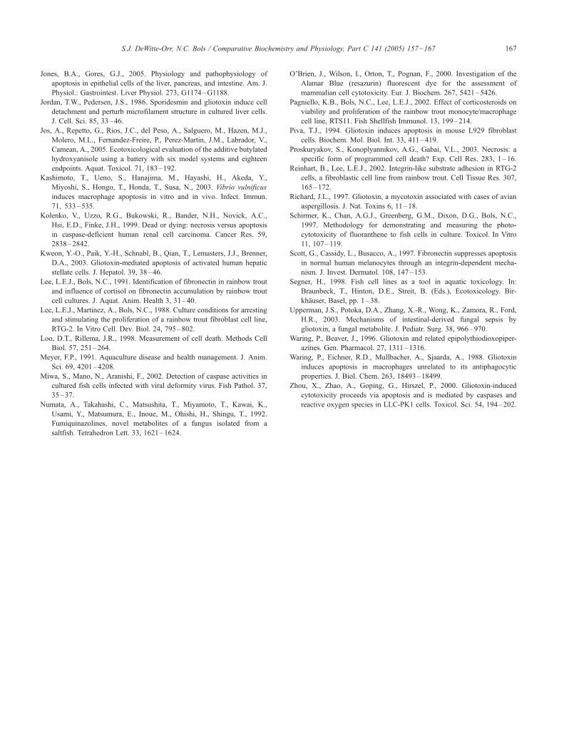