Glial cell line-derived nenrotrophic factor (GDNF) stimulates ret activity
Upload
independentCategory
view
1download
0
GL
S
Ds3
Aflfseaa
alitnsastp
aTtgamo
rgP
K
IeicFonTaa
*3EAtE
Neuroscience 153 (2008) 1332–1343
0d
LIAL-TOXIN-MEDIATED DISRUPTION OF SPINAL CORD
OCOMOTOR NETWORK FUNCTION AND ITS MODULATION BY 5-HTPpgieneM
lpageeA(iYa
oaeenh(Wwvgct1tet
nrefuatnp
. BAUDOUX AND D. PARKER*
epartment of Zoology and Physiology, Development, and Neuro-cience, University of Cambridge, Downing Street, Cambridge, CB2DG, UK
bstract—While it is established that glial cells actively in-uence neuronal and synaptic properties, the functional ef-ects of glial–neuronal interactions are still not well under-tood. To address the role of glia at the network level we havexamined the effects of the specific gliotoxin L-aminoadipiccid on the locomotor network output and cellular and syn-ptic properties in the lamprey spinal cord.
The gliotoxic effect of aminoadipic acid was associated with specific depolarization of glial cells. Aminoadipic acid depo-arized the membrane potential of spinal cord neurons, suggest-ng a functional link between glia and neurons. The depolariza-ion was significantly reduced by glutamate receptor antago-ists in adults, but by gap junction blockers in larvae,uggesting a developmental difference in glial–neuronal inter-ctions. Aminoadipic acid also reduced the amplitude of mono-ynaptic excitatory postsynaptic potentials (EPSPs), an effecthat was not associated with changes in the presynaptic releaserobability or postsynaptic response to glutamate.
These cellular and synaptic effects of aminoadipic acid weressociated with disruption of the locomotor network output.his could not be accounted for by changes in glutamate up-
ake or potassium buffering by glia, suggesting a direct role forlia in the network. Interestingly, we found that the aminoadipiccid– evoked disruption of network activity and reduction ofonosynaptic EPSP amplitudes did not occur in the presencef the endogenous spinal modulator 5-HT.
These results thus provide evidence for an active functionalole for glial cells in spinal cord locomotor networks, and sug-est a potential glial modulatory effect of 5-HT. © 2008 IBRO.ublished by Elsevier Ltd. All rights reserved.
ey words: glial, lamprey, modulation, spinal cord, CPG.
t is now recognized that glial cells have active functionalffects that go beyond their traditionally assumed support-
ng roles (i.e. glutamate uptake and the buffering of extra-ellular potassium; Parpura et al., 1994; Haydon, 2001;ellin et al., 2006). Glial cells have receptors to a numberf neurotransmitters, which could allow them to monitorervous system activity (Haydon, 2001; Fellin et al., 2006).hey can in turn release transmitters (“gliotransmitters”)nd could thus influence nervous system function (Auldnd Robitaille, 2003; Fellin et al., 2006; Newman, 2003;
Corresponding author. Tel: �44-01223-339836; fax: �44-01223-30934.-mail address: [email protected] (D. Parker).bbreviations: ANOVA, analysis of variance; CV, coefficient of varia-
Tion; EPSP, excitatory postsynaptic potential; mEPSP, miniaturePSP; PP, paired pulse; TTX, tetrodotoxin.
306-4522/08$32.00�0.00 © 2008 IBRO. Published by Elsevier Ltd. All rights reseroi:10.1016/j.neuroscience.2008.03.034
1332
arpura et al., 1994). However, it is still not clear howresynaptic and postsynaptic neurons and their associatedlial elements (the “tripartite” synapse; Araque et al., 1999)
nteract to influence synaptic function, let alone how glialffects on cellular and synaptic properties influence globaletwork outputs and behaviors (for reviews see Angulot al., 2004; Haydon and Carmignoto, 2006; Fiacco andcCarthy, 2006).
Here, we have examined the role of glial cells in theamprey spinal cord locomotor network using L-aminoadi-ic acid. L-Aminoadipic acid is a commonly used potentnd specific gliotoxin that is specifically taken up by thelial sodium-dependent glutamate port (Alvarez-Maubecint al., 2000; Brown and Kretzschmar, 1998; Pannicket al., 1994; Pow, 2001; Wakakura and Yamamoto, 1992).s a result it depolarizes the glial cell membrane potential
Pannicke et al., 1994; Brown and Kretzschmar, 1998),ncreases intracellular calcium levels (Wakakura andamamoto 1992), and ultimately causes astrocyte swellingnd death (Huck et al., 1984; Takada and Hattori, 1986).
The lamprey locomotor network generates a rhythmicutput that drives muscle activity during swimming. It offers useful model system in which to investigate the networkffects of glial cells as it allows cellular and synaptic prop-rties to be examined in the context of an intact functionaletwork output. Where the network effects of glial cellsave been examined previously it has been in tissue slicesAlvarez-Maubecin et al., 2000; Hülsmann et al., 2000).
hile this has provided information on the potential net-ork role of glial cells, slice-induced changes in glial cellolume, in the spatial relationship between neurons andlia, and in the glial involvement in glutamate breakdownomplicate the interpretation of glial effects in these sys-ems (McMahon and Nicholls, 1990; Gundersen et al.,995). In addition to the advantages of an intact network,he use of the lamprey allows the role of glial cells to bexamined in spinal cord locomotor networks, somethinghat has not previously been investigated.
We show that aminoadipic acid disrupted locomotoretwork activity, depolarized spinal cord neurons, andeduced the amplitude of monosynaptic glutamatergicxcitatory postsynaptic potentials (EPSPs). These ef-
ects could not be accounted for by changes in glutamateptake or potassium buffering (Brodin et al., 1988; Wallén etl., 1984), suggesting an active functional role for glial cells in
he network. Interestingly, we have found that the effects onetwork activity and synaptic transmission did not occur in theresence of the endogenous spinal cord modulator 5-HT.
he results thus suggest an active role for glial cells in theved.

sn
AifiiiLsewP2ciaMae
ufirteiotPclwisted2rctittt(aiccmpm
b�mlpdwrtas
lmbto((pa
(van1c1d
dabweuowv
E
Tn(nrnaomtcsPenvmPwdTmHaerotee
S. Baudoux and D. Parker / Neuroscience 153 (2008) 1332–1343 1333
pinal cord locomotor network, and that 5-HT can protect theetwork against gliotoxic effects.
EXPERIMENTAL PROCEDURES
dult male and female lampreys (Lamptera fluviatilis) were caughtn the wild at the start of their upstream migration by commercialshermen in the UK (Baitbox, Grimsby, UK). Larval and transform-ng Petromyzon marinus were obtained from commercial suppliersn the USA (Lamprey Services (Ludington, MO, USA) and Acmeamprey (Harrison, ME, USA), respectively). The use of differentpecies reflected the seasonal availability of animals. We havexamined these species in several studies, and in all cases effectsere the same (Parker et al., 1998; Parker and Gilbey, 2007;arker and Grillner, 1999). Animals were anesthetized with MS-22 and the spinal cord and notochord were removed. The spinalord was isolated from the notochord and placed ventral side upn a Sylgard-lined chamber where it was superfused with oxygen-ted Ringer containing (in mM): 138 NaCl, 2.1 KCl, 1.8 CaCl2, 1.2gCl2, 4 glucose, 2 Hepes, 0.5 L-glutamine. The pH of the Ringernd of drug solutions was adjusted to 7.4 with 1 M NaOH. Thexperimental chamber was kept at a temperature of 10–12 °C.
Intracellular recordings were made from motor neurons ornidentified spinal cord neurons using thin-walled micropipetteslled with 3 M potassium acetate and 0.1 M potassium chloride toeduce tip potentials. As the aim of this study was to investigatehe potential for changes below lesion sites rather than examiningffects in different classes of neurons, unidentified cells were
ncluded in the analysis. These cells had stable resting potentialsf less than �50 mV over relatively long recording times, and werehus likely to be larger cells in the spinal cord (Buchanan, 2001;arker, 2003; Parker and Gilbey, 2007). They were not the largerossing caudally projecting interneurons or ipsilaterally projectingateral interneurons, as spiking in these cells was not associatedith extracellular spikes in extracellular electrodes placed on the
psilateral or contralateral region of the spinal cord �5 to 10egments caudal to the recorded cell body. This suggested thathey were probably motor neurons. We saw no difference inffects in unidentified cells or identified motor neurons, and thusata from these cells were grouped in the Results. An AxoclampA amplifier (Axon Instruments, CA, USA) was used for voltageecording and current injection. Unless stated otherwise, whenellular or synaptic effects were examined the membrane potential ofhe postsynaptic cell was kept at a constant level of �70 mV bynjecting depolarizing or hyperpolarizing current using single elec-rode discontinuous current clamp. This was necessary to ensurehat the cellular and synaptic effects of aminoadipic acid were not dueo differences in the membrane potential of the postsynaptic neuronwhile this is an important parameter in itself, and one that we havelso studied here, in order to identify the range of aminoadipic effects
n some experiments we needed to keep the membrane potentialonstant). The output of the sample-and-hold amplifier in the DCCircuit was monitored to ensure complete settling before voltageeasurement. Data were acquired, stored, and analyzed on com-uter using an analog-to-digital interface (Digidata 1200, Axon Instru-ents) and Axon Instruments software (pClamp 9).
The input resistance of postsynaptic neurons was measuredy injecting depolarizing and hyperpolarizing current pulses from2.5 nA to �2.5 nA in 0.5 nA steps. The excitability was deter-ined from the number of spikes evoked in response to the depo-
arizing current pulses. Synaptic inputs were examined by makingaired recordings from Muller reticulospinal axons in the ventrome-ial column and postsynaptic spinal neurons. Monosynaptic EPSPsere evoked by injecting 1 ms depolarizing current pulses into the
eticulospinal axons. Monosynaptic connections were identified byheir reliability and constant latency following presynaptic stimulationt 20 Hz (Berry and Pentreath, 1976). Reticulospinal axons were
timulated at 0.1 Hz to examine aminoadipic acid effects on single gow frequency-evoked EPSPs, or at 20 Hz for 1 s or at 20 Hz for 250s every 500 ms to examine EPSPs during spike trains or spikeursts. The plasticity during spike trains was examined by calculatinghe paired pulse (PP) plasticity (EPSP2/EPSP1), and the plasticityver the 2nd to 5th spikes (Train2–5/EPSP1), the 6th to 10th spikesTrain6–10/EPSP1), and the 11th to 20th spikes in the trainTrain11–20/EPSP1). Effects during spike bursts were examined bylotting the average of groups of 50 EPSPs (i.e. 10 bursts) in controlnd in aminoadipic acid.
Locomotor network activity was examined by applying NMDA50 �M), and was monitored by recording extracellularly fromentral roots using glass suction electrodes. Because networkctivity can vary, aminoadipic acid was only applied after theetwork activity had been stable for at least 1 h (Parker et al.,998). Single concentrations of NMDA can give variable frequen-ies of network activity in different experiments (Parker et al.,998). However, the effects of aminoadipic acid did not differ withifferent frequencies of network activity (see Fig. 1).
Aminoadipic acid (1 mM) was applied for variable times inifferent experiments (see text for details). The glutamate receptorntagonists DNQX (20 �M) and AP5 (100 �M) or the gap junctionlocker carbenoxolone (100 �M; Alvarez-Maubecin et al., 2000)as applied in the presence of aminoadipic acid after it hadvoked its plateau effects. Statistical significance was examinedsing two-tailed paired or independent t-tests, or one-way analysisf variance (ANOVA). When an ANOVA was used a Tukey testas used for post hoc analysis of differences between groups. Allalues given refer to mean�S.E.M.
RESULTS
ffects of aminoadipic acid on network activity
he effects of aminoadipic acid were studied on locomotoretwork activity evoked by NMDA (50 �M) in maturen�14) and juvenile adults (“transformers”; n�17). We didot examine larval network activity as it was usually lessegular than adult activity (see Cohen et al., 1990). Ami-oadipic acid (1 mM for 30min) abolished rhythmic networkctivity and bursting in some cases (n�5 of 14 adults, n�2f 12 transformers; Fig. 1A). This prevented any measure-ent of individual ventral root bursts, and thus quantifica-
ion of the effects of aminoadipic acid. Where burstingould still be measured (Fig. 1B), the frequency was non-ignificantly reduced by aminoadipic acid in adults (n�9,�0.05) and transformers (n�10, P�0.05; Fig. 1D). How-ver, in both stages there was significant disruption ofetwork activity, shown by the increase in the coefficient ofariation (CV, standard deviation of the cycle duration/ean cycle duration; n�9 adults, n�10 transformers,�0.05; Fig. 1E). While the effects of aminoadipic acidere routinely examined after application for 30 min, theisruption could develop within 10 min (data not shown).he increase in the CV recovered after washing for 30in–3 h in seven of nine adults and in 8 of 10 transformers.owever, with longer (�1 h) applications of aminoadipiccid in transformers (longer application times were notxamined in adults) the effect did not recover and thehythm instead continued to deteriorate on washing (n�4f five experiments; Fig. 1C). These effects do not matchhose caused by blocking glutamate uptake or increasingxtracellular potassium levels (Brodin et al., 1988; Wallént al., 1984). This suggests that these associated roles of
lial cells do not cause the effects shown here, and thus
tn
tfgwmra
rw
mtv2aa
Fat(oC
S. Baudoux and D. Parker / Neuroscience 153 (2008) 1332–13431334
hat glial cells may have an active functional role in theetwork.
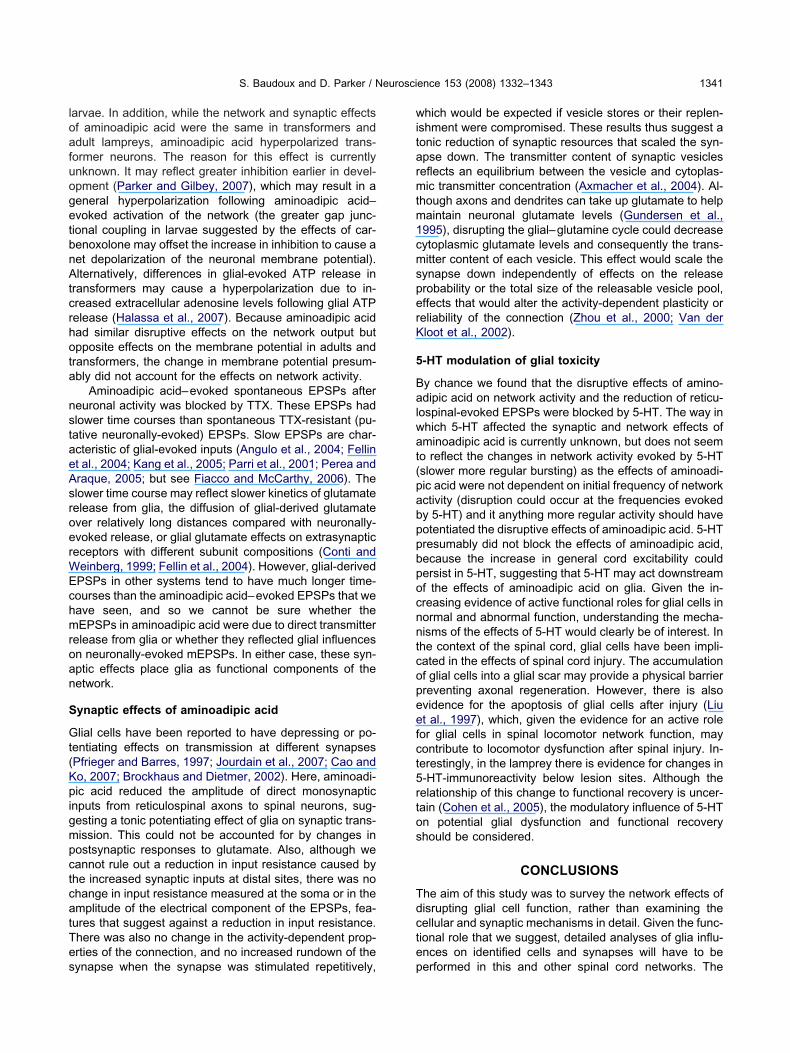
There was a significant negative correlation betweenhe increase in the CV and the initial CV value in trans-ormers (r2�0.82, P�0.05; Fig. 1F). This suggested that areater disruptive effect of aminoadipic acid occurredhen the initial CV was low (i.e. the network activity wasore regular). However, in adults there was no significant
elationship between the initial CV and the CV in amino-
Control
30minadipicacid
30minwash
1s
Control
30minadipicacid
2hwash
AdipicAcid
AdipicAcid
Control
Control
Wash
AdultTransformer
0.00
0.25
0.50
0.75
1.00
1.25
1.50
1.75
Frequency(Hz)
0.0 0.1 0.2 0.30.0
2.5
5.0
7.5
10.0
Initial CV
IncreaseinCV
Transformer
A B
D
F
ig. 1. Effects of aminoadipic acid (1 mM) on NMDA (50 �M) -evokedbolished by aminoadipic acid. (B) Example of activity where aminoahe effect recovered on washout. (C) Example of activity disrupted by aD) Bar graph showing the overall lack of effect of aminoadipic acid on tf the CV (i.e. disruption of network activity) by aminoadipic acid. (F, GV by aminoadipic acid in transformers and adults, respectively.
dipic acid (r2�0.3, P�0.05; Fig. 1G). This reflected the n
educed disruptive effect of aminoadipic acid in adultshen the initial CV was low.
One of the major roles for glia is the uptake of gluta-ate and its subsequent conversion into glutamine. Glu-
amine is then taken up into synaptic terminals and con-erted to glutamate (Bröer and Brookes, 2001; Pow,001). By disrupting the glial glutamate–glutamine cycle,minoadipic acid could reduce neuronal glutamate levels,nd thus affect glutamatergic synaptic transmission in the
Control
Control
Adipic Acid
Adipic Acid
Wash
Wash
0.0
0.2
0.4
0.6
0.8
0.0 0.1 0.2 0.3 0.4 0.50
1
2
3
4
5
Initial CV
IncreaseinCV
Adult
C
**
Control
1.5hadipicacid
2.5hwash
2s1s
or network activity. (A) Example of an experiment where bursting wasdisrupted network activity without abolishing bursting. In both casesting application of aminoadipic acid that did not recover after washing.ncy of network activity. (E) Bar graph showing the significant increaseshowing the relationship of the burst frequency to the increase in the
Wash
CV
0.4
E
G
locomotdipic acidlonger-lashe freque) Graphs
etwork (although hippocampal neurons can maintain syn-

atpwohaga0ssoamtsnwspmartmgtirng
C
TspbfnnP6negstneptst
c(rcswttsponnf0rdc2
hlmN(opDad3ttpn
Fa(a
S. Baudoux and D. Parker / Neuroscience 153 (2008) 1332–1343 1335
ptic transmission independently of a glial-derived glu-amine supply; Kam and Nicoll 2007). This effect couldotentially be avoided by adding exogenous glutamine,hich can overcome the effects of disrupting glial functionn respiratory network activity (Hülsmann et al., 2000). Weave examined whether the network disruption was due toreduction in glutamine levels by applying exogenous
lutamine (1–5 mM) to the Ringer in transformers (n�5)nd adults (n�4). Lampetra Ringer has routinely had.5 mM glutamine added, because EM studies havehown that it preserves glial structure (Shupliakov, per-onal communication). This level of glutamine was obvi-usly not sufficient to prevent disruption of the networkctivity by aminoadipic acid, and if disruption of the gluta-ate–glutamine cycle did contribute to the disruption of
he network output then the additional glutamine shouldtill have had a beneficial effect. Glutamine on its own didot significantly affect the frequency or regularity of net-ork activity (Fig. 2A). Neither 1 mM nor 5 mM glutamineignificantly reduced the CV when applied after aminoadi-ic acid (P�0.05, n�9; Fig. 2A, B). Given that the trans-itter pool in excitatory network interneurons is on aver-ge a few hundred vesicles and that each action potentialeleases approximately 10 vesicles (see Parker, 2003), theotal transmitter pool would be released and replenishedany times during the course of the 1 h application oflutamine. There would thus be ample opportunity for glu-amine to affect the aminoadipic acid–evoked disruption ift were due to a reduction of the glutamine supply. Theseesults thus suggest that the effects of aminoadipic acid onetwork activity were not due to the disruption of neuronallutamate levels.
ig. 2. Glutamine did not reverse the disruptive effects of aminoadipiccid on network activity. (A) Graph showing the effects of glutamine
C1–5 mM) on control network activity, and activity after aminoadipiccid–evoked disruption.
ellular and synaptic effects of aminoadipic acid
he disruption of network activity by aminoadipic acidhould be associated with changes in cellular and synapticroperties in the spinal cord. We examined these changesy recording from spinal cord neurons in larval, trans-ormer, and adult animals. Aminoadipic acid (1 mM) sig-ificantly depolarized the membrane potential of larvaleurons (from �74�2.3 mV to �69�3 mV, n�23,�0.05; Fig. 3A) and adult neurons (from 68.6�0.9 mV to4.1�1.4 mV, P�0.05, n�27; Fig. 3A, Bi). Because ami-oadipic acid specifically depolarizes glial cells (Pannicket al., 1994), this result suggested an interaction betweenlia and neurons. However, aminoadipic acid (1 mM) non-ignificantly hyperpolarized the membrane potential ofransformer neurons (from �73�1.6 mV to �74�1.6 mV;�17, P�0.05; Fig. 3A), suggesting developmental differ-nces in its effects. These changes in resting membraneotential were not associated with significant changes inhe input resistance or excitability in any developmentaltage when effects were measured from a standard poten-ial of �70 mV (data not shown).
In all developmental stages, aminoadipic acid in-reased the frequency of spontaneous EPSPs and IPSPsn�16 of 23 larval neurons, n�15 of 17 transformer neu-ons, n�24 of 27 adult neurons; Fig. 3Bii), and in someases could cause neurons to spike (n�2; data nothown). While spiking was relatively rare in the cells thate recorded from, it must have been more widespread as
here was a consistent (�90% of experiments) increase inhe extracellular activity recorded from the surface of thepinal cord in all developmental stages (Fig. 3Bi). In ap-roximately 20% of neurons aminoadipic acid also causedscillations of the membrane potential (n�5 of 23 larvaleurons; n�4 of 17 transformer neurons; n�4 of 27 adulteurons; Fig. 3Ci, Cii, Di). The oscillations had a meanrequency of 0.18�0.5 Hz (n�5), 0.19�0.9 Hz (n�4), and.22�0.5 Hz (n�6) in larval, transformer, and adult neu-ons, respectively, and were thus faster than the calcium-ependent oscillations that have been reported in glialells (�0.003 Hz; Parri et al., 2001; but see Nett et al.,002).
The depolarization of larval and adult neurons couldave reflected changes caused by an increase in extracel-
ular glutamate levels due to the disruption of glial gluta-ate uptake. This was examined by applying the non-MDA and NMDA glutamate receptor antagonists DNQX
20 �M) and AP5 (100 �M) before or after the applicationf aminoadipic acid. The aminoadipic acid–mediated de-olarization in larvae was not significantly affected whenNQX and AP5 were added 5–10 min after aminoadipiccid application (there was instead a further non-significantepolarization in the glutamate receptor antagonists; Fig.A), suggesting against the involvement of increased ex-racellular glutamate levels or glial glutamate release inhis effect. However, in adults the aminoadipic-evoked de-olarization (Fig. 3Bi) and oscillations (Fig. 3Ci) were sig-ificantly reduced by DNQX and AP5 (P�0.05; Fig. 3A, Bi,
i), and aminoadipic acid evoked a smaller depolarization
wacatfwtmamie
aM
abfaece(amooial
Farac(sidtot
S. Baudoux and D. Parker / Neuroscience 153 (2008) 1332–13431336
hen it was applied in the presence of glutamate receptorntagonists (Fig. 3A). These results suggested a signifi-ant contribution of increased glutamate levels only to thedult effects of aminoadipic acid, highlighting another po-ential developmental difference in glial effects. In trans-ormers, the aminoadipic acid–evoked hyperpolarizationas reduced by glutamate receptor antagonists, shown by
he depolarization of the membrane potential when gluta-ate receptor antagonists were applied in the presence ofminoadipic acid (Fig. 3A). This suggested the involve-ent of an increase in glutamate levels to the hyperpolar-
zation (Fig. 3A), which could reflect the glutamatergicxcitation of presynaptic inhibitory neurons.
In addition to their ability to release glutamate, glia canlso make electrical connections onto neurons (Alvarez-
ig. 3. The effects of aminoadipic acid (1 mM) on cellular properties.ntagonists on the resting membrane potential of larval, transformer,eceptor antagonists being applied after (open bar) or before (light graycid on the resting membrane potential of an adult spinal cord neuronord. Note that the depolarization was markedly reduced but not abolisBii) Traces showing the increase in spontaneous EPSPs and IPSPshowing the membrane potential oscillations evoked by aminoadipic an Ci to show the oscillations. (Di) Example of aminoadipic acid-evokeepolarization and the oscillations to the gap junction blocker carbenohe effects of longer applications (�10 min) of carbenoxolone (100 �Mf 10 EPSPs in control and after aminoadipic acid. Note that carbenoxhe EPSP.
aubecin et al., 2000). This would allow the specific m
minoadipic acid– evoked depolarization of glia cells toe passively transferred to neurons, and may account
or the glutamate-independent effects of aminoadipiccid on the resting membrane potential in larvae. Wexamined the role of gap junctions using carbenoxolone, aommonly used gap junction blocker (Alvarez-Maubecint al., 2000; Rekling et al., 2000). In larvae, carbenoxolone100 �M for 5 min) significantly reduced the aminoadipiccid–evoked depolarization from 6.5�1.4 mV to 3.7�2.1V (n�6, P�0.05; Fig. 3Di): where membrane potentialscillations occurred they were also abolished by carben-xolone (n�2 of 2; Fig. 3Di). Where oscillations occurred
n adults (n�2 of 2) and transformers (n�1 of 1) they werelso abolished by carbenoxolone. However, in contrast to
arvae carbenoxolone non-significantly hyperpolarized the
summarizing the effects of aminoadipic acid and glutamate receptort neurons. Note the order of application of the drugs, with glutamateoadipic acid application. (Bi) Trace showing the effects of aminoadipiccrease in extracellular activity recorded from the surface of the spinale glutamate receptor antagonists DNQX (10 �M) and AP5 (100 �M).by aminoadipic acid (the traces are from different cells). (Ci) Tracetheir abolition by DNQX and AP5. (Cii) Expanded region of the traceions in a larval neuron, and the sensitivity of the membrane potential00 �M), and DNQX (10 �M) and AP5 (100 �M). (Dii) Traces showingosynaptic reticulospinal-evoked EPSPs. The traces show an averageused a reduction of the electrical (arrow) and chemical component of
(A) Graphand adulbar) aminand the inhed by thevoked
cid, andd oscillatxolone (1) on monolone ca
embrane potential (0.74�0.36 mV in adults (n�5) and

0WtvtaatPtpteaaoja
om
l1letnatipa(fit
FaeTea
S. Baudoux and D. Parker / Neuroscience 153 (2008) 1332–1343 1337
.71�1.6 mV in transformers (n�4); data not shown).hile these results supported an involvement of gap junc-
ions in the effects of aminoadipic acid, particularly in lar-ae, we found that application of carbenoxolone for greaterhan 10–20 min, either on its own or after aminoadipic acidpplication, caused several changes in cellular and syn-ptic properties. These included a significant depolariza-ion of the resting membrane potential (7.2�2.1 mV,�0.05), a marked reduction in spontaneous and evoked
ransmitter release, a reduction of the presynaptic actionotential amplitude (data not shown), and a reduction ofhe electrical and chemical components of reticulospinal-voked EPSPs (Fig. 3Dii). These effects raise caveatsbout the use of this drug to examine gap junction–medi-ted effects, something that has been highlighted previ-usly for carbenoxolone and other commonly used gap
unction blockers (see Rekling et al., 2000). Thus, while thebove analysis showed effects with short-term application
ig. 4. (A) Effects of aminoadipic acid on the membrane potential of and membrane potential oscillations. (B) Graph and traces showingvoked glutamate (1 mM) in TTX. (C) Traces showing spontaneous mTX. (D) Comparison of the rise time and half-width of non-aminoa
xamples of non–aminoadipic acid– and aminoadipic acid–evoked mEPSPsmplitudes have been normalized to show the differences in the mEPSP timef carbenoxolone (5 min), we cannot rule out the involve-ent of non–gap junction–mediated effects.
We examined the effects of aminoadipic acid further inarvae by applying it in the presence of tetrodotoxin (TTX;.5 �M) to block action potential–evoked transmitter re-
ease. Under these conditions aminoadipic acid alsovoked a depolarization (n�5 of 7) and membrane poten-ial oscillations (n�2 of 7; Fig. 4A), suggesting that neuro-al activity was not necessary for its effects. Aminoadipiccid did not significantly affect the amplitude of responseso pressure applied glutamate (1 mM for 100 ms), suggest-ng that the responses in TTX were not due to effects onostsynaptic glutamate receptors (Fig. 4B). Aminoadipiccid could also evoke miniature EPSPs (mEPSPs) in TTXn�4 of 7; Fig. 4C). An increase in TTX-resistant mEPSPrequency is traditionally associated with a presynapticncrease in spontaneous vesicle release from synapticerminals, while an increase in amplitude is traditionally
ord neuron in the presence of TTX (1.5 �M). Note the depolarizationf a significant effect of aminoadipic acid on responses to pressure-
voked by aminoadipic acid in a spinal cord neuron in the presence ofand aminoadipic acid–evoked mEPSPs in TTX. The inset shows
spinal cthe lack oEPSPs edipic acid
(the thicker trace shows the aminoadipic acid–evoked EPSP). Thecourse.

aatlmctm
S
Wms(wibmonsaraB
aorualtosoEtand(NieafPnstwtcrnbso
5
TangtcrN(waTeciGfw(eotefttowTe
an1td(Htatw(t(Et(aifttPdE
S. Baudoux and D. Parker / Neuroscience 153 (2008) 1332–13431338
ssociated with postsynaptic effects. However, the amino-dipic acid–evoked mEPSPs had significantly longer riseimes (7�0.3 ms compared with 2.5�0.5 ms; P�0.05),onger half-widths (12.1�0.4 ms compared with 3.9�0.7
s; P�0.05; Fig. 4D), and larger amplitudes (1.1�0.3 mVompared with a maximum amplitude of 0.5 mV; Fig. 4C)han non–aminoadipic acid (putative neuronally) –evokedEPSPs (Fig. 4D).
ynaptic effects
e examined the synaptic effects of aminoadipic acid inore detail by making paired recordings from postsynaptic
pinal cord neurons and descending reticulospinal axonsGundersen et al., 1995). While inputs from these axonsill not influence NMDA-evoked network activity in the
solated spinal cord (this will depend on interneurons thatelong to the locomotor network), they provide a modelonosynaptic connection in which to examine the effectsf glial disruption on synaptic transmission. Although ami-oadipic acid depolarized postsynaptic neurons, it did notignificantly affect the membrane potential of reticulospinalxons (0.1�0.07 mV; Fig. 5A). However, it did significantlyeduce the amplitude of reticulospinal-evoked EPSPs indults (from 2.3�0.3–1.27�0.28, P�0.05, n�23; Fig. 5Bi,ii) and transformers (n�7; data not shown).
The reduction of the EPSP amplitude in aminoadipiccid suggested that glia had a tonic facilitating influencen synaptic efficacy. The PP plasticity (Fig. 5C) andesponses to pressure applied glutamate (Fig. 4B) werenaffected by aminoadipic acid, which suggestedgainst the involvement of changes in presynaptic re-
ease probability or postsynaptic glutamate receptors inhe synaptic modulation. In addition, there was no effectn the postsynaptic input resistance measured at theoma (data not shown), and no change in the amplitudef the electrical component of reticulospinal-evokedPSPs (see Fig. 5Bii), suggesting against a reduction of
he postsynaptic input resistance. The reduction in themplitude of reticulospinal-evoked EPSPs could alter-atively reflect a reduction in the available vesicle poolue to the disruption of the glutamate– glutamine cycleBröer and Brookes, 2001; Pow, 2001; but see Kam andicoll, 2007). We examined this possibility by investigat-
ng the effects of aminoadipic acid on synaptic inputsvoked by stimulating presynaptic reticulospinal axonst 20 Hz for 1 s (n�20), or in bursts of 20 Hz stimulationor 250 ms every 500 ms (n�10; see Experimentalrocedures for details of stimulation and analysis). If theumber of available vesicles was reduced then therehould be a greater rundown of transmission underhese conditions (Pieribone et al., 1995). However, thereas no significant change in the activity-dependent plas-
icity during spike trains, shown by the lack of a signifi-ance difference in the plasticity evoked over differentegions of the spike trains (P�0.05, n�20; Fig. 5D), ando significant rundown of the input over repetitive spikeursts (P�0.05, n�10; Fig. 5E, F). These results thusuggested against a significant reduction in the number
f available synaptic vesicles. a-HT interactions with aminoadipic acid
hese results show that the gliotoxic effects of aminoadipiccid altered cellular, synaptic properties and the locomotoretwork output, providing support for a functional role forlial cells in the locomotor network. In addition to studyinghe effects of glial disruption on the network, during theourse of this work we also found evidence that the dis-uptive effects of aminoadipic acid could be modulated.MDA-evoked network activity can often be irregular
Parker et al., 1998; Zhang and Grillner, 2000). Becausee were finding that aminoadipic acid disrupted networkctivity we wanted to have regular activity to begin with.hus, when network activity was initially very irregular wexamined the effects of aminoadipic acid after the appli-ation of 5-HT, which slows network activity and improves
ts regularity (Harris-Warrick and Cohen, 1985; Zhang andrillner, 2000; Fig. 6A). However, in these experiments we
ound that there was no significant disruption of the net-ork output by aminoadipic acid in either transformers
n�5) or adults (n�4, P�0.05; Fig. 6A). The lack of anffect in 5-HT could not be due to the improved regularityf network activity, and if anything this should have poten-iated the disruption by aminoadipic acid (Fig. 1F, G). Theffect of 5-HT was also not simply due to the lower initialrequency of network activity. While aminoadipic acid failedo disrupt the slower network activity evoked by 5-HT, inhe absence of 5-HT it did disrupt network activity thatccurred at the same slow frequencies (i.e. the disruptionas not frequency-dependent; r2�0.08, P�0.05; Fig. 6B).hese features suggested that 5-HT directly modulated theffects of aminoadipic acid.
We also examined whether 5-HT altered the cellularnd synaptic effects of aminoadipic acid in adults. 5-HT didot affect the increase in general cord activity (n�11 of2), or the increase in spontaneous synaptic inputs, al-hough these were largely IPSPs, possibly due to theepressing effect of 5-HT on glutamatergic transmissionn�7 of 12, data not shown; Parker and Grillner, 1999).owever, an aminoadipic acid–evoked membrane poten-
ial depolarization occurred in only 5 of 12 experiments,nd the mean depolarization (1.25�0.65 mV, P�0.05) andhe proportion of cells in which a depolarization occurredere significantly less than in aminoadipic acid alone
P�0.05 chi square). 5-HT also affected the reduction ofhe monosynaptic EPSP amplitude by aminoadipic acidFig. 5Bi, Bii). In 5-HT, which significantly itself reduced thePSP amplitude (Buchanan and Grillner, 1991; Fig. 6C),
here was no significant synaptic effect of aminoadipic acidn�12, P�0.05; Fig. 6C). The failure of aminoadipic acid toffect the EPSP amplitude could have been related to the
nitial reduction of the EPSP by 5-HT, which occluded anyurther reduction. However, there was no relationship be-ween the reduction of the EPSP and the initial EPSP ampli-ude in either control (r2�0.16, P�0.05) or 5-HT (r2�0.09,�0.05), and in some cases control EPSPs that were re-uced by aminoadipic acid were of the same amplitude asPSPs in 5-HT (Fig. 6D). In addition, 5-HT and aminoadipic
cid individually reduced the EPSP amplitude to the same
lrtaE
Tc
Fpaaahes dipic acid
S. Baudoux and D. Parker / Neuroscience 153 (2008) 1332–1343 1339
evel (Fig. 6C), but while aminoadipic acid did not significantlyeduce the EPSP amplitude when it was applied after 5-HT,here was a reduction of the EPSP amplitude when 5-HT waspplied after aminoadipic acid (Fig. 6C), suggesting that the
ig. 5. Effects of aminoadipic acid on evoked synaptic properties. (A)otential of reticulospinal axons, despite the change in the membranecid application. (Bi) Bar graph showing the reduction of the EPSP ampnd in aminoadipic acid. (C) Bar graph showing the lack of a changeminoadipic acid on the PP plasticity of reticulospinal EPSPs, and of thas been normalized to allow comparison of the activity-dependent effevoked EPSPs evoked by 50 bursts of five action potentials at 20 Hz.timulation. While the amplitude of the EPSPs was reduced by aminoa
PSP had not been reduced to a plateau level. t
DISCUSSION
he results of this study suggest an active role for glialells in the spinal cord locomotor network. While the func-
owing the lack of effect of aminoadipic acid (1 mM) on the membranel of the postsynaptic neuron. The bar shows the onset of aminoadipicaminoadipic acid. (Bii) Inset showing averages of 10 EPSPs in controlP ratio by aminoadipic acid. (D) Graph showing the lack of effect ofty over different parts of 20 Hz spike trains. The initial EPSP amplitude
raph showing the lack of effect of aminoadipic acid on reticulospinal-s showing the effects of aminoadipic acid on EPSPs evoked by burst, there was no effect on their reliability or activity-dependent plasticity.
Traces shpotentialitude byin the P
e plasticicts. (E) G
(F) Trace
ional effects of glial cells on cellular and synaptic proper-

tHtnc
aoprbit1tgtomoii
tbsis
tcnafoaePtomrbo
F(hbnfatE -HT, sugga
S. Baudoux and D. Parker / Neuroscience 153 (2008) 1332–13431340
ies are increasingly being recognized (Angulo et al., 2004;aydon, 2001; Fellin et al., 2006), their role in intact func-
ioning networks has received little attention, and glia haveot previously been considered as potential functionalomponents of spinal cord locomotor networks.
It could be argued that that the effects of aminoadipiccid that we have shown were caused by indirect effectsn network activity due to altered glutamate uptake orotassium buffering, the conventional “housekeeping”oles of glia. Several features make this unlikely. Firstly,locking glutamate uptake with dihydrokainate or increas-
ng extracellular potassium levels did not cause the disrup-ion shown here (see supplementary material; Brodin et al.,988; Wallén et al., 1984). Secondly, the addition of glu-
amine, which can overcome some of the effects of alteredlutamate uptake (Hülsmann et al., 2000), did not reversehe aminoadipic acid–evoked disruption of the networkutput. Finally, aminoadipic acid did not depolarize theembrane potential of reticulospinal axons. As these ax-ns have glutamate receptors (Cochilla and Alford, 1999),
f the effects of aminoadipic acid were simply caused by an
ig. 6. The protective modulation by 5-HT of aminoadipic acid-evoked1 mM) on the frequency and regularity of network activity. 5-HT signifiad no effect. Note that in the presence of 5-HT aminoadipic acid wasetween the frequency of network activity and the effects of aminoadietwork activity at the low frequencies evoked by 5-HT, suggesting tharequency. (C) Graph showing the effects of aminoadipic acid and 5-HTcid failed to affect the EPSP amplitude in 5-HT, but that 5-HT was ablehe relationship of the initial EPSP amplitude to the effects of aminoadPSP amplitude when it was initially as small in control as it was in 5mplitude of the initial EPSP.
ncrease in glutamate (or extracellular potassium) levels d
hen the axonal membrane potential should also haveeen affected. Taken together, these features suggestpecific functional effects of aminoadipic acid rather than
ndirect effects caused by disrupted glutamate or potas-ium uptake.
Disruption of glial function by aminoadipic acid alteredhe resting membrane potential of spinal neurons, in-reased general spinal cord excitability, and increased theumber of spontaneous EPSPs and IPSPs. The effects ofminoadipic acid differed during development, adding glialunction to the growing list of differences in different devel-pmental stages of lamprey (Cohen et al., 1990; Parkernd Gilbey, 2007; Ruiz et al., 2004). Developmental differ-nces in glial effects have received little attention (seearri et al., 2001; Fiacco and McCarthy, 2006). However,
his is an important factor to consider, as glial effects areften studied in tissue from neonatal animals where effectsay differ to mature systems. There was a significant
eduction of the aminoadipic acid–evoked depolarizationy glutamate receptor antagonists in adults but by carben-xolone in larvae. This could suggest a glutamate-depen-
(A) Graph showing the effects of 5-HT (10 �M) and aminoadipic acidduced the frequency of network activity while aminoadipic acid againr able to disrupt network activity. (B) Graph showing the relationship
n control and in 5-HT. Note that aminoadipic acid alone could disruptto disrupt network activity was not simply due to the lower initial burst
mplitude of monosynaptic reticulospinal-evoked EPSPs. Aminoadipice the EPSP amplitude further in aminoadipic acid. (D) Graph showingon its own and in 5-HT. Note that aminoadipic acid could reduce theesting that the failure to reduce the EPSP was not simply due to the
effects.cantly reno longe
pic acid it its failure
on the ato reduc
ipic acid
ent effect in adults but a gap junction–mediated effect in
loafuogetbnAtcrhota
nstaeAsroerWEchmroan
S
Gt(KpigmpctcatTes
witarmtm1cmsperK
5
Balwat(pabppbpocnntcopeefct5rtos
Tdcte
S. Baudoux and D. Parker / Neuroscience 153 (2008) 1332–1343 1341
arvae. In addition, while the network and synaptic effectsf aminoadipic acid were the same in transformers anddult lampreys, aminoadipic acid hyperpolarized trans-ormer neurons. The reason for this effect is currentlynknown. It may reflect greater inhibition earlier in devel-pment (Parker and Gilbey, 2007), which may result in aeneral hyperpolarization following aminoadipic acid–voked activation of the network (the greater gap junc-ional coupling in larvae suggested by the effects of car-enoxolone may offset the increase in inhibition to cause aet depolarization of the neuronal membrane potential).lternatively, differences in glial-evoked ATP release in
ransformers may cause a hyperpolarization due to in-reased extracellular adenosine levels following glial ATPelease (Halassa et al., 2007). Because aminoadipic acidad similar disruptive effects on the network output butpposite effects on the membrane potential in adults andransformers, the change in membrane potential presum-bly did not account for the effects on network activity.
Aminoadipic acid–evoked spontaneous EPSPs aftereuronal activity was blocked by TTX. These EPSPs hadlower time courses than spontaneous TTX-resistant (pu-ative neuronally-evoked) EPSPs. Slow EPSPs are char-cteristic of glial-evoked inputs (Angulo et al., 2004; Fellint al., 2004; Kang et al., 2005; Parri et al., 2001; Perea andraque, 2005; but see Fiacco and McCarthy, 2006). Thelower time course may reflect slower kinetics of glutamateelease from glia, the diffusion of glial-derived glutamatever relatively long distances compared with neuronally-voked release, or glial glutamate effects on extrasynapticeceptors with different subunit compositions (Conti and
einberg, 1999; Fellin et al., 2004). However, glial-derivedPSPs in other systems tend to have much longer time-ourses than the aminoadipic acid–evoked EPSPs that weave seen, and so we cannot be sure whether theEPSPs in aminoadipic acid were due to direct transmitter
elease from glia or whether they reflected glial influencesn neuronally-evoked mEPSPs. In either case, these syn-ptic effects place glia as functional components of theetwork.
ynaptic effects of aminoadipic acid
lial cells have been reported to have depressing or po-entiating effects on transmission at different synapsesPfrieger and Barres, 1997; Jourdain et al., 2007; Cao ando, 2007; Brockhaus and Dietmer, 2002). Here, aminoadi-ic acid reduced the amplitude of direct monosynaptic
nputs from reticulospinal axons to spinal neurons, sug-esting a tonic potentiating effect of glia on synaptic trans-ission. This could not be accounted for by changes inostsynaptic responses to glutamate. Also, although weannot rule out a reduction in input resistance caused byhe increased synaptic inputs at distal sites, there was nohange in input resistance measured at the soma or in themplitude of the electrical component of the EPSPs, fea-ures that suggest against a reduction in input resistance.here was also no change in the activity-dependent prop-rties of the connection, and no increased rundown of the
ynapse when the synapse was stimulated repetitively, phich would be expected if vesicle stores or their replen-shment were compromised. These results thus suggest aonic reduction of synaptic resources that scaled the syn-pse down. The transmitter content of synaptic vesicleseflects an equilibrium between the vesicle and cytoplas-ic transmitter concentration (Axmacher et al., 2004). Al-
hough axons and dendrites can take up glutamate to helpaintain neuronal glutamate levels (Gundersen et al.,995), disrupting the glial–glutamine cycle could decreaseytoplasmic glutamate levels and consequently the trans-itter content of each vesicle. This effect would scale the
ynapse down independently of effects on the releaserobability or the total size of the releasable vesicle pool,ffects that would alter the activity-dependent plasticity oreliability of the connection (Zhou et al., 2000; Van derloot et al., 2002).
-HT modulation of glial toxicity
y chance we found that the disruptive effects of amino-dipic acid on network activity and the reduction of reticu-
ospinal-evoked EPSPs were blocked by 5-HT. The way inhich 5-HT affected the synaptic and network effects ofminoadipic acid is currently unknown, but does not seemo reflect the changes in network activity evoked by 5-HTslower more regular bursting) as the effects of aminoadi-ic acid were not dependent on initial frequency of networkctivity (disruption could occur at the frequencies evokedy 5-HT) and it anything more regular activity should haveotentiated the disruptive effects of aminoadipic acid. 5-HTresumably did not block the effects of aminoadipic acid,ecause the increase in general cord excitability couldersist in 5-HT, suggesting that 5-HT may act downstreamf the effects of aminoadipic acid on glia. Given the in-reasing evidence of active functional roles for glial cells inormal and abnormal function, understanding the mecha-isms of the effects of 5-HT would clearly be of interest. Inhe context of the spinal cord, glial cells have been impli-ated in the effects of spinal cord injury. The accumulationf glial cells into a glial scar may provide a physical barrierreventing axonal regeneration. However, there is alsovidence for the apoptosis of glial cells after injury (Liut al., 1997), which, given the evidence for an active roleor glial cells in spinal locomotor network function, mayontribute to locomotor dysfunction after spinal injury. In-erestingly, in the lamprey there is evidence for changes in-HT-immunoreactivity below lesion sites. Although theelationship of this change to functional recovery is uncer-ain (Cohen et al., 2005), the modulatory influence of 5-HTn potential glial dysfunction and functional recoveryhould be considered.
CONCLUSIONS
he aim of this study was to survey the network effects ofisrupting glial cell function, rather than examining theellular and synaptic mechanisms in detail. Given the func-ional role that we suggest, detailed analyses of glia influ-nces on identified cells and synapses will have to be
erformed in this and other spinal cord networks. The
lwtioMrtpa2(2ecf
AWh
A
A
A
A
A
A
B
B
B
B
B
B
B
C
C
C
C
C
F
F
F
G
H
H
H
H
H
H
J
K
K
L
M
N
N
P
P
S. Baudoux and D. Parker / Neuroscience 153 (2008) 1332–13431342
amprey locomotor network provides a useful system inhich to investigate the involvement of glial–neuronal in-
eractions in the generation of network outputs. Thesenteractions have been implicated in the generation ofscillations in the cortex and hippocampus (Amzica andassimini, 2002; Fellin et al., 2004; Angulo et al., 2004), a
ole supported here by the effects of disrupting glial func-ion on network activity. Glial cells could influence theatterning of network activity by synchronizing neuronalctivity through gap junctions (Alvarez-Maubecin et al.,000) or as a result of glial-derived glutamate releaseAuld and Robitaille, 2003; Fellin et al., 2006; Newman,003; Parpura et al., 1994). Further analyses of theseffects on different components of the locomotor networkould thus help to elucidate the integrative functional ef-ects of glia in neuronal networks.
cknowledgments—This work was supported by grants from theellcome Trust and the Royal Society. We thank Tom Gilbey for
is comments on the manuscript.
REFERENCES
lvarez-Maubecin V, Garcia-Hernandez F, Williams JT, Van Bocks-taele EJ (2000) Functional coupling between neurons and glia.J Neurosci 20:4091–4098.
mzica F, Massimini M (2002) Glial and neuronal interactions duringslow wave and paroxysmal activities in the neocortex. Cereb Cor-tex 12:1101–1113.
ngulo MC, Kozlov AS, Charpak S, Audinat E (2004) Glutamatereleased from glial cells synchronizes neuronal activity in the hip-pocampus. J Neurosci 24:6920–6927.
raque A, Parpura V, Sanzgiri RP, Haydon PG (1999) Tripartite syn-apses: glia, the unacknowledged partner. Trends Neurosci22:208–215.
uld D, Robitaille R (2003) Perisynaptic Schwann cells at the neuro-muscular junction: nerve- and activity-dependent contributions tosynaptic efficacy, plasticity, and reinnervation. Neuroscientist9:1144–1157.
xmacher N, Stemmler M, Engel D, Draguhn A, Ritz R (2004) Trans-mitter metabolism as a mechanism of synaptic plasticity: a model-ing study. J Neurophysiol 91:25–39.
erry M, Pentreath V (1976) Criteria for distinguishing between mono-synaptic and polysynaptic transmission. Brain Res 105:1–20.
rockhaus J, Deitmer JW (2002) Long-lasting modulation of synapticinput to Purkinje neurons by Bergmann glia stimulation in rat brainslices. J Physiol 545:581–593; doi: 10.1113/jphysiol.2002.028423.
rodin L, Tossman V, Ohta Y, Ungerstedt U, Grillner S (1988) Theeffect of an uptake inhibitor (dihydrokainate) on endogenous exci-tatory amino acid levels in the lamprey spinal cord as revealed bymicrodialysis. Brain Res 458:166–169.
röer S, Brookes N (2001) Transfer of glutamine between astrocytesand neurons. J Neurochem 77:705–719.
rown D, Kretzschmar H (1998) The glio-toxic mechanism of alpha-aminoadipic acid on cultured astrocytes. J Neurocytol 27:109–118.
uchanan J (2001) Contributions of identifiable neurons and neuronclasses to lamprey vertebrate neurobiology. Prog Neurobiol63:441–446.
uchanan J, Grillner S (1991) 5-Hydroxytryptamine depresses reticu-lospinal excitatory postsynaptic potentials in motoneurones of thelamprey. Neurosci Lett 112:71–74.
ao G, Ko CP (2007) Schwann cell-derived factors modulate synapticactivities at developing neuromuscular synapses. J Neurosci27:6712–6722.
ochilla A, Alford S (1999) NMDA receptor-mediated control of preysnap-
tic calcium and neurotransmitter release. J Neurosci 19:193–205.ohen A, Dobrov T, Li G, Kiemel T, Baker M (1990) The developmentof the lamprey pattern generator for locomotion. J Neurobiol21:958–969.
ohen A, Abdelnabi M, Guan L, Ottinger M, Chakrabarti L (2005)Changes in distribution of serotonin induced by spinal injury inlarval lampreys: evidence from immunohistochemistry and HPLC.J Neurotrauma 22:172–188.
onti F, Weinberg R (1999) Shaping excitation at glutamatergic syn-apses. Trends Neurosci 22:451–458.
ellin T, Pascual O, Gobbo S, Pozzan T, Haydon PG, Carmignoto G(2004) Neuronal synchrony mediated by astrocytic glutamatethrough activation of extrasynaptic NMDA receptors. Neuron43:729–743.
ellin T, Sul J, D’Ascenzo M, Takano H, Pascual O, Haydon P (2006)Bidirectional astrocyte-neuron communication: the many roles ofglutamate and ATP. Novartis Found Symp 276:208–217.
iacco T, McCarthy KD (2006) Astrocyte calcium elevations: Proper-ties, propagation, and effects on brain signaling. Glia 54:676–690.
undersen V, Shupliakov O, Brodin L, Ottersen O, Storm-Mathisen J(1995) Quantification of excitatory amino acid uptake at intactglutamatergic synapses by immunocytochemistry of exogenousD-aspartate. J Neurosci 15:4417–4428.
alassa MM, Fellin T, Takano H, Dong JH, Haydon PG (2007) Syn-aptic islands defined by the territory of a single astrocyte. J Neu-rosci 27:6473–6477.
arris-Warrick R, Cohen A (1985) Serotonin modulates the centralpattern generator for locomotion in the isolated lamprey spinalcord. J Exp Biol 116:27–46.
aydon P (2001) Glia: listening and talking to the synapse. Nat RevNeurosci 2:185–193.
aydon PG, Carmignoto G (2006) Astrocyte control of synaptic trans-mission and neurovascular coupling. Physiol Rev 86:1009–1031.
uck S, Grass F, Hatten M (1984) Gliotoxic effects of alpha-amino-adipic acid on monolayer cultures of dissociated postnatal mousecerebellum. Neuroscience 12:783–791.
ülsmann S, Oku Y, Zhang W, Richter DW (2000) Metabolic couplingbetween glia and neurons is necessary for maintaining respiratoryactivity in transverse medullary slices of neonatal mouse. EurJ Neurosci 12:856–862.
ourdain P, Bergersen LH, Bhaukaurally K, Bezzi P, Santello M,Domercq M, Matute C, Tonello F, Gundersen V, Volterra A (2007)Glutamate exocytosis from astrocytes controls synaptic strength.Nat Neurosci 10:331–339.
am K, Nicoll R (2007) Excitatory synaptic transmission persists in-dependently of the glutamate glutamine cycle. J Neurosci 27:9192–9200.
ang N, Xu J, Xu Q, Nedergaard M, Kang J (2005) Astrocytic gluta-mate release-induced transient depolarization and epileptiform dis-charges in hippocampal CA1 pyramidal neurons. J Neurophysiol94:4121–4130.
iu X, Xu X, Hu R, Du C, Zhang S, McDonald J, Dong H, Wu Y, FanG, Jacquin M, Hsu C, Choi D (1997) Neuronal and glial apoptosisafter traumatic spinal cord injury. J Neurosci 14:5395–5406.
cMahon H, Nicholls D (1990) Glutamine and aspartate loading ofsynaptosomes: a reevaluation of effects on calcium-dependentexcitatory amino acid release. J Neurochem 54:373–380.
ett WJ, Oloff SH, McCarthy KD (2002) Hippocampal astrocytes insitu exhibit calcium oscillations that occur independent of neuronalactivity. J Neurophysiol 87:528–537.
ewman EA (2003) Glial cell inhibition of neurons by release of ATP.J Neurosci 23:1659–1666.
annicke T, Stabel J, Heinemann U, Reichelt W (1994) alpha-Amino-adipic acid blocks the Na(�)-dependent glutamate transport intoacutely isolated Muller glial cells from guinea pig retina. PflugersArch 429:140–142.
arker D (2003) Activity-dependent feedforward inhibition modulatessynaptic transmission in a spinal locomotor network. J Neurosci
23:11085–11093.
P
P
P
P
P
P
P
P
P
R
R
T
V
W
W
Z
Z
S
S
S. Baudoux and D. Parker / Neuroscience 153 (2008) 1332–1343 1343
arker D, Gilbey T (2007) Developmental differences in neuromodu-lation and synaptic properties in the lamprey spinal cord. Neuro-science 145:142–152.
arker D, Grillner S (1999) Activity-dependent metaplasticity of inhib-itory and excitatory synaptic transmission in the lamprey spinalcord locomotor network. J Neurosci 19:1647–1656.
arker D, Zhang W, Grillner S (1998) Substance P modulates NMDAresponses and causes long-term protein synthesis-dependentmodulation of the lamprey locomotor network. J Neurosci 18:4800–4813.
arpura V, Basarsky TA, Liu F, Jeftinija K, Jeftinija S, Haydon PG(1994) Glutamate-mediated astrocyte-neuron signaling. 369:744 –747.
arri HR, Gould TM, Crunelli V (2001) Spontaneous astrocytic Ca2�
oscillations in situ drive NMDAR-mediated neuronal excitation. NatNeurosci 4:803–812.
erea G, Araque A (2005) Glial calcium signaling and neuron-gliacommunication. Cell Calcium 38:375–382.
frieger FW, Barres BA (1997) Synaptic efficacy enhanced by glialcells in vitro. Science 277:1684–1687.
ieribone V, Shupliakov O, Brodin L, Hilfiker-Rothenfluh S, Czernik A,Greengard G (1995) Distinct pools of synaptic vesicles in neuro-transmitter release. Nature 375:493–497.
ow DV (2001) Visualising the activity of the cystine-glutamate anti-porter in glial cells using antibodies to aminoadipic acid, a selec-tively transported substrate. Glia 34:27–38.
ekling JC, Shao XM, Feldman JL (2000) Electrical coupling andexcitatory synaptic transmission between rhythmogenic respiratory
neurons in the PreBotzinger complex. J Neurosci 20:113RC. tuiz Y, Pombal M, Megias M (2004) Development of GABA-immuno-reactive cells in the spinal cord of the sea lamprey, P. marinus.J Comp Neurol 470:151–163.
akada M, Hattori T (1986) Fine structural changes in the rat brainafter local injections of gliotoxin, alpha-aminoadipic acid. HistolHistopathol 1:271–275.
an der Kloot W, Molgo J, Cameron R, Colasante C (2002) Vesiclesize and transmitter release at the frog neuromuscular junctionwhen quantal acetylcholine content is increased or decreased.J Physiol 541:385–393.
akakura M, Yamamoto N (1992) Rapid increase of intracellular Ca2�
concentration caused by aminoadipic acid enantiomers in retinalMüller cells and neurons in vitro. Doc Ophthalmol 80:385–395.
allén P, Grafe P, Grillner S (1984) Phasic variations of extracellularpotassium during fictive swimming in the lamprey spinal cord invitro. Acta Physiol Scand 120:457–463.
hang W, Grillner S (2000) The spinal 5-HT system contributes to thegeneration of fictive locomotion in lamprey. Brain Res 879:188–192.
hou Q, Petersen C, Nicoll R (2000) Effects of reduced vesicular fillingon synaptic transmission in rat hippocampal neurones. J Physiol525:195–206.
APPENDIX
upplementary data
upplementary data associated with this article can be found, in
he online version, at doi: 10.1016/j.neuroscience.2008.03.034.(Accepted 19 March 2008)(Available online 22 March 2008)
Copyright © 2022 FDOKUMEN