Geographical Differences in Autoantibodies and Anti-infectious Agents Antibodies Among Healthy...
10
Geographical Differences in Autoantibodies and Anti-infectious Agents Antibodies Among Healthy Adults Yinon Shapira & Bat-Sheva Porat Katz & Boris Gilburd & Ori Barzilai & Maya Ram & Miri Blank & Staffan Lindeberg & Johan Frostegård & Juan-Manuel Anaya & Nicola Bizzaro & Luis J. Jara & Jan Damoiseaux & Yehuda Shoenfeld & Nancy Agmon Levin # Springer Science+Business Media, LLC 2011 Abstract Much is known about the geoepidemiology of defined autoimmune diseases (AD); however, there is currently limited data regarding the prevalence of autoanti- bodies among healthy populations of different geographical areas. The aim of this study was to evaluate a large profile of autoantibodies in healthy adults from distinct global regions as well as the prevalence of anti-infectious agents antibodies in those regions. Sera samples from 557 healthy donors were obtained at six centers located in different countries (i.e., Italy, Netherlands, Israel, Mexico, Columbia, Papua New Guinea (Kitavans)). Sera were tested for the presence of antinuclear antibodies (ANA) and autoanti- bodies associated with thrombophilia, vasculitis, and gastrointestinal (GI) disease. Sera samples were also screened for antibodies against infectious agents (i.e., EBV, CMV, HBV, Helicobacter pylori, Treponema pal- lidum, and Toxoplasma gondii). Tests were performed using the BioPlex 2200 or ELISA kits (Bio-Rad Laboratories, USA). We found a significant gradient of ANA positivity among the groups: 45% of Columbians, 38% of Kitavans, Yinon Shapira and Bat-Sheva Porat Katz have equally contributed to this article. Y. Shapira : B. Gilburd : O. Barzilai : M. Ram : M. Blank : Y. Shoenfeld (*) : N. A. Levin The Zabludowicz Center for Autoimmune Diseases, Sheba Medical Center, Tel Hashomer 52621, Israel e-mail: [email protected] B.-S. P. Katz Faculty of Agricultural, Food and Environmental Quality Sciences, The Hebrew University, Rehovot, Israel S. Lindeberg University of Lund, Lund, Sweden J. Frostegård Department of Medicine, Karolinska University Hospital, Stockholm, Sweden J.-M. Anaya Center for Autoimmune Diseases Research, Rosario University Medellin, Medellin, Colombia N. Bizzaro Laboratorio di Patologia Clinica, Ospedale S. Antonio, Tolmezzo, Italy L. J. Jara Hospital de especialidades, Centro Medico La Raza, Colonia, Mexico J. Damoiseaux Laboratory of Clinical Immunology, Maastricht University Medical Center, Maastricht, the Netherlands Y. Shoenfeld : N. A. Levin Department of Medicine ‘B’, Sheba Medical Center, Tel Hashomer 52621, Israel Y. Shoenfeld Incumbent of the Laura Schwarz-Kipp Chair for Research of Autoimmune Diseases, Tel-Aviv University, Tel-Aviv, Israel Clinic Rev Allerg Immunol DOI 10.1007/s12016-010-8241-z
-
Upload
independent -
Category
Documents
-
view
2 -
download
0
Transcript of Geographical Differences in Autoantibodies and Anti-infectious Agents Antibodies Among Healthy...

Geographical Differences in Autoantibodiesand Anti-infectious Agents AntibodiesAmong Healthy Adults
Yinon Shapira & Bat-Sheva Porat Katz & Boris Gilburd & Ori Barzilai & Maya Ram &
Miri Blank & Staffan Lindeberg & Johan Frostegård & Juan-Manuel Anaya &
Nicola Bizzaro & Luis J. Jara & Jan Damoiseaux & Yehuda Shoenfeld &
Nancy Agmon Levin
# Springer Science+Business Media, LLC 2011
Abstract Much is known about the geoepidemiology ofdefined autoimmune diseases (AD); however, there iscurrently limited data regarding the prevalence of autoanti-bodies among healthy populations of different geographicalareas. The aim of this study was to evaluate a large profileof autoantibodies in healthy adults from distinct globalregions as well as the prevalence of anti-infectious agentsantibodies in those regions. Sera samples from 557 healthydonors were obtained at six centers located in differentcountries (i.e., Italy, Netherlands, Israel, Mexico, Columbia,
Papua New Guinea (Kitavans)). Sera were tested for thepresence of antinuclear antibodies (ANA) and autoanti-bodies associated with thrombophilia, vasculitis, andgastrointestinal (GI) disease. Sera samples were alsoscreened for antibodies against infectious agents (i.e.,EBV, CMV, HBV, Helicobacter pylori, Treponema pal-lidum, and Toxoplasma gondii). Tests were performed usingthe BioPlex 2200 or ELISA kits (Bio-Rad Laboratories,USA). We found a significant gradient of ANA positivityamong the groups: 45% of Columbians, 38% of Kitavans,
Yinon Shapira and Bat-Sheva Porat Katz have equally contributed tothis article.
Y. Shapira :B. Gilburd :O. Barzilai :M. Ram :M. Blank :Y. Shoenfeld (*) :N. A. LevinThe Zabludowicz Center for Autoimmune Diseases,Sheba Medical Center,Tel Hashomer 52621, Israele-mail: [email protected]
B.-S. P. KatzFaculty of Agricultural, Food and EnvironmentalQuality Sciences, The Hebrew University,Rehovot, Israel
S. LindebergUniversity of Lund,Lund, Sweden
J. FrostegårdDepartment of Medicine, Karolinska University Hospital,Stockholm, Sweden
J.-M. AnayaCenter for Autoimmune Diseases Research,Rosario University Medellin,Medellin, Colombia
N. BizzaroLaboratorio di Patologia Clinica,Ospedale S. Antonio,Tolmezzo, Italy
L. J. JaraHospital de especialidades, Centro Medico La Raza,Colonia, Mexico
J. DamoiseauxLaboratory of Clinical Immunology,Maastricht University Medical Center,Maastricht, the Netherlands
Y. Shoenfeld :N. A. LevinDepartment of Medicine ‘B’, Sheba Medical Center,Tel Hashomer 52621, Israel
Y. ShoenfeldIncumbent of the Laura Schwarz-Kipp Chair for Researchof Autoimmune Diseases, Tel-Aviv University,Tel-Aviv, Israel
Clinic Rev Allerg ImmunolDOI 10.1007/s12016-010-8241-z

26% of Mexicans, 12% of Italians, 12% of Dutch, and 11%of Israelis were ANA positive. Geographical differenceswere also observed regarding the prevalence of specificautoantibodies, namely ANA: anti-dsDNA, chromatin,SmRNP, Ro/SSA, La/SSB, Scl70; GI associated: antiglia-din; and thrombophilia-associated: anti-β2GP1 and pro-thrombin. Additionally, significant differences wereobserved regarding serological markers of all infectiousagents screened. The observed variance between healthyethno-geographical distinct populations in prevalence ofautoantibodies may represent different genetic or environ-mental (e.g., prior exposure to infection) influences. Thusmay illuminate possible causes of geoepidemiologicaldifferences in AD.
Keywords Autoimmunity . Geoepidemiology . Ethnicity .
Antinuclear antibodies . Antigliadin . Antiphospholipidantibodies . Infectious agents
Introduction
Autoantibodies and Autoimmunity
The detection of serum autoantibodies has had a central rolein the diagnosis and classification of autoimmune diseases(AD) for over 50 years. Some autoantibodies also have aprognostic significance or are used as markers for diseaseactivity. In recent years, it is becoming clear that asymp-tomatic individuals may carry autoantibodies for manyyears prior to clinical manifestation of an established AD(Table 1), and that serological detection of these antibodieshas a strong predictive value [1–4]
For instance, in systemic lupus erythematosus (SLE) theappearance of antibodies to RNP, Sm, dsDNA, cardiolipin,Ro (SSA), and La (SSB) can precede clinical diagnosis by7–10 years with a positive predictive value (PPV) of 94–100% [5]. In rheumatoid arthritis (RA), antibodies forrheumatoid factor or anticyclic-citrullinated-peptide (anti-CCP) were detected in serum samples obtained a median of4.5 years before disease onset, with a PPVof 100% [6]. Thepresence of autoantibodies is also used for the prediction offuture type 1 diabetes mellitus (T1D) [7, 8].
The predictive capacity of AD made possible byserological detection of autoantibodies in the preclinicalstage may have a crucial role in disease prevention.Several lines of evidence (e.g., in SLE, T1D, and RA)suggest that intervention in the preclinical period maymodify future AD risk [9–11]. Moreover, prediction isespecially important when the disease progression can beprevented or postponed by avoiding environmentalfactors, among them infectious agents, which may triggeror worsen autoimmunity [1, 12–14].
Geoepidemiology and Autoimmunity
Geoepidemiology of AD portrays the burden of theseillnesses across various regions and ethnic populations, andyields important clues regarding their genetic and triggeringenvironmental mechanisms. Uneven global distribution ofcertain AD, on the one hand suggests that population geneticshave an important role in disease development. On the otherhand, genetic factors cannot completely account for thephenomenon, and it is only by combining evidence ofenvironmental factors that the geoepidemiological picturecan be elucidated [15, 16]. The recent Congress ofAutoimmunity in Slovenia highlighted environmental fac-tors in geoepidemiology and the reader is directed to thepublications that emphasize this issue [16–57].
Table 1 Predictive autoantibodies in autoimmune diseases [2]
Disease Autoantibodies
Type 1 diabetes mellitus anti-GAD IA-2
IAA
RA RF (anti-IgG)
Anti-CCP
SLE Anti-PL
Anti-Ro
Anti-La
Anti-dsDNA
Anti-Sm
Antinuclear ribonucleoprotein
Anti-HS
Antinucleosome
Antihistone
Antiribosomal P protein
Sjogren’s Syndrome Anti-SSA(Ro)
Anti-SSB (La)
APS Anticardiolipin
Lupus anticoagulant
PBC Anti-gp120
Anti-PDC
AIH ANA
Crohn’s disease ASCA
UC pANCA
Addison’s disease ACA (anti-21-hydroxylase)
AITD Anti-TG
Anti-TPO
Pemphigus Antidesmoglein 1
GAD glutamic acid decarboxylase, IA-2 protein tyrosine phosphatase-like molecule, IAA insulin autoantibodies, RF rheumatoid factor, CCPcyclic citrullinated peptide, PL phospholipids, HS heparan sulphate,PDC pyruvate dehydrogenase complex, ANA antinuclear antibodies,ASCA anti-Saccharomyces cerevisiae antibodies, pANCA perinuclearantineutrophil cytoplasmic antibodies, ACA adrenal cortex cellsantibodies, TG thyroglobulin, TPO thyroid peroxidase
Clinic Rev Allerg Immunol
Unlike the case in several defined AD, data is scarceregarding ethno-geographic differences in prevalence ofautoantibodies among healthy individuals, currently limit-ing our understanding of their predictive utility acrossdifferent populations (e.g., differential specifity). Moreover,by investigating the prevalence of a variety of autoanti-bodies and concomitantly infection exposure, in distincthealthy populations, one may reveal unique ethno-geographical interactions. The aim of the present studywas thus to investigate the prevalence of a variety ofautoantibodies as well as the frequency of anti-infectiousantibodies in six different healthy populations.
Materials and Methods
The present study is observational and of comparativecross-sectional design.
Subjects The sera of 557 healthy volunteers (age and sexmatched between groups) were collected in six centerslocated in different countries. The healthy subject groupscomprise of: 100 subjects from Italy, 98 from Israel, 50from Mexico, 49 from the Netherlands, 140 from Colum-bia, and 120 from the indigenous population of the island ofKitava in Papua New Guinea (PNG).
Autoantibodies Profile A large profile belonging to fourgroups of autoantibodies has been tested in the sera:
Antinuclear antibodies (ANA IgG): Anti-dsDNA, Sm,chromatin, ribosomal-P, RNP, SmRNP, Ro /SSA, La/SSB, centromer, Scl-70, and Jo-1.
Of note, subjects exhibiting serological positivity toat least one antigen of the entire ANA analytes panelabove were considered to be “ANA positive”. Subjectsexhibiting serological positivity to at least one of theextractable nuclear antigens group (ENA: anti-Sm,RNP, SmRNP, SSA, SSB, Scl-70, and Jo-1) wereconsidered “ENA positive”.Vasculitis associated (IgG): Antiglomerular basementmembrane, proteinase 3, and myeloperoxidase.Gastrointestinal associated (IgG, IgA): Anti-Saccharo-myces cerevisiae, gliadin, and tissue transglutaminase.Thrombophilia associated (IgG and IgM): Anticardioli-pin (CL), β2 glycoprotein 1 (β2GP1), complex of both,phosphatydilserine-β2GP1 complex, phosphatydiletha-nolamine, prothrombin (PT), and phosphatydilserine-prothrombin complex.
Infectious Agents Exposure to various infectious agents hasbeen assessed by testing sera for presence of IgG antibodiesdirected to viral infection: hepatitis B (IgG and IgM),
cytomegalovirus (CMV), Epstein–Barr virus (EBV); bacte-rial infection: Helicobacter pylori and Treponema pallidum;and parasitic infection: Toxoplasma gondii.
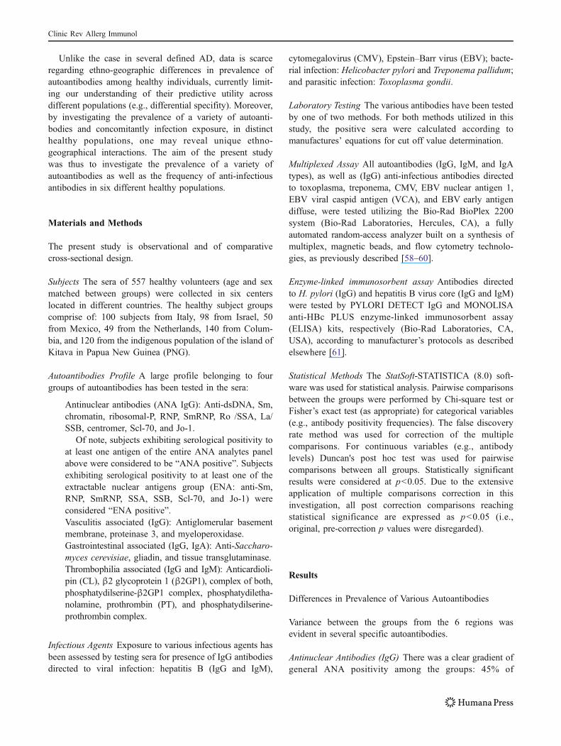
Laboratory Testing The various antibodies have been testedby one of two methods. For both methods utilized in thisstudy, the positive sera were calculated according tomanufactures’ equations for cut off value determination.
Multiplexed Assay All autoantibodies (IgG, IgM, and IgAtypes), as well as (IgG) anti-infectious antibodies directedto toxoplasma, treponema, CMV, EBV nuclear antigen 1,EBV viral caspid antigen (VCA), and EBV early antigendiffuse, were tested utilizing the Bio-Rad BioPlex 2200system (Bio-Rad Laboratories, Hercules, CA), a fullyautomated random-access analyzer built on a synthesis ofmultiplex, magnetic beads, and flow cytometry technolo-gies, as previously described [58–60].
Enzyme-linked immunosorbent assay Antibodies directedto H. pylori (IgG) and hepatitis B virus core (IgG and IgM)were tested by PYLORI DETECT IgG and MONOLISAanti-HBc PLUS enzyme-linked immunosorbent assay(ELISA) kits, respectively (Bio-Rad Laboratories, CA,USA), according to manufacturer’s protocols as describedelsewhere [61].
Statistical Methods The StatSoft-STATISTICA (8.0) soft-ware was used for statistical analysis. Pairwise comparisonsbetween the groups were performed by Chi-square test orFisher’s exact test (as appropriate) for categorical variables(e.g., antibody positivity frequencies). The false discoveryrate method was used for correction of the multiplecomparisons. For continuous variables (e.g., antibodylevels) Duncan's post hoc test was used for pairwisecomparisons between all groups. Statistically significantresults were considered at p<0.05. Due to the extensiveapplication of multiple comparisons correction in thisinvestigation, all post correction comparisons reachingstatistical significance are expressed as p<0.05 (i.e.,original, pre-correction p values were disregarded).
Results
Differences in Prevalence of Various Autoantibodies
Variance between the groups from the 6 regions wasevident in several specific autoantibodies.
Antinuclear Antibodies (IgG) There was a clear gradient ofgeneral ANA positivity among the groups: 45% of
Clinic Rev Allerg Immunol

Columbians, 38% of Kitavans (PNG), 26% of Mexicans,12% of Italians, 12% of Dutch, and 11% of Israelis wereANA positive. The rate among Columbians was significantlyelevated when compared with all other subject groups (p<0.05), followed by PNG (p<0.05, for comparisons withItaly, Netherlands and Israel), and Mexico (p<0.05, forcomparison with Israel). Two specific ANAs were especiallynotable. Anti-dsDNA antibodies were particularly frequentamong Columbians, followed by PNG subjects, comparedwith the rest of the subject groups (Fig. 1; p<0.05).Antichromatin antibodies were detected in high proportionof Columbian subjects, while detected in comparably lowproportion among all other subject groups (Fig. 1; p<0.05).
We further detected general ENA positivity in 15% ofKitavan, 14% of Mexican, 9% of Columbian, 6% of Dutch,5% of Israeli, and 4% of Italian subjects. However, thisgradient did not reach statistical significance after multiplecomparisons adjustments (p<0.05, only for comparisonbetween Papua New Guinea and Israel). There was also atrend (NS) of comparatively high frequencies of severalspecific antibodies directed to ENAs among Mexicans,namely anti-SmRNP, Ro/SSA, La/SSB, and Scl70 (Fig. 1).The mean levels of antibodies directed to these four ENAswere significantly higher in sera of Mexicans whencompared with all other subject groups (p<0.05). Incontrast, frequency or levels of anti-Sm, RNP, Centromere,Jo-1, and Ribo P antibodies did not differ across the groups,as these were generally detected in a negligible proportionof subjects.
Vasculitis Associated (IgG) The occurrence of these anti-bodies was rare across all healthy subject groups from thedifferent regions (Table 2). No significant variation wasobserved.
Gastrointestinal Associated (IgG and IgA) Were tested inthree groups of healthy subjects from different regions,namely Italy, Israel, and Columbia (prevalence datapresented in Table 3). Out of the six analytes compared,only antigliadin of the IgG type exhibited significantintergroup variance, as it was more frequently detectedamong Israeli and Italian subjects than among Columbiansubjects (p<0.05).
Thrombophilia Associated (IgG and IgM) A large profilewas tested in the Italian and Columbian subject groupsonly, presented in Fig. 2. Two IgM type antibodies weresignificantly more prevalent among Columbians, namelyanti-β2GP1 and PT (p<0.05 for both).
Differences in Prevalence of Various Anti-infectious AgentsAntibodies
We found variance between the groups in prevalence ofevery one of the eight anti-infectious antibodies (IgG)tested (prevalence data presented in Fig. 3). Variancebetween the regions was especially prominent in positivityrates of T. pallidum antibodies, as 87% of subjects fromPapua New Guinea tested positive while the frequency inall other groups was comparably low (range, 0–4%; p<0.05). The highest prevalence of anti-H. pylori antibodieswas recorded among Columbian subjects (81%) comparedwith all other groups (range, 30–62%; p<0.05; we did nothave data for Papua New Guinea). Anticytomegalovirusantibodies were detected in highest frequency amongsubjects from Papua New Guinea (100%) when comparedwith all five other groups of subjects (range, 33–93%; p<0.05). Relatively high prevalence was also detected among
0%
5%
10%
15%
20%
25%
30%
35%
40%
Anti SmRNPAnti Scl-70Anti La/SSBAnti Ro/SSAAnti chromatinAnti dsDNA
Per
cen
t Po
sitiv
e
Papua New Guinea Columbia Mexico Italy Netherlands Israel
Fig. 1 Prevalence of antinuclearautoantibodies in six groups ofhealthy subjects. ☆p<0.05,significant differences in preva-lence and/or in serum levels ofthese antinuclear antibodieswere evident between healthysubject groups
Clinic Rev Allerg Immunol

Columbian subjects. Conversely, prevalence in subjectsfrom the Netherlands was lowest of all six groups (Fig. 3;p<0.05). The presence of antibodies directed to EBV earlyantigen was also highly variable (range, 8–41%), demon-strating a pronounced gradient (Fig. 3). The highestfrequency was registered in the Mexican group (p<0.05for comparison with all groups except Netherlands), andlowest frequency in the Columbian group (p<0.05 forcomparison with all groups except Italy). The prevalence ofantibodies directed to EBV VCA (range, 80–98%) andnuclear antigen 1 (EBNA; range, 72–90%) did not exhibitas prominent a geographic gradient (Fig. 3), nonethelessdifferences were noted. The highest prevalence of anti-EBVVCA was registered in the Mexican subjects, reachingstatistical significance in the comparison to subjects fromIsrael and the Netherlands (p<0.05). Mean serum levels ofthese antibodies was significantly higher in Mexicansubjects compared with groups from all other regions (p<0.05). Antibodies directed to EBNA were comparablyfrequent in the Mexican, Italian and Papua New Guineasubjects, and less frequent among the Columbian subjects(p<0.05). The frequency of antibodies directed to T. gondiiparasite was lowest among subjects from Israel and theNetherlands compared with all other groups (range, 15–43%; p<0.05). The mean bacterium antibody levels werealso significantly high in sera of Mexican subjects (as wellas in Columbians) compared with the other groups (p<0.05). Finally, antihepatitis B antibodies were detected inhighest frequency among the Columbian subjects (range,0–11%, we did not have data for Papua New Guinea).However, this reached statistical significance (p<0.05) onlyin comparison to subjects from the Netherlands andMexico.
Discussion
In the present study, we have compared the frequencies of alarge profile of autoantibodies across several healthy, ethno-geographically distinct populations. Unsurprisingly, themajority of the autoantibodies tested negative across mostof the subjects in all six geographical groups, subsequentlyexhibiting no ethno-geographic variability. Nevertheless,we report a significant variability in some particularautoantibodies, warranting further discussion.
The most prominent intergroup variation was shown inantinuclear antibodies. The frequency range of ANA-positive sera was considerable, from approximately 12%comparably in the European groups (including Israel) andup to 26%, 38%, and 45% in the Mexican, PNG, andColumbian subjects (respectively).
There are several discrete reports (e.g., [62, 63])providing rates of ANA positivity among geographicallyhomogenous healthy populations (i.e., non-comparativepoint prevalence). The existing data has been recentlyexamined by the American College of Rheumatology AdHoc Committee on Immunologic Testing Guidelines,which concluded that ANA can be detected (by immuno-fluorescence (IIF)) in 5–30% of healthy subjects depend-ing on the dilution of sera (1:40–1:160, respectively). Thisseroprevalence rises with increasing age and particularlyin women [64].
The frequency of ANA in different healthy populationsevaluated as controls was reported sporadically utilizing IIFor other methods (e.g., ELISA). However, the detection ofautoantibodies was performed in each study employingdifferent techniques, different dilutions and in differentlaboratories. In the current study we have utilized the same
Table 2 Prevalence of vasculitis associated autoantibodies in six groups of healthy subjects
Vasculitis associated autoantibody(IgG)
Italy (%) Netherlands(%)
Israel (%) Mexico (%) Columbia(%)
Papua New Guinea (%)
Anti-GBM 0 0 0 0 0 2
Anti-PR3 1 0 1 0 1 0
Anti-MPO 0 2 0 0 1 0
GBM glomerular basement membrane, PR3 proteinase 3, MPO myeloperoxidase
Gastrointestinal associated autoantibody Italy (%) Israel (%) Columbia (%)
Antigliadin (IgG) 11 10 2a
Antigliadin (IgA) 2 1 4
Antitissue transglutaminase (IgG) 2 0 0
Antitissue transglutaminase (IgA) 0 2 0
Anti-Saccharomyces cerevisiae (IgG) 1 0 4
Anti-S. cerevisiae (IgA) 2 0 2
Table 3 Prevalence of gastroin-testinal associated autoantibod-ies in 3 groups of healthysubjects
a Higher frequency of antigliadin(IgG) in Italy and Israel subjectgroups when compared with theColumbia subject group (p<0.05)
Clinic Rev Allerg Immunol

testing method across all six groups of healthy subjects,therefore in a sense rendering comparisons across differentcohorts plausible, and suggesting that the observed differ-ences account for true ANA detection variance rather thantechnical bias. The multiplexed assay we employed, namelythe BioPlex 2200 ANA Screen, has been previouslyvalidated by our group as well as by others [59, 65]. In atotal of 510 healthy European blood bank donors, we havepreviously found that the frequency of positive results was7.5%, 5.5%, and 15.5% obtained by using BioPlex 2200ANA Screen, ANA ELISA, and IIF, respectively [59]. Amore recent evaluation of the BioPlex 2200 ANA Screen ina European hospital population found that 221 out of the1,104 sera (20%) were ANA positive. The most frequentlydetected autoantibody in this cohors was antichromatin [65].The conclusions from both evaluations were that thismultiplexed ANA screen is of comparable specifity to otherroutine tests (namely ELISA and double immunodiffusion),
has a lower positive rate than IIF, and is suitable as asensitive screening test to confirm or to exclude the presenceof a large number of autoantibodies simultaneously andrapidly [59, 65].
Truly detected geographical differences notwithstanding,it is challenging to explain the exceptionally high ANApositivity rates herein observed among the non-Europeanpopulations. The soaring ANAs rates we found in theColumbian subjects group are attributable to the elevatedprevalence of two specific autoantibodies, namely anti-chromatin and anti-dsDNA. The latter was also the mostprominently prevalent ANA in subjects from PNG. In theMexican subject group, it was in fact several antibodiesdirected to ENAs which seem to have elevated the overallANA positivity.
On the one hand, given the strong predictive value ofautoantibodies regarding the future progression (and insome cases prognosis) of an established AD, it may be
0%
10%
20%
30%
40%
50%
Anti PS-
PT (IgG)
Anti PS-
PT (IgM)
Anti CL
(IgG)
Anti CL
(IgM)
Anti
B2GP1
(IgG)
Anti
B2GP1
(IgM)
Anti CL-
B2 (IgG)
Anti CL-
B2 (IgM)
Anti PS-
B2 (IgG)
Anti PS-
B2 (IgM)
Anti PE
(IgG)
Anti PE
(IgM)
Anti PT
(IgG)
Anti PT
(IgM)
Per
cen
t Po
sitiv
e
Columbia Italy
Fig. 2 Prevalence ofthrombophilia-associatedautoantibodies in healthysubjects. PT prothrombin, PEphosphatydilethanolamine, PS-B2 phosphatydilserine-β2GP1complex, CLB2 cardiolipin-β2glycoprotein 1 complex,CL cardiolipin, PS-PTphosphatydilserine- prothrombincomplex. ☆p<0.05
0%
10%
20%
30%
40%
50%
60%
70%
80%
90%
100%
CMVEBNAEBV-VCAEBVEAHBV*H Pylori* TreponemaToxoplasmosis
Pe
rce
nt
Po
sit
ive
Papua New Guinea* Columbia Mexico Italy Netherlands Israel
Fig. 3 Prevalence of anti-infections antibodies in sixgroups of healthy subjects. Hhelicobacter, HBV hepatitis Bvirus, EBVEA Epstein–Barr vi-rus early antigen, EBV-VCA viralcaspid antigen, EBNA nuclearantigen, CMV cytomegalovirus.*, Helicobacter pylori andHBV—no data for PapuaNew Guinea subjects group.☆p<0.05, significant differen-ces in prevalence of theseanti-infections antibodies wereevident between the subjectgroups
Clinic Rev Allerg Immunol

hypothesized that the detected ANA gradient should in partmanifest true predictive risk of AD in these populations.Moreover, it implies that autoantibodies screening specifi-cally tailored to different populations may be a valuabletool for future AD risk stratification.
However, while most patients with an established ADhave marking autoantibodies, most people with autoanti-bodies do not have and likely will never have an overt AD.Therefore, one must consider that while on the one handgeographical differences in autoantibodies detection maymanifest true AD risk variance, on the other hand uniquenon-pathological serological mechanisms should also con-tribute to the detection rates. In fact, both alternativestogether may ultimately account for extraordinarily highautoantibodies (e.g., ANA) detection rates in sera of healthysubjects from these particular regions.
The intriguing relationship between the mechanisms ofmolecular mimicry and cross-reactivity may, in theory, holdthe key. In recent years, many investigations into thepathogenesis of autoimmunity elaborated on the definitionof these mechanisms [66–68]. For example, several humanmonoclonal antibodies were found to react specifically withinfectious agents as well as with human antigens [13, 69].These antibodies could represent merely cross-reactivity thatwill not result in any clinical or pathological outcome, oralternatively, they can initiate a pathogenic process that mayeventually result in autoimmunity, hence defining molecularmimicry. The process of molecular mimicry betweenmicrobial and self-components (i.e. proteins, carbohydratesor DNA epitopes) is postulated to be the most likelymechanism of post-infectious autoimmunity [68–73].
In fact, infectious agents have been repeatedly and for along time implicated as a major environmental trigger ofAD in general, and in the induction of autoantibodiesspecifically [13, 14, 60, 68–79]. In addition to molecularmimicry, there are other underlying mechanisms which arecomplex and vary with different agents; among them arepotentiation and diversion of innate immunity, and expo-sure or spillage of intracellular autoantigens [3, 14]. Thereis a wide unevenness in the geographic distribution ofinfectious agents, thus it seems plausible that in interactionwith population genetics the infection gradient shouldinfluence global gradients of AD and autoimmune mani-festations (e.g., autoantibodies) [15, 16, 80, 81].
In the current study we have indeed identified agenerally elevated evidence of infection exposure (i.e.,anti-infectious agents antibodies) in the subject groups fromthe Latin American region (i.e., Columbia and Mexico) andin the indigenous Kitavan population (from PNG), whencompared with the European and Israeli subject groups. Itmay be suggested, that in these populations the elevatedinfection exposure over lifetime should indeed have asignificant role in the elevated detection of autoantibodies.
Furthermore, the particular combination of infectiousagents prominent in each population may account for thedistinct autoantibody profiles detected in these groups.
One salient example, in the current study the frequencyof anti-Treponema antibodies was exceptionally highamong the (PNG) Kitavans (87%), while almost undetect-able in all other groups. The geographical region of PNGis indeed known to be endemic to a subspecies of T.pallidum and Treponema pertenue, which causes Yawsdisease [82]. We and Frostegård J et al. have previouslystudied the same individuals from PNG, and foundelevated levels of antiphosphorylcholin antibodies in100% of the Kitavans, plausibly induced by the trepone-mal exposure [83–85]. Ultimately, the high presence ofantiphosphorylcholin antibodies may in turn be responsi-ble for the high rates of anti-dsDNA antibodies observedin the same population, on account of cross-reactivitybetween these two autoantibodies [84, 86].
Taken together, the observed link between generally highinfection exposure and elevated ANA prevalence amongColumbians, Kitavans and Mexicans, perhaps representsmolecular mimicry implying future AD risk, and/or detectiondue to serological cross-reactivity unique to each population.
In the present study three additional specific autoantibodiesexhibited marked intergroup differences, namely antigliadin(IgG), IgM β2GP1, and IgM PT, meriting a brief elaboration.The current model of the natural history of celiac disease (CD)recognizes that in many cases there is a preclinical stage, inwhich there are no intestinal and/or extraintestinal symptoms(including no findings in a small bowel biopsy), howeverautoantibodies can often be detected [87]. Several seriesconducted in Europe and the US have reported a rathercomparable prevalence of around 0.5% of preclinical CD(based on presence of autoantibodies alone), while the estimatesof combined preclinical and diagnosed (i.e., including clinicaldisease) ranged between 0.5–2.5% in the different populations.In Africa, and in East Asian populations who lack the diseaseassociated HLA haplotype, the disease is considered to be rare[87]. Our finding of approximately fivefold lower antigliadinantibodies in Columbians than in Israelis and Italians seemsto be rather in agreement with a US population based seriesreporting a three-fold lower incidence of a positive serolog-ical test to CD in Hispanics than in non-Hispanic whites[88]. In fact, it has been suggested that this may beattributable to the low frequency of the HLA-DR3,DQB1*0201 haplotype in the studied Hispanic population[88]. Genetic determination notwithstanding, we previouslyreported that certain infections, CMVamong them, may havea protective role in the etiopathogenesis of CD [89]. Wetherefore propose that in the present study the elevatedexposure to several infectious agents (including CMV) in thehealthy Columbians group may in this case have a protectiverole in the appearance of disease associated antibodies.
Clinic Rev Allerg Immunol

Nonetheless, while such an association may be hypothesized,a protective (causal) relationship may not be establishedbased on this observational report.
Antiphospholipid antibodies (aPL) were previouslyreported in 1–5% of asymptomatic healthy subjects, with ahighly variable point prevalence when comparing differentreports from various ethno-geographic populations [21, 90,91]. The variability has been attributed to both genetic andenvironmental factors [21, 90]. We herein report a signifi-cantly higher frequency of IgM anti-β2GP1 and anti-PTantibodies in Columbian compared with Italian healthyindividuals. These specific aPL account for most of theglobal antiphospholipid binding activity [92, 93]. aPL havebeen shown to be induced by many viral, bacterial andparasitic infections [94]. We have previously detectedelevated levels of EBV early antigen IgG, toxoplasma IgM,and rubella IgM antibodies in sera of primary APS patientscompared with controls [95]. Although in the present studywe did not find differences in prevalence of EBV earlyantigen IgG or toxoplasma (IgG) between Columbians andItalians (rubella was not tested), other infectious or environ-mental influences (herein not tested) may not be ruled out.
Conclusions
Of note, due to the investigative nature and broad scope of thisstudy, we did not apply a specific analysis of the numerousdemographic variables which may serve as confounders in theobserved intergroup variability. Nonetheless, we applied thesame valid laboratory method across geographically distinct,age and sex matched populations, thereby revealing markedgeoepidemiological variance in autoantibodies positivity andinfectious agents exposure. Our results warrant furtherprospective investigations to establish whether indeed theAD rates will eventually be elevated in regions found to bemore prevalent in autoantibodies. This should ultimatelyvalidate the predictive value of population-specific autoanti-bodies screening, and help establish unique causal factors.Furthermore, in the ongoing quest of autoantibodies testingmethods standardization, it is becoming clear that populationvariance should be significantly factored in.
Acknowledgements This work was performed in partial fulfillmentof the M.D. thesis requirements of the Sackler Faculty of Medicine,Tel Aviv University, Israel.
References
1. Shoenfeld Y, Blank M, Abu-Shakra M, Amital H, Barzilai O,Berkun Y (2008) The mosaic of autoimmunity: prediction,autoantibodies, and therapy in autoimmune diseases—2008. IsrMed Assoc J 10(1):13–19
2. Harel M, Shoenfeld Y (2006) Predicting and preventing autoim-munity, myth or reality? Ann N Y Acad Sci 1069:322–345
3. Lleo A, Invernizzi P, Gao B, Podda M, Gershwin ME (2010)Definition of human autoimmunity—autoantibodies versus auto-immune disease. Autoimmun Rev 9:A259–A266
4. Scofield RH (2004) Autoantibodies as predictors of disease.Lancet 363(9420):1544–1546
5. Arbuckle MR, McClain MT, Rubertone MV, Scofield RH et al(2003) Development of autoantibodies before the clinical onset ofsystemic lupus erythematosus. N Engl J Med 349:1526–1533
6. Nielen MM, van Schaardenburg D, Reesink HW, van de Stadt RJ,van der Horst-Bruinsma IE, de Koning MH (2004) Specificautoantibodies precede the symptoms of rheumatoid arthritis: astudy of serial measurements in blood donors. Arthritis Rheum 50(2):380–386
7. Taplin CE, Barker JM (2008) Natural evolution, prediction, andprevention of type 1 diabetes in youth. Endocr Res 33(1–2):17–33
8. LaGasse JM, Brantley MS, Leech NJ, Rowe RE, Monks S,Palmer JP et al (2002) Successful prospective prediction of type 1diabetes in schoolchildren through multiple defined autoanti-bodies: an 8-year follow-up of the Washington State DiabetesPrediction Study. Diab Care 25(3):505–511
9. Rewers M, Gottlieb P (2009) Immunotherapy for the preventionand treatment of type 1 diabetes: human trials and a look into thefuture. Diab Care 32(10):1769–1782
10. James JA, Kim-Howard XR, Bruner BF, Jonsson MK, McClainMT, Arbuckle MR et al (2007) Hydroxychloroquine sulfatetreatment is associated with later onset of systemic lupuserythematosus. Lupus 16(6):401–409
11. Deane KD, Norris JM, Holers VM (2010) Preclinical rheumatoidarthritis: identification, evaluation, and future directions forinvestigation. Rheum Dis Clin North Am 36(2):213–241
12. Shapira Y, Agmon-Levin N, Shoenfeld Y (2010) Mycobacteriumtuberculosis, autoimmunity, and vitamin D. Clin Rev AllergyImmunol 38(2–3):169–177
13. Barzilai O, Ram M, Shoenfeld Y (2007) Viral infection caninduce the production of autoantibodies. Curr Opin Rheumatol19:636–643
14. Kivity S, Agmon-Levin N, Blank M, Shoenfeld Y (2009)Infections and autoimmunity—friends or foes? Trends Immunol30:409–414
15. Shapira Y, Agmon-Levin N, Shoenfeld Y (2010) Geoepidemiol-ogy of autoimmune rheumatic diseases. Nat Rev Rheumatol 6(8):468–476
16. Shapira Y, Agmon-Levin N, Shoenfeld Y (2010) Defining andanalyzing geoepidemiology and human autoimmunity. J Auto-immun 34(3):J168–J177
17. Shoenfeld Y, Selmi C, Zimlichman E, Gershwin ME (2008) Theautoimmunologist: geoepidemiology, a new center of gravity, andprime time for autoimmunity. J Autoimmun 31(4):325–330
18. Arnson Y, Shoenfeld Y, Amital H (2010) Effects of tobaccosmoke on immunity, inflammation and autoimmunity. J Auto-immun 34:J258–J265
19. Berkun Y, Padeh S (2010) Environmental factors and thegeoepidemiology of juvenile idiopathic arthritis. AutoimmunRev 9:A319–A324
20. Bhat A, Naguwa S, Cheema G, Gershwin ME (2010) Theepidemiology of transverse myelitis. Autoimmun Rev 9:A395–A399
21. Biggioggero M, Meroni PL (2010) The geoepidemiology of theantiphospholipid antibody syndrome. Autoimmun Rev 9:A299–A304
22. Borchers AT, Naguwa SM, Keen CL, Gershwin ME (2010) Theimplications of autoimmunity and pregnancy. J Autoimmun 34:J287–J299
Clinic Rev Allerg Immunol
23. Borchers AT, Naguwa SM, Shoenfeld Y, Gershwin ME (2010)The geoepidemiology of systemic lupus erythematosus. Auto-immun Rev 9:A277–A287
24. Borchers AT, Uibo R, Gershwin ME (2010) The geoepidemiologyof type 1 diabetes. Autoimmun Rev 9:A355–A365
25. Brooks WH, Le Dantec C, Pers JO, Youinou P, Renaudineau Y(2010) Epigenetics and autoimmunity. J Autoimmun 34:J207–J219
26. Chandran V, Raychaudhuri SP (2010) Geoepidemiology andenvironmental factors of psoriasis and psoriatic arthritis. J Auto-immun 34:J314–J321
27. Chang C (2010) The immune effects of naturally occurring andsynthetic nanoparticles. J Autoimmun 34:J234–J246
28. Chang C, Gershwin ME (2009) Drugs and autoimmunity—acontemporary review and mechanistic approach. J Autoimmun 34:J266–J275
29. Chen M, Daha MR, Kallenberg CG (2010) The complementsystem in systemic autoimmune disease. J Autoimmun 34:J276–J286
30. Chen M, Kallenberg CG (2010) The environment, geoepidemiol-ogy and ANCA-associated vasculitides. Autoimmun Rev 9:A293–A298
31. Deane S, Teuber SS, Gershwin ME (2010) The geoepidemiologyof immune thrombocytopenic purpura. Autoimmun Rev 9:A342–A349
32. Ehrenfeld M (2010) Geoepidemiology: the environment andspondyloarthropathies. Autoimmun Rev 9:A325–A329
33. Hemminki K, Li X, Sundquist J, Sundquist K (2010) Theepidemiology of Graves' disease: evidence of a genetic and anenvironmental contribution. J Autoimmun 34:J307–J313
34. Hoffmann MH, Trembleau S, Muller S, Steiner G (2010) Nucleicacid-associated autoantigens: pathogenic involvement and thera-peutic potential. J Autoimmun 34:J178–J206
35. Invernizzi P (2010) Geoepidemiology of autoimmune liverdiseases. J Autoimmun 34:J300–J306
36. Lambert JF, Nydegger UE (2010) Geoepidemiology of autoim-mune hemolytic anemia. Autoimmun Rev 9:A350–A354
37. Leung PS, Shu SA, Kenny TP,Wu PY, TaoMH (2010) Developmentand validation of gene therapies in autoimmune diseases: epidemi-ology to animal models. Autoimmun Rev 9:A400–A405
38. Logan I, Bowlus CL (2010) The geoepidemiology of autoimmuneintestinal diseases. Autoimmun Rev 9:A372–A378
39. Mackay IR (2010) Travels and travails of autoimmunity: ahistorical journey from discovery to rediscovery. AutoimmunRev 9:A251–A258
40. Maverakis E, Miyamura Y, Bowen MP, Correa G, Ono Y,Goodarzi H (2010) Light, including ultraviolet. J Autoimmun34:J247–J257
41. Mavragani CP, Moutsopoulos HM (2010) The geoepidemiologyof Sjogren's syndrome. Autoimmun Rev 9:A305–A310
42. Meyer A, Levy Y (2010) Geoepidemiology of myasthenia gravis.Autoimmun Rev 9:A383–A386
43. Meyer N, Misery L (2010) Geoepidemiologic considerations ofauto-immune pemphigus. Autoimmun Rev 9:A379–A382
44. Milo R, Kahana E (2010) Multiple sclerosis: geoepidemiology,genetics and the environment. Autoimmun Rev 9:A387–A394
45. Powell JJ, Faria N, Thomas-McKay E, Pele LC (2010) Origin andfate of dietary nanoparticles and microparticles in the gastrointes-tinal tract. J Autoimmun 34:J226–J233
46. Prieto S, Grau JM (2010) The geoepidemiology of autoimmunemuscle disease. Autoimmun Rev 9:A330–A334
47. Ranque B, Mouthon L (2010) Geoepidemiology of systemicsclerosis. Autoimmun Rev 9:A311–A318
48. Round JL, O'Connell RM, Mazmanian SK (2010) Coordination oftolerogenic immune responses by the commensal microbiota. JAutoimmun 34:J220–J225
49. Sands J, Tuscano JM (2010) Geoepidemiology and autoimmunemanifestations of lymphoproliferative disorders. Autoimmun Rev9:A335–A341
50. Segelmark M, Hellmark T (2010) Autoimmune kidney diseases.Autoimmun Rev 9:A366–A371
51. Selmi C (2010) The worldwide gradient of autoimmune con-ditions. Autoimmun Rev 9:A247–A250
52. Selmi C, Tsuneyama K (2010) Nutrition, geoepidemiology, andautoimmunity. Autoimmun Rev 9:A267–A270
53. Stojanovich L (2010) Stress and autoimmunity. Autoimmun Rev9:A271–A276
54. Tobon GJ, Youinou P, Saraux A (2010) The environment, geo-epidemiology, and autoimmune disease: rheumatoid arthritis.Autoimmun Rev 9:A288–A292
55. Tomer Y (2010) Hepatitis C and interferon induced thyroiditis. JAutoimmun 34:J322–J326
56. Youinou P, Pers JO, Gershwin ME, Shoenfeld Y (2010) Geo-epidemiology and autoimmunity. J Autoimmun 34:J163–J167
57. Zeki AA, Schivo M, Chan AL, Hardin KA, Kenyon NJ,Albertson TE, Rosenquist GL et al (2010) Geoepidemiologyof COPD and idiopathic pulmonary fibrosis. J Autoimmun 34:J327–J338
58. Klutts JS, Liao RS, Dunne WM Jr, Gronowski AM (2004)Evaluation of a multiplexed bead assay for assessment of Epstein–Barr virus immunologic status. J Clin Microbiol 42:4996–5000
59. Shovman O, Gilburd B, Barzilai O, Shinar E, Larida B, Zandman-Goddard G et al (2005) Evaluation of the BioPlex 2200 ANAscreen: analysis of 510 healthy subjects: incidence of natural/predictive autoantibodies. Ann NY Acad Sci 1050:380–388
60. Barzilai O, Sherer Y, Ram M, Izhaky D, Anaya JM, Shoenfeld Y(2007) Epstein-Barr virus and cytomegalovirus in autoimmunediseases: are they truly notorious? A preliminary report. Ann NYAcad Sci 1108:567–577
61. Ram M, Anaya JM, Barzilai O, Izhaky D, Porat Katz BS, BlankM, Shoenfeld Y (2008) The putative protective role of hepatitis Bvirus (HBV) infection from autoimmune disorders. AutoimmunRev 7:621–625
62. Piura B, Tauber E, Sarov B, Naggan L, Shoenfeld Y (1988)Antinuclear autoantibodies in sera of healthy pregnant womenand their offspring. Am J Reprod Immunol Microbiol 18(4):116–119
63. Yadin O, Sarov B, Naggan L, Slor H, Shoenfeld Y (1989) Naturalautoantibodies in the serum of healthy women—a five-yearfollow-up. Clin Exp Immunol 75(3):402–406
64. Solomon DH, Kavanaugh AJ, Schur PH, College A, AmericanCollege of Rheumatology Ad Hoc Committee on ImmunologicTesting Guidelines (2002) Evidence-based guidelines for the useof immunologic tests: antinuclear antibody testing. ArthritisRheum 47(4):434–444
65. Desplat-Jego S, Bardin N, Larida B, Sanmarco M (2007)Evaluation of the BioPlex 2200 ANA screen for the detection ofantinuclear antibodies and comparison with conventional meth-ods. Ann NY Acad Sci 1109:245–255
66. Rose NR, Mackay IR (2000) Molecular mimicry: a critical look atexemplary instances in human diseases. Cell Mol Life Sci57:542–551
67. Blank M, Barzilai O, Shoenfeld Y (2007) Molecular mimicry andimmunity. Clin Rev Allergy Immunol 32:111–118
68. Agmon-Levin N, Blank M, Paz Z, Shoenfeld Y (2009)Molecular mimicry in systemic lupus erythematosus. Lupus18:1181–1185
69. Shoenfeld Y, Wilner Y, Coates AR, Rauch J, Lavie G, Pinkhas J(1986) Infection and autoimmunity: monoclonal anti-tuberculosisantibodies react with DNA and monoclonal anti-DNA autoanti-bodies bind to mycobacterial derived glycolipids. Clin ExpImmunol 66:255–261
Clinic Rev Allerg Immunol

70. Shoenfeld Y, Zandman-Goddard G, Stojanovich L, Cutolo M,Amital H, Levy Y et al (2008) The mosaic of autoimmunity:hormonal and environmental factors involved in autoimmunediseases—2008. Isr Med Assoc J 10(1):8–12
71. Agmon-Levin N, Ram M, Barzilai O, Porat-Katz BS, Parikman R,Selmi C et al (2009) Prevalence of hepatitis C serum antibody inautoimmune diseases. J Autoimmun 32:261–266
72. Berkun Y, Zandman-Goddard G, Barzilai O, Boaz M, Sherer Y,Larida B et al (2009) Infectious antibodies in systemic lupuserythematosus patients. Lupus 18(13):1129–1135
73. Peterson LK, Fujinami RS (2007) Chapter 3: molecular mimicry.In: Shoenfeld Y, Gershwin ME, Meroni PL (eds) Autoantibodies,2nd edn. Elsevier, Oxford
74. Berlin T, Zandman-Goddard G, Blank M, Matthias T, Pfeiffer S,Weis I et al (2007) Autoantibodies in nonautoimmune individualsduring infections. Ann NY Acad Sci 1108:584–593
75. Lidar M, Lipschitz N, Langevitz P, Barzilai O, Ram M, Porat-KatzBS, Pagnoux C et al (2009) Infectious serologies and autoanti-bodies in Wegener's granulomatosis and other vasculitides: novelassociations disclosed using the Rad BioPlex 2200. Ann NYAcadSci 1173:649–657
76. Lidar M, Langevitz P, Barzilai O, Ram M, Porat-Katz BS, BizzaroN, Tonutti E et al (2009) Infectious serologies and autoantibodiesin inflammatory bowel disease: insinuations at a true pathogenicrole. Ann NY Acad Sci 1173:640–648
77. Krause I, Anaya JM, Fraser A, Barzilai O, Ram M, Abad V,Arango A et al (2009) Anti-infectious antibodies andautoimmune-associated autoantibodies in patients with type Idiabetes mellitus and their close family members. Ann NY AcadSci 1173:633–639
78. Arnson Y, Amital H, Guiducci S, Matucci-Cerinic M, Valentini G,Barzilai O, Maya R et al (2009) The role of infections in theimmunopathogensis of systemic sclerosis—evidence from sero-logical studies. Ann NY Acad Sci 1173:627–632
79. Meron MK, Amital H, Shepshelovich D, Barzilai O, Ram M,Anaya JM, Gerli R et al (2010) Infectious aspects and theetiopathogenesis of rheumatoid arthritis. Clin Rev AllergyImmunol 38(2–3):287–291
80. World Health Organization (2010) Chapter 5: infectious diseasesof potential risk for travelers. In International Travel and Health2010 edn. Available at: http://www.who.int/ith/en/
81. Pordeus V, Barzilai O, Sherer Y, Luiz RR, Blank M, Bizzaro N etal (2008) A latitudinal gradient study of common anti-infectiousagent antibody prevalence in Italy and Colombia. Isr Med Assoc J10(1):65–8
82. Garner MF, Hornabrook RW, Backhouse JL (1972) Prevalence ofyaws on Kar Kar Island, New Guinea. Br J Vener Dis 48(5):350–355
83. Frostegård J, Tao W, Georgiades A, Råstam L, Lindblad U,Lindeberg S (2007) Atheroprotective natural anti-phosphorylcholineantibodies of IgM subclass are decreased in Swedish controls ascompared to non-westernized individuals from New Guinea. NutrMetab (Lond) 4:7
84. Agmon-Levin N, Bat-sheva PK, Barzilai O, Ram M, Lindeberg S,Frostegård J, Shoenfeld Y (2009) Antitreponemal antibodies leadingto autoantibody production and protection from atherosclerosis inKitavans from Papua New Guinea. Ann NYAcad Sci 1173:675–682
85. Blanco DR, Champion CI, Dooley A, Cox DL, Whitelegge JP,Faull K, Lovett MA (2005) A monoclonal antibody that conveysin vitro killing and partial protection in experimental syphilisbinds a phosphorylcholine surface epitope of Treponema pal-lidum. Infect Immun 73(5):3083–3095
86. Sharma A, Isenberg DA, Diamond B (2001) Crossreactivity ofhuman anti-dsDNA antibodies to phosphorylcholine: clues to theirorigin. J Autoimmun 16(4):479–484
87. Rewers M (2005) Epidemiology of celiac disease: what are theprevalence, incidence, and progression of celiac disease? Gastro-enterology 128(4):S47–S51
88. Hoffenberg EJ, MacKenzie T, Barriga KJ, Eisenbarth GS, Bao F,Haas JE (2003) A prospective study of the incidence of childhoodceliac disease. J Pediatr 143(3):308–314
89. Plot L, Amital H, Barzilai O, Ram M, Nicola B, Shoenfeld Y(2009) Infections may have a protective role in the etiopatho-genesis of celiac disease. Ann NY Acad Sci 1173:670–674
90. Uthman I, Khamashta M (2005) Ethnic and geographical variationin antiphospholipid (Hughes) syndrome. Ann Rheum Dis 64(12):1671–1676
91. Mchrani T, Pertri M (2009) Epidemiology of the antiphospholipidsyndrome. In: Asherson RA (ed) Handbook of systemic autoim-mune diseases, vol 10. Amsterdam, Elsevier
92. Atsumi T, Koike T (2002) Clinical relevance of antiprothrombinantibodies. Autoimmun Rev 1:49–53
93. Asherson RA, Cervera R, Merrill JT, Erkan D (2008) Antiphos-pholipid antibodies and the antiphospholipid syndrome: clinicalsignificance and treatment. Semin Thromb Hemost 34:256–266
94. Sène D, Piette JC, Cacoub P (2008) Antiphospholipid antibodies,antiphospholipid syndrome and infections. Autoimmun Rev 7(4):272–277
95. Zinger H, Sherer Y, Goddard G, Berkun Y, Barzilai O, Agmon-Levin N et al (2009) Common infectious agents prevalence in theantiphospholipid syndrome. Lupus 18(13):1149–1153
Clinic Rev Allerg Immunol