Platelet membrane fluidity identifies a clinical subtype of Alzheimer's disease

ORIGINAL ARTICLE
Genome-wide haplotype association study identifies theFRMD4A gene as a risk locus for Alzheimer’s diseaseJ-C Lambert1,2,3,47, B Grenier-Boley1,2,3,47, D Harold4, D Zelenika5, V Chouraki1,2,3, Y Kamatani5,6,
K Sleegers7,8, MA Ikram9, M Hiltunen10, C Reitz11, I Mateo12, T Feulner13, M Bullido14, D Galimberti15,
L Concari16, V Alvarez17, R Sims4, A Gerrish4, J Chapman4, C Deniz-Naranjo18, V Solfrizzi19,
S Sorbi20, B Arosio21, G Spalletta22, G Siciliano23, J Epelbaum24, D Hannequin25, J-F Dartigues26,
C Tzourio27,28, C Berr29, EMC Schrijvers30, R Rogers11, G Tosto11, F Pasquier3,31, K Bettens7,8,
C Van Cauwenberghe7,8, L Fratiglioni32,33, C Graff33,34, M Delepine5, R Ferri35, CA Reynolds36, L Lannfelt37,
M Ingelsson37, JA Prince38, C Chillotti39, A Pilotto40, D Seripa40, A Boland5, M Mancuso23, P Bossu22,
G Annoni21, B Nacmias20, P Bosco35, F Panza19, F Sanchez-Garcia18, M Del Zompo41, E Coto17, M Owen4,
M O’Donovan4, F Valdivieso14, P Caffara16, E Scarpini15, O Combarros12, L Buee2,42, D Campion25,
H Soininen10, M Breteler43,44, M Riemenschneider13, C Van Broeckhoven7,8, A Alperovitch27,28, M Lathrop5,6,
D-A Tregouet45,46, J Williams4 and P Amouyel1,2,3,31, EADI consortium48, GERAD consortium48
1INSERM, U744, Lille, France; 2Institut Pasteur de Lille, Lille, France; 3Universite Lille-Nord de France, Lille, France; 4MedicalResearch Council (MRC) Centre for Neuropsychiatric Genetics and Genomics, Neurosciences and Mental Health ResearchInstitute, Department of Psychological Medicine and Neurology, School of Medicine, Cardiff University, Cardiff, UK; 5CentreNational de Genotypage, Institut Genomique, Commissariat a l’energie Atomique, Evry, France; 6Fondation Jean Dausset-CEPH, Paris, France; 7Neurodegenerative Brain Diseases group, Department of Molecular Genetics, VIB, Antwerpen,Belgium; 8Laboratory of Neurogenetics, Institute Born-Bunge, University of Antwerp, Antwerpen, Belgium; 9Departement ofEpidemiology and Radiology, Erasmus MC, Rotterdam; 10Department of neurology, University of Eastern Finland and KuopioUniversity Hospital, Kuopio, Finland; 11G.H. Sergievsky Center, Columbia University, New York, NY, USA; 12Service ofNeurology, University Hospital Marques de Valdecilla, University of Cantabria, Santander, Spain; 13Department of Psychiatryand Psychotherapy, Universitatsklinikum des Saarlandes, Universitat des Saarlandes Saarbruecken, Germany; 14Centro deBiologia Molecular Severo Ochoa (UAM-CSIC) and CIBERNED, Universidad Autonoma, Madrid, Spain; 15Department ofNeurological Sciences, Dino Ferrari Center, University of Milan, Fondazione Ca Granda, IRCCS Ospedale MaggiorePoliclinico, Milan, Italy; 16Department of Neuroscience, University of Parma, Parma, Italy; 17Genetica Molecular-Laboratorio deMedicina, Hospital Universitario Central Asturias, Oviedo, Spain; 18Servicio de Inmunologıa. Hospital Unviersitario de GranCanaria Dr Negrın. Bco, Las Palmas de Gran Canaria, Spain; 19Department of Geriatrics, Center for aging brain, Memory unit,University of Bari, Bari, Italy; 20Department of neurological and psychiatric Sciences, University of Florence, Florence, Italy;21Department of Internal Medicine, Geriatric Unit, Fondazione IRCCS Ca Granda Ospedale Maggiore Policlinico, Universita degliStudi di Milano, Milan, Italy; 22Clinical and Behavioral Neurology, IRCCS Fondazione Santa Lucia, Roma, Italy; 23Neurologicalclinic, University of Pisa, Pisa, Italy; 24INSERM UMR 894, Paris Descartes University, Paris, France; 25Inserm U614, Faculte deMedecine-Pharmacie de Rouen, Rouen, France; 26Inserm U897, Victor Segalen University, Bordeaux, France; 27Inserm U708,Paris, France; 28UPMC University Paris 06, Paris, France; 29Inserm U1061, Hopital La Colombiere, Montpellier, France;30Department of Epidemiology and Neurology, Erasmus MC, Rotterdam; 31CHRU de Lille, Lille, France; 32Aging ReasearchCenter, Department NVS, Karolinska Institutet and Stockholm University, Stockholm, Sweden; 33Department of GeriatricMedicine, Karolinska University Hospital, Stockholm, Sweden; 34KI-Alzheimer’s Disease Reseach Center, Department NVS,Karolinska Institutet, KIADRC, Stockholm, Sweden; 35IRCCS Associazione Oasi Maria SS, Institute for Research on MentalRetardation and Brain Aging, Troina (EN), Italy; 36Department of Psychology, University of California at Riverside, Riverside,CA, USA; 37Department of Public health and Caring Sciences, Rudbeck Laboratory, Uppsala University, Uppsala, Sweden;38Department of Medical Epidemiology and Biostatistics, Karolinska Institute, Stockholm, Sweden; 39Unit of ClinicalPharmacology, Teaching Hospital of Cagliari, Cagliari, Italy; 40Geriatric Unit & Gerontology-Geriatrics Research Laboratory,Department of Medical Sciences, I.R.C.C.S. ‘Casa Sollievo della Sofferenza’, San Giovanni Rotondo, Italy; 41Section of ClinicalPharmacology, Department of Neurosciences ‘B.B. Brodie’, University of Cagliari, Cagliari, Italy; 42Inserm U837, Lille, France;43Department of Epidemiology, Erasmus MC, Rotterdam; 44DZNE, Bonn, Germany; 45INSERM UMR_S 937, Paris, Franceand 46ICAN Institute of Cardiometabolism and Nutrition, Universite Pierre & Marie Curie, Paris, France
Received 15 November 2011; revised 3 January 2012; accepted 10 January 2012; published online 20 March 2012
Correspondence: Dr J-C Lambert, INSERM U744, Institut Pasteur de Lille, BP 245, 1 rue du professeur Calmette, 59019 Lille cedex,France.E-mail: [email protected] authors contributed equally to this work.48A full list of members is provided in the Supplementary Note.
Molecular Psychiatry (2013) 18, 461–470& 2013 Macmillan Publishers Limited All rights reserved 1359-4184/13
www.nature.com/mp
Recently, several genome-wide association studies (GWASs) have led to the discovery ofnine new loci of genetic susceptibility in Alzheimer’s disease (AD). However, the landscapeof the AD genetic susceptibility is far away to be complete and in addition to single-SNP(single-nucleotide polymorphism) analyses as performed in conventional GWAS, complemen-tary strategies need to be applied to overcome limitations inherent to this type of approaches.We performed a genome-wide haplotype association (GWHA) study in the EADI1 study(n = 2025 AD cases and 5328 controls) by applying a sliding-windows approach. Afterexclusion of loci already known to be involved in AD (APOE, BIN1 and CR1), 91 regions withsuggestive haplotype effects were identified. In a second step, we attempted to replicatethe best suggestive haplotype associations in the GERAD1 consortium (2820 AD cases and6356 controls) and observed that 9 of them showed nominal association. In a third step,we tested relevant haplotype associations in a combined analysis of five additional case–control studies (5093 AD cases and 4061 controls). We consistently replicated the associationof a haplotype within FRMD4A on Chr.10p13 in all the data set analyzed (OR: 1.68; 95% CI:(1.43–1.96); P = 1.1� 10�10). We finally searched for association between SNPs withinthe FRMD4A locus and Ab plasma concentrations in three independent non-dementedpopulations (n = 2579). We reported that polymorphisms were associated with plasma Ab42/Ab40 ratio (best signal, P = 5.4� 10�7). In conclusion, combining both GWHA study and aconservative three-stage replication approach, we characterised FRMD4A as a new geneticrisk factor of AD.Molecular Psychiatry (2013) 18, 461–470; doi:10.1038/mp.2012.14; published online 20 March 2012
Keywords: Alzheimer; amyloid; FRMD4A; GWAS; plasma
Introduction
The identification of genes involved in monogenicforms of Alzheimer’s disease (AD) has significantlycontributed to our knowledge of the disease mecha-nisms. The causal links between mutations, thefunctions of the mutated genes (APP, PS1 and PS2)and disease development prompted a pathophysio-logical hypothesis, which radically changed ourunderstanding of AD: the amyloid cascade hypo-thesis.1 The systematic association of pathogenicmutations with changes in APP metabolism and,more particularly, a relative overproduction of Ab42peptides indicates that this metabolism is at theheart of the disease process (at least in the monogenicforms of the disease). The overproduction of theseneurotoxic peptides is supposed to lead to or accen-tuate neuron-to-neuron propagation of the t patho-logy (leading to neuronal death) by an unknownmechanism.2
By analogy, it was expected that the characteriza-tion of genetic factors involved in the common formsof AD (that is, lacking classical Mendelian inheri-tance), the most frequent form of the disease, shouldalso help to better understand the AD physiopatho-logical process. However, the characterization of thesegenetic factors has encountered significant difficul-ties. Until 2009, the APOE (apolipoprotein E) genewas the only globally valid genetic determinant of ADto have been unambiguously identified in 15 years ofintensive research.3,4
As with other multifactorial diseases, this systema-tic inability to detect new genetic determinantshas prompted more comprehensive investigationsusing genome-wide association studies (GWASs).We and others performed five large GWASs in this
field and reported that the CLU (clusterin), PICALM(phosphatidylinositol-binding clathrin assemblyprotein), CR1 (complement component (3b/4b) recep-tor 1), BIN1 (bridging integrator 1), ABCA7 (ATP-binding cassette, sub-family A, member 7), MS4A(membrane spanning 4A) cluster, EPHA1 (ephrin type-A receptor 1), CD33 (differentiation antigen 33) andCD2AP (CD2-associated protein) genes were associatedwith the AD risk.5–9 Most of these susceptibility geneshave been already systematically replicated in Cauca-sians in large case–control studies and in families.10
However, our understanding of the AD geneticsis far away to be complete and strong efforts have stillto be done. At this level, classical GWAS approachespresent an important limitation with systematicapplication of a conventional, highly conservativeBonferroni correction leading to select only the moststatistically significant associations (commonly,P < 1� 10�8). This involves the risk of rejecting bio-logically valid hypotheses on purely statisticalgrounds, that is, false negatives.
To partly handle some of this limitation, othercomplementary approach consists in extracting perti-nent information from single-nucleotide polymor-phisms (SNPs) nominally associated with the risk ofdeveloping AD in GWAS by using complex statisticaland bioinformatics multiple-SNP analyses such asgenome-wide haplotype association (GWHA) study.11
We adopted this GWHA strategy to the AD geneticsusceptibility through a three-step approach.
Materials and methods
Population descriptionThe main characteristics of the populations used forthe GWHA study are described in the Supplementary
Alzheimer’s disease vs GWHA studyJ-C Lambert et al
462
Molecular Psychiatry
Notes and Supplementary Table 1. All AD cases metthe criteria for either probable AD (NINCDS-ADRDA,DSM-IV)12 or definite AD (CERAD).13 All elderlycontrols were screened for dementia using the MMSEor the AD Assessment Scale-cognitive subscale andwere determined to be free from dementia at neuro-pathological examination or had a Braak score of 2.5or below. All subjects or, in those with substantialcognitive impairment, a caregiver, legal guardian, orother proxy gave written informed consent forparticipation in this study. The study protocols forall populations were reviewed and approved by theappropriate Institutional review boards of eachcountry.
The main characteristics of the populations (non-demented individuals) used for the Ab plasma study(3C study, Rotterdam and CHS) are described in theSupplementary Notes and Supplementary Table 2.14–16
GenotypingParticipants in the French GWA study (includingthe 3C study) were genotyped using an Illumina 610–quad array (Illumina, San Diego, CA, USA). Qualitycontrol and analytical parameters have been de-scribed in detail elsewhere.5 The GERAD participantswere genotyped using an Illumina 610–quad array(Illumina), a HumanHap550 array or a HumanHap300array (Illumina). Again, QC and analytical parametershave been described in detail elsewhere.6 TheRotterdam and CHS studies were genotyped usingthe Affymetrix 500K array (Affymetrix, Santa Clara,CA, USA). Again, QC and analytical parameters havebeen described in detail elsewhere.7
In the European populations (stage 3), genotypingwas performed using Sequenom assays at the excep-tion of the German population genotyped with a 610KILLUMINA chip (Illumina). The primer and probesequences used in the genotyping assays are avail-able upon request. In order to avoid any genotypingbias, cases and controls were randomly mixed whilegenotyping and laboratory personnel were blinded tocase/control status. The genotyping success rate wasat least 95%. Departure from Hardy–Weinberg equili-brium was observed for rs2446581 in the Swedishcontrol and case samples (P = 9.1�10�7 for the wholepopulation). The Swedish sample was consequentlyexcluded from further analysis, as haplotype analysesthat do not comply with Hardy–Weinberg equilibriumare likely to bias observations.
Statistical analyses
Missing age or gender data. Any individuals withmissing age or gender data were excluded. This gave amaximum of 2025 AD cases and 5328 controls in step1, 2820 AD cases and 6356 controls in step 2 and 5093AD cases and 4061 controls in step 3.
Detection of haplotype effects using a sliding-windowapproach. This approach has been fully describedelsewhere.11,17 Briefly, the search for haplotype effects
was carried out by applying a sliding-windowapproach18,19 to the French GWA data set for eachchromosome. After excluding SNPs not in Hardy–Weinberg equilibrium or with a minor allele frequency(MAF) < 0.02, the first step of the strategy was toeliminate part of the redundancy between SNPs byusing haplotype-tagging SNPs (htSNPs). For this, thesame binning procedure as in11 was used: within eachbin of 10 adjacent SNPs, we identified a minimal set ofhtSNPs that were able to characterize more than 95% ofthe inferred haplotypes with estimated frequencygreater than 0.02. Once a bin had been characterizedby a set of htSNPs, the same strategy was applied tothe bin composed of the next 10 adjacent SNPs. Thefinal set of htSNPs (n = 287 956) was then fed into thesliding-windows approach.
Given a window of 10 htSNPs, the search forthe most informative and parsimonious haplotypeconfiguration in terms of disease prediction wasperformed for all possible 1 to 4 loci combinationsof not necessarily adjacent SNPs. We used a strategybased on Akaike’s information criterion, which hasbeen previously described for candidate gene haplo-type analysis.19,20 It relies on the stochastic expecta-tion–maximisation module21 in THESIAS software.22
If required, missing genotypes were inferred byapplying,23 multiple imputation.21 In all, 37 330 050combinations were investigated in our genome scanand this investigation was conducted thanks to theuse of the grid technology developed by the EuropeanGrid Infrastructure (http://www.egi.eu).20 This tech-nology enables several thousand computations to berun in parallel on a large number of different CPUs.The sliding-window haplotype approach was deve-loped into a GridHaplo grid package for the EGI grid(http://genecanvas.ecgene.net).11
Replication of haplotype effects. Regions with‘window P-values’ (i) below 10�5 and (ii) 100 timessmaller than the smallest single-locus P-value(including all SNPs and not only htSNPs) were ana-lyzed in terms of replication in the GERAD1 dataset by using THESIAS software, with systematicadjustment for age and gender.
Association of FRMD4A SNPs with Ab plasmaconcentrations. In each center, Ab plasma variablesare normally distributed. We excluded prior analysesall samples with a þ /� two s.d. values in order toavoid potential associations driven by extremeobservations. Finally, each quantitative variable wastransformed into a z-score (equal to (observed valueminus the sample mean), divided by the samples.d.). The association between the Ab1�40, Ab1�42 andAb1�42/Ab1�40 z-scores on one hand and imputedFRMD4A imputed SNPs on the other (see below) wereassessed using a general linear model under anadditive model adjusted for age, center and gender.
We used inverse-variance weighting (also knownas fixed-effects meta-analysis) to investigate thehomogeneity of haplotype effects from one study
Alzheimer’s disease vs GWHA studyJ-C Lambert et al
463
Molecular Psychiatry
to another and to provide meta-analysed, age- andgender-adjusted ORs for haplotype effect estimatesin the seven studies. A similar strategy was used toprovide meta-analysed, age- and gender-adjustedassociation levels between Ab plasma z-scores andFRMD4A SNPs.
Imputation analysesWe imputed SNPs by using MaCH (http://www.sph.umich.edu/csg/abecasis/mach/index.html) and mini-mac software (http://genome.sph.umich.edu/wiki/minimac). The reference haplotype data are providedby the MaCH website, which was built for thecombined Caucasian populations as part of the 1000genomes project. In our data set, all individualswere genotyped on the same platform (the IlluminaHuman660W-Quad Beadchip) and we used 492 941observed SNP genotypes that passed quality filters asfollows: genotyping call rate X98%, Hardy–WeinbergEquilibrium Test P value X1�10�6 and MAF X1%.We first inferred haplotype combinations of eachindividual using the ‘phase’ option in the MaCHprogram and then imputed them with minimac. Asminimac is a newly developed software tool, we com-pared the correlation between the imputed geneticdosage from minimac and those from the standardMaCH program for SNPs in chromosome 22. Theresults were very similar (data not shown). Doses for7 704 555 million SNPs with a MAF > 0.01 were avai-lable from the French GWA data set using the 1000genomes data set. We selected 2538 SNPs within theChromosome 10p13 locus of interest (chromosome10:13 655 705–14 402 866) and evaluated their associa-tions with AD risk in an additive logistic regressionmodel adjusted for age, gender and disease status. Agraphic representation was then generated with Locus-zoom software (http://csg.sph.umich.edu/locuszoom/).
In the 3C, Rotterdam and CHS cohorts, we usedthe genotype data to impute to the 2.5 millionnon-monomorphic, autosomal SNPs described in
HapMap II (CEU population) as described elsewhere.7
We selected 1486 SNPs (MAF > 0.01) within theChromosome 10p13 locus of interest (chr10:13 655 705–14 402 866) and evaluated their associations withAb plasma concentrations as described above. Again, agraphic representation was then generated with Locus-zoom software (http://csg.sph.umich.edu/locuszoom/).
Results
We developed a three-step approach. In the first step,the French GWA study (EADI1 for European Alzhei-mer’s initiative 1),5 including 2025 AD cases and 5328controls was used to select regions with potentialhaplotype associations with AD. Following a sliding-windows approach (see Material and methods), weapplied two a priori criteria to select loci of interestas previously described:11 level of association with aP-value (i) below 10�5 and (ii) at least 100 timessmaller than the smallest single-SNP P value observedin the corresponding locus. We were able to detectloci already known to be involved in AD (the APOE,BIN1 and CR1 locus)5–9 from previous GWASs. Theobtained signal was systematically highly strongerthan the one observed for each SNP taken separately(Table 1). After exclusion of these loci, we retained 91regions of interest.
In the second step, we replicated these haplotypeassociations in the GWA database from anotherconsortium involved in the study of AD geneticsusceptibility, the GERAD1 consortium,6 including2820 AD cases and 6356 controls. All 91 regions wereavailable for investigation and 9 of them showednominal association (P < 0.05; Table 2). We decidedto further investigate two regions showing the samebest haplotypes associated with AD risk in bothEADI1 and GERAD1, with similar magnitude anddirection of association (Table 2). These two loci,not previously detected in single-SNP GWAS ana-lyses, were located on chromosomes 6p21 and 10p13.
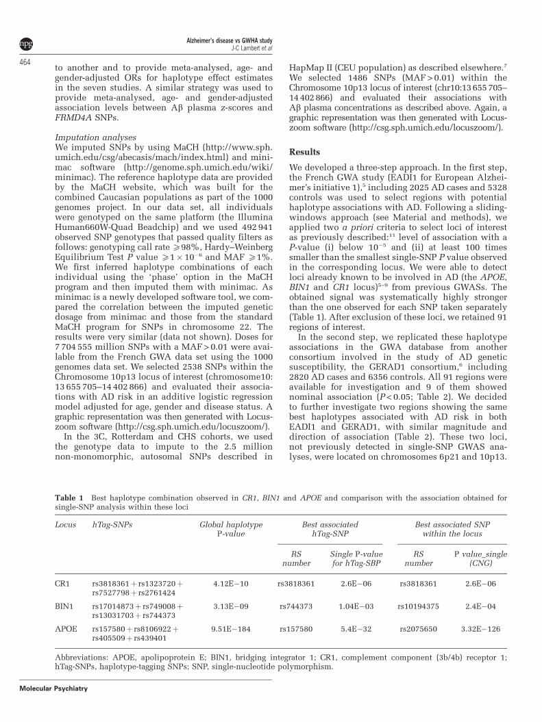
Table 1 Best haplotype combination observed in CR1, BIN1 and APOE and comparison with the association obtained forsingle-SNP analysis within these loci
Locus hTag-SNPs Global haplotypeP-value
Best associatedhTag-SNP
Best associated SNPwithin the locus
RSnumber
Single P-valuefor hTag-SBP
RSnumber
P value_single(CNG)
CR1 rs3818361þ rs1323720þrs7527798þ rs2761424
4.12E�10 rs3818361 2.6E�06 rs3818361 2.6E�06
BIN1 rs17014873þ rs749008þrs13031703þ rs744373
3.13E�09 rs744373 1.04E�03 rs10194375 2.4E�04
APOE rs157580þ rs8106922þrs405509þ rs439401
9.51E�184 rs157580 5.4E�32 rs2075650 3.32E�126
Abbreviations: APOE, apolipoprotein E; BIN1, bridging integrator 1; CR1, complement component (3b/4b) receptor 1;hTag-SNPs, haplotype-tagging SNPs; SNP, single-nucleotide polymorphism.
Alzheimer’s disease vs GWHA studyJ-C Lambert et al
464
Molecular Psychiatry
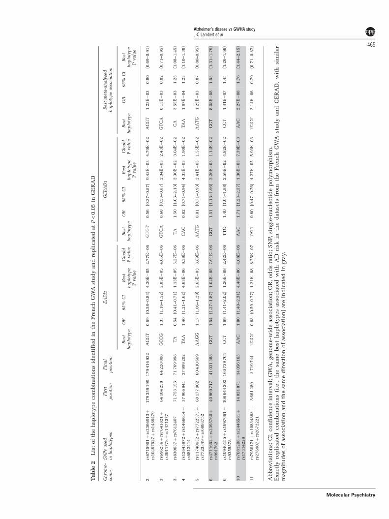
Table
2L
ist
of
the
hap
loty
pe
com
bin
ati
on
sid
en
tifi
ed
inth
eF
ren
ch
GW
Ast
ud
yan
dre
pli
cate
dat
P<
0.0
5in
GE
RA
D
Ch
rom
o-
som
eS
NP
su
sed
inh
ap
loty
pes
Fir
stp
osi
tion
Fin
al
posi
tion
EA
DI1
GE
RA
D1
Best
meta
-an
aly
sed
hap
loty
pe
ass
ocia
tion
Best
hap
loty
pe
OR
95%
CI
Best
hap
loty
pe
Pvalu
e
Glo
abl
Pvalu
eB
est
hap
loty
pe
OR
95%
CI
Best
hap
loty
pe
Pvalu
e
Glo
abl
Pvalu
eB
est
hap
loty
pe
OR
95%
CI
Best
hap
loty
pe
Pvalu
e
2rs
6715901þ
rs2366913þ
rs10497527þ
rs1489479
179
359
199
179
416
922
AC
GT
0.6
9(0
.58–0.8
3)
4.3
0E�
05
2.7
7E�
06
GT
GT
0.5
6(0
.37–0.8
7)
9.4
2E�
03
4.7
0E�
02
AC
GT
1.2
3E�
03
0.8
0(0
.69–0.9
1)
3rs
696236þ
rs7641821þ
rs3911778þ
rs1471377
64
184
258
64
228
008
GC
CG
1.3
3(1
.16–1.5
2)
2.8
5E�
05
4.6
5E�
06
GT
CA
0.6
8(0
.53–0.8
7)
2.3
4E�
03
2.4
3E�
02
GT
CA
8.1
5E�
03
0.8
2(0
.71–0.9
5)
3rs
830637þ
rs7612407
71
753
155
71
769
998
TA
0.5
4(0
.41–0.7
1)
1.1
3E�
05
5.2
7E�
06
TA
1.5
0(1
.06–2.1
3)
2.3
0E�
02
3.0
4E�
02
CA
3.5
5E�
03
1.2
5(1
.08–1.4
5)
4rs
12645972þ
rs1466614þ
rs6812414
37
966
941
37
999
202
TA
A1.4
0(1
.21–1.6
2)
4.6
3E�
06
9.3
9E�
06
CA
C0.8
2(0
.71–0.9
4)
4.3
3E�
03
1.9
0E�
02
TA
A1.9
7E�
04
1.2
3(1
.10–1.3
8)
5rs
11740632þ
rs7722373þ
rs7723349þ
rs6893752
60
177
002
60
410
669
AA
GG
1.1
7(1
.06–1.2
9)
2.6
5E�
03
8.8
9E�
06
AA
TG
0.8
1(0
.71–0.9
3)
2.4
1E�
03
1.5
5E�
02
AA
TG
1.2
5E�
03
0.8
7(0
.80–0.9
5)
6rs
4711652þ
rs2395760þ
rs991762
40
960
717
41
031
388
GG
T1.5
4(1
.27–1.8
7)
1.0
2E�
05
7.9
1E�
06
GG
T1.5
1(1
.16–1.9
6)
2.2
6E�
03
1.1
4E�
02
GG
T8.0
8E�
08
1.5
3(1
.31–1.7
9)
6rs
10946155þ
rs1997661þ
rs9355578
166
644
302
166
739
764
CC
T1.6
9(1
.41–2.0
2)
1.2
6E�
08
2.4
2E�
06
TT
C1.4
0(1
.04–1.8
8)
2.5
9E�
02
4.8
2E�
02
CC
T1.4
1E�
07
1.4
5(1
.26–1.6
6)
10
rs7081208þ
rs2446581þ
rs17314229
14
031
871
14
056
165
AA
C1.8
0(1
.40–2.3
1)
4.4
8E�
06
4.0
8E�
06
AA
C1.7
1(1
.23–2.3
7]
1.3
6E�
03
7.3
9E�
03
AA
C2.2
7E�
08
1.7
6(1
.44–2.1
5)
11
rs7950171þ
rs10834863þ
rs276907þ
rs2672221
3661
280
3719
744
TG
CT
0.6
8(0
.59–0.7
7)
1.2
1E�
08
8.7
5E�
07
TA
TT
0.6
0[0
.47–0.7
6)
4.2
7E�
05
5.9
3E�
03
TG
CT
2.1
4E�
06
0.7
9(0
.71–0.8
7)
Abbre
via
tion
s:C
I,con
fid
en
ce
inte
rval;
GW
A,
gen
om
e-w
ide
ass
ocia
tion
;O
R,
od
ds
rati
o;
SN
P,si
ngle
-nu
cle
oti
de
poly
morp
his
m.
Exactl
yre
pli
cate
dcom
bin
ati
on
s(i
.e.,
the
sam
ebest
hap
loty
pes
ass
ocia
ted
wit
hA
Dri
skin
the
data
sets
from
the
Fre
nch
GW
Ast
ud
yan
dG
ER
AD
,w
ith
sim
ilar
magn
itu
des
of
ass
ocia
tion
an
dth
esa
me
dir
ecti
on
of
ass
ocia
tion
)are
ind
icate
din
gra
y.
Alzheimer’s disease vs GWHA studyJ-C Lambert et al
465
Molecular Psychiatry
The identified AD-associated haplotypes at the6p21 region were tagged by rs2395760, rs991762 andrs4711652. The rs7081208, rs2446581 and rs17314229tagged the 10p13 haplotypes. For the Chromosome6p21 locus, the best association was attributable tothe GGT haplotype in both GWASs (OR (Odds Ratio):1.53; 95% CI: (1.31–1.79); P = 8.1�10�8 after adjust-ment for age and gender when both EADI1 andGERAD1 studies were combined). For the Chromo-some 10p13 locus, the highest level of association wasattributable to the AAC haplotype (OR: 1.76; 95% CI:(1.44–2.15); P = 2.3� 10�8 after adjustment for age andgender when both EADI1 and GERAD1 studies werecombined. Additional adjustment for the four mainprincipal components did not modify the results, datanot shown) (Table 2).
In the third step, the six tagging SNPs were furthergenotyped in five additional AD case–control studiesfrom Flanders-Belgium (842 cases and 489 controls),Finland (560 cases and 623 controls), Germany (728 casesand 961 controls), Italy (1846 cases and 904 controls) andSpain (1117 cases and 1084 controls) (SupplementaryTables 3 and 4). The same common haplotypes wereinferred from the six SNPs in the two loci, with theexception of the AGT haplotype in Chromosome 10p13in the Italian population (frequency < 0.01 in controls)(Supplementary Table 5 and 6).
In a combined analysis of the five replication datasets, the overall difference in haplotype distributionfor the 6p21 locus was not significant between ADcases and controls (P = 0.13) and the GGT haplotypewas not associated with AD risk (OR: 0.89; 95% CI:(0.74–1.08); P = 0.23 after adjustment for age andgender). We therefore considered that the haplotypeassociation in this locus was not confirmed (seeSupplementary Table 5).
Conversely, an overall significant difference inhaplotype distribution between AD cases and controls
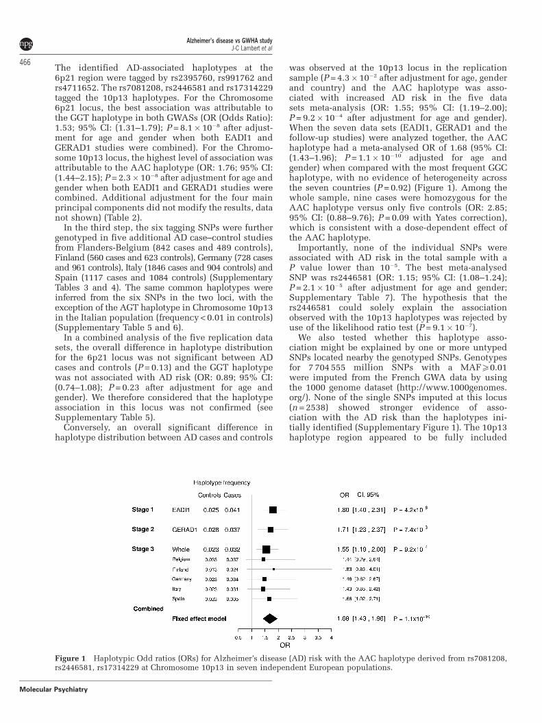
was observed at the 10p13 locus in the replicationsample (P = 4.3� 10�2 after adjustment for age, genderand country) and the AAC haplotype was asso-ciated with increased AD risk in the five datasets meta-analysis (OR: 1.55; 95% CI: (1.19–2.00);P = 9.2�10�4 after adjustment for age and gender).When the seven data sets (EADI1, GERAD1 and thefollow-up studies) were analyzed together, the AAChaplotype had a meta-analysed OR of 1.68 (95% CI:(1.43–1.96); P = 1.1� 10�10 adjusted for age andgender) when compared with the most frequent GGChaplotype, with no evidence of heterogeneity acrossthe seven countries (P = 0.92) (Figure 1). Among thewhole sample, nine cases were homozygous for theAAC haplotype versus only five controls (OR: 2.85;95% CI: (0.88–9.76); P = 0.09 with Yates correction),which is consistent with a dose-dependent effect ofthe AAC haplotype.
Importantly, none of the individual SNPs wereassociated with AD risk in the total sample with aP value lower than 10�5. The best meta-analysedSNP was rs2446581 (OR: 1.15; 95% CI: (1.08–1.24);P = 2.1�10�5 after adjustment for age and gender;Supplementary Table 7). The hypothesis that thers2446581 could solely explain the associationobserved with the 10p13 haplotypes was rejected byuse of the likelihood ratio test (P = 9.1� 10�7).
We also tested whether this haplotype asso-ciation might be explained by one or more untypedSNPs located nearby the genotyped SNPs. Genotypesfor 7 704 555 million SNPs with a MAFX0.01were imputed from the French GWA data by usingthe 1000 genome dataset (http://www.1000genomes.org/). None of the single SNPs imputed at this locus(n = 2538) showed stronger evidence of asso-ciation with the AD risk than the haplotypes ini-tially identified (Supplementary Figure 1). The 10p13haplotype region appeared to be fully included
Figure 1 Haplotypic Odd ratios (ORs) for Alzheimer’s disease (AD) risk with the AAC haplotype derived from rs7081208,rs2446581, rs17314229 at Chromosome 10p13 in seven independent European populations.
Alzheimer’s disease vs GWHA studyJ-C Lambert et al
466
Molecular Psychiatry
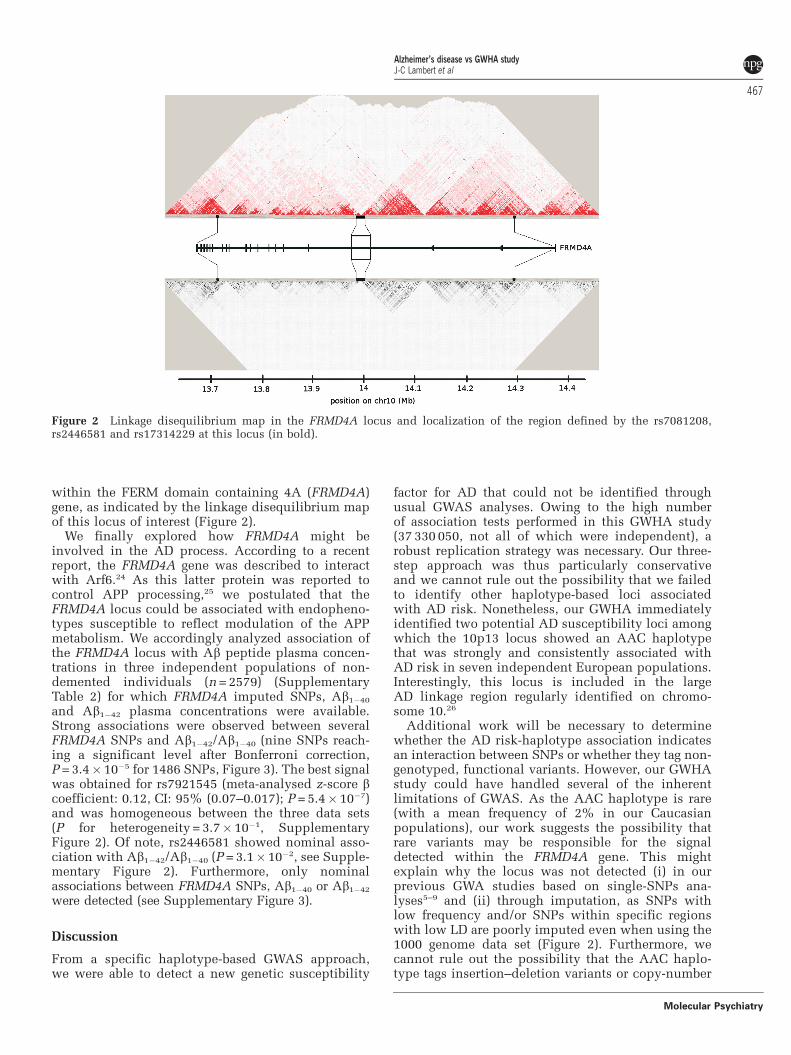
within the FERM domain containing 4A (FRMD4A)gene, as indicated by the linkage disequilibrium mapof this locus of interest (Figure 2).
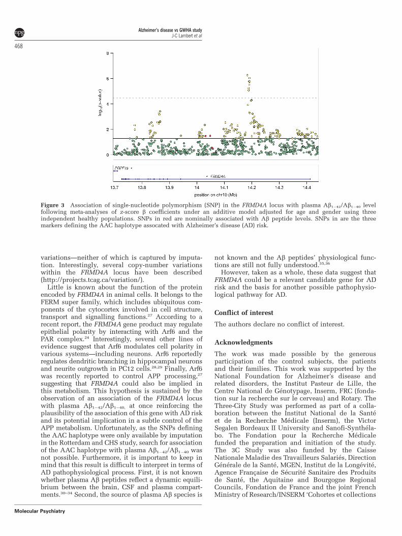
We finally explored how FRMD4A might beinvolved in the AD process. According to a recentreport, the FRMD4A gene was described to interactwith Arf6.24 As this latter protein was reported tocontrol APP processing,25 we postulated that theFRMD4A locus could be associated with endopheno-types susceptible to reflect modulation of the APPmetabolism. We accordingly analyzed association ofthe FRMD4A locus with Ab peptide plasma concen-trations in three independent populations of non-demented individuals (n = 2579) (SupplementaryTable 2) for which FRMD4A imputed SNPs, Ab1�40
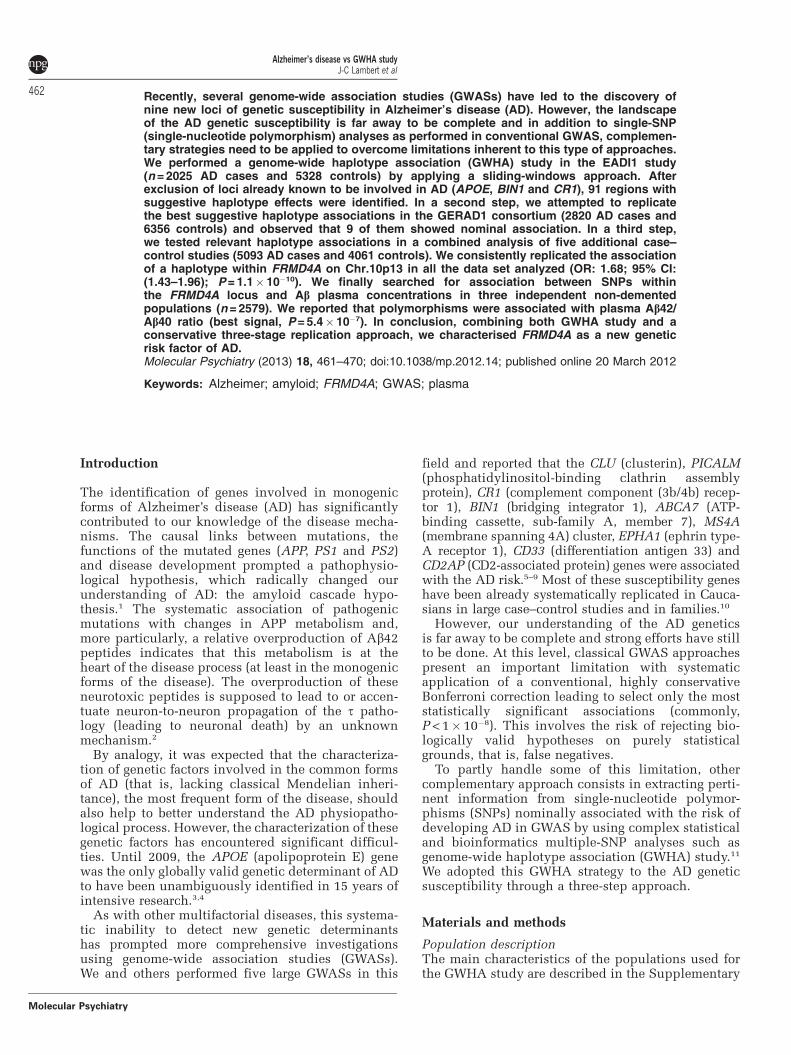
and Ab1�42 plasma concentrations were available.Strong associations were observed between severalFRMD4A SNPs and Ab1�42/Ab1�40 (nine SNPs reach-ing a significant level after Bonferroni correction,P = 3.4�10�5 for 1486 SNPs, Figure 3). The best signalwas obtained for rs7921545 (meta-analysed z-score bcoefficient: 0.12, CI: 95% (0.07–0.017); P = 5.4�10�7)and was homogeneous between the three data sets(P for heterogeneity = 3.7� 10�1, SupplementaryFigure 2). Of note, rs2446581 showed nominal asso-ciation with Ab1�42/Ab1�40 (P = 3.1�10�2, see Supple-mentary Figure 2). Furthermore, only nominalassociations between FRMD4A SNPs, Ab1�40 or Ab1�42
were detected (see Supplementary Figure 3).
Discussion
From a specific haplotype-based GWAS approach,we were able to detect a new genetic susceptibility
factor for AD that could not be identified throughusual GWAS analyses. Owing to the high numberof association tests performed in this GWHA study(37 330 050, not all of which were independent), arobust replication strategy was necessary. Our three-step approach was thus particularly conservativeand we cannot rule out the possibility that we failedto identify other haplotype-based loci associatedwith AD risk. Nonetheless, our GWHA immediatelyidentified two potential AD susceptibility loci amongwhich the 10p13 locus showed an AAC haplotypethat was strongly and consistently associated withAD risk in seven independent European populations.Interestingly, this locus is included in the largeAD linkage region regularly identified on chromo-some 10.26
Additional work will be necessary to determinewhether the AD risk-haplotype association indicatesan interaction between SNPs or whether they tag non-genotyped, functional variants. However, our GWHAstudy could have handled several of the inherentlimitations of GWAS. As the AAC haplotype is rare(with a mean frequency of 2% in our Caucasianpopulations), our work suggests the possibility thatrare variants may be responsible for the signaldetected within the FRMD4A gene. This mightexplain why the locus was not detected (i) in ourprevious GWA studies based on single-SNPs ana-lyses5–9 and (ii) through imputation, as SNPs withlow frequency and/or SNPs within specific regionswith low LD are poorly imputed even when using the1000 genome data set (Figure 2). Furthermore, wecannot rule out the possibility that the AAC haplo-type tags insertion–deletion variants or copy-number
Figure 2 Linkage disequilibrium map in the FRMD4A locus and localization of the region defined by the rs7081208,rs2446581 and rs17314229 at this locus (in bold).
Alzheimer’s disease vs GWHA studyJ-C Lambert et al
467
Molecular Psychiatry
variations—neither of which is captured by imputa-tion. Interestingly, several copy-number variationswithin the FRMD4A locus have been described(http://projects.tcag.ca/variation/).
Little is known about the function of the proteinencoded by FRMD4A in animal cells. It belongs to theFERM super family, which includes ubiquitous com-ponents of the cytocortex involved in cell structure,transport and signalling functions.27 According to arecent report, the FRMD4A gene product may regulateepithelial polarity by interacting with Arf6 and thePAR complex.24 Interestingly, several other lines ofevidence suggest that Arf6 modulates cell polarity invarious systems—including neurons. Arf6 reportedlyregulates dendritic branching in hippocampal neuronsand neurite outgrowth in PC12 cells.28,29 Finally, Arf6was recently reported to control APP processing,27
suggesting that FRMD4A could also be implied inthis metabolism. This hypothesis is sustained by theobservation of an association of the FRMD4A locuswith plasma Ab1�42/Ab1�40, at once reinforcing theplausibility of the association of this gene with AD riskand its potential implication in a subtle control of theAPP metabolism. Unfortunately, as the SNPs definingthe AAC haplotype were only available by imputationin the Rotterdam and CHS study, search for associationof the AAC haplotype with plasma Ab1�42/Ab1�40 wasnot possible. Furthermore, it is important to keep inmind that this result is difficult to interpret in terms ofAD pathophysiological process. First, it is not knownwhether plasma Ab peptides reflect a dynamic equili-brium between the brain, CSF and plasma compart-ments.30–34 Second, the source of plasma Ab species is
not known and the Ab peptides’ physiological func-tions are still not fully understood.35,36
However, taken as a whole, these data suggest thatFRMD4A could be a relevant candidate gene for ADrisk and the basis for another possible pathophysio-logical pathway for AD.
Conflict of interest
The authors declare no conflict of interest.
Acknowledgments
The work was made possible by the generousparticipation of the control subjects, the patientsand their families. This work was supported by theNational Foundation for Alzheimer’s disease andrelated disorders, the Institut Pasteur de Lille, theCentre National de Genotypage, Inserm, FRC (fonda-tion sur la recherche sur le cerveau) and Rotary. TheThree-City Study was performed as part of a colla-boration between the Institut National de la Santeet de la Recherche Medicale (Inserm), the VictorSegalen Bordeaux II University and Sanofi-Synthela-bo. The Fondation pour la Recherche Medicalefunded the preparation and initiation of the study.The 3C Study was also funded by the CaisseNationale Maladie des Travailleurs Salaries, DirectionGenerale de la Sante, MGEN, Institut de la Longevite,Agence Francaise de Securite Sanitaire des Produitsde Sante, the Aquitaine and Bourgogne RegionalCouncils, Fondation de France and the joint FrenchMinistry of Research/INSERM ‘Cohortes et collections
Figure 3 Association of single-nucleotide polymorphism (SNP) in the FRMD4A locus with plasma Ab1�42/Ab1�40 levelfollowing meta-analyses of z-score b coefficients under an additive model adjusted for age and gender using threeindependent healthy populations. SNPs in red are nominally associated with Ab peptide levels. SNPs in are the threemarkers defining the AAC haplotype assocated with Alzheimer’s disease (AD) risk.
Alzheimer’s disease vs GWHA studyJ-C Lambert et al
468
Molecular Psychiatry
de donnees biologiques’ programme. Lille Genopolereceived an unconditional grant from Eisai. Experi-ments presented in this paper were carried out usingthe EGI European grid infrastructure, supported byEGI, NGI France and Universite Lille 1. We highlyappreciate and thank the technical staff of the CRI-Lille 1 center for their strong and helpful support.Belgium sample collection: the research at the Antwerpsite was in part supported by the InteruniversityAttraction Poles program P6/43 of the Belgian SciencePolicy Office, the Foundation for Alzheimer Research(SAO-FRMA), a Methusalem Excellence Grant of theFlemish Government, the Research Foundation Flan-ders (FWO), the University of Antwerp and theSpecial Research Fund of the University of Antwerp,the Antwerp Medical Research Foundation andNeurosearch, Belgium. KS and KB are postdoctoralfellows of the FWO. The Antwerp site authors thankthe personnel of the VIB Genetic Service Facility,the Biobank of the Institute Born-Bunge and theDepartments of Neurology and Memory Clinics atthe ZNA Middelheim, ZNA Hoge Beuken and theUniversity Hospitals Leuven. Italian sample collec-tions: the Florence site was supported by a grant fromthe Italian Ministry of Health: Progetto Strategico2007- convenzione 39 (sottoprogetto 2). The Milansite was supported by grants from Italian Ministry ofHealth (PS39) and ‘Fondazione Monzino’. We thankthe expert contribution of Mr. Carmelo Romano.Spanish sample collection: the Madrid site (MB)was supported by grants of the Ministerio de Educa-cion y Ciencia and the Ministerio de Sanidad yConsumo (Instituto de Salud Carlos III), and aninstitutional grant of the Fundacion Ramon Arecesto the CBMSO. We thank I Sastre and Dr A Martınez-Garcıa for the preparation and control of the DNAcollection, and Drs P Gil and P Coria for their co-operation in the cases/controls recruitment. We aregrateful to the Asociacion de Familiares de Alzheimerde Madrid (AFAL) for continuous encouragement andhelp. The Gran Canaria site was supported by grantsof the Ministerio de Sanidad y Consumo (Instituto deSalud Carlos III), and the Fundacion Canaria deInvestigacion en Salud (FUNCIS). German samplecollection: the Saarland site at the UdS was supportedby the German National Genome Research Network(NGFN), grant 01GS08125 to MR, the HelmholtzAlliance for Mental Health in Ageing Societies(HelMA), and the Deutsche Forschungsgemeinschaft(DFG) grant INST 256/317-1 FUGG. Swedish samplecollection: the department of medical Epidemiologyand Biostatistics at the Karolinska Institute is fundedby the US National Institutes of Health (AG028555,AG08724, AG 04563, AG10175, AG08861. The samplecollection contributed by LF and CG were financiallysupported in part by the Swedish Brain Powernetwork, the Marianne and Marcus WallenbergFoundation, the Swedish Research Council (521-2010-3134), the King Gustaf V and Queen Victoria’sFoundation of Freemasons, the Regional Agree-ment on Medical Training and Clinical Research
(ALF) between Stockholm County Council and theKarolinska Institutet. We also wish to acknowledgeAnne Kinhult-Stahlbom, Charlotte Forsell, HakanThonberg and Lena Lilius for preparation of theDNA and samples.
References
1 Hardy J, Selkoe D. The amyloid hypothesis of Alzheimer’s disease:progress and problems on the road to therapeutics. Science 2002;297: 353–356.
2 Small SA, Duff K. Linking Abeta and tau in late-onsetAlzheimer’s disease: a dual pathway hypothesis. Neuron 2008;60: 534–542.
3 Lambert JC, Amouyel P. Genetic heterogeneity of Alzheimer’sdisease: complexity and advances. Psychoneuroendocrinology2007; 32: S62–S70.
4 Genin E, Hannequin D, Wallon D, Sleegers K, Hiltunen M,Combarros O et al. APOE and Alzheimer disease: a majorgene with semi-dominant inheritance. Mol Psychiatry 2011; 16:903–907.
5 Lambert JC, Heath S, Even G, Campion D, Sleegers K, Hiltunen Met al. Genome-wide association study identifies variants at CLUand CR1 associated with Alzheimer’s disease. Nat Genet 2009; 41:1094–1099.
6 Harold D, Abraham R, Hollingworth P, Sims R, Gerrish A,Hamshere ML et al. Genome-wide association study identifiesvariants at CLU and PICALM associated with Alzheimer’s disease.Nat Genet 2009; 41: 1088–1093.
7 Seshadri S, Fitzpatrick AL, Ikram MA, DeStefano AL, Gudnason V,Boada M et al. Genome-wide analysis of genetic loci associatedwith Alzheimer disease. JAMA 2010; 303: 1832–1840.
8 Hollingworth P, Harold D, Sims R, Gerrish A, Lambert JC,Carrasquillo MM et al. Common variants at ABCA7, MS4A6A/MS4A4E, EPHA1, CD33 and CD2AP are associated withAlzheimer’s disease. Nat Genet 2011; 43: 429–435.
9 Naj AC, Jun G, Beecham GW, Wang LS, Vardarajan BN, Buros Jet al. Common variants at MS4A4/MS4A6E, CD2AP, CD33and EPHA1 are associated with late-onset Alzheimer’s disease.Nat Genet 2011; 43: 436–441.
10 Lambert JC, Amouyel P. Genetics of Alzheimer’s disease: newevidences for an old hypothesis? Curr Opin Genet Dev 2011; 21:295–301.
11 Tregouet DA, Konig IR, Erdmann J, Munteanu A, Braund PS,Hall AS et al. Genome-wide haplotype association study identifiesthe SLC22A3-LPAL2-LPA gene cluster as a risk locus for coronaryartery disease. Nat Genet 2009; 41: 283–285.
12 McKhann G, Drachman D, Folstein M, Katzman R, Price D,Stadlan EM. Clinical diagnosis of Alzheimer’s disease: report ofthe NINCDS-ADRDA work group under the auspices of depart-ment of Health and Human services task force on Alzheimer’sdisease. Neurology 1984; 34: 939–944.
13 Mirra SS, Heyman A, McKeel D, Sumi SM, Crain BJ, Brownlee LMet al. The consortium to establish a registry for Alzheiemr’s disease(CERAD). Part II. Standardization of the neuropathologic assess-ment of Alzheimer’s disease. Neurology 1991; 41: 479–486.
14 Van Oijen M, Hofman A, Soares HD, Koudstaal PJ, Breteler MM.Plasma Abeta(1-40) and Abeta(1-42) and the risk of dementia:a prospective case-cohort study. Lancet Neurol 2006; 5: 655–660.
15 Lambert JC, Schraen-Maschke S, Richard F, Fievet N, Rouaud O,Berr C et al. Association of plasma amyloid beta with risk ofdementia: the prospective three-city study. Neurology 2009; 73:847–853.
16 Lopez OL, Kuller LH, Mehta PD, Becker JT, Gach HM, Sweet RAet al. Plasma amyloid levels and the risk of AD in normalsubjects in the cardiovascular health study. Neurology 2008; 70:1664–1671.
17 Mathias RA, Gao P, Goldstein JL, Wilson AF. A graphicalassessment of P-values from sliding window haplotype tests ofassociation to identify asthma susceptibility loci on chromosome11q. BMC Genet 2006; 7: 38.
Alzheimer’s disease vs GWHA studyJ-C Lambert et al
469
Molecular Psychiatry

18 Yu Z, Schaid DJ. Sequential haplotype scan methods for associa-tion analysis. Genet Epidemiol 2007; 31: 553–564.
19 Huang BE, Amos CI, Lin DY. Detecting haplotype effects in genomewide association studies. Genet Epidemiol 2007; 31: 803–812.
20 Tregouet DA, Ricard S, Nicaud V, Arnould I, Soubigou S, Rosier Met al. In-depth haplotype analysis of ABCA1 gene polymorphismsin relation to plasma ApoA1 levels and myocardial infarction.Arterioscler Thromb Vasc Biol 2004; 24: 775–781.
21 Tregouet DA, Escolano S, Tiret L, Mallet A, Golmard JL. A newalgorithm for haplotype-based association analysis: the stochastic-EM algorithm. Ann Hum Genet 2004; 68: 165–177.
22 Tregouet DA, Garelle V. A new JAVA interface implementationof THESIAS: testing haplotype effects in association studies.Bioinformatics 2007; 23: 1038–1039.
23 Gagliardi F, Jones B, Grey F, Begin ME, Heikkurinen M. Buildingan infrastructure for scientific grid computing: status and goals ofthe EGEE project. Philos Transact A Math Phys Eng Sci 2005; 363:1729–1742.
24 Ikenouchi J, Umeda M. FRMD4A regulates epithelial polarity byconnecting Arf6 activation with the PAR complex. Proc Natl AcadSci USA 2010; 107: 748–753.
25 Sannerud R, Declerck I, Peric A, Raemaekers T, Menendez G,Zhou L et al. ADP ribosylation factor 6 (ARF6) controls amyloidprecursor protein (APP) processing by mediating the endosomalsorting of BACE1. Proc Natl Acad Sci USA 2011; 108: E559–E568.
26 Hamshere ML, Holmans PA, Avramopoulos D, Bassett SS, BlackerD, Bertram L et al. Genome-wide linkage analysis of 723 affectedrelative pairs with late-onset Alzheimer’s disease. Hum Mol Genet2007; 16: 2703–2712.
27 Tepass U. FERM proteins in animal morphogenesis. Curr OpinGenet Dev 2009; 19: 357–367.
28 Hernandez-Deviez DJ, Casanova JE, Wilson JM. Regulation ofdendritic development by the ARF exchange factor ARNO. NatNeurosci 2002; 5: 623–624.
29 Albertinazzi C, Za L, Paris S, de Curtis I. ADP-ribosylationfactor 6 and a functional PIX/p95-APP1 complex are required
for Rac1B-mediated neurite outgrowth. Mol Biol Cell 2003; 14:1295–1307.
30 Mehta PD, Pirttila T, Patrick BA, Barshatzky M, Mehta SP.Amyloid beta protein 1-40 and 1-42 levels in matched cerebro-spinal fluid and plasma from patients with Alzheimer disease.Neurosci Lett 2001; 304: 102–106.
31 Vanderstichele H, Van Kerschaver E, Hesse C, Davidsson P,Buyse MA, Andreasen N et al. Standardization of measurementof beta-amyloid(1-42) in cerebrospinal fluid and plasma. Amyloid2000; 7: 245–258.
32 Kawarabayashi T, Younkin LH, Saido TC, Shoji M, Ashe KH,Younkin SG. Age-dependent changes in brain, CSF, and plasmaamyloid (beta) protein in the Tg2576 transgenic mouse model ofAlzheimer’s disease. J Neurosci 2001; 21: 372–381.
33 Ghersi-Egea JF, Gorevic PD, Ghiso J, Frangione B, Patlak CS,Fenstermacher JD. Fate of cerebrospinal fluid-borne amyloid beta-peptide: rapid clearance into blood and appreciable accumulationby cerebral arteries. J Neurochem 1996; 67: 880–883.
34 Giedraitis V, Sundelof J, Irizarry MC, Garevik N, Hyman BT,Wahlund LO et al. The normal equilibrium between CSF andplasma amyloid beta levels is disrupted in Alzheimer’s disease.Neurosci Lett 2007; 427: 127–131.
35 Soscia SJ, Kirby JE, Washicosky KJ, Tucker SM, Ingelsson M,Hyman B et al. The Alzheimer’s disease-associated amyloid beta-protein is an antimicrobial peptide. PloS One 2010; 5: e9505.
36 Lambert JC, Dallongeville J, Ellis KA, Schraen-Maschke S, Lui J,Laws S et al. Association of plasma Ab peptides with bloodpressure in the elderly. PloS One 2011; 6: e18536.
This work is licensed under the CreativeCommons Attribution-NonCommercial-
Share Alike 3.0 Unported License. To view a copyof this license, visit http://creativecommons.org/licenses/by-nc-sa/3.0/
Supplementary Information accompanies the paper on the Molecular Psychiatry website (http://www.nature.com/mp)
Alzheimer’s disease vs GWHA studyJ-C Lambert et al
470
Molecular Psychiatry
Copyright © 2022 FDOKUMEN