Pseudomonas sp. strain AKM-P6 enhances tolerance of sorghum seedlings to elevated temperatures
Upload
khangminh22Category
view
3download
0
RESEARCH ARTICLE Open Access
Genome analysis of Pseudomonas sp.OF001 and Rubrivivax sp. A210 suggestsmulticopper oxidases catalyze manganeseoxidation required for cylindrospermopsintransformationErika Berenice Martínez-Ruiz1*, Myriel Cooper1* , Jimena Barrero-Canosa1, Mindia A. S. Haryono2, Irina Bessarab2,Rohan B. H. Williams2 and Ulrich Szewzyk1
Abstract
Background: Cylindrospermopsin is a highly persistent cyanobacterial secondary metabolite toxic to humans andother living organisms. Strain OF001 and A210 are manganese-oxidizing bacteria (MOB) able to transformcylindrospermopsin during the oxidation of Mn2+. So far, the enzymes involved in manganese oxidation in strainOF001 and A210 are unknown. Therefore, we analyze the genomes of two cylindrospermopsin-transforming MOB,Pseudomonas sp. OF001 and Rubrivivax sp. A210, to identify enzymes that could catalyze the oxidation of Mn2+. Wealso investigated specific metabolic features related to pollutant degradation and explored the metabolic potentialof these two MOB with respect to the role they may play in biotechnological applications and/or in theenvironment.
Results: Strain OF001 encodes two multicopper oxidases and one haem peroxidase potentially involved in Mn2+
oxidation, with a high similarity to manganese-oxidizing enzymes described for Pseudomonas putida GB-1 (80, 83and 42% respectively). Strain A210 encodes one multicopper oxidase potentially involved in Mn2+ oxidation, with ahigh similarity (59%) to the manganese-oxidizing multicopper oxidase in Leptothrix discophora SS-1. Strain OF001and A210 have genes that might confer them the ability to remove aromatic compounds via the catechol meta-and ortho-cleavage pathway, respectively. Based on the genomic content, both strains may grow over a widerange of O2 concentrations, including microaerophilic conditions, fix nitrogen, and reduce nitrate and sulfate in anassimilatory fashion. Moreover, the strain A210 encodes genes which may convey the ability to reduce nitrate in adissimilatory manner, and fix carbon via the Calvin cycle. Both MOB encode CRISPR-Cas systems, several predictedgenomic islands, and phage proteins, which likely contribute to their genome plasticity.
© The Author(s). 2021 Open Access This article is licensed under a Creative Commons Attribution 4.0 International License,which permits use, sharing, adaptation, distribution and reproduction in any medium or format, as long as you giveappropriate credit to the original author(s) and the source, provide a link to the Creative Commons licence, and indicate ifchanges were made. The images or other third party material in this article are included in the article's Creative Commonslicence, unless indicated otherwise in a credit line to the material. If material is not included in the article's Creative Commonslicence and your intended use is not permitted by statutory regulation or exceeds the permitted use, you will need to obtainpermission directly from the copyright holder. To view a copy of this licence, visit http://creativecommons.org/licenses/by/4.0/.The Creative Commons Public Domain Dedication waiver (http://creativecommons.org/publicdomain/zero/1.0/) applies to thedata made available in this article, unless otherwise stated in a credit line to the data.
* Correspondence: [email protected];[email protected] of Environmental Microbiology, Technische Universität Berlin, Instituteof Environmental Technology, Straße des 17. Juni 135, 10623 Berlin, GermanyFull list of author information is available at the end of the article
Martínez-Ruiz et al. BMC Genomics (2021) 22:464 https://doi.org/10.1186/s12864-021-07766-0

Conclusions: The genomes of Pseudomonas sp. OF001 and Rubrivivax sp. A210 encode sequences with highsimilarity to already described MCOs which may catalyze manganese oxidation required for cylindrospermopsintransformation. Furthermore, the analysis of the general metabolism of two MOB strains may contribute to a betterunderstanding of the niches of cylindrospermopsin-removing MOB in natural habitats and their implementation inbiotechnological applications to treat water.
Keywords: Metabolic potential, Manganese-oxidizing bacteria, Biotransformation, Cyanotoxins
BackgroundCylindrospermopsin (CYN) is a secondary metaboliteproduced by several cyanobacteria, toxic for humans andother living organisms [1]. The two bacterial strainsOF001 and A210 transform the cyanotoxin CYN duringthe oxidation of Mn2+ [2, 3]. Strain OF001 belongs tothe gammaproteobacteria and was isolated from the ef-fluent of an experimental fixed-bed biofilm reactorestablished for the removal of recalcitrant substancesfrom wastewater. Strain A210 belongs to the betaproteo-bacteria and was isolated from an iron manganese-depositing biofilm in a freshwater pond in the LowerOder Valley National Park, Germany.The removal of CYN by both strains required the ac-
tive oxidation of MnCO3 whereas no or low CYN re-moval was observed with MnSO4 or in setups withoutmanganese. Sterile biogenic oxides formed by the strainsdid not show any influence on CYN removal, highlight-ing the importance of the active manganese oxidation.Both strains are able to remove 100% of CYN at thehighest rates reported for biological CYN removal so far[2, 3]. Furthermore, analysis of CYN transformationproducts revealed that the same seven transformationproducts were formed by both strains corroborating theimportant role of manganese oxidation. However, strainOF001 and A210 showed important differences. Pseudo-monas sp. strain OF001 degraded CYN within 3 days.Whereas strain A210 degraded CYN within 14 to 28days when cultivated under the same conditions. More-over, strain OF001 required yeast extract as additionalcarbon source for the removal of CYN. In contrast,strain A210 was able to transform CYN in mineralmedia [2].So far, little is known about biological CYN removal
[2–6]. Even though several bacterial strains have beenreported to remove CYN, to date, no enzymes or definedmetabolic pathway for the transformation of CYN havebeen identified [7, 8]. Moreover, for biological CYN re-moval, no transformation products have been identifiedexcept for CYN transformed by MOB [3].MOB are present in terrestrial [9], marine and fresh-
water environments [10–13], but they also occur indrinking water systems and reactors aiming at the re-moval of manganese and other pollutants [13–16]. MOBbelong to diverse phylogenetic lineages with a broad
physiological diversity (e.g. autotrophs and mixotrophs)[10, 17, 18]. Through the oxidation of Mn2+, MOB formwater-insoluble biogenic manganese oxides, which areone of the strongest natural oxidants [17, 19]. Biogenicmanganese oxides often interact with other compoundsand thus play an important role in the biogeochemicalcycle of manganese and other elements [17, 18, 20, 21].The physiological role of manganese oxidation is
not fully understood. Manganese oxidation was pro-posed to provide energy to support the growth ofbacteria. However, no conclusive results were shown[22]. Other proposed functions are the protectionagainst the toxicity of organic compounds, and react-ive oxygen species [23, 24], the breakdown of organicmatter into utilizable substrates [25, 26], and the useas a carbon reservoir [27]. Nevertheless, the precisephysiological role of manganese oxidation remains un-known [18]. Different manganese oxidation mecha-nisms have been described including non-enzymaticpathways based on a pH increase, the oxidationthrough superoxide production, or an anaerobicallyphoto-driven reaction; and enzymatic reactions gener-ally associated to the activity of multicopper oxidases(MCO) and haem peroxidases [11, 18, 28].Besides CYN, MOB transform different organic and
inorganic pollutants, including diclofenac, benzotri-azole, 17 α-ethinylestradiol, bisphenol A, As(III), andSb(III) [2, 3, 29–34]. The mechanism of pollutant trans-formation was proposed to be based on unspecific oxi-dation by reactive manganese Mn3+/Mn4+ species thatare formed through the oxidation of Mn2+ [29]. ForCYN transformation, a similar mechanism was assumedbased on the requirement of active Mn2+ oxidation forefficient CYN removal and the formation of the sametransformation products among all tested MOB, includ-ing strain OF001 and A210 [3]. Thus, it is suggestedthat MOB act as suppliers of biogenic oxides that indir-ect oxidize the pollutants. However, the intrinsic cap-acity of MOB to remove organic compounds has notbeen deeply investigated [18].The whole genome sequences of some MOB have
been analysed previously to gain a better insight into themechanism of manganese oxidation [35, 36]. However,so far, no reported pollutant-removing MOB strainswere analyzed on a genomic level. Besides, for strain
Martínez-Ruiz et al. BMC Genomics (2021) 22:464 Page 2 of 19

OF001 and A210, information about the metabolic po-tential, including also about genes potentially involvedin manganese oxidation, was missing. The genomicanalysis of strain OF001 and strain A210 might allowto identify enzymes potentially involved in the oxida-tion of manganese based on the comparison withmanganese oxidizing enzymes reported to date. Fur-thermore, metabolic differences between the twoMOB strains became evident during cultivation exper-iments in presence of CYN [2], and further dissimilar-ities could be assumed. Such metabolic differencescould be relevant for the application of the strains forthe removal of pollutants from water in technical sys-tems including but not limited to wastewater ordrinking water treatment plants, and for the under-standing of the niche they may occupy in naturalenvironments.Therefore, in this study, we analysed the draft ge-
nomes of the MOB strains OF001 and A210, both ofwhich are able to transform CYN during oxidation ofMnCO3. We aim to provide further insight into i) man-ganese oxidation mechanism, ii) other metabolic path-ways relevant for pollutant removal, iii) energyharvesting processes such as respiration, iv) their meta-bolic potential in comparison with their closest de-scribed phylogenetic relatives, and v) genome plasticityrelated to horizontal gene transfer mechanisms.
Results and discussionGeneral genome featuresGenome quality estimation determined with CheckMshowed that both genomes are of high quality (> 90%completeness and < 5% contamination). Genomes ofstrains OF001 and A210 have a completeness of 99.59and 99.38%, respectively, with a contamination level of2.12 and 0.35%.The genome sequence of strain OF001 contains 4,476,
686 bp in 65 contigs with a N50 contig length of 147,742 bp, and a GC content of 68.01%. The genome ofstrain OF001 encodes 4845 genes of which 4720 are pro-tein coding sequences (CDS). Furthermore, one 16S, one23S, and six 5S rRNA genes were identified in the gen-ome of OF001, as well as, sixty-seven tRNA genes thatenable recognition of codons for all 20 amino acids.The genome sequence of strain A210 contains 5,371,
534 bp in 72 contigs with a N50 contig length of 327,374 bp, and a GC content of 69.54%. The genome ofstrain A210 encodes 5184 genes from which 5112 areCDS. In addition, one 5S–23S-16S rRNA operon, and 52tRNA genes were identified in the genome of strainA210. Genome quality estimation and general genomicfeatures are summarized in Table 1.Genome Taxonomy Database tool kit (GTDB-tk) was
used to classify the bacterial genomes. GTDB-tk analysisclassified strain OF001 as a member of the
Table 1 Genomic features of strains OF001 and A210
OF001 A210
N50 147,742 327,374
Number of contigs 65 72
CheckM completeness 99.59% 99.38%
CheckM contamination 2.12% 0.35%
Complete genome size (bp) 4,476,686 5,371,534
Undetermined bases 6400 7100
% GC 68.01 69.54
% Protein coding density 90.64 93.09
Pseudogene 6 2
CDS 4720 5112
Genes assigned to:
COG 3436 (72.70%) 3798 (74.27%)
KEGG 2490 (52.7%) 2632 (51.5%)
Hypothetical proteins / unknown function 1327 (28.08%) 1492 (29.17%)
Fragment CDS 6 2
tRNA 67 52
rRNA 8 3
misc_RNA 43 14
tmRNA 1 1
CDS Coding sequences, COG Cluster of Orthologous Groups, KEGG Kyoto Encyclopedia of Genes and Genomes, tRNA transfer RNA, rRNA Ribosomal RNA, misc_RNAMiscellaneous RNA, tmRNA Transfer-messenger RNA
Martínez-Ruiz et al. BMC Genomics (2021) 22:464 Page 3 of 19

Pseudomonas_K group. According to the GTDB (May,2020) P. oryzae, P. sagittaria, P. linyingensis, and P.guangdongensis belong to the Pseudomonas_K group.The genus status of the strain OF001 in the Pseudo-monas_ K group was supported by the genetic related-ness determined by whole-genome analysis and 16SrRNA phylogeny (Additional file 1: Fig. S1).To determine the species affiliation of Pseudomonas
sp. OF001 average nucleotide identity (ANI), and tetra-nucleotide frequencies (TETRA) analysis were done. Theanalysis revealed highest similarity between strain OF001and P. oryzae KCTC 32247 with an ANI based onBLAST (ANIb) value of 89.06%, an ANI based onMUMmer (ANIm) value of 90.98%, and a TETRA valueof 0.998 (Fig. 1a). Organisms with an ANI value above95%, and a TETRA value above 0.99 are suggested to de-lineate the same species level [38–40]. TETRA valuesshould be in agreement with ANI values to support thespecies assignation [39]. TETRA values of strain OF001and the organisms of the Pseudomonas_K group werehigher than 0.99, but ANI values were below the specieslimit. Together, the data suggest strain OF001 is a po-tential new species of the Pseudomonas_K group.GTDB-tk analysis classified strain A210 as a member
of the Rubrivivax genus. According to the GTDB data-base this genus belongs to the order Burkholderiales andhas so far only three described species: R. benzoatilyti-cus, R. gelatinosus, and R. albus [41–43].Based on the phylogenetic analysis using the whole
16S rRNA gene sequence (Additional file 1: Fig. S2a),strain A210 could not be classified at genus level. Organ-isms with high similarity to the 16S rRNA gene se-quence of strain A210 were mainly bacteria of thegenera incertae sedis from the Comamonadaceae family(Aquabacterium, Ideonella, Leptothrix, Roseateles, Rubri-vivax, Sphaerotilus). However, phylogenomic analysis ofstrain A210 done with TYGS platform affiliated A210
with organisms of the genus Rubrivivax (Additional file1: Fig. S2b), supporting the results obtained with theGTDB-tk.ANI, and TETRA analysis were done with the genome
of A210 to analyze species affiliation. The analysisshowed the highest similarity of Rubrivivax sp. A210with R. benzoatilyticus JA2 with an ANIb value of76.69%, an ANIm value of 84.45%, and a TETRA valueof 0.913 (Fig. 1b). Thus suggesting, strain A210 is a po-tential new species of the genus Rubrivivax.
Pan and core genomeThe pan-genome of the Pseudomonas_K group genomescomprised 20,296 genes belonging to 6805 Microscopegene Families (MICFAM) [44, 45]. The core-genomecomprised 11,985 genes that correspond to 1957 MICFAM, and the variable-genome contained 8311 corre-sponding to 4848 MICFAM. The Pseudomonas sp.OF001 genome contains 1091 strain-specific genes from1052 MICFAM that correspond to 24.08% strain-specificcoding sequences. With this, strain OF001 contains thehighest number of CDS from the Pseudomonas_K groupgenomes analyzed (Additional file 1: Fig. S3a). Amongthe strain-specific genes in OF001, we found genes re-lated to mercury resistance, transport, and foreign DNA(see also section 2.5 Elements potentially acquired byhorizontal gene transfer).Pan- and core-genome size evolutions were estimated
with the four available genomes of the Pseudomonas_Kgroup and the genome of strain OF001. The curve of thepan-genome of strain OF001 and Pseudomonas_K groupdid not reach the plateau, suggesting that the pan-genome of Pseudomonas_K group is open and the se-quences of other genomes from this group might in-crease the gene pool of novel genes (Additional file 1:Fig. S4a). The plateau of the core-genome is reached
Fig. 1 Heatmap representing the degree of similarity of the MOB genomes. a Pseudomonas sp. OF001, and b Rubrivivax sp. A210. Heatmaps werederived from the average nucleotide identity (ANI) matrix based on BLAST (ANIb). Dendrogram directly reflects the degree of identity betweengenomes. ANIm: ANI based on MUMmer; TETRA: correlation indexes of the tetra-nucleotide frequencies; DDH d4: DDH calculated with theformula d4, which is the non-logarithmic version of formula d5 (used for the Fig. S1 and S2). Formula d4 is highly recommended when using draftgenomes to assure confident results [37]
Martínez-Ruiz et al. BMC Genomics (2021) 22:464 Page 4 of 19
with the five genomes selected and is composed of ap-proximately 2000 MICFAM (Additional file 1: Fig. S4b).The pan-genome of Rubrivivax genomes comprised
23,140 genes belonging to 9974 MICFAM. The core-genome comprises 10,154 genes that correspond to 1629MICFAM, and the variable-genome contains 12,986genes corresponding to 8345 MICFAM. The Rubrivivaxsp. A210 genome contains 2123 strain-specific genesfrom 2035 MICFAM that correspond to 42.98% strain-specific coding sequences (Additional file 1: Fig. S3b).Among the strain-specific genes in A210, we foundgenes related to transport like ABC transporters, and cy-tochromes (see also section 2.4.3 Aerobic respiration).Pan- and core-genome size evolutions were estimated
according to the genomes selected for the A210 analysis(Additional file 1: Fig. S4c-d). The core-genome plateauis apparently reached with the analyzed genomes and iscomposed of approximately 1600 MICFAM.
Genes potentially involved in manganese oxidationIn Pseudomonas sp. OF001, we detected three differenthomologues of manganese-oxidizing multicopper oxi-dases (MO-mco’s) (OF001_u20185, OF001_u60094, andOF001_u90046). Gene name, accession number, locustag in the evaluated genomes, E-value, and percent simi-larity of amino acid alignments are shown in Additionalfile 1: Table S1. All three MO-mco’s homologues ofstrain OF001 belong to the homologous cupredoxinsuperfamily (IPR008972), according to the InterPro-
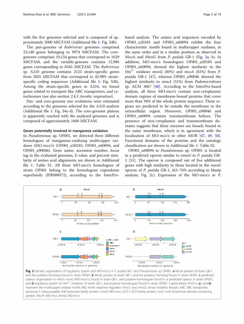
based analysis. The amino acid sequences encoded byOF001_u20185 and OF001_u60094 exhibit the fourcharacteristic motifs found in multicopper oxidases, inthe same order and in a similar position as observed inMcoA and MnxG from P. putida GB-1 (Fig. 2a, b). Inaddition, MO-mco’s homologues OF001_u20185 andOF001_u60094, showed the highest similarity to theMn2+ oxidases mnxG (80%) and mcoA (83%) from P.putida GB-1 [47], whereas OF001_u90046 showed thehighest similarity to moxA (51%) from Pedomicrobiumsp. ACM 3067 [48]. According to the InterPro-basedanalysis, all three MO-mco’s contain non-cytoplasmicdomain regions of membrane-bound proteins that covermore than 94% of the whole protein sequence. These re-gions are predicted to be outside the membrane in theextracellular region. Moreover, OF001_u90046 andOF001_u60094 contain transmembrane helixes. Thepresence of non-cytoplasmic and transmembrane do-mains suggests that these enzymes are loosely bound tothe outer membrane, which is in agreement with thelocalization of MO-mco’s in other MOB [47, 49, 50].Functional domains of the proteins and the ontologyclassification are shown in Additional file 1: Table S2.OF001_u60094 in Pseudomonas sp. OF001 is located
in a predicted operon similar to mnxG in P. putida GB-1 [51]. The operon is composed out of five additionalgenes with high similarity to those located in the mnxGoperon of P. putida GB-1, (63–76% according to blastpanalysis, Fig. 2c). Expression of the MO-mco’s in P.
Fig. 2 Genetic organization of regulatory system and MO-mco’s in P. putida GB-1 and Pseudomonas sp. OF001. a McoA protein of strain GB-1,and the putative homolog found in strain OF001, b MnxG protein of strain GB-1, and the putative homolog found in strain OF001, c predictedoperon organization in which mnxG (MO-mco) is found in strain GB-1, and putative homologues found in a predicted operon in strain OF001,and d regulatory system for Mn2+ oxidation of strain GB-1, and putative homologues found in strain OF001. Capital letters (A-D) in a), and b)represent the multicopper oxidase motifs [46]. mnxR: response regulator; mnxS1 and mnxS2: sensor histidine kinases; ABC: ABC transporter;lactonase f.: beta-propeller fold lactonase family protein; mnxG: MO-mco, SCO f. SCO family protein; SurA: SurA N-terminal domain-containingprotein, McoA: MO-mco, MnxG: MO-mco
Martínez-Ruiz et al. BMC Genomics (2021) 22:464 Page 5 of 19

putida GB-1 is regulated by a two-component pathway,mnxS1/mnxS2/mnxR [51]. In the genome of strainOF001, we found putative homologues to the mnxS2histidine kinase, and to the mnxR regulator, arranged ina similar operon structure as in P. putida GB-1 (Fig. 2d)[51]. Our results suggest that the regulation of the MO-mco’s of strain OF001 follows a similar regulation to theone observed in P. putida GB-1.Furthermore, two homologues of manganese-oxidizing
haem peroxidases (MO-hpox’s) (OF001_u100035, andOF001_u220048) were identified in strain OF001. Theputative MO-hpox homologue OF001_u100035 showedthe highest similarity with the Mn2+ oxidase mopA of P.putida GB-1 (42%). Together with the MO-hpox of A.manganoxydans SI85-9A1, they belong to the haem per-oxidase superfamily (IPR010255). The MO-hpoxhomologue OF001_ u220048, showed highest similarityto mopA of Erythrobacter sp. SD-21 (38%) and neitherof the two belong to the haem peroxidase superfamily(Additional file 1: Table S2). No cytoplasmic or non-cytoplasmic domains could be identified for the putativeMO-hpox’s homologues of strain OF001. Therefore, weevaluated the probable subcellular localization with Loc-Tree3 [52]. According to this analysis, both putativeMO-hpox’s of OF001 are likely secreted to the media(accuracy percentage 88%), similar as previously de-scribed for several MO-hpox’s of other MOB [47–50, 53,54].In the genome of Rubrivivax sp. A210, five MO-mco’s
homologues were identified (RA210_u420004, RA210_u30250, RA210_u110082, RA210_u10102, and RA210_u100111) (Additional file 1: Table S1). Two MO-hpox’shomologues (RA210_u10091, and RA210_u140033) wereidentified, but were discarded for further analysis due tovery low coverage of the query sequences (Additional file1: Table S1).All MO-mco’s homologues of strain A210 belong to
the homologous cupredoxin superfamily (IPR008972),according to the InterPro-based analysis. They contain
non-cytoplasmic domains which cover more than 84% ofthe whole protein sequence, and possess either a trans-membrane domain or a transmembrane helix, except forRA210_ u420004. This suggests that these enzymes areloosely bound to the outer membranes, similar as previ-ously reported for other MO-mco’s [47, 49, 50]. RA210_u30250 shows highest similarity (59%) to the mofA geneof L. discophora SS-1 [49]. In addition, the amino acidsequence of RA210_u30250 encodes the four character-istic motifs found in multicopper oxidases in the sameorder and in a similar position than those found inMofA from L. discophora SS-1 (Fig. 3a). In addition, it islocated in a predicted operon similar to mofA in L. dis-cophora SS-1 (Fig. 3b). The mof operon in L. discophoraSS-1 is composed out of mofA, mofB and mofC [55]. Theputative mof operon in strain A210 encodes five genes,including the putative mofA homologue, and two geneswith high similarity to mofB (68%) and mofC (60%), to-gether with a putative metallochaperon, and an exportedprotein of unknown function (RA210_u30246 – RA210_u30250).In spite of the low homology between MO-mco’s from
different organisms, we attempted to gain further evi-dence for the Mn2+ oxidation activity of the suggestedmulticopper oxidases by using a phylogenetic approach.For this purpose, a phylogenetic tree was constructedwith sequences of MO-mco and non-MO-mco retrievedform the NCBI database excluding the newly identifiedputative MO-mco homologues (Additional file 1: TableS3), to discard the possibility that the new sequenceswere the main factor driving the topology of the tree(Additional file 1: Fig. S5). Subsequently, the putativeMO-mco homologues of the strains OF001 and A210were added. Phylogenetic analysis revealed one cluster ofall MO-mco sequences and one cluster of non-MO-mco(Fig. 4). The only identified outlier was moxA from Ped-omicrobium sp. ACM 3067, a reported MO-mco, affili-ated with the non-MO-mco. Possibly, this is due to anuncertain assignation as suggested previously by
Fig. 3 Genetic organization of the MO-mco in L. discophora SS-1 and Rubrivivax sp. A210. a MofA protein of strain SS-1, and the putativehomolog found in strain A210, and b predicted operon organization in which mofA (MO-mco) is found in strain SS-1, and the putativehomologues found in a predicted operon in strain A210. Capital letters (A-D) in a) represent the multicopper oxidase motifs [46]. mofA: MO-mco;mofB: macrophage infectivity potentiator (mip); mofC: Cytochrome c domain-containing protein. Note that in a the operon in strain SS-1 isrepresented based on the total length of the operon because the genome has not been sequenced. Capital letters in b are the other twoproteins predicted within the operon of strain A210, D: copper metallochaperone, and E: protein of unknown function
Martínez-Ruiz et al. BMC Genomics (2021) 22:464 Page 6 of 19

Anderson et al. (2009). In contrast to OF001_u20185,OF001_u60094, and RA210_u30250, the proteinsencoded by OF001_u90046, and RA210_u100111 didnot cluster with the MO-mco (Fig. 4). This result sug-gests that the two annotated multicopper oxidasesOF0011_u90046 and RA210_u100111 in Pseudomonassp. OF001 and Rubrivivax sp. A210, respectively, do notpossess the Mn2+ oxidation activity. Collectively, thedata suggest that the best candidates for Mn2+ oxidationare MO-mco OF001_u20185, OF001_u60094, and MO-hpox OF001_u100035 in strain OF001 and MO-mcoRA210_u30250 in strain A210.Our results indicate that both MOB strains, OF001
and A210, oxidize manganese through enzyme-mediatedmechanisms. In spite of the evidences found based onthe genomic analysis, further experiments are requiredto determine which enzymes are involved in the oxida-tion of Mn2+ in Pseudomonas sp. OF001 and Rubrivivaxsp. A210.
General metabolismOrganic carbon metabolismPseudomonas sp. OF001 and Rubrivivax sp. A210 pos-sess all genes necessary for commonly found centralcarbohydrate metabolism in aerobic organism includingglycolysis (Embden-Meyerhof-Parnas), gluconeogenesis,tricarboxylic acid cycle (Krebs cycle), and the non-oxidative branch of the pentose phosphate pathway, tosupport basic growth. In both MOB, genes involved inthe oxidative branch of the pentose phosphate pathwaywere incomplete which is in accordance with its absencein many aerobic and thermophilic organisms [56].
CO2 fixationPseudomonas sp. OF001 possesses several genes encod-ing enzymes related to CO2 fixation via the Calvin cycle,however the key enzyme D-ribulose-1,5-bisphosphatecarboxylase/oxygenase (RuBisCO) is missing (Additionalfile 1: Table S4). This is in accordance with our previousstudy which demonstrated the growth of strain OF001only in presence of an organic carbon source [2].In contrast, Rubrivivax sp. A210 has the complete rep-
ertoire of genes required for CO2 fixation via the Calvincycle, including the RuBisCO, which is supported byprevious studies of our group showing that A210 wasable to grow in mineral media [2]. The cbb operon instrain A210 has all genes predicted to be encoded to-gether with the RuBisCO small (cbxSP) and large (cbbL)subunits (gpx, cbbYP, prkB, fbp, cbxXC). The presence ofgenes coding for enzymes of the Calvin cycle in strainA210 is in accordance with their detection in the threedescribed species of the Rubrivivax genus R. albus [42],R. gelatinosus [43] and R. benzoatilyticus [41].
Aerobic respirationAll genes for oxidative phosphorylation and aerobic res-piration were present in Pseudomonas sp. OF001.Among them, twenty genes annotated as cytochromes,including 14 c-type, 5 b-type, and 1 d-type cytochromeswere found (Additional file 1: Table S5). Also, severalpredicted terminal oxidases are present including cyto-chrome bd-type quinol oxidase, cytochrome c oxidases,and cbb3-type cytochrome c oxidases.cbb3-type cytochrome c oxidases in the genome of
strain OF001 are predicted to be organized in two
Fig. 4 Maximum Likelihood phylogenetic tree based on multicopper oxidase sequences with and without reported Mn2+ oxidation activity.Numbers in the branches represent bootstrap values. Scale bar represents sequence divergence
Martínez-Ruiz et al. BMC Genomics (2021) 22:464 Page 7 of 19

operons, one operon containing cbbP and cbbQONgenes, similar as reported for other bacteria [57–60], andone operon containing a copy of cbbPO and a gene ofunknown function. Next to the cbbPQON operon, a pre-dicted operon with three (ccoSIG) genes encoding theenzymes responsible of the assembly of cbb3-type cyto-chrome c oxidases was observed [61]. The ccoH assem-bly factor for the cbb3-type cytochrome oxidase ismissing in strain OF001, suggesting that it follows accoH-independent assembly mechanism, similar as de-scribed for H. pylori, and R. gelatinosus (Durand et al.2018).Likewise, all genes for oxidative phosphorylation and
aerobic respiration were found in Rubrivivax sp. A210.Thirty-nine genes were annotated as cytochromes, in-cluding thirty c-type, and nine b-type cytochromes. Pre-dicted terminal oxidases are present, includingcytochrome c oxidases and cbb3-type cytochrome c oxi-dases (Additional file 1: Table S5). cbb3-type cytochromec oxidases and the enzymes responsible of their assemblyin the genome of strain A210 are predicted to be orga-nized in a single operon ccoISNOQPG. Similar as forOF001, the ccoH assembly factor for the cbb3-type cyto-chrome oxidase is missing in strain A210, which suggestthat it also follows a ccoH-independent assembly mech-anism likewise to strain OF001.The presence of diverse cytochrome oxidases, with
high O2 affinity, rather than only cytochrome c oxidases,indicate the potential of strain OF001 and A210 to growunder a wide range of O2 concentrations.
Nitrogen metabolismPseudomonas sp. OF001 possesses genes predicted toparticipate in ammonium uptake, including specifictransporters like amtB and genes involved in the regula-tion of the process such as glnA, glnL, and glnK [62–67].The genes are predicted to be arranged within differentoperons, with glnA as a single regulated gene, locatedimmediately downstream from the operon encodingglnG and glnL (Additional file 1: Table S4).In addition, the genome of Pseudomonas sp. OF001
encodes the genes nifDKH, implicated in nitrogen fix-ation. Nitrogenase genes in strain OF001 are not pre-dicted to form an operon, but cluster together in thegenome. The detection of the nifDKH genes is in ac-cordance with their detection in the genomes of the twotaxonomically closest organisms to OF001, P. linyingen-sis [68] and P. sagittaria [69].Furthermore, strain OF001 encodes genes related with
assimilatory nitrate reduction, including nitrate trans-port, the ammonium-forming nitrite reductase smallsubunit nasD, and the nitrate reductase nasA [70–73].Strain OF001 also possesses two nitrite reductases, onein a predicted operon together with the nitrate reductase
nasA, and the other as an independent regulated gene(Additional file 1: Table S4). Genes involved in dissimila-tory nitrate reduction were missing. This is in agreementwith the absence of genes involved in dissimilatory ni-trate reduction in the closest related Pseudomonas_Kgroup.Similar to strain OF001, the genome of Rubrivivax sp.
A210 encodes genes predicted to participate in ammo-nium uptake in five predicted operons, including specifictransporters like amtB and genes involved in the regula-tion of the process such as glnA, glnL, and glnK [62–67](Additional file 1: Table S4). However, in contrast tostrain OF001, the glnB gene is encoded in the genome ofstrain A210. GlnB is a PII signal transcription protein,homologue to GlnK [74]. Both are key for the metabolicregulation of ammonium uptake. The presence of theglnK and glnB genes in Rubrivivax sp. A210 suggeststhat ammonium uptake in strain A210 follows a similarregulation as described for Escherichia coli [64, 75].GlnB found in Proteobacteria is commonly associatedwith glutamine synthetase genes [76], and likewise, theglnB gene of strain A210 is located in an operon struc-ture next to the glutamine synthetase nadE.The genes nifDKH, implicated in nitrogen fixation, are
encoded in the genome of Rubrivivax sp. A210 similarlyas observed for all three known Rubrivivax species [41–43]. The nifDKH genes are located in a predicted operonstructure together with a putative ferredoxin and a con-served protein of unknown function. Ferredoxin maymediate nitrogenase activity when ammonium is avail-able for uptake [77–80].Moreover, strain A210 encodes genes related with as-
similatory nitrate reduction, including nitrate transport,the ammonium-forming nitrite reductase small subunitnirB, and the nitrate reductase nasA [70–73].In contrast to strain OF001, in the genome of strain
A210 genes related with dissimilatory nitrate reductionwere detected, including nitrate reductase narGHI, andnitrite reductase nirBD. Noteworthy, in the three de-scribed species of the Rubrivivax genus [41–43], genescoding for enzymes related to dissimilatory nitrate re-duction were not detected.The enzymes related with thedissimilatory and assimilatory nitrate reduction are orga-nized in two predicted operons.The genomic data indicate that both MOB strains have
the ability to assimilate ammonium. In addition, it seemslikely that OF001 can use nitrate in an assimilatory butnot dissimilatory pathway. In contrary, the genomic datasuggest that Rubrivivax sp. A210 has not only the gen-omic potential to assimilate nitrate, but also to performanaerobic respiration using nitrate as final electron ac-ceptor. This characteristic may confer a higher flexibilityto strain A210, compared to OF001, to adapt to chan-ging conditions in technical and natural environments.
Martínez-Ruiz et al. BMC Genomics (2021) 22:464 Page 8 of 19

Sulfur metabolismPseudomonas sp. OF001 harbours all genes required forassimilatory sulfate reduction, which are organised inseveral predicted operons, and some as single regulatedgenes (Additional file 1: Table S4). Strain OF001 alsopossesses different sulfate transporters including ABCtype UWA [81], the proton: sulfate symporter or puta-tive sulfate: bicarbonate antiporter SulP [82, 83], and thehigh affinity sulfate transporter CysZ, essential for sul-fate uptake at low concentrations [84, 85]. No genes re-quired for dissimilatory sulfate reduction were detectedin the genome of strain OF001.Rubrivivax sp. A210 harbours several genes required
for assimilatory sulfate reduction, organised in three pre-dicted operons (Additional file 1: Table S4). However,A210 lacks the adenylyl sulfate kinase cysC, responsibleof the transformation of adenosine 5′-phosphosulfate(APS) to 3′-phosphoadenosine-5′-phosphosulfate(PAPS), which is an essential step in the assimilatory sul-fate reduction [86]. Nevertheless, other organisms like P.aeruginosa, Sinorhizobium meliloti, and Burkholderiacenocepacia lacking cysC, reduce APS via the phosphoa-denosine phosphosulfate reductase cysH to sulphite [87–89]. Strain A210 also possesses different sulfate trans-porters like ABC type UWA [81]. Similar to strainOF001 and other Rubrivivax species, not all genes in-volved in dissimilatory sulfate reduction were identifiedin strain A210.
Iron metabolismPseudomonas sp. OF001 possess genes predicted to par-ticipate in the transport and storage of iron, includingferrous and ferric iron transporters and bacterioferritin[90] (Additional file 1: Table S4). Strain OF001 also pos-sesses different genes related to heme uptake, such asheme-binding protein and periplasmic heme chaperone[91]. AntiSmash analysis could not detect gene clustersrelated to siderophores synthesis in strain OF001 gen-ome. Nonetheless, the search with blastp revealed thepresence of thirty-one genes related to the synthesis andtransport of siderophores. Twenty-one out of the thirty-one genes detected in the genome of strain OF001, cor-respond to siderophores transport (Additional file 1:Table S4).Similar to strain OF001, the genome of Rubrivivax sp.
A210 encodes genes predicted to participate in thetransport and storage of iron, such as ferric iron trans-porters and bacterioferritin [90] (Additional file 1: TableS4). Strain A210 also possesses different genes related toheme uptake, such as heme-binding protein and peri-plasmic heme chaperone [91]. AntiSmash could not de-tect gene clusters related to siderophores synthesis instrain A210 genome. However, the search with blastp re-vealed the presence of thirty-six genes related to the
synthesis and transport of siderophores. Twenty-two outof the thirty-six genes detected in A210 genome, corres-pond to siderophores transport (Additional file 1: TableS4).
Cell motility and biofilm formationProteins for motility, including genes related to chemo-taxis, and flagellar proteins were present in the genomeof Pseudomonas sp. OF001 (Additional file 1: Table S6).Genes encoding flagellar proteins in strain OF001 belongto the flg, and fli family, which are part of the core set offlagellar genes [92]. Among the genes related to the cen-tral signal transduction pathway for chemotaxis, wefound cheAWYBR genes, and the transmembrane che-moreceptors, methyl-accepting chemotaxis proteins(MCPs) in the genome of strain OF001. This is in agree-ment with the description of the closest relatives ofstrain OF001, P. oryzae, P. sagittaria, P. guangdongensis,and P. linyingensis, as motile bacteria [68, 69, 93, 94].Sixty-four genes associated with biofilm formation werefound in the genome of Pseudomonas sp. OF001, includ-ing siaD, bifA, and fleQ (Additional file 1: Table S6).In the genome of Rubrivivax sp. A210 genes required
for motility were present, including genes related tochemotaxis, and genes encoding flagellar proteins (Add-itional file 1: Table S6). Similar as in strain OF001, instrain A210 found genes encoding flagellar proteins be-long to the flg, and fli family. Strain A210 possessescheAWYBR genes, and the transmembrane chemorecep-tors MCPs, which are related to the central signal trans-duction pathway for chemotaxis. The taxonomicallyclosest bacteria to strain A210, R. gelatinosus, and R.benzoatilyticus, are also motile bacteria [41, 43]. How-ever, despite the presence of motility-related genes in R.albus, the absence of motility was experimentally evi-denced [42]. Therefore, further experiments are requiredto verify motility of strain A210. One hundred twentyone genes associated with biofilm formation were foundin the genome of Rubrivivax sp. A210, including sadC,pilI, and pslH. (Additional file 1: Table S6).As discussed above, both strains contain genes that en-
code proteins involve in biofilm formation, which is inagreement with the isolation of both MOB from bio-films. Moreover, both strains form biofilm in pureculture.
Organic compound degradationGenes for the aerobic degradation of aromatic com-pounds via the catechol meta-cleavage pathway, and forthe specific degradation of benzoate, phenol, and ben-zene, including phenol/toluene 2-monooxygenases(NADH), benzoate/toluate 1,2-dioxygenases, and cat-echol 2,3-dioxygenases, were detected in Pseudomonassp. OF001 (Additional file 1: Table S7). In addition,
Martínez-Ruiz et al. BMC Genomics (2021) 22:464 Page 9 of 19

strain OF001 may have the potential to transform othercompounds like 2-, 3- and 4-fluorobenzoate, toluene,steroids, citalopram, trinitrotoluene, p-methylbenzoate,trans-cinnamate, phenylpropanoate, and 4-hydroxyphenylacetate. This is in accordance with theability of several Pseudomonas spp. to transform diverseorganic pollutants such as benzoate, toluene, phenol,and poly- chlorobiphenyls (PCBs) [95].Because strain OF001 is able to degrade the cyano-
toxin CYN we were also interested in the potential ofthe strains to degrade other cyanobacterial toxins. How-ever, specific enzymes for the degradation of cyanotoxinsare described only for microcystin, the most studied cya-notoxin [7, 8]. No genes involved in microcystin degrad-ation were found in the genome of OF001. Althoughbiodegradation of CYN is considered one of the mainnatural attenuation processes [96], no specific genes in-volved in their transformation are known yet [3].Rubrivivax sp. A210 harbors genes involved in the aer-
obic degradation of aromatic compounds via the cat-echol ortho-cleavage pathway, and for the specificdegradation of benzoate, and 3- and 4-fluorobenzoate,including benzoate/toluate 1,2-dioxygenases, muconatecycloisomerase, and catechol 1,2-dioxygenase (Add-itional file 1: Table S7). Similarly, the closely-relatedstrain R. bezoatilyticus JA2 catabolizes different aromaticcompounds including benzoate [41]. Strain A210 alsohas the potential to transform other compounds such as4-methylcatechol, acrylonitrile, 2-fluorobenzoate, andtrinitrotoluene.Because strain A210 is able to degrade CYN similarly
as strain OF001, we searched the genome for genes re-lated to cyanotoxin transformation. We could not findgenes associated with the transformation of microcystin.
Elements potentially acquired by horizontal gene transferGenomic islandsGenomic islands are genomic regions potentially ob-tained by horizontal gene transfer that can drive straindifferentiation and support adaptation. Analysis ofPseudomonas sp. OF001 genome with IslandViewer 4led to the identification of at least 12 genomic islandswith size ranges from 4.2 to 70.5 Kb (Additional file 1:Table S8 and Fig. S6a). Genomic islands in strain OF001include genes associated with transposases, phage pro-teins, CRISPR systems, 147 proteins of unknown func-tion, toxin-antitoxin systems, metal-related proteins, andmercury resistance. Metal resistance genes are relatedwith environmental pollution, and specifically, mercuryresistance genes are the genes most frequently associatedwith genomic islands [97].Analysis of Rubrivivax sp. A210 genome with Island-
Viewer 4 led to the identification of at least 8 genomicislands with size ranges from 3.8 to 99.5 Kb (Additional
file 1: Table S8 and Fig. S6b). Genomic islands in strainA210 included genes associated with transposases, phageproteins, 136 proteins of unknown function, toxin-antitoxin systems, transporters, and nitrate reduction.Genes related to nitrogen metabolism associated to gen-omic island have been previously reported [98, 99].
ProphagesUsing PHASTER (PHAge Search Tool Enhanced Re-lease), we detected four incomplete and three intact pro-phage regions (Score ≥ 100) in Pseudomonas sp. OF001genome (Additional file 1: Table S9 and Fig. S7a). Thethree intact prophages were named OF001 region 2,OF001 region 5, and OF001 region 7 based on the gen-ome location retrieved by PHASTER. A summary of thedistribution and genetic features of these prophages isshown in Additional file 1: Fig. S7b. All prophages inOF001 exhibited structural proteins, including majorcapsid, fiber, and tail proteins.Based on the proteomic tree generated with the VIP-
Tree server, all complete prophages in OF001 belong tothe order Caudovirales (Additional file 1: Fig. S8).OF001 region 2 and OF001 region 5 were classified inthe family Siphoviridae, while OF001 region 7 was classi-fied in the family Myoviridae. Interestingly, OF001 re-gion 2 and OF001 region 7 display putative site-specificintegrases and excisionases, indicating site-specific re-combination [100]. Multiple prophages have alreadybeen observed in other members of the genus Pseudo-monas [101–104].Within A210 genome, two incomplete prophages of
28.8 and 9.9 kb were detected (Additional file 1: TableS9 and Fig. S7c). No complete prophage was identified.
CRISPR-Cas systemsUsing the CRISPRCas finder tool, we identified onecomplete class 1 CRISPR-Cas system, with a level ofconfidence of 4 (levels from 1 to 4, representing level 4the most confident identification [105]) in the genomeof OF001 (Additional file 1: Table S10 and S11). Theseven cas genes are downstream of the repeat/spacer re-gion. The repeats and spacers were compared with theCRISPRCas database [106] and were highly similar to se-quences found in other bacteria including some Pseudo-monads like P. stutzeri, P. aeruginosa, and Pseudomonassp. phDV1 (Additional file 1: Table S12 and S13). Fromthe taxonomically closest organisms to strain OF001,only P. guandongensis has one confirmed class 1 CRISPR/Cas system.In the genome of A210, one complete class 1 CRISPR-
Cas system, with a level of confidence of 4, and oneCRISPR region without cas genes associated, with a levelof confidence of 2 were identified (Additional file 1:Table S10 and S11). The three cas genes associated to
Martínez-Ruiz et al. BMC Genomics (2021) 22:464 Page 10 of 19

the complete CRISPR-Cas loci are downstream of the re-peat/spacer region. The repeats and spacers of the CRISPR region with a level of confidence 4 were comparedwith the CRISPRCas database [106] and, for the majorityof them, no matching were found (Additional file 1:Table S14 and S15). For the three spacers and two re-peats, only results with a similarity around 50% werefound. The organisms were these repeats and spacerswere found are Verrucomicrobium spinosum, Raphidiop-sis curvata, Pectobacterium carotovorum and Opituta-ceae bacterium. The taxonomically closest organisms tostrain A210, R. gelatinosus and R. benzoatilyticus, havetwo CRISPRs without associated cas genes, and 4 incom-plete and 2 complete CRISPR-Cas systems, respectively.Class 1 CRISPR-Cas systems, as the one found in both
MOB, are the most abundant class in Beta and Gamma-proteobacteria, and in general in archaea and bacteria[107].The presence of CRISPR-Cas systems in strain OF001
and A210 might represent protection from phage infec-tions, but could represent a disadvantage if useful genesfor competitive adaptation cannot be acquired via exter-nal DNA [108].Together the presence of genomic islands, including
phage material and CRISPR-Cas systems in Pseudo-monas sp. OF001 and Rubrivivax sp. A210 suggest thatboth MOB have undergone diverse genetic changes re-lated to different horizontal gene transfer mechanismswhich likely contribute to their genome plasticity.
Implications of the metabolic potential of strains OF001and A210In this study, we aimed at a better understanding of themetabolic capacities of the two CYN removing MOBwhich could potentially contribute to the biotechno-logical use of MOB for the removal of pollutants fromwater. In agreement with the genomes of other MOBwith so far uncharacterized degradation ability [35, 36],the content of the genomes of strain OF001 and strainA210 suggests a potential metabolic versatility and thus,a broader application potential.The genomic potential of MOB strains OF001 and
A210 for the degradation of different organic com-pounds via specific enzymatic pathways might comple-ment the unspecific transformation pathways ofsubstances like diclofenac [109] and CYN [2, 3], viamanganese oxidation. Our results suggest that strainOF001 and A210 might be able to remove different or-ganic pollutants by a coupled mechanism involving spe-cific enzymatic activity and unspecific oxidation by thereactive manganese species, as has been observed for theremoval of phenolic compounds, which are commonwastewater pollutants [110]. Moreover, it seems likelythat the MOB described in this study transform other
organic compounds like carbofuran, ciprofloxacin, and17α-ethinylestradiol, similar to other MOB [30, 111,112].Both analyzed MOB, strain OF001 and A210, trans-
form CYN indirectly through the oxidation of Mn2+ [3],and according to the results of this study most likely me-diated by the activity of multicopper oxidases and haemperoxidases. The unspecific transformation of CYN byMOB does not require an adaptation phase or a pre-conditioned towards the toxin as it is known for manyenzymatically catalyzed processes. Therefore, the use ofMOB to remove the only periodically occurring CYNmolecule, might represent an advantage in comparisonto other biological removal processes that require a pre-conditioning with the toxin to remove it. Moreover, theunspecific oxidation of organic pollutants via reactivemanganese species might allow for the removal of othercyanotoxins, however further studies are required.The different metabolic pathways encoded in the gen-
ome of strain OF001 and strain A210 also suggest differ-ent fields of application aiming at the removal ofpollutants. For instance, the ability of strain A210 tothrive and degrade CYN in the absence of an organiccarbon source suggests that it is more suitable for an ap-plication in settings, in which readily degradable organiccarbon sources are depleted, such as reactors for the re-moval of pollutants from secondary wastewater. Also,due to the metabolic potential of strain A210, it mayadapt within the reactor or the biofilm to varying oxygenconcentrations or even the depletion of oxygen by a shiftto nitrate respiration [113]. Moreover, both strains areable to form biofilms which may allow them to establishand be retained on fixed bed reactors.Pseudomonas sp. OF001 showed the highest CYN re-
moval efficiency and the fastest growth from all testedMOB [2]. Furthermore, it was isolated from a fixed-bedreactor system, however, the genome of strain OF001encodes less diverse metabolic pathways to adapt tochanging environments. Together, this data suggests thatstudies investigating degradation potential of MOBshould consider the phylogenetic and metabolic diversityof MOB to identify the most suitable organisms that ful-fil the requirements of the removal system.The metabolic diversity of strains OF001 and A210
also suggests an important role of MOB in the removalof CYN in different habitats. For instance, strain A210was isolated from a freshwater lake in the National ParkLower Oder Valley in Germany. This strain has themetabolic potential to dissimilatory reduced nitrate,which is an important mechanism to control nitrogenloading in aquatic environments [114–116]. Dissimila-tory nitrate reduction to ammonium has been related tothe promotion of eutrophic conditions in water systems,due to the release of ammonium that could be used
Martínez-Ruiz et al. BMC Genomics (2021) 22:464 Page 11 of 19

preferentially by cyanobacteria, and therefore favouringcyanobacterial blooms [116]. MOB strains with the abil-ity to denitrify and degrade CYN may be thereforetightly interconnected with the production and removalof the cyanotoxin. Furthermore, the metabolic versatilityof MOB may allow them to inhabit sediments and watercolumns. Therefore, MOB might contribute to the re-moval of CYN produced by benthic organisms in sedi-ments, but also might transform CYN produced byplanktonic cyanobacteria in the water column. However,further studies on the occurrence and distribution ofMOB in CYN contaminated environments are required.
ConclusionsIn summary, this study provides an insight into the mo-lecular basis of Mn2+ oxidation, and into the metabolicpotential of two CYN-transforming MOB strains. Weidentified sequences in Pseudomonas sp. OF001 andRubrivivax sp. A210 that show high similarity to alreadydescribed MCOs which may catalyze manganese oxida-tion required for CYN transformation. Furthermore,considering the mechanism proposed for the removal ofother pollutants by MOB the multicopper oxidasesfound in both strains and the haem peroxidase identifiedin strain OF001 might covey the ability to both strainsto transform also other pollutants susceptible to reactiveMn species. Both MOB share the potential to grow overa wide range of O2 concentrations, to fix nitrogen, andreduce nitrate and sulfate via the assimilatory pathway.Both strains encode pathways that might enable them toremove different aromatic compounds such as benzoate,benzene, and phenol. However, while strain A210 har-bors the genomic potential to fix CO2 and to reduce ni-trate as final respiratory electron acceptor, strain OF001requires additional organic carbon sources and lacks theability for dissimilatory nitrate reduction. The analysis ofthe general metabolism of two MOB strains able to re-move organic pollutants such as CYN and DCF mighthelp to implement MOB in biotechnological applicationsand contributes to a better understanding of the naturalniches of CYN-removing MOB in natural habitats.
MethodsStrains, culturing conditions and genomic DNA extractionPseudomonas sp. OF001 and Rubrivivax sp. A210 wereobtained from the culture collection of the Laboratory ofEnvironmental Microbiology from the TU Berlin,Germany [2]. Bacteria were routinely cultivated in amedium that was originaly developed for Leptothrixstrains [117], which was modified by our research groupand is known as LSM2.Cells from a pure, fresh 50mL liquid culture from
each strain were harvested by centrifugation at 15,000 xg for 3 min and washed three times with sterile Milli Q
water under sterile conditions. Total genomic DNA wasextracted using the GeneMATRIX Soil DNA PurificationKit (EURX Gdańsk, Poland) following the manufacturer’sinstructions. Quality and quantity of the extracted DNAwas determined using QubitTM fluorometric quantita-tion and NanoDrop 2000 (both Thermo Fisher Scientific,Bremen, Germany).
Genome sequencing, assembly and annotationThe genome of both MOB strains was sequenced on anIllumina MiSeq platform with a read length of 301 bp(paired end). The genome of each isolate was assembledusing SPAdes 3.10.1 and draft genomes obtained usingmanual binning procedures based on coverage-GC plotsperformed in R 3.6.1 (Additional file 1: Fig. S9). Genomequality estimation based on completeness and contamin-ation was determined with CheckM [118]. Genome an-notation was performed with the interface MagnifyingGenomes (MaGE) of the MicroScope web-based servicefrom GenoScope [119]. Protein coding genes were classi-fied based on the annotation into Cluster of OrthologousGroups (COG) functional categories [120] with the auto-matic classification COG tool at Microscope platform.Function and pathway analysis were performed usingBlastKOALA web tool of KEGG (Kyoto Encyclopedia ofGenes and Genomes) database according to the KEGGgroups of orthologs [121], and using MicroCyc tool ofthe MicroScope web-based service from GenoScope[119] which is a collection of microbial Pathway/Gen-ome databases (PGDBs). PGDBs within MicroScope aregenerated by comparing the genome annotations to themetabolic reference database MetaCyc [122]. In thepresent work, metabolic potential will refer to the possi-bility of the strains to follow a specific metabolic path-way based only on their genome information, withoutbeing so far experimentally corroborated.The data for this study have been deposited in the
European Nucleotide Archive (ENA) at EMBL-EBIunder project number PRJEB40009 with accession num-bers GCA_904426495 and GCA_904426505 for strainOF001 and A210, respectively (https://www.ebi.ac.uk/ena/browser/view/PRJEB40009).
Genomes comparisonFirst classification of the genomes was determined ac-cording to the Genome Taxonomy Database (GTDB)using the GTDB-tool kit (GTDB-tk) v.1.1.0 integrated inthe MicroScope web-based service [44, 123, 124].GTDB-tk provides a taxonomic classification of bacterialand archaeal genomes based on the combination of theGTDB reference tree, the relative evolutionary diver-gence and the ANI value against reference genomes[123]. GTDB proposed a bacterial taxonomy based onthe phylogeny inferred from the concatenation of 120
Martínez-Ruiz et al. BMC Genomics (2021) 22:464 Page 12 of 19

ubiquitous single-copy proteins that normalizes taxo-nomic ranks by using the relative evolutionary diver-gence [124]. Therefore, it is considered that the analysisperformed by GTDB-tk has an advantage over otherphylogenies currently in use [124].Genomes sequences were uploaded to the Type
strain genome server (TYGS), a free bioinformaticsplatform (https://tygs.dsmz.de) for a whole genome-based taxonomic analysis [125]. TYGS platform runsautomatically all the analysis. Briefly, TYGS performedfirst a determination of closely related type strain ge-nomes, comparing the query genome against all avail-able genomes in the TYGS database with the MASHalgorithm [126] and selecting ten type strains. Then,additionally ten close related type strains were deter-mined based on the 16S rRNA sequence extracted fromthe query genome using RNAmmer [127]. 16S rRNAsequences were compared with BLAST [128] againstthe TYGS database. The best 50 matching types wereused to calculate precise distances using the GenomeBLAST distance phylogeny approach (GBDP) [37]. Thedistances calculated by GBDP were then used to deter-mine the ten closest type strain genomes for eachquery. Afterwards, GBDP conducted all pairwise com-parisons among the set of genomes selected in the pre-vious steps, and inferred accurate intergenomicdistances under the algorithm “trimming” and distanceformula d5 [37]. One hundred distance replicates werecalculated each. In silico DNA-DNA hybridization(DDH) analysis were calculated using the recom-mended settings of the Genome-to-genome distancecalculator (GGDC) 2.1 [37]. The resulting intergenomicdistances were used to infer a balanced minimum evo-lution tree with branch support via FASTME 2.1.4 in-cluding subtree pruning and regrafting (SPR)postprocessing [129]. Branch support was inferredfrom 100 pseudo-bootstrap replicates each.JSpeciesWS [40] was used to calculate the average nu-
cleotide identity (ANI) values [40] based on BLAST(ANIb) [38, 128] and MUMmer (ANIm) [130], and tocalculate the correlation indexes of the tetra-nucleotidefrequencies (TETRA) [131].For the ANI and TETRA analysis, the genome of
strain OF001 was compared to the Pseudomonads be-longing to the Pseudomonas_K group: P. oryzae (GCA_900104805.1), P. sagittaria (GCA_900109175.1), P.guangdongensis (GCA_900105885.1), and P. liyingensis(GCA_900115715.1).For the ANI and TETRA analysis, the genome of
strain A210 was compared to the genomes of the threespecies of the genus Rubrivivax: R. benzoatilyticus JA2(GCA_000420125.1), R. gelatinosus IL144 (GCA_000284255.1), R. gelatinosus DSM 1709 (GCA_00430905.1), and R. albus ICH-03 (GCA_004016515.1).
Core- and pan-genomeDetermination of the core- and pan-genome analysiswas performed with the Pan/Core-genome tool from theMicroScope web-based service [119]. The analysis isbased on the computation of Microscope gene families(MICFAM) using a single linkage clustering algorithm ofhomologous genes sharing an amino-acid alignmentcoverage and identity above the defined threshold [45].This analysis considered i) any MICFAM associated withat least one gene from every genome used for the com-parison as a part of the core-genome, ii) any MICFAMassociated with at least 2 compared genomes as a part ofthe variable- genome, and iii) the sum of the core-genome and variable-genome as the pan-genome [44].Parameter of 50/80 was selected (50% amino-acid iden-tity, 80% amino-acid alignment coverage). All bacterialgenomes used for the comparison with the genomes ofstrain OF001 or strain A210 that were not available inthe MicroScope database were also annotated withMaGe from GenoScope [119].For the pan- and core-genome analysis, the same
strains as for the ANI and TETRA analysis, were used.
Manganese-oxidation genesWe used the blastp function on the Microscope web ser-ver [128, 132] to identify potential Mn2+ oxidases inPseudomonas sp. OF001 and Rubrivivax sp. A210, usingexperimentally verified Mn2+ oxidases of othermanganese-oxidizing bacteria. Nine sequences of multi-copper oxidases and three sequences of haem peroxi-dases related with the oxidation of Mn2+ in other MOBwere used for the search (Table S1). Multicopper oxi-dases and haem peroxidases with Mn2+ oxidation activ-ity will be referred as MO-mco and MO-hpox,respectively. We considered as homologue any proteinwith an E-value lower than 10− 10.Mn2+ oxidases and putative homologues found in
OF001 and A210 were functionally analyzed with theInterPro web server [133]. InterPro web server classifiesproteins into families, and predicts functional domainsand important sites of the proteins, integrating proteinsignatures from 13 different databases. We predicted thesub-cellular localization with LocTree3 [52] of those pu-tative homologues without a predicted cytoplasmic ornon-cytoplasmic domain according to the InterProanalysis.To determine a possible phylogenetic relationship be-
tween manganese-oxidizing multicopper oxidases (MO-mco) and non-manganese oxidizing multicopper oxi-dases (non-MO-mco), we created a dataset sequences ofmulticopper oxidases with experimental evidence ofMn2+ oxidation [47–49, 53, 134, 135] and multicopperoxidases with experimental evidence of non-Mn2+ oxida-tion activity [47, 54] and included our sequences. They
Martínez-Ruiz et al. BMC Genomics (2021) 22:464 Page 13 of 19

were aligned using MUSCLE [136] in MEGA v7.0.25. Aphylogenetic tree was constructed with Maximum Like-lihood method in MEGA v7.025. A bootstrap analysiswas performed with 1000 replicates for the MaximumLikelihood tree.
Operon predictionOperon prediction was done using the FGENESB pro-gram [137]. FGENESB gene prediction algorithm isbased on Markov chain models of coding regions, startof translation, and termination sites. Predicted genes arethen used for the operon models using distances be-tween ORFs frequencies of neighboring genes in knownbacterial genomes, and positions of predicted promotersand terminators [137].
SiderophoresIdentification of siderophore biosynthesis gene clusterswas performed with AntiSMASH tool [138] from theMicroScope web-based service. In addition, we used theblastp function on the Microscope web server [128, 132]to search for genes previously reported for the biosyn-thesis of siderophores pyoverdine, enterobactin, yersinia-bactin, ornibactin and pyochelin [139–142].
Elements potentially acquired by horizontal gene transferGenomic islandsGenomic islands were predicted with the IslandPath-DIMOB [143] and SIGI-HMM [144] method included inthe IslandViewer 4 tool using the default settings [145].Among the prediction methods included in IslandViewer4 tool, SIGI-HMM has the highest precision and overallaccuracy [146].
ProphagesPutative phages from Pseudomonas sp. OF001 andRubrivivax sp. A210 were predicted with PHASTER(PHAge Search Tool Enhanced Release) web server[147]. PHASTER classifies genome regions with a scorebelow 70 as incomplete, between 70 to 90 as question-able, and greater than 90 as complete prophages [147].The resulting complete prophage genomes were anno-
tated with multiPhATE v.1.0 (multiple-genome PhageAnnotation Toolkit and Evaluator) [148] using Phano-tate to predict ORFs [149]. PhAnToMe (Phage Annota-tion Tools and Methods), pVOGs [150], and SwissProt[151] databases were used for the identification of thehomologs of the input genomes and its predicted geneand peptide sequences. Additionally, highly divergentstructural proteins were detected with iVireons [152]and confirmed with VIRALPro [153].To classify the complete prophages, a whole proteomic
tree based on genome-wide similarities was computedby tBLASTx, using the VIPTree web server v.1.9 [154].
CRISPR-Cas systemsThe presence of Clustered Regularly Interspaced ShortPalindromic Repeats (CRISPR) and their associatedgenes (cas) was evaluated with CRISPRCasFinder [105].CRISPRCasFinder include a rating system which classi-fies the detected CRISPRs to differentiate between CRISPR-like elements and true CRISPRs. Evidence levelsfrom 1 to 4 are assigned, with 1 representing the lowestevidence classification and 4 the most confident identifi-cation [105].Spacers and repeat regions detected in both genomes
were searched with BLAST (blastn) against the CRISPRCasdb to identify their presence in other organisms.CRISPRCasdb contains CRISPR arrays and cas genesfrom complete genome sequences [106].
Data graphicsFigures were made with the R packages ggplot2 (Wick-ham, 2016), gridExtra (Auguie, 2017), pheatmap [155],VennDiagram [156], and gggenes [157], using Viridis[158] and RcolorBrewer [159] packages for colouring inRStudio version 1.0.153 [160, 161]. Genomic maps ofprophages were generated using the Snapgene® software(GSL Biotech). Trees generated with the TYGS tool andthe multicopper oxidase tree were visualized and anno-tated with the online server iTOL [162].
Supplementary InformationThe online version contains supplementary material available at https://doi.org/10.1186/s12864-021-07766-0.
Additional file 1: Fig. S1. Phylogenetic tree based on 16S rDNAsequences and whole genome sequences including strain OF001sequence. Tree inferred with FastME 2.1.6.1 [129] from GBDP distancescalculated from a) 16S rDNA gene sequences and b) genome sequences.The branch lengths are scaled in terms of GBDP distance formula d5. Thenumbers above branches are GBDP pseudo-bootstrap support values >60% from 100 replications, with an average branch support of a) 68.8%and b) 92.5%. Tree was rooted at the midpoint [163]. Bold text representthe sequences generated in the present work. Scale bar represent se-quence divergence. Fig. S2. Phylogenetic tree based on 16S rDNA se-quences and whole genome sequences including strain A210 sequence.Tree inferred with FastME 2.1.6.1 [129] from GBDP distances calculatedfrom a) 16S rDNA gene sequences and b) genome sequences. Thebranch lengths are scaled in terms of GBDP distance formula d5. Thenumbers above branches are GBDP pseudo-bootstrap support values >60% from 100 replications, with an average branch support of a) 76.8%and b) 83.4%. Tree was rooted at the midpoint [163]. Bold text representthe sequences generated in the present work. Scale bar represent se-quence divergence. Fig. S3. Pan- and core genome overview. Venn dia-gram shows the number of shared and specific Microscope gene families(MICFAM) a) among Pseudomonas sp. OF001 and the members of thePseudomonas_K group, and b) among Rubrivivax sp. A210 and the mem-bers of the Rubrivivax genus. MICFAM grouping was based on 50% aminoacid identity cut-off and at least 80% amino-acid alignment coverage.Fig. S4. Pan- and core- genome sizes estimated evolution. a, c) Numberof MICFAM families in the pan-genome size by the number of genomes,and b, d) number of MICFAM families in the core-genome by the numberof genomes. a, b) Including Pseudomonas sp. OF001, and c, d) includingRubrivivax sp. A210. Fig. S5. Maximum Likelihood phylogenetic treebased on multicopper oxidases sequences with and without reported
Martínez-Ruiz et al. BMC Genomics (2021) 22:464 Page 14 of 19

Mn2+ oxidation activity. Sequences of the studied strains in the presentstudy are not included. Numbers in the branches represent bootstrapvalue. Scale bar represent sequence divergence. Fig. S6. Putative gen-omic islands harbored by the studied MOB. a) Pseudomonas sp. OF001,and b) Rubrivivax sp. A210. Outer circle represents the genome size inMbps. Genomic islands obtained by different prediction methods arehighlighted in color. Integrated represent those islands detected by atleast one method. Fig. S7. Distribution and genetic features of pro-phages detected in Pseudomonas sp. OF001 and Rubrivivax sp. A210. a)Circular genome map of strain OF001, b) genetic features of thecomplete prophages in strain OF001, and b) circular genome map ofstrain A210. In the genome maps location of prophages are highlightedin colors depending on the completeness of the prophages (Table S9).Number assigned to each prophage region is based on the genome lo-cation retrieved by PHASTER [147]. Fig. S8. Whole proteomic tree ofPseudomonas sp. OF001 prophages based on genome-wide similaritiescomputed by tBLASTx. The tree was constructed using the VIPTree webserver v.1.9 [154]. Numbers in brackets in the figure legend represent thenumber of virus genomes. Red stars represent the three complete pro-phages of strain OF001. Scale bar represent sequence divergence. Fig.S9. Coverage-GC plots of contig properties for strain OF001 and A210. a,c) Coverage vs GC content, and b, d) coverage vs length. Both sampleshas a primary, high abundance cluster of contigs, with GC centeredaround 0.6 with arise form the primary culture populations. For strainOF001 a second contig cluster with GC centered around 0.35 at a muchlower abundance was detected, which might represent a slight DNA con-tamination. Table S1. List of genes in strains OF001 and A210 with hom-ology to putative Mn2+ oxidases from other MOB. Table S2. Functionaldomains and ontology classification of MO-mco and MO-hpox fromMOB. Table S3. List of sequences of MO-mco and non- Mn2+ oxidases.Table S4. List of genes related to the metabolic potential of Pseudo-monas sp. OF001 and Rubrivivax sp. A210. Table S5. List of cytochromegenes within Pseudomonas sp. OF001 and Rubrivivax sp. A210. Table S6.List of genes related to cell motility and biofilm formation within Pseudo-monas sp. OF001 and Rubrivivax sp. A210. Table S7. List of genes relatedto degradation of organic compounds in Pseudomonas sp. OF001 andRubrivivax sp. A210. Table S8. Genomic islands identified within Pseudo-monas sp. OF001 and Rubrivivax sp. A210 genome sequences. Table S9.Characteristics of prophage regions identified in Pseudomonas sp. OF001and Rubrivivax sp. A210 genome. Table S10. CRISPR-Cas systems de-tected within Pseudomonas sp. OF001 and Rubrivivax sp. A210 genome.Table S11. Characteristics of the cas genes detected within Pseudo-monas sp. OF001 and Rubrivivax sp. A210 genome. Table S12. Compari-son of repeats in the CRISPR of confidence level 4 found in strain OF001to CRISPRCasdb. Table S13. Comparison of spacers in the CRISPR of con-fidence level 4 found in strain OF001 to CRISPRCasdb. Table S14. Com-parison of repeats in the CRISPR of confidence level 4 found in strainA210 to CRISPRCasdb. Table S15. Comparison of spacers in the CRISPRof confidence level 4 found in strain A210 to CRISPRCasdb.
AcknowledgmentsThe LABGeM (CEA/Genoscope & CNRS UMR8030), the France Génomiqueand French Bioinformatics Institute national infrastructures (funded as part ofInvestissement d’Avenir program managed by Agence Nationale pour laRecherche, contracts ANR-10-INBS-09 and ANR-11-INBS-0013) are acknowl-edged for support within the MicroScope annotation platform.
Authors’ contributionsEBMR discussed the conceptualization and methodology of the project,extracted the DNA and performed all the bioinformatic analysis except thoserelated with the phages and the assembly of the genomes, and wrote theoriginal draft including all tables and figures. MC supervised and discussedthe conceptualization and methodology of the project, and participated inthe revision and edition of the manuscript. JBC performed the analysisrelated with the phages, and participated in the revision of the manuscript.IB coordinated the sequencing submission, and participated in the revisionof the manuscript. MASH performed the assembly of the genomes, andparticipated in the revision of the manuscript. RW provided resources for theresearch, and participated in the revision of the manuscript. US supervised,provided resources for the research, and participated in the revision and
edition of the manuscript. The author(s) read and approved the finalmanuscript.
FundingErika B. Martinez-Ruiz was supported by a research scholarship from theDAAD (Deutscher Akademischer Austauschdienst). Contributions of Mindia A.S. Haryono, Irina Bessarab, and Rohan B. H. Williams are funded by theSingapore National Research Foundation and Ministry of Education underthe Research Centre of Excellence Programme. We acknowledge support bythe German Research Foundation and the Open Access Publication Fund ofTU Berlin. Open Access funding enabled and organized by Projekt DEAL.
Availability of data and materialsThe data for this study have been deposited in the European NucleotideArchive (ENA) at EMBL-EBI under project number PRJEB40009 with accessionnumbers GCA_904426495 and GCA_904426505 for strain OF001 and A210,respectively.
Declarations
Ethics approval and consent to participateNot applicable.
Consent for publicationNot applicable.
Competing interestsThe authors declare that they have no competing interests.
Author details1Chair of Environmental Microbiology, Technische Universität Berlin, Instituteof Environmental Technology, Straße des 17. Juni 135, 10623 Berlin,Germany. 2Singapore Centre for Environmental Life Sciences Engineering,National University of Singapore, Singapore 119077, Singapore.
Received: 9 October 2020 Accepted: 3 June 2021
References1. Poniedziałek B, Rzymski P, Kokociński M. Cylindrospermopsin: water-linked
potential threat to human health in Europe. Environ Toxicol Pharmacol.2012;34(3):651–60. https://doi.org/10.1016/j.etap.2012.08.005.
2. Martínez-Ruiz EB, Cooper M, Fastner J, Szewzyk U. Manganese-oxidizingbacteria isolated from natural and technical systems removecylindrospermopsin. Chemosphere. 2020;238:124625. https://doi.org/10.1016/j.chemosphere.2019.124625.
3. Martínez-Ruiz EB, Cooper M, Al-Zeer MA, Kurreck J, Adrian L, Szewzyk U.Manganese-oxidizing bacteria form multiple cylindrospermopsintransformation products with reduced human liver cell toxicity. Sci TotalEnviron. 2020;729:138924. https://doi.org/10.1016/j.scitotenv.2020.138924.
4. Mohamed ZA, Alamri SA. Biodegradation of cylindrospermopsin toxin bymicrocystin-degrading bacteria isolated from cyanobacterial blooms.Toxicon. 2012;60(8):1390–5. https://doi.org/10.1016/j.toxicon.2012.10.004.
5. Dziga D, Kokocinski M, Maksylewicz A, Czaja-Prokop U, Barylski J.Cylindrospermopsin biodegradation abilities of Aeromonas sp. isolated fromRusałka Lake. Toxins (Basel). 2016;8:55.
6. Nybom SMK, Salminen SJ, Meriluoto JAO. Specific strains of probioticbacteria are efficient in removal of several different cyanobacterial toxinsfrom solution. Toxicon. 2008;52:214–20.
7. Kumar P, Hegde K, Brar SK, Cledon M, Kermanshahi-pour A. Potential ofbiological approaches for cyanotoxin removal from drinking water: a review.Ecotoxicol Environ Saf. 2019;172:488–503. https://doi.org/10.1016/j.ecoenv.2019.01.066.
8. Kormas KA, Lymperopoulou DS. Cyanobacterial toxin degrading bacteria:who are they? Biomed Res Int. 2013;2013:463894.
9. Yang W, Zhang Z, Zhang Z, Chen H, Liu J, Ali M, et al. Population structureof manganese-oxidizing bacteria in stratified soils and properties ofmanganese oxide aggregates under manganese-complex mediumenrichment. PLoS One. 2013;8(9):e73778. https://doi.org/10.1371/journal.pone.0073778.
Martínez-Ruiz et al. BMC Genomics (2021) 22:464 Page 15 of 19
10. Tebo BM, Johnson HA, McCarthy JK, Templeton AS. Geomicrobiology ofmanganese(II) oxidation. Trends Microbiol. 2005;13:421–8.
11. Hansel CM, Learman DR. Geomicrobiology of manganese. In: Ehrlich H,Newman D, Kappler A, editors. Ehrlich’s Geomicrobiology. 6th ed. BocaRaton: CRC Press; 2015. p. 403–33.
12. Schmidt B, Sánchez LA, Fretschner T, Kreps G, Ferrero MA, Siñeriz F, et al.Isolation of Sphaerotilus-Leptothrix strains from iron bacteria communities inTierra del Fuego wetlands. FEMS Microbiol Ecol. 2014;90:454–66.
13. Szewzyk U, Szewzyk R, Schmidt B, Braun B. Neutrophilic iron-depositingmicroorganisms. In: Flemming H-C, Wingender J, Szewzyk U, editors. Biofilmhighlights. Springer: Berlin Heidelberg; 2011. p. 63–79. https://doi.org/10.1007/978-3-642-19940-0.
14. Cerrato JM, Falkinham JO, Dietrich AM, Knocke WR, McKinney CW,Pruden A. Manganese-oxidizing and -reducing microorganisms isolatedfrom biofilms in chlorinated drinking water systems. Water Res. 2010;44:3935–45.
15. Tobiason JE, Bazilio A, Goodwill J, Mai X, Nguyen C. Manganese removalfrom drinking water sources. Curr Pollut Reports. 2016;2(3):168–77. https://doi.org/10.1007/s40726-016-0036-2.
16. Zhang Y, Zhu H, Szewzyk U, Geissen SU. Removal of pharmaceuticals inaerated biofilters with manganese feeding. Water Res. 2015;72:218–26.https://doi.org/10.1016/j.watres.2015.01.009.
17. Tebo BM, Bargar JR, Clement BG, Dick GJ, Murray KJ, Parker D, et al. Biogenicmanganese oxides: properties and mechanisms of formation. Annu RevEarth Planet Sci. 2004;32(1):287–328. https://doi.org/10.1146/annurev.earth.32.101802.120213.
18. Zhou H, Fu C. Manganese-oxidizing microbes and biogenic manganeseoxides: characterization, Mn(II) oxidation mechanism and environmentalrelevance. Rev Environ Sci Bio/Technol. 2020;1(3):489–507. https://doi.org/10.1007/s11157-020-09541-1.
19. Hennebel T, De Gusseme B, Boon N, Verstraete W. Biogenic metals inadvanced water treatment. Trends Biotechnol. 2009;27:90–8.
20. Lee S, Xu H. XRD and TEM studies on nanophase manganese oxides infreshwater ferromanganese nodules from Green Bay, Lake Michigan. ClaysClay Miner. 2016;64:523–36.
21. Stein LY, La Duc MT, Grund TJ, Nealson KH. Bacterial and archaealpopulations associated with freshwater ferromanganous micronodules andsediments. Environ Microbiol. 2001;3:10–8.
22. Kepkay PE, Nealson KH. Growth of a manganese oxidizing Pseudomonas sp.in continuous culture. Arch Microbiol. 1987;148:63–7.
23. Banh A, Chavez V, Doi J, Nguyen A, Hernandez S, Ha V, et al. Manganese(Mn) oxidation increases intracellular Mn in Pseudomonas putida GB-1. PLoSOne. 2013;8:e77835.
24. Zerfaß C, Christie-Oleza JA, Soyer OS. Manganese oxide biomineralizationprovides protection against nitrite toxicity in a cell-density-dependentmanner. Appl Environ Microbiol. 2019;85:5–8.
25. Jones ME, Nico PS, Ying S, Regier T, Thieme J, Keiluweit M. Manganese-driven carbon oxidation at oxic-anoxic interfaces. Environ Sci Technol. 2018;52:12349–57.
26. Sunda WG, Kieber DJ. Oxidation of humic substances by manganese oxidesyields low-molecular-weight organic substrates. Nature. 1994;367:62–4.
27. Estes ER, Andeer PF, Nordlund D, Wankel SD, Hansel CM. Biogenicmanganese oxides as reservoirs of organic carbon and proteins in terrestrialand marine environments. Geobiology. 2017;15:158–72.
28. Geszvain K, Butterfield C, Davis RE, Madison AS, Lee S-W, Parker DL, et al.The molecular biogeochemistry of manganese(II) oxidation. Biochem SocTrans. 2012;40(6):1244–8. https://doi.org/10.1042/BST20120229.
29. Meerburg F, Hennebel T, Vanhaecke L, Verstraete W, Boon N. Diclofenacand 2-anilinophenylacetate degradation by combined activity of biogenicmanganese oxides and silver. Microb Biotechnol. 2012;5(3):388–95. https://doi.org/10.1111/j.1751-7915.2011.00323.x.
30. Tran TN, Kim DG, Ko SO. Synergistic effects of biogenic manganese oxideand Mn(II)-oxidizing bacterium Pseudomonas putida strain MnB1 on thedegradation of 17 α-ethinylestradiol. J Hazard Mater. 2018;344:350–9.https://doi.org/10.1016/j.jhazmat.2017.10.045.
31. Watanabe J, Tani Y, Chang J, Miyata N, Naitou H, Seyama H. As(III) oxidationkinetics of biogenic manganese oxides formed by Acremonium strictumstrain KR21-2. Chem Geol. 2013;347:227–32. https://doi.org/10.1016/j.chemgeo.2013.03.012.
32. Wang H, Lv Z, Song Y, Wang YN, Zhang D, Sun Y, et al. Adsorptive removalof Sb(III) from wastewater by environmentally-friendly biogenic manganese
oxide (BMO) materials: efficiency and mechanisms. Process Saf Environ Prot.2019;124:223–30. https://doi.org/10.1016/j.psep.2019.02.022.
33. Wang R, Wang S, Tai Y, Tao R, Dai Y, Guo J, et al. Biogenic manganeseoxides generated by green algae Desmodesmus sp. WR1 to improvebisphenol a removal. J Hazard Mater. 2017;339:310–9. https://doi.org/10.1016/j.jhazmat.2017.06.026.
34. Sochacki A, Felis E, Bajkacz S, Kalka J, Michalska JK. Removal andtransformation of benzotriazole in manganese-oxide biofilters with Mn(II)feeding. Chemosphere. 2018;212:143–51. https://doi.org/10.1016/j.chemosphere.2018.08.092.
35. Dick GJ, Podell S, Johnson HA, Rivera-Espinoza Y, Bernier-Latmani R,McCarthy JK, et al. Genomic insights into Mn(II) oxidation by the marinealphaproteobacterium Aurantimonas sp. strain SI85-9A1. Appl EnvironMicrobiol. 2008;74:2646–58.
36. Wang X, Yu M, Wang L, Lin H, Li B, Xue CX, et al. Comparative genomic andmetabolic analysis of manganese-oxidizing mechanisms in Celeribactermanganoxidans DY25T: its adaptation to the environment of polymetallicnodules. Genomics. 2020;112(2):2080–91. https://doi.org/10.1016/j.ygeno.2019.12.002.
37. Meier-Kolthoff JP, Auch AF, Klenk HP, Göker M. Genome sequence-basedspecies delimitation with confidence intervals and improved distancefunctions. BMC Bioinformatics. 2013;14(1):60. https://doi.org/10.1186/1471-2105-14-60.
38. Goris J, Konstantinidis KT, Klappenbach JA, Coenye T, Vandamme P, TiedjeJM. DNA-DNA hybridization values and their relationship to whole-genomesequence similarities. Int J Syst Evol Microbiol. 2007;57:81–91.
39. Richter M, Rosselló-Móra R. Shifting the genomic gold standard for theprokaryotic species definition. Proc Natl Acad Sci U S A. 2009;106:19126–31.
40. Richter M, Rosselló-Móra R, Oliver Glöckner F, Peplies J. JSpeciesWS: a webserver for prokaryotic species circumscription based on pairwise genomecomparison. Bioinformatics. 2016;32(6):929–31. https://doi.org/10.1093/bioinformatics/btv681.
41. Ramana CV, Sasikala C, Arunasri K, Anil Kumar P, Srinivas TNR, Shivaji S,et al. Rubrivivax benzoatilyticus sp. nov., an aromatic hydrocarbon-degradiing purple betaproteobacterium. Int J Syst Evol Microbiol. 2006;56:2157–64.
42. Sheu SY, Li ZH, Young CC, Chen WM. Rubrivivax albus sp. nov., isolated froma freshwater pond. Int J Syst Evol Microbiol. 2020;70:805–13.
43. Nagashima S, Kamimura A, Shimizu T, Nakamura-Isaki S, Aono E, SakamotoK, et al. Complete genome sequence of phototrophic betaproteobacteriumRubrivivax gelatinosus IL144. J Bacteriol. 2012;194:3541–2.
44. Vallenet D, Calteau A, Cruveiller S, Gachet M, Lajus A, Josso A, et al.MicroScope in 2017: an expanding and evolving integrated resource forcommunity expertise of microbial genomes. Nucleic Acids Res. 2017;45:D517–28.
45. Miele V, Penel S, Duret L. Ultra-fast sequence clustering from similaritynetworks with SiLiX. BMC Bioinformatics. 2011;12:116.
46. Spiro TG, Bargar JR, Sposito G, Tebo BM. Bacteriogenic manganese oxides.Acc Chem Res. 2008;43:2–9.
47. Geszvain K, McCarthy JK, Tebo BM. Elimination of manganese(II, III) oxidationin Pseudomonas putida GB-1 by a double knockout of two putativemulticopper oxidase genes. Appl Environ Microbiol. 2013;79:357–66. https://doi.org/10.1128/AEM.01850-12.
48. Ridge JP, Lin M, Larsen EI, Fegan M, Mcewan AG, Sly LI. A multicopperoxidase is essential for manganese oxidation and laccase-like activity inPedomicrobium sp. ACM 3067. Environ Microbiol. 2007;9(4):944–53. https://doi.org/10.1111/j.1462-2920.2006.01216.x.
49. Corstjens PLAM, Devrind JPM, Goosen T, de Vrind-de Jong EW. Identification andmolecular analysis of the Leptothrix discophora SS-1 mofA gene, a gene putativelyencoding a manganese-oxidizing protein with copper domains. Geomicrobiol J.1997;14(2):91–108. https://doi.org/10.1080/01490459709378037.
50. Dick GJ, Torpey JW, Beveridge TJ, Tebo BM. Direct identification of abacterial manganese(II) oxidase, the multicopper oxidase MnxG, from sporesof several different marine Bacillus species. Appl Environ Microbiol. 2008;74:1527–34.
51. Geszvain K, Tebo BM. Identification of a two-component regulatorypathway essential for Mn(II) oxidation in Pseudomonas putida GB-1. ApplEnviron Microbiol. 2010;76:1224–31.
52. Goldberg T, Hecht M, Hamp T, Karl T, Yachdav G, Ahmed N, et al. LocTree3prediction of localization. Nucleic Acids Res. 2014;42:350–5.
Martínez-Ruiz et al. BMC Genomics (2021) 22:464 Page 16 of 19

53. Geszvain K, Smesrud L, Tebo BM. Identification of a third Mn(II) oxidaseenzyme in Pseudomonas putida GB-1. Appl Environ Microbiol. 2016;82(13):3774–82. https://doi.org/10.1128/AEM.00046-16.
54. Anderson CR, Johnson HA, Caputo N, Davis RE, Torpey JW, Tebo BM. Mn(II)oxidation is catalyzed by heme peroxidases in Aurantimonasmanganoxydans strain SI85-9A1 and Erythrobacter sp. strain SD-21. ApplEnviron Microbiol. 2009;75:4130–8.
55. Brouwers GJ, Corstjens PLAM, De Vrind JPM, Verkamman A, De Kuyper M,De Vrind-De Jong EW. Stimulation of Mn2+ oxidation in Leptothrixdiscophora SS-1 by Cu2+ and sequence analysis of the region flanking thegene encoding putative multicopper oxidase MofA. Geomicrobiol J. 2000;17:25–33.
56. Stincone A, Prigione A, Cramer T, Wmelink MM, Campbell K, Cheung E, et al.The return of metabolism: biochemistry and physiology of the pentosephosphate pathway. Biol Rev Camb Philos Soc. 2015;90(3):927–63. https://doi.org/10.1111/brv.12140.
57. Ekici S, Pawlik G, Lohmeyer E, Koch HG, Daldal F. Biogenesis of cbb3-typecytochrome c oxidase in Rhodobacter capsulatus. Biochim Biophys Acta.1817;2012(2):898–910. https://doi.org/10.1038/jid.2014.371.
58. Koch HG, Hwang O, Daldal F. Isolation and characterization of Rhodobactercapsulatus mutants affected in cytochrome cbb3 oxidase activity. J Bacteriol.1998;180:969–78.
59. Kulajta C, Thumfart JO, Haid S, Daldal F, Koch HG. Multi-step assemblypathway of the cbb3-type cytochrome c oxidase complex. J Mol Biol. 2006;355:989–1004.
60. Preisig O, Zufferey R, Hennecke H. The Bradyrhizobium japonicum fixGHISgenes are required for the formation of the high-affinity cbb3-typecytochrome oxidase. Arch Microbiol. 1996;165(5):297–305. https://doi.org/10.1007/s002030050330.
61. Durand A, Bourbon ML, Steunou AS, Khalfaoui-Hassani B, Legrand C,Legrand A, et al. Biogenesis of the bacterial cbb3 cytochrome c oxidase:active subcomplexes support a sequential assembly model. J Biol Chem.2018;293:808–18.
62. Atkinson MR, Ninfa AJ. Characterization of the GlnK protein of Escherichiacoli. Mol Microbiol. 1999;32(2):301–13. https://doi.org/10.1046/j.1365-2958.1999.01349.x.
63. Blauwkamp TA, Ninfa AJ. Physiological role of the GlnK signal transductionprotein of Escherichia coli: survival of nitrogen starvation. Mol Microbiol.2002;46:203–14.
64. Gosztolai A, Schumacher J, Behrends V, Bundy JG, Heydenreich F, BennettMH, et al. GlnK facilitates the dynamic regulation of bacterial nitrogenassimilation. Biophys J. 2017;112:2219–30.
65. Magasanik B. Genetic control of nitrogen assimilation in bacteria. Annu RevGenet. 1982;16:135–68.
66. Pahel G, Rothstein DM, Magasanik B. Complex glnA-glnL-glnG operon ofEscherichia coli. J Bacteriol. 1982;150:202–13.
67. Schreier HJ, Sonenshein AL. Altered regulation of the glnA gene inglutamine synthetase mutants of Bacillus subtilis. J Bacteriol. 1986;167(1):35–43. https://doi.org/10.1128/JB.167.1.35-43.1986.
68. He WH, Wang YN, Du X, Zhou Y, Jia B, Bian J, et al. Pseudomonas linyingensissp. nov.: a novel bacterium isolated from wheat soil subjected to long-termherbicides application. Curr Microbiol. 2012;65:595–600.
69. Lin SY, Hameed A, Liu YC, Hsu YH, Lai WA, Chen WM, et al. Pseudomonassagittaria sp. nov., a siderophore-producing bacterium isolated from oil-contaminated soil. Int J Syst Evol Microbiol. 2013;63:2410–7.
70. Mohan SB, Cole JA. The dissimilatory reduction of nitrate to ammonia byanaerobic bacteria. In: Bothe H, Ferguson SJ, Newton WE, editors. Biology ofthe Nitrogen Cycle. Elsevier B.V.; 2007. p. 93–106.
71. Kawasaki S, Arai H, Kodama T, Igarashi Y. Gene cluster for dissimilatory nitritereductase (nir) from Pseudomonas aeruginosa: sequencing and identification ofa locus for heme d1 biosynthesis. J Bacteriol. 1997;179:235–42.
72. Ramos F, Blanco G, Gutiérrez JC, Luque F, Tortolero M. Identification of anoperon involved in the assimilatory nitrate-reducing system of Azotobactervinelandii. Mol Microbiol. 1993;8(6):1145–53. https://doi.org/10.1111/j.1365-2958.1993.tb01659.x.
73. Yukioka Y, Tanahashi T, Shida K, Oguchi H, Ogawa S, Saito C, et al. A role ofnitrite reductase (NirBD) for NO homeostatic regulation in Streptomycescoelicolor A3(2). FEMS Microbiol Lett. 2017;364:1–6.
74. Arcondéguy T, Jack R, Merrick M. PII signal transduction proteins,pivotal players in microbial nitrogen control. Microbiol Mol Biol Rev.2001;65:80–105.
75. Yurgel SN, Rice J, Mulder M, Kahn ML. GlnB/GlnK PII proteins and regulationof the Sinorhizobium meliloti Rm1021 nitrogen stress response andsymbiotic function. J Bacteriol. 2010;192(10):2473–81. https://doi.org/10.1128/JB.01657-09.
76. Sant’Anna FH, Trentini DB, De Souto WS, Cecagno R, Da Silva SC, Schrank IS.The PII superfamily revised: a novel group and evolutionary insights. J MolEvol. 2009;68:322–36.
77. Egener T, Martin DE, Sarkar A, Reinhold-Hurek B. Role of a ferredoxin genecotranscribed with the nifHDK operon in N2 fixation and nitrogenase“switch-off” of Azoarcus sp. strain BH72. J Bacteriol. 2001;183:3752–60.
78. Martin AE, Burgess BK, Iismaa SE, Smartt CT, Jacobson MR, Dean DR.Construction and characterization of an Azotobacter vinelandii strain withmutations in the genes encoding flavodoxin and ferredoxin I. J Bacteriol.1989;171(6):3162–7. https://doi.org/10.1128/jb.171.6.3162-3167.1989.
79. Poudel S, Colman DR, Fixen KR, Ledbetter RN, Zheng Y, Pence N, et al.Electron transfer to nitrogenase in different genomic and metabolicbackgrounds. J Bacteriol. 2018;200:1–19.
80. Yakunin AF, Gennaro G, Hallenbeck PC. Purification and properties of a nif-specific flavodoxin from the photosynthetic bacterium Rhodobactercapsulatus. J Bacteriol. 1993;175:6775–80.
81. Sirko A, Zatyka M, Sadowy E, Hulanicka D. Sulfate and thiosulfate transportin Escherichia coli K-12: evidence for a functional overlapping of sulfate- andthiosulfate-binding proteins. J Bacteriol. 1995;177(14):4134–6. https://doi.org/10.1128/JB.177.14.4134-4136.1995.
82. Loughlin P, Shelden MC, Tierney ML, Howitt SM. Structure and function of amodel member of the SulP transporter family. Cell Biochem Biophys. 2002;36(2-3):183–90. https://doi.org/10.1385/CBB:36:2-3:183.
83. Van De Kamp M, Pizzinini E, Vos A, Van Der Lende TR, Schuurs TA, NewbertRW, et al. Sulfate transport in Penicillium chrysogenum: cloning andcharacterization of the sutA and sutB genes. J Bacteriol. 1999;181:7228–34.
84. Parra F, Britton P, Castle C, Jones-Mortimer MC, Kornberg HL. Two separategenes involved in sulphate transport in Escherichia coli K12. J Gen Microbiol.1983;129:357–8.
85. Rückert C, Koch DJ, Rey DA, Albersmeier A, Mormann S, Pühler A, et al.Functional genomics and expression analysis of the Corynebacteriumglutamicum fpr2-cycIXHDNYZ gene cluster involved in assimilatory sulphatereduction. BMC Genomics. 2005;6:1–18.
86. Kushkevych I, Cejnar J, Treml J, Dordević D, Kollar P, Vítězová M. Recentadvances in metabolic pathways of sulfate reduction in intestinal bacteria.Cells. 2020;9:698.
87. Bick JA, Dennis JJ, Zylstra GJ, Nowack J, Leustek T. Identification of a newclass of 5′-adenylylsulfate (APS) reductases from sulfate-assimilating bacteria.J Bacteriol. 2000;182(1):135–42. https://doi.org/10.1128/JB.182.1.135-142.2000.
88. Abola AP, Willits MG, Wang RC, Long SR. Reduction of adenosine-5′-phosphosulfate instead of 3′-phosphoadenosine- 5′-phosphosulfate incysteine biosynthesis by Rhizobium meliloti and other members of thefamily Rhizobiaceae. J Bacteriol. 1999;181:5280–7.
89. Iwanicka-Nowicka R, Zielak A, Cook AM, Thomas MS, Hryniewicz MM.Regulation of sulfur assimilation pathways in Burkholderia cenocepacia:identification of transcription factors CysB and SsuR and their role in controlof target genes. J Bacteriol. 2007;189:1675–88.
90. Rivera M. Bacterioferritin: structure, dynamics, and protein-proteininteractions at play in iron storage and mobilization. Acc Chem Res. 2017;50(2):331–40. https://doi.org/10.1021/acs.accounts.6b00514.
91. Tong Y, Guo M. Bacterial heme-transport proteins and their heme-coordination modes. Arch Biochem Biophys. 2009;481(1):1–15. https://doi.org/10.1016/j.abb.2008.10.013.
92. Liu R, Ochman H. Stepwise formation of the bacterial flagellar system. ProcNatl Acad Sci U S A. 2007;104:7116–21.
93. Yu Z, Chang M, Wu M, Yang G, Zhou S, Zhuang L. Pseudomonas oryzae sp.nov. isolated from a paddy soil in South China. Arch Microbiol. 2013;195(12):815–22. https://doi.org/10.1007/s00203-013-0930-6.
94. Yang G, Han L, Wen J, Zhou S. Pseudomonas guangdongensis sp. nov.,isolated from an electroactive biofilm, and emended description of thegenus Pseudomonas Migula 1894. Int J Syst Evol Microbiol. 2013;63 PART 12:4599–605.
95. Nogales J, García JL, Díaz E. Degradation of aromatic compounds inPseudomonas: a systems biology view. In: Rojo F, editor. Aerobic utilizationof hydrocarbons, oils and lipids. Handbook of hydrocarbon and lipidmicrobiology. Springer International Publishing AG; 2017. https://doi.org/10.1007/978-3-319-39782-5_32-1.
Martínez-Ruiz et al. BMC Genomics (2021) 22:464 Page 17 of 19

96. de la Cruz AA, Hiskia A, Kaloudis T, Chernoff N, Hill D, Antoniou MG, et al. Areview on cylindrospermopsin: the global occurrence, detection, toxicityand degradation of a potent cyanotoxin. Environ Sci Process Impacts. 2013;15(11):1979–2003. https://doi.org/10.1039/c3em00353a.
97. Reva ON, Bezuidt O. Distribution of horizontally transferred heavy metalresistance operons in recent outbreak bacteria. Mob Genet Elements. 2012;2:96–100.
98. Loux V, Mariadassou M, Almeida S, Chiapello H, Hammani A, Buratti J, et al.Mutations and genomic islands can explain the strain dependency of sugarutilization in 21 strains of Propionibacterium freudenreichii. BMC Genomics.2015;16:1–19.
99. Zhu B, Zhang X, Zhao C, Chen S, Yang S. Comparative genome analysis ofmarine purple sulfur bacterium Marichromatium gracile YL28 reveals thediverse nitrogen cycle mechanisms and habitat-specific traits. Sci Rep. 2018;8:1–11. https://doi.org/10.1038/s41598-018-36160-2.
100. Grindley NDF, Whiteson KL, Rice PA. Mechanisms of site-specificrecombination. Annu Rev Biochem. 2006;75:567–605.
101. Martínez-García E, Jatsenko T, Kivisaar M, de Lorenzo V. FreeingPseudomonas putida KT2440 of its proviral load strengthens endurance toenvironmental stresses. Environ Microbiol. 2014;17:76–90.
102. Dziewit L, Radlinska M. Two inducible prophages of an antarcticPseudomonas sp. ANT_H14 use the same capsid for packaging theirgenomes - characterization of a novel phage helper-satellite system. PLoSOne. 2016;11:1–27.
103. Paulsen IT, Press CM, Ravel J, Kobayashi DY, Myers GSA, Mavrodi DV, et al.Complete genome sequence of the plant commensal Pseudomonasfluorescens pf-5. Nat Biotechnol. 2005;23:873–8.
104. de Sousa LP. Mobile genetic elements in Pseudomonas stutzeri. CurrMicrobiol. 2020;77:179–84. https://doi.org/10.1007/s00284-019-01812-7.
105. Couvin D, Bernheim A, Toffano-Nioche C, Touchon M, Michalik J, Néron B,et al. CRISPRCasFinder, an update of CRISRFinder, includes a portableversion, enhanced performance and integrates search for Cas proteins.Nucleic Acids Res. 2018;46(W1):W246–51. https://doi.org/10.1093/nar/gky425.
106. Pourcel C, Touchon M, Villeriot N, Vernadet JP, Couvin D, Toffano-Nioche C,et al. CRISPRCasdb a successor of CRISPRdb containing CRISPR arrays andcas genes from complete genome sequences, and tools to download andquery lists of repeats and spacers. Nucleic Acids Res. 2020;48:D535–44.
107. Makarova KS, Wolf YI, Iranzo J, Shmakov SA, Alkhnbashi OS, Brouns SJJ, et al.Evolutionary classification of CRISPR–Cas systems: a burst of class 2 andderived variants. Nat Rev Microbiol. 2020;18:67–83.
108. Bhaya D, Davison M, Barrangou R. CRISPR-Cas Systems in Bacteria andArchaea: versatile small RNAs for adaptive defense and regulation. Annu RevGenet. 2011;45:273–97.
109. Nega M. Effects of micropollutants on the Panke River microbialcommunities and degradation of selected pharmaceuticals by manganese-oxidizing bacteria. Technische Universität Berlin; 2020. http://dx.doi.org/10.14279/depositonce-10452.
110. Wang G, Liu Y, Wu M, Zong W, Yi X, Zhan J, et al. Coupling the phenolicoxidation capacities of a bacterial consortium and in situ-generatedmanganese oxides in a moving bed biofilm reactor (MBBR). Water Res.2019;166:115047. https://doi.org/10.1016/j.watres.2019.115047.
111. Tu J, Yang Z, Hu C, Qu J. Characterization and reactivity of biogenicmanganese oxides for ciprofloxacin oxidation. J Environ Sci (China). 2014;26(5):1154–61. https://doi.org/10.1016/S1001-0742(13)60505-7.
112. Liu Z, Wang J, Qian S, Wang G, Wang J, Liao S. Carbofuran degradation bybiogenic manganese oxides. Bull Environ Contam Toxicol. 2017;98:420–5.https://doi.org/10.1007/s00128-016-1940-2.
113. Wu Y, Shukal S, Mukherjee M, Cao B. Involvement in denitrification isbeneficial to the biofilm lifestyle of Comamonas testosteroni: a mechanisticstudy and its environmental implications. Environ Sci Technol. 2015;49(19):11551–9. https://doi.org/10.1021/acs.est.5b03381.
114. Burgin AJ, Hamilton SK, Gardner WS, McCarthy MJ. Nitrate reduction,denitrification, and dissimilatory nitrate reduction to ammonium in wetlandsediments; 2015. p. 519–37.
115. Wang S, Pi Y, Song Y, Jiang Y, Zhou L, Liu W, et al. Hotspot of dissimilatory nitratereduction to ammonium (DNRA) process in freshwater sediments of riparianzones. Water Res. 2020;173:115539. https://doi.org/10.1016/j.watres.2020.115539.
116. Li X, Song C, Zhou Z, Xiao J, Wang S, Yang L, et al. Comparison ofcommunity and function of dissimilatory nitrate reduction to ammonium(DNRA) bacteria in Chinese Shallow Lakes with different eutrophicationdegrees. Water (Switzerland). 2020;12:1–15.
117. Atlas R. Handbook of microbiological media, fourth edition. Fourth edi.Washington, D. C: Taylor & Francis Group, LLC; 2010. https://doi.org/10.1201/EBK1439804063.
118. Parks DH, Imelfort M, Skennerton CT, Hugenholtz P, Tyson GW. CheckM:assessing the quality of microbial genomes recovered from isolates, singlecells, and metagenomes. Genome Res. 2015;25:1043–55.
119. Vallenet D, Belda E, Calteau A, Cruveiller S, Engelen S, Lajus A, et al.MicroScope - an integrated microbial resource for the curation andcomparative analysis of genomic and metabolic data. Nucleic Acids Res.2013;41(D1):D636–47. https://doi.org/10.1093/nar/gks1194.
120. Tatusov RL. The COG database: a tool for genome-scale analysis of proteinfunctions and evolution. Nucleic Acids Res. 2000;28:33–6.
121. Kanehisa M, Sato Y, Morishima K. BlastKOALA and GhostKOALA: KEGG toolsfor functional characterization of genome and metagenome sequences. JMol Biol. 2016;428:726–31. https://doi.org/10.1016/j.jmb.2015.11.006.
122. Caspi R, Billington R, Fulcher CA, Keseler IM, Kothari A, Krummenacker M,et al. The MetaCyc database of metabolic pathways and enzymes. NucleicAcids Res. 2018;46(D1):D633–9. https://doi.org/10.1093/nar/gkx935.
123. Chaumeil P-A, Mussig AJ, Hugenholtz P, Parks DH. GTDB-Tk: a toolkit toclassify genomes with the genome taxonomy database. Bioinformatics.2019;36:1925–7.
124. Parks DH, Chuvochina M, Waite DW, Rinke C, Skarshewski A, Chaumeil PA,et al. A standardized bacterial taxonomy based on genome phylogenysubstantially revises the tree of life. Nat Biotechnol. 2018;36(10):996–1004.https://doi.org/10.1038/nbt.4229.
125. Meier-Kolthoff JP, Göker M. TYGS is an automated high-throughput platformfor state-of-the-art genome-based taxonomy. Nat Commun. 2019;10.https://doi.org/10.1038/s41467-019-10210-3.
126. Ondov BD, Treangen TJ, Melsted P, Mallonee AB, Bergman NH, Koren S,et al. Mash: fast genome and metagenome distance estimation usingMinHash. Genome Biol. 2016;17:1–14. https://doi.org/10.1186/s13059-016-0997-x.
127. Lagesen K, Hallin P, Rødland EA, Stærfeldt HH, Rognes T, Ussery DW.RNAmmer: consistent and rapid annotation of ribosomal RNA genes.Nucleic Acids Res. 2007;35:3100–8.
128. Camacho C, Coulouris G, Avagyan V, Ma N, Papadopoulos J, Bealer K, et al.BLAST+: architecture and applications. BMC Bioinformatics. 2009;10:1–9.
129. Lefort V, Desper R, Gascuel O. FastME 2.0: a comprehensive, accurate, andfast distance-based phylogeny inference program. Mol Biol Evol. 2015;32:2798–800.
130. Kurtz S, Phillippy A, Delcher AL, Smoot M, Shumway M, Antonescu C, et al.Versatile and open software for comparing large genomes. Genome Biol.2004;5(2):R12. https://doi.org/10.1186/gb-2004-5-2-r12.
131. Teeling H, Meyerdierks A, Bauer M, Amann R, Glöckner FO. Application oftetranucleotide frequencies for the assignment of genomic fragments.Environ Microbiol. 2004;6:938–47.
132. Vallenet D, Calteau A, Dubois M, Amours P, Bazin A, Beuvin M, et al.MicroScope: an integrated platform for the annotation and exploration ofmicrobial gene functions through genomic, pangenomic and metaboliccomparative analysis. Nucleic Acids Res. 2020;48:D579–89.
133. Mitchell AL, Attwood TK, Babbitt PC, Blum M, Bork P, Bridge A, et al. InterProin 2019: improving coverage, classification and access to protein sequenceannotations. Nucleic Acids Res. 2019;47(D1):D351–60. https://doi.org/10.1093/nar/gky1100.
134. Francis CA, Tebo BM. Enzymatic manganese(II) oxidation by metabolicallydormant spores of diverse Bacillus species. Appl Environ Microbiol. 2002;68:874–80.
135. Tang W, Liu Y, Gong J, Chen S, Zeng X. Analysis of manganese oxidase andits encoding gene in Lysinibacillus strain MK-1. Process Saf Environ Prot.2019;127:299–305. https://doi.org/10.1016/j.psep.2019.04.002.
136. Edgar RC. MUSCLE: multiple sequence alignment with high accuracy andhigh throughput. Nucleic Acids Res. 2004;32(5):1792–7. https://doi.org/10.1093/nar/gkh340.
137. Solovyev V, Salamov A. Automatic annotation of microbial genomes andmetagenomic sequences. In: Li RW, editor. Metagenomics and itsapplications in agriculture, biomedicine and environmental studies: NovaScience Publishers; 2011. p. 61–78. https://www.researchgate.net/publication/259450599_V_Solovyev_A_Salamov_2011_Automatic_Annotation_of_Microbial_Genomes_and_Metagenomic_Sequences_In_Metagenomics_and_its_Applications_in_Agriculture_Biomedicine_and_Environmental_Studies_Ed_RW_Li_Nova_Sc.
Martínez-Ruiz et al. BMC Genomics (2021) 22:464 Page 18 of 19

138. Blin K, Shaw S, Steinke K, Villebro R, Ziemert N, Lee SY, et al. AntiSMASH 5.0:updates to the secondary metabolite genome mining pipeline. NucleicAcids Res. 2019;47(W1):W81–7. https://doi.org/10.1093/nar/gkz310.
139. Schalk IJ, Rigouin C, Godet J. An overview of siderophore biosynthesisamong fluorescent pseudomonads and new insights into their complexcellular organization. Environ Microbiol. 2020;22:1447–66.
140. Crosa JH, Walsh CT. Genetics and assembly line enzymology of siderophorebiosynthesis in bacteria. Microbiol Mol Biol Rev. 2002;66(2):223–49. https://doi.org/10.1128/MMBR.66.2.223-249.2002.
141. Khan A, Singh P, Srivastava A. Synthesis, nature and utility of universal ironchelator – Siderophore: a review. Microbiol Res. 2018;212–213:103–11.https://doi.org/10.1016/j.micres.2017.10.012.
142. Visser MB, Majumdar S, Hani E, Sokol PA. Importance of the ornibactin andpyochelin siderophore transport systems in Burkholderia cenocepacia lunginfections. Infect Immun. 2004;72:2850–7.
143. Hsiao W, Wan I, Jones SJ, Brinkman FSL. IslandPath: aiding detection ofgenomic islands in prokaryotes. Bioinformatics. 2003;19:418–20.
144. Waack S, Keller O, Asper R, Brodag T, Damm C, Fricke WF, et al. Score-basedprediction of genomic islands in prokaryotic genomes using hidden Markovmodels. BMC Bioinformatics. 2006;7:1–12.
145. Bertelli C, Laird MR, Williams KP, Lau BY, Hoad G, Winsor GL, et al.IslandViewer 4: expanded prediction of genomic islands for larger-scaledatasets. Nucleic Acids Res. 2017;45:W30–5.
146. Langille MGI, Hsiao WWL, Brinkman FSL. Evaluation of genomic islandpredictors using a comparative genomics approach. BMC Bioinformatics.2008;9:1–10.
147. Arndt D, Grant JR, Marcu A, Sajed T, Pon A, Liang Y, et al. PHASTER: a better,faster version of the PHAST phage search tool. Nucleic Acids Res. 2016;44(W1):W16–21. https://doi.org/10.1093/nar/gkw387.
148. Ecale Zhou CL, Malfatti S, Kimbrel J, Philipson C, McNair K, Hamilton T, et al.multiPhATE: bioinformatics pipeline for functional annotation of phageisolates. Bioinformatics. 2019;35:4402–4.
149. Mcnair K, Zhou C, Dinsdale EA, Souza B, Edwards RA, Hancock J. PHANOTATE: anovel approach to gene identification in phage genomes. Bioinformatics. 2019;35(22):4537–42. https://doi.org/10.1093/bioinformatics/btz265.
150. Grazziotin AL, Koonin EV, Kristensen DM. Prokaryotic virus orthologousgroups (pVOGs): a resource for comparative genomics and protein familyannotation. Nucleic Acids Res. 2017;45:D491–8.
151. Bairoch A, Apweiler R. The SWISS-PROT protein sequence database and itssupplement TrEMBL in 2000. Nucleic Acids Res. 2000;28:45–8.
152. Seguritan V, Alves N, Arnoult M, Raymond A, Lorimer D, Burgin AB, et al.Artificial neural networks trained to detect viral and phage structuralproteins. PLoS Comput Biol. 2012;8:e1002657.
153. Galiez C, Magnan CN, Coste F, Baldi P. VIRALpro: a tool to identify viralcapsid and tail sequences. Bioinformatics. 2016;32(9):1405–7. https://doi.org/10.1093/bioinformatics/btv727.
154. Nishimura Y, Yoshida T, Kuronishi M, Uehara H, Ogata H, Goto S. ViPTree:the viral proteomic tree server. Bioinformatics. 2017;33:2379–80.
155. Kolde R. pheatmap: Pretty heatmaps. R package version 1.0.8; 2015.156. Chen H, Boutros PC. VennDiagram: a package for the generation of highly-
customizable Venn and Euler diagrams in R. BMC Bioinformatics. 2011;12:35.157. Wilkins D. gggenes: Draw gene arrow maps in “ggplot2.” R package version
0.4.0. 2019. https://cran.r-project.org/package=gggenes.158. Garnier S. viridis: Default color maps from “matplotlib”. R package version 0.
5.1. 2018. https://cran.r-project.org/package=viridis.159. Neuwirth E. RColorBrewer: ColorBrewer palettes. R package version 1.1–2.
2014. https://cran.r-project.org/package=RColorBrewer.160. R Core Team. R: A language and environment for statistical computing.
2020. https://www.r-project.org/.161. R Core Team. RStudio: Integrated development for R. v1.0.153. 2016. http://
www.rstudio.com/.162. Letunic I, Bork P. Interactive tree of life (iTOL) v4: recent updates and new
developments. Nucleic Acids Res. 2019;47:256–9.163. Farris JS. Estimating phylogenetic trees from distance matrices. Am Nat.
1972;106(951):645–68. https://doi.org/10.1086/282802.
Publisher’s NoteSpringer Nature remains neutral with regard to jurisdictional claims inpublished maps and institutional affiliations.
Martínez-Ruiz et al. BMC Genomics (2021) 22:464 Page 19 of 19
Copyright © 2022 FDOKUMEN