HEAVY METAL ACCUMULATING AND ENZYME SECRETING NOVEL PSEUDOMONAS SP. FROM EAST CALCUTTA WETLAND:...
18
HEAVY METAL ACCUMULATING AND ENZYME SECRETING NOVEL PSEUDOMONAS SP. FROM EAST CALCUTTA WETLAND: IMPLICATIONS FOR ENVIRONMENTAL SUSTAINANCE Madhusmita Mishra 1 , P. R. Rout 1 , S. Mohapatra 1 , M. Sudarshan 2 , A. R. Thakur 3 and Shaon Ray Chaudhuri 1* ABSTRACT Microbial enrichments from soil and water samples collected from different sites of East Calcutta Wetland (ECW) produced three bacterial isolates which were identified as Pseudomonas based on 16S rRNA sequence analysis. They were further examined for their ability to tolerate heavy metals like lead (Pb + ), chromium (Cr +++ ), copper (Cu ++ ) etc.. Energy Dispersive X Ray Fluorescence analysis, Transmission Electron Microscopy and Scanning Electron Microscopy were used for understanding the metal microbe interaction. The presence of Super Oxide Dismutase (SOD) gene explains the defense mechanism against the oxygen radical and the metal induced stress. From the point of environmental sustainance and commercial application, the extracellular enzymes protease and lipase from one of the isolates were tried out as dehairing agents for the treatment of goat hide for possible replacement of the conventional method of using harmful chemicals. Key words: East Calcutta Wetland, heavy metal, super oxide dismutase, extracellular enzymes, dehairing. 1 Department of Biotechnology, West Bengal University of Technology, BF 142, Salt Lake, Sector 1, Kolkata-700064 2 UGC-DAE Consortium for Scientific Research, Calcutta Center, III/LB-8, Bidhan Nagar, Calcutta-700064. 3 Vice Chancellor’s Office, West Bengal State University (Barasat, North 24 Parganas), Berunanpukuria, P.O. Malikapur, North 24 Parganas, Calcutta-700126, India Author for correspondence : [email protected], +91-33-23370731, +91- 33-23341030.
Transcript of HEAVY METAL ACCUMULATING AND ENZYME SECRETING NOVEL PSEUDOMONAS SP. FROM EAST CALCUTTA WETLAND:...
HEAVY METAL ACCUMULATING ANDENZYME SECRETING NOVEL
PSEUDOMONAS SP. FROM EAST CALCUTTAWETLAND: IMPLICATIONS FOR
ENVIRONMENTAL SUSTAINANCE
Madhusmita Mishra1, P. R. Rout1, S. Mohapatra1, M. Sudarshan2,A. R. Thakur3 and Shaon Ray Chaudhuri1*
ABSTRACTMicrobial enrichments from soil and water samples collected from different
sites of East Calcutta Wetland (ECW) produced three bacterial isolates which wereidentified as Pseudomonas based on 16S rRNA sequence analysis. They were furtherexamined for their ability to tolerate heavy metals like lead (Pb+), chromium (Cr+++),copper (Cu++) etc.. Energy Dispersive X Ray Fluorescence analysis, TransmissionElectron Microscopy and Scanning Electron Microscopy were used for understandingthe metal microbe interaction. The presence of Super Oxide Dismutase (SOD) geneexplains the defense mechanism against the oxygen radical and the metal inducedstress. From the point of environmental sustainance and commercial application, theextracellular enzymes protease and lipase from one of the isolates were tried out asdehairing agents for the treatment of goat hide for possible replacement of theconventional method of using harmful chemicals.
Key words: East Calcutta Wetland, heavy metal, super oxide dismutase, extracellularenzymes, dehairing.
1 Department of Biotechnology, West Bengal University of Technology, BF 142, SaltLake, Sector 1, Kolkata-7000642 UGC-DAE Consortium for Scientific Research, Calcutta Center, III/LB-8, Bidhan Nagar,Calcutta-700064.3 Vice Chancellor’s Office, West Bengal State University (Barasat, North 24 Parganas),Berunanpukuria, P.O. Malikapur, North 24 Parganas, Calcutta-700126, India
Author for correspondence : [email protected], +91-33-23370731, +91-33-23341030.
1. INTRODUCTIONThe aqueous as well as surface discharges of heavy metals have introduced
a toxicological risk to all life forms on earth (Goyer, 1997; Barceloux, 1999; Cervantes,2001). The biological agents like microbes, are of considerable interest for thedevelopment of safe, economical and environment friendly methods for the cause ofenvironment detoxification and sustainance. Several aerobic bacteria belonging togenus Pseudomonas, Shewanella, Bacillus, Cellulomonas have been reported toinfluence the remediation of several toxic metals like Pb and Cr (Ekundayo and Killham,2001; Mattuschka et al., 1994; Ackerley et al., 2004; Camargo et al., 2003; Chowdhuryet al., 2008). Significant research has been made towards understanding the cellularmechanism of metal uptake and the role of cellular design in metal adsorption.(Beveridge and Grahm, 1991; Gadd, 2000). It is not only the microbes that were appliedfor the environment reclamation from toxic wastes, but also many microbial enzymesare presently used in production process with an objective to reduce energy and rawmaterial consumption, avoid the use of harmful chemicals as well as reduce the loadof waste generated (Alcade et al., 2006).
In the study reported here, the bacterial isolates were obtained by enrichmentof soil and water samples collected from different sites of a wetland ecosystem ofKolkata, viz. East Calcutta Wetland. It is worth mentioning that this wetland whichcovers an area of about 12500 hectares function as the sewage dumping ground forthe entire city. At the same time it acts as a resource recovery system where waste isrecycled and used in production of paddy, vegetables as well as fish (Ghosh 1999;Ghosh 2005; Bhattacharyya et al., 2003; Ray Chaudhuri et al., 2008a, 2008b). Thevast population of microbes, planktons as well as plants existing in this ecosystemoperate the natural mechanism of remediation (Ray Chaudhuri et al., 2008; Pradhanet al., 2008; Ray Chaudhuri and Thakur, 2006). The presence of diverse bacterialpopulation in the wetland system has been demonstrated by culture independentapproaches (RayChaudhuri and Thakur, 2006) and the significant finding was the closeresemblance of majority of the novel sequences to groups like Actinobacteria,Proteobacteria and Firmicutes. These data reinforce the idea of exploring the richmicrobial resource for selective screening of potential microbes and most importantlythe development of a strategy for their application in environmental sustainance. Themain objectives of this study were i) to investigate the heavy metal tolerance andaccumulation efficiency of the isolates ii) to quantify the extent of intracellularaccumulation followed by the detection of their internal localization by the methodsof Energy Dispersive X ray Fluorescence (EDXRF) and Transmission ElectronMicroscopy (TEM) respectively iii) to evaluate the effect of metal induced stress onthe cytoskeletal structure of the cells exposed to different heavy metals by theScanning Electron Microscopy iv) to analyze the efficiency of extracellular enzymesprotease and lipase in dehairing of goat hide, the strategy was to develop analternative method to chemical mediated conventional process.
217 Microbial Biotechnology: Methods and Applications

2. THE ORIGIN SITE OF THE MICROBESThe soil and water samples were obtained from three different sites of ECW,
i) the old solid dumping grounds that were later converted to the recreational centerwith forest like ecosystem and was thereby designated as green zone, ii) theCharakdanga Bheri, a swallow flat bottomed sewage fed fish pond and iii) itsassociated Raw Sewage Canal.
3. CULTURE CONDITIONS FOR SELECTIVE ENRICHMENT OF MICROBESFROM ENVIRONMENTAL SAMPLES
The study aimed at isolation of microbes which can have significantapplication in bioremediation, the main focus was for organic and heavy metalpollutants. Isolation and purification were performed by dilution plating of sampleson carbon minimal salt (CMS) medium [K2HPO4 2.2 g L-1, KH2PO4 0.73 g L-1, (NH4)SO4
1 g L-1 , NaCl 30 g L-1 , MgSO4 0.2g L-1 , Oil 15 ml L-1]. This selective medium wasemployed to screen microbes having potential of utilizing different oils (bothvegetative and mineral) as their primary growth substrates. The cultivation conditionsand the regular maintenance of the isolates were carried out as previously described(Adarsh et al., 2007).
4. CHARACTERIZATION OF THE PURE ISOLATES
The preliminary characterization (morphological, biochemical, physiological,molecular nature) of the pure isolates were carried out using methods previouslydescribed by Sarkar et al., (2008). Three pure bacterial isolates were obtained whichappeared as gram negative rods having about 1-2.5 m length and 0.4-0.6 m diameter.The details regarding the source of isolation are presented in Table 1.
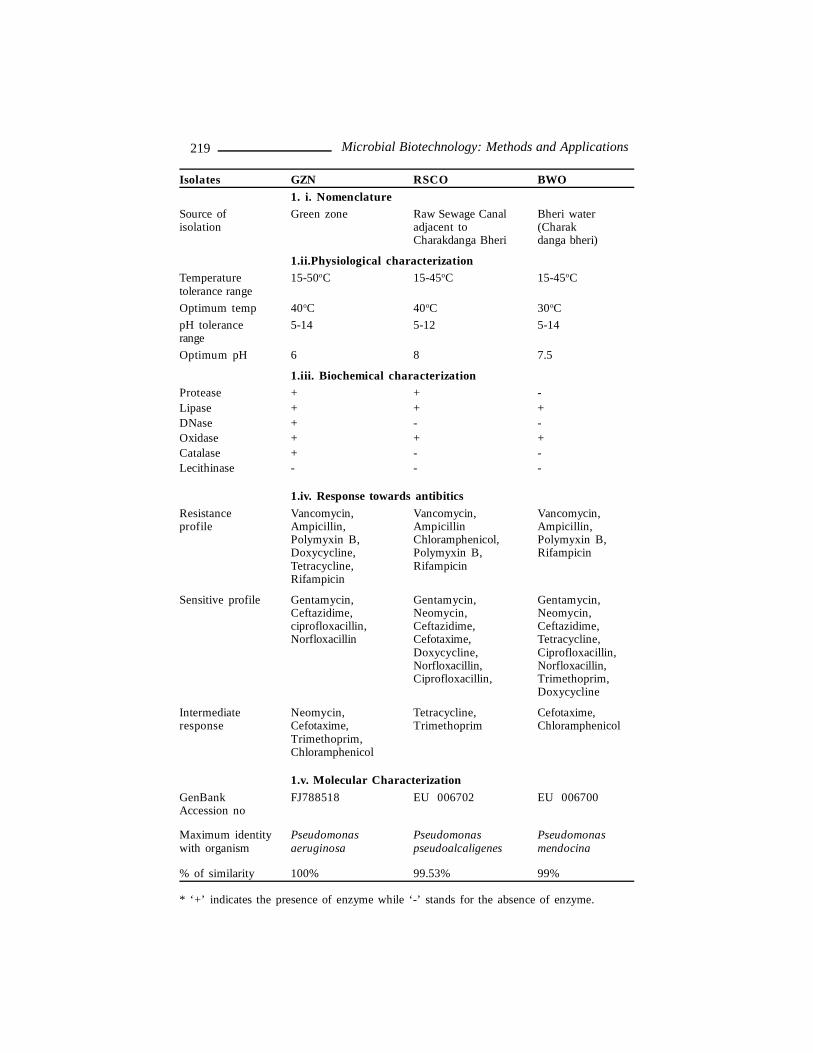
Table 1: Table represents the preliminary characterization of the pure bacterial isolates.1.i. The nomenclature of the bacterial isolates designated according to their site ofisolation. 1.ii. The details of the physiological parameters supporting the growth ofthe isolates. 1.iii. The biochemical features of the isolates in term of the presence orabsence of enzyme. 1.iv. The antibiotic sensitivity of the isolates as evaluated on thebasis of the diameter of the clearance zone as compared to the standard chart providedby National Committee for Clinical Laboratory Standard’s (NCCLS). 1.v. The molecularidentification of the isolates based on 16S rDNA analysis. The sequence was subjectedto BLAST analysis and the identity was deciphered on basis of the closest neighborwithin the existing database showing maximum % of identity. Isolate GZN showed100% identity at the partial sequence level. The partial sequence can reveal theidentity only upto the genus level.
Madhusmita Mishra et al 218
Isolates GZN RSCO BWO1. i. Nomenclature
Source of Green zone Raw Sewage Canal Bheri waterisolation adjacent to (Charak
Charakdanga Bheri danga bheri)
1.ii.Physiological characterizationTemperature 15-50oC 15-45oC 15-45oCtolerance rangeOptimum temp 40oC 40oC 30oCpH tolerance 5-14 5-12 5-14rangeOptimum pH 6 8 7.5
1.iii. Biochemical characterizationProtease + + -Lipase + + +DNase + - -Oxidase + + +Catalase + - -Lecithinase - - -
1.iv. Response towards antibiticsResistance Vancomycin, Vancomycin, Vancomycin,profile Ampicillin, Ampicillin Ampicillin,
Polymyxin B, Chloramphenicol, Polymyxin B,Doxycycline, Polymyxin B, RifampicinTetracycline, RifampicinRifampicin
Sensitive profile Gentamycin, Gentamycin, Gentamycin,Ceftazidime, Neomycin, Neomycin,ciprofloxacillin, Ceftazidime, Ceftazidime,Norfloxacillin Cefotaxime, Tetracycline,
Doxycycline, Ciprofloxacillin,Norfloxacillin, Norfloxacillin,Ciprofloxacillin, Trimethoprim,
Doxycycline
Intermediate Neomycin, Tetracycline, Cefotaxime,response Cefotaxime, Trimethoprim Chloramphenicol
Trimethoprim,Chloramphenicol
1.v. Molecular CharacterizationGenBank FJ788518 EU 006702 EU 006700Accession no
Maximum identity Pseudomonas Pseudomonas Pseudomonaswith organism aeruginosa pseudoalcaligenes mendocina
% of similarity 100% 99.53% 99%
* ‘+’ indicates the presence of enzyme while ‘-’ stands for the absence of enzyme.
219 Microbial Biotechnology: Methods and Applications
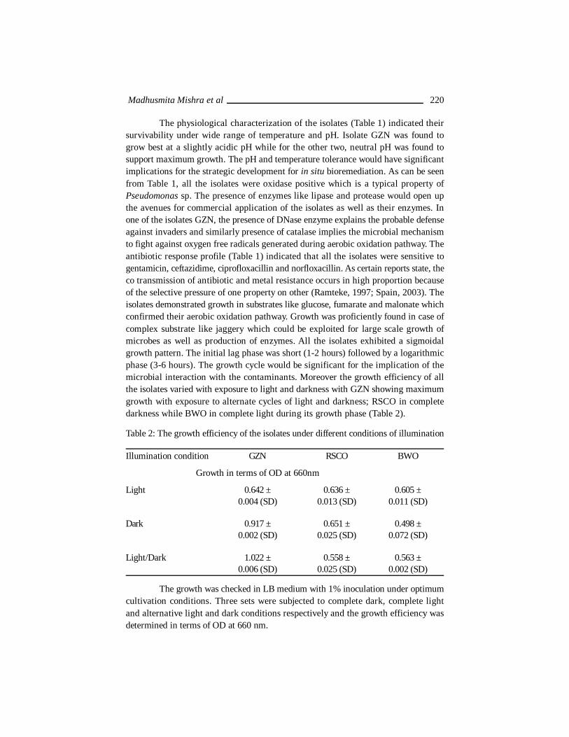
The physiological characterization of the isolates (Table 1) indicated theirsurvivability under wide range of temperature and pH. Isolate GZN was found togrow best at a slightly acidic pH while for the other two, neutral pH was found tosupport maximum growth. The pH and temperature tolerance would have significantimplications for the strategic development for in situ bioremediation. As can be seenfrom Table 1, all the isolates were oxidase positive which is a typical property ofPseudomonas sp. The presence of enzymes like lipase and protease would open upthe avenues for commercial application of the isolates as well as their enzymes. Inone of the isolates GZN, the presence of DNase enzyme explains the probable defenseagainst invaders and similarly presence of catalase implies the microbial mechanismto fight against oxygen free radicals generated during aerobic oxidation pathway. Theantibiotic response profile (Table 1) indicated that all the isolates were sensitive togentamicin, ceftazidime, ciprofloxacillin and norfloxacillin. As certain reports state, theco transmission of antibiotic and metal resistance occurs in high proportion becauseof the selective pressure of one property on other (Ramteke, 1997; Spain, 2003). Theisolates demonstrated growth in substrates like glucose, fumarate and malonate whichconfirmed their aerobic oxidation pathway. Growth was proficiently found in case ofcomplex substrate like jaggery which could be exploited for large scale growth ofmicrobes as well as production of enzymes. All the isolates exhibited a sigmoidalgrowth pattern. The initial lag phase was short (1-2 hours) followed by a logarithmicphase (3-6 hours). The growth cycle would be significant for the implication of themicrobial interaction with the contaminants. Moreover the growth efficiency of allthe isolates varied with exposure to light and darkness with GZN showing maximumgrowth with exposure to alternate cycles of light and darkness; RSCO in completedarkness while BWO in complete light during its growth phase (Table 2).
Table 2: The growth efficiency of the isolates under different conditions of illumination
Illumination condition GZN RSCO BWO
Growth in terms of OD at 660nm
Light 0.642 ± 0.636 ± 0.605 ±0.004 (SD) 0.013 (SD) 0.011 (SD)
Dark 0.917 ± 0.651 ± 0.498 ±0.002 (SD) 0.025 (SD) 0.072 (SD)
Light/Dark 1.022 ± 0.558 ± 0.563 ±0.006 (SD) 0.025 (SD) 0.002 (SD)
The growth was checked in LB medium with 1% inoculation under optimumcultivation conditions. Three sets were subjected to complete dark, complete lightand alternative light and dark conditions respectively and the growth efficiency wasdetermined in terms of OD at 660 nm.
Madhusmita Mishra et al 220
The oil utilizing ability of the isolates were tested using both vegetative (coconutand mustard oil) and mineral oils (like petrol, diesel, mobil and burnt mobil) throughturbidity measurement after 72hours. The isolates were found to utilize vegetative oillike coconut oil and mineral oils like diesel, mobil and burnt mobil as its carbon source,this property would be exploited for the application in crude oil remediation.
The gram nature of the isolates was reconfirmed using Real time PCR analysis asper the protocol of Shigemura et al. (2005). The pathogenecity test for the isolateswas conducted using TaqMan kit (Applied biosystems) [Detection Kit forStaphylococcus aureus (PN 4368606) and Pseudomonas aeruginosa (PN4368604)]as per manufacturers protocol and none of the three isolates were found topossess either of the pathogenic gene.
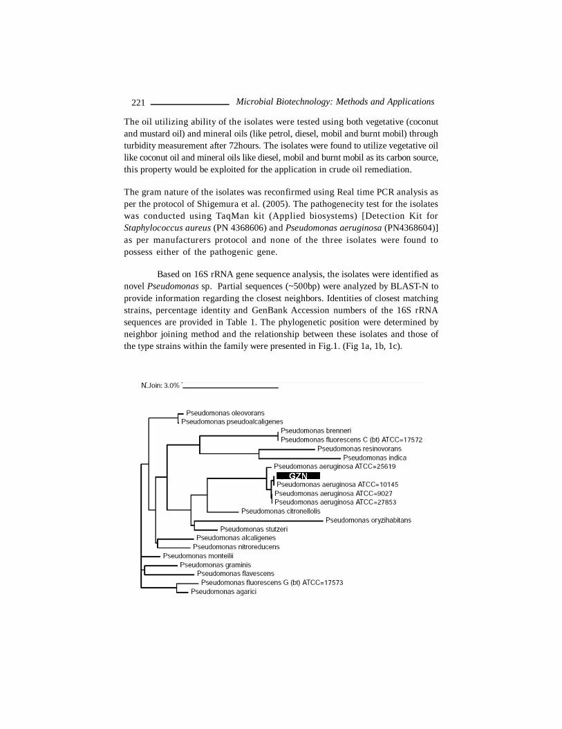
Based on 16S rRNA gene sequence analysis, the isolates were identified asnovel Pseudomonas sp. Partial sequences (~500bp) were analyzed by BLAST-N toprovide information regarding the closest neighbors. Identities of closest matchingstrains, percentage identity and GenBank Accession numbers of the 16S rRNAsequences are provided in Table 1. The phylogenetic position were determined byneighbor joining method and the relationship between these isolates and those ofthe type strains within the family were presented in Fig.1. (Fig 1a, 1b, 1c).
221 Microbial Biotechnology: Methods and Applications
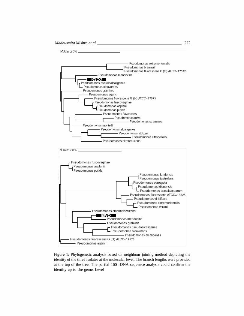
Figure 1: Phylogenetic analysis based on neighbour joining method depicting theidentity of the three isolates at the molecular level. The branch lengths were providedat the top of the tree. The partial 16S rDNA sequence analysis could confirm theidentity up to the genus Level
Madhusmita Mishra et al 222
5. TOLERANCE TOWARDS HEAVY METALSNine different heavy metal salts like Al(NO3)3.9H2O, CuSO4.5H2O, AgNO3,
Pb(NO3), NiCl2.6H2O, HgCl2, CrO3, CoCl.6H2O and CdCl3 were used for screeningof heavy metal tolerance. The determination of Minimum Inhibitory Concentration(MIC) of the respective metals was done as described previously (Adarsh et al.,2007). All the isolates were found to grow in presence of heavy metals like nickel(Ni), copper (Cu), silver (Ag), aluminium (Al), iron (Fe), chromium (Cr), lead (Pb) uptodifferent extent. The metal accumulation was tested using Energy Dispersive X-ray Fluorescence (EDXRF) technique in a Jordan Valley EX 3600 system asreported elsewhere (Adarsh et al., 2007). The relative accumulation of differentmetals as obtained from EDXRF analysis was presented in Table 3. As expected themaximum accumulation of Pb corresponded to highest Minimum InhibitoryConcentration of Pb.
Table 3: Tabular representation of the efficiency of different isolates in theaccumulation of various heavy metals
Isolates GZN RSCO BWOMetals salts Metal Concentration in ppbNi 1.589 0.641 1.504Co 9.921Cu 28.39 16.31 52.028Cr 249.89 669.1 424.29Pb 2688.99 1970.4 1504
* The data for metal accumulation has been checked in three sets and the mean result is presentedin table.
Prior to analysis by EDXRF, the metal treated cells were washed with 0.1N HCl toremove the adsorbed metal. The cell suspension was concentrated by vaccum filtrationthrough Whatman Filter membrane (0.22 mm) so as to detect only the intracellularmetal concentration which was represented in ppb. The total accumulation would bemuch higher.
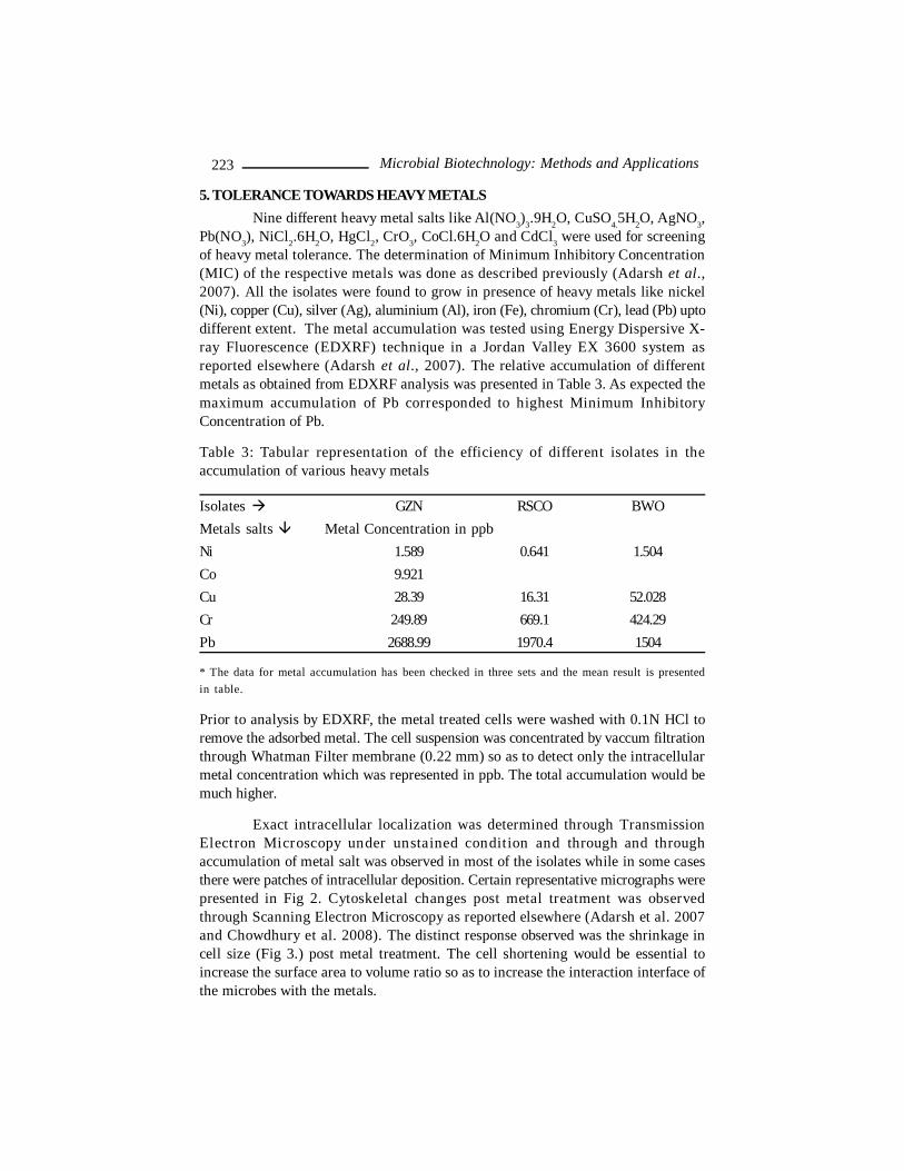
Exact intracellular localization was determined through TransmissionElectron Microscopy under unstained condition and through and throughaccumulation of metal salt was observed in most of the isolates while in some casesthere were patches of intracellular deposition. Certain representative micrographs werepresented in Fig 2. Cytoskeletal changes post metal treatment was observedthrough Scanning Electron Microscopy as reported elsewhere (Adarsh et al. 2007and Chowdhury et al. 2008). The distinct response observed was the shrinkage incell size (Fig 3.) post metal treatment. The cell shortening would be essential toincrease the surface area to volume ratio so as to increase the interaction interface ofthe microbes with the metals.
223 Microbial Biotechnology: Methods and Applications
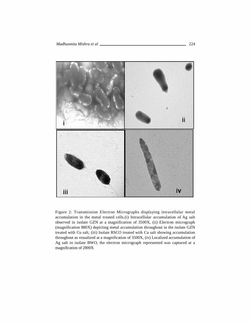
Figure 2: Transmission Electron Micrographs displaying intracellular metalaccumulation in the metal treated cells.(i) Intracellular accumulation of Ag saltobserved in isolate GZN at a magnification of 3500X, (ii) Electron micrograph(magnification 880X) depicting metal accumulation throughout in the isolate GZNtreated with Cu salt, (iii) Isolate RSCO treated with Cu salt showing accumulationthroughout as visualized at a magnification of 3500X, (iv) Localized accumulation ofAg salt in isolate BWO, the electron micrograph represented was captured at amagnification of 2800X
Madhusmita Mishra et al 224
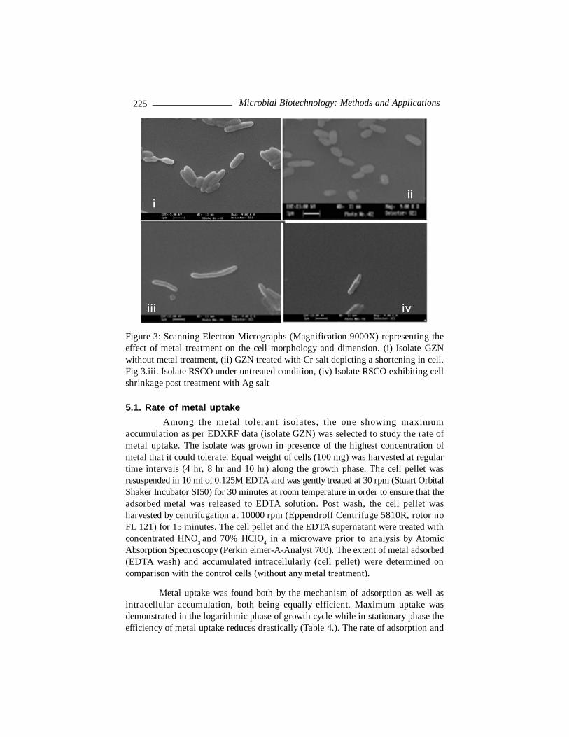
Figure 3: Scanning Electron Micrographs (Magnification 9000X) representing theeffect of metal treatment on the cell morphology and dimension. (i) Isolate GZNwithout metal treatment, (ii) GZN treated with Cr salt depicting a shortening in cell.Fig 3.iii. Isolate RSCO under untreated condition, (iv) Isolate RSCO exhibiting cellshrinkage post treatment with Ag salt
5.1. Rate of metal uptake Among the metal tolerant isolates, the one showing maximum
accumulation as per EDXRF data (isolate GZN) was selected to study the rate ofmetal uptake. The isolate was grown in presence of the highest concentration ofmetal that it could tolerate. Equal weight of cells (100 mg) was harvested at regulartime intervals (4 hr, 8 hr and 10 hr) along the growth phase. The cell pellet wasresuspended in 10 ml of 0.125M EDTA and was gently treated at 30 rpm (Stuart OrbitalShaker Incubator SI50) for 30 minutes at room temperature in order to ensure that theadsorbed metal was released to EDTA solution. Post wash, the cell pellet washarvested by centrifugation at 10000 rpm (Eppendroff Centrifuge 5810R, rotor noFL 121) for 15 minutes. The cell pellet and the EDTA supernatant were treated withconcentrated HNO3 and 70% HClO4 in a microwave prior to analysis by AtomicAbsorption Spectroscopy (Perkin elmer-A-Analyst 700). The extent of metal adsorbed(EDTA wash) and accumulated intracellularly (cell pellet) were determined oncomparison with the control cells (without any metal treatment).
Metal uptake was found both by the mechanism of adsorption as well asintracellular accumulation, both being equally efficient. Maximum uptake wasdemonstrated in the logarithmic phase of growth cycle while in stationary phase theefficiency of metal uptake reduces drastically (Table 4.). The rate of adsorption and
225 Microbial Biotechnology: Methods and Applications
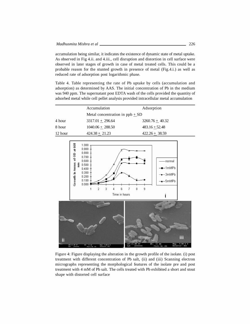
accumulation being similar, it indicates the existence of dynamic state of metal uptake.As observed in Fig 4.ii. and 4.iii., cell disruption and distortion in cell surface wereobserved in later stages of growth in case of metal treated cells. This could be aprobable reason for the stunted growth in presence of metal (Fig.4.i.) as well asreduced rate of adsorption post logarithmic phase.
Table 4. Table representing the rate of Pb uptake by cells (accumulation andadsorption) as determined by AAS. The initial concentration of Pb in the mediumwas 940 ppm. The supernatant post EDTA wash of the cells provided the quantity ofadsorbed metal while cell pellet analysis provided intracellular metal accumulation
Accumulation AdsorptionMetal concentration in ppb + SD
4 hour 3317.01 + 296.64 3260.76 + 40.328 hour 1040.06 + 288.50 483.16 + 52.4812 hour 424.38 + 21.23 422.26 + 38.59
Figure 4: Figure displaying the alteration in the growth profile of the isolate. (i) posttreatment with different concentration of Pb salt, (ii) and (iii) Scanning electronmicrographs representing the morphological features of the isolate pre and posttreatment with 4 mM of Pb salt. The cells treated with Pb exhibited a short and stoutshape with distorted cell surface
Madhusmita Mishra et al 226

5.2. Detection of SOD gene Super Oxide Dismutase is a major component of biological defense
mechanism against oxygen toxicity and is also reported to exhibit defense inpresence of metal stress where oxygen radical is a main byproduct (Roy et al.2008). Presence of SOD as a defensive tool would be essential in these isolates whichare aerobic in nature as well as showing metal tolerance. With universal SOD primersas reported by Zolg and Schulz (1994), a 480 bp fragment was lifted from the genomicDNA of the three isolates (Fig 5). The 451 bp sequence was obtained for that ofisolate GZN and the BLAST-N analysis of the sequence provided 97% similarity withPseudomonas Mn SOD gene. The sequence being novel was submitted to GenBank(GenBank Accession no FJ788516).
Figure 5: Figure showing the SOD gene amplification products resolved on a 2%agarose gel. Lane M represents the 500bp ladder (Fermentus), L1 is the negativecontrol, L2, L3 and L4 represents the SOD amplicons obtained from the isolate GZN,RSCO and BWO respectively
6. MICROBIAL ENZYMES AS AN AID IN DEHAIRING OF HIDEThe objective of employing protease and lipase enzymes in dehairing was
to achieve effective degreasing by the simultaneous action of proteolysis, lipolysis,and emulsification. The enzyme production was achieved using shake flask under
227 Microbial Biotechnology: Methods and Applications
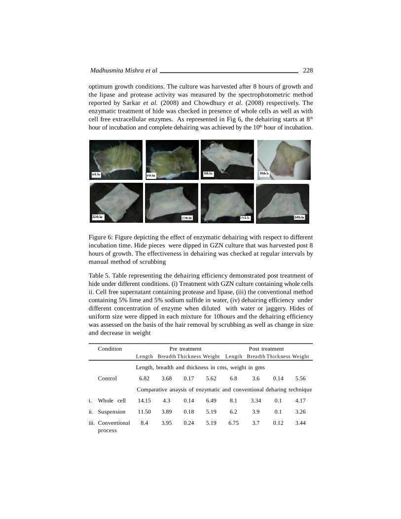
optimum growth conditions. The culture was harvested after 8 hours of growth andthe lipase and protease activity was measured by the spectrophotometric methodreported by Sarkar et al. (2008) and Chowdhury et al. (2008) respectively. Theenzymatic treatment of hide was checked in presence of whole cells as well as withcell free extracellular enzymes. As represented in Fig 6, the dehairing starts at 8th
hour of incubation and complete dehairing was achieved by the 10th hour of incubation.
Figure 6: Figure depicting the effect of enzymatic dehairing with respect to differentincubation time. Hide pieces were dipped in GZN culture that was harvested post 8hours of growth. The effectiveness in dehairing was checked at regular intervals bymanual method of scrubbing
Table 5. Table representing the dehairing efficiency demonstrated post treatment ofhide under different conditions. (i) Treatment with GZN culture containing whole cellsii. Cell free supernatant containing protease and lipase, (iii) the conventional methodcontaining 5% lime and 5% sodium sulfide in water, (iv) dehairing efficiency underdifferent concentration of enzyme when diluted with water or jaggery. Hides ofuniform size were dipped in each mixture for 10hours and the dehairing efficiencywas assessed on the basis of the hair removal by scrubbing as well as change in sizeand decrease in weight
Condition Pre treatment Post treatmentLength Breadth Thickness Weight Length Breadth Thickness Weight
Length, breadth and thickness in cms, weight in gms
Control 6.82 3.68 0.17 5.62 6.8 3.6 0.14 5.56
Comparative anaysis of enzymatic and conventional deharing technique
i. Whole cell 14.15 4.3 0.14 6.49 8.1 3.34 0.1 4.17
ii. Suspension 11.50 3.89 0.18 5.19 6.2 3.9 0.1 3.26
iii. Conventional 8.4 3.95 0.24 5.19 6.75 3.7 0.12 3.44process
Madhusmita Mishra et al 228

iv. Deharing efficiency under different concentrations of enzyme
10% enzyme 6.1 3.98 0.23 5.46 6.54 2.83 0.14 4.68 + water
75% enzyme 8.00 3.3 0.15 6.03 9.7 3.00 0.1 4.04 + jaggery
50% enzyme 6.94 3.13 0.14 5.44 7.25 3.15 0.14 3.91 + jaggery
25% enzyme 8.74 3.25 0.15 6.13 7.95 3.4 0.2 5.64 + jaggery
10% enzyme 6.5 3.7 0.16 5.06 6.2 3.00 0.14 5.45 + jaggery
The enzyme efficiency was compared with the conventional technique whichinvolves treatment with 5% lime and 5% sodium sulfide. The effectiveness of theenzymatic treatment over the conventional process was observed by the markeddecrease in hide weight. Dehairing efficiency of hides dipped in cell culture was atpar with cell free enzyme (Table 5) and this step overcomes the time lag as well asexpenditure necessary for cell removal. The optimum enzyme concentration requiredfor effective dehairing was determined by using supernatant containing protease andlipase enzyme diluted with water and jaggery upto different percentage. Thesignificant finding was that 75% of enzyme (diluted with 25% water) showed completedehairing while 25% of enzyme also showed indication of partial dehairing. In caseof jaggery with enzyme, 50% enzyme showed dehairing while 75% enzymeconcentration was most efficient for dehairing.
6.1. A comparative statement of growth and enzyme production undershake flask and fermentation condition
Since the enzymes were used in applications like dehairing, the next objectivewas to scale up the cell as well as enzyme production. Isolate GZN producing boththe enzymes was further studied for optimization of different parameters under boththe cultivation conditions. For shake flask culture, 1% inoculum was added to theenriched medium [Luria Bertani (LB) broth, 1% tryptone, 0.5% yeast extract and 0.5%NaCl] and cultivation was allowed at the respective optimum temperature and pH. Incase of batch fermentation, 5% inoculum was seeded into 3 lit LB medium (workingvolume capacity) in 5lt BioG Micom, BIOTRON fermenter. The fermentation wascarried out at optimum growth condition with different agitation speed (100-300 rpm)and with maximum air flow (1-3 vvm). The growth was measured in terms of turbidity(optical density at 660nm measured in Beckman Coulter DV– 530, UV Visspectrophotometer) while the enzyme (lipase and protease) production was determinedby the quantitative assay method reported by Sarkar et al. (2008) and Chowdhury etal. (2008) respectively.
The impact of agitation speed on the growth efficiency was clearly depictedin Fig 7. The lower shaft speed facilitates microbial growth while growth was inhibitedat higher agitation speed of 300rpm. The higher agitation could provide a shearing
229 Microbial Biotechnology: Methods and Applications
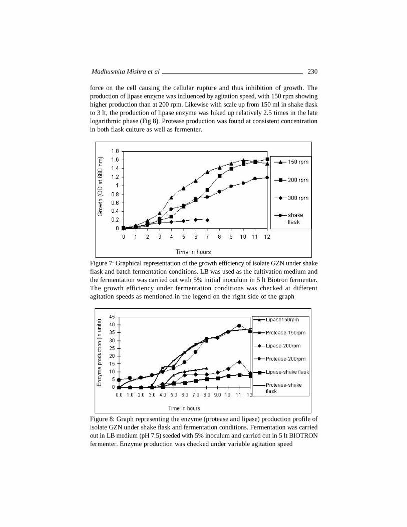
force on the cell causing the cellular rupture and thus inhibition of growth. Theproduction of lipase enzyme was influenced by agitation speed, with 150 rpm showinghigher production than at 200 rpm. Likewise with scale up from 150 ml in shake flaskto 3 lt, the production of lipase enzyme was hiked up relatively 2.5 times in the latelogarithmic phase (Fig 8). Protease production was found at consistent concentrationin both flask culture as well as fermenter.
Figure 7: Graphical representation of the growth efficiency of isolate GZN under shakeflask and batch fermentation conditions. LB was used as the cultivation medium andthe fermentation was carried out with 5% initial inoculum in 5 lt Biotron fermenter.The growth efficiency under fermentation conditions was checked at differentagitation speeds as mentioned in the legend on the right side of the graph
Figure 8: Graph representing the enzyme (protease and lipase) production profile ofisolate GZN under shake flask and fermentation conditions. Fermentation was carriedout in LB medium (pH 7.5) seeded with 5% inoculum and carried out in 5 lt BIOTRONfermenter. Enzyme production was checked under variable agitation speed
Madhusmita Mishra et al 230
7. CONCLUSIONThe detailed characterization of these isolates exposes the untapped
potential of these for the cause of environmental sustainance. The heavy metalaccumulating property would cause the concentration of the toxic waste within minimalvolume of a cell thereby reducing the load of metal toxicity in the environment. Thestudy also initiates the probability of application of the microbe as well as its productsi.e. the enzyme for industrial bioremediation. The enzyme application in dehairingprocess could replace the conventional method involving harmful chemicals and inoverall sense would make the commercial process environment friendly leading to“Green Technology”.
8. ACKNOWLEDGEMENTSThe authors would like to acknowledge the financial assistance from
Department of Atomic Energy, Government of India under the Board of Research inNuclear Studies scheme and Department of Science and Technology, Government ofIndia under the DST Fast Track Scheme. They would acknowledge West BengalUniversity of Technology, Calcutta, India for their computational facility.
9. REFERENCESAckerley, D.F., Gonzalez, C.F., Park, C.H., Balke, R., Keyhan, A. and Matin, A. (2004).
Chromate reducing properties of soluble flavoproteins from Pseudomonasputida and Escherichia coli. Appl. Environ. Microbiol. 70, 873-888.
Adarsh, V.K., Mishra, M., Chowdhury, S., Sudarshan, M., Thakur, A.R. and RayChaudhuri, S. (2007). Studies on metal microbe interaction of three bacterialisolates from East Calcutta Wetland. OnLine J. Biol. Sci. 7, 80-88.
Alcalde, M., Ferrer, M, Plou, F.J. and Ballesteros, A. (2006). Environmental biocatalysis:from remediation with enzymes to novel green processes. Trends Biotechnol.24, 281-287.
Barceolux, D.G. (1999). Chromium. J. Tox. Clin. Tox. 37, 173-194.Beveridge, T.J. and Graham, L.L. (1991), Surface Layers of Bacteria. Microbiol Rev.
55, 684-705.Bhattacharyya, S., Santra, S.C. (2003). Environmental Status of East Calcutta Wetlands
and Strategies for Sustainable Management, In Khasnabis, R. (ed.), Ecology,Economy and Society. DRS Phase-II Programme, EnvironmentalManagement, Department of Business Management, University of Calcutta,Kolkata, India, pp. 65-94.
Camargo, F.A.O., Okeke B.C., Bento FM. and Frankenberger, W.T. (2003), In vitroreduction of hexavalant chromium by a cell free extract of Bacillus sp. ES 29.stimulated by Cu2+. Appl Microbiol Biotechnol. 62, 569-573.
Cervantes, C., Campos Gracia, J., Devras, S., Guttierez Corona, F., Loza Tavera, H.,Torres Guzman, J.C. and Moreno Sanchez, R. (2001), Interactions of chromiumwith microorganisms and plants. FEMS Microbiol Rev. 25, 335- 347.
231 Microbial Biotechnology: Methods and Applications
Chowdhury, S., Mishra, M., Adarsh, VK., Mukherjee, A., Thakur, AR. and RayChaudhuri, S. (2008), Novel Metal Accumulator and Protease Secretor Microbesfrom East Calcutta Wetland. Am. J. Biochem. & Biotech. 4, 255-264.
Ekundayo, E.O. and Killham, K. (2001), Lead solubilization and accumulation by twostrains of Pseudomonas obtained from a contaminated Alfisol’s Effluent insouth western Nigeria. Environ Monit Assess. 71, 1573-2959.
Foster, T.J. (1983), Plasmid determined resistance to anti-microbial drugs and oxic metalions in bacteria, Microbiol Rev. 47, 361–409.
Gadd, G.M. (2000), Bioremedial potential of microbial mechanism of metal mobilizationand immobilization. Curr Opin Biotechnol. 11, 271- 279.
Ghosh, D. (1999), Participatory Management in Wastewater Treatment and Reuse inWest Bengal. UWEP Occasional paper.
Ghosh, D. (2005), Ecology and Traditional Wetland Practice – Lessons fromWastewater Utilisation in the East Calcutta Wetlands. Kolkata. WorldView.
Goyer, R.A. (1997): Toxic and essential metal interactions. Annu Rev Nutr. 17, 37-50.
Mattuschka, M., Straube, G. and Trevors, J.T. (1994), Silver, copper, lead and zincaccumulation by Pseudomonas stutzeri AG259 and Streptomyces albus:electron microscopy and Energy Dispersive X ray studies. Biometals. 7, 204-208.
Pradhan, A., Bhaumik, P., Das, S., Mishra, M., Khanam, S., Amin Hoque, B., Mukherjee,I., Thakur, A.R. and Ray Chaudhuri, S. (2008), Phytoplankton diversity asindicator of water quality for fish cultivation. American Journal of.Environmental. Sciences. 4, 271-276.
Ramteke, P.W. (1997), Plasmid mediated co-transfer of antibiotic resistance and heavymetal tolerance in coliforms, Indian J Med Microbiol. 37, 177–181.
Ray Chaudhuri, S., Mishra, M., Nandy, P. and Thakur, A.R. (2008), Waste management:A case study of ongoing traditional practices at East Calcutta Wetland. Am. J.Agril. & Biol. Sci. 3, 315-320.
Ray Chaudhuri, S., Mishra, M., Salodkar, S., Sudarshan, M. and Thakur, A.R. (2008a):Traditional Aquaculture Practice at East Calcutta Wetland: The SafetyAssessment. Am. J. Environ. Sci. 4, 140-144.
Ray Chaudhuri, S., Salodkar, S., Sudarshan, M. and Thakur, A.R. (2007). IntegratedResource Recovery at East Calcutta Wetland – how safe is these?. Am. J. Agril.& Biol. Sci. 2, 75-80.
Ray Chaudhuri, S., Salodkar, S., Sudarshan, M., Mukherjee, I. and Thakur, A.R.,(2008b): Role of Water hyacinth mediated Phytoremediation in Waste WaterPurification at East Calcutta Wetland. Env Sci. 5, 53-62.
Ray Chaudhuri, S. and Thakur, A.R. (2006): Microbial genetic resource mapping ofEast Calcutta Wetland. Curr. Sci. 91, 212-217.
Madhusmita Mishra et al 232

Roy, S., Ghosh, A.N. and Thakur, A.R. (2008): Uptake of Pb2+ by a cyanobacteriumbelonging to genus Synechocystis, isolated from East Kolkata Wetlands.BioMetals. 21, 515-524.
Sarkar, A.K., Roy, S., Pal, A., Pakrashi, S., Mahra, P.K., Sahoo, S., Deb, A., Mishra,M., Sen, S.K., Thakur, A.R. and RayChaudhuri, S. (2008): Microbial BiodiversityScreening For Metal Accumulators From Mineral Ore Rich Site In AndhraPradesh, India. OnLine J. Biol. Sci. 8, 32-40.
Shigemura, K., Shirakawa, T., Okada, H., Tanaka, K., Kamidono, S. and Arakawa, S.G.(2005), A rapid detection and differentiation of gram negative and gram positivepathogenic bacteria in urine using TaqMan probe. Clin Exp Med. 4, 196-201.
Zolg, W. and Schulz, S.P. (1994), The Superoxide Dismutase Gene, a Target forDetection and Identification of Mycobacteria by PCR. J. Clin. Microbiol. 32,2801-2812.
233 Microbial Biotechnology: Methods and Applications