Phenotypic and Genome-Wide Analysis of an Antibiotic-Resistant Small Colony Variant (SCV) of...
63
Editorial Manager(tm) for PLoS ONE Manuscript Draft Manuscript Number: Title: Phenotypic and Genome-Wide Analysis of an Antibiotic-Resistant Small Colony Variant (SCV) of Pseudomonas aeruginosa Short Title: Antibiotic-Resistant Pseudomonas SCV Article Type: Research Article Section/Category: Other Keywords: Pseudomonas aeruginosa SCV aminoglycosides PQS PhoP-PhoQ MexXY Corresponding Author: Pierre Cornelis Corresponding Author's Institution: Vrije Universiteit Brussel First Author: Qing Wei Order of Authors: Qing Wei;Saeed Tarighi;Andreas Dötsch;Susanne Haüssler;Mathias Muesken;Victoria Wright;Miguel Cámara;Paul Williams;Steven Haenen;Bart Boerjan;Annelies Bogaerts;Evy Vierstraete;Peter Verleyen;Liliane Schoofs;Ronnie Willaert;Valérie De Groote;Jan Michiels;Aurélie Crabbé;Pierre Cornelis Abstract: Background Small colony variants (SCVs) are slow-growing bacteria, which often show increased resistance to antibiotics and cause latent or recurrent infections. It is therefore important to understand the mechanisms at the basis of this phenotypic switch. Methodology/Principal findings One SCV (termed PAO-SCV) was isolated, showing high resistance to gentamicin and to the cephalosporine cefotaxime. PAO-SCV was prone to reversion as evidenced by emergence of large colonies with a frequency of 10-5 on media without antibiotics while it was stably maintained in presence of gentamicin. PAO-SCV showed a delayed growth, defective motility, and strongly reduced levels of the quorum sensing Pseudomonas quinolone signal (PQS). Whole genome expression analysis further suggested a multi-layered antibiotic resistance mechanism, including simultaneous over- expression of two drug efflux pumps (MexAB-OprM, MexXY-OprM), the LPS modification operon arnBCADTEF, and the PhoP-PhoQ two-component system. Conversely, the genes for the synthesis of PQS and were strongly down-regulated in PAO-SCV. A proteome analysis confirmed higher expression of the two-component response regulator PhoP in PAO-SCV. Finally, genomic analysis revealed the presence of mutations in phoP and phoQ genes as well as in the mexZ gene encoding a repressor of the mexXY and mexAB-oprM genes. However, no evidence was found for a compensatory mutation explaining the emergence of one analyzed revertant, suggesting epigenetic changes. However, high
Transcript of Phenotypic and Genome-Wide Analysis of an Antibiotic-Resistant Small Colony Variant (SCV) of...

Editorial Manager(tm) for PLoS ONE Manuscript Draft Manuscript Number: Title: Phenotypic and Genome-Wide Analysis of an Antibiotic-Resistant Small Colony Variant (SCV) of Pseudomonas aeruginosa Short Title: Antibiotic-Resistant Pseudomonas SCV Article Type: Research Article Section/Category: Other Keywords: Pseudomonas aeruginosa SCV aminoglycosides PQS PhoP-PhoQ MexXY Corresponding Author: Pierre Cornelis Corresponding Author's Institution: Vrije Universiteit Brussel First Author: Qing Wei Order of Authors: Qing Wei;Saeed Tarighi;Andreas Dötsch;Susanne Haüssler;Mathias Muesken;Victoria Wright;Miguel Cámara;Paul Williams;Steven Haenen;Bart Boerjan;Annelies Bogaerts;Evy Vierstraete;Peter Verleyen;Liliane Schoofs;Ronnie Willaert;Valérie De Groote;Jan Michiels;Aurélie Crabbé;Pierre Cornelis Abstract: Background Small colony variants (SCVs) are slow-growing bacteria, which often show increased resistance to antibiotics and cause latent or recurrent infections. It is therefore important to understand the mechanisms at the basis of this phenotypic switch. Methodology/Principal findings One SCV (termed PAO-SCV) was isolated, showing high resistance to gentamicin and to the cephalosporine cefotaxime. PAO-SCV was prone to reversion as evidenced by emergence of large colonies with a frequency of 10-5 on media without antibiotics while it was stably maintained in presence of gentamicin. PAO-SCV showed a delayed growth, defective motility, and strongly reduced levels of the quorum sensing Pseudomonas quinolone signal (PQS). Whole genome expression analysis further suggested a multi-layered antibiotic resistance mechanism, including simultaneous over-expression of two drug efflux pumps (MexAB-OprM, MexXY-OprM), the LPS modification operon arnBCADTEF, and the PhoP-PhoQ two-component system. Conversely, the genes for the synthesis of PQS and were strongly down-regulated in PAO-SCV. A proteome analysis confirmed higher expression of the two-component response regulator PhoP in PAO-SCV. Finally, genomic analysis revealed the presence of mutations in phoP and phoQ genes as well as in the mexZ gene encoding a repressor of the mexXY and mexAB-oprM genes. However, no evidence was found for a compensatory mutation explaining the emergence of one analyzed revertant, suggesting epigenetic changes. However, high

expression of phoP and phoQ was confirmed for the SCV variant while the revertant showed expression levels reduced to wild-type levels. Conclusions By combining data coming from phenotypic, gene expression and proteome analysis, we could demonstrate that resistance to aminoglycosides in one SCV mutant is multifactorial including overexpression of efflux mechanisms, LPS modification and is accompanied by a drastic down-regulation of the Pseudomonas quinolone signal quorum sensing system. The phenotypic change is reversible and its origin is probably epigenetic. Suggested Reviewers: Leo Eberl University of Zürich [email protected] Leo Eberl is a specialist of quorum sensing regulation, whom advice on the manuscript could be highly valuable because QS is down-regulated in our SCV. Robert Hancock University of British Columbia [email protected] Bob Hancock has published many interesting articles about antibiotic resistance in Pseudomonas aeruginosa, and, in particular the role played by the PhoP-PhoQ system which seems to central in our case. Opposed Reviewers:

Dear Editor,
We would like to submit the following manuscript to PloSOne, entitled “Phenotypic and Genome-
Wide Analysis of an Antibiotic-Resistant Small Colony Variant (SCV) of Pseudomonas aeruginosa”.
As you know SCVs are frequently isolated from tissues infected with P. aeruginosa, especially in the
case of CF patients lungs chronically colonized by this bacterium. In this work we describe the
isolation of one in vitro selected SCV after culturing in the presence of gentamycin. This SCV shows
resistance to different aminoglycosides, and to cefotaxime but is prone to reversion resulting in loss
of the resistance to the antibiotic. Phenotypic, metabolic, proteomic, transcriptomic, and genomic
data were here combined to show that the resistance to aminoglycosides is multi-layered, arising
primarily from mutations in the two-component system PhoP-PhoQ and in the MexS regulator,
inducing the overexpression of the MexAB-OprM efflux pump and the aminoglycoside efflux pump
MexXY-OprM, together with genes involved in the modification of LPS. One interesting observation is
the down-regulation of the genes involved in the production of the Pseudomonas quinolone signal
(PQS) molecule, resulting in decreased production of virulence factors and lowered pathogenicity.
Finally, a genome sequence analysis revealed that the mutations are present in both the SCV and the
revertant, suggesting epigenetic changes which could lead to the apparition or the reversionof the
SCV morphotype.
Sincerely yours,
Pierre Cornelis
Corresponding author
Cover Letter
1 2 3 4 5 6 7 8 9 10 11 12 13 14 15 16 17 18 19 20 21 22 23 24 25 26 27 28 29 30 31 32 33 34 35 36 37 38 39 40 41 42 43 44 45 46 47 48 49 50 51 52 53 54 55 56 57 58 59 60 61 62 63 64 65

1
Phenotypic and Genome-Wide Analysis of an Antibiotic-Resistant 1
Small Colony Variant (SCV) of Pseudomonas aeruginosa 2
3
Qing Wei1, Saeed Tarighi
1, Andreas Dötsch
2, Susanne Häussler
2,3, Mathias Müsken
2,3, 4
Victoria J. Wright4, Miguel Cámara
4, Paul Williams
4, Steven Haenen
5, Bart Boerjan
5, 5
Annelies Bogaerts5, Evy Vierstraete
5, Peter Verleyen
5, Liliane Schoofs
5, Ronnie Willaert
6, 6
Valérie N. De Groote7, Jan Michiels
7, Aurélie Crabbé
8, and Pierre Cornelis
1* 7
8
1Laboratory of Microbial Interactions,
6Structural Biology Brussels, Department of Molecular 9
and Cellular Interactions, Flanders Institute for Biotechnology (VIB), Vrije Universiteit 10
Brussel, Pleinlaan 2, B-1050 Brussels, Belgium 11 2Chronic Pseudomonas Infections, Helmholtz Centre for Infection Research, Inhoffenstrasse 12
7, D-38124 Braunschweig, Germany 13 3Twincore, Center for Experimental and Clinical Infection Research, a joint venture of the 14
Helmholtz Center for Infection Research and the Medical School Hannover, Hannover, 15
Germany 16 4School of Molecular Medical Sciences, Centre for Biomolecular Sciences, University Park, 17
University of Nottingham, Nottingham NG72RD, UK 18 5Functional Genomics and Proteomics, Faculty of Sciences, K.U.Leuven, Naamsestraat 59, B-19
3000 Leuven, Belgium 20 7Centre of Microbial and Plant Genetics, K.U. Leuven, Kasteelpark Arenberg 20 box 2460, B-21
3001 Heverlee, Belgium 22 8The Biodesign Institute, Center for Infectious Diseases and Vaccinology, Arizona State 23
University, 1001 South McAllister Avenue, Tempe, Arizona 85287, U.S.A. 24
25
Short title: Antibiotic-Resistant Pseudomonas SCV 26
Keywords: Pseudomonas aeruginosa, SCV, aminoglycosides, PQS, PhoP-PhoQ, MexXY 27
*Corresponding author: 28
Pierre Cornelis; Tel: +32 2 6291906; Fax: +32 2 6291902; E-mail: [email protected] 29
30
*ManuscriptClick here to download Manuscript: SCV-PloS-One.doc
1 2 3 4 5 6 7 8 9 10 11 12 13 14 15 16 17 18 19 20 21 22 23 24 25 26 27 28 29 30 31 32 33 34 35 36 37 38 39 40 41 42 43 44 45 46 47 48 49 50 51 52 53 54 55 56 57 58 59 60 61 62 63 64 65

2
ABSTRACT 31
Background 32
Small colony variants (SCVs) are slow-growing bacteria, which often show increased 33
resistance to antibiotics and cause latent or recurrent infections. It is therefore important to 34
understand the mechanisms at the basis of this phenotypic switch. 35
Methodology/Principal findings 36
One SCV (termed PAO-SCV) was isolated, showing high resistance to gentamicin and to the 37
cephalosporine cefotaxime. PAO-SCV was prone to reversion as evidenced by emergence of 38
large colonies with a frequency of 10-5
on media without antibiotics while it was stably 39
maintained in presence of gentamicin. PAO-SCV showed a delayed growth, defective 40
motility, and strongly reduced levels of the quorum sensing Pseudomonas quinolone signal 41
(PQS). Whole genome expression analysis further suggested a multi-layered antibiotic 42
resistance mechanism, including simultaneous over-expression of two drug efflux pumps 43
(MexAB-OprM, MexXY-OprM), the LPS modification operon arnBCADTEF, and the PhoP-44
PhoQ two-component system. Conversely, the genes for the synthesis of PQS and were 45
strongly down-regulated in PAO-SCV. A proteome analysis confirmed higher expression of 46
the two-component response regulator PhoP in PAO-SCV. Finally, genomic analysis revealed 47
the presence of mutations in phoP and phoQ genes as well as in the mexZ gene encoding a 48
1 2 3 4 5 6 7 8 9 10 11 12 13 14 15 16 17 18 19 20 21 22 23 24 25 26 27 28 29 30 31 32 33 34 35 36 37 38 39 40 41 42 43 44 45 46 47 48 49 50 51 52 53 54 55 56 57 58 59 60 61 62 63 64 65

3
repressor of the mexXY and mexAB-oprM genes. However, no evidence was found for a 49
compensatory mutation explaining the emergence of one analyzed revertant, suggesting 50
epigenetic changes. However, high expression of phoP and phoQ was confirmed for the SCV 51
variant while the revertant showed expression levels reduced to wild-type levels. 52
Conclusions 53
By combining data coming from phenotypic, gene expression and proteome analysis, we 54
could demonstrate that resistance to aminoglycosides in one SCV mutant is multifactorial 55
including overexpression of efflux mechanisms, LPS modification and is accompanied by a 56
drastic down-regulation of the Pseudomonas quinolone signal quorum sensing system. The 57
phenotypic change is reversible and its origin is probably epigenetic. 58
59
INTRODUCTION 60
Pseudomonas aeruginosa is a ubiquitous Gram-negative bacterium found in diverse 61
ecological habitats such as soils, marshes and coastal marine waters. As an opportunistic 62
pathogen, P. aeruginosa is able to infect humans, animals and plants [1,2,3]. P. aeruginosa is 63
a primary nosocomial diseases causative agent and represents the major cause of morbidity 64
and mortality in patients with cystic fibrosis (CF). P. aeruginosa produces a large panel of 65
secreted virulence factors like the phenazine pyocyanin, the siderophore pyoverdine, elastase, 66
1 2 3 4 5 6 7 8 9 10 11 12 13 14 15 16 17 18 19 20 21 22 23 24 25 26 27 28 29 30 31 32 33 34 35 36 37 38 39 40 41 42 43 44 45 46 47 48 49 50 51 52 53 54 55 56 57 58 59 60 61 62 63 64 65

4
and toxins. It is also characterized by its high level of drug resistance involving the formation 67
of antibiotic-resistant biofilms resulting from the emergence of phenotypic variants [2,3]. 68
During the course of infection, P. aeruginosa can efficiently adopt diverse strategies to evade 69
antimicrobial stresses and the host immune system defenses, making it impossible to eradicate 70
this bacterium permanently from CF lungs [2,4]. Important phenotypic variations can occur 71
during chronic colonization, such as conversion to mucoidy [5], the emergence of persister 72
cells after antibiotics treatment [6,7] or the occurrence of small colony variants with higher 73
resistance to antibiotics [8,9,10,11,12]. Compared to wild-type P. aeruginosa, SCVs show 74
increased antibiotic resistance, enhanced biofilm formation, reversion to wild-type-like 75
morphotypes, reduced motility, and slow and auto-aggregative growth behavior [13,14]. 76
SCVs have been isolated from CF lungs or sputum [4,8,9,12], laboratory-grown biofilms 77
[11,12,14], in vitro selection upon antibiotic exposure [15,16] or as a consequence of gene 78
inactivation [17,18]. Clinically, P. aeruginosa SCVs have already been proven to associate 79
with chronic infections behaving as persisters in pathogenesis of CF patients and making 80
almost impossible for clinicians to eradicate the infections [8,19,20]. The intracellular second 81
messenger cyclic-di-GMP (c-di-GMP) [21] has been recently shown to be involved in SCV 82
phenotype switching in terms of biofilm formation, reduced motility, and exopolysaccharide 83
(EPS) production [18,22,23,24,25,26]. The “phenotypic variant regulator”, PvrR, containing a 84
conserved EAL domain of phosphodiesterase (PDE) involved in the hydrolysis of c-di-GMP, 85
1 2 3 4 5 6 7 8 9 10 11 12 13 14 15 16 17 18 19 20 21 22 23 24 25 26 27 28 29 30 31 32 33 34 35 36 37 38 39 40 41 42 43 44 45 46 47 48 49 50 51 52 53 54 55 56 57 58 59 60 61 62 63 64 65

5
has been identified to control the phenotypic switch from an antibiotic resistant and auto-86
aggregative rough SCV (RSCV) of P. aeruginosa strain PA14 to wild-type-like antibiotics 87
susceptible revertants [15]. Another characteristic driven by the elevated level of c-di-GMP 88
in SCVs is the contribution of two EPS-encoding loci in some P. aeruginosa strains (PA2231-89
PA2245 for psl and PA3058-PA3064 for pel) to auto-aggregation and hyper adherence 90
phenotypes characterized by increased Congo Red dye binding [27,28,29]. Although 91
antibiotics resistance of P. aeruginosa has been connected to biofilm formation and linked to 92
phenotypic variation [15], the mechanisms underlying the extremely high antibiotic resistance 93
of SCVs has not been reported extensively due to the unavailability, in some cases, of the WT 94
counterpart for comparison. 95
In this study, we present the identification of a novel, reversion-prone, P. aeruginosa SCV 96
with distinct features, including resistance to various antibiotics, defective motility, and 97
absence of production of the quorum sensing PQS signal molecule. Using a combination of 98
genomic, transcriptomic, proteomic and phenotypic approaches, we provide the first evidence 99
of concerted mechanisms harnessed by this P. aeruginosa SCV leading to antibiotic resistance 100
as well as down-regulation of acute virulence genes, probably involving the PhoP PhoQ two 101
component system. 102
103
104
1 2 3 4 5 6 7 8 9 10 11 12 13 14 15 16 17 18 19 20 21 22 23 24 25 26 27 28 29 30 31 32 33 34 35 36 37 38 39 40 41 42 43 44 45 46 47 48 49 50 51 52 53 54 55 56 57 58 59 60 61 62 63 64 65

6
Results 105
Phenotypic characterization of a gentamicin-resistant P. aeruginosa PAO1-SCV and large 106
colony pseudo-revertants 107
Following sub-culturing P. aeruginosa PAO1 (ATCC 15692) in the presence of high-108
concentration of gentamicin (200 μg ml-1
, Gm), we isolated a Gm-resistant SCV designated 109
PAO-SCV, which formed small (ca. 1/5 of the wild-type diameter), smooth colonies after 110
three days of incubation at 37°C on LB agar plates (Figure 1A). PAO-SCV grown in liquid 111
LB also showed a delayed entry in exponential phase compared to the wild-type (Figure 1B). 112
PAO-SCV showed high level of resistance towards gentamicin and cefotaxime (Table 1 and 113
Figure 2). The persistence fraction of PAO-SCV after treatment with the fluoroquinolone 114
antibiotic ofloxacin was approximately 2-fold higher compared to the PAO1 wild-type strain 115
(Figure S1). 116
In the absence of Gm large colonies variants tended to appear, characterized by rough 117
contours, at a frequency of 10-5
(Figure 1A and Figure 2) on agar plates. The frequency of 118
reversion varied between 1.3 10-5
to 8.7 10-5
depending on the medium used (LB or CAA) or 119
the incubation temperature (25°C or 37°C). Importantly, no large colonies appeared when the 120
PAO-SCV was grown in the presence of Gm since the cells from large colonies regained full 121
Gm and cefotaxime sensitivity (Fig. 2). Given its unstable character, PAO-SCV was kept on 122
LB plates supplemented with Gm (200 µg ml-1
) to avoid the emergence of pseudo-revertants. 123
1 2 3 4 5 6 7 8 9 10 11 12 13 14 15 16 17 18 19 20 21 22 23 24 25 26 27 28 29 30 31 32 33 34 35 36 37 38 39 40 41 42 43 44 45 46 47 48 49 50 51 52 53 54 55 56 57 58 59 60 61 62 63 64 65

7
However, during experiments described below no antibiotic was added (unless mentioned in 124
the text) in order to avoid Gm-induced changes independent of those caused by the SCV 125
phenotype. At the end of experiments cell suspensions were diluted and the number of large 126
colonies counted. When their number was less than 1/105 the experiment was considered to be 127
valid. 128
PQS production is strongly decreased in PAO-SCV 129
We observed that the small colony variant showed reduced production of some known 130
quorum sensing-dependent virulence factors (pyocyanin, pyoverdine, elastase, and a total 131
absence of motility [Fig. S2]). Likewise, the PAO-SCV showed strongly reduced virulence 132
using both plants (Belgian endive) and Drosophila as hosts (Fig. S3). This prompted us to 133
look at the production of quorum sensing signal molecules themselves, including N-3-134
(oxododecanoyl)-L-homoserine lactone (3-oxo-C12-HSL) for the LasR–LasI system and N-135
butyryl-L-homoserine lactone (C4-HSL) for the RhlR–RhlI system [30,31]. Finally, we also 136
checked the production of 4-quinolones such as 2-heptyl-4-quinolone (HHQ) and 2-heptyl-3-137
hydroxy-4-quinolone (PQS) [32]. The levels of 3-oxo-C12-HSL and C4-HSL in the cell 138
culture supernatants were similar for the wild-type, PAO-SCV and the pseudo-revertant 139
(results not shown). However, in PAO-SCV a strong decrease in the production of both HHQ 140
and PQS was observed as compared to that of wild-type while the wild type level was 141
restored in the pseudo-revertant (Figure 3 and results not shown). 142
1 2 3 4 5 6 7 8 9 10 11 12 13 14 15 16 17 18 19 20 21 22 23 24 25 26 27 28 29 30 31 32 33 34 35 36 37 38 39 40 41 42 43 44 45 46 47 48 49 50 51 52 53 54 55 56 57 58 59 60 61 62 63 64 65

8
Comparison of proteome profiles of PAO-SCV and wild-type P. aeruginosa 143
Because profound phenotypic changes were detected in PAO-SCV, we decided to compare 144
the proteomes of PAO-SCV and wild-type cells (Figure 4). After protein identification with 145
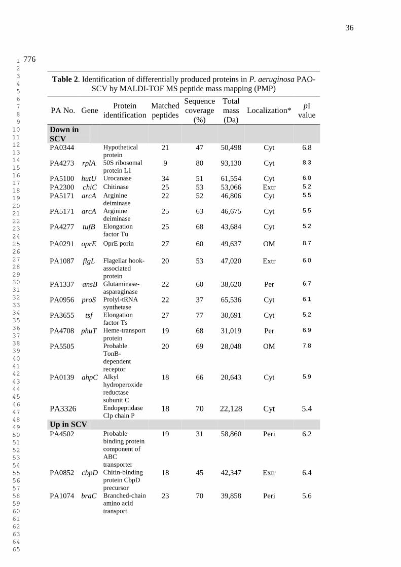
MALDI-TOF MS analysis, we found at least 24 differentially expressed proteins, whereby 16 146
proteins were less abundant and 8 more abundant in PAO-SCV (Table 2). The proteins 147
showing differential abundance are involved in amino acid biosynthesis and metabolism, 148
motility, transport of small molecules and transcriptional regulation. According to this 149
analysis, the two-component response regulator PhoP is one of the most prominently induced 150
proteins in PAO-SCV. Another finding is the over-expression of the major outer membrane 151
protein OprF in PAO-SCV, which is the P. aeruginosa major non-specific porin allowing 152
diffusion of various solutes, such as nitrates or nitrites under anaerobic conditions or small 153
oligosaccharides with a molecular weight up to 1519 Da [33,34]. We also found decreased 154
expression of the anaerobiosis-induced outer membrane porin OprE, which, similarly to 155
OprD, was predicted to be involved in outer membrane permeability of the β-lactam antibiotic 156
imipenem and basic amino acids [35,36]. 157
Genome-wide transcriptional profile of PAO-SCV and PAO1 158
Since some of the differentially produced proteins could already give clue to the changes 159
occurring in the SCV mutant, we decided to further investigate which global changes in gene 160
expression could account for this phenotypic variation. The gene transcription profiles of 161
1 2 3 4 5 6 7 8 9 10 11 12 13 14 15 16 17 18 19 20 21 22 23 24 25 26 27 28 29 30 31 32 33 34 35 36 37 38 39 40 41 42 43 44 45 46 47 48 49 50 51 52 53 54 55 56 57 58 59 60 61 62 63 64 65

9
PAO-SCV and WT strains were compared in early and late stationary-phase of growth, 162
corresponding to incubation times of 20 and 40 h respectively, using P. aeruginosa 163
Affymetrix GeneChips. The results are presented using Venn diagrams and pie charts for 164
simplicity, facilitating the understanding and interpretation of the overall genome 165
transcriptional profile [37]. The tables showing the complete lists of differentially expressed 166
genes are shown as supplementary material (Tables S1 to S5). As shown in Figure 5 and in 167
supplementary Tables S1-S5), during stationary phase, a total of 642 genes representing 168
approximately 12% of the entire genome displayed a differential expression pattern in PAO-169
SCV compared to that of wild type PAO1 (P value < 0.05, Student’s t-test). Among these 642 170
genes, 466 were up-regulated (≈ 73% of differentially regulated genes, from 2- to 26-fold, see 171
Table S3) and 176 were down-regulated (≈ 27% of differentially regulated genes, from 2- to 172
16-fold, see Table S4). Interestingly, remarkable differences were observed for up-regulated 173
genes (Figure 5B), among which 356 genes were found to be highly expressed during late 174
stationary phase while only 164 genes were up-regulated during early stationary phase as 175
compared to the wild-type. Genes involved in amino acid biosynthesis and metabolism 176
showed an increased transcription level in both early and late stationary phase of growth of 177
PAO-SCV (Table S2). Genes involved in antibiotic resistance and genes coding for 178
membrane proteins were highly expressed in the SCV mutant in early stationary phase. 179
Conversely, some genes involved in the production of secreted factors and those related to 180
1 2 3 4 5 6 7 8 9 10 11 12 13 14 15 16 17 18 19 20 21 22 23 24 25 26 27 28 29 30 31 32 33 34 35 36 37 38 39 40 41 42 43 44 45 46 47 48 49 50 51 52 53 54 55 56 57 58 59 60 61 62 63 64 65
10
phage, transposon and plasmids were expressed at a lower level in PAO-SCV compared to the 181
wild-type. 182
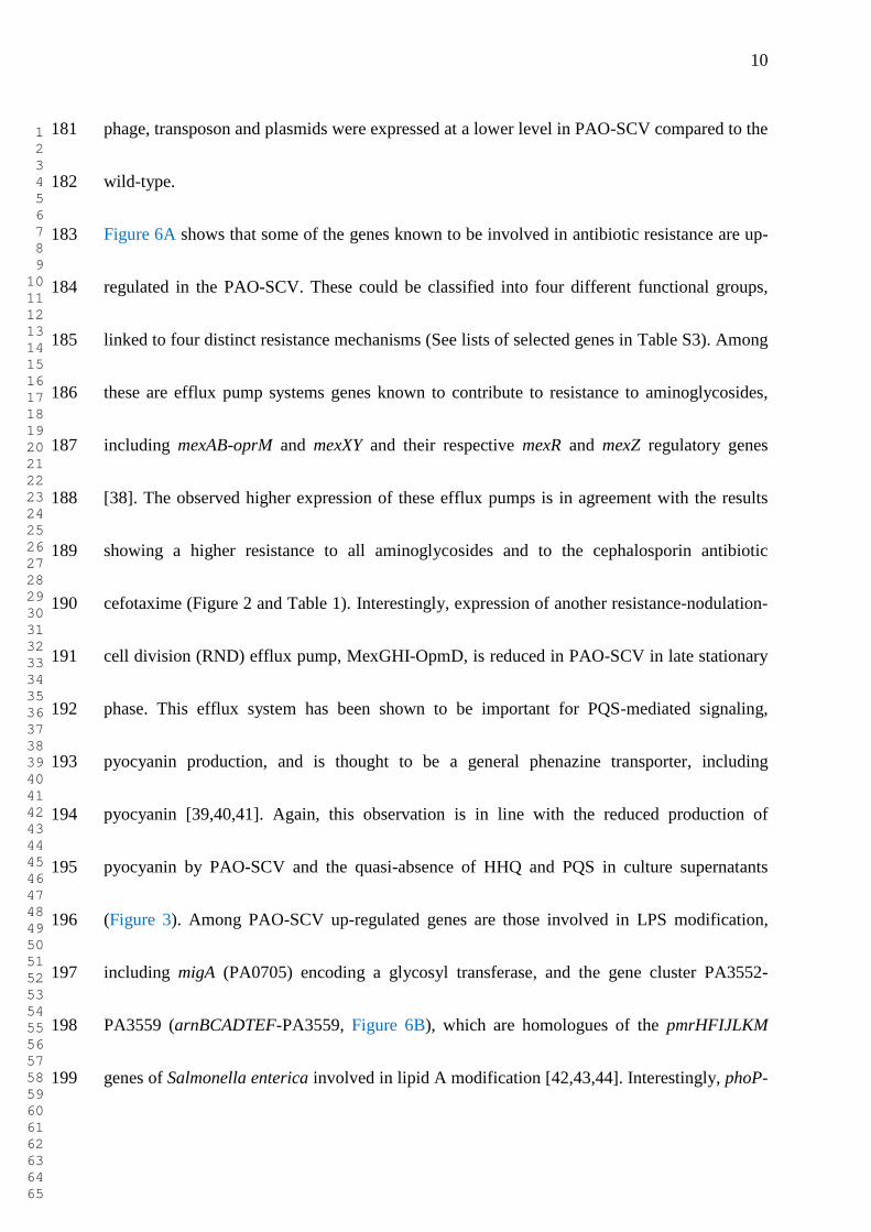
Figure 6A shows that some of the genes known to be involved in antibiotic resistance are up-183
regulated in the PAO-SCV. These could be classified into four different functional groups, 184
linked to four distinct resistance mechanisms (See lists of selected genes in Table S3). Among 185
these are efflux pump systems genes known to contribute to resistance to aminoglycosides, 186
including mexAB-oprM and mexXY and their respective mexR and mexZ regulatory genes 187
[38]. The observed higher expression of these efflux pumps is in agreement with the results 188
showing a higher resistance to all aminoglycosides and to the cephalosporin antibiotic 189
cefotaxime (Figure 2 and Table 1). Interestingly, expression of another resistance-nodulation-190
cell division (RND) efflux pump, MexGHI-OpmD, is reduced in PAO-SCV in late stationary 191
phase. This efflux system has been shown to be important for PQS-mediated signaling, 192
pyocyanin production, and is thought to be a general phenazine transporter, including 193
pyocyanin [39,40,41]. Again, this observation is in line with the reduced production of 194
pyocyanin by PAO-SCV and the quasi-absence of HHQ and PQS in culture supernatants 195
(Figure 3). Among PAO-SCV up-regulated genes are those involved in LPS modification, 196
including migA (PA0705) encoding a glycosyl transferase, and the gene cluster PA3552-197
PA3559 (arnBCADTEF-PA3559, Figure 6B), which are homologues of the pmrHFIJLKM 198
genes of Salmonella enterica involved in lipid A modification [42,43,44]. Interestingly, phoP-199
1 2 3 4 5 6 7 8 9 10 11 12 13 14 15 16 17 18 19 20 21 22 23 24 25 26 27 28 29 30 31 32 33 34 35 36 37 38 39 40 41 42 43 44 45 46 47 48 49 50 51 52 53 54 55 56 57 58 59 60 61 62 63 64 65

11
phoQ, together with the upstream porin protein gene oprH was markedly up-regulated 200
throughout the stationary phase in PAO-SCV, forming the third functional group and making 201
the link with the overexpression of migA and arnBCADTEF-PA3559 (Figure 6C). As already 202
mentioned, higher levels of the transcriptional regulator PhoP were also detected by 2D-203
PAGE analysis. The PhoP-PhoQ system is known to be involved in aminoglycoside 204
resistance in P. aeruginosa [45]. 205
A fourth functional group of genes markedly up-regulated in PAO-SCV included those 206
encoding membrane proteins, transcriptional regulators and transporters of small molecules 207
(Figure 6D). More specifically, several genes encoding outer membrane proteins are up-208
regulated in PAO-SCV: the previously mentioned oprH, oprD, PA1198 (encoding a 209
lipoprotein), oprQ, opdQ, opdP, and the lipoprotein gene omlA. OprQ, and OpdP belong to 210
the OprD family and have been proposed to contribute to the transport of arginine [46]. In this 211
context, it is interesting to note that the genes PA5152 (ABC transporter, ATP binding 212
component), and, to a large extent, PA5153 (periplasmic binding protein), probably involved 213
in the transport of arginine, are also up-regulated. 214
The transcriptome analysis not only provided insights into the PAO-SCV mechanisms 215
involved in aminoglycoside-resistance, but also explained some of the prominent phenotypic 216
changes. As shown in Figure 7A, transcript levels of the pqsABCDE genes as well as for the 217
two neighboring anthranilate synthase genes phnA and phnB were strongly reduced in PAO-218
1 2 3 4 5 6 7 8 9 10 11 12 13 14 15 16 17 18 19 20 21 22 23 24 25 26 27 28 29 30 31 32 33 34 35 36 37 38 39 40 41 42 43 44 45 46 47 48 49 50 51 52 53 54 55 56 57 58 59 60 61 62 63 64 65
12
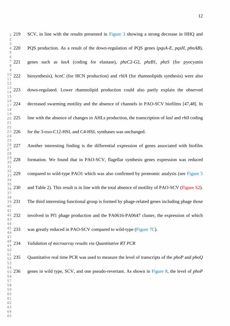
SCV, in line with the results presented in Figure 3 showing a strong decrease in HHQ and 219
PQS production. As a result of the down-regulation of PQS genes (pqsA-E, pqsH, phnAB), 220
genes such as lasA (coding for elastase), phzC2-G2, phzB1, phzS (for pyocyanin 221
biosynthesis), hcnC (for HCN production) and rhlA (for rhamnolipids synthesis) were also 222
down-regulated. Lower rhamnolipid production could also partly explain the observed 223
decreased swarming motility and the absence of channels in PAO-SCV biofilms [47,48]. In 224
line with the absence of changes in AHLs production, the transcription of lasI and rhlI coding 225
for the 3-oxo-C12-HSL and C4-HSL synthases was unchanged. 226
Another interesting finding is the differential expression of genes associated with biofilm 227
formation. We found that in PAO-SCV, flagellar synthesis genes expression was reduced 228
compared to wild-type PAO1 which was also confirmed by proteomic analysis (see Figure 5 229
and Table 2). This result is in line with the total absence of motility of PAO-SCV (Figure S2). 230
The third interesting functional group is formed by phage-related genes including phage those 231
involved in Pf1 phage production and the PA0616-PA0647 cluster, the expression of which 232
was greatly reduced in PAO-SCV compared to wild-type (Figure 7C). 233
Validation of microarray results via Quantitative RT PCR 234
Quantitative real time PCR was used to measure the level of transcripts of the phoP and phoQ 235
genes in wild type, SCV, and one pseudo-revertant. As shown in Figure 8, the level of phoP 236
1 2 3 4 5 6 7 8 9 10 11 12 13 14 15 16 17 18 19 20 21 22 23 24 25 26 27 28 29 30 31 32 33 34 35 36 37 38 39 40 41 42 43 44 45 46 47 48 49 50 51 52 53 54 55 56 57 58 59 60 61 62 63 64 65

13
and phoQ transcription was increased in the SCV while the levels were similar for wild-type 237
and the revertant large colony variant. 238
Whole genome analysis of P. aeruginosa SCV 239
The genome of the originally selected pseudo-revertant (REV) of PAO-SCV was fully 240
sequenced using the Illumina Genome Analyzer. The choice to re-sequence the revertant only 241
was justified by the fact that it should contain all mutations present in three strains (PAO1, 242
PAO-SCV, revertant), and single nucleotide polymorphisms (SNPs) could be easily checked 243
for their presence in the genomes of PAO-SCV and its clonal wild-type using a combination 244
of PCR amplification and Sanger sequencing. A limited list of sequence variations in relation 245
to the PAO1 sequence was found (Table 3 and Table S6 for full list), most of which were 246
already detected when we re-sequenced these regions in our own PAO1 lab strain and in the 247
PAO1 strain of Chronic Pseudomonas Infection Group in Helmholtz Infection Research 248
Centre. Finally, seven changes remained that were unique to REV after elimination of the 249
mutations also found in PAO1 wild-type. Through PCR amplification of these regions and re-250
sequencing via the Sanger method these differences in sequence were confirmed to be present 251
in both REV and SCV, which was surprising since the phenotypic differences between SCV 252
and REV are very dramatic and expected to be reflected by differences in genetic background. 253
Interestingly, changes were identified in phoPQ and mexZ, in line with the results of the 254
transcriptional analysis. Specifically, the phoP gene contains a SNP which confers a histidine 255
1 2 3 4 5 6 7 8 9 10 11 12 13 14 15 16 17 18 19 20 21 22 23 24 25 26 27 28 29 30 31 32 33 34 35 36 37 38 39 40 41 42 43 44 45 46 47 48 49 50 51 52 53 54 55 56 57 58 59 60 61 62 63 64 65

14
(H) to arginine (R) change while, remarkably, phoQ harbors an in-frame 39-bp deletion in its 256
coding sequence, deleting a 13 amino acids sequence RLLRSEHKQRERY between residues 257
226 and 239. In PAO-SCV the mexZ gene was inactivated by the introduction of a stop codon, 258
which could explain the over-expression of the MexXY pump involved in aminoglycosides 259
and fluoroquinolone resistance. 260
Discussion 261
The SCV phenotype observed in this study is reminiscent of the observations made by Tarighi 262
et al. who found that knocking out the the ppgL gene (PA4204) of P. aeruginosa caused a 263
SCV phenotype with the apparition of large colony variants [17]. In this particular case the 264
SCV phenotype was thought to be due to the accumulation of a toxic intermediate: 265
gluconolactone [17]. This observation suggests that SCV phenotypes can be the results of 266
exposures to different stresses. In P. aeruginosa, several mechanisms of aminoglycoside 267
resistance have been described: resistance through efflux systems, by alteration of porins or 268
outer membrane properties (including LPS modification), resistance through chromosomal 269
mutations of regulatory genes, and resistance through enzymatic drug modification, including 270
both intrinsic and acquired resistance [49,50,51,52,53,54]. P. aeruginosa can use these 271
mechanisms in combination, to reach high-level of resistance to certain antibiotics, which is 272
precisely what we observed in this study since we found an overexpression of two efflux 273
systems (MexXY-OprM, MexAB-OprM, increased expression of the arnBCADTEF-PA3559 274
1 2 3 4 5 6 7 8 9 10 11 12 13 14 15 16 17 18 19 20 21 22 23 24 25 26 27 28 29 30 31 32 33 34 35 36 37 38 39 40 41 42 43 44 45 46 47 48 49 50 51 52 53 54 55 56 57 58 59 60 61 62 63 64 65

15
LPS modification genes, of the porins OprH, OprF, and decreased expression of OprE. The 275
tripartite efflux pump MexXY-OprM is known to be a major contributor to aminoglycoside 276
resistance in P. aeruginosa [55,56,57,58] as well as the efflux of the drug tigecycline [59]. In 277
addition, MexAB-OprM was also proven to confer resistance to β-lactams, fluoroquinolones 278
[50] and to contribute to aminoglycosides resistance [60]. Both efflux pumps share the same 279
efflux porin, OprM [61]. In P. aeruginosa, the PhoP-PhoQ two-component regulatory system 280
is known to be induced upon Mg2+
starvation to up-regulate the production of the outer-281
membrane protein OprH and to increase the resistance to the polycationic antibiotic 282
polymyxin B [62]. In addition, PhoP-PhoQ is also involved in resistance to antimicrobial 283
cationic peptides and aminoglycoside antibiotics [45], in good agreement with the higher 284
resistance of PAO-SCV to this class of antibiotics. The arnBCADTEF-PA3559 genes could 285
also be involved in conferring a higher resistance to aminoglycosides. Intriguingly, PA3559 286
encodes a UDP-glucose dehydrogenase that is induced by low concentrations of Mg2+
[43] 287
and its expression depends on the PmrA-PmrB two-component regulatory system, which is 288
itself regulated by the PhoP-PhoQ two-component system [63,64,65,66]. The general porin 289
OprF, which is overexpressed in the SCV, has been recently shown to participate in resistance 290
mechanisms to a broad spectrum of antibiotics such as β-lactams, cephalosporins, and 291
fluoroquinolones [67], supporting the role of OprF as an intrinsic antibiotic resistance 292
contributor as well as partially explaining the cefotaxime resistance revealed by phenotypic 293
1 2 3 4 5 6 7 8 9 10 11 12 13 14 15 16 17 18 19 20 21 22 23 24 25 26 27 28 29 30 31 32 33 34 35 36 37 38 39 40 41 42 43 44 45 46 47 48 49 50 51 52 53 54 55 56 57 58 59 60 61 62 63 64 65

16
assays. In PAO-SCV we observed a down-regulation of phage genes compared to the wild-294
type. The expression of phage genes has been shown to reciprocally associate with biofilm 295
formation and antibiotic resistance as evidenced by Whiteley and colleagues [68]. These 296
authors found that phages genes were up-regulated in mature biofilms as compared to 297
planktonic cultures while they were shown to be down-regulated in biofilms exposed to the 298
aminoglycoside tobramycin. A puzzling observation is the strong down-regulation in PAO-299
SCV of PQS biosynthesis genes and of the PQS-related efflux pump MexGHI-OpmD [39], 300
which is confirmed by the near absence of PQS production presented in Figure 3. It has been 301
suggested that high production of PQS, which results in an autolysis phenotype, could be 302
explained by the induction of prophages [22], which fits with the results presented here. 303
Collectively, our whole genome expression analysis of PAO-SCV versus wild-type PAO1 304
allowed us to get a good correlation between phenotypic traits (antibiotic resistance, PQS and 305
virulence factors production), proteomic and gene expression data. However, we failed to 306
pinpoint the mutation(s) leading to the phenotypic conversion to the SCV state since all of the 307
mutations observed in the revertant were also present in the SCV genome. One possible 308
explanation is a phenotypic switch without mutation, like the recently described bi-stable 309
phenotypic switch due to the LysR regulator BexR [69]. 310
Experimental procedures 311
Bacterial strains and culture conditions 312
1 2 3 4 5 6 7 8 9 10 11 12 13 14 15 16 17 18 19 20 21 22 23 24 25 26 27 28 29 30 31 32 33 34 35 36 37 38 39 40 41 42 43 44 45 46 47 48 49 50 51 52 53 54 55 56 57 58 59 60 61 62 63 64 65

17
P. aeruginosa PAO1 Strain (ATTC 15692) and its gentamicin-resistant mutant PAO-SCV 313
were used in this study. P. aeruginosa strains were grown at 37°C in Luria-Bertani (LB) broth 314
or on LB agar plates, iron poor casamino acids (CAA) medium (Difco Laboratories) or 315
Pseudomonas agar medium (Difco Laboratories). The antibiotics gentamicin (Gm) at 200 µg 316
ml-1
and spectinomycin (Sp) at 50 µg ml-1
were used when necessary. Growth rate of three 317
replicates for each strain was monitored spectrophotometrically (Bioscreen C, Thermo 318
Labsystems). 319
Motility assay 320
Swarming, swimming and twitching motility were determined as previously described [70]. 321
To investigate swarming motility, 4 µl of overnight cultures of P. aeruginosa grown in LB (1 322
x 109 cells) were placed in the center of 0.4% agar LB or CAA plates while swimming 323
motility was evaluated using 0.3% agar LB or CAA plates. For twitching motility, LB or 324
CAA plates containing 1.5% agar were inoculated with a toothpick by stabbing the plates. 325
The plates were then incubated at 37°C. In the case of twitching, after incubation the LB- or 326
CAA-agar media was removed from the plates and plates were stained with 1% crystal violet 327
(Merck) in 33% acetic acid for a minimum of 20 min. Spreading of bacteria from the 328
inoculation point was measured and pictures were taken. Three independent experiments were 329
performed. 330
331
1 2 3 4 5 6 7 8 9 10 11 12 13 14 15 16 17 18 19 20 21 22 23 24 25 26 27 28 29 30 31 32 33 34 35 36 37 38 39 40 41 42 43 44 45 46 47 48 49 50 51 52 53 54 55 56 57 58 59 60 61 62 63 64 65

18
Detection and analysis of signal molecules 332
Rapid detection of N-acyl homoserine lactones (AHLs) in filter sterilized (0.2 µm pore-size
333
filters) culture supernatants was done using AHL reporter plate bioassays by either E. coli 334
JM109 carrying the plasmid pSB401 for the detection of N-(butanoyl)-L-homoserine lactone 335
(C4-HSL) [71] or E. coli MH155 [72] for the detection of N-(3-oxododecanoyl)-L-336
homoserine lactone (3-oxo-C12-HSL). For accurate AHL quantification, 100 ml of acidified 337
filter-sterilized culture supernatants were extracted with equal volumes of dichloromethane. 338
The organic phase was removed and dried by evaporation in vacuum. Extracts were re-339
dissolved in 1 ml of 50% acetonitrile. Thin-layer chromatography (TLC) plates Silica gel 60 340
F254 (Merck) and RP-18 F245 (Merck) were used for detection of 3-oxo-C12-HSL and C4-341
HSL, respectively. Twenty µl of extracted AHLs were fractionated on TLC plates. After 342
development in a solvent mixture of methanol/water (60:40, vol/vol), the plates were dried 343
and overlaid with 50 ml of soft top LB-agar mixed with 1 ml of overnight culture of an E. coli 344
MH155 strain harboring the reporter plasmid pUCP22NotI-PlasB::gfp(ASV)Plac ::lasR (to 345
detect 3-oxo-C12-HSL) or 1 ml of overnight culture of E. coli JM109 pSB401 (to detect C4 346
and C6-HSL). After 18 h incubation at 37°C the production of 3-oxo-C12-HSL was detected 347
under UV by visualization of green fluorescent spots and production of C4 and C6 was visible 348
by the production of light. 349
The alkyl-hydroxy quinolones PQS and HHQ were extracted from 10 ml of early stationary 350
1 2 3 4 5 6 7 8 9 10 11 12 13 14 15 16 17 18 19 20 21 22 23 24 25 26 27 28 29 30 31 32 33 34 35 36 37 38 39 40 41 42 43 44 45 46 47 48 49 50 51 52 53 54 55 56 57 58 59 60 61 62 63 64 65

19
phase filtered supernatants by adding equal volumes of acidified ethyl acetate. The organic 351
phase was dried and the residue re-suspended in 50 ml methanol. Ten µl samples of this 352
extract were spotted onto normal phase silica 60 F254 (Merck) TLC plates, pre-treated by 353
soaking in 5% K2HPO4 for 30 min and activated at 100°C for 1 h. Extracts were separated 354
using a dichloromethane:methanol (95:5, vol/vol) solvent system until the solvent front 355
reached the top of the plate. PQS was visualised under UV light and specific detection was 356
done using soft top LB-agar including 1ml of overnight culture of a P. aeruginosa lecA::lux 357
∆pqsA strain as bioreporter [73]. Bioluminescence was detected and quantified with a Bio 358
Imaging System (Syngene). Amounts of C4-HSL, C6-HSL, 3-oxo-C12-HSL and PQS were 359
determined by measuring the diameter of the spots. 360
Persistence assay 361
The persistence assay was performed essentially as described previously [6]. Shortly, cultures 362
were grown overnight at 37°C in 100 ml LB medium in erlenmeyer flasks. One mL of a 363
stationary phase culture was treated with 10 µL of ofloxacin at a final concentration of 5 µg 364
mL-1
; a control treatment was performed with sterile water. Both treatments were performed 365
at 37 °C, shaking at 200 rpm, during five hours, after which the number of colony forming 366
units were determined by plate counts. The persister fraction is defined as the number of 367
surviving cells after treatment with ofloxacin, divided by the number of cells after the control 368
treatment. The relative persister fraction for each strain is the persister fraction of the strain 369
1 2 3 4 5 6 7 8 9 10 11 12 13 14 15 16 17 18 19 20 21 22 23 24 25 26 27 28 29 30 31 32 33 34 35 36 37 38 39 40 41 42 43 44 45 46 47 48 49 50 51 52 53 54 55 56 57 58 59 60 61 62 63 64 65

20
divided by that of the wild-type. The mean relative persister fraction is calculated as the 370
inverse logarithm of the mean of the logarithmic values of these relative persister fractions of 371
separate experiments. Each experiment was independently repeated at least four times. The 372
mean relative persister fractions are displayed with the bars representing the 25th
and 75th
373
percentiles as shown in Figure S1. Each experiment was repeated three times. 374
Biofilm formation assay 375
The protocol of O'Toole et al. [74] was followed with some minor modifications. Briefly, 376
bacterial strains were grown aerobically in LB broth at 37°C for 24 h. Cells diluted to an 377
absorbance of 0.5 at 600 nm were then sub-cultured (1:100), in LB and CAA broth in 378
triplicates in a final volume of 2 ml in 96- or 24-well polystyrene plates (BD Bioscience) and 379
incubated aerobically at 30°C without shaking for 48 h. The culture was removed from the 380
wells and plates washed twice with distilled water. The biofilm was then stained with 1% 381
crystal violet (1% crystal violet in 33% acetic acid) and incubated at room temperature for 30 382
min. Crystal violet was removed from the wells and plates were washed twice with phosphate 383
buffered saline (PBS) (0.1 M, pH 7.2). Plates were dried and crystal violet stain was dissolved 384
using 70% ethanol of which absorbance was determined at 590 nm. The average of three 385
measurements per strain was determined and expressed as biomass production characteristic 386
of biofilm formation. The same procedures were performed with abiotic materials including 387
borosilcate glass, polyvinyl chloride plastics and polypropylene plastics. The biofilm 388
formation was also analyzed in 96-well microtiter plates with an image-based approach [75]. 389
1 2 3 4 5 6 7 8 9 10 11 12 13 14 15 16 17 18 19 20 21 22 23 24 25 26 27 28 29 30 31 32 33 34 35 36 37 38 39 40 41 42 43 44 45 46 47 48 49 50 51 52 53 54 55 56 57 58 59 60 61 62 63 64 65

21
Briefly, bacterial strains were grown overnight in LB broth at 37°C. Cultures were diluted to 390
an absorbance of 0.02 at 600 nm and a final volume of 100 μl was transferred in triplicate into 391
the wells of the 96-well μClear microplate (Greiner Bio-One). The plate was covered with an 392
air-permeable BREATHseal cover foil (Greiner Bio-One) and placed into a humid incubator 393
at 37°C. After 24 h incubation, the bacterial cultures were stained with the LIVE/DEAD 394
BacLight Bacterial Viability Kit (Molecular Probes, Invitrogen). Fifty μl of a diluted staining 395
solution (1:500) was added into each well resulting in a final concentration of 1.4 μM SYTO9 396
and 8.3 μM PI (propidium iodide). After 48 h incubation structured biofilm were observed. 397
Microscopy was performed after 72 h using an Olympus Fluoview 1000 system equipped 398
with an x40/NA (numerical aperture) 0.90 air lens. Total biovolumes of the image stacks of 399
each strain were calculated using the tool PHLIP [76]. 3D-visualization of the biofilms was 400
performed with the software IMARIS (version 5.7.2, Bitplane). 401
Proteome analysis by two-dimensional (2D) gel electrophoresis 402
P. aeruginosa cells were harvested in early stationary phase by centrifugation (4,000 g, 10 403
min, 4°C) and washed three times with Tris-HCl buffer (pH 8.0). To prepare extracts of 404
cellular proteins, bacterial cells were washed twice in PBS buffer (pH 8.0) and re-suspended 405
in a solution containing 40 ml of 2.5 mM Tris-HCl (pH 8.0), one tablet of protease inhibitor 406
(Sigma), 80 µl of 0.5 M Na2EDTA, and 400 µl of DNase at 10 mg ml-1
. After lysis of the cells 407
by sonication with a Branson Sonifier 250, each suspension was centrifuged (2,500 g, 15 min, 408
1 2 3 4 5 6 7 8 9 10 11 12 13 14 15 16 17 18 19 20 21 22 23 24 25 26 27 28 29 30 31 32 33 34 35 36 37 38 39 40 41 42 43 44 45 46 47 48 49 50 51 52 53 54 55 56 57 58 59 60 61 62 63 64 65

22
4°C) to remove the cell debris and unbroken cells. The supernatant was then subjected to a 409
second centrifugation (50,000 g, 40 min, 4°C) to remove the insoluble components, and the 410
protein concentration in the resulting supernatant was determined by a Bradford Protein assay 411
(Bio-Rad). For the preparation of extracellular protein extracts, the supernatants obtained after 412
centrifugation of the bacterial cultures (6,000 g, 15 min, 4°C) were passed through a 0.2 µm-413
pore-size filter. Deoxycholic acid (sodium salt) was added to a final concentration of 0.2 mg 414
ml-1
. After 30 min of incubation on ice, the proteins were precipitated by addition of 6% 415
(wt/vol) trichloroacetic acid and incubated at 4°C for 2 hr. After centrifugation (18,000 g, 30 416
min, 4°C) the precipitated proteins were re-suspended in distilled water, and eight volumes of 417
cold acetone (-20°C) were added. After incubation at -20°C for 2 h, the mixture was 418
centrifuged (3,500 g, 20 min, 4°C), and the pellet was allowed to dry for 5 min before it was 419
dissolved in an appropriate amount of solubilisation buffer. After centrifugation (50,000 g, 40 420
min, 4°C) to remove the insoluble components, the protein concentration of the remaining 421
supernatant was determined. Protein extracts were either used immediately for 2-D gel 422
electrophoresis or stored at -80°C. Isoelectric focusing was performed with the IPGphor 423
system and Immobiline DryStrip gel strips (GE Healthcare). Equal quantities of solubilised 424
proteins from the different P. aeruginosa strains were diluted to obtain a final volume of 360 425
µl with solubilisation solution and applied to the Immobiline gel strips by in-gel rehydration. 426
Linear immobilized pH gradients (pH 4 to 7) were used. Thirty to 50 µg of protein was 427
1 2 3 4 5 6 7 8 9 10 11 12 13 14 15 16 17 18 19 20 21 22 23 24 25 26 27 28 29 30 31 32 33 34 35 36 37 38 39 40 41 42 43 44 45 46 47 48 49 50 51 52 53 54 55 56 57 58 59 60 61 62 63 64 65

23
applied for analytical gels (silver staining), and 200 to 500 µg of protein was loaded for 428
Coomassie staining was used. After rehydration under silicone oil for 10 hr, the proteins were 429
focused for a total of 120 kV/h at 20°C. The proteins were reduced by equilibration of the 430
strips in equilibration solution (6 M urea, 30% glycerol, 2% [wt/vol] sodium dodecyl sulfate, 431
and 1% [wt/vol] dithiothreitol in 0.05 M Tris-HCl (pH 8.8) for 15 min and then 432
carbamidomethylated in the same solution containing 260 mM iodoacetamide for 15 min. The 433
strips were transferred to 12% acrylamide gradient gels and electrophoresis was performed 434
overnight at 125 V at 10°C. Gels were stained with Coomassie brilliant blue solution and 435
spots of interest were then further analyzed through peptide mass fingerprinting according to 436
the described protocol [77]. Peptides examined on a MALDI-TOF mass spectrometer 437
(Bruker) and analyzed by MASCOT (Matrix Science) were used to identify proteins from 438
peptide identifications using the NCBInr database. 439
Microarray and quantitative real time PCR analysis 440
Cultures were grown in triplicate until early (24 h) and late (48 h) stationary phase, 441
respectively, allowing three biological replicates per condition. Total RNA was obtained from 442
the cultures of early and late stationary phase by first treating the cells with RNAprotect 443
Bacteria Reagent (Qiagen) as recommended by the manufacturer. Cells were then lysed and 444
total RNA was extracted using the RNeasy Midi Kit (Qiagen), on-column DNase digestion 445
was performed using the RNase-free DNase Set (Qiagen) according to the manufactures 446
1 2 3 4 5 6 7 8 9 10 11 12 13 14 15 16 17 18 19 20 21 22 23 24 25 26 27 28 29 30 31 32 33 34 35 36 37 38 39 40 41 42 43 44 45 46 47 48 49 50 51 52 53 54 55 56 57 58 59 60 61 62 63 64 65

24
instructions. RNA integrity was assessed using the 2100 bioanalyzer (Agilent Technologies 447
Inc.). cDNA synthesis, fragmentation and labeling were performed according to the supplier 448
protocol for the P. aeruginosa Genechip genome array (Affymetrix) at 50°C. Washing and 449
staining of the arrays was performed according to the manufacturer’s instructions using a 450
fluidics station 400 (Affymetrix). Slides were scanned using the 2500A GeneArray Scanner 451
(Agilent Technologies Inc.) and Affymetrix MAS 5.0. Data analysis was performed using 452
GeneSpring GX (Agilent Technologies Inc.) in which the scaled data was further normalized 453
by per Chip and per Gene median normalizations. Filtering of genes was performed to find 454
genes that had changed in expression by a magnitude of 2-fold (P value less than 0.05, 455
Student’s t-test). 456
Bacterial cells were harvested in stationary phase, bacterial RNA was extracted by using 457
RNeasy Midi Kit (QIAGEN). The purity and concentration of the RNA was determined by 458
spectrophotometry (NanoDrop, Thermo Scientific). First-strand cDNA was reverse 459
transcribed from one microgram of total RNA by using First-strand cDNA Synthesis Kit 460
(Amersham Biosciences, GE Healthcare). qRT-PCR was performed in a Bio-Rad (Hercules, 461
CA, USA) iCycler with Bio-Rad iQ SYBR Green Supermix. For all primer sets, the following 462
cycling parameters were used: 94°C for 3 min followed by 40 cycles of 94°C for 30 s, 55°C 463
for 45 s and 72°C for 30 s, followed by 72°C for 7 min. The outer membrane lipoprotein oprI 464
gene was used to normalize gene 465
1 2 3 4 5 6 7 8 9 10 11 12 13 14 15 16 17 18 19 20 21 22 23 24 25 26 27 28 29 30 31 32 33 34 35 36 37 38 39 40 41 42 43 44 45 46 47 48 49 50 51 52 53 54 55 56 57 58 59 60 61 62 63 64 65
25
expression[78][78][78][78][78][71][78][77][78][69][78][78][84]. Amplification products 466
were electrophoresed on 0.8% agarose gels. For statistical analysis of relative gene 467
expression, the 2-△△CT
method was used [79]. All experiments were carried out in triplicate. 468
Acknowledgements 469
This work was supported by two research grants from the FWO Belgium (Fonds voor 470
Wetenschappelijk Onderzoek Vlaanderen) and a grant from BBSRC UK (BBF0143921). QW 471
has a CSC-VUB scholarship and got a FEMS student fellowship to perform experiments in 472
the lab of Dr. Susanne Haüssler. BB and AB are research assistants and PV is a postdoctoral 473
researcher of the FWO-Vlaanderen. We wish to thank Dr. Jean Paul Pirnay, Daniel De Vos, 474
and Florence Bilocq from the Military hospital in Brussels for the VITEK analysis. We would 475
like to thank De Meuter Sonja and Van Hemelrijck Herman for technical assistance. 476
477
References 478
1. Stover CK, Pham XQ, Erwin AL, Mizoguchi SD, Warrener P, et al. (2000) Complete 479
genome sequence of Pseudomonas aeruginosa PAO1, an opportunistic pathogen. 480
Nature 406: 959-964. 481
2. Lyczak JB, Cannon CL, Pier GB (2002) Lung infections associated with cystic fibrosis. 482
Clin Microbiol Rev 15: 194-222. 483
3. Lyczak JB, Cannon CL, Pier GB (2000) Establishment of Pseudomonas aeruginosa 484
infection: lessons from a versatile opportunist. Microbes Infect 2: 1051-1060. 485
4. Boles BR, Thoendel M, Singh PK (2004) Self-generated diversity produces "insurance 486
effects" in biofilm communities. Proc Natl Acad Sci U S A 101: 16630-16635. 487
5. Govan JR, Deretic V (1996) Microbial pathogenesis in cystic fibrosis: mucoid 488
Pseudomonas aeruginosa and Burkholderia cepacia. Microbiol Rev 60: 539-574. 489
1 2 3 4 5 6 7 8 9 10 11 12 13 14 15 16 17 18 19 20 21 22 23 24 25 26 27 28 29 30 31 32 33 34 35 36 37 38 39 40 41 42 43 44 45 46 47 48 49 50 51 52 53 54 55 56 57 58 59 60 61 62 63 64 65

26
6. De Groote VN, Verstraeten N, Fauvart M, Kint CI, Verbeeck AM, et al. (2009) Novel 490
persistence genes in Pseudomonas aeruginosa identified by high-throughput 491
screening. FEMS Microbiol Lett 297: 73-79. 492
7. Moker N, Dean CR, Tao J (2010) Pseudomonas aeruginosa increases formation of 493
multidrug-tolerant persister cells in response to quorum-sensing signaling molecules. J 494
Bacteriol 192: 1946-1955. 495
8. Haussler S, Tummler B, Weissbrodt H, Rohde M, Steinmetz I (1999) Small-colony variants 496
of Pseudomonas aeruginosa in cystic fibrosis. Clin Infect Dis 29: 621-625. 497
9. Haussler S, Ziegler I, Lottel A, von Gotz F, Rohde M, et al. (2003) Highly adherent small-498
colony variants of Pseudomonas aeruginosa in cystic fibrosis lung infection. J Med 499
Microbiol 52: 295-301. 500
10. von Gotz F, Haussler S, Jordan D, Saravanamuthu SS, Wehmhoner D, et al. (2004) 501
Expression analysis of a highly adherent and cytotoxic small colony variant of 502
Pseudomonas aeruginosa isolated from a lung of a patient with cystic fibrosis. J 503
Bacteriol 186: 3837-3847. 504
11. Kirisits MJ, Prost L, Starkey M, Parsek MR (2005) Characterization of colony 505
morphology variants isolated from Pseudomonas aeruginosa biofilms. Appl Environ 506
Microbiol 71: 4809-4821. 507
12. Starkey M, Hickman JH, Ma L, Zhang N, De Long S, et al. (2009) Pseudomonas 508
aeruginosa rugose small-colony variants have adaptations that likely promote 509
persistence in the cystic fibrosis lung. J Bacteriol 191: 3492-3503. 510
13. Proctor RA, von Eiff C, Kahl BC, Becker K, McNamara P, et al. (2006) Small colony 511
variants: a pathogenic form of bacteria that facilitates persistent and recurrent 512
infections. Nat Rev Microbiol 4: 295-305. 513
14. Deziel E, Comeau Y, Villemur R (2001) Initiation of biofilm formation by Pseudomonas 514
aeruginosa 57RP correlates with emergence of hyperpiliated and highly adherent 515
phenotypic variants deficient in swimming, swarming, and twitching motilities. J 516
Bacteriol 183: 1195-1204. 517
15. Drenkard E, Ausubel FM (2002) Pseudomonas biofilm formation and antibiotic resistance 518
are linked to phenotypic variation. Nature 416: 740-743. 519
16. Nelson LK, Stanton MM, Elphinstone RE, Helwerda J, Turner RJ, et al. (2010) 520
Phenotypic diversification in vivo.: Pseudomonas aeruginosa gacS- strains generate 521
small colony variants in vivo that are distinct from in vitro variants. Microbiology. 522
17. Tarighi S, Wei Q, Camara M, Williams P, Fletcher MP, et al. (2008) The PA4204 gene 523
encodes a periplasmic gluconolactonase (PpgL) which is important for fitness of 524
Pseudomonas aeruginosa. Microbiology 154: 2979-2990. 525
18. Malone JG, Jaeger T, Spangler C, Ritz D, Spang A, et al. (2010) YfiBNR mediates cyclic 526
di-GMP dependent small colony variant formation and persistence in Pseudomonas 527
aeruginosa. PLoS Pathog 6: e1000804. 528
1 2 3 4 5 6 7 8 9 10 11 12 13 14 15 16 17 18 19 20 21 22 23 24 25 26 27 28 29 30 31 32 33 34 35 36 37 38 39 40 41 42 43 44 45 46 47 48 49 50 51 52 53 54 55 56 57 58 59 60 61 62 63 64 65

27
19. Schneider M, Muhlemann K, Droz S, Couzinet S, Casaulta C, et al. (2008) Clinical 529
characteristics associated with isolation of small-colony variants of Staphylococcus 530
aureus and Pseudomonas aeruginosa from respiratory secretions of patients with 531
cystic fibrosis. J Clin Microbiol 46: 1832-1834. 532
20. Mulcahy LR, Burns JL, Lory S, Lewis K (2010) Emergence of Pseudomonas aeruginosa 533
strains producing high levels of persister cells in patients with cystic fibrosis. J 534
Bacteriol. 535
21. Hengge R (2009) Principles of c-di-GMP signalling in bacteria. Nat Rev Microbiol 7: 536
263-273. 537
22. D'Argenio DA, Calfee MW, Rainey PB, Pesci EC (2002) Autolysis and autoaggregation 538
in Pseudomonas aeruginosa colony morphology mutants. J Bacteriol 184: 6481-6489. 539
23. Hickman JW, Tifrea DF, Harwood CS (2005) A chemosensory system that regulates 540
biofilm formation through modulation of cyclic diguanylate levels. Proc Natl Acad Sci 541
U S A 102: 14422-14427. 542
24. Kuchma SL, Brothers KM, Merritt JH, Liberati NT, Ausubel FM, et al. (2007) BifA, a 543
cyclic-Di-GMP phosphodiesterase, inversely regulates biofilm formation and 544
swarming motility by Pseudomonas aeruginosa PA14. J Bacteriol 189: 8165-8178. 545
25. Kulasekara BR, Kulasekara HD, Wolfgang MC, Stevens L, Frank DW, et al. (2006) 546
Acquisition and evolution of the exoU locus in Pseudomonas aeruginosa. J Bacteriol 547
188: 4037-4050. 548
26. Meissner A, Wild V, Simm R, Rohde M, Erck C, et al. (2007) Pseudomonas aeruginosa 549
cupA-encoded fimbriae expression is regulated by a GGDEF and EAL domain-550
dependent modulation of the intracellular level of cyclic diguanylate. Environ 551
Microbiol 9: 2475-2485. 552
27. Friedman L, Kolter R (2004) Two genetic loci produce distinct carbohydrate-rich 553
structural components of the Pseudomonas aeruginosa biofilm matrix. J Bacteriol 554
186: 4457-4465. 555
28. Jackson KD, Starkey M, Kremer S, Parsek MR, Wozniak DJ (2004) Identification of psl, 556
a locus encoding a potential exopolysaccharide that is essential for Pseudomonas 557
aeruginosa PAO1 biofilm formation. J Bacteriol 186: 4466-4475. 558
29. Matsukawa M, Greenberg EP (2004) Putative exopolysaccharide synthesis genes 559
influence Pseudomonas aeruginosa biofilm development. J Bacteriol 186: 4449-4456. 560
30. Williams P (2006) Quorum sensing. Int J Med Microbiol 296: 57-59. 561
31. Venturi V (2006) Regulation of quorum sensing in Pseudomonas. FEMS Microbiol Rev 562
30: 274-291. 563
32. Diggle SP, Cornelis P, Williams P, Camara M (2006) 4-quinolone signalling in 564
Pseudomonas aeruginosa: old molecules, new perspectives. Int J Med Microbiol 296: 565
83-91. 566
1 2 3 4 5 6 7 8 9 10 11 12 13 14 15 16 17 18 19 20 21 22 23 24 25 26 27 28 29 30 31 32 33 34 35 36 37 38 39 40 41 42 43 44 45 46 47 48 49 50 51 52 53 54 55 56 57 58 59 60 61 62 63 64 65
28
33. Nestorovich EM, Sugawara E, Nikaido H, Bezrukov SM (2006) Pseudomonas aeruginosa 567
porin OprF: properties of the channel. J Biol Chem 281: 16230-16237. 568
34. Yoon SS, Hennigan RF, Hilliard GM, Ochsner UA, Parvatiyar K, et al. (2002) 569
Pseudomonas aeruginosa anaerobic respiration in biofilms: relationships to cystic 570
fibrosis pathogenesis. Dev Cell 3: 593-603. 571
35. Yamano Y, Nishikawa T, Komatsu Y (1993) Cloning and nucleotide sequence of 572
anaerobically induced porin protein E1 (OprE) of Pseudomonas aeruginosa PAO1. 573
Mol Microbiol 8: 993-1004. 574
36. Yamano Y, Nishikawa T, Komatsu Y (1998) Involvement of the RpoN protein in the 575
transcription of the oprE gene in Pseudomonas aeruginosa. FEMS Microbiol Lett 576
162: 31-37. 577
37. Eisen MB, Spellman PT, Brown PO, Botstein D (1998) Cluster analysis and display of 578
genome-wide expression patterns. Proc Natl Acad Sci U S A 95: 14863-14868. 579
38. Llanes C, Hocquet D, Vogne C, Benali-Baitich D, Neuwirth C, et al. (2004) Clinical 580
strains of Pseudomonas aeruginosa overproducing MexAB-OprM and MexXY efflux 581
pumps simultaneously. Antimicrob Agents Chemother 48: 1797-1802. 582
39. Aendekerk S, Diggle SP, Song Z, Hoiby N, Cornelis P, et al. (2005) The MexGHI-OpmD 583
multidrug efflux pump controls growth, antibiotic susceptibility and virulence in 584
Pseudomonas aeruginosa via 4-quinolone-dependent cell-to-cell communication. 585
Microbiology 151: 1113-1125. 586
40. Aendekerk S, Ghysels B, Cornelis P, Baysse C (2002) Characterization of a new efflux 587
pump, MexGHI-OpmD, from Pseudomonas aeruginosa that confers resistance to 588
vanadium. Microbiology 148: 2371-2381. 589
41. Dietrich LE, Price-Whelan A, Petersen A, Whiteley M, Newman DK (2006) The 590
phenazine pyocyanin is a terminal signalling factor in the quorum sensing network of 591
Pseudomonas aeruginosa. Mol Microbiol 61: 1308-1321. 592
42. Trent MS, Ribeiro AA, Lin S, Cotter RJ, Raetz CR (2001) An inner membrane enzyme in 593
Salmonella and Escherichia coli that transfers 4-amino-4-deoxy-L-arabinose to lipid 594
A: induction on polymyxin-resistant mutants and role of a novel lipid-linked donor. J 595
Biol Chem 276: 43122-43131. 596
43. Hung RJ, Chien HS, Lin RZ, Lin CT, Vatsyayan J, et al. (2007) Comparative analysis of 597
two UDP-glucose dehydrogenases in Pseudomonas aeruginosa PAO1. J Biol Chem 598
282: 17738-11748. 599
44. Poon KK, Westman EL, Vinogradov E, Jin S, Lam JS (2008) Functional characterization 600
of MigA and WapR: putative rhamnosyltransferases involved in outer core 601
oligosaccharide biosynthesis of Pseudomonas aeruginosa. J Bacteriol 190: 1857-1865. 602
45. Macfarlane EL, Kwasnicka A, Hancock RE (2000) Role of Pseudomonas aeruginosa 603
PhoP-phoQ in resistance to antimicrobial cationic peptides and aminoglycosides. 604
Microbiology 146 2543-2554. 605
1 2 3 4 5 6 7 8 9 10 11 12 13 14 15 16 17 18 19 20 21 22 23 24 25 26 27 28 29 30 31 32 33 34 35 36 37 38 39 40 41 42 43 44 45 46 47 48 49 50 51 52 53 54 55 56 57 58 59 60 61 62 63 64 65

29
46. Tamber S, Ochs MM, Hancock RE (2006) Role of the novel OprD family of porins in 606
nutrient uptake in Pseudomonas aeruginosa. J Bacteriol 188: 45-54. 607
47. Davey ME, Caiazza NC, O'Toole GA (2003) Rhamnolipid surfactant production affects 608
biofilm architecture in Pseudomonas aeruginosa PAO1. J Bacteriol 185: 1027-1036. 609
48. Caiazza NC, Shanks RM, O'Toole GA (2005) Rhamnolipids modulate swarming motility 610
patterns of Pseudomonas aeruginosa. J Bacteriol 187: 7351-7361. 611
49. Poole K (2002) Outer membranes and efflux: the path to multidrug resistance in Gram-612
negative bacteria. Curr Pharm Biotechnol 3: 77-98. 613
50. Alekshun MN, Levy SB (2007) Molecular mechanisms of antibacterial multidrug 614
resistance. Cell 128: 1037-1050. 615
51. Gooderham WJ, Hancock RE (2009) Regulation of virulence and antibiotic resistance by 616
two-component regulatory systems in Pseudomonas aeruginosa. FEMS Microbiol 617
Rev 33: 279-294. 618
52. Mingeot-Leclercq MP, Glupczynski Y, Tulkens PM (1999) Aminoglycosides: activity and 619
resistance. Antimicrob Agents Chemother 43: 727-737. 620
53. Poole K (2005) Efflux-mediated antimicrobial resistance. J Antimicrob Chemother 56: 20-621
51. 622
54. Poole K (2005) Aminoglycoside resistance in Pseudomonas aeruginosa. Antimicrob 623
Agents Chemother 49: 479-487. 624
55. Aires JR, Kohler T, Nikaido H, Plesiat P (1999) Involvement of an active efflux system in 625
the natural resistance of Pseudomonas aeruginosa to aminoglycosides. Antimicrob 626
Agents Chemother 43: 2624-2628. 627
56. Hocquet D, Vogne C, El Garch F, Vejux A, Gotoh N, et al. (2003) MexXY-OprM efflux 628
pump is necessary for a adaptive resistance of Pseudomonas aeruginosa to 629
aminoglycosides. Antimicrob Agents Chemother 47: 1371-1375. 630
57. Sobel ML, McKay GA, Poole K (2003) Contribution of the MexXY multidrug transporter 631
to aminoglycoside resistance in Pseudomonas aeruginosa clinical isolates. Antimicrob 632
Agents Chemother 47: 3202-3207. 633
58. Vogne C, Aires JR, Bailly C, Hocquet D, Plesiat P (2004) Role of the multidrug efflux 634
system MexXY in the emergence of moderate resistance to aminoglycosides among 635
Pseudomonas aeruginosa isolates from patients with cystic fibrosis. Antimicrob 636
Agents Chemother 48: 1676-1680. 637
59. Dean CR, Visalli MA, Projan SJ, Sum PE, Bradford PA (2003) Efflux-mediated 638
resistance to tigecycline (GAR-936) in Pseudomonas aeruginosa PAO1. Antimicrob 639
Agents Chemother 47: 972-978. 640
60. Li XZ, Poole K, Nikaido H (2003) Contributions of MexAB-OprM and an EmrE homolog 641
to intrinsic resistance of Pseudomonas aeruginosa to aminoglycosides and dyes. 642
Antimicrob Agents Chemother 47: 27-33. 643
1 2 3 4 5 6 7 8 9 10 11 12 13 14 15 16 17 18 19 20 21 22 23 24 25 26 27 28 29 30 31 32 33 34 35 36 37 38 39 40 41 42 43 44 45 46 47 48 49 50 51 52 53 54 55 56 57 58 59 60 61 62 63 64 65

30
61. Masuda N, Sakagawa E, Ohya S, Gotoh N, Tsujimoto H, et al. (2000) Contribution of the 644
MexX-MexY-oprM efflux system to intrinsic resistance in Pseudomonas aeruginosa. 645
Antimicrob Agents Chemother 44: 2242-2246. 646
62. Macfarlane EL, Kwasnicka A, Ochs MM, Hancock RE (1999) PhoP-PhoQ homologues in 647
Pseudomonas aeruginosa regulate expression of the outer-membrane protein OprH 648
and polymyxin B resistance. Mol Microbiol 34: 305-316. 649
63. Kwon DH, Lu CD (2006) Polyamines induce resistance to cationic peptide, 650
aminoglycoside, and quinolone antibiotics in Pseudomonas aeruginosa PAO1. 651
Antimicrob Agents Chemother 50: 1615-1622. 652
64. McPhee JB, Lewenza S, Hancock RE (2003) Cationic antimicrobial peptides activate a 653
two-component regulatory system, PmrA-PmrB, that regulates resistance to 654
polymyxin B and cationic antimicrobial peptides in Pseudomonas aeruginosa. Mol 655
Microbiol 50: 205-217. 656
65. Gunn JS, Miller SI (1996) PhoP-PhoQ activates transcription of pmrAB, encoding a two-657
component regulatory system involved in Salmonella typhimurium antimicrobial 658
peptide resistance. J Bacteriol 178: 6857-6864. 659
66. Gooderham WJ, Gellatly SL, Sanschagrin F, McPhee JB, Bains M, et al. (2009) The 660
sensor kinase PhoQ mediates virulence in Pseudomonas aeruginosa. Microbiology 661
155: 699-711. 662
67. Dotsch A, Becker T, Pommerenke C, Magnowska Z, Jansch L, et al. (2009) Genomewide 663
identification of genetic determinants of antimicrobial drug resistance in Pseudomonas 664
aeruginosa. Antimicrob Agents Chemother 53: 2522-2531. 665
68. Whiteley M, Bangera MG, Bumgarner RE, Parsek MR, Teitzel GM, et al. (2001) Gene 666
expression in Pseudomonas aeruginosa biofilms. Nature 413: 860-864. 667
69. Turner KH, Vallet-Gely I, Dove SL (2009) Epigenetic control of virulence gene 668
expression in Pseudomonas aeruginosa by a LysR-type transcription regulator. PLoS 669
Genet 5: e1000779. 670
70. Kinscherf TG, Willis DK (1999) Swarming by Pseudomonas syringae B728a requires 671
gacS (lemA) and gacA but not the acyl-homoserine lactone biosynthetic gene ahlI. J 672
Bacteriol 181: 4133-4136. 673
71. Swift S, Karlyshev AV, Fish L, Durant EL, Winson MK, et al. (1997) Quorum sensing in 674
Aeromonas hydrophila and Aeromonas salmonicida: identification of the LuxRI 675
homologs AhyRI and AsaRI and their cognate N-acylhomoserine lactone signal 676
molecules. J Bacteriol 179: 5271-5281. 677
72. Hentzer M, Riedel K, Rasmussen TB, Heydorn A, Andersen JB, et al. (2002) Inhibition of 678
quorum sensing in Pseudomonas aeruginosa biofilm bacteria by a halogenated 679
furanone compound. Microbiology 148: 87-102. 680
1 2 3 4 5 6 7 8 9 10 11 12 13 14 15 16 17 18 19 20 21 22 23 24 25 26 27 28 29 30 31 32 33 34 35 36 37 38 39 40 41 42 43 44 45 46 47 48 49 50 51 52 53 54 55 56 57 58 59 60 61 62 63 64 65

31
73. Fletcher MP, Diggle SP, Camara M, Williams P (2007) Biosensor-based assays for PQS, 681
HHQ and related 2-alkyl-4-quinolone quorum sensing signal molecules. Nat Protoc 2: 682
1254-1262. 683
74. O'Toole GA, Pratt LA, Watnick PI, Newman DK, Weaver VB, et al. (1999) Genetic 684
approaches to study of biofilms. Methods Enzymol 310: 91-109. 685
75. Musken M, Di Fiore S, Dotsch A, Fischer R, Haussler S (2010) Genetic determinants of 686
Pseudomonas aeruginosa biofilm establishment. Microbiology 156: 431-441. 687
76. Merod RT, Warren JE, McCaslin H, Wuertz S (2007) Toward automated analysis of 688
biofilm architecture: bias caused by extraneous confocal laser scanning microscopy 689
images. Appl Environ Microbiol 73: 4922-4930. 690
77. Shevchenko A, Tomas H, Havlis J, Olsen JV, Mann M (2006) In-gel digestion for mass 691
spectrometric characterization of proteins and proteomes. Nat Protoc 1: 2856-2860. 692
78. Cornelis P, Bouia A, Belarbi A, Guyonvarch A, Kammerer B, et al. (1989) Cloning and 693
analysis of the gene for the major outer membrane lipoprotein from Pseudomonas 694
aeruginosa. Mol Microbiol 3: 421-428. 695
79. Livak KJ, Schmittgen TD (2001) Analysis of relative gene expression data using real-time 696
quantitative PCR and the 2(-Delta Delta C(T)) Method. Methods 25: 402-408. 697
698
699
Figures legends 700
701
Figure 1 702
A: Comparison of the sizes of colonies from wild-type (left), PAO-SCV (middle), and a large 703
colony variant originating from PAO-SCV (right). The large colony variant shows evidence 704
of autolysis. 705
B: Growth of wild-type (●) and PAO-SCV (○) in LB liquid medium measured in the 706
Bioscreen. 707
708
709
1 2 3 4 5 6 7 8 9 10 11 12 13 14 15 16 17 18 19 20 21 22 23 24 25 26 27 28 29 30 31 32 33 34 35 36 37 38 39 40 41 42 43 44 45 46 47 48 49 50 51 52 53 54 55 56 57 58 59 60 61 62 63 64 65

32
Figure 2 710
Sensitivity to gentamicin (A) and cefotaxime (B) of wild type PAO1 (left), PAO-SCV 711
(middle), and one pseudo-revertant (right). The pseudo-revertant and wild-type are sensitive 712
to both antibiotics while PAO-SCV is resistant. Notice the presence of large colonies in the 713
bacterial lawns corresponding to PAO-SCV. 714
Figure 3 715
Production of signal molecules 716
Detection of HHQ and PQS: lane 1: wild-type supernatant from a stationary phase culture in 717
LB, lane 2: same, but from another culture, lane 3 and 4: PAO-SCV supernatant. 718
Figure 4 719
2-D gel electrophoresis of soluble proteins from wild-type and from PAO-SCV grown in LB 720
medium till stationary phase. Spots showing differences are indicated as well as the 721
corresponding PA gene number. See Table 2 for details. 722
Figure 5 723
A: Comparison of up-regulated and down-regulated genes in PAO-SCV in early stationary 724
phase (ESP) and in late stationary phase (LSP). 725
B: Repartition of up- and down-regulated genes in function of genes categories (color coded). 726
727
728
1 2 3 4 5 6 7 8 9 10 11 12 13 14 15 16 17 18 19 20 21 22 23 24 25 26 27 28 29 30 31 32 33 34 35 36 37 38 39 40 41 42 43 44 45 46 47 48 49 50 51 52 53 54 55 56 57 58 59 60 61 62 63 64 65

33
Figure 6 729
A: Differentially expressed genes corresponding to efflux systems. The mexXY and mexAB-730
oprM genes are up-regulated while the mexGHI-opmD genes are down-regulated in PAO-731
SCV. 732
B: Up-regulation of the arnBCADTEF-PA3559 operon for lipid A modification in PAO-SCV. 733
C: Up-regulation of oprH-phoPQ operon in PAO-SCV. 734
D: Hierarchical clustering of differentially-expressed genes in PAO-SCV corresponding to 735
membrane proteins, transcriptional regulators, and transporters. 736
Figure 7 737
A: Down-regulation of quorum-sensing-regulated genes in PAO-SCV: pqsABCDE-phnAB for 738
the biosynthesis of PQS, the two phenazine biosynthesis operons (phz), the rhamnolipid 739
production rhlA gene, and the hydrogen cyanide production gene (hcn). 740
B: Up-regulation of pyochelin siderophore biosynthesis (pch) and uptake (fptA) genes in 741
PAO-SCV. 742
C: Down-regulation of two phage-related clusters of genes in PAO-SCV. 743
Figure 8 744
Quantitative real time PCR analysis of phoP and phoQ gene expression in wild type PAO1, in 745
PAO-SCV, PAO-SCV grown in the presence of Gm (20 µg ml). 746
747
1 2 3 4 5 6 7 8 9 10 11 12 13 14 15 16 17 18 19 20 21 22 23 24 25 26 27 28 29 30 31 32 33 34 35 36 37 38 39 40 41 42 43 44 45 46 47 48 49 50 51 52 53 54 55 56 57 58 59 60 61 62 63 64 65

34
Supplementary figures: 748
Figure S1 749
Mean relative persister fraction of wild-type PAO1 and PAO-SCV after exposure to 750
ofloxacin. See text for details. 751
Figure S2 752
(A) Production of virulence factors pyocyanin, pyoverdine, and elastase; (B) motility of wild 753
type PAO1 and PAO-SCV on swimming (top), swarming (middle), and twitching (bottom) 754
plates; (C) atomic force microscopy images of wild-type and SCV showing the loss of 755
flagella. 756
Figure S3 757
Virulence of wild-type, PAO-SCV and revertant (A) in plants (Cychorium intybus) and (B) of 758
wild-type and PAO-SCV in Drosophila melanogaster larvae. 759
760
761
762
763
764
765
766
1 2 3 4 5 6 7 8 9 10 11 12 13 14 15 16 17 18 19 20 21 22 23 24 25 26 27 28 29 30 31 32 33 34 35 36 37 38 39 40 41 42 43 44 45 46 47 48 49 50 51 52 53 54 55 56 57 58 59 60 61 62 63 64 65

35
767
___________________________________________________________________________ 768
Table 1. 769
Minimal inhibitory concentrations (MICs) of different antibiotics against wild type (PAO), 770
PAO-SCV and one selected pseudo-revertant (REV) as measured by the Bio-Merieux VITEK 771
2 system. 772
773
Antibiotic PAO PAO-SCV REV
Piperacillin 8 8 8
Cefotaxime 16 32 16
Ceftazidime 4 4 4
Imipenem 8 8 <=1
Meropenem 4 2 0.5
Gentamicin <=1 >16 8
Ciprofloxacin <0.25 <0.25 <0.25
Levofloxacin 1 0.5 1
774
775
1 2 3 4 5 6 7 8 9 10 11 12 13 14 15 16 17 18 19 20 21 22 23 24 25 26 27 28 29 30 31 32 33 34 35 36 37 38 39 40 41 42 43 44 45 46 47 48 49 50 51 52 53 54 55 56 57 58 59 60 61 62 63 64 65
36
776
Table 2. Identification of differentially produced proteins in P. aeruginosa PAO-
SCV by MALDI-TOF MS peptide mass mapping (PMP)
PA No. Gene Protein
identification
Matched
peptides
Sequence
coverage
(%)
Total
mass
(Da)
Localization* pI
value
Down in
SCV
PA0344 Hypothetical
protein 21 47 50,498 Cyt 6.8
PA4273 rplA 50S ribosomal
protein L1 9 80 93,130 Cyt 8.3
PA5100 hutU Urocanase 34 51 61,554 Cyt 6.0
PA2300 chiC Chitinase 25 53 53,066 Extr 5.2
PA5171 arcA Arginine
deiminase 22 52 46,806 Cyt 5.5
PA5171 arcA Arginine
deiminase 25 63 46,675 Cyt 5.5
PA4277
tufB Elongation
factor Tu 25 68 43,684 Cyt 5.2
PA0291 oprE OprE porin 27 60 49,637 OM 8.7
PA1087 flgL Flagellar hook-
associated
protein
20 53 47,020 Extr 6.0
PA1337 ansB Glutaminase-
asparaginase 22 60 38,620 Per 6.7
PA0956 proS Prolyl-tRNA
synthetase 22 37 65,536 Cyt 6.1
PA3655 tsf Elongation
factor Ts 27 77 30,691 Cyt 5.2
PA4708 phuT Heme-transport
protein 19 68 31,019 Per 6.9
PA5505 Probable
TonB-
dependent
receptor
20 69 28,048 OM 7.8
PA0139 ahpC Alkyl
hydroperoxide
reductase
subunit C
18 66 20,643 Cyt 5.9
PA3326 Endopeptidase
Clp chain P 18 70 22,128 Cyt 5.4
Up in SCV
PA4502 Probable
binding protein
component of
ABC
transporter
19 31 58,860 Peri 6.2
PA0852 cbpD Chitin-binding
protein CbpD
precursor
18 45 42,347 Extr 6.4
PA1074 braC Branched-chain
amino acid
transport
23 70 39,858 Peri 5.6
1 2 3 4 5 6 7 8 9 10 11 12 13 14 15 16 17 18 19 20 21 22 23 24 25 26 27 28 29 30 31 32 33 34 35 36 37 38 39 40 41 42 43 44 45 46 47 48 49 50 51 52 53 54 55 56 57 58 59 60 61 62 63 64 65

37
protein BraC
PA3655 tsf Elongation
factor Ts 27 83 30,691 Cyt 5.2
PA0395 pilT Twitching
motility protein
PilT
16 56 41,644 OM 7.2
PA4495 Hypothetical
protein 16 55 24,921 Peri 5.8
PA1179 phoP Two-
component
response
regulator PhoP
18 71 25,748 Cyt 5.3
PA1777 oprF OprF 17 75 23,270 OM 4.8
* Cyt= cytoplasm, Peri= periplasm, IM= inner membrane, OM= outer membrane, Extr= extracellular 777
778
779
1 2 3 4 5 6 7 8 9 10 11 12 13 14 15 16 17 18 19 20 21 22 23 24 25 26 27 28 29 30 31 32 33 34 35 36 37 38 39 40 41 42 43 44 45 46 47 48 49 50 51 52 53 54 55 56 57 58 59 60 61 62 63 64 65

38
780
Table 3. List of SNPs in P. aeruginosa SCV revertant
Locusa Gene
name Product Positionb Nucleotide
Ref.
basec
SNP
base Type
Protein
effectd
Position
[AA]e SCVf WT
PA0748 mmsR
Still frameshift
probable
transcriptional
regulator
816532 425 G C Single
insertion Frameshift 142 + +
PA1179 phoP
Two-component
response regulator
PhoP
1277728 41 A G SNP H->R
exchange 14 + -
PA1180 phoQ Two-component
sensor PhoQ 1279044 683
39 bp
deletion
In frame
deletion 228 + -
PA1385
Probable glycosyl
transferase 1505156 552 C T SNP
Silent
mutation 184 ND ND
PA2020 mexZ
Probable
transcriptional
regulator
2213076 400 C T SNP Nonsense
mutation 134 + -
igPA2046
Intergenic region 2239547 280 T G SNP
ND ND
PA2141 Hypothetical protein 2356684 517 C - Single
deletion Frameshift 173 + +
a Gene locus, gene name and product description are extracted from Pseudomonas Genome Database 781
(http://www.pseudomonas.com). 782 b Chromosomal position (nt). 783
c Ref. base, reference base. 784
d Change of protein sequence. 785
e Position relative to first amino acid of the protein. 786
f Found in SCV as well after Sanger sequencing 787
ND, not determined. 788
789
790
791
792
1 2 3 4 5 6 7 8 9 10 11 12 13 14 15 16 17 18 19 20 21 22 23 24 25 26 27 28 29 30 31 32 33 34 35 36 37 38 39 40 41 42 43 44 45 46 47 48 49 50 51 52 53 54 55 56 57 58 59 60 61 62 63 64 65

FigureClick here to download high resolution image

FigureClick here to download high resolution image
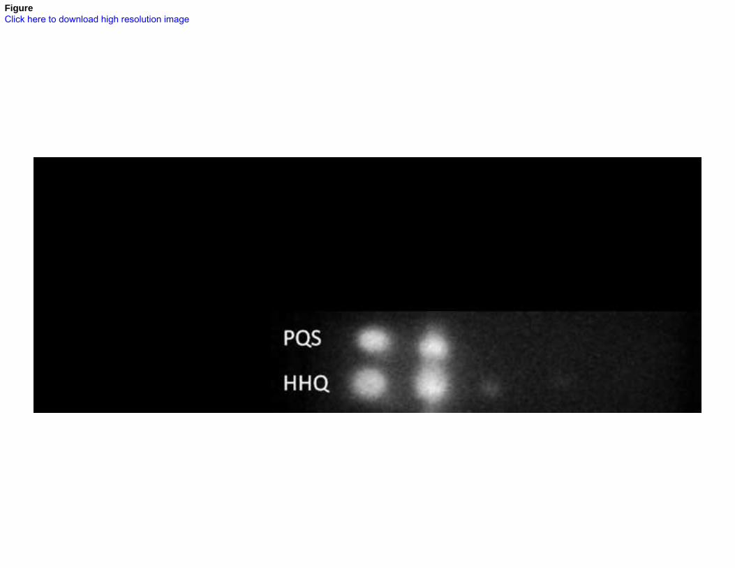
FigureClick here to download high resolution image

FigureClick here to download high resolution image

FigureClick here to download high resolution image

FigureClick here to download high resolution image
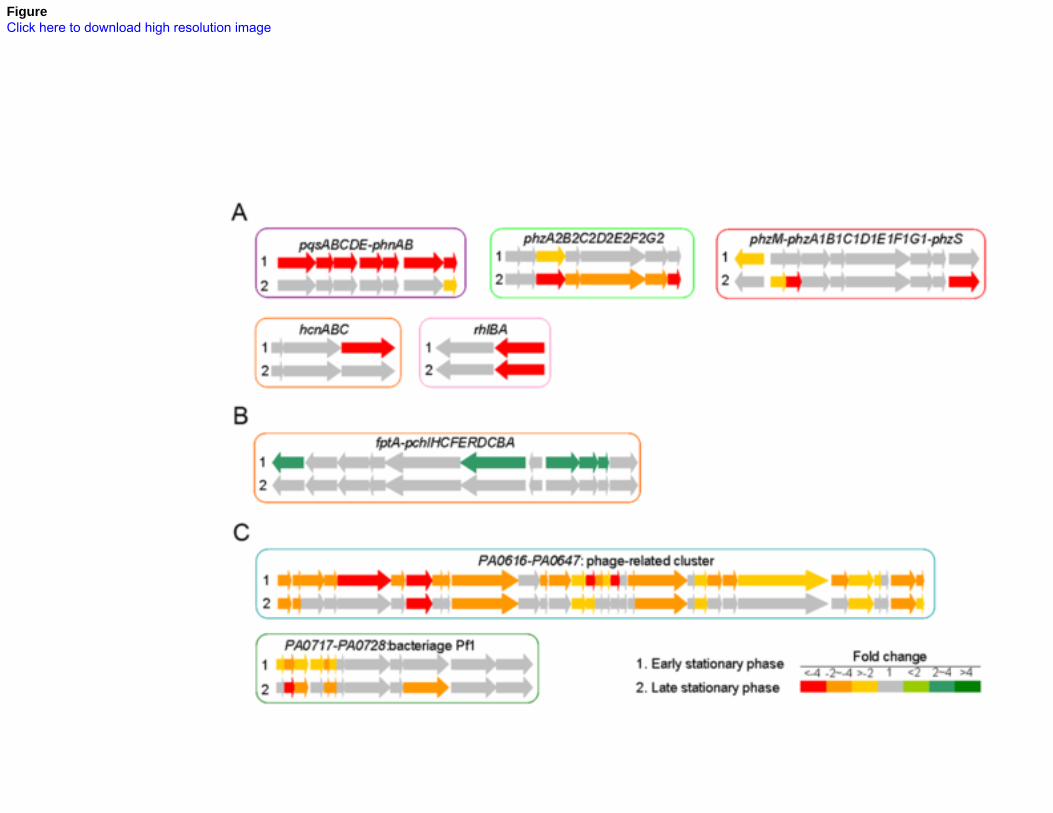
FigureClick here to download high resolution image

FigureClick here to download high resolution image
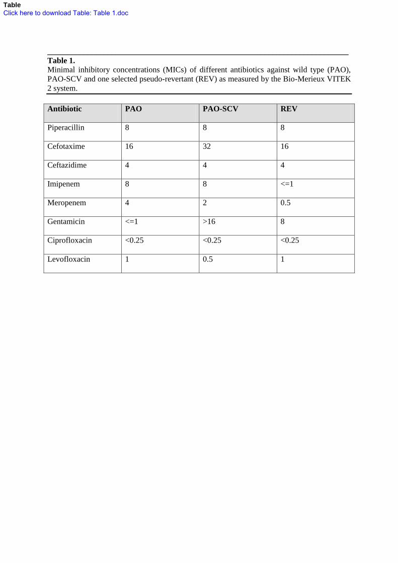
___________________________________________________________________________
Table 1. Minimal inhibitory concentrations (MICs) of different antibiotics against wild type (PAO),
PAO-SCV and one selected pseudo-revertant (REV) as measured by the Bio-Merieux VITEK
2 system.
Antibiotic PAO PAO-SCV REV
Piperacillin 8 8 8
Cefotaxime 16 32 16
Ceftazidime 4 4 4
Imipenem 8 8 <=1
Meropenem 4 2 0.5
Gentamicin <=1 >16 8
Ciprofloxacin <0.25 <0.25 <0.25
Levofloxacin 1 0.5 1
TableClick here to download Table: Table 1.doc
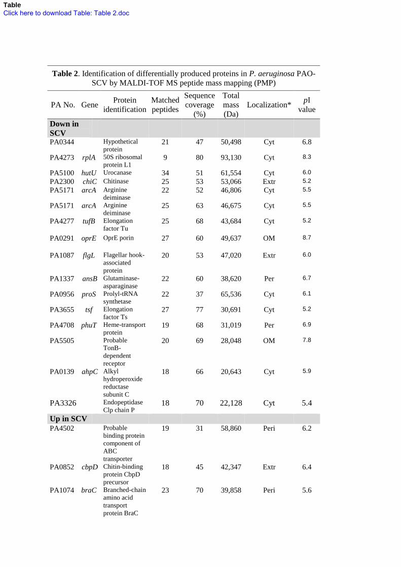
Table 2. Identification of differentially produced proteins in P. aeruginosa PAO-
SCV by MALDI-TOF MS peptide mass mapping (PMP)
PA No. Gene Protein
identification
Matched
peptides
Sequence
coverage
(%)
Total
mass
(Da)
Localization* pI
value
Down in
SCV
PA0344 Hypothetical
protein 21 47 50,498 Cyt 6.8
PA4273 rplA 50S ribosomal
protein L1 9 80 93,130 Cyt 8.3
PA5100 hutU Urocanase 34 51 61,554 Cyt 6.0
PA2300 chiC Chitinase 25 53 53,066 Extr 5.2
PA5171 arcA Arginine
deiminase 22 52 46,806 Cyt 5.5
PA5171 arcA Arginine
deiminase 25 63 46,675 Cyt 5.5
PA4277
tufB Elongation
factor Tu 25 68 43,684 Cyt 5.2
PA0291 oprE OprE porin 27 60 49,637 OM 8.7
PA1087 flgL Flagellar hook-
associated
protein
20 53 47,020 Extr 6.0
PA1337 ansB Glutaminase-
asparaginase 22 60 38,620 Per 6.7
PA0956 proS Prolyl-tRNA
synthetase 22 37 65,536 Cyt 6.1
PA3655 tsf Elongation
factor Ts 27 77 30,691 Cyt 5.2
PA4708 phuT Heme-transport
protein 19 68 31,019 Per 6.9
PA5505 Probable
TonB-
dependent
receptor
20 69 28,048 OM 7.8
PA0139 ahpC Alkyl
hydroperoxide
reductase
subunit C
18 66 20,643 Cyt 5.9
PA3326 Endopeptidase
Clp chain P 18 70 22,128 Cyt 5.4
Up in SCV
PA4502 Probable
binding protein
component of
ABC
transporter
19 31 58,860 Peri 6.2
PA0852 cbpD Chitin-binding
protein CbpD
precursor
18 45 42,347 Extr 6.4
PA1074 braC Branched-chain
amino acid
transport
protein BraC
23 70 39,858 Peri 5.6
TableClick here to download Table: Table 2.doc

PA3655 tsf Elongation
factor Ts 27 83 30,691 Cyt 5.2
PA0395 pilT Twitching
motility protein
PilT
16 56 41,644 OM 7.2
PA4495 Hypothetical
protein 16 55 24,921 Peri 5.8
PA1179 phoP Two-
component
response
regulator PhoP
18 71 25,748 Cyt 5.3
PA1777 oprF OprF 17 75 23,270 OM 4.8
* Cyt= cytoplasm, Peri= periplasm, IM= inner membrane, OM= outer membrane, Extr= extracellular

Table 3. List of SNPs in P. aeruginosa SCV revertant
Locusa Gene
name Product Positionb Nucleotide
Ref.
basec
SNP
base Type
Protein
effectd
Position
[AA]e SCVf WT
PA0748 mmsR
Still frameshift
probable
transcriptional
regulator
816532 425 G C Single
insertion Frameshift 142 + +
PA1179 phoP
Two-component
response regulator
PhoP
1277728 41 A G SNP H->R
exchange 14 + -
PA1180 phoQ Two-component
sensor PhoQ 1279044 683
39 bp
deletion
In frame
deletion 228 + -
PA1385
Probable glycosyl
transferase 1505156 552 C T SNP
Silent
mutation 184 ND ND
PA2020 mexZ
Probable
transcriptional
regulator
2213076 400 C T SNP Nonsense
mutation 134 + -
igPA2046
Intergenic region 2239547 280 T G SNP
ND ND
PA2141 Hypothetical protein 2356684 517 C - Single
deletion Frameshift 173 + +
a Gene locus, gene name and product description are extracted from Pseudomonas Genome Database
(http://www.pseudomonas.com). b Chromosomal position (nt).
c Ref. base, reference base.
d Change of protein sequence.
e Position relative to first amino acid of the protein.
f Found in SCV as well after Sanger sequencing
ND, not determined.
TableClick here to download Table: Table 3.doc

Supporting InformationClick here to download Supporting Information: Figure S1.tif

Supporting InformationClick here to download Supporting Information: Figure S2.tif

Supporting InformationClick here to download Supporting Information: Figure S3.TIF

Supporting InformationClick here to download Supporting Information: Table S1.doc

Supporting InformationClick here to download Supporting Information: Table S2.doc

Supporting InformationClick here to download Supporting Information: Table S3.doc

Supporting InformationClick here to download Supporting Information: Table S4.doc

Supporting InformationClick here to download Supporting Information: TableS 5.doc

Supporting InformationClick here to download Supporting Information: TableS 6.doc

Supporting InformationClick here to download Supporting Information: Table S 7.doc