Genetic Determinants of Sudden Cardiac Death
11
Genetic Determinants of Sudden Cardiac Death Peter A. Noseworthy, MD; Christopher Newton-Cheh, MD, MPH S udden cardiac death (SCD) is the final common end point of multiple disease processes. It results from a complex interplay of structural, metabolic, and genetic determinants. Although epidemiological risk factors such as age, prior myocardial infarction, and low ejection fraction are well established, this syndrome also has a strong genetic compo- nent. An understanding of the genetic contributions to risk could add substantially to the prediction and prevention of SCD. In this review, we explore the epidemiology, heritabil- ity, and allelic architecture of SCD and provide a detailed overview of the genetics of inherited electrophysiological and structural heart diseases that are potent risk factors for SCD. Epidemiology of SCD Each year, SCD claims 300 000 lives in the United States. 1 Most cases, 80%, are related to underlying coronary artery disease. Fewer cases, 10% to 15%, are associated with an underlying nonischemic myopathic process such as hypertro- phic cardiomyopathy (HCM) or dilated cardiomyopathy (DCM). Approximately 5% are estimated to have a primary defect of cardiac electrophysiology (eg, long-QT syndrome [LQTS] or Brugada syndrome [BS]). 2 At the population level, the conventional cardiac risk factors are powerful risk factors for SCD. For example, diabetes mellitus is associated with a marked increase in risk of SCD (odds ratio1.7 without microvascular disease; odds ratio2.7 with microvascular disease). 3 Among subjects with known coronary artery disease, smoking is linked to an increase in risk of SCD (hazard ratio2.5). 4 In a multivariate analysis from the Paris Prospective Study, a 42-mg/dL increase in total cholesterol (1 SD of the sample variation) was associated with a 23% increased risk of SCD. In the same analysis, an increase of 20 mm Hg in systolic blood pressure was associated with a 23% increase in the risk of SCD. 5 Furthermore, data from the Framingham Heart Study demon- strate that left ventricular hypertrophy is a risk factor for ventricular arrhythmias 6 and SCD. 7 Regular high-intensity exercise appears to be associated with a lower rate of SCD, 8 although the risk is transiently increased in the 30 minutes immediately after vigorous exercise. 8 –10 Advances in the treatment of these risk factors may be partly responsible for the decline in cardiovascular mortality over the past 20 years, 11,12 but the burden of SCD remains high. The primary challenge to reducing the rate of SCD is that the bulk of events occur without warning and in patients with few identifiable risk factors (Figure 1). 13 For example, although an ejection fraction of 35% is a powerful risk factor for SCD and a current indication for implantable cardioverter-defibrillator implantation, only one third of SCD victims have a low ejection fraction. 14 The conventional coronary risk factors and presence of congestive heart failure are associated with SCD in the general population but have poor ability to predict SCD at the individual level because of their prevalence and comparatively modest effects on risk. At the individual level, separating risk factors for all-cause cardiovascular mortality from risk factors for primary ar- rhythmia has been challenging. Among post–myocardial infarction patients who are known to be at risk of SCD, electrophysiological evaluation has been the most powerful tool in current clinical use to refine individual risk stratifica- tion. 15 Current measures that have been examined with varying predictive power include QRS duration, 16 QT- interval prolongation and dispersion, 17 microvolt T-wave alternans, 16,18 and late potentials on signal-averaged ECG. 19,20 Markers of autonomic dysfunction, such as reduced heart rate variability, have been shown to correlate with higher risk of SCD after myocardial infarction 21 and in nonischemic car- diomyopathy. 22–24 A recent analysis by the Multicenter Au- tomatic Defibrillator Implantation Trial (MADIT) II investi- gators showed that low-risk patients (defined as New York Heart Association functional class III, age 70 years, blood urea nitrogen 26 mg/dL, QRS 0.12 seconds, and absence of atrial fibrillation) or very high-risk patients (de- fined as blood urea nitrogen 50 mg/dL and/or serum creatinine 2.5 mg/dL) are unlikely to benefit from primary implantable cardioverter-defibrillator implantation despite meeting current implantation guidelines. 25 These approaches achieve stratification of populations based on clinical data but lack the power to predict SCD on the individual level. Evolving evidence of the heritability of SCD and its risk factors suggests that genetic factors could ultimately be an important component of individualized risk assessment. Mechanisms of Arrhythmic SCD Most often, SCD occurs in patients with underlying ischemic heart disease that predisposes to either ventricular tachycardia From the Cardiology Division (P.A.N., C.N.-C.), the Center for Human Genetic Research (C.N.-C.), and Cardiovascular Research Center (C.N.-C.), Massachusetts General Hospital, Harvard Medical School, Boston, Mass; and the Program in Medical and Population Genetics, Broad Institute of Harvard and Massachusetts Institute of Technology, Cambridge, Mass (C.N.-C.). Correspondence to Christopher Newton-Cheh, MD, MPH, Center for Human Genetic Research, Cardiovascular Research Center, Massachusetts General Hospital, 185 Cambridge St, CPZN 5.242, Boston, MA 02114. E-mail [email protected] (Circulation. 2008;118:1854-1863.) © 2008 American Heart Association, Inc. Circulation is available at http://circ.ahajournals.org DOI: 10.1161/CIRCULATIONAHA.108.783654 1854 Contemporary Reviews in Cardiovascular Medicine by guest on June 15, 2015 http://circ.ahajournals.org/ Downloaded from
-
Upload
hms-harvard -
Category
Documents
-
view
0 -
download
0
Transcript of Genetic Determinants of Sudden Cardiac Death
Genetic Determinants of Sudden Cardiac DeathPeter A. Noseworthy, MD; Christopher Newton-Cheh, MD, MPH
Sudden cardiac death (SCD) is the final common end pointof multiple disease processes. It results from a complex
interplay of structural, metabolic, and genetic determinants.Although epidemiological risk factors such as age, priormyocardial infarction, and low ejection fraction are wellestablished, this syndrome also has a strong genetic compo-nent. An understanding of the genetic contributions to riskcould add substantially to the prediction and prevention ofSCD. In this review, we explore the epidemiology, heritabil-ity, and allelic architecture of SCD and provide a detailedoverview of the genetics of inherited electrophysiological andstructural heart diseases that are potent risk factors for SCD.
Epidemiology of SCDEach year, SCD claims �300 000 lives in the United States.1
Most cases, �80%, are related to underlying coronary arterydisease. Fewer cases, �10% to 15%, are associated with anunderlying nonischemic myopathic process such as hypertro-phic cardiomyopathy (HCM) or dilated cardiomyopathy(DCM). Approximately 5% are estimated to have a primarydefect of cardiac electrophysiology (eg, long-QT syndrome[LQTS] or Brugada syndrome [BS]).2
At the population level, the conventional cardiac riskfactors are powerful risk factors for SCD. For example,diabetes mellitus is associated with a marked increase in riskof SCD (odds ratio�1.7 without microvascular disease; oddsratio�2.7 with microvascular disease).3 Among subjects withknown coronary artery disease, smoking is linked to anincrease in risk of SCD (hazard ratio�2.5).4 In a multivariateanalysis from the Paris Prospective Study, a 42-mg/dLincrease in total cholesterol (1 SD of the sample variation)was associated with a 23% increased risk of SCD. In the sameanalysis, an increase of 20 mm Hg in systolic blood pressurewas associated with a 23% increase in the risk of SCD.5
Furthermore, data from the Framingham Heart Study demon-strate that left ventricular hypertrophy is a risk factor forventricular arrhythmias6 and SCD.7 Regular high-intensityexercise appears to be associated with a lower rate of SCD,8
although the risk is transiently increased in the 30 minutesimmediately after vigorous exercise.8–10
Advances in the treatment of these risk factors may bepartly responsible for the decline in cardiovascular mortalityover the past 20 years,11,12 but the burden of SCD remains
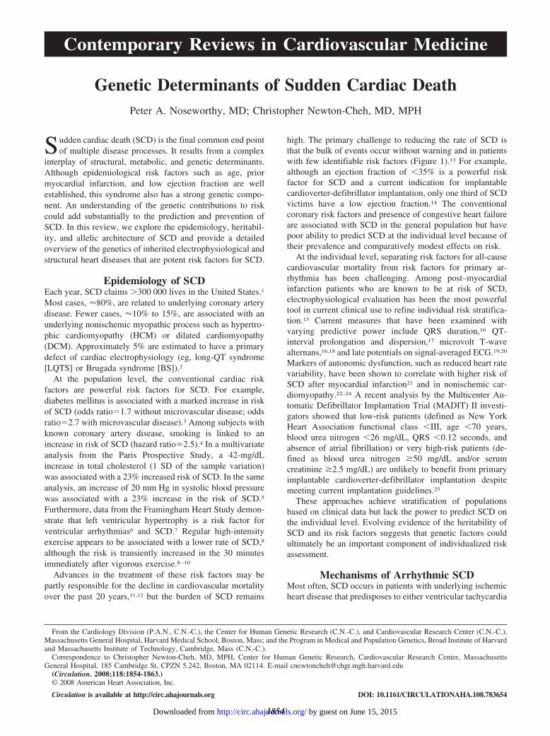
high. The primary challenge to reducing the rate of SCD isthat the bulk of events occur without warning and in patientswith few identifiable risk factors (Figure 1).13 For example,although an ejection fraction of �35% is a powerful riskfactor for SCD and a current indication for implantablecardioverter-defibrillator implantation, only one third of SCDvictims have a low ejection fraction.14 The conventionalcoronary risk factors and presence of congestive heart failureare associated with SCD in the general population but havepoor ability to predict SCD at the individual level because oftheir prevalence and comparatively modest effects on risk.
At the individual level, separating risk factors for all-causecardiovascular mortality from risk factors for primary ar-rhythmia has been challenging. Among post–myocardialinfarction patients who are known to be at risk of SCD,electrophysiological evaluation has been the most powerfultool in current clinical use to refine individual risk stratifica-tion.15 Current measures that have been examined withvarying predictive power include QRS duration,16 QT-interval prolongation and dispersion,17 microvolt T-wavealternans,16,18 and late potentials on signal-averaged ECG.19,20
Markers of autonomic dysfunction, such as reduced heart ratevariability, have been shown to correlate with higher risk ofSCD after myocardial infarction21 and in nonischemic car-diomyopathy.22–24 A recent analysis by the Multicenter Au-tomatic Defibrillator Implantation Trial (MADIT) II investi-gators showed that low-risk patients (defined as New YorkHeart Association functional class �III, age �70 years,blood urea nitrogen �26 mg/dL, QRS �0.12 seconds, andabsence of atrial fibrillation) or very high-risk patients (de-fined as blood urea nitrogen �50 mg/dL and/or serumcreatinine �2.5 mg/dL) are unlikely to benefit from primaryimplantable cardioverter-defibrillator implantation despitemeeting current implantation guidelines.25
These approaches achieve stratification of populationsbased on clinical data but lack the power to predict SCD onthe individual level. Evolving evidence of the heritability ofSCD and its risk factors suggests that genetic factors couldultimately be an important component of individualized riskassessment.
Mechanisms of Arrhythmic SCDMost often, SCD occurs in patients with underlying ischemicheart disease that predisposes to either ventricular tachycardia
From the Cardiology Division (P.A.N., C.N.-C.), the Center for Human Genetic Research (C.N.-C.), and Cardiovascular Research Center (C.N.-C.),Massachusetts General Hospital, Harvard Medical School, Boston, Mass; and the Program in Medical and Population Genetics, Broad Institute of Harvardand Massachusetts Institute of Technology, Cambridge, Mass (C.N.-C.).
Correspondence to Christopher Newton-Cheh, MD, MPH, Center for Human Genetic Research, Cardiovascular Research Center, MassachusettsGeneral Hospital, 185 Cambridge St, CPZN 5.242, Boston, MA 02114. E-mail [email protected]
(Circulation. 2008;118:1854-1863.)© 2008 American Heart Association, Inc.
Circulation is available at http://circ.ahajournals.org DOI: 10.1161/CIRCULATIONAHA.108.783654
1854
Contemporary Reviews in Cardiovascular Medicine
by guest on June 15, 2015http://circ.ahajournals.org/Downloaded from
(VT) or ventricular fibrillation and ultimately pump failure.Acute ischemia results in electrochemical shifts (net potas-sium efflux and resultant sodium and calcium influx) thatslow conduction, alter refractoriness, and result in delayedafterdepolarizations that can initiate ventricular fibrillation.26
Over time, cardiac ischemia or infarction can create myocar-dial scars that form the substrate for reentrant VT. Inadvanced heart failure, multiple perturbations including pro-longation of the action potential, alteration in calcium ho-meostasis, and changes in neurohumoral signaling result in amarked increase in the risk of ventricular arrhythmia andSCD.27 Among patients without ischemic heart disease,heritable causes of SCD (LQTS or BS, for instance) arisefrom mutations that cause a dispersion of refractoriness. Thiselectric heterogeneity can support reentrant arrhythmias trig-gered by early or delayed afterdepolarizations.
Heritability of SCDObservations from population-based studies demonstrate amarked increase in risk of SCD in first-degree relatives ofSCD victims. In an Israeli case-control study of SCD patients,a family history of myocardial infarction or SCD (or resus-citated cardiac arrest) was associated with a 46% increasedrisk of SCD compared with matched controls (relativerisk�1.5).28 In a case-control study from Seattle, first-degreerelatives of patients with early-onset SCD (before age 65years) had 2.7-fold higher odds of suffering SCD than age-and sex-matched controls after adjustment for cardiovascularrisk factors.29 In the Paris Prospective Study, a parentalhistory of SCD increased the risk of fatal arrhythmia in theoffspring by 80%; in subjects with both parents affected, riskof SCD increased 9-fold.5 In a Finnish autopsy cohort,
subjects who died suddenly during first myocardial infarctionwere 60% more likely to have a family history of SCD thanthose who survived myocardial infarction.30
A Dutch case-control study demonstrated that patientspresenting with ventricular fibrillation during a first myocar-dial infarction are substantially more likely to have a familyhistory of SCD than those without ventricular fibrillation(odds ratio�2.7).31 In aggregate, these data support a strongrole for heritable factors in SCD risk. Moreover, this findingpoints to the independence of at least some genetic factorsthat predispose to ischemia-mediated arrhythmia from thosethat predispose to coronary disease in general.
Among subjects with well-defined SCD syndromes, afamily history markedly potentiates the risk of SCD. Forexample, a family history of SCD in patients with HCM isassociated with a 5-fold increased risk of SCD.32
Allelic Architecture of SCDSCD likely has a strong genetic component, but only afraction of the genetic variants that underlie the risk areknown, and the allelic architecture of SCD thus remainspoorly defined.33 The heterogeneity of the associated cardiacsubstrate and the difficulty of ascertaining adequate pheno-typic information after arrest have been key barriers todefining this architecture.
In general, genetic contributors to complex traits such asSCD can be classified as either rare variants with strongeffect, common variants with modest effect, or rare variantswith modest effect. Rare variants with strong effect have beenidentified in linkage studies of families affected by LQTS andBS, as well as HCM and DCM. These syndromes areclinically recognizable and are powerful predictors of SCD.However, together they account for a minority of suddendeaths. Negative selection has kept the allele frequency ofmost of these mutations low in all but a few isolatedpopulations that carry high-prevalence founder mutations,such as LQTS mutations in Finland34 and South Africa.35
Of potentially greater relevance to the general populationare common variants of modest effect that may contribute torisk of SCD in an incremental fashion. Unlike rare variantswith strong effects, these variants may allow individuals tosurvive to reproductive age and thus escape negative selec-tion and reach higher population allele frequencies. Rese-quencing candidate genes in case-control studies has identi-fied the SCN5A variant S1102Y, which has been shown to belinked to arrhythmias and SCD in self-described blacks.36,37
Ultimately, unbiased genomewide association studies mayidentify more common variants beyond the known candidategenes. The search for rare variants with modest effect size isin its infancy and will require large-scale resequencing effortsthat have been prohibitively costly with existing technology.Newer sequencing techniques under active development andapplication may facilitate such efforts.
Primary Electrical Causes of SCDApproximately 5% of cases of SCD occur in the absence ofstructural heart disease or coronary disease and are attribut-able to an isolated electrical disorder.2 The best understood ofthese disorders are the familial syndromes such as the LQTS
Figure 1. The incidence of SCD in the United States across arange of risk groups (left) and the corresponding number of car-diac deaths in these groups (right). Most SCD occurs in com-paratively low-risk groups, and a minority of cases occur in peo-ple with known high-risk characteristics such as myocardialinfarction associated with VT/VF. MI indicates myocardial infarc-tion; VF, ventricular fibrillation; EF, ejection fraction; CAD, coro-nary artery disease; and pop., population. Modified from Myer-burg et al,13 copyright © 1997, with permission from Elsevier.
Noseworthy and Newton-Cheh Genetics of SCD 1855
by guest on June 15, 2015http://circ.ahajournals.org/Downloaded from
and BS. In the general population, rare, sporadic, and com-mon, prevalent variants have been examined for an influenceon the risk of SCD.
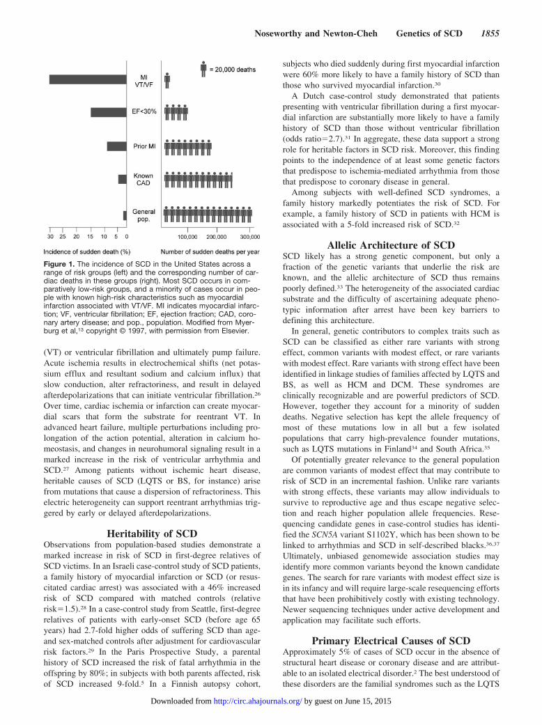
Rare Variants in Families WithRecognizable SyndromesThe normal action potential is generated through a highlyorchestrated series of transmembrane currents resulting fromthe coordinated opening and closing of sodium, potassium,and calcium channels. DNA sequence variants in these ionchannels account for many of the known heritable causes ofSCD in the absence of structural heart disease. These includethe congenital LQTS and short-QT syndrome (SQTS), BS,and catecholaminergic polymorphic VT (CPVT) (Figure 2).
Congenital LQTS and SQTS are uncommon (prevalenceestimates from 0.01% to 0.04%) but important causes offamilial SCD. Considerable genotypic heterogeneity exists,and, to date, �400 mutations have been described in 11different genes (Figure 2).38 Generally speaking, LQTS re-sults from loss-of-function mutations in potassium channelgenes (KCNQ1, KCNH2, KCNE1, KCNE2, KCNJ2) or gain-of-function mutations in sodium or calcium channel genes(SCN5A, CACNA1C).39 Conversely, SQTS is the conse-quence of gain-of-function mutations in potassium channelgenes40,41 or loss-of-function mutations in calcium channelgenes.42 Rare coding variants in these genes account for�75% of congenital LQTS. The balance could includeregulatory variants at known gene locí or additional as yetunrecognized genes. Fewer cases of the LQTS are caused bymutations in structural proteins required for cellular localiza-tion of ion channels such as ankyrin 2 (ANK2), which causesLQT4. Gene products that affect ion channel kinetics throughdirect protein-protein interactions (the so-called channel-interacting proteins) include caveolin 3 (CAV3),43 A-kinaseanchoring protein 9 gene (AKAP9),44 and, most recently,cytoskeletal protein �-1-syntrophin (SNTA1).45
The risk of SCD and response to therapy in LQTS aredetermined, in part, by the underlying causative gene.46 Theincidence of SCD or resuscitated arrest before age 40 yearsranges from 30% in patients who harbor mutations in KCNQ1(LQT1) to 46% among patients with mutations in KCNH2(LQT2).47 �-Blocker therapy is particularly effective inpatients with LQT1 and LQT2, whereas those with SCN5Amutations (LQT3) garner no benefit from �-blockers.48
Knowledge of genotype may help to inform clinical decisionmaking, including risk stratification of genotype-positive/phenotype-negative relatives of SCD victims.47
The BS is somewhat more common than the QT syn-dromes. Prevalence ranges from 0.4% in the United States49
to �1% in Japan.50,51 It is the most common cause of SCDwithout structural heart disease in Southeast Asia.49 Whenfamilial, BS is inherited in an autosomal dominant pattern,but as many as two thirds of cases are sporadic.52 Approxi-mately 15% of probands have identifiable mutations inSCN5A and, to date, �100 mutations have been described.53
A second locus on chromosome 3,54 close to but distinct fromSCN5A, encodes the glycerol-3-phosphate dehydrogenase1-like gene (GPD1L) and has been identified in a single largepedigree.55 GPD1L is believed to be involved in trafficking of
the cardiac sodium channel to the cell surface, although itsrole in BS needs to be confirmed. Disruption of the geneproduct decreases SCN5A surface membrane expression andreduces inward sodium current in transfected HEK cells thatcoexpress GPD1L and SCN5A.54 The third and fourth genesassociated with the BS encode the �-1 (CACNA1C) and �
Figure 2. Myocardial action potential currents and arrhythmicsyndromes. Shown are genes that underlie the sodium, calcium,and potassium currents in the cardiac myocyte action potential.Rare variants in these genes with resultant gain or loss of chan-nel function are implicated in familial syndromes such as LQTS,SQTS, and BS. Rare variants in some of these genes have beenidentified in cases of SCD and drug-induced torsades depointes (TdP) in studies in the general population. Other genesnot directly responsible for transmembrane currents are impli-cated in familial SCD syndromes, including the ankyrin B(ANK2), which underlies LQT4 (and possibly CPVT). Caveolin3 (CAV3), cytoskeletal protein �-1-syntrophin (SNTA1), andA-kinase anchoring protein 9 gene (AKAP9) are also implicatedin LQTS. Glycerol-3-phosphate dehydrogenase 1-like gene(GPD1L) is implicated in type 2 BS (BrS2).
1856 Circulation October 28, 2008
by guest on June 15, 2015http://circ.ahajournals.org/Downloaded from
(CACNB2b) subunits of the L-type cardiac calcium channel.These mutations have been hypothesized to produce a newclinical entity of combined BS/SQTS.56
CPVT is an inherited disorder characterized by stress-induced ventricular arrhythmia and SCD. CPVT appears toresult from a defect of intracellular calcium cycling thattriggers transient inward current and delayed afterdepolariza-tion. Missense mutations underlying CPVT have been iden-tified in 2 genes: the ryanodine receptor 2 (RyR2)57–59 andcalsequestrin 2 (CASQ2).60,61
Rare Variants in the General Population NotAscertained Through Affected FamiliesBecause the familial mutations associated with SCD areindividually uncommon, they appear to contribute relativelylittle to the overall burden of SCD in the general population.However, evidence from postmortem series suggests thatvictims of apparently sporadic SCD (ie, not recognized tohave a high-risk clinical syndrome) may harbor mutations inthese candidate genes, particularly those described in LQTS.A small case series by Chugh et al62 identified a mutation inKCNH2 (encoding the HERG � subunit) in 2 of 12 adultvictims of SCD. A larger series by Tester and Ackerman63
showed that 30% of 49 victims of sudden unexplained deathharbored a mutation in 1 of the genes implicated in LQTS(KCNQ1, KCNE1, SCN5A, KCNH2), and 14% harbored amutation in the ryanodine receptor gene (RyR2).63,64 Acase-control analysis from the Nurses Health Study revealedthat 6 of 60 women (10%) had 1 of 5 rare missense variantsin SCN5A compared with 12 of 733 matched controls (1.6%)from the same prospective cohort (P�0.001).65
Variants that are individually rare may exist collectively atrelatively high frequency in the general population.Population-based resequencing efforts have shown that 25%of black and 14% of white apparently healthy subjects areheterozygous for at least 1 nonsynonymous (amino acid–altering) rare variant (allele frequency �0.5%) in exons thatencode 4 potassium channel subunits (KCNQ1, KCNH2,KCNE1, or KCNE2).66 Similarly, missense variants in theprotein coding region of SCN5A were found in 39 of 829healthy subjects (4.7%).67 Because these variants are found ina sizable minority of healthy individuals in the generalpopulation, it is very important to establish the functionalsignificance, either through segregation studies or relevant invitro models. To date, direct functional data linking manyimplicated variants to arrhythmia or SCD have been rela-tively limited.
On their own, functional rare variants may not produce anidentifiable clinical syndrome in isolation, but they maypredispose to acquired LQTS and SCD after a “second hit”such as exposure to a QT-prolonging medication. Thus,drug-induced arrhythmia may be a forme fruste of LQTS. Auseful model of this phenomenon is the concept of repolar-ization reserve.68–70 Like many biologically important pro-cesses, uniform repolarization is maintained by several re-dundant mechanisms. A single defect in 1 of the redundantpathways may remain undetected until an additional exposure(eg, QT-prolonging drug, hypokalemia, or ischemia) incre-mentally reduces repolarizing capacity below a critical
threshold and unmasks the defect, with resultant arrhythmia.Under this model, a genetic variant that reduces repolariza-tion reserve modestly may remain clinically inapparent untilan additional loss of repolarizing capacity produces anarrhythmic substrate.
Indeed, rare variants in the known LQTS genes areidentifiable in 10% to 15% of subjects with drug-induced QTprolongation.71 A Dutch case-control study identified 3 dis-tinct missense variants in the LQTS genes in 4 of 32drug-induced LQTS cases (KCNE1 [D85N], KCNE2 [T8A],and KCNH2 [P347S]) that were not found among 32 controls,although power was limited to make definitive statementsabout the significance of these differences.72 These smallseries suggest that missense mutations in known LQTS genesmay be clinically important but appear to explain only aminority of acquired LQTS.
Common Variants in the General PopulationHistorically, when resequencing genes in SCD families,variants with population allele frequency �0.5% have beenignored because they were thought too common to cause arare disorder. However, some polymorphisms have beenshown to underlie a proportion of LQTS cases in which noneof the classic LQTS/BS mutations were identified. Forexample, the N85 allele (asparagine) of the KCNE1 D85Nvariant (rs1805128, minor allele frequency 1.4%) is morecommon in 2 genotype-negative LQTS collections thancontrols (11 of 98 cases [11%] and 13/147 [8.8%] versus 9 of364 controls [2.5%]).73 Additionally, the N85 allele wasfound in 7.3% of patients in a drug-induced LQTS collectioncompared with only 1.5% of controls.74
Similarly, the common SNC5A polymorphism S1102Y isdisproportionately represented among black arrhythmia pa-tients and victims of SCD. In the original report by Splawksiet al,36 13 of 23 black cases (57%) with an assortment ofarrhythmia, syncope, and QT prolongation carried at least 1copy of the Y1102 allele compared with 13 of 100 controls(P�0.00003). In a follow-up autopsy case-control study, theSCN5A Y1102 allele was present in 28% of black cases ofsudden death without structural heart disease compared with5.6% of controls with a sudden but noncardiac death(P�0.0005).37 In vitro, cells transfected with the variantallele have accelerated sodium channel activation, perhapsincreasing the likelihood of arrhythmia and supporting thefunctional relevance of the S1102Y variant.36
Common variants that underlie normal variation in the QTinterval could contribute incrementally to risk of SCD in thegeneral population. The QT interval, adjusted for heart rate,age, and sex, is normally distributed in the general populationand has a substantial genetic underpinning; �35% of itsvariability is attributable to genetic factors.75 QT prolongationhas been a consistent risk factor for SCD76 and could offer auseful intermediate trait for the study of SCD in the generalpopulation. To date, common variants in candidate geneshave been shown to influence QT duration, including 2 inKCNH277–80 and 1 in NOS1AP.81,82 It should be stressed thatvariants that affect QT duration have a modest effect onQT-interval duration at baseline (ranging from 6 to 12 ms).These effects require large sample sizes to detect.
Noseworthy and Newton-Cheh Genetics of SCD 1857
by guest on June 15, 2015http://circ.ahajournals.org/Downloaded from

Common variants are unlikely to be sufficient causes ofsudden death on their own. If they caused SCD in mostcarriers, strong selection pressure would reduce their allelefrequency substantially. More likely, they contribute onlyincrementally to the overall risk of SCD. By reducingrepolarization reserve, they could predispose a portion of thepopulation to sudden death through an interaction with otherrisk factors that reduce repolarization capacity such as ische-mia, hypokalemia, or drug exposure. This remains to beproven.
Genetic Variants Causing SCD via StructuralHeart Disease
Nonischemic cardiomyopathies and other structural heartdiseases account for �10% to 15% of SCD.2 The geneticunderpinnings of these disorders, particularly HCM, are wellstudied and are described below.
Hypertrophic Cardiomyopathy: A SarcomericDiseaseHCM, the most common known inherited predisposition toSCD in the young, affects �0.5% of the population and mayaccount for as much as 48% of SCD in patients aged �35years.14 HCM is a disease of contractile sarcomeric proteinsthat results in myocardial hypertrophy and fibrosis. To date,�450 mutations in 13 genes encoding structural proteins ofthe contractile machinery have been identified in patientswith HCM83,84 (Table 1). The bulk of the described mutationsare in the genes encoding the �-cardiac myosin heavy chain(MYH7) or the cardiac myosin binding protein-C gene(MYBPC3). Other HCM genes include cardiac troponin T(TNNT2), �-tropomyosin (TPM1), cardiac troponin I(TNNI3), and, less commonly, cardiac actin (ACTC) and themyosin light chains (MYL3, MYL2). In addition, as many as4% to 12% of probands initially diagnosed with HCM on thebasis of clinical characteristics may actually have a cardio-myopathy due to a glycogen storage diseases caused by eitherPRKAG2 or LAMP2 mutations.85
The risk of SCD in HCM is variable and may be related tothe underlying mutation. For example, TNNT2 mutationshave been reported to cause a phenotype of minimal hyper-trophy but high risk of SCD.86 The recent introduction of acommercially available cardiac sarcomere gene mutationscreen has made the identification of the underlying genemutation possible for some patients in clinical practice. In theMayo Clinic HCM cohort, this screen identified mutations in147 of 389 patients (38%).87 However, the authors cautionagainst using this test to prognosticate for SCD. In their caseseries, patients with a range of genotypes were phenotypicallyindistinguishable, making prognostication for SCD on thebasis of genotype alone unreliable.87 As larger series arecollected, observation of the natural history of patients whoharbor each of these specific mutations will allow a betterunderstanding of the penetrance and expression of thesemutations in progressively less selected patients.
In the general population, the mutations responsible forHCM may contribute to idiopathic left ventricular hypertro-phy, a risk factor for SCD. In the community-based Framing-ham Heart Study, 18% of subjects with left ventricular wall
thickness �13 mm but no evidence of HCM, aortic stenosis,or severe hypertension had a sarcomere protein (MYH7,MYBPC3, TNNT2, TNNI3, MYL3) or lipid storage (GLA)gene mutation. These mutations occur in only 0.5% of thegeneral population, suggesting that they may contribute to theoverall burden of left ventricular hypertrophy.84 Long-termoutcomes with regard to risk of SCD are not established.
Dilated Cardiomyopathy: Defects of ForceGeneration and TransmissionIdiopathic DCM predisposes to arrhythmic SCD as well asprimary pump failure. Although these disorders are oftensporadic, �20% of individuals with DCM have an affectedfirst-degree relative.88 Furthermore, 7% of initially unaffectedfirst-degree relatives have been shown to develop DCM whenmonitored over a median 10-year follow-up period.89 When afamilial pattern is present, it is most commonly transmitted inan autosomal dominant pattern, but autosomal recessive,X-linked, and mitochondrial inheritance have also beendescribed. Schonberger and Seidman have outlined a usefulclassification scheme for mutations that result in DCM bydisrupting various aspects of cellular machinery, includingforce generation, force transmission, and energy production(Table 2).90 Defects in force generation are caused by
Table 1. Genes and Loci Implicated in HCM
GeneSymbol Locus Pattern
ElectrophysiologicalAssociations
Thick filament
�-Myosin heavychain
MYH7 14q12 AD SCD common
Myosin-bindingprotein C
MYBPC3 11p11.2 AD � � �
Troponin T TNNT2 1q32 AD SCD�HCM
Troponin I TNNI3 19q13.4 AD � � �
�-Tropomyosin TPM1 15q22.1 AD � � �
Regulatory myosinlight chain
MYL2 12q24.3 AD � � �
Essential myosin lightchain
MYL3 3p21 AD � � �
Actin ACTC1 15q14 AD � � �
Z-disc
Titin TTN 2q31 AD � � �
Muscle LIM protein CSRP3 11p15.1 AD � � �
Telethonin TCAP 17q12 AD � � �
Myozenin 2 MYOZ2 4q26 AD � � �
Intercalated disc
Vinculin VCL 10q22.1 AD
Storagecardiomyopathies
� � �
AMP-dependentprotein kinase
PRKAG2 � � � AD/AR �/� preexcitation
Lysosomal-associatedmembrane protein 2
LAMP2 � � � AD/AR � � �
Protein 2
�-Galactosidase A(Fabry disease)
GLA Xq22 X-linked Atrial fibrillation,VT, SCD,
conduction disease
AD indicates autosomal dominant; AR, autosomal recessive.
1858 Circulation October 28, 2008
by guest on June 15, 2015http://circ.ahajournals.org/Downloaded from

alterations of the contractile machinery such as missensemutations in the �-cardiac myosin heavy chain or cardiactroponin T,91 2 genes previously associated with HCM.Defects in force transmission are caused by mutations thatdisrupt coupling of the contractile machinery to the myocar-dial structural proteins, namely, mutations in cardiac actin92,93
and �-tropomyosin.94 Familial defects in energy productioninclude recessive mutations that result in impaired cardiacfatty acid �-oxidation. These mutations impair efficientenergy production or clearance of toxic metabolites, typicallyleading to death in the first months of life.95 Currently no dataare available on common DCM variants and the risk of SCDin the general population.
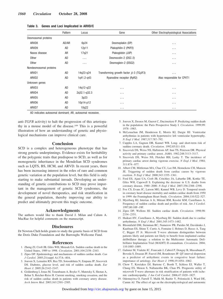
Arrhythmogenic Right Ventricular Dysplasia: ADisorder of the DesmosomeArrhythmogenic right ventricular dysplasia/cardiomyopathy(ARVD/C) is another significant cause of SCD. ARVD/C isa primary myocardial disorder that is characterized by grad-ual loss of myocytes and replacement by fatty and fibroustissue that results in cardiomyopathy and arrhythmia. Al-though it is relatively rare (0.01% in the general US popula-tion), ARVD/C may account for as much as 5% of unexplainedSCD in some populations.96 In Italy, where the prevalence issomewhat higher at 1 of 5000, it may account for as much as20% of sudden deaths in young adults. Although it is oftensporadic, in 35% to 50% of cases a family history is apparent.Thus far, mutations in 6 genes have been implicated in the
pathogenesis of the disorder97 (Table 3). Five of the genesencode cell-cell adhesion proteins (plakoglobin,98–100 desmo-plakin,101,102 plakophilin-2,103 desmocolin-2,104,105 anddesmoglein-2106). Impaired cellular adhesion appears to resultin myocyte detachment and cell death with subsequentfibrofatty replacement of damaged tissue that is the patho-logical hallmark of the disease. These areas of scar tissueprovide the substrate for reentrant arrhythmia and predisposeto SCD. Two nonstructural proteins have also been impli-cated, namely, mutations in the ryanodine receptor gene107
(also responsible for CPVT) and mutations in the transform-ing growth factor genes.108
Inherited Arteriopathies: Sudden Death Secondaryto Vascular RuptureInherited arteriopathies such as Marfan syndrome, Loeys-Dietz syndrome, and Ehlers-Danlos syndrome predispose toSCD through catastrophic vascular rupture rather thanthrough arrhythmia. Most cases of Marfan syndrome arecaused by mutations in the FBN1 gene (encoding fibrillin-1)and are inherited in an autosomal dominant pattern. Alteredfibrillin-1 structure interferes with normal TGFB signalingand results in a distinctive phenotype and propensity to aorticdissection, among other findings. A small percentage ofMarfan syndrome cases are caused by mutations in theTGFBR2 gene directly. Recent insights into the role of TGFBsignaling in the pathogenesis of Marfan syndrome have led touse of losartan (an angiotensin-2 receptor blocker with
Table 2. Genes and Loci Implicated in DCM
Pattern Locus Gene Symbol Electrophysiological Associations
Force generation (thick filament)
�-Myosin heavy chain AD 14q11 MYH7 � � �
Troponin T AD 1q32 TNNT2 � � �
Force transmission
Actin AD 15q14 ACTC1 � � �
�-Tropomyosin AD 15q22.1 TPM1 � � �
Dystrophin X Xp21 � � � � � �
�-sarcoglycan AD 5q33-34 � � � � � �
Desmin AD � � � � � � � � �
Desmoplakin AD/AR 6p24 � � � ARVD
Plakoglobin AR 17q21 � � � ARVD/Naxos disease
Defects in energy production
Carnitine deficiency AR � � � � � � � � �
Organic cation transporter protein AR � � � � � � � � �
Translocase AR � � � � � � � � �
Carnitine palmitoultransferase II AR � � � � � � � � �
Mitochondrial trifunctional protein � � � � � � � � � � � �
Other
Tafazzin X Xq28 � � � � � �
Lamin A AD � � � � � � Conduction disease
Lamin C AD � � � � � � Conduction disease
Ryanodine receptor AD 1q41.2-q43 RyR2 CPVT, ARVD
SCN5A (sodium channel) 3p21 D1275N AF
AD indicates autosomal dominant; AR, autosomal recessive; and AF, atrial fibrillation.
Noseworthy and Newton-Cheh Genetics of SCD 1859
by guest on June 15, 2015http://circ.ahajournals.org/Downloaded from
anti-TGFB activity) to halt the progression of this arteriopa-thy in a mouse model of the disease.109 This is a powerfulillustration of how an understanding of genetic and physio-logical mechanisms can improve clinical care.
ConclusionSCD is a complex and heterogeneous phenotype that hasstrong genetic underpinnings. Evidence exists for heritabilityof the polygenic traits that predispose to SCD, as well as formonogenetic inheritance in the Mendelian SCD syndromessuch as LQTS, BS, HCM, and ARVD. In recent years, therehas been increasing interest in the roles of rare and commongenetic variation at the population level, but this field is onlystarting to make substantial inroads. Developing an under-standing of genetic contributions to SCD may prove impor-tant in the management of genetic SCD syndromes, thedevelopment of novel therapeutics, and risk stratification inthe general population, thereby improving our ability topredict and ultimately prevent this tragic outcome.
AcknowledgmentsThe authors would like to thank David J. Milan and Calum A.MacRae for helpful comments on the manuscript.
DisclosuresDr Newton-Cheh holds grants to study the genetic basis of SCD fromthe Doris Duke Foundation and the Burroughs Wellcome Fund.
References1. Zheng ZJ, Croft JB, Giles WH, Mensah GA. Sudden cardiac death in the
United States, 1989 to 1998. Circulation. 2001;104:2158–2163.2. Zipes DP. Epidemiology and mechanisms of sudden cardiac death. Can
J Cardiol. 2005;21(suppl A):37A–40A.3. Jouven X, Lemaitre RN, Rea TD, Sotoodehnia N, Empana JP, Siscovick
DS. Diabetes, glucose level, and risk of sudden cardiac death. EurHeart J. 2005;26:2142–2147.
4. Goldenberg I, Jonas M, Tenenbaum A, Boyko V, Matetzky S, Shotan A,Behar S, Reicher-Reiss H. Current smoking, smoking cessation, and therisk of sudden cardiac death in patients with coronary artery disease.Arch Intern Med. 2003;163:2301–2305.
5. Jouven X, Desnos M, Guerot C, Ducimetiere P. Predicting sudden deathin the population: the Paris Prospective Study I. Circulation. 1999;99:1978–1983.
6. McLenachan JM, Henderson E, Morris KI, Dargie HJ. Ventriculararrhythmias in patients with hypertensive left ventricular hypertrophy.N Engl J Med. 1987;317:787–792.
7. Cupples LA, Gagnon DR, Kannel WB. Long- and short-term risk ofsudden coronary death. Circulation. 1992;85:I11–I18.
8. Siscovick DS, Weiss NS, Hallstrom AP, Inui TS, Peterson DR. Physicalactivity and primary cardiac arrest. JAMA. 1982;248:3113–3117.
9. Siscovick DS, Weiss NS, Fletcher RH, Lasky T. The incidence ofprimary cardiac arrest during vigorous exercise. N Engl J Med. 1984;311:874–877.
10. Albert CM, Mittleman MA, Chae CU, Lee IM, Hennekens CH, MansonJE. Triggering of sudden death from cardiac causes by vigorousexertion. N Engl J Med. 2000;343:1355–1361.
11. Ford ES, Ajani UA, Croft JB, Critchley JA, Labarthe DR, Kottke TE,Giles WH, Capewell S. Explaining the decrease in U.S. deaths fromcoronary disease, 1980–2000. N Engl J Med. 2007;356:2388–2398.
12. Fox CS, Evans JC, Larson MG, Kannel WB, Levy D. Temporal trendsin coronary heart disease mortality and sudden cardiac death from 1950to 1999: the Framingham Heart Study. Circulation. 2004;110:522–527.
13. Myerburg RJ, Interian A Jr, Mitrani RM, Kessler KM, Castellanos A.Frequency of sudden cardiac death and profiles of risk. Am J Cardiol.1997;80:10F–19F.
14. Zipes DP, Wellens HJ. Sudden cardiac death. Circulation. 1998;98:2334–2351.
15. Huikuri HV, Castellanos A, Myerburg RJ. Sudden death due to cardiacarrhythmias. N Engl J Med. 2001;345:1473–1482.
16. Bloomfield DM, Steinman RC, Namerow PB, Parides M, Davidenko J,Kaufman ES, Shinn T, Curtis A, Fontaine J, Holmes D, Russo A, TangC, Bigger JT Jr. Microvolt T-wave alternans distinguishes betweenpatients likely and patients not likely to benefit from implanted cardiacdefibrillator therapy: a solution to the Multicenter Automatic Defi-brillator Implantation Trial (MADIT) II conundrum. Circulation. 2004;110:1885–1889.
17. Galinier M, Vialette JC, Fourcade J, Cabrol P, Dongay B, Massabuau P,Boveda S, Doazan JP, Fauvel JM, Bounhoure JP. QT interval dispersionas a predictor of arrhythmic events in congestive heart failure:importance of aetiology. Eur Heart J. 1998;19:1054–1062.
18. Chow T, Kereiakes DJ, Bartone C, Booth T, Schloss EJ, Waller T,Chung ES, Menon S, Nallamothu BK, Chan PS. Prognostic utility ofmicrovolt T-wave alternans in risk stratification of patients with ische-mic cardiomyopathy. J Am Coll Cardiol. 2006;47:1820–1827.
19. Odemuyiwa O, Farrell T, Malik M, Bashir Y, Poloniecki J, Ward DE,Camm AJ. The effect of age on the electrophysiological and autonomic
Table 3. Genes and Loci Implicated in ARVD/C
Pattern Locus Gene Other Electrophysiological Associations
Desmosomal proteins
ARVD8 AD/AR 6p24 Desmoplakin (DP) � � �
ARVD9 AD 12p11 Plakophilin-2 (PKP2) � � �
Naxos disease AR 17q21 Plakoglobin (JUP) � � �
Other AD � � � Desmocolin-2 (DSC-2) � � �
Other AD � � � Desmoglein-2 (DSG2) � � �
Nondesmosomal proteins
ARVD1 AD 14q23-q24 Transforming growth factor �-3 (TG�F3) � � �
ARVD2 AD 1q41.2-q43 Ryanodine receptor (RyR2) Also responsible for CPVT1
Unknown genes
ARVD3 AD 14q12-q22 � � � � � �
ARVD4 AD 2q32.1-q32.3 � � � � � �
ARVD5 AD 3p23 � � � � � �
ARVD6 AD 10p14-p12 � � � � � �
ARVD7 AD 10q22 � � � � � �
AD indicates autosomal dominant; AR, autosomal recessive.
1860 Circulation October 28, 2008
by guest on June 15, 2015http://circ.ahajournals.org/Downloaded from
correlates of sudden death after acute myocardial infarction. Pacing ClinElectrophysiol. 1991;14:2049–2055.
20. Fauchier L, Babuty D, Cosnay P, Poret P, Rouesnel P, Fauchier JP.Long-term prognostic value of time domain analysis of signal-averagedelectrocardiography in idiopathic dilated cardiomyopathy. Am J Cardiol.2000;85:618–623.
21. Bigger JT Jr, Fleiss JL, Steinman RC, Rolnitzky LM, Kleiger RE,Rottman JN. Frequency domain measures of heart period variability andmortality after myocardial infarction. Circulation. 1992;85:164–171.
22. Fauchier L, Babuty D, Cosnay P, Fauchier JP. Prognostic value of heartrate variability for sudden death and major arrhythmic events in patientswith idiopathic dilated cardiomyopathy. J Am Coll Cardiol. 1999;33:1203–1207.
23. Ponikowski P, Anker SD, Chua TP, Szelemej R, Piepoli M, Adamo-poulos S, Webb-Peploe K, Harrington D, Banasiak W, Wrabec K, CoatsAJ. Depressed heart rate variability as an independent predictor of deathin chronic congestive heart failure secondary to ischemic or idiopathicdilated cardiomyopathy. Am J Cardiol. 1997;79:1645–1650.
24. Fei L, Goldman JH, Prasad K, Keeling PJ, Reardon K, Camm AJ,McKenna WJ. QT dispersion and RR variations on 12-lead ECGs inpatients with congestive heart failure secondary to idiopathic dilatedcardiomyopathy. Eur Heart J. 1996;17:258–263.
25. Goldenberg I, Vyas AK, Hall WJ, Moss AJ, Wang H, He H, Zareba W,McNitt S, Andrews ML. Risk stratification for primary implantation ofa cardioverter-defibrillator in patients with ischemic left ventriculardysfunction. J Am Coll Cardiol. 2008;51:288–296.
26. Rubart M, Zipes DP. Mechanisms of sudden cardiac death. J Clin Invest.2005;115:2305–2315.
27. Tomaselli GF, Zipes DP. What causes sudden death in heart failure?Circ Res. 2004;95:754–763.
28. Friedlander Y, Siscovick DS, Weinmann S, Austin MA, Psaty BM,Lemaitre RN, Arbogast P, Raghunathan TE, Cobb LA. Family history asa risk factor for primary cardiac arrest. Circulation. 1998;97:155–160.
29. Friedlander Y, Siscovick DS, Arbogast P, Psaty BM, Weinmann S,Lemaitre RN, Raghunathan TE, Cobb LA. Sudden death and myocardialinfarction in first degree relatives as predictors of primary cardiac arrest.Atherosclerosis. 2002;162:211–216.
30. Kaikkonen KS, Kortelainen ML, Linna E, Huikuri HV. Family historyand the risk of sudden cardiac death as a manifestation of an acutecoronary event. Circulation. 2006;114:1462–1467.
31. Dekker LR, Bezzina CR, Henriques JP, Tanck MW, Koch KT, AlingsMW, Arnold AE, de Boer MJ, Gorgels AP, Michels HR, Verkerk A,Verheugt FW, Zijlstra F, Wilde AA. Familial sudden death is animportant risk factor for primary ventricular fibrillation: a case-controlstudy in acute myocardial infarction patients. Circulation. 2006;114:1140–1145.
32. Elliott PM, Poloniecki J, Dickie S, Sharma S, Monserrat L, Varnava A,Mahon NG, McKenna WJ. Sudden death in hypertrophic cardiomyop-athy: identification of high risk patients. J Am Coll Cardiol. 2000;36:2212–2218.
33. Newton-Cheh C, Shah R. Genetic determinants of QT interval variationand sudden cardiac death. Curr Opin Genet Dev. 2007;17:213–221.
34. Fodstad H, Swan H, Laitinen P, Piippo K, Paavonen K, Viitasalo M,Toivonen L, Kontula K. Four potassium channel mutations account for73% of the genetic spectrum underlying long-QT syndrome (LQTS) andprovide evidence for a strong founder effect in Finland. Ann Med.2004;36(suppl 1):53–63.
35. Brink PA, Crotti L, Corfield V, Goosen A, Durrheim G, Hedley P,Heradien M, Geldenhuys G, Vanoli E, Bacchini S, Spazzolini C, Lun-dquist AL, Roden DM, George AL Jr, Schwartz PJ. Phenotypic vari-ability and unusual clinical severity of congenital long-QT syndrome ina founder population. Circulation. 2005;112:2602–2610.
36. Splawski I, Timothy KW, Tateyama M, Clancy CE, Malhotra A, BeggsAH, Cappuccio FP, Sagnella GA, Kass RS, Keating MT. Variant ofSCN5A sodium channel implicated in risk of cardiac arrhythmia.Science. 2002;297:1333–1336.
37. Burke A, Creighton W, Mont E, Li L, Hogan S, Kutys R, Fowler D,Virmani R. Role of SCN5A Y1102 polymorphism in sudden cardiacdeath in blacks. Circulation. 2005;112:798–802.
38. Fitzgerald PT, Ackerman MJ. Drug-induced torsades de pointes: theevolving role of pharmacogenetics. Heart Rhythm. 2005;2:S30–S37.
39. Arking DE, Chugh SS, Chakravarti A, Spooner PM. Genomics insudden cardiac death. Circ Res. 2004;94:712–723.
40. Bellocq C, van Ginneken AC, Bezzina CR, Alders M, Escande D,Mannens MM, Baro I, Wilde AA. Mutation in the KCNQ1 gene leadingto the short QT-interval syndrome. Circulation. 2004;109:2394–2397.
41. Brugada R, Hong K, Dumaine R, Cordeiro J, Gaita F, Borggrefe M,Menendez TM, Brugada J, Pollevick GD, Wolpert C, Burashnikov E,Matsuo K, Wu YS, Guerchicoff A, Bianchi F, Giustetto C, Schimpf R,Brugada P, Antzelevitch C. Sudden death associated with short-QTsyndrome linked to mutations in HERG. Circulation. 2004;109:30–35.
42. Schimpf R, Borggrefe M, Wolpert C. Clinical and molecular genetics ofthe short QT syndrome. Curr Opin Cardiol. 2008;23:192–198.
43. Vatta M, Ackerman MJ, Ye B, Makielski JC, Ughanze EE, Taylor EW,Tester DJ, Balijepalli RC, Foell JD, Li Z, Kamp TJ, Towbin JA. Mutantcaveolin-3 induces persistent late sodium current and is associated withlong-QT syndrome. Circulation. 2006;114:2104–2112.
44. Chen L, Marquardt ML, Tester DJ, Sampson KJ, Ackerman MJ, KassRS. Mutation of an A-kinase-anchoring protein causes long-QTsyndrome. Proc Natl Acad Sci U S A. 2007;104:20990–20995.
45. Wu GAT, Kim JJ, Mohapatra B, Xi Y, Li Z, Abbasi S, Purevjav E,Samani K, Ackerman MJ, Qi M, Moss AJ, Shimizu W, Towbin JA,Cheng J, Vatta M. Alpha-1-syntrophin mutation and the long QTsyndrome: a disease of sodium channel disruption. Circ ArrhythmiaElectrophysiol. May 28, 2008. DOI: 10.1161.CIRCEP.108.769224.Available at: http://circep.ahajournals.org/cgi/content/abstract/CIRCEP.108.769224v1. Accessed October 7, 2008.
46. Sauer AJ, Moss AJ, McNitt S, Peterson DR, Zareba W, Robinson JL, QiM, Goldenberg I, Hobbs JB, Ackerman MJ, Benhorin J, Hall WJ,Kaufman ES, Locati EH, Napolitano C, Priori SG, Schwartz PJ, TowbinJA, Vincent GM, Zhang L. Long QT syndrome in adults. J Am CollCardiol. 2007;49:329–337.
47. Priori SG, Schwartz PJ, Napolitano C, Bloise R, Ronchetti E, Grillo M,Vicentini A, Spazzolini C, Nastoli J, Bottelli G, Folli R, Cappelletti D.Risk stratification in the long-QT syndrome. N Engl J Med. 2003;348:1866–1874.
48. Moss AJ, Zareba W, Hall WJ, Schwartz PJ, Crampton RS, Benhorin J,Vincent GM, Locati EH, Priori SG, Napolitano C, Medina A, Zhang L,Robinson JL, Timothy K, Towbin JA, Andrews ML. Effectiveness andlimitations of beta-blocker therapy in congenital long-QT syndrome.Circulation. 2000;101:616–623.
49. Brugada J, Brugada P, Brugada R. The syndrome of right bundle branchblock ST segment elevation in V1 to V3 and sudden death: the Brugadasyndrome. Europace. 1999;1:156–166.
50. Matsuo K, Akahoshi M, Nakashima E, Seto S, Yano K. Clinicalcharacteristics of subjects with the Brugada-type electrocardiogram.J Cardiovasc Electrophysiol. 2004;15:653– 657.
51. Matsuo K, Akahoshi M, Nakashima E, Suyama A, Seto S, Hayano M,Yano K. The prevalence, incidence and prognostic value of theBrugada-type electrocardiogram: a population-based study of fourdecades. J Am Coll Cardiol. 2001;38:765–770.
52. Schulze-Bahr E, Zoelch KA, Eckardt L, Haverkamp W, Breithardt G,Borggrefe M. Electrical alternans in long QT syndrome resembling aBrugada syndrome pattern. Pacing Clin Electrophysiol. 2003;26:2033–2035.
53. Antzelevitch C. Genetic basis of Brugada syndrome. Heart Rhythm.2007;4:756–757.
54. London B, Michalec M, Mehdi H, Zhu X, Kerchner L, Sanyal S,Viswanathan PC, Pfahnl AE, Shang LL, Madhusudanan M, Baty CJ,Lagana S, Aleong R, Gutmann R, Ackerman MJ, McNamara DM, WeissR, Dudley SC Jr. Mutation in glycerol-3-phosphate dehydrogenase 1like gene (GPD1-L) decreases cardiac Na� current and causes inheritedarrhythmias. Circulation. 2007;116:2260–2268.
55. Weiss R, Barmada MM, Nguyen T, Seibel JS, Cavlovich D, KornblitCA, Angelilli A, Villanueva F, McNamara DM, London B. Clinical andmolecular heterogeneity in the Brugada syndrome: a novel gene locus onchromosome 3. Circulation. 2002;105:707–713.
56. Antzelevitch C, Pollevick GD, Cordeiro JM, Casis O, Sanguinetti MC,Aizawa Y, Guerchicoff A, Pfeiffer R, Oliva A, Wollnik B, Gelber P,Bonaros EP Jr, Burashnikov E, Wu Y, Sargent JD, Schickel S, Ober-heiden R, Bhatia A, Hsu LF, Haissaguerre M, Schimpf R, Borggrefe M,Wolpert C. Loss-of-function mutations in the cardiac calcium channelunderlie a new clinical entity characterized by ST-segment elevation,short QT intervals, and sudden cardiac death. Circulation. 2007;115:442–449.
57. Laitinen PJ, Brown KM, Piippo K, Swan H, Devaney JM, BrahmbhattB, Donarum EA, Marino M, Tiso N, Viitasalo M, Toivonen L, StephanDA, Kontula K. Mutations of the cardiac ryanodine receptor (RyR2)
Noseworthy and Newton-Cheh Genetics of SCD 1861
by guest on June 15, 2015http://circ.ahajournals.org/Downloaded from
gene in familial polymorphic ventricular tachycardia. Circulation. 2001;103:485–490.
58. Lehnart SE, Wehrens XH, Laitinen PJ, Reiken SR, Deng SX, Cheng Z,Landry DW, Kontula K, Swan H, Marks AR. Sudden death in familialpolymorphic ventricular tachycardia associated with calcium releasechannel (ryanodine receptor) leak. Circulation. 2004;109:3208–3214.
59. Marks AR, Priori S, Memmi M, Kontula K, Laitinen PJ. Involvement ofthe cardiac ryanodine receptor/calcium release channel in catechol-aminergic polymorphic ventricular tachycardia. J Cell Physiol. 2002;190:1–6.
60. Song L, Alcalai R, Arad M, Wolf CM, Toka O, Conner DA, Berul CI,Eldar M, Seidman CE, Seidman JG. Calsequestrin 2 (CASQ2) mutationsincrease expression of calreticulin and ryanodine receptors, causingcatecholaminergic polymorphic ventricular tachycardia. J Clin Invest.2007;117:1814–1823.
61. di Barletta MR, Viatchenko-Karpinski S, Nori A, Memmi M, TerentyevD, Turcato F, Valle G, Rizzi N, Napolitano C, Gyorke S, Volpe P, PrioriSG. Clinical phenotype and functional characterization of CASQ2mutations associated with catecholaminergic polymorphic ventriculartachycardia. Circulation. 2006;114:1012–1019.
62. Chugh SS, Senashova O, Watts A, Tran PT, Zhou Z, Gong Q, Titus JL,Hayflick SJ. Postmortem molecular screening in unexplained suddendeath. J Am Coll Cardiol. 2004;43:1625–1629.
63. Tester DJ, Ackerman MJ. Postmortem long QT syndrome genetictesting for sudden unexplained death in the young. J Am Coll Cardiol.2007;49:240–246.
64. Tester DJ, Spoon DB, Valdivia HH, Makielski JC, Ackerman MJ.Targeted mutational analysis of the RyR2-encoded cardiac ryanodinereceptor in sudden unexplained death: a molecular autopsy of 49medical examiner/coroner’s cases. Mayo Clin Proc. 2004;79:1380–1384.
65. Albert CM, Nam EG, Rimm EB, Jin HW, Hajjar RJ, Hunter DJ, MacRaeCA, Ellinor PT. Cardiac sodium channel gene variants and suddencardiac death in women. Circulation. 2008;117:16–23.
66. Ackerman MJ, Tester DJ, Jones GS, Will ML, Burrow CR, Curran ME.Ethnic differences in cardiac potassium channel variants: implicationsfor genetic susceptibility to sudden cardiac death and genetic testing forcongenital long QT syndrome. Mayo Clin Proc. 2003;78:1479–1487.
67. Ackerman MJ, Splawski I, Makielski JC, Tester DJ, Will ML, TimothyKW, Keating MT, Jones G, Chadha M, Burrow CR, Stephens JC, Xu C,Judson R, Curran ME. Spectrum and prevalence of cardiac sodiumchannel variants among black, white, Asian, and Hispanic individuals:implications for arrhythmogenic susceptibility and Brugada/long QTsyndrome genetic testing. Heart Rhythm. 2004;1:600–607.
68. Kannankeril PJ, Roden DM. Drug-induced long QT and torsade depointes: recent advances. Curr Opin Cardiol. 2007;22:39–43.
69. Roden DM. Long QT syndrome: reduced repolarization reserve and thegenetic link. J Intern Med. 2006;259:59–69.
70. Roden DM, Yang T. Protecting the heart against arrhythmias: potassiumcurrent physiology and repolarization reserve. Circulation. 2005;112:1376–1378.
71. Yang P, Kanki H, Drolet B, Yang T, Wei J, Viswanathan PC, HohnloserSH, Shimizu W, Schwartz PJ, Stanton M, Murray KT, Norris K, GeorgeAL Jr, Roden DM. Allelic variants in long-QT disease genes in patientswith drug-associated torsades de pointes. Circulation. 2002;105:1943–1948.
72. Paulussen AD, Gilissen RA, Armstrong M, Doevendans PA, VerhasseltP, Smeets HJ, Schulze-Bahr E, Haverkamp W, Breithardt G, Cohen N,Aerssens J. Genetic variations of KCNQ1, KCNH2, SCN5A, KCNE1,and KCNE2 in drug-induced long QT syndrome patients. J Mol Med.2004;82:182–188.
73. Salisbury BJR, Pungliya M, Carr J, Qi M, Zareba W, Robinson J, MossA, Will M, Tester D. The single nucleotide polymorphismD85N-KCNE1 is associated with both congenital and drug-inducedLong QT. Heart Rhythm. 2006;3:S98. Abstract.
74. Wei JYI, Tapper AR, Murray KT, Viswanathan P, Rudy Y, Bennett PB,Norris K, Balser JR, Roden DM, George AL. KCNEI polymorphismconfers risk of drug-induced long-QT syndrome by altering kineticproperties of I-Ks potassium channels. Circulation. 1999;100:495–495.
75. Newton-Cheh C, Larson MG, Corey DC, Benjamin EJ, Herbert AG,Levy D, D’Agostino RB, O’Donnell CJ. QT interval is a heritablequantitative trait with evidence of linkage to chromosome 3 in agenome-wide linkage analysis: the Framingham Heart Study. HeartRhythm. 2005;2:277–284.
76. Straus SM, Kors JA, De Bruin ML, van der Hooft CS, Hofman A,Heeringa J, Deckers JW, Kingma JH, Sturkenboom MC, Stricker BH,Witteman JC. Prolonged QTc interval and risk of sudden cardiac deathin a population of older adults. J Am Coll Cardiol. 2006;47:362–367.
77. Pfeufer A, Jalilzadeh S, Perz S, Mueller JC, Hinterseer M, Illig T, AkyolM, Huth C, Schopfer-Wendels A, Kuch B, Steinbeck G, Holle R,Nabauer M, Wichmann HE, Meitinger T, Kaab S. Common variants inmyocardial ion channel genes modify the QT interval in the generalpopulation: results from the KORA study. Circ Res. 2005;96:693–701.
78. Newton-Cheh C, Guo CY, Larson MG, Musone SL, Surti A, CamargoAL, Drake JA, Benjamin EJ, Levy D, D’Agostino RB Sr, HirschhornJN, O’Donnell CJ. Common genetic variation in KCNH2 is associatedwith QT interval duration: the Framingham Heart Study. Circulation.2007;116:1128–1136.
79. Bezzina CR, Verkerk AO, Busjahn A, Jeron A, Erdmann J, KoopmannTT, Bhuiyan ZA, Wilders R, Mannens MM, Tan HL, Luft FC,Schunkert H, Wilde AA. A common polymorphism in KCNH2 (HERG)hastens cardiac repolarization. Cardiovasc Res. 2003;59:27–36.
80. Pietila E, Fodstad H, Niskasaari E, Laitinen PP, Swan H, Savolainen M,Kesaniemi YA, Kontula K, Huikuri HV. Association between HERGK897T polymorphism and QT interval in middle-aged Finnish women.J Am Coll Cardiol. 2002;40:511–514.
81. Aarnoudse AJ, Newton-Cheh C, de Bakker PI, Straus SM, Kors JA,Hofman A, Uitterlinden AG, Witteman JC, Stricker BH. CommonNOS1AP variants are associated with a prolonged QTc interval in theRotterdam Study. Circulation. 2007;116:10–16.
82. Arking DE, Pfeufer A, Post W, Kao WH, Newton-Cheh C, Ikeda M,West K, Kashuk C, Akyol M, Perz S, Jalilzadeh S, Illig T, Gieger C,Guo CY, Larson MG, Wichmann HE, Marban E, O’Donnell CJ,Hirschhorn JN, Kaab S, Spooner PM, Meitinger T, Chakravarti A. Acommon genetic variant in the NOS1 regulator NOS1AP modulatescardiac repolarization. Nat Genet. 2006;38:644–651.
83. Alcalai R, Seidman JG, Seidman CE. Genetic basis of hypertrophiccardiomyopathy: from bench to the clinics. J Cardiovasc Electrophysiol.2007;19:104–110.
84. Morita H, Larson MG, Barr SC, Vasan RS, O’Donnell CJ, HirschhornJN, Levy D, Corey D, Seidman CE, Seidman JG, Benjamin EJ.Single-gene mutations and increased left ventricular wall thickness inthe community: the Framingham Heart Study. Circulation. 2006;113:2697–2705.
85. Arad M, Maron BJ, Gorham JM, Johnson WH Jr, Saul JP, Perez-AtaydeAR, Spirito P, Wright GB, Kanter RJ, Seidman CE, Seidman JG.Glycogen storage diseases presenting as hypertrophic cardiomyopathy.N Engl J Med. 2005;352:362–372.
86. Moolman JC, Corfield VA, Posen B, Ngumbela K, Seidman C, BrinkPA, Watkins H. Sudden death due to troponin T mutations. J Am CollCardiol. 1997;29:549–555.
87. Ackerman MJ. Genetic testing for risk stratification in hypertrophiccardiomyopathy and long QT syndrome: fact or fiction? Curr OpinCardiol. 2005;20:175–181.
88. Michels VV, Moll PP, Miller FA, Tajik AJ, Chu JS, Driscoll DJ, BurnettJC, Rodeheffer RJ, Chesebro JH, Tazelaar HD. The frequency offamilial dilated cardiomyopathy in a series of patients with idiopathicdilated cardiomyopathy. N Engl J Med. 1992;326:77–82.
89. Michels VV, Olson TM, Miller FA, Ballman KV, Rosales AG, DriscollDJ. Frequency of development of idiopathic dilated cardiomyopathyamong relatives of patients with idiopathic dilated cardiomyopathy.Am J Cardiol. 2003;91:1389–1392.
90. Schonberger J, Seidman CE. Many roads lead to a broken heart: thegenetics of dilated cardiomyopathy. Am J Hum Genet. 2001;69:249–260.
91. Kamisago M, Sharma SD, DePalma SR, Solomon S, Sharma P,McDonough B, Smoot L, Mullen MP, Woolf PK, Wigle ED, SeidmanJG, Seidman CE. Mutations in sarcomere protein genes as a cause ofdilated cardiomyopathy. N Engl J Med. 2000;343:1688–1696.
92. Olson TM, Doan TP, Kishimoto NY, Whitby FG, Ackerman MJ,Fananapazir L. Inherited and de novo mutations in the cardiac actin genecause hypertrophic cardiomyopathy. J Mol Cell Cardiol. 2000;32:1687–1694.
93. Olson TM, Michels VV, Thibodeau SN, Tai YS, Keating MT. Actinmutations in dilated cardiomyopathy, a heritable form of heart failure.Science. 1998;280:750–752.
94. Olson TM, Kishimoto NY, Whitby FG, Michels VV. Mutations thatalter the surface charge of alpha-tropomyosin are associated with dilatedcardiomyopathy. J Mol Cell Cardiol. 2001;33:723–732.
1862 Circulation October 28, 2008
by guest on June 15, 2015http://circ.ahajournals.org/Downloaded from

95. Nuoffer JM, de Lonlay P, Costa C, Roe CR, Chamoles N, Brivet M,Saudubray JM. Familial neonatal SIDS revealing carnitine-acylcarnitinetranslocase deficiency. Eur J Pediatr. 2000;159:82–85.
96. Ahmad F. The molecular genetics of arrhythmogenic right ventriculardysplasia-cardiomyopathy. Clin Invest Med. 2003;26:167–178.
97. Moric-Janiszewska E, Markiewicz-Loskot G. Review on the genetics ofarrhythmogenic right ventricular dysplasia. Europace. 2007;9:259–266.
98. Asimaki A, Syrris P, Wichter T, Matthias P, Saffitz JE, McKenna WJ.A novel dominant mutation in plakoglobin causes arrhythmogenic rightventricular cardiomyopathy. Am J Hum Genet. 2007;81:964–973.
99. McKoy G, Protonotarios N, Crosby A, Tsatsopoulou A, Anastasakis A,Coonar A, Norman M, Baboonian C, Jeffery S, McKenna WJ. Identi-fication of a deletion in plakoglobin in arrhythmogenic right ventricularcardiomyopathy with palmoplantar keratoderma and woolly hair (Naxosdisease). Lancet. 2000;355:2119–2124.
100. Protonotarios NI, Tsatsopoulou AA, Gatzoulis KA. Arrhythmogenicright ventricular cardiomyopathy caused by a deletion in plakoglobin(Naxos disease). Card Electrophysiol Rev. 2002;6:72–80.
101. Rampazzo A, Nava A, Malacrida S, Beffagna G, Bauce B, Rossi V,Zimbello R, Simionati B, Basso C, Thiene G, Towbin JA, Danieli GA.Mutation in human desmoplakin domain binding to plakoglobin causesa dominant form of arrhythmogenic right ventricular cardiomyopathy.Am J Hum Genet. 2002;71:1200–1206.
102. Yang Z, Bowles NE, Scherer SE, Taylor MD, Kearney DL, Ge S,Nadvoretskiy VV, DeFreitas G, Carabello B, Brandon LI, Godsel LM,Green KJ, Saffitz JE, Li H, Danieli GA, Calkins H, Marcus F, TowbinJA. Desmosomal dysfunction due to mutations in desmoplakin causesarrhythmogenic right ventricular dysplasia/cardiomyopathy. Circ Res.2006;99:646–655.
103. Lahtinen AM, Lehtonen A, Kaartinen M, Toivonen L, Swan H, WidenE, Lehtonen E, Lehto VP, Kontula K. Plakophilin-2 missense mutations
in arrhythmogenic right ventricular cardiomyopathy. Int J Cardiol.2007;126:92–100.
104. Syrris P, Ward D, Asimaki A, Evans A, Sen-Chowdhry S, Hughes SE,McKenna WJ. Desmoglein-2 mutations in arrhythmogenic right ventric-ular cardiomyopathy: a genotype-phenotype characterization of familialdisease. Eur Heart J. 2007;28:581–588.
105. Beffagna G, De Bortoli M, Nava A, Salamon M, Lorenzon A, ZaccoloM, Mancuso L, Sigalotti L, Bauce B, Occhi G, Basso C, Lanfranchi G,Towbin JA, Thiene G, Danieli GA, Rampazzo A. Missense mutations inDesmocollin-2 N-terminus, associated with arrhythmogenic right ven-tricular cardiomyopathy, affect intracellular localization ofdesmocollin-2 in vitro. BMC Med Genet. 2007;8:65.
106. Awad MM, Dalal D, Cho E, Amat-Alarcon N, James C, Tichnell C,Tucker A, Russell SD, Bluemke DA, Dietz HC, Calkins H, Judge DP.DSG2 mutations contribute to arrhythmogenic right ventricular dyspla-sia/cardiomyopathy. Am J Hum Genet. 2006;79:136–142.
107. Danieli GA, Rampazzo A. Genetics of arrhythmogenic right ventricularcardiomyopathy. Curr Opin Cardiol. 2002;17:218–221.
108. Rampazzo A, Beffagna G, Nava A, Occhi G, Bauce B, Noiato M, BassoC, Frigo G, Thiene G, Towbin J, Danieli GA. Arrhythmogenic rightventricular cardiomyopathy type 1 (ARVD1): confirmation of locusassignment and mutation screening of four candidate genes. Eur J HumGenet. 2003;11:69–76.
109. Habashi JP, Judge DP, Holm TM, Cohn RD, Loeys BL, Cooper TK,Myers L, Klein EC, Liu G, Calvi C, Podowski M, Neptune ER,Halushka MK, Bedja D, Gabrielson K, Rifkin DB, Carta L, Ramirez F,Huso DL, Dietz HC. Losartan, an AT1 antagonist, prevents aorticaneurysm in a mouse model of Marfan syndrome. Science. 2006;312:117–121.
KEY WORDS: death, sudden � epidemiology � genetics � heart arrest �tachyarrhythmias
Noseworthy and Newton-Cheh Genetics of SCD 1863
by guest on June 15, 2015http://circ.ahajournals.org/Downloaded from
Peter A. Noseworthy and Christopher Newton-ChehGenetic Determinants of Sudden Cardiac Death
Print ISSN: 0009-7322. Online ISSN: 1524-4539 Copyright © 2008 American Heart Association, Inc. All rights reserved.
is published by the American Heart Association, 7272 Greenville Avenue, Dallas, TX 75231Circulation doi: 10.1161/CIRCULATIONAHA.108.783654
2008;118:1854-1863Circulation.
http://circ.ahajournals.org/content/118/18/1854World Wide Web at:
The online version of this article, along with updated information and services, is located on the
http://circ.ahajournals.org//subscriptions/
is online at: Circulation Information about subscribing to Subscriptions:
http://www.lww.com/reprints Information about reprints can be found online at: Reprints:
document. Permissions and Rights Question and Answer this process is available in the
click Request Permissions in the middle column of the Web page under Services. Further information aboutOffice. Once the online version of the published article for which permission is being requested is located,
can be obtained via RightsLink, a service of the Copyright Clearance Center, not the EditorialCirculationin Requests for permissions to reproduce figures, tables, or portions of articles originally publishedPermissions:
by guest on June 15, 2015http://circ.ahajournals.org/Downloaded from