Genetic analysis of the herpes simplex virus type 1 UL9 gene: Isolation of a lacZ insertion mutant...
14
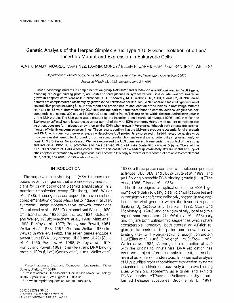
VIROLOGY 190, 702-715 (1992) Genetic Analysis of the Herpes Simplex Virus Type 1 UL9 Gene: Isolation of a LacZ Insertion Mutant and Expression in Eukaryotic Cells AJAY K. MALIK, RICARDO MARTINEZ, LAVINIA MUNCY,’ ELLEN P. CARMICHAEL,2 AND SANDRA K. WELLER3 Department of Microbiology, University of Connecticut Health Center, Farmington, Connecticut 06030 Received March 13, 1992; accepted June 22, 1992 HSV-1 host range mutants in complementation group l-36 (hr27 and hr156) whose mutations map in the UL9 gene, encoding the origin binding protein, are unable to form plaques or synthesize viral DNA or late viral proteins when grown in nonpermissive Vero cells (Carmichael, E. P., Kosovsky, M. J., Weller, S. K., 1988, J. l&o/. 62, 91-99). These defects are complemented efficiently by growth in the permissive cell line, S22, which contains the wild type version of several HSV genes including UL9. In this report the precise nature and location of the lesions in host range mutants hr27 and hr156 were determined by DNA sequencing; both mutants were found to contain identical single-base-pair substitutions at codons 309 and 311 in the UL9 open reading frame. This region lies within the putative helicase domain of the UL9 protein. The UL9 gene was disrupted by the insertion of an insertional mutagen ICP6::lacZ in which the Escherichia co/i /acZ gene is expressed under control of the viral ICP6 promoter. Hr94, a viral mutant containing this insertion, does not form plaques or synthesize viral DNA when grown in Vero cells, although both defects are comple- mented efficiently on permissive cell lines. These results confirm that the UL9 gene product is essential for viral growth and DNA replication. Furthermore, since no detectable UL9 protein is synthesized in hr94-infected cells, this virus provides a useful genetic background for further structure-function analysis since no potentially interfering nonfunc- tional UL9 protein will be expressed. We have expressed the UL9 open reading frame under the control of the strong and inducible HSV-1 ICP6 promoter and have derived Vero cell lines containing variable copy numbers of the ICP6:: UL9 construct. Cells whose copy number of this construct exceeded approximately 120 are unable to support efficient plaque formation by wild-type virus. Cell lines with low copy numbers of this construct are able to complement hr27, hrl56, and hr94. o 1992 Academic PESS, IIIC. INTRODUCTION The herpes simplexvirus type-l (HSV-1) genome en- codes seven viral genes that are necessary and suffi- cient for origin-dependent plasmid amplification in a transient transfection assay (Challberg, 1986; Wu et al., 1988). These genes correspond to seven distinct complementation groups which fail to induce viral DNA synthesis under nonpermissive growth conditions (Carmichael eta/., 1988; Carmichael and Weller, 1989; Chartrand et al., 1980; Coen et a/., 1984; Goldstein and Weller, 198813; Marchetti et a/., 1988; Matz et a/., 1983; Purifoy et al., 1977; Purifoy and Powell, 1981; Weller et a/., 1983, 1987; Zhu and Weller, 1988) (re- viewed in (Weller, 1990)). The seven genes encode a two-subunit DNA polymerase (UL30 and UL42) (Gallo et al., 1989; Parris et al., 1988; Purifoy et al., 1977; Purifoy and Powell, 1981), a single-strand DNA binding protein, ICP8 (UL29) (Conley et al., 1981; Weller et al., ’ Present address: Electronic Component Engineering, Pitney Bowes, Shelton, CT 06484. ’ Present address: Department of Cellular and Molecular Biology, Bristol-Myers Squibb, Wallingford, CT 06492. 3 To whom reprint requests should be addressed. 1983), a three-protein complex with helicase-primase activities (UL5, UL8, and UL52) (Crute et a/., 1989), and an HSV origin-specific DNA binding protein (UL9) (Elias et al., 1986; Olivo et al., 1988). The three origins of replication on the HSV-1 ge- nome were defined using plasmid amplification assays in transiently transfected cells: oris, present in two cop- ies in the viral genome within the inverted repeats flanking U, (Spaete and Frenkel, 1982; Stow and McMonagle, 1983), and one copy of oriL, localized to a region near the center of U, (Weller et al., 1985). Ori, and or& are both palindromic sequences which share considerable homology; both contain an AT-rich re- gion at the center of the palindrome as well as two binding sites for the origin-specific recognition protein UL9 (Elias et a/., 1986; Olivo et a/., 1988; Stow, 1982; Weller et al., 1985). Although the interaction of UL9 with the origins to initiate viral DNA replication has been the subject of considerable interest, its mecha- nism of action is not understood. Biochemical analysis of UL9 purified from recombinant expression systems indicates that it binds cooperatively to the two binding sites within ori, apparently as a dimer and exhibits DNA-dependent ATPase and helicase activity on pre- formed helicase substrates (Bruckner et al., 1991; 0042.6822/92 $5.00 702 Copyright 0 1992 by Academic Press. lnc All rights of reproduction in any form resewed.
-
Upload
independent -
Category
Documents
-
view
3 -
download
0
Transcript of Genetic analysis of the herpes simplex virus type 1 UL9 gene: Isolation of a lacZ insertion mutant...
VIROLOGY 190, 702-715 (1992)
Genetic Analysis of the Herpes Simplex Virus Type 1 UL9 Gene: Isolation of a LacZ Insertion Mutant and Expression in Eukaryotic Cells
AJAY K. MALIK, RICARDO MARTINEZ, LAVINIA MUNCY,’ ELLEN P. CARMICHAEL,2 AND SANDRA K. WELLER3
Department of Microbiology, University of Connecticut Health Center, Farmington, Connecticut 06030
Received March 13, 1992; accepted June 22, 1992
HSV-1 host range mutants in complementation group l-36 (hr27 and hr156) whose mutations map in the UL9 gene, encoding the origin binding protein, are unable to form plaques or synthesize viral DNA or late viral proteins when grown in nonpermissive Vero cells (Carmichael, E. P., Kosovsky, M. J., Weller, S. K., 1988, J. l&o/. 62, 91-99). These defects are complemented efficiently by growth in the permissive cell line, S22, which contains the wild type version of several HSV genes including UL9. In this report the precise nature and location of the lesions in host range mutants hr27 and hr156 were determined by DNA sequencing; both mutants were found to contain identical single-base-pair substitutions at codons 309 and 311 in the UL9 open reading frame. This region lies within the putative helicase domain of the UL9 protein. The UL9 gene was disrupted by the insertion of an insertional mutagen ICP6::lacZ in which the Escherichia co/i /acZ gene is expressed under control of the viral ICP6 promoter. Hr94, a viral mutant containing this insertion, does not form plaques or synthesize viral DNA when grown in Vero cells, although both defects are comple- mented efficiently on permissive cell lines. These results confirm that the UL9 gene product is essential for viral growth and DNA replication. Furthermore, since no detectable UL9 protein is synthesized in hr94-infected cells, this virus provides a useful genetic background for further structure-function analysis since no potentially interfering nonfunc- tional UL9 protein will be expressed. We have expressed the UL9 open reading frame under the control of the strong and inducible HSV-1 ICP6 promoter and have derived Vero cell lines containing variable copy numbers of the ICP6:: UL9 construct. Cells whose copy number of this construct exceeded approximately 120 are unable to support efficient plaque formation by wild-type virus. Cell lines with low copy numbers of this construct are able to complement hr27, hrl56, and hr94. o 1992 Academic PESS, IIIC.
INTRODUCTION
The herpes simplexvirus type-l (HSV-1) genome en- codes seven viral genes that are necessary and suffi- cient for origin-dependent plasmid amplification in a transient transfection assay (Challberg, 1986; Wu et al., 1988). These genes correspond to seven distinct complementation groups which fail to induce viral DNA synthesis under nonpermissive growth conditions (Carmichael eta/., 1988; Carmichael and Weller, 1989; Chartrand et al., 1980; Coen et a/., 1984; Goldstein and Weller, 198813; Marchetti et a/., 1988; Matz et a/., 1983; Purifoy et al., 1977; Purifoy and Powell, 1981; Weller et a/., 1983, 1987; Zhu and Weller, 1988) (re- viewed in (Weller, 1990)). The seven genes encode a two-subunit DNA polymerase (UL30 and UL42) (Gallo et al., 1989; Parris et al., 1988; Purifoy et al., 1977; Purifoy and Powell, 1981), a single-strand DNA binding protein, ICP8 (UL29) (Conley et al., 1981; Weller et al.,
’ Present address: Electronic Component Engineering, Pitney Bowes, Shelton, CT 06484.
’ Present address: Department of Cellular and Molecular Biology, Bristol-Myers Squibb, Wallingford, CT 06492.
3 To whom reprint requests should be addressed.
1983), a three-protein complex with helicase-primase activities (UL5, UL8, and UL52) (Crute et a/., 1989), and an HSV origin-specific DNA binding protein (UL9) (Elias et al., 1986; Olivo et al., 1988).
The three origins of replication on the HSV-1 ge- nome were defined using plasmid amplification assays in transiently transfected cells: oris, present in two cop- ies in the viral genome within the inverted repeats flanking U, (Spaete and Frenkel, 1982; Stow and McMonagle, 1983), and one copy of oriL, localized to a region near the center of U, (Weller et al., 1985). Ori, and or& are both palindromic sequences which share considerable homology; both contain an AT-rich re- gion at the center of the palindrome as well as two binding sites for the origin-specific recognition protein UL9 (Elias et a/., 1986; Olivo et a/., 1988; Stow, 1982; Weller et al., 1985). Although the interaction of UL9 with the origins to initiate viral DNA replication has been the subject of considerable interest, its mecha- nism of action is not understood. Biochemical analysis of UL9 purified from recombinant expression systems indicates that it binds cooperatively to the two binding sites within ori, apparently as a dimer and exhibits DNA-dependent ATPase and helicase activity on pre- formed helicase substrates (Bruckner et al., 1991;
0042.6822/92 $5.00 702
Copyright 0 1992 by Academic Press. lnc All rights of reproduction in any form resewed.
HERPES SIMPLEX VIRUS TYPE 1 UL9 GENE 703
Elias et a/., 1990; Fierer and Challberg, 1992). During HSV infection, UL9 presumably binds to at least one of the three origins of DNA synthesis on the HSV mole- cule and either acts alone to unwind the duplex or in- teracts with other components of the replication ma- chinery to unwind the template at the origin.
The first viral mutants unambiguously mapped to the UL9 gene were host range mutants in complementa- tion group l-36 (hr27, hr48, and hrl56) (Carmichael et a/., 1988). These spontaneously arising mutants are defective in plaque formation and viral DNA synthesis on Vero cells indicating that UL9 is essential for viral DNA synthesis in infected cells. To further our under- standing of the role of UL9 in the initiation of DNA repli- cation, we have undertaken a more detailed structure- function analysis of this gene. In this study, we report the characterization of existing UL9 host range mu- tants hr27 and hr156; both mutants were found to con- tain two single-base-pair substitutions and to synthe- size detectable amounts of UL9 protein. We have previ- ously described a transient transfection assay to test the ability of engineered mutations in the UL5 gene of HSV, encoding a component of the helicase-primase complex, to complement the defect of a UL5 null mu- tant for the amplification of HSV origin containing plas- mids (Zhu and Weller, 199213). This type of in viva com- plementation assay provides a rapid way to screen large numbers of variants. However, to avoid intet-fer- ence, this test optimally requires a genetic background that is devoid of any allele of the protein other than the one that is expressed from the expression plasmid (Schimmel, 1990). Thus, the presence of a nonfunc- tional form of the UL9 protein in cells infected with hr27 or hr156 make these viruses a poor background for this purpose. In this study, we report the isolation of a null mutant, hr94, in which the UL9 gene has been disrupted by the insertion of the insertional mutagen, ICP6 : : IacZ. Since no detectable UL9 protein is synthe- sized in hr94-infected cells, this provides a useful ge- netic background for the functional analysis of engi- neered mutations in the UL9 gene (Martinez, R., Shao, L., and Weller, S. K., in press). In addition, the IacZ insertion in hr94 provides a convenient screening sys- tem for the introduction of site-specific mutations into the viral genome.
MATERIALS AND METHODS
Cells and viruses
African green monkey kidney ceils (Vero, American Type Culture Collection, Rockville, MD) were propa- gated and maintained as described (Weller et a/., 1983). The KOS strain of HSV-1 was used as the wild- type virus. Mutants hr27 and hr156 were described
previously (Carmichael et al., 1988). The IacZ insertion mutant hr94 and permissive cell lines for UL9 mutants are described in this report.
Plasmids
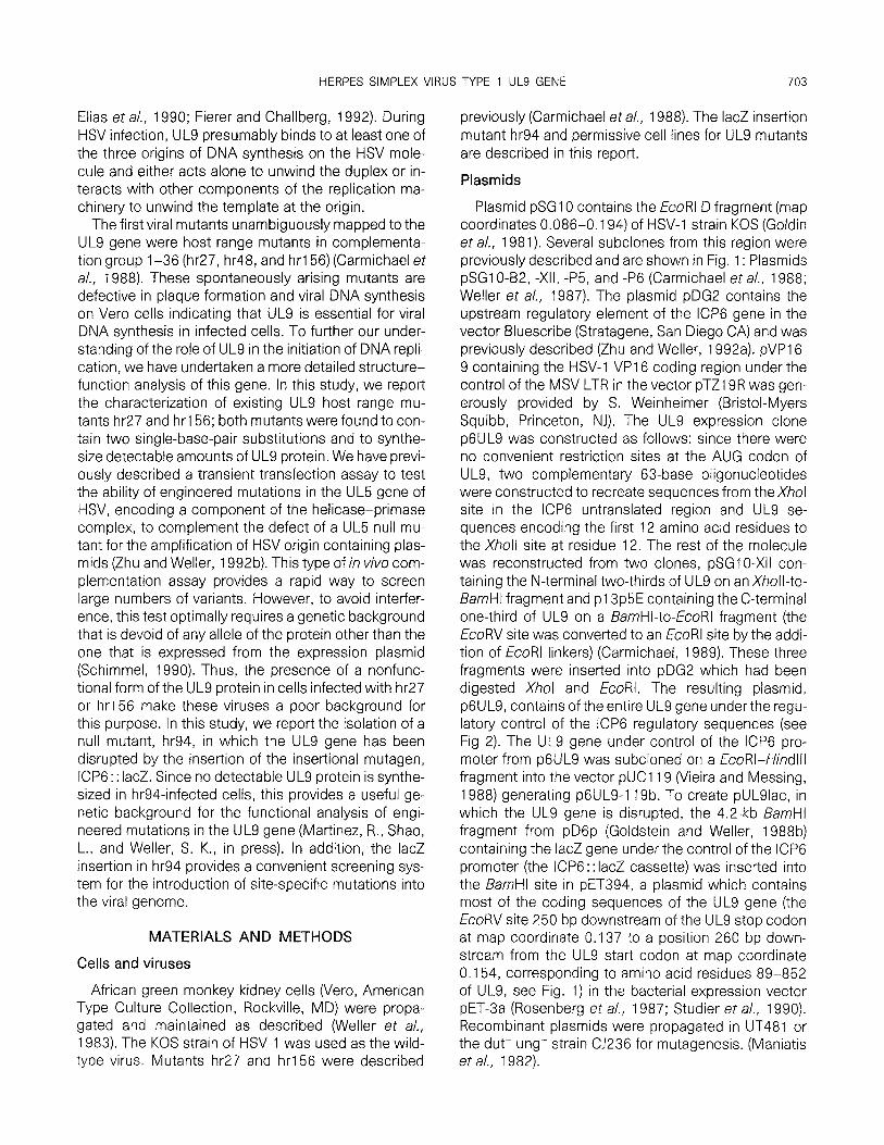
Plasmid pSGl0 contains the EcoRI D fragment (map coordinates 0,086-O. 194) of HSV-1 strain KOS (Goldin et a/., 1981). Several subclones from this region were previously described and are shown in Fig. 1: Plasmids pSG1 O-B2, -XII, -P5, and -P6 (Carmichael et a/., 1988; Weller et a/., 1987). The plasmid pDG2 contains the upstream regulatory element of the ICP6 gene in the vector Bluescribe (Stratagene, San Diego CA) and was previously described (Zhu and Weller, 1992a). pVP16- 9 containing the HSV-1 VP1 6 coding region under the control of the MSV LTR in the vector pTZ19R was gen- erously provided by S. Weinheimer (Bristol-Myers Squibb, Princeton, NJ). The UL9 expression clone p6UL9 was constructed as follows: since there were no convenient restriction sites at the AUG codon of UL9, two complementary 63-base oligonucleotides were constructed to recreate sequences from theXhol site in the ICP6 untranslated region and UL9 se- quences encoding the first 12 amino acid residues to the Xholl site at residue 12. The rest of the molecule was reconstructed from two clones, pSG1 O-XII con- taining the N-terminal two-thirds of UL9 on an Xholl-to- BarnHI fragment and pl3p5E containing the C-terminal one-third of UL9 on a BarnHI-to-EcoRI fragment (the EcoRV site was converted to an EcoRl site by the addi- tion of EcoRI linkers) (Carmichael, 1989). These three fragments were inserted into pDG2 which had been digested Xhol and EcoRI. The resulting plasmid, p6UL9, contains of the entire UL9 gene underthe regu- latory control of the ICP6 regulatory sequences (see Fig 2). The UL9 gene under control of the ICP6 pro- moter from p6UL9 was subcloned on a EcoRI-HindIll fragment into the vector pUC1 19 (Vieira and Messing, 1988) generating p6UL9-1 19b. To create pULSlac, in which the UL9 gene is disrupted, the 4.2-kb BamHl fragment from pD6p (Goldstein and Weller, 1988b) containing the IacZ gene under the control of the ICP6 promoter (the ICP6:: IacZ cassette) was inserted into the BarnHI site in pET394, a plasmid which contains most of the coding sequences of the UL9 gene (the EcoRV site 250 bp downstream of the UL9 stop codon at map coordinate 0.137 to a position 260 bp down- stream from the UL9 start codon at map coordinate 0.154, corresponding to amino acid residues 89-852 of UL9, see Fig. 1) in the bacterial expression vector PET-3a (Rosenberg et a/., 1987; Studier et al., 1990). Recombinant plasmids were propagated in UT481 or the dut- ung- strain 0236 for mutagenesis. (Maniatis et a/., 1982).

704 MALIK ET AL
ab b’ a’c’ “B ca
0.0 0.1 0.2 0.3 0.4 0.5 0.6 0.7 0.6 0.9 1.0
o.l*,9.167
B P H E xxx P x P !EG
I I I II I I I I I I
II 1 I I
M M M M M 0.5 kb
pSGlO-B2
pSGlO-XII
pSG10-P6
pSGlO-PS
pET394
UL9 ORF
ICP6::lac Z insertion in pUL9lac
FIG. 1. Map positions of UL9 and ULS-containing clones used in this study. The HSV genome and coordinates are represented on top of the figure with U, and Us referring to the long and short unique sequences and a, b, c referring to repetitive sequences. The region from the Pstl site at 0.129 to the Bg/ll site at 0.167 is expanded. The markers for restriction sites are: B, BarnHI; E, EcoRV; H, /-/pal; G, Bglll; M, Mlul; P, Pstl; and X, Xholl. Recombinant clones, pSGlO-B2, -XII, -P6, and -P5 are shown. pSG1 O-82 contains an internal BamHl fragment from pSGl0 (coordinates 0.145 to 0.165). [DNA sequence information indicates that a BamHl site is present at position 24,890 (coordinate 0.165) in HSV-1 strain KOS but not in strain 17 (Shao and Weller, unpublished data)]. pSG1 O-XII contains an Xholl fragment from within the UL9 gene (coordinates 0.145- 0.1 55). Plasmids pSG1 O-P5 and pSG1 O-P6 contain Pstl fragments (coordinates 0.129 to 0.151 and coordinates 0.151-0.158, respectively) (Carmichael eta/., 1988). Plasmid pET394 contains from the EcoRV site 250 bp downstream of the UL9 stop codon at map coordinate 0.137 to a position 260 bp downstream from the UL9 start codon at map coordinate 0.154 (UL9 amino acid residues 89-852). At the bottom, the open reading frame for UL9 is shown as well as the position of insertion of the mutagen ICP6:: IacZ.
Plasmids used for viral mutant sequencing
Viral DNA from mutant strains was isolated as de- scribed below and appropriate fragments for sequenc- ing were subcloned. DNA from mutant hr156 was di- gested with Bglll and fragments resolved on a 0.6% agarose gel. The 10.2.kb Bglll fragment containing the UL9 gene was isolated and ligated into the Bglll site of pDLl9, a derivative of pUCl9 which contains a Bglll site in the polylinker. The resultant clone, pl56G, was digested with BamHl and the internal 3-kb fragment (map coordinates 0.145 and 0.165; sequence coordi- nates 21,655 to 24,890) was subcloned into the Bglll site of pDLl9 to generate p156Bam. Viral DNA from mutant hr27 was digested with BarnHI and fragments
resolved on a 0.8% gel. The 3-kb fragment (map coordi- nates 0.145 and 0.165; sequence coordinates 2 1,655 to 24,890) was cloned directly into pUC18 vector to generate p27Bam. p27RBam containing the same Barn fragment from a wild type revertant virus derived from hr27 was constructed as described for hr27.
Marker rescue and marker transfer
Marker rescue experiments were performed as de- scribed previously (Goldstein and Weller, 1988c). Marker transfer was performed essentially as de- scribed previously (Zhu and Weller, 1992a). Infectious intact wild type DNA was mixed with a 1 O-fold molar excess of fragment containing the disrupted UL9 gene

HERPES SIMPLEX VIRUS TYPE 1 UL9 GENE
-315 +l +231
705
m TATABOX
OVERLAPPING OCTAYER AND TAATGARAT
SP-1 BINDING SITE
AP 1 BINDING SITE
1.1 kb Sac1 fragment
0.6 kb Sac1 fragment
p6UL9
FIG. 2. Plasmid p6UL9 showing the ICP6 promoter regron. The black box corresponds to a 549-bp /%I-to-Bail fragment from the ICP6 gene promoter and upstream untranslated region. The start site of the ICP6 transcript is marked by an arrow and numbered as +l (McLauchlan and Clements, 1983). The 315 bp upstream from the transcription start site contain consensus sequence elements known to be responsive to various transcription factors are illustrated with boxes as shown. The translation start codon is at position +231. The UL9 open reading frame was inserted downstream at the AUG codon as described under Materials and Methods. Restriction enzyme recognition sites are abbreviated: Sp, Sphl; Sa, Sacl. Fragments used in Southern blot analysis are shown.
and used to transfect S22 cells. When widespread cy- topathic effects were observed, progeny were har- vested and titers were determined on permissive cells. Since ,&galactosidase (P-gal) activity can be easily de- tected in the presence of the chromogenic substrate X-gal (5bromo-4-chloro-3-indolyl-fl-D-galactoside), re- combinants that express the /acZ gene were readily identified. Recombinant viruses were detected as fol- lows. On Day 4 after infection, plaques were stained by adding 1 to 2 ml of a solution containing 300 pg of X-gal per milliliter and neutral red (0.00125%) in Tris-buffered saline to a methylcellulose overlay. Plaques were visu- alized within 12 to 24 hr at 34”. Blue plaques were purified three or four times in S22 cells before stocks were made.
Analysis of cellular and viral DNA
Intact infectious viral genomic DNA used in marker transfer experiments was prepared as described previ- ously (Zhu and Weller, 1988). Viral DNA used for Southern analysis was isolated as described for in- fectious DNA except that the DNA was deproteinized by extraction with phenol:chloroform:isoamyl alcohol (25:24: 1) and precipitated with ethanol. Alternatively, viral DNA was isolated from infected cells as described (Coen et a/., 1986). Infected cells were scraped into media followed by three cycles of freezing and thaw- ing. The sample was sonicated for 1 min and centri- fuged at 2500 rpm for 10 min to pellet the cell debris. The supernatant was transferred into a fresh tube and centrifuged at 13,000 g for 1 hr to pellet virions. The
virion pellet was resuspended in 400 ~1 TE and depro- teinized by phenol:chloroform:isoamyl alcohol (25: 24:l) and precipitated with ethanol. High-molecular- weight cellular DNA for Southern blot analysis was iso- lated as described previously (Weller et a/., 1985). Southern blot analysis was performed on Genescreen- Plus membranes (DuPont) according to manufac- turer’s instructions. DNA probes were prepared by random priming of gel-isolated restriction fragments with a-[32P]dATP (Feinberg and Vogelstein, 1983).
Transformation of Vero cells
Vero cells were cotransfected with plasmids pSV2neo and p6UL9 as described previously (Gold- stein and Weller, 1988a). pSV2neo (1 pg) and p6UL9 (9 pg) were coprecipitated in the presence of 10 pg of salmon sperm DNA and 0.125 M CaCI, in a total vol- ume of 1 .O ml by the procedure of Graham and van der Eb (1973). G418 selection was carried out as de- scribed by DeLuca et al. (1985). The cells were grown to confluence (2 days), trypsinized, and plated at a 1: 10 dilution in medium containing 500 pg/ml of G418. After approximately 2 weeks at 37” (with periodic changes of media) individual G418-resistant colonies were iso- lated, amplified, and screened as described under Re- sults.
Analysis of viral DNA synthesis
Viral DNA synthesis was determined based on a method described by Gadler (1983). Specifically, cells in 35-mm plates were infected with virus at a m.o.i. of

706 MALIK ET At
2-5 for 16 hr at 37”. The plates were washed with PBS twice and cells scraped into 15-ml tubes. The cells were pelleted by centrifugation at 4” for 10 min at 2000 rpm and the pellets were resuspended in 100 ~1 of fresh PBS. A series of fivefold dilutions were spotted onto a GenescreenPlus membrane using a Microsam- ple Filtration Manifold (Schleicher & Schuell). The membrane was treated twice with 0.4 N NaOH and twice with 1 M Tris-HCI, pH 7.5. The membrane was dried at room temperature and probed with a 32P-la- beled HSV-specific probe (EcoRI F fragment from plas- mid pSG18, map coordinates 0.315-0.422) as de- scribed above.
Detection of UL9 proteins in viral infection and transient transfection by immunological methods
To detect UL9 protein in cells transiently transfected with the expression clone p6UL9-119b, 3 X 1 O6 Vero cells were transfected with 12 pg of the expression plasmid as described (Goldstein and Weller, 1988c) and plated in a 1 00-mm tissue culture dish. Induction of the ICP6 promoter was achieved by superinfection at 30 hr post-transfection with hr94. At 18 hr postinfec- tion, the cells were washed in PBS and resuspended in 400 ~1 PBS plus 200 ~1 3X lysis buffer (30 mM Tris- HCI, pH 8.0, 15 mM EDTA, pH 8.0, and 3% SDS) and chilled on ice for 10 min. The cells were disrupted by sonication and the proteins precipitated with four vol- umes of acetone at -20” for 30 min (Triezenberg et al., 1988). Proteins were pelleted in SS34 rotor in the Sor- vall RC-5B at 1 OK for 15 min and resuspended in 160 ~1 PBS and 40 ~1 5X loading dye (containing 0.0625M Tris, pH 6.8, 2% SDS, 10% glycerol, 5% fl-mercap- toethanol and 0.001% bromophenol blue) (Laemmli, 1970). The samples were heated at 70” for 15 min, boiled for 10 min and spun to remove any insoluble debris. Extracts were subjected to SDS-PAGE on 9%
gels as described above and proteins were electropho- retically transferred to filters essentially as described by Towbin et a/. (1979). lmmobilon membrane filters (Millipore, Bedford, MA) were used according to proce- dures recommended by the supplier. Filters were incu- bated with a 1 :lOOO dilution of primary rabbit a-UL9 (generously provided by M. D. Challberg, NIH, Be- thesda, MD). a-UL9 is a rabbit polyclonal antiserum directed against the 10 C-terminal amino acid residues of UL9 (Olivo et al., 1988). The immunoreactive pro- teins were detected by reaction with a goat anti-rabbit IgG (Fc) antibody conjugated to alkaline phosphatase (AP) (Promega; Madison, WI). AP color development was carried out as described previously (Zhu and Weller, 1988).
In some experiments a combination of immunopre- cipitation and immunoblotting was used to detect the
UL9 protein as described (Zhu and Weller, 1992a). Im- mune complexes were washed three times with RIPA buffer (50 mM Tris-HCI (pH 8.0), 150 mM NaCI, 0.5% DOC, 1.0% NP-40, 0.1% SDS) containing 1 mM EDTA, 0.5 mM PMSF, 2 pg/ml aprotonin, 200 PM TPCK, and 200 &’ TLCK. Immune complexes were subjected to 9% SDS-PAGE and immunoblotting as described above.
indirect immunofluorescence and confocal microscopy
Vero cells were transfected with p6UL9 and pVPl6- 9 or infected with KOS. Indirect immunofluorescence was performed as described (Zhu and Weller, 1988). Basically, fixed cells on coverslips were incubated for 60 min at room temperature with a 1 :lOO dilution of a-UL9 antiserum, washed with 1% NGS (normal goat serum) in PBS four times, incubated with rhodamine- conjugated goat anti-rabbit IgG (1: 100 dilution in PBS with 1% NGS, Cappel Laboratories, West Chester, PA) for 30 min, and washed three times with lo/o NGS in PBS. Microscopy was performed with a MRC 600 laser scanning confocal microscope (Bio-Rad).
RESULTS
The location and nature of spontaneously arising mutations in hr27 and hr156
We have initiated structure-function analysis of the UL9 gene of HSV in an attempt to localize functional domains responsible for its various activities and to elucidate its mechanism of action. The sequence-spe- cific DNA binding activity of UL9 has been localized to the C-terminal 269 amino acids (Deb and Deb, 1991; Weir et al., 1989); however, little is known about the N-terminal two-thirds of the UL9 gene. Analysis of the predicted protein sequence of the UL9 gene (McGeoch et al., 1988) reveals the existence of a set of motifs which are shared within a superfamily of pro- teins which include at least 25 established or putative helicases of fscherichia co/i, yeast, insects, mam- mals, pox- and herpesviruses, and three groups of posi- tive-strand RNA viruses (Gorbalenya et a/., 1988, 1989; Hodgman, 1988). In addition, UL9 contains a series of four leucines repeated in a heptad pattern characteris- tic of a leucine zipper, a motif implicated in homo- or heterodimer formation (Landschulz et al., 1988; McGeoch et al., 1988). The position within the UL9 gene of the putative leucine zipper and the seven mo- tifs shared among known and putative helicases are shown in Fig. 3. These putative helicase motifs lie

707 HERPES SIMPLEX VIRUS TYPE 1 UL9 GENE
Putative helicase motifs
I la II Ill IV v VI
100 200 300 400 500 600 700 800
851 aas
Putative leucine zipper 150-171
site of lac 2 insertion in hr 94
ACG GTC TCC TTC GCG GAG ATC GTG thr val ser phe ala glu ile val 306 309 311
TAC GGG w= @Y
point mutations in hr 27 and hr 156
FIG. 3. UL9 gene showing putative helicase domains, DNA-binding domain, and the positions of mutations in hr27, hrl56, and hr94. The UL9 open reading frame, 851 amino acids, is shown with the locations of seven putative helicase motifs. A putative leucine zipper (150-l 71) is also shown. The point mutations in hr27 and hr156 lie within motif IV, and the amino acid changes are as shown. The DNA-binding domain within the carboxy terminus (Deb and Deb, 199 1; Weir and Stow, 1990) is shown as is the site of the IacZ insertion in hr94.
within the first 400 N-terminal amino acid residues of UL9 and suggest that the N-terminal domain of UL9 may be responsible for helicase activity.
We began our structure-function analysis of this gene with the characterization of the previously de- scribed host range mutants, hr27 and hr156 (Carmi- chael eta/,, 1988). Since both mutants arose spontane- ously in our stock of the KOS strain of wild-type HSV-1, the precise location and nature of the mutations was not clear. To investigate the possibility that the ge- nomes of these mutants contain large deletions, DNAs from viruses hr27, hrl56, and KOS were prepared, di- gested with Smal which recognizes several sites within the UL9 gene, and analyzed by Southern blot hybridiza- tion. Smal, was used to facilitate the detection of a relatively small deletion. Both mutant DNAs generated a band pattern indistinguishable from that of wild type KOS DNA (Carmichael, 1989); therefore, we con- cluded that the mutant DNAs contain no large dele- tions or gross alterations in the structure of this gene. The precise nature of the lesions in hr27 and hr156 was determined by DNA sequencing. The lesion in hr27 was rescued efficiently by plasmids pSGlO-P5 and pSGlO-XII, thereby localizing the mutation to a 800-bp fragment within the UL9 gene (coordinates 0.145 to 0.151) (Carmichael et a/., 1988) (see Fig. 1). The BarnHI fragment (coordinates 0.145 to 0.165) con- taining this 800-bp region from both hr27 and hr156 were subcloned and sequenced as described under Materials and Methods. Both mutants contain two base changes when compared to the wild type: a T-to-
A transversion at nucleotide position 926 (within the UL9 ORF) resulting in the alteration of phenylalanine 309 to tyrosine, and an A-to-G transition at position 932 (within the UL9 ORF) resulting in a glutamic acid 31 1 to glycine alteration (Fig. 3). Revertants of hr27 were isolated and the 800-bp region of one revertant hr27R was sequenced in order to determine which of the two mutations is responsible for the mutant pheno- type. The sequence of hr27R was identical to wild type sequence. This indicates that either the two mutations coreverted or, perhaps more likely, that these rever- tants arose as the result of recombination between mutant viral genomes and wild type genomes present in the permissive S22 cells. The position of these mu- tations places them within the putative helicase do- main and well outside of the DNA-binding domain.
In order to determine whether the two amino acid alterations in hr27 and hr156 affect the stability of the UL9 protein, infected cell extracts were subjected to SDS-PAGE electrophoresis and immunoblot analysis with a-UL9, an antiserum raised against the carboxy- terminal 10 amino acid residues of the UL9 gene (Olivo et a/., 1988). Figure 4, left-hand panel, shows that Vero cells infected with KOS contained a protein of the ex- pected size for UL9. lmmunoblot analysis of hr27- and hrl56-infected Vero cells indicates that these mutants are capable of producing a stable form of UL9 which migrates at a position similar to that of wild type UL9 (Fig. 4, left panel as marked). These results indicate that the two mutations at residues 309 and 3 1 1 of the UL9 open reading frame do not grossly alter the con-

708 MALIK ET AL
Vero 2B-11
- UL9
FIG. 4. Detection of UL9 gene product during viral infection by immunoblotting. Vero or ZB-1 1 cells (left and right panels, respec- tively) were infected with the indicated virus. Cell extracts were pre- pared as described under Materials and Methods and subjected to electrophoresis and immunoblotting. Filters were treated with poly- clonal antiserum to the C-terminal 10 amino acid residues of the UL9 protein. The position of the UL9 protein is indicated.
formation of the protein leading to instability of the pro- tein.
The presence of a nonfunctional but stable UL9 pro- tein in hr27- and hrl%infected cells indicates that these mutants do not provide an appropriate back- ground for structure-function analysis since, as de- scribed above, it is preferable to have a genetic back- ground that is devoid of any allele of the protein under study. For this reason, it was necessary to isolate a mutant which fails to synthesize UL9 protein.
isolation of hr94 in which the UL9 gene is disrupted with ICP6: : IacZ
We previously reported the construction and use of a plasmid, pDGp, bearing an insertional mutagen con- sisting of the /acZgene expressed under the control of the upstream regulatory signals of the ICP6 gene (Car- michael and Weller, 1989; Goldstein and Weller, 1988b; Weller et al., 1990). Restriction digestion of pD6p with BarnHI results in the excision of a 4.2-kb ICP6: : IacZ cassette. In pUL9lac this cassette was in- serted into the BamHl site within the UL9 gene as de- scribed under Materials and Methods (see Fig. 1). The site of insertion corresponds to residue 536 and the resulting virus might be expected to encode a C termi- nally truncated UL9 molecule containing the first 536 amino acid residues of UL9 which would terminate within the upstream regulatory sequences of the ICP6 gene to generate a protein of approximately 60 kDa (see below).
An HSV mutant was recovered by cotransfecting 522 cells with infectious wild type DNA and pUL9lac. Homologous recombination between KOS genome
DNA and pUL9lac resulted in the generation of recom- binants at a frequency of approximately 1 O/o which formed dark blue plaques in the presence of X-gal. Mu- tant viruses were picked, purified, and propagated in S22 cells. One recombinant virus, hr94, was used in further studies. To confirm that hr94 contains the ICP6 : : IacZ cassette at the appropriate position, DNAs from p6UL9, pULSlac, KOS, and hr94 were purified, digested with Mlul, and subjected to gel electrophore- sis and Southern blot hybridization. The expected frag- ments consistent with the insertion of the ICP6: : IacZ cassette into the BarnHI site of the UL9 gene were obtained (not shown).
Phenotypic properties of hr94
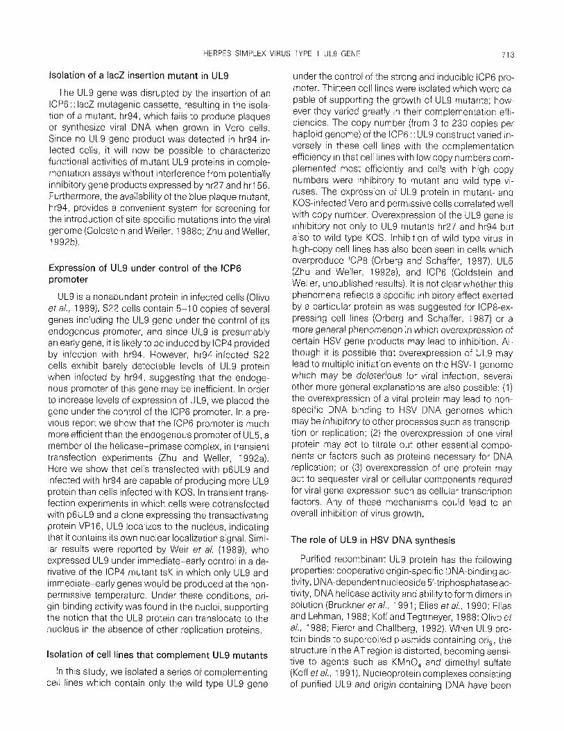
The mutant hr94 was tested for its ability to form plaques in untransformed Vero cells. A stock of hr94 was propagated on S22 cells and the titers on 522 and on Vero cells were determined. Table 1 shows that hr94 fails to form plaques efficiently on Vero cells and that this growth defect can be complemented by the S22 cell line. These results confirm that the UL9 gene is essential for virus growth. The ability of hr94, hr27, and hr156 to induce DNA synthesis in 28-l 1 (UL9 per- missive cell lines described below) and Vero cells was assessed by dot blot hybridization of total DNA from infected cells to a virus specific probe (see Materials and Methods). The results shown in Fig. 5 clearlydem- onstrate that all three mutant viruses are unable to syn- thesize detectable viral DNA in Vero cells. In contrast, they are able to synthesize wild type levels of viral DNA in 2B-1 1 cells (Fig. 5). These results indicate that the UL9 gene is essential for HSV DNA synthesis, consis- tent with results obtained with other UL9 hr mutants (Carmichael and Weller, 1989).
Since S22 cells contain several genes from the left end of the HSV genome (Carmichael and Weller, 1989), the ability of hr94 to form plaques on these cells cannot be taken as evidence that a lesion in UL9 is responsible for lack of growth on Vero cells. In fact, S22 cells have also been used as a permissive host for mutants in UL8 and alkaline nuclease (Carmichael and Weller, 1989; Weller eta/., 1990). Thus, to facilitate the genetic analysis of hr94 and also the isolation and char- acterization of new mutations in the UL9 gene without potential complications arising from the expression of more than one open reading frame in the permissive cell line, cell lines were constructed in which only the UL9 gene is expressed.
Construction of expression clone p6UL9
In S22 cells, UL9 is expressed from its endogenous promoter which has been presumed to be fairly ineffi-

HERPES SIMPLEX VIRUS TYPE 1 UL9 GENE 709
TABLE 1
PLAQLJING EFFICIENCIES OF KOS AND UL9 MUTANTS ON VERO, S22, AND NINE NEW UL9 CELL LINES
Cell line Copy NumbeP KOS
Titer in PFU/ml and plaque size
hr27 hr94
Vero
s22 2A-1 28-5
2B-11 2A-15
lA-5 1 c-2 2C-8 2c-10 lA-8
5-lob 3 5
18 21
40 95
118 150 230
5.4 x 1 OS large , ,o x , 03 heterogeneous 5.0 x 10³ heterogeneous
2.6 x 1 O* large 6.2 X 10⁷’arge 2.5 x 1081arge , .6 x ,@medhe 2.2 x 10⁷ med large 1 ,g x 108 med We 3.0 x 10⁸ med farce 5.7 x ~07med~awe 1.2 x 10⁸ med large
2.0 x 10⁸ med he 3.7 x 10⁷ med large 1 .4 x 1 O8 large 2.3 x ,053 heterOgeneO”S 4.6 x , 07 h’StWOg~~~O”S , .o x 108 heterOge”eO”S
2.1 x 1 O8 Sma” 3.2 X 10’ Sma” 6.1 x 10⁷sma” 6.2 X 10⁷t’ny 2 x 10⁷tiny 2.5 x 10⁷t’ny
9.0 x 10⁶ti”y 3.4 x 1 06t’ny 7.0 x 1 061i”Y TTKc FKc TTTCC TTTCC TCC TKc
a Copy number per haploid genome calculated as described in text. b Copy number of UL9 gene under control of the UL9 endogenous promoter. Remainder of cell lines contain ICP6::UL9 construct c mC is too tiny to count.
cient based on levels of expression of UL9 in infected cells (Olivo el al., 1989). In order to facilitate in viva complementation analysis, it was necessary to ex- press the UL9 gene in Vero cells both in transient and stable transfection experiments at levels that were higher than could be expected from using the endoge- nous UL9 promoter. We have previously shown that the promoter region from the large subunit of the HSV ribonucleotide reductase gene, ICP6, is strong and in- ducible by infection with HSV (Goldstein and Weller, 1988a; Zhu and Weller, 1992a; Zhu, L., Shao, L., and
VW0 KOS ~
29-11
VHO hr94 __
2B-11
VW0 hr27 -
29-11
VW0 hr156 ~
2B-11
VW3 Mock ~
28-l 1
FIG. 5. Analysrs of viral DNA synthesis. Vero cells or 2B-1 1 cells (as marked) were infected with the indicated virus at 37” for 18 hr. A series of five-fold dilutions of cell suspensions were spotted onto GenescreenPlus membranes (DuPont), and the cells were lysed on the membrane. ‘“P-labeled EcoRl F fragment (coordinates 0.315 to 0.422 on the HSV genome) was used as a hybridization probe.
Weller, S., manuscript in preparation). The expression clone p6UL9 in which UL9 is expressed under the con- trol of the ICP6 promoter was constructed as de- scribed under Materials and Methods (see Fig. 2).
The isolation of UL9 expressing cell lines and determination of copy number
Vero cells were cotransfected with plasmid p6UL9 containing the UL9 gene under the control of the ICP6 promoter and pSV2neo. Approximately 70 stably transfected G418resistant colonies were picked and 13 were able to support the growth of hr27. Of these 13 lines, considerable heterogeneity was observed with respect to plaque size and efficiency of plaque formation (see below).
To confirm the presence of the UL9 construct and to determine its copy number in the cell lines, Southern blot analysis of high-molecular-weight cellular DNA was performed. Cellular DNA and p6UL9 DNA were digested, subjected to electrophoresis, transferred to a nylon membrane, and probed with a 32P-labeled HindIll-EcoRI fragment containing the entire 3.3-kb ICP6:: UL9 insert from p6UL9. Various dilutions of p6UL9 provided standards for the determination of copy number. Digestion of p6UL9 and cellular DNA with Sphl generates an internal fragment of 1.9 kb and digestion with Sacl generates two internal fragments of 0.6 and 1.1 kb, respectively (data not shown) (see Fig. 2 for location of internal Sphl and Sacl fragments). All cell lines exhibited patterns identical to p6UL9, con- firming the presence of intact p6UL9 in the cell ge- nome. By comparing the intensity of either the internal

710 MALIK ET AL.
1.9-kb Sphl fragment or the internal 1.1 -kb Sac1 frag- ment with the standards, the number of copies of ICP6:: UL9 per haploid genome for each cell line was determined (Table 1). The copy numbers range be- tween approximately 3 to 230 copies per haploid ge- nome.
Correlation of complementation ability with copy number
To determine the ability of each cell line to support the growth of wild type KOS and UL9 mutants, the plaquing efficiencies were determined. Table 1 shows titers of stocks of KOS, hr27, and hr94 obtained on Vero, S22, and nine of the ICP6:: UL9 cell lines. Cell lines 2A-1, 2B-5, 2B-1 1, and 2A-15 were fully efficient for complementation of hr27 and hr94: these cells are similar to S22 cells in terms of plaque sizes and num- bers of wild type KOS and mutant plaques (Table 1). On lines l A-5 and lC-2 plaque sizes were uniformly smaller although numbers of plaques were roughlysim- ilar to those on S22. In contrast, the plaque sizes and plating efficiencies of all three viruses were substan- tially decreased on 2C-8, 2C-10, and l A-8 cells. In fact on 1 A-8 and 2C-10, the plaques were so tiny that they could not be counted, and on 2C-8, the efficiency of plating of all three viruses was significantly reduced. In summary, cells containing between 3 and 21 copies of the construct were able to complement wild type and UL9 mutant viruses as efficiently as S22 cells; cells with intermediate copy numbers between 40 and 100 were able to complement with only moderate effi- ciency with plaque sizes smaller than in S22; and cell lines with over 100 copies were inhibitory to mutant and wild type viruses. One exception was observed in which a cell line with only two copies (2A-2, data not shown) was able to support wild type and mutants with only moderate efficiency; the reason for the poor effi- ciency is not understood. Overall, however, a striking inverse correlation exists between complementation ability and copy number, with low copy number cells complementing most efficiently and the highest copy number cells inhibiting mutant and wild type virus growth. This phenomena will be discussed further under Discussion.
Genetic analysis of hr94
The ability of hr94 to form plaques efficiently in per- missive cells such as 2B-1 1 and not in Vero cells clearly demonstrates that the lesion in hr94 is in the UL9 gene. This was further verified by marker rescue experiments: transfections which included hr94 DNA and p6UL9 generated approximately 1.8 X 1 03-fold more recombinants that grew on Vero cells than trans-
A
kDa
116 -
66-
45 -
116
84
58
48
123 4
FIG. 6. Detection of UL9 gene product expressed in infected or transfected Vero cells. Extracts from Vero cells infected with hr94 or KOS or transfected with plasmid p6UL9 and superinfected with hr94 were analyzed by immunoblot analysis as described for Fig 4. (A) Filters were treated with a-UL9, a polyclonal antiserum to the C-ter- minal 10 amino acid residues of the UL9 protein; (B) filters were treated with a monoclonal antiserum specific to the N-terminal por- tion of the UL9 gene. The position of the UL9 protein is indicated as are the positions of molecular weight markers. (A) Mock-infected cells, lane 1; hr94-infected cells, lane 2; KOS-infected cells, lane 3; cells transfected with p6UL9 and superinfected with hr94, lane 4. (6) Cells transfected with pWL9 and superinfected with hr94, lane 1; mock-infected cells, lane 2; KOS-infected cells, lane 3; hr94infected cells. lane 4.
fections with hr94 DNA alone. These recombinants form white plaques in the presence of X-gal since wild type UL9 sequences have replaced the UL9 gene con- taining a IacZ insertion. The ability to pick white plaques from a background of blue plaques forms the basis for the selection of additional UL9 mutations in- troduced into the viral genome.
inability of hr94 to induce a stable truncated UL9 product
As described above, hr94 is predicted to be able to encode a truncated UL9 protein containing the first 536 aa residues of UL9 and terminating within the ICP6 promoter to generate a protein of approximately 60 kDa. This truncated protein, if stable, would not have been detected in Western blots of hr94-infected cells shown in Fig. 4 since the sera used in this blot was directed against the C-terminal 10 aas of the UL9 gene; as predicted, no immunoreactive material was seen in hr94-infected cells. In the experiment shown in Fig. 6, extracts of mock-, KOS-, and hr94-infected cells were subjected to immunoblot analysis and developed

HERPES SIMPLEX VIRUS TYPE 1 UL9 GENE 711
either.with c&L9 serum raised against the C-terminal 10 amino acid residues of the UL9 gene (Fig. 6A) or a recently isolated monoclonal antiserum whose epitope lies within the first 536 amino acids of the protein (Shao, Shanley, Malik and Weller, unpublished obser- vations) (Fig. 66). Although both antisera are capable of detecting UL9 in KOS-infected cells (Fig. 6A, lane 3 and Fig. 66, lane 3) no immunoreactive protein of any size is detected in hr94-infected cells (Fig. 6B, lane 4). As mentioned above, no immunoreactive material was expected in extracts of hr94-infected cells reacted with the polyclonal antisera (Fig. 6A, lane 2). As a positive control, KOS- and hr94-infected Vero cell extracts were tested for expression of another HSV early gene, the alkaline nuclease; similar amounts of alkaline nu- clease are seen in both extracts (data not shown). Therefore, it appears that although hr94 produces al- kaline nuclease, it is not capable of producing a stable UL9 protein; this indicates that hr94 can be considered to be a null mutant.
Detection of UL9 from ICP6:: UL9 in transient and stable transfections
Expression of UL9 in cells transiently transfected with p6UL9 was detected by a variety of immunological techniques including immunoblotting and a combina- tion of immunoprecipitation and immunoblotting. In ex- periments not shown, we have observed that UL9 is not expressed in cells transfected with p6UL9 alone but is expressed very efficiently if these cells are in- duced by infection with hr94. Infection with hr94 would be expected to provide HSV regulatory proteins such as VP16 and ICPO which transactivate the ICP6 pro- moter (Zhu, L., Shao, L., and Weller, S., manuscript in preparation). In the immunoblot experiment shown in Fig. 6 we compared expression of UL9 from p6UL9 induced by hr94 infection to the expression in KOS-in- fected cells. The band corresponding to UL9 from p6UL9 in cells which were superinfected with hr94 is very strong (Fig. 6A, lane 4 and Fig. 6B, lane 1). On the other hand, only a faint band corresponding to UL9 was observed in cells infected with KOS (Fig. 6A, lane 3 and Fig. 6B, lane 3). A band of immunoreactive material running at less than 45 kDa was consistently observed in KOS-infected cells (Fig. 6A, lane 3) which may corre- spond to a proteolytic breakdown product of UL9. Since this band was only detected when the carboxy- terminal antibody was used, it is likely that this product represents a N-terminally truncated peptide. No spe- cific immunoreactive material was observed in mock- or hr94-infected cells as described above (Fig. 6A, lane 1 and Fig. 6B, lane 2). The levels of expression of UL9 in cells transfected with p6UL9 suggest that the ICP6
28-11 11 IA-8 /I 522
UL9 -
IgG -
1 2 3 4 5 6 7 8 9 10 11 12
FIG. 7. Detection of UL9 gene product in KOS- and hr94-infected cells by combination of immunoprecipitation and immunoblot analy- sis. Cell extracts from mock-, KOS-, or hr94-infected Vero, 522, 1 A- 8, or 2B-11 cells were immunoprecipitated with cu-UL9. The immune complexes were subjected to SDS-PAGE and UL9 protein was de- tected by immunoblotting with &JLS. The positions of the UL9 pro- tein and IgG are indicated.
promoter is very strong. In transfection experiments designed to directly compare the strength of the ICP6 promoter with the endogenous UL9 promoter, UL9 ex- pression was considerably higher in the ICP6 promoter construct (Zhu, L., Shao, L., and Weller, S., manuscript in preparation).
UL9 expression was also monitored in the cell lines 2B-1 1, l A-8, S22, or Vero. Figure 7 shows a represen- tative experiment in which extracts of mock-, hr94-, or KOS-infected cell lines were immunoprecipitated with aUL9 antisera and the precipitates were subjected to electrophoresis and immunoblotting with LyUL9. No UL9 protein was observed in any of the mock-infected extracts indicating that the complementing cell lines did not constitutively express detectable levels of UL9 (Fig. 7, lanes 3, 6, 9, and 12). Infection of Vero cells with KOS yielded faint but detectable amounts of UL9, indicating that the protein is present but not in great abundance in infected cells (Fig. 7, lane 10). Faint UL9 bands were also observed in 2B-1 1 and S22 cells (Fig. 7, lanes 1 and 7, respectively). On the other hand, in- fection of the high copy number cell line l A-8 with ei- ther hr94 or KOS generates significantly higher levels of UL9 (Fig. 7, lanes 4 and 5, respectively). This indi- cates not only that infection with hr94 is sufficient to induce UL9 expression from the ICP6 promoter but also that the high copy number of the ICP6 : : UL9 con- struct in these cell lines results in enhanced UL9 ex- pression over lower copy number cell lines. Expression of UL9 from hr94-infected 2B-1 1 cells is also shown in Fig. 4. In this experiment, expression of UL9 in cells infected with KOS, hr94, hr27, and hr156 were all en- hanced over levels of expression in Vero cells. Taken together, these experiments indicate that the ICP6 : : UL9 construct is capable of expressing signifi-

712
FIG. 8. lmmunofluorescence and confocal microscopy of Vero cells infected with KOS or transfected with p6UL9 and pVP16-9. Vero cells were infected or transfected on glass coverslips and processed for immunofluoresence as described under Materials and Methods. Confocal microscopy was performed using a MRC 600 laser scanning confocal microscope (Biorad). (A) Mock-infected Vero cells; (B) KOS-infected Vero; (C) Vero cells cotransfected with p6UL9 and pVP16-9. The white bar corresponds to approximately 10 pm.
cant amounts of UL9 either in transient or stable trans- fection and that the levels of expression in stable cell lines correlates with the copy number of the construct.
UL9 expressed from p6UL9 localizes to the nucleus
lmmunofluorescence and confocal microscopy was employed to examine the intracellular localization of UL9 expressed either from p6UL9 in transiently trans- fected cells or in KOS-infected cells. Figure 8B shows that in KOS-infected Vero cells, UL9 localizes to the nucleus. In Fig. 8C, cells were transfected with p6UL9 and a clone expressing the transactivator VP1 6 under the control of the MSV LTR promoter. In these cells UL9 also localizes to the nucleus in the absence of other HSV proteins besides VP1 6. In mock-infected cells, no specific staining was observed (Fig. 8A). These results indicate that UL9 contains either a nu- clear localization signal or a signal for the recognition by a cellular protein which can transport it to the nu- cleus.
DISCUSSION
Understanding the role of UL9 in the initiation of viral DNA replication is important to our understanding of the mechanisms of viral DNA replication and to the development of new targets for antiviral chemother- apy. An essential step in this process will be the use of viral mutants to define protein domains that contribute to the various activities of UL9 including specific and nonspecific DNA binding, helicase, ATPase, intracellu- lar localization, and interaction with itself (dimerization) and with other proteins. The specific DNA-binding do- main has been localized to the carboxy terminal 269 amino acids (Deb and Deb, 1991; Weir et al., 1989). UL9 is a member of a superfamily (superfamily 2) of proteins which include at least 25 established or puta- tive helicases of E. co/i, yeast, insects, mammals, pox- and herpesviruses, and three groups of positive-strand
RNA viruses (Gorbalenya and Koonin, 1988; Gorba- lenya et al., 1989). Seven motifs have been identified which are shared among these proteins (see Fig. 3). The seven putative helicase motifs in UL9 lie within the N terminal 400 amino acid residues. Thus, the UL9 gene may contain at least two domains, a putative heli- case domain in the N terminal portion of the protein and the DNA-binding domain in the C terminal one- third. It is anticipated that the isolation and functional analysis of additional mutants in UL9 will facilitate the structure-function analysis of the UL9 gene and lead to the more precise definition of regions that contribute to its various enzymatic and biological activities.
In this report, the precise nature of the gene alter- ations in spontaneously arising host range mutants in the UL9 gene, hr27 and hr156, was determined by DNA sequencing. Both mutants were found to contain identical single-base-pair substitutions at codons 309 and 31 1, resulting in a change of phenylalanine 309 to tyrosine and glutamic acid 31 1 to glycine. The fact that both mutants contain the same alterations suggests that they did not occur independently and may repre- sent alterations preexisting in the wild type KOS stock. The mutations lie at a position within motif IV (FSSTVSFAEI, residues 303-312), a motif found in sev- eral known and putative helicases but without a known function. We reported previously that hr27, hr48, and hr156 fail to complement three HSV-1 Glasgow strain 17 ts mutants tsR, tsX, and tsS and that these mutants defined the l-36 complementation group (Carmichael eta/., 1988). Although the precise location of the muta- tions in mutants tsR, tsX, and tsS has not been deter- mined, the lesions were mapped by marker rescue to a 3.5-kb fragment containing the 5’ 518 codons of UL9 as well as ULl 0, ULl 1, and part of UL12 (Matz et a/., 1983). Since the complementation data indicate that tsR, tsX, and tsS have lesions in the UL9 gene, it ap- pears that five previously isolated UL9 mutants contain lesions within the N-terminal 518 amino acids of UL9 in the putative helicase domain.
HERPES SIMPLEX VIRUS TYPE 1 UL9 GENE 713
Isolation of a IacZ insertion mutant in UL9
The UL9 gene was disrupted by the insertion of an ICP6 : : IacZ mutagenic cassette, resulting in the isola- tion of a mutant, hr94, which fails to produce plaques or synthesize viral DNA when grown in Vero ceils. Since no UL9 gene product was detected in hr94-in- fected cells, it will now be possible to characterize functional activities of mutant UL9 proteins in comple- mentation assays without interference from potentially inhibitory gene products expressed by hr27 and hrl56. Furthermore, the availability of the blue plaque mutant, hr94, provides a convenient system for screening for the introduction of site-specific mutations into the viral genome (Goldstein and Weller, 1988c; Zhu and Weller, 199213).
Expression of UL9 under control of the ICP6 promoter
UL9 is a nonabundant protein in infected cells (Olivo et al., 1989). S22 cells contain 5-10 copies of several genes including the UL9 gene under the control of its endogenous promoter, and since UL9 is presumably an early gene, it is likely to be induced by ICP4 provided by infection with hr94. However, hr94-infected 522 cells exhibit barely detectable levels of UL9 protein when infected by hr94, suggesting that the endoge- nous promoter of this gene may be inefficient. In order to increase levels of expression of UL9, we placed the gene under the control of the ICP6 promoter. In a pre- vious report we show that the ICP6 promoter is much more efficient than the endogenous promoter of UL5, a member of the helicase-primase complex, in transient transfection experiments (Zhu and Weller, 1992a). Here we show that cells transfected with p6UL9 and infected with hr94 are capable of producing more UL9 protein than cells infected with KOS. In transient trans- fection experiments in which cells were cotransfected with p6UL9 and a clone expressing the transactivating protein VP1 6, UL9 localizes to the nucleus, indicating that it contains its own nuclear localization signal. Simi- lar results were reported by Weir et a/. (1989), who expressed UL9 under immediate-early control in a de- rivative of the ICP4 mutant tsK in which only UL9 and immediate-early genes would be produced at the non- permissive temperature. Under these conditions, ori- gin-binding activity was found in the nuclei, supporting the notion that the UL9 protein can translocate to the nucleus in the absence of other replication proteins.
Isolation of cell lines that complement UL9 mutants
In this study, we isolated a series of complementing cell lines which contain only the wild type UL9 gene
under the control of the strong and inducible ICP6 pro- moter. Thirteen cell lines were isolated which were ca- pable of supporting the growth of UL9 mutants; how- ever they varied greatly in their complementation effi- ciencies The copy number (from 3 to 230 copies per haploid genome) of the ICP6 : : UL9 construct varied in- versely in these cell lines with the complementation efficiency in that cell lines with low copy numbers com- plemented most efficiently and cells with high copy numbers were inhibitory to mutant and wild type vi- ruses. The expression of UL9 protein in mutant- and KOS-infected Vero and permissive cells correlated well with copy number. Overexpression of the UL9 gene is inhibitory not only to UL9 mutants hr27 and hr94 but also to wild type KOS. Inhibition of wild type virus in high-copy cell lines has also been seen in cells which overproduce ICP8 (Orberg and Schaffer, 1987) UL5 (Zhu and Weller, 1992a), and ICP6 (Goldstein and Weller, unpublished results). It is not clear whether this phenomena reflects a specific inhibitory effect exerted by a particular protein as was suggested for ICP8-ex- pressing cell lines (Orberg and Schaffer, 1987) or a more general phenomenon in which overexpression of certain HSV gene products may lead to inhibition. Al- though it is possible that overexpression of UL9 may lead to multiple initiation events on the HSV-1 genome which may be deleterious for viral infection, several other more general explanations are also possible: (1) the overexpression of a viral protein may lead to non- specific DNA binding to HSV DNA genomes which may be inhibitory to other processes such as transcrip- tion or replication; (2) the overexpression of one viral protein may act to titrate out other essential compo- nents or factors such as proteins necessary for DNA replication; or (3) overexpression of one protein may act to sequester viral or cellular components required for viral gene expression such as cellular transcription factors. Any of these mechanisms could lead to an overall inhibition of virus growth.
The role of UL9 in HSV DNA synthesis
Purified recombinant UL9 protein has the following properties: cooperative origin-specific DNA-binding ac- tivity, DNA-dependent nucleoside 5’-triphosphataseac- tivity, DNA helicase activity and ability to form dimers in solution (Bruckner et al., 1991; Elias et a/., 1990; Elias and Lehman, 1988; Koff and Tegtmeyer, 1988; Olivo et a/., 1988; Fierer and Challberg, 1992). When UL9 pro- tein binds to supercoiled plasmids containing or&, the structure in the AT region is distorted, becoming sensi- tive to agents such as KMnO, and dimethyl sulfate (Koff et al., 199 1). Nucleoprotein complexes consisting of purified UL9 and origin containing DNA have been

714 MALIK ET AL.
visualized by electron microscopy and form inter- and intramolecular interactions, raising the possibility that multimers of UL9 protein are formed at the replication origin (Rabkin and Hanlon, 1991). Thus, although sev- eral enzymatic and biological functions have been as- signed to the UL9 protein, it is still unclearwhat precise role the protein plays in initiation.
In this repot-t we have initiated a structure-function analysis of the UL9 gene and laid the ground work for a much more detailed analysis of functional and biologi- cal aspects of UL9. We expect that the IacZ insertion mutant and point mutants and deletions which have been isolated (Martinez, R., Shao, L., and Weller, S. K., in press) will facilitate the identification of domains re- sponsible for its various activities. It is anticipated that the isolation of mutants which can separate these functional domains will facilitate efforts to elucidate the precise role of the UL9 protein in the initiation of DNA replication.
ACKNOWLEDGMENTS
We thank Dr. Lei Shao for providing the immunoblot shown in Fig. 6. We are grateful to Dr. M. D. Challberg for providing antisera to UL9 and Dr. S. Weinheimer for providing the VP1 6-expressing plasmid. This investigation was supported by Public Health Service Grant Al21747 and American Heart Association Grant in Aid 880731. S.K.W. is the recipient of an American Heart Association-Genen- tech Established Investigator Award.
REFERENCES
BRUCKNER, R. C., CRUTE, J. J., DODSON, M. S., and LEHMAN, I. R. (1991). The herpes simplex virus 1 origin binding protein: A DNA helicase. J. Eiol. Chem. 266, 2669-2674.
CARMICHAEL, E. P. (1989). “Study of Two Genes Involved in HSV DNA Replication,” Ph.D Thesis, University of Connecticut.
CARMICHAEL, E. P., KOSOVSKY, M. J., and WELLER, S. K. (1988). Isola- tion and characterization of herpes simplex virus type 1 host range mutants defective in viral DNA synthesis. J. Viral. 62, 91-99.
CARMICHAEL, E. P., and WELLER, S. K. (1989). Herpes simplex virus type 1 DNA synthesis requires the product of the UL8 gene: Isola- tion and characterization of an ICP6::lacZ insertion mutation. 1. Virol. 63, 591-599.
CHALLBERG, M. D. (1986). A method for identifying the viral genes required for herpesvirus DNA replication. Proc. Nat/. Acad. Sci. USA 83, 9094-9098.
CHARTRAND, P., CRUMPACKER, C. S., SCHAFFER, P. A., and WILKIE, N. M. (1980). Physical and genetic analysis of the herpes simplex virus DNA polymerase locus. Virology 103, 31 l-326.
COEN, D. M., ASCHMAN, D. P., GELEP, P. T., RETONDO, M. J., WELLER, S. K., and SCHAFFER, P. A. (1984). Fine mapping and molecular cloning of mutations in the herpes simplex virus DNA polymerase locus. 1. Viral. 49, 236-247.
COEN, D. M., WEINHEIMER, S., and MCKNIGHT, S. L. (1986). A genetic approach to promoter recognition during trans induction of viral gene expression. Science 234, 53-59.
CONLEY, A. J., KNIPE, D. M., JONES, P. C., and ROIZMAN, 6. (1981). Molecular genetics of herpes simplex virus. VII. Characterization of a temperature-sensitive mutant produced by in vitro mutagene- sis and defective in DNA synthesis and accumulation of-y polypep- tides. 1. Viral. 37, 191-206.
CRUTE, J. J., TSURUMI, T., ZHU, L., WELLER, S. K., OLIVO, P. D., CHALL- BERG, M. D., MOCARSKI, E. S., and LEHMAN, I. R. (1989). Herpes simplex virus 1 helicase-primase: A complex of three herpes-en- coded gene products. Proc. Nat/. Acad. Sci. USA 86, 2186-2189.
DEB, S., and DEE, S. P. (1991). A 269-amino-acid segment with a pseudo-leucine zipper and a helix-turn-helix motif codes for the sequence-specific DNA-binding domain of herpes simplex virus type 1 origin-binding protein. J. Viral. 65, 2829-2838.
DELUCA, N. A., MCCARTHY, A. M., and SCHAFFER, P. A. (1985). Isola- tion and characterization of deletion mutants of herpes simplex virus type 1 in the gene encoding immediate-early regulatory pro- tein ICP4. J. Viral. 56, 558-570.
ELIAS, P., GUSTAFSSON, C. M., and HAMMARSTEN, 0. (1990). The ori- gin binding protein of herpes simplex virus 1 binds cooperatively to the viral origin of replication oris. J. B/o/. Chem. 265, 17 167- 17173.
ELIAS, P., and LEHMAN, I. R. (1988). Interaction of origin binding pro- tein with an origin of replication of herpes simplex virus 1. Proc. Natl. Acad. Sci. USA 85, 2959-2963.
ELIAS, P., O’DONNELL, M. E., MOCARSKI, E. S., and LEHMAN, I, R. (1986). A DNA binding protein specific for an origin of replication of herpes simplex virus type 1. Proc. Nat/. Acad. Sci, USA 83, 6322- 6326.
FEINBERG, A. P., and VOGELSTEIN, B. (1983). A technique for radiola- beling DNA restriction fragments to high specific activity. Anal. Biochem. 132, 6-l 3.
FIERER, D. S., and CHALLBERG, M. D. (1992). Purification and charac- terization of UL9, the herpes simplexvirus 1 origin binding protein. 1. Viral. 66, 3986-3995.
GADLER, H. (1983). Nucleic acid hybridization for measurement of effects of antiviral compounds on human cytomegalovirus DNA replication. Antimicrob. Agents Chemother. 15, 758-762.
GALLO, M. L., DORSKY, D. I., CRUMPACKER, C. S., and PARRIS, D. S. (1989). The essential 65-kilodalton DNA-binding protein of herpes simplex virus stimulates the virus-encoded DNA polymerase. J. Viral. 63, 5023-5029.
GOLDIN, A. L., SANDRI-GOLDIN, R. M., LEVINE, M., and GLORIOSO, J. C. (1981). Cloning of herpes simplex virus type 1 sequences repre- senting the whole genome. J. Viral. 38, 50-58.
GOLDSTEIN, D. J., and WELLER, S. K. (1988a). Herpes simplex virus type 1 -induced ribonucleotide reductase activity is dispensable for virus growth and DNA synthesis: Isolation and characterization of an ICP6 IacZ insertion mutant. J. Viral. 62, 196-205.
GOLDSTEIN, D. J., and WELLER, S. K. (1988b). An ICP6::lacZ inser- tional mutagen is used to demonstrate that the UL52 gene of herpes simplex virus type 1 is required for virus growth and DNA synthesis. J. Viral. 62, 2970-2977.
GOLDSTEIN, D. J., and WELLER, S. K. (1988~). Factor(s) present in herpes simplex virus type l-infected cells can compensate for the loss of the large subunit of the viral ribonucleotide reductase: Char- acterization of an ICP6 deletion mutant. Virology 166, 41-51.
GORBALENYA, A. E., and KOONIN, E. V. (1988). One more conserved sequence motif in helicases. Nucleic Acids Res. 16, 7734.
GORBALENYA, A. E., KOONIN, E. V., DONCHENKO, A. P., and BLINOV, V. M. (1988). A novel superfamily of nucleoside triphosphate-bind- ing motif containing proteins which are probably involved in duplex unwinding in DNA and RNA replication and recombination. FEBS Left. 235, 16-24.
GORBALENYA, A. E., KOONIN, E. V., DONCHENKO, A. P., and BLINOV, V. M. (1989). Two related superfamilies of putative helicases in- volved in replication, recombination, repair and expression of DNA and RNA genomes. Nucleic Acids Res. 17,4713-4730.
GRAHAM F. L., and VAN DER EB, A. J. (1973). A new technique for the assay of infectivity of human adenovirus DNA. Virology 52, 456- 467.
HERPES SIMPLEX VIRUS TYPE 1 UL9 GENE 715
HODGMAN, T. C. (1988). A new superfamily of replicative proteins. tions of molecular biology to structure-function relationships. Bio- Nature 333, 578 [Erratum]. chemistry 29, 9495-9502.
KOFF, A., SCHWEDES, J. F., and TEGTMEYER, P. (1991). Herpes simplex virus origin-binding protein (UL9) loops and distorts the viral repli- cation origin. J. Viral. 65, 3284-3292.
KOFF, A., and TEGTMEYER, P. (1988). Characterization of major recog- nition sequences for a herpes simplex virus type 1 origin-binding protein. J. Virol. 62, 4096-4103.
SPAETE, R. R., and FRENKEL, N. (1982). The herpes simplex virus am- plicon: A new eucaryotic defective-virus cloning-amplifying vec- tor. Cell 30, 295-304.
LAEMMLI, U. K. (1970). Cleavage of structural proteins during the assembly of the head of bacteriophage T4. Nature 227,680-685.
LANDSCHULZ, W. H., JOHNSON, P. F., and MCKNIGHT, S. L. (1988). The DNA binding domain of the rat liver nuclear protein C/EBP is bipar- tite. Science 240, 1759-l 764.
STOW, N. D. (1982). Localization of an origin of DNA replication within the TRS/IRS repeated region of the herpes simplex virus type 1 genome. EMBO J. 1, 863-867.
STOW, N. D., and MCMONAGLE, E. C. (1983). Characterization of the TRS/IRS origin of DNA replication of herpes simplex virus type 1. Virology 130, 427-438.
MANIATIS, T., FRITSCH, F. F., and SAMBROOK, J. (1982). Molecular Cloning: A laboratory manual. Cold Spring Harbor Laboratory, Cold Spring Harbor, NY.
STUDIER, F. W., ROSENBERG, A. H., DUNN, J. J., and DUBENDORFF. J. W. (1990). Use of T7 RNA polymerase to direct expression of cloned genes, ln “Methods in Enzymology” (D. V. Goeddel, Ed.), pp. 60- 89. Academic Press, San Diego.
MARCHE’TTI, M. E., SMITH, C. A., and SCHAFFER, P. A. (1988). Atemper- ature-sensitive mutation in a herpes simplex virus type 1 gene required for viral DNA synthesis maps to coordinates 0.609 through 0.614 in UL. J. Viral. 62, 715-721.
MARTINEZ, R., SHAO, L., and WELLER, S. K. (1992). The conserved helicase motifs of the HSV-1 origin binding protein UL9 are impor- tant for function. J. Viral., in press.
TOWBIN, H., STAEHELIN, T., and GORDON, J. (1979). Electrophoretic transfer of proteins from polyacrylamide gels to nitrocellulose sheets: Procedure and some applications. Proc. Nat/. Acad. Sci. USA 76,4350-4354.
TRIEZENBERG, S. J., KINGSBURY, R. C., and MCKNIGHT. S. L. (1988). Functional dissection of VP1 6, the trans-activator of herpes sim- plex virus immediate early gene expression. Genes Dev. 2, 7 18- 729.
MATZ, B., SUBAK-SHARPE, J. H., and PRESTON, V. G. (1983). Physical mapping of temperature-sensitive mutations of herpes simplex virus type 1 using cloned restriction endonuclease fragments. J. Gen. Viral. 64, 2261-2270.
MCGEOCH, D. J., DALRYMPLE, M. A., DOLAN, A., MCNAB, D., PERRY, L. J., TAYLOR, P., and CHALLBERG, M. D. (1988). Structures of herpes simplex virus type 1 genes required for replication of virus DNA. J. Viral. 62, 444-453.
MCLAUCHLAN, H., and CLEMENTS, J. B. (1983). Organization of the herpes simplex type 1 transcription unit encoding two early pro- teins with molecular weights of 140,000 and 40,000. J. Gen. Viral. 64, 997-l 006.
Owe, P. D., NELSON, N. J., and CHALLBERG, M. D. (1988). Herpes simplex virus DNA replication: The UL9 gene encodes an origin- binding protein. Proc. Natl. Acad. Sci. USA 85, 5414-5418.
Owe, P. D., NELSON, N. J., and CHALLBERG. M. D. (1989). Herpes simplex virus type 1 gene products required for DNA replication: Identification and overexpression. J. Viral. 63, 196-204.
ORBERG, P. K., and SCHAFFER, P. A. (1987). Expression of herpes simplex virus type 1 major DNA-binding protein, ICP8, in trans- formed cell lines: Complementation of deletion mutants and inhibi- tion of wild-type virus. 1. Viral. 61, 1 136-l 146.
PARRIS, D. S., CROSS, A., HAARR, L., ORR, A., FRAME, M. C., MURPHY, M., MCGEOCH, D. J., and MARSDEN, H. S. (1988). Identification of the gene encoding the 65-kilodalton DNA-binding protein of herpes simplex virus type 1. J. Viral. 62, 818-825.
PURIFOY, D. J. M., LEWIS, R. B., and POWELL, K. L. (1977). Identifica- tion of the herpes simplex virus DNA polymerase gene. Nature (London) 269, 621-623.
VIEIRA, J., and MESSING, J. (1988). Production of single-stranded plas- mid DNA. Methods fnzymol. 153, 3-l 1.
WEIR, H. M., CALDER, J. M., and STOW, N. D. (1989). Binding of the herpes simplex virus type 1 UL9 gene product to an origin of viral DNA replication. Nucleic Acids Res. 17, 1409-1425.
WEIR, H. M., and STOW, N. D. (1990). Two binding sites for the herpes simplex virus type 1 UL9 protein are required for efficient activity of the oris replication origin. J. Gen. Viral. 71, 1379-l 385.
WELLER, S. K. (1990). Genetic analysis of HSV genes required for genome replication. ln “Herpesvirus Transcription and its Regula- tion” (E. Wagner, Ed.), pp. 105-l 35. CRC Press, Boca Raton, FL.
WELLER, S. K., CARMICHAEL, E. P., ASCHMAN, D. P., GOLDSTEIN, D. J., and SCHAFFER, P. A. (1987). Genetic and phenotypic characteriza- tion of mutants in four essential genes that map to the left half of HSV-1 UL DNA. Virology 161, 198-210.
WELLER, S. K., LEE, K. J.. SABOURIN, D. J., and SCHAFFER, P. A. (1983). Genetic analysis of temperature-sensitive mutants which define the gene for the major herpes simplex virus type 1 DNA-binding protein. J. Viral. 45, 354-366.
WELLER, S. K., SEGHATOLESLAMI, R. M., SHAO, L., ROWSE, D., and CARMICHAEL, E. P. (1990). The herpes simplex virus type 1 alkaline nuclease is not essential for viral DNA synthesis: Isolation and characterization of a IacZ insertion mutant. 1. Gen. Viral. 71, 2941- 2952.
PURIFOY. D. J. M., and POWELL, K. L. (1981). Temperature-sensitive mutants in two distinct complementation groups of herpes sim- plex virus type 1 specify thermolabile DNA polymerase. J. Gen. Viral. 54, 2 19-222.
RABKIN, S. D., and HANLON, B. (1991). Nucleoprotein complexformed between herpes simplex virus UL9 protein and the origin of DNA replication: Inter- and intramolecular interactions, Proc. Nat/. Acad. Sci. USA 88, 10946-l 0950.
WELLER, S. K., SPADARO, A., SCHAFFER. J. E., MURRAY, A. W., MAXAM, A. M., and SCHAFFER, P. A. (1985). Cloning, sequencing, and func- tional analysis of oriL, a herpes simplex virus type 1 origin of DNA synthesis. Mol. Cell. Biol. 5, 930-942.
Wu, C. A., NELSON, N. J., MCGEOCH, D. J., and CHALLBERG, M. D. (1988). Identification of herpes simplexvirus type 1 genes required for origin-dependent DNA synthesis. J. Viral. 62, 435-443.
ZHU, L., and WELLER, S. K. (1988). UL5, a protein required for HSV DNA synthesis: Genetic analysis, overexpression in Escherichia co/i, and generation of polyclonal antibodies. Virology 166, 366- 378.
ROSENBERG, A. H., ~.ADE, R. N., CHUI, D., LIN, S., DUNN, J. J., and STUDIER, F. W. (1987). Vectors for selective expression of cloned DNAs by T7 RNA polymerase. Gene 56, 125.
SCHIMMEL, P. (1990). Hazards and their exploitation in the applica-
ZHU, L., and WELLER, S. K. (1992a). The UL5 gene of the herpes simplex virus type 1: Isolation of a LacZ insertion mutant and asso- ciation of the UL5 gene product with other members of the heli- case-primase complex. 1. Virol. 66, 458-468.
ZHU, L., and WELLER, S. K. (1992b). The six conserved helicase mo- tifs of the UL5 gene product, a component of the HSV-1 helicase- primase. are essential for its function. J. Viral. 66, 469-479.