Generating asymmetries in the early vertebrate embryo: the role of the Cerberus-like family
10
Generating asymmetries in the early vertebrate embryo: the role of the Cerberus-like family JOSÉ ANTÓNIO BELO 1,2,3, *, ANA C. SILVA 1,2 , ANA-CRISTINA BORGES 1,2 , MÁRIO FILIPE 1 , MARGARET BENTO 1,2 , LISA GONÇALVES 1,2 , MARTA VITORINO 2 , ANA-MARISA SALGUEIRO 1,2 , VERA TEXEIRA 1 , ANA T. TAVARES 1 and SARA MARQUES 1,2 1 Instituto Gulbenkian de Ciência, Oeiras, Portugal and 2 IBB-Institute for Biotechnology and Bioengineering, Centro de Biomedicina Molecular e Estrutural, Universidade do Algarve, Campus de Gambelas, Faro, Portugal ABSTRACT One fundamental aspect of vertebrate embryonic development is the formation of the body plan. For this process, asymmetries have to be generated during early stages of development along the three main body axes: Anterior-Posterior, Dorso-Ventral and Left-Right. We have been studying the role of a novel class of molecules, the Cerberus/Dan gene family. These are dedicated secreted antagonists of three major signaling pathways: Nodal, BMP and Wnt. Our studies contribute to the current view that the fine tuning of signaling is controlled by a set of inhibitory molecules rather than by activators. In this context, the Cerberus-like molecules emerge as key players in the regulation and generation of asymmetries in the early vertebrate embryo. KEY WORDS: Cerberus, nodal, gastrulation, body axis, embryogenesis Introduction One common feature of all vertebrate embryos is their begin- ning as a single cell upon fertilization, the fertilized egg or zygote. A subsequent series of mitotic divisions leads to the formation of an apparently unpatterned and uncommited mass of identical cells. However, subtle differences between these cells arise very early, creating asymmetries within the embryo, which are essen- tial for the development of a complex new organism. Before becoming morphologically visible, the initial embryonic polarities and asymmetries are patent only at the molecular level. Scientists are now starting to unravel the genetic and morphogenetic mecha- nisms generating polarities in the vertebrate embryo. Ultimately, these processes will lay down the basis for the initial asymmetries that direct the establishment of the three main body axes: the Anterio-Posterior (A-P), Dorso-Ventral (D-V) and Left-Right (L-R) axis. Many of the initial studies were performed using the amphibian embryo, Xenopus laevis, as an animal model. The basic require- ments for the maintenance of the animals, the large amounts of fertilized embryos easily obtained, and the relative large egg size, made them commonly used in embryological studies. In 1924, Hans Spemann and Hilde Mangold demonstrated that transplan- tation of the dorsal blastopore lip of a newt embryo into the ventral (opposite) side of a host embryo, generated the formation of a Int. J. Dev. Biol. 53: 1399-1407 (2009) doi: 10.1387/ijdb.072297jb THE INTERNATIONAL JOURNAL OF DEVELOPMENTAL BIOLOGY www.intjdevbiol.com *Address correspondence to: José António Belo. CBME/FERN, Universidade do Algarve, Campus de Gambelas, 8005-139 Faro, Portugal. Fax +351-289-818-419. e-mail: [email protected] Final author-corrected PDF published online: 14 November 2008. ISSN: Online 1696-3547, Print 0214-6282 © 2008 UBC Press Printed in Spain new body axis, or Siamese twin (Spemann and Mangold, 1924). Furthermore, they observed that this transplanted tissue was able to induce the surrounding cells to acquire a new fate, organizing this way the formation of a novel central nervous system (CNS) and axial mesoderm (notochord), and also contributing to the dorsalization of mesoderm and generation of somites. Because of these activities, the dorsal blastopore lip is now also designated as the “Organizer” or “Spemann Organizer”. With the introduction of novel molecular biology techniques, scientists started to search for the molecular players that hold the inductive activity of the Organizer. Goosecoid was the first Orga- nizer-expressed molecule that was isolated from the Xenopus laevis embryo. Over-expression in the ventral side of the embryo of this homeodomain-containing transcription factor, was able to efficiently generate secondary body axis, thus resembling the activity of the Organizer (Cho et al., 1991). The isolation and study of goosecoid homologous genes in other vertebrate species led to the identification of the organizer as a common feature of the vertebrate embryo. The organizer is known as Hensen’s node in the rabbit and chick, the shield in zebrafish and the anterior end of the primitive streak in the mouse embryo (Cho et al., 1991; Blum et al., 1992; Izpisúa-Belmonte et al., 1993; Stachel et al., 1993; Schulte-Merker et al., 1994). Since these first findings, several other molecules expressed in the vertebrate organizer have been isolated and studied, contributing to its molecular characteriza-
Transcript of Generating asymmetries in the early vertebrate embryo: the role of the Cerberus-like family

Generating asymmetries in the early vertebrate embryo:
the role of the Cerberus-like family
JOSÉ ANTÓNIO BELO1,2,3,*, ANA C. SILVA1,2, ANA-CRISTINA BORGES1,2, MÁRIO FILIPE1,MARGARET BENTO1,2, LISA GONÇALVES1,2, MARTA VITORINO2, ANA-MARISA SALGUEIRO1,2,
VERA TEXEIRA1, ANA T. TAVARES1 and SARA MARQUES1,2
1Instituto Gulbenkian de Ciência, Oeiras, Portugal and 2IBB-Institute for Biotechnology and Bioengineering, Centro de BiomedicinaMolecular e Estrutural, Universidade do Algarve, Campus de Gambelas, Faro, Portugal
ABSTRACT One fundamental aspect of vertebrate embryonic development is the formation of
the body plan. For this process, asymmetries have to be generated during early stages of
development along the three main body axes: Anterior-Posterior, Dorso-Ventral and Left-Right.
We have been studying the role of a novel class of molecules, the Cerberus/Dan gene family. These
are dedicated secreted antagonists of three major signaling pathways: Nodal, BMP and Wnt. Our
studies contribute to the current view that the fine tuning of signaling is controlled by a set of
inhibitory molecules rather than by activators. In this context, the Cerberus-like molecules emerge
as key players in the regulation and generation of asymmetries in the early vertebrate embryo.
KEY WORDS: Cerberus, nodal, gastrulation, body axis, embryogenesis
Introduction
One common feature of all vertebrate embryos is their begin-ning as a single cell upon fertilization, the fertilized egg or zygote.A subsequent series of mitotic divisions leads to the formation ofan apparently unpatterned and uncommited mass of identicalcells. However, subtle differences between these cells arise veryearly, creating asymmetries within the embryo, which are essen-tial for the development of a complex new organism. Beforebecoming morphologically visible, the initial embryonic polaritiesand asymmetries are patent only at the molecular level. Scientistsare now starting to unravel the genetic and morphogenetic mecha-nisms generating polarities in the vertebrate embryo. Ultimately,these processes will lay down the basis for the initial asymmetriesthat direct the establishment of the three main body axes: theAnterio-Posterior (A-P), Dorso-Ventral (D-V) and Left-Right (L-R)axis.
Many of the initial studies were performed using the amphibianembryo, Xenopus laevis, as an animal model. The basic require-ments for the maintenance of the animals, the large amounts offertilized embryos easily obtained, and the relative large egg size,made them commonly used in embryological studies. In 1924,Hans Spemann and Hilde Mangold demonstrated that transplan-tation of the dorsal blastopore lip of a newt embryo into the ventral(opposite) side of a host embryo, generated the formation of a
Int. J. Dev. Biol. 53: 1399-1407 (2009)doi: 10.1387/ijdb.072297jb
THE INTERNATIONAL JOURNAL OF
DEVELOPMENTAL
BIOLOGYwww.intjdevbiol.com
*Address correspondence to: José António Belo.CBME/FERN, Universidade do Algarve, Campus de Gambelas, 8005-139 Faro, Portugal.Fax +351-289-818-419. e-mail: [email protected]
Final author-corrected PDF published online: 14 November 2008.
ISSN: Online 1696-3547, Print 0214-6282© 2008 UBC PressPrinted in Spain
new body axis, or Siamese twin (Spemann and Mangold, 1924).Furthermore, they observed that this transplanted tissue was ableto induce the surrounding cells to acquire a new fate, organizingthis way the formation of a novel central nervous system (CNS)and axial mesoderm (notochord), and also contributing to thedorsalization of mesoderm and generation of somites. Because ofthese activities, the dorsal blastopore lip is now also designatedas the “Organizer” or “Spemann Organizer”.
With the introduction of novel molecular biology techniques,scientists started to search for the molecular players that hold theinductive activity of the Organizer. Goosecoid was the first Orga-nizer-expressed molecule that was isolated from the Xenopuslaevis embryo. Over-expression in the ventral side of the embryoof this homeodomain-containing transcription factor, was able toefficiently generate secondary body axis, thus resembling theactivity of the Organizer (Cho et al., 1991). The isolation and studyof goosecoid homologous genes in other vertebrate species ledto the identification of the organizer as a common feature of thevertebrate embryo. The organizer is known as Hensen’s node inthe rabbit and chick, the shield in zebrafish and the anterior endof the primitive streak in the mouse embryo (Cho et al., 1991; Blumet al., 1992; Izpisúa-Belmonte et al., 1993; Stachel et al., 1993;Schulte-Merker et al., 1994). Since these first findings, severalother molecules expressed in the vertebrate organizer have beenisolated and studied, contributing to its molecular characteriza-

1400 J.A. Belo et al.
tion. The organizer cells express several transcription factors, likegoosecoid, Siamois, HNF3β, Xtwn, Xlim-1, Xnot, and secretedfactors with “dorsalizing activity”, like Chordin, Noggin, Follistatin,Frzb-1, crescent, SFRPs and DKK-1. Several of the secretedmolecules by the organizer to the extracellular space bind to andinhibit the activity of “ventralizing factors” namely BMP’s andWnt8, giving identity to the ectodermal and the mesodermal cells(see De Robertis et al., 1997; De Robertis and Kuroda, 2004).Among the newly identified antagonizers, one excelled due to itsremarkable properties: Xenopus cerberus (Xcer). This novelsecreted protein is expressed in the most anterior tip of the non-involuting yolky endomesodermal cells located in the deep layerof the Spemann´s organizer (Bouwmeester et al., 1996). Injectionof Xcer mRNA in ventral blastomeres resulted in the induction ofsecondary heads. This characteristic phenotype led to its denomi-nation, Cerberus, after the mythological dog with three heads thatguards the gates of Hades.
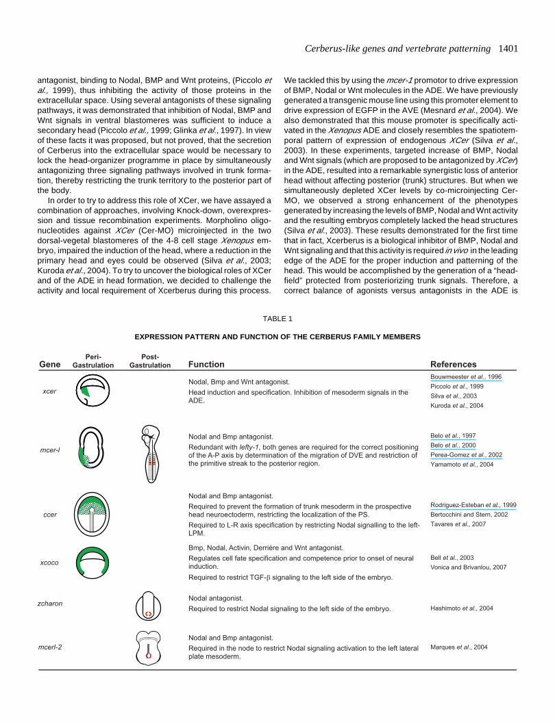
Cerberus-related proteins have been identified in other verte-brate species (see Table 1): mouse cerberus-like gene (cerl-1;Belo et al., 1997; Biben et al., 1998; Shawlot et al., 1998), chickCerberus (cCer; Rodriguez-Esteban et al., 1999; Yokouchi et al.,1999; Zhu et al., 1999), Xenopus Coco (Bell et al., 2003),zebrafish Charon (Hashimoto et al., 2004) and mouse Cerberus-like-2 (Cerl–2; Marques et al., 2004), and are now grouped in theCerberus/Dan gene family. Xenopus XCer, chick cCer andmouse Cerl-1 genes are syntenic (www.metazome.net) and, atperi gastrulation stages, are expressed in topological equivalentembryonic structures, such as the anterior endomesoderm, hypo-blast and anterior visceral endoderm, respectively (Bouwmeesteret al., 1996; Foley et al., 2000; Belo et al., 1997). In contrast,Xenopus coco was found to be expressed during pre-gastrulastages exclusively in the animal pole. At gastrula stages, cocotranscripts can be detected in both the dorsal and ventral marginalzone as well as in the animal cap ectoderm (Bell et al., 2003). Byearly neurulation stages, mouse Cerl-1 and chick cCer transcriptsare also detected in the anterior definitive mesendoderm (Belo etal., 1997; Rodriguez-Esteban et al., 1999). But at later stages,during somitogenesis, Cerberus-related genes display very dis-tinct expression patterns. XCer expression is no longer observed.Mouse Cerl-1 transcripts are found in the rostral half of the twonewly formed somites and rostral presomitic mesoderm. ChickcCer is expressed in the left paraxial and lateral plate mesoderm.Zebrafish Charon and mouse Cerl-2 are both expressed aroundthe node region but with a remarkable difference between theirexpression patterns: while Charon has a symmetric domain, Cerl-2 becomes strongly expressed on the right side (Bouwmeester etal., 1996; Belo et al., 1997; Rodriguez-Esteban et al., 1999;Hashimoto et al., 2004; Marques et al., 2004). Very recently,Xcoco was shown to be expressed bilaterally in the posteriorparaxial mesoderm during neurula stages (Vonica and Brivanlou,2007).
Cerberus-related genes belong to the cysteine-knot superfam-ily and encode for small secreted proteins (from 185 a.a., Cerl-2,to 272 a.a., Xcer and cerl-1) with a signal peptide at the N-terminaland a Cystein-Rich Domain (CRD) containing 9 cysteines at theC-terminal region. After secretion these proteins are proteolyti-cally cleaved and form active dimmers (Shawlot et al., 1998;Piccolo et al., 1999). Sequence analysis revealed that thesemolecules share a significant level of similarity in the CRD but
outside of this region, towards the N-terminus, they display verylittle homology, (Marques et al., 2004; Hashimoto et al., 2004).The spacing of cysteines in the CRD resembles a motif termed thecysteine-knot (McDonald and Hendrickson, 1993; Isaacs, 1995),which is also found in a number of cytokines such as BMP, NGF,PDGF, among others. The CRD of Cerberus-related proteinscontains the C-X-G-X-C motif, conserved in all cysteine-knotproteins, and 3 of the 4 additional cysteines that are also presentin NDP and mucins. This domain is essential for the biologicalactivity of the Cerberus molecules, which have been shown to besecreted multivalent antagonists that bind Nodal, BMP and Wntproteins, and probably inhibit their activity in the extracellularspace (Belo et al., 1997; Hsu et al., 1998; Piccolo et al., 1999; Beloet al., 2000; Marques et al., 2004)
Cerberus in anterior-posterior patterning
Embryological and genetic studies have provided evidence forthe existence of distinct vertebrate head and trunk organizers(Spemann, 1931; Thomas and Beddington, 1996; Belo et al.,1997; Bouwmeester and Leyns, 1997; Schneider and Mercola,1999). The isolation and study of Xenopus cerberus (Xcer;Bouwmeester et al., 1996), a novel secreted factor with stronghead inducing activity expressed in the anterior dorsal endoderm(ADE), was the first report that pointed to the possible role of thisregion in the induction of the anterior head. The topographicalequivalent of this region in the mouse embryo, the anteriorvisceral endoderm (AVE), has also been implicated in anteriorspecification (Thomas and Beddington, 1996). The isolation of amouse cerberus-like gene (cerl-1; Belo et al., 1997; Biben et al.,1998; Shawlot et al., 1998), expressed in the AVE before andduring gastrulation, in a region underlying the prospective anteriorneuroectoderm, underscored the inductive role of this region inthe mouse embryo.
Microinjection of Xcer mRNA in ventral blastomeres results inthe induction of secondary heads. Those included fore andmidbrain, eye, cement gland and olfactory placodes (Bouwmeesteret al., 1996). Xenopus animal cap explants are widely used as anassay system for studying cell differentiation induced by microin-jection of mRNAs encoding for specific gene products, sinceuninjected animal caps normally only give rise to epidermis. Inanimal cap explants, microinjection of XCer mRNA inducesanterior central nervous system (CNS) markers such as Otx2, butnot more posterior ones like Engrailed-2 or HoxB9, consistentwith the lack of brain structures posterior to the midbrain in thegenerated ectopic heads. Endodermal (endodermin) and heart(Nkx-2.5) markers are also upregulated in the animal cap experi-ments (Bouwmeester et al., 1996). In some cases, the inductionof those head-like structures was accompanied by duplication ofthe heart and liver. This fact could be related to the ability ofXCerberus mRNA to induce the referred endodermal and heartmarkers. XCerberus is expressed in the yolky cells that form theleading edge of the Xenopus gastrulating endoderm. This cellpopulation will eventually give rise to foregut and midgut, includ-ing the liver, as observed in cell lineage tracing experiments withDiI (Bouwmeester et al., 1996).
In an effort to determine the genetic and biochemical basis ofthe head induction by XCer, Piccolo and colleagues demon-strated that Cerberus functions as a multivalent growth-factor
Cerberus-like genes and vertebrate patterning 1401
antagonist, binding to Nodal, BMP and Wnt proteins, (Piccolo etal., 1999), thus inhibiting the activity of those proteins in theextracellular space. Using several antagonists of these signalingpathways, it was demonstrated that inhibition of Nodal, BMP andWnt signals in ventral blastomeres was sufficient to induce asecondary head (Piccolo et al., 1999; Glinka et al., 1997). In viewof these facts it was proposed, but not proved, that the secretionof Cerberus into the extracellular space would be necessary tolock the head-organizer programme in place by simultaneouslyantagonizing three signaling pathways involved in trunk forma-tion, thereby restricting the trunk territory to the posterior part ofthe body.
In order to try to address this role of XCer, we have assayed acombination of approaches, involving Knock-down, overexpres-sion and tissue recombination experiments. Morpholino oligo-nucleotides against XCer (Cer-MO) microinjected in the twodorsal-vegetal blastomeres of the 4-8 cell stage Xenopus em-bryo, impaired the induction of the head, where a reduction in theprimary head and eyes could be observed (Silva et al., 2003;Kuroda et al., 2004). To try to uncover the biological roles of XCerand of the ADE in head formation, we decided to challenge theactivity and local requirement of Xcerberus during this process.
We tackled this by using the mcer-1 promotor to drive expressionof BMP, Nodal or Wnt molecules in the ADE. We have previouslygenerated a transgenic mouse line using this promoter element todrive expression of EGFP in the AVE (Mesnard et al., 2004). Wealso demonstrated that this mouse promoter is specifically acti-vated in the Xenopus ADE and closely resembles the spatiotem-poral pattern of expression of endogenous XCer (Silva et al.,2003). In these experiments, targeted increase of BMP, Nodaland Wnt signals (which are proposed to be antagonized by XCer)in the ADE, resulted into a remarkable synergistic loss of anteriorhead without affecting posterior (trunk) structures. But when wesimultaneously depleted XCer levels by co-microinjecting Cer-MO, we observed a strong enhancement of the phenotypesgenerated by increasing the levels of BMP, Nodal and Wnt activityand the resulting embryos completely lacked the head structures(Silva et al., 2003). These results demonstrated for the first timethat in fact, Xcerberus is a biological inhibitor of BMP, Nodal andWnt signaling and that this activity is required in vivo in the leadingedge of the ADE for the proper induction and patterning of thehead. This would be accomplished by the generation of a “head-field” protected from posteriorizing trunk signals. Therefore, acorrect balance of agonists versus antagonists in the ADE is
Gene Peri-
Gastrulation Post-
Gastrulation secnerefeR noitcnuF
xcer Nodal, Bmp and Wnt antagonist. Head induction and specification. Inhibition of mesoderm signals in the ADE.
Bouwmeester et al., 1996 Piccolo et al., 1999 Silva et al., 2003 Kuroda et al., 2004
mcer-l
Nodal and Bmp antagonist. Redundant with lefty-1, both genes are required for the correct positioning of the A-P axis by determination of the migration of DVE and restriction of the primitive streak to the posterior region.
Belo et al., 1997 Belo et al., 2000 Perea-Gomez et al., 2002 Yamamoto et al., 2004
ccer
Nodal and Bmp antagonist. Required to prevent the formation of trunk mesoderm in the prospective head neuroectoderm, restricting the localization of the PS. Required to L-R axis specification by restricting Nodal signalling to the left-LPM.
Rodriguez-Esteban et al., 1999 Bertocchini and Stern, 2002 Tavares et al., 2007
xcoco
Bmp, Nodal, Activin, Derriére and Wnt antagonist. Regulates cell fate specification and competence prior to onset of neural induction. Required to restrict TGF-β signaling to the left side of the embryo.
Bell et al., 2003 Vonica and Brivanlou, 2007
zcharonNodal antagonist. Required to restrict Nodal signaling to the left side of the embryo. Hashimoto et al., 2004
mcerl-2 Nodal and Bmp antagonist. Required in the node to restrict Nodal signaling activation to the left lateral plate mesoderm.
Marques et al., 2004
TABLE 1
EXPRESSION PATTERN AND FUNCTION OF THE CERBERUS FAMILY MEMBERS

1402 J.A. Belo et al.
crucial for head formation.Due to this relevant role of Xcer and the ADE in the process of
head induction, isolation and study of its mouse homologue wouldbe of major importance for the understanding of this developmentalmechanism in the mammalian embryo. We and others haveisolated the mouse Cerberus-like gene (cerl-1; Belo et al., 1997;Biben et al., 1998; Shawlot et al., 1998). This gene is expressed inthe AVE by E5.5 and later is gradually displaced by the emerginganterior definitive endoderm. Animal cap experiments demon-strated that this mouse gene is able to induce the same set ofsignals as the XCer homologue (Belo et al., 1997; Biben et al.,1998), but is not able to generate the characteristic ectopic headswhen microinjected in ventral vegetal blastomeres (Belo et al.,1997). These inductive activities of mCer-1 and Xcer in animalcaps are characteristic of inhibition of BMP signaling (Sasai et al.,1996). Using biochemical and functional assays, we demonstratedthat in fact cerl-1 binds to and inhibits both BMP and Nodal signals,but unlike its Xenopus counterpart, is not able to bind to Wntproteins (Belo et al., 2000). Furthermore, tissue recombinationexperiments demonstrated that the expression of Otx2 was main-tained in ectoderm explants recombined with Cerl-1-expressingsomitic-presomitic mesoderm (Shawlot et al., 1998).
In summary, cerl-1 is expressed in the AVE, in a region under-lying the prospective anterior neuroectoderm, has neural inducingabilities in Xenopus experiments and is able to maintain Otx2expression in mouse explants.
Otx2 and Lim1 are genes expressed in the AVE and anteriorneuroectoderm. Generated KO mouse lines demonstrated theirrequirement for the proper formation of anterior head structures(Acampora et al., 1995; Matsuo et al., 1995; Shawlot and Behringer,1995). The AVE is the topological equivalent of the Xenopus ADEand has been implicated in anterior specification (Thomas andBeddington, 1996; Belo et al., 1997). This role was supported bythe finding that chimeric mouse embryos composed of a combina-tion of wild-type epiblast, with mutant extraembryonic tissues(namely the AVE) lacking either Otx2 or Lim1, the head is notproperly formed (Rhinn et al., 1998; Shawlot et al., 1999). In lightof all these properties, cerl-1 was well positioned to be a crucialplayer of the head organizer programme.
In order to study its role in development, we have generated atargeted inactivation of cerl-1 in the mouse (Belo et al., 2000;Shawlot et al., 2000; Stanley et al., 2000). Surprisingly however, innone of the three generated KO mouse lines an abnormal head orembryonic axis defects were observed, arguing against a previ-ously anticipated essential role of cerl-1 in early development.
Cerl-1 has been demonstrated to have strong anti-BMP activity(Belo et al., 1997; Biben et al., 1998). Other secreted factors havealso been shown to share this biochemical activity: chordin, nog-gin, and follistatin (Iemura et al., 1995; Piccolo et al., 1996;Zimmerman et al., 1996; Piccolo et al., 1999). This antagonismgenerates a graded inhibition of ventral BMP signaling which isessential for dorsoventral patterning and neural induction in thevertebrate embryo (De Robertis and Sasai, 1996). Consideringthese observations we hypothesized that some other genes mightbe compensating for the lack of function of cerl-1 in the KO mouse.This phenomenon has been observed with the BMP inhibitorsChordin and Noggin, as described previously (Bachiller et al.,2000). Both genes are expressed in the node of the mouse embryo,at late gastrula stage. Later they are co-expressed at the level of
the notochordal and prechordal plates. Remarkably, double ho-mozygous chordin;noggin mutants present synergistic defects atthe level of the forebrain development (Bachiller et al., 2000).These defects were not induced by the single mutations alone(McMahon et al., 1998; Brunet et al., 1998; Bachiller et al., 2003),meaning that some compensation was occurring when only oneBMP inhibitor was missing.
This evidence led us to test whether noggin or goosecoid(McMahon et al., 1998; Brunet et al., 1998; Yamada et al., 1995)could compensate for the lack of function of cerl-1 in the mouse.Both are involved in BMP signaling inhibition and are coexpressedwith cerl-1 at the level of the prechordal plate mesendoderm, andthe latter also in the AVE (Belo et al., 1998). In Xenopus experi-ments, Xgsc represses the expression of BMP-4 in the marginalzone (Fainsod et al., 1994) and can induce the expression ofchordin (Sasai et al., 1994). However, generated double mutantsfor both cerl-1;noggin and cerl-1;gsc did not display any synergis-tic phenotype (Borges et al., 2001, 2002), indicating that neithernoggin nor goosecoid compensate for cerl-1 loss-of-function andthat these genes do not interact genetically. These results could beindicative that the true biological relevance of cerl-1 in the mousemight not be its anti-BMP activity.
As referred before, mouse cerl-1 has a strong anti-Nodal activity(Belo et al., 2000). Studies using the chick homologue Caronte (orchick Cerberus, cCer ), demonstrated that its expression in thehypoblast (the chick equivalent of the AVE in the mouse and of theADE in Xenopus) is necessary for head formation (Bertocchini andStern, 2002), by preventing the formation of trunk mesoderm in theprospective head neuroectoderm via its anti-Nodal activity. Lefty-1 is another nodal secreted antagonist and is expressed in AVEcells (Meno et al., 1997). Like cerl-1 KO embryos, generatedmouse mutants lacking lefty-1 also lack gastrulation phenotypes(Meno et al., 1998; Belo et al., 2000). However, when cerl-1;lefty1double mutant animals were generated, development of the result-ing embryos was greatly impaired due to excessive and unregu-lated nodal activity (Perea-Gomez et al., 2002; Yamamoto et al.,2004). Yamamoto and colleagues showed that asymmetric Nodalinhibition directs Distal Visceral Endoderm (DVE) migration to ananterior position. This migration is accomplished by simulating theproliferation of visceral endodermal cells by Nodal while Lefty1 andCerl-1 determine the direction of migration by asymmetricallyinhibiting Nodal activity on the prospective anterior side (Yamamotoet al., 2004). Later, this concerted inhibition of Nodal activity in theAVE is also required in order to restrict primitive streak formationto the posterior end of mouse embryos by antagonizing Nodalsignaling (Perea-Gomez et al., 2002). Collectively, these studiesclearly show the requirement of the nodal inhibitors cerl-1 andlefty1, whose redundant activities during early development areessential for A-P axis development.
In conclusion, the results from Xcer, Cer-l and cCer studiesstrongly support their role in generating asymmetries atperigastrulation stages. In this context, Cerberus molecules emergeas major players in the establishment of the Anterior-Posterior axisof the early vertebrate embryo.
Cerberus in left-right patterning
In vertebrates, the correct development of the organs along aleft-right (L-R) plane of organization is essential for the normal

Cerberus-like genes and vertebrate patterning 1403
physiology of the living organism. Therefore, the formation of theleft-right axis of body symmetry during embryogenesis is of majorimportance. The establishment of this asymmetry was shown torequire the asymmetric activation of the Nodal signaling cascadein the left side of the body wall (for review see Hamada et al., 2002),the Left-Lateral Plate Mesoderm (L-LPM). During the course ofvertebrate evolution, this basic feature was preserved, althoughsome species specificities have diverged.
In the chick embryo, the first signal of morphological asymme-try is the tilt of Hensen’s node by the end of gastrulation (Dathe etal., 2002). The signaling molecule Sonic hedgehog (Shh) isexpressed symmetrically within the ectoderm of Hensen’s nodebefore HH4 (Hamburger and Hamilton 1951), the time at which itbecomes restricted to the left side of the node. This is followed atHH7 by the expression of Nodal on the left-side. Nodal is firstexpressed in a small domain of cells directly adjacent to the onesexpressing Shh, and then in a large domain in the lateral platemesoderm. Although Shh expression in the node is necessaryand sufficient to induce Nodal in the non-adjacent L-LPM (Pagán-Westphal et al., 1998), the exact mechanism is largely unknown.This led to the hypothesis that there was an unknown molecule inthe paraxial mesoderm (the intermediate embryonic tissue) thatwould transduce this information from the node towards the L-LPM.
The chick homologue of XCer has been isolated and studied bya number of groups (cCer; Rodriguez-Esteban et al., 1999;Yokouchi et al., 1999; Zhu et al., 1999). Besides its expression inthe hypoblast and anterior endomesoderm, cCer is also ex-pressed in the left paraxial and L-LPM. Experiments usingmisexpression, protein-soaked beads and in vitro binding studiesrevealed that cCer was necessary and sufficient to transmit theShh signal from the node to the L-LPM, leading to Nodal expres-sion and subsequent activation of Left-Right specific gene ex-pression (Rodriguez-Esteban et al., 1999; Yokouchi et al., 1999;Zhu et al., 1999). In light of these properties, this novel Cerberus-like gene was denominated Caronte (Car), after the boatman inGreek mythology who ferried the souls of the dead across theRiver Styx. Caronte has been therefore, reported to be a second-ary signal induced by Shh and repressed by Fgf8, and Carontemisexpression experiments suggested that it was sufficient toactivate Nodal in the LPM. Caronte has been proposed to act asa BMP antagonist (Rodriguez-Esteban et al., 1999; Yokouchi etal., 1999) activating Nodal expression in the left lateral platemesoderm by relieving a repressive effect of BMPs on Nodaltranscription.
However, Nodal molecules have been shown to induce theexpression of XCer (Osada et al., 2000) and mouse Cerl-1(Waldrip et al., 1998; Brennan et al., 2001), which is the oppositeof what has been reported for Caronte. In addition, Nodal expres-sion on the left side of the chicken node can be observed prior tothe onset of Caronte (Rodriguez-Esteban et al., 1999; Yokouchiet al., 1999; Zhu et al., 1999). Moreover, in the mouse embryo,Nodal expression in the L-LPM requires Nodal protein producedin the node (Saijoh et al., 2003; Yamamoto et al., 2003). All ofthese data combined leaves the relationship between Caronteand Nodal unclear, raising the possibility that the induction ofCaronte expression by Shh may be mediated by Nodal and not theother way around. Moreover, Caronte has been shown to bind toNodal (Rodriguez-Esteban et al., 1999), as the other Cerberus
family members do; so if its biological activity is also similar to its‘siblings’ it should behave as an inhibitor of nodal signaling, not anactivator.
We have re-investigated the role of Caronte, hereafter denomi-nated chicken Cerberus (cCer), in the establishment of asymmet-ric nodal signaling. We have implemented novel approaches inour study, namely electroporation of cDNAs, knock-down experi-ments using morpholino oligonucleotides against cCer, implanta-tion of Nodal protein-soaked beads and transcriptional analysis ofthe cis-regulatory regions of the cCer gene. In our experiments,cCer misexpression in the node region or on the right-LPM wasnever able to induce Nodal, whereas cCer overexpression on theleft side actually repressed Nodal. Conversely, in cCer knock-down embryos, Nodal is ectopically expressed on the right side,demonstrating that cCer acts as negative regulator of Nodalsignaling (Tavares et al., 2007). We could also observe that in factNodal is necessary and sufficient for the induction of the transcrip-tional activation of the cCer gene (Tavares et al., 2007) even whentargeted to the Right-LPM. We could indeed determine that theNodal nuclear effectors FoxH1 and SMAD elements in cCer left-side enhancer are sufficient to induce the asymmetric expressionon the left side of the chick embryo. Similar enhancer elementsare also seen in the promoters of other asymmetrically expressedNodal responsive genes such as Leftys, Pitx2, and Nodal itself,and have been reported to be responsible for the asymmetricexpression of these genes. As in the case of the mouse node(Saijoh et al., 2003), Nodal signaling in the Hensen’s node is ableto induce Nodal expression in the chick L-LPM (Tavares et al.,2007). However, the induction of cCer expression by Nodalprotein is faster then the induction of the Nodal one. Therefore,Nodal protein released by the node induces first cCer in a moremedial domain (left paraxial mesoderm) and later Nodal in a morelateral domain (left lateral plate mesoderm).
Some of this data is reminiscent of the interactions postulatedin reaction-diffusion models (Turing, 1990; Meinhardt and Gierer,2000). Some reaction-diffusion models propose the following fourinteractions between an activator and an inhibitor: the activatoractivates its own production; the activator activates an inhibitor;the inhibitor blocks the auto-activation of the activator; the inhibi-tor acts long-range to restrict the effective range of the activator.The relationship between Nodal and its antagonists Lefty1 and 2in the mouse embryo has recently been proposed to be a reaction-diffusion type, a self-enhancement and lateral-inhibition type(Nakamura et al., 2006). cCer may therefore share some of theregulatory properties of Leftys that have been described recently.
In conclusion, this data strongly supported the view that Nodalis the intermediary signal that transfers the asymmetric informa-tion from the node to the lateral plate (Pagán-Westphal and Tabin,1998) and that the role of cCer is to confine Nodal signaling to theleft side through a negative feedback mechanism, preventingNodal signals from crossing to the right side of the chicken embryo(Tavares et al., 2007). Furthermore, given the similarities be-tween the expression patterns and functions of chick Cer andmouse Lefty2 (for a review, see Juan and Hamada, 2001), weproposed that in the chick, cCer has taken the role of mouse’sLefty2 in left-right patterning, and acts in addition to the midlinebarrier to confine Nodal signaling to the left side.
Recently, novel cerberus molecules have been isolated inmouse (cerberus-like2; Cerl-2 ) and zebrafish (charon; Marques

1404 J.A. Belo et al.
et al., 2004; Hashimoto et al., 2004). Cerl-2 is closely related tomouse cerberus-like (I=35%, P=47%), cCerberus (I=34%,P=51%), Xcoco (I=38%, P=53%), zebrafish charon (I=33%,P=55%) and to human hCer-2 (I=57%, P=65%). Functionalanalysis revealed that both genes play an important role in theestablishment of Left-Right asymmetry (Marques et al., 2004;Hashimoto et al., 2004). Cerl-2 displays a unique asymmetricexpression domain around the mouse node, being asymmetri-cally expressed on the right side. cerl-2 transcripts can be firstlydetected in a horse-shoe shaped pattern in the perinodal regionof the E7.0 mouse embryo, resembling Nodal expression at thisstage (Lowe et al., 1996; Collignon et al., 1996). However, bylate headfold stage, expression of cerl-2 begins to decrease inintensity on the left side and by E8.0 is mostly detected in theright side of the node assuming a complementary expressionpattern to the one of Nodal at this stage (Lowe et al., 1996;Collignon et al., 1996). We demonstrated that Cerl-2 is a potentantagonist of Nodal signaling by directly binding to Nodalprotein (Marques et al., 2004). Analysis of a generated mutantmouse line in which we deleted the Cerl-2 gene revealed that 1/3 of the cerl-2-/- newborns died within the first 48 hours afterbirth and displayed left pulmonary isomerism, thoracic situsinversus and cardiovascular malformations, being the lattertheir probable cause of death. The mutant animals that survivedbecome normal adults although 1/4 die between weaning ageand 3 months old, most of them showing heterotaxia of theabdominal organs (Marques et al., 2004). When analyzedduring early somitogenesis, we detected that the Cerl-2 loss offunction leads to bilateral expression of the left side-specificgenes Nodal, Lefty2 and Pitx2 (Marques et al., 2004).
In light of this data, the role of Cerl-2 is to restrict nodalactivity to the left side of the node, preventing additionalactivation of Nodal, Lefty-2 and Pitx-2 in the R-LPM. In theabsence of Cerl-2 antagonistic activity on the node, Nodal cannow also be activated in the R-LPM, leading to bilateral expres-sion of this genetic cascade. These observations highlight the
function of cerl-2 in the tight regulation of Nodal activity in thenode, indicating that Cerl-2 plays an important role in the earlysymmetry breaking events that take place in the node.
In mice, a model has been proposed in which monociliaprotruding from cells in the late gastrula node generate a left-right flow of extracellular fluid that results in the establishmentof asymmetric gene expression (Nonaka et al., 1998). This“nodal flow” is disturbed in several mouse mutant lines thatdisplay L-R phenotypes, which support its role in the establish-ment of correct left-right body axis (Nonaka et al., 1998;Marszalek et al., 1999). The work on Cerl-2 suggests that, in themouse, L-R asymmetry is controlled by a double-assurancemechanism consisting of two systems working in parallel, thefirst relying on the leftward nodal-cilia flow, and the second, onthe antagonism between Cerl-2 and Nodal.
In the zebrafish embryo, Kupffer’s vesicle has been demon-strated to be the functional equivalent of the mouse node(Essner et al., 2002, 2005), with motile cilia that create adirectional fluid flow just prior to the onset of asymmetric geneexpression in lateral cells. Disruption of this flow impairs correctL-R patterning (Essner et al., 2005). Zebrafish Charon is ex-pressed in the Kupffer’s vesicle at the 10-somite stage (14 hpf;Hashimoto et al., 2004). Charon expression pattern assumes ahorse shoe-shape, resembling the one from Cerl-2 at initialstages (Marques et al., 2004). But in zebrafish, expression ofsouthpaw (the equivalent of mouse Nodal) and Charon are notasymmetric in the vicinity of the Kupffer’s vesicle. Functionalassays using microinjection of Charon mRNA in zebrafishembryos demonstrated that the dorsalizing activity of all of thethree known zebrafish Nodal-related molecules (southpaw,Cyclops and squint; Sampath et al., 1998; Long et al., 2003;Rebagliati et al., 1998) can be inhibited by Charon. Similarly tothe phenotype of the cerl-2 mutants, down-regulation of Charonby morpholino oligonucleotides lead to disturbs in the correctestablishment of the Left-Right axis. Those included bilateralexpression in the LPM of the left side-specific genes southpaw,cyclops, lefty2 and pitx2 (Hashimoto et al., 2004), and defectsin asymmetric heart development.
Recently it has been reported that Xenopus Coco also playsan important role in the establishment of the L-R axis of theXenopus embryo (Vonica et al., 2007). Coco has been found tobe a nodal antagonist, that is expressed bilaterally in theposterior paraxial mesoderm at neurula stage, where it sharesthe same expression pattern with Xnr1 and derriére. Experi-ments using Coco morpholinos demonstrated that Coco isrequired exclusively on the right side and Xnr1 on the left side,for proper Left-Right patterning (Vonica et al., 2007). Takentogether, these results suggest that Charon and Coco might betrue orthologues of cerl-2. And as in the case of the mouseembryo, the antagonistic activity of the Cerberus moleculesCharon and Coco, against Southpaw and Xnr1 (Nodal mol-ecules), plays an important role in the establishment of the L-Rbody axis in zebrafish and Xenopus embryos.
Although chick Cer, Xenopus Coco, zebrafish Charon andmouse Cerl-2 have different expression patterns, the Cerberus-like proteins encoded by these genes seem to have an evolu-tionary conserved mechanism of Nodal antagonism in thevertebrate embryo. This conserved mechanism, crucial forcorrect L-R development, is to restrict Nodal signaling in the left
Posterior Anterior
Left Right
Nodal Nodal
Cerberus
N o d a l
S i g n a l i n g
N o d a l
S i g n a l i n g
Asymmetric
Organ Distribution
Head formation
Fig. 1. Cerberus prevents Nodal signaling in the anterior and right
sides of the vertebrate embryo in order to promote head formation andasymmetric distribution of the internal organs.

Cerberus-like genes and vertebrate patterning 1405
side of the vertebrate embryo.
Final remarks
10 years after the isolation of Xcerberus (Bouwmeester etal., 1996), the founding member of this still increasing family,Cerberus-like molecules emerge as key regulators of Nodalsignaling. Although not signaling proteins per se, they are veryimportant regulatory molecules, playing a primordial role inrestricting Nodal signaling both in space and time. This datasuggests that one efficient way of creating asymmetries in theearly developing embryo is by restricting the activation ofcertain signals in a specific region in time. It is becoming moreand more evident that biological processes like the generationof asymmetries arise from complex regulatory mechanisms,and inhibitory molecules like the Cerberus-family members arenot secondary players in these pathways, but can now claimtheir intrinsic importance as generators of developmental pro-cesses. Our observations confirm the increasing body of evi-dence that patterning of the vertebrate embryo results mostlyfrom antagonistic protein-protein interactions in the extracellu-lar space (Fig. 1), being the Cerberus-like family of secretedinhibitors a crucial player in this process.
Bearing this in mind, and in order to further characterize themolecular mechanisms that play a role in the early A/P axisestablishment, we have been using Cerberus-family members asa ‘bait’ to uncover potential new players. We have performed adifferential screening using a generated transgenic mouse line inwhich EGFP is expressed in the AVE, under the control of thepromoter region of the Cerl-1 gene. Gene expression profilingusing GeneChips ® (Affymetrix ®) identified novel differentiallyexpressed transcripts at the very early stages of A-P axis estab-lishment (Mário Filipe, unpublished), some of them now currentlybeing studied at our laboratory (Silva et al., 2006; Filipe et al.,2006; Salgueiro et al., 2006). We expect that this approach willallow us to learn more about asymmetry generation, tissuepatterning and specification during early vertebrate developmentand that this knowledge can be applied in the future to regenera-tive medicine.
AcknowledgementsJA Belo would like to thank Eddy M. De Robertis for his guidance and
generosity, at whose laboratory part of this body of work was initiated. Wethank H Steinbeisser and S Piccolo for help through the years; A Tavaresand H Steinbeisser for critically reading of this manuscript; FCT for PDfellowships to AT Tavares, V Teixeira and AC Borges, and for PhDfellowships to M. Filipe, AC Borges, L. Gonçalves, M Vitorino, AMSalgueiro, M Bento and A. C. Silva; IEFP for fellowships to AC Borges, L.Gonçalves, M Vitorino, AM Salgueiro and M Bento; FLAD for a fellowshipto S Marques. This work was supported by research grants from C.R.U.P,F.C.T. and IGC/Fundação Calouste Gulbenkian to J. A. Belo, where he isa Principal Investigator.
References
ACAMPORA, D., MAZAN, S., LALLEMAND, Y., AVANTAGGIATO, V., MAURY, M.,SIMEONE, A. and BRÛLET, P. (1995). Forebrain and midbrain regions aredeleted in Otx2- /- mutants due to a defective anterior neurectoderm specifica-tion during gastrulation. Development 121: 3279–3290.
BACHILLER, D., KLINGENSMITH, J., KEMP, C.,BELO, J.A., ANDERSON, MAY,S.R., MCMAHON, J.A., MCMAHON, A.P., HARLAND, R.M., ROSSANT, J. and
DE ROBERTIS, E.M. (2000). The organizer factors Chordin and Noggin arerequired for mouse forebrain development. Nature. 403: 658-661.
BELL, E., MUNOZ-SANJUAN, I., ALTMANN, C.R., VONICA, A. and BRIVANLOU,A.H. (2003). Cell fate specification and competence by Coco, maternal BMP,TGFbeta and Wnt inhibitor. Development 130:1381-1389.
BELO, J.A., BOUWMEESTER, T., LEYNS, L., KERTESZ, N., GALLO, M.,FOLLETTIE, M. and DE ROBERTIS, E.M. (1997). Cerberus-like is a secretedfactor with neutralizing activity expressed in the anterior primitive endoderm ofthe mouse gastrula. Mech. Dev. 68: 45-57.
BELO, J.A., LEYNS, L., YAMADA, G. and DE ROBERTIS, E.M. (1998). Theprechordal midline of the chondrocranium is defective in Goosecoid-1 mousemutants. Mech. Dev. 72: 15-25.
BELO, J.A., BACHILLER, D., AGIUS, E., KEMP, C., BORGES, A.C., MARQUES,S., PICCOLO, S. and DE ROBERTIS, E.M. (2000). Cerberus-like is a secretedBMP and Nodal antagonist not essential for mouse development. Genesis 26:265-270.
BERTOCCHINI, F. and STERN, C.D. (2002). The hypoblast of the chick embryopositions the primitive streak by antagonizing nodal signaling. Dev. Cell 3: 735-744.
BIBEN, C., STANLEY, E., FABRI, L., KOTECHA, S., RHINN, M., DRINKWATER,C., LAH, M., WANG, C.C., NASH, A., HILTON, D., ANG, S.L., MOHUN, T., andHARVEY, R.P. (1998). Murine Cerberus homologue mCer-1: a candidateanterior patterning molecule. Dev. Biol. 194: 135-151.
BLUM, M., GAUNT, S.J., CHO, K.W.Y., STEINBEISSER, H., BLUMBERG, B.,BITTNER, D. and DE ROBERTIS, E.M. (1992). Gastrulation in the mouse: therole of the homeobox gene goosecoid. Cell 69: 1097–1106.
BORGES, A.C., MARQUES, S. and BELO, J.A. (2001) The BMP antagonistscerberus-like and noggin do not interact during mouse forebrain development.Int. J. Dev. Biol. 45: 441-443.
BORGES, A.C., MARQUES, S. and BELO, J.A. (2002). Goosecoid and cerberus-like do not interact during mouse embryogenesis. Int. J. Dev. Biol. 46: 259-262.
BOUWMEESTER, T., KIM, S., SASAI, Y., LU, B. and DE ROBERTIS, E.M. (1996).Cerberus is a head-inducing secreted factor expressed in the anterior endo-derm of Spemann’s organizer. Nature 382: 595-601.
BRENNAN, J., LU, C.C., NORRIS, D.P., RODRIGUEZ, T.A., BEDDINGTON, R.S.and ROBERTSON, E.J. (2001). Nodal signaling in the epiblast patterns theearly mouse embryo. Nature 411: 965-969.
BRUNET, L.J., MCMAHON, J.A., MACMAHON, A.P. and HARLAND, R.M. (1998).Noggin, Cartilage Morphogenesis and joint formation in the mammalian skel-eton. Science 280: 1455-1457.
CHO, K.W.Y., BLUMBERG, B., STEINBEISSER, H. and DE 7ROBERTIS, E.M.(1991). Molecular nature of Spemann’s organizer: the role of the Xenopushomeobox gene goosecoid. Cell 67: 1111–1120.
COLLIGNON, J., VARLET, I. and ROBERTSON, E.J. (1996). Relashionshipbetween asymmetric nodal expression and the direction of embryonic turning.Nature 381: 155-158.
DATHE, V., GAMEL, A., MANNER, J., BRAND-SABERI, B. and CHRIST, B. (2002).Morphological left–right asymmetry of Hensen’s node precedes the asymmetricexpression of Shh and Fgf8 in the chick embryo. Anat. Embryol. 205: 343–354.
DE ROBERTIS, E.M. and SASAI, Y. (1996). A common plan for dorsoventralpatterning in Bilateria. Nature 380: 37-40.
DE ROBERTIS, E.M. and KURODA, H. (2004). Dorsal-ventral patterning andneural induction in Xenopus embryos. Annual Review of Cell and Developmen-tal Biology. 20: 285-308.
ESSNER, J. J., VOGAN, K. J., WAGNER, M. K., TABIN, C. J., YOST, H. J. andBRUECKNER, M. (2002). Conserved function for embryonic nodal cilia. Nature418: 37-38.
ESSNER, J. J., AMACK, J. D., NYHOLM, M. K., HARRIS, E. B. and YOST, H. J.(2005). Kupffer’s vesicle is a ciliated organ of asymmetry in the zebrafishembryo that s initiates left-right development of the brain, heart and gut.Development 132: 1247-1260.
FAINSOD, A., STEINBEISSER, H. and DE ROBERTIS, E. M. (1994). On thefunction of BMP-4 in patterning the marginal zone of the Xenopus embryo.EMBO J. 13: 5015-5025.
FILIPE, M., GONÇALVES, L., BENTO, M., SILVA, A.C. and BELO, J.A. (2006).Comparative expression of mouse and chicken Shisa homologues during early

1406 J.A. Belo et al.
development. Dev. Dyn. 235(9): 2567-73.
FOLEY, A.C., SKROMME, I.S., and STERN, C.D. (2000). Reconciling differentmodels of forebrain induction and patterning: a dual role for the hypoblast.Development 127: 3839–3854.
GLINKA, A., WU, W., DELIUS, H., MONAGHAN, A.P., BLUMENSTOCK, C. andNIEHRS, C. (1998). Dickkopf-1 is a member of a new family of secreted proteinsand functions in head induction. Nature 391: 357–362.
HAMADA, H., MENO, C., WATANABE, D., and SAIJOH, Y. (2002). Establishmentof vertebrate left-right asymmetry. Nat. Rev. Genet. 3:103-113.
HAMBURGER, V. and HAMILTON, H.L. 1951. A series of normal stages in thedevelopment of the chick embryo. J. Morphol. 88: 49-92.
HASHIMOTO, H., REBAGLIATI, M., AHMAD, N., MURAOKA, O., KUROKAWA, T.HIBI, M. and SUZUKI, T. (2004). The Cerberus/Dan-family protein Charon is anegative regulator of Nodal signaling during left-right patterning in zebrafish.Development 131:1741-1753.
HSU, D.R., ECONOMIDES, A.N., WANG, X., EIMON, P.M., HARLAND, R.M.(1998). The Xenopus dorsalizing factor gremlin identifies a novel family ofsecreted proteins that antagonize BMP activities. Mol Cell 1: 673-683.
IEMURA, S., YAMAMOTO, T.S., TAKAGI, C., UCHIYAMA, H., NATSUME, T.,SHIMASAKI, S., SUGINO, H. and UENO, N. (1995). Direct binding of follistatinto a complex of bone-morphogenetic protein and its receptor inhibits ventral andepidermal cell fates in early Xenopus embryo. Proc. Natl. Acad. Sci. USA 95:9337-9342.
ISAACS, N. W. (1995). Cystine knots. Curr. Opinion Struct. Biol. 5: 391–395.
IZPISÚA-BELMONTE, J.C., DE ROBERTIS, E.M., STOREY, K.G. and STERN,C.D. (1993). The homeobox gene goosecoid and the origin of organizer cells inthe early chick blastoderm. Cell 74: 645–659.
JUAN, H. and HAMADA, H. (2001). Roles of nodal-lefty regulatory loops inembryonic patterning of vertebrates. Genes to Cells 6: 923-930.
KURODA, H., WESSELY, O., and DE ROBERTIS, E.M. (2004). Neural induction inXenopus: Requirement for ectodermal and endomesodermal signals via chor-din, noggin, β-catenin and cerberus. PLoS Biol. 2: 623–634.
LONG, S., AHMAD, N. and REBAGLIATI, M. (2003). The zebrafish nodal-relatedgene southpaw is required for visceral and diencephalic left-right asymmetry.Development 130: 2303-2316.
LOWE, L.A., SUPP, D.M., SAMPATH, K., YOKOYAMA, T., WRIGHT, C.V., POT-TER, S.S., OVERBEEK, P. and KUEHN, M.R. (1996). Conserved left-rightasymmetry of nodal expression and alterations in murine situs inversus. Nature381: 158-161.
MARQUES, S., BORGES, A.C., SILVA, A.C., FREITAS, S., CORDENONSI, M. andBELO, J.A. (2004). The activity of the Nodal antagonist Cerl-2 in the mouse nodeis required for correct L/R body axis. Genes and Development 18: 2342-2347.
MARSZALEK, J. R., RUIZ-LOZANO, P., ROBERTS, E., CHIEN, K. R. andGOLDSTEIN, L. S. (1999). Situs inversus and embryonic ciliary morphogenesisdefects in mouse mutants lacking the KIF3A subunit of kinesin-II. Proc. NatlAcad. Sci. USA 96: 5043–5048.
MATSUO, I., KURATANI, S., KIMURA, C., TAKEDA, N. and AIZAWA, S., (1995).Mouse Otx2 functions in the formation and patterning of rostral head. GenesDev. 9: 2646–2658.
MCDONALD, N. Q., and HENDRICKSON, W. A. (1993). A structural superfamily ofgrowth factors containing a cystine knot motif. Cell 73: 421–424.
MCMAHON, J.A., TAKADA, S., ZIMMERMAN, L.B., FAN, C., HARLAND, R.M. andMCMAHON, A.P. (1998). Noggin-mediated antagonism of BMP signalling isrequired for growth and patterning of the neural tube and somite. Genes Dev.12: 1438-1452.
MEINHARDT, H. and GIERER, A. (2000). Pattern formation by local self-activationand lateral inhibition. BioEssays 22: 753-760.
MENO, C., ITO, Y., SAIJOH, Y., MATSUDA, Y., TASHIRO, K., KUHARA, S. andHAMADA, H. (1997). Two closely-related left–right asymmetrically expressedgenes, lefty-1 and lefty-2: Their distinct expression domains, chromosomallinkage and direct neuralizing activity in Xenopus embryos. Genes Cells 2: 513–524.
MENO, C., SHIMONO, A., SAIJOH, Y., YASHIRO, K., MOCHIDA, K., OHISHI, S.,NOJI, S., KONDOH, H., and HAMADA, H. (1998). Lefty-1 is required for left–right determination as a regulator of lefty-2 and nodal. Cell 94: 287–297.
MESNARD, D., FILIPE, D., BELO, J.A. and ZERNICKA-GOETZ, M. (2004).Emergence of the anterior-posterior axis after implantation relates to the re-orienting symmetry of the mouse embryo rather than the uterine axis. CurrentBiology 14, 184- 196.
NAKAMURA, T., MINE N., NAKAGUCHI A., MOCHIZUKI A., YAMAMOTO M.,YASHIRO K., MENO C. AND HAMADA H. (2006) Generation of robust left-rightasymmetry in the mouse embryo requires a self-enhancement and lateral-inhibition system. Dev Cell 11: 495-504.
NONAKA, S., TANAKA, Y., OKADA, Y., TAKEDA, S., HARADA, A., KANAI, Y.,KIDO, M. and HIROKAWA, N. (1998) Randomization of left–right asymmetrydue to loss of nodal cilia generating leftward flow of extraembryonic fluid in micelacking KIF3B motor protein. Cell 95: 829–837.
OSADA, S.I., SAIJOH, Y., FRISCH, A., YEO, C.Y., ADACHI, H., WATANABE, M.,WHITMAN, M., HAMADA, H. and WRIGHT, C.V. (2000). Activin/Nodal respon-siveness and asymmetric expression of a Xenopus nodal-related gene con-verge on a FAST-regulated module in intron 1. Development 127: 2503-2514.
PAGÁN-WESTPHAL, S. M. and TABIN, C. J. (1998). The transfer of left-rightpositional information during chick embryogenesis. Cell 93: 25-35.
PEREA-GOMEZ, A. VELLA, F.D.J., SHAWLOT, W., OULAD-ABDELGHANI, M.,CHAZAUD, C., MENO, C., PFISTER, V., CHEN, L., ROBERTSON, E.J.,HAMADA, H., BEHRINGER, R.R. and ANG, S.L. (2002). Nodal antagonists inthe anterior visceral endoderm prevent the formation of multiple primitivestreaks. Dev. Cell. 3: 745-756.
PICCOLO, S., SASAI, Y., LU, B., and DE ROBERTIS, E.M. (1996). Dorsoventralpatterning in Xenopus: inhibition of ventral signals by direct binding of chordinto BMP-4. Cell. 86: 589-598.
PICCOLO, S., AGIUS, E., LETNS, L., BHATTACHARYYA, S., GRUNZ, H.,BOUWMEESTER, T., and DE ROBERTIS, E.M. (1999). The head inducerCerberus is a multifunctional antagonist of Nodal, BMP and Wnt signals. Nature397: 707-10.
REBAGLIATI, M. R., TOYAMA, R., HAFFTER, P. and DAWID, I. B. (1998). Cyclopsencodes a nodal-related factor involved in midline signaling. Proc. Natl. Acad.Sci. USA 95: 9932-9937.
RHINN, M., DIERICH, A., SHAWLOT, W., BEHRINGER, R. R., LE MEUR, M. andANG, S. L. (1998). Sequential roles for Otx2 in visceral endoderm and neuro-ectoderm for forebrain and midbrain induction and specification. Development125: 845-856.
RODRIGUEZ-ESTEBAN, C., CAPDEVILA, J., ECONOMIDES, A.N., PASCUAL,J., ORTIZ, A., IZPISÚA-BELMONTE, J.C. (1999). The novel Cerberus-likeprotein Caronte mediates de establishment of embryonic left-right asymmetry.Nature 401: 243-251.
SAIJOH, Y., OKI, S., OHISHI, S., and HAMADA, H. (2003). Left–right patterning ofthe mouse lateral plate requires nodal produced in the node. Dev. Biol. 256: 160-172.
SALGUEIRO, A.M., FILIPE, M. and BELO, J.A. (2006). Expression of N-acetylgalactosamine 4-sulfate 6-O-sulfotransferase during early mouse embry-onic development. Int. J. Dev. Biol. 50: 705-708.
SAMPATH, K., RUBINSTEIN, A. L., CHENG, A. M., LIANG, J. O., FEKANY, K.,SOLNICA-KREZEL, L., KORZH, V., HALPERN, M. E. and WRIGHT, C. V.(1998). Induction of the zebrafish ventral brain and floorplate requires cyclops/nodal signalling. Nature 395: 185-189.
SASAI, Y., LU, B., STEINBEISSER, H., GEISSERT, D., GONT, L. K. and DEROBERTIS, E. M. (1994). Xenopus chordin: a novel dorsalizing factor activatedby organizer-specific homeobox genes. Cell 79: 779-790.
SASAI Y., LU B., PICCOLO S., DE ROBERTIS E.M. (1996). Endoderm inductionby the organizer secreted factors Chordin and Noggin in Xenopus animal caps.EMBO J. 15: 4547-4555.
SCHULTE-MERKER, S., HAMMERSCHMIDT, M., BEUCHLE, D., CHO, K.W., DEROBERTIS, E.M. and NUSSLEIN-VOLHARD, C. (1994). Expression ofzebrafishKgoosecoid and no tail gene products in wild-type and mutant no tailembryos. Development 120: 843–852.
SHAWLOT, W. and BEHRINGER, R.R. (1995). Requirement for Lim1 in head-organizer function. Nature 374: 425–430.
SHAWLOT, W., DENG, J.M. and BEHRINGER, R.R. (1998). Expression of themouse Cerberus-related gene, Cerr1, suggest a role in anterior neural inductionand somitogeneses. Proc. Natl. Acad. Sci. USA 95:6198-6203.

Cerberus-like genes and vertebrate patterning 1407
SHAWLOT, W., WAKAMIYA, M., KWAN, K. M., KANIA, A., JESSELL, T. M. andBEHRINGER, R. R. (1999). Lim1 is required in both primitive streak-derivedtissues and visceral endoderm for head formation in the mouse. Development126: 4925-4932.
SHAWLOT, W., MIN DENG, J., WAKAMIYA, M. And BEHRINGER, R.R. (2000).Ther cerberus- related gene, Cerr1, is not essential for mouse head formation.Genesis 26:253-268.
SILVA, A.C., FILIPE, M., KUERNER, K.M.,STEINBEISSER, H. and BELO, J.A.(2003). Endogenous Cerberus activity is required for anterior head specificationin Xenopus. Development 130: 4943-4953.
SILVA, A.C., FILIPE, M., VITORINO, M., STEINBEISSER, H. and BELO, J.A.(2006). Developmental Expression of Shisa-2 in Xenopus laevis. Int. J. Dev.Biol. 50, 575-579.
SCHNEIDER, V.A. and MERCOLA, M. (1999). Spatially distinct head and heartinducers within the Xenopus organizer region. Curr. Biol. 9: 800-809.
SPEMANN, H. and MANGOLD, H. (1924). Über induction von embryonalanlagendurch implantationartfremder organisatoren. Wilhelm Roux Arch.Entwicklungsmech. Org. 100: 599–638.
SPEMANN, H. (1931). Über den anteil von implantat und wirtskeim an derorientierung und beschaffenheit der induzierten embryonalanlage. WilhelmRoux Arch. Entwicklungsmech. Org. 123: 389–517.
STACHEL, S.E., GRUNWALD, D.J. and MYERS, P. (1993). Lithium perturbationand goosecoid expression identify a dorsal specification pathway in thepregastrula zebrafish. Development 117: 1261–1274.
STANLEY, E.G., BIBEN, C., ALLISON, J., HARTLEY, L., WICKS, I.P., CAMPBELL,I. K., CKINLEY, M., BARNETT, L., KOENTGEN, F., ROBB, L. and HARVEY,R.P. (2000). Targeted insertion of a lacZ reporter gene into the mouse Cer1Locus reveals complex and dynamic expression during embryogenesis. Gen-esis. 26: 259-264.
TAVARES, A.T., ANDRADE, S., SILVA, A.C. and BELO, J.A. (2007). Cerberus isa feedback inhibitor of Nodal asymmetric signaling in the chick embryo.Development 134: 2051-2060.
THOMAS, P., BEDDINGTON, R.S.P. (1996). Anterior primitive endoderm may beresponsible for patterning the anterior neural plate in the mouse embryo. CurrBiol 6: 1487-1496.
TURING, A. M. (1990). The chemical basis of morphogenesis. 1953. Bull. Math.Biol. 52: 153-197.
VONICA, A. and BRIVANLOU, A.H. (2007). The left –right axis is regulated by theinterplay of Coco, Xnr1 and derrière in Xenopus embryos. Dev. Biol. 303: 281–294.
WALDRIP, W.R., BIKOFF, E.K., HOODLESS, P.A., WRANA, J.L. and ROBERTSON,E.J. (1998). Smad2 signalling in extraembryonic tissues determines anterior-posterior polarity of the early mouse embryo. Cell 92: 797-808.
YAMADA, G., MANSOURI, A., TORRES, M., STUART, E. T., BLUM, M., SCHULTZ,M., DE ROBERTIS, E. M. and GRUSS, P. (1995) Targeted mutation of themurine goosecoid gene results in craniofacial defects and neonathal death.Development 121: 2917-2922.
YAMAMOTO, M., MINE, N., MOCHIDA, K., SAKAI, Y., SAIJOH, Y., MENO, C., andHAMADA, H. (2003). Nodal signaling induces the midline barrier by activatingNodalexpression in the lateral plate. Development 130: 1795-1804.
YAMAMOTO, M., SAIJOH, Y., PEREA-GOMEZ, A., SHAWLOT, W., BEHRINGER,R. R., ANG, S. L., HAMADA, H., and MENO, C. (2004). Nodal antagonistsregulate formation of the anteroposterior axis of the mouse embryo. Nature 428:387-392.
YOKOUCHI, Y., VOGAN K.J., PEARSE R.V., TABIN C.J. (1999). Antagonisticsignaling by Caronte, a novel Cerberus-related gene, establishes left-rightasymmetric gene expression. Cell 98: 573-583.
ZHU, L., MARVIN, M., GARDINER, A., LASSAR, A., MERCOLA, M., STERN, C.D.,LEVIN, M. (1999). Cerberus regulates left/right asymmetry of the embryonichead and heart. Curr Biol 9: 931-938.
ZIMMERMAN, L.B., DE JESÚS-ESCOBAR, J.M. and HARLAND, R.M. (1996). TheSpemann organizer signal noggin binds and inactivates bone morphogeneticprotein-4. Cell 86: 599-606.

1408 J.A. Belo et al.
Further Related Reading, published previously in the Int. J. Dev. Biol.
See our recent Special Issue Ear Development edited by Fernando Giraldez and Bernd Fritzsch at:http://www.ijdb.ehu.es/web/contents.php?vol=51&issue=6-7
Xenopus glucose transporter 1 (xGLUT1) is required for gastrulation movement in Xenopus laevisKeiko Suzawa, Akira Yukita, Tadayoshi Hayata, Toshiyasu Goto, Hiroki Danno, Tatsuo Michiue, Ken W. Cho and MakotoAsashimaInt. J. Dev. Biol. (2007) 51: 183-190
Geometry and mechanics of teleost gastrulation and the formation of primary embryonic axesElena M. Cherdantseva and Vladimir G. CherdantsevInt. J. Dev. Biol. (2006) 50: 157-168
Xenopus nodal related-1 is indispensable only for left-right axis determinationRyuji Toyoizumi, Tsuyoshi Ogasawara, Shigeo Takeuchi and Kazue MogiInt. J. Dev. Biol. (2005) 49: 923-938
cdx4/lacZ and cdx2/lacZ protein gradients formed by decay duringgastrulation in the mouseStephen J. Gaunt, Deborah Drage and Richard C. TrubshawInt. J. Dev. Biol. (2005) 49: 901-908
Xantivin suppresses the activity of EGF-CFC genes to regulate nodalsignalingKousuke Tanegashima, Yoshikazu Haramoto, Chika Yokota,Shuji Takahashiand Makoto AsashimaInt. J. Dev. Biol. (2004) 48: 275-283
Multiple interactions between maternally-activated signalling pathwayscontrol Xenopus nodal-related genes.Maria Rex, Emma Hilton and Robert OldInt. J. Dev. Biol. (2002) 46: 217-226
Goosecoid and cerberus-like do not interact during mouseembryogenesis.Ana C Borges, Sara Marques and José A BeloInt. J. Dev. Biol. (2002) 46: 259-262
The BMP antagonists cerberus-like and noggin do not interact duringmouse forebrain development.A C Borges, S Marques and J A BeloInt. J. Dev. Biol. (2001) 45: 441-444
Siamois cooperates with TGFbeta signals to induce the completefunction of the Spemann-Mangold organizer.M J Engleka and D S KesslerInt. J. Dev. Biol. (2001) 45: 241-250
Nodal signaling and the zebrafish organizer.A F Schier and W S TalbotInt. J. Dev. Biol. (2001) 45: 289-297
Molecular mechanisms of cell-cell signaling by the Spemann-Mangoldorganizer.E M De Robertis, O Wessely, M Oelgeschläger, B Brizuela, E Pera, J Larraín,J Abreu and D BachillerInt. J. Dev. Biol. (2001) 45: 189-197
Developmental biology of amphibians after Hans Spemann in Germany.H GrunzInt. J. Dev. Biol. (2001) 45: 39-50
5 yr ISI Impact Factor (2008) = 3.271